Abnormalities, Multiple
Chromosome Aberrations
Chromosome Disorders
Magnetic Resonance Imaging
Brain
Cardiovascular Abnormalities
Craniofacial Abnormalities
Phenotype
Urogenital Abnormalities
Pregnancy
Mutation
Musculoskeletal Abnormalities
In Situ Hybridization, Fluorescence
Translocation, Genetic
Aneuploidy
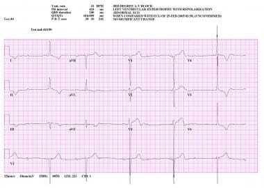
Electrocardiography
Disease Models, Animal
Sex Chromosome Aberrations
Retrospective Studies
Abnormalities, Drug-Induced
Case-Control Studies
Fetal Diseases
Schizophrenia
Pedigree
Prospective Studies
Cytogenetic Analysis
Brain Diseases
Tomography, X-Ray Computed
Mice, Knockout
Ultrasonography, Prenatal
Heart Defects, Congenital
Sensitivity and Specificity
Image Processing, Computer-Assisted
Chromosome Banding
Follow-Up Studies
Cytogenetics
Prenatal Diagnosis
Nervous System Malformations
Echocardiography
Mice, Transgenic
Prognosis
Risk Factors
Mice, Inbred C57BL
Heterozygote
Intellectual Disability
Chromosomes, Human, Pair 13
Reference Values
Biopsy
Predictive Value of Tests
Agenesis of Corpus Callosum
Down Syndrome
Electroencephalography
Atrophy
Limb Deformities, Congenital
Mosaicism
Chromosomes, Human, Pair 11
Neurologic Examination
Diffusion Magnetic Resonance Imaging
Ocular Motility Disorders
Myelodysplastic Syndromes
Gestational Age
Nerve Fibers, Myelinated
Gene Deletion
Infertility, Male
Analysis of Variance
Blood Coagulation Disorders
Chromosomes, Human, Pair 7
Cerebral Cortex
Immunohistochemistry
Severity of Illness Index
Heart Diseases
Molecular Sequence Data
Base Sequence
Prevalence
Nervous System Diseases
Corpus Callosum
Cohort Studies
Age Factors
Treatment Outcome
Tomography, Emission-Computed, Single-Photon
Reproducibility of Results
Anisotropy
Disease Progression
Neuropsychological Tests
Chromosomes, Human, Pair 17
Chromosomes, Human, Pair 18
Monosomy
Kidney
Polymerase Chain Reaction
Arrhythmias, Cardiac
Heart Ventricles
Diffusion Tensor Imaging
Cardiomyopathies
Gene Targeting
Chromosomes, Human, Pair 14
Brain Mapping
Developmental Disabilities
Sturge-Weber Syndrome
Bone Marrow
Biological Markers
Retinal Diseases
Maxillofacial Abnormalities
Microscopy, Electron
Cerebellum
RNA, Messenger
Spermatozoa
Fetus
Autistic Disorder
Gene Expression Regulation, Developmental
Cells, Cultured
Chromosomes, Human, 6-12 and X
Radiography, Thoracic
Amniocentesis
Temporal Lobe
Chromosomes, Human, Pair 8
Genotype
Lung
Myocardium
Chromosomes, Human, Pair 5
Chromosomes, Human, Pair 1
Bone and Bones
Cognition Disorders
Epilepsy
Aging
Chronic Disease
Turner Syndrome
Chromosomes, Human, Pair 3
Metabolic Diseases
Chromosome Mapping
Chromosomes, Human, Pair 12
Embryo, Mammalian
Neural Conduction
Skin
Leukemia, Myeloid, Acute
Echoencephalography
Frontal Lobe
Microcephaly
Observer Variation
Seizures
Ventricular Dysfunction, Left
Sensation Disorders
Liver
Hypertension
Transcription Factors
Gene Rearrangement
Bone Diseases, Developmental
Chromosomes, Human, X
Ultrasonography
Malformations of Cortical Development
Functional Laterality
Imaging, Three-Dimensional
Vaginal Smears
Chromosomes, Human, Pair 21
Foot Deformities, Congenital
Pregnancy Trimester, Second
Gene Expression
Body Weight
Movement Disorders
Growth Disorders
Nerve Tissue Proteins
Chromosomes, Human, 13-15
Signal Transduction
Vision Disorders
Statistics, Nonparametric
Alleles
Cerebral Ventricles
A Wnt5a pathway underlies outgrowth of multiple structures in the vertebrate embryo. (1/3131)
Morphogenesis depends on the precise control of basic cellular processes such as cell proliferation and differentiation. Wnt5a may regulate these processes since it is expressed in a gradient at the caudal end of the growing embryo during gastrulation, and later in the distal-most aspect of several structures that extend from the body. A loss-of-function mutation of Wnt5a leads to an inability to extend the A-P axis due to a progressive reduction in the size of caudal structures. In the limbs, truncation of the proximal skeleton and absence of distal digits correlates with reduced proliferation of putative progenitor cells within the progress zone. However, expression of progress zone markers, and several genes implicated in distal outgrowth and patterning including Distalless, Hoxd and Fgf family members was not altered. Taken together with the outgrowth defects observed in the developing face, ears and genitals, our data indicates that Wnt5a regulates a pathway common to many structures whose development requires extension from the primary body axis. The reduced number of proliferating cells in both the progress zone and the primitive streak mesoderm suggests that one function of Wnt5a is to regulate the proliferation of progenitor cells. (+info)The homeobox gene Pitx2: mediator of asymmetric left-right signaling in vertebrate heart and gut looping. (2/3131)
Left-right asymmetry in vertebrates is controlled by activities emanating from the left lateral plate. How these signals get transmitted to the forming organs is not known. A candidate mediator in mouse, frog and zebrafish embryos is the homeobox gene Pitx2. It is asymmetrically expressed in the left lateral plate mesoderm, tubular heart and early gut tube. Localized Pitx2 expression continues when these organs undergo asymmetric looping morphogenesis. Ectopic expression of Xnr1 in the right lateral plate induces Pitx2 transcription in Xenopus. Misexpression of Pitx2 affects situs and morphology of organs. These experiments suggest a role for Pitx2 in promoting looping of the linear heart and gut. (+info)Family study of inherited syndrome with multiple congenital deformities: symphalangism, carpal and tarsal fusion, brachydactyly, craniosynostosis, strabismus, hip osteochondritis. (3/3131)
A syndrome of brachydactyly (absence of some middle or distal phalanges), aplastic or hypoplastic nails, symphalangism (ankylois of proximal interphalangeal joints), synostosis of some carpal and tarsal bones, craniosynostosis, and dysplastic hip joints is reported in five members of an Italian family. It may represent a previously undescribed autosomal dominant trait. (+info)Amelioration of TCDD-induced teratogenesis in aryl hydrocarbon receptor (AhR)-null mice. (4/3131)
The aryl hydrocarbon receptor (AhR) mediates many of the biological effects of 2,3,7,8-tetrachlorodibenzo-p-dioxin (TCDD) and transcriptional activation of genes encoding a number of xenobiotic metabolizing enzymes. Prenatal exposure of mice to TCDD causes severe alterations in embryo and fetal development, including hydronephrosis and cleft palate. However, the mechanisms underlying these effects are unclear. In this work, the teratogenicity of TCDD in AhR-null mice was evaluated to determine if this effect is mediated by the AhR. Homozygous wild-type (+/+) or AhR-null (-/-) female mice were mated with males of the same genotype overnight. On gestation day (GD)-10, mice were intubated orally with either corn oil (vehicle control) or 25 micrograms/kg TCDD. Fetuses were examined on GD18 for visceral and skeletal alterations. For non-TCDD-exposed litters, all developmental endpoints were comparable between genotypes, with the exception of a lower incidence of large interfrontal bones in (-/-) mice. For TCDD-exposed litters, (+/+) fetuses had a significantly greater incidence of cleft palate, hydronephrosis, small kidneys, tortuous ureters and greater dilation of the renal pelves and ureters compared to (-/-) fetuses. Interestingly, an increased resorption rate was observed in (-/-) fetuses exposed to TCDD. Results from this work demonstrate that fetal development per se is generally unaffected by the absence of the AhR or that other genes may have compensated for the loss of the AhR. More importantly, these data indicate that the AhR mediates TCDD-induced teratogenicity. Further, since a higher percentage of resorptions was observed in (-/-) litters from TCDD-treated dams, it is possible that AhR-independent mechanisms contribute to TCDD-induced developmental toxicity. (+info)Townes-Brocks syndrome. (5/3131)
Townes-Brocks syndrome (TBS) is an autosomal dominant disorder with multiple malformations and variable expression. Major findings include external ear anomalies, hearing loss, preaxial polydactyly and triphalangeal thumbs, imperforate anus, and renal malformations. Most patients with Townes-Brocks syndrome have normal intelligence, although mental retardation has been noted in a few. (+info)A new lethal syndrome of exomphalos, short limbs, and macrogonadism. (6/3131)
We report a new lethal multiple congenital abnormality (MCA) syndrome of exomphalos, short limbs, nuchal web, macrogonadism, and facial dysmorphism in seven fetuses (six males and one female) belonging to three unrelated families. X rays showed enlarged and irregular metaphyses with a heterogeneous pattern of mineralisation of the long bones. Pathological examination showed adrenal cytomegaly, hyperplasia of Leydig cells, ovarian stroma cells, and Langherans cells, and renal microcysts. We suggest that this condition is a new autosomal recessive MCA syndrome different from Beckwith-Wiedemann syndrome, especially as no infracytogenetic deletion or uniparental disomy of chromosome 11 was found. (+info)Isolation and embryonic expression of the novel mouse gene Hic1, the homologue of HIC1, a candidate gene for the Miller-Dieker syndrome. (7/3131)
The human gene HIC1 (hypermethylated in cancer) maps to chromosome 17p13.3 and is deleted in the contiguous gene disorder Miller-Dieker syndrome (MDS) [Makos-Wales et al. (1995) Nature Med., 1, 570-577; Chong et al. (1996) Genome Res., 6, 735-741]. We isolated the murine homologue Hic1, encoding a zinc-finger protein with a poxvirus and zinc-finger (POZ) domain and mapped it to mouse chromosome 11 in a region exhibiting conserved synteny to human chromosome 17. Comparison of genomic and cDNA sequences predicts two exons for the murine Hic1. The second exon exhibits 88% identity to the human HIC1 on DNA level. During embryonic development, Hic1 is expressed in mesenchymes of the sclerotomes, lateral body wall, limb and cranio-facial regions embedding the outgrowing peripheral nerves during their differentiation. During fetal development, Hic1 additionally is expressed in mesenchymes apposed to precartilaginous condensations, at many interfaces to budding epithelia of inner organs, and weakly in muscles. We observed activation of Hic1 expression in the embryonic anlagen of many tissues displaying anomalies in MDS patients. Besides lissencephaly, MDS patients exhibit facial dysmorphism and frequently additional birth defects, e.g. anomalies of the heart, kidney, gastrointestinal tract and the limbs (OMIM 247200). Thus, HIC1 activity may correlate with the defective development of the nose, jaws, extremities, gastrointestinal tract and kidney in MDS patients. (+info)Comparison of prenatal ultrasound and postmortem findings in fetuses and infants with congenital heart defects. (8/3131)
OBJECTIVE: Detection of congenital heart defects by prenatal ultrasound examination has been one of the great challenges since the investigation for fetal anomalies became part of the routine fetal examination. This prospective study was designed to evaluate the concordance of prenatal ultrasound findings with autopsy examination in a population consisting of both referred women and non-selected pregnant women. DESIGN: Criteria for inclusion were an ultrasound examination at the National Center for Fetal Medicine and an autopsy performed during the years 1985-94. Results from the ultrasound and autopsy examinations were systematized into categories depending on the degree of concordance. RESULTS: Of 408 infants and fetuses with developmental anomalies, 106 (26%) had congenital heart defects. In 63 (59%) of these 106 cases, the heart defect was the principal reason for the termination of pregnancy or the cause of death. Excluding five cases with a secundum atrial septal defect, there was complete agreement between the ultrasound examination and the autopsy findings in 74 (73%) of 101 cases. In 18 cases, there were minor discrepancies between ultrasound and autopsy findings. The main diagnosis was thus correct in 92 cases (91%). From the first time period (1985-89) to the second (1990-94), the detection rate of all heart defects increased from 48% to 82%. CONCLUSION: This study confirms a good correlation between ultrasound and autopsy diagnoses in fetuses and infants with congenital heart defects. A significant improvement in the detection of heart defects occurred from the first time period to the second and was probably due to increased experience and technical advances. (+info)'Abnormalities, Multiple' is a broad term that refers to the presence of two or more structural or functional anomalies in an individual. These abnormalities can be present at birth (congenital) or can develop later in life (acquired). They can affect various organs and systems of the body and can vary greatly in severity and impact on a person's health and well-being.
Multiple abnormalities can occur due to genetic factors, environmental influences, or a combination of both. Chromosomal abnormalities, gene mutations, exposure to teratogens (substances that cause birth defects), and maternal infections during pregnancy are some of the common causes of multiple congenital abnormalities.
Examples of multiple congenital abnormalities include Down syndrome, Turner syndrome, and VATER/VACTERL association. Acquired multiple abnormalities can result from conditions such as trauma, infection, degenerative diseases, or cancer.
The medical evaluation and management of individuals with multiple abnormalities depend on the specific abnormalities present and their impact on the individual's health and functioning. A multidisciplinary team of healthcare professionals is often involved in the care of these individuals to address their complex needs.
Chromosome aberrations refer to structural and numerical changes in the chromosomes that can occur spontaneously or as a result of exposure to mutagenic agents. These changes can affect the genetic material encoded in the chromosomes, leading to various consequences such as developmental abnormalities, cancer, or infertility.
Structural aberrations include deletions, duplications, inversions, translocations, and rings, which result from breaks and rearrangements of chromosome segments. Numerical aberrations involve changes in the number of chromosomes, such as aneuploidy (extra or missing chromosomes) or polyploidy (multiples of a complete set of chromosomes).
Chromosome aberrations can be detected and analyzed using various cytogenetic techniques, including karyotyping, fluorescence in situ hybridization (FISH), and comparative genomic hybridization (CGH). These methods allow for the identification and characterization of chromosomal changes at the molecular level, providing valuable information for genetic counseling, diagnosis, and research.
Congenital abnormalities, also known as birth defects, are structural or functional anomalies that are present at birth. These abnormalities can develop at any point during fetal development, and they can affect any part of the body. They can be caused by genetic factors, environmental influences, or a combination of both.
Congenital abnormalities can range from mild to severe and may include structural defects such as heart defects, neural tube defects, and cleft lip and palate, as well as functional defects such as intellectual disabilities and sensory impairments. Some congenital abnormalities may be visible at birth, while others may not become apparent until later in life.
In some cases, congenital abnormalities may be detected through prenatal testing, such as ultrasound or amniocentesis. In other cases, they may not be diagnosed until after the baby is born. Treatment for congenital abnormalities varies depending on the type and severity of the defect, and may include surgery, therapy, medication, or a combination of these approaches.
Chromosome disorders are a group of genetic conditions caused by abnormalities in the number or structure of chromosomes. Chromosomes are thread-like structures located in the nucleus of cells that contain most of the body's genetic material, which is composed of DNA and proteins. Normally, humans have 23 pairs of chromosomes, for a total of 46 chromosomes.
Chromosome disorders can result from changes in the number of chromosomes (aneuploidy) or structural abnormalities in one or more chromosomes. Some common examples of chromosome disorders include:
1. Down syndrome: a condition caused by an extra copy of chromosome 21, resulting in intellectual disability, developmental delays, and distinctive physical features.
2. Turner syndrome: a condition that affects only females and is caused by the absence of all or part of one X chromosome, resulting in short stature, lack of sexual development, and other symptoms.
3. Klinefelter syndrome: a condition that affects only males and is caused by an extra copy of the X chromosome, resulting in tall stature, infertility, and other symptoms.
4. Cri-du-chat syndrome: a condition caused by a deletion of part of the short arm of chromosome 5, resulting in intellectual disability, developmental delays, and a distinctive cat-like cry.
5. Fragile X syndrome: a condition caused by a mutation in the FMR1 gene on the X chromosome, resulting in intellectual disability, behavioral problems, and physical symptoms.
Chromosome disorders can be diagnosed through various genetic tests, such as karyotyping, chromosomal microarray analysis (CMA), or fluorescence in situ hybridization (FISH). Treatment for these conditions depends on the specific disorder and its associated symptoms and may include medical interventions, therapies, and educational support.
Eye abnormalities refer to any structural or functional anomalies that affect the eye or its surrounding tissues. These abnormalities can be present at birth (congenital) or acquired later in life due to various factors such as injury, disease, or aging. Some examples of eye abnormalities include:
1. Strabismus: Also known as crossed eyes, strabismus is a condition where the eyes are misaligned and point in different directions.
2. Nystagmus: This is an involuntary movement of the eyes that can be horizontal, vertical, or rotatory.
3. Cataracts: A cataract is a clouding of the lens inside the eye that can cause vision loss.
4. Glaucoma: This is a group of eye conditions that damage the optic nerve and can lead to vision loss.
5. Retinal disorders: These include conditions such as retinal detachment, macular degeneration, and diabetic retinopathy.
6. Corneal abnormalities: These include conditions such as keratoconus, corneal ulcers, and Fuchs' dystrophy.
7. Orbital abnormalities: These include conditions such as orbital tumors, thyroid eye disease, and Graves' ophthalmopathy.
8. Ptosis: This is a condition where the upper eyelid droops over the eye.
9. Color blindness: A condition where a person has difficulty distinguishing between certain colors.
10. Microphthalmia: A condition where one or both eyes are abnormally small.
These are just a few examples of eye abnormalities, and there are many others that can affect the eye and its functioning. If you suspect that you have an eye abnormality, it is important to consult with an ophthalmologist for proper diagnosis and treatment.
Karyotyping is a medical laboratory test used to study the chromosomes in a cell. It involves obtaining a sample of cells from a patient, usually from blood or bone marrow, and then staining the chromosomes so they can be easily seen under a microscope. The chromosomes are then arranged in pairs based on their size, shape, and other features to create a karyotype. This visual representation allows for the identification and analysis of any chromosomal abnormalities, such as extra or missing chromosomes, or structural changes like translocations or inversions. These abnormalities can provide important information about genetic disorders, diseases, and developmental problems.
Medical Definition:
Magnetic Resonance Imaging (MRI) is a non-invasive diagnostic imaging technique that uses a strong magnetic field and radio waves to create detailed cross-sectional or three-dimensional images of the internal structures of the body. The patient lies within a large, cylindrical magnet, and the scanner detects changes in the direction of the magnetic field caused by protons in the body. These changes are then converted into detailed images that help medical professionals to diagnose and monitor various medical conditions, such as tumors, injuries, or diseases affecting the brain, spinal cord, heart, blood vessels, joints, and other internal organs. MRI does not use radiation like computed tomography (CT) scans.
The brain is the central organ of the nervous system, responsible for receiving and processing sensory information, regulating vital functions, and controlling behavior, movement, and cognition. It is divided into several distinct regions, each with specific functions:
1. Cerebrum: The largest part of the brain, responsible for higher cognitive functions such as thinking, learning, memory, language, and perception. It is divided into two hemispheres, each controlling the opposite side of the body.
2. Cerebellum: Located at the back of the brain, it is responsible for coordinating muscle movements, maintaining balance, and fine-tuning motor skills.
3. Brainstem: Connects the cerebrum and cerebellum to the spinal cord, controlling vital functions such as breathing, heart rate, and blood pressure. It also serves as a relay center for sensory information and motor commands between the brain and the rest of the body.
4. Diencephalon: A region that includes the thalamus (a major sensory relay station) and hypothalamus (regulates hormones, temperature, hunger, thirst, and sleep).
5. Limbic system: A group of structures involved in emotional processing, memory formation, and motivation, including the hippocampus, amygdala, and cingulate gyrus.
The brain is composed of billions of interconnected neurons that communicate through electrical and chemical signals. It is protected by the skull and surrounded by three layers of membranes called meninges, as well as cerebrospinal fluid that provides cushioning and nutrients.
Cardiovascular abnormalities refer to structural or functional anomalies in the heart or blood vessels. These abnormalities can be present at birth (congenital) or acquired later in life. They can affect the heart's chambers, valves, walls, or blood vessels, leading to various complications such as heart failure, stroke, or even death if left untreated.
Examples of congenital cardiovascular abnormalities include:
1. Septal defects - holes in the walls separating the heart's chambers (atrial septal defect, ventricular septal defect)
2. Valvular stenosis or insufficiency - narrowing or leakage of the heart valves
3. Patent ductus arteriosus - a persistent opening between the aorta and pulmonary artery
4. Coarctation of the aorta - narrowing of the aorta
5. Tetralogy of Fallot - a combination of four heart defects, including ventricular septal defect, overriding aorta, pulmonary stenosis, and right ventricular hypertrophy
Examples of acquired cardiovascular abnormalities include:
1. Atherosclerosis - the buildup of plaque in the arteries, leading to narrowing or blockage
2. Cardiomyopathy - disease of the heart muscle, causing it to become enlarged, thickened, or stiffened
3. Hypertension - high blood pressure, which can damage the heart and blood vessels over time
4. Myocardial infarction (heart attack) - damage to the heart muscle due to blocked blood supply
5. Infective endocarditis - infection of the inner lining of the heart chambers and valves
These abnormalities can be diagnosed through various tests, such as echocardiography, electrocardiogram (ECG), stress testing, cardiac catheterization, or magnetic resonance imaging (MRI). Treatment options depend on the type and severity of the abnormality and may include medications, medical procedures, or surgery.
Craniofacial abnormalities refer to a group of birth defects that affect the development of the skull and face. These abnormalities can range from mild to severe and may involve differences in the shape and structure of the head, face, and jaws, as well as issues with the formation of facial features such as the eyes, nose, and mouth.
Craniofacial abnormalities can be caused by genetic factors, environmental influences, or a combination of both. Some common examples of craniofacial abnormalities include cleft lip and palate, craniosynostosis (premature fusion of the skull bones), and hemifacial microsomia (underdevelopment of one side of the face).
Treatment for craniofacial abnormalities may involve a team of healthcare professionals, including plastic surgeons, neurosurgeons, orthodontists, speech therapists, and other specialists. Treatment options may include surgery, bracing, therapy, and other interventions to help improve function and appearance.
A syndrome, in medical terms, is a set of symptoms that collectively indicate or characterize a disease, disorder, or underlying pathological process. It's essentially a collection of signs and/or symptoms that frequently occur together and can suggest a particular cause or condition, even though the exact physiological mechanisms might not be fully understood.
For example, Down syndrome is characterized by specific physical features, cognitive delays, and other developmental issues resulting from an extra copy of chromosome 21. Similarly, metabolic syndromes like diabetes mellitus type 2 involve a group of risk factors such as obesity, high blood pressure, high blood sugar, and abnormal cholesterol or triglyceride levels that collectively increase the risk of heart disease, stroke, and diabetes.
It's important to note that a syndrome is not a specific diagnosis; rather, it's a pattern of symptoms that can help guide further diagnostic evaluation and management.
Skin abnormalities refer to any changes in the skin that deviate from its normal structure, function, or color. These can manifest as various conditions such as lesions, growths, discolorations, or textural alterations. Examples include moles, freckles, birthmarks, rashes, hives, acne, eczema, psoriasis, rosacea, skin cancer, and many others. Some skin abnormalities may be harmless and require no treatment, while others might indicate an underlying medical condition that requires further evaluation and management.
A phenotype is the physical or biochemical expression of an organism's genes, or the observable traits and characteristics resulting from the interaction of its genetic constitution (genotype) with environmental factors. These characteristics can include appearance, development, behavior, and resistance to disease, among others. Phenotypes can vary widely, even among individuals with identical genotypes, due to differences in environmental influences, gene expression, and genetic interactions.
Urogenital abnormalities refer to structural or functional anomalies that affect the urinary and genital systems. These two systems are closely linked during embryonic development, and sometimes they may not develop properly, leading to various types of congenital defects. Urogenital abnormalities can range from minor issues like a bifid scrotum (a condition where the scrotum is split into two parts) to more severe problems such as bladder exstrophy (where the bladder develops outside the body).
These conditions may affect urination, reproduction, and sexual function. They can also increase the risk of infections and other complications. Urogenital abnormalities can be diagnosed through physical examination, imaging tests, or genetic testing. Treatment options depend on the specific condition but may include surgery, medication, or lifestyle changes.
Pregnancy is a physiological state or condition where a fertilized egg (zygote) successfully implants and grows in the uterus of a woman, leading to the development of an embryo and finally a fetus. This process typically spans approximately 40 weeks, divided into three trimesters, and culminates in childbirth. Throughout this period, numerous hormonal and physical changes occur to support the growing offspring, including uterine enlargement, breast development, and various maternal adaptations to ensure the fetus's optimal growth and well-being.
Trisomy is a genetic condition where there is an extra copy of a particular chromosome, resulting in 47 chromosomes instead of the typical 46 in a cell. This usually occurs due to an error in cell division during the development of the egg, sperm, or embryo.
Instead of the normal pair, there are three copies (trisomy) of that chromosome. The most common form of trisomy is Trisomy 21, also known as Down syndrome, where there is an extra copy of chromosome 21. Other forms include Trisomy 13 (Patau syndrome) and Trisomy 18 (Edwards syndrome), which are associated with more severe developmental issues and shorter lifespans.
Trisomy can also occur in a mosaic form, where some cells have the extra chromosome while others do not, leading to varying degrees of symptoms depending on the proportion of affected cells.
A mutation is a permanent change in the DNA sequence of an organism's genome. Mutations can occur spontaneously or be caused by environmental factors such as exposure to radiation, chemicals, or viruses. They may have various effects on the organism, ranging from benign to harmful, depending on where they occur and whether they alter the function of essential proteins. In some cases, mutations can increase an individual's susceptibility to certain diseases or disorders, while in others, they may confer a survival advantage. Mutations are the driving force behind evolution, as they introduce new genetic variability into populations, which can then be acted upon by natural selection.
Musculoskeletal abnormalities refer to structural and functional disorders that affect the musculoskeletal system, which includes the bones, muscles, cartilages, tendons, ligaments, joints, and other related tissues. These abnormalities can result from genetic factors, trauma, overuse, degenerative processes, infections, or tumors. They may cause pain, stiffness, limited mobility, deformity, weakness, and susceptibility to injuries. Examples of musculoskeletal abnormalities include osteoarthritis, rheumatoid arthritis, scoliosis, kyphosis, lordosis, fractures, dislocations, tendinitis, bursitis, myopathies, and various congenital conditions.
In situ hybridization, fluorescence (FISH) is a type of molecular cytogenetic technique used to detect and localize the presence or absence of specific DNA sequences on chromosomes through the use of fluorescent probes. This technique allows for the direct visualization of genetic material at a cellular level, making it possible to identify chromosomal abnormalities such as deletions, duplications, translocations, and other rearrangements.
The process involves denaturing the DNA in the sample to separate the double-stranded molecules into single strands, then adding fluorescently labeled probes that are complementary to the target DNA sequence. The probe hybridizes to the complementary sequence in the sample, and the location of the probe is detected by fluorescence microscopy.
FISH has a wide range of applications in both clinical and research settings, including prenatal diagnosis, cancer diagnosis and monitoring, and the study of gene expression and regulation. It is a powerful tool for identifying genetic abnormalities and understanding their role in human disease.
Tooth abnormalities refer to any variations or irregularities in the size, shape, number, structure, or development of teeth that deviate from the typical or normal anatomy. These abnormalities can occur in primary (deciduous) or permanent teeth and can be caused by genetic factors, environmental influences, systemic diseases, or localized dental conditions during tooth formation.
Some examples of tooth abnormalities include:
1. Microdontia - teeth that are smaller than normal in size.
2. Macrodontia - teeth that are larger than normal in size.
3. Peg-shaped teeth - teeth with a narrow, conical shape.
4. Talon cusps - additional cusps or points on the biting surface of a tooth.
5. Dens invaginatus - an abnormal development where the tooth crown has an extra fold or pouch that can trap bacteria and cause dental problems.
6. Taurodontism - teeth with large pulp chambers and short roots.
7. Supernumerary teeth - having more teeth than the typical number (20 primary and 32 permanent teeth).
8. Hypodontia - missing one or more teeth due to a failure of development.
9. Germination - two adjacent teeth fused together, usually occurring in the front teeth.
10. Fusion - two separate teeth that have grown together during development.
Tooth abnormalities may not always require treatment unless they cause functional, aesthetic, or dental health issues. A dentist can diagnose and manage tooth abnormalities through various treatments, such as fillings, extractions, orthodontic care, or restorative procedures.
Translocation, genetic, refers to a type of chromosomal abnormality in which a segment of a chromosome is transferred from one chromosome to another, resulting in an altered genome. This can occur between two non-homologous chromosomes (non-reciprocal translocation) or between two homologous chromosomes (reciprocal translocation). Genetic translocations can lead to various clinical consequences, depending on the genes involved and the location of the translocation. Some translocations may result in no apparent effects, while others can cause developmental abnormalities, cancer, or other genetic disorders. In some cases, translocations can also increase the risk of having offspring with genetic conditions.
A newborn infant is a baby who is within the first 28 days of life. This period is also referred to as the neonatal period. Newborns require specialized care and attention due to their immature bodily systems and increased vulnerability to various health issues. They are closely monitored for signs of well-being, growth, and development during this critical time.
Aneuploidy is a medical term that refers to an abnormal number of chromosomes in a cell. Chromosomes are thread-like structures located inside the nucleus of cells that contain genetic information in the form of genes.
In humans, the normal number of chromosomes in a cell is 46, arranged in 23 pairs. Aneuploidy occurs when there is an extra or missing chromosome in one or more of these pairs. For example, Down syndrome is a condition that results from an extra copy of chromosome 21, also known as trisomy 21.
Aneuploidy can arise during the formation of gametes (sperm or egg cells) due to errors in the process of cell division called meiosis. These errors can result in eggs or sperm with an abnormal number of chromosomes, which can then lead to aneuploidy in the resulting embryo.
Aneuploidy is a significant cause of birth defects and miscarriages. The severity of the condition depends on which chromosomes are affected and the extent of the abnormality. In some cases, aneuploidy may have no noticeable effects, while in others it can lead to serious health problems or developmental delays.
Electrocardiography (ECG or EKG) is a medical procedure that records the electrical activity of the heart. It provides a graphic representation of the electrical changes that occur during each heartbeat. The resulting tracing, called an electrocardiogram, can reveal information about the heart's rate and rhythm, as well as any damage to its cells or abnormalities in its conduction system.
During an ECG, small electrodes are placed on the skin of the chest, arms, and legs. These electrodes detect the electrical signals produced by the heart and transmit them to a machine that amplifies and records them. The procedure is non-invasive, painless, and quick, usually taking only a few minutes.
ECGs are commonly used to diagnose and monitor various heart conditions, including arrhythmias, coronary artery disease, heart attacks, and electrolyte imbalances. They can also be used to evaluate the effectiveness of certain medications or treatments.
Animal disease models are specialized animals, typically rodents such as mice or rats, that have been genetically engineered or exposed to certain conditions to develop symptoms and physiological changes similar to those seen in human diseases. These models are used in medical research to study the pathophysiology of diseases, identify potential therapeutic targets, test drug efficacy and safety, and understand disease mechanisms.
The genetic modifications can include knockout or knock-in mutations, transgenic expression of specific genes, or RNA interference techniques. The animals may also be exposed to environmental factors such as chemicals, radiation, or infectious agents to induce the disease state.
Examples of animal disease models include:
1. Mouse models of cancer: Genetically engineered mice that develop various types of tumors, allowing researchers to study cancer initiation, progression, and metastasis.
2. Alzheimer's disease models: Transgenic mice expressing mutant human genes associated with Alzheimer's disease, which exhibit amyloid plaque formation and cognitive decline.
3. Diabetes models: Obese and diabetic mouse strains like the NOD (non-obese diabetic) or db/db mice, used to study the development of type 1 and type 2 diabetes, respectively.
4. Cardiovascular disease models: Atherosclerosis-prone mice, such as ApoE-deficient or LDLR-deficient mice, that develop plaque buildup in their arteries when fed a high-fat diet.
5. Inflammatory bowel disease models: Mice with genetic mutations affecting intestinal barrier function and immune response, such as IL-10 knockout or SAMP1/YitFc mice, which develop colitis.
Animal disease models are essential tools in preclinical research, but it is important to recognize their limitations. Differences between species can affect the translatability of results from animal studies to human patients. Therefore, researchers must carefully consider the choice of model and interpret findings cautiously when applying them to human diseases.
Sex chromosome aberrations refer to structural and numerical abnormalities in the sex chromosomes, which are typically represented as X and Y chromosomes in humans. These aberrations can result in variations in the number of sex chromosomes, such as Klinefelter syndrome (47,XXY), Turner syndrome (45,X), and Jacobs/XYY syndrome (47,XYY). They can also include structural changes, such as deletions, duplications, or translocations of sex chromosome material.
Sex chromosome aberrations may lead to a range of phenotypic effects, including differences in physical characteristics, cognitive development, fertility, and susceptibility to certain health conditions. The manifestation and severity of these impacts can vary widely depending on the specific type and extent of the aberration, as well as individual genetic factors and environmental influences.
It is important to note that while sex chromosome aberrations may pose challenges and require medical management, they do not inherently define or limit a person's potential, identity, or worth. Comprehensive care, support, and education can help individuals with sex chromosome aberrations lead fulfilling lives and reach their full potential.
Retrospective studies, also known as retrospective research or looking back studies, are a type of observational study that examines data from the past to draw conclusions about possible causal relationships between risk factors and outcomes. In these studies, researchers analyze existing records, medical charts, or previously collected data to test a hypothesis or answer a specific research question.
Retrospective studies can be useful for generating hypotheses and identifying trends, but they have limitations compared to prospective studies, which follow participants forward in time from exposure to outcome. Retrospective studies are subject to biases such as recall bias, selection bias, and information bias, which can affect the validity of the results. Therefore, retrospective studies should be interpreted with caution and used primarily to generate hypotheses for further testing in prospective studies.
"Drug-induced abnormalities" refer to physical or physiological changes that occur as a result of taking medication or drugs. These abnormalities can affect various organs and systems in the body and can range from minor symptoms, such as nausea or dizziness, to more serious conditions, such as liver damage or heart rhythm disturbances.
Drug-induced abnormalities can occur for several reasons, including:
1. Direct toxicity: Some drugs can directly damage cells and tissues in the body, leading to abnormalities.
2. Altered metabolism: Drugs can interfere with normal metabolic processes in the body, leading to the accumulation of harmful substances or the depletion of essential nutrients.
3. Hormonal imbalances: Some drugs can affect hormone levels in the body, leading to abnormalities.
4. Allergic reactions: Some people may have allergic reactions to certain drugs, which can cause a range of symptoms, including rashes, swelling, and difficulty breathing.
5. Interactions with other drugs: Taking multiple medications or drugs at the same time can increase the risk of drug-induced abnormalities.
It is important for healthcare providers to monitor patients closely for signs of drug-induced abnormalities and to adjust medication dosages or switch to alternative treatments as necessary. Patients should also inform their healthcare providers of any symptoms they experience while taking medication, as these may be related to drug-induced abnormalities.
A case-control study is an observational research design used to identify risk factors or causes of a disease or health outcome. In this type of study, individuals with the disease or condition (cases) are compared with similar individuals who do not have the disease or condition (controls). The exposure history or other characteristics of interest are then compared between the two groups to determine if there is an association between the exposure and the disease.
Case-control studies are often used when it is not feasible or ethical to conduct a randomized controlled trial, as they can provide valuable insights into potential causes of diseases or health outcomes in a relatively short period of time and at a lower cost than other study designs. However, because case-control studies rely on retrospective data collection, they are subject to biases such as recall bias and selection bias, which can affect the validity of the results. Therefore, it is important to carefully design and conduct case-control studies to minimize these potential sources of bias.
In the field of medicine, "time factors" refer to the duration of symptoms or time elapsed since the onset of a medical condition, which can have significant implications for diagnosis and treatment. Understanding time factors is crucial in determining the progression of a disease, evaluating the effectiveness of treatments, and making critical decisions regarding patient care.
For example, in stroke management, "time is brain," meaning that rapid intervention within a specific time frame (usually within 4.5 hours) is essential to administering tissue plasminogen activator (tPA), a clot-busting drug that can minimize brain damage and improve patient outcomes. Similarly, in trauma care, the "golden hour" concept emphasizes the importance of providing definitive care within the first 60 minutes after injury to increase survival rates and reduce morbidity.
Time factors also play a role in monitoring the progression of chronic conditions like diabetes or heart disease, where regular follow-ups and assessments help determine appropriate treatment adjustments and prevent complications. In infectious diseases, time factors are crucial for initiating antibiotic therapy and identifying potential outbreaks to control their spread.
Overall, "time factors" encompass the significance of recognizing and acting promptly in various medical scenarios to optimize patient outcomes and provide effective care.
Fetal diseases are medical conditions or abnormalities that affect a fetus during pregnancy. These diseases can be caused by genetic factors, environmental influences, or a combination of both. They can range from mild to severe and may impact various organ systems in the developing fetus. Examples of fetal diseases include congenital heart defects, neural tube defects, chromosomal abnormalities such as Down syndrome, and infectious diseases such as toxoplasmosis or rubella. Fetal diseases can be diagnosed through prenatal testing, including ultrasound, amniocentesis, and chorionic villus sampling. Treatment options may include medication, surgery, or delivery of the fetus, depending on the nature and severity of the disease.
Schizophrenia is a severe mental disorder characterized by disturbances in thought, perception, emotion, and behavior. It often includes hallucinations (usually hearing voices), delusions, paranoia, and disorganized speech and behavior. The onset of symptoms typically occurs in late adolescence or early adulthood. Schizophrenia is a complex, chronic condition that requires ongoing treatment and management. It significantly impairs social and occupational functioning, and it's often associated with reduced life expectancy due to comorbid medical conditions. The exact causes of schizophrenia are not fully understood, but research suggests that genetic, environmental, and neurodevelopmental factors play a role in its development.
I must clarify that the term "pedigree" is not typically used in medical definitions. Instead, it is often employed in genetics and breeding, where it refers to the recorded ancestry of an individual or a family, tracing the inheritance of specific traits or diseases. In human genetics, a pedigree can help illustrate the pattern of genetic inheritance in families over multiple generations. However, it is not a medical term with a specific clinical definition.
Prospective studies, also known as longitudinal studies, are a type of cohort study in which data is collected forward in time, following a group of individuals who share a common characteristic or exposure over a period of time. The researchers clearly define the study population and exposure of interest at the beginning of the study and follow up with the participants to determine the outcomes that develop over time. This type of study design allows for the investigation of causal relationships between exposures and outcomes, as well as the identification of risk factors and the estimation of disease incidence rates. Prospective studies are particularly useful in epidemiology and medical research when studying diseases with long latency periods or rare outcomes.
Cytogenetic analysis is a laboratory technique used to identify and study the structure and function of chromosomes, which are the structures in the cell that contain genetic material. This type of analysis involves examining the number, size, shape, and banding pattern of chromosomes in cells, typically during metaphase when they are at their most condensed state.
There are several methods used for cytogenetic analysis, including karyotyping, fluorescence in situ hybridization (FISH), and comparative genomic hybridization (CGH). Karyotyping involves staining the chromosomes with a dye to visualize their banding patterns and then arranging them in pairs based on their size and shape. FISH uses fluorescent probes to label specific DNA sequences, allowing for the detection of genetic abnormalities such as deletions, duplications, or translocations. CGH compares the DNA content of two samples to identify differences in copy number, which can be used to detect chromosomal imbalances.
Cytogenetic analysis is an important tool in medical genetics and is used for a variety of purposes, including prenatal diagnosis, cancer diagnosis and monitoring, and the identification of genetic disorders.
Brain diseases, also known as neurological disorders, refer to a wide range of conditions that affect the brain and nervous system. These diseases can be caused by various factors such as genetics, infections, injuries, degeneration, or structural abnormalities. They can affect different parts of the brain, leading to a variety of symptoms and complications.
Some examples of brain diseases include:
1. Alzheimer's disease - a progressive degenerative disorder that affects memory and cognitive function.
2. Parkinson's disease - a movement disorder characterized by tremors, stiffness, and difficulty with coordination and balance.
3. Multiple sclerosis - a chronic autoimmune disease that affects the nervous system and can cause a range of symptoms such as vision loss, muscle weakness, and cognitive impairment.
4. Epilepsy - a neurological disorder characterized by recurrent seizures.
5. Brain tumors - abnormal growths in the brain that can be benign or malignant.
6. Stroke - a sudden interruption of blood flow to the brain, which can cause paralysis, speech difficulties, and other neurological symptoms.
7. Meningitis - an infection of the membranes surrounding the brain and spinal cord.
8. Encephalitis - an inflammation of the brain that can be caused by viruses, bacteria, or autoimmune disorders.
9. Huntington's disease - a genetic disorder that affects muscle coordination, cognitive function, and mental health.
10. Migraine - a neurological condition characterized by severe headaches, often accompanied by nausea, vomiting, and sensitivity to light and sound.
Brain diseases can range from mild to severe and may be treatable or incurable. They can affect people of all ages and backgrounds, and early diagnosis and treatment are essential for improving outcomes and quality of life.
X-ray computed tomography (CT or CAT scan) is a medical imaging method that uses computer-processed combinations of many X-ray images taken from different angles to produce cross-sectional (tomographic) images (virtual "slices") of the body. These cross-sectional images can then be used to display detailed internal views of organs, bones, and soft tissues in the body.
The term "computed tomography" is used instead of "CT scan" or "CAT scan" because the machines take a series of X-ray measurements from different angles around the body and then use a computer to process these data to create detailed images of internal structures within the body.
CT scanning is a noninvasive, painless medical test that helps physicians diagnose and treat medical conditions. CT imaging provides detailed information about many types of tissue including lung, bone, soft tissue and blood vessels. CT examinations can be performed on every part of the body for a variety of reasons including diagnosis, surgical planning, and monitoring of therapeutic responses.
In computed tomography (CT), an X-ray source and detector rotate around the patient, measuring the X-ray attenuation at many different angles. A computer uses this data to construct a cross-sectional image by the process of reconstruction. This technique is called "tomography". The term "computed" refers to the use of a computer to reconstruct the images.
CT has become an important tool in medical imaging and diagnosis, allowing radiologists and other physicians to view detailed internal images of the body. It can help identify many different medical conditions including cancer, heart disease, lung nodules, liver tumors, and internal injuries from trauma. CT is also commonly used for guiding biopsies and other minimally invasive procedures.
In summary, X-ray computed tomography (CT or CAT scan) is a medical imaging technique that uses computer-processed combinations of many X-ray images taken from different angles to produce cross-sectional images of the body. It provides detailed internal views of organs, bones, and soft tissues in the body, allowing physicians to diagnose and treat medical conditions.
A "knockout" mouse is a genetically engineered mouse in which one or more genes have been deleted or "knocked out" using molecular biology techniques. This allows researchers to study the function of specific genes and their role in various biological processes, as well as potential associations with human diseases. The mice are generated by introducing targeted DNA modifications into embryonic stem cells, which are then used to create a live animal. Knockout mice have been widely used in biomedical research to investigate gene function, disease mechanisms, and potential therapeutic targets.
Prenatal ultrasonography, also known as obstetric ultrasound, is a medical diagnostic procedure that uses high-frequency sound waves to create images of the developing fetus, placenta, and amniotic fluid inside the uterus. It is a non-invasive and painless test that is widely used during pregnancy to monitor the growth and development of the fetus, detect any potential abnormalities or complications, and determine the due date.
During the procedure, a transducer (a small handheld device) is placed on the mother's abdomen and moved around to capture images from different angles. The sound waves travel through the mother's body and bounce back off the fetus, producing echoes that are then converted into electrical signals and displayed as images on a screen.
Prenatal ultrasonography can be performed at various stages of pregnancy, including early pregnancy to confirm the pregnancy and detect the number of fetuses, mid-pregnancy to assess the growth and development of the fetus, and late pregnancy to evaluate the position of the fetus and determine if it is head down or breech. It can also be used to guide invasive procedures such as amniocentesis or chorionic villus sampling.
Overall, prenatal ultrasonography is a valuable tool in modern obstetrics that helps ensure the health and well-being of both the mother and the developing fetus.
A chromosome deletion is a type of genetic abnormality that occurs when a portion of a chromosome is missing or deleted. Chromosomes are thread-like structures located in the nucleus of cells that contain our genetic material, which is organized into genes.
Chromosome deletions can occur spontaneously during the formation of reproductive cells (eggs or sperm) or can be inherited from a parent. They can affect any chromosome and can vary in size, from a small segment to a large portion of the chromosome.
The severity of the symptoms associated with a chromosome deletion depends on the size and location of the deleted segment. In some cases, the deletion may be so small that it does not cause any noticeable symptoms. However, larger deletions can lead to developmental delays, intellectual disabilities, physical abnormalities, and various medical conditions.
Chromosome deletions are typically detected through a genetic test called karyotyping, which involves analyzing the number and structure of an individual's chromosomes. Other more precise tests, such as fluorescence in situ hybridization (FISH) or chromosomal microarray analysis (CMA), may also be used to confirm the diagnosis and identify the specific location and size of the deletion.
Congenital heart defects (CHDs) are structural abnormalities in the heart that are present at birth. They can affect any part of the heart's structure, including the walls of the heart, the valves inside the heart, and the major blood vessels that lead to and from the heart.
Congenital heart defects can range from mild to severe and can cause various symptoms depending on the type and severity of the defect. Some common symptoms of CHDs include cyanosis (a bluish tint to the skin, lips, and fingernails), shortness of breath, fatigue, poor feeding, and slow growth in infants and children.
There are many different types of congenital heart defects, including:
1. Septal defects: These are holes in the walls that separate the four chambers of the heart. The two most common septal defects are atrial septal defect (ASD) and ventricular septal defect (VSD).
2. Valve abnormalities: These include narrowed or leaky valves, which can affect blood flow through the heart.
3. Obstruction defects: These occur when blood flow is blocked or restricted due to narrowing or absence of a part of the heart's structure. Examples include pulmonary stenosis and coarctation of the aorta.
4. Cyanotic heart defects: These cause a lack of oxygen in the blood, leading to cyanosis. Examples include tetralogy of Fallot and transposition of the great arteries.
The causes of congenital heart defects are not fully understood, but genetic factors and environmental influences during pregnancy may play a role. Some CHDs can be detected before birth through prenatal testing, while others may not be diagnosed until after birth or later in childhood. Treatment for CHDs may include medication, surgery, or other interventions to improve blood flow and oxygenation of the body's tissues.
Sensitivity and specificity are statistical measures used to describe the performance of a diagnostic test or screening tool in identifying true positive and true negative results.
* Sensitivity refers to the proportion of people who have a particular condition (true positives) who are correctly identified by the test. It is also known as the "true positive rate" or "recall." A highly sensitive test will identify most or all of the people with the condition, but may also produce more false positives.
* Specificity refers to the proportion of people who do not have a particular condition (true negatives) who are correctly identified by the test. It is also known as the "true negative rate." A highly specific test will identify most or all of the people without the condition, but may also produce more false negatives.
In medical testing, both sensitivity and specificity are important considerations when evaluating a diagnostic test. High sensitivity is desirable for screening tests that aim to identify as many cases of a condition as possible, while high specificity is desirable for confirmatory tests that aim to rule out the condition in people who do not have it.
It's worth noting that sensitivity and specificity are often influenced by factors such as the prevalence of the condition in the population being tested, the threshold used to define a positive result, and the reliability and validity of the test itself. Therefore, it's important to consider these factors when interpreting the results of a diagnostic test.
Computer-assisted image processing is a medical term that refers to the use of computer systems and specialized software to improve, analyze, and interpret medical images obtained through various imaging techniques such as X-ray, CT (computed tomography), MRI (magnetic resonance imaging), ultrasound, and others.
The process typically involves several steps, including image acquisition, enhancement, segmentation, restoration, and analysis. Image processing algorithms can be used to enhance the quality of medical images by adjusting contrast, brightness, and sharpness, as well as removing noise and artifacts that may interfere with accurate diagnosis. Segmentation techniques can be used to isolate specific regions or structures of interest within an image, allowing for more detailed analysis.
Computer-assisted image processing has numerous applications in medical imaging, including detection and characterization of lesions, tumors, and other abnormalities; assessment of organ function and morphology; and guidance of interventional procedures such as biopsies and surgeries. By automating and standardizing image analysis tasks, computer-assisted image processing can help to improve diagnostic accuracy, efficiency, and consistency, while reducing the potential for human error.
Chromosome banding is a technique used in cytogenetics to identify and describe the physical structure and organization of chromosomes. This method involves staining the chromosomes with specific dyes that bind differently to the DNA and proteins in various regions of the chromosome, resulting in a distinct pattern of light and dark bands when viewed under a microscope.
The most commonly used banding techniques are G-banding (Giemsa banding) and R-banding (reverse banding). In G-banding, the chromosomes are stained with Giemsa dye, which preferentially binds to the AT-rich regions, creating a characteristic banding pattern. The bands are numbered from the centromere (the constriction point where the chromatids join) outwards, with the darker bands (rich in A-T base pairs and histone proteins) labeled as "q" arms and the lighter bands (rich in G-C base pairs and arginine-rich proteins) labeled as "p" arms.
R-banding, on the other hand, uses a different staining procedure that results in a reversed banding pattern compared to G-banding. The darker R-bands correspond to the lighter G-bands, and vice versa. This technique is particularly useful for identifying and analyzing specific regions of chromosomes that may be difficult to visualize with G-banding alone.
Chromosome banding plays a crucial role in diagnosing genetic disorders, identifying chromosomal abnormalities, and studying the structure and function of chromosomes in both clinical and research settings.
Follow-up studies are a type of longitudinal research that involve repeated observations or measurements of the same variables over a period of time, in order to understand their long-term effects or outcomes. In medical context, follow-up studies are often used to evaluate the safety and efficacy of medical treatments, interventions, or procedures.
In a typical follow-up study, a group of individuals (called a cohort) who have received a particular treatment or intervention are identified and then followed over time through periodic assessments or data collection. The data collected may include information on clinical outcomes, adverse events, changes in symptoms or functional status, and other relevant measures.
The results of follow-up studies can provide important insights into the long-term benefits and risks of medical interventions, as well as help to identify factors that may influence treatment effectiveness or patient outcomes. However, it is important to note that follow-up studies can be subject to various biases and limitations, such as loss to follow-up, recall bias, and changes in clinical practice over time, which must be carefully considered when interpreting the results.
Cytogenetics is a branch of genetics that deals with the study of chromosomes and their structure, function, and abnormalities. It involves the examination of chromosome number and structure in the cells of an organism, usually through microscopic analysis of chromosomes prepared from cell cultures or tissue samples. Cytogenetic techniques can be used to identify chromosomal abnormalities associated with genetic disorders, cancer, and other diseases.
The process of cytogenetics typically involves staining the chromosomes to make them visible under a microscope, and then analyzing their number, size, shape, and banding pattern. Chromosomal abnormalities such as deletions, duplications, inversions, translocations, and aneuploidy (abnormal number of chromosomes) can be detected through cytogenetic analysis.
Cytogenetics is an important tool in medical genetics and has many clinical applications, including prenatal diagnosis, cancer diagnosis and monitoring, and identification of genetic disorders. Advances in molecular cytogenetic techniques, such as fluorescence in situ hybridization (FISH) and comparative genomic hybridization (CGH), have improved the resolution and accuracy of chromosome analysis and expanded its clinical applications.
Prenatal diagnosis is the medical testing of fetuses, embryos, or pregnant women to detect the presence or absence of certain genetic disorders or birth defects. These tests can be performed through various methods such as chorionic villus sampling (CVS), amniocentesis, or ultrasound. The goal of prenatal diagnosis is to provide early information about the health of the fetus so that parents and healthcare providers can make informed decisions about pregnancy management and newborn care. It allows for early intervention, treatment, or planning for the child's needs after birth.
Nervous system malformations, also known as nervous system dysplasias or developmental anomalies, refer to structural abnormalities or defects in the development of the nervous system. These malformations can occur during fetal development and can affect various parts of the nervous system, including the brain, spinal cord, and peripheral nerves.
Nervous system malformations can result from genetic mutations, environmental factors, or a combination of both. They can range from mild to severe and may cause a wide variety of symptoms, depending on the specific type and location of the malformation. Some common examples of nervous system malformations include:
* Spina bifida: a defect in the closure of the spinal cord and surrounding bones, which can lead to neurological problems such as paralysis, bladder and bowel dysfunction, and hydrocephalus.
* Anencephaly: a severe malformation where the brain and skull do not develop properly, resulting in stillbirth or death shortly after birth.
* Chiari malformation: a structural defect in the cerebellum, the part of the brain that controls balance and coordination, which can cause headaches, neck pain, and difficulty swallowing.
* Microcephaly: a condition where the head is smaller than normal due to abnormal development of the brain, which can lead to intellectual disability and developmental delays.
* Hydrocephalus: a buildup of fluid in the brain that can cause pressure on the brain and lead to cognitive impairment, vision problems, and other neurological symptoms.
Treatment for nervous system malformations depends on the specific type and severity of the condition and may include surgery, medication, physical therapy, or a combination of these approaches.
Echocardiography is a medical procedure that uses sound waves to produce detailed images of the heart's structure, function, and motion. It is a non-invasive test that can help diagnose various heart conditions, such as valve problems, heart muscle damage, blood clots, and congenital heart defects.
During an echocardiogram, a transducer (a device that sends and receives sound waves) is placed on the chest or passed through the esophagus to obtain images of the heart. The sound waves produced by the transducer bounce off the heart structures and return to the transducer, which then converts them into electrical signals that are processed to create images of the heart.
There are several types of echocardiograms, including:
* Transthoracic echocardiography (TTE): This is the most common type of echocardiogram and involves placing the transducer on the chest.
* Transesophageal echocardiography (TEE): This type of echocardiogram involves passing a specialized transducer through the esophagus to obtain images of the heart from a closer proximity.
* Stress echocardiography: This type of echocardiogram is performed during exercise or medication-induced stress to assess how the heart functions under stress.
* Doppler echocardiography: This type of echocardiogram uses sound waves to measure blood flow and velocity in the heart and blood vessels.
Echocardiography is a valuable tool for diagnosing and managing various heart conditions, as it provides detailed information about the structure and function of the heart. It is generally safe, non-invasive, and painless, making it a popular choice for doctors and patients alike.
Transgenic mice are genetically modified rodents that have incorporated foreign DNA (exogenous DNA) into their own genome. This is typically done through the use of recombinant DNA technology, where a specific gene or genetic sequence of interest is isolated and then introduced into the mouse embryo. The resulting transgenic mice can then express the protein encoded by the foreign gene, allowing researchers to study its function in a living organism.
The process of creating transgenic mice usually involves microinjecting the exogenous DNA into the pronucleus of a fertilized egg, which is then implanted into a surrogate mother. The offspring that result from this procedure are screened for the presence of the foreign DNA, and those that carry the desired genetic modification are used to establish a transgenic mouse line.
Transgenic mice have been widely used in biomedical research to model human diseases, study gene function, and test new therapies. They provide a valuable tool for understanding complex biological processes and developing new treatments for a variety of medical conditions.
A "mutant strain of mice" in a medical context refers to genetically engineered mice that have specific genetic mutations introduced into their DNA. These mutations can be designed to mimic certain human diseases or conditions, allowing researchers to study the underlying biological mechanisms and test potential therapies in a controlled laboratory setting.
Mutant strains of mice are created through various techniques, including embryonic stem cell manipulation, gene editing technologies such as CRISPR-Cas9, and radiation-induced mutagenesis. These methods allow scientists to introduce specific genetic changes into the mouse genome, resulting in mice that exhibit altered physiological or behavioral traits.
These strains of mice are widely used in biomedical research because their short lifespan, small size, and high reproductive rate make them an ideal model organism for studying human diseases. Additionally, the mouse genome has been well-characterized, and many genetic tools and resources are available to researchers working with these animals.
Examples of mutant strains of mice include those that carry mutations in genes associated with cancer, neurodegenerative disorders, metabolic diseases, and immunological conditions. These mice provide valuable insights into the pathophysiology of human diseases and help advance our understanding of potential therapeutic interventions.
Prognosis is a medical term that refers to the prediction of the likely outcome or course of a disease, including the chances of recovery or recurrence, based on the patient's symptoms, medical history, physical examination, and diagnostic tests. It is an important aspect of clinical decision-making and patient communication, as it helps doctors and patients make informed decisions about treatment options, set realistic expectations, and plan for future care.
Prognosis can be expressed in various ways, such as percentages, categories (e.g., good, fair, poor), or survival rates, depending on the nature of the disease and the available evidence. However, it is important to note that prognosis is not an exact science and may vary depending on individual factors, such as age, overall health status, and response to treatment. Therefore, it should be used as a guide rather than a definitive forecast.
A homozygote is an individual who has inherited the same allele (version of a gene) from both parents and therefore possesses two identical copies of that allele at a specific genetic locus. This can result in either having two dominant alleles (homozygous dominant) or two recessive alleles (homozygous recessive). In contrast, a heterozygote has inherited different alleles from each parent for a particular gene.
The term "homozygote" is used in genetics to describe the genetic makeup of an individual at a specific locus on their chromosomes. Homozygosity can play a significant role in determining an individual's phenotype (observable traits), as having two identical alleles can strengthen the expression of certain characteristics compared to having just one dominant and one recessive allele.
Medical Definition:
"Risk factors" are any attribute, characteristic or exposure of an individual that increases the likelihood of developing a disease or injury. They can be divided into modifiable and non-modifiable risk factors. Modifiable risk factors are those that can be changed through lifestyle choices or medical treatment, while non-modifiable risk factors are inherent traits such as age, gender, or genetic predisposition. Examples of modifiable risk factors include smoking, alcohol consumption, physical inactivity, and unhealthy diet, while non-modifiable risk factors include age, sex, and family history. It is important to note that having a risk factor does not guarantee that a person will develop the disease, but rather indicates an increased susceptibility.
C57BL/6 (C57 Black 6) is an inbred strain of laboratory mouse that is widely used in biomedical research. The term "inbred" refers to a strain of animals where matings have been carried out between siblings or other closely related individuals for many generations, resulting in a population that is highly homozygous at most genetic loci.
The C57BL/6 strain was established in 1920 by crossing a female mouse from the dilute brown (DBA) strain with a male mouse from the black strain. The resulting offspring were then interbred for many generations to create the inbred C57BL/6 strain.
C57BL/6 mice are known for their robust health, longevity, and ease of handling, making them a popular choice for researchers. They have been used in a wide range of biomedical research areas, including studies of cancer, immunology, neuroscience, cardiovascular disease, and metabolism.
One of the most notable features of the C57BL/6 strain is its sensitivity to certain genetic modifications, such as the introduction of mutations that lead to obesity or impaired glucose tolerance. This has made it a valuable tool for studying the genetic basis of complex diseases and traits.
Overall, the C57BL/6 inbred mouse strain is an important model organism in biomedical research, providing a valuable resource for understanding the genetic and molecular mechanisms underlying human health and disease.
A heterozygote is an individual who has inherited two different alleles (versions) of a particular gene, one from each parent. This means that the individual's genotype for that gene contains both a dominant and a recessive allele. The dominant allele will be expressed phenotypically (outwardly visible), while the recessive allele may or may not have any effect on the individual's observable traits, depending on the specific gene and its function. Heterozygotes are often represented as 'Aa', where 'A' is the dominant allele and 'a' is the recessive allele.
Intellectual disability (ID) is a term used when there are significant limitations in both intellectual functioning and adaptive behavior, which covers many everyday social and practical skills. This disability originates before the age of 18.
Intellectual functioning, also known as intelligence, refers to general mental capacity, such as learning, reasoning, problem-solving, and other cognitive skills. Adaptive behavior includes skills needed for day-to-day life, such as communication, self-care, social skills, safety judgement, and basic academic skills.
Intellectual disability is characterized by below-average intelligence or mental ability and a lack of skills necessary for day-to-day living. It can be mild, moderate, severe, or profound, depending on the degree of limitation in intellectual functioning and adaptive behavior.
It's important to note that people with intellectual disabilities have unique strengths and limitations, just like everyone else. With appropriate support and education, they can lead fulfilling lives and contribute to their communities in many ways.
Human chromosome pair 13 consists of two rod-shaped structures present in the nucleus of each cell in the human body. Each chromosome is made up of DNA tightly coiled around histone proteins, forming a complex structure called a chromatin.
Chromosomes carry genetic information in the form of genes, which are sequences of DNA that code for specific traits and functions. Human cells typically have 23 pairs of chromosomes, for a total of 46 chromosomes. Chromosome pair 13 is one of the autosomal pairs, meaning it is not a sex chromosome (X or Y).
Chromosome pair 13 contains several important genes that are associated with various genetic disorders, such as cri-du-chat syndrome and Phelan-McDermid syndrome. Cri-du-chat syndrome is caused by a deletion of the short arm of chromosome 13 (13p), resulting in distinctive cat-like crying sounds in infants, developmental delays, and intellectual disabilities. Phelan-McDermid syndrome is caused by a deletion or mutation of the terminal end of the long arm of chromosome 13 (13q), leading to developmental delays, intellectual disability, absent or delayed speech, and autistic behaviors.
It's important to note that while some genetic disorders are associated with specific chromosomal abnormalities, many factors can contribute to the development and expression of these conditions, including environmental influences and interactions between multiple genes.
Reference values, also known as reference ranges or reference intervals, are the set of values that are considered normal or typical for a particular population or group of people. These values are often used in laboratory tests to help interpret test results and determine whether a patient's value falls within the expected range.
The process of establishing reference values typically involves measuring a particular biomarker or parameter in a large, healthy population and then calculating the mean and standard deviation of the measurements. Based on these statistics, a range is established that includes a certain percentage of the population (often 95%) and excludes extreme outliers.
It's important to note that reference values can vary depending on factors such as age, sex, race, and other demographic characteristics. Therefore, it's essential to use reference values that are specific to the relevant population when interpreting laboratory test results. Additionally, reference values may change over time due to advances in measurement technology or changes in the population being studied.
A biopsy is a medical procedure in which a small sample of tissue is taken from the body to be examined under a microscope for the presence of disease. This can help doctors diagnose and monitor various medical conditions, such as cancer, infections, or autoimmune disorders. The type of biopsy performed will depend on the location and nature of the suspected condition. Some common types of biopsies include:
1. Incisional biopsy: In this procedure, a surgeon removes a piece of tissue from an abnormal area using a scalpel or other surgical instrument. This type of biopsy is often used when the lesion is too large to be removed entirely during the initial biopsy.
2. Excisional biopsy: An excisional biopsy involves removing the entire abnormal area, along with a margin of healthy tissue surrounding it. This technique is typically employed for smaller lesions or when cancer is suspected.
3. Needle biopsy: A needle biopsy uses a thin, hollow needle to extract cells or fluid from the body. There are two main types of needle biopsies: fine-needle aspiration (FNA) and core needle biopsy. FNA extracts loose cells, while a core needle biopsy removes a small piece of tissue.
4. Punch biopsy: In a punch biopsy, a round, sharp tool is used to remove a small cylindrical sample of skin tissue. This type of biopsy is often used for evaluating rashes or other skin abnormalities.
5. Shave biopsy: During a shave biopsy, a thin slice of tissue is removed from the surface of the skin using a sharp razor-like instrument. This technique is typically used for superficial lesions or growths on the skin.
After the biopsy sample has been collected, it is sent to a laboratory where a pathologist will examine the tissue under a microscope and provide a diagnosis based on their findings. The results of the biopsy can help guide further treatment decisions and determine the best course of action for managing the patient's condition.
The Predictive Value of Tests, specifically the Positive Predictive Value (PPV) and Negative Predictive Value (NPV), are measures used in diagnostic tests to determine the probability that a positive or negative test result is correct.
Positive Predictive Value (PPV) is the proportion of patients with a positive test result who actually have the disease. It is calculated as the number of true positives divided by the total number of positive results (true positives + false positives). A higher PPV indicates that a positive test result is more likely to be a true positive, and therefore the disease is more likely to be present.
Negative Predictive Value (NPV) is the proportion of patients with a negative test result who do not have the disease. It is calculated as the number of true negatives divided by the total number of negative results (true negatives + false negatives). A higher NPV indicates that a negative test result is more likely to be a true negative, and therefore the disease is less likely to be present.
The predictive value of tests depends on the prevalence of the disease in the population being tested, as well as the sensitivity and specificity of the test. A test with high sensitivity and specificity will generally have higher predictive values than a test with low sensitivity and specificity. However, even a highly sensitive and specific test can have low predictive values if the prevalence of the disease is low in the population being tested.
Agenesis of the corpus callosum is a birth defect in which the corpus callosum, the part of the brain that connects the two hemispheres and allows them to communicate, fails to develop normally during fetal development. In cases of agenesis of the corpus callosum, the corpus callosum is partially or completely absent.
This condition can vary in severity and may be associated with other brain abnormalities. Some individuals with agenesis of the corpus callosum may have normal intelligence and few symptoms, while others may have intellectual disability, developmental delays, seizures, vision problems, and difficulties with movement and coordination. The exact cause of agenesis of the corpus callosum is not always known, but it can be caused by genetic factors or exposure to certain medications or environmental toxins during pregnancy.
Down syndrome is a genetic disorder caused by the presence of all or part of a third copy of chromosome 21. It is characterized by intellectual and developmental disabilities, distinctive facial features, and sometimes physical growth delays and health problems. The condition affects approximately one in every 700 babies born in the United States.
Individuals with Down syndrome have varying degrees of cognitive impairment, ranging from mild to moderate or severe. They may also have delayed development, including late walking and talking, and may require additional support and education services throughout their lives.
People with Down syndrome are at increased risk for certain health conditions, such as congenital heart defects, respiratory infections, hearing loss, vision problems, gastrointestinal issues, and thyroid disorders. However, many individuals with Down syndrome live healthy and fulfilling lives with appropriate medical care and support.
The condition is named after John Langdon Down, an English physician who first described the syndrome in 1866.
The digestive system is a complex series of organs and glands that process food. Abnormalities in the digestive system can refer to a wide range of conditions that affect any part of the system, including the esophagus, stomach, small intestine, large intestine, liver, pancreas, and gallbladder. These abnormalities can be present at birth (congenital) or acquired later in life due to various factors such as infection, inflammation, injury, or disease.
Some examples of digestive system abnormalities include:
1. Gastroesophageal Reflux Disease (GERD): A condition where the stomach acid flows back into the esophagus, causing heartburn and damage to the esophageal lining.
2. Peptic Ulcers: Open sores that develop on the lining of the stomach or duodenum, often caused by bacterial infections or long-term use of nonsteroidal anti-inflammatory drugs (NSAIDs).
3. Inflammatory Bowel Disease (IBD): A group of chronic inflammatory conditions of the intestine, including Crohn's disease and ulcerative colitis.
4. Irritable Bowel Syndrome (IBS): A functional gastrointestinal disorder characterized by abdominal pain, bloating, and altered bowel habits.
5. Celiac Disease: An autoimmune disorder where the ingestion of gluten leads to damage in the small intestine.
6. Diverticulosis: The presence of small pouches or sacs that form on the lining of the intestine, which can become inflamed or infected (diverticulitis).
7. Hiatal Hernia: A condition where a portion of the stomach protrudes through the diaphragm into the chest cavity.
8. Hepatitis: Inflammation of the liver, often caused by viral infections or toxins.
9. Cirrhosis: A chronic liver disease characterized by scarring and loss of liver function, often due to long-term alcohol abuse or hepatitis.
10. Gallstones: Small, hard deposits that form in the gallbladder and can cause pain and inflammation.
These are just a few examples of gastrointestinal disorders, and there are many others. If you are experiencing symptoms such as abdominal pain, bloating, diarrhea, constipation, or difficulty swallowing, it is important to speak with your healthcare provider to determine the cause and develop an appropriate treatment plan.
Electroencephalography (EEG) is a medical procedure that records electrical activity in the brain. It uses small, metal discs called electrodes, which are attached to the scalp with paste or a specialized cap. These electrodes detect tiny electrical charges that result from the activity of brain cells, and the EEG machine then amplifies and records these signals.
EEG is used to diagnose various conditions related to the brain, such as seizures, sleep disorders, head injuries, infections, and degenerative diseases like Alzheimer's or Parkinson's. It can also be used during surgery to monitor brain activity and ensure that surgical procedures do not interfere with vital functions.
EEG is a safe and non-invasive procedure that typically takes about 30 minutes to an hour to complete, although longer recordings may be necessary in some cases. Patients are usually asked to relax and remain still during the test, as movement can affect the quality of the recording.
Atrophy is a medical term that refers to the decrease in size and wasting of an organ or tissue due to the disappearance of cells, shrinkage of cells, or decreased number of cells. This process can be caused by various factors such as disuse, aging, degeneration, injury, or disease.
For example, if a muscle is immobilized for an extended period, it may undergo atrophy due to lack of use. Similarly, certain medical conditions like diabetes, cancer, and heart failure can lead to the wasting away of various tissues and organs in the body.
Atrophy can also occur as a result of natural aging processes, leading to decreased muscle mass and strength in older adults. In general, atrophy is characterized by a decrease in the volume or weight of an organ or tissue, which can have significant impacts on its function and overall health.
Congenital limb deformities refer to abnormalities in the structure, position, or function of the arms or legs that are present at birth. These deformities can vary greatly in severity and may affect any part of the limb, including the bones, muscles, joints, and nerves.
Congenital limb deformities can be caused by genetic factors, exposure to certain medications or chemicals during pregnancy, or other environmental factors. Some common types of congenital limb deformities include:
1. Clubfoot: A condition in which the foot is twisted out of shape, making it difficult to walk normally.
2. Polydactyly: A condition in which a person is born with extra fingers or toes.
3. Radial clubhand: A rare condition in which the radius bone in the forearm is missing or underdeveloped, causing the hand to turn inward and the wrist to bend.
4. Amniotic band syndrome: A condition in which strands of the amniotic sac wrap around a developing limb, restricting its growth and leading to deformities.
5. Agenesis: A condition in which a limb or part of a limb is missing at birth.
Treatment for congenital limb deformities may include surgery, bracing, physical therapy, or other interventions depending on the severity and nature of the deformity. In some cases, early intervention and treatment can help to improve function and reduce the impact of the deformity on a person's daily life.
Mosaicism, in the context of genetics and medicine, refers to the presence of two or more cell lines with different genetic compositions in an individual who has developed from a single fertilized egg. This means that some cells have one genetic makeup, while others have a different genetic makeup. This condition can occur due to various reasons such as errors during cell division after fertilization.
Mosaicism can involve chromosomes (where whole or parts of chromosomes are present in some cells but not in others) or it can involve single genes (where a particular gene is present in one form in some cells and a different form in others). The symptoms and severity of mosaicism can vary widely, depending on the type and location of the genetic difference and the proportion of cells that are affected. Some individuals with mosaicism may not experience any noticeable effects, while others may have significant health problems.
Human chromosome pair 11 consists of two rod-shaped structures present in the nucleus of each cell in the human body. Each member of the pair is a single chromosome, and together they contain the genetic material that is inherited from both parents. They are located on the eleventh position in the standard karyotype, which is a visual representation of the 23 pairs of human chromosomes.
Chromosome 11 is one of the largest human chromosomes and contains an estimated 135 million base pairs. It contains approximately 1,400 genes that provide instructions for making proteins, as well as many non-coding RNA molecules that play a role in regulating gene expression.
Chromosome 11 is known to contain several important genes and genetic regions associated with various human diseases and conditions. For example, it contains the Wilms' tumor 1 (WT1) gene, which is associated with kidney cancer in children, and the neurofibromatosis type 1 (NF1) gene, which is associated with a genetic disorder that causes benign tumors to grow on nerves throughout the body. Additionally, chromosome 11 contains the region where the ABO blood group genes are located, which determine a person's blood type.
It's worth noting that human chromosomes come in pairs because they contain two copies of each gene, one inherited from the mother and one from the father. This redundancy allows for genetic diversity and provides a backup copy of essential genes, ensuring their proper function and maintaining the stability of the genome.
A neurological examination is a series of tests used to evaluate the functioning of the nervous system, including both the central nervous system (the brain and spinal cord) and peripheral nervous system (the nerves that extend from the brain and spinal cord to the rest of the body). It is typically performed by a healthcare professional such as a neurologist or a primary care physician with specialized training in neurology.
During a neurological examination, the healthcare provider will assess various aspects of neurological function, including:
1. Mental status: This involves evaluating a person's level of consciousness, orientation, memory, and cognitive abilities.
2. Cranial nerves: There are 12 cranial nerves that control functions such as vision, hearing, smell, taste, and movement of the face and neck. The healthcare provider will test each of these nerves to ensure they are functioning properly.
3. Motor function: This involves assessing muscle strength, tone, coordination, and reflexes. The healthcare provider may ask the person to perform certain movements or tasks to evaluate these functions.
4. Sensory function: The healthcare provider will test a person's ability to feel different types of sensations, such as touch, pain, temperature, vibration, and proprioception (the sense of where your body is in space).
5. Coordination and balance: The healthcare provider may assess a person's ability to perform coordinated movements, such as touching their finger to their nose or walking heel-to-toe.
6. Reflexes: The healthcare provider will test various reflexes throughout the body using a reflex hammer.
The results of a neurological examination can help healthcare providers diagnose and monitor conditions that affect the nervous system, such as stroke, multiple sclerosis, Parkinson's disease, or peripheral neuropathy.
Diffusion Magnetic Resonance Imaging (MRI) is a non-invasive medical imaging technique that uses magnetic fields and radio waves to produce detailed images of the body's internal structures, particularly the brain and nervous system. In diffusion MRI, the movement of water molecules in biological tissues is measured and analyzed to generate contrast in the images based on the microstructural properties of the tissue.
Diffusion MRI is unique because it allows for the measurement of water diffusion in various directions, which can reveal important information about the organization and integrity of nerve fibers in the brain. This technique has been widely used in research and clinical settings to study a variety of neurological conditions, including stroke, traumatic brain injury, multiple sclerosis, and neurodegenerative diseases such as Alzheimer's disease.
In summary, diffusion MRI is a specialized type of MRI that measures the movement of water molecules in biological tissues to generate detailed images of the body's internal structures, particularly the brain and nervous system. It provides valuable information about the microstructural properties of tissues and has important applications in both research and clinical settings.
Ocular motility disorders refer to a group of conditions that affect the movement of the eyes. These disorders can result from nerve damage, muscle dysfunction, or brain injuries. They can cause abnormal eye alignment, limited range of motion, and difficulty coordinating eye movements. Common symptoms include double vision, blurry vision, strabismus (crossed eyes), nystagmus (involuntary eye movement), and difficulty tracking moving objects. Ocular motility disorders can be congenital or acquired and may require medical intervention to correct or manage the condition.
Myelodysplastic syndromes (MDS) are a group of diverse bone marrow disorders characterized by dysplasia (abnormal development or maturation) of one or more types of blood cells or by ineffective hematopoiesis, resulting in cytopenias (lower than normal levels of one or more types of blood cells). MDS can be classified into various subtypes based on the number and type of cytopenias, the degree of dysplasia, the presence of ring sideroblasts, and cytogenetic abnormalities.
The condition primarily affects older adults, with a median age at diagnosis of around 70 years. MDS can evolve into acute myeloid leukemia (AML) in approximately 30-40% of cases. The pathophysiology of MDS involves genetic mutations and chromosomal abnormalities that lead to impaired differentiation and increased apoptosis of hematopoietic stem and progenitor cells, ultimately resulting in cytopenias and an increased risk of developing AML.
The diagnosis of MDS typically requires a bone marrow aspiration and biopsy, along with cytogenetic and molecular analyses to identify specific genetic mutations and chromosomal abnormalities. Treatment options for MDS depend on the subtype, severity of cytopenias, and individual patient factors. These may include supportive care measures, such as transfusions and growth factor therapy, or more aggressive treatments, such as chemotherapy and stem cell transplantation.
Gestational age is the length of time that has passed since the first day of the last menstrual period (LMP) in pregnant women. It is the standard unit used to estimate the age of a pregnancy and is typically expressed in weeks. This measure is used because the exact date of conception is often not known, but the start of the last menstrual period is usually easier to recall.
It's important to note that since ovulation typically occurs around two weeks after the start of the LMP, gestational age is approximately two weeks longer than fetal age, which is the actual time elapsed since conception. Medical professionals use both gestational and fetal age to track the development and growth of the fetus during pregnancy.
An abnormal karyotype refers to an abnormal number or structure of chromosomes in a person's cells. A karyotype is a visual representation of a person's chromosomes, arranged in pairs according to their size, shape, and banding pattern. In a normal karyotype, humans have 23 pairs of chromosomes, for a total of 46 chromosomes.
An abnormal karyotype can result from an extra chromosome (as in trisomy 21 or Down syndrome), missing chromosomes (as in monosomy X or Turner syndrome), rearrangements of chromosome parts (translocations, deletions, duplications), or mosaicism (a mixture of cells with different karyotypes).
Abnormal karyotypes can be associated with various genetic disorders, developmental abnormalities, intellectual disabilities, and increased risks for certain medical conditions. They are typically detected through a procedure called chromosome analysis or karyotyping, which involves staining and visualizing the chromosomes under a microscope.
Myelinated nerve fibers are neuronal processes that are surrounded by a myelin sheath, a fatty insulating substance that is produced by Schwann cells in the peripheral nervous system and oligodendrocytes in the central nervous system. This myelin sheath helps to increase the speed of electrical impulse transmission, also known as action potentials, along the nerve fiber. The myelin sheath has gaps called nodes of Ranvier where the electrical impulses can jump from one node to the next, which also contributes to the rapid conduction of signals. Myelinated nerve fibers are typically found in the peripheral nerves and the optic nerve, but not in the central nervous system (CNS) tracts that are located within the brain and spinal cord.
Gene deletion is a type of mutation where a segment of DNA, containing one or more genes, is permanently lost or removed from a chromosome. This can occur due to various genetic mechanisms such as homologous recombination, non-homologous end joining, or other types of genomic rearrangements.
The deletion of a gene can have varying effects on the organism, depending on the function of the deleted gene and its importance for normal physiological processes. If the deleted gene is essential for survival, the deletion may result in embryonic lethality or developmental abnormalities. However, if the gene is non-essential or has redundant functions, the deletion may not have any noticeable effects on the organism's phenotype.
Gene deletions can also be used as a tool in genetic research to study the function of specific genes and their role in various biological processes. For example, researchers may use gene deletion techniques to create genetically modified animal models to investigate the impact of gene deletion on disease progression or development.
In medical terms, the heart is a muscular organ located in the thoracic cavity that functions as a pump to circulate blood throughout the body. It's responsible for delivering oxygen and nutrients to the tissues and removing carbon dioxide and other wastes. The human heart is divided into four chambers: two atria on the top and two ventricles on the bottom. The right side of the heart receives deoxygenated blood from the body and pumps it to the lungs, while the left side receives oxygenated blood from the lungs and pumps it out to the rest of the body. The heart's rhythmic contractions and relaxations are regulated by a complex electrical conduction system.
Male infertility is a condition characterized by the inability to cause pregnancy in a fertile female. It is typically defined as the failure to achieve a pregnancy after 12 months or more of regular unprotected sexual intercourse.
The causes of male infertility can be varied and include issues with sperm production, such as low sperm count or poor sperm quality, problems with sperm delivery, such as obstructions in the reproductive tract, or hormonal imbalances that affect sperm production. Other factors that may contribute to male infertility include genetic disorders, environmental exposures, lifestyle choices, and certain medical conditions or treatments.
It is important to note that male infertility can often be treated or managed with medical interventions, such as medication, surgery, or assisted reproductive technologies (ART). A healthcare provider can help diagnose the underlying cause of male infertility and recommend appropriate treatment options.
Analysis of Variance (ANOVA) is a statistical technique used to compare the means of two or more groups and determine whether there are any significant differences between them. It is a way to analyze the variance in a dataset to determine whether the variability between groups is greater than the variability within groups, which can indicate that the groups are significantly different from one another.
ANOVA is based on the concept of partitioning the total variance in a dataset into two components: variance due to differences between group means (also known as "between-group variance") and variance due to differences within each group (also known as "within-group variance"). By comparing these two sources of variance, ANOVA can help researchers determine whether any observed differences between groups are statistically significant, or whether they could have occurred by chance.
ANOVA is a widely used technique in many areas of research, including biology, psychology, engineering, and business. It is often used to compare the means of two or more experimental groups, such as a treatment group and a control group, to determine whether the treatment had a significant effect. ANOVA can also be used to compare the means of different populations or subgroups within a population, to identify any differences that may exist between them.
Blood coagulation disorders, also known as bleeding disorders or clotting disorders, refer to a group of medical conditions that affect the body's ability to form blood clots properly. Normally, when a blood vessel is injured, the body's coagulation system works to form a clot to stop the bleeding and promote healing.
In blood coagulation disorders, there can be either an increased tendency to bleed due to problems with the formation of clots (hemorrhagic disorder), or an increased tendency for clots to form inappropriately even without injury, leading to blockages in the blood vessels (thrombotic disorder).
Examples of hemorrhagic disorders include:
1. Hemophilia - a genetic disorder that affects the ability to form clots due to deficiencies in clotting factors VIII or IX.
2. Von Willebrand disease - another genetic disorder caused by a deficiency or abnormality of the von Willebrand factor, which helps platelets stick together to form a clot.
3. Liver diseases - can lead to decreased production of coagulation factors, increasing the risk of bleeding.
4. Disseminated intravascular coagulation (DIC) - a serious condition where clotting and bleeding occur simultaneously due to widespread activation of the coagulation system.
Examples of thrombotic disorders include:
1. Factor V Leiden mutation - a genetic disorder that increases the risk of inappropriate blood clot formation.
2. Antithrombin III deficiency - a genetic disorder that impairs the body's ability to break down clots, increasing the risk of thrombosis.
3. Protein C or S deficiencies - genetic disorders that lead to an increased risk of thrombosis due to impaired regulation of the coagulation system.
4. Antiphospholipid syndrome (APS) - an autoimmune disorder where the body produces antibodies against its own clotting factors, increasing the risk of thrombosis.
Treatment for blood coagulation disorders depends on the specific diagnosis and may include medications to manage bleeding or prevent clots, as well as lifestyle changes and monitoring to reduce the risk of complications.
Human chromosome pair 7 consists of two rod-shaped structures present in the nucleus of each cell in the human body. Each member of the pair is a single chromosome, and together they contain the genetic material that is inherited from both parents. They are identical in size, shape, and banding pattern and are therefore referred to as homologous chromosomes.
Chromosome 7 is one of the autosomal chromosomes, meaning it is not a sex chromosome (X or Y). It is composed of double-stranded DNA that contains approximately 159 million base pairs and around 1,200 genes. Chromosome 7 contains several important genes associated with human health and disease, including those involved in the development of certain types of cancer, such as colon cancer and lung cancer, as well as genetic disorders such as Williams-Beuren syndrome and Charcot-Marie-Tooth disease.
Abnormalities in chromosome 7 have been linked to various genetic conditions, including deletions, duplications, translocations, and other structural changes. These abnormalities can lead to developmental delays, intellectual disabilities, physical abnormalities, and increased risk of certain types of cancer.
The cerebral cortex is the outermost layer of the brain, characterized by its intricate folded structure and wrinkled appearance. It is a region of great importance as it plays a key role in higher cognitive functions such as perception, consciousness, thought, memory, language, and attention. The cerebral cortex is divided into two hemispheres, each containing four lobes: the frontal, parietal, temporal, and occipital lobes. These areas are responsible for different functions, with some regions specializing in sensory processing while others are involved in motor control or associative functions. The cerebral cortex is composed of gray matter, which contains neuronal cell bodies, and is covered by a layer of white matter that consists mainly of myelinated nerve fibers.
Immunohistochemistry (IHC) is a technique used in pathology and laboratory medicine to identify specific proteins or antigens in tissue sections. It combines the principles of immunology and histology to detect the presence and location of these target molecules within cells and tissues. This technique utilizes antibodies that are specific to the protein or antigen of interest, which are then tagged with a detection system such as a chromogen or fluorophore. The stained tissue sections can be examined under a microscope, allowing for the visualization and analysis of the distribution and expression patterns of the target molecule in the context of the tissue architecture. Immunohistochemistry is widely used in diagnostic pathology to help identify various diseases, including cancer, infectious diseases, and immune-mediated disorders.
A Severity of Illness Index is a measurement tool used in healthcare to assess the severity of a patient's condition and the risk of mortality or other adverse outcomes. These indices typically take into account various physiological and clinical variables, such as vital signs, laboratory values, and co-morbidities, to generate a score that reflects the patient's overall illness severity.
Examples of Severity of Illness Indices include the Acute Physiology and Chronic Health Evaluation (APACHE) system, the Simplified Acute Physiology Score (SAPS), and the Mortality Probability Model (MPM). These indices are often used in critical care settings to guide clinical decision-making, inform prognosis, and compare outcomes across different patient populations.
It is important to note that while these indices can provide valuable information about a patient's condition, they should not be used as the sole basis for clinical decision-making. Rather, they should be considered in conjunction with other factors, such as the patient's overall clinical presentation, treatment preferences, and goals of care.
Heart disease is a broad term for a class of diseases that involve the heart or blood vessels. It's often used to refer to conditions that include:
1. Coronary artery disease (CAD): This is the most common type of heart disease. It occurs when the arteries that supply blood to the heart become hardened and narrowed due to the buildup of cholesterol and other substances, which can lead to chest pain (angina), shortness of breath, or a heart attack.
2. Heart failure: This condition occurs when the heart is unable to pump blood efficiently to meet the body's needs. It can be caused by various conditions, including coronary artery disease, high blood pressure, and cardiomyopathy.
3. Arrhythmias: These are abnormal heart rhythms, which can be too fast, too slow, or irregular. They can lead to symptoms such as palpitations, dizziness, and fainting.
4. Valvular heart disease: This involves damage to one or more of the heart's four valves, which control blood flow through the heart. Damage can be caused by various conditions, including infection, rheumatic fever, and aging.
5. Cardiomyopathy: This is a disease of the heart muscle that makes it harder for the heart to pump blood efficiently. It can be caused by various factors, including genetics, viral infections, and drug abuse.
6. Pericardial disease: This involves inflammation or other problems with the sac surrounding the heart (pericardium). It can cause chest pain and other symptoms.
7. Congenital heart defects: These are heart conditions that are present at birth, such as a hole in the heart or abnormal blood vessels. They can range from mild to severe and may require medical intervention.
8. Heart infections: The heart can become infected by bacteria, viruses, or parasites, leading to various symptoms and complications.
It's important to note that many factors can contribute to the development of heart disease, including genetics, lifestyle choices, and certain medical conditions. Regular check-ups and a healthy lifestyle can help reduce the risk of developing heart disease.
Molecular sequence data refers to the specific arrangement of molecules, most commonly nucleotides in DNA or RNA, or amino acids in proteins, that make up a biological macromolecule. This data is generated through laboratory techniques such as sequencing, and provides information about the exact order of the constituent molecules. This data is crucial in various fields of biology, including genetics, evolution, and molecular biology, allowing for comparisons between different organisms, identification of genetic variations, and studies of gene function and regulation.
A base sequence in the context of molecular biology refers to the specific order of nucleotides in a DNA or RNA molecule. In DNA, these nucleotides are adenine (A), guanine (G), cytosine (C), and thymine (T). In RNA, uracil (U) takes the place of thymine. The base sequence contains genetic information that is transcribed into RNA and ultimately translated into proteins. It is the exact order of these bases that determines the genetic code and thus the function of the DNA or RNA molecule.
Prevalence, in medical terms, refers to the total number of people in a given population who have a particular disease or condition at a specific point in time, or over a specified period. It is typically expressed as a percentage or a ratio of the number of cases to the size of the population. Prevalence differs from incidence, which measures the number of new cases that develop during a certain period.
Nervous system diseases, also known as neurological disorders, refer to a group of conditions that affect the nervous system, which includes the brain, spinal cord, nerves, and muscles. These diseases can affect various functions of the body, such as movement, sensation, cognition, and behavior. They can be caused by genetics, infections, injuries, degeneration, or tumors. Examples of nervous system diseases include Alzheimer's disease, Parkinson's disease, multiple sclerosis, epilepsy, migraine, stroke, and neuroinfections like meningitis and encephalitis. The symptoms and severity of these disorders can vary widely, ranging from mild to severe and debilitating.
The corpus callosum is the largest collection of white matter in the brain, consisting of approximately 200 million nerve fibers. It is a broad, flat band of tissue that connects the two hemispheres of the brain, allowing them to communicate and coordinate information processing. The corpus callosum plays a crucial role in integrating sensory, motor, and cognitive functions between the two sides of the brain. Damage to the corpus callosum can result in various neurological symptoms, including difficulties with movement, speech, memory, and social behavior.
A cohort study is a type of observational study in which a group of individuals who share a common characteristic or exposure are followed up over time to determine the incidence of a specific outcome or outcomes. The cohort, or group, is defined based on the exposure status (e.g., exposed vs. unexposed) and then monitored prospectively to assess for the development of new health events or conditions.
Cohort studies can be either prospective or retrospective in design. In a prospective cohort study, participants are enrolled and followed forward in time from the beginning of the study. In contrast, in a retrospective cohort study, researchers identify a cohort that has already been assembled through medical records, insurance claims, or other sources and then look back in time to assess exposure status and health outcomes.
Cohort studies are useful for establishing causality between an exposure and an outcome because they allow researchers to observe the temporal relationship between the two. They can also provide information on the incidence of a disease or condition in different populations, which can be used to inform public health policy and interventions. However, cohort studies can be expensive and time-consuming to conduct, and they may be subject to bias if participants are not representative of the population or if there is loss to follow-up.
An acute disease is a medical condition that has a rapid onset, develops quickly, and tends to be short in duration. Acute diseases can range from minor illnesses such as a common cold or flu, to more severe conditions such as pneumonia, meningitis, or a heart attack. These types of diseases often have clear symptoms that are easy to identify, and they may require immediate medical attention or treatment.
Acute diseases are typically caused by an external agent or factor, such as a bacterial or viral infection, a toxin, or an injury. They can also be the result of a sudden worsening of an existing chronic condition. In general, acute diseases are distinct from chronic diseases, which are long-term medical conditions that develop slowly over time and may require ongoing management and treatment.
Examples of acute diseases include:
* Acute bronchitis: a sudden inflammation of the airways in the lungs, often caused by a viral infection.
* Appendicitis: an inflammation of the appendix that can cause severe pain and requires surgical removal.
* Gastroenteritis: an inflammation of the stomach and intestines, often caused by a viral or bacterial infection.
* Migraine headaches: intense headaches that can last for hours or days, and are often accompanied by nausea, vomiting, and sensitivity to light and sound.
* Myocardial infarction (heart attack): a sudden blockage of blood flow to the heart muscle, often caused by a buildup of plaque in the coronary arteries.
* Pneumonia: an infection of the lungs that can cause coughing, chest pain, and difficulty breathing.
* Sinusitis: an inflammation of the sinuses, often caused by a viral or bacterial infection.
It's important to note that while some acute diseases may resolve on their own with rest and supportive care, others may require medical intervention or treatment to prevent complications and promote recovery. If you are experiencing symptoms of an acute disease, it is always best to seek medical attention to ensure proper diagnosis and treatment.
"Age factors" refer to the effects, changes, or differences that age can have on various aspects of health, disease, and medical care. These factors can encompass a wide range of issues, including:
1. Physiological changes: As people age, their bodies undergo numerous physical changes that can affect how they respond to medications, illnesses, and medical procedures. For example, older adults may be more sensitive to certain drugs or have weaker immune systems, making them more susceptible to infections.
2. Chronic conditions: Age is a significant risk factor for many chronic diseases, such as heart disease, diabetes, cancer, and arthritis. As a result, age-related medical issues are common and can impact treatment decisions and outcomes.
3. Cognitive decline: Aging can also lead to cognitive changes, including memory loss and decreased decision-making abilities. These changes can affect a person's ability to understand and comply with medical instructions, leading to potential complications in their care.
4. Functional limitations: Older adults may experience physical limitations that impact their mobility, strength, and balance, increasing the risk of falls and other injuries. These limitations can also make it more challenging for them to perform daily activities, such as bathing, dressing, or cooking.
5. Social determinants: Age-related factors, such as social isolation, poverty, and lack of access to transportation, can impact a person's ability to obtain necessary medical care and affect their overall health outcomes.
Understanding age factors is critical for healthcare providers to deliver high-quality, patient-centered care that addresses the unique needs and challenges of older adults. By taking these factors into account, healthcare providers can develop personalized treatment plans that consider a person's age, physical condition, cognitive abilities, and social circumstances.
Treatment outcome is a term used to describe the result or effect of medical treatment on a patient's health status. It can be measured in various ways, such as through symptoms improvement, disease remission, reduced disability, improved quality of life, or survival rates. The treatment outcome helps healthcare providers evaluate the effectiveness of a particular treatment plan and make informed decisions about future care. It is also used in clinical research to compare the efficacy of different treatments and improve patient care.
Emission-Computed Tomography, Single-Photon (SPECT) is a type of nuclear medicine imaging procedure that generates detailed, three-dimensional images of the distribution of radioactive pharmaceuticals within the body. It uses gamma rays emitted by a radiopharmaceutical that is introduced into the patient's body, and a specialized gamma camera to detect these gamma rays and create tomographic images. The data obtained from the SPECT imaging can be used to diagnose various medical conditions, evaluate organ function, and guide treatment decisions. It is commonly used to image the heart, brain, and bones, among other organs and systems.
Reproducibility of results in a medical context refers to the ability to obtain consistent and comparable findings when a particular experiment or study is repeated, either by the same researcher or by different researchers, following the same experimental protocol. It is an essential principle in scientific research that helps to ensure the validity and reliability of research findings.
In medical research, reproducibility of results is crucial for establishing the effectiveness and safety of new treatments, interventions, or diagnostic tools. It involves conducting well-designed studies with adequate sample sizes, appropriate statistical analyses, and transparent reporting of methods and findings to allow other researchers to replicate the study and confirm or refute the results.
The lack of reproducibility in medical research has become a significant concern in recent years, as several high-profile studies have failed to produce consistent findings when replicated by other researchers. This has led to increased scrutiny of research practices and a call for greater transparency, rigor, and standardization in the conduct and reporting of medical research.
Anisotropy is a medical term that refers to the property of being directionally dependent, meaning that its properties or characteristics vary depending on the direction in which they are measured. In the context of medicine and biology, anisotropy can refer to various biological structures, tissues, or materials that exhibit different physical or chemical properties along different axes.
For example, certain types of collagen fibers in tendons and ligaments exhibit anisotropic behavior because they are stronger and stiffer when loaded along their long axis compared to being loaded perpendicular to it. Similarly, some brain tissues may show anisotropy due to the presence of nerve fibers that are organized in specific directions, leading to differences in electrical conductivity or diffusion properties depending on the orientation of the measurement.
Anisotropy is an important concept in various medical fields, including radiology, neurology, and materials science, as it can provide valuable information about the structure and function of biological tissues and help guide diagnostic and therapeutic interventions.
Disease progression is the worsening or advancement of a medical condition over time. It refers to the natural course of a disease, including its development, the severity of symptoms and complications, and the impact on the patient's overall health and quality of life. Understanding disease progression is important for developing appropriate treatment plans, monitoring response to therapy, and predicting outcomes.
The rate of disease progression can vary widely depending on the type of medical condition, individual patient factors, and the effectiveness of treatment. Some diseases may progress rapidly over a short period of time, while others may progress more slowly over many years. In some cases, disease progression may be slowed or even halted with appropriate medical interventions, while in other cases, the progression may be inevitable and irreversible.
In clinical practice, healthcare providers closely monitor disease progression through regular assessments, imaging studies, and laboratory tests. This information is used to guide treatment decisions and adjust care plans as needed to optimize patient outcomes and improve quality of life.
DNA Mutational Analysis is a laboratory test used to identify genetic variations or changes (mutations) in the DNA sequence of a gene. This type of analysis can be used to diagnose genetic disorders, predict the risk of developing certain diseases, determine the most effective treatment for cancer, or assess the likelihood of passing on an inherited condition to offspring.
The test involves extracting DNA from a patient's sample (such as blood, saliva, or tissue), amplifying specific regions of interest using polymerase chain reaction (PCR), and then sequencing those regions to determine the precise order of nucleotide bases in the DNA molecule. The resulting sequence is then compared to reference sequences to identify any variations or mutations that may be present.
DNA Mutational Analysis can detect a wide range of genetic changes, including single-nucleotide polymorphisms (SNPs), insertions, deletions, duplications, and rearrangements. The test is often used in conjunction with other diagnostic tests and clinical evaluations to provide a comprehensive assessment of a patient's genetic profile.
It is important to note that not all mutations are pathogenic or associated with disease, and the interpretation of DNA Mutational Analysis results requires careful consideration of the patient's medical history, family history, and other relevant factors.
Neuropsychological tests are a type of psychological assessment that measures cognitive functions, such as attention, memory, language, problem-solving, and perception. These tests are used to help diagnose and understand the cognitive impact of neurological conditions, including dementia, traumatic brain injury, stroke, Parkinson's disease, and other disorders that affect the brain.
The tests are typically administered by a trained neuropsychologist and can take several hours to complete. They may involve paper-and-pencil tasks, computerized tasks, or interactive activities. The results of the tests are compared to normative data to help identify any areas of cognitive weakness or strength.
Neuropsychological testing can provide valuable information for treatment planning, rehabilitation, and assessing response to treatment. It can also be used in research to better understand the neural basis of cognition and the impact of neurological conditions on cognitive function.
Human chromosome pair 17 consists of two rod-shaped structures present in the nucleus of each human cell. Each chromosome is made up of DNA tightly coiled around histone proteins, forming a complex called chromatin. Chromosomes carry genetic information in the form of genes, which are segments of DNA that contain instructions for the development and function of an organism.
Human cells typically have 23 pairs of chromosomes, for a total of 46 chromosomes. Pair 17 is one of the autosomal pairs, meaning it is not a sex chromosome (X or Y). Chromosome 17 is a medium-sized chromosome and contains an estimated 800 million base pairs of DNA. It contains approximately 1,500 genes that provide instructions for making proteins and regulating various cellular processes.
Chromosome 17 is associated with several genetic disorders, including inherited cancer syndromes such as Li-Fraumeni syndrome and hereditary nonpolyposis colorectal cancer (HNPCC). Mutations in genes located on chromosome 17 can increase the risk of developing various types of cancer, including breast, ovarian, colon, and pancreatic cancer.
Human chromosome pair 18 consists of two rod-shaped structures present in the nucleus of each cell of the human body. Chromosomes are made up of DNA, protein, and RNA, and they carry genetic information that determines an individual's physical characteristics, biochemical processes, and susceptibility to disease.
Chromosome pair 18 is one of the 23 pairs of chromosomes that make up the human genome. Each member of chromosome pair 18 has a length of about 75 million base pairs and contains around 600 genes. Chromosome pair 18 is also known as the "smart chromosome" because it contains many genes involved in brain development, function, and cognition.
Abnormalities in chromosome pair 18 can lead to genetic disorders such as Edwards syndrome (trisomy 18), in which there is an extra copy of chromosome 18, or deletion of a portion of the chromosome, leading to various developmental and cognitive impairments.
Monosomy is a type of chromosomal abnormality in which there is only one copy of a particular chromosome instead of the usual pair in a diploid cell. In monosomy, an individual has one less chromosome than the normal diploid number (46 chromosomes) due to the absence of one member of a chromosome pair. This condition arises from the loss of one chromosome in an egg or sperm during gamete formation or at conception.
Examples of monosomy include Turner syndrome, which is characterized by the presence of only one X chromosome (45,X), and Cri du Chat syndrome, which results from a deletion of a portion of the short arm of chromosome 5 (46,del(5)(p15.2)). Monosomy can lead to developmental abnormalities, physical defects, intellectual disabilities, and various health issues depending on the chromosome involved.
A kidney, in medical terms, is one of two bean-shaped organs located in the lower back region of the body. They are essential for maintaining homeostasis within the body by performing several crucial functions such as:
1. Regulation of water and electrolyte balance: Kidneys help regulate the amount of water and various electrolytes like sodium, potassium, and calcium in the bloodstream to maintain a stable internal environment.
2. Excretion of waste products: They filter waste products from the blood, including urea (a byproduct of protein metabolism), creatinine (a breakdown product of muscle tissue), and other harmful substances that result from normal cellular functions or external sources like medications and toxins.
3. Endocrine function: Kidneys produce several hormones with important roles in the body, such as erythropoietin (stimulates red blood cell production), renin (regulates blood pressure), and calcitriol (activated form of vitamin D that helps regulate calcium homeostasis).
4. pH balance regulation: Kidneys maintain the proper acid-base balance in the body by excreting either hydrogen ions or bicarbonate ions, depending on whether the blood is too acidic or too alkaline.
5. Blood pressure control: The kidneys play a significant role in regulating blood pressure through the renin-angiotensin-aldosterone system (RAAS), which constricts blood vessels and promotes sodium and water retention to increase blood volume and, consequently, blood pressure.
Anatomically, each kidney is approximately 10-12 cm long, 5-7 cm wide, and 3 cm thick, with a weight of about 120-170 grams. They are surrounded by a protective layer of fat and connected to the urinary system through the renal pelvis, ureters, bladder, and urethra.
Polymerase Chain Reaction (PCR) is a laboratory technique used to amplify specific regions of DNA. It enables the production of thousands to millions of copies of a particular DNA sequence in a rapid and efficient manner, making it an essential tool in various fields such as molecular biology, medical diagnostics, forensic science, and research.
The PCR process involves repeated cycles of heating and cooling to separate the DNA strands, allow primers (short sequences of single-stranded DNA) to attach to the target regions, and extend these primers using an enzyme called Taq polymerase, resulting in the exponential amplification of the desired DNA segment.
In a medical context, PCR is often used for detecting and quantifying specific pathogens (viruses, bacteria, fungi, or parasites) in clinical samples, identifying genetic mutations or polymorphisms associated with diseases, monitoring disease progression, and evaluating treatment effectiveness.
Cardiac arrhythmias are abnormal heart rhythms that result from disturbances in the electrical conduction system of the heart. The heart's normal rhythm is controlled by an electrical signal that originates in the sinoatrial (SA) node, located in the right atrium. This signal travels through the atrioventricular (AV) node and into the ventricles, causing them to contract and pump blood throughout the body.
An arrhythmia occurs when there is a disruption in this electrical pathway or when the heart's natural pacemaker produces an abnormal rhythm. This can cause the heart to beat too fast (tachycardia), too slow (bradycardia), or irregularly.
There are several types of cardiac arrhythmias, including:
1. Atrial fibrillation: A rapid and irregular heartbeat that starts in the atria (the upper chambers of the heart).
2. Atrial flutter: A rapid but regular heartbeat that starts in the atria.
3. Supraventricular tachycardia (SVT): A rapid heartbeat that starts above the ventricles, usually in the atria or AV node.
4. Ventricular tachycardia: A rapid and potentially life-threatening heart rhythm that originates in the ventricles.
5. Ventricular fibrillation: A chaotic and disorganized electrical activity in the ventricles, which can be fatal if not treated immediately.
6. Heart block: A delay or interruption in the conduction of electrical signals from the atria to the ventricles.
Cardiac arrhythmias can cause various symptoms, such as palpitations, dizziness, shortness of breath, chest pain, and fatigue. In some cases, they may not cause any symptoms and go unnoticed. However, if left untreated, certain types of arrhythmias can lead to serious complications, including stroke, heart failure, or even sudden cardiac death.
Treatment for cardiac arrhythmias depends on the type, severity, and underlying causes. Options may include lifestyle changes, medications, cardioversion (electrical shock therapy), catheter ablation, implantable devices such as pacemakers or defibrillators, and surgery. It is essential to consult a healthcare professional for proper evaluation and management of cardiac arrhythmias.
The heart ventricles are the two lower chambers of the heart that receive blood from the atria and pump it to the lungs or the rest of the body. The right ventricle pumps deoxygenated blood to the lungs, while the left ventricle pumps oxygenated blood to the rest of the body. Both ventricles have thick, muscular walls to generate the pressure necessary to pump blood through the circulatory system.
Diffusion Tensor Imaging (DTI) is a type of magnetic resonance imaging (MRI) technique that allows for the measurement and visualization of water diffusion in biological tissues, particularly in the brain. DTI provides information about the microstructural organization and integrity of nerve fibers within the brain by measuring the directionality of water diffusion in the brain's white matter tracts.
In DTI, a tensor is used to describe the three-dimensional diffusion properties of water molecules in each voxel (three-dimensional pixel) of an MRI image. The tensor provides information about the magnitude and direction of water diffusion, which can be used to calculate various diffusion metrics such as fractional anisotropy (FA), mean diffusivity (MD), axial diffusivity (AD), and radial diffusivity (RD). These metrics provide insights into the structural properties of nerve fibers, including their orientation, density, and integrity.
DTI has numerous clinical applications, such as in the diagnosis and monitoring of neurological disorders like multiple sclerosis, traumatic brain injury, and neurodegenerative diseases. It can also be used for presurgical planning to identify critical white matter tracts that need to be preserved during surgery.
Cardiomyopathies are a group of diseases that affect the heart muscle, leading to mechanical and/or electrical dysfunction. The American Heart Association (AHA) defines cardiomyopathies as "a heterogeneous group of diseases of the myocardium associated with mechanical and/or electrical dysfunction that usually (but not always) exhibit inappropriate ventricular hypertrophy or dilatation and frequently lead to heart failure."
There are several types of cardiomyopathies, including:
1. Dilated cardiomyopathy (DCM): This is the most common type of cardiomyopathy, characterized by an enlarged left ventricle and impaired systolic function, leading to heart failure.
2. Hypertrophic cardiomyopathy (HCM): In this type, there is abnormal thickening of the heart muscle, particularly in the septum between the two ventricles, which can obstruct blood flow and increase the risk of arrhythmias.
3. Restrictive cardiomyopathy (RCM): This is a rare form of cardiomyopathy characterized by stiffness of the heart muscle, impaired relaxation, and diastolic dysfunction, leading to reduced filling of the ventricles and heart failure.
4. Arrhythmogenic right ventricular cardiomyopathy (ARVC): In this type, there is replacement of the normal heart muscle with fatty or fibrous tissue, primarily affecting the right ventricle, which can lead to arrhythmias and sudden cardiac death.
5. Unclassified cardiomyopathies: These are conditions that do not fit into any of the above categories but still significantly affect the heart muscle and function.
Cardiomyopathies can be caused by genetic factors, acquired conditions (e.g., infections, toxins, or autoimmune disorders), or a combination of both. The diagnosis typically involves a comprehensive evaluation, including medical history, physical examination, electrocardiogram (ECG), echocardiography, cardiac magnetic resonance imaging (MRI), and sometimes genetic testing. Treatment depends on the type and severity of the condition but may include medications, lifestyle modifications, implantable devices, or even heart transplantation in severe cases.
Gene targeting is a research technique in molecular biology used to precisely modify specific genes within the genome of an organism. This technique allows scientists to study gene function by creating targeted genetic changes, such as insertions, deletions, or mutations, in a specific gene of interest. The process typically involves the use of engineered nucleases, such as CRISPR-Cas9 or TALENs, to introduce double-stranded breaks at desired locations within the genome. These breaks are then repaired by the cell's own DNA repair machinery, often leading to the incorporation of designed changes in the targeted gene. Gene targeting is a powerful tool for understanding gene function and has wide-ranging applications in basic research, agriculture, and therapeutic development.
Human chromosome pair 14 consists of two rod-shaped structures present in the nucleus of human cells, which contain genetic material in the form of DNA and proteins. Each member of the pair contains a single very long DNA molecule that carries an identical set of genes and other genetic elements, totaling approximately 105 million base pairs. These chromosomes play a crucial role in the development, functioning, and reproduction of human beings.
Chromosome 14 is one of the autosomal chromosomes, meaning it is not involved in determining the sex of an individual. It contains around 800-1,000 genes that provide instructions for producing various proteins responsible for numerous cellular functions and processes. Some notable genes located on chromosome 14 include those associated with neurodevelopmental disorders, cancer susceptibility, and immune system regulation.
Human cells typically have 23 pairs of chromosomes, including 22 autosomal pairs (numbered 1-22) and one pair of sex chromosomes (XX for females or XY for males). Chromosome pair 14 is the eighth largest autosomal pair in terms of its total length.
It's important to note that genetic information on chromosome 14, like all human chromosomes, can vary between individuals due to genetic variations and mutations. These differences contribute to the unique characteristics and traits found among humans.
Brain mapping is a broad term that refers to the techniques used to understand the structure and function of the brain. It involves creating maps of the various cognitive, emotional, and behavioral processes in the brain by correlating these processes with physical locations or activities within the nervous system. Brain mapping can be accomplished through a variety of methods, including functional magnetic resonance imaging (fMRI), positron emission tomography (PET) scans, electroencephalography (EEG), and others. These techniques allow researchers to observe which areas of the brain are active during different tasks or thoughts, helping to shed light on how the brain processes information and contributes to our experiences and behaviors. Brain mapping is an important area of research in neuroscience, with potential applications in the diagnosis and treatment of neurological and psychiatric disorders.
Developmental disabilities are a group of conditions that arise in childhood and are characterized by significant impairments in cognitive functioning, physical development, or both. These disabilities can affect various areas of an individual's life, including their ability to learn, communicate, socialize, and take care of themselves.
Examples of developmental disabilities include intellectual disabilities, cerebral palsy, autism spectrum disorder, Down syndrome, and fetal alcohol spectrum disorders. These conditions are typically diagnosed in childhood and can persist throughout an individual's life.
The causes of developmental disabilities are varied and can include genetic factors, environmental influences, and complications during pregnancy or childbirth. In some cases, the exact cause may be unknown.
It is important to note that individuals with developmental disabilities have unique strengths and abilities, as well as challenges. With appropriate support and services, they can lead fulfilling lives and participate actively in their communities.
Sturge-Weber syndrome is a rare neurocutaneous disorder characterized by the combination of a facial port-wine birthmark and neurological abnormalities. The facial birthmark, which is typically located on one side of the face, occurs due to the malformation of small blood vessels (capillaries) in the skin and eye.
Neurological features often include seizures that begin in infancy, muscle weakness or paralysis on one side of the body (hemiparesis), developmental delay, and intellectual disability. These neurological symptoms are caused by abnormal blood vessel formation in the brain (leptomeningeal angiomatosis) leading to increased pressure, reduced blood flow, and potential damage to the brain tissue.
Sturge-Weber syndrome can also affect the eyes, with glaucoma being a common occurrence due to increased pressure within the eye. Early diagnosis and appropriate management of this condition are crucial for improving the quality of life and reducing potential complications.
Bone marrow is the spongy tissue found inside certain bones in the body, such as the hips, thighs, and vertebrae. It is responsible for producing blood-forming cells, including red blood cells, white blood cells, and platelets. There are two types of bone marrow: red marrow, which is involved in blood cell production, and yellow marrow, which contains fatty tissue.
Red bone marrow contains hematopoietic stem cells, which can differentiate into various types of blood cells. These stem cells continuously divide and mature to produce new blood cells that are released into the circulation. Red blood cells carry oxygen throughout the body, white blood cells help fight infections, and platelets play a crucial role in blood clotting.
Bone marrow also serves as a site for immune cell development and maturation. It contains various types of immune cells, such as lymphocytes, macrophages, and dendritic cells, which help protect the body against infections and diseases.
Abnormalities in bone marrow function can lead to several medical conditions, including anemia, leukopenia, thrombocytopenia, and various types of cancer, such as leukemia and multiple myeloma. Bone marrow aspiration and biopsy are common diagnostic procedures used to evaluate bone marrow health and function.
Electroretinography (ERG) is a medical test used to evaluate the functioning of the retina, which is the light-sensitive tissue located at the back of the eye. The test measures the electrical responses of the retina to light stimulation.
During the procedure, a special contact lens or electrode is placed on the surface of the eye to record the electrical activity generated by the retina's light-sensitive cells (rods and cones) and other cells in the retina. The test typically involves presenting different levels of flashes of light to the eye while the electrical responses are recorded.
The resulting ERG waveform provides information about the overall health and function of the retina, including the condition of the photoreceptors, the integrity of the inner retinal layers, and the health of the retinal ganglion cells. This test is often used to diagnose and monitor various retinal disorders, such as retinitis pigmentosa, macular degeneration, and diabetic retinopathy.
A biological marker, often referred to as a biomarker, is a measurable indicator that reflects the presence or severity of a disease state, or a response to a therapeutic intervention. Biomarkers can be found in various materials such as blood, tissues, or bodily fluids, and they can take many forms, including molecular, histologic, radiographic, or physiological measurements.
In the context of medical research and clinical practice, biomarkers are used for a variety of purposes, such as:
1. Diagnosis: Biomarkers can help diagnose a disease by indicating the presence or absence of a particular condition. For example, prostate-specific antigen (PSA) is a biomarker used to detect prostate cancer.
2. Monitoring: Biomarkers can be used to monitor the progression or regression of a disease over time. For instance, hemoglobin A1c (HbA1c) levels are monitored in diabetes patients to assess long-term blood glucose control.
3. Predicting: Biomarkers can help predict the likelihood of developing a particular disease or the risk of a negative outcome. For example, the presence of certain genetic mutations can indicate an increased risk for breast cancer.
4. Response to treatment: Biomarkers can be used to evaluate the effectiveness of a specific treatment by measuring changes in the biomarker levels before and after the intervention. This is particularly useful in personalized medicine, where treatments are tailored to individual patients based on their unique biomarker profiles.
It's important to note that for a biomarker to be considered clinically valid and useful, it must undergo rigorous validation through well-designed studies, including demonstrating sensitivity, specificity, reproducibility, and clinical relevance.
Retinal diseases refer to a group of conditions that affect the retina, which is the light-sensitive tissue located at the back of the eye. The retina is responsible for converting light into electrical signals that are sent to the brain and interpreted as visual images. Retinal diseases can cause vision loss or even blindness, depending on their severity and location in the retina.
Some common retinal diseases include:
1. Age-related macular degeneration (AMD): A progressive disease that affects the central part of the retina called the macula, causing blurred or distorted vision.
2. Diabetic retinopathy: A complication of diabetes that can damage the blood vessels in the retina, leading to vision loss.
3. Retinal detachment: A serious condition where the retina becomes separated from its underlying tissue, requiring immediate medical attention.
4. Macular edema: Swelling or thickening of the macula due to fluid accumulation, which can cause blurred vision.
5. Retinitis pigmentosa: A group of inherited eye disorders that affect the retina's ability to respond to light, causing progressive vision loss.
6. Macular hole: A small break in the macula that can cause distorted or blurry vision.
7. Retinal vein occlusion: Blockage of the retinal veins that can lead to bleeding, swelling, and potential vision loss.
Treatment for retinal diseases varies depending on the specific condition and its severity. Some treatments include medication, laser therapy, surgery, or a combination of these options. Regular eye exams are essential for early detection and treatment of retinal diseases.
Maxillofacial abnormalities, also known as craniofacial anomalies, refer to a broad range of structural and functional disorders that affect the development of the skull, face, jaws, and related soft tissues. These abnormalities can result from genetic factors, environmental influences, or a combination of both. They can vary in severity, from minor cosmetic issues to significant impairments of vital functions such as breathing, speaking, and eating.
Examples of maxillofacial abnormalities include cleft lip and palate, craniosynostosis (premature fusion of the skull bones), hemifacial microsomia (underdevelopment of one side of the face), and various other congenital anomalies. These conditions may require multidisciplinary treatment involving surgeons, orthodontists, speech therapists, and other healthcare professionals to address both functional and aesthetic concerns.
Electron microscopy (EM) is a type of microscopy that uses a beam of electrons to create an image of the sample being examined, resulting in much higher magnification and resolution than light microscopy. There are several types of electron microscopy, including transmission electron microscopy (TEM), scanning electron microscopy (SEM), and reflection electron microscopy (REM).
In TEM, a beam of electrons is transmitted through a thin slice of the sample, and the electrons that pass through the sample are focused to form an image. This technique can provide detailed information about the internal structure of cells, viruses, and other biological specimens, as well as the composition and structure of materials at the atomic level.
In SEM, a beam of electrons is scanned across the surface of the sample, and the electrons that are scattered back from the surface are detected to create an image. This technique can provide information about the topography and composition of surfaces, as well as the structure of materials at the microscopic level.
REM is a variation of SEM in which the beam of electrons is reflected off the surface of the sample, rather than scattered back from it. This technique can provide information about the surface chemistry and composition of materials.
Electron microscopy has a wide range of applications in biology, medicine, and materials science, including the study of cellular structure and function, disease diagnosis, and the development of new materials and technologies.
The cerebellum is a part of the brain that lies behind the brainstem and is involved in the regulation of motor movements, balance, and coordination. It contains two hemispheres and a central portion called the vermis. The cerebellum receives input from sensory systems and other areas of the brain and spinal cord and sends output to motor areas of the brain. Damage to the cerebellum can result in problems with movement, balance, and coordination.
Messenger RNA (mRNA) is a type of RNA (ribonucleic acid) that carries genetic information copied from DNA in the form of a series of three-base code "words," each of which specifies a particular amino acid. This information is used by the cell's machinery to construct proteins, a process known as translation. After being transcribed from DNA, mRNA travels out of the nucleus to the ribosomes in the cytoplasm where protein synthesis occurs. Once the protein has been synthesized, the mRNA may be degraded and recycled. Post-transcriptional modifications can also occur to mRNA, such as alternative splicing and addition of a 5' cap and a poly(A) tail, which can affect its stability, localization, and translation efficiency.
Spermatozoa are the male reproductive cells, or gametes, that are produced in the testes. They are microscopic, flagellated (tail-equipped) cells that are highly specialized for fertilization. A spermatozoon consists of a head, neck, and tail. The head contains the genetic material within the nucleus, covered by a cap-like structure called the acrosome which contains enzymes to help the sperm penetrate the female's egg (ovum). The long, thin tail propels the sperm forward through fluid, such as semen, enabling its journey towards the egg for fertilization.
Respiratory system abnormalities refer to any conditions or structures that do not function properly or are outside the normal range in the respiratory system. The respiratory system is responsible for taking in oxygen and expelling carbon dioxide through the process of breathing. It includes the nose, throat (pharynx), voice box (larynx), windpipe (trachea), bronchi, bronchioles, alveoli, and muscles and nerves that support breathing.
Respiratory system abnormalities can be congenital or acquired. Congenital abnormalities are present at birth and may include conditions such as cystic fibrosis, pulmonary hypoplasia, and congenital diaphragmatic hernia. Acquired abnormalities can develop at any time throughout a person's life due to various factors such as infections, injuries, environmental exposures, or aging. Examples of acquired respiratory system abnormalities include chronic obstructive pulmonary disease (COPD), asthma, pneumonia, lung cancer, and sleep apnea.
Respiratory system abnormalities can cause a range of symptoms, including coughing, wheezing, shortness of breath, chest pain, and fatigue. Treatment for respiratory system abnormalities depends on the specific condition and severity and may include medications, breathing treatments, surgery, or lifestyle changes.
A fetus is the developing offspring in a mammal, from the end of the embryonic period (approximately 8 weeks after fertilization in humans) until birth. In humans, the fetal stage of development starts from the eleventh week of pregnancy and continues until childbirth, which is termed as full-term pregnancy at around 37 to 40 weeks of gestation. During this time, the organ systems become fully developed and the body grows in size. The fetus is surrounded by the amniotic fluid within the amniotic sac and is connected to the placenta via the umbilical cord, through which it receives nutrients and oxygen from the mother. Regular prenatal care is essential during this period to monitor the growth and development of the fetus and ensure a healthy pregnancy and delivery.
Autistic Disorder, also known as Autism or Classic Autism, is a neurodevelopmental disorder that affects communication and behavior. It is characterized by:
1. Persistent deficits in social communication and social interaction across multiple contexts, including:
* Deficits in social-emotional reciprocity;
* Deficits in nonverbal communicative behaviors used for social interaction;
* Deficits in developing, maintaining, and understanding relationships.
2. Restricted, repetitive patterns of behavior, interests, or activities, as manifested by at least two of the following:
* Stereotyped or repetitive motor movements, use of objects, or speech;
* Insistence on sameness, inflexible adherence to routines, or ritualized patterns of verbal or nonverbal behavior;
* Highly restricted, fixated interests that are abnormal in intensity or focus;
* Hyper- or hyporeactivity to sensory input or unusual interest in sensory aspects of the environment.
3. Symptoms must be present in the early developmental period (but may not become fully manifest until social demands exceed limited capacities) and limit or impair everyday functioning.
4. Symptoms do not occur exclusively during the course of a schizophrenia spectrum disorder or other psychotic disorders.
Autistic Disorder is part of the autism spectrum disorders (ASDs), which also include Asperger's Syndrome and Pervasive Developmental Disorder Not Otherwise Specified (PDD-NOS). The current diagnostic term for this category of conditions, according to the Diagnostic and Statistical Manual of Mental Disorders, Fifth Edition (DSM-5), is Autism Spectrum Disorder.
Developmental gene expression regulation refers to the processes that control the activation or repression of specific genes during embryonic and fetal development. These regulatory mechanisms ensure that genes are expressed at the right time, in the right cells, and at appropriate levels to guide proper growth, differentiation, and morphogenesis of an organism.
Developmental gene expression regulation is a complex and dynamic process involving various molecular players, such as transcription factors, chromatin modifiers, non-coding RNAs, and signaling molecules. These regulators can interact with cis-regulatory elements, like enhancers and promoters, to fine-tune the spatiotemporal patterns of gene expression during development.
Dysregulation of developmental gene expression can lead to various congenital disorders and developmental abnormalities. Therefore, understanding the principles and mechanisms governing developmental gene expression regulation is crucial for uncovering the etiology of developmental diseases and devising potential therapeutic strategies.
Jaw abnormalities, also known as maxillofacial abnormalities, refer to any structural or functional deviations from the normal anatomy and physiology of the jaw bones (mandible and maxilla) and the temporomandibular joint (TMJ). These abnormalities can be present at birth (congenital) or acquired later in life due to various factors such as trauma, infection, tumors, or degenerative diseases.
Examples of jaw abnormalities include:
1. Micrognathia: a condition where the lower jaw is underdeveloped and appears recessed or small.
2. Prognathism: a condition where the lower jaw protrudes forward beyond the normal position.
3. Maxillary hypoplasia/aplasia: a condition where the upper jaw is underdeveloped or absent.
4. Mandibular hypoplasia/aplasia: a condition where the lower jaw is underdeveloped or absent.
5. Condylar hyperplasia: a condition where one or both of the condyles (the rounded ends of the mandible that articulate with the skull) continue to grow abnormally, leading to an asymmetrical jaw and facial deformity.
6. TMJ disorders: conditions affecting the temporomandibular joint, causing pain, stiffness, and limited movement.
7. Jaw tumors or cysts: abnormal growths that can affect the function and structure of the jaw bones.
Jaw abnormalities can cause various problems, including difficulty with chewing, speaking, breathing, and swallowing, as well as aesthetic concerns. Treatment options may include orthodontic treatment, surgery, or a combination of both, depending on the severity and nature of the abnormality.
"Cells, cultured" is a medical term that refers to cells that have been removed from an organism and grown in controlled laboratory conditions outside of the body. This process is called cell culture and it allows scientists to study cells in a more controlled and accessible environment than they would have inside the body. Cultured cells can be derived from a variety of sources, including tissues, organs, or fluids from humans, animals, or cell lines that have been previously established in the laboratory.
Cell culture involves several steps, including isolation of the cells from the tissue, purification and characterization of the cells, and maintenance of the cells in appropriate growth conditions. The cells are typically grown in specialized media that contain nutrients, growth factors, and other components necessary for their survival and proliferation. Cultured cells can be used for a variety of purposes, including basic research, drug development and testing, and production of biological products such as vaccines and gene therapies.
It is important to note that cultured cells may behave differently than they do in the body, and results obtained from cell culture studies may not always translate directly to human physiology or disease. Therefore, it is essential to validate findings from cell culture experiments using additional models and ultimately in clinical trials involving human subjects.
Chromosomes are thread-like structures that contain genetic material, made up of DNA and proteins, in the nucleus of cells. In humans, there are typically 46 chromosomes arranged in 23 pairs, with one member of each pair coming from each parent. The six pairs of chromosomes numbered 6 through 12, along with the X chromosome, are part of these 23 pairs and are referred to as autosomal chromosomes and a sex chromosome.
Human chromosome 6 is one of the autosomal chromosomes and contains an estimated 170 million base pairs and around 1,500 genes. It plays a role in several important functions, including immune response, cell signaling, and nervous system function.
Human chromosome 7 is another autosomal chromosome that contains approximately 159 million base pairs and around 1,200 genes. Chromosome 7 is best known for containing the gene for the cystic fibrosis transmembrane conductance regulator (CFTR) protein, whose mutations can lead to cystic fibrosis.
Human chromosome 8 is an autosomal chromosome that contains around 146 million base pairs and approximately 900 genes. Chromosome 8 has been associated with several genetic disorders, including Smith-Magenis syndrome and 8p deletion syndrome.
Human chromosome 9 is an autosomal chromosome that contains around 139 million base pairs and approximately 950 genes. Chromosome 9 has been linked to several genetic disorders, including Hereditary Spherocytosis and CHARGE syndrome.
Human chromosome 10 is an autosomal chromosome that contains around 135 million base pairs and approximately 800 genes. Chromosome 10 has been associated with several genetic disorders, including Dyschondrosteosis and Melanoma.
Human chromosome 11 is an autosomal chromosome that contains around 135 million base pairs and approximately 800 genes. Chromosome 11 has been linked to several genetic disorders, including Wilms tumor and Beckwith-Wiedemann syndrome.
Human chromosome 12 is an autosomal chromosome that contains around 133 million base pairs and approximately 750 genes. Chromosome 12 has been associated with several genetic disorders, including Charcot-Marie-Tooth disease type 1A and Hereditary Neuropathy with Liability to Pressure Palsies (HNPP).
The X chromosome is one of the two sex chromosomes in humans. Females have two X chromosomes, while males have one X and one Y chromosome. The X chromosome contains around 155 million base pairs and approximately 1,000 genes. It has been linked to several genetic disorders, including Duchenne muscular dystrophy and Fragile X syndrome.
The Y chromosome is the other sex chromosome in humans. Males have one X and one Y chromosome, while females have two X chromosomes. The Y chromosome contains around 59 million base pairs and approximately 70 genes. It is primarily responsible for male sexual development and fertility.
In summary, the human genome consists of 23 pairs of chromosomes, including 22 autosomal pairs and one sex chromosome pair (XX in females and XY in males). The total length of the human genome is approximately 3 billion base pairs, and it contains around 20,000-25,000 protein-coding genes. Chromosomes are made up of DNA and proteins called histones, which help to package the DNA into a compact structure. The chromosomes contain genetic information that is passed down from parents to their offspring through reproduction.
Thoracic radiography is a type of diagnostic imaging that involves using X-rays to produce images of the chest, including the lungs, heart, bronchi, great vessels, and the bones of the spine and chest wall. It is a commonly used tool in the diagnosis and management of various respiratory, cardiovascular, and thoracic disorders such as pneumonia, lung cancer, heart failure, and rib fractures.
During the procedure, the patient is positioned between an X-ray machine and a cassette containing a film or digital detector. The X-ray beam is directed at the chest, and the resulting image is captured on the film or detector. The images produced can help identify any abnormalities in the structure or function of the organs within the chest.
Thoracic radiography may be performed as a routine screening test for certain conditions, such as lung cancer, or it may be ordered when a patient presents with symptoms suggestive of a respiratory or cardiovascular disorder. It is a safe and non-invasive procedure that can provide valuable information to help guide clinical decision making and improve patient outcomes.
Amniocentesis is a medical procedure in which a small amount of amniotic fluid, which contains fetal cells, is withdrawn from the uterus through a hollow needle inserted into the abdomen of a pregnant woman. This procedure is typically performed between the 16th and 20th weeks of pregnancy.
The main purpose of amniocentesis is to diagnose genetic disorders and chromosomal abnormalities in the developing fetus, such as Down syndrome, Edwards syndrome, and neural tube defects. The fetal cells obtained from the amniotic fluid can be cultured and analyzed for various genetic characteristics, including chromosomal structure and number, as well as specific gene mutations.
Amniocentesis carries a small risk of complications, such as miscarriage, infection, or injury to the fetus. Therefore, it is generally offered to women who have an increased risk of having a baby with a genetic disorder or chromosomal abnormality, such as those over the age of 35, those with a family history of genetic disorders, or those who have had a previous pregnancy affected by a genetic condition.
It's important to note that while amniocentesis can provide valuable information about the health of the fetus, it does not guarantee a completely normal baby, and there are some risks associated with the procedure. Therefore, the decision to undergo amniocentesis should be made carefully, in consultation with a healthcare provider, taking into account the individual circumstances and preferences of each woman.
The temporal lobe is one of the four main lobes of the cerebral cortex in the brain, located on each side of the head roughly level with the ears. It plays a major role in auditory processing, memory, and emotion. The temporal lobe contains several key structures including the primary auditory cortex, which is responsible for analyzing sounds, and the hippocampus, which is crucial for forming new memories. Damage to the temporal lobe can result in various neurological symptoms such as hearing loss, memory impairment, and changes in emotional behavior.
Cerebral dominance is a concept in neuropsychology that refers to the specialization of one hemisphere of the brain over the other for certain cognitive functions. In most people, the left hemisphere is dominant for language functions such as speaking and understanding spoken or written language, while the right hemisphere is dominant for non-verbal functions such as spatial ability, face recognition, and artistic ability.
Cerebral dominance does not mean that the non-dominant hemisphere is incapable of performing the functions of the dominant hemisphere, but rather that it is less efficient or specialized in those areas. The concept of cerebral dominance has been used to explain individual differences in cognitive abilities and learning styles, as well as the laterality of brain damage and its effects on cognition and behavior.
It's important to note that cerebral dominance is a complex phenomenon that can vary between individuals and can be influenced by various factors such as genetics, environment, and experience. Additionally, recent research has challenged the strict lateralization of functions and suggested that there is more functional overlap and interaction between the two hemispheres than previously thought.
Human chromosome pair 8 consists of two rod-shaped structures present in the nucleus of each cell of the human body. Each chromosome is made up of DNA tightly coiled around histone proteins, forming a complex structure known as a chromatin.
Human cells have 23 pairs of chromosomes, for a total of 46 chromosomes. Pair 8 is one of the autosomal pairs, meaning that it is not a sex chromosome (X or Y). Each member of chromosome pair 8 has a similar size, shape, and banding pattern, and they are identical in males and females.
Chromosome pair 8 contains several genes that are essential for various cellular functions and human development. Some of the genes located on chromosome pair 8 include those involved in the regulation of metabolism, nerve function, immune response, and cell growth and division.
Abnormalities in chromosome pair 8 can lead to genetic disorders such as Wolf-Hirschhorn syndrome, which is caused by a partial deletion of the short arm of chromosome 4, or partial trisomy 8, which results from an extra copy of all or part of chromosome 8. Both of these conditions are associated with developmental delays, intellectual disability, and various physical abnormalities.
Genotype, in genetics, refers to the complete heritable genetic makeup of an individual organism, including all of its genes. It is the set of instructions contained in an organism's DNA for the development and function of that organism. The genotype is the basis for an individual's inherited traits, and it can be contrasted with an individual's phenotype, which refers to the observable physical or biochemical characteristics of an organism that result from the expression of its genes in combination with environmental influences.
It is important to note that an individual's genotype is not necessarily identical to their genetic sequence. Some genes have multiple forms called alleles, and an individual may inherit different alleles for a given gene from each parent. The combination of alleles that an individual inherits for a particular gene is known as their genotype for that gene.
Understanding an individual's genotype can provide important information about their susceptibility to certain diseases, their response to drugs and other treatments, and their risk of passing on inherited genetic disorders to their offspring.
A lung is a pair of spongy, elastic organs in the chest that work together to enable breathing. They are responsible for taking in oxygen and expelling carbon dioxide through the process of respiration. The left lung has two lobes, while the right lung has three lobes. The lungs are protected by the ribcage and are covered by a double-layered membrane called the pleura. The trachea divides into two bronchi, which further divide into smaller bronchioles, leading to millions of tiny air sacs called alveoli, where the exchange of gases occurs.
The myocardium is the middle layer of the heart wall, composed of specialized cardiac muscle cells that are responsible for pumping blood throughout the body. It forms the thickest part of the heart wall and is divided into two sections: the left ventricle, which pumps oxygenated blood to the rest of the body, and the right ventricle, which pumps deoxygenated blood to the lungs.
The myocardium contains several types of cells, including cardiac muscle fibers, connective tissue, nerves, and blood vessels. The muscle fibers are arranged in a highly organized pattern that allows them to contract in a coordinated manner, generating the force necessary to pump blood through the heart and circulatory system.
Damage to the myocardium can occur due to various factors such as ischemia (reduced blood flow), infection, inflammation, or genetic disorders. This damage can lead to several cardiac conditions, including heart failure, arrhythmias, and cardiomyopathy.
Human chromosome pair 5 consists of two rod-shaped structures present in the nucleus of human cells, which contain genetic material in the form of DNA and proteins. Each member of chromosome pair 5 is a single chromosome, and humans typically have 23 pairs of chromosomes for a total of 46 chromosomes in every cell of their body (except gametes or sex cells, which contain 23 chromosomes).
Chromosome pair 5 is one of the autosomal pairs, meaning it is not a sex chromosome. Each member of chromosome pair 5 is approximately 197 million base pairs in length and contains around 800-900 genes that provide instructions for making proteins and regulating various cellular processes.
Chromosome pair 5 is associated with several genetic disorders, including cri du chat syndrome (resulting from a deletion on the short arm of chromosome 5), Prader-Willi syndrome and Angelman syndrome (both resulting from abnormalities in gene expression on the long arm of chromosome 5).
Human chromosome pair 1 refers to the first pair of chromosomes in a set of 23 pairs found in the cells of the human body, excluding sex cells (sperm and eggs). Each cell in the human body, except for the gametes, contains 46 chromosomes arranged in 23 pairs. These chromosomes are rod-shaped structures that contain genetic information in the form of DNA.
Chromosome pair 1 is the largest pair, making up about 8% of the total DNA in a cell. Each chromosome in the pair consists of two arms - a shorter p arm and a longer q arm - connected at a centromere. Chromosome 1 carries an estimated 2,000-2,500 genes, which are segments of DNA that contain instructions for making proteins or regulating gene expression.
Defects or mutations in the genes located on chromosome 1 can lead to various genetic disorders and diseases, such as Charcot-Marie-Tooth disease type 1A, Huntington's disease, and certain types of cancer.
"Bone" is the hard, dense connective tissue that makes up the skeleton of vertebrate animals. It provides support and protection for the body's internal organs, and serves as a attachment site for muscles, tendons, and ligaments. Bone is composed of cells called osteoblasts and osteoclasts, which are responsible for bone formation and resorption, respectively, and an extracellular matrix made up of collagen fibers and mineral crystals.
Bones can be classified into two main types: compact bone and spongy bone. Compact bone is dense and hard, and makes up the outer layer of all bones and the shafts of long bones. Spongy bone is less dense and contains large spaces, and makes up the ends of long bones and the interior of flat and irregular bones.
The human body has 206 bones in total. They can be further classified into five categories based on their shape: long bones, short bones, flat bones, irregular bones, and sesamoid bones.
Cognitive disorders are a category of mental health disorders that primarily affect cognitive abilities including learning, memory, perception, and problem-solving. These disorders can be caused by various factors such as brain injury, degenerative diseases, infection, substance abuse, or developmental disabilities. Examples of cognitive disorders include dementia, amnesia, delirium, and intellectual disability. It's important to note that the specific definition and diagnostic criteria for cognitive disorders may vary depending on the medical source or classification system being used.
Epilepsy is a chronic neurological disorder characterized by recurrent, unprovoked seizures. These seizures are caused by abnormal electrical activity in the brain, which can result in a wide range of symptoms, including convulsions, loss of consciousness, and altered sensations or behaviors. Epilepsy can have many different causes, including genetic factors, brain injury, infection, or stroke. In some cases, the cause may be unknown.
There are many different types of seizures that can occur in people with epilepsy, and the specific type of seizure will depend on the location and extent of the abnormal electrical activity in the brain. Some people may experience only one type of seizure, while others may have several different types. Seizures can vary in frequency, from a few per year to dozens or even hundreds per day.
Epilepsy is typically diagnosed based on the patient's history of recurrent seizures and the results of an electroencephalogram (EEG), which measures the electrical activity in the brain. Imaging tests such as MRI or CT scans may also be used to help identify any structural abnormalities in the brain that may be contributing to the seizures.
While there is no cure for epilepsy, it can often be effectively managed with medication. In some cases, surgery may be recommended to remove the area of the brain responsible for the seizures. With proper treatment and management, many people with epilepsy are able to lead normal, productive lives.
Aging is a complex, progressive and inevitable process of bodily changes over time, characterized by the accumulation of cellular damage and degenerative changes that eventually lead to increased vulnerability to disease and death. It involves various biological, genetic, environmental, and lifestyle factors that contribute to the decline in physical and mental functions. The medical field studies aging through the discipline of gerontology, which aims to understand the underlying mechanisms of aging and develop interventions to promote healthy aging and extend the human healthspan.
A chronic disease is a long-term medical condition that often progresses slowly over a period of years and requires ongoing management and care. These diseases are typically not fully curable, but symptoms can be managed to improve quality of life. Common chronic diseases include heart disease, stroke, cancer, diabetes, arthritis, and COPD (chronic obstructive pulmonary disease). They are often associated with advanced age, although they can also affect children and younger adults. Chronic diseases can have significant impacts on individuals' physical, emotional, and social well-being, as well as on healthcare systems and society at large.
Turner Syndrome is a genetic disorder that affects females, caused by complete or partial absence of one X chromosome. The typical karyotype is 45,X0 instead of the normal 46,XX in women. This condition leads to distinctive physical features and medical issues in growth, development, and fertility. Characteristic features include short stature, webbed neck, low-set ears, and swelling of the hands and feet. Other potential symptoms can include heart defects, hearing and vision problems, skeletal abnormalities, kidney issues, and learning disabilities. Not all individuals with Turner Syndrome will have every symptom, but most will require medical interventions and monitoring throughout their lives to address various health concerns associated with the condition.
Human chromosome pair 3 consists of two rod-shaped structures present in the nucleus of each cell in the human body. Each member of the pair is a single chromosome, and together they contain the genetic material that is inherited from both parents. Chromosomes are made up of DNA, which contains the instructions for the development and function of all living organisms.
Human chromosomes are numbered from 1 to 22, with an additional two sex chromosomes (X and Y) that determine biological sex. Chromosome pair 3 is one of the autosomal pairs, meaning it contains genes that are not related to sex determination. Each member of chromosome pair 3 is identical in size and shape and contains a single long DNA molecule that is coiled tightly around histone proteins to form a compact structure.
Chromosome pair 3 is associated with several genetic disorders, including Waardenburg syndrome, which affects pigmentation and hearing; Marfan syndrome, which affects the connective tissue; and some forms of retinoblastoma, a rare eye cancer that typically affects young children.
Organ size refers to the volume or physical measurement of an organ in the body of an individual. It can be described in terms of length, width, and height or by using specialized techniques such as imaging studies (like CT scans or MRIs) to determine the volume. The size of an organ can vary depending on factors such as age, sex, body size, and overall health status. Changes in organ size may indicate various medical conditions, including growths, inflammation, or atrophy.
Metabolic diseases are a group of disorders caused by abnormal chemical reactions in your body's cells. These reactions are part of a complex process called metabolism, where your body converts the food you eat into energy.
There are several types of metabolic diseases, but they most commonly result from:
1. Your body not producing enough of certain enzymes that are needed to convert food into energy.
2. Your body producing too much of certain substances or toxins, often due to a genetic disorder.
Examples of metabolic diseases include phenylketonuria (PKU), diabetes, and gout. PKU is a rare condition where the body cannot break down an amino acid called phenylalanine, which can lead to serious health problems if left untreated. Diabetes is a common disorder that occurs when your body doesn't produce enough insulin or can't properly use the insulin it produces, leading to high blood sugar levels. Gout is a type of arthritis that results from too much uric acid in the body, which can form crystals in the joints and cause pain and inflammation.
Metabolic diseases can be inherited or acquired through environmental factors such as diet or lifestyle choices. Many metabolic diseases can be managed with proper medical care, including medication, dietary changes, and lifestyle modifications.
Fetal death, also known as stillbirth or intrauterine fetal demise, is defined as the death of a fetus at 20 weeks of gestation or later. The criteria for defining fetal death may vary slightly by country and jurisdiction, but in general, it refers to the loss of a pregnancy after the point at which the fetus is considered viable outside the womb.
Fetal death can occur for a variety of reasons, including chromosomal abnormalities, placental problems, maternal health conditions, infections, and umbilical cord accidents. In some cases, the cause of fetal death may remain unknown.
The diagnosis of fetal death is typically made through ultrasound or other imaging tests, which can confirm the absence of a heartbeat or movement in the fetus. Once fetal death has been diagnosed, medical professionals will work with the parents to determine the best course of action for managing the pregnancy and delivering the fetus. This may involve waiting for labor to begin naturally, inducing labor, or performing a cesarean delivery.
Experiencing a fetal death can be a very difficult and emotional experience for parents, and it is important for them to receive supportive care from their healthcare providers, family members, and friends. Grief counseling and support groups may also be helpful in coping with the loss.
Chromosome mapping, also known as physical mapping, is the process of determining the location and order of specific genes or genetic markers on a chromosome. This is typically done by using various laboratory techniques to identify landmarks along the chromosome, such as restriction enzyme cutting sites or patterns of DNA sequence repeats. The resulting map provides important information about the organization and structure of the genome, and can be used for a variety of purposes, including identifying the location of genes associated with genetic diseases, studying evolutionary relationships between organisms, and developing genetic markers for use in breeding or forensic applications.
Human chromosome pair 12 consists of two rod-shaped structures present in the nucleus of each cell in the human body. Each chromosome is made up of DNA tightly coiled around histone proteins, forming a complex structure called a chromatin.
Chromosomes come in pairs, with one chromosome inherited from each parent. In humans, there are 23 pairs of chromosomes, for a total of 46 chromosomes in each cell. Chromosome pair 12 is the 12th pair of autosomal chromosomes, meaning they are not sex chromosomes (X or Y).
Chromosome 12 is a medium-sized chromosome and contains an estimated 130 million base pairs of DNA. It contains around 1,200 genes that provide instructions for making proteins and regulating various cellular processes. Some of the genes located on chromosome 12 include those involved in metabolism, development, and response to environmental stimuli.
Abnormalities in chromosome 12 can lead to genetic disorders, such as partial trisomy 12q, which is characterized by an extra copy of the long arm of chromosome 12, and Jacobsen syndrome, which is caused by a deletion of the distal end of the long arm of chromosome 12.
A mammalian embryo is the developing offspring of a mammal, from the time of implantation of the fertilized egg (blastocyst) in the uterus until the end of the eighth week of gestation. During this period, the embryo undergoes rapid cell division and organ differentiation to form a complex structure with all the major organs and systems in place. This stage is followed by fetal development, which continues until birth. The study of mammalian embryos is important for understanding human development, evolution, and reproductive biology.
Neural conduction is the process by which electrical signals, known as action potentials, are transmitted along the axon of a neuron (nerve cell) to transmit information between different parts of the nervous system. This electrical impulse is generated by the movement of ions across the neuronal membrane, and it propagates down the length of the axon until it reaches the synapse, where it can then stimulate the release of neurotransmitters to communicate with other neurons or target cells. The speed of neural conduction can vary depending on factors such as the diameter of the axon, the presence of myelin sheaths (which act as insulation and allow for faster conduction), and the temperature of the environment.
In medical terms, the skin is the largest organ of the human body. It consists of two main layers: the epidermis (outer layer) and dermis (inner layer), as well as accessory structures like hair follicles, sweat glands, and oil glands. The skin plays a crucial role in protecting us from external factors such as bacteria, viruses, and environmental hazards, while also regulating body temperature and enabling the sense of touch.
"Newborn animals" refers to the very young offspring of animals that have recently been born. In medical terminology, newborns are often referred to as "neonates," and they are classified as such from birth until about 28 days of age. During this time period, newborn animals are particularly vulnerable and require close monitoring and care to ensure their survival and healthy development.
The specific needs of newborn animals can vary widely depending on the species, but generally, they require warmth, nutrition, hydration, and protection from harm. In many cases, newborns are unable to regulate their own body temperature or feed themselves, so they rely heavily on their mothers for care and support.
In medical settings, newborn animals may be examined and treated by veterinarians to ensure that they are healthy and receiving the care they need. This can include providing medical interventions such as feeding tubes, antibiotics, or other treatments as needed to address any health issues that arise. Overall, the care and support of newborn animals is an important aspect of animal medicine and conservation efforts.
Acute myeloid leukemia (AML) is a type of cancer that originates in the bone marrow, the soft inner part of certain bones where new blood cells are made. In AML, the immature cells, called blasts, in the bone marrow fail to mature into normal blood cells. Instead, these blasts accumulate and interfere with the production of normal blood cells, leading to a shortage of red blood cells (anemia), platelets (thrombocytopenia), and normal white blood cells (leukopenia).
AML is called "acute" because it can progress quickly and become severe within days or weeks without treatment. It is a type of myeloid leukemia, which means that it affects the myeloid cells in the bone marrow. Myeloid cells are a type of white blood cell that includes monocytes and granulocytes, which help fight infection and defend the body against foreign invaders.
In AML, the blasts can build up in the bone marrow and spread to other parts of the body, including the blood, lymph nodes, liver, spleen, and brain. This can cause a variety of symptoms, such as fatigue, fever, frequent infections, easy bruising or bleeding, and weight loss.
AML is typically treated with a combination of chemotherapy, radiation therapy, and/or stem cell transplantation. The specific treatment plan will depend on several factors, including the patient's age, overall health, and the type and stage of the leukemia.
Echoencephalography (EEG) is a type of neurosonology technique that uses ultrasound to assess the structures of the brain and detect any abnormalities. It is also known as brain ultrasound or transcranial Doppler ultrasound. This non-invasive procedure involves placing a small ultrasound probe on the skull, which emits sound waves that travel through the skull and bounce back (echo) when they reach the brain tissue. The resulting echoes are then analyzed to create images of the brain's structures, including the ventricles, cerebral arteries, and other blood vessels.
EEG is often used in infants and young children, as their skulls are still thin enough to allow for clear ultrasound imaging. It can help diagnose conditions such as hydrocephalus (fluid buildup in the brain), intracranial hemorrhage (bleeding in the brain), stroke, and other neurological disorders. EEG is a safe and painless procedure that does not require any radiation or contrast agents, making it an attractive alternative to other imaging techniques such as CT or MRI scans. However, its use is limited in older children and adults due to the thickening of the skull bones, which can make it difficult to obtain clear images.
The frontal lobe is the largest lobes of the human brain, located at the front part of each cerebral hemisphere and situated in front of the parietal and temporal lobes. It plays a crucial role in higher cognitive functions such as decision making, problem solving, planning, parts of social behavior, emotional expressions, physical reactions, and motor function. The frontal lobe is also responsible for what's known as "executive functions," which include the ability to focus attention, understand rules, switch focus, plan actions, and inhibit inappropriate behaviors. It is divided into five areas, each with its own specific functions: the primary motor cortex, premotor cortex, Broca's area, prefrontal cortex, and orbitofrontal cortex. Damage to the frontal lobe can result in a wide range of impairments, depending on the location and extent of the injury.
Microcephaly is a medical condition where an individual has a smaller than average head size. The circumference of the head is significantly below the normal range for age and sex. This condition is typically caused by abnormal brain development, which can be due to genetic factors or environmental influences such as infections or exposure to harmful substances during pregnancy.
Microcephaly can be present at birth (congenital) or develop in the first few years of life. People with microcephaly often have intellectual disabilities, delayed development, and other neurological problems. However, the severity of these issues can vary widely, ranging from mild to severe. It is important to note that not all individuals with microcephaly will experience significant impairments or challenges.
Recessive genes refer to the alleles (versions of a gene) that will only be expressed when an individual has two copies of that particular allele, one inherited from each parent. If an individual inherits one recessive allele and one dominant allele for a particular gene, the dominant allele will be expressed and the recessive allele will have no effect on the individual's phenotype (observable traits).
Recessive genes can still play a role in determining an individual's genetic makeup and can be passed down through generations even if they are not expressed. If two carriers of a recessive gene have children, there is a 25% chance that their offspring will inherit two copies of the recessive allele and exhibit the associated recessive trait.
Examples of genetic disorders caused by recessive genes include cystic fibrosis, sickle cell anemia, and albinism.
Observer variation, also known as inter-observer variability or measurement agreement, refers to the difference in observations or measurements made by different observers or raters when evaluating the same subject or phenomenon. It is a common issue in various fields such as medicine, research, and quality control, where subjective assessments are involved.
In medical terms, observer variation can occur in various contexts, including:
1. Diagnostic tests: Different radiologists may interpret the same X-ray or MRI scan differently, leading to variations in diagnosis.
2. Clinical trials: Different researchers may have different interpretations of clinical outcomes or adverse events, affecting the consistency and reliability of trial results.
3. Medical records: Different healthcare providers may document medical histories, physical examinations, or treatment plans differently, leading to inconsistencies in patient care.
4. Pathology: Different pathologists may have varying interpretations of tissue samples or laboratory tests, affecting diagnostic accuracy.
Observer variation can be minimized through various methods, such as standardized assessment tools, training and calibration of observers, and statistical analysis of inter-rater reliability.
A seizure is an uncontrolled, abnormal firing of neurons (brain cells) that can cause various symptoms such as convulsions, loss of consciousness, altered awareness, or changes in behavior. Seizures can be caused by a variety of factors including epilepsy, brain injury, infection, toxic substances, or genetic disorders. They can also occur without any identifiable cause, known as idiopathic seizures. Seizures are a medical emergency and require immediate attention.
Eye diseases are a range of conditions that affect the eye or visual system, causing damage to vision and, in some cases, leading to blindness. These diseases can be categorized into various types, including:
1. Refractive errors: These include myopia (nearsightedness), hyperopia (farsightedness), astigmatism, and presbyopia, which affect the way light is focused on the retina and can usually be corrected with glasses or contact lenses.
2. Cataracts: A clouding of the lens inside the eye that leads to blurry vision, glare, and decreased contrast sensitivity. Cataract surgery is the most common treatment for this condition.
3. Glaucoma: A group of diseases characterized by increased pressure in the eye, leading to damage to the optic nerve and potential blindness if left untreated. Treatment includes medications, laser therapy, or surgery.
4. Age-related macular degeneration (AMD): A progressive condition that affects the central part of the retina called the macula, causing blurry vision and, in advanced stages, loss of central vision. Treatment may include anti-VEGF injections, laser therapy, or nutritional supplements.
5. Diabetic retinopathy: A complication of diabetes that affects the blood vessels in the retina, leading to bleeding, leakage, and potential blindness if left untreated. Treatment includes laser therapy, anti-VEGF injections, or surgery.
6. Retinal detachment: A separation of the retina from its underlying tissue, which can lead to vision loss if not treated promptly with surgery.
7. Amblyopia (lazy eye): A condition where one eye does not develop normal vision, often due to a misalignment or refractive error in childhood. Treatment includes correcting the underlying problem and encouraging the use of the weaker eye through patching or other methods.
8. Strabismus (crossed eyes): A misalignment of the eyes that can lead to amblyopia if not treated promptly with surgery, glasses, or other methods.
9. Corneal diseases: Conditions that affect the transparent outer layer of the eye, such as keratoconus, Fuchs' dystrophy, and infectious keratitis, which can lead to vision loss if not treated promptly.
10. Uveitis: Inflammation of the middle layer of the eye, which can cause vision loss if not treated promptly with anti-inflammatory medications or surgery.
Left ventricular dysfunction (LVD) is a condition characterized by the impaired ability of the left ventricle of the heart to pump blood efficiently during contraction. The left ventricle is one of the four chambers of the heart and is responsible for pumping oxygenated blood to the rest of the body.
LVD can be caused by various underlying conditions, such as coronary artery disease, cardiomyopathy, valvular heart disease, or hypertension. These conditions can lead to structural changes in the left ventricle, including remodeling, hypertrophy, and dilation, which ultimately impair its contractile function.
The severity of LVD is often assessed by measuring the ejection fraction (EF), which is the percentage of blood that is pumped out of the left ventricle during each contraction. A normal EF ranges from 55% to 70%, while an EF below 40% is indicative of LVD.
LVD can lead to various symptoms, such as shortness of breath, fatigue, fluid retention, and decreased exercise tolerance. It can also increase the risk of complications, such as heart failure, arrhythmias, and cardiac arrest. Treatment for LVD typically involves managing the underlying cause, along with medications to improve contractility, reduce fluid buildup, and control heart rate. In severe cases, devices such as implantable cardioverter-defibrillators (ICDs) or left ventricular assist devices (LVADs) may be required.
Sensation disorders are conditions that affect the nervous system's ability to receive and interpret sensory information from the environment. These disorders can affect any of the five senses, including sight, hearing, touch, taste, and smell. They can result in symptoms such as numbness, tingling, pain, or loss of sensation in various parts of the body.
Some common types of sensation disorders include:
1. Neuropathy: A disorder that affects the nerves, often causing numbness, tingling, or pain in the hands and feet.
2. Central pain syndrome: A condition that results from damage to the brain or spinal cord, leading to chronic pain.
3. Tinnitus: A ringing or buzzing sound in the ears that can be a symptom of an underlying hearing disorder.
4. Ageusia: The loss of taste sensation, often caused by damage to the tongue or nerves that transmit taste information to the brain.
5. Anosmia: The loss of smell sensation, which can result from a variety of causes including injury, infection, or neurological disorders.
Sensation disorders can have significant impacts on a person's quality of life and ability to perform daily activities. Treatment may involve medication, physical therapy, or other interventions aimed at addressing the underlying cause of the disorder.
The liver is a large, solid organ located in the upper right portion of the abdomen, beneath the diaphragm and above the stomach. It plays a vital role in several bodily functions, including:
1. Metabolism: The liver helps to metabolize carbohydrates, fats, and proteins from the food we eat into energy and nutrients that our bodies can use.
2. Detoxification: The liver detoxifies harmful substances in the body by breaking them down into less toxic forms or excreting them through bile.
3. Synthesis: The liver synthesizes important proteins, such as albumin and clotting factors, that are necessary for proper bodily function.
4. Storage: The liver stores glucose, vitamins, and minerals that can be released when the body needs them.
5. Bile production: The liver produces bile, a digestive juice that helps to break down fats in the small intestine.
6. Immune function: The liver plays a role in the immune system by filtering out bacteria and other harmful substances from the blood.
Overall, the liver is an essential organ that plays a critical role in maintaining overall health and well-being.
Hypertension is a medical term used to describe abnormally high blood pressure in the arteries, often defined as consistently having systolic blood pressure (the top number in a blood pressure reading) over 130 mmHg and/or diastolic blood pressure (the bottom number) over 80 mmHg. It is also commonly referred to as high blood pressure.
Hypertension can be classified into two types: primary or essential hypertension, which has no identifiable cause and accounts for about 95% of cases, and secondary hypertension, which is caused by underlying medical conditions such as kidney disease, hormonal disorders, or use of certain medications.
If left untreated, hypertension can lead to serious health complications such as heart attack, stroke, heart failure, and chronic kidney disease. Therefore, it is important for individuals with hypertension to manage their condition through lifestyle modifications (such as healthy diet, regular exercise, stress management) and medication if necessary, under the guidance of a healthcare professional.
Transcription factors are proteins that play a crucial role in regulating gene expression by controlling the transcription of DNA to messenger RNA (mRNA). They function by binding to specific DNA sequences, known as response elements, located in the promoter region or enhancer regions of target genes. This binding can either activate or repress the initiation of transcription, depending on the properties and interactions of the particular transcription factor. Transcription factors often act as part of a complex network of regulatory proteins that determine the precise spatiotemporal patterns of gene expression during development, differentiation, and homeostasis in an organism.
Mouth abnormalities, also known as oral or orofacial anomalies, refer to structural or functional differences or defects in the mouth and surrounding structures, including the lips, teeth, gums, palate, tongue, and salivary glands. These abnormalities can be present at birth (congenital) or acquired later in life due to injury, disease, or surgery. They can range from minor variations in size, shape, or position of oral structures to more significant anomalies that may affect speech, swallowing, chewing, breathing, and overall quality of life.
Examples of mouth abnormalities include cleft lip and palate, macroglossia (enlarged tongue), microglossia (small tongue), ankyloglossia (tongue-tie), high or narrow palate, bifid uvula (split uvula), dental malocclusion (misaligned teeth), supernumerary teeth (extra teeth), missing teeth, and various oral tumors or cysts. Some mouth abnormalities may require medical intervention, such as surgery, orthodontic treatment, or speech therapy, while others may not necessitate any treatment.
"Gene rearrangement" is a process that involves the alteration of the order, orientation, or copy number of genes or gene segments within an organism's genome. This natural mechanism plays a crucial role in generating diversity and specificity in the immune system, particularly in vertebrates.
In the context of the immune system, gene rearrangement occurs during the development of B-cells and T-cells, which are responsible for adaptive immunity. The process involves breaking and rejoining DNA segments that encode antigen recognition sites, resulting in a unique combination of gene segments and creating a vast array of possible antigen receptors.
There are two main types of gene rearrangement:
1. V(D)J recombination: This process occurs in both B-cells and T-cells. It involves the recombination of variable (V), diversity (D), and joining (J) gene segments to form a functional antigen receptor gene. In humans, there are multiple copies of V, D, and J segments for each antigen receptor gene, allowing for a vast number of possible combinations.
2. Class switch recombination: This process occurs only in mature B-cells after antigen exposure. It involves the replacement of the constant (C) region of the immunoglobulin heavy chain gene with another C region, resulting in the production of different isotypes of antibodies (IgG, IgA, or IgE) that have distinct effector functions while maintaining the same antigen specificity.
These processes contribute to the generation of a diverse repertoire of antigen receptors, allowing the immune system to recognize and respond effectively to a wide range of pathogens.
Developmental bone diseases are a group of medical conditions that affect the growth and development of bones. These diseases are present at birth or develop during childhood and adolescence, when bones are growing rapidly. They can result from genetic mutations, hormonal imbalances, or environmental factors such as poor nutrition.
Some examples of developmental bone diseases include:
1. Osteogenesis imperfecta (OI): Also known as brittle bone disease, OI is a genetic disorder that affects the body's production of collagen, a protein necessary for healthy bones. People with OI have fragile bones that break easily and may also experience other symptoms such as blue sclerae (whites of the eyes), hearing loss, and joint laxity.
2. Achondroplasia: This is the most common form of dwarfism, caused by a genetic mutation that affects bone growth. People with achondroplasia have short limbs and a large head relative to their body size.
3. Rickets: A condition caused by vitamin D deficiency or an inability to absorb or use vitamin D properly. This leads to weak, soft bones that can bow or bend easily, particularly in children.
4. Fibrous dysplasia: A rare bone disorder where normal bone is replaced with fibrous tissue, leading to weakened bones and deformities.
5. Scoliosis: An abnormal curvature of the spine that can develop during childhood or adolescence. While not strictly a developmental bone disease, scoliosis can be caused by various underlying conditions such as cerebral palsy, muscular dystrophy, or spina bifida.
Treatment for developmental bone diseases varies depending on the specific condition and its severity. Treatment may include medication, physical therapy, bracing, or surgery to correct deformities and improve function. Regular follow-up with a healthcare provider is essential to monitor growth, manage symptoms, and prevent complications.
A chromosome is a thread-like structure that contains genetic material, made up of DNA and proteins, in the nucleus of a cell. In humans, there are 23 pairs of chromosomes, for a total of 46 chromosomes, in each cell of the body, with the exception of the sperm and egg cells which contain only 23 chromosomes.
The X chromosome is one of the two sex-determining chromosomes in humans. Females typically have two X chromosomes (XX), while males have one X and one Y chromosome (XY). The X chromosome contains hundreds of genes that are responsible for various functions in the body, including some related to sexual development and reproduction.
Humans inherit one X chromosome from their mother and either an X or a Y chromosome from their father. In females, one of the two X chromosomes is randomly inactivated during embryonic development, resulting in each cell having only one active X chromosome. This process, known as X-inactivation, helps to ensure that females have roughly equal levels of gene expression from the X chromosome, despite having two copies.
Abnormalities in the number or structure of the X chromosome can lead to various genetic disorders, such as Turner syndrome (X0), Klinefelter syndrome (XXY), and fragile X syndrome (an X-linked disorder caused by a mutation in the FMR1 gene).
Ultrasonography, also known as sonography, is a diagnostic medical procedure that uses high-frequency sound waves (ultrasound) to produce dynamic images of organs, tissues, or blood flow inside the body. These images are captured in real-time and can be used to assess the size, shape, and structure of various internal structures, as well as detect any abnormalities such as tumors, cysts, or inflammation.
During an ultrasonography procedure, a small handheld device called a transducer is placed on the patient's skin, which emits and receives sound waves. The transducer sends high-frequency sound waves into the body, and these waves bounce back off internal structures and are recorded by the transducer. The recorded data is then processed and transformed into visual images that can be interpreted by a medical professional.
Ultrasonography is a non-invasive, painless, and safe procedure that does not use radiation like other imaging techniques such as CT scans or X-rays. It is commonly used to diagnose and monitor conditions in various parts of the body, including the abdomen, pelvis, heart, blood vessels, and musculoskeletal system.
Myocardial contraction refers to the rhythmic and forceful shortening of heart muscle cells (myocytes) in the myocardium, which is the muscular wall of the heart. This process is initiated by electrical signals generated by the sinoatrial node, causing a wave of depolarization that spreads throughout the heart.
During myocardial contraction, calcium ions flow into the myocytes, triggering the interaction between actin and myosin filaments, which are the contractile proteins in the muscle cells. This interaction causes the myofilaments to slide past each other, resulting in the shortening of the sarcomeres (the functional units of muscle contraction) and ultimately leading to the contraction of the heart muscle.
Myocardial contraction is essential for pumping blood throughout the body and maintaining adequate circulation to vital organs. Any impairment in myocardial contractility can lead to various cardiac disorders, such as heart failure, cardiomyopathy, and arrhythmias.
Malformations of Cortical Development (MCDs) are a group of congenital brain abnormalities that occur during the development and organization of the cerebral cortex, which is the brain region responsible for higher cognitive functions. These malformations result from disruptions in neuronal migration, proliferation, or organization, leading to varying degrees of cortical thickness, folding, and structural integrity.
MCDs can be classified into several subtypes based on their distinct neuroimaging and histopathological features. Some common MCD subtypes include:
1. Lissencephaly (smooth brain): A severe malformation characterized by the absence of normal gyral and sulcal patterns, resulting in a smooth cortical surface. This is caused by defects in neuronal migration during early development.
2. Polymicrogyria (many small folds): A condition where the cortex has an excessive number of small, irregular gyri, leading to thickened and disorganized cortical layers. This can be focal or diffuse and is caused by abnormal neuronal migration or organization during mid to late development.
3. Schizencephaly (cleft brain): A malformation characterized by a linear cleft or gap in the cerebral cortex, extending from the pial surface to the ventricular system. This can be unilateral or bilateral and is caused by disruptions in neuronal migration and/or cortical organization during early development.
4. Heterotopias (misplaced cells): A condition where groups of neurons are abnormally located within the white matter or at the gray-white matter junction, instead of their normal position in the cerebral cortex. This can be focal or diffuse and is caused by defects in neuronal migration during early development.
5. Focal cortical dysplasia (abnormal localized tissue): A condition characterized by abnormal cortical architecture, including disorganized lamination, enlarged neurons, and heterotopic neurons. This can be focal or multifocal and is caused by defects in cortical organization during late development.
MCDs are often associated with neurological symptoms such as epilepsy, intellectual disability, motor deficits, and behavioral abnormalities. The severity of these symptoms depends on the type, location, and extent of the malformation.
Functional laterality, in a medical context, refers to the preferential use or performance of one side of the body over the other for specific functions. This is often demonstrated in hand dominance, where an individual may be right-handed or left-handed, meaning they primarily use their right or left hand for tasks such as writing, eating, or throwing.
However, functional laterality can also apply to other bodily functions and structures, including the eyes (ocular dominance), ears (auditory dominance), or legs. It's important to note that functional laterality is not a strict binary concept; some individuals may exhibit mixed dominance or no strong preference for one side over the other.
In clinical settings, assessing functional laterality can be useful in diagnosing and treating various neurological conditions, such as stroke or traumatic brain injury, where understanding any resulting lateralized impairments can inform rehabilitation strategies.
Three-dimensional (3D) imaging in medicine refers to the use of technologies and techniques that generate a 3D representation of internal body structures, organs, or tissues. This is achieved by acquiring and processing data from various imaging modalities such as X-ray computed tomography (CT), magnetic resonance imaging (MRI), ultrasound, or confocal microscopy. The resulting 3D images offer a more detailed visualization of the anatomy and pathology compared to traditional 2D imaging techniques, allowing for improved diagnostic accuracy, surgical planning, and minimally invasive interventions.
In 3D imaging, specialized software is used to reconstruct the acquired data into a volumetric model, which can be manipulated and viewed from different angles and perspectives. This enables healthcare professionals to better understand complex anatomical relationships, detect abnormalities, assess disease progression, and monitor treatment response. Common applications of 3D imaging include neuroimaging, orthopedic surgery planning, cancer staging, dental and maxillofacial reconstruction, and interventional radiology procedures.
A vaginal smear, also known as a Pap test or Pap smear, is a medical procedure in which a sample of cells is collected from the cervix (the lower part of the uterus that opens into the vagina) and examined under a microscope. The purpose of this test is to detect abnormal cells, including precancerous changes, that may indicate the presence of cervical cancer or other conditions such as infections or inflammation.
During the procedure, a speculum is inserted into the vagina to allow the healthcare provider to visualize the cervix. A spatula or brush is then used to gently scrape cells from the surface of the cervix. The sample is spread onto a microscope slide and sent to a laboratory for analysis.
Regular Pap smears are recommended for women as part of their routine healthcare, as they can help detect abnormalities at an early stage when they are more easily treated. The frequency of Pap smears may vary depending on age, medical history, and other factors. It is important to follow the recommendations of a healthcare provider regarding the timing and frequency of Pap smears.
Human chromosome pair 21 consists of two rod-shaped structures present in the nucleus of each cell in the human body. Each member of the pair is a single chromosome, and they are identical to each other. Chromosomes are made up of DNA, which contains genetic information that determines many of an individual's traits and characteristics.
Chromosome pair 21 is one of the 23 pairs of human autosomal chromosomes, meaning they are not sex chromosomes (X or Y). Chromosome pair 21 is the smallest of the human chromosomes, and it contains approximately 48 million base pairs of DNA. It contains around 200-300 genes that provide instructions for making proteins and regulating various cellular processes.
Down syndrome, a genetic disorder characterized by intellectual disability, developmental delays, distinct facial features, and sometimes heart defects, is caused by an extra copy of chromosome pair 21 or a part of it. This additional genetic material can lead to abnormalities in brain development and function, resulting in the characteristic symptoms of Down syndrome.
Congenital foot deformities refer to abnormal structural changes in the foot that are present at birth. These deformities can vary from mild to severe and may affect the shape, position, or function of one or both feet. Common examples include clubfoot (talipes equinovarus), congenital vertical talus, and cavus foot. Congenital foot deformities can be caused by genetic factors, environmental influences during fetal development, or a combination of both. Treatment options may include stretching, casting, surgery, or a combination of these approaches, depending on the severity and type of the deformity.
The skull is the bony structure that encloses and protects the brain, the eyes, and the ears. It is composed of two main parts: the cranium, which contains the brain, and the facial bones. The cranium is made up of several fused flat bones, while the facial bones include the upper jaw (maxilla), lower jaw (mandible), cheekbones, nose bones, and eye sockets (orbits).
The skull also provides attachment points for various muscles that control chewing, moving the head, and facial expressions. Additionally, it contains openings for blood vessels, nerves, and the spinal cord to pass through. The skull's primary function is to protect the delicate and vital structures within it from injury and trauma.
The second trimester of pregnancy is the period between the completion of 12 weeks (the end of the first trimester) and 26 weeks (the beginning of the third trimester) of gestational age. It is often considered the most comfortable period for many pregnant women as the risk of miscarriage decreases significantly, and the symptoms experienced during the first trimester, such as nausea and fatigue, typically improve.
During this time, the uterus expands above the pubic bone, allowing more space for the growing fetus. The fetal development in the second trimester includes significant growth in size and weight, formation of all major organs, and the beginning of movement sensations that the mother can feel. Additionally, the fetus starts to hear, swallow and kick, and the skin is covered with a protective coating called vernix.
Prenatal care during this period typically includes regular prenatal appointments to monitor the mother's health and the baby's growth and development. These appointments may include measurements of the uterus, fetal heart rate monitoring, and screening tests for genetic disorders or other potential issues.
'Animal behavior' refers to the actions or responses of animals to various stimuli, including their interactions with the environment and other individuals. It is the study of the actions of animals, whether they are instinctual, learned, or a combination of both. Animal behavior includes communication, mating, foraging, predator avoidance, and social organization, among other things. The scientific study of animal behavior is called ethology. This field seeks to understand the evolutionary basis for behaviors as well as their physiological and psychological mechanisms.
Gene expression is the process by which the information encoded in a gene is used to synthesize a functional gene product, such as a protein or RNA molecule. This process involves several steps: transcription, RNA processing, and translation. During transcription, the genetic information in DNA is copied into a complementary RNA molecule, known as messenger RNA (mRNA). The mRNA then undergoes RNA processing, which includes adding a cap and tail to the mRNA and splicing out non-coding regions called introns. The resulting mature mRNA is then translated into a protein on ribosomes in the cytoplasm through the process of translation.
The regulation of gene expression is a complex and highly controlled process that allows cells to respond to changes in their environment, such as growth factors, hormones, and stress signals. This regulation can occur at various stages of gene expression, including transcriptional activation or repression, RNA processing, mRNA stability, and translation. Dysregulation of gene expression has been implicated in many diseases, including cancer, genetic disorders, and neurological conditions.
Neurologic mutant mice are genetically engineered or spontaneously mutated rodents that are used as models to study various neurological disorders and conditions. These mice have specific genetic modifications or mutations that affect their nervous system, leading to phenotypes that resemble human neurological diseases.
Some examples of neurologic mutant mice include:
1. Alzheimer's disease models: Mice that overexpress genes associated with Alzheimer's disease, such as the amyloid precursor protein (APP) or presenilin 1 (PS1), to study the pathogenesis and potential treatments of this disorder.
2. Parkinson's disease models: Mice that have genetic mutations in genes associated with Parkinson's disease, such as alpha-synuclein or parkin, to investigate the mechanisms underlying this condition and develop new therapies.
3. Huntington's disease models: Mice that carry an expanded CAG repeat in the huntingtin gene to replicate the genetic defect seen in humans with Huntington's disease and study disease progression and treatment strategies.
4. Epilepsy models: Mice with genetic mutations that cause spontaneous seizures or increased susceptibility to seizures, used to investigate the underlying mechanisms of epilepsy and develop new treatments.
5. Stroke models: Mice that have surgical induction of stroke or genetic modifications that increase the risk of stroke, used to study the pathophysiology of stroke and identify potential therapeutic targets.
Neurologic mutant mice are essential tools in biomedical research, allowing scientists to investigate the complex interactions between genes and the environment that contribute to neurological disorders. These models help researchers better understand disease mechanisms, develop new therapies, and test their safety and efficacy before moving on to clinical trials in humans.
Body weight is the measure of the force exerted on a scale or balance by an object's mass, most commonly expressed in units such as pounds (lb) or kilograms (kg). In the context of medical definitions, body weight typically refers to an individual's total weight, which includes their skeletal muscle, fat, organs, and bodily fluids.
Healthcare professionals often use body weight as a basic indicator of overall health status, as it can provide insights into various aspects of a person's health, such as nutritional status, metabolic function, and risk factors for certain diseases. For example, being significantly underweight or overweight can increase the risk of developing conditions like malnutrition, diabetes, heart disease, and certain types of cancer.
It is important to note that body weight alone may not provide a complete picture of an individual's health, as it does not account for factors such as muscle mass, bone density, or body composition. Therefore, healthcare professionals often use additional measures, such as body mass index (BMI), waist circumference, and blood tests, to assess overall health status more comprehensively.
Movement disorders are a group of neurological conditions that affect the control and coordination of voluntary movements. These disorders can result from damage to or dysfunction of the cerebellum, basal ganglia, or other parts of the brain that regulate movement. Symptoms may include tremors, rigidity, bradykinesia (slowness of movement), akathisia (restlessness and inability to remain still), dystonia (sustained muscle contractions leading to abnormal postures), chorea (rapid, unpredictable movements), tics, and gait disturbances. Examples of movement disorders include Parkinson's disease, Huntington's disease, Tourette syndrome, and dystonic disorders.
Embryonic and fetal development is the process of growth and development that occurs from fertilization of the egg (conception) to birth. The terms "embryo" and "fetus" are used to describe different stages of this development:
* Embryonic development: This stage begins at fertilization and continues until the end of the 8th week of pregnancy. During this time, the fertilized egg (zygote) divides and forms a blastocyst, which implants in the uterus and begins to develop into a complex structure called an embryo. The embryo consists of three layers of cells that will eventually form all of the organs and tissues of the body. During this stage, the basic structures of the body, including the nervous system, heart, and gastrointestinal tract, begin to form.
* Fetal development: This stage begins at the end of the 8th week of pregnancy and continues until birth. During this time, the embryo is called a fetus, and it grows and develops rapidly. The organs and tissues that were formed during the embryonic stage continue to mature and become more complex. The fetus also begins to move and kick, and it can hear and respond to sounds from outside the womb.
Overall, embryonic and fetal development is a complex and highly regulated process that involves the coordinated growth and differentiation of cells and tissues. It is a critical period of development that lays the foundation for the health and well-being of the individual throughout their life.
Growth disorders are medical conditions that affect a person's growth and development, leading to shorter or taller stature than expected for their age, sex, and ethnic group. These disorders can be caused by various factors, including genetic abnormalities, hormonal imbalances, chronic illnesses, malnutrition, and psychosocial issues.
There are two main types of growth disorders:
1. Short stature: This refers to a height that is significantly below average for a person's age, sex, and ethnic group. Short stature can be caused by various factors, including genetic conditions such as Turner syndrome or dwarfism, hormonal deficiencies, chronic illnesses, malnutrition, and psychosocial issues.
2. Tall stature: This refers to a height that is significantly above average for a person's age, sex, and ethnic group. Tall stature can be caused by various factors, including genetic conditions such as Marfan syndrome or Klinefelter syndrome, hormonal imbalances, and certain medical conditions like acromegaly.
Growth disorders can have significant impacts on a person's physical, emotional, and social well-being. Therefore, it is essential to diagnose and manage these conditions early to optimize growth and development and improve overall quality of life. Treatment options for growth disorders may include medication, nutrition therapy, surgery, or a combination of these approaches.
Nerve tissue proteins are specialized proteins found in the nervous system that provide structural and functional support to nerve cells, also known as neurons. These proteins include:
1. Neurofilaments: These are type IV intermediate filaments that provide structural support to neurons and help maintain their shape and size. They are composed of three subunits - NFL (light), NFM (medium), and NFH (heavy).
2. Neuronal Cytoskeletal Proteins: These include tubulins, actins, and spectrins that provide structural support to the neuronal cytoskeleton and help maintain its integrity.
3. Neurotransmitter Receptors: These are specialized proteins located on the postsynaptic membrane of neurons that bind neurotransmitters released by presynaptic neurons, triggering a response in the target cell.
4. Ion Channels: These are transmembrane proteins that regulate the flow of ions across the neuronal membrane and play a crucial role in generating and transmitting electrical signals in neurons.
5. Signaling Proteins: These include enzymes, receptors, and adaptor proteins that mediate intracellular signaling pathways involved in neuronal development, differentiation, survival, and death.
6. Adhesion Proteins: These are cell surface proteins that mediate cell-cell and cell-matrix interactions, playing a crucial role in the formation and maintenance of neural circuits.
7. Extracellular Matrix Proteins: These include proteoglycans, laminins, and collagens that provide structural support to nerve tissue and regulate neuronal migration, differentiation, and survival.
Blood pressure is the force exerted by circulating blood on the walls of the blood vessels. It is measured in millimeters of mercury (mmHg) and is given as two figures:
1. Systolic pressure: This is the pressure when the heart pushes blood out into the arteries.
2. Diastolic pressure: This is the pressure when the heart rests between beats, allowing it to fill with blood.
Normal blood pressure for adults is typically around 120/80 mmHg, although this can vary slightly depending on age, sex, and other factors. High blood pressure (hypertension) is generally considered to be a reading of 130/80 mmHg or higher, while low blood pressure (hypotension) is usually defined as a reading below 90/60 mmHg. It's important to note that blood pressure can fluctuate throughout the day and may be affected by factors such as stress, physical activity, and medication use.
Ploidy is a term used in genetics to describe the number of sets of chromosomes in a cell or an organism. The ploidy level can have important implications for genetic inheritance and expression, as well as for evolutionary processes such as speciation and hybridization.
In most animals, including humans, the normal ploidy level is diploid, meaning that each cell contains two sets of chromosomes - one set inherited from each parent. However, there are also many examples of polyploidy, in which an organism has more than two sets of chromosomes.
Polyploidy can arise through various mechanisms, such as genome duplication or hybridization between different species. In some cases, polyploidy may confer evolutionary advantages, such as increased genetic diversity and adaptability to new environments. However, it can also lead to reproductive isolation and the formation of new species.
In plants, polyploidy is relatively common and has played a significant role in their evolution and diversification. Many crop plants are polyploids, including wheat, cotton, and tobacco. In some cases, artificial induction of polyploidy has been used to create new varieties with desirable traits for agriculture and horticulture.
Overall, ploidy is an important concept in genetics and evolution, with implications for a wide range of biological processes and phenomena.
Human chromosomes 13-15 are part of a set of 23 pairs of chromosomes found in the cells of the human body. Chromosomes are thread-like structures that contain genetic material, or DNA, that is inherited from each parent. They are responsible for the development and function of all the body's organs and systems.
Chromosome 13 is a medium-sized chromosome and contains an estimated 114 million base pairs of DNA. It is associated with several genetic disorders, including cri du chat syndrome, which is caused by a deletion on the short arm of the chromosome. Chromosome 13 also contains several important genes, such as those involved in the production of enzymes and proteins that help regulate growth and development.
Chromosome 14 is a medium-sized chromosome and contains an estimated 107 million base pairs of DNA. It is known to contain many genes that are important for the normal functioning of the brain and nervous system, as well as genes involved in the production of immune system proteins. Chromosome 14 is also associated with a number of genetic disorders, including Wolf-Hirschhorn syndrome, which is caused by a deletion on the short arm of the chromosome.
Chromosome 15 is a medium-sized chromosome and contains an estimated 102 million base pairs of DNA. It is associated with several genetic disorders, including Prader-Willi syndrome and Angelman syndrome, which are caused by abnormalities in the expression of genes on the chromosome. Chromosome 15 also contains important genes involved in the regulation of growth and development, as well as genes that play a role in the production of neurotransmitters, the chemical messengers of the brain.
It is worth noting that while chromosomes 13-15 are important for normal human development and function, abnormalities in these chromosomes can lead to a variety of genetic disorders and developmental issues.
Signal transduction is the process by which a cell converts an extracellular signal, such as a hormone or neurotransmitter, into an intracellular response. This involves a series of molecular events that transmit the signal from the cell surface to the interior of the cell, ultimately resulting in changes in gene expression, protein activity, or metabolism.
The process typically begins with the binding of the extracellular signal to a receptor located on the cell membrane. This binding event activates the receptor, which then triggers a cascade of intracellular signaling molecules, such as second messengers, protein kinases, and ion channels. These molecules amplify and propagate the signal, ultimately leading to the activation or inhibition of specific cellular responses.
Signal transduction pathways are highly regulated and can be modulated by various factors, including other signaling molecules, post-translational modifications, and feedback mechanisms. Dysregulation of these pathways has been implicated in a variety of diseases, including cancer, diabetes, and neurological disorders.
Vision disorders refer to a wide range of conditions that affect the visual system and result in various symptoms, such as blurry vision, double vision, distorted vision, impaired depth perception, and difficulty with visual tracking or focusing. These disorders can be categorized into several types, including:
1. Refractive errors: These occur when the shape of the eye prevents light from focusing directly on the retina, resulting in blurry vision. Examples include myopia (nearsightedness), hyperopia (farsightedness), astigmatism, and presbyopia (age-related loss of near vision).
2. Strabismus: Also known as crossed eyes or walleye, strabismus is a misalignment of the eyes where they point in different directions, which can lead to double vision or loss of depth perception.
3. Amblyopia: Often called lazy eye, amblyopia is a condition where one eye has reduced vision due to lack of proper visual development during childhood. It may be caused by strabismus, refractive errors, or other factors that interfere with normal visual development.
4. Accommodative disorders: These involve problems with the focusing ability of the eyes, such as convergence insufficiency (difficulty focusing on close objects) and accommodative dysfunction (inability to maintain clear vision at different distances).
5. Binocular vision disorders: These affect how the eyes work together as a team, leading to issues like poor depth perception, eye strain, and headaches. Examples include convergence insufficiency, divergence excess, and suppression.
6. Ocular motility disorders: These involve problems with eye movement, such as nystagmus (involuntary eye movements), strabismus, or restricted extraocular muscle function.
7. Visual processing disorders: These affect the brain's ability to interpret and make sense of visual information, even when the eyes themselves are healthy. Symptoms may include difficulty with reading, recognizing shapes and objects, and understanding spatial relationships.
8. Low vision: This term refers to significant visual impairment that cannot be fully corrected with glasses, contact lenses, medication, or surgery. It includes conditions like macular degeneration, diabetic retinopathy, glaucoma, and cataracts.
9. Blindness: Complete loss of sight in both eyes, which can be caused by various factors such as injury, disease, or genetic conditions.
Nonparametric statistics is a branch of statistics that does not rely on assumptions about the distribution of variables in the population from which the sample is drawn. In contrast to parametric methods, nonparametric techniques make fewer assumptions about the data and are therefore more flexible in their application. Nonparametric tests are often used when the data do not meet the assumptions required for parametric tests, such as normality or equal variances.
Nonparametric statistical methods include tests such as the Wilcoxon rank-sum test (also known as the Mann-Whitney U test) for comparing two independent groups, the Wilcoxon signed-rank test for comparing two related groups, and the Kruskal-Wallis test for comparing more than two independent groups. These tests use the ranks of the data rather than the actual values to make comparisons, which allows them to be used with ordinal or continuous data that do not meet the assumptions of parametric tests.
Overall, nonparametric statistics provide a useful set of tools for analyzing data in situations where the assumptions of parametric methods are not met, and can help researchers draw valid conclusions from their data even when the data are not normally distributed or have other characteristics that violate the assumptions of parametric tests.
An allele is a variant form of a gene that is located at a specific position on a specific chromosome. Alleles are alternative forms of the same gene that arise by mutation and are found at the same locus or position on homologous chromosomes.
Each person typically inherits two copies of each gene, one from each parent. If the two alleles are identical, a person is said to be homozygous for that trait. If the alleles are different, the person is heterozygous.
For example, the ABO blood group system has three alleles, A, B, and O, which determine a person's blood type. If a person inherits two A alleles, they will have type A blood; if they inherit one A and one B allele, they will have type AB blood; if they inherit two B alleles, they will have type B blood; and if they inherit two O alleles, they will have type O blood.
Alleles can also influence traits such as eye color, hair color, height, and other physical characteristics. Some alleles are dominant, meaning that only one copy of the allele is needed to express the trait, while others are recessive, meaning that two copies of the allele are needed to express the trait.
An autopsy, also known as a post-mortem examination or obduction, is a medical procedure in which a qualified professional (usually a pathologist) examines a deceased person's body to determine the cause and manner of death. This process may involve various investigative techniques, such as incisions to study internal organs, tissue sampling, microscopic examination, toxicology testing, and other laboratory analyses. The primary purpose of an autopsy is to gather objective evidence about the medical conditions and factors contributing to the individual's demise, which can be essential for legal, insurance, or public health purposes. Additionally, autopsies can provide valuable insights into disease processes and aid in advancing medical knowledge.
The cerebral ventricles are a system of interconnected fluid-filled cavities within the brain. They are located in the center of the brain and are filled with cerebrospinal fluid (CSF), which provides protection to the brain by cushioning it from impacts and helping to maintain its stability within the skull.
There are four ventricles in total: two lateral ventricles, one third ventricle, and one fourth ventricle. The lateral ventricles are located in each cerebral hemisphere, while the third ventricle is located between the thalami of the two hemispheres. The fourth ventricle is located at the base of the brain, above the spinal cord.
CSF flows from the lateral ventricles into the third ventricle through narrow passageways called the interventricular foramen. From there, it flows into the fourth ventricle through another narrow passageway called the cerebral aqueduct. CSF then leaves the fourth ventricle and enters the subarachnoid space surrounding the brain and spinal cord, where it can be absorbed into the bloodstream.
Abnormalities in the size or shape of the cerebral ventricles can indicate underlying neurological conditions, such as hydrocephalus (excessive accumulation of CSF) or atrophy (shrinkage) of brain tissue. Imaging techniques, such as computed tomography (CT) or magnetic resonance imaging (MRI), are often used to assess the size and shape of the cerebral ventricles in clinical settings.
Neurons, also known as nerve cells or neurocytes, are specialized cells that constitute the basic unit of the nervous system. They are responsible for receiving, processing, and transmitting information and signals within the body. Neurons have three main parts: the dendrites, the cell body (soma), and the axon. The dendrites receive signals from other neurons or sensory receptors, while the axon transmits these signals to other neurons, muscles, or glands. The junction between two neurons is called a synapse, where neurotransmitters are released to transmit the signal across the gap (synaptic cleft) to the next neuron. Neurons vary in size, shape, and structure depending on their function and location within the nervous system.
The retina is the innermost, light-sensitive layer of tissue in the eye of many vertebrates and some cephalopods. It receives light that has been focused by the cornea and lens, converts it into neural signals, and sends these to the brain via the optic nerve. The retina contains several types of photoreceptor cells including rods (which handle vision in low light) and cones (which are active in bright light and are capable of color vision).
In medical terms, any pathological changes or diseases affecting the retinal structure and function can lead to visual impairment or blindness. Examples include age-related macular degeneration, diabetic retinopathy, retinal detachment, and retinitis pigmentosa among others.
Blood platelet disorders are conditions that affect the number and/or function of platelets, which are small blood cells that help your body form clots to stop bleeding. Normal platelet count ranges from 150,000 to 450,000 platelets per microliter of blood. A lower-than-normal platelet count is called thrombocytopenia, while a higher-than-normal platelet count is called thrombocytosis.
There are several types of platelet disorders, including:
1. Immune thrombocytopenia (ITP): A condition in which the immune system mistakenly attacks and destroys platelets, leading to a low platelet count. ITP can be acute (lasting less than six months) or chronic (lasting longer than six months).
2. Thrombotic thrombocytopenic purpura (TTP): A rare but serious condition that causes blood clots to form in small blood vessels throughout the body, leading to a low platelet count, anemia, and other symptoms.
3. Hemolytic uremic syndrome (HUS): A condition that is often caused by a bacterial infection, which can lead to the formation of blood clots in the small blood vessels of the kidneys, resulting in kidney damage and a low platelet count.
4. Hereditary platelet disorders: Some people inherit genetic mutations that can affect the number or function of their platelets, leading to bleeding disorders such as von Willebrand disease or Bernard-Soulier syndrome.
5. Medication-induced thrombocytopenia: Certain medications can cause a decrease in platelet count as a side effect.
6. Platelet dysfunction disorders: Some conditions can affect the ability of platelets to function properly, leading to bleeding disorders such as von Willebrand disease or storage pool deficiency.
Symptoms of platelet disorders may include easy bruising, prolonged bleeding from cuts or injuries, nosebleeds, blood in urine or stools, and in severe cases, internal bleeding. Treatment for platelet disorders depends on the underlying cause and may include medications, surgery, or other therapies.
Lymphatic abnormalities refer to conditions or defects that affect the lymphatic system, which is a part of the immune and circulatory systems. The lymphatic system includes a network of vessels, tissues, and organs that help rid the body of waste and toxins, fight infections, and maintain fluid balance.
Lymphatic abnormalities can occur due to genetic mutations, infections, inflammation, or cancer. These abnormalities may affect various components of the lymphatic system, including:
1. Lymph vessels: Abnormalities in lymph vessels can lead to a buildup of lymph fluid in certain parts of the body, causing swelling known as lymphedema.
2. Lymph nodes: Enlarged or abnormally shaped lymph nodes (lymphadenopathy) may indicate an infection, inflammation, or cancer.
3. Spleen: An enlarged spleen (splenomegaly) can be a sign of various conditions, such as infections, blood disorders, or cancer.
4. Thymus: Abnormalities in the thymus gland, which is part of the immune system, can lead to immunodeficiency disorders.
5. Tonsils and adenoids: Enlarged tonsils and adenoids can cause breathing and swallowing difficulties, especially in children.
6. Aggregated lymphatic tissue: Abnormalities in aggregated lymphatic tissue, such as Peyer's patches in the small intestine or the appendix, can increase the risk of infections and autoimmune disorders.
Lymphatic abnormalities can present with various symptoms, including swelling, pain, recurrent infections, and fatigue. Treatment depends on the underlying cause and may involve medications, surgery, or lifestyle changes.
The Chi-square distribution is a continuous probability distribution that is often used in statistical hypothesis testing. It is the distribution of a sum of squares of k independent standard normal random variables. The resulting quantity follows a chi-square distribution with k degrees of freedom, denoted as χ²(k).
The probability density function (pdf) of the Chi-square distribution with k degrees of freedom is given by:
f(x; k) = (1/ (2^(k/2) * Γ(k/2))) \* x^((k/2)-1) \* e^(-x/2), for x > 0 and 0, otherwise.
Where Γ(k/2) is the gamma function evaluated at k/2. The mean and variance of a Chi-square distribution with k degrees of freedom are k and 2k, respectively.
The Chi-square distribution has various applications in statistical inference, including testing goodness-of-fit, homogeneity of variances, and independence in contingency tables.
Disorders/Differences of Sex Development (DSDs) related to sex chromosomes are conditions in which the development of chromosomal, gonadal, or anatomical sex is atypical. These disorders are caused by differences in the number or structure of the sex chromosomes (X and Y). Some examples of DSDs related to sex chromosomes include:
1. Turner Syndrome (45,X): This condition occurs when an individual has only one X chromosome instead of the typical pair. Affected individuals typically have female physical characteristics but may have short stature, webbed neck, and other features. They usually have underdeveloped ovaries and are unable to menstruate or bear children without medical intervention.
2. Klinefelter Syndrome (47,XXY): This condition occurs when an individual has an extra X chromosome, resulting in a total of 3 sex chromosomes (XXY). Affected individuals typically have male physical characteristics but may have reduced fertility, breast development, and other features.
3. Triple X Syndrome (47,XXX): This condition occurs when an individual has an extra X chromosome, resulting in a total of 3 sex chromosomes (XXX). Affected individuals typically have normal female physical characteristics but may have learning disabilities and other developmental delays.
4. Jacobs Syndrome (47,XYY): This condition occurs when an individual has an extra Y chromosome, resulting in a total of 3 sex chromosomes (XYY). Affected individuals typically have normal male physical characteristics but may have learning disabilities and other developmental delays.
5. Other variations such as 45,X/46,XY mosaicism or 46,XX/46,XY true hermaphroditism can also occur, leading to a range of physical and developmental characteristics that may not fit typical definitions of male or female.
It's important to note that individuals with DSDs should receive comprehensive medical care from a team of specialists who can provide individualized treatment plans based on their specific needs and circumstances.
DNA-binding proteins are a type of protein that have the ability to bind to DNA (deoxyribonucleic acid), the genetic material of organisms. These proteins play crucial roles in various biological processes, such as regulation of gene expression, DNA replication, repair and recombination.
The binding of DNA-binding proteins to specific DNA sequences is mediated by non-covalent interactions, including electrostatic, hydrogen bonding, and van der Waals forces. The specificity of binding is determined by the recognition of particular nucleotide sequences or structural features of the DNA molecule.
DNA-binding proteins can be classified into several categories based on their structure and function, such as transcription factors, histones, and restriction enzymes. Transcription factors are a major class of DNA-binding proteins that regulate gene expression by binding to specific DNA sequences in the promoter region of genes and recruiting other proteins to modulate transcription. Histones are DNA-binding proteins that package DNA into nucleosomes, the basic unit of chromatin structure. Restriction enzymes are DNA-binding proteins that recognize and cleave specific DNA sequences, and are widely used in molecular biology research and biotechnology applications.
I believe there might be a misunderstanding in your question. "Dogs" is not a medical term or condition. It is the common name for a domesticated carnivore of the family Canidae, specifically the genus Canis, which includes wolves, foxes, and other extant and extinct species of mammals. Dogs are often kept as pets and companions, and they have been bred in a wide variety of forms and sizes for different purposes, such as hunting, herding, guarding, assisting police and military forces, and providing companionship and emotional support.
If you meant to ask about a specific medical condition or term related to dogs, please provide more context so I can give you an accurate answer.
I must clarify that there is no such thing as "Schizophrenic Psychology." The term schizophrenia is used to describe a specific and serious mental disorder that affects how a person thinks, feels, and behaves. It's important not to use the term casually or inaccurately, as it can perpetuate stigma and misunderstanding about the condition.
Schizophrenia is characterized by symptoms such as hallucinations (hearing or seeing things that aren't there), delusions (false beliefs that are not based on reality), disorganized speech, and grossly disorganized or catatonic behavior. These symptoms can impair a person's ability to function in daily life, maintain relationships, and experience emotions appropriately.
If you have any questions related to mental health conditions or psychology, I would be happy to provide accurate information and definitions.
Magnetic Resonance Angiography (MRA) is a non-invasive medical imaging technique that uses magnetic fields and radio waves to create detailed images of the blood vessels or arteries within the body. It is a type of Magnetic Resonance Imaging (MRI) that focuses specifically on the circulatory system.
MRA can be used to diagnose and evaluate various conditions related to the blood vessels, such as aneurysms, stenosis (narrowing of the vessel), or the presence of plaques or tumors. It can also be used to plan for surgeries or other treatments related to the vascular system. The procedure does not use radiation and is generally considered safe, although people with certain implants like pacemakers may not be able to have an MRA due to safety concerns.
Klinefelter Syndrome: A genetic disorder in males, caused by the presence of one or more extra X chromosomes, typically resulting in XXY karyotype. It is characterized by small testes, infertility, gynecomastia (breast enlargement), tall stature, and often mild to moderate intellectual disability. The symptoms can vary greatly among individuals with Klinefelter Syndrome. Some men may not experience any significant health problems and may never be diagnosed, while others may have serious medical or developmental issues that require treatment. It is one of the most common chromosomal disorders, affecting about 1 in every 500-1,000 newborn males.
Human chromosome pair 6 consists of two rod-shaped structures present in the nucleus of each human cell. They are identical in size and shape and contain genetic material, made up of DNA and proteins, that is essential for the development and function of the human body.
Chromosome pair 6 is one of the 23 pairs of chromosomes found in humans, with one chromosome inherited from each parent. Each chromosome contains thousands of genes that provide instructions for the production of proteins and regulate various cellular processes.
Chromosome pair 6 contains several important genes, including those involved in the development and function of the immune system, such as the major histocompatibility complex (MHC) genes. It also contains genes associated with certain genetic disorders, such as hereditary neuropathy with liability to pressure palsies (HNPP), a condition that affects the nerves, and Waardenburg syndrome, a disorder that affects pigmentation and hearing.
Abnormalities in chromosome pair 6 can lead to various genetic disorders, including numerical abnormalities such as trisomy 6 (three copies of chromosome 6) or monosomy 6 (only one copy of chromosome 6), as well as structural abnormalities such as deletions, duplications, or translocations of parts of the chromosome.
A cross-sectional study is a type of observational research design that examines the relationship between variables at one point in time. It provides a snapshot or a "cross-section" of the population at a particular moment, allowing researchers to estimate the prevalence of a disease or condition and identify potential risk factors or associations.
In a cross-sectional study, data is collected from a sample of participants at a single time point, and the variables of interest are measured simultaneously. This design can be used to investigate the association between exposure and outcome, but it cannot establish causality because it does not follow changes over time.
Cross-sectional studies can be conducted using various data collection methods, such as surveys, interviews, or medical examinations. They are often used in epidemiology to estimate the prevalence of a disease or condition in a population and to identify potential risk factors that may contribute to its development. However, because cross-sectional studies only provide a snapshot of the population at one point in time, they cannot account for changes over time or determine whether exposure preceded the outcome.
Therefore, while cross-sectional studies can be useful for generating hypotheses and identifying potential associations between variables, further research using other study designs, such as cohort or case-control studies, is necessary to establish causality and confirm any findings.
Recurrence, in a medical context, refers to the return of symptoms or signs of a disease after a period of improvement or remission. It indicates that the condition has not been fully eradicated and may require further treatment. Recurrence is often used to describe situations where a disease such as cancer comes back after initial treatment, but it can also apply to other medical conditions. The likelihood of recurrence varies depending on the type of disease and individual patient factors.
Blood glucose, also known as blood sugar, is the concentration of glucose in the blood. Glucose is a simple sugar that serves as the main source of energy for the body's cells. It is carried to each cell through the bloodstream and is absorbed into the cells with the help of insulin, a hormone produced by the pancreas.
The normal range for blood glucose levels in humans is typically between 70 and 130 milligrams per deciliter (mg/dL) when fasting, and less than 180 mg/dL after meals. Levels that are consistently higher than this may indicate diabetes or other metabolic disorders.
Blood glucose levels can be measured through a variety of methods, including fingerstick blood tests, continuous glucose monitoring systems, and laboratory tests. Regular monitoring of blood glucose levels is important for people with diabetes to help manage their condition and prevent complications.
In situ hybridization (ISH) is a molecular biology technique used to detect and localize specific nucleic acid sequences, such as DNA or RNA, within cells or tissues. This technique involves the use of a labeled probe that is complementary to the target nucleic acid sequence. The probe can be labeled with various types of markers, including radioisotopes, fluorescent dyes, or enzymes.
During the ISH procedure, the labeled probe is hybridized to the target nucleic acid sequence in situ, meaning that the hybridization occurs within the intact cells or tissues. After washing away unbound probe, the location of the labeled probe can be visualized using various methods depending on the type of label used.
In situ hybridization has a wide range of applications in both research and diagnostic settings, including the detection of gene expression patterns, identification of viral infections, and diagnosis of genetic disorders.
Reverse Transcriptase Polymerase Chain Reaction (RT-PCR) is a laboratory technique used in molecular biology to amplify and detect specific DNA sequences. This technique is particularly useful for the detection and quantification of RNA viruses, as well as for the analysis of gene expression.
The process involves two main steps: reverse transcription and polymerase chain reaction (PCR). In the first step, reverse transcriptase enzyme is used to convert RNA into complementary DNA (cDNA) by reading the template provided by the RNA molecule. This cDNA then serves as a template for the PCR amplification step.
In the second step, the PCR reaction uses two primers that flank the target DNA sequence and a thermostable polymerase enzyme to repeatedly copy the targeted cDNA sequence. The reaction mixture is heated and cooled in cycles, allowing the primers to anneal to the template, and the polymerase to extend the new strand. This results in exponential amplification of the target DNA sequence, making it possible to detect even small amounts of RNA or cDNA.
RT-PCR is a sensitive and specific technique that has many applications in medical research and diagnostics, including the detection of viruses such as HIV, hepatitis C virus, and SARS-CoV-2 (the virus that causes COVID-19). It can also be used to study gene expression, identify genetic mutations, and diagnose genetic disorders.
An abnormal reflex in a medical context refers to an involuntary and exaggerated response or lack of response to a stimulus that is not expected in the normal physiological range. These responses can be indicative of underlying neurological disorders or damage to the nervous system. Examples include hyperreflexia (overactive reflexes) and hyporeflexia (underactive reflexes). The assessment of reflexes is an important part of a physical examination, as it can provide valuable information about the functioning of the nervous system.
In medical terms, the "neck" is defined as the portion of the body that extends from the skull/head to the thorax or chest region. It contains 7 cervical vertebrae, muscles, nerves, blood vessels, lymphatic vessels, and glands (such as the thyroid gland). The neck is responsible for supporting the head, allowing its movement in various directions, and housing vital structures that enable functions like respiration and circulation.
Comparative genomic hybridization (CGH) is a molecular cytogenetic technique used to detect and measure changes in the DNA content of an individual's genome. It is a type of microarray-based analysis that compares the DNA of two samples, typically a test sample and a reference sample, to identify copy number variations (CNVs), including gains or losses of genetic material.
In CGH, the DNA from both samples is labeled with different fluorescent dyes, typically one sample with a green fluorophore and the other with a red fluorophore. The labeled DNAs are then co-hybridized to a microarray, which contains thousands of DNA probes representing specific genomic regions. The intensity of each spot on the array reflects the amount of DNA from each sample that has hybridized to the probe.
By comparing the ratio of green to red fluorescence intensities for each probe, CGH can detect gains or losses of genetic material in the test sample relative to the reference sample. A ratio of 1 indicates no difference in copy number between the two samples, while a ratio greater than 1 suggests a gain of genetic material, and a ratio less than 1 suggests a loss.
CGH is a powerful tool for detecting genomic imbalances associated with various genetic disorders, including cancer, developmental delay, intellectual disability, and congenital abnormalities. It can also be used to study the genomics of organisms in evolutionary biology and ecological studies.
Uterine cervical neoplasms, also known as cervical cancer or cervical dysplasia, refer to abnormal growths or lesions on the lining of the cervix that have the potential to become cancerous. These growths are usually caused by human papillomavirus (HPV) infection and can be detected through routine Pap smears.
Cervical neoplasms are classified into different grades based on their level of severity, ranging from mild dysplasia (CIN I) to severe dysplasia or carcinoma in situ (CIN III). In some cases, cervical neoplasms may progress to invasive cancer if left untreated.
Risk factors for developing cervical neoplasms include early sexual activity, multiple sexual partners, smoking, and a weakened immune system. Regular Pap smears and HPV testing are recommended for early detection and prevention of cervical cancer.
Leukemia, myeloid is a type of cancer that originates in the bone marrow, where blood cells are produced. Myeloid leukemia affects the myeloid cells, which include red blood cells, platelets, and most types of white blood cells. In this condition, the bone marrow produces abnormal myeloid cells that do not mature properly and accumulate in the bone marrow and blood. These abnormal cells hinder the production of normal blood cells, leading to various symptoms such as anemia, fatigue, increased risk of infections, and easy bruising or bleeding.
There are several types of myeloid leukemias, including acute myeloid leukemia (AML) and chronic myeloid leukemia (CML). AML progresses rapidly and requires immediate treatment, while CML tends to progress more slowly. The exact causes of myeloid leukemia are not fully understood, but risk factors include exposure to radiation or certain chemicals, smoking, genetic disorders, and a history of chemotherapy or other cancer treatments.
In epidemiology, the incidence of a disease is defined as the number of new cases of that disease within a specific population over a certain period of time. It is typically expressed as a rate, with the number of new cases in the numerator and the size of the population at risk in the denominator. Incidence provides information about the risk of developing a disease during a given time period and can be used to compare disease rates between different populations or to monitor trends in disease occurrence over time.
Gene dosage, in genetic terms, refers to the number of copies of a particular gene present in an organism's genome. Each gene usually has two copies (alleles) in diploid organisms, one inherited from each parent. An increase or decrease in the number of copies of a specific gene can lead to changes in the amount of protein it encodes, which can subsequently affect various biological processes and phenotypic traits.
For example, gene dosage imbalances have been associated with several genetic disorders, such as Down syndrome (trisomy 21), where an individual has three copies of chromosome 21 instead of the typical two copies, leading to developmental delays and intellectual disabilities. Similarly, in certain cases of cancer, gene amplification (an increase in the number of copies of a particular gene) can result in overexpression of oncogenes, contributing to tumor growth and progression.
The X chromosome is one of the two types of sex-determining chromosomes in humans (the other being the Y chromosome). It's one of the 23 pairs of chromosomes that make up a person's genetic material. Females typically have two copies of the X chromosome (XX), while males usually have one X and one Y chromosome (XY).
The X chromosome contains hundreds of genes that are responsible for the production of various proteins, many of which are essential for normal bodily functions. Some of the critical roles of the X chromosome include:
1. Sex Determination: The presence or absence of the Y chromosome determines whether an individual is male or female. If there is no Y chromosome, the individual will typically develop as a female.
2. Genetic Disorders: Since females have two copies of the X chromosome, they are less likely to be affected by X-linked genetic disorders than males. Males, having only one X chromosome, will express any recessive X-linked traits they inherit.
3. Dosage Compensation: To compensate for the difference in gene dosage between males and females, a process called X-inactivation occurs during female embryonic development. One of the two X chromosomes is randomly inactivated in each cell, resulting in a single functional copy per cell.
The X chromosome plays a crucial role in human genetics and development, contributing to various traits and characteristics, including sex determination and dosage compensation.
Leukoencephalopathies are a group of medical conditions that primarily affect the white matter of the brain, which consists mainly of nerve fibers covered by myelin sheaths. These conditions are characterized by abnormalities in the structure and function of the white matter, leading to various neurological symptoms such as cognitive decline, motor impairment, seizures, and behavioral changes.
The term "leukoencephalopathy" is derived from two Greek words: "leukos," meaning white, and "enkephalos," meaning brain. The suffix "-pathy" refers to a disease or suffering. Therefore, leukoencephalopathies refer specifically to diseases that affect the white matter of the brain.
There are various types of leukoencephalopathies, including genetic, metabolic, infectious, toxic, and immune-mediated forms. Some examples include multiple sclerosis, adrenoleukodystrophy, Alexander disease, Canavan disease, and Marchiafava-Bignami disease. The diagnosis of leukoencephalopathies typically involves a combination of clinical evaluation, imaging studies such as MRI, and sometimes genetic or laboratory testing to identify the underlying cause. Treatment depends on the specific type and severity of the condition and may include medications, dietary modifications, physical therapy, or supportive care.
Peripheral Nervous System (PNS) diseases, also known as Peripheral Neuropathies, refer to conditions that affect the functioning of the peripheral nervous system, which includes all the nerves outside the brain and spinal cord. These nerves transmit signals between the central nervous system (CNS) and the rest of the body, controlling sensations, movements, and automatic functions such as heart rate and digestion.
PNS diseases can be caused by various factors, including genetics, infections, toxins, metabolic disorders, trauma, or autoimmune conditions. The symptoms of PNS diseases depend on the type and extent of nerve damage but often include:
1. Numbness, tingling, or pain in the hands and feet
2. Muscle weakness or cramps
3. Loss of reflexes
4. Decreased sensation to touch, temperature, or vibration
5. Coordination problems and difficulty with balance
6. Sexual dysfunction
7. Digestive issues, such as constipation or diarrhea
8. Dizziness or fainting due to changes in blood pressure
Examples of PNS diseases include Guillain-Barre syndrome, Charcot-Marie-Tooth disease, diabetic neuropathy, and peripheral nerve injuries. Treatment for these conditions varies depending on the underlying cause but may involve medications, physical therapy, lifestyle changes, or surgery.
Neuroimaging is a medical term that refers to the use of various techniques to either directly or indirectly image the structure, function, or pharmacology of the nervous system. It includes techniques such as computed tomography (CT), magnetic resonance imaging (MRI), functional MRI (fMRI), positron emission tomography (PET), single-photon emission computed tomography (SPECT), and diffusion tensor imaging (DTI). These techniques are used to diagnose and monitor various neurological and psychiatric conditions, as well as to understand the underlying mechanisms of brain function in health and disease.
Temporal lobe epilepsy (TLE) is a type of focal (localized) epilepsy that originates from the temporal lobes of the brain. The temporal lobes are located on each side of the brain and are involved in processing sensory information, memory, and emotion. TLE is characterized by recurrent seizures that originate from one or both temporal lobes.
The symptoms of TLE can vary depending on the specific area of the temporal lobe that is affected. However, common symptoms include auras (sensory or emotional experiences that occur before a seizure), strange smells or tastes, lip-smacking or chewing movements, and memory problems. Some people with TLE may also experience automatisms (involuntary movements such as picking at clothes or fumbling with objects) during their seizures.
Treatment for TLE typically involves medication to control seizures, although surgery may be recommended in some cases. The goal of treatment is to reduce the frequency and severity of seizures and improve quality of life.
Azoospermia is a medical condition where there is no measurable level of sperm in the semen. This means that during ejaculation, the seminal fluid does not contain any sperm cells. Azoospermia can be caused by various factors including problems with testicular function, obstruction of the genital tract, or hormonal imbalances. It is an important cause of male infertility and may require further medical evaluation and treatment to determine the underlying cause and explore potential options for fertility.
There are two types of azoospermia: obstructive azoospermia and non-obstructive azoospermia. Obstructive azoospermia is caused by blockages or obstructions in the genital tract that prevent sperm from being released into the semen, while non-obstructive azoospermia is due to problems with sperm production in the testicles.
In some cases, men with azoospermia may still be able to father children through assisted reproductive technologies such as intracytoplasmic sperm injection (ICSI), where a single sperm is injected directly into an egg for fertilization. However, this will depend on the underlying cause of the azoospermia and whether or not there are viable sperm available for extraction.
A missense mutation is a type of point mutation in which a single nucleotide change results in the substitution of a different amino acid in the protein that is encoded by the affected gene. This occurs when the altered codon (a sequence of three nucleotides that corresponds to a specific amino acid) specifies a different amino acid than the original one. The function and/or stability of the resulting protein may be affected, depending on the type and location of the missense mutation. Missense mutations can have various effects, ranging from benign to severe, depending on the importance of the changed amino acid for the protein's structure or function.
Human chromosome pair 22 consists of two rod-shaped structures present in the nucleus of each cell in the human body. Each chromosome is made up of DNA tightly coiled around histone proteins, forming a complex structure called a chromatin.
Chromosome pair 22 is one of the 22 autosomal pairs of human chromosomes, meaning they are not sex chromosomes (X or Y). Chromosome 22 is the second smallest human chromosome, with each arm of the chromosome designated as p and q. The short arm is labeled "p," and the long arm is labeled "q."
Chromosome 22 contains several genes that are associated with various genetic disorders, including DiGeorge syndrome, velocardiofacial syndrome, and cat-eye syndrome, which result from deletions or duplications of specific regions on the chromosome. Additionally, chromosome 22 is the location of the NRXN1 gene, which has been associated with an increased risk for autism spectrum disorder (ASD) and schizophrenia when deleted or disrupted.
Understanding the genetic makeup of human chromosome pair 22 can provide valuable insights into human genetics, evolution, and disease susceptibility, as well as inform medical diagnoses, treatments, and research.
Abnormal erythrocytes refer to red blood cells that have an abnormal shape, size, or other characteristics. This can include various types of abnormalities such as:
1. Anisocytosis: Variation in the size of erythrocytes.
2. Poikilocytosis: Variation in the shape of erythrocytes, including but not limited to teardrop-shaped cells (dacrocytes), crescent-shaped cells (sickle cells), and spherical cells (spherocytes).
3. Anemia: A decrease in the total number of erythrocytes or a reduction in hemoglobin concentration, which can result from various underlying conditions such as iron deficiency, chronic disease, or blood loss.
4. Hemoglobinopathies: Abnormalities in the structure or function of hemoglobin, the protein responsible for carrying oxygen in erythrocytes, such as sickle cell anemia and thalassemia.
5. Inclusion bodies: Abnormal structures within erythrocytes, such as Heinz bodies (denatured hemoglobin) or Howell-Jolly bodies (nuclear remnants).
These abnormalities can be detected through a complete blood count (CBC) and peripheral blood smear examination. The presence of abnormal erythrocytes may indicate an underlying medical condition, and further evaluation is often necessary to determine the cause and appropriate treatment.
Consanguinity is a medical and genetic term that refers to the degree of genetic relationship between two individuals who share common ancestors. Consanguineous relationships exist when people are related by blood, through a common ancestor or siblings who have children together. The closer the relationship between the two individuals, the higher the degree of consanguinity.
The degree of consanguinity is typically expressed as a percentage or fraction, with higher values indicating a closer genetic relationship. For example, first-degree relatives, such as parents and children or full siblings, share approximately 50% of their genes and have a consanguinity coefficient of 0.25 (or 25%).
Consanguinity can increase the risk of certain genetic disorders and birth defects in offspring due to the increased likelihood of sharing harmful recessive genes. The risks depend on the degree of consanguinity, with closer relationships carrying higher risks. It is important for individuals who are planning to have children and have a history of consanguinity to consider genetic counseling and testing to assess their risk of passing on genetic disorders.
Kidney disease, also known as nephropathy or renal disease, refers to any functional or structural damage to the kidneys that impairs their ability to filter blood, regulate electrolytes, produce hormones, and maintain fluid balance. This damage can result from a wide range of causes, including diabetes, hypertension, glomerulonephritis, polycystic kidney disease, lupus, infections, drugs, toxins, and congenital or inherited disorders.
Depending on the severity and progression of the kidney damage, kidney diseases can be classified into two main categories: acute kidney injury (AKI) and chronic kidney disease (CKD). AKI is a sudden and often reversible loss of kidney function that occurs over hours to days, while CKD is a progressive and irreversible decline in kidney function that develops over months or years.
Symptoms of kidney diseases may include edema, proteinuria, hematuria, hypertension, electrolyte imbalances, metabolic acidosis, anemia, and decreased urine output. Treatment options depend on the underlying cause and severity of the disease and may include medications, dietary modifications, dialysis, or kidney transplantation.
A fatal outcome is a term used in medical context to describe a situation where a disease, injury, or illness results in the death of an individual. It is the most severe and unfortunate possible outcome of any medical condition, and is often used as a measure of the severity and prognosis of various diseases and injuries. In clinical trials and research, fatal outcome may be used as an endpoint to evaluate the effectiveness and safety of different treatments or interventions.
The hippocampus is a complex, curved formation in the brain that resembles a seahorse (hence its name, from the Greek word "hippos" meaning horse and "kampos" meaning sea monster). It's part of the limbic system and plays crucial roles in the formation of memories, particularly long-term ones.
This region is involved in spatial navigation and cognitive maps, allowing us to recognize locations and remember how to get to them. Additionally, it's one of the first areas affected by Alzheimer's disease, which often results in memory loss as an early symptom.
Anatomically, it consists of two main parts: the Ammon's horn (or cornu ammonis) and the dentate gyrus. These structures are made up of distinct types of neurons that contribute to different aspects of learning and memory.
The fetal heart is the cardiovascular organ that develops in the growing fetus during pregnancy. It starts to form around 22 days after conception and continues to develop throughout the first trimester. By the end of the eighth week of gestation, the fetal heart has developed enough to pump blood throughout the body.
The fetal heart is similar in structure to the adult heart but has some differences. It is smaller and more compact, with a four-chambered structure that includes two atria and two ventricles. The fetal heart also has unique features such as the foramen ovale, which is a hole between the right and left atria that allows blood to bypass the lungs, and the ductus arteriosus, a blood vessel that connects the pulmonary artery to the aorta and diverts blood away from the lungs.
The fetal heart is responsible for pumping oxygenated blood from the placenta to the rest of the body and returning deoxygenated blood back to the placenta for re-oxygenation. The rate of the fetal heartbeat is faster than that of an adult, typically ranging from 120 to 160 beats per minute. Fetal heart rate monitoring is a common method used during pregnancy and childbirth to assess the health and well-being of the developing fetus.
Neural pathways, also known as nerve tracts or fasciculi, refer to the highly organized and specialized routes through which nerve impulses travel within the nervous system. These pathways are formed by groups of neurons (nerve cells) that are connected in a series, creating a continuous communication network for electrical signals to transmit information between different regions of the brain, spinal cord, and peripheral nerves.
Neural pathways can be classified into two main types: sensory (afferent) and motor (efferent). Sensory neural pathways carry sensory information from various receptors in the body (such as those for touch, temperature, pain, and vision) to the brain for processing. Motor neural pathways, on the other hand, transmit signals from the brain to the muscles and glands, controlling movements and other effector functions.
The formation of these neural pathways is crucial for normal nervous system function, as it enables efficient communication between different parts of the body and allows for complex behaviors, cognitive processes, and adaptive responses to internal and external stimuli.
Chromosomes are thread-like structures located in the nucleus of cells that contain most of the DNA present in cells. They come in pairs, with one set inherited from each parent. In humans, there are typically 23 pairs of chromosomes, for a total of 46 chromosomes.
Chromosomes 16-18 refer to the specific chromosomes that make up the 16th and 17th pairs in human cells. Chromosome 16 is an acrocentric chromosome, meaning it has a short arm (p arm) and a long arm (q arm), with the centromere located near the middle of the chromosome. It contains around 115 million base pairs of DNA and encodes approximately 1,100 genes.
Chromosome 17 is a metacentric chromosome, meaning it has a centromere located in the middle, dividing the chromosome into two arms of equal length. It contains around 81 million base pairs of DNA and encodes approximately 1,300 genes.
Chromosome 18 is a small acrocentric chromosome with a short arm (p arm) and a long arm (q arm), with the centromere located near the end of the short arm. It contains around 76 million base pairs of DNA and encodes approximately 1,200 genes.
Abnormalities in these chromosomes can lead to various genetic disorders, such as Edwards syndrome (trisomy 18), Patau syndrome (trisomy 13), and some forms of Down syndrome (translocation between chromosomes 14 and 21).
The first trimester of pregnancy is defined as the period of gestational development that extends from conception (fertilization of the egg by sperm) to the end of the 13th week. This critical phase marks significant transformations in both the mother's body and the growing embryo/fetus.
During the first trimester, the fertilized egg implants into the uterine lining (implantation), initiating a series of complex interactions leading to the formation of the placenta - an organ essential for providing nutrients and oxygen to the developing fetus while removing waste products. Simultaneously, the embryo undergoes rapid cell division and differentiation, giving rise to various organs and systems. By the end of the first trimester, most major structures are present, although they continue to mature and grow throughout pregnancy.
The mother may experience several physiological changes during this time, including:
- Morning sickness (nausea and vomiting)
- Fatigue
- Breast tenderness
- Frequent urination
- Food aversions or cravings
- Mood swings
Additionally, hormonal shifts can cause various symptoms and prepare the body for potential changes in lactation, posture, and pelvic alignment as pregnancy progresses. Regular prenatal care is crucial during this period to monitor both maternal and fetal wellbeing, identify any potential complications early on, and provide appropriate guidance and support throughout the pregnancy.
The Autonomic Nervous System (ANS) is a part of the nervous system that controls involuntary actions, such as heart rate, digestion, respiratory rate, pupillary response, urination, and sexual arousal. It consists of two subdivisions: the sympathetic and parasympathetic nervous systems, which generally have opposing effects and maintain homeostasis in the body.
Autonomic Nervous System Diseases (also known as Autonomic Disorders or Autonomic Neuropathies) refer to a group of conditions that affect the functioning of the autonomic nervous system. These diseases can cause damage to the nerves that control automatic functions, leading to various symptoms and complications.
Autonomic Nervous System Diseases can be classified into two main categories:
1. Primary Autonomic Nervous System Disorders: These are conditions that primarily affect the autonomic nervous system without any underlying cause. Examples include:
* Pure Autonomic Failure (PAF): A rare disorder characterized by progressive loss of autonomic nerve function, leading to symptoms such as orthostatic hypotension, urinary retention, and constipation.
* Multiple System Atrophy (MSA): A degenerative neurological disorder that affects both the autonomic nervous system and movement coordination. Symptoms may include orthostatic hypotension, urinary incontinence, sexual dysfunction, and Parkinsonian features like stiffness and slowness of movements.
* Autonomic Neuropathy associated with Parkinson's Disease: Some individuals with Parkinson's disease develop autonomic symptoms such as orthostatic hypotension, constipation, and urinary dysfunction due to the degeneration of autonomic nerves.
2. Secondary Autonomic Nervous System Disorders: These are conditions that affect the autonomic nervous system as a result of an underlying cause or disease. Examples include:
* Diabetic Autonomic Neuropathy: A complication of diabetes mellitus that affects the autonomic nerves, leading to symptoms such as orthostatic hypotension, gastroparesis (delayed gastric emptying), and sexual dysfunction.
* Autoimmune-mediated Autonomic Neuropathies: Conditions like Guillain-Barré syndrome or autoimmune autonomic ganglionopathy can cause autonomic symptoms due to the immune system attacking the autonomic nerves.
* Infectious Autonomic Neuropathies: Certain infections, such as HIV or Lyme disease, can lead to autonomic dysfunction as a result of nerve damage.
* Toxin-induced Autonomic Neuropathy: Exposure to certain toxins, like heavy metals or organophosphate pesticides, can cause autonomic neuropathy.
Autonomic nervous system disorders can significantly impact a person's quality of life and daily functioning. Proper diagnosis and management are crucial for improving symptoms and preventing complications. Treatment options may include lifestyle modifications, medications, and in some cases, devices or surgical interventions.
The gyrus cinguli, also known as the cingulate gyrus, is a structure located in the brain. It forms part of the limbic system and plays a role in various functions such as emotion, memory, and perception of pain. The gyrus cinguli is situated in the medial aspect of the cerebral hemisphere, adjacent to the corpus callosum, and curves around the frontal portion of the corpus callosum, forming a C-shaped structure. It has been implicated in several neurological and psychiatric conditions, including depression, anxiety disorders, and chronic pain syndromes.
Dominant genes refer to the alleles (versions of a gene) that are fully expressed in an individual's phenotype, even if only one copy of the gene is present. In dominant inheritance patterns, an individual needs only to receive one dominant allele from either parent to express the associated trait. This is in contrast to recessive genes, where both copies of the gene must be the recessive allele for the trait to be expressed. Dominant genes are represented by uppercase letters (e.g., 'A') and recessive genes by lowercase letters (e.g., 'a'). If an individual inherits one dominant allele (A) from either parent, they will express the dominant trait (A).
Chromosomes are thread-like structures that contain genetic material, i.e., DNA and proteins, present in the nucleus of human cells. In humans, there are 23 pairs of chromosomes, for a total of 46 chromosomes, in each diploid cell. Twenty-two of these pairs are called autosomal chromosomes, which come in identical pairs and contain genes that determine various traits unrelated to sex.
The last pair is referred to as the sex chromosomes (X and Y), which determines a person's biological sex. Females have two X chromosomes (46, XX), while males possess one X and one Y chromosome (46, XY). Chromosomes vary in size, with the largest being chromosome 1 and the smallest being the Y chromosome.
Human chromosomes are typically visualized during mitosis or meiosis using staining techniques that highlight their banding patterns, allowing for identification of specific regions and genes. Chromosomal abnormalities can lead to various genetic disorders, including Down syndrome (trisomy 21), Turner syndrome (monosomy X), and Klinefelter syndrome (XXY).
Coronary artery disease, often simply referred to as coronary disease, is a condition in which the blood vessels that supply oxygen-rich blood to the heart become narrowed or blocked due to the buildup of fatty deposits called plaques. This can lead to chest pain (angina), shortness of breath, or in severe cases, a heart attack.
The medical definition of coronary artery disease is:
A condition characterized by the accumulation of atheromatous plaques in the walls of the coronary arteries, leading to decreased blood flow and oxygen supply to the myocardium (heart muscle). This can result in symptoms such as angina pectoris, shortness of breath, or arrhythmias, and may ultimately lead to myocardial infarction (heart attack) or heart failure.
Risk factors for coronary artery disease include age, smoking, high blood pressure, high cholesterol, diabetes, obesity, physical inactivity, and a family history of the condition. Lifestyle changes such as quitting smoking, exercising regularly, eating a healthy diet, and managing stress can help reduce the risk of developing coronary artery disease. Medical treatments may include medications to control blood pressure, cholesterol levels, or irregular heart rhythms, as well as procedures such as angioplasty or bypass surgery to improve blood flow to the heart.
Chronic brain damage is a condition characterized by long-term, persistent injury to the brain that results in cognitive, physical, and behavioral impairments. It can be caused by various factors such as trauma, hypoxia (lack of oxygen), infection, toxic exposure, or degenerative diseases. The effects of chronic brain damage may not be immediately apparent and can worsen over time, leading to significant disability and reduced quality of life.
The symptoms of chronic brain damage can vary widely depending on the severity and location of the injury. They may include:
* Cognitive impairments such as memory loss, difficulty concentrating, trouble with problem-solving and decision-making, and decreased learning ability
* Motor impairments such as weakness, tremors, poor coordination, and balance problems
* Sensory impairments such as hearing or vision loss, numbness, tingling, or altered sense of touch
* Speech and language difficulties such as aphasia (problems with understanding or producing speech) or dysarthria (slurred or slow speech)
* Behavioral changes such as irritability, mood swings, depression, anxiety, and personality changes
Chronic brain damage can be diagnosed through a combination of medical history, physical examination, neurological evaluation, and imaging studies such as MRI or CT scans. Treatment typically focuses on managing symptoms and maximizing function through rehabilitation therapies such as occupational therapy, speech therapy, and physical therapy. In some cases, medication or surgery may be necessary to address specific symptoms or underlying causes of the brain damage.
Pregnancy outcome refers to the final result or status of a pregnancy, including both the health of the mother and the newborn baby. It can be categorized into various types such as:
1. Live birth: The delivery of one or more babies who show signs of life after separation from their mother.
2. Stillbirth: The delivery of a baby who has died in the womb after 20 weeks of pregnancy.
3. Miscarriage: The spontaneous loss of a pregnancy before the 20th week.
4. Abortion: The intentional termination of a pregnancy before the fetus can survive outside the uterus.
5. Ectopic pregnancy: A pregnancy that develops outside the uterus, usually in the fallopian tube, which is not viable and requires medical attention.
6. Preterm birth: The delivery of a baby before 37 weeks of gestation, which can lead to various health issues for the newborn.
7. Full-term birth: The delivery of a baby between 37 and 42 weeks of gestation.
8. Post-term pregnancy: The delivery of a baby after 42 weeks of gestation, which may increase the risk of complications for both mother and baby.
The pregnancy outcome is influenced by various factors such as maternal age, health status, lifestyle habits, genetic factors, and access to quality prenatal care.
A point mutation is a type of genetic mutation where a single nucleotide base (A, T, C, or G) in DNA is altered, deleted, or substituted with another nucleotide. Point mutations can have various effects on the organism, depending on the location of the mutation and whether it affects the function of any genes. Some point mutations may not have any noticeable effect, while others might lead to changes in the amino acids that make up proteins, potentially causing diseases or altering traits. Point mutations can occur spontaneously due to errors during DNA replication or be inherited from parents.
Computer-assisted image interpretation is the use of computer algorithms and software to assist healthcare professionals in analyzing and interpreting medical images. These systems use various techniques such as pattern recognition, machine learning, and artificial intelligence to help identify and highlight abnormalities or patterns within imaging data, such as X-rays, CT scans, MRI, and ultrasound images. The goal is to increase the accuracy, consistency, and efficiency of image interpretation, while also reducing the potential for human error. It's important to note that these systems are intended to assist healthcare professionals in their decision making process and not to replace them.
Cerebellar diseases refer to a group of medical conditions that affect the cerebellum, which is the part of the brain located at the back of the head, below the occipital lobe and above the brainstem. The cerebellum plays a crucial role in motor control, coordination, balance, and some cognitive functions.
Cerebellar diseases can be caused by various factors, including genetics, infections, tumors, stroke, trauma, or degenerative processes. These conditions can result in a wide range of symptoms, such as:
1. Ataxia: Loss of coordination and unsteady gait
2. Dysmetria: Inability to judge distance and force while performing movements
3. Intention tremors: Shaking or trembling that worsens during purposeful movements
4. Nystagmus: Rapid, involuntary eye movement
5. Dysarthria: Speech difficulty due to muscle weakness or incoordination
6. Hypotonia: Decreased muscle tone
7. Titubation: Rhythmic, involuntary oscillations of the head and neck
8. Cognitive impairment: Problems with memory, attention, and executive functions
Some examples of cerebellar diseases include:
1. Ataxia-telangiectasia
2. Friedrich's ataxia
3. Multiple system atrophy (MSA)
4. Spinocerebellar ataxias (SCAs)
5. Cerebellar tumors, such as medulloblastomas or astrocytomas
6. Infarctions or hemorrhages in the cerebellum due to stroke or trauma
7. Infections, such as viral encephalitis or bacterial meningitis
8. Autoimmune disorders, like multiple sclerosis (MS) or paraneoplastic syndromes
9. Metabolic disorders, such as Wilson's disease or phenylketonuria (PKU)
10. Chronic alcoholism and withdrawal
Treatment for cerebellar diseases depends on the underlying cause and may involve medications, physical therapy, surgery, or supportive care to manage symptoms and improve quality of life.
Doppler echocardiography is a type of ultrasound test that uses high-frequency sound waves to produce detailed images of the heart and its blood vessels. It measures the direction and speed of blood flow in the heart and major blood vessels leading to and from the heart. This helps to evaluate various conditions such as valve problems, congenital heart defects, and heart muscle diseases.
In Doppler echocardiography, a small handheld device called a transducer is placed on the chest, which emits sound waves that bounce off the heart and blood vessels. The transducer then picks up the returning echoes, which are processed by a computer to create moving images of the heart.
The Doppler effect is used to measure the speed and direction of blood flow. This occurs when the frequency of the sound waves changes as they bounce off moving objects, such as red blood cells. By analyzing these changes, the ultrasound machine can calculate the velocity and direction of blood flow in different parts of the heart.
Doppler echocardiography is a non-invasive test that does not require any needles or dyes. It is generally safe and painless, although patients may experience some discomfort from the pressure applied by the transducer on the chest. The test usually takes about 30 to 60 minutes to complete.
Cell differentiation is the process by which a less specialized cell, or stem cell, becomes a more specialized cell type with specific functions and structures. This process involves changes in gene expression, which are regulated by various intracellular signaling pathways and transcription factors. Differentiation results in the development of distinct cell types that make up tissues and organs in multicellular organisms. It is a crucial aspect of embryonic development, tissue repair, and maintenance of homeostasis in the body.
An exercise test, also known as a stress test or an exercise stress test, is a medical procedure used to evaluate the heart's function and response to physical exertion. It typically involves walking on a treadmill or pedaling a stationary bike while being monitored for changes in heart rate, blood pressure, electrocardiogram (ECG), and sometimes other variables such as oxygen consumption or gas exchange.
During the test, the patient's symptoms, such as chest pain or shortness of breath, are also closely monitored. The exercise test can help diagnose coronary artery disease, assess the severity of heart-related symptoms, and evaluate the effectiveness of treatments for heart conditions. It may also be used to determine a person's safe level of physical activity and fitness.
There are different types of exercise tests, including treadmill stress testing, stationary bike stress testing, nuclear stress testing, and stress echocardiography. The specific type of test used depends on the patient's medical history, symptoms, and overall health status.
A karyotype is a method used in genetics to describe the number and visual appearance of chromosomes in the nucleus of a cell. It includes the arrangement of the chromosomes by length, position of the centromeres, and banding pattern. A karyotype is often represented as a photograph or image of an individual's chromosomes, arranged in pairs from largest to smallest, that has been stained to show the bands of DNA. This information can be used to identify genetic abnormalities, such as extra or missing chromosomes, or structural changes, such as deletions, duplications, or translocations. A karyotype is typically obtained by culturing cells from a sample of blood or tissue, then arresting the cell division at metaphase and staining the chromosomes to make them visible for analysis.
Genetic testing is a type of medical test that identifies changes in chromosomes, genes, or proteins. The results of a genetic test can confirm or rule out a suspected genetic condition or help determine a person's chance of developing or passing on a genetic disorder. Genetic tests are performed on a sample of blood, hair, skin, amniotic fluid (the fluid that surrounds a fetus during pregnancy), or other tissue. For example, a physician may recommend genetic testing to help diagnose a genetic condition, confirm the presence of a gene mutation known to increase the risk of developing certain cancers, or determine the chance for a couple to have a child with a genetic disorder.
There are several types of genetic tests, including:
* Diagnostic testing: This type of test is used to identify or confirm a suspected genetic condition in an individual. It may be performed before birth (prenatal testing) or at any time during a person's life.
* Predictive testing: This type of test is used to determine the likelihood that a person will develop a genetic disorder. It is typically offered to individuals who have a family history of a genetic condition but do not show any symptoms themselves.
* Carrier testing: This type of test is used to determine whether a person carries a gene mutation for a genetic disorder. It is often offered to couples who are planning to have children and have a family history of a genetic condition or belong to a population that has an increased risk of certain genetic disorders.
* Preimplantation genetic testing: This type of test is used in conjunction with in vitro fertilization (IVF) to identify genetic changes in embryos before they are implanted in the uterus. It can help couples who have a family history of a genetic disorder or who are at risk of having a child with a genetic condition to conceive a child who is free of the genetic change in question.
* Pharmacogenetic testing: This type of test is used to determine how an individual's genes may affect their response to certain medications. It can help healthcare providers choose the most effective medication and dosage for a patient, reducing the risk of adverse drug reactions.
It is important to note that genetic testing should be performed under the guidance of a qualified healthcare professional who can interpret the results and provide appropriate counseling and support.
Hemodynamics is the study of how blood flows through the cardiovascular system, including the heart and the vascular network. It examines various factors that affect blood flow, such as blood volume, viscosity, vessel length and diameter, and pressure differences between different parts of the circulatory system. Hemodynamics also considers the impact of various physiological and pathological conditions on these variables, and how they in turn influence the function of vital organs and systems in the body. It is a critical area of study in fields such as cardiology, anesthesiology, and critical care medicine.
Insulin is a hormone produced by the beta cells of the pancreatic islets, primarily in response to elevated levels of glucose in the circulating blood. It plays a crucial role in regulating blood glucose levels and facilitating the uptake and utilization of glucose by peripheral tissues, such as muscle and adipose tissue, for energy production and storage. Insulin also inhibits glucose production in the liver and promotes the storage of excess glucose as glycogen or triglycerides.
Deficiency in insulin secretion or action leads to impaired glucose regulation and can result in conditions such as diabetes mellitus, characterized by chronic hyperglycemia and associated complications. Exogenous insulin is used as a replacement therapy in individuals with diabetes to help manage their blood glucose levels and prevent long-term complications.
The heart conduction system is a group of specialized cardiac muscle cells that generate and conduct electrical impulses to coordinate the contraction of the heart chambers. The main components of the heart conduction system include:
1. Sinoatrial (SA) node: Also known as the sinus node, it is located in the right atrium near the entrance of the superior vena cava and functions as the primary pacemaker of the heart. It sets the heart rate by generating electrical impulses at regular intervals.
2. Atrioventricular (AV) node: Located in the interatrial septum, near the opening of the coronary sinus, it serves as a relay station for electrical signals between the atria and ventricles. The AV node delays the transmission of impulses to allow the atria to contract before the ventricles.
3. Bundle of His: A bundle of specialized cardiac muscle fibers that conducts electrical impulses from the AV node to the ventricles. It divides into two main branches, the right and left bundle branches, which further divide into smaller Purkinje fibers.
4. Right and left bundle branches: These are extensions of the Bundle of His that transmit electrical impulses to the respective right and left ventricular myocardium. They consist of specialized conducting tissue with large diameters and minimal resistance, allowing for rapid conduction of electrical signals.
5. Purkinje fibers: Fine, branching fibers that arise from the bundle branches and spread throughout the ventricular myocardium. They are responsible for transmitting electrical impulses to the working cardiac muscle cells, triggering coordinated ventricular contraction.
In summary, the heart conduction system is a complex network of specialized muscle cells responsible for generating and conducting electrical signals that coordinate the contraction of the atria and ventricles, ensuring efficient blood flow throughout the body.
Exons are the coding regions of DNA that remain in the mature, processed mRNA after the removal of non-coding intronic sequences during RNA splicing. These exons contain the information necessary to encode proteins, as they specify the sequence of amino acids within a polypeptide chain. The arrangement and order of exons can vary between different genes and even between different versions of the same gene (alternative splicing), allowing for the generation of multiple protein isoforms from a single gene. This complexity in exon structure and usage significantly contributes to the diversity and functionality of the proteome.
Radiopharmaceuticals are defined as pharmaceutical preparations that contain radioactive isotopes and are used for diagnosis or therapy in nuclear medicine. These compounds are designed to interact specifically with certain biological targets, such as cells, tissues, or organs, and emit radiation that can be detected and measured to provide diagnostic information or used to destroy abnormal cells or tissue in therapeutic applications.
The radioactive isotopes used in radiopharmaceuticals have carefully controlled half-lives, which determine how long they remain radioactive and how long the pharmaceutical preparation remains effective. The choice of radioisotope depends on the intended use of the radiopharmaceutical, as well as factors such as its energy, range of emission, and chemical properties.
Radiopharmaceuticals are used in a wide range of medical applications, including imaging, cancer therapy, and treatment of other diseases and conditions. Examples of radiopharmaceuticals include technetium-99m for imaging the heart, lungs, and bones; iodine-131 for treating thyroid cancer; and samarium-153 for palliative treatment of bone metastases.
The use of radiopharmaceuticals requires specialized training and expertise in nuclear medicine, as well as strict adherence to safety protocols to minimize radiation exposure to patients and healthcare workers.
Williams Syndrome is a rare genetic disorder caused by the deletion of a small portion of chromosome 7. This results in various developmental and medical problems, which can include:
1. Distinctive facial features such as a broad forehead, wide-set eyes, short nose, and full lips.
2. Cardiovascular disease, particularly narrowed or missing blood vessels near the heart.
3. Developmental delays and learning disabilities, although most people with Williams Syndrome have an IQ in the mild to moderate range of intellectual disability.
4. A unique pattern of strengths and weaknesses in cognitive skills, such as strong language skills but significant difficulty with visual-spatial tasks.
5. Overly friendly or sociable personality, often displaying a lack of fear or wariness around strangers.
6. Increased risk of anxiety and depression.
7. Sensitive hearing and poor depth perception.
8. Short stature in adulthood.
Williams Syndrome affects about 1 in every 10,000 people worldwide, regardless of race or ethnic background. It is not an inherited disorder, but rather a spontaneous genetic mutation.
Left ventricular function refers to the ability of the left ventricle (the heart's lower-left chamber) to contract and relax, thereby filling with and ejecting blood. The left ventricle is responsible for pumping oxygenated blood to the rest of the body. Its function is evaluated by measuring several parameters, including:
1. Ejection fraction (EF): This is the percentage of blood that is pumped out of the left ventricle with each heartbeat. A normal ejection fraction ranges from 55% to 70%.
2. Stroke volume (SV): The amount of blood pumped by the left ventricle in one contraction. A typical SV is about 70 mL/beat.
3. Cardiac output (CO): The total volume of blood that the left ventricle pumps per minute, calculated as the product of stroke volume and heart rate. Normal CO ranges from 4 to 8 L/minute.
Assessment of left ventricular function is crucial in diagnosing and monitoring various cardiovascular conditions such as heart failure, coronary artery disease, valvular heart diseases, and cardiomyopathies.
Bone diseases is a broad term that refers to various medical conditions that affect the bones. These conditions can be categorized into several groups, including:
1. Developmental and congenital bone diseases: These are conditions that affect bone growth and development before or at birth. Examples include osteogenesis imperfecta (brittle bone disease), achondroplasia (dwarfism), and cleidocranial dysostosis.
2. Metabolic bone diseases: These are conditions that affect the body's ability to maintain healthy bones. They are often caused by hormonal imbalances, vitamin deficiencies, or problems with mineral metabolism. Examples include osteoporosis, osteomalacia, and Paget's disease of bone.
3. Inflammatory bone diseases: These are conditions that cause inflammation in the bones. They can be caused by infections, autoimmune disorders, or other medical conditions. Examples include osteomyelitis, rheumatoid arthritis, and ankylosing spondylitis.
4. Degenerative bone diseases: These are conditions that cause the bones to break down over time. They can be caused by aging, injury, or disease. Examples include osteoarthritis, avascular necrosis, and diffuse idiopathic skeletal hyperostosis (DISH).
5. Tumors and cancers of the bone: These are conditions that involve abnormal growths in the bones. They can be benign or malignant. Examples include osteosarcoma, chondrosarcoma, and Ewing sarcoma.
6. Fractures and injuries: While not strictly a "disease," fractures and injuries are common conditions that affect the bones. They can result from trauma, overuse, or weakened bones. Examples include stress fractures, compound fractures, and dislocations.
Overall, bone diseases can cause a wide range of symptoms, including pain, stiffness, deformity, and decreased mobility. Treatment for these conditions varies depending on the specific diagnosis but may include medication, surgery, physical therapy, or lifestyle changes.
Skeletal muscle, also known as striated or voluntary muscle, is a type of muscle that is attached to bones by tendons or aponeuroses and functions to produce movements and support the posture of the body. It is composed of long, multinucleated fibers that are arranged in parallel bundles and are characterized by alternating light and dark bands, giving them a striped appearance under a microscope. Skeletal muscle is under voluntary control, meaning that it is consciously activated through signals from the nervous system. It is responsible for activities such as walking, running, jumping, and lifting objects.
Muscular diseases, also known as myopathies, refer to a group of conditions that affect the functionality and health of muscle tissue. These diseases can be inherited or acquired and may result from inflammation, infection, injury, or degenerative processes. They can cause symptoms such as weakness, stiffness, cramping, spasms, wasting, and loss of muscle function.
Examples of muscular diseases include:
1. Duchenne Muscular Dystrophy (DMD): A genetic disorder that results in progressive muscle weakness and degeneration due to a lack of dystrophin protein.
2. Myasthenia Gravis: An autoimmune disease that causes muscle weakness and fatigue, typically affecting the eyes and face, throat, and limbs.
3. Inclusion Body Myositis (IBM): A progressive muscle disorder characterized by muscle inflammation and wasting, typically affecting older adults.
4. Polymyositis: An inflammatory myopathy that causes muscle weakness and inflammation throughout the body.
5. Metabolic Myopathies: A group of inherited disorders that affect muscle metabolism, leading to exercise intolerance, muscle weakness, and other symptoms.
6. Muscular Dystonias: Involuntary muscle contractions and spasms that can cause abnormal postures or movements.
It is important to note that muscular diseases can have a significant impact on an individual's quality of life, mobility, and overall health. Proper diagnosis and treatment are crucial for managing symptoms and improving outcomes.
Membrane proteins are a type of protein that are embedded in the lipid bilayer of biological membranes, such as the plasma membrane of cells or the inner membrane of mitochondria. These proteins play crucial roles in various cellular processes, including:
1. Cell-cell recognition and signaling
2. Transport of molecules across the membrane (selective permeability)
3. Enzymatic reactions at the membrane surface
4. Energy transduction and conversion
5. Mechanosensation and signal transduction
Membrane proteins can be classified into two main categories: integral membrane proteins, which are permanently associated with the lipid bilayer, and peripheral membrane proteins, which are temporarily or loosely attached to the membrane surface. Integral membrane proteins can further be divided into three subcategories based on their topology:
1. Transmembrane proteins, which span the entire width of the lipid bilayer with one or more alpha-helices or beta-barrels.
2. Lipid-anchored proteins, which are covalently attached to lipids in the membrane via a glycosylphosphatidylinositol (GPI) anchor or other lipid modifications.
3. Monotopic proteins, which are partially embedded in the membrane and have one or more domains exposed to either side of the bilayer.
Membrane proteins are essential for maintaining cellular homeostasis and are targets for various therapeutic interventions, including drug development and gene therapy. However, their structural complexity and hydrophobicity make them challenging to study using traditional biochemical methods, requiring specialized techniques such as X-ray crystallography, nuclear magnetic resonance (NMR) spectroscopy, and single-particle cryo-electron microscopy (cryo-EM).
Uniparental disomy (UPD) is a chromosomal abnormality where an individual receives two copies of a chromosome, or part of a chromosome, from one parent and no copies from the other parent. This occurs when there is an error in gamete formation, such as nondisjunction or segregation defects during meiosis, resulting in the production of gametes with abnormal numbers of chromosomes.
There are two types of UPD: heterodisomy and isodisomy. Heterodisomy occurs when an individual receives two different copies of a chromosome from one parent, while isodisomy occurs when an individual receives two identical copies of a chromosome from one parent.
UPD can have significant genetic consequences, particularly if the affected chromosome contains imprinted genes, which are genes that are expressed differently depending on whether they are inherited from the mother or father. UPD can lead to abnormal gene expression and may result in developmental disorders, growth abnormalities, and increased risk of certain diseases, such as Prader-Willi syndrome and Angelman syndrome.
It is important to note that UPD is a rare event and occurs in less than 1% of the population. However, it can have serious health consequences, and genetic counseling and testing may be recommended for individuals with a family history of chromosomal abnormalities or developmental disorders.
Delirium, Dementia, Amnestic, and Other Cognitive Disorders are conditions that affect cognitive abilities such as thinking, memory, perception, and judgment. Here are brief medical definitions of each:
1. Delirium: A serious disturbance in mental abilities that results in confused thinking and reduced awareness of the environment. It can cause hallucinations, delusions, and disorientation. Delirium often comes on suddenly and can be caused by various factors such as medication side effects, infection, or illness.
2. Dementia: A chronic and progressive decline in cognitive abilities that affects memory, language, problem-solving, and judgment. Alzheimer's disease is the most common cause of dementia, but other conditions such as vascular dementia, Lewy body dementia, and frontotemporal dementia can also cause it. Dementia can significantly interfere with daily life and activities.
3. Amnestic Disorders: A group of conditions that primarily affect memory. These disorders can be caused by brain injury, illness, or substance abuse. The most common amnestic disorder is Korsakoff's syndrome, which is caused by alcohol abuse and results in significant memory loss and confusion.
4. Other Cognitive Disorders: This category includes a range of conditions that affect cognitive abilities but do not fit into the categories of delirium, dementia, or amnestic disorders. Examples include mild cognitive impairment (MCI), which is a decline in cognitive abilities that does not interfere significantly with daily life, and various cognitive disorders caused by brain injury or disease.
It's important to note that these conditions can overlap and may co-occur with other mental health or neurological disorders. Proper diagnosis and treatment require a comprehensive evaluation by a qualified healthcare professional.
"Facies" is a medical term that refers to the typical appearance of a person or part of the body, particularly the face, which may provide clues about their underlying medical condition or genetic background. A specific facies is often associated with certain syndromes or disorders. For example, a "downsyndrome facies" refers to the distinctive facial features commonly found in individuals with Down syndrome, such as a flattened nasal bridge, almond-shaped eyes, and an upward slant to the eyelids.
It's important to note that while facies can provide valuable diagnostic information, it should be used in conjunction with other clinical findings and genetic testing to make a definitive diagnosis. Additionally, facies should be described objectively and without judgment, as they are simply physical characteristics associated with certain medical conditions.
An amino acid sequence is the specific order of amino acids in a protein or peptide molecule, formed by the linking of the amino group (-NH2) of one amino acid to the carboxyl group (-COOH) of another amino acid through a peptide bond. The sequence is determined by the genetic code and is unique to each type of protein or peptide. It plays a crucial role in determining the three-dimensional structure and function of proteins.
Risk assessment in the medical context refers to the process of identifying, evaluating, and prioritizing risks to patients, healthcare workers, or the community related to healthcare delivery. It involves determining the likelihood and potential impact of adverse events or hazards, such as infectious diseases, medication errors, or medical devices failures, and implementing measures to mitigate or manage those risks. The goal of risk assessment is to promote safe and high-quality care by identifying areas for improvement and taking action to minimize harm.
Oligospermia is a medical term used to describe a condition in which the semen contains a lower than normal number of sperm. Generally, a sperm count of less than 15 million sperm per milliliter (ml) of semen is considered to be below the normal range.
Oligospermia can make it more difficult for a couple to conceive naturally and may require medical intervention such as intracytoplasmic sperm injection (ICSI) or in vitro fertilization (IVF). The condition can result from various factors, including hormonal imbalances, genetic abnormalities, varicocele, environmental factors, and certain medications.
It's important to note that oligospermia is not the same as azoospermia, which is a condition where there is no sperm present in the semen at all.
Incidental findings are diagnoses or conditions that are discovered unintentionally while evaluating a patient for a different condition or symptom. These findings are not related to the primary reason for the medical examination, investigation, or procedure. They can occur in various contexts such as radiology studies, laboratory tests, or physical examinations.
Incidental findings can sometimes lead to further evaluation and management, depending on their nature and potential clinical significance. However, they also pose challenges related to communication, informed consent, and potential patient anxiety or harm. Therefore, it is essential to have clear guidelines for managing incidental findings in clinical practice.
The "age of onset" is a medical term that refers to the age at which an individual first develops or displays symptoms of a particular disease, disorder, or condition. It can be used to describe various medical conditions, including both physical and mental health disorders. The age of onset can have implications for prognosis, treatment approaches, and potential causes of the condition. In some cases, early onset may indicate a more severe or progressive course of the disease, while late-onset symptoms might be associated with different underlying factors or etiologies. It is essential to provide accurate and precise information regarding the age of onset when discussing a patient's medical history and treatment plan.
Diastole is the phase of the cardiac cycle during which the heart muscle relaxes and the chambers of the heart fill with blood. It follows systole, the phase in which the heart muscle contracts and pumps blood out to the body. In a normal resting adult, diastole lasts for approximately 0.4-0.5 seconds during each heartbeat. The period of diastole is divided into two phases: early diastole and late diastole. During early diastole, the ventricles fill with blood due to the pressure difference between the atria and ventricles. During late diastole, the atrioventricular valves close, and the ventricles continue to fill with blood due to the relaxation of the ventricular muscle and the compliance of the ventricular walls. The duration and pressure changes during diastole are important for maintaining adequate cardiac output and blood flow to the body.
Left ventricular hypertrophy (LVH) is a medical condition in which the left ventricle of the heart undergoes an enlargement or thickening of its muscle wall. The left ventricle is the main pumping chamber of the heart that supplies oxygenated blood to the rest of the body.
In response to increased workload, such as hypertension (high blood pressure), aortic valve stenosis, or athletic training, the left ventricular muscle may thicken and enlarge. This process is called "hypertrophy." While some degree of hypertrophy can be adaptive in athletes, significant or excessive hypertrophy can lead to impaired relaxation and filling of the left ventricle during diastole, reduced pumping capacity, and decreased compliance of the chamber.
Left ventricular hypertrophy is often asymptomatic initially but can increase the risk of various cardiovascular complications such as heart failure, arrhythmias, myocardial infarction (heart attack), and sudden cardiac death over time. It is typically diagnosed through imaging techniques like echocardiography or cardiac MRI and confirmed by measuring the thickness of the left ventricular wall.
Spontaneous abortion, also known as miscarriage, is the unintentional expulsion of a nonviable fetus from the uterus before the 20th week of gestation. It is a common complication of early pregnancy, with most miscarriages occurring during the first trimester. Spontaneous abortion can have various causes, including chromosomal abnormalities, maternal health conditions, infections, hormonal imbalances, and structural issues of the uterus or cervix. In many cases, the exact cause may remain unknown.
The symptoms of spontaneous abortion can vary but often include vaginal bleeding, which may range from light spotting to heavy bleeding; abdominal pain or cramping; and the passing of tissue or clots from the vagina. While some miscarriages occur suddenly and are immediately noticeable, others may progress slowly over several days or even weeks.
In medical practice, healthcare providers often use specific terminology to describe different stages and types of spontaneous abortion. For example:
* Threatened abortion: Vaginal bleeding during early pregnancy, but the cervix remains closed, and there is no evidence of fetal demise or passing of tissue.
* Inevitable abortion: Vaginal bleeding with an open cervix, indicating that a miscarriage is imminent or already in progress.
* Incomplete abortion: The expulsion of some but not all products of conception from the uterus, requiring medical intervention to remove any remaining tissue.
* Complete abortion: The successful passage of all products of conception from the uterus, often confirmed through an ultrasound or pelvic examination.
* Missed abortion: The death of a fetus in the uterus without any expulsion of the products of conception, which may be discovered during routine prenatal care.
* Septic abortion: A rare and life-threatening complication of spontaneous abortion characterized by infection of the products of conception and the surrounding tissues, requiring prompt medical attention and antibiotic treatment.
Healthcare providers typically monitor patients who experience a spontaneous abortion to ensure that all products of conception have been expelled and that there are no complications, such as infection or excessive bleeding. In some cases, medication or surgical intervention may be necessary to remove any remaining tissue or address other issues related to the miscarriage. Counseling and support services are often available for individuals and couples who experience a spontaneous abortion, as they may face emotional challenges and concerns about future pregnancies.
Retinal vessels refer to the blood vessels that are located in the retina, which is the light-sensitive tissue that lines the inner surface of the eye. The retina contains two types of blood vessels: arteries and veins.
The central retinal artery supplies oxygenated blood to the inner layers of the retina, while the central retinal vein drains deoxygenated blood from the retina. These vessels can be visualized during a routine eye examination using an ophthalmoscope, which allows healthcare professionals to assess their health and any potential abnormalities.
Retinal vessels are essential for maintaining the health and function of the retina, and any damage or changes to these vessels can affect vision and lead to various eye conditions such as diabetic retinopathy, retinal vein occlusion, and hypertensive retinopathy.
Metabolic brain diseases refer to a group of conditions that are caused by disruptions in the body's metabolic processes, which affect the brain. These disorders can be inherited or acquired and can result from problems with the way the body produces, breaks down, or uses energy and nutrients.
Examples of metabolic brain diseases include:
1. Mitochondrial encephalomyopathies: These are a group of genetic disorders that affect the mitochondria, which are the energy-producing structures in cells. When the mitochondria don't function properly, it can lead to muscle weakness, neurological problems, and developmental delays.
2. Leukodystrophies: These are a group of genetic disorders that affect the white matter of the brain, which is made up of nerve fibers covered in myelin, a fatty substance that insulates the fibers and helps them transmit signals. When the myelin breaks down or is not produced properly, it can lead to cognitive decline, motor problems, and other neurological symptoms.
3. Lysosomal storage disorders: These are genetic disorders that affect the lysosomes, which are structures in cells that break down waste products and recycle cellular materials. When the lysosomes don't function properly, it can lead to the accumulation of waste products in cells, including brain cells, causing damage and neurological symptoms.
4. Maple syrup urine disease: This is a genetic disorder that affects the way the body breaks down certain amino acids, leading to a buildup of toxic levels of these substances in the blood and urine. If left untreated, it can cause brain damage, developmental delays, and other neurological problems.
5. Homocystinuria: This is a genetic disorder that affects the way the body processes an amino acid called methionine, leading to a buildup of homocysteine in the blood. High levels of homocysteine can cause damage to the blood vessels and lead to neurological problems, including seizures, developmental delays, and cognitive decline.
Treatment for metabolic brain diseases may involve dietary changes, supplements, medications, or other therapies aimed at managing symptoms and preventing further damage to the brain. In some cases, a stem cell transplant may be recommended as a treatment option.
A leukocyte count, also known as a white blood cell (WBC) count, is a laboratory test that measures the number of leukocytes in a sample of blood. Leukocytes are a vital part of the body's immune system and help fight infection and inflammation. A high or low leukocyte count may indicate an underlying medical condition, such as an infection, inflammation, or a bone marrow disorder. The normal range for a leukocyte count in adults is typically between 4,500 and 11,000 cells per microliter (mcL) of blood. However, the normal range can vary slightly depending on the laboratory and the individual's age and sex.
Neuromuscular diseases are a group of disorders that involve the peripheral nervous system, which includes the nerves and muscles outside of the brain and spinal cord. These conditions can affect both children and adults, and they can be inherited or acquired. Neuromuscular diseases can cause a wide range of symptoms, including muscle weakness, numbness, tingling, pain, cramping, and twitching. Some common examples of neuromuscular diseases include muscular dystrophy, amyotrophic lateral sclerosis (ALS), peripheral neuropathy, and myasthenia gravis. The specific symptoms and severity of these conditions can vary widely depending on the underlying cause and the specific muscles and nerves that are affected. Treatment for neuromuscular diseases may include medications, physical therapy, assistive devices, or surgery, depending on the individual case.
Medical definitions of "malformed nails" may vary, but generally, it refers to a condition where the nails are abnormally formed or shaped. This can include various deformities such as:
1. Koilonychia: Also known as "spoon nails," where the nails appear scooped out and concave.
2. Pterygium: A condition where skin grows over the nail, causing it to adhere to the finger.
3. Onychogryphosis: Also known as "ram's horn nails," where the nails become thick, curved, and overgrown.
4. Brachyonychia: Shortened nails that do not grow normally.
5. Onychauxis: Thickening of the nails.
6. Leukonychia: White spots or lines on the nails.
7. Beau's lines: Indentations across the nails, often caused by a previous illness or injury.
8. Pitting: Small depressions or holes in the nails.
9. Cracking or splitting of the nails.
These nail abnormalities can be caused by various factors such as genetics, fungal infections, trauma, nutritional deficiencies, and underlying medical conditions.
Human chromosome pair 15 consists of two rod-shaped structures present in the nucleus of each cell in the human body. Each chromosome is made up of DNA tightly coiled around histone proteins, forming a complex structure called a chromatin.
Chromosomes come in pairs, with one chromosome inherited from each parent. Chromosome pair 15 includes two homologous chromosomes, meaning they have the same size, shape, and gene content but may contain slight variations in their DNA sequences.
These chromosomes play a crucial role in inheritance and the development and function of the human body. Chromosome pair 15 contains around 100 million base pairs of DNA and approximately 700 protein-coding genes, which are involved in various biological processes such as growth, development, metabolism, and regulation of gene expression.
Abnormalities in chromosome pair 15 can lead to genetic disorders, including Prader-Willi syndrome and Angelman syndrome, which are caused by the loss or alteration of specific regions on chromosome 15.
Pigmentation disorders are conditions that affect the production or distribution of melanin, the pigment responsible for the color of skin, hair, and eyes. These disorders can cause changes in the color of the skin, resulting in areas that are darker (hyperpigmentation) or lighter (hypopigmentation) than normal. Examples of pigmentation disorders include melasma, age spots, albinism, and vitiligo. The causes, symptoms, and treatments for these conditions can vary widely, so it is important to consult a healthcare provider for an accurate diagnosis and treatment plan.
"Prenatal exposure delayed effects" refer to the adverse health outcomes or symptoms that become apparent in an individual during their development or later in life, which are caused by exposure to certain environmental factors or substances while they were still in the womb. These effects may not be immediately observable at birth and can take weeks, months, years, or even decades to manifest. They can result from maternal exposure to various agents such as infectious diseases, medications, illicit drugs, tobacco smoke, alcohol, or environmental pollutants during pregnancy. The delayed effects can impact multiple organ systems and may include physical, cognitive, behavioral, and developmental abnormalities. It is important to note that the risk and severity of these effects can depend on several factors, including the timing, duration, and intensity of the exposure, as well as the individual's genetic susceptibility.
Insulin resistance is a condition in which the body's cells become less responsive to insulin, a hormone produced by the pancreas that regulates blood sugar levels. In response to this decreased sensitivity, the pancreas produces more insulin to help glucose enter the cells. However, over time, the pancreas may not be able to keep up with the increased demand for insulin, leading to high levels of glucose in the blood and potentially resulting in type 2 diabetes, prediabetes, or other health issues such as metabolic syndrome, cardiovascular disease, and non-alcoholic fatty liver disease. Insulin resistance is often associated with obesity, physical inactivity, and genetic factors.
Congenital hand deformities refer to physical abnormalities or malformations of the hand, wrist, and/or digits (fingers) that are present at birth. These deformities can result from genetic factors, environmental influences during pregnancy, or a combination of both. They may affect the bones, muscles, tendons, joints, and other structures in the hand, leading to varying degrees of impairment in function and appearance.
There are numerous types of congenital hand deformities, some of which include:
1. Polydactyly: The presence of extra digits on the hand, which can be fully formed or rudimentary.
2. Syndactyly: Webbing or fusion of two or more fingers, which may involve soft tissue only or bone as well.
3. Clinodactyly: A curved finger due to a sideways deviation of the fingertip, often affecting the little finger.
4. Camptodactyly: Permanent flexion or bending of one or more fingers, typically involving the proximal interphalangeal joint.
5. Trigger Finger/Thumb: A condition where a finger or thumb becomes locked in a bent position due to thickening and narrowing of the tendon sheath.
6. Radial Club Hand (Radial Ray Deficiency): Underdevelopment or absence of the radius bone, resulting in a short, curved forearm and hand deformity.
7. Ulnar Club Hand (Ulnar Ray Deficiency): Underdevelopment or absence of the ulna bone, leading to a short, curved forearm and hand deformity.
8. Cleidocranial Dysplasia: A genetic disorder affecting bone growth, resulting in underdeveloped or absent collarbones, dental abnormalities, and occasionally hand deformities.
9. Apert Syndrome: A rare genetic disorder characterized by the fusion of fingers and toes (syndactyly) and other skeletal abnormalities.
10. Holt-Oram Syndrome: A genetic disorder involving heart defects and upper limb deformities, such as radial ray deficiency or thumb anomalies.
Treatment for hand deformities varies depending on the specific condition and severity. Options may include physical therapy, bracing, splinting, medications, or surgical intervention.
Human chromosome pair 9 consists of two rod-shaped structures present in the nucleus of each cell of the human body. Each member of the pair contains thousands of genes and other genetic material, encoded in the form of DNA molecules. The two chromosomes in a pair are identical or very similar to each other in terms of their size, shape, and genetic makeup.
Chromosome 9 is one of the autosomal chromosomes, meaning that it is not a sex chromosome (X or Y) and is present in two copies in all cells of the body, regardless of sex. Chromosome 9 is a medium-sized chromosome, and it is estimated to contain around 135 million base pairs of DNA and approximately 1200 genes.
Chromosome 9 contains several important genes that are associated with various human traits and diseases. For example, mutations in the gene that encodes the protein APOE on chromosome 9 have been linked to an increased risk of developing Alzheimer's disease. Additionally, variations in the gene that encodes the protein EGFR on chromosome 9 have been associated with an increased risk of developing certain types of cancer.
Overall, human chromosome pair 9 plays a critical role in the development and function of the human body, and variations in its genetic makeup can contribute to a wide range of traits and diseases.
In medical terms, the face refers to the front part of the head that is distinguished by the presence of the eyes, nose, and mouth. It includes the bones of the skull (frontal bone, maxilla, zygoma, nasal bones, lacrimal bones, palatine bones, inferior nasal conchae, and mandible), muscles, nerves, blood vessels, skin, and other soft tissues. The face plays a crucial role in various functions such as breathing, eating, drinking, speaking, seeing, smelling, and expressing emotions. It also serves as an important identifier for individuals, allowing them to be recognized by others.
Hemorrhagic disorders are medical conditions characterized by abnormal bleeding due to impaired blood clotting. This can result from deficiencies in coagulation factors, platelet dysfunction, or the use of medications that interfere with normal clotting processes. Examples include hemophilia, von Willebrand disease, and disseminated intravascular coagulation (DIC). Treatment often involves replacing the missing clotting factor or administering medications to help control bleeding.
Respiratory Function Tests (RFTs) are a group of medical tests that measure how well your lungs take in and exhale air, and how well they transfer oxygen and carbon dioxide into and out of your blood. They can help diagnose certain lung disorders, measure the severity of lung disease, and monitor response to treatment.
RFTs include several types of tests, such as:
1. Spirometry: This test measures how much air you can exhale and how quickly you can do it. It's often used to diagnose and monitor conditions like asthma, chronic obstructive pulmonary disease (COPD), and other lung diseases.
2. Lung volume testing: This test measures the total amount of air in your lungs. It can help diagnose restrictive lung diseases, such as pulmonary fibrosis or sarcoidosis.
3. Diffusion capacity testing: This test measures how well oxygen moves from your lungs into your bloodstream. It's often used to diagnose and monitor conditions like pulmonary fibrosis, interstitial lung disease, and other lung diseases that affect the ability of the lungs to transfer oxygen to the blood.
4. Bronchoprovocation testing: This test involves inhaling a substance that can cause your airways to narrow, such as methacholine or histamine. It's often used to diagnose and monitor asthma.
5. Exercise stress testing: This test measures how well your lungs and heart work together during exercise. It's often used to diagnose lung or heart disease.
Overall, Respiratory Function Tests are an important tool for diagnosing and managing a wide range of lung conditions.
Lung diseases refer to a broad category of disorders that affect the lungs and other structures within the respiratory system. These diseases can impair lung function, leading to symptoms such as coughing, shortness of breath, chest pain, and wheezing. They can be categorized into several types based on the underlying cause and nature of the disease process. Some common examples include:
1. Obstructive lung diseases: These are characterized by narrowing or blockage of the airways, making it difficult to breathe out. Examples include chronic obstructive pulmonary disease (COPD), asthma, bronchiectasis, and cystic fibrosis.
2. Restrictive lung diseases: These involve stiffening or scarring of the lungs, which reduces their ability to expand and take in air. Examples include idiopathic pulmonary fibrosis, sarcoidosis, and asbestosis.
3. Infectious lung diseases: These are caused by bacteria, viruses, fungi, or parasites that infect the lungs. Examples include pneumonia, tuberculosis, and influenza.
4. Vascular lung diseases: These affect the blood vessels in the lungs, impairing oxygen exchange. Examples include pulmonary embolism, pulmonary hypertension, and chronic thromboembolic pulmonary hypertension (CTEPH).
5. Neoplastic lung diseases: These involve abnormal growth of cells within the lungs, leading to cancer. Examples include small cell lung cancer, non-small cell lung cancer, and mesothelioma.
6. Other lung diseases: These include interstitial lung diseases, pleural effusions, and rare disorders such as pulmonary alveolar proteinosis and lymphangioleiomyomatosis (LAM).
It is important to note that this list is not exhaustive, and there are many other conditions that can affect the lungs. Proper diagnosis and treatment of lung diseases require consultation with a healthcare professional, such as a pulmonologist or respiratory therapist.
Lymphocytes are a type of white blood cell that is an essential part of the immune system. They are responsible for recognizing and responding to potentially harmful substances such as viruses, bacteria, and other foreign invaders. There are two main types of lymphocytes: B-lymphocytes (B-cells) and T-lymphocytes (T-cells).
B-lymphocytes produce antibodies, which are proteins that help to neutralize or destroy foreign substances. When a B-cell encounters a foreign substance, it becomes activated and begins to divide and differentiate into plasma cells, which produce and secrete large amounts of antibodies. These antibodies bind to the foreign substance, marking it for destruction by other immune cells.
T-lymphocytes, on the other hand, are involved in cell-mediated immunity. They directly attack and destroy infected cells or cancerous cells. T-cells can also help to regulate the immune response by producing chemical signals that activate or inhibit other immune cells.
Lymphocytes are produced in the bone marrow and mature in either the bone marrow (B-cells) or the thymus gland (T-cells). They circulate throughout the body in the blood and lymphatic system, where they can be found in high concentrations in lymph nodes, the spleen, and other lymphoid organs.
Abnormalities in the number or function of lymphocytes can lead to a variety of immune-related disorders, including immunodeficiency diseases, autoimmune disorders, and cancer.
Metabolic brain diseases are a group of disorders caused by genetic defects that affect the body's metabolism and result in abnormal accumulation of harmful substances in the brain. These conditions are present at birth (inborn) or develop during infancy or early childhood. Examples of metabolic brain diseases that are present at birth include:
1. Phenylketonuria (PKU): A disorder caused by a deficiency of the enzyme phenylalanine hydroxylase, which leads to an accumulation of phenylalanine in the brain and can cause intellectual disability, seizures, and behavioral problems if left untreated.
2. Maple syrup urine disease (MSUD): A disorder caused by a deficiency of the enzyme branched-chain ketoacid dehydrogenase, which leads to an accumulation of branched-chain amino acids in the body and can cause intellectual disability, seizures, and metabolic crisis if left untreated.
3. Urea cycle disorders: A group of disorders caused by defects in enzymes that help remove ammonia from the body. Accumulation of ammonia in the blood can lead to brain damage, coma, or death if not treated promptly.
4. Organic acidemias: A group of disorders caused by defects in enzymes that help break down certain amino acids and other organic compounds. These conditions can cause metabolic acidosis, seizures, and developmental delays if left untreated.
Early diagnosis and treatment of these conditions are crucial to prevent irreversible brain damage and other complications. Treatment typically involves dietary restrictions, supplements, and medications to manage the underlying metabolic imbalance. In some cases, enzyme replacement therapy or liver transplantation may be necessary.
Liver diseases refer to a wide range of conditions that affect the normal functioning of the liver. The liver is a vital organ responsible for various critical functions such as detoxification, protein synthesis, and production of biochemicals necessary for digestion.
Liver diseases can be categorized into acute and chronic forms. Acute liver disease comes on rapidly and can be caused by factors like viral infections (hepatitis A, B, C, D, E), drug-induced liver injury, or exposure to toxic substances. Chronic liver disease develops slowly over time, often due to long-term exposure to harmful agents or inherent disorders of the liver.
Common examples of liver diseases include hepatitis, cirrhosis (scarring of the liver tissue), fatty liver disease, alcoholic liver disease, autoimmune liver diseases, genetic/hereditary liver disorders (like Wilson's disease and hemochromatosis), and liver cancers. Symptoms may vary widely depending on the type and stage of the disease but could include jaundice, abdominal pain, fatigue, loss of appetite, nausea, and weight loss.
Early diagnosis and treatment are essential to prevent progression and potential complications associated with liver diseases.
Obesity is a complex disease characterized by an excess accumulation of body fat to the extent that it negatively impacts health. It's typically defined using Body Mass Index (BMI), a measure calculated from a person's weight and height. A BMI of 30 or higher is indicative of obesity. However, it's important to note that while BMI can be a useful tool for identifying obesity in populations, it does not directly measure body fat and may not accurately reflect health status in individuals. Other factors such as waist circumference, blood pressure, cholesterol levels, and blood sugar levels should also be considered when assessing health risks associated with weight.
A chromosome inversion is a genetic rearrangement where a segment of a chromosome has been reversed end to end, so that its order of genes is opposite to the original. This means that the gene sequence on the segment of the chromosome has been inverted.
In an inversion, the chromosome breaks in two places, and the segment between the breaks rotates 180 degrees before reattaching. This results in a portion of the chromosome being inverted, or turned upside down, relative to the rest of the chromosome.
Chromosome inversions can be either paracentric or pericentric. Paracentric inversions involve a segment that does not include the centromere (the central constriction point of the chromosome), while pericentric inversions involve a segment that includes the centromere.
Inversions can have various effects on an individual's phenotype, depending on whether the inversion involves genes and if so, how those genes are affected by the inversion. In some cases, inversions may have no noticeable effect, while in others they may cause genetic disorders or predispose an individual to certain health conditions.
Dwarfism is a medical condition that is characterized by short stature, typically with an adult height of 4 feet 10 inches (147 centimeters) or less. It is caused by a variety of genetic and medical conditions that affect bone growth, including skeletal dysplasias, hormonal deficiencies, and chromosomal abnormalities.
Skeletal dysplasias are the most common cause of dwarfism and are characterized by abnormalities in the development and growth of bones and cartilage. Achondroplasia is the most common form of skeletal dysplasia, accounting for about 70% of all cases of dwarfism. It is caused by a mutation in the fibroblast growth factor receptor 3 (FGFR3) gene and results in short limbs, a large head, and a prominent forehead.
Hormonal deficiencies, such as growth hormone deficiency or hypothyroidism, can also cause dwarfism if they are not diagnosed and treated early. Chromosomal abnormalities, such as Turner syndrome (monosomy X) or Down syndrome (trisomy 21), can also result in short stature and other features of dwarfism.
It is important to note that people with dwarfism are not "dwarves" - the term "dwarf" is a medical and sociological term used to describe individuals with this condition, while "dwarves" is a term often used in fantasy literature and media to refer to mythical beings. The use of the term "dwarf" can be considered disrespectful or offensive to some people with dwarfism, so it is important to use respectful language when referring to individuals with this condition.
Human Y chromosomes are one of the two sex-determining chromosomes in humans (the other being the X chromosome). They are found in the 23rd pair of human chromosomes and are significantly smaller than the X chromosome.
The Y chromosome is passed down from father to son through the paternal line, and it plays a crucial role in male sex determination. The SRY gene (sex-determining region Y) on the Y chromosome initiates the development of male sexual characteristics during embryonic development.
In addition to the SRY gene, the human Y chromosome contains several other genes that are essential for sperm production and male fertility. However, the Y chromosome has a much lower gene density compared to other chromosomes, with only about 80 protein-coding genes, making it one of the most gene-poor chromosomes in the human genome.
Because of its small size and low gene density, the Y chromosome is particularly susceptible to genetic mutations and deletions, which can lead to various genetic disorders and male infertility. Nonetheless, the Y chromosome remains a critical component of human genetics and evolution, providing valuable insights into sex determination, inheritance patterns, and human diversity.
Nuclear proteins are a category of proteins that are primarily found in the nucleus of a eukaryotic cell. They play crucial roles in various nuclear functions, such as DNA replication, transcription, repair, and RNA processing. This group includes structural proteins like lamins, which form the nuclear lamina, and regulatory proteins, such as histones and transcription factors, that are involved in gene expression. Nuclear localization signals (NLS) often help target these proteins to the nucleus by interacting with importin proteins during active transport across the nuclear membrane.
The term "DNA, neoplasm" is not a standard medical term or concept. DNA refers to deoxyribonucleic acid, which is the genetic material present in the cells of living organisms. A neoplasm, on the other hand, is a tumor or growth of abnormal tissue that can be benign (non-cancerous) or malignant (cancerous).
In some contexts, "DNA, neoplasm" may refer to genetic alterations found in cancer cells. These genetic changes can include mutations, amplifications, deletions, or rearrangements of DNA sequences that contribute to the development and progression of cancer. Identifying these genetic abnormalities can help doctors diagnose and treat certain types of cancer more effectively.
However, it's important to note that "DNA, neoplasm" is not a term that would typically be used in medical reports or research papers without further clarification. If you have any specific questions about DNA changes in cancer cells or neoplasms, I would recommend consulting with a healthcare professional or conducting further research on the topic.
'Gene expression regulation' refers to the processes that control whether, when, and where a particular gene is expressed, meaning the production of a specific protein or functional RNA encoded by that gene. This complex mechanism can be influenced by various factors such as transcription factors, chromatin remodeling, DNA methylation, non-coding RNAs, and post-transcriptional modifications, among others. Proper regulation of gene expression is crucial for normal cellular function, development, and maintaining homeostasis in living organisms. Dysregulation of gene expression can lead to various diseases, including cancer and genetic disorders.
Sclerosis is a medical term that refers to the abnormal hardening or scarring of body tissues, particularly in the context of various degenerative diseases affecting the nervous system. The term "sclerosis" comes from the Greek word "skleros," which means hard. In these conditions, the normally flexible and adaptable nerve cells or their protective coverings (myelin sheath) become rigid and inflexible due to the buildup of scar tissue or abnormal protein deposits.
There are several types of sclerosis, but one of the most well-known is multiple sclerosis (MS). In MS, the immune system mistakenly attacks the myelin sheath surrounding nerve fibers in the brain and spinal cord, leading to scarring and damage that disrupts communication between the brain and the rest of the body. This results in a wide range of symptoms, such as muscle weakness, numbness, vision problems, balance issues, and cognitive impairment.
Other conditions that involve sclerosis include:
1. Amyotrophic lateral sclerosis (ALS): Also known as Lou Gehrig's disease, ALS is a progressive neurodegenerative disorder affecting motor neurons in the brain and spinal cord, leading to muscle weakness, stiffness, and atrophy.
2. Systemic sclerosis: A rare autoimmune connective tissue disorder characterized by thickening and hardening of the skin and internal organs due to excessive collagen deposition.
3. Plaque psoriasis: A chronic inflammatory skin condition marked by red, scaly patches (plaques) resulting from rapid turnover and accumulation of skin cells.
4. Adhesive capsulitis: Also known as frozen shoulder, this condition involves stiffening and thickening of the shoulder joint's capsule due to scarring or inflammation, leading to limited mobility and pain.
Leukemia is a type of cancer that originates from the bone marrow - the soft, inner part of certain bones where new blood cells are made. It is characterized by an abnormal production of white blood cells, known as leukocytes or blasts. These abnormal cells accumulate in the bone marrow and interfere with the production of normal blood cells, leading to a decrease in red blood cells (anemia), platelets (thrombocytopenia), and healthy white blood cells (leukopenia).
There are several types of leukemia, classified based on the specific type of white blood cell affected and the speed at which the disease progresses:
1. Acute Leukemias - These types of leukemia progress rapidly, with symptoms developing over a few weeks or months. They involve the rapid growth and accumulation of immature, nonfunctional white blood cells (blasts) in the bone marrow and peripheral blood. The two main categories are:
- Acute Lymphoblastic Leukemia (ALL) - Originates from lymphoid progenitor cells, primarily affecting children but can also occur in adults.
- Acute Myeloid Leukemia (AML) - Develops from myeloid progenitor cells and is more common in older adults.
2. Chronic Leukemias - These types of leukemia progress slowly, with symptoms developing over a period of months to years. They involve the production of relatively mature, but still abnormal, white blood cells that can accumulate in large numbers in the bone marrow and peripheral blood. The two main categories are:
- Chronic Lymphocytic Leukemia (CLL) - Affects B-lymphocytes and is more common in older adults.
- Chronic Myeloid Leukemia (CML) - Originates from myeloid progenitor cells, characterized by the presence of a specific genetic abnormality called the Philadelphia chromosome. It can occur at any age but is more common in middle-aged and older adults.
Treatment options for leukemia depend on the type, stage, and individual patient factors. Treatments may include chemotherapy, targeted therapy, immunotherapy, stem cell transplantation, or a combination of these approaches.
Systemic Lupus Erythematosus (SLE) is a complex autoimmune disease that can affect almost any organ or system in the body. In SLE, the immune system produces an exaggerated response, leading to the production of autoantibodies that attack the body's own cells and tissues, causing inflammation and damage. The symptoms and severity of SLE can vary widely from person to person, but common features include fatigue, joint pain, skin rashes (particularly a "butterfly" rash across the nose and cheeks), fever, hair loss, and sensitivity to sunlight.
Systemic lupus erythematosus can also affect the kidneys, heart, lungs, brain, blood vessels, and other organs, leading to a wide range of symptoms such as kidney dysfunction, chest pain, shortness of breath, seizures, and anemia. The exact cause of SLE is not fully understood, but it is believed to involve a combination of genetic, environmental, and hormonal factors. Treatment typically involves medications to suppress the immune system and manage symptoms, and may require long-term management by a team of healthcare professionals.
Dandy-Walker Syndrome is a congenital brain malformation characterized by the absence or underdevelopment of the cerebellar vermis (the part of the brain that helps coordinate movement) and an enlarged fluid-filled space (fourth ventricle) surrounding it. This condition can also be associated with an upward bulging of the back of the skull (occipital bone), and in some cases, hydrocephalus (excessive accumulation of cerebrospinal fluid in the brain). The syndrome can vary in severity, and symptoms may include problems with balance, coordination, developmental delays, and increased intracranial pressure. It is usually diagnosed through imaging tests such as ultrasound, CT scan, or MRI. Treatment typically involves managing symptoms and addressing complications, which may include surgical procedures to relieve hydrocephalus if present.
Homeodomain proteins are a group of transcription factors that play crucial roles in the development and differentiation of cells in animals and plants. They are characterized by the presence of a highly conserved DNA-binding domain called the homeodomain, which is typically about 60 amino acids long. The homeodomain consists of three helices, with the third helix responsible for recognizing and binding to specific DNA sequences.
Homeodomain proteins are involved in regulating gene expression during embryonic development, tissue maintenance, and organismal growth. They can act as activators or repressors of transcription, depending on the context and the presence of cofactors. Mutations in homeodomain proteins have been associated with various human diseases, including cancer, congenital abnormalities, and neurological disorders.
Some examples of homeodomain proteins include PAX6, which is essential for eye development, HOX genes, which are involved in body patterning, and NANOG, which plays a role in maintaining pluripotency in stem cells.
Diabetes Mellitus, Type 2 is a metabolic disorder characterized by high blood glucose (or sugar) levels resulting from the body's inability to produce sufficient amounts of insulin or effectively use the insulin it produces. This form of diabetes usually develops gradually over several years and is often associated with older age, obesity, physical inactivity, family history of diabetes, and certain ethnicities.
In Type 2 diabetes, the body's cells become resistant to insulin, meaning they don't respond properly to the hormone. As a result, the pancreas produces more insulin to help glucose enter the cells. Over time, the pancreas can't keep up with the increased demand, leading to high blood glucose levels and diabetes.
Type 2 diabetes is managed through lifestyle modifications such as weight loss, regular exercise, and a healthy diet. Medications, including insulin therapy, may also be necessary to control blood glucose levels and prevent long-term complications associated with the disease, such as heart disease, nerve damage, kidney damage, and vision loss.
Statistics, as a topic in the context of medicine and healthcare, refers to the scientific discipline that involves the collection, analysis, interpretation, and presentation of numerical data or quantifiable data in a meaningful and organized manner. It employs mathematical theories and models to draw conclusions, make predictions, and support evidence-based decision-making in various areas of medical research and practice.
Some key concepts and methods in medical statistics include:
1. Descriptive Statistics: Summarizing and visualizing data through measures of central tendency (mean, median, mode) and dispersion (range, variance, standard deviation).
2. Inferential Statistics: Drawing conclusions about a population based on a sample using hypothesis testing, confidence intervals, and statistical modeling.
3. Probability Theory: Quantifying the likelihood of events or outcomes in medical scenarios, such as diagnostic tests' sensitivity and specificity.
4. Study Designs: Planning and implementing various research study designs, including randomized controlled trials (RCTs), cohort studies, case-control studies, and cross-sectional surveys.
5. Sampling Methods: Selecting a representative sample from a population to ensure the validity and generalizability of research findings.
6. Multivariate Analysis: Examining the relationships between multiple variables simultaneously using techniques like regression analysis, factor analysis, or cluster analysis.
7. Survival Analysis: Analyzing time-to-event data, such as survival rates in clinical trials or disease progression.
8. Meta-Analysis: Systematically synthesizing and summarizing the results of multiple studies to provide a comprehensive understanding of a research question.
9. Biostatistics: A subfield of statistics that focuses on applying statistical methods to biological data, including medical research.
10. Epidemiology: The study of disease patterns in populations, which often relies on statistical methods for data analysis and interpretation.
Medical statistics is essential for evidence-based medicine, clinical decision-making, public health policy, and healthcare management. It helps researchers and practitioners evaluate the effectiveness and safety of medical interventions, assess risk factors and outcomes associated with diseases or treatments, and monitor trends in population health.
Sprague-Dawley rats are a strain of albino laboratory rats that are widely used in scientific research. They were first developed by researchers H.H. Sprague and R.C. Dawley in the early 20th century, and have since become one of the most commonly used rat strains in biomedical research due to their relatively large size, ease of handling, and consistent genetic background.
Sprague-Dawley rats are outbred, which means that they are genetically diverse and do not suffer from the same limitations as inbred strains, which can have reduced fertility and increased susceptibility to certain diseases. They are also characterized by their docile nature and low levels of aggression, making them easier to handle and study than some other rat strains.
These rats are used in a wide variety of research areas, including toxicology, pharmacology, nutrition, cancer, and behavioral studies. Because they are genetically diverse, Sprague-Dawley rats can be used to model a range of human diseases and conditions, making them an important tool in the development of new drugs and therapies.
Microphthalmos is a medical condition where one or both eyes are abnormally small due to developmental anomalies in the eye. The size of the eye may vary from slightly smaller than normal to barely visible. This condition can occur in isolation or as part of a syndrome with other congenital abnormalities. It can also be associated with other ocular conditions such as cataracts, retinal disorders, and orbital defects. Depending on the severity, microphthalmos may lead to visual impairment or blindness.
Fibroblasts are specialized cells that play a critical role in the body's immune response and wound healing process. They are responsible for producing and maintaining the extracellular matrix (ECM), which is the non-cellular component present within all tissues and organs, providing structural support and biochemical signals for surrounding cells.
Fibroblasts produce various ECM proteins such as collagens, elastin, fibronectin, and laminins, forming a complex network of fibers that give tissues their strength and flexibility. They also help in the regulation of tissue homeostasis by controlling the turnover of ECM components through the process of remodeling.
In response to injury or infection, fibroblasts become activated and start to proliferate rapidly, migrating towards the site of damage. Here, they participate in the inflammatory response, releasing cytokines and chemokines that attract immune cells to the area. Additionally, they deposit new ECM components to help repair the damaged tissue and restore its functionality.
Dysregulation of fibroblast activity has been implicated in several pathological conditions, including fibrosis (excessive scarring), cancer (where they can contribute to tumor growth and progression), and autoimmune diseases (such as rheumatoid arthritis).
Genetic predisposition to disease refers to an increased susceptibility or vulnerability to develop a particular illness or condition due to inheriting specific genetic variations or mutations from one's parents. These genetic factors can make it more likely for an individual to develop a certain disease, but it does not guarantee that the person will definitely get the disease. Environmental factors, lifestyle choices, and interactions between genes also play crucial roles in determining if a genetically predisposed person will actually develop the disease. It is essential to understand that having a genetic predisposition only implies a higher risk, not an inevitable outcome.
A lethal gene is a type of gene that causes the death of an organism or prevents it from surviving to maturity. This can occur when the gene contains a mutation that disrupts the function of a protein essential for the organism's survival. In some cases, the presence of two copies of a lethal gene (one inherited from each parent) can result in a condition that is incompatible with life, and the organism will not survive beyond embryonic development or shortly after birth.
Lethal genes can also contribute to genetic disorders, where the disruption of protein function caused by the mutation leads to progressive degeneration and ultimately death. In some cases, lethal genes may only cause harm when expressed in certain tissues or at specific stages of development, leading to a range of phenotypes from embryonic lethality to adult-onset disorders.
It's important to note that the term "lethal" is relative and can depend on various factors such as genetic background, environmental conditions, and the presence of modifier genes. Additionally, some lethal genes may be targeted for gene editing or other therapeutic interventions to prevent their harmful effects.
The endocrine system is a complex network of glands and organs that produce, store, and secrete hormones. It plays a crucial role in regulating various functions in the body, including metabolism, growth and development, tissue function, sexual function, reproduction, sleep, and mood.
Endocrine system diseases or disorders occur when there is a problem with the production or regulation of hormones. This can result from:
1. Overproduction or underproduction of hormones by the endocrine glands.
2. Impaired response of target cells to hormones.
3. Disruption in the feedback mechanisms that regulate hormone production.
Examples of endocrine system diseases include:
1. Diabetes Mellitus - a group of metabolic disorders characterized by high blood sugar levels due to insulin deficiency or resistance.
2. Hypothyroidism - underactive thyroid gland leading to slow metabolism, weight gain, fatigue, and depression.
3. Hyperthyroidism - overactive thyroid gland causing rapid heartbeat, anxiety, weight loss, and heat intolerance.
4. Cushing's Syndrome - excess cortisol production resulting in obesity, high blood pressure, and weak muscles.
5. Addison's Disease - insufficient adrenal hormone production leading to weakness, fatigue, and low blood pressure.
6. Acromegaly - overproduction of growth hormone after puberty causing enlargement of bones, organs, and soft tissues.
7. Gigantism - similar to acromegaly but occurs before puberty resulting in excessive height and body size.
8. Hypopituitarism - underactive pituitary gland leading to deficiencies in various hormones.
9. Hyperparathyroidism - overactivity of the parathyroid glands causing calcium imbalances and kidney stones.
10. Precocious Puberty - early onset of puberty due to premature activation of the pituitary gland.
Treatment for endocrine system diseases varies depending on the specific disorder and may involve medication, surgery, lifestyle changes, or a combination of these approaches.
Hypertrophic cardiomyopathy (HCM) is a genetic disorder characterized by the thickening of the heart muscle, specifically the ventricles (the lower chambers of the heart that pump blood out to the body). This thickening can make it harder for the heart to pump blood effectively, which can lead to symptoms such as shortness of breath, chest pain, and fatigue. In some cases, HCM can also cause abnormal heart rhythms (arrhythmias) and may increase the risk of sudden cardiac death.
The thickening of the heart muscle in HCM is caused by an overgrowth of the cells that make up the heart muscle, known as cardiomyocytes. This overgrowth can be caused by mutations in any one of several genes that encode proteins involved in the structure and function of the heart muscle. These genetic mutations are usually inherited from a parent, but they can also occur spontaneously in an individual with no family history of the disorder.
HCM is typically diagnosed using echocardiography (a type of ultrasound that uses sound waves to create images of the heart) and other diagnostic tests such as electrocardiogram (ECG) and cardiac magnetic resonance imaging (MRI). Treatment for HCM may include medications to help manage symptoms, lifestyle modifications, and in some cases, surgical procedures or implantable devices to help prevent or treat arrhythmias.
Bipolar disorder, also known as manic-depressive illness, is a mental health condition that causes extreme mood swings that include emotional highs (mania or hypomania) and lows (depression). When you become depressed, you may feel sad or hopeless and lose interest or pleasure in most activities. When your mood shifts to mania or hypomania (a less severe form of mania), you may feel euphoric, full of energy, or unusually irritable. These mood swings can significantly affect your job, school, relationships, and overall quality of life.
Bipolar disorder is typically characterized by the presence of one or more manic or hypomanic episodes, often accompanied by depressive episodes. The episodes may be separated by periods of normal mood, but in some cases, a person may experience rapid cycling between mania and depression.
There are several types of bipolar disorder, including:
* Bipolar I Disorder: This type is characterized by the occurrence of at least one manic episode, which may be preceded or followed by hypomanic or major depressive episodes.
* Bipolar II Disorder: This type involves the presence of at least one major depressive episode and at least one hypomanic episode, but no manic episodes.
* Cyclothymic Disorder: This type is characterized by numerous periods of hypomania and depression that are not severe enough to meet the criteria for a full manic or depressive episode.
* Other Specified and Unspecified Bipolar and Related Disorders: These categories include bipolar disorders that do not fit the criteria for any of the other types.
The exact cause of bipolar disorder is unknown, but it appears to be related to a combination of genetic, environmental, and neurochemical factors. Treatment typically involves a combination of medication, psychotherapy, and lifestyle changes to help manage symptoms and prevent relapses.
Precursor Cell Lymphoblastic Leukemia-Lymphoma (previously known as Precursor T-lymphoblastic Leukemia/Lymphoma) is a type of cancer that affects the early stages of T-cell development. It is a subtype of acute lymphoblastic leukemia (ALL), which is characterized by the overproduction of immature white blood cells called lymphoblasts in the bone marrow, blood, and other organs.
In Precursor Cell Lymphoblastic Leukemia-Lymphoma, these abnormal lymphoblasts accumulate primarily in the lymphoid tissues such as the thymus and lymph nodes, leading to the enlargement of these organs. This subtype is more aggressive than other forms of ALL and has a higher risk of spreading to the central nervous system (CNS).
The medical definition of Precursor Cell Lymphoblastic Leukemia-Lymphoma includes:
1. A malignant neoplasm of immature T-cell precursors, also known as lymphoblasts.
2. Characterized by the proliferation and accumulation of these abnormal cells in the bone marrow, blood, and lymphoid tissues such as the thymus and lymph nodes.
3. Often associated with chromosomal abnormalities, genetic mutations, or aberrant gene expression that contribute to its aggressive behavior and poor prognosis.
4. Typically presents with symptoms related to bone marrow failure (anemia, neutropenia, thrombocytopenia), lymphadenopathy (swollen lymph nodes), hepatosplenomegaly (enlarged liver and spleen), and potential CNS involvement.
5. Diagnosed through a combination of clinical evaluation, imaging studies, and laboratory tests, including bone marrow aspiration and biopsy, immunophenotyping, cytogenetic analysis, and molecular genetic testing.
6. Treated with intensive multi-agent chemotherapy regimens, often combined with radiation therapy and/or stem cell transplantation to achieve remission and improve survival outcomes.
Muscle hypotonia, also known as decreased muscle tone, refers to a condition where the muscles appear to be flaccid or lacking in tension and stiffness. This results in reduced resistance to passive movements, making the limbs feel "floppy" or "like a rag doll." It can affect any muscle group in the body and can be caused by various medical conditions, including neurological disorders, genetic diseases, and injuries to the nervous system. Hypotonia should not be confused with muscle weakness, which refers to the inability to generate normal muscle strength.
"Motor activity" is a general term used in the field of medicine and neuroscience to refer to any kind of physical movement or action that is generated by the body's motor system. The motor system includes the brain, spinal cord, nerves, and muscles that work together to produce movements such as walking, talking, reaching for an object, or even subtle actions like moving your eyes.
Motor activity can be voluntary, meaning it is initiated intentionally by the individual, or involuntary, meaning it is triggered automatically by the nervous system without conscious control. Examples of voluntary motor activity include deliberately lifting your arm or kicking a ball, while examples of involuntary motor activity include heartbeat, digestion, and reflex actions like jerking your hand away from a hot stove.
Abnormalities in motor activity can be a sign of neurological or muscular disorders, such as Parkinson's disease, cerebral palsy, or multiple sclerosis. Assessment of motor activity is often used in the diagnosis and treatment of these conditions.
DNA primers are short single-stranded DNA molecules that serve as a starting point for DNA synthesis. They are typically used in laboratory techniques such as the polymerase chain reaction (PCR) and DNA sequencing. The primer binds to a complementary sequence on the DNA template through base pairing, providing a free 3'-hydroxyl group for the DNA polymerase enzyme to add nucleotides and synthesize a new strand of DNA. This allows for specific and targeted amplification or analysis of a particular region of interest within a larger DNA molecule.
"Sex factors" is a term used in medicine and epidemiology to refer to the differences in disease incidence, prevalence, or response to treatment that are observed between males and females. These differences can be attributed to biological differences such as genetics, hormones, and anatomy, as well as social and cultural factors related to gender.
For example, some conditions such as autoimmune diseases, depression, and osteoporosis are more common in women, while others such as cardiovascular disease and certain types of cancer are more prevalent in men. Additionally, sex differences have been observed in the effectiveness and side effects of various medications and treatments.
It is important to consider sex factors in medical research and clinical practice to ensure that patients receive appropriate and effective care.
Human chromosome pair 20 is one of the 23 pairs of human chromosomes present in every cell of the body, except for the sperm and egg cells which contain only 23 individual chromosomes. Chromosomes are thread-like structures that carry genetic information in the form of genes.
Human chromosome pair 20 is an acrocentric chromosome, meaning it has a short arm (p arm) and a long arm (q arm), with the centromere located near the junction of the two arms. The short arm of chromosome 20 is very small and contains few genes, while the long arm contains several hundred genes that play important roles in various biological processes.
Chromosome pair 20 is associated with several genetic disorders, including DiGeorge syndrome, which is caused by a deletion of a portion of the long arm of chromosome 20. This syndrome is characterized by birth defects affecting the heart, face, and immune system. Other conditions associated with abnormalities of chromosome pair 20 include some forms of intellectual disability, autism spectrum disorder, and cancer.
Epilepsy, partial is a type of epilepsy characterized by recurrent, unprovoked seizures that originate in a specific, localized area of the brain. These seizures are also known as focal seizures and can vary in severity and symptoms depending on the location of the abnormal electrical activity in the brain.
Partial epilepsies can be further classified into two main categories: simple partial seizures and complex partial seizures. Simple partial seizures do not involve a loss of consciousness, while complex partial seizures are associated with impaired awareness or responsiveness during the seizure.
The causes of partial epilepsies can include brain injury, infection, stroke, tumors, genetic factors, or an unknown cause. Treatment typically involves anti-seizure medications, and in some cases, surgery may be recommended to remove the specific area of the brain responsible for the seizures.
Ataxia is a medical term that refers to a group of disorders affecting coordination, balance, and speech. It is characterized by a lack of muscle control during voluntary movements, causing unsteady or awkward movements, and often accompanied by tremors. Ataxia can affect various parts of the body, such as the limbs, trunk, eyes, and speech muscles. The condition can be congenital or acquired, and it can result from damage to the cerebellum, spinal cord, or sensory nerves. There are several types of ataxia, including hereditary ataxias, degenerative ataxias, cerebellar ataxias, and acquired ataxias, each with its own specific causes, symptoms, and prognosis. Treatment for ataxia typically focuses on managing symptoms and improving quality of life, as there is no cure for most forms of the disorder.
The brainstem is the lower part of the brain that connects to the spinal cord. It consists of the midbrain, pons, and medulla oblongata. The brainstem controls many vital functions such as heart rate, breathing, and blood pressure. It also serves as a relay center for sensory and motor information between the cerebral cortex and the rest of the body. Additionally, several cranial nerves originate from the brainstem, including those that control eye movements, facial movements, and hearing.
Inborn errors of metabolism (IEM) refer to a group of genetic disorders caused by defects in enzymes or transporters that play a role in the body's metabolic processes. These disorders result in the accumulation or deficiency of specific chemicals within the body, which can lead to various clinical manifestations, such as developmental delay, intellectual disability, seizures, organ damage, and in some cases, death.
Examples of IEM include phenylketonuria (PKU), maple syrup urine disease (MSUD), galactosemia, and glycogen storage diseases, among many others. These disorders are typically inherited in an autosomal recessive manner, meaning that an affected individual has two copies of the mutated gene, one from each parent.
Early diagnosis and management of IEM are crucial to prevent or minimize complications and improve outcomes. Treatment options may include dietary modifications, supplementation with missing enzymes or cofactors, medication, and in some cases, stem cell transplantation or gene therapy.
Syndactyly is a congenital condition where two or more digits (fingers or toes) are fused together. It can occur in either the hand or foot, and it can involve fingers or toes on both sides of the hand or foot. The fusion can be partial, where only the skin is connected, or complete, where the bones are also connected. Syndactyly is usually noticed at birth and can be associated with other genetic conditions or syndromes. Surgical intervention may be required to separate the digits and improve function and appearance.
The basal ganglia are a group of interconnected nuclei, or clusters of neurons, located in the base of the brain. They play a crucial role in regulating motor function, cognition, and emotion. The main components of the basal ganglia include the striatum (made up of the caudate nucleus, putamen, and ventral striatum), globus pallidus (divided into external and internal segments), subthalamic nucleus, and substantia nigra (with its pars compacta and pars reticulata).
The basal ganglia receive input from various regions of the cerebral cortex and other brain areas. They process this information and send output back to the thalamus and cortex, helping to modulate and coordinate movement. The basal ganglia also contribute to higher cognitive functions such as learning, decision-making, and habit formation. Dysfunction in the basal ganglia can lead to neurological disorders like Parkinson's disease, Huntington's disease, and dystonia.
A "torsion abnormality" is not a standard medical term, but I believe you are asking about torsional deformities or abnormalities related to torsion. Torsion refers to a twisting force or movement that can cause structures to rotate around their long axis. In the context of medical definitions:
Torsional abnormality could refer to a congenital or acquired condition where anatomical structures, such as blood vessels, muscles, tendons, or bones, are twisted or rotated in an abnormal way. This can lead to various complications depending on the structure involved and the degree of torsion.
For instance, in congenital torsional deformities of long bones (like tibia or femur), the rotation of the bone axis can cause issues with gait, posture, and joint function. In some cases, this may require surgical intervention to correct the abnormality.
In the context of vascular torsion abnormalities, such as mesenteric torsion, it could lead to bowel ischemia due to the twisting of blood vessels that supply the intestines. This can be a surgical emergency and requires immediate intervention to restore blood flow and prevent further damage.
It's essential to consult with a medical professional for a precise diagnosis and treatment options if you or someone else experiences symptoms related to torsional abnormalities.
Liver function tests (LFTs) are a group of blood tests that are used to assess the functioning and health of the liver. These tests measure the levels of various enzymes, proteins, and waste products that are produced or metabolized by the liver. Some common LFTs include:
1. Alanine aminotransferase (ALT): An enzyme found primarily in the liver, ALT is released into the bloodstream in response to liver cell damage. Elevated levels of ALT may indicate liver injury or disease.
2. Aspartate aminotransferase (AST): Another enzyme found in various tissues, including the liver, heart, and muscles. Like ALT, AST is released into the bloodstream following tissue damage. High AST levels can be a sign of liver damage or other medical conditions.
3. Alkaline phosphatase (ALP): An enzyme found in several organs, including the liver, bile ducts, and bones. Elevated ALP levels may indicate a blockage in the bile ducts, liver disease, or bone disorders.
4. Gamma-glutamyl transferase (GGT): An enzyme found mainly in the liver, pancreas, and biliary system. Increased GGT levels can suggest liver disease, alcohol consumption, or the use of certain medications.
5. Bilirubin: A yellowish pigment produced when hemoglobin from red blood cells is broken down. Bilirubin is processed by the liver and excreted through bile. High bilirubin levels can indicate liver dysfunction, bile duct obstruction, or certain types of anemia.
6. Albumin: A protein produced by the liver that helps maintain fluid balance in the body and transports various substances in the blood. Low albumin levels may suggest liver damage, malnutrition, or kidney disease.
7. Total protein: A measure of all proteins present in the blood, including albumin and other types of proteins produced by the liver. Decreased total protein levels can indicate liver dysfunction or other medical conditions.
These tests are often ordered together as part of a routine health checkup or when evaluating symptoms related to liver function or disease. The results should be interpreted in conjunction with clinical findings, medical history, and other diagnostic tests.
Flow cytometry is a medical and research technique used to measure physical and chemical characteristics of cells or particles, one cell at a time, as they flow in a fluid stream through a beam of light. The properties measured include:
* Cell size (light scatter)
* Cell internal complexity (granularity, also light scatter)
* Presence or absence of specific proteins or other molecules on the cell surface or inside the cell (using fluorescent antibodies or other fluorescent probes)
The technique is widely used in cell counting, cell sorting, protein engineering, biomarker discovery and monitoring disease progression, particularly in hematology, immunology, and cancer research.
Sperm count, also known as sperm concentration, is the number of sperm present in a given volume of semen. The World Health Organization (WHO) previously defined a normal sperm count as at least 20 million sperm per milliliter of semen. However, more recent studies suggest that fertility may be affected even when sperm counts are slightly lower than this threshold. It's important to note that sperm count is just one factor among many that can influence male fertility. Other factors, such as sperm motility (the ability of sperm to move properly) and morphology (the shape of the sperm), also play crucial roles in successful conception.
The esophagus is the muscular tube that connects the throat (pharynx) to the stomach. It is located in the midline of the neck and chest, passing through the diaphragm to enter the abdomen and join the stomach. The main function of the esophagus is to transport food and liquids from the mouth to the stomach for digestion.
The esophagus has a few distinct parts: the upper esophageal sphincter (a ring of muscle that separates the esophagus from the throat), the middle esophagus, and the lower esophageal sphincter (another ring of muscle that separates the esophagus from the stomach). The lower esophageal sphincter relaxes to allow food and liquids to enter the stomach and then contracts to prevent stomach contents from flowing back into the esophagus.
The walls of the esophagus are made up of several layers, including mucosa (a moist tissue that lines the inside of the tube), submucosa (a layer of connective tissue), muscle (both voluntary and involuntary types), and adventitia (an outer layer of connective tissue).
Common conditions affecting the esophagus include gastroesophageal reflux disease (GERD), Barrett's esophagus, esophageal cancer, esophageal strictures, and eosinophilic esophagitis.
Image enhancement in the medical context refers to the process of improving the quality and clarity of medical images, such as X-rays, CT scans, MRI scans, or ultrasound images, to aid in the diagnosis and treatment of medical conditions. Image enhancement techniques may include adjusting contrast, brightness, or sharpness; removing noise or artifacts; or applying specialized algorithms to highlight specific features or structures within the image.
The goal of image enhancement is to provide clinicians with more accurate and detailed information about a patient's anatomy or physiology, which can help inform medical decision-making and improve patient outcomes.
Hydrocephalus is a medical condition characterized by an abnormal accumulation of cerebrospinal fluid (CSF) within the brain, leading to an increase in intracranial pressure and potentially causing damage to the brain tissues. This excessive buildup of CSF can result from either overproduction or impaired absorption of the fluid, which typically causes the ventricles (fluid-filled spaces) inside the brain to expand and put pressure on surrounding brain structures.
The condition can be congenital, present at birth due to genetic factors or abnormalities during fetal development, or acquired later in life as a result of injuries, infections, tumors, or other disorders affecting the brain's ability to regulate CSF flow and absorption. Symptoms may vary depending on age, severity, and duration but often include headaches, vomiting, balance problems, vision issues, cognitive impairment, and changes in behavior or personality.
Treatment for hydrocephalus typically involves surgically implanting a shunt system that diverts the excess CSF from the brain to another part of the body where it can be absorbed, such as the abdominal cavity. In some cases, endoscopic third ventriculostomy (ETV) might be an alternative treatment option, creating a new pathway for CSF flow within the brain. Regular follow-ups with neurosurgeons and other healthcare professionals are essential to monitor the condition and make any necessary adjustments to the treatment plan.
The urinary tract is a system in the body responsible for producing, storing, and eliminating urine. It includes two kidneys, two ureters, the bladder, and the urethra. The kidneys filter waste and excess fluids from the blood to produce urine, which then travels down the ureters into the bladder. When the bladder is full, urine is released through the urethra during urination. Any part of this system can become infected or inflamed, leading to conditions such as urinary tract infections (UTIs) or kidney stones.
Human chromosomes are the thread-like structures located in the nucleus of human cells, which carry genetic information in the form of DNA. Humans have a total of 46 chromosomes arranged in 23 pairs. The first 22 pairs are called autosomes, and the last pair are the sex chromosomes, X and Y.
Chromosomes 1-3 are the largest human chromosomes, and they contain a significant portion of the human genome. Here is a brief overview of each:
1. Chromosome 1: This is the largest human chromosome, spanning about 8% of the human genome. It contains approximately 2,800 genes that are responsible for various functions such as cell growth and division, nerve function, and response to stimuli.
2. Chromosome 2: The second largest human chromosome, spanning about 7% of the human genome. It contains approximately 2,300 genes that are involved in various functions such as metabolism, development, and immune response.
3. Chromosome 3: This is the third largest human chromosome, spanning about 6% of the human genome. It contains approximately 1,900 genes that are responsible for various functions such as DNA repair, cell signaling, and response to stress.
It's worth noting that while these chromosomes contain a large number of genes, they also have significant amounts of non-coding DNA, which means that not all of the genetic material on these chromosomes is responsible for encoding proteins or other functional elements.
Magnetic Resonance Spectroscopy (MRS) is a non-invasive diagnostic technique that provides information about the biochemical composition of tissues, including their metabolic state. It is often used in conjunction with Magnetic Resonance Imaging (MRI) to analyze various metabolites within body tissues, such as the brain, heart, liver, and muscles.
During MRS, a strong magnetic field, radio waves, and a computer are used to produce detailed images and data about the concentration of specific metabolites in the targeted tissue or organ. This technique can help detect abnormalities related to energy metabolism, neurotransmitter levels, pH balance, and other biochemical processes, which can be useful for diagnosing and monitoring various medical conditions, including cancer, neurological disorders, and metabolic diseases.
There are different types of MRS, such as Proton (^1^H) MRS, Phosphorus-31 (^31^P) MRS, and Carbon-13 (^13^C) MRS, each focusing on specific elements or metabolites within the body. The choice of MRS technique depends on the clinical question being addressed and the type of information needed for diagnosis or monitoring purposes.
Chromosomes are thread-like structures located in the nucleus of cells that carry genetic information in the form of genes. In humans, there are 23 pairs of chromosomes for a total of 46 chromosomes in every cell of the body, except for the sperm and egg cells which contain only 23 chromosomes.
Human chromosomes are numbered from 1 to 22, based on their size, with chromosome 1 being the largest and chromosome 22 being the smallest. The last two pairs of human chromosomes are known as the sex chromosomes because they determine a person's biological sex. These are labeled X and Y, with females having two X chromosomes (44+XX) and males having one X and one Y chromosome (44+XY).
Therefore, "Chromosomes, Human, 4-5" refers to the fourth and fifth pairs of human chromosomes. Chromosome 4 is an acrocentric chromosome, meaning its centromere is located near one end, resulting in a short arm (p) and a long arm (q). It contains about 190 million base pairs and encodes approximately 700 genes.
Chromosome 5 is a submetacentric chromosome, with the centromere located closer to the middle, creating two arms of roughly equal length: the short arm (p) and the long arm (q). It contains about 182 million base pairs and encodes approximately 900 genes.
Both chromosomes 4 and 5 are involved in various genetic disorders when abnormalities occur, such as deletions, duplications, or translocations. Some of the well-known genetic conditions associated with these chromosomes include:
* Chromosome 4: Wolf-Hirschhorn syndrome (deletion), Charcot-Marie-Tooth disease type 1A (duplication)
* Chromosome 5: Cri du Chat syndrome (deletion), Duchenne muscular dystrophy (deletion or mutation in a gene located on chromosome 5)
Visual acuity is a measure of the sharpness or clarity of vision. It is usually tested by reading an eye chart from a specific distance, such as 20 feet (6 meters). The standard eye chart used for this purpose is called the Snellen chart, which contains rows of letters that decrease in size as you read down the chart.
Visual acuity is typically expressed as a fraction, with the numerator representing the testing distance and the denominator indicating the smallest line of type that can be read clearly. For example, if a person can read the line on the eye chart that corresponds to a visual acuity of 20/20, it means they have normal vision at 20 feet. If their visual acuity is 20/40, it means they must be as close as 20 feet to see what someone with normal vision can see at 40 feet.
It's important to note that visual acuity is just one aspect of overall vision and does not necessarily reflect other important factors such as peripheral vision, depth perception, color vision, or contrast sensitivity.
Heart block is a cardiac condition characterized by the interruption of electrical impulse transmission from the atria (the upper chambers of the heart) to the ventricles (the lower chambers of the heart). This disruption can lead to abnormal heart rhythms, including bradycardia (a slower-than-normal heart rate), and in severe cases, can cause the heart to stop beating altogether. Heart block is typically caused by damage to the heart's electrical conduction system due to various factors such as aging, heart disease, or certain medications.
There are three types of heart block: first-degree, second-degree, and third-degree (also known as complete heart block). Each type has distinct electrocardiogram (ECG) findings and symptoms. Treatment for heart block depends on the severity of the condition and may include monitoring, medication, or implantation of a pacemaker to regulate the heart's electrical activity.
Triglycerides are the most common type of fat in the body, and they're found in the food we eat. They're carried in the bloodstream to provide energy to the cells in our body. High levels of triglycerides in the blood can increase the risk of heart disease, especially in combination with other risk factors such as high LDL (bad) cholesterol, low HDL (good) cholesterol, and high blood pressure.
It's important to note that while triglycerides are a type of fat, they should not be confused with cholesterol, which is a waxy substance found in the cells of our body. Both triglycerides and cholesterol are important for maintaining good health, but high levels of either can increase the risk of heart disease.
Triglyceride levels are measured through a blood test called a lipid panel or lipid profile. A normal triglyceride level is less than 150 mg/dL. Borderline-high levels range from 150 to 199 mg/dL, high levels range from 200 to 499 mg/dL, and very high levels are 500 mg/dL or higher.
Elevated triglycerides can be caused by various factors such as obesity, physical inactivity, excessive alcohol consumption, smoking, and certain medical conditions like diabetes, hypothyroidism, and kidney disease. Medications such as beta-blockers, steroids, and diuretics can also raise triglyceride levels.
Lifestyle changes such as losing weight, exercising regularly, eating a healthy diet low in saturated and trans fats, avoiding excessive alcohol consumption, and quitting smoking can help lower triglyceride levels. In some cases, medication may be necessary to reduce triglycerides to recommended levels.
Spectral karyotyping (SKY) is a molecular cytogenetic technique used to analyze the chromosomal composition and structure of cells. It involves the use of fluorescent probes that bind specifically to each chromosome pair, with each probe labeled with a different color. This allows for the visualization of individual chromosomes in multiple colors throughout the genome, creating a "spectrum" of colors for each chromosome pair.
The technique is particularly useful in identifying complex chromosomal rearrangements, such as translocations, deletions, and duplications, that may be associated with various genetic disorders or cancer. By comparing the spectral karyotype of a patient's cells to a normal reference karyotype, researchers and clinicians can identify abnormalities and gain insights into the underlying genetic causes of diseases.
Overall, spectral karyotyping is an important tool in the field of genetics and genomics, providing a powerful means of visualizing and analyzing chromosomal structure and composition at the molecular level.
Lipids are a broad group of organic compounds that are insoluble in water but soluble in nonpolar organic solvents. They include fats, waxes, sterols, fat-soluble vitamins (such as vitamins A, D, E, and K), monoglycerides, diglycerides, triglycerides, and phospholipids. Lipids serve many important functions in the body, including energy storage, acting as structural components of cell membranes, and serving as signaling molecules. High levels of certain lipids, particularly cholesterol and triglycerides, in the blood are associated with an increased risk of cardiovascular disease.
The testis, also known as the testicle, is a male reproductive organ that is part of the endocrine system. It is located in the scrotum, outside of the abdominal cavity. The main function of the testis is to produce sperm and testosterone, the primary male sex hormone.
The testis is composed of many tiny tubules called seminiferous tubules, where sperm are produced. These tubules are surrounded by a network of blood vessels, nerves, and supportive tissues. The sperm then travel through a series of ducts to the epididymis, where they mature and become capable of fertilization.
Testosterone is produced in the Leydig cells, which are located in the interstitial tissue between the seminiferous tubules. Testosterone plays a crucial role in the development and maintenance of male secondary sexual characteristics, such as facial hair, deep voice, and muscle mass. It also supports sperm production and sexual function.
Abnormalities in testicular function can lead to infertility, hormonal imbalances, and other health problems. Regular self-examinations and medical check-ups are recommended for early detection and treatment of any potential issues.
The facial bones, also known as the facial skeleton, are a series of bones that make up the framework of the face. They include:
1. Frontal bone: This bone forms the forehead and the upper part of the eye sockets.
2. Nasal bones: These two thin bones form the bridge of the nose.
3. Maxilla bones: These are the largest bones in the facial skeleton, forming the upper jaw, the bottom of the eye sockets, and the sides of the nose. They also contain the upper teeth.
4. Zygomatic bones (cheekbones): These bones form the cheekbones and the outer part of the eye sockets.
5. Palatine bones: These bones form the back part of the roof of the mouth, the side walls of the nasal cavity, and contribute to the formation of the eye socket.
6. Inferior nasal conchae: These are thin, curved bones that form the lateral walls of the nasal cavity and help to filter and humidify air as it passes through the nose.
7. Lacrimal bones: These are the smallest bones in the skull, located at the inner corner of the eye socket, and help to form the tear duct.
8. Mandible (lower jaw): This is the only bone in the facial skeleton that can move. It holds the lower teeth and forms the chin.
These bones work together to protect vital structures such as the eyes, brain, and nasal passages, while also providing attachment points for muscles that control chewing, expression, and other facial movements.
Creatine is a organic acid that is produced naturally in the liver, kidneys and pancreas. It is also found in small amounts in certain foods such as meat and fish. The chemical formula for creatine is C4H9N3O2. In the body, creatine is converted into creatine phosphate, which is used to help produce energy during high-intensity exercise, such as weightlifting or sprinting.
Creatine can also be taken as a dietary supplement, in the form of creatine monohydrate, with the goal of increasing muscle creatine and phosphocreatine levels, which may improve athletic performance and help with muscle growth. However, it is important to note that while some studies have found that creatine supplementation can improve exercise performance and muscle mass in certain populations, others have not found significant benefits.
Creatine supplements are generally considered safe when used as directed, but they can cause side effects such as weight gain, stomach discomfort, and muscle cramps in some people. It is always recommended to consult a healthcare professional before starting any new supplement regimen.
Myotonic dystrophy is a genetic disorder characterized by progressive muscle weakness, myotonia (delayed relaxation of muscles after contraction), and other symptoms. It is caused by an expansion of repetitive DNA sequences in the DMPK gene on chromosome 19 (type 1) or the ZNF9 gene on chromosome 3 (type 2). These expansions result in abnormal protein production and accumulation, which disrupt muscle function and can also affect other organs such as the heart, eyes, and endocrine system. Myotonic dystrophy is a progressive disease, meaning that symptoms tend to worsen over time. It is typically divided into two types: myotonic dystrophy type 1 (DM1), which is more common and severe, and myotonic dystrophy type 2 (DM2), which tends to be milder with a later onset of symptoms.
Metaphase is a phase in the cell division process (mitosis or meiosis) where the chromosomes align in the middle of the cell, also known as the metaphase plate or equatorial plane. During this stage, each chromosome consists of two sister chromatids attached to each other by a protein complex called the centromere. The spindle fibers from opposite poles of the cell attach to the centromeres of each chromosome, and through a process called congression, they align the chromosomes in the middle of the cell. This alignment allows for accurate segregation of genetic material during the subsequent anaphase stage.
Diabetic neuropathies refer to a group of nerve disorders that are caused by diabetes. High blood sugar levels can injure nerves throughout the body, but diabetic neuropathies most commonly affect the nerves in the legs and feet.
There are four main types of diabetic neuropathies:
1. Peripheral neuropathy: This is the most common type of diabetic neuropathy. It affects the nerves in the legs and feet, causing symptoms such as numbness, tingling, burning, or shooting pain.
2. Autonomic neuropathy: This type of neuropathy affects the autonomic nerves, which control involuntary functions such as heart rate, blood pressure, digestion, and bladder function. Symptoms may include dizziness, fainting, digestive problems, sexual dysfunction, and difficulty regulating body temperature.
3. Proximal neuropathy: Also known as diabetic amyotrophy, this type of neuropathy affects the nerves in the hips, thighs, or buttocks, causing weakness, pain, and difficulty walking.
4. Focal neuropathy: This type of neuropathy affects a single nerve or group of nerves, causing symptoms such as weakness, numbness, or pain in the affected area. Focal neuropathies can occur anywhere in the body, but they are most common in the head, torso, and legs.
The risk of developing diabetic neuropathies increases with the duration of diabetes and poor blood sugar control. Other factors that may contribute to the development of diabetic neuropathies include genetics, age, smoking, and alcohol consumption.
Electromyography (EMG) is a medical diagnostic procedure that measures the electrical activity of skeletal muscles during contraction and at rest. It involves inserting a thin needle electrode into the muscle to record the electrical signals generated by the muscle fibers. These signals are then displayed on an oscilloscope and may be heard through a speaker.
EMG can help diagnose various neuromuscular disorders, such as muscle weakness, numbness, or pain, and can distinguish between muscle and nerve disorders. It is often used in conjunction with other diagnostic tests, such as nerve conduction studies, to provide a comprehensive evaluation of the nervous system.
EMG is typically performed by a neurologist or a physiatrist, and the procedure may cause some discomfort or pain, although this is usually minimal. The results of an EMG can help guide treatment decisions and monitor the progression of neuromuscular conditions over time.
The spine, also known as the vertebral column, is a complex structure in the human body that is part of the axial skeleton. It is composed of 33 individual vertebrae (except in some people where there are fewer due to fusion of certain vertebrae), intervertebral discs, facet joints, ligaments, muscles, and nerves.
The spine has several important functions:
1. Protection: The spine protects the spinal cord, which is a major component of the nervous system, by enclosing it within a bony canal.
2. Support: The spine supports the head and upper body, allowing us to maintain an upright posture and facilitating movement of the trunk and head.
3. Movement: The spine enables various movements such as flexion (bending forward), extension (bending backward), lateral flexion (bending sideways), and rotation (twisting).
4. Weight-bearing: The spine helps distribute weight and pressure evenly across the body, reducing stress on individual vertebrae and other structures.
5. Blood vessel and nerve protection: The spine protects vital blood vessels and nerves that pass through it, including the aorta, vena cava, and spinal nerves.
The spine is divided into five regions: cervical (7 vertebrae), thoracic (12 vertebrae), lumbar (5 vertebrae), sacrum (5 fused vertebrae), and coccyx (4 fused vertebrae, also known as the tailbone). Each region has unique characteristics that allow for specific functions and adaptations to the body's needs.
Human chromosome pair 16 consists of two rod-shaped structures present in the nucleus of each cell in the human body. Each chromosome is made up of DNA tightly coiled around histone proteins, forming a complex structure called a chromatin.
Chromosomes come in pairs, with one chromosome inherited from each parent. Chromosome pair 16 contains two homologous chromosomes, which are similar in size, shape, and genetic content but may have slight variations due to differences in the DNA sequences inherited from each parent.
Chromosome pair 16 is one of the 22 autosomal pairs, meaning it contains non-sex chromosomes that are present in both males and females. Chromosome 16 is a medium-sized chromosome, and it contains around 2,800 genes that provide instructions for making proteins and regulating various cellular processes.
Abnormalities in chromosome pair 16 can lead to genetic disorders such as chronic myeloid leukemia, some forms of mental retardation, and other developmental abnormalities.
Proteins are complex, large molecules that play critical roles in the body's functions. They are made up of amino acids, which are organic compounds that are the building blocks of proteins. Proteins are required for the structure, function, and regulation of the body's tissues and organs. They are essential for the growth, repair, and maintenance of body tissues, and they play a crucial role in many biological processes, including metabolism, immune response, and cellular signaling. Proteins can be classified into different types based on their structure and function, such as enzymes, hormones, antibodies, and structural proteins. They are found in various foods, especially animal-derived products like meat, dairy, and eggs, as well as plant-based sources like beans, nuts, and grains.
The prefrontal cortex is the anterior (frontal) part of the frontal lobe in the brain, involved in higher-order cognitive processes such as planning complex cognitive behavior, personality expression, decision making, and moderating social behavior. It also plays a significant role in working memory and executive functions. The prefrontal cortex is divided into several subregions, each associated with specific cognitive and emotional functions. Damage to the prefrontal cortex can result in various impairments, including difficulties with planning, decision making, and social behavior regulation.
Contrast media are substances that are administered to a patient in order to improve the visibility of internal body structures or processes in medical imaging techniques such as X-rays, CT scans, MRI scans, and ultrasounds. These media can be introduced into the body through various routes, including oral, rectal, or intravenous administration.
Contrast media work by altering the appearance of bodily structures in imaging studies. For example, when a patient undergoes an X-ray examination, contrast media can be used to highlight specific organs, tissues, or blood vessels, making them more visible on the resulting images. In CT and MRI scans, contrast media can help to enhance the differences between normal and abnormal tissues, allowing for more accurate diagnosis and treatment planning.
There are several types of contrast media available, each with its own specific properties and uses. Some common examples include barium sulfate, which is used as a contrast medium in X-ray studies of the gastrointestinal tract, and iodinated contrast media, which are commonly used in CT scans to highlight blood vessels and other structures.
While contrast media are generally considered safe, they can sometimes cause adverse reactions, ranging from mild symptoms such as nausea or hives to more serious complications such as anaphylaxis or kidney damage. As a result, it is important for healthcare providers to carefully evaluate each patient's medical history and individual risk factors before administering contrast media.
Dystonia is a neurological movement disorder characterized by involuntary muscle contractions, leading to repetitive or twisting movements. These movements can be painful and may affect one part of the body (focal dystonia) or multiple parts (generalized dystonia). The exact cause of dystonia varies, with some cases being inherited and others resulting from damage to the brain. Treatment options include medications, botulinum toxin injections, and deep brain stimulation surgery.
Systole is the phase of the cardiac cycle during which the heart muscle contracts to pump blood out of the heart. Specifically, it refers to the contraction of the ventricles, the lower chambers of the heart. This is driven by the action of the electrical conduction system of the heart, starting with the sinoatrial node and passing through the atrioventricular node and bundle branches to the Purkinje fibers.
During systole, the pressure within the ventricles increases as they contract, causing the aortic and pulmonary valves to open and allowing blood to be ejected into the systemic and pulmonary circulations, respectively. The duration of systole is typically shorter than that of diastole, the phase during which the heart muscle relaxes and the chambers fill with blood.
In clinical settings, the terms "systolic" and "diastolic" are often used to describe blood pressure measurements, with the former referring to the pressure exerted on the artery walls when the ventricles contract and eject blood, and the latter referring to the pressure when the ventricles are relaxed and filling with blood.
Blood flow velocity is the speed at which blood travels through a specific part of the vascular system. It is typically measured in units of distance per time, such as centimeters per second (cm/s) or meters per second (m/s). Blood flow velocity can be affected by various factors, including cardiac output, vessel diameter, and viscosity of the blood. Measuring blood flow velocity is important in diagnosing and monitoring various medical conditions, such as heart disease, stroke, and peripheral vascular disease.
Mitral valve prolapse (MVP) is a heart condition where the mitral valve, which separates the left atrium and left ventricle in the heart, doesn't function properly. In MVP, one or both of the mitral valve flaps (known as leaflets) bulge or billow into the left atrium during the contraction of the left ventricle. This prolapse can cause a leakage of blood back into the atrium, known as mitral regurgitation. In many cases, MVP is asymptomatic and doesn't require treatment, but in some instances, it may lead to complications such as infective endocarditis or arrhythmias. The exact causes of MVP are not fully understood, but it can be associated with certain genetic factors, connective tissue disorders, and mitral valve abnormalities present at birth.
Radiation-induced abnormalities refer to changes in tissues, organs, or bodily functions that are caused by exposure to radiation. These abnormalities can occur as a result of therapeutic radiation used in cancer treatment or from exposure to high levels of ionizing radiation in diagnostic procedures or environmental settings.
The severity and type of radiation-induced abnormalities depend on several factors, including the dose and duration of radiation exposure, the part of the body that was exposed, and the individual's sensitivity to radiation. Some common radiation-induced abnormalities include:
1. Radiation sickness: This is a set of symptoms that occur after exposure to high levels of ionizing radiation. Symptoms can include nausea, vomiting, diarrhea, fatigue, and fever.
2. Skin damage: Radiation can cause skin redness, blistering, and peeling, especially in areas where the radiation was focused.
3. Cataracts: Exposure to high levels of radiation can cause cataracts, which are cloudy areas that develop in the lens of the eye.
4. Infertility: Radiation exposure can damage the reproductive organs and lead to infertility in both men and women.
5. Increased risk of cancer: Exposure to radiation can increase the risk of developing certain types of cancer, such as leukemia and thyroid cancer.
6. Damage to the nervous system: High levels of radiation exposure can cause damage to the nervous system, leading to symptoms such as headaches, seizures, and confusion.
7. Genetic mutations: Radiation exposure can cause genetic mutations that can be passed down to future generations.
It is important to note that the risk of developing radiation-induced abnormalities depends on many factors, including the dose and duration of radiation exposure, the individual's sensitivity to radiation, and their overall health status. If you have concerns about radiation exposure or radiation-induced abnormalities, it is best to speak with a healthcare professional.
"Cell count" is a medical term that refers to the process of determining the number of cells present in a given volume or sample of fluid or tissue. This can be done through various laboratory methods, such as counting individual cells under a microscope using a specialized grid called a hemocytometer, or using automated cell counters that use light scattering and electrical impedance techniques to count and classify different types of cells.
Cell counts are used in a variety of medical contexts, including hematology (the study of blood and blood-forming tissues), microbiology (the study of microscopic organisms), and pathology (the study of diseases and their causes). For example, a complete blood count (CBC) is a routine laboratory test that includes a white blood cell (WBC) count, red blood cell (RBC) count, hemoglobin level, hematocrit value, and platelet count. Abnormal cell counts can indicate the presence of various medical conditions, such as infections, anemia, or leukemia.
Pathologic dilatation refers to an abnormal and excessive widening or enlargement of a body cavity or organ, which can result from various medical conditions. This abnormal dilation can occur in different parts of the body, including the blood vessels, digestive tract, airways, or heart chambers.
In the context of the cardiovascular system, pathologic dilatation may indicate a weakening or thinning of the heart muscle, leading to an enlarged chamber that can no longer pump blood efficiently. This condition is often associated with various heart diseases, such as cardiomyopathy, valvular heart disease, or long-standing high blood pressure.
In the gastrointestinal tract, pathologic dilatation may occur due to mechanical obstruction, neuromuscular disorders, or inflammatory conditions that affect the normal motility of the intestines. Examples include megacolon in Hirschsprung's disease, toxic megacolon in ulcerative colitis, or volvulus (twisting) of the bowel.
Pathologic dilatation can lead to various complications, such as reduced organ function, impaired circulation, and increased risk of infection or perforation. Treatment depends on the underlying cause and may involve medications, surgery, or other interventions to address the root problem and prevent further enlargement.
Immunologic deficiency syndromes refer to a group of disorders characterized by defective functioning of the immune system, leading to increased susceptibility to infections and malignancies. These deficiencies can be primary (genetic or congenital) or secondary (acquired due to environmental factors, medications, or diseases).
Primary immunodeficiency syndromes (PIDS) are caused by inherited genetic mutations that affect the development and function of immune cells, such as T cells, B cells, and phagocytes. Examples include severe combined immunodeficiency (SCID), common variable immunodeficiency (CVID), Wiskott-Aldrich syndrome, and X-linked agammaglobulinemia.
Secondary immunodeficiency syndromes can result from various factors, including:
1. HIV/AIDS: Human Immunodeficiency Virus infection leads to the depletion of CD4+ T cells, causing profound immune dysfunction and increased vulnerability to opportunistic infections and malignancies.
2. Medications: Certain medications, such as chemotherapy, immunosuppressive drugs, and long-term corticosteroid use, can impair immune function and increase infection risk.
3. Malnutrition: Deficiencies in essential nutrients like protein, vitamins, and minerals can weaken the immune system and make individuals more susceptible to infections.
4. Aging: The immune system naturally declines with age, leading to an increased incidence of infections and poorer vaccine responses in older adults.
5. Other medical conditions: Chronic diseases such as diabetes, cancer, and chronic kidney or liver disease can also compromise the immune system and contribute to immunodeficiency syndromes.
Immunologic deficiency syndromes require appropriate diagnosis and management strategies, which may include antimicrobial therapy, immunoglobulin replacement, hematopoietic stem cell transplantation, or targeted treatments for the underlying cause.
Dobutamine is a synthetic catecholamine used in medical treatment, specifically as a positive inotrope and vasodilator. It works by stimulating the beta-1 adrenergic receptors of the heart, thereby increasing its contractility and stroke volume. This results in an improved cardiac output, making dobutamine beneficial in treating heart failure, cardiogenic shock, and other conditions where heart function is compromised.
It's important to note that dobutamine should be administered under strict medical supervision due to its potential to cause adverse effects such as arrhythmias, hypotension, or hypertension. The dosage, frequency, and duration of administration are determined by the patient's specific condition and response to treatment.
Multiple myeloma is a type of cancer that forms in a type of white blood cell called a plasma cell. Plasma cells help your body fight infection by producing antibodies. In multiple myeloma, cancerous plasma cells accumulate in the bone marrow and crowd out healthy blood cells. Rather than producing useful antibodies, the cancer cells produce abnormal proteins that can cause complications such as kidney damage, bone pain and fractures.
Multiple myeloma is a type of cancer called a plasma cell neoplasm. Plasma cell neoplasms are diseases in which there is an overproduction of a single clone of plasma cells. In multiple myeloma, this results in the crowding out of normal plasma cells, red and white blood cells and platelets, leading to many of the complications associated with the disease.
The abnormal proteins produced by the cancer cells can also cause damage to organs and tissues in the body. These abnormal proteins can be detected in the blood or urine and are often used to monitor the progression of multiple myeloma.
Multiple myeloma is a relatively uncommon cancer, but it is the second most common blood cancer after non-Hodgkin lymphoma. It typically occurs in people over the age of 65, and men are more likely to develop multiple myeloma than women. While there is no cure for multiple myeloma, treatments such as chemotherapy, radiation therapy, and stem cell transplantation can help manage the disease and its symptoms, and improve quality of life.
Embryonic development is the series of growth and developmental stages that occur during the formation and early growth of the embryo. In humans, this stage begins at fertilization (when the sperm and egg cell combine) and continues until the end of the 8th week of pregnancy. During this time, the fertilized egg (now called a zygote) divides and forms a blastocyst, which then implants into the uterus. The cells in the blastocyst begin to differentiate and form the three germ layers: the ectoderm, mesoderm, and endoderm. These germ layers will eventually give rise to all of the different tissues and organs in the body.
Embryonic development is a complex and highly regulated process that involves the coordinated interaction of genetic and environmental factors. It is characterized by rapid cell division, migration, and differentiation, as well as programmed cell death (apoptosis) and tissue remodeling. Abnormalities in embryonic development can lead to birth defects or other developmental disorders.
It's important to note that the term "embryo" is used to describe the developing organism from fertilization until the end of the 8th week of pregnancy in humans, after which it is called a fetus.
A "false positive reaction" in medical testing refers to a situation where a diagnostic test incorrectly indicates the presence of a specific condition or disease in an individual who does not actually have it. This occurs when the test results give a positive outcome, while the true health status of the person is negative or free from the condition being tested for.
False positive reactions can be caused by various factors including:
1. Presence of unrelated substances that interfere with the test result (e.g., cross-reactivity between similar molecules).
2. Low specificity of the test, which means it may detect other conditions or irrelevant factors as positive.
3. Contamination during sample collection, storage, or analysis.
4. Human errors in performing or interpreting the test results.
False positive reactions can have significant consequences, such as unnecessary treatments, anxiety, and increased healthcare costs. Therefore, it is essential to confirm any positive test result with additional tests or clinical evaluations before making a definitive diagnosis.
Cerebral infarction, also known as a "stroke" or "brain attack," is the sudden death of brain cells caused by the interruption of their blood supply. It is most commonly caused by a blockage in one of the blood vessels supplying the brain (an ischemic stroke), but can also result from a hemorrhage in or around the brain (a hemorrhagic stroke).
Ischemic strokes occur when a blood clot or other particle blocks a cerebral artery, cutting off blood flow to a part of the brain. The lack of oxygen and nutrients causes nearby brain cells to die. Hemorrhagic strokes occur when a weakened blood vessel ruptures, causing bleeding within or around the brain. This bleeding can put pressure on surrounding brain tissues, leading to cell death.
Symptoms of cerebral infarction depend on the location and extent of the affected brain tissue but may include sudden weakness or numbness in the face, arm, or leg; difficulty speaking or understanding speech; vision problems; loss of balance or coordination; and severe headache with no known cause. Immediate medical attention is crucial for proper diagnosis and treatment to minimize potential long-term damage or disability.
Apoptosis is a programmed and controlled cell death process that occurs in multicellular organisms. It is a natural process that helps maintain tissue homeostasis by eliminating damaged, infected, or unwanted cells. During apoptosis, the cell undergoes a series of morphological changes, including cell shrinkage, chromatin condensation, and fragmentation into membrane-bound vesicles called apoptotic bodies. These bodies are then recognized and engulfed by neighboring cells or phagocytic cells, preventing an inflammatory response. Apoptosis is regulated by a complex network of intracellular signaling pathways that involve proteins such as caspases, Bcl-2 family members, and inhibitors of apoptosis (IAPs).
Organotechnetium compounds are chemical substances that contain carbon-technetium bonds, where technetium is an element with the symbol Tc and atomic number 43. These types of compounds are primarily used in medical imaging as radioactive tracers due to the ability of technetium-99m to emit gamma rays. The organotechnetium compounds help in localizing specific organs, tissues, or functions within the body, making them useful for diagnostic purposes in nuclear medicine.
It is important to note that most organotechnetium compounds are synthesized from technetium-99m, which is generated from the decay of molybdenum-99. The use of these compounds requires proper handling and administration by trained medical professionals due to their radioactive nature.
Job Syndrome is a rare primary immunodeficiency disorder, also known as Hyper-IgE Syndrome (HIES). It is characterized by the triad of recurrent staphylococcal skin abscesses, recurrent pulmonary infections, and elevated serum IgE levels.
The condition was first described in 1966 by Dr. Angelo A. Pedrioli et al., in a patient with eczema, recurrent staphylococcal abscesses, and severe lung infections, whose name was later used to describe the syndrome (Job's Syndrome).
The clinical features of Job Syndrome include:
1. Recurrent skin abscesses and boils, often on the face, neck, and upper extremities.
2. Cold-stimulated erythema (cold-induced urticaria) and recurrent herpes simplex infections.
3. Recurrent pulmonary infections, such as pneumonia, bronchitis, and lung abscesses.
4. High levels of IgE antibodies in the blood (hyper-IgE).
5. Characteristic facial features, including a broad nasal bridge, deep-set eyes, and prognathism (protruding jaw).
6. Scoliosis, joint hypermobility, and connective tissue abnormalities.
7. Increased susceptibility to fungal infections, such as candidiasis.
8. Bone fractures and osteopenia.
The genetic basis of Job Syndrome is a mutation in the STAT3 gene, which encodes a transcription factor that regulates immune responses, cell growth, and differentiation. The diagnosis of Job Syndrome is based on clinical criteria and laboratory tests, including IgE levels and genetic testing for STAT3 mutations.
Treatment of Job Syndrome includes antibiotics for bacterial infections, antifungal agents for fungal infections, and prophylactic antibiotics to prevent recurrent infections. In addition, immunoglobulin replacement therapy may be used to boost the patient's immune system.
Job Syndrome is a rare genetic disorder that affects multiple organ systems, including the immune system, bones, and connective tissue. Early diagnosis and treatment can improve outcomes and quality of life for affected individuals.
Western blotting is a laboratory technique used in molecular biology to detect and quantify specific proteins in a mixture of many different proteins. This technique is commonly used to confirm the expression of a protein of interest, determine its size, and investigate its post-translational modifications. The name "Western" blotting distinguishes this technique from Southern blotting (for DNA) and Northern blotting (for RNA).
The Western blotting procedure involves several steps:
1. Protein extraction: The sample containing the proteins of interest is first extracted, often by breaking open cells or tissues and using a buffer to extract the proteins.
2. Separation of proteins by electrophoresis: The extracted proteins are then separated based on their size by loading them onto a polyacrylamide gel and running an electric current through the gel (a process called sodium dodecyl sulfate-polyacrylamide gel electrophoresis or SDS-PAGE). This separates the proteins according to their molecular weight, with smaller proteins migrating faster than larger ones.
3. Transfer of proteins to a membrane: After separation, the proteins are transferred from the gel onto a nitrocellulose or polyvinylidene fluoride (PVDF) membrane using an electric current in a process called blotting. This creates a replica of the protein pattern on the gel but now immobilized on the membrane for further analysis.
4. Blocking: The membrane is then blocked with a blocking agent, such as non-fat dry milk or bovine serum albumin (BSA), to prevent non-specific binding of antibodies in subsequent steps.
5. Primary antibody incubation: A primary antibody that specifically recognizes the protein of interest is added and allowed to bind to its target protein on the membrane. This step may be performed at room temperature or 4°C overnight, depending on the antibody's properties.
6. Washing: The membrane is washed with a buffer to remove unbound primary antibodies.
7. Secondary antibody incubation: A secondary antibody that recognizes the primary antibody (often coupled to an enzyme or fluorophore) is added and allowed to bind to the primary antibody. This step may involve using a horseradish peroxidase (HRP)-conjugated or alkaline phosphatase (AP)-conjugated secondary antibody, depending on the detection method used later.
8. Washing: The membrane is washed again to remove unbound secondary antibodies.
9. Detection: A detection reagent is added to visualize the protein of interest by detecting the signal generated from the enzyme-conjugated or fluorophore-conjugated secondary antibody. This can be done using chemiluminescent, colorimetric, or fluorescent methods.
10. Analysis: The resulting image is analyzed to determine the presence and quantity of the protein of interest in the sample.
Western blotting is a powerful technique for identifying and quantifying specific proteins within complex mixtures. It can be used to study protein expression, post-translational modifications, protein-protein interactions, and more. However, it requires careful optimization and validation to ensure accurate and reproducible results.
Loss of Heterozygosity (LOH) is a term used in genetics to describe the loss of one copy of a gene or a segment of a chromosome, where there was previously a pair of different genes or chromosomal segments (heterozygous). This can occur due to various genetic events such as mutation, deletion, or mitotic recombination.
LOH is often associated with the development of cancer, as it can lead to the loss of tumor suppressor genes, which normally help to regulate cell growth and division. When both copies of a tumor suppressor gene are lost or inactivated, it can result in uncontrolled cell growth and the formation of a tumor.
In medical terms, LOH is used as a biomarker for cancer susceptibility, progression, and prognosis. It can also be used to identify individuals who may be at increased risk for certain types of cancer, or to monitor patients for signs of cancer recurrence.
Hyperplasia is a medical term that refers to an abnormal increase in the number of cells in an organ or tissue, leading to an enlargement of the affected area. It's a response to various stimuli such as hormones, chronic irritation, or inflammation. Hyperplasia can be physiological, like the growth of breast tissue during pregnancy, or pathological, like in the case of benign or malignant tumors. The process is generally reversible if the stimulus is removed. It's important to note that hyperplasia itself is not cancerous, but some forms of hyperplasia can increase the risk of developing cancer over time.
A pupil disorder refers to any abnormality or condition affecting the size, shape, or reactivity of the pupils, the circular black openings in the center of the eyes through which light enters. The pupil's primary function is to regulate the amount of light that reaches the retina, adjusting its size accordingly.
There are several types of pupil disorders, including:
1. Anisocoria: A condition characterized by unequal pupil sizes in either one or both eyes. This may be caused by various factors, such as nerve damage, trauma, inflammation, or medication side effects.
2. Horner's syndrome: A neurological disorder affecting the autonomic nervous system, resulting in a smaller pupil (miosis), partial eyelid droop (ptosis), and decreased sweating (anhidrosis) on the same side of the face. It is caused by damage to the sympathetic nerve pathway.
3. Adie's tonic pupil: A condition characterized by a dilated, poorly reactive pupil due to damage to the ciliary ganglion or short ciliary nerves. This disorder usually affects one eye and may be associated with decreased deep tendon reflexes in the affected limbs.
4. Argyll Robertson pupil: A condition where the pupils are small, irregularly shaped, and do not react to light but constrict when focusing on nearby objects (accommodation). This disorder is often associated with neurosyphilis or other brainstem disorders.
5. Pupillary dilation: Abnormally dilated pupils can be a sign of various conditions, such as drug use (e.g., atropine, cocaine), brainstem injury, Adie's tonic pupil, or oculomotor nerve palsy.
6. Pupillary constriction: Abnormally constricted pupils can be a sign of various conditions, such as Horner's syndrome, Argyll Robertson pupil, drug use (e.g., opioids, pilocarpine), or oculomotor nerve palsy.
7. Light-near dissociation: A condition where the pupils do not react to light but constrict when focusing on nearby objects. This can be seen in Argyll Robertson pupil and Adie's tonic pupil.
Prompt evaluation by an ophthalmologist or neurologist is necessary for accurate diagnosis and management of these conditions.
The thorax is the central part of the human body, located between the neck and the abdomen. In medical terms, it refers to the portion of the body that contains the heart, lungs, and associated structures within a protective cage made up of the sternum (breastbone), ribs, and thoracic vertebrae. The thorax is enclosed by muscles and protected by the ribcage, which helps to maintain its structural integrity and protect the vital organs contained within it.
The thorax plays a crucial role in respiration, as it allows for the expansion and contraction of the lungs during breathing. This movement is facilitated by the flexible nature of the ribcage, which expands and contracts with each breath, allowing air to enter and exit the lungs. Additionally, the thorax serves as a conduit for major blood vessels, such as the aorta and vena cava, which carry blood to and from the heart and the rest of the body.
Understanding the anatomy and function of the thorax is essential for medical professionals, as many conditions and diseases can affect this region of the body. These may include respiratory disorders such as pneumonia or chronic obstructive pulmonary disease (COPD), cardiovascular conditions like heart attacks or aortic aneurysms, and musculoskeletal issues involving the ribs, spine, or surrounding muscles.
Gene expression profiling is a laboratory technique used to measure the activity (expression) of thousands of genes at once. This technique allows researchers and clinicians to identify which genes are turned on or off in a particular cell, tissue, or organism under specific conditions, such as during health, disease, development, or in response to various treatments.
The process typically involves isolating RNA from the cells or tissues of interest, converting it into complementary DNA (cDNA), and then using microarray or high-throughput sequencing technologies to determine which genes are expressed and at what levels. The resulting data can be used to identify patterns of gene expression that are associated with specific biological states or processes, providing valuable insights into the underlying molecular mechanisms of diseases and potential targets for therapeutic intervention.
In recent years, gene expression profiling has become an essential tool in various fields, including cancer research, drug discovery, and personalized medicine, where it is used to identify biomarkers of disease, predict patient outcomes, and guide treatment decisions.
Spermatogenesis is the process by which sperm cells, or spermatozoa, are produced in male organisms. It occurs in the seminiferous tubules of the testes and involves several stages:
1. Spermatocytogenesis: This is the initial stage where diploid spermatogonial stem cells divide mitotically to produce more spermatogonia, some of which will differentiate into primary spermatocytes.
2. Meiosis: The primary spermatocytes undergo meiotic division to form haploid secondary spermatocytes, which then divide again to form haploid spermatids. This process results in the reduction of chromosome number from 46 (diploid) to 23 (haploid).
3. Spermiogenesis: The spermatids differentiate into spermatozoa, undergoing morphological changes such as the formation of a head and tail. During this stage, most of the cytoplasm is discarded, resulting in highly compacted and streamlined sperm cells.
4. Spermation: The final stage where mature sperm are released from the seminiferous tubules into the epididymis for further maturation and storage.
The entire process takes approximately 72-74 days in humans, with continuous production throughout adulthood.
Psychomotor disorders are conditions that involve abnormalities in cognition, emotion, and behavior associated with impaired voluntary motor or movement functions. These disorders can be characterized by hypoactivity (decreased motor activity) or hyperactivity (increased motor activity). Examples of psychomotor disorders include Parkinson's disease, Huntington's disease, Tourette syndrome, and catatonia. Psychomotor agitation, retardation, and stereotypies are also considered psychomotor disorders. These conditions can significantly impact a person's daily functioning and quality of life.
Heart rate is the number of heartbeats per unit of time, often expressed as beats per minute (bpm). It can vary significantly depending on factors such as age, physical fitness, emotions, and overall health status. A resting heart rate between 60-100 bpm is generally considered normal for adults, but athletes and individuals with high levels of physical fitness may have a resting heart rate below 60 bpm due to their enhanced cardiovascular efficiency. Monitoring heart rate can provide valuable insights into an individual's health status, exercise intensity, and response to various treatments or interventions.
Water-electrolyte imbalance refers to a disturbance in the balance of water and electrolytes (such as sodium, potassium, chloride, and bicarbonate) in the body. This imbalance can occur when there is an excess or deficiency of water or electrolytes in the body, leading to altered concentrations in the blood and other bodily fluids.
Such imbalances can result from various medical conditions, including kidney disease, heart failure, liver cirrhosis, severe dehydration, burns, excessive sweating, vomiting, diarrhea, and certain medications. Symptoms of water-electrolyte imbalance may include weakness, fatigue, muscle cramps, seizures, confusion, and in severe cases, coma or even death. Treatment typically involves addressing the underlying cause and correcting the electrolyte and fluid levels through appropriate medical interventions.
Cerebellar ataxia is a type of ataxia, which refers to a group of disorders that cause difficulties with coordination and movement. Cerebellar ataxia specifically involves the cerebellum, which is the part of the brain responsible for maintaining balance, coordinating muscle movements, and regulating speech and eye movements.
The symptoms of cerebellar ataxia may include:
* Unsteady gait or difficulty walking
* Poor coordination of limb movements
* Tremors or shakiness, especially in the hands
* Slurred or irregular speech
* Abnormal eye movements, such as nystagmus (rapid, involuntary movement of the eyes)
* Difficulty with fine motor tasks, such as writing or buttoning a shirt
Cerebellar ataxia can be caused by a variety of underlying conditions, including:
* Genetic disorders, such as spinocerebellar ataxia or Friedreich's ataxia
* Brain injury or trauma
* Stroke or brain hemorrhage
* Infections, such as meningitis or encephalitis
* Exposure to toxins, such as alcohol or certain medications
* Tumors or other growths in the brain
Treatment for cerebellar ataxia depends on the underlying cause. In some cases, there may be no cure, and treatment is focused on managing symptoms and improving quality of life. Physical therapy, occupational therapy, and speech therapy can help improve coordination, balance, and communication skills. Medications may also be used to treat specific symptoms, such as tremors or muscle spasticity. In some cases, surgery may be recommended to remove tumors or repair damage to the brain.
Dilated cardiomyopathy (DCM) is a type of cardiomyopathy characterized by the enlargement and weakened contraction of the heart's main pumping chamber (the left ventricle). This enlargement and weakness can lead to symptoms such as shortness of breath, fatigue, and fluid retention. DCM can be caused by various factors including genetics, viral infections, alcohol and drug abuse, and other medical conditions like high blood pressure and diabetes. It is important to note that this condition can lead to heart failure if left untreated.
"Genetic crosses" refer to the breeding of individuals with different genetic characteristics to produce offspring with specific combinations of traits. This process is commonly used in genetics research to study the inheritance patterns and function of specific genes.
There are several types of genetic crosses, including:
1. Monohybrid cross: A cross between two individuals that differ in the expression of a single gene or trait.
2. Dihybrid cross: A cross between two individuals that differ in the expression of two genes or traits.
3. Backcross: A cross between an individual from a hybrid population and one of its parental lines.
4. Testcross: A cross between an individual with unknown genotype and a homozygous recessive individual.
5. Reciprocal cross: A cross in which the male and female parents are reversed to determine if there is any effect of sex on the expression of the trait.
These genetic crosses help researchers to understand the mode of inheritance, linkage, recombination, and other genetic phenomena.
Hematuria is a medical term that refers to the presence of blood in urine. It can be visible to the naked eye, which is called gross hematuria, or detected only under a microscope, known as microscopic hematuria. The blood in urine may come from any site along the urinary tract, including the kidneys, ureters, bladder, or urethra. Hematuria can be a symptom of various medical conditions, such as urinary tract infections, kidney stones, kidney disease, or cancer of the urinary tract. It is essential to consult a healthcare professional if you notice blood in your urine to determine the underlying cause and receive appropriate treatment.
Inbred strains of mice are defined as lines of mice that have been brother-sister mated for at least 20 consecutive generations. This results in a high degree of homozygosity, where the mice of an inbred strain are genetically identical to one another, with the exception of spontaneous mutations.
Inbred strains of mice are widely used in biomedical research due to their genetic uniformity and stability, which makes them useful for studying the genetic basis of various traits, diseases, and biological processes. They also provide a consistent and reproducible experimental system, as compared to outbred or genetically heterogeneous populations.
Some commonly used inbred strains of mice include C57BL/6J, BALB/cByJ, DBA/2J, and 129SvEv. Each strain has its own unique genetic background and phenotypic characteristics, which can influence the results of experiments. Therefore, it is important to choose the appropriate inbred strain for a given research question.
Deoxyribonucleic acid (DNA) is the genetic material present in the cells of organisms where it is responsible for the storage and transmission of hereditary information. DNA is a long molecule that consists of two strands coiled together to form a double helix. Each strand is made up of a series of four nucleotide bases - adenine (A), guanine (G), cytosine (C), and thymine (T) - that are linked together by phosphate and sugar groups. The sequence of these bases along the length of the molecule encodes genetic information, with A always pairing with T and C always pairing with G. This base-pairing allows for the replication and transcription of DNA, which are essential processes in the functioning and reproduction of all living organisms.
The cervix uteri, often simply referred to as the cervix, is the lower part of the uterus (womb) that connects to the vagina. It has an opening called the external os through which menstrual blood exits the uterus and sperm enters during sexual intercourse. During childbirth, the cervix dilates or opens to allow for the passage of the baby through the birth canal.
Pleural diseases refer to conditions that affect the pleura, which is the thin, double-layered membrane that surrounds the lungs and lines the inside of the chest wall. The space between these two layers contains a small amount of fluid that helps the lungs move smoothly during breathing. Pleural diseases can cause inflammation, infection, or abnormal collections of fluid in the pleural space, leading to symptoms such as chest pain, cough, and difficulty breathing.
Some common examples of pleural diseases include:
1. Pleurisy: Inflammation of the pleura that causes sharp chest pain, often worsened by breathing or coughing.
2. Pleural effusion: An abnormal accumulation of fluid in the pleural space, which can be caused by various underlying conditions such as heart failure, pneumonia, cancer, or autoimmune disorders.
3. Empyema: A collection of pus in the pleural space, usually resulting from a bacterial infection.
4. Pleural thickening: Scarring and hardening of the pleura, which can restrict lung function and cause breathlessness.
5. Mesothelioma: A rare form of cancer that affects the pleura, often caused by exposure to asbestos.
6. Pneumothorax: A collection of air in the pleural space, which can result from trauma or a rupture of the lung tissue.
Proper diagnosis and treatment of pleural diseases require a thorough evaluation by a healthcare professional, often involving imaging tests such as chest X-rays or CT scans, as well as fluid analysis or biopsy if necessary.
Cervical intraepithelial neoplasia (CIN) is a term used to describe the abnormal growth and development of cells on the surface of the cervix. These changes are usually caused by human papillomavirus (HPV) infection, which is a common sexually transmitted infection. CIN is not cancer, but it can develop into cancer if left untreated.
The term "intraepithelial" refers to the fact that the abnormal cells are found in the epithelium, or the lining of the cervix. The term "neoplasia" means abnormal growth or development of cells. CIN is further classified into three grades based on the severity of the cell changes:
* CIN 1: Mild dysplasia (abnormal cell growth) affecting the lower third of the epithelium.
* CIN 2: Moderate dysplasia affecting the lower two-thirds of the epithelium.
* CIN 3: Severe dysplasia or carcinoma in situ, which means that the abnormal cells are found in the full thickness of the epithelium and have a high risk of progressing to invasive cancer if not treated.
It's important to note that CIN can regress on its own without treatment, especially in younger women. However, some cases may progress to invasive cervical cancer if left untreated. Regular Pap testing is recommended to detect and monitor any abnormal cell changes in the cervix. If CIN is detected, further diagnostic procedures such as a colposcopy or biopsy may be performed to determine the extent of the abnormality and guide treatment decisions.
Genetic linkage is the phenomenon where two or more genetic loci (locations on a chromosome) tend to be inherited together because they are close to each other on the same chromosome. This occurs during the process of sexual reproduction, where homologous chromosomes pair up and exchange genetic material through a process called crossing over.
The closer two loci are to each other on a chromosome, the lower the probability that they will be separated by a crossover event. As a result, they are more likely to be inherited together and are said to be linked. The degree of linkage between two loci can be measured by their recombination frequency, which is the percentage of meiotic events in which a crossover occurs between them.
Linkage analysis is an important tool in genetic research, as it allows researchers to identify and map genes that are associated with specific traits or diseases. By analyzing patterns of linkage between markers (identifiable DNA sequences) and phenotypes (observable traits), researchers can infer the location of genes that contribute to those traits or diseases on chromosomes.
Mitochondrial diseases are a group of disorders caused by dysfunctions in the mitochondria, which are the energy-producing structures in cells. These diseases can affect people of any age and can manifest in various ways, depending on which organs or systems are affected. Common symptoms include muscle weakness, neurological problems, cardiac disease, diabetes, and vision/hearing loss. Mitochondrial diseases can be inherited from either the mother's or father's side, or they can occur spontaneously due to genetic mutations. They can range from mild to severe and can even be life-threatening in some cases.
p53 is a tumor suppressor gene that encodes a protein responsible for controlling cell growth and division. The p53 protein plays a crucial role in preventing the development of cancer by regulating the cell cycle and activating DNA repair processes when genetic damage is detected. If the damage is too severe to be repaired, p53 can trigger apoptosis, or programmed cell death, to prevent the propagation of potentially cancerous cells. Mutations in the TP53 gene, which encodes the p53 protein, are among the most common genetic alterations found in human cancers and are often associated with a poor prognosis.
Blood coagulation tests, also known as coagulation studies or clotting tests, are a series of medical tests used to evaluate the blood's ability to clot. These tests measure the functioning of various clotting factors and regulatory proteins involved in the coagulation cascade, which is a complex process that leads to the formation of a blood clot to prevent excessive bleeding.
The most commonly performed coagulation tests include:
1. Prothrombin Time (PT): Measures the time it takes for a sample of plasma to clot after the addition of calcium and tissue factor, which activates the extrinsic pathway of coagulation. The PT is reported in seconds and can be converted to an International Normalized Ratio (INR) to monitor anticoagulant therapy.
2. Activated Partial Thromboplastin Time (aPTT): Measures the time it takes for a sample of plasma to clot after the addition of calcium, phospholipid, and a contact activator, which activates the intrinsic pathway of coagulation. The aPTT is reported in seconds and is used to monitor heparin therapy.
3. Thrombin Time (TT): Measures the time it takes for a sample of plasma to clot after the addition of thrombin, which directly converts fibrinogen to fibrin. The TT is reported in seconds and can be used to detect the presence of fibrin degradation products or abnormalities in fibrinogen function.
4. Fibrinogen Level: Measures the amount of fibrinogen, a protein involved in clot formation, present in the blood. The level is reported in grams per liter (g/L) and can be used to assess bleeding risk or the effectiveness of fibrinogen replacement therapy.
5. D-dimer Level: Measures the amount of D-dimer, a protein fragment produced during the breakdown of a blood clot, present in the blood. The level is reported in micrograms per milliliter (µg/mL) and can be used to diagnose or exclude venous thromboembolism (VTE), such as deep vein thrombosis (DVT) or pulmonary embolism (PE).
These tests are important for the diagnosis, management, and monitoring of various bleeding and clotting disorders. They can help identify the underlying cause of abnormal bleeding or clotting, guide appropriate treatment decisions, and monitor the effectiveness of therapy. It is essential to interpret these test results in conjunction with a patient's clinical presentation and medical history.
Cerebrovascular disorders are a group of medical conditions that affect the blood vessels of the brain. These disorders can be caused by narrowing, blockage, or rupture of the blood vessels, leading to decreased blood flow and oxygen supply to the brain. The most common types of cerebrovascular disorders include:
1. Stroke: A stroke occurs when a blood vessel in the brain becomes blocked or bursts, causing a lack of oxygen and nutrients to reach brain cells. This can lead to permanent damage or death of brain tissue.
2. Transient ischemic attack (TIA): Also known as a "mini-stroke," a TIA occurs when blood flow to the brain is temporarily blocked, often by a blood clot. Symptoms may last only a few minutes to a few hours and typically resolve on their own. However, a TIA is a serious warning sign that a full-blown stroke may occur in the future.
3. Aneurysm: An aneurysm is a weakened or bulging area in the wall of a blood vessel. If left untreated, an aneurysm can rupture and cause bleeding in the brain.
4. Arteriovenous malformation (AVM): An AVM is a tangled mass of abnormal blood vessels that connect arteries and veins. This can lead to bleeding in the brain or stroke.
5. Carotid stenosis: Carotid stenosis occurs when the carotid arteries, which supply blood to the brain, become narrowed or blocked due to plaque buildup. This can increase the risk of stroke.
6. Vertebrobasilar insufficiency: This condition occurs when the vertebral and basilar arteries, which supply blood to the back of the brain, become narrowed or blocked. This can lead to symptoms such as dizziness, vertigo, and difficulty swallowing.
Cerebrovascular disorders are a leading cause of disability and death worldwide. Risk factors for these conditions include age, high blood pressure, smoking, diabetes, high cholesterol, and family history. Treatment may involve medications, surgery, or lifestyle changes to reduce the risk of further complications.
The myelin sheath is a multilayered, fatty substance that surrounds and insulates many nerve fibers in the nervous system. It is essential for the rapid transmission of electrical signals, or nerve impulses, along these nerve fibers, allowing for efficient communication between different parts of the body. The myelin sheath is produced by specialized cells called oligodendrocytes in the central nervous system (CNS) and Schwann cells in the peripheral nervous system (PNS). Damage to the myelin sheath, as seen in conditions like multiple sclerosis, can significantly impair nerve function and result in various neurological symptoms.
Histochemistry is the branch of pathology that deals with the microscopic localization of cellular or tissue components using specific chemical reactions. It involves the application of chemical techniques to identify and locate specific biomolecules within tissues, cells, and subcellular structures. This is achieved through the use of various staining methods that react with specific antigens or enzymes in the sample, allowing for their visualization under a microscope. Histochemistry is widely used in diagnostic pathology to identify different types of tissues, cells, and structures, as well as in research to study cellular and molecular processes in health and disease.
Coronary circulation refers to the circulation of blood in the coronary vessels, which supply oxygenated blood to the heart muscle (myocardium) and drain deoxygenated blood from it. The coronary circulation system includes two main coronary arteries - the left main coronary artery and the right coronary artery - that branch off from the aorta just above the aortic valve. These arteries further divide into smaller branches, which supply blood to different regions of the heart muscle.
The left main coronary artery divides into two branches: the left anterior descending (LAD) artery and the left circumflex (LCx) artery. The LAD supplies blood to the front and sides of the heart, while the LCx supplies blood to the back and sides of the heart. The right coronary artery supplies blood to the lower part of the heart, including the right ventricle and the bottom portion of the left ventricle.
The veins that drain the heart muscle include the great cardiac vein, the middle cardiac vein, and the small cardiac vein, which merge to form the coronary sinus. The coronary sinus empties into the right atrium, allowing deoxygenated blood to enter the right side of the heart and be pumped to the lungs for oxygenation.
Coronary circulation is essential for maintaining the health and function of the heart muscle, as it provides the necessary oxygen and nutrients required for proper contraction and relaxation of the myocardium. Any disruption or blockage in the coronary circulation system can lead to serious consequences, such as angina, heart attack, or even death.
Medical mass screening, also known as population screening, is a public health service that aims to identify and detect asymptomatic individuals in a given population who have or are at risk of a specific disease. The goal is to provide early treatment, reduce morbidity and mortality, and prevent the spread of diseases within the community.
A mass screening program typically involves offering a simple, quick, and non-invasive test to a large number of people in a defined population, regardless of their risk factors or symptoms. Those who test positive are then referred for further diagnostic tests and appropriate medical interventions. Examples of mass screening programs include mammography for breast cancer detection, PSA (prostate-specific antigen) testing for prostate cancer, and fecal occult blood testing for colorectal cancer.
It is important to note that mass screening programs should be evidence-based, cost-effective, and ethically sound, with clear benefits outweighing potential harms. They should also consider factors such as the prevalence of the disease in the population, the accuracy and reliability of the screening test, and the availability and effectiveness of treatment options.
Hair diseases is a broad term that refers to various medical conditions affecting the hair shaft, follicle, or scalp. These conditions can be categorized into several types, including:
1. Hair shaft abnormalities: These are conditions that affect the structure and growth of the hair shaft. Examples include trichorrhexis nodosa, where the hair becomes weak and breaks easily, and pili torti, where the hair shaft is twisted and appears sparse and fragile.
2. Hair follicle disorders: These are conditions that affect the hair follicles, leading to hair loss or abnormal growth patterns. Examples include alopecia areata, an autoimmune disorder that causes patchy hair loss, and androgenetic alopecia, a genetic condition that leads to pattern baldness in both men and women.
3. Scalp disorders: These are conditions that affect the scalp, leading to symptoms such as itching, redness, scaling, or pain. Examples include seborrheic dermatitis, psoriasis, and tinea capitis (ringworm of the scalp).
4. Hair cycle abnormalities: These are conditions that affect the normal growth cycle of the hair, leading to excessive shedding or thinning. Examples include telogen effluvium, where a large number of hairs enter the resting phase and fall out, and anagen effluvium, which is typically caused by chemotherapy or radiation therapy.
5. Infectious diseases: Hair follicles can become infected with various bacteria, viruses, or fungi, leading to conditions such as folliculitis, furunculosis, and kerion.
6. Genetic disorders: Some genetic disorders can affect the hair, such as Menkes syndrome, which is a rare inherited disorder that affects copper metabolism and leads to kinky, sparse, and brittle hair.
Proper diagnosis and treatment of hair diseases require consultation with a healthcare professional, often a dermatologist or a trichologist who specializes in hair and scalp disorders.
Fluorescein angiography is a medical diagnostic procedure used in ophthalmology to examine the blood flow in the retina and choroid, which are the inner layers of the eye. This test involves injecting a fluorescent dye, Fluorescein, into a patient's arm vein. As the dye reaches the blood vessels in the eye, a specialized camera takes rapid sequences of photographs to capture the dye's circulation through the retina and choroid.
The images produced by fluorescein angiography can help doctors identify any damage to the blood vessels, leakage, or abnormal growth of new blood vessels. This information is crucial in diagnosing and managing various eye conditions such as age-related macular degeneration, diabetic retinopathy, retinal vein occlusions, and inflammatory eye diseases.
It's important to note that while fluorescein angiography is a valuable diagnostic tool, it does carry some risks, including temporary side effects like nausea, vomiting, or allergic reactions to the dye. In rare cases, severe adverse reactions can occur, so patients should discuss these potential risks with their healthcare provider before undergoing the procedure.
A choristoma is a type of growth that occurs when normally functioning tissue is found in an abnormal location within the body. It is not cancerous or harmful, but it can cause problems if it presses on surrounding structures or causes symptoms. Choristomas are typically congenital, meaning they are present at birth, and are thought to occur due to developmental errors during embryonic growth. They can be found in various organs and tissues throughout the body, including the brain, eye, skin, and gastrointestinal tract.
Immunophenotyping is a medical laboratory technique used to identify and classify cells, usually in the context of hematologic (blood) disorders and malignancies (cancers), based on their surface or intracellular expression of various proteins and antigens. This technique utilizes specific antibodies tagged with fluorochromes, which bind to the target antigens on the cell surface or within the cells. The labeled cells are then analyzed using flow cytometry, allowing for the detection and quantification of multiple antigenic markers simultaneously.
Immunophenotyping helps in understanding the distribution of different cell types, their subsets, and activation status, which can be crucial in diagnosing various hematological disorders, immunodeficiencies, and distinguishing between different types of leukemias, lymphomas, and other malignancies. Additionally, it can also be used to monitor the progression of diseases, evaluate the effectiveness of treatments, and detect minimal residual disease (MRD) during follow-up care.
Situs Inversus is a congenital condition in which the major visceral organs are situated in mirror-image positions to their normal locations. Instead of being on the left side, the heart and its large blood vessels are on the right side, while the liver is on the left side and the lungs are reversed. The stomach, spleen, and pancreas may also be affected. It's important to note that this condition is generally asymptomatic and often goes unnoticed unless there are complications or associated abnormalities.
There are two types of Situs Inversus: total (complete reversal of all organs) and partial (reversal of only some organs). Total Situs Inversus is also sometimes referred to as "mirror-image dextrocardia" because the heart, which is usually on the left side, is located on the right side in a mirrored position.
While Situs Inversus itself does not typically cause health problems, people with this condition may have an increased risk for certain medical conditions, such as congenital heart defects or primary ciliary dyskinesia (PCD), which can lead to chronic respiratory infections and infertility.
Human chromosomes are thread-like structures that contain genetic material, composed of DNA and proteins, present in the nucleus of human cells. Each chromosome is a single, long DNA molecule that carries hundreds to thousands of genes.
Chromosomes 21, 22, and Y are three of the 23 pairs of human chromosomes. Here's what you need to know about each:
* Chromosome 21 is the smallest human autosomal chromosome, with a total length of about 47 million base pairs. It contains an estimated 200-300 genes and is associated with several genetic disorders, most notably Down syndrome, which occurs when there is an extra copy of this chromosome (trisomy 21).
* Chromosome 22 is the second smallest human autosomal chromosome, with a total length of about 50 million base pairs. It contains an estimated 500-600 genes and is associated with several genetic disorders, including DiGeorge syndrome and cat-eye syndrome.
* The Y chromosome is one of the two sex chromosomes (the other being the X chromosome) and is found only in males. It is much smaller than the X chromosome, with a total length of about 59 million base pairs and an estimated 70-200 genes. The Y chromosome determines maleness by carrying the gene for the testis-determining factor (TDF), which triggers male development in the embryo.
It's worth noting that while we have a standard set of 23 pairs of chromosomes, there can be variations and abnormalities in the number or structure of these chromosomes that can lead to genetic disorders.
Muscular dystrophies are a group of genetic disorders that primarily affect skeletal muscles, causing progressive weakness and degeneration. They are characterized by the lack or deficiency of a protein called dystrophin, which is essential for maintaining the integrity of muscle fibers. The most common form is Duchenne muscular dystrophy (DMD), but there are many other types with varying symptoms and severity. Over time, muscle wasting and weakness can lead to disability and shortened lifespan, depending on the type and progression of the disease. Treatment typically focuses on managing symptoms, maintaining mobility, and supporting quality of life.
Electrodiagnosis, also known as electromyography (EMG), is a medical diagnostic procedure that evaluates the health and function of muscles and nerves. It measures the electrical activity of skeletal muscles at rest and during contraction, as well as the conduction of electrical signals along nerves.
The test involves inserting a thin needle electrode into the muscle to record its electrical activity. The physician will ask the patient to contract and relax the muscle while the electrical activity is recorded. The resulting data can help diagnose various neuromuscular disorders, such as nerve damage or muscle diseases, by identifying abnormalities in the electrical signals.
Electrodiagnosis can be used to diagnose conditions such as carpal tunnel syndrome, peripheral neuropathy, muscular dystrophy, and amyotrophic lateral sclerosis (ALS), among others. It is a valuable tool in the diagnosis and management of neuromuscular disorders, helping physicians to develop appropriate treatment plans for their patients.
A sequence deletion in a genetic context refers to the removal or absence of one or more nucleotides (the building blocks of DNA or RNA) from a specific region in a DNA or RNA molecule. This type of mutation can lead to the loss of genetic information, potentially resulting in changes in the function or expression of a gene. If the deletion involves a critical portion of the gene, it can cause diseases, depending on the role of that gene in the body. The size of the deleted sequence can vary, ranging from a single nucleotide to a large segment of DNA.
Homeostasis is a fundamental concept in the field of medicine and physiology, referring to the body's ability to maintain a stable internal environment, despite changes in external conditions. It is the process by which biological systems regulate their internal environment to remain in a state of dynamic equilibrium. This is achieved through various feedback mechanisms that involve sensors, control centers, and effectors, working together to detect, interpret, and respond to disturbances in the system.
For example, the body maintains homeostasis through mechanisms such as temperature regulation (through sweating or shivering), fluid balance (through kidney function and thirst), and blood glucose levels (through insulin and glucagon secretion). When homeostasis is disrupted, it can lead to disease or dysfunction in the body.
In summary, homeostasis is the maintenance of a stable internal environment within biological systems, through various regulatory mechanisms that respond to changes in external conditions.
Heart failure is a pathophysiological state in which the heart is unable to pump sufficient blood to meet the metabolic demands of the body or do so only at the expense of elevated filling pressures. It can be caused by various cardiac disorders, including coronary artery disease, hypertension, valvular heart disease, cardiomyopathy, and arrhythmias. Symptoms may include shortness of breath, fatigue, and fluid retention. Heart failure is often classified based on the ejection fraction (EF), which is the percentage of blood that is pumped out of the left ventricle during each contraction. A reduced EF (less than 40%) is indicative of heart failure with reduced ejection fraction (HFrEF), while a preserved EF (greater than or equal to 50%) is indicative of heart failure with preserved ejection fraction (HFpEF). There is also a category of heart failure with mid-range ejection fraction (HFmrEF) for those with an EF between 40-49%.
Hematologic diseases, also known as hematological disorders, refer to a group of conditions that affect the production, function, or destruction of blood cells or blood-related components, such as plasma. These diseases can affect erythrocytes (red blood cells), leukocytes (white blood cells), and platelets (thrombocytes), as well as clotting factors and hemoglobin.
Hematologic diseases can be broadly categorized into three main types:
1. Anemia: A condition characterized by a decrease in the total red blood cell count, hemoglobin, or hematocrit, leading to insufficient oxygen transport to tissues and organs. Examples include iron deficiency anemia, sickle cell anemia, and aplastic anemia.
2. Leukemia and other disorders of white blood cells: These conditions involve the abnormal production or function of leukocytes, which can lead to impaired immunity and increased susceptibility to infections. Examples include leukemias (acute lymphoblastic leukemia, chronic myeloid leukemia), lymphomas, and myelodysplastic syndromes.
3. Platelet and clotting disorders: These diseases affect the production or function of platelets and clotting factors, leading to abnormal bleeding or clotting tendencies. Examples include hemophilia, von Willebrand disease, thrombocytopenia, and disseminated intravascular coagulation (DIC).
Hematologic diseases can have various causes, including genetic defects, infections, autoimmune processes, environmental factors, or malignancies. Proper diagnosis and management of these conditions often require the expertise of hematologists, who specialize in diagnosing and treating disorders related to blood and its components.
Cerebrovascular circulation refers to the network of blood vessels that supply oxygenated blood and nutrients to the brain tissue, and remove waste products. It includes the internal carotid arteries, vertebral arteries, circle of Willis, and the intracranial arteries that branch off from them.
The internal carotid arteries and vertebral arteries merge to form the circle of Willis, a polygonal network of vessels located at the base of the brain. The anterior cerebral artery, middle cerebral artery, posterior cerebral artery, and communicating arteries are the major vessels that branch off from the circle of Willis and supply blood to different regions of the brain.
Interruptions or abnormalities in the cerebrovascular circulation can lead to various neurological conditions such as stroke, transient ischemic attack (TIA), and vascular dementia.
Regression analysis is a statistical technique used in medicine, as well as in other fields, to examine the relationship between one or more independent variables (predictors) and a dependent variable (outcome). It allows for the estimation of the average change in the outcome variable associated with a one-unit change in an independent variable, while controlling for the effects of other independent variables. This technique is often used to identify risk factors for diseases or to evaluate the effectiveness of medical interventions. In medical research, regression analysis can be used to adjust for potential confounding variables and to quantify the relationship between exposures and health outcomes. It can also be used in predictive modeling to estimate the probability of a particular outcome based on multiple predictors.
Hypospadias is a congenital condition in males where the urethral opening (meatus), which is the end of the urethra through which urine exits, is not located at the tip of the penis but instead appears on the underside of the penis. The severity of hypospadias can vary, with some cases having the meatus located closer to the tip and others further down on the shaft or even at the scrotum or perineum (the area between the scrotum and the anus). This condition affects about 1 in every 200-250 male newborns. The exact cause of hypospadias is not fully understood, but it's believed to be a combination of genetic and environmental factors. Surgical correction is usually recommended during infancy or early childhood to prevent complications such as difficulty urinating while standing, problems with sexual function, and psychological issues related to body image.
Metabolic syndrome, also known as Syndrome X, is a cluster of conditions that increase the risk of heart disease, stroke, and diabetes. It is not a single disease but a group of risk factors that often co-occur. According to the American Heart Association and the National Heart, Lung, and Blood Institute, a person has metabolic syndrome if they have any three of the following five conditions:
1. Abdominal obesity (waist circumference of 40 inches or more in men, and 35 inches or more in women)
2. Triglyceride level of 150 milligrams per deciliter of blood (mg/dL) or greater
3. HDL cholesterol level of less than 40 mg/dL in men or less than 50 mg/dL in women
4. Systolic blood pressure of 130 millimeters of mercury (mmHg) or greater, or diastolic blood pressure of 85 mmHg or greater
5. Fasting glucose level of 100 mg/dL or greater
Metabolic syndrome is thought to be caused by a combination of genetic and lifestyle factors, such as physical inactivity and a diet high in refined carbohydrates and unhealthy fats. Treatment typically involves making lifestyle changes, such as eating a healthy diet, getting regular exercise, and losing weight if necessary. In some cases, medication may also be needed to manage individual components of the syndrome, such as high blood pressure or high cholesterol.
"Fundus Oculi" is a medical term that refers to the back part of the interior of the eye, including the optic disc, macula, fovea, retinal vasculature, and peripheral retina. It is the area where light is focused and then transmitted to the brain via the optic nerve, forming visual images. Examinations of the fundus oculi are crucial for detecting various eye conditions such as diabetic retinopathy, macular degeneration, glaucoma, and other retinal diseases. The examination is typically performed using an ophthalmoscope or a specialized camera called a retinal camera.
Technetium Tc 99m Exametazime is a radiopharmaceutical agent used in nuclear medicine imaging procedures. The compound consists of the radioisotope Technetium-99m (^99m^Tc) bonded to Exametazime, also known as HMPAO (hexamethylpropyleneamine oxime).
Once injected into the patient's bloodstream, Technetium Tc 99m Exametazime distributes evenly throughout the brain, crossing the blood-brain barrier and entering cells. The radioactive decay of Technetium-99m emits gamma rays that can be detected by a gamma camera, creating images of the brain's blood flow and distribution of the tracer.
This imaging technique is often used in cerebral perfusion studies to assess conditions such as stroke, epilepsy, or dementia, providing valuable information about regional cerebral blood flow and potential areas of injury or abnormality.
Systemic Scleroderma, also known as Systemic Sclerosis (SSc), is a rare, chronic autoimmune disease that involves the abnormal growth and accumulation of collagen in various connective tissues, blood vessels, and organs throughout the body. This excessive collagen production leads to fibrosis or scarring, which can cause thickening, hardening, and tightening of the skin and damage to internal organs such as the heart, lungs, kidneys, and gastrointestinal tract.
Systemic Scleroderma is characterized by two main features: small blood vessel abnormalities (Raynaud's phenomenon) and fibrosis. The disease can be further classified into two subsets based on the extent of skin involvement: limited cutaneous systemic sclerosis (lcSSc) and diffuse cutaneous systemic sclerosis (dcSSc).
Limited cutaneous systemic sclerosis affects the skin distally, typically involving fingers, hands, forearms, feet, lower legs, and face. It is often associated with Raynaud's phenomenon, calcinosis, telangiectasias, and pulmonary arterial hypertension.
Diffuse cutaneous systemic sclerosis involves more extensive skin thickening and fibrosis that spreads proximally to affect the trunk, upper arms, thighs, and face. It is commonly associated with internal organ involvement, such as interstitial lung disease, heart disease, and kidney problems.
The exact cause of Systemic Scleroderma remains unknown; however, it is believed that genetic, environmental, and immunological factors contribute to its development. There is currently no cure for Systemic Scleroderma, but various treatments can help manage symptoms, slow disease progression, and improve quality of life.
Vascular diseases are medical conditions that affect the circulatory system, specifically the blood vessels (arteries, veins, and capillaries). These diseases can include conditions such as:
1. Atherosclerosis: The buildup of fats, cholesterol, and other substances in and on the walls of the arteries, which can restrict blood flow.
2. Peripheral Artery Disease (PAD): A condition caused by atherosclerosis where there is narrowing or blockage of the peripheral arteries, most commonly in the legs. This can lead to pain, numbness, and cramping.
3. Coronary Artery Disease (CAD): Atherosclerosis of the coronary arteries that supply blood to the heart muscle. This can lead to chest pain, shortness of breath, or a heart attack.
4. Carotid Artery Disease: Atherosclerosis of the carotid arteries in the neck that supply blood to the brain. This can increase the risk of stroke.
5. Cerebrovascular Disease: Conditions that affect blood flow to the brain, including stroke and transient ischemic attack (TIA or "mini-stroke").
6. Aneurysm: A weakened area in the wall of a blood vessel that causes it to bulge outward and potentially rupture.
7. Deep Vein Thrombosis (DVT): A blood clot that forms in the deep veins, usually in the legs, which can cause pain, swelling, and increased risk of pulmonary embolism if the clot travels to the lungs.
8. Varicose Veins: Swollen, twisted, and often painful veins that have filled with an abnormal collection of blood, usually appearing in the legs.
9. Vasculitis: Inflammation of the blood vessels, which can cause damage and narrowing, leading to reduced blood flow.
10. Raynaud's Phenomenon: A condition where the small arteries that supply blood to the skin become narrowed, causing decreased blood flow, typically in response to cold temperatures or stress.
These are just a few examples of vascular conditions that fall under the umbrella term "cerebrovascular disease." Early diagnosis and treatment can significantly improve outcomes for many of these conditions.
Transmission electron microscopy (TEM) is a type of microscopy in which an electron beam is transmitted through a ultra-thin specimen, interacting with it as it passes through. An image is formed from the interaction of the electrons with the specimen; the image is then magnified and visualized on a fluorescent screen or recorded on an electronic detector (or photographic film in older models).
TEM can provide high-resolution, high-magnification images that can reveal the internal structure of specimens including cells, viruses, and even molecules. It is widely used in biological and materials science research to investigate the ultrastructure of cells, tissues and materials. In medicine, TEM is used for diagnostic purposes in fields such as virology and bacteriology.
It's important to note that preparing a sample for TEM is a complex process, requiring specialized techniques to create thin (50-100 nm) specimens. These include cutting ultrathin sections of embedded samples using an ultramicrotome, staining with heavy metal salts, and positive staining or negative staining methods.
A cell line is a culture of cells that are grown in a laboratory for use in research. These cells are usually taken from a single cell or group of cells, and they are able to divide and grow continuously in the lab. Cell lines can come from many different sources, including animals, plants, and humans. They are often used in scientific research to study cellular processes, disease mechanisms, and to test new drugs or treatments. Some common types of human cell lines include HeLa cells (which come from a cancer patient named Henrietta Lacks), HEK293 cells (which come from embryonic kidney cells), and HUVEC cells (which come from umbilical vein endothelial cells). It is important to note that cell lines are not the same as primary cells, which are cells that are taken directly from a living organism and have not been grown in the lab.
Carrier proteins, also known as transport proteins, are a type of protein that facilitates the movement of molecules across cell membranes. They are responsible for the selective and active transport of ions, sugars, amino acids, and other molecules from one side of the membrane to the other, against their concentration gradient. This process requires energy, usually in the form of ATP (adenosine triphosphate).
Carrier proteins have a specific binding site for the molecule they transport, and undergo conformational changes upon binding, which allows them to move the molecule across the membrane. Once the molecule has been transported, the carrier protein returns to its original conformation, ready to bind and transport another molecule.
Carrier proteins play a crucial role in maintaining the balance of ions and other molecules inside and outside of cells, and are essential for many physiological processes, including nerve impulse transmission, muscle contraction, and nutrient uptake.
Pregnancy complications refer to any health problems that arise during pregnancy which can put both the mother and the baby at risk. These complications may occur at any point during the pregnancy, from conception until childbirth. Some common pregnancy complications include:
1. Gestational diabetes: a type of diabetes that develops during pregnancy in women who did not have diabetes before becoming pregnant.
2. Preeclampsia: a pregnancy complication characterized by high blood pressure and damage to organs such as the liver or kidneys.
3. Placenta previa: a condition where the placenta covers the cervix, which can cause bleeding and may require delivery via cesarean section.
4. Preterm labor: when labor begins before 37 weeks of gestation, which can lead to premature birth and other complications.
5. Intrauterine growth restriction (IUGR): a condition where the fetus does not grow at a normal rate inside the womb.
6. Multiple pregnancies: carrying more than one baby, such as twins or triplets, which can increase the risk of premature labor and other complications.
7. Rh incompatibility: a condition where the mother's blood type is different from the baby's, which can cause anemia and jaundice in the newborn.
8. Pregnancy loss: including miscarriage, stillbirth, or ectopic pregnancy, which can be emotionally devastating for the parents.
It is important to monitor pregnancy closely and seek medical attention promptly if any concerning symptoms arise. With proper care and management, many pregnancy complications can be treated effectively, reducing the risk of harm to both the mother and the baby.
The internal capsule is a critical structure in the brain that consists of a bundle of white matter fibers (nerve tracts) located deep within the cerebral hemispheres. It serves as a major pathway for the transmission of motor, sensory, and cognitive information between different regions of the brain. The internal capsule is divided into several segments, including the anterior limb, genu, posterior limb, and retrolentiform and sublentiform parts.
The fibers within the internal capsule can be categorized into three groups: corticopontine fibers, corticospinal and corticobulbar fibers, and thalamocortical fibers. Corticopontine fibers originate from the cerebral cortex and terminate in the pons. Corticospinal and corticobulbar fibers are responsible for motor functions, with corticospinal fibers controlling movements of the trunk and limbs, while corticobulbar fibers control movements of the face and head. Thalamocortical fibers carry sensory information from the thalamus to the cerebral cortex.
Damage to the internal capsule can result in various neurological deficits, depending on the specific location and extent of the injury. These may include motor impairments, sensory loss, cognitive dysfunction, or a combination of these symptoms.
A dose-response relationship in the context of drugs refers to the changes in the effects or symptoms that occur as the dose of a drug is increased or decreased. Generally, as the dose of a drug is increased, the severity or intensity of its effects also increases. Conversely, as the dose is decreased, the effects of the drug become less severe or may disappear altogether.
The dose-response relationship is an important concept in pharmacology and toxicology because it helps to establish the safe and effective dosage range for a drug. By understanding how changes in the dose of a drug affect its therapeutic and adverse effects, healthcare providers can optimize treatment plans for their patients while minimizing the risk of harm.
The dose-response relationship is typically depicted as a curve that shows the relationship between the dose of a drug and its effect. The shape of the curve may vary depending on the drug and the specific effect being measured. Some drugs may have a steep dose-response curve, meaning that small changes in the dose can result in large differences in the effect. Other drugs may have a more gradual dose-response curve, where larger changes in the dose are needed to produce significant effects.
In addition to helping establish safe and effective dosages, the dose-response relationship is also used to evaluate the potential therapeutic benefits and risks of new drugs during clinical trials. By systematically testing different doses of a drug in controlled studies, researchers can identify the optimal dosage range for the drug and assess its safety and efficacy.
Sperm motility is the ability of sperm to move actively and effectively through the female reproductive tract towards the egg for fertilization. It is typically measured as the percentage of moving sperm in a sample, and their progressiveness or velocity. Normal human sperm motility is generally defined as forward progression of at least 25 micrometers per second, with at least 50% of sperm showing progressive motility. Reduced sperm motility, also known as asthenozoospermia, can negatively impact fertility and reproductive outcomes.
Pathological nystagmus is an abnormal, involuntary movement of the eyes that can occur in various directions (horizontal, vertical, or rotatory) and can be rhythmical or arrhythmic. It is typically a result of a disturbance in the vestibular system, central nervous system, or ocular motor pathways. Pathological nystagmus can cause visual symptoms such as blurred vision, difficulty with fixation, and oscillopsia (the sensation that one's surroundings are moving). The type, direction, and intensity of the nystagmus may vary depending on the underlying cause, which can include conditions such as brainstem or cerebellar lesions, multiple sclerosis, drug toxicity, inner ear disorders, and congenital abnormalities.
Single-Stranded Conformational Polymorphism (SSCP) is not a medical condition but rather a laboratory technique used in molecular biology and genetics. It refers to the phenomenon where a single-stranded DNA or RNA molecule can adopt different conformations or shapes based on its nucleotide sequence, even if the difference in the sequence is as small as a single base pair change. This property is used in SSCP analysis to detect mutations or variations in DNA or RNA sequences.
In SSCP analysis, the denatured single-stranded DNA or RNA sample is subjected to electrophoresis on a non-denaturing polyacrylamide gel. The different conformations of the single-stranded molecules migrate at different rates in the gel, creating multiple bands that can be visualized by staining or other detection methods. The presence of additional bands or shifts in band patterns can indicate the presence of a sequence variant or mutation.
SSCP analysis is often used as a screening tool for genetic diseases, cancer, and infectious diseases to identify genetic variations associated with these conditions. However, it has largely been replaced by more sensitive and accurate methods such as next-generation sequencing.
Psychotic disorders are a group of severe mental health conditions characterized by distorted perceptions, thoughts, and emotions that lead to an inability to recognize reality. The two most common symptoms of psychotic disorders are hallucinations and delusions. Hallucinations are when a person sees, hears, or feels things that aren't there, while delusions are fixed, false beliefs that are not based on reality.
Other symptoms may include disorganized speech, disorganized behavior, catatonic behavior, and negative symptoms such as apathy and lack of emotional expression. Schizophrenia is the most well-known psychotic disorder, but other types include schizoaffective disorder, delusional disorder, brief psychotic disorder, shared psychotic disorder, and substance-induced psychotic disorder.
Psychotic disorders can be caused by a variety of factors, including genetics, brain chemistry imbalances, trauma, and substance abuse. Treatment typically involves a combination of medication, therapy, and support services to help manage symptoms and improve quality of life.
Genetic markers are specific segments of DNA that are used in genetic mapping and genotyping to identify specific genetic locations, diseases, or traits. They can be composed of short tandem repeats (STRs), single nucleotide polymorphisms (SNPs), restriction fragment length polymorphisms (RFLPs), or variable number tandem repeats (VNTRs). These markers are useful in various fields such as genetic research, medical diagnostics, forensic science, and breeding programs. They can help to track inheritance patterns, identify genetic predispositions to diseases, and solve crimes by linking biological evidence to suspects or victims.
Osteochondrodysplasias are a group of genetic disorders that affect the development of bones and cartilage. These conditions can result in dwarfism or short stature, as well as other skeletal abnormalities. Osteochondrodysplasias can be caused by mutations in genes that regulate bone and cartilage growth, and they are often characterized by abnormalities in the shape, size, and/or structure of the bones and cartilage.
There are many different types of osteochondrodysplasias, each with its own specific symptoms and patterns of inheritance. Some common examples include achondroplasia, thanatophoric dysplasia, and spondyloepiphyseal dysplasia. These conditions can vary in severity, and some may be associated with other health problems, such as respiratory difficulties or neurological issues.
Treatment for osteochondrodysplasias typically focuses on managing the symptoms and addressing any related health concerns. This may involve physical therapy, bracing or surgery to correct skeletal abnormalities, and treatment for any associated medical conditions. In some cases, genetic counseling may also be recommended for individuals with osteochondrodysplasias and their families.
Urography is a medical imaging technique used to examine the urinary system, which includes the kidneys, ureters, and bladder. It involves the use of a contrast material that is injected into a vein or given orally, which then travels through the bloodstream to the kidneys and gets excreted in the urine. This allows the radiologist to visualize the structures and any abnormalities such as tumors, stones, or blockages. There are different types of urography, including intravenous urography (IVU), CT urography, and retrograde urography.
Embryo loss is a medical term that refers to the miscarriage or spontaneous abortion of an embryo, which is the developing offspring from the time of fertilization until the end of the eighth week of pregnancy. Embryo loss can occur at any point during this period and may be caused by various factors such as chromosomal abnormalities, maternal health issues, infections, environmental factors, or lifestyle habits.
Embryo loss is a common occurrence, with up to 30% of pregnancies ending in miscarriage, many of which happen before the woman even realizes she is pregnant. In most cases, embryo loss is a natural process that occurs when the body detects an abnormality or problem with the developing embryo and terminates the pregnancy to prevent further complications. However, recurrent embryo loss can be a sign of underlying medical issues and may require further evaluation and treatment.
Maternal age is a term used to describe the age of a woman at the time she becomes pregnant or gives birth. It is often used in medical and epidemiological contexts to discuss the potential risks, complications, and outcomes associated with pregnancy and childbirth at different stages of a woman's reproductive years.
Advanced maternal age typically refers to women who become pregnant or give birth at 35 years of age or older. This group faces an increased risk for certain chromosomal abnormalities, such as Down syndrome, and other pregnancy-related complications, including gestational diabetes, preeclampsia, and cesarean delivery.
On the other end of the spectrum, adolescent pregnancies (those that occur in women under 20 years old) also come with their own set of potential risks and complications, such as preterm birth, low birth weight, and anemia.
It's important to note that while maternal age can influence pregnancy outcomes, many other factors – including genetics, lifestyle choices, and access to quality healthcare – can also play a significant role in determining the health of both mother and baby during pregnancy and childbirth.
Fertility is the natural ability to conceive or to cause conception of offspring. In humans, it is the capacity of a woman and a man to reproduce through sexual reproduction. For women, fertility usually takes place during their reproductive years, which is from adolescence until menopause. A woman's fertility depends on various factors including her age, overall health, and the health of her reproductive system.
For men, fertility can be affected by a variety of factors such as age, genetics, general health, sexual function, and environmental factors that may affect sperm production or quality. Factors that can negatively impact male fertility include exposure to certain chemicals, radiation, smoking, alcohol consumption, drug use, and sexually transmitted infections (STIs).
Infertility is a common medical condition affecting about 10-15% of couples trying to conceive. Infertility can be primary or secondary. Primary infertility refers to the inability to conceive after one year of unprotected sexual intercourse, while secondary infertility refers to the inability to conceive following a previous pregnancy.
Infertility can be treated with various medical and surgical interventions depending on the underlying cause. These may include medications to stimulate ovulation, intrauterine insemination (IUI), in vitro fertilization (IVF), or surgery to correct anatomical abnormalities.
Cleft palate is a congenital birth defect that affects the roof of the mouth (palate). It occurs when the tissues that form the palate do not fuse together properly during fetal development, resulting in an opening or split in the palate. This can range from a small cleft at the back of the soft palate to a complete cleft that extends through the hard and soft palates, and sometimes into the nasal cavity.
A cleft palate can cause various problems such as difficulty with feeding, speaking, hearing, and ear infections. It may also affect the appearance of the face and mouth. Treatment typically involves surgical repair of the cleft palate, often performed during infancy or early childhood. Speech therapy, dental care, and other supportive treatments may also be necessary to address related issues.
A zebrafish is a freshwater fish species belonging to the family Cyprinidae and the genus Danio. Its name is derived from its distinctive striped pattern that resembles a zebra's. Zebrafish are often used as model organisms in scientific research, particularly in developmental biology, genetics, and toxicology studies. They have a high fecundity rate, transparent embryos, and a rapid development process, making them an ideal choice for researchers. However, it is important to note that providing a medical definition for zebrafish may not be entirely accurate or relevant since they are primarily used in biological research rather than clinical medicine.
Alzheimer's disease is a progressive disorder that causes brain cells to waste away (degenerate) and die. It's the most common cause of dementia — a continuous decline in thinking, behavioral and social skills that disrupts a person's ability to function independently.
The early signs of the disease include forgetting recent events or conversations. As the disease progresses, a person with Alzheimer's disease will develop severe memory impairment and lose the ability to carry out everyday tasks.
Currently, there's no cure for Alzheimer's disease. However, treatments can temporarily slow the worsening of dementia symptoms and improve quality of life.
Morphogenesis is a term used in developmental biology and refers to the process by which cells give rise to tissues and organs with specific shapes, structures, and patterns during embryonic development. This process involves complex interactions between genes, cells, and the extracellular environment that result in the coordinated movement and differentiation of cells into specialized functional units.
Morphogenesis is a dynamic and highly regulated process that involves several mechanisms, including cell proliferation, death, migration, adhesion, and differentiation. These processes are controlled by genetic programs and signaling pathways that respond to environmental cues and regulate the behavior of individual cells within a developing tissue or organ.
The study of morphogenesis is important for understanding how complex biological structures form during development and how these processes can go awry in disease states such as cancer, birth defects, and degenerative disorders.
High-risk pregnancy is a term used to describe a situation where the mother or the fetus has an increased risk of developing complications during pregnancy, labor, delivery, or in the postpartum period. These risks may be due to pre-existing medical conditions in the mother, such as diabetes, hypertension, heart disease, kidney disease, autoimmune disorders, or infectious diseases like HIV/AIDS. Other factors that can contribute to a high-risk pregnancy include advanced maternal age (35 years and older), obesity, multiple gestations (twins, triplets, etc.), fetal growth restriction, placental issues, and a history of previous pregnancy complications or preterm labor.
High-risk pregnancies require specialized care and monitoring by healthcare professionals, often involving maternal-fetal medicine specialists, obstetricians, perinatologists, and neonatologists. Regular prenatal care, frequent checkups, ultrasound monitoring, and sometimes additional testing and interventions may be necessary to ensure the best possible outcomes for both the mother and the baby.
Holoprosencephaly is a congenital brain malformation that occurs due to the failure of the prosencephalon (the forebrain) to properly divide into the two hemispheres during embryonic development. This condition can vary in severity, from mild anomalies to severe neurological defects and facial abnormalities.
There are four primary types of holoprosencephaly: alobar, semilobar, lobar, and middle interhemispheric variant (MIV). Alobar holoprosencephaly is the most severe form, where the forebrain fails to divide into separate hemispheres, and there is a single ventricle instead of two. Semilobar holoprosencephaly has some separation of the hemispheres but not completely. Lobar holoprosencephaly shows more separation of the hemispheres, with a more typical appearance of the cerebral cortex. MIV is the mildest form and involves an abnormal development of the corpus callosum and third ventricle.
Facial anomalies often accompany holoprosencephaly, such as a single central eye (cyclopia), closely spaced eyes (hypotelorism), a proboscis above the nose, or a flat nasal bridge with a median cleft lip and palate. The severity of these facial abnormalities can correlate with the degree of brain malformation.
Holoprosencephaly is caused by genetic mutations, chromosomal abnormalities, or environmental factors that disrupt normal embryonic development. It affects approximately 1 in 250 conceptuses but has a lower prevalence at birth due to early pregnancy loss. The condition can be diagnosed through prenatal ultrasound, fetal MRI, or postnatal imaging techniques such as CT or MRI scans. Management of holoprosencephaly involves multidisciplinary care, addressing neurological, developmental, and medical needs.
Strabismus is a condition of the ocular muscles where the eyes are not aligned properly and point in different directions. One eye may turn inward, outward, upward, or downward while the other one remains fixed and aligns normally. This misalignment can occur occasionally or constantly. Strabismus is also commonly referred to as crossed eyes or walleye. The condition can lead to visual impairments such as amblyopia (lazy eye) and depth perception problems if not treated promptly and effectively, usually through surgery, glasses, or vision therapy.
Vascular malformations are abnormalities in the development and growth of blood vessels and lymphatic vessels that can occur anywhere in the body. They can be present at birth or develop later in life, and they can affect both the form and function of the affected tissues and organs. Vascular malformations can involve arteries, veins, capillaries, and/or lymphatic vessels, and they can range from simple, localized lesions to complex, multifocal disorders.
Vascular malformations are typically classified based on their location, size, flow characteristics, and the type of blood or lymphatic vessels involved. Some common types of vascular malformations include:
1. Capillary malformations (CMs): These are characterized by abnormal dilated capillaries that can cause red or pink discoloration of the skin, typically on the face or neck.
2. Venous malformations (VMs): These involve abnormal veins that can cause swelling, pain, and disfigurement in the affected area.
3. Lymphatic malformations (LMs): These involve abnormal lymphatic vessels that can cause swelling, infection, and other complications.
4. Arteriovenous malformations (AVMs): These involve a tangled mass of arteries and veins that can cause high-flow lesions, bleeding, and other serious complications.
5. Combined vascular malformations: These involve a combination of different types of blood or lymphatic vessels, such as capillary-lymphatic-venous malformations (CLVMs) or arteriovenous-lymphatic malformations (AVLMs).
The exact cause of vascular malformations is not fully understood, but they are believed to result from genetic mutations that affect the development and growth of blood vessels and lymphatic vessels. Treatment options for vascular malformations depend on the type, size, location, and severity of the lesion, as well as the patient's age and overall health. Treatment may include medication, compression garments, sclerotherapy, surgery, or a combination of these approaches.
Blood protein disorders refer to a group of medical conditions that affect the production or function of proteins in the blood. These proteins are crucial for maintaining the proper functioning of the body's immune system, transporting nutrients, and preventing excessive bleeding. Some examples of blood protein disorders include:
1. Hemophilia: A genetic disorder caused by a deficiency or absence of clotting factors in the blood, leading to prolonged bleeding and poor clot formation.
2. Von Willebrand disease: A genetic disorder characterized by abnormal or deficient von Willebrand factor, which is necessary for platelet function and proper clotting.
3. Dysproteinemias: Abnormal levels of certain proteins in the blood, such as immunoglobulins (antibodies) or paraproteins, which can indicate underlying conditions like multiple myeloma or macroglobulinemia.
4. Hypoproteinemia: Low levels of total protein in the blood, often caused by liver disease, malnutrition, or kidney disease.
5. Hyperproteinemia: Elevated levels of total protein in the blood, which can be caused by dehydration, inflammation, or certain types of cancer.
6. Hemoglobinopathies: Genetic disorders affecting the structure and function of hemoglobin, a protein found in red blood cells that carries oxygen throughout the body. Examples include sickle cell anemia and thalassemia.
7. Disorders of complement proteins: Abnormalities in the complement system, which is a group of proteins involved in the immune response, can lead to conditions like autoimmune disorders or recurrent infections.
Treatment for blood protein disorders varies depending on the specific condition and its severity but may include medications, transfusions, or other medical interventions.
Stroke volume is a term used in cardiovascular physiology and medicine. It refers to the amount of blood that is pumped out of the left ventricle of the heart during each contraction (systole). Specifically, it is the difference between the volume of blood in the left ventricle at the end of diastole (when the ventricle is filled with blood) and the volume at the end of systole (when the ventricle has contracted and ejected its contents into the aorta).
Stroke volume is an important measure of heart function, as it reflects the ability of the heart to pump blood effectively to the rest of the body. A low stroke volume may indicate that the heart is not pumping efficiently, while a high stroke volume may suggest that the heart is working too hard. Stroke volume can be affected by various factors, including heart disease, high blood pressure, and physical fitness level.
The formula for calculating stroke volume is:
Stroke Volume = End-Diastolic Volume - End-Systolic Volume
Where end-diastolic volume (EDV) is the volume of blood in the left ventricle at the end of diastole, and end-systolic volume (ESV) is the volume of blood in the left ventricle at the end of systole.
Hydronephrosis is a medical condition characterized by the swelling of one or both kidneys due to the accumulation of urine. This occurs when the flow of urine from the kidney to the bladder is obstructed, causing urine to back up into the kidney. The obstruction can be caused by various factors such as kidney stones, tumors, or congenital abnormalities. If left untreated, hydronephrosis can lead to serious complications including kidney damage and infection. It is typically diagnosed through imaging tests such as ultrasound, CT scan, or MRI.
Oligonucleotide Array Sequence Analysis is a type of microarray analysis that allows for the simultaneous measurement of the expression levels of thousands of genes in a single sample. In this technique, oligonucleotides (short DNA sequences) are attached to a solid support, such as a glass slide, in a specific pattern. These oligonucleotides are designed to be complementary to specific target mRNA sequences from the sample being analyzed.
During the analysis, labeled RNA or cDNA from the sample is hybridized to the oligonucleotide array. The level of hybridization is then measured and used to determine the relative abundance of each target sequence in the sample. This information can be used to identify differences in gene expression between samples, which can help researchers understand the underlying biological processes involved in various diseases or developmental stages.
It's important to note that this technique requires specialized equipment and bioinformatics tools for data analysis, as well as careful experimental design and validation to ensure accurate and reproducible results.
Multivariate analysis is a statistical method used to examine the relationship between multiple independent variables and a dependent variable. It allows for the simultaneous examination of the effects of two or more independent variables on an outcome, while controlling for the effects of other variables in the model. This technique can be used to identify patterns, associations, and interactions among multiple variables, and is commonly used in medical research to understand complex health outcomes and disease processes. Examples of multivariate analysis methods include multiple regression, factor analysis, cluster analysis, and discriminant analysis.
Colposcopy is a medical procedure in which a colposcope, which is a type of microscope, is used to examine the cervix, vagina, and vulva for signs of disease or abnormalities. The colposcope allows the healthcare provider to see these areas in greater detail than is possible with the naked eye. During the procedure, the provider may take a small sample of tissue (biopsy) for further examination under a microscope.
Colposcopy is often used to investigate abnormal Pap test results or to follow up on women who have been diagnosed with certain types of cervical dysplasia (abnormal cell growth). It can also be used to diagnose and monitor other conditions, such as genital warts, inflammation, or cancer.
It is important to note that colposcopy is a diagnostic procedure and not a treatment. If abnormalities are found during the exam, additional procedures may be necessary to remove or treat them.
Neurofibromatosis 1 (NF1) is a genetic disorder that affects the development and growth of nerve tissue. It's also known as von Recklinghausen disease. NF1 is characterized by the growth of non-cancerous tumors on the nerves, as well as skin and bone abnormalities.
The symptoms of Neurofibromatosis 1 can vary widely, even among members of the same family. Some common features include:
* Multiple café au lait spots (flat, light brown patches on the skin)
* Freckles in the underarms and groin area
* Benign growths on or under the skin called neurofibromas
* Larger, more complex tumors called plexiform neurofibromas
* Optic gliomas (tumors that form on the optic nerve)
* Distinctive bone abnormalities, such as a curved spine (scoliosis) or an enlarged head (macrocephaly)
* Learning disabilities and behavioral problems
Neurofibromatosis 1 is caused by mutations in the NF1 gene, which provides instructions for making a protein called neurofibromin. This protein helps regulate cell growth and division. When the NF1 gene is mutated, the production of neurofibromin is reduced or absent, leading to uncontrolled cell growth and the development of tumors.
NF1 is an autosomal dominant disorder, which means that a person has a 50% chance of inheriting the mutated gene from an affected parent. However, about half of all cases are the result of new mutations in the NF1 gene, and occur in people with no family history of the disorder.
There is currently no cure for Neurofibromatosis 1, but treatments are available to manage the symptoms and complications of the disease. These may include medications to control pain or reduce the size of tumors, surgery to remove tumors or correct bone abnormalities, and physical therapy to improve mobility and strength. Regular monitoring by a healthcare team experienced in treating Neurofibromatosis 1 is also important to detect any changes in the condition and provide appropriate care.
Spinal cord diseases refer to a group of conditions that affect the spinal cord, which is a part of the central nervous system responsible for transmitting messages between the brain and the rest of the body. These diseases can cause damage to the spinal cord, leading to various symptoms such as muscle weakness, numbness, pain, bladder and bowel dysfunction, and difficulty with movement and coordination.
Spinal cord diseases can be congenital or acquired, and they can result from a variety of causes, including infections, injuries, tumors, degenerative conditions, autoimmune disorders, and genetic factors. Some examples of spinal cord diseases include multiple sclerosis, spina bifida, spinal cord injury, herniated discs, spinal stenosis, and motor neuron diseases such as amyotrophic lateral sclerosis (ALS).
The treatment for spinal cord diseases varies depending on the underlying cause and severity of the condition. Treatment options may include medication, physical therapy, surgery, and rehabilitation. In some cases, the damage to the spinal cord may be irreversible, leading to permanent disability or paralysis.
Refractory anemia with excess blasts is a type of blood disorder that is characterized by the presence of increased numbers of immature blood cells, or "blasts," in the bone marrow and peripheral blood. This condition is considered a subtype of myelodysplastic syndrome (MDS), which is a group of disorders caused by abnormalities in the production of blood cells in the bone marrow.
In refractory anemia with excess blasts, the bone marrow fails to produce sufficient numbers of healthy red blood cells, white blood cells, and platelets. This results in anemia (low red blood cell count), neutropenia (low white blood cell count), and thrombocytopenia (low platelet count). Additionally, there is an increased number of blasts in the bone marrow and peripheral blood, which can indicate the development of acute myeloid leukemia (AML), a more aggressive form of blood cancer.
Refractory anemia with excess blasts is considered "refractory" because it does not respond well to treatment, including chemotherapy and stem cell transplantation. The prognosis for this condition varies depending on the severity of the disease and other individual factors, but it is generally poor, with many patients progressing to AML within a few years.
Somatosensory evoked potentials (SEPs) are electrical signals generated in the brain and spinal cord in response to the stimulation of peripheral nerves. These responses are recorded and measured to assess the functioning of the somatosensory system, which is responsible for processing sensations such as touch, temperature, vibration, and proprioception (the sense of the position and movement of body parts).
SEPs are typically elicited by applying electrical stimuli to peripheral nerves in the arms or legs. The resulting neural responses are then recorded using electrodes placed on the scalp or other locations on the body. These recordings can provide valuable information about the integrity and function of the nervous system, and are often used in clinical settings to diagnose and monitor conditions such as nerve damage, spinal cord injury, multiple sclerosis, and other neurological disorders.
SEPs can be further categorized based on the specific type of stimulus used and the location of the recording electrodes. For example, short-latency SEPs (SLSEPs) are those that occur within the first 50 milliseconds after stimulation, and are typically recorded from the scalp over the primary sensory cortex. These responses reflect the earliest stages of sensory processing and can be used to assess the integrity of the peripheral nerves and the ascending sensory pathways in the spinal cord.
In contrast, long-latency SEPs (LLSEPs) occur after 50 milliseconds and are typically recorded from more posterior regions of the scalp over the parietal cortex. These responses reflect later stages of sensory processing and can be used to assess higher-level cognitive functions such as attention, memory, and perception.
Overall, SEPs provide a valuable tool for clinicians and researchers seeking to understand the functioning of the somatosensory system and diagnose or monitor neurological disorders.
In medical terms, ribs are the long, curved bones that make up the ribcage in the human body. They articulate with the thoracic vertebrae posteriorly and connect to the sternum anteriorly via costal cartilages. There are 12 pairs of ribs in total, and they play a crucial role in protecting the lungs and heart, allowing room for expansion and contraction during breathing. Ribs also provide attachment points for various muscles involved in respiration and posture.
Chromosome breakage is a medical term that refers to the breaking or fragmentation of chromosomes, which are thread-like structures located in the nucleus of cells that carry genetic information. Normally, chromosomes are tightly coiled and consist of two strands called chromatids, joined together at a central point called the centromere.
Chromosome breakage can occur spontaneously or be caused by environmental factors such as radiation or chemicals, or inherited genetic disorders. When a chromosome breaks, it can result in various genetic abnormalities, depending on the location and severity of the break.
For instance, if the break occurs in a region containing important genes, it can lead to the loss or alteration of those genes, causing genetic diseases or birth defects. In some cases, the broken ends of the chromosome may rejoin incorrectly, leading to chromosomal rearrangements such as translocations, deletions, or inversions. These rearrangements can also result in genetic disorders or cancer.
Chromosome breakage is commonly observed in individuals with certain inherited genetic conditions, such as Bloom syndrome, Fanconi anemia, and ataxia-telangiectasia, which are characterized by an increased susceptibility to chromosome breakage due to defects in DNA repair mechanisms.
There is no medical definition for "dog diseases" as it is too broad a term. However, dogs can suffer from various health conditions and illnesses that are specific to their species or similar to those found in humans. Some common categories of dog diseases include:
1. Infectious Diseases: These are caused by viruses, bacteria, fungi, or parasites. Examples include distemper, parvovirus, kennel cough, Lyme disease, and heartworms.
2. Hereditary/Genetic Disorders: Some dogs may inherit certain genetic disorders from their parents. Examples include hip dysplasia, elbow dysplasia, progressive retinal atrophy (PRA), and degenerative myelopathy.
3. Age-Related Diseases: As dogs age, they become more susceptible to various health issues. Common age-related diseases in dogs include arthritis, dental disease, cancer, and cognitive dysfunction syndrome (CDS).
4. Nutritional Disorders: Malnutrition or improper feeding can lead to various health problems in dogs. Examples include obesity, malnutrition, and vitamin deficiencies.
5. Environmental Diseases: These are caused by exposure to environmental factors such as toxins, allergens, or extreme temperatures. Examples include heatstroke, frostbite, and toxicities from ingesting harmful substances.
6. Neurological Disorders: Dogs can suffer from various neurological conditions that affect their nervous system. Examples include epilepsy, intervertebral disc disease (IVDD), and vestibular disease.
7. Behavioral Disorders: Some dogs may develop behavioral issues due to various factors such as anxiety, fear, or aggression. Examples include separation anxiety, noise phobias, and resource guarding.
It's important to note that regular veterinary care, proper nutrition, exercise, and preventative measures can help reduce the risk of many dog diseases.
Marfan syndrome is a genetic disorder that affects the body's connective tissue. Connective tissue helps to strengthen and support various structures in the body, including the skin, ligaments, blood vessels, and heart. In Marfan syndrome, the body produces an abnormal amount of a protein called fibrillin-1, which is a key component of connective tissue. This leads to problems with the formation and function of connective tissue throughout the body.
The most serious complications of Marfan syndrome typically involve the heart and blood vessels. The aorta, which is the large artery that carries blood away from the heart, can become weakened and stretched, leading to an increased risk of aortic dissection or rupture. Other common features of Marfan syndrome include long, thin fingers and toes; tall stature; a curved spine; and eye problems such as nearsightedness and lens dislocation.
Marfan syndrome is usually inherited in an autosomal dominant pattern, which means that a child has a 50% chance of inheriting the gene mutation from a parent who has the condition. However, about 25% of cases are the result of a new mutation and occur in people with no family history of the disorder. There is no cure for Marfan syndrome, but treatment can help to manage the symptoms and reduce the risk of complications.
Hypothyroidism is a medical condition where the thyroid gland, which is a small butterfly-shaped gland located in the front of your neck, does not produce enough thyroid hormones. This results in a slowing down of the body's metabolic processes, leading to various symptoms such as fatigue, weight gain, constipation, cold intolerance, dry skin, hair loss, muscle weakness, and depression.
The two main thyroid hormones produced by the thyroid gland are triiodothyronine (T3) and thyroxine (T4). These hormones play crucial roles in regulating various bodily functions, including heart rate, body temperature, and energy levels. In hypothyroidism, the production of these hormones is insufficient, leading to a range of symptoms that can affect multiple organ systems.
Hypothyroidism can be caused by several factors, including autoimmune disorders (such as Hashimoto's thyroiditis), surgical removal of the thyroid gland, radiation therapy for neck cancer, certain medications, and congenital defects. Hypothyroidism is typically diagnosed through blood tests that measure levels of TSH (thyroid-stimulating hormone), T3, and T4. Treatment usually involves taking synthetic thyroid hormones to replace the missing hormones and alleviate symptoms.
Albinism is a group of genetic disorders that result in little or no production of melanin, the pigment responsible for coloring skin, hair, and eyes. It is caused by mutations in genes involved in the production of melanin. There are several types of albinism, including oculocutaneous albinism (OCA) and ocular albinism (OA). OCA affects the skin, hair, and eyes, while OA primarily affects the eyes.
People with albinism typically have very pale skin, white or light-colored hair, and light-colored eyes. They may also have vision problems, such as sensitivity to light (photophobia), rapid eye movements (nystagmus), and decreased visual acuity. The severity of these symptoms can vary depending on the type and extent of albinism.
Albinism is inherited in an autosomal recessive manner, which means that an individual must inherit two copies of the mutated gene, one from each parent, in order to have the condition. If both parents are carriers of a mutated gene for albinism, they have a 25% chance with each pregnancy of having a child with albinism.
There is no cure for albinism, but individuals with the condition can take steps to protect their skin and eyes from the sun and use visual aids to help with vision problems. It is important for people with albinism to undergo regular eye examinations and to use sun protection, such as sunscreen, hats, and sunglasses, to prevent skin damage and skin cancer.
Myocardial ischemia is a condition in which the blood supply to the heart muscle (myocardium) is reduced or blocked, leading to insufficient oxygen delivery and potential damage to the heart tissue. This reduction in blood flow typically results from the buildup of fatty deposits, called plaques, in the coronary arteries that supply the heart with oxygen-rich blood. The plaques can rupture or become unstable, causing the formation of blood clots that obstruct the artery and limit blood flow.
Myocardial ischemia may manifest as chest pain (angina pectoris), shortness of breath, fatigue, or irregular heartbeats (arrhythmias). In severe cases, it can lead to myocardial infarction (heart attack) if the oxygen supply is significantly reduced or cut off completely, causing permanent damage or death of the heart muscle. Early diagnosis and treatment of myocardial ischemia are crucial for preventing further complications and improving patient outcomes.
Preimplantation Diagnosis (PID) is a genetic testing procedure performed on embryos created through in vitro fertilization (IVF), before they are implanted in the uterus. The purpose of PID is to identify genetic disorders or chromosomal abnormalities in the embryos, allowing only those free of such issues to be transferred to the uterus, thereby reducing the risk of passing on genetic diseases to offspring. It involves biopsying one or more cells from an embryo and analyzing its DNA for specific genetic disorders or chromosomal abnormalities. PID is often recommended for couples with a known history of genetic disorders or those who have experienced multiple miscarriages or failed IVF cycles.
A "false negative" reaction in medical testing refers to a situation where a diagnostic test incorrectly indicates the absence of a specific condition or disease, when in fact it is present. This can occur due to various reasons such as issues with the sensitivity of the test, improper sample collection, or specimen handling and storage.
False negative results can have serious consequences, as they may lead to delayed treatment, misdiagnosis, or a false sense of security for the patient. Therefore, it is essential to interpret medical test results in conjunction with other clinical findings, patient history, and physical examination. In some cases, repeating the test or using a different diagnostic method may be necessary to confirm the initial result.
Cell division is the process by which a single eukaryotic cell (a cell with a true nucleus) divides into two identical daughter cells. This complex process involves several stages, including replication of DNA, separation of chromosomes, and division of the cytoplasm. There are two main types of cell division: mitosis and meiosis.
Mitosis is the type of cell division that results in two genetically identical daughter cells. It is a fundamental process for growth, development, and tissue repair in multicellular organisms. The stages of mitosis include prophase, prometaphase, metaphase, anaphase, and telophase, followed by cytokinesis, which divides the cytoplasm.
Meiosis, on the other hand, is a type of cell division that occurs in the gonads (ovaries and testes) during the production of gametes (sex cells). Meiosis results in four genetically unique daughter cells, each with half the number of chromosomes as the parent cell. This process is essential for sexual reproduction and genetic diversity. The stages of meiosis include meiosis I and meiosis II, which are further divided into prophase, prometaphase, metaphase, anaphase, and telophase.
In summary, cell division is the process by which a single cell divides into two daughter cells, either through mitosis or meiosis. This process is critical for growth, development, tissue repair, and sexual reproduction in multicellular organisms.
Urologic diseases refer to a variety of conditions that affect the urinary tract, which includes the kidneys, ureters, bladder, and urethra in both males and females, as well as the male reproductive system. These diseases can range from relatively common conditions such as urinary tract infections (UTIs) and benign prostatic hyperplasia (BPH), to more complex diseases like kidney stones, bladder cancer, and prostate cancer.
Some of the common urologic diseases include:
1. Urinary Tract Infections (UTIs): These are infections that occur in any part of the urinary system, including the kidneys, ureters, bladder, and urethra. UTIs are more common in women than men.
2. Kidney Stones: These are small, hard mineral deposits that form inside the kidneys and can cause pain, nausea, and blood in the urine when passed.
3. Benign Prostatic Hyperplasia (BPH): This is a non-cancerous enlargement of the prostate gland that can cause difficulty urinating, frequent urination, and a weak urine stream.
4. Bladder Cancer: This is a type of cancer that begins in the bladder, usually in the lining of the bladder.
5. Prostate Cancer: This is a type of cancer that occurs in the prostate gland, which is a small walnut-shaped gland in men that produces seminal fluid.
6. Erectile Dysfunction (ED): This is a condition where a man has trouble achieving or maintaining an erection.
7. Overactive Bladder (OAB): This is a condition characterized by the sudden and strong need to urinate frequently, as well as involuntary loss of urine (incontinence).
Urologic diseases can affect people of all ages and genders, although some conditions are more common in certain age groups or among men or women. Treatment for urologic diseases varies depending on the specific condition and its severity, but may include medication, surgery, or lifestyle changes.
Uterine diseases refer to a range of medical conditions that affect the uterus, which is the reproductive organ in females where fetal development occurs. These diseases can be categorized into structural abnormalities, infectious diseases, and functional disorders. Here are some examples:
1. Structural abnormalities: These include congenital malformations such as septate uterus or bicornuate uterus, as well as acquired conditions like endometrial polyps, fibroids (benign tumors of the muscular wall), and adenomyosis (where the endometrial tissue grows into the muscular wall).
2. Infectious diseases: The uterus can be affected by various infections, including bacterial, viral, fungal, or parasitic agents. Examples include pelvic inflammatory disease (PID), tuberculosis, and candidiasis.
3. Functional disorders: These are conditions that affect the normal functioning of the uterus without any apparent structural abnormalities or infections. Examples include dysmenorrhea (painful periods), menorrhagia (heavy periods), and endometriosis (where the endometrial tissue grows outside the uterus).
4. Malignant diseases: Uterine cancer, including endometrial cancer and cervical cancer, are significant health concerns for women.
5. Other conditions: Miscarriage, ectopic pregnancy, and infertility can also be considered as uterine diseases since they involve the abnormal functioning or structural issues of the uterus.
A precancerous condition, also known as a premalignant condition, is a state of abnormal cellular growth and development that has a higher-than-normal potential to progress into cancer. These conditions are characterized by the presence of certain anomalies in the cells, such as dysplasia (abnormal changes in cell shape or size), which can indicate an increased risk for malignant transformation.
It is important to note that not all precancerous conditions will eventually develop into cancer, and some may even regress on their own. However, individuals with precancerous conditions are often at a higher risk of developing cancer compared to the general population. Regular monitoring and appropriate medical interventions, if necessary, can help manage this risk and potentially prevent or detect cancer at an early stage when it is more treatable.
Examples of precancerous conditions include:
1. Dysplasia in the cervix (cervical intraepithelial neoplasia or CIN)
2. Atypical ductal hyperplasia or lobular hyperplasia in the breast
3. Actinic keratosis on the skin
4. Leukoplakia in the mouth
5. Barrett's esophagus in the digestive tract
Regular medical check-ups, screenings, and lifestyle modifications are crucial for individuals with precancerous conditions to monitor their health and reduce the risk of cancer development.
Survival analysis is a branch of statistics that deals with the analysis of time to event data. It is used to estimate the time it takes for a certain event of interest to occur, such as death, disease recurrence, or treatment failure. The event of interest is called the "failure" event, and survival analysis estimates the probability of not experiencing the failure event until a certain point in time, also known as the "survival" probability.
Survival analysis can provide important information about the effectiveness of treatments, the prognosis of patients, and the identification of risk factors associated with the event of interest. It can handle censored data, which is common in medical research where some participants may drop out or be lost to follow-up before the event of interest occurs.
Survival analysis typically involves estimating the survival function, which describes the probability of surviving beyond a certain time point, as well as hazard functions, which describe the instantaneous rate of failure at a given time point. Other important concepts in survival analysis include median survival times, restricted mean survival times, and various statistical tests to compare survival curves between groups.
Southern blotting is a type of membrane-based blotting technique that is used in molecular biology to detect and locate specific DNA sequences within a DNA sample. This technique is named after its inventor, Edward M. Southern.
In Southern blotting, the DNA sample is first digested with one or more restriction enzymes, which cut the DNA at specific recognition sites. The resulting DNA fragments are then separated based on their size by gel electrophoresis. After separation, the DNA fragments are denatured to convert them into single-stranded DNA and transferred onto a nitrocellulose or nylon membrane.
Once the DNA has been transferred to the membrane, it is hybridized with a labeled probe that is complementary to the sequence of interest. The probe can be labeled with radioactive isotopes, fluorescent dyes, or chemiluminescent compounds. After hybridization, the membrane is washed to remove any unbound probe and then exposed to X-ray film (in the case of radioactive probes) or scanned (in the case of non-radioactive probes) to detect the location of the labeled probe on the membrane.
The position of the labeled probe on the membrane corresponds to the location of the specific DNA sequence within the original DNA sample. Southern blotting is a powerful tool for identifying and characterizing specific DNA sequences, such as those associated with genetic diseases or gene regulation.
An oncogene protein fusion is a result of a genetic alteration in which parts of two different genes combine to create a hybrid gene that can contribute to the development of cancer. This fusion can lead to the production of an abnormal protein that promotes uncontrolled cell growth and division, ultimately resulting in a malignant tumor. Oncogene protein fusions are often caused by chromosomal rearrangements such as translocations, inversions, or deletions and are commonly found in various types of cancer, including leukemia and sarcoma. These genetic alterations can serve as potential targets for cancer diagnosis and therapy.
Erythrocytes, also known as red blood cells (RBCs), are the most common type of blood cell in circulating blood in mammals. They are responsible for transporting oxygen from the lungs to the body's tissues and carbon dioxide from the tissues to the lungs.
Erythrocytes are formed in the bone marrow and have a biconcave shape, which allows them to fold and bend easily as they pass through narrow blood vessels. They do not have a nucleus or mitochondria, which makes them more flexible but also limits their ability to reproduce or repair themselves.
In humans, erythrocytes are typically disc-shaped and measure about 7 micrometers in diameter. They contain the protein hemoglobin, which binds to oxygen and gives blood its red color. The lifespan of an erythrocyte is approximately 120 days, after which it is broken down in the liver and spleen.
Abnormalities in erythrocyte count or function can lead to various medical conditions, such as anemia, polycythemia, and sickle cell disease.
Central nervous system (CNS) diseases refer to medical conditions that primarily affect the brain and spinal cord. The CNS is responsible for controlling various functions in the body, including movement, sensation, cognition, and behavior. Therefore, diseases of the CNS can have significant impacts on a person's quality of life and overall health.
There are many different types of CNS diseases, including:
1. Infectious diseases: These are caused by viruses, bacteria, fungi, or parasites that infect the brain or spinal cord. Examples include meningitis, encephalitis, and polio.
2. Neurodegenerative diseases: These are characterized by progressive loss of nerve cells in the brain or spinal cord. Examples include Alzheimer's disease, Parkinson's disease, and Huntington's disease.
3. Structural diseases: These involve damage to the physical structure of the brain or spinal cord, such as from trauma, tumors, or stroke.
4. Functional diseases: These affect the function of the nervous system without obvious structural damage, such as multiple sclerosis and epilepsy.
5. Genetic disorders: Some CNS diseases are caused by genetic mutations, such as spinal muscular atrophy and Friedreich's ataxia.
Symptoms of CNS diseases can vary widely depending on the specific condition and the area of the brain or spinal cord that is affected. They may include muscle weakness, paralysis, seizures, loss of sensation, difficulty with coordination and balance, confusion, memory loss, changes in behavior or mood, and pain. Treatment for CNS diseases depends on the specific condition and may involve medications, surgery, rehabilitation therapy, or a combination of these approaches.
The limbic system is a complex set of structures in the brain that includes the hippocampus, amygdala, fornix, cingulate gyrus, and other nearby areas. It's associated with emotional responses, instinctual behaviors, motivation, long-term memory formation, and olfaction (smell). The limbic system is also involved in the modulation of visceral functions and drives, such as hunger, thirst, and sexual drive.
The structures within the limbic system communicate with each other and with other parts of the brain, particularly the hypothalamus and the cortex, to regulate various physiological and psychological processes. Dysfunctions in the limbic system can lead to a range of neurological and psychiatric conditions, including depression, anxiety disorders, post-traumatic stress disorder (PTSD), and certain types of memory impairment.
Skin diseases, also known as dermatological conditions, refer to any medical condition that affects the skin, which is the largest organ of the human body. These diseases can affect the skin's function, appearance, or overall health. They can be caused by various factors, including genetics, infections, allergies, environmental factors, and aging.
Skin diseases can present in many different forms, such as rashes, blisters, sores, discolorations, growths, or changes in texture. Some common examples of skin diseases include acne, eczema, psoriasis, dermatitis, fungal infections, viral infections, bacterial infections, and skin cancer.
The symptoms and severity of skin diseases can vary widely depending on the specific condition and individual factors. Some skin diseases are mild and can be treated with over-the-counter medications or topical creams, while others may require more intensive treatments such as prescription medications, light therapy, or even surgery.
It is important to seek medical attention if you experience any unusual or persistent changes in your skin, as some skin diseases can be serious or indicative of other underlying health conditions. A dermatologist is a medical doctor who specializes in the diagnosis and treatment of skin diseases.
A premature infant is a baby born before 37 weeks of gestation. They may face various health challenges because their organs are not fully developed. The earlier a baby is born, the higher the risk of complications. Prematurity can lead to short-term and long-term health issues, such as respiratory distress syndrome, jaundice, anemia, infections, hearing problems, vision problems, developmental delays, and cerebral palsy. Intensive medical care and support are often necessary for premature infants to ensure their survival and optimal growth and development.
B-lymphocytes, also known as B-cells, are a type of white blood cell that plays a key role in the immune system's response to infection. They are responsible for producing antibodies, which are proteins that help to neutralize or destroy pathogens such as bacteria and viruses.
When a B-lymphocyte encounters a pathogen, it becomes activated and begins to divide and differentiate into plasma cells, which produce and secrete large amounts of antibodies specific to the antigens on the surface of the pathogen. These antibodies bind to the pathogen, marking it for destruction by other immune cells such as neutrophils and macrophages.
B-lymphocytes also have a role in presenting antigens to T-lymphocytes, another type of white blood cell involved in the immune response. This helps to stimulate the activation and proliferation of T-lymphocytes, which can then go on to destroy infected cells or help to coordinate the overall immune response.
Overall, B-lymphocytes are an essential part of the adaptive immune system, providing long-lasting immunity to previously encountered pathogens and helping to protect against future infections.
Arnold-Chiari malformation is a structural abnormality of the brain and skull base, specifically the cerebellum and brainstem. It is characterized by the descent of the cerebellar tonsils and sometimes parts of the brainstem through the foramen magnum (the opening at the base of the skull) into the upper spinal canal. This can cause pressure on the brainstem and cerebellum, potentially leading to a range of symptoms such as headaches, neck pain, unsteady gait, swallowing difficulties, hearing or balance problems, and in severe cases, neurological deficits. There are four types of Arnold-Chiari malformations, with type I being the most common and least severe form. Types II, III, and IV are progressively more severe and involve varying degrees of hindbrain herniation and associated neural tissue damage. Surgical intervention is often required to alleviate symptoms and prevent further neurological deterioration.
The thalamus is a large, paired structure in the brain that serves as a relay station for sensory and motor signals to the cerebral cortex. It is located in the dorsal part of the diencephalon and is made up of two symmetrical halves, each connected to the corresponding cerebral hemisphere.
The thalamus receives inputs from almost all senses, except for the olfactory system, and processes them before sending them to specific areas in the cortex. It also plays a role in regulating consciousness, sleep, and alertness. Additionally, the thalamus is involved in motor control by relaying information between the cerebellum and the motor cortex.
The thalamus is divided into several nuclei, each with distinct connections and functions. Some of these nuclei are involved in sensory processing, while others are involved in motor function or regulation of emotions and cognition. Overall, the thalamus plays a critical role in integrating information from various brain regions and modulating cognitive and emotional processes.
Evoked potentials, visual, also known as visually evoked potentials (VEPs), are electrical responses recorded from the brain following the presentation of a visual stimulus. These responses are typically measured using electroencephalography (EEG) and can provide information about the functioning of the visual pathways in the brain.
There are several types of VEPs, including pattern-reversal VEPs and flash VEPs. Pattern-reversal VEPs are elicited by presenting alternating checkerboard patterns, while flash VEPs are elicited by flashing a light. The responses are typically analyzed in terms of their latency (the time it takes for the response to occur) and amplitude (the size of the response).
VEPs are often used in clinical settings to help diagnose and monitor conditions that affect the visual system, such as multiple sclerosis, optic neuritis, and brainstem tumors. They can also be used in research to study the neural mechanisms underlying visual perception.
Oligohydramnios is a medical condition that refers to an abnormally low amount of amniotic fluid surrounding the fetus in the uterus during pregnancy. The amniotic fluid is essential for the protection and development of the fetus, including lung maturation and joint mobility. Oligohydramnios is often diagnosed through ultrasound measurements of the pocket depth of the amniotic fluid and is defined as an amniotic fluid index (AFI) of less than 5 cm or a single deepest pocket (SDP) of less than 2 cm after 24 weeks of gestation.
The condition can be caused by various factors, such as fetal growth restriction, maternal high blood pressure, placental insufficiency, rupture of membranes, and genetic disorders. Oligohydramnios may increase the risk of complications during pregnancy and childbirth, including preterm labor, fetal distress, and stillbirth. The management of oligohydramnios depends on the underlying cause and gestational age, and may include close monitoring, delivery, or treatment of the underlying condition.
An umbilical hernia is a type of hernia that occurs at the umbilicus, or belly button. It results from a protrusion of abdominal contents through a weakened area in the abdominal wall surrounding the navel. This condition is common in newborns and infants, especially premature babies, due to incomplete closure of the abdominal muscles during development.
In most cases, umbilical hernias in children close on their own by age 3-4 or by the time they reach school age. However, if the hernia is still present after this age, surgical intervention may be required to prevent potential complications such as incarceration (where the herniated tissue becomes trapped and cannot be pushed back in) or strangulation (where the blood supply to the herniated tissue is cut off, leading to tissue death).
Adults can also develop umbilical hernias, often as a result of increased pressure in the abdomen due to obesity, pregnancy, heavy lifting, or persistent coughing. Umbilical hernias in adults are generally more likely to require surgical repair due to the higher risk of complications.
Hyperlipidemias are a group of disorders characterized by an excess of lipids (fats) or lipoproteins in the blood. These include elevated levels of cholesterol, triglycerides, or both. Hyperlipidemias can be inherited (primary) or caused by other medical conditions (secondary). They are a significant risk factor for developing cardiovascular diseases, such as atherosclerosis and coronary artery disease.
There are two main types of lipids that are commonly measured in the blood: low-density lipoprotein (LDL) cholesterol, often referred to as "bad" cholesterol, and high-density lipoprotein (HDL) cholesterol, known as "good" cholesterol. High levels of LDL cholesterol can lead to the formation of plaques in the arteries, which can narrow or block them and increase the risk of heart attack or stroke. On the other hand, high levels of HDL cholesterol are protective because they help remove LDL cholesterol from the bloodstream.
Triglycerides are another type of lipid that can be measured in the blood. Elevated triglyceride levels can also contribute to the development of cardiovascular disease, particularly when combined with high LDL cholesterol and low HDL cholesterol levels.
Hyperlipidemias are typically diagnosed through a blood test that measures the levels of various lipids and lipoproteins in the blood. Treatment may include lifestyle changes, such as following a healthy diet, getting regular exercise, losing weight, and quitting smoking, as well as medication to lower lipid levels if necessary.
Fetal growth retardation, also known as intrauterine growth restriction (IUGR), is a condition in which a fetus fails to grow at the expected rate during pregnancy. This can be caused by various factors such as maternal health problems, placental insufficiency, chromosomal abnormalities, and genetic disorders. The fetus may be smaller than expected for its gestational age, have reduced movement, and may be at risk for complications during labor and delivery. It is important to monitor fetal growth and development closely throughout pregnancy to detect any potential issues early on and provide appropriate medical interventions.
Chronic kidney failure, also known as chronic kidney disease (CKD) stage 5 or end-stage renal disease (ESRD), is a permanent loss of kidney function that occurs gradually over a period of months to years. It is defined as a glomerular filtration rate (GFR) of less than 15 ml/min, which means the kidneys are filtering waste and excess fluids at less than 15% of their normal capacity.
CKD can be caused by various underlying conditions such as diabetes, hypertension, glomerulonephritis, polycystic kidney disease, and recurrent kidney infections. Over time, the damage to the kidneys can lead to a buildup of waste products and fluids in the body, which can cause a range of symptoms including fatigue, weakness, shortness of breath, nausea, vomiting, and confusion.
Treatment for chronic kidney failure typically involves managing the underlying condition, making lifestyle changes such as following a healthy diet, and receiving supportive care such as dialysis or a kidney transplant to replace lost kidney function.
Hemolytic anemia is a type of anemia that occurs when red blood cells are destroyed (hemolysis) faster than they can be produced. Red blood cells are essential for carrying oxygen throughout the body. When they are destroyed, hemoglobin and other cellular components are released into the bloodstream, which can lead to complications such as kidney damage and gallstones.
Hemolytic anemia can be inherited or acquired. Inherited forms of the condition may result from genetic defects that affect the structure or function of red blood cells. Acquired forms of hemolytic anemia can be caused by various factors, including infections, medications, autoimmune disorders, and certain medical conditions such as cancer or blood disorders.
Symptoms of hemolytic anemia may include fatigue, weakness, shortness of breath, pale skin, jaundice (yellowing of the skin and eyes), dark urine, and a rapid heartbeat. Treatment for hemolytic anemia depends on the underlying cause and may include medications, blood transfusions, or surgery.
Esophageal motility disorders are a group of conditions that affect the normal movement (motility) of the muscles in the esophagus, which is the tube that connects the throat to the stomach. The esophageal muscles normally contract and relax in a coordinated manner to help move food from the mouth to the stomach.
In esophageal motility disorders, this muscle movement is impaired, leading to difficulty swallowing (dysphagia), chest pain, heartburn, or regurgitation of food. Some common examples of esophageal motility disorders include:
1. Achalasia: a condition in which the lower esophageal sphincter muscle fails to relax properly, preventing food from passing into the stomach.
2. Diffuse esophageal spasm: a disorder characterized by uncoordinated contractions of the esophageal muscles, leading to difficulty swallowing and chest pain.
3. Nutcracker esophagus: a condition in which the esophageal muscles contract too forcefully, causing pain and difficulty swallowing.
4. Hypertensive lower esophageal sphincter: a disorder in which the lower esophageal sphincter muscle is too tight, making it difficult to swallow and leading to symptoms such as heartburn and regurgitation.
5. Ineffective esophageal motility: a condition in which the esophageal muscles have weak or disorganized contractions, leading to difficulty swallowing and other symptoms.
Esophageal motility disorders can be diagnosed through tests such as manometry, which measures the pressure and coordination of esophageal muscle contractions, or barium swallow studies, which use X-rays to visualize the movement of food through the esophagus. Treatment may include medications, lifestyle changes, or surgery, depending on the specific disorder and its severity.
Manometry is a medical test that measures pressure inside various parts of the gastrointestinal tract. It is often used to help diagnose digestive disorders such as achalasia, gastroparesis, and irritable bowel syndrome. During the test, a thin, flexible tube called a manometer is inserted through the mouth or rectum and into the area being tested. The tube is connected to a machine that measures and records pressure readings. These readings can help doctors identify any abnormalities in muscle function or nerve reflexes within the digestive tract.
"Preleukemia" is a term that was used historically to describe conditions characterized by the presence of preleukemic cells or certain genetic changes that could potentially progress into acute leukemia. However, this terminology has largely been replaced in modern medicine.
Currently, the preferred terms are "clonal hematopoiesis" or "clonal cytopenias of undetermined significance (CCUS)" for conditions where there is an expansion of blood cells with certain genetic mutations but without evidence of progression to acute leukemia.
One example of this is a condition called "clonal hematopoiesis of indeterminate potential" (CHIP), which is defined by the presence of certain somatic mutations in hematopoietic stem cells, but without evidence of cytopenias or progression to malignancy.
It's important to note that not all individuals with CHIP will develop leukemia, and many may never experience any symptoms related to this condition. However, the presence of CHIP has been associated with an increased risk of hematologic cancers, as well as cardiovascular disease.
Cholesterol is a type of lipid (fat) molecule that is an essential component of cell membranes and is also used to make certain hormones and vitamins in the body. It is produced by the liver and is also obtained from animal-derived foods such as meat, dairy products, and eggs.
Cholesterol does not mix with blood, so it is transported through the bloodstream by lipoproteins, which are particles made up of both lipids and proteins. There are two main types of lipoproteins that carry cholesterol: low-density lipoproteins (LDL), also known as "bad" cholesterol, and high-density lipoproteins (HDL), also known as "good" cholesterol.
High levels of LDL cholesterol in the blood can lead to a buildup of cholesterol in the walls of the arteries, increasing the risk of heart disease and stroke. On the other hand, high levels of HDL cholesterol are associated with a lower risk of these conditions because HDL helps remove LDL cholesterol from the bloodstream and transport it back to the liver for disposal.
It is important to maintain healthy levels of cholesterol through a balanced diet, regular exercise, and sometimes medication if necessary. Regular screening is also recommended to monitor cholesterol levels and prevent health complications.
T-lymphocytes, also known as T-cells, are a type of white blood cell that plays a key role in the adaptive immune system's response to infection. They are produced in the bone marrow and mature in the thymus gland. There are several different types of T-cells, including CD4+ helper T-cells, CD8+ cytotoxic T-cells, and regulatory T-cells (Tregs).
CD4+ helper T-cells assist in activating other immune cells, such as B-lymphocytes and macrophages. They also produce cytokines, which are signaling molecules that help coordinate the immune response. CD8+ cytotoxic T-cells directly kill infected cells by releasing toxic substances. Regulatory T-cells help maintain immune tolerance and prevent autoimmune diseases by suppressing the activity of other immune cells.
T-lymphocytes are important in the immune response to viral infections, cancer, and other diseases. Dysfunction or depletion of T-cells can lead to immunodeficiency and increased susceptibility to infections. On the other hand, an overactive T-cell response can contribute to autoimmune diseases and chronic inflammation.
Teratogens are substances, such as certain medications, chemicals, or infectious agents, that can cause birth defects or abnormalities in the developing fetus when a woman is exposed to them during pregnancy. They can interfere with the normal development of the fetus and lead to a range of problems, including physical deformities, intellectual disabilities, and sensory impairments. Examples of teratogens include alcohol, tobacco smoke, some prescription medications, and infections like rubella (German measles). It is important for women who are pregnant or planning to become pregnant to avoid exposure to known teratogens as much as possible.
Human chromosome pair 2 consists of two rod-shaped structures present in the nucleus of each cell of the human body. Each member of the pair contains thousands of genes and other genetic material, encoded in the form of DNA molecules. Chromosomes are the physical carriers of inheritance, and human cells typically contain 23 pairs of chromosomes for a total of 46 chromosomes.
Chromosome pair 2 is one of the autosomal pairs, meaning that it is not a sex chromosome (X or Y). Each member of chromosome pair 2 is approximately 247 million base pairs in length and contains an estimated 1,000-1,300 genes. These genes play crucial roles in various biological processes, including development, metabolism, and response to environmental stimuli.
Abnormalities in chromosome pair 2 can lead to genetic disorders, such as cat-eye syndrome (CES), which is characterized by iris abnormalities, anal atresia, hearing loss, and intellectual disability. This disorder arises from the presence of an extra copy of a small region on chromosome 2, resulting in partial trisomy of this region. Other genetic conditions associated with chromosome pair 2 include proximal 2q13.3 microdeletion syndrome and Potocki-Lupski syndrome (PTLS).
A "Blood Cell Count" is a medical laboratory test that measures the number of red blood cells (RBCs), white blood cells (WBCs), and platelets in a sample of blood. This test is often used as a part of a routine check-up or to help diagnose various medical conditions, such as anemia, infection, inflammation, and many others.
The RBC count measures the number of oxygen-carrying cells in the blood, while the WBC count measures the number of immune cells that help fight infections. The platelet count measures the number of cells involved in clotting. Abnormal results in any of these counts may indicate an underlying medical condition and further testing may be required for diagnosis and treatment.
Cardiovascular diseases (CVDs) are a class of diseases that affect the heart and blood vessels. They are the leading cause of death globally, according to the World Health Organization (WHO). The term "cardiovascular disease" refers to a group of conditions that include:
1. Coronary artery disease (CAD): This is the most common type of heart disease and occurs when the arteries that supply blood to the heart become narrowed or blocked due to the buildup of cholesterol, fat, and other substances in the walls of the arteries. This can lead to chest pain, shortness of breath, or a heart attack.
2. Heart failure: This occurs when the heart is unable to pump blood efficiently to meet the body's needs. It can be caused by various conditions, including coronary artery disease, high blood pressure, and cardiomyopathy.
3. Stroke: A stroke occurs when the blood supply to a part of the brain is interrupted or reduced, often due to a clot or a ruptured blood vessel. This can cause brain damage or death.
4. Peripheral artery disease (PAD): This occurs when the arteries that supply blood to the limbs become narrowed or blocked, leading to pain, numbness, or weakness in the legs or arms.
5. Rheumatic heart disease: This is a complication of untreated strep throat and can cause damage to the heart valves, leading to heart failure or other complications.
6. Congenital heart defects: These are structural problems with the heart that are present at birth. They can range from mild to severe and may require medical intervention.
7. Cardiomyopathy: This is a disease of the heart muscle that makes it harder for the heart to pump blood efficiently. It can be caused by various factors, including genetics, infections, and certain medications.
8. Heart arrhythmias: These are abnormal heart rhythms that can cause the heart to beat too fast, too slow, or irregularly. They can lead to symptoms such as palpitations, dizziness, or fainting.
9. Valvular heart disease: This occurs when one or more of the heart valves become damaged or diseased, leading to problems with blood flow through the heart.
10. Aortic aneurysm and dissection: These are conditions that affect the aorta, the largest artery in the body. An aneurysm is a bulge in the aorta, while a dissection is a tear in the inner layer of the aorta. Both can be life-threatening if not treated promptly.
It's important to note that many of these conditions can be managed or treated with medical interventions such as medications, surgery, or lifestyle changes. If you have any concerns about your heart health, it's important to speak with a healthcare provider.
Diagnostic errors refer to inaccurate or delayed diagnoses of a patient's medical condition, which can lead to improper or unnecessary treatment and potentially serious harm to the patient. These errors can occur due to various factors such as lack of clinical knowledge, failure to consider all possible diagnoses, inadequate communication between healthcare providers and patients, and problems with testing or interpretation of test results. Diagnostic errors are a significant cause of preventable harm in medical care and have been identified as a priority area for quality improvement efforts.
The eye is the organ of sight, primarily responsible for detecting and focusing on visual stimuli. It is a complex structure composed of various parts that work together to enable vision. Here are some of the main components of the eye:
1. Cornea: The clear front part of the eye that refracts light entering the eye and protects the eye from harmful particles and microorganisms.
2. Iris: The colored part of the eye that controls the amount of light reaching the retina by adjusting the size of the pupil.
3. Pupil: The opening in the center of the iris that allows light to enter the eye.
4. Lens: A biconvex structure located behind the iris that further refracts light and focuses it onto the retina.
5. Retina: A layer of light-sensitive cells (rods and cones) at the back of the eye that convert light into electrical signals, which are then transmitted to the brain via the optic nerve.
6. Optic Nerve: The nerve that carries visual information from the retina to the brain.
7. Vitreous: A clear, gel-like substance that fills the space between the lens and the retina, providing structural support to the eye.
8. Conjunctiva: A thin, transparent membrane that covers the front of the eye and the inner surface of the eyelids.
9. Extraocular Muscles: Six muscles that control the movement of the eye, allowing for proper alignment and focus.
The eye is a remarkable organ that allows us to perceive and interact with our surroundings. Various medical specialties, such as ophthalmology and optometry, are dedicated to the diagnosis, treatment, and management of various eye conditions and diseases.
Ophthalmoscopy is a medical examination technique used by healthcare professionals to observe the interior structures of the eye, including the retina, optic disc, and vitreous humor. This procedure typically involves using an ophthalmoscope, a handheld device that consists of a light and magnifying lenses. The healthcare provider looks through the ophthalmoscope and directly observes the internal structures of the eye by illuminating them.
There are several types of ophthalmoscopy, including direct ophthalmoscopy, indirect ophthalmoscopy, and slit-lamp biomicroscopy. Each type has its own advantages and disadvantages, and they may be used in different situations depending on the specific clinical situation and the information needed.
Ophthalmoscopy is an important diagnostic tool for detecting and monitoring a wide range of eye conditions, including diabetic retinopathy, glaucoma, age-related macular degeneration, and other retinal disorders. It can also provide valuable information about the overall health of the individual, as changes in the appearance of the retina or optic nerve may indicate the presence of systemic diseases such as hypertension or diabetes.
Parkinson's disease is a progressive neurodegenerative disorder that affects movement. It is characterized by the death of dopamine-producing cells in the brain, specifically in an area called the substantia nigra. The loss of these cells leads to a decrease in dopamine levels, which results in the motor symptoms associated with Parkinson's disease. These symptoms can include tremors at rest, stiffness or rigidity of the limbs and trunk, bradykinesia (slowness of movement), and postural instability (impaired balance and coordination). In addition to these motor symptoms, non-motor symptoms such as cognitive impairment, depression, anxiety, and sleep disturbances are also common in people with Parkinson's disease. The exact cause of Parkinson's disease is unknown, but it is thought to be a combination of genetic and environmental factors. There is currently no cure for Parkinson's disease, but medications and therapies can help manage the symptoms and improve quality of life.
Neural Tube Defects (NTDs) are a group of birth defects that affect the brain, spine, or spinal cord. They occur when the neural tube, which forms the early brain and spinal cord of the embryo, does not close properly during fetal development. This can result in various conditions such as:
1. Anencephaly: a severe defect where most of the brain and skull are missing. Infants with anencephaly are usually stillborn or die shortly after birth.
2. Spina bifida: a condition where the spine does not close properly, leaving a portion of the spinal cord and nerves exposed. This can result in various neurological problems, including paralysis, bladder and bowel dysfunction, and hydrocephalus (fluid buildup in the brain).
3. Encephalocele: a condition where the skull does not close properly, allowing the brain to protrude through an opening in the skull. This can result in various neurological problems, including developmental delays, vision and hearing impairments, and seizures.
NTDs are thought to be caused by a combination of genetic and environmental factors, such as folic acid deficiency, obesity, diabetes, and exposure to certain medications during pregnancy. Folic acid supplementation before and during early pregnancy has been shown to reduce the risk of NTDs.
The posterior cranial fossa is a term used in anatomy to refer to the portion of the skull that forms the lower, back part of the cranial cavity. It is located between the occipital bone and the temporal bones, and it contains several important structures including the cerebellum, pons, medulla oblongata, and the lower cranial nerves (IX-XII). The posterior fossa also contains the foramen magnum, which is a large opening through which the spinal cord connects to the brainstem. This region of the skull is protected by the occipital bone, which forms the base of the skull and provides attachment for several neck muscles.
Mitochondria are specialized structures located inside cells that convert the energy from food into ATP (adenosine triphosphate), which is the primary form of energy used by cells. They are often referred to as the "powerhouses" of the cell because they generate most of the cell's supply of chemical energy. Mitochondria are also involved in various other cellular processes, such as signaling, differentiation, and apoptosis (programmed cell death).
Mitochondria have their own DNA, known as mitochondrial DNA (mtDNA), which is inherited maternally. This means that mtDNA is passed down from the mother to her offspring through the egg cells. Mitochondrial dysfunction has been linked to a variety of diseases and conditions, including neurodegenerative disorders, diabetes, and aging.
An axon is a long, slender extension of a neuron (a type of nerve cell) that conducts electrical impulses (nerve impulses) away from the cell body to target cells, such as other neurons or muscle cells. Axons can vary in length from a few micrometers to over a meter long and are typically surrounded by a myelin sheath, which helps to insulate and protect the axon and allows for faster transmission of nerve impulses.
Axons play a critical role in the functioning of the nervous system, as they provide the means by which neurons communicate with one another and with other cells in the body. Damage to axons can result in serious neurological problems, such as those seen in spinal cord injuries or neurodegenerative diseases like multiple sclerosis.
Glucose is a simple monosaccharide (or single sugar) that serves as the primary source of energy for living organisms. It's a fundamental molecule in biology, often referred to as "dextrose" or "grape sugar." Glucose has the molecular formula C6H12O6 and is vital to the functioning of cells, especially those in the brain and nervous system.
In the body, glucose is derived from the digestion of carbohydrates in food, and it's transported around the body via the bloodstream to cells where it can be used for energy. Cells convert glucose into a usable form through a process called cellular respiration, which involves a series of metabolic reactions that generate adenosine triphosphate (ATP)—the main currency of energy in cells.
Glucose is also stored in the liver and muscles as glycogen, a polysaccharide (multiple sugar) that can be broken down back into glucose when needed for energy between meals or during physical activity. Maintaining appropriate blood glucose levels is crucial for overall health, and imbalances can lead to conditions such as diabetes mellitus.
Biological models, also known as physiological models or organismal models, are simplified representations of biological systems, processes, or mechanisms that are used to understand and explain the underlying principles and relationships. These models can be theoretical (conceptual or mathematical) or physical (such as anatomical models, cell cultures, or animal models). They are widely used in biomedical research to study various phenomena, including disease pathophysiology, drug action, and therapeutic interventions.
Examples of biological models include:
1. Mathematical models: These use mathematical equations and formulas to describe complex biological systems or processes, such as population dynamics, metabolic pathways, or gene regulation networks. They can help predict the behavior of these systems under different conditions and test hypotheses about their underlying mechanisms.
2. Cell cultures: These are collections of cells grown in a controlled environment, typically in a laboratory dish or flask. They can be used to study cellular processes, such as signal transduction, gene expression, or metabolism, and to test the effects of drugs or other treatments on these processes.
3. Animal models: These are living organisms, usually vertebrates like mice, rats, or non-human primates, that are used to study various aspects of human biology and disease. They can provide valuable insights into the pathophysiology of diseases, the mechanisms of drug action, and the safety and efficacy of new therapies.
4. Anatomical models: These are physical representations of biological structures or systems, such as plastic models of organs or tissues, that can be used for educational purposes or to plan surgical procedures. They can also serve as a basis for developing more sophisticated models, such as computer simulations or 3D-printed replicas.
Overall, biological models play a crucial role in advancing our understanding of biology and medicine, helping to identify new targets for therapeutic intervention, develop novel drugs and treatments, and improve human health.
Hepatomegaly is a medical term that refers to an enlargement of the liver beyond its normal size. The liver is usually located in the upper right quadrant of the abdomen and can be felt during a physical examination. A healthcare provider may detect hepatomegaly by palpating (examining through touch) the abdomen, noticing that the edge of the liver extends past the lower ribcage.
There are several possible causes for hepatomegaly, including:
- Fatty liver disease (both alcoholic and nonalcoholic)
- Hepatitis (viral or autoimmune)
- Liver cirrhosis
- Cancer (such as primary liver cancer, metastatic cancer, or lymphoma)
- Infections (e.g., bacterial, fungal, or parasitic)
- Heart failure and other cardiovascular conditions
- Genetic disorders (e.g., Gaucher's disease, Niemann-Pick disease, or Hunter syndrome)
- Metabolic disorders (e.g., glycogen storage diseases, hemochromatosis, or Wilson's disease)
Diagnosing the underlying cause of hepatomegaly typically involves a combination of medical history, physical examination, laboratory tests, and imaging studies like ultrasound, CT scan, or MRI. Treatment depends on the specific cause identified and may include medications, lifestyle changes, or, in some cases, surgical intervention.
Reaction time, in the context of medicine and physiology, refers to the time period between the presentation of a stimulus and the subsequent initiation of a response. This complex process involves the central nervous system, particularly the brain, which perceives the stimulus, processes it, and then sends signals to the appropriate muscles or glands to react.
There are different types of reaction times, including simple reaction time (responding to a single, expected stimulus) and choice reaction time (choosing an appropriate response from multiple possibilities). These measures can be used in clinical settings to assess various aspects of neurological function, such as cognitive processing speed, motor control, and alertness.
However, it is important to note that reaction times can be influenced by several factors, including age, fatigue, attention, and the use of certain medications or substances.
A Glucose Tolerance Test (GTT) is a medical test used to diagnose prediabetes, type 2 diabetes, and gestational diabetes. It measures how well your body is able to process glucose, which is a type of sugar.
During the test, you will be asked to fast (not eat or drink anything except water) for at least eight hours before the test. Then, a healthcare professional will take a blood sample to measure your fasting blood sugar level. After that, you will be given a sugary drink containing a specific amount of glucose. Your blood sugar levels will be measured again after two hours and sometimes also after one hour.
The results of the test will indicate how well your body is able to process the glucose and whether you have normal, impaired, or diabetic glucose tolerance. If your blood sugar levels are higher than normal but not high enough to be diagnosed with diabetes, you may have prediabetes, which means that you are at increased risk of developing type 2 diabetes in the future.
It is important to note that a Glucose Tolerance Test should be performed under the supervision of a healthcare professional, as high blood sugar levels can be dangerous if not properly managed.
Longitudinal studies are a type of research design where data is collected from the same subjects repeatedly over a period of time, often years or even decades. These studies are used to establish patterns of changes and events over time, and can help researchers identify causal relationships between variables. They are particularly useful in fields such as epidemiology, psychology, and sociology, where the focus is on understanding developmental trends and the long-term effects of various factors on health and behavior.
In medical research, longitudinal studies can be used to track the progression of diseases over time, identify risk factors for certain conditions, and evaluate the effectiveness of treatments or interventions. For example, a longitudinal study might follow a group of individuals over several decades to assess their exposure to certain environmental factors and their subsequent development of chronic diseases such as cancer or heart disease. By comparing data collected at multiple time points, researchers can identify trends and correlations that may not be apparent in shorter-term studies.
Longitudinal studies have several advantages over other research designs, including their ability to establish temporal relationships between variables, track changes over time, and reduce the impact of confounding factors. However, they also have some limitations, such as the potential for attrition (loss of participants over time), which can introduce bias and affect the validity of the results. Additionally, longitudinal studies can be expensive and time-consuming to conduct, requiring significant resources and a long-term commitment from both researchers and study participants.
Refractory anemia is a type of anemia that does not respond to typical treatments, such as iron supplements or hormonal therapy. It is often associated with various bone marrow disorders, including myelodysplastic syndromes (MDS), a group of conditions characterized by abnormal blood cell production in the bone marrow.
In refractory anemia, the bone marrow fails to produce enough healthy red blood cells, leading to symptoms such as fatigue, weakness, shortness of breath, and pale skin. The condition can be difficult to treat, and treatment options may include more aggressive therapies such as immunosuppressive drugs, chemotherapy, or stem cell transplantation.
It is important to note that the term "refractory" in this context refers specifically to the lack of response to initial treatments, rather than a specific severity or type of anemia.
Spontaneous remission in a medical context refers to the disappearance or significant improvement of symptoms of a disease or condition without any specific treatment being administered. In other words, it's a situation where the disease resolves on its own, without any apparent cause. While spontaneous remission can occur in various conditions, it is relatively rare and not well understood. It's important to note that just because a remission occurs without treatment doesn't mean that medical care should be avoided, as many conditions can worsen or lead to complications if left untreated.
In the context of medicine, risk is the probability or likelihood of an adverse health effect or the occurrence of a negative event related to treatment or exposure to certain hazards. It is usually expressed as a ratio or percentage and can be influenced by various factors such as age, gender, lifestyle, genetics, and environmental conditions. Risk assessment involves identifying, quantifying, and prioritizing risks to make informed decisions about prevention, mitigation, or treatment strategies.
Dermatoglyphics is the study of the fingerprints, palm prints, and other skin ridge patterns found on the hands and feet. These patterns are formed during fetal development and are generally considered to be unique to each individual. Dermatoglyphics can provide important clues about a person's genetic makeup and health status, and they are often used in forensic investigations to help identify individuals. In medicine, dermatoglyphics may be used to help diagnose certain genetic disorders or birth defects.
I'm sorry for any confusion, but "Japan" is not a medical term. Japan is the name of a country, officially known as Nippon-koku or Nihon-koku in Japanese, and is located in East Asia. It is an island nation in the Pacific Ocean with a population of about 126 million people.
If you have any medical questions or terms that you would like me to define, please let me know!
The cerebrum is the largest part of the brain, located in the frontal part of the skull. It is divided into two hemispheres, right and left, which are connected by a band of nerve fibers called the corpus callosum. The cerebrum is responsible for higher cognitive functions such as thinking, learning, memory, language, perception, and consciousness.
The outer layer of the cerebrum is called the cerebral cortex, which is made up of gray matter containing billions of neurons. This region is responsible for processing sensory information, generating motor commands, and performing higher-level cognitive functions. The cerebrum also contains several subcortical structures such as the thalamus, hypothalamus, hippocampus, and amygdala, which play important roles in various brain functions.
Damage to different parts of the cerebrum can result in a range of neurological symptoms, depending on the location and severity of the injury. For example, damage to the left hemisphere may affect language function, while damage to the right hemisphere may affect spatial perception and visual-spatial skills.
The medical definition of "Habitual Abortion" refers to a woman who has three or more consecutive pregnancies that end in spontaneous miscarriages before 20 weeks of gestation. The cause of habitual abortions can be difficult to determine and may involve genetic, anatomical, hormonal, or immune system factors. Treatment is often aimed at addressing any underlying issues that may be contributing to the recurrent miscarriages. It's important to note that the terminology has changed over time and the term "recurrent pregnancy loss" is now more commonly used in place of "habitual abortion".
In medical terms, toes are the digits located at the end of the foot. Humans typically have five toes on each foot, consisting of the big toe (hallux), second toe, third toe, fourth toe, and little toe (fifth toe). The bones of the toes are called phalanges, with the exception of the big toe, which has a different bone structure and is composed of a proximal phalanx, distal phalanx, and sometimes a sesamoid bone.
Toes play an essential role in maintaining balance and assisting in locomotion by helping to push off the ground during walking or running. They also contribute to the overall stability and posture of the body. Various medical conditions can affect toes, such as ingrown toenails, bunions, hammertoes, and neuromas, which may require specific treatments or interventions to alleviate pain, restore function, or improve appearance.
A sperm head is the anterior (front) part of a spermatozoon, which contains the genetic material (DNA). It is covered by a protein layer called the acrosome, which plays a crucial role in fertilization. The sperm head is followed by the midpiece and the tail, which provide mobility to the sperm for its journey towards the egg.
Hemostasis is the physiological process that occurs to stop bleeding (bleeding control) when a blood vessel is damaged. This involves the interaction of platelets, vasoconstriction, and blood clotting factors leading to the formation of a clot. The ultimate goal of hemostasis is to maintain the integrity of the vascular system while preventing excessive blood loss.
The Septum Pellucidum is a thin, delicate, and almost transparent partition in the brain that separates the lateral ventricles, which are fluid-filled spaces within the brain. It consists of two laminae (plates) that fuse together during fetal development, forming a single structure. The Septum Pellucidum is an essential component of the brain's ventricular system and plays a role in maintaining the structural integrity of the brain. Any abnormalities or damage to the Septum Pellucidum can lead to neurological disorders or cognitive impairments.
I couldn't find a medical definition specifically for "Diabetes Mellitus, Experimental." However, I can provide you with information about experimental diabetes research and its relevance to understanding the disease.
Experimental diabetes models are used in biomedical research to study the pathophysiology of diabetes mellitus and to test potential therapies or treatments. These models can be broadly categorized into two types: chemically-induced and genetically modified.
1. Chemically-induced diabetes models: These involve administering chemicals, such as alloxan or streptozotocin, to animals (commonly mice or rats) to destroy their pancreatic β-cells, which produce insulin. This results in hyperglycemia and symptoms similar to those seen in type 1 diabetes in humans.
2. Genetically modified diabetes models: These involve altering the genes of animals (commonly mice) to create a diabetes phenotype. Examples include non-obese diabetic (NOD) mice, which develop an autoimmune form of diabetes similar to human type 1 diabetes, and various strains of obese mice with insulin resistance, such as ob/ob or db/db mice, which model aspects of type 2 diabetes.
These experimental models help researchers better understand the mechanisms behind diabetes development and progression, identify new therapeutic targets, and test potential treatments before moving on to human clinical trials. However, it's essential to recognize that these models may not fully replicate all aspects of human diabetes, so findings from animal studies should be interpreted with caution.
Immunoglobulins (Igs), also known as antibodies, are glycoprotein molecules produced by the immune system's B cells in response to the presence of foreign substances, such as bacteria, viruses, and toxins. These Y-shaped proteins play a crucial role in identifying and neutralizing pathogens and other antigens, thereby protecting the body against infection and disease.
Immunoglobulins are composed of four polypeptide chains: two identical heavy chains and two identical light chains, held together by disulfide bonds. The variable regions of these chains form the antigen-binding sites, which recognize and bind to specific epitopes on antigens. Based on their heavy chain type, immunoglobulins are classified into five main isotypes or classes: IgA, IgD, IgE, IgG, and IgM. Each class has distinct functions in the immune response, such as providing protection in different body fluids and tissues, mediating hypersensitivity reactions, and aiding in the development of immunological memory.
In medical settings, immunoglobulins can be administered therapeutically to provide passive immunity against certain diseases or to treat immune deficiencies, autoimmune disorders, and other conditions that may benefit from immunomodulation.
An "eugenic abortion" is not a medical term, but rather a descriptive phrase that combines two concepts: eugenics and abortion.
Eugenics refers to the belief and practice of improving the human species by encouraging reproduction of individuals with desired traits and preventing reproduction of those with undesired traits. This concept has been widely criticized for its potential to be used as a tool for discrimination and oppression.
Abortion, on the other hand, is the medical procedure to end a pregnancy before the fetus can survive outside the womb.
A "eugenic abortion," therefore, generally refers to the practice of terminating a pregnancy based on the perceived genetic traits or characteristics of the fetus, such as disability, race, or sex. This phrase is often used in discussions about the ethics and morality of selective abortions, and it raises important questions about discrimination, reproductive rights, and medical ethics. It's worth noting that the vast majority of abortions are not performed for eugenic reasons, but rather due to a variety of personal, medical, and socioeconomic factors.
Electrooculography (EOG) is a technique for measuring the resting potential of the eye and the changes in this potential that occur with eye movements. It involves placing electrodes near the eyes to detect the small electric fields generated by the movement of the eyeball within the surrounding socket. This technique is used in research and clinical settings to study eye movements and their control, as well as in certain diagnostic applications such as assessing the function of the oculomotor system in patients with neurological disorders.
Nail diseases, also known as onychopathies, refer to a group of medical conditions that affect the nail unit, which includes the nail plate, nail bed, lunula, and surrounding skin (nail fold). These diseases can be caused by various factors such as fungal infections, bacterial infections, viral infections, systemic diseases, trauma, and neoplasms.
Some common examples of nail diseases include:
1. Onychomycosis - a fungal infection that affects the nail plate and bed, causing discoloration, thickening, and crumbling of the nail.
2. Paronychia - an infection or inflammation of the nail fold, caused by bacteria or fungi, resulting in redness, swelling, and pain.
3. Ingrown toenails - a condition where the nail plate grows into the surrounding skin, causing pain, redness, and infection.
4. Onycholysis - a separation of the nail plate from the nail bed, often caused by trauma or underlying medical conditions.
5. Psoriasis - a systemic disease that can affect the nails, causing pitting, ridging, discoloration, and onycholysis.
6. Lichen planus - an inflammatory condition that can affect the skin and nails, causing nail thinning, ridging, and loss.
7. Melanonychia - a darkening of the nail plate due to pigmentation, which can be benign or malignant.
8. Brittle nails - a condition characterized by weak, thin, and fragile nails that easily break or split.
9. Subungual hematoma - a collection of blood under the nail plate, often caused by trauma, resulting in discoloration and pain.
10. Tumors - abnormal growths that can develop in or around the nail unit, ranging from benign to malignant.
Accurate diagnosis and treatment of nail diseases require a thorough examination and sometimes laboratory tests, such as fungal cultures or skin biopsies. Treatment options vary depending on the underlying cause and may include topical or oral medications, surgical intervention, or lifestyle modifications.
Dystonic disorders are a group of neurological conditions characterized by sustained or intermittent muscle contractions that result in involuntary, repetitive, and often twisting movements and abnormal postures. These movements can affect any part of the body, including the face, neck, limbs, and trunk. Dystonic disorders can be primary, meaning they are caused by genetic mutations or idiopathic causes, or secondary, resulting from brain injury, infection, or other underlying medical conditions.
The most common form of dystonia is cervical dystonia (spasmodic torticollis), which affects the muscles of the neck and results in abnormal head positioning. Other forms of dystonia include blepharospasm (involuntary eyelid spasms), oromandibular dystonia (affecting the muscles of the jaw, face, and tongue), and generalized dystonia (affecting multiple parts of the body).
Dystonic disorders can significantly impact a person's quality of life, causing pain, discomfort, and social isolation. Treatment options include oral medications, botulinum toxin injections, and deep brain stimulation surgery in severe cases.
Proteinuria is a medical term that refers to the presence of excess proteins, particularly albumin, in the urine. Under normal circumstances, only small amounts of proteins should be found in the urine because the majority of proteins are too large to pass through the glomeruli, which are the filtering units of the kidneys.
However, when the glomeruli become damaged or diseased, they may allow larger molecules such as proteins to leak into the urine. Persistent proteinuria is often a sign of kidney disease and can indicate damage to the glomeruli. It is usually detected through a routine urinalysis and may be confirmed with further testing.
The severity of proteinuria can vary, and it can be a symptom of various underlying conditions such as diabetes, hypertension, glomerulonephritis, and other kidney diseases. Treatment for proteinuria depends on the underlying cause and may include medications to control blood pressure, manage diabetes, or reduce protein loss in the urine.
Human chromosome pair 4 consists of two rod-shaped structures present in the nucleus of each cell in the human body. Each member of the pair is a single chromosome, and they are identical or very similar in length and gene content. Chromosomes are made up of DNA, which contains genetic information, and proteins that package and organize the DNA.
Human chromosomes are numbered from 1 to 22, with chromosome pair 4 being one of the autosomal pairs, meaning it is not a sex chromosome (X or Y). Chromosome pair 4 is a medium-sized pair and contains an estimated 1,800-2,000 genes. These genes provide instructions for making proteins that are essential for various functions in the body, such as development, growth, and metabolism.
Abnormalities in chromosome pair 4 can lead to genetic disorders, including Wolf-Hirschhorn syndrome, which is caused by a deletion of part of the short arm of chromosome 4, and 4p16.3 microdeletion syndrome, which is caused by a deletion of a specific region on the short arm of chromosome 4. These conditions can result in developmental delays, intellectual disability, physical abnormalities, and other health problems.
Diploidy is a term used in genetics to describe the state of having two sets of chromosomes in each cell. In diploid organisms, one set of chromosomes is inherited from each parent, resulting in a total of 2 sets of chromosomes.
In humans, for example, most cells are diploid and contain 46 chromosomes arranged in 23 pairs. This includes 22 pairs of autosomal chromosomes and one pair of sex chromosomes (XX in females or XY in males). Diploidy is a characteristic feature of many complex organisms, including animals, plants, and fungi.
Diploid cells can undergo a process called meiosis, which results in the formation of haploid cells that contain only one set of chromosomes. These haploid cells can then combine with other haploid cells during fertilization to form a new diploid organism.
Abnormalities in diploidy can lead to genetic disorders, such as Down syndrome, which occurs when an individual has three copies of chromosome 21 instead of the typical two. This extra copy of the chromosome can result in developmental delays and intellectual disabilities.
Positron-Emission Tomography (PET) is a type of nuclear medicine imaging that uses small amounts of radioactive material, called a radiotracer, to produce detailed, three-dimensional images. This technique measures metabolic activity within the body, such as sugar metabolism, to help distinguish between healthy and diseased tissue, identify cancerous cells, or examine the function of organs.
During a PET scan, the patient is injected with a radiotracer, typically a sugar-based compound labeled with a positron-emitting radioisotope, such as fluorine-18 (^18^F). The radiotracer accumulates in cells that are metabolically active, like cancer cells. As the radiotracer decays, it emits positrons, which then collide with electrons in nearby tissue, producing gamma rays. A special camera, called a PET scanner, detects these gamma rays and uses this information to create detailed images of the body's internal structures and processes.
PET is often used in conjunction with computed tomography (CT) or magnetic resonance imaging (MRI) to provide both functional and anatomical information, allowing for more accurate diagnosis and treatment planning. Common applications include detecting cancer recurrence, staging and monitoring cancer, evaluating heart function, and assessing brain function in conditions like dementia and epilepsy.
A startle reaction is a natural, defensive response to an unexpected stimulus that is characterized by a sudden contraction of muscles, typically in the face, neck, and arms. It's a reflexive action that occurs involuntarily and is mediated by the brainstem. The startle reaction can be observed in many different species, including humans, and is thought to have evolved as a protective mechanism to help organisms respond quickly to potential threats. In addition to the muscle contraction, the startle response may also include other physiological changes such as an increase in heart rate and blood pressure.
Respiratory disorders are a group of conditions that affect the respiratory system, including the nose, throat (pharynx), windpipe (trachea), bronchi, lungs, and diaphragm. These disorders can make it difficult for a person to breathe normally and may cause symptoms such as coughing, wheezing, shortness of breath, and chest pain.
There are many different types of respiratory disorders, including:
1. Asthma: A chronic inflammatory disease that causes the airways to become narrow and swollen, leading to difficulty breathing.
2. Chronic obstructive pulmonary disease (COPD): A group of lung diseases, including emphysema and chronic bronchitis, that make it hard to breathe.
3. Pneumonia: An infection of the lungs that can cause coughing, chest pain, and difficulty breathing.
4. Lung cancer: A type of cancer that forms in the tissues of the lungs and can cause symptoms such as coughing, chest pain, and shortness of breath.
5. Tuberculosis (TB): A bacterial infection that mainly affects the lungs but can also affect other parts of the body.
6. Sleep apnea: A disorder that causes a person to stop breathing for short periods during sleep.
7. Interstitial lung disease: A group of disorders that cause scarring of the lung tissue, leading to difficulty breathing.
8. Pulmonary fibrosis: A type of interstitial lung disease that causes scarring of the lung tissue and makes it hard to breathe.
9. Pleural effusion: An abnormal accumulation of fluid in the space between the lungs and chest wall.
10. Lung transplantation: A surgical procedure to replace a diseased or failing lung with a healthy one from a donor.
Respiratory disorders can be caused by a variety of factors, including genetics, exposure to environmental pollutants, smoking, and infections. Treatment for respiratory disorders may include medications, oxygen therapy, breathing exercises, and lifestyle changes. In some cases, surgery may be necessary to treat the disorder.
A ring chromosome is a structurally abnormal chromosome that has formed a circle or ring shape. This occurs when both ends of the chromosome break off and the resulting fragments join together to form a circular structure. Ring chromosomes can vary in size, and the loss of genetic material during the formation of the ring can lead to genetic disorders and developmental delays. The effects of a ring chromosome depend on the location of the breakpoints and the amount of genetic material lost. Some individuals with ring chromosomes may have mild symptoms, while others may have severe disabilities or health problems.
Haploinsufficiency is a genetic concept referring to the situation where an individual with only one functional copy of a gene, out of the two copies (one inherited from each parent) that most genes have, exhibits a phenotype or clinical features associated with the gene. This means that having just one working copy of the gene is not enough to ensure normal function, and a reduction in the dosage of the gene's product leads to a negative effect on the organism.
Haploinsufficiency can occur due to various genetic mechanisms such as point mutations, deletions, or other types of alterations that affect the expression or function of the gene. This concept is important in genetics and genomics research, particularly in the study of genetic disorders and diseases, including cancer, where haploinsufficiency of tumor suppressor genes can contribute to tumor development and progression.
Psychiatric Status Rating Scales are standardized assessment tools used by mental health professionals to evaluate and rate the severity of a person's psychiatric symptoms and functioning. These scales provide a systematic and structured approach to measuring various aspects of an individual's mental health, such as mood, anxiety, psychosis, behavior, and cognitive abilities.
The purpose of using Psychiatric Status Rating Scales is to:
1. Assess the severity and improvement of psychiatric symptoms over time.
2. Aid in diagnostic decision-making and treatment planning.
3. Monitor treatment response and adjust interventions accordingly.
4. Facilitate communication among mental health professionals about a patient's status.
5. Provide an objective basis for research and epidemiological studies.
Examples of Psychiatric Status Rating Scales include:
1. Clinical Global Impression (CGI): A brief, subjective rating scale that measures overall illness severity, treatment response, and improvement.
2. Positive and Negative Syndrome Scale (PANSS): A comprehensive scale used to assess the symptoms of psychosis, including positive, negative, and general psychopathology domains.
3. Hamilton Rating Scale for Depression (HRSD) or Montgomery-Åsberg Depression Rating Scale (MADRS): Scales used to evaluate the severity of depressive symptoms.
4. Young Mania Rating Scale (YMRS): A scale used to assess the severity of manic or hypomanic symptoms.
5. Brief Psychiatric Rating Scale (BPRS) or Symptom Checklist-90 Revised (SCL-90-R): Scales that measure a broad range of psychiatric symptoms and psychopathology.
6. Global Assessment of Functioning (GAF): A scale used to rate an individual's overall psychological, social, and occupational functioning on a hypothetical continuum of mental health-illness.
It is important to note that Psychiatric Status Rating Scales should be administered by trained mental health professionals to ensure accurate and reliable results.
The Papanicolaou (Pap) test, also known as the Pap smear, is a screening procedure for detecting precancerous and cancerous cells in the cervix. It involves collecting cells from the cervix and examining them under a microscope to look for any abnormalities. The test is typically recommended for women aged 21-65 as part of routine pelvic exams, with the frequency depending on age and risk factors.
The Pap test was developed by Georgios Papanikolaou in the early 20th century and has since become a widely used and important tool in preventing cervical cancer. The test is usually performed in a healthcare provider's office and takes only a few minutes to complete. It is a relatively simple, safe, and painless procedure that can help detect cervical abnormalities at an early stage, when they are most treatable.
Technetium is not a medical term itself, but it is a chemical element with the symbol Tc and atomic number 43. However, in the field of nuclear medicine, which is a branch of medicine that uses small amounts of radioactive material to diagnose or treat diseases, Technetium-99m (a radioisotope of technetium) is commonly used for various diagnostic procedures.
Technetium-99m is a metastable nuclear isomer of technetium-99, and it emits gamma rays that can be detected outside the body to create images of internal organs or tissues. It has a short half-life of about 6 hours, which makes it ideal for diagnostic imaging since it decays quickly and reduces the patient's exposure to radiation.
Technetium-99m is used in a variety of medical procedures, such as bone scans, lung scans, heart scans, liver-spleen scans, brain scans, and kidney scans, among others. It can be attached to different pharmaceuticals or molecules that target specific organs or tissues, allowing healthcare professionals to assess their function or identify any abnormalities.
A neoplasm is a tumor or growth that is formed by an abnormal and excessive proliferation of cells, which can be benign or malignant. Neoplasm proteins are therefore any proteins that are expressed or produced in these neoplastic cells. These proteins can play various roles in the development, progression, and maintenance of neoplasms.
Some neoplasm proteins may contribute to the uncontrolled cell growth and division seen in cancer, such as oncogenic proteins that promote cell cycle progression or inhibit apoptosis (programmed cell death). Others may help the neoplastic cells evade the immune system, allowing them to proliferate undetected. Still others may be involved in angiogenesis, the formation of new blood vessels that supply the tumor with nutrients and oxygen.
Neoplasm proteins can also serve as biomarkers for cancer diagnosis, prognosis, or treatment response. For example, the presence or level of certain neoplasm proteins in biological samples such as blood or tissue may indicate the presence of a specific type of cancer, help predict the likelihood of cancer recurrence, or suggest whether a particular therapy will be effective.
Overall, understanding the roles and behaviors of neoplasm proteins can provide valuable insights into the biology of cancer and inform the development of new diagnostic and therapeutic strategies.
Thrombocytopenia is a medical condition characterized by an abnormally low platelet count (thrombocytes) in the blood. Platelets are small cell fragments that play a crucial role in blood clotting, helping to stop bleeding when a blood vessel is damaged. A healthy adult typically has a platelet count between 150,000 and 450,000 platelets per microliter of blood. Thrombocytopenia is usually diagnosed when the platelet count falls below 150,000 platelets/µL.
Thrombocytopenia can be classified into three main categories based on its underlying cause:
1. Immune thrombocytopenia (ITP): An autoimmune disorder where the immune system mistakenly attacks and destroys its own platelets, leading to a decreased platelet count. ITP can be further divided into primary or secondary forms, depending on whether it occurs alone or as a result of another medical condition or medication.
2. Decreased production: Thrombocytopenia can occur when there is insufficient production of platelets in the bone marrow due to various causes, such as viral infections, chemotherapy, radiation therapy, leukemia, aplastic anemia, or vitamin B12 or folate deficiency.
3. Increased destruction or consumption: Thrombocytopenia can also result from increased platelet destruction or consumption due to conditions like disseminated intravascular coagulation (DIC), thrombotic thrombocytopenic purpura (TTP), hemolytic uremic syndrome (HUS), or severe bacterial infections.
Symptoms of thrombocytopenia may include easy bruising, prolonged bleeding from cuts, spontaneous nosebleeds, bleeding gums, blood in urine or stools, and skin rashes like petechiae (small red or purple spots) or purpura (larger patches). The severity of symptoms can vary depending on the degree of thrombocytopenia and the presence of any underlying conditions. Treatment for thrombocytopenia depends on the cause and may include medications, transfusions, or addressing the underlying condition.
Autoantibodies are defined as antibodies that are produced by the immune system and target the body's own cells, tissues, or organs. These antibodies mistakenly identify certain proteins or molecules in the body as foreign invaders and attack them, leading to an autoimmune response. Autoantibodies can be found in various autoimmune diseases such as rheumatoid arthritis, lupus, and thyroiditis. The presence of autoantibodies can also be used as a diagnostic marker for certain conditions.
Multiple Sclerosis (MS) is a chronic autoimmune disease that affects the central nervous system (CNS), which includes the brain, spinal cord, and optic nerves. In MS, the immune system mistakenly attacks the protective covering of nerve fibers, called myelin, leading to damage and scarring (sclerosis). This results in disrupted communication between the brain and the rest of the body, causing a variety of neurological symptoms that can vary widely from person to person.
The term "multiple" refers to the numerous areas of scarring that occur throughout the CNS in this condition. The progression, severity, and specific symptoms of MS are unpredictable and may include vision problems, muscle weakness, numbness or tingling, difficulty with balance and coordination, cognitive impairment, and mood changes. There is currently no cure for MS, but various treatments can help manage symptoms, modify the course of the disease, and improve quality of life for those affected.
Chronic lymphocytic leukemia (CLL) is a type of cancer that starts from cells that become certain white blood cells (called lymphocytes) in the bone marrow. The cancer (leukemia) cells start in the bone marrow but then go into the blood.
In CLL, the leukemia cells often build up slowly. Many people don't have any symptoms for at least a few years. But over time, the cells can spread to other parts of the body, including the lymph nodes, liver, and spleen.
The "B-cell" part of the name refers to the fact that the cancer starts in a type of white blood cell called a B lymphocyte or B cell. The "chronic" part means that this leukemia usually progresses more slowly than other types of leukemia.
It's important to note that chronic lymphocytic leukemia is different from chronic myelogenous leukemia (CML). Although both are cancers of the white blood cells, they start in different types of white blood cells and progress differently.
A coloboma is a congenital condition that results from incomplete closure of the optic fissure during fetal development. This results in a gap or hole in one or more structures of the eye, such as the iris, retina, choroid, or optic nerve. The size and location of the coloboma can vary widely, and it may affect one or both eyes.
Colobomas can cause a range of visual symptoms, depending on their size and location. Some people with colobomas may have no visual impairment, while others may experience reduced vision, double vision, or sensitivity to light. In severe cases, colobomas can lead to blindness.
Colobomas are usually diagnosed during routine eye exams and are typically not treatable, although some visual symptoms may be managed with glasses, contact lenses, or surgery in certain cases. Colobomas can occur as an isolated condition or as part of a genetic syndrome, so individuals with colobomas may benefit from genetic counseling to understand their risk of passing the condition on to their offspring.
Hypergammaglobulinemia is a medical condition characterized by an elevated level of gamma globulins (a type of immunoglobulins or antibodies) in the blood. These proteins are part of the body's immune system and help to fight off infections. However, when their levels become too high, it can indicate an underlying medical disorder.
There are several types of hypergammaglobulinemia, including:
1. Primary hypergammaglobulinemia: This is a rare condition that is present at birth or develops during early childhood. It is caused by genetic mutations that lead to overproduction of immunoglobulins.
2. Secondary hypergammaglobulinemia: This type is more common and is caused by an underlying medical condition, such as chronic infections, autoimmune disorders, or certain types of cancer.
Symptoms of hypergammaglobulinemia can vary depending on the cause and severity of the condition. They may include recurrent infections, fatigue, swelling of the lymph nodes, and joint pain. Treatment typically involves addressing the underlying cause of the condition, if possible, as well as managing symptoms and preventing complications.
Calcium is an essential mineral that is vital for various physiological processes in the human body. The medical definition of calcium is as follows:
Calcium (Ca2+) is a crucial cation and the most abundant mineral in the human body, with approximately 99% of it found in bones and teeth. It plays a vital role in maintaining structural integrity, nerve impulse transmission, muscle contraction, hormonal secretion, blood coagulation, and enzyme activation.
Calcium homeostasis is tightly regulated through the interplay of several hormones, including parathyroid hormone (PTH), calcitonin, and vitamin D. Dietary calcium intake, absorption, and excretion are also critical factors in maintaining optimal calcium levels in the body.
Hypocalcemia refers to low serum calcium levels, while hypercalcemia indicates high serum calcium levels. Both conditions can have detrimental effects on various organ systems and require medical intervention to correct.
Polycystic Kidney Disease (PKD) is a genetic disorder characterized by the growth of multiple cysts in the kidneys. These cysts are fluid-filled sacs that can vary in size and can multiply, leading to enlarged kidneys. The increased size and number of cysts can result in reduced kidney function, high blood pressure, and eventually kidney failure.
There are two main types of PKD: Autosomal Dominant Polycystic Kidney Disease (ADPKD) and Autosomal Recessive Polycystic Kidney Disease (ARPKD). ADPKD is the most common form, affecting approximately 1 in every 500 people. It typically develops in adulthood. On the other hand, ARPKD is a rarer form, affecting about 1 in every 20,000 children, and it often presents in infancy or early childhood.
In addition to kidney problems, PKD can also affect other organs, such as the liver and the heart. It's important to note that while there is no cure for PKD, various treatments can help manage symptoms and slow down the progression of the disease.
Oxidative stress is defined as an imbalance between the production of reactive oxygen species (free radicals) and the body's ability to detoxify them or repair the damage they cause. This imbalance can lead to cellular damage, oxidation of proteins, lipids, and DNA, disruption of cellular functions, and activation of inflammatory responses. Prolonged or excessive oxidative stress has been linked to various health conditions, including cancer, cardiovascular diseases, neurodegenerative disorders, and aging-related diseases.
The Rotarod performance test is not a medical diagnosis or condition, but rather a laboratory test used in both preclinical research and clinical settings to evaluate various aspects of motor function and balance in animals, including mice and rats. The test is often used to assess the neurological status, sensorimotor function, and coordination abilities of animals following drug treatments, surgical interventions, or in models of neurodegenerative diseases.
In this test, a rodent is placed on a rotating rod with a diameter that allows the animal to comfortably grip it. The rotation speed gradually increases over time, and the researcher records how long the animal can maintain its balance and stay on the rod without falling off. This duration is referred to as the "latency to fall" or "rotarod performance."
The Rotarod performance test offers several advantages, such as its sensitivity to various neurological impairments, ease of use, and ability to provide quantitative data for statistical analysis. It can help researchers evaluate potential therapeutic interventions, monitor disease progression, and investigate the underlying mechanisms of motor function and balance in health and disease.
Dipyridamole is a medication that belongs to a class of drugs called antiplatelet agents. It works by preventing platelets in your blood from sticking together to form clots. Dipyridamole is often used in combination with aspirin to prevent stroke and other complications in people who have had a heart valve replacement or a type of irregular heartbeat called atrial fibrillation.
Dipyridamole can also be used as a stress agent in myocardial perfusion imaging studies, which are tests used to evaluate blood flow to the heart. When used for this purpose, dipyridamole is given intravenously and works by dilating the blood vessels in the heart, allowing more blood to flow through them and making it easier to detect areas of reduced blood flow.
The most common side effects of dipyridamole include headache, dizziness, and gastrointestinal symptoms such as diarrhea, nausea, and vomiting. In rare cases, dipyridamole can cause more serious side effects, such as allergic reactions, abnormal heart rhythms, or low blood pressure. It is important to take dipyridamole exactly as directed by your healthcare provider and to report any unusual symptoms or side effects promptly.
Diabetes Mellitus is a chronic metabolic disorder characterized by elevated levels of glucose in the blood (hyperglycemia) due to absolute or relative deficiency in insulin secretion and/or insulin action. There are two main types: Type 1 diabetes, which results from the autoimmune destruction of pancreatic beta cells leading to insulin deficiency, and Type 2 diabetes, which is associated with insulin resistance and relative insulin deficiency.
Type 1 diabetes typically presents in childhood or young adulthood, while Type 2 diabetes tends to occur later in life, often in association with obesity and physical inactivity. Both types of diabetes can lead to long-term complications such as damage to the eyes, kidneys, nerves, and cardiovascular system if left untreated or not well controlled.
The diagnosis of diabetes is usually made based on fasting plasma glucose levels, oral glucose tolerance tests, or hemoglobin A1c (HbA1c) levels. Treatment typically involves lifestyle modifications such as diet and exercise, along with medications to lower blood glucose levels and manage associated conditions.
Lipid metabolism is the process by which the body breaks down and utilizes lipids (fats) for various functions, such as energy production, cell membrane formation, and hormone synthesis. This complex process involves several enzymes and pathways that regulate the digestion, absorption, transport, storage, and consumption of fats in the body.
The main types of lipids involved in metabolism include triglycerides, cholesterol, phospholipids, and fatty acids. The breakdown of these lipids begins in the digestive system, where enzymes called lipases break down dietary fats into smaller molecules called fatty acids and glycerol. These molecules are then absorbed into the bloodstream and transported to the liver, which is the main site of lipid metabolism.
In the liver, fatty acids may be further broken down for energy production or used to synthesize new lipids. Excess fatty acids may be stored as triglycerides in specialized cells called adipocytes (fat cells) for later use. Cholesterol is also metabolized in the liver, where it may be used to synthesize bile acids, steroid hormones, and other important molecules.
Disorders of lipid metabolism can lead to a range of health problems, including obesity, diabetes, cardiovascular disease, and non-alcoholic fatty liver disease (NAFLD). These conditions may be caused by genetic factors, lifestyle habits, or a combination of both. Proper diagnosis and management of lipid metabolism disorders typically involves a combination of dietary changes, exercise, and medication.
X-linked genetic diseases refer to a group of disorders caused by mutations in genes located on the X chromosome. These conditions primarily affect males since they have only one X chromosome and therefore don't have a second normal copy of the gene to compensate for the mutated one. Females, who have two X chromosomes, are typically less affected because they usually have one normal copy of the gene on their other X chromosome.
Examples of X-linked genetic diseases include Duchenne and Becker muscular dystrophy, hemophilia A and B, color blindness, and fragile X syndrome. Symptoms and severity can vary widely depending on the specific condition and the nature of the genetic mutation involved. Treatment options depend on the particular disease but may include physical therapy, medication, or in some cases, gene therapy.
Aspartic acid is an α-amino acid with the chemical formula HO2CCH(NH2)CO2H. It is one of the twenty standard amino acids, and it is a polar, negatively charged, and hydrophilic amino acid. In proteins, aspartic acid usually occurs in its ionized form, aspartate, which has a single negative charge.
Aspartic acid plays important roles in various biological processes, including metabolism, neurotransmitter synthesis, and energy production. It is also a key component of many enzymes and proteins, where it often contributes to the formation of ionic bonds and helps stabilize protein structure.
In addition to its role as a building block of proteins, aspartic acid is also used in the synthesis of other important biological molecules, such as nucleotides, which are the building blocks of DNA and RNA. It is also a component of the dipeptide aspartame, an artificial sweetener that is widely used in food and beverages.
Like other amino acids, aspartic acid is essential for human health, but it cannot be synthesized by the body and must be obtained through the diet. Foods that are rich in aspartic acid include meat, poultry, fish, dairy products, eggs, legumes, and some fruits and vegetables.
Peripheral nerves are nerve fibers that transmit signals between the central nervous system (CNS, consisting of the brain and spinal cord) and the rest of the body. These nerves convey motor, sensory, and autonomic information, enabling us to move, feel, and respond to changes in our environment. They form a complex network that extends from the CNS to muscles, glands, skin, and internal organs, allowing for coordinated responses and functions throughout the body. Damage or injury to peripheral nerves can result in various neurological symptoms, such as numbness, weakness, or pain, depending on the type and severity of the damage.
The spinal cord is a major part of the nervous system, extending from the brainstem and continuing down to the lower back. It is a slender, tubular bundle of nerve fibers (axons) and support cells (glial cells) that carries signals between the brain and the rest of the body. The spinal cord primarily serves as a conduit for motor information, which travels from the brain to the muscles, and sensory information, which travels from the body to the brain. It also contains neurons that can independently process and respond to information within the spinal cord without direct input from the brain.
The spinal cord is protected by the bony vertebral column (spine) and is divided into 31 segments: 8 cervical, 12 thoracic, 5 lumbar, 5 sacral, and 1 coccygeal. Each segment corresponds to a specific region of the body and gives rise to pairs of spinal nerves that exit through the intervertebral foramina at each level.
The spinal cord is responsible for several vital functions, including:
1. Reflexes: Simple reflex actions, such as the withdrawal reflex when touching a hot surface, are mediated by the spinal cord without involving the brain.
2. Muscle control: The spinal cord carries motor signals from the brain to the muscles, enabling voluntary movement and muscle tone regulation.
3. Sensory perception: The spinal cord transmits sensory information, such as touch, temperature, pain, and vibration, from the body to the brain for processing and awareness.
4. Autonomic functions: The sympathetic and parasympathetic divisions of the autonomic nervous system originate in the thoracolumbar and sacral regions of the spinal cord, respectively, controlling involuntary physiological responses like heart rate, blood pressure, digestion, and respiration.
Damage to the spinal cord can result in various degrees of paralysis or loss of sensation below the level of injury, depending on the severity and location of the damage.
A polyp is a general term for a small growth that protrudes from a mucous membrane, such as the lining of the nose or the digestive tract. Polyps can vary in size and shape, but they are usually cherry-sized or smaller and have a stalk or a broad base. They are often benign (noncancerous), but some types of polyps, especially those in the colon, can become cancerous over time.
In the digestive tract, polyps can form in the colon, rectum, stomach, or small intestine. Colorectal polyps are the most common type and are usually found during routine colonoscopies. There are several types of colorectal polyps, including:
* Adenomatous polyps (adenomas): These polyps can become cancerous over time and are the most likely to turn into cancer.
* Hyperplastic polyps: These polyps are usually small and benign, but some types may have a higher risk of becoming cancerous.
* Inflammatory polyps: These polyps are caused by chronic inflammation in the digestive tract, such as from inflammatory bowel disease (IBD).
Polyps can also form in other parts of the body, including the nose, sinuses, ears, and uterus. In most cases, polyps are benign and do not cause any symptoms. However, if they become large enough, they may cause problems such as bleeding, obstruction, or discomfort. Treatment typically involves removing the polyp through a surgical procedure.
Dyslipidemia is a condition characterized by an abnormal amount of cholesterol and/or triglycerides in the blood. It can be caused by genetic factors, lifestyle habits such as poor diet and lack of exercise, or other medical conditions such as diabetes or hypothyroidism.
There are several types of dyslipidemias, including:
1. Hypercholesterolemia: This is an excess of low-density lipoprotein (LDL) cholesterol, also known as "bad" cholesterol, in the blood. High levels of LDL cholesterol can lead to the formation of plaque in the arteries, increasing the risk of heart disease and stroke.
2. Hypertriglyceridemia: This is an excess of triglycerides, a type of fat found in the blood, which can also contribute to the development of plaque in the arteries.
3. Mixed dyslipidemia: This is a combination of high LDL cholesterol and high triglycerides.
4. Low high-density lipoprotein (HDL) cholesterol: HDL cholesterol, also known as "good" cholesterol, helps remove LDL cholesterol from the blood. Low levels of HDL cholesterol can increase the risk of heart disease and stroke.
Dyslipidemias often do not cause any symptoms but can be detected through a blood test that measures cholesterol and triglyceride levels. Treatment typically involves lifestyle changes such as eating a healthy diet, getting regular exercise, and quitting smoking. In some cases, medication may also be necessary to lower cholesterol or triglyceride levels.
Ichthyosis is a group of skin disorders that are characterized by dry, thickened, scaly skin. The name "ichthyosis" comes from the Greek word "ichthys," which means fish, as the skin can have a fish-like scale appearance. These conditions can be inherited or acquired and vary in severity.
The medical definition of ichthyosis is a heterogeneous group of genetic keratinization disorders that result in dry, thickened, and scaly skin. The condition may affect any part of the body, but it most commonly appears on the extremities, scalp, and trunk. Ichthyosis can also have associated symptoms such as redness, itching, and blistering.
The severity of ichthyosis can range from mild to severe, and some forms of the condition may be life-threatening in infancy. The exact symptoms and their severity depend on the specific type of ichthyosis a person has. Treatment for ichthyosis typically involves moisturizing the skin, avoiding irritants, and using medications to help control scaling and inflammation.
The cell nucleus is a membrane-bound organelle found in the eukaryotic cells (cells with a true nucleus). It contains most of the cell's genetic material, organized as DNA molecules in complex with proteins, RNA molecules, and histones to form chromosomes.
The primary function of the cell nucleus is to regulate and control the activities of the cell, including growth, metabolism, protein synthesis, and reproduction. It also plays a crucial role in the process of mitosis (cell division) by separating and protecting the genetic material during this process. The nuclear membrane, or nuclear envelope, surrounding the nucleus is composed of two lipid bilayers with numerous pores that allow for the selective transport of molecules between the nucleoplasm (nucleus interior) and the cytoplasm (cell exterior).
The cell nucleus is a vital structure in eukaryotic cells, and its dysfunction can lead to various diseases, including cancer and genetic disorders.
DiGeorge syndrome is a genetic disorder caused by the deletion of a small piece of chromosome 22. It is also known as 22q11.2 deletion syndrome. The symptoms and severity can vary widely among affected individuals, but often include birth defects such as congenital heart disease, poor immune system function, and palatal abnormalities. Characteristic facial features, learning disabilities, and behavioral problems are also common. Some people with DiGeorge syndrome may have mild symptoms while others may be more severely affected. The condition is typically diagnosed through genetic testing. Treatment is focused on managing the specific symptoms and may include surgery, medications, and therapy.
I could not find a medical definition for "Severe Teratoid Abnormalities" as it is not a widely recognized or established medical term. However, the term "teratoid" is used in medical contexts to describe abnormal growths or tumors that contain a mixture of tissue types, such as skin, muscle, bone, and nerve cells. A severe teratoid abnormality would likely refer to a particularly serious or extreme case of such a condition.
If you are looking for information on a specific medical condition or issue, I would be happy to help you try to find more accurate and relevant information. Can you please provide me with more context or details?
Huntington Disease (HD) is a genetic neurodegenerative disorder that affects both cognitive and motor functions. It is characterized by the progressive loss of neurons in various areas of the brain, particularly in the striatum and cortex. The disease is caused by an autosomal dominant mutation in the HTT gene, which codes for the huntingtin protein. The most common mutation is a CAG repeat expansion in this gene, leading to the production of an abnormal form of the huntingtin protein that is toxic to nerve cells.
The symptoms of HD typically appear between the ages of 30 and 50, but they can start earlier or later in life. The early signs of HD may include subtle changes in mood, cognition, and coordination. As the disease progresses, individuals with HD experience uncontrolled movements (chorea), emotional disturbances, cognitive decline, and difficulties with communication and swallowing. Eventually, they become dependent on others for their daily needs and lose their ability to walk, talk, and care for themselves.
There is currently no cure for HD, but medications and therapies can help manage the symptoms of the disease and improve quality of life. Genetic testing is available to confirm the diagnosis and provide information about the risk of passing the disease on to future generations.
Sickle cell anemia is a genetic disorder that affects the hemoglobin in red blood cells. Hemoglobin is responsible for carrying oxygen throughout the body. In sickle cell anemia, the hemoglobin is abnormal and causes the red blood cells to take on a sickle shape, rather than the normal disc shape. These sickled cells are stiff and sticky, and they can block blood vessels, causing tissue damage and pain. They also die more quickly than normal red blood cells, leading to anemia.
People with sickle cell anemia often experience fatigue, chronic pain, and jaundice. They may also have a higher risk of infections and complications such as stroke, acute chest syndrome, and priapism. The disease is inherited from both parents, who must both be carriers of the sickle cell gene. It primarily affects people of African descent, but it can also affect people from other ethnic backgrounds.
There is no cure for sickle cell anemia, but treatments such as blood transfusions, medications to manage pain and prevent complications, and bone marrow transplantation can help improve quality of life for affected individuals. Regular medical care and monitoring are essential for managing the disease effectively.
Myocardial infarction (MI), also known as a heart attack, is a medical condition characterized by the death of a segment of heart muscle (myocardium) due to the interruption of its blood supply. This interruption is most commonly caused by the blockage of a coronary artery by a blood clot formed on the top of an atherosclerotic plaque, which is a buildup of cholesterol and other substances in the inner lining of the artery.
The lack of oxygen and nutrients supply to the heart muscle tissue results in damage or death of the cardiac cells, causing the affected area to become necrotic. The extent and severity of the MI depend on the size of the affected area, the duration of the occlusion, and the presence of collateral circulation.
Symptoms of a myocardial infarction may include chest pain or discomfort, shortness of breath, nausea, lightheadedness, and sweating. Immediate medical attention is necessary to restore blood flow to the affected area and prevent further damage to the heart muscle. Treatment options for MI include medications, such as thrombolytics, antiplatelet agents, and pain relievers, as well as procedures such as percutaneous coronary intervention (PCI) or coronary artery bypass grafting (CABG).
Human chromosome pair 19 refers to a group of 19 identical chromosomes that are present in every cell of the human body, except for the sperm and egg cells which contain only 23 chromosomes. Chromosomes are thread-like structures that carry genetic information in the form of DNA (deoxyribonucleic acid) molecules.
Each chromosome is made up of two arms, a shorter p arm and a longer q arm, separated by a centromere. Human chromosome pair 19 is an acrocentric chromosome, which means that the centromere is located very close to the end of the short arm (p arm).
Chromosome pair 19 contains approximately 58 million base pairs of DNA and encodes for around 1,400 genes. It is one of the most gene-dense chromosomes in the human genome, with many genes involved in important biological processes such as metabolism, immunity, and neurological function.
Abnormalities in chromosome pair 19 have been associated with various genetic disorders, including Sotos syndrome, which is characterized by overgrowth, developmental delay, and distinctive facial features, and Smith-Magenis syndrome, which is marked by intellectual disability, behavioral problems, and distinct physical features.
Periventricular Nodular Heterotopia (PNH) is a type of brain malformation where nodules or clusters of gray matter are abnormally located in the periventricular region, which is the area surrounding the ventricles (fluid-filled spaces) within the brain. These nodules fail to migrate to their proper location during brain development, resulting in the heterotopia or misplacement of neurons.
PNH can be classified into two types: symmetrical and asymmetrical. Symmetrical PNH is characterized by bilateral, symmetric nodules along the lateral ventricles, while asymmetrical PNH presents with unilateral or asymmetric nodular distribution. The condition may occur as an isolated finding (nonsyndromic) or in association with other brain abnormalities and genetic disorders (syndromic).
The severity of symptoms associated with Periventricular Nodular Heterotopia varies widely, ranging from normal cognitive function to various neurological impairments such as epilepsy, intellectual disability, and motor deficits. The presence of PNH may increase the risk for developing seizures, particularly in cases where nodules are large or located near the cortex. Treatment typically focuses on managing symptoms, including antiepileptic drugs to control seizures and rehabilitation therapies to address any neurological deficits.
Neoplasms are abnormal growths of cells or tissues in the body that serve no physiological function. They can be benign (non-cancerous) or malignant (cancerous). Benign neoplasms are typically slow growing and do not spread to other parts of the body, while malignant neoplasms are aggressive, invasive, and can metastasize to distant sites.
Neoplasms occur when there is a dysregulation in the normal process of cell division and differentiation, leading to uncontrolled growth and accumulation of cells. This can result from genetic mutations or other factors such as viral infections, environmental exposures, or hormonal imbalances.
Neoplasms can develop in any organ or tissue of the body and can cause various symptoms depending on their size, location, and type. Treatment options for neoplasms include surgery, radiation therapy, chemotherapy, immunotherapy, and targeted therapy, among others.
Craniosynostosis is a medical condition that affects the skull of a developing fetus or infant. It is characterized by the premature closure of one or more of the fibrous sutures between the bones of the skull (cranial sutures). These sutures typically remain open during infancy to allow for the growth and development of the brain.
When a suture closes too early, it can restrict the growth of the surrounding bones and cause an abnormal shape of the head. The severity of craniosynostosis can vary depending on the number of sutures involved and the extent of the premature closure. In some cases, craniosynostosis can also lead to increased pressure on the brain, which can cause a range of neurological symptoms.
There are several types of craniosynostoses, including:
1. Sagittal synostosis: This is the most common type and involves the premature closure of the sagittal suture, which runs from front to back along the top of the head. This can cause the skull to grow long and narrow, a condition known as scaphocephaly.
2. Coronal synostosis: This type involves the premature closure of one or both of the coronal sutures, which run from the temples to the front of the head. When one suture is affected, it can cause the forehead to bulge and the eye socket on that side to sink in (anterior plagiocephaly). When both sutures are affected, it can cause a flattened appearance of the forehead and a prominent back of the head (brachycephaly).
3. Metopic synostosis: This type involves the premature closure of the metopic suture, which runs from the top of the forehead to the bridge of the nose. It can cause a triangular shape of the forehead and a prominent ridge along the midline of the skull (trigonocephaly).
4. Lambdoid synostosis: This is the least common type and involves the premature closure of the lambdoid suture, which runs along the back of the head. It can cause an asymmetrical appearance of the head and face, as well as possible neurological symptoms.
In some cases, multiple sutures may be affected, leading to more complex craniofacial abnormalities. Treatment for craniosynostosis typically involves surgery to release the fused suture(s) and reshape the skull. The timing of the surgery depends on the type and severity of the condition but is usually performed within the first year of life. Early intervention can help prevent further complications, such as increased intracranial pressure and developmental delays.
Leukemia, lymphoid is a type of cancer that affects the lymphoid cells, which are a vital part of the body's immune system. It is characterized by the uncontrolled production of abnormal white blood cells (leukocytes or WBCs) in the bone marrow, specifically the lymphocytes. These abnormal lymphocytes accumulate and interfere with the production of normal blood cells, leading to a decrease in red blood cells (anemia), platelets (thrombocytopenia), and healthy white blood cells (leukopenia).
There are two main types of lymphoid leukemia: acute lymphoblastic leukemia (ALL) and chronic lymphocytic leukemia (CLL). Acute lymphoblastic leukemia progresses rapidly, while chronic lymphocytic leukemia has a slower onset and progression.
Symptoms of lymphoid leukemia may include fatigue, frequent infections, easy bruising or bleeding, weight loss, swollen lymph nodes, and bone pain. Treatment options depend on the type, stage, and individual patient factors but often involve chemotherapy, radiation therapy, targeted therapy, immunotherapy, or stem cell transplantation.
Intelligence tests are standardized procedures used to assess various aspects of an individual's cognitive abilities, such as their problem-solving skills, logical reasoning, verbal comprehension, and spatial relations. These tests provide a quantitative measurement of intelligence, often reported as an Intelligence Quotient (IQ) score. It is important to note that intelligence is a multifaceted concept, and intelligence tests measure only certain aspects of it. They should not be considered the sole determinant of an individual's overall intellectual capabilities or potential.
The amygdala is an almond-shaped group of nuclei located deep within the temporal lobe of the brain, specifically in the anterior portion of the temporal lobes and near the hippocampus. It forms a key component of the limbic system and plays a crucial role in processing emotions, particularly fear and anxiety. The amygdala is involved in the integration of sensory information with emotional responses, memory formation, and decision-making processes.
In response to emotionally charged stimuli, the amygdala can modulate various physiological functions, such as heart rate, blood pressure, and stress hormone release, via its connections to the hypothalamus and brainstem. Additionally, it contributes to social behaviors, including recognizing emotional facial expressions and responding appropriately to social cues. Dysfunctions in amygdala function have been implicated in several psychiatric and neurological conditions, such as anxiety disorders, depression, post-traumatic stress disorder (PTSD), and autism spectrum disorder (ASD).
I'm sorry for any confusion, but "Rats, Mutant Strains" is not a recognized medical term or concept. It may be a term used in science fiction, gaming, or other non-medical contexts to refer to genetically modified rats with altered characteristics. However, in the field of medical research, scientists do conduct studies using various strains of lab rats, some of which have been selectively bred or genetically modified to exhibit specific traits, but these are not referred to as "mutant strains." If you have any questions related to medical definitions or concepts, I'd be happy to help with those!
Oxygen is a colorless, odorless, tasteless gas that constitutes about 21% of the earth's atmosphere. It is a crucial element for human and most living organisms as it is vital for respiration. Inhaled oxygen enters the lungs and binds to hemoglobin in red blood cells, which carries it to tissues throughout the body where it is used to convert nutrients into energy and carbon dioxide, a waste product that is exhaled.
Medically, supplemental oxygen therapy may be provided to patients with conditions such as chronic obstructive pulmonary disease (COPD), pneumonia, heart failure, or other medical conditions that impair the body's ability to extract sufficient oxygen from the air. Oxygen can be administered through various devices, including nasal cannulas, face masks, and ventilators.
Regional blood flow (RBF) refers to the rate at which blood flows through a specific region or organ in the body, typically expressed in milliliters per minute per 100 grams of tissue (ml/min/100g). It is an essential physiological parameter that reflects the delivery of oxygen and nutrients to tissues while removing waste products. RBF can be affected by various factors such as metabolic demands, neural regulation, hormonal influences, and changes in blood pressure or vascular resistance. Measuring RBF is crucial for understanding organ function, diagnosing diseases, and evaluating the effectiveness of treatments.
Sensorineural hearing loss (SNHL) is a type of hearing impairment that occurs due to damage to the inner ear (cochlea) or to the nerve pathways from the inner ear to the brain. It can be caused by various factors such as aging, exposure to loud noises, genetics, certain medical conditions (like diabetes and heart disease), and ototoxic medications.
SNHL affects the ability of the hair cells in the cochlea to convert sound waves into electrical signals that are sent to the brain via the auditory nerve. As a result, sounds may be perceived as muffled, faint, or distorted, making it difficult to understand speech, especially in noisy environments.
SNHL is typically permanent and cannot be corrected with medication or surgery, but hearing aids or cochlear implants can help improve communication and quality of life for those affected.
Eyelids are the thin folds of skin that cover and protect the front surface (cornea) of the eye when closed. They are composed of several layers, including the skin, muscle, connective tissue, and a mucous membrane called the conjunctiva. The upper and lower eyelids meet at the outer corner of the eye (lateral canthus) and the inner corner of the eye (medial canthus).
The main function of the eyelids is to protect the eye from foreign particles, light, and trauma. They also help to distribute tears evenly over the surface of the eye through blinking, which helps to keep the eye moist and healthy. Additionally, the eyelids play a role in facial expressions and non-verbal communication.
Mucocutaneous Lymph Node Syndrome is also known as Kawasaki Disease. It is a type of vasculitis that primarily affects young children, usually those under the age of 5. The disease is named after Dr. Tomisaku Kawasaki, who first described it in Japan in 1967.
The condition is characterized by inflammation of the mucous membranes (mucosa), skin (cutaneous), and lymph nodes. The symptoms typically include fever, rash, red eyes, swollen lips and tongue, strawberry tongue, and swollen lymph nodes in the neck. In addition, children with Kawasaki disease may also experience joint pain, diarrhea, vomiting, and abdominal pain.
In severe cases, Kawasaki disease can lead to complications such as coronary artery aneurysms, which can increase the risk of heart attacks and other cardiovascular problems. The exact cause of Kawasaki disease is unknown, but it is thought to be triggered by an infection or other environmental factor in genetically susceptible children. Treatment typically involves administering high doses of intravenous immunoglobulin (IVIG) and aspirin to reduce inflammation and prevent complications.
Tumor suppressor protein p53, also known as p53 or tumor protein p53, is a nuclear phosphoprotein that plays a crucial role in preventing cancer development and maintaining genomic stability. It does so by regulating the cell cycle and acting as a transcription factor for various genes involved in apoptosis (programmed cell death), DNA repair, and cell senescence (permanent cell growth arrest).
In response to cellular stress, such as DNA damage or oncogene activation, p53 becomes activated and accumulates in the nucleus. Activated p53 can then bind to specific DNA sequences and promote the transcription of target genes that help prevent the proliferation of potentially cancerous cells. These targets include genes involved in cell cycle arrest (e.g., CDKN1A/p21), apoptosis (e.g., BAX, PUMA), and DNA repair (e.g., GADD45).
Mutations in the TP53 gene, which encodes p53, are among the most common genetic alterations found in human cancers. These mutations often lead to a loss or reduction of p53's tumor suppressive functions, allowing cancer cells to proliferate uncontrollably and evade apoptosis. As a result, p53 has been referred to as "the guardian of the genome" due to its essential role in preventing tumorigenesis.
Sex chromosomes, often denoted as X and Y, are one of the 23 pairs of human chromosomes found in each cell of the body. Normally, females have two X chromosomes (46,XX), and males have one X and one Y chromosome (46,XY). The sex chromosomes play a significant role in determining the sex of an individual. They contain genes that contribute to physical differences between men and women. Any variations or abnormalities in the number or structure of these chromosomes can lead to various genetic disorders and conditions related to sexual development and reproduction.
Nerve fibers are specialized structures that constitute the long, slender processes (axons) of neurons (nerve cells). They are responsible for conducting electrical impulses, known as action potentials, away from the cell body and transmitting them to other neurons or effector organs such as muscles and glands. Nerve fibers are often surrounded by supportive cells called glial cells and are grouped together to form nerve bundles or nerves. These fibers can be myelinated (covered with a fatty insulating sheath called myelin) or unmyelinated, which influences the speed of impulse transmission.
Cell proliferation is the process by which cells increase in number, typically through the process of cell division. In the context of biology and medicine, it refers to the reproduction of cells that makes up living tissue, allowing growth, maintenance, and repair. It involves several stages including the transition from a phase of quiescence (G0 phase) to an active phase (G1 phase), DNA replication in the S phase, and mitosis or M phase, where the cell divides into two daughter cells.
Abnormal or uncontrolled cell proliferation is a characteristic feature of many diseases, including cancer, where deregulated cell cycle control leads to excessive and unregulated growth of cells, forming tumors that can invade surrounding tissues and metastasize to distant sites in the body.
The median nerve is one of the major nerves in the human body, providing sensation and motor function to parts of the arm and hand. It originates from the brachial plexus, a network of nerves that arise from the spinal cord in the neck. The median nerve travels down the arm, passing through the cubital tunnel at the elbow, and continues into the forearm and hand.
In the hand, the median nerve supplies sensation to the palm side of the thumb, index finger, middle finger, and half of the ring finger. It also provides motor function to some of the muscles that control finger movements, allowing for flexion of the fingers and opposition of the thumb.
Damage to the median nerve can result in a condition called carpal tunnel syndrome, which is characterized by numbness, tingling, and weakness in the hand and fingers.
Bone marrow cells are the types of cells found within the bone marrow, which is the spongy tissue inside certain bones in the body. The main function of bone marrow is to produce blood cells. There are two types of bone marrow: red and yellow. Red bone marrow is where most blood cell production takes place, while yellow bone marrow serves as a fat storage site.
The three main types of bone marrow cells are:
1. Hematopoietic stem cells (HSCs): These are immature cells that can differentiate into any type of blood cell, including red blood cells, white blood cells, and platelets. They have the ability to self-renew, meaning they can divide and create more hematopoietic stem cells.
2. Red blood cell progenitors: These are immature cells that will develop into mature red blood cells, also known as erythrocytes. Red blood cells carry oxygen from the lungs to the body's tissues and carbon dioxide back to the lungs.
3. Myeloid and lymphoid white blood cell progenitors: These are immature cells that will develop into various types of white blood cells, which play a crucial role in the body's immune system by fighting infections and diseases. Myeloid progenitors give rise to granulocytes (neutrophils, eosinophils, and basophils), monocytes, and megakaryocytes (which eventually become platelets). Lymphoid progenitors differentiate into B cells, T cells, and natural killer (NK) cells.
Bone marrow cells are essential for maintaining a healthy blood cell count and immune system function. Abnormalities in bone marrow cells can lead to various medical conditions, such as anemia, leukopenia, leukocytosis, thrombocytopenia, or thrombocytosis, depending on the specific type of blood cell affected. Additionally, bone marrow cells are often used in transplantation procedures to treat patients with certain types of cancer, such as leukemia and lymphoma, or other hematologic disorders.
Uterine cervical diseases refer to conditions that affect the cervix, which is the lower part of the uterus that opens into the vagina. These diseases can range from minor abnormalities to more serious conditions, such as:
1. Cervical dysplasia: This is a precancerous condition characterized by the presence of abnormal cells on the cervix. It is usually caused by the human papillomavirus (HPV) and can be detected through a Pap test.
2. Cervical cancer: This is a malignant tumor that develops in the cervical tissue. The most common type of cervical cancer is squamous cell carcinoma, which arises from the cells lining the surface of the cervix.
3. Cervicitis: This is an inflammation of the cervix, which can be caused by infections, irritants, or allergies. Symptoms may include vaginal discharge, pain, and bleeding.
4. Cervical polyps: These are benign growths that develop on the cervix. They are usually small and asymptomatic but can cause abnormal vaginal bleeding or discharge.
5. Cervical incompetence: This is a condition where the cervix begins to open prematurely during pregnancy, leading to a risk of miscarriage or preterm labor.
It's important to note that regular screening and early detection can help prevent or manage many cervical diseases, including cervical cancer.
Childhood-onset schizophrenia is a rare and severe form of schizophrenia that begins before the age of 13. The Diagnostic and Statistical Manual of Mental Disorders, Fifth Edition (DSM-5) defines it as a disorder characterized by the presence of at least two active symptom categories (delusions, hallucinations, disorganized speech, grossly disorganized or catatonic behavior, or negative symptoms) for a significant portion of time during a 1-month period (with some symptoms persisting for at least 6 months).
The onset of schizophrenia in children can be insidious and may present with subtle changes in behavior, social interactions, and emotional expression. Symptoms may include:
* Delusions: False beliefs that are not based on reality and are firmly held despite evidence to the contrary.
* Hallucinations: Perception of stimuli without an external source, such as hearing voices or seeing things that are not there.
* Disorganized speech: Incoherent or irrelevant speech, frequent derailment, or inability to maintain a conversation.
* Grossly disorganized or catatonic behavior: Marked impairment in personal hygiene, self-care, and interpersonal relationships, or unusual motor behaviors such as rigidity, stupor, or agitation.
* Negative symptoms: Reduced emotional expression, avolition (lack of motivation), alogia (poverty of speech), or anhedonia (inability to feel pleasure).
Childhood-onset schizophrenia can have a profound impact on a child's development, academic performance, and social relationships. Early identification and intervention are crucial for improving outcomes and reducing the risk of long-term disability. Treatment typically involves a combination of antipsychotic medication, psychotherapy, and supportive services to address the complex needs of children with this disorder.
Early diagnosis refers to the identification and detection of a medical condition or disease in its initial stages, before the appearance of significant symptoms or complications. This is typically accomplished through various screening methods, such as medical history reviews, physical examinations, laboratory tests, and imaging studies. Early diagnosis can allow for more effective treatment interventions, potentially improving outcomes and quality of life for patients, while also reducing the overall burden on healthcare systems.
Blood platelets, also known as thrombocytes, are small, colorless cell fragments in our blood that play an essential role in normal blood clotting. They are formed in the bone marrow from large cells called megakaryocytes and circulate in the blood in an inactive state until they are needed to help stop bleeding. When a blood vessel is damaged, platelets become activated and change shape, releasing chemicals that attract more platelets to the site of injury. These activated platelets then stick together to form a plug, or clot, that seals the wound and prevents further blood loss. In addition to their role in clotting, platelets also help to promote healing by releasing growth factors that stimulate the growth of new tissue.
Joint diseases is a broad term that refers to various conditions affecting the joints, including but not limited to:
1. Osteoarthritis (OA): A degenerative joint disease characterized by the breakdown of cartilage and underlying bone, leading to pain, stiffness, and potential loss of function.
2. Rheumatoid Arthritis (RA): An autoimmune disorder causing inflammation in the synovial membrane lining the joints, resulting in swelling, pain, and joint damage if left untreated.
3. Infectious Arthritis: Joint inflammation caused by bacterial, viral, or fungal infections that spread through the bloodstream or directly enter the joint space.
4. Gout: A type of arthritis resulting from the buildup of uric acid crystals in the joints, typically affecting the big toe and characterized by sudden attacks of severe pain, redness, and swelling.
5. Psoriatic Arthritis (PsA): An inflammatory joint disease associated with psoriasis, causing symptoms such as pain, stiffness, and swelling in the joints and surrounding tissues.
6. Juvenile Idiopathic Arthritis (JIA): A group of chronic arthritis conditions affecting children, characterized by joint inflammation, pain, and stiffness.
7. Ankylosing Spondylitis: A form of arthritis primarily affecting the spine, causing inflammation, pain, and potential fusion of spinal vertebrae.
8. Bursitis: Inflammation of the fluid-filled sacs (bursae) that cushion joints, leading to pain and swelling.
9. Tendinitis: Inflammation or degeneration of tendons, which connect muscles to bones, often resulting in pain and stiffness near joints.
These conditions can impact the function and mobility of affected joints, causing discomfort and limiting daily activities. Proper diagnosis and treatment are essential for managing joint diseases and preserving joint health.
Brain neoplasms, also known as brain tumors, are abnormal growths of cells within the brain. These growths can be benign (non-cancerous) or malignant (cancerous). Benign brain tumors typically grow slowly and do not spread to other parts of the body. However, they can still cause serious problems if they press on sensitive areas of the brain. Malignant brain tumors, on the other hand, are cancerous and can grow quickly, invading surrounding brain tissue and spreading to other parts of the brain or spinal cord.
Brain neoplasms can arise from various types of cells within the brain, including glial cells (which provide support and insulation for nerve cells), neurons (nerve cells that transmit signals in the brain), and meninges (the membranes that cover the brain and spinal cord). They can also result from the spread of cancer cells from other parts of the body, known as metastatic brain tumors.
Symptoms of brain neoplasms may vary depending on their size, location, and growth rate. Common symptoms include headaches, seizures, weakness or paralysis in the limbs, difficulty with balance and coordination, changes in speech or vision, confusion, memory loss, and changes in behavior or personality.
Treatment for brain neoplasms depends on several factors, including the type, size, location, and grade of the tumor, as well as the patient's age and overall health. Treatment options may include surgery, radiation therapy, chemotherapy, targeted therapy, or a combination of these approaches. Regular follow-up care is essential to monitor for recurrence and manage any long-term effects of treatment.
Technetium Tc 99m Sestamibi is a radiopharmaceutical compound used in medical imaging, specifically in myocardial perfusion scintigraphy. It is a technetium-labeled isonitrile chelate that is taken up by mitochondria in cells with high metabolic activity, such as cardiomyocytes (heart muscle cells).
Once injected into the patient's body, Technetium Tc 99m Sestamibi emits gamma rays, which can be detected by a gamma camera. This allows for the creation of images that reflect the distribution and function of the radiopharmaceutical within the heart muscle. The images can help identify areas of reduced blood flow or ischemia, which may indicate coronary artery disease.
The uptake of Technetium Tc 99m Sestamibi in other organs, such as the breast and thyroid, can also be used for imaging purposes, although its primary use remains in cardiac imaging.
Chromosome duplication is a genetic alteration where a segment of a chromosome or the entire chromosome is present in an extra copy. This results in an additional portion of genetic material, leading to an abnormal number of genes. In humans, chromosomes typically occur in pairs (23 pairs for a total of 46 chromosomes), and any deviation from this normal number can cause genetic disorders or developmental abnormalities.
Duplication can occur in various ways:
1. Duplication of a chromosome segment: A specific region of a chromosome is repeated, leading to an extra copy of the genes present in that area. This type of duplication may not always cause noticeable effects, depending on the size and location of the duplicated segment. However, if the duplicated region contains important genes or growth regulatory elements, it can lead to genetic disorders or developmental abnormalities.
2. Duplication of a whole chromosome: An entire chromosome is present in an extra copy, leading to 3 copies instead of the typical 2 copies (one from each parent). This condition is called trisomy and can result in various genetic disorders, depending on which chromosome is duplicated. For example, Trisomy 21 or Down syndrome occurs when there are three copies of chromosome 21.
3. Mosaicism: When an individual has some cells with a normal number of chromosomes and others with the extra copy, it is called mosaicism. The severity of symptoms depends on the proportion of cells carrying the duplication and the specific genes involved in the duplicated region.
Chromosome duplications can occur spontaneously during cell division or may be inherited from a parent. They are often detected through prenatal testing, such as amniocentesis or chorionic villus sampling (CVS), or through genetic testing for individuals with developmental delays, intellectual disabilities, or birth defects.
Diabetic retinopathy is a diabetes complication that affects the eyes. It's caused by damage to the blood vessels of the light-sensitive tissue at the back of the eye (retina).
At first, diabetic retinopathy may cause no symptoms or only mild vision problems. Eventually, it can cause blindness. The condition usually affects both eyes.
There are two main stages of diabetic retinopathy:
1. Early diabetic retinopathy. This is when the blood vessels in the eye start to leak fluid or bleed. You might not notice any changes in your vision at this stage, but it's still important to get treatment because it can prevent the condition from getting worse.
2. Advanced diabetic retinopathy. This is when new, abnormal blood vessels grow on the surface of the retina. These vessels can leak fluid and cause severe vision problems, including blindness.
Diabetic retinopathy can be treated with laser surgery, injections of medication into the eye, or a vitrectomy (a surgical procedure to remove the gel-like substance that fills the center of the eye). It's important to get regular eye exams to detect diabetic retinopathy early and get treatment before it causes serious vision problems.
Postoperative complications refer to any unfavorable condition or event that occurs during the recovery period after a surgical procedure. These complications can vary in severity and may include, but are not limited to:
1. Infection: This can occur at the site of the incision or inside the body, such as pneumonia or urinary tract infection.
2. Bleeding: Excessive bleeding (hemorrhage) can lead to a drop in blood pressure and may require further surgical intervention.
3. Blood clots: These can form in the deep veins of the legs (deep vein thrombosis) and can potentially travel to the lungs (pulmonary embolism).
4. Wound dehiscence: This is when the surgical wound opens up, which can lead to infection and further complications.
5. Pulmonary issues: These include atelectasis (collapsed lung), pneumonia, or respiratory failure.
6. Cardiovascular problems: These include abnormal heart rhythms (arrhythmias), heart attack, or stroke.
7. Renal failure: This can occur due to various reasons such as dehydration, blood loss, or the use of certain medications.
8. Pain management issues: Inadequate pain control can lead to increased stress, anxiety, and decreased mobility.
9. Nausea and vomiting: These can be caused by anesthesia, opioid pain medication, or other factors.
10. Delirium: This is a state of confusion and disorientation that can occur in the elderly or those with certain medical conditions.
Prompt identification and management of these complications are crucial to ensure the best possible outcome for the patient.
An animal model in medicine refers to the use of non-human animals in experiments to understand, predict, and test responses and effects of various biological and chemical interactions that may also occur in humans. These models are used when studying complex systems or processes that cannot be easily replicated or studied in human subjects, such as genetic manipulation or exposure to harmful substances. The choice of animal model depends on the specific research question being asked and the similarities between the animal's and human's biological and physiological responses. Examples of commonly used animal models include mice, rats, rabbits, guinea pigs, and non-human primates.
Fertilization in vitro, also known as in-vitro fertilization (IVF), is a medical procedure where an egg (oocyte) and sperm are combined in a laboratory dish to facilitate fertilization. The fertilized egg (embryo) is then transferred to a uterus with the hope of establishing a successful pregnancy. This procedure is often used when other assisted reproductive technologies have been unsuccessful or are not applicable, such as in cases of blocked fallopian tubes, severe male factor infertility, and unexplained infertility. The process involves ovarian stimulation, egg retrieval, fertilization, embryo culture, and embryo transfer. In some cases, additional techniques such as intracytoplasmic sperm injection (ICSI) or preimplantation genetic testing (PGT) may be used to increase the chances of success.
Bundle-branch block (BBB) is a type of conduction delay or block in the heart's electrical system that affects the way electrical impulses travel through the ventricles (the lower chambers of the heart). In BBB, one of the two main bundle branches that conduct electrical impulses to the ventricles is partially or completely blocked, causing a delay in the contraction of one of the ventricles.
There are two types of bundle-branch block: right bundle-branch block (RBBB) and left bundle-branch block (LBBB). In RBBB, the right bundle branch is affected, while in LBBB, the left bundle branch is affected. The symptoms and severity of BBB can vary depending on the underlying cause and the presence of other heart conditions.
In some cases, BBB may not cause any noticeable symptoms and may only be detected during a routine electrocardiogram (ECG). However, if BBB occurs along with other heart conditions such as coronary artery disease, heart failure, or cardiomyopathy, it can increase the risk of serious complications such as arrhythmias, syncope, and even sudden cardiac death.
Treatment for bundle-branch block depends on the underlying cause and the severity of the condition. In some cases, no treatment may be necessary, while in others, medications, pacemakers, or other treatments may be recommended to manage symptoms and prevent complications.
Blood vessels are the part of the circulatory system that transport blood throughout the body. They form a network of tubes that carry blood to and from the heart, lungs, and other organs. The main types of blood vessels are arteries, veins, and capillaries. Arteries carry oxygenated blood away from the heart to the rest of the body, while veins return deoxygenated blood back to the heart. Capillaries connect arteries and veins and facilitate the exchange of oxygen, nutrients, and waste materials between the blood and the body's tissues.
Energy metabolism is the process by which living organisms produce and consume energy to maintain life. It involves a series of chemical reactions that convert nutrients from food, such as carbohydrates, fats, and proteins, into energy in the form of adenosine triphosphate (ATP).
The process of energy metabolism can be divided into two main categories: catabolism and anabolism. Catabolism is the breakdown of nutrients to release energy, while anabolism is the synthesis of complex molecules from simpler ones using energy.
There are three main stages of energy metabolism: glycolysis, the citric acid cycle (also known as the Krebs cycle), and oxidative phosphorylation. Glycolysis occurs in the cytoplasm of the cell and involves the breakdown of glucose into pyruvate, producing a small amount of ATP and nicotinamide adenine dinucleotide (NADH). The citric acid cycle takes place in the mitochondria and involves the further breakdown of pyruvate to produce more ATP, NADH, and carbon dioxide. Oxidative phosphorylation is the final stage of energy metabolism and occurs in the inner mitochondrial membrane. It involves the transfer of electrons from NADH and other electron carriers to oxygen, which generates a proton gradient across the membrane. This gradient drives the synthesis of ATP, producing the majority of the cell's energy.
Overall, energy metabolism is a complex and essential process that allows organisms to grow, reproduce, and maintain their bodily functions. Disruptions in energy metabolism can lead to various diseases, including diabetes, obesity, and neurodegenerative disorders.
Microcirculation is the circulation of blood in the smallest blood vessels, including arterioles, venules, and capillaries. It's responsible for the delivery of oxygen and nutrients to the tissues and the removal of waste products. The microcirculation plays a crucial role in maintaining tissue homeostasis and is regulated by various physiological mechanisms such as autonomic nervous system activity, local metabolic factors, and hormones.
Impairment of microcirculation can lead to tissue hypoxia, inflammation, and organ dysfunction, which are common features in several diseases, including diabetes, hypertension, sepsis, and ischemia-reperfusion injury. Therefore, understanding the structure and function of the microcirculation is essential for developing new therapeutic strategies to treat these conditions.
Coronary angiography is a medical procedure that uses X-ray imaging to visualize the coronary arteries, which supply blood to the heart muscle. During the procedure, a thin, flexible catheter is inserted into an artery in the arm or groin and threaded through the blood vessels to the heart. A contrast dye is then injected through the catheter, and X-ray images are taken as the dye flows through the coronary arteries. These images can help doctors diagnose and treat various heart conditions, such as blockages or narrowing of the arteries, that can lead to chest pain or heart attacks. It is also known as coronary arteriography or cardiac catheterization.
Choline is an essential nutrient that is vital for the normal functioning of all cells, particularly those in the brain and liver. It is a water-soluble compound that is neither a vitamin nor a mineral, but is often grouped with vitamins because it has many similar functions. Choline is a precursor to the neurotransmitter acetylcholine, which plays an important role in memory, mood, and other cognitive processes. It also helps to maintain the structural integrity of cell membranes and is involved in the transport and metabolism of fats.
Choline can be synthesized by the body in small amounts, but it is also found in a variety of foods such as eggs, meat, fish, nuts, and cruciferous vegetables. Some people may require additional choline through supplementation, particularly if they follow a vegetarian or vegan diet, are pregnant or breastfeeding, or have certain medical conditions that affect choline metabolism.
Deficiency in choline can lead to a variety of health problems, including liver disease, muscle damage, and neurological disorders. On the other hand, excessive intake of choline can cause fishy body odor, sweating, and gastrointestinal symptoms such as diarrhea and vomiting. It is important to maintain adequate levels of choline through a balanced diet and, if necessary, supplementation under the guidance of a healthcare professional.
The cervical vertebrae are the seven vertebrae that make up the upper part of the spine, also known as the neck region. They are labeled C1 to C7, with C1 being closest to the skull and C7 connecting to the thoracic vertebrae in the chest region. The cervical vertebrae have unique structures to allow for a wide range of motion in the neck while also protecting the spinal cord and providing attachment points for muscles and ligaments.
Placental diseases, also known as placental pathologies, refer to a group of conditions that affect the development and function of the placenta during pregnancy. The placenta is an organ that develops in the uterus during pregnancy and provides oxygen and nutrients to the developing fetus while removing waste products.
Placental diseases can have serious consequences for both the mother and the fetus, including preterm labor, growth restriction, stillbirth, and long-term health problems for the child. Some common placental diseases include:
1. Placental abruption: This occurs when the placenta separates from the uterine wall before delivery, causing bleeding and potentially harming the fetus.
2. Placental previa: This is a condition where the placenta implants in the lower part of the uterus, covering the cervix. It can cause bleeding and may require cesarean delivery.
3. Preeclampsia: This is a pregnancy-related disorder characterized by high blood pressure and damage to organs such as the liver and kidneys. Placental dysfunction is thought to play a role in its development.
4. Intrauterine growth restriction (IUGR): This occurs when the fetus does not grow properly due to poor placental function, leading to low birth weight and potential health problems.
5. Chorioamnionitis: This is an infection of the membranes surrounding the fetus, which can lead to preterm labor and other complications.
6. Placental infarction: This occurs when a portion of the placenta dies due to a lack of blood flow, which can lead to growth restriction or stillbirth.
Prompt diagnosis and treatment of placental diseases are essential for ensuring the best possible outcomes for both the mother and the fetus.
Diagnostic imaging is a medical specialty that uses various technologies to produce visual representations of the internal structures and functioning of the body. These images are used to diagnose injury, disease, or other abnormalities and to monitor the effectiveness of treatment. Common modalities of diagnostic imaging include:
1. Radiography (X-ray): Uses ionizing radiation to produce detailed images of bones, teeth, and some organs.
2. Computed Tomography (CT) Scan: Combines X-ray technology with computer processing to create cross-sectional images of the body.
3. Magnetic Resonance Imaging (MRI): Uses a strong magnetic field and radio waves to generate detailed images of soft tissues, organs, and bones.
4. Ultrasound: Employs high-frequency sound waves to produce real-time images of internal structures, often used for obstetrics and gynecology.
5. Nuclear Medicine: Involves the administration of radioactive tracers to assess organ function or detect abnormalities within the body.
6. Positron Emission Tomography (PET) Scan: Uses a small amount of radioactive material to produce detailed images of metabolic activity in the body, often used for cancer detection and monitoring treatment response.
7. Fluoroscopy: Utilizes continuous X-ray imaging to observe moving structures or processes within the body, such as swallowing studies or angiography.
Diagnostic imaging plays a crucial role in modern medicine, allowing healthcare providers to make informed decisions about patient care and treatment plans.
Medically, hair is defined as a threadlike structure that grows from the follicles found in the skin of mammals. It is primarily made up of a protein called keratin and consists of three parts: the medulla (the innermost part or core), the cortex (middle layer containing keratin filaments) and the cuticle (outer layer of overlapping scales).
Hair growth occurs in cycles, with each cycle consisting of a growth phase (anagen), a transitional phase (catagen), and a resting phase (telogen). The length of hair is determined by the duration of the anagen phase.
While hair plays a crucial role in protecting the skin from external factors like UV radiation, temperature changes, and physical damage, it also serves as an essential aspect of human aesthetics and identity.
Heterotaxy syndrome is a rare and complex congenital disorder characterized by the abnormal lateralization or arrangement of internal organs in the chest and abdomen. In this condition, the normal left-right (LR) asymmetry of the thoracic and abdominal organs is disrupted, resulting in either complete or partial reversal of the usual LR orientation. The term "heterotaxy" literally means "different arrangement."
Heterotaxy syndrome can be further classified into two main types:
1. **Ivemark's syndrome** (or left atrial isomerism): In this type, there is a mirror-image reversal of the normal LR organization of the thoracic and abdominal organs. This results in both sides of the body having structures that are typically found on the left side (left atrial isomerism). Common features include:
* Complete heart block or complex congenital heart defects, such as transposition of the great arteries, double outlet right ventricle, and total anomalous pulmonary venous return.
* Bilateral bilobed lungs with a central location of the liver (situs ambiguus).
* Bronchial malformations, including bilateral eparterial bronchi.
* Gastrointestinal tract abnormalities, such as intestinal malrotation and biliary atresia.
* Increased incidence of situs inversus totalis (complete mirror-image reversal of the normal LR arrangement).
2. **Right atrial isomerism** (or asplenia syndrome): In this type, there is a lack of normal LR organization, and both sides of the body have structures that are typically found on the right side (right atrial isomerism). Common features include:
* Complex congenital heart defects, such as single ventricle, double outlet right ventricle, pulmonary stenosis or atresia, and total anomalous pulmonary venous return.
* Absent or multiple spleens (polysplenia) with varying degrees of functional asplenia.
* Bilateral trilobed lungs with a right-sided location of the liver (situs ambiguus).
* Bronchial malformations, including bilateral hyperarterial bronchi.
* Gastrointestinal tract abnormalities, such as intestinal malrotation and biliary atresia.
* Increased incidence of congenital diaphragmatic hernia.
Both situs ambiguus and heterotaxy syndrome are associated with increased morbidity and mortality due to the complex congenital heart defects, gastrointestinal tract abnormalities, and immunological dysfunction in cases of asplenia or hyposplenia. Early diagnosis and management by a multidisciplinary team are crucial for improving outcomes in these patients.
Diabetes Mellitus, Type 1 is a chronic autoimmune disease characterized by the destruction of insulin-producing beta cells in the pancreas, leading to an absolute deficiency of insulin. This results in an inability to regulate blood glucose levels, causing hyperglycemia (high blood sugar). Type 1 diabetes typically presents in childhood or early adulthood, although it can develop at any age. It is usually managed with regular insulin injections or the use of an insulin pump, along with monitoring of blood glucose levels and adjustments to diet and physical activity. Uncontrolled type 1 diabetes can lead to serious complications such as kidney damage, nerve damage, blindness, and cardiovascular disease.
Nerve degeneration, also known as neurodegeneration, is the progressive loss of structure and function of neurons, which can lead to cognitive decline, motor impairment, and various other symptoms. This process occurs due to a variety of factors, including genetics, environmental influences, and aging. It is a key feature in several neurological disorders such as Alzheimer's disease, Parkinson's disease, Huntington's disease, and multiple sclerosis. The degeneration can affect any part of the nervous system, leading to different symptoms depending on the location and extent of the damage.
Cilia are tiny, hair-like structures that protrude from the surface of many types of cells in the body. They are composed of a core bundle of microtubules surrounded by a protein matrix and are covered with a membrane. Cilia are involved in various cellular functions, including movement of fluid or mucus across the cell surface, detection of external stimuli, and regulation of signaling pathways.
There are two types of cilia: motile and non-motile. Motile cilia are able to move in a coordinated manner to propel fluids or particles across a surface, such as those found in the respiratory tract and reproductive organs. Non-motile cilia, also known as primary cilia, are present on most cells in the body and serve as sensory organelles that detect chemical and mechanical signals from the environment.
Defects in cilia structure or function can lead to a variety of diseases, collectively known as ciliopathies. These conditions can affect multiple organs and systems in the body, including the brain, kidneys, liver, and eyes. Examples of ciliopathies include polycystic kidney disease, Bardet-Biedl syndrome, and Meckel-Gruber syndrome.
Hypothalamic diseases refer to conditions that affect the hypothalamus, a small but crucial region of the brain responsible for regulating many vital functions in the body. The hypothalamus helps control:
1. Body temperature
2. Hunger and thirst
3. Sleep cycles
4. Emotions and behavior
5. Release of hormones from the pituitary gland
Hypothalamic diseases can be caused by genetic factors, infections, tumors, trauma, or other conditions that damage the hypothalamus. Some examples of hypothalamic diseases include:
1. Hypothalamic dysfunction syndrome: A condition characterized by various symptoms such as obesity, sleep disturbances, and hormonal imbalances due to hypothalamic damage.
2. Kallmann syndrome: A genetic disorder that affects the development of the hypothalamus and results in a lack of sexual maturation and a decreased sense of smell.
3. Prader-Willi syndrome: A genetic disorder that causes obesity, developmental delays, and hormonal imbalances due to hypothalamic dysfunction.
4. Craniopharyngiomas: Tumors that develop near the pituitary gland and hypothalamus, often causing visual impairment, hormonal imbalances, and growth problems.
5. Infiltrative diseases: Conditions such as sarcoidosis or histiocytosis can infiltrate the hypothalamus, leading to various symptoms related to hormonal imbalances and neurological dysfunction.
6. Traumatic brain injury: Damage to the hypothalamus due to head trauma can result in various hormonal and neurological issues.
7. Infections: Bacterial or viral infections that affect the hypothalamus, such as encephalitis or meningitis, can cause damage and lead to hypothalamic dysfunction.
Treatment for hypothalamic diseases depends on the underlying cause and may involve medications, surgery, hormone replacement therapy, or other interventions to manage symptoms and improve quality of life.
A cyst is a closed sac, having a distinct membrane and division between the sac and its surrounding tissue, that contains fluid, air, or semisolid material. Cysts can occur in various parts of the body, including the skin, internal organs, and bones. They can be caused by various factors, such as infection, genetic predisposition, or blockage of a duct or gland. Some cysts may cause symptoms, such as pain or discomfort, while others may not cause any symptoms at all. Treatment for cysts depends on the type and location of the cyst, as well as whether it is causing any problems. Some cysts may go away on their own, while others may need to be drained or removed through a surgical procedure.
Mitosis is a type of cell division in which the genetic material of a single cell, called the mother cell, is equally distributed into two identical daughter cells. It's a fundamental process that occurs in multicellular organisms for growth, maintenance, and repair, as well as in unicellular organisms for reproduction.
The process of mitosis can be broken down into several stages: prophase, prometaphase, metaphase, anaphase, and telophase. During prophase, the chromosomes condense and become visible, and the nuclear envelope breaks down. In prometaphase, the nuclear membrane is completely disassembled, and the mitotic spindle fibers attach to the chromosomes at their centromeres.
During metaphase, the chromosomes align at the metaphase plate, an imaginary line equidistant from the two spindle poles. In anaphase, sister chromatids are pulled apart by the spindle fibers and move toward opposite poles of the cell. Finally, in telophase, new nuclear envelopes form around each set of chromosomes, and the chromosomes decondense and become less visible.
Mitosis is followed by cytokinesis, a process that divides the cytoplasm of the mother cell into two separate daughter cells. The result of mitosis and cytokinesis is two genetically identical cells, each with the same number and kind of chromosomes as the original parent cell.
The placenta is an organ that develops in the uterus during pregnancy and provides oxygen and nutrients to the growing baby through the umbilical cord. It also removes waste products from the baby's blood. The placenta attaches to the wall of the uterus, and the baby's side of the placenta contains many tiny blood vessels that connect to the baby's circulatory system. This allows for the exchange of oxygen, nutrients, and waste between the mother's and baby's blood. After the baby is born, the placenta is usually expelled from the uterus in a process called afterbirth.
Nucleic acid hybridization is a process in molecular biology where two single-stranded nucleic acids (DNA, RNA) with complementary sequences pair together to form a double-stranded molecule through hydrogen bonding. The strands can be from the same type of nucleic acid or different types (i.e., DNA-RNA or DNA-cDNA). This process is commonly used in various laboratory techniques, such as Southern blotting, Northern blotting, polymerase chain reaction (PCR), and microarray analysis, to detect, isolate, and analyze specific nucleic acid sequences. The hybridization temperature and conditions are critical to ensure the specificity of the interaction between the two strands.
Myeloproliferative disorders (MPDs) are a group of rare, chronic blood cancers that originate from the abnormal proliferation or growth of one or more types of blood-forming cells in the bone marrow. These disorders result in an overproduction of mature but dysfunctional blood cells, which can lead to serious complications such as blood clots, bleeding, and organ damage.
There are several subtypes of MPDs, including:
1. Chronic Myeloid Leukemia (CML): A disorder characterized by the overproduction of mature granulocytes (a type of white blood cell) in the bone marrow, leading to an increased number of these cells in the blood. CML is caused by a genetic mutation that results in the formation of the BCR-ABL fusion protein, which drives uncontrolled cell growth and division.
2. Polycythemia Vera (PV): A disorder characterized by the overproduction of all three types of blood cells - red blood cells, white blood cells, and platelets - in the bone marrow. This can lead to an increased risk of blood clots, bleeding, and enlargement of the spleen.
3. Essential Thrombocythemia (ET): A disorder characterized by the overproduction of platelets in the bone marrow, leading to an increased risk of blood clots and bleeding.
4. Primary Myelofibrosis (PMF): A disorder characterized by the replacement of normal bone marrow tissue with scar tissue, leading to impaired blood cell production and anemia, enlargement of the spleen, and increased risk of infections and bleeding.
5. Chronic Neutrophilic Leukemia (CNL): A rare disorder characterized by the overproduction of neutrophils (a type of white blood cell) in the bone marrow, leading to an increased number of these cells in the blood. CNL can lead to an increased risk of infections and organ damage.
MPDs are typically treated with a combination of therapies, including chemotherapy, targeted therapy, immunotherapy, and stem cell transplantation. The choice of treatment depends on several factors, including the subtype of MPD, the patient's age and overall health, and the presence of any comorbidities.
Splenomegaly is a medical term that refers to an enlargement or expansion of the spleen beyond its normal size. The spleen is a vital organ located in the upper left quadrant of the abdomen, behind the stomach and below the diaphragm. It plays a crucial role in filtering the blood, fighting infections, and storing red and white blood cells and platelets.
Splenomegaly can occur due to various underlying medical conditions, including infections, liver diseases, blood disorders, cancer, and inflammatory diseases. The enlarged spleen may put pressure on surrounding organs, causing discomfort or pain in the abdomen, and it may also lead to a decrease in red and white blood cells and platelets, increasing the risk of anemia, infections, and bleeding.
The diagnosis of splenomegaly typically involves a physical examination, medical history, and imaging tests such as ultrasound, CT scan, or MRI. Treatment depends on the underlying cause and may include medications, surgery, or other interventions to manage the underlying condition.
'Diseases in Twins' is a field of study that focuses on the similarities and differences in the occurrence, development, and outcomes of diseases among twins. This research can provide valuable insights into the genetic and environmental factors that contribute to various medical conditions.
Twins can be classified into two types: monozygotic (identical) and dizygotic (fraternal). Monozygotic twins share 100% of their genes, while dizygotic twins share about 50%, similar to non-twin siblings. By comparing the concordance rates (the likelihood of both twins having the same disease) between monozygotic and dizygotic twins, researchers can estimate the heritability of a particular disease.
Studying diseases in twins also helps understand the role of environmental factors. When both twins develop the same disease, but they are discordant for certain risk factors (e.g., one twin smokes and the other does not), it suggests that the disease may have a stronger genetic component. On the other hand, when both twins share similar risk factors and develop the disease, it implies that environmental factors play a significant role.
Diseases in Twins research has contributed to our understanding of various medical conditions, including infectious diseases, cancer, mental health disorders, and developmental disorders. This knowledge can lead to better prevention strategies, early detection methods, and more targeted treatments for these diseases.
Thallium radioisotopes are radioactive isotopes or variants of the element thallium (Tl), which decays and emits radiation. Thallium has several radioisotopes, with the most commonly used being thallium-201 (^201Tl). This radioisotope is used in medical imaging, specifically in myocardial perfusion scintigraphy, to evaluate blood flow to the heart muscle. It decays by electron capture and emits gamma radiation with a half-life of 73 hours, making it suitable for diagnostic procedures.
It's important to note that handling and using radioisotopes require proper training and safety measures due to their ionizing radiation properties.
Osteosclerosis is a medical term that refers to an abnormal thickening and increased density of bone tissue. This condition can occur as a result of various diseases or conditions, such as certain types of bone cancer, Paget's disease of bone, fluoride poisoning, or chronic infection of the bone. Osteosclerosis can also be seen in some benign conditions, such as osteopetrosis, which is a rare genetic disorder characterized by an excessively hard and dense skeleton.
In some cases, osteosclerosis may not cause any symptoms and may only be discovered on X-rays or other imaging studies. However, in other cases, it can lead to complications such as bone pain, fractures, or deformities. Treatment for osteosclerosis depends on the underlying cause of the condition and may include medications, surgery, or other therapies.
Sensory gating is a term used in neuroscience and psychology to describe the brain's ability to filter out redundant or unnecessary sensory information. It is a fundamental process that allows the nervous system to focus attention on relevant stimuli while suppressing irrelevant ones, thereby preventing overwhelming of the brain with too much information.
In medical terms, sensory gating is often assessed through the use of electrophysiological measures such as event-related potentials (ERPs) or auditory evoked potentials (AEPs). One commonly used measure of sensory gating is the P50 suppression ratio, which compares the amplitude of the P50 waveform in response to the first and second stimuli in a paired-stimulus paradigm. A reduced P50 suppression ratio indicates impaired sensory gating, which has been associated with various neurological and psychiatric conditions such as schizophrenia, bipolar disorder, and attention deficit hyperactivity disorder (ADHD).
Overall, sensory gating is a crucial mechanism for maintaining appropriate sensory processing and cognitive functioning in everyday life.
'Leg bones' is a general term that refers to the bones in the leg portion of the lower extremity. In humans, this would specifically include:
1. Femur: This is the thigh bone, the longest and strongest bone in the human body. It connects the hip bone to the knee.
2. Patella: This is the kneecap, a small triangular bone located at the front of the knee joint.
3. Tibia and Fibula: These are the bones of the lower leg. The tibia, or shin bone, is the larger of the two and bears most of the body's weight. It connects the knee to the ankle. The fibula, a slender bone, runs parallel to the tibia on its outside.
Please note that in medical terminology, 'leg bones' doesn't include the bones of the foot (tarsal bones, metatarsal bones, and phalanges), which are often collectively referred to as the 'foot bones'.
The sural nerve is a purely sensory peripheral nerve in the lower leg and foot. It provides sensation to the outer ( lateral) aspect of the little toe and the adjacent side of the fourth toe, as well as a small portion of the skin on the back of the leg between the ankle and knee joints.
The sural nerve is formed by the union of branches from the tibial and common fibular nerves (branches of the sciatic nerve) in the lower leg. It runs down the calf, behind the lateral malleolus (the bony prominence on the outside of the ankle), and into the foot.
The sural nerve is often used as a donor nerve during nerve grafting procedures due to its consistent anatomy and relatively low risk for morbidity at the donor site.
Human chromosome pair 10 refers to a group of genetic materials that are present in every cell of the human body. Chromosomes are thread-like structures that carry our genes and are located in the nucleus of most cells. They come in pairs, with one set inherited from each parent.
Chromosome pair 10 is one of the 22 autosomal chromosome pairs, meaning they contain genes that are not related to sex determination. Each member of chromosome pair 10 is a single, long DNA molecule that contains thousands of genes and other genetic material.
Chromosome pair 10 is responsible for carrying genetic information that influences various traits and functions in the human body. Some of the genes located on chromosome pair 10 are associated with certain medical conditions, such as hereditary breast and ovarian cancer syndrome, neurofibromatosis type 1, and Waardenburg syndrome type 2A.
It's important to note that while chromosomes carry genetic information, not all variations in the DNA sequence will result in a change in phenotype or function. Some variations may have no effect at all, while others may lead to changes in how proteins are made and function, potentially leading to disease or other health issues.
Fetal Alcohol Spectrum Disorders (FASD) is a term used to describe a range of effects that can occur in an individual whose mother drank alcohol during pregnancy. These effects may include physical, mental, and behavioral abnormalities, and can vary in severity and combination from one individual to another.
The four diagnostic categories within FASD are:
1. Fetal Alcohol Syndrome (FAS): This is the most severe form of FASD and is characterized by a specific pattern of facial features, growth deficiencies, and central nervous system dysfunction.
2. Partial Fetal Alcohol Syndrome (pFAS): This category includes individuals who have some, but not all, of the features of FAS.
3. Alcohol-Related Neurodevelopmental Disorder (ARND): This category includes individuals who have functional or cognitive impairments due to prenatal alcohol exposure, but do not meet the criteria for FAS or pFAS.
4. Alcohol-Related Birth Defects (ARBD): This category includes individuals who have physical birth defects due to prenatal alcohol exposure.
It is important to note that FASD is a completely preventable condition, and there is no known safe amount or safe time to drink alcohol during pregnancy.
The occipital lobe is the portion of the cerebral cortex that lies at the back of the brain (posteriorly) and is primarily involved in visual processing. It contains areas that are responsible for the interpretation and integration of visual stimuli, including color, form, movement, and recognition of objects. The occipital lobe is divided into several regions, such as the primary visual cortex (V1), secondary visual cortex (V2 to V5), and the visual association cortex, which work together to process different aspects of visual information. Damage to the occipital lobe can lead to various visual deficits, including blindness or partial loss of vision, known as a visual field cut.
Nuchal translucency measurement (NT) is a prenatal ultrasound assessment used to screen for chromosomal abnormalities, particularly Down syndrome (Trisomy 21), and other fetal abnormalities. The nuchal translucency refers to the sonolucent space or fluid-filled area at the back of the neck of a developing fetus. During the first trimester of pregnancy, an increased nuchal translucency measurement may indicate an increased risk for certain genetic disorders and structural defects.
The procedure involves measuring the thickness of this fluid-filled space using ultrasound imaging, typically between 11 and 14 weeks of gestation. A larger nuchal translucency measurement (usually greater than 3 mm) may suggest an increased risk for chromosomal abnormalities or structural issues in the fetus. The NT measurement is often combined with maternal age, biochemical markers (such as PAPP-A and free beta-hCG), and sometimes first-trimester fetal heart rate assessment to calculate the overall risk of chromosomal abnormalities in the fetus.
It's important to note that while an increased nuchal translucency measurement can indicate a higher risk for genetic disorders, it does not confirm their presence. Further diagnostic testing, such as chorionic villus sampling (CVS) or amniocentesis, may be recommended to obtain a definitive diagnosis.
Neurosyphilis is a term used to describe the invasion and infection of the nervous system by the spirochetal bacterium Treponema pallidum, which is the causative agent of syphilis. This serious complication can occur at any stage of syphilis, although it's more common in secondary or tertiary stages if left untreated. Neurosyphilis can cause a variety of neurological and psychiatric symptoms, such as:
1. Meningitis: Inflammation of the meninges (the protective membranes covering the brain and spinal cord) leading to headaches, stiff neck, and fever.
2. Meningovascular syphilis: Affects the blood vessels in the brain causing strokes, transient ischemic attacks (TIAs), or small-vessel disease, which can lead to cognitive decline.
3. General paresis (also known as tertiary general paresis): Progressive dementia characterized by memory loss, personality changes, disorientation, and psychiatric symptoms like delusions or hallucinations.
4. Tabes dorsalis: A degenerative disorder affecting the spinal cord, leading to ataxia (loss of coordination), muscle weakness, pain, sensory loss, and bladder and bowel dysfunction.
5. Argyll Robertson pupils: Small, irregularly shaped pupils that react poorly or not at all to light but constrict when focusing on near objects. This is a rare finding in neurosyphilis.
Diagnosis of neurosyphilis typically involves a combination of clinical evaluation, cerebrospinal fluid (CSF) analysis, and serological tests for syphilis. Treatment usually consists of intravenous penicillin G, which can halt the progression of the disease if initiated early enough. However, any neurological damage that has already occurred may be irreversible. Regular follow-up evaluations are essential to monitor treatment response and potential complications.
Neoplastic cell transformation is a process in which a normal cell undergoes genetic alterations that cause it to become cancerous or malignant. This process involves changes in the cell's DNA that result in uncontrolled cell growth and division, loss of contact inhibition, and the ability to invade surrounding tissues and metastasize (spread) to other parts of the body.
Neoplastic transformation can occur as a result of various factors, including genetic mutations, exposure to carcinogens, viral infections, chronic inflammation, and aging. These changes can lead to the activation of oncogenes or the inactivation of tumor suppressor genes, which regulate cell growth and division.
The transformation of normal cells into cancerous cells is a complex and multi-step process that involves multiple genetic and epigenetic alterations. It is characterized by several hallmarks, including sustained proliferative signaling, evasion of growth suppressors, resistance to cell death, enabling replicative immortality, induction of angiogenesis, activation of invasion and metastasis, reprogramming of energy metabolism, and evading immune destruction.
Neoplastic cell transformation is a fundamental concept in cancer biology and is critical for understanding the molecular mechanisms underlying cancer development and progression. It also has important implications for cancer diagnosis, prognosis, and treatment, as identifying the specific genetic alterations that underlie neoplastic transformation can help guide targeted therapies and personalized medicine approaches.
Prader-Willi Syndrome (PWS) is a genetic disorder that affects several parts of the body and is characterized by a range of symptoms including:
1. Developmental delays and intellectual disability.
2. Hypotonia (low muscle tone) at birth, which can lead to feeding difficulties in infancy.
3. Excessive appetite and obesity, typically beginning around age 2, due to a persistent hunger drive and decreased satiety.
4. Behavioral problems such as temper tantrums, stubbornness, and compulsive behaviors.
5. Hormonal imbalances leading to short stature, small hands and feet, incomplete sexual development, and decreased bone density.
6. Distinctive facial features including a thin upper lip, almond-shaped eyes, and a narrowed forehead.
7. Sleep disturbances such as sleep apnea or excessive daytime sleepiness.
PWS is caused by the absence of certain genetic material on chromosome 15, which results in abnormal gene function. It affects both males and females equally and has an estimated incidence of 1 in 10,000 to 30,000 live births. Early diagnosis and management can help improve outcomes for individuals with PWS.
Chorionic villi sampling (CVS) is a prenatal testing procedure that involves taking a small sample of the chorionic villi, which are finger-like projections of the placenta that contain fetal cells. The sample is then tested for genetic disorders and chromosomal abnormalities, such as Down syndrome.
CVS is typically performed between the 10th and 12th weeks of pregnancy and carries a small risk of miscarriage (about 1 in 100 to 1 in 200 procedures). The results of CVS can provide important information about the health of the fetus, allowing parents to make informed decisions about their pregnancy. However, it is important to note that CVS does not detect all genetic disorders and may produce false positive or false negative results in some cases. Therefore, follow-up testing may be necessary.
Periventricular leukomalacia (PVL) is a medical condition that refers to the damage and softening (leukomalacia) of white matter in the brain around the ventricles, which are fluid-filled spaces near the center of the brain. This damage primarily affects the preterm infants, particularly those born before 32 weeks of gestation and weighing less than 1500 grams.
PVL is caused by a decrease in blood flow and oxygen to the periventricular area of the brain, leading to the death of brain cells (infarction) and subsequent scarring (gliosis). The damage to the white matter can result in various neurological problems such as cerebral palsy, developmental delays, visual impairments, and hearing difficulties.
The severity of PVL can vary from mild to severe, with more severe cases resulting in significant neurological deficits. The diagnosis is typically made through imaging techniques like ultrasound, CT, or MRI scans. Currently, there is no specific treatment for PVL, and management focuses on addressing the symptoms and preventing further complications.
"Family Health" is not a term that has a single, widely accepted medical definition. However, in the context of healthcare and public health, "family health" often refers to the physical, mental, and social well-being of all members of a family unit. It includes the assessment, promotion, and prevention of health conditions that affect individual family members as well as the family as a whole.
Family health may also encompass interventions and programs that aim to strengthen family relationships, communication, and functioning, as these factors can have a significant impact on overall health outcomes. Additionally, family health may involve addressing social determinants of health, such as poverty, housing, and access to healthcare, which can affect the health of families and communities.
Overall, family health is a holistic approach to healthcare that recognizes the importance of considering the needs and experiences of all family members in promoting and maintaining good health.
Cryptorchidism is a medical condition in which one or both of a male infant's testicles fail to descend from the abdomen into the scrotum before birth or within the first year of life. Normally, the testicles descend from the abdomen into the scrotum during fetal development in the second trimester. If the testicles do not descend on their own, medical intervention may be necessary to correct the condition.
Cryptorchidism is a common birth defect, affecting about 3-5% of full-term and 30% of preterm male infants. In most cases, the testicle will descend on its own within the first six months of life. If it does not, treatment may be necessary to prevent complications such as infertility, testicular cancer, and inguinal hernia.
Treatment for cryptorchidism typically involves surgery to bring the testicle down into the scrotum. This procedure is called orchiopexy and is usually performed before the age of 2. In some cases, hormonal therapy may be used as an alternative to surgery. However, this approach has limited success and is generally only recommended in certain situations.
Overall, cryptorchidism is a treatable condition that can help prevent future health problems if addressed early on. Regular check-ups with a pediatrician or healthcare provider can help ensure timely diagnosis and treatment of this condition.
Oximes are a class of chemical compounds that contain the functional group =N-O-, where two organic groups are attached to the nitrogen atom. In a clinical context, oximes are used as antidotes for nerve agent and pesticide poisoning. The most commonly used oxime in medicine is pralidoxime (2-PAM), which is used to reactivate acetylcholinesterase that has been inhibited by organophosphorus compounds, such as nerve agents and certain pesticides. These compounds work by forming a bond with the phosphoryl group of the inhibited enzyme, allowing for its reactivation and restoration of normal neuromuscular function.
The optic nerve, also known as the second cranial nerve, is the nerve that transmits visual information from the retina to the brain. It is composed of approximately one million nerve fibers that carry signals related to vision, such as light intensity and color, from the eye's photoreceptor cells (rods and cones) to the visual cortex in the brain. The optic nerve is responsible for carrying this visual information so that it can be processed and interpreted by the brain, allowing us to see and perceive our surroundings. Damage to the optic nerve can result in vision loss or impairment.
Autoimmune diseases are a group of disorders in which the immune system, which normally protects the body from foreign invaders like bacteria and viruses, mistakenly attacks the body's own cells and tissues. This results in inflammation and damage to various organs and tissues in the body.
In autoimmune diseases, the body produces autoantibodies that target its own proteins or cell receptors, leading to their destruction or malfunction. The exact cause of autoimmune diseases is not fully understood, but it is believed that a combination of genetic and environmental factors contribute to their development.
There are over 80 different types of autoimmune diseases, including rheumatoid arthritis, lupus, multiple sclerosis, type 1 diabetes, Hashimoto's thyroiditis, Graves' disease, psoriasis, and inflammatory bowel disease. Symptoms can vary widely depending on the specific autoimmune disease and the organs or tissues affected. Treatment typically involves managing symptoms and suppressing the immune system to prevent further damage.
Scanning electron microscopy (SEM) is a type of electron microscopy that uses a focused beam of electrons to scan the surface of a sample and produce a high-resolution image. In SEM, a beam of electrons is scanned across the surface of a specimen, and secondary electrons are emitted from the sample due to interactions between the electrons and the atoms in the sample. These secondary electrons are then detected by a detector and used to create an image of the sample's surface topography. SEM can provide detailed images of the surface of a wide range of materials, including metals, polymers, ceramics, and biological samples. It is commonly used in materials science, biology, and electronics for the examination and analysis of surfaces at the micro- and nanoscale.
A platelet count is a laboratory test that measures the number of platelets, also known as thrombocytes, in a sample of blood. Platelets are small, colorless cell fragments that circulate in the blood and play a crucial role in blood clotting. They help to stop bleeding by sticking together to form a plug at the site of an injured blood vessel.
A normal platelet count ranges from 150,000 to 450,000 platelets per microliter (µL) of blood. A lower than normal platelet count is called thrombocytopenia, while a higher than normal platelet count is known as thrombocytosis.
Abnormal platelet counts can be a sign of various medical conditions, including bleeding disorders, infections, certain medications, and some types of cancer. It is important to consult with a healthcare provider if you have any concerns about your platelet count or if you experience symptoms such as easy bruising, prolonged bleeding, or excessive menstrual flow.
Epithelium is the tissue that covers the outer surface of the body, lines the internal cavities and organs, and forms various glands. It is composed of one or more layers of tightly packed cells that have a uniform shape and size, and rest on a basement membrane. Epithelial tissues are avascular, meaning they do not contain blood vessels, and are supplied with nutrients by diffusion from the underlying connective tissue.
Epithelial cells perform a variety of functions, including protection, secretion, absorption, excretion, and sensation. They can be classified based on their shape and the number of cell layers they contain. The main types of epithelium are:
1. Squamous epithelium: composed of flat, scalelike cells that fit together like tiles on a roof. It forms the lining of blood vessels, air sacs in the lungs, and the outermost layer of the skin.
2. Cuboidal epithelium: composed of cube-shaped cells with equal height and width. It is found in glands, tubules, and ducts.
3. Columnar epithelium: composed of tall, rectangular cells that are taller than they are wide. It lines the respiratory, digestive, and reproductive tracts.
4. Pseudostratified epithelium: appears stratified or layered but is actually made up of a single layer of cells that vary in height. The nuclei of these cells appear at different levels, giving the tissue a stratified appearance. It lines the respiratory and reproductive tracts.
5. Transitional epithelium: composed of several layers of cells that can stretch and change shape to accommodate changes in volume. It is found in the urinary bladder and ureters.
Epithelial tissue provides a barrier between the internal and external environments, protecting the body from physical, chemical, and biological damage. It also plays a crucial role in maintaining homeostasis by regulating the exchange of substances between the body and its environment.
Imperforate anus is a congenital condition in which the opening of the anus is absent or abnormally closed or narrowed, preventing the normal passage of stool. This results in a blockage in the digestive tract and can lead to serious health complications if not treated promptly.
The anus is the external opening of the rectum, which is the lower end of the digestive tract. During fetal development, the rectum and anus normally connect through a canal called the anal canal or the recto-anal canal. In imperforate anus, this canal may be completely closed or narrowed, or it may not form properly.
Imperforate anus can occur as an isolated condition or as part of a genetic syndrome or other congenital abnormalities. The exact cause is not fully understood, but it is believed to result from a combination of genetic and environmental factors.
Treatment for imperforate anus typically involves surgery to create an opening in the anus and restore normal bowel function. In some cases, additional procedures may be necessary to correct related abnormalities or complications. The prognosis for individuals with imperforate anus depends on the severity of the condition and any associated abnormalities. With prompt and appropriate treatment, most people with imperforate anus can lead normal lives.
An Encephalocele is a type of neural tube defect that occurs when the bones of the skull do not close completely during fetal development. This results in a sac-like protrusion of the brain and the membranes that cover it through an opening in the skull. The sac may be visible on the scalp, forehead, or back of the head, and can vary in size. Encephaloceles can cause a range of symptoms, including developmental delays, intellectual disabilities, vision problems, and seizures, depending on the severity and location of the defect. Treatment typically involves surgical repair of the encephalocele soon after birth to prevent further damage to the brain and improve outcomes.
Antipsychotic agents are a class of medications used to manage and treat psychosis, which includes symptoms such as delusions, hallucinations, paranoia, disordered thought processes, and agitated behavior. These drugs work by blocking the action of dopamine, a neurotransmitter in the brain that is believed to play a role in the development of psychotic symptoms. Antipsychotics can be broadly divided into two categories: first-generation antipsychotics (also known as typical antipsychotics) and second-generation antipsychotics (also known as atypical antipsychotics).
First-generation antipsychotics, such as chlorpromazine, haloperidol, and fluphenazine, were developed in the 1950s and have been widely used for several decades. They are generally effective in reducing positive symptoms of psychosis (such as hallucinations and delusions) but can cause significant side effects, including extrapyramidal symptoms (EPS), such as rigidity, tremors, and involuntary movements, as well as weight gain, sedation, and orthostatic hypotension.
Second-generation antipsychotics, such as clozapine, risperidone, olanzapine, quetiapine, and aripiprazole, were developed more recently and are considered to have a more favorable side effect profile than first-generation antipsychotics. They are generally effective in reducing both positive and negative symptoms of psychosis (such as apathy, anhedonia, and social withdrawal) and cause fewer EPS. However, they can still cause significant weight gain, metabolic disturbances, and sedation.
Antipsychotic agents are used to treat various psychiatric disorders, including schizophrenia, bipolar disorder, major depressive disorder with psychotic features, delusional disorder, and other conditions that involve psychosis or agitation. They can be administered orally, intramuscularly, or via long-acting injectable formulations. The choice of antipsychotic agent depends on the individual patient's needs, preferences, and response to treatment, as well as the potential for side effects. Regular monitoring of patients taking antipsychotics is essential to ensure their safety and effectiveness.
In medical terms, the "head" is the uppermost part of the human body that contains the brain, skull, face, eyes, nose, mouth, and ears. It is connected to the rest of the body by the neck and is responsible for many vital functions such as sight, hearing, smell, taste, touch, and thought processing. The head also plays a crucial role in maintaining balance, speech, and eating.
Isochromosomes are abnormal chromosomes that contain identical arms on both sides, instead of having one arm longer than the other. This occurs due to an error in cell division where the centromere, the region where the chromatids (the two copies of chromosome) are attached, is duplicated and then separated improperly. As a result, each new chromosome has two identical arms.
Isochromosomes can lead to genetic disorders because they can disrupt the balance of genes on the chromosome. For example, if an isochromosome forms for chromosome 18 (i(18)), there will be three copies of the genes on one arm and only one copy on the other arm, leading to an overexpression of some genes and a loss of expression of others. This can cause developmental abnormalities and intellectual disabilities.
Isochromosomes are often associated with certain types of cancer, as well as genetic disorders such as Turner syndrome and Klinefelter syndrome.
Behavioral symptoms refer to changes or abnormalities in a person's behavior, which may be indicative of an underlying medical or psychological condition. These symptoms can manifest as a wide range of observable behaviors that are unusual, disruptive, or distressing for the individual experiencing them or those around them. Examples of behavioral symptoms include:
1. Agitation: A state of irritability, restlessness, or excitement, often accompanied by aggressive or disruptive behavior.
2. Aggression: Hostile or violent behavior directed towards others, including verbal or physical attacks.
3. Apathy: A lack of interest, motivation, or emotion, often leading to social withdrawal and decreased activity levels.
4. Changes in appetite or sleep patterns: Significant fluctuations in the amount or frequency of food intake or sleep, which can be indicative of various medical or psychological conditions.
5. Disinhibition: A loss of restraint or impulse control, leading to inappropriate behavior in social situations.
6. Hallucinations: Perception of sensory stimuli (such as sight, sound, touch) without an external source, often associated with certain mental illnesses or neurological disorders.
7. Hyperactivity: Increased activity levels, often accompanied by impulsivity and difficulty focusing attention.
8. Impaired judgment: Poor decision-making abilities, often resulting in risky or harmful behavior.
9. Inattention: Difficulty focusing or sustaining attention on a task or activity.
10. Mood changes: Fluctuations in emotional state, such as depression, anxiety, or euphoria.
11. Psychosis: A severe mental disorder characterized by detachment from reality, hallucinations, and disorganized thinking or behavior.
12. Repetitive behaviors: Engaging in repetitive actions or movements, often associated with certain developmental disorders or neurological conditions.
13. Social withdrawal: Avoidance of social interactions or activities, often indicative of depression, anxiety, or other mental health concerns.
14. Thought disturbances: Disorganized or disrupted thinking patterns, such as racing thoughts, tangential thinking, or loose associations between ideas.
Behavioral symptoms can be caused by various factors, including medical conditions (such as infections, brain injuries, or neurodegenerative diseases), mental health disorders (such as depression, anxiety, bipolar disorder, or schizophrenia), substance abuse, and environmental factors (such as stress or trauma). Accurate assessment and diagnosis are crucial for determining appropriate treatment interventions.
Vestibular function tests are a series of diagnostic assessments used to determine the functionality and health of the vestibular system, which is responsible for maintaining balance and spatial orientation. These tests typically include:
1. **Caloric Testing:** This test evaluates the response of each ear to stimulation with warm and cold water or air. The resulting responses are recorded and analyzed to assess the function of the horizontal semicircular canals and the vestibular-ocular reflex (VOR).
2. **Rotary Chair Testing:** This test measures how well the vestibular system adapts to different speeds of rotation. The patient sits in a chair that moves in a controlled, consistent manner while their eye movements are recorded.
3. **Videonystagmography (VNG):** This test uses video goggles to record eye movements in response to various stimuli, such as changes in head position, temperature, and visual environment.
4. **Electronystagmography (ENG):** Similar to VNG, this test records eye movements but uses electrodes placed near the eyes instead of video goggles.
5. **Dix-Hallpike Test:** This is a clinical maneuver used to diagnose benign paroxysmal positional vertigo (BPPV). It involves rapidly moving the patient's head from an upright position to a position where their head is hanging off the end of the examination table.
6. **Head Shaking Test:** This test involves shaking the head back and forth for 15-20 seconds and then observing the patient's eye movements for nystagmus (involuntary eye movement).
These tests help diagnose various vestibular disorders, including benign paroxysmal positional vertigo, labyrinthitis, vestibular neuritis, Meniere's disease, and other balance disorders.
A frameshift mutation is a type of genetic mutation that occurs when the addition or deletion of nucleotides in a DNA sequence is not divisible by three. Since DNA is read in groups of three nucleotides (codons), which each specify an amino acid, this can shift the "reading frame," leading to the insertion or deletion of one or more amino acids in the resulting protein. This can cause a protein to be significantly different from the normal protein, often resulting in a nonfunctional protein and potentially causing disease. Frameshift mutations are typically caused by insertions or deletions of nucleotides, but they can also result from more complex genetic rearrangements.
Sudden cardiac death (SCD) is a sudden, unexpected natural death caused by the cessation of cardiac activity. It is often caused by cardiac arrhythmias, particularly ventricular fibrillation, and is often associated with underlying heart disease, although it can occur in people with no known heart condition. SCD is typically defined as a natural death due to cardiac causes that occurs within one hour of the onset of symptoms, or if the individual was last seen alive in a normal state of health, it can be defined as occurring within 24 hours.
It's important to note that sudden cardiac arrest (SCA) is different from SCD, although they are related. SCA refers to the sudden cessation of cardiac activity, which if not treated immediately can lead to SCD.
Genetically modified animals (GMAs) are those whose genetic makeup has been altered using biotechnological techniques. This is typically done by introducing one or more genes from another species into the animal's genome, resulting in a new trait or characteristic that does not naturally occur in that species. The introduced gene is often referred to as a transgene.
The process of creating GMAs involves several steps:
1. Isolation: The desired gene is isolated from the DNA of another organism.
2. Transfer: The isolated gene is transferred into the target animal's cells, usually using a vector such as a virus or bacterium.
3. Integration: The transgene integrates into the animal's chromosome, becoming a permanent part of its genetic makeup.
4. Selection: The modified cells are allowed to multiply, and those that contain the transgene are selected for further growth and development.
5. Breeding: The genetically modified individuals are bred to produce offspring that carry the desired trait.
GMAs have various applications in research, agriculture, and medicine. In research, they can serve as models for studying human diseases or testing new therapies. In agriculture, GMAs can be developed to exhibit enhanced growth rates, improved disease resistance, or increased nutritional value. In medicine, GMAs may be used to produce pharmaceuticals or other therapeutic agents within their bodies.
Examples of genetically modified animals include mice with added genes for specific proteins that make them useful models for studying human diseases, goats that produce a human protein in their milk to treat hemophilia, and pigs with enhanced resistance to certain viruses that could potentially be used as organ donors for humans.
It is important to note that the use of genetically modified animals raises ethical concerns related to animal welfare, environmental impact, and potential risks to human health. These issues must be carefully considered and addressed when developing and implementing GMA technologies.
A centrosome is a microtubule-organizing center found in animal cells. It consists of two barrel-shaped structures called centrioles, which are surrounded by a protein matrix called the pericentriolar material. The centrosome plays a crucial role in organizing the microtubules that form the cell's cytoskeleton and help to shape the cell, as well as in separating the chromosomes during cell division.
During mitosis, the two centrioles of the centrosome separate and move to opposite poles of the cell, where they nucleate the formation of the spindle fibers that pull the chromosomes apart. The centrosome also helps to ensure that the genetic material is equally distributed between the two resulting daughter cells.
It's worth noting that while centrioles are present in many animal cells, they are not always present in all types of cells. For example, plant cells do not have centrioles or centrosomes, and instead rely on other mechanisms to organize their microtubules.
Medical survival rate is a statistical measure used to determine the percentage of patients who are still alive for a specific period of time after their diagnosis or treatment for a certain condition or disease. It is often expressed as a five-year survival rate, which refers to the proportion of people who are alive five years after their diagnosis. Survival rates can be affected by many factors, including the stage of the disease at diagnosis, the patient's age and overall health, the effectiveness of treatment, and other health conditions that the patient may have. It is important to note that survival rates are statistical estimates and do not necessarily predict an individual patient's prognosis.
Complex partial epilepsy, also known as temporal lobe epilepsy or focal impaired awareness epilepsy, is a type of epilepsy characterized by recurrent, unprovoked seizures that originate in the temporal lobe or other localized areas of the brain. These seizures typically involve alterations in consciousness or awareness, and may include automatisms (involuntary, repetitive movements), such as lip smacking, fidgeting, or picking at clothes. Complex partial seizures can last from a few seconds to several minutes and may be followed by a post-ictal period of confusion or fatigue.
Complex partial epilepsy is often associated with structural abnormalities in the brain, such as hippocampal sclerosis, tumors, or malformations. It can also be caused by infectious or inflammatory processes, vascular disorders, or genetic factors. The diagnosis of complex partial epilepsy typically involves a thorough neurological evaluation, including a detailed history of seizure symptoms, neuroimaging studies (such as MRI or CT scans), and electroencephalography (EEG) to record brain activity during and between seizures.
Treatment for complex partial epilepsy usually involves medication therapy with antiepileptic drugs (AEDs). In some cases, surgery may be recommended if medications are not effective in controlling seizures or if there is a structural lesion that can be safely removed. Other treatment options may include dietary modifications, such as the ketogenic diet, or vagus nerve stimulation.
Ophthalmoplegia is a medical term that refers to the paralysis or weakness of the eye muscles, which can result in double vision (diplopia) or difficulty moving the eyes. It can be caused by various conditions, including nerve damage, muscle disorders, or neurological diseases such as myasthenia gravis or multiple sclerosis. Ophthalmoplegia can affect one or more eye muscles and can be partial or complete. Depending on the underlying cause, ophthalmoplegia may be treatable with medications, surgery, or other interventions.
Immunoglobulin G (IgG) is a type of antibody, which is a protective protein produced by the immune system in response to foreign substances like bacteria or viruses. IgG is the most abundant type of antibody in human blood, making up about 75-80% of all antibodies. It is found in all body fluids and plays a crucial role in fighting infections caused by bacteria, viruses, and toxins.
IgG has several important functions:
1. Neutralization: IgG can bind to the surface of bacteria or viruses, preventing them from attaching to and infecting human cells.
2. Opsonization: IgG coats the surface of pathogens, making them more recognizable and easier for immune cells like neutrophils and macrophages to phagocytose (engulf and destroy) them.
3. Complement activation: IgG can activate the complement system, a group of proteins that work together to help eliminate pathogens from the body. Activation of the complement system leads to the formation of the membrane attack complex, which creates holes in the cell membranes of bacteria, leading to their lysis (destruction).
4. Antibody-dependent cellular cytotoxicity (ADCC): IgG can bind to immune cells like natural killer (NK) cells and trigger them to release substances that cause target cells (such as virus-infected or cancerous cells) to undergo apoptosis (programmed cell death).
5. Immune complex formation: IgG can form immune complexes with antigens, which can then be removed from the body through various mechanisms, such as phagocytosis by immune cells or excretion in urine.
IgG is a critical component of adaptive immunity and provides long-lasting protection against reinfection with many pathogens. It has four subclasses (IgG1, IgG2, IgG3, and IgG4) that differ in their structure, function, and distribution in the body.
Schizotypal Personality Disorder is defined by the American Psychiatric Association's Diagnostic and Statistical Manual of Mental Disorders (DSM-5) as a pervasive pattern of social and interpersonal deficits marked by acute discomfort with, and reduced capacity for, close relationships, as well as by cognitive or perceptual distortions and eccentricities of behavior. The disorder is often characterized by individuals having difficulty with expressing emotions and relating to others. They may also experience unusual perceptions, such as hearing voices or seeing things that aren't there, but these are not as severe as in Schizophrenia. It is important to note that this disorder can cause significant distress and impairment in social, occupational, and other areas of functioning.
Auditory brainstem evoked potentials (ABEPs or BAEPs) are medical tests that measure the electrical activity in the auditory pathway of the brain in response to sound stimulation. The test involves placing electrodes on the scalp and recording the tiny electrical signals generated by the nerve cells in the brainstem as they respond to clicks or tone bursts presented through earphones.
The resulting waveform is analyzed for latency (the time it takes for the signal to travel from the ear to the brain) and amplitude (the strength of the signal). Abnormalities in the waveform can indicate damage to the auditory nerve or brainstem, and are often used in the diagnosis of various neurological conditions such as multiple sclerosis, acoustic neuroma, and brainstem tumors.
The test is non-invasive, painless, and takes only a few minutes to perform. It provides valuable information about the functioning of the auditory pathway and can help guide treatment decisions for patients with hearing or balance disorders.
Proto-oncogene proteins are normal cellular proteins that play crucial roles in various cellular processes, such as signal transduction, cell cycle regulation, and apoptosis (programmed cell death). They are involved in the regulation of cell growth, differentiation, and survival under physiological conditions.
When proto-oncogene proteins undergo mutations or aberrations in their expression levels, they can transform into oncogenic forms, leading to uncontrolled cell growth and division. These altered proteins are then referred to as oncogene products or oncoproteins. Oncogenic mutations can occur due to various factors, including genetic predisposition, environmental exposures, and aging.
Examples of proto-oncogene proteins include:
1. Ras proteins: Involved in signal transduction pathways that regulate cell growth and differentiation. Activating mutations in Ras genes are found in various human cancers.
2. Myc proteins: Regulate gene expression related to cell cycle progression, apoptosis, and metabolism. Overexpression of Myc proteins is associated with several types of cancer.
3. EGFR (Epidermal Growth Factor Receptor): A transmembrane receptor tyrosine kinase that regulates cell proliferation, survival, and differentiation. Mutations or overexpression of EGFR are linked to various malignancies, such as lung cancer and glioblastoma.
4. Src family kinases: Intracellular tyrosine kinases that regulate signal transduction pathways involved in cell proliferation, survival, and migration. Dysregulation of Src family kinases is implicated in several types of cancer.
5. Abl kinases: Cytoplasmic tyrosine kinases that regulate various cellular processes, including cell growth, differentiation, and stress responses. Aberrant activation of Abl kinases, as seen in chronic myelogenous leukemia (CML), leads to uncontrolled cell proliferation.
Understanding the roles of proto-oncogene proteins and their dysregulation in cancer development is essential for developing targeted cancer therapies that aim to inhibit or modulate these aberrant signaling pathways.
A physical examination is a methodical and systematic process of evaluating a patient's overall health status. It involves inspecting, palpating, percussing, and auscultating different parts of the body to detect any abnormalities or medical conditions. The primary purpose of a physical examination is to gather information about the patient's health, identify potential health risks, diagnose medical conditions, and develop an appropriate plan for prevention, treatment, or further evaluation.
During a physical examination, a healthcare provider may assess various aspects of a patient's health, including their vital signs (such as blood pressure, heart rate, temperature, and respiratory rate), height, weight, body mass index (BMI), and overall appearance. They may also examine different organ systems, such as the cardiovascular, respiratory, gastrointestinal, neurological, musculoskeletal, and genitourinary systems, to identify any signs of disease or abnormalities.
Physical examinations are an essential part of preventive healthcare and are typically performed during routine check-ups, annual physicals, and when patients present with symptoms or concerns about their health. The specific components of a physical examination may vary depending on the patient's age, sex, medical history, and presenting symptoms.
Polyhydramnios is a medical condition characterized by an excessive accumulation of amniotic fluid in the sac surrounding the fetus during pregnancy, typically defined as an amniotic fluid index (AFI) greater than 24 cm or a single deepest pocket (SDP) measurement of more than 8 cm. It occurs in approximately 1-2% of pregnancies and can be associated with various maternal, fetal, and genetic conditions. If left untreated, polyhydramnios may increase the risk of premature labor, premature rupture of membranes, and other pregnancy complications. Proper diagnosis and management are essential to ensure a healthy pregnancy outcome.
Memory disorders are a category of cognitive impairments that affect an individual's ability to acquire, store, retain, and retrieve memories. These disorders can be caused by various underlying medical conditions, including neurological disorders, psychiatric illnesses, substance abuse, or even normal aging processes. Some common memory disorders include:
1. Alzheimer's disease: A progressive neurodegenerative disorder that primarily affects older adults and is characterized by a decline in cognitive abilities, including memory, language, problem-solving, and decision-making skills.
2. Dementia: A broader term used to describe a group of symptoms associated with a decline in cognitive function severe enough to interfere with daily life. Alzheimer's disease is the most common cause of dementia, but other causes include vascular dementia, Lewy body dementia, and frontotemporal dementia.
3. Amnesia: A memory disorder characterized by difficulties in forming new memories or recalling previously learned information due to brain damage or disease. Amnesia can be temporary or permanent and may result from head trauma, stroke, infection, or substance abuse.
4. Mild cognitive impairment (MCI): A condition where an individual experiences mild but noticeable memory or cognitive difficulties that are greater than expected for their age and education level. While some individuals with MCI may progress to dementia, others may remain stable or even improve over time.
5. Korsakoff's syndrome: A memory disorder often caused by alcohol abuse and thiamine deficiency, characterized by severe short-term memory loss, confabulation (making up stories to fill in memory gaps), and disorientation.
It is essential to consult a healthcare professional if you or someone you know experiences persistent memory difficulties, as early diagnosis and intervention can help manage symptoms and improve quality of life.
Uveal diseases refer to a group of medical conditions that affect the uvea, which is the middle layer of the eye located between the sclera (the white of the eye) and the retina (the light-sensitive tissue at the back of the eye). The uvea consists of the iris (the colored part of the eye), the ciliary body (which controls the lens), and the choroid (a layer of blood vessels that provides nutrients to the retina).
Uveal diseases can cause inflammation, damage, or tumors in the uvea, leading to symptoms such as eye pain, redness, light sensitivity, blurred vision, and floaters. Some common uveal diseases include uveitis (inflammation of the uvea), choroidal melanoma (a type of eye cancer that affects the choroid), and iris nevus (a benign growth on the iris). Treatment for uveal diseases depends on the specific condition and may include medications, surgery, or radiation therapy.
Muscle hypertonia is a term used to describe an increased tone or tension in the muscles, which can be caused by various medical conditions. This state leads to a reduced ability to stretch the muscle fully, and it may interfere with normal movement. The two main types of muscle hypertonia are spasticity and rigidity.
1. Spasticity: It is a velocity-dependent increase in muscle tone, which means that the resistance to passive movement increases as the speed of the movement increases. This type of hypertonia is often associated with upper motor neuron lesions, such as those caused by stroke, spinal cord injury, or multiple sclerosis.
2. Rigidity: It is a constant and non-velocity dependent increase in muscle tone, meaning that the resistance to passive movement remains consistent regardless of the speed. This type of hypertonia can be seen in conditions like Parkinson's disease.
It is essential to diagnose and manage muscle hypertonia effectively to prevent complications such as contractures, pain, and decreased functional abilities. Treatment options may include physical therapy, medications (like antispasticity agents), orthoses, or surgical interventions in severe cases.
Calcinosis is a medical condition characterized by the abnormal deposit of calcium salts in various tissues of the body, commonly under the skin or in the muscles and tendons. These calcium deposits can form hard lumps or nodules that can cause pain, inflammation, and restricted mobility. Calcinosis can occur as a complication of other medical conditions, such as autoimmune disorders, kidney disease, and hypercalcemia (high levels of calcium in the blood). In some cases, the cause of calcinosis may be unknown. Treatment for calcinosis depends on the underlying cause and may include medications to manage calcium levels, physical therapy, and surgical removal of large deposits.
Optic nerve diseases refer to a group of conditions that affect the optic nerve, which transmits visual information from the eye to the brain. These diseases can cause various symptoms such as vision loss, decreased visual acuity, changes in color vision, and visual field defects. Examples of optic nerve diseases include optic neuritis (inflammation of the optic nerve), glaucoma (damage to the optic nerve due to high eye pressure), optic nerve damage from trauma or injury, ischemic optic neuropathy (lack of blood flow to the optic nerve), and optic nerve tumors. Treatment for optic nerve diseases varies depending on the specific condition and may include medications, surgery, or lifestyle changes.
Goldenhar Syndrome, also known as Oculoauriculovertebral Spectrum (OAVS), is a rare congenital condition characterized by a combination of abnormalities affecting the development of the eyes, ears, jaw, and spine. The specific features of this syndrome can vary significantly from one individual to another, but they often include underdevelopment or absence of one ear (microtia) or both ears (anotia), benign growths or cysts in the ear (preauricular tags or sinuses), abnormalities in the formation of the jaw (hemifacial microsomia), and a variety of eye problems such as small eyes (microphthalmia) or anophthalmia (absence of one or both eyes). In addition, some individuals with Goldenhar Syndrome may have vertebral abnormalities, including scoliosis or spina bifida.
The exact cause of Goldenhar Syndrome is not fully understood, but it is believed to be related to disturbances in the development of the first and second branchial arches during embryonic development. These structures give rise to the facial bones, muscles, ears, and nerves. In some cases, genetic factors may play a role, but most cases appear to occur spontaneously, without a clear family history.
Treatment for Goldenhar Syndrome typically involves a multidisciplinary approach, with input from specialists such as plastic surgeons, ophthalmologists, audiologists, and orthodontists. Treatment may include reconstructive surgery to address facial asymmetry or ear abnormalities, hearing aids or other devices to improve hearing, and corrective lenses or surgery to address eye problems. Regular monitoring and follow-up care are also important to ensure optimal outcomes and to address any new issues that may arise over time.
A kidney glomerulus is a functional unit in the nephron of the kidney. It is a tuft of capillaries enclosed within a structure called Bowman's capsule, which filters waste and excess fluids from the blood. The glomerulus receives blood from an afferent arteriole and drains into an efferent arteriole.
The process of filtration in the glomerulus is called ultrafiltration, where the pressure within the glomerular capillaries drives plasma fluid and small molecules (such as ions, glucose, amino acids, and waste products) through the filtration membrane into the Bowman's space. Larger molecules, like proteins and blood cells, are retained in the blood due to their larger size. The filtrate then continues down the nephron for further processing, eventually forming urine.
Logistic models, specifically logistic regression models, are a type of statistical analysis used in medical and epidemiological research to identify the relationship between the risk of a certain health outcome or disease (dependent variable) and one or more independent variables, such as demographic factors, exposure variables, or other clinical measurements.
In contrast to linear regression models, logistic regression models are used when the dependent variable is binary or dichotomous in nature, meaning it can only take on two values, such as "disease present" or "disease absent." The model uses a logistic function to estimate the probability of the outcome based on the independent variables.
Logistic regression models are useful for identifying risk factors and estimating the strength of associations between exposures and health outcomes, adjusting for potential confounders, and predicting the probability of an outcome given certain values of the independent variables. They can also be used to develop clinical prediction rules or scores that can aid in decision-making and patient care.
Primary myelofibrosis (PMF) is a rare, chronic bone marrow disorder characterized by the replacement of normal bone marrow tissue with fibrous scar tissue, leading to impaired production of blood cells. This results in cytopenias (anemia, leukopenia, thrombocytopenia), which can cause fatigue, infection susceptibility, and bleeding tendencies. Additionally, PMF is often accompanied by the proliferation of abnormal megakaryocytes (large, atypical bone marrow cells that produce platelets) and extramedullary hematopoiesis (blood cell formation outside the bone marrow, typically in the spleen and liver).
PMF is a type of myeloproliferative neoplasm (MPN), which is a group of clonal stem cell disorders characterized by excessive proliferation of one or more types of blood cells. PMF can present with various symptoms such as fatigue, weight loss, night sweats, abdominal discomfort due to splenomegaly (enlarged spleen), and bone pain. In some cases, PMF may progress to acute myeloid leukemia (AML).
The exact cause of PMF remains unclear; however, genetic mutations are known to play a significant role in its development. The Janus kinase 2 (JAK2), calreticulin (CALR), and MPL genes have been identified as commonly mutated in PMF patients. These genetic alterations contribute to the dysregulated production of blood cells and the activation of signaling pathways that promote fibrosis.
Diagnosis of PMF typically involves a combination of clinical evaluation, complete blood count (CBC), bone marrow aspiration and biopsy, cytogenetic analysis, and molecular testing to identify genetic mutations. Treatment options depend on the individual patient's symptoms, risk stratification, and disease progression. They may include observation, supportive care, medications to manage symptoms and control the disease (such as JAK inhibitors), and stem cell transplantation for eligible patients.
Uremia is not a disease itself, but rather it's a condition that results from the buildup of waste products in the blood due to kidney failure. The term "uremia" comes from the word "urea," which is one of the waste products that accumulate when the kidneys are not functioning properly.
In uremia, the kidneys are unable to effectively filter waste and excess fluids from the blood, leading to a variety of symptoms such as nausea, vomiting, fatigue, itching, mental confusion, and ultimately, if left untreated, can lead to coma and death. It is a serious condition that requires immediate medical attention, often involving dialysis or a kidney transplant to manage the underlying kidney dysfunction.
Electronystagmography (ENG) is a medical test used to assess the function of the vestibular system, which is responsible for maintaining balance and eye movements. This test measures involuntary eye movements, called nystagmus, which can be indicative of various conditions affecting the inner ear or brainstem.
During the ENG test, electrodes are placed around the eyes to record eye movements while the patient undergoes a series of stimuli, such as changes in head position, visual stimuli, and caloric irrigations (where warm or cool water is introduced into the ear canal to stimulate the inner ear). The recorded data is then analyzed to evaluate the function of the vestibular system and identify any abnormalities.
ENG testing can help diagnose conditions such as vestibular neuritis, labyrinthitis, benign paroxysmal positional vertigo (BPPV), Meniere's disease, and other balance disorders. It is also used to assess the effectiveness of various treatments for these conditions.
Genetic counseling is a process of communication and education between a healthcare professional and an individual or family, aimed at understanding, adapting to, and managing the medical, psychological, and familial implications of genetic contributions to disease. This includes providing information about the risk of inherited conditions, explaining the implications of test results, discussing reproductive options, and offering support and resources for coping with a genetic condition. Genetic counselors are trained healthcare professionals who specialize in helping people understand genetic information and its impact on their health and lives.
The abdomen refers to the portion of the body that lies between the thorax (chest) and the pelvis. It is a musculo-fascial cavity containing the digestive, urinary, and reproductive organs. The abdominal cavity is divided into several regions and quadrants for medical description and examination purposes. These include the upper and lower abdomen, as well as nine quadrants formed by the intersection of the midline and a horizontal line drawn at the level of the umbilicus (navel).
The major organs located within the abdominal cavity include:
1. Stomach - muscular organ responsible for initial digestion of food
2. Small intestine - long, coiled tube where most nutrient absorption occurs
3. Large intestine - consists of the colon and rectum; absorbs water and stores waste products
4. Liver - largest internal organ, involved in protein synthesis, detoxification, and metabolism
5. Pancreas - secretes digestive enzymes and hormones such as insulin
6. Spleen - filters blood and removes old red blood cells
7. Kidneys - pair of organs responsible for filtering waste products from the blood and producing urine
8. Adrenal glands - sit atop each kidney, produce hormones that regulate metabolism, immune response, and stress response
The abdomen is an essential part of the human body, playing a crucial role in digestion, absorption, and elimination of food and waste materials, as well as various metabolic processes.
Thyroid function tests (TFTs) are a group of blood tests that assess the functioning of the thyroid gland, which is a small butterfly-shaped gland located in the front of the neck. The thyroid gland produces hormones that regulate metabolism, growth, and development in the body.
TFTs typically include the following tests:
1. Thyroid-stimulating hormone (TSH) test: This test measures the level of TSH, a hormone produced by the pituitary gland that regulates the production of thyroid hormones. High levels of TSH may indicate an underactive thyroid gland (hypothyroidism), while low levels may indicate an overactive thyroid gland (hyperthyroidism).
2. Thyroxine (T4) test: This test measures the level of T4, a hormone produced by the thyroid gland. High levels of T4 may indicate hyperthyroidism, while low levels may indicate hypothyroidism.
3. Triiodothyronine (T3) test: This test measures the level of T3, another hormone produced by the thyroid gland. High levels of T3 may indicate hyperthyroidism, while low levels may indicate hypothyroidism.
4. Thyroid peroxidase antibody (TPOAb) test: This test measures the level of TPOAb, an antibody that attacks the thyroid gland and can cause hypothyroidism.
5. Thyroglobulin (Tg) test: This test measures the level of Tg, a protein produced by the thyroid gland. It is used to monitor the treatment of thyroid cancer.
These tests help diagnose and manage various thyroid disorders, including hypothyroidism, hyperthyroidism, thyroiditis, and thyroid cancer.
Microsatellite repeats, also known as short tandem repeats (STRs), are repetitive DNA sequences made up of units of 1-6 base pairs that are repeated in a head-to-tail manner. These repeats are spread throughout the human genome and are highly polymorphic, meaning they can have different numbers of repeat units in different individuals.
Microsatellites are useful as genetic markers because of their high degree of variability. They are commonly used in forensic science to identify individuals, in genealogy to trace ancestry, and in medical research to study genetic diseases and disorders. Mutations in microsatellite repeats have been associated with various neurological conditions, including Huntington's disease and fragile X syndrome.
Chorea is a medical term that describes an involuntary movement disorder characterized by brief, irregular, and abrupt jerky movements. These movements often occur randomly and can affect any part of the body. Chorea can also cause difficulty with coordination and balance, and can sometimes be accompanied by muscle weakness or rigidity.
The term "chorea" comes from the Greek word "χορεία" (khoréia), which means "dance," reflecting the graceful, dance-like movements that are characteristic of this condition. Chorea can occur as a symptom of various underlying medical conditions, including neurological disorders such as Huntington's disease, Sydenham's chorea, and cerebral palsy, as well as metabolic disorders, infections, and certain medications.
Treatment for chorea depends on the underlying cause of the condition and may include medications to help control the involuntary movements, physical therapy to improve coordination and balance, and lifestyle modifications to reduce the risk of injury from falls or other accidents. In some cases, surgery may be recommended as a last resort for severe or refractory chorea.
Acute Monocytic Leukemia (AML-M5) is a subtype of acute myeloid leukemia (AML), which is a type of cancer affecting the blood and bone marrow. In AML-M5, there is an overproduction of abnormal monocytes, a type of white blood cell that normally helps fight infection and is involved in the body's immune response. These abnormal monocytes accumulate in the bone marrow and interfere with the production of normal blood cells, leading to symptoms such as fatigue, frequent infections, and easy bruising or bleeding. The disease progresses rapidly without treatment, making it crucial to begin therapy as soon as possible after diagnosis.
Hyperglycemia is a medical term that refers to an abnormally high level of glucose (sugar) in the blood. Fasting hyperglycemia is defined as a fasting blood glucose level greater than or equal to 126 mg/dL (milligrams per deciliter) on two separate occasions. Alternatively, a random blood glucose level greater than or equal to 200 mg/dL in combination with symptoms of hyperglycemia (such as increased thirst, frequent urination, blurred vision, and fatigue) can also indicate hyperglycemia.
Hyperglycemia is often associated with diabetes mellitus, a chronic metabolic disorder characterized by high blood glucose levels due to insulin resistance or insufficient insulin production. However, hyperglycemia can also occur in other conditions such as stress, surgery, infection, certain medications, and hormonal imbalances.
Prolonged or untreated hyperglycemia can lead to serious complications such as diabetic ketoacidosis (DKA), hyperosmolar hyperglycemic state (HHS), and long-term damage to various organs such as the eyes, kidneys, nerves, and blood vessels. Therefore, it is essential to monitor blood glucose levels regularly and maintain them within normal ranges through proper diet, exercise, medication, and lifestyle modifications.
Meningomyelocele is a type of neural tube defect that affects the development of the spinal cord and the surrounding membranes known as meninges. In this condition, a portion of the spinal cord and meninges protrude through an opening in the spine, creating a sac-like structure on the back. This sac is usually covered by skin, but it may be open in some cases.
Meningomyelocele can result in various neurological deficits, including muscle weakness, paralysis, and loss of sensation below the level of the lesion. It can also cause bladder and bowel dysfunction, as well as problems with sexual function. The severity of these symptoms depends on the location and extent of the spinal cord defect.
Early diagnosis and treatment are crucial for managing meningomyelocele and preventing further complications. Treatment typically involves surgical closure of the opening in the spine to protect the spinal cord and prevent infection. Physical therapy, occupational therapy, and other supportive care measures may also be necessary to help individuals with meningomyelocele achieve their full potential for mobility and independence.
Tumor suppressor proteins are a type of regulatory protein that helps control the cell cycle and prevent cells from dividing and growing in an uncontrolled manner. They work to inhibit tumor growth by preventing the formation of tumors or slowing down their progression. These proteins can repair damaged DNA, regulate gene expression, and initiate programmed cell death (apoptosis) if the damage is too severe for repair.
Mutations in tumor suppressor genes, which provide the code for these proteins, can lead to a decrease or loss of function in the resulting protein. This can result in uncontrolled cell growth and division, leading to the formation of tumors and cancer. Examples of tumor suppressor proteins include p53, Rb (retinoblastoma), and BRCA1/2.
Lymphatic diseases refer to a group of conditions that affect the lymphatic system, which is an important part of the immune and circulatory systems. The lymphatic system consists of a network of vessels, organs, and tissues that help to transport lymph fluid throughout the body, fight infection, and remove waste products.
Lymphatic diseases can be caused by various factors, including genetics, infections, cancer, and autoimmune disorders. Some common types of lymphatic diseases include:
1. Lymphedema: A condition that causes swelling in the arms or legs due to a blockage or damage in the lymphatic vessels.
2. Lymphoma: A type of cancer that affects the lymphatic system, including Hodgkin's and non-Hodgkin's lymphoma.
3. Infections: Certain bacterial and viral infections can affect the lymphatic system, such as tuberculosis, cat-scratch disease, and HIV/AIDS.
4. Autoimmune disorders: Conditions such as rheumatoid arthritis, lupus, and scleroderma can cause inflammation and damage to the lymphatic system.
5. Congenital abnormalities: Some people are born with abnormalities in their lymphatic system, such as malformations or missing lymph nodes.
Symptoms of lymphatic diseases may vary depending on the specific condition and its severity. Treatment options may include medication, physical therapy, surgery, or radiation therapy. It is important to seek medical attention if you experience symptoms of a lymphatic disease, as early diagnosis and treatment can improve outcomes.
Basal ganglia diseases are a group of neurological disorders that affect the function of the basal ganglia, which are clusters of nerve cells located deep within the brain. The basal ganglia play a crucial role in controlling movement and coordination. When they are damaged or degenerate, it can result in various motor symptoms such as tremors, rigidity, bradykinesia (slowness of movement), and difficulty with balance and walking.
Some examples of basal ganglia diseases include:
1. Parkinson's disease - a progressive disorder that affects movement due to the death of dopamine-producing cells in the basal ganglia.
2. Huntington's disease - an inherited neurodegenerative disorder that causes uncontrolled movements, emotional problems, and cognitive decline.
3. Dystonia - a movement disorder characterized by sustained or intermittent muscle contractions that cause twisting and repetitive movements or abnormal postures.
4. Wilson's disease - a rare genetic disorder that causes excessive copper accumulation in the liver and brain, leading to neurological and psychiatric symptoms.
5. Progressive supranuclear palsy (PSP) - a rare brain disorder that affects movement, gait, and balance, as well as speech and swallowing.
6. Corticobasal degeneration (CBD) - a rare neurological disorder characterized by progressive loss of nerve cells in the cerebral cortex and basal ganglia, leading to stiffness, rigidity, and difficulty with movement and coordination.
Treatment for basal ganglia diseases varies depending on the specific diagnosis and symptoms but may include medication, surgery, physical therapy, or a combination of these approaches.
I'm sorry for any confusion, but "pilot projects" is not a medical term per se. Instead, it is a general term used in various fields, including healthcare and medicine, to describe a small-scale initiative that is implemented on a temporary basis to evaluate its feasibility, effectiveness, or impact before deciding whether to expand or continue it.
In the context of healthcare, pilot projects might involve testing new treatment protocols, implementing innovative care models, or introducing technology solutions in a limited setting to assess their potential benefits and drawbacks. The results of these projects can help inform decisions about broader implementation and provide valuable insights for improving the quality and efficiency of healthcare services.
The Y chromosome is one of the two sex-determining chromosomes in humans and many other animals, along with the X chromosome. The Y chromosome contains the genetic information that helps to determine an individual's sex as male. It is significantly smaller than the X chromosome and contains fewer genes.
The Y chromosome is present in males, who inherit it from their father. Females, on the other hand, have two X chromosomes, one inherited from each parent. The Y chromosome includes a gene called SRY (sex-determining region Y), which initiates the development of male sexual characteristics during embryonic development.
It is worth noting that the Y chromosome has a relatively high rate of genetic mutation and degeneration compared to other chromosomes, leading to concerns about its long-term viability in human evolution. However, current evidence suggests that the Y chromosome has been stable for at least the past 25 million years.
Heart valve diseases are a group of conditions that affect the function of one or more of the heart's four valves (tricuspid, pulmonic, mitral, and aortic). These valves are responsible for controlling the direction and flow of blood through the heart. Heart valve diseases can cause the valves to become narrowed (stenosis), leaky (regurgitation or insufficiency), or improperly closed (prolapse), leading to disrupted blood flow within the heart and potentially causing symptoms such as shortness of breath, fatigue, chest pain, and irregular heart rhythms. The causes of heart valve diseases can include congenital defects, age-related degenerative changes, infections, rheumatic heart disease, and high blood pressure. Treatment options may include medications, surgical repair or replacement of the affected valve(s), or transcatheter procedures.
The temporal bone is a paired bone that is located on each side of the skull, forming part of the lateral and inferior walls of the cranial cavity. It is one of the most complex bones in the human body and has several important structures associated with it. The main functions of the temporal bone include protecting the middle and inner ear, providing attachment for various muscles of the head and neck, and forming part of the base of the skull.
The temporal bone is divided into several parts, including the squamous part, the petrous part, the tympanic part, and the styloid process. The squamous part forms the lateral portion of the temporal bone and articulates with the parietal bone. The petrous part is the most medial and superior portion of the temporal bone and contains the inner ear and the semicircular canals. The tympanic part forms the lower and anterior portions of the temporal bone and includes the external auditory meatus or ear canal. The styloid process is a long, slender projection that extends downward from the inferior aspect of the temporal bone and serves as an attachment site for various muscles and ligaments.
The temporal bone plays a crucial role in hearing and balance, as it contains the structures of the middle and inner ear, including the oval window, round window, cochlea, vestibule, and semicircular canals. The stapes bone, one of the three bones in the middle ear, is entirely encased within the petrous portion of the temporal bone. Additionally, the temporal bone contains important structures for facial expression and sensation, including the facial nerve, which exits the skull through the stylomastoid foramen, a small opening in the temporal bone.
The heart atria are the upper chambers of the heart that receive blood from the veins and deliver it to the lower chambers, or ventricles. There are two atria in the heart: the right atrium receives oxygen-poor blood from the body and pumps it into the right ventricle, which then sends it to the lungs to be oxygenated; and the left atrium receives oxygen-rich blood from the lungs and pumps it into the left ventricle, which then sends it out to the rest of the body. The atria contract before the ventricles during each heartbeat, helping to fill the ventricles with blood and prepare them for contraction.
Iodobenzenes are organic compounds that contain a iodine atom (I) attached to a benzene ring. The general formula for iodobenzenes is C6H5I. They can be considered as aryl halides and can undergo various chemical reactions such as nucleophilic substitution, electrophilic aromatic substitution, and reduction. Iodobenzenes are less reactive than other aryl halides due to the larger size and lower electronegativity of iodine compared to other halogens. They are used in organic synthesis as building blocks or reagents for various chemical transformations.
Polydactyly is a genetic condition where an individual is born with more than the usual number of fingers or toes, often caused by mutations in specific genes. It can occur as an isolated trait or as part of a genetic syndrome. The additional digit(s) may be fully formed and functional, underdeveloped, or just a small bump. Polydactyly is one of the most common congenital limb abnormalities.
Tachycardia is a medical term that refers to an abnormally rapid heart rate, often defined as a heart rate greater than 100 beats per minute in adults. It can occur in either the atria (upper chambers) or ventricles (lower chambers) of the heart. Different types of tachycardia include supraventricular tachycardia (SVT), atrial fibrillation, atrial flutter, and ventricular tachycardia.
Tachycardia can cause various symptoms such as palpitations, shortness of breath, dizziness, lightheadedness, chest discomfort, or syncope (fainting). In some cases, tachycardia may not cause any symptoms and may only be detected during a routine physical examination or medical test.
The underlying causes of tachycardia can vary widely, including heart disease, electrolyte imbalances, medications, illicit drug use, alcohol abuse, smoking, stress, anxiety, and other medical conditions. In some cases, the cause may be unknown. Treatment for tachycardia depends on the underlying cause, type, severity, and duration of the arrhythmia.
The putamen is a round, egg-shaped structure that is a part of the basal ganglia, located in the forebrain. It is situated laterally to the globus pallidus and medially to the internal capsule. The putamen plays a crucial role in regulating movement and is involved in various functions such as learning, motivation, and habit formation.
It receives input from the cerebral cortex via the corticostriatal pathway and sends output to the globus pallidus and substantia nigra pars reticulata, which are also part of the basal ganglia circuitry. The putamen is heavily innervated by dopaminergic neurons from the substantia nigra pars compacta, and degeneration of these neurons in Parkinson's disease leads to a significant reduction in dopamine levels in the putamen, resulting in motor dysfunction.
Inborn genetic diseases, also known as inherited genetic disorders, are conditions caused by abnormalities in an individual's DNA that are present at conception. These abnormalities can include mutations, deletions, or rearrangements of genes or chromosomes. In many cases, these genetic changes are inherited from one or both parents and may be passed down through families.
Inborn genetic diseases can affect any part of the body and can cause a wide range of symptoms, which can vary in severity depending on the specific disorder. Some genetic disorders are caused by mutations in a single gene, while others are caused by changes in multiple genes or chromosomes. In some cases, environmental factors may also contribute to the development of these conditions.
Examples of inborn genetic diseases include cystic fibrosis, sickle cell anemia, Huntington's disease, Duchenne muscular dystrophy, and Down syndrome. These conditions can have significant impacts on an individual's health and quality of life, and many require ongoing medical management and treatment. In some cases, genetic counseling and testing may be recommended for individuals with a family history of a particular genetic disorder to help them make informed decisions about their reproductive options.
Auditory evoked potentials (AEP) are medical tests that measure the electrical activity in the brain in response to sound stimuli. These tests are often used to assess hearing function and neural processing in individuals, particularly those who cannot perform traditional behavioral hearing tests.
There are several types of AEP tests, including:
1. Brainstem Auditory Evoked Response (BAER) or Brainstem Auditory Evoked Potentials (BAEP): This test measures the electrical activity generated by the brainstem in response to a click or tone stimulus. It is often used to assess the integrity of the auditory nerve and brainstem pathways, and can help diagnose conditions such as auditory neuropathy and retrocochlear lesions.
2. Middle Latency Auditory Evoked Potentials (MLAEP): This test measures the electrical activity generated by the cortical auditory areas of the brain in response to a click or tone stimulus. It is often used to assess higher-level auditory processing, and can help diagnose conditions such as auditory processing disorders and central auditory dysfunction.
3. Long Latency Auditory Evoked Potentials (LLAEP): This test measures the electrical activity generated by the cortical auditory areas of the brain in response to a complex stimulus, such as speech. It is often used to assess language processing and cognitive function, and can help diagnose conditions such as learning disabilities and dementia.
Overall, AEP tests are valuable tools for assessing hearing and neural function in individuals who cannot perform traditional behavioral hearing tests or who have complex neurological conditions.
Gonadal dysgenesis is a condition characterized by the abnormal development of the gonads, which are the reproductive organs that produce sex hormones and gametes (sperm or eggs). In individuals with gonadal dysgenesis, the gonads may be underdeveloped, structurally abnormal, or completely absent. This condition can affect people of any gender and is often associated with other genetic disorders, such as Turner or Klinefelter syndromes.
The clinical presentation of gonadal dysgenesis varies widely depending on the severity of the disorder and the presence of other associated conditions. Some individuals may have normal sexual development and fertility, while others may experience delayed puberty, infertility, or ambiguous genitalia. Gonadal dysgenesis can also increase the risk of developing gonadal tumors, particularly in individuals with complete or partial absence of the gonads.
The diagnosis of gonadal dysgenesis is typically made through a combination of clinical evaluation, imaging studies, and genetic testing. Treatment may include hormone replacement therapy to support sexual development and prevent complications associated with hormonal imbalances. In some cases, surgical removal of the gonads may be recommended to reduce the risk of tumor development.
"Wistar rats" are a strain of albino rats that are widely used in laboratory research. They were developed at the Wistar Institute in Philadelphia, USA, and were first introduced in 1906. Wistar rats are outbred, which means that they are genetically diverse and do not have a fixed set of genetic characteristics like inbred strains.
Wistar rats are commonly used as animal models in biomedical research because of their size, ease of handling, and relatively low cost. They are used in a wide range of research areas, including toxicology, pharmacology, nutrition, cancer, cardiovascular disease, and behavioral studies. Wistar rats are also used in safety testing of drugs, medical devices, and other products.
Wistar rats are typically larger than many other rat strains, with males weighing between 500-700 grams and females weighing between 250-350 grams. They have a lifespan of approximately 2-3 years. Wistar rats are also known for their docile and friendly nature, making them easy to handle and work with in the laboratory setting.
Takotsubo cardiomyopathy, also known as Takotsubo syndrome or stress-induced cardiomyopathy, is a temporary heart condition usually triggered by emotional or physical stress. It's named after the Japanese word for "octopus pot" because of the shape of the left ventricle during the contraction phase, which resembles this pot.
In Takotsubo cardiomyopathy, a part of the heart muscle becomes weakened and doesn't pump well, often following a surge of stress hormones. The condition can be misdiagnosed as a heart attack because it has similar symptoms and test results. However, unlike a heart attack, there's no evidence of blocked heart arteries in Takotsubo cardiomyopathy.
The symptoms of Takotsubo cardiomyopathy include chest pain, shortness of breath, irregular heartbeat, and sometimes fluid retention. Treatment typically includes medication to manage symptoms and support the heart while it recovers. Most people with Takotsubo cardiomyopathy make a full recovery within a few weeks. However, in rare cases, complications such as heart failure or arrhythmias can occur.
Inborn errors of amino acid metabolism refer to genetic disorders that affect the body's ability to properly break down and process individual amino acids, which are the building blocks of proteins. These disorders can result in an accumulation of toxic levels of certain amino acids or their byproducts in the body, leading to a variety of symptoms and health complications.
There are many different types of inborn errors of amino acid metabolism, each affecting a specific amino acid or group of amino acids. Some examples include:
* Phenylketonuria (PKU): This disorder affects the breakdown of the amino acid phenylalanine, leading to its accumulation in the body and causing brain damage if left untreated.
* Maple syrup urine disease: This disorder affects the breakdown of the branched-chain amino acids leucine, isoleucine, and valine, leading to their accumulation in the body and causing neurological problems.
* Homocystinuria: This disorder affects the breakdown of the amino acid methionine, leading to its accumulation in the body and causing a range of symptoms including developmental delay, intellectual disability, and cardiovascular problems.
Treatment for inborn errors of amino acid metabolism typically involves dietary restrictions or supplementation to manage the levels of affected amino acids in the body. In some cases, medication or other therapies may also be necessary. Early diagnosis and treatment can help prevent or minimize the severity of symptoms and health complications associated with these disorders.
Radionuclide imaging, also known as nuclear medicine, is a medical imaging technique that uses small amounts of radioactive material, called radionuclides or radiopharmaceuticals, to diagnose and treat various diseases and conditions. The radionuclides are introduced into the body through injection, inhalation, or ingestion and accumulate in specific organs or tissues. A special camera then detects the gamma rays emitted by these radionuclides and converts them into images that provide information about the structure and function of the organ or tissue being studied.
Radionuclide imaging can be used to evaluate a wide range of medical conditions, including heart disease, cancer, neurological disorders, gastrointestinal disorders, and bone diseases. The technique is non-invasive and generally safe, with minimal exposure to radiation. However, it should only be performed by qualified healthcare professionals in accordance with established guidelines and regulations.
Echinostomatidae is a family of trematodes (flatworms) that are parasitic in various vertebrate hosts, including birds and mammals. These parasites have a complex life cycle involving one or more intermediate hosts, typically snails. Adult echinostomatids are characterized by the presence of a collar of spines around their mouthparts, which they use to attach to the intestinal lining of their definitive host.
Infection with these parasites can cause gastrointestinal symptoms such as abdominal pain, diarrhea, and vomiting in humans, although this is relatively rare. It typically occurs after ingesting raw or undercooked aquatic animals that serve as intermediate hosts for the parasite. Proper cooking and good hygiene practices can help prevent infection with echinostomatids and other food-borne trematodes.
Perceptual disorders are conditions that affect the way a person perceives or interprets sensory information from their environment. These disorders can involve any of the senses, including sight, sound, touch, taste, and smell. They can cause a person to have difficulty recognizing, interpreting, or responding appropriately to sensory stimuli.
Perceptual disorders can result from damage to the brain or nervous system, such as from a head injury, stroke, or degenerative neurological condition. They can also be caused by certain mental health conditions, such as schizophrenia or severe depression.
Symptoms of perceptual disorders may include:
* Misinterpretations of sensory information, such as seeing things that are not there or hearing voices that are not present
* Difficulty recognizing familiar objects or people
* Problems with depth perception or spatial awareness
* Difficulty judging the size, shape, or distance of objects
* Trouble distinguishing between similar sounds or colors
* Impaired sense of smell or taste
Perceptual disorders can have a significant impact on a person's daily life and functioning. Treatment may involve medication, therapy, or rehabilitation to help the person better cope with their symptoms and improve their ability to interact with their environment.
Cranial nerves are a set of twelve pairs of nerves that originate from the brainstem and skull, rather than the spinal cord. These nerves are responsible for transmitting sensory information (such as sight, smell, hearing, and taste) to the brain, as well as controlling various muscles in the head and neck (including those involved in chewing, swallowing, and eye movement). Each cranial nerve has a specific function and is named accordingly. For example, the optic nerve (cranial nerve II) transmits visual information from the eyes to the brain, while the vagus nerve (cranial nerve X) controls parasympathetic functions in the body such as heart rate and digestion.
Uterine cervical dysplasia is a condition characterized by abnormal cell growth on the lining of the cervix, which is the lower part of the uterus that connects to the vagina. It is also known as cervical intraepithelial neoplasia (CIN).
Cervical dysplasia can be caused by certain strains of human papillomavirus (HPV), a common sexually transmitted infection. The abnormal cells may develop into cancerous cells over time, although not all cases of cervical dysplasia will progress to cancer.
Cervical dysplasia is typically detected through a Pap test or HPV test, which are screening tests used to detect precancerous changes in the cervix. Depending on the severity and extent of the abnormal cells, treatment options may include close monitoring, surgical removal of the affected tissue, or more extensive surgery.
It is important for women to receive regular Pap tests and HPV tests as recommended by their healthcare provider to detect and treat cervical dysplasia early, before it has a chance to progress to cancer.
Fanconi anemia is a rare, inherited disorder that affects the body's ability to produce healthy blood cells. It is characterized by bone marrow failure, congenital abnormalities, and an increased risk of developing certain types of cancer. The condition is caused by mutations in genes responsible for repairing damaged DNA, leading to chromosomal instability and cell death.
The classic form of Fanconi anemia (type A) is typically diagnosed in childhood and is associated with various physical abnormalities such as short stature, skin pigmentation changes, thumb and radial ray anomalies, kidney and genitourinary malformations, and developmental delays. Other types of Fanconi anemia (B-G) may have different clinical presentations but share the common feature of bone marrow failure and cancer predisposition.
Bone marrow failure in Fanconi anemia results in decreased production of all three types of blood cells: red blood cells, white blood cells, and platelets. This can lead to anemia (low red blood cell count), neutropenia (low white blood cell count), and thrombocytopenia (low platelet count). These conditions increase the risk of infections, fatigue, and bleeding.
Individuals with Fanconi anemia have a significantly higher risk of developing various types of cancer, particularly acute myeloid leukemia (AML) and solid tumors such as squamous cell carcinomas of the head, neck, esophagus, and anogenital region.
Treatment for Fanconi anemia typically involves managing symptoms related to bone marrow failure, such as transfusions, growth factors, and antibiotics. Hematopoietic stem cell transplantation (HSCT) is the only curative treatment option for bone marrow failure but carries risks of its own, including graft-versus-host disease and transplant-related mortality. Regular cancer surveillance is essential due to the increased risk of malignancies in these patients.
A "premature infant" is a newborn delivered before 37 weeks of gestation. They are at greater risk for various health complications and medical conditions compared to full-term infants, due to their immature organ systems and lower birth weight. Some common diseases and health issues that premature infants may face include:
1. Respiratory Distress Syndrome (RDS): A lung disorder caused by the lack of surfactant, a substance that helps keep the lungs inflated. Premature infants, especially those born before 34 weeks, are at higher risk for RDS.
2. Intraventricular Hemorrhage (IVH): Bleeding in the brain's ventricles, which can lead to developmental delays or neurological issues. The risk of IVH is inversely proportional to gestational age, meaning that the earlier the infant is born, the higher the risk.
3. Necrotizing Enterocolitis (NEC): A gastrointestinal disease where the intestinal tissue becomes inflamed and can die. Premature infants are at greater risk for NEC due to their immature digestive systems.
4. Jaundice: A yellowing of the skin and eyes caused by an accumulation of bilirubin, a waste product from broken-down red blood cells. Premature infants may have higher rates of jaundice due to their liver's immaturity.
5. Infections: Premature infants are more susceptible to infections because of their underdeveloped immune systems. Common sources of infection include the mother's genital tract, bloodstream, or hospital environment.
6. Anemia: A condition characterized by a low red blood cell count or insufficient hemoglobin. Premature infants may develop anemia due to frequent blood sampling, rapid growth, or inadequate erythropoietin production.
7. Retinopathy of Prematurity (ROP): An eye disorder affecting premature infants, where abnormal blood vessel growth occurs in the retina. Severe ROP can lead to vision loss or blindness if not treated promptly.
8. Developmental Delays: Premature infants are at risk for developmental delays due to their immature nervous systems and environmental factors such as sensory deprivation or separation from parents.
9. Patent Ductus Arteriosus (PDA): A congenital heart defect where the ductus arteriosus, a blood vessel that connects two major arteries in the fetal heart, fails to close after birth. Premature infants are at higher risk for PDA due to their immature cardiovascular systems.
10. Hypothermia: Premature infants have difficulty maintaining body temperature and are at risk for hypothermia, which can lead to increased metabolic demands, poor feeding, and infection.
Edema is the medical term for swelling caused by excess fluid accumulation in the body tissues. It can affect any part of the body, but it's most commonly noticed in the hands, feet, ankles, and legs. Edema can be a symptom of various underlying medical conditions, such as heart failure, kidney disease, liver disease, or venous insufficiency.
The swelling occurs when the capillaries leak fluid into the surrounding tissues, causing them to become swollen and puffy. The excess fluid can also collect in the cavities of the body, leading to conditions such as pleural effusion (fluid around the lungs) or ascites (fluid in the abdominal cavity).
The severity of edema can vary from mild to severe, and it may be accompanied by other symptoms such as skin discoloration, stiffness, and pain. Treatment for edema depends on the underlying cause and may include medications, lifestyle changes, or medical procedures.
The Fluorescent Antibody Technique (FAT) is a type of immunofluorescence assay used in laboratory medicine and pathology for the detection and localization of specific antigens or antibodies in tissues, cells, or microorganisms. In this technique, a fluorescein-labeled antibody is used to selectively bind to the target antigen or antibody, forming an immune complex. When excited by light of a specific wavelength, the fluorescein label emits light at a longer wavelength, typically visualized as green fluorescence under a fluorescence microscope.
The FAT is widely used in diagnostic microbiology for the identification and characterization of various bacteria, viruses, fungi, and parasites. It has also been applied in the diagnosis of autoimmune diseases and certain cancers by detecting specific antibodies or antigens in patient samples. The main advantage of FAT is its high sensitivity and specificity, allowing for accurate detection and differentiation of various pathogens and disease markers. However, it requires specialized equipment and trained personnel to perform and interpret the results.
Cognition refers to the mental processes involved in acquiring, processing, and utilizing information. These processes include perception, attention, memory, language, problem-solving, and decision-making. Cognitive functions allow us to interact with our environment, understand and respond to stimuli, learn new skills, and remember experiences.
In a medical context, cognitive function is often assessed as part of a neurological or psychiatric evaluation. Impairments in cognition can be caused by various factors, such as brain injury, neurodegenerative diseases (e.g., Alzheimer's disease), infections, toxins, and mental health conditions. Assessing cognitive function helps healthcare professionals diagnose conditions, monitor disease progression, and develop treatment plans.
Cell cycle proteins are a group of regulatory proteins that control the progression of the cell cycle, which is the series of events that take place in a eukaryotic cell leading to its division and duplication. These proteins can be classified into several categories based on their functions during different stages of the cell cycle.
The major groups of cell cycle proteins include:
1. Cyclin-dependent kinases (CDKs): CDKs are serine/threonine protein kinases that regulate key transitions in the cell cycle. They require binding to a regulatory subunit called cyclin to become active. Different CDK-cyclin complexes are activated at different stages of the cell cycle.
2. Cyclins: Cyclins are a family of regulatory proteins that bind and activate CDKs. Their levels fluctuate throughout the cell cycle, with specific cyclins expressed during particular phases. For example, cyclin D is important for the G1 to S phase transition, while cyclin B is required for the G2 to M phase transition.
3. CDK inhibitors (CKIs): CKIs are regulatory proteins that bind to and inhibit CDKs, thereby preventing their activation. CKIs can be divided into two main families: the INK4 family and the Cip/Kip family. INK4 family members specifically inhibit CDK4 and CDK6, while Cip/Kip family members inhibit a broader range of CDKs.
4. Anaphase-promoting complex/cyclosome (APC/C): APC/C is an E3 ubiquitin ligase that targets specific proteins for degradation by the 26S proteasome. During the cell cycle, APC/C regulates the metaphase to anaphase transition and the exit from mitosis by targeting securin and cyclin B for degradation.
5. Other regulatory proteins: Several other proteins play crucial roles in regulating the cell cycle, such as p53, a transcription factor that responds to DNA damage and arrests the cell cycle, and the polo-like kinases (PLKs), which are involved in various aspects of mitosis.
Overall, cell cycle proteins work together to ensure the proper progression of the cell cycle, maintain genomic stability, and prevent uncontrolled cell growth, which can lead to cancer.
Cerebral veins are the blood vessels that carry deoxygenated blood from the brain to the dural venous sinuses, which are located between the layers of tissue covering the brain. The largest cerebral vein is the superior sagittal sinus, which runs along the top of the brain. Other major cerebral veins include the straight sinus, transverse sinus, sigmoid sinus, and cavernous sinus. These veins receive blood from smaller veins called venules that drain the surface and deep structures of the brain. The cerebral veins play an important role in maintaining normal circulation and pressure within the brain.
Progeria, also known as Hutchinson-Gilford Progeria Syndrome (HGPS), is a rare and fatal genetic condition characterized by the rapid aging of children. The term "progeria" comes from the Greek words "pro," meaning prematurely, and "gereas," meaning old age.
Individuals with progeria typically appear normal at birth but begin to display signs of accelerated aging within the first two years of life. These symptoms can include growth failure, loss of body fat and hair, aged-looking skin, joint stiffness, hip dislocation, and cardiovascular disease. The most common cause of death in progeria patients is heart attack or stroke due to widespread atherosclerosis (the hardening and narrowing of the arteries).
Progeria is caused by a mutation in the LMNA gene, which provides instructions for making a protein called lamin A. This protein is essential for the structure and function of the nuclear envelope, the membrane that surrounds the cell's nucleus. The mutation leads to the production of an abnormal form of lamin A called progerin, which accumulates in cells throughout the body, causing premature aging.
There is currently no cure for progeria, and treatment is focused on managing symptoms and complications. Researchers are actively studying potential treatments that could slow or reverse the effects of the disease.
A visual field test is a method used to measure an individual's entire scope of vision, which includes what can be seen straight ahead and in peripheral (or side) vision. During the test, the person being tested is asked to focus on a central point while gradually identifying the appearance of objects moving into their peripheral vision. The visual field test helps detect blind spots (scotomas) or gaps in the visual field, which can be caused by various conditions such as glaucoma, brain injury, optic nerve damage, or retinal disorders. It's an essential tool for diagnosing and monitoring eye-related diseases and conditions.
Pervasive developmental disorders (PDD) are a group of conditions that affect the development and functioning of the brain, leading to delays in many areas of development. The American Psychiatric Association's Diagnostic and Statistical Manual of Mental Disorders, Fifth Edition (DSM-5) has replaced the term "pervasive developmental disorders" with "autism spectrum disorder" and "other neurodevelopmental disorders."
Autism spectrum disorder (ASD) is a neurodevelopmental disorder characterized by persistent deficits in social communication and interaction across multiple contexts, as well as restricted, repetitive patterns of behavior, interests, or activities. The symptoms of ASD can range from mild to severe, and the condition affects approximately 1 in 54 children in the United States.
Other neurodevelopmental disorders that were previously classified as PDDs include:
1. Intellectual disability (ID): a condition characterized by significant limitations in intellectual functioning and adaptive behavior, which covers many everyday social and practical skills. This disorder used to be referred to as "mental retardation."
2. Communication disorders: these are disorders that affect an individual's ability to communicate, including language disorders, speech sound disorders, and stuttering.
3. Attention-deficit/hyperactivity disorder (ADHD): a neurodevelopmental disorder characterized by symptoms of inattention, hyperactivity, and impulsivity.
4. Specific learning disorder: a neurodevelopmental disorder that affects an individual's ability to learn and use specific academic skills, such as reading, writing, or mathematics.
5. Motor disorders: these are disorders that affect an individual's movement and coordination, including developmental coordination disorder, stereotypic movement disorder, and tic disorders.
The medical definition of 'Child Development Disorders, Pervasive' has been replaced with more specific diagnoses in the DSM-5 to better reflect the diverse nature of these conditions and improve diagnostic accuracy and treatment planning.
Chromosomal instability is a term used in genetics to describe a type of genetic alteration where there are abnormalities in the number or structure of chromosomes within cells. Chromosomes are thread-like structures that contain our genetic material, and they usually exist in pairs in the nucleus of a cell.
Chromosomal instability can arise due to various factors, including errors in DNA replication or repair, problems during cell division, or exposure to environmental mutagens. This instability can lead to an increased frequency of chromosomal abnormalities, such as deletions, duplications, translocations, or changes in the number of chromosomes.
Chromosomal instability is associated with several human diseases, including cancer. In cancer cells, chromosomal instability can contribute to tumor heterogeneity, drug resistance, and disease progression. It is also observed in certain genetic disorders, such as Down syndrome, where an extra copy of chromosome 21 is present, and in some rare inherited syndromes, such as Bloom syndrome and Fanconi anemia, which are characterized by a high risk of cancer and other health problems.
A Receiver Operating Characteristic (ROC) curve is a graphical representation used in medical decision-making and statistical analysis to illustrate the performance of a binary classifier system, such as a diagnostic test or a machine learning algorithm. It's a plot that shows the tradeoff between the true positive rate (sensitivity) and the false positive rate (1 - specificity) for different threshold settings.
The x-axis of an ROC curve represents the false positive rate (the proportion of negative cases incorrectly classified as positive), while the y-axis represents the true positive rate (the proportion of positive cases correctly classified as positive). Each point on the curve corresponds to a specific decision threshold, with higher points indicating better performance.
The area under the ROC curve (AUC) is a commonly used summary measure that reflects the overall performance of the classifier. An AUC value of 1 indicates perfect discrimination between positive and negative cases, while an AUC value of 0.5 suggests that the classifier performs no better than chance.
ROC curves are widely used in healthcare to evaluate diagnostic tests, predictive models, and screening tools for various medical conditions, helping clinicians make informed decisions about patient care based on the balance between sensitivity and specificity.
Lameness in animals refers to an alteration in the animal's normal gait or movement, which is often caused by pain, injury, or disease affecting the locomotor system. This can include structures such as bones, joints, muscles, tendons, and ligaments. The severity of lameness can vary from subtle to non-weight bearing, and it can affect one or more limbs.
Lameness can have various causes, including trauma, infection, degenerative diseases, congenital defects, and neurological disorders. In order to diagnose and treat lameness in animals, a veterinarian will typically perform a physical examination, observe the animal's gait and movement, and may use diagnostic imaging techniques such as X-rays or ultrasound to identify the underlying cause. Treatment for lameness can include medication, rest, physical therapy, surgery, or a combination of these approaches.
"Male genitalia" refers to the reproductive and sexual organs that are typically present in male individuals. These structures include:
1. Testes: A pair of oval-shaped glands located in the scrotum that produce sperm and testosterone.
2. Epididymis: A long, coiled tube that lies on the surface of each testicle where sperm matures and is stored.
3. Vas deferens: A pair of muscular tubes that transport sperm from the epididymis to the urethra.
4. Seminal vesicles: Glands that produce a fluid that mixes with sperm to create semen.
5. Prostate gland: A small gland that surrounds the urethra and produces a fluid that also mixes with sperm to create semen.
6. Bulbourethral glands (Cowper's glands): Two pea-sized glands that produce a lubricating fluid that is released into the urethra during sexual arousal.
7. Urethra: A tube that runs through the penis and carries urine from the bladder out of the body, as well as semen during ejaculation.
8. Penis: The external organ that serves as both a reproductive and excretory organ, expelling both semen and urine.
Ventricular dysfunction is a term that refers to the impaired ability of the ventricles, which are the lower chambers of the heart, to fill with blood or pump it efficiently to the rest of the body. This condition can lead to reduced cardiac output and may cause symptoms such as shortness of breath, fatigue, and fluid retention.
There are two types of ventricular dysfunction:
1. Systolic dysfunction: This occurs when the ventricles cannot contract forcefully enough to eject an adequate amount of blood out of the heart during each beat. This is often due to damage to the heart muscle, such as that caused by a heart attack or cardiomyopathy.
2. Diastolic dysfunction: This happens when the ventricles are unable to relax and fill properly with blood between beats. This can be caused by stiffening of the heart muscle, often due to aging, high blood pressure, or diabetes.
Both types of ventricular dysfunction can lead to heart failure, a serious condition in which the heart is unable to pump blood effectively to meet the body's needs. Treatment for ventricular dysfunction may include medications, lifestyle changes, and in some cases, medical procedures or surgery.
Abnormal hemoglobins refer to variants of the oxygen-carrying protein found in red blood cells, which differ from the normal adult hemoglobin (HbA) in terms of their structure and function. These variations can result from genetic mutations that affect the composition of the globin chains in the hemoglobin molecule. Some abnormal hemoglobins are clinically insignificant, while others can lead to various medical conditions such as hemolytic anemia, thalassemia, or sickle cell disease. Examples of abnormal hemoglobins include HbS (associated with sickle cell anemia), HbC, HbE, and HbF (fetal hemoglobin). These variants can be detected through specialized laboratory tests, such as hemoglobin electrophoresis or high-performance liquid chromatography (HPLC).
Retinal degeneration is a broad term that refers to the progressive loss of photoreceptor cells (rods and cones) in the retina, which are responsible for converting light into electrical signals that are sent to the brain. This process can lead to vision loss or blindness. There are many different types of retinal degeneration, including age-related macular degeneration, retinitis pigmentosa, and Stargardt's disease, among others. These conditions can have varying causes, such as genetic mutations, environmental factors, or a combination of both. Treatment options vary depending on the specific type and progression of the condition.
Interstitial lung diseases (ILDs) are a group of disorders characterized by inflammation and scarring (fibrosis) in the interstitium, the tissue and space around the air sacs (alveoli) of the lungs. The interstitium is where the blood vessels that deliver oxygen to the lungs are located. ILDs can be caused by a variety of factors, including environmental exposures, medications, connective tissue diseases, and autoimmune disorders.
The scarring and inflammation in ILDs can make it difficult for the lungs to expand and contract normally, leading to symptoms such as shortness of breath, cough, and fatigue. The scarring can also make it harder for oxygen to move from the air sacs into the bloodstream.
There are many different types of ILDs, including:
* Idiopathic pulmonary fibrosis (IPF): a type of ILD that is caused by unknown factors and tends to progress rapidly
* Hypersensitivity pneumonitis: an ILD that is caused by an allergic reaction to inhaled substances, such as mold or bird droppings
* Connective tissue diseases: ILDs can be a complication of conditions such as rheumatoid arthritis and scleroderma
* Sarcoidosis: an inflammatory disorder that can affect multiple organs, including the lungs
* Asbestosis: an ILD caused by exposure to asbestos fibers
Treatment for ILDs depends on the specific type of disease and its underlying cause. Some treatments may include corticosteroids, immunosuppressive medications, and oxygen therapy. In some cases, a lung transplant may be necessary.
XYY karyotype is a chromosomal abnormality where an individual's cells have one extra Y chromosome, resulting in a 47, XYY pattern of sex chromosomes. This condition is also known as Jacob's syndrome or XYY syndrome. Typically, human cells contain 23 pairs of chromosomes, for a total of 46 chromosomes, with one pair being the sex chromosomes (XX in females and XY in males). In an XYY karyotype, there are two Y chromosomes and one X chromosome, which can lead to developmental differences and various health concerns.
Individuals with XYY karyotype may have a higher risk of developing learning disabilities, speech and language delays, and behavioral issues such as attention deficit hyperactivity disorder (ADHD) or autism spectrum disorders. However, many people with XYY karyotype do not experience significant health problems and can lead typical lives with appropriate support and interventions.
It is important to note that an XYY karyotype does not typically affect physical characteristics, and most individuals with this condition are phenotypically male. However, they may be taller than their peers due to the influence of the extra Y chromosome on growth hormones.
Papillomaviridae is a family of small, non-enveloped DNA viruses that primarily infect the epithelial cells of mammals, birds, and reptiles. The name "papillomavirus" comes from the Latin word "papilla," which means nipple or small projection, reflecting the characteristic wart-like growths (papillomas) that these viruses can cause in infected host tissues.
The family Papillomaviridae includes more than 200 distinct papillomavirus types, with each type being defined by its specific DNA sequence. Human papillomaviruses (HPVs), which are the most well-studied members of this family, are associated with a range of diseases, from benign warts and lesions to malignant cancers such as cervical, anal, penile, vulvar, and oropharyngeal cancers.
Papillomaviruses have a circular, double-stranded DNA genome that is approximately 8 kbp in size. The viral genome encodes several early (E) proteins involved in viral replication and oncogenesis, as well as late (L) proteins that form the viral capsid. The life cycle of papillomaviruses is tightly linked to the differentiation program of their host epithelial cells, with productive infection occurring primarily in the differentiated layers of the epithelium.
In summary, Papillomaviridae is a family of DNA viruses that infect epithelial cells and can cause a variety of benign and malignant diseases. Human papillomaviruses are a significant public health concern due to their association with several cancer types.
The cell cycle is a series of events that take place in a cell leading to its division and duplication. It consists of four main phases: G1 phase, S phase, G2 phase, and M phase.
During the G1 phase, the cell grows in size and synthesizes mRNA and proteins in preparation for DNA replication. In the S phase, the cell's DNA is copied, resulting in two complete sets of chromosomes. During the G2 phase, the cell continues to grow and produces more proteins and organelles necessary for cell division.
The M phase is the final stage of the cell cycle and consists of mitosis (nuclear division) and cytokinesis (cytoplasmic division). Mitosis results in two genetically identical daughter nuclei, while cytokinesis divides the cytoplasm and creates two separate daughter cells.
The cell cycle is regulated by various checkpoints that ensure the proper completion of each phase before progressing to the next. These checkpoints help prevent errors in DNA replication and division, which can lead to mutations and cancer.
Electrophysiology is a branch of medicine that deals with the electrical activities of the body, particularly the heart. In a medical context, electrophysiology studies (EPS) are performed to assess abnormal heart rhythms (arrhythmias) and to evaluate the effectiveness of certain treatments, such as medication or pacemakers.
During an EPS, electrode catheters are inserted into the heart through blood vessels in the groin or neck. These catheters can record the electrical activity of the heart and stimulate it to help identify the source of the arrhythmia. The information gathered during the study can help doctors determine the best course of treatment for each patient.
In addition to cardiac electrophysiology, there are also other subspecialties within electrophysiology, such as neuromuscular electrophysiology, which deals with the electrical activity of the nervous system and muscles.
Brachiaria is a genus of tropical and subtropical grasses that are native to Africa, but have since been introduced and naturalized in many other parts of the world. They are important pasture grasses for grazing livestock, particularly in areas with low soil fertility and high temperatures. Some species of Brachiaria have also been found to have potential as cover crops and for erosion control.
There is no medical definition of 'Brachiaria' as it is a term used in botany and agriculture, not medicine.
The endothelium is a thin layer of simple squamous epithelial cells that lines the interior surface of blood vessels, lymphatic vessels, and heart chambers. The vascular endothelium, specifically, refers to the endothelial cells that line the blood vessels. These cells play a crucial role in maintaining vascular homeostasis by regulating vasomotor tone, coagulation, platelet activation, inflammation, and permeability of the vessel wall. They also contribute to the growth and repair of the vascular system and are involved in various pathological processes such as atherosclerosis, hypertension, and diabetes.
Heterozygote detection is a method used in genetics to identify individuals who carry one normal and one mutated copy of a gene. These individuals are known as heterozygotes and they do not typically show symptoms of the genetic disorder associated with the mutation, but they can pass the mutated gene on to their offspring, who may then be affected.
Heterozygote detection is often used in genetic counseling and screening programs for recessive disorders such as cystic fibrosis or sickle cell anemia. By identifying heterozygotes, individuals can be informed of their carrier status and the potential risks to their offspring. This information can help them make informed decisions about family planning and reproductive options.
Various methods can be used for heterozygote detection, including polymerase chain reaction (PCR) based tests, DNA sequencing, and genetic linkage analysis. The choice of method depends on the specific gene or mutation being tested, as well as the availability and cost of the testing technology.
Female infertility is a condition characterized by the inability to conceive after 12 months or more of regular, unprotected sexual intercourse or the inability to carry a pregnancy to a live birth. The causes of female infertility can be multifactorial and may include issues with ovulation, damage to the fallopian tubes or uterus, endometriosis, hormonal imbalances, age-related factors, and other medical conditions.
Some common causes of female infertility include:
1. Ovulation disorders: Conditions such as polycystic ovary syndrome (PCOS), thyroid disorders, premature ovarian failure, and hyperprolactinemia can affect ovulation and lead to infertility.
2. Damage to the fallopian tubes: Pelvic inflammatory disease, endometriosis, or previous surgeries can cause scarring and blockages in the fallopian tubes, preventing the egg and sperm from meeting.
3. Uterine abnormalities: Structural issues with the uterus, such as fibroids, polyps, or congenital defects, can interfere with implantation and pregnancy.
4. Age-related factors: As women age, their fertility declines due to a decrease in the number and quality of eggs.
5. Other medical conditions: Certain medical conditions, such as diabetes, celiac disease, and autoimmune disorders, can contribute to infertility.
In some cases, female infertility can be treated with medications, surgery, or assisted reproductive technologies (ART) like in vitro fertilization (IVF). A thorough evaluation by a healthcare professional is necessary to determine the underlying cause and develop an appropriate treatment plan.
Prune Belly Syndrome, also known as Eagle-Barrett syndrome, is a rare congenital disorder that primarily affects the urinary and digestive systems, as well as the abdominal wall. The condition is named for its most distinctive feature - a wrinkled, shrunken appearance of the abdomen, similar to a prune.
The medical definition of Prune Belly Syndrome includes the following major characteristics:
1. Absence or severe deficiency of the abdominal muscles: This results in the characteristic "prune belly" appearance and may also lead to respiratory issues due to weakened breathing muscles.
2. Urinary tract abnormalities: These can include dilated urinary tracts, undescended testes, and various kidney defects such as dysplastic (abnormally developed) or hypoplastic (underdeveloped) kidneys. Approximately 1 in 3 patients with Prune Belly Syndrome will develop chronic kidney disease.
3. Gastrointestinal abnormalities: These may include intestinal malrotation, constipation, and a higher risk of developing inguinal hernias.
Prune Belly Syndrome occurs almost exclusively in males, with an estimated incidence of 1 in 30,000 to 40,000 live births. The exact cause of the condition is unknown, but it is believed to result from a combination of genetic and environmental factors during fetal development. Treatment typically involves a multidisciplinary approach, addressing both surgical interventions for urinary tract abnormalities and supportive care for respiratory and gastrointestinal issues.
Microscopic angioscopy is not a widely recognized or established medical term. However, based on the individual terms, it can be interpreted as the use of a microscope with an angioscope (a type of endoscope used for visualizing the interior of blood vessels) to examine the microscopic structures of the inner walls of blood vessels. This technique would allow for detailed examination of the vasculature at a cellular level, potentially providing valuable information for research and diagnosis of various vascular diseases. However, as this is not a standard medical procedure or term, it's essential to consult the relevant literature or experts in the field for more precise information.
A nerve net, also known as a neural net or neuronal network, is not a medical term per se, but rather a concept in neuroscience and artificial intelligence (AI). It refers to a complex network of interconnected neurons that process and transmit information. In the context of the human body, the nervous system can be thought of as a type of nerve net, with the brain and spinal cord serving as the central processing unit and peripheral nerves carrying signals to and from various parts of the body.
In the field of AI, artificial neural networks are computational models inspired by the structure and function of biological nerve nets. These models consist of interconnected nodes or "neurons" that process information and learn patterns through a process of training and adaptation. They have been used in a variety of applications, including image recognition, natural language processing, and machine learning.
Papillomavirus infections are a group of diseases caused by various types of human papillomaviruses (HPVs). These viruses infect the skin and mucous membranes, and can cause benign growths such as warts or papillomas, as well as malignant growths like cervical cancer.
There are more than 100 different types of HPVs, and they can be classified into low-risk and high-risk types based on their potential to cause cancer. Low-risk HPV types, such as HPV-6 and HPV-11, commonly cause benign genital warts and respiratory papillomas. High-risk HPV types, such as HPV-16 and HPV-18, are associated with an increased risk of developing cancer, including cervical, anal, penile, vulvar, and oropharyngeal cancers.
HPV infections are typically transmitted through sexual contact, and most sexually active individuals will acquire at least one HPV infection during their lifetime. In many cases, the immune system is able to clear the virus without any symptoms or long-term consequences. However, persistent high-risk HPV infections can lead to the development of cancer over time.
Prevention measures for HPV infections include vaccination against high-risk HPV types, safe sex practices, and regular screening for cervical cancer in women. The HPV vaccine is recommended for both boys and girls aged 11-12 years old, and can also be given to older individuals up to age 45 who have not previously been vaccinated or who have not completed the full series of shots.
Amenorrhea is a medical condition characterized by the absence or cessation of menstrual periods in women of reproductive age. It can be categorized as primary amenorrhea, when a woman who has not yet had her first period at the expected age (usually around 16 years old), or secondary amenorrhea, when a woman who has previously had regular periods stops getting them for six months or more.
There are various causes of amenorrhea, including hormonal imbalances, pregnancy, breastfeeding, menopause, extreme weight loss or gain, eating disorders, intense exercise, stress, chronic illness, tumors, and certain medications or medical treatments. In some cases, amenorrhea may indicate an underlying medical condition that requires further evaluation and treatment.
Amenorrhea can have significant impacts on a woman's health and quality of life, including infertility, bone loss, and emotional distress. Therefore, it is essential to consult with a healthcare provider if you experience amenorrhea or missed periods to determine the underlying cause and develop an appropriate treatment plan.
Lung neoplasms refer to abnormal growths or tumors in the lung tissue. These tumors can be benign (non-cancerous) or malignant (cancerous). Malignant lung neoplasms are further classified into two main types: small cell lung carcinoma and non-small cell lung carcinoma. Lung neoplasms can cause symptoms such as cough, chest pain, shortness of breath, and weight loss. They are often caused by smoking or exposure to secondhand smoke, but can also occur due to genetic factors, radiation exposure, and other environmental carcinogens. Early detection and treatment of lung neoplasms is crucial for improving outcomes and survival rates.
Inflammation is a complex biological response of tissues to harmful stimuli, such as pathogens, damaged cells, or irritants. It is characterized by the following signs: rubor (redness), tumor (swelling), calor (heat), dolor (pain), and functio laesa (loss of function). The process involves the activation of the immune system, recruitment of white blood cells, and release of inflammatory mediators, which contribute to the elimination of the injurious stimuli and initiation of the healing process. However, uncontrolled or chronic inflammation can also lead to tissue damage and diseases.
Hypertriglyceridemia is a medical condition characterized by an elevated level of triglycerides in the blood. Triglycerides are a type of fat (lipid) found in your blood that can increase the risk of developing heart disease, especially when levels are very high.
In general, hypertriglyceridemia is defined as having triglyceride levels greater than 150 milligrams per deciliter (mg/dL) of blood. However, the specific definition of hypertriglyceridemia may vary depending on individual risk factors and medical history.
Hypertriglyceridemia can be caused by a variety of factors, including genetics, obesity, physical inactivity, excessive alcohol consumption, and certain medications. In some cases, it may also be a secondary consequence of other medical conditions such as diabetes or hypothyroidism. Treatment for hypertriglyceridemia typically involves lifestyle modifications such as dietary changes, increased exercise, and weight loss, as well as medication if necessary.
A clone is a group of cells that are genetically identical to each other because they are derived from a common ancestor cell through processes such as mitosis or asexual reproduction. Therefore, the term "clone cells" refers to a population of cells that are genetic copies of a single parent cell.
In the context of laboratory research, cells can be cloned by isolating a single cell and allowing it to divide in culture, creating a population of genetically identical cells. This is useful for studying the behavior and characteristics of individual cell types, as well as for generating large quantities of cells for use in experiments.
It's important to note that while clone cells are genetically identical, they may still exhibit differences in their phenotype (physical traits) due to epigenetic factors or environmental influences.
Demyelinating diseases are a group of disorders that are characterized by damage to the myelin sheath, which is the protective covering surrounding nerve fibers in the brain, optic nerves, and spinal cord. Myelin is essential for the rapid transmission of nerve impulses, and its damage results in disrupted communication between the brain and other parts of the body.
The most common demyelinating disease is multiple sclerosis (MS), where the immune system mistakenly attacks the myelin sheath. Other demyelinating diseases include:
1. Acute Disseminated Encephalomyelitis (ADEM): An autoimmune disorder that typically follows a viral infection or vaccination, causing widespread inflammation and demyelination in the brain and spinal cord.
2. Neuromyelitis Optica (NMO) or Devic's Disease: A rare autoimmune disorder that primarily affects the optic nerves and spinal cord, leading to severe vision loss and motor disability.
3. Transverse Myelitis: Inflammation of the spinal cord causing damage to both sides of one level (segment) of the spinal cord, resulting in various neurological symptoms such as muscle weakness, numbness, or pain, depending on which part of the spinal cord is affected.
4. Guillain-Barré Syndrome: An autoimmune disorder that causes rapid-onset muscle weakness, often beginning in the legs and spreading to the upper body, including the face and breathing muscles. It occurs when the immune system attacks the peripheral nerves' myelin sheath.
5. Central Pontine Myelinolysis (CPM): A rare neurological disorder caused by rapid shifts in sodium levels in the blood, leading to damage to the myelin sheath in a specific area of the brainstem called the pons.
These diseases can result in various symptoms, such as muscle weakness, numbness, vision loss, difficulty with balance and coordination, and cognitive impairment, depending on the location and extent of the demyelination. Treatment typically focuses on managing symptoms, modifying the immune system's response, and promoting nerve regeneration and remyelination when possible.
Neurocutaneous syndromes are a group of rare, genetic disorders that primarily affect the nervous system and skin. These conditions are present at birth or develop in early childhood. They are characterized by the growth of benign tumors along nerve pathways (neurocutaneous) and various abnormalities of the skin, eyes, brain, spine, and other organs.
Some common examples of neurocutaneous syndromes include:
1. Neurofibromatosis type 1 (NF1): A condition characterized by multiple café-au-lait spots on the skin, freckling in the axillary and inguinal regions, and neurofibromas (benign tumors of the nerves).
2. Neurofibromatosis type 2 (NF2): A condition that primarily affects the auditory nerves and is characterized by bilateral acoustic neuromas (vestibular schwannomas), which can cause hearing loss, tinnitus, and balance problems.
3. Tuberous sclerosis complex (TSC): A condition characterized by benign tumors in various organs, including the brain, skin, heart, kidneys, and lungs. The skin manifestations include hypomelanotic macules, facial angiofibromas, and shagreen patches.
4. Sturge-Weber syndrome (SWS): A condition characterized by a port-wine birthmark on the face, which involves the trigeminal nerve distribution, and abnormal blood vessels in the brain, leading to seizures, developmental delays, and visual impairment.
5. Von Hippel-Lindau disease (VHL): A condition characterized by the growth of benign tumors in various organs, including the brain, spinal cord, kidneys, pancreas, and adrenal glands. The tumors can become malignant over time.
6. Ataxia-telangiectasia (A-T): A condition characterized by progressive ataxia (loss of coordination), oculocutaneous telangiectasias (dilated blood vessels in the skin and eyes), immune deficiency, and increased risk of cancer.
Early diagnosis and management of neurocutaneous disorders are essential to prevent complications and improve outcomes. Regular follow-up with a multidisciplinary team, including neurologists, dermatologists, ophthalmologists, geneticists, and other specialists, is necessary to monitor disease progression and provide appropriate interventions.
Meiosis is a type of cell division that results in the formation of four daughter cells, each with half the number of chromosomes as the parent cell. It is a key process in sexual reproduction, where it generates gametes or sex cells (sperm and eggs).
The process of meiosis involves one round of DNA replication followed by two successive nuclear divisions, meiosis I and meiosis II. In meiosis I, homologous chromosomes pair, form chiasma and exchange genetic material through crossing over, then separate from each other. In meiosis II, sister chromatids separate, leading to the formation of four haploid cells. This process ensures genetic diversity in offspring by shuffling and recombining genetic information during the formation of gametes.
Dyskinesias are a type of movement disorder characterized by involuntary, erratic, and often repetitive muscle movements. These movements can affect any part of the body and can include twisting, writhing, or jerking motions, as well as slow, writhing contortions. Dyskinesias can be caused by a variety of factors, including certain medications (such as those used to treat Parkinson's disease), brain injury, stroke, infection, or exposure to toxins. They can also be a side effect of some medical treatments, such as radiation therapy or chemotherapy.
Dyskinesias can have a significant impact on a person's daily life, making it difficult for them to perform routine tasks and affecting their overall quality of life. Treatment for dyskinesias depends on the underlying cause and may include medication adjustments, surgery, or physical therapy. In some cases, dyskinesias may be managed with the use of assistive devices or by modifying the person's environment to make it easier for them to move around.
The Ventilation-Perfusion (V/Q) ratio is a measure used in respiratory physiology to describe the relationship between the amount of air that enters the alveoli (ventilation) and the amount of blood that reaches the alveoli to pick up oxygen (perfusion).
In a healthy lung, these two processes are well-matched, meaning that well-ventilated areas of the lung also have good blood flow. This results in a V/Q ratio close to 1.0.
However, certain lung conditions such as emphysema or pulmonary embolism can cause ventilation and perfusion to become mismatched, leading to a V/Q ratio that is either higher (ventilation exceeds perfusion) or lower (perfusion exceeds ventilation) than normal. This mismatch can result in impaired gas exchange and lead to hypoxemia (low oxygen levels in the blood).
The V/Q ratio is often used in clinical settings to assess lung function and diagnose respiratory disorders.
Fluorodeoxyglucose F18 (FDG-18) is not a medical condition, but a radiopharmaceutical used in medical imaging. It is a type of glucose (a simple sugar) that has been chemically combined with a small amount of a radioactive isotope called fluorine-18.
FDG-18 is used in positron emission tomography (PET) scans to help identify areas of the body where cells are using more energy than normal, such as cancerous tumors. The FDG-18 is injected into the patient's vein and travels throughout the body. Because cancer cells often use more glucose than normal cells, they tend to absorb more FDG-18.
Once inside the body, the FDG-18 emits positrons, which interact with electrons in nearby tissue, producing gamma rays that can be detected by a PET scanner. The resulting images can help doctors locate and assess the size and activity of cancerous tumors, as well as monitor the effectiveness of treatment.
Genetic polymorphism refers to the occurrence of multiple forms (called alleles) of a particular gene within a population. These variations in the DNA sequence do not generally affect the function or survival of the organism, but they can contribute to differences in traits among individuals. Genetic polymorphisms can be caused by single nucleotide changes (SNPs), insertions or deletions of DNA segments, or other types of genetic rearrangements. They are important for understanding genetic diversity and evolution, as well as for identifying genetic factors that may contribute to disease susceptibility in humans.
Gene amplification is a process in molecular biology where a specific gene or set of genes are copied multiple times, leading to an increased number of copies of that gene within the genome. This can occur naturally in cells as a response to various stimuli, such as stress or exposure to certain chemicals, but it can also be induced artificially through laboratory techniques for research purposes.
In cancer biology, gene amplification is often associated with tumor development and progression, where the amplified genes can contribute to increased cell growth, survival, and drug resistance. For example, the overamplification of the HER2/neu gene in breast cancer has been linked to more aggressive tumors and poorer patient outcomes.
In diagnostic and research settings, gene amplification techniques like polymerase chain reaction (PCR) are commonly used to detect and analyze specific genes or genetic sequences of interest. These methods allow researchers to quickly and efficiently generate many copies of a particular DNA sequence, facilitating downstream analysis and detection of low-abundance targets.
Lymphoma is a type of cancer that originates from the white blood cells called lymphocytes, which are part of the immune system. These cells are found in various parts of the body such as the lymph nodes, spleen, bone marrow, and other organs. Lymphoma can be classified into two main types: Hodgkin lymphoma (HL) and non-Hodgkin lymphoma (NHL).
HL is characterized by the presence of a specific type of abnormal lymphocyte called Reed-Sternberg cells, while NHL includes a diverse group of lymphomas that lack these cells. The symptoms of lymphoma may include swollen lymph nodes, fever, night sweats, weight loss, and fatigue.
The exact cause of lymphoma is not known, but it is believed to result from genetic mutations in the lymphocytes that lead to uncontrolled cell growth and division. Exposure to certain viruses, chemicals, and radiation may increase the risk of developing lymphoma. Treatment options for lymphoma depend on various factors such as the type and stage of the disease, age, and overall health of the patient. Common treatments include chemotherapy, radiation therapy, immunotherapy, and stem cell transplantation.
Amniotic fluid is a clear, slightly yellowish liquid that surrounds and protects the developing baby in the uterus. It is enclosed within the amniotic sac, which is a thin-walled sac that forms around the embryo during early pregnancy. The fluid is composed of fetal urine, lung secretions, and fluids that cross over from the mother's bloodstream through the placenta.
Amniotic fluid plays several important roles in pregnancy:
1. It provides a shock-absorbing cushion for the developing baby, protecting it from injury caused by movement or external forces.
2. It helps to maintain a constant temperature around the fetus, keeping it warm and comfortable.
3. It allows the developing baby to move freely within the uterus, promoting normal growth and development of the muscles and bones.
4. It provides a source of nutrients and hydration for the fetus, helping to support its growth and development.
5. It helps to prevent infection by providing a barrier between the fetus and the outside world.
Throughout pregnancy, the volume of amniotic fluid increases as the fetus grows. The amount of fluid typically peaks around 34-36 weeks of gestation, after which it begins to gradually decrease. Abnormalities in the volume of amniotic fluid can indicate problems with the developing baby or the pregnancy itself, and may require medical intervention.
Antineoplastic agents are a class of drugs used to treat malignant neoplasms or cancer. These agents work by inhibiting the growth and proliferation of cancer cells, either by killing them or preventing their division and replication. Antineoplastic agents can be classified based on their mechanism of action, such as alkylating agents, antimetabolites, topoisomerase inhibitors, mitotic inhibitors, and targeted therapy agents.
Alkylating agents work by adding alkyl groups to DNA, which can cause cross-linking of DNA strands and ultimately lead to cell death. Antimetabolites interfere with the metabolic processes necessary for DNA synthesis and replication, while topoisomerase inhibitors prevent the relaxation of supercoiled DNA during replication. Mitotic inhibitors disrupt the normal functioning of the mitotic spindle, which is essential for cell division. Targeted therapy agents are designed to target specific molecular abnormalities in cancer cells, such as mutated oncogenes or dysregulated signaling pathways.
It's important to note that antineoplastic agents can also affect normal cells and tissues, leading to various side effects such as nausea, vomiting, hair loss, and myelosuppression (suppression of bone marrow function). Therefore, the use of these drugs requires careful monitoring and management of their potential adverse effects.
An algorithm is not a medical term, but rather a concept from computer science and mathematics. In the context of medicine, algorithms are often used to describe step-by-step procedures for diagnosing or managing medical conditions. These procedures typically involve a series of rules or decision points that help healthcare professionals make informed decisions about patient care.
For example, an algorithm for diagnosing a particular type of heart disease might involve taking a patient's medical history, performing a physical exam, ordering certain diagnostic tests, and interpreting the results in a specific way. By following this algorithm, healthcare professionals can ensure that they are using a consistent and evidence-based approach to making a diagnosis.
Algorithms can also be used to guide treatment decisions. For instance, an algorithm for managing diabetes might involve setting target blood sugar levels, recommending certain medications or lifestyle changes based on the patient's individual needs, and monitoring the patient's response to treatment over time.
Overall, algorithms are valuable tools in medicine because they help standardize clinical decision-making and ensure that patients receive high-quality care based on the latest scientific evidence.
Aicardi syndrome is a rare genetic disorder that primarily affects girls and women. It is characterized by the absence or underdevelopment of a part of the brain called the corpus callosum, which connects the two hemispheres of the brain. This results in various neurological symptoms such as seizures, developmental delays, and intellectual disabilities.
Individuals with Aicardi syndrome may also have other distinctive features, including abnormalities of the eyes (such as retinal lacunae or colobomas), agenesis of the corpus callosum, and characteristic skin abnormalities called chorioretinal lacunae. The disorder is usually sporadic, meaning that it occurs randomly and is not inherited from parents.
The exact cause of Aicardi syndrome is unknown, but it is believed to be related to genetic mutations or deletions on the X chromosome. Because the disorder primarily affects girls and women, it is thought that the absence of a second X chromosome in males may lead to more severe symptoms or early lethality.
There is no cure for Aicardi syndrome, and treatment is focused on managing the symptoms and improving quality of life. This may include anti-seizure medications, physical therapy, occupational therapy, and special education services. The prognosis for individuals with Aicardi syndrome varies widely depending on the severity of their symptoms and the effectiveness of treatment.
A "delta rhythm" is a term used in electroencephalography (EEG) to describe a pattern of brain waves that are typically seen in the delta frequency range (0.5-4 Hz) and are maximal over the posterior regions of the head. This rhythm is often observed during deep sleep stages, specifically stage 3 and stage 4 of non-rapid eye movement (NREM) sleep, also known as slow-wave sleep.
Delta waves are characterized by their high amplitude and slow frequency, making them easily distinguishable from other brain wave patterns. The presence of a robust delta rhythm during sleep is thought to reflect the restorative processes that occur during this stage of sleep, including memory consolidation and physical restoration.
However, it's important to note that abnormal delta rhythms can also be observed in certain neurological conditions, such as epilepsy or encephalopathy, where they may indicate underlying brain dysfunction or injury. In these cases, the presence of delta rhythm may have different clinical implications and require further evaluation by a medical professional.
The nervous system is a complex, highly organized network of specialized cells called neurons and glial cells that communicate with each other via electrical and chemical signals to coordinate various functions and activities in the body. It consists of two main parts: the central nervous system (CNS), including the brain and spinal cord, and the peripheral nervous system (PNS), which includes all the nerves and ganglia outside the CNS.
The primary function of the nervous system is to receive, process, and integrate information from both internal and external environments and then respond by generating appropriate motor outputs or behaviors. This involves sensing various stimuli through specialized receptors, transmitting this information through afferent neurons to the CNS for processing, integrating this information with other inputs and memories, making decisions based on this processed information, and finally executing responses through efferent neurons that control effector organs such as muscles and glands.
The nervous system can be further divided into subsystems based on their functions, including the somatic nervous system, which controls voluntary movements and reflexes; the autonomic nervous system, which regulates involuntary physiological processes like heart rate, digestion, and respiration; and the enteric nervous system, which is a specialized subset of the autonomic nervous system that controls gut functions. Overall, the nervous system plays a critical role in maintaining homeostasis, regulating behavior, and enabling cognition and consciousness.
A brain injury is defined as damage to the brain that occurs following an external force or trauma, such as a blow to the head, a fall, or a motor vehicle accident. Brain injuries can also result from internal conditions, such as lack of oxygen or a stroke. There are two main types of brain injuries: traumatic and acquired.
Traumatic brain injury (TBI) is caused by an external force that results in the brain moving within the skull or the skull being fractured. Mild TBIs may result in temporary symptoms such as headaches, confusion, and memory loss, while severe TBIs can cause long-term complications, including physical, cognitive, and emotional impairments.
Acquired brain injury (ABI) is any injury to the brain that occurs after birth and is not hereditary, congenital, or degenerative. ABIs are often caused by medical conditions such as strokes, tumors, anoxia (lack of oxygen), or infections.
Both TBIs and ABIs can range from mild to severe and may result in a variety of physical, cognitive, and emotional symptoms that can impact a person's ability to perform daily activities and function independently. Treatment for brain injuries typically involves a multidisciplinary approach, including medical management, rehabilitation, and supportive care.
A ureter is a thin, muscular tube that transports urine from the kidney to the bladder. In humans, there are two ureters, one for each kidney, and they are typically about 10-12 inches long. The ureters are lined with a special type of cells called transitional epithelium that can stretch and expand as urine passes through them. They are located in the retroperitoneal space, which is the area behind the peritoneum, the membrane that lines the abdominal cavity. The ureters play a critical role in the urinary system by ensuring that urine flows from the kidneys to the bladder for storage and eventual elimination from the body.
Bone development, also known as ossification, is the process by which bone tissue is formed and grows. This complex process involves several different types of cells, including osteoblasts, which produce new bone matrix, and osteoclasts, which break down and resorb existing bone tissue.
There are two main types of bone development: intramembranous and endochondral ossification. Intramembranous ossification occurs when bone tissue forms directly from connective tissue, while endochondral ossification involves the formation of a cartilage model that is later replaced by bone.
During fetal development, most bones develop through endochondral ossification, starting as a cartilage template that is gradually replaced by bone tissue. However, some bones, such as those in the skull and clavicles, develop through intramembranous ossification.
Bone development continues after birth, with new bone tissue being laid down and existing tissue being remodeled throughout life. This ongoing process helps to maintain the strength and integrity of the skeleton, allowing it to adapt to changing mechanical forces and repair any damage that may occur.
Visual fields refer to the total area in which objects can be seen while keeping the eyes focused on a central point. It is the entire area that can be observed using peripheral (side) vision while the eye gazes at a fixed point. A visual field test is used to detect blind spots or gaps (scotomas) in a person's vision, which could indicate various medical conditions such as glaucoma, retinal damage, optic nerve disease, brain tumors, or strokes. The test measures both the central and peripheral vision and maps the entire area that can be seen when focusing on a single point.
Muscle weakness is a condition in which muscles cannot develop the expected level of physical force or power. This results in reduced muscle function and can be caused by various factors, including nerve damage, muscle diseases, or hormonal imbalances. Muscle weakness may manifest as difficulty lifting objects, maintaining posture, or performing daily activities. It is essential to consult a healthcare professional for proper diagnosis and treatment of muscle weakness.
A DNA probe is a single-stranded DNA molecule that contains a specific sequence of nucleotides, and is labeled with a detectable marker such as a radioisotope or a fluorescent dye. It is used in molecular biology to identify and locate a complementary sequence within a sample of DNA. The probe hybridizes (forms a stable double-stranded structure) with its complementary sequence through base pairing, allowing for the detection and analysis of the target DNA. This technique is widely used in various applications such as genetic testing, diagnosis of infectious diseases, and forensic science.
Collagen is the most abundant protein in the human body, and it is a major component of connective tissues such as tendons, ligaments, skin, and bones. Collagen provides structure and strength to these tissues and helps them to withstand stretching and tension. It is made up of long chains of amino acids, primarily glycine, proline, and hydroxyproline, which are arranged in a triple helix structure. There are at least 16 different types of collagen found in the body, each with slightly different structures and functions. Collagen is important for maintaining the integrity and health of tissues throughout the body, and it has been studied for its potential therapeutic uses in various medical conditions.
Von Willebrand disease (vWD) is a genetic bleeding disorder caused by deficiency or dysfunction of the von Willebrand factor (VWF), a protein involved in blood clotting. The VWF plays a crucial role in the formation of a stable platelet plug during the process of hemostasis, which helps to stop bleeding.
There are three main types of vWD:
1. Type 1: This is the most common form, characterized by a partial quantitative deficiency of functional VWF. Bleeding symptoms are usually mild.
2. Type 2: In this type, there is a qualitative defect in the VWF protein leading to various subtypes (2A, 2B, 2M, and 2N) with different bleeding patterns. Symptoms can range from mild to severe.
3. Type 3: This is the most severe form of vWD, characterized by a near or complete absence of functional VWF and Factor VIII. Affected individuals have a high risk of spontaneous and severe bleeding episodes.
The clinical manifestations of vWD include easy bruising, prolonged nosebleeds (epistaxis), heavy menstrual periods in women, and excessive bleeding after dental procedures, surgeries, or trauma. The diagnosis is made based on laboratory tests that assess VWF antigen levels, VWF activity, and Factor VIII coagulant activity. Treatment options include desmopressin (DDAVP) to stimulate the release of VWF from endothelial cells, recombinant VWF, or plasma-derived VWF concentrates, and antifibrinolytic agents like tranexamic acid to reduce bleeding.
Cardiomegaly is a medical term that refers to an enlarged heart. It can be caused by various conditions such as high blood pressure, heart valve problems, cardiomyopathy, or fluid accumulation around the heart (pericardial effusion). Cardiomegaly can be detected through imaging tests like chest X-rays or echocardiograms. Depending on the underlying cause, treatment options may include medications, lifestyle changes, or in some cases, surgery. It is important to consult with a healthcare professional for proper diagnosis and treatment.
The intestinal mucosa is the innermost layer of the intestines, which comes into direct contact with digested food and microbes. It is a specialized epithelial tissue that plays crucial roles in nutrient absorption, barrier function, and immune defense. The intestinal mucosa is composed of several cell types, including absorptive enterocytes, mucus-secreting goblet cells, hormone-producing enteroendocrine cells, and immune cells such as lymphocytes and macrophages.
The surface of the intestinal mucosa is covered by a single layer of epithelial cells, which are joined together by tight junctions to form a protective barrier against harmful substances and microorganisms. This barrier also allows for the selective absorption of nutrients into the bloodstream. The intestinal mucosa also contains numerous lymphoid follicles, known as Peyer's patches, which are involved in immune surveillance and defense against pathogens.
In addition to its role in absorption and immunity, the intestinal mucosa is also capable of producing hormones that regulate digestion and metabolism. Dysfunction of the intestinal mucosa can lead to various gastrointestinal disorders, such as inflammatory bowel disease, celiac disease, and food allergies.
Zebrafish proteins refer to the diverse range of protein molecules that are produced by the organism Danio rerio, commonly known as the zebrafish. These proteins play crucial roles in various biological processes such as growth, development, reproduction, and response to environmental stimuli. They are involved in cellular functions like enzymatic reactions, signal transduction, structural support, and regulation of gene expression.
Zebrafish is a popular model organism in biomedical research due to its genetic similarity with humans, rapid development, and transparent embryos that allow for easy observation of biological processes. As a result, the study of zebrafish proteins has contributed significantly to our understanding of protein function, structure, and interaction in both zebrafish and human systems.
Some examples of zebrafish proteins include:
* Transcription factors that regulate gene expression during development
* Enzymes involved in metabolic pathways
* Structural proteins that provide support to cells and tissues
* Receptors and signaling molecules that mediate communication between cells
* Heat shock proteins that assist in protein folding and protect against stress
The analysis of zebrafish proteins can be performed using various techniques, including biochemical assays, mass spectrometry, protein crystallography, and computational modeling. These methods help researchers to identify, characterize, and understand the functions of individual proteins and their interactions within complex networks.
The "sperm tail" is also known as the flagellum, which is a whip-like structure that enables the sperm to move or swim through fluid. The human sperm tail is made up of nine microtubule doublets and a central pair of microtubules, which are surrounded by a mitochondrial sheath that provides energy for its movement. This complex structure allows the sperm to navigate through the female reproductive tract in order to reach and fertilize an egg.
The Myeloid-Lymphoid Leukemia (MLL) protein, also known as MLL1 or HRX, is a histone methyltransferase that plays a crucial role in the regulation of gene expression. It is involved in various cellular processes, including embryonic development and hematopoiesis (the formation of blood cells).
The MLL protein is encoded by the MLL gene, which is located on chromosome 11q23. This gene is frequently rearranged or mutated in certain types of leukemia, leading to the production of abnormal fusion proteins that contribute to tumor development and progression. These MLL-rearranged leukemias are aggressive and have a poor prognosis, making them an important area of research in the field of oncology.
A "newborn infant" refers to a baby in the first 28 days of life outside of the womb. This period is crucial for growth and development, but also poses unique challenges as the infant's immune system is not fully developed, making them more susceptible to various diseases.
"Newborn diseases" are health conditions that specifically affect newborn infants. These can be categorized into three main types:
1. Congenital disorders: These are conditions that are present at birth and may be inherited or caused by factors such as infection, exposure to harmful substances during pregnancy, or chromosomal abnormalities. Examples include Down syndrome, congenital heart defects, and spina bifida.
2. Infectious diseases: Newborn infants are particularly vulnerable to infections due to their immature immune systems. Common infectious diseases in newborns include sepsis (bloodstream infection), pneumonia, and meningitis. These can be acquired from the mother during pregnancy or childbirth, or from the environment after birth.
3. Developmental disorders: These are conditions that affect the normal growth and development of the newborn infant. Examples include cerebral palsy, intellectual disabilities, and vision or hearing impairments.
It is important to note that many newborn diseases can be prevented or treated with appropriate medical care, including prenatal care, proper hygiene practices, and timely vaccinations. Regular check-ups and monitoring of the newborn's health by a healthcare provider are essential for early detection and management of any potential health issues.
'Schizophrenic language' is not a formal medical term, but the concept refers to the unusual and often disturbed patterns of speech that can be observed in individuals with schizophrenia. These language abnormalities are considered one of the positive symptoms of schizophrenia and can include:
1. **Word Salad (Incoherent Speech)**: This is when a person's speech becomes disorganized, fragmented, and lacks logical or understandable connections between words, phrases, or sentences. It may seem like the individual is randomly stringing together words without any clear meaning.
2. **Neologisms (Made-Up Words)**: These are new words or phrases that have been invented by the individual. They may be understandable only to the person using them.
3. **Tangentiality (Straying Off Topic)**: This is when a person's responses are indirect and unrelated to the topic being discussed, although they may start off on topic. The speaker may stray further and further from the original point until they are no longer discussing it at all.
4. **Perseveration (Persistent Repetition)**: This is when a person repeats certain words, phrases, or ideas over and over again, even when they are not relevant to the conversation.
5. **Illogical Thinking/Conclusions**: A person's thoughts may not follow a logical sequence, leading to illogical conclusions or statements that do not make sense in the context of the conversation.
6. **Thought Disorder**: This is a broader term that includes various disturbances in thinking and thought processes, which can then manifest as abnormalities in speech.
It's important to note that these symptoms can vary widely from person to person, and not everyone with schizophrenia will experience all of them. Furthermore, these symptoms should be evaluated and diagnosed by a qualified mental health professional.
The jejunum is the middle section of the small intestine, located between the duodenum and the ileum. It is responsible for the majority of nutrient absorption that occurs in the small intestine, particularly carbohydrates, proteins, and some fats. The jejunum is characterized by its smooth muscle structure, which allows it to contract and mix food with digestive enzymes and absorb nutrients through its extensive network of finger-like projections called villi.
The jejunum is also lined with microvilli, which further increase the surface area available for absorption. Additionally, the jejunum contains numerous lymphatic vessels called lacteals, which help to absorb fats and fat-soluble vitamins into the bloodstream. Overall, the jejunum plays a critical role in the digestion and absorption of nutrients from food.
Acute Myelomonocytic Leukemia (AML-M4) is a subtype of acute myeloid leukemia, which is a type of cancer that affects the blood and bone marrow. In AML-M4, there is an overproduction of immature white blood cells called myeloblasts and monoblasts, which accumulate in the bone marrow and interfere with normal blood cell production.
These abnormal cells can also spread to other parts of the body, such as the skin, lymph nodes, and organs. Symptoms of AML-M4 may include fatigue, fever, frequent infections, easy bruising or bleeding, and shortness of breath. Treatment typically involves chemotherapy, radiation therapy, and/or stem cell transplantation.
It is important to note that a diagnosis of acute myelomonocytic leukemia should be made by a qualified healthcare professional based on a thorough medical evaluation, including a review of the patient's medical history, physical examination, and diagnostic test results.
Intracytoplasmic Sperm Injection (ICSI) is a specialized form of assisted reproductive technology (ART), specifically used in the context of in vitro fertilization (IVF). It involves the direct injection of a single sperm into the cytoplasm of a mature egg (oocyte) to facilitate fertilization. This technique is often used when there are issues with male infertility, such as low sperm count or poor sperm motility, to increase the chances of successful fertilization. The resulting embryos can then be transferred to the uterus in hopes of achieving a pregnancy.
I'm sorry for any confusion, but "Linear Models" is actually a term from the field of statistics and machine learning, rather than medicine. A linear model is a type of statistical model that is used to analyze the relationship between two or more variables. In a linear model, the relationship between the dependent variable (the outcome or result) and the independent variable(s) (the factors being studied) is assumed to be linear, meaning that it can be described by a straight line on a graph.
The equation for a simple linear model with one independent variable (x) and one dependent variable (y) looks like this:
y = β0 + β1*x + ε
In this equation, β0 is the y-intercept or the value of y when x equals zero, β1 is the slope or the change in y for each unit increase in x, and ε is the error term or the difference between the actual values of y and the predicted values of y based on the linear model.
Linear models are widely used in medical research to study the relationship between various factors (such as exposure to a risk factor or treatment) and health outcomes (such as disease incidence or mortality). They can also be used to adjust for confounding variables, which are factors that may influence both the independent variable and the dependent variable, and thus affect the observed relationship between them.
The corpus striatum is a part of the brain that plays a crucial role in movement, learning, and cognition. It consists of two structures called the caudate nucleus and the putamen, which are surrounded by the external and internal segments of the globus pallidus. Together, these structures form the basal ganglia, a group of interconnected neurons that help regulate voluntary movement.
The corpus striatum receives input from various parts of the brain, including the cerebral cortex, thalamus, and other brainstem nuclei. It processes this information and sends output to the globus pallidus and substantia nigra, which then project to the thalamus and back to the cerebral cortex. This feedback loop helps coordinate and fine-tune movements, allowing for smooth and coordinated actions.
Damage to the corpus striatum can result in movement disorders such as Parkinson's disease, Huntington's disease, and dystonia. These conditions are characterized by abnormal involuntary movements, muscle stiffness, and difficulty initiating or controlling voluntary movements.
Hypogonadism is a medical condition characterized by the inability of the gonads (testes in males and ovaries in females) to produce sufficient amounts of sex hormones, such as testosterone and estrogen. This can lead to various symptoms including decreased libido, erectile dysfunction in men, irregular menstrual periods in women, and reduced fertility in both sexes. Hypogonadism may be caused by genetic factors, aging, injury to the gonads, or certain medical conditions such as pituitary disorders. It can be treated with hormone replacement therapy.
Arrhythmogenic Right Ventricular Dysplasia (ARVD) is a rare cardiac condition characterized by the replacement of the normal heart muscle tissue in the right ventricle with fatty and fibrous tissues. This can lead to abnormal heart rhythms (arrhythmias), particularly during exercise or emotional stress.
The condition can be inherited and is often associated with genetic mutations that affect the desmosomes, which are protein structures that help connect heart muscle cells together. These mutations can weaken the heart muscle and make it more prone to arrhythmias and heart failure over time.
Symptoms of ARVD may include palpitations, chest pain, shortness of breath, dizziness, or fainting, especially during exercise. In some cases, the condition may not cause any symptoms and may only be discovered during a routine medical exam or evaluation for another condition.
Diagnosis of ARVD typically involves a combination of clinical evaluation, imaging tests such as echocardiography or magnetic resonance imaging (MRI), and electrophysiological testing to assess heart rhythm abnormalities. Treatment may include medications to control arrhythmias, implantable devices such as pacemakers or defibrillators, and lifestyle modifications such as avoiding strenuous exercise. In severe cases, a heart transplant may be necessary.
HIV (Human Immunodeficiency Virus) infection is a viral illness that progressively attacks and weakens the immune system, making individuals more susceptible to other infections and diseases. The virus primarily infects CD4+ T cells, a type of white blood cell essential for fighting off infections. Over time, as the number of these immune cells declines, the body becomes increasingly vulnerable to opportunistic infections and cancers.
HIV infection has three stages:
1. Acute HIV infection: This is the initial stage that occurs within 2-4 weeks after exposure to the virus. During this period, individuals may experience flu-like symptoms such as fever, fatigue, rash, swollen glands, and muscle aches. The virus replicates rapidly, and the viral load in the body is very high.
2. Chronic HIV infection (Clinical latency): This stage follows the acute infection and can last several years if left untreated. Although individuals may not show any symptoms during this phase, the virus continues to replicate at low levels, and the immune system gradually weakens. The viral load remains relatively stable, but the number of CD4+ T cells declines over time.
3. AIDS (Acquired Immunodeficiency Syndrome): This is the most advanced stage of HIV infection, characterized by a severely damaged immune system and numerous opportunistic infections or cancers. At this stage, the CD4+ T cell count drops below 200 cells/mm3 of blood.
It's important to note that with proper antiretroviral therapy (ART), individuals with HIV infection can effectively manage the virus, maintain a healthy immune system, and significantly reduce the risk of transmission to others. Early diagnosis and treatment are crucial for improving long-term health outcomes and reducing the spread of HIV.
Amniotic Band Syndrome (ABS) is a group of congenital abnormalities that occur when the fetus becomes entangled in fibrous strands or bands of the amnion, the innermost membrane surrounding the developing embryo or fetus in the womb. These strands can constrict the fetal parts, leading to various deformities and limitations of growth, depending on the severity and location of the entanglement.
The medical definition of Amniotic Band Syndrome includes:
1. Constriction bands: These are the most common manifestation of ABS, where fibrous bands encircle a digit, limb, or other body parts, causing varying degrees of constriction and deformity. The constriction can lead to swelling, discoloration, and, in severe cases, amputation of the affected body part.
2. Amniotic band sequence (ABSq): This term is used interchangeably with ABS but emphasizes that multiple congenital abnormalities may result from a single etiology - entanglement in fibrous bands. The abnormalities can include cleft lip and palate, clubfoot, craniofacial deformities, and spinal defects, among others.
3. Limb-body wall complex (LBWC): This is a severe form of ABS where the fetus has extensive deformities involving multiple body parts, including limbs, abdominal wall, and organs. LBWC can be fatal in utero or during early infancy due to its severity.
4. ADAM complex: Acronym for Amniotic Deformation and Adhesion Malformation, this term refers to a specific pattern of deformities seen in ABS, including craniofacial defects, limb deformities, and abdominal wall defects.
The exact cause of amniotic band syndrome remains unknown, but it is not believed to be inherited or genetic. It is thought to occur randomly due to the rupture of the amnion during pregnancy, leading to the formation of fibrous bands that entangle the fetus.
Ultrasonography, Doppler refers to a non-invasive diagnostic medical procedure that uses high-frequency sound waves to create real-time images of the movement of blood flow through vessels, tissues, or heart valves. The Doppler effect is used to measure the frequency shift of the ultrasound waves as they bounce off moving red blood cells, which allows for the calculation of the speed and direction of blood flow. This technique is commonly used to diagnose and monitor various conditions such as deep vein thrombosis, carotid artery stenosis, heart valve abnormalities, and fetal heart development during pregnancy. It does not use radiation or contrast agents and is considered safe with minimal risks.
DNA methylation is a process by which methyl groups (-CH3) are added to the cytosine ring of DNA molecules, often at the 5' position of cytospine phosphate-deoxyguanosine (CpG) dinucleotides. This modification is catalyzed by DNA methyltransferase enzymes and results in the formation of 5-methylcytosine.
DNA methylation plays a crucial role in the regulation of gene expression, genomic imprinting, X chromosome inactivation, and suppression of transposable elements. Abnormal DNA methylation patterns have been associated with various diseases, including cancer, where tumor suppressor genes are often silenced by promoter methylation.
In summary, DNA methylation is a fundamental epigenetic modification that influences gene expression and genome stability, and its dysregulation has important implications for human health and disease.
A muscle is a soft tissue in our body that contracts to produce force and motion. It is composed mainly of specialized cells called muscle fibers, which are bound together by connective tissue. There are three types of muscles: skeletal (voluntary), smooth (involuntary), and cardiac. Skeletal muscles attach to bones and help in movement, while smooth muscles are found within the walls of organs and blood vessels, helping with functions like digestion and circulation. Cardiac muscle is the specific type that makes up the heart, allowing it to pump blood throughout the body.
A gait disorder is a disturbance in the ability to walk that can't be attributed to physical disabilities such as weakness or paralysis. Neurologic gait disorders are those specifically caused by underlying neurological conditions. These disorders can result from damage to the brain, spinal cord, or peripheral nerves that disrupts communication between the muscles and the brain.
Neurologic gait disorders can present in various ways, including:
1. **Spastic Gait:** This is a stiff, foot-dragging walk caused by increased muscle tone (hypertonia) and stiffness (spasticity). It's often seen in conditions like cerebral palsy or multiple sclerosis.
2. **Ataxic Gait:** This is a broad-based, unsteady, and irregular walk caused by damage to the cerebellum, which affects balance and coordination. Conditions such as cerebellar atrophy or stroke can cause this type of gait disorder.
3. **Parkinsonian Gait:** This is a shuffling walk with small steps, flexed knees, and difficulty turning. It's often seen in Parkinson's disease.
4. **Neuropathic Gait:** This is a high-stepping walk caused by foot drop (difficulty lifting the front part of the foot), which results from damage to the peripheral nerves. Conditions such as diabetic neuropathy or Guillain-Barre syndrome can cause this type of gait disorder.
5. **Choreic Gait:** This is an irregular, dance-like walk caused by involuntary movements (chorea) seen in conditions like Huntington's disease.
6. **Mixed Gait:** Sometimes, a person may exhibit elements of more than one type of gait disorder.
The specific type of gait disorder can provide important clues about the underlying neurological condition and help guide diagnosis and treatment.
Posture is the position or alignment of body parts supported by the muscles, especially the spine and head in relation to the vertebral column. It can be described as static (related to a stationary position) or dynamic (related to movement). Good posture involves training your body to stand, walk, sit, and lie in positions where the least strain is placed on supporting muscles and ligaments during movement or weight-bearing activities. Poor posture can lead to various health issues such as back pain, neck pain, headaches, and respiratory problems.
Capillaries are the smallest blood vessels in the body, with diameters that range from 5 to 10 micrometers. They form a network of tiny tubes that connect the arterioles (small branches of arteries) and venules (small branches of veins), allowing for the exchange of oxygen, carbon dioxide, nutrients, and waste products between the blood and the surrounding tissues.
Capillaries are composed of a single layer of endothelial cells that surround a hollow lumen through which blood flows. The walls of capillaries are extremely thin, allowing for easy diffusion of molecules between the blood and the surrounding tissue. This is essential for maintaining the health and function of all body tissues.
Capillaries can be classified into three types based on their structure and function: continuous, fenestrated, and sinusoidal. Continuous capillaries have a continuous layer of endothelial cells with tight junctions that restrict the passage of large molecules. Fenestrated capillaries have small pores or "fenestrae" in the endothelial cell walls that allow for the passage of larger molecules, such as proteins and lipids. Sinusoidal capillaries are found in organs with high metabolic activity, such as the liver and spleen, and have large, irregular spaces between the endothelial cells that allow for the exchange of even larger molecules.
Overall, capillaries play a critical role in maintaining the health and function of all body tissues by allowing for the exchange of nutrients, oxygen, and waste products between the blood and surrounding tissues.
The epidermis is the outermost layer of the skin, composed mainly of stratified squamous epithelium. It forms a protective barrier that prevents water loss and inhibits the entry of microorganisms. The epidermis contains no blood vessels, and its cells are nourished by diffusion from the underlying dermis. The bottom-most layer of the epidermis, called the stratum basale, is responsible for generating new skin cells that eventually move up to replace dead cells on the surface. This process of cell turnover takes about 28 days in adults.
The most superficial part of the epidermis consists of dead cells called squames, which are constantly shed and replaced. The exact rate at which this happens varies depending on location; for example, it's faster on the palms and soles than elsewhere. Melanocytes, the pigment-producing cells, are also located in the epidermis, specifically within the stratum basale layer.
In summary, the epidermis is a vital part of our integumentary system, providing not only physical protection but also playing a crucial role in immunity and sensory perception through touch receptors called Pacinian corpuscles.
Gastrointestinal diseases refer to a group of conditions that affect the gastrointestinal (GI) tract, which includes the organs from the mouth to the anus, responsible for food digestion, absorption, and elimination of waste. These diseases can affect any part of the GI tract, causing various symptoms such as abdominal pain, bloating, diarrhea, constipation, nausea, vomiting, and weight loss.
Common gastrointestinal diseases include:
1. Gastroesophageal reflux disease (GERD) - a condition where stomach acid flows back into the esophagus, causing heartburn and other symptoms.
2. Peptic ulcers - sores that develop in the lining of the stomach or duodenum, often caused by bacterial infection or long-term use of nonsteroidal anti-inflammatory drugs (NSAIDs).
3. Inflammatory bowel disease (IBD) - a group of chronic inflammatory conditions of the intestine, including Crohn's disease and ulcerative colitis.
4. Irritable bowel syndrome (IBS) - a functional gastrointestinal disorder characterized by abdominal pain, bloating, and altered bowel habits.
5. Celiac disease - an autoimmune disorder where the ingestion of gluten leads to damage in the small intestine.
6. Diverticular disease - a condition that affects the colon, causing diverticula (small pouches) to form and potentially become inflamed or infected.
7. Constipation - a common gastrointestinal symptom characterized by infrequent bowel movements, hard stools, and difficulty passing stools.
8. Diarrhea - a common gastrointestinal symptom characterized by loose, watery stools and frequent bowel movements.
9. Food intolerances and allergies - adverse reactions to specific foods or food components that can cause various gastrointestinal symptoms.
10. Gastrointestinal infections - caused by bacteria, viruses, parasites, or fungi that can lead to a range of symptoms, including diarrhea, vomiting, and abdominal pain.
The inner ear is the innermost part of the ear that contains the sensory organs for hearing and balance. It consists of a complex system of fluid-filled tubes and sacs called the vestibular system, which is responsible for maintaining balance and spatial orientation, and the cochlea, a spiral-shaped organ that converts sound vibrations into electrical signals that are sent to the brain.
The inner ear is located deep within the temporal bone of the skull and is protected by a bony labyrinth. The vestibular system includes the semicircular canals, which detect rotational movements of the head, and the otolith organs (the saccule and utricle), which detect linear acceleration and gravity.
Damage to the inner ear can result in hearing loss, tinnitus (ringing in the ears), vertigo (a spinning sensation), and balance problems.
Repressor proteins are a type of regulatory protein in molecular biology that suppress the transcription of specific genes into messenger RNA (mRNA) by binding to DNA. They function as part of gene regulation processes, often working in conjunction with an operator region and a promoter region within the DNA molecule. Repressor proteins can be activated or deactivated by various signals, allowing for precise control over gene expression in response to changing cellular conditions.
There are two main types of repressor proteins:
1. DNA-binding repressors: These directly bind to specific DNA sequences (operator regions) near the target gene and prevent RNA polymerase from transcribing the gene into mRNA.
2. Allosteric repressors: These bind to effector molecules, which then cause a conformational change in the repressor protein, enabling it to bind to DNA and inhibit transcription.
Repressor proteins play crucial roles in various biological processes, such as development, metabolism, and stress response, by controlling gene expression patterns in cells.
In the context of medical terminology, "nails" primarily refer to the keratinous plates that are found at the tips of fingers and toes. These specialized structures are part of the outermost layer of the skin (epidermis) and are formed by a type of cells called keratinocytes. The nails serve to protect the delicate underlying tissues from trauma, and they also aid in tasks such as picking up small objects or scratching itches.
The medical term for fingernails and toenails is "unguis," which comes from Latin. Each nail consists of several parts:
1. Nail plate: The visible part of the nail that is hard and flat, made up of keratin.
2. Nail bed: The skin beneath the nail plate to which the nail plate is attached; it supplies blood to the nail.
3. Matrix: The area where new cells are produced for the growth of the nail plate; located under the cuticle and extends slightly onto the finger or toe.
4. Lunula: The crescent-shaped white area at the base of the nail plate, which is the visible portion of the matrix.
5. Cuticle: The thin layer of skin that overlaps the nail plate and protects the underlying tissue from infection.
6. Eponychium: The fold of skin that surrounds and covers the nail plate; also known as the "proximal nail fold."
7. Hyponychium: The area of skin between the free edge of the nail plate and the fingertip or toe tip.
8. Perionychiun: The skin surrounding the nail on all sides.
Understanding the anatomy and medical aspects of nails is essential for healthcare professionals, as various conditions can affect nail health, such as fungal infections, ingrown nails, or tumors.
Polyploidy is a condition in which a cell or an organism has more than two sets of chromosomes, unlike the typical diploid state where there are only two sets (one from each parent). Polyploidy can occur through various mechanisms such as errors during cell division, fusion of egg and sperm cells that have an abnormal number of chromosomes, or through the reproduction process in plants.
Polyploidy is common in the plant kingdom, where it often leads to larger size, increased biomass, and sometimes hybrid vigor. However, in animals, polyploidy is less common and usually occurs in only certain types of cells or tissues, as most animals require a specific number of chromosomes for normal development and reproduction. In humans, polyploidy is typically not compatible with life and can lead to developmental abnormalities and miscarriage.
Deafness is a hearing loss that is so severe that it results in significant difficulty in understanding or comprehending speech, even when using hearing aids. It can be congenital (present at birth) or acquired later in life due to various causes such as disease, injury, infection, exposure to loud noises, or aging. Deafness can range from mild to profound and may affect one ear (unilateral) or both ears (bilateral). In some cases, deafness may be accompanied by tinnitus, which is the perception of ringing or other sounds in the ears.
Deaf individuals often use American Sign Language (ASL) or other forms of sign language to communicate. Some people with less severe hearing loss may benefit from hearing aids, cochlear implants, or other assistive listening devices. Deafness can have significant social, educational, and vocational implications, and early intervention and appropriate support services are critical for optimal development and outcomes.
Brugada Syndrome is a genetic disorder characterized by abnormal electrocardiogram (ECG) findings and an increased risk of sudden cardiac death. It is typically caused by a mutation in the SCN5A gene, which encodes for a sodium channel protein in the heart. This mutation can lead to abnormal ion transport in the heart cells, causing changes in the electrical activity of the heart that can trigger dangerous arrhythmias.
The ECG findings associated with Brugada Syndrome include a distinct pattern of ST-segment elevation in the right precordial leads (V1-V3), which can appear spontaneously or be induced by certain medications. The syndrome is often classified into two types based on the presence or absence of symptoms:
* Type 1 Brugada Syndrome: This type is characterized by a coved-type ST-segment elevation of at least 2 mm in height in at least one right precordial lead, with a negative T wave. This pattern must be present to make the diagnosis, and it should not be transient or induced by any medication or condition. Type 1 Brugada Syndrome is associated with a higher risk of sudden cardiac death.
* Type 2 Brugada Syndrome: This type is characterized by a saddleback-type ST-segment elevation of at least 2 mm in height in at least one right precordial lead, with a positive or biphasic T wave. The ST segment should return to the baseline level or below within 0.08 seconds after the J point (the junction between the QRS complex and the ST segment). Type 2 Brugada Syndrome is associated with a lower risk of sudden cardiac death compared to Type 1, but it can still pose a significant risk in some individuals.
Brugada Syndrome can affect people of any age, gender, or ethnicity, although it is more commonly diagnosed in middle-aged men of Asian descent. The syndrome can be inherited in an autosomal dominant manner, meaning that a child has a 50% chance of inheriting the mutation from a parent who carries the gene. However, not all individuals with the genetic mutation will develop symptoms or have abnormal ECG findings.
Treatment for Brugada Syndrome typically involves implanting a cardioverter-defibrillator (ICD) to prevent sudden cardiac death. Medications such as quinidine or isoproterenol may also be used to reduce the risk of arrhythmias. Lifestyle modifications, such as avoiding alcohol and certain medications that can trigger arrhythmias, may also be recommended.
Abdominal radiography, also known as a KUB (kidneys, ureters, bladder) X-ray, is a medical imaging technique used to examine the abdominal cavity. It involves using ionizing radiation to produce images of the internal structures of the abdomen, including the bones, organs, and soft tissues.
The procedure typically involves the patient lying down on a table while a specialized X-ray machine captures images of the abdomen from different angles. The images produced can help doctors diagnose and monitor a variety of conditions, such as kidney stones, intestinal obstructions, and abnormalities in the spine or other bones.
Abdominal radiography is a quick, painless, and non-invasive procedure that requires little preparation on the part of the patient. However, it does involve exposure to radiation, so it is typically only used when necessary and when other imaging techniques are not appropriate.
Fragile X syndrome is a genetic disorder caused by a mutation in the FMR1 gene, which provides instructions for making a protein called fragile X mental retardation protein (FMRP). This protein is essential for normal brain development.
In people with Fragile X syndrome, the FMR1 gene is missing a critical piece of DNA, leading to little or no production of FMRP. As a result, the brain's nerve cells cannot develop and function normally, which can cause a range of developmental problems, including learning disabilities, cognitive impairment, and behavioral and emotional difficulties.
Fragile X syndrome is the most common form of inherited intellectual disability, affecting about 1 in 4,000 males and 1 in 8,000 females. The symptoms and severity can vary widely, but most people with Fragile X syndrome have some degree of intellectual disability, ranging from mild to severe. They may also have physical features associated with the condition, such as a long face, large ears, flexible joints, and flat feet.
There is no cure for Fragile X syndrome, but early intervention and treatment can help improve outcomes. Treatment typically involves a combination of educational support, behavioral therapy, speech and language therapy, physical therapy, and medication to manage symptoms such as anxiety, hyperactivity, and aggression.
A transgene is a segment of DNA that has been artificially transferred from one organism to another, typically between different species, to introduce a new trait or characteristic. The term "transgene" specifically refers to the genetic material that has been transferred and has become integrated into the host organism's genome. This technology is often used in genetic engineering and biomedical research, including the development of genetically modified organisms (GMOs) for agricultural purposes or the creation of animal models for studying human diseases.
Transgenes can be created using various techniques, such as molecular cloning, where a desired gene is isolated, manipulated, and then inserted into a vector (a small DNA molecule, such as a plasmid) that can efficiently enter the host organism's cells. Once inside the cell, the transgene can integrate into the host genome, allowing for the expression of the new trait in the resulting transgenic organism.
It is important to note that while transgenes can provide valuable insights and benefits in research and agriculture, their use and release into the environment are subjects of ongoing debate due to concerns about potential ecological impacts and human health risks.
Proto-oncogenes are normal genes that are present in all cells and play crucial roles in regulating cell growth, division, and death. They code for proteins that are involved in signal transduction pathways that control various cellular processes such as proliferation, differentiation, and survival. When these genes undergo mutations or are activated abnormally, they can become oncogenes, which have the potential to cause uncontrolled cell growth and lead to cancer. Oncogenes can contribute to tumor formation through various mechanisms, including promoting cell division, inhibiting programmed cell death (apoptosis), and stimulating blood vessel growth (angiogenesis).
Maze learning is not a medical term per se, but it is a concept that is often used in the field of neuroscience and psychology. It refers to the process by which an animal or human learns to navigate through a complex environment, such as a maze, in order to find its way to a goal or target.
Maze learning involves several cognitive processes, including spatial memory, learning, and problem-solving. As animals or humans navigate through the maze, they encode information about the location of the goal and the various landmarks within the environment. This information is then used to form a cognitive map that allows them to navigate more efficiently in subsequent trials.
Maze learning has been widely used as a tool for studying learning and memory processes in both animals and humans. For example, researchers may use maze learning tasks to investigate the effects of brain damage or disease on cognitive function, or to evaluate the efficacy of various drugs or interventions for improving cognitive performance.
Hematologic tests, also known as hematology tests, are a group of diagnostic exams that evaluate the health and function of different components of blood, such as red and white blood cells, platelets, and clotting factors. These tests can detect various disorders, including anemia, infection, bleeding problems, and several types of cancer. Common hematologic tests include complete blood count (CBC), coagulation studies, peripheral smear examination, and erythrocyte sedimentation rate (ESR). The specific test or combination of tests ordered will depend on the patient's symptoms, medical history, and physical examination findings.
Sudden death is a term used to describe a situation where a person dies abruptly and unexpectedly, often within minutes to hours of the onset of symptoms. It is typically caused by cardiac or respiratory arrest, which can be brought on by various medical conditions such as heart disease, stroke, severe infections, drug overdose, or trauma. In some cases, the exact cause of sudden death may remain unknown even after a thorough post-mortem examination.
It is important to note that sudden death should not be confused with "sudden cardiac death," which specifically refers to deaths caused by the abrupt loss of heart function (cardiac arrest). Sudden cardiac death is often related to underlying heart conditions such as coronary artery disease, cardiomyopathy, or electrical abnormalities in the heart.
Thyroid diseases are a group of conditions that affect the function and structure of the thyroid gland, a small butterfly-shaped endocrine gland located in the base of the neck. The thyroid gland produces hormones that regulate many vital functions in the body, including metabolism, growth, and development.
Thyroid diseases can be classified into two main categories: hypothyroidism and hyperthyroidism. Hypothyroidism occurs when the thyroid gland does not produce enough hormones, leading to symptoms such as fatigue, weight gain, cold intolerance, constipation, and depression. Hyperthyroidism, on the other hand, occurs when the thyroid gland produces too much hormone, resulting in symptoms such as weight loss, heat intolerance, rapid heart rate, tremors, and anxiety.
Other common thyroid diseases include:
1. Goiter: an enlargement of the thyroid gland that can be caused by iodine deficiency or autoimmune disorders.
2. Thyroid nodules: abnormal growths on the thyroid gland that can be benign or malignant.
3. Thyroid cancer: a malignant tumor of the thyroid gland that requires medical treatment.
4. Hashimoto's disease: an autoimmune disorder that causes chronic inflammation of the thyroid gland, leading to hypothyroidism.
5. Graves' disease: an autoimmune disorder that causes hyperthyroidism and can also lead to eye problems and skin changes.
Thyroid diseases are diagnosed through a combination of physical examination, medical history, blood tests, and imaging studies such as ultrasound or CT scan. Treatment options depend on the specific type and severity of the disease and may include medication, surgery, or radioactive iodine therapy.
Occupational diseases are health conditions or illnesses that occur as a result of exposure to hazards in the workplace. These hazards can include physical, chemical, and biological agents, as well as ergonomic factors and work-related psychosocial stressors. Examples of occupational diseases include respiratory illnesses caused by inhaling dust or fumes, hearing loss due to excessive noise exposure, and musculoskeletal disorders caused by repetitive movements or poor ergonomics. The development of an occupational disease is typically related to the nature of the work being performed and the conditions in which it is carried out. It's important to note that these diseases can be prevented or minimized through proper risk assessment, implementation of control measures, and adherence to safety regulations.
A chimera, in the context of medicine and biology, is a single organism that is composed of cells with different genetics. This can occur naturally in some situations, such as when fraternal twins do not fully separate in utero and end up sharing some organs or tissues. The term "chimera" can also refer to an organism that contains cells from two different species, which can happen in certain types of genetic research or medical treatments. For example, a patient's cells might be genetically modified in a lab and then introduced into their body to treat a disease; if some of these modified cells mix with the patient's original cells, the result could be a chimera.
It's worth noting that the term "chimera" comes from Greek mythology, where it referred to a fire-breathing monster that was part lion, part goat, and part snake. In modern scientific usage, the term has a specific technical meaning related to genetics and organisms, but it may still evoke images of fantastical creatures for some people.
Megaloblastic anemia is a type of macrocytic anemia, which is characterized by the presence of large, structurally abnormal, and immature red blood cells called megaloblasts in the bone marrow. This condition arises due to impaired DNA synthesis during erythropoiesis (the process of red blood cell production), often as a result of deficiencies in vitamin B12 or folate, or from the use of certain medications that interfere with DNA synthesis.
The hallmark feature of megaloblastic anemia is the presence of megaloblasts in the bone marrow, which exhibit an asynchrony between nuclear and cytoplasmic maturation. This means that although the cytoplasm of these cells may appear well-developed, their nuclei remain underdeveloped and fragmented. As a result, the peripheral blood shows an increase in mean corpuscular volume (MCV), reflecting the larger size of the red blood cells.
Additional hematological findings include decreased reticulocyte counts, neutrophil hypersegmentation, and occasionally thrombocytopenia or leukopenia. Neurological symptoms may also be present due to the involvement of the nervous system in vitamin B12 deficiency.
Megaloblastic anemia is typically treated with supplementation of the deficient vitamin (B12 or folate), which helps restore normal erythropoiesis and alleviate symptoms over time.
Psychomotor performance refers to the integration and coordination of mental processes (cognitive functions) with physical movements. It involves the ability to perform complex tasks that require both cognitive skills, such as thinking, remembering, and perceiving, and motor skills, such as gross and fine motor movements. Examples of psychomotor performances include driving a car, playing a musical instrument, or performing surgical procedures.
In a medical context, psychomotor performance is often used to assess an individual's ability to perform activities of daily living (ADLs) and instrumental activities of daily living (IADLs), such as bathing, dressing, cooking, cleaning, and managing medications. Deficits in psychomotor performance can be a sign of neurological or psychiatric disorders, such as dementia, Parkinson's disease, or depression.
Assessment of psychomotor performance may involve tests that measure reaction time, coordination, speed, precision, and accuracy of movements, as well as cognitive functions such as attention, memory, and problem-solving skills. These assessments can help healthcare professionals develop appropriate treatment plans and monitor the progression of diseases or the effectiveness of interventions.
Motor neurons are specialized nerve cells in the brain and spinal cord that play a crucial role in controlling voluntary muscle movements. They transmit electrical signals from the brain to the muscles, enabling us to perform actions such as walking, talking, and swallowing. There are two types of motor neurons: upper motor neurons, which originate in the brain's motor cortex and travel down to the brainstem and spinal cord; and lower motor neurons, which extend from the brainstem and spinal cord to the muscles. Damage or degeneration of these motor neurons can lead to various neurological disorders, such as amyotrophic lateral sclerosis (ALS) and spinal muscular atrophy (SMA).
Body Mass Index (BMI) is a measure used to assess whether a person has a healthy weight for their height. It's calculated by dividing a person's weight in kilograms by the square of their height in meters. Here is the medical definition:
Body Mass Index (BMI) = weight(kg) / [height(m)]^2
According to the World Health Organization, BMI categories are defined as follows:
* Less than 18.5: Underweight
* 18.5-24.9: Normal or healthy weight
* 25.0-29.9: Overweight
* 30.0 and above: Obese
It is important to note that while BMI can be a useful tool for identifying weight issues in populations, it does have limitations when applied to individuals. For example, it may not accurately reflect body fat distribution or muscle mass, which can affect health risks associated with excess weight. Therefore, BMI should be used as one of several factors when evaluating an individual's health status and risk for chronic diseases.
The spleen is an organ in the upper left side of the abdomen, next to the stomach and behind the ribs. It plays multiple supporting roles in the body:
1. It fights infection by acting as a filter for the blood. Old red blood cells are recycled in the spleen, and platelets and white blood cells are stored there.
2. The spleen also helps to control the amount of blood in the body by removing excess red blood cells and storing platelets.
3. It has an important role in immune function, producing antibodies and removing microorganisms and damaged red blood cells from the bloodstream.
The spleen can be removed without causing any significant problems, as other organs take over its functions. This is known as a splenectomy and may be necessary if the spleen is damaged or diseased.
"Sex characteristics" refer to the anatomical, chromosomal, and genetic features that define males and females. These include both primary sex characteristics (such as reproductive organs like ovaries or testes) and secondary sex characteristics (such as breasts or facial hair) that typically develop during puberty. Sex characteristics are primarily determined by the presence of either X or Y chromosomes, with XX individuals usually developing as females and XY individuals usually developing as males, although variations and exceptions to this rule do occur.
Genomic imprinting is a epigenetic process that leads to the differential expression of genes depending on their parental origin. It involves the methylation of certain CpG sites in the DNA, which results in the silencing of one of the two copies of a gene, either the maternal or paternal allele. This means that only one copy of the gene is active and expressed, while the other is silent.
This phenomenon is critical for normal development and growth, and it plays a role in the regulation of genes involved in growth and behavior. Genomic imprinting is also associated with certain genetic disorders, such as Prader-Willi and Angelman syndromes, which occur when there are errors in the imprinting process that lead to the absence or abnormal expression of certain genes.
It's important to note that genomic imprinting is a complex and highly regulated process that is not yet fully understood. Research in this area continues to provide new insights into the mechanisms underlying gene regulation and their impact on human health and disease.
Immunoglobulin M (IgM) is a type of antibody that is primarily found in the blood and lymph fluid. It is the first antibody to be produced in response to an initial exposure to an antigen, making it an important part of the body's primary immune response. IgM antibodies are large molecules that are composed of five basic units, giving them a pentameric structure. They are primarily found on the surface of B cells as membrane-bound immunoglobulins (mlgM), where they function as receptors for antigens. Once an mlgM receptor binds to an antigen, it triggers the activation and differentiation of the B cell into a plasma cell that produces and secretes large amounts of soluble IgM antibodies.
IgM antibodies are particularly effective at agglutination (clumping) and complement activation, which makes them important in the early stages of an immune response to help clear pathogens from the bloodstream. However, they are not as stable or long-lived as other types of antibodies, such as IgG, and their levels tend to decline after the initial immune response has occurred.
In summary, Immunoglobulin M (IgM) is a type of antibody that plays a crucial role in the primary immune response to antigens by agglutination and complement activation. It is primarily found in the blood and lymph fluid, and it is produced by B cells after they are activated by an antigen.
Smoking is not a medical condition, but it's a significant health risk behavior. Here is the definition from a public health perspective:
Smoking is the act of inhaling and exhaling the smoke of burning tobacco that is commonly consumed through cigarettes, pipes, and cigars. The smoke contains over 7,000 chemicals, including nicotine, tar, carbon monoxide, and numerous toxic and carcinogenic substances. These toxins contribute to a wide range of diseases and health conditions, such as lung cancer, heart disease, stroke, chronic obstructive pulmonary disease (COPD), and various other cancers, as well as adverse reproductive outcomes and negative impacts on the developing fetus during pregnancy. Smoking is highly addictive due to the nicotine content, which makes quitting smoking a significant challenge for many individuals.
Ciliary motility disorders are a group of rare genetic conditions that affect the function of cilia, which are tiny hair-like structures on the surface of cells in the body. Cilia play an important role in moving fluids and particles across the cell surface, including the movement of mucus and other substances in the respiratory system, the movement of eggs and sperm in the reproductive system, and the movement of fluid in the inner ear.
Ciliary motility disorders are caused by mutations in genes that are responsible for the proper functioning of cilia. These mutations can lead to abnormalities in the structure or function of cilia, which can result in a range of symptoms depending on the specific disorder and the parts of the body that are affected.
Some common symptoms of ciliary motility disorders include recurrent respiratory infections, chronic sinusitis, hearing loss, infertility, and situs inversus, a condition in which the major organs are reversed or mirrored from their normal positions. There are several different types of ciliary motility disorders, including primary ciliary dyskinesia, Kartagener syndrome, and immotile cilia syndrome.
Treatment for ciliary motility disorders typically involves addressing the specific symptoms and underlying causes of the disorder. This may include antibiotics to treat respiratory infections, surgery to correct structural abnormalities, or assisted reproductive technologies to help with infertility.
The anal canal is the terminal portion of the digestive tract, located between the rectum and the anus. It is a short tube-like structure that is about 1 to 1.5 inches long in adults. The main function of the anal canal is to provide a seal for the elimination of feces from the body while also preventing the leakage of intestinal contents.
The inner lining of the anal canal is called the mucosa, which is kept moist by the production of mucus. The walls of the anal canal contain specialized muscles that help control the passage of stool during bowel movements. These muscles include the internal and external sphincters, which work together to maintain continence and allow for the voluntary release of feces.
The anal canal is an important part of the digestive system and plays a critical role in maintaining bowel function and overall health.
"Failure to Thrive" is a medical term used to describe a condition in infants and children who are not growing and gaining weight as expected. It is typically defined as significant deviation from normal growth patterns, such as poor weight gain or loss, slow increase in length/height, and delayed developmental milestones. The condition can have various causes, including medical, psychological, social, and environmental factors. Early identification and intervention are crucial to address the underlying cause and promote healthy growth and development.
Airway obstruction is a medical condition that occurs when the normal flow of air into and out of the lungs is partially or completely blocked. This blockage can be caused by a variety of factors, including swelling of the tissues in the airway, the presence of foreign objects or substances, or abnormal growths such as tumors.
When the airway becomes obstructed, it can make it difficult for a person to breathe normally. They may experience symptoms such as shortness of breath, wheezing, coughing, and chest tightness. In severe cases, airway obstruction can lead to respiratory failure and other life-threatening complications.
There are several types of airway obstruction, including:
1. Upper airway obstruction: This occurs when the blockage is located in the upper part of the airway, such as the nose, throat, or voice box.
2. Lower airway obstruction: This occurs when the blockage is located in the lower part of the airway, such as the trachea or bronchi.
3. Partial airway obstruction: This occurs when the airway is partially blocked, allowing some air to flow in and out of the lungs.
4. Complete airway obstruction: This occurs when the airway is completely blocked, preventing any air from flowing into or out of the lungs.
Treatment for airway obstruction depends on the underlying cause of the condition. In some cases, removing the obstruction may be as simple as clearing the airway of foreign objects or mucus. In other cases, more invasive treatments such as surgery may be necessary.
Affective disorders, psychotic are a category of mental health conditions characterized by significant disturbances in mood, thinking, and behavior. These disorders combine the symptoms of both mood disorders (such as depression or bipolar disorder) and psychotic disorders (such as schizophrenia).
In psychotic affective disorders, individuals experience severe changes in their mood, such as prolonged periods of depression or mania, along with psychotic features like hallucinations, delusions, or disorganized thinking and speech. These symptoms can significantly impair a person's ability to function in daily life and may require intensive treatment, including medication and therapy.
Examples of psychotic affective disorders include:
1. Psychotic Depression: A severe form of major depressive disorder that includes psychotic symptoms like delusions or hallucinations, often with a theme of guilt or worthlessness.
2. Bipolar Disorder with Psychotic Features: During manic or depressive episodes, some individuals with bipolar disorder may experience psychotic symptoms such as delusions or hallucinations. These symptoms can vary in intensity and may require hospitalization and intensive treatment.
3. Schizoaffective Disorder: A mental health condition that includes features of both schizophrenia and a mood disorder, such as depression or bipolar disorder. Individuals with this disorder experience psychotic symptoms like hallucinations and delusions, along with significant changes in mood.
It is essential to seek professional help if you suspect you or someone you know may have a psychotic affective disorder. Early intervention and treatment can significantly improve outcomes and quality of life.
Stress echocardiography is a medical test that uses ultrasound imaging to assess how well your heart muscles are pumping blood and how well they respond to stress. It can help diagnose and evaluate coronary artery disease, valvular heart disease, and other cardiac conditions.
During the test, you will be asked to exercise on a treadmill or stationary bike while your heart rate and blood pressure are monitored. At peak exercise, a healthcare professional will take ultrasound images of your heart to evaluate its structure and function. If you are unable to exercise, medication may be given to simulate the effects of exercise on your heart.
The test can help identify areas of your heart that aren't receiving enough oxygen-rich blood due to blocked or narrowed arteries. It can also assess how well your heart valves are functioning and whether there are any structural abnormalities in your heart. Your healthcare provider will use the results of the test to develop a treatment plan tailored to your individual needs.
A nonmammalian embryo refers to the developing organism in animals other than mammals, from the fertilized egg (zygote) stage until hatching or birth. In nonmammalian species, the developmental stages and terminology differ from those used in mammals. The term "embryo" is generally applied to the developing organism up until a specific stage of development that is characterized by the formation of major organs and structures. After this point, the developing organism is referred to as a "larva," "juvenile," or other species-specific terminology.
The study of nonmammalian embryos has played an important role in our understanding of developmental biology and evolutionary developmental biology (evo-devo). By comparing the developmental processes across different animal groups, researchers can gain insights into the evolutionary origins and diversification of body plans and structures. Additionally, nonmammalian embryos are often used as model systems for studying basic biological processes, such as cell division, gene regulation, and pattern formation.
The mitral valve, also known as the bicuspid valve, is a two-leaflet valve located between the left atrium and left ventricle in the heart. Its function is to ensure unidirectional flow of blood from the left atrium into the left ventricle during the cardiac cycle. The mitral valve consists of two leaflets (anterior and posterior), the chordae tendineae, papillary muscles, and the left atrial and ventricular myocardium. Dysfunction of the mitral valve can lead to various heart conditions such as mitral regurgitation or mitral stenosis.
A blastocyst is a stage in the early development of a fertilized egg, or embryo, in mammals. It occurs about 5-6 days after fertilization and consists of an outer layer of cells called trophoblasts, which will eventually form the placenta, and an inner cell mass, which will give rise to the fetus. The blastocyst is characterized by a fluid-filled cavity called the blastocoel. This stage is critical for the implantation of the embryo into the uterine lining.
Blood coagulation, also known as blood clotting, is a complex process that occurs in the body to prevent excessive bleeding when a blood vessel is damaged. This process involves several different proteins and chemical reactions that ultimately lead to the formation of a clot.
The coagulation cascade is initiated when blood comes into contact with tissue factor, which is exposed after damage to the blood vessel wall. This triggers a series of enzymatic reactions that activate clotting factors, leading to the formation of a fibrin clot. Fibrin is a protein that forms a mesh-like structure that traps platelets and red blood cells to form a stable clot.
Once the bleeding has stopped, the coagulation process is regulated and inhibited to prevent excessive clotting. The fibrinolytic system degrades the clot over time, allowing for the restoration of normal blood flow.
Abnormalities in the blood coagulation process can lead to bleeding disorders or thrombotic disorders such as deep vein thrombosis and pulmonary embolism.
An erythrocyte, also known as a red blood cell, is a type of cell that circulates in the blood and is responsible for transporting oxygen throughout the body. The erythrocyte membrane refers to the thin, flexible barrier that surrounds the erythrocyte and helps to maintain its shape and stability.
The erythrocyte membrane is composed of a lipid bilayer, which contains various proteins and carbohydrates. These components help to regulate the movement of molecules into and out of the erythrocyte, as well as provide structural support and protection for the cell.
The main lipids found in the erythrocyte membrane are phospholipids and cholesterol, which are arranged in a bilayer structure with the hydrophilic (water-loving) heads facing outward and the hydrophobic (water-fearing) tails facing inward. This arrangement helps to maintain the integrity of the membrane and prevent the leakage of cellular components.
The proteins found in the erythrocyte membrane include integral proteins, which span the entire width of the membrane, and peripheral proteins, which are attached to the inner or outer surface of the membrane. These proteins play a variety of roles, such as transporting molecules across the membrane, maintaining the shape of the erythrocyte, and interacting with other cells and proteins in the body.
The carbohydrates found in the erythrocyte membrane are attached to the outer surface of the membrane and help to identify the cell as part of the body's own immune system. They also play a role in cell-cell recognition and adhesion.
Overall, the erythrocyte membrane is a complex and dynamic structure that plays a critical role in maintaining the function and integrity of red blood cells.
Cloning of an organism is the process of creating a genetically identical copy of an entire living organism, including all of its DNA. This is achieved through a variety of laboratory techniques that can vary depending on the type of organism being cloned. In the case of animals, one common method is called somatic cell nuclear transfer (SCNT).
In SCNT, the nucleus of a donor animal's cell (which contains its DNA) is removed and transferred into an egg cell that has had its own nucleus removed. The egg cell is then stimulated to divide and grow, resulting in an embryo that is genetically identical to the donor animal. This embryo can be implanted into a surrogate mother, where it will continue to develop until birth.
Cloning of organisms has raised ethical concerns and debates, particularly in the case of animals, due to questions about the welfare of cloned animals and the potential implications for human cloning. However, cloning is also seen as having potential benefits, such as the ability to produce genetically identical animals for research or agricultural purposes.
It's important to note that while cloning can create genetically identical organisms, it does not necessarily mean that they will be identical in every way, as environmental factors and random genetic mutations can still result in differences between clones.
Agammaglobulinemia is a medical condition characterized by a severe deficiency or complete absence of gamma globulins (a type of antibodies) in the blood. This deficiency results from a lack of functional B cells, which are a type of white blood cell that produces antibodies to help fight off infections.
There are two main types of agammaglobulinemia: X-linked agammaglobulinemia (XLA) and autosomal recessive agammaglobulinemia (ARA). XLA is caused by mutations in the BTK gene and primarily affects males, while ARA is caused by mutations in other genes and can affect both males and females.
People with agammaglobulinemia are at increased risk for recurrent bacterial infections, particularly respiratory tract infections such as pneumonia and sinusitis. They may also be more susceptible to certain viral and parasitic infections. Treatment typically involves replacement therapy with intravenous immunoglobulin (IVIG) to provide the patient with functional antibodies.
Interphase is a phase in the cell cycle during which the cell primarily performs its functions of growth and DNA replication. It is the longest phase of the cell cycle, consisting of G1 phase (during which the cell grows and prepares for DNA replication), S phase (during which DNA replication occurs), and G2 phase (during which the cell grows further and prepares for mitosis). During interphase, the chromosomes are in their relaxed, extended form and are not visible under the microscope. Interphase is followed by mitosis, during which the chromosomes condense and separate to form two genetically identical daughter cells.
According to the National Institutes of Health (NIH), stem cells are "initial cells" or "precursor cells" that have the ability to differentiate into many different cell types in the body. They can also divide without limit to replenish other cells for as long as the person or animal is still alive.
There are two main types of stem cells: embryonic stem cells, which come from human embryos, and adult stem cells, which are found in various tissues throughout the body. Embryonic stem cells have the ability to differentiate into all cell types in the body, while adult stem cells have more limited differentiation potential.
Stem cells play an essential role in the development and repair of various tissues and organs in the body. They are currently being studied for their potential use in the treatment of a wide range of diseases and conditions, including cancer, diabetes, heart disease, and neurological disorders. However, more research is needed to fully understand the properties and capabilities of these cells before they can be used safely and effectively in clinical settings.
Mucopolysaccharidoses (MPS) are a group of inherited metabolic disorders caused by the deficiency of specific enzymes needed to break down complex sugars called glycosaminoglycans (GAGs or mucopolysaccharides). As a result, these GAGs accumulate in various tissues and organs, leading to progressive cellular damage and multi-organ dysfunction. There are several types of MPS, including Hurler syndrome, Hunter syndrome, Sanfilippo syndrome, Morquio syndrome, Maroteaux-Lamy syndrome, and Sly syndrome, each resulting from a deficiency in one of the eleven different enzymes involved in GAGs metabolism. The clinical presentation, severity, and prognosis vary among the types but commonly include features such as developmental delay, coarse facial features, skeletal abnormalities, hearing loss, heart problems, and reduced life expectancy.
Retinitis pigmentosa (RP) is a group of rare, genetic disorders that involve a breakdown and loss of cells in the retina - a light-sensitive tissue located at the back of the eye. The retina converts light into electrical signals which are then sent to the brain and interpreted as visual images.
In RP, the cells that detect light (rods and cones) degenerate more slowly than other cells in the retina, leading to a progressive loss of vision. Symptoms typically begin in childhood with night blindness (difficulty seeing in low light), followed by a gradual narrowing of the visual field (tunnel vision). Over time, this can lead to significant vision loss and even blindness.
The condition is usually inherited and there are several different genes that have been associated with RP. The diagnosis is typically made based on a combination of genetic testing, family history, and clinical examination. Currently, there is no cure for RP, but researchers are actively working to develop new treatments that may help slow or stop the progression of the disease.
Long QT syndrome (LQTS) is a cardiac electrical disorder characterized by a prolonged QT interval on the electrocardiogram (ECG), which can potentially trigger rapid, chaotic heartbeats known as ventricular tachyarrhythmias, such as torsades de pointes. These arrhythmias can be life-threatening and lead to syncope (fainting) or sudden cardiac death. LQTS is often congenital but may also be acquired due to certain medications, medical conditions, or electrolyte imbalances. It's essential to identify and manage LQTS promptly to reduce the risk of severe complications.
A learning disorder is a neurodevelopmental disorder that affects an individual's ability to acquire, process, and use information in one or more academic areas despite normal intelligence and adequate instruction. It can manifest as difficulties with reading (dyslexia), writing (dysgraphia), mathematics (dyscalculia), or other academic skills. Learning disorders are not the result of low intelligence, lack of motivation, or environmental factors alone, but rather reflect a significant discrepancy between an individual's cognitive abilities and their academic achievement. They can significantly impact a person's ability to perform in school, at work, and in daily life, making it important to diagnose and manage these disorders effectively.
Intestinal diseases refer to a wide range of conditions that affect the function or structure of the small intestine, large intestine (colon), or both. These diseases can cause various symptoms such as abdominal pain, diarrhea, constipation, bloating, nausea, vomiting, and weight loss. They can be caused by infections, inflammation, genetic disorders, or other factors. Some examples of intestinal diseases include inflammatory bowel disease (IBD), irritable bowel syndrome (IBS), celiac disease, Crohn's disease, ulcerative colitis, and intestinal infections. The specific medical definition may vary depending on the context and the specific condition being referred to.
Pancytopenia is a medical condition characterized by a reduction in the number of all three types of blood cells in the peripheral blood: red blood cells (anemia), white blood cells (leukopenia), and platelets (thrombocytopenia). This condition can be caused by various underlying diseases, including bone marrow disorders, viral infections, exposure to toxic substances or radiation, vitamin deficiencies, and certain medications. Symptoms of pancytopenia may include fatigue, weakness, increased susceptibility to infections, and easy bruising or bleeding.
Fibrosis is a pathological process characterized by the excessive accumulation and/or altered deposition of extracellular matrix components, particularly collagen, in various tissues and organs. This results in the formation of fibrous scar tissue that can impair organ function and structure. Fibrosis can occur as a result of chronic inflammation, tissue injury, or abnormal repair mechanisms, and it is a common feature of many diseases, including liver cirrhosis, lung fibrosis, heart failure, and kidney disease.
In medical terms, fibrosis is defined as:
"The process of producing scar tissue (consisting of collagen) in response to injury or chronic inflammation in normal connective tissue. This can lead to the thickening and stiffening of affected tissues and organs, impairing their function."
Pathological constriction refers to an abnormal narrowing or tightening of a body passage or organ, which can interfere with the normal flow of blood, air, or other substances through the area. This constriction can occur due to various reasons such as inflammation, scarring, or abnormal growths, and can affect different parts of the body, including blood vessels, airways, intestines, and ureters. Pathological constriction can lead to a range of symptoms and complications depending on its location and severity, and may require medical intervention to correct.
"Gene knock-in techniques" refer to a group of genetic engineering methods used in molecular biology to precisely insert or "knock-in" a specific gene or DNA sequence into a specific location within the genome of an organism. This is typically done using recombinant DNA technology and embryonic stem (ES) cells, although other techniques such as CRISPR-Cas9 can also be used.
The goal of gene knock-in techniques is to create a stable and heritable genetic modification in which the introduced gene is expressed at a normal level and in the correct spatial and temporal pattern. This allows researchers to study the function of individual genes, investigate gene regulation, model human diseases, and develop potential therapies for genetic disorders.
In general, gene knock-in techniques involve several steps: first, a targeting vector is constructed that contains the desired DNA sequence flanked by homologous regions that match the genomic locus where the insertion will occur. This vector is then introduced into ES cells, which are cultured and allowed to undergo homologous recombination with the endogenous genome. The resulting modified ES cells are selected for and characterized to confirm the correct integration of the DNA sequence. Finally, the modified ES cells are used to generate chimeric animals, which are then bred to produce offspring that carry the genetic modification in their germline.
Overall, gene knock-in techniques provide a powerful tool for studying gene function and developing new therapies for genetic diseases.
Brain chemistry refers to the chemical processes that occur within the brain, particularly those involving neurotransmitters, neuromodulators, and neuropeptides. These chemicals are responsible for transmitting signals between neurons (nerve cells) in the brain, allowing for various cognitive, emotional, and physical functions.
Neurotransmitters are chemical messengers that transmit signals across the synapse (the tiny gap between two neurons). Examples of neurotransmitters include dopamine, serotonin, norepinephrine, GABA (gamma-aminobutyric acid), and glutamate. Each neurotransmitter has a specific role in brain function, such as regulating mood, motivation, attention, memory, and movement.
Neuromodulators are chemicals that modify the effects of neurotransmitters on neurons. They can enhance or inhibit the transmission of signals between neurons, thereby modulating brain activity. Examples of neuromodulators include acetylcholine, histamine, and substance P.
Neuropeptides are small protein-like molecules that act as neurotransmitters or neuromodulators. They play a role in various physiological functions, such as pain perception, stress response, and reward processing. Examples of neuropeptides include endorphins, enkephalins, and oxytocin.
Abnormalities in brain chemistry can lead to various neurological and psychiatric conditions, such as depression, anxiety disorders, schizophrenia, Parkinson's disease, and Alzheimer's disease. Understanding brain chemistry is crucial for developing effective treatments for these conditions.
Cerebral angiography is a medical procedure that involves taking X-ray images of the blood vessels in the brain after injecting a contrast dye into them. This procedure helps doctors to diagnose and treat various conditions affecting the blood vessels in the brain, such as aneurysms, arteriovenous malformations, and stenosis (narrowing of the blood vessels).
During the procedure, a catheter is inserted into an artery in the leg and threaded through the body to the blood vessels in the neck or brain. The contrast dye is then injected through the catheter, and X-ray images are taken to visualize the blood flow through the brain's blood vessels.
Cerebral angiography provides detailed images of the blood vessels in the brain, allowing doctors to identify any abnormalities or blockages that may be causing symptoms or increasing the risk of stroke. Based on the results of the cerebral angiography, doctors can develop a treatment plan to address these issues and prevent further complications.
I must clarify that I cannot provide a "medical definition" of intelligence, as intelligence is not a concept that is typically defined within the field of medicine. Intelligence is a term used to describe the ability to learn, understand, and make judgments or decisions based on reason, experience, and information. It is often measured through various cognitive abilities such as problem-solving, critical thinking, creativity, and knowledge acquisition.
The concept of intelligence is studied in many fields, including psychology, neuroscience, and education. In medicine, healthcare professionals may assess a person's cognitive abilities to better understand their health status or develop treatment plans. However, there is no specific "medical definition" for intelligence. Instead, it is a multifaceted concept that can be influenced by various genetic, environmental, and experiential factors.
A needle biopsy is a medical procedure in which a thin, hollow needle is used to remove a small sample of tissue from a suspicious or abnormal area of the body. The tissue sample is then examined under a microscope to check for cancer cells or other abnormalities. Needle biopsies are often used to diagnose lumps or masses that can be felt through the skin, but they can also be guided by imaging techniques such as ultrasound, CT scan, or MRI to reach areas that cannot be felt. There are several types of needle biopsy procedures, including fine-needle aspiration (FNA) and core needle biopsy. FNA uses a thin needle and gentle suction to remove fluid and cells from the area, while core needle biopsy uses a larger needle to remove a small piece of tissue. The type of needle biopsy used depends on the location and size of the abnormal area, as well as the reason for the procedure.
The term "extremities" in a medical context refers to the most distant parts of the body, including the hands and feet (both fingers and toes), as well as the arms and legs. These are the farthest parts from the torso and head. Medical professionals may examine a patient's extremities for various reasons, such as checking circulation, assessing nerve function, or looking for injuries or abnormalities.
The Philadelphia chromosome is a specific genetic alteration in certain types of leukemia and lymphoma, including chronic myelogenous leukemia (CML) and acute lymphoblastic leukemia (ALL). It is the result of a translocation between chromosomes 9 and 22, which forms an abnormal fusion gene called BCR-ABL. This gene produces an abnormal protein that leads to unregulated cell growth and division, causing cancer. The Philadelphia chromosome was first discovered in Philadelphia, USA, hence the name.
Hypokalemia is a medical condition characterized by abnormally low potassium levels in the blood, specifically when the concentration falls below 3.5 milliequivalents per liter (mEq/L). Potassium is an essential electrolyte that helps regulate heart function, nerve signals, and muscle contractions.
Hypokalemia can result from various factors, including inadequate potassium intake, increased potassium loss through the urine or gastrointestinal tract, or shifts of potassium between body compartments. Common causes include diuretic use, vomiting, diarrhea, certain medications, kidney diseases, and hormonal imbalances.
Mild hypokalemia may not cause noticeable symptoms but can still affect the proper functioning of muscles and nerves. More severe cases can lead to muscle weakness, fatigue, cramps, paralysis, heart rhythm abnormalities, and in rare instances, respiratory failure or cardiac arrest. Treatment typically involves addressing the underlying cause and replenishing potassium levels through oral or intravenous (IV) supplementation, depending on the severity of the condition.
Reproduction, in the context of biology and medicine, refers to the process by which organisms produce offspring. It is a complex process that involves the creation, development, and growth of new individuals from parent organisms. In sexual reproduction, this process typically involves the combination of genetic material from two parents through the fusion of gametes (sex cells) such as sperm and egg cells. This results in the formation of a zygote, which then develops into a new individual with a unique genetic makeup.
In contrast, asexual reproduction does not involve the fusion of gametes and can occur through various mechanisms such as budding, fragmentation, or parthenogenesis. Asexual reproduction results in offspring that are genetically identical to the parent organism.
Reproduction is a fundamental process that ensures the survival and continuation of species over time. It is also an area of active research in fields such as reproductive medicine, where scientists and clinicians work to understand and address issues related to human fertility, contraception, and genetic disorders.
The pulmonary artery is a large blood vessel that carries deoxygenated blood from the right ventricle of the heart to the lungs for oxygenation. It divides into two main branches, the right and left pulmonary arteries, which further divide into smaller vessels called arterioles, and then into a vast network of capillaries in the lungs where gas exchange occurs. The thin walls of these capillaries allow oxygen to diffuse into the blood and carbon dioxide to diffuse out, making the blood oxygen-rich before it is pumped back to the left side of the heart through the pulmonary veins. This process is crucial for maintaining proper oxygenation of the body's tissues and organs.
Hypertelorism is a medical term that refers to an ocular condition where the distance between two eyes (interpupillary distance) is abnormally increased. It's typically defined as an interpupillary distance that measures more than 2 standard deviations beyond the mean for a given age, gender, and race.
This condition can be associated with various genetic syndromes or conditions such as craniosynostosis (premature fusion of skull sutures), fetal alcohol syndrome, and certain chromosomal abnormalities like Down syndrome. Hypertelorism may also occur in isolation without any other associated anomalies.
It's important to note that hypertelorism can have cosmetic implications, particularly if the distance between the eyes is significantly increased, as it may affect the overall symmetry and appearance of the face. However, in most cases, this condition does not directly impact vision unless there are other related structural abnormalities of the eye or orbit.
Dark adaptation is the process by which the eyes adjust to low levels of light. This process allows the eyes to become more sensitive to light and see better in the dark. It involves the dilation of the pupils, as well as chemical changes in the rods and cones (photoreceptor cells) of the retina. These changes allow the eye to detect even small amounts of light and improve visual acuity in low-light conditions. Dark adaptation typically takes several minutes to occur fully, but can be faster or slower depending on various factors such as age, prior exposure to light, and certain medical conditions. It is an important process for maintaining good vision in a variety of lighting conditions.
Lipoproteins are complex particles composed of multiple proteins and lipids (fats) that play a crucial role in the transport and metabolism of fat molecules in the body. They consist of an outer shell of phospholipids, free cholesterols, and apolipoproteins, enclosing a core of triglycerides and cholesteryl esters.
There are several types of lipoproteins, including:
1. Chylomicrons: These are the largest lipoproteins and are responsible for transporting dietary lipids from the intestines to other parts of the body.
2. Very-low-density lipoproteins (VLDL): Produced by the liver, VLDL particles carry triglycerides to peripheral tissues for energy storage or use.
3. Low-density lipoproteins (LDL): Often referred to as "bad cholesterol," LDL particles transport cholesterol from the liver to cells throughout the body. High levels of LDL in the blood can lead to plaque buildup in artery walls and increase the risk of heart disease.
4. High-density lipoproteins (HDL): Known as "good cholesterol," HDL particles help remove excess cholesterol from cells and transport it back to the liver for excretion or recycling. Higher levels of HDL are associated with a lower risk of heart disease.
Understanding lipoproteins and their roles in the body is essential for assessing cardiovascular health and managing risks related to heart disease and stroke.
Esophageal diseases refer to a range of medical conditions that affect the esophagus, which is the muscular tube that connects the throat to the stomach. Here are some common esophageal diseases with their brief definitions:
1. Gastroesophageal reflux disease (GERD): A chronic condition in which stomach acid or bile flows back into the esophagus, causing symptoms such as heartburn, chest pain, and difficulty swallowing.
2. Esophagitis: Inflammation of the esophageal lining, often caused by GERD, infection, or medication.
3. Esophageal stricture: Narrowing of the esophagus due to scarring or inflammation, which can make swallowing difficult.
4. Esophageal cancer: Cancer that forms in the tissues of the esophagus, often as a result of long-term GERD or smoking.
5. Esophageal motility disorders: Disorders that affect the normal movement and function of the esophagus, such as achalasia, diffuse spasm, and nutcracker esophagus.
6. Barrett's esophagus: A condition in which the lining of the lower esophagus changes, increasing the risk of esophageal cancer.
7. Esophageal diverticula: Small pouches that form in the esophageal wall, often causing difficulty swallowing or regurgitation.
8. Eosinophilic esophagitis (EoE): A chronic immune-mediated disorder characterized by inflammation of the esophagus due to an allergic reaction.
These are some of the common esophageal diseases, and their diagnosis and treatment may vary depending on the severity and underlying cause of the condition.
Septo-Optic Dysplasia (SOD) is a rare disorder that affects the development of the brain, eyes, and pituitary gland. It is also known as De Morsier's syndrome. The condition is characterized by underdevelopment of the optic nerve, which can lead to varying degrees of vision loss, from mild visual impairment to complete blindness.
The septum pellucidum, a part of the brain that separates the two hemispheres, may be absent or poorly formed in individuals with SOD. This can result in a range of neurological symptoms, including developmental delays, intellectual disability, and movement disorders.
Additionally, SOD is often associated with pituitary gland dysfunction, which can lead to hormonal imbalances and growth problems. Treatment for SOD typically involves managing the individual symptoms and may include vision therapy, special education services, and hormone replacement therapy.
The caudate nucleus is a part of the brain located within the basal ganglia, a group of structures that are important for movement control and cognition. It has a distinctive C-shaped appearance and plays a role in various functions such as learning, memory, emotion, and motivation. The caudate nucleus receives inputs from several areas of the cerebral cortex and sends outputs to other basal ganglia structures, contributing to the regulation of motor behavior and higher cognitive processes.
Evoked potentials (EPs) are medical tests that measure the electrical activity in the brain or spinal cord in response to specific sensory stimuli, such as sight, sound, or touch. These tests are often used to help diagnose and monitor conditions that affect the nervous system, such as multiple sclerosis, brainstem tumors, and spinal cord injuries.
There are several types of EPs, including:
1. Visual Evoked Potentials (VEPs): These are used to assess the function of the visual pathway from the eyes to the back of the brain. A patient is typically asked to look at a patterned image or flashing light while electrodes placed on the scalp record the electrical responses.
2. Brainstem Auditory Evoked Potentials (BAEPs): These are used to evaluate the function of the auditory nerve and brainstem. Clicking sounds are presented to one or both ears, and electrodes placed on the scalp measure the response.
3. Somatosensory Evoked Potentials (SSEPs): These are used to assess the function of the peripheral nerves and spinal cord. Small electrical shocks are applied to a nerve at the wrist or ankle, and electrodes placed on the scalp record the response as it travels up the spinal cord to the brain.
4. Motor Evoked Potentials (MEPs): These are used to assess the function of the motor pathways in the brain and spinal cord. A magnetic or electrical stimulus is applied to the brain or spinal cord, and electrodes placed on a muscle measure the response as it travels down the motor pathway.
EPs can help identify abnormalities in the nervous system that may not be apparent through other diagnostic tests, such as imaging studies or clinical examinations. They are generally safe, non-invasive procedures with few risks or side effects.
An acid-base imbalance refers to a disturbance in the normal balance of acids and bases in the body, which can lead to serious health consequences. The body maintains a delicate balance between acids and bases, which is measured by the pH level of the blood. The normal range for blood pH is between 7.35 and 7.45, with a pH below 7.35 considered acidic and a pH above 7.45 considered basic or alkaline.
Acid-base imbalances can occur due to various factors such as lung or kidney disease, diabetes, severe infections, certain medications, and exposure to toxins. The two main types of acid-base imbalances are acidosis (excess acid in the body) and alkalosis (excess base in the body).
Acidosis can be further classified into respiratory acidosis (caused by impaired lung function or breathing difficulties) and metabolic acidosis (caused by an accumulation of acid in the body due to impaired kidney function, diabetes, or other conditions).
Alkalosis can also be classified into respiratory alkalosis (caused by hyperventilation or excessive breathing) and metabolic alkalosis (caused by excessive loss of stomach acid or an excess intake of base-forming substances).
Symptoms of acid-base imbalances may include confusion, lethargy, shortness of breath, rapid heartbeat, nausea, vomiting, and muscle weakness. If left untreated, these conditions can lead to serious complications such as coma, seizures, or even death. Treatment typically involves addressing the underlying cause of the imbalance and may include medications, oxygen therapy, or fluid and electrolyte replacement.
Comorbidity is the presence of one or more additional health conditions or diseases alongside a primary illness or condition. These co-occurring health issues can have an impact on the treatment plan, prognosis, and overall healthcare management of an individual. Comorbidities often interact with each other and the primary condition, leading to more complex clinical situations and increased healthcare needs. It is essential for healthcare professionals to consider and address comorbidities to provide comprehensive care and improve patient outcomes.
Chediak-Higashi Syndrome is a rare autosomal recessive disorder characterized by partial albinism, photophobia, bleeding diathesis, recurrent infections, and progressive neurological degeneration. It is caused by mutations in the LYST gene, which leads to abnormalities in lysosomes, melanosomes, and neutrophil granules. The disorder is named after two Mexican hematologists, Dr. Chediak and Dr. Higashi, who first described it in 1952.
The symptoms of Chediak-Higashi Syndrome typically appear in early childhood and include light skin and hair, blue or gray eyes, and a sensitivity to light. Affected individuals may also have bleeding problems due to abnormal platelets, and they are prone to recurrent bacterial infections, particularly of the skin, gums, and respiratory system.
The neurological symptoms of Chediak-Higashi Syndrome can include poor coordination, difficulty walking, and seizures. The disorder can also affect the immune system, leading to an accelerated phase known as the "hemophagocytic syndrome," which is characterized by fever, enlarged liver and spleen, and abnormal blood counts.
There is no cure for Chediak-Higashi Syndrome, and treatment typically focuses on managing the symptoms of the disorder. This may include antibiotics to treat infections, medications to control bleeding, and physical therapy to help with mobility issues. In some cases, bone marrow transplantation may be recommended as a potential cure for the disorder.
Right ventricular dysfunction is a condition characterized by the impaired ability of the right ventricle (one of the two pumping chambers in the heart) to fill with blood during the diastolic phase or eject blood during the systolic phase. This results in reduced cardiac output from the right ventricle, which can lead to various complications such as fluid accumulation in the body, particularly in the abdomen and lower extremities, and ultimately congestive heart failure if left untreated.
Right ventricular dysfunction can be caused by various factors, including damage to the heart muscle due to a heart attack, high blood pressure in the lungs (pulmonary hypertension), chronic lung diseases, congenital heart defects, viral infections, and certain medications. Symptoms of right ventricular dysfunction may include shortness of breath, fatigue, swelling in the legs, ankles, or abdomen, and a decreased tolerance for physical activity.
Diagnosis of right ventricular dysfunction typically involves a combination of medical history, physical examination, imaging tests such as echocardiography, cardiac MRI, or CT scan, and other diagnostic procedures such as electrocardiogram (ECG) or cardiac catheterization. Treatment options depend on the underlying cause but may include medications to reduce fluid buildup, improve heart function, and manage symptoms, as well as lifestyle modifications such as reducing salt intake and increasing physical activity levels. In severe cases, more invasive treatments such as surgery or implantable devices like pacemakers or ventricular assist devices may be necessary.
Total Lung Capacity (TLC) is the maximum volume of air that can be contained within the lungs at the end of a maximal inspiration. It includes all of the following lung volumes: tidal volume, inspiratory reserve volume, expiratory reserve volume, and residual volume. TLC can be measured directly using gas dilution techniques or indirectly by adding residual volume to vital capacity. Factors that affect TLC include age, sex, height, and lung health status.
Vesico-Ureteral Reflux (VUR) is a medical condition that affects the urinary system, specifically the junction where the ureters (tubes that carry urine from the kidneys to the bladder) connect with the bladder. In normal physiology, once the bladder fills up with urine and contracts during micturition (urination), the pressure within the bladder should prevent the backflow of urine into the ureters.
However, in VUR, the valve-like mechanism that prevents this backflow does not function properly, allowing urine to flow backward from the bladder into the ureters and potentially even into the kidneys. This reflux can lead to recurrent urinary tract infections (UTIs), kidney damage, and other complications if left untreated. VUR is more commonly diagnosed in children but can also occur in adults.
Von Willebrand factor (vWF) is a large multimeric glycoprotein that plays a crucial role in hemostasis, the process which leads to the cessation of bleeding and the formation of a blood clot. It was named after Erik Adolf von Willebrand, a Finnish physician who first described the disorder associated with its deficiency, known as von Willebrand disease (vWD).
The primary functions of vWF include:
1. Platelet adhesion and aggregation: vWF mediates the initial attachment of platelets to damaged blood vessel walls by binding to exposed collagen fibers and then interacting with glycoprotein Ib (GPIb) receptors on the surface of platelets, facilitating platelet adhesion. Subsequently, vWF also promotes platelet-platelet interactions (aggregation) through its interaction with platelet glycoprotein IIb/IIIa (GPIIb/IIIa) receptors under high shear stress conditions found in areas of turbulent blood flow, such as arterioles and the capillary bed.
2. Transport and stabilization of coagulation factor VIII: vWF serves as a carrier protein for coagulation factor VIII (FVIII), protecting it from proteolytic degradation and maintaining its stability in circulation. This interaction between vWF and FVIII is essential for the proper functioning of the coagulation cascade, particularly in the context of vWD, where impaired FVIII function can lead to bleeding disorders.
3. Wound healing: vWF contributes to wound healing by promoting platelet adhesion and aggregation at the site of injury, which facilitates the formation of a provisional fibrin-based clot that serves as a scaffold for tissue repair and regeneration.
In summary, von Willebrand factor is a vital hemostatic protein involved in platelet adhesion, aggregation, coagulation factor VIII stabilization, and wound healing. Deficiencies or dysfunctions in vWF can lead to bleeding disorders such as von Willebrand disease.
Triploidy is a genetic condition characterized by the presence of three sets of chromosomes in a cell instead of the typical two sets (two sets from each parent), resulting in a total of 69 chromosomes rather than the usual 46. This extra set of chromosomes can arise due to errors during fertilization, such as when an egg or sperm with an extra set of chromosomes is involved, or during early embryonic development.
Triploidy can lead to various developmental abnormalities and growth delays, and it is often incompatible with life. Many pregnancies with triploidy result in miscarriage, stillbirth, or early neonatal death. In some cases, infants with triploidy may be born alive but have severe medical issues, including developmental delays, physical abnormalities, and organ dysfunction.
Triploidy is not typically inherited from parents and is usually a random event during conception or early embryonic development. It can also occur in some forms of cancer, where cells may acquire extra sets of chromosomes due to genetic instability.
The basement membrane is a thin, specialized layer of extracellular matrix that provides structural support and separates epithelial cells (which line the outer surfaces of organs and blood vessels) from connective tissue. It is composed of two main layers: the basal lamina, which is produced by the epithelial cells, and the reticular lamina, which is produced by the connective tissue. The basement membrane plays important roles in cell adhesion, migration, differentiation, and survival.
The basal lamina is composed mainly of type IV collagen, laminins, nidogens, and proteoglycans, while the reticular lamina contains type III collagen, fibronectin, and other matrix proteins. The basement membrane also contains a variety of growth factors and cytokines that can influence cell behavior.
Defects in the composition or organization of the basement membrane can lead to various diseases, including kidney disease, eye disease, and skin blistering disorders.
Medical Definition of Rest:
1. A state of motionless, inactivity, or repose of the body.
2. A period during which such a state is experienced, usually as a result of sleep or relaxation.
3. The cessation of mental or physical activity; a pause or interval of rest is a period of time in which one does not engage in work or exertion.
4. In medical contexts, rest may also refer to the treatment or management strategy that involves limiting physical activity or exertion in order to allow an injury or illness to heal, reduce pain or prevent further harm. This can include bed rest, where a person is advised to stay in bed for a certain period of time.
5. In physiology, rest refers to the state of the body when it is not engaged in physical activity and the muscles are at their resting length and tension. During rest, the body's systems have an opportunity to recover from the demands placed on them during activity, allowing for optimal functioning and overall health.
Hypoparathyroidism is a medical condition characterized by decreased levels or insufficient function of parathyroid hormone (PTH), which is produced and released by the parathyroid glands. These glands are located in the neck, near the thyroid gland, and play a crucial role in regulating calcium and phosphorus levels in the body.
In hypoparathyroidism, low PTH levels result in decreased absorption of calcium from the gut, increased excretion of calcium through the kidneys, and impaired regulation of bone metabolism. This leads to low serum calcium levels (hypocalcemia) and high serum phosphorus levels (hyperphosphatemia).
Symptoms of hypoparathyroidism can include muscle cramps, spasms, or tetany (involuntary muscle contractions), numbness or tingling sensations in the fingers, toes, and around the mouth, fatigue, weakness, anxiety, cognitive impairment, and in severe cases, seizures. Hypoparathyroidism can be caused by various factors, including surgical removal or damage to the parathyroid glands, autoimmune disorders, radiation therapy, genetic defects, or low magnesium levels. Treatment typically involves calcium and vitamin D supplementation to maintain normal serum calcium levels and alleviate symptoms. In some cases, recombinant PTH (Natpara) may be prescribed as well.
Apraxia is a motor disorder characterized by the inability to perform learned, purposeful movements despite having the physical ability and mental understanding to do so. It is not caused by weakness, paralysis, or sensory loss, and it is not due to poor comprehension or motivation.
There are several types of apraxias, including:
1. Limb-Kinematic Apraxia: This type affects the ability to make precise movements with the limbs, such as using tools or performing complex gestures.
2. Ideomotor Apraxia: In this form, individuals have difficulty executing learned motor actions in response to verbal commands or visual cues, but they can still perform the same action when given the actual object to use.
3. Ideational Apraxia: This type affects the ability to sequence and coordinate multiple steps of a complex action, such as dressing oneself or making coffee.
4. Oral Apraxia: Also known as verbal apraxia, this form affects the ability to plan and execute speech movements, leading to difficulties with articulation and speech production.
5. Constructional Apraxia: This type impairs the ability to draw, copy, or construct geometric forms and shapes, often due to visuospatial processing issues.
Apraxias can result from various neurological conditions, such as stroke, brain injury, dementia, or neurodegenerative diseases like Parkinson's disease and Alzheimer's disease. Treatment typically involves rehabilitation and therapy focused on retraining the affected movements and compensating for any residual deficits.
Aortic valve insufficiency, also known as aortic regurgitation or aortic incompetence, is a cardiac condition in which the aortic valve does not close properly during the contraction phase of the heart cycle. This allows blood to flow back into the left ventricle from the aorta, instead of being pumped out to the rest of the body. As a result, the left ventricle must work harder to maintain adequate cardiac output, which can lead to left ventricular enlargement and heart failure over time if left untreated.
The aortic valve is a trileaflet valve that lies between the left ventricle and the aorta. During systole (the contraction phase of the heart cycle), the aortic valve opens to allow blood to be pumped out of the left ventricle into the aorta and then distributed to the rest of the body. During diastole (the relaxation phase of the heart cycle), the aortic valve closes to prevent blood from flowing back into the left ventricle.
Aortic valve insufficiency can be caused by various conditions, including congenital heart defects, infective endocarditis, rheumatic heart disease, Marfan syndrome, and trauma. Symptoms of aortic valve insufficiency may include shortness of breath, fatigue, chest pain, palpitations, and edema (swelling). Diagnosis is typically made through physical examination, echocardiography, and other imaging studies. Treatment options depend on the severity of the condition and may include medication, surgery to repair or replace the aortic valve, or a combination of both.
Hypertrophy, in the context of physiology and pathology, refers to an increase in the size of an organ or tissue due to an enlargement of its constituent cells. It is often used to describe the growth of muscle cells (myocytes) in response to increased workload or hormonal stimulation, resulting in an increase in muscle mass. However, hypertrophy can also occur in other organs such as the heart (cardiac hypertrophy) in response to high blood pressure or valvular heart disease.
It is important to note that while hypertrophy involves an increase in cell size, hyperplasia refers to an increase in cell number. In some cases, both hypertrophy and hyperplasia can occur together, leading to a significant increase in the overall size and function of the organ or tissue.
Neuroglia, also known as glial cells or simply glia, are non-neuronal cells that provide support and protection for neurons in the nervous system. They maintain homeostasis, form myelin sheaths around nerve fibers, and provide structural support. They also play a role in the immune response of the central nervous system. Some types of neuroglia include astrocytes, oligodendrocytes, microglia, and ependymal cells.
Chromosomes are thread-like structures that exist in the nucleus of cells, carrying genetic information in the form of genes. They are composed of DNA and proteins, and are typically present in pairs in the nucleus, with one set inherited from each parent. In humans, there are 23 pairs of chromosomes for a total of 46 chromosomes. Chromosomes come in different shapes and forms, including sex chromosomes (X and Y) that determine the biological sex of an individual. Changes or abnormalities in the number or structure of chromosomes can lead to genetic disorders and diseases.
The rectum is the lower end of the digestive tract, located between the sigmoid colon and the anus. It serves as a storage area for feces before they are eliminated from the body. The rectum is about 12 cm long in adults and is surrounded by layers of muscle that help control defecation. The mucous membrane lining the rectum allows for the detection of stool, which triggers the reflex to have a bowel movement.
Ultrasonography, Doppler, color is a type of diagnostic ultrasound technique that uses the Doppler effect to produce visual images of blood flow in vessels and the heart. The Doppler effect is the change in frequency or wavelength of a wave in relation to an observer who is moving relative to the source of the wave. In this context, it refers to the change in frequency of the ultrasound waves as they reflect off moving red blood cells.
In color Doppler ultrasonography, different colors are used to represent the direction and speed of blood flow. Red typically represents blood flowing toward the transducer (the device that sends and receives sound waves), while blue represents blood flowing away from the transducer. The intensity or brightness of the color is proportional to the velocity of blood flow.
Color Doppler ultrasonography is often used in conjunction with grayscale ultrasound imaging, which provides information about the structure and composition of tissues. Together, these techniques can help diagnose a wide range of conditions, including heart disease, blood clots, and abnormalities in blood flow.
HDL (High-Density Lipoprotein) cholesterol is often referred to as "good" cholesterol. It is a type of lipoprotein that helps remove excess cholesterol from cells and carry it back to the liver, where it can be broken down and removed from the body. High levels of HDL cholesterol have been associated with a lower risk of heart disease and stroke.
I'm sorry for any confusion, but "photography" is not a term typically used in medical definitions. Photography refers to the art, application, or process of creating images by recording light or other electromagnetic radiation, either electronically by means of an image sensor, or chemically by means of a light-sensitive material such as photographic film.
If you're looking for a medical term related to imaging, there are several terms that might be relevant, such as:
1. Radiography: This is a technique using X-rays to visualize the internal structures of the body.
2. Ultrasonography: Also known as ultrasound, this is a diagnostic imaging technique using high-frequency sound waves to create images of the inside of the body.
3. Computed Tomography (CT): A type of imaging that uses X-rays to create detailed cross-sectional images of the body.
4. Magnetic Resonance Imaging (MRI): A type of imaging that uses magnetic fields and radio waves to create detailed images of the organs and tissues within the body.
5. Nuclear Medicine: This is a branch of medical imaging that uses small amounts of radioactive material to diagnose and treat diseases.
If you have any questions related to medical definitions or topics, feel free to ask!
Thalassemia is a group of inherited genetic disorders that affect the production of hemoglobin, a protein in red blood cells responsible for carrying oxygen throughout the body. The disorder results in less efficient or abnormal hemoglobin, which can lead to anemia, an insufficient supply of oxygen-rich red blood cells.
There are two main types of Thalassemia: alpha and beta. Alpha thalassemia occurs when there is a problem with the alpha globin chain production, while beta thalassemia results from issues in beta globin chain synthesis. These disorders can range from mild to severe, depending on the number of genes affected and their specific mutations.
Severe forms of Thalassemia may require regular blood transfusions, iron chelation therapy, or even a bone marrow transplant to manage symptoms and prevent complications.
CD (cluster of differentiation) antigens are cell-surface proteins that are expressed on leukocytes (white blood cells) and can be used to identify and distinguish different subsets of these cells. They are important markers in the field of immunology and hematology, and are commonly used to diagnose and monitor various diseases, including cancer, autoimmune disorders, and infectious diseases.
CD antigens are designated by numbers, such as CD4, CD8, CD19, etc., which refer to specific proteins found on the surface of different types of leukocytes. For example, CD4 is a protein found on the surface of helper T cells, while CD8 is found on cytotoxic T cells.
CD antigens can be used as targets for immunotherapy, such as monoclonal antibody therapy, in which antibodies are designed to bind to specific CD antigens and trigger an immune response against cancer cells or infected cells. They can also be used as markers to monitor the effectiveness of treatments and to detect minimal residual disease (MRD) after treatment.
It's important to note that not all CD antigens are exclusive to leukocytes, some can be found on other cell types as well, and their expression can vary depending on the activation state or differentiation stage of the cells.
Ectodermal dysplasia (ED) is a group of genetic disorders that affect the development and formation of ectodermal tissues, which include the skin, hair, nails, teeth, and sweat glands. The condition is usually present at birth or appears in early infancy.
The symptoms of ED can vary widely depending on the specific type and severity of the disorder. Common features may include:
* Sparse or absent hair
* Thin, wrinkled, or rough skin
* Abnormal or missing teeth
* Nail abnormalities
* Absent or reduced sweat glands, leading to heat intolerance and problems regulating body temperature
* Ear abnormalities, which can result in hearing loss
* Eye abnormalities
ED is caused by mutations in genes that are involved in the development of ectodermal tissues. Most cases of ED are inherited in an autosomal dominant or autosomal recessive pattern, meaning that a child can inherit the disorder even if only one parent (dominant) or both parents (recessive) carry the mutated gene.
There is no cure for ED, but treatment is focused on managing the symptoms and improving quality of life. This may include measures to maintain body temperature, such as cooling vests or frequent cool baths; dental treatments to replace missing teeth; hearing aids for hearing loss; and skin care regimens to prevent dryness and irritation.
The Autonomic Nervous System (ANS) is a part of the peripheral nervous system that operates largely below the level of consciousness and controls visceral functions. It is divided into two main subdivisions: the sympathetic and parasympathetic nervous systems, which generally have opposing effects and maintain homeostasis in the body.
The Sympathetic Nervous System (SNS) prepares the body for stressful or emergency situations, often referred to as the "fight or flight" response. It increases heart rate, blood pressure, respiratory rate, and metabolic rate, while also decreasing digestive activity. This response helps the body respond quickly to perceived threats.
The Parasympathetic Nervous System (PNS), on the other hand, promotes the "rest and digest" state, allowing the body to conserve energy and restore itself after the stress response has subsided. It decreases heart rate, blood pressure, and respiratory rate, while increasing digestive activity and promoting relaxation.
These two systems work together to maintain balance in the body by adjusting various functions based on internal and external demands. Disorders of the Autonomic Nervous System can lead to a variety of symptoms, such as orthostatic hypotension, gastroparesis, and cardiac arrhythmias, among others.
In medical terms, fingers are not specifically defined as they are common anatomical structures. However, I can provide you with a general anatomy definition:
Fingers are the terminal parts of the upper limb in primates, including humans, consisting of four digits (thumb, index, middle, and ring fingers) and one opposable thumb. They contain bones called phalanges, connected by joints that allow for movement and flexibility. Each finger has a nail, nerve endings for sensation, and blood vessels to supply nutrients and oxygen. Fingers are crucial for various activities such as grasping, manipulating objects, and tactile exploration of the environment.
Genomic instability is a term used in genetics and molecular biology to describe a state of increased susceptibility to genetic changes or mutations in the genome. It can be defined as a condition where the integrity and stability of the genome are compromised, leading to an increased rate of DNA alterations such as point mutations, insertions, deletions, and chromosomal rearrangements.
Genomic instability is a hallmark of cancer cells and can also be observed in various other diseases, including genetic disorders and aging. It can arise due to defects in the DNA repair mechanisms, telomere maintenance, epigenetic regulation, or chromosome segregation during cell division. These defects can result from inherited genetic mutations, acquired somatic mutations, exposure to environmental mutagens, or age-related degenerative changes.
Genomic instability is a significant factor in the development and progression of cancer as it promotes the accumulation of oncogenic mutations that contribute to tumor initiation, growth, and metastasis. Therefore, understanding the mechanisms underlying genomic instability is crucial for developing effective strategies for cancer prevention, diagnosis, and treatment.
Spina Bifida Occulta is a type of spinal dysraphism, which is a birth defect involving incomplete closure of the spine. In Spina Bifida Occulta, the spinal bones (vertebrae) do not fully form and close around the spinal cord during fetal development, leaving a small gap or split in the lower back region. However, the spinal cord and nerves usually develop normally and are not exposed or damaged, unlike in more severe forms of spina bifida.
In many cases, individuals with Spina Bifida Occulta do not experience any symptoms and may not even know they have the condition unless it is discovered during an imaging test for another reason. In some instances, people with this condition might develop late-onset neurological symptoms or complications such as back pain, muscle weakness, or changes in bladder or bowel function.
It's essential to note that while Spina Bifida Occulta is generally less severe than other forms of spina bifida, it can still pose risks and may require medical evaluation and monitoring to ensure proper development and address any potential issues.
Bronchiectasis is a medical condition characterized by permanent, abnormal widening and thickening of the walls of the bronchi (the airways leading to the lungs). This can lead to recurrent respiratory infections, coughing, and the production of large amounts of sputum. The damage to the airways is usually irreversible and can be caused by various factors such as bacterial or viral infections, genetic disorders, immune deficiencies, or exposure to environmental pollutants. In some cases, the cause may remain unknown. Treatment typically includes chest physiotherapy, bronchodilators, antibiotics, and sometimes surgery.
Scoliosis is a medical condition characterized by an abnormal lateral curvature of the spine, which most often occurs in the thoracic or lumbar regions. The curvature can be "C" or "S" shaped and may also include rotation of the vertebrae. Mild scoliosis doesn't typically cause problems, but severe cases can interfere with breathing and other bodily functions.
The exact cause of most scoliosis is unknown, but it may be related to genetic factors. It often develops in the pre-teen or teenage years, particularly in girls, and is more commonly found in individuals with certain neuromuscular disorders such as cerebral palsy and muscular dystrophy.
Treatment for scoliosis depends on the severity of the curve, its location, and the age and expected growth of the individual. Mild cases may only require regular monitoring to ensure the curve doesn't worsen. More severe cases may require bracing or surgery to correct the curvature and prevent it from getting worse.
Diabetes complications refer to a range of health issues that can develop as a result of poorly managed diabetes over time. These complications can affect various parts of the body and can be classified into two main categories: macrovascular and microvascular.
Macrovascular complications include:
* Cardiovascular disease (CVD): People with diabetes are at an increased risk of developing CVD, including coronary artery disease, peripheral artery disease, and stroke.
* Peripheral arterial disease (PAD): This condition affects the blood vessels that supply oxygen and nutrients to the limbs, particularly the legs. PAD can cause pain, numbness, or weakness in the legs and may increase the risk of amputation.
Microvascular complications include:
* Diabetic neuropathy: This is a type of nerve damage that can occur due to prolonged high blood sugar levels. It commonly affects the feet and legs, causing symptoms such as numbness, tingling, or pain.
* Diabetic retinopathy: This condition affects the blood vessels in the eye and can cause vision loss or blindness if left untreated.
* Diabetic nephropathy: This is a type of kidney damage that can occur due to diabetes. It can lead to kidney failure if not managed properly.
Other complications of diabetes include:
* Increased risk of infections, particularly skin and urinary tract infections.
* Slow healing of wounds, which can increase the risk of infection and amputation.
* Gum disease and other oral health problems.
* Hearing impairment.
* Sexual dysfunction.
Preventing or managing diabetes complications involves maintaining good blood sugar control, regular monitoring of blood glucose levels, following a healthy lifestyle, and receiving routine medical care.
Aplastic anemia is a medical condition characterized by pancytopenia (a decrease in all three types of blood cells: red blood cells, white blood cells, and platelets) due to the failure of bone marrow to produce new cells. It is called "aplastic" because the bone marrow becomes hypocellular or "aplastic," meaning it contains few or no blood-forming stem cells.
The condition can be acquired or inherited, with acquired aplastic anemia being more common. Acquired aplastic anemia can result from exposure to toxic chemicals, radiation, drugs, viral infections, or autoimmune disorders. Inherited forms of the disease include Fanconi anemia and dyskeratosis congenita.
Symptoms of aplastic anemia may include fatigue, weakness, shortness of breath, pale skin, easy bruising or bleeding, frequent infections, and fever. Treatment options for aplastic anemia depend on the severity of the condition and its underlying cause. They may include blood transfusions, immunosuppressive therapy, and stem cell transplantation.
Generalized epilepsy is a type of epilepsy characterized by seizures that involve both halves of the brain (generalized onset) from the beginning of the seizure. These types of seizures include tonic-clonic (grand mal) seizures, absence (petit mal) seizures, and myoclonic seizures. Generalized epilepsy can be caused by genetic factors or brain abnormalities, and it is typically treated with medication. People with generalized epilepsy may experience difficulties with learning, memory, and behavior, and they may have a higher risk of injury during a seizure. It's important for individuals with generalized epilepsy to work closely with their healthcare team to manage their condition and reduce the frequency and severity of seizures.
Macrocytic anemia is a type of anemia in which the red blood cells are larger than normal in size (macrocytic). This condition can be caused by various factors such as deficiency of vitamin B12 or folate, alcohol abuse, certain medications, bone marrow disorders, and some inherited genetic conditions.
The large red blood cells may not function properly, leading to symptoms such as fatigue, weakness, shortness of breath, pale skin, and a rapid heartbeat. Macrocytic anemia can be diagnosed through a complete blood count (CBC) test, which measures the size and number of red blood cells in the blood.
Treatment for macrocytic anemia depends on the underlying cause. In cases of vitamin B12 or folate deficiency, supplements or dietary changes may be recommended. If the anemia is caused by medication, a different medication may be prescribed. In severe cases, blood transfusions or injections of vitamin B12 may be necessary.
A cerebral hemorrhage, also known as an intracranial hemorrhage or intracerebral hemorrhage, is a type of stroke that results from bleeding within the brain tissue. It occurs when a weakened blood vessel bursts and causes localized bleeding in the brain. This bleeding can increase pressure in the skull, damage nearby brain cells, and release toxic substances that further harm brain tissues.
Cerebral hemorrhages are often caused by chronic conditions like hypertension (high blood pressure) or cerebral amyloid angiopathy, which weakens the walls of blood vessels over time. Other potential causes include trauma, aneurysms, arteriovenous malformations, illicit drug use, and brain tumors. Symptoms may include sudden headache, weakness, numbness, difficulty speaking or understanding speech, vision problems, loss of balance, and altered level of consciousness. Immediate medical attention is required to diagnose and manage cerebral hemorrhage through imaging techniques, supportive care, and possible surgical interventions.
DNA Sequence Analysis is the systematic determination of the order of nucleotides in a DNA molecule. It is a critical component of modern molecular biology, genetics, and genetic engineering. The process involves determining the exact order of the four nucleotide bases - adenine (A), guanine (G), cytosine (C), and thymine (T) - in a DNA molecule or fragment. This information is used in various applications such as identifying gene mutations, studying evolutionary relationships, developing molecular markers for breeding, and diagnosing genetic diseases.
The process of DNA Sequence Analysis typically involves several steps, including DNA extraction, PCR amplification (if necessary), purification, sequencing reaction, and electrophoresis. The resulting data is then analyzed using specialized software to determine the exact sequence of nucleotides.
In recent years, high-throughput DNA sequencing technologies have revolutionized the field of genomics, enabling the rapid and cost-effective sequencing of entire genomes. This has led to an explosion of genomic data and new insights into the genetic basis of many diseases and traits.
A ventricular septal defect (VSD) is a type of congenital heart defect that involves a hole in the wall separating the two lower chambers of the heart, the ventricles. This defect allows oxygenated blood from the left ventricle to mix with deoxygenated blood in the right ventricle, leading to inefficient oxygenation of the body's tissues. The size and location of the hole can vary, and symptoms may range from none to severe, depending on the size of the defect and the amount of blood that is able to shunt between the ventricles. Small VSDs may close on their own over time, while larger defects usually require medical intervention, such as medication or surgery, to prevent complications like pulmonary hypertension and heart failure.
Corneal diseases are a group of disorders that affect the cornea, which is the clear, dome-shaped surface at the front of the eye. The cornea plays an important role in focusing vision, and any damage or disease can cause significant visual impairment or loss. Some common types of corneal diseases include:
1. Keratoconus: A progressive disorder in which the cornea thins and bulges outward into a cone shape, causing distorted vision.
2. Fuchs' dystrophy: A genetic disorder that affects the inner layer of the cornea called the endothelium, leading to swelling, cloudiness, and decreased vision.
3. Dry eye syndrome: A condition in which the eyes do not produce enough tears or the tears evaporate too quickly, causing discomfort, redness, and blurred vision.
4. Corneal ulcers: Open sores on the cornea that can be caused by infection, trauma, or other factors.
5. Herpes simplex keratitis: A viral infection of the cornea that can cause recurrent episodes of inflammation, scarring, and vision loss.
6. Corneal dystrophies: Inherited disorders that affect the structure and clarity of the cornea, leading to visual impairment or blindness.
7. Bullous keratopathy: A condition in which the endothelium fails to pump fluid out of the cornea, causing it to swell and form blisters.
8. Corneal trauma: Injury to the cornea caused by foreign objects, chemicals, or other factors that can lead to scarring, infection, and vision loss.
Treatment for corneal diseases varies depending on the specific condition and severity of the disease. Options may include eyedrops, medications, laser surgery, corneal transplantation, or other treatments.
Magnetic Resonance Imaging (MRI) is a non-invasive diagnostic technique that uses a strong magnetic field and radio waves to create detailed cross-sectional images of the body's internal structures. In MRI, Cine is a specific mode of imaging that allows for the evaluation of moving structures, such as the heart, by acquiring and displaying a series of images in rapid succession. This technique is particularly useful in cardiac imaging, where it can help assess heart function, valve function, and blood flow. The term "Cine" refers to the continuous playback of these images, similar to watching a movie, allowing doctors to evaluate motion and timing within the heart.
Rett Syndrome is a rare, progressive neurodevelopmental disorder that almost exclusively affects females. It is caused by mutations in the MECP2 gene, which provides instructions for making a protein essential for normal brain function. The symptoms of Rett Syndrome typically appear after a period of apparently normal development and can include:
1. Loss of purposeful hand skills, replaced with repetitive, stereotyped movements such as hand wringing or washing
2. Slowed growth and reduced body weight
3. Impaired speech and language skills, although many individuals can understand complex commands
4. Breathing difficulties, including hyperventilation, apnea (breath holding), and air swallowing
5. Impaired muscle tone, which can range from too loose to too rigid
6. Scoliosis or other spinal deformities
7. Seizures
8. Sleep disturbances
9. Anxiety and gastrointestinal issues
The progression of Rett Syndrome varies among individuals, but most require assistance with daily living activities. Despite the challenges associated with this disorder, many individuals with Rett Syndrome live into middle age or beyond.
Hyperthyroidism is a medical condition characterized by an excessive production and release of thyroid hormones from the thyroid gland, leading to an increased metabolic rate in various body systems. The thyroid gland, located in the front of the neck, produces two main thyroid hormones: triiodothyronine (T3) and thyroxine (T4). These hormones play crucial roles in regulating many bodily functions, including heart rate, digestion, energy levels, and mood.
In hyperthyroidism, the elevated levels of T3 and T4 can cause a wide range of symptoms, such as rapid heartbeat, weight loss, heat intolerance, increased appetite, tremors, anxiety, and sleep disturbances. Some common causes of hyperthyroidism include Graves' disease, toxic adenoma, Plummer's disease (toxic multinodular goiter), and thyroiditis. Proper diagnosis and treatment are essential to manage the symptoms and prevent potential complications associated with this condition.
Cleft lip is a congenital birth defect that affects the upper lip, causing it to develop incompletely or split. This results in an opening or gap in the lip, which can range from a small split to a significant separation that extends into the nose. Cleft lip is often accompanied by cleft palate, which is a similar condition affecting the roof of the mouth.
The medical definition of cleft lip is as follows:
A congenital deformity resulting from failure of fusion of the maxillary and medial nasal processes during embryonic development, leading to a varying degree of separation or split in the upper lip, ranging from a minor notch to a complete cleft extending into the nose. It may occur as an isolated anomaly or in association with other congenital defects, such as cleft palate.
Cleft lip can be surgically corrected through various reconstructive procedures, typically performed during infancy or early childhood. The specific treatment plan depends on the severity and location of the cleft, as well as any associated medical conditions. Early intervention and comprehensive care from a multidisciplinary team of healthcare professionals are crucial for optimal outcomes in cleft lip repair.
Breast neoplasms refer to abnormal growths in the breast tissue that can be benign or malignant. Benign breast neoplasms are non-cancerous tumors or growths, while malignant breast neoplasms are cancerous tumors that can invade surrounding tissues and spread to other parts of the body.
Breast neoplasms can arise from different types of cells in the breast, including milk ducts, milk sacs (lobules), or connective tissue. The most common type of breast cancer is ductal carcinoma, which starts in the milk ducts and can spread to other parts of the breast and nearby structures.
Breast neoplasms are usually detected through screening methods such as mammography, ultrasound, or MRI, or through self-examination or clinical examination. Treatment options for breast neoplasms depend on several factors, including the type and stage of the tumor, the patient's age and overall health, and personal preferences. Treatment may include surgery, radiation therapy, chemotherapy, hormone therapy, or targeted therapy.
Inclusion bodies are abnormal, intracellular accumulations or aggregations of various misfolded proteins, protein complexes, or other materials within the cells of an organism. They can be found in various tissues and cell types and are often associated with several pathological conditions, including infectious diseases, neurodegenerative disorders, and genetic diseases.
Inclusion bodies can vary in size, shape, and location depending on the specific disease or condition. Some inclusion bodies have a characteristic appearance under the microscope, such as eosinophilic (pink) staining with hematoxylin and eosin (H&E) histological stain, while others may require specialized stains or immunohistochemical techniques to identify the specific misfolded proteins involved.
Examples of diseases associated with inclusion bodies include:
1. Infectious diseases: Some viral infections, such as HIV, hepatitis B and C, and herpes simplex virus, can lead to the formation of inclusion bodies within infected cells.
2. Neurodegenerative disorders: Several neurodegenerative diseases are characterized by the presence of inclusion bodies, including Alzheimer's disease (amyloid-beta plaques and tau tangles), Parkinson's disease (Lewy bodies), Huntington's disease (Huntingtin aggregates), and amyotrophic lateral sclerosis (TDP-43 and SOD1 inclusions).
3. Genetic diseases: Certain genetic disorders, such as Danon disease, neuronal intranuclear inclusion disease, and some lysosomal storage disorders, can also present with inclusion bodies due to the accumulation of abnormal proteins or metabolic products within cells.
The exact role of inclusion bodies in disease pathogenesis remains unclear; however, they are often associated with cellular dysfunction, oxidative stress, and increased inflammation, which can contribute to disease progression and neurodegeneration.
Coronary vessel anomalies refer to abnormalities in the structure, origin, or course of the coronary arteries or veins. These vessels are responsible for delivering oxygenated blood to the heart muscle. Some common types of coronary vessel anomalies include:
1. Anomalous Origin of the Coronary Artery (AOCA): This occurs when one or both of the coronary arteries originate from an abnormal location in the aorta. The left coronary artery may arise from the right sinus of Valsalva, while the right coronary artery may arise from the left sinus of Valsalva. This can lead to ischemia (reduced blood flow) and potentially life-threatening complications such as sudden cardiac death.
2. Coronary Artery Fistula: A fistula is an abnormal connection between a coronary artery and another chamber or vessel in the heart. Blood flows directly from the high-pressure coronary artery into a low-pressure chamber, bypassing the capillaries and leading to a steal phenomenon where oxygenated blood is diverted away from the heart muscle.
3. Coronary Artery Aneurysm: An aneurysm is a localized dilation or bulging of the coronary artery wall. This can lead to complications such as thrombosis (blood clot formation), embolism (blockage caused by a clot that travels to another location), or rupture, which can be life-threatening.
4. Myocardial Bridge: In this condition, a segment of the coronary artery passes between the muscle fibers of the heart, instead of running along its surface. This can cause compression of the artery during systole (contraction) and lead to ischemia.
5. Kawasaki Disease: Although not strictly an anomaly, Kawasaki disease is a pediatric illness that can result in coronary artery aneurysms and other complications if left untreated.
Coronary vessel anomalies may be asymptomatic or present with symptoms such as chest pain, shortness of breath, palpitations, or syncope (fainting). Diagnosis typically involves imaging techniques such as coronary angiography, computed tomography (CT) angiography, or magnetic resonance angiography. Treatment depends on the specific anomaly and may involve medications, percutaneous interventions, or surgical correction.
Febrile seizures are a type of seizure that occurs in young children, typically between the ages of 6 months and 5 years, and is often associated with fever. A febrile seizure is defined as a convulsion or seizure that is brought on by a high fever, usually greater than 100.4°F (38°C), but can also occur in response to a rapid rise in body temperature. The seizures can vary in length and may involve shaking of the entire body, jerking of the arms and legs, or just twitching of one part of the body. They can be quite alarming to witness, but they are usually harmless and do not cause any long-term neurological problems.
Febrile seizures are most commonly caused by viral infections, such as a cold or flu, but they can also occur with bacterial infections, such as a urinary tract infection or ear infection. In some cases, the fever and seizure may be the first signs that a child is ill.
While febrile seizures are generally harmless, it is important to seek medical attention if your child has a seizure. This is because a small percentage of children who have febrile seizures may go on to develop epilepsy, a condition characterized by recurrent seizures. Additionally, some serious underlying conditions, such as meningitis or encephalitis, can cause fever and seizures, so it is important to rule out these possibilities with a thorough medical evaluation.
If your child has a febrile seizure, the best course of action is to remain calm and make sure they are in a safe place where they cannot injure themselves. Do not try to restrain them or put anything in their mouth. Instead, gently turn them onto their side to prevent choking and call for medical help. Most febrile seizures last only a few minutes and resolve on their own without any treatment. After the seizure, your child may be sleepy or confused, but they should return to their normal state within a short period of time.
Aspartate aminotransferases (ASTs) are a group of enzymes found in various tissues throughout the body, including the heart, liver, and muscles. They play a crucial role in the metabolic process of transferring amino groups between different molecules.
In medical terms, AST is often used as a blood test to measure the level of this enzyme in the serum. Elevated levels of AST can indicate damage or injury to tissues that contain this enzyme, such as the liver or heart. For example, liver disease, including hepatitis and cirrhosis, can cause elevated AST levels due to damage to liver cells. Similarly, heart attacks can also result in increased AST levels due to damage to heart muscle tissue.
It is important to note that an AST test alone cannot diagnose a specific medical condition, but it can provide valuable information when used in conjunction with other diagnostic tests and clinical evaluation.
Chronic myelogenous leukemia (CML), BCR-ABL positive is a specific subtype of leukemia that originates in the bone marrow and involves the excessive production of mature granulocytes, a type of white blood cell. It is characterized by the presence of the Philadelphia chromosome, which is formed by a genetic translocation between chromosomes 9 and 22, resulting in the formation of the BCR-ABL fusion gene. This gene encodes for an abnormal protein with increased tyrosine kinase activity, leading to uncontrolled cell growth and division. The presence of this genetic abnormality is used to confirm the diagnosis and guide treatment decisions.
Transposition of the Great Vessels is a congenital heart defect in which the two main vessels that carry blood from the heart to the rest of the body are switched in position. Normally, the aorta arises from the left ventricle and carries oxygenated blood to the body, while the pulmonary artery arises from the right ventricle and carries deoxygenated blood to the lungs. In transposition of the great vessels, the aorta arises from the right ventricle and the pulmonary artery arises from the left ventricle. This results in oxygen-poor blood being pumped to the body and oxygen-rich blood being recirculated back to the lungs, which can lead to serious health problems and is often fatal if not corrected through surgery soon after birth.
Motor skills disorders are conditions that affect a person's ability to perform coordinated movements. These movements can be simple, such as buttoning a shirt, or complex, such as playing a musical instrument. Motor skills disorders can make it difficult for a person to perform everyday activities and can impact their quality of life.
There are two main types of motor skills: fine motor skills and gross motor skills. Fine motor skills involve the small movements of the hands, fingers, and wrists, such as writing or using utensils. Gross motor skills involve larger movements of the arms, legs, and torso, such as crawling, walking, or running.
Motor skills disorders can affect either fine or gross motor skills, or both. Some common types of motor skills disorders include:
* Developmental coordination disorder (DCD): a condition that affects a child's ability to perform coordinated movements and is often diagnosed in early childhood. Children with DCD may have difficulty with tasks such as tying their shoes, buttoning their clothes, or using scissors.
* Cerebral palsy: a group of disorders that affect movement and muscle tone, caused by damage to the brain before, during, or after birth. Cerebral palsy can cause stiff or floppy muscles, uncontrolled movements, and difficulty with balance and coordination.
* Dyspraxia: a condition that affects a person's ability to plan and perform coordinated movements. People with dyspraxia may have difficulty with tasks such as writing, buttoning their clothes, or playing sports.
* Ataxia: a group of disorders that affect coordination and balance, caused by damage to the cerebellum (the part of the brain that controls movement). Ataxia can cause unsteady gait, poor coordination, and difficulty with fine motor tasks.
Motor skills disorders can be caused by a variety of factors, including genetics, injury, illness, or developmental delays. Treatment for motor skills disorders may include physical therapy, occupational therapy, speech therapy, and medication. In some cases, surgery may also be necessary to treat the underlying cause of the disorder.
Thyroxine (T4) is a type of hormone produced and released by the thyroid gland, a small butterfly-shaped endocrine gland located in the front of your neck. It is one of two major hormones produced by the thyroid gland, with the other being triiodothyronine (T3).
Thyroxine plays a crucial role in regulating various metabolic processes in the body, including growth, development, and energy expenditure. Specifically, T4 helps to control the rate at which your body burns calories for energy, regulates protein, fat, and carbohydrate metabolism, and influences the body's sensitivity to other hormones.
T4 is produced by combining iodine and tyrosine, an amino acid found in many foods. Once produced, T4 circulates in the bloodstream and gets converted into its active form, T3, in various tissues throughout the body. Thyroxine has a longer half-life than T3, which means it remains active in the body for a more extended period.
Abnormal levels of thyroxine can lead to various medical conditions, such as hypothyroidism (underactive thyroid) or hyperthyroidism (overactive thyroid). These conditions can cause a range of symptoms, including weight gain or loss, fatigue, mood changes, and changes in heart rate and blood pressure.
Cineangiography is a medical imaging technique used to visualize the blood flow in the heart and cardiovascular system. It involves the injection of a contrast agent into the bloodstream while X-ray images are taken in quick succession, creating a movie-like sequence that shows the movement of the contrast through the blood vessels and chambers of the heart. This technique is often used to diagnose and evaluate various heart conditions, such as coronary artery disease, valvular heart disease, and congenital heart defects.
The procedure typically involves threading a catheter through a blood vessel in the arm or leg and guiding it to the heart. Once in place, the contrast agent is injected, and X-ray images are taken using a specialized X-ray machine called a fluoroscope. The images captured during cineangiography can help doctors identify areas of narrowing or blockage in the coronary arteries, abnormalities in heart valves, and other cardiovascular problems.
Cineangiography is an invasive procedure that carries some risks, such as bleeding, infection, and reactions to the contrast agent. However, it can provide valuable information for diagnosing and treating heart conditions, and may be recommended when other diagnostic tests have been inconclusive.
Technetium Tc 99m Medronate is a radiopharmaceutical agent used in nuclear medicine for bone scintigraphy. It is a technetium-labeled bisphosphonate compound, which accumulates in areas of increased bone turnover and metabolism. This makes it useful for detecting and evaluating various bone diseases and conditions, such as fractures, tumors, infections, and arthritis.
The "Tc 99m" refers to the radioisotope technetium-99m, which has a half-life of approximately 6 hours and emits gamma rays that can be detected by a gamma camera. The medronate component is a bisphosphonate molecule that binds to hydroxyapatite crystals in bone tissue, allowing the radiolabeled compound to accumulate in areas of active bone remodeling.
Overall, Technetium Tc 99m Medronate is an important tool in nuclear medicine for diagnosing and managing various musculoskeletal disorders.
The Central Nervous System (CNS) is the part of the nervous system that consists of the brain and spinal cord. It is called the "central" system because it receives information from, and sends information to, the rest of the body through peripheral nerves, which make up the Peripheral Nervous System (PNS).
The CNS is responsible for processing sensory information, controlling motor functions, and regulating various autonomic processes like heart rate, respiration, and digestion. The brain, as the command center of the CNS, interprets sensory stimuli, formulates thoughts, and initiates actions. The spinal cord serves as a conduit for nerve impulses traveling to and from the brain and the rest of the body.
The CNS is protected by several structures, including the skull (which houses the brain) and the vertebral column (which surrounds and protects the spinal cord). Despite these protective measures, the CNS remains vulnerable to injury and disease, which can have severe consequences due to its crucial role in controlling essential bodily functions.
Constipation is a condition characterized by infrequent bowel movements or difficulty in passing stools that are often hard and dry. The medical definition of constipation varies, but it is generally defined as having fewer than three bowel movements in a week. In addition to infrequent bowel movements, other symptoms of constipation can include straining during bowel movements, feeling like you haven't completely evacuated your bowels, and experiencing hard or lumpy stools.
Constipation can have many causes, including a low-fiber diet, dehydration, certain medications, lack of physical activity, and underlying medical conditions such as irritable bowel syndrome or hypothyroidism. In most cases, constipation can be treated with lifestyle changes, such as increasing fiber intake, drinking more water, and getting regular exercise. However, if constipation is severe, persistent, or accompanied by other symptoms, it's important to seek medical attention to rule out any underlying conditions that may require treatment.
Atrial septal defect (ASD) is a type of congenital heart defect that involves the septum, which is the wall that separates the two upper chambers of the heart (atria). An ASD is a hole or abnormal opening in the atrial septum, allowing oxygen-rich blood to leak into the oxygen-poor blood chambers in the heart. This leads to an overload of blood in the right side of the heart, which can cause enlargement of the heart and increased work for the right ventricle.
ASDs can vary in size, and small defects may not cause any symptoms or require treatment. Larger defects, however, can result in symptoms such as shortness of breath, fatigue, and heart rhythm abnormalities. Over time, if left untreated, ASDs can lead to complications like pulmonary hypertension, atrial fibrillation, and stroke.
Treatment for ASD typically involves surgical closure of the defect or catheter-based procedures using devices to close the hole. The choice of treatment depends on factors such as the size and location of the defect, the patient's age and overall health, and the presence of any coexisting conditions.
Antinuclear antibodies (ANA) are a type of autoantibody that target structures found in the nucleus of a cell. These antibodies are produced by the immune system and attack the body's own cells and tissues, leading to inflammation and damage. The presence of ANA is often used as a marker for certain autoimmune diseases, such as systemic lupus erythematosus (SLE), Sjogren's syndrome, rheumatoid arthritis, scleroderma, and polymyositis.
ANA can be detected through a blood test called the antinuclear antibody test. A positive result indicates the presence of ANA in the blood, but it does not necessarily mean that a person has an autoimmune disease. Further testing is usually needed to confirm a diagnosis and determine the specific type of autoantibodies present.
It's important to note that ANA can also be found in healthy individuals, particularly as they age. Therefore, the test results should be interpreted in conjunction with other clinical findings and symptoms.
Genitalia, also known as the genitals, refer to the reproductive organs located in the pelvic region. In males, these include the penis and testicles, while in females, they consist of the vulva, vagina, clitoris, and ovaries. Genitalia are essential for sexual reproduction and can also be associated with various medical conditions, such as infections, injuries, or congenital abnormalities.
The endocardium is the innermost layer of tissue that lines the chambers of the heart and the valves between them. It is a thin, smooth membrane that is in contact with the blood within the heart. This layer helps to maintain the heart's internal environment, facilitates the smooth movement of blood through the heart, and provides a protective barrier against infection and other harmful substances. The endocardium is composed of simple squamous epithelial cells called endothelial cells, which are supported by a thin layer of connective tissue.
Cranial nerve diseases refer to conditions that affect the cranial nerves, which are a set of 12 pairs of nerves that originate from the brainstem and control various functions in the head and neck. These functions include vision, hearing, taste, smell, movement of the eyes and face, and sensation in the face.
Diseases of the cranial nerves can result from a variety of causes, including injury, infection, inflammation, tumors, or degenerative conditions. The specific symptoms that a person experiences will depend on which cranial nerve is affected and how severely it is damaged.
For example, damage to the optic nerve (cranial nerve II) can cause vision loss or visual disturbances, while damage to the facial nerve (cranial nerve VII) can result in weakness or paralysis of the face. Other common symptoms of cranial nerve diseases include pain, numbness, tingling, and hearing loss.
Treatment for cranial nerve diseases varies depending on the underlying cause and severity of the condition. In some cases, medication or surgery may be necessary to treat the underlying cause and relieve symptoms. Physical therapy or rehabilitation may also be recommended to help individuals regain function and improve their quality of life.
Gliosis is a term used in histopathology and neuroscience to describe the reaction of support cells in the brain, called glial cells, to injury or disease. This response includes an increase in the number and size of glial cells, as well as changes in their shape and function. The most common types of glial cells involved in gliosis are astrocytes and microglia.
Gliosis can be triggered by a variety of factors, including trauma, infection, inflammation, neurodegenerative diseases, and stroke. In response to injury or disease, astrocytes become hypertrophied (enlarged) and undergo changes in their gene expression profile that can lead to the production of various proteins, such as glial fibrillary acidic protein (GFAP). These changes can result in the formation of a dense network of astrocytic processes, which can contribute to the formation of a glial scar.
Microglia, another type of glial cell, become activated during gliosis and play a role in the immune response in the central nervous system (CNS). They can release pro-inflammatory cytokines, chemokines, and reactive oxygen species that contribute to the inflammatory response.
While gliosis is a protective response aimed at containing damage and promoting tissue repair, it can also have negative consequences. For example, the formation of glial scars can impede axonal regeneration and contribute to neurological deficits. Additionally, chronic activation of microglia has been implicated in various neurodegenerative diseases, such as Alzheimer's disease and Parkinson's disease.
Protein-Serine-Threonine Kinases (PSTKs) are a type of protein kinase that catalyzes the transfer of a phosphate group from ATP to the hydroxyl side chains of serine or threonine residues on target proteins. This phosphorylation process plays a crucial role in various cellular signaling pathways, including regulation of metabolism, gene expression, cell cycle progression, and apoptosis. PSTKs are involved in many physiological and pathological processes, and their dysregulation has been implicated in several diseases, such as cancer, diabetes, and neurodegenerative disorders.
The term "asymptomatic disease" refers to a medical condition or infection that does not cause any obvious symptoms in an affected individual. Some people with asymptomatic diseases may never develop any signs or symptoms throughout their lives, while others may eventually go on to develop symptoms at a later stage. In some cases, asymptomatic diseases may still be detected through medical testing or screening, even if the person feels completely well. A classic example of an asymptomatic disease is a person who has a positive blood test for a latent viral infection, such as HIV or HSV (herpes simplex virus), but does not have any symptoms related to the infection at that time.
A diverticulum is a small sac or pouch that forms as a result of a weakness in the wall of a hollow organ, such as the intestine. These sacs can become inflamed or infected, leading to conditions like diverticulitis. Diverticula are common in the large intestine, particularly in the colon, and are more likely to develop with age. They are usually asymptomatic but can cause symptoms such as abdominal pain, bloating, constipation, or diarrhea if they become inflamed or infected.
Up-regulation is a term used in molecular biology and medicine to describe an increase in the expression or activity of a gene, protein, or receptor in response to a stimulus. This can occur through various mechanisms such as increased transcription, translation, or reduced degradation of the molecule. Up-regulation can have important functional consequences, for example, enhancing the sensitivity or response of a cell to a hormone, neurotransmitter, or drug. It is a normal physiological process that can also be induced by disease or pharmacological interventions.
Mitochondrial DNA (mtDNA) is the genetic material present in the mitochondria, which are specialized structures within cells that generate energy. Unlike nuclear DNA, which is present in the cell nucleus and inherited from both parents, mtDNA is inherited solely from the mother.
MtDNA is a circular molecule that contains 37 genes, including 13 genes that encode for proteins involved in oxidative phosphorylation, a process that generates energy in the form of ATP. The remaining genes encode for rRNAs and tRNAs, which are necessary for protein synthesis within the mitochondria.
Mutations in mtDNA can lead to a variety of genetic disorders, including mitochondrial diseases, which can affect any organ system in the body. These mutations can also be used in forensic science to identify individuals and establish biological relationships.
Hematocrit is a medical term that refers to the percentage of total blood volume that is made up of red blood cells. It is typically measured as part of a complete blood count (CBC) test. A high hematocrit may indicate conditions such as dehydration, polycythemia, or living at high altitudes, while a low hematocrit may be a sign of anemia, bleeding, or overhydration. It is important to note that hematocrit values can vary depending on factors such as age, gender, and pregnancy status.
Erythrocyte count, also known as red blood cell (RBC) count, is a laboratory test that measures the number of red blood cells in a sample of blood. Red blood cells are important because they carry oxygen from the lungs to the rest of the body. A low erythrocyte count may indicate anemia, while a high count may be a sign of certain medical conditions such as polycythemia. The normal range for erythrocyte count varies depending on a person's age, sex, and other factors.
Phosphorus is an essential mineral that is required by every cell in the body for normal functioning. It is a key component of several important biomolecules, including adenosine triphosphate (ATP), which is the primary source of energy for cells, and deoxyribonucleic acid (DNA) and ribonucleic acid (RNA), which are the genetic materials in cells.
Phosphorus is also a major constituent of bones and teeth, where it combines with calcium to provide strength and structure. In addition, phosphorus plays a critical role in various metabolic processes, including energy production, nerve impulse transmission, and pH regulation.
The medical definition of phosphorus refers to the chemical element with the atomic number 15 and the symbol P. It is a highly reactive non-metal that exists in several forms, including white phosphorus, red phosphorus, and black phosphorus. In the body, phosphorus is primarily found in the form of organic compounds, such as phospholipids, phosphoproteins, and nucleic acids.
Abnormal levels of phosphorus in the body can lead to various health problems. For example, high levels of phosphorus (hyperphosphatemia) can occur in patients with kidney disease or those who consume large amounts of phosphorus-rich foods, and can contribute to the development of calcification of soft tissues and cardiovascular disease. On the other hand, low levels of phosphorus (hypophosphatemia) can occur in patients with malnutrition, vitamin D deficiency, or alcoholism, and can lead to muscle weakness, bone pain, and an increased risk of infection.
Hereditary nephritis is a genetic disorder that causes recurring inflammation of the kidneys' glomeruli, which are the tiny blood vessel clusters that filter waste from the blood. This condition is also known as hereditary glomerulonephritis.
The inherited form of nephritis is caused by mutations in specific genes, leading to abnormalities in the proteins responsible for maintaining the structural integrity and proper functioning of the glomeruli. As a result, affected individuals typically experience hematuria (blood in urine), proteinuria (protein in urine), hypertension (high blood pressure), and progressive kidney dysfunction that can ultimately lead to end-stage renal disease (ESRD).
There are different types of hereditary nephritis, such as Alport syndrome and thin basement membrane nephropathy. These conditions have distinct genetic causes, clinical presentations, and inheritance patterns. Early diagnosis and appropriate management can help slow the progression of kidney damage and improve long-term outcomes for affected individuals.
Hematopoietic stem cells (HSCs) are immature, self-renewing cells that give rise to all the mature blood and immune cells in the body. They are capable of both producing more hematopoietic stem cells (self-renewal) and differentiating into early progenitor cells that eventually develop into red blood cells, white blood cells, and platelets. HSCs are found in the bone marrow, umbilical cord blood, and peripheral blood. They have the ability to repair damaged tissues and offer significant therapeutic potential for treating various diseases, including hematological disorders, genetic diseases, and cancer.
Cardiotonic agents are a type of medication that have a positive inotropic effect on the heart, meaning they help to improve the contractility and strength of heart muscle contractions. These medications are often used to treat heart failure, as they can help to improve the efficiency of the heart's pumping ability and increase cardiac output.
Cardiotonic agents work by increasing the levels of calcium ions inside heart muscle cells during each heartbeat, which in turn enhances the force of contraction. Some common examples of cardiotonic agents include digitalis glycosides (such as digoxin), which are derived from the foxglove plant, and synthetic medications such as dobutamine and milrinone.
While cardiotonic agents can be effective in improving heart function, they can also have potentially serious side effects, including arrhythmias, electrolyte imbalances, and digestive symptoms. As a result, they are typically used under close medical supervision and their dosages may need to be carefully monitored to minimize the risk of adverse effects.
Adenocarcinoma is a type of cancer that arises from glandular epithelial cells. These cells line the inside of many internal organs, including the breasts, prostate, colon, and lungs. Adenocarcinomas can occur in any of these organs, as well as in other locations where glands are present.
The term "adenocarcinoma" is used to describe a cancer that has features of glandular tissue, such as mucus-secreting cells or cells that produce hormones. These cancers often form glandular structures within the tumor mass and may produce mucus or other substances.
Adenocarcinomas are typically slow-growing and tend to spread (metastasize) to other parts of the body through the lymphatic system or bloodstream. They can be treated with surgery, radiation therapy, chemotherapy, targeted therapy, or a combination of these treatments. The prognosis for adenocarcinoma depends on several factors, including the location and stage of the cancer, as well as the patient's overall health and age.
Mucolipidoses are a group of inherited metabolic disorders characterized by the accumulation of complex carbohydrates (muco-) and fatty substances (lipids) in various tissues and cells (-oses). This is due to deficiency in enzymes that help break down these substances within lysosomes, which are organelles responsible for recycling and breaking down waste materials inside the cell.
There are four main types of mucolipidoses (I, II, III, and IV), each resulting from specific genetic mutations affecting different enzymes or proteins involved in the lysosomal degradation pathway. The symptoms, severity, and age of onset can vary widely among these types, ranging from mild to severe and including developmental delays, bone abnormalities, vision and hearing loss, heart problems, and coarse facial features.
Mucolipidoses are typically inherited in an autosomal recessive manner, meaning that an individual must inherit two copies of the mutated gene (one from each parent) to develop the condition. However, mucolipidosis II is caused by X-linked inheritance, where a single copy of the mutated gene on the X chromosome is enough to cause the disorder.
Early diagnosis and management of mucolipidoses can help improve quality of life and slow disease progression. Treatment options include physical therapy, occupational therapy, speech therapy, medications for symptom management, and in some cases, enzyme replacement therapy or bone marrow transplantation.
The thyroid gland is a major endocrine gland located in the neck, anterior to the trachea and extends from the lower third of the Adams apple to the suprasternal notch. It has two lateral lobes, connected by an isthmus, and sometimes a pyramidal lobe. This gland plays a crucial role in the metabolism, growth, and development of the human body through the production of thyroid hormones (triiodothyronine/T3 and thyroxine/T4) and calcitonin. The thyroid hormones regulate body temperature, heart rate, and the production of protein, while calcitonin helps in controlling calcium levels in the blood. The function of the thyroid gland is controlled by the hypothalamus and pituitary gland through the thyroid-stimulating hormone (TSH).
Fatty acids are carboxylic acids with a long aliphatic chain, which are important components of lipids and are widely distributed in living organisms. They can be classified based on the length of their carbon chain, saturation level (presence or absence of double bonds), and other structural features.
The two main types of fatty acids are:
1. Saturated fatty acids: These have no double bonds in their carbon chain and are typically solid at room temperature. Examples include palmitic acid (C16:0) and stearic acid (C18:0).
2. Unsaturated fatty acids: These contain one or more double bonds in their carbon chain and can be further classified into monounsaturated (one double bond) and polyunsaturated (two or more double bonds) fatty acids. Examples of unsaturated fatty acids include oleic acid (C18:1, monounsaturated), linoleic acid (C18:2, polyunsaturated), and alpha-linolenic acid (C18:3, polyunsaturated).
Fatty acids play crucial roles in various biological processes, such as energy storage, membrane structure, and cell signaling. Some essential fatty acids cannot be synthesized by the human body and must be obtained through dietary sources.
Blood chemical analysis, also known as clinical chemistry or chemistry panel, is a series of tests that measure the levels of various chemicals in the blood. These tests can help evaluate the function of organs such as the kidneys and liver, and can also detect conditions such as diabetes and heart disease.
The tests typically include:
* Glucose: to check for diabetes
* Electrolytes (such as sodium, potassium, chloride, and bicarbonate): to check the body's fluid and electrolyte balance
* Calcium: to check for problems with bones, nerves, or kidneys
* Creatinine: to check for kidney function
* Urea Nitrogen (BUN): to check for kidney function
* Albumin: to check for liver function and nutrition status
* ALT (Alanine Transaminase) and AST (Aspartate Transaminase): to check for liver function
* Alkaline Phosphatase: to check for liver or bone disease
* Total Bilirubin: to check for liver function and gallbladder function
* Cholesterol: to check for heart disease risk
* Triglycerides: to check for heart disease risk
These tests are usually ordered by a doctor as part of a routine check-up, or to help diagnose and monitor specific medical conditions. The results of the blood chemical analysis are compared to reference ranges provided by the laboratory performing the test, which take into account factors such as age, sex, and race.
Asbestosis is a chronic lung disease that is caused by the inhalation of asbestos fibers. It is characterized by scarring (fibrosis) of the lung tissue, which can lead to symptoms such as shortness of breath, coughing, and chest pain. The severity of the disease can range from mild to severe, and it is often progressive, meaning that it tends to worsen over time. Asbestosis is not a malignant condition, but it can increase the risk of developing lung cancer or mesothelioma, which are forms of cancer that are associated with asbestos exposure. The disease is typically diagnosed through a combination of medical history, physical examination, and imaging tests such as chest X-rays or CT scans. There is no cure for asbestosis, but treatment can help to manage the symptoms and slow the progression of the disease.
A diaphragmatic hernia is a type of hernia that occurs when the abdominal organs (such as the stomach, intestines, or liver) protrude through an opening in the diaphragm, the thin muscle that separates the chest and abdominal cavities. This condition can be present at birth (congenital) or acquired due to injury or surgery.
There are two main types of diaphragmatic hernias:
1. Bochdalek hernia: This is a congenital defect that occurs when the posterior portion of the diaphragm fails to close properly during fetal development, creating an opening through which abdominal organs can move into the chest cavity. It is more common on the left side and can lead to pulmonary hypoplasia (underdevelopment of the lungs) and other complications if not detected and treated early.
2. Morgagni hernia: This is a less common type of congenital diaphragmatic hernia that occurs when there is an opening in the anterior portion of the diaphragm, allowing abdominal organs to move into the chest cavity near the sternum. It tends to be asymptomatic and may not be discovered until adulthood.
Acquired diaphragmatic hernias can result from trauma, such as a car accident or penetrating injury, which causes a tear in the diaphragm. In some cases, surgical procedures involving the abdomen or chest can also lead to a diaphragmatic hernia.
Symptoms of a diaphragmatic hernia may include difficulty breathing, chest pain, vomiting, and bowel obstruction. Treatment typically involves surgery to repair the defect in the diaphragm and return the abdominal organs to their proper position.
Connective tissue diseases (CTDs) are a group of disorders that involve the abnormal production and accumulation of abnormal connective tissues in various parts of the body. Connective tissues are the structural materials that support and bind other tissues and organs together. They include tendons, ligaments, cartilage, fat, and the material that fills the spaces between cells, called the extracellular matrix.
Connective tissue diseases can affect many different systems in the body, including the skin, joints, muscles, lungs, kidneys, gastrointestinal tract, and blood vessels. Some CTDs are autoimmune disorders, meaning that the immune system mistakenly attacks healthy connective tissues. Others may be caused by genetic mutations or environmental factors.
Some examples of connective tissue diseases include:
* Systemic lupus erythematosus (SLE)
* Rheumatoid arthritis (RA)
* Scleroderma
* Dermatomyositis/Polymyositis
* Mixed Connective Tissue Disease (MCTD)
* Sjogren's syndrome
* Ehlers-Danlos syndrome
* Marfan syndrome
* Osteogenesis imperfecta
The specific symptoms and treatment of connective tissue diseases vary depending on the type and severity of the condition. Treatment may include medications to reduce inflammation, suppress the immune system, or manage pain. In some cases, surgery may be necessary to repair or replace damaged tissues or organs.
A nonsense codon is a sequence of three nucleotides in DNA or RNA that does not code for an amino acid. Instead, it signals the end of the protein-coding region of a gene and triggers the termination of translation, the process by which the genetic code is translated into a protein.
In DNA, the nonsense codons are UAA, UAG, and UGA, which are also known as "stop codons." When these codons are encountered during translation, they cause the release of the newly synthesized polypeptide chain from the ribosome, bringing the process of protein synthesis to a halt.
Nonsense mutations are changes in the DNA sequence that result in the appearance of a nonsense codon where an amino acid-coding codon used to be. These types of mutations can lead to premature termination of translation and the production of truncated, nonfunctional proteins, which can cause genetic diseases or contribute to cancer development.
Ambulatory electrocardiography, also known as ambulatory ECG or Holter monitoring, is a non-invasive method of recording the electrical activity of the heart over an extended period of time (typically 24 hours or more) while the patient goes about their daily activities. The device used to record the ECG is called a Holter monitor, which consists of a small, portable recorder that is attached to the patient's chest with electrodes.
The recorded data provides information on any abnormalities in the heart's rhythm or electrical activity during different stages of activity and rest, allowing healthcare providers to diagnose and evaluate various cardiac conditions such as arrhythmias, ischemia, and infarction. The ability to monitor the heart's activity over an extended period while the patient performs their normal activities provides valuable information that may not be captured during a standard ECG, which only records the heart's electrical activity for a few seconds.
In summary, ambulatory electrocardiography is a diagnostic tool used to evaluate the electrical activity of the heart over an extended period, allowing healthcare providers to diagnose and manage various cardiac conditions.
Lymphocyte activation is the process by which B-cells and T-cells (types of lymphocytes) become activated to perform effector functions in an immune response. This process involves the recognition of specific antigens presented on the surface of antigen-presenting cells, such as dendritic cells or macrophages.
The activation of B-cells leads to their differentiation into plasma cells that produce antibodies, while the activation of T-cells results in the production of cytotoxic T-cells (CD8+ T-cells) that can directly kill infected cells or helper T-cells (CD4+ T-cells) that assist other immune cells.
Lymphocyte activation involves a series of intracellular signaling events, including the binding of co-stimulatory molecules and the release of cytokines, which ultimately result in the expression of genes involved in cell proliferation, differentiation, and effector functions. The activation process is tightly regulated to prevent excessive or inappropriate immune responses that can lead to autoimmunity or chronic inflammation.
The gallbladder is a small, pear-shaped organ located just under the liver in the right upper quadrant of the abdomen. Its primary function is to store and concentrate bile, a digestive enzyme produced by the liver, which helps in the breakdown of fats during the digestion process. When food, particularly fatty foods, enter the stomach and small intestine, the gallbladder contracts and releases bile through the common bile duct into the duodenum, the first part of the small intestine, to aid in fat digestion.
The gallbladder is made up of three main parts: the fundus, body, and neck. It has a muscular wall that allows it to contract and release bile. Gallstones, an inflammation of the gallbladder (cholecystitis), or other gallbladder diseases can cause pain, discomfort, and potentially serious health complications if left untreated.
Bradycardia is a medical term that refers to an abnormally slow heart rate, typically defined as a resting heart rate of less than 60 beats per minute in adults. While some people, particularly well-trained athletes, may have a naturally low resting heart rate, bradycardia can also be a sign of an underlying health problem.
There are several potential causes of bradycardia, including:
* Damage to the heart's electrical conduction system, such as from heart disease or aging
* Certain medications, including beta blockers, calcium channel blockers, and digoxin
* Hypothyroidism (underactive thyroid gland)
* Sleep apnea
* Infection of the heart (endocarditis or myocarditis)
* Infiltrative diseases such as amyloidosis or sarcoidosis
Symptoms of bradycardia can vary depending on the severity and underlying cause. Some people with bradycardia may not experience any symptoms, while others may feel weak, fatigued, dizzy, or short of breath. In severe cases, bradycardia can lead to fainting, confusion, or even cardiac arrest.
Treatment for bradycardia depends on the underlying cause. If a medication is causing the slow heart rate, adjusting the dosage or switching to a different medication may help. In other cases, a pacemaker may be necessary to regulate the heart's rhythm. It is important to seek medical attention if you experience symptoms of bradycardia, as it can be a sign of a serious underlying condition.
Anencephaly is a serious birth defect that affects the neural tube, which is the structure that develops into the brain and spinal cord. In anencephaly, the neural tube fails to close properly during fetal development, resulting in the absence of a major portion of the brain, skull, and scalp.
Anencephaly is typically diagnosed through prenatal ultrasound or other imaging tests. Unfortunately, it is a fatal condition, and most babies with anencephaly do not survive birth or live for more than a few hours or days after birth.
The exact cause of anencephaly is not fully understood, but it is believed to be related to genetic factors as well as environmental influences such as folic acid deficiency and exposure to certain medications or chemicals during pregnancy. Pregnant women are often advised to take folic acid supplements to reduce the risk of neural tube defects, including anencephaly.
Thyrotropin, also known as thyroid-stimulating hormone (TSH), is a hormone secreted by the anterior pituitary gland. Its primary function is to regulate the production and release of thyroxine (T4) and triiodothyronine (T3) hormones from the thyroid gland. Thyrotropin binds to receptors on the surface of thyroid follicular cells, stimulating the uptake of iodide and the synthesis and release of T4 and T3. The secretion of thyrotropin is controlled by the hypothalamic-pituitary-thyroid axis: thyrotropin-releasing hormone (TRH) from the hypothalamus stimulates the release of thyrotropin, while T3 and T4 inhibit its release through a negative feedback mechanism.
Mitochondria in muscle, also known as the "powerhouses" of the cell, are organelles that play a crucial role in generating energy for muscle cells through a process called cellular respiration. They convert the chemical energy found in glucose and oxygen into ATP (adenosine triphosphate), which is the main source of energy used by cells.
Muscle cells contain a high number of mitochondria due to their high energy demands for muscle contraction and relaxation. The number and size of mitochondria in muscle fibers can vary depending on the type of muscle fiber, with slow-twitch, aerobic fibers having more numerous and larger mitochondria than fast-twitch, anaerobic fibers.
Mitochondrial dysfunction has been linked to various muscle disorders, including mitochondrial myopathies, which are characterized by muscle weakness, exercise intolerance, and other symptoms related to impaired energy production in the muscle cells.
Optical coherence tomography (OCT) is a non-invasive imaging technique that uses low-coherence light to capture high-resolution cross-sectional images of biological tissues, particularly the retina and other ocular structures. OCT works by measuring the echo time delay of light scattered back from different depths within the tissue, creating a detailed map of the tissue's structure. This technique is widely used in ophthalmology to diagnose and monitor various eye conditions such as macular degeneration, diabetic retinopathy, and glaucoma.
Oxygen consumption, also known as oxygen uptake, is the amount of oxygen that is consumed or utilized by the body during a specific period of time, usually measured in liters per minute (L/min). It is a common measurement used in exercise physiology and critical care medicine to assess an individual's aerobic metabolism and overall health status.
In clinical settings, oxygen consumption is often measured during cardiopulmonary exercise testing (CPET) to evaluate cardiovascular function, pulmonary function, and exercise capacity in patients with various medical conditions such as heart failure, chronic obstructive pulmonary disease (COPD), and other respiratory or cardiac disorders.
During exercise, oxygen is consumed by the muscles to generate energy through a process called oxidative phosphorylation. The amount of oxygen consumed during exercise can provide important information about an individual's fitness level, exercise capacity, and overall health status. Additionally, measuring oxygen consumption can help healthcare providers assess the effectiveness of treatments and rehabilitation programs in patients with various medical conditions.
Core Binding Factor Alpha 2 Subunit, also known as CBF-A2 or CEBP-α, is a protein that forms a complex with other proteins to act as a transcription factor. Transcription factors are proteins that help regulate the expression of genes by binding to specific DNA sequences and controlling the rate of transcription of genetic information from DNA to RNA.
CBF-A2 is a member of the CCAAT/enhancer-binding protein (C/EBP) family of transcription factors, which are important in regulating various biological processes such as cell growth, development, and inflammation. CBF-A2 forms a heterodimer with Core Binding Factor Beta (CBF-β) to form the active transcription factor complex known as the core binding factor (CBF).
The CBF complex binds to the CCAAT box, a specific DNA sequence found in the promoter regions of many genes. By binding to this sequence, the CBF complex can either activate or repress the transcription of target genes, depending on the context and the presence of other regulatory factors.
Mutations in the gene encoding CBF-A2 have been associated with several human diseases, including acute myeloid leukemia (AML) and multiple myeloma. In AML, mutations in the CBF-A2 gene can lead to the formation of abnormal CBF complexes that disrupt normal gene expression patterns and contribute to the development of leukemia.
Neoplastic gene expression regulation refers to the processes that control the production of proteins and other molecules from genes in neoplastic cells, or cells that are part of a tumor or cancer. In a normal cell, gene expression is tightly regulated to ensure that the right genes are turned on or off at the right time. However, in cancer cells, this regulation can be disrupted, leading to the overexpression or underexpression of certain genes.
Neoplastic gene expression regulation can be affected by a variety of factors, including genetic mutations, epigenetic changes, and signals from the tumor microenvironment. These changes can lead to the activation of oncogenes (genes that promote cancer growth and development) or the inactivation of tumor suppressor genes (genes that prevent cancer).
Understanding neoplastic gene expression regulation is important for developing new therapies for cancer, as targeting specific genes or pathways involved in this process can help to inhibit cancer growth and progression.
Primary Ovarian Insufficiency (POI), also known as Premature Ovarian Failure, is a condition characterized by the cessation of ovarian function before the age of 40. This results in decreased estrogen production and loss of fertility. It is often associated with menstrual irregularities or amenorrhea (absence of menstruation). The exact cause can vary, including genetic factors, autoimmune diseases, toxins, and iatrogenic causes such as chemotherapy or radiation therapy.
Paraproteinemias refer to the presence of abnormal levels of paraproteins in the blood. Paraproteins are immunoglobulins (antibodies) produced by plasma cells, which are a type of white blood cell found in the bone marrow. In healthy individuals, paraproteins play a role in the immune system's response to infection and disease. However, in certain conditions, such as multiple myeloma, monoclonal gammopathy of undetermined significance (MGUS), and Waldenstrom macroglobulinemia, plasma cells produce excessive amounts of a single type of paraprotein, leading to its accumulation in the blood.
Paraproteinemias can cause various symptoms depending on the level of paraproteins present and their impact on organs and tissues. These symptoms may include fatigue, weakness, numbness or tingling in the extremities, bone pain, recurrent infections, and kidney problems. In some cases, paraproteinemias may not cause any symptoms and may only be detected during routine blood tests.
It is important to note that while paraproteinemias are often associated with plasma cell disorders, they can also occur in other conditions such as chronic inflammation or autoimmune diseases. Therefore, further testing and evaluation are necessary to determine the underlying cause of paraproteinemia and develop an appropriate treatment plan.
Extracellular matrix (ECM) proteins are a group of structural and functional molecules that provide support, organization, and regulation to the cells in tissues and organs. The ECM is composed of a complex network of proteins, glycoproteins, and carbohydrates that are secreted by the cells and deposited outside of them.
ECM proteins can be classified into several categories based on their structure and function, including:
1. Collagens: These are the most abundant ECM proteins and provide strength and stability to tissues. They form fibrils that can withstand high tensile forces.
2. Proteoglycans: These are complex molecules made up of a core protein and one or more glycosaminoglycan (GAG) chains. The GAG chains attract water, making proteoglycans important for maintaining tissue hydration and resilience.
3. Elastin: This is an elastic protein that allows tissues to stretch and recoil, such as in the lungs and blood vessels.
4. Fibronectins: These are large glycoproteins that bind to cells and ECM components, providing adhesion, migration, and signaling functions.
5. Laminins: These are large proteins found in basement membranes, which provide structural support for epithelial and endothelial cells.
6. Tenascins: These are large glycoproteins that modulate cell adhesion and migration, and regulate ECM assembly and remodeling.
Together, these ECM proteins create a microenvironment that influences cell behavior, differentiation, and function. Dysregulation of ECM proteins has been implicated in various diseases, including fibrosis, cancer, and degenerative disorders.
Syncope is a medical term defined as a transient, temporary loss of consciousness and postural tone due to reduced blood flow to the brain. It's often caused by a drop in blood pressure, which can be brought on by various factors such as dehydration, emotional stress, prolonged standing, or certain medical conditions like heart diseases, arrhythmias, or neurological disorders.
During a syncope episode, an individual may experience warning signs such as lightheadedness, dizziness, blurred vision, or nausea before losing consciousness. These episodes usually last only a few minutes and are followed by a rapid, full recovery. However, if left untreated or undiagnosed, recurrent syncope can lead to severe injuries from falls or even life-threatening conditions related to the underlying cause.
Eye proteins, also known as ocular proteins, are specific proteins that are found within the eye and play crucial roles in maintaining proper eye function and health. These proteins can be found in various parts of the eye, including the cornea, iris, lens, retina, and other structures. They perform a wide range of functions, such as:
1. Structural support: Proteins like collagen and elastin provide strength and flexibility to the eye's tissues, enabling them to maintain their shape and withstand mechanical stress.
2. Light absorption and transmission: Proteins like opsins and crystallins are involved in capturing and transmitting light signals within the eye, which is essential for vision.
3. Protection against damage: Some eye proteins, such as antioxidant enzymes and heat shock proteins, help protect the eye from oxidative stress, UV radiation, and other environmental factors that can cause damage.
4. Regulation of eye growth and development: Various growth factors and signaling molecules, which are protein-based, contribute to the proper growth, differentiation, and maintenance of eye tissues during embryonic development and throughout adulthood.
5. Immune defense: Proteins involved in the immune response, such as complement components and immunoglobulins, help protect the eye from infection and inflammation.
6. Maintenance of transparency: Crystallin proteins in the lens maintain its transparency, allowing light to pass through unobstructed for clear vision.
7. Neuroprotection: Certain eye proteins, like brain-derived neurotrophic factor (BDNF), support the survival and function of neurons within the retina, helping to preserve vision.
Dysfunction or damage to these eye proteins can contribute to various eye disorders and diseases, such as cataracts, age-related macular degeneration, glaucoma, diabetic retinopathy, and others.
Thalamic diseases refer to conditions that affect the thalamus, which is a part of the brain that acts as a relay station for sensory and motor signals to the cerebral cortex. The thalamus plays a crucial role in regulating consciousness, sleep, and alertness. Thalamic diseases can cause a variety of symptoms depending on the specific area of the thalamus that is affected. These symptoms may include sensory disturbances, motor impairment, cognitive changes, and altered levels of consciousness. Examples of thalamic diseases include stroke, tumors, multiple sclerosis, infections, and degenerative disorders such as dementia and Parkinson's disease. Treatment for thalamic diseases depends on the underlying cause and may include medications, surgery, or rehabilitation therapy.
Arthrogryposis is a medical term that describes a condition characterized by the presence of multiple joint contractures at birth. A contracture occurs when the range of motion in a joint is limited, making it difficult or impossible to move the joint through its full range of motion. In arthrogryposis, these contractures are present in two or more areas of the body.
The term "arthrogryposis" comes from two Greek words: "arthro," meaning joint, and "gyros," meaning curved or bent. Therefore, arthrogryposis literally means "curving of the joints."
There are many different types of arthrogryposis, each with its own specific set of symptoms and causes. However, in general, arthrogryposis is caused by decreased fetal movement during pregnancy, which can be due to a variety of factors such as genetic mutations, nervous system abnormalities, or environmental factors that restrict fetal movement.
Treatment for arthrogryposis typically involves a combination of physical therapy, bracing, and surgery to help improve joint mobility and function. The prognosis for individuals with arthrogryposis varies depending on the severity and type of contractures present, as well as the underlying cause of the condition.
Juvenile Myoclonic Epilepsy (JME) is a genetic condition that is characterized by the occurrence of myoclonic seizures, which are sudden, brief, shock-like jerks of muscles typically occurring in the arms and legs. These seizures usually begin in adolescence or early adulthood, between 12 to 18 years of age.
JME is a type of generalized epilepsy, meaning that it involves abnormal electrical activity throughout the brain rather than just one area. In addition to myoclonic seizures, individuals with JME may also experience absence seizures (brief periods of staring and unresponsiveness) and/or tonic-clonic seizures (generalized convulsions).
The condition is often inherited in an autosomal dominant manner, meaning that a child has a 50% chance of inheriting the gene mutation from a parent with JME. However, not all cases are familial, and some may result from new genetic changes (mutations) that occur spontaneously.
JME is typically treated with anticonvulsant medications such as valproate or lamotrigine to control seizures. Lifestyle modifications, including avoiding sleep deprivation, stress, and excessive alcohol consumption, may also help reduce the frequency of seizures. With appropriate treatment, most individuals with JME can lead normal or near-normal lives.
Kartagener Syndrome is a rare genetic disorder that primarily affects the respiratory system. It is characterized by the triad of chronic sinusitis, bronchiectasis (damage and widening of the airways in the lungs), and situs inversus totalis - a condition where the major visceral organs are mirrored or reversed from their normal positions.
In Kartagener Syndrome, the cilia (tiny hair-like structures) lining the respiratory tract are abnormal or dysfunctional, which impairs their ability to clear mucus and other particles. This leads to recurrent respiratory infections, bronchiectasis, and ultimately, progressive lung damage.
The condition is inherited as an autosomal recessive trait, meaning that an individual must inherit two copies of the defective gene - one from each parent - to develop the syndrome. Kartagener Syndrome is a subtype of primary ciliary dyskinesia (PCD), a group of disorders affecting ciliary structure and function.
Arteriovenous malformations (AVMs) are abnormal tangles of blood vessels that directly connect arteries and veins, bypassing the capillary system. This results in a high-flow and high-pressure circulation in the affected area. AVMs can occur anywhere in the body but are most common in the brain and spine. They can vary in size and may cause symptoms such as headaches, seizures, or bleeding in the brain. In some cases, AVMs may not cause any symptoms and may only be discovered during imaging tests for other conditions. Treatment options include surgery, radiation therapy, or embolization to reduce the flow of blood through the malformation and prevent complications.
Pulmonary hypertension is a medical condition characterized by increased blood pressure in the pulmonary arteries, which are the blood vessels that carry blood from the right side of the heart to the lungs. This results in higher than normal pressures in the pulmonary circulation and can lead to various symptoms and complications.
Pulmonary hypertension is typically defined as a mean pulmonary artery pressure (mPAP) greater than or equal to 25 mmHg at rest, as measured by right heart catheterization. The World Health Organization (WHO) classifies pulmonary hypertension into five groups based on the underlying cause:
1. Pulmonary arterial hypertension (PAH): This group includes idiopathic PAH, heritable PAH, drug-induced PAH, and associated PAH due to conditions such as connective tissue diseases, HIV infection, portal hypertension, congenital heart disease, and schistosomiasis.
2. Pulmonary hypertension due to left heart disease: This group includes conditions that cause elevated left atrial pressure, such as left ventricular systolic or diastolic dysfunction, valvular heart disease, and congenital cardiovascular shunts.
3. Pulmonary hypertension due to lung diseases and/or hypoxia: This group includes chronic obstructive pulmonary disease (COPD), interstitial lung disease, sleep-disordered breathing, alveolar hypoventilation disorders, and high altitude exposure.
4. Chronic thromboembolic pulmonary hypertension (CTEPH): This group includes persistent obstruction of the pulmonary arteries due to organized thrombi or emboli.
5. Pulmonary hypertension with unclear and/or multifactorial mechanisms: This group includes hematologic disorders, systemic disorders, metabolic disorders, and other conditions that can cause pulmonary hypertension but do not fit into the previous groups.
Symptoms of pulmonary hypertension may include shortness of breath, fatigue, chest pain, lightheadedness, and syncope (fainting). Diagnosis typically involves a combination of medical history, physical examination, imaging studies, and invasive testing such as right heart catheterization. Treatment depends on the underlying cause but may include medications, oxygen therapy, pulmonary rehabilitation, and, in some cases, surgical intervention.
The thymus gland is an essential organ of the immune system, located in the upper chest, behind the sternum and surrounding the heart. It's primarily active until puberty and begins to shrink in size and activity thereafter. The main function of the thymus gland is the production and maturation of T-lymphocytes (T-cells), which are crucial for cell-mediated immunity, helping to protect the body from infection and cancer.
The thymus gland provides a protected environment where immune cells called pre-T cells develop into mature T cells. During this process, they learn to recognize and respond appropriately to foreign substances while remaining tolerant to self-tissues, which is crucial for preventing autoimmune diseases.
Additionally, the thymus gland produces hormones like thymosin that regulate immune cell activities and contribute to the overall immune response.
An adenoma is a benign (noncancerous) tumor that develops from glandular epithelial cells. These types of cells are responsible for producing and releasing fluids, such as hormones or digestive enzymes, into the surrounding tissues. Adenomas can occur in various organs and glands throughout the body, including the thyroid, pituitary, adrenal, and digestive systems.
Depending on their location, adenomas may cause different symptoms or remain asymptomatic. Some common examples of adenomas include:
1. Colorectal adenoma (also known as a polyp): These growths occur in the lining of the colon or rectum and can develop into colorectal cancer if left untreated. Regular screenings, such as colonoscopies, are essential for early detection and removal of these polyps.
2. Thyroid adenoma: This type of adenoma affects the thyroid gland and may result in an overproduction or underproduction of hormones, leading to conditions like hyperthyroidism (overactive thyroid) or hypothyroidism (underactive thyroid).
3. Pituitary adenoma: These growths occur in the pituitary gland, which is located at the base of the brain and controls various hormonal functions. Depending on their size and location, pituitary adenomas can cause vision problems, headaches, or hormonal imbalances that affect growth, reproduction, and metabolism.
4. Liver adenoma: These rare benign tumors develop in the liver and may not cause any symptoms unless they become large enough to press on surrounding organs or structures. In some cases, liver adenomas can rupture and cause internal bleeding.
5. Adrenal adenoma: These growths occur in the adrenal glands, which are located above the kidneys and produce hormones that regulate stress responses, metabolism, and blood pressure. Most adrenal adenomas are nonfunctioning, meaning they do not secrete excess hormones. However, functioning adrenal adenomas can lead to conditions like Cushing's syndrome or Conn's syndrome, depending on the type of hormone being overproduced.
It is essential to monitor and manage benign tumors like adenomas to prevent potential complications, such as rupture, bleeding, or hormonal imbalances. Treatment options may include surveillance with imaging studies, medication to manage hormonal issues, or surgical removal of the tumor in certain cases.
Fluorescence microscopy is a type of microscopy that uses fluorescent dyes or proteins to highlight and visualize specific components within a sample. In this technique, the sample is illuminated with high-energy light, typically ultraviolet (UV) or blue light, which excites the fluorescent molecules causing them to emit lower-energy, longer-wavelength light, usually visible light in the form of various colors. This emitted light is then collected by the microscope and detected to produce an image.
Fluorescence microscopy has several advantages over traditional brightfield microscopy, including the ability to visualize specific structures or molecules within a complex sample, increased sensitivity, and the potential for quantitative analysis. It is widely used in various fields of biology and medicine, such as cell biology, neuroscience, and pathology, to study the structure, function, and interactions of cells and proteins.
There are several types of fluorescence microscopy techniques, including widefield fluorescence microscopy, confocal microscopy, two-photon microscopy, and total internal reflection fluorescence (TIRF) microscopy, each with its own strengths and limitations. These techniques can provide valuable insights into the behavior of cells and proteins in health and disease.
I'm sorry for any confusion, but "Turkey" is not a medical term. It is a common name for the country located in Eastern Europe and Western Asia, as well as a type of large bird native to North America that is often eaten as a holiday meal. If you have any questions about medical terminology or health-related topics, I'd be happy to try and help answer them!
Uterine neoplasms refer to abnormal growths in the uterus, which can be benign (non-cancerous) or malignant (cancerous). These growths can originate from different types of cells within the uterus, leading to various types of uterine neoplasms. The two main categories of uterine neoplasms are endometrial neoplasms and uterine sarcomas.
Endometrial neoplasms develop from the endometrium, which is the inner lining of the uterus. Most endometrial neoplasms are classified as endometrioid adenocarcinomas, arising from glandular cells in the endometrium. Other types include serous carcinoma, clear cell carcinoma, and mucinous carcinoma.
Uterine sarcomas, on the other hand, are less common and originate from the connective tissue (stroma) or muscle (myometrium) of the uterus. Uterine sarcomas can be further divided into several subtypes, such as leiomyosarcoma, endometrial stromal sarcoma, and undifferentiated uterine sarcoma.
Uterine neoplasms can cause various symptoms, including abnormal vaginal bleeding or discharge, pelvic pain, and difficulty urinating or having bowel movements. The diagnosis typically involves a combination of imaging tests (such as ultrasound, CT, or MRI scans) and tissue biopsies to determine the type and extent of the neoplasm. Treatment options depend on the type, stage, and patient's overall health but may include surgery, radiation therapy, chemotherapy, or hormone therapy.
Diabetic angiopathies refer to a group of vascular complications that occur due to diabetes mellitus. Prolonged exposure to high blood sugar levels can damage the blood vessels, leading to various types of angiopathies such as:
1. Diabetic retinopathy: This is a condition where the small blood vessels in the retina get damaged due to diabetes, leading to vision loss or blindness if left untreated.
2. Diabetic nephropathy: In this condition, the kidneys' glomeruli (the filtering units) become damaged due to diabetes, leading to protein leakage and eventually kidney failure if not managed properly.
3. Diabetic neuropathy: This is a type of nerve damage caused by diabetes that can affect various parts of the body, including the legs, feet, and hands, causing numbness, tingling, or pain.
4. Diabetic cardiomyopathy: This is a condition where the heart muscle becomes damaged due to diabetes, leading to heart failure.
5. Diabetic peripheral arterial disease (PAD): In this condition, the blood vessels that supply the legs and feet become narrowed or blocked due to diabetes, leading to pain, cramping, or even gangrene in severe cases.
Overall, diabetic angiopathies are serious complications of diabetes that can significantly impact a person's quality of life and overall health. Therefore, it is crucial for individuals with diabetes to manage their blood sugar levels effectively and undergo regular check-ups to detect any early signs of these complications.
Anticonvulsants are a class of drugs used primarily to treat seizure disorders, also known as epilepsy. These medications work by reducing the abnormal electrical activity in the brain that leads to seizures. In addition to their use in treating epilepsy, anticonvulsants are sometimes also prescribed for other conditions, such as neuropathic pain, bipolar disorder, and migraine headaches.
Anticonvulsants can work in different ways to reduce seizure activity. Some medications, such as phenytoin and carbamazepine, work by blocking sodium channels in the brain, which helps to stabilize nerve cell membranes and prevent excessive electrical activity. Other medications, such as valproic acid and gabapentin, increase the levels of a neurotransmitter called gamma-aminobutyric acid (GABA) in the brain, which has a calming effect on nerve cells and helps to reduce seizure activity.
While anticonvulsants are generally effective at reducing seizure frequency and severity, they can also have side effects, such as dizziness, drowsiness, and gastrointestinal symptoms. In some cases, these side effects may be managed by adjusting the dosage or switching to a different medication. It is important for individuals taking anticonvulsants to work closely with their healthcare provider to monitor their response to the medication and make any necessary adjustments.
Pain is an unpleasant sensory and emotional experience associated with actual or potential tissue damage, or described in terms of such damage. It is a complex phenomenon that can result from various stimuli, such as thermal, mechanical, or chemical irritation, and it can be acute or chronic. The perception of pain involves the activation of specialized nerve cells called nociceptors, which transmit signals to the brain via the spinal cord. These signals are then processed in different regions of the brain, leading to the conscious experience of pain. It's important to note that pain is a highly individual and subjective experience, and its perception can vary widely among individuals.
Cerebral palsy (CP) is a group of disorders that affect a person's ability to move and maintain balance and posture. According to the Mayo Clinic, CP is caused by abnormal brain development or damage to the developing brain that affects a child's ability to control movement.
The symptoms of cerebral palsy can vary in severity and may include:
* Spasticity (stiff or tight muscles)
* Rigidity (resistance to passive movement)
* Poor coordination and balance
* Weakness or paralysis
* Tremors or involuntary movements
* Abnormal gait or difficulty walking
* Difficulty with fine motor skills, such as writing or using utensils
* Speech and language difficulties
* Vision, hearing, or swallowing problems
It's important to note that cerebral palsy is not a progressive condition, meaning that it does not worsen over time. However, the symptoms may change over time, and some individuals with CP may experience additional medical conditions as they age.
Cerebral palsy is usually caused by brain damage that occurs before or during birth, but it can also be caused by brain injuries that occur in the first few years of life. Some possible causes of cerebral palsy include:
* Infections during pregnancy
* Lack of oxygen to the brain during delivery
* Traumatic head injury during birth
* Brain bleeding or stroke in the newborn period
* Genetic disorders
* Maternal illness or infection during pregnancy
There is no cure for cerebral palsy, but early intervention and treatment can help improve outcomes and quality of life. Treatment may include physical therapy, occupational therapy, speech therapy, medications to manage symptoms, surgery, and assistive devices such as braces or wheelchairs.
A migraine disorder is a neurological condition characterized by recurrent headaches that often involve one side of the head and are accompanied by various symptoms such as nausea, vomiting, sensitivity to light and sound, and visual disturbances. Migraines can last from several hours to days and can be severely debilitating. The exact cause of migraines is not fully understood, but they are believed to result from a combination of genetic and environmental factors that affect the brain and blood vessels. There are different types of migraines, including migraine without aura, migraine with aura, chronic migraine, and others, each with its own specific set of symptoms and diagnostic criteria. Treatment typically involves a combination of lifestyle changes, medications, and behavioral therapies to manage symptoms and prevent future attacks.
Alopecia is a medical term that refers to the loss of hair or baldness. It can occur in various parts of the body, but it's most commonly used to describe hair loss from the scalp. Alopecia can have several causes, including genetics, hormonal changes, medical conditions, and aging.
There are different types of alopecia, such as:
* Alopecia Areata: It is a condition that causes round patches of hair loss on the scalp or other parts of the body. The immune system attacks the hair follicles, causing the hair to fall out.
* Androgenetic Alopecia: Also known as male pattern baldness or female pattern baldness, it's a genetic condition that causes gradual hair thinning and eventual hair loss, typically following a specific pattern.
* Telogen Effluvium: It is a temporary hair loss condition caused by stress, medication, pregnancy, or other factors that can cause the hair follicles to enter a resting phase, leading to shedding and thinning of the hair.
The treatment for alopecia depends on the underlying cause. In some cases, such as with telogen effluvium, hair growth may resume without any treatment. However, other forms of alopecia may require medical intervention, including topical treatments, oral medications, or even hair transplant surgery in severe cases.
Remission induction is a treatment approach in medicine, particularly in the field of oncology and hematology. It refers to the initial phase of therapy aimed at reducing or eliminating the signs and symptoms of active disease, such as cancer or autoimmune disorders. The primary goal of remission induction is to achieve a complete response (disappearance of all detectable signs of the disease) or a partial response (a decrease in the measurable extent of the disease). This phase of treatment is often intensive and may involve the use of multiple drugs or therapies, including chemotherapy, immunotherapy, or targeted therapy. After remission induction, patients may receive additional treatments to maintain the remission and prevent relapse, known as consolidation or maintenance therapy.
Ventricular Tachycardia (VT) is a rapid heart rhythm that originates from the ventricles, the lower chambers of the heart. It is defined as three or more consecutive ventricular beats at a rate of 120 beats per minute or greater in a resting adult. This abnormal heart rhythm can cause the heart to pump less effectively, leading to inadequate blood flow to the body and potentially life-threatening conditions such as hypotension, shock, or cardiac arrest.
VT can be classified into three types based on its duration, hemodynamic stability, and response to treatment:
1. Non-sustained VT (NSVT): It lasts for less than 30 seconds and is usually well tolerated without causing significant symptoms or hemodynamic instability.
2. Sustained VT (SVT): It lasts for more than 30 seconds, causes symptoms such as palpitations, dizziness, shortness of breath, or chest pain, and may lead to hemodynamic instability.
3. Pulseless VT: It is a type of sustained VT that does not produce a pulse, blood pressure, or adequate cardiac output, requiring immediate electrical cardioversion or defibrillation to restore a normal heart rhythm.
VT can occur in people with various underlying heart conditions such as coronary artery disease, cardiomyopathy, valvular heart disease, congenital heart defects, and electrolyte imbalances. It can also be triggered by certain medications, substance abuse, or electrical abnormalities in the heart. Prompt diagnosis and treatment of VT are crucial to prevent complications and improve outcomes.
In medical terms, the leg refers to the lower portion of the human body that extends from the knee down to the foot. It includes the thigh (femur), lower leg (tibia and fibula), foot, and ankle. The leg is primarily responsible for supporting the body's weight and enabling movements such as standing, walking, running, and jumping.
The leg contains several important structures, including bones, muscles, tendons, ligaments, blood vessels, nerves, and joints. These structures work together to provide stability, support, and mobility to the lower extremity. Common medical conditions that can affect the leg include fractures, sprains, strains, infections, peripheral artery disease, and neurological disorders.
Atrioventricular (AV) block is a disorder of the electrical conduction system of the heart that causes a delay or interruption in the transmission of electrical signals from the atria (the upper chambers of the heart) to the ventricles (the lower chambers of the heart). This results in an abnormal heart rhythm, also known as an arrhythmia.
There are three degrees of AV block:
1. First-degree AV block: In this type of AV block, there is a delay in the conduction of electrical signals from the atria to the ventricles, but all signals are eventually conducted. This condition may not cause any symptoms and is often discovered during a routine electrocardiogram (ECG).
2. Second-degree AV block: In this type of AV block, some electrical signals from the atria are not conducted to the ventricles. There are two types of second-degree AV block: Mobitz type I and Mobitz type II. Mobitz type I is characterized by a progressive prolongation of the PR interval (the time between the electrical activation of the atria and ventricles) until a QRS complex (which represents the electrical activation of the ventricles) is dropped. Mobitz type II is characterized by a constant PR interval with occasional non-conducted P waves.
3. Third-degree AV block: In this type of AV block, no electrical signals are conducted from the atria to the ventricles. The atria and ventricles beat independently of each other, resulting in a slow heart rate (bradycardia) and an irregular rhythm. This condition can be life-threatening if not treated promptly.
The causes of AV block include aging, heart disease, medications, and certain medical conditions such as hypothyroidism and Lyme disease. Treatment depends on the severity of the condition and may include medication, a pacemaker, or surgery.
Blood coagulation factors, also known as clotting factors, are a group of proteins that play a crucial role in the blood coagulation process. They are essential for maintaining hemostasis, which is the body's ability to stop bleeding after injury.
There are 13 known blood coagulation factors, and they are designated by Roman numerals I through XIII. These factors are produced in the liver and are normally present in an inactive form in the blood. When there is an injury to a blood vessel, the coagulation process is initiated, leading to the activation of these factors in a specific order.
The coagulation cascade involves two pathways: the intrinsic and extrinsic pathways. The intrinsic pathway is activated when there is damage to the blood vessel itself, while the extrinsic pathway is activated by tissue factor released from damaged tissues. Both pathways converge at the common pathway, leading to the formation of a fibrin clot.
Blood coagulation factors work together in a complex series of reactions that involve activation, binding, and proteolysis. When one factor is activated, it activates the next factor in the cascade, and so on. This process continues until a stable fibrin clot is formed.
Deficiencies or abnormalities in blood coagulation factors can lead to bleeding disorders such as hemophilia or thrombosis. Hemophilia is a genetic disorder that affects one or more of the coagulation factors, leading to excessive bleeding and difficulty forming clots. Thrombosis, on the other hand, occurs when there is an abnormal formation of blood clots in the blood vessels, which can lead to serious complications such as stroke or pulmonary embolism.
Crown-rump length (CRL) is a medical measurement used in obstetrics to estimate the age of a developing fetus. It refers to the length from the top of the head (crown) to the bottom of the buttocks (rump). This measurement is typically taken during an ultrasound examination in the first trimester of pregnancy, between 8 and 13 weeks of gestation.
The CRL is used to calculate the estimated due date and to monitor fetal growth and development. It can also help identify potential issues or abnormalities in fetal development. As the pregnancy progresses, other measurements such as head circumference, abdominal circumference, and femur length are used to assess fetal growth and development.
Esophageal atresia is a congenital condition in which the esophagus, the tube that connects the throat to the stomach, does not develop properly. In most cases, the upper esophagus ends in a pouch instead of connecting to the lower esophagus and stomach. This condition prevents food and liquids from reaching the stomach, leading to difficulty swallowing and feeding problems in newborn infants. Esophageal atresia often occurs together with a congenital defect called tracheoesophageal fistula, in which there is an abnormal connection between the esophagus and the windpipe (trachea).
The medical definition of 'Esophageal Atresia' is:
A congenital anomaly characterized by the absence of a normal connection between the upper esophagus and the stomach, resulting in the separation of the proximal and distal esophageal segments. The proximal segment usually ends in a blind pouch, while the distal segment may communicate with the trachea through a tracheoesophageal fistula. Esophageal atresia is often associated with other congenital anomalies and can cause serious complications if not diagnosed and treated promptly after birth.
The uterus, also known as the womb, is a hollow, muscular organ located in the female pelvic cavity, between the bladder and the rectum. It has a thick, middle layer called the myometrium, which is composed of smooth muscle tissue, and an inner lining called the endometrium, which provides a nurturing environment for the fertilized egg to develop into a fetus during pregnancy.
The uterus is where the baby grows and develops until it is ready for birth through the cervix, which is the lower, narrow part of the uterus that opens into the vagina. The uterus plays a critical role in the menstrual cycle as well, by shedding its lining each month if pregnancy does not occur.
Lens diseases refer to conditions that affect the lens of the eye, which is a transparent structure located behind the iris and pupil. The main function of the lens is to focus light onto the retina, enabling clear vision. Here are some examples of lens diseases:
1. Cataract: A cataract is a clouding of the lens that affects vision. It is a common age-related condition, but can also be caused by injury, disease, or medication.
2. Presbyopia: This is not strictly a "disease," but rather an age-related change in the lens that causes difficulty focusing on close objects. It typically becomes noticeable in people over the age of 40.
3. Lens dislocation: This occurs when the lens slips out of its normal position, usually due to trauma or a genetic disorder. It can cause vision problems and may require surgical intervention.
4. Lens opacity: This refers to any clouding or opacification of the lens that is not severe enough to be considered a cataract. It can cause visual symptoms such as glare or blurred vision.
5. Anterior subcapsular cataract: This is a type of cataract that forms in the front part of the lens, often as a result of injury or inflammation. It can cause significant visual impairment.
6. Posterior subcapsular cataract: This is another type of cataract that forms at the back of the lens, often as a result of diabetes or certain medications. It can also cause significant visual impairment.
Overall, lens diseases can have a significant impact on vision and quality of life, and may require medical intervention to manage or treat.
Medical Definition:
Microtubule-associated proteins (MAPs) are a diverse group of proteins that bind to microtubules, which are key components of the cytoskeleton in eukaryotic cells. MAPs play crucial roles in regulating microtubule dynamics and stability, as well as in mediating interactions between microtubules and other cellular structures. They can be classified into several categories based on their functions, including:
1. Microtubule stabilizers: These MAPs promote the assembly of microtubules and protect them from disassembly by enhancing their stability. Examples include tau proteins and MAP2.
2. Microtubule dynamics regulators: These MAPs modulate the rate of microtubule polymerization and depolymerization, allowing for dynamic reorganization of the cytoskeleton during cell division and other processes. Examples include stathmin and XMAP215.
3. Microtubule motor proteins: These MAPs use energy from ATP hydrolysis to move along microtubules, transporting various cargoes within the cell. Examples include kinesin and dynein.
4. Adapter proteins: These MAPs facilitate interactions between microtubules and other cellular structures, such as membranes, organelles, or signaling molecules. Examples include MAP4 and CLASPs.
Dysregulation of MAPs has been implicated in several diseases, including neurodegenerative disorders like Alzheimer's disease (where tau proteins form abnormal aggregates called neurofibrillary tangles) and cancer (where altered microtubule dynamics can contribute to uncontrolled cell division).
Cyclin-Dependent Kinase Inhibitor p16, also known as CDKN2A or INK4a, is a protein that regulates the cell cycle. It functions as an inhibitor of cyclin-dependent kinases (CDKs) 4 and 6, which are enzymes that play a crucial role in regulating the progression of the cell cycle.
The p16 protein is produced in response to various signals, including DNA damage and oncogene activation, and its main function is to prevent the phosphorylation and activation of the retinoblastoma protein (pRb) by CDK4/6. When pRb is not phosphorylated, it binds to and inhibits the E2F transcription factor, which results in the suppression of genes required for cell cycle progression.
Therefore, p16 acts as a tumor suppressor protein by preventing the uncontrolled proliferation of cells that can lead to cancer. Mutations or deletions in the CDKN2A gene, which encodes the p16 protein, have been found in many types of human cancers, including lung, breast, and head and neck cancers.
Renal dialysis is a medical procedure that is used to artificially remove waste products, toxins, and excess fluids from the blood when the kidneys are no longer able to perform these functions effectively. This process is also known as hemodialysis.
During renal dialysis, the patient's blood is circulated through a special machine called a dialyzer or an artificial kidney, which contains a semi-permeable membrane that filters out waste products and excess fluids from the blood. The cleaned blood is then returned to the patient's body.
Renal dialysis is typically recommended for patients with advanced kidney disease or kidney failure, such as those with end-stage renal disease (ESRD). It is a life-sustaining treatment that helps to maintain the balance of fluids and electrolytes in the body, prevent the buildup of waste products and toxins, and control blood pressure.
There are two main types of renal dialysis: hemodialysis and peritoneal dialysis. Hemodialysis is the most common type and involves using a dialyzer to filter the blood outside the body. Peritoneal dialysis, on the other hand, involves placing a catheter in the abdomen and using the lining of the abdomen (peritoneum) as a natural filter to remove waste products and excess fluids from the body.
Overall, renal dialysis is an essential treatment option for patients with kidney failure, helping them to maintain their quality of life and prolong their survival.
Bone marrow diseases, also known as hematologic disorders, are conditions that affect the production and function of blood cells in the bone marrow. The bone marrow is the spongy tissue inside bones where all blood cells are produced. There are various types of bone marrow diseases, including:
1. Leukemia: A cancer of the blood-forming tissues, including the bone marrow. Leukemia causes the body to produce large numbers of abnormal white blood cells, which can crowd out healthy blood cells and impair their function.
2. Lymphoma: A cancer that starts in the lymphatic system, which is part of the immune system. Lymphoma can affect the bone marrow and cause an overproduction of abnormal white blood cells.
3. Multiple myeloma: A cancer of the plasma cells, a type of white blood cell found in the bone marrow. Multiple myeloma causes an overproduction of abnormal plasma cells, which can lead to bone pain, fractures, and other complications.
4. Aplastic anemia: A condition in which the bone marrow does not produce enough new blood cells. This can lead to symptoms such as fatigue, weakness, and an increased risk of infection.
5. Myelodysplastic syndromes (MDS): A group of disorders in which the bone marrow does not produce enough healthy blood cells. MDS can lead to anemia, infections, and bleeding.
6. Myeloproliferative neoplasms (MPNs): A group of disorders in which the bone marrow produces too many abnormal white or red blood cells, or platelets. MPNs can lead to symptoms such as fatigue, itching, and an increased risk of blood clots.
Treatment for bone marrow diseases depends on the specific condition and its severity. Treatment options may include chemotherapy, radiation therapy, stem cell transplantation, or targeted therapies that target specific genetic mutations.
An oocyte, also known as an egg cell or female gamete, is a large specialized cell found in the ovary of female organisms. It contains half the number of chromosomes as a normal diploid cell, as it is the product of meiotic division. Oocytes are surrounded by follicle cells and are responsible for the production of female offspring upon fertilization with sperm. The term "oocyte" specifically refers to the immature egg cell before it reaches full maturity and is ready for fertilization, at which point it is referred to as an ovum or egg.
Preoperative care refers to the series of procedures, interventions, and preparations that are conducted before a surgical operation. The primary goal of preoperative care is to ensure the patient's well-being, optimize their physical condition, reduce potential risks, and prepare them mentally and emotionally for the upcoming surgery.
Preoperative care typically includes:
1. Preoperative assessment: A thorough evaluation of the patient's overall health status, including medical history, physical examination, laboratory tests, and diagnostic imaging, to identify any potential risk factors or comorbidities that may impact the surgical procedure and postoperative recovery.
2. Informed consent: The process of ensuring the patient understands the nature of the surgery, its purpose, associated risks, benefits, and alternative treatment options. The patient signs a consent form indicating they have been informed and voluntarily agree to undergo the surgery.
3. Preoperative instructions: Guidelines provided to the patient regarding their diet, medication use, and other activities in the days leading up to the surgery. These instructions may include fasting guidelines, discontinuing certain medications, or arranging for transportation after the procedure.
4. Anesthesia consultation: A meeting with the anesthesiologist to discuss the type of anesthesia that will be used during the surgery and address any concerns related to anesthesia risks, side effects, or postoperative pain management.
5. Preparation of the surgical site: Cleaning and shaving the area where the incision will be made, as well as administering appropriate antimicrobial agents to minimize the risk of infection.
6. Medical optimization: Addressing any underlying medical conditions or correcting abnormalities that may negatively impact the surgical outcome. This may involve adjusting medications, treating infections, or managing chronic diseases such as diabetes.
7. Emotional and psychological support: Providing counseling, reassurance, and education to help alleviate anxiety, fear, or emotional distress related to the surgery.
8. Preoperative holding area: The patient is transferred to a designated area near the operating room where they are prepared for surgery by changing into a gown, having intravenous (IV) lines inserted, and receiving monitoring equipment.
By following these preoperative care guidelines, healthcare professionals aim to ensure that patients undergo safe and successful surgical procedures with optimal outcomes.
"Evaluation studies" is a broad term that refers to the systematic assessment or examination of a program, project, policy, intervention, or product. The goal of an evaluation study is to determine its merits, worth, and value by measuring its effects, efficiency, and impact. There are different types of evaluation studies, including formative evaluations (conducted during the development or implementation of a program to provide feedback for improvement), summative evaluations (conducted at the end of a program to determine its overall effectiveness), process evaluations (focusing on how a program is implemented and delivered), outcome evaluations (assessing the short-term and intermediate effects of a program), and impact evaluations (measuring the long-term and broad consequences of a program).
In medical contexts, evaluation studies are often used to assess the safety, efficacy, and cost-effectiveness of new treatments, interventions, or technologies. These studies can help healthcare providers make informed decisions about patient care, guide policymakers in developing evidence-based policies, and promote accountability and transparency in healthcare systems. Examples of evaluation studies in medicine include randomized controlled trials (RCTs) that compare the outcomes of a new treatment to those of a standard or placebo treatment, observational studies that examine the real-world effectiveness and safety of interventions, and economic evaluations that assess the costs and benefits of different healthcare options.
Noonan Syndrome is a genetic disorder that affects various parts of the body and is characterized by distinctive facial features, short stature, heart defects, and developmental delays. It is caused by mutations in genes responsible for regulating cell growth and division. The syndrome is often identified at birth or in early childhood due to its physical manifestations, which may include widely spaced eyes, low-set ears, a short neck, a broad or webbed neck, chest deformities, and pulmonary valve stenosis. Noonan Syndrome affects both sexes and all races equally, with an estimated prevalence of 1 in 1,000 to 1 in 2,500 live births.
Complementary DNA (cDNA) is a type of DNA that is synthesized from a single-stranded RNA molecule through the process of reverse transcription. In this process, the enzyme reverse transcriptase uses an RNA molecule as a template to synthesize a complementary DNA strand. The resulting cDNA is therefore complementary to the original RNA molecule and is a copy of its coding sequence, but it does not contain non-coding regions such as introns that are present in genomic DNA.
Complementary DNA is often used in molecular biology research to study gene expression, protein function, and other genetic phenomena. For example, cDNA can be used to create cDNA libraries, which are collections of cloned cDNA fragments that represent the expressed genes in a particular cell type or tissue. These libraries can then be screened for specific genes or gene products of interest. Additionally, cDNA can be used to produce recombinant proteins in heterologous expression systems, allowing researchers to study the structure and function of proteins that may be difficult to express or purify from their native sources.
Anemia is a medical condition characterized by a lower than normal number of red blood cells or lower than normal levels of hemoglobin in the blood. Hemoglobin is an important protein in red blood cells that carries oxygen from the lungs to the rest of the body. Anemia can cause fatigue, weakness, shortness of breath, and a pale complexion because the body's tissues are not getting enough oxygen.
Anemia can be caused by various factors, including nutritional deficiencies (such as iron, vitamin B12, or folate deficiency), blood loss, chronic diseases (such as kidney disease or rheumatoid arthritis), inherited genetic disorders (such as sickle cell anemia or thalassemia), and certain medications.
There are different types of anemia, classified based on the underlying cause, size and shape of red blood cells, and the level of hemoglobin in the blood. Treatment for anemia depends on the underlying cause and may include dietary changes, supplements, medication, or blood transfusions.
Tumor markers are substances that can be found in the body and their presence can indicate the presence of certain types of cancer or other conditions. Biological tumor markers refer to those substances that are produced by cancer cells or by other cells in response to cancer or certain benign (non-cancerous) conditions. These markers can be found in various bodily fluids such as blood, urine, or tissue samples.
Examples of biological tumor markers include:
1. Proteins: Some tumor markers are proteins that are produced by cancer cells or by other cells in response to the presence of cancer. For example, prostate-specific antigen (PSA) is a protein produced by normal prostate cells and in higher amounts by prostate cancer cells.
2. Genetic material: Tumor markers can also include genetic material such as DNA, RNA, or microRNA that are shed by cancer cells into bodily fluids. For example, circulating tumor DNA (ctDNA) is genetic material from cancer cells that can be found in the bloodstream.
3. Metabolites: Tumor markers can also include metabolic products produced by cancer cells or by other cells in response to cancer. For example, lactate dehydrogenase (LDH) is an enzyme that is released into the bloodstream when cancer cells break down glucose for energy.
It's important to note that tumor markers are not specific to cancer and can be elevated in non-cancerous conditions as well. Therefore, they should not be used alone to diagnose cancer but rather as a tool in conjunction with other diagnostic tests and clinical evaluations.
I'm sorry for any confusion, but "Mice, 129 Strain" is not a medical definition. Instead, it refers to a specific strain of laboratory mice used in biomedical research. The 129 strain is one of the most commonly used inbred mouse strains and has been extensively characterized genetically and phenotypically. These mice are often used as models for various human diseases due to their well-defined genetic background, which facilitates reproducible experimental results.
The 129 strain is maintained through brother-sister mating for many generations, resulting in a high degree of genetic homogeneity within the strain. There are several substrains of the 129 strain, including 129S1/SvImJ, 129X1/SvJ, 129S6/SvEvTac, and 129P3/J, among others. Each substrain may have distinct genetic differences that can influence experimental outcomes. Therefore, it is essential to specify the exact substrain when reporting research findings involving 129 mice.
Coronary vessels refer to the network of blood vessels that supply oxygenated blood and nutrients to the heart muscle, also known as the myocardium. The two main coronary arteries are the left main coronary artery and the right coronary artery.
The left main coronary artery branches off into the left anterior descending artery (LAD) and the left circumflex artery (LCx). The LAD supplies blood to the front of the heart, while the LCx supplies blood to the side and back of the heart.
The right coronary artery supplies blood to the right lower part of the heart, including the right atrium and ventricle, as well as the back of the heart.
Coronary vessel disease (CVD) occurs when these vessels become narrowed or blocked due to the buildup of plaque, leading to reduced blood flow to the heart muscle. This can result in chest pain, shortness of breath, or a heart attack.
Disorders of Sex Development (DSD) are a group of conditions that occur when there is a difference in the development and assignment of sex characteristics. These differences may be apparent at birth, at puberty, or later in life. DSD can affect chromosomes, gonads, genitals, or secondary sexual characteristics, and can result from genetic mutations or environmental factors during fetal development.
DSDs were previously referred to as "intersex" conditions, but the term "Disorders of Sex Development" is now preferred in medical settings because it is more descriptive and less stigmatizing. DSDs are not errors or abnormalities, but rather variations in human development that require sensitive and individualized care.
The diagnosis and management of DSD can be complex and may involve a team of healthcare providers, including endocrinologists, urologists, gynecologists, psychologists, and genetic counselors. Treatment options depend on the specific type of DSD and may include hormone therapy, surgery, or other interventions to support physical and emotional well-being.
Alkaline phosphatase (ALP) is an enzyme found in various body tissues, including the liver, bile ducts, digestive system, bones, and kidneys. It plays a role in breaking down proteins and minerals, such as phosphate, in the body.
The medical definition of alkaline phosphatase refers to its function as a hydrolase enzyme that removes phosphate groups from molecules at an alkaline pH level. In clinical settings, ALP is often measured through blood tests as a biomarker for various health conditions.
Elevated levels of ALP in the blood may indicate liver or bone diseases, such as hepatitis, cirrhosis, bone fractures, or cancer. Therefore, physicians may order an alkaline phosphatase test to help diagnose and monitor these conditions. However, it is essential to interpret ALP results in conjunction with other diagnostic tests and clinical findings for accurate diagnosis and treatment.
Organ specificity, in the context of immunology and toxicology, refers to the phenomenon where a substance (such as a drug or toxin) or an immune response primarily affects certain organs or tissues in the body. This can occur due to various reasons such as:
1. The presence of specific targets (like antigens in the case of an immune response or receptors in the case of drugs) that are more abundant in these organs.
2. The unique properties of certain cells or tissues that make them more susceptible to damage.
3. The way a substance is metabolized or cleared from the body, which can concentrate it in specific organs.
For example, in autoimmune diseases, organ specificity describes immune responses that are directed against antigens found only in certain organs, such as the thyroid gland in Hashimoto's disease. Similarly, some toxins or drugs may have a particular affinity for liver cells, leading to liver damage or specific drug interactions.
Oncogenes are genes that have the potential to cause cancer. They can do this by promoting cell growth and division (cellular proliferation), preventing cell death (apoptosis), or enabling cells to invade surrounding tissue and spread to other parts of the body (metastasis). Oncogenes can be formed when normal genes, called proto-oncogenes, are mutated or altered in some way. This can happen as a result of exposure to certain chemicals or radiation, or through inherited genetic mutations. When activated, oncogenes can contribute to the development of cancer by causing cells to divide and grow in an uncontrolled manner.
Micrognathism is a medical term that refers to a condition where the lower jaw (mandible) is abnormally small or underdeveloped. This can result in various dental and skeletal problems, including an improper bite (malocclusion), difficulty speaking, chewing, or swallowing, and sleep apnea. Micrognathism may be congenital or acquired later in life due to trauma, disease, or surgical removal of part of the jaw. Treatment options depend on the severity of the condition and can include orthodontic treatment, surgery, or a combination of both.
Macular degeneration, also known as age-related macular degeneration (AMD), is a medical condition that affects the central part of the retina, called the macula. The macula is responsible for sharp, detailed vision, which is necessary for activities such as reading, driving, and recognizing faces.
In AMD, there is a breakdown or deterioration of the macula, leading to gradual loss of central vision. There are two main types of AMD: dry (atrophic) and wet (exudative). Dry AMD is more common and progresses more slowly, while wet AMD is less common but can cause rapid and severe vision loss if left untreated.
The exact causes of AMD are not fully understood, but risk factors include age, smoking, family history, high blood pressure, obesity, and exposure to sunlight. While there is no cure for AMD, treatments such as vitamin supplements, laser therapy, and medication injections can help slow its progression and reduce the risk of vision loss.
Dopamine is a type of neurotransmitter, which is a chemical messenger that transmits signals in the brain and nervous system. It plays several important roles in the body, including:
* Regulation of movement and coordination
* Modulation of mood and motivation
* Control of the reward and pleasure centers of the brain
* Regulation of muscle tone
* Involvement in memory and attention
Dopamine is produced in several areas of the brain, including the substantia nigra and the ventral tegmental area. It is released by neurons (nerve cells) and binds to specific receptors on other neurons, where it can either excite or inhibit their activity.
Abnormalities in dopamine signaling have been implicated in several neurological and psychiatric conditions, including Parkinson's disease, schizophrenia, and addiction.
Möbius syndrome is a rare neurological disorder characterized by congenital facial palsy and abducens palsy, which are paralyses of the muscles that control lateral movement of the eye and facial expression. The condition is present at birth and is thought to be caused by underdevelopment of the cranial nerves (VI and VII) during embryonic development.
Individuals with Möbius syndrome may have a variety of symptoms, including:
* Inability to move the eyes from side to side
* Absent or weak facial expressions
* Difficulty with sucking, swallowing, and speaking
* Dental abnormalities
* Hearing loss
* Limb abnormalities
Möbius syndrome is typically diagnosed based on physical examination and medical history. There is no cure for the condition, but treatment may include physical therapy, speech therapy, and surgical interventions to improve function and appearance. The exact cause of Möbius syndrome is not known, but it is believed to be related to genetic or environmental factors during fetal development.
Tonic-clonic epilepsy, also known as grand mal epilepsy, is a type of generalized seizure that affects the entire brain. This type of epilepsy is characterized by two distinct phases: the tonic phase and the clonic phase.
During the tonic phase, which usually lasts for about 10-20 seconds, the person loses consciousness and their muscles stiffen, causing them to fall to the ground. This can result in injuries if the person falls unexpectedly or hits an object on the way down.
The clonic phase follows immediately after the tonic phase and is characterized by rhythmic jerking movements of the limbs, face, and neck. These movements are caused by alternating contractions and relaxations of the muscles and can last for several minutes. The person may also lose bladder or bowel control during this phase.
After the seizure, the person may feel tired, confused, and disoriented. They may also have a headache, sore muscles, and difficulty remembering what happened during the seizure.
Tonic-clonic epilepsy can be caused by a variety of factors, including genetics, brain injury, infection, or stroke. It is typically diagnosed through a combination of medical history, physical examination, and diagnostic tests such as an electroencephalogram (EEG) or imaging studies. Treatment may include medication, surgery, or dietary changes, depending on the underlying cause and severity of the seizures.
The duodenum is the first part of the small intestine, immediately following the stomach. It is a C-shaped structure that is about 10-12 inches long and is responsible for continuing the digestion process that begins in the stomach. The duodenum receives partially digested food from the stomach through the pyloric valve and mixes it with digestive enzymes and bile produced by the pancreas and liver, respectively. These enzymes help break down proteins, fats, and carbohydrates into smaller molecules, allowing for efficient absorption in the remaining sections of the small intestine.
The scrotum is a part of the external male genitalia. It's a sac-like structure made up of several layers of skin and smooth muscle, which hangs down behind and beneath the penis. The primary function of the scrotum is to maintain the testicles at a temperature slightly lower than the core body temperature, which is optimal for sperm production.
The scrotum contains two compartments, each one housing a testicle. It's located in the pubic region and is usually visible externally. The skin of the scrotum is thin and wrinkled, which allows it to expand and contract depending on the temperature, accommodating the shrinking or swelling of the testicles.
Please note that while I strive to provide accurate information, this definition is intended to be a general overview and should not replace professional medical advice.
Sideroblastic anemia is a type of anemia characterized by the presence of ringed sideroblasts in the bone marrow. Ringed sideroblasts are red blood cell precursors that have an abnormal amount of iron accumulated in their mitochondria, which forms a ring around the nucleus. This results in the production of abnormal hemoglobin and impaired oxygen transport.
Sideroblastic anemia can be classified as congenital or acquired. Congenital sideroblastic anemias are caused by genetic defects that affect heme synthesis or mitochondrial function, while acquired sideroblastic anemias are associated with various conditions such as myelodysplastic syndromes, chronic alcoholism, lead toxicity, and certain medications.
Symptoms of sideroblastic anemia may include fatigue, weakness, shortness of breath, and pallor. Diagnosis is typically made through a bone marrow aspiration and biopsy, which can identify the presence of ringed sideroblasts. Treatment depends on the underlying cause but may include iron chelation therapy, vitamin B6 supplementation, or blood transfusions.
Sensory thresholds are the minimum levels of stimulation that are required to produce a sensation in an individual, as determined through psychophysical testing. These tests measure the point at which a person can just barely detect the presence of a stimulus, such as a sound, light, touch, or smell.
There are two types of sensory thresholds: absolute and difference. Absolute threshold is the minimum level of intensity required to detect a stimulus 50% of the time. Difference threshold, also known as just noticeable difference (JND), is the smallest change in intensity that can be detected between two stimuli.
Sensory thresholds can vary between individuals and are influenced by factors such as age, attention, motivation, and expectations. They are often used in clinical settings to assess sensory function and diagnose conditions such as hearing or vision loss.
Polyneuropathy is a medical condition that refers to the damage or dysfunction of peripheral nerves (nerves outside the brain and spinal cord) in multiple areas of the body. These nerves are responsible for transmitting sensory, motor, and autonomic signals between the central nervous system and the rest of the body.
In polyneuropathies, this communication is disrupted, leading to various symptoms depending on the type and extent of nerve damage. Commonly reported symptoms include:
1. Numbness or tingling in the hands and feet
2. Muscle weakness and cramps
3. Loss of reflexes
4. Burning or stabbing pain
5. Balance and coordination issues
6. Increased sensitivity to touch
7. Autonomic dysfunction, such as bowel, bladder, or digestive problems, and changes in blood pressure
Polyneuropathies can be caused by various factors, including diabetes, alcohol abuse, nutritional deficiencies, autoimmune disorders, infections, toxins, inherited genetic conditions, or idiopathic (unknown) causes. The treatment for polyneuropathy depends on the underlying cause and may involve managing underlying medical conditions, physical therapy, pain management, and lifestyle modifications.
Hysteroscopy is a diagnostic procedure that allows healthcare professionals to examine the interior of the uterus (hyster(o)- and -scopy from Greek "womb" + "examination"). It is performed using a hysteroscope, which is a thin, lighted tube with a camera attached to its end. The hysteroscope is inserted through the vagina and cervix into the uterus, enabling the visualization of the uterine cavity and the detection of any abnormalities, such as polyps, fibroids, or structural issues like a septum.
Hysteroscopy can be performed in a doctor's office or an outpatient surgical center under local, regional, or general anesthesia depending on the situation and patient comfort. The procedure may also be used for minor surgical interventions, such as removing polyps or fibroids, or to assist with other procedures like laparoscopy.
In summary, hysteroscopy is a medical examination of the uterine cavity using a thin, lighted tube called a hysteroscope, which can aid in diagnosing and treating various conditions affecting the uterus.
Brain edema is a medical condition characterized by the abnormal accumulation of fluid in the brain, leading to an increase in intracranial pressure. This can result from various causes, such as traumatic brain injury, stroke, infection, brain tumors, or inflammation. The swelling of the brain can compress vital structures, impair blood flow, and cause neurological symptoms, which may range from mild headaches to severe cognitive impairment, seizures, coma, or even death if not treated promptly and effectively.
The prosencephalon is a term used in the field of neuroembryology, which refers to the developmental stage of the forebrain in the embryonic nervous system. It is one of the three primary vesicles that form during the initial stages of neurulation, along with the mesencephalon (midbrain) and rhombencephalon (hindbrain).
The prosencephalon further differentiates into two secondary vesicles: the telencephalon and diencephalon. The telencephalon gives rise to structures such as the cerebral cortex, basal ganglia, and olfactory bulbs, while the diencephalon develops into structures like the thalamus, hypothalamus, and epithalamus.
It is important to note that 'prosencephalon' itself is not used as a medical term in adult neuroanatomy, but it is crucial for understanding the development of the human brain during embryogenesis.
The postoperative period is the time following a surgical procedure during which the patient's response to the surgery and anesthesia is monitored, and any complications or adverse effects are managed. This period can vary in length depending on the type of surgery and the individual patient's needs, but it typically includes the immediate recovery phase in the post-anesthesia care unit (PACU) or recovery room, as well as any additional time spent in the hospital for monitoring and management of pain, wound healing, and other aspects of postoperative care.
The goals of postoperative care are to ensure the patient's safety and comfort, promote optimal healing and rehabilitation, and minimize the risk of complications such as infection, bleeding, or other postoperative issues. The specific interventions and treatments provided during this period will depend on a variety of factors, including the type and extent of surgery performed, the patient's overall health and medical history, and any individualized care plans developed in consultation with the patient and their healthcare team.
Neurologic manifestations refer to the signs and symptoms that occur due to a disturbance or disease of the nervous system, which includes the brain, spinal cord, nerves, and muscles. These manifestations can vary widely depending on the specific location and nature of the underlying problem. They may include motor (movement-related) symptoms such as weakness, paralysis, tremors, or difficulty with coordination; sensory symptoms such as numbness, tingling, or pain; cognitive or behavioral changes; seizures; and autonomic symptoms such as changes in blood pressure, heart rate, or sweating. Neurologic manifestations can be caused by a wide range of conditions, including infections, injuries, degenerative diseases, strokes, tumors, and autoimmune disorders.
Tuberous Sclerosis Complex (TSC) is a rare genetic disorder that causes non-cancerous (benign) tumors to grow in many parts of the body. These tumors can affect the brain, skin, heart, kidneys, eyes, and lungs. The signs and symptoms of TSC can vary widely, depending on where the tumors develop and how severely a person is affected.
The condition is caused by mutations in either the TSC1 or TSC2 gene, which regulate a protein that helps control cell growth and division. When these genes are mutated, the protein is not produced correctly, leading to excessive cell growth and the development of tumors.
TSC is typically diagnosed based on clinical symptoms, medical imaging, and genetic testing. Treatment for TSC often involves a multidisciplinary approach, with specialists in neurology, dermatology, cardiology, nephrology, pulmonology, and ophthalmology working together to manage the various symptoms of the condition. Medications, surgery, and other therapies may be used to help control seizures, developmental delays, skin abnormalities, and other complications of TSC.
Optic atrophy is a medical term that refers to the degeneration and shrinkage (atrophy) of the optic nerve, which transmits visual information from the eye to the brain. This condition can result in various vision abnormalities, including loss of visual acuity, color vision deficiencies, and peripheral vision loss.
Optic atrophy can occur due to a variety of causes, such as:
* Traumatic injuries to the eye or optic nerve
* Glaucoma
* Optic neuritis (inflammation of the optic nerve)
* Ischemic optic neuropathy (reduced blood flow to the optic nerve)
* Compression or swelling of the optic nerve
* Hereditary or congenital conditions affecting the optic nerve
* Toxins and certain medications that can damage the optic nerve.
The diagnosis of optic atrophy typically involves a comprehensive eye examination, including visual acuity testing, refraction assessment, slit-lamp examination, and dilated funduscopic examination to evaluate the health of the optic nerve. In some cases, additional diagnostic tests such as visual field testing, optical coherence tomography (OCT), or magnetic resonance imaging (MRI) may be necessary to confirm the diagnosis and determine the underlying cause.
There is no specific treatment for optic atrophy, but addressing the underlying cause can help prevent further damage to the optic nerve. In some cases, vision rehabilitation may be recommended to help patients adapt to their visual impairment.
'Foot bones,' also known as the tarsal and metatarsal bones, are the 26 bones that make up the foot in humans. The foot is divided into three parts: the hindfoot, midfoot, and forefoot.
The hindfoot contains two bones: the talus, which connects to the leg bone (tibia), and the calcaneus (heel bone). These bones form the ankle joint and heel.
The midfoot is made up of five irregularly shaped bones called the navicular, cuboid, and three cuneiform bones. These bones help form the arch of the foot and connect the hindfoot to the forefoot.
The forefoot contains the metatarsals (five long bones) and the phalanges (14 small bones). The metatarsals connect the midfoot to the toes, while the phalanges make up the toes themselves.
These bones work together to provide stability, support, and movement for the foot, allowing us to walk, run, and jump.
Disseminated Intravascular Coagulation (DIC) is a complex medical condition characterized by the abnormal activation of the coagulation cascade, leading to the formation of blood clots in small blood vessels throughout the body. This process can result in the consumption of clotting factors and platelets, which can then lead to bleeding complications. DIC can be caused by a variety of underlying conditions, including sepsis, trauma, cancer, and obstetric emergencies.
The term "disseminated" refers to the widespread nature of the clotting activation, while "intravascular" indicates that the clotting is occurring within the blood vessels. The condition can manifest as both bleeding and clotting complications, which can make it challenging to diagnose and manage.
The diagnosis of DIC typically involves laboratory tests that evaluate coagulation factors, platelet count, fibrin degradation products, and other markers of coagulation activation. Treatment is focused on addressing the underlying cause of the condition while also managing any bleeding or clotting complications that may arise.
The cardiovascular system, also known as the circulatory system, is a biological system responsible for pumping and transporting blood throughout the body in animals and humans. It consists of the heart, blood vessels (comprising arteries, veins, and capillaries), and blood. The main function of this system is to transport oxygen, nutrients, hormones, and cellular waste products throughout the body to maintain homeostasis and support organ function.
The heart acts as a muscular pump that contracts and relaxes to circulate blood. It has four chambers: two atria on the top and two ventricles on the bottom. The right side of the heart receives deoxygenated blood from the body, pumps it through the lungs for oxygenation, and then sends it back to the left side of the heart. The left side of the heart then pumps the oxygenated blood through the aorta and into the systemic circulation, reaching all parts of the body via a network of arteries and capillaries. Deoxygenated blood is collected by veins and returned to the right atrium, completing the cycle.
The cardiovascular system plays a crucial role in regulating temperature, pH balance, and fluid balance throughout the body. It also contributes to the immune response and wound healing processes. Dysfunctions or diseases of the cardiovascular system can lead to severe health complications, such as hypertension, coronary artery disease, heart failure, stroke, and peripheral artery disease.
Anthropometry is the scientific study of measurements and proportions of the human body. It involves the systematic measurement and analysis of various physical characteristics, such as height, weight, blood pressure, waist circumference, and other body measurements. These measurements are used in a variety of fields, including medicine, ergonomics, forensics, and fashion design, to assess health status, fitness level, or to design products and environments that fit the human body. In a medical context, anthropometry is often used to assess growth and development, health status, and disease risk factors in individuals and populations.
A hemangioma is a benign (noncancerous) vascular tumor or growth that originates from blood vessels. It is characterized by an overgrowth of endothelial cells, which line the interior surface of blood vessels. Hemangiomas can occur in various parts of the body, but they are most commonly found on the skin and mucous membranes.
Hemangiomas can be classified into two main types:
1. Capillary hemangioma (also known as strawberry hemangioma): This type is more common and typically appears during the first few weeks of life. It grows rapidly for several months before gradually involuting (or shrinking) on its own, usually within the first 5 years of life. Capillary hemangiomas can be superficial, appearing as a bright red, raised lesion on the skin, or deep, forming a bluish, compressible mass beneath the skin.
2. Cavernous hemangioma: This type is less common and typically appears during infancy or early childhood. It consists of large, dilated blood vessels and can occur in various organs, including the skin, liver, brain, and gastrointestinal tract. Cavernous hemangiomas on the skin appear as a rubbery, bluish mass that does not typically involute like capillary hemangiomas.
Most hemangiomas do not require treatment, especially if they are small and not causing any significant problems. However, in cases where hemangiomas interfere with vital functions, impair vision or hearing, or become infected, various treatments may be considered, such as medication (e.g., corticosteroids, propranolol), laser therapy, surgical excision, or embolization.
Myocardial perfusion imaging (MPI) is a non-invasive nuclear medicine test used to assess the blood flow to the heart muscle (myocardium). It typically involves the injection of a radioactive tracer, such as thallium-201 or technetium-99m sestamibi, into a vein. The tracer is taken up by healthy heart muscle in proportion to blood flow. A special camera then takes images of the distribution of the tracer within the heart, providing information about areas of reduced or blocked blood flow (ischemia) or scarred tissue (infarction). MPI can help diagnose coronary artery disease, assess the effectiveness of treatments, and determine prognosis.
I'm sorry for any confusion, but "Italy" is not a medical term or concept. It is a country located in Southern Europe. If you have any questions related to medical terminology or health-related topics, I would be happy to try and help answer those for you.
Vital capacity (VC) is a term used in pulmonary function tests to describe the maximum volume of air that can be exhaled after taking a deep breath. It is the sum of inspiratory reserve volume, tidal volume, and expiratory reserve volume. In other words, it's the total amount of air you can forcibly exhale after inhaling as deeply as possible. Vital capacity is an important measurement in assessing lung function and can be reduced in conditions such as chronic obstructive pulmonary disease (COPD), asthma, and other respiratory disorders.
Bleeding time is a medical test that measures the time it takes for a small blood vessel to stop bleeding after being cut. It's used to evaluate platelet function and the effectiveness of blood clotting. The most common method used to measure bleeding time is the Ivy method, which involves making a standardized incision on the forearm and measuring the time it takes for the bleeding to stop. A normal bleeding time ranges from 2 to 9 minutes, but this can vary depending on the specific method used. Prolonged bleeding time may indicate an impairment in platelet function or clotting factor deficiency.
Hyponatremia is a condition characterized by abnormally low sodium levels in the blood, specifically levels less than 135 mEq/L. Sodium is an essential electrolyte that helps regulate water balance in and around your cells and plays a crucial role in nerve and muscle function. Hyponatremia can occur due to various reasons, including certain medical conditions, medications, or excessive water intake leading to dilution of sodium in the body. Symptoms may range from mild, such as nausea, confusion, and headache, to severe, like seizures, coma, or even death in extreme cases. It's essential to seek medical attention if you suspect hyponatremia, as prompt diagnosis and treatment are vital for a favorable outcome.
Paired box (PAX) transcription factors are a group of proteins that regulate gene expression during embryonic development and in some adult tissues. They are characterized by the presence of a paired box domain, a conserved DNA-binding motif that recognizes specific DNA sequences. PAX proteins play crucial roles in various developmental processes, such as the formation of the nervous system, eyes, and pancreas. Dysregulation of PAX genes has been implicated in several human diseases, including cancer.
"Random allocation," also known as "random assignment" or "randomization," is a process used in clinical trials and other research studies to distribute participants into different intervention groups (such as experimental group vs. control group) in a way that minimizes selection bias and ensures the groups are comparable at the start of the study.
In random allocation, each participant has an equal chance of being assigned to any group, and the assignment is typically made using a computer-generated randomization schedule or other objective methods. This process helps to ensure that any differences between the groups are due to the intervention being tested rather than pre-existing differences in the participants' characteristics.
Deglutition disorders, also known as swallowing disorders, are conditions that affect the ability to move food or liquids from the mouth to the stomach safely and efficiently. These disorders can occur at any stage of the swallowing process, which includes oral preparation (chewing and manipulating food in the mouth), pharyngeal phase (activating muscles and structures in the throat to move food toward the esophagus), and esophageal phase (relaxing and contracting the esophagus to propel food into the stomach).
Symptoms of deglutition disorders may include coughing or choking during or after eating, difficulty initiating a swallow, food sticking in the throat or chest, regurgitation, unexplained weight loss, and aspiration (inhaling food or liquids into the lungs), which can lead to pneumonia.
Deglutition disorders can be caused by various factors, such as neurological conditions (e.g., stroke, Parkinson's disease, multiple sclerosis), structural abnormalities (e.g., narrowing or blockage of the esophagus), muscle weakness or dysfunction, and cognitive or behavioral issues. Treatment for deglutition disorders may involve dietary modifications, swallowing exercises, medications, or surgical interventions, depending on the underlying cause and severity of the condition.
Gastrointestinal motility refers to the coordinated muscular contractions and relaxations that propel food, digestive enzymes, and waste products through the gastrointestinal tract. This process involves the movement of food from the mouth through the esophagus into the stomach, where it is mixed with digestive enzymes and acids to break down food particles.
The contents are then emptied into the small intestine, where nutrients are absorbed, and the remaining waste products are moved into the large intestine for further absorption of water and electrolytes and eventual elimination through the rectum and anus.
Gastrointestinal motility is controlled by a complex interplay between the autonomic nervous system, hormones, and local reflexes. Abnormalities in gastrointestinal motility can lead to various symptoms such as bloating, abdominal pain, nausea, vomiting, diarrhea, or constipation.
Gated Blood-Pool Imaging (GBPI) is a type of nuclear medicine test that uses radioactive material and a specialized camera to create detailed images of the heart and its function. In this procedure, a small amount of radioactive tracer is injected into the patient's bloodstream, which then accumulates in the heart muscle and the blood pool within the heart chambers.
The term "gated" refers to the use of an electrocardiogram (ECG) signal to synchronize the image acquisition with the heart's contractions. This allows for the visualization of the heart's motion during different phases of the cardiac cycle, providing valuable information about the size, shape, and contraction of the heart chambers, as well as the movement of the walls of the heart.
GBPI is often used to assess patients with known or suspected heart disease, such as valvular abnormalities, cardiomyopathies, or congenital heart defects. It can help diagnose and evaluate the severity of these conditions, guide treatment decisions, and monitor the effectiveness of therapy.
Optic neuritis is a medical condition characterized by inflammation and damage to the optic nerve, which transmits visual information from the eye to the brain. This condition can result in various symptoms such as vision loss, pain with eye movement, color vision disturbances, and pupillary abnormalities. Optic neuritis may occur in isolation or be associated with other underlying medical conditions, including multiple sclerosis, neuromyelitis optica, and autoimmune disorders. The diagnosis typically involves a comprehensive eye examination, including visual acuity testing, dilated funduscopic examination, and possibly imaging studies like MRI to evaluate the optic nerve and brain. Treatment options may include corticosteroids or other immunomodulatory therapies to reduce inflammation and prevent further damage to the optic nerve.
Bronchoscopy is a medical procedure that involves the examination of the inside of the airways and lungs with a flexible or rigid tube called a bronchoscope. This procedure allows healthcare professionals to directly visualize the airways, take tissue samples for biopsy, and remove foreign objects or secretions. Bronchoscopy can be used to diagnose and manage various respiratory conditions such as lung infections, inflammation, cancer, and bleeding. It is usually performed under local or general anesthesia to minimize discomfort and risks associated with the procedure.
Anophthalmos is a medical condition where an individual is born without one or both eyes. It is a congenital disorder, which means it is present at birth. In cases where only one eye is affected, it is called unilateral anophthalmos, and when both eyes are missing, it is referred to as bilateral anophthalmos.
Anophthalmos is different from microphthalmia, another congenital condition where the eye is present but abnormally small. In some cases, anophthalmos may be accompanied by other developmental anomalies or syndromes. The exact cause of anophthalmos is not always known, but it can be associated with genetic mutations or environmental factors that affect fetal development.
Individuals with anophthalmos require specialized medical care and management to ensure proper eye socket development, visual rehabilitation, and overall well-being. This may include the use of prosthetic eyes, orthoptic therapy, and other supportive measures.
Torticollis, also known as wry neck, is a condition where the neck muscles contract and cause the head to turn to one side. There are different types of torticollis including congenital (present at birth), acquired (develops after birth), and spasmodic (neurological).
Congenital torticollis can be caused by a tight or shortened sternocleidomastoid muscle in the neck, which can occur due to positioning in the womb or abnormal blood vessels in the muscle. Acquired torticollis can result from injury, infection, or tumors in the neck. Spasmodic torticollis is a neurological disorder that causes involuntary contractions of the neck muscles and can be caused by a variety of factors including genetics, environmental toxins, or head trauma.
Symptoms of torticollis may include difficulty turning the head, tilting the chin upwards or downwards, pain or discomfort in the neck, and a limited range of motion. Treatment for torticollis depends on the underlying cause and can include physical therapy, stretching exercises, medication, or surgery.
A tracheoesophageal fistula (TEF) is an abnormal connection between the trachea (windpipe) and the esophagus (tube that carries food from the mouth to the stomach). This congenital anomaly is usually present at birth and can vary in size and location. It can cause complications such as respiratory distress, feeding difficulties, and recurrent lung infections. TEF is often treated surgically to separate the trachea and esophagus and restore their normal functions.
Tissue distribution, in the context of pharmacology and toxicology, refers to the way that a drug or xenobiotic (a chemical substance found within an organism that is not naturally produced by or expected to be present within that organism) is distributed throughout the body's tissues after administration. It describes how much of the drug or xenobiotic can be found in various tissues and organs, and is influenced by factors such as blood flow, lipid solubility, protein binding, and the permeability of cell membranes. Understanding tissue distribution is important for predicting the potential effects of a drug or toxin on different parts of the body, and for designing drugs with improved safety and efficacy profiles.
Fibrinogen is a soluble protein present in plasma, synthesized by the liver. It plays an essential role in blood coagulation. When an injury occurs, fibrinogen gets converted into insoluble fibrin by the action of thrombin, forming a fibrin clot that helps to stop bleeding from the injured site. Therefore, fibrinogen is crucial for hemostasis, which is the process of stopping bleeding and starting the healing process after an injury.
Gadolinium DTPA (Diethylenetriaminepentaacetic acid) is a type of gadolinium-based contrast agent (GBCA) used in medical imaging, particularly magnetic resonance imaging (MRI) and magnetic resonance angiography (MRA). It functions as a paramagnetic substance that enhances the visibility of internal body structures during these imaging techniques.
The compound Gadolinium DTPA is formed when gadolinium ions are bound to diethylenetriaminepentaacetic acid, a chelating agent. This binding helps to make the gadolinium ion safer for use in medical imaging by reducing its toxicity and improving its stability in the body.
Gadolinium DTPA is eliminated from the body primarily through the kidneys, making it important to monitor renal function before administering this contrast agent. In some cases, Gadolinium DTPA may cause adverse reactions, including allergic-like responses and nephrogenic systemic fibrosis (NSF) in patients with impaired kidney function.
Down-regulation is a process that occurs in response to various stimuli, where the number or sensitivity of cell surface receptors or the expression of specific genes is decreased. This process helps maintain homeostasis within cells and tissues by reducing the ability of cells to respond to certain signals or molecules.
In the context of cell surface receptors, down-regulation can occur through several mechanisms:
1. Receptor internalization: After binding to their ligands, receptors can be internalized into the cell through endocytosis. Once inside the cell, these receptors may be degraded or recycled back to the cell surface in smaller numbers.
2. Reduced receptor synthesis: Down-regulation can also occur at the transcriptional level, where the expression of genes encoding for specific receptors is decreased, leading to fewer receptors being produced.
3. Receptor desensitization: Prolonged exposure to a ligand can lead to a decrease in receptor sensitivity or affinity, making it more difficult for the cell to respond to the signal.
In the context of gene expression, down-regulation refers to the decreased transcription and/or stability of specific mRNAs, leading to reduced protein levels. This process can be induced by various factors, including microRNA (miRNA)-mediated regulation, histone modification, or DNA methylation.
Down-regulation is an essential mechanism in many physiological processes and can also contribute to the development of several diseases, such as cancer and neurodegenerative disorders.
DNA Copy Number Variations (CNVs) refer to deletions or duplications of sections of the DNA molecule that are larger than 1 kilobase (kb). These variations result in gains or losses of genetic material, leading to changes in the number of copies of a particular gene or genes. CNVs can affect the expression level of genes and have been associated with various genetic disorders, complex diseases, and phenotypic differences among individuals. They are typically detected through techniques such as array comparative genomic hybridization (aCGH), single nucleotide polymorphism (SNP) arrays, or next-generation sequencing (NGS).
A retinal hemorrhage is a type of bleeding that occurs in the blood vessels of the retina, which is the light-sensitive tissue located at the back of the eye. This condition can result from various underlying causes, including diabetes, high blood pressure, age-related macular degeneration, or trauma to the eye. Retinal hemorrhages can be categorized into different types based on their location and appearance, such as dot and blot hemorrhages, flame-shaped hemorrhages, or subhyaloid hemorrhages. Depending on the severity and cause of the hemorrhage, treatment options may vary from monitoring to laser therapy, medication, or even surgery. It is essential to consult an ophthalmologist for a proper evaluation and management plan if you suspect a retinal hemorrhage.
'Tumor cells, cultured' refers to the process of removing cancerous cells from a tumor and growing them in controlled laboratory conditions. This is typically done by isolating the tumor cells from a patient's tissue sample, then placing them in a nutrient-rich environment that promotes their growth and multiplication.
The resulting cultured tumor cells can be used for various research purposes, including the study of cancer biology, drug development, and toxicity testing. They provide a valuable tool for researchers to better understand the behavior and characteristics of cancer cells outside of the human body, which can lead to the development of more effective cancer treatments.
It is important to note that cultured tumor cells may not always behave exactly the same way as they do in the human body, so findings from cell culture studies must be validated through further research, such as animal models or clinical trials.
Prothrombin time (PT) is a medical laboratory test that measures the time it takes for blood to clot. It's often used to evaluate the functioning of the extrinsic and common pathways of the coagulation system, which is responsible for blood clotting. Specifically, PT measures how long it takes for prothrombin (a protein produced by the liver) to be converted into thrombin, an enzyme that converts fibrinogen into fibrin and helps form a clot.
Prolonged PT may indicate a bleeding disorder or a deficiency in coagulation factors, such as vitamin K deficiency or the use of anticoagulant medications like warfarin. It's important to note that PT is often reported with an international normalized ratio (INR), which allows for standardization and comparison of results across different laboratories and reagent types.
Somatosensory disorders are a category of neurological conditions that affect the somatosensory system, which is responsible for receiving and processing sensory information from the body. These disorders can result in abnormal or distorted perception of touch, temperature, pain, vibration, position, movement, and pressure.
Somatosensory disorders can be caused by damage to or dysfunction of the peripheral nerves, spinal cord, or brain. They can manifest as a variety of symptoms, including numbness, tingling, burning sensations, hypersensitivity to touch, loss of sensation, and difficulty with coordination and balance.
Examples of somatosensory disorders include peripheral neuropathy, complex regional pain syndrome (CRPS), and dysesthesias. Treatment for these conditions may involve medication, physical therapy, or other interventions aimed at managing symptoms and improving quality of life.
Genetic transcription is the process by which the information in a strand of DNA is used to create a complementary RNA molecule. This process is the first step in gene expression, where the genetic code in DNA is converted into a form that can be used to produce proteins or functional RNAs.
During transcription, an enzyme called RNA polymerase binds to the DNA template strand and reads the sequence of nucleotide bases. As it moves along the template, it adds complementary RNA nucleotides to the growing RNA chain, creating a single-stranded RNA molecule that is complementary to the DNA template strand. Once transcription is complete, the RNA molecule may undergo further processing before it can be translated into protein or perform its functional role in the cell.
Transcription can be either "constitutive" or "regulated." Constitutive transcription occurs at a relatively constant rate and produces essential proteins that are required for basic cellular functions. Regulated transcription, on the other hand, is subject to control by various intracellular and extracellular signals, allowing cells to respond to changing environmental conditions or developmental cues.
Coronary artery disease (CAD) is a medical condition in which the coronary arteries, which supply oxygen-rich blood to the heart muscle, become narrowed or blocked due to the buildup of cholesterol, fatty deposits, and other substances, known as plaque. Over time, this buildup can cause the arteries to harden and narrow (a process called atherosclerosis), reducing blood flow to the heart muscle.
The reduction in blood flow can lead to various symptoms and complications, including:
1. Angina (chest pain or discomfort) - This occurs when the heart muscle doesn't receive enough oxygen-rich blood, causing pain, pressure, or discomfort in the chest, arms, neck, jaw, or back.
2. Shortness of breath - When the heart isn't receiving adequate blood flow, it can't pump blood efficiently to meet the body's demands, leading to shortness of breath during physical activities or at rest.
3. Heart attack - If a piece of plaque ruptures or breaks off in a coronary artery, a blood clot can form and block the artery, causing a heart attack (myocardial infarction). This can damage or destroy part of the heart muscle.
4. Heart failure - Chronic reduced blood flow to the heart muscle can weaken it over time, leading to heart failure, a condition in which the heart can't pump blood efficiently to meet the body's needs.
5. Arrhythmias - Reduced blood flow and damage to the heart muscle can lead to abnormal heart rhythms (arrhythmias), which can be life-threatening if not treated promptly.
Coronary artery disease is typically diagnosed through a combination of medical history, physical examination, and diagnostic tests such as electrocardiograms (ECGs), stress testing, cardiac catheterization, and imaging studies like coronary computed tomography angiography (CCTA). Treatment options for CAD include lifestyle modifications, medications, medical procedures, and surgery.
Osteomalacia is a medical condition characterized by the softening of bones due to defective bone mineralization, resulting from inadequate vitamin D, phosphate, or calcium. It mainly affects adults and is different from rickets, which occurs in children. The primary symptom is bone pain, but muscle weakness can also occur. Prolonged osteomalacia may lead to skeletal deformities and an increased risk of fractures. Treatment typically involves supplementation with vitamin D, calcium, and sometimes phosphate.
A heart septal defect is a type of congenital heart defect, which means it is present at birth. It involves an abnormal opening in the septum, the wall that separates the two sides of the heart. This opening allows oxygen-rich blood to leak into the oxygen-poor blood chambers in the heart.
There are several types of heart septal defects, including:
1. Atrial Septal Defect (ASD): A hole in the atrial septum, the wall between the two upper chambers of the heart (the right and left atria).
2. Ventricular Septal Defect (VSD): A hole in the ventricular septum, the wall between the two lower chambers of the heart (the right and left ventricles).
3. Atrioventricular Septal Defect (AVSD): A combination of an ASD and a VSD, often accompanied by malformation of the mitral and/or tricuspid valves.
The severity of a heart septal defect depends on the size of the opening and its location in the septum. Small defects may cause no symptoms and may close on their own over time. Larger defects can lead to complications, such as heart failure, pulmonary hypertension, or infective endocarditis, and may require medical or surgical intervention.
Mitral valve insufficiency, also known as mitral regurgitation, is a cardiac condition in which the mitral valve located between the left atrium and left ventricle of the heart does not close properly, causing blood to flow backward into the atrium during contraction of the ventricle. This leads to an increased volume load on the left heart chamber and can result in symptoms such as shortness of breath, fatigue, and fluid retention. The condition can be caused by various factors including valve damage due to degenerative changes, infective endocarditis, rheumatic heart disease, or trauma. Treatment options include medication, mitral valve repair, or replacement surgery depending on the severity and underlying cause of the insufficiency.
The heart septum is the thick, muscular wall that divides the right and left sides of the heart. It consists of two main parts: the atrial septum, which separates the right and left atria (the upper chambers of the heart), and the ventricular septum, which separates the right and left ventricles (the lower chambers of the heart). A normal heart septum ensures that oxygen-rich blood from the lungs does not mix with oxygen-poor blood from the body. Any defect or abnormality in the heart septum is called a septal defect, which can lead to various congenital heart diseases.
Celiac disease is a genetic autoimmune disorder in which the consumption of gluten, a protein found in wheat, barley, and rye, leads to damage in the small intestine. In people with celiac disease, their immune system reacts to gluten by attacking the lining of the small intestine, leading to inflammation and destruction of the villi - finger-like projections that help absorb nutrients from food.
This damage can result in various symptoms such as diarrhea, bloating, fatigue, anemia, and malnutrition. Over time, if left untreated, celiac disease can lead to serious health complications, including osteoporosis, infertility, neurological disorders, and even certain types of cancer.
The only treatment for celiac disease is a strict gluten-free diet, which involves avoiding all foods, beverages, and products that contain gluten. With proper management, individuals with celiac disease can lead healthy lives and prevent further intestinal damage and related health complications.
Acanthosis nigricans is a medical condition characterized by the darkening and thickening of the skin in certain areas of the body. These areas typically include the back of the neck, armpits, groin, and skin folds. The skin becomes velvety to touch, and may have a "dirty" appearance.
The condition is often associated with insulin resistance, which can be a sign of type 2 diabetes or prediabetes. It can also be linked to obesity, hormonal imbalances, certain medications, and some rare genetic syndromes.
In addition to the changes in skin color and texture, people with acanthosis nigricans may also experience itching, odor, or discomfort in the affected areas. Treatment typically involves addressing the underlying cause of the condition, such as managing diabetes or losing weight. Topical treatments may also be used to improve the appearance of the skin.
Nephrotic syndrome is a group of symptoms that indicate kidney damage, specifically damage to the glomeruli—the tiny blood vessel clusters in the kidneys that filter waste and excess fluids from the blood. The main features of nephrotic syndrome are:
1. Proteinuria (excess protein in urine): Large amounts of a protein called albumin leak into the urine due to damaged glomeruli, which can't properly filter proteins. This leads to low levels of albumin in the blood, causing fluid buildup and swelling.
2. Hypoalbuminemia (low blood albumin levels): As albumin leaks into the urine, the concentration of albumin in the blood decreases, leading to hypoalbuminemia. This can cause edema (swelling), particularly in the legs, ankles, and feet.
3. Edema (fluid retention and swelling): With low levels of albumin in the blood, fluids move into the surrounding tissues, causing swelling or puffiness. The swelling is most noticeable around the eyes, face, hands, feet, and abdomen.
4. Hyperlipidemia (high lipid/cholesterol levels): The kidneys play a role in regulating lipid metabolism. Damage to the glomeruli can lead to increased lipid production and high cholesterol levels in the blood.
Nephrotic syndrome can result from various underlying kidney diseases, such as minimal change disease, membranous nephropathy, or focal segmental glomerulosclerosis. Treatment depends on the underlying cause and may include medications to control inflammation, manage high blood pressure, and reduce proteinuria. In some cases, dietary modifications and lifestyle changes are also recommended.
Hypopituitarism is a medical condition characterized by deficient secretion of one or more hormones produced by the pituitary gland, a small endocrine gland located at the base of the brain. The pituitary gland controls several other endocrine glands in the body, including the thyroid, adrenals, and sex glands (ovaries and testes).
Hypopituitarism can result from damage to the pituitary gland due to various causes such as tumors, surgery, radiation therapy, trauma, or inflammation. In some cases, hypopituitarism may also be caused by a dysfunction of the hypothalamus, a region in the brain that regulates the pituitary gland's function.
The symptoms and signs of hypopituitarism depend on which hormones are deficient and can include fatigue, weakness, decreased appetite, weight loss, low blood pressure, decreased sex drive, infertility, irregular menstrual periods, intolerance to cold, constipation, thinning hair, dry skin, and depression.
Treatment of hypopituitarism typically involves hormone replacement therapy to restore the deficient hormones' normal levels. The type and dosage of hormones used will depend on which hormones are deficient and may require regular monitoring and adjustments over time.
Cell movement, also known as cell motility, refers to the ability of cells to move independently and change their location within tissue or inside the body. This process is essential for various biological functions, including embryonic development, wound healing, immune responses, and cancer metastasis.
There are several types of cell movement, including:
1. **Crawling or mesenchymal migration:** Cells move by extending and retracting protrusions called pseudopodia or filopodia, which contain actin filaments. This type of movement is common in fibroblasts, immune cells, and cancer cells during tissue invasion and metastasis.
2. **Amoeboid migration:** Cells move by changing their shape and squeezing through tight spaces without forming protrusions. This type of movement is often observed in white blood cells (leukocytes) as they migrate through the body to fight infections.
3. **Pseudopodial extension:** Cells extend pseudopodia, which are temporary cytoplasmic projections containing actin filaments. These protrusions help the cell explore its environment and move forward.
4. **Bacterial flagellar motion:** Bacteria use a whip-like structure called a flagellum to propel themselves through their environment. The rotation of the flagellum is driven by a molecular motor in the bacterial cell membrane.
5. **Ciliary and ependymal movement:** Ciliated cells, such as those lining the respiratory tract and fallopian tubes, have hair-like structures called cilia that beat in coordinated waves to move fluids or mucus across the cell surface.
Cell movement is regulated by a complex interplay of signaling pathways, cytoskeletal rearrangements, and adhesion molecules, which enable cells to respond to environmental cues and navigate through tissues.
Brain ischemia is the medical term used to describe a reduction or interruption of blood flow to the brain, leading to a lack of oxygen and glucose delivery to brain tissue. This can result in brain damage or death of brain cells, known as infarction. Brain ischemia can be caused by various conditions such as thrombosis (blood clot formation), embolism (obstruction of a blood vessel by a foreign material), or hypoperfusion (reduced blood flow). The severity and duration of the ischemia determine the extent of brain damage. Symptoms can range from mild, such as transient ischemic attacks (TIAs or "mini-strokes"), to severe, including paralysis, speech difficulties, loss of consciousness, and even death. Immediate medical attention is required for proper diagnosis and treatment to prevent further damage and potential long-term complications.
Immunoglobulin A (IgA) is a type of antibody that plays a crucial role in the immune function of the human body. It is primarily found in external secretions, such as saliva, tears, breast milk, and sweat, as well as in mucous membranes lining the respiratory and gastrointestinal tracts. IgA exists in two forms: a monomeric form found in serum and a polymeric form found in secretions.
The primary function of IgA is to provide immune protection at mucosal surfaces, which are exposed to various environmental antigens, such as bacteria, viruses, parasites, and allergens. By doing so, it helps prevent the entry and colonization of pathogens into the body, reducing the risk of infections and inflammation.
IgA functions by binding to antigens present on the surface of pathogens or allergens, forming immune complexes that can neutralize their activity. These complexes are then transported across the epithelial cells lining mucosal surfaces and released into the lumen, where they prevent the adherence and invasion of pathogens.
In summary, Immunoglobulin A (IgA) is a vital antibody that provides immune defense at mucosal surfaces by neutralizing and preventing the entry of harmful antigens into the body.
Dementia is a broad term that describes a decline in cognitive functioning, including memory, language, problem-solving, and judgment, severe enough to interfere with daily life. It is not a specific disease but rather a group of symptoms that may be caused by various underlying diseases or conditions. Alzheimer's disease is the most common cause of dementia, accounting for 60-80% of cases. Other causes include vascular dementia, Lewy body dementia, frontotemporal dementia, and Huntington's disease.
The symptoms of dementia can vary widely depending on the cause and the specific areas of the brain that are affected. However, common early signs of dementia may include:
* Memory loss that affects daily life
* Difficulty with familiar tasks
* Problems with language or communication
* Difficulty with visual and spatial abilities
* Misplacing things and unable to retrace steps
* Decreased or poor judgment
* Withdrawal from work or social activities
* Changes in mood or behavior
Dementia is a progressive condition, meaning that symptoms will gradually worsen over time. While there is currently no cure for dementia, early diagnosis and treatment can help slow the progression of the disease and improve quality of life for those affected.
Physiological monitoring is the continuous or intermittent observation and measurement of various body functions or parameters in a patient, with the aim of evaluating their health status, identifying any abnormalities or changes, and guiding clinical decision-making and treatment. This may involve the use of specialized medical equipment, such as cardiac monitors, pulse oximeters, blood pressure monitors, and capnographs, among others. The data collected through physiological monitoring can help healthcare professionals assess the effectiveness of treatments, detect complications early, and make timely adjustments to patient care plans.
Phosphorylation is the process of adding a phosphate group (a molecule consisting of one phosphorus atom and four oxygen atoms) to a protein or other organic molecule, which is usually done by enzymes called kinases. This post-translational modification can change the function, localization, or activity of the target molecule, playing a crucial role in various cellular processes such as signal transduction, metabolism, and regulation of gene expression. Phosphorylation is reversible, and the removal of the phosphate group is facilitated by enzymes called phosphatases.
Chest pain is a discomfort or pain that you feel in the chest area. The pain can be sharp, dull, burning, crushing, heaviness, or tightness. It may be accompanied by other symptoms such as shortness of breath, sweating, nausea, dizziness, or pain that radiates to the arm, neck, jaw, or back.
Chest pain can have many possible causes, including heart-related conditions such as angina or a heart attack, lung conditions such as pneumonia or pleurisy, gastrointestinal problems such as acid reflux or gastritis, musculoskeletal issues such as costochondritis or muscle strain, and anxiety or panic attacks.
It is important to seek immediate medical attention if you experience chest pain that is severe, persistent, or accompanied by other concerning symptoms, as it may be a sign of a serious medical condition. A healthcare professional can evaluate your symptoms, perform tests, and provide appropriate treatment.
The pleura is the medical term for the double-layered serous membrane that surrounds the lungs and lines the inside of the chest cavity. The two layers of the pleura are called the parietal pleura, which lines the chest cavity, and the visceral pleura, which covers the surface of the lungs.
The space between these two layers is called the pleural cavity, which contains a small amount of lubricating fluid that allows the lungs to move smoothly within the chest during breathing. The main function of the pleura is to protect the lungs and facilitate their movement during respiration.
Induced abortion is a medical procedure that intentionally terminates a pregnancy before the fetus can survive outside the womb. It can be performed either surgically or medically through the use of medications. The timing of an induced abortion is typically based on the gestational age of the pregnancy, with different methods used at different stages.
The most common surgical procedure for induced abortion is vacuum aspiration, which is usually performed during the first trimester (up to 12-13 weeks of gestation). This procedure involves dilating the cervix and using a vacuum device to remove the pregnancy tissue from the uterus. Other surgical procedures, such as dilation and evacuation (D&E), may be used in later stages of pregnancy.
Medical abortion involves the use of medications to induce the termination of a pregnancy. The most common regimen involves the use of two drugs: mifepristone and misoprostol. Mifepristone works by blocking the action of progesterone, a hormone necessary for maintaining pregnancy. Misoprostol causes the uterus to contract and expel the pregnancy tissue. This method is typically used during the first 10 weeks of gestation.
Induced abortion is a safe and common medical procedure, with low rates of complications when performed by trained healthcare providers in appropriate settings. Access to induced abortion varies widely around the world, with some countries restricting or prohibiting the practice entirely.
A stroke, also known as cerebrovascular accident (CVA), is a serious medical condition that occurs when the blood supply to part of the brain is interrupted or reduced, leading to deprivation of oxygen and nutrients to brain cells. This can result in the death of brain tissue and cause permanent damage or temporary impairment to cognitive functions, speech, memory, movement, and other body functions controlled by the affected area of the brain.
Strokes can be caused by either a blockage in an artery that supplies blood to the brain (ischemic stroke) or the rupture of a blood vessel in the brain (hemorrhagic stroke). A transient ischemic attack (TIA), also known as a "mini-stroke," is a temporary disruption of blood flow to the brain that lasts only a few minutes and does not cause permanent damage.
Symptoms of a stroke may include sudden weakness or numbness in the face, arm, or leg; difficulty speaking or understanding speech; vision problems; loss of balance or coordination; severe headache with no known cause; and confusion or disorientation. Immediate medical attention is crucial for stroke patients to receive appropriate treatment and prevent long-term complications.
Patient navigation is a patient-centered model of care that helps patients and their families overcome barriers to timely access, quality care, and effective navigation throughout the healthcare continuum. Patient navigators are trained professionals who provide guidance and assistance to patients with complex health conditions or health disparities, helping them navigate through various stages of the healthcare system, such as scheduling appointments, understanding diagnoses and treatment options, coordinating care among different providers, and accessing community resources. The goal of patient navigation is to improve patient outcomes by reducing barriers to care, promoting timely access to care, increasing patient satisfaction, and decreasing health disparities.
Trans-activators are proteins that increase the transcriptional activity of a gene or a set of genes. They do this by binding to specific DNA sequences and interacting with the transcription machinery, thereby enhancing the recruitment and assembly of the complexes needed for transcription. In some cases, trans-activators can also modulate the chromatin structure to make the template more accessible to the transcription machinery.
In the context of HIV (Human Immunodeficiency Virus) infection, the term "trans-activator" is often used specifically to refer to the Tat protein. The Tat protein is a viral regulatory protein that plays a critical role in the replication of HIV by activating the transcription of the viral genome. It does this by binding to a specific RNA structure called the Trans-Activation Response Element (TAR) located at the 5' end of all nascent HIV transcripts, and recruiting cellular cofactors that enhance the processivity and efficiency of RNA polymerase II, leading to increased viral gene expression.
HIV-Associated Lipodystrophy Syndrome is a term used to describe a range of body shape changes and metabolic abnormalities that can occur in some individuals receiving long-term combination antiretroviral therapy (cART) for HIV infection. The syndrome is characterized by the abnormal distribution of fat, including:
1. Lipoatrophy: Loss of subcutaneous fat from the face, limbs, and buttocks, leading to a gaunt appearance.
2. Lipohypertrophy: Accumulation of fat in the abdomen, breasts, and dorsocervical region (buffalo hump), resulting in an altered body shape.
3. Metabolic abnormalities: Insulin resistance, hyperlipidemia, and lactic acidosis, which can increase the risk of developing cardiovascular disease and diabetes mellitus.
The exact pathogenesis of HIV-Associated Lipodystrophy Syndrome is not fully understood, but it is believed to be related to a combination of factors, including the direct effects of HIV infection on adipose tissue, mitochondrial toxicity caused by certain antiretroviral medications, and chronic inflammation. The syndrome can have significant psychological and social consequences for affected individuals, and management typically involves a multidisciplinary approach that includes switching to alternative antiretroviral regimens, addressing metabolic abnormalities, and providing cosmetic interventions as needed.
Mammalian chromosomes are thread-like structures that exist in the nucleus of mammalian cells, consisting of DNA, hist proteins, and RNA. They carry genetic information that is essential for the development and function of all living organisms. In mammals, each cell contains 23 pairs of chromosomes, for a total of 46 chromosomes, with one set inherited from the mother and the other from the father.
The chromosomes are typically visualized during cell division, where they condense and become visible under a microscope. Each chromosome is composed of two identical arms, separated by a constriction called the centromere. The short arm of the chromosome is labeled as "p," while the long arm is labeled as "q."
Mammalian chromosomes play a critical role in the transmission of genetic information from one generation to the next and are essential for maintaining the stability and integrity of the genome. Abnormalities in the number or structure of mammalian chromosomes can lead to various genetic disorders, including Down syndrome, Turner syndrome, and Klinefelter syndrome.
Hemolytic anemia, congenital is a type of anemia that is present at birth and characterized by the abnormal breakdown (hemolysis) of red blood cells. This can occur due to various genetic defects that affect the structure or function of the red blood cells, making them more susceptible to damage and destruction.
There are several types of congenital hemolytic anemias, including:
1. Congenital spherocytosis: A condition caused by mutations in genes that affect the shape and stability of red blood cells, leading to the formation of abnormally shaped and fragile cells that are prone to hemolysis.
2. G6PD deficiency: A genetic disorder that affects the enzyme glucose-6-phosphate dehydrogenase (G6PD), which is essential for protecting red blood cells from damage. People with this condition have low levels of G6PD, making their red blood cells more susceptible to hemolysis when exposed to certain triggers such as infections or certain medications.
3. Hereditary elliptocytosis: A condition caused by mutations in genes that affect the structure and flexibility of red blood cells, leading to the formation of abnormally shaped and fragile cells that are prone to hemolysis.
4. Pyruvate kinase deficiency: A rare genetic disorder that affects an enzyme called pyruvate kinase, which is essential for the production of energy in red blood cells. People with this condition have low levels of pyruvate kinase, leading to the formation of fragile and abnormally shaped red blood cells that are prone to hemolysis.
Symptoms of congenital hemolytic anemia can vary depending on the severity of the condition but may include fatigue, weakness, pale skin, jaundice, dark urine, and an enlarged spleen. Treatment may involve blood transfusions, medications to manage symptoms, and in some cases, surgery to remove the spleen.
A tumor virus infection is a condition in which a person's cells become cancerous or transformed due to the integration and disruption of normal cellular functions by a viral pathogen. These viruses are also known as oncoviruses, and they can cause tumors or cancer by altering the host cell's genetic material, promoting uncontrolled cell growth and division, evading immune surveillance, and inhibiting apoptosis (programmed cell death).
Examples of tumor viruses include:
1. DNA tumor viruses: These are double-stranded DNA viruses that can cause cancer in humans. Examples include human papillomavirus (HPV), hepatitis B virus (HBV), and Merkel cell polyomavirus (MCV).
2. RNA tumor viruses: Also known as retroviruses, these single-stranded RNA viruses can cause cancer in humans. Examples include human T-cell leukemia virus type 1 (HTLV-1) and human immunodeficiency virus (HIV).
Tumor virus infections are responsible for approximately 15-20% of all cancer cases worldwide, making them a significant public health concern. Prevention strategies, such as vaccination against HPV and HBV, have been shown to reduce the incidence of associated cancers.
Hepatolenticular degeneration, also known as Wilson's disease, is a rare genetic disorder of copper metabolism. It is characterized by the accumulation of copper in various organs, particularly the liver and brain. This leads to progressive damage and impairment of their functions.
The medical definition of Hepatolenticular degeneration (Wilson's disease) is:
A genetic disorder caused by a mutation in the ATP7B gene, resulting in impaired biliary excretion of copper and its accumulation within hepatocytes. This causes liver damage, which can manifest as acute hepatitis, cirrhosis, or fulminant hepatic failure. Additionally, excess copper is released into the bloodstream and deposited in various tissues, including the basal ganglia of the brain, leading to neurological symptoms such as tremors, rigidity, dysarthria, and behavioral changes. Other features include Kayser-Fleischer rings (copper deposition in the cornea), splenomegaly, and hemolytic anemia. Early diagnosis and treatment with copper-chelating agents can significantly improve outcomes and prevent complications.
Urinary Tract Infections (UTIs) are defined as the presence of pathogenic microorganisms, typically bacteria, in any part of the urinary system, which includes the kidneys, ureters, bladder, and urethra, resulting in infection and inflammation. The majority of UTIs are caused by Escherichia coli (E. coli) bacteria, but other organisms such as Klebsiella, Proteus, Staphylococcus saprophyticus, and Enterococcus can also cause UTIs.
UTIs can be classified into two types based on the location of the infection:
1. Lower UTI or bladder infection (cystitis): This type of UTI affects the bladder and urethra. Symptoms may include a frequent and urgent need to urinate, pain or burning during urination, cloudy or strong-smelling urine, and discomfort in the lower abdomen or back.
2. Upper UTI or kidney infection (pyelonephritis): This type of UTI affects the kidneys and can be more severe than a bladder infection. Symptoms may include fever, chills, nausea, vomiting, and pain in the flanks or back.
UTIs are more common in women than men due to their shorter urethra, which makes it easier for bacteria to reach the bladder. Other risk factors for UTIs include sexual activity, use of diaphragms or spermicides, urinary catheterization, diabetes, and weakened immune systems.
UTIs are typically diagnosed through a urinalysis and urine culture to identify the causative organism and determine the appropriate antibiotic treatment. In some cases, imaging studies such as ultrasound or CT scan may be necessary to evaluate for any underlying abnormalities in the urinary tract.
Hemoglobinuria is a medical condition characterized by the presence of hemoglobin in the urine. Hemoglobin is a protein found in red blood cells that carries oxygen throughout the body. Normally, when red blood cells die, they are broken down and their hemoglobin is recycled. However, in certain conditions such as intravascular hemolysis (the destruction of red blood cells inside blood vessels), hemoglobin can be released into the bloodstream and then filtered by the kidneys into the urine.
Hemoglobinuria can be a symptom of various underlying medical conditions, including hemolytic anemias, disseminated intravascular coagulation (DIC), severe infections, snake bites, and exposure to certain toxins or medications. It is important to identify the underlying cause of hemoglobinuria, as treatment will depend on the specific condition.
In some cases, hemoglobinuria can lead to kidney damage due to the toxic effects of free hemoglobin on the renal tubules. This can result in acute or chronic kidney injury, and in severe cases, it may require dialysis or transplantation.
Congenital Lower Extremity Deformities refer to abnormal structures or development in the lower limbs (including thighs, legs, and feet) that are present at birth. These deformities can vary greatly in severity, from mild differences in shape or position to severe defects that significantly limit mobility or function.
Congenital lower extremity deformities can be caused by genetic factors, environmental influences during pregnancy, or a combination of both. Some common examples include:
1. Congenital Talipes Equinovarus (Clubfoot): A deformity where the foot is turned inward and downward at birth.
2. Developmental Dysplasia of the Hip (DDH): A condition where the hip joint does not form properly, leading to instability or dislocation.
3. Congenital Femoral Deficiency: A rare condition where there is a deficiency or absence of the femur (thigh bone) or abnormal development of the hip joint.
4. Fibular Hemimelia: A congenital absence or shortening of the fibula bone, which can lead to foot and ankle deformities.
5. Tibial Hemimelia: A rare condition where there is a partial or complete absence of the tibia bone, leading to significant leg length discrepancies and joint instability.
Treatment for congenital lower extremity deformities can range from non-surgical interventions such as bracing, casting, or physical therapy to surgical procedures aimed at correcting the deformity and improving function.
Liver cirrhosis is a chronic, progressive disease characterized by the replacement of normal liver tissue with scarred (fibrotic) tissue, leading to loss of function. The scarring is caused by long-term damage from various sources such as hepatitis, alcohol abuse, nonalcoholic fatty liver disease, and other causes. As the disease advances, it can lead to complications like portal hypertension, fluid accumulation in the abdomen (ascites), impaired brain function (hepatic encephalopathy), and increased risk of liver cancer. It is generally irreversible, but early detection and treatment of underlying causes may help slow down its progression.
Bardet-Biedl Syndrome (BBD) is a rare genetic disorder that affects multiple organs and systems in the body. It is characterized by a combination of symptoms including:
1. Obesity: Excessive weight gain, especially around the trunk and face, is a common feature of BBS.
2. Polydactyly: Extra fingers or toes are present at birth in about 70% of individuals with BBS.
3. Retinal degeneration: Progressive loss of vision due to retinal dystrophy is a hallmark of the syndrome.
4. Renal abnormalities: Structural and functional kidney problems, such as cysts, nephronophthisis, and chronic kidney disease, are common in BBS patients.
5. Learning difficulties: Intellectual disability or developmental delay is often present in individuals with BBS.
6. Hypogonadism: Abnormalities of the reproductive system, such as small genitals, delayed puberty, and infertility, are common in both males and females with BBS.
7. Other features: Additional symptoms may include speech and language delay, behavioral problems, diabetes mellitus, heart defects, and hearing loss.
Bardet-Biedl Syndrome is inherited as an autosomal recessive trait, meaning that an individual must inherit two copies of the mutated gene (one from each parent) to develop the syndrome. The disorder affects both males and females equally and has a prevalence of about 1 in 100,000-160,000 individuals worldwide.
Hematopoiesis is the process of forming and developing blood cells. It occurs in the bone marrow and includes the production of red blood cells (erythropoiesis), white blood cells (leukopoiesis), and platelets (thrombopoiesis). This process is regulated by various growth factors, hormones, and cytokines. Hematopoiesis begins early in fetal development and continues throughout a person's life. Disorders of hematopoiesis can result in conditions such as anemia, leukopenia, leukocytosis, thrombocytopenia, or thrombocytosis.
Bilirubin is a yellowish pigment that is produced by the liver when it breaks down old red blood cells. It is a normal byproduct of hemoglobin metabolism and is usually conjugated (made water-soluble) in the liver before being excreted through the bile into the digestive system. Elevated levels of bilirubin can cause jaundice, a yellowing of the skin and eyes. Increased bilirubin levels may indicate liver disease or other medical conditions such as gallstones or hemolysis. It is also measured to assess liver function and to help diagnose various liver disorders.
Computer-assisted signal processing is a medical term that refers to the use of computer algorithms and software to analyze, interpret, and extract meaningful information from biological signals. These signals can include physiological data such as electrocardiogram (ECG) waves, electromyography (EMG) signals, electroencephalography (EEG) readings, or medical images.
The goal of computer-assisted signal processing is to automate the analysis of these complex signals and extract relevant features that can be used for diagnostic, monitoring, or therapeutic purposes. This process typically involves several steps, including:
1. Signal acquisition: Collecting raw data from sensors or medical devices.
2. Preprocessing: Cleaning and filtering the data to remove noise and artifacts.
3. Feature extraction: Identifying and quantifying relevant features in the signal, such as peaks, troughs, or patterns.
4. Analysis: Applying statistical or machine learning algorithms to interpret the extracted features and make predictions about the underlying physiological state.
5. Visualization: Presenting the results in a clear and intuitive way for clinicians to review and use.
Computer-assisted signal processing has numerous applications in healthcare, including:
* Diagnosing and monitoring cardiac arrhythmias or other heart conditions using ECG signals.
* Assessing muscle activity and function using EMG signals.
* Monitoring brain activity and diagnosing neurological disorders using EEG readings.
* Analyzing medical images to detect abnormalities, such as tumors or fractures.
Overall, computer-assisted signal processing is a powerful tool for improving the accuracy and efficiency of medical diagnosis and monitoring, enabling clinicians to make more informed decisions about patient care.
Single Nucleotide Polymorphism (SNP) is a type of genetic variation that occurs when a single nucleotide (A, T, C, or G) in the DNA sequence is altered. This alteration must occur in at least 1% of the population to be considered a SNP. These variations can help explain why some people are more susceptible to certain diseases than others and can also influence how an individual responds to certain medications. SNPs can serve as biological markers, helping scientists locate genes that are associated with disease. They can also provide information about an individual's ancestry and ethnic background.
Necrosis is the premature death of cells or tissues due to damage or injury, such as from infection, trauma, infarction (lack of blood supply), or toxic substances. It's a pathological process that results in the uncontrolled and passive degradation of cellular components, ultimately leading to the release of intracellular contents into the extracellular space. This can cause local inflammation and may lead to further tissue damage if not treated promptly.
There are different types of necrosis, including coagulative, liquefactive, caseous, fat, fibrinoid, and gangrenous necrosis, each with distinct histological features depending on the underlying cause and the affected tissues or organs.
Polycyctic Ovary Syndrome (PCOS) is a complex endocrine-metabolic disorder characterized by the presence of hyperandrogenism (excess male hormones), ovulatory dysfunction, and polycystic ovaries. The Rotterdam criteria are commonly used for diagnosis, which require at least two of the following three features:
1. Oligo- or anovulation (irregular menstrual cycles)
2. Clinical and/or biochemical signs of hyperandrogenism (e.g., hirsutism, acne, or high levels of androgens in the blood)
3. Polycystic ovaries on ultrasound examination (presence of 12 or more follicles measuring 2-9 mm in diameter, or increased ovarian volume >10 mL)
The exact cause of PCOS remains unclear, but it is believed to involve a combination of genetic and environmental factors. Insulin resistance and obesity are common findings in women with PCOS, which can contribute to the development of metabolic complications such as type 2 diabetes, dyslipidemia, and cardiovascular disease.
Management of PCOS typically involves a multidisciplinary approach that includes lifestyle modifications (diet, exercise, weight loss), medications to regulate menstrual cycles and reduce hyperandrogenism (e.g., oral contraceptives, metformin, anti-androgens), and fertility treatments if desired. Regular monitoring of metabolic parameters and long-term follow-up are essential for optimal management and prevention of complications.
Color perception tests are a type of examination used to evaluate an individual's ability to perceive and distinguish different colors. These tests typically consist of a series of plates or images that contain various patterns or shapes displayed in different colors. The person being tested is then asked to identify or match the colors based on specific instructions.
There are several types of color perception tests, including:
1. Ishihara Test: This is a commonly used test for red-green color deficiency. It consists of a series of plates with circles made up of dots in different sizes and colors. Within these circles, there may be a number or symbol visible only to those with normal color vision or to those with specific types of color blindness.
2. Farnsworth D-15 Test: This test measures an individual's ability to arrange colored caps in a specific order based on their hue. It is often used to diagnose and monitor the progression of color vision deficiencies.
3. Hardy-Rand-Rittler (HRR) Test: This is another type of color arrangement test that measures an individual's ability to distinguish between different colors based on their hue, saturation, and brightness.
4. Color Discrimination Tests: These tests measure an individual's ability to distinguish between two similar colors that are presented side by side or in close proximity.
5. Anomaloscope Test: This is a more sophisticated test that measures the degree of color vision deficiency by asking the person to match the brightness and hue of two lights.
Color perception tests are often used in occupational settings, such as aviation, military, and manufacturing, where color discrimination is critical for safety and performance. They may also be used in educational and clinical settings to diagnose and monitor color vision deficiencies.
Iodine radioisotopes are radioactive isotopes of the element iodine, which decays and emits radiation in the form of gamma rays. Some commonly used iodine radioisotopes include I-123, I-125, I-131. These radioisotopes have various medical applications such as in diagnostic imaging, therapy for thyroid disorders, and cancer treatment.
For example, I-131 is commonly used to treat hyperthyroidism and differentiated thyroid cancer due to its ability to destroy thyroid tissue. On the other hand, I-123 is often used in nuclear medicine scans of the thyroid gland because it emits gamma rays that can be detected by a gamma camera, allowing for detailed images of the gland's structure and function.
It is important to note that handling and administering radioisotopes require specialized training and safety precautions due to their radiation-emitting properties.
Urine is a physiological excretory product that is primarily composed of water, urea, and various ions (such as sodium, potassium, chloride, and others) that are the byproducts of protein metabolism. It also contains small amounts of other substances like uric acid, creatinine, ammonia, and various organic compounds. Urine is produced by the kidneys through a process called urination or micturition, where it is filtered from the blood and then stored in the bladder until it is excreted from the body through the urethra. The color, volume, and composition of urine can provide important diagnostic information about various medical conditions.
Sarcoidosis is a multi-system disorder characterized by the formation of granulomas (small clumps of inflammatory cells) in various organs, most commonly the lungs and lymphatic system. These granulomas can impair the function of the affected organ(s), leading to a variety of symptoms. The exact cause of sarcoidosis is unknown, but it's thought to be an overactive immune response to an unknown antigen, possibly triggered by an infection, chemical exposure, or another environmental factor.
The diagnosis of sarcoidosis typically involves a combination of clinical evaluation, imaging studies (such as chest X-rays and CT scans), and laboratory tests (including blood tests and biopsies). While there is no cure for sarcoidosis, treatment may be necessary to manage symptoms and prevent complications. Corticosteroids are often used to suppress the immune system and reduce inflammation, while other medications may be prescribed to treat specific organ involvement or symptoms. In some cases, sarcoidosis may resolve on its own without any treatment.
Thrombophilia is a medical condition characterized by an increased tendency to form blood clots (thrombi) due to various genetic or acquired abnormalities in the coagulation system. These abnormalities can lead to a hypercoagulable state, which can cause thrombosis in both veins and arteries. Commonly identified thrombophilias include factor V Leiden mutation, prothrombin G20210A mutation, antithrombin deficiency, protein C deficiency, and protein S deficiency.
Acquired thrombophilias can be caused by various factors such as antiphospholipid antibody syndrome (APS), malignancies, pregnancy, oral contraceptive use, hormone replacement therapy, and certain medical conditions like inflammatory bowel disease or nephrotic syndrome.
It is essential to diagnose thrombophilia accurately, as it may influence the management of venous thromboembolism (VTE) events and guide decisions regarding prophylactic anticoagulation in high-risk situations.
Neurodegenerative diseases are a group of disorders characterized by progressive and persistent loss of neuronal structure and function, often leading to cognitive decline, functional impairment, and ultimately death. These conditions are associated with the accumulation of abnormal protein aggregates, mitochondrial dysfunction, oxidative stress, chronic inflammation, and genetic mutations in the brain. Examples of neurodegenerative diseases include Alzheimer's disease, Parkinson's disease, Huntington's disease, Amyotrophic Lateral Sclerosis (ALS), and Spinal Muscular Atrophy (SMA). The underlying causes and mechanisms of these diseases are not fully understood, and there is currently no cure for most neurodegenerative disorders. Treatment typically focuses on managing symptoms and slowing disease progression.
Cell survival refers to the ability of a cell to continue living and functioning normally, despite being exposed to potentially harmful conditions or treatments. This can include exposure to toxins, radiation, chemotherapeutic drugs, or other stressors that can damage cells or interfere with their normal processes.
In scientific research, measures of cell survival are often used to evaluate the effectiveness of various therapies or treatments. For example, researchers may expose cells to a particular drug or treatment and then measure the percentage of cells that survive to assess its potential therapeutic value. Similarly, in toxicology studies, measures of cell survival can help to determine the safety of various chemicals or substances.
It's important to note that cell survival is not the same as cell proliferation, which refers to the ability of cells to divide and multiply. While some treatments may promote cell survival, they may also inhibit cell proliferation, making them useful for treating diseases such as cancer. Conversely, other treatments may be designed to specifically target and kill cancer cells, even if it means sacrificing some healthy cells in the process.
A cataract is a clouding of the natural lens in the eye that affects vision. This clouding can cause vision to become blurry, faded, or dim, making it difficult to see clearly. Cataracts are a common age-related condition, but they can also be caused by injury, disease, or medication use. In most cases, cataracts develop gradually over time and can be treated with surgery to remove the cloudy lens and replace it with an artificial one.
Obsessive-Compulsive Disorder (OCD) is a mental health disorder characterized by the presence of obsessions and compulsions. Obsessions are recurrent and persistent thoughts, urges, or images that are intrusive, unwanted, and often distressing. Compulsions are repetitive behaviors or mental acts that an individual feels driven to perform in response to an obsession or according to rigid rules, and which are aimed at preventing or reducing anxiety or distress, or preventing some dreaded event or situation. These obsessions and/or compulsions cause significant distress, take up a lot of time (an hour or more a day), and interfere with the individual's daily life, including social activities, relationships, and work or school performance. OCD is considered a type of anxiety disorder and can also co-occur with other mental health conditions.
Tetralogy of Fallot is a congenital heart defect that consists of four components: ventricular septal defect (a hole between the lower chambers of the heart), pulmonary stenosis (narrowing of the pulmonary valve and outflow tract), overriding aorta (the aorta lies directly over the ventricular septal defect), and right ventricular hypertrophy (thickening of the right ventricular muscle). This condition results in insufficient oxygenation of the blood, leading to cyanosis (bluish discoloration of the skin and mucous membranes) and other symptoms such as shortness of breath, fatigue, and poor growth. Treatment typically involves surgical repair, which is usually performed during infancy or early childhood.
Gait is a medical term used to describe the pattern of movement of the limbs during walking or running. It includes the manner or style of walking, including factors such as rhythm, speed, and step length. A person's gait can provide important clues about their physical health and neurological function, and abnormalities in gait may indicate the presence of underlying medical conditions, such as neuromuscular disorders, orthopedic problems, or injuries.
A typical human gait cycle involves two main phases: the stance phase, during which the foot is in contact with the ground, and the swing phase, during which the foot is lifted and moved forward in preparation for the next step. The gait cycle can be further broken down into several sub-phases, including heel strike, foot flat, midstance, heel off, and toe off.
Gait analysis is a specialized field of study that involves observing and measuring a person's gait pattern using various techniques, such as video recordings, force plates, and motion capture systems. This information can be used to diagnose and treat gait abnormalities, improve mobility and function, and prevent injuries.
Aortic coarctation is a narrowing of the aorta, the largest blood vessel in the body that carries oxygen-rich blood from the heart to the rest of the body. This condition usually occurs in the part of the aorta that is just beyond where it arises from the left ventricle and before it divides into the iliac arteries.
In aortic coarctation, the narrowing can vary from mild to severe, and it can cause a variety of symptoms depending on the severity of the narrowing and the age of the individual. In newborns and infants with severe coarctation, symptoms may include difficulty breathing, poor feeding, and weak or absent femoral pulses (located in the groin area). Older children and adults with mild to moderate coarctation may not experience any symptoms until later in life, when high blood pressure, headaches, nosebleeds, leg cramps, or heart failure develop.
Aortic coarctation is typically diagnosed through physical examination, imaging tests such as echocardiography, CT angiography, or MRI, and sometimes cardiac catheterization. Treatment options include surgical repair or balloon dilation (also known as balloon angioplasty) to open the narrowed section of the aorta. If left untreated, aortic coarctation can lead to serious complications such as high blood pressure, heart failure, stroke, and rupture or dissection of the aorta.
Northern blotting is a laboratory technique used in molecular biology to detect and analyze specific RNA molecules (such as mRNA) in a mixture of total RNA extracted from cells or tissues. This technique is called "Northern" blotting because it is analogous to the Southern blotting method, which is used for DNA detection.
The Northern blotting procedure involves several steps:
1. Electrophoresis: The total RNA mixture is first separated based on size by running it through an agarose gel using electrical current. This separates the RNA molecules according to their length, with smaller RNA fragments migrating faster than larger ones.
2. Transfer: After electrophoresis, the RNA bands are denatured (made single-stranded) and transferred from the gel onto a nitrocellulose or nylon membrane using a technique called capillary transfer or vacuum blotting. This step ensures that the order and relative positions of the RNA fragments are preserved on the membrane, similar to how they appear in the gel.
3. Cross-linking: The RNA is then chemically cross-linked to the membrane using UV light or heat treatment, which helps to immobilize the RNA onto the membrane and prevent it from washing off during subsequent steps.
4. Prehybridization: Before adding the labeled probe, the membrane is prehybridized in a solution containing blocking agents (such as salmon sperm DNA or yeast tRNA) to minimize non-specific binding of the probe to the membrane.
5. Hybridization: A labeled nucleic acid probe, specific to the RNA of interest, is added to the prehybridization solution and allowed to hybridize (form base pairs) with its complementary RNA sequence on the membrane. The probe can be either a DNA or an RNA molecule, and it is typically labeled with a radioactive isotope (such as ³²P) or a non-radioactive label (such as digoxigenin).
6. Washing: After hybridization, the membrane is washed to remove unbound probe and reduce background noise. The washing conditions (temperature, salt concentration, and detergent concentration) are optimized based on the stringency required for specific hybridization.
7. Detection: The presence of the labeled probe is then detected using an appropriate method, depending on the type of label used. For radioactive probes, this typically involves exposing the membrane to X-ray film or a phosphorimager screen and analyzing the resulting image. For non-radioactive probes, detection can be performed using colorimetric, chemiluminescent, or fluorescent methods.
8. Data analysis: The intensity of the signal is quantified and compared to controls (such as housekeeping genes) to determine the relative expression level of the RNA of interest. This information can be used for various purposes, such as identifying differentially expressed genes in response to a specific treatment or comparing gene expression levels across different samples or conditions.
Angiography is a medical procedure in which an x-ray image is taken to visualize the internal structure of blood vessels, arteries, or veins. This is done by injecting a radiopaque contrast agent (dye) into the blood vessel using a thin, flexible catheter. The dye makes the blood vessels visible on an x-ray image, allowing doctors to diagnose and treat various medical conditions such as blockages, narrowing, or malformations of the blood vessels.
There are several types of angiography, including:
* Cardiac angiography (also called coronary angiography) - used to examine the blood vessels of the heart
* Cerebral angiography - used to examine the blood vessels of the brain
* Peripheral angiography - used to examine the blood vessels in the limbs or other parts of the body.
Angiography is typically performed by a radiologist, cardiologist, or vascular surgeon in a hospital setting. It can help diagnose conditions such as coronary artery disease, aneurysms, and peripheral arterial disease, among others.
Albuminuria is a medical condition that refers to the presence of albumin in the urine. Albumin is a type of protein normally found in the blood, but not in the urine. When the kidneys are functioning properly, they prevent large proteins like albumin from passing through into the urine. However, when the kidneys are damaged or not working correctly, such as in nephrotic syndrome or other kidney diseases, small amounts of albumin can leak into the urine.
The amount of albumin in the urine is often measured in milligrams per liter (mg/L) or in a spot urine sample, as the albumin-to-creatinine ratio (ACR). A small amount of albumin in the urine is called microalbuminuria, while a larger amount is called macroalbuminuria or proteinuria. The presence of albuminuria can indicate kidney damage and may be a sign of underlying medical conditions such as diabetes or high blood pressure. It is important to monitor and manage albuminuria to prevent further kidney damage and potential complications.
Color vision defects, also known as color blindness, are conditions in which a person has difficulty distinguishing between certain colors. The most common types of color vision defects involve the inability to distinguish between red and green or blue and yellow. These deficiencies result from an alteration or absence of one or more of the three types of cone cells in the retina that are responsible for normal color vision.
In red-green color vision defects, there is a problem with either the red or green cones, or both. This results in difficulty distinguishing between these two colors and their shades. Protanopia is a type of red-green color vision defect where there is an absence of red cone cells, making it difficult to distinguish between red and green as well as between red and black or green and black. Deuteranopia is another type of red-green color vision defect where there is an absence of green cone cells, resulting in similar difficulties distinguishing between red and green, as well as between blue and yellow.
Blue-yellow color vision defects are less common than red-green color vision defects. Tritanopia is a type of blue-yellow color vision defect where there is an absence of blue cone cells, making it difficult to distinguish between blue and yellow, as well as between blue and purple or yellow and pink.
Color vision defects are usually inherited and present from birth, but they can also result from eye diseases, chemical exposure, aging, or medication side effects. They affect both men and women, although red-green color vision defects are more common in men than in women. People with color vision defects may have difficulty with tasks that require color discrimination, such as matching clothes, selecting ripe fruit, reading colored maps, or identifying warning signals. However, most people with mild to moderate color vision defects can adapt and function well in daily life.
Inborn errors of purine-pyrimidine metabolism refer to genetic disorders that result in dysfunctional enzymes involved in the metabolic pathways of purines and pyrimidines. These are essential components of nucleotides, which in turn are building blocks of DNA and RNA.
Inherited as autosomal recessive or X-linked recessive traits, these disorders can lead to an accumulation of toxic metabolites, a deficiency of necessary compounds, or both. Clinical features vary widely depending on the specific enzyme defect but may include neurologic symptoms, kidney problems, gout, and/or immunodeficiency.
Examples of such disorders include Lesch-Nyhan syndrome (deficiency of hypoxanthine-guanine phosphoribosyltransferase), adenosine deaminase deficiency (leading to severe combined immunodeficiency), and orotic aciduria (due to defects in pyrimidine metabolism). Early diagnosis and management are crucial to improve outcomes.
The third trimester of pregnancy is the final stage of pregnancy that lasts from week 29 until birth, which typically occurs around the 40th week. During this period, the fetus continues to grow and mature, gaining weight rapidly. The mother's body also prepares for childbirth by dilating the cervix and producing milk in preparation for breastfeeding. Regular prenatal care is crucial during this time to monitor the health of both the mother and the developing fetus, as well as to prepare for delivery.
Glaucoma is a group of eye conditions that damage the optic nerve, often caused by an abnormally high pressure in the eye (intraocular pressure). This damage can lead to permanent vision loss or even blindness if left untreated. The most common type is open-angle glaucoma, which has no warning signs and progresses slowly. Angle-closure glaucoma, on the other hand, can cause sudden eye pain, redness, nausea, and vomiting, as well as rapid vision loss. Other less common types of glaucoma also exist. While there is no cure for glaucoma, early detection and treatment can help slow or prevent further vision loss.
Creatinine is a waste product that's produced by your muscles and removed from your body by your kidneys. Creatinine is a breakdown product of creatine, a compound found in meat and fish, as well as in the muscles of vertebrates, including humans.
In healthy individuals, the kidneys filter out most of the creatinine and eliminate it through urine. However, when the kidneys are not functioning properly, creatinine levels in the blood can rise. Therefore, measuring the amount of creatinine in the blood or urine is a common way to test how well the kidneys are working. High creatinine levels in the blood may indicate kidney damage or kidney disease.
Menstruation disturbances, also known as menstrual disorders, refer to any irregularities or abnormalities in a woman's menstrual cycle. These disturbances can manifest in various ways, including:
1. Amenorrhea: The absence of menstrual periods for three consecutive cycles or more in women of reproductive age.
2. Oligomenorrhea: Infrequent or light menstrual periods that occur at intervals greater than 35 days.
3. Dysmenorrhea: Painful menstruation, often accompanied by cramping, pelvic pain, and other symptoms that can interfere with daily activities.
4. Menorrhagia: Heavy or prolonged menstrual periods that last longer than seven days or result in excessive blood loss, leading to anemia or other health complications.
5. Polymenorrhea: Abnormally frequent menstrual periods that occur at intervals of 21 days or less.
6. Metrorrhagia: Irregular and unpredictable vaginal bleeding between expected menstrual periods, which can be caused by various factors such as hormonal imbalances, infections, or structural abnormalities.
Menstruation disturbances can have significant impacts on a woman's quality of life, fertility, and overall health. They may result from various underlying conditions, including hormonal imbalances, polycystic ovary syndrome (PCOS), thyroid disorders, uterine fibroids, endometriosis, or sexually transmitted infections. Proper diagnosis and treatment of the underlying cause are essential for managing menstruation disturbances effectively.
Cystic fibrosis (CF) is a genetic disorder that primarily affects the lungs and digestive system. It is caused by mutations in the CFTR gene, which regulates the movement of salt and water in and out of cells. When this gene is not functioning properly, thick, sticky mucus builds up in various organs, leading to a range of symptoms.
In the lungs, this mucus can clog the airways, making it difficult to breathe and increasing the risk of lung infections. Over time, lung damage can occur, which may lead to respiratory failure. In the digestive system, the thick mucus can prevent the release of digestive enzymes from the pancreas, impairing nutrient absorption and leading to malnutrition. CF can also affect the reproductive system, liver, and other organs.
Symptoms of cystic fibrosis may include persistent coughing, wheezing, lung infections, difficulty gaining weight, greasy stools, and frequent greasy diarrhea. The severity of the disease can vary significantly among individuals, depending on the specific genetic mutations they have inherited.
Currently, there is no cure for cystic fibrosis, but treatments are available to help manage symptoms and slow the progression of the disease. These may include airway clearance techniques, medications to thin mucus, antibiotics to treat infections, enzyme replacement therapy, and a high-calorie, high-fat diet. Lung transplantation is an option for some individuals with advanced lung disease.
Vasodilator agents are pharmacological substances that cause the relaxation or widening of blood vessels by relaxing the smooth muscle in the vessel walls. This results in an increase in the diameter of the blood vessels, which decreases vascular resistance and ultimately reduces blood pressure. Vasodilators can be further classified based on their site of action:
1. Systemic vasodilators: These agents cause a generalized relaxation of the smooth muscle in the walls of both arteries and veins, resulting in a decrease in peripheral vascular resistance and preload (the volume of blood returning to the heart). Examples include nitroglycerin, hydralazine, and calcium channel blockers.
2. Arterial vasodilators: These agents primarily affect the smooth muscle in arterial vessel walls, leading to a reduction in afterload (the pressure against which the heart pumps blood). Examples include angiotensin-converting enzyme (ACE) inhibitors, angiotensin receptor blockers (ARBs), and direct vasodilators like sodium nitroprusside.
3. Venous vasodilators: These agents primarily affect the smooth muscle in venous vessel walls, increasing venous capacitance and reducing preload. Examples include nitroglycerin and other organic nitrates.
Vasodilator agents are used to treat various cardiovascular conditions such as hypertension, heart failure, angina, and pulmonary arterial hypertension. It is essential to monitor their use carefully, as excessive vasodilation can lead to orthostatic hypotension, reflex tachycardia, or fluid retention.
Acrocephalosyndactyly is a genetic disorder that affects the development of the skull and limbs. The term comes from the Greek words "acros," meaning extremity, "cephale," meaning head, and "syndactylia," meaning webbed or fused fingers or toes.
There are several types of acrocephalosyndactyly, but the most common is Type 1, also known as Apert syndrome. People with Apert syndrome have a characteristic appearance, including a high, prominent forehead (acrocephaly), widely spaced eyes (hypertelorism), and underdeveloped upper jaw and midface (maxillary hypoplasia). They also have webbed or fused fingers and toes (syndactyly) and may have other skeletal abnormalities.
Acrocephalosyndactyly is caused by a mutation in the FGFR2 gene, which provides instructions for making a protein that is involved in the development of bones and tissues. The mutation leads to overactive signaling of the FGFR2 protein, which can cause abnormal bone growth and fusion.
Treatment for acrocephalosyndactyly typically involves a team of specialists, including geneticists, orthopedic surgeons, craniofacial surgeons, and other healthcare professionals. Surgery may be necessary to correct skeletal abnormalities, improve function, and enhance appearance. Speech therapy, occupational therapy, and other supportive care may also be recommended.
Fetal development is the process in which a fertilized egg grows and develops into a fetus, which is a developing human being from the end of the eighth week after conception until birth. This complex process involves many different stages, including:
1. Fertilization: The union of a sperm and an egg to form a zygote.
2. Implantation: The movement of the zygote into the lining of the uterus, where it will begin to grow and develop.
3. Formation of the embryo: The development of the basic structures of the body, including the neural tube (which becomes the brain and spinal cord), heart, gastrointestinal tract, and sensory organs.
4. Differentiation of tissues and organs: The process by which different cells and tissues become specialized to perform specific functions.
5. Growth and maturation: The continued growth and development of the fetus, including the formation of bones, muscles, and other tissues.
Fetal development is a complex and highly regulated process that involves the interaction of genetic and environmental factors. Proper nutrition, prenatal care, and avoidance of harmful substances such as tobacco, alcohol, and drugs are important for ensuring healthy fetal development.
"Body patterning" is a general term that refers to the process of forming and organizing various tissues and structures into specific patterns during embryonic development. This complex process involves a variety of molecular mechanisms, including gene expression, cell signaling, and cell-cell interactions. It results in the creation of distinct body regions, such as the head, trunk, and limbs, as well as the organization of internal organs and systems.
In medical terminology, "body patterning" may refer to specific developmental processes or abnormalities related to embryonic development. For example, in genetic disorders such as Poland syndrome or Holt-Oram syndrome, mutations in certain genes can lead to abnormal body patterning, resulting in the absence or underdevelopment of certain muscles, bones, or other structures.
It's important to note that "body patterning" is not a formal medical term with a specific definition, but rather a general concept used in developmental biology and genetics.
Radiography is a diagnostic technique that uses X-rays, gamma rays, or similar types of radiation to produce images of the internal structures of the body. It is a non-invasive procedure that can help healthcare professionals diagnose and monitor a wide range of medical conditions, including bone fractures, tumors, infections, and foreign objects lodged in the body.
During a radiography exam, a patient is positioned between an X-ray machine and a special film or digital detector. The machine emits a beam of radiation that passes through the body and strikes the film or detector, creating a shadow image of the internal structures. Denser tissues, such as bones, block more of the radiation and appear white on the image, while less dense tissues, such as muscles and organs, allow more of the radiation to pass through and appear darker.
Radiography is a valuable tool in modern medicine, but it does involve exposure to ionizing radiation, which can carry some risks. Healthcare professionals take steps to minimize these risks by using the lowest possible dose of radiation necessary to produce a diagnostic image, and by shielding sensitive areas of the body with lead aprons or other protective devices.
Sodium is an essential mineral and electrolyte that is necessary for human health. In a medical context, sodium is often discussed in terms of its concentration in the blood, as measured by serum sodium levels. The normal range for serum sodium is typically between 135 and 145 milliequivalents per liter (mEq/L).
Sodium plays a number of important roles in the body, including:
* Regulating fluid balance: Sodium helps to regulate the amount of water in and around your cells, which is important for maintaining normal blood pressure and preventing dehydration.
* Facilitating nerve impulse transmission: Sodium is involved in the generation and transmission of electrical signals in the nervous system, which is necessary for proper muscle function and coordination.
* Assisting with muscle contraction: Sodium helps to regulate muscle contractions by interacting with other minerals such as calcium and potassium.
Low sodium levels (hyponatremia) can cause symptoms such as confusion, seizures, and coma, while high sodium levels (hypernatremia) can lead to symptoms such as weakness, muscle cramps, and seizures. Both conditions require medical treatment to correct.
Inositol is not considered a true "vitamin" because it can be created by the body from glucose. However, it is an important nutrient and is sometimes referred to as vitamin B8. It is a type of sugar alcohol that is found in both animals and plants. Inositol is involved in various biological processes, including:
1. Signal transduction: Inositol phospholipids are key components of cell membranes and play a crucial role in intracellular signaling pathways. They act as secondary messengers in response to hormones, neurotransmitters, and growth factors.
2. Insulin sensitivity: Inositol and its derivatives, such as myo-inositol and D-chiro-inositol, are involved in insulin signal transduction. Abnormalities in inositol metabolism have been linked to insulin resistance and conditions like polycystic ovary syndrome (PCOS).
3. Cerebral and ocular functions: Inositol is essential for the proper functioning of neurons and has been implicated in various neurological and psychiatric disorders, such as depression, anxiety, and bipolar disorder. It also plays a role in maintaining eye health.
4. Lipid metabolism: Inositol participates in the breakdown and transport of fats within the body.
5. Gene expression: Inositol and its derivatives are involved in regulating gene expression through epigenetic modifications.
Inositol can be found in various foods, including fruits, beans, grains, nuts, and vegetables. It is also available as a dietary supplement for those who wish to increase their intake.
Assisted reproductive techniques (ART) are medical procedures that involve the handling of human sperm and ova to establish a pregnancy. These techniques are used when other methods of achieving pregnancy have failed or are not available. Examples of ART include in vitro fertilization (IVF), intracytoplasmic sperm injection (ICSI), gamete intrafallopian transfer (GIFT), and zygote intrafallopian transfer (ZIFT). These procedures may be used to treat infertility, prevent genetic disorders, or to help same-sex couples or single people have children. It is important to note that the use of ART can involve significant physical, emotional, and financial costs, and it may not always result in a successful pregnancy.
Capillary fragility refers to the susceptibility of the small blood vessels, or capillaries, to damage and rupture. Capillaries are tiny, hair-like vessels that form a network between arteries and veins, allowing oxygenated blood to flow from the heart to the rest of the body, and deoxygenated blood to return to the heart.
Capillary fragility can be caused by various factors, including genetics, aging, certain medical conditions (such as hypertension, diabetes, and vitamin C deficiency), and medications (such as corticosteroids). When capillaries become fragile, they may rupture easily, leading to bleeding under the skin, bruising, or other symptoms.
In clinical settings, capillary fragility is often assessed through a test called the "tourniquet test," which measures the time it takes for bruising to appear after applying pressure to a small area of the skin. A longer-than-normal time may indicate capillary fragility. However, this test has limitations and is not always reliable in diagnosing capillary fragility.
Echocardiography, Doppler, color is a type of ultrasound test that uses sound waves to create detailed moving images of the heart and its blood vessels. In this technique, color Doppler is used to visualize the direction and speed of blood flow through the heart and great vessels. The movement of the red blood cells causes a change in frequency of the reflected sound waves (Doppler shift), which can be used to calculate the velocity and direction of the blood flow. By adding color to the Doppler image, it becomes easier for the interpreting physician to understand the complex three-dimensional motion of blood through the heart. This test is often used to diagnose and monitor various heart conditions, including valve disorders, congenital heart defects, and cardiac muscle diseases.
Chronic Fatigue Syndrome (CFS) is a complex disorder characterized by extreme fatigue that does not improve with rest and is often worsened by physical or mental activity. The exact cause of CFS remains unknown, although it can be triggered by various factors such as infections, immune system dysfunction, hormonal imbalances, and stress.
The main symptom of CFS is severe fatigue that lasts for six months or longer, which is not relieved by rest and is often accompanied by other symptoms such as:
* Difficulty concentrating or remembering things
* Sore throat
* Swollen lymph nodes in the neck or armpits
* Muscle pain
* Joint pain without redness or swelling
* Headaches of a new type, pattern, or severity
* Unrefreshing sleep
* Extreme exhaustion lasting more than 24 hours after physical or mental exercise
The diagnosis of CFS is based on the patient's symptoms and medical history, as there are no specific diagnostic tests for this condition. Treatment typically focuses on relieving symptoms and improving function through a combination of medications, lifestyle changes, and complementary therapies.
Prednisolone is a synthetic glucocorticoid drug, which is a class of steroid hormones. It is commonly used in the treatment of various inflammatory and autoimmune conditions due to its potent anti-inflammatory and immunosuppressive effects. Prednisolone works by binding to specific receptors in cells, leading to changes in gene expression that reduce the production of substances involved in inflammation, such as cytokines and prostaglandins.
Prednisolone is available in various forms, including tablets, syrups, and injectable solutions. It can be used to treat a wide range of medical conditions, including asthma, rheumatoid arthritis, inflammatory bowel disease, allergies, skin conditions, and certain types of cancer.
Like other steroid medications, prednisolone can have significant side effects if used in high doses or for long periods of time. These may include weight gain, mood changes, increased risk of infections, osteoporosis, diabetes, and adrenal suppression. As a result, the use of prednisolone should be closely monitored by a healthcare professional to ensure that its benefits outweigh its risks.
Pseudohypoparathyroidism (PHP) is a rare genetic disorder characterized by the body's resistance to the action of parathyroid hormone (PTH), leading to hypocalcemia (low serum calcium levels) and hyperphosphatemia (high serum phosphate levels). Despite normal or elevated PTH levels, target organs such as the kidneys and bones do not respond appropriately to its actions.
There are several types of PHP, with the most common being type Ia, which is caused by mutations in the GNAS gene. This gene provides instructions for making a protein called the alpha-subunit of the stimulatory G protein (Gs-alpha), which plays a crucial role in transmitting signals within cells. In PHP type Ia, there is a reduced amount or functionally impaired Gs-alpha protein, leading to resistance to PTH and other hormones that use this signaling pathway, such as thyroid-stimulating hormone (TSH) and gonadotropins.
PHP type Ia patients often exhibit physical features known as Albright's hereditary osteodystrophy (AHO), including short stature, round face, obesity, brachydactyly (shortened fingers and toes), and ectopic ossifications (formation of bone in abnormal places). However, it is important to note that not all individuals with AHO have PHP, and not all PHP patients display AHO features.
PHP type Ib is another common form of the disorder, characterized by PTH resistance without the physical manifestations of AHO. This type is caused by mutations in the STX16 gene or other genes involved in the intracellular trafficking of Gs-alpha protein.
Pseudohypoparathyroidism should be differentiated from hypoparathyroidism, a condition where there is an insufficient production or secretion of PTH by the parathyroid glands, leading to similar biochemical abnormalities but without resistance to PTH action.
X-ray tomography, also known as computed tomography (CT) or computerized axial tomography (CAT), is a medical imaging technique that uses X-rays to create detailed cross-sectional images of the body. In this technique, an X-ray source and detectors rotate around the patient, acquiring multiple X-ray projections at different angles. A computer then processes these projections to reconstruct tomographic images (slices) of the internal structures of the body, such as bones, organs, and soft tissues.
The term "tomography" comes from the Greek words "tome," meaning slice or section, and "graphein," meaning to write or record. X-ray tomography allows radiologists and other medical professionals to visualize and diagnose various conditions, such as fractures, tumors, infections, and internal injuries, more accurately and efficiently than with traditional X-ray imaging techniques.
It is important to note that while X-ray tomography provides valuable diagnostic information, it does involve exposure to ionizing radiation. Therefore, the benefits of the examination should outweigh the potential risks, and the use of this technique should be justified based on clinical necessity and patient safety considerations.
A blast crisis is a severe and life-threatening complication that can occur in patients with certain types of blood cancer, such as chronic myelogenous leukemia (CML) or acute lymphoblastic leukemia (ALL). It is characterized by the rapid growth and accumulation of immature blood cells, known as blasts, in the bone marrow and peripheral blood.
In a blast crisis, the blasts crowd out normal blood-forming cells in the bone marrow, leading to a significant decrease in the production of healthy red blood cells, white blood cells, and platelets. This can result in symptoms such as anemia, fatigue, infection, easy bruising or bleeding, and an enlarged spleen.
Blast crisis is often treated with aggressive chemotherapy, targeted therapy, or stem cell transplantation to eliminate the abnormal blasts and restore normal blood cell production. The prognosis for patients in blast crisis can be poor, depending on the type of leukemia, the patient's age and overall health, and the response to treatment.
Ethylnitrosourea (ENU) is an alkylating agent, which is a type of chemical compound that has the ability to interact with and modify the structure of DNA. It is commonly used in laboratory research as a mutagen, which is a substance that increases the frequency of mutations or changes in the genetic material of organisms.
ENU is known to cause point mutations, which are small changes in the DNA sequence that can lead to alterations in the function of genes. This property makes ENU a valuable tool for studying gene function and for creating animal models of human diseases caused by genetic mutations.
It is important to note that ENU is a potent carcinogen, meaning it can cause cancer, and should be handled with care in laboratory settings. It is not used as a medical treatment in humans or animals.
Alanine transaminase (ALT) is a type of enzyme found primarily in the cells of the liver and, to a lesser extent, in the cells of other tissues such as the heart, muscles, and kidneys. Its primary function is to catalyze the reversible transfer of an amino group from alanine to another alpha-keto acid, usually pyruvate, to form pyruvate and another amino acid, usually glutamate. This process is known as the transamination reaction.
When liver cells are damaged or destroyed due to various reasons such as hepatitis, alcohol abuse, nonalcoholic fatty liver disease, or drug-induced liver injury, ALT is released into the bloodstream. Therefore, measuring the level of ALT in the blood is a useful diagnostic tool for evaluating liver function and detecting liver damage. Normal ALT levels vary depending on the laboratory, but typically range from 7 to 56 units per liter (U/L) for men and 6 to 45 U/L for women. Elevated ALT levels may indicate liver injury or disease, although other factors such as muscle damage or heart disease can also cause elevations in ALT.
Asphyxia neonatorum is a medical condition that refers to a newborn baby's lack of oxygen or difficulty breathing, which can lead to suffocation and serious complications. It is often caused by problems during the birthing process, such as umbilical cord compression or prolapse, placental abruption, or prolonged labor.
Symptoms of asphyxia neonatorum may include bluish skin color (cyanosis), weak or absent breathing, poor muscle tone, meconium-stained amniotic fluid, and a slow heart rate. In severe cases, it can lead to organ damage, developmental delays, or even death.
Prompt medical attention is necessary to diagnose and treat asphyxia neonatorum. Treatment may include oxygen therapy, mechanical ventilation, and medications to support the baby's heart function and blood pressure. In some cases, therapeutic hypothermia (cooling the body) may be used to reduce the risk of brain damage. Preventive measures such as proper prenatal care, timely delivery, and careful monitoring during labor and delivery can also help reduce the risk of asphyxia neonatorum.
Hypereosinophilic Syndrome (HES) is a group of disorders characterized by persistent eosinophilia (an abnormal increase in the number of eosinophils, a type of white blood cell) leading to organ damage. The eosinophil count in the peripheral blood is typically greater than 1500 cells/μL. HES can affect various organs, including the heart, skin, nervous system, and digestive tract, causing symptoms such as shortness of breath, cough, fatigue, skin rashes, muscle weakness, and abdominal pain. The exact cause of HES is not fully understood, but it is thought to be related to abnormal production or activation of eosinophils. Treatment may include corticosteroids, immunosuppressive drugs, and targeted therapies that reduce eosinophil levels.
The small intestine is the portion of the gastrointestinal tract that extends from the pylorus of the stomach to the beginning of the large intestine (cecum). It plays a crucial role in the digestion and absorption of nutrients from food. The small intestine is divided into three parts: the duodenum, jejunum, and ileum.
1. Duodenum: This is the shortest and widest part of the small intestine, approximately 10 inches long. It receives chyme (partially digested food) from the stomach and begins the process of further digestion with the help of various enzymes and bile from the liver and pancreas.
2. Jejunum: The jejunum is the middle section, which measures about 8 feet in length. It has a large surface area due to the presence of circular folds (plicae circulares), finger-like projections called villi, and microvilli on the surface of the absorptive cells (enterocytes). These structures increase the intestinal surface area for efficient absorption of nutrients, electrolytes, and water.
3. Ileum: The ileum is the longest and final section of the small intestine, spanning about 12 feet. It continues the absorption process, mainly of vitamin B12, bile salts, and any remaining nutrients. At the end of the ileum, there is a valve called the ileocecal valve that prevents backflow of contents from the large intestine into the small intestine.
The primary function of the small intestine is to absorb the majority of nutrients, electrolytes, and water from ingested food. The mucosal lining of the small intestine contains numerous goblet cells that secrete mucus, which protects the epithelial surface and facilitates the movement of chyme through peristalsis. Additionally, the small intestine hosts a diverse community of microbiota, which contributes to various physiological functions, including digestion, immunity, and protection against pathogens.
The patella, also known as the kneecap, is a sesamoid bone located at the front of the knee joint. It is embedded in the tendon of the quadriceps muscle and serves to protect the knee joint and increase the leverage of the extensor mechanism, allowing for greater extension force of the lower leg. The patella moves within a groove on the femur called the trochlea during flexion and extension of the knee.
Dysarthria is a motor speech disorder that results from damage to the nervous system, particularly the brainstem or cerebellum. It affects the muscles used for speaking, causing slurred, slow, or difficult speech. The specific symptoms can vary depending on the underlying cause and the extent of nerve damage. Treatment typically involves speech therapy to improve communication abilities.
Radiation injuries refer to the damages that occur to living tissues as a result of exposure to ionizing radiation. These injuries can be acute, occurring soon after exposure to high levels of radiation, or chronic, developing over a longer period after exposure to lower levels of radiation. The severity and type of injury depend on the dose and duration of exposure, as well as the specific tissues affected.
Acute radiation syndrome (ARS), also known as radiation sickness, is the most severe form of acute radiation injury. It can cause symptoms such as nausea, vomiting, diarrhea, fatigue, fever, and skin burns. In more severe cases, it can lead to neurological damage, hemorrhage, infection, and death.
Chronic radiation injuries, on the other hand, may not appear until months or even years after exposure. They can cause a range of symptoms, including fatigue, weakness, skin changes, cataracts, reduced fertility, and an increased risk of cancer.
Radiation injuries can be treated with supportive care, such as fluids and electrolytes replacement, antibiotics, wound care, and blood transfusions. In some cases, surgery may be necessary to remove damaged tissue or control bleeding. Prevention is the best approach to radiation injuries, which includes limiting exposure through proper protective measures and monitoring radiation levels in the environment.
The term "Asian Continental Ancestry Group" is a medical/ethnic classification used to describe a person's genetic background and ancestry. According to this categorization, individuals with origins in the Asian continent are grouped together. This includes populations from regions such as East Asia (e.g., China, Japan, Korea), South Asia (e.g., India, Pakistan, Bangladesh), Southeast Asia (e.g., Philippines, Indonesia, Thailand), and Central Asia (e.g., Kazakhstan, Uzbekistan, Tajikistan). It is important to note that this broad categorization may not fully capture the genetic diversity within these regions or accurately reflect an individual's specific ancestral origins.
Transfection is a term used in molecular biology that refers to the process of deliberately introducing foreign genetic material (DNA, RNA or artificial gene constructs) into cells. This is typically done using chemical or physical methods, such as lipofection or electroporation. Transfection is widely used in research and medical settings for various purposes, including studying gene function, producing proteins, developing gene therapies, and creating genetically modified organisms. It's important to note that transfection is different from transduction, which is the process of introducing genetic material into cells using viruses as vectors.
Vestibular diseases are a group of disorders that affect the vestibular system, which is responsible for maintaining balance and spatial orientation. The vestibular system includes the inner ear and parts of the brain that process sensory information related to movement and position.
These diseases can cause symptoms such as vertigo (a spinning sensation), dizziness, imbalance, nausea, and visual disturbances. Examples of vestibular diseases include:
1. Benign paroxysmal positional vertigo (BPPV): a condition in which small crystals in the inner ear become dislodged and cause brief episodes of vertigo triggered by changes in head position.
2. Labyrinthitis: an inner ear infection that can cause sudden onset of vertigo, hearing loss, and tinnitus (ringing in the ears).
3. Vestibular neuronitis: inflammation of the vestibular nerve that causes severe vertigo, nausea, and imbalance but typically spares hearing.
4. Meniere's disease: a disorder characterized by recurrent episodes of vertigo, tinnitus, hearing loss, and a feeling of fullness in the affected ear.
5. Vestibular migraine: a type of migraine that includes vestibular symptoms such as dizziness, imbalance, and disorientation.
6. Superior canal dehiscence syndrome: a condition in which there is a thinning or absence of bone over the superior semicircular canal in the inner ear, leading to vertigo, sound- or pressure-induced dizziness, and hearing loss.
7. Bilateral vestibular hypofunction: reduced function of both vestibular systems, causing chronic imbalance, unsteadiness, and visual disturbances.
Treatment for vestibular diseases varies depending on the specific diagnosis but may include medication, physical therapy, surgery, or a combination of these approaches.
Lamin Type A, also known as LMNA, is a gene that provides instructions for making proteins called lamins. These proteins are part of the nuclear lamina, a network of fibers that lies just inside the nuclear envelope, which is the membrane that surrounds the cell's nucleus. The nuclear lamina helps maintain the shape and stability of the nucleus and plays a role in regulating gene expression and DNA replication.
Mutations in the LMNA gene can lead to various diseases collectively known as laminopathies, which affect different tissues and organs in the body. These conditions include Emery-Dreifuss muscular dystrophy, limb-girdle muscular dystrophy, dilated cardiomyopathy with conduction system disease, and a type of premature aging disorder called Hutchinson-Gilford progeria syndrome. The specific symptoms and severity of these disorders depend on the particular LMNA mutation and the tissues affected.
Chronic myeloid leukemia (CML), atypical, BCR-ABL negative is a rare subtype of CML that does not have the typical Philadelphia chromosome abnormality or the resulting BCR-ABL fusion gene. This means that the disease lacks the constitutively active tyrosine kinase that is targeted by imatinib mesylate (Gleevec) and other similar drugs.
The atypical form of CML is often characterized by a more aggressive clinical course, with a higher risk of transformation to acute leukemia compared to the classic form of CML. It can be difficult to diagnose and treat due to its rarity and heterogeneity. Treatment options may include chemotherapy, targeted therapy, stem cell transplantation, or a combination of these approaches. Regular follow-up with blood tests and bone marrow examinations is essential for monitoring the disease course and adjusting treatment as necessary.
Neutrophils are a type of white blood cell that are part of the immune system's response to infection. They are produced in the bone marrow and released into the bloodstream where they circulate and are able to move quickly to sites of infection or inflammation in the body. Neutrophils are capable of engulfing and destroying bacteria, viruses, and other foreign substances through a process called phagocytosis. They are also involved in the release of inflammatory mediators, which can contribute to tissue damage in some cases. Neutrophils are characterized by the presence of granules in their cytoplasm, which contain enzymes and other proteins that help them carry out their immune functions.
Oral administration is a route of giving medications or other substances by mouth. This can be in the form of tablets, capsules, liquids, pastes, or other forms that can be swallowed. Once ingested, the substance is absorbed through the gastrointestinal tract and enters the bloodstream to reach its intended target site in the body. Oral administration is a common and convenient route of medication delivery, but it may not be appropriate for all substances or in certain situations, such as when rapid onset of action is required or when the patient has difficulty swallowing.
The oculomotor nerve, also known as the third cranial nerve (CN III), is a motor nerve that originates from the midbrain. It controls the majority of the eye muscles, including the levator palpebrae superioris muscle that raises the upper eyelid, and the extraocular muscles that enable various movements of the eye such as looking upward, downward, inward, and outward. Additionally, it carries parasympathetic fibers responsible for pupillary constriction and accommodation (focusing on near objects). Damage to this nerve can result in various ocular motor disorders, including strabismus, ptosis, and pupillary abnormalities.
Glucocorticoids are a class of steroid hormones that are naturally produced in the adrenal gland, or can be synthetically manufactured. They play an essential role in the metabolism of carbohydrates, proteins, and fats, and have significant anti-inflammatory effects. Glucocorticoids suppress immune responses and inflammation by inhibiting the release of inflammatory mediators from various cells, such as mast cells, eosinophils, and lymphocytes. They are frequently used in medical treatment for a wide range of conditions, including allergies, asthma, rheumatoid arthritis, dermatological disorders, and certain cancers. Prolonged use or high doses of glucocorticoids can lead to several side effects, such as weight gain, mood changes, osteoporosis, and increased susceptibility to infections.
Semen analysis is a laboratory test that evaluates various characteristics of semen, the fluid that is released during ejaculation. These characteristics include:
1. Volume: The amount of semen produced in one ejaculation.
2. Liquefaction time: The time it takes for the semen to change from a gel-like consistency to a liquid state.
3. pH: The acidity or alkalinity of the semen.
4. Sperm concentration: The number of sperm present in each milliliter of semen.
5. Total sperm count: The total number of sperm in the entire ejaculate.
6. Motility: The percentage of sperm that are moving and their forward progression.
7. Morphology: The shape and size of the sperm.
8. Vitality: The percentage of live sperm in the sample.
9. White blood cell count: The presence of white blood cells, which can indicate an infection.
Semen analysis is often used to help diagnose male infertility, as well as to monitor the effectiveness of treatments for infertility. It may also be used to detect abnormalities in the reproductive system or to evaluate the effects of certain medications on sperm production and quality.
Taste disorders, also known as dysgeusia, refer to conditions that affect a person's ability to taste or distinguish between different tastes. These tastes include sweet, sour, salty, bitter, and umami (savory). Taste disorders can result from damage to the taste buds, nerves that transmit taste signals to the brain, or areas of the brain responsible for processing taste information.
Taste disorders can manifest in several ways, including:
1. Hypogeusia: Reduced ability to taste
2. Ageusia: Complete loss of taste
3. Dysgeusia: Distorted or altered taste perception
4. Phantogeusia: Tasting something that is not present
5. Parageusia: Unpleasant or metallic tastes in the mouth
Taste disorders can be caused by various factors, including damage to the tongue or other areas of the mouth, certain medications, infections, exposure to chemicals or radiation, and neurological conditions such as Bell's palsy or multiple sclerosis. In some cases, taste disorders may be a symptom of an underlying medical condition, such as diabetes or kidney disease.
Treatment for taste disorders depends on the underlying cause. If a medication is causing the disorder, adjusting the dosage or switching to a different medication may help. In other cases, treating the underlying medical condition may resolve the taste disorder. If the cause cannot be identified or treated, various therapies and strategies can be used to manage the symptoms of taste disorders.
"Age distribution" is a term used to describe the number of individuals within a population or sample that fall into different age categories. It is often presented in the form of a graph, table, or chart, and can provide important information about the demographic structure of a population.
The age distribution of a population can be influenced by a variety of factors, including birth rates, mortality rates, migration patterns, and aging. Public health officials and researchers use age distribution data to inform policies and programs related to healthcare, social services, and other areas that affect the well-being of populations.
For example, an age distribution graph might show a larger number of individuals in the younger age categories, indicating a population with a high birth rate. Alternatively, it might show a larger number of individuals in the older age categories, indicating a population with a high life expectancy or an aging population. Understanding the age distribution of a population can help policymakers plan for future needs and allocate resources more effectively.
In the context of medical and clinical neuroscience, memory is defined as the brain's ability to encode, store, retain, and recall information or experiences. Memory is a complex cognitive process that involves several interconnected regions of the brain and can be categorized into different types based on various factors such as duration and the nature of the information being remembered.
The major types of memory include:
1. Sensory memory: The shortest form of memory, responsible for holding incoming sensory information for a brief period (less than a second to several seconds) before it is either transferred to short-term memory or discarded.
2. Short-term memory (also called working memory): A temporary storage system that allows the brain to hold and manipulate information for approximately 20-30 seconds, although this duration can be extended through rehearsal strategies. Short-term memory has a limited capacity, typically thought to be around 7±2 items.
3. Long-term memory: The memory system responsible for storing large amounts of information over extended periods, ranging from minutes to a lifetime. Long-term memory has a much larger capacity compared to short-term memory and is divided into two main categories: explicit (declarative) memory and implicit (non-declarative) memory.
Explicit (declarative) memory can be further divided into episodic memory, which involves the recollection of specific events or episodes, including their temporal and spatial contexts, and semantic memory, which refers to the storage and retrieval of general knowledge, facts, concepts, and vocabulary, independent of personal experience or context.
Implicit (non-declarative) memory encompasses various forms of learning that do not require conscious awareness or intention, such as procedural memory (skills and habits), priming (facilitated processing of related stimuli), classical conditioning (associative learning), and habituation (reduced responsiveness to repeated stimuli).
Memory is a crucial aspect of human cognition and plays a significant role in various aspects of daily life, including learning, problem-solving, decision-making, social interactions, and personal identity. Memory dysfunction can result from various neurological and psychiatric conditions, such as dementia, Alzheimer's disease, stroke, traumatic brain injury, and depression.
Fatty liver, also known as hepatic steatosis, is a medical condition characterized by the abnormal accumulation of fat in the liver. The liver's primary function is to process nutrients, filter blood, and fight infections, among other tasks. When excess fat builds up in the liver cells, it can impair liver function and lead to inflammation, scarring, and even liver failure if left untreated.
Fatty liver can be caused by various factors, including alcohol consumption, obesity, nonalcoholic fatty liver disease (NAFLD), viral hepatitis, and certain medications or medical conditions. NAFLD is the most common cause of fatty liver in the United States and other developed countries, affecting up to 25% of the population.
Symptoms of fatty liver may include fatigue, weakness, weight loss, loss of appetite, nausea, abdominal pain or discomfort, and jaundice (yellowing of the skin and eyes). However, many people with fatty liver do not experience any symptoms, making it essential to diagnose and manage the condition through regular check-ups and blood tests.
Treatment for fatty liver depends on the underlying cause. Lifestyle changes such as weight loss, exercise, and dietary modifications are often recommended for people with NAFLD or alcohol-related fatty liver disease. Medications may also be prescribed to manage related conditions such as diabetes, high cholesterol, or metabolic syndrome. In severe cases of liver damage, a liver transplant may be necessary.
Vascular resistance is a measure of the opposition to blood flow within a vessel or a group of vessels, typically expressed in units of mmHg/(mL/min) or sometimes as dynes*sec/cm^5. It is determined by the diameter and length of the vessels, as well as the viscosity of the blood flowing through them. In general, a decrease in vessel diameter, an increase in vessel length, or an increase in blood viscosity will result in an increase in vascular resistance, while an increase in vessel diameter, a decrease in vessel length, or a decrease in blood viscosity will result in a decrease in vascular resistance. Vascular resistance is an important concept in the study of circulation and cardiovascular physiology because it plays a key role in determining blood pressure and blood flow within the body.
A feasibility study is a preliminary investigation or analysis conducted to determine the viability of a proposed project, program, or product. In the medical field, feasibility studies are often conducted before implementing new treatments, procedures, equipment, or facilities. These studies help to assess the practicality and effectiveness of the proposed intervention, as well as its potential benefits and risks.
Feasibility studies in healthcare typically involve several steps:
1. Problem identification: Clearly define the problem that the proposed project, program, or product aims to address.
2. Objectives setting: Establish specific, measurable, achievable, relevant, and time-bound (SMART) objectives for the study.
3. Literature review: Conduct a thorough review of existing research and best practices related to the proposed intervention.
4. Methodology development: Design a methodology for data collection and analysis that will help answer the research questions and achieve the study's objectives.
5. Resource assessment: Evaluate the availability and adequacy of resources, including personnel, time, and finances, required to carry out the proposed intervention.
6. Risk assessment: Identify potential risks and challenges associated with the implementation of the proposed intervention and develop strategies to mitigate them.
7. Cost-benefit analysis: Estimate the costs and benefits of the proposed intervention, including direct and indirect costs, as well as short-term and long-term benefits.
8. Stakeholder engagement: Engage relevant stakeholders, such as patients, healthcare providers, administrators, and policymakers, to gather their input and support for the proposed intervention.
9. Decision-making: Based on the findings of the feasibility study, make an informed decision about whether or not to proceed with the proposed project, program, or product.
Feasibility studies are essential in healthcare as they help ensure that resources are allocated efficiently and effectively, and that interventions are evidence-based, safe, and beneficial for patients.
Disease susceptibility, also known as genetic predisposition or genetic susceptibility, refers to the increased likelihood or risk of developing a particular disease due to inheriting specific genetic variations or mutations. These genetic factors can make an individual more vulnerable to certain diseases compared to those who do not have these genetic changes.
It is important to note that having a genetic predisposition does not guarantee that a person will definitely develop the disease. Other factors, such as environmental exposures, lifestyle choices, and additional genetic variations, can influence whether or not the disease will manifest. In some cases, early detection and intervention may help reduce the risk or delay the onset of the disease in individuals with a known genetic susceptibility.
Cardiac catheterization is a medical procedure used to diagnose and treat cardiovascular conditions. In this procedure, a thin, flexible tube called a catheter is inserted into a blood vessel in the arm or leg and threaded up to the heart. The catheter can be used to perform various diagnostic tests, such as measuring the pressure inside the heart chambers and assessing the function of the heart valves.
Cardiac catheterization can also be used to treat certain cardiovascular conditions, such as narrowed or blocked arteries. In these cases, a balloon or stent may be inserted through the catheter to open up the blood vessel and improve blood flow. This procedure is known as angioplasty or percutaneous coronary intervention (PCI).
Cardiac catheterization is typically performed in a hospital cardiac catheterization laboratory by a team of healthcare professionals, including cardiologists, radiologists, and nurses. The procedure may be done under local anesthesia with sedation or general anesthesia, depending on the individual patient's needs and preferences.
Overall, cardiac catheterization is a valuable tool in the diagnosis and treatment of various heart conditions, and it can help improve symptoms, reduce complications, and prolong life for many patients.
The Ulnar nerve is one of the major nerves in the forearm and hand, which provides motor function to the majority of the intrinsic muscles of the hand (except for those innervated by the median nerve) and sensory innervation to the little finger and half of the ring finger. It originates from the brachial plexus, passes through the cubital tunnel at the elbow, and continues down the forearm, where it runs close to the ulna bone. The ulnar nerve then passes through the Guyon's canal in the wrist before branching out to innervate the hand muscles and provide sensation to the skin on the little finger and half of the ring finger.
Histological techniques are a set of laboratory methods and procedures used to study the microscopic structure of tissues, also known as histology. These techniques include:
1. Tissue fixation: The process of preserving tissue specimens to maintain their structural integrity and prevent decomposition. This is typically done using formaldehyde or other chemical fixatives.
2. Tissue processing: The preparation of fixed tissues for embedding by removing water, fat, and other substances that can interfere with sectioning and staining. This is usually accomplished through a series of dehydration, clearing, and infiltration steps.
3. Embedding: The placement of processed tissue specimens into a solid support medium, such as paraffin or plastic, to facilitate sectioning.
4. Sectioning: The cutting of thin slices (usually 4-6 microns thick) from embedded tissue blocks using a microtome.
5. Staining: The application of dyes or stains to tissue sections to highlight specific structures or components. This can be done through a variety of methods, including hematoxylin and eosin (H&E) staining, immunohistochemistry, and special stains for specific cell types or molecules.
6. Mounting: The placement of stained tissue sections onto glass slides and covering them with a mounting medium to protect the tissue from damage and improve microscopic visualization.
7. Microscopy: The examination of stained tissue sections using a light or electron microscope to observe and analyze their structure and composition.
These techniques are essential for the diagnosis and study of various diseases, including cancer, neurological disorders, and infections. They allow pathologists and researchers to visualize and understand the cellular and molecular changes that occur in tissues during disease processes.
Major Depressive Disorder (MDD), also simply referred to as depression, is a serious mental health condition characterized by the presence of one or more major depressive episodes. A major depressive episode is a period of at least two weeks during which an individual experiences a severely depressed mood and/or loss of interest or pleasure in nearly all activities, accompanied by at least four additional symptoms such as significant changes in appetite or weight, sleep disturbances, psychomotor agitation or retardation, fatigue or loss of energy, feelings of worthlessness or excessive guilt, difficulty thinking, concentrating, or making decisions, and recurrent thoughts of death or suicide.
MDD can significantly impair an individual's ability to function in daily life, and it is associated with increased risks of suicide, substance abuse, and other mental health disorders. The exact cause of MDD is not fully understood, but it is believed to result from a complex interplay of genetic, biological, environmental, and psychological factors. Treatment typically involves a combination of psychotherapy (such as cognitive-behavioral therapy) and medication (such as selective serotonin reuptake inhibitors or tricyclic antidepressants).
The Tibial nerve is a major branch of the sciatic nerve that originates in the lower back and runs through the buttock and leg. It provides motor (nerve impulses that control muscle movement) and sensory (nerve impulses that convey information about touch, temperature, and pain) innervation to several muscles and skin regions in the lower limb.
More specifically, the Tibial nerve supplies the following structures:
1. Motor Innervation: The Tibial nerve provides motor innervation to the muscles in the back of the leg (posterior compartment), including the calf muscles (gastrocnemius and soleus) and the small muscles in the foot (intrinsic muscles). These muscles are responsible for plantarflexion (pointing the foot downward) and inversion (turning the foot inward) of the foot.
2. Sensory Innervation: The Tibial nerve provides sensory innervation to the skin on the sole of the foot, as well as the heel and some parts of the lower leg.
The Tibial nerve travels down the leg, passing behind the knee and through the calf, where it eventually joins with the common fibular (peroneal) nerve to form the tibial-fibular trunk. This trunk then divides into several smaller nerves that innervate the foot's intrinsic muscles and skin.
Damage or injury to the Tibial nerve can result in various symptoms, such as weakness or paralysis of the calf and foot muscles, numbness or tingling sensations in the sole of the foot, and difficulty walking or standing on tiptoes.
Phosphocreatine (PCr) is a high-energy phosphate compound found in the skeletal muscles, cardiac muscle, and brain. It plays a crucial role in energy metabolism and storage within cells. Phosphocreatine serves as an immediate energy reserve that helps regenerate ATP (adenosine triphosphate), the primary source of cellular energy, during short bursts of intense activity or stress. This process is facilitated by the enzyme creatine kinase, which catalyzes the transfer of a phosphate group from phosphocreatine to ADP (adenosine diphosphate) to form ATP.
In a medical context, phosphocreatine levels may be assessed in muscle biopsies or magnetic resonance spectroscopy (MRS) imaging to evaluate muscle energy metabolism and potential mitochondrial dysfunction in conditions such as muscular dystrophies, mitochondrial disorders, and neuromuscular diseases. Additionally, phosphocreatine depletion has been implicated in various pathological processes, including ischemia-reperfusion injury, neurodegenerative disorders, and heart failure.
Molecular cloning is a laboratory technique used to create multiple copies of a specific DNA sequence. This process involves several steps:
1. Isolation: The first step in molecular cloning is to isolate the DNA sequence of interest from the rest of the genomic DNA. This can be done using various methods such as PCR (polymerase chain reaction), restriction enzymes, or hybridization.
2. Vector construction: Once the DNA sequence of interest has been isolated, it must be inserted into a vector, which is a small circular DNA molecule that can replicate independently in a host cell. Common vectors used in molecular cloning include plasmids and phages.
3. Transformation: The constructed vector is then introduced into a host cell, usually a bacterial or yeast cell, through a process called transformation. This can be done using various methods such as electroporation or chemical transformation.
4. Selection: After transformation, the host cells are grown in selective media that allow only those cells containing the vector to grow. This ensures that the DNA sequence of interest has been successfully cloned into the vector.
5. Amplification: Once the host cells have been selected, they can be grown in large quantities to amplify the number of copies of the cloned DNA sequence.
Molecular cloning is a powerful tool in molecular biology and has numerous applications, including the production of recombinant proteins, gene therapy, functional analysis of genes, and genetic engineering.
Epigenetics is the study of heritable changes in gene function that occur without a change in the underlying DNA sequence. These changes can be caused by various mechanisms such as DNA methylation, histone modification, and non-coding RNA molecules. Epigenetic changes can be influenced by various factors including age, environment, lifestyle, and disease state.
Genetic epigenesis specifically refers to the study of how genetic factors influence these epigenetic modifications. Genetic variations between individuals can lead to differences in epigenetic patterns, which in turn can contribute to phenotypic variation and susceptibility to diseases. For example, certain genetic variants may predispose an individual to develop cancer, and environmental factors such as smoking or exposure to chemicals can interact with these genetic variants to trigger epigenetic changes that promote tumor growth.
Overall, the field of genetic epigenesis aims to understand how genetic and environmental factors interact to regulate gene expression and contribute to disease susceptibility.
The oculomotor nerve, also known as the third cranial nerve (CN III), is responsible for controlling several important eye movements and functions. Oculomotor nerve diseases refer to conditions that affect this nerve and can lead to various symptoms related to eye movement and function. Here's a medical definition of oculomotor nerve diseases:
Oculomotor nerve diseases are a group of medical disorders characterized by the dysfunction or damage to the oculomotor nerve (CN III), resulting in impaired eye movements, abnormalities in pupillary response, and potential effects on eyelid position. These conditions can be congenital, acquired, or traumatic in nature and may lead to partial or complete paralysis of the nerve. Common oculomotor nerve diseases include oculomotor nerve palsy, third nerve ganglionopathies, and compressive oculomotor neuropathies caused by various pathologies such as aneurysms, tumors, or infections.
Testosterone is a steroid hormone that belongs to androsten class of hormones. It is primarily secreted by the Leydig cells in the testes of males and, to a lesser extent, by the ovaries and adrenal glands in females. Testosterone is the main male sex hormone and anabolic steroid. It plays a key role in the development of masculine characteristics, such as body hair and muscle mass, and contributes to bone density, fat distribution, red cell production, and sex drive. In females, testosterone contributes to sexual desire and bone health. Testosterone is synthesized from cholesterol and its production is regulated by luteinizing hormone (LH) and follicle-stimulating hormone (FSH).
A single-blind method in medical research is a study design where the participants are unaware of the group or intervention they have been assigned to, but the researchers conducting the study know which participant belongs to which group. This is done to prevent bias from the participants' expectations or knowledge of their assignment, while still allowing the researchers to control the study conditions and collect data.
In a single-blind trial, the participants do not know whether they are receiving the active treatment or a placebo (a sham treatment that looks like the real thing but has no therapeutic effect), whereas the researcher knows which participant is receiving which intervention. This design helps to ensure that the participants' responses and outcomes are not influenced by their knowledge of the treatment assignment, while still allowing the researchers to assess the effectiveness or safety of the intervention being studied.
Single-blind methods are commonly used in clinical trials and other medical research studies where it is important to minimize bias and control for confounding variables that could affect the study results.
The anterior eye segment refers to the front portion of the eye, which includes the cornea, iris, ciliary body, and lens. The cornea is the clear, dome-shaped surface at the front of the eye that refracts light entering the eye and provides protection. The iris is the colored part of the eye that controls the amount of light reaching the retina by adjusting the size of the pupil. The ciliary body is a muscle that changes the shape of the lens to focus on objects at different distances. The lens is a transparent structure located behind the iris that further refracts light to provide a clear image. Together, these structures work to focus light onto the retina and enable vision.
Medical Definition of Respiration:
Respiration, in physiology, is the process by which an organism takes in oxygen and gives out carbon dioxide. It's also known as breathing. This process is essential for most forms of life because it provides the necessary oxygen for cellular respiration, where the cells convert biochemical energy from nutrients into adenosine triphosphate (ATP), and releases waste products, primarily carbon dioxide.
In humans and other mammals, respiration is a two-stage process:
1. Breathing (or external respiration): This involves the exchange of gases with the environment. Air enters the lungs through the mouth or nose, then passes through the pharynx, larynx, trachea, and bronchi, finally reaching the alveoli where the actual gas exchange occurs. Oxygen from the inhaled air diffuses into the blood, while carbon dioxide, a waste product of metabolism, diffuses from the blood into the alveoli to be exhaled.
2. Cellular respiration (or internal respiration): This is the process by which cells convert glucose and other nutrients into ATP, water, and carbon dioxide in the presence of oxygen. The carbon dioxide produced during this process then diffuses out of the cells and into the bloodstream to be exhaled during breathing.
In summary, respiration is a vital physiological function that enables organisms to obtain the necessary oxygen for cellular metabolism while eliminating waste products like carbon dioxide.
A headache is defined as pain or discomfort in the head, scalp, or neck. It can be a symptom of various underlying conditions such as stress, sinus congestion, migraine, or more serious issues like meningitis or concussion. Headaches can vary in intensity, ranging from mild to severe, and may be accompanied by other symptoms such as nausea, vomiting, or sensitivity to light and sound. There are over 150 different types of headaches, including tension headaches, cluster headaches, and sinus headaches, each with their own specific characteristics and causes.
Monozygotic twins, also known as identical twins, are derived from a single fertilized egg (ovum) that splits and develops into two separate embryos. This results in the formation of genetically identical individuals who share the same genetic material, with the exception of potential mutations that may occur after the split. Monozygotic twins have the same sex, blood type, and other genetic traits. They are a unique pair of siblings, sharing an extraordinary degree of resemblance in physical characteristics, abilities, and behaviors.
Erythrocyte deformability refers to the ability of red blood cells (erythrocytes) to change shape and bend without rupturing, which is crucial for their efficient movement through narrow blood vessels. This deformability is influenced by several factors including the cell membrane structure, hemoglobin concentration, and intracellular viscosity. A decrease in erythrocyte deformability can negatively impact blood flow and oxygen delivery to tissues, potentially contributing to various pathological conditions such as sickle cell disease, diabetes, and cardiovascular diseases.
Muscular atrophy is a condition characterized by a decrease in the size and mass of muscles due to lack of use, disease, or injury. This occurs when there is a disruption in the balance between muscle protein synthesis and degradation, leading to a net loss of muscle proteins. There are two main types of muscular atrophy:
1. Disuse atrophy: This type of atrophy occurs when muscles are not used or are immobilized for an extended period, such as after an injury, surgery, or prolonged bed rest. In this case, the nerves that control the muscles may still be functioning properly, but the muscles themselves waste away due to lack of use.
2. Neurogenic atrophy: This type of atrophy is caused by damage to the nerves that supply the muscles, leading to muscle weakness and wasting. Conditions such as amyotrophic lateral sclerosis (ALS), spinal cord injuries, and peripheral neuropathies can cause neurogenic atrophy.
In both cases, the affected muscles may become weak, shrink in size, and lose their tone and mass. Treatment for muscular atrophy depends on the underlying cause and may include physical therapy, exercise, and medication to manage symptoms and improve muscle strength and function.
Hyperinsulinism is a medical condition characterized by an excess production and release of insulin from the pancreas. Insulin is a hormone that helps regulate blood sugar levels by allowing cells in the body to take in sugar (glucose) for energy or storage. In hyperinsulinism, the increased insulin levels can cause low blood sugar (hypoglycemia), which can lead to symptoms such as sweating, shaking, confusion, and in severe cases, seizures or loss of consciousness.
There are several types of hyperinsulinism, including congenital forms that are present at birth and acquired forms that develop later in life. Congenital hyperinsulinism is often caused by genetic mutations that affect the way insulin is produced or released from the pancreas. Acquired hyperinsulinism can be caused by factors such as certain medications, hormonal disorders, or tumors of the pancreas.
Treatment for hyperinsulinism depends on the underlying cause and severity of the condition. Treatment options may include dietary changes, medication to reduce insulin secretion, or surgery to remove part or all of the pancreas.
Cardiac myocytes are the muscle cells that make up the heart muscle, also known as the myocardium. These specialized cells are responsible for contracting and relaxing in a coordinated manner to pump blood throughout the body. They differ from skeletal muscle cells in several ways, including their ability to generate their own electrical impulses, which allows the heart to function as an independent rhythmical pump. Cardiac myocytes contain sarcomeres, the contractile units of the muscle, and are connected to each other by intercalated discs that help coordinate contraction and ensure the synchronous beating of the heart.
Beckwith-Wiedemann syndrome (BWS) is a genetic overgrowth disorder that affects several parts of the body. It is characterized by an increased risk of developing certain tumors, especially during the first few years of life. The symptoms and features of BWS can vary widely among affected individuals.
The medical definition of Beckwith-Wiedemann syndrome includes the following major criteria:
1. Excessive growth before birth (macrosomia) or in infancy (infantile gigantism)
2. Enlargement of the tongue (macroglossia)
3. Abdominal wall defects, such as an omphalocele (protrusion of abdominal organs through the belly button) or a diastasis recti (separation of the abdominal muscles)
4. Enlargement of specific internal organs, like the kidneys, liver, or pancreas
5. A distinctive facial appearance, which may include ear creases or pits, wide-set eyes, and a prominent jaw
Additional findings in BWS can include:
1. Increased risk of developing embryonal tumors, such as Wilms tumor (a type of kidney cancer), hepatoblastoma (a liver cancer), and neuroblastoma (a nerve tissue cancer)
2. Hypoglycemia (low blood sugar) in infancy due to hyperinsulinism (overproduction of insulin)
3. Asymmetric growth, where one side of the body or a specific region is significantly larger than the other
4. Ear abnormalities, such as cupped ears or low-set ears
5. Developmental delays and learning disabilities in some cases
Beckwith-Wiedemann syndrome is caused by changes in the chromosome 11p15 region, which contains several genes that regulate growth and development. The most common cause of BWS is an epigenetic abnormality called paternal uniparental disomy (UPD), where both copies of this region come from the father instead of one copy from each parent. Other genetic mechanisms, such as mutations in specific genes or imprinting center defects, can also lead to BWS.
The diagnosis of Beckwith-Wiedemann syndrome is typically based on clinical findings and confirmed by molecular testing. Management includes regular monitoring for tumor development, controlling hypoglycemia, and addressing any other complications as needed. Surgical intervention may be required in cases of organ enlargement or structural abnormalities. Genetic counseling is recommended for affected individuals and their families to discuss the risks of recurrence and available reproductive options.
Protein isoforms are different forms or variants of a protein that are produced from a single gene through the process of alternative splicing, where different exons (or parts of exons) are included in the mature mRNA molecule. This results in the production of multiple, slightly different proteins that share a common core structure but have distinct sequences and functions. Protein isoforms can also arise from genetic variations such as single nucleotide polymorphisms or mutations that alter the protein-coding sequence of a gene. These differences in protein sequence can affect the stability, localization, activity, or interaction partners of the protein isoform, leading to functional diversity and specialization within cells and organisms.
Platelet Storage Pool Deficiency (PSPD) is a group of bleeding disorders characterized by a decrease in the number or function of secretory granules (storage pools) in platelets, which are small blood cells that play a crucial role in clotting. These granules contain various substances such as ADP (adenosine diphosphate), ATP (adenosine triphosphate), calcium ions, and serotonin, which are released during platelet activation to help promote clot formation.
In PSPD, the quantitative or qualitative deficiency of these granules leads to impaired platelet function and increased bleeding tendency. The condition can be inherited or acquired, and it is often classified based on the type of granule affected: dense granules (delta granules) or alpha granules.
Delta granule deficiency, also known as Dense Granule Deficiency (DGD), results in decreased levels of ADP, ATP, and calcium ions, while alpha granule deficiency leads to reduced levels of von Willebrand factor, fibrinogen, and other clotting factors.
Symptoms of PSPD can vary from mild to severe and may include easy bruising, prolonged bleeding after injury or surgery, nosebleeds, and gum bleeding. The diagnosis typically involves platelet function tests, electron microscopy, and genetic testing. Treatment options depend on the severity of the condition and may include desmopressin (DDAVP), platelet transfusions, or other medications to manage bleeding symptoms.
Pneumoencephalography is a diagnostic procedure that is rarely used today, due to the development of less invasive techniques. It involves the introduction of air or another gas into the ventricular system or subarachnoid space of the brain, followed by X-ray imaging to visualize the structures and any abnormalities within the intracranial cavity.
The primary purpose of this procedure was to diagnose conditions affecting the brain's ventricles, such as hydrocephalus, tumors, or inflammation. The introduction of air into the cranium allowed for better visualization of these structures and any potential abnormalities. However, due to its invasive nature, risks associated with the procedure, and the availability of non-invasive imaging techniques like CT and MRI scans, pneumoencephalography has fallen out of favor in modern medicine.
Blood protein electrophoresis (BPE) is a laboratory test that separates and measures the different proteins in the blood, such as albumin, alpha-1 globulins, alpha-2 globulins, beta globulins, and gamma globulins. This test is often used to help diagnose or monitor conditions related to abnormal protein levels, such as multiple myeloma, macroglobulinemia, and other plasma cell disorders.
In this test, a sample of the patient's blood is placed on a special gel and an electric current is applied. The proteins in the blood migrate through the gel based on their electrical charge and size, creating bands that can be visualized and measured. By comparing the band patterns to reference ranges, doctors can identify any abnormal protein levels or ratios, which may indicate underlying medical conditions.
It's important to note that while BPE is a useful diagnostic tool, it should be interpreted in conjunction with other clinical findings and laboratory tests for accurate diagnosis and management of the patient's condition.
Glomerular filtration rate (GFR) is a test used to check how well the kidneys are working. Specifically, it estimates how much blood passes through the glomeruli each minute. The glomeruli are the tiny fibers in the kidneys that filter waste from the blood. A lower GFR number means that the kidneys aren't working properly and may indicate kidney disease.
The GFR is typically calculated using a formula that takes into account the patient's serum creatinine level, age, sex, and race. The most commonly used formula is the CKD-EPI (Chronic Kidney Disease Epidemiology Collaboration) equation. A normal GFR is usually above 90 mL/min/1.73m2, but this can vary depending on the individual's age and other factors.
Ehlers-Danlos syndrome (EDS) is a group of inherited disorders that affect connective tissues, which are the proteins and chemicals in the body that provide structure and support for skin, bones, blood vessels, and other organs. People with EDS have stretching (elastic) skin and joints that are too loose (hypermobile). There are several types of EDS, each with its own set of symptoms and level of severity. Some of the more common types include:
* Classical EDS: This type is characterized by skin that can be stretched far beyond normal and bruises easily. Affected individuals may also have joints that dislocate easily.
* Hypermobile EDS: This type is marked by joint hypermobility, which can lead to frequent dislocations and subluxations (partial dislocations). Some people with this type of EDS also have Marfan syndrome-like features, such as long fingers and a curved spine.
* Vascular EDS: This type is caused by changes in the COL3A1 gene and is characterized by thin, fragile skin that tears or bruises easily. People with vascular EDS are at risk of serious complications, such as arterial rupture and organ perforation.
* Kyphoscoliosis EDS: This type is marked by severe kyphoscoliosis (a forward curvature of the spine) and joint laxity. Affected individuals may also have fragile skin that tears or bruises easily.
EDS is typically inherited in an autosomal dominant manner, meaning that a person only needs to inherit one copy of the altered gene from either parent to develop the condition. However, some types of EDS are inherited in an autosomal recessive manner, which means that a person must inherit two copies of the altered gene (one from each parent) to develop the condition.
There is no cure for EDS, and treatment is focused on managing symptoms and preventing complications. This may include physical therapy to strengthen muscles and improve joint stability, bracing to support joints, and surgery to repair damaged tissues or organs.
Eyelid diseases refer to a variety of medical conditions that affect the function and/or appearance of the eyelids. These can include structural abnormalities, such as entropion (inward turning of the eyelid) or ectropion (outward turning of the eyelid), as well as functional issues like ptosis (drooping of the upper eyelid). Other common eyelid diseases include blepharitis (inflammation of the eyelid margin), chalazion (a blocked oil gland in the eyelid), and cancerous or benign growths on the eyelid. Symptoms of eyelid diseases can vary widely, but often include redness, swelling, pain, itching, tearing, and sensitivity to light. Treatment for these conditions depends on the specific diagnosis and may range from self-care measures and medications to surgical intervention.
I'm sorry for any confusion, but "Sports" is not a medical term. It refers to physical activities that are governed by a set of rules and often engaged in competitively. However, there are fields such as Sports Medicine and Exercise Science that deal with various aspects of physical activity, fitness, and sports-related injuries or conditions. If you have any questions related to these areas, I'd be happy to try to help!
A mental disorder is a syndrome characterized by clinically significant disturbance in an individual's cognition, emotion regulation, or behavior. It's associated with distress and/or impaired functioning in social, occupational, or other important areas of life, often leading to a decrease in quality of life. These disorders are typically persistent and can be severe and disabling. They may be related to factors such as genetics, early childhood experiences, or trauma. Examples include depression, anxiety disorders, bipolar disorder, schizophrenia, and personality disorders. It's important to note that a diagnosis should be made by a qualified mental health professional.
Hypernatremia is a medical condition characterized by an abnormally high concentration of sodium (na+) in the blood, specifically a serum sodium level greater than 145 mEq/L. Sodium is an essential electrolyte that helps regulate water balance in and around your cells. It's crucial for many body functions, including the maintenance of blood pressure, regulation of nerve and muscle function, and regulation of fluid balance.
Hypernatremia typically results from a deficit of total body water relative to solute, which can be caused by decreased water intake, increased water loss, or a combination of both. Common causes include dehydration due to severe vomiting or diarrhea, excessive sweating, burns, kidney diseases, and the use of certain medications such as diuretics.
Symptoms of hypernatremia can range from mild to severe and may include thirst, muscle weakness, lethargy, irritability, confusion, seizures, and in extreme cases, coma or even death. Treatment typically involves correcting the underlying cause and gradually rehydrating the individual with intravenous fluids to restore normal sodium levels.
Membrane glycoproteins are proteins that contain oligosaccharide chains (glycans) covalently attached to their polypeptide backbone. They are integral components of biological membranes, spanning the lipid bilayer and playing crucial roles in various cellular processes.
The glycosylation of these proteins occurs in the endoplasmic reticulum (ER) and Golgi apparatus during protein folding and trafficking. The attached glycans can vary in structure, length, and composition, which contributes to the diversity of membrane glycoproteins.
Membrane glycoproteins can be classified into two main types based on their orientation within the lipid bilayer:
1. Type I (N-linked): These glycoproteins have a single transmembrane domain and an extracellular N-terminus, where the oligosaccharides are predominantly attached via asparagine residues (Asn-X-Ser/Thr sequon).
2. Type II (C-linked): These glycoproteins possess two transmembrane domains and an intracellular C-terminus, with the oligosaccharides linked to tryptophan residues via a mannose moiety.
Membrane glycoproteins are involved in various cellular functions, such as:
* Cell adhesion and recognition
* Receptor-mediated signal transduction
* Enzymatic catalysis
* Transport of molecules across membranes
* Cell-cell communication
* Immunological responses
Some examples of membrane glycoproteins include cell surface receptors (e.g., growth factor receptors, cytokine receptors), adhesion molecules (e.g., integrins, cadherins), and transporters (e.g., ion channels, ABC transporters).
Pyuria is a medical term that refers to the presence of pus or purulent exudate (containing white blood cells) in the urine. It's typically indicative of a urinary tract infection (UTI), inflammation, or other conditions that cause an elevated number of leukocytes in the urine. The pus may come from the kidneys, ureters, bladder, or urethra. Other possible causes include sexually transmitted infections, kidney stones, trauma, or medical procedures involving the urinary tract. A healthcare professional will usually confirm pyuria through a urinalysis and might recommend further testing to determine the underlying cause and appropriate treatment.
Hyperammonemia is a medical condition characterized by an excessively high level of ammonia (a toxic byproduct of protein metabolism) in the blood. This can lead to serious neurological symptoms and complications, as ammonia is highly toxic to the brain. Hyperammonemia can be caused by various underlying conditions, including liver disease, genetic disorders that affect ammonia metabolism, certain medications, and infections. It is important to diagnose and treat hyperammonemia promptly to prevent long-term neurological damage or even death. Treatment typically involves addressing the underlying cause of the condition, as well as providing supportive care such as administering medications that help remove ammonia from the blood.
The penis is a part of the male reproductive and urinary systems. It has three parts: the root, the body, and the glans. The root attaches to the pelvic bone and the body makes up the majority of the free-hanging portion. The glans is the cone-shaped end that protects the urethra, the tube inside the penis that carries urine from the bladder and semen from the testicles.
The penis has a dual function - it acts as a conduit for both urine and semen. During sexual arousal, the penis becomes erect when blood fills two chambers inside its shaft. This process is facilitated by the relaxation of the smooth muscles in the arterial walls and the trappping of blood in the corpora cavernosa. The stiffness of the penis enables sexual intercourse. After ejaculation, or when the sexual arousal passes, the muscles contract and the blood flows out of the penis back into the body, causing it to become flaccid again.
The foreskin, a layer of skin that covers the glans, is sometimes removed in a procedure called circumcision. Circumcision is often performed for religious or cultural reasons, or as a matter of family custom. In some countries, it's also done for medical reasons, such as to treat conditions like phimosis (an inability to retract the foreskin) or balanitis (inflammation of the glans).
It's important to note that any changes in appearance, size, or function of the penis should be evaluated by a healthcare professional, as they could indicate an underlying medical condition.
Pulmonary diffusing capacity, also known as pulmonary diffusion capacity, is a measure of the ability of the lungs to transfer gas from the alveoli to the bloodstream. It is often used to assess the severity of lung diseases such as chronic obstructive pulmonary disease (COPD) and pulmonary fibrosis.
The most common measurement of pulmonary diffusing capacity is the diffusing capacity for carbon monoxide (DLCO), which reflects the transfer of carbon monoxide from the alveoli to the red blood cells in the capillaries. The DLCO is measured during a spirometry test, which involves breathing in a small amount of carbon monoxide and then measuring how much of it is exhaled.
A reduced DLCO may indicate a problem with the lung's ability to transfer oxygen to the blood, which can be caused by a variety of factors including damage to the alveoli or capillaries, thickening of the alveolar membrane, or a decrease in the surface area available for gas exchange.
It is important to note that other factors such as hemoglobin concentration, carboxyhemoglobin level, and lung volume can also affect the DLCO value, so these should be taken into account when interpreting the results of a diffusing capacity test.
A subarachnoid hemorrhage is a type of stroke that results from bleeding into the space surrounding the brain, specifically within the subarachnoid space which contains cerebrospinal fluid (CSF). This space is located between the arachnoid membrane and the pia mater, two of the three layers that make up the meninges, the protective covering of the brain and spinal cord.
The bleeding typically originates from a ruptured aneurysm, a weakened area in the wall of a cerebral artery, or less commonly from arteriovenous malformations (AVMs) or head trauma. The sudden influx of blood into the CSF-filled space can cause increased intracranial pressure, irritation to the brain, and vasospasms, leading to further ischemia and potential additional neurological damage.
Symptoms of a subarachnoid hemorrhage may include sudden onset of severe headache (often described as "the worst headache of my life"), neck stiffness, altered mental status, nausea, vomiting, photophobia, and focal neurological deficits. Rapid diagnosis and treatment are crucial to prevent further complications and improve the chances of recovery.
Speech disorders refer to a group of conditions in which a person has difficulty producing or articulating sounds, words, or sentences in a way that is understandable to others. These disorders can be caused by various factors such as developmental delays, neurological conditions, hearing loss, structural abnormalities, or emotional issues.
Speech disorders may include difficulties with:
* Articulation: the ability to produce sounds correctly and clearly.
* Phonology: the sound system of language, including the rules that govern how sounds are combined and used in words.
* Fluency: the smoothness and flow of speech, including issues such as stuttering or cluttering.
* Voice: the quality, pitch, and volume of the spoken voice.
* Resonance: the way sound is produced and carried through the vocal tract, which can affect the clarity and quality of speech.
Speech disorders can impact a person's ability to communicate effectively, leading to difficulties in social situations, academic performance, and even employment opportunities. Speech-language pathologists are trained to evaluate and treat speech disorders using various evidence-based techniques and interventions.
Polycythemia Vera is a type of myeloproliferative neoplasm, a group of rare blood cancers. In Polycythemia Vera, the body produces too many red blood cells, leading to an increased risk of blood clots and thickening of the blood, which can cause various symptoms such as fatigue, headache, dizziness, and itching. It can also lead to enlargement of the spleen. The exact cause of Polycythemia Vera is not known, but it is associated with genetic mutations in the JAK2 gene in most cases. It is a progressive disease that can lead to complications such as bleeding, thrombosis, and transformation into acute leukemia if left untreated.
Brain waves, also known as electroencephalography (EEG) waves, are the rhythmic electrical activity produced by the brain's neurons. These waves are detected by placing electrodes on the scalp and can be visualized using an EEG machine. Brain waves are typically categorized into different frequency bands, including:
1. Delta waves (0.5-4 Hz): Slow waves that are typically seen during deep sleep or in pathological states such as coma.
2. Theta waves (4-8 Hz): Slower waves that are associated with drowsiness, meditation, and creative thinking.
3. Alpha waves (8-13 Hz): These waves are present during relaxed wakefulness and can be seen during eyes-closed rest.
4. Beta waves (13-30 Hz): Faster waves that are associated with active thinking, focus, and alertness.
5. Gamma waves (30-100 Hz): The fastest waves, which are associated with higher cognitive functions such as attention, perception, and problem-solving.
Abnormalities in brain wave patterns can be indicative of various neurological conditions, including epilepsy, sleep disorders, brain injuries, and neurodegenerative diseases.
Cerebrospinal fluid (CSF) is a clear, colorless fluid that surrounds and protects the brain and spinal cord. It acts as a shock absorber for the central nervous system and provides nutrients to the brain while removing waste products. CSF is produced by specialized cells called ependymal cells in the choroid plexus of the ventricles (fluid-filled spaces) inside the brain. From there, it circulates through the ventricular system and around the outside of the brain and spinal cord before being absorbed back into the bloodstream. CSF analysis is an important diagnostic tool for various neurological conditions, including infections, inflammation, and cancer.
Eye manifestations refer to any changes or abnormalities in the eye that can be observed or detected. These manifestations can be related to various medical conditions, diseases, or disorders affecting the eye or other parts of the body. They can include structural changes, such as swelling or bulging of the eye, as well as functional changes, such as impaired vision or sensitivity to light. Examples of eye manifestations include cataracts, glaucoma, diabetic retinopathy, macular degeneration, and uveitis.
Confocal microscopy is a powerful imaging technique used in medical and biological research to obtain high-resolution, contrast-rich images of thick samples. This super-resolution technology provides detailed visualization of cellular structures and processes at various depths within a specimen.
In confocal microscopy, a laser beam focused through a pinhole illuminates a small spot within the sample. The emitted fluorescence or reflected light from this spot is then collected by a detector, passing through a second pinhole that ensures only light from the focal plane reaches the detector. This process eliminates out-of-focus light, resulting in sharp images with improved contrast compared to conventional widefield microscopy.
By scanning the laser beam across the sample in a raster pattern and collecting fluorescence at each point, confocal microscopy generates optical sections of the specimen. These sections can be combined to create three-dimensional reconstructions, allowing researchers to study cellular architecture and interactions within complex tissues.
Confocal microscopy has numerous applications in medical research, including studying protein localization, tracking intracellular dynamics, analyzing cell morphology, and investigating disease mechanisms at the cellular level. Additionally, it is widely used in clinical settings for diagnostic purposes, such as analyzing skin lesions or detecting pathogens in patient samples.
Exploratory behavior refers to the actions taken by an individual to investigate and gather information about their environment. This type of behavior is often driven by curiosity and a desire to understand new or unfamiliar situations, objects, or concepts. In a medical context, exploratory behavior may refer to a patient's willingness to learn more about their health condition, try new treatments, or engage in self-care activities. It can also refer to the behaviors exhibited by young children as they explore their world and develop their cognitive and motor skills. Exploratory behavior is an important aspect of learning and development, and it can have a positive impact on overall health and well-being.
Cell death is the process by which cells cease to function and eventually die. There are several ways that cells can die, but the two most well-known and well-studied forms of cell death are apoptosis and necrosis.
Apoptosis is a programmed form of cell death that occurs as a normal and necessary process in the development and maintenance of healthy tissues. During apoptosis, the cell's DNA is broken down into small fragments, the cell shrinks, and the membrane around the cell becomes fragmented, allowing the cell to be easily removed by phagocytic cells without causing an inflammatory response.
Necrosis, on the other hand, is a form of cell death that occurs as a result of acute tissue injury or overwhelming stress. During necrosis, the cell's membrane becomes damaged and the contents of the cell are released into the surrounding tissue, causing an inflammatory response.
There are also other forms of cell death, such as autophagy, which is a process by which cells break down their own organelles and proteins to recycle nutrients and maintain energy homeostasis, and pyroptosis, which is a form of programmed cell death that occurs in response to infection and involves the activation of inflammatory caspases.
Cell death is an important process in many physiological and pathological processes, including development, tissue homeostasis, and disease. Dysregulation of cell death can contribute to the development of various diseases, including cancer, neurodegenerative disorders, and autoimmune diseases.
The pituitary gland is a small, endocrine gland located at the base of the brain, in the sella turcica of the sphenoid bone. It is often called the "master gland" because it controls other glands and makes the hormones that trigger many body functions. The pituitary gland measures about 0.5 cm in height and 1 cm in width, and it weighs approximately 0.5 grams.
The pituitary gland is divided into two main parts: the anterior lobe (adenohypophysis) and the posterior lobe (neurohypophysis). The anterior lobe is further divided into three zones: the pars distalis, pars intermedia, and pars tuberalis. Each part of the pituitary gland has distinct functions and produces different hormones.
The anterior pituitary gland produces and releases several important hormones, including:
* Growth hormone (GH), which regulates growth and development in children and helps maintain muscle mass and bone strength in adults.
* Thyroid-stimulating hormone (TSH), which controls the production of thyroid hormones by the thyroid gland.
* Adrenocorticotropic hormone (ACTH), which stimulates the adrenal glands to produce cortisol and other steroid hormones.
* Follicle-stimulating hormone (FSH) and luteinizing hormone (LH), which regulate reproductive function in both males and females.
* Prolactin, which stimulates milk production in pregnant and lactating women.
The posterior pituitary gland stores and releases two hormones that are produced by the hypothalamus:
* Antidiuretic hormone (ADH), which helps regulate water balance in the body by controlling urine production.
* Oxytocin, which stimulates uterine contractions during childbirth and milk release during breastfeeding.
Overall, the pituitary gland plays a critical role in maintaining homeostasis and regulating various bodily functions, including growth, development, metabolism, and reproductive function.
Cytoskeletal proteins are a type of structural proteins that form the cytoskeleton, which is the internal framework of cells. The cytoskeleton provides shape, support, and structure to the cell, and plays important roles in cell division, intracellular transport, and maintenance of cell shape and integrity.
There are three main types of cytoskeletal proteins: actin filaments, intermediate filaments, and microtubules. Actin filaments are thin, rod-like structures that are involved in muscle contraction, cell motility, and cell division. Intermediate filaments are thicker than actin filaments and provide structural support to the cell. Microtubules are hollow tubes that are involved in intracellular transport, cell division, and maintenance of cell shape.
Cytoskeletal proteins are composed of different subunits that polymerize to form filamentous structures. These proteins can be dynamically assembled and disassembled, allowing cells to change their shape and move. Mutations in cytoskeletal proteins have been linked to various human diseases, including cancer, neurological disorders, and muscular dystrophies.
A hallucination is a perception in the absence of external stimuli. They are sensory experiences that feel real, but are generated from inside the mind rather than by external reality. Hallucinations can occur in any of the senses, causing individuals to hear sounds, see visions, or smell odors that aren't actually present. They can range from relatively simple experiences, such as seeing flashes of light, to complex experiences like seeing and interacting with people or objects that aren't there. Hallucinations are often associated with certain medical conditions, mental health disorders, or the use of certain substances.
Immune system diseases, also known as immunological disorders or autoimmune diseases, refer to a group of conditions in which the immune system mistakenly attacks and damages healthy tissues in the body. The immune system is designed to protect the body from harmful substances such as viruses, bacteria, and toxins. However, in immune system diseases, the immune system fails to distinguish between these harmful substances and the body's own cells, leading to an overactive or misdirected response.
There are several types of immune system diseases, including:
1. Allergies: An abnormal immune response to harmless substances such as pollen, dust mites, or certain foods.
2. Autoimmune disorders: A group of conditions in which the immune system attacks healthy tissues, such as rheumatoid arthritis, lupus, and multiple sclerosis.
3. Immunodeficiency disorders: Conditions that weaken the immune system, making it harder for the body to fight off infections, such as HIV/AIDS or primary immunodeficiency diseases.
4. Autoinflammatory disorders: A group of conditions characterized by recurrent episodes of inflammation due to abnormal activation of the immune system, such as familial Mediterranean fever and cryopyrin-associated periodic syndromes.
5. Transplant rejection: A response in which the immune system attacks and rejects transplanted organs or tissues.
Immune system diseases can cause a wide range of symptoms, depending on the specific condition and the severity of the disease. Treatment may involve medications to suppress the immune system, as well as other therapies to manage symptoms and prevent complications.
Retinal dysplasia is a developmental abnormality of the retina, which is the light-sensitive tissue located at the back of the eye. This condition is characterized by the presence of folds or rosettes (round clusters) in the retinal structure, resulting from improper or disorganized growth of the retinal cells during fetal development.
Retinal dysplasia can be classified into two types:
1. Focal or localized retinal dysplasia: This type is limited to a small area of the retina and usually does not significantly affect vision. It may present as mild folds or rosettes in the retinal structure.
2. Generalized or severe retinal dysplasia: This type involves widespread disorganization of the retinal layers, leading to more significant visual impairment. In extreme cases, it can result in complete detachment of the retina from the underlying tissue, causing blindness.
Retinal dysplasia can be an isolated finding or associated with various genetic disorders, infections, or environmental factors during pregnancy. Depending on the severity and underlying cause, management may include monitoring for visual development, corrective lenses, or treatment of associated conditions.
Glial Fibrillary Acidic Protein (GFAP) is a type of intermediate filament protein that is primarily found in astrocytes, which are a type of star-shaped glial cells in the central nervous system (CNS). These proteins play an essential role in maintaining the structural integrity and stability of astrocytes. They also participate in various cellular processes such as responding to injury, providing support to neurons, and regulating the extracellular environment.
GFAP is often used as a marker for astrocytic activation or reactivity, which can occur in response to CNS injuries, neuroinflammation, or neurodegenerative diseases. Elevated GFAP levels in cerebrospinal fluid (CSF) or blood can indicate astrocyte damage or dysfunction and are associated with several neurological conditions, including traumatic brain injury, stroke, multiple sclerosis, Alzheimer's disease, and Alexander's disease.
Syringomyelia is a medical condition characterized by the formation of a fluid-filled cavity or cavities (syrinx) within the spinal cord. This syrinx can lead to various symptoms depending on its size and location, which may include pain, muscle weakness, numbness, and stiffness in the neck, back, shoulders, arms, or legs. In some cases, it may also affect bladder and bowel function, sexual performance, and the ability to maintain normal body temperature. Syringomyelia is often associated with Chiari malformation, a condition where the lower part of the brain extends into the spinal canal. However, other conditions such as spinal cord injuries, tumors, or infections may also cause syringomyelia.
Acquired Immunodeficiency Syndrome (AIDS) is a chronic, life-threatening condition caused by the Human Immunodeficiency Virus (HIV). AIDS is the most advanced stage of HIV infection, characterized by the significant weakening of the immune system, making the person more susceptible to various opportunistic infections and cancers.
The medical definition of AIDS includes specific criteria based on CD4+ T-cell count or the presence of certain opportunistic infections and diseases. According to the Centers for Disease Control and Prevention (CDC), a person with HIV is diagnosed with AIDS when:
1. The CD4+ T-cell count falls below 200 cells per cubic millimeter of blood (mm3) - a normal range is typically between 500 and 1,600 cells/mm3.
2. They develop one or more opportunistic infections or cancers that are indicative of advanced HIV disease, regardless of their CD4+ T-cell count.
Some examples of these opportunistic infections and cancers include:
* Pneumocystis pneumonia (PCP)
* Candidiasis (thrush) affecting the esophagus, trachea, or lungs
* Cryptococcal meningitis
* Toxoplasmosis of the brain
* Cytomegalovirus disease
* Kaposi's sarcoma
* Non-Hodgkin's lymphoma
* Invasive cervical cancer
It is important to note that with appropriate antiretroviral therapy (ART), people living with HIV can maintain their CD4+ T-cell counts, suppress viral replication, and prevent the progression to AIDS. Early diagnosis and consistent treatment are crucial for managing HIV and improving life expectancy and quality of life.
Dermatitis herpetiformis (DH) is a chronic, autoimmune blistering skin disorder that is characterized by the presence of symmetrical, pruritic (itchy), papulo-vesicular (papules and small fluid-filled blisters) eruptions on the extensor surfaces of the body, such as the elbows, knees, buttocks, and shoulders. It is often associated with gluten sensitivity or celiac disease, a condition that causes an abnormal immune response to gluten, a protein found in wheat, barley, and rye.
The exact cause of DH is not fully understood, but it is believed to result from the interaction between genetic, environmental, and immunological factors. The disorder is characterized by the presence of IgA antibodies in the skin, which trigger an immune response that leads to the formation of the characteristic rash.
DH is typically treated with a gluten-free diet, which can help to control the symptoms and prevent complications such as malabsorption and nutritional deficiencies. Medications such as dapsone may also be used to control the itching and blistering associated with the disorder. In some cases, topical corticosteroids or other anti-inflammatory medications may be prescribed to help manage symptoms.
It is important to note that DH is a chronic condition that requires ongoing management and monitoring. People with DH should work closely with their healthcare provider to develop an appropriate treatment plan and monitor their progress over time.
Hereditary eye diseases refer to conditions that affect the eyes and are passed down from parents to their offspring through genetics. These diseases are caused by mutations or changes in an individual's DNA that are inherited from their parents. The mutations can occur in any of the genes associated with eye development, function, or health.
There are many different types of hereditary eye diseases, some of which include:
1. Retinitis Pigmentosa - a group of rare, genetic disorders that involve a breakdown and loss of cells in the retina.
2. Macular Degeneration - a progressive disease that damages the central portion of the retina, impairing vision.
3. Glaucoma - a group of eye conditions that damage the optic nerve, often caused by an increase in pressure inside the eye.
4. Cataracts - clouding of the lens inside the eye, which can lead to blurry vision and blindness.
5. Keratoconus - a progressive eye disease that causes the cornea to thin and bulge outward into a cone shape.
6. Color Blindness - a condition where an individual has difficulty distinguishing between certain colors.
7. Optic Neuropathy - damage to the optic nerve, which can result in vision loss.
The symptoms and severity of hereditary eye diseases can vary widely depending on the specific condition and the individual's genetic makeup. Some conditions may be present at birth or develop in early childhood, while others may not appear until later in life. Treatment options for these conditions may include medication, surgery, or lifestyle changes, and are often most effective when started early.
Promoter regions in genetics refer to specific DNA sequences located near the transcription start site of a gene. They serve as binding sites for RNA polymerase and various transcription factors that regulate the initiation of gene transcription. These regulatory elements help control the rate of transcription and, therefore, the level of gene expression. Promoter regions can be composed of different types of sequences, such as the TATA box and CAAT box, and their organization and composition can vary between different genes and species.
Language disorders, also known as communication disorders, refer to a group of conditions that affect an individual's ability to understand or produce spoken, written, or other symbolic language. These disorders can be receptive (difficulty understanding language), expressive (difficulty producing language), or mixed (a combination of both).
Language disorders can manifest as difficulties with grammar, vocabulary, sentence structure, and coherence in communication. They can also affect social communication skills such as taking turns in conversation, understanding nonverbal cues, and interpreting tone of voice.
Language disorders can be developmental, meaning they are present from birth or early childhood, or acquired, meaning they develop later in life due to injury, illness, or trauma. Examples of acquired language disorders include aphasia, which can result from stroke or brain injury, and dysarthria, which can result from neurological conditions affecting speech muscles.
Language disorders can have significant impacts on an individual's academic, social, and vocational functioning, making it important to diagnose and treat them as early as possible. Treatment typically involves speech-language therapy to help individuals develop and improve their language skills.
Tumor suppressor genes are a type of gene that helps to regulate and prevent cells from growing and dividing too rapidly or in an uncontrolled manner. They play a critical role in preventing the formation of tumors and cancer. When functioning properly, tumor suppressor genes help to repair damaged DNA, control the cell cycle, and trigger programmed cell death (apoptosis) when necessary. However, when these genes are mutated or altered, they can lose their ability to function correctly, leading to uncontrolled cell growth and the development of tumors. Examples of tumor suppressor genes include TP53, BRCA1, and BRCA2.
The epiphyses are the rounded ends of long bones in the body, which articulate with other bones to form joints. They are separated from the main shaft of the bone (diaphysis) by a growth plate called the physis or epiphyseal plate. The epiphyses are made up of spongy bone and covered with articular cartilage, which allows for smooth movement between bones. During growth, the epiphyseal plates produce new bone cells that cause the bone to lengthen until they eventually fuse during adulthood, at which point growth stops.
Adipose tissue, also known as fatty tissue, is a type of connective tissue that is composed mainly of adipocytes (fat cells). It is found throughout the body, but is particularly abundant in the abdominal cavity, beneath the skin, and around organs such as the heart and kidneys.
Adipose tissue serves several important functions in the body. One of its primary roles is to store energy in the form of fat, which can be mobilized and used as an energy source during periods of fasting or exercise. Adipose tissue also provides insulation and cushioning for the body, and produces hormones that help regulate metabolism, appetite, and reproductive function.
There are two main types of adipose tissue: white adipose tissue (WAT) and brown adipose tissue (BAT). WAT is the more common form and is responsible for storing energy as fat. BAT, on the other hand, contains a higher number of mitochondria and is involved in heat production and energy expenditure.
Excessive accumulation of adipose tissue can lead to obesity, which is associated with an increased risk of various health problems such as diabetes, heart disease, and certain types of cancer.
Child development is a multidisciplinary field that examines the biological, psychological, emotional, and social growth and changes that occur in human beings between birth and the onset of adulthood. It involves a complex interaction of genetics, environment, culture, and experiences that shape a child's growth and development over time.
Child development is typically divided into several domains, including:
1. Physical Development: This refers to the growth and changes in a child's body, including their motor skills, sensory abilities, and overall health.
2. Cognitive Development: This involves the development of a child's thinking, learning, problem-solving, memory, language, and other mental processes.
3. Emotional Development: This refers to the development of a child's emotional awareness, expression, understanding, and regulation.
4. Social Development: This involves the development of a child's ability to interact with others, form relationships, communicate effectively, and understand social norms and expectations.
Child development is an ongoing process that occurs at different rates and in different ways for each child. Understanding typical patterns of child development can help parents, educators, and healthcare providers support children's growth and identify any potential delays or concerns.
Echo-Planar Imaging (EPI) is a type of magnetic resonance imaging (MRI) technique that uses rapidly alternating magnetic field gradients and radiofrequency pulses to acquire multiple images in a very short period of time. This technique allows for the rapid acquisition of images, making it useful for functional MRI (fMRI) studies, diffusion-weighted imaging, and other applications where motion artifacts can be a problem.
In EPI, a single excitation pulse is followed by a series of gradient echoes that are acquired in a rapid succession, with each echo providing information about a different slice or plane of the object being imaged. The resulting images can then be combined to create a 3D representation of the object.
One of the key advantages of EPI is its speed, as it can acquire an entire brain volume in as little as 50 milliseconds. This makes it possible to capture rapid changes in the brain, such as those that occur during cognitive tasks or in response to neural activation. However, the technique can be susceptible to distortions and artifacts, particularly at higher field strengths, which can affect image quality and accuracy.
Rheumatoid arthritis (RA) is a systemic autoimmune disease that primarily affects the joints. It is characterized by persistent inflammation, synovial hyperplasia, and subsequent damage to the articular cartilage and bone. The immune system mistakenly attacks the body's own tissues, specifically targeting the synovial membrane lining the joint capsule. This results in swelling, pain, warmth, and stiffness in affected joints, often most severely in the hands and feet.
RA can also have extra-articular manifestations, affecting other organs such as the lungs, heart, skin, eyes, and blood vessels. The exact cause of RA remains unknown, but it is believed to involve a complex interplay between genetic susceptibility and environmental triggers. Early diagnosis and treatment are crucial in managing rheumatoid arthritis to prevent joint damage, disability, and systemic complications.
An ovary is a part of the female reproductive system in which ova or eggs are produced through the process of oogenesis. They are a pair of solid, almond-shaped structures located one on each side of the uterus within the pelvic cavity. Each ovary measures about 3 to 5 centimeters in length and weighs around 14 grams.
The ovaries have two main functions: endocrine (hormonal) function and reproductive function. They produce and release eggs (ovulation) responsible for potential fertilization and development of an embryo/fetus during pregnancy. Additionally, they are essential in the production of female sex hormones, primarily estrogen and progesterone, which regulate menstrual cycles, sexual development, and reproduction.
During each menstrual cycle, a mature egg is released from one of the ovaries into the fallopian tube, where it may be fertilized by sperm. If not fertilized, the egg, along with the uterine lining, will be shed, leading to menstruation.
Lipomatosis is a medical term that refers to a condition characterized by the abnormal growth of fatty tumors (lipomas) in various parts of the body. These lipomas are benign, soft, and rubbery masses made up of adipose or fatty tissue. Unlike isolated lipomas, which occur as solitary lumps under the skin, lipomatosis is a more widespread condition where multiple lipomas develop in a diffuse pattern, affecting a particular region or area of the body.
There are different types of lipomatosis, including:
1. Diffuse Lipomatosis: This type involves the growth of numerous small lipomas distributed throughout the subcutaneous tissue, giving the affected area a doughy feel and appearance.
2. Adiposis Dolorosa or Dercum's Disease: A rare condition characterized by painful and tender lipomas typically found in the trunk, arms, and legs. It primarily affects middle-aged women and can be accompanied by other systemic symptoms like fatigue, memory problems, and depression.
3. Multiple Symmetric Lipomatosis (MSL) or Madelung's Disease: This condition predominantly affects middle-aged men, particularly those with a history of alcohol abuse. It is characterized by the growth of large, symmetrical lipomas around the neck, shoulders, and upper trunk, leading to a "horse collar" appearance.
4. Familial Multiple Lipomatosis: An inherited condition where multiple benign fatty tumors develop in various parts of the body, usually appearing during adulthood. It tends to run in families with an autosomal dominant pattern of inheritance.
Treatment for lipomatosis typically involves surgical removal of the lipomas if they cause discomfort, limit mobility, or negatively impact a person's appearance. Regular monitoring and follow-up appointments with healthcare professionals are essential to ensure that no malignant changes occur in the lipomas over time.
Paranasal sinuses are air-filled cavities in the skull that surround the nasal cavity. There are four pairs of paranasal sinuses, including the maxillary, frontal, ethmoid, and sphenoid sinuses. These sinuses help to warm, humidify, and filter the air we breathe. They also contribute to our voice resonance and provide a slight cushioning effect for the skull. The openings of the paranasal sinuses lead directly into the nasal cavity, allowing mucus produced in the sinuses to drain into the nose. Infections or inflammation of the paranasal sinuses can result in conditions such as sinusitis.
ICR (Institute of Cancer Research) is a strain of albino Swiss mice that are widely used in scientific research. They are an outbred strain, which means that they have been bred to maintain maximum genetic heterogeneity. However, it is also possible to find inbred strains of ICR mice, which are genetically identical individuals produced by many generations of brother-sister mating.
Inbred ICR mice are a specific type of ICR mouse that has been inbred for at least 20 generations. This means that they have a high degree of genetic uniformity and are essentially genetically identical to one another. Inbred strains of mice are often used in research because their genetic consistency makes them more reliable models for studying biological phenomena and testing new therapies or treatments.
It is important to note that while inbred ICR mice may be useful for certain types of research, they do not necessarily represent the genetic diversity found in human populations. Therefore, it is important to consider the limitations of using any animal model when interpreting research findings and applying them to human health.
Arteries are blood vessels that carry oxygenated blood away from the heart to the rest of the body. They have thick, muscular walls that can withstand the high pressure of blood being pumped out of the heart. Arteries branch off into smaller vessels called arterioles, which further divide into a vast network of tiny capillaries where the exchange of oxygen, nutrients, and waste occurs between the blood and the body's cells. After passing through the capillary network, deoxygenated blood collects in venules, then merges into veins, which return the blood back to the heart.
Infantile spasms, also known as West syndrome, is a rare but serious type of epilepsy that affects infants typically between 4-8 months of age. The spasms are characterized by sudden, brief, and frequent muscle jerks or contractions, often involving the neck, trunk, and arms. These spasms usually occur in clusters and may cause the infant to bend forward or stretch out. Infantile spasms can be a symptom of various underlying neurological conditions and are often associated with developmental delays and regression. Early recognition and treatment are crucial for improving outcomes.
Rubinstein-Taybi Syndrome (RTS) is a rare genetic disorder characterized by distinct facial features, broad thumbs and first toes, and intellectual disability or developmental delay. Other common features include short stature, small size at birth, and various skeletal abnormalities. RTS is caused by mutations in the CREBBP or EP300 genes, which play a role in gene regulation and are involved in the development and function of the brain and other body systems. The disorder affects both sexes and all racial and ethnic groups, and its incidence is estimated to be 1 in 125,000 live births.
A rare disease, also known as an orphan disease, is a health condition that affects fewer than 200,000 people in the United States or fewer than 1 in 2,000 people in Europe. There are over 7,000 rare diseases identified, and many of them are severe, chronic, and often life-threatening. The causes of rare diseases can be genetic, infectious, environmental, or degenerative. Due to their rarity, research on rare diseases is often underfunded, and treatments may not be available or well-studied. Additionally, the diagnosis of rare diseases can be challenging due to a lack of awareness and understanding among healthcare professionals.
Encephalitis is defined as inflammation of the brain parenchyma, which is often caused by viral infections but can also be due to bacterial, fungal, or parasitic infections, autoimmune disorders, or exposure to toxins. The infection or inflammation can cause various symptoms such as headache, fever, confusion, seizures, and altered consciousness, ranging from mild symptoms to severe cases that can lead to brain damage, long-term disabilities, or even death.
The diagnosis of encephalitis typically involves a combination of clinical evaluation, imaging studies (such as MRI or CT scans), and laboratory tests (such as cerebrospinal fluid analysis). Treatment may include antiviral medications, corticosteroids, immunoglobulins, and supportive care to manage symptoms and prevent complications.
Partial Thromboplastin Time (PTT) is a medical laboratory test that measures the time it takes for blood to clot. It's more specifically a measure of the intrinsic and common pathways of the coagulation cascade, which are the series of chemical reactions that lead to the formation of a clot.
The test involves adding a partial thromboplastin reagent (an activator of the intrinsic pathway) and calcium to plasma, and then measuring the time it takes for a fibrin clot to form. This is compared to a control sample, and the ratio of the two times is calculated.
The PTT test is often used to help diagnose bleeding disorders or abnormal blood clotting, such as hemophilia or disseminated intravascular coagulation (DIC). It can also be used to monitor the effectiveness of anticoagulant therapy, such as heparin. Prolonged PTT results may indicate a bleeding disorder or an increased risk of bleeding, while shortened PTT results may indicate a hypercoagulable state and an increased risk of thrombosis.
Hydrocortisone is a synthetic glucocorticoid, which is a class of steroid hormones. It is identical to the naturally occurring cortisol, a hormone produced by the adrenal gland that helps regulate metabolism and helps your body respond to stress. Hydrocortisone has anti-inflammatory effects and is used to treat various inflammatory conditions such as allergies, skin disorders, and autoimmune diseases. It works by suppressing the immune system's response to reduce swelling, redness, itching, and other symptoms caused by inflammation.
Hydrocortisone is available in different forms, including oral tablets, topical creams, lotions, gels, and ointments, as well as injectable solutions. The specific use and dosage depend on the condition being treated and the individual patient's medical history and current health status.
As with any medication, hydrocortisone can have side effects, especially when used in high doses or for extended periods. Common side effects include increased appetite, weight gain, mood changes, insomnia, and skin thinning. Long-term use of hydrocortisone may also increase the risk of developing osteoporosis, diabetes, cataracts, and other health problems. Therefore, it is essential to follow your healthcare provider's instructions carefully when using this medication.
The odds ratio (OR) is a statistical measure used in epidemiology and research to estimate the association between an exposure and an outcome. It represents the odds that an event will occur in one group versus the odds that it will occur in another group, assuming that all other factors are held constant.
In medical research, the odds ratio is often used to quantify the strength of the relationship between a risk factor (exposure) and a disease outcome. An OR of 1 indicates no association between the exposure and the outcome, while an OR greater than 1 suggests that there is a positive association between the two. Conversely, an OR less than 1 implies a negative association.
It's important to note that the odds ratio is not the same as the relative risk (RR), which compares the incidence rates of an outcome in two groups. While the OR can approximate the RR when the outcome is rare, they are not interchangeable and can lead to different conclusions about the association between an exposure and an outcome.
Pancreatic diseases refer to a group of medical conditions that affect the structure and function of the pancreas, a vital organ located in the abdomen. The pancreas has two main functions: an exocrine function, which involves the production of digestive enzymes that help break down food in the small intestine, and an endocrine function, which involves the production of hormones such as insulin and glucagon that regulate blood sugar levels.
Pancreatic diseases can be broadly classified into two categories: inflammatory and non-inflammatory. Inflammatory pancreatic diseases include conditions such as acute pancreatitis, which is characterized by sudden inflammation of the pancreas, and chronic pancreatitis, which is a long-term inflammation that can lead to scarring and loss of function.
Non-inflammatory pancreatic diseases include conditions such as pancreatic cancer, which is a malignant tumor that can arise from the cells of the pancreas, and benign tumors such as cysts or adenomas. Other non-inflammatory conditions include pancreatic insufficiency, which can occur when the pancreas does not produce enough digestive enzymes, and diabetes mellitus, which can result from impaired insulin production or action.
Overall, pancreatic diseases can have serious consequences on a person's health and quality of life, and early diagnosis and treatment are essential for optimal outcomes.
Maternal-fetal exchange, also known as maternal-fetal transport or placental transfer, refers to the physiological process by which various substances are exchanged between the mother and fetus through the placenta. This exchange includes the transfer of oxygen and nutrients from the mother's bloodstream to the fetal bloodstream, as well as the removal of waste products and carbon dioxide from the fetal bloodstream to the mother's bloodstream.
The process occurs via passive diffusion, facilitated diffusion, and active transport mechanisms across the placental barrier, which is composed of fetal capillary endothelial cells, the extracellular matrix, and the syncytiotrophoblast layer of the placenta. The maternal-fetal exchange is crucial for the growth, development, and survival of the fetus throughout pregnancy.
Factor VIII is a protein in the blood that is essential for normal blood clotting. It is also known as antihemophilic factor (AHF). Deficiency or dysfunction of this protein results in hemophilia A, a genetic disorder characterized by prolonged bleeding and easy bruising. Factor VIII works together with other proteins to help form a clot and stop bleeding at the site of an injury. It acts as a cofactor for another clotting factor, IX, in the so-called intrinsic pathway of blood coagulation. Intravenous infusions of Factor VIII concentrate are used to treat and prevent bleeding episodes in people with hemophilia A.
Abnormal fibrinogen refers to any variation in the structure, function, or concentration of fibrinogen proteins outside of their normal physiological range. Fibrinogen is a soluble glycoprotein complex produced by the liver that plays a crucial role in blood coagulation. It is composed of three pairs of nonidentical polypeptide chains (Aα, Bβ, and γ) and is converted into fibrin by thrombin during the coagulation cascade.
Abnormalities in fibrinogen can be quantitative or qualitative and may result from genetic mutations, acquired conditions, or medications. Examples of abnormal fibrinogens include:
1. Hypofibrinogenemia: A decrease in the concentration of fibrinogen below the normal range (200-400 mg/dL). This can be caused by genetic defects, liver disease, or consumption during disseminated intravascular coagulation (DIC).
2. Afibrinogenemia: A rare autosomal recessive disorder characterized by the complete absence of fibrinogen due to mutations in the genes encoding its subunits. This condition results in a severe bleeding diathesis.
3. Dysfibrinogenemia: A qualitative defect in fibrinogen structure or function caused by genetic mutations affecting the assembly, configuration, or stability of the fibrinogen complex. These abnormalities can lead to impaired clot formation, increased fibrinolysis, or both, resulting in a bleeding diathesis or thrombotic tendency.
4. Dysproteinemias: Abnormal fibrinogens may also be observed in various dysproteinemias, such as dysglobulinemias and paraproteinemias, where monoclonal immunoglobulins produced by plasma cell dyscrasias can interfere with fibrinogen function.
5. Medication-induced abnormalities: Certain medications, like fibrinolytic agents (e.g., tissue plasminogen activator), can lower fibrinogen levels or impair its function by promoting premature fibrin degradation.
In summary, various genetic and acquired conditions can lead to the production of abnormal fibrinogens with altered structure, stability, or function. These defects may result in bleeding diatheses, thrombotic tendencies, or both, depending on the specific nature of the abnormality.
Rectal prolapse is a medical condition where the rectum, which is the lower end of the colon, slips outside the anus, the opening through which stool leaves the body. This usually occurs due to weakened muscles and supporting structures in the pelvic area, often as a result of aging, childbirth, or long-term constipation or diarrhea.
The rectal prolapse can be partial, where only a small portion of the rectum slips outside the anus, or complete, where the entire rectum protrudes. This condition can cause discomfort, pain, bleeding, and difficulty with bowel movements. Treatment options may include dietary changes, medication, or surgical intervention.
BALB/c is an inbred strain of laboratory mouse that is widely used in biomedical research. The strain was developed at the Institute of Cancer Research in London by Henry Baldwin and his colleagues in the 1920s, and it has since become one of the most commonly used inbred strains in the world.
BALB/c mice are characterized by their black coat color, which is determined by a recessive allele at the tyrosinase locus. They are also known for their docile and friendly temperament, making them easy to handle and work with in the laboratory.
One of the key features of BALB/c mice that makes them useful for research is their susceptibility to certain types of tumors and immune responses. For example, they are highly susceptible to developing mammary tumors, which can be induced by chemical carcinogens or viral infection. They also have a strong Th2-biased immune response, which makes them useful models for studying allergic diseases and asthma.
BALB/c mice are also commonly used in studies of genetics, neuroscience, behavior, and infectious diseases. Because they are an inbred strain, they have a uniform genetic background, which makes it easier to control for genetic factors in experiments. Additionally, because they have been bred in the laboratory for many generations, they are highly standardized and reproducible, making them ideal subjects for scientific research.
Artificial cardiac pacing is a medical procedure that involves the use of an artificial device to regulate and stimulate the contraction of the heart muscle. This is often necessary when the heart's natural pacemaker, the sinoatrial node, is not functioning properly and the heart is beating too slowly or irregularly.
The artificial pacemaker consists of a small generator that produces electrical impulses and leads that are positioned in the heart to transmit the impulses. The generator is typically implanted just under the skin in the chest, while the leads are inserted into the heart through a vein.
There are different types of artificial cardiac pacing systems, including single-chamber pacemakers, which stimulate either the right atrium or right ventricle, and dual-chamber pacemakers, which stimulate both chambers of the heart. Some pacemakers also have additional features that allow them to respond to changes in the body's needs, such as during exercise or sleep.
Artificial cardiac pacing is a safe and effective treatment for many people with abnormal heart rhythms, and it can significantly improve their quality of life and longevity.
Epithelial cells are types of cells that cover the outer surfaces of the body, line the inner surfaces of organs and glands, and form the lining of blood vessels and body cavities. They provide a protective barrier against the external environment, regulate the movement of materials between the internal and external environments, and are involved in the sense of touch, temperature, and pain. Epithelial cells can be squamous (flat and thin), cuboidal (square-shaped and of equal height), or columnar (tall and narrow) in shape and are classified based on their location and function.
Intestinal pseudo-obstruction, also known as paralytic ileus or functional obstruction, is a gastrointestinal motility disorder characterized by the absence of mechanical obstruction in the intestines, but with symptoms mimicking a mechanical small bowel obstruction. These symptoms may include abdominal distention, cramping, nausea, vomiting, and constipation or difficulty passing stools.
The condition is caused by impaired intestinal motility due to dysfunction of the nerves or muscles that control the movement of food and waste through the digestive system. It can be a chronic or acute condition and may occur as a primary disorder or secondary to other medical conditions, such as surgery, trauma, infections, metabolic disorders, neurological diseases, or certain medications.
Diagnosis of intestinal pseudo-obstruction typically involves imaging studies, such as X-rays or CT scans, to rule out mechanical obstruction and confirm the presence of dilated bowel loops. Manometry and other specialized tests may also be used to assess intestinal motility. Treatment options include medications to stimulate intestinal motility, dietary modifications, and in severe cases, surgery or intravenous nutrition.
IGA glomerulonephritis (also known as Berger's disease) is a type of glomerulonephritis, which is a condition characterized by inflammation of the glomeruli, the tiny filtering units in the kidneys. In IgA glomerulonephritis, the immune system produces an abnormal amount of IgA antibodies, which deposit in the glomeruli and cause inflammation. This can lead to symptoms such as blood in the urine, protein in the urine, and swelling in the legs and feet. In some cases, it can also lead to kidney failure. The exact cause of IgA glomerulonephritis is not known, but it is often associated with other conditions such as infections, autoimmune diseases, and certain medications.
Spinal diseases refer to a range of medical conditions that affect the spinal column, which is made up of vertebrae (bones), intervertebral discs, facet joints, nerves, ligaments, and muscles. These diseases can cause pain, discomfort, stiffness, numbness, weakness, or even paralysis, depending on the severity and location of the condition. Here are some examples of spinal diseases:
1. Degenerative disc disease: This is a condition where the intervertebral discs lose their elasticity and height, leading to stiffness, pain, and decreased mobility.
2. Herniated disc: This occurs when the inner material of the intervertebral disc bulges or herniates out through a tear in the outer layer, causing pressure on the spinal nerves and resulting in pain, numbness, tingling, or weakness in the affected area.
3. Spinal stenosis: This is a narrowing of the spinal canal or the neural foramen (the openings where the spinal nerves exit the spinal column), which can cause pressure on the spinal cord or nerves and result in pain, numbness, tingling, or weakness.
4. Scoliosis: This is a curvature of the spine that can occur in children or adults, leading to an abnormal posture, back pain, and decreased lung function.
5. Osteoarthritis: This is a degenerative joint disease that affects the facet joints in the spine, causing pain, stiffness, and decreased mobility.
6. Ankylosing spondylitis: This is a chronic inflammatory disease that affects the spine and sacroiliac joints, leading to pain, stiffness, and fusion of the vertebrae.
7. Spinal tumors: These are abnormal growths that can occur in the spinal column, which can be benign or malignant, causing pain, neurological symptoms, or even paralysis.
8. Infections: Bacterial or viral infections can affect the spine, leading to pain, fever, and other systemic symptoms.
9. Trauma: Fractures, dislocations, or sprains of the spine can occur due to accidents, falls, or sports injuries, causing pain, neurological deficits, or even paralysis.
Pulmonary emphysema is a chronic respiratory disease characterized by abnormal, permanent enlargement of the airspaces distal to the terminal bronchioles, accompanied by destruction of their walls and without obvious fibrosis. This results in loss of elastic recoil, which leads to trappling of air within the lungs and difficulty exhaling. It is often caused by cigarette smoking or long-term exposure to harmful pollutants. The disease is part of a group of conditions known as chronic obstructive pulmonary disease (COPD), which also includes chronic bronchitis.
Gingival fibromatosis is a benign (non-cancerous) condition characterized by the excessive growth of gum (gingival) tissue. The overgrowth can affect one or both the maxilla (upper jaw) and mandible (lower jaw) and can lead to various dental and oral health issues, such as difficulty in chewing, speaking, and maintaining proper oral hygiene.
The etiology of gingival fibromatosis can be divided into two categories: hereditary and acquired. Hereditary gingival fibromatosis is often associated with genetic mutations, while acquired gingival fibromatosis can result from factors like chronic inflammation due to poor oral hygiene, certain medications (such as phenytoin, cyclosporine, or nifedipine), and systemic conditions (like leukemia).
The management of gingival fibromatosis typically involves surgical removal of the excess tissue. However, recurrence is common due to the condition's tendency for regrowth. Regular follow-ups with a dental professional are essential to monitor any potential regrowth and maintain good oral hygiene.
Hearing disorders, also known as hearing impairments or auditory impairments, refer to conditions that affect an individual's ability to hear sounds in one or both ears. These disorders can range from mild to profound and may result from genetic factors, aging, exposure to loud noises, infections, trauma, or certain medical conditions.
There are mainly two types of hearing disorders: conductive hearing loss and sensorineural hearing loss. Conductive hearing loss occurs when there is a problem with the outer or middle ear, preventing sound waves from reaching the inner ear. Causes include earwax buildup, fluid in the middle ear, a perforated eardrum, or damage to the ossicles (the bones in the middle ear).
Sensorineural hearing loss, on the other hand, is caused by damage to the inner ear (cochlea) or the nerve pathways from the inner ear to the brain. This type of hearing loss is often permanent and can be due to aging (presbycusis), exposure to loud noises, genetics, viral infections, certain medications, or head injuries.
Mixed hearing loss is a combination of both conductive and sensorineural components. In some cases, hearing disorders can also involve tinnitus (ringing or other sounds in the ears) or vestibular problems that affect balance and equilibrium.
Early identification and intervention for hearing disorders are crucial to prevent further deterioration and to help individuals develop appropriate communication skills and maintain a good quality of life.
"Inbred strains of rats" are genetically identical rodents that have been produced through many generations of brother-sister mating. This results in a high degree of homozygosity, where the genes at any particular locus in the genome are identical in all members of the strain.
Inbred strains of rats are widely used in biomedical research because they provide a consistent and reproducible genetic background for studying various biological phenomena, including the effects of drugs, environmental factors, and genetic mutations on health and disease. Additionally, inbred strains can be used to create genetically modified models of human diseases by introducing specific mutations into their genomes.
Some commonly used inbred strains of rats include the Wistar Kyoto (WKY), Sprague-Dawley (SD), and Fischer 344 (F344) rat strains. Each strain has its own unique genetic characteristics, making them suitable for different types of research.
Fetal viability is the point in pregnancy at which a fetus is considered capable of surviving outside the uterus, given appropriate medical support. Although there is no precise gestational age that defines fetal viability, it is generally considered to occur between 24 and 28 weeks of gestation. At this stage, the fetus has developed sufficient lung maturity and body weight, and the risk of neonatal mortality and morbidity significantly decreases. However, the exact definition of fetal viability may vary depending on regional standards, medical facilities, and individual clinical assessments.
The tricuspid valve is the heart valve that separates the right atrium and the right ventricle in the human heart. It is called "tricuspid" because it has three leaflets or cusps, which are also referred to as flaps or segments. These cusps are named anterior, posterior, and septal. The tricuspid valve's function is to prevent the backflow of blood from the ventricle into the atrium during systole, ensuring unidirectional flow of blood through the heart.
Renal insufficiency, also known as kidney failure, is a medical condition in which the kidneys are unable to properly filter waste products and excess fluids from the blood. This results in a buildup of these substances in the body, which can cause a variety of symptoms such as weakness, shortness of breath, and fluid retention. Renal insufficiency can be acute, meaning it comes on suddenly, or chronic, meaning it develops over time. It is typically diagnosed through blood tests, urine tests, and imaging studies. Treatment may include medications to control symptoms, dietary changes, and in severe cases, dialysis or a kidney transplant.
Piperazines are a class of heterocyclic organic compounds that contain a seven-membered ring with two nitrogen atoms at positions 1 and 4. They have the molecular formula N-NRR' where R and R' can be alkyl or aryl groups. Piperazines have a wide range of uses in pharmaceuticals, agrochemicals, and as building blocks in organic synthesis.
In a medical context, piperazines are used in the manufacture of various drugs, including some antipsychotics, antidepressants, antihistamines, and anti-worm medications. For example, the antipsychotic drug trifluoperazine and the antidepressant drug nefazodone both contain a piperazine ring in their chemical structure.
However, it's important to note that some piperazines are also used as recreational drugs due to their stimulant and euphoric effects. These include compounds such as BZP (benzylpiperazine) and TFMPP (trifluoromethylphenylpiperazine), which have been linked to serious health risks, including addiction, seizures, and death. Therefore, the use of these substances should be avoided.
The oculomotor muscles are a group of extraocular muscles that control the movements of the eye. They include:
1. Superior rectus: This muscle is responsible for elevating the eye and helping with inward rotation (intorsion) when looking downwards.
2. Inferior rectus: It depresses the eye and helps with outward rotation (extorsion) when looking upwards.
3. Medial rectus: This muscle adducts, or moves, the eye towards the midline of the face.
4. Inferior oblique: The inferior oblique muscle intorts and elevates the eye.
5. Superior oblique: It extorts and depresses the eye.
These muscles work together to allow for smooth and precise movements of the eyes, enabling tasks such as tracking moving objects, reading, and maintaining visual fixation on a single point in space.
A pulmonary embolism (PE) is a medical condition that occurs when a blood clot, often formed in the deep veins of the legs (deep vein thrombosis), breaks off and travels to the lungs, blocking one or more pulmonary arteries. This blockage can lead to various symptoms such as shortness of breath, chest pain, rapid heart rate, and coughing up blood. In severe cases, it can cause life-threatening complications like low oxygen levels, hypotension, and even death if not promptly diagnosed and treated with anticoagulant medications or thrombolytic therapy to dissolve the clot.
The term "family" in a medical context often refers to a group of individuals who are related by blood, marriage, or adoption and who consider themselves to be a single household. This can include spouses, parents, children, siblings, grandparents, and other extended family members. In some cases, the term may also be used more broadly to refer to any close-knit group of people who provide emotional and social support for one another, regardless of their biological or legal relationship.
In healthcare settings, understanding a patient's family dynamics can be important for providing effective care. Family members may be involved in decision-making about medical treatments, providing care and support at home, and communicating with healthcare providers. Additionally, cultural beliefs and values within families can influence health behaviors and attitudes towards medical care, making it essential for healthcare professionals to take a culturally sensitive approach when working with patients and their families.
Dizziness is a term used to describe a range of sensations, such as feeling lightheaded, faint, unsteady, or a false sense of spinning or moving. Medically, dizziness is often described as a non-specific symptom that can be caused by various underlying conditions or factors. These may include:
1. Inner ear disorders (such as benign paroxysmal positional vertigo, labyrinthitis, vestibular neuronitis, or Meniere's disease)
2. Cardiovascular problems (like low blood pressure, arrhythmias, or orthostatic hypotension)
3. Neurological issues (such as migraines, multiple sclerosis, or stroke)
4. Anxiety disorders and panic attacks
5. Side effects of medications
6. Dehydration or overheating
7. Infections (like viral infections or bacterial meningitis)
8. Head or neck injuries
9. Low blood sugar levels (hypoglycemia)
It is essential to consult a healthcare professional if you experience persistent dizziness, as it can be a sign of a more severe underlying condition. The appropriate treatment will depend on the specific cause of the dizziness.
Leukocytosis is a condition characterized by an increased number of leukocytes (white blood cells) in the peripheral blood. A normal white blood cell count ranges from 4,500 to 11,000 cells per microliter of blood in adults. Leukocytosis is typically considered present when the white blood cell count exceeds 11,000 cells/µL. However, the definition might vary slightly depending on the laboratory and clinical context.
Leukocytosis can be a response to various underlying conditions, including bacterial or viral infections, inflammation, tissue damage, leukemia, and other hematological disorders. It is essential to investigate the cause of leukocytosis through further diagnostic tests, such as blood smears, differential counts, and additional laboratory and imaging studies, to guide appropriate treatment.
An action potential is a brief electrical signal that travels along the membrane of a nerve cell (neuron) or muscle cell. It is initiated by a rapid, localized change in the permeability of the cell membrane to specific ions, such as sodium and potassium, resulting in a rapid influx of sodium ions and a subsequent efflux of potassium ions. This ion movement causes a brief reversal of the electrical potential across the membrane, which is known as depolarization. The action potential then propagates along the cell membrane as a wave, allowing the electrical signal to be transmitted over long distances within the body. Action potentials play a crucial role in the communication and functioning of the nervous system and muscle tissue.
Bone marrow transplantation (BMT) is a medical procedure in which damaged or destroyed bone marrow is replaced with healthy bone marrow from a donor. Bone marrow is the spongy tissue inside bones that produces blood cells. The main types of BMT are autologous, allogeneic, and umbilical cord blood transplantation.
In autologous BMT, the patient's own bone marrow is used for the transplant. This type of BMT is often used in patients with lymphoma or multiple myeloma who have undergone high-dose chemotherapy or radiation therapy to destroy their cancerous bone marrow.
In allogeneic BMT, bone marrow from a genetically matched donor is used for the transplant. This type of BMT is often used in patients with leukemia, lymphoma, or other blood disorders who have failed other treatments.
Umbilical cord blood transplantation involves using stem cells from umbilical cord blood as a source of healthy bone marrow. This type of BMT is often used in children and adults who do not have a matched donor for allogeneic BMT.
The process of BMT typically involves several steps, including harvesting the bone marrow or stem cells from the donor, conditioning the patient's body to receive the new bone marrow or stem cells, transplanting the new bone marrow or stem cells into the patient's body, and monitoring the patient for signs of engraftment and complications.
BMT is a complex and potentially risky procedure that requires careful planning, preparation, and follow-up care. However, it can be a life-saving treatment for many patients with blood disorders or cancer.
Calcium-binding proteins (CaBPs) are a diverse group of proteins that have the ability to bind calcium ions (Ca^2+^) with high affinity and specificity. They play crucial roles in various cellular processes, including signal transduction, muscle contraction, neurotransmitter release, and protection against oxidative stress.
The binding of calcium ions to these proteins induces conformational changes that can either activate or inhibit their functions. Some well-known CaBPs include calmodulin, troponin C, S100 proteins, and parvalbumins. These proteins are essential for maintaining calcium homeostasis within cells and for mediating the effects of calcium as a second messenger in various cellular signaling pathways.
In medical and embryological terms, the mesoderm is one of the three primary germ layers in the very early stages of embryonic development. It forms between the ectoderm and endoderm during gastrulation, and it gives rise to a wide variety of cell types, tissues, and organs in the developing embryo.
The mesoderm contributes to the formation of structures such as:
1. The connective tissues (including tendons, ligaments, and most of the bones)
2. Muscular system (skeletal, smooth, and cardiac muscles)
3. Circulatory system (heart, blood vessels, and blood cells)
4. Excretory system (kidneys and associated structures)
5. Reproductive system (gonads, including ovaries and testes)
6. Dermis of the skin
7. Parts of the eye and inner ear
8. Several organs in the urogenital system
Dysfunctions or abnormalities in mesoderm development can lead to various congenital disorders and birth defects, highlighting its importance during embryogenesis.
I'm sorry for any confusion, but Fourier Analysis is not a medical term. It is a mathematical concept used in various scientific fields, including physics, engineering, and signal processing.
Fourier Analysis is a method to decompose functions into sinusoidal components (sines and cosines) of different frequencies. This allows for the representation of a function or a signal as a sum of these frequency components. It's particularly useful in analyzing periodic functions, understanding signals, and solving partial differential equations.
If you have any medical terms you would like me to define, please let me know!
The umbilical arteries are a pair of vessels that develop within the umbilical cord during fetal development. They carry oxygenated and nutrient-rich blood from the mother to the developing fetus through the placenta. These arteries arise from the internal iliac arteries in the fetus and pass through the umbilical cord to connect with the two umbilical veins within the placenta. After birth, the umbilical arteries become ligaments (the medial umbilical ligaments) that run along the inner abdominal wall.
Galactosemia is a rare metabolic disorder that affects the body's ability to metabolize the simple sugar galactose, which is found in milk and other dairy products. It is caused by deficiency or complete absence of one of the three enzymes needed to convert galactose into glucose:
1. Galactokinase (GALK) deficiency - also known as Galactokinase galactosemia, is a milder form of the disorder.
2. Galactose-1-phosphate uridylyltransferase (GALT) deficiency - the most common and severe form of classic galactosemia.
3. Galactose epimerase (GALE) deficiency - also known as Epimerase deficiency galactosemia, is a rare and milder form of the disorder.
The most severe form of the disorder, GALT deficiency, can lead to serious health problems such as cataracts, liver damage, mental retardation, and sepsis if left untreated. Treatment typically involves removing galactose from the diet, which requires avoiding all milk and dairy products. Early diagnosis and treatment are crucial for improving outcomes in individuals with galactosemia.
Fibrous Dysplasia, Polyostotic is a rare genetic disorder that affects the bone tissue. It is characterized by the replacement of normal bone tissue with fibrous (scar-like) tissue, leading to weak and fragile bones that are prone to fractures and deformities. The term "polyostotic" refers to the involvement of multiple bones in the body.
In this condition, there is an abnormal development of the bone during fetal growth or early childhood due to a mutation in the GNAS gene. This results in the formation of fibrous tissue instead of normal bone tissue, leading to the characteristic features of Fibrous Dysplasia, Polyostotic.
The symptoms of this condition can vary widely depending on the severity and location of the affected bones. Common symptoms include:
* Bone pain and tenderness
* Bone deformities (such as bowing of the legs)
* Increased risk of fractures
* Skin pigmentation changes (cafe-au-lait spots)
* Hearing loss or other hearing problems (if the skull is affected)
Fibrous Dysplasia, Polyostotic can also be associated with endocrine disorders such as precocious puberty and hyperthyroidism. Treatment typically involves a combination of medications to manage pain and prevent fractures, as well as surgical intervention to correct bone deformities or stabilize fractures.
'C3H' is the name of an inbred strain of laboratory mice that was developed at the Jackson Laboratory in Bar Harbor, Maine. The mice are characterized by their uniform genetic background and have been widely used in biomedical research for many decades.
The C3H strain is particularly notable for its susceptibility to certain types of cancer, including mammary tumors and lymphomas. It also has a high incidence of age-related macular degeneration and other eye diseases. The strain is often used in studies of immunology, genetics, and carcinogenesis.
Like all inbred strains, the C3H mice are the result of many generations of brother-sister matings, which leads to a high degree of genetic uniformity within the strain. This makes them useful for studying the effects of specific genes or environmental factors on disease susceptibility and other traits. However, it also means that they may not always be representative of the genetic diversity found in outbred populations, including humans.
Gene duplication, in the context of genetics and genomics, refers to an event where a segment of DNA that contains a gene is copied, resulting in two identical copies of that gene. This can occur through various mechanisms such as unequal crossing over during meiosis, retrotransposition, or whole genome duplication. The duplicate genes are then passed on to the next generation.
Gene duplications can have several consequences. Often, one copy may continue to function normally while the other is free to mutate without affecting the organism's survival, potentially leading to new functions (neofunctionalization) or subfunctionalization where each copy takes on some of the original gene's roles.
Gene duplication plays a significant role in evolution by providing raw material for the creation of novel genes and genetic diversity. However, it can also lead to various genetic disorders if multiple copies of a gene become dysfunctional or if there are too many copies, leading to an overdose effect.
Phonocardiography is a non-invasive medical procedure that involves the graphical representation and analysis of sounds produced by the heart. It uses a device called a phonocardiograph to record these sounds, which are then displayed as waveforms on a screen. The procedure is often used in conjunction with other diagnostic techniques, such as electrocardiography (ECG), to help diagnose various heart conditions, including valvular heart disease and heart murmurs.
During the procedure, a specialized microphone called a phonendoscope is placed on the chest wall over the area of the heart. The microphone picks up the sounds generated by the heart's movements, such as the closing and opening of the heart valves, and transmits them to the phonocardiograph. The phonocardiograph then converts these sounds into a visual representation, which can be analyzed for any abnormalities or irregularities in the heart's function.
Phonocardiography is a valuable tool for healthcare professionals, as it can provide important insights into the health and functioning of the heart. By analyzing the waveforms produced during phonocardiography, doctors can identify any potential issues with the heart's valves or other structures, which may require further investigation or treatment. Overall, phonocardiography is an essential component of modern cardiac diagnostics, helping to ensure that patients receive accurate and timely diagnoses for their heart conditions.
Recombinant proteins are artificially created proteins produced through the use of recombinant DNA technology. This process involves combining DNA molecules from different sources to create a new set of genes that encode for a specific protein. The resulting recombinant protein can then be expressed, purified, and used for various applications in research, medicine, and industry.
Recombinant proteins are widely used in biomedical research to study protein function, structure, and interactions. They are also used in the development of diagnostic tests, vaccines, and therapeutic drugs. For example, recombinant insulin is a common treatment for diabetes, while recombinant human growth hormone is used to treat growth disorders.
The production of recombinant proteins typically involves the use of host cells, such as bacteria, yeast, or mammalian cells, which are engineered to express the desired protein. The host cells are transformed with a plasmid vector containing the gene of interest, along with regulatory elements that control its expression. Once the host cells are cultured and the protein is expressed, it can be purified using various chromatography techniques.
Overall, recombinant proteins have revolutionized many areas of biology and medicine, enabling researchers to study and manipulate proteins in ways that were previously impossible.
Xenon radioisotopes are unstable isotopes of the element xenon that emit radiation as they decay into more stable forms. These isotopes can be produced through various nuclear reactions and have a wide range of applications, including medical imaging and cancer treatment. Examples of commonly used xenon radioisotopes include xenon-127, xenon-131m, xenon-133, and xenon-135.
It's important to note that the use of radioisotopes in medical settings must be carefully regulated and monitored to ensure safety and minimize potential risks to patients and healthcare workers.
Ventricular function, in the context of cardiac medicine, refers to the ability of the heart's ventricles (the lower chambers) to fill with blood during the diastole phase and eject blood during the systole phase. The ventricles are primarily responsible for pumping oxygenated blood out to the body (left ventricle) and deoxygenated blood to the lungs (right ventricle).
There are several ways to assess ventricular function, including:
1. Ejection Fraction (EF): This is the most commonly used measure of ventricular function. It represents the percentage of blood that is ejected from the ventricle during each heartbeat. A normal left ventricular ejection fraction is typically between 55% and 70%.
2. Fractional Shortening (FS): This is another measure of ventricular function, which calculates the change in size of the ventricle during contraction as a percentage of the original size. A normal FS for the left ventricle is typically between 25% and 45%.
3. Stroke Volume (SV): This refers to the amount of blood that is pumped out of the ventricle with each heartbeat. SV is calculated by multiplying the ejection fraction by the end-diastolic volume (the amount of blood in the ventricle at the end of diastole).
4. Cardiac Output (CO): This is the total amount of blood that the heart pumps in one minute. It is calculated by multiplying the stroke volume by the heart rate.
Impaired ventricular function can lead to various cardiovascular conditions, such as heart failure, cardiomyopathy, and valvular heart disease. Assessing ventricular function is crucial for diagnosing these conditions, monitoring treatment response, and guiding clinical decision-making.
Intracellular signaling peptides and proteins are molecules that play a crucial role in transmitting signals within cells, which ultimately lead to changes in cell behavior or function. These signals can originate from outside the cell (extracellular) or within the cell itself. Intracellular signaling molecules include various types of peptides and proteins, such as:
1. G-protein coupled receptors (GPCRs): These are seven-transmembrane domain receptors that bind to extracellular signaling molecules like hormones, neurotransmitters, or chemokines. Upon activation, they initiate a cascade of intracellular signals through G proteins and secondary messengers.
2. Receptor tyrosine kinases (RTKs): These are transmembrane receptors that bind to growth factors, cytokines, or hormones. Activation of RTKs leads to autophosphorylation of specific tyrosine residues, creating binding sites for intracellular signaling proteins such as adapter proteins, phosphatases, and enzymes like Ras, PI3K, and Src family kinases.
3. Second messenger systems: Intracellular second messengers are small molecules that amplify and propagate signals within the cell. Examples include cyclic adenosine monophosphate (cAMP), cyclic guanosine monophosphate (cGMP), diacylglycerol (DAG), inositol triphosphate (IP3), calcium ions (Ca2+), and nitric oxide (NO). These second messengers activate or inhibit various downstream effectors, leading to changes in cellular responses.
4. Signal transduction cascades: Intracellular signaling proteins often form complex networks of interacting molecules that relay signals from the plasma membrane to the nucleus. These cascades involve kinases (protein kinases A, B, C, etc.), phosphatases, and adapter proteins, which ultimately regulate gene expression, cell cycle progression, metabolism, and other cellular processes.
5. Ubiquitination and proteasome degradation: Intracellular signaling pathways can also control protein stability by modulating ubiquitin-proteasome degradation. E3 ubiquitin ligases recognize specific substrates and conjugate them with ubiquitin molecules, targeting them for proteasomal degradation. This process regulates the abundance of key signaling proteins and contributes to signal termination or amplification.
In summary, intracellular signaling pathways involve a complex network of interacting proteins that relay signals from the plasma membrane to various cellular compartments, ultimately regulating gene expression, metabolism, and other cellular processes. Dysregulation of these pathways can contribute to disease development and progression, making them attractive targets for therapeutic intervention.
The adrenal cortex hormones are a group of steroid hormones produced and released by the outer portion (cortex) of the adrenal glands, which are located on top of each kidney. These hormones play crucial roles in regulating various physiological processes, including:
1. Glucose metabolism: Cortisol helps control blood sugar levels by increasing glucose production in the liver and reducing its uptake in peripheral tissues.
2. Protein and fat metabolism: Cortisol promotes protein breakdown and fatty acid mobilization, providing essential building blocks for energy production during stressful situations.
3. Immune response regulation: Cortisol suppresses immune function to prevent overactivation and potential damage to the body during stress.
4. Cardiovascular function: Aldosterone regulates electrolyte balance and blood pressure by promoting sodium reabsorption and potassium excretion in the kidneys.
5. Sex hormone production: The adrenal cortex produces small amounts of sex hormones, such as androgens and estrogens, which contribute to sexual development and function.
6. Growth and development: Cortisol plays a role in normal growth and development by influencing the activity of growth-promoting hormones like insulin-like growth factor 1 (IGF-1).
The main adrenal cortex hormones include:
1. Glucocorticoids: Cortisol is the primary glucocorticoid, responsible for regulating metabolism and stress response.
2. Mineralocorticoids: Aldosterone is the primary mineralocorticoid, involved in electrolyte balance and blood pressure regulation.
3. Androgens: Dehydroepiandrosterone (DHEA) and its sulfate derivative (DHEAS) are the most abundant adrenal androgens, contributing to sexual development and function.
4. Estrogens: Small amounts of estrogens are produced by the adrenal cortex, mainly in women.
Disorders related to impaired adrenal cortex hormone production or regulation can lead to various clinical manifestations, such as Addison's disease (adrenal insufficiency), Cushing's syndrome (hypercortisolism), and congenital adrenal hyperplasia (CAH).
Thrombosis is the formation of a blood clot (thrombus) inside a blood vessel, obstructing the flow of blood through the circulatory system. When a clot forms in an artery, it can cut off the supply of oxygen and nutrients to the tissues served by that artery, leading to damage or tissue death. If a thrombus forms in the heart, it can cause a heart attack. If a thrombus breaks off and travels through the bloodstream, it can lodge in a smaller vessel, causing blockage and potentially leading to damage in the organ that the vessel supplies. This is known as an embolism.
Thrombosis can occur due to various factors such as injury to the blood vessel wall, abnormalities in blood flow, or changes in the composition of the blood. Certain medical conditions, medications, and lifestyle factors can increase the risk of thrombosis. Treatment typically involves anticoagulant or thrombolytic therapy to dissolve or prevent further growth of the clot, as well as addressing any underlying causes.
Monoclonal antibodies are a type of antibody that are identical because they are produced by a single clone of cells. They are laboratory-produced molecules that act like human antibodies in the immune system. They can be designed to attach to specific proteins found on the surface of cancer cells, making them useful for targeting and treating cancer. Monoclonal antibodies can also be used as a therapy for other diseases, such as autoimmune disorders and inflammatory conditions.
Monoclonal antibodies are produced by fusing a single type of immune cell, called a B cell, with a tumor cell to create a hybrid cell, or hybridoma. This hybrid cell is then able to replicate indefinitely, producing a large number of identical copies of the original antibody. These antibodies can be further modified and engineered to enhance their ability to bind to specific targets, increase their stability, and improve their effectiveness as therapeutic agents.
Monoclonal antibodies have several mechanisms of action in cancer therapy. They can directly kill cancer cells by binding to them and triggering an immune response. They can also block the signals that promote cancer growth and survival. Additionally, monoclonal antibodies can be used to deliver drugs or radiation directly to cancer cells, increasing the effectiveness of these treatments while minimizing their side effects on healthy tissues.
Monoclonal antibodies have become an important tool in modern medicine, with several approved for use in cancer therapy and other diseases. They are continuing to be studied and developed as a promising approach to treating a wide range of medical conditions.
Auditory perceptual disorders, also known as auditory processing disorders (APD), refer to a group of hearing-related problems in which the ears are able to hear sounds normally, but the brain has difficulty interpreting or making sense of those sounds. This means that individuals with APD have difficulty recognizing and discriminating speech sounds, especially in noisy environments. They may also have trouble identifying where sounds are coming from, distinguishing between similar sounds, and understanding spoken language when it is rapid or complex.
APD can lead to difficulties in academic performance, communication, and social interactions. It is important to note that APD is not a hearing loss, but rather a problem with how the brain processes auditory information. Diagnosis of APD typically involves a series of tests administered by an audiologist, and treatment may include specialized therapy and/or assistive listening devices.
Chagas cardiomyopathy is a specific type of heart disease that is caused by infection with the parasite Trypanosoma cruzi, which is spread through the feces of infected triatomine bugs (also known as "kissing bugs"). The disease is named after Carlos Chagas, who discovered the parasite in 1909.
In Chagas cardiomyopathy, the infection can lead to inflammation of the heart muscle (myocarditis), which can cause damage to the heart over time. This damage can lead to a range of complications, including:
* Dilated cardiomyopathy: This is a condition in which the heart muscle becomes weakened and stretched, leading to an enlarged heart chamber and reduced pumping ability.
* Arrhythmias: These are abnormal heart rhythms that can cause symptoms such as palpitations, dizziness, and fainting.
* Heart failure: This is a condition in which the heart is unable to pump blood effectively, leading to symptoms such as shortness of breath, fatigue, and fluid buildup in the body.
* Cardiac arrest: In severe cases, Chagas cardiomyopathy can lead to sudden cardiac arrest, which is a medical emergency that requires immediate treatment.
Chagas cardiomyopathy is most commonly found in Latin America, where the parasite that causes the disease is endemic. However, due to increased travel and migration, cases of Chagas cardiomyopathy have been reported in other parts of the world, including the United States. Treatment for Chagas cardiomyopathy typically involves medications to manage symptoms and prevent further complications, as well as lifestyle changes such as diet and exercise modifications. In some cases, more invasive treatments such as surgery or implantable devices may be necessary to treat severe complications of the disease.
Hormones are defined as chemical messengers that are produced by endocrine glands or specialized cells and are transported through the bloodstream to tissues and organs, where they elicit specific responses. They play crucial roles in regulating various physiological processes such as growth, development, metabolism, reproduction, and mood. Examples of hormones include insulin, estrogen, testosterone, adrenaline, and thyroxine.
Renal osteodystrophy is a bone disease that occurs in individuals with chronic kidney disease (CKD). It is characterized by abnormalities in the bones' structure and mineral composition due to disturbances in the metabolism of calcium, phosphorus, and vitamin D. These metabolic disturbances result from the kidneys' decreased ability to maintain balance in the levels of these minerals and hormones.
Renal osteodystrophy can manifest as several bone disorders, including:
1. Osteitis fibrosa cystica: Increased bone turnover due to excessive parathyroid hormone (PTH) production, leading to high levels of alkaline phosphatase and increased resorption of bones.
2. Adynamic bone disease: Decreased bone turnover due to reduced PTH levels, resulting in low bone formation rates and increased fracture risk.
3. Mixed uremic osteodystrophy: A combination of high and low bone turnover, with varying degrees of mineralization defects.
4. Osteomalacia: Defective mineralization of bones due to vitamin D deficiency or resistance, leading to soft and weak bones.
Symptoms of renal osteodystrophy may include bone pain, muscle weakness, fractures, deformities, and growth retardation in children. Diagnosis typically involves laboratory tests, imaging studies, and sometimes bone biopsies. Treatment focuses on correcting the metabolic imbalances through dietary modifications, medications (such as phosphate binders, vitamin D analogs, and calcimimetics), and addressing any secondary hyperparathyroidism if present.
Squamous cell carcinoma is a type of skin cancer that begins in the squamous cells, which are flat, thin cells that form the outer layer of the skin (epidermis). It commonly occurs on sun-exposed areas such as the face, ears, lips, and backs of the hands. Squamous cell carcinoma can also develop in other areas of the body including the mouth, lungs, and cervix.
This type of cancer usually develops slowly and may appear as a rough or scaly patch of skin, a red, firm nodule, or a sore or ulcer that doesn't heal. While squamous cell carcinoma is not as aggressive as some other types of cancer, it can metastasize (spread) to other parts of the body if left untreated, making early detection and treatment important.
Risk factors for developing squamous cell carcinoma include prolonged exposure to ultraviolet (UV) radiation from the sun or tanning beds, fair skin, a history of sunburns, a weakened immune system, and older age. Prevention measures include protecting your skin from the sun by wearing protective clothing, using a broad-spectrum sunscreen with an SPF of at least 30, avoiding tanning beds, and getting regular skin examinations.
The aorta is the largest artery in the human body, which originates from the left ventricle of the heart and carries oxygenated blood to the rest of the body. It can be divided into several parts, including the ascending aorta, aortic arch, and descending aorta. The ascending aorta gives rise to the coronary arteries that supply blood to the heart muscle. The aortic arch gives rise to the brachiocephalic, left common carotid, and left subclavian arteries, which supply blood to the head, neck, and upper extremities. The descending aorta travels through the thorax and abdomen, giving rise to various intercostal, visceral, and renal arteries that supply blood to the chest wall, organs, and kidneys.
The knee joint, also known as the tibiofemoral joint, is the largest and one of the most complex joints in the human body. It is a synovial joint that connects the thighbone (femur) to the shinbone (tibia). The patella (kneecap), which is a sesamoid bone, is located in front of the knee joint and helps in the extension of the leg.
The knee joint is made up of three articulations: the femorotibial joint between the femur and tibia, the femoropatellar joint between the femur and patella, and the tibiofibular joint between the tibia and fibula. These articulations are surrounded by a fibrous capsule that encloses the synovial membrane, which secretes synovial fluid to lubricate the joint.
The knee joint is stabilized by several ligaments, including the medial and lateral collateral ligaments, which provide stability to the sides of the joint, and the anterior and posterior cruciate ligaments, which prevent excessive forward and backward movement of the tibia relative to the femur. The menisci, which are C-shaped fibrocartilaginous structures located between the femoral condyles and tibial plateaus, also help to stabilize the joint by absorbing shock and distributing weight evenly across the articular surfaces.
The knee joint allows for flexion, extension, and a small amount of rotation, making it essential for activities such as walking, running, jumping, and sitting.
Glucose metabolism disorders are a group of conditions that result from abnormalities in the body's ability to produce, store, or use glucose, which is a simple sugar that serves as the primary source of energy for the body's cells. These disorders can be categorized into two main types: those caused by insufficient insulin production (such as type 1 diabetes) and those caused by impaired insulin action (such as type 2 diabetes).
In healthy individuals, glucose is absorbed from food during digestion and enters the bloodstream. The pancreas responds to this increase in blood glucose levels by releasing insulin, a hormone that signals cells throughout the body to take up glucose from the bloodstream and use it for energy production or storage.
Glucose metabolism disorders can disrupt this process at various stages, leading to high blood glucose levels (hyperglycemia) or low blood glucose levels (hypoglycemia). Some common examples of these disorders include:
1. Diabetes Mellitus: A group of metabolic disorders characterized by high blood glucose levels due to insufficient insulin production, impaired insulin action, or both. Type 1 diabetes results from the autoimmune destruction of pancreatic beta-cells that produce insulin, while type 2 diabetes is caused by a combination of insulin resistance and inadequate insulin secretion.
2. Gestational Diabetes: A form of high blood glucose that develops during pregnancy due to hormonal changes that impair insulin action.
3. Prediabetes: A condition where blood glucose levels are higher than normal but not yet high enough to be classified as diabetes.
4. Hypoglycemia: Abnormally low blood glucose levels, which can result from certain medications, hormonal deficiencies, or other medical conditions.
5. Glycogen Storage Diseases: A group of rare inherited metabolic disorders that affect the body's ability to store and break down glycogen, a complex carbohydrate that serves as an energy reserve in muscles and the liver.
6. Maturity-Onset Diabetes of the Young (MODY): A group of monogenic forms of diabetes caused by mutations in specific genes involved in insulin secretion or action.
7. Glucose Galactose Malabsorption: An inherited disorder that impairs the absorption of glucose and galactose, leading to severe diarrhea, dehydration, and high blood glucose levels.
8. Fructose Intolerance: A condition where the body cannot metabolize fructose properly due to a deficiency in the enzyme aldolase B, resulting in abdominal pain, diarrhea, and high blood glucose levels.
A scotoma is a blind spot or area of reduced vision within the visual field. It's often surrounded by an area of less distinct vision and can be caused by various conditions such as eye diseases, neurological disorders, or brain injuries. A scotoma may be temporary or permanent, depending on its underlying cause.
There are different types of scotomas, including:
1. Central scotoma - a blind spot in the center of the visual field, often associated with conditions like age-related macular degeneration and diabetic retinopathy.
2. Paracentral scotoma - a blind spot located slightly away from the center of the visual field, which can be caused by optic neuritis or other optic nerve disorders.
3. Peripheral scotoma - a blind spot in the peripheral vision, often associated with retinal diseases like retinitis pigmentosa.
4. Absolute scotoma - a complete loss of vision in a specific area of the visual field.
5. Relative scotoma - a partial loss of vision in which some details can still be perceived, but not as clearly or vividly as in normal vision.
It is essential to consult an eye care professional if you experience any changes in your vision or notice a scotoma, as early detection and treatment can help prevent further vision loss.
Angina pectoris is a medical term that describes chest pain or discomfort caused by an inadequate supply of oxygen-rich blood to the heart muscle. This condition often occurs due to coronary artery disease, where the coronary arteries become narrowed or blocked by the buildup of cholesterol, fatty deposits, and other substances, known as plaques. These blockages can reduce blood flow to the heart, causing ischemia (lack of oxygen) and leading to angina symptoms.
There are two primary types of angina: stable and unstable. Stable angina is predictable and usually occurs during physical exertion or emotional stress when the heart needs more oxygen-rich blood. The pain typically subsides with rest or after taking prescribed nitroglycerin medication, which helps widen the blood vessels and improve blood flow to the heart.
Unstable angina, on the other hand, is more severe and unpredictable. It can occur at rest, during sleep, or with minimal physical activity and may not be relieved by rest or nitroglycerin. Unstable angina is considered a medical emergency, as it could indicate an imminent heart attack.
Symptoms of angina pectoris include chest pain, pressure, tightness, or heaviness that typically radiates to the left arm, neck, jaw, or back. Shortness of breath, nausea, sweating, and fatigue may also accompany angina symptoms. Immediate medical attention is necessary if you experience chest pain or discomfort, especially if it's new, severe, or persistent, as it could be a sign of a more serious condition like a heart attack.
In the context of medicine and healthcare, "movement" refers to the act or process of changing physical location or position. It involves the contraction and relaxation of muscles, which allows for the joints to move and the body to be in motion. Movement can also refer to the ability of a patient to move a specific body part or limb, which is assessed during physical examinations. Additionally, "movement" can describe the progression or spread of a disease within the body.
Eye movements, also known as ocular motility, refer to the voluntary or involuntary motion of the eyes that allows for visual exploration of our environment. There are several types of eye movements, including:
1. Saccades: rapid, ballistic movements that quickly shift the gaze from one point to another.
2. Pursuits: smooth, slow movements that allow the eyes to follow a moving object.
3. Vergences: coordinated movements of both eyes in opposite directions, usually in response to a three-dimensional stimulus.
4. Vestibulo-ocular reflex (VOR): automatic eye movements that help stabilize the gaze during head movement.
5. Optokinetic nystagmus (OKN): rhythmic eye movements that occur in response to large moving visual patterns, such as when looking out of a moving vehicle.
Abnormalities in eye movements can indicate neurological or ophthalmological disorders and are often assessed during clinical examinations.
I'm sorry for any confusion, but "India" is not a medical term or concept. It is a country in South Asia, the second-most populous country in the world, known for its rich history, diverse culture, and numerous contributions to various fields including medicine. If you have any questions related to medical topics, I would be happy to help answer them!
The occipital bone is the single, posterior cranial bone that forms the base of the skull and encloses the brain. It articulates with the parietal bones anteriorly and the temporal bones laterally. The occipital bone also contains several important structures such as the foramen magnum, through which the spinal cord connects to the brain, and the external and internal occipital protuberances, which serve as attachment points for neck muscles.
3-Iodobenzylguanidine (3-IBG) is a radioactive tracer drug that is used in nuclear medicine to help diagnose and evaluate pheochromocytomas and paragangliomas, which are rare tumors of the adrenal glands or nearby nerve tissue. It works by accumulating in the cells of these tumors, allowing them to be detected through imaging techniques such as single-photon emission computed tomography (SPECT) scans.
The drug contains a radioactive isotope of iodine (I-123 or I-131) that emits gamma rays, which can be detected by a gamma camera during the imaging procedure. The 3-IBG molecule also includes a guanidine group, which selectively binds to the norepinephrine transporter (NET) on the surface of the tumor cells, allowing the drug to accumulate within the tumor tissue.
It is important to note that the use of 3-IBG should be under the supervision of a qualified healthcare professional, as it involves exposure to radiation and may have potential side effects.
Neurotoxicity syndromes refer to a group of conditions caused by exposure to neurotoxins, which are substances that can damage the structure or function of the nervous system. Neurotoxicity syndromes can affect both the central and peripheral nervous systems and may cause a wide range of symptoms depending on the type and severity of the exposure.
Symptoms of neurotoxicity syndromes may include:
* Headache
* Dizziness
* Tremors or shaking
* Difficulty with coordination or balance
* Numbness or tingling in the hands and feet
* Vision problems
* Memory loss or difficulty concentrating
* Seizures or convulsions
* Mood changes, such as depression or anxiety
Neurotoxicity syndromes can be caused by exposure to a variety of substances, including heavy metals (such as lead, mercury, and arsenic), pesticides, solvents, and certain medications. In some cases, neurotoxicity syndromes may be reversible with treatment, while in other cases, the damage may be permanent.
Prevention is key in avoiding neurotoxicity syndromes, and it is important to follow safety guidelines when working with or around potential neurotoxins. If exposure does occur, prompt medical attention is necessary to minimize the risk of long-term health effects.
Hemoglobinopathies are a group of genetic disorders characterized by structural or functional abnormalities of the hemoglobin molecule in red blood cells. Hemoglobin is a complex protein that plays a crucial role in carrying oxygen throughout the body. The two most common types of hemoglobinopathies are sickle cell disease and thalassemia.
In sickle cell disease, a single mutation in the beta-globin gene results in the production of an abnormal form of hemoglobin called hemoglobin S (HbS). When deoxygenated, HbS molecules tend to aggregate and form long polymers, causing the red blood cells to become sickle-shaped, rigid, and fragile. These abnormally shaped cells can block small blood vessels, leading to tissue damage, chronic pain, organ dysfunction, and other serious complications.
Thalassemias are a heterogeneous group of disorders caused by mutations in the genes that regulate the production of alpha- or beta-globin chains. These mutations result in reduced or absent synthesis of one or more globin chains, leading to an imbalance in hemoglobin composition and structure. This imbalance can cause premature destruction of red blood cells (hemolysis), resulting in anemia, jaundice, splenomegaly, and other symptoms.
Hemoglobinopathies are typically inherited in an autosomal recessive manner, meaning that affected individuals have two copies of the abnormal gene – one from each parent. Carriers of a single abnormal gene usually do not show any signs or symptoms of the disorder but can pass the abnormal gene on to their offspring.
Early diagnosis and appropriate management of hemoglobinopathies are essential for improving quality of life, reducing complications, and increasing survival rates. Treatment options may include blood transfusions, iron chelation therapy, antibiotics, pain management, and, in some cases, bone marrow transplantation or gene therapy.
Hemorrhage is defined in the medical context as an excessive loss of blood from the circulatory system, which can occur due to various reasons such as injury, surgery, or underlying health conditions that affect blood clotting or the integrity of blood vessels. The bleeding may be internal, external, visible, or concealed, and it can vary in severity from minor to life-threatening, depending on the location and extent of the bleeding. Hemorrhage is a serious medical emergency that requires immediate attention and treatment to prevent further blood loss, organ damage, and potential death.
Charcot-Marie-Tooth disease (CMT) is a group of inherited disorders that cause nerve damage, primarily affecting the peripheral nerves. These are the nerves that transmit signals between the brain and spinal cord to the rest of the body. CMT affects both motor and sensory nerves, leading to muscle weakness and atrophy, as well as numbness or tingling in the hands and feet.
The disease is named after the three physicians who first described it: Jean-Martin Charcot, Pierre Marie, and Howard Henry Tooth. CMT is characterized by its progressive nature, meaning symptoms typically worsen over time, although the rate of progression can vary significantly among individuals.
There are several types of CMT, classified based on their genetic causes and patterns of inheritance. The two most common forms are CMT1 and CMT2:
1. CMT1: This form is caused by mutations in the genes responsible for the myelin sheath, which insulates peripheral nerves and allows for efficient signal transmission. As a result, demyelination occurs, slowing down nerve impulses and causing muscle weakness, particularly in the lower limbs. Symptoms usually begin in childhood or adolescence and include foot drop, high arches, and hammertoes.
2. CMT2: This form is caused by mutations in the genes responsible for the axons, the nerve fibers that transmit signals within peripheral nerves. As a result, axonal degeneration occurs, leading to muscle weakness and atrophy. Symptoms usually begin in early adulthood and progress more slowly than CMT1. They primarily affect the lower limbs but can also involve the hands and arms.
Diagnosis of CMT typically involves a combination of clinical evaluation, family history, nerve conduction studies, and genetic testing. While there is no cure for CMT, treatment focuses on managing symptoms and maintaining mobility and function through physical therapy, bracing, orthopedic surgery, and pain management.
The femur is the medical term for the thigh bone, which is the longest and strongest bone in the human body. It connects the hip bone to the knee joint and plays a crucial role in supporting the weight of the body and allowing movement during activities such as walking, running, and jumping. The femur is composed of a rounded head, a long shaft, and two condyles at the lower end that articulate with the tibia and patella to form the knee joint.
Amblyopia is a medical condition that affects the visual system, specifically the way the brain and eyes work together. It is often referred to as "lazy eye" and is characterized by reduced vision in one or both eyes that is not correctable with glasses or contact lenses alone. This occurs because the brain favors one eye over the other, causing the weaker eye to become neglected and underdeveloped.
Amblyopia can result from various conditions such as strabismus (eye misalignment), anisometropia (significant difference in prescription between the two eyes), or deprivation (such as a cataract that blocks light from entering the eye). Treatment for amblyopia typically involves correcting any underlying refractive errors, patching or blurring the stronger eye to force the weaker eye to work, and/or vision therapy. Early intervention is crucial to achieve optimal visual outcomes.
The palate is the roof of the mouth in humans and other mammals, separating the oral cavity from the nasal cavity. It consists of two portions: the anterior hard palate, which is composed of bone, and the posterior soft palate, which is composed of muscle and connective tissue. The palate plays a crucial role in speech, swallowing, and breathing, as it helps to direct food and air to their appropriate locations during these activities.
Genetic heterogeneity is a phenomenon in genetics where different genetic variations or mutations in various genes can result in the same or similar phenotypic characteristics, disorders, or diseases. This means that multiple genetic alterations can lead to the same clinical presentation, making it challenging to identify the specific genetic cause based on the observed symptoms alone.
There are two main types of genetic heterogeneity:
1. Allelic heterogeneity: Different mutations in the same gene can cause the same or similar disorders. For example, various mutations in the CFTR gene can lead to cystic fibrosis, a genetic disorder affecting the respiratory and digestive systems.
2. Locus heterogeneity: Mutations in different genes can result in the same or similar disorders. For instance, mutations in several genes, such as BRCA1, BRCA2, and PALB2, are associated with an increased risk of developing breast cancer.
Genetic heterogeneity is essential to consider when diagnosing genetic conditions, evaluating recurrence risks, and providing genetic counseling. It highlights the importance of comprehensive genetic testing and interpretation for accurate diagnosis and appropriate management of genetic disorders.
Parathyroid hormone (PTH) is a polypeptide hormone that plays a crucial role in the regulation of calcium and phosphate levels in the body. It is produced and secreted by the parathyroid glands, which are four small endocrine glands located on the back surface of the thyroid gland.
The primary function of PTH is to maintain normal calcium levels in the blood by increasing calcium absorption from the gut, mobilizing calcium from bones, and decreasing calcium excretion by the kidneys. PTH also increases phosphate excretion by the kidneys, which helps to lower serum phosphate levels.
In addition to its role in calcium and phosphate homeostasis, PTH has been shown to have anabolic effects on bone tissue, stimulating bone formation and preventing bone loss. However, chronic elevations in PTH levels can lead to excessive bone resorption and osteoporosis.
Overall, Parathyroid Hormone is a critical hormone that helps maintain mineral homeostasis and supports healthy bone metabolism.
Acrocallosal syndrome is a rare genetic disorder characterized by the underdevelopment or absence of the corpus callosum (the part of the brain that connects the two hemispheres) and abnormalities of the fingers, toes, and face. The symptoms of this condition can vary widely in severity, but may include intellectual disability, developmental delays, seizures, weak muscle tone, abnormalities of the skull and facial bones, widely spaced eyes, a flat nasal bridge, a short nose with upturned nostrils, an open mouth with a highly arched roof, and a small jaw. In addition, individuals with Acrocallosal syndrome often have extra fingers or toes (polydactyly) and other skeletal abnormalities.
Acrocallosal syndrome is caused by mutations in the KIF7 gene, which provides instructions for making a protein that helps regulate the development and organization of cells in the body. Mutations in this gene are thought to disrupt the normal functioning of this protein, leading to the characteristic signs and symptoms of Acrocallosal syndrome. The disorder is inherited in an autosomal recessive manner, which means that an individual must inherit two copies of the mutated gene (one from each parent) in order to be affected.
Spinal dysraphism is a broad term used to describe a group of congenital malformations of the spine and spinal cord. These defects occur during embryonic development when the neural tube, which eventually forms the brain and spinal cord, fails to close properly. This results in an incomplete development or formation of the spinal cord and/or vertebral column.
There are two main categories of spinal dysraphism: open (also called exposed or overt) and closed (also called hidden or occult). Open spinal dysraphisms, such as myelomeningocele and myelocele, involve exposure of the spinal cord and/or its coverings through an opening in the back. Closed spinal dysraphisms, such as lipomyelomeningocele, tethered cord syndrome, and diastematomyelia, are more subtle and may not be visibly apparent at birth.
Symptoms of spinal dysraphism can vary widely depending on the type and severity of the defect. They may include motor and sensory impairments, bowel and bladder dysfunction, orthopedic deformities, and increased risk for neurological complications such as hydrocephalus (accumulation of fluid in the brain). Early diagnosis and intervention are crucial to optimize outcomes and minimize potential complications.
An amino acid substitution is a type of mutation in which one amino acid in a protein is replaced by another. This occurs when there is a change in the DNA sequence that codes for a particular amino acid in a protein. The genetic code is redundant, meaning that most amino acids are encoded by more than one codon (a sequence of three nucleotides). As a result, a single base pair change in the DNA sequence may not necessarily lead to an amino acid substitution. However, if a change does occur, it can have a variety of effects on the protein's structure and function, depending on the nature of the substituted amino acids. Some substitutions may be harmless, while others may alter the protein's activity or stability, leading to disease.
Microvessels are the smallest blood vessels in the body, including capillaries, venules, and arterioles. They form a crucial part of the circulatory system, responsible for delivering oxygen and nutrients to tissues and organs while removing waste products. Capillaries, the tiniest microvessels, facilitate the exchange of substances between blood and tissue cells through their thin walls. Overall, microvessels play a vital role in maintaining proper organ function and overall health.
Choanal atresia is a medical condition where the back of the nasal passage (choana) is blocked or narrowed, usually by bone, membrane, or a combination of both. This blockage can be present at birth (congenital) or acquired later in life due to various reasons such as infection, injury, or tumor.
Congenital choanal atresia is more common and occurs during fetal development when the nasal passages fail to open properly. It can affect one or both sides of the nasal passage and can be unilateral (affecting one side) or bilateral (affecting both sides). Bilateral choanal atresia can cause breathing difficulties in newborns, as they are obligate nose breathers and cannot breathe through their mouth yet.
Treatment for choanal atresia typically involves surgical intervention to open up the nasal passage and restore normal breathing. The specific type of surgery may depend on the location and extent of the blockage. In some cases, follow-up surgeries or additional treatments may be necessary to ensure proper functioning of the nasal passage.
Thyroid hormones are hormones produced and released by the thyroid gland, a small endocrine gland located in the neck that helps regulate metabolism, growth, and development in the human body. The two main thyroid hormones are triiodothyronine (T3) and thyroxine (T4), which contain iodine atoms. These hormones play a crucial role in various bodily functions, including heart rate, body temperature, digestion, and brain development. They help regulate the rate at which your body uses energy, affects how sensitive your body is to other hormones, and plays a vital role in the development and differentiation of all cells of the human body. Thyroid hormone levels are regulated by the hypothalamus and pituitary gland through a feedback mechanism that helps maintain proper balance.
Asbestos is a group of naturally occurring mineral fibers that are resistant to heat, chemical reactions, and electrical currents. There are six types of asbestos, but the most common ones are chrysotile, amosite, and crocidolite. Asbestos has been widely used in various construction materials, such as roofing shingles, ceiling and floor tiles, paper products, and cement products.
Exposure to asbestos can cause serious health problems, including lung cancer, mesothelioma (a rare form of cancer that affects the lining of the lungs, heart, or abdomen), and asbestosis (a chronic lung disease characterized by scarring of the lung tissue). These health risks are related to the inhalation of asbestos fibers, which can become lodged in the lungs and cause inflammation and scarring over time.
As a result, the use of asbestos has been heavily regulated in many countries, and its use is banned in several others. Despite these regulations, asbestos remains a significant public health concern due to the large number of buildings and products that still contain it.
Veins are blood vessels that carry deoxygenated blood from the tissues back to the heart. They have a lower pressure than arteries and contain valves to prevent the backflow of blood. Veins have a thin, flexible wall with a larger lumen compared to arteries, allowing them to accommodate more blood volume. The color of veins is often blue or green due to the absorption characteristics of light and the reduced oxygen content in the blood they carry.
Microfilament proteins are a type of structural protein that form part of the cytoskeleton in eukaryotic cells. They are made up of actin monomers, which polymerize to form long, thin filaments. These filaments are involved in various cellular processes such as muscle contraction, cell division, and cell motility. Microfilament proteins also interact with other cytoskeletal components like intermediate filaments and microtubules to maintain the overall shape and integrity of the cell. Additionally, they play a crucial role in the formation of cell-cell junctions and cell-matrix adhesions, which are essential for tissue structure and function.
The double-blind method is a study design commonly used in research, including clinical trials, to minimize bias and ensure the objectivity of results. In this approach, both the participants and the researchers are unaware of which group the participants are assigned to, whether it be the experimental group or the control group. This means that neither the participants nor the researchers know who is receiving a particular treatment or placebo, thus reducing the potential for bias in the evaluation of outcomes. The assignment of participants to groups is typically done by a third party not involved in the study, and the codes are only revealed after all data have been collected and analyzed.
Uterine cervical erosion, also known as ectropion or cervical ectopy, is not typically considered a disease or a medical condition but rather a normal variant in the appearance of the cervix. It occurs when the cells that normally line the inside of the cervical canal (glandular cells) extend out onto the surface of the exocervix, which is the portion of the cervix that is visible during a routine pelvic examination.
This extension of glandular cells can appear as a red, smooth, and shiny area on the cervix, and it may be more prone to bleeding or discomfort during intercourse or menstruation. Cervical erosion can be caused by various factors, including hormonal changes, inflammation, or irritation of the cervix.
While cervical erosion is not typically harmful, it can increase the risk of certain infections, such as human papillomavirus (HPV) and other sexually transmitted infections (STIs). Therefore, it is essential to monitor and treat any underlying conditions that may contribute to cervical erosion. In some cases, cervical erosion may resolve on its own without treatment, but if it causes discomfort or bleeding, treatment options such as cryotherapy, laser therapy, or cauterization may be recommended.
Hyperventilation is a medical condition characterized by an increased respiratory rate and depth, resulting in excessive elimination of carbon dioxide (CO2) from the body. This leads to hypocapnia (low CO2 levels in the blood), which can cause symptoms such as lightheadedness, dizziness, confusion, tingling sensations in the extremities, and muscle spasms. Hyperventilation may occur due to various underlying causes, including anxiety disorders, lung diseases, neurological conditions, or certain medications. It is essential to identify and address the underlying cause of hyperventilation for proper treatment.
Non-Hodgkin lymphoma (NHL) is a type of cancer that originates in the lymphatic system, which is part of the immune system. It involves the abnormal growth and proliferation of malignant lymphocytes (a type of white blood cell), leading to the formation of tumors in lymph nodes, spleen, bone marrow, or other organs. NHL can be further classified into various subtypes based on the specific type of lymphocyte involved and its characteristics.
The symptoms of Non-Hodgkin lymphoma may include:
* Painless swelling of lymph nodes in the neck, armpits, or groin
* Persistent fatigue
* Unexplained weight loss
* Fever
* Night sweats
* Itchy skin
The exact cause of Non-Hodgkin lymphoma is not well understood, but it has been associated with certain risk factors such as age (most common in people over 60), exposure to certain chemicals, immune system deficiencies, and infection with viruses like Epstein-Barr virus or HIV.
Treatment for Non-Hodgkin lymphoma depends on the stage and subtype of the disease, as well as the patient's overall health. Treatment options may include chemotherapy, radiation therapy, immunotherapy, targeted therapy, stem cell transplantation, or a combination of these approaches. Regular follow-up care is essential to monitor the progression of the disease and manage any potential long-term side effects of treatment.
In a medical or psychological context, attention is the cognitive process of selectively concentrating on certain aspects of the environment while ignoring other things. It involves focusing mental resources on specific stimuli, sensory inputs, or internal thoughts while blocking out irrelevant distractions. Attention can be divided into different types, including:
1. Sustained attention: The ability to maintain focus on a task or stimulus over time.
2. Selective attention: The ability to concentrate on relevant stimuli while ignoring irrelevant ones.
3. Divided attention: The capacity to pay attention to multiple tasks or stimuli simultaneously.
4. Alternating attention: The skill of shifting focus between different tasks or stimuli as needed.
Deficits in attention are common symptoms of various neurological and psychiatric conditions, such as ADHD, dementia, depression, and anxiety disorders. Assessment of attention is an essential part of neuropsychological evaluations and can be measured using various tests and tasks.
The Hypothalamo-Hypophyseal system, also known as the hypothalamic-pituitary system, is a crucial part of the endocrine system that regulates many bodily functions. It consists of two main components: the hypothalamus and the pituitary gland.
The hypothalamus is a region in the brain that receives information from various parts of the body and integrates them to regulate vital functions such as body temperature, hunger, thirst, sleep, and emotional behavior. It also produces and releases neurohormones that control the secretion of hormones from the pituitary gland.
The pituitary gland is a small gland located at the base of the brain, just below the hypothalamus. It consists of two parts: the anterior pituitary (also called adenohypophysis) and the posterior pituitary (also called neurohypophysis). The anterior pituitary produces and releases several hormones that regulate various bodily functions such as growth, metabolism, reproduction, and stress response. The posterior pituitary stores and releases hormones produced by the hypothalamus, including antidiuretic hormone (ADH) and oxytocin.
The hypothalamo-hypophyseal system works together to maintain homeostasis in the body by regulating various physiological processes through hormonal signaling. Dysfunction of this system can lead to several endocrine disorders, such as diabetes insipidus, pituitary tumors, and hypothalamic-pituitary axis disorders.
Alagille syndrome is a genetic disorder that affects the liver, heart, and other parts of the body. It is also known as Arteriohepatic dysplasia or Alagille-Watson syndrome. The main features of this condition include:
1. Liver disease: Most individuals with Alagille syndrome have a liver disorder called bile duct paucity, which means that the small tubes (bile ducts) inside the liver that carry bile to the intestine are narrowed or missing. This can lead to liver scarring and damage over time.
2. Heart defects: About 90% of people with Alagille syndrome have a congenital heart defect, such as pulmonary stenosis (narrowing of the pulmonary valve) or tetralogy of Fallot (a combination of four heart defects).
3. Skeletal abnormalities: Many individuals with Alagille syndrome have distinctive facial features and skeletal changes, such as a broad forehead, wide-set eyes, a pointed chin, and butterfly-shaped vertebrae in the spine.
4. Eye problems: Approximately 90% of people with Alagille syndrome have eye abnormalities, including posterior embryotoxon (a narrowing of the drainage angle of the eye) or retinal changes.
5. Kidney issues: Up to 40% of individuals with Alagille syndrome may experience kidney problems, such as renal dysplasia (abnormal kidney development) or vesicoureteral reflux (backflow of urine from the bladder into the ureters).
6. Other features: Some people with Alagille syndrome may have growth delays, cognitive impairment, or hearing loss.
Alagille syndrome is caused by mutations in one of two genes: JAG1 or NOTCH2. These genes play crucial roles in embryonic development and tissue growth. Inheritance of Alagille syndrome is autosomal dominant, meaning that a person has a 50% chance of inheriting the condition if one parent carries the mutated gene. However, about 30-40% of cases result from new (de novo) mutations and have no family history of the disorder.
Status epilepticus is a serious and life-threatening medical condition characterized by an ongoing seizure activity or a series of seizures without full recovery of consciousness between them, lasting for 30 minutes or more. It is a neurological emergency that requires immediate medical attention to prevent potential complications such as brain damage, respiratory failure, or even death.
The condition can occur in people with a history of epilepsy or seizure disorders, as well as those without any prior history of seizures. The underlying causes of status epilepticus can vary and may include infection, trauma, stroke, metabolic imbalances, toxins, or other medical conditions that affect the brain's normal functioning. Prompt diagnosis and treatment are crucial to prevent long-term neurological damage and improve outcomes in patients with this condition.
A growth plate, also known as an epiphyseal plate or physis, is a layer of cartilaginous tissue found near the ends of long bones in children and adolescents. This region is responsible for the longitudinal growth of bones during development. The growth plate contains actively dividing cells that differentiate into chondrocytes, which produce and deposit new matrix, leading to bone elongation. Once growth is complete, usually in late adolescence or early adulthood, the growth plates ossify (harden) and are replaced by solid bone, transforming into the epiphyseal line.
Serotonin, also known as 5-hydroxytryptamine (5-HT), is a monoamine neurotransmitter that is found primarily in the gastrointestinal (GI) tract, blood platelets, and the central nervous system (CNS) of humans and other animals. It is produced by the conversion of the amino acid tryptophan to 5-hydroxytryptophan (5-HTP), and then to serotonin.
In the CNS, serotonin plays a role in regulating mood, appetite, sleep, memory, learning, and behavior, among other functions. It also acts as a vasoconstrictor, helping to regulate blood flow and blood pressure. In the GI tract, it is involved in peristalsis, the contraction and relaxation of muscles that moves food through the digestive system.
Serotonin is synthesized and stored in serotonergic neurons, which are nerve cells that use serotonin as their primary neurotransmitter. These neurons are found throughout the brain and spinal cord, and they communicate with other neurons by releasing serotonin into the synapse, the small gap between two neurons.
Abnormal levels of serotonin have been linked to a variety of disorders, including depression, anxiety, schizophrenia, and migraines. Medications that affect serotonin levels, such as selective serotonin reuptake inhibitors (SSRIs), are commonly used to treat these conditions.
Genetic models are theoretical frameworks used in genetics to describe and explain the inheritance patterns and genetic architecture of traits, diseases, or phenomena. These models are based on mathematical equations and statistical methods that incorporate information about gene frequencies, modes of inheritance, and the effects of environmental factors. They can be used to predict the probability of certain genetic outcomes, to understand the genetic basis of complex traits, and to inform medical management and treatment decisions.
There are several types of genetic models, including:
1. Mendelian models: These models describe the inheritance patterns of simple genetic traits that follow Mendel's laws of segregation and independent assortment. Examples include autosomal dominant, autosomal recessive, and X-linked inheritance.
2. Complex trait models: These models describe the inheritance patterns of complex traits that are influenced by multiple genes and environmental factors. Examples include heart disease, diabetes, and cancer.
3. Population genetics models: These models describe the distribution and frequency of genetic variants within populations over time. They can be used to study evolutionary processes, such as natural selection and genetic drift.
4. Quantitative genetics models: These models describe the relationship between genetic variation and phenotypic variation in continuous traits, such as height or IQ. They can be used to estimate heritability and to identify quantitative trait loci (QTLs) that contribute to trait variation.
5. Statistical genetics models: These models use statistical methods to analyze genetic data and infer the presence of genetic associations or linkage. They can be used to identify genetic risk factors for diseases or traits.
Overall, genetic models are essential tools in genetics research and medical genetics, as they allow researchers to make predictions about genetic outcomes, test hypotheses about the genetic basis of traits and diseases, and develop strategies for prevention, diagnosis, and treatment.
A telomere is a region of repetitive DNA sequences found at the end of chromosomes, which protects the genetic data from damage and degradation during cell division. Telomeres naturally shorten as cells divide, and when they become too short, the cell can no longer divide and becomes senescent or dies. This natural process is associated with aging and various age-related diseases. The length of telomeres can also be influenced by various genetic and environmental factors, including stress, diet, and lifestyle.
Plasma cell leukemia (PCL) is a rare and aggressive type of cancer that involves the uncontrolled multiplication of malignant plasma cells in the bone marrow, blood, and sometimes in other organs. Plasma cells are a type of white blood cell that produces antibodies to help fight infections. In PCL, the malignant plasma cells produce abnormal antibodies called M-proteins or paraproteins, which can accumulate in various tissues and cause damage.
PCL is typically classified into two types: primary and secondary. Primary PCL is a distinct clinical entity that presents with more than 20% plasma cells in the bone marrow and/or blood. Secondary PCL is a complication of multiple myeloma, a more common type of plasma cell cancer, and occurs when the malignant plasma cells spread from the bone marrow to the blood.
The symptoms of PCL are similar to those of other types of leukemia and may include fatigue, weakness, weight loss, frequent infections, easy bruising or bleeding, and bone pain. Diagnosis of PCL typically involves a combination of clinical evaluation, laboratory tests, imaging studies, and bone marrow aspiration and biopsy. Treatment options for PCL may include chemotherapy, stem cell transplantation, radiation therapy, and targeted therapies. The prognosis for patients with PCL is generally poor, with a median survival time of less than one year.
Gene silencing is a process by which the expression of a gene is blocked or inhibited, preventing the production of its corresponding protein. This can occur naturally through various mechanisms such as RNA interference (RNAi), where small RNAs bind to and degrade specific mRNAs, or DNA methylation, where methyl groups are added to the DNA molecule, preventing transcription. Gene silencing can also be induced artificially using techniques such as RNAi-based therapies, antisense oligonucleotides, or CRISPR-Cas9 systems, which allow for targeted suppression of gene expression in research and therapeutic applications.
Photoreceptor cells in vertebrates are specialized types of neurons located in the retina of the eye that are responsible for converting light stimuli into electrical signals. These cells are primarily responsible for the initial process of vision and have two main types: rods and cones.
Rods are more numerous and are responsible for low-light vision or scotopic vision, enabling us to see in dimly lit conditions. They do not contribute to color vision but provide information about the shape and movement of objects.
Cones, on the other hand, are less numerous and are responsible for color vision and high-acuity vision or photopic vision. There are three types of cones, each sensitive to different wavelengths of light: short (S), medium (M), and long (L) wavelengths, which correspond to blue, green, and red, respectively. The combination of signals from these three types of cones allows us to perceive a wide range of colors.
Both rods and cones contain photopigments that consist of a protein called opsin and a light-sensitive chromophore called retinal. When light hits the photopigment, it triggers a series of chemical reactions that ultimately lead to the generation of an electrical signal that is transmitted to the brain via the optic nerve. This process enables us to see and perceive our visual world.
I'm sorry for any confusion, but "United States" is a geopolitical entity, specifically the name of the country consisting of 50 states, and it is not a medical term or concept. Therefore, I can't provide a medical definition for it. If you have any questions related to health, medicine, or biology, I would be happy to try to help answer those!
Angiocardiography is a medical procedure used to examine the heart and blood vessels, particularly the chambers of the heart and the valves between them. It involves injecting a contrast agent into the bloodstream and taking X-ray images as the agent flows through the heart. This allows doctors to visualize any abnormalities such as blockages, narrowing, or leakage in the heart valves or blood vessels.
There are different types of angiocardiography, including:
* Left heart catheterization (LHC): A thin tube called a catheter is inserted into a vein in the arm or groin and threaded through to the left side of the heart to measure pressure and oxygen levels.
* Right heart catheterization (RHC): Similar to LHC, but the catheter is threaded through to the right side of the heart to measure pressure and oxygen levels there.
* Selective angiocardiography: A catheter is used to inject the contrast agent into specific blood vessels or chambers of the heart to get a more detailed view.
Angiocardiography can help diagnose and evaluate various heart conditions, including congenital heart defects, coronary artery disease, cardiomyopathy, and valvular heart disease. It is an invasive procedure that carries some risks, such as bleeding, infection, and damage to blood vessels or heart tissue. However, it can provide valuable information for diagnosing and treating heart conditions.
Palpation is a medical examination technique in which a healthcare professional uses their hands to feel the size, shape, and consistency of body parts, including organs, tissues, and bones. It is used to assess the patient's overall health, identify any abnormalities or areas of pain, monitor healing and disease progression, and guide diagnostic and treatment decisions.
During palpation, the healthcare professional applies gentle pressure with their fingers or hands to specific areas of the body, feeling for any changes in texture, temperature, moisture, or movement. The technique can be used to assess various bodily systems, including the cardiovascular, respiratory, gastrointestinal, musculoskeletal, and nervous systems.
Palpation is a valuable tool in physical examinations because it is non-invasive, relatively quick, and cost-effective. It can provide important information that helps healthcare professionals make accurate diagnoses and develop effective treatment plans for their patients.
Spirometry is a common type of pulmonary function test (PFT) that measures how well your lungs work. This is done by measuring how much air you can exhale from your lungs after taking a deep breath, and how quickly you can exhale it. The results are compared to normal values for your age, height, sex, and ethnicity.
Spirometry is used to diagnose and monitor certain lung conditions, such as asthma, chronic obstructive pulmonary disease (COPD), and other respiratory diseases that cause narrowing of the airways. It can also be used to assess the effectiveness of treatment for these conditions. The test is non-invasive, safe, and easy to perform.
Pneumoconiosis is a group of lung diseases caused by inhaling dust particles, leading to fibrosis or scarring of the lungs. The type of pneumoconiosis depends on the specific dust inhaled. Examples include coal worker's pneumoconiosis (from coal dust), silicosis (from crystalline silica dust), and asbestosis (from asbestos fibers). These diseases are generally preventable by minimizing exposure to harmful dusts through proper engineering controls, protective equipment, and workplace safety regulations.
Nerve compression syndromes refer to a group of conditions characterized by the pressure or irritation of a peripheral nerve, causing various symptoms such as pain, numbness, tingling, and weakness in the affected area. This compression can occur due to several reasons, including injury, repetitive motion, bone spurs, tumors, or swelling. Common examples of nerve compression syndromes include carpal tunnel syndrome, cubital tunnel syndrome, radial nerve compression, and ulnar nerve entrapment at the wrist or elbow. Treatment options may include physical therapy, splinting, medications, injections, or surgery, depending on the severity and underlying cause of the condition.
Lymphocytosis is a medical term that refers to an abnormal increase in the number of lymphocytes (a type of white blood cell) in the peripheral blood. A normal lymphocyte count ranges from 1,000 to 4,800 cells per microliter (μL) of blood in adults. Lymphocytosis is typically defined as a lymphocyte count greater than 4,800 cells/μL in adults or higher than age-specific normal values in children.
There are various causes of lymphocytosis, including viral infections (such as mononucleosis), bacterial infections, tuberculosis, fungal infections, parasitic infections, autoimmune disorders, allergies, and certain cancers like chronic lymphocytic leukemia or lymphoma. It is essential to investigate the underlying cause of lymphocytosis through a thorough clinical evaluation, medical history, physical examination, and appropriate diagnostic tests, such as blood tests, imaging studies, or biopsies.
It's important to note that an isolated episode of mild lymphocytosis is often not clinically significant and may resolve on its own without any specific treatment. However, persistent or severe lymphocytosis requires further evaluation and management based on the underlying cause.
Glucose intolerance is a condition in which the body has difficulty processing and using glucose, or blood sugar, effectively. This results in higher than normal levels of glucose in the blood after eating, particularly after meals that are high in carbohydrates. Glucose intolerance can be an early sign of developing diabetes, specifically type 2 diabetes, and it may also indicate other metabolic disorders such as prediabetes or insulin resistance.
In a healthy individual, the pancreas produces insulin to help regulate blood sugar levels by facilitating glucose uptake in muscles, fat tissue, and the liver. When someone has glucose intolerance, their body may not produce enough insulin, or their cells may have become less responsive to insulin (insulin resistance), leading to impaired glucose metabolism.
Glucose intolerance can be diagnosed through various tests, including the oral glucose tolerance test (OGTT) and hemoglobin A1c (HbA1c) test. Treatment for glucose intolerance often involves lifestyle modifications such as weight loss, increased physical activity, and a balanced diet with reduced sugar and refined carbohydrate intake. In some cases, medication may be prescribed to help manage blood sugar levels more effectively.
Infertility is a reproductive health disorder defined as the failure to achieve a clinical pregnancy after 12 months or more of regular, unprotected sexual intercourse or due to an impairment of a person's capacity to reproduce either as an individual or with their partner. It can be caused by various factors in both men and women, including hormonal imbalances, structural abnormalities, genetic issues, infections, age, lifestyle factors, and others. Infertility can have significant emotional and psychological impacts on individuals and couples experiencing it, and medical intervention may be necessary to help them conceive.
The macula lutea, often simply referred to as the macula or fovea centralis, is a part of the eye that is responsible for central vision and color perception. It's located in the center of the retina, the light-sensitive tissue at the back of the eye. The macula contains a high concentration of pigments called xanthophylls, which give it a yellowish color and protect the photoreceptor cells in this area from damage by blue light.
The central part of the macula is called the fovea, which is a small depression that contains only cones, the photoreceptor cells responsible for color vision and high visual acuity. The fovea is surrounded by the parafovea and the perifovea, which contain both cones and rods, the photoreceptor cells responsible for low-light vision and peripheral vision.
Damage to the macula can result in a loss of central vision and color perception, a condition known as age-related macular degeneration (AMD), which is a leading cause of blindness in older adults. Other conditions that can affect the macula include macular edema, macular holes, and macular pucker.
Cholecystography is a medical procedure that involves the use of X-rays to examine the gallbladder and bile ducts. It is also known as an oral cholecystogram (OCG).
The procedure involves administering a contrast agent, typically a iodine-based dye, which is absorbed by the liver and excreted into the bile ducts and gallbladder. The dye makes the bile ducts and gallbladder visible on X-ray images, allowing doctors to diagnose conditions such as gallstones, tumors, or inflammation of the gallbladder.
Cholecystography is not commonly used today due to the development of more advanced imaging techniques, such as ultrasound and computed tomography (CT) scans, which are non-invasive and do not require the use of contrast agents. However, it may still be used in certain cases where other imaging tests are inconclusive or unavailable.
The cytoskeleton is a complex network of various protein filaments that provides structural support, shape, and stability to the cell. It plays a crucial role in maintaining cellular integrity, intracellular organization, and enabling cell movement. The cytoskeleton is composed of three major types of protein fibers: microfilaments (actin filaments), intermediate filaments, and microtubules. These filaments work together to provide mechanical support, participate in cell division, intracellular transport, and help maintain the cell's architecture. The dynamic nature of the cytoskeleton allows cells to adapt to changing environmental conditions and respond to various stimuli.
A "second primary neoplasm" is a distinct, new cancer or malignancy that develops in a person who has already had a previous cancer. It is not a recurrence or metastasis of the original tumor, but rather an independent cancer that arises in a different location or organ system. The development of second primary neoplasms can be influenced by various factors such as genetic predisposition, environmental exposures, and previous treatments like chemotherapy or radiation therapy.
It is important to note that the definition of "second primary neoplasm" may vary slightly depending on the specific source or context. In general medical usage, it refers to a new, separate cancer; however, in some research or clinical settings, there might be more precise criteria for defining and diagnosing second primary neoplasms.
Ethylenethiourea is defined as a white, crystalline solid with a slightly bitter taste and an odorless property. It is used as a stabilizer in certain industrial processes and products, such as rubber and pesticides. In the medical field, ethylenethiourea has been studied for its potential effects on human health.
It is known to have reproductive and developmental toxicity, and it has been classified as a possible human carcinogen by some organizations. However, exposure to ethylenethiourea through consumer products or the environment is generally low, and the risk it poses to human health is considered to be minimal.
It's important to note that this compound should be handled with care in industrial settings due to its potential hazards. As with any chemical, it's essential to follow proper safety protocols and guidelines when working with ethylenethiourea or any products containing it.
Purkinje cells are a type of neuron located in the cerebellar cortex, which is the outer layer of the cerebellum, a part of the brain that plays a crucial role in motor control and coordination. These cells have large branching dendrites and receive input from many other neurons, particularly granule cells. The axons of Purkinje cells form the principal output pathway of the cerebellar cortex, synapsing with deep cerebellar nuclei. They are named after Johannes Evangelista Purkinje, a Czech physiologist who first described them in 1837.
Platelet aggregation is the clumping together of platelets (thrombocytes) in the blood, which is an essential step in the process of hemostasis (the stopping of bleeding) after injury to a blood vessel. When the inner lining of a blood vessel is damaged, exposure of subendothelial collagen and tissue factor triggers platelet activation. Activated platelets change shape, become sticky, and release the contents of their granules, which include ADP (adenosine diphosphate).
ADP then acts as a chemical mediator to attract and bind additional platelets to the site of injury, leading to platelet aggregation. This forms a plug that seals the damaged vessel and prevents further blood loss. Platelet aggregation is also a crucial component in the formation of blood clots (thrombosis) within blood vessels, which can have pathological consequences such as heart attacks and strokes if they obstruct blood flow to vital organs.
Emotions are complex psychological states that involve three distinct components: a subjective experience, a physiological response, and a behavioral or expressive response. Emotions can be short-lived, such as a flash of anger, or more long-lasting, such as enduring sadness. They can also vary in intensity, from mild irritation to intense joy or fear.
Emotions are often distinguished from other psychological states, such as moods and temperament, which may be less specific and more enduring. Emotions are typically thought to have a clear cause or object, such as feeling happy when you receive good news or feeling anxious before a job interview.
There are many different emotions that people can experience, including happiness, sadness, anger, fear, surprise, disgust, and shame. These emotions are often thought to serve important adaptive functions, helping individuals respond to challenges and opportunities in their environment.
In medical contexts, emotions may be relevant to the diagnosis and treatment of various mental health conditions, such as depression, anxiety disorders, and bipolar disorder. Abnormalities in emotional processing and regulation have been implicated in many psychiatric illnesses, and therapies that target these processes may be effective in treating these conditions.
Facial neoplasms refer to abnormal growths or tumors that develop in the tissues of the face. These growths can be benign (non-cancerous) or malignant (cancerous). Facial neoplasms can occur in any of the facial structures, including the skin, muscles, bones, nerves, and glands.
Benign facial neoplasms are typically slow-growing and do not spread to other parts of the body. Examples include papillomas, hemangiomas, and neurofibromas. While these tumors are usually harmless, they can cause cosmetic concerns or interfere with normal facial function.
Malignant facial neoplasms, on the other hand, can be aggressive and invasive. They can spread to other parts of the face, as well as to distant sites in the body. Common types of malignant facial neoplasms include basal cell carcinoma, squamous cell carcinoma, and melanoma.
Treatment for facial neoplasms depends on several factors, including the type, size, location, and stage of the tumor. Treatment options may include surgery, radiation therapy, chemotherapy, or a combination of these approaches. It is important to seek medical attention promptly if you notice any unusual growths or changes in the skin or tissues of your face.
Occupational exposure refers to the contact of an individual with potentially harmful chemical, physical, or biological agents as a result of their job or occupation. This can include exposure to hazardous substances such as chemicals, heavy metals, or dusts; physical agents such as noise, radiation, or ergonomic stressors; and biological agents such as viruses, bacteria, or fungi.
Occupational exposure can occur through various routes, including inhalation, skin contact, ingestion, or injection. Prolonged or repeated exposure to these hazards can increase the risk of developing acute or chronic health conditions, such as respiratory diseases, skin disorders, neurological damage, or cancer.
Employers have a legal and ethical responsibility to minimize occupational exposures through the implementation of appropriate control measures, including engineering controls, administrative controls, personal protective equipment, and training programs. Regular monitoring and surveillance of workers' health can also help identify and prevent potential health hazards in the workplace.
Immunoenzyme techniques are a group of laboratory methods used in immunology and clinical chemistry that combine the specificity of antibody-antigen reactions with the sensitivity and amplification capabilities of enzyme reactions. These techniques are primarily used for the detection, quantitation, or identification of various analytes (such as proteins, hormones, drugs, viruses, or bacteria) in biological samples.
In immunoenzyme techniques, an enzyme is linked to an antibody or antigen, creating a conjugate. This conjugate then interacts with the target analyte in the sample, forming an immune complex. The presence and amount of this immune complex can be visualized or measured by detecting the enzymatic activity associated with it.
There are several types of immunoenzyme techniques, including:
1. Enzyme-linked Immunosorbent Assay (ELISA): A widely used method for detecting and quantifying various analytes in a sample. In ELISA, an enzyme is attached to either the capture antibody or the detection antibody. After the immune complex formation, a substrate is added that reacts with the enzyme, producing a colored product that can be measured spectrophotometrically.
2. Immunoblotting (Western blot): A method used for detecting specific proteins in a complex mixture, such as a protein extract from cells or tissues. In this technique, proteins are separated by gel electrophoresis and transferred to a membrane, where they are probed with an enzyme-conjugated antibody directed against the target protein.
3. Immunohistochemistry (IHC): A method used for detecting specific antigens in tissue sections or cells. In IHC, an enzyme-conjugated primary or secondary antibody is applied to the sample, and the presence of the antigen is visualized using a chromogenic substrate that produces a colored product at the site of the antigen-antibody interaction.
4. Immunofluorescence (IF): A method used for detecting specific antigens in cells or tissues by employing fluorophore-conjugated antibodies. The presence of the antigen is visualized using a fluorescence microscope.
5. Enzyme-linked immunosorbent assay (ELISA): A method used for detecting and quantifying specific antigens or antibodies in liquid samples, such as serum or culture supernatants. In ELISA, an enzyme-conjugated detection antibody is added after the immune complex formation, and a substrate is added that reacts with the enzyme to produce a colored product that can be measured spectrophotometrically.
These techniques are widely used in research and diagnostic laboratories for various applications, including protein characterization, disease diagnosis, and monitoring treatment responses.
Acoustic stimulation refers to the use of sound waves or vibrations to elicit a response in an individual, typically for the purpose of assessing or treating hearing, balance, or neurological disorders. In a medical context, acoustic stimulation may involve presenting pure tones, speech sounds, or other types of auditory signals through headphones, speakers, or specialized devices such as bone conduction transducers.
The response to acoustic stimulation can be measured using various techniques, including electrophysiological tests like auditory brainstem responses (ABRs) or otoacoustic emissions (OAEs), behavioral observations, or functional imaging methods like fMRI. Acoustic stimulation is also used in therapeutic settings, such as auditory training programs for hearing impairment or vestibular rehabilitation for balance disorders.
It's important to note that acoustic stimulation should be administered under the guidance of a qualified healthcare professional to ensure safety and effectiveness.
Blood proteins, also known as serum proteins, are a group of complex molecules present in the blood that are essential for various physiological functions. These proteins include albumin, globulins (alpha, beta, and gamma), and fibrinogen. They play crucial roles in maintaining oncotic pressure, transporting hormones, enzymes, vitamins, and minerals, providing immune defense, and contributing to blood clotting.
Albumin is the most abundant protein in the blood, accounting for about 60% of the total protein mass. It functions as a transporter of various substances, such as hormones, fatty acids, and drugs, and helps maintain oncotic pressure, which is essential for fluid balance between the blood vessels and surrounding tissues.
Globulins are divided into three main categories: alpha, beta, and gamma globulins. Alpha and beta globulins consist of transport proteins like lipoproteins, hormone-binding proteins, and enzymes. Gamma globulins, also known as immunoglobulins or antibodies, are essential for the immune system's defense against pathogens.
Fibrinogen is a protein involved in blood clotting. When an injury occurs, fibrinogen is converted into fibrin, which forms a mesh to trap platelets and form a clot, preventing excessive bleeding.
Abnormal levels of these proteins can indicate various medical conditions, such as liver or kidney disease, malnutrition, infections, inflammation, or autoimmune disorders. Blood protein levels are typically measured through laboratory tests like serum protein electrophoresis (SPE) and immunoelectrophoresis (IEP).
Blepharoptosis is a medical term that refers to the drooping or falling of the upper eyelid. It is usually caused by weakness or paralysis of the muscle that raises the eyelid, known as the levator palpebrae superioris. This condition can be present at birth or acquired later in life due to various factors such as aging, nerve damage, eye surgery complications, or certain medical conditions like myasthenia gravis or brain tumors. Blepharoptosis may obstruct vision and cause difficulty with daily activities, and treatment options include eyedrops, eye patches, or surgical correction.
Glycoproteins are complex proteins that contain oligosaccharide chains (glycans) covalently attached to their polypeptide backbone. These glycans are linked to the protein through asparagine residues (N-linked) or serine/threonine residues (O-linked). Glycoproteins play crucial roles in various biological processes, including cell recognition, cell-cell interactions, cell adhesion, and signal transduction. They are widely distributed in nature and can be found on the outer surface of cell membranes, in extracellular fluids, and as components of the extracellular matrix. The structure and composition of glycoproteins can vary significantly depending on their function and location within an organism.
Immunoblotting, also known as western blotting, is a laboratory technique used in molecular biology and immunogenetics to detect and quantify specific proteins in a complex mixture. This technique combines the electrophoretic separation of proteins by gel electrophoresis with their detection using antibodies that recognize specific epitopes (protein fragments) on the target protein.
The process involves several steps: first, the protein sample is separated based on size through sodium dodecyl sulfate-polyacrylamide gel electrophoresis (SDS-PAGE). Next, the separated proteins are transferred onto a nitrocellulose or polyvinylidene fluoride (PVDF) membrane using an electric field. The membrane is then blocked with a blocking agent to prevent non-specific binding of antibodies.
After blocking, the membrane is incubated with a primary antibody that specifically recognizes the target protein. Following this, the membrane is washed to remove unbound primary antibodies and then incubated with a secondary antibody conjugated to an enzyme such as horseradish peroxidase (HRP) or alkaline phosphatase (AP). The enzyme catalyzes a colorimetric or chemiluminescent reaction that allows for the detection of the target protein.
Immunoblotting is widely used in research and clinical settings to study protein expression, post-translational modifications, protein-protein interactions, and disease biomarkers. It provides high specificity and sensitivity, making it a valuable tool for identifying and quantifying proteins in various biological samples.
The parietal lobe is a region of the brain that is located in the posterior part of the cerebral cortex, covering the upper and rear portions of the brain. It is involved in processing sensory information from the body, such as touch, temperature, and pain, as well as spatial awareness and perception, visual-spatial cognition, and the integration of different senses.
The parietal lobe can be divided into several functional areas, including the primary somatosensory cortex (which receives tactile information from the body), the secondary somatosensory cortex (which processes more complex tactile information), and the posterior parietal cortex (which is involved in spatial attention, perception, and motor planning).
Damage to the parietal lobe can result in various neurological symptoms, such as neglect of one side of the body, difficulty with spatial orientation, problems with hand-eye coordination, and impaired mathematical and language abilities.
The nasal bones are a pair of small, thin bones located in the upper part of the face, specifically in the middle of the nose. They articulate with each other at the nasal bridge and with the frontal bone above, the maxillae (upper jaw bones) on either side, and the septal cartilage inside the nose. The main function of the nasal bones is to form the bridge of the nose and protect the nasal cavity. Any damage to these bones can result in a fracture or broken nose.
Aniridia is a genetic condition that affects the development of the eye. The most notable feature of aniridia is the partial or complete absence of the colored part of the eye, called the iris. This gives the appearance of a larger than normal pupil and can lead to sensitivity to light (photophobia). Aniridia is usually present at birth and can affect one or both eyes.
The condition is caused by mutations in the PAX6 gene, which plays a crucial role in the early development of the eye. In addition to the iris abnormalities, people with aniridia may also have other eye problems such as cloudy corneas, cataracts, glaucoma, and degeneration of the retina. These complications can lead to decreased vision or blindness if not properly managed.
Aniridia is typically inherited in an autosomal dominant manner, meaning that a child has a 50% chance of inheriting the condition if one parent has it. However, approximately two-thirds of aniridia cases are sporadic, occurring due to new mutations in the PAX6 gene and not inherited from a parent.
It is essential to monitor and manage aniridia-related complications through regular eye examinations and appropriate treatments to preserve vision as much as possible. Some individuals with aniridia may also benefit from low-vision aids, such as magnifiers or telescopic lenses, to help maximize their remaining visual function.
I apologize, but "video recording" is not a term that has a specific medical definition. It generally refers to the process of capturing and storing moving visual images on electronic media, which is used in various medical fields for different purposes such as surgical training, telemedicine consultations, or monitoring patient conditions. However, there is no unique medical meaning associated with this term.
Hypertensive retinopathy is a term used to describe changes in the blood vessels and other structures in the retina that are caused by high blood pressure (hypertension). These changes can include narrowing of the blood vessels, thickening of their walls, and the formation of small bulges (microaneurysms) or bleeding. In severe cases, there may be swelling of the optic nerve and cotton wool spots, which are fluffy white patches that indicate areas where the blood supply to the retina has been disrupted.
Hypertensive retinopathy is usually asymptomatic in its early stages, but if it becomes advanced, it can lead to vision loss or even blindness. It is typically diagnosed by a doctor or eye care professional during an examination of the retina using specialized equipment such as an ophthalmoscope or a retinal camera. Treatment for hypertensive retinopathy usually involves controlling the underlying high blood pressure, which can help to prevent further damage to the retina and other structures in the eye.
The parietal bone is one of the four flat bones that form the skull's cranial vault, which protects the brain. There are two parietal bones in the skull, one on each side, located posterior to the frontal bone and temporal bone, and anterior to the occipital bone. Each parietal bone has a squamous part, which forms the roof and sides of the skull, and a smaller, wing-like portion called the mastoid process. The parietal bones contribute to the formation of the coronal and lambdoid sutures, which are fibrous joints that connect the bones in the skull.
The aortic valve is the valve located between the left ventricle (the lower left chamber of the heart) and the aorta (the largest artery in the body, which carries oxygenated blood from the heart to the rest of the body). It is made up of three thin flaps or leaflets that open and close to regulate blood flow. During a heartbeat, the aortic valve opens to allow blood to be pumped out of the left ventricle into the aorta, and then closes to prevent blood from flowing back into the ventricle when it relaxes. Any abnormality or damage to this valve can lead to various cardiovascular conditions such as aortic stenosis, aortic regurgitation, or infective endocarditis.
A Transient Ischemic Attack (TIA), also known as a "mini-stroke," is a temporary period of symptoms similar to those you'd get if you were having a stroke. A TIA doesn't cause permanent damage and is often caused by a temporary decrease in blood supply to part of your brain, which may last as little as five minutes.
Like an ischemic stroke, a TIA occurs when a clot or debris blocks blood flow to part of your nervous system. However, unlike a stroke, a TIA doesn't leave lasting damage because the blockage is temporary.
Symptoms of a TIA can include sudden onset of weakness, numbness or paralysis in your face, arm or leg, typically on one side of your body. You could also experience slurred or garbled speech, or difficulty understanding others. Other symptoms can include blindness in one or both eyes, dizziness, or a severe headache with no known cause.
Even though TIAs usually last only a few minutes, they are a serious condition and should not be ignored. If you suspect you or someone else is experiencing a TIA, seek immediate medical attention. TIAs can be a warning sign that a full-blown stroke is imminent.
A contusion is a medical term for a bruise. It's a type of injury that occurs when blood vessels become damaged or broken as a result of trauma to the body. This trauma can be caused by a variety of things, such as a fall, a blow, or a hit. When the blood vessels are damaged, blood leaks into the surrounding tissues, causing the area to become discolored and swollen.
Contusions can occur anywhere on the body, but they are most common in areas that are more likely to be injured, such as the knees, elbows, and hands. In some cases, a contusion may be accompanied by other injuries, such as fractures or sprains.
Most contusions will heal on their own within a few days or weeks, depending on the severity of the injury. Treatment typically involves rest, ice, compression, and elevation (RICE) to help reduce swelling and pain. In some cases, over-the-counter pain medications may also be recommended to help manage discomfort.
If you suspect that you have a contusion, it's important to seek medical attention if the injury is severe or if you experience symptoms such as difficulty breathing, chest pain, or loss of consciousness. These could be signs of a more serious injury and require immediate medical attention.
Night blindness, also known as nyctalopia, is a visual impairment characterized by the inability to see well in low light or darkness. It's not an eye condition itself but rather a symptom of various underlying eye disorders, most commonly vitamin A deficiency and retinal diseases like retinitis pigmentosa.
In a healthy eye, a molecule called rhodopsin is present in the rods (special light-sensitive cells in our eyes responsible for vision in low light conditions). This rhodopsin requires sufficient amounts of vitamin A to function properly. When there's a deficiency of vitamin A or damage to the rods, the ability to see in dim light gets affected, leading to night blindness.
People with night blindness often have difficulty adjusting to changes in light levels, such as when entering a dark room from bright sunlight. They may also experience trouble seeing stars at night, driving at dusk or dawn, and navigating in poorly lit areas. If you suspect night blindness, it's essential to consult an eye care professional for proper diagnosis and treatment of the underlying cause.
Hodgkin disease, also known as Hodgkin lymphoma, is a type of cancer that originates in the white blood cells called lymphocytes. It typically affects the lymphatic system, which is a network of vessels and glands spread throughout the body. The disease is characterized by the presence of a specific type of abnormal cell, known as a Reed-Sternberg cell, within the affected lymph nodes.
The symptoms of Hodgkin disease may include painless swelling of the lymph nodes in the neck, armpits, or groin; fever; night sweats; weight loss; and fatigue. The exact cause of Hodgkin disease is unknown, but it is thought to involve a combination of genetic, environmental, and infectious factors.
Hodgkin disease is typically treated with a combination of chemotherapy, radiation therapy, and/or immunotherapy, depending on the stage and extent of the disease. With appropriate treatment, the prognosis for Hodgkin disease is generally very good, with a high cure rate. However, long-term side effects of treatment may include an increased risk of secondary cancers and other health problems.
Legg-Calve-Perthes disease is a childhood hip disorder that occurs when the blood supply to the ball part of the thigh bone (femoral head) is disrupted. This causes the bone tissue to die, leading to its collapse and deformity. The femoral head then regenerates itself, but often not as round and smooth as it should be, which can lead to hip problems in later life.
The disease is named after three doctors who independently described it: Arthur Legg, Jacques Calve, and Georg Perthes. It typically affects children between the ages of 4 and 10, more commonly boys than girls. Symptoms may include limping, pain in the hip or knee, reduced range of motion in the hip, and muscle wasting. Treatment often involves rest, physical therapy, and sometimes surgery to realign or reshape the femoral head.
The Fluorescent Antibody Technique (FAT), Indirect is a type of immunofluorescence assay used to detect the presence of specific antigens in a sample. In this method, the sample is first incubated with a primary antibody that binds to the target antigen. After washing to remove unbound primary antibodies, a secondary fluorescently labeled antibody is added, which recognizes and binds to the primary antibody. This indirect labeling approach allows for amplification of the signal, making it more sensitive than direct methods. The sample is then examined under a fluorescence microscope to visualize the location and amount of antigen based on the emitted light from the fluorescent secondary antibody. It's commonly used in diagnostic laboratories for detection of various bacteria, viruses, and other antigens in clinical specimens.
The epididymis is a tightly coiled tube located on the upper and posterior portion of the testicle that serves as the site for sperm maturation and storage. It is an essential component of the male reproductive system. The epididymis can be divided into three parts: the head (where newly produced sperm enter from the testicle), the body, and the tail (where mature sperm exit and are stored). Any abnormalities or inflammation in the epididymis may lead to discomfort, pain, or infertility.
Stereotyped behavior, in the context of medicine and psychology, refers to repetitive, rigid, and invariant patterns of behavior or movements that are purposeless and often non-functional. These behaviors are not goal-directed or spontaneous and typically do not change in response to environmental changes or social interactions.
Stereotypies can include a wide range of motor behaviors such as hand flapping, rocking, head banging, body spinning, self-biting, or complex sequences of movements. They are often seen in individuals with developmental disabilities, intellectual disabilities, autism spectrum disorder, and some mental health conditions.
Stereotyped behaviors can also be a result of substance abuse, neurological disorders, or brain injuries. In some cases, these behaviors may serve as a self-soothing mechanism or a way to cope with stress, anxiety, or boredom. However, they can also interfere with daily functioning and social interactions, and in severe cases, may cause physical harm to the individual.
Diethylstilbestrol (DES) is a synthetic form of the hormone estrogen that was prescribed to pregnant women from the 1940s until the early 1970s to prevent miscarriage, premature labor, and other complications of pregnancy. However, it was later discovered that DES could cause serious health problems in both the mothers who took it and their offspring.
DES is a non-selective estrogen agonist, meaning that it binds to and activates both estrogen receptors (ERα and ERβ) in the body. It has a higher binding affinity for ERα than for ERβ, which can lead to disruptions in normal hormonal signaling pathways.
In addition to its use as a pregnancy aid, DES has also been used in the treatment of prostate cancer, breast cancer, and other conditions associated with hormonal imbalances. However, due to its potential health risks, including an increased risk of certain cancers, DES is no longer widely used in clinical practice.
Some of the known health effects of DES exposure include:
* In women who were exposed to DES in utero (i.e., their mothers took DES during pregnancy):
+ A rare form of vaginal or cervical cancer called clear cell adenocarcinoma
+ Abnormalities of the reproductive system, such as structural changes in the cervix and vagina, and an increased risk of infertility, ectopic pregnancy, and preterm delivery
+ An increased risk of breast cancer later in life
* In men who were exposed to DES in utero:
+ Undescended testicles
+ Abnormalities of the penis and scrotum
+ A higher risk of testicular cancer
* In both men and women who were exposed to DES in utero or who took DES themselves:
+ An increased risk of certain types of breast cancer
+ A possible increased risk of cardiovascular disease, including high blood pressure and stroke.
It is important for individuals who have been exposed to DES to inform their healthcare providers of this fact, as it may have implications for their medical care and monitoring.
Myocardial stunning is a condition in cardiovascular medicine where the heart muscle (myocardium) temporarily loses its ability to contract effectively after being exposed to a brief, severe episode of ischemia (restriction of blood supply) or reperfusion injury (damage that occurs when blood flow is restored to an organ or tissue after a period of ischemia). This results in a reduction in the heart's pumping function, which can be detected using imaging techniques such as echocardiography.
The stunning phenomenon is believed to be caused by complex biochemical and cellular processes that occur during ischemia-reperfusion injury, including the generation of free radicals, calcium overload, inflammation, and activation of various signaling pathways. These changes can lead to the dysfunction of contractile proteins, mitochondrial damage, and altered gene expression in cardiomyocytes (heart muscle cells).
Myocardial stunning is often observed following procedures such as coronary angioplasty or bypass surgery, where blood flow is temporarily interrupted and then restored to the heart. It can also occur during episodes of unstable angina, acute myocardial infarction, or cardiac arrest. Although the stunning itself is usually reversible within a few days to several weeks, it may contribute to short-term hemodynamic instability and increased risk of adverse events such as heart failure, arrhythmias, or even death.
Management of myocardial stunning typically involves supportive care, optimizing hemodynamics, and addressing any underlying conditions that may have contributed to the ischemic episode. In some cases, medications like inotropes or vasopressors might be used to support cardiac function temporarily. Preventive strategies, such as maintaining adequate blood pressure, heart rate, and oxygenation during procedures, can help reduce the risk of myocardial stunning.
Peristalsis is an involuntary muscular movement that occurs in the digestive tract, including the esophagus, stomach, and intestines. It is characterized by alternate contraction and relaxation of the smooth muscles in the walls of these organs, which creates a wave-like motion that helps propel food, fluids, and waste through the digestive system.
The process of peristalsis begins with a narrowing or constriction of the muscle in one area of the digestive tract, followed by a relaxation of the muscle in the adjacent area. This creates a localized contraction that moves along the length of the organ, pushing its contents forward. The wave of contractions continues to move along the digestive tract until it reaches the anus, where waste is eliminated from the body.
Peristalsis plays a crucial role in maintaining proper digestion and absorption of nutrients, as well as in the elimination of waste products from the body. Disorders that affect peristalsis, such as gastrointestinal motility disorders, can lead to symptoms such as abdominal pain, bloating, constipation, or diarrhea.
The ear is the sensory organ responsible for hearing and maintaining balance. It can be divided into three parts: the outer ear, middle ear, and inner ear. The outer ear consists of the pinna (the visible part of the ear) and the external auditory canal, which directs sound waves toward the eardrum. The middle ear contains three small bones called ossicles that transmit sound vibrations from the eardrum to the inner ear. The inner ear contains the cochlea, a spiral-shaped organ responsible for converting sound vibrations into electrical signals that are sent to the brain, and the vestibular system, which is responsible for maintaining balance.
Combined modality therapy (CMT) is a medical treatment approach that utilizes more than one method or type of therapy simultaneously or in close succession, with the goal of enhancing the overall effectiveness of the treatment. In the context of cancer care, CMT often refers to the combination of two or more primary treatment modalities, such as surgery, radiation therapy, and systemic therapies (chemotherapy, immunotherapy, targeted therapy, etc.).
The rationale behind using combined modality therapy is that each treatment method can target cancer cells in different ways, potentially increasing the likelihood of eliminating all cancer cells and reducing the risk of recurrence. The specific combination and sequence of treatments will depend on various factors, including the type and stage of cancer, patient's overall health, and individual preferences.
For example, a common CMT approach for locally advanced rectal cancer may involve preoperative (neoadjuvant) chemoradiation therapy, followed by surgery to remove the tumor, and then postoperative (adjuvant) chemotherapy. This combined approach allows for the reduction of the tumor size before surgery, increases the likelihood of complete tumor removal, and targets any remaining microscopic cancer cells with systemic chemotherapy.
It is essential to consult with a multidisciplinary team of healthcare professionals to determine the most appropriate CMT plan for each individual patient, considering both the potential benefits and risks associated with each treatment method.
Mucopolysaccharidosis IV (MPS IV), also known as Morquio Syndrome, is a rare genetic disorder that belongs to the family of diseases called mucopolysaccharidoses. It is characterized by the accumulation of glycosaminoglycans (GAGs or mucopolysaccharides) in various tissues and organs due to deficiencies in specific enzymes needed to break down these complex carbohydrates.
There are two types of MPS IV: Type A and Type B, which are caused by deficiencies in different enzymes (GALNS and B3GALNT1, respectively). Both types result in similar symptoms but may vary in severity. The accumulation of GAGs primarily affects the bones, cartilage, eyes, ears, heart, and respiratory system.
Common features of MPS IV include:
* Dwarfism with short trunk and long limbs
* Progressive skeletal abnormalities such as kyphosis (hunchback), scoliosis (curvature of the spine), pectus carinatum (protruding breastbone), and joint laxity or stiffness
* Coarse facial features
* Corneal clouding
* Hearing loss
* Heart valve abnormalities
* Respiratory issues
* Hypermobile and dislocated joints
* Carpal tunnel syndrome
* Spinal cord compression
Treatment for MPS IV primarily focuses on managing symptoms, improving quality of life, and preventing complications. Enzyme replacement therapy (ERT) is available for Type B but not for Type A. Other treatments may include physical therapy, surgery, and medications to address specific symptoms.
Diffuse cerebral sclerosis of Schilder, also known as Schilder's disease, is a rare inflammatory demyelinating disorder of the central nervous system. It primarily affects children and young adults, but can occur at any age. The condition is characterized by widespread destruction of the myelin sheath, which surrounds and protects nerve fibers in the brain.
The hallmark feature of Schilder's disease is the presence of multiple, large, symmetrical lesions in the white matter of both cerebral hemispheres. These lesions are typically located in the parieto-occipital regions of the brain and can extend to involve other areas as well.
The symptoms of Schilder's disease vary depending on the location and extent of the lesions, but may include:
* Progressive intellectual decline
* Seizures
* Visual disturbances
* Weakness or paralysis on one side of the body (hemiparesis)
* Loss of sensation in various parts of the body
* Speech difficulties
* Behavioral changes, such as irritability, mood swings, and depression
The exact cause of Schilder's disease is not known, but it is believed to be an autoimmune disorder, in which the body's own immune system mistakenly attacks the myelin sheath. There is no cure for Schilder's disease, and treatment typically involves corticosteroids or other immunosuppressive therapies to reduce inflammation and slow the progression of the disease. Despite treatment, many patients with Schilder's disease experience significant disability and may require long-term care.
Leukocytes, also known as white blood cells (WBCs), are a crucial component of the human immune system. They are responsible for protecting the body against infections and foreign substances. Leukocytes are produced in the bone marrow and circulate throughout the body in the bloodstream and lymphatic system.
There are several types of leukocytes, including:
1. Neutrophils - These are the most abundant type of leukocyte and are primarily responsible for fighting bacterial infections. They contain enzymes that can destroy bacteria.
2. Lymphocytes - These are responsible for producing antibodies and destroying virus-infected cells, as well as cancer cells. There are two main types of lymphocytes: B-lymphocytes and T-lymphocytes.
3. Monocytes - These are the largest type of leukocyte and help to break down and remove dead or damaged tissues, as well as microorganisms.
4. Eosinophils - These play a role in fighting parasitic infections and are also involved in allergic reactions and inflammation.
5. Basophils - These release histamine and other chemicals that cause inflammation in response to allergens or irritants.
An abnormal increase or decrease in the number of leukocytes can indicate an underlying medical condition, such as an infection, inflammation, or a blood disorder.
In medical terms, the iris refers to the colored portion of the eye that surrounds the pupil. It is a circular structure composed of thin, contractile muscle fibers (radial and circumferential) arranged in a regular pattern. These muscles are controlled by the autonomic nervous system and can adjust the size of the pupil in response to changes in light intensity or emotional arousal. By constricting or dilating the iris, the amount of light entering the eye can be regulated, which helps maintain optimal visual acuity under various lighting conditions.
The color of the iris is determined by the concentration and distribution of melanin pigments within the iris stroma. The iris also contains blood vessels, nerves, and connective tissue that support its structure and function. Anatomically, the iris is continuous with the ciliary body and the choroid, forming part of the uveal tract in the eye.
Habituation, psychophysiologic, refers to the decrease in autonomic nervous system response to repeated exposure to a stimulus. It is a form of learning that occurs when an individual is exposed to a stimulus repeatedly over time, leading to a reduced reaction or no reaction at all. This process involves the decreased responsiveness of both the sympathetic and parasympathetic branches of the autonomic nervous system.
Examples of psychophysiologic habituation include the decreased heart rate and skin conductance response that occurs with repeated exposure to a startling stimulus, such as a loud noise. This form of habituation is thought to be an adaptive mechanism that allows individuals to respond appropriately to novel or important stimuli while reducing the response to non-significant or irrelevant stimuli.
It's worth noting that habituation can also occur in other systems and contexts, such as sensory habituation (decreased response to repeated sensory stimulation) or cognitive habituation (reduced attention or memory for repeated exposure to a stimulus). However, the term "psychophysiologic habituation" specifically refers to the decreased autonomic nervous system response that occurs with repeated exposure to a stimulus.
'Staining and labeling' are techniques commonly used in pathology, histology, cytology, and molecular biology to highlight or identify specific components or structures within tissues, cells, or molecules. These methods enable researchers and medical professionals to visualize and analyze the distribution, localization, and interaction of biological entities, contributing to a better understanding of diseases, cellular processes, and potential therapeutic targets.
Medical definitions for 'staining' and 'labeling' are as follows:
1. Staining: A process that involves applying dyes or stains to tissues, cells, or molecules to enhance their contrast and reveal specific structures or components. Stains can be categorized into basic stains (which highlight acidic structures) and acidic stains (which highlight basic structures). Common staining techniques include Hematoxylin and Eosin (H&E), which differentiates cell nuclei from the surrounding cytoplasm and extracellular matrix; special stains, such as PAS (Periodic Acid-Schiff) for carbohydrates or Masson's trichrome for collagen fibers; and immunostains, which use antibodies to target specific proteins.
2. Labeling: A process that involves attaching a detectable marker or tag to a molecule of interest, allowing its identification, quantification, or tracking within a biological system. Labels can be direct, where the marker is directly conjugated to the targeting molecule, or indirect, where an intermediate linker molecule is used to attach the label to the target. Common labeling techniques include fluorescent labels (such as FITC, TRITC, or Alexa Fluor), enzymatic labels (such as horseradish peroxidase or alkaline phosphatase), and radioactive labels (such as ³²P or ¹⁴C). Labeling is often used in conjunction with staining techniques to enhance the specificity and sensitivity of detection.
Together, staining and labeling provide valuable tools for medical research, diagnostics, and therapeutic development, offering insights into cellular and molecular processes that underlie health and disease.
Neuroradiography is a subspecialty of radiology that focuses on the diagnosis and treatment of medical conditions related to the nervous system, including the brain, spine, and peripheral nerves, using various imaging techniques. These techniques may include X-rays, computed tomography (CT) scans, magnetic resonance imaging (MRI), positron emission tomography (PET) scans, and angiography. Neuroradiographers use these tools to identify and diagnose a wide range of conditions, such as tumors, strokes, aneurysms, spinal cord injuries, and degenerative diseases. They work closely with other medical specialists, such as neurologists and neurosurgeons, to provide comprehensive care for patients with neurological disorders.
Mitochondrial myopathies are a group of genetic disorders caused by mutations in the mitochondrial DNA or nuclear DNA that affect the function of the mitochondria, which are the energy-producing structures in cells. These mutations can result in impaired muscle function and other symptoms, depending on the specific type and severity of the disorder.
Mitochondrial myopathies can present at any age and can cause a range of symptoms, including muscle weakness, exercise intolerance, fatigue, muscle pain, and difficulty with coordination and balance. Some people with mitochondrial myopathies may also experience neurological symptoms such as seizures, developmental delays, and hearing or vision loss.
The diagnosis of mitochondrial myopathies typically involves a combination of clinical evaluation, muscle biopsy, genetic testing, and other diagnostic tests to assess mitochondrial function. Treatment is generally supportive and may include physical therapy, medications to manage symptoms, and nutritional support. In some cases, specific therapies such as vitamin or coenzyme Q10 supplementation may be recommended based on the underlying genetic defect.
The pancreas is a glandular organ located in the abdomen, posterior to the stomach. It has both exocrine and endocrine functions. The exocrine portion of the pancreas consists of acinar cells that produce and secrete digestive enzymes into the duodenum via the pancreatic duct. These enzymes help in the breakdown of proteins, carbohydrates, and fats in food.
The endocrine portion of the pancreas consists of clusters of cells called islets of Langerhans, which include alpha, beta, delta, and F cells. These cells produce and secrete hormones directly into the bloodstream, including insulin, glucagon, somatostatin, and pancreatic polypeptide. Insulin and glucagon are critical regulators of blood sugar levels, with insulin promoting glucose uptake and storage in tissues and glucagon stimulating glycogenolysis and gluconeogenesis to raise blood glucose when it is low.
Motor Neuron Disease (MND) is a progressive neurodegenerative disorder that affects the motor neurons, which are nerve cells in the brain and spinal cord responsible for controlling voluntary muscles involved in movement, speaking, breathing, and swallowing. As the motor neurons degenerate and die, they stop sending signals to the muscles, causing them to weaken, waste away (atrophy), and eventually lead to paralysis.
There are several types of MND, including:
1. Amyotrophic Lateral Sclerosis (ALS): Also known as Lou Gehrig's disease, this is the most common form of MND. It affects both upper and lower motor neurons, causing muscle weakness, stiffness, twitching, and atrophy throughout the body.
2. Progressive Bulbar Palsy (PBP): This type primarily affects the bulbar muscles in the brainstem, which control speech, swallowing, and chewing. Patients with PBP experience difficulties with speaking, slurred speech, and problems swallowing and may also have weak facial muscles and limb weakness.
3. Primary Lateral Sclerosis (PLS): This form of MND affects only the upper motor neurons, causing muscle stiffness, spasticity, and weakness, primarily in the legs. PLS progresses more slowly than ALS, and patients usually maintain their ability to speak and swallow for a longer period.
4. Progressive Muscular Atrophy (PMA): This type of MND affects only the lower motor neurons, causing muscle wasting, weakness, and fasciculations (muscle twitches). PMA progresses more slowly than ALS but can still be severely disabling over time.
5. Spinal Muscular Atrophy (SMA): This is a genetic form of MND that typically presents in infancy or childhood, although adult-onset forms exist. SMA affects the lower motor neurons in the spinal cord, causing muscle weakness and atrophy, primarily in the legs and trunk.
The exact cause of Motor Neuron Disease is not fully understood, but it is believed to involve a combination of genetic, environmental, and lifestyle factors. There is currently no cure for MND, and treatment focuses on managing symptoms, maintaining quality of life, and slowing disease progression through various therapies and medications.
Hydrops Fetalis is a serious condition characterized by the accumulation of excessive fluid in two or more fetal compartments, including the abdomen (ascites), around the heart (pericardial effusion), and/or within the lungs (pleural effusion). This accumulation can also affect the skin, causing it to become edematous. Hydrops Fetalis is often associated with various underlying causes, such as chromosomal abnormalities, congenital infections, genetic disorders, and structural defects that impair the fetus's ability to maintain fluid balance. In some cases, the cause may remain unknown. The prognosis for Hydrops Fetalis is generally poor, with a high mortality rate, although early detection and appropriate management can improve outcomes in certain situations.
Cytoplasm is the material within a eukaryotic cell (a cell with a true nucleus) that lies between the nuclear membrane and the cell membrane. It is composed of an aqueous solution called cytosol, in which various organelles such as mitochondria, ribosomes, endoplasmic reticulum, Golgi apparatus, lysosomes, and vacuoles are suspended. Cytoplasm also contains a variety of dissolved nutrients, metabolites, ions, and enzymes that are involved in various cellular processes such as metabolism, signaling, and transport. It is where most of the cell's metabolic activities take place, and it plays a crucial role in maintaining the structure and function of the cell.
Central hearing loss is a type of hearing disorder that occurs due to damage or dysfunction in the central auditory pathways of the brain, rather than in the ear itself. This condition can result from various causes, such as stroke, tumors, trauma, infection, or degenerative diseases affecting the brain.
In central hearing loss, the person may have difficulty understanding and processing speech, even when they can hear sounds at normal levels. They might experience problems with sound localization, discriminating between similar sounds, and comprehending complex auditory signals. This type of hearing loss is different from sensorineural or conductive hearing loss, which are related to issues in the outer, middle, or inner ear.
Cystic kidney diseases are a group of genetic disorders that cause fluid-filled sacs called cysts to form in the kidneys. These cysts can vary in size and can grow over time, which can lead to damage in the kidneys and affect their function. There are two main types of cystic kidney diseases: autosomal dominant polycystic kidney disease (ADPKD) and autosomal recessive polycystic kidney disease (ARPKD).
ADPKD is the most common type and is characterized by the presence of numerous cysts in both kidneys. It is usually diagnosed in adulthood, but it can also occur in children. The cysts can cause high blood pressure, kidney stones, urinary tract infections, and eventually kidney failure.
ARPKD is a rare, inherited disorder that affects both the kidneys and liver. It is characterized by the presence of numerous cysts in the kidneys and abnormalities in the bile ducts of the liver. ARPKD is usually diagnosed in infancy or early childhood and can cause serious complications such as respiratory distress, kidney failure, and liver fibrosis.
Other types of cystic kidney diseases include nephronophthisis, medullary cystic kidney disease, and glomerulocystic kidney disease. These conditions are also inherited and can cause kidney damage and kidney failure.
Treatment for cystic kidney diseases typically involves managing symptoms such as high blood pressure, pain, and infections. In some cases, surgery may be necessary to remove large cysts or to treat complications such as kidney stones. For individuals with advanced kidney disease, dialysis or a kidney transplant may be necessary.
The cornea is the clear, dome-shaped surface at the front of the eye. It plays a crucial role in focusing vision. The cornea protects the eye from harmful particles and microorganisms, and it also serves as a barrier against UV light. Its transparency allows light to pass through and get focused onto the retina. The cornea does not contain blood vessels, so it relies on tears and the fluid inside the eye (aqueous humor) for nutrition and oxygen. Any damage or disease that affects its clarity and shape can significantly impact vision and potentially lead to blindness if left untreated.
Immunosuppressive agents are medications that decrease the activity of the immune system. They are often used to prevent the rejection of transplanted organs and to treat autoimmune diseases, where the immune system mistakenly attacks the body's own tissues. These drugs work by interfering with the immune system's normal responses, which helps to reduce inflammation and damage to tissues. However, because they suppress the immune system, people who take immunosuppressive agents are at increased risk for infections and other complications. Examples of immunosuppressive agents include corticosteroids, azathioprine, cyclophosphamide, mycophenolate mofetil, tacrolimus, and sirolimus.
A hamartoma is a benign tumor-like growth that is composed of an unusual mixture of cells and tissues that are normally found in the affected area. These growths can occur anywhere in the body, but they are most commonly found in the skin, lungs, and brain. Hamartomas are typically slow growing and do not spread to other parts of the body (metastasize). They are usually harmless, but in some cases, they may cause symptoms or complications depending on their size and location. In general, hamartomas do not require treatment unless they are causing problems.
Chromosome segregation is the process that occurs during cell division (mitosis or meiosis) where replicated chromosomes are separated and distributed equally into two daughter cells. Each chromosome consists of two sister chromatids, which are identical copies of genetic material. During chromosome segregation, these sister chromatids are pulled apart by a structure called the mitotic spindle and moved to opposite poles of the cell. This ensures that each new cell receives one copy of each chromosome, preserving the correct number and composition of chromosomes in the organism.
Common Variable Immunodeficiency (CVID) is a type of primary immunodeficiency disorder characterized by reduced levels of immunoglobulins (also known as antibodies) in the blood, which makes an individual more susceptible to infections. The term "common" refers to its prevalence compared to other types of immunodeficiencies, and "variable" indicates the variability in the severity and types of symptoms among affected individuals.
Immunoglobulins are proteins produced by the immune system to help fight off infections caused by bacteria, viruses, and other pathogens. In CVID, there is a deficiency in the production or function of these immunoglobulins, particularly IgG, IgA, and/or IgM. This results in recurrent infections, chronic inflammation, and an increased risk of developing autoimmune disorders and cancer.
Symptoms of CVID can include:
1. Recurrent sinus, ear, and lung infections
2. Gastrointestinal issues, such as diarrhea, bloating, and malabsorption
3. Autoimmune disorders, like rheumatoid arthritis, lupus, or inflammatory bowel disease
4. Increased risk of certain cancers, particularly lymphomas
5. Fatigue and poor growth in children
6. Delayed puberty in adolescents
7. Lung damage due to recurrent infections
8. Poor response to vaccinations
The exact cause of CVID is not fully understood, but it is believed to be related to genetic factors. In some cases, a family history of immunodeficiency disorders may be present. Diagnosis typically involves blood tests to measure immunoglobulin levels and other immune system components, as well as genetic testing to identify any known genetic mutations associated with CVID. Treatment usually consists of regular infusions of immunoglobulins to replace the missing antibodies and help prevent infections.
In medical terms, sensation refers to the ability to perceive and interpret various stimuli from our environment through specialized receptor cells located throughout the body. These receptors convert physical stimuli such as light, sound, temperature, pressure, and chemicals into electrical signals that are transmitted to the brain via nerves. The brain then interprets these signals, allowing us to experience sensations like sight, hearing, touch, taste, and smell.
There are two main types of sensations: exteroceptive and interoceptive. Exteroceptive sensations involve stimuli from outside the body, such as light, sound, and touch. Interoceptive sensations, on the other hand, refer to the perception of internal bodily sensations, such as hunger, thirst, heartbeat, or emotions.
Disorders in sensation can result from damage to the nervous system, including peripheral nerves, spinal cord, or brain. Examples include numbness, tingling, pain, or loss of sensation in specific body parts, which can significantly impact a person's quality of life and ability to perform daily activities.
Adult T-cell Leukemia/Lymphoma (ATLL) is a rare and aggressive type of cancer that affects the circulating white blood cells called T-lymphocytes or T-cells. It is caused by the human T-cell leukemia virus type 1 (HTLV-1), which infects CD4+ T-cells and leads to their malignant transformation. The disease can present as either acute or chronic leukemia, or as lymphoma, depending on the clinical features and laboratory findings.
The acute form of ATLL is characterized by the rapid proliferation of abnormal T-cells in the blood, bone marrow, and other organs. Patients with acute ATLL typically have a poor prognosis, with a median survival of only a few months. Symptoms may include skin rashes, lymphadenopathy (swollen lymph nodes), hepatosplenomegaly (enlarged liver and spleen), and hypercalcemia (high levels of calcium in the blood).
The chronic form of ATLL is less aggressive than the acute form, but it can still lead to serious complications. Chronic ATLL is characterized by the accumulation of abnormal T-cells in the blood and lymph nodes, as well as skin lesions and hypercalcemia. The median survival for patients with chronic ATLL is around two years.
ATLL can also present as a lymphoma, which is characterized by the proliferation of abnormal T-cells in the lymph nodes, spleen, and other organs. Lymphoma may occur in isolation or in combination with leukemic features.
The diagnosis of ATLL is based on clinical findings, laboratory tests, and the detection of HTLV-1 antibodies or proviral DNA in the blood or tissue samples. Treatment options for ATLL include chemotherapy, antiretroviral therapy, immunotherapy, and stem cell transplantation. The choice of treatment depends on several factors, including the patient's age, overall health, and the stage and type of ATLL.
The sympathetic nervous system (SNS) is a part of the autonomic nervous system that operates largely below the level of consciousness, and it functions to produce appropriate physiological responses to perceived danger. It's often associated with the "fight or flight" response. The SNS uses nerve impulses to stimulate target organs, causing them to speed up (e.g., increased heart rate), prepare for action, or otherwise respond to stressful situations.
The sympathetic nervous system is activated due to stressful emotional or physical situations and it prepares the body for immediate actions. It dilates the pupils, increases heart rate and blood pressure, accelerates breathing, and slows down digestion. The primary neurotransmitter involved in this system is norepinephrine (also known as noradrenaline).
In the context of medicine and healthcare, 'probability' does not have a specific medical definition. However, in general terms, probability is a branch of mathematics that deals with the study of numerical quantities called probabilities, which are assigned to events or sets of events. Probability is a measure of the likelihood that an event will occur. It is usually expressed as a number between 0 and 1, where 0 indicates that the event is impossible and 1 indicates that the event is certain to occur.
In medical research and statistics, probability is often used to quantify the uncertainty associated with statistical estimates or hypotheses. For example, a p-value is a probability that measures the strength of evidence against a hypothesis. A small p-value (typically less than 0.05) suggests that the observed data are unlikely under the assumption of the null hypothesis, and therefore provides evidence in favor of an alternative hypothesis.
Probability theory is also used to model complex systems and processes in medicine, such as disease transmission dynamics or the effectiveness of medical interventions. By quantifying the uncertainty associated with these models, researchers can make more informed decisions about healthcare policies and practices.
Barrett esophagus is a condition in which the tissue lining of the lower esophagus changes, becoming more like the tissue that lines the intestines (intestinal metaplasia). This change can increase the risk of developing esophageal adenocarcinoma, a type of cancer. The exact cause of Barrett esophagus is not known, but it is often associated with long-term gastroesophageal reflux disease (GERD), also known as chronic acid reflux.
In Barrett esophagus, the normal squamous cells that line the lower esophagus are replaced by columnar epithelial cells. This change is usually detected during an upper endoscopy and biopsy. The diagnosis of Barrett esophagus is confirmed when the biopsy shows intestinal metaplasia in the lower esophagus.
It's important to note that not everyone with GERD will develop Barrett esophagus, and not everyone with Barrett esophagus will develop esophageal cancer. However, if you have been diagnosed with Barrett esophagus, your healthcare provider may recommend regular endoscopies and biopsies to monitor the condition and reduce the risk of cancer. Treatment options for Barrett esophagus include medications to control acid reflux, lifestyle changes, and in some cases, surgery.
Genetic variation refers to the differences in DNA sequences among individuals and populations. These variations can result from mutations, genetic recombination, or gene flow between populations. Genetic variation is essential for evolution by providing the raw material upon which natural selection acts. It can occur within a single gene, between different genes, or at larger scales, such as differences in the number of chromosomes or entire sets of chromosomes. The study of genetic variation is crucial in understanding the genetic basis of diseases and traits, as well as the evolutionary history and relationships among species.
Heart function tests are a group of diagnostic exams that are used to evaluate the structure and functioning of the heart. These tests help doctors assess the pumping efficiency of the heart, the flow of blood through the heart, the presence of any heart damage, and the overall effectiveness of the heart in delivering oxygenated blood to the rest of the body.
Some common heart function tests include:
1. Echocardiogram (Echo): This test uses sound waves to create detailed images of the heart's structure and functioning. It can help detect any damage to the heart muscle, valves, or sac surrounding the heart.
2. Nuclear Stress Test: This test involves injecting a small amount of radioactive substance into the patient's bloodstream and taking images of the heart while it is at rest and during exercise. The test helps evaluate blood flow to the heart and detect any areas of reduced blood flow, which could indicate coronary artery disease.
3. Cardiac Magnetic Resonance Imaging (MRI): This test uses magnetic fields and radio waves to create detailed images of the heart's structure and function. It can help detect any damage to the heart muscle, valves, or other structures of the heart.
4. Electrocardiogram (ECG): This test measures the electrical activity of the heart and helps detect any abnormalities in the heart's rhythm or conduction system.
5. Exercise Stress Test: This test involves walking on a treadmill or riding a stationary bike while being monitored for changes in heart rate, blood pressure, and ECG readings. It helps evaluate exercise capacity and detect any signs of coronary artery disease.
6. Cardiac Catheterization: This is an invasive procedure that involves inserting a catheter into the heart to measure pressures and take samples of blood from different parts of the heart. It can help diagnose various heart conditions, including heart valve problems, congenital heart defects, and coronary artery disease.
Overall, heart function tests play an essential role in diagnosing and managing various heart conditions, helping doctors provide appropriate treatment and improve patient outcomes.
Combination drug therapy is a treatment approach that involves the use of multiple medications with different mechanisms of action to achieve better therapeutic outcomes. This approach is often used in the management of complex medical conditions such as cancer, HIV/AIDS, and cardiovascular diseases. The goal of combination drug therapy is to improve efficacy, reduce the risk of drug resistance, decrease the likelihood of adverse effects, and enhance the overall quality of life for patients.
In combining drugs, healthcare providers aim to target various pathways involved in the disease process, which may help to:
1. Increase the effectiveness of treatment by attacking the disease from multiple angles.
2. Decrease the dosage of individual medications, reducing the risk and severity of side effects.
3. Slow down or prevent the development of drug resistance, a common problem in chronic diseases like HIV/AIDS and cancer.
4. Improve patient compliance by simplifying dosing schedules and reducing pill burden.
Examples of combination drug therapy include:
1. Antiretroviral therapy (ART) for HIV treatment, which typically involves three or more drugs from different classes to suppress viral replication and prevent the development of drug resistance.
2. Chemotherapy regimens for cancer treatment, where multiple cytotoxic agents are used to target various stages of the cell cycle and reduce the likelihood of tumor cells developing resistance.
3. Cardiovascular disease management, which may involve combining medications such as angiotensin-converting enzyme (ACE) inhibitors, beta-blockers, diuretics, and statins to control blood pressure, heart rate, fluid balance, and cholesterol levels.
4. Treatment of tuberculosis, which often involves a combination of several antibiotics to target different aspects of the bacterial life cycle and prevent the development of drug-resistant strains.
When prescribing combination drug therapy, healthcare providers must carefully consider factors such as potential drug interactions, dosing schedules, adverse effects, and contraindications to ensure safe and effective treatment. Regular monitoring of patients is essential to assess treatment response, manage side effects, and adjust the treatment plan as needed.
'NZB mice' is a term used to refer to an inbred strain of laboratory mice that are genetically identical to each other and have been used extensively in biomedical research. The 'NZB' designation stands for "New Zealand Black," which refers to the coat color of these mice.
NZB mice are known to spontaneously develop an autoimmune disease that is similar to human systemic lupus erythematosus (SLE), a chronic inflammatory disorder caused by an overactive immune system. This makes them a valuable model for studying the genetic and environmental factors that contribute to the development of SLE, as well as for testing new therapies and treatments.
It's important to note that while NZB mice are an inbred strain, they may still exhibit some variability in their disease phenotype due to genetic modifiers or environmental influences. Therefore, researchers often use large cohorts of mice and standardized experimental conditions to ensure the reproducibility and reliability of their findings.
Lung volume measurements are clinical tests that determine the amount of air inhaled, exhaled, and present in the lungs at different times during the breathing cycle. These measurements include:
1. Tidal Volume (TV): The amount of air inhaled or exhaled during normal breathing, usually around 500 mL in resting adults.
2. Inspiratory Reserve Volume (IRV): The additional air that can be inhaled after a normal inspiration, approximately 3,000 mL in adults.
3. Expiratory Reserve Volume (ERV): The extra air that can be exhaled after a normal expiration, about 1,000-1,200 mL in adults.
4. Residual Volume (RV): The air remaining in the lungs after a maximal exhalation, approximately 1,100-1,500 mL in adults.
5. Total Lung Capacity (TLC): The total amount of air the lungs can hold at full inflation, calculated as TV + IRV + ERV + RV, around 6,000 mL in adults.
6. Functional Residual Capacity (FRC): The volume of air remaining in the lungs after a normal expiration, equal to ERV + RV, about 2,100-2,700 mL in adults.
7. Inspiratory Capacity (IC): The maximum amount of air that can be inhaled after a normal expiration, equal to TV + IRV, around 3,500 mL in adults.
8. Vital Capacity (VC): The total volume of air that can be exhaled after a maximal inspiration, calculated as IC + ERV, approximately 4,200-5,600 mL in adults.
These measurements help assess lung function and identify various respiratory disorders such as chronic obstructive pulmonary disease (COPD), asthma, and restrictive lung diseases.
Congenital hip dislocation, also known as developmental dysplasia of the hip (DDH), is a condition where the hip joint fails to develop normally in utero or during early infancy. In a healthy hip, the head of the femur (thigh bone) fits snugly into the acetabulum (hip socket). However, in congenital hip dislocation, the femoral head is not held firmly in place within the acetabulum due to abnormal development or laxity of the ligaments that support the joint.
There are two types of congenital hip dislocations:
1. Teratologic dislocation: This type is present at birth and occurs due to abnormalities in the development of the hip joint during fetal growth. The femoral head may be completely outside the acetabulum or partially dislocated.
2. Developmental dysplasia: This type develops after birth, often within the first few months of life, as a result of ligamentous laxity and shallow acetabulum. In some cases, it can progress to a complete hip dislocation if left untreated.
Risk factors for congenital hip dislocation include family history, breech presentation during delivery, and female gender. Early diagnosis and treatment are crucial to prevent long-term complications such as pain, limited mobility, and osteoarthritis. Treatment options may include bracing, closed reduction, or surgical intervention, depending on the severity and age of the child at diagnosis.
Functional neuroimaging is a branch of medical imaging that involves the use of various techniques to measure and visualize the metabolic activity or blood flow in different regions of the brain. These measurements can be used to infer the level of neural activation in specific brain areas, allowing researchers and clinicians to study the functioning of the brain in various states, such as during rest, cognitive tasks, or disease processes.
Some common functional neuroimaging techniques include:
1. Functional Magnetic Resonance Imaging (fMRI): This technique uses magnetic fields and radio waves to measure changes in blood flow and oxygenation levels in the brain, which are associated with neural activity.
2. Positron Emission Tomography (PET): This technique involves the injection of a small amount of radioactive tracer into the body, which is taken up by active brain cells. The resulting gamma rays are then detected and used to create images of brain activity.
3. Single-Photon Emission Computed Tomography (SPECT): Similar to PET, SPECT uses a radioactive tracer to measure blood flow in the brain, but with lower resolution and sensitivity.
4. Functional Near-Infrared Spectroscopy (fNIRS): This technique uses near-infrared light to measure changes in oxygenation levels in the brain, providing a non-invasive and relatively inexpensive method for studying brain function.
Functional neuroimaging has numerous applications in both research and clinical settings, including the study of cognitive processes, the diagnosis and monitoring of neurological and psychiatric disorders, and the development of new treatments and interventions.
Sudden Infant Death Syndrome (SIDS) is defined by the American Academy of Pediatrics as "the sudden unexpected death of an infant
Amyotrophic Lateral Sclerosis (ALS) is a progressive neurodegenerative disorder that affects nerve cells in the brain and spinal cord responsible for controlling voluntary muscle movements, such as speaking, walking, breathing, and swallowing. The condition is characterized by the degeneration of motor neurons in the brain (upper motor neurons) and spinal cord (lower motor neurons), leading to their death.
The term "amyotrophic" comes from the Greek words "a" meaning no or negative, "myo" referring to muscle, and "trophic" relating to nutrition. When a motor neuron degenerates and can no longer send impulses to the muscle, the muscle becomes weak and eventually atrophies due to lack of use.
The term "lateral sclerosis" refers to the hardening or scarring (sclerosis) of the lateral columns of the spinal cord, which are primarily composed of nerve fibers that carry information from the brain to the muscles.
ALS is often called Lou Gehrig's disease, named after the famous American baseball player who was diagnosed with the condition in 1939. The exact cause of ALS remains unknown, but it is believed to involve a combination of genetic and environmental factors. There is currently no cure for ALS, and treatment primarily focuses on managing symptoms and maintaining quality of life.
The progression of ALS varies from person to person, with some individuals experiencing rapid decline over just a few years, while others may have a more slow-progressing form of the disease that lasts several decades. The majority of people with ALS die from respiratory failure within 3 to 5 years after the onset of symptoms. However, approximately 10% of those affected live for 10 or more years following diagnosis.
Neurofibromin 1 is a protein that is encoded by the NF1 gene in humans. Neurofibromin 1 acts as a tumor suppressor, helping to regulate cell growth and division. It plays an important role in the nervous system, where it helps to control the development and function of nerve cells. Mutations in the NF1 gene can lead to neurofibromatosis type 1 (NF1), a genetic disorder characterized by the growth of non-cancerous tumors on the nerves (neurofibromas) and other symptoms.
Antibodies are proteins produced by the immune system in response to the presence of a foreign substance, such as a bacterium or virus. They are capable of identifying and binding to specific antigens (foreign substances) on the surface of these invaders, marking them for destruction by other immune cells. Antibodies are also known as immunoglobulins and come in several different types, including IgA, IgD, IgE, IgG, and IgM, each with a unique function in the immune response. They are composed of four polypeptide chains, two heavy chains and two light chains, that are held together by disulfide bonds. The variable regions of the heavy and light chains form the antigen-binding site, which is specific to a particular antigen.
Dextrocardia is a medical condition in which the heart is positioned on the right side of the chest instead of the left side. This is a congenital condition, meaning it is present at birth. In people with dextrocardia, the heart's structure and function are usually normal, but the orientation of the heart within the chest is reversed.
There are two main types of dextrocardia:
1. Dextrocardia without visceral situs inversus: In this type, the heart is on the right side of the chest, but the other organs in the chest and abdomen are in their normal positions. This is a rare condition and can be associated with other congenital heart defects.
2. Dextrocardia with visceral situs inversus: In this type, the heart is on the right side of the chest, and the other organs in the chest and abdomen are mirrored or reversed from their normal positions. This is a less common form of dextrocardia and is often referred to as "situs inversus totalis."
It's important to note that while dextrocardia itself is not a life-threatening condition, people with this condition may have other heart defects or medical issues that require treatment. If you or someone you know has been diagnosed with dextrocardia, it's essential to consult with a healthcare professional for proper evaluation and management.
In the context of human behavior, grooming typically refers to the act of cleaning or maintaining one's own or another person's appearance or hygiene. However, in the field of forensic psychology and child protection, "grooming" has a specific meaning. It refers to the process by which an abuser gradually gains the trust of a potential victim, or the victim's family or friends, with the intent to manipulate or coerce the victim into sexual activity.
This can involve various behaviors such as complimenting, giving gifts, attention, and affection, gradually increasing in intimacy and inappropriateness over time. The grooming process can take place in person, online, or a combination of both. It's important to note that grooming is a criminal behavior and is often used by abusers to exploit and victimize children and vulnerable adults.
Creatine kinase (CK) is a muscle enzyme that is normally present in small amounts in the blood. It is primarily found in tissues that require a lot of energy, such as the heart, brain, and skeletal muscles. When these tissues are damaged or injured, CK is released into the bloodstream, causing the levels to rise.
Creatine kinase exists in several forms, known as isoenzymes, which can be measured in the blood to help identify the location of tissue damage. The three main isoenzymes are:
1. CK-MM: Found primarily in skeletal muscle
2. CK-MB: Found primarily in heart muscle
3. CK-BB: Found primarily in the brain
Elevated levels of creatine kinase, particularly CK-MB, can indicate damage to the heart muscle, such as occurs with a heart attack. Similarly, elevated levels of CK-BB may suggest brain injury or disease. Overall, measuring creatine kinase levels is a useful diagnostic tool for assessing tissue damage and determining the severity of injuries or illnesses.
A circadian rhythm is a roughly 24-hour biological cycle that regulates various physiological and behavioral processes in living organisms. It is driven by the body's internal clock, which is primarily located in the suprachiasmatic nucleus (SCN) of the hypothalamus in the brain.
The circadian rhythm controls many aspects of human physiology, including sleep-wake cycles, hormone secretion, body temperature, and metabolism. It helps to synchronize these processes with the external environment, particularly the day-night cycle caused by the rotation of the Earth.
Disruptions to the circadian rhythm can have negative effects on health, leading to conditions such as insomnia, sleep disorders, depression, bipolar disorder, and even increased risk of chronic diseases like cancer, diabetes, and cardiovascular disease. Factors that can disrupt the circadian rhythm include shift work, jet lag, irregular sleep schedules, and exposure to artificial light at night.
A skeleton is not a medical condition or term, but rather an anatomical structure. Medically, the skeletal system refers to the body's organic framework that provides support and shape to the body, protects vital organs, and enables motion through attachment to muscles. The human skeleton is made up of 206 bones in an adult, which are categorized into axial (80 bones) and appendicular (126 bones) skeletons.
The axial skeleton forms the central axis of the body and consists of the skull, vertebral column, sternum, and ribcage. The appendicular skeleton includes the upper and lower extremities (limbs), shoulder girdle, and pelvic girdle.
In summary, a skeleton is the collective term for all bones in an organism's body that provide structure, support, protection, and mobility.
Event-Related Potentials (ERPs) are brain responses that are directly related to a specific sensory, cognitive, or motor event. P300 is a positive deflection in the ERP waveform that occurs approximately 300 milliseconds after the onset of a rare or unexpected stimulus. It is often used as an index of cognitive processes such as attention, memory, and decision-making. The amplitude of the P300 component is typically larger for targets than for non-targets, and it is thought to reflect the amount of attentional resources allocated to the processing of the stimulus. Additionally, the latency of the P300 component can be used as an indicator of the speed of cognitive processing.
It's important to note that ERPs are measured using electroencephalography (EEG) and it requires averaging multiple trials to extract the signal from the noise. Also, P300 is just one component of ERP, there are other components like N100, P100, N200 etc which also have their own significance in understanding the cognitive processes.
The tibia, also known as the shin bone, is the larger of the two bones in the lower leg and part of the knee joint. It supports most of the body's weight and is a major insertion point for muscles that flex the foot and bend the leg. The tibia articulates with the femur at the knee joint and with the fibula and talus bone at the ankle joint. Injuries to the tibia, such as fractures, are common in sports and other activities that put stress on the lower leg.
I'm sorry for any confusion, but "Muscular Dystrophy, Animal" is not a standard medical term. Muscular Dystrophy is a group of genetic disorders that cause progressive weakness and loss of muscle mass. They are primarily human diseases and there are no known animal models of muscular dystrophy that directly correspond to any type of muscular dystrophy in humans.
However, scientists often use animals (like mice, dogs, and cats) as models for human diseases, including various types of muscular dystrophies. These animal models are used to study the disease process and to test potential treatments. For example, the mdx mouse is a well-known model of Duchenne Muscular Dystrophy (DMD), which is caused by a mutation in the dystrophin gene. This mouse lacks the muscle protein dystrophin, similar to humans with DMD, and shows many of the same symptoms, making it a valuable tool for research.
Physical exertion is defined as the act of applying energy to physically demandable activities or tasks, which results in various body systems working together to produce movement and maintain homeostasis. It often leads to an increase in heart rate, respiratory rate, and body temperature, among other physiological responses. The level of physical exertion can vary based on the intensity, duration, and frequency of the activity.
It's important to note that engaging in regular physical exertion has numerous health benefits, such as improving cardiovascular fitness, strengthening muscles and bones, reducing stress, and preventing chronic diseases like obesity, diabetes, and heart disease. However, it is also crucial to balance physical exertion with adequate rest and recovery time to avoid overtraining or injury.
Hereditary Spherocytosis is a genetic disorder that affects the red blood cells (RBCs) causing them to take on a spherical shape instead of their normal biconcave disc shape. This occurs due to mutations in the genes responsible for the proteins that maintain the structure and flexibility of RBCs, such as ankyrin, band 3, spectrin, and protein 4.2.
The abnormally shaped RBCs are fragile and prone to hemolysis (premature destruction), which can lead to anemia, jaundice, and gallstones. Symptoms can vary from mild to severe and may include fatigue, weakness, shortness of breath, and a yellowing of the skin and eyes (jaundice). Diagnosis is typically made through a combination of family history, physical examination, complete blood count (CBC), and specialized tests such as osmotic fragility test, eosin-5'-maleimide binding test, or direct antiglobulin test. Treatment may include monitoring, supplementation with folic acid, and in severe cases, splenectomy (surgical removal of the spleen) to reduce RBC destruction.
Fasting is defined in medical terms as the abstinence from food or drink for a period of time. This practice is often recommended before certain medical tests or procedures, as it helps to ensure that the results are not affected by recent eating or drinking.
In some cases, fasting may also be used as a therapeutic intervention, such as in the management of seizures or other neurological conditions. Fasting can help to lower blood sugar and insulin levels, which can have a variety of health benefits. However, it is important to note that prolonged fasting can also have negative effects on the body, including malnutrition, dehydration, and electrolyte imbalances.
Fasting is also a spiritual practice in many religions, including Christianity, Islam, Buddhism, and Hinduism. In these contexts, fasting is often seen as a way to purify the mind and body, to focus on spiritual practices, or to express devotion or mourning.
Megakaryocytes are large, specialized bone marrow cells that are responsible for the production and release of platelets (also known as thrombocytes) into the bloodstream. Platelets play an essential role in blood clotting and hemostasis, helping to prevent excessive bleeding during injuries or trauma.
Megakaryocytes have a unique structure with multilobed nuclei and abundant cytoplasm rich in organelles called alpha-granules and dense granules, which store various proteins, growth factors, and enzymes necessary for platelet function. As megakaryocytes mature, they extend long cytoplasmic processes called proplatelets into the bone marrow sinuses, where these extensions fragment into individual platelets that are released into circulation.
Abnormalities in megakaryocyte number, size, or function can lead to various hematological disorders, such as thrombocytopenia (low platelet count), thrombocytosis (high platelet count), and certain types of leukemia.
I'm sorry for any confusion, but "siblings" is not a medical term. It is a term used in genealogy and sociology to refer to the brothers and sisters that someone has. Sibling relationships can have medical implications, such as when inherited genetic disorders are present in a family, but the term "siblings" itself does not have a specific medical definition.
In medical terms, a hand is the part of the human body that is attached to the forearm and consists of the carpus (wrist), metacarpus, and phalanges. It is made up of 27 bones, along with muscles, tendons, ligaments, and other soft tissues. The hand is a highly specialized organ that is capable of performing a wide range of complex movements and functions, including grasping, holding, manipulating objects, and communicating through gestures. It is also richly innervated with sensory receptors that provide information about touch, temperature, pain, and proprioception (the sense of the position and movement of body parts).
Microarray analysis is a laboratory technique used to measure the expression levels of large numbers of genes (or other types of DNA sequences) simultaneously. This technology allows researchers to monitor the expression of thousands of genes in a single experiment, providing valuable information about which genes are turned on or off in response to various stimuli or diseases.
In microarray analysis, samples of RNA from cells or tissues are labeled with fluorescent dyes and then hybridized to a solid surface (such as a glass slide) onto which thousands of known DNA sequences have been spotted in an organized array. The intensity of the fluorescence at each spot on the array is proportional to the amount of RNA that has bound to it, indicating the level of expression of the corresponding gene.
Microarray analysis can be used for a variety of applications, including identifying genes that are differentially expressed between healthy and diseased tissues, studying genetic variations in populations, and monitoring gene expression changes over time or in response to environmental factors. However, it is important to note that microarray data must be analyzed carefully using appropriate statistical methods to ensure the accuracy and reliability of the results.
Achondroplasia is a genetic disorder that affects bone growth, leading to dwarfism. It is the most common form of short-limbed dwarfism and is caused by a mutation in the FGFR3 gene. This mutation results in impaired endochondral ossification, which is the process by which cartilage is converted into bone.
People with achondroplasia have a characteristic appearance, including:
* Short stature (typically less than 4 feet, 4 inches tall)
* Disproportionately short arms and legs
* Large head with a prominent forehead and flat nasal bridge
* Short fingers with a gap between the middle and ring fingers (known as a trident hand)
* Bowing of the lower legs
* A swayed back (lordosis)
Achondroplasia is usually inherited in an autosomal dominant manner, which means that a child has a 50% chance of inheriting the disorder if one parent has it. However, about 80% of cases result from new mutations in the FGFR3 gene and occur in people with no family history of the condition.
While achondroplasia can cause various medical issues, such as breathing difficulties, ear infections, and spinal cord compression, most individuals with this condition have normal intelligence and a typical lifespan. Treatment typically focuses on managing specific symptoms and addressing any related complications.
Kidney tubules are the structural and functional units of the kidney responsible for reabsorption, secretion, and excretion of various substances. They are part of the nephron, which is the basic unit of the kidney's filtration and reabsorption process.
There are three main types of kidney tubules:
1. Proximal tubule: This is the initial segment of the kidney tubule that receives the filtrate from the glomerulus. It is responsible for reabsorbing approximately 65% of the filtrate, including water, glucose, amino acids, and electrolytes.
2. Loop of Henle: This U-shaped segment of the tubule consists of a thin descending limb, a thin ascending limb, and a thick ascending limb. The loop of Henle helps to concentrate urine by creating an osmotic gradient that allows water to be reabsorbed in the collecting ducts.
3. Distal tubule: This is the final segment of the kidney tubule before it empties into the collecting duct. It is responsible for fine-tuning the concentration of electrolytes and pH balance in the urine by selectively reabsorbing or secreting substances such as sodium, potassium, chloride, and hydrogen ions.
Overall, kidney tubules play a critical role in maintaining fluid and electrolyte balance, regulating acid-base balance, and removing waste products from the body.
Diagnostic techniques in ophthalmology refer to the various methods and tests used by eye specialists (ophthalmologists) to examine, evaluate, and diagnose conditions related to the eyes and visual system. Here are some commonly used diagnostic techniques:
1. Visual Acuity Testing: This is a basic test to measure the sharpness of a person's vision. It typically involves reading letters or numbers from an eye chart at a specific distance.
2. Refraction Test: This test helps determine the correct lens prescription for glasses or contact lenses by measuring how light is bent as it passes through the cornea and lens.
3. Slit Lamp Examination: A slit lamp is a microscope that allows an ophthalmologist to examine the structures of the eye, including the cornea, iris, lens, and retina, in great detail.
4. Tonometry: This test measures the pressure inside the eye (intraocular pressure) to detect conditions like glaucoma. Common methods include applanation tonometry and non-contact tonometry.
5. Retinal Imaging: Several techniques are used to capture images of the retina, including fundus photography, fluorescein angiography, and optical coherence tomography (OCT). These tests help diagnose conditions like macular degeneration, diabetic retinopathy, and retinal detachments.
6. Color Vision Testing: This test evaluates a person's ability to distinguish between different colors, which can help detect color vision deficiencies or neurological disorders affecting the visual pathway.
7. Visual Field Testing: This test measures a person's peripheral (or side) vision and can help diagnose conditions like glaucoma, optic nerve damage, or brain injuries.
8. Pupillary Reactions Tests: These tests evaluate how the pupils respond to light and near objects, which can provide information about the condition of the eye's internal structures and the nervous system.
9. Ocular Motility Testing: This test assesses eye movements and alignment, helping diagnose conditions like strabismus (crossed eyes) or nystagmus (involuntary eye movement).
10. Corneal Topography: This non-invasive imaging technique maps the curvature of the cornea, which can help detect irregularities, assess the fit of contact lenses, and plan refractive surgery procedures.
The branchial region, also known as the pharyngeal region or viscerocranium, is a term used in human anatomy to refer to the area of the developing embryo that gives rise to structures derived from the branchial (or pharyngeal) arches. The branchial arches are a series of paired, rod-like structures that appear early in embryonic development and give rise to various head and neck structures, including the bones and muscles of the face, jaws, and neck, as well as the associated nerves, blood vessels, and connective tissues.
The branchial region is divided into several subregions, each corresponding to a specific branchial arch. The first branchial arch gives rise to structures such as the mandible (lower jaw), maxilla (upper jaw), and muscles of mastication (chewing). The second branchial arch forms the stapes and styloid process in the ear, as well as some neck muscles. The third and fourth branchial arches contribute to the formation of the larynx, thyroid cartilage, and other structures in the neck.
Abnormalities in the development of the branchial region can lead to a variety of congenital defects, such as cleft palate, micrognathia (small jaw), and branchial cysts or sinuses. These conditions may require surgical intervention to correct.
Localized scleroderma, also known as morphea, is a rare autoimmune disorder that affects the skin and connective tissues. It is characterized by thickening and hardening (sclerosis) of the skin in patches or bands, usually on the trunk, limbs, or face. Unlike systemic scleroderma, localized scleroderma does not affect internal organs, although it can cause significant disfigurement and disability in some cases.
There are two main types of localized scleroderma: plaque morphea and generalized morphea. Plaque morphea typically presents as oval or circular patches of thickened, hard skin that are often white or pale in the center and surrounded by a purple or darker border. Generalized morphea, on the other hand, is characterized by larger areas of sclerosis that can cover much of the body surface.
The exact cause of localized scleroderma is not fully understood, but it is thought to involve an overactive immune system response that leads to inflammation and scarring of the skin and underlying tissues. Treatment typically involves a combination of topical therapies (such as corticosteroids or calcineurin inhibitors), phototherapy, and systemic medications (such as methotrexate or mycophenolate mofetil) in more severe cases.
Cytokines are a broad and diverse category of small signaling proteins that are secreted by various cells, including immune cells, in response to different stimuli. They play crucial roles in regulating the immune response, inflammation, hematopoiesis, and cellular communication.
Cytokines mediate their effects by binding to specific receptors on the surface of target cells, which triggers intracellular signaling pathways that ultimately result in changes in gene expression, cell behavior, and function. Some key functions of cytokines include:
1. Regulating the activation, differentiation, and proliferation of immune cells such as T cells, B cells, natural killer (NK) cells, and macrophages.
2. Coordinating the inflammatory response by recruiting immune cells to sites of infection or tissue damage and modulating their effector functions.
3. Regulating hematopoiesis, the process of blood cell formation in the bone marrow, by controlling the proliferation, differentiation, and survival of hematopoietic stem and progenitor cells.
4. Modulating the development and function of the nervous system, including neuroinflammation, neuroprotection, and neuroregeneration.
Cytokines can be classified into several categories based on their structure, function, or cellular origin. Some common types of cytokines include interleukins (ILs), interferons (IFNs), tumor necrosis factors (TNFs), chemokines, colony-stimulating factors (CSFs), and transforming growth factors (TGFs). Dysregulation of cytokine production and signaling has been implicated in various pathological conditions, such as autoimmune diseases, chronic inflammation, cancer, and neurodegenerative disorders.
Stomach diseases refer to a range of conditions that affect the stomach, a muscular sac located in the upper part of the abdomen and is responsible for storing and digesting food. These diseases can cause various symptoms such as abdominal pain, nausea, vomiting, heartburn, indigestion, loss of appetite, and bloating. Some common stomach diseases include:
1. Gastritis: Inflammation of the stomach lining that can cause pain, irritation, and ulcers.
2. Gastroesophageal reflux disease (GERD): A condition where stomach acid flows back into the esophagus, causing heartburn and damage to the esophageal lining.
3. Peptic ulcers: Open sores that develop on the lining of the stomach or duodenum, often caused by bacterial infections or long-term use of nonsteroidal anti-inflammatory drugs (NSAIDs).
4. Stomach cancer: Abnormal growth of cancerous cells in the stomach, which can spread to other parts of the body if left untreated.
5. Gastroparesis: A condition where the stomach muscles are weakened or paralyzed, leading to difficulty digesting food and emptying the stomach.
6. Functional dyspepsia: A chronic disorder characterized by symptoms such as pain, bloating, and fullness in the upper abdomen, without any identifiable cause.
7. Eosinophilic esophagitis: A condition where eosinophils, a type of white blood cell, accumulate in the esophagus, causing inflammation and difficulty swallowing.
8. Stomal stenosis: Narrowing of the opening between the stomach and small intestine, often caused by scar tissue or surgical complications.
9. Hiatal hernia: A condition where a portion of the stomach protrudes through the diaphragm into the chest cavity, causing symptoms such as heartburn and difficulty swallowing.
These are just a few examples of stomach diseases, and there are many other conditions that can affect the stomach. Proper diagnosis and treatment are essential for managing these conditions and preventing complications.
Kinetocardiography (often abbreviated as KCG) is not a widely recognized or established medical term. However, in general terms, it appears to refer to a method of measuring and recording the motion or vibrations of the chest wall that may be related to cardiac activity. It's possible that this term is used in some specific research or technical contexts, but it does not have a standardized medical definition.
It's important to note that there is another term called "ballistocardiography" (BCG) which is a non-invasive method of measuring the mechanical forces generated by the heart and great vessels during each cardiac cycle. BCG can provide information about various aspects of cardiovascular function, such as stroke volume, contractility, and vascular compliance. However, kinetocardiography does not seem to be synonymous with ballistocardiography or any other established medical technique.
Hedgehog proteins are a group of signaling molecules that play crucial roles in the development and regulation of various biological processes in animals. They are named after the hedgehog mutant fruit flies, which have spiky bristles due to defects in this pathway. These proteins are involved in cell growth, differentiation, and tissue regeneration. They exert their effects by binding to specific receptors on the surface of target cells, leading to a cascade of intracellular signaling events that ultimately influence gene expression and cell behavior.
There are three main types of Hedgehog proteins in mammals: Sonic hedgehog (Shh), Indian hedgehog (Ihh), and Desert hedgehog (Dhh). These protecules undergo post-translational modifications, including cleavage and lipid modification, which are essential for their activity. Dysregulation of Hedgehog signaling has been implicated in various diseases, including cancer, developmental abnormalities, and degenerative disorders.
Sjögren's syndrome is a chronic autoimmune disorder in which the body's immune system mistakenly attacks its own moisture-producing glands, particularly the tear and salivary glands. This can lead to symptoms such as dry eyes, dry mouth, and dryness in other areas of the body. In some cases, it may also affect other organs, leading to a variety of complications.
There are two types of Sjögren's syndrome: primary and secondary. Primary Sjögren's syndrome occurs when the condition develops on its own, while secondary Sjögren's syndrome occurs when it develops in conjunction with another autoimmune disease, such as rheumatoid arthritis or lupus.
The exact cause of Sjögren's syndrome is not fully understood, but it is believed to involve a combination of genetic and environmental factors. Treatment typically focuses on relieving symptoms and may include artificial tears, saliva substitutes, medications to stimulate saliva production, and immunosuppressive drugs in more severe cases.
The colon, also known as the large intestine, is a part of the digestive system in humans and other vertebrates. It is an organ that eliminates waste from the body and is located between the small intestine and the rectum. The main function of the colon is to absorb water and electrolytes from digested food, forming and storing feces until they are eliminated through the anus.
The colon is divided into several regions, including the cecum, ascending colon, transverse colon, descending colon, sigmoid colon, rectum, and anus. The walls of the colon contain a layer of muscle that helps to move waste material through the organ by a process called peristalsis.
The inner surface of the colon is lined with mucous membrane, which secretes mucus to lubricate the passage of feces. The colon also contains a large population of bacteria, known as the gut microbiota, which play an important role in digestion and immunity.
An osteophyte, also known as a bone spur, is a bony projection that forms along the margins of joints, often as a result of degenerative changes in the cartilage and underlying bone. These changes are most commonly seen in conditions such as osteoarthritis, where the protective cartilage that cushions the ends of bones breaks down, leading to inflammation, pain, and reduced mobility.
Osteophytes can develop in any joint in the body, but they are most commonly found in the spine, hips, knees, and hands. They may vary in size from small bumps to large, irregular growths that can restrict joint movement and cause discomfort or pain. In some cases, osteophytes may also compress nearby nerves, leading to symptoms such as numbness, tingling, or weakness in the affected limb.
While osteophytes are often considered a sign of aging or joint degeneration, they can also be caused by other conditions that put excessive stress on the joints, such as injury, infection, or inflammatory arthritis. Treatment for osteophytes typically involves addressing the underlying cause of joint damage, along with pain management strategies such as physical therapy, medication, or in some cases, surgery.
The Globus Pallidus is a structure in the brain that is part of the basal ganglia, a group of nuclei associated with movement control and other functions. It has two main subdivisions: the external (GPe) and internal (GPi) segments. The GPe receives input from the striatum and sends inhibitory projections to the subthalamic nucleus, while the GPi sends inhibitory projections to the thalamus, which in turn projects to the cerebral cortex. These connections allow for the regulation of motor activity, with abnormal functioning of the Globus Pallidus being implicated in various movement disorders such as Parkinson's disease and Huntington's disease.
Blinking is the rapid and repetitive closing and reopening of the eyelids. It is a normal physiological process that helps to keep the eyes moist, protected and comfortable by spreading tears over the surface of the eye and removing any foreign particles or irritants that may have accumulated on the eyelid or the conjunctiva (the mucous membrane that covers the front of the eye and lines the inside of the eyelids).
Blinking is controlled by the facial nerve (cranial nerve VII), which sends signals to the muscles that control the movement of the eyelids. On average, people blink about 15-20 times per minute, but this rate can vary depending on factors such as mood, level of attention, and visual tasks. For example, people tend to blink less frequently when they are concentrating on a visual task or looking at a screen, which can lead to dry eye symptoms.
Hyperkinesis is not considered a formal medical diagnosis. However, the term is often used informally to refer to a state of excessive or involuntary muscle movements. It is sometimes used as a synonym for hyperkinetic movement disorders, which are a group of neurological conditions characterized by an excess of involuntary movements. Examples of hyperkinetic movement disorders include chorea, dystonia, tics, myoclonus, and stereotypies.
It is important to note that the term "hyperkinesis" is not used in the current diagnostic classifications such as the Diagnostic and Statistical Manual of Mental Disorders (DSM-5) or the International Classification of Diseases (ICD-10). Instead, specific movement disorders are diagnosed and classified based on their underlying causes and symptoms.
Urinalysis is a medical examination and analysis of urine. It's used to detect and manage a wide range of disorders, such as diabetes, kidney disease, and liver problems. A urinalysis can also help monitor medications and drug compliance. The test typically involves checking the color, clarity, and specific gravity (concentration) of urine. It may also include chemical analysis to detect substances like glucose, protein, blood, and white blood cells, which could indicate various medical conditions. In some cases, a microscopic examination is performed to identify any abnormal cells, casts, or crystals present in the urine.
I believe there may be some confusion in your question. "Rabbits" is a common name used to refer to the Lagomorpha species, particularly members of the family Leporidae. They are small mammals known for their long ears, strong legs, and quick reproduction.
However, if you're referring to "rabbits" in a medical context, there is a term called "rabbit syndrome," which is a rare movement disorder characterized by repetitive, involuntary movements of the fingers, resembling those of a rabbit chewing. It is also known as "finger-chewing chorea." This condition is usually associated with certain medications, particularly antipsychotics, and typically resolves when the medication is stopped or adjusted.
Antineoplastic combined chemotherapy protocols refer to a treatment plan for cancer that involves the use of more than one antineoplastic (chemotherapy) drug given in a specific sequence and schedule. The combination of drugs is used because they may work better together to destroy cancer cells compared to using a single agent alone. This approach can also help to reduce the likelihood of cancer cells becoming resistant to the treatment.
The choice of drugs, dose, duration, and frequency are determined by various factors such as the type and stage of cancer, patient's overall health, and potential side effects. Combination chemotherapy protocols can be used in various settings, including as a primary treatment, adjuvant therapy (given after surgery or radiation to kill any remaining cancer cells), neoadjuvant therapy (given before surgery or radiation to shrink the tumor), or palliative care (to alleviate symptoms and prolong survival).
It is important to note that while combined chemotherapy protocols can be effective in treating certain types of cancer, they can also cause significant side effects, including nausea, vomiting, hair loss, fatigue, and an increased risk of infection. Therefore, patients undergoing such treatment should be closely monitored and managed by a healthcare team experienced in administering chemotherapy.
Testicular diseases refer to a range of conditions that affect the testicles, the male reproductive organs located in the scrotum. These diseases can affect either one or both testicles and may cause pain, swelling, or impact fertility. Here are some examples of testicular diseases:
1. Testicular cancer: A malignant tumor that develops in the testicle. It is a relatively rare cancer but is highly treatable if detected early.
2. Testicular torsion: A surgical emergency that occurs when the spermatic cord, which supplies blood to the testicle, becomes twisted, cutting off the blood flow.
3. Epididymitis: An infection or inflammation of the epididymis, a coiled tube that stores and carries sperm from the testicle.
4. Orchitis: An infection or inflammation of the testicle itself. It can occur on its own or as a complication of mumps.
5. Hydrocele: A fluid-filled sac that forms around the testicle, causing swelling.
6. Varicocele: Enlarged veins in the scrotum that can cause pain and affect fertility.
7. Inguinal hernia: A condition where a portion of the intestine or fat protrudes through a weakened area in the abdominal wall, often appearing as a bulge in the groin or scrotum.
8. Testicular trauma: Injury to the testicle, which can result from accidents, sports injuries, or other causes.
9. Undescended testicles: A condition where one or both testicles fail to descend from the abdomen into the scrotum before birth.
It is essential for men to perform regular self-examinations to check for any unusual lumps, swelling, or pain in the testicles and seek medical attention if they notice any changes.
Granulocytes are a type of white blood cell that plays a crucial role in the body's immune system. They are called granulocytes because they contain small granules in their cytoplasm, which are filled with various enzymes and proteins that help them fight off infections and destroy foreign substances.
There are three types of granulocytes: neutrophils, eosinophils, and basophils. Neutrophils are the most abundant type and are primarily responsible for fighting bacterial infections. Eosinophils play a role in defending against parasitic infections and regulating immune responses. Basophils are involved in inflammatory reactions and allergic responses.
Granulocytes are produced in the bone marrow and released into the bloodstream, where they circulate and patrol for any signs of infection or foreign substances. When they encounter a threat, they quickly move to the site of infection or injury and release their granules to destroy the invading organisms or substances.
Abnormal levels of granulocytes in the blood can indicate an underlying medical condition, such as an infection, inflammation, or a bone marrow disorder.
Müllerian ducts are a pair of embryonic structures found in female mammals, including humans. They give rise to the female reproductive system during fetal development. In females, the Müllerian ducts develop into the fallopian tubes, uterus, cervix, and upper part of the vagina.
In males, the regression of Müllerian ducts is induced by a hormone called anti-Müllerian hormone (AMH), produced by the developing testes. In the absence of AMH or if it fails to function properly, the Müllerian ducts may persist and lead to conditions known as persistent Müllerian duct syndrome (PMDS) or Müllerian remnants in males.
In summary, Müllerian ducts are essential structures for female reproductive system development, and their regression is crucial for male reproductive organ formation.
Hereditary elliptocytosis is a genetic condition characterized by the presence of abnormally shaped red blood cells (RBCs), which are often oval or elliptical in shape instead of the typical biconcave disc shape. This condition is caused by mutations in genes that encode proteins responsible for maintaining the stability and flexibility of RBCs, such as spectrin and ankyrin.
There are several types of hereditary elliptocytosis, including:
1. Type 1 Hereditary Elliptocytosis (HE): This is the most common form and is usually a mild condition with few or no symptoms. It is caused by mutations in the spectrin gene.
2. Type 2 Hereditary Elliptocytosis (HE): This form is less common and can be more severe than type 1, with symptoms such as anemia, fatigue, and jaundice. It is caused by mutations in the gene that encodes the protein ankyrin.
3. Spherocytic Elliptocytosis (SE): This is a rare form of hereditary elliptocytosis that combines features of both hereditary elliptocytosis and hereditary spherocytosis, another genetic RBC disorder. SE is caused by mutations in genes that encode spectrin or ankyrin.
In general, people with hereditary elliptocytosis have few or no symptoms and do not require treatment. However, in some cases, severe hemolysis (breakdown of RBCs) can occur, leading to anemia, jaundice, gallstones, and other complications. In these cases, treatment may be necessary to manage the symptoms and prevent further complications.
Iris diseases refer to a variety of conditions that affect the iris, which is the colored part of the eye that regulates the amount of light reaching the retina by adjusting the size of the pupil. Some common iris diseases include:
1. Iritis: This is an inflammation of the iris and the adjacent tissues in the eye. It can cause pain, redness, photophobia (sensitivity to light), and blurred vision.
2. Aniridia: A congenital condition characterized by the absence or underdevelopment of the iris. This can lead to decreased visual acuity, sensitivity to light, and an increased risk of glaucoma.
3. Iris cysts: These are fluid-filled sacs that form on the iris. They are usually benign but can cause vision problems if they grow too large or interfere with the function of the eye.
4. Iris melanoma: A rare type of eye cancer that develops in the pigmented cells of the iris. It can cause symptoms such as blurred vision, floaters, and changes in the appearance of the iris.
5. Iridocorneal endothelial syndrome (ICE): A group of rare eye conditions that affect the cornea and the iris. They are characterized by the growth of abnormal tissue on the back surface of the cornea and can lead to vision loss.
It is important to seek medical attention if you experience any symptoms of iris diseases, as early diagnosis and treatment can help prevent complications and preserve your vision.
In medical terms, the orbit refers to the bony cavity or socket in the skull that contains and protects the eye (eyeball) and its associated structures, including muscles, nerves, blood vessels, fat, and the lacrimal gland. The orbit is made up of several bones: the frontal bone, sphenoid bone, zygomatic bone, maxilla bone, and palatine bone. These bones form a pyramid-like shape that provides protection for the eye while also allowing for a range of movements.
Smith-Magenis Syndrome (SMS) is a genetic disorder caused by a deletion or mutation in chromosome 17p11.2. It is characterized by a distinct pattern of facial features, developmental delay, intellectual disability, behavioral problems such as aggression, self-injury, and sleep disturbances. Individuals with SMS may also have hearing and vision issues, speech and language delays, orthopedic problems, and heart defects. It is important to note that the severity of symptoms can vary widely among individuals with SMS.
Chromosome painting is a molecular cytogenetic technique used to identify and visualize the specific chromosomes or chromosomal regions that are present in an abnormal location or number in a cell. This technique uses fluorescent probes that bind specifically to different chromosomes or chromosomal regions, allowing for their identification under a fluorescence microscope.
The process of chromosome painting involves labeling different chromosomes or chromosomal regions with fluorescent dyes of distinct colors. The labeled probes are then hybridized to the metaphase chromosomes of a cell, and any excess probe is washed away. The resulting fluorescent pattern allows for the identification of specific chromosomes or chromosomal regions that have been gained, lost, or rearranged in the genome.
Chromosome painting has numerous applications in medical genetics, including prenatal diagnosis, cancer cytogenetics, and constitutional genetic disorders. It can help to identify chromosomal abnormalities such as translocations, deletions, and duplications that may contribute to disease or cancer development.
In medical terms, the thumb is referred to as "pollex" and it's the first digit of the hand, located laterally to the index finger. It's opposable, meaning it can move opposite to the other fingers, allowing for powerful gripping and precise manipulation. The thumb contains two phalanges bones - the distal and proximal - and is connected to the hand by the carpometacarpal joint, which provides a wide range of motion.
Neonatal screening is a medical procedure in which specific tests are performed on newborn babies within the first few days of life to detect certain congenital or inherited disorders that are not otherwise clinically apparent at birth. These conditions, if left untreated, can lead to serious health problems, developmental delays, or even death.
The primary goal of neonatal screening is to identify affected infants early so that appropriate treatment and management can be initiated as soon as possible, thereby improving their overall prognosis and quality of life. Commonly screened conditions include phenylketonuria (PKU), congenital hypothyroidism, galactosemia, maple syrup urine disease, sickle cell disease, cystic fibrosis, and hearing loss, among others.
Neonatal screening typically involves collecting a small blood sample from the infant's heel (heel stick) or through a dried blood spot card, which is then analyzed using various biochemical, enzymatic, or genetic tests. In some cases, additional tests such as hearing screenings and pulse oximetry for critical congenital heart disease may also be performed.
It's important to note that neonatal screening is not a diagnostic tool but rather an initial step in identifying infants who may be at risk of certain conditions. Positive screening results should always be confirmed with additional diagnostic tests before any treatment decisions are made.
Inborn errors of carbohydrate metabolism refer to genetic disorders that affect the body's ability to break down and process carbohydrates, which are sugars and starches that provide energy for the body. These disorders are caused by defects in enzymes or transport proteins that play a critical role in the metabolic pathways involved in carbohydrate metabolism.
There are several types of inborn errors of carbohydrate metabolism, including:
1. Galactosemia: This disorder affects the body's ability to metabolize the sugar galactose, which is found in milk and other dairy products. It is caused by a deficiency of the enzyme galactose-1-phosphate uridylyltransferase.
2. Glycogen storage diseases: These disorders affect the body's ability to store and break down glycogen, which is a complex carbohydrate that serves as a source of energy for the body. There are several types of glycogen storage diseases, each caused by a deficiency in a different enzyme involved in glycogen metabolism.
3. Hereditary fructose intolerance: This disorder affects the body's ability to metabolize the sugar fructose, which is found in fruits and sweeteners. It is caused by a deficiency of the enzyme aldolase B.
4. Pentose phosphate pathway disorders: These disorders affect the body's ability to metabolize certain sugars and generate energy through the pentose phosphate pathway. They are caused by defects in enzymes involved in this pathway.
Symptoms of inborn errors of carbohydrate metabolism can vary widely depending on the specific disorder and its severity. Treatment typically involves dietary restrictions, supplementation with necessary enzymes or cofactors, and management of complications. In some cases, enzyme replacement therapy or even organ transplantation may be considered.
Microvascular angina, also known as cardiac syndrome X or microvascular ischemia, is a type of angina (chest pain) that results from reduced blood flow to the heart muscle due to dysfunction in the small coronary arteries (microvasculature). These vessels are too small to be visualized during conventional diagnostic tests like coronary angiography.
The medical definition of microvascular angina is:
A clinical syndrome characterized by the presence of anginal chest pain, often accompanied by evidence of myocardial ischemia (insufficient blood flow to the heart muscle), in the absence of obstructive coronary artery disease on conventional diagnostic imaging. The underlying mechanism involves dysfunction and impaired regulation of the microvasculature, leading to reduced vasodilatory capacity, increased vasoconstriction, and ultimately, inadequate oxygen supply to meet the metabolic demands of the heart muscle.
Microvascular angina is more prevalent in women, especially those with risk factors such as hypertension, diabetes, hyperlipidemia, and smoking. Diagnosis often requires specialized testing like coronary flow reserve assessment using positron emission tomography (PET) or cardiac magnetic resonance imaging (MRI). Treatment typically involves a combination of lifestyle modifications, medications to improve blood vessel function and reduce chest pain, and sometimes, invasive treatments such as transmyocardial laser revascularization.
Clubfoot, also known as talipes equinovarus, is a congenital foot deformity where the foot is twisted inward and downward. The affected foot appears to be turned inward and downward, resembling a club or a bowling pin. This condition usually affects one foot but can occur in both feet as well.
The cause of clubfoot is not fully understood, but it is believed to be a combination of genetic and environmental factors. Clubfoot is often diagnosed at birth or during routine prenatal ultrasound exams. Treatment for clubfoot typically involves nonsurgical methods such as stretching, casting, and bracing to gradually correct the position of the foot over time. In some cases, surgery may be required to release tight tendons and realign the bones in the foot and ankle.
If left untreated, clubfoot can lead to significant mobility issues and difficulty walking or participating in activities. However, with early intervention and consistent treatment, most children with clubfoot are able to lead active and normal lives.
Clinical trials are research studies that involve human participants and are designed to evaluate the safety and efficacy of new medical treatments, drugs, devices, or behavioral interventions. The purpose of clinical trials is to determine whether a new intervention is safe, effective, and beneficial for patients, as well as to compare it with currently available treatments. Clinical trials follow a series of phases, each with specific goals and criteria, before a new intervention can be approved by regulatory authorities for widespread use.
Clinical trials are conducted according to a protocol, which is a detailed plan that outlines the study's objectives, design, methodology, statistical analysis, and ethical considerations. The protocol is developed and reviewed by a team of medical experts, statisticians, and ethicists, and it must be approved by an institutional review board (IRB) before the trial can begin.
Participation in clinical trials is voluntary, and participants must provide informed consent before enrolling in the study. Informed consent involves providing potential participants with detailed information about the study's purpose, procedures, risks, benefits, and alternatives, as well as their rights as research subjects. Participants can withdraw from the study at any time without penalty or loss of benefits to which they are entitled.
Clinical trials are essential for advancing medical knowledge and improving patient care. They help researchers identify new treatments, diagnostic tools, and prevention strategies that can benefit patients and improve public health. However, clinical trials also pose potential risks to participants, including adverse effects from experimental interventions, time commitment, and inconvenience. Therefore, it is important for researchers to carefully design and conduct clinical trials to minimize risks and ensure that the benefits outweigh the risks.
Cerebrospinal fluid (CSF) proteins refer to the proteins present in the cerebrospinal fluid, which is a clear, colorless fluid that surrounds and protects the brain and spinal cord. The protein concentration in the CSF is much lower than that in the blood, and it contains a specific set of proteins that are produced by the brain, spinal cord, and associated tissues.
The normal range for CSF protein levels is typically between 15-45 mg/dL, although this can vary slightly depending on the laboratory's reference range. An elevation in CSF protein levels may indicate the presence of neurological disorders such as meningitis, encephalitis, multiple sclerosis, or Guillain-Barre syndrome. Additionally, certain conditions such as spinal cord injury, brain tumors, or neurodegenerative diseases can also cause an increase in CSF protein levels.
Therefore, measuring CSF protein levels is an important diagnostic tool for neurologists to evaluate various neurological disorders and monitor disease progression. However, it's essential to interpret the results of CSF protein tests in conjunction with other clinical findings and laboratory test results to make an accurate diagnosis.
Corneal opacity refers to a condition in which the cornea, the clear front part of the eye, becomes cloudy or opaque. This can occur due to various reasons such as injury, infection, degenerative changes, or inherited disorders. As a result, light is not properly refracted and vision becomes blurred or distorted. In some cases, corneal opacity can lead to complete loss of vision in the affected eye. Treatment options depend on the underlying cause and may include medication, corneal transplantation, or other surgical procedures.
Craniofacial dysostosis is a term used to describe a group of rare genetic disorders that affect the development of the skull and face. These conditions are characterized by cranial and facial abnormalities, including a misshapen head, wide-set eyes, a beaked nose, and underdeveloped jaws.
The most common type of craniofacial dysostosis is Crouzon syndrome, which is caused by mutations in the FGFR2 gene. Other types include Apert syndrome (caused by mutations in the FGFR2 or FGFR3 gene), Pfeiffer syndrome (caused by mutations in the FGFR1 or FGFR2 gene), and Saethre-Chotzen syndrome (caused by mutations in the TWIST1 gene).
These conditions can vary in severity, but they often cause complications such as breathing difficulties, vision problems, hearing loss, and developmental delays. Treatment typically involves a team of specialists, including craniofacial surgeons, orthodontists, ophthalmologists, and audiologists, and may include surgery to correct the structural abnormalities and improve function.
Mucopolysaccharidosis I (MPS I) is a rare genetic disorder caused by the deficiency of an enzyme called alpha-L-iduronidase. This enzyme is responsible for breaking down complex sugars called glycosaminoglycans (GAGs), also known as mucopolysaccharides, in the body.
When the enzyme is deficient, GAGs accumulate in various tissues and organs, leading to a range of symptoms that can affect different parts of the body, including the skeletal system, heart, respiratory system, eyes, and central nervous system. There are three subtypes of MPS I: Hurler syndrome (the most severe form), Hurler-Scheie syndrome (an intermediate form), and Scheie syndrome (the least severe form).
The symptoms and severity of MPS I can vary widely depending on the specific subtype, with Hurler syndrome typically causing more significant health problems and a shorter life expectancy than the other two forms. Treatment options for MPS I include enzyme replacement therapy, bone marrow transplantation, and various supportive therapies to manage symptoms and improve quality of life.
Acid anhydride hydrolases are a class of enzymes that catalyze the hydrolysis (breakdown) of acid anhydrides, which are chemical compounds formed by the reaction between two carboxylic acids. This reaction results in the formation of a molecule of water and the release of a new carboxylic acid.
Acid anhydride hydrolases play important roles in various biological processes, including the metabolism of lipids, carbohydrates, and amino acids. They are also involved in the regulation of intracellular pH and the detoxification of xenobiotics (foreign substances).
Examples of acid anhydride hydrolases include esterases, lipases, and phosphatases. These enzymes have different substrate specificities and catalytic mechanisms, but they all share the ability to hydrolyze acid anhydrides.
The term "acid anhydride hydrolase" is often used interchangeably with "esterase," although not all esterases are capable of hydrolyzing acid anhydrides.
Electrolytes are substances that, when dissolved in water, break down into ions that can conduct electricity. In the body, electrolytes are responsible for regulating various important physiological functions, including nerve and muscle function, maintaining proper hydration and acid-base balance, and helping to repair tissue damage.
The major electrolytes found in the human body include sodium, potassium, chloride, bicarbonate, calcium, magnesium, and phosphate. These electrolytes are tightly regulated by various mechanisms, including the kidneys, which help to maintain their proper balance in the body.
When there is an imbalance of electrolytes in the body, it can lead to a range of symptoms and health problems. For example, low levels of sodium (hyponatremia) can cause confusion, seizures, and even coma, while high levels of potassium (hyperkalemia) can lead to heart arrhythmias and muscle weakness.
Electrolytes are also lost through sweat during exercise or illness, so it's important to replace them through a healthy diet or by drinking fluids that contain electrolytes, such as sports drinks or coconut water. In some cases, electrolyte imbalances may require medical treatment, such as intravenous (IV) fluids or medication.
Infection is defined medically as the invasion and multiplication of pathogenic microorganisms such as bacteria, viruses, fungi, or parasites within the body, which can lead to tissue damage, illness, and disease. This process often triggers an immune response from the host's body in an attempt to eliminate the infectious agents and restore homeostasis. Infections can be transmitted through various routes, including airborne particles, direct contact with contaminated surfaces or bodily fluids, sexual contact, or vector-borne transmission. The severity of an infection may range from mild and self-limiting to severe and life-threatening, depending on factors such as the type and quantity of pathogen, the host's immune status, and any underlying health conditions.
A diet, in medical terms, refers to the planned and regular consumption of food and drinks. It is a balanced selection of nutrient-rich foods that an individual eats on a daily or periodic basis to meet their energy needs and maintain good health. A well-balanced diet typically includes a variety of fruits, vegetables, whole grains, lean proteins, and low-fat dairy products.
A diet may also be prescribed for therapeutic purposes, such as in the management of certain medical conditions like diabetes, hypertension, or obesity. In these cases, a healthcare professional may recommend specific restrictions or modifications to an individual's regular diet to help manage their condition and improve their overall health.
It is important to note that a healthy and balanced diet should be tailored to an individual's age, gender, body size, activity level, and any underlying medical conditions. Consulting with a healthcare professional, such as a registered dietitian or nutritionist, can help ensure that an individual's dietary needs are being met in a safe and effective way.
Computer-assisted diagnosis (CAD) is the use of computer systems to aid in the diagnostic process. It involves the use of advanced algorithms and data analysis techniques to analyze medical images, laboratory results, and other patient data to help healthcare professionals make more accurate and timely diagnoses. CAD systems can help identify patterns and anomalies that may be difficult for humans to detect, and they can provide second opinions and flag potential errors or uncertainties in the diagnostic process.
CAD systems are often used in conjunction with traditional diagnostic methods, such as physical examinations and patient interviews, to provide a more comprehensive assessment of a patient's health. They are commonly used in radiology, pathology, cardiology, and other medical specialties where imaging or laboratory tests play a key role in the diagnostic process.
While CAD systems can be very helpful in the diagnostic process, they are not infallible and should always be used as a tool to support, rather than replace, the expertise of trained healthcare professionals. It's important for medical professionals to use their clinical judgment and experience when interpreting CAD results and making final diagnoses.
Pyrimidines are heterocyclic aromatic organic compounds similar to benzene and pyridine, containing two nitrogen atoms at positions 1 and 3 of the six-member ring. They are one of the two types of nucleobases found in nucleic acids, the other being purines. The pyrimidine bases include cytosine (C) and thymine (T) in DNA, and uracil (U) in RNA, which pair with guanine (G) and adenine (A), respectively, through hydrogen bonding to form the double helix structure of nucleic acids. Pyrimidines are also found in many other biomolecules and have various roles in cellular metabolism and genetic regulation.
An Enzyme-Linked Immunosorbent Assay (ELISA) is a type of analytical biochemistry assay used to detect and quantify the presence of a substance, typically a protein or peptide, in a liquid sample. It takes its name from the enzyme-linked antibodies used in the assay.
In an ELISA, the sample is added to a well containing a surface that has been treated to capture the target substance. If the target substance is present in the sample, it will bind to the surface. Next, an enzyme-linked antibody specific to the target substance is added. This antibody will bind to the captured target substance if it is present. After washing away any unbound material, a substrate for the enzyme is added. If the enzyme is present due to its linkage to the antibody, it will catalyze a reaction that produces a detectable signal, such as a color change or fluorescence. The intensity of this signal is proportional to the amount of target substance present in the sample, allowing for quantification.
ELISAs are widely used in research and clinical settings to detect and measure various substances, including hormones, viruses, and bacteria. They offer high sensitivity, specificity, and reproducibility, making them a reliable choice for many applications.
Social behavior, in the context of medicine and psychology, refers to the ways in which individuals interact and engage with others within their social environment. It involves various actions, communications, and responses that are influenced by cultural norms, personal values, emotional states, and cognitive processes. These behaviors can include but are not limited to communication, cooperation, competition, empathy, altruism, aggression, and conformity.
Abnormalities in social behavior may indicate underlying mental health conditions such as autism spectrum disorder, schizophrenia, or personality disorders. Therefore, understanding and analyzing social behavior is an essential aspect of diagnosing and treating various psychological and psychiatric conditions.
Elastic tissue is a type of connective tissue found in the body that is capable of returning to its original shape after being stretched or deformed. It is composed mainly of elastin fibers, which are protein molecules with a unique structure that allows them to stretch and recoil. Elastic tissue is found in many areas of the body, including the lungs, blood vessels, and skin, where it provides flexibility and resilience.
The elastin fibers in elastic tissue are intertwined with other types of connective tissue fibers, such as collagen, which provide strength and support. The combination of these fibers allows elastic tissue to stretch and recoil efficiently, enabling organs and tissues to function properly. For example, the elasticity of lung tissue allows the lungs to expand and contract during breathing, while the elasticity of blood vessels helps maintain blood flow and pressure.
Elastic tissue can become less flexible and resilient with age or due to certain medical conditions, such as emphysema or Marfan syndrome. This can lead to a variety of health problems, including respiratory difficulties, cardiovascular disease, and skin sagging.
I am not aware of a specific medical definition for the term "China." Generally, it is used to refer to:
1. The People's Republic of China (PRC), which is a country in East Asia. It is the most populous country in the world and the fourth largest by geographical area. Its capital city is Beijing.
2. In a historical context, "China" was used to refer to various dynasties and empires that existed in East Asia over thousands of years. The term "Middle Kingdom" or "Zhongguo" (中国) has been used by the Chinese people to refer to their country for centuries.
3. In a more general sense, "China" can also be used to describe products or goods that originate from or are associated with the People's Republic of China.
If you have a specific context in which you encountered the term "China" related to medicine, please provide it so I can give a more accurate response.
Paralysis is a loss of muscle function in part or all of your body. It can be localized, affecting only one specific area, or generalized, impacting multiple areas or even the entire body. Paralysis often occurs when something goes wrong with the way messages pass between your brain and muscles. In most cases, paralysis is caused by damage to the nervous system, especially the spinal cord. Other causes include stroke, trauma, infections, and various neurological disorders.
It's important to note that paralysis doesn't always mean a total loss of movement or feeling. Sometimes, it may just cause weakness or numbness in the affected area. The severity and extent of paralysis depend on the underlying cause and the location of the damage in the nervous system.
Hysterosalpingography (HSG) is a medical diagnostic procedure that involves the use of fluoroscopy and a contrast medium to examine the internal structure of the uterus and fallopian tubes. It is primarily used to diagnose abnormalities related to the shape and size of the uterus, endometrial lining, and fallopian tubes, including blockages or scarring that may affect fertility.
During the procedure, a thin catheter is inserted through the cervix into the uterus, and a contrast medium is injected. The radiologist then takes X-ray images as the contrast fills the uterine cavity and flows through the fallopian tubes. This allows for the visualization of any abnormalities such as blockages, scarring, or structural issues that may be impacting fertility or menstrual function.
HSG is typically performed in a radiology department or outpatient clinic by a trained radiologist or gynecologist. It is usually recommended for women who are experiencing infertility, recurrent miscarriages, or abnormal menstrual bleeding, and may be used as part of an evaluation prior to fertility treatments such as in vitro fertilization (IVF).
A germ-line mutation is a genetic change that occurs in the egg or sperm cells (gametes), and thus can be passed down from parents to their offspring. These mutations are present throughout the entire body of the offspring, as they are incorporated into the DNA of every cell during embryonic development.
Germ-line mutations differ from somatic mutations, which occur in other cells of the body that are not involved in reproduction. While somatic mutations can contribute to the development of cancer and other diseases within an individual, they are not passed down to future generations.
It's important to note that germ-line mutations can have significant implications for medical genetics and inherited diseases. For example, if a parent has a germ-line mutation in a gene associated with a particular disease, their offspring may have an increased risk of developing that disease as well.
Intracranial sinus thrombosis is a medical condition characterized by the formation of a blood clot (thrombus) within the intracranial venous sinuses, which are responsible for draining blood from the brain. The condition can lead to various neurological symptoms and complications, such as increased intracranial pressure, headaches, seizures, visual disturbances, and altered consciousness. Intracranial sinus thrombosis may result from various factors, including hypercoagulable states, infections, trauma, and malignancies. Immediate medical attention is necessary for proper diagnosis and treatment to prevent potential long-term neurological damage or even death.
Photic stimulation is a medical term that refers to the exposure of the eyes to light, specifically repetitive pulses of light, which is used as a method in various research and clinical settings. In neuroscience, it's often used in studies related to vision, circadian rhythms, and brain function.
In a clinical context, photic stimulation is sometimes used in the diagnosis of certain medical conditions such as seizure disorders (like epilepsy). By observing the response of the brain to this light stimulus, doctors can gain valuable insights into the functioning of the brain and the presence of any neurological disorders.
However, it's important to note that photic stimulation should be conducted under the supervision of a trained healthcare professional, as improper use can potentially trigger seizures in individuals who are susceptible to them.
B-cell lymphoma is a type of cancer that originates from the B-lymphocytes, which are a part of the immune system and play a crucial role in fighting infections. These cells can develop mutations in their DNA, leading to uncontrolled growth and division, resulting in the formation of a tumor.
B-cell lymphomas can be classified into two main categories: Hodgkin's lymphoma and non-Hodgkin's lymphoma. B-cell lymphomas are further divided into subtypes based on their specific characteristics, such as the appearance of the cells under a microscope, the genetic changes present in the cancer cells, and the aggressiveness of the disease.
Some common types of B-cell lymphomas include diffuse large B-cell lymphoma, follicular lymphoma, mantle cell lymphoma, and Burkitt lymphoma. Treatment options for B-cell lymphomas depend on the specific subtype, stage of the disease, and other individual factors. Treatment may include chemotherapy, radiation therapy, immunotherapy, targeted therapy, or stem cell transplantation.
The intestines, also known as the bowel, are a part of the digestive system that extends from the stomach to the anus. They are responsible for the further breakdown and absorption of nutrients from food, as well as the elimination of waste products. The intestines can be divided into two main sections: the small intestine and the large intestine.
The small intestine is a long, coiled tube that measures about 20 feet in length and is lined with tiny finger-like projections called villi, which increase its surface area and enhance nutrient absorption. The small intestine is where most of the digestion and absorption of nutrients takes place.
The large intestine, also known as the colon, is a wider tube that measures about 5 feet in length and is responsible for absorbing water and electrolytes from digested food, forming stool, and eliminating waste products from the body. The large intestine includes several regions, including the cecum, colon, rectum, and anus.
Together, the intestines play a critical role in maintaining overall health and well-being by ensuring that the body receives the nutrients it needs to function properly.
Ethanol is the medical term for pure alcohol, which is a colorless, clear, volatile, flammable liquid with a characteristic odor and burning taste. It is the type of alcohol that is found in alcoholic beverages and is produced by the fermentation of sugars by yeasts.
In the medical field, ethanol is used as an antiseptic and disinfectant, and it is also used as a solvent for various medicinal preparations. It has central nervous system depressant properties and is sometimes used as a sedative or to induce sleep. However, excessive consumption of ethanol can lead to alcohol intoxication, which can cause a range of negative health effects, including impaired judgment, coordination, and memory, as well as an increased risk of accidents, injuries, and chronic diseases such as liver disease and addiction.
Vascular dementia is a type of dementia that is caused by damage to the blood vessels that supply blood to the brain. This damage can result from conditions such as stroke, chronic high blood pressure, diabetes, or other diseases that affect the circulatory system. The interruption in blood flow to the brain can lead to damaged or dead brain cells, which can impair cognitive function and cause symptoms similar to those seen in other types of dementia, such as Alzheimer's disease.
The symptoms of vascular dementia can vary depending on the severity and location of the damage to the blood vessels. However, common symptoms include difficulties with memory, attention, and decision-making; problems with language and speech; changes in mood or behavior; and difficulty walking or performing other physical tasks. Vascular dementia is typically a progressive condition, meaning that the symptoms tend to worsen over time.
It's important to note that vascular dementia can coexist with other types of dementia, such as Alzheimer's disease, and this is known as mixed dementia. Proper diagnosis and management of underlying medical conditions that contribute to vascular dementia can help slow down the progression of cognitive decline and improve quality of life for individuals living with this condition.
In situ nick-end labeling (ISEL, also known as TUNEL) is a technique used in pathology and molecular biology to detect DNA fragmentation, which is a characteristic of apoptotic cells (cells undergoing programmed cell death). The method involves labeling the 3'-hydroxyl termini of double or single stranded DNA breaks in situ (within tissue sections or individual cells) using modified nucleotides that are coupled to a detectable marker, such as a fluorophore or an enzyme. This technique allows for the direct visualization and quantification of apoptotic cells within complex tissues or cell populations.
Pulmonary fibrosis is a specific type of lung disease that results from the thickening and scarring of the lung tissues, particularly those in the alveoli (air sacs) and interstitium (the space around the air sacs). This scarring makes it harder for the lungs to properly expand and transfer oxygen into the bloodstream, leading to symptoms such as shortness of breath, coughing, fatigue, and eventually respiratory failure. The exact cause of pulmonary fibrosis can vary, with some cases being idiopathic (without a known cause) or related to environmental factors, medications, medical conditions, or genetic predisposition.
Pulmonary Valve Stenosis is a cardiac condition where the pulmonary valve, located between the right ventricle and the pulmonary artery, has a narrowed opening. This stenosis (narrowing) can cause obstruction of blood flow from the right ventricle to the lungs. The narrowing can be caused by a fusion of the valve leaflets, thickened or calcified valve leaflets, or rarely, a dysplastic valve.
The severity of Pulmonary Valve Stenosis is classified based on the gradient pressure across the valve, which is measured during an echocardiogram. A mild stenosis has a gradient of less than 30 mmHg, moderate stenosis has a gradient between 30-59 mmHg, and severe stenosis has a gradient of 60 mmHg or higher.
Mild Pulmonary Valve Stenosis may not require treatment, while more severe cases may need to be treated with balloon valvuloplasty or surgical valve replacement. If left untreated, Pulmonary Valve Stenosis can lead to right ventricular hypertrophy, heart failure, and other complications.
Gene expression regulation in leukemia refers to the processes that control the production or activation of specific proteins encoded by genes in leukemic cells. These regulatory mechanisms include various molecular interactions that can either promote or inhibit gene transcription and translation. In leukemia, abnormal gene expression regulation can lead to uncontrolled proliferation, differentiation arrest, and accumulation of malignant white blood cells (leukemia cells) in the bone marrow and peripheral blood.
Dysregulated gene expression in leukemia may involve genetic alterations such as mutations, chromosomal translocations, or epigenetic changes that affect DNA methylation patterns and histone modifications. These changes can result in the overexpression of oncogenes (genes with cancer-promoting functions) or underexpression of tumor suppressor genes (genes that prevent uncontrolled cell growth).
Understanding gene expression regulation in leukemia is crucial for developing targeted therapies and improving diagnostic, prognostic, and treatment strategies.
Friedreich Ataxia is a genetic disorder that affects the nervous system and causes issues with movement. It is characterized by progressive damage to the nerves (neurons) in the spinal cord and peripheral nerves, which can lead to problems with muscle coordination, gait, speech, and hearing. The condition is also associated with heart disorders, diabetes, and vision impairment.
Friedreich Ataxia is caused by a mutation in the FXN gene, which provides instructions for making a protein called frataxin. This protein plays a role in the production of energy within cells, particularly in the mitochondria. The mutation in the FXN gene leads to reduced levels of frataxin, which can cause nerve damage and other symptoms associated with Friedreich Ataxia.
The condition typically begins in childhood or early adulthood and progresses over time, often leading to significant disability. There is currently no cure for Friedreich Ataxia, but treatments are available to help manage the symptoms and improve quality of life.
Pre-eclampsia is a pregnancy-related disorder, typically characterized by the onset of high blood pressure (hypertension) and damage to organs, such as the kidneys, after the 20th week of pregnancy. It is often accompanied by proteinuria, which is the presence of excess protein in the urine. Pre-eclampsia can lead to serious complications for both the mother and the baby if left untreated or unmanaged.
The exact causes of pre-eclampsia are not fully understood, but it is believed that placental issues, genetic factors, and immune system problems may contribute to its development. Risk factors include first-time pregnancies, history of pre-eclampsia in previous pregnancies, chronic hypertension, obesity, older age (35 or older), and assisted reproductive technology (ART) pregnancies.
Pre-eclampsia can progress to a more severe form called eclampsia, which is characterized by the onset of seizures. HELLP syndrome, another severe complication, involves hemolysis (breaking down of red blood cells), elevated liver enzymes, and low platelet count.
Early detection and management of pre-eclampsia are crucial to prevent severe complications. Regular prenatal care, including frequent blood pressure checks and urine tests, can help identify early signs of the condition. Treatment typically involves close monitoring, medication to lower blood pressure, corticosteroids to promote fetal lung maturity, and, in some cases, delivery of the baby if the mother's or baby's health is at risk.
Myocarditis is an inflammation of the myocardium, which is the middle layer of the heart wall. The myocardium is composed of cardiac muscle cells and is responsible for the heart's pumping function. Myocarditis can be caused by various infectious and non-infectious agents, including viruses, bacteria, fungi, parasites, autoimmune diseases, toxins, and drugs.
In myocarditis, the inflammation can damage the cardiac muscle cells, leading to decreased heart function, arrhythmias (irregular heart rhythms), and in severe cases, heart failure or even sudden death. Symptoms of myocarditis may include chest pain, shortness of breath, fatigue, palpitations, and swelling in the legs, ankles, or abdomen.
The diagnosis of myocarditis is often based on a combination of clinical presentation, laboratory tests, electrocardiogram (ECG), echocardiography, cardiac magnetic resonance imaging (MRI), and endomyocardial biopsy. Treatment depends on the underlying cause and severity of the disease and may include medications to support heart function, reduce inflammation, control arrhythmias, and prevent further damage to the heart muscle. In some cases, hospitalization and intensive care may be necessary.
Bronchial diseases refer to medical conditions that affect the bronchi, which are the large airways that lead into the lungs. These diseases can cause inflammation, narrowing, or obstruction of the bronchi, leading to symptoms such as coughing, wheezing, chest tightness, and difficulty breathing.
Some common bronchial diseases include:
1. Asthma: A chronic inflammatory disease of the airways that causes recurring episodes of wheezing, breathlessness, chest tightness, and coughing.
2. Chronic Bronchitis: A long-term inflammation of the bronchi that leads to a persistent cough and excessive mucus production.
3. Bronchiectasis: A condition in which the bronchi become damaged and widened, leading to chronic infection and inflammation.
4. Bronchitis: An inflammation of the bronchi that can cause coughing, wheezing, and chest tightness.
5. Emphysema: A lung condition that causes shortness of breath due to damage to the air sacs in the lungs. While not strictly a bronchial disease, it is often associated with chronic bronchitis and COPD (Chronic Obstructive Pulmonary Disease).
Treatment for bronchial diseases may include medications such as bronchodilators, corticosteroids, or antibiotics, as well as lifestyle changes such as quitting smoking and avoiding irritants. In severe cases, oxygen therapy or surgery may be necessary.
Malabsorption syndromes refer to a group of disorders in which the small intestine is unable to properly absorb nutrients from food, leading to various gastrointestinal and systemic symptoms. This can result from a variety of underlying conditions, including:
1. Mucosal damage: Conditions such as celiac disease, inflammatory bowel disease (IBD), or bacterial overgrowth that cause damage to the lining of the small intestine, impairing nutrient absorption.
2. Pancreatic insufficiency: A lack of digestive enzymes produced by the pancreas can lead to poor breakdown and absorption of fats, proteins, and carbohydrates. Examples include chronic pancreatitis or cystic fibrosis.
3. Bile acid deficiency: Insufficient bile acids, which are necessary for fat emulsification and absorption, can result in steatorrhea (fatty stools) and malabsorption. This may occur due to liver dysfunction, gallbladder removal, or ileal resection.
4. Motility disorders: Abnormalities in small intestine motility can affect nutrient absorption, as seen in conditions like gastroparesis, intestinal pseudo-obstruction, or scleroderma.
5. Structural abnormalities: Congenital or acquired structural defects of the small intestine, such as short bowel syndrome, may lead to malabsorption.
6. Infections: Certain bacterial, viral, or parasitic infections can cause transient malabsorption by damaging the intestinal mucosa or altering gut flora.
Symptoms of malabsorption syndromes may include diarrhea, steatorrhea, bloating, abdominal cramps, weight loss, and nutrient deficiencies. Diagnosis typically involves a combination of clinical evaluation, laboratory tests, radiologic imaging, and sometimes endoscopic procedures to identify the underlying cause. Treatment is focused on addressing the specific etiology and providing supportive care to manage symptoms and prevent complications.
The sciatic nerve is the largest and longest nerve in the human body, running from the lower back through the buttocks and down the legs to the feet. It is formed by the union of the ventral rami (branches) of the L4 to S3 spinal nerves. The sciatic nerve provides motor and sensory innervation to various muscles and skin areas in the lower limbs, including the hamstrings, calf muscles, and the sole of the foot. Sciatic nerve disorders or injuries can result in symptoms such as pain, numbness, tingling, or weakness in the lower back, hips, legs, and feet, known as sciatica.
A tremor is an involuntary, rhythmic muscle contraction and relaxation that causes a shaking movement. It's a type of motion disorder that can affect any part of your body, but it most often occurs in your hands. Tremors can be harmless, but they can also be a symptom of a more serious neurological disorder. The cause of tremors isn't always known, but they can be the result of damage to the brain from a stroke, multiple sclerosis, or trauma. Certain medications, alcohol abuse, and drug withdrawal can also cause tremors. In some cases, tremors may be inherited and run in families.
Tremors can be classified based on their cause, appearance, and the situation in which they occur. The two most common types of tremors are:
* Resting tremors, which occur when your muscles are relaxed, such as when your hands are resting on your lap. Parkinson's disease is a common cause of this type of tremor.
* Action tremors, which occur with purposeful movement, such as when you're trying to hold something or when you're using a utensil. Essential tremor, the most common type of tremor, is an action tremor.
Tremors can also be classified based on their frequency (how often they occur) and amplitude (the size of the movement). High-frequency tremors are faster and smaller in amplitude, while low-frequency tremors are slower and larger in amplitude.
In general, tremors are not a life-threatening condition, but they can be embarrassing or make it difficult to perform daily activities. In some cases, tremors may indicate a more serious underlying condition that requires treatment. If you're concerned about tremors or have any questions about your symptoms, it's important to speak with a healthcare provider for an accurate diagnosis and appropriate treatment.
Transesophageal echocardiography (TEE) is a type of echocardiogram, which is a medical test that uses sound waves to create detailed images of the heart. In TEE, a special probe containing a transducer is passed down the esophagus (the tube that connects the mouth to the stomach) to obtain views of the heart from behind. This allows for more detailed images of the heart structures and function compared to a standard echocardiogram, which uses a probe placed on the chest. TEE is often used in patients with poor image quality from a standard echocardiogram or when more detailed images are needed to diagnose or monitor certain heart conditions. It is typically performed by a trained cardiologist or sonographer under the direction of a cardiologist.
Hepatic encephalopathy (HE) is a neuropsychiatric syndrome associated with liver dysfunction and/or portosystemic shunting. It results from the accumulation of toxic substances, such as ammonia and inflammatory mediators, which are normally metabolized by the liver. HE can present with a wide range of symptoms, including changes in sleep-wake cycle, altered mental status, confusion, disorientation, asterixis (flapping tremor), and in severe cases, coma. The diagnosis is based on clinical evaluation, neuropsychological testing, and exclusion of other causes of cognitive impairment. Treatment typically involves addressing the underlying liver dysfunction, reducing ammonia production through dietary modifications and medications, and preventing further episodes with lactulose or rifaximin therapy.
Unmyelinated nerve fibers, also known as unmyelinated axons or non-myelinated fibers, are nerve cells that lack a myelin sheath. Myelin is a fatty, insulating substance that surrounds the axon of many nerve cells and helps to increase the speed of electrical impulses traveling along the nerve fiber.
In unmyelinated nerve fibers, the axons are surrounded by a thin layer of Schwann cell processes called the endoneurium, but there is no continuous myelin sheath. Instead, the axons are packed closely together in bundles, with several axons lying within the same Schwann cell.
Unmyelinated nerve fibers tend to be smaller in diameter than myelinated fibers and conduct electrical impulses more slowly. They are commonly found in the autonomic nervous system, which controls involuntary functions such as heart rate, blood pressure, and digestion, as well as in sensory nerves that transmit pain and temperature signals.
Enzyme inhibitors are substances that bind to an enzyme and decrease its activity, preventing it from catalyzing a chemical reaction in the body. They can work by several mechanisms, including blocking the active site where the substrate binds, or binding to another site on the enzyme to change its shape and prevent substrate binding. Enzyme inhibitors are often used as drugs to treat various medical conditions, such as high blood pressure, abnormal heart rhythms, and bacterial infections. They can also be found naturally in some foods and plants, and can be used in research to understand enzyme function and regulation.
Carcinoma in situ is a medical term used to describe the earliest stage of cancer, specifically a type of cancer that begins in the epithelial tissue, which is the tissue that lines the outer surfaces of organs and body structures. In this stage, the cancer cells are confined to the layer of cells where they first developed and have not spread beyond that layer into the surrounding tissues or organs.
Carcinoma in situ can occur in various parts of the body, including the skin, cervix, breast, lung, prostate, bladder, and other areas. It is often detected through routine screening tests, such as Pap smears for cervical cancer or mammograms for breast cancer.
While carcinoma in situ is not invasive, it can still be a serious condition because it has the potential to develop into an invasive cancer if left untreated. Treatment options for carcinoma in situ may include surgery, radiation therapy, or other forms of treatment, depending on the location and type of cancer. It is important to consult with a healthcare provider to determine the best course of action for each individual case.
Mutagenesis is the process by which the genetic material (DNA or RNA) of an organism is changed in a way that can alter its phenotype, or observable traits. These changes, known as mutations, can be caused by various factors such as chemicals, radiation, or viruses. Some mutations may have no effect on the organism, while others can cause harm, including diseases and cancer. Mutagenesis is a crucial area of study in genetics and molecular biology, with implications for understanding evolution, genetic disorders, and the development of new medical treatments.
Muscle proteins are a type of protein that are found in muscle tissue and are responsible for providing structure, strength, and functionality to muscles. The two major types of muscle proteins are:
1. Contractile proteins: These include actin and myosin, which are responsible for the contraction and relaxation of muscles. They work together to cause muscle movement by sliding along each other and shortening the muscle fibers.
2. Structural proteins: These include titin, nebulin, and desmin, which provide structural support and stability to muscle fibers. Titin is the largest protein in the human body and acts as a molecular spring that helps maintain the integrity of the sarcomere (the basic unit of muscle contraction). Nebulin helps regulate the length of the sarcomere, while desmin forms a network of filaments that connects adjacent muscle fibers together.
Overall, muscle proteins play a critical role in maintaining muscle health and function, and their dysregulation can lead to various muscle-related disorders such as muscular dystrophy, myopathies, and sarcopenia.
Hemiplegia is a medical term that refers to paralysis affecting one side of the body. It is typically caused by damage to the motor center of the brain, such as from a stroke, head injury, or brain tumor. The symptoms can vary in severity but often include muscle weakness, stiffness, and difficulty with coordination and balance on the affected side. In severe cases, the individual may be unable to move or feel anything on that side of the body. Hemiplegia can also affect speech, vision, and other functions controlled by the damaged area of the brain. Rehabilitation therapy is often recommended to help individuals with hemiplegia regain as much function as possible.
Abdominal pain is defined as discomfort or painful sensation in the abdomen. The abdomen is the region of the body between the chest and the pelvis, and contains many important organs such as the stomach, small intestine, large intestine, liver, gallbladder, pancreas, and spleen. Abdominal pain can vary in intensity from mild to severe, and can be acute or chronic depending on the underlying cause.
Abdominal pain can have many different causes, ranging from benign conditions such as gastritis, indigestion, or constipation, to more serious conditions such as appendicitis, inflammatory bowel disease, or abdominal aortic aneurysm. The location, quality, and duration of the pain can provide important clues about its cause. For example, sharp, localized pain in the lower right quadrant of the abdomen may indicate appendicitis, while crampy, diffuse pain in the lower abdomen may suggest irritable bowel syndrome.
It is important to seek medical attention if you experience severe or persistent abdominal pain, especially if it is accompanied by other symptoms such as fever, vomiting, or bloody stools. A thorough physical examination, including a careful history and a focused abdominal exam, can help diagnose the underlying cause of the pain and guide appropriate treatment.
Ebstein anomaly is a congenital heart defect that affects the tricuspid valve, which is the valve between the right atrium and right ventricle of the heart. In Ebstein anomaly, the tricuspid valve is abnormally formed and positioned, causing it to leak blood back into the right atrium. This can lead to various symptoms such as shortness of breath, fatigue, and cyanosis (bluish discoloration of the skin). Treatment for Ebstein anomaly may include medication, surgery, or a combination of both. It is important to note that the severity of the condition can vary widely among individuals, and some people with Ebstein anomaly may require more intensive treatment than others.
Blood viscosity is a measure of the thickness or flow resistance of blood. It is defined as the ratio of shear stress to shear rate within the flowing blood, which reflects the internal friction or resistance to flow. Blood viscosity is primarily determined by the concentration and size of red blood cells (hematocrit), plasma proteins, and other blood constituents. An increase in any of these components can raise blood viscosity, leading to impaired blood flow, reduced oxygen delivery to tissues, and potential cardiovascular complications if not managed appropriately.
Phospholipids are a major class of lipids that consist of a hydrophilic (water-attracting) head and two hydrophobic (water-repelling) tails. The head is composed of a phosphate group, which is often bound to an organic molecule such as choline, ethanolamine, serine or inositol. The tails are made up of two fatty acid chains.
Phospholipids are a key component of cell membranes and play a crucial role in maintaining the structural integrity and function of the cell. They form a lipid bilayer, with the hydrophilic heads facing outwards and the hydrophobic tails facing inwards, creating a barrier that separates the interior of the cell from the outside environment.
Phospholipids are also involved in various cellular processes such as signal transduction, intracellular trafficking, and protein function regulation. Additionally, they serve as emulsifiers in the digestive system, helping to break down fats in the diet.
Desmosomes are specialized intercellular junctions that provide strong adhesion between adjacent epithelial cells and help maintain the structural integrity and stability of tissues. They are composed of several proteins, including desmoplakin, plakoglobin, and cadherins, which form complex structures that anchor intermediate filaments (such as keratin) to the cell membrane. This creates a network of interconnected cells that can withstand mechanical stresses. Desmosomes are particularly abundant in tissues subjected to high levels of tension, such as the skin and heart.
Wilms tumor, also known as nephroblastoma, is a type of kidney cancer that primarily affects children. It occurs in the cells of the developing kidneys and is named after Dr. Max Wilms, who first described this type of tumor in 1899. Wilms tumor typically develops before the age of 5, with most cases occurring in children under the age of 3.
The medical definition of Wilms tumor is:
A malignant, embryonal kidney tumor originating from the metanephric blastema, which is a mass of undifferentiated cells in the developing kidney. Wilms tumor is characterized by its rapid growth and potential for spread (metastasis) to other parts of the body, particularly the lungs and liver. The tumor usually presents as a large, firm, and irregular mass in the abdomen, and it may be associated with various symptoms such as abdominal pain, swelling, or blood in the urine.
Wilms tumor is typically treated with a combination of surgery, chemotherapy, and radiation therapy. The prognosis for children with Wilms tumor has improved significantly over the past few decades due to advances in treatment methods and early detection.
Retinal drusen are yellow-white, deposits of extracellular material that accumulate beneath the retina, most commonly in the macula. They are a common age-related finding and can also be seen in various other conditions such as inherited retinal diseases. Drusen can vary in size and number, and their presence is often associated with an increased risk of developing age-related macular degeneration (AMD), a leading cause of vision loss in older adults. However, not all individuals with drusen will develop AMD, and the significance of drusen depends on factors such as size, number, and location. It's important to monitor drusen and have regular eye examinations to assess any changes or progression that may indicate a higher risk for developing AMD.
Cytodiagnosis is the rapid, initial evaluation and diagnosis of a disease based on the examination of individual cells obtained from a body fluid or tissue sample. This technique is often used in cytopathology to investigate abnormalities such as lumps, bumps, or growths that may be caused by cancerous or benign conditions.
The process involves collecting cells through various methods like fine-needle aspiration (FNA), body fluids such as urine, sputum, or washings from the respiratory, gastrointestinal, or genitourinary tracts. The collected sample is then spread onto a microscope slide, stained, and examined under a microscope for abnormalities in cell size, shape, structure, and organization.
Cytodiagnosis can provide crucial information to guide further diagnostic procedures and treatment plans. It is often used as an initial screening tool due to its speed, simplicity, and cost-effectiveness compared to traditional histopathological methods that require tissue biopsy and more extensive processing. However, cytodiagnosis may not always be able to distinguish between benign and malignant conditions definitively; therefore, additional tests or follow-up evaluations might be necessary for a conclusive diagnosis.
Paresis is a medical term that refers to a partial loss of voluntary muscle function. It is often described as muscle weakness, and it can affect one or several parts of the body. Paresis can be caused by various conditions, including nerve damage, stroke, spinal cord injuries, multiple sclerosis, and infections like polio or botulism. The severity of paresis can range from mild to severe, depending on the underlying cause and the specific muscles involved. Treatment for paresis typically focuses on addressing the underlying condition causing it.
Green Fluorescent Protein (GFP) is not a medical term per se, but a scientific term used in the field of molecular biology. GFP is a protein that exhibits bright green fluorescence when exposed to light, particularly blue or ultraviolet light. It was originally discovered in the jellyfish Aequorea victoria.
In medical and biological research, scientists often use recombinant DNA technology to introduce the gene for GFP into other organisms, including bacteria, plants, and animals, including humans. This allows them to track the expression and localization of specific genes or proteins of interest in living cells, tissues, or even whole organisms.
The ability to visualize specific cellular structures or processes in real-time has proven invaluable for a wide range of research areas, from studying the development and function of organs and organ systems to understanding the mechanisms of diseases and the effects of therapeutic interventions.
Tricuspid valve insufficiency, also known as tricuspid regurgitation, is a cardiac condition in which the tricuspid valve located between the right atrium and right ventricle of the heart does not close properly, allowing blood to flow back into the right atrium during contraction of the right ventricle. This results in a portion of the blood being pumped inefficiently, which can lead to volume overload of the right side of the heart and potentially result in symptoms such as fatigue, weakness, shortness of breath, and fluid retention. The condition can be congenital or acquired, with common causes including dilated cardiomyopathy, infective endocarditis, rheumatic heart disease, and trauma.
Xanthomatosis is a medical term that refers to the condition characterized by the presence of xanthomas, which are yellowish, fat-laden deposits that form under the skin or in other tissues. These deposits consist of lipids, such as cholesterol and triglycerides, and immune cells called macrophages, which have engulfed the lipids.
Xanthomas can occur in various parts of the body, including the eyelids, tendons, joints, and other areas with connective tissue. They may appear as small papules or larger nodules, and their size and number can vary depending on the severity of the underlying disorder.
Xanthomatosis is often associated with genetic disorders that affect lipid metabolism, such as familial hypercholesterolemia, or with acquired conditions that cause high levels of lipids in the blood, such as diabetes, hypothyroidism, and certain liver diseases. Treatment typically involves addressing the underlying disorder and controlling lipid levels through dietary changes, medications, or a combination of both.
Defecography is a medical diagnostic procedure that involves taking X-ray images of the rectum and anus while a person is defecating. Also known as evacuation proctography, this test assesses how well the muscles and structures of the pelvic floor perform during a bowel movement. It can help identify issues such as rectal prolapse, intussusception, or abnormalities in muscle function that may be causing difficulties with defecation or fecal incontinence.
During the procedure, the individual is usually given an enema containing a contrast material, which makes the contents of the rectum visible on X-ray images. The person then sits on a special toilet seat placed within the X-ray machine, and is asked to strain and evacuate as if having a bowel movement. Fluoroscopic X-ray imaging is used to capture real-time images of the pelvic floor and surrounding structures during this process. The resulting images can help healthcare providers diagnose and treat various anorectal conditions.
Sequence homology, amino acid, refers to the similarity in the order of amino acids in a protein or a portion of a protein between two or more species. This similarity can be used to infer evolutionary relationships and functional similarities between proteins. The higher the degree of sequence homology, the more likely it is that the proteins are related and have similar functions. Sequence homology can be determined through various methods such as pairwise alignment or multiple sequence alignment, which compare the sequences and calculate a score based on the number and type of matching amino acids.
A spasm is a sudden, involuntary contraction or tightening of a muscle, group of muscles, or a hollow organ such as the ureter or bronchi. Spasms can occur as a result of various factors including muscle fatigue, injury, irritation, or abnormal nerve activity. They can cause pain and discomfort, and in some cases, interfere with normal bodily functions. For example, a spasm in the bronchi can cause difficulty breathing, while a spasm in the ureter can cause severe pain and may lead to a kidney stone blockage. The treatment for spasms depends on the underlying cause and may include medication, physical therapy, or lifestyle changes.
Potassium is a essential mineral and an important electrolyte that is widely distributed in the human body. The majority of potassium in the body (approximately 98%) is found within cells, with the remaining 2% present in blood serum and other bodily fluids. Potassium plays a crucial role in various physiological processes, including:
1. Regulation of fluid balance and maintenance of normal blood pressure through its effects on vascular tone and sodium excretion.
2. Facilitation of nerve impulse transmission and muscle contraction by participating in the generation and propagation of action potentials.
3. Protein synthesis, enzyme activation, and glycogen metabolism.
4. Regulation of acid-base balance through its role in buffering systems.
The normal serum potassium concentration ranges from 3.5 to 5.0 mEq/L (milliequivalents per liter) or mmol/L (millimoles per liter). Potassium levels outside this range can have significant clinical consequences, with both hypokalemia (low potassium levels) and hyperkalemia (high potassium levels) potentially leading to serious complications such as cardiac arrhythmias, muscle weakness, and respiratory failure.
Potassium is primarily obtained through the diet, with rich sources including fruits (e.g., bananas, oranges, and apricots), vegetables (e.g., leafy greens, potatoes, and tomatoes), legumes, nuts, dairy products, and meat. In cases of deficiency or increased needs, potassium supplements may be recommended under the guidance of a healthcare professional.
Tourette Syndrome (TS) is a neurological disorder characterized by the presence of multiple motor tics and at least one vocal (phonic) tic. These tics are sudden, repetitive, rapid, involuntary movements or sounds that occur for more than a year and are not due to substance use or other medical conditions. The symptoms typically start before the age of 18, with the average onset around 6-7 years old.
The severity, frequency, and types of tics can vary greatly among individuals with TS and may change over time. Common motor tics include eye blinking, facial grimacing, shoulder shrugging, and head or limb jerking. Vocal tics can range from simple sounds like throat clearing, coughing, or barking to more complex phrases or words.
In some cases, TS may be accompanied by co-occurring conditions such as attention deficit hyperactivity disorder (ADHD), obsessive-compulsive disorder (OCD), anxiety, and depression. These associated symptoms can sometimes have a greater impact on daily functioning than the tics themselves.
The exact cause of Tourette Syndrome remains unclear, but it is believed to involve genetic factors and abnormalities in certain brain regions involved in movement control and inhibition. There is currently no cure for TS, but various treatments, including behavioral therapy and medications, can help manage the symptoms and improve quality of life.
Thrombocytosis is a medical condition characterized by an abnormally high platelet count (also known as thrombocytes) in the blood. Platelets are small cell fragments that play a crucial role in blood clotting. A normal platelet count ranges from 150,000 to 450,000 platelets per microliter of blood. Thrombocytosis is typically defined as a platelet count exceeding 450,000-500,000 platelets/µL.
Thrombocytosis can be classified into two types: reactive (or secondary) thrombocytosis and primary (or essential) thrombocytosis. Reactive thrombocytosis is more common and occurs as a response to an underlying condition, such as infection, inflammation, surgery, or certain types of cancer. Primary thrombocytosis, on the other hand, is caused by intrinsic abnormalities in the bone marrow cells responsible for platelet production (megakaryocytes), and it is often associated with myeloproliferative neoplasms like essential thrombocythemia.
While mild thrombocytosis may not cause any symptoms, higher platelet counts can increase the risk of blood clots (thrombosis) and bleeding disorders due to excessive platelet aggregation. Symptoms of thrombocytosis may include headaches, dizziness, visual disturbances, or chest pain if a blood clot forms in the brain or heart. Bleeding symptoms can manifest as easy bruising, nosebleeds, or gastrointestinal bleeding.
Treatment for thrombocytosis depends on the underlying cause and the severity of the condition. In cases of reactive thrombocytosis, treating the underlying disorder often resolves the high platelet count. For primary thrombocytosis, medications like aspirin or cytoreductive therapy (such as hydroxyurea) may be used to reduce the risk of blood clots and control platelet production. Regular monitoring of platelet counts is essential for managing this condition and preventing potential complications.
Endocrine glands are ductless glands in the human body that release hormones directly into the bloodstream, which then carry the hormones to various tissues and organs in the body. These glands play a crucial role in regulating many of the body's functions, including metabolism, growth and development, tissue function, sexual function, reproduction, sleep, and mood.
Examples of endocrine glands include the pituitary gland, thyroid gland, parathyroid glands, adrenal glands, pineal gland, pancreas, ovaries, and testes. Each of these glands produces specific hormones that have unique effects on various target tissues in the body.
The endocrine system works closely with the nervous system to regulate many bodily functions through a complex network of feedback mechanisms. Disorders of the endocrine system can result in a wide range of symptoms and health problems, including diabetes, thyroid disease, growth disorders, and sexual dysfunction.
Trichiasis is a medical condition where the eyelashes are abnormally positioned and grow inward, so that they rub against the cornea or the inner surface of the eyelid. This can cause irritation, discomfort, and potentially lead to corneal abrasions, scarring, or infection if left untreated. It is often caused by inflammation, injury, or an aging process that affects the eyelids. Treatment options include epilation (removal of the lashes), electrolysis, or surgery to reposition or remove the misdirected lashes and prevent recurrence.
Congenital diseases and abnormalities refer to medical conditions or abnormalities that are present at birth. These conditions can be caused by genetic factors, environmental influences during pregnancy, or a combination of both.
Hereditary diseases and abnormalities, on the other hand, are those that are passed down from parents to offspring through genes. These conditions may not necessarily be present at birth but may develop over time due to the expression of certain genetic traits.
Neonatal diseases and abnormalities refer to medical conditions or abnormalities that occur during the first 28 days of life. These conditions can range from relatively minor issues, such as jaundice, to more severe problems, such as heart defects or infections.
Examples of congenital, hereditary, and neonatal diseases and abnormalities include:
* Congenital heart defects
* Down syndrome (a chromosomal disorder)
* Cleft lip and palate
* Spina bifida (a neural tube defect)
* Hemophilia (a bleeding disorder caused by a genetic mutation)
* Sickle cell disease (a genetic blood disorder)
* Neonatal respiratory distress syndrome (a breathing disorder that affects premature infants)
* Neonatal sepsis (a serious bacterial infection that can occur in newborns).
Hyperkalemia is a medical condition characterized by an elevated level of potassium (K+) in the blood serum, specifically when the concentration exceeds 5.0-5.5 mEq/L (milliequivalents per liter). Potassium is a crucial intracellular ion that plays a significant role in various physiological processes, including nerve impulse transmission, muscle contraction, and heart rhythm regulation.
Mild to moderate hyperkalemia might not cause noticeable symptoms but can still have harmful effects on the body, particularly on the cardiovascular system. Severe cases of hyperkalemia (potassium levels > 6.5 mEq/L) can lead to potentially life-threatening arrhythmias and heart failure.
Hyperkalemia may result from various factors, such as kidney dysfunction, hormonal imbalances, medication side effects, trauma, or excessive potassium intake. Prompt identification and management of hyperkalemia are essential to prevent severe complications and ensure proper treatment.
Neurological diagnostic techniques are medical tests and examinations used to identify and diagnose conditions related to the nervous system, which includes the brain, spinal cord, nerves, and muscles. These techniques can be divided into several categories:
1. Clinical Examination: A thorough physical examination, including a neurological evaluation, is often the first step in diagnosing neurological conditions. This may involve assessing a person's mental status, muscle strength, coordination, reflexes, sensation, and gait.
2. Imaging Techniques: These are used to produce detailed images of the brain and nervous system. Common imaging techniques include:
- Computed Tomography (CT): This uses X-rays to create cross-sectional images of the brain and other parts of the body.
- Magnetic Resonance Imaging (MRI): This uses a strong magnetic field and radio waves to produce detailed images of the brain and other internal structures.
- Functional MRI (fMRI): This is a type of MRI that measures brain activity by detecting changes in blood flow.
- Positron Emission Tomography (PET): This uses small amounts of radioactive material to produce detailed images of brain function.
- Single Photon Emission Computed Tomography (SPECT): This is a type of nuclear medicine imaging that uses a gamma camera and a computer to produce detailed images of brain function.
3. Electrophysiological Tests: These are used to measure the electrical activity of the brain and nervous system. Common electrophysiological tests include:
- Electroencephalography (EEG): This measures the electrical activity of the brain.
- Evoked Potentials (EPs): These measure the electrical response of the brain and nervous system to sensory stimuli, such as sound or light.
- Nerve Conduction Studies (NCS): These measure the speed and strength of nerve impulses.
- Electromyography (EMG): This measures the electrical activity of muscles.
4. Laboratory Tests: These are used to analyze blood, cerebrospinal fluid, and other bodily fluids for signs of neurological conditions. Common laboratory tests include:
- Complete Blood Count (CBC): This measures the number and type of white and red blood cells in the body.
- Blood Chemistry Tests: These measure the levels of various chemicals in the blood.
- Lumbar Puncture (Spinal Tap): This is used to collect cerebrospinal fluid for analysis.
- Genetic Testing: This is used to identify genetic mutations associated with neurological conditions.
5. Imaging Studies: These are used to produce detailed images of the brain and nervous system. Common imaging studies include:
- Magnetic Resonance Imaging (MRI): This uses a strong magnetic field and radio waves to produce detailed images of the brain and nervous system.
- Computed Tomography (CT): This uses X-rays to produce detailed images of the brain and nervous system.
- Functional MRI (fMRI): This measures changes in blood flow in the brain during cognitive tasks.
- Diffusion Tensor Imaging (DTI): This is used to assess white matter integrity in the brain.
- Magnetic Resonance Spectroscopy (MRS): This is used to measure chemical levels in the brain.
Executive function is a term used to describe a set of cognitive processes that are necessary for the control and regulation of thought and behavior. These functions include:
1. Working memory: The ability to hold and manipulate information in mind over short periods of time.
2. Cognitive flexibility: The ability to switch between tasks or mental sets, and to adapt to new rules and situations.
3. Inhibitory control: The ability to inhibit or delay automatic responses, and to resist impulses and distractions.
4. Planning and organization: The ability to plan and organize actions, and to manage time and resources effectively.
5. Problem-solving: The ability to analyze problems, generate solutions, and evaluate the outcomes of actions.
6. Decision-making: The ability to weigh risks and benefits, and to make informed choices based on available information.
7. Emotional regulation: The ability to manage and regulate emotions, and to respond appropriately to social cues and situations.
Executive functions are primarily controlled by the frontal lobes of the brain, and they play a critical role in goal-directed behavior, problem-solving, decision-making, and self-regulation. Deficits in executive function can have significant impacts on daily life, including difficulties with academic performance, work productivity, social relationships, and mental health.
The menisci are crescent-shaped fibrocartilaginous structures located in the knee joint. There are two menisci in each knee: the medial meniscus and the lateral meniscus. The tibial menisci, also known as the medial and lateral menisci, are named according to their location in the knee joint. They lie on the top surface of the tibia (shin bone) and provide shock absorption, stability, and lubrication to the knee joint.
The tibial menisci have a complex shape, with a wider outer portion called the peripheral rim and a narrower inner portion called the central portion or root attachment. The menisci are attached to the bones of the knee joint by ligaments and have a rich blood supply in their outer portions, which helps in healing after injury. However, the inner two-thirds of the menisci have a poor blood supply, making them more prone to degeneration and less likely to heal after injury.
Damage to the tibial menisci can occur due to trauma or degenerative changes, leading to symptoms such as pain, swelling, stiffness, and limited mobility of the knee joint. Treatment for meniscal injuries may include physical therapy, bracing, or surgery, depending on the severity and location of the injury.
The Valsalva maneuver is a medical procedure that involves forced exhalation against a closed airway, typically by closing one's mouth, pinching the nose shut, and then blowing. This maneuver increases the pressure in the chest and affects the heart's filling and pumping capabilities, as well as the pressures within the ears and eyes.
It is often used during medical examinations to test for conditions such as heart murmurs or to help clear the ears during changes in air pressure (like when scuba diving or flying). It can also be used to help diagnose or monitor conditions related to the autonomic nervous system, such as orthostatic hypotension or dysautonomia.
However, it's important to perform the Valsalva maneuver correctly and under medical supervision, as improper technique or overdoing it can lead to adverse effects like increased heart rate, changes in blood pressure, or even damage to the eardrum.
Hypophosphatemia is a medical condition characterized by abnormally low levels of phosphate (phosphorus) in the blood, specifically below 2.5 mg/dL. Phosphate is an essential electrolyte that plays a crucial role in various bodily functions such as energy production, bone formation, and maintaining acid-base balance.
Hypophosphatemia can result from several factors, including malnutrition, vitamin D deficiency, alcoholism, hormonal imbalances, and certain medications. Symptoms of hypophosphatemia may include muscle weakness, fatigue, bone pain, confusion, and respiratory failure in severe cases. Treatment typically involves correcting the underlying cause and administering phosphate supplements to restore normal levels.
The spindle apparatus is a microtubule-based structure that plays a crucial role in the process of cell division, specifically during mitosis and meiosis. It consists of three main components:
1. The spindle poles: These are organized structures composed of microtubules and associated proteins that serve as the anchoring points for the spindle fibers. In animal cells, these poles are typically formed by centrosomes, while in plant cells, they form around nucleation sites called microtubule-organizing centers (MTOCs).
2. The spindle fibers: These are dynamic arrays of microtubules that extend between the two spindle poles. They can be categorized into three types: kinetochore fibers, which connect to the kinetochores on chromosomes; astral fibers, which radiate from the spindle poles and help position the spindle within the cell; and interpolar fibers, which lie between the two spindle poles and contribute to their separation during anaphase.
3. Regulatory proteins: Various motor proteins, such as dynein and kinesin, as well as non-motor proteins like tubulin and septins, are involved in the assembly, maintenance, and dynamics of the spindle apparatus. These proteins help to generate forces that move chromosomes, position the spindle, and ultimately segregate genetic material between two daughter cells during cell division.
The spindle apparatus is essential for ensuring accurate chromosome separation and maintaining genomic stability during cell division. Dysfunction of the spindle apparatus can lead to various abnormalities, including aneuploidy (abnormal number of chromosomes) and chromosomal instability, which have been implicated in several diseases, such as cancer and developmental disorders.
Metabolic bone diseases are a group of conditions that affect the bones and are caused by disorders in the body's metabolism. These disorders can result in changes to the bone structure, density, and strength, leading to an increased risk of fractures and other complications. Some common examples of metabolic bone diseases include:
1. Osteoporosis: a condition characterized by weak and brittle bones that are more likely to break, often as a result of age-related bone loss or hormonal changes.
2. Paget's disease of bone: a chronic disorder that causes abnormal bone growth and deformities, leading to fragile and enlarged bones.
3. Osteomalacia: a condition caused by a lack of vitamin D or problems with the body's ability to absorb it, resulting in weak and soft bones.
4. Hyperparathyroidism: a hormonal disorder that causes too much parathyroid hormone to be produced, leading to bone loss and other complications.
5. Hypoparathyroidism: a hormonal disorder that results in low levels of parathyroid hormone, causing weak and brittle bones.
6. Renal osteodystrophy: a group of bone disorders that occur as a result of chronic kidney disease, including osteomalacia, osteoporosis, and high turnover bone disease.
Treatment for metabolic bone diseases may include medications to improve bone density and strength, dietary changes, exercise, and lifestyle modifications. In some cases, surgery may be necessary to correct bone deformities or fractures.
Cerebral arteries refer to the blood vessels that supply oxygenated blood to the brain. These arteries branch off from the internal carotid arteries and the vertebral arteries, which combine to form the basilar artery. The major cerebral arteries include:
1. Anterior cerebral artery (ACA): This artery supplies blood to the frontal lobes of the brain, including the motor and sensory cortices responsible for movement and sensation in the lower limbs.
2. Middle cerebral artery (MCA): The MCA is the largest of the cerebral arteries and supplies blood to the lateral surface of the brain, including the temporal, parietal, and frontal lobes. It is responsible for providing blood to areas involved in motor function, sensory perception, speech, memory, and vision.
3. Posterior cerebral artery (PCA): The PCA supplies blood to the occipital lobe, which is responsible for visual processing, as well as parts of the temporal and parietal lobes.
4. Anterior communicating artery (ACoA) and posterior communicating arteries (PComAs): These are small arteries that connect the major cerebral arteries, forming an important circulatory network called the Circle of Willis. The ACoA connects the two ACAs, while the PComAs connect the ICA with the PCA and the basilar artery.
These cerebral arteries play a crucial role in maintaining proper brain function by delivering oxygenated blood to various regions of the brain. Any damage or obstruction to these arteries can lead to serious neurological conditions, such as strokes or transient ischemic attacks (TIAs).
RNA (Ribonucleic acid) is a single-stranded molecule similar in structure to DNA, involved in the process of protein synthesis in the cell. It acts as a messenger carrying genetic information from DNA to the ribosomes, where proteins are produced.
A neoplasm, on the other hand, is an abnormal growth of cells, which can be benign or malignant. Benign neoplasms are not cancerous and do not invade nearby tissues or spread to other parts of the body. Malignant neoplasms, however, are cancerous and have the potential to invade surrounding tissues and spread to distant sites in the body through a process called metastasis.
Therefore, an 'RNA neoplasm' is not a recognized medical term as RNA is not a type of growth or tumor. However, there are certain types of cancer-causing viruses known as oncoviruses that contain RNA as their genetic material and can cause neoplasms. For example, human T-cell leukemia virus (HTLV-1) and hepatitis C virus (HCV) are RNA viruses that can cause certain types of cancer in humans.
Frontal lobe epilepsy is a type of focal epilepsy, which means that the seizures originate from a specific area in the brain called the frontal lobe. The frontal lobe is located at the front part of the brain and is responsible for various functions such as motor function, problem-solving, decision making, emotional expression, and social behavior.
In frontal lobe epilepsy, seizures can be quite varied in their presentation, but they often occur during sleep or wakefulness and may include symptoms such as:
* Brief staring spells or automatisms (such as lip smacking, chewing, or fumbling movements)
* Sudden and frequent falls or drops
* Vocalizations or sounds
* Complex behaviors, such as agitation, aggression, or sexual arousal
* Auras or warning sensations before the seizure
Frontal lobe epilepsy can be difficult to diagnose due to the varied nature of the seizures and their occurrence during sleep. Diagnostic tests such as electroencephalogram (EEG) and imaging studies like magnetic resonance imaging (MRI) may be used to help confirm the diagnosis. Treatment typically involves medication, but in some cases, surgery may be recommended if medications are not effective or cause significant side effects.
Skin neoplasms refer to abnormal growths or tumors in the skin that can be benign (non-cancerous) or malignant (cancerous). They result from uncontrolled multiplication of skin cells, which can form various types of lesions. These growths may appear as lumps, bumps, sores, patches, or discolored areas on the skin.
Benign skin neoplasms include conditions such as moles, warts, and seborrheic keratoses, while malignant skin neoplasms are primarily classified into melanoma, squamous cell carcinoma, and basal cell carcinoma. These three types of cancerous skin growths are collectively known as non-melanoma skin cancers (NMSCs). Melanoma is the most aggressive and dangerous form of skin cancer, while NMSCs tend to be less invasive but more common.
It's essential to monitor any changes in existing skin lesions or the appearance of new growths and consult a healthcare professional for proper evaluation and treatment if needed.
DNA repair is the process by which cells identify and correct damage to the DNA molecules that encode their genome. DNA can be damaged by a variety of internal and external factors, such as radiation, chemicals, and metabolic byproducts. If left unrepaired, this damage can lead to mutations, which may in turn lead to cancer and other diseases.
There are several different mechanisms for repairing DNA damage, including:
1. Base excision repair (BER): This process repairs damage to a single base in the DNA molecule. An enzyme called a glycosylase removes the damaged base, leaving a gap that is then filled in by other enzymes.
2. Nucleotide excision repair (NER): This process repairs more severe damage, such as bulky adducts or crosslinks between the two strands of the DNA molecule. An enzyme cuts out a section of the damaged DNA, and the gap is then filled in by other enzymes.
3. Mismatch repair (MMR): This process repairs errors that occur during DNA replication, such as mismatched bases or small insertions or deletions. Specialized enzymes recognize the error and remove a section of the newly synthesized strand, which is then replaced by new nucleotides.
4. Double-strand break repair (DSBR): This process repairs breaks in both strands of the DNA molecule. There are two main pathways for DSBR: non-homologous end joining (NHEJ) and homologous recombination (HR). NHEJ directly rejoins the broken ends, while HR uses a template from a sister chromatid to repair the break.
Overall, DNA repair is a crucial process that helps maintain genome stability and prevent the development of diseases caused by genetic mutations.
Laurence-Moon syndrome is a rare genetic disorder that affects multiple body systems. It is characterized by the combination of retinal degeneration (pigmentary retinopathy), obesity, intellectual disability, polydactyly (extra fingers or toes), and various neurological symptoms such as spastic paraplegia (stiffness and weakness in the legs). The condition is inherited in an autosomal recessive pattern, which means that an individual must inherit two copies of the defective gene, one from each parent, to develop the syndrome. It is caused by mutations in the RPGRIP1 or CC2D2A genes.
Kidney transplantation is a surgical procedure where a healthy kidney from a deceased or living donor is implanted into a patient with end-stage renal disease (ESRD) or permanent kidney failure. The new kidney takes over the functions of filtering waste and excess fluids from the blood, producing urine, and maintaining the body's electrolyte balance.
The transplanted kidney is typically placed in the lower abdomen, with its blood vessels connected to the recipient's iliac artery and vein. The ureter of the new kidney is then attached to the recipient's bladder to ensure proper urine flow. Following the surgery, the patient will require lifelong immunosuppressive therapy to prevent rejection of the transplanted organ by their immune system.
A facial expression is a result of the contraction or relaxation of muscles in the face that change the physical appearance of an individual's face to convey various emotions, intentions, or physical sensations. Facial expressions can be voluntary or involuntary and are a form of non-verbal communication that plays a crucial role in social interaction and conveying a person's state of mind.
The seven basic facial expressions of emotion, as proposed by Paul Ekman, include happiness, sadness, fear, disgust, surprise, anger, and contempt. These facial expressions are universally recognized across cultures and can be detected through the interpretation of specific muscle movements in the face, known as action units, which are measured and analyzed in fields such as psychology, neurology, and computer vision.
A questionnaire in the medical context is a standardized, systematic, and structured tool used to gather information from individuals regarding their symptoms, medical history, lifestyle, or other health-related factors. It typically consists of a series of written questions that can be either self-administered or administered by an interviewer. Questionnaires are widely used in various areas of healthcare, including clinical research, epidemiological studies, patient care, and health services evaluation to collect data that can inform diagnosis, treatment planning, and population health management. They provide a consistent and organized method for obtaining information from large groups or individual patients, helping to ensure accurate and comprehensive data collection while minimizing bias and variability in the information gathered.
Homocystinuria is a genetic disorder characterized by the accumulation of homocysteine and its metabolites in the body due to a deficiency in the enzyme cystathionine beta-synthase (CBS). This enzyme is responsible for converting homocysteine to cystathionine, which is a critical step in the metabolic pathway that breaks down methionine.
As a result of this deficiency, homocysteine levels in the blood increase and can lead to various health problems, including neurological impairment, ocular abnormalities (such as ectopia lentis or dislocation of the lens), skeletal abnormalities (such as Marfan-like features), and vascular complications.
Homocystinuria can be diagnosed through newborn screening or by measuring homocysteine levels in the blood or urine. Treatment typically involves a low-methionine diet, supplementation with vitamin B6 (pyridoxine), betaine, and/or methylcobalamin (a form of vitamin B12) to help reduce homocysteine levels and prevent complications associated with the disorder.
Attention Deficit Hyperactivity Disorder (ADHD) with hyperactivity is a neurodevelopmental disorder that affects both children and adults. The condition is characterized by symptoms including:
1. Difficulty paying attention or staying focused on a single task
2. Impulsivity, or acting without thinking
3. Hyperactivity, or excessive fidgeting, restlessness, or talking
In order to be diagnosed with ADHD with hyperactivity, an individual must exhibit these symptoms to a degree that is developmentally inappropriate and interferes with their daily functioning. Additionally, the symptoms must have been present for at least six months and be present in multiple settings (e.g., at home, school, work).
It's important to note that ADHD can manifest differently in different people, and some individuals may experience predominantly inattentive or impulsive symptoms rather than hyperactive ones. However, when the hyperactive component is prominent, it is referred to as ADHD with hyperactivity.
Effective treatments for ADHD with hyperactivity include a combination of medication (such as stimulants) and behavioral therapy. With appropriate treatment, individuals with ADHD can learn to manage their symptoms and lead successful, fulfilling lives.
Gonads are the reproductive organs that produce gametes (sex cells) and sex hormones. In males, the gonads are the testes, which produce sperm and testosterone. In females, the gonads are the ovaries, which produce eggs and estrogen and progesterone. The development, function, and regulation of the gonads are crucial for reproductive health and fertility.
Myoclonic epilepsies are a group of epilepsy syndromes characterized by the presence of myoclonic seizures. A myoclonic seizure is a type of seizure that involves quick, involuntary muscle jerks or twitches. These seizures can affect one part of the body or multiple parts simultaneously and may vary in frequency and severity.
Myoclonic epilepsies can occur at any age but are more common in infancy, childhood, or adolescence. Some myoclonic epilepsy syndromes have a genetic basis, while others may be associated with brain injury, infection, or other medical conditions.
Some examples of myoclonic epilepsy syndromes include:
1. Juvenile Myoclonic Epilepsy (JME): This is the most common type of myoclonic epilepsy and typically begins in adolescence. It is characterized by myoclonic jerks, often occurring upon awakening or after a period of relaxation, as well as generalized tonic-clonic seizures.
2. Progressive Myoclonic Epilepsies (PME): These are rare inherited disorders that typically begin in childhood or adolescence and involve both myoclonic seizures and other types of seizures. PMEs often progress to include cognitive decline, movement disorders, and other neurological symptoms.
3. Lennox-Gastaut Syndrome (LGS): This is a severe form of epilepsy that typically begins in early childhood and involves multiple types of seizures, including myoclonic seizures. LGS can be difficult to treat and often results in cognitive impairment and developmental delays.
4. Myoclonic Astatic Epilepsy (MAE): Also known as Doose syndrome, MAE is a childhood epilepsy syndrome characterized by myoclonic seizures, atonic seizures (brief periods of muscle weakness or loss of tone), and other types of seizures. It often responds well to treatment with antiepileptic drugs.
The management of myoclonic epilepsies typically involves a combination of medication, lifestyle changes, and, in some cases, dietary modifications. The specific treatment plan will depend on the type of myoclonic epilepsy and its underlying cause.
"CBA" is an abbreviation for a specific strain of inbred mice that were developed at the Cancer Research Institute in London. The "Inbred CBA" mice are genetically identical individuals within the same strain, due to many generations of brother-sister matings. This results in a homozygous population, making them valuable tools for research because they reduce variability and increase reproducibility in experimental outcomes.
The CBA strain is known for its susceptibility to certain diseases, such as autoimmune disorders and cancer, which makes it a popular choice for researchers studying those conditions. Additionally, the CBA strain has been widely used in studies related to transplantation immunology, infectious diseases, and genetic research.
It's important to note that while "Inbred CBA" mice are a well-established and useful tool in biomedical research, they represent only one of many inbred strains available for scientific investigation. Each strain has its own unique characteristics and advantages, depending on the specific research question being asked.
In anatomical terms, the stomach is a muscular, J-shaped organ located in the upper left portion of the abdomen. It is part of the gastrointestinal tract and plays a crucial role in digestion. The stomach's primary functions include storing food, mixing it with digestive enzymes and hydrochloric acid to break down proteins, and slowly emptying the partially digested food into the small intestine for further absorption of nutrients.
The stomach is divided into several regions, including the cardia (the area nearest the esophagus), the fundus (the upper portion on the left side), the body (the main central part), and the pylorus (the narrowed region leading to the small intestine). The inner lining of the stomach, called the mucosa, is protected by a layer of mucus that prevents the digestive juices from damaging the stomach tissue itself.
In medical contexts, various conditions can affect the stomach, such as gastritis (inflammation of the stomach lining), peptic ulcers (sores in the stomach or duodenum), gastroesophageal reflux disease (GERD), and stomach cancer. Symptoms related to the stomach may include abdominal pain, bloating, nausea, vomiting, heartburn, and difficulty swallowing.
The pelvis is the lower part of the trunk, located between the abdomen and the lower limbs. It is formed by the fusion of several bones: the ilium, ischium, and pubis (which together form the hip bone on each side), and the sacrum and coccyx in the back. The pelvis has several functions including supporting the weight of the upper body when sitting, protecting the lower abdominal organs, and providing attachment for muscles that enable movement of the lower limbs. In addition, it serves as a bony canal through which the reproductive and digestive tracts pass. The pelvic cavity contains several vital organs such as the bladder, parts of the large intestine, and in females, the uterus, ovaries, and fallopian tubes.
The Kaplan-Meier estimate is a statistical method used to calculate the survival probability over time in a population. It is commonly used in medical research to analyze time-to-event data, such as the time until a patient experiences a specific event like disease progression or death. The Kaplan-Meier estimate takes into account censored data, which occurs when some individuals are lost to follow-up before experiencing the event of interest.
The method involves constructing a survival curve that shows the proportion of subjects still surviving at different time points. At each time point, the survival probability is calculated as the product of the conditional probabilities of surviving from one time point to the next. The Kaplan-Meier estimate provides an unbiased and consistent estimator of the survival function, even when censoring is present.
In summary, the Kaplan-Meier estimate is a crucial tool in medical research for analyzing time-to-event data and estimating survival probabilities over time while accounting for censored observations.
Glomerulonephritis is a medical condition that involves inflammation of the glomeruli, which are the tiny blood vessel clusters in the kidneys that filter waste and excess fluids from the blood. This inflammation can impair the kidney's ability to filter blood properly, leading to symptoms such as proteinuria (protein in the urine), hematuria (blood in the urine), edema (swelling), hypertension (high blood pressure), and eventually kidney failure.
Glomerulonephritis can be acute or chronic, and it may occur as a primary kidney disease or secondary to other medical conditions such as infections, autoimmune disorders, or vasculitis. The diagnosis of glomerulonephritis typically involves a combination of medical history, physical examination, urinalysis, blood tests, and imaging studies, with confirmation often requiring a kidney biopsy. Treatment depends on the underlying cause and severity of the disease but may include medications to suppress inflammation, control blood pressure, and manage symptoms.
A haplotype is a group of genes or DNA sequences that are inherited together from a single parent. It refers to a combination of alleles (variant forms of a gene) that are located on the same chromosome and are usually transmitted as a unit. Haplotypes can be useful in tracing genetic ancestry, understanding the genetic basis of diseases, and developing personalized medical treatments.
In population genetics, haplotypes are often used to study patterns of genetic variation within and between populations. By comparing haplotype frequencies across populations, researchers can infer historical events such as migrations, population expansions, and bottlenecks. Additionally, haplotypes can provide information about the evolutionary history of genes and genomic regions.
In clinical genetics, haplotypes can be used to identify genetic risk factors for diseases or to predict an individual's response to certain medications. For example, specific haplotypes in the HLA gene region have been associated with increased susceptibility to certain autoimmune diseases, while other haplotypes in the CYP450 gene family can affect how individuals metabolize drugs.
Overall, haplotypes provide a powerful tool for understanding the genetic basis of complex traits and diseases, as well as for developing personalized medical treatments based on an individual's genetic makeup.
FMS-like tyrosine kinase 3 (FLT3) is a type of receptor tyrosine kinase, which is a type of enzyme that plays a role in signal transduction within cells. FLT3 is found on the surface of certain types of blood cells, including hematopoietic stem cells and some types of leukemia cells.
FLT3 is activated when it binds to its ligand, FLT3L, leading to activation of various signaling pathways that are involved in cell survival, proliferation, and differentiation. Mutations in the FLT3 gene can lead to constitutive activation of the receptor, even in the absence of its ligand, resulting in uncontrolled cell growth and division. Such mutations are commonly found in certain types of leukemia, including acute myeloid leukemia (AML) and acute lymphoblastic leukemia (ALL), and are associated with a poor prognosis.
FLT3 inhibitors are a class of drugs that are being developed to target FLT3 mutations in leukemia cells, with the goal of blocking the abnormal signaling pathways that contribute to the growth and survival of these cancer cells.
Methyl-CpG-Binding Protein 2 (MeCP2) is a protein that binds to methylated DNA at symmetric CpG sites and plays a crucial role in the regulation of gene expression. MeCP2 is involved in various cellular processes, including chromatin organization, transcriptional repression, and neurological development. Mutations in the MECP2 gene have been associated with several neurodevelopmental disorders, most notably Rett syndrome, a severe X-linked genetic disorder that primarily affects girls. The MeCP2 protein is highly expressed in brain cells, particularly in neurons, where it helps to maintain the balance between methylated and unmethylated DNA, thereby ensuring proper gene expression and neural function.
Penetrance, in medical genetics, refers to the proportion of individuals with a particular genetic variant or mutation who exhibit clinical features or symptoms of a resulting disease. It is often expressed as a percentage, with complete penetrance indicating that all individuals with the genetic change will develop the disease, and reduced or incomplete penetrance suggesting that not all individuals with the genetic change will necessarily develop the disease, even if they express some of its characteristics.
Penetrance can vary depending on various factors such as age, sex, environmental influences, and interactions with other genes. Incomplete penetrance is common in many genetic disorders, making it challenging to predict who will develop symptoms based solely on their genotype.
Angelman Syndrome is a genetic disorder that affects the nervous system and is characterized by intellectual disability, developmental delay, lack of speech or limited speech, movement and balance disorders, and a happy, excitable demeanor. Individuals with Angelman Syndrome often have a distinctive facial appearance, including widely spaced teeth, a wide mouth, and protruding tongue. Seizures are also common in individuals with this condition.
The disorder is caused by the absence or malfunction of a gene called UBE3A, which is located on chromosome 15. In about 70% of cases, the deletion of a portion of chromosome 15 that includes the UBE3A gene is responsible for the syndrome. In other cases, mutations in the UBE3A gene or inheritance of two copies of chromosome 15 from the father (uniparental disomy) can cause the disorder.
There is no cure for Angelman Syndrome, but early intervention with physical therapy, speech therapy, and other supportive therapies can help improve outcomes. Anticonvulsant medications may be used to manage seizures. The prognosis for individuals with Angelman Syndrome varies, but most are able to live active, fulfilling lives with appropriate support and care.
A cell line that is derived from tumor cells and has been adapted to grow in culture. These cell lines are often used in research to study the characteristics of cancer cells, including their growth patterns, genetic changes, and responses to various treatments. They can be established from many different types of tumors, such as carcinomas, sarcomas, and leukemias. Once established, these cell lines can be grown and maintained indefinitely in the laboratory, allowing researchers to conduct experiments and studies that would not be feasible using primary tumor cells. It is important to note that tumor cell lines may not always accurately represent the behavior of the original tumor, as they can undergo genetic changes during their time in culture.
Viscera is a medical term that refers to the internal organs of the body, specifically those contained within the chest and abdominal cavities. These include the heart, lungs, liver, pancreas, spleen, kidneys, and intestines. In some contexts, it may also refer to the reproductive organs. The term viscera is often used in anatomical or surgical descriptions, and is derived from the Latin word "viscus," meaning "an internal organ."
Ichthyosiform erythroderma, congenital, also known as Congenital Ichthyosiform Erythroderma (CIE), is a rare inherited genetic disorder of keratinization. It is characterized by widespread scaliness and erythema (redness) that are present at birth or develop soon thereafter.
The condition is caused by mutations in various genes involved in the development of the skin barrier, leading to abnormalities in the formation and shedding of skin cells. This results in a thickened, scaly appearance of the skin, which can be associated with severe dryness, irritation, and inflammation.
The symptoms of CIE can vary widely among affected individuals, ranging from mild to severe. In addition to the characteristic skin changes, some people with CIE may also experience additional features such as ectropion (outward turning of the eyelids), eclabium (splitting of the lips), and hyperkeratosis of palms and soles.
CIE is typically a lifelong condition, and treatment is focused on managing symptoms and preventing complications. This may include the use of topical moisturizers, emollients, and keratolytic agents to help soften and remove excess skin cells. In some cases, systemic medications such as retinoids may be used to help reduce the severity of skin changes.
Dyserythropoietic anemia, congenital is a rare type of inherited anemia characterized by ineffective red blood cell production (erythropoiesis) in the bone marrow. This means that the body has difficulty producing healthy and fully mature red blood cells. The condition is caused by mutations in genes responsible for the development and maturation of red blood cells, leading to the production of abnormally shaped and dysfunctional red blood cells.
There are two main types of congenital dyserythropoietic anemia (CDA), type I and type II, each caused by different genetic mutations:
1. CDA Type I (HEMPAS): This form is caused by a mutation in the SEC23B gene. It typically presents in early childhood with mild to moderate anemia, jaundice, and splenomegaly (enlarged spleen). The severity of the condition can vary widely among affected individuals.
2. CDA Type II (HIEM): This form is caused by a mutation in the KIF23 gene or, less commonly, the TCIRG1 gene. It typically presents in infancy with moderate to severe anemia, hepatomegaly (enlarged liver), and splenomegaly. The condition can lead to iron overload due to repeated blood transfusions, which may require chelation therapy to manage.
Both types of congenital dyserythropoietic anemia are characterized by ineffective erythropoiesis, abnormal red blood cell morphology, and increased destruction of red blood cells (hemolysis). Treatment typically involves supportive care, such as blood transfusions to manage anemia, and occasionally chelation therapy to address iron overload. In some cases, bone marrow transplantation may be considered as a curative option.
'Azure stains' is a term used in pathology to describe a histological staining technique that uses a type of dye called methyl blue, which turns the stained structures a blue-purple color. This technique is often used to stain acid mucins, which are found in various types of tissues and can be indicative of certain medical conditions.
In particular, azure stains are sometimes used to help diagnose certain types of cancer, such as mucoepidermoid carcinoma, a type of salivary gland tumor that produces acid mucins. The staining technique can help pathologists identify the presence and distribution of these mucins within the tumor cells, which can aid in making an accurate diagnosis and determining the best course of treatment.
It's worth noting that there are several different types of histological stains that use various dyes to highlight different structures or features within tissues. Azure stains are just one example of these techniques, and they are typically used in conjunction with other staining methods to provide a comprehensive picture of the tissue being examined.
Kidney neoplasms refer to abnormal growths or tumors in the kidney tissues that can be benign (non-cancerous) or malignant (cancerous). These growths can originate from various types of kidney cells, including the renal tubules, glomeruli, and the renal pelvis.
Malignant kidney neoplasms are also known as kidney cancers, with renal cell carcinoma being the most common type. Benign kidney neoplasms include renal adenomas, oncocytomas, and angiomyolipomas. While benign neoplasms are generally not life-threatening, they can still cause problems if they grow large enough to compromise kidney function or if they undergo malignant transformation.
Early detection and appropriate management of kidney neoplasms are crucial for improving patient outcomes and overall prognosis. Regular medical check-ups, imaging studies, and urinalysis can help in the early identification of these growths, allowing for timely intervention and treatment.
The lateral ventricles are a pair of fluid-filled cavities located within the brain. They are part of the ventricular system, which is a series of interconnected spaces filled with cerebrospinal fluid (CSF). The lateral ventricles are situated in the left and right hemispheres of the brain and are among the largest of the ventricles.
Each lateral ventricle has a complex structure and can be divided into several parts:
1. Anterior horn: This is the front part of the lateral ventricle, located in the frontal lobe of the brain.
2. Body: The central part of the lateral ventricle, which is continuous with the anterior horn and posterior horn.
3. Posterior horn: The back part of the lateral ventricle, located in the occipital lobe of the brain.
4. Temporal horn: An extension that projects into the temporal lobe of the brain.
The lateral ventricles are lined with ependymal cells, which produce cerebrospinal fluid. CSF circulates through the ventricular system, providing buoyancy and protection to the brain, and is eventually absorbed into the bloodstream. Abnormalities in the size or shape of the lateral ventricles can be associated with various neurological conditions, such as hydrocephalus, brain tumors, or neurodegenerative diseases.
A coma is a deep state of unconsciousness in which an individual cannot be awakened, cannot respond to stimuli, and does not exhibit any sleep-wake cycles. It is typically caused by severe brain injury, illness, or toxic exposure that impairs the function of the brainstem and cerebral cortex.
In a coma, the person may appear to be asleep, but they are not aware of their surroundings or able to communicate or respond to stimuli. Comas can last for varying lengths of time, from days to weeks or even months, and some people may emerge from a coma with varying degrees of brain function and disability.
Medical professionals use various diagnostic tools and assessments to evaluate the level of consciousness and brain function in individuals who are in a coma, including the Glasgow Coma Scale (GCS), which measures eye opening, verbal response, and motor response. Treatment for coma typically involves supportive care to maintain vital functions, manage any underlying medical conditions, and prevent further complications.
Congenital hypothyroidism is a medical condition characterized by the partial or complete absence of thyroid hormone production in the baby's body at birth. The thyroid gland, which is located in the front of the neck, produces hormones that are essential for normal growth and development of the brain and body.
Congenital hypothyroidism can occur due to various reasons such as the absence or abnormal development of the thyroid gland, or a defect in the production or regulation of thyroid hormones. In some cases, it may be caused by genetic mutations that affect the development or function of the thyroid gland.
If left untreated, congenital hypothyroidism can lead to mental and physical retardation, growth problems, and other health issues. Therefore, it is important to diagnose and treat this condition as early as possible, usually within the first few weeks of life. Treatment typically involves replacing the missing thyroid hormones with synthetic medications, which are safe and effective when administered under a doctor's supervision.
The subarachnoid space is the area between the arachnoid mater and pia mater, which are two of the three membranes covering the brain and spinal cord (the third one being the dura mater). This space is filled with cerebrospinal fluid (CSF), which provides protection and cushioning to the central nervous system. The subarachnoid space also contains blood vessels that supply the brain and spinal cord with oxygen and nutrients. It's important to note that subarachnoid hemorrhage, a type of stroke, can occur when there is bleeding into this space.
The mediastinum is the medical term for the area in the middle of the chest that separates the two lungs. It contains various vital organs and structures, including:
* The heart and its blood vessels
* The trachea (windpipe) and esophagus (tube connecting the throat to the stomach)
* The thymus gland
* Lymph nodes
* Nerves, including the vagus nerve and phrenic nerves
* Connective tissue and fat
The mediastinum is enclosed by the breastbone in front, the spine in back, and the lungs on either side. Abnormalities in the structures contained within the mediastinum can lead to various medical conditions, such as tumors or infections.
Glycosuria is a medical term that refers to the presence of glucose in the urine. Under normal circumstances, the kidneys are able to reabsorb all of the filtered glucose back into the bloodstream. However, when the blood glucose levels become excessively high, such as in uncontrolled diabetes mellitus, the kidneys may not be able to reabsorb all of the glucose, and some of it will spill over into the urine.
Glycosuria can also occur in other conditions that affect glucose metabolism or renal function, such as impaired kidney function, certain medications, pregnancy, and rare genetic disorders. It is important to note that glycosuria alone does not necessarily indicate diabetes, but it may be a sign of an underlying medical condition that requires further evaluation by a healthcare professional.
Diplopia is a medical term that refers to the condition where a person sees two images of a single object. It is commonly known as double vision. This can occur due to various reasons, such as nerve damage or misalignment of the eyes. Diplopia can be temporary or chronic and can affect one or both eyes. If you're experiencing diplopia, it's essential to consult an eye care professional for proper evaluation and treatment.
DNA damage refers to any alteration in the structure or composition of deoxyribonucleic acid (DNA), which is the genetic material present in cells. DNA damage can result from various internal and external factors, including environmental exposures such as ultraviolet radiation, tobacco smoke, and certain chemicals, as well as normal cellular processes such as replication and oxidative metabolism.
Examples of DNA damage include base modifications, base deletions or insertions, single-strand breaks, double-strand breaks, and crosslinks between the two strands of the DNA helix. These types of damage can lead to mutations, genomic instability, and chromosomal aberrations, which can contribute to the development of diseases such as cancer, neurodegenerative disorders, and aging-related conditions.
The body has several mechanisms for repairing DNA damage, including base excision repair, nucleotide excision repair, mismatch repair, and double-strand break repair. However, if the damage is too extensive or the repair mechanisms are impaired, the cell may undergo apoptosis (programmed cell death) to prevent the propagation of potentially harmful mutations.
Proportional hazards models are a type of statistical analysis used in medical research to investigate the relationship between covariates (predictor variables) and survival times. The most common application of proportional hazards models is in the Cox regression model, which is named after its developer, Sir David Cox.
In a proportional hazards model, the hazard rate or risk of an event occurring at a given time is assumed to be proportional to the hazard rate of a reference group, after adjusting for the covariates. This means that the ratio of the hazard rates between any two individuals remains constant over time, regardless of their survival times.
Mathematically, the hazard function h(t) at time t for an individual with a set of covariates X can be expressed as:
h(t|X) = h0(t) \* exp(β1X1 + β2X2 + ... + βpXp)
where h0(t) is the baseline hazard function, X1, X2, ..., Xp are the covariates, and β1, β2, ..., βp are the regression coefficients that represent the effect of each covariate on the hazard rate.
The assumption of proportionality is crucial in the interpretation of the results from a Cox regression model. If the assumption is violated, then the estimated regression coefficients may be biased and misleading. Therefore, it is important to test for the proportional hazards assumption before interpreting the results of a Cox regression analysis.
Plasma cells are a type of white blood cell that are derived from B cells (another type of white blood cell) and are responsible for producing antibodies. Antibodies are proteins that help the body to fight against infections by recognizing and binding to specific antigens, such as bacteria or viruses. Plasma cells are found in the bone marrow, spleen, and lymph nodes, and they play a crucial role in the immune system's response to infection.
Plasma cells are characterized by their large size, eccentric nucleus, and abundant cytoplasm filled with rough endoplasmic reticulum, which is where antibody proteins are synthesized and stored. When activated, plasma cells can produce and secrete large amounts of antibodies into the bloodstream and lymphatic system, where they can help to neutralize or eliminate pathogens.
It's worth noting that while plasma cells play an important role in the immune response, abnormal accumulations of these cells can also be a sign of certain diseases, such as multiple myeloma, a type of cancer that affects plasma cells.
The enteric nervous system (ENS) is a part of the autonomic nervous system that directly controls the gastrointestinal tract, including the stomach, small intestine, colon, and rectum. It is sometimes referred to as the "second brain" because it can operate independently of the central nervous system (CNS).
The ENS contains around 500 million neurons that are organized into two main plexuses: the myenteric plexus, which lies between the longitudinal and circular muscle layers of the gut, and the submucosal plexus, which is located in the submucosa. These plexuses contain various types of neurons that are responsible for regulating gastrointestinal motility, secretion, and blood flow.
The ENS can communicate with the CNS through afferent nerve fibers that transmit information about the state of the gut to the brain, and efferent nerve fibers that carry signals from the brain back to the ENS. However, the ENS is also capable of functioning independently of the CNS, allowing it to regulate gastrointestinal functions in response to local stimuli such as food intake, inflammation, or infection.
Species specificity is a term used in the field of biology, including medicine, to refer to the characteristic of a biological entity (such as a virus, bacterium, or other microorganism) that allows it to interact exclusively or preferentially with a particular species. This means that the biological entity has a strong affinity for, or is only able to infect, a specific host species.
For example, HIV is specifically adapted to infect human cells and does not typically infect other animal species. Similarly, some bacterial toxins are species-specific and can only affect certain types of animals or humans. This concept is important in understanding the transmission dynamics and host range of various pathogens, as well as in developing targeted therapies and vaccines.
Breast diseases refer to a wide range of conditions that affect the breast tissue. These can be broadly categorized into non-cancerous and cancerous conditions.
Non-cancerous breast diseases include:
1. Fibrocystic breast changes: This is a common condition where the breast tissue becomes lumpy, tender, and sometimes painful. It is caused by hormonal changes and is most common in women aged 20 to 50.
2. Mastitis: This is an infection of the breast tissue, usually occurring in breastfeeding women. Symptoms include redness, swelling, warmth, and pain in the affected area.
3. Breast abscess: This is a collection of pus in the breast tissue, often caused by bacterial infection. It can be painful and may require surgical drainage.
4. Fibroadenomas: These are benign tumors made up of glandular and fibrous tissue. They are usually round, firm, and mobile, and can be removed if they cause discomfort.
5. Intraductal papillomas: These are small, wart-like growths that occur in the milk ducts. They may cause nipple discharge, which can be bloody or clear.
Cancerous breast diseases include:
1. Breast cancer: This is a malignant tumor that starts in the breast tissue. It can spread to other parts of the body if left untreated. There are several types of breast cancer, including ductal carcinoma, lobular carcinoma, and inflammatory breast cancer.
2. Paget's disease of the nipple: This is a rare form of breast cancer that affects the skin of the nipple and areola. It can cause symptoms such as redness, itching, burning, and flaking of the nipple skin.
3. Phyllodes tumors: These are rare breast tumors that can be benign or malignant. They usually grow quickly and may require surgical removal.
It is important to note that not all breast lumps are cancerous, and many non-cancerous conditions can cause breast changes. However, any new or unusual breast symptoms should be evaluated by a healthcare professional to rule out serious conditions such as breast cancer.
Dystrophin is a protein that provides structural stability to muscle fibers. It is an essential component of the dystrophin-glycoprotein complex, which helps maintain the integrity of the sarcolemma (the membrane surrounding muscle cells) during muscle contraction and relaxation. Dystrophin plays a crucial role in connecting the cytoskeleton of the muscle fiber to the extracellular matrix, allowing for force transmission and protecting the muscle cell from damage.
Mutations in the DMD gene, which encodes dystrophin, can lead to various forms of muscular dystrophy, including Duchenne muscular dystrophy (DMD) and Becker muscular dystrophy (BMD). In DMD, a severe form of the disease, genetic alterations typically result in little or no production of functional dystrophin, causing progressive muscle weakness, wasting, and degeneration. In BMD, a milder form of the disorder, partially functional dystrophin is produced, leading to less severe symptoms and later onset of the disease.
Norepinephrine, also known as noradrenaline, is a neurotransmitter and a hormone that is primarily produced in the adrenal glands and is released into the bloodstream in response to stress or physical activity. It plays a crucial role in the "fight-or-flight" response by preparing the body for action through increasing heart rate, blood pressure, respiratory rate, and glucose availability.
As a neurotransmitter, norepinephrine is involved in regulating various functions of the nervous system, including attention, perception, motivation, and arousal. It also plays a role in modulating pain perception and responding to stressful or emotional situations.
In medical settings, norepinephrine is used as a vasopressor medication to treat hypotension (low blood pressure) that can occur during septic shock, anesthesia, or other critical illnesses. It works by constricting blood vessels and increasing heart rate, which helps to improve blood pressure and perfusion of vital organs.
Alcoholism is a chronic and often relapsing brain disorder characterized by the excessive and compulsive consumption of alcohol despite negative consequences to one's health, relationships, and daily life. It is also commonly referred to as alcohol use disorder (AUD) or alcohol dependence.
The diagnostic criteria for AUD include a pattern of alcohol use that includes problems controlling intake, continued use despite problems resulting from drinking, development of a tolerance, drinking that leads to risky behaviors or situations, and withdrawal symptoms when not drinking.
Alcoholism can cause a wide range of physical and psychological health problems, including liver disease, heart disease, neurological damage, mental health disorders, and increased risk of accidents and injuries. Treatment for alcoholism typically involves a combination of behavioral therapies, medications, and support groups to help individuals achieve and maintain sobriety.
Keratinocytes are the predominant type of cells found in the epidermis, which is the outermost layer of the skin. These cells are responsible for producing keratin, a tough protein that provides structural support and protection to the skin. Keratinocytes undergo constant turnover, with new cells produced in the basal layer of the epidermis and older cells moving upward and eventually becoming flattened and filled with keratin as they reach the surface of the skin, where they are then shed. They also play a role in the immune response and can release cytokines and other signaling molecules to help protect the body from infection and injury.
A splenectomy is a surgical procedure in which the spleen is removed from the body. The spleen is an organ located in the upper left quadrant of the abdomen, near the stomach and behind the ribs. It plays several important roles in the body, including fighting certain types of infections, removing old or damaged red blood cells from the circulation, and storing platelets and white blood cells.
There are several reasons why a splenectomy may be necessary, including:
* Trauma to the spleen that cannot be repaired
* Certain types of cancer, such as Hodgkin's lymphoma or non-Hodgkin's lymphoma
* Sickle cell disease, which can cause the spleen to enlarge and become damaged
* A ruptured spleen, which can be life-threatening if not treated promptly
* Certain blood disorders, such as idiopathic thrombocytopenic purpura (ITP) or hemolytic anemia
A splenectomy is typically performed under general anesthesia and may be done using open surgery or laparoscopically. After the spleen is removed, the incision(s) are closed with sutures or staples. Recovery time varies depending on the individual and the type of surgery performed, but most people are able to return to their normal activities within a few weeks.
It's important to note that following a splenectomy, individuals may be at increased risk for certain types of infections, so it's recommended that they receive vaccinations to help protect against these infections. They should also seek medical attention promptly if they develop fever, chills, or other signs of infection.
Hypochromic anemia is a type of anemia characterized by the presence of red blood cells that have lower than normal levels of hemoglobin and appear paler in color than normal. Hemoglobin is a protein in red blood cells that carries oxygen from the lungs to the rest of the body. In hypochromic anemia, there may be a decrease in the production or increased destruction of red blood cells, leading to a reduced number of red blood cells and insufficient oxygen supply to the tissues.
Hypochromic anemia can result from various underlying medical conditions, including iron deficiency, thalassemia, chronic inflammation, lead poisoning, and certain infections or chronic diseases. Treatment for hypochromic anemia depends on the underlying cause and may include iron supplements, dietary changes, medications, or blood transfusions.
Inappropriate Antidiuretic Hormone (ADH) Syndrome, also known as the Syndrome of Inappropriate Antidiuresis (SIAD), is a condition characterized by the excessive release or action of antidiuretic hormone (ADH) leading to an imbalance of water and electrolytes in the body.
ADH is a hormone produced by the pituitary gland that helps regulate water balance in the body by controlling the amount of urine produced by the kidneys. In normal conditions, ADH levels increase in response to dehydration or decreased blood volume, causing the kidneys to retain water and decrease urine output.
However, in Inappropriate ADH Syndrome, there is an overproduction or inappropriate release of ADH, even when the body does not need it. This can lead to a condition called hyponatremia, which is low sodium levels in the blood. Hyponatremia can cause symptoms such as headache, confusion, seizures, and in severe cases, coma or death.
Inappropriate ADH Syndrome can be caused by various factors, including certain medications, brain tumors, lung diseases, and other medical conditions that affect the production or release of ADH. It is important to diagnose and treat Inappropriate ADH Syndrome promptly to prevent serious complications from hyponatremia. Treatment typically involves addressing the underlying cause and adjusting fluid intake and electrolyte levels as needed.
Beta-thalassemia is a genetic blood disorder that affects the production of hemoglobin, a protein in red blood cells that carries oxygen throughout the body. Specifically, beta-thalassemia is caused by mutations in the beta-globin gene, which leads to reduced or absent production of the beta-globin component of hemoglobin.
There are two main types of beta-thalassemia:
1. Beta-thalassemia major (also known as Cooley's anemia): This is a severe form of the disorder that typically becomes apparent in early childhood. It is characterized by a significant reduction or absence of beta-globin production, leading to anemia, enlarged spleen and liver, jaundice, and growth retardation.
2. Beta-thalassemia intermedia: This is a milder form of the disorder that may not become apparent until later in childhood or even adulthood. It is characterized by a variable reduction in beta-globin production, leading to mild to moderate anemia and other symptoms that can range from nonexistent to severe.
Treatment for beta-thalassemia depends on the severity of the disorder and may include blood transfusions, iron chelation therapy, and/or bone marrow transplantation. In some cases, genetic counseling and prenatal diagnosis may also be recommended for families with a history of the disorder.
An endocardial cushion defect is a type of congenital heart defect that affects the development of the heart's septum and valves. The endocardial cushions are a pair of tissue formations in the developing heart that eventually become part of the atrial and ventricular septums (the walls that divide the right and left chambers of the heart) as well as the tricuspid and mitral valves (which control blood flow between the chambers).
Endocardial cushion defects occur when these tissues fail to fuse properly during fetal development, resulting in abnormal openings or malformations of the septum and/or valves. This can lead to various heart-related symptoms and complications, such as:
* A hole between the right and left atria (atrial septal defect) or ventricles (ventricular septal defect)
* Improper functioning of the tricuspid or mitral valve, leading to leakage or regurgitation of blood
* Increased risk of infection in the heart (endocarditis) due to abnormal blood flow patterns
Endocardial cushion defects can range from mild to severe and may require medical intervention, such as surgery or medication, to correct. Symptoms may include shortness of breath, fatigue, poor feeding, and slow growth in infants and children. In some cases, endocardial cushion defects may not cause any noticeable symptoms until later in life.
Cartilage is a type of connective tissue that is found throughout the body in various forms. It is made up of specialized cells called chondrocytes, which are embedded in a firm, flexible matrix composed of collagen fibers and proteoglycans. This unique structure gives cartilage its characteristic properties of being both strong and flexible.
There are three main types of cartilage in the human body: hyaline cartilage, elastic cartilage, and fibrocartilage.
1. Hyaline cartilage is the most common type and is found in areas such as the articular surfaces of bones (where they meet to form joints), the nose, trachea, and larynx. It has a smooth, glassy appearance and provides a smooth, lubricated surface for joint movement.
2. Elastic cartilage contains more elastin fibers than hyaline cartilage, which gives it greater flexibility and resilience. It is found in structures such as the external ear and parts of the larynx and epiglottis.
3. Fibrocartilage has a higher proportion of collagen fibers and fewer chondrocytes than hyaline or elastic cartilage. It is found in areas that require high tensile strength, such as the intervertebral discs, menisci (found in joints like the knee), and the pubic symphysis.
Cartilage plays a crucial role in supporting and protecting various structures within the body, allowing for smooth movement and providing a cushion between bones to absorb shock and prevent wear and tear. However, cartilage has limited capacity for self-repair and regeneration, making damage or degeneration of cartilage tissue a significant concern in conditions such as osteoarthritis.
A zygote is the initial cell formed when a sperm fertilizes an egg, also known as an oocyte. This occurs in the process of human reproduction and marks the beginning of a new genetic identity, containing 46 chromosomes - 23 from the sperm and 23 from the egg. The zygote starts the journey of cell division and growth, eventually developing into a blastocyst, then an embryo, and finally a fetus over the course of pregnancy.
Locomotion, in a medical context, refers to the ability to move independently and change location. It involves the coordinated movement of the muscles, bones, and nervous system that enables an individual to move from one place to another. This can include walking, running, jumping, or using assistive devices such as wheelchairs or crutches. Locomotion is a fundamental aspect of human mobility and is often assessed in medical evaluations to determine overall health and functioning.
Ellis-van Creveld syndrome is a rare genetic disorder that affects the development of bones and other organs. It is characterized by short limbs, narrow chest, extra fingers or toes (polydactyly), heart defects, and abnormalities of the teeth and nails. The condition is caused by mutations in the EVC or EVC2 gene and is inherited in an autosomal recessive manner. It is also known as chondroectodermal dysplasia.
Pancreatitis is a medical condition characterized by inflammation of the pancreas, a gland located in the abdomen that plays a crucial role in digestion and regulating blood sugar levels. The inflammation can be acute (sudden and severe) or chronic (persistent and recurring), and it can lead to various complications if left untreated.
Acute pancreatitis often results from gallstones or excessive alcohol consumption, while chronic pancreatitis may be caused by long-term alcohol abuse, genetic factors, autoimmune conditions, or metabolic disorders like high triglyceride levels. Symptoms of acute pancreatitis include severe abdominal pain, nausea, vomiting, fever, and increased heart rate, while chronic pancreatitis may present with ongoing abdominal pain, weight loss, diarrhea, and malabsorption issues due to impaired digestive enzyme production. Treatment typically involves supportive care, such as intravenous fluids, pain management, and addressing the underlying cause. In severe cases, hospitalization and surgery may be necessary.
A pupillary reflex is a type of reflex that involves the constriction or dilation of the pupils in response to changes in light or near vision. It is mediated by the optic and oculomotor nerves. The pupillary reflex helps regulate the amount of light that enters the eye, improving visual acuity and protecting the retina from excessive light exposure.
In a clinical setting, the pupillary reflex is often assessed as part of a neurological examination. A normal pupillary reflex consists of both direct and consensual responses. The direct response occurs when light is shone into one eye and the pupil of that same eye constricts. The consensual response occurs when light is shone into one eye, causing the pupil of the other eye to also constrict.
Abnormalities in the pupillary reflex can indicate various neurological conditions, such as brainstem injuries or diseases affecting the optic or oculomotor nerves.
Forced Expiratory Volume (FEV) is a medical term used to describe the volume of air that can be forcefully exhaled from the lungs in one second. It is often measured during pulmonary function testing to assess lung function and diagnose conditions such as chronic obstructive pulmonary disease (COPD) or asthma.
FEV is typically expressed as a percentage of the Forced Vital Capacity (FVC), which is the total volume of air that can be exhaled from the lungs after taking a deep breath in. The ratio of FEV to FVC is used to determine whether there is obstruction in the airways, with a lower ratio indicating more severe obstruction.
There are different types of FEV measurements, including FEV1 (the volume of air exhaled in one second), FEV25-75 (the average volume of air exhaled during the middle 50% of the FVC maneuver), and FEV0.5 (the volume of air exhaled in half a second). These measurements can provide additional information about lung function and help guide treatment decisions.
Fibrinolysis is the natural process in the body that leads to the dissolution of blood clots. It is a vital part of hemostasis, the process that regulates bleeding and wound healing. Fibrinolysis occurs when plasminogen activators convert plasminogen to plasmin, an enzyme that breaks down fibrin, the insoluble protein mesh that forms the structure of a blood clot. This process helps to prevent excessive clotting and maintains the fluidity of the blood. In medical settings, fibrinolysis can also refer to the therapeutic use of drugs that stimulate this process to dissolve unwanted or harmful blood clots, such as those that cause deep vein thrombosis or pulmonary embolism.
Neuromuscular junction diseases are a group of disorders that affect the functioning of the neuromuscular junction, which is the site where nerve impulses are transmitted to muscles. These diseases are characterized by muscle weakness and fatigue, and can be caused by various factors such as autoimmune disorders, genetic mutations, or toxins.
Examples of neuromuscular junction diseases include myasthenia gravis, Lambert-Eaton myasthenic syndrome (LEMS), congenital myasthenic syndromes (CMS), and botulism. Myasthenia gravis is an autoimmune disorder that causes the immune system to attack the receptors in the neuromuscular junction, leading to muscle weakness and fatigue. LEMS is a rare autoimmune disorder that affects the nerve endings at the neuromuscular junction, causing muscle weakness and decreased reflexes.
Congenital myasthenic syndromes are genetic disorders that affect the functioning of the neuromuscular junction from birth, leading to muscle weakness and fatigue. Botulism is a rare but serious condition caused by the ingestion of botulinum toxin, which can lead to paralysis of the muscles due to interference with nerve impulse transmission at the neuromuscular junction.
Treatment for neuromuscular junction diseases may include medications such as cholinesterase inhibitors, immunosuppressive drugs, or plasma exchange therapy, depending on the specific diagnosis and severity of the condition.
Collagen diseases, also known as collagen disorders or connective tissue diseases, refer to a group of medical conditions that affect the body's connective tissues. These tissues provide support and structure for various organs and systems in the body, including the skin, joints, muscles, and blood vessels.
Collagen is a major component of connective tissues, and it plays a crucial role in maintaining their strength and elasticity. In collagen diseases, the body's immune system mistakenly attacks healthy collagen, leading to inflammation, pain, and damage to the affected tissues.
There are several types of collagen diseases, including:
1. Systemic Lupus Erythematosus (SLE): This is a chronic autoimmune disease that can affect various organs and systems in the body, including the skin, joints, kidneys, heart, and lungs.
2. Rheumatoid Arthritis (RA): This is a chronic inflammatory disease that primarily affects the joints, causing pain, swelling, and stiffness.
3. Scleroderma: This is a rare autoimmune disorder that causes thickening and hardening of the skin and connective tissues, leading to restricted movement and organ damage.
4. Dermatomyositis: This is an inflammatory muscle disease that can also affect the skin, causing rashes and weakness.
5. Mixed Connective Tissue Disease (MCTD): This is a rare autoimmune disorder that combines symptoms of several collagen diseases, including SLE, RA, scleroderma, and dermatomyositis.
The exact cause of collagen diseases is not fully understood, but they are believed to be related to genetic, environmental, and hormonal factors. Treatment typically involves a combination of medications, lifestyle changes, and physical therapy to manage symptoms and prevent complications.
Mammary ultrasonography, also known as breast ultrasound, is a non-invasive diagnostic imaging technique that uses high-frequency sound waves to produce detailed images of the internal structures of the breast tissue. It is often used in conjunction with mammography to help identify and characterize breast abnormalities, such as lumps, cysts, or tumors, and to guide biopsy procedures.
Ultrasonography is particularly useful for evaluating palpable masses, assessing the integrity of breast implants, and distinguishing between solid and fluid-filled lesions. It is also a valuable tool for monitoring treatment response in patients with known breast cancer. Because it does not use radiation like mammography, mammary ultrasonography is considered safe and can be repeated as often as necessary. However, its effectiveness is highly dependent on the skill and experience of the sonographer performing the examination.
In the context of medicine and pharmacology, "kinetics" refers to the study of how a drug moves throughout the body, including its absorption, distribution, metabolism, and excretion (often abbreviated as ADME). This field is called "pharmacokinetics."
1. Absorption: This is the process of a drug moving from its site of administration into the bloodstream. Factors such as the route of administration (e.g., oral, intravenous, etc.), formulation, and individual physiological differences can affect absorption.
2. Distribution: Once a drug is in the bloodstream, it gets distributed throughout the body to various tissues and organs. This process is influenced by factors like blood flow, protein binding, and lipid solubility of the drug.
3. Metabolism: Drugs are often chemically modified in the body, typically in the liver, through processes known as metabolism. These changes can lead to the formation of active or inactive metabolites, which may then be further distributed, excreted, or undergo additional metabolic transformations.
4. Excretion: This is the process by which drugs and their metabolites are eliminated from the body, primarily through the kidneys (urine) and the liver (bile).
Understanding the kinetics of a drug is crucial for determining its optimal dosing regimen, potential interactions with other medications or foods, and any necessary adjustments for special populations like pediatric or geriatric patients, or those with impaired renal or hepatic function.
Telangiectasia is a medical term that refers to the dilation and widening of small blood vessels called capillaries, leading to their visibility under the skin or mucous membranes. These dilated vessels often appear as tiny red lines or patterns, measuring less than 1 millimeter in diameter.
Telangiectasias can occur in various parts of the body, such as the face, nose, cheeks, legs, and fingers. They are typically harmless but may cause cosmetic concerns for some individuals. In certain cases, telangiectasias can be a sign of an underlying medical condition, like rosacea, hereditary hemorrhagic telangiectasia (HHT), or liver disease.
It is essential to consult with a healthcare professional if you notice any unusual changes in your skin or mucous membranes, as they can provide appropriate evaluation and treatment recommendations based on the underlying cause of the telangiectasias.
Benzamides are a class of organic compounds that consist of a benzene ring (a aromatic hydrocarbon) attached to an amide functional group. The amide group can be bound to various substituents, leading to a variety of benzamide derivatives with different biological activities.
In a medical context, some benzamides have been developed as drugs for the treatment of various conditions. For example, danzol (a benzamide derivative) is used as a hormonal therapy for endometriosis and breast cancer. Additionally, other benzamides such as sulpiride and amisulpride are used as antipsychotic medications for the treatment of schizophrenia and related disorders.
It's important to note that while some benzamides have therapeutic uses, others may be toxic or have adverse effects, so they should only be used under the supervision of a medical professional.
Pulmonary gas exchange is the process by which oxygen (O2) from inhaled air is transferred to the blood, and carbon dioxide (CO2), a waste product of metabolism, is removed from the blood and exhaled. This process occurs in the lungs, primarily in the alveoli, where the thin walls of the alveoli and capillaries allow for the rapid diffusion of gases between them. The partial pressure gradient between the alveolar air and the blood in the pulmonary capillaries drives this diffusion process. Oxygen-rich blood is then transported to the body's tissues, while CO2-rich blood returns to the lungs to be exhaled.
Renal tubular acidosis (RTA) is a medical condition that occurs when the kidneys are unable to properly excrete acid into the urine, leading to an accumulation of acid in the bloodstream. This results in a state of metabolic acidosis.
There are several types of RTA, but renal tubular acidosis type 1 (also known as distal RTA) is characterized by a defect in the ability of the distal tubules to acidify the urine, leading to an inability to lower the pH of the urine below 5.5, even in the face of metabolic acidosis. This results in a persistently alkaline urine, which can lead to calcium phosphate stones and bone demineralization.
Type 1 RTA is often caused by inherited genetic defects, but it can also be acquired due to various kidney diseases, drugs, or autoimmune disorders. Symptoms of type 1 RTA may include fatigue, weakness, muscle cramps, decreased appetite, and vomiting. Treatment typically involves alkali therapy to correct the acidosis and prevent complications.
'Drosophila proteins' refer to the proteins that are expressed in the fruit fly, Drosophila melanogaster. This organism is a widely used model system in genetics, developmental biology, and molecular biology research. The study of Drosophila proteins has contributed significantly to our understanding of various biological processes, including gene regulation, cell signaling, development, and aging.
Some examples of well-studied Drosophila proteins include:
1. HSP70 (Heat Shock Protein 70): A chaperone protein involved in protein folding and protection from stress conditions.
2. TUBULIN: A structural protein that forms microtubules, important for cell division and intracellular transport.
3. ACTIN: A cytoskeletal protein involved in muscle contraction, cell motility, and maintenance of cell shape.
4. BETA-GALACTOSIDASE (LACZ): A reporter protein often used to monitor gene expression patterns in transgenic flies.
5. ENDOGLIN: A protein involved in the development of blood vessels during embryogenesis.
6. P53: A tumor suppressor protein that plays a crucial role in preventing cancer by regulating cell growth and division.
7. JUN-KINASE (JNK): A signaling protein involved in stress response, apoptosis, and developmental processes.
8. DECAPENTAPLEGIC (DPP): A member of the TGF-β (Transforming Growth Factor Beta) superfamily, playing essential roles in embryonic development and tissue homeostasis.
These proteins are often studied using various techniques such as biochemistry, genetics, molecular biology, and structural biology to understand their functions, interactions, and regulation within the cell.
Pituitary diseases refer to a group of conditions that affect the pituitary gland, a small endocrine gland located at the base of the brain. The pituitary gland is responsible for producing and secreting several important hormones that regulate various bodily functions, including growth and development, metabolism, stress response, and reproduction.
Pituitary diseases can be classified into two main categories:
1. Pituitary tumors: These are abnormal growths in or around the pituitary gland that can affect its function. Pituitary tumors can be benign (non-cancerous) or malignant (cancerous), and they can vary in size. Some pituitary tumors produce excess hormones, leading to a variety of symptoms, while others may not produce any hormones but can still cause problems by compressing nearby structures in the brain.
2. Pituitary gland dysfunction: This refers to conditions that affect the normal function of the pituitary gland without the presence of a tumor. Examples include hypopituitarism, which is a condition characterized by decreased production of one or more pituitary hormones, and Sheehan's syndrome, which occurs when the pituitary gland is damaged due to severe blood loss during childbirth.
Symptoms of pituitary diseases can vary widely depending on the specific condition and the hormones that are affected. Treatment options may include surgery, radiation therapy, medication, or a combination of these approaches.
Phosphates, in a medical context, refer to the salts or esters of phosphoric acid. Phosphates play crucial roles in various biological processes within the human body. They are essential components of bones and teeth, where they combine with calcium to form hydroxyapatite crystals. Phosphates also participate in energy transfer reactions as phosphate groups attached to adenosine diphosphate (ADP) and adenosine triphosphate (ATP). Additionally, they contribute to buffer systems that help maintain normal pH levels in the body.
Abnormal levels of phosphates in the blood can indicate certain medical conditions. High phosphate levels (hyperphosphatemia) may be associated with kidney dysfunction, hyperparathyroidism, or excessive intake of phosphate-containing products. Low phosphate levels (hypophosphatemia) might result from malnutrition, vitamin D deficiency, or certain diseases affecting the small intestine or kidneys. Both hypophosphatemia and hyperphosphatemia can have significant impacts on various organ systems and may require medical intervention.
Myositis is a medical term that refers to inflammation of the muscle tissue. This condition can cause various symptoms, including muscle weakness, pain, swelling, and stiffness. There are several types of myositis, such as polymyositis, dermatomyositis, and inclusion body myositis, which have different causes and characteristics.
Polymyositis is a type of myositis that affects multiple muscle groups, particularly those close to the trunk of the body. Dermatomyositis is characterized by muscle inflammation as well as a skin rash. Inclusion body myositis is a less common form of myositis that typically affects older adults and can cause both muscle weakness and wasting.
The causes of myositis vary depending on the type, but they can include autoimmune disorders, infections, medications, and other medical conditions. Treatment for myositis may involve medication to reduce inflammation, physical therapy to maintain muscle strength and flexibility, and lifestyle changes to manage symptoms and prevent complications.
A heart aneurysm, also known as a ventricular aneurysm, is a localized bulging or ballooning of the heart muscle in the left ventricle, which is the main pumping chamber of the heart. This condition typically occurs following a myocardial infarction (heart attack), where blood flow to a portion of the heart muscle is blocked, leading to tissue death and weakness in the heart wall. As a result, the weakened area may stretch and form a sac-like bulge or aneurysm.
Heart aneurysms can vary in size and may cause complications such as blood clots, arrhythmias (irregular heartbeats), or heart failure. In some cases, they may be asymptomatic and discovered during routine imaging tests. The diagnosis of a heart aneurysm is typically made through echocardiography, cardiac MRI, or cardiac CT scans. Treatment options depend on the size, location, and symptoms of the aneurysm and may include medications, surgical repair, or implantation of a device to support heart function.
Multicystic Dysplastic Kidney (MCDK) is a congenital kidney disorder, which means it is present at birth. It occurs when the kidney doesn't develop properly and forms one or more non-functioning cysts. The kidney with MCDK is usually small and has abnormally shaped cysts that can be seen on an ultrasound.
In a normal kidney, the renal pelvis (the central part of the kidney where urine collects) and the calyces (the smaller cups that receive urine from the renal tubules) are shaped like funnels to help direct urine into the ureter and then to the bladder. However, in a dysplastic kidney, these structures don't form correctly and instead develop as cysts of various sizes.
MCDK is usually unilateral (occurring in one kidney), but it can be bilateral (occurring in both kidneys), which is a more serious condition because it can lead to kidney failure. Most cases of MCDK are discovered prenatally during routine ultrasounds, and if the other kidney is normal, no treatment is necessary. The affected kidney will shrink over time and may disappear entirely. However, regular follow-ups with a healthcare provider are essential to monitor kidney function and overall health.
Thoracic diseases refer to a range of medical conditions that affect the thorax, which is the part of the body that includes the chest cage, lungs, pleura (lining of the lungs), mediastinum (the area between the lungs), and diaphragm. Examples of thoracic diseases include:
1. Chronic obstructive pulmonary disease (COPD): A progressive lung disease characterized by difficulty breathing, cough, and sputum production.
2. Asthma: A chronic inflammatory disorder of the airways that causes wheezing, shortness of breath, chest tightness, and coughing.
3. Lung cancer: A malignant tumor that forms in the tissues of the lungs, usually in the cells lining the air passages.
4. Pneumonia: An infection of the lung tissue that can be caused by bacteria, viruses, or fungi.
5. Tuberculosis (TB): A bacterial infection that mainly affects the lungs but can also affect other parts of the body.
6. Pleural effusion: An abnormal accumulation of fluid in the pleural space between the lungs and the chest wall.
7. Pneumothorax: A collection of air in the pleural space that causes the lung to collapse.
8. Lung fibrosis: A condition characterized by scarring and thickening of the lung tissue, leading to difficulty breathing.
9. Esophageal cancer: A malignant tumor that forms in the tissues of the esophagus, the tube that connects the throat to the stomach.
10. Thoracic aortic aneurysm: A bulging or weakened area in the wall of the thoracic aorta, the largest artery in the body.
These are just a few examples of thoracic diseases, and there are many others that can affect the chest and lungs.
Clinical enzyme tests are laboratory tests that measure the amount or activity of certain enzymes in biological samples, such as blood or bodily fluids. These tests are used to help diagnose and monitor various medical conditions, including organ damage, infection, inflammation, and genetic disorders.
Enzymes are proteins that catalyze chemical reactions in the body. Some enzymes are found primarily within specific organs or tissues, so elevated levels of these enzymes in the blood can indicate damage to those organs or tissues. For example, high levels of creatine kinase (CK) may suggest muscle damage, while increased levels of aspartate aminotransferase (AST) and alanine aminotransferase (ALT) can indicate liver damage.
There are several types of clinical enzyme tests, including:
1. Serum enzyme tests: These measure the level of enzymes in the blood serum, which is the liquid portion of the blood after clotting. Examples include CK, AST, ALT, alkaline phosphatase (ALP), and lactate dehydrogenase (LDH).
2. Urine enzyme tests: These measure the level of enzymes in the urine. An example is N-acetyl-β-D-glucosaminidase (NAG), which can indicate kidney damage.
3. Enzyme immunoassays (EIAs): These use antibodies to detect and quantify specific enzymes or proteins in a sample. They are often used for the diagnosis of infectious diseases, such as HIV or hepatitis.
4. Genetic enzyme tests: These can identify genetic mutations that cause deficiencies in specific enzymes, leading to inherited metabolic disorders like phenylketonuria (PKU) or Gaucher's disease.
It is important to note that the interpretation of clinical enzyme test results should be done by a healthcare professional, taking into account the patient's medical history, symptoms, and other diagnostic tests.
Vision tests are a series of procedures used to assess various aspects of the visual system, including visual acuity, accommodation, convergence, divergence, stereopsis, color vision, and peripheral vision. These tests help healthcare professionals diagnose and manage vision disorders, such as nearsightedness, farsightedness, astigmatism, amblyopia, strabismus, and eye diseases like glaucoma, cataracts, and macular degeneration. Common vision tests include:
1. Visual acuity test (Snellen chart or letter chart): Measures the sharpness of a person's vision at different distances.
2. Refraction test: Determines the correct lens prescription for glasses or contact lenses by assessing how light is bent as it passes through the eye.
3. Color vision test: Evaluates the ability to distinguish between different colors and color combinations, often using pseudoisochromatic plates or Ishihara tests.
4. Stereopsis test: Assesses depth perception and binocular vision by presenting separate images to each eye that, when combined, create a three-dimensional effect.
5. Cover test: Examines eye alignment and the presence of strabismus (crossed eyes or turned eyes) by covering and uncovering each eye while observing eye movements.
6. Ocular motility test: Assesses the ability to move the eyes in various directions and coordinate both eyes during tracking and convergence/divergence movements.
7. Accommodation test: Evaluates the ability to focus on objects at different distances by using lenses, prisms, or dynamic retinoscopy.
8. Pupillary response test: Examines the size and reaction of the pupils to light and near objects.
9. Visual field test: Measures the peripheral (side) vision using automated perimetry or manual confrontation techniques.
10. Slit-lamp examination: Inspects the structures of the front part of the eye, such as the cornea, iris, lens, and anterior chamber, using a specialized microscope.
These tests are typically performed by optometrists, ophthalmologists, or other vision care professionals during routine eye examinations or when visual symptoms are present.
Heinz bodies are small, irregularly shaped inclusions found in the red blood cells (RBCs). They are aggregates of denatured hemoglobin and are typically seen in RBCs that have been exposed to oxidative stress. This can occur due to various factors such as exposure to certain chemicals, drugs, or diseases.
The presence of Heinz bodies can lead to the premature destruction of RBCs, a condition known as hemolysis. This can result in anemia and related symptoms such as fatigue, weakness, and shortness of breath. It's important to note that while Heinz bodies are often associated with certain diseases, they can also be present in otherwise healthy individuals who have been exposed to oxidative stress.
It's worth mentioning that the term "Heinz bodies" comes from the name of the scientist Robert Heinz, who first described them in 1890.
Gastric emptying is the process by which the stomach empties its contents into the small intestine. In medical terms, it refers to the rate and amount of food that leaves the stomach and enters the duodenum, which is the first part of the small intestine. This process is regulated by several factors, including the volume and composition of the meal, hormonal signals, and neural mechanisms. Abnormalities in gastric emptying can lead to various gastrointestinal symptoms and disorders, such as gastroparesis, where the stomach's ability to empty food is delayed.
'Cell lineage' is a term used in biology and medicine to describe the developmental history or relationship of a cell or group of cells to other cells, tracing back to the original progenitor or stem cell. It refers to the series of cell divisions and differentiation events that give rise to specific types of cells in an organism over time.
In simpler terms, cell lineage is like a family tree for cells, showing how they are related to each other through a chain of cell division and specialization events. This concept is important in understanding the development, growth, and maintenance of tissues and organs in living beings.
Hypokinesia is a term used in medicine to describe decreased or reduced mobility and amplitude of movements. It can be seen in various medical conditions, most notably in Parkinson's disease. In this condition, hypokinesia manifests as bradykinesia (slowness of movement), akinesia (absence of movement), or both. Hypokinesia can also affect facial expressions, leading to a mask-like appearance. Other causes of hypokinesia include certain medications, stroke, and other neurological disorders.
Cystic lymphangioma is a benign (noncancerous) tumor that develops in the lymphatic system, which is a part of the immune system. It is typically present at birth or appears in early childhood. The tumor is caused by the abnormal development of lymphatic vessels, resulting in the formation of cystic spaces filled with lymph fluid.
Cystic lymphangioma can occur anywhere in the body but are most commonly found in the head and neck region, particularly in the tongue, mouth, and throat. They may also appear in the armpits or groin. The tumor usually grows slowly and can cause various symptoms depending on its location. For example, a cystic lymphangioma in the throat can cause difficulty breathing, swallowing, or speaking.
Treatment options for cystic lymphangioma include surgical removal of the tumor, sclerotherapy (injection of a solution that causes the cysts to harden and shrink), or observation if the tumor is not causing any symptoms. The choice of treatment depends on various factors, including the size and location of the tumor, as well as the patient's age and overall health.
Precursor B-cell Acute Lymphoblastic Leukemia/Lymphoma (also known as Precursor B-cell ALL or Precursor B-cell Non-Hodgkin Lymphoma) is a type of cancer that affects the early stages of B-cell development. It is characterized by the uncontrolled proliferation of immature B-cells, also known as lymphoblasts, in the bone marrow, blood, and sometimes in other organs such as the lymph nodes. These malignant cells accumulate and interfere with the normal production of blood cells, leading to symptoms such as anemia, infection, and bleeding.
The distinction between Precursor B-cell ALL and Precursor B-cell Lymphoma is based on the site of involvement. If the majority of the cancerous cells are found in the bone marrow and/or blood, it is classified as a leukemia (ALL). However, if the malignant cells primarily involve the lymph nodes or other extramedullary sites, it is considered a lymphoma. Despite this distinction, both entities share similar biological features, treatment approaches, and prognoses.
It's important to note that medical definitions can vary slightly based on the source and context. For the most accurate information, consult authoritative resources such as medical textbooks or peer-reviewed articles.
Heart valves are specialized structures in the heart that ensure unidirectional flow of blood through its chambers during the cardiac cycle. There are four heart valves: the tricuspid valve and the mitral (bicuspid) valve, located between the atria and ventricles, and the pulmonic (pulmonary) valve and aortic valve, located between the ventricles and the major blood vessels leaving the heart.
The heart valves are composed of thin flaps of tissue called leaflets or cusps, which are supported by a fibrous ring. The aortic and pulmonic valves have three cusps each, while the tricuspid and mitral valves have three and two cusps, respectively.
The heart valves open and close in response to pressure differences across them, allowing blood to flow forward into the ventricles during diastole (filling phase) and preventing backflow of blood into the atria during systole (contraction phase). A properly functioning heart valve ensures efficient pumping of blood by the heart and maintains normal blood circulation throughout the body.
Protein binding, in the context of medical and biological sciences, refers to the interaction between a protein and another molecule (known as the ligand) that results in a stable complex. This process is often reversible and can be influenced by various factors such as pH, temperature, and concentration of the involved molecules.
In clinical chemistry, protein binding is particularly important when it comes to drugs, as many of them bind to proteins (especially albumin) in the bloodstream. The degree of protein binding can affect a drug's distribution, metabolism, and excretion, which in turn influence its therapeutic effectiveness and potential side effects.
Protein-bound drugs may be less available for interaction with their target tissues, as only the unbound or "free" fraction of the drug is active. Therefore, understanding protein binding can help optimize dosing regimens and minimize adverse reactions.
In the context of medical research, "methods" refers to the specific procedures or techniques used in conducting a study or experiment. This includes details on how data was collected, what measurements were taken, and what statistical analyses were performed. The methods section of a medical paper allows other researchers to replicate the study if they choose to do so. It is considered one of the key components of a well-written research article, as it provides transparency and helps establish the validity of the findings.
Lymph nodes are small, bean-shaped organs that are part of the immune system. They are found throughout the body, especially in the neck, armpits, groin, and abdomen. Lymph nodes filter lymph fluid, which carries waste and unwanted substances such as bacteria, viruses, and cancer cells. They contain white blood cells called lymphocytes that help fight infections and diseases by attacking and destroying the harmful substances found in the lymph fluid. When an infection or disease is present, lymph nodes may swell due to the increased number of immune cells and fluid accumulation as they work to fight off the invaders.
The optic chiasm is a structure in the brain where the optic nerves from each eye meet and cross. This allows for the integration of visual information from both eyes into the brain's visual cortex, creating a single, combined image of the visual world. The optic chiasm plays an important role in the processing of visual information and helps to facilitate depth perception and other complex visual tasks. Damage to the optic chiasm can result in various visual field deficits, such as bitemporal hemianopsia, where there is a loss of vision in the outer halves (temporal fields) of both eyes' visual fields.
Congenital Upper Extremity Deformities refer to physical abnormalities or malformations of the upper limb (arm, elbow, forearm, wrist, and hand) that are present at birth. These deformities can vary greatly in severity, complexity, and impact on function and appearance. They may result from genetic factors, environmental influences, or a combination of both during fetal development. Examples of congenital upper extremity deformities include:
1. Radial club hand: A condition where the radius bone in the forearm is underdeveloped or absent, causing the hand to turn outward and the wrist to bend inward.
2. Club foot of the arm: Also known as congenital vertical talus, this deformity affects the ankle and foot, causing them to point upwards. In the upper extremity, it can lead to limited mobility and function.
3. Polydactyly: The presence of extra fingers or toes, which can be fully formed or rudimentary.
4. Syndactyly: Fusion or webbing of fingers or toes.
5. Radial longitudinal deficiency: A spectrum of radial ray anomalies that includes radial club hand and other associated malformations.
6. Ulnar longitudinal deficiency: Underdevelopment or absence of the ulna bone, which can lead to deformities in the forearm, wrist, and hand.
7. Amniotic band syndrome: A condition where fibrous bands in the amniotic sac entangle and restrict the growth of fetal parts, including the upper limbs.
8. Cleidocranial dysplasia: A genetic disorder characterized by underdeveloped or absent collarbones, delayed closing of the skull bones, and other skeletal abnormalities, including shortened or deformed upper extremities.
9. Arthrogryposis: A group of conditions characterized by joint contractures and stiffness, which can affect any part of the body, including the upper extremities.
Treatment for congenital upper extremity deformities typically involves a combination of surgical interventions, physical therapy, bracing, or prosthetics to improve function, appearance, and quality of life.
Lupus vasculitis in the central nervous system (CNS) is a specific type of inflammation that occurs in the blood vessels of the brain and/or spinal cord due to systemic lupus erythematosus (SLE), an autoimmune disease. In this condition, the body's immune system mistakenly attacks healthy tissue, including blood vessel walls, leading to their inflammation and damage.
CNS vasculitis can cause various neurological symptoms such as headaches, seizures, cognitive impairment, mood changes, stroke-like episodes, and even loss of consciousness. The diagnosis typically involves a combination of clinical evaluation, imaging studies (such as MRI or angiography), and laboratory tests to detect the presence of autoantibodies associated with SLE. Treatment usually includes immunosuppressive therapy to control the inflammation and prevent further damage to the blood vessels in the CNS.
A meningioma is a type of slow-growing tumor that forms on the membranes (meninges) surrounding the brain and spinal cord. It's usually benign, meaning it doesn't spread to other parts of the body, but it can still cause serious problems if it grows and presses on nearby tissues.
Meningiomas most commonly occur in adults, and are more common in women than men. They can cause various symptoms depending on their location and size, including headaches, seizures, vision or hearing problems, memory loss, and changes in personality or behavior. In some cases, they may not cause any symptoms at all and are discovered only during imaging tests for other conditions.
Treatment options for meningiomas include monitoring with regular imaging scans, surgery to remove the tumor, and radiation therapy to shrink or kill the tumor cells. The best treatment approach depends on factors such as the size and location of the tumor, the patient's age and overall health, and their personal preferences.
Fragile X Mental Retardation Protein (FMRP) is a protein encoded by the FMR1 gene in humans. It is an RNA-binding protein that plays a critical role in regulating the translation and stability of mRNAs, particularly those involved in synaptic plasticity and neuronal development.
Mutations in the FMR1 gene, leading to the absence or reduction of FMRP, have been associated with Fragile X syndrome (FXS), which is the most common inherited form of intellectual disability and the leading genetic cause of autism spectrum disorder (ASD). In FXS, the lack of FMRP leads to an overproduction of proteins at synapses, resulting in altered neuronal connectivity and dysfunctional synaptic plasticity.
FMRP is widely expressed in various tissues, but it has a particularly high expression level in the brain, where it regulates the translation of mRNAs involved in learning, memory, and other cognitive functions. FMRP also interacts with several other proteins involved in neuronal development and function, such as ion channels, receptors, and signaling molecules.
Overall, Fragile X Mental Retardation Protein is a crucial regulator of synaptic plasticity and neuronal development, and its dysfunction has been linked to various neurodevelopmental disorders, including Fragile X syndrome, autism spectrum disorder, and intellectual disability.
The neural crest is a transient, multipotent embryonic cell population that originates from the ectoderm (outermost layer) of the developing neural tube (precursor to the central nervous system). These cells undergo an epithelial-to-mesenchymal transition and migrate throughout the embryo, giving rise to a diverse array of cell types and structures.
Neural crest cells differentiate into various tissues, including:
1. Peripheral nervous system (PNS) components: sensory neurons, sympathetic and parasympathetic ganglia, and glial cells (e.g., Schwann cells).
2. Facial bones and cartilage, as well as connective tissue of the skull.
3. Melanocytes, which are pigment-producing cells in the skin.
4. Smooth muscle cells in major blood vessels, heart, gastrointestinal tract, and other organs.
5. Secretory cells in endocrine glands (e.g., chromaffin cells of the adrenal medulla).
6. Parts of the eye, such as the cornea and iris stroma.
7. Dental tissues, including dentin, cementum, and dental pulp.
Due to their wide-ranging contributions to various tissues and organs, neural crest cells play a crucial role in embryonic development and organogenesis. Abnormalities in neural crest cell migration or differentiation can lead to several congenital disorders, such as neurocristopathies.
Ectromelia is a medical term that refers to the congenital absence or malformation of a limb or extremity. It is also known as "congenital amputation" or "limb reduction defect." This condition can affect any extremity, including arms, legs, hands, or feet, and can range from mild, such as a missing finger or toe, to severe, such as the absence of an entire limb.
Ectromelia can be caused by various factors, including genetic mutations, environmental factors, or a combination of both. In some cases, the cause may be unknown. Treatment options for ectromelia depend on the severity and location of the malformation and may include prosthetics, physical therapy, or surgery.
Anoxia is a medical condition that refers to the absence or complete lack of oxygen supply in the body or a specific organ, tissue, or cell. This can lead to serious health consequences, including damage or death of cells and tissues, due to the vital role that oxygen plays in supporting cellular metabolism and energy production.
Anoxia can occur due to various reasons, such as respiratory failure, cardiac arrest, severe blood loss, carbon monoxide poisoning, or high altitude exposure. Prolonged anoxia can result in hypoxic-ischemic encephalopathy, a serious condition that can cause brain damage and long-term neurological impairments.
Medical professionals use various diagnostic tests, such as blood gas analysis, pulse oximetry, and electroencephalography (EEG), to assess oxygen levels in the body and diagnose anoxia. Treatment for anoxia typically involves addressing the underlying cause, providing supplemental oxygen, and supporting vital functions, such as breathing and circulation, to prevent further damage.
AIDS Dementia Complex (ADC) is a neurological disorder that occurs in people with advanced HIV infection or AIDS. It is also known as HIV-associated dementia (HAD) or HIV encephalopathy. ADC is characterized by cognitive impairment, motor dysfunction, and behavioral changes that can significantly affect the individual's daily functioning and quality of life.
The symptoms of AIDS Dementia Complex may include:
- Difficulty with concentration and memory
- Slowness in thinking, processing information, or making decisions
- Changes in mood or personality, such as depression, irritability, or apathy
- Difficulty with coordination, balance, or speech
- Progressive weakness and wasting of muscles
- Difficulty with swallowing or speaking
The exact cause of ADC is not fully understood, but it is believed to be related to the direct effects of HIV on the brain. The virus can infect and damage nerve cells, leading to inflammation and degeneration of brain tissue. Treatment for ADC typically involves antiretroviral therapy (ART) to control HIV replication, as well as medications to manage specific symptoms. In some cases, supportive care such as physical therapy or occupational therapy may also be recommended.
The cleavage stage of an ovum, also known as a fertilized egg, refers to the series of rapid cell divisions that occur after fertilization. During this stage, the single cell (zygote) divides into multiple cells, forming a blastomere. This process occurs in the fallopian tube and continues until the blastocyst reaches the uterus, typically around 5-6 days after fertilization. The cleavage stage is a critical period in early embryonic development, as any abnormalities during this time can lead to implantation failure or developmental defects.
Anorexia nervosa is a psychological eating disorder characterized by an intense fear of gaining weight, a distorted body image, and extremely restrictive eating behaviors leading to significantly low body weight. It primarily affects adolescent girls and young women but can also occur in boys and men. The diagnostic criteria for anorexia nervosa, as per the Diagnostic and Statistical Manual of Mental Disorders (DSM-5), include:
1. Restriction of energy intake relative to requirements leading to a significantly low body weight in the context of age, sex, developmental trajectory, and physical health. Significantly low weight is defined as a weight that is less than minimally normal or, for children and adolescents, less than that expected.
2. Intense fear of gaining weight or becoming fat, even though underweight.
3. Disturbance in the way in which one's body weight or shape is experienced, undue influence of body weight or shape on self-evaluation, or denial of the seriousness of the current low body weight.
4. In postmenarcheal females, amenorrhea (the absence of at least three consecutive menstrual cycles). A woman is considered to have amenorrhea if her periods occur only following hormone replacement therapy.
Anorexia nervosa can manifest in two subtypes: the restricting type and the binge-eating/purging type. The restricting type involves limiting food intake without engaging in binge eating or purging behaviors, while the binge-eating/purging type includes recurrent episodes of binge eating or purging through self-induced vomiting or misuse of laxatives, diuretics, or enemas.
Anorexia nervosa can lead to severe medical complications, including but not limited to malnutrition, electrolyte imbalances, heart problems, bone density loss, and hormonal disturbances. Early identification, intervention, and comprehensive treatment, which often involve a combination of psychotherapy, nutrition counseling, and medication management, are crucial for improving outcomes and reducing the risk of long-term health consequences.
Intracranial arterial diseases refer to conditions that affect the blood vessels within the brain. These diseases can include stenosis (narrowing) or occlusion (blockage) of the intracranial arteries, aneurysms (bulging or weakened areas in the artery wall), and vasculitis (inflammation of the blood vessel walls).
These conditions can lead to serious complications such as stroke, transient ischemic attack (TIA or "mini-stroke"), bleeding in the brain, and cognitive decline. Risk factors for intracranial arterial diseases include age, hypertension, diabetes, smoking, high cholesterol, and a history of heart disease.
Diagnosis of intracranial arterial diseases may involve imaging tests such as magnetic resonance angiography (MRA), computed tomographic angiography (CTA), or digital subtraction angiography (DSA). Treatment options may include medications to manage risk factors, endovascular procedures such as angioplasty and stenting, or surgical intervention in some cases.
Uterine hemorrhage, also known as uterine bleeding or gynecological bleeding, is an abnormal loss of blood from the uterus. It can occur in various clinical settings such as menstruation (known as menorrhagia), postpartum period (postpartum hemorrhage), or in non-pregnant women (dysfunctional uterine bleeding). The bleeding may be light to heavy, intermittent or continuous, and can be accompanied by symptoms such as pain, dizziness, or fainting. Uterine hemorrhage is a common gynecological problem that can have various underlying causes, including hormonal imbalances, structural abnormalities, coagulopathies, and malignancies. It is important to seek medical attention if experiencing heavy or prolonged uterine bleeding to determine the cause and receive appropriate treatment.
Exocrine pancreatic insufficiency (EPI) is a condition characterized by the reduced ability to digest and absorb nutrients due to a lack of digestive enzymes produced by the exocrine glands in the pancreas. These enzymes, including lipases, amylases, and proteases, are necessary for breaking down fats, carbohydrates, and proteins in food during the digestion process.
When EPI occurs, undigested food passes through the gastrointestinal tract, leading to malabsorption of nutrients, which can result in various symptoms such as abdominal pain, bloating, diarrhea, weight loss, and steatorrhea (fatty stools). EPI is often associated with chronic pancreatitis, cystic fibrosis, pancreatic cancer, or other conditions that damage the exocrine glands in the pancreas.
EPI can be diagnosed through various tests, including fecal elastase testing, fecal fat quantification, and imaging studies to assess the structure and function of the pancreas. Treatment typically involves replacing the missing enzymes with oral supplements taken with meals and snacks to improve digestion and absorption of nutrients. In addition, dietary modifications and management of underlying conditions are essential for optimal outcomes.
Culture techniques are methods used in microbiology to grow and multiply microorganisms, such as bacteria, fungi, or viruses, in a controlled laboratory environment. These techniques allow for the isolation, identification, and study of specific microorganisms, which is essential for diagnostic purposes, research, and development of medical treatments.
The most common culture technique involves inoculating a sterile growth medium with a sample suspected to contain microorganisms. The growth medium can be solid or liquid and contains nutrients that support the growth of the microorganisms. Common solid growth media include agar plates, while liquid growth media are used for broth cultures.
Once inoculated, the growth medium is incubated at a temperature that favors the growth of the microorganisms being studied. During incubation, the microorganisms multiply and form visible colonies on the solid growth medium or turbid growth in the liquid growth medium. The size, shape, color, and other characteristics of the colonies can provide important clues about the identity of the microorganism.
Other culture techniques include selective and differential media, which are designed to inhibit the growth of certain types of microorganisms while promoting the growth of others, allowing for the isolation and identification of specific pathogens. Enrichment cultures involve adding specific nutrients or factors to a sample to promote the growth of a particular type of microorganism.
Overall, culture techniques are essential tools in microbiology and play a critical role in medical diagnostics, research, and public health.
Hemoglobin (Hb or Hgb) is the main oxygen-carrying protein in the red blood cells, which are responsible for delivering oxygen throughout the body. It is a complex molecule made up of four globin proteins and four heme groups. Each heme group contains an iron atom that binds to one molecule of oxygen. Hemoglobin plays a crucial role in the transport of oxygen from the lungs to the body's tissues, and also helps to carry carbon dioxide back to the lungs for exhalation.
There are several types of hemoglobin present in the human body, including:
* Hemoglobin A (HbA): This is the most common type of hemoglobin, making up about 95-98% of total hemoglobin in adults. It consists of two alpha and two beta globin chains.
* Hemoglobin A2 (HbA2): This makes up about 1.5-3.5% of total hemoglobin in adults. It consists of two alpha and two delta globin chains.
* Hemoglobin F (HbF): This is the main type of hemoglobin present in fetal life, but it persists at low levels in adults. It consists of two alpha and two gamma globin chains.
* Hemoglobin S (HbS): This is an abnormal form of hemoglobin that can cause sickle cell disease when it occurs in the homozygous state (i.e., both copies of the gene are affected). It results from a single amino acid substitution in the beta globin chain.
* Hemoglobin C (HbC): This is another abnormal form of hemoglobin that can cause mild to moderate hemolytic anemia when it occurs in the homozygous state. It results from a different single amino acid substitution in the beta globin chain than HbS.
Abnormal forms of hemoglobin, such as HbS and HbC, can lead to various clinical disorders, including sickle cell disease, thalassemia, and other hemoglobinopathies.
Cartilage diseases refer to conditions that affect the cartilaginous tissues in the body. Cartilage is a firm, flexible connective tissue found in many areas of the body, including the joints, ribcage, ears, and nose. It provides structure and support, allows for smooth movement between bones, and protects the ends of bones from friction.
There are several types of cartilage diseases, including:
1. Osteoarthritis (OA): This is a degenerative joint disease that occurs when the protective cartilage that cushions the ends of your bones wears down over time. It can cause pain, stiffness, and loss of mobility in the affected joints.
2. Rheumatoid arthritis (RA): This is an autoimmune disorder that causes inflammation in the lining of the joints, leading to cartilage damage and bone erosion.
3. Traumatic arthritis: This occurs when a joint is injured, causing damage to the cartilage and resulting in pain, stiffness, and loss of mobility.
4. Infectious arthritis: This occurs when a joint becomes infected, leading to inflammation and potential damage to the cartilage.
5. Chondromalacia patellae: This is a condition that affects the cartilage on the back of the kneecap, causing pain and stiffness in the knee.
6. Costochondritis: This is an inflammation of the cartilage in the ribcage, causing chest pain and discomfort.
7. Nasal septal deviation: This is a condition where the cartilage that separates the nostrils is crooked or off-center, causing difficulty breathing through the nose.
8. Osteochondritis dissecans (OCD): This is a joint condition that occurs when a piece of cartilage and bone in a joint becomes detached, causing pain and stiffness.
9. Synovial chondromatosis: This is a rare condition where nodules made up of cartilage form in the lining of a joint, causing pain, swelling, and limited mobility.
Treatment for cartilage diseases varies depending on the specific condition and severity, but may include medication, physical therapy, surgery, or a combination of these.
Urodynamics is a medical test that measures the function and performance of the lower urinary tract, which includes the bladder, urethra, and sphincters. It involves the use of specialized equipment to record measurements such as bladder pressure, urine flow rate, and residual urine volume. The test can help diagnose various urinary problems, including incontinence, urinary retention, and overactive bladder.
During the test, a small catheter is inserted into the bladder through the urethra to measure bladder pressure while filling it with sterile water or saline solution. Another catheter may be placed in the rectum to record abdominal pressure. The patient is then asked to urinate, and the flow rate and any leaks are recorded.
Urodynamics can help identify the underlying cause of urinary symptoms and guide treatment decisions. It is often recommended for patients with complex or persistent urinary problems that have not responded to initial treatments.
Disease-free survival (DFS) is a term used in medical research and clinical practice, particularly in the field of oncology. It refers to the length of time after primary treatment for a cancer during which no evidence of the disease can be found. This means that the patient shows no signs or symptoms of the cancer, and any imaging studies or other tests do not reveal any tumors or other indications of the disease.
DFS is often used as an important endpoint in clinical trials to evaluate the effectiveness of different treatments for cancer. By measuring the length of time until the cancer recurs or a new cancer develops, researchers can get a better sense of how well a particular treatment is working and whether it is improving patient outcomes.
It's important to note that DFS is not the same as overall survival (OS), which refers to the length of time from primary treatment until death from any cause. While DFS can provide valuable information about the effectiveness of cancer treatments, it does not necessarily reflect the impact of those treatments on patients' overall survival.
Gametogenesis in plants refers to the process of formation and development of gametes or sex cells (male: sperm and female: egg) through meiotic cell division. This process occurs within specialized reproductive organs called anthers (in male gametophyte) and ovules (in female gametophyte).
In the case of male gametogenesis, also known as microsporogenesis, diploid microspore mother cells undergo meiosis to produce haploid microspores. These microspores further develop into mature pollen grains through a process called pollen grain development or maturation.
Female gametogenesis, also known as megasporogenesis, involves the formation of megaspore mother cells within the ovule sac. The megaspore mother cell undergoes meiosis to produce four haploid megaspores. Only one of these megaspores survives and develops into a multicellular female gametophyte, also known as an embryo sac. This embryo sac contains several cells, including the egg cell, two synergids, three antipodal cells, and two polar nuclei.
These male and female gametes are involved in fertilization to form a zygote, which eventually develops into a new plant through the process of embryogenesis.
Anodontia is a medical term that refers to the congenital absence or lack of development of all primary (deciduous) and/or permanent teeth. It is a rare dental condition that affects tooth development and can be isolated or associated with various syndromes and genetic disorders.
In anodontia, the dental tissues responsible for forming teeth, including the dental lamina, dental papilla, and dental follicle, fail to develop properly, resulting in missing teeth. The condition can affect all teeth or only some of them, leading to partial anodontia.
Anodontia is different from hypodontia, which refers to the congenital absence of one or more, but not all, teeth. It is also distinct from oligodontia, which is the absence of six or more permanent teeth, excluding third molars (wisdom teeth).
People with anodontia may experience difficulties in chewing, speaking, and maintaining oral hygiene, leading to various dental and social problems. Prosthodontic treatments, such as dentures or implants, are often necessary to restore oral function and aesthetics.
Confusion is a state of bewilderment or disorientation in which a person has difficulty processing information, understanding their surroundings, and making clear decisions. It can be caused by various medical conditions such as infections, brain injury, stroke, dementia, alcohol or drug intoxication or withdrawal, and certain medications. Confusion can also occur in older adults due to age-related changes in the brain.
In medical terms, confusion is often referred to as "acute confusional state" or "delirium." It is characterized by symptoms such as difficulty paying attention, memory loss, disorientation, hallucinations, and delusions. Confusion can be a serious medical condition that requires immediate evaluation and treatment by a healthcare professional.
Mammography is defined as a specialized medical imaging technique used to create detailed X-ray images of the breast tissue. It's primarily used as a screening tool to detect early signs of breast cancer in women who have no symptoms or complaints, as well as a diagnostic tool for further evaluation of abnormalities detected by other imaging techniques or during a clinical breast exam.
There are two primary types of mammography: film-screen mammography and digital mammography. Film-screen mammography uses traditional X-ray films to capture the images, while digital mammography utilizes digital detectors to convert X-rays into electronic signals, which are then displayed on a computer screen. Digital mammography offers several advantages over film-screen mammography, including lower radiation doses, improved image quality, and the ability to manipulate and enhance the images for better interpretation.
Mammography plays a crucial role in reducing breast cancer mortality by enabling early detection and treatment of this disease. Regular mammography screenings are recommended for women over a certain age (typically starting at age 40 or 50, depending on individual risk factors) to increase the chances of detecting breast cancer at an early stage when it is most treatable.
Ataxia telangiectasia is a rare, inherited genetic disorder that affects the nervous system, immune system, and overall development. The condition is characterized by progressive difficulty with coordination and balance (ataxia), as well as the development of small, dilated blood vessels (telangiectasias) on the skin and eyes.
The underlying cause of ataxia telangiectasia is a mutation in the ATM gene, which provides instructions for making a protein that plays a critical role in DNA repair and maintaining genetic stability. When this gene is mutated, cells are unable to properly repair damaged DNA, leading to an increased risk of cancer and other health problems.
Individuals with ataxia telangiectasia typically begin to show symptoms during early childhood, with progressive difficulties in coordination and balance, slurred speech, and recurrent respiratory infections due to weakened immune function. Over time, these symptoms can worsen, leading to significant disability and reduced life expectancy.
There is currently no cure for ataxia telangiectasia, and treatment is focused on managing the symptoms and complications of the condition. This may include physical therapy, speech therapy, and medications to help control infections and other health problems.
Leiomyoma is a benign (non-cancerous) tumor that originates from the smooth muscle cells. It most commonly occurs in the uterus, where it is also known as a fibroid, but can also develop in other parts of the body such as the skin, gastrointestinal tract, and genitourinary system. Leiomyomas are typically slow-growing and often cause no symptoms, although they can lead to various complications depending on their size and location. Treatment options for leiomyomas include surveillance, medication, or surgical removal.
Bronchography is a medical imaging technique that involves the injection of a contrast material into the airways (bronchi) of the lungs, followed by X-ray imaging to produce detailed pictures of the bronchial tree. This diagnostic procedure was commonly used in the past to identify abnormalities such as narrowing, blockages, or inflammation in the airways, but it has largely been replaced by newer, less invasive techniques like computed tomography (CT) scans and bronchoscopy.
The process of bronchography involves the following steps:
1. The patient is sedated or given a local anesthetic to minimize discomfort during the procedure.
2. A radiopaque contrast material is introduced into the bronchi through a catheter that is inserted into the trachea, either via a nostril or through a small incision in the neck.
3. Once the contrast material has been distributed throughout the bronchial tree, X-ray images are taken from various angles to capture detailed views of the airways.
4. The images are then analyzed by a radiologist to identify any abnormalities or irregularities in the structure and function of the bronchi.
Although bronchography is considered a relatively safe procedure, it does carry some risks, including allergic reactions to the contrast material, infection, and bleeding. Additionally, the use of ionizing radiation during X-ray imaging should be carefully weighed against the potential benefits of the procedure.
Rothmund-Thomson syndrome (RTS) is a rare genetic disorder characterized by the triad of poikiloderma, juvenile cataracts, and skeletal abnormalities. Poikiloderma is a skin condition that involves changes in coloration, including redness, brownish pigmentation, and telangiectasia (dilation of small blood vessels), as well as atrophy (wasting) of the skin.
The syndrome is caused by mutations in the RECQL4 gene, which plays a role in DNA repair. RTS has an autosomal recessive pattern of inheritance, meaning that an individual must inherit two copies of the mutated gene, one from each parent, to develop the condition.
Individuals with RTS may also experience other symptoms, such as sparse hair, short stature, small hands and feet, missing teeth, and a predisposition to developing certain types of cancer, particularly osteosarcoma (a type of bone cancer). The severity of the condition can vary widely among individuals.
RTS is typically diagnosed based on clinical features and genetic testing. Treatment is focused on managing the symptoms of the condition and may include measures such as sun protection to prevent skin damage, eye exams to monitor for cataracts, and regular cancer screenings.
22q11 Deletion Syndrome, also known as DiGeorge Syndrome or Velocardiofacial Syndrome, is a genetic disorder caused by the deletion of a small piece of chromosome 22 at a specific location (q11.2). This deletion results in the poor development of several body systems, including the following:
* The third and fourth pharyngeal pouches, which give rise to various structures in the neck, such as the parathyroid glands and thymus. As a result, affected individuals often have hypocalcemia (low levels of calcium in the blood) due to decreased parathyroid hormone production, and may have immune deficiencies due to abnormal or missing thymus tissue.
* The fourth pharyngeal arch, which forms parts of the aortic arch, the cranial base, and the neck. This can lead to congenital heart defects, such as tetralogy of Fallot or interrupted aortic arch.
* The branchial arches, which contribute to the formation of the face and neck. This can result in distinctive facial features, such as a prominent nasal bridge, hooded eyelids, a small jaw, and low-set ears.
The severity of 22q11 Deletion Syndrome can vary widely, even among members of the same family. Common symptoms include heart defects, palate abnormalities, immune deficiencies, developmental delays, learning disabilities, behavioral problems, and hearing loss. Some individuals with this syndrome may also have psychiatric disorders, such as schizophrenia or anxiety disorders.
Treatment for 22q11 Deletion Syndrome typically involves a multidisciplinary approach, addressing each of the affected body systems. For example, heart defects may require surgical repair, while immune deficiencies may be managed with medications or thymus transplantation. Calcium supplements and vitamin D may be prescribed to treat hypocalcemia. Speech therapy, occupational therapy, and special education services can help address developmental delays and learning disabilities.
Restriction Fragment Length Polymorphism (RFLP) is a term used in molecular biology and genetics. It refers to the presence of variations in DNA sequences among individuals, which can be detected by restriction enzymes. These enzymes cut DNA at specific sites, creating fragments of different lengths.
In RFLP analysis, DNA is isolated from an individual and treated with a specific restriction enzyme that cuts the DNA at particular recognition sites. The resulting fragments are then separated by size using gel electrophoresis, creating a pattern unique to that individual's DNA. If there are variations in the DNA sequence between individuals, the restriction enzyme may cut the DNA at different sites, leading to differences in the length of the fragments and thus, a different pattern on the gel.
These variations can be used for various purposes, such as identifying individuals, diagnosing genetic diseases, or studying evolutionary relationships between species. However, RFLP analysis has largely been replaced by more modern techniques like polymerase chain reaction (PCR)-based methods and DNA sequencing, which offer higher resolution and throughput.
Burkitt lymphoma is a type of aggressive non-Hodgkin lymphoma (NHL), which is a cancer that originates in the lymphatic system. It is named after Denis Parsons Burkitt, an Irish surgeon who first described this form of cancer in African children in the 1950s.
Burkitt lymphoma is characterized by the rapid growth and spread of abnormal B-lymphocytes (a type of white blood cell), which can affect various organs and tissues, including the lymph nodes, spleen, liver, gastrointestinal tract, and central nervous system.
There are three main types of Burkitt lymphoma: endemic, sporadic, and immunodeficiency-associated. The endemic form is most common in equatorial Africa and is strongly associated with Epstein-Barr virus (EBV) infection. The sporadic form occurs worldwide but is rare, accounting for less than 1% of all NHL cases in the United States. Immunodeficiency-associated Burkitt lymphoma is seen in individuals with weakened immune systems due to HIV/AIDS or immunosuppressive therapy after organ transplantation.
Burkitt lymphoma typically presents as a rapidly growing mass, often involving the jaw, facial bones, or abdominal organs. Symptoms may include swollen lymph nodes, fever, night sweats, weight loss, and fatigue. Diagnosis is made through a biopsy of the affected tissue, followed by immunohistochemical staining and genetic analysis to confirm the presence of characteristic chromosomal translocations involving the MYC oncogene.
Treatment for Burkitt lymphoma typically involves intensive chemotherapy regimens, often combined with targeted therapy or immunotherapy. The prognosis is generally good when treated aggressively and promptly, with a high cure rate in children and young adults. However, the prognosis may be poorer in older patients or those with advanced-stage disease at diagnosis.
Arginine is an α-amino acid that is classified as a semi-essential or conditionally essential amino acid, depending on the developmental stage and health status of the individual. The adult human body can normally synthesize sufficient amounts of arginine to meet its needs, but there are certain circumstances, such as periods of rapid growth or injury, where the dietary intake of arginine may become necessary.
The chemical formula for arginine is C6H14N4O2. It has a molecular weight of 174.20 g/mol and a pKa value of 12.48. Arginine is a basic amino acid, which means that it contains a side chain with a positive charge at physiological pH levels. The side chain of arginine is composed of a guanidino group, which is a functional group consisting of a nitrogen atom bonded to three methyl groups.
In the body, arginine plays several important roles. It is a precursor for the synthesis of nitric oxide, a molecule that helps regulate blood flow and immune function. Arginine is also involved in the detoxification of ammonia, a waste product produced by the breakdown of proteins. Additionally, arginine can be converted into other amino acids, such as ornithine and citrulline, which are involved in various metabolic processes.
Foods that are good sources of arginine include meat, poultry, fish, dairy products, nuts, seeds, and legumes. Arginine supplements are available and may be used for a variety of purposes, such as improving exercise performance, enhancing wound healing, and boosting immune function. However, it is important to consult with a healthcare provider before taking arginine supplements, as they can interact with certain medications and have potential side effects.
Vitamin B12 deficiency is a condition characterized by insufficient levels of vitamin B12 in the body, leading to impaired production of red blood cells, nerve function damage, and potential neurological complications. Vitamin B12 is an essential nutrient that plays a crucial role in DNA synthesis, fatty acid metabolism, and maintaining the health of the nervous system.
The medical definition of vitamin B12 deficiency includes:
1. Reduced serum or whole blood vitamin B12 concentrations (typically below 200 pg/mL or 145 pmol/L)
2. Presence of clinical symptoms and signs, such as:
* Fatigue, weakness, and lethargy
* Pale skin, shortness of breath, and heart palpitations due to anemia (megaloblastic or macrocytic anemia)
* Neurological symptoms like numbness, tingling, or burning sensations in the hands and feet (peripheral neuropathy), balance problems, confusion, memory loss, and depression
3. Laboratory findings consistent with deficiency, such as:
* Increased mean corpuscular volume (MCV) of red blood cells
* Reduced numbers of red and white blood cells and platelets in severe cases
* Elevated homocysteine and methylmalonic acid levels in the blood due to impaired metabolism
The most common causes of vitamin B12 deficiency include dietary insufficiency (common in vegetarians and vegans), pernicious anemia (an autoimmune condition affecting intrinsic factor production), gastrointestinal disorders (such as celiac disease, Crohn's disease, or gastric bypass surgery), and certain medications that interfere with vitamin B12 absorption.
Untreated vitamin B12 deficiency can lead to severe complications, including irreversible nerve damage, cognitive impairment, and increased risk of cardiovascular diseases. Therefore, prompt diagnosis and treatment are essential for preventing long-term health consequences.
I'm sorry for any confusion, but "Hungary" is not a medical term or concept. It is a country located in Central Europe, known for its rich history, culture, and natural beauty. If you have any questions about medical topics or definitions, I would be happy to help!
Barium sulfate is a medication that is commonly used as a contrast material in medical imaging procedures, such as X-rays and CT scans. It works by coating the inside of the digestive tract, making it visible on an X-ray or CT scan and allowing doctors to see detailed images of the stomach, intestines, and other parts of the digestive system.
Barium sulfate is a white, chalky powder that is mixed with water to create a thick, milky liquid. It is generally safe and does not cause significant side effects when used in medical imaging procedures. However, it should not be taken by individuals who have a known allergy to barium or who have certain digestive conditions, such as obstructions or perforations of the bowel.
It's important to note that while barium sulfate is an important tool for medical diagnosis, it is not a treatment for any medical condition and should only be used under the direction of a healthcare professional.
In medical terms, the arm refers to the upper limb of the human body, extending from the shoulder to the wrist. It is composed of three major bones: the humerus in the upper arm, and the radius and ulna in the lower arm. The arm contains several joints, including the shoulder joint, elbow joint, and wrist joint, which allow for a wide range of motion. The arm also contains muscles, blood vessels, nerves, and other soft tissues that are essential for normal function.
Cell surface receptors, also known as membrane receptors, are proteins located on the cell membrane that bind to specific molecules outside the cell, known as ligands. These receptors play a crucial role in signal transduction, which is the process of converting an extracellular signal into an intracellular response.
Cell surface receptors can be classified into several categories based on their structure and mechanism of action, including:
1. Ion channel receptors: These receptors contain a pore that opens to allow ions to flow across the cell membrane when they bind to their ligands. This ion flux can directly activate or inhibit various cellular processes.
2. G protein-coupled receptors (GPCRs): These receptors consist of seven transmembrane domains and are associated with heterotrimeric G proteins that modulate intracellular signaling pathways upon ligand binding.
3. Enzyme-linked receptors: These receptors possess an intrinsic enzymatic activity or are linked to an enzyme, which becomes activated when the receptor binds to its ligand. This activation can lead to the initiation of various signaling cascades within the cell.
4. Receptor tyrosine kinases (RTKs): These receptors contain intracellular tyrosine kinase domains that become activated upon ligand binding, leading to the phosphorylation and activation of downstream signaling molecules.
5. Integrins: These receptors are transmembrane proteins that mediate cell-cell or cell-matrix interactions by binding to extracellular matrix proteins or counter-receptors on adjacent cells. They play essential roles in cell adhesion, migration, and survival.
Cell surface receptors are involved in various physiological processes, including neurotransmission, hormone signaling, immune response, and cell growth and differentiation. Dysregulation of these receptors can contribute to the development of numerous diseases, such as cancer, diabetes, and neurological disorders.
Galactorrhea is an uncommon condition where someone (typically a woman, but it can also occur in men and children) experiences abnormal or spontaneous production and secretion of milk from their breasts, not associated with childbirth or nursing. This condition can be caused by various factors such as hormonal imbalances, medications, tumors affecting the pituitary gland, or other underlying medical conditions. It is important to consult a healthcare professional if you experience galactorrhea to determine the cause and appropriate treatment.
A urachal cyst is a rare type of abdominal wall defect that results from the persistent embryonic remnant of the urachus, which is a canal-like structure that connects the bladder to the umbilicus (belly button) during fetal development. This canal normally obliterates and becomes a fibrous cord known as the median umbilical ligament after birth. However, if it fails to do so, it can result in the formation of various urachal anomalies, including a urachal cyst.
A urachal cyst is a fluid-filled sac that forms along any part of the urachus, usually located between the bladder and the umbilicus. These cysts are typically asymptomatic but can become infected or inflamed, leading to symptoms such as abdominal pain, tenderness, fever, and a palpable mass in the lower abdomen. In some cases, urachal cysts may also cause urinary tract infections or bladder irritation. Diagnosis is usually made through imaging studies such as ultrasound, CT scan, or MRI, and treatment typically involves surgical excision of the cyst to prevent complications.
A human genome is the complete set of genetic information contained within the 23 pairs of chromosomes found in the nucleus of most human cells. It includes all of the genes, which are segments of DNA that contain the instructions for making proteins, as well as non-coding regions of DNA that regulate gene expression and provide structural support to the chromosomes.
The human genome contains approximately 3 billion base pairs of DNA and is estimated to contain around 20,000-25,000 protein-coding genes. The sequencing of the human genome was completed in 2003 as part of the Human Genome Project, which has had a profound impact on our understanding of human biology, disease, and evolution.
Truncus Arteriosus is a congenital heart defect where a single large vessel arises from the heart, instead of separate pulmonary and aortic trunks. This results in the mixing of oxygenated and deoxygenated blood within the truncus. The truncus then divides into systemic, coronary, and pulmonary arteries. It's usually associated with a ventricular septal defect. This condition is often diagnosed early in life due to cyanosis (bluish discoloration of the skin) and heart murmurs. Surgical correction is required for survival.
Uric acid is a chemical compound that is formed when the body breaks down purines, which are substances that are found naturally in certain foods such as steak, organ meats and seafood, as well as in our own cells. After purines are broken down, they turn into uric acid and then get excreted from the body in the urine.
However, if there is too much uric acid in the body, it can lead to a condition called hyperuricemia. High levels of uric acid can cause gout, which is a type of arthritis that causes painful swelling and inflammation in the joints, especially in the big toe. Uric acid can also form crystals that can collect in the kidneys and lead to kidney stones.
It's important for individuals with gout or recurrent kidney stones to monitor their uric acid levels and follow a treatment plan prescribed by their healthcare provider, which may include medications to lower uric acid levels and dietary modifications.
A diaphragm is a thin, dome-shaped muscle that separates the chest cavity from the abdominal cavity. It plays a vital role in the process of breathing as it contracts and flattens to draw air into the lungs (inhalation) and relaxes and returns to its domed shape to expel air out of the lungs (exhalation).
In addition, a diaphragm is also a type of barrier method of birth control. It is a flexible dome-shaped device made of silicone that fits over the cervix inside the vagina. When used correctly and consistently, it prevents sperm from entering the uterus and fertilizing an egg, thereby preventing pregnancy.
Ulcerative colitis is a type of inflammatory bowel disease (IBD) that affects the lining of the large intestine (colon) and rectum. In ulcerative colitis, the lining of the colon becomes inflamed and develops ulcers or open sores that produce pus and mucous. The symptoms of ulcerative colitis include diarrhea, abdominal pain, and rectal bleeding.
The exact cause of ulcerative colitis is not known, but it is thought to be related to an abnormal immune response in which the body's immune system attacks the cells in the digestive tract. The inflammation can be triggered by environmental factors such as diet, stress, and infections.
Ulcerative colitis is a chronic condition that can cause symptoms ranging from mild to severe. It can also lead to complications such as anemia, malnutrition, and colon cancer. There is no cure for ulcerative colitis, but treatment options such as medications, lifestyle changes, and surgery can help manage the symptoms and prevent complications.
Female genitalia refer to the reproductive and sexual organs located in the female pelvic region. They are primarily involved in reproduction, menstruation, and sexual activity. The external female genitalia, also known as the vulva, include the mons pubis, labia majora, labia minora, clitoris, and the external openings of the urethra and vagina. The internal female genitalia consist of the vagina, cervix, uterus, fallopian tubes, and ovaries. These structures work together to facilitate menstruation, fertilization, pregnancy, and childbirth.
Puberty is the period of sexual maturation, generally occurring between the ages of 10 and 16 in females and between 12 and 18 in males. It is characterized by a series of events including rapid growth, development of secondary sexual characteristics, and the acquisition of reproductive capabilities. Puberty is initiated by the activation of the hypothalamic-pituitary-gonadal axis, leading to the secretion of hormones such as estrogen and testosterone that drive the physical changes associated with this stage of development.
In females, puberty typically begins with the onset of breast development (thelarche) and the appearance of pubic hair (pubarche), followed by the start of menstruation (menarche). In males, puberty usually starts with an increase in testicular size and the growth of pubic hair, followed by the deepening of the voice, growth of facial hair, and the development of muscle mass.
It's important to note that the onset and progression of puberty can vary widely among individuals, and may be influenced by genetic, environmental, and lifestyle factors.
I'm sorry for any confusion, but "Genes, myc" is not a recognized medical term or abbreviation. It seems like there might be a misunderstanding or a missing word in the request. "Myc" could refer to the Myc family of transcription factors that are involved in cell growth and division, and are often deregulated in cancer. However, without more context, it's difficult to provide an accurate definition. If you could provide more information or clarify your question, I would be happy to help further!
Ocular albinism is a type of albinism that primarily affects the eyes. It is a genetic disorder characterized by the reduction or absence of melanin, the pigment responsible for coloring the skin, hair, and eyes. In ocular albinism, melanin production is deficient in the eyes, leading to various eye abnormalities.
The main features of ocular albinism include:
1. Nystagmus: Rapid, involuntary back-and-forth movement of the eyes.
2. Iris transillumination: The iris appears translucent due to the lack of pigment, allowing light to pass through easily. This can be observed using a light source shone into the eye.
3. Foveal hypoplasia: Underdevelopment or absence of the fovea, a small pit in the retina responsible for sharp, central vision.
4. Photophobia: Increased sensitivity to light due to the lack of pigment in the eyes.
5. Strabismus: Misalignment of the eyes, which can result in double vision or lazy eye.
6. Reduced visual acuity: Decreased ability to see clearly, even with corrective lenses.
Ocular albinism is typically inherited as an X-linked recessive trait, meaning it primarily affects males, while females can be carriers of the condition. However, there are also autosomal recessive forms of ocular albinism that can affect both males and females equally. Treatment for ocular albinism usually involves managing symptoms with corrective lenses, low-vision aids, and vision therapy to improve visual skills.
Absence epilepsy is a type of epilepsy characterized by recurrent brief episodes of "absences," or staring spells, that can last from a few seconds to several minutes. These episodes are often accompanied by subtle body movements such as lip smacking or eyelid flutters. Absence epilepsy is most commonly diagnosed in children and adolescents, and it is more common in girls than boys.
The seizures in absence epilepsy are caused by abnormal electrical activity in the brain, specifically in a part of the brain called the cortex. These abnormal electrical discharges occur in a pattern that involves both sides of the brain simultaneously. This differs from other types of epilepsy, which may involve only one side of the brain or specific areas within a single hemisphere.
Absence seizures are typically brief and do not cause confusion or disorientation after they end. However, if they occur frequently, they can interfere with learning and social development. In some cases, absence epilepsy may be associated with other types of seizures, such as generalized tonic-clonic (grand mal) seizures or myoclonic jerks.
The diagnosis of absence epilepsy is usually made based on the characteristic symptoms and the results of an electroencephalogram (EEG), which can detect the abnormal electrical activity in the brain during a seizure. Treatment typically involves medication to control the seizures, such as ethosuximide or valproic acid. In some cases, a ketogenic diet may also be recommended as an alternative treatment option.
Chromosome fragility refers to the susceptibility of specific regions on chromosomes to break or become unstable during cell division. These fragile sites are prone to forming gaps or breaks in the chromosome structure, which can lead to genetic rearrangements, including deletions, duplications, or translocations.
Chromosome fragility is often associated with certain genetic disorders and syndromes. For example, the most common fragile site in human chromosomes is FRAXA, located on the X chromosome, which is linked to Fragile X Syndrome, a leading cause of inherited intellectual disability and autism.
Environmental factors such as exposure to chemicals or radiation can also increase chromosome fragility, leading to an increased risk of genetic mutations and diseases.
A therapeutic abortion is the deliberate termination of a pregnancy before viability (the ability of the fetus to survive outside the womb), which is generally considered to be around 24 weeks of gestation. The term "therapeutic" is used to describe abortions that are performed for medical reasons, such as to protect the life or health of the pregnant individual, or in cases where the fetus has a severe abnormality and cannot survive outside the womb.
Therapeutic abortions may be recommended in situations where continuing the pregnancy poses a significant risk to the health or life of the pregnant individual. For example, if a pregnant person has a serious medical condition such as heart disease, cancer, or severe pre-eclampsia, continuing the pregnancy could worsen their condition and put them at risk of serious complications or even death. In these cases, a therapeutic abortion may be necessary to protect the health or life of the pregnant individual.
Therapeutic abortions may also be recommended in cases where the fetus has a severe abnormality that is not compatible with life outside the womb. For example, if the fetus has a condition such as anencephaly (a neural tube defect where the brain and skull do not form properly), or a chromosomal abnormality such as Trisomy 13 or 18, continuing the pregnancy may result in a stillbirth or a short, painful life for the infant after birth. In these cases, a therapeutic abortion may be considered a compassionate option to prevent unnecessary suffering.
It's important to note that the decision to undergo a therapeutic abortion is a deeply personal one, and should be made in consultation with medical professionals and trusted family members or support networks. Ultimately, the decision should be based on what is best for the physical and emotional health of the pregnant individual, taking into account their values, beliefs, and circumstances.
The vagina is the canal that joins the cervix (the lower part of the uterus) to the outside of the body. It also is known as the birth canal because babies pass through it during childbirth. The vagina is where sexual intercourse occurs and where menstrual blood exits the body. It has a flexible wall that can expand and retract. During sexual arousal, the vaginal walls swell with blood to become more elastic in order to accommodate penetration.
It's important to note that sometimes people use the term "vagina" to refer to the entire female genital area, including the external structures like the labia and clitoris. But technically, these are considered part of the vulva, not the vagina.
A stillbirth is defined as the delivery of a baby who has died in the womb after 20 weeks of pregnancy. The baby may die at any time during the pregnancy, but death must occur after 20 weeks to be classified as a stillbirth. Stillbirths can have many different causes, including problems with the placenta or umbilical cord, chromosomal abnormalities, infections, and birth defects. In some cases, the cause of a stillbirth may not be able to be determined.
Stillbirth is a tragic event that can have significant emotional and psychological impacts on the parents and other family members. It is important for healthcare providers to offer support and resources to help families cope with their loss. This may include counseling, support groups, and information about memorializing their baby.
"Bronchi" are a pair of airways in the respiratory system that branch off from the trachea (windpipe) and lead to the lungs. They are responsible for delivering oxygen-rich air to the lungs and removing carbon dioxide during exhalation. The right bronchus is slightly larger and more vertical than the left, and they further divide into smaller branches called bronchioles within the lungs. Any abnormalities or diseases affecting the bronchi can impact lung function and overall respiratory health.
The term "African Continental Ancestry Group" is a racial category used in the field of genetics and population health to describe individuals who have ancestral origins in the African continent. This group includes people from diverse ethnic backgrounds, cultures, and languages across the African continent. It's important to note that this term is used for genetic and epidemiological research purposes and should not be used to make assumptions about an individual's personal identity, culture, or experiences.
It's also worth noting that there is significant genetic diversity within Africa, and using a single category to describe all individuals with African ancestry can oversimplify this diversity. Therefore, it's more accurate and informative to specify the particular population or region of African ancestry when discussing genetic research or health outcomes.
Fluorine radioisotopes are radioactive isotopes or variants of the chemical element Fluorine (F, atomic number 9). These radioisotopes have an unstable nucleus that emits radiation in the form of alpha particles, beta particles, or gamma rays. Examples of Fluorine radioisotopes include Fluorine-18 and Fluorine-19.
Fluorine-18 is a positron-emitting radionuclide with a half-life of approximately 110 minutes, making it useful for medical imaging techniques such as Positron Emission Tomography (PET) scans. It is commonly used in the production of fluorodeoxyglucose (FDG), a radiopharmaceutical that can be used to detect cancer and other metabolic disorders.
Fluorine-19, on the other hand, is a stable isotope of Fluorine and does not emit radiation. However, it can be enriched and used as a non-radioactive tracer in medical research and diagnostic applications.
Facial paralysis is a loss of facial movement due to damage or dysfunction of the facial nerve (cranial nerve VII). This nerve controls the muscles involved in facial expressions, such as smiling, frowning, and closing the eyes. Damage to one side of the facial nerve can cause weakness or paralysis on that side of the face.
Facial paralysis can result from various conditions, including:
1. Bell's palsy - an idiopathic (unknown cause) inflammation of the facial nerve
2. Trauma - skull fractures, facial injuries, or surgical trauma to the facial nerve
3. Infections - Lyme disease, herpes zoster (shingles), HIV/AIDS, or bacterial infections like meningitis
4. Tumors - benign or malignant growths that compress or invade the facial nerve
5. Stroke - damage to the brainstem where the facial nerve originates
6. Congenital conditions - some people are born with facial paralysis due to genetic factors or birth trauma
Symptoms of facial paralysis may include:
* Inability to move one or more parts of the face, such as the eyebrows, eyelids, mouth, or cheeks
* Drooping of the affected side of the face
* Difficulty closing the eye on the affected side
* Changes in saliva and tear production
* Altered sense of taste
* Pain around the ear or jaw
* Speech difficulties due to weakened facial muscles
Treatment for facial paralysis depends on the underlying cause. In some cases, such as Bell's palsy, spontaneous recovery may occur within a few weeks to months. However, physical therapy, medications, and surgical interventions might be necessary in other situations to improve function and minimize complications.
Lesch-Nyhan Syndrome is a rare X-linked recessive genetic disorder caused by a deficiency of the enzyme hypoxanthine-guanine phosphoribosyltransferase (HGPRT). This leads to an accumulation of purines, which can result in neurological symptoms and self-injurious behaviors.
The main features of Lesch-Nyhan Syndrome include:
1. Neurological symptoms: These may include delayed development, choreoathetosis (involuntary movements), spasticity, and dystonia (sustained muscle contractions).
2. Self-injurious behaviors: Affected individuals often bite their lips, fingers, and inside of their cheeks, causing significant tissue damage.
3. Intellectual disability: Most individuals with Lesch-Nyhan Syndrome have moderate to severe intellectual disability.
4. Speech and language difficulties: Many affected individuals have difficulty speaking and understanding language.
5. Kidney problems: The accumulation of purines can lead to kidney stones and kidney failure in some cases.
6. Hyperuricemia: Elevated levels of uric acid in the blood (hyperuricemia) are a hallmark of Lesch-Nyhan Syndrome, which can lead to gout and joint damage.
Lesch-Nyhan Syndrome is typically diagnosed through genetic testing and enzyme assays. There is no cure for the disorder, but treatments may include medications to manage symptoms, behavioral interventions, and physical therapy.
A ureterocele is a congenital abnormality of the urinary system, specifically involving the ureter. It is characterized by a sac-like dilation or expansion of the distal end of the ureter as it enters the bladder. Ureters are tubes that carry urine from the kidneys to the bladder. When a ureterocele occurs, the affected ureter's opening into the bladder is narrowed or obstructed, leading to the dilation of its terminal portion.
Ureteroceles are more commonly found in females than males and can be associated with other urological anomalies such as duplex kidneys (having two separate ureters draining a single kidney). Ureteroceles may cause various symptoms, including urinary tract infections, incontinence, or kidney problems, depending on their size and location. Treatment typically involves surgical correction to alleviate the obstruction and prevent further complications.
A "Drug Administration Schedule" refers to the plan for when and how a medication should be given to a patient. It includes details such as the dose, frequency (how often it should be taken), route (how it should be administered, such as orally, intravenously, etc.), and duration (how long it should be taken) of the medication. This schedule is often created and prescribed by healthcare professionals, such as doctors or pharmacists, to ensure that the medication is taken safely and effectively. It may also include instructions for missed doses or changes in the dosage.
Fabry disease is a rare X-linked inherited lysosomal storage disorder caused by mutations in the GLA gene, which encodes the enzyme alpha-galactosidase A. This enzyme deficiency leads to the accumulation of glycosphingolipids, particularly globotriaosylceramide (Gb3 or GL-3), in various tissues and organs throughout the body. The accumulation of these lipids results in progressive damage to multiple organ systems, including the heart, kidneys, nerves, and skin.
The symptoms of Fabry disease can vary widely among affected individuals, but common manifestations include:
1. Pain: Acroparesthesias (burning or tingling sensations) in the hands and feet, episodic pain crises, chronic pain, and neuropathy.
2. Skin: Angiokeratomas (small, red, rough bumps on the skin), hypohidrosis (decreased sweating), and anhydrosis (absent sweating).
3. Gastrointestinal: Abdominal pain, diarrhea, constipation, nausea, and vomiting.
4. Cardiovascular: Left ventricular hypertrophy (enlargement of the heart muscle), cardiomyopathy, ischemic heart disease, arrhythmias, and valvular abnormalities.
5. Renal: Proteinuria (protein in the urine), hematuria (blood in the urine), chronic kidney disease, and end-stage renal disease.
6. Nervous system: Hearing loss, tinnitus, vertigo, stroke, and cognitive decline.
7. Ocular: Corneal opacities, cataracts, and retinal vessel abnormalities.
8. Pulmonary: Chronic cough, bronchial hyperresponsiveness, and restrictive lung disease.
9. Reproductive system: Erectile dysfunction in males and menstrual irregularities in females.
Fabry disease affects both males and females, but the severity of symptoms is generally more pronounced in males due to the X-linked inheritance pattern. Early diagnosis and treatment with enzyme replacement therapy (ERT) or chaperone therapy can help manage the progression of the disease and improve quality of life.
A saccade is a quick, rapid, and ballistic conjugate eye movement that shifts the point of fixation from one target to another. It helps in rapidly repositioning the fovea (the central part of the retina with the highest visual acuity) to focus on different targets of interest in the visual scene. Saccades are essential for efficient scanning and exploration of our environment, allowing us to direct our high-resolution vision towards various points of interest. They typically take only about 20-200 milliseconds to complete and can reach peak velocities of up to 500 degrees per second or more, depending on the amplitude of the movement. Saccades are a critical component of normal visual function and are often studied in fields such as ophthalmology, neurology, and neuroscience.
The abducens nerve, also known as the sixth cranial nerve, is responsible for controlling the lateral rectus muscle of the eye, which enables the eye to move outward. Abducens nerve diseases refer to conditions that affect this nerve and can result in various symptoms, primarily affecting eye movement.
Here are some medical definitions related to abducens nerve diseases:
1. Abducens Nerve Palsy: A condition characterized by weakness or paralysis of the abducens nerve, causing difficulty in moving the affected eye outward. This results in double vision (diplopia), especially when gazing towards the side of the weakened nerve. Abducens nerve palsy can be congenital, acquired, or caused by various factors such as trauma, tumors, aneurysms, infections, or diseases like diabetes and multiple sclerosis.
2. Sixth Nerve Palsy: Another term for abducens nerve palsy, referring to the weakness or paralysis of the sixth cranial nerve.
3. Internuclear Ophthalmoplegia (INO): A neurological condition affecting eye movement, often caused by a lesion in the medial longitudinal fasciculus (MLF), a bundle of nerve fibers that connects the abducens nucleus with the oculomotor nucleus. INO results in impaired adduction (inward movement) of the eye on the side of the lesion and nystagmus (involuntary eye movements) of the abducting eye on the opposite side when attempting to look towards the side of the lesion.
4. One-and-a-Half Syndrome: A rare neurological condition characterized by a combination of INO and internuclear ophthalmoplegia with horizontal gaze palsy on the same side, caused by damage to both the abducens nerve and the paramedian pontine reticular formation (PPRF). This results in limited or no ability to move the eyes towards the side of the lesion and impaired adduction of the eye on the opposite side.
5. Brainstem Encephalitis: Inflammation of the brainstem, which can affect the abducens nerve and other cranial nerves, leading to various neurological symptoms such as diplopia (double vision), ataxia (loss of balance and coordination), and facial weakness. Brainstem encephalitis can be caused by infectious agents, autoimmune disorders, or paraneoplastic syndromes.
6. Multiple Sclerosis (MS): An autoimmune disorder characterized by inflammation and demyelination of the central nervous system, including the brainstem and optic nerves. MS can cause various neurological symptoms, such as diplopia, nystagmus, and INO, due to damage to the abducens nerve and other cranial nerves.
7. Wernicke's Encephalopathy: A neurological disorder caused by thiamine (vitamin B1) deficiency, often seen in alcoholics or individuals with malnutrition. Wernicke's encephalopathy can affect the brainstem and cause various symptoms such as diplopia, ataxia, confusion, and oculomotor abnormalities.
8. Pontine Glioma: A rare type of brain tumor that arises from the glial cells in the pons (a part of the brainstem). Pontine gliomas can cause various neurological symptoms such as diplopia, facial weakness, and difficulty swallowing due to their location in the brainstem.
9. Brainstem Cavernous Malformation: A benign vascular lesion that arises from the small blood vessels in the brainstem. Brainstem cavernous malformations can cause various neurological symptoms such as diplopia, ataxia, and facial weakness due to their location in the brainstem.
10. Pituitary Adenoma: A benign tumor that arises from the pituitary gland, located at the base of the brain. Large pituitary adenomas can compress the optic nerves and cause various visual symptoms such as diplopia, visual field defects, and decreased vision.
11. Craniopharyngioma: A benign tumor that arises from the remnants of the Rathke's pouch, a structure that gives rise to the anterior pituitary gland. Craniopharyngiomas can cause various neurological and endocrine symptoms such as diplopia, visual field defects, headaches, and hormonal imbalances due to their location near the optic nerves and pituitary gland.
12. Meningioma: A benign tumor that arises from the meninges, the protective covering of the brain and spinal cord. Meningiomas can cause various neurological symptoms such as diplopia, headaches, and seizures depending on their location in the brain or spinal cord.
13. Chordoma: A rare type of malignant tumor that arises from the remnants of the notochord, a structure that gives rise to the spine during embryonic development. Chordomas can cause various neurological and endocrine symptoms such as diplopia, visual field defects, headaches, and hormonal imbalances due to their location near the brainstem and spinal cord.
14. Metastatic Brain Tumors: Malignant tumors that spread from other parts of the body to the brain. Metastatic brain tumors can cause various neurological symptoms such as diplopia, headaches, seizures, and cognitive impairment depending on their location in the brain.
15. Other Rare Brain Tumors: There are many other rare types of brain tumors that can cause diplopia or other neurological symptoms, including gliomas, ependymomas, pineal region tumors, and others. These tumors require specialized diagnosis and treatment by neuro-oncologists and neurosurgeons with expertise in these rare conditions.
In summary, diplopia can be caused by various brain tumors, including pituitary adenomas, meningiomas, chordomas, metastatic brain tumors, and other rare types of tumors. It is important to seek medical attention promptly if you experience diplopia or other neurological symptoms, as early diagnosis and treatment can improve outcomes and quality of life.
The Brief Psychiatric Rating Scale (BPRS) is a widely used clinician-rated scale for assessing the severity of psychopathology in individuals with mental illness. It consists of 18 items, each rated on a 7-point scale (1=not present to 7=extremely severe), that measure various symptoms such as depression, anxiety, hostility, hallucinations, and unusual thoughts. The BPRS is often used in research and clinical settings to monitor treatment response and symptom changes over time.
The forearm is the region of the upper limb between the elbow and the wrist. It consists of two bones, the radius and ulna, which are located side by side and run parallel to each other. The forearm is responsible for movements such as flexion, extension, supination, and pronation of the hand and wrist.
Blood volume refers to the total amount of blood present in an individual's circulatory system at any given time. It is the combined volume of both the plasma (the liquid component of blood) and the formed elements (such as red and white blood cells and platelets) in the blood. In a healthy adult human, the average blood volume is approximately 5 liters (or about 1 gallon). However, blood volume can vary depending on several factors, including age, sex, body weight, and overall health status.
Blood volume plays a critical role in maintaining proper cardiovascular function, as it affects blood pressure, heart rate, and the delivery of oxygen and nutrients to tissues throughout the body. Changes in blood volume can have significant impacts on an individual's health and may be associated with various medical conditions, such as dehydration, hemorrhage, heart failure, and liver disease. Accurate measurement of blood volume is essential for diagnosing and managing these conditions, as well as for guiding treatment decisions in clinical settings.
Cell size refers to the volume or spatial dimensions of a cell, which can vary widely depending on the type and function of the cell. In general, eukaryotic cells (cells with a true nucleus) tend to be larger than prokaryotic cells (cells without a true nucleus). The size of a cell is determined by various factors such as genetic makeup, the cell's role in the organism, and its environment.
The study of cell size and its relationship to cell function is an active area of research in biology, with implications for our understanding of cellular processes, evolution, and disease. For example, changes in cell size have been linked to various pathological conditions, including cancer and neurodegenerative disorders. Therefore, measuring and analyzing cell size can provide valuable insights into the health and function of cells and tissues.
Retinal cone photoreceptor cells are specialized neurons located in the retina of the eye, responsible for visual phototransduction and color vision. They are one of the two types of photoreceptors, with the other being rods, which are more sensitive to low light levels. Cones are primarily responsible for high-acuity, color vision during daylight or bright-light conditions.
There are three types of cone cells, each containing different photopigments that absorb light at distinct wavelengths: short (S), medium (M), and long (L) wavelengths, which correspond to blue, green, and red light, respectively. The combination of signals from these three types of cones allows the human visual system to perceive a wide range of colors and discriminate between them. Cones are densely packed in the central region of the retina, known as the fovea, which provides the highest visual acuity.
Raynaud's disease, also known as Raynaud's phenomenon or syndrome, is a condition that affects the blood vessels, particularly in the fingers and toes. It is characterized by episodes of vasospasm (constriction) of the small digital arteries and arterioles, which can be triggered by cold temperatures or emotional stress. This results in reduced blood flow to the affected areas, causing them to become pale or white and then cyanotic (blue) due to the accumulation of deoxygenated blood. As the episode resolves, the affected areas may turn red as blood flow returns, sometimes accompanied by pain, numbness, or tingling sensations.
Raynaud's disease can be primary, meaning it occurs without an underlying medical condition, or secondary, which is associated with connective tissue disorders, autoimmune diseases, or other health issues such as carpal tunnel syndrome, vibration tool usage, or smoking. Primary Raynaud's is more common and tends to be less severe than secondary Raynaud's.
Treatment for Raynaud's disease typically involves avoiding triggers, keeping the body warm, and using medications to help dilate blood vessels and improve circulation. In some cases, lifestyle modifications and smoking cessation may also be recommended to manage symptoms and prevent progression of the condition.
Conjoined twins, also known as Siamese twins, are a rare type of monozygotic (identical) twins who are born physically connected to each other. They develop from a single fertilized egg that fails to fully separate, resulting in various degrees of fusion between their bodies. The point of connection and the extent of sharing body parts can vary greatly between sets of conjoined twins. Some may be connected at the chest, abdomen, or hips, while others may share vital organs such as the heart or brain. Treatment options depend on the type of conjunction and whether separation is possible without causing harm to either twin. Conjoined twins occur in about 1 in every 200,000 live births.
Right Ventricular Function refers to the ability of the right ventricle (RV) of the heart to receive and eject blood during the cardiac cycle. The right ventricle is one of the four chambers of the heart and is responsible for pumping deoxygenated blood from the body to the lungs for re-oxygenation.
Right ventricular function can be assessed by measuring various parameters such as:
1. Right Ventricular Ejection Fraction (RVEF): It is the percentage of blood that is ejected from the right ventricle during each heartbeat. A normal RVEF ranges from 45-75%.
2. Right Ventricular Systolic Function: It refers to the ability of the right ventricle to contract and eject blood during systole (contraction phase). This can be assessed by measuring the tricuspid annular plane systolic excursion (TAPSE) or tissue Doppler imaging.
3. Right Ventricular Diastolic Function: It refers to the ability of the right ventricle to relax and fill with blood during diastole (relaxation phase). This can be assessed by measuring the right ventricular inflow pattern, tricuspid valve E/A ratio, or deceleration time.
4. Right Ventricular Afterload: It refers to the pressure that the right ventricle must overcome to eject blood into the pulmonary artery. Increased afterload can impair right ventricular function.
Abnormalities in right ventricular function can lead to various cardiovascular conditions such as pulmonary hypertension, heart failure, and arrhythmias.
Genetic recombination is the process by which genetic material is exchanged between two similar or identical molecules of DNA during meiosis, resulting in new combinations of genes on each chromosome. This exchange occurs during crossover, where segments of DNA are swapped between non-sister homologous chromatids, creating genetic diversity among the offspring. It is a crucial mechanism for generating genetic variability and facilitating evolutionary change within populations. Additionally, recombination also plays an essential role in DNA repair processes through mechanisms such as homologous recombinational repair (HRR) and non-homologous end joining (NHEJ).
Neoplasm staging is a systematic process used in medicine to describe the extent of spread of a cancer, including the size and location of the original (primary) tumor and whether it has metastasized (spread) to other parts of the body. The most widely accepted system for this purpose is the TNM classification system developed by the American Joint Committee on Cancer (AJCC) and the Union for International Cancer Control (UICC).
In this system, T stands for tumor, and it describes the size and extent of the primary tumor. N stands for nodes, and it indicates whether the cancer has spread to nearby lymph nodes. M stands for metastasis, and it shows whether the cancer has spread to distant parts of the body.
Each letter is followed by a number that provides more details about the extent of the disease. For example, a T1N0M0 cancer means that the primary tumor is small and has not spread to nearby lymph nodes or distant sites. The higher the numbers, the more advanced the cancer.
Staging helps doctors determine the most appropriate treatment for each patient and estimate the patient's prognosis. It is an essential tool for communication among members of the healthcare team and for comparing outcomes of treatments in clinical trials.
"Migraine without Aura," also known as "Common Migraine," is defined by the International Classification of Headache Disorders (ICHD-3) as follows:
"Headaches fulfilling criteria C and D:
C. At least five attacks fulfilling criterion B
B. Headache lasting 4-72 hours (untreated or unsuccessfully treated)
1. a) Has at least two of the following characteristics:
b) One-sided location
c) Pulsating quality
d) Moderate or severe pain intensity
e) Aggravation by or causing avoidance of routine physical activity (e.g., walking or climbing stairs)
D. During headache at least one of the following:
1. a) Nausea and/or vomiting
2. b) Photophobia and phonophobia"
In simpler terms, Migraine without Aura is a recurring headache disorder characterized by moderate to severe headaches that typically occur on one side of the head, have a pulsating quality, and are aggravated by physical activity. The headaches last between 4 and 72 hours if not treated or if treatment is unsuccessful. Additionally, during the headache, at least one of the following symptoms must be present: nausea/vomiting, sensitivity to light (photophobia), or sensitivity to sound (phonophobia).
Lacrimal apparatus diseases refer to conditions that affect the structure and function of the lacrimal system, which is responsible for producing, storing, and draining tears. The lacrimal apparatus includes the lacrimal glands, lacrimal canaliculi, lacrimal sac, and nasolacrimal duct.
Diseases of the lacrimal apparatus can cause a range of symptoms, including watery eyes, redness, pain, swelling, and discharge. Some common conditions that affect the lacrimal apparatus include:
1. Dry eye syndrome: A condition in which the lacrimal glands do not produce enough tears or the tears are of poor quality, leading to dryness, irritation, and inflammation of the eyes.
2. Dacryocystitis: An infection of the lacrimal sac that can cause pain, swelling, redness, and discharge from the eye.
3. Nasolacrimal duct obstruction: A blockage in the nasolacrimal duct that can cause watery eyes, discharge, and recurrent infections.
4. Epiphora: Excessive tearing or watering of the eyes due to overflow of tears from the eye because of blocked tear ducts or increased production of tears.
5. Canaliculitis: An infection of the lacrimal canaliculi that can cause swelling, redness, and discharge from the eye.
6. Lacrimal gland tumors: Rare tumors that can affect the lacrimal glands and cause symptoms such as pain, swelling, and protrusion of the eyeball.
Treatment for lacrimal apparatus diseases depends on the specific condition and its severity. Treatment options may include medications, surgery, or a combination of both.
Wiskott-Aldrich Syndrome (WAS) is a rare X-linked recessive primary immunodeficiency disorder characterized by the triad of microthrombocytopenia, eczema, and recurrent infections. It is caused by mutations in the WAS gene, which encodes the Wiskott-Aldrich syndrome protein (WASp), a key regulator of actin cytoskeleton reorganization in hematopoietic cells.
The clinical features of WAS include:
1. Microthrombocytopenia: This is characterized by small platelet size and low platelet count, leading to an increased risk of bleeding.
2. Eczema: This is a chronic inflammatory skin disorder that can cause itching, redness, and scaly patches on the skin.
3. Recurrent infections: Patients with WAS are susceptible to bacterial, viral, and fungal infections due to impaired immune function.
Other clinical manifestations of WAS may include autoimmune disorders, lymphoma, and inflammatory bowel disease. The severity of the disease can vary widely among patients, ranging from mild to severe. Treatment options for WAS include hematopoietic stem cell transplantation (HSCT), gene therapy, and supportive care measures such as antibiotics, immunoglobulin replacement therapy, and platelet transfusions.
"Cat" is a common name that refers to various species of small carnivorous mammals that belong to the family Felidae. The domestic cat, also known as Felis catus or Felis silvestris catus, is a popular pet and companion animal. It is a subspecies of the wildcat, which is found in Europe, Africa, and Asia.
Domestic cats are often kept as pets because of their companionship, playful behavior, and ability to hunt vermin. They are also valued for their ability to provide emotional support and therapy to people. Cats are obligate carnivores, which means that they require a diet that consists mainly of meat to meet their nutritional needs.
Cats are known for their agility, sharp senses, and predatory instincts. They have retractable claws, which they use for hunting and self-defense. Cats also have a keen sense of smell, hearing, and vision, which allow them to detect prey and navigate their environment.
In medical terms, cats can be hosts to various parasites and diseases that can affect humans and other animals. Some common feline diseases include rabies, feline leukemia virus (FeLV), feline immunodeficiency virus (FIV), and toxoplasmosis. It is important for cat owners to keep their pets healthy and up-to-date on vaccinations and preventative treatments to protect both the cats and their human companions.
Complement C3 is a protein that plays a central role in the complement system, which is a part of the immune system that helps to clear pathogens and damaged cells from the body. Complement C3 can be activated through three different pathways: the classical pathway, the lectin pathway, and the alternative pathway. Once activated, it breaks down into two fragments, C3a and C3b.
C3a is an anaphylatoxin that helps to recruit immune cells to the site of infection or injury, while C3b plays a role in opsonization, which is the process of coating pathogens or damaged cells with proteins to make them more recognizable to the immune system. Additionally, C3b can also activate the membrane attack complex (MAC), which forms a pore in the membrane of target cells leading to their lysis or destruction.
In summary, Complement C3 is an important protein in the complement system that helps to identify and eliminate pathogens and damaged cells from the body through various mechanisms.
Asperger Syndrome is a developmental disorder that is part of the autism spectrum disorders (ASDs). It is characterized by significant difficulties in social interaction and nonverbal communication, as well as restricted and repetitive patterns of behavior and interests. However, people with Asperger Syndrome usually have normal or above-average intelligence and language development.
The following are some of the diagnostic criteria for Asperger Syndrome according to the Diagnostic and Statistical Manual of Mental Disorders (DSM-5):
1. Persistent deficits in social communication and social interaction across multiple contexts, including:
* Deficits in social-emotional reciprocity;
* Deficits in nonverbal communicative behaviors used for social interaction;
* Deficits in developing, maintaining, and understanding relationships.
2. Restricted, repetitive patterns of behavior, interests, or activities, as manifested by at least two of the following:
* Stereotyped or repetitive motor movements, use of objects, or speech;
* Insistence on sameness, inflexible adherence to routines, or ritualized patterns of verbal or nonverbal behavior;
* Highly restricted, fixated interests that are abnormal in intensity or focus;
* Hyper- or hyporeactivity to sensory input or unusual interest in sensory aspects of the environment.
3. Symptoms must be present in early childhood but may not become fully manifest until social demands exceed limited capacities or may be masked by learned strategies in later life.
4. Symptoms cause clinically significant impairment in social, occupational, or other important areas of functioning.
5. These disturbances are not better explained by intellectual disability (intellectual developmental disorder) or global developmental delay.
It's worth noting that the term "Asperger Syndrome" is no longer used in the DSM-5, and it has been subsumed under the broader category of autism spectrum disorder. However, many people still use the term to describe a particular presentation of ASD with normal language development and intelligence.
The pigment epithelium of the eye, also known as the retinal pigment epithelium (RPE), is a layer of cells located between the photoreceptor cells of the retina and the choroid, which is the vascular layer of the eye. The RPE plays a crucial role in maintaining the health and function of the photoreceptors by providing them with nutrients, removing waste products, and helping to regulate the light that enters the eye.
The RPE cells contain pigment granules that absorb excess light, preventing it from scattering within the eye and improving visual acuity. They also help to create a barrier between the retina and the choroid, which is important for maintaining the proper functioning of the photoreceptors. Additionally, the RPE plays a role in the regeneration of visual pigments in the photoreceptor cells, allowing us to see in different light conditions.
Damage to the RPE can lead to various eye diseases and conditions, including age-related macular degeneration (AMD), which is a leading cause of vision loss in older adults.
Tubulin is a type of protein that forms microtubules, which are hollow cylindrical structures involved in the cell's cytoskeleton. These structures play important roles in various cellular processes, including maintaining cell shape, cell division, and intracellular transport. There are two main types of tubulin proteins: alpha-tubulin and beta-tubulin. They polymerize to form heterodimers, which then assemble into microtubules. The assembly and disassembly of microtubules are dynamic processes that are regulated by various factors, including GTP hydrolysis, motor proteins, and microtubule-associated proteins (MAPs). Tubulin is an essential component of the eukaryotic cell and has been a target for anti-cancer drugs such as taxanes and vinca alkaloids.
I'm sorry for any confusion, but "Genes, DCC" is not a recognized medical term or abbreviation in human genetics. DCC (Deleted in Colorectal Cancer) is a gene that encodes a netrin-1 receptor and is involved in neuronal development and certain types of cancer. However, it's not typically referred to as "Genes, DCC." If you have more context or details about what you're looking for, I'd be happy to help further clarify!
Cell separation is a process used to separate and isolate specific cell types from a heterogeneous mixture of cells. This can be accomplished through various physical or biological methods, depending on the characteristics of the cells of interest. Some common techniques for cell separation include:
1. Density gradient centrifugation: In this method, a sample containing a mixture of cells is layered onto a density gradient medium and then centrifuged. The cells are separated based on their size, density, and sedimentation rate, with denser cells settling closer to the bottom of the tube and less dense cells remaining near the top.
2. Magnetic-activated cell sorting (MACS): This technique uses magnetic beads coated with antibodies that bind to specific cell surface markers. The labeled cells are then passed through a column placed in a magnetic field, which retains the magnetically labeled cells while allowing unlabeled cells to flow through.
3. Fluorescence-activated cell sorting (FACS): In this method, cells are stained with fluorochrome-conjugated antibodies that recognize specific cell surface or intracellular markers. The stained cells are then passed through a laser beam, which excites the fluorophores and allows for the detection and sorting of individual cells based on their fluorescence profile.
4. Filtration: This simple method relies on the physical size differences between cells to separate them. Cells can be passed through filters with pore sizes that allow smaller cells to pass through while retaining larger cells.
5. Enzymatic digestion: In some cases, cells can be separated by enzymatically dissociating tissues into single-cell suspensions and then using various separation techniques to isolate specific cell types.
These methods are widely used in research and clinical settings for applications such as isolating immune cells, stem cells, or tumor cells from biological samples.
Waldenstrom macroglobulinemia is a type of rare cancer called a lymphoplasmacytic lymphoma. It is characterized by the uncontrolled growth of malignant white blood cells, specifically B lymphocytes or plasma cells, in the bone marrow and sometimes in other organs. These abnormal cells produce an excessive amount of a protein called macroglobulin, which can lead to the thickening of the blood and various symptoms associated with this condition.
The signs and symptoms of Waldenstrom macroglobulinemia may include fatigue, weakness, bruising or bleeding, frequent infections, numbness or tingling in the hands and feet, visual disturbances, and confusion or difficulty thinking. The diagnosis typically involves a combination of blood tests, bone marrow biopsy, imaging studies, and sometimes genetic testing to confirm the presence of the disease and determine its extent.
Treatment options for Waldenstrom macroglobulinemia depend on the severity of the symptoms and the stage of the disease. They may include chemotherapy, targeted therapy, immunotherapy, stem cell transplantation, or a combination of these approaches. Regular follow-up care is essential to monitor the progression of the disease and adjust treatment plans as needed.
A heart murmur is an abnormal sound heard during a heartbeat, which is caused by turbulent blood flow through the heart. It is often described as a blowing, whooshing, or rasping noise. Heart murmurs can be innocent (harmless and not associated with any heart disease) or pathological (indicating an underlying heart condition). They are typically detected during routine physical examinations using a stethoscope. The classification of heart murmurs includes systolic, diastolic, continuous, and functional murmurs, based on the timing and auscultatory location. Various heart conditions, such as valvular disorders, congenital heart defects, or infections, can cause pathological heart murmurs. Further evaluation with diagnostic tests like echocardiography is often required to determine the underlying cause and appropriate treatment.
In medical terms, the foot is the part of the lower limb that is distal to the leg and below the ankle, extending from the tarsus to the toes. It is primarily responsible for supporting body weight and facilitating movement through push-off during walking or running. The foot is a complex structure made up of 26 bones, 33 joints, and numerous muscles, tendons, ligaments, and nerves that work together to provide stability, balance, and flexibility. It can be divided into three main parts: the hindfoot, which contains the talus and calcaneus (heel) bones; the midfoot, which includes the navicular, cuboid, and cuneiform bones; and the forefoot, which consists of the metatarsals and phalanges that form the toes.
Infarction is the term used in medicine to describe the death of tissue (also known as an "area of necrosis") due to the lack of blood supply. This can occur when a blood vessel that supplies oxygen and nutrients to a particular area of the body becomes blocked or obstructed, leading to the deprivation of oxygen and nutrients necessary for the survival of cells in that region.
The blockage in the blood vessel is usually caused by a clot (thrombus) or an embolus, which is a small particle that travels through the bloodstream and lodges in a smaller vessel. The severity and extent of infarction depend on several factors, including the size and location of the affected blood vessel, the duration of the obstruction, and the presence of collateral circulation (alternative blood vessels that can compensate for the blocked one).
Common examples of infarctions include myocardial infarction (heart attack), cerebral infarction (stroke), and pulmonary infarction (lung tissue death due to obstruction in the lung's blood vessels). Infarctions can lead to various symptoms, depending on the affected organ or tissue, and may require medical intervention to manage complications and prevent further damage.
The carotid arteries are a pair of vital blood vessels in the human body that supply oxygenated blood to the head and neck. Each person has two common carotid arteries, one on each side of the neck, which branch off from the aorta, the largest artery in the body.
The right common carotid artery originates from the brachiocephalic trunk, while the left common carotid artery arises directly from the aortic arch. As they ascend through the neck, they split into two main branches: the internal and external carotid arteries.
The internal carotid artery supplies oxygenated blood to the brain, eyes, and other structures within the skull, while the external carotid artery provides blood to the face, scalp, and various regions of the neck.
Maintaining healthy carotid arteries is crucial for overall cardiovascular health and preventing serious conditions like stroke, which can occur when the arteries become narrowed or blocked due to the buildup of plaque or fatty deposits (atherosclerosis). Regular check-ups with healthcare professionals may include monitoring carotid artery health through ultrasound or other imaging techniques.
Blindness is a condition of complete or near-complete vision loss. It can be caused by various factors such as eye diseases, injuries, or birth defects. Total blindness means that a person cannot see anything at all, while near-complete blindness refers to having only light perception or the ability to perceive the direction of light, but not able to discern shapes or forms. Legal blindness is a term used to define a certain level of visual impairment that qualifies an individual for government assistance and benefits; it usually means best corrected visual acuity of 20/200 or worse in the better eye, or a visual field no greater than 20 degrees in diameter.
The breast is the upper ventral region of the human body in females, which contains the mammary gland. The main function of the breast is to provide nutrition to infants through the production and secretion of milk, a process known as lactation. The breast is composed of fibrous connective tissue, adipose (fatty) tissue, and the mammary gland, which is made up of 15-20 lobes that are arranged in a radial pattern. Each lobe contains many smaller lobules, where milk is produced during lactation. The milk is then transported through a network of ducts to the nipple, where it can be expressed by the infant.
In addition to its role in lactation, the breast also has important endocrine and psychological functions. It contains receptors for hormones such as estrogen and progesterone, which play a key role in sexual development and reproduction. The breast is also a source of sexual pleasure and can be an important symbol of femininity and motherhood.
It's worth noting that males also have breast tissue, although it is usually less developed than in females. Male breast tissue consists mainly of adipose tissue and does not typically contain functional mammary glands. However, some men may develop enlarged breast tissue due to conditions such as gynecomastia, which can be caused by hormonal imbalances or certain medications.
Alcohol Amnestic Disorder is not listed as a separate disorder in the current edition of the Diagnostic and Statistical Manual of Mental Disorders (DSM-5), which is used by mental health professionals to diagnose mental conditions. However, it was previously included in earlier editions as a subtype of Amnestic Disorder due to the effects of substance use or exposure to toxins.
Alcohol Amnestic Disorder is characterized by significant memory impairment that is directly caused by alcohol consumption. This disorder can result in anterograde amnesia, which is the inability to form new memories after drinking, and/or retrograde amnesia, which involves forgetting previously learned information or personal experiences.
The diagnosis of Alcohol Amnestic Disorder typically requires a comprehensive medical and neuropsychological evaluation to determine the extent and nature of memory impairment, as well as to rule out other potential causes for cognitive decline. Treatment usually involves a combination of abstinence from alcohol, pharmacotherapy, and psychosocial interventions to address substance use disorder and any co-occurring mental health conditions.
The thoracic aorta is the segment of the largest artery in the human body (the aorta) that runs through the chest region (thorax). The thoracic aorta begins at the aortic arch, where it branches off from the ascending aorta, and extends down to the diaphragm, where it becomes the abdominal aorta.
The thoracic aorta is divided into three parts: the ascending aorta, the aortic arch, and the descending aorta. The ascending aorta rises from the left ventricle of the heart and is about 2 inches (5 centimeters) long. The aortic arch curves backward and to the left, giving rise to the brachiocephalic trunk, the left common carotid artery, and the left subclavian artery. The descending thoracic aorta runs downward through the chest, passing through the diaphragm to become the abdominal aorta.
The thoracic aorta supplies oxygenated blood to the upper body, including the head, neck, arms, and chest. It plays a critical role in maintaining blood flow and pressure throughout the body.
The exome is the part of the genome that contains all the protein-coding regions. It represents less than 2% of the human genome but accounts for about 85% of disease-causing mutations. Exome sequencing, therefore, is a cost-effective and efficient method to identify genetic variants associated with various diseases, including cancer, neurological disorders, and inherited genetic conditions.
Extrapyramidal tracts are a part of the motor system that lies outside of the pyramidal tracts, which are responsible for controlling voluntary movements. These extrapyramidal tracts consist of several different pathways in the brain and spinal cord that work together to regulate and coordinate involuntary movements, muscle tone, and posture.
The extrapyramidal system includes structures such as the basal ganglia, cerebellum, and brainstem, and it helps to modulate and fine-tune motor activity. Disorders of the extrapyramidal tracts can result in a variety of symptoms, including rigidity, tremors, involuntary movements, and difficulty with coordination and balance.
Some common conditions that affect the extrapyramidal system include Parkinson's disease, Huntington's disease, and drug-induced movement disorders. Treatment for these conditions may involve medications that target specific components of the extrapyramidal system to help alleviate symptoms and improve function.
RNA (Ribonucleic Acid) is a single-stranded, linear polymer of ribonucleotides. It is a nucleic acid present in the cells of all living organisms and some viruses. RNAs play crucial roles in various biological processes such as protein synthesis, gene regulation, and cellular signaling. There are several types of RNA including messenger RNA (mRNA), ribosomal RNA (rRNA), transfer RNA (tRNA), small nuclear RNA (snRNA), microRNA (miRNA), and long non-coding RNA (lncRNA). These RNAs differ in their structure, function, and location within the cell.
Fanconi syndrome is a medical condition that affects the proximal tubules of the kidneys. These tubules are responsible for reabsorbing various substances, such as glucose, amino acids, and electrolytes, back into the bloodstream after they have been filtered through the kidneys.
In Fanconi syndrome, there is a defect in the reabsorption process, causing these substances to be lost in the urine instead. This can lead to a variety of symptoms, including:
* Polyuria (excessive urination)
* Polydipsia (excessive thirst)
* Dehydration
* Metabolic acidosis (an imbalance of acid and base in the body)
* Hypokalemia (low potassium levels)
* Hypophosphatemia (low phosphate levels)
* Vitamin D deficiency
* Rickets (softening and weakening of bones in children) or osteomalacia (softening of bones in adults)
Fanconi syndrome can be caused by a variety of underlying conditions, including genetic disorders, kidney diseases, drug toxicity, and heavy metal poisoning. Treatment typically involves addressing the underlying cause, as well as managing symptoms such as electrolyte imbalances and acid-base disturbances.
Synaptic transmission is the process by which a neuron communicates with another cell, such as another neuron or a muscle cell, across a junction called a synapse. It involves the release of neurotransmitters from the presynaptic terminal of the neuron, which then cross the synaptic cleft and bind to receptors on the postsynaptic cell, leading to changes in the electrical or chemical properties of the target cell. This process is critical for the transmission of signals within the nervous system and for controlling various physiological functions in the body.
Biological transport refers to the movement of molecules, ions, or solutes across biological membranes or through cells in living organisms. This process is essential for maintaining homeostasis, regulating cellular functions, and enabling communication between cells. There are two main types of biological transport: passive transport and active transport.
Passive transport does not require the input of energy and includes:
1. Diffusion: The random movement of molecules from an area of high concentration to an area of low concentration until equilibrium is reached.
2. Osmosis: The diffusion of solvent molecules (usually water) across a semi-permeable membrane from an area of lower solute concentration to an area of higher solute concentration.
3. Facilitated diffusion: The assisted passage of polar or charged substances through protein channels or carriers in the cell membrane, which increases the rate of diffusion without consuming energy.
Active transport requires the input of energy (in the form of ATP) and includes:
1. Primary active transport: The direct use of ATP to move molecules against their concentration gradient, often driven by specific transport proteins called pumps.
2. Secondary active transport: The coupling of the movement of one substance down its electrochemical gradient with the uphill transport of another substance, mediated by a shared transport protein. This process is also known as co-transport or counter-transport.
Facial asymmetry refers to a condition in which the facial features are not identical or proportionate on both sides of a vertical line drawn down the middle of the face. This can include differences in the size, shape, or positioning of facial features such as the eyes, ears, nose, cheeks, and jaw. Facial asymmetry can be mild and barely noticeable, or it can be more severe and affect a person's appearance and/or functionality of the mouth and jaw.
Facial asymmetry can be present at birth (congenital) or can develop later in life due to various factors such as injury, surgery, growth disorders, nerve damage, or tumors. In some cases, facial asymmetry may not cause any medical problems and may only be of cosmetic concern. However, in other cases, it may indicate an underlying medical condition that requires treatment.
Depending on the severity and cause of the facial asymmetry, treatment options may include cosmetic procedures such as fillers or surgery, orthodontic treatment, physical therapy, or medication to address any underlying conditions.
Neurobehavioral manifestations refer to the observable behaviors and symptoms that result from an underlying neurological disorder or injury. These manifestations can include cognitive, emotional, and motor impairments, such as difficulties with attention, memory, mood regulation, communication, and coordination. The specific neurobehavioral manifestations exhibited can vary widely depending on the location and extent of the neurological damage or dysfunction. They are often assessed and diagnosed by healthcare professionals trained in neuropsychology, neurology, or related fields.
Exercise is defined in the medical context as a physical activity that is planned, structured, and repetitive, with the primary aim of improving or maintaining one or more components of physical fitness. Components of physical fitness include cardiorespiratory endurance, muscular strength, muscular endurance, flexibility, and body composition. Exercise can be classified based on its intensity (light, moderate, or vigorous), duration (length of time), and frequency (number of times per week). Common types of exercise include aerobic exercises, such as walking, jogging, cycling, and swimming; resistance exercises, such as weightlifting; flexibility exercises, such as stretching; and balance exercises. Exercise has numerous health benefits, including reducing the risk of chronic diseases, improving mental health, and enhancing overall quality of life.
Immunoglobulin heavy chains are proteins that make up the framework of antibodies, which are Y-shaped immune proteins. These heavy chains, along with light chains, form the antigen-binding sites of an antibody, which recognize and bind to specific foreign substances (antigens) in order to neutralize or remove them from the body.
The heavy chain is composed of a variable region, which contains the antigen-binding site, and constant regions that determine the class and function of the antibody. There are five classes of immunoglobulins (IgA, IgD, IgE, IgG, and IgM) that differ in their heavy chain constant regions and therefore have different functions in the immune response.
Immunoglobulin heavy chains are synthesized by B cells, a type of white blood cell involved in the adaptive immune response. The genetic rearrangement of immunoglobulin heavy chain genes during B cell development results in the production of a vast array of different antibodies with unique antigen-binding sites, allowing for the recognition and elimination of a wide variety of pathogens.
A matched-pair analysis is a type of statistical analysis used in epidemiological or clinical research to reduce or control confounding and increase the power of a study. In this approach, pairs of subjects are created who are similar to each other with respect to certain covariates or potential confounders, such as age, sex, race, or disease severity. One member of the pair is then exposed to the factor of interest (e.g., a treatment or risk factor), while the other member is not. By comparing outcomes between the exposed and non-exposed members of each pair, researchers can better isolate the effects of the exposure from the influence of confounding variables.
This technique is particularly useful in observational studies where random assignment to exposure groups is not possible or ethical. However, it's important to note that matching on too many variables or selecting inappropriate matching criteria can actually reduce the generalizability and power of the study. Therefore, careful consideration should be given when designing a matched-pair analysis.
The pons is a part of the brainstem that lies between the medulla oblongata and the midbrain. Its name comes from the Latin word "ponte" which means "bridge," as it serves to connect these two regions of the brainstem. The pons contains several important structures, including nerve fibers that carry signals between the cerebellum (the part of the brain responsible for coordinating muscle movements) and the rest of the nervous system. It also contains nuclei (clusters of neurons) that help regulate various functions such as respiration, sleep, and facial movements.
Leptin is a hormone primarily produced and released by adipocytes, which are the fat cells in our body. It plays a crucial role in regulating energy balance and appetite by sending signals to the brain when the body has had enough food. This helps control body weight by suppressing hunger and increasing energy expenditure. Leptin also influences various metabolic processes, including glucose homeostasis, neuroendocrine function, and immune response. Defects in leptin signaling can lead to obesity and other metabolic disorders.
Jaundice is a medical condition characterized by the yellowing of the skin, sclera (whites of the eyes), and mucous membranes due to an excess of bilirubin in the bloodstream. Bilirubin is a yellow-orange pigment produced when hemoglobin from red blood cells is broken down. Normally, bilirubin is processed by the liver and excreted through bile into the digestive system. However, if there's an issue with bilirubin metabolism or elimination, it can accumulate in the body, leading to jaundice.
Jaundice can be a symptom of various underlying conditions, such as liver diseases (hepatitis, cirrhosis), gallbladder issues (gallstones, tumors), or blood disorders (hemolysis). It is essential to consult a healthcare professional if jaundice is observed, as it may indicate a severe health problem requiring prompt medical attention.
Hypocalcemia is a medical condition characterized by an abnormally low level of calcium in the blood. Calcium is a vital mineral that plays a crucial role in various bodily functions, including muscle contraction, nerve impulse transmission, and bone formation. Normal calcium levels in the blood usually range from 8.5 to 10.2 milligrams per deciliter (mg/dL). Hypocalcemia is typically defined as a serum calcium level below 8.5 mg/dL or, when adjusted for albumin (a protein that binds to calcium), below 8.4 mg/dL (ionized calcium).
Hypocalcemia can result from several factors, such as vitamin D deficiency, hypoparathyroidism (underactive parathyroid glands), kidney dysfunction, certain medications, and severe magnesium deficiency. Symptoms of hypocalcemia may include numbness or tingling in the fingers, toes, or lips; muscle cramps or spasms; seizures; and, in severe cases, cognitive impairment or cardiac arrhythmias. Treatment typically involves correcting the underlying cause and administering calcium and vitamin D supplements to restore normal calcium levels in the blood.
Hearing loss is a partial or total inability to hear sounds in one or both ears. It can occur due to damage to the structures of the ear, including the outer ear, middle ear, inner ear, or nerve pathways that transmit sound to the brain. The degree of hearing loss can vary from mild (difficulty hearing soft sounds) to severe (inability to hear even loud sounds). Hearing loss can be temporary or permanent and may be caused by factors such as exposure to loud noises, genetics, aging, infections, trauma, or certain medical conditions. It is important to note that hearing loss can have significant impacts on a person's communication abilities, social interactions, and overall quality of life.
A gene is a specific sequence of nucleotides in DNA that carries genetic information. Genes are the fundamental units of heredity and are responsible for the development and function of all living organisms. They code for proteins or RNA molecules, which carry out various functions within cells and are essential for the structure, function, and regulation of the body's tissues and organs.
Each gene has a specific location on a chromosome, and each person inherits two copies of every gene, one from each parent. Variations in the sequence of nucleotides in a gene can lead to differences in traits between individuals, including physical characteristics, susceptibility to disease, and responses to environmental factors.
Medical genetics is the study of genes and their role in health and disease. It involves understanding how genes contribute to the development and progression of various medical conditions, as well as identifying genetic risk factors and developing strategies for prevention, diagnosis, and treatment.
Progressive Supranuclear Palsy (PSP) is a rare neurological disorder characterized by the progressive degeneration of brain cells that regulate movement, thoughts, behavior, and eye movements. The term "supranuclear" refers to the location of the damage in the brain, specifically above the level of the "nuclei" which are clusters of nerve cells that control voluntary movements.
The most common early symptom of PSP is a loss of balance and difficulty coordinating eye movements, particularly vertical gaze. Other symptoms may include stiffness or rigidity of muscles, slowness of movement, difficulty swallowing, changes in speech and writing, and cognitive decline leading to dementia.
PSP typically affects people over the age of 60, and its progression can vary from person to person. Currently, there is no cure for PSP, and treatment is focused on managing symptoms and maintaining quality of life.
Polysomnography (PSG) is a comprehensive sleep study that monitors various body functions during sleep, including brain activity, eye movement, muscle tone, heart rate, respirations, and oxygen levels. It is typically conducted in a sleep laboratory under the supervision of a trained technologist. The data collected during PSG is used to diagnose and manage various sleep disorders such as sleep-related breathing disorders (e.g., sleep apnea), movement disorders (e.g., periodic limb movement disorder), parasomnias, and narcolepsy.
The study usually involves the attachment of electrodes to different parts of the body, such as the scalp, face, chest, and legs, to record electrical signals from the brain, eye movements, muscle activity, and heartbeats. Additionally, sensors may be placed on or near the nose and mouth to measure airflow, and a belt may be worn around the chest and abdomen to monitor breathing efforts. Oxygen levels are also monitored through a sensor attached to the finger or ear.
Polysomnography is often recommended when a sleep disorder is suspected based on symptoms or medical history, and other diagnostic tests have been inconclusive. The results of the study can help guide treatment decisions and improve overall sleep health.
Myoclonus is a medical term that describes a quick, involuntary jerking muscle spasm. These spasms can happen once or repeat in a series, and they can range from mild to severe in nature. Myoclonus can affect any muscle in the body and can be caused by several different conditions, including certain neurological disorders, injuries, or diseases. In some cases, myoclonus may occur without an identifiable cause.
There are various types of myoclonus, classified based on their underlying causes, patterns of occurrence, and associated symptoms. Some common forms include:
1. Action myoclonus: Occurs during voluntary muscle movements
2. Stimulus-sensitive myoclonus: Triggered by external or internal stimuli, such as touch, sound, or light
3. Physiological myoclonus: Normal muscle jerks that occur during sleep onset (hypnic jerks) or during sleep (nocturnal myoclonus)
4. Reflex myoclonus: Result of a reflex arc activation due to a peripheral nerve stimulation
5. Epileptic myoclonus: Part of an epilepsy syndrome, often involving the brainstem or cortex
6. Symptomatic myoclonus: Occurs as a result of an underlying medical condition, such as metabolic disorders, infections, or neurodegenerative diseases
Treatment for myoclonus depends on the specific type and underlying cause. Medications, physical therapy, or lifestyle modifications may be recommended to help manage symptoms and improve quality of life.
Anxiety: A feeling of worry, nervousness, or unease, typically about an imminent event or something with an uncertain outcome. In a medical context, anxiety refers to a mental health disorder characterized by feelings of excessive and persistent worry, fear, or panic that interfere with daily activities. It can also be a symptom of other medical conditions, such as heart disease, diabetes, or substance abuse disorders. Anxiety disorders include generalized anxiety disorder, panic disorder, social anxiety disorder, and phobias.
Pseudotumor cerebri, also known as idiopathic intracranial hypertension, is a condition characterized by increased pressure around the brain without any identifiable cause such as a tumor or other space-occupying lesion. The symptoms mimic those of a brain mass, hence the term "pseudotumor."
The primary manifestation of this condition is headaches, often accompanied by vision changes like blurry vision, double vision, or temporary loss of vision, and pulsatile tinnitus (a rhythmic whooshing sound in the ears). Other symptoms can include neck pain, nausea, vomiting, and papilledema (swelling of the optic nerve disc). If left untreated, pseudotumor cerebri can lead to permanent vision loss.
The exact cause of pseudotumor cerebri remains unknown, but it has been associated with certain factors such as obesity, rapid weight gain, female gender (particularly during reproductive years), sleep apnea, and the use of certain medications like tetracyclines, vitamin A derivatives, and steroid withdrawal. Diagnosis typically involves a series of tests including neurological examination, imaging studies (such as MRI or CT scan), and lumbar puncture to measure cerebrospinal fluid pressure. Treatment usually focuses on lowering intracranial pressure through medications, weight loss, and sometimes surgical interventions like optic nerve sheath fenestration or shunting procedures.
There are many diseases that can affect cats, and the specific medical definitions for these conditions can be quite detailed and complex. However, here are some common categories of feline diseases and examples of each:
1. Infectious diseases: These are caused by viruses, bacteria, fungi, or parasites. Examples include:
* Feline panleukopenia virus (FPV), also known as feline parvovirus, which can cause severe gastrointestinal symptoms and death in kittens.
* Feline calicivirus (FCV), which can cause upper respiratory symptoms such as sneezing and nasal discharge.
* Feline leukemia virus (FeLV), which can suppress the immune system and lead to a variety of secondary infections and diseases.
* Bacterial infections, such as those caused by Pasteurella multocida or Bartonella henselae, which can cause abscesses or other symptoms.
2. Neoplastic diseases: These are cancerous conditions that can affect various organs and tissues in cats. Examples include:
* Lymphoma, which is a common type of cancer in cats that can affect the lymph nodes, spleen, liver, and other organs.
* Fibrosarcoma, which is a type of soft tissue cancer that can arise from fibrous connective tissue.
* Squamous cell carcinoma, which is a type of skin cancer that can be caused by exposure to sunlight or tobacco smoke.
3. Degenerative diseases: These are conditions that result from the normal wear and tear of aging or other factors. Examples include:
* Osteoarthritis, which is a degenerative joint disease that can cause pain and stiffness in older cats.
* Dental disease, which is a common condition in cats that can lead to tooth loss, gum inflammation, and other problems.
* Heart disease, such as hypertrophic cardiomyopathy (HCM), which is a thickening of the heart muscle that can lead to congestive heart failure.
4. Hereditary diseases: These are conditions that are inherited from a cat's parents and are present at birth or develop early in life. Examples include:
* Polycystic kidney disease (PKD), which is a genetic disorder that causes cysts to form in the kidneys and can lead to kidney failure.
* Hypertrophic cardiomyopathy (HCM), which can be inherited as an autosomal dominant trait in some cats.
* Progressive retinal atrophy (PRA), which is a group of genetic disorders that cause degeneration of the retina and can lead to blindness.
Electric stimulation, also known as electrical nerve stimulation or neuromuscular electrical stimulation, is a therapeutic treatment that uses low-voltage electrical currents to stimulate nerves and muscles. It is often used to help manage pain, promote healing, and improve muscle strength and mobility. The electrical impulses can be delivered through electrodes placed on the skin or directly implanted into the body.
In a medical context, electric stimulation may be used for various purposes such as:
1. Pain management: Electric stimulation can help to block pain signals from reaching the brain and promote the release of endorphins, which are natural painkillers produced by the body.
2. Muscle rehabilitation: Electric stimulation can help to strengthen muscles that have become weak due to injury, illness, or surgery. It can also help to prevent muscle atrophy and improve range of motion.
3. Wound healing: Electric stimulation can promote tissue growth and help to speed up the healing process in wounds, ulcers, and other types of injuries.
4. Urinary incontinence: Electric stimulation can be used to strengthen the muscles that control urination and reduce symptoms of urinary incontinence.
5. Migraine prevention: Electric stimulation can be used as a preventive treatment for migraines by applying electrical impulses to specific nerves in the head and neck.
It is important to note that electric stimulation should only be administered under the guidance of a qualified healthcare professional, as improper use can cause harm or discomfort.
Duane Retraction Syndrome (DRS) is a congenital eye movement disorder, characterized by limited abduction (lateral movement away from the nose) of the affected eye, and on attempted adduction (movement towards the nose), the eye retracts into the orbit and the lid narrows. It is often accompanied by other eye alignment or vision anomalies. The exact cause is not known, but it is believed to be a result of abnormal development of the cranial nerves that control eye movement during fetal development. DRS is usually idiopathic, but it can also be associated with other congenital anomalies. It is typically diagnosed in early childhood and managed with a combination of observation, prism glasses, and/or surgery, depending on the severity and impact on vision.
LDL, or low-density lipoprotein, is often referred to as "bad" cholesterol. It is one of the lipoproteins that helps carry cholesterol throughout your body. High levels of LDL cholesterol can lead to a buildup of cholesterol in your arteries, which can increase the risk of heart disease and stroke.
Cholesterol is a type of fat (lipid) that is found in the cells of your body. Your body needs some cholesterol to function properly, but having too much can lead to health problems. LDL cholesterol is one of the two main types of cholesterol; the other is high-density lipoprotein (HDL), or "good" cholesterol.
It's important to keep your LDL cholesterol levels in a healthy range to reduce your risk of developing heart disease and stroke. A healthcare professional can help you determine what your target LDL cholesterol level should be based on your individual health status and risk factors.
Neurotransmitter agents are substances that affect the synthesis, storage, release, uptake, degradation, or reuptake of neurotransmitters, which are chemical messengers that transmit signals across a chemical synapse from one neuron to another. These agents can be either agonists, which mimic the action of a neurotransmitter and bind to its receptor, or antagonists, which block the action of a neurotransmitter by binding to its receptor without activating it. They are used in medicine to treat various neurological and psychiatric disorders, such as depression, anxiety, and Parkinson's disease.
In the context of medicine, iron is an essential micromineral and key component of various proteins and enzymes. It plays a crucial role in oxygen transport, DNA synthesis, and energy production within the body. Iron exists in two main forms: heme and non-heme. Heme iron is derived from hemoglobin and myoglobin in animal products, while non-heme iron comes from plant sources and supplements.
The recommended daily allowance (RDA) for iron varies depending on age, sex, and life stage:
* For men aged 19-50 years, the RDA is 8 mg/day
* For women aged 19-50 years, the RDA is 18 mg/day
* During pregnancy, the RDA increases to 27 mg/day
* During lactation, the RDA for breastfeeding mothers is 9 mg/day
Iron deficiency can lead to anemia, characterized by fatigue, weakness, and shortness of breath. Excessive iron intake may result in iron overload, causing damage to organs such as the liver and heart. Balanced iron levels are essential for maintaining optimal health.
Respiratory insufficiency is a condition characterized by the inability of the respiratory system to maintain adequate gas exchange, resulting in an inadequate supply of oxygen and/or removal of carbon dioxide from the body. This can occur due to various causes, such as lung diseases (e.g., chronic obstructive pulmonary disease, pneumonia), neuromuscular disorders (e.g., muscular dystrophy, spinal cord injury), or other medical conditions that affect breathing mechanics and/or gas exchange.
Respiratory insufficiency can manifest as hypoxemia (low oxygen levels in the blood) and/or hypercapnia (high carbon dioxide levels in the blood). Symptoms of respiratory insufficiency may include shortness of breath, rapid breathing, fatigue, confusion, and in severe cases, loss of consciousness or even death. Treatment depends on the underlying cause and severity of the condition and may include oxygen therapy, mechanical ventilation, medications, and/or other supportive measures.
Congenital nystagmus is a type of involuntary eye movement that is present at birth or develops within the first few months of life. It is characterized by rhythmic oscillations or repetitive, rapid movements of the eyes in either horizontal, vertical, or rotatory directions. These movements can impair vision and may be associated with other ocular conditions such as albinism, congenital cataracts, or optic nerve hypoplasia. The exact cause of congenital nystagmus is not fully understood, but it is believed to result from abnormal development or dysfunction in the areas of the brain that control eye movements. In some cases, congenital nystagmus may be inherited as a genetic trait. Treatment options for congenital nystagmus include corrective lenses, prism glasses, surgery, and vision therapy, depending on the underlying cause and severity of the condition.
Gaucher disease is an inherited metabolic disorder caused by the deficiency of the enzyme glucocerebrosidase. This enzyme is responsible for breaking down a complex fatty substance called glucocerebroside, found in the cells of various tissues throughout the body. When the enzyme is not present in sufficient quantities or is entirely absent, glucocerebroside accumulates inside the lysosomes (cellular organelles responsible for waste material breakdown) of certain cell types, particularly within white blood cells called macrophages. This buildup of lipids leads to the formation of characteristic lipid-laden cells known as Gaucher cells.
There are three main types of Gaucher disease, classified based on the absence or presence and severity of neurological symptoms:
1. Type 1 (non-neuronopathic) - This is the most common form of Gaucher disease, accounting for approximately 95% of cases. It primarily affects the spleen, liver, and bone marrow but does not typically involve the central nervous system. Symptoms may include an enlarged spleen and/or liver, low red blood cell counts (anemia), low platelet counts (thrombocytopenia), bone pain and fractures, and fatigue.
2. Type 2 (acute neuronopathic) - This rare and severe form of Gaucher disease affects both visceral organs and the central nervous system. Symptoms usually appear within the first six months of life and progress rapidly, often leading to death before two years of age due to neurological complications.
3. Type 3 (subacute neuronopathic) - This form of Gaucher disease affects both visceral organs and the central nervous system but has a slower progression compared to type 2. Symptoms may include those seen in type 1, as well as neurological issues such as seizures, eye movement abnormalities, and cognitive decline.
Gaucher disease is inherited in an autosomal recessive manner, meaning that an individual must inherit two defective copies of the gene (one from each parent) to develop the condition. Treatment options for Gaucher disease include enzyme replacement therapy (ERT), substrate reduction therapy (SRT), and chaperone therapy, depending on the type and severity of the disease.
I'm sorry for any confusion, but "Ukraine" is not a medical term. It is the name of a country located in Eastern Europe. If you have any questions about medical terminology or health-related topics, I would be happy to try and help answer those for you.
Foot deformities refer to abnormal changes in the structure and/or alignment of the bones, joints, muscles, ligaments, or tendons in the foot, leading to a deviation from the normal shape and function of the foot. These deformities can occur in various parts of the foot, such as the toes, arch, heel, or ankle, and can result in pain, difficulty walking, and reduced mobility. Some common examples of foot deformities include:
1. Hammertoes: A deformity where the toe bends downward at the middle joint, resembling a hammer.
2. Mallet toes: A condition where the end joint of the toe is bent downward, creating a mallet-like shape.
3. Claw toes: A combination of both hammertoes and mallet toes, causing all three joints in the toe to bend abnormally.
4. Bunions: A bony bump that forms on the inside of the foot at the base of the big toe, caused by the misalignment of the big toe joint.
5. Tailor's bunion (bunionette): A similar condition to a bunion but occurring on the outside of the foot, at the base of the little toe.
6. Flat feet (pes planus): A condition where the arch of the foot collapses, causing the entire sole of the foot to come into contact with the ground when standing or walking.
7. High arches (pes cavus): An excessively high arch that doesn't provide enough shock absorption and can lead to pain and instability.
8. Cavus foot: A condition characterized by a very high arch and tight heel cord, often leading to an imbalance in the foot structure and increased risk of ankle injuries.
9. Haglund's deformity: A bony enlargement on the back of the heel, which can cause pain and irritation when wearing shoes.
10. Charcot foot: A severe deformity that occurs due to nerve damage in the foot, leading to weakened bones, joint dislocations, and foot collapse.
Foot deformities can be congenital (present at birth) or acquired (develop later in life) due to various factors such as injury, illness, poor footwear, or abnormal biomechanics. Proper diagnosis, treatment, and management are essential for maintaining foot health and preventing further complications.
Hypoprothrombinemia is a medical condition characterized by a decreased level of prothrombin (coagulation factor II) in the blood, which can lead to an increased bleeding tendency. Prothrombin is a protein involved in the coagulation cascade that helps to form blood clots and stop bleeding.
Hypoprothrombinemia can be caused by various factors, including vitamin K deficiency, liver disease, inherited or acquired disorders of prothrombin synthesis, or the use of certain medications such as warfarin. Symptoms may include easy bruising, prolonged bleeding from cuts or injuries, nosebleeds, and in severe cases, internal bleeding. Treatment typically involves addressing the underlying cause and may include vitamin K supplementation, fresh frozen plasma transfusions, or other specific therapies depending on the etiology of the condition.
Fetal resorption, also known as fetal demise or intrauterine fetal death, is a medical term that refers to the absorption of a nonviable fetus by the mother's body after its death in utero. This process typically occurs before the 20th week of gestation and may go unnoticed if it happens early in pregnancy.
During fetal resorption, the fetal tissue is broken down and absorbed by the mother's body, leaving no visible remains of the fetus. The placenta and other surrounding tissues may still be present, but they often undergo changes as well. In some cases, a small amount of fetal tissue may be expelled from the uterus during the resorption process.
The causes of fetal resorption can vary, including chromosomal abnormalities, maternal health conditions, infections, and environmental factors. It is essential to seek medical attention if a woman suspects fetal resorption or experiences any unusual symptoms during pregnancy, such as vaginal bleeding or decreased fetal movement, to ensure proper diagnosis and management.
Tracheal diseases refer to a group of medical conditions that affect the trachea, also known as the windpipe. The trachea is a tube-like structure made up of rings of cartilage and smooth muscle, which extends from the larynx (voice box) to the bronchi (airways leading to the lungs). Its primary function is to allow the passage of air to and from the lungs.
Tracheal diseases can be categorized into several types, including:
1. Tracheitis: Inflammation of the trachea, often caused by viral or bacterial infections.
2. Tracheal stenosis: Narrowing of the trachea due to scarring, inflammation, or compression from nearby structures such as tumors or goiters.
3. Tracheomalacia: Weakening and collapse of the tracheal walls, often seen in newborns and young children but can also occur in adults due to factors like chronic cough, aging, or connective tissue disorders.
4. Tracheoesophageal fistula: An abnormal connection between the trachea and the esophagus, which can lead to respiratory complications and difficulty swallowing.
5. Tracheal tumors: Benign or malignant growths that develop within the trachea, obstructing airflow and potentially leading to more severe respiratory issues.
6. Tracheobronchial injury: Damage to the trachea and bronchi, often caused by trauma such as blunt force or penetrating injuries.
7. Congenital tracheal abnormalities: Structural defects present at birth, including complete tracheal rings, which can cause narrowing or collapse of the airway.
Symptoms of tracheal diseases may include cough, wheezing, shortness of breath, chest pain, and difficulty swallowing. Treatment options depend on the specific condition and its severity but may involve medications, surgery, or other interventions to alleviate symptoms and improve respiratory function.
Mood disorders are a category of mental health disorders characterized by significant and persistent changes in mood, affect, and emotional state. These disorders can cause disturbances in normal functioning and significantly impair an individual's ability to carry out their daily activities. The two primary types of mood disorders are depressive disorders (such as major depressive disorder or persistent depressive disorder) and bipolar disorders (which include bipolar I disorder, bipolar II disorder, and cyclothymic disorder).
Depressive disorders involve prolonged periods of low mood, sadness, hopelessness, and a lack of interest in activities. Individuals with these disorders may also experience changes in sleep patterns, appetite, energy levels, concentration, and self-esteem. In severe cases, they might have thoughts of death or suicide.
Bipolar disorders involve alternating episodes of mania (or hypomania) and depression. During a manic episode, individuals may feel extremely elated, energetic, or irritable, with racing thoughts, rapid speech, and impulsive behavior. They might engage in risky activities, have decreased sleep needs, and display poor judgment. In contrast, depressive episodes involve the same symptoms as depressive disorders.
Mood disorders can be caused by a combination of genetic, biological, environmental, and psychological factors. Proper diagnosis and treatment, which may include psychotherapy, medication, or a combination of both, are essential for managing these conditions and improving quality of life.
Chromomycin A3 is an antibiotic and a DNA-binding molecule that is used in research and scientific studies. It is a type of glycosylated anthracycline that can intercalate into DNA and inhibit DNA-dependent RNA synthesis. Chromomycin A3 has been used as a fluorescent stain for microscopy, particularly for the staining of chromosomes during mitosis. It is also used in molecular biology research to study the interactions between drugs and DNA.
It's important to note that Chromomycin A3 is not used as a therapeutic drug in human or veterinary medicine due to its toxicity, it's mainly used for research purposes.
Clozapine is an atypical antipsychotic medication that is primarily used to treat schizophrenia in patients who have not responded to other antipsychotic treatments. It is also used off-label for the treatment of severe aggression, suicidal ideation, and self-injurious behavior in individuals with developmental disorders.
Clozapine works by blocking dopamine receptors in the brain, particularly the D4 receptor, which is thought to be involved in the development of schizophrenia. It also has a strong affinity for serotonin receptors, which contributes to its unique therapeutic profile.
Clozapine is considered a medication of last resort due to its potential side effects, which can include agranulocytosis (a severe decrease in white blood cell count), myocarditis (inflammation of the heart muscle), seizures, orthostatic hypotension (low blood pressure upon standing), and weight gain. Because of these risks, patients taking clozapine must undergo regular monitoring of their blood counts and other vital signs.
Despite its potential side effects, clozapine is often effective in treating treatment-resistant schizophrenia and has been shown to reduce the risk of suicide in some patients. It is available in tablet and orally disintegrating tablet formulations.
Rheumatic diseases are a group of disorders that cause pain, stiffness, and swelling in the joints, muscles, tendons, ligaments, or bones. They include conditions such as rheumatoid arthritis, osteoarthritis, systemic lupus erythematosus (SLE), gout, ankylosing spondylitis, psoriatic arthritis, and many others. These diseases can also affect other body systems including the skin, eyes, lungs, heart, kidneys, and nervous system. Rheumatic diseases are often chronic and may be progressive, meaning they can worsen over time. They can cause significant pain, disability, and reduced quality of life if not properly diagnosed and managed. The exact causes of rheumatic diseases are not fully understood, but genetics, environmental factors, and immune system dysfunction are believed to play a role in their development.
Aortic valve stenosis is a cardiac condition characterized by the narrowing or stiffening of the aortic valve, which separates the left ventricle (the heart's main pumping chamber) from the aorta (the large artery that carries oxygen-rich blood to the rest of the body). This narrowing or stiffening prevents the aortic valve from opening fully, resulting in reduced blood flow from the left ventricle to the aorta and the rest of the body.
The narrowing can be caused by several factors, including congenital heart defects, calcification (hardening) of the aortic valve due to aging, or scarring of the valve due to rheumatic fever or other inflammatory conditions. As a result, the left ventricle must work harder to pump blood through the narrowed valve, which can lead to thickening and enlargement of the left ventricular muscle (left ventricular hypertrophy).
Symptoms of aortic valve stenosis may include chest pain or tightness, shortness of breath, fatigue, dizziness or fainting, and heart palpitations. Severe aortic valve stenosis can lead to serious complications such as heart failure, arrhythmias, or even sudden cardiac death. Treatment options may include medications to manage symptoms, lifestyle changes, or surgical intervention such as aortic valve replacement.
Familial Hypophosphatemia is a genetic disorder characterized by low levels of phosphate in the blood (hypophosphatemia) due to impaired absorption of phosphates in the gut. This condition results from mutations in the SLC34A3 gene, which provides instructions for making a protein called NaPi-IIc, responsible for reabsorbing phosphates from the filtrate in the kidney tubules back into the bloodstream.
In familial hypophosphatemia, the impaired function of NaPi-IIc leads to excessive loss of phosphate through urine, resulting in hypophosphatemia. This condition can cause rickets (a softening and weakening of bones) in children and osteomalacia (softening of bones) in adults. Symptoms may include bowed legs, bone pain, muscle weakness, and short stature.
Familial Hypophosphatemia is inherited as an autosomal recessive trait, meaning that an individual must inherit two copies of the mutated gene (one from each parent) to develop the condition.
Campomelic dysplasia is a rare genetic disorder that affects the development of bones and cartilage. The term "campomelic" comes from Greek words meaning "bent limb." The main feature of this condition is bowing of the legs, which occurs because of abnormal development of the bones in the legs before birth.
Campomelic dysplasia is caused by mutations in the SOX9 gene, which provides instructions for making a protein that is essential for normal development of the skeleton and reproductive system. This condition is often lethal in newborns, and those who survive may have other symptoms such as:
* Abnormalities of the face, including a small nose, cleft palate, and low-set ears
* Respiratory problems due to narrowed airways or underdeveloped lungs
* Abnormalities of the fingers and toes, such as webbing or fusion
* Genital abnormalities, particularly in males
* Hearing loss
* Intellectual disability
Campomelic dysplasia is typically inherited in an autosomal dominant manner, which means that a child can inherit the condition even if only one parent carries the mutated gene. However, most cases of campomelic dysplasia occur spontaneously due to new mutations in the SOX9 gene.
Drug-Induced Liver Injury (DILI) is a medical term that refers to liver damage or injury caused by the use of medications or drugs. This condition can vary in severity, from mild abnormalities in liver function tests to severe liver failure, which may require a liver transplant.
The exact mechanism of DILI can differ depending on the drug involved, but it generally occurs when the liver metabolizes the drug into toxic compounds that damage liver cells. This can happen through various pathways, including direct toxicity to liver cells, immune-mediated reactions, or metabolic idiosyncrasies.
Symptoms of DILI may include jaundice (yellowing of the skin and eyes), fatigue, abdominal pain, nausea, vomiting, loss of appetite, and dark urine. In severe cases, it can lead to complications such as ascites, encephalopathy, and bleeding disorders.
The diagnosis of DILI is often challenging because it requires the exclusion of other potential causes of liver injury. Liver function tests, imaging studies, and sometimes liver biopsies may be necessary to confirm the diagnosis. Treatment typically involves discontinuing the offending drug and providing supportive care until the liver recovers. In some cases, medications that protect the liver or promote its healing may be used.
Actin is a type of protein that forms part of the contractile apparatus in muscle cells, and is also found in various other cell types. It is a globular protein that polymerizes to form long filaments, which are important for many cellular processes such as cell division, cell motility, and the maintenance of cell shape. In muscle cells, actin filaments interact with another type of protein called myosin to enable muscle contraction. Actins can be further divided into different subtypes, including alpha-actin, beta-actin, and gamma-actin, which have distinct functions and expression patterns in the body.
Human chromosomes are thread-like structures that contain genetic information in the form of DNA and proteins. Each human cell typically contains 46 chromosomes arranged in 23 pairs, except for the sperm and egg cells which contain only 23 chromosomes (one half of the full set).
Chromosome 19 is one of the autosomal chromosomes, meaning it is not a sex chromosome. It is the fifth smallest human chromosome, spanning about 58 million base pairs and representing approximately 1.9% of the total DNA in cells. Chromosome 19 contains more than 1,200 genes that provide instructions for making proteins and RNA molecules involved in various cellular processes.
Chromosome 20 is also an autosomal chromosome, slightly smaller than chromosome 19. It spans about 54 million base pairs and contains around 800 genes that code for proteins and RNA molecules. Chromosome 20 is known to contain several important genes involved in cancer development, such as the tumor suppressor gene TP53.
Together, chromosomes 19 and 20 carry crucial genetic information necessary for normal human growth, development, and health. Abnormalities in these chromosomes can lead to various genetic disorders and diseases.
In a medical or physiological context, "arousal" refers to the state of being awake and responsive to stimuli. It involves the activation of the nervous system, particularly the autonomic nervous system, which prepares the body for action. Arousal levels can vary from low (such as during sleep) to high (such as during states of excitement or stress). In clinical settings, changes in arousal may be assessed to help diagnose conditions such as coma, brain injury, or sleep disorders. It is also used in the context of sexual response, where it refers to the level of physical and mental awareness and readiness for sexual activity.
Fever, also known as pyrexia or febrile response, is a common medical sign characterized by an elevation in core body temperature above the normal range of 36.5-37.5°C (97.7-99.5°F) due to a dysregulation of the body's thermoregulatory system. It is often a response to an infection, inflammation, or other underlying medical conditions, and it serves as a part of the immune system's effort to combat the invading pathogens or to repair damaged tissues.
Fevers can be classified based on their magnitude:
* Low-grade fever: 37.5-38°C (99.5-100.4°F)
* Moderate fever: 38-39°C (100.4-102.2°F)
* High-grade or severe fever: above 39°C (102.2°F)
It is important to note that a single elevated temperature reading does not necessarily indicate the presence of a fever, as body temperature can fluctuate throughout the day and can be influenced by various factors such as physical activity, environmental conditions, and the menstrual cycle in females. The diagnosis of fever typically requires the confirmation of an elevated core body temperature on at least two occasions or a consistently high temperature over a period of time.
While fevers are generally considered beneficial in fighting off infections and promoting recovery, extremely high temperatures or prolonged febrile states may necessitate medical intervention to prevent potential complications such as dehydration, seizures, or damage to vital organs.
Caloric tests are a type of diagnostic test used in otology and neurotology to evaluate the function of the vestibular system, which is responsible for maintaining balance and eye movements. The tests involve stimulating the vestibular system with warm or cool air or water, and then observing and measuring the resulting eye movements.
During the test, the patient sits in a chair with their head tilted back at a 30-degree angle. A special goggles device is placed over their eyes to measure and record eye movements. Then, warm or cool air or water is introduced into each ear canal, alternately, for about 20-30 seconds.
The stimulation of the inner ear with warm or cold temperatures creates a difference in temperature between the inner ear and the brain, which activates the vestibular system and causes eye movements called nystagmus. The direction and intensity of the nystagmus are then analyzed to determine if there is any damage or dysfunction in the vestibular system.
Caloric tests can help identify lesions in the vestibular system, such as vestibular neuritis or labyrinthitis, and can also help differentiate between peripheral and central vestibular disorders.
Antioxidants are substances that can prevent or slow damage to cells caused by free radicals, which are unstable molecules that the body produces as a reaction to environmental and other pressures. Antioxidants are able to neutralize free radicals by donating an electron to them, thus stabilizing them and preventing them from causing further damage to the cells.
Antioxidants can be found in a variety of foods, including fruits, vegetables, nuts, and grains. Some common antioxidants include vitamins C and E, beta-carotene, and selenium. Antioxidants are also available as dietary supplements.
In addition to their role in protecting cells from damage, antioxidants have been studied for their potential to prevent or treat a number of health conditions, including cancer, heart disease, and age-related macular degeneration. However, more research is needed to fully understand the potential benefits and risks of using antioxidant supplements.
Microscopy is a technical field in medicine that involves the use of microscopes to observe structures and phenomena that are too small to be seen by the naked eye. It allows for the examination of samples such as tissues, cells, and microorganisms at high magnifications, enabling the detection and analysis of various medical conditions, including infections, diseases, and cellular abnormalities.
There are several types of microscopy used in medicine, including:
1. Light Microscopy: This is the most common type of microscopy, which uses visible light to illuminate and magnify samples. It can be used to examine a wide range of biological specimens, such as tissue sections, blood smears, and bacteria.
2. Electron Microscopy: This type of microscopy uses a beam of electrons instead of light to produce highly detailed images of samples. It is often used in research settings to study the ultrastructure of cells and tissues.
3. Fluorescence Microscopy: This technique involves labeling specific molecules within a sample with fluorescent dyes, allowing for their visualization under a microscope. It can be used to study protein interactions, gene expression, and cell signaling pathways.
4. Confocal Microscopy: This type of microscopy uses a laser beam to scan a sample point by point, producing high-resolution images with reduced background noise. It is often used in medical research to study the structure and function of cells and tissues.
5. Scanning Probe Microscopy: This technique involves scanning a sample with a physical probe, allowing for the measurement of topography, mechanical properties, and other characteristics at the nanoscale. It can be used in medical research to study the structure and function of individual molecules and cells.
Isoenzymes, also known as isoforms, are multiple forms of an enzyme that catalyze the same chemical reaction but differ in their amino acid sequence, structure, and/or kinetic properties. They are encoded by different genes or alternative splicing of the same gene. Isoenzymes can be found in various tissues and organs, and they play a crucial role in biological processes such as metabolism, detoxification, and cell signaling. Measurement of isoenzyme levels in body fluids (such as blood) can provide valuable diagnostic information for certain medical conditions, including tissue damage, inflammation, and various diseases.
AIDS-Related Complex (ARC) is a term that was used to describe a group of symptoms and conditions that occurred in people who were infected with the Human Immunodeficiency Virus (HIV), but had not yet developed full-blown AIDS. It was characterized by the presence of certain opportunistic infections or malignancies, as well as constitutional symptoms such as fever, night sweats, and weight loss.
The term ARC is no longer commonly used in clinical practice, since it has been largely replaced by the concept of "stages of HIV infection" based on CD4+ T-cell count and viral load. However, historically, the diagnosis of ARC required the presence of certain clinical conditions, such as:
* A CD4+ T-cell count between 200 and 500 cells/mm3
* The presence of constitutional symptoms (such as fever, night sweats, or weight loss)
* The presence of one or more opportunistic infections or malignancies (such as Pneumocystis pneumonia, oral candidiasis, or Kaposi's sarcoma)
It is important to note that the diagnosis and management of HIV infection have evolved significantly over time, and people with HIV can now live long and healthy lives with appropriate medical care. If you have any concerns about HIV or AIDS, it is important to speak with a healthcare provider for accurate information and guidance.
A peptide fragment is a short chain of amino acids that is derived from a larger peptide or protein through various biological or chemical processes. These fragments can result from the natural breakdown of proteins in the body during regular physiological processes, such as digestion, or they can be produced experimentally in a laboratory setting for research or therapeutic purposes.
Peptide fragments are often used in research to map the structure and function of larger peptides and proteins, as well as to study their interactions with other molecules. In some cases, peptide fragments may also have biological activity of their own and can be developed into drugs or diagnostic tools. For example, certain peptide fragments derived from hormones or neurotransmitters may bind to receptors in the body and mimic or block the effects of the full-length molecule.
Dexamethasone is a type of corticosteroid medication, which is a synthetic version of a natural hormone produced by the adrenal glands. It is often used to reduce inflammation and suppress the immune system in a variety of medical conditions, including allergies, asthma, rheumatoid arthritis, and certain skin conditions.
Dexamethasone works by binding to specific receptors in cells, which triggers a range of anti-inflammatory effects. These include reducing the production of chemicals that cause inflammation, suppressing the activity of immune cells, and stabilizing cell membranes.
In addition to its anti-inflammatory effects, dexamethasone can also be used to treat other medical conditions, such as certain types of cancer, brain swelling, and adrenal insufficiency. It is available in a variety of forms, including tablets, liquids, creams, and injectable solutions.
Like all medications, dexamethasone can have side effects, particularly if used for long periods of time or at high doses. These may include mood changes, increased appetite, weight gain, acne, thinning skin, easy bruising, and an increased risk of infections. It is important to follow the instructions of a healthcare provider when taking dexamethasone to minimize the risk of side effects.
The meninges are the protective membranes that cover the brain and spinal cord. They consist of three layers: the dura mater (the outermost, toughest layer), the arachnoid mater (middle layer), and the pia mater (the innermost, delicate layer). These membranes provide protection and support to the central nervous system, and contain blood vessels that supply nutrients and remove waste products. Inflammation or infection of the meninges is called meningitis, which can be a serious medical condition requiring prompt treatment.
I'm sorry for any confusion, but "England" is not a medical term or concept. It is one of the four constituent countries that make up the United Kingdom, along with Wales, Scotland, and Northern Ireland. England is located in the southern part of Great Britain, which is the largest island of the British Isles.
If you have any questions related to medicine or healthcare, I would be happy to try to help answer them for you!
Intestinal absorption refers to the process by which the small intestine absorbs water, nutrients, and electrolytes from food into the bloodstream. This is a critical part of the digestive process, allowing the body to utilize the nutrients it needs and eliminate waste products. The inner wall of the small intestine contains tiny finger-like projections called villi, which increase the surface area for absorption. Nutrients are absorbed into the bloodstream through the walls of the capillaries in these villi, and then transported to other parts of the body for use or storage.
Semen is a complex, whitish fluid that is released from the male reproductive system during ejaculation. It is produced by several glands, including the seminal vesicles, prostate gland, and bulbourethral glands. Semen contains several components, including sperm (the male reproductive cells), as well as various proteins, enzymes, vitamins, and minerals. Its primary function is to transport sperm through the female reproductive tract during sexual intercourse, providing nutrients and aiding in the protection of the sperm as they travel toward the egg for fertilization.
The pericardium is the double-walled sac that surrounds the heart. It has an outer fibrous layer and an inner serous layer, which further divides into two parts: the parietal layer lining the fibrous pericardium and the visceral layer (epicardium) closely adhering to the heart surface.
The space between these two layers is filled with a small amount of lubricating serous fluid, allowing for smooth movement of the heart within the pericardial cavity. The pericardium provides protection, support, and helps maintain the heart's normal position within the chest while reducing friction during heart contractions.
Body composition refers to the relative proportions of different components that make up a person's body, including fat mass, lean muscle mass, bone mass, and total body water. It is an important measure of health and fitness, as changes in body composition can indicate shifts in overall health status. For example, an increase in fat mass and decrease in lean muscle mass can be indicative of poor nutrition, sedentary behavior, or certain medical conditions.
There are several methods for measuring body composition, including:
1. Bioelectrical impedance analysis (BIA): This method uses low-level electrical currents to estimate body fat percentage based on the conductivity of different tissues.
2. Dual-energy X-ray absorptiometry (DXA): This method uses low-dose X-rays to measure bone density and body composition, including lean muscle mass and fat distribution.
3. Hydrostatic weighing: This method involves submerging a person in water and measuring their weight underwater to estimate body density and fat mass.
4. Air displacement plethysmography (ADP): This method uses air displacement to measure body volume and density, which can be used to estimate body composition.
Understanding body composition can help individuals make informed decisions about their health and fitness goals, as well as provide valuable information for healthcare providers in the management of chronic diseases such as obesity, diabetes, and heart disease.
Dystroglycans are a type of protein that play a crucial role in the structure and function of the muscle membrane (sarcolemma). They are an essential component of the dystrophin-glycoprotein complex, which helps maintain the stability and integrity of the sarcolemma during muscle contraction and relaxation.
Dystroglycans consist of two subunits: alpha-dystroglycan and beta-dystroglycan. Alpha-dystroglycan is a large, heavily glycosylated protein that extends from the intracellular space to the extracellular matrix, where it interacts with various extracellular matrix proteins such as laminin and agrin. Beta-dystroglycan, on the other hand, spans the muscle membrane and binds to dystrophin, a cytoskeletal protein that helps maintain the structural integrity of the sarcolemma.
Mutations in genes encoding for proteins involved in the glycosylation of alpha-dystroglycan can lead to a group of genetic disorders known as congenital muscular dystrophies, which are characterized by muscle weakness, hypotonia, and developmental delays. These disorders include Walker-Warburg syndrome, Fukuyama congenital muscular dystrophy, and Muscle-Eye-Brain disease, among others.
The ear auricle, also known as the pinna or outer ear, is the visible external structure of the ear that serves to collect and direct sound waves into the ear canal. It is composed of cartilage and skin and is shaped like a curved funnel. The ear auricle consists of several parts including the helix (the outer rim), antihelix (the inner curved prominence), tragus and antitragus (the small pointed eminences in front of and behind the ear canal opening), concha (the bowl-shaped area that directs sound into the ear canal), and lobule (the fleshy lower part hanging from the ear).
"Sex distribution" is a term used to describe the number of males and females in a study population or sample. It can be presented as a simple count, a percentage, or a ratio. This information is often used in research to identify any differences in health outcomes, disease prevalence, or response to treatment between males and females. Additionally, understanding sex distribution can help researchers ensure that their studies are representative of the general population and can inform the design of future studies.
Amyloidosis is a medical condition characterized by the abnormal accumulation of insoluble proteins called amyloid in various tissues and organs throughout the body. These misfolded protein deposits can disrupt the normal function of affected organs, leading to a range of symptoms depending on the location and extent of the amyloid deposition.
There are different types of amyloidosis, classified based on the specific proteins involved:
1. Primary (AL) Amyloidosis: This is the most common form, accounting for around 80% of cases. It results from the overproduction and misfolding of immunoglobulin light chains, typically by clonal plasma cells in the bone marrow. The amyloid deposits can affect various organs, including the heart, kidneys, liver, and nervous system.
2. Secondary (AA) Amyloidosis: This form is associated with chronic inflammatory diseases, such as rheumatoid arthritis, tuberculosis, or familial Mediterranean fever. The amyloid fibrils are composed of serum amyloid A protein (SAA), an acute-phase reactant produced during the inflammatory response. The kidneys are commonly affected in this type of amyloidosis.
3. Hereditary or Familial Amyloidosis: These forms are caused by genetic mutations that result in the production of abnormal proteins prone to misfolding and amyloid formation. Examples include transthyretin (TTR) amyloidosis, fibrinogen amyloidosis, and apolipoprotein AI amyloidosis. These forms can affect various organs, including the heart, nerves, and kidneys.
4. Dialysis-Related Amyloidosis: This form is seen in patients undergoing long-term dialysis for chronic kidney disease. The amyloid fibrils are composed of beta-2 microglobulin, a protein that accumulates due to impaired clearance during dialysis. The joints and bones are commonly affected in this type of amyloidosis.
The diagnosis of amyloidosis typically involves a combination of clinical evaluation, imaging studies, and tissue biopsy with the demonstration of amyloid deposition using special stains (e.g., Congo red). Treatment depends on the specific type and extent of organ involvement and may include supportive care, medications to target the underlying cause (e.g., chemotherapy, immunomodulatory agents), and organ transplantation in some cases.
A larva is a distinct stage in the life cycle of various insects, mites, and other arthropods during which they undergo significant metamorphosis before becoming adults. In a medical context, larvae are known for their role in certain parasitic infections. Specifically, some helminth (parasitic worm) species use larval forms to infect human hosts. These invasions may lead to conditions such as cutaneous larva migrans, visceral larva migrans, or gnathostomiasis, depending on the specific parasite involved and the location of the infection within the body.
The larval stage is characterized by its markedly different morphology and behavior compared to the adult form. Larvae often have a distinct appearance, featuring unsegmented bodies, simple sense organs, and undeveloped digestive systems. They are typically adapted for a specific mode of life, such as free-living or parasitic existence, and rely on external sources of nutrition for their development.
In the context of helminth infections, larvae may be transmitted to humans through various routes, including ingestion of contaminated food or water, direct skin contact with infective stages, or transmission via an intermediate host (such as a vector). Once inside the human body, these parasitic larvae can cause tissue damage and provoke immune responses, leading to the clinical manifestations of disease.
It is essential to distinguish between the medical definition of 'larva' and its broader usage in biology and zoology. In those fields, 'larva' refers to any juvenile form that undergoes metamorphosis before reaching adulthood, regardless of whether it is parasitic or not.
The fornix, in the context of brain anatomy, is a bundle of nerve fibers that arises from the hippocampus, a major component of the limbic system associated with memory and spatial navigation. The fornix plays a crucial role in conveying information between different parts of the brain.
The fornix has two primary divisions: the precommissural fornix and the postcommissural fornix. The precommissural fornix contains fibers that originate from the hippocampus and the subiculum, while the postcommissural fornix consists of fibers that originate from the septal nuclei and other structures in the limbic system.
The two divisions of the fornix join together to form a structure called the body of the fornix, which then curves around the thalamus and continues as the crura (plural of crus) of the fornix. The crura split into two columns that pass through the interventricular foramen and terminate in the hypothalamus, specifically at the mammillary bodies.
The fornix is an essential structure for memory function, particularly episodic memory (memory of specific events or episodes). Damage to the fornix can result in various cognitive impairments, including memory loss and difficulties with spatial navigation.
Apnea is a medical condition defined as the cessation of breathing for 10 seconds or more. It can occur during sleep (sleep apnea) or while awake (wakeful apnea). There are different types of sleep apnea, including obstructive sleep apnea, central sleep apnea, and complex sleep apnea syndrome. Obstructive sleep apnea occurs when the airway becomes blocked during sleep, while central sleep apnea occurs when the brain fails to signal the muscles to breathe. Complex sleep apnea syndrome, also known as treatment-emergent central sleep apnea, is a combination of obstructive and central sleep apneas. Sleep apnea can lead to various complications, such as fatigue, difficulty concentrating, high blood pressure, heart disease, and stroke.
Thalidomide is a pharmaceutical drug that was initially developed and marketed as a sedative and treatment for morning sickness in pregnant women. However, it was later found to cause severe birth defects when given during pregnancy, particularly damage to the limbs, ears, and eyes of the developing fetus. As a result, thalidomide was banned in many countries in the 1960s.
In recent years, thalidomide has been reintroduced as a treatment for certain medical conditions, including multiple myeloma (a type of cancer that affects plasma cells) and leprosy. It is also being studied as a potential treatment for other diseases, such as rheumatoid arthritis and Crohn's disease.
Thalidomide works by suppressing the immune system and inhibiting the formation of new blood vessels (angiogenesis). However, its use is tightly regulated due to its teratogenic effects, meaning it can cause birth defects if taken during pregnancy. Women who are pregnant or planning to become pregnant should not take thalidomide, and healthcare providers must follow strict guidelines when prescribing the drug to ensure that it is used safely and effectively.
Adaptor proteins are a type of protein that play a crucial role in intracellular signaling pathways by serving as a link between different components of the signaling complex. Specifically, "signal transducing adaptor proteins" refer to those adaptor proteins that are involved in signal transduction processes, where they help to transmit signals from the cell surface receptors to various intracellular effectors. These proteins typically contain modular domains that allow them to interact with multiple partners, thereby facilitating the formation of large signaling complexes and enabling the integration of signals from different pathways.
Signal transducing adaptor proteins can be classified into several families based on their structural features, including the Src homology 2 (SH2) domain, the Src homology 3 (SH3) domain, and the phosphotyrosine-binding (PTB) domain. These domains enable the adaptor proteins to recognize and bind to specific motifs on other signaling molecules, such as receptor tyrosine kinases, G protein-coupled receptors, and cytokine receptors.
One well-known example of a signal transducing adaptor protein is the growth factor receptor-bound protein 2 (Grb2), which contains an SH2 domain that binds to phosphotyrosine residues on activated receptor tyrosine kinases. Grb2 also contains an SH3 domain that interacts with proline-rich motifs on other signaling proteins, such as the guanine nucleotide exchange factor SOS. This interaction facilitates the activation of the Ras small GTPase and downstream signaling pathways involved in cell growth, differentiation, and survival.
Overall, signal transducing adaptor proteins play a critical role in regulating various cellular processes by modulating intracellular signaling pathways in response to extracellular stimuli. Dysregulation of these proteins has been implicated in various diseases, including cancer and inflammatory disorders.
Androgen Insensitivity Syndrome (AIS) is a genetic condition that occurs in individuals who are genetically male (have one X and one Y chromosome) but are resistant to androgens, which are hormones that play a role in male sexual development. This resistance is caused by changes (mutations) in the gene for the androgen receptor.
There are three main types of AIS: complete androgen insensitivity syndrome (CAIS), partial androgen insensitivity syndrome (PAIS), and mild androgen insensitivity syndrome (MAIS).
In CAIS, individuals are completely resistant to androgens, which results in the development of female external genitalia at birth. Despite having testes, these individuals do not have a functioning male reproductive system and typically have a female gender identity. They may be diagnosed during adolescence when they do not begin to menstruate or experience other signs of puberty.
In PAIS and MAIS, the degree of androgen insensitivity varies, resulting in a range of physical characteristics that can include both male and female features. These individuals may have ambiguous genitalia at birth, and their gender identity may not align with their genetic sex.
It's important to note that people with AIS are typically healthy and do not have an increased risk of medical conditions beyond those related to their hormonal differences. However, they may face challenges related to their gender identity, sexual development, and fertility. It is recommended that individuals with AIS receive comprehensive medical care and support from a team of healthcare professionals who specialize in this condition.
Lysosomes are membrane-bound organelles found in the cytoplasm of eukaryotic cells. They are responsible for breaking down and recycling various materials, such as waste products, foreign substances, and damaged cellular components, through a process called autophagy or phagocytosis. Lysosomes contain hydrolytic enzymes that can break down biomolecules like proteins, nucleic acids, lipids, and carbohydrates into their basic building blocks, which can then be reused by the cell. They play a crucial role in maintaining cellular homeostasis and are often referred to as the "garbage disposal system" of the cell.
The vertebral artery is a major blood vessel that supplies oxygenated blood to the brain and upper spinal cord. It arises from the subclavian artery, then ascends through the transverse processes of several cervical vertebrae before entering the skull through the foramen magnum. Inside the skull, it joins with the opposite vertebral artery to form the basilar artery, which supplies blood to the brainstem and cerebellum. The vertebral artery also gives off several important branches that supply blood to various regions of the brainstem and upper spinal cord.
"Postmortem changes," also known as "autolysis" or "decomposition," refer to the natural biological processes that occur in a deceased body after death. These changes include various chemical, physical, and biological alterations such as livor mortis (pooling of blood), algor mortis (drop in body temperature), rigor mortis (stiffening of muscles), putrefaction (breakdown by microorganisms), and decomposition by insects and other animals. These changes help forensic experts estimate the time since death, known as the postmortem interval.
The sacrum is a triangular-shaped bone in the lower portion of the human vertebral column, located between the lumbar spine and the coccyx (tailbone). It forms through the fusion of several vertebrae during fetal development. The sacrum's base articulates with the fifth lumbar vertebra, while its apex connects with the coccyx.
The sacrum plays an essential role in supporting the spine and transmitting weight from the upper body to the pelvis and lower limbs. It also serves as an attachment site for various muscles and ligaments. The sacral region is often a focus in medical and chiropractic treatments due to its importance in spinal stability, posture, and overall health.
Classical lissencephaly and subcortical band heterotopia are rare neurological conditions that affect the development of the brain. These conditions are characterized by abnormal migration of nerve cells (neurons) during fetal development, leading to a smooth brain surface or disorganized layers of neurons.
Classical lissencephaly, also known as "smooth brain," is a condition where the brain's surface appears smooth due to the absence of normal convolutions (gyri) and sulci. This occurs because the nerve cells fail to migrate properly during fetal development, resulting in a thickened cortex with disorganized layers of neurons.
Subcortical band heterotopia, also known as "double cortex syndrome," is a condition where there are abnormal clusters of nerve cells located between the cortex and the white matter of the brain. These clusters form a band-like structure beneath the cerebral cortex, hence the name "subcortical."
Both classical lissencephaly and subcortical band heterotopia can result in varying degrees of intellectual disability, developmental delay, seizures, motor impairment, and visual abnormalities. The severity of these symptoms depends on the extent and location of the brain abnormalities.
These conditions are typically caused by genetic mutations that affect genes involved in neuronal migration during fetal development. In some cases, they can be inherited from parents or occur spontaneously due to new mutations.
Neurofilament proteins (NFs) are type IV intermediate filament proteins that are specific to neurons. They are the major structural components of the neuronal cytoskeleton and play crucial roles in maintaining the structural integrity, stability, and diameter of axons. Neurofilaments are composed of three subunits: light (NFL), medium (NFM), and heavy (NFH) neurofilament proteins, which differ in their molecular weights. These subunits assemble into heteropolymers to form the neurofilament core, while the C-terminal tails of NFH and NFM extend outward from the core, interacting with other cellular components and participating in various neuronal functions. Increased levels of neurofilament proteins, particularly NFL, in cerebrospinal fluid (CSF) and blood are considered biomarkers for axonal damage and neurodegeneration in several neurological disorders, such as Alzheimer's disease, Parkinson's disease, amyotrophic lateral sclerosis (ALS), and multiple sclerosis (MS).
Sodium channels are specialized protein structures that are embedded in the membranes of excitable cells, such as nerve and muscle cells. They play a crucial role in the generation and transmission of electrical signals in these cells. Sodium channels are responsible for the rapid influx of sodium ions into the cell during the initial phase of an action potential, which is the electrical signal that travels along the membrane of a neuron or muscle fiber. This sudden influx of sodium ions causes the membrane potential to rapidly reverse, leading to the depolarization of the cell. After the action potential, the sodium channels close and become inactivated, preventing further entry of sodium ions and helping to restore the resting membrane potential.
Sodium channels are composed of a large alpha subunit and one or two smaller beta subunits. The alpha subunit forms the ion-conducting pore, while the beta subunits play a role in modulating the function and stability of the channel. Mutations in sodium channel genes have been associated with various inherited diseases, including certain forms of epilepsy, cardiac arrhythmias, and muscle disorders.
The umbilical cord is a flexible, tube-like structure that connects the developing fetus to the placenta in the uterus during pregnancy. It arises from the abdomen of the fetus and transports essential nutrients, oxygen, and blood from the mother's circulation to the growing baby. Additionally, it carries waste products, such as carbon dioxide, from the fetus back to the placenta for elimination. The umbilical cord is primarily composed of two arteries (the umbilical arteries) and one vein (the umbilical vein), surrounded by a protective gelatinous substance called Wharton's jelly, and enclosed within a fibrous outer covering known as the umbilical cord coating. Following birth, the umbilical cord is clamped and cut, leaving behind the stump that eventually dries up and falls off, resulting in the baby's belly button.
A meningocele is a type of neural tube defect that results in the herniation of the meninges (the protective membranes covering the brain and spinal cord) through a defect in the vertebral column. The meninges protrude as a sac-like structure, which may be covered by skin or a thin layer of tissue. Meningoceles usually do not contain neural tissue, but cerebrospinal fluid is present within the sac. They are typically asymptomatic unless there is compression of surrounding structures or infection. Treatment generally involves surgical repair to prevent potential complications such as meningitis or neurological damage.
Intracranial arteriovenous malformations (AVMs) are abnormal, tangled connections between the arteries and veins in the brain. These connections bypass the capillary system, which can lead to high-flow shunting and potential complications such as hemorrhage, stroke, or neurological deficits. AVMs are congenital conditions, meaning they are present at birth, although symptoms may not appear until later in life. They are relatively rare, affecting approximately 0.1% of the population. Treatment options for AVMs include surgery, radiation therapy, and endovascular embolization, depending on the size, location, and specific characteristics of the malformation.
De Lange Syndrome, also known as De Lange-Muncke syndrome or Cornelia de Lange syndrome, is a genetic disorder that affects multiple parts of the body. It is characterized by distinctive physical features such as a small head (microbrachycephaly), thin eyebrows that meet at midline (synophrys), long eyelashes, low-set ears, and a small jaw (micrognathia). Other common features include growth retardation, intellectual disability, behavioral problems, and limb abnormalities.
The syndrome is caused by mutations in the NIPBL, SMC1A, SMC3, or RAD21 genes, which are involved in regulating gene expression during embryonic development. De Lange Syndrome is usually inherited in an autosomal dominant manner, meaning that a child can inherit the disorder even if only one parent carries the mutated gene. However, some cases may occur spontaneously due to new mutations in the genes.
There is no cure for De Lange Syndrome, but early intervention and supportive care can help improve outcomes. Treatment typically involves addressing individual symptoms and may include therapies such as physical therapy, occupational therapy, speech therapy, and special education. Surgery may also be necessary to correct certain physical abnormalities.
Alcoholic psychosis is a term used to describe a group of psychiatric disorders that are directly related to alcohol abuse or withdrawal. The two most common types of alcoholic psychosis are Wernicke's encephalopathy and Korsakoff's syndrome, which often occur together and are referred to as Wernicke-Korsakoff syndrome.
Wernicke's encephalopathy is a acute neurological disorder characterized by confusion, memory loss, difficulty with muscle coordination, and abnormal eye movements. It is caused by a thiamine (vitamin B1) deficiency that is often seen in people who are chronic alcoholics.
Korsakoff's syndrome, on the other hand, is a chronic memory disorder characterized by severe memory loss and confusion. People with Korsakoff's syndrome often have difficulty learning new information and may confabulate, or make up information, to fill in gaps in their memory.
Both Wernicke's encephalopathy and Korsakoff's syndrome are serious conditions that require immediate medical attention. Treatment typically involves administering thiamine and other vitamins, as well as providing supportive care to help manage symptoms. In some cases, hospitalization may be necessary.
It is important to note that alcohol abuse can also lead to other types of psychosis, such as delirium tremens (DTs), which is a severe form of alcohol withdrawal that can cause confusion, hallucinations, and seizures. If you or someone you know is struggling with alcohol abuse, it is important to seek professional help as soon as possible.
Fibrin(ogen) degradation products (FDPs) are a group of proteins that result from the breakdown of fibrinogen and fibrin, which are key components of blood clots. This process occurs during the normal physiological process of fibrinolysis, where clots are dissolved to maintain blood flow.
FDPs can be measured in the blood as a marker for the activation of the coagulation and fibrinolytic systems. Elevated levels of FDPs may indicate the presence of a disorder that causes abnormal clotting or bleeding, such as disseminated intravascular coagulation (DIC), deep vein thrombosis (DVT), pulmonary embolism (PE), or certain types of cancer.
It is important to note that FDPs are not specific to any particular disorder and their measurement should be interpreted in conjunction with other clinical and laboratory findings.
Serum albumin is the most abundant protein in human blood plasma, synthesized by the liver. It plays a crucial role in maintaining the oncotic pressure or colloid osmotic pressure of blood, which helps to regulate the fluid balance between the intravascular and extravascular spaces.
Serum albumin has a molecular weight of around 66 kDa and is composed of a single polypeptide chain. It contains several binding sites for various endogenous and exogenous substances, such as bilirubin, fatty acids, hormones, and drugs, facilitating their transport throughout the body. Additionally, albumin possesses antioxidant properties, protecting against oxidative damage.
Albumin levels in the blood are often used as a clinical indicator of liver function, nutritional status, and overall health. Low serum albumin levels may suggest liver disease, malnutrition, inflammation, or kidney dysfunction.
'Growth' and 'development' are two interrelated concepts that are often used together to describe the changes an individual undergoes from conception until death. However, they refer to distinct yet complementary processes. Here are their medical definitions:
1. Growth: In a medical context, growth refers to the quantitative increase in size (e.g., height, weight, or organ dimensions) of an individual or an organ over time. It is typically measured using various anthropometric parameters and is influenced by genetic, environmental, and nutritional factors. Growth can be assessed at different stages of life, such as intrauterine growth, postnatal growth (infancy, childhood, adolescence), and adult growth.
2. Development: Development is a more complex and qualitative concept that encompasses the progressive series of changes in an individual's physical, cognitive, emotional, and social capabilities over time. These changes involve the acquisition, organization, and integration of new skills, abilities, and functions, which are essential for adapting to the environment and interacting with others. Development can be categorized into various domains, such as:
* Physical development (e.g., neuromotor, sensory-perceptual, and sexual maturation)
* Cognitive development (e.g., language acquisition, memory, problem-solving, and abstract thinking)
* Emotional development (e.g., self-regulation, attachment, empathy, and emotional expression)
* Social development (e.g., interpersonal relationships, social roles, and cultural understanding)
In summary, growth refers to the quantitative increase in size, while development involves the qualitative progression of various skills, abilities, and functions across different domains. Both processes are interconnected and contribute to an individual's overall maturation and well-being.
Construction materials are substances or components that are used in the building and construction of infrastructure, such as buildings, roads, bridges, and other structures. These materials can be naturally occurring, like wood, stone, and clay, or they can be manufactured, like steel, concrete, and glass. The choice of construction material depends on various factors, including the project's requirements, structural strength, durability, cost, and sustainability.
In a medical context, construction materials may refer to the substances used in the construction or fabrication of medical devices, equipment, or furniture. These materials must meet strict regulations and standards to ensure they are safe, biocompatible, and do not pose a risk to patients or healthcare workers. Examples of medical construction materials include surgical-grade stainless steel, medical-grade plastics, and radiation-shielding materials used in the construction of medical imaging equipment enclosures.
"Recovery of function" is a term used in medical rehabilitation to describe the process in which an individual regains the ability to perform activities or tasks that were previously difficult or impossible due to injury, illness, or disability. This can involve both physical and cognitive functions. The goal of recovery of function is to help the person return to their prior level of independence and participation in daily activities, work, and social roles as much as possible.
Recovery of function may be achieved through various interventions such as physical therapy, occupational therapy, speech-language therapy, and other rehabilitation strategies. The specific approach used will depend on the individual's needs and the nature of their impairment. Recovery of function can occur spontaneously as the body heals, or it may require targeted interventions to help facilitate the process.
It is important to note that recovery of function does not always mean a full return to pre-injury or pre-illness levels of ability. Instead, it often refers to the person's ability to adapt and compensate for any remaining impairments, allowing them to achieve their maximum level of functional independence and quality of life.
Kidney calculi, also known as kidney stones, are hard deposits made of minerals and salts that form inside your kidneys. They can range in size from a grain of sand to a golf ball. When they're small enough, they can be passed through your urine without causing too much discomfort. However, larger stones may block the flow of urine, causing severe pain and potentially leading to serious complications such as urinary tract infections or kidney damage if left untreated.
The formation of kidney calculi is often associated with factors like dehydration, high levels of certain minerals in your urine, family history, obesity, and certain medical conditions such as gout or inflammatory bowel disease. Symptoms of kidney stones typically include severe pain in the back, side, lower abdomen, or groin; nausea and vomiting; fever and chills if an infection is present; and blood in the urine. Treatment options depend on the size and location of the stone but may include medications to help pass the stone, shock wave lithotripsy to break up the stone, or surgical removal of the stone in severe cases.
Kyphosis is a medical term used to describe an excessive curvature of the spine in the sagittal plane, leading to a rounded or humped back appearance. This condition often affects the thoracic region of the spine and can result from various factors such as age-related degenerative changes, congenital disorders, Scheuermann's disease, osteoporosis, or traumatic injuries. Mild kyphosis may not cause any significant symptoms; however, severe cases can lead to pain, respiratory difficulties, and decreased quality of life. Treatment options typically include physical therapy, bracing, and, in some cases, surgical intervention.
Sleep apnea syndromes refer to a group of disorders characterized by abnormal breathing patterns during sleep. These patterns can result in repeated pauses in breathing (apneas) or shallow breaths (hypopneas), causing interruptions in sleep and decreased oxygen supply to the body. There are three main types of sleep apnea syndromes:
1. Obstructive Sleep Apnea (OSA): This is the most common form, caused by the collapse or obstruction of the upper airway during sleep, often due to relaxation of the muscles in the throat and tongue.
2. Central Sleep Apnea (CSA): This type is less common and results from the brain's failure to send proper signals to the breathing muscles. It can be associated with conditions such as heart failure, stroke, or certain medications.
3. Complex/Mixed Sleep Apnea: In some cases, a person may experience both obstructive and central sleep apnea symptoms, known as complex or mixed sleep apnea.
Symptoms of sleep apnea syndromes can include loud snoring, excessive daytime sleepiness, fatigue, morning headaches, difficulty concentrating, and mood changes. Diagnosis typically involves a sleep study (polysomnography) to monitor breathing patterns, heart rate, brain activity, and other physiological factors during sleep. Treatment options may include lifestyle modifications, oral appliances, positive airway pressure therapy, or even surgery in severe cases.
Craniocerebral trauma, also known as traumatic brain injury (TBI), is a type of injury that occurs to the head and brain. It can result from a variety of causes, including motor vehicle accidents, falls, sports injuries, violence, or other types of trauma. Craniocerebral trauma can range in severity from mild concussions to severe injuries that cause permanent disability or death.
The injury typically occurs when there is a sudden impact to the head, causing the brain to move within the skull and collide with the inside of the skull. This can result in bruising, bleeding, swelling, or tearing of brain tissue, as well as damage to blood vessels and nerves. In severe cases, the skull may be fractured or penetrated, leading to direct injury to the brain.
Symptoms of craniocerebral trauma can vary widely depending on the severity and location of the injury. They may include headache, dizziness, confusion, memory loss, difficulty speaking or understanding speech, changes in vision or hearing, weakness or numbness in the limbs, balance problems, and behavioral or emotional changes. In severe cases, the person may lose consciousness or fall into a coma.
Treatment for craniocerebral trauma depends on the severity of the injury. Mild injuries may be treated with rest, pain medication, and close monitoring, while more severe injuries may require surgery, intensive care, and rehabilitation. Prevention is key to reducing the incidence of craniocerebral trauma, including measures such as wearing seat belts and helmets, preventing falls, and avoiding violent situations.
Hypolipoproteinemias are a group of genetic disorders characterized by low levels of lipoproteins in the blood. Lipoproteins are complex particles composed of proteins and lipids that play a crucial role in the transport and metabolism of fat molecules, such as cholesterol and triglycerides, in the body.
There are several types of hypolipoproteinemias, each associated with deficiencies in specific lipoproteins:
1. Hypobetalipoproteinemia: This disorder is characterized by low levels of beta-lipoproteins, also known as low-density lipoproteins (LDL), or "bad" cholesterol. It can lead to decreased absorption of fat-soluble vitamins and an increased risk of fatty liver disease.
2. Abetalipoproteinemia: This is a rare autosomal recessive disorder characterized by the absence of beta-lipoproteins and apolipoprotein B, which results in very low levels of LDL cholesterol and high-density lipoproteins (HDL), or "good" cholesterol. It can lead to fat malabsorption, neurological symptoms, and retinal degeneration.
3. Tangier disease: This disorder is caused by a deficiency in apolipoprotein A-I and results in low levels of HDL cholesterol. It can cause enlarged orange-colored tonsils, neuropathy, and an increased risk of coronary artery disease.
4. Familial hypoalphalipoproteinemia: This disorder is characterized by low levels of HDL cholesterol due to a deficiency in apolipoprotein A-I or A-II. It can increase the risk of premature coronary artery disease.
It's important to note that while some hypolipoproteinemias are associated with an increased risk of cardiovascular disease, others may actually protect against it due to reduced levels of atherogenic lipoproteins. Treatment for these disorders typically involves dietary modifications and supplementation of fat-soluble vitamins and essential fatty acids. In some cases, medication may be necessary to manage symptoms or prevent complications.
Real-Time Polymerase Chain Reaction (RT-PCR) is a laboratory technique used in molecular biology to amplify and detect specific DNA sequences in real-time. It is a sensitive and specific method that allows for the quantification of target nucleic acids, such as DNA or RNA, through the use of fluorescent reporter molecules.
The RT-PCR process involves several steps: first, the template DNA is denatured to separate the double-stranded DNA into single strands. Then, primers (short sequences of DNA) specific to the target sequence are added and allowed to anneal to the template DNA. Next, a heat-stable enzyme called Taq polymerase adds nucleotides to the annealed primers, extending them along the template DNA until a new double-stranded DNA molecule is formed.
During each amplification cycle, fluorescent reporter molecules are added that bind specifically to the newly synthesized DNA. As more and more copies of the target sequence are generated, the amount of fluorescence increases in proportion to the number of copies present. This allows for real-time monitoring of the PCR reaction and quantification of the target nucleic acid.
RT-PCR is commonly used in medical diagnostics, research, and forensics to detect and quantify specific DNA or RNA sequences. It has been widely used in the diagnosis of infectious diseases, genetic disorders, and cancer, as well as in the identification of microbial pathogens and the detection of gene expression.
Sex chromosome disorders are genetic conditions that occur due to an atypical number or structure of the sex chromosomes, which are X and Y. Normally, females have two X chromosomes (XX), and males have one X and one Y chromosome (XY). However, in sex chromosome disorders, there is a variation in the number or composition of these chromosomes.
The most common sex chromosome disorders include:
1. Turner syndrome (Monosomy X): Occurs when a female has only one X chromosome (45,X). This condition affects about 1 in every 2,500 female births and can lead to short stature, infertility, heart defects, and learning disabilities.
2. Klinefelter syndrome (XXY): Occurs when a male has an extra X chromosome (47,XXY). This condition affects about 1 in every 500-1,000 male births and can lead to tall stature, infertility, breast development, and learning disabilities.
3. Jacobs syndrome (XYY): Occurs when a male has an extra Y chromosome (47,XYY). This condition affects about 1 in every 1,000 male births and can lead to tall stature, learning disabilities, and behavioral issues.
4. Triple X syndrome (XXX): Occurs when a female has an extra X chromosome (47,XXX). This condition affects about 1 in every 1,000 female births and can lead to mild developmental delays and learning disabilities.
5. Other rare sex chromosome disorders: These include conditions like 48,XXXX, 49,XXXXY, and mosaicism (a mixture of cells with different chromosome compositions).
Sex chromosome disorders can have varying degrees of impact on an individual's physical and cognitive development. While some individuals may experience significant challenges, others may have only mild or no symptoms at all. Early diagnosis and appropriate interventions can help improve outcomes for those affected by sex chromosome disorders.
A LOD (Logarithm of Odds) score is not a medical term per se, but rather a statistical concept that is used in genetic research and linkage analysis to determine the likelihood of a gene or genetic marker being linked to a particular disease or trait. The LOD score compares the odds of observing the pattern of inheritance of a genetic marker in a family if the marker is linked to the disease, versus the odds if the marker is not linked. A LOD score of 3 or higher is generally considered evidence for linkage, while a score of -2 or lower is considered evidence against linkage.
Osteogenesis Imperfecta (OI), also known as brittle bone disease, is a group of genetic disorders that mainly affect the bones. It is characterized by bones that break easily, often from little or no apparent cause. This happens because the body produces an insufficient amount of collagen or poor quality collagen, which are crucial for the formation of healthy bones.
The severity of OI can vary greatly, even within the same family. Some people with OI have only a few fractures in their lifetime while others may have hundreds. Other symptoms can include blue or gray sclera (the white part of the eye), hearing loss, short stature, curved or bowed bones, loose joints, and a triangular face shape.
There are several types of OI, each caused by different genetic mutations. Most types of OI are inherited in an autosomal dominant pattern, meaning only one copy of the altered gene is needed to cause the condition. However, some types are inherited in an autosomal recessive pattern, which means that two copies of the altered gene must be present for the condition to occur.
There is no cure for OI, but treatment can help manage symptoms and prevent complications. Treatment may include medication to strengthen bones, physical therapy, bracing, and surgery.
Restrictive cardiomyopathy (RCM) is a type of heart muscle disorder characterized by impaired relaxation and filling of the lower chambers of the heart (the ventricles), leading to reduced pump function. This is caused by stiffening or rigidity of the heart muscle, often due to fibrosis or scarring. The stiffness prevents the ventricles from filling properly with blood during the diastolic phase, which can result in symptoms such as shortness of breath, fatigue, and fluid retention.
RCM is a less common form of cardiomyopathy compared to dilated or hypertrophic cardiomyopathies. It can be idiopathic (no known cause) or secondary to other conditions like amyloidosis, sarcoidosis, or storage diseases. Diagnosis typically involves a combination of medical history, physical examination, echocardiography, and sometimes cardiac MRI or biopsy. Treatment is focused on managing symptoms and addressing underlying causes when possible.
Gene frequency, also known as allele frequency, is a measure in population genetics that reflects the proportion of a particular gene or allele (variant of a gene) in a given population. It is calculated as the number of copies of a specific allele divided by the total number of all alleles at that genetic locus in the population.
For example, if we consider a gene with two possible alleles, A and a, the gene frequency of allele A (denoted as p) can be calculated as follows:
p = (number of copies of allele A) / (total number of all alleles at that locus)
Similarly, the gene frequency of allele a (denoted as q) would be:
q = (number of copies of allele a) / (total number of all alleles at that locus)
Since there are only two possible alleles for this gene in this example, p + q = 1. These frequencies can help researchers understand genetic diversity and evolutionary processes within populations.
Large B-cell lymphoma, diffuse is a type of cancer that starts in cells called B-lymphocytes, which are part of the body's immune system. "Large B-cell" refers to the size and appearance of the abnormal cells when viewed under a microscope. "Diffuse" means that the abnormal cells are spread throughout the lymph node or tissue where the cancer has started, rather than being clustered in one area.
This type of lymphoma is typically aggressive, which means it grows and spreads quickly. It can occur almost anywhere in the body, but most commonly affects the lymph nodes, spleen, and bone marrow. Symptoms may include swollen lymph nodes, fever, night sweats, weight loss, and fatigue.
Treatment for large B-cell lymphoma, diffuse typically involves chemotherapy, radiation therapy, or a combination of both. In some cases, stem cell transplantation or targeted therapy may also be recommended. The prognosis varies depending on several factors, including the stage and location of the cancer, as well as the patient's age and overall health.
Crohn's disease is a type of inflammatory bowel disease (IBD) that can affect any part of the gastrointestinal tract, from the mouth to the anus. It is characterized by chronic inflammation of the digestive tract, which can lead to symptoms such as abdominal pain, diarrhea, fatigue, weight loss, and malnutrition.
The specific causes of Crohn's disease are not fully understood, but it is believed to be related to a combination of genetic, environmental, and immune system factors. The disease can affect people of any age, but it is most commonly diagnosed in young adults between the ages of 15 and 35.
There is no cure for Crohn's disease, but treatments such as medications, lifestyle changes, and surgery can help manage symptoms and prevent complications. Treatment options depend on the severity and location of the disease, as well as the individual patient's needs and preferences.
Relapsing-remitting multiple sclerosis (RRMS) is a type of multiple sclerosis (MS), which is a chronic autoimmune disease that affects the central nervous system (CNS). In RRMS, the immune system attacks the protective covering of nerve fibers (myelin sheath) in the CNS, leading to the formation of lesions or scars (scleroses). These attacks result in episodes of new or worsening symptoms, known as relapses or exacerbations.
The distinguishing feature of RRMS is that these relapses are followed by periods of partial or complete recovery (remissions), during which symptoms may improve, stabilize, or even disappear temporarily. The duration and severity of relapses and remissions can vary significantly among individuals with RRMS. Over time, the accumulation of damage to the nervous system can lead to progressive disability.
Approximately 85% of people with MS are initially diagnosed with the relapsing-remitting form. With appropriate treatment and management, many people with RRMS can effectively manage their symptoms and maintain a good quality of life for several years.
Organ culture techniques refer to the methods used to maintain or grow intact organs or pieces of organs under controlled conditions in vitro, while preserving their structural and functional characteristics. These techniques are widely used in biomedical research to study organ physiology, pathophysiology, drug development, and toxicity testing.
Organ culture can be performed using a variety of methods, including:
1. Static organ culture: In this method, the organs or tissue pieces are placed on a porous support in a culture dish and maintained in a nutrient-rich medium. The medium is replaced periodically to ensure adequate nutrition and removal of waste products.
2. Perfusion organ culture: This method involves perfusing the organ with nutrient-rich media, allowing for better distribution of nutrients and oxygen throughout the tissue. This technique is particularly useful for studying larger organs such as the liver or kidney.
3. Microfluidic organ culture: In this approach, microfluidic devices are used to create a controlled microenvironment for organ cultures. These devices allow for precise control over the flow of nutrients and waste products, as well as the application of mechanical forces.
Organ culture techniques can be used to study various aspects of organ function, including metabolism, secretion, and response to drugs or toxins. Additionally, these methods can be used to generate three-dimensional tissue models that better recapitulate the structure and function of intact organs compared to traditional two-dimensional cell cultures.
Tau proteins are a type of microtubule-associated protein (MAP) found primarily in neurons of the central nervous system. They play a crucial role in maintaining the stability and structure of microtubules, which are essential components of the cell's cytoskeleton. Tau proteins bind to and stabilize microtubules, helping to regulate their assembly and disassembly.
In Alzheimer's disease and other neurodegenerative disorders known as tauopathies, tau proteins can become abnormally hyperphosphorylated, leading to the formation of insoluble aggregates called neurofibrillary tangles (NFTs) within neurons. These aggregates disrupt the normal function of microtubules and contribute to the degeneration and death of nerve cells, ultimately leading to cognitive decline and other symptoms associated with these disorders.
Drug resistance, also known as antimicrobial resistance, is the ability of a microorganism (such as bacteria, viruses, fungi, or parasites) to withstand the effects of a drug that was originally designed to inhibit or kill it. This occurs when the microorganism undergoes genetic changes that allow it to survive in the presence of the drug. As a result, the drug becomes less effective or even completely ineffective at treating infections caused by these resistant organisms.
Drug resistance can develop through various mechanisms, including mutations in the genes responsible for producing the target protein of the drug, alteration of the drug's target site, modification or destruction of the drug by enzymes produced by the microorganism, and active efflux of the drug from the cell.
The emergence and spread of drug-resistant microorganisms pose significant challenges in medical treatment, as they can lead to increased morbidity, mortality, and healthcare costs. The overuse and misuse of antimicrobial agents, as well as poor infection control practices, contribute to the development and dissemination of drug-resistant strains. To address this issue, it is crucial to promote prudent use of antimicrobials, enhance surveillance and monitoring of resistance patterns, invest in research and development of new antimicrobial agents, and strengthen infection prevention and control measures.
Nonketotic hyperglycinemia (NKH) is a rare inherited metabolic disorder characterized by an elevated level of the amino acid glycine in the body due to a deficiency of the enzyme needed to break it down, specifically the glycine cleavage system. This condition leads to developmental delays, seizures, and hypotonia (low muscle tone). The nonketotic part of the name refers to the fact that there is no production of ketones during periods of fasting or illness, which is unusual in disorders associated with elevated glycine levels.
NKH is typically caused by mutations in the GLDC or AMT gene and can be further classified into two types: type I (also known as the classic or severe form) and type II (also known as the attenuated or mild form). Type I NKH usually presents in early infancy with severe symptoms, including seizures, lethargy, and apnea, while type II NKH may present later in childhood with less severe symptoms.
Treatment for NKH is primarily supportive and includes anticonvulsant medications to manage seizures, as well as dietary management to limit glycine intake. In some cases, sodium benzoate or dextromethorphan may be used to reduce glycine levels in the body. However, there is no cure for NKH and prognosis varies depending on the severity of the condition.
Streptozocin is an antibiotic and antineoplastic agent, which is primarily used in the treatment of metastatic pancreatic islet cell carcinoma (a type of pancreatic cancer). It is a naturally occurring compound produced by the bacterium Streptomyces achromogenes.
Medically, streptozocin is classified as an alkylating agent due to its ability to interact with DNA and RNA, disrupting the growth and multiplication of malignant cells. However, it can also have adverse effects on non-cancerous cells, particularly in the kidneys and pancreas, leading to potential side effects such as nephrotoxicity (kidney damage) and hyperglycemia (high blood sugar).
It is essential that streptozocin be administered under the supervision of a healthcare professional, who can monitor its effectiveness and potential side effects. The drug is typically given through intravenous infusion, with the dosage and duration tailored to individual patient needs and treatment responses.
Meningeal neoplasms, also known as malignant meningitis or leptomeningeal carcinomatosis, refer to cancerous tumors that originate in the meninges, which are the membranes covering the brain and spinal cord. These tumors can arise primarily from the meningeal cells themselves, although they more commonly result from the spread (metastasis) of cancer cells from other parts of the body, such as breast, lung, or melanoma.
Meningeal neoplasms can cause a variety of symptoms, including headaches, nausea and vomiting, mental status changes, seizures, and focal neurological deficits. Diagnosis typically involves imaging studies (such as MRI) and analysis of cerebrospinal fluid obtained through a spinal tap. Treatment options may include radiation therapy, chemotherapy, or surgery, depending on the type and extent of the tumor. The prognosis for patients with meningeal neoplasms is generally poor, with a median survival time of several months to a year.
Diffuse axonal injury (DAI) is a type of traumatic brain injury that occurs when there is extensive damage to the nerve fibers (axons) in the brain. It is often caused by rapid acceleration or deceleration forces, such as those experienced during motor vehicle accidents or falls. In DAI, the axons are stretched and damaged, leading to disruption of communication between different parts of the brain. This can result in a wide range of symptoms, including cognitive impairment, loss of consciousness, and motor dysfunction. DAI is often difficult to diagnose and can have long-term consequences, making it an important area of study in traumatic brain injury research.
Vasodilation is the widening or increase in diameter of blood vessels, particularly the involuntary relaxation of the smooth muscle in the tunica media (middle layer) of the arteriole walls. This results in an increase in blood flow and a decrease in vascular resistance. Vasodilation can occur due to various physiological and pathophysiological stimuli, such as local metabolic demands, neural signals, or pharmacological agents. It plays a crucial role in regulating blood pressure, tissue perfusion, and thermoregulation.
Optokinetic nystagmus (OKN) is a type of involuntary eye movement that occurs in response to large moving visual patterns. It serves as a mechanism for stabilizing the image on the retina during head movement and helps in maintaining visual fixation.
In OKN, there are two phases of eye movement: a slow phase where the eyes follow or track the moving pattern, and a fast phase where the eyes quickly reset to the starting position. This results in a back-and-forth or "to-and-fro" motion of the eyes.
Optokinetic nystagmus can be elicited by observing a large moving object or a series of alternating visual stimuli, such as stripes on a rotating drum. It is often used in clinical settings to assess various aspects of the visual system, including oculomotor function and visual acuity.
Abnormalities in OKN can indicate problems with the vestibular system, brainstem, or cerebellum, and may be associated with conditions such as brain injury, multiple sclerosis, or cerebral palsy.
A mutant protein is a protein that has undergone a genetic mutation, resulting in an altered amino acid sequence and potentially changed structure and function. These changes can occur due to various reasons such as errors during DNA replication, exposure to mutagenic substances, or inherited genetic disorders. The alterations in the protein's structure and function may have no significant effects, lead to benign phenotypic variations, or cause diseases, depending on the type and location of the mutation. Some well-known examples of diseases caused by mutant proteins include cystic fibrosis, sickle cell anemia, and certain types of cancer.
1. Intracranial Embolism: This is a medical condition that occurs when a blood clot or other particle (embolus) formed elsewhere in the body, travels through the bloodstream and lodges itself in the intracranial blood vessels, blocking the flow of blood to a part of the brain. This can lead to various neurological symptoms such as weakness, numbness, speech difficulties, or even loss of consciousness, depending on the severity and location of the blockage.
2. Intracranial Thrombosis: This is a medical condition that occurs when a blood clot (thrombus) forms within the intracranial blood vessels. The clot can partially or completely obstruct the flow of blood, leading to various symptoms such as headache, confusion, seizures, or neurological deficits, depending on the severity and location of the thrombosis. Intracranial thrombosis can occur due to various factors including atherosclerosis, hypertension, diabetes, and other medical conditions that increase the risk of blood clot formation.
Water-electrolyte balance refers to the regulation of water and electrolytes (sodium, potassium, chloride, bicarbonate) in the body to maintain homeostasis. This is crucial for various bodily functions such as nerve impulse transmission, muscle contraction, fluid balance, and pH regulation. The body maintains this balance through mechanisms that control water intake, excretion, and electrolyte concentration in various body fluids like blood and extracellular fluid. Disruptions in water-electrolyte balance can lead to dehydration or overhydration, and imbalances in electrolytes can cause conditions such as hyponatremia (low sodium levels) or hyperkalemia (high potassium levels).
Dyslexia is a neurodevelopmental disorder that impairs an individual's ability to read, write, and spell, despite having normal intelligence and adequate education. It is characterized by difficulties with accurate and fluent word recognition, poor decoding and spelling abilities, and often accompanied by problems with reading comprehension and reduced reading experience. Dyslexia is not a result of low intelligence, lack of motivation, or poor instruction, but rather a specific learning disability that affects the way the brain processes written language. It is typically diagnosed in children, although it can go unnoticed until adulthood, and there are effective interventions and accommodations to help individuals with dyslexia overcome their challenges and achieve academic and professional success.
Cricetinae is a subfamily of rodents that includes hamsters, gerbils, and relatives. These small mammals are characterized by having short limbs, compact bodies, and cheek pouches for storing food. They are native to various parts of the world, particularly in Europe, Asia, and Africa. Some species are popular pets due to their small size, easy care, and friendly nature. In a medical context, understanding the biology and behavior of Cricetinae species can be important for individuals who keep them as pets or for researchers studying their physiology.
Valproic acid is a medication that is primarily used as an anticonvulsant, which means it is used to treat seizure disorders. It works by increasing the amount of gamma-aminobutyric acid (GABA) in the brain, a neurotransmitter that helps to reduce abnormal electrical activity in the brain. In addition to its use as an anticonvulsant, valproic acid may also be used to treat migraines and bipolar disorder. It is available in various forms, including tablets, capsules, and liquid solutions, and is usually taken by mouth. As with any medication, valproic acid can have side effects, and it is important for patients to be aware of these and to discuss them with their healthcare provider.
'Drosophila melanogaster' is the scientific name for a species of fruit fly that is commonly used as a model organism in various fields of biological research, including genetics, developmental biology, and evolutionary biology. Its small size, short generation time, large number of offspring, and ease of cultivation make it an ideal subject for laboratory studies. The fruit fly's genome has been fully sequenced, and many of its genes have counterparts in the human genome, which facilitates the understanding of genetic mechanisms and their role in human health and disease.
Here is a brief medical definition:
Drosophila melanogaster (droh-suh-fih-luh meh-lon-guh-ster): A species of fruit fly used extensively as a model organism in genetic, developmental, and evolutionary research. Its genome has been sequenced, revealing many genes with human counterparts, making it valuable for understanding genetic mechanisms and their role in human health and disease.
"Swine" is a common term used to refer to even-toed ungulates of the family Suidae, including domestic pigs and wild boars. However, in a medical context, "swine" often appears in the phrase "swine flu," which is a strain of influenza virus that typically infects pigs but can also cause illness in humans. The 2009 H1N1 pandemic was caused by a new strain of swine-origin influenza A virus, which was commonly referred to as "swine flu." It's important to note that this virus is not transmitted through eating cooked pork products; it spreads from person to person, mainly through respiratory droplets produced when an infected person coughs or sneezes.
Gadolinium is a rare earth metal that is used as a contrast agent in medical imaging techniques such as Magnetic Resonance Imaging (MRI) and Magnetic Resonance Angiography (MRA). It works by shortening the relaxation time of protons in tissues, which enhances the visibility of internal body structures on the images. Gadolinium-based contrast agents are injected into the patient's bloodstream during the imaging procedure.
It is important to note that in some individuals, gadolinium-based contrast agents can cause a condition called nephrogenic systemic fibrosis (NSF), which is a rare but serious disorder that affects people with severe kidney disease. NSF causes thickening and hardening of the skin, joints, eyes, and internal organs. Therefore, it is essential to evaluate a patient's renal function before administering gadolinium-based contrast agents.
Exercise tolerance is a term used to describe the ability of an individual to perform physical activity or exercise without experiencing symptoms such as shortness of breath, chest pain, or undue fatigue. It is often used as a measure of cardiovascular fitness and can be assessed through various tests, such as a stress test or a six-minute walk test. Exercise intolerance may indicate the presence of underlying medical conditions, such as heart disease, lung disease, or deconditioning.
Intermediate filament proteins (IFPs) are a type of cytoskeletal protein that form the intermediate filaments (IFs), which are one of the three major components of the cytoskeleton in eukaryotic cells, along with microtubules and microfilaments. These proteins have a unique structure, characterized by an alpha-helical rod domain flanked by non-helical head and tail domains.
Intermediate filament proteins are classified into six major types based on their amino acid sequence: Type I (acidic) and Type II (basic) keratins, Type III (desmin, vimentin, glial fibrillary acidic protein, and peripherin), Type IV (neurofilaments), Type V (lamins), and Type VI (nestin). Each type of IFP has a distinct pattern of expression in different tissues and cell types.
Intermediate filament proteins play important roles in maintaining the structural integrity and mechanical strength of cells, providing resilience to mechanical stress, and regulating various cellular processes such as cell division, migration, and signal transduction. Mutations in IFP genes have been associated with several human diseases, including cancer, neurodegenerative disorders, and genetic skin fragility disorders.
Maximal expiratory flow-volume (MEFV) curves are a graphical representation of the maximum volume of air that can be exhaled during a forced breath, measured at different lung volumes. It is a pulmonary function test used to assess obstructive lung diseases such as asthma or chronic obstructive pulmonary disease (COPD).
The MEFV curve is created by having the patient take a deep breath in and then exhale as forcefully and quickly as possible into a spirometer, which measures the volume and flow of air. The test is repeated multiple times to ensure accurate results.
The MEFV curve provides information on the degree of obstruction in the airways, the location of the obstruction (central or peripheral), and the severity of the disease. It can also be used to monitor the effectiveness of treatment and disease progression over time.
Dendrites are the branched projections of a neuron that receive and process signals from other neurons. They are typically short and highly branching, increasing the surface area for receiving incoming signals. Dendrites are covered in small protrusions called dendritic spines, which can form connections with the axon terminals of other neurons through chemical synapses. The structure and function of dendrites play a critical role in the integration and processing of information in the nervous system.
Environmental exposure refers to the contact of an individual with any chemical, physical, or biological agent in the environment that can cause a harmful effect on health. These exposures can occur through various pathways such as inhalation, ingestion, or skin contact. Examples of environmental exposures include air pollution, water contamination, occupational chemicals, and allergens. The duration and level of exposure, as well as the susceptibility of the individual, can all contribute to the risk of developing an adverse health effect.
Neuropeptides are small protein-like molecules that are used by neurons to communicate with each other and with other cells in the body. They are produced in the cell body of a neuron, processed from larger precursor proteins, and then transported to the nerve terminal where they are stored in secretory vesicles. When the neuron is stimulated, the vesicles fuse with the cell membrane and release their contents into the extracellular space.
Neuropeptides can act as neurotransmitters or neuromodulators, depending on their target receptors and the duration of their effects. They play important roles in a variety of physiological processes, including pain perception, appetite regulation, stress response, and social behavior. Some neuropeptides also have hormonal functions, such as oxytocin and vasopressin, which are produced in the hypothalamus and released into the bloodstream to regulate reproductive and cardiovascular function, respectively.
There are hundreds of different neuropeptides that have been identified in the nervous system, and many of them have multiple functions and interact with other signaling molecules to modulate neural activity. Dysregulation of neuropeptide systems has been implicated in various neurological and psychiatric disorders, such as chronic pain, addiction, depression, and anxiety.
Monocytes are a type of white blood cell that are part of the immune system. They are large cells with a round or oval shape and a nucleus that is typically indented or horseshoe-shaped. Monocytes are produced in the bone marrow and then circulate in the bloodstream, where they can differentiate into other types of immune cells such as macrophages and dendritic cells.
Monocytes play an important role in the body's defense against infection and tissue damage. They are able to engulf and digest foreign particles, microorganisms, and dead or damaged cells, which helps to clear them from the body. Monocytes also produce cytokines, which are signaling molecules that help to coordinate the immune response.
Elevated levels of monocytes in the bloodstream can be a sign of an ongoing infection, inflammation, or other medical conditions such as cancer or autoimmune disorders.
Nonesterified fatty acids (NEFA), also known as free fatty acids (FFA), refer to fatty acid molecules that are not bound to glycerol in the form of triglycerides or other esters. In the bloodstream, NEFAs are transported while bound to albumin and can serve as a source of energy for peripheral tissues. Under normal physiological conditions, NEFA levels are tightly regulated by the body; however, elevated NEFA levels have been associated with various metabolic disorders such as insulin resistance, obesity, and type 2 diabetes.
Parkinsonian disorders are a group of neurological conditions characterized by motor symptoms such as bradykinesia (slowness of movement), rigidity, resting tremor, and postural instability. These symptoms are caused by the degeneration of dopamine-producing neurons in the brain, particularly in the substantia nigra pars compacta.
The most common Parkinsonian disorder is Parkinson's disease (PD), which is a progressive neurodegenerative disorder. However, there are also several other secondary Parkinsonian disorders, including:
1. Drug-induced parkinsonism: This is caused by the use of certain medications, such as antipsychotics and metoclopramide.
2. Vascular parkinsonism: This is caused by small vessel disease in the brain, which can lead to similar symptoms as PD.
3. Dementia with Lewy bodies (DLB): This is a type of dementia that shares some features with PD, such as the presence of alpha-synuclein protein clumps called Lewy bodies.
4. Progressive supranuclear palsy (PSP): This is a rare brain disorder that affects movement, gait, and eye movements.
5. Multiple system atrophy (MSA): This is a progressive neurodegenerative disorder that affects multiple systems in the body, including the autonomic nervous system, motor system, and cerebellum.
6. Corticobasal degeneration (CBD): This is a rare neurological disorder that affects both movement and cognition.
It's important to note that while these disorders share some symptoms with PD, they have different underlying causes and may require different treatments.
Epidermolytic hyperkeratosis (EH) is a rare genetic skin disorder characterized by the abnormal growth and accumulation of keratin, a protein found in the outermost layer of the skin (epidermis). This condition results in widespread blistering and peeling of the skin, particularly in areas prone to friction such as the hands, feet, knees, and elbows.
EH is caused by mutations in the KRT1 or KRT10 genes, which provide instructions for making keratin proteins that are essential for maintaining the structure and integrity of the epidermis. When these genes are mutated, the keratin proteins become unstable and form clumps, leading to the formation of blisters and areas of thickened, scaly skin (hyperkeratosis).
EH is typically present at birth or appears in early childhood, and it can range from mild to severe. In addition to the skin symptoms, individuals with EH may also experience nail abnormalities, hair loss, and an increased risk of skin infections. Treatment for EH is focused on managing symptoms and preventing complications, and may include topical creams or ointments, wound care, and protection from friction and injury.
Sodium Chloride is defined as the inorganic compound with the chemical formula NaCl, representing a 1:1 ratio of sodium and chloride ions. It is commonly known as table salt or halite, and it is used extensively in food seasoning and preservation due to its ability to enhance flavor and inhibit bacterial growth. In medicine, sodium chloride is used as a balanced electrolyte solution for rehydration and as a topical wound irrigant and antiseptic. It is also an essential component of the human body's fluid balance and nerve impulse transmission.
Vitamin B12, also known as cobalamin, is a water-soluble vitamin that plays a crucial role in the synthesis of DNA, formation of red blood cells, and maintenance of the nervous system. It is involved in the metabolism of every cell in the body, particularly affecting DNA regulation and neurological function.
Vitamin B12 is unique among vitamins because it contains a metal ion, cobalt, from which its name is derived. This vitamin can be synthesized only by certain types of bacteria and is not produced by plants or animals. The major sources of vitamin B12 in the human diet include animal-derived foods such as meat, fish, poultry, eggs, and dairy products, as well as fortified plant-based milk alternatives and breakfast cereals.
Deficiency in vitamin B12 can lead to various health issues, including megaloblastic anemia, fatigue, neurological symptoms such as numbness and tingling in the extremities, memory loss, and depression. Since vitamin B12 is not readily available from plant-based sources, vegetarians and vegans are at a higher risk of deficiency and may require supplementation or fortified foods to meet their daily requirements.
A confidence interval (CI) is a range of values that is likely to contain the true value of a population parameter with a certain level of confidence. It is commonly used in statistical analysis to express the uncertainty associated with estimates derived from sample data.
For example, if we calculate a 95% confidence interval for the mean height of a population based on a sample of individuals, we can say that we are 95% confident that the true population mean height falls within the calculated range. The width of the confidence interval gives us an idea of how precise our estimate is - narrower intervals indicate more precise estimates, while wider intervals suggest greater uncertainty.
Confidence intervals are typically calculated using statistical formulas that take into account the sample size, standard deviation, and level of confidence desired. They can be used to compare different groups or to evaluate the effectiveness of interventions in medical research.
Phosphoproteins are proteins that have been post-translationally modified by the addition of a phosphate group (-PO3H2) onto specific amino acid residues, most commonly serine, threonine, or tyrosine. This process is known as phosphorylation and is mediated by enzymes called kinases. Phosphoproteins play crucial roles in various cellular processes such as signal transduction, cell cycle regulation, metabolism, and gene expression. The addition or removal of a phosphate group can activate or inhibit the function of a protein, thereby serving as a switch to control its activity. Phosphoproteins can be detected and quantified using techniques such as Western blotting, mass spectrometry, and immunofluorescence.
Gene knockdown techniques are methods used to reduce the expression or function of specific genes in order to study their role in biological processes. These techniques typically involve the use of small RNA molecules, such as siRNAs (small interfering RNAs) or shRNAs (short hairpin RNAs), which bind to and promote the degradation of complementary mRNA transcripts. This results in a decrease in the production of the protein encoded by the targeted gene.
Gene knockdown techniques are often used as an alternative to traditional gene knockout methods, which involve completely removing or disrupting the function of a gene. Knockdown techniques allow for more subtle and reversible manipulation of gene expression, making them useful for studying genes that are essential for cell survival or have redundant functions.
These techniques are widely used in molecular biology research to investigate gene function, genetic interactions, and disease mechanisms. However, it is important to note that gene knockdown can have off-target effects and may not completely eliminate the expression of the targeted gene, so results should be interpreted with caution.
Obstructive lung disease is a category of respiratory diseases characterized by airflow limitation that causes difficulty in completely emptying the alveoli (tiny air sacs) of the lungs during exhaling. This results in the trapping of stale air and prevents fresh air from entering the alveoli, leading to various symptoms such as coughing, wheezing, shortness of breath, and decreased exercise tolerance.
The most common obstructive lung diseases include:
1. Chronic Obstructive Pulmonary Disease (COPD): A progressive disease that includes chronic bronchitis and emphysema, often caused by smoking or exposure to harmful pollutants.
2. Asthma: A chronic inflammatory disorder of the airways characterized by variable airflow obstruction, bronchial hyperresponsiveness, and an underlying inflammation. Symptoms can be triggered by various factors such as allergens, irritants, or physical activity.
3. Bronchiectasis: A condition in which the airways become abnormally widened, scarred, and thickened due to chronic inflammation or infection, leading to mucus buildup and impaired clearance.
4. Cystic Fibrosis: An inherited genetic disorder that affects the exocrine glands, resulting in thick and sticky mucus production in various organs, including the lungs. This can lead to chronic lung infections, inflammation, and airway obstruction.
5. Alpha-1 Antitrypsin Deficiency: A genetic condition characterized by low levels of alpha-1 antitrypsin protein, which leads to uncontrolled protease enzyme activity that damages the lung tissue, causing emphysema-like symptoms.
Treatment for obstructive lung diseases typically involves bronchodilators (to relax and widen the airways), corticosteroids (to reduce inflammation), and lifestyle modifications such as smoking cessation and pulmonary rehabilitation programs. In severe cases, oxygen therapy or even lung transplantation may be considered.
Intravenous Immunoglobulins (IVIG) are a preparation of antibodies, specifically immunoglobulins, that are derived from the plasma of healthy donors. They are administered intravenously to provide passive immunity and help boost the immune system's response in individuals with weakened or compromised immune systems. IVIG can be used for various medical conditions such as primary immunodeficiency disorders, secondary immunodeficiencies, autoimmune diseases, and some infectious diseases. The administration of IVIG can help prevent infections, reduce the severity and frequency of infections, and manage the symptoms of certain autoimmune disorders. It is important to note that while IVIG provides temporary immunity, it does not replace a person's own immune system.
Bartter syndrome is a rare genetic disorder that affects the kidneys' ability to reabsorb sodium and chloride, leading to an imbalance of electrolytes in the body. This condition is characterized by hypokalemia (low potassium levels), metabolic alkalosis (high pH levels in the blood), and normal or low blood pressure. It can also result in increased urine production, excessive thirst, and growth retardation in children. There are two major types of Bartter syndrome, based on the genes affected: type I caused by mutations in the SLC12A1 gene, and type II caused by mutations in the KCNJ1 gene. Type III is caused by mutations in the CLCNKB gene, while type IV is caused by mutations in the BSND or CLCNKB genes. Treatment typically involves supplementation of electrolytes, such as potassium and magnesium, as well as nonsteroidal anti-inflammatory drugs (NSAIDs) to help reduce sodium loss in the urine.
A contracture, in a medical context, refers to the abnormal shortening and hardening of muscles, tendons, or other tissue, which can result in limited mobility and deformity of joints. This condition can occur due to various reasons such as injury, prolonged immobilization, scarring, neurological disorders, or genetic conditions.
Contractures can cause significant impairment in daily activities and quality of life, making it difficult for individuals to perform routine tasks like dressing, bathing, or walking. Treatment options may include physical therapy, splinting, casting, medications, surgery, or a combination of these approaches, depending on the severity and underlying cause of the contracture.
The sex ratio is not a medical term per se, but it is a term used in demography and population health. The sex ratio is the ratio of males to females in a given population. It is typically expressed as the number of males for every 100 females. A sex ratio of 100 would indicate an equal number of males and females.
In the context of human populations, the sex ratio at birth is usually around 103-107 males per 100 females, reflecting a slightly higher likelihood of male births. However, due to biological factors such as higher male mortality rates in infancy and childhood, as well as social and behavioral factors, the sex ratio tends to equalize over time and can even shift in favor of women in older age groups.
It's worth noting that significant deviations from the expected sex ratio at birth or in a population can indicate underlying health issues or societal problems. For example, skewed sex ratios may be associated with gender discrimination, selective abortion of female fetuses, or exposure to environmental toxins that affect male reproductive health.
Biomechanics is the application of mechanical laws to living structures and systems, particularly in the field of medicine and healthcare. A biomechanical phenomenon refers to a observable event or occurrence that involves the interaction of biological tissues or systems with mechanical forces. These phenomena can be studied at various levels, from the molecular and cellular level to the tissue, organ, and whole-body level.
Examples of biomechanical phenomena include:
1. The way that bones and muscles work together to produce movement (known as joint kinematics).
2. The mechanical behavior of biological tissues such as bone, cartilage, tendons, and ligaments under various loads and stresses.
3. The response of cells and tissues to mechanical stimuli, such as the way that bone tissue adapts to changes in loading conditions (known as Wolff's law).
4. The biomechanics of injury and disease processes, such as the mechanisms of joint injury or the development of osteoarthritis.
5. The use of mechanical devices and interventions to treat medical conditions, such as orthopedic implants or assistive devices for mobility impairments.
Understanding biomechanical phenomena is essential for developing effective treatments and prevention strategies for a wide range of medical conditions, from musculoskeletal injuries to neurological disorders.
Desmopressin, also known as 1-deamino-8-D-arginine vasopressin (dDAVP), is a synthetic analogue of the natural hormone arginine vasopressin. It is commonly used in medical practice for the treatment of diabetes insipidus, a condition characterized by excessive thirst and urination due to lack of antidiuretic hormone (ADH).
Desmopressin works by binding to V2 receptors in the kidney, which leads to increased water reabsorption and reduced urine production. It also has some effect on V1 receptors, leading to vasoconstriction and increased blood pressure. However, its primary use is for its antidiuretic effects.
In addition to its use in diabetes insipidus, desmopressin may also be used to treat bleeding disorders such as hemophilia and von Willebrand disease, as it can help to promote platelet aggregation and reduce bleeding times. It is available in various forms, including nasal sprays, injectable solutions, and oral tablets or dissolvable films.
A "missed abortion" is a medical term used to describe a pregnancy in which the fetus has died or failed to develop, but the products of conception (i.e., the placenta and gestational sac) remain in the uterus. This condition is also sometimes referred to as a "silent miscarriage" or "delayed miscarriage." In a missed abortion, there may be no symptoms or only very mild ones, such as vaginal bleeding or the passing of tissue. The diagnosis is typically made through an ultrasound exam that shows an empty gestational sac or a non-viable fetus. Treatment options include waiting for the body to expel the products of conception naturally, taking medication to induce expulsion, or undergoing a surgical procedure to remove the products of conception.
Polycythemia is a medical condition characterized by an abnormal increase in the total red blood cell (RBC) mass or hematocrit (the percentage of RBCs in the blood). This results in a higher-than-normal viscosity of the blood, which can lead to various complications such as impaired circulation, increased risk of blood clots, and reduced oxygen supply to the tissues.
There are two main types of polycythemia: primary and secondary. Primary polycythemia, also known as polycythemia vera, is a rare myeloproliferative neoplasm caused by genetic mutations that lead to excessive production of RBCs in the bone marrow. Secondary polycythemia, on the other hand, is a reactive condition triggered by various factors such as chronic hypoxia (low oxygen levels), high altitude, smoking, or certain medical conditions like sleep apnea, heart disease, or kidney tumors.
Symptoms of polycythemia may include fatigue, headaches, dizziness, shortness of breath, itching, and a bluish or reddish tint to the skin (cyanosis). Treatment depends on the underlying cause and severity of the condition and may involve phlebotomy, medications to reduce RBC production, and management of associated complications.
An endophenotype is a measurable biological or neurophysiological characteristic that is associated with a particular disease or disorder. It is thought to be a heritable component that contributes to the development and expression of the disease, and can be used to help understand the underlying genetic and neural mechanisms of the disorder. Endophenotypes are often quantifiable and can be observed in individuals both with and without the disorder, making them useful for research purposes. They may include biochemical measures, neurophysiological measures, neuroanatomical measures, or cognitive/neural performance measures.
Hypoventilation is a medical condition that refers to the decreased rate and depth of breathing, which leads to an inadequate exchange of oxygen and carbon dioxide in the lungs. As a result, there is an increase in the levels of carbon dioxide (hypercapnia) and a decrease in the levels of oxygen (hypoxemia) in the blood. Hypoventilation can occur due to various reasons such as respiratory muscle weakness, sedative or narcotic overdose, chest wall deformities, neuromuscular disorders, obesity hypoventilation syndrome, and sleep-disordered breathing. Prolonged hypoventilation can lead to serious complications such as respiratory failure, cardiac arrhythmias, and even death.
I'm sorry for any confusion, but "Iran" is not a medical term or concept. It is the name of a country located in southwest Asia. If you have any questions about medical terminology or health-related topics, I would be happy to try and help answer them!
"Drosophila" is a genus of small flies, also known as fruit flies. The most common species used in scientific research is "Drosophila melanogaster," which has been a valuable model organism for many areas of biological and medical research, including genetics, developmental biology, neurobiology, and aging.
The use of Drosophila as a model organism has led to numerous important discoveries in genetics and molecular biology, such as the identification of genes that are associated with human diseases like cancer, Parkinson's disease, and obesity. The short reproductive cycle, large number of offspring, and ease of genetic manipulation make Drosophila a powerful tool for studying complex biological processes.
Nitric oxide (NO) is a molecule made up of one nitrogen atom and one oxygen atom. In the body, it is a crucial signaling molecule involved in various physiological processes such as vasodilation, immune response, neurotransmission, and inhibition of platelet aggregation. It is produced naturally by the enzyme nitric oxide synthase (NOS) from the amino acid L-arginine. Inhaled nitric oxide is used medically to treat pulmonary hypertension in newborns and adults, as it helps to relax and widen blood vessels, improving oxygenation and blood flow.
Anti-inflammatory agents are a class of drugs or substances that reduce inflammation in the body. They work by inhibiting the production of inflammatory mediators, such as prostaglandins and leukotrienes, which are released during an immune response and contribute to symptoms like pain, swelling, redness, and warmth.
There are two main types of anti-inflammatory agents: steroidal and nonsteroidal. Steroidal anti-inflammatory drugs (SAIDs) include corticosteroids, which mimic the effects of hormones produced by the adrenal gland. Nonsteroidal anti-inflammatory drugs (NSAIDs) are a larger group that includes both prescription and over-the-counter medications, such as aspirin, ibuprofen, naproxen, and celecoxib.
While both types of anti-inflammatory agents can be effective in reducing inflammation and relieving symptoms, they differ in their mechanisms of action, side effects, and potential risks. Long-term use of NSAIDs, for example, can increase the risk of gastrointestinal bleeding, kidney damage, and cardiovascular events. Corticosteroids can have significant side effects as well, particularly with long-term use, including weight gain, mood changes, and increased susceptibility to infections.
It's important to use anti-inflammatory agents only as directed by a healthcare provider, and to be aware of potential risks and interactions with other medications or health conditions.
A hair follicle is a part of the human skin from which hair grows. It is a complex organ that consists of several layers, including an outer root sheath, inner root sheath, and matrix. The hair follicle is located in the dermis, the second layer of the skin, and is surrounded by sebaceous glands and erector pili muscles.
The hair growth cycle includes three phases: anagen (growth phase), catagen (transitional phase), and telogen (resting phase). During the anagen phase, cells in the matrix divide rapidly to produce new hair fibers that grow out of the follicle. The hair fiber is made up of a protein called keratin, which also makes up the outer layers of the skin and nails.
Hair follicles are important for various biological functions, including thermoregulation, sensory perception, and social communication. They also play a role in wound healing and can serve as a source of stem cells that can differentiate into other cell types.
Physiologic nystagmus is a type of normal, involuntary eye movement that occurs in certain situations. It is characterized by rhythmical to-and-fro movements of the eyes, which can be horizontal, vertical, or rotatory. The most common form of physiologic nystagmus is called "optokinetic nystagmus," which occurs when a person looks at a moving pattern, such as stripes on a rotating drum or scenery passing by a car window.
Optokinetic nystagmus helps to stabilize the image of the environment on the retina and allows the brain to perceive motion accurately. Another form of physiologic nystagmus is "pursuit nystagmus," which occurs when the eyes attempt to follow a slowly moving target. In this case, the eyes may overshoot the target and then make a corrective movement in the opposite direction.
Physiologic nystagmus is different from pathological nystagmus, which can be caused by various medical conditions such as brain damage, inner ear disorders, or medications that affect the nervous system. Pathological nystagmus may indicate a serious underlying condition and should be evaluated by a healthcare professional.
Retinoblastoma genes, often referred to as RB1, are tumor suppressor genes that play a critical role in regulating cell growth and division. When functioning properly, these genes help prevent the development of cancer by ensuring that cells divide and grow in a controlled manner.
Mutations in the Retinoblastoma gene can lead to retinoblastoma, a rare type of eye cancer that typically affects young children. There are two types of retinoblastoma: hereditary and non-hereditary. Hereditary retinoblastoma is caused by an inherited mutation in the RB1 gene, while non-hereditary retinoblastoma is caused by a mutation that occurs spontaneously during development.
When both copies of the RB1 gene are mutated or inactivated in a retinal cell, it can lead to uncontrolled cell growth and division, resulting in the formation of a tumor. Symptoms of retinoblastoma may include an unusual white pupil reflex, crossed eyes, or a lazy eye. If left untreated, retinoblastoma can spread to other parts of the body and be life-threatening.
It is important to note that mutations in the RB1 gene can also increase the risk of developing other types of cancer, such as lung, breast, and bladder cancer, later in life.
Limbic encephalitis is a rare type of inflammatory autoimmune disorder that affects the limbic system, which is a part of the brain involved in emotions, behavior, memory, and sense of smell. It is characterized by inflammation of the limbic system, leading to symptoms such as memory loss, confusion, seizures, changes in behavior and mood, and problems with autonomic functions.
Limbic encephalitis can be caused by a variety of factors, including viral infections, cancer, or autoimmune disorders. In some cases, the cause may remain unknown. Diagnosis typically involves a combination of clinical evaluation, imaging studies (such as MRI), and analysis of cerebrospinal fluid. Treatment usually involves immunosuppressive therapy to reduce inflammation, as well as addressing any underlying causes if they can be identified.
It is important to note that limbic encephalitis is a serious condition that requires prompt medical attention and treatment. If you or someone else experiences symptoms such as sudden confusion, memory loss, or seizures, it is essential to seek medical care immediately.
Oculocutaneous albinism (OCA) is a group of genetic disorders characterized by reduced or complete absence of melanin pigment in the eyes, skin, and hair. Melanin is the pigment responsible for giving color to our skin, hair, and eyes. OCA affects both the eyes (oculo-) and the skin (cutaneous), hence the name oculocutaneous albinism.
There are several types of OCA, each caused by different genetic mutations affecting melanin production. The most common forms include:
1. OCA1: This type is further divided into two subtypes - OCA1A and OCA1B. OCA1A is characterized by complete absence of melanin in the eyes, skin, and hair from birth. Individuals with this condition have white hair, very light skin, and pale blue or gray irises. OCA1B, on the other hand, presents with reduced melanin production, leading to lighter-than-average skin, hair, and eye color at birth. Over time, some melanin may be produced, resulting in milder pigmentation changes compared to OCA1A.
2. OCA2: This form of albinism is caused by mutations in the tyrosinase-related protein 1 (TYRP1) gene, which plays a role in melanin production. Individuals with OCA2 typically have light brown or yellowish skin, golden or straw-colored hair, and lighter irises compared to their family members without albinism.
3. OCA3: Also known as Rufous oculocutaneous albinism (ROCA), this type is caused by mutations in the tyrosinase gene (TYR). It primarily affects people of African descent, leading to reddish-brown hair, light brown skin, and normal or near-normal eye color.
4. OCA4: This form of albinism results from mutations in the membrane-associated transporter protein (MATP) gene, which is involved in melanin transport within cells. Individuals with OCA4 usually have light brown skin, yellowish or blond hair, and lighter irises compared to their family members without albinism.
Regardless of the type, all individuals with oculocutaneous albinism face similar challenges, including reduced vision due to abnormal eye development (nystagmus, strabismus, and farsightedness) and increased sensitivity to sunlight (photophobia). Proper management, such as wearing UV-protective sunglasses, hats, and sunscreen, can help protect their skin and eyes from damage.
Kidney function tests (KFTs) are a group of diagnostic tests that evaluate how well your kidneys are functioning by measuring the levels of various substances in the blood and urine. The tests typically assess the glomerular filtration rate (GFR), which is an indicator of how efficiently the kidneys filter waste from the blood, as well as the levels of electrolytes, waste products, and proteins in the body.
Some common KFTs include:
1. Serum creatinine: A waste product that's produced by normal muscle breakdown and is excreted by the kidneys. Elevated levels may indicate reduced kidney function.
2. Blood urea nitrogen (BUN): Another waste product that's produced when protein is broken down and excreted by the kidneys. Increased BUN levels can suggest impaired kidney function.
3. Estimated glomerular filtration rate (eGFR): A calculation based on serum creatinine, age, sex, and race that estimates the GFR and provides a more precise assessment of kidney function than creatinine alone.
4. Urinalysis: An examination of a urine sample to detect abnormalities such as protein, blood, or bacteria that may indicate kidney disease.
5. Electrolyte levels: Measurement of sodium, potassium, chloride, and bicarbonate in the blood to ensure they're properly balanced, which is essential for normal kidney function.
KFTs are often ordered as part of a routine check-up or when kidney disease is suspected based on symptoms or other diagnostic tests. Regular monitoring of kidney function can help detect and manage kidney disease early, potentially preventing or slowing down its progression.
Alcoholic cardiomyopathy is a type of cardiomyopathy (a disease of the heart muscle) that is caused by excessive alcohol consumption. The condition is characterized by the progressive weakening and enlargement of the heart muscle, which can lead to heart failure. Over time, alcoholic cardiomyopathy can cause the heart to become weakened and unable to pump blood efficiently throughout the body. This can result in symptoms such as shortness of breath, fatigue, irregular heartbeat, and fluid retention.
The exact mechanism by which alcohol causes cardiomyopathy is not fully understood, but it is thought to involve a combination of factors, including direct toxic effects of alcohol on the heart muscle, nutritional deficiencies, and genetic predisposition. Treatment for alcoholic cardiomyopathy typically involves lifestyle changes such as abstaining from alcohol, as well as medications to manage symptoms and improve heart function. In severe cases, hospitalization or surgery may be necessary.
Portal hypertension is a medical condition characterized by an increased pressure in the portal vein, which is the large blood vessel that carries blood from the intestines, spleen, and pancreas to the liver. Normal portal venous pressure is approximately 5-10 mmHg. Portal hypertension is defined as a portal venous pressure greater than 10 mmHg.
The most common cause of portal hypertension is cirrhosis of the liver, which leads to scarring and narrowing of the small blood vessels in the liver, resulting in increased resistance to blood flow. Other causes include blood clots in the portal vein, inflammation of the liver or bile ducts, and invasive tumors that block the flow of blood through the liver.
Portal hypertension can lead to a number of complications, including the development of abnormal blood vessels (varices) in the esophagus, stomach, and intestines, which are prone to bleeding. Ascites, or the accumulation of fluid in the abdominal cavity, is another common complication of portal hypertension. Other potential complications include encephalopathy, which is a condition characterized by confusion, disorientation, and other neurological symptoms, and an increased risk of bacterial infections.
Treatment of portal hypertension depends on the underlying cause and the severity of the condition. Medications to reduce pressure in the portal vein, such as beta blockers or nitrates, may be used. Endoscopic procedures to band or inject varices can help prevent bleeding. In severe cases, surgery or liver transplantation may be necessary.
Prodromal symptoms refer to the early, often nonspecific signs or symptoms that appear before the onset of a particular disease or condition. These symptoms can vary widely depending on the specific illness, and they may include things like fatigue, headache, muscle aches, or changes in appetite or sleep patterns.
In some cases, prodromal symptoms may be mild and easily dismissed as simply feeling "under the weather." However, they can also serve as important warning signs that a more serious condition is on the horizon. For example, prodromal symptoms of a migraine headache might include mood changes, food cravings, or neck stiffness in the days leading up to the actual headache.
It's worth noting that not everyone who experiences prodromal symptoms will go on to develop the full-blown illness. However, if you notice any unusual or persistent symptoms that concern you, it's always a good idea to speak with your healthcare provider for further evaluation and guidance.
Glutamic acid is an alpha-amino acid, which is one of the 20 standard amino acids in the genetic code. The systematic name for this amino acid is (2S)-2-Aminopentanedioic acid. Its chemical formula is HO2CCH(NH2)CH2CH2CO2H.
Glutamic acid is a crucial excitatory neurotransmitter in the human brain, and it plays an essential role in learning and memory. It's also involved in the metabolism of sugars and amino acids, the synthesis of proteins, and the removal of waste nitrogen from the body.
Glutamic acid can be found in various foods such as meat, fish, beans, eggs, dairy products, and vegetables. In the human body, glutamic acid can be converted into gamma-aminobutyric acid (GABA), another important neurotransmitter that has a calming effect on the nervous system.
Sleep is a complex physiological process characterized by altered consciousness, relatively inhibited sensory activity, reduced voluntary muscle activity, and decreased interaction with the environment. It's typically associated with specific stages that can be identified through electroencephalography (EEG) patterns. These stages include rapid eye movement (REM) sleep, associated with dreaming, and non-rapid eye movement (NREM) sleep, which is further divided into three stages.
Sleep serves a variety of functions, including restoration and strengthening of the immune system, support for growth and development in children and adolescents, consolidation of memory, learning, and emotional regulation. The lack of sufficient sleep or poor quality sleep can lead to significant health problems, such as obesity, diabetes, cardiovascular disease, and even cognitive decline.
The American Academy of Sleep Medicine (AASM) defines sleep as "a period of daily recurring natural rest during which consciousness is suspended and metabolic processes are reduced." However, it's important to note that the exact mechanisms and purposes of sleep are still being researched and debated among scientists.
Methylazoxymethanol Acetate (MAM) is not a medication or therapeutic agent used in human medicine. It is a research tool, specifically a neurotoxin, that is used in laboratory studies to help understand the development and organization of the nervous system, particularly in relation to neurodegenerative disorders and brain injuries.
MAM is primarily used in animal models, often rats or mice, to study the effects of early life exposure to neurotoxic substances on brain development. It is known to cause widespread degeneration of nerve cells (neurons) and disruption of normal neural connections, which can provide valuable insights into the processes underlying various neurological conditions.
However, it's important to note that MAM is not used as a treatment or therapy in human medicine due to its neurotoxic properties.
Perfusion, in medical terms, refers to the process of circulating blood through the body's organs and tissues to deliver oxygen and nutrients and remove waste products. It is a measure of the delivery of adequate blood flow to specific areas or tissues in the body. Perfusion can be assessed using various methods, including imaging techniques like computed tomography (CT) scans, magnetic resonance imaging (MRI), and perfusion scintigraphy.
Perfusion is critical for maintaining proper organ function and overall health. When perfusion is impaired or inadequate, it can lead to tissue hypoxia, acidosis, and cell death, which can result in organ dysfunction or failure. Conditions that can affect perfusion include cardiovascular disease, shock, trauma, and certain surgical procedures.
The pyramidal tracts, also known as the corticospinal tracts, are bundles of nerve fibers that run through the brainstem and spinal cord, originating from the cerebral cortex. These tracts are responsible for transmitting motor signals from the brain to the muscles, enabling voluntary movement and control of the body.
The pyramidal tracts originate from the primary motor cortex in the frontal lobe of the brain and decussate (cross over) in the lower medulla oblongata before continuing down the spinal cord. The left pyramidal tract controls muscles on the right side of the body, while the right pyramidal tract controls muscles on the left side of the body.
Damage to the pyramidal tracts can result in various motor impairments, such as weakness or paralysis, spasticity, and loss of fine motor control, depending on the location and extent of the damage.
Oxidation-Reduction (redox) reactions are a type of chemical reaction involving a transfer of electrons between two species. The substance that loses electrons in the reaction is oxidized, and the substance that gains electrons is reduced. Oxidation and reduction always occur together in a redox reaction, hence the term "oxidation-reduction."
In biological systems, redox reactions play a crucial role in many cellular processes, including energy production, metabolism, and signaling. The transfer of electrons in these reactions is often facilitated by specialized molecules called electron carriers, such as nicotinamide adenine dinucleotide (NAD+/NADH) and flavin adenine dinucleotide (FAD/FADH2).
The oxidation state of an element in a compound is a measure of the number of electrons that have been gained or lost relative to its neutral state. In redox reactions, the oxidation state of one or more elements changes as they gain or lose electrons. The substance that is oxidized has a higher oxidation state, while the substance that is reduced has a lower oxidation state.
Overall, oxidation-reduction reactions are fundamental to the functioning of living organisms and are involved in many important biological processes.
Lissencephaly is a rare neurological disorder characterized by the absence or significant reduction of normal folds (gyri) and sulci (grooves) in the cerebral cortex of the brain. The cerebral cortex, which is responsible for higher brain functions such as thinking, learning, and language, usually has a smooth, flat appearance in individuals with lissencephaly. This condition results from abnormal neuronal migration during fetal development, where nerve cells fail to migrate to their proper positions in the brain.
There are several types of lissencephaly, each with distinct genetic causes and associated symptoms. The most common form is Type I (Classic) Lissencephaly, which affects both hemispheres of the brain and is characterized by a smooth brain surface with four bands of shallow grooves. Other forms include Type II (Cobblestone) Lissencephaly, Miller-Dieker Syndrome, and X-linked Lissencephaly with Ambiguous Genitalia (XLAG).
Symptoms of lissencephaly can vary but often include severe intellectual disability, developmental delays, muscle spasticity or hypotonia, seizures, difficulty swallowing, and problems with vision and hearing. The severity of the condition depends on the extent of the brain malformation. Lissencephaly is a lifelong condition, and individuals with this disorder usually require extensive care and support throughout their lives.
Brain hypoxia is a medical condition characterized by a reduced supply of oxygen to the brain. The brain requires a continuous supply of oxygen to function properly, and even a brief period of hypoxia can cause significant damage to brain cells.
Hypoxia can result from various conditions, such as cardiac arrest, respiratory failure, carbon monoxide poisoning, or high altitude exposure. When the brain is deprived of oxygen, it can lead to a range of symptoms, including confusion, disorientation, seizures, loss of consciousness, and ultimately, brain death.
Brain hypoxia is a medical emergency that requires immediate treatment to prevent long-term neurological damage or death. Treatment typically involves addressing the underlying cause of hypoxia, such as administering oxygen therapy, resuscitating the heart, or treating respiratory failure. In some cases, more invasive treatments, such as therapeutic hypothermia or mechanical ventilation, may be necessary to prevent further brain damage.
Body Dysmorphic Disorder (BDD) is a mental health condition where an individual becomes obsessed with perceived flaws or defects in their appearance, which are often either minimal or not observable to others. The preoccupation with these real or imagined physical imperfections can cause significant distress and impairment in social, occupational, and other areas of functioning.
Individuals with BDD may spend excessive time examining themselves in the mirror, seeking reassurance from others, or trying to camouflage or cover up their perceived flaws. They may also engage in repetitive behaviors such as skin picking, hair pulling, or excessive grooming. The obsessions and compulsions related to BDD can lead to significant anxiety, depression, and social isolation.
BDD typically begins in adolescence and is more common in women than men. It is important to note that BDD is different from normal concerns about appearance, as the preoccupation with perceived flaws in BDD is excessive and causes significant distress or impairment. Treatment for BDD often involves a combination of medication and cognitive-behavioral therapy (CBT).
Motor skills are defined as the abilities required to plan, control and execute physical movements. They involve a complex interplay between the brain, nerves, muscles, and the environment. Motor skills can be broadly categorized into two types: fine motor skills, which involve small, precise movements (such as writing or picking up small objects), and gross motor skills, which involve larger movements using the arms, legs, and torso (such as crawling, walking, or running).
Motor skills development is an essential aspect of child growth and development, and it continues to evolve throughout adulthood. Difficulties with motor skills can impact a person's ability to perform daily activities and can be associated with various neurological and musculoskeletal conditions.
I apologize, but "Brazil" is not a medical term or condition. It is the largest country in both South America and Latin America by land area and population. If you have any questions related to medical terminology or health concerns, please provide more information and I will do my best to help.
Respiratory mechanics refers to the biomechanical properties and processes that involve the movement of air through the respiratory system during breathing. It encompasses the mechanical behavior of the lungs, chest wall, and the muscles of respiration, including the diaphragm and intercostal muscles.
Respiratory mechanics includes several key components:
1. **Compliance**: The ability of the lungs and chest wall to expand and recoil during breathing. High compliance means that the structures can easily expand and recoil, while low compliance indicates greater resistance to expansion and recoil.
2. **Resistance**: The opposition to airflow within the respiratory system, primarily due to the friction between the air and the airway walls. Airway resistance is influenced by factors such as airway diameter, length, and the viscosity of the air.
3. **Lung volumes and capacities**: These are the amounts of air present in the lungs during different phases of the breathing cycle. They include tidal volume (the amount of air inspired or expired during normal breathing), inspiratory reserve volume (additional air that can be inspired beyond the tidal volume), expiratory reserve volume (additional air that can be exhaled beyond the tidal volume), and residual volume (the air remaining in the lungs after a forced maximum exhalation).
4. **Work of breathing**: The energy required to overcome the resistance and elastic forces during breathing. This work is primarily performed by the respiratory muscles, which contract to generate negative intrathoracic pressure and expand the chest wall, allowing air to flow into the lungs.
5. **Pressure-volume relationships**: These describe how changes in lung volume are associated with changes in pressure within the respiratory system. Important pressure components include alveolar pressure (the pressure inside the alveoli), pleural pressure (the pressure between the lungs and the chest wall), and transpulmonary pressure (the difference between alveolar and pleural pressures).
Understanding respiratory mechanics is crucial for diagnosing and managing various respiratory disorders, such as chronic obstructive pulmonary disease (COPD), asthma, and restrictive lung diseases.
Astrocytes are a type of star-shaped glial cell found in the central nervous system (CNS), including the brain and spinal cord. They play crucial roles in supporting and maintaining the health and function of neurons, which are the primary cells responsible for transmitting information in the CNS.
Some of the essential functions of astrocytes include:
1. Supporting neuronal structure and function: Astrocytes provide structural support to neurons by ensheathing them and maintaining the integrity of the blood-brain barrier, which helps regulate the entry and exit of substances into the CNS.
2. Regulating neurotransmitter levels: Astrocytes help control the levels of neurotransmitters in the synaptic cleft (the space between two neurons) by taking up excess neurotransmitters and breaking them down, thus preventing excessive or prolonged activation of neuronal receptors.
3. Providing nutrients to neurons: Astrocytes help supply energy metabolites, such as lactate, to neurons, which are essential for their survival and function.
4. Modulating synaptic activity: Through the release of various signaling molecules, astrocytes can modulate synaptic strength and plasticity, contributing to learning and memory processes.
5. Participating in immune responses: Astrocytes can respond to CNS injuries or infections by releasing pro-inflammatory cytokines and chemokines, which help recruit immune cells to the site of injury or infection.
6. Promoting neuronal survival and repair: In response to injury or disease, astrocytes can become reactive and undergo morphological changes that aid in forming a glial scar, which helps contain damage and promote tissue repair. Additionally, they release growth factors and other molecules that support the survival and regeneration of injured neurons.
Dysfunction or damage to astrocytes has been implicated in several neurological disorders, including Alzheimer's disease, Parkinson's disease, amyotrophic lateral sclerosis (ALS), and multiple sclerosis (MS).
Eosinophilia is a medical condition characterized by an abnormally high concentration of eosinophils in the circulating blood. Eosinophils are a type of white blood cell that play an important role in the immune system, particularly in fighting off parasitic infections and regulating allergic reactions. However, when their numbers become excessively high, they can contribute to tissue damage and inflammation.
Eosinophilia is typically defined as a count of more than 500 eosinophils per microliter of blood. Mild eosinophilia (up to 1,500 cells/μL) may not cause any symptoms and may be discovered during routine blood tests. However, higher levels of eosinophilia can lead to various symptoms such as coughing, wheezing, skin rashes, and organ damage, depending on the underlying cause.
The causes of eosinophilia are varied and can include allergic reactions, parasitic infections, autoimmune disorders, certain medications, and some types of cancer. Accurate diagnosis and treatment of eosinophilia require identification and management of the underlying cause.
Arteriosclerosis is a general term that describes the hardening and stiffening of the artery walls. It's a progressive condition that can occur as a result of aging, or it may be associated with certain risk factors such as high blood pressure, high cholesterol, diabetes, smoking, and a sedentary lifestyle.
The process of arteriosclerosis involves the buildup of plaque, made up of fat, cholesterol, calcium, and other substances, in the inner lining of the artery walls. Over time, this buildup can cause the artery walls to thicken and harden, reducing the flow of oxygen-rich blood to the body's organs and tissues.
Arteriosclerosis can affect any of the body's arteries, but it is most commonly found in the coronary arteries that supply blood to the heart, the cerebral arteries that supply blood to the brain, and the peripheral arteries that supply blood to the limbs. When arteriosclerosis affects the coronary arteries, it can lead to heart disease, angina, or heart attack. When it affects the cerebral arteries, it can lead to stroke or transient ischemic attack (TIA). When it affects the peripheral arteries, it can cause pain, numbness, or weakness in the limbs, and in severe cases, gangrene and amputation.
Atrial fibrillation (A-tre-al fi-bru-la'shun) is a type of abnormal heart rhythm characterized by rapid and irregular beating of the atria, the upper chambers of the heart. In this condition, the electrical signals that coordinate heartbeats don't function properly, causing the atria to quiver instead of contracting effectively. As a result, blood may not be pumped efficiently into the ventricles, which can lead to blood clots, stroke, and other complications. Atrial fibrillation is a common type of arrhythmia and can cause symptoms such as palpitations, shortness of breath, fatigue, and dizziness. It can be caused by various factors, including heart disease, high blood pressure, age, and genetics. Treatment options include medications, electrical cardioversion, and surgical procedures to restore normal heart rhythm.
Splenic neoplasms refer to abnormal growths or tumors in the spleen, which can be benign (non-cancerous) or malignant (cancerous). These growths can arise from various cell types present within the spleen, including hematopoietic cells (red and white blood cells, platelets), stromal cells (supporting tissue), or lymphoid cells (part of the immune system).
There are several types of splenic neoplasms:
1. Hematologic malignancies: These are cancers that affect the blood and bone marrow, such as leukemias, lymphomas, and multiple myeloma. They often involve the spleen, causing enlargement (splenomegaly) and neoplastic infiltration of splenic tissue.
2. Primary splenic tumors: These are rare and include benign lesions like hemangiomas, lymphangiomas, and hamartomas, as well as malignant tumors such as angiosarcoma, littoral cell angiosarcoma, and primary splenic lymphoma.
3. Metastatic splenic tumors: These occur when cancer cells from other primary sites spread (metastasize) to the spleen. Common sources of metastasis include lung, breast, colon, and ovarian cancers, as well as melanomas and sarcomas.
Symptoms of splenic neoplasms may vary depending on the type and extent of the disease but often include abdominal pain or discomfort, fatigue, weight loss, and anemia. Diagnosis typically involves imaging studies (such as ultrasound, CT, or MRI scans) and sometimes requires a biopsy for confirmation. Treatment options depend on the type of neoplasm and may include surgery, chemotherapy, radiation therapy, targeted therapy, or immunotherapy.
Homologous transplantation is a type of transplant surgery where organs or tissues are transferred between two genetically non-identical individuals of the same species. The term "homologous" refers to the similarity in structure and function of the donated organ or tissue to the recipient's own organ or tissue.
For example, a heart transplant from one human to another is an example of homologous transplantation because both organs are hearts and perform the same function. Similarly, a liver transplant, kidney transplant, lung transplant, and other types of organ transplants between individuals of the same species are also considered homologous transplantations.
Homologous transplantation is in contrast to heterologous or xenogeneic transplantation, where organs or tissues are transferred from one species to another, such as a pig heart transplanted into a human. Homologous transplantation is more commonly performed than heterologous transplantation due to the increased risk of rejection and other complications associated with xenogeneic transplants.
Blood cells are the formed elements in the blood, including red blood cells (erythrocytes), white blood cells (leukocytes), and platelets (thrombocytes). These cells are produced in the bone marrow and play crucial roles in the body's functions. Red blood cells are responsible for carrying oxygen to tissues and carbon dioxide away from them, while white blood cells are part of the immune system and help defend against infection and disease. Platelets are cell fragments that are essential for normal blood clotting.
Thyroxine-binding proteins (TBPs) are specialized transport proteins in the blood that bind and carry thyroid hormones, primarily Thyroxine (T4), but also Triiodothyronine (T3) to a lesser extent. The majority of T4 and T3 in the blood are bound to these proteins, while only a small fraction (0.03% of T4 and 0.3% of T3) remains unbound or free, which is the biologically active form that can enter cells and tissues to exert its physiological effects.
There are three main types of thyroxine-binding proteins:
1. Thyroxine-binding globulin (TBG): This is the major thyroid hormone transport protein, synthesized in the liver and accounting for approximately 70-80% of T4 and T3 binding. TBG has a high affinity but low capacity for thyroid hormones.
2. Transthyretin (TTR), also known as prealbumin: This protein accounts for around 10-20% of T4 and T3 binding. It has a lower affinity but higher capacity for thyroid hormones compared to TBG.
3. Albumin: This is the most abundant protein in the blood and binds approximately 15-20% of T4 and a smaller fraction of T3. Although albumin has a low affinity for thyroid hormones, its high concentration allows it to contribute significantly to their transport.
The binding of thyroid hormones to these proteins helps maintain stable levels in the blood and ensures a steady supply to tissues. Additionally, TBPs protect thyroid hormones from degradation and rapid clearance by the kidneys, thereby extending their half-life in the circulation.
The Periodic Acid-Schiff (PAS) reaction is a histological staining method used to detect the presence of certain carbohydrates, such as glycogen and glycoproteins, in tissues or cells. This technique involves treating the tissue with periodic acid, which oxidizes the vicinal hydroxyl groups in the carbohydrates, creating aldehydes. The aldehydes then react with Schiff's reagent, forming a magenta-colored complex that is visible under a microscope.
The PAS reaction is commonly used to identify and analyze various tissue components, such as basement membranes, fungal cell walls, and mucins in the respiratory and gastrointestinal tracts. It can also be used to diagnose certain medical conditions, like kidney diseases, where abnormal accumulations of carbohydrates occur in the renal tubules or glomeruli.
In summary, the Periodic Acid-Schiff reaction is a staining method that detects specific carbohydrates in tissues or cells, which can aid in diagnostic and research applications.
Carbon radioisotopes are radioactive isotopes of carbon, which is an naturally occurring chemical element with the atomic number 6. The most common and stable isotope of carbon is carbon-12 (^12C), but there are also several radioactive isotopes, including carbon-11 (^11C), carbon-14 (^14C), and carbon-13 (^13C). These radioisotopes have different numbers of neutrons in their nuclei, which makes them unstable and causes them to emit radiation.
Carbon-11 has a half-life of about 20 minutes and is used in medical imaging techniques such as positron emission tomography (PET) scans. It is produced by bombarding nitrogen-14 with protons in a cyclotron.
Carbon-14, also known as radiocarbon, has a half-life of about 5730 years and is used in archaeology and geology to date organic materials. It is produced naturally in the atmosphere by cosmic rays.
Carbon-13 is stable and has a natural abundance of about 1.1% in carbon. It is not radioactive, but it can be used as a tracer in medical research and in the study of metabolic processes.
I'm sorry for any confusion, but "France" is not a medical term or concept. France is the largest country in Western Europe and the third-largest in Europe after Russia and Ukraine. It has been a major player in world affairs for centuries, with a significant cultural and artistic influence. If you have any questions about medical terms or concepts, I would be happy to help answer those for you.
Neurogenesis is the process by which new neurons (nerve cells) are generated in the brain. It occurs throughout life in certain areas of the brain, such as the hippocampus and subventricular zone, although the rate of neurogenesis decreases with age. Neurogenesis involves the proliferation, differentiation, and integration of new neurons into existing neural circuits. This process plays a crucial role in learning, memory, and recovery from brain injury or disease.
Pulmonary alveoli, also known as air sacs, are tiny clusters of air-filled pouches located at the end of the bronchioles in the lungs. They play a crucial role in the process of gas exchange during respiration. The thin walls of the alveoli, called alveolar membranes, allow oxygen from inhaled air to pass into the bloodstream and carbon dioxide from the bloodstream to pass into the alveoli to be exhaled out of the body. This vital function enables the lungs to supply oxygen-rich blood to the rest of the body and remove waste products like carbon dioxide.
Mediastinal neoplasms refer to abnormal growths or tumors located in the mediastinum, which is the central compartment of the thoracic cavity that lies between the lungs and contains various vital structures such as the heart, esophagus, trachea, blood vessels, lymph nodes, and nerves. Mediastinal neoplasms can be benign (non-cancerous) or malignant (cancerous), and they can arise from any of the tissues or organs within the mediastinum.
Benign mediastinal neoplasms may include thymomas, lipomas, neurofibromas, or teratomas, among others. These tumors are typically slow-growing and rarely spread to other parts of the body. However, they can still cause symptoms or complications by compressing adjacent structures within the mediastinum, such as the airways, blood vessels, or nerves.
Malignant mediastinal neoplasms are cancerous tumors that can invade and destroy surrounding tissues and may spread (metastasize) to other parts of the body. Common types of malignant mediastinal neoplasms include thymic carcinomas, lymphomas, germ cell tumors, and neuroendocrine tumors. These tumors often require aggressive treatment, such as surgery, radiation therapy, and chemotherapy, to control their growth and spread.
It is important to note that mediastinal neoplasms can present with various symptoms depending on their location, size, and type. Some patients may be asymptomatic, while others may experience cough, chest pain, difficulty breathing, hoarseness, or swallowing difficulties. A thorough diagnostic workup, including imaging studies and biopsies, is necessary to confirm the diagnosis and determine the best course of treatment for mediastinal neoplasms.
Ventricular Fibrillation (VF) is a type of cardiac arrhythmia, which is an abnormal heart rhythm. In VF, the ventricles, which are the lower chambers of the heart, beat in a rapid and unorganized manner. This results in the heart being unable to pump blood effectively to the rest of the body, leading to immediate circulatory collapse and cardiac arrest if not treated promptly. It is often caused by underlying heart conditions such as coronary artery disease, structural heart problems, or electrolyte imbalances. VF is a medical emergency that requires immediate defibrillation to restore a normal heart rhythm.
Hypercalciuria is a medical condition characterized by an excessive amount of calcium in the urine. It can occur when the body absorbs too much calcium from food, or when the bones release more calcium than usual. In some cases, it may be caused by certain medications, kidney disorders, or genetic factors.
Hypercalciuria can increase the risk of developing kidney stones and other kidney problems. It is often diagnosed through a 24-hour urine collection test that measures the amount of calcium in the urine. Treatment may include changes in diet, increased fluid intake, and medications to help reduce the amount of calcium in the urine.
Insertional mutagenesis is a process of introducing new genetic material into an organism's genome at a specific location, which can result in a change or disruption of the function of the gene at that site. This technique is often used in molecular biology research to study gene function and regulation. The introduction of the foreign DNA is typically accomplished through the use of mobile genetic elements, such as transposons or viruses, which are capable of inserting themselves into the genome.
The insertion of the new genetic material can lead to a loss or gain of function in the affected gene, resulting in a mutation. This type of mutagenesis is called "insertional" because the mutation is caused by the insertion of foreign DNA into the genome. The effects of insertional mutagenesis can range from subtle changes in gene expression to the complete inactivation of a gene.
This technique has been widely used in genetic research, including the study of developmental biology, cancer, and genetic diseases. It is also used in the development of genetically modified organisms (GMOs) for agricultural and industrial applications.
Apolipoproteins are a group of proteins that are associated with lipids (fats) in the body and play a crucial role in the metabolism, transportation, and regulation of lipids. They are structural components of lipoprotein particles, which are complexes of lipids and proteins that transport lipids in the bloodstream.
There are several types of apolipoproteins, including ApoA, ApoB, ApoC, ApoD, ApoE, and others. Each type has a specific function in lipid metabolism. For example, ApoA is a major component of high-density lipoprotein (HDL), often referred to as "good cholesterol," and helps remove excess cholesterol from cells and tissues and transport it to the liver for excretion. ApoB, on the other hand, is a major component of low-density lipoprotein (LDL), or "bad cholesterol," and plays a role in the delivery of cholesterol to cells and tissues.
Abnormal levels of apolipoproteins or dysfunctional forms of these proteins have been linked to various diseases, including cardiovascular disease, Alzheimer's disease, and metabolic disorders such as diabetes. Therefore, measuring apolipoprotein levels in the blood can provide valuable information for diagnosing and monitoring these conditions.
Paresthesia is a medical term that describes an abnormal sensation such as tingling, numbness, prickling, or burning, usually in the hands, feet, arms, or legs. These sensations can occur without any obvious cause, often described as "pins and needles" or falling asleep in a limb. However, persistent paresthesia can be a sign of an underlying medical condition, such as nerve damage, diabetes, multiple sclerosis, or a vitamin deficiency. It is important to consult with a healthcare professional if experiencing persistent paresthesia to determine the cause and appropriate treatment.
Hemophilia A is a genetic bleeding disorder caused by a deficiency in clotting factor VIII. This results in impaired blood clotting and prolonged bleeding, particularly after injuries or surgeries. Symptoms can range from mild to severe, with the most severe form resulting in spontaneous bleeding into joints and muscles, leading to pain, swelling, and potential joint damage over time. Hemophilia A primarily affects males, as it is an X-linked recessive disorder, and is usually inherited from a carrier mother. However, about one third of cases result from a spontaneous mutation in the gene for factor VIII. Treatment typically involves replacement therapy with infusions of factor VIII concentrates to prevent or control bleeding episodes.
Adenosine Triphosphate (ATP) is a high-energy molecule that stores and transports energy within cells. It is the main source of energy for most cellular processes, including muscle contraction, nerve impulse transmission, and protein synthesis. ATP is composed of a base (adenine), a sugar (ribose), and three phosphate groups. The bonds between these phosphate groups contain a significant amount of energy, which can be released when the bond between the second and third phosphate group is broken, resulting in the formation of adenosine diphosphate (ADP) and inorganic phosphate. This process is known as hydrolysis and can be catalyzed by various enzymes to drive a wide range of cellular functions. ATP can also be regenerated from ADP through various metabolic pathways, such as oxidative phosphorylation or substrate-level phosphorylation, allowing for the continuous supply of energy to cells.
The vestibulocochlear nerve, also known as the 8th cranial nerve, is responsible for transmitting sound and balance information from the inner ear to the brain. Vestibulocochlear nerve diseases refer to conditions that affect this nerve and can result in hearing loss, vertigo, and balance problems.
These diseases can be caused by various factors, including genetics, infection, trauma, tumors, or degeneration. Some examples of vestibulocochlear nerve diseases include:
1. Vestibular neuritis: an inner ear infection that causes severe vertigo, nausea, and balance problems.
2. Labyrinthitis: an inner ear infection that affects both the vestibular and cochlear nerves, causing vertigo, hearing loss, and tinnitus.
3. Acoustic neuroma: a benign tumor that grows on the vestibulocochlear nerve, causing hearing loss, tinnitus, and balance problems.
4. Meniere's disease: a inner ear disorder that causes vertigo, hearing loss, tinnitus, and a feeling of fullness in the ear.
5. Ototoxicity: damage to the inner ear caused by certain medications or chemicals that can result in hearing loss and balance problems.
6. Vestibular migraine: a type of migraine that is associated with vertigo, dizziness, and balance problems.
Treatment for vestibulocochlear nerve diseases varies depending on the specific condition and its severity. It may include medication, physical therapy, surgery, or a combination of these approaches.
Radionuclide ventriculography (RVG), also known as multiple-gated acquisition scan (MUGA) or nuclear ventriculography, is a non-invasive diagnostic test used to evaluate the function and pumping efficiency of the heart's lower chambers (ventricles). The test involves the use of radioactive tracers (radionuclides) that are injected into the patient's bloodstream. A specialized camera then captures images of the distribution of the radionuclide within the heart, which allows for the measurement of ventricular volumes and ejection fraction (EF), an important indicator of cardiac function.
During the test, the patient lies on a table while the camera takes pictures of their heart as it beats. The images are captured in "gates" or intervals, corresponding to different phases of the cardiac cycle. This allows for the calculation of ventricular volumes and EF at each phase of the cycle, providing detailed information about the heart's pumping ability.
RVG is commonly used to assess patients with known or suspected heart disease, including those who have had a heart attack, heart failure, valvular heart disease, or cardiomyopathy. It can also be used to monitor the effectiveness of treatment and to evaluate changes in cardiac function over time.
WKY (Wistar Kyoto) is not a term that refers to "rats, inbred" in a medical definition. Instead, it is a strain of laboratory rat that is widely used in biomedical research. WKY rats are an inbred strain, which means they are the result of many generations of brother-sister matings, resulting in a genetically uniform population.
WKY rats originated from the Wistar Institute in Philadelphia and were established as a normotensive control strain to contrast with other rat strains that exhibit hypertension. They have since been used in various research areas, including cardiovascular, neurological, and behavioral studies. Compared to other commonly used rat strains like the spontaneously hypertensive rat (SHR), WKY rats are known for their lower blood pressure, reduced stress response, and greater emotionality.
In summary, "WKY" is a designation for an inbred strain of laboratory rat that is often used as a control group in biomedical research due to its normotensive characteristics.
The neocortex, also known as the isocortex, is the most recently evolved and outermost layer of the cerebral cortex in mammalian brains. It plays a crucial role in higher cognitive functions such as sensory perception, spatial reasoning, conscious thought, language, and memory. The neocortex is characterized by its six-layered structure and is divided into several functional regions, including the primary motor, somatosensory, and visual cortices. It is highly expanded in humans and other primates, reflecting our advanced cognitive abilities compared to other animals.
'Diagnostic tests, routine' is a medical term that refers to standard or commonly used tests that are performed to help diagnose, monitor, or manage a patient's health condition. These tests are typically simple, non-invasive, and safe, and they may be ordered as part of a regular check-up or when a patient presents with specific symptoms.
Routine diagnostic tests may include:
1. Complete Blood Count (CBC): A test that measures the number of red and white blood cells, platelets, and hemoglobin in the blood. It can help diagnose conditions such as anemia, infection, and inflammation.
2. Urinalysis: A test that examines a urine sample for signs of infection, kidney disease, or other medical conditions.
3. Blood Chemistry Tests: Also known as a chemistry panel or comprehensive metabolic panel, this test measures various chemicals in the blood such as glucose, electrolytes, and enzymes to evaluate organ function and overall health.
4. Electrocardiogram (ECG): A test that records the electrical activity of the heart, which can help diagnose heart conditions such as arrhythmias or heart attacks.
5. Chest X-ray: An imaging test that creates pictures of the structures inside the chest, including the heart, lungs, and bones, to help diagnose conditions such as pneumonia or lung cancer.
6. Fecal Occult Blood Test (FOBT): A test that checks for hidden blood in the stool, which can be a sign of colon cancer or other gastrointestinal conditions.
7. Pap Smear: A test that collects cells from the cervix to check for abnormalities that may indicate cervical cancer or other gynecological conditions.
These are just a few examples of routine diagnostic tests that healthcare providers may order. The specific tests ordered will depend on the patient's age, sex, medical history, and current symptoms.
Ankylosis is a medical term that refers to the abnormal joining or fusion of bones, typically in a joint. This can occur as a result of various conditions such as injury, infection, or inflammatory diseases like rheumatoid arthritis. The fusion of bones can restrict movement and cause stiffness in the affected joint. In some cases, ankylosis can lead to deformity and disability if not treated promptly and effectively.
There are different types of ankylosis depending on the location and extent of bone fusion. For instance, when it affects the spine, it is called "ankylosing spondylitis," which is a chronic inflammatory disease that can cause stiffness and pain in the joints between the vertebrae.
Treatment for ankylosis depends on the underlying cause and severity of the condition. In some cases, physical therapy or surgery may be necessary to restore mobility and function to the affected joint.
Renal glycosuria is a medical condition characterized by the presence of glucose in the urine due to defective renal tubular reabsorption, despite normal blood glucose levels. In healthy individuals, the kidneys are able to reabsorb all filtered glucose back into the bloodstream. However, in renal glycosuria, the kidneys fail to reabsorb some or all of the glucose, leading to its excretion in the urine.
Renal glycosuria can be congenital or acquired. Congenital renal glycosuria is a rare inherited disorder caused by mutations in the SLC5A2 gene, which encodes the glucose transporter 2 (GLUT2) protein responsible for glucose reabsorption in the kidneys. Acquired renal glycosuria can occur as a result of damage to the renal tubules due to various causes such as diabetes, hypertension, or certain medications.
Renal glycosuria is usually asymptomatic and discovered incidentally during routine urinalysis. However, in some cases, it may lead to increased urinary frequency, urgency, and polyuria due to the osmotic diuretic effect of glucose in the urine. If left untreated, renal glycosuria can increase the risk of urinary tract infections and kidney stones. Treatment is typically not necessary for asymptomatic individuals with renal glycosuria, but monitoring blood glucose levels is recommended to rule out underlying diabetes mellitus.
Biliary tract diseases refer to a group of medical conditions that affect the biliary system, which includes the gallbladder, bile ducts, and liver. Bile is a digestive juice produced by the liver, stored in the gallbladder, and released into the small intestine through the bile ducts to help digest fats.
Biliary tract diseases can cause various symptoms such as abdominal pain, jaundice, fever, nausea, vomiting, and changes in stool color. Some of the common biliary tract diseases include:
1. Gallstones: Small, hard deposits that form in the gallbladder or bile ducts made up of cholesterol or bilirubin.
2. Cholecystitis: Inflammation of the gallbladder, often caused by gallstones.
3. Cholangitis: Infection or inflammation of the bile ducts.
4. Biliary dyskinesia: A motility disorder that affects the contraction and relaxation of the muscles in the biliary system.
5. Primary sclerosing cholangitis: A chronic autoimmune disease that causes scarring and narrowing of the bile ducts.
6. Biliary tract cancer: Rare cancers that affect the gallbladder, bile ducts, or liver.
Treatment for biliary tract diseases varies depending on the specific condition and severity but may include medications, surgery, or a combination of both.
Adrenocorticotropic Hormone (ACTH) is a hormone produced and released by the anterior pituitary gland, a small endocrine gland located at the base of the brain. ACTH plays a crucial role in the regulation of the body's stress response and has significant effects on various physiological processes.
The primary function of ACTH is to stimulate the adrenal glands, which are triangular-shaped glands situated on top of the kidneys. The adrenal glands consist of two parts: the outer cortex and the inner medulla. ACTH specifically targets the adrenal cortex, where it binds to specific receptors and initiates a series of biochemical reactions leading to the production and release of steroid hormones, primarily cortisol (a glucocorticoid) and aldosterone (a mineralocorticoid).
Cortisol is involved in various metabolic processes, such as regulating blood sugar levels, modulating the immune response, and helping the body respond to stress. Aldosterone plays a vital role in maintaining electrolyte and fluid balance by promoting sodium reabsorption and potassium excretion in the kidneys.
ACTH release is controlled by the hypothalamus, another part of the brain, which produces corticotropin-releasing hormone (CRH). CRH stimulates the anterior pituitary gland to secrete ACTH, which in turn triggers cortisol production in the adrenal glands. This complex feedback system helps maintain homeostasis and ensures that appropriate amounts of cortisol are released in response to various physiological and psychological stressors.
Disorders related to ACTH can lead to hormonal imbalances, resulting in conditions such as Cushing's syndrome (excessive cortisol production) or Addison's disease (insufficient cortisol production). Proper diagnosis and management of these disorders typically involve assessing the function of the hypothalamic-pituitary-adrenal axis and addressing any underlying issues affecting ACTH secretion.
Copper is a chemical element with the symbol Cu (from Latin: *cuprum*) and atomic number 29. It is a soft, malleable, and ductile metal with very high thermal and electrical conductivity. Copper is found as a free element in nature, and it is also a constituent of many minerals such as chalcopyrite and bornite.
In the human body, copper is an essential trace element that plays a role in various physiological processes, including iron metabolism, energy production, antioxidant defense, and connective tissue synthesis. Copper is found in a variety of foods, such as shellfish, nuts, seeds, whole grains, and organ meats. The recommended daily intake of copper for adults is 900 micrograms (mcg) per day.
Copper deficiency can lead to anemia, neutropenia, impaired immune function, and abnormal bone development. Copper toxicity, on the other hand, can cause nausea, vomiting, abdominal pain, diarrhea, and in severe cases, liver damage and neurological symptoms. Therefore, it is important to maintain a balanced copper intake through diet and supplements if necessary.
A genetic vector is a vehicle, often a plasmid or a virus, that is used to introduce foreign DNA into a host cell as part of genetic engineering or gene therapy techniques. The vector contains the desired gene or genes, along with regulatory elements such as promoters and enhancers, which are needed for the expression of the gene in the target cells.
The choice of vector depends on several factors, including the size of the DNA to be inserted, the type of cell to be targeted, and the efficiency of uptake and expression required. Commonly used vectors include plasmids, adenoviruses, retroviruses, and lentiviruses.
Plasmids are small circular DNA molecules that can replicate independently in bacteria. They are often used as cloning vectors to amplify and manipulate DNA fragments. Adenoviruses are double-stranded DNA viruses that infect a wide range of host cells, including human cells. They are commonly used as gene therapy vectors because they can efficiently transfer genes into both dividing and non-dividing cells.
Retroviruses and lentiviruses are RNA viruses that integrate their genetic material into the host cell's genome. This allows for stable expression of the transgene over time. Lentiviruses, a subclass of retroviruses, have the advantage of being able to infect non-dividing cells, making them useful for gene therapy applications in post-mitotic tissues such as neurons and muscle cells.
Overall, genetic vectors play a crucial role in modern molecular biology and medicine, enabling researchers to study gene function, develop new therapies, and modify organisms for various purposes.
Introns are non-coding sequences of DNA that are present within the genes of eukaryotic organisms, including plants, animals, and humans. Introns are removed during the process of RNA splicing, in which the initial RNA transcript is cut and reconnected to form a mature, functional RNA molecule.
After the intron sequences are removed, the remaining coding sequences, known as exons, are joined together to create a continuous stretch of genetic information that can be translated into a protein or used to produce non-coding RNAs with specific functions. The removal of introns allows for greater flexibility in gene expression and regulation, enabling the generation of multiple proteins from a single gene through alternative splicing.
In summary, introns are non-coding DNA sequences within genes that are removed during RNA processing to create functional RNA molecules or proteins.
Language development disorders, also known as language impairments or communication disorders, refer to a group of conditions that affect an individual's ability to understand and/or use spoken or written language in a typical manner. These disorders can manifest as difficulties with grammar, vocabulary, sentence structure, word finding, following directions, and/or conversational skills.
Language development disorders can be receptive (difficulty understanding language), expressive (difficulty using language to communicate), or mixed (a combination of both). They can occur in isolation or as part of a broader neurodevelopmental disorder, such as autism spectrum disorder or intellectual disability.
The causes of language development disorders are varied and may include genetic factors, environmental influences, neurological conditions, hearing loss, or other medical conditions. It is important to note that language development disorders are not the result of low intelligence or lack of motivation; rather, they reflect a specific impairment in the brain's language processing systems.
Early identification and intervention for language development disorders can significantly improve outcomes and help individuals develop effective communication skills. Treatment typically involves speech-language therapy, which may be provided individually or in a group setting, and may involve strategies such as modeling correct language use, practicing targeted language skills, and using visual aids to support comprehension.
Hyperparathyroidism is a condition in which the parathyroid glands produce excessive amounts of parathyroid hormone (PTH). There are four small parathyroid glands located in the neck, near or within the thyroid gland. They release PTH into the bloodstream to help regulate the levels of calcium and phosphorus in the body.
In hyperparathyroidism, overproduction of PTH can lead to an imbalance in these minerals, causing high blood calcium levels (hypercalcemia) and low phosphate levels (hypophosphatemia). This can result in various symptoms such as fatigue, weakness, bone pain, kidney stones, and cognitive issues.
There are two types of hyperparathyroidism: primary and secondary. Primary hyperparathyroidism occurs when there is a problem with one or more of the parathyroid glands, causing them to become overactive and produce too much PTH. Secondary hyperparathyroidism develops as a response to low calcium levels in the body due to conditions like vitamin D deficiency, chronic kidney disease, or malabsorption syndromes.
Treatment for hyperparathyroidism depends on the underlying cause and severity of symptoms. In primary hyperparathyroidism, surgery to remove the overactive parathyroid gland(s) is often recommended. For secondary hyperparathyroidism, treating the underlying condition and managing calcium levels with medications or dietary changes may be sufficient.
Liver neoplasms refer to abnormal growths in the liver that can be benign or malignant. Benign liver neoplasms are non-cancerous tumors that do not spread to other parts of the body, while malignant liver neoplasms are cancerous tumors that can invade and destroy surrounding tissue and spread to other organs.
Liver neoplasms can be primary, meaning they originate in the liver, or secondary, meaning they have metastasized (spread) to the liver from another part of the body. Primary liver neoplasms can be further classified into different types based on their cell of origin and behavior, including hepatocellular carcinoma, cholangiocarcinoma, and hepatic hemangioma.
The diagnosis of liver neoplasms typically involves a combination of imaging studies, such as ultrasound, CT scan, or MRI, and biopsy to confirm the type and stage of the tumor. Treatment options depend on the type and extent of the neoplasm and may include surgery, radiation therapy, chemotherapy, or liver transplantation.
X-linked genes are those genes that are located on the X chromosome. In humans, females have two copies of the X chromosome (XX), while males have one X and one Y chromosome (XY). This means that males have only one copy of each X-linked gene, whereas females have two copies.
X-linked genes are important in medical genetics because they can cause different patterns of inheritance and disease expression between males and females. For example, if a mutation occurs in an X-linked gene, it is more likely to affect males than females because males only have one copy of the gene. This means that even a single mutated copy of the gene can cause the disease in males, while females may be carriers of the mutation and not show any symptoms due to their second normal copy of the gene.
X-linked recessive disorders are more common in males than females because they only have one X chromosome. Examples of X-linked recessive disorders include Duchenne muscular dystrophy, hemophilia, and color blindness. In contrast, X-linked dominant disorders can affect both males and females, but females may have milder symptoms due to their second normal copy of the gene. Examples of X-linked dominant disorders include Rett syndrome and incontinentia pigmenti.
A synapse is a structure in the nervous system that allows for the transmission of signals from one neuron (nerve cell) to another. It is the point where the axon terminal of one neuron meets the dendrite or cell body of another, and it is here that neurotransmitters are released and received. The synapse includes both the presynaptic and postsynaptic elements, as well as the cleft between them.
At the presynaptic side, an action potential travels down the axon and triggers the release of neurotransmitters into the synaptic cleft through exocytosis. These neurotransmitters then bind to receptors on the postsynaptic side, which can either excite or inhibit the receiving neuron. The strength of the signal between two neurons is determined by the number and efficiency of these synapses.
Synapses play a crucial role in the functioning of the nervous system, allowing for the integration and processing of information from various sources. They are also dynamic structures that can undergo changes in response to experience or injury, which has important implications for learning, memory, and recovery from neurological disorders.
Orthostatic hypotension is a type of low blood pressure that occurs when you stand up from a sitting or lying position. The drop in blood pressure causes a brief period of lightheadedness or dizziness, and can even cause fainting in some cases. This condition is also known as postural hypotension.
Orthostatic hypotension is caused by a rapid decrease in blood pressure when you stand up, which reduces the amount of blood that reaches your brain. Normally, when you stand up, your body compensates for this by increasing your heart rate and constricting blood vessels to maintain blood pressure. However, if these mechanisms fail or are impaired, orthostatic hypotension can occur.
Orthostatic hypotension is more common in older adults, but it can also affect younger people who have certain medical conditions or take certain medications. Some of the risk factors for orthostatic hypotension include dehydration, prolonged bed rest, pregnancy, diabetes, heart disease, Parkinson's disease, and certain neurological disorders.
If you experience symptoms of orthostatic hypotension, it is important to seek medical attention. Your healthcare provider can perform tests to determine the underlying cause of your symptoms and recommend appropriate treatment options. Treatment may include lifestyle changes, such as increasing fluid intake, avoiding alcohol and caffeine, and gradually changing positions from lying down or sitting to standing up. In some cases, medication may be necessary to manage orthostatic hypotension.
Spectrin is a type of cytoskeletal protein that is responsible for providing structural support and maintaining the shape of red blood cells (erythrocytes). It is a key component of the erythrocyte membrane skeleton, which provides flexibility and resilience to these cells, allowing them to deform and change shape as they pass through narrow capillaries. Spectrin forms a network of fibers just beneath the cell membrane, along with other proteins such as actin, band 4.1, and band 3. Mutations in spectrin genes can lead to various blood disorders, including hereditary spherocytosis and hemolytic anemia.
Reactive Oxygen Species (ROS) are highly reactive molecules containing oxygen, including peroxides, superoxide, hydroxyl radical, and singlet oxygen. They are naturally produced as byproducts of normal cellular metabolism in the mitochondria, and can also be generated by external sources such as ionizing radiation, tobacco smoke, and air pollutants. At low or moderate concentrations, ROS play important roles in cell signaling and homeostasis, but at high concentrations, they can cause significant damage to cell structures, including lipids, proteins, and DNA, leading to oxidative stress and potential cell death.
Biliary cirrhosis is a specific type of liver cirrhosis that results from chronic inflammation and scarring of the bile ducts, leading to impaired bile flow, liver damage, and fibrosis. It can be further classified into primary biliary cholangitis (PBC) and secondary biliary cirrhosis. PBC is an autoimmune disease, while secondary biliary cirrhosis is often associated with chronic gallstones, biliary tract obstruction, or recurrent pyogenic cholangitis. Symptoms may include fatigue, itching, jaundice, and abdominal discomfort. Diagnosis typically involves blood tests, imaging studies, and sometimes liver biopsy. Treatment focuses on managing symptoms, slowing disease progression, and preventing complications.
Orofaciodigital syndromes (OFDS) are a group of rare genetic disorders that primarily affect the development of the face, mouth, and digits. The term "orofaciodigital" describes the specific areas of the body that are impacted: oro (mouth), facio (face), and digital (fingers and toes).
There are several types of OFDS, each with its own set of symptoms and genetic cause. Some common features across various types of OFDS include:
1. Oral manifestations: These may include cleft lip and/or palate, tongue abnormalities, such as a lobulated or bifid tongue, and dental anomalies.
2. Facial manifestations: These can range from mild to severe and may include hypertelorism (widely spaced eyes), broad nasal bridge, low-set ears, and a thin upper lip.
3. Digital manifestations: Abnormalities of the fingers and toes may include brachydactyly (shortened digits), clinodactyily (curved digits), syndactyly (fused digits), or extra digits (polydactyly). Nail abnormalities might also be present.
The different types of OFDS are caused by mutations in various genes, such as OFD1, CCDC8, and TMEM216. The specific genetic cause determines the type of OFDS and its associated symptoms.
It is essential to consult with a medical professional or genetic counselor for an accurate diagnosis and personalized management plan if you suspect or have been diagnosed with an orofaciodigital syndrome.
A cell membrane, also known as the plasma membrane, is a thin semi-permeable phospholipid bilayer that surrounds all cells in animals, plants, and microorganisms. It functions as a barrier to control the movement of substances in and out of the cell, allowing necessary molecules such as nutrients, oxygen, and signaling molecules to enter while keeping out harmful substances and waste products. The cell membrane is composed mainly of phospholipids, which have hydrophilic (water-loving) heads and hydrophobic (water-fearing) tails. This unique structure allows the membrane to be flexible and fluid, yet selectively permeable. Additionally, various proteins are embedded in the membrane that serve as channels, pumps, receptors, and enzymes, contributing to the cell's overall functionality and communication with its environment.
Hematopoietic Stem Cell Transplantation (HSCT) is a medical procedure where hematopoietic stem cells (immature cells that give rise to all blood cell types) are transplanted into a patient. This procedure is often used to treat various malignant and non-malignant disorders affecting the hematopoietic system, such as leukemias, lymphomas, multiple myeloma, aplastic anemia, inherited immune deficiency diseases, and certain genetic metabolic disorders.
The transplantation can be autologous (using the patient's own stem cells), allogeneic (using stem cells from a genetically matched donor, usually a sibling or unrelated volunteer), or syngeneic (using stem cells from an identical twin).
The process involves collecting hematopoietic stem cells, most commonly from the peripheral blood or bone marrow. The collected cells are then infused into the patient after the recipient's own hematopoietic system has been ablated (or destroyed) using high-dose chemotherapy and/or radiation therapy. This allows the donor's stem cells to engraft, reconstitute, and restore the patient's hematopoietic system.
HSCT is a complex and potentially risky procedure with various complications, including graft-versus-host disease, infections, and organ damage. However, it offers the potential for cure or long-term remission in many patients with otherwise fatal diseases.
Renal hypertension, also known as renovascular hypertension, is a type of secondary hypertension (high blood pressure) that is caused by narrowing or obstruction of the renal arteries or veins, which supply blood to the kidneys. This can lead to decreased blood flow and oxygen delivery to the kidney tissue, activating the renin-angiotensin-aldosterone system (RAAS) and resulting in increased peripheral vascular resistance, sodium retention, and extracellular fluid volume, ultimately causing hypertension.
Renal hypertension can be classified into two types:
1. Renin-dependent renal hypertension: This is caused by a decrease in blood flow to the kidneys, leading to increased renin release from the juxtaglomerular cells of the kidney. Renin converts angiotensinogen to angiotensin I, which is then converted to angiotensin II by angiotensin-converting enzyme (ACE). Angiotensin II is a potent vasoconstrictor that causes an increase in peripheral vascular resistance and blood pressure.
2. Renin-independent renal hypertension: This is caused by increased sodium retention and extracellular fluid volume, leading to an increase in blood pressure. This can be due to various factors such as obstructive sleep apnea, primary aldosteronism, or pheochromocytoma.
Renal hypertension is often asymptomatic but can lead to serious complications such as kidney damage, heart failure, and stroke if left untreated. Diagnosis of renal hypertension involves imaging studies such as renal artery duplex ultrasound, CT angiography, or magnetic resonance angiography (MRA) to identify any narrowing or obstruction in the renal arteries or veins. Treatment options include medications such as ACE inhibitors, angiotensin receptor blockers (ARBs), calcium channel blockers, and diuretics, as well as interventions such as angioplasty and stenting to improve blood flow to the kidneys.
NAV1.5, also known as SCN5A, is a specific type of voltage-gated sodium channel found in the heart muscle cells (cardiomyocytes). These channels play a crucial role in the generation and transmission of electrical signals that coordinate the contraction of the heart.
More specifically, NAV1.5 channels are responsible for the rapid influx of sodium ions into cardiomyocytes during the initial phase of the action potential, which is the electrical excitation of the cell. This rapid influx of sodium ions helps to initiate and propagate the action potential throughout the heart muscle, allowing for coordinated contraction and proper heart function.
Mutations in the SCN5A gene, which encodes the NAV1.5 channel, have been associated with various cardiac arrhythmias, including long QT syndrome, Brugada syndrome, and familial atrial fibrillation, among others. These genetic disorders can lead to abnormal heart rhythms, syncope, and in some cases, sudden cardiac death.
Olivopontocerebellar atrophies (OPCA) are a group of rare, progressive neurodegenerative disorders that primarily affect the cerebellum, olive (inferior olivary nucleus), and pons in the brainstem. The condition is characterized by degeneration and atrophy of these specific areas, leading to various neurological symptoms.
The term "olivopontocerebellar atrophies" encompasses several subtypes, including:
1. Hereditary spastic paraplegia with cerebellar ataxia (SPG/ATA) - Autosomal dominant or recessive inheritance pattern.
2. Hereditary dentatorubral-pallidoluysian atrophy (DRPLA) - Autosomal dominant inheritance pattern.
3. Idiopathic OPCA - No known genetic cause, possibly related to environmental factors or spontaneous mutations.
Symptoms of olivopontocerebellar atrophies may include:
* Progressive cerebellar ataxia (gait and limb incoordination)
* Dysarthria (slurred speech)
* Oculomotor abnormalities (nystagmus, gaze palsy)
* Spasticity (stiffness and rigidity of muscles)
* Dysphagia (difficulty swallowing)
* Tremors or dystonia (involuntary muscle contractions)
Diagnosis typically involves a combination of clinical examination, neuroimaging studies (MRI), genetic testing, and exclusion of other possible causes. Currently, there is no cure for olivopontocerebellar atrophies, but supportive care can help manage symptoms and improve quality of life.
HIV seropositivity is a term used to describe a positive result on an HIV antibody test. This means that the individual has developed antibodies against the Human Immunodeficiency Virus (HIV), indicating that they have been infected with the virus. However, it's important to note that this does not necessarily mean that the person has AIDS, as there can be a long period between HIV infection and the development of AIDS.
Chromosomes are thread-like structures located in the nucleus of cells that carry genetic information in the form of genes. A chromosome is made up of one long DNA molecule coiled tightly with proteins called histones to form a compact structure. In humans, there are 23 pairs of chromosomes, for a total of 46 chromosomes in every cell of the body, except for the sperm and egg cells which contain only 23 chromosomes each.
Chromosome structures can be described by their number, shape, size, and banding pattern. The number of chromosomes in a cell is usually constant for a species, but can vary between species. Chromosomes come in different shapes, including rod-shaped, V-shaped, or J-shaped, depending on the position of the centromere, which is the constricted region where the chromatids (the two copies of chromosome) are joined together.
The size of chromosomes also varies, with some being much larger than others. Chromosomes can be classified into several groups based on their size and banding pattern, which is a series of light and dark bands that appear when chromosomes are stained with certain dyes. The banding pattern is unique to each chromosome and can be used to identify specific regions or genes on the chromosome.
Chromosome structures can also be affected by genetic changes, such as mutations, deletions, duplications, inversions, and translocations, which can lead to genetic disorders and diseases. Understanding the structure and function of chromosomes is essential for diagnosing and treating genetic conditions, as well as for advancing our knowledge of genetics and human health.
Hip injuries refer to damages or harm caused to the hip joint or its surrounding structures, including bones, muscles, tendons, ligaments, and cartilage. These injuries can occur due to various reasons such as falls, accidents, sports-related activities, or degenerative conditions. Common hip injuries include fractures, dislocations, strains, sprains, bursitis, and labral tears. Symptoms may include pain, swelling, bruising, stiffness, limited mobility, and inability to bear weight on the affected leg. Proper diagnosis and treatment are crucial to ensure optimal recovery and prevent long-term complications.
Hemolytic-Uremic Syndrome (HUS) is a serious condition that affects the blood and kidneys. It is characterized by three major features: the breakdown of red blood cells (hemolysis), the abnormal clotting of small blood vessels (microthrombosis), and acute kidney failure.
The breakdown of red blood cells leads to the release of hemoglobin into the bloodstream, which can cause anemia. The microthrombi can obstruct the flow of blood in the kidneys' filtering system (glomeruli), leading to damaged kidney function and potentially acute kidney failure.
HUS is often caused by a bacterial infection, most commonly Escherichia coli (E. coli) that produces Shiga toxins. This form of HUS is known as STEC-HUS or Stx-HUS. Other causes include infections with other bacteria, viruses, medications, pregnancy complications, and certain medical conditions such as autoimmune diseases.
Symptoms of HUS may include fever, fatigue, decreased urine output, blood in the stool, swelling in the face, hands, or feet, and irritability or confusion. Treatment typically involves supportive care, including dialysis for kidney failure, transfusions to replace lost red blood cells, and managing high blood pressure. In severe cases, a kidney transplant may be necessary.
Carcinoma is a type of cancer that develops from epithelial cells, which are the cells that line the inner and outer surfaces of the body. These cells cover organs, glands, and other structures within the body. Carcinomas can occur in various parts of the body, including the skin, lungs, breasts, prostate, colon, and pancreas. They are often characterized by the uncontrolled growth and division of abnormal cells that can invade surrounding tissues and spread to other parts of the body through a process called metastasis. Carcinomas can be further classified based on their appearance under a microscope, such as adenocarcinoma, squamous cell carcinoma, and basal cell carcinoma.
Sleep disorders are a group of conditions that affect the ability to sleep well on a regular basis. They can include problems with falling asleep, staying asleep, or waking up too early in the morning. These disorders can be caused by various factors such as stress, anxiety, depression, medical conditions, or substance abuse.
The American Academy of Sleep Medicine (AASM) recognizes over 80 distinct sleep disorders, which are categorized into the following major groups:
1. Insomnia - difficulty falling asleep or staying asleep.
2. Sleep-related breathing disorders - abnormal breathing during sleep such as obstructive sleep apnea.
3. Central disorders of hypersomnolence - excessive daytime sleepiness, including narcolepsy.
4. Circadian rhythm sleep-wake disorders - disruption of the internal body clock that regulates the sleep-wake cycle.
5. Parasomnias - abnormal behaviors during sleep such as sleepwalking or night terrors.
6. Sleep-related movement disorders - repetitive movements during sleep such as restless legs syndrome.
7. Isolated symptoms and normal variants - brief and occasional symptoms that do not warrant a specific diagnosis.
Sleep disorders can have significant impacts on an individual's quality of life, productivity, and overall health. If you suspect that you may have a sleep disorder, it is recommended to consult with a healthcare professional or a sleep specialist for proper evaluation and treatment.
Respiratory tract diseases refer to a broad range of medical conditions that affect the respiratory system, which includes the nose, throat (pharynx), windpipe (trachea), bronchi, bronchioles, and lungs. These diseases can be categorized into upper and lower respiratory tract infections based on the location of the infection.
Upper respiratory tract infections affect the nose, sinuses, pharynx, and larynx, and include conditions such as the common cold, flu, sinusitis, and laryngitis. Symptoms often include nasal congestion, sore throat, cough, and fever.
Lower respiratory tract infections affect the trachea, bronchi, bronchioles, and lungs, and can be more severe. They include conditions such as pneumonia, bronchitis, and tuberculosis. Symptoms may include cough, chest congestion, shortness of breath, and fever.
Respiratory tract diseases can also be caused by allergies, irritants, or genetic factors. Treatment varies depending on the specific condition and severity but may include medications, breathing treatments, or surgery in severe cases.
Incontinentia Pigmenti (IP) is a rare genetic disorder that primarily affects the skin, hair, and teeth. It is usually apparent at birth or in early infancy. The condition is characterized by four stages of skin changes:
1. Vesiculobullous stage: This stage appears shortly after birth and is characterized by blisters and inflammation on the skin.
2. Verrucous stage: In this stage, which occurs around 6 months of age, the blisters turn into wart-like growths.
3. Hyperpigmented stage: This stage appears between ages 1 to 6 years and is characterized by swirling patterns of darkened skin.
4. Hypopigmented stage: In this final stage, which occurs in adolescence or early adulthood, the skin becomes paler in areas where the hyperpigmentation occurred.
Incontinentia Pigmenti is caused by mutations in the IKBKG gene and is inherited in an X-linked dominant pattern, meaning that females are more likely to be affected than males. The condition can also affect other organs, including the eyes, nails, hair, teeth, and central nervous system. Treatment typically focuses on managing symptoms and preventing complications.
Café-au-lait spots are light to dark brown, flat patches on the skin that are benign and usually harmless. The term "café-au-lait" means "coffee with milk," which describes the color of these spots. They can vary in size from a few millimeters to several centimeters in diameter and can appear anywhere on the body, although they are most commonly found on the trunk and buttocks.
While café-au-lait spots are common and can occur in up to 20% of the general population, having multiple (more than six) such spots, especially if they are large or present at birth, may be a sign of an underlying medical condition, such as neurofibromatosis type 1 (NF1), a genetic disorder that affects the growth and development of nerve tissue.
Therefore, it is essential to monitor café-au-lait spots and report any changes or concerns to a healthcare provider.
Smith-Lemli-Opitz syndrome (SLOS) is a genetic disorder that affects the development of multiple body systems. It is caused by a deficiency in the enzyme 7-dehydrocholesterol reductase, which is needed for the production of cholesterol in the body.
The symptoms of SLOS can vary widely in severity, but often include developmental delays, intellectual disability, low muscle tone (hypotonia), feeding difficulties, and behavioral problems. Physical abnormalities may also be present, such as cleft palate, heart defects, extra fingers or toes (polydactyly), and genital abnormalities in males.
SLOS is an autosomal recessive disorder, which means that an individual must inherit two copies of the mutated gene (one from each parent) in order to develop the condition. It is typically diagnosed through genetic testing and biochemical analysis of blood or body fluids. Treatment for SLOS may include cholesterol supplementation, special education services, and management of associated medical conditions.
Spirulina is not typically considered in medical definitions, as it is a type of blue-green algae that is often used as a dietary supplement or superfood due to its high nutritional content. However, here's a brief description:
Spirulina (Arthrospira spp.) is a filamentous, spiral-shaped, photosynthetic cyanobacterium that grows in warm, alkaline fresh and brackish waters. It is often found in tropical and subtropical lakes with high pH values and high concentrations of carbonate and bicarbonate. Spirulina contains various nutrients such as proteins, carbohydrates, lipids, vitamins (including B12), minerals, carotenoids, and antioxidants like phycocyanobilin. It has been used for its potential health benefits, including boosting the immune system, reducing inflammation, supporting cardiovascular health, and providing antioxidant protection. However, it is essential to consult healthcare professionals before starting any dietary supplement regimen, as individual needs and responses may vary.
Prednisone is a synthetic glucocorticoid, which is a type of corticosteroid hormone. It is primarily used to reduce inflammation in various conditions such as asthma, allergies, arthritis, and autoimmune disorders. Prednisone works by mimicking the effects of natural hormones produced by the adrenal glands, suppressing the immune system's response and reducing the release of substances that cause inflammation.
It is available in oral tablet form and is typically prescribed to be taken at specific times during the day, depending on the condition being treated. Common side effects of prednisone include increased appetite, weight gain, mood changes, insomnia, and easy bruising. Long-term use or high doses can lead to more serious side effects such as osteoporosis, diabetes, cataracts, and increased susceptibility to infections.
Healthcare providers closely monitor patients taking prednisone for extended periods to minimize the risk of adverse effects. It is essential to follow the prescribed dosage regimen and not discontinue the medication abruptly without medical supervision, as this can lead to withdrawal symptoms or a rebound of the underlying condition.
Genetic association studies are a type of epidemiological research that aims to identify statistical associations between genetic variations and particular traits or diseases. These studies typically compare the frequency of specific genetic markers, such as single nucleotide polymorphisms (SNPs), in individuals with a given trait or disease to those without it.
The goal of genetic association studies is to identify genetic factors that contribute to the risk of developing common complex diseases, such as diabetes, heart disease, or cancer. By identifying these genetic associations, researchers hope to gain insights into the underlying biological mechanisms of these diseases and develop new strategies for prevention, diagnosis, and treatment.
It's important to note that while genetic association studies can identify statistical associations between genetic markers and traits or diseases, they cannot prove causality. Further research is needed to confirm and validate these findings and to understand the functional consequences of the identified genetic variants.
Tretinoin is a form of vitamin A that is used in the treatment of acne vulgaris, fine wrinkles, and dark spots caused by aging or sun damage. It works by increasing the turnover of skin cells, helping to unclog pores and promote the growth of new skin cells. Tretinoin is available as a cream, gel, or liquid, and is usually applied to the affected area once a day in the evening. Common side effects include redness, dryness, and peeling of the skin. It is important to avoid sunlight and use sunscreen while using tretinoin, as it can make the skin more sensitive to the sun.
Blood Urea Nitrogen (BUN) is a laboratory value that measures the amount of urea nitrogen in the blood. Urea nitrogen is a waste product that is formed when proteins are broken down in the liver. The kidneys filter urea nitrogen from the blood and excrete it as urine.
A high BUN level may indicate impaired kidney function, as the kidneys are not effectively removing urea nitrogen from the blood. However, BUN levels can also be affected by other factors such as dehydration, heart failure, or gastrointestinal bleeding. Therefore, BUN should be interpreted in conjunction with other laboratory values and clinical findings.
The normal range for BUN is typically between 7-20 mg/dL (milligrams per deciliter) or 2.5-7.1 mmol/L (millimoles per liter), but the reference range may vary depending on the laboratory.
Dyspnea is defined as difficulty or discomfort in breathing, often described as shortness of breath. It can range from mild to severe, and may occur during rest, exercise, or at any time. Dyspnea can be caused by various medical conditions, including heart and lung diseases, anemia, and neuromuscular disorders. It is important to seek medical attention if experiencing dyspnea, as it can be a sign of a serious underlying condition.
Essential tremor is a type of involuntary tremor, or shaking, that primarily affects the hands and arms. It can also affect the head, vocal cords, and other parts of the body. Essential tremor is often confused with Parkinson's disease, as they share some similar symptoms, but essential tremor is generally not associated with other neurological conditions.
The tremors associated with essential tremor typically occur when a person is performing voluntary movements, such as writing, eating, or using tools. The shaking may also occur at rest, but this is less common. Essential tremor usually worsens with stress, fatigue, and age.
While the exact cause of essential tremor is not known, it appears to have a genetic component, as it tends to run in families. In some cases, essential tremor may be related to alcohol use or other factors. There is no cure for essential tremor, but medications and lifestyle changes can help manage the symptoms and improve quality of life.
Genital diseases in males refer to various medical conditions that affect the male reproductive and urinary systems, including the penis, testicles, epididymis, vas deferens, seminal vesicles, prostate, and urethra. These conditions can be infectious, inflammatory, degenerative, or neoplastic (cancerous) in nature. Some common examples of male genital diseases include:
1. Balanitis: Inflammation of the foreskin and glans penis, often caused by infection, irritants, or poor hygiene.
2. Prostatitis: Inflammation of the prostate gland, which can be acute or chronic, bacterial or non-bacterial in origin.
3. Epididymitis: Inflammation of the epididymis, a coiled tube at the back of the testicle that stores and carries sperm. It is often caused by infection.
4. Orchitis: Inflammation of the testicle, usually resulting from infection or autoimmune disorders.
5. Testicular torsion: A surgical emergency characterized by twisting of the spermatic cord, leading to reduced blood flow and potential tissue damage in the testicle.
6. Varicocele: Dilated veins in the scrotum that can cause pain, discomfort, or fertility issues.
7. Peyronie's disease: A connective tissue disorder causing scarring and curvature of the penis during erections.
8. Penile cancer: Malignant growths on the penis, often squamous cell carcinomas, which can spread to other parts of the body if left untreated.
9. Benign prostatic hyperplasia (BPH): Non-cancerous enlargement of the prostate gland that can cause lower urinary tract symptoms such as difficulty initiating or maintaining a steady stream of urine.
10. Sexually transmitted infections (STIs): Infectious diseases, like chlamydia, gonorrhea, syphilis, and human papillomavirus (HPV), that can be transmitted through sexual contact and affect the male genital region.
"Cattle" is a term used in the agricultural and veterinary fields to refer to domesticated animals of the genus *Bos*, primarily *Bos taurus* (European cattle) and *Bos indicus* (Zebu). These animals are often raised for meat, milk, leather, and labor. They are also known as bovines or cows (for females), bulls (intact males), and steers/bullocks (castrated males). However, in a strict medical definition, "cattle" does not apply to humans or other animals.
The lumbar vertebrae are the five largest and strongest vertebrae in the human spine, located in the lower back region. They are responsible for bearing most of the body's weight and providing stability during movement. The lumbar vertebrae have a characteristic shape, with a large body in the front, which serves as the main weight-bearing structure, and a bony ring in the back, formed by the pedicles, laminae, and processes. This ring encloses and protects the spinal cord and nerves. The lumbar vertebrae are numbered L1 to L5, starting from the uppermost one. They allow for flexion, extension, lateral bending, and rotation movements of the trunk.
Diabetic nephropathy is a kidney disease that occurs as a complication of diabetes. It is also known as diabetic kidney disease (DKD). This condition affects the ability of the kidneys to filter waste and excess fluids from the blood, leading to their accumulation in the body.
Diabetic nephropathy is caused by damage to the small blood vessels in the kidneys, which can occur over time due to high levels of glucose in the blood. This damage can lead to scarring and thickening of the kidney's filtering membranes, reducing their ability to function properly.
Symptoms of diabetic nephropathy may include proteinuria (the presence of protein in the urine), edema (swelling in the legs, ankles, or feet due to fluid retention), and hypertension (high blood pressure). Over time, if left untreated, diabetic nephropathy can progress to end-stage kidney disease, which requires dialysis or a kidney transplant.
Preventing or delaying the onset of diabetic nephropathy involves maintaining good control of blood sugar levels, keeping blood pressure under control, and making lifestyle changes such as quitting smoking, eating a healthy diet, and getting regular exercise. Regular monitoring of kidney function through urine tests and blood tests is also important for early detection and treatment of this condition.
Thrombophlebitis is a medical condition characterized by the inflammation and clotting of blood in a vein, usually in the legs. The term thrombophlebitis comes from two words: "thrombo" which means blood clot, and "phlebitis" which refers to inflammation of the vein.
The condition can occur in superficial or deep veins. Superficial thrombophlebitis affects the veins just below the skin's surface, while deep vein thrombophlebitis (DVT) occurs in the deeper veins. DVT is a more serious condition as it can lead to complications such as pulmonary embolism if the blood clot breaks off and travels to the lungs.
Symptoms of thrombophlebitis may include redness, warmth, pain, swelling, or discomfort in the affected area. In some cases, there may be visible surface veins that are hard, tender, or ropy to touch. If left untreated, thrombophlebitis can lead to chronic venous insufficiency and other long-term complications. Treatment typically involves medications such as anticoagulants, antiplatelet agents, or thrombolytics, along with compression stockings and other supportive measures.
Radiographic image enhancement refers to the process of improving the quality and clarity of radiographic images, such as X-rays, CT scans, or MRI images, through various digital techniques. These techniques may include adjusting contrast, brightness, and sharpness, as well as removing noise and artifacts that can interfere with image interpretation.
The goal of radiographic image enhancement is to provide medical professionals with clearer and more detailed images, which can help in the diagnosis and treatment of medical conditions. This process may be performed using specialized software or hardware tools, and it requires a strong understanding of imaging techniques and the specific needs of medical professionals.
Demography is the statistical study of populations, particularly in terms of size, distribution, and characteristics such as age, race, gender, and occupation. In medical contexts, demography is often used to analyze health-related data and trends within specific populations. This can include studying the prevalence of certain diseases or conditions, identifying disparities in healthcare access and outcomes, and evaluating the effectiveness of public health interventions. Demographic data can also be used to inform policy decisions and allocate resources to address population health needs.
Hirsutism is a medical condition characterized by excessive hair growth in women in areas where hair growth is typically androgen-dependent, such as the face, chest, lower abdomen, and inner thighs. This hair growth is often thick, dark, and coarse, resembling male-pattern hair growth. Hirsutism can be caused by various factors, including hormonal imbalances, certain medications, and genetic conditions. It's essential to consult a healthcare professional if you experience excessive or unwanted hair growth to determine the underlying cause and develop an appropriate treatment plan.
Cumulative Trauma Disorders (CTDs) are a group of conditions that result from repeated exposure to biomechanical stressors, often related to work activities. These disorders can affect the muscles, tendons, nerves, and joints, leading to symptoms such as pain, numbness, tingling, weakness, and reduced range of motion.
CTDs are also known as repetitive strain injuries (RSIs) or overuse injuries. They occur when there is a mismatch between the demands placed on the body and its ability to recover from those demands. Over time, this imbalance can lead to tissue damage and inflammation, resulting in chronic pain and functional limitations.
Examples of CTDs include carpal tunnel syndrome, tendonitis, epicondylitis (tennis elbow), rotator cuff injuries, and trigger finger. Prevention strategies for CTDs include proper ergonomics, workstation design, body mechanics, taking regular breaks to stretch and rest, and performing exercises to strengthen and condition the affected muscles and joints.
Ras genes are a group of genes that encode for proteins involved in cell signaling pathways that regulate cell growth, differentiation, and survival. Mutations in Ras genes have been associated with various types of cancer, as well as other diseases such as developmental disorders and autoimmune diseases. The Ras protein family includes H-Ras, K-Ras, and N-Ras, which are activated by growth factor receptors and other signals to activate downstream effectors involved in cell proliferation and survival. Abnormal activation of Ras signaling due to mutations or dysregulation can contribute to tumor development and progression.
A nipple is a small projection or tubular structure located at the center of the areola, which is the darker circle of skin surrounding the nipple on the breast. The primary function of the nipple is to provide a pathway for milk flow from the mammary glands during lactation in females.
The nipple contains smooth muscle fibers that contract and cause the nipple to become erect when stimulated, such as during sexual arousal or cold temperatures. Nipples can come in various shapes, sizes, and colors, and some individuals may have inverted or flat nipples. It is essential to monitor any changes in the appearance or sensation of the nipples, as these could be indicative of underlying medical conditions, such as breast cancer.
A blood transfusion is a medical procedure in which blood or its components are transferred from one individual (donor) to another (recipient) through a vein. The donated blood can be fresh whole blood, packed red blood cells, platelets, plasma, or cryoprecipitate, depending on the recipient's needs. Blood transfusions are performed to replace lost blood due to severe bleeding, treat anemia, support patients undergoing major surgeries, or manage various medical conditions such as hemophilia, thalassemia, and leukemia. The donated blood must be carefully cross-matched with the recipient's blood type to minimize the risk of transfusion reactions.
Triiodothyronine (T3) is a thyroid hormone, specifically the active form of thyroid hormone, that plays a critical role in the regulation of metabolism, growth, and development in the human body. It is produced by the thyroid gland through the iodination and coupling of the amino acid tyrosine with three atoms of iodine. T3 is more potent than its precursor, thyroxine (T4), which has four iodine atoms, as T3 binds more strongly to thyroid hormone receptors and accelerates metabolic processes at the cellular level.
In circulation, about 80% of T3 is bound to plasma proteins, while the remaining 20% is unbound or free, allowing it to enter cells and exert its biological effects. The primary functions of T3 include increasing the rate of metabolic reactions, promoting protein synthesis, enhancing sensitivity to catecholamines (e.g., adrenaline), and supporting normal brain development during fetal growth and early infancy. Imbalances in T3 levels can lead to various medical conditions, such as hypothyroidism or hyperthyroidism, which may require clinical intervention and management.
Fibroblast Growth Factors (FGFs) are a family of growth factors that play crucial roles in various biological processes, including cell survival, proliferation, migration, and differentiation. They bind to specific tyrosine kinase receptors (FGFRs) on the cell surface, leading to intracellular signaling cascades that regulate gene expression and downstream cellular responses. FGFs are involved in embryonic development, tissue repair, and angiogenesis (the formation of new blood vessels). There are at least 22 distinct FGFs identified in humans, each with unique functions and patterns of expression. Some FGFs, like FGF1 and FGF2, have mitogenic effects on fibroblasts and other cell types, while others, such as FGF7 and FGF10, are essential for epithelial-mesenchymal interactions during organ development. Dysregulation of FGF signaling has been implicated in various pathological conditions, including cancer, fibrosis, and developmental disorders.
Flumazenil is a medication that acts as a competitive antagonist at benzodiazepine receptors. It is primarily used in clinical settings to reverse the effects of benzodiazepines, which are commonly prescribed for their sedative, muscle relaxant, and anxiety-reducing properties. Flumazenil can reverse symptoms such as excessive sedation, respiratory depression, and impaired consciousness caused by benzodiazepine overdose or adverse reactions. It is important to note that flumazenil should be administered with caution, as it can precipitate seizures in individuals who are physically dependent on benzodiazepines.
An "athlete" is defined in the medical field as an individual who actively participates in sports, physical training, or other forms of exercise that require a significant amount of physical exertion and stamina. Athletes are often divided into different categories based on the specific type of sport or activity they engage in, such as:
1. Professional athletes: These are individuals who compete in organized sports at the highest level and earn a living from their athletic pursuits. Examples include professional football players, basketball players, golfers, tennis players, and soccer players.
2. Collegiate athletes: These are students who participate in intercollegiate sports at the university or college level. They may receive scholarships or other forms of financial aid to support their athletic and academic pursuits.
3. Amateur athletes: These are individuals who engage in sports or physical activity for recreation, fitness, or personal enjoyment rather than as a profession. Examples include weekend warriors, joggers, swimmers, and hikers.
4. Elite athletes: These are individuals who have achieved a high level of skill and performance in their chosen sport or activity. They may compete at the national or international level and represent their country in competitions.
5. Para-athletes: These are athletes with disabilities who compete in sports specifically adapted for their abilities. Examples include wheelchair basketball, blind soccer, and deaf swimming.
Regardless of the category, athletes are prone to various medical conditions related to their physical exertion, including musculoskeletal injuries, cardiovascular issues, respiratory problems, and nutritional deficiencies. Therefore, it is essential for athletes to receive regular medical check-ups, maintain a healthy lifestyle, and follow proper training and nutrition guidelines to prevent injuries and optimize their performance.
Organometallic compounds are a type of chemical compound that contain at least one metal-carbon bond. This means that the metal is directly attached to carbon atom(s) from an organic molecule. These compounds can be synthesized through various methods, and they have found widespread use in industrial and medicinal applications, including catalysis, polymerization, and pharmaceuticals.
It's worth noting that while organometallic compounds contain metal-carbon bonds, not all compounds with metal-carbon bonds are considered organometallic. For example, in classical inorganic chemistry, simple salts of metal carbonyls (M(CO)n) are not typically classified as organometallic, but rather as metal carbonyl complexes. The distinction between these classes of compounds can sometimes be subtle and is a matter of ongoing debate among chemists.
Articular cartilage is the smooth, white tissue that covers the ends of bones where they come together to form joints. It provides a cushion between bones and allows for smooth movement by reducing friction. Articular cartilage also absorbs shock and distributes loads evenly across the joint, protecting the bones from damage. It is avascular, meaning it does not have its own blood supply, and relies on the surrounding synovial fluid for nutrients. Over time, articular cartilage can wear down or become damaged due to injury or disease, leading to conditions such as osteoarthritis.
p16, also known as CDKN2A, is a tumor suppressor gene that encodes the protein p16INK4a. This protein plays a crucial role in regulating the cell cycle by inhibiting the activity of cyclin-dependent kinases (CDKs) 4 and 6, which are essential for the progression from G1 to S phase.
The p16 gene is located on chromosome 9p21 and is often inactivated or deleted in various types of cancer, including lung, breast, and head and neck cancers. Inactivation of the p16 gene leads to uncontrolled cell growth and division, which can contribute to tumor development and progression.
Therefore, the p16 gene is an important tumor suppressor gene that helps prevent cancer by regulating cell growth and division.
Protein transport, in the context of cellular biology, refers to the process by which proteins are actively moved from one location to another within or between cells. This is a crucial mechanism for maintaining proper cell function and regulation.
Intracellular protein transport involves the movement of proteins within a single cell. Proteins can be transported across membranes (such as the nuclear envelope, endoplasmic reticulum, Golgi apparatus, or plasma membrane) via specialized transport systems like vesicles and transport channels.
Intercellular protein transport refers to the movement of proteins from one cell to another, often facilitated by exocytosis (release of proteins in vesicles) and endocytosis (uptake of extracellular substances via membrane-bound vesicles). This is essential for communication between cells, immune response, and other physiological processes.
It's important to note that any disruption in protein transport can lead to various diseases, including neurological disorders, cancer, and metabolic conditions.
Genetic therapy, also known as gene therapy, is a medical intervention that involves the use of genetic material, such as DNA or RNA, to treat or prevent diseases. It works by introducing functional genes into cells to replace missing or faulty ones caused by genetic disorders or mutations. The introduced gene is incorporated into the recipient's genome, allowing for the production of a therapeutic protein that can help manage the disease symptoms or even cure the condition.
There are several approaches to genetic therapy, including:
1. Replacing a faulty gene with a healthy one
2. Inactivating or "silencing" a dysfunctional gene causing a disease
3. Introducing a new gene into the body to help fight off a disease, such as cancer
Genetic therapy holds great promise for treating various genetic disorders, including cystic fibrosis, muscular dystrophy, hemophilia, and certain types of cancer. However, it is still an evolving field with many challenges, such as efficient gene delivery, potential immune responses, and ensuring the safety and long-term effectiveness of the therapy.
In the context of medicine and physiology, permeability refers to the ability of a tissue or membrane to allow the passage of fluids, solutes, or gases. It is often used to describe the property of the capillary walls, which control the exchange of substances between the blood and the surrounding tissues.
The permeability of a membrane can be influenced by various factors, including its molecular structure, charge, and the size of the molecules attempting to pass through it. A more permeable membrane allows for easier passage of substances, while a less permeable membrane restricts the movement of substances.
In some cases, changes in permeability can have significant consequences for health. For example, increased permeability of the blood-brain barrier (a specialized type of capillary that regulates the passage of substances into the brain) has been implicated in a number of neurological conditions, including multiple sclerosis, Alzheimer's disease, and traumatic brain injury.
I'm sorry for any confusion, but "videotape recording" is not a term that has a specific medical definition. Videotape recording is a general technology term that refers to the process of capturing and storing visual and/or audio content on magnetic tape in the form of a videocassette.
In a medical context, videotape recordings might be used for various purposes, such as documenting medical procedures or patient consultations, creating educational materials, or conducting research. However, the use of videotape recording in these situations would not change the fundamental meaning of the term.
Lipid metabolism disorders are a group of conditions that result from abnormalities in the breakdown, transport, or storage of lipids (fats) in the body. These disorders can lead to an accumulation of lipids in various tissues and organs, causing them to function improperly.
There are several types of lipid metabolism disorders, including:
1. Hyperlipidemias: These are conditions characterized by high levels of cholesterol or triglycerides in the blood. They can increase the risk of cardiovascular disease and pancreatitis.
2. Hypercholesterolemia: This is a condition characterized by high levels of low-density lipoprotein (LDL) cholesterol, also known as "bad" cholesterol, in the blood. It can increase the risk of cardiovascular disease.
3. Hypocholesterolemias: These are conditions characterized by low levels of cholesterol in the blood. Some of these disorders may be associated with an increased risk of cancer and neurological disorders.
4. Hypertriglyceridemias: These are conditions characterized by high levels of triglycerides in the blood. They can increase the risk of pancreatitis and cardiovascular disease.
5. Lipodystrophies: These are conditions characterized by abnormalities in the distribution of body fat, which can lead to metabolic abnormalities such as insulin resistance, diabetes, and high levels of triglycerides.
6. Disorders of fatty acid oxidation: These are conditions that affect the body's ability to break down fatty acids for energy, leading to muscle weakness, liver dysfunction, and in some cases, life-threatening neurological complications.
Lipid metabolism disorders can be inherited or acquired, and their symptoms and severity can vary widely depending on the specific disorder and the individual's overall health status. Treatment may include lifestyle changes, medications, and dietary modifications to help manage lipid levels and prevent complications.
Molecular biology is a branch of biology that deals with the structure, function, and organization of molecules involved in biological processes, especially informational molecules such as DNA, RNA, and proteins. It includes the study of molecular mechanisms of genetic inheritance, gene expression, protein synthesis, and cellular regulation. Molecular biology also involves the use of various experimental techniques to investigate and manipulate these molecules, including recombinant DNA technology, genomic sequencing, protein crystallography, and bioinformatics. The ultimate goal of molecular biology is to understand how biological systems work at a fundamental level and to apply this knowledge to improve human health and the environment.
Microinjection is a medical technique that involves the use of a fine, precise needle to inject small amounts of liquid or chemicals into microscopic structures, cells, or tissues. This procedure is often used in research settings to introduce specific substances into individual cells for study purposes, such as introducing DNA or RNA into cell nuclei to manipulate gene expression.
In clinical settings, microinjections may be used in various medical and cosmetic procedures, including:
1. Intracytoplasmic Sperm Injection (ICSI): A type of assisted reproductive technology where a single sperm is injected directly into an egg to increase the chances of fertilization during in vitro fertilization (IVF) treatments.
2. Botulinum Toxin Injections: Microinjections of botulinum toxin (Botox, Dysport, or Xeomin) are used for cosmetic purposes to reduce wrinkles and fine lines by temporarily paralyzing the muscles responsible for their formation. They can also be used medically to treat various neuromuscular disorders, such as migraines, muscle spasticity, and excessive sweating (hyperhidrosis).
3. Drug Delivery: Microinjections may be used to deliver drugs directly into specific tissues or organs, bypassing the systemic circulation and potentially reducing side effects. This technique can be particularly useful in treating localized pain, delivering growth factors for tissue regeneration, or administering chemotherapy agents directly into tumors.
4. Gene Therapy: Microinjections of genetic material (DNA or RNA) can be used to introduce therapeutic genes into cells to treat various genetic disorders or diseases, such as cystic fibrosis, hemophilia, or cancer.
Overall, microinjection is a highly specialized and precise technique that allows for the targeted delivery of substances into small structures, cells, or tissues, with potential applications in research, medical diagnostics, and therapeutic interventions.
Nitrogen radioisotopes are unstable isotopes of the element nitrogen that emit radiation as they decay into more stable forms. Nitrogen has several radioisotopes, with the most common being nitrogen-13 and nitrogen-15. These isotopes have 7 protons in their nucleus, but differ in the number of neutrons.
Nitrogen-13 has a half-life of about 10 minutes, making it useful for medical imaging studies such as positron emission tomography (PET) scans. When nitrogen-13 decays, it emits a positron, which then collides with an electron and produces gamma rays that can be detected by a PET scanner.
Nitrogen-15, on the other hand, has a half-life of about 3 minutes and is not typically used for medical imaging. However, it is widely used in research settings as a stable isotope tracer to study metabolic processes in the body.
It's important to note that handling and using radioisotopes requires specialized training and equipment due to their potential radiation hazards.
The sternum, also known as the breastbone, is a long, flat bone located in the central part of the chest. It serves as the attachment point for several muscles and tendons, including those involved in breathing. The sternum has three main parts: the manubrium at the top, the body in the middle, and the xiphoid process at the bottom. The upper seven pairs of ribs connect to the sternum via costal cartilages.
Cyclin-Dependent Kinase Inhibitor p15, also known as CDKN2B or INK4b, is a protein that regulates the cell cycle. It inhibits the activity of cyclin-dependent kinases (CDKs), specifically the CDK4 and CDK6 complexes with cyclin D, which play a crucial role in regulating the progression of the cell cycle from the G1 phase to the S phase.
The p15 protein is encoded by the CDKN2B gene, which is located on human chromosome 9p21. The expression of the CDKN2B gene is induced by various signals, including DNA damage and differentiation signals, leading to the inhibition of CDK4/6-cyclin D complexes and cell cycle arrest in the G1 phase. This provides an essential mechanism for preventing cells with damaged DNA from entering the S phase and undergoing DNA replication, thereby ensuring genomic stability and preventing tumorigenesis.
Mutations or deletions of the CDKN2B gene have been implicated in various human cancers, including gliomas, melanomas, and leukemias, suggesting that the loss of p15 function may contribute to tumor development and progression.
Walker-Warburg Syndrome (WWS) is a rare, inherited disorder that affects the development of muscles, nerves, and the brain. It is considered to be the most severe form of congenital muscular dystrophy (CMD), which is a group of genetic disorders characterized by muscle weakness and wasting.
WWS is caused by mutations in one of several genes involved in the formation and stabilization of the basement membrane, a thin layer that surrounds cells and helps to maintain their structure and function. As a result, individuals with WWS have abnormal brain development, including underdevelopment or absence of the cerebellum (the part of the brain responsible for coordinating movements), hydrocephalus (excessive accumulation of fluid in the brain), and eye abnormalities such as cataracts and retinal detachment.
Symptoms of WWS are usually apparent at birth or within the first few months of life, and may include weak muscle tone, feeding difficulties, developmental delays, seizures, and visual impairment. The condition is often fatal in infancy or early childhood due to respiratory complications or other medical issues.
There is currently no cure for WWS, and treatment is focused on managing symptoms and improving quality of life. This may include physical therapy, feeding tubes, medications to control seizures, and surgery to correct eye abnormalities.
Cephalometry is a medical term that refers to the measurement and analysis of the skull, particularly the head face relations. It is commonly used in orthodontics and maxillofacial surgery to assess and plan treatment for abnormalities related to the teeth, jaws, and facial structures. The process typically involves taking X-ray images called cephalograms, which provide a lateral view of the head, and then using various landmarks and reference lines to make measurements and evaluate skeletal and dental relationships. This information can help clinicians diagnose problems, plan treatment, and assess treatment outcomes.
Surface antigens are molecules found on the surface of cells that can be recognized by the immune system as being foreign or different from the host's own cells. Antigens are typically proteins or polysaccharides that are capable of stimulating an immune response, leading to the production of antibodies and activation of immune cells such as T-cells.
Surface antigens are important in the context of infectious diseases because they allow the immune system to identify and target infected cells for destruction. For example, viruses and bacteria often display surface antigens that are distinct from those found on host cells, allowing the immune system to recognize and attack them. In some cases, these surface antigens can also be used as targets for vaccines or other immunotherapies.
In addition to their role in infectious diseases, surface antigens are also important in the context of cancer. Tumor cells often display abnormal surface antigens that differ from those found on normal cells, allowing the immune system to potentially recognize and attack them. However, tumors can also develop mechanisms to evade the immune system, making it difficult to mount an effective response.
Overall, understanding the properties and behavior of surface antigens is crucial for developing effective immunotherapies and vaccines against infectious diseases and cancer.
Visual pathways, also known as the visual system or the optic pathway, refer to the series of specialized neurons in the nervous system that transmit visual information from the eyes to the brain. This complex network includes the retina, optic nerve, optic chiasma, optic tract, lateral geniculate nucleus, pulvinar, and the primary and secondary visual cortices located in the occipital lobe of the brain.
The process begins when light enters the eye and strikes the photoreceptor cells (rods and cones) in the retina, converting the light energy into electrical signals. These signals are then transmitted to bipolar cells and subsequently to ganglion cells, whose axons form the optic nerve. The fibers from each eye's nasal hemiretina cross at the optic chiasma, while those from the temporal hemiretina continue without crossing. This results in the formation of the optic tract, which carries visual information from both eyes to the opposite side of the brain.
The majority of fibers in the optic tract synapse with neurons in the lateral geniculate nucleus (LGN), a part of the thalamus. The LGN sends this information to the primary visual cortex, also known as V1 or Brodmann area 17, located in the occipital lobe. Here, simple features like lines and edges are initially processed. Further processing occurs in secondary (V2) and tertiary (V3-V5) visual cortices, where more complex features such as shape, motion, and depth are analyzed. Ultimately, this information is integrated to form our perception of the visual world.
Diffusion, in the context of medicine and physiology, refers to the process by which molecules move from an area of high concentration to an area of low concentration until they are evenly distributed throughout a space or solution. This passive transport mechanism does not require energy and relies solely on the random motion of particles. Diffusion is a vital process in many biological systems, including the exchange of gases in the lungs, the movement of nutrients and waste products across cell membranes, and the spread of drugs and other substances throughout tissues.
Rickets is a medical condition characterized by the softening and weakening of bones in children, primarily caused by deficiency of vitamin D, calcium, or phosphate. It leads to skeletal deformities, bone pain, and growth retardation. Prolonged lack of sunlight exposure, inadequate intake of vitamin D-rich foods, or impaired absorption or utilization of vitamin D can contribute to the development of rickets.
Sulfasalazine is defined as a medication that is commonly used to treat inflammatory bowel disease (IBD), such as ulcerative colitis and Crohn's disease. It is also used in the treatment of rheumatoid arthritis. Sulfasalazine has an anti-inflammatory effect, which helps to reduce inflammation in the gut or joints.
The medication contains two components: sulfapyridine and 5-aminosalicylic acid (5-ASA). The sulfapyridine component is an antibiotic that may help to reduce the number of harmful bacteria in the gut, while the 5-ASA component is responsible for the anti-inflammatory effect.
Sulfasalazine works by being broken down into its two components after it is ingested. The 5-ASA component then acts directly on the lining of the gut to reduce inflammation, while the sulfapyridine component is absorbed into the bloodstream and excreted in the urine.
Common side effects of sulfasalazine include nausea, vomiting, heartburn, headache, and loss of appetite. Less common but more serious side effects may include allergic reactions, liver or kidney problems, and blood disorders. It is important to take sulfasalazine exactly as directed by a healthcare provider and to report any concerning symptoms promptly.
Osteoarthritis (OA) of the knee is a degenerative joint disease that affects the articular cartilage and subchondral bone in the knee joint. It is characterized by the breakdown and eventual loss of the smooth, cushioning cartilage that covers the ends of bones and allows for easy movement within joints. As the cartilage wears away, the bones rub against each other, causing pain, stiffness, and limited mobility. Osteoarthritis of the knee can also lead to the formation of bone spurs (osteophytes) and cysts in the joint. This condition is most commonly found in older adults, but it can also occur in younger people as a result of injury or overuse. Risk factors include obesity, family history, previous joint injuries, and repetitive stress on the knee joint. Treatment options typically include pain management, physical therapy, and in some cases, surgery.
SHR (Spontaneously Hypertensive Rats) are an inbred strain of rats that were originally developed through selective breeding for high blood pressure. They are widely used as a model to study hypertension and related cardiovascular diseases, as well as neurological disorders such as stroke and dementia.
Inbred strains of animals are created by mating genetically identical individuals (siblings or offspring) for many generations, resulting in a population that is highly homozygous at all genetic loci. This means that the animals within an inbred strain are essentially genetically identical to one another, which makes them useful for studying the effects of specific genes or environmental factors on disease processes.
SHR rats develop high blood pressure spontaneously, without any experimental manipulation, and show many features of human hypertension, such as increased vascular resistance, left ventricular hypertrophy, and renal dysfunction. They also exhibit a number of behavioral abnormalities, including hyperactivity, impulsivity, and cognitive deficits, which make them useful for studying the neurological consequences of hypertension and other cardiovascular diseases.
Overall, inbred SHR rats are an important tool in biomedical research, providing a valuable model for understanding the genetic and environmental factors that contribute to hypertension and related disorders.
Hypoglycemic agents are a class of medications that are used to lower blood glucose levels in the treatment of diabetes mellitus. These medications work by increasing insulin sensitivity, stimulating insulin release from the pancreas, or inhibiting glucose production in the liver. Examples of hypoglycemic agents include sulfonylureas, meglitinides, biguanides, thiazolidinediones, DPP-4 inhibitors, SGLT2 inhibitors, and GLP-1 receptor agonists. It's important to note that the term "hypoglycemic" refers to a condition of abnormally low blood glucose levels, but in this context, the term is used to describe agents that are used to treat high blood glucose levels (hyperglycemia) associated with diabetes.
Intercellular signaling peptides and proteins are molecules that mediate communication and interaction between different cells in living organisms. They play crucial roles in various biological processes, including cell growth, differentiation, migration, and apoptosis (programmed cell death). These signals can be released into the extracellular space, where they bind to specific receptors on the target cell's surface, triggering intracellular signaling cascades that ultimately lead to a response.
Peptides are short chains of amino acids, while proteins are larger molecules made up of one or more polypeptide chains. Both can function as intercellular signaling molecules by acting as ligands for cell surface receptors or by being cleaved from larger precursor proteins and released into the extracellular space. Examples of intercellular signaling peptides and proteins include growth factors, cytokines, chemokines, hormones, neurotransmitters, and their respective receptors.
These molecules contribute to maintaining homeostasis within an organism by coordinating cellular activities across tissues and organs. Dysregulation of intercellular signaling pathways has been implicated in various diseases, such as cancer, autoimmune disorders, and neurodegenerative conditions. Therefore, understanding the mechanisms underlying intercellular signaling is essential for developing targeted therapies to treat these disorders.
Dysostosis is a term used to describe a group of genetic disorders that are characterized by abnormal development and formation of one or more bones in the body. The condition is typically present at birth (congenital) and can affect any bone, but it most commonly involves the bones of the skull, face, hands, and feet.
The term "dysostosis" comes from the Greek words "dys," meaning difficult or abnormal, and "osteon," meaning bone. Dysostoses are usually caused by mutations in specific genes that regulate bone development. These genetic changes can be inherited from one or both parents or can occur spontaneously during fetal development.
There are many different types of dysostoses, each with its own set of symptoms and characteristics. Some common examples include:
1. Cleidocranial Dysplasia: This is a rare genetic disorder that affects the development of the skull and collarbones (cleido). People with cleidocranial dysplasia may have a larger than normal head, wide-set eyes, a prominent forehead, and underdeveloped or missing collarbones.
2. Acrocephalopolysyndactyly Type II: Also known as ACPS II or Greig cephalopolysyndactyly syndrome, this disorder is characterized by a pointed skull (acrocephaly), extra fingers and toes (polydactyly), and wide-set eyes.
3. Osteogenesis Imperfecta: This is a group of genetic disorders that affect the body's production of collagen, a protein that helps to strengthen bones. People with osteogenesis imperfecta have fragile bones that break easily, often as a result of minor trauma.
4. Diastrophic Dysplasia: This is a rare genetic disorder that affects the development of the bones and cartilage in the body. People with diastrophic dysplasia may have short limbs, a deformed spine, and a characteristic "hitchhiker's thumb" appearance.
5. Thanatophoric Dysplasia: This is a severe genetic disorder that affects the development of the bones in the body. People with thanatophoric dysplasia have very short limbs, a small chest, and a deformed skull. The condition is often fatal in infancy or early childhood.
These are just a few examples of the many different types of skeletal dysplasias that exist. While some forms of these disorders can be managed with medical treatment and therapy, others may require surgery or other interventions to help improve quality of life. In some cases, genetic counseling and testing may be recommended for individuals who are considering starting a family and have a history of skeletal dysplasia in their family.
Homeobox genes are a specific class of genes that play a crucial role in the development and regulation of an organism's body plan. They encode transcription factors, which are proteins that regulate the expression of other genes. The homeobox region within these genes contains a highly conserved sequence of about 180 base pairs that encodes a DNA-binding domain called the homeodomain. This domain is responsible for recognizing and binding to specific DNA sequences, thereby controlling the transcription of target genes.
Homeobox genes are particularly important during embryonic development, where they help establish the anterior-posterior axis and regulate the development of various organs and body segments. They also play a role in maintaining adult tissue homeostasis and have been implicated in certain diseases, including cancer. Mutations in homeobox genes can lead to developmental abnormalities and congenital disorders.
Some examples of homeobox gene families include HOX genes, PAX genes, and NKX genes, among others. These genes are highly conserved across species, indicating their fundamental role in the development and regulation of body plans throughout the animal kingdom.
I'm sorry for any confusion, but "Nigeria" is not a medical term. It is a country located in West Africa, and it is the most populous country in Africa. If you have any questions about medical conditions or terms, I would be happy to help clarify those for you.
"Gluten" is not strictly defined as a medical term, but it refers to a group of proteins found in certain grains, including wheat, barley, and rye. Gluten gives these grains their elasticity and helps them maintain their shape, making it possible to bake breads and other baked goods.
From a medical perspective, gluten is significant because some people have adverse reactions to it. The two main conditions related to gluten are celiac disease and non-celiac gluten sensitivity. In both cases, consuming gluten can lead to various symptoms such as gastrointestinal distress, fatigue, and skin rashes.
Celiac disease is an autoimmune disorder where the ingestion of gluten causes damage to the small intestine lining, impairing nutrient absorption. On the other hand, non-celiac gluten sensitivity does not involve an immune response or intestinal damage but can still cause uncomfortable symptoms in some individuals.
It is essential to understand that a gluten-free diet should be medically recommended and supervised by healthcare professionals for those diagnosed with celiac disease or non-celiac gluten sensitivity, as it may lead to nutritional deficiencies if not properly managed.
The Blood-Brain Barrier (BBB) is a highly specialized, selective interface between the central nervous system (CNS) and the circulating blood. It is formed by unique endothelial cells that line the brain's capillaries, along with tight junctions, astrocytic foot processes, and pericytes, which together restrict the passage of substances from the bloodstream into the CNS. This barrier serves to protect the brain from harmful agents and maintain a stable environment for proper neural function. However, it also poses a challenge in delivering therapeutics to the CNS, as most large and hydrophilic molecules cannot cross the BBB.
Premature aging, also known as "accelerated aging" or "early aging," refers to the physiological process in which the body shows signs of aging at an earlier age than typically expected. This can include various symptoms such as wrinkles, graying hair, decreased energy and mobility, cognitive decline, and increased risk of chronic diseases.
The medical definition of premature aging is not well-established, as aging is a complex process influenced by a variety of genetic and environmental factors. However, certain conditions and syndromes are associated with premature aging, such as Hutchinson-Gilford progeria syndrome, Werner syndrome, and Down syndrome.
In general, the signs of premature aging may be caused by a combination of genetic predisposition, lifestyle factors (such as smoking, alcohol consumption, and poor diet), exposure to environmental toxins, and chronic stress. While some aspects of aging are inevitable, maintaining a healthy lifestyle and reducing exposure to harmful factors can help slow down the aging process and improve overall quality of life.
Peroxisomal disorders are a group of inherited metabolic diseases caused by defects in the function or structure of peroxisomes, which are specialized subcellular organelles found in the cells of animals, plants, and humans. These disorders can affect various aspects of metabolism, including fatty acid oxidation, bile acid synthesis, and plasma cholesterol levels.
Peroxisomal disorders can be classified into two main categories: single peroxisomal enzyme deficiencies and peroxisome biogenesis disorders (PBDs). Single peroxisomal enzyme deficiencies are characterized by a defect in a specific enzyme found within the peroxisome, while PBDs are caused by problems with the formation or assembly of the peroxisome itself.
Examples of single peroxisomal enzyme deficiencies include X-linked adrenoleukodystrophy (X-ALD), Refsum disease, and acyl-CoA oxidase deficiency. PBDs include Zellweger spectrum disorders, such as Zellweger syndrome, neonatal adrenoleukodystrophy, and infantile Refsum disease.
Symptoms of peroxisomal disorders can vary widely depending on the specific disorder and the severity of the enzyme or biogenesis defect. They may include neurological problems, vision and hearing loss, developmental delays, liver dysfunction, and skeletal abnormalities. Treatment typically focuses on managing symptoms and addressing any underlying metabolic imbalances.
Brain infarction, also known as cerebral infarction, is a type of stroke that occurs when blood flow to a part of the brain is blocked, often by a blood clot. This results in oxygen and nutrient deprivation to the brain tissue, causing it to become damaged or die. The effects of a brain infarction depend on the location and extent of the damage, but can include weakness, numbness, paralysis, speech difficulties, memory loss, and other neurological symptoms.
Brain infarctions are often caused by underlying medical conditions such as atherosclerosis, atrial fibrillation, or high blood pressure. Treatment typically involves addressing the underlying cause of the blockage, administering medications to dissolve clots or prevent further clotting, and providing supportive care to manage symptoms and prevent complications.
Leigh Disease, also known as Subacute Necrotizing Encephalomyelopathy (SNE), is a rare inherited neurometabolic disorder that affects the central nervous system. It is characterized by progressive degeneration of the brain and spinal cord. The condition typically appears in infancy or early childhood, although it can develop in adolescence or adulthood.
Leigh Disease is caused by mutations in mitochondrial DNA or nuclear genes that disrupt the function of the oxidative phosphorylation system, a part of the cellular energy production process. This results in decreased ATP (adenosine triphosphate) production and an accumulation of lactic acid in the body.
The symptoms of Leigh Disease can vary widely but often include vomiting, seizures, developmental delays, muscle weakness, loss of muscle tone, and difficulty swallowing and breathing. The condition can also cause lesions to form on the brainstem and basal ganglia, which can lead to further neurological problems.
There is no cure for Leigh Disease, and treatment is focused on managing symptoms and supporting affected individuals as they cope with the progression of the disease.
'Mental retardation, X-linked' is not a term that is used in modern medicine. The term "mental retardation" has been replaced by the term "intellectual disability" to avoid stigmatization and to more accurately describe the condition. Furthermore, the use of terms like "X-linked" to describe a genetic disorder has been replaced by more precise genetic terminology.
The specific condition that you may be referring to is known as "Fragile X syndrome," which is a genetic disorder caused by mutations in the FMR1 gene on the X chromosome. Fragile X syndrome is the most common inherited cause of intellectual disability, affecting about 1 in 4,000 boys and 1 in 8,000 girls.
Individuals with Fragile X syndrome may have a range of symptoms, including intellectual disability that can vary from mild to severe, developmental delays, behavioral and learning challenges, physical features such as a long face, large ears, and flexible joints, and speech and language difficulties. They may also be at increased risk for certain medical conditions, such as seizures and autism spectrum disorder.
It's important to note that the use of outdated terminology can contribute to stigma and discrimination against individuals with intellectual disabilities. It is always best to use person-first language, such as "a person with Fragile X syndrome," to emphasize the individuality and dignity of people with intellectual disabilities.
Morpholinos are synthetic oligonucleotides that contain morpholine rings in their backbone instead of the ribose or deoxyribose sugars found in DNA and RNA. They are often used as antisense agents to inhibit gene expression by binding to complementary RNA sequences, preventing translation or splicing. Morpholinos are resistant to nucleases and have a neutral charge, which makes them more stable and less likely to cause off-target effects compared to other antisense technologies. They have been widely used in research to study gene function and have also shown promise as therapeutic agents for various diseases, including neuromuscular disorders and viral infections.
"Body height" is a measure of the vertical length of a person's body from the top of their head to the bottom of their feet. It is typically measured in units such as centimeters (cm) or inches (in). In medical settings, body height is often used as a basic anthropometric measurement to assess overall health status, growth and development, nutritional status, and aging-related changes.
There are different methods for measuring body height, but the most common one involves having the person stand upright against a vertical surface (such as a wall or a stadiometer) with their heels, buttocks, shoulders, and head touching the surface. The measurement is taken at the point where the top of the person's head meets the surface.
Body height can be influenced by various factors, including genetics, nutrition, health status, and environmental conditions. Changes in body height over time can provide important insights into a person's health trajectory and potential health risks. For example, a significant decrease in body height may indicate bone loss or spinal compression, while a rapid increase in height during childhood or adolescence may suggest optimal growth and development.
The kidney pelvis, also known as the renal pelvis, is the funnel-shaped part of the upper end of the ureter in the kidney. It receives urine from the minor and major calyces, which are extensions of the renal collecting tubules, and then drains it into the ureter, which carries it to the bladder for storage and eventual elimination from the body. The kidney pelvis is lined with transitional epithelium, which is designed to stretch and accommodate changes in urine volume.
Fibroblast Growth Factor Receptor 2 (FGFR2) is a type of receptor tyrosine kinase that plays a crucial role in various biological processes such as cell survival, proliferation, differentiation, and migration. Specifically, FGFR2 is activated by binding to its specific ligands, fibroblast growth factors (FGFs), leading to the activation of downstream signaling pathways.
FGFR2 has several isoforms generated by alternative splicing, including FGFR2-IIIb and FGFR2-IIIc. These isoforms differ in their extracellular ligand-binding domains and have distinct expression patterns and functions. FGFR2-IIIb is primarily expressed in epithelial cells and binds to FGFs 1, 3, 7, 10, and 22, while FGFR2-IIIc is mainly expressed in mesenchymal cells and binds to FGFs 1, 2, 4, 6, 9, 10, and 22.
Mutations in the FGFR2 gene have been associated with various human diseases, including developmental disorders, cancers, and fibrosis. In particular, activating mutations or amplifications of FGFR2 have been identified in several types of cancer, such as breast, lung, gastric, and endometrial cancers, making it an attractive therapeutic target for cancer treatment.
Collagen Type IV is a type of collagen that forms the structural basis of basement membranes, which are thin, sheet-like structures that separate and support cells in many types of tissues. It is a major component of the basement membrane's extracellular matrix and provides strength and flexibility to this structure. Collagen Type IV is composed of three chains that form a distinctive, mesh-like structure. Mutations in the genes encoding Collagen Type IV can lead to a variety of inherited disorders affecting the kidneys, eyes, and ears.
Follicle-Stimulating Hormone (FSH) is a glycoprotein hormone secreted and released by the anterior pituitary gland. In females, it promotes the growth and development of ovarian follicles in the ovary, which ultimately leads to the maturation and release of an egg (ovulation). In males, FSH stimulates the testes to produce sperm. It works in conjunction with luteinizing hormone (LH) to regulate reproductive processes. The secretion of FSH is controlled by the hypothalamic-pituitary-gonadal axis and its release is influenced by the levels of gonadotropin-releasing hormone (GnRH), estrogen, inhibin, and androgens.
Hirschsprung disease is a gastrointestinal disorder that affects the large intestine, specifically the section known as the colon. This condition is congenital, meaning it is present at birth. It occurs due to the absence of ganglion cells (nerve cells) in the bowel's muscular wall, which are responsible for coordinating muscle contractions that move food through the digestive tract.
The affected segment of the colon cannot relax and propel the contents within it, leading to various symptoms such as constipation, intestinal obstruction, or even bowel perforation in severe cases. Common diagnostic methods include rectal suction biopsy, anorectal manometry, and contrast enema studies. Treatment typically involves surgical removal of the aganglionic segment and reattachment of the normal colon to the anus (known as a pull-through procedure).
Rectal diseases refer to conditions that affect the structure or function of the rectum, which is the lower end of the large intestine, just above the anus. The rectum serves as a storage area for stool before it is eliminated from the body. Some common rectal diseases include:
1. Hemorrhoids: Swollen veins in the rectum or anus that can cause pain, itching, bleeding, and discomfort.
2. Rectal cancer: Abnormal growth of cells in the rectum that can invade and destroy nearby tissue and spread to other parts of the body.
3. Anal fissures: Small tears in the lining of the anus that can cause pain, bleeding, and itching.
4. Rectal prolapse: A condition where the rectum slips outside the anus, causing discomfort, fecal incontinence, and other symptoms.
5. Inflammatory bowel disease (IBD): A group of chronic inflammatory conditions that affect the digestive tract, including the rectum, such as Crohn's disease and ulcerative colitis.
6. Rectal abscess: A collection of pus in the rectum caused by an infection, which can cause pain, swelling, and fever.
7. Fistula-in-ano: An abnormal connection between the rectum and the skin around the anus, which can cause drainage of pus or stool.
8. Rectal foreign bodies: Objects that are accidentally or intentionally inserted into the rectum and can cause injury, infection, or obstruction.
These are just a few examples of rectal diseases, and there are many other conditions that can affect the rectum. If you experience any symptoms related to the rectum, it is important to seek medical attention from a healthcare professional for proper diagnosis and treatment.
Basic Helix-Loop-Helix (bHLH) transcription factors are a type of proteins that regulate gene expression through binding to specific DNA sequences. They play crucial roles in various biological processes, including cell growth, differentiation, and apoptosis. The bHLH domain is composed of two amphipathic α-helices separated by a loop region. This structure allows the formation of homodimers or heterodimers, which then bind to the E-box DNA motif (5'-CANNTG-3') to regulate transcription.
The bHLH family can be further divided into several subfamilies based on their sequence similarities and functional characteristics. Some members of this family are involved in the development and function of the nervous system, while others play critical roles in the development of muscle and bone. Dysregulation of bHLH transcription factors has been implicated in various human diseases, including cancer and neurodevelopmental disorders.
Skeletal muscle fibers, also known as striated muscle fibers, are the type of muscle cells that make up skeletal muscles, which are responsible for voluntary movements of the body. These muscle fibers are long, cylindrical, and multinucleated, meaning they contain multiple nuclei. They are surrounded by a connective tissue layer called the endomysium, and many fibers are bundled together into fascicles, which are then surrounded by another layer of connective tissue called the perimysium.
Skeletal muscle fibers are composed of myofibrils, which are long, thread-like structures that run the length of the fiber. Myofibrils contain repeating units called sarcomeres, which are responsible for the striated appearance of skeletal muscle fibers. Sarcomeres are composed of thick and thin filaments, which slide past each other during muscle contraction to shorten the sarcomere and generate force.
Skeletal muscle fibers can be further classified into two main types based on their contractile properties: slow-twitch (type I) and fast-twitch (type II). Slow-twitch fibers have a high endurance capacity and are used for sustained, low-intensity activities such as maintaining posture. Fast-twitch fibers, on the other hand, have a higher contractile speed and force generation capacity but fatigue more quickly and are used for powerful, explosive movements.
Myelodysplastic-myeloproliferative diseases (MDS/MPD) are a group of rare and complex bone marrow disorders that exhibit features of both myelodysplastic syndromes (MDS) and myeloproliferative neoplasms (MPN). MDS is characterized by ineffective hematopoiesis, leading to cytopenias, and dysplastic changes in the bone marrow. MPNs are clonal disorders of the hematopoietic stem cells resulting in increased proliferation of one or more cell lines, often leading to elevated blood counts.
MDS/MPD share features of both these entities, with patients showing signs of both ineffective hematopoiesis and increased cell production. These disorders have overlapping clinical, laboratory, and morphological characteristics, making their classification challenging. The World Health Organization (WHO) has recognized several MDS/MPD subtypes, including chronic myelomonocytic leukemia (CMML), juvenile myelomonocytic leukemia (JMML), atypical chronic myeloid leukemia (aCML), and myelodysplastic/myeloproliferative neoplasm, unclassifiable (MDS/MPN, U).
The pathogenesis of MDS/MPD involves genetic mutations that affect various cellular processes, such as signal transduction, epigenetic regulation, and splicing machinery. The prognosis for patients with MDS/MPD varies depending on the specific subtype, age, performance status, and the presence of certain genetic abnormalities. Treatment options may include supportive care, chemotherapy, targeted therapy, or stem cell transplantation.
Gastroparesis is a gastrointestinal disorder that affects the stomach's normal motility, resulting in the delayed emptying of food from the stomach into the small intestine. The term "gastroparesis" literally means "stomach paralysis," although the stomach doesn't actually become paralyzed in this condition. Instead, the muscles of the stomach wall become weakened or damaged, leading to a decrease in their ability to contract and push food through the digestive tract effectively.
The causes of gastroparesis can vary, but some common reasons include diabetes (both type 1 and type 2), viral infections, surgery involving the vagus nerve (which controls stomach muscle contractions), certain medications (such as narcotics, antidepressants, and high blood pressure drugs), gastroesophageal reflux disease (GERD), scleroderma, Parkinson's disease, multiple sclerosis, and Amyloidosis.
Symptoms of gastroparesis may include nausea, vomiting, feeling full quickly after starting to eat, bloating, heartburn, abdominal pain, lack of appetite, and unintended weight loss. These symptoms can significantly impact a person's quality of life and make it difficult for them to maintain proper nutrition.
Diagnosis typically involves a thorough medical history, physical examination, and various tests such as upper endoscopy, gastric emptying studies (such as the scintigraphy scan), and manometry to assess stomach muscle function. Treatment options may include dietary modifications, medications to stimulate stomach contractions or reduce symptoms like nausea and vomiting, botulinum toxin injections, electrical stimulation of the stomach muscles, or, in severe cases, feeding tubes or surgery.
Acute Promyelocytic Leukemia (APL) is a specific subtype of acute myeloid leukemia (AML), a cancer of the blood and bone marrow. It is characterized by the accumulation of abnormal promyelocytes, which are immature white blood cells, in the bone marrow and blood. These abnormal cells are produced due to a genetic mutation that involves the retinoic acid receptor alpha (RARA) gene on chromosome 17, often as a result of a translocation with the promyelocytic leukemia (PML) gene on chromosome 15 [t(15;17)]. This genetic alteration disrupts the normal differentiation and maturation process of the promyelocytes, leading to their uncontrolled proliferation and impaired function.
APL typically presents with symptoms related to decreased blood cell production, such as anemia (fatigue, weakness, shortness of breath), thrombocytopenia (easy bruising, bleeding, or petechiae), and neutropenia (increased susceptibility to infections). Additionally, APL is often associated with a high risk of disseminated intravascular coagulation (DIC), a serious complication characterized by abnormal blood clotting and bleeding.
The treatment for Acute Promyelocytic Leukemia typically involves a combination of chemotherapy and all-trans retinoic acid (ATRA) or arsenic trioxide (ATO) therapy, which target the specific genetic alteration in APL cells. This approach has significantly improved the prognosis for patients with this disease, with many achieving long-term remission and even cures.
Electrophoresis, polyacrylamide gel (EPG) is a laboratory technique used to separate and analyze complex mixtures of proteins or nucleic acids (DNA or RNA) based on their size and electrical charge. This technique utilizes a matrix made of cross-linked polyacrylamide, a type of gel, which provides a stable and uniform environment for the separation of molecules.
In this process:
1. The polyacrylamide gel is prepared by mixing acrylamide monomers with a cross-linking agent (bis-acrylamide) and a catalyst (ammonium persulfate) in the presence of a buffer solution.
2. The gel is then poured into a mold and allowed to polymerize, forming a solid matrix with uniform pore sizes that depend on the concentration of acrylamide used. Higher concentrations result in smaller pores, providing better resolution for separating smaller molecules.
3. Once the gel has set, it is placed in an electrophoresis apparatus containing a buffer solution. Samples containing the mixture of proteins or nucleic acids are loaded into wells on the top of the gel.
4. An electric field is applied across the gel, causing the negatively charged molecules to migrate towards the positive electrode (anode) while positively charged molecules move toward the negative electrode (cathode). The rate of migration depends on the size, charge, and shape of the molecules.
5. Smaller molecules move faster through the gel matrix and will migrate farther from the origin compared to larger molecules, resulting in separation based on size. Proteins and nucleic acids can be selectively stained after electrophoresis to visualize the separated bands.
EPG is widely used in various research fields, including molecular biology, genetics, proteomics, and forensic science, for applications such as protein characterization, DNA fragment analysis, cloning, mutation detection, and quality control of nucleic acid or protein samples.
Chorionic villi are finger-like projections of the chorion, which is the outermost extraembryonic membrane in a developing embryo. These structures are composed of both fetal and maternal tissues and play a crucial role in the early stages of pregnancy by providing a site for exchange of nutrients and waste products between the mother and the developing fetus.
Chorionic villi contain fetal blood vessels that are surrounded by stromal cells, trophoblasts, and connective tissue. They are formed during the process of implantation, when the fertilized egg attaches to the uterine wall. The chorionic villi continue to grow and multiply as the placenta develops, eventually forming a highly vascular and specialized organ that supports fetal growth and development throughout pregnancy.
One important function of chorionic villi is to serve as the site for the production of human chorionic gonadotropin (hCG), a hormone that can be detected in the mother's blood and urine during early pregnancy. This hormone plays a critical role in maintaining pregnancy by signaling the corpus luteum to continue producing progesterone, which helps to prevent menstruation and support fetal growth.
Abnormalities in chorionic villi can lead to various pregnancy complications, such as miscarriage, stillbirth, or intrauterine growth restriction. For this reason, chorionic villus sampling (CVS) is a diagnostic procedure that may be performed during early pregnancy to obtain fetal cells for genetic testing and diagnosis of chromosomal abnormalities or other genetic disorders.
Blastomeres are early stage embryonic cells that result from the initial rounds of cell division in a fertilized egg, also known as a zygote. These cells are typically smaller and have a more simple organization compared to more mature cells. They are important for the normal development of the embryo and contribute to the formation of the blastocyst, which is an early stage embryonic structure that will eventually give rise to the fetus. The process of cell division that produces blastomeres is called cleavage.
The trigeminal nerve, also known as the fifth cranial nerve or CNV, is a paired nerve that carries both sensory and motor information. It has three major branches: ophthalmic (V1), maxillary (V2), and mandibular (V3). The ophthalmic branch provides sensation to the forehead, eyes, and upper portion of the nose; the maxillary branch supplies sensation to the lower eyelid, cheek, nasal cavity, and upper lip; and the mandibular branch is responsible for sensation in the lower lip, chin, and parts of the oral cavity, as well as motor function to the muscles involved in chewing. The trigeminal nerve plays a crucial role in sensations of touch, pain, temperature, and pressure in the face and mouth, and it also contributes to biting, chewing, and swallowing functions.
Vulvar diseases refer to a range of medical conditions that affect the vulva, which is the external female genital area including the mons pubis, labia majora and minora, clitoris, and the vaginal opening. These conditions can cause various symptoms such as itching, burning, pain, soreness, irritation, or abnormal growths or lesions. Some common vulvar diseases include:
1. Vulvitis: inflammation of the vulva that can be caused by infection, allergies, or irritants.
2. Lichen sclerosus: a chronic skin condition that causes thin, white patches on the vulva.
3. Lichen planus: an inflammatory condition that affects the skin and mucous membranes, including the vulva.
4. Vulvar cancer: a rare type of cancer that develops in the tissues of the vulva.
5. Genital warts: caused by human papillomavirus (HPV) infection, these are small growths or bumps on the vulva.
6. Pudendal neuralgia: a nerve condition that causes pain in the vulvar area.
7. Vestibulodynia: pain or discomfort in the vestibule, the area surrounding the vaginal opening.
It is important to consult a healthcare professional if experiencing any symptoms related to vulvar diseases for proper diagnosis and treatment.
Plethysmography is a non-invasive medical technique used to measure changes in volume or blood flow within an organ or body part, typically in the lungs or extremities. There are several types of plethysmography, including:
1. **Whole Body Plethysmography (WBP):** This type of plethysmography is used to assess lung function and volumes by measuring changes in pressure within a sealed chamber that contains the patient's entire body except for their head. The patient breathes normally while wearing a nose clip, allowing technicians to analyze respiratory patterns, airflow, and lung volume changes.
2. **Segmental or Local Plethysmography:** This technique measures volume or blood flow changes in specific body parts, such as the limbs or digits. It can help diagnose and monitor conditions affecting peripheral circulation, like deep vein thrombosis, arterial occlusive disease, or Raynaud's phenomenon.
3. **Impedance Plethysmography (IPG):** This non-invasive method uses electrical impedance to estimate changes in blood volume within an organ or body part. By applying a small electrical current and measuring the opposition to flow (impedance), technicians can determine variations in blood volume, which can help diagnose conditions like deep vein thrombosis or heart failure.
4. **Optical Plethysmography:** This technique uses light to measure changes in blood volume, typically in the skin or mucous membranes. By shining a light on the area and analyzing the reflected or transmitted light, technicians can detect variations in blood volume related to cardiac output, respiration, or other physiological factors.
Overall, plethysmography is an essential tool for diagnosing and monitoring various medical conditions affecting circulation, respiratory function, and organ volumes.
In medical terms, pressure is defined as the force applied per unit area on an object or body surface. It is often measured in millimeters of mercury (mmHg) in clinical settings. For example, blood pressure is the force exerted by circulating blood on the walls of the arteries and is recorded as two numbers: systolic pressure (when the heart beats and pushes blood out) and diastolic pressure (when the heart rests between beats).
Pressure can also refer to the pressure exerted on a wound or incision to help control bleeding, or the pressure inside the skull or spinal canal. High or low pressure in different body systems can indicate various medical conditions and require appropriate treatment.
"Migraine with Aura" is a neurological condition that is formally defined by the International Classification of Headache Disorders (ICHD) as follows:
"An migraine attack with focal neurological symptoms that usually develop gradually over 5 to 20 minutes and last for less than 60 minutes. Motor weakness is not a feature of the aura."
The symptoms of an aura may include visual disturbances such as flickering lights, zigzag lines, or blind spots; sensory disturbances such as tingling or numbness in the face, arms, or legs; and speech or language difficulties. These symptoms are caused by abnormal electrical activity in the brain and typically precede or accompany a migraine headache, although they can also occur without a headache.
It's important to note that not all people who experience migraines will have an aura, and some people may have an aura without a headache. If you are experiencing symptoms of a migraine with aura or any other type of headache, it is recommended that you consult with a healthcare professional for proper diagnosis and treatment.
An artifact, in the context of medical terminology, refers to something that is created or introduced during a scientific procedure or examination that does not naturally occur in the patient or specimen being studied. Artifacts can take many forms and can be caused by various factors, including contamination, damage, degradation, or interference from equipment or external sources.
In medical imaging, for example, an artifact might appear as a distortion or anomaly on an X-ray, MRI, or CT scan that is not actually present in the patient's body. This can be caused by factors such as patient movement during the scan, metal implants or other foreign objects in the body, or issues with the imaging equipment itself.
Similarly, in laboratory testing, an artifact might refer to a substance or characteristic that is introduced into a sample during collection, storage, or analysis that can interfere with accurate results. This could include things like contamination from other samples, degradation of the sample over time, or interference from chemicals used in the testing process.
In general, artifacts are considered to be sources of error or uncertainty in medical research and diagnosis, and it is important to identify and account for them in order to ensure accurate and reliable results.
The extracellular matrix (ECM) is a complex network of biomolecules that provides structural and biochemical support to cells in tissues and organs. It is composed of various proteins, glycoproteins, and polysaccharides, such as collagens, elastin, fibronectin, laminin, and proteoglycans. The ECM plays crucial roles in maintaining tissue architecture, regulating cell behavior, and facilitating communication between cells. It provides a scaffold for cell attachment, migration, and differentiation, and helps to maintain the structural integrity of tissues by resisting mechanical stresses. Additionally, the ECM contains various growth factors, cytokines, and chemokines that can influence cellular processes such as proliferation, survival, and differentiation. Overall, the extracellular matrix is essential for the normal functioning of tissues and organs, and its dysregulation can contribute to various pathological conditions, including fibrosis, cancer, and degenerative diseases.
A tooth is a hard, calcified structure found in the jaws (upper and lower) of many vertebrates and used for biting and chewing food. In humans, a typical tooth has a crown, one or more roots, and three layers: the enamel (the outermost layer, hardest substance in the body), the dentin (the layer beneath the enamel), and the pulp (the innermost layer, containing nerves and blood vessels). Teeth are essential for proper nutrition, speech, and aesthetics. There are different types of teeth, including incisors, canines, premolars, and molars, each designed for specific functions in the mouth.
Acanthocytes are irregularly shaped red blood cells that have thorny or spiculated projections on their surface. These abnormal red blood cells are often seen in various medical conditions, including abetalipoproteinemia, malabsorption syndromes, liver diseases, and neuroacanthocytosis. The presence of acanthocytes can indicate abnormalities in lipid metabolism or membrane structure, which can lead to hemolysis and anemia. A blood film or smear is typically used to identify acanthocytes under a microscope.
Kallmann Syndrome is a genetic condition that is characterized by hypogonadotropic hypogonadism (reduced or absent function of the gonads (ovaries or testes) due to deficient secretion of pituitary gonadotropins) and anosmia or hyposmia (reduced or absent sense of smell). It is caused by abnormal migration of neurons that produce gonadotropin-releasing hormone (GnRH) during fetal development, which results in decreased production of sex hormones and delayed or absent puberty.
Kallmann Syndrome can also be associated with other symptoms such as color vision deficiency, hearing loss, renal agenesis, and neurological defects. It is typically inherited in an autosomal dominant or X-linked recessive pattern, and diagnosis usually involves a combination of clinical evaluation, hormonal testing, and genetic analysis. Treatment may include hormone replacement therapy to induce puberty and maintain sexual function, as well as management of associated symptoms.
The hypothalamus is a small, vital region of the brain that lies just below the thalamus and forms part of the limbic system. It plays a crucial role in many important functions including:
1. Regulation of body temperature, hunger, thirst, fatigue, sleep, and circadian rhythms.
2. Production and regulation of hormones through its connection with the pituitary gland (the hypophysis). It controls the release of various hormones by producing releasing and inhibiting factors that regulate the anterior pituitary's function.
3. Emotional responses, behavior, and memory formation through its connections with the limbic system structures like the amygdala and hippocampus.
4. Autonomic nervous system regulation, which controls involuntary physiological functions such as heart rate, blood pressure, and digestion.
5. Regulation of the immune system by interacting with the autonomic nervous system.
Damage to the hypothalamus can lead to various disorders like diabetes insipidus, growth hormone deficiency, altered temperature regulation, sleep disturbances, and emotional or behavioral changes.
Cross-sectional anatomy refers to the study and visualization of the internal structures of the body as if they were cut along a plane, creating a two-dimensional image. This method allows for a detailed examination of the relationships between various organs, tissues, and structures that may not be as easily appreciated through traditional observation or examination.
In cross-sectional anatomy, different imaging techniques such as computed tomography (CT) scans, magnetic resonance imaging (MRI), and ultrasound are used to create detailed images of the body's internal structures at various depths and planes. These images can help medical professionals diagnose conditions, plan treatments, and assess the effectiveness of interventions.
Cross-sectional anatomy is an important tool in modern medicine, as it provides a more comprehensive understanding of the human body than traditional gross anatomy alone. By allowing for a detailed examination of the internal structures of the body, cross-sectional anatomy can help medical professionals make more informed decisions about patient care.
Urinary bladder diseases refer to a range of conditions that affect the urinary bladder, a muscular sac located in the pelvis that stores urine before it is excreted from the body. These diseases can impair the bladder's ability to store or empty urine properly, leading to various symptoms and complications. Here are some common urinary bladder diseases with their medical definitions:
1. Cystitis: This is an inflammation of the bladder, often caused by bacterial infections (known as UTI - Urinary Tract Infection). However, it can also be triggered by irritants, radiation therapy, or chemical exposure.
2. Overactive Bladder (OAB): A group of symptoms that include urgency, frequency, and, in some cases, urge incontinence. The bladder muscle contracts excessively, causing a strong, sudden desire to urinate.
3. Interstitial Cystitis/Bladder Pain Syndrome (IC/BPS): A chronic bladder condition characterized by pain, pressure, or discomfort in the bladder and pelvic region, often accompanied by urinary frequency and urgency. Unlike cystitis, IC/BPS is not caused by infection, but its exact cause remains unknown.
4. Bladder Cancer: The abnormal growth of cancerous cells within the bladder lining or muscle. It can present as non-muscle-invasive (superficial) or muscle-invasive, depending on whether the tumor has grown into the bladder muscle.
5. Bladder Diverticula: Small sac-like pouches that form in the bladder lining and protrude outward through its wall. These may result from increased bladder pressure due to conditions like OAB or an enlarged prostate.
6. Neurogenic Bladder: A condition where nerve damage or dysfunction affects the bladder's ability to store or empty urine properly. This can lead to symptoms such as incontinence, urgency, and retention.
7. Benign Prostatic Hyperplasia (BPH): Although not a bladder disease itself, BPH is a common condition in older men where the prostate gland enlarges, putting pressure on the bladder and urethra, leading to urinary symptoms like frequency, urgency, and hesitancy.
Understanding these various bladder conditions can help individuals identify potential issues early on and seek appropriate medical attention for proper diagnosis and treatment.
Pulmonary surfactants are a complex mixture of lipids and proteins that are produced by the alveolar type II cells in the lungs. They play a crucial role in reducing the surface tension at the air-liquid interface within the alveoli, which helps to prevent collapse of the lungs during expiration. Surfactants also have important immunological functions, such as inhibiting the growth of certain bacteria and modulating the immune response. Deficiency or dysfunction of pulmonary surfactants can lead to respiratory distress syndrome (RDS) in premature infants and other lung diseases.
Nuclear transfer techniques are scientific procedures that involve the transfer of the nucleus of a cell, containing its genetic material, from one cell to another. The most well-known type of nuclear transfer is somatic cell nuclear transfer (SCNT), which is used in therapeutic cloning and reproductive cloning.
In SCNT, the nucleus of a somatic cell (a body cell, not an egg or sperm cell) is transferred into an enucleated egg cell (an egg cell from which the nucleus has been removed). The egg cell with the new nucleus is then stimulated to divide and grow, creating an embryo that is genetically identical to the donor of the somatic cell.
Nuclear transfer techniques have various potential applications in medicine, including the creation of patient-specific stem cells for use in regenerative medicine, drug development and testing, and the study of genetic diseases. However, these procedures are also associated with ethical concerns, particularly in relation to reproductive cloning and the creation of human embryos for research purposes.
"Maternal exposure" is a medical term that refers to the contact or interaction of a pregnant woman with various environmental factors, such as chemicals, radiation, infectious agents, or physical environments, which could potentially have an impact on the developing fetus. This exposure can occur through different routes, including inhalation, ingestion, dermal contact, or even transplacentally. The effects of maternal exposure on the fetus can vary widely depending on the type, duration, and intensity of the exposure, as well as the stage of pregnancy at which it occurs. It is important to monitor and minimize maternal exposure to potentially harmful substances or environments during pregnancy to ensure the best possible outcomes for both the mother and developing fetus.
Fetal heart rate (FHR) is the number of times a fetus's heart beats in one minute. It is measured through the use of a fetoscope, Doppler ultrasound device, or cardiotocograph (CTG). A normal FHR ranges from 120 to 160 beats per minute (bpm), although it can vary throughout pregnancy and is usually faster than an adult's heart rate. Changes in the FHR pattern may indicate fetal distress, hypoxia, or other conditions that require medical attention. Regular monitoring of FHR during pregnancy, labor, and delivery helps healthcare providers assess fetal well-being and ensure a safe outcome for both the mother and the baby.
Melanoma is defined as a type of cancer that develops from the pigment-containing cells known as melanocytes. It typically occurs in the skin but can rarely occur in other parts of the body, including the eyes and internal organs. Melanoma is characterized by the uncontrolled growth and multiplication of melanocytes, which can form malignant tumors that invade and destroy surrounding tissue.
Melanoma is often caused by exposure to ultraviolet (UV) radiation from the sun or tanning beds, but it can also occur in areas of the body not exposed to the sun. It is more likely to develop in people with fair skin, light hair, and blue or green eyes, but it can affect anyone, regardless of their skin type.
Melanoma can be treated effectively if detected early, but if left untreated, it can spread to other parts of the body and become life-threatening. Treatment options for melanoma include surgery, radiation therapy, chemotherapy, immunotherapy, and targeted therapy, depending on the stage and location of the cancer. Regular skin examinations and self-checks are recommended to detect any changes or abnormalities in moles or other pigmented lesions that may indicate melanoma.
Meningitis is a medical condition characterized by the inflammation of the meninges, which are the membranes that cover the brain and spinal cord. This inflammation can be caused by various infectious agents, such as bacteria, viruses, fungi, or parasites, or by non-infectious causes like autoimmune diseases, cancer, or certain medications.
The symptoms of meningitis may include fever, headache, stiff neck, nausea, vomiting, confusion, and sensitivity to light. In severe cases, it can lead to seizures, coma, or even death if not treated promptly and effectively. Bacterial meningitis is usually more severe and requires immediate medical attention, while viral meningitis is often less severe and may resolve on its own without specific treatment.
It's important to note that meningitis can be a serious and life-threatening condition, so if you suspect that you or someone else has symptoms of meningitis, you should seek medical attention immediately.
Birth weight refers to the first weight of a newborn infant, usually taken immediately after birth. It is a critical vital sign that indicates the baby's health status and is used as a predictor for various short-term and long-term health outcomes.
Typically, a full-term newborn's weight ranges from 5.5 to 8.8 pounds (2.5 to 4 kg), although normal birth weights can vary significantly based on factors such as gestational age, genetics, maternal health, and nutrition. Low birth weight is defined as less than 5.5 pounds (2.5 kg), while high birth weight is greater than 8.8 pounds (4 kg).
Low birth weight babies are at a higher risk for various medical complications, including respiratory distress syndrome, jaundice, infections, and developmental delays. High birth weight babies may face challenges with delivery, increased risk of obesity, and potential metabolic issues later in life. Regular prenatal care is essential to monitor fetal growth and ensure a healthy pregnancy and optimal birth weight for the baby.
Bile ducts are tubular structures that carry bile from the liver to the gallbladder for storage or directly to the small intestine to aid in digestion. There are two types of bile ducts: intrahepatic and extrahepatic. Intrahepatic bile ducts are located within the liver and drain bile from liver cells, while extrahepatic bile ducts are outside the liver and include the common hepatic duct, cystic duct, and common bile duct. These ducts can become obstructed or inflamed, leading to various medical conditions such as cholestasis, cholecystitis, and gallstones.
Fertilization is the process by which a sperm cell (spermatozoon) penetrates and fuses with an egg cell (ovum), resulting in the formation of a zygote. This fusion of genetic material from both the male and female gametes initiates the development of a new organism. In human biology, fertilization typically occurs in the fallopian tube after sexual intercourse, when a single sperm out of millions is able to reach and penetrate the egg released from the ovary during ovulation. The successful fusion of these two gametes marks the beginning of pregnancy.
Three-dimensional echocardiography (3DE) is a type of cardiac ultrasound that uses advanced technologies to create a real-time, detailed 3D image of the heart. This imaging technique provides a more comprehensive view of the heart's structure and function compared to traditional 2D echocardiography. By visualizing the heart from multiple angles, 3DE can help physicians better assess complex cardiac conditions, plan treatments, and monitor their effectiveness.
In a 3DE examination, a transducer (a handheld device that emits and receives sound waves) is placed on the chest to capture ultrasound data. This data is then processed by specialized software to create a 3D model of the heart. The procedure is non-invasive and typically takes less than an hour to complete.
Three-dimensional echocardiography has several clinical applications, including:
1. Evaluation of cardiac morphology and function in congenital heart disease
2. Assessment of valvular structure and function, such as mitral or aortic valve regurgitation or stenosis
3. Guidance during interventional procedures like transcatheter aortic valve replacement (TAVR)
4. Quantification of left ventricular volumes, ejection fraction, and mass
5. Assessment of right ventricular size and function
6. Detection and monitoring of cardiac tumors or other masses
7. Pre-surgical planning for complex heart surgeries
Overall, 3DE offers a more accurate and detailed view of the heart, allowing healthcare providers to make informed decisions about patient care and improve outcomes.
Spermatids are immature sperm cells that are produced during the process of spermatogenesis in the male testes. They are the product of the final stage of meiosis, where a diploid spermatocyte divides into four haploid spermatids. Each spermatid then undergoes a series of changes, including the development of a tail for motility and the condensation of its nucleus to form a head containing the genetic material. Once this process is complete, the spermatids are considered mature spermatozoa and are capable of fertilizing an egg.
Macrophages are a type of white blood cell that are an essential part of the immune system. They are large, specialized cells that engulf and destroy foreign substances, such as bacteria, viruses, parasites, and fungi, as well as damaged or dead cells. Macrophages are found throughout the body, including in the bloodstream, lymph nodes, spleen, liver, lungs, and connective tissues. They play a critical role in inflammation, immune response, and tissue repair and remodeling.
Macrophages originate from monocytes, which are a type of white blood cell produced in the bone marrow. When monocytes enter the tissues, they differentiate into macrophages, which have a larger size and more specialized functions than monocytes. Macrophages can change their shape and move through tissues to reach sites of infection or injury. They also produce cytokines, chemokines, and other signaling molecules that help coordinate the immune response and recruit other immune cells to the site of infection or injury.
Macrophages have a variety of surface receptors that allow them to recognize and respond to different types of foreign substances and signals from other cells. They can engulf and digest foreign particles, bacteria, and viruses through a process called phagocytosis. Macrophages also play a role in presenting antigens to T cells, which are another type of immune cell that helps coordinate the immune response.
Overall, macrophages are crucial for maintaining tissue homeostasis, defending against infection, and promoting wound healing and tissue repair. Dysregulation of macrophage function has been implicated in a variety of diseases, including cancer, autoimmune disorders, and chronic inflammatory conditions.
Hypolipidemic agents are a class of medications that are used to lower the levels of lipids (fats) in the blood, particularly cholesterol and triglycerides. These drugs work by reducing the production or increasing the breakdown of fats in the body, which can help prevent or treat conditions such as hyperlipidemia (high levels of fats in the blood), atherosclerosis (hardening and narrowing of the arteries), and cardiovascular disease.
There are several different types of hypolipidemic agents, including:
1. Statins: These drugs block the action of an enzyme called HMG-CoA reductase, which is necessary for the production of cholesterol in the liver. By reducing the amount of cholesterol produced, statins can help lower LDL (bad) cholesterol levels and increase HDL (good) cholesterol levels.
2. Bile acid sequestrants: These drugs bind to bile acids in the intestines and prevent them from being reabsorbed into the bloodstream. This causes the liver to produce more bile acids, which requires it to use up more cholesterol, thereby lowering LDL cholesterol levels.
3. Nicotinic acid: Also known as niacin, this drug can help lower LDL and VLDL (very low-density lipoprotein) cholesterol levels and increase HDL cholesterol levels. It works by reducing the production of fatty acids in the liver.
4. Fibrates: These drugs are used to treat high triglyceride levels. They work by increasing the breakdown of fats in the body and reducing the production of VLDL cholesterol in the liver.
5. PCSK9 inhibitors: These drugs block the action of a protein called PCSK9, which helps regulate the amount of LDL cholesterol in the blood. By blocking PCSK9, these drugs can help lower LDL cholesterol levels.
It's important to note that hypolipidemic agents should only be used under the guidance and supervision of a healthcare provider, as they can have side effects and may interact with other medications.
Trinucleotide Repeat Expansion is a genetic mutation where a sequence of three DNA nucleotides is repeated more frequently than what is typically found in the general population. In this type of mutation, the number of repeats can expand or increase from one generation to the next, leading to an increased risk of developing certain genetic disorders.
These disorders are often neurological and include conditions such as Huntington's disease, myotonic dystrophy, fragile X syndrome, and Friedreich's ataxia. The severity of these diseases can be related to the number of repeats present in the affected gene, with a higher number of repeats leading to more severe symptoms or an earlier age of onset.
It is important to note that not all trinucleotide repeat expansions will result in disease, and some people may carry these mutations without ever developing any symptoms. However, if the number of repeats crosses a certain threshold, it can lead to genetic instability and an increased risk of disease development.
Pericardial effusion is an abnormal accumulation of fluid in the pericardial space, which is the potential space between the two layers of the pericardium - the fibrous and serous layers. The pericardium is a sac that surrounds the heart to provide protection and lubrication for the heart's movement during each heartbeat. Normally, there is only a small amount of fluid (5-15 mL) in this space to ensure smooth motion of the heart. However, when an excessive amount of fluid accumulates, it can cause increased pressure on the heart, leading to various complications such as decreased cardiac output and even cardiac tamponade, a life-threatening condition that requires immediate medical attention.
Pericardial effusion may result from several causes, including infections (viral, bacterial, or fungal), inflammatory conditions (such as rheumatoid arthritis, lupus, or cancer), trauma, heart surgery, kidney failure, or iatrogenic causes. The symptoms of pericardial effusion can vary depending on the rate and amount of fluid accumulation. Slowly developing effusions may not cause any symptoms, while rapid accumulations can lead to chest pain, shortness of breath, cough, palpitations, or even hypotension (low blood pressure). Diagnosis is usually confirmed through imaging techniques such as echocardiography, CT scan, or MRI. Treatment depends on the underlying cause and severity of the effusion, ranging from close monitoring to drainage procedures or medications to address the root cause.
Mycosis fungoides is the most common type of cutaneous T-cell lymphoma (CTCL), a rare cancer that affects the skin's immune system. It is characterized by the infiltration of malignant CD4+ T-lymphocytes into the skin, leading to the formation of patches, plaques, and tumors. The disease typically progresses slowly over many years, often starting with scaly, itchy rashes that can be mistaken for eczema or psoriasis. As the disease advances, tumors may form, and the lymphoma may spread to other organs, such as the lymph nodes, lungs, or spleen. Mycosis fungoides is not contagious and cannot be spread from person to person. The exact cause of mycosis fungoides is unknown, but it is thought to result from a combination of genetic, environmental, and immune system factors.
Haploidy is a term used in genetics to describe the condition of having half the normal number of chromosomes in a cell or an organism. In humans, for example, a haploid cell contains 23 chromosomes, whereas a diploid cell has 46 chromosomes.
Haploid cells are typically produced through a process called meiosis, which is a type of cell division that occurs in the reproductive organs of sexually reproducing organisms. During meiosis, a diploid cell undergoes two rounds of division to produce four haploid cells, each containing only one set of chromosomes.
In humans, haploid cells are found in the sperm and egg cells, which fuse together during fertilization to create a diploid zygote with 46 chromosomes. Haploidy is important for maintaining the correct number of chromosomes in future generations and preventing genetic abnormalities that can result from having too many or too few chromosomes.
Ventricular remodeling is a structural adaptation process of the heart in response to stress or injury, such as myocardial infarction (heart attack) or pressure overload. This process involves changes in size, shape, and function of the ventricles (the lower chambers of the heart).
In ventricular remodeling, the heart muscle may thicken, enlarge, or become more stiff, leading to alterations in the pumping ability of the heart. These changes can ultimately result in cardiac dysfunction, heart failure, and an increased risk of arrhythmias (irregular heart rhythms).
Ventricular remodeling is often classified into two types:
1. Concentric remodeling: This occurs when the ventricular wall thickens (hypertrophy) without a significant increase in chamber size, leading to a decrease in the cavity volume and an increase in the thickness of the ventricular wall.
2. Eccentric remodeling: This involves an increase in both the ventricular chamber size and wall thickness due to the addition of new muscle cells (hyperplasia) or enlargement of existing muscle cells (hypertrophy). As a result, the overall shape of the ventricle becomes more spherical and less elliptical.
Both types of remodeling can negatively impact heart function and contribute to the development of heart failure. Close monitoring and appropriate treatment are essential for managing ventricular remodeling and preventing further complications.
Acetylcholine is a neurotransmitter, a type of chemical messenger that transmits signals across a chemical synapse from one neuron (nerve cell) to another "target" neuron, muscle cell, or gland cell. It is involved in both peripheral and central nervous system functions.
In the peripheral nervous system, acetylcholine acts as a neurotransmitter at the neuromuscular junction, where it transmits signals from motor neurons to activate muscles. Acetylcholine also acts as a neurotransmitter in the autonomic nervous system, where it is involved in both the sympathetic and parasympathetic systems.
In the central nervous system, acetylcholine plays a role in learning, memory, attention, and arousal. Disruptions in cholinergic neurotransmission have been implicated in several neurological disorders, including Alzheimer's disease, Parkinson's disease, and myasthenia gravis.
Acetylcholine is synthesized from choline and acetyl-CoA by the enzyme choline acetyltransferase and is stored in vesicles at the presynaptic terminal of the neuron. When a nerve impulse arrives, the vesicles fuse with the presynaptic membrane, releasing acetylcholine into the synapse. The acetylcholine then binds to receptors on the postsynaptic membrane, triggering a response in the target cell. Acetylcholine is subsequently degraded by the enzyme acetylcholinesterase, which terminates its action and allows for signal transduction to be repeated.
Osmolar concentration is a measure of the total number of solute particles (such as ions or molecules) dissolved in a solution per liter of solvent (usually water), which affects the osmotic pressure. It is expressed in units of osmoles per liter (osmol/L). Osmolarity and osmolality are related concepts, with osmolarity referring to the number of osmoles per unit volume of solution, typically measured in liters, while osmolality refers to the number of osmoles per kilogram of solvent. In clinical contexts, osmolar concentration is often used to describe the solute concentration of bodily fluids such as blood or urine.
Growth Hormone (GH), also known as somatotropin, is a peptide hormone secreted by the somatotroph cells in the anterior pituitary gland. It plays a crucial role in regulating growth, cell reproduction, and regeneration by stimulating the production of another hormone called insulin-like growth factor 1 (IGF-1) in the liver and other tissues. GH also has important metabolic functions, such as increasing glucose levels, enhancing protein synthesis, and reducing fat storage. Its secretion is regulated by two hypothalamic hormones: growth hormone-releasing hormone (GHRH), which stimulates its release, and somatostatin (SRIF), which inhibits its release. Abnormal levels of GH can lead to various medical conditions, such as dwarfism or gigantism if there are deficiencies or excesses, respectively.
I'm sorry for any confusion, but "Montana" is not a medical term or condition. It is the name of a state in the United States, located in the northwestern region of the country. If you have any questions about medical terminology or health-related topics, I would be happy to help with those instead.
The term "body constitution" is often used in traditional systems of medicine, such as Traditional Chinese Medicine (TCM) and Ayurveda. It refers to the unique combination of physical and psychological characteristics that make up an individual's inherent nature and predisposition to certain health conditions. In TCM, for example, a person's body constitution may be classified as being predominantly hot, cold, damp, or dry, which can influence their susceptibility to certain diseases and their response to treatment. Similarly, in Ayurveda, an individual's constitution is determined by the balance of three fundamental energies or doshas: Vata, Pitta, and Kapha. Understanding a person's body constitution is thought to be essential for developing a personalized approach to healthcare that addresses their unique needs and tendencies. However, it should be noted that this concept is not widely recognized in modern Western medicine.
Cluster analysis is a statistical method used to group similar objects or data points together based on their characteristics or features. In medical and healthcare research, cluster analysis can be used to identify patterns or relationships within complex datasets, such as patient records or genetic information. This technique can help researchers to classify patients into distinct subgroups based on their symptoms, diagnoses, or other variables, which can inform more personalized treatment plans or public health interventions.
Cluster analysis involves several steps, including:
1. Data preparation: The researcher must first collect and clean the data, ensuring that it is complete and free from errors. This may involve removing outlier values or missing data points.
2. Distance measurement: Next, the researcher must determine how to measure the distance between each pair of data points. Common methods include Euclidean distance (the straight-line distance between two points) or Manhattan distance (the distance between two points along a grid).
3. Clustering algorithm: The researcher then applies a clustering algorithm, which groups similar data points together based on their distances from one another. Common algorithms include hierarchical clustering (which creates a tree-like structure of clusters) or k-means clustering (which assigns each data point to the nearest centroid).
4. Validation: Finally, the researcher must validate the results of the cluster analysis by evaluating the stability and robustness of the clusters. This may involve re-running the analysis with different distance measures or clustering algorithms, or comparing the results to external criteria.
Cluster analysis is a powerful tool for identifying patterns and relationships within complex datasets, but it requires careful consideration of the data preparation, distance measurement, and validation steps to ensure accurate and meaningful results.
Postural balance is the ability to maintain, achieve, or restore a state of equilibrium during any posture or activity. It involves the integration of sensory information (visual, vestibular, and proprioceptive) to control and adjust body position in space, thereby maintaining the center of gravity within the base of support. This is crucial for performing daily activities and preventing falls, especially in older adults and individuals with neurological or orthopedic conditions.
The medical definition of "eating" refers to the process of consuming and ingesting food or nutrients into the body. This process typically involves several steps, including:
1. Food preparation: This may involve cleaning, chopping, cooking, or combining ingredients to make them ready for consumption.
2. Ingestion: The act of taking food or nutrients into the mouth and swallowing it.
3. Digestion: Once food is ingested, it travels down the esophagus and enters the stomach, where it is broken down by enzymes and acids to facilitate absorption of nutrients.
4. Absorption: Nutrients are absorbed through the walls of the small intestine and transported to cells throughout the body for use as energy or building blocks for growth and repair.
5. Elimination: Undigested food and waste products are eliminated from the body through the large intestine (colon) and rectum.
Eating is an essential function that provides the body with the nutrients it needs to maintain health, grow, and repair itself. Disorders of eating, such as anorexia nervosa or bulimia nervosa, can have serious consequences for physical and mental health.
Fructose is a simple monosaccharide, also known as "fruit sugar." It is a naturally occurring carbohydrate that is found in fruits, vegetables, and honey. Fructose has the chemical formula C6H12O6 and is a hexose, or six-carbon sugar.
Fructose is absorbed directly into the bloodstream during digestion and is metabolized primarily in the liver. It is sweeter than other sugars such as glucose and sucrose (table sugar), which makes it a popular sweetener in many processed foods and beverages. However, consuming large amounts of fructose can have negative health effects, including increasing the risk of obesity, diabetes, and heart disease.
Epidemiologic methods are systematic approaches used to investigate and understand the distribution, determinants, and outcomes of health-related events or diseases in a population. These methods are applied to study the patterns of disease occurrence and transmission, identify risk factors and causes, and evaluate interventions for prevention and control. The core components of epidemiologic methods include:
1. Descriptive Epidemiology: This involves the systematic collection and analysis of data on the who, what, when, and where of health events to describe their distribution in a population. It includes measures such as incidence, prevalence, mortality, and morbidity rates, as well as geographic and temporal patterns.
2. Analytical Epidemiology: This involves the use of statistical methods to examine associations between potential risk factors and health outcomes. It includes observational studies (cohort, case-control, cross-sectional) and experimental studies (randomized controlled trials). The goal is to identify causal relationships and quantify the strength of associations.
3. Experimental Epidemiology: This involves the design and implementation of interventions or experiments to test hypotheses about disease prevention and control. It includes randomized controlled trials, community trials, and other experimental study designs.
4. Surveillance and Monitoring: This involves ongoing systematic collection, analysis, and interpretation of health-related data for early detection, tracking, and response to health events or diseases.
5. Ethical Considerations: Epidemiologic studies must adhere to ethical principles such as respect for autonomy, beneficence, non-maleficence, and justice. This includes obtaining informed consent, ensuring confidentiality, and minimizing harm to study participants.
Overall, epidemiologic methods provide a framework for investigating and understanding the complex interplay between host, agent, and environmental factors that contribute to the occurrence of health-related events or diseases in populations.
Osteogenesis is the process of bone formation or development. It involves the differentiation and maturation of osteoblasts, which are bone-forming cells that synthesize and deposit the organic matrix of bone tissue, composed mainly of type I collagen. This organic matrix later mineralizes to form the inorganic crystalline component of bone, primarily hydroxyapatite.
There are two primary types of osteogenesis: intramembranous and endochondral. Intramembranous osteogenesis occurs directly within connective tissue, where mesenchymal stem cells differentiate into osteoblasts and form bone tissue without an intervening cartilage template. This process is responsible for the formation of flat bones like the skull and clavicles.
Endochondral osteogenesis, on the other hand, involves the initial development of a cartilaginous model or template, which is later replaced by bone tissue. This process forms long bones, such as those in the limbs, and occurs through several stages involving chondrocyte proliferation, hypertrophy, and calcification, followed by invasion of blood vessels and osteoblasts to replace the cartilage with bone tissue.
Abnormalities in osteogenesis can lead to various skeletal disorders and diseases, such as osteogenesis imperfecta (brittle bone disease), achondroplasia (a form of dwarfism), and cleidocranial dysplasia (a disorder affecting skull and collarbone development).
Viral DNA refers to the genetic material present in viruses that consist of DNA as their core component. Deoxyribonucleic acid (DNA) is one of the two types of nucleic acids that are responsible for storing and transmitting genetic information in living organisms. Viruses are infectious agents much smaller than bacteria that can only replicate inside the cells of other organisms, called hosts.
Viral DNA can be double-stranded (dsDNA) or single-stranded (ssDNA), depending on the type of virus. Double-stranded DNA viruses have a genome made up of two complementary strands of DNA, while single-stranded DNA viruses contain only one strand of DNA.
Examples of dsDNA viruses include Adenoviruses, Herpesviruses, and Poxviruses, while ssDNA viruses include Parvoviruses and Circoviruses. Viral DNA plays a crucial role in the replication cycle of the virus, encoding for various proteins necessary for its multiplication and survival within the host cell.
Inborn errors of metal metabolism refer to genetic disorders that affect the way the body processes and handles certain metallic elements. These disorders can result in an accumulation or deficiency of specific metals, leading to various clinical manifestations. Examples of such conditions include:
1. Wilson's disease: An autosomal recessive disorder caused by a mutation in the ATP7B gene, which results in abnormal copper metabolism and accumulation in various organs, particularly the liver and brain.
2. Menkes disease: An X-linked recessive disorder caused by a mutation in the ATP7A gene, leading to impaired copper transport and deficiency, affecting the brain, bones, and connective tissue.
3. Hemochromatosis: An autosomal recessive disorder characterized by excessive iron absorption and deposition in various organs, causing damage to the liver, heart, and pancreas.
4. Acrodermatitis enteropathica: A rare autosomal recessive disorder caused by a mutation in the SLC39A4 gene, resulting in zinc deficiency and affecting the skin, gastrointestinal system, and immune function.
5. Disturbances in manganese metabolism: Rare genetic disorders that can lead to either manganese accumulation or deficiency, causing neurological symptoms.
These conditions often require specialized medical management, including dietary modifications, chelation therapy, and/or supplementation to maintain appropriate metal homeostasis and prevent organ damage.
Vectorcardiography (VCG) is a type of graphical recording that depicts the vector magnitude and direction of the electrical activity of the heart over time. It provides a three-dimensional view of the electrical activation pattern of the heart, as opposed to the one-dimensional view offered by a standard electrocardiogram (ECG).
In VCG, the electrical potentials are recorded using a special array of electrodes placed on the body surface. These potentials are then mathematically converted into vectors and plotted on a vector loop or a series of loops that represent different planes of the heart's electrical activity. The resulting tracing provides information about the magnitude, direction, and timing of the electrical activation of the heart, which can be helpful in diagnosing various cardiac arrhythmias, ischemic heart disease, and other cardiac conditions.
Overall, vectorcardiography offers a more detailed and comprehensive view of the heart's electrical activity than traditional ECG, making it a valuable tool in clinical cardiology.
Thrombin time (TT) is a medical laboratory test that measures the time it takes for a clot to form after thrombin, an enzyme that converts fibrinogen to fibrin in the final step of the coagulation cascade, is added to a plasma sample. This test is used to evaluate the efficiency of the conversion of fibrinogen to fibrin and can be used to detect the presence of abnormalities in the coagulation system, such as the presence of heparin or dysfibrinogenemia. Increased thrombin time may indicate the presence of a systemic anticoagulant or a deficiency in fibrinogen.
Ischemia is the medical term used to describe a lack of blood flow to a part of the body, often due to blocked or narrowed blood vessels. This can lead to a shortage of oxygen and nutrients in the tissues, which can cause them to become damaged or die. Ischemia can affect many different parts of the body, including the heart, brain, legs, and intestines. Symptoms of ischemia depend on the location and severity of the blockage, but they may include pain, cramping, numbness, weakness, or coldness in the affected area. In severe cases, ischemia can lead to tissue death (gangrene) or organ failure. Treatment for ischemia typically involves addressing the underlying cause of the blocked blood flow, such as through medication, surgery, or lifestyle changes.
Recombinant fusion proteins are artificially created biomolecules that combine the functional domains or properties of two or more different proteins into a single protein entity. They are generated through recombinant DNA technology, where the genes encoding the desired protein domains are linked together and expressed as a single, chimeric gene in a host organism, such as bacteria, yeast, or mammalian cells.
The resulting fusion protein retains the functional properties of its individual constituent proteins, allowing for novel applications in research, diagnostics, and therapeutics. For instance, recombinant fusion proteins can be designed to enhance protein stability, solubility, or immunogenicity, making them valuable tools for studying protein-protein interactions, developing targeted therapies, or generating vaccines against infectious diseases or cancer.
Examples of recombinant fusion proteins include:
1. Etaglunatide (ABT-523): A soluble Fc fusion protein that combines the heavy chain fragment crystallizable region (Fc) of an immunoglobulin with the extracellular domain of the human interleukin-6 receptor (IL-6R). This fusion protein functions as a decoy receptor, neutralizing IL-6 and its downstream signaling pathways in rheumatoid arthritis.
2. Etanercept (Enbrel): A soluble TNF receptor p75 Fc fusion protein that binds to tumor necrosis factor-alpha (TNF-α) and inhibits its proinflammatory activity, making it a valuable therapeutic option for treating autoimmune diseases like rheumatoid arthritis, ankylosing spondylitis, and psoriasis.
3. Abatacept (Orencia): A fusion protein consisting of the extracellular domain of cytotoxic T-lymphocyte antigen 4 (CTLA-4) linked to the Fc region of an immunoglobulin, which downregulates T-cell activation and proliferation in autoimmune diseases like rheumatoid arthritis.
4. Belimumab (Benlysta): A monoclonal antibody that targets B-lymphocyte stimulator (BLyS) protein, preventing its interaction with the B-cell surface receptor and inhibiting B-cell activation in systemic lupus erythematosus (SLE).
5. Romiplostim (Nplate): A fusion protein consisting of a thrombopoietin receptor agonist peptide linked to an immunoglobulin Fc region, which stimulates platelet production in patients with chronic immune thrombocytopenia (ITP).
6. Darbepoetin alfa (Aranesp): A hyperglycosylated erythropoiesis-stimulating protein that functions as a longer-acting form of recombinant human erythropoietin, used to treat anemia in patients with chronic kidney disease or cancer.
7. Palivizumab (Synagis): A monoclonal antibody directed against the F protein of respiratory syncytial virus (RSV), which prevents RSV infection and is administered prophylactically to high-risk infants during the RSV season.
8. Ranibizumab (Lucentis): A recombinant humanized monoclonal antibody fragment that binds and inhibits vascular endothelial growth factor A (VEGF-A), used in the treatment of age-related macular degeneration, diabetic retinopathy, and other ocular disorders.
9. Cetuximab (Erbitux): A chimeric monoclonal antibody that binds to epidermal growth factor receptor (EGFR), used in the treatment of colorectal cancer and head and neck squamous cell carcinoma.
10. Adalimumab (Humira): A fully humanized monoclonal antibody that targets tumor necrosis factor-alpha (TNF-α), used in the treatment of various inflammatory diseases, including rheumatoid arthritis, psoriasis, and Crohn's disease.
11. Bevacizumab (Avastin): A recombinant humanized monoclonal antibody that binds to VEGF-A, used in the treatment of various cancers, including colorectal, lung, breast, and kidney cancer.
12. Trastuzumab (Herceptin): A humanized monoclonal antibody that targets HER2/neu receptor, used in the treatment of breast cancer.
13. Rituximab (Rituxan): A chimeric monoclonal antibody that binds to CD20 antigen on B cells, used in the treatment of non-Hodgkin's lymphoma and rheumatoid arthritis.
14. Palivizumab (Synagis): A humanized monoclonal antibody that binds to the F protein of respiratory syncytial virus, used in the prevention of respiratory syncytial virus infection in high-risk infants.
15. Infliximab (Remicade): A chimeric monoclonal antibody that targets TNF-α, used in the treatment of various inflammatory diseases, including Crohn's disease, ulcerative colitis, rheumatoid arthritis, and ankylosing spondylitis.
16. Natalizumab (Tysabri): A humanized monoclonal antibody that binds to α4β1 integrin, used in the treatment of multiple sclerosis and Crohn's disease.
17. Adalimumab (Humira): A fully human monoclonal antibody that targets TNF-α, used in the treatment of various inflammatory diseases, including rheumatoid arthritis, psoriatic arthritis, ankylosing spondylitis, Crohn's disease, and ulcerative colitis.
18. Golimumab (Simponi): A fully human monoclonal antibody that targets TNF-α, used in the treatment of rheumatoid arthritis, psoriatic arthritis, ankylosing spondylitis, and ulcerative colitis.
19. Certolizumab pegol (Cimzia): A PEGylated Fab' fragment of a humanized monoclonal antibody that targets TNF-α, used in the treatment of rheumatoid arthritis, psoriatic arthritis, ankylosing spondylitis, and Crohn's disease.
20. Ustekinumab (Stelara): A fully human monoclonal antibody that targets IL-12 and IL-23, used in the treatment of psoriasis, psoriatic arthritis, and Crohn's disease.
21. Secukinumab (Cosentyx): A fully human monoclonal antibody that targets IL-17A, used in the treatment of psoriasis, psoriatic arthritis, and ankylosing spondylitis.
22. Ixekizumab (Taltz): A fully human monoclonal antibody that targets IL-17A, used in the treatment of psoriasis and psoriatic arthritis.
23. Brodalumab (Siliq): A fully human monoclonal antibody that targets IL-17 receptor A, used in the treatment of psoriasis.
24. Sarilumab (Kevzara): A fully human monoclonal antibody that targets the IL-6 receptor, used in the treatment of rheumatoid arthritis.
25. Tocilizumab (Actemra): A humanized monoclonal antibody that targets the IL-6 receptor, used in the treatment of rheumatoid arthritis, systemic juvenile idiopathic arthritis, polyarticular juvenile idiopathic arthritis, giant cell arteritis, and chimeric antigen receptor T-cell-induced cytokine release syndrome.
26. Siltuximab (Sylvant): A chimeric monoclonal antibody that targets IL-6, used in the treatment of multicentric Castleman disease.
27. Satralizumab (Enspryng): A humanized monoclonal antibody that targets IL-6 receptor alpha, used in the treatment of neuromyelitis optica spectrum disorder.
28. Sirukumab (Plivensia): A human monoclonal antibody that targets IL-6, used in the treatment
Orbital diseases refer to a group of medical conditions that affect the orbit, which is the bony cavity in the skull that contains the eye, muscles, nerves, fat, and blood vessels. These diseases can cause various symptoms such as eyelid swelling, protrusion or displacement of the eyeball, double vision, pain, and limited extraocular muscle movement.
Orbital diseases can be broadly classified into inflammatory, infectious, neoplastic (benign or malignant), vascular, traumatic, and congenital categories. Some examples of orbital diseases include:
* Orbital cellulitis: a bacterial or fungal infection that causes swelling and inflammation in the orbit
* Graves' disease: an autoimmune disorder that affects the thyroid gland and can cause protrusion of the eyeballs (exophthalmos)
* Orbital tumors: benign or malignant growths that develop in the orbit, such as optic nerve gliomas, lacrimal gland tumors, and lymphomas
* Carotid-cavernous fistulas: abnormal connections between the carotid artery and cavernous sinus, leading to pulsatile proptosis and other symptoms
* Orbital fractures: breaks in the bones surrounding the orbit, often caused by trauma
* Congenital anomalies: structural abnormalities present at birth, such as craniofacial syndromes or dermoid cysts.
Proper diagnosis and management of orbital diseases require a multidisciplinary approach involving ophthalmologists, neurologists, radiologists, and other specialists.
Treatment failure is a term used in medicine to describe the situation when a prescribed treatment or intervention is not achieving the desired therapeutic goals or objectives. This may occur due to various reasons, such as:
1. Development of drug resistance by the pathogen or disease being treated.
2. Inadequate dosage or frequency of the medication.
3. Poor adherence or compliance to the treatment regimen by the patient.
4. The presence of underlying conditions or comorbidities that may affect the efficacy of the treatment.
5. The severity or progression of the disease despite appropriate treatment.
When treatment failure occurs, healthcare providers may need to reassess the patient's condition and modify the treatment plan accordingly, which may include adjusting the dosage, changing the medication, adding new medications, or considering alternative treatments.
A lymphocyte count is a laboratory test that measures the number of white blood cells called lymphocytes in a sample of blood. Lymphocytes are a vital part of the immune system and help fight off infections and diseases. A normal lymphocyte count ranges from 1,000 to 4,800 cells per microliter (µL) of blood for adults.
An abnormal lymphocyte count can indicate an infection, immune disorder, or blood cancer. A low lymphocyte count is called lymphopenia, while a high lymphocyte count is called lymphocytosis. The cause of an abnormal lymphocyte count should be investigated through further testing and clinical evaluation.
Coloring agents, also known as food dyes or color additives, are substances that are added to foods, medications, and cosmetics to improve their appearance by giving them a specific color. These agents can be made from both synthetic and natural sources. They must be approved by regulatory agencies such as the U.S. Food and Drug Administration (FDA) before they can be used in products intended for human consumption.
Coloring agents are used for various reasons, including:
* To replace color lost during food processing or preparation
* To make foods more visually appealing
* To help consumers easily identify certain types of food
* To indicate the flavor of a product (e.g., fruit-flavored candies)
It's important to note that while coloring agents can enhance the appearance of products, they do not affect their taste or nutritional value. Some people may have allergic reactions to certain coloring agents, so it's essential to check product labels if you have any known allergies. Additionally, excessive consumption of some synthetic coloring agents has been linked to health concerns, so moderation is key.
In the context of human anatomy, the term "tail" is not used to describe any part of the body. Humans are considered tailless primates, and there is no structure or feature that corresponds directly to the tails found in many other animals.
However, there are some medical terms related to the lower end of the spine that might be confused with a tail:
1. Coccyx (Tailbone): The coccyx is a small triangular bone at the very bottom of the spinal column, formed by the fusion of several rudimentary vertebrae. It's also known as the tailbone because it resembles the end of an animal's tail in its location and appearance.
2. Cauda Equina (Horse's Tail): The cauda equina is a bundle of nerve roots at the lower end of the spinal cord, just above the coccyx. It got its name because it looks like a horse's tail due to the numerous rootlets radiating from the conus medullaris (the tapering end of the spinal cord).
These two structures are not tails in the traditional sense but rather medical terms related to the lower end of the human spine.
Paroxysmal nocturnal hemoglobinuria (PNH) is a rare, acquired disorder of the blood characterized by the destruction of red blood cells (hemolysis), which can cause symptoms such as fatigue, dark colored urine (especially in the morning), chest pain, shortness of breath, and an increased risk of blood clots. The hemoglobin from the lysed red blood cells appears in the urine, hence the term "hemoglobinuria."
The paroxysmal nature of the disorder refers to the sudden and recurring episodes of hemolysis that can occur at any time, although they may be more frequent at night. The condition is caused by mutations in a gene called PIG-A, which leads to the production of defective red blood cell membranes that are sensitive to destruction by complement, a component of the immune system.
PNH is a serious and potentially life-threatening condition that can lead to complications such as kidney damage, pulmonary hypertension, and thrombosis. Treatment typically involves supportive care, such as blood transfusions, and medications to manage symptoms and prevent complications. In some cases, stem cell transplantation may be considered as a curative treatment option.
Chronic Renal Insufficiency (CRI) is a medical condition characterized by a gradual and progressive loss of kidney function over a period of months or years. It is also known as Chronic Kidney Disease (CKD). The main function of the kidneys is to filter waste products and excess fluids from the blood, which are then excreted in the urine. When the kidneys become insufficient, these waste products and fluids accumulate in the body, leading to various complications.
CRI is defined as a glomerular filtration rate (GFR) of less than 60 ml/min/1.73m2 for three months or more, regardless of cause. GFR is a measure of kidney function that estimates how well the kidneys are filtering waste products from the blood. The condition is classified into five stages based on the severity of the disease and the GFR value.
Stage 1: GFR greater than or equal to 90 ml/min/1.73m2
Stage 2: GFR between 60-89 ml/min/1.73m2
Stage 3: GFR between 30-59 ml/min/1.73m2
Stage 4: GFR between 15-29 ml/min/1.73m2
Stage 5: GFR less than 15 ml/min/1.73m2 or dialysis
CRI can be caused by various underlying conditions such as diabetes, hypertension, glomerulonephritis, polycystic kidney disease, and other genetic or acquired disorders. Symptoms of CRI may include fatigue, weakness, loss of appetite, swelling in the legs and ankles, shortness of breath, and changes in urination patterns. Treatment for CRI focuses on slowing down the progression of the disease, managing symptoms, and preventing complications. This may involve lifestyle modifications, medication, dialysis, or kidney transplantation.
I apologize, but "sheep" is not a term that has a medical definition. It is a common animal with the scientific name Ovis aries. If you have any medical or health-related questions, I would be happy to try and help answer those for you.
Erythropoiesis is the process of forming and developing red blood cells (erythrocytes) in the body. It occurs in the bone marrow and is regulated by the hormone erythropoietin (EPO), which is produced by the kidneys. Erythropoiesis involves the differentiation and maturation of immature red blood cell precursors called erythroblasts into mature red blood cells, which are responsible for carrying oxygen to the body's tissues. Disorders that affect erythropoiesis can lead to anemia or other blood-related conditions.
Autoimmunity is a medical condition in which the body's immune system mistakenly attacks and destroys healthy tissues within the body. In normal function, the immune system recognizes and fights off foreign substances such as bacteria, viruses, and toxins. However, when autoimmunity occurs, the immune system identifies self-molecules or tissues as foreign and produces an immune response against them.
This misguided response can lead to chronic inflammation, tissue damage, and impaired organ function. Autoimmune diseases can affect various parts of the body, including the joints, skin, glands, muscles, and blood vessels. Some common examples of autoimmune diseases are rheumatoid arthritis, lupus, multiple sclerosis, type 1 diabetes, Hashimoto's thyroiditis, and Graves' disease.
The exact cause of autoimmunity is not fully understood, but it is believed to involve a combination of genetic, environmental, and lifestyle factors that trigger an abnormal immune response in susceptible individuals. Treatment for autoimmune diseases typically involves managing symptoms, reducing inflammation, and suppressing the immune system's overactive response using medications such as corticosteroids, immunosuppressants, and biologics.
Echocardiography, Doppler, pulsed is a type of diagnostic medical test that uses ultrasound to create detailed images of the heart's structures and assess their function. In this technique, high-frequency sound waves are directed at the heart using a handheld device called a transducer, which is placed on the chest wall. The sound waves bounce off the heart structures and return to the transducer, which then sends the information to a computer that converts it into images.
Pulsed Doppler echocardiography is a specific type of Doppler ultrasound that allows for the measurement of blood flow velocities in the heart and great vessels. In this technique, the transducer emits short bursts or "pulses" of sound waves and then measures the time it takes for the echoes to return. By analyzing the frequency shifts of the returning echoes, the velocity and direction of blood flow can be determined. This information is particularly useful in evaluating valvular function, assessing the severity of valvular lesions, and identifying areas of turbulent or abnormal blood flow.
Overall, echocardiography, Doppler, pulsed is a valuable tool for diagnosing and managing a wide range of cardiovascular conditions, including heart valve disorders, congenital heart defects, cardiomyopathies, and pericardial diseases.
Pulmonary subvalvular stenosis is a rare cardiac condition that refers to the narrowing or obstruction of the pulmonary valve or the outflow tract below it, within the right ventricle of the heart. This results in restricted blood flow from the right ventricle to the pulmonary artery and subsequently to the lungs.
The narrowing can be caused by various factors such as a membranous shelf-like structure (dysplasia), a fibrous ring, or a tunnel-like narrowing of the outflow tract (tunneling). The severity of the stenosis may vary from mild to severe, and symptoms can range from shortness of breath, fatigue, and chest pain to more serious complications like heart failure or arrhythmias.
Diagnosis typically involves imaging tests such as echocardiography, cardiac MRI, or cardiac catheterization. Treatment options depend on the severity of the stenosis and may include monitoring, medications, or invasive procedures such as balloon dilation or surgical repair.
Hypoglycemia is a medical condition characterized by an abnormally low level of glucose (sugar) in the blood. Generally, hypoglycemia is defined as a blood glucose level below 70 mg/dL (3.9 mmol/L), although symptoms may not occur until the blood sugar level falls below 55 mg/dL (3.0 mmol/L).
Hypoglycemia can occur in people with diabetes who are taking insulin or medications that increase insulin production, as well as those with certain medical conditions such as hormone deficiencies, severe liver illnesses, or disorders of the adrenal glands. Symptoms of hypoglycemia include sweating, shaking, confusion, rapid heartbeat, and in severe cases, loss of consciousness or seizures.
Hypoglycemia is typically treated by consuming fast-acting carbohydrates such as fruit juice, candy, or glucose tablets to rapidly raise blood sugar levels. If left untreated, hypoglycemia can lead to serious complications, including brain damage and even death.
A nose, in a medical context, refers to the external part of the human body that is located on the face and serves as the primary organ for the sense of smell. It is composed of bone and cartilage, with a thin layer of skin covering it. The nose also contains nasal passages that are lined with mucous membranes and tiny hairs known as cilia. These structures help to filter, warm, and moisturize the air we breathe in before it reaches our lungs. Additionally, the nose plays an essential role in the process of verbal communication by shaping the sounds we make when we speak.
Fear is a basic human emotion that is typically characterized by a strong feeling of anxiety, apprehension, or distress in response to a perceived threat or danger. It is a natural and adaptive response that helps individuals identify and respond to potential dangers in their environment, and it can manifest as physical, emotional, and cognitive symptoms.
Physical symptoms of fear may include increased heart rate, rapid breathing, sweating, trembling, and muscle tension. Emotional symptoms may include feelings of anxiety, worry, or panic, while cognitive symptoms may include difficulty concentrating, racing thoughts, and intrusive thoughts about the perceived threat.
Fear can be a normal and adaptive response to real dangers, but it can also become excessive or irrational in some cases, leading to phobias, anxiety disorders, and other mental health conditions. In these cases, professional help may be necessary to manage and overcome the fear.
Lactic acid, also known as 2-hydroxypropanoic acid, is a chemical compound that plays a significant role in various biological processes. In the context of medicine and biochemistry, lactic acid is primarily discussed in relation to muscle metabolism and cellular energy production. Here's a medical definition for lactic acid:
Lactic acid (LA): A carboxylic acid with the molecular formula C3H6O3 that plays a crucial role in anaerobic respiration, particularly during strenuous exercise or conditions of reduced oxygen availability. It is formed through the conversion of pyruvate, catalyzed by the enzyme lactate dehydrogenase (LDH), when there is insufficient oxygen to complete the final step of cellular respiration in the Krebs cycle. The accumulation of lactic acid can lead to acidosis and muscle fatigue. Additionally, lactic acid serves as a vital intermediary in various metabolic pathways and is involved in the production of glucose through gluconeogenesis in the liver.
Hereditary Spastic Paraplegia (HSP) is a group of genetic disorders that affect the long motor neurons in the spinal cord, leading to lower limb spasticity and weakness. It is characterized by progressive stiffness and contraction of the leg muscles, resulting in difficulty with walking and balance.
The symptoms of HSP typically begin in childhood or early adulthood and worsen over time. The severity of the condition can vary widely, even within the same family, depending on the specific genetic mutation involved. In addition to lower limb spasticity, some individuals with HSP may also experience bladder dysfunction, sensory loss, or other neurological symptoms.
HSP is inherited in an autosomal dominant or autosomal recessive pattern, depending on the specific genetic mutation involved. There are over 70 different genes that have been identified as causing HSP, and genetic testing can be used to confirm the diagnosis and identify the specific genetic mutation responsible.
Treatment for HSP is focused on managing symptoms and maintaining mobility. Physical therapy, orthotics, and medications such as baclofen or tizanidine may be used to help reduce muscle spasticity and improve mobility. In some cases, surgery may be necessary to relieve muscle contractures or other complications.
Chyle is a milky, slightly opaque fluid that is present in the lymphatic system. It is formed in the small intestine during the digestion of food, particularly fats. Chyle consists of emulsified fat droplets (chylomicrons), proteins, electrolytes, and lymphocytes suspended in a watery solution. It is transported through the lacteals in the villi of the small intestine into the cisterna chyli and then to the thoracic duct, where it empties into the left subclavian vein. From there, it mixes with blood and circulates throughout the body. Chyle formation plays a crucial role in fat absorption and transportation in the human body.
Facial muscles, also known as facial nerves or cranial nerve VII, are a group of muscles responsible for various expressions and movements of the face. These muscles include:
1. Orbicularis oculi: muscle that closes the eyelid and raises the upper eyelid
2. Corrugator supercilii: muscle that pulls the eyebrows down and inward, forming wrinkles on the forehead
3. Frontalis: muscle that raises the eyebrows and forms horizontal wrinkles on the forehead
4. Procerus: muscle that pulls the medial ends of the eyebrows downward, forming vertical wrinkles between the eyebrows
5. Nasalis: muscle that compresses or dilates the nostrils
6. Depressor septi: muscle that pulls down the tip of the nose
7. Levator labii superioris alaeque nasi: muscle that raises the upper lip and flares the nostrils
8. Levator labii superioris: muscle that raises the upper lip
9. Zygomaticus major: muscle that raises the corner of the mouth, producing a smile
10. Zygomaticus minor: muscle that raises the nasolabial fold and corner of the mouth
11. Risorius: muscle that pulls the angle of the mouth laterally, producing a smile
12. Depressor anguli oris: muscle that pulls down the angle of the mouth
13. Mentalis: muscle that raises the lower lip and forms wrinkles on the chin
14. Buccinator: muscle that retracts the cheek and helps with chewing
15. Platysma: muscle that depresses the corner of the mouth and wrinkles the skin of the neck.
These muscles are innervated by the facial nerve, which arises from the brainstem and exits the skull through the stylomastoid foramen. Damage to the facial nerve can result in facial paralysis or weakness on one or both sides of the face.
Myasthenia Gravis is a long-term autoimmune neuromuscular disorder that leads to muscle weakness. It occurs when communication between nerves and muscles is disrupted at the nerve endings, resulting in fewer impulses being transmitted to activate the muscles. This results in muscle weakness and rapid fatigue. The condition can affect any voluntary muscle, but it most commonly affects muscles of the eyes, face, throat, and limbs. Symptoms may include drooping eyelids (ptosis), double vision (diplopia), difficulty swallowing, slurred speech, and weakness in the arms and legs. The severity of symptoms can vary greatly from person to person, ranging from mild to life-threatening.
The disorder is caused by an abnormal immune system response that produces antibodies against the acetylcholine receptors in the postsynaptic membrane of the neuromuscular junction. These antibodies block or destroy the receptors, which leads to a decrease in the number of available receptors for nerve impulses to activate the muscle fibers.
Myasthenia Gravis can be treated with medications that improve communication between nerves and muscles, such as cholinesterase inhibitors, immunosuppressants, and plasmapheresis or intravenous immunoglobulin (IVIG) to remove the harmful antibodies from the blood. With proper treatment, many people with Myasthenia Gravis can lead normal or nearly normal lives.
Succinate dehydrogenase (SDH) is an enzyme complex that plays a crucial role in the process of cellular respiration, specifically in the citric acid cycle (also known as the Krebs cycle) and the electron transport chain. It is located in the inner mitochondrial membrane of eukaryotic cells.
SDH catalyzes the oxidation of succinate to fumarate, converting it into a molecule of fadaquate in the process. During this reaction, two electrons are transferred from succinate to the FAD cofactor within the SDH enzyme complex, reducing it to FADH2. These electrons are then passed on to ubiquinone (CoQ), which is a mobile electron carrier in the electron transport chain, leading to the generation of ATP, the main energy currency of the cell.
SDH is also known as mitochondrial complex II because it is the second complex in the electron transport chain. Mutations in the genes encoding SDH subunits or associated proteins have been linked to various human diseases, including hereditary paragangliomas, pheochromocytomas, gastrointestinal stromal tumors (GISTs), and some forms of neurodegenerative disorders.
"Macaca mulatta" is the scientific name for the Rhesus macaque, a species of monkey that is native to South, Central, and Southeast Asia. They are often used in biomedical research due to their genetic similarity to humans.
Oligodendroglia are a type of neuroglial cell found in the central nervous system (CNS) of vertebrates, including humans. These cells play a crucial role in providing support and insulation to nerve fibers (axons) in the CNS, which includes the brain and spinal cord.
More specifically, oligodendroglia produce a fatty substance called myelin that wraps around axons, forming myelin sheaths. This myelination process helps to increase the speed of electrical impulse transmission (nerve impulses) along the axons, allowing for efficient communication between different neurons.
In addition to their role in myelination, oligodendroglia also contribute to the overall health and maintenance of the CNS by providing essential nutrients and supporting factors to neurons. Dysfunction or damage to oligodendroglia has been implicated in various neurological disorders, such as multiple sclerosis (MS), where demyelination of axons leads to impaired nerve function and neurodegeneration.
X chromosome inactivation (XCI) is a process that occurs in females of mammalian species, including humans, to compensate for the difference in gene dosage between the sexes. Females have two X chromosomes, while males have one X and one Y chromosome. To prevent females from having twice as many X-linked genes expressed as males, one of the two X chromosomes in each female cell is randomly inactivated during early embryonic development.
XCI results in the formation of a condensed and transcriptionally inactive structure called a Barr body, which can be observed in the nucleus of female cells. This process ensures that females express similar levels of X-linked genes as males, maintaining a balanced gene dosage. The choice of which X chromosome is inactivated (maternal or paternal) is random and occurs independently in each cell, leading to a mosaic expression pattern of X-linked genes in different cells and tissues of the female body.
Medical Definition:
Superoxide dismutase (SOD) is an enzyme that catalyzes the dismutation of superoxide radicals (O2-) into oxygen (O2) and hydrogen peroxide (H2O2). This essential antioxidant defense mechanism helps protect the body's cells from damage caused by reactive oxygen species (ROS), which are produced during normal metabolic processes and can lead to oxidative stress when their levels become too high.
There are three main types of superoxide dismutase found in different cellular locations:
1. Copper-zinc superoxide dismutase (CuZnSOD or SOD1) - Present mainly in the cytoplasm of cells.
2. Manganese superoxide dismutase (MnSOD or SOD2) - Located within the mitochondrial matrix.
3. Extracellular superoxide dismutase (EcSOD or SOD3) - Found in the extracellular spaces, such as blood vessels and connective tissues.
Imbalances in SOD levels or activity have been linked to various pathological conditions, including neurodegenerative diseases, cancer, and aging-related disorders.
Neurogenic bladder is a term used to describe bladder dysfunction due to neurological damage or disease. The condition can result in problems with bladder storage and emptying, leading to symptoms such as urinary frequency, urgency, hesitancy, incontinence, and retention.
Neurogenic bladder can occur due to various medical conditions, including spinal cord injury, multiple sclerosis, Parkinson's disease, diabetic neuropathy, and stroke. The damage to the nerves that control bladder function can result in overactivity or underactivity of the bladder muscle, leading to urinary symptoms.
Management of neurogenic bladder typically involves a multidisciplinary approach, including medications, bladder training, catheterization, and surgery in some cases. The specific treatment plan depends on the underlying cause of the condition and the severity of the symptoms.
Secondary Parkinson's disease, also known as acquired or symptomatic Parkinsonism, is a clinical syndrome characterized by the signs and symptoms of classic Parkinson's disease (tremor at rest, rigidity, bradykinesia, and postural instability) but caused by a known secondary cause. These causes can include various conditions such as brain injuries, infections, drugs or toxins, metabolic disorders, and vascular damage. The underlying pathology of secondary Parkinson's disease is different from that of classic Parkinson's disease, which is primarily due to the degeneration of dopamine-producing neurons in a specific area of the brain called the substantia nigra pars compacta.
A labial frenum, also known as the frenulum of the lip, is a small fold of mucous membrane that attaches the inner surface of the upper or lower lip to the gums. The maxillary labial frenum connects the upper lip to the gums behind the upper front teeth, while the mandibular labial frenum connects the lower lip to the gums between the lower front teeth. In some cases, a thick or tight labial frenum can cause dental issues such as gaps between the front teeth or recession of the gums, and may require surgical intervention.
Glycosylated Hemoglobin A, also known as Hemoglobin A1c or HbA1c, is a form of hemoglobin that is bound to glucose. It is formed in a non-enzymatic glycation reaction with glucose in the blood. The amount of this hemoglobin present in the blood is proportional to the average plasma glucose concentration over the previous 8-12 weeks, making it a useful indicator for monitoring long-term blood glucose control in people with diabetes mellitus.
In other words, HbA1c reflects the integrated effects of glucose regulation over time and is an important clinical marker for assessing glycemic control and risk of diabetic complications. The normal range for HbA1c in individuals without diabetes is typically less than 5.7%, while a value greater than 6.5% is indicative of diabetes.
Short-term memory, also known as primary or active memory, is the system responsible for holding and processing limited amounts of information for brief periods of time, typically on the order of seconds to minutes. It has a capacity of around 7±2 items, as suggested by George Miller's "magic number" theory. Short-term memory allows us to retain and manipulate information temporarily while we are using it, such as remembering a phone number while dialing or following a set of instructions. Information in short-term memory can be maintained through rehearsal, which is the conscious repetition of the information. Over time, if the information is not transferred to long-term memory through consolidation processes, it will be forgotten.
Genetic dosage compensation is a process that evens out the effects of genes on an organism's phenotype (observable traits), even when there are differences in the number of copies of those genes present. This is especially important in cases where sex chromosomes are involved, as males and females often have different numbers of sex chromosomes.
In many species, including humans, females have two X chromosomes, while males have one X and one Y chromosome. To compensate for the difference in dosage, one of the female's X chromosomes is randomly inactivated during early embryonic development, resulting in each cell having only one active X chromosome, regardless of sex. This process ensures that both males and females have similar levels of gene expression from their X chromosomes and helps to prevent an imbalance in gene dosage between the sexes.
Defects in dosage compensation can lead to various genetic disorders, such as Turner syndrome (where a female has only one X chromosome) or Klinefelter syndrome (where a male has two or more X chromosomes). These conditions can result in developmental abnormalities and health issues due to the imbalance in gene dosage.
Psoriasis is a chronic skin disorder that is characterized by recurrent episodes of red, scaly patches on the skin. The scales are typically silvery-white and often occur on the elbows, knees, scalp, and lower back, but they can appear anywhere on the body. The exact cause of psoriasis is unknown, but it is believed to be related to an immune system issue that causes skin cells to grow too quickly.
There are several types of psoriasis, including plaque psoriasis (the most common form), guttate psoriasis, inverse psoriasis, pustular psoriasis, and erythrodermic psoriasis. The symptoms and severity of the condition can vary widely from person to person, ranging from mild to severe.
While there is no cure for psoriasis, various treatments are available that can help manage the symptoms and improve quality of life. These may include topical medications, light therapy, and systemic medications such as biologics. Lifestyle measures such as stress reduction, quitting smoking, and avoiding triggers (such as certain foods or alcohol) may also be helpful in managing psoriasis.
'DBA' is an abbreviation for 'Database of Genotypes and Phenotypes,' but in the context of "Inbred DBA mice," it refers to a specific strain of laboratory mice that have been inbred for many generations. The DBA strain is one of the oldest inbred strains, and it was established in 1909 by C.C. Little at the Bussey Institute of Harvard University.
The "Inbred DBA" mice are genetically identical mice that have been produced by brother-sister matings for more than 20 generations. This extensive inbreeding results in a homozygous population, where all members of the strain have the same genetic makeup. The DBA strain is further divided into several sub-strains, including DBA/1, DBA/2, and DBA/J, among others.
DBA mice are known for their black coat color, which can fade to gray with age, and they exhibit a range of phenotypic traits that make them useful for research purposes. For example, DBA mice have a high incidence of retinal degeneration, making them a valuable model for studying eye diseases. They also show differences in behavior, immune response, and susceptibility to various diseases compared to other inbred strains.
In summary, "Inbred DBA" mice are a specific strain of laboratory mice that have been inbred for many generations, resulting in a genetically identical population with distinct phenotypic traits. They are widely used in biomedical research to study various diseases and biological processes.
Afibrinogenemia is a rare genetic disorder characterized by the complete absence or severely decreased levels of fibrinogen, a protein involved in blood clotting. This condition leads to an increased risk of excessive bleeding due to the inability to form proper blood clots. It is caused by mutations in the genes that provide instructions for making the three chains (Aα, Bβ, and γ) that make up the fibrinogen protein. Inheritance is autosomal recessive, meaning an individual must inherit two copies of the defective gene, one from each parent, to have the condition.
An aortic aneurysm is a medical condition characterized by the abnormal widening or bulging of the wall of the aorta, which is the largest artery in the body. The aorta carries oxygenated blood from the heart to the rest of the body. When the aortic wall weakens, it can stretch and balloon out, forming an aneurysm.
Aortic aneurysms can occur anywhere along the aorta but are most commonly found in the abdominal section (abdominal aortic aneurysm) or the chest area (thoracic aortic aneurysm). The size and location of the aneurysm, as well as the patient's overall health, determine the risk of rupture and associated complications.
Aneurysms often do not cause symptoms until they become large or rupture. Symptoms may include:
* Pain in the chest, back, or abdomen
* Pulsating sensation in the abdomen
* Difficulty breathing
* Hoarseness
* Coughing or vomiting
Risk factors for aortic aneurysms include age, smoking, high blood pressure, family history, and certain genetic conditions. Treatment options depend on the size and location of the aneurysm and may include monitoring, medication, or surgical repair.
Gallbladder diseases refer to a range of conditions that affect the function and structure of the gallbladder, a small pear-shaped organ located beneath the liver. The primary role of the gallbladder is to store, concentrate, and release bile into the small intestine to aid in digesting fats. Gallbladder diseases can be chronic or acute and may cause various symptoms, discomfort, or complications if left untreated. Here are some common gallbladder diseases with brief definitions:
1. Cholelithiasis: The presence of gallstones within the gallbladder. Gallstones are small, hard deposits made of cholesterol, bilirubin, or a combination of both, which can vary in size from tiny grains to several centimeters.
2. Cholecystitis: Inflammation of the gallbladder, often caused by obstruction of the cystic duct (the tube connecting the gallbladder and the common bile duct) due to a gallstone. This condition can be acute or chronic and may cause abdominal pain, fever, and tenderness in the right upper quadrant of the abdomen.
3. Choledocholithiasis: The presence of gallstones within the common bile duct, which can lead to obstruction, jaundice, and potential infection of the biliary system (cholangitis).
4. Acalculous gallbladder disease: Gallbladder dysfunction or inflammation without the presence of gallstones. This condition is often seen in critically ill patients and can lead to similar symptoms as cholecystitis.
5. Gallbladder polyps: Small growths attached to the inner wall of the gallbladder. While most polyps are benign, some may have malignant potential, especially if they are larger than 1 cm in size or associated with certain risk factors.
6. Gallbladder cancer: A rare form of cancer that originates in the gallbladder tissue. It is often asymptomatic in its early stages and can be challenging to diagnose. Symptoms may include abdominal pain, jaundice, or a palpable mass in the right upper quadrant of the abdomen.
It is essential to consult with a healthcare professional if experiencing symptoms related to gallbladder disease for proper diagnosis and treatment.
Anti-arrhythmia agents are a class of medications used to treat abnormal heart rhythms or arrhythmias. These drugs work by modifying the electrical activity of the heart to restore and maintain a normal heart rhythm. There are several types of anti-arrhythmia agents, including:
1. Sodium channel blockers: These drugs slow down the conduction of electrical signals in the heart, which helps to reduce rapid or irregular heartbeats. Examples include flecainide, propafenone, and quinidine.
2. Beta-blockers: These medications work by blocking the effects of adrenaline on the heart, which helps to slow down the heart rate and reduce the force of heart contractions. Examples include metoprolol, atenolol, and esmolol.
3. Calcium channel blockers: These drugs block the entry of calcium into heart muscle cells, which helps to slow down the heart rate and reduce the force of heart contractions. Examples include verapamil and diltiazem.
4. Potassium channel blockers: These medications work by prolonging the duration of the heart's electrical cycle, which helps to prevent abnormal rhythms. Examples include amiodarone and sotalol.
5. Digoxin: This drug increases the force of heart contractions and slows down the heart rate, which can help to restore a normal rhythm in certain types of arrhythmias.
It's important to note that anti-arrhythmia agents can have significant side effects and should only be prescribed by a healthcare professional who has experience in managing arrhythmias. Close monitoring is necessary to ensure the medication is working effectively and not causing any adverse effects.
Neuronal migration disorders (NMDs) are a group of genetic conditions that affect the development and migration of neurons (nerve cells) in the brain during embryonic development. These disorders result from abnormalities in the genetic code that control the movement and organization of neurons as they migrate to their proper positions in the brain.
NMDs can cause a wide range of neurological symptoms, depending on which areas of the brain are affected and the severity of the disorder. Symptoms may include intellectual disability, developmental delay, seizures, motor abnormalities, vision or hearing problems, and behavioral issues. Some NMDs may also be associated with structural brain abnormalities that can be seen on imaging studies.
Examples of neuronal migration disorders include lissencephaly, pachygyria, heterotopias, and agyria. These conditions are typically diagnosed through a combination of clinical evaluation, genetic testing, and neuroimaging studies. Treatment for NMDs is generally supportive and may involve medications, therapies, and surgical interventions to manage symptoms and improve quality of life.
The parahippocampal gyrus is a region within the brain's temporal lobe that plays a significant role in memory encoding and retrieval, as well as in the processing of spatial navigation and visual perception. It is located next to the hippocampus, which is another crucial structure for long-term memory formation. The parahippocampal gyrus contains several subregions, including the entorhinal cortex, perirhinal cortex, and the posterior cingulate cortex, all of which contribute to various aspects of learning and memory. Damage to this area can lead to memory impairments, particularly in the context of recognizing places or objects (source: Nieuwenhuis & De Dreu, 2016).
Barotrauma is a type of injury that occurs when there is a difference in pressure between the external environment and the internal body, leading to damage to body tissues. It commonly affects gas-filled spaces in the body, such as the lungs, middle ear, or sinuses.
In medical terms, barotrauma refers to the damage caused by changes in pressure that occur rapidly, such as during scuba diving, flying in an airplane, or receiving treatment in a hyperbaric chamber. These rapid changes in pressure can cause the gas-filled spaces in the body to expand or contract, leading to injury.
For example, during descent while scuba diving, the pressure outside the body increases, and if the diver does not equalize the pressure in their middle ear by swallowing or yawning, the increased pressure can cause the eardrum to rupture, resulting in barotrauma. Similarly, rapid ascent while flying can cause the air in the lungs to expand, leading to lung overexpansion injuries such as pneumothorax or arterial gas embolism.
Prevention of barotrauma involves equalizing pressure in the affected body spaces during changes in pressure and avoiding diving or flying with respiratory infections or other conditions that may increase the risk of injury. Treatment of barotrauma depends on the severity and location of the injury and may include pain management, antibiotics, surgery, or hyperbaric oxygen therapy.
The azygos vein is a large, unpaired venous structure in the thoracic cavity of the human body. It begins as the ascending lumbar vein, which receives blood from the lower extremities and abdominal organs. As it enters the thorax through the diaphragm, it becomes the azygos vein and continues to ascend along the vertebral column.
The azygos vein receives blood from various tributaries, including the intercostal veins, esophageal veins, mediastinal veins, and bronchial veins. It then arches over the right mainstem bronchus and empties into the superior vena cava, which returns blood to the right atrium of the heart.
The azygos vein provides an important collateral pathway for venous return in cases where the inferior vena cava is obstructed or occluded. It also plays a role in the spread of certain thoracic diseases, such as tuberculosis and cancer.
The crystalline lens is a biconvex transparent structure in the eye that helps to refract (bend) light rays and focus them onto the retina. It is located behind the iris and pupil and is suspended by small fibers called zonules that connect it to the ciliary body. The lens can change its shape to accommodate and focus on objects at different distances, a process known as accommodation. With age, the lens may become cloudy or opaque, leading to cataracts.
Mediastinal diseases refer to a group of conditions that affect the mediastinum, which is the area in the chest separating the lungs and containing various vital structures such as the heart, esophagus, trachea, thymus gland, lymph nodes, blood vessels, and nerves. These diseases can be benign or malignant (cancerous) and may cause symptoms due to compression or invasion of surrounding tissues. Examples of mediastinal diseases include:
1. Mediastinal tumors: Abnormal growths in the mediastinum, which can be benign or malignant. Common types include thymomas, germ cell tumors, lymphomas, and neurogenic tumors.
2. Mediastinitis: Inflammation of the mediastinal tissues, often caused by infections, trauma, or complications from medical procedures.
3. Enlarged lymph nodes: Abnormal swelling of the lymph nodes in the mediastinum can be a sign of various conditions, including infections, cancer, and autoimmune disorders.
4. Mediastinal cysts: Fluid-filled sacs that develop in the mediastinum, which are usually benign but may cause symptoms due to compression or infection.
5. Aneurysms or dissections of the aorta: Abnormal weakening or tearing of the aortic wall within the mediastinum, which can lead to life-threatening complications if not treated promptly.
6. Esophageal diseases: Conditions affecting the esophagus, such as tumors, strictures, or motility disorders, may present with symptoms related to the mediastinum.
7. Thyroid disorders: Enlargement of the thyroid gland (goiter) can extend into the mediastinum and cause compression symptoms.
8. Hematomas or effusions: Accumulation of blood (hematoma) or fluid (effusion) in the mediastinal space due to trauma, surgery, or other underlying conditions.
Early diagnosis and appropriate treatment are crucial for managing mediastinal diseases and improving patient outcomes.
Intravenous injections are a type of medical procedure where medication or fluids are administered directly into a vein using a needle and syringe. This route of administration is also known as an IV injection. The solution injected enters the patient's bloodstream immediately, allowing for rapid absorption and onset of action. Intravenous injections are commonly used to provide quick relief from symptoms, deliver medications that are not easily absorbed by other routes, or administer fluids and electrolytes in cases of dehydration or severe illness. It is important that intravenous injections are performed using aseptic technique to minimize the risk of infection.
"Hairless mice" is a term used to describe strains of laboratory mice that lack a functional fur coat. This condition is also known as "nude mice." The hairlessness in these mice is caused by a genetic mutation that results in the absence or underdevelopment of hair follicles and a weakened immune system.
Hairless mice are often used in scientific research because their impaired immune systems make them more susceptible to certain diseases, allowing researchers to study the progression and treatment of those conditions in a controlled environment. Additionally, their lack of fur makes it easier to observe and monitor skin conditions and wounds. These mice are also used as models for human diseases such as cancer, AIDS, and autoimmune disorders.
In medical terms, the tongue is a muscular organ in the oral cavity that plays a crucial role in various functions such as taste, swallowing, and speech. It's covered with a mucous membrane and contains papillae, which are tiny projections that contain taste buds to help us perceive different tastes - sweet, salty, sour, and bitter. The tongue also assists in the initial process of digestion by moving food around in the mouth for chewing and mixing with saliva. Additionally, it helps in forming words and speaking clearly by shaping the sounds produced in the mouth.
An epidermal cyst is a common benign skin condition characterized by the growth of a sac-like structure filled with keratin, a protein found in the outermost layer of the skin (epidermis). These cysts typically appear as round, firm bumps just under the surface of the skin, often on the face, neck, trunk, or scalp. They can vary in size from a few millimeters to several centimeters in diameter.
Epidermal cysts usually develop as a result of the accumulation of dead skin cells that become trapped within a hair follicle or a pilosebaceous unit (a structure that contains a hair follicle and an oil gland). The keratin produced by the skin cells then collects inside the sac, causing it to expand gradually.
These cysts are generally slow-growing, painless, and rarely cause any symptoms. However, they may become infected or inflamed, leading to redness, tenderness, pain, or pus formation. In such cases, medical attention might be necessary to drain the cyst or administer antibiotics to treat the infection.
Epidermal cysts can be removed surgically if they cause cosmetic concerns or become frequently infected. The procedure typically involves making an incision in the skin and removing the entire sac along with its contents to prevent recurrence.
Adrenoleukodystrophy (ADL) is a rare genetic disorder that affects the nervous system and adrenal glands. It is characterized by the accumulation of very long-chain fatty acids (VLCFAs) in the brain, leading to progressive neurological symptoms such as behavioral changes, visual loss, hearing loss, seizures, and difficulties with coordination and movement.
ADL is caused by mutations in the ABCD1 gene, which provides instructions for making a protein involved in the breakdown of VLCFA. Without this protein, VLCFAs accumulate in the brain and adrenal glands, leading to damage and dysfunction.
There are several forms of ADL, including:
* Childhood cerebral ADL: This is the most severe form of the disorder, typically affecting boys between the ages of 4 and 8. It progresses rapidly and can lead to significant neurological impairment within a few years.
* Adrenomyeloneuropathy (AMN): This form of ADL affects both men and women and is characterized by progressive stiffness, weakness, and spasticity in the legs. It typically develops in adulthood and progresses slowly over many years.
* Addison's disease: This is a condition that affects the adrenal glands, leading to hormonal imbalances and symptoms such as fatigue, weight loss, and low blood pressure.
There is no cure for ADL, but treatments can help manage the symptoms and slow down the progression of the disorder. These may include dietary changes, medications to control seizures or hormone levels, and physical therapy. In some cases, stem cell transplantation may be recommended as a treatment option.
The Musculoskeletal System is a complex system composed of the bones, joints, muscles, tendons, ligaments, and associated tissues that work together to provide form, support, stability, and movement to the body. It serves various functions including:
1. Protection: The musculoskeletal system protects vital organs by encasing them in bones, such as the ribcage protecting the lungs and heart, and the skull protecting the brain.
2. Support and Movement: Muscles and bones work together to enable movement and maintain posture. Muscles contract to pull on bones, causing joint motion and producing movements like walking, running, or jumping.
3. Storage: Bones act as a reservoir for essential minerals like calcium and phosphorus, which can be released into the bloodstream when needed.
4. Hematopoiesis: Within the bone marrow, hematopoietic cells produce blood cells, including red blood cells, white blood cells, and platelets.
5. Endocrine Function: Bone tissue is also an endocrine organ, producing hormones like osteocalcin and FGF23 that regulate various physiological processes, such as energy metabolism and mineral homeostasis.
Dysfunctions or injuries in the musculoskeletal system can result in conditions like arthritis, fractures, muscle strains, tendonitis, and other painful or debilitating ailments that impact an individual's quality of life and mobility.
Lung compliance is a measure of the ease with which the lungs expand and is defined as the change in lung volume for a given change in transpulmonary pressure. It is often expressed in units of liters per centimeter of water (L/cm H2O). A higher compliance indicates that the lungs are more easily distensible, while a lower compliance suggests that the lungs are stiffer and require more force to expand. Lung compliance can be affected by various conditions such as pulmonary fibrosis, pneumonia, acute respiratory distress syndrome (ARDS), and chronic obstructive pulmonary disease (COPD).
Hyperesthesia is a medical term that refers to an increased sensitivity to sensory stimuli, including touch, pain, or temperature. It can affect various parts of the body and can be caused by different conditions, such as nerve damage, multiple sclerosis, or complex regional pain syndrome. Hyperesthesia can manifest as a heightened awareness of sensations, which can be painful or uncomfortable, and may interfere with daily activities. It is essential to consult a healthcare professional for an accurate diagnosis and appropriate treatment if experiencing symptoms of hyperesthesia.
Dendritic spines are small, specialized protrusions found on the dendrites of neurons, which are cells that transmit information in the nervous system. These structures receive and process signals from other neurons. Dendritic spines have a small head connected to the dendrite by a thin neck, and they vary in shape, size, and number depending on the type of neuron and its function. They are dynamic structures that can change their morphology and strength of connections with other neurons in response to various stimuli, such as learning and memory processes.
Cytochrome-c oxidase deficiency is a genetic disorder that affects the function of the mitochondria, which are the energy-producing structures in cells. Specifically, it is a deficiency in cytochrome-c oxidase (COX), also known as complex IV, which is an enzyme located in the inner membrane of the mitochondria that plays a critical role in the electron transport chain and oxidative phosphorylation.
Cytochrome-c oxidase deficiency can be caused by mutations in any of the genes that encode the subunits or assembly factors of COX. The severity of the disorder and the specific symptoms can vary widely, depending on the extent of the enzyme deficiency and the particular tissues and organs that are affected.
Symptoms of cytochrome-c oxidase deficiency may include muscle weakness, developmental delay, hypotonia (low muscle tone), seizures, lactic acidosis, and cardiac and neurological problems. In some cases, the disorder can be life-threatening in infancy or early childhood.
There is no cure for cytochrome-c oxidase deficiency, and treatment is generally supportive and aimed at addressing specific symptoms. Antioxidant therapy, such as vitamin C and E supplements, may help to reduce oxidative stress and improve mitochondrial function in some cases. In severe cases, a heart or liver transplant may be considered.
Rodent-borne diseases are infectious diseases transmitted to humans (and other animals) by rodents, their parasites or by contact with rodent urine, feces, or saliva. These diseases can be caused by viruses, bacteria, fungi, or parasites. Some examples of rodent-borne diseases include Hantavirus Pulmonary Syndrome, Leptospirosis, Salmonellosis, Rat-bite fever, and Plague. It's important to note that rodents can also cause allergic reactions in some people through their dander, urine, or saliva. Proper sanitation, rodent control measures, and protective equipment when handling rodents can help prevent the spread of these diseases.
'Plant infertility' is not a standard medical term, as it is typically used in the context of agriculture and plant biology. However, I can provide you with a general definition related to this context:
Plant infertility refers to the inability of a plant to produce viable seeds, fruits, or propagules due to various factors affecting its reproductive system. These factors may include genetic disorders, environmental stressors (such as extreme temperatures, drought, or nutrient deficiencies), pathogens, pests, or poor pollination. In some cases, assisted reproduction techniques, such as hand-pollination or embryo rescue, might be employed to overcome infertility issues in plants.
The complement system is a group of proteins found in the blood and on the surface of cells that when activated, work together to help eliminate pathogens such as bacteria, viruses, and fungi from the body. The proteins are normally inactive in the bloodstream. When they encounter an invading microorganism or foreign substance, a series of reactions take place leading to the activation of the complement system. Activation results in the production of effector molecules that can punch holes in the cell membranes of pathogens, recruit and activate immune cells, and help remove debris and dead cells from the body.
There are three main pathways that can lead to complement activation: the classical pathway, the lectin pathway, and the alternative pathway. Each pathway involves a series of proteins that work together in a cascade-like manner to amplify the response and generate effector molecules. The three main effector molecules produced by the complement system are C3b, C4b, and C5b. These molecules can bind to the surface of pathogens, marking them for destruction by other immune cells.
Complement proteins also play a role in the regulation of the immune response. They help to prevent excessive activation of the complement system, which could damage host tissues. Dysregulation of the complement system has been implicated in a number of diseases, including autoimmune disorders and inflammatory conditions.
In summary, Complement System Proteins are a group of proteins that play a crucial role in the immune response by helping to eliminate pathogens and regulate the immune response. They can be activated through three different pathways, leading to the production of effector molecules that mark pathogens for destruction. Dysregulation of the complement system has been linked to various diseases.
In genetics, sequence alignment is the process of arranging two or more DNA, RNA, or protein sequences to identify regions of similarity or homology between them. This is often done using computational methods to compare the nucleotide or amino acid sequences and identify matching patterns, which can provide insight into evolutionary relationships, functional domains, or potential genetic disorders. The alignment process typically involves adjusting gaps and mismatches in the sequences to maximize the similarity between them, resulting in an aligned sequence that can be visually represented and analyzed.
A medical definition of the wrist is the complex joint that connects the forearm to the hand, composed of eight carpal bones arranged in two rows. The wrist allows for movement and flexibility in the hand, enabling us to perform various activities such as grasping, writing, and typing. It also provides stability and support for the hand during these movements. Additionally, numerous ligaments, tendons, and nerves pass through or near the wrist, making it susceptible to injuries and conditions like carpal tunnel syndrome.
Pleural effusion is a medical condition characterized by the abnormal accumulation of fluid in the pleural space, which is the thin, fluid-filled space that surrounds the lungs and lines the inside of the chest wall. This space typically contains a small amount of fluid to allow for smooth movement of the lungs during breathing. However, when an excessive amount of fluid accumulates, it can cause symptoms such as shortness of breath, coughing, and chest pain.
Pleural effusions can be caused by various underlying medical conditions, including pneumonia, heart failure, cancer, pulmonary embolism, and autoimmune disorders. The fluid that accumulates in the pleural space can be transudative or exudative, depending on the cause of the effusion. Transudative effusions are caused by increased pressure in the blood vessels or decreased protein levels in the blood, while exudative effusions are caused by inflammation, infection, or cancer.
Diagnosis of pleural effusion typically involves a physical examination, chest X-ray, and analysis of the fluid in the pleural space. Treatment depends on the underlying cause of the effusion and may include medications, drainage of the fluid, or surgery.
A bone marrow examination is a medical procedure in which a sample of bone marrow, the spongy tissue inside bones where blood cells are produced, is removed and examined. This test is used to diagnose or monitor various conditions affecting blood cell production, such as infections, leukemia, anemia, and other disorders of the bone marrow.
The sample is typically taken from the hipbone (iliac crest) or breastbone (sternum) using a special needle. The procedure may be done under local anesthesia or with sedation to minimize discomfort. Once the sample is obtained, it is examined under a microscope for the presence of abnormal cells, changes in cell size and shape, and other characteristics that can help diagnose specific conditions. Various stains, cultures, and other tests may also be performed on the sample to provide additional information.
Bone marrow examination is an important diagnostic tool in hematology and oncology, as it allows for a detailed assessment of blood cell production and can help guide treatment decisions for patients with various blood disorders.
Ascites is an abnormal accumulation of fluid in the peritoneal cavity, which is the space between the lining of the abdominal wall and the organs within it. This buildup of fluid can cause the belly to swell and become distended. Ascites can be caused by various medical conditions, including liver cirrhosis, cancer, heart failure, and kidney disease. The accumulation of fluid in the peritoneal cavity can lead to complications such as infection, reduced mobility, and difficulty breathing. Treatment for ascites depends on the underlying cause and may include diuretics, paracentesis (a procedure to remove excess fluid from the abdomen), or treatment of the underlying medical condition.
Podocytes are specialized cells that make up the visceral epithelial layer of the glomerular basement membrane in the kidney. They have long, interdigitating foot processes that wrap around the capillaries of the glomerulus and play a crucial role in maintaining the filtration barrier of the kidney. The slit diaphragms between the foot processes allow for the passage of small molecules while retaining larger proteins in the bloodstream. Podocytes also contribute to the maintenance and regulation of the glomerular filtration rate, making them essential for normal renal function. Damage or loss of podocytes can lead to proteinuria and kidney disease.
Osmotic fragility is a term used in medicine, specifically in the field of hematology. It refers to the susceptibility or tendency of red blood cells (RBCs) to undergo lysis (rupture or breaking open) when exposed to hypotonic solutions (solutions with lower osmotic pressure than the RBCs). This test is often used to diagnose and monitor hereditary spherocytosis, a genetic disorder that affects the structure and stability of red blood cells.
In this condition, the RBC membrane proteins are defective, leading to abnormally shaped and fragile cells. When these abnormal RBCs come into contact with hypotonic solutions, they rupture more easily than normal RBCs due to their decreased osmotic resistance. The degree of osmotic fragility can be measured through a laboratory test called the "osmotic fragility test," which evaluates the stability and structural integrity of RBCs in response to varying osmotic pressures.
In summary, osmotic fragility is a medical term that describes the increased susceptibility of red blood cells to lysis when exposed to hypotonic solutions, often associated with hereditary spherocytosis or other conditions affecting RBC membrane stability.
Transient Receptor Potential (TRP) channels are a type of ion channel that play a crucial role in various physiological processes, including sensory perception, cellular signaling, and regulation of intracellular calcium levels. TRPP cation channels, also known as TRPP subfamily or polycystin channels, are a specific subgroup within the TRP channel family.
TRPP channels consist of two members: TRPP1 (also known as PKD1 or polycystin-1) and TRPP2 (also known as PKD2 or polycystin-2). These channels form heterodimers, meaning they are composed of two different subunits that come together to create a functional channel.
TRPP channels are primarily located in the primary cilium, a hair-like structure protruding from the cell surface, and in the endoplasmic reticulum (ER), an intracellular organelle involved in protein folding and calcium storage. They function as mechano- and chemosensors, responding to various stimuli such as mechanical forces, changes in temperature, pH, or chemical ligands.
TRPP channels are particularly important in the context of renal physiology and pathophysiology. Mutations in TRPP1 and TRPP2 have been linked to autosomal dominant polycystic kidney disease (ADPKD), a genetic disorder characterized by the formation of fluid-filled cysts in the kidneys, leading to progressive loss of renal function.
In summary, TRPP cation channels are a subfamily of TRP channels formed by the heterodimerization of TRPP1 and TRPP2 subunits. They play essential roles in sensory perception, cellular signaling, and calcium homeostasis, with particular significance in renal physiology and pathophysiology.
A depressive disorder is a mental health condition characterized by persistent feelings of sadness, hopelessness, and loss of interest or pleasure in activities. It can also include changes in sleep, appetite, energy levels, concentration, and self-esteem, as well as thoughts of death or suicide. Depressive disorders can vary in severity and duration, with some people experiencing mild and occasional symptoms, while others may have severe and chronic symptoms that interfere with their ability to function in daily life.
There are several types of depressive disorders, including major depressive disorder (MDD), persistent depressive disorder (PDD), and postpartum depression. MDD is characterized by symptoms that interfere significantly with a person's ability to function and last for at least two weeks, while PDD involves chronic low-grade depression that lasts for two years or more. Postpartum depression occurs in women after childbirth and can range from mild to severe.
Depressive disorders are thought to be caused by a combination of genetic, biological, environmental, and psychological factors. Treatment typically involves a combination of medication, psychotherapy (talk therapy), and lifestyle changes.
Spinal nerve roots are the initial parts of spinal nerves that emerge from the spinal cord through the intervertebral foramen, which are small openings between each vertebra in the spine. These nerve roots carry motor, sensory, and autonomic fibers to and from specific regions of the body. There are 31 pairs of spinal nerve roots in total, with 8 cervical, 12 thoracic, 5 lumbar, 5 sacral, and 1 coccygeal pair. Each root has a dorsal (posterior) and ventral (anterior) ramus that branch off to form the peripheral nervous system. Irritation or compression of these nerve roots can result in pain, numbness, weakness, or loss of reflexes in the affected area.
Deoxyglucose is a glucose molecule that has had one oxygen atom removed, resulting in the absence of a hydroxyl group (-OH) at the 2' position of the carbon chain. It is used in research and medical settings as a metabolic tracer to study glucose uptake and metabolism in cells and organisms.
Deoxyglucose can be taken up by cells through glucose transporters, but it cannot be further metabolized by glycolysis or other glucose-utilizing pathways. This leads to the accumulation of deoxyglucose within the cell, which can interfere with normal cellular processes and cause toxicity in high concentrations.
In medical research, deoxyglucose is sometimes labeled with radioactive isotopes such as carbon-14 or fluorine-18 to create radiolabeled deoxyglucose (FDG), which can be used in positron emission tomography (PET) scans to visualize and measure glucose uptake in tissues. This technique is commonly used in cancer imaging, as tumors often have increased glucose metabolism compared to normal tissue.
Cholestasis is a medical condition characterized by the interruption or reduction of bile flow from the liver to the small intestine. Bile is a digestive fluid produced by the liver that helps in the breakdown and absorption of fats. When the flow of bile is blocked or reduced, it can lead to an accumulation of bile components, such as bilirubin, in the blood, which can cause jaundice, itching, and other symptoms.
Cholestasis can be caused by various factors, including liver diseases (such as hepatitis, cirrhosis, or cancer), gallstones, alcohol abuse, certain medications, pregnancy, and genetic disorders. Depending on the underlying cause, cholestasis may be acute or chronic, and it can range from mild to severe in its symptoms and consequences. Treatment for cholestasis typically involves addressing the underlying cause and managing the symptoms with supportive care.
Hyperandrogenism is a medical condition characterized by excessive levels of androgens (male sex hormones) in the body. This can lead to various symptoms such as hirsutism (excessive hair growth), acne, irregular menstrual periods, and infertility in women. It can be caused by conditions like polycystic ovary syndrome (PCOS), congenital adrenal hyperplasia, and tumors in the ovaries or adrenal glands. Proper diagnosis and management of hyperandrogenism is important to prevent complications and improve quality of life.
Photomicrography is not a medical term per se, but it is a technique often used in the field of medicine and pathology. It refers to the process of taking photographs through a microscope, using specialized equipment and techniques to capture detailed images of specimens or structures that are too small to be seen by the naked eye. These images can be used for various purposes, such as medical research, diagnosis, education, and publication.
In summary, photomicrography is the photography of microscopic subjects, which can have many applications in the medical field.
Technetium Tc 99m Pentetate is a radioactive pharmaceutical preparation used as a radiopharmaceutical agent in medical imaging. It is a salt of technetium-99m, a metastable nuclear isomer of technetium-99, which emits gamma rays and has a half-life of 6 hours.
Technetium Tc 99m Pentetate is used in various diagnostic procedures, including renal imaging, brain scans, lung perfusion studies, and bone scans. It is distributed throughout the body after intravenous injection and is excreted primarily by the kidneys, making it useful for evaluating renal function and detecting abnormalities in the urinary tract.
The compound itself is a colorless, sterile, pyrogen-free solution that is typically supplied in a lead shielded container to protect against radiation exposure. It should be used promptly after preparation and handled with care to minimize radiation exposure to healthcare workers and patients.
The mouth mucosa refers to the mucous membrane that lines the inside of the mouth, also known as the oral mucosa. It covers the tongue, gums, inner cheeks, palate, and floor of the mouth. This moist tissue is made up of epithelial cells, connective tissue, blood vessels, and nerve endings. Its functions include protecting the underlying tissues from physical trauma, chemical irritation, and microbial infections; aiding in food digestion by producing enzymes; and providing sensory information about taste, temperature, and texture.
Gastroesophageal reflux (GER) is the retrograde movement of stomach contents into the esophagus, which can cause discomfort and symptoms. It occurs when the lower esophageal sphincter (a ring of muscle between the esophagus and stomach) relaxes inappropriately, allowing the acidic or non-acidic gastric contents to flow back into the esophagus.
Gastroesophageal reflux becomes gastroesophageal reflux disease (GERD) when it is more severe, persistent, and/or results in complications such as esophagitis, strictures, or Barrett's esophagus. Common symptoms of GERD include heartburn, regurgitation, chest pain, difficulty swallowing, and chronic cough or hoarseness.
Dysgammaglobulinemia is a medical term that refers to an abnormal gamma globulin or immunoglobulin (antibody) level in the blood. Gamma globulins are proteins that play a crucial role in the immune system and help fight off infections. Immunoglobulins are classified into five types (IgA, IgD, IgE, IgG, and IgM), each with a specific function in the immune response.
In dysgammaglobulinemia, there is an imbalance in the levels of these immunoglobulins, which can be either elevated or decreased. This condition can result from various underlying causes, including genetic disorders, autoimmune diseases, infections, and malignancies that affect the bone marrow or lymphatic system.
Depending on the specific pattern of immunoglobulin levels, dysgammaglobulinemia can be further classified into different types, such as:
1. Hypogammaglobulinemia - a decrease in one or more classes of immunoglobulins
2. Agammaglobulinemia - a severe deficiency or absence of all classes of immunoglobulins
3. Hypergammaglobulinemia - an elevation of one or more classes of immunoglobulins
Dysgammaglobulinemia can lead to increased susceptibility to infections, autoimmune disorders, and other health complications. Therefore, it is essential to identify the underlying cause and provide appropriate treatment to manage the condition and prevent further complications.
Cadherins are a type of cell adhesion molecule that play a crucial role in the development and maintenance of intercellular junctions. They are transmembrane proteins that mediate calcium-dependent homophilic binding between adjacent cells, meaning that they bind to identical cadherin molecules on neighboring cells.
There are several types of cadherins, including classical cadherins, desmosomal cadherins, and protocadherins, each with distinct functions and localization in tissues. Classical cadherins, also known as type I cadherins, are the most well-studied and are essential for the formation of adherens junctions, which help to maintain cell-to-cell contact and tissue architecture.
Desmosomal cadherins, on the other hand, are critical for the formation and maintenance of desmosomes, which are specialized intercellular junctions that provide mechanical strength and stability to tissues. Protocadherins are a diverse family of cadherin-related proteins that have been implicated in various developmental processes, including neuronal connectivity and tissue patterning.
Mutations in cadherin genes have been associated with several human diseases, including cancer, neurological disorders, and heart defects. Therefore, understanding the structure, function, and regulation of cadherins is essential for elucidating their roles in health and disease.
Behçet syndrome is a rare inflammatory disease that can cause symptoms in various parts of the body. It's characterized by recurrent mouth sores (aphthous ulcers), genital sores, and inflammation of the eyes (uveitis). The condition may also cause skin lesions, joint pain and swelling, and inflammation of the digestive tract, brain, or spinal cord.
The exact cause of Behçet syndrome is not known, but it's thought to be an autoimmune disorder, in which the body's immune system mistakenly attacks its own healthy cells and tissues. The condition tends to affect men more often than women and typically develops during a person's 20s or 30s.
There is no cure for Behçet syndrome, but treatments can help manage symptoms and prevent complications. Treatment options may include medications such as corticosteroids, immunosuppressants, and biologics to reduce inflammation, as well as pain relievers and other supportive therapies.
Gastrointestinal (GI) hemorrhage is a term used to describe any bleeding that occurs in the gastrointestinal tract, which includes the esophagus, stomach, small intestine, large intestine, and rectum. The bleeding can range from mild to severe and can produce symptoms such as vomiting blood, passing black or tarry stools, or having low blood pressure.
GI hemorrhage can be classified as either upper or lower, depending on the location of the bleed. Upper GI hemorrhage refers to bleeding that occurs above the ligament of Treitz, which is a point in the small intestine where it becomes narrower and turns a corner. Common causes of upper GI hemorrhage include gastritis, ulcers, esophageal varices, and Mallory-Weiss tears.
Lower GI hemorrhage refers to bleeding that occurs below the ligament of Treitz. Common causes of lower GI hemorrhage include diverticulosis, colitis, inflammatory bowel disease, and vascular abnormalities such as angiodysplasia.
The diagnosis of GI hemorrhage is often made based on the patient's symptoms, medical history, physical examination, and diagnostic tests such as endoscopy, CT scan, or radionuclide scanning. Treatment depends on the severity and cause of the bleeding and may include medications, endoscopic procedures, surgery, or a combination of these approaches.
Sezary Syndrome is a rare and aggressive form of cutaneous T-cell lymphoma (CTCL), a type of cancer that involves the skin's immune system. It is characterized by the presence of malignant T-lymphocytes, known as Sezary cells, in the blood, skin, and lymph nodes.
Sezary cells are typically found in large numbers in the peripheral blood, and they have a distinctive appearance with convoluted or "cerebriform" nuclei. These cells can infiltrate the skin, leading to erythroderma (a widespread redness and scaling of the skin), pruritus (severe itching), alopecia (hair loss), and lymphadenopathy (swelling of the lymph nodes).
Sezary Syndrome is often treatment-resistant, and its prognosis is generally poor. Treatment options may include chemotherapy, radiation therapy, photopheresis, immunotherapy, and stem cell transplantation.
Physiologic calcification is the normal deposit of calcium salts in body tissues and organs. It is a natural process that occurs as part of the growth and development of the human body, as well as during the repair and remodeling of tissues.
Calcium is an essential mineral that plays a critical role in many bodily functions, including bone formation, muscle contraction, nerve impulse transmission, and blood clotting. In order to maintain proper levels of calcium in the body, excess calcium that is not needed for these functions may be deposited in various tissues as a normal part of the aging process.
Physiologic calcification typically occurs in areas such as the walls of blood vessels, the lungs, and the heart valves. While these calcifications are generally harmless, they can sometimes lead to complications, particularly if they occur in large amounts or in sensitive areas. For example, calcification of the coronary arteries can increase the risk of heart disease, while calcification of the lung tissue can cause respiratory symptoms.
It is important to note that pathologic calcification, on the other hand, refers to the abnormal deposit of calcium salts in tissues and organs, which can be caused by various medical conditions such as chronic kidney disease, hyperparathyroidism, and certain infections. Pathologic calcification is not a normal process and can lead to serious health complications if left untreated.
An Arachnoid cyst is a type of abnormal fluid-filled sac that develops between the brain or spinal cord and the arachnoid membrane, which is one of the three layers that cover and protect the central nervous system. These cysts are filled with cerebrospinal fluid (CSF), which is the same fluid that surrounds and cushions the brain and spinal cord.
Arachnoid cysts can vary in size and may be present at birth or develop later in life due to trauma, infection, or other factors. While many arachnoid cysts are asymptomatic and do not cause any problems, larger cysts or those that grow or shift over time can put pressure on the brain or spinal cord, leading to a range of neurological symptoms such as headaches, seizures, hearing or vision changes, balance or coordination difficulties, and cognitive impairments.
Treatment for arachnoid cysts depends on their size, location, and associated symptoms. In some cases, observation and monitoring may be sufficient, while in others, surgical intervention may be necessary to drain the cyst or create a connection between it and the surrounding CSF space to relieve pressure.
Electrophysiologic techniques, cardiac, refer to medical procedures used to study the electrical activities and conduction systems of the heart. These techniques involve the insertion of electrode catheters into the heart through blood vessels under fluoroscopic guidance to record and stimulate electrical signals. The information obtained from these studies can help diagnose and evaluate various cardiac arrhythmias, determine the optimal treatment strategy, and assess the effectiveness of therapies such as ablation or implantable devices.
The electrophysiologic study (EPS) is a type of cardiac electrophysiologic technique that involves the measurement of electrical signals from different regions of the heart to evaluate its conduction system's function. The procedure can help identify the location of abnormal electrical pathways responsible for arrhythmias and determine the optimal treatment strategy, such as catheter ablation or medication therapy.
Cardiac electrophysiologic techniques are also used in device implantation procedures, such as pacemaker or defibrillator implantation, to ensure proper placement and function of the devices. These techniques can help program and test the devices to optimize their settings for each patient's needs.
In summary, cardiac electrophysiologic techniques are medical procedures used to study and manipulate the electrical activities of the heart, helping diagnose and treat various arrhythmias and other cardiac conditions.
Discriminant analysis is a statistical method used for classifying observations or individuals into distinct categories or groups based on multiple predictor variables. It is commonly used in medical research to help diagnose or predict the presence or absence of a particular condition or disease.
In discriminant analysis, a linear combination of the predictor variables is created, and the resulting function is used to determine the group membership of each observation. The function is derived from the means and variances of the predictor variables for each group, with the goal of maximizing the separation between the groups while minimizing the overlap.
There are two types of discriminant analysis:
1. Linear Discriminant Analysis (LDA): This method assumes that the predictor variables are normally distributed and have equal variances within each group. LDA is used when there are two or more groups to be distinguished.
2. Quadratic Discriminant Analysis (QDA): This method does not assume equal variances within each group, allowing for more flexibility in modeling the distribution of predictor variables. QDA is used when there are two or more groups to be distinguished.
Discriminant analysis can be useful in medical research for developing diagnostic models that can accurately classify patients based on a set of clinical or laboratory measures. It can also be used to identify which predictor variables are most important in distinguishing between different groups, providing insights into the underlying biological mechanisms of disease.
Fetal hypoxia is a medical condition that refers to a reduced level of oxygen supply to the fetus. This can occur due to various reasons, such as maternal health problems, complications during pregnancy or delivery, or issues with the placenta. Prolonged fetal hypoxia can lead to serious complications, including brain damage and even fetal death. It is important for healthcare providers to closely monitor fetal oxygen levels during pregnancy and delivery to ensure the well-being of the fetus.
Aortic diseases refer to conditions that affect the aorta, which is the largest and main artery in the body. The aorta carries oxygenated blood from the heart to the rest of the body. Aortic diseases can weaken or damage the aorta, leading to various complications. Here are some common aortic diseases with their medical definitions:
1. Aortic aneurysm: A localized dilation or bulging of the aortic wall, which can occur in any part of the aorta but is most commonly found in the abdominal aorta (abdominal aortic aneurysm) or the thoracic aorta (thoracic aortic aneurysm). Aneurysms can increase the risk of rupture, leading to life-threatening bleeding.
2. Aortic dissection: A separation of the layers of the aortic wall due to a tear in the inner lining, allowing blood to flow between the layers and potentially cause the aorta to rupture. This is a medical emergency that requires immediate treatment.
3. Aortic stenosis: A narrowing of the aortic valve opening, which restricts blood flow from the heart to the aorta. This can lead to shortness of breath, chest pain, and other symptoms. Severe aortic stenosis may require surgical or transcatheter intervention to replace or repair the aortic valve.
4. Aortic regurgitation: Also known as aortic insufficiency, this condition occurs when the aortic valve does not close properly, allowing blood to leak back into the heart. This can lead to symptoms such as fatigue, shortness of breath, and palpitations. Treatment may include medication or surgical repair or replacement of the aortic valve.
5. Aortitis: Inflammation of the aorta, which can be caused by various conditions such as infections, autoimmune diseases, or vasculitides. Aortitis can lead to aneurysms, dissections, or stenosis and may require medical treatment with immunosuppressive drugs or surgical intervention.
6. Marfan syndrome: A genetic disorder that affects the connective tissue, including the aorta. People with Marfan syndrome are at risk of developing aortic aneurysms and dissections, and may require close monitoring and prophylactic surgery to prevent complications.
Cytoplasmic granules are small, membrane-bound organelles or inclusions found within the cytoplasm of cells. They contain various substances such as proteins, lipids, carbohydrates, and genetic material. Cytoplasmic granules have diverse functions depending on their specific composition and cellular location. Some examples include:
1. Secretory granules: These are found in secretory cells and store hormones, neurotransmitters, or enzymes before they are released by exocytosis.
2. Lysosomes: These are membrane-bound organelles that contain hydrolytic enzymes for intracellular digestion of waste materials, foreign substances, and damaged organelles.
3. Melanosomes: Found in melanocytes, these granules produce and store the pigment melanin, which is responsible for skin, hair, and eye color.
4. Weibel-Palade bodies: These are found in endothelial cells and store von Willebrand factor and P-selectin, which play roles in hemostasis and inflammation.
5. Peroxisomes: These are single-membrane organelles that contain enzymes for various metabolic processes, such as β-oxidation of fatty acids and detoxification of harmful substances.
6. Lipid bodies (also called lipid droplets): These are cytoplasmic granules that store neutral lipids, such as triglycerides and cholesteryl esters. They play a role in energy metabolism and intracellular signaling.
7. Glycogen granules: These are cytoplasmic inclusions that store glycogen, a polysaccharide used for energy storage in animals.
8. Protein bodies: Found in plants, these granules store excess proteins and help regulate protein homeostasis within the cell.
9. Electron-dense granules: These are found in certain immune cells, such as mast cells and basophils, and release mediators like histamine during an allergic response.
10. Granules of unknown composition or function may also be present in various cell types.
Gastrointestinal endoscopy is a medical procedure that allows direct visualization of the inner lining of the digestive tract, which includes the esophagus, stomach, small intestine, large intestine (colon), and sometimes the upper part of the small intestine (duodenum). This procedure is performed using an endoscope, a long, thin, flexible tube with a light and camera at its tip. The endoscope is inserted through the mouth for upper endoscopy or through the rectum for lower endoscopy (colonoscopy), and the images captured by the camera are transmitted to a monitor for the physician to view.
Gastrointestinal endoscopy can help diagnose various conditions, such as inflammation, ulcers, tumors, polyps, or bleeding in the digestive tract. It can also be used for therapeutic purposes, such as removing polyps, taking tissue samples (biopsies), treating bleeding, and performing other interventions to manage certain digestive diseases.
There are different types of gastrointestinal endoscopy procedures, including:
1. Upper Endoscopy (Esophagogastroduodenoscopy or EGD): This procedure examines the esophagus, stomach, and duodenum.
2. Colonoscopy: This procedure examines the colon and rectum.
3. Sigmoidoscopy: A limited examination of the lower part of the colon (sigmoid colon) using a shorter endoscope.
4. Enteroscopy: An examination of the small intestine, which can be performed using various techniques, such as push enteroscopy, single-balloon enteroscopy, or double-balloon enteroscopy.
5. Capsule Endoscopy: A procedure that involves swallowing a small capsule containing a camera, which captures images of the digestive tract as it passes through.
Gastrointestinal endoscopy is generally considered safe when performed by experienced medical professionals. However, like any medical procedure, there are potential risks and complications, such as bleeding, infection, perforation, or adverse reactions to sedatives used during the procedure. Patients should discuss these risks with their healthcare provider before undergoing gastrointestinal endoscopy.
Arthrography is a medical imaging technique used to diagnose problems within joints. It involves the injection of a contrast agent, such as a radiopaque dye or air, into the joint space, followed by the use of fluoroscopy or X-ray imaging to visualize the internal structures of the joint. This can help to identify injuries, tears, or other abnormalities in the cartilage, ligaments, tendons, or bones within the joint.
The procedure is typically performed on an outpatient basis and may be used to diagnose conditions such as shoulder dislocations, rotator cuff tears, meniscal tears in the knee, or hip labral injuries. It is a relatively safe and minimally invasive procedure, although there may be some temporary discomfort or swelling at the injection site. Patients are usually advised to avoid strenuous activity for a day or two following the procedure to allow the contrast agent to fully dissipate from the joint.
Cell adhesion refers to the binding of cells to extracellular matrices or to other cells, a process that is fundamental to the development, function, and maintenance of multicellular organisms. Cell adhesion is mediated by various cell surface receptors, such as integrins, cadherins, and immunoglobulin-like cell adhesion molecules (Ig-CAMs), which interact with specific ligands in the extracellular environment. These interactions lead to the formation of specialized junctions, such as tight junctions, adherens junctions, and desmosomes, that help to maintain tissue architecture and regulate various cellular processes, including proliferation, differentiation, migration, and survival. Disruptions in cell adhesion can contribute to a variety of diseases, including cancer, inflammation, and degenerative disorders.
Statistical data interpretation involves analyzing and interpreting numerical data in order to identify trends, patterns, and relationships. This process often involves the use of statistical methods and tools to organize, summarize, and draw conclusions from the data. The goal is to extract meaningful insights that can inform decision-making, hypothesis testing, or further research.
In medical contexts, statistical data interpretation is used to analyze and make sense of large sets of clinical data, such as patient outcomes, treatment effectiveness, or disease prevalence. This information can help healthcare professionals and researchers better understand the relationships between various factors that impact health outcomes, develop more effective treatments, and identify areas for further study.
Some common statistical methods used in data interpretation include descriptive statistics (e.g., mean, median, mode), inferential statistics (e.g., hypothesis testing, confidence intervals), and regression analysis (e.g., linear, logistic). These methods can help medical professionals identify patterns and trends in the data, assess the significance of their findings, and make evidence-based recommendations for patient care or public health policy.
Prostatic diseases refer to a range of medical conditions that affect the prostate gland, a small gland that is part of the male reproductive system. The prostate is located below the bladder and surrounds the urethra, the tube that carries urine and semen out of the body. Some common prostatic diseases include:
1. Benign Prostatic Hyperplasia (BPH): This is a non-cancerous enlargement of the prostate gland that can cause difficulties with urination, such as a weak stream, frequent urination, and a feeling of incomplete bladder emptying.
2. Prostatitis: This is an inflammation or infection of the prostate gland that can cause pain, fever, difficulty urinating, and sexual dysfunction.
3. Prostate Cancer: This is a malignant tumor that develops in the prostate gland and can spread to other parts of the body. It is one of the most common types of cancer in men and can often be treated successfully if detected early.
4. Acute Bacterial Prostatitis: This is a sudden and severe infection of the prostate gland that can cause fever, chills, pain in the lower back and genital area, and difficulty urinating.
5. Chronic Bacterial Prostatitis: This is a recurring or persistent bacterial infection of the prostate gland that can cause symptoms similar to chronic pelvic pain syndrome.
6. Chronic Pelvic Pain Syndrome (CPPS): Also known as chronic nonbacterial prostatitis, this condition is characterized by ongoing pain in the pelvic area, often accompanied by urinary and sexual dysfunction. The exact cause of CPPS is not well understood, but it is thought to be related to inflammation or nerve damage in the prostate gland.
Articular ligaments, also known as fibrous ligaments, are bands of dense, fibrous connective tissue that connect and stabilize bones to each other at joints. They help to limit the range of motion of a joint and provide support, preventing excessive movement that could cause injury. Articular ligaments are composed mainly of collagen fibers arranged in a parallel pattern, making them strong and flexible. They have limited blood supply and few nerve endings, which makes them less prone to injury but also slower to heal if damaged. Examples of articular ligaments include the anterior cruciate ligament (ACL) and posterior cruciate ligament (PCL) in the knee joint, and the medial collateral ligament (MCL) and lateral collateral ligament (LCL) in the elbow joint.
Glycosylation is the enzymatic process of adding a sugar group, or glycan, to a protein, lipid, or other organic molecule. This post-translational modification plays a crucial role in modulating various biological functions, such as protein stability, trafficking, and ligand binding. The structure and composition of the attached glycans can significantly influence the functional properties of the modified molecule, contributing to cell-cell recognition, signal transduction, and immune response regulation. Abnormal glycosylation patterns have been implicated in several disease states, including cancer, diabetes, and neurodegenerative disorders.
Elastin is a protein that provides elasticity to tissues and organs, allowing them to resume their shape after stretching or contracting. It is a major component of the extracellular matrix in many tissues, including the skin, lungs, blood vessels, and ligaments. Elastin fibers can stretch up to 1.5 times their original length and then return to their original shape due to the unique properties of this protein. The elastin molecule is made up of cross-linked chains of the protein tropoelastin, which are produced by cells called fibroblasts and then assembled into larger elastin fibers by enzymes called lysyl oxidases. Elastin has a very long half-life, with some estimates suggesting that it can remain in the body for up to 70 years or more.
Sex determination analysis is a medical or biological examination used to establish the genetic or phenotypic sex of an individual. This can be done through various methods, including:
1. Genetic testing: Examination of an individual's DNA to identify the presence of specific sex chromosomes (XX for females and XY for males). This is typically performed through a blood or tissue sample.
2. Chromosomal analysis: Microscopic examination of an individual's chromosomes to determine their number and structure. In humans, females typically have 46 chromosomes, including two X chromosomes (46,XX), while males typically have 46 chromosomes, including one X and one Y chromosome (46,XY).
3. Phenotypic analysis: Observation of an individual's physical characteristics, such as the presence or absence of certain sex organs or secondary sexual characteristics, to determine their phenotypic sex.
Sex determination analysis is used in various medical and research contexts, including prenatal testing, diagnosis of disorders of sex development (DSDs), forensic investigations, and population studies. It's important to note that while sex determination analysis can provide information about an individual's genetic or phenotypic sex, it does not necessarily reflect their gender identity, which is a personal sense of being male, female, or something else.
Organophosphorus compounds are a class of chemical substances that contain phosphorus bonded to organic compounds. They are used in various applications, including as plasticizers, flame retardants, pesticides (insecticides, herbicides, and nerve gases), and solvents. In medicine, they are also used in the treatment of certain conditions such as glaucoma. However, organophosphorus compounds can be toxic to humans and animals, particularly those that affect the nervous system by inhibiting acetylcholinesterase, an enzyme that breaks down the neurotransmitter acetylcholine. Exposure to these compounds can cause symptoms such as nausea, vomiting, muscle weakness, and in severe cases, respiratory failure and death.
Medical history taking is the process of obtaining and documenting a patient's health information through a series of questions and observations. It is a critical component of the medical assessment and helps healthcare providers understand the patient's current health status, past medical conditions, medications, allergies, lifestyle habits, and family medical history.
The information gathered during medical history taking is used to make informed decisions about diagnosis, treatment, and management plans for the patient's care. The process typically includes asking open-ended questions, actively listening to the patient's responses, clarifying any uncertainties, and documenting the findings in a clear and concise manner.
Medical history taking can be conducted in various settings, including hospitals, clinics, or virtual consultations, and may be performed by physicians, nurses, or other healthcare professionals. It is essential to ensure that medical history taking is conducted in a private and confidential setting to protect the patient's privacy and maintain trust in the provider-patient relationship.
The mandible, also known as the lower jaw, is the largest and strongest bone in the human face. It forms the lower portion of the oral cavity and plays a crucial role in various functions such as mastication (chewing), speaking, and swallowing. The mandible is a U-shaped bone that consists of a horizontal part called the body and two vertical parts called rami.
The mandible articulates with the skull at the temporomandibular joints (TMJs) located in front of each ear, allowing for movements like opening and closing the mouth, protrusion, retraction, and side-to-side movement. The mandible contains the lower teeth sockets called alveolar processes, which hold the lower teeth in place.
In medical terminology, the term "mandible" refers specifically to this bone and its associated structures.
Rosette formation is a term used in pathology and histology, which refers to the circular arrangement of cells or structures around a central point, creating a pattern that resembles a rose flower. This phenomenon can be observed in various tissues and diseases. For example, in the context of cancer, rosette formation may be seen in certain types of tumors, such as medulloblastomas or retinoblastomas, where cancer cells cluster around blood vessels or form distinctive arrangements that are characteristic of these malignancies. In some cases, rosette formation can provide valuable clues for the diagnosis and classification of neoplasms. However, it is essential to consider other histological features and clinical context when interpreting rosette formation in diagnostic pathology.
Dental enamel hypoplasia is a condition characterized by the deficiency or reduction in the thickness of the tooth's enamel surface. This results in the enamel being thin, weak, and prone to wear, fractures, and dental cavities. The appearance of teeth with enamel hypoplasia may be yellowish, brownish, or creamy white, and they can have pits, grooves, or bands of varying widths and shapes.
Enamel hypoplasia can occur due to various factors, including genetics, premature birth, low birth weight, malnutrition, infections during childhood (such as measles or chickenpox), trauma, exposure to environmental toxins, and certain medical conditions that affect enamel formation.
The condition is usually diagnosed through a dental examination, where the dentist can observe and assess the appearance and structure of the teeth. Treatment options depend on the severity of the hypoplasia and may include fluoride treatments, sealants, fillings, crowns, or extractions in severe cases. Preventive measures such as maintaining good oral hygiene, a balanced diet, and regular dental check-ups can help reduce the risk of developing enamel hypoplasia.
Organogenesis is the process of formation and development of organs during embryonic growth. It involves the complex interactions of cells, tissues, and signaling molecules that lead to the creation of specialized structures in the body. This process begins in the early stages of embryonic development, around week 4-8, and continues until birth. During organogenesis, the three primary germ layers (ectoderm, mesoderm, and endoderm) differentiate into various cell types and organize themselves into specific structures that will eventually form the functional organs of the body. Abnormalities in organogenesis can result in congenital disorders or birth defects.
Non-steroidal estrogens are a class of compounds that exhibit estrogenic activity but do not have a steroid chemical structure. They are often used in hormone replacement therapy and to treat symptoms associated with menopause. Examples of non-steroidal estrogens include:
1. Phytoestrogens: These are plant-derived compounds that have estrogenic activity. They can be found in various foods such as soy, nuts, seeds, and some fruits and vegetables.
2. Selective Estrogen Receptor Modulators (SERMs): These are synthetic compounds that act as estrogen receptor agonists or antagonists, depending on the target tissue. Examples include tamoxifen, raloxifene, and toremifene. They are used in the treatment of breast cancer and osteoporosis.
3. Designer Estrogens: These are synthetic compounds that have been specifically designed to mimic the effects of estrogen. They are often used in research but have not been approved for clinical use.
It is important to note that non-steroidal estrogens can also have side effects and risks, including an increased risk of certain types of cancer, cardiovascular disease, and thromboembolic events. Therefore, their use should be carefully monitored and managed by a healthcare professional.
Amino acids are organic compounds that serve as the building blocks of proteins. They consist of a central carbon atom, also known as the alpha carbon, which is bonded to an amino group (-NH2), a carboxyl group (-COOH), a hydrogen atom (H), and a variable side chain (R group). The R group can be composed of various combinations of atoms such as hydrogen, oxygen, sulfur, nitrogen, and carbon, which determine the unique properties of each amino acid.
There are 20 standard amino acids that are encoded by the genetic code and incorporated into proteins during translation. These include:
1. Alanine (Ala)
2. Arginine (Arg)
3. Asparagine (Asn)
4. Aspartic acid (Asp)
5. Cysteine (Cys)
6. Glutamine (Gln)
7. Glutamic acid (Glu)
8. Glycine (Gly)
9. Histidine (His)
10. Isoleucine (Ile)
11. Leucine (Leu)
12. Lysine (Lys)
13. Methionine (Met)
14. Phenylalanine (Phe)
15. Proline (Pro)
16. Serine (Ser)
17. Threonine (Thr)
18. Tryptophan (Trp)
19. Tyrosine (Tyr)
20. Valine (Val)
Additionally, there are several non-standard or modified amino acids that can be incorporated into proteins through post-translational modifications, such as hydroxylation, methylation, and phosphorylation. These modifications expand the functional diversity of proteins and play crucial roles in various cellular processes.
Amino acids are essential for numerous biological functions, including protein synthesis, enzyme catalysis, neurotransmitter production, energy metabolism, and immune response regulation. Some amino acids can be synthesized by the human body (non-essential), while others must be obtained through dietary sources (essential).
A hernia is a protrusion of an organ or tissue through a weakened area in the abdominal wall, often appearing as a bulge beneath the skin. This condition can occur in various parts of the body such as the groin (inguinal hernia), navel (umbilical hernia), or site of a previous surgical incision (incisional hernia). Hernias may cause discomfort or pain, especially when straining, lifting heavy objects, or during bowel movements. In some cases, they may lead to serious complications like intestinal obstruction or strangulation, requiring immediate medical attention.
Connective tissue is a type of biological tissue that provides support, strength, and protection to various structures in the body. It is composed of cells called fibroblasts, which produce extracellular matrix components such as collagen, elastin, and proteoglycans. These components give connective tissue its unique properties, including tensile strength, elasticity, and resistance to compression.
There are several types of connective tissue in the body, each with its own specific functions and characteristics. Some examples include:
1. Loose or Areolar Connective Tissue: This type of connective tissue is found throughout the body and provides cushioning and support to organs and other structures. It contains a large amount of ground substance, which allows for the movement and gliding of adjacent tissues.
2. Dense Connective Tissue: This type of connective tissue has a higher concentration of collagen fibers than loose connective tissue, making it stronger and less flexible. Dense connective tissue can be further divided into two categories: regular (or parallel) and irregular. Regular dense connective tissue, such as tendons and ligaments, has collagen fibers that run parallel to each other, providing great tensile strength. Irregular dense connective tissue, such as the dermis of the skin, has collagen fibers arranged in a more haphazard pattern, providing support and flexibility.
3. Adipose Tissue: This type of connective tissue is primarily composed of fat cells called adipocytes. Adipose tissue serves as an energy storage reservoir and provides insulation and cushioning to the body.
4. Cartilage: A firm, flexible type of connective tissue that contains chondrocytes within a matrix of collagen and proteoglycans. Cartilage is found in various parts of the body, including the joints, nose, ears, and trachea.
5. Bone: A specialized form of connective tissue that consists of an organic matrix (mainly collagen) and an inorganic mineral component (hydroxyapatite). Bone provides structural support to the body and serves as a reservoir for calcium and phosphate ions.
6. Blood: Although not traditionally considered connective tissue, blood does contain elements of connective tissue, such as plasma proteins and leukocytes (white blood cells). Blood transports nutrients, oxygen, hormones, and waste products throughout the body.
Pain measurement, in a medical context, refers to the quantification or evaluation of the intensity and/or unpleasantness of a patient's subjective pain experience. This is typically accomplished through the use of standardized self-report measures such as numerical rating scales (NRS), visual analog scales (VAS), or categorical scales (mild, moderate, severe). In some cases, physiological measures like heart rate, blood pressure, and facial expressions may also be used to supplement self-reported pain ratings. The goal of pain measurement is to help healthcare providers better understand the nature and severity of a patient's pain in order to develop an effective treatment plan.
Neuronal plasticity, also known as neuroplasticity or neural plasticity, refers to the ability of the brain and nervous system to change and adapt as a result of experience, learning, injury, or disease. This can involve changes in the structure, organization, and function of neurons (nerve cells) and their connections (synapses) in the central and peripheral nervous systems.
Neuronal plasticity can take many forms, including:
* Synaptic plasticity: Changes in the strength or efficiency of synaptic connections between neurons. This can involve the formation, elimination, or modification of synapses.
* Neural circuit plasticity: Changes in the organization and connectivity of neural circuits, which are networks of interconnected neurons that process information.
* Structural plasticity: Changes in the physical structure of neurons, such as the growth or retraction of dendrites (branches that receive input from other neurons) or axons (projections that transmit signals to other neurons).
* Functional plasticity: Changes in the physiological properties of neurons, such as their excitability, responsiveness, or sensitivity to stimuli.
Neuronal plasticity is a fundamental property of the nervous system and plays a crucial role in many aspects of brain function, including learning, memory, perception, and cognition. It also contributes to the brain's ability to recover from injury or disease, such as stroke or traumatic brain injury.
An anatomic variation refers to a deviation from the typical or normal anatomical structure, position, or configuration of organs, tissues, or bodily parts. These variations can occur in any part of the body and can be congenital (present at birth) or acquired (develop later in life).
Anatomic variations are relatively common and usually do not cause any symptoms or problems. However, in some cases, they may affect the function of adjacent structures, predispose to injury or disease, or complicate medical procedures or surgeries. Therefore, it is essential for healthcare professionals to be aware of these variations during diagnoses, treatment planning, and surgical interventions.
Examples of anatomic variations include:
* Variations in the course or number of blood vessels, such as a persistent left superior vena cava or an accessory renal artery.
* Variations in the position or shape of organs, such as a mobile cecum or a horseshoe kidney.
* Variations in the number or configuration of bones, such as an extra rib or a bifid uvula.
* Variations in the innervation or sensory distribution of nerves, such as a variant course of the brachial plexus or a cross-innervated hand.
Anatomic variations can be detected through various imaging techniques, such as X-rays, CT scans, MRI scans, and ultrasound examinations. Sometimes, they are discovered during surgical procedures or autopsies. Understanding anatomic variations is crucial for accurate diagnosis, effective treatment, and optimal patient outcomes.
Hemostatic disorders are medical conditions that affect the body's ability to stop bleeding (hemorrhage) after an injury or surgery. The hemostatic system includes blood vessels, platelets, and clotting factors that work together to form a clot and prevent further blood loss.
Disorders of hemostasis can be broadly classified into three categories:
1. Bleeding disorders: These are conditions in which the body is unable to form a clot or forms clots that are too weak, leading to excessive bleeding. Examples include hemophilia, von Willebrand disease, and platelet function disorders.
2. Thrombotic disorders: These are conditions in which the body forms clots that are too large or inappropriately located, leading to obstruction of blood flow. Examples include deep vein thrombosis (DVT), pulmonary embolism (PE), and disseminated intravascular coagulation (DIC).
3. Combined disorders: These are conditions in which both bleeding and thrombotic tendencies may be present, depending on the specific circumstances. Examples include antiphospholipid syndrome and thrombotic microangiopathies.
Hemostatic disorders can be inherited or acquired, and their diagnosis and management require a thorough understanding of the underlying pathophysiology and clinical context.
Spina Bifida Cystica is a type of neural tube defect that occurs when the bones of the spine (vertebrae) do not form properly around the developing spinal cord, resulting in a sac-like protrusion of the spinal cord and its surrounding membranes through an opening in the spine. This sac, called a meningocele or myelomeningocele, can be covered with skin or exposed, and it may contain cerebrospinal fluid, nerve roots, or portions of the spinal cord.
Myelomeningocele is the most severe form of Spina Bifida Cystica, where the sac contains a portion of the spinal cord and nerves. This can lead to various neurological complications such as weakness or paralysis below the level of the spine affected, loss of sensation, bladder and bowel dysfunction, and hydrocephalus (accumulation of cerebrospinal fluid in the brain). Early diagnosis and intervention, including prenatal surgery, can help improve outcomes for individuals with Spina Bifida Cystica.
Tertiary protein structure refers to the three-dimensional arrangement of all the elements (polypeptide chains) of a single protein molecule. It is the highest level of structural organization and results from interactions between various side chains (R groups) of the amino acids that make up the protein. These interactions, which include hydrogen bonds, ionic bonds, van der Waals forces, and disulfide bridges, give the protein its unique shape and stability, which in turn determines its function. The tertiary structure of a protein can be stabilized by various factors such as temperature, pH, and the presence of certain ions. Any changes in these factors can lead to denaturation, where the protein loses its tertiary structure and thus its function.
Central nervous system (CNS) cysts are abnormal fluid-filled sacs that develop in the brain or spinal cord. These cysts can be congenital, meaning they are present at birth and develop as a result of abnormal embryonic development, or they can be acquired later in life due to injury, infection, or disease.
CNS cysts can vary in size and may cause symptoms depending on their location and the amount of pressure they place on surrounding brain or spinal cord tissue. Symptoms may include headaches, seizures, weakness, numbness, or difficulty with coordination and balance. In some cases, CNS cysts may not cause any symptoms and may be discovered incidentally during imaging studies performed for other reasons.
There are several types of CNS cysts, including:
1. Arachnoid cysts: These are the most common type of CNS cyst and occur between the layers of the arachnoid membrane that covers the brain and spinal cord.
2. Colloid cysts: These cysts typically develop at the junction of the third and fourth ventricles in the brain and can obstruct the flow of cerebrospinal fluid (CSF), leading to increased intracranial pressure.
3. Ependymal cysts: These cysts arise from the ependymal cells that line the ventricular system of the brain and can cause symptoms by compressing surrounding brain tissue.
4. Neuroglial cysts: These cysts are composed of glial cells, which support and protect nerve cells in the CNS.
5. Pineal cysts: These cysts develop in the pineal gland, a small endocrine gland located near the center of the brain.
Treatment for CNS cysts depends on their size, location, and symptoms. In some cases, observation and monitoring may be all that is necessary. However, if the cyst is causing significant symptoms or is at risk of rupturing or obstructing CSF flow, surgical intervention may be required to remove or reduce the size of the cyst.
Oncogene proteins are derived from oncogenes, which are genes that have the potential to cause cancer. Normally, these genes help regulate cell growth and division, but when they become altered or mutated, they can become overactive and lead to uncontrolled cell growth and division, which is a hallmark of cancer. Oncogene proteins can contribute to tumor formation and progression by promoting processes such as cell proliferation, survival, angiogenesis, and metastasis. Examples of oncogene proteins include HER2/neu, EGFR, and BCR-ABL.
The inferior vena cava (IVC) is the largest vein in the human body that carries deoxygenated blood from the lower extremities, pelvis, and abdomen to the right atrium of the heart. It is formed by the union of the left and right common iliac veins at the level of the fifth lumbar vertebra. The inferior vena cava is a retroperitoneal structure, meaning it lies behind the peritoneum, the lining that covers the abdominal cavity. It ascends through the posterior abdominal wall and passes through the central tendon of the diaphragm to enter the thoracic cavity.
The inferior vena cava is composed of three parts:
1. The infrarenal portion, which lies below the renal veins
2. The renal portion, which receives blood from the renal veins
3. The suprahepatic portion, which lies above the liver and receives blood from the hepatic veins before draining into the right atrium of the heart.
The inferior vena cava plays a crucial role in maintaining venous return to the heart and contributing to cardiovascular function.
Electron Transport Complex IV is also known as Cytochrome c oxidase. It is the last complex in the electron transport chain, located in the inner mitochondrial membrane of eukaryotic cells and the plasma membrane of prokaryotic cells. This complex contains 13 subunits, two heme groups (a and a3), and three copper centers (A, B, and C).
In the electron transport chain, Complex IV receives electrons from cytochrome c and transfers them to molecular oxygen, reducing it to water. This process is accompanied by the pumping of protons across the membrane, contributing to the generation of a proton gradient that drives ATP synthesis via ATP synthase (Complex V). The overall reaction catalyzed by Complex IV can be summarized as follows:
4e- + 4H+ + O2 → 2H2O
Defects in Cytochrome c oxidase can lead to various diseases, including mitochondrial encephalomyopathies and neurodegenerative disorders.
A vestibulo-ocular reflex (VOR) is a automatic motion of the eyes that helps to stabilize images on the retina during head movement. It is mediated by the vestibular system, which includes the semicircular canals and otolith organs in the inner ear.
When the head moves, the movement is detected by the vestibular system, which sends signals to the oculomotor nuclei in the brainstem. These nuclei then generate an eye movement that is equal and opposite to the head movement, allowing the eyes to remain fixed on a target while the head is moving. This reflex helps to maintain visual stability during head movements and is essential for activities such as reading, walking, and driving.
The VOR can be tested clinically by having the patient follow a target with their eyes while their head is moved passively. If the VOR is functioning properly, the eyes should remain fixed on the target despite the head movement. Abnormalities in the VOR can indicate problems with the vestibular system or the brainstem.
Pulmonary circulation refers to the process of blood flow through the lungs, where blood picks up oxygen and releases carbon dioxide. This is a vital part of the overall circulatory system, which delivers nutrients and oxygen to the body's cells while removing waste products like carbon dioxide.
In pulmonary circulation, deoxygenated blood from the systemic circulation returns to the right atrium of the heart via the superior and inferior vena cava. The blood then moves into the right ventricle through the tricuspid valve and gets pumped into the pulmonary artery when the right ventricle contracts.
The pulmonary artery divides into smaller vessels called arterioles, which further branch into a vast network of tiny capillaries in the lungs. Here, oxygen from the alveoli diffuses into the blood, binding to hemoglobin in red blood cells, while carbon dioxide leaves the blood and is exhaled through the nose or mouth.
The now oxygenated blood collects in venules, which merge to form pulmonary veins. These veins transport the oxygen-rich blood back to the left atrium of the heart, where it enters the systemic circulation once again. This continuous cycle enables the body's cells to receive the necessary oxygen and nutrients for proper functioning while disposing of waste products.
Tendinopathy is a general term referring to the degeneration or dysrepair of a tendon, which can result in pain and impaired function. It was previously referred to as tendinitis or tendinosis, but tendinopathy is now preferred because it describes various pathological conditions within the tendon, rather than a specific diagnosis.
Tendinopathy often develops due to overuse, repetitive strain, or age-related wear and tear. The condition typically involves collagen breakdown in the tendon, along with an increase in disorganized tenocytes (tendon cells) and vascular changes. This process can lead to thickening of the tendon, loss of elasticity, and the formation of calcium deposits or nodules.
Commonly affected tendons include the Achilles tendon, patellar tendon, rotator cuff tendons in the shoulder, and the extensor carpi radialis brevis tendon in the elbow (also known as tennis elbow). Treatment for tendinopathy often includes rest, physical therapy, exercise, pain management, and occasionally, surgical intervention.
Laminin is a family of proteins that are an essential component of the basement membrane, which is a specialized type of extracellular matrix. Laminins are large trimeric molecules composed of three different chains: α, β, and γ. There are five different α chains, three different β chains, and three different γ chains that can combine to form at least 15 different laminin isoforms.
Laminins play a crucial role in maintaining the structure and integrity of basement membranes by interacting with other components of the extracellular matrix, such as collagen IV, and cell surface receptors, such as integrins. They are involved in various biological processes, including cell adhesion, differentiation, migration, and survival.
Laminin dysfunction has been implicated in several human diseases, including cancer, diabetic nephropathy, and muscular dystrophy.
The Mitotic Index (MI) is a measure of cell proliferation that reflects the percentage of cells in a population or sample that are undergoing mitosis, which is the process of cell division. It is often expressed as the number of mitotic figures (dividing cells) per 100 or 1,000 cells counted in a microscopic field. The Mitotic Index is used in various fields, including pathology and research, to assess the growth fraction of cells in tissues or cultures, and to monitor the effects of treatments that affect cell division, such as chemotherapy or radiation therapy.
Physiological stress is a response of the body to a demand or threat that disrupts homeostasis and activates the autonomic nervous system and hypothalamic-pituitary-adrenal (HPA) axis. This results in the release of stress hormones such as adrenaline, cortisol, and noradrenaline, which prepare the body for a "fight or flight" response. Increased heart rate, rapid breathing, heightened sensory perception, and increased alertness are some of the physiological changes that occur during this response. Chronic stress can have negative effects on various bodily functions, including the immune, cardiovascular, and nervous systems.
Tumor Necrosis Factor-alpha (TNF-α) is a cytokine, a type of small signaling protein involved in immune response and inflammation. It is primarily produced by activated macrophages, although other cell types such as T-cells, natural killer cells, and mast cells can also produce it.
TNF-α plays a crucial role in the body's defense against infection and tissue injury by mediating inflammatory responses, activating immune cells, and inducing apoptosis (programmed cell death) in certain types of cells. It does this by binding to its receptors, TNFR1 and TNFR2, which are found on the surface of many cell types.
In addition to its role in the immune response, TNF-α has been implicated in the pathogenesis of several diseases, including autoimmune disorders such as rheumatoid arthritis, inflammatory bowel disease, and psoriasis, as well as cancer, where it can promote tumor growth and metastasis.
Therapeutic agents that target TNF-α, such as infliximab, adalimumab, and etanercept, have been developed to treat these conditions. However, these drugs can also increase the risk of infections and other side effects, so their use must be carefully monitored.
An extremely premature infant is a baby born alive before 28 weeks of gestation. This group of infants is at the highest risk for morbidity and mortality among preterm infants. They often require extensive medical support in the neonatal intensive care unit, including mechanical ventilation, surfactant replacement therapy, and total parenteral nutrition. Extremely premature infants are also at increased risk for long-term neurodevelopmental impairments, such as cerebral palsy, intellectual disability, and vision and hearing problems. The survival rate for extremely premature infants has improved in recent decades due to advances in medical technology and care, but these infants remain a vulnerable population that requires close monitoring and specialized treatment.
Akinetic mutism is a neurological condition characterized by a severe decrease in initiating and sustaining voluntary movements and speech, along with a decreased level of responsiveness to the environment. It is often caused by damage to the frontal lobe of the brain, particularly to the anterior cingulate cortex and its connections to other parts of the brain.
People with akinetic mutism may appear awake and have their eyes open, but they are generally unresponsive to external stimuli and do not initiate voluntary movements or speech on their own. They may occasionally respond to direct questions or commands, but their responses are often limited and delayed. The condition can be caused by various factors, including brain injury, stroke, tumors, infections, or degenerative diseases such as Parkinson's disease.
Akinetic mutism is distinct from a vegetative state, which is characterized by the absence of both awareness and sleep-wake cycles. In contrast, people with akinetic mutism may retain some degree of awareness and have sleep-wake cycles, although their level of responsiveness is significantly reduced.
Reflex epilepsy is a type of epilepsy in which seizures are consistently triggered by specific, recurring sensory stimuli. These triggers can vary widely and may include visual patterns, flashes of light, touch, sound, or even emotional experiences. When the brain receives input from these triggers, it responds with an abnormal electrical discharge that can lead to a seizure.
Reflex epilepsy is relatively rare, accounting for only about 5-10% of all epilepsy cases. It's important to note that not everyone who experiences seizures in response to these triggers has reflex epilepsy; the defining characteristic of this condition is the consistent and reproducible nature of the seizure response to a specific stimulus.
There are several different types of reflex epilepsy, each characterized by its own unique set of triggers. For example, some people with this condition may experience seizures in response to visual patterns or flashes of light (known as photosensitive epilepsy), while others may have seizures triggered by certain sounds or tactile sensations.
Treatment for reflex epilepsy typically involves identifying and avoiding triggers whenever possible, as well as using medication to control seizures. In some cases, surgery may be recommended to remove the specific area of the brain that is responsible for the abnormal electrical activity. With proper treatment and management, many people with reflex epilepsy are able to lead full and active lives.
Glutamine is defined as a conditionally essential amino acid in humans, which means that it can be produced by the body under normal circumstances, but may become essential during certain conditions such as stress, illness, or injury. It is the most abundant free amino acid found in the blood and in the muscles of the body.
Glutamine plays a crucial role in various biological processes, including protein synthesis, energy production, and acid-base balance. It serves as an important fuel source for cells in the intestines, immune system, and skeletal muscles. Glutamine has also been shown to have potential benefits in wound healing, gut function, and immunity, particularly during times of physiological stress or illness.
In summary, glutamine is a vital amino acid that plays a critical role in maintaining the health and function of various tissues and organs in the body.
Gamma-glutamyltransferase (GGT), also known as gamma-glutamyl transpeptidase, is an enzyme found in many tissues, including the liver, bile ducts, and pancreas. GGT is involved in the metabolism of certain amino acids and plays a role in the detoxification of various substances in the body.
GGT is often measured as a part of a panel of tests used to evaluate liver function. Elevated levels of GGT in the blood may indicate liver disease or injury, bile duct obstruction, or alcohol consumption. However, it's important to note that several other factors can also affect GGT levels, so abnormal results should be interpreted in conjunction with other clinical findings and diagnostic tests.
Amphetamine-related disorders are a category of mental disorders related to the use of amphetamines or similar stimulant drugs. According to the Diagnostic and Statistical Manual of Mental Disorders, Fifth Edition (DSM-5), there are several specific amphetamine-related disorders:
1. Amphetamine Use Disorder: This disorder is characterized by a problematic pattern of amphetamine use leading to clinically significant impairment or distress. The symptoms include increased tolerance, withdrawal, unsuccessful attempts to cut down or quit using, and continued use despite negative consequences.
2. Amphetamine Intoxication: This disorder occurs when an individual uses amphetamines and experiences symptoms such as agitation, aggression, hallucinations, delusions, tachycardia, hypertension, and elevated body temperature.
3. Amphetamine Withdrawal: This disorder is characterized by a cluster of symptoms that occur after cessation or reduction in amphetamine use, including dysphoric mood, fatigue, increased appetite, sleep disturbances, vivid dreams, and slowing of psychomotor activity.
4. Other Specified Amphetamine-Related Disorder: This category is used when an individual experiences significant problems related to amphetamine use that do not meet the full criteria for any of the other disorders in this category.
5. Unspecified Amphetamine-Related Disorder: This category is used when an individual experiences significant problems related to amphetamine use, but the specific diagnosis cannot be determined due to insufficient information or because the clinician chooses not to specify the reason.
It's important to note that amphetamines are a class of drugs that include prescription stimulants such as Adderall and Ritalin, as well as illicit substances like methamphetamine. Amphetamine-related disorders can have serious consequences for an individual's physical and mental health, relationships, and overall quality of life.
Lymphocyte subsets refer to distinct populations of white blood cells called lymphocytes, which are crucial components of the adaptive immune system. There are two main types of lymphocytes: T cells and B cells, and each type has several subsets based on their surface receptors, functions, and activation status.
1. T cell subsets: These include CD4+ T helper cells (Th cells), CD8+ cytotoxic T cells (Tc cells), regulatory T cells (Tregs), and memory T cells. Th cells are further divided into Th1, Th2, Th17, and Tfh cells based on their cytokine production profiles and functions.
* CD4+ T helper cells (Th cells) play a central role in orchestrating the immune response by producing various cytokines that activate other immune cells.
* CD8+ cytotoxic T cells (Tc cells) directly kill virus-infected or malignant cells upon recognition of specific antigens presented on their surface.
* Regulatory T cells (Tregs) suppress the activation and proliferation of other immune cells to maintain self-tolerance and prevent autoimmunity.
* Memory T cells are long-lived cells that remain in the body after an initial infection or immunization, providing rapid protection upon subsequent encounters with the same pathogen.
2. B cell subsets: These include naïve B cells, memory B cells, and plasma cells. Upon activation by antigens, B cells differentiate into antibody-secreting plasma cells that produce specific antibodies to neutralize or eliminate pathogens.
* Naïve B cells are resting cells that have not yet encountered their specific antigen.
* Memory B cells are long-lived cells generated after initial antigen exposure, which can quickly differentiate into antibody-secreting plasma cells upon re-exposure to the same antigen.
* Plasma cells are terminally differentiated B cells that secrete large amounts of specific antibodies.
Analyzing lymphocyte subsets is essential for understanding immune system function and dysfunction, as well as monitoring the effectiveness of immunotherapies and vaccinations.
I'm sorry for any confusion, but "Skin Window Technique" is not a recognized or established term in medicine or dermatology. It may refer to a specific procedure or technique used in a particular study or clinical setting, but without more context, it's difficult to provide an accurate definition.
However, there is a similar term called "Skin as a Window Technique," which is a non-invasive method used in dermatology to evaluate the underlying structures and processes of the skin through transillumination or direct visualization. This technique can be used to diagnose various skin conditions such as hemangiomas, nevi, and other vascular anomalies.
Without more context, I cannot provide a precise definition of "Skin Window Technique." If you could provide more information about where you encountered this term or its intended meaning, I would be happy to try to help further.
Chronic myelomonocytic leukemia (CMML) is a type of cancer that affects the blood-forming cells of the bone marrow. It is characterized by an overproduction of white blood cells, specifically monocytes and myeloid cells. These abnormal cells accumulate in the bone marrow and interfere with normal blood cell production, leading to a shortage of red blood cells, platelets, and normal white blood cells.
CMML is considered a myelodysplastic/myeloproliferative neoplasm (MDS/MPN), which means it has features of both myelodysplastic syndromes (MDS) and myeloproliferative neoplasms (MPN). MDS are conditions in which the bone marrow does not produce enough healthy blood cells, while MPNs are conditions in which the bone marrow produces too many abnormal blood cells.
In CMML, the myelomonocytic cells may accumulate in various organs and tissues, leading to enlargement of the spleen (splenomegaly) and liver (hepatomegaly), as well as an increased risk of infections, bleeding, and anemia. The symptoms of CMML can vary widely depending on the severity of the disease and the specific organs affected.
CMML is typically a disease of older adults, with a median age at diagnosis of around 70 years. It is slightly more common in men than in women. The exact cause of CMML is not known, but it is thought to be related to genetic mutations that occur over time and are associated with aging. Treatment options for CMML depend on the stage and severity of the disease, as well as the patient's overall health and preferences.
Cardiac output is a measure of the amount of blood that is pumped by the heart in one minute. It is defined as the product of stroke volume (the amount of blood pumped by the left ventricle during each contraction) and heart rate (the number of contractions per minute). Normal cardiac output at rest for an average-sized adult is about 5 to 6 liters per minute. Cardiac output can be increased during exercise or other conditions that require more blood flow, such as during illness or injury. It can be measured noninvasively using techniques such as echocardiography or invasively through a catheter placed in the heart.
I'm sorry for any confusion, but "Guadeloupe" is not a medical term. It is actually an overseas region and department of France, located in the Caribbean Sea. Guadeloupe is an archipelago consisting of several islands, with a total land area of approximately 1,700 square kilometers (656 square miles) and a population of around 400,000 people.
If you have any questions related to medical terminology or health-related topics, I would be happy to try and help answer them for you.
Hypoxia-Ischemia, Brain refers to a condition characterized by a reduced supply of oxygen (hypoxia) and blood flow (ischemia) to the brain. This can lead to serious damage or death of brain cells, depending on the severity and duration of the hypoxic-ischemic event.
Hypoxia occurs when there is insufficient oxygen available to meet the metabolic needs of the brain tissue. Ischemia results from a decrease in blood flow, which can be caused by various factors such as cardiac arrest, stroke, or severe respiratory distress. When both hypoxia and ischemia occur together, they can have a synergistic effect, leading to more severe brain damage.
Brain Hypoxia-Ischemia can result in neurological deficits, cognitive impairment, and physical disabilities, depending on the area of the brain affected. Treatment typically focuses on addressing the underlying cause of the hypoxia-ischemia and providing supportive care to minimize secondary damage. In some cases, therapeutic hypothermia may be used to reduce metabolic demands and protect vulnerable brain tissue.
Microtubules are hollow, cylindrical structures composed of tubulin proteins in the cytoskeleton of eukaryotic cells. They play crucial roles in various cellular processes such as maintaining cell shape, intracellular transport, and cell division (mitosis and meiosis). Microtubules are dynamic, undergoing continuous assembly and disassembly, which allows them to rapidly reorganize in response to cellular needs. They also form part of important cellular structures like centrioles, basal bodies, and cilia/flagella.
Hydrogen-ion concentration, also known as pH, is a measure of the acidity or basicity of a solution. It is defined as the negative logarithm (to the base 10) of the hydrogen ion activity in a solution. The standard unit of measurement is the pH unit. A pH of 7 is neutral, less than 7 is acidic, and greater than 7 is basic.
In medical terms, hydrogen-ion concentration is important for maintaining homeostasis within the body. For example, in the stomach, a high hydrogen-ion concentration (low pH) is necessary for the digestion of food. However, in other parts of the body such as blood, a high hydrogen-ion concentration can be harmful and lead to acidosis. Conversely, a low hydrogen-ion concentration (high pH) in the blood can lead to alkalosis. Both acidosis and alkalosis can have serious consequences on various organ systems if not corrected.
Thermosensing refers to the ability of living organisms to detect and respond to changes in temperature. This is achieved through specialized proteins called thermosensors, which are capable of converting thermal energy into chemical or electrical signals that can be interpreted by the organism's nervous system. Thermosensing plays a critical role in regulating various physiological processes, such as body temperature, metabolism, and development. In medicine, understanding thermosensing mechanisms can provide insights into the treatment of conditions associated with impaired temperature regulation, such as fever or hypothermia.
Psychological stress is the response of an individual's mind and body to challenging or demanding situations. It can be defined as a state of emotional and physical tension resulting from adversity, demand, or change. This response can involve a variety of symptoms, including emotional, cognitive, behavioral, and physiological components.
Emotional responses may include feelings of anxiety, fear, anger, sadness, or frustration. Cognitive responses might involve difficulty concentrating, racing thoughts, or negative thinking patterns. Behaviorally, psychological stress can lead to changes in appetite, sleep patterns, social interactions, and substance use. Physiologically, the body's "fight-or-flight" response is activated, leading to increased heart rate, blood pressure, muscle tension, and other symptoms.
Psychological stress can be caused by a wide range of factors, including work or school demands, financial problems, relationship issues, traumatic events, chronic illness, and major life changes. It's important to note that what causes stress in one person may not cause stress in another, as individual perceptions and coping mechanisms play a significant role.
Chronic psychological stress can have negative effects on both mental and physical health, increasing the risk of conditions such as anxiety disorders, depression, heart disease, diabetes, and autoimmune diseases. Therefore, it's essential to identify sources of stress and develop effective coping strategies to manage and reduce its impact.
Pituitary neoplasms refer to abnormal growths or tumors in the pituitary gland, a small endocrine gland located at the base of the brain. These neoplasms can be benign (non-cancerous) or malignant (cancerous), with most being benign. They can vary in size and may cause various symptoms depending on their location, size, and hormonal activity.
Pituitary neoplasms can produce and secrete excess hormones, leading to a variety of endocrine disorders such as Cushing's disease (caused by excessive ACTH production), acromegaly (caused by excessive GH production), or prolactinoma (caused by excessive PRL production). They can also cause local compression symptoms due to their size, leading to headaches, vision problems, and cranial nerve palsies.
The exact causes of pituitary neoplasms are not fully understood, but genetic factors, radiation exposure, and certain inherited conditions may increase the risk of developing these tumors. Treatment options for pituitary neoplasms include surgical removal, radiation therapy, and medical management with drugs that can help control hormonal imbalances.
Endocrine disruptors are defined as exogenous (external) substances or mixtures that interfere with the way hormones work in the body, leading to negative health effects. They can mimic, block, or alter the normal synthesis, secretion, transport, binding, action, or elimination of natural hormones in the body responsible for maintaining homeostasis, reproduction, development, and/or behavior.
Endocrine disruptors can be found in various sources, including industrial chemicals, pesticides, pharmaceuticals, and personal care products. They have been linked to a range of health problems, such as cancer, reproductive issues, developmental disorders, neurological impairments, and immune system dysfunction.
Examples of endocrine disruptors include bisphenol A (BPA), phthalates, dioxins, polychlorinated biphenyls (PCBs), perfluoroalkyl substances (PFAS), and certain pesticides like dichlorodiphenyltrichloroethane (DDT) and vinclozolin.
It is important to note that endocrine disruptors can have effects at very low doses, and their impact may depend on the timing of exposure, particularly during critical windows of development such as fetal growth and early childhood.
Lymphoproliferative disorders (LPDs) are a group of diseases characterized by the excessive proliferation of lymphoid cells, which are crucial components of the immune system. These disorders can arise from both B-cells and T-cells, leading to various clinical manifestations ranging from benign to malignant conditions.
LPDs can be broadly classified into reactive and neoplastic categories:
1. Reactive Lymphoproliferative Disorders: These are typically triggered by infections, autoimmune diseases, or immunodeficiency states. They involve an exaggerated response of the immune system leading to the excessive proliferation of lymphoid cells. Examples include:
* Infectious mononucleosis (IM) caused by Epstein-Barr virus (EBV)
* Lymph node enlargement due to various infections or autoimmune disorders
* Post-transplant lymphoproliferative disorder (PTLD), which occurs in the context of immunosuppression following organ transplantation
2. Neoplastic Lymphoproliferative Disorders: These are malignant conditions characterized by uncontrolled growth and accumulation of abnormal lymphoid cells, leading to the formation of tumors. They can be further classified into Hodgkin lymphoma (HL) and non-Hodgkin lymphoma (NHL). Examples include:
* Hodgkin lymphoma (HL): Classical HL and nodular lymphocyte-predominant HL
* Non-Hodgkin lymphoma (NHL): Various subtypes, such as diffuse large B-cell lymphoma, follicular lymphoma, mantle cell lymphoma, and Burkitt lymphoma
It is important to note that the distinction between reactive and neoplastic LPDs can sometimes be challenging, requiring careful clinical, histopathological, immunophenotypic, and molecular evaluations. Proper diagnosis and classification of LPDs are crucial for determining appropriate treatment strategies and predicting patient outcomes.
Levodopa, also known as L-dopa, is a medication used primarily in the treatment of Parkinson's disease. It is a direct precursor to the neurotransmitter dopamine and works by being converted into dopamine in the brain, helping to restore the balance between dopamine and other neurotransmitters. This helps alleviate symptoms such as stiffness, tremors, spasms, and poor muscle control. Levodopa is often combined with carbidopa (a peripheral decarboxylase inhibitor) to prevent the conversion of levodopa to dopamine outside of the brain, reducing side effects like nausea and vomiting.
The term "European Continental Ancestry Group" is a medical/ethnic classification that refers to individuals who trace their genetic ancestry to the continent of Europe. This group includes people from various ethnic backgrounds and nationalities, such as Northern, Southern, Eastern, and Western European descent. It is often used in research and medical settings for population studies or to identify genetic patterns and predispositions to certain diseases that may be more common in specific ancestral groups. However, it's important to note that this classification can oversimplify the complex genetic diversity within and between populations, and should be used with caution.
Cockayne Syndrome is a rare genetic disorder that affects the body's ability to repair DNA. It is characterized by progressive growth failure, neurological abnormalities, and premature aging. The syndrome is typically diagnosed in childhood and is often associated with photosensitivity, meaning that affected individuals are unusually sensitive to sunlight.
Cockayne Syndrome is caused by mutations in either the ERCC6 or ERCC8 gene, which are involved in the repair of damaged DNA. There are two types of Cockayne Syndrome: Type I and Type II. Type I is the more common form and is characterized by normal development during the first year of life followed by progressive growth failure, neurological abnormalities, and premature aging. Type II is a more severe form that is apparent at birth or within the first few months of life and is associated with severe developmental delays, intellectual disability, and early death.
There is no cure for Cockayne Syndrome, and treatment is focused on managing symptoms and improving quality of life. This may include physical therapy, occupational therapy, speech therapy, and special education services. In some cases, medications may be used to treat specific symptoms such as seizures or gastrointestinal problems.
Fetal movement, also known as quickening, refers to the first perceived movements of the fetus in the uterus during pregnancy. These movements are often described as a fluttering sensation in the lower abdomen and are usually felt by pregnant individuals between 18 and 25 weeks of gestation, although they may occur earlier or later depending on various factors such as the position of the placenta and whether it is a first-time pregnancy.
Fetal movements are an important sign of fetal well-being, and pregnant individuals are typically advised to monitor them regularly starting from around 28 weeks of gestation. A decrease in fetal movement or the absence of fetal movement for an extended period may indicate a problem and should be reported to a healthcare provider immediately.
Fetal movements can be described as kicks, rolls, jabs, or turns, and they become stronger and more frequent as the pregnancy progresses. By 32 weeks of gestation, most fetuses move around 10 times per hour, and by 37 weeks, they typically move around 30 times per day. However, it is important to note that every fetus has its own pattern of movements, and what is normal for one may not be normal for another.
HELLP syndrome is a serious complication in pregnancy, characterized by Hemolysis (the breakdown of red blood cells), Elevated Liver enzymes, and Low Platelet count. It is often considered a variant of severe preeclampsia or eclampsia, although it can also occur without these conditions.
The symptoms of HELLP syndrome include headache, nausea and vomiting, upper right abdominal pain, and visual disturbances. It can lead to serious complications for both the mother and the baby, such as liver failure, placental abruption, disseminated intravascular coagulation (DIC), and even death if not promptly diagnosed and treated.
The exact cause of HELLP syndrome is not known, but it is thought to be related to problems with the blood vessels that supply the placenta. Treatment typically involves delivering the baby as soon as possible, even if the baby is premature. Women who have had HELLP syndrome are at increased risk for complications in future pregnancies.
"Oryzias" is not a medical term, but a genus name in the family Adrianichthyidae, which includes various species of small fish commonly known as "ricefishes" or "medaka." These fish are often used in scientific research, particularly in the fields of genetics and developmental biology. They are not associated with human diseases or medical conditions.
Computer-assisted radiographic image interpretation is the use of computer algorithms and software to assist and enhance the interpretation and analysis of medical images produced by radiography, such as X-rays, CT scans, and MRI scans. The computer-assisted system can help identify and highlight certain features or anomalies in the image, such as tumors, fractures, or other abnormalities, which may be difficult for the human eye to detect. This technology can improve the accuracy and speed of diagnosis, and may also reduce the risk of human error. It's important to note that the final interpretation and diagnosis is always made by a qualified healthcare professional, such as a radiologist, who takes into account the computer-assisted analysis in conjunction with their clinical expertise and knowledge.
The thoracic vertebrae are the 12 vertebrae in the thoracic region of the spine, which is the portion between the cervical and lumbar regions. These vertebrae are numbered T1 to T12, with T1 being closest to the skull and T12 connecting to the lumbar region.
The main function of the thoracic vertebrae is to provide stability and support for the chest region, including protection for the vital organs within, such as the heart and lungs. Each thoracic vertebra has costal facets on its sides, which articulate with the heads of the ribs, forming the costovertebral joints. This connection between the spine and the ribcage allows for a range of movements while maintaining stability.
The thoracic vertebrae have a unique structure compared to other regions of the spine. They are characterized by having long, narrow bodies, small bony processes, and prominent spinous processes that point downwards. This particular shape and orientation of the thoracic vertebrae contribute to their role in limiting excessive spinal movement and providing overall trunk stability.
Endoscopic retrograde cholangiopancreatography (ERCP) is a medical procedure that combines upper gastrointestinal (GI) endoscopy and fluoroscopy to diagnose and treat certain problems of the bile ducts and pancreas.
During ERCP, a flexible endoscope (a long, thin, lighted tube with a camera on the end) is passed through the patient's mouth and throat, then through the stomach and into the first part of the small intestine (duodenum). A narrow plastic tube (catheter) is then inserted through the endoscope and into the bile ducts and/or pancreatic duct. Contrast dye is injected through the catheter, and X-rays are taken to visualize the ducts.
ERCP can be used to diagnose a variety of conditions affecting the bile ducts and pancreas, including gallstones, tumors, strictures (narrowing of the ducts), and chronic pancreatitis. It can also be used to treat certain conditions, such as removing gallstones from the bile duct or placing stents to keep the ducts open in cases of stricture.
ERCP is an invasive procedure that carries a risk of complications, including pancreatitis, infection, bleeding, and perforation (a tear in the lining of the GI tract). It should only be performed by experienced medical professionals in a hospital setting.
Pierre Robin Syndrome is a congenital condition characterized by a set of distinctive features including:
1. Micrognathia: This is the term for an abnormally small lower jaw (mandible). In Pierre Robin Syndrome, this feature is present at birth and can lead to breathing difficulties due to the tongue falling back and obstructing the airway.
2. Glossoptosis: This refers to the displacement of the tongue towards the back of the mouth. Because of the small jaw, the tongue has limited space and tends to fall back and block the airway, especially during sleep.
3. Cleft Palate: A cleft palate is a birth defect where there is an opening in the roof of the mouth (palate). This occurs because the two sides of the palate do not fuse together properly during fetal development.
The syndrome can vary in severity among individuals, and some may also have other associated conditions such as hearing problems, heart defects, or learning disabilities. The exact cause of Pierre Robin Syndrome is unknown, but it's often associated with genetic syndromes like Stickler syndrome and velocardiofacial syndrome. Treatment typically involves addressing the airway issues first, often through positioning, prone sleeping, or in severe cases, a surgical procedure to bring the jaw forward (distraction osteogenesis). The cleft palate is usually repaired with surgery within the first year of life.
Radioimmunoassay (RIA) is a highly sensitive analytical technique used in clinical and research laboratories to measure concentrations of various substances, such as hormones, vitamins, drugs, or tumor markers, in biological samples like blood, urine, or tissues. The method relies on the specific interaction between an antibody and its corresponding antigen, combined with the use of radioisotopes to quantify the amount of bound antigen.
In a typical RIA procedure, a known quantity of a radiolabeled antigen (also called tracer) is added to a sample containing an unknown concentration of the same unlabeled antigen. The mixture is then incubated with a specific antibody that binds to the antigen. During the incubation period, the antibody forms complexes with both the radiolabeled and unlabeled antigens.
After the incubation, the unbound (free) radiolabeled antigen is separated from the antibody-antigen complexes, usually through a precipitation or separation step involving centrifugation, filtration, or chromatography. The amount of radioactivity in the pellet (containing the antibody-antigen complexes) is then measured using a gamma counter or other suitable radiation detection device.
The concentration of the unlabeled antigen in the sample can be determined by comparing the ratio of bound to free radiolabeled antigen in the sample to a standard curve generated from known concentrations of unlabeled antigen and their corresponding bound/free ratios. The higher the concentration of unlabeled antigen in the sample, the lower the amount of radiolabeled antigen that will bind to the antibody, resulting in a lower bound/free ratio.
Radioimmunoassays offer high sensitivity, specificity, and accuracy, making them valuable tools for detecting and quantifying low levels of various substances in biological samples. However, due to concerns about radiation safety and waste disposal, alternative non-isotopic immunoassay techniques like enzyme-linked immunosorbent assays (ELISAs) have become more popular in recent years.
Technetium Tc 99m Dimercaptosuccinic Acid (DMSA) is a radiopharmaceutical agent used in nuclear medicine imaging procedures. The compound is made up of the radioisotope Technetium-99m, which emits gamma rays that can be detected by a gamma camera, and dimercaptosuccinic acid, which binds to certain types of metal ions in the body.
In medical imaging, Technetium Tc 99m DMSA is typically used to visualize the kidneys and detect any abnormalities such as inflammation, infection, or tumors. The compound is taken up by the renal tubules in the kidneys, allowing for detailed images of the kidney structure and function to be obtained.
It's important to note that the use of Technetium Tc 99m DMSA should be under the supervision of a trained medical professional, as with any radiopharmaceutical agent, due to the radiation exposure involved in its use.
Artificial bacterial chromosomes (ABCs) are synthetic replicons that are designed to function like natural bacterial chromosomes. They are created through the use of molecular biology techniques, such as recombination and cloning, to construct large DNA molecules that can stably replicate and segregate within a host bacterium.
ABCs are typically much larger than traditional plasmids, which are smaller circular DNA molecules that can also replicate in bacteria but have a limited capacity for carrying genetic information. ABCs can accommodate large DNA inserts, making them useful tools for cloning and studying large genes, gene clusters, or even entire genomes of other organisms.
There are several types of ABCs, including bacterial artificial chromosomes (BACs), P1-derived artificial chromosomes (PACs), and yeast artificial chromosomes (YACs). BACs are the most commonly used type of ABC and can accommodate inserts up to 300 kilobases (kb) in size. They have been widely used in genome sequencing projects, functional genomics studies, and protein production.
Overall, artificial bacterial chromosomes provide a powerful tool for manipulating and studying large DNA molecules in a controlled and stable manner within bacterial hosts.
Luteinizing Hormone (LH) is a glycoprotein hormone, which is primarily produced and released by the anterior pituitary gland. In women, a surge of LH triggers ovulation, the release of an egg from the ovaries during the menstrual cycle. During pregnancy, LH stimulates the corpus luteum to produce progesterone. In men, LH stimulates the testes to produce testosterone. It plays a crucial role in sexual development, reproduction, and maintaining the reproductive system.
nitroprusside (ni-troe-rus-ide)
A rapid-acting vasodilator used in the management of severe hypertension, acute heart failure, and to reduce afterload in patients undergoing cardiac surgery. It is a potent arterial and venous dilator that decreases preload and afterload, thereby reducing myocardial oxygen demand. Nitroprusside is metabolized to cyanide, which must be monitored closely during therapy to prevent toxicity.
Pharmacologic class: Peripheral vasodilators
Therapeutic class: Antihypertensives, Vasodilators
Medical Categories: Cardiovascular Drugs, Hypertension Agents
A joint is the location at which two or more bones make contact. They are constructed to allow movement and provide support and stability to the body during motion. Joints can be classified in several ways, including structure, function, and the type of tissue that forms them. The three main types of joints based on structure are fibrous (or fixed), cartilaginous, and synovial (or diarthrosis). Fibrous joints do not have a cavity and have limited movement, while cartilaginous joints allow for some movement and are connected by cartilage. Synovial joints, the most common and most movable type, have a space between the articular surfaces containing synovial fluid, which reduces friction and wear. Examples of synovial joints include hinge, pivot, ball-and-socket, saddle, and condyloid joints.
The telencephalon is the most anterior (front) region of the embryonic brain, which eventually develops into the largest portion of the adult human brain, including the cerebral cortex, basal ganglia, and olfactory bulbs. It is derived from the prosencephalon (forebrain) during embryonic development and is responsible for higher cognitive functions such as thinking, perception, and language. The telencephalon can be further divided into two hemispheres, each containing regions associated with different functions.
Intraocular pressure (IOP) is the fluid pressure within the eye, specifically within the anterior chamber, which is the space between the cornea and the iris. It is measured in millimeters of mercury (mmHg). The aqueous humor, a clear fluid that fills the anterior chamber, is constantly produced and drained, maintaining a balance that determines the IOP. Normal IOP ranges from 10-21 mmHg, with average values around 15-16 mmHg. Elevated IOP is a key risk factor for glaucoma, a group of eye conditions that can lead to optic nerve damage and vision loss if not treated promptly and effectively. Regular monitoring of IOP is essential in diagnosing and managing glaucoma and other ocular health issues.
Thyrotropin-Releasing Hormone (TRH) is a tripeptide hormone that is produced and released by the hypothalamus in the brain. Its main function is to regulate the release of thyroid-stimulating hormone (TSH) from the anterior pituitary gland. TRH acts on the pituitary gland to stimulate the synthesis and secretion of TSH, which then stimulates the thyroid gland to produce and release thyroid hormones (triiodothyronine (T3) and thyroxine (T4)) into the bloodstream.
TRH is a tripeptide amino acid sequence with the structure of pGlu-His-Pro-NH2, and it is synthesized as a larger precursor molecule called preprothyrotropin-releasing hormone (preproTRH) in the hypothalamus. PreproTRH undergoes post-translational processing to produce TRH, which is then stored in secretory vesicles and released into the hypophyseal portal system, where it travels to the anterior pituitary gland and binds to TRH receptors on thyrotroph cells.
In addition to its role in regulating TSH release, TRH has been shown to have other physiological functions, including modulation of feeding behavior, body temperature, and neurotransmitter release. Dysregulation of the TRH-TSH axis can lead to various thyroid disorders, such as hypothyroidism or hyperthyroidism.
Neurofibromatosis 1 (NF1) is a genetic disorder caused by mutations in the NF1 gene, which is located on chromosome 17 and encodes the protein neurofibromin. Neurofibromin is a tumor suppressor protein that regulates cell growth and differentiation.
The NF1 gene mutation leads to the development of benign (non-cancerous) tumors on nerves and skin, called neurofibromas, as well as other clinical features such as café-au-lait spots (light brown patches on the skin), freckling in the axillary or inguinal regions, Lisch nodules (harmless growths on the iris of the eye), and skeletal abnormalities.
Neurofibromatosis 1 is an autosomal dominant disorder, which means that a person has a 50% chance of inheriting the mutated gene from an affected parent. However, up to 50% of cases result from new mutations in the NF1 gene and occur in people with no family history of the condition.
The clinical manifestations of Neurofibromatosis 1 can vary widely among individuals, even within the same family. The diagnosis is typically made based on clinical criteria established by the National Institutes of Health (NIH). Treatment is generally focused on managing symptoms and addressing complications as they arise, although surgery may be necessary to remove large or symptomatic tumors.
Joint instability is a condition characterized by the loss of normal joint function and increased risk of joint injury due to impaired integrity of the supporting structures, such as ligaments, muscles, or cartilage. This can result in excessive movement or laxity within the joint, leading to decreased stability and increased susceptibility to dislocations or subluxations. Joint instability may cause pain, swelling, and limited range of motion, and it can significantly impact a person's mobility and quality of life. It is often caused by trauma, degenerative conditions, or congenital abnormalities and may require medical intervention, such as physical therapy, bracing, or surgery, to restore joint stability.
Waardenburg Syndrome is a genetic disorder that affects the development of melanin, a pigment responsible for hair, skin, and eye color. Named after the Dutch ophthalmologist Petrus Waardenburg who first described it in 1907, this syndrome is characterized by distinctive physical features and hearing loss.
There are four types of Waardenburg Syldrome (WS1, WS2, WS3, and WS4), each with varying degrees of symptoms. Common features include:
1. Differential coloring of the hair, skin, and eyes (poliosis, vitiligo, and heterochromia)
2. Distinctive facial features (wide-set eyes, broad nasal root, and a high arched or cleft palate)
3. Hearing loss, which can be unilateral (one-sided) or bilateral (both-sided), conductive, sensorineural, or mixed
4. Pigmentary changes in the iris, such as different colors between the eyes or within one eye
5. Sometimes, musculoskeletal abnormalities and/or developmental delays
WS1 and WS2 are more common than WS3 and WS4. The genetic causes of Waardenburg Syndrome involve mutations in several different genes associated with melanin production and transport. These include PAX3, MITF, SNAI2, EDN3, and EDNRB.
Diagnosis is typically based on clinical findings, including physical features and hearing tests. Genetic testing can confirm the diagnosis and help determine the specific type of Waardenburg Syndrome. Treatment usually involves addressing individual symptoms, such as using hearing aids or cochlear implants for hearing loss and managing any skin or eye concerns.
Mutagens are physical or chemical agents that can cause permanent changes in the structure of genetic material, including DNA and chromosomes, leading to mutations. These mutations can be passed down to future generations and may increase the risk of cancer and other diseases. Examples of mutagens include ultraviolet (UV) radiation, tobacco smoke, and certain chemicals found in industrial settings. It is important to note that not all mutations are harmful, but some can have negative effects on health and development.
Atherosclerosis is a medical condition characterized by the buildup of plaques, made up of fat, cholesterol, calcium, and other substances found in the blood, on the inner walls of the arteries. This process gradually narrows and hardens the arteries, reducing the flow of oxygen-rich blood to various parts of the body. Atherosclerosis can affect any artery in the body, including those that supply blood to the heart (coronary arteries), brain, limbs, and other organs. The progressive narrowing and hardening of the arteries can lead to serious complications such as coronary artery disease, carotid artery disease, peripheral artery disease, and aneurysms, which can result in heart attacks, strokes, or even death if left untreated.
The exact cause of atherosclerosis is not fully understood, but it is believed to be associated with several risk factors, including high blood pressure, high cholesterol levels, smoking, diabetes, obesity, physical inactivity, and a family history of the condition. Atherosclerosis can often progress without any symptoms for many years, but as the disease advances, it can lead to various signs and symptoms depending on which arteries are affected. Treatment typically involves lifestyle changes, medications, and, in some cases, surgical procedures to restore blood flow.
I understand you are asking for a medical definition of "Horse Diseases." However, it's important to note that there isn't a specific medical field dedicated to horse diseases as we typically categorize medical fields by human diseases and conditions. Veterinary medicine is the field responsible for studying, diagnosing, and treating diseases in animals, including horses.
Here's a general definition of 'Horse Diseases':
Horse diseases are health issues or medical conditions that affect equine species, particularly horses. These diseases can be caused by various factors such as bacterial, viral, fungal, or parasitic infections; genetic predispositions; environmental factors; and metabolic disorders. Examples of horse diseases include Strangles (Streptococcus equi), Equine Influenza, Equine Herpesvirus, West Nile Virus, Rabies, Potomac Horse Fever, Lyme Disease, and internal or external parasites like worms and ticks. Additionally, horses can suffer from musculoskeletal disorders such as arthritis, laminitis, and various injuries. Regular veterinary care, preventative measures, and proper management are crucial for maintaining horse health and preventing diseases.
Hepatitis is a medical condition characterized by inflammation of the liver, often resulting in damage to liver cells. It can be caused by various factors, including viral infections (such as Hepatitis A, B, C, D, and E), alcohol abuse, toxins, medications, and autoimmune disorders. Symptoms may include jaundice, fatigue, abdominal pain, loss of appetite, nausea, vomiting, and dark urine. The severity of the disease can range from mild illness to severe, life-threatening conditions, such as liver failure or cirrhosis.
I'm sorry for any confusion, but "London" is a place and not a medical term or condition. It is the capital city and largest metropolitan area in both England and the United Kingdom. If you have any questions related to medical definitions or health-related topics, I would be happy to help!
Stereotaxic techniques are minimally invasive surgical procedures used in neuroscience and neurology that allow for precise targeting and manipulation of structures within the brain. These methods use a stereotactic frame, which is attached to the skull and provides a three-dimensional coordinate system to guide the placement of instruments such as electrodes, cannulas, or radiation sources. The main goal is to reach specific brain areas with high precision and accuracy, minimizing damage to surrounding tissues. Stereotaxic techniques are widely used in research, diagnosis, and treatment of various neurological disorders, including movement disorders, pain management, epilepsy, and psychiatric conditions.
Diabetes Insipidus is a medical condition characterized by the excretion of large amounts of dilute urine (polyuria) and increased thirst (polydipsia). It is caused by a deficiency in the hormone vasopressin (also known as antidiuretic hormone or ADH), which regulates the body's water balance.
In normal physiology, vasopressin is released from the posterior pituitary gland in response to an increase in osmolality of the blood or a decrease in blood volume. This causes the kidneys to retain water and concentrate the urine. In Diabetes Insipidus, there is either a lack of vasopressin production (central diabetes insipidus) or a decreased response to vasopressin by the kidneys (nephrogenic diabetes insipidus).
Central Diabetes Insipidus can be caused by damage to the hypothalamus or pituitary gland, such as from tumors, trauma, or surgery. Nephrogenic Diabetes Insipidus can be caused by genetic factors, kidney disease, or certain medications that interfere with the action of vasopressin on the kidneys.
Treatment for Diabetes Insipidus depends on the underlying cause. In central diabetes insipidus, desmopressin, a synthetic analogue of vasopressin, can be administered to replace the missing hormone. In nephrogenic diabetes insipidus, treatment may involve addressing the underlying kidney disease or adjusting medications that interfere with vasopressin action. It is important for individuals with Diabetes Insipidus to maintain adequate hydration and monitor their fluid intake and urine output.
The choroid is a layer of the eye that contains blood vessels that supply oxygen and nutrients to the outer layers of the retina. It lies between the sclera (the white, protective coat of the eye) and the retina (the light-sensitive tissue at the back of the eye). The choroid is essential for maintaining the health and function of the retina, particularly the photoreceptor cells that detect light and transmit visual signals to the brain. Damage to the choroid can lead to vision loss or impairment.
Transforming Growth Factor-beta (TGF-β) is a type of cytokine, which is a cell signaling protein involved in the regulation of various cellular processes, including cell growth, differentiation, and apoptosis (programmed cell death). TGF-β plays a critical role in embryonic development, tissue homeostasis, and wound healing. It also has been implicated in several pathological conditions such as fibrosis, cancer, and autoimmune diseases.
TGF-β exists in multiple isoforms (TGF-β1, TGF-β2, and TGF-β3) that are produced by many different cell types, including immune cells, epithelial cells, and fibroblasts. The protein is synthesized as a precursor molecule, which is cleaved to release the active TGF-β peptide. Once activated, TGF-β binds to its receptors on the cell surface, leading to the activation of intracellular signaling pathways that regulate gene expression and cell behavior.
In summary, Transforming Growth Factor-beta (TGF-β) is a multifunctional cytokine involved in various cellular processes, including cell growth, differentiation, apoptosis, embryonic development, tissue homeostasis, and wound healing. It has been implicated in several pathological conditions such as fibrosis, cancer, and autoimmune diseases.
Hypoaldosteronism is a medical condition characterized by decreased levels or impaired function of the hormone aldosterone, which is produced by the adrenal gland. Aldosterone plays a crucial role in regulating electrolyte and fluid balance in the body by increasing the reabsorption of sodium and excretion of potassium in the kidneys.
Hypoaldosteronism can lead to low blood pressure, muscle weakness, and an imbalance of electrolytes, particularly low serum sodium levels and high serum potassium levels. This condition can be caused by various factors, including damage to the adrenal gland, impaired production or function of aldosterone, or decreased responsiveness of the kidneys to aldosterone.
Hypoaldosteronism can be primary or secondary. Primary hypoaldosteronism is caused by a problem with the adrenal glands themselves, such as damage to the gland or a genetic disorder that affects aldosterone production. Secondary hypoaldosteronism is caused by a problem outside of the adrenal glands, such as decreased production of renin (an enzyme produced by the kidneys) or certain medications that interfere with aldosterone production or function.
Treatment for hypoaldosteronism depends on the underlying cause and may include medication to replace missing aldosterone or correct electrolyte imbalances, as well as addressing any underlying conditions contributing to the development of the condition.
Hypercalcemia is a medical condition characterized by an excess of calcium ( Ca2+ ) in the blood. While the normal range for serum calcium levels is typically between 8.5 to 10.2 mg/dL (milligrams per deciliter) or 2.14 to 2.55 mmol/L (millimoles per liter), hypercalcemia is generally defined as a serum calcium level greater than 10.5 mg/dL or 2.6 mmol/L.
Hypercalcemia can result from various underlying medical disorders, including primary hyperparathyroidism, malignancy (cancer), certain medications, granulomatous diseases, and excessive vitamin D intake or production. Symptoms of hypercalcemia may include fatigue, weakness, confusion, memory loss, depression, constipation, nausea, vomiting, increased thirst, frequent urination, bone pain, and kidney stones. Severe or prolonged hypercalcemia can lead to serious complications such as kidney failure, cardiac arrhythmias, and calcification of soft tissues. Treatment depends on the underlying cause and severity of the condition.
Pituitary hormones are chemical messengers produced and released by the pituitary gland, a small endocrine gland located at the base of the brain. The pituitary gland is often referred to as the "master gland" because it controls several other endocrine glands and regulates various bodily functions.
There are two main types of pituitary hormones: anterior pituitary hormones and posterior pituitary hormones, which are produced in different parts of the pituitary gland and have distinct functions.
Anterior pituitary hormones include:
1. Growth hormone (GH): regulates growth and metabolism.
2. Thyroid-stimulating hormone (TSH): stimulates the thyroid gland to produce thyroid hormones.
3. Adrenocorticotropic hormone (ACTH): stimulates the adrenal glands to produce cortisol and other steroid hormones.
4. Follicle-stimulating hormone (FSH) and luteinizing hormone (LH): regulate reproductive function in both males and females.
5. Prolactin: stimulates milk production in lactating women.
6. Melanocyte-stimulating hormone (MSH): regulates skin pigmentation and appetite.
Posterior pituitary hormones include:
1. Oxytocin: stimulates uterine contractions during childbirth and milk ejection during lactation.
2. Vasopressin (antidiuretic hormone, ADH): regulates water balance in the body by controlling urine production in the kidneys.
Overall, pituitary hormones play crucial roles in regulating growth, development, metabolism, reproductive function, and various other bodily functions. Abnormalities in pituitary hormone levels can lead to a range of medical conditions, such as dwarfism, acromegaly, Cushing's disease, infertility, and diabetes insipidus.
An antigen-antibody complex is a type of immune complex that forms when an antibody binds to a specific antigen. An antigen is any substance that triggers an immune response, while an antibody is a protein produced by the immune system to neutralize or destroy foreign substances like antigens.
When an antibody binds to an antigen, it forms a complex that can be either soluble or insoluble. Soluble complexes are formed when the antigen is small and can move freely through the bloodstream. Insoluble complexes, on the other hand, are formed when the antigen is too large to move freely, such as when it is part of a bacterium or virus.
The formation of antigen-antibody complexes plays an important role in the immune response. Once formed, these complexes can be recognized and cleared by other components of the immune system, such as phagocytes, which help to prevent further damage to the body. However, in some cases, the formation of large numbers of antigen-antibody complexes can lead to inflammation and tissue damage, contributing to the development of certain autoimmune diseases.
Protein-losing enteropathies (PLE) refer to a group of conditions characterized by excessive loss of proteins from the gastrointestinal tract into the intestinal lumen and ultimately into the stool. This results in hypoproteinemia, which is a decrease in the concentration of proteins in the bloodstream, particularly albumin.
The protein loss can occur due to various reasons such as increased permeability of the intestinal mucosa, lymphatic obstruction, or inflammatory processes affecting the gastrointestinal tract. Common causes of PLE include conditions such as inflammatory bowel disease, intestinal lymphangiectasia, celiac disease, Whipple's disease, and menetrier's disease.
Symptoms of PLE may include edema, ascites, weight loss, diarrhea, and fatigue. The diagnosis of PLE typically involves measuring the concentration of proteins in the stool, as well as other diagnostic tests to determine the underlying cause. Treatment of PLE depends on the underlying cause and may involve dietary modifications, medications, or surgical interventions.
Artificial chromosomes, yeast are synthetic chromosomes that have been created in the laboratory and can function in yeast cells. They are made up of DNA sequences that have been chemically synthesized or engineered from existing yeast chromosomes. These artificial chromosomes can be used to introduce new genes or modify existing ones in yeast, allowing for the study of gene function and genetic interactions in a controlled manner.
The creation of artificial chromosomes in yeast has been an important tool in biotechnology and synthetic biology, enabling the development of novel industrial processes and the engineering of yeast strains with enhanced properties for various applications, such as biofuel production or the manufacture of pharmaceuticals. Additionally, the study of artificial chromosomes in yeast has provided valuable insights into the fundamental principles of genome organization, replication, and inheritance.
Thallium is a chemical element with the symbol Tl and atomic number 81. It is a soft, malleable, silver-like metal that is highly toxic. In the context of medicine, thallium may be used as a component in medical imaging tests, such as thallium stress tests, which are used to evaluate blood flow to the heart and detect coronary artery disease. Thallium-201 is a radioactive isotope of thallium that is used as a radiopharmaceutical in these tests. When administered to a patient, it is taken up by heart muscle tissue in proportion to its blood flow, allowing doctors to identify areas of the heart that may not be receiving enough oxygen-rich blood. However, due to concerns about its potential toxicity and the availability of safer alternatives, thallium stress tests are less commonly used today than they were in the past.
In medical terms, percussion is a diagnostic procedure in which the edge of a solid object (usually the finger or a small rubber hammer) is used to quickly and sharply strike the surface of the body, producing a sound that can help determine the size, shape, and density of underlying organs and structures. The resulting sound waves travel through the body and are interpreted by the practitioner to make assessments about the condition of the patient's internal organs.
Percussion is often used in conjunction with other diagnostic techniques, such as auscultation (listening to bodily sounds) and palpation (feeling the body for abnormalities), to help form a complete picture of a patient's health. It is commonly used to assess the size and position of the lungs, heart, liver, spleen, and other organs, as well as to identify any fluid or air accumulations in the body.
Percussion is a valuable tool in physical examinations and can help healthcare providers make informed decisions about patient care. However, it requires practice and skill to perform accurately, and should be used in conjunction with other diagnostic techniques for best results.
Laser-Doppler flowmetry (LDF) is a non-invasive, investigative technique used to measure microcirculatory blood flow in real time. It is based on the principle of the Doppler effect, which describes the change in frequency or wavelength of light or sound waves as they encounter a moving object or reflect off a moving surface.
In LDF, a low-power laser beam is directed at the skin or other transparent tissue. The light penetrates the tissue and scatters off the moving red blood cells within the microvasculature. As the light scatters, it undergoes a slight frequency shift due to the movement of the red blood cells. This frequency shift is then detected by a photodetector, which converts it into an electrical signal. The magnitude of this signal is directly proportional to the speed and concentration of the moving red blood cells, providing a measure of microcirculatory blood flow.
LDF has various clinical applications, including the assessment of skin perfusion in patients with peripheral arterial disease, burn injuries, and flaps used in reconstructive surgery. It can also be used to study the effects of drugs or other interventions on microcirculation in research settings.
The hip joint, also known as the coxal joint, is a ball-and-socket type synovial joint that connects the femur (thigh bone) to the pelvis. The "ball" is the head of the femur, while the "socket" is the acetabulum, a concave surface on the pelvic bone.
The hip joint is surrounded by a strong fibrous capsule and is reinforced by several ligaments, including the iliofemoral, ischiofemoral, and pubofemoral ligaments. The joint allows for flexion, extension, abduction, adduction, medial and lateral rotation, and circumduction movements, making it one of the most mobile joints in the body.
The hip joint is also supported by various muscles, including the gluteus maximus, gluteus medius, gluteus minimus, iliopsoas, and other hip flexors and extensors. These muscles provide stability and strength to the joint, allowing for weight-bearing activities such as walking, running, and jumping.
The Amyloid Beta-Protein Precursor (AβPP) is a type of transmembrane protein that is widely expressed in various tissues and organs, including the brain. It plays a crucial role in normal physiological processes, such as neuronal development, synaptic plasticity, and repair.
AβPP undergoes proteolytic processing by enzymes called secretases, resulting in the production of several protein fragments, including the amyloid-beta (Aβ) peptide. Aβ is a small peptide that can aggregate and form insoluble fibrils, which are the main component of amyloid plaques found in the brains of patients with Alzheimer's disease (AD).
The accumulation of Aβ plaques is believed to contribute to the neurodegeneration and cognitive decline observed in AD. Therefore, AβPP and its proteolytic processing have been the focus of extensive research aimed at understanding the pathogenesis of AD and developing potential therapies.
Left atrial function refers to the role and performance of the left atrium in the heart. The left atrium is the upper chamber on the left side of the heart that receives oxygenated blood from the lungs via the pulmonary veins and then contracts to help pump it into the left ventricle, which is the lower chamber that pumps blood out to the rest of the body.
The main functions of the left atrium include:
1. Receiving oxygen-rich blood from the lungs: The left atrium receives oxygenated blood from the pulmonary veins and acts as a reservoir for this blood before it is pumped into the left ventricle.
2. Contracting to help pump blood into the left ventricle: During atrial contraction, also known as atrial kick, the left atrium contracts and helps push blood into the left ventricle, increasing the amount of blood that can be ejected with each heartbeat.
3. Relaxing to receive more blood: Between heartbeats, the left atrium relaxes and fills up with more oxygenated blood from the lungs.
4. Contributing to heart rate regulation: The left atrium contains specialized cells called pacemaker cells that can help regulate the heart rate by initiating electrical impulses that trigger heart contractions.
Left atrial function is crucial for maintaining efficient cardiac output and overall cardiovascular health. Various conditions, such as heart failure, atrial fibrillation, and hypertension, can negatively impact left atrial function and contribute to the development of complications like stroke and reduced exercise tolerance.
The cerebellar cortex is the outer layer of the cerebellum, which is a part of the brain that plays a crucial role in motor control, balance, and coordination of muscle movements. The cerebellar cortex contains numerous small neurons called granule cells, as well as other types of neurons such as Purkinje cells, basket cells, and stellate cells. These neurons are organized into distinct layers and microcircuits that process information related to motor function and possibly other functions such as cognition and emotion. The cerebellar cortex receives input from various sources, including the spinal cord, vestibular system, and cerebral cortex, and sends output to brainstem nuclei and thalamus, which in turn project to the cerebral cortex. Damage to the cerebellar cortex can result in ataxia, dysmetria, dysdiadochokinesia, and other motor symptoms.
In the context of nutrition and health, minerals are inorganic elements that are essential for various bodily functions, such as nerve impulse transmission, muscle contraction, maintaining fluid and electrolyte balance, and bone structure. They are required in small amounts compared to macronutrients (carbohydrates, proteins, and fats) and are obtained from food and water.
Some of the major minerals include calcium, phosphorus, magnesium, sodium, potassium, and chloride, while trace minerals or microminerals are required in even smaller amounts and include iron, zinc, copper, manganese, iodine, selenium, and fluoride.
It's worth noting that the term "minerals" can also refer to geological substances found in the earth, but in medical terminology, it specifically refers to the essential inorganic elements required for human health.
A glioma is a type of tumor that originates from the glial cells in the brain. Glial cells are non-neuronal cells that provide support and protection for nerve cells (neurons) within the central nervous system, including providing nutrients, maintaining homeostasis, and insulating neurons.
Gliomas can be classified into several types based on the specific type of glial cell from which they originate. The most common types include:
1. Astrocytoma: Arises from astrocytes, a type of star-shaped glial cells that provide structural support to neurons.
2. Oligodendroglioma: Develops from oligodendrocytes, which produce the myelin sheath that insulates nerve fibers.
3. Ependymoma: Originate from ependymal cells, which line the ventricles (fluid-filled spaces) in the brain and spinal cord.
4. Glioblastoma multiforme (GBM): A highly aggressive and malignant type of astrocytoma that tends to spread quickly within the brain.
Gliomas can be further classified based on their grade, which indicates how aggressive and fast-growing they are. Lower-grade gliomas tend to grow more slowly and may be less aggressive, while higher-grade gliomas are more likely to be aggressive and rapidly growing.
Symptoms of gliomas depend on the location and size of the tumor but can include headaches, seizures, cognitive changes, and neurological deficits such as weakness or paralysis in certain parts of the body. Treatment options for gliomas may include surgery, radiation therapy, chemotherapy, or a combination of these approaches.
A chromosome breakpoint is a specific location on a chromosome where a chromosomal rearrangement, such as a translocation or inversion, has occurred. A breakpoint is the point at which the chromosome has broken and then rejoined, often with another chromosome, resulting in a changed genetic sequence. These changes can have various consequences, including altered gene expression, loss of genetic material, or gain of new genetic material, which can lead to genetic disorders or predisposition to certain diseases. The identification and characterization of breakpoints are important for understanding the molecular basis of genomic rearrangements and their associated phenotypes.
Spermatocytes are a type of cell that is involved in the process of spermatogenesis, which is the formation of sperm in the testes. Specifically, spermatocytes are the cells that undergo meiosis, a special type of cell division that results in the production of four haploid daughter cells, each containing half the number of chromosomes as the parent cell.
There are two types of spermatocytes: primary and secondary. Primary spermatocytes are diploid cells that contain 46 chromosomes (23 pairs). During meiosis I, these cells undergo a process called crossing over, in which genetic material is exchanged between homologous chromosomes. After crossing over, the primary spermatocytes divide into two secondary spermatocytes, each containing 23 chromosomes (but still with 23 pairs).
Secondary spermatocytes then undergo meiosis II, which results in the formation of four haploid spermatids. Each spermatid contains 23 single chromosomes and will eventually develop into a mature sperm cell through a process called spermiogenesis.
It's worth noting that spermatocytes are only found in males, as they are specific to the male reproductive system.
The term "cisterna magna" is derived from Latin, where "cisterna" means "reservoir" or "receptacle," and "magna" means "large." In medical anatomy, the cisterna magna refers to a large, sac-like space located near the lower part of the brainstem. It is a subarachnoid cistern, which means it is a space that contains cerebrospinal fluid (CSF) between the arachnoid and pia mater membranes covering the brain and spinal cord.
More specifically, the cisterna magna is situated between the cerebellum (the lower part of the brain responsible for coordinating muscle movements and maintaining balance) and the occipital bone (the bone at the back of the skull). This space contains a significant amount of CSF, which serves as a protective cushion for the brain and spinal cord, helps regulate intracranial pressure, and facilitates the circulation of nutrients and waste products.
The cisterna magna is an essential structure in neurosurgical procedures and diagnostic imaging techniques like lumbar puncture (spinal tap) or myelograms, where contrast agents are introduced into the CSF to visualize the spinal cord and surrounding structures. Additionally, it serves as a crucial landmark for various surgical approaches to the posterior fossa (the lower part of the skull that houses the cerebellum and brainstem).
Cell adhesion molecules (CAMs) are a type of protein found on the surface of cells that mediate the attachment or adhesion of cells to either other cells or to the extracellular matrix (ECM), which is the network of proteins and carbohydrates that provides structural and biochemical support to surrounding cells.
CAMs play crucial roles in various biological processes, including tissue development, differentiation, repair, and maintenance of tissue architecture and function. They are also involved in cell signaling, migration, and regulation of the immune response.
There are several types of CAMs, classified based on their structure and function, such as immunoglobulin-like CAMs (IgCAMs), cadherins, integrins, and selectins. Dysregulation of CAMs has been implicated in various diseases, including cancer, inflammation, and neurological disorders.
Embryo implantation is the process by which a fertilized egg, or embryo, becomes attached to the wall of the uterus (endometrium) and begins to receive nutrients from the mother's blood supply. This process typically occurs about 6-10 days after fertilization and is a critical step in the establishment of a successful pregnancy.
During implantation, the embryo secretes enzymes that help it to burrow into the endometrium, while the endometrium responds by producing receptors for the embryo's enzymes and increasing blood flow to the area. The embryo then begins to grow and develop, eventually forming the placenta, which will provide nutrients and oxygen to the developing fetus throughout pregnancy.
Implantation is a complex process that requires precise timing and coordination between the embryo and the mother's body. Factors such as age, hormonal imbalances, and uterine abnormalities can affect implantation and increase the risk of miscarriage or difficulty becoming pregnant.
Practice guidelines, also known as clinical practice guidelines, are systematically developed statements that aim to assist healthcare professionals and patients in making informed decisions about appropriate health care for specific clinical circumstances. They are based on a thorough evaluation of the available scientific evidence, consensus of expert opinion, and consideration of patient preferences. Practice guidelines can cover a wide range of topics, including diagnosis, management, prevention, and treatment options for various medical conditions. They are intended to improve the quality and consistency of care, reduce unnecessary variations in practice, and promote evidence-based medicine. However, they should not replace clinical judgment or individualized patient care.
Interleukin-6 (IL-6) is a cytokine, a type of protein that plays a crucial role in communication between cells, especially in the immune system. It is produced by various cells including T-cells, B-cells, fibroblasts, and endothelial cells in response to infection, injury, or inflammation.
IL-6 has diverse effects on different cell types. In the immune system, it stimulates the growth and differentiation of B-cells into plasma cells that produce antibodies. It also promotes the activation and survival of T-cells. Moreover, IL-6 plays a role in fever induction by acting on the hypothalamus to raise body temperature during an immune response.
In addition to its functions in the immune system, IL-6 has been implicated in various physiological processes such as hematopoiesis (the formation of blood cells), bone metabolism, and neural development. However, abnormal levels of IL-6 have also been associated with several diseases, including autoimmune disorders, chronic inflammation, and cancer.
Fanconi anemia (FA) is a genetic disorder characterized by various developmental abnormalities, bone marrow failure, and increased risk of malignancies. It is caused by mutations in genes involved in the FA complementation group, which are responsible for repairing damaged DNA.
The FA complementation group proteins include FANCA, FANCB, FANCC, FANCD1/BRCA2, FANCD2, FANCE, FANCF, FANCG, FANCI, FANCJ/BRIP1, FANCL, FANCM, and FAAP100. These proteins work together to form the FA core complex, which is responsible for monoubiquitinating FANCD2 and FANCI in response to DNA damage. This modification allows for the recruitment of downstream effectors that facilitate DNA repair and maintain genomic stability.
Defects in any of these FA complementation group proteins can lead to Fanconi anemia, with varying clinical manifestations depending on the specific gene involved and the severity of the mutation.
"Terminology as a topic" in the context of medical education and practice refers to the study and use of specialized language and terms within the field of medicine. This includes understanding the meaning, origins, and appropriate usage of medical terminology in order to effectively communicate among healthcare professionals and with patients. It may also involve studying the evolution and cultural significance of medical terminology. The importance of "terminology as a topic" lies in promoting clear and accurate communication, which is essential for providing safe and effective patient care.
Atopic dermatitis is a chronic, inflammatory skin condition that is commonly known as eczema. It is characterized by dry, itchy, and scaly patches on the skin that can become red, swollen, and cracked over time. The condition often affects the skin on the face, hands, feet, and behind the knees, and it can be triggered or worsened by exposure to certain allergens, irritants, stress, or changes in temperature and humidity. Atopic dermatitis is more common in people with a family history of allergies, such as asthma or hay fever, and it often begins in infancy or early childhood. The exact cause of atopic dermatitis is not fully understood, but it is thought to involve a combination of genetic and environmental factors that affect the immune system and the skin's ability to maintain a healthy barrier function.
Keratoconus is a degenerative non-inflammatory disorder of the eye, primarily affecting the cornea. It is characterized by a progressive thinning and steepening of the central or paracentral cornea, causing it to assume a conical shape. This results in irregular astigmatism, myopia, and scattering of light leading to blurred vision, visual distortions, and sensitivity to glare. The exact cause of keratoconus is unknown, but it may be associated with genetics, eye rubbing, and certain medical conditions. It typically starts in the teenage years and progresses into the third or fourth decade of life. Treatment options include glasses, contact lenses, cross-linking, and corneal transplantation in advanced cases.
Pulmonary ventilation, also known as pulmonary respiration or simply ventilation, is the process of moving air into and out of the lungs to facilitate gas exchange. It involves two main phases: inhalation (or inspiration) and exhalation (or expiration). During inhalation, the diaphragm and external intercostal muscles contract, causing the chest volume to increase and the pressure inside the chest to decrease, which then draws air into the lungs. Conversely, during exhalation, these muscles relax, causing the chest volume to decrease and the pressure inside the chest to increase, which pushes air out of the lungs. This process ensures that oxygen-rich air from the atmosphere enters the alveoli (air sacs in the lungs), where it can diffuse into the bloodstream, while carbon dioxide-rich air from the bloodstream in the capillaries surrounding the alveoli is expelled out of the body.
Congenital Cystic Adenomatoid Malformation (CCAM) of the lung is a rare developmental anomaly of the lungs that affects the terminal ends of the bronchus. It is characterized by the presence of abnormal masses or nodules filled with mucus or air-filled cysts in the lung tissue. These malformations are typically present at birth but may not cause any symptoms until later in life, if at all.
CCAMs are classified into three types based on their size, location, and the number of cysts present. Type I CCAMs have one or more large cysts (greater than 2 cm in diameter), type II CCAMs have multiple small cysts (less than 1 cm in diameter), and type III CCAMs are solid masses without any visible cysts.
CCAMs can cause a range of symptoms, including respiratory distress, coughing, wheezing, recurrent lung infections, and difficulty gaining weight. In severe cases, they may lead to heart failure or fetal hydrops (a condition characterized by fluid accumulation in the fetus).
The diagnosis of CCAMs is typically made through prenatal ultrasound or imaging studies such as CT scans or MRIs after birth. Treatment usually involves surgical removal of the affected lung tissue, which can be done safely with minimal risk to the child's health and development.
Anticoagulants are a class of medications that work to prevent the formation of blood clots in the body. They do this by inhibiting the coagulation cascade, which is a series of chemical reactions that lead to the formation of a clot. Anticoagulants can be given orally, intravenously, or subcutaneously, depending on the specific drug and the individual patient's needs.
There are several different types of anticoagulants, including:
1. Heparin: This is a naturally occurring anticoagulant that is often used in hospitalized patients who require immediate anticoagulation. It works by activating an enzyme called antithrombin III, which inhibits the formation of clots.
2. Low molecular weight heparin (LMWH): LMWH is a form of heparin that has been broken down into smaller molecules. It has a longer half-life than standard heparin and can be given once or twice daily by subcutaneous injection.
3. Direct oral anticoagulants (DOACs): These are newer oral anticoagulants that work by directly inhibiting specific clotting factors in the coagulation cascade. Examples include apixaban, rivaroxaban, and dabigatran.
4. Vitamin K antagonists: These are older oral anticoagulants that work by inhibiting the action of vitamin K, which is necessary for the formation of clotting factors. Warfarin is an example of a vitamin K antagonist.
Anticoagulants are used to prevent and treat a variety of conditions, including deep vein thrombosis (DVT), pulmonary embolism (PE), atrial fibrillation, and prosthetic heart valve thrombosis. It is important to note that anticoagulants can increase the risk of bleeding, so they must be used with caution and regular monitoring of blood clotting times may be required.
The optic disk, also known as the optic nerve head, is the point where the optic nerve fibers exit the eye and transmit visual information to the brain. It appears as a pale, circular area in the back of the eye, near the center of the retina. The optic disk has no photoreceptor cells (rods and cones), so it is insensitive to light. It is an important structure to observe during eye examinations because changes in its appearance can indicate various ocular diseases or conditions, such as glaucoma, optic neuritis, or papilledema.
Diamond-Blackfan anemia is a rare, congenital bone marrow failure disorder characterized by a decreased production of red blood cells (erythroblasts) in the bone marrow. This results in a reduced number of circulating red blood cells, leading to anemia and related symptoms such as fatigue, weakness, and pallor. The disorder is typically diagnosed in infancy or early childhood and can also be associated with physical abnormalities.
The exact cause of Diamond-Blackfan anemia is not fully understood, but it is believed to involve genetic mutations that affect the development and function of the bone marrow. In many cases, the disorder is inherited in an autosomal dominant manner, meaning that a child has a 50% chance of inheriting the mutated gene from an affected parent. However, some cases may arise spontaneously due to new genetic mutations.
Treatment for Diamond-Blackfan anemia typically involves regular blood transfusions to maintain adequate red blood cell levels and alleviate symptoms. Corticosteroid therapy may also be used to stimulate red blood cell production in some cases. In severe or refractory cases, stem cell transplantation may be considered as a curative treatment option.
Hematology is a branch of medicine that deals with the study of blood, its physiology, and pathophysiology. It involves the diagnosis, treatment, and prevention of diseases related to the blood and blood-forming organs such as the bone marrow, spleen, and lymphatic system. This includes disorders of red and white blood cells, platelets, hemoglobin, blood vessels, and coagulation (blood clotting). Some common hematological diseases include anemia, leukemia, lymphoma, sickle cell disease, and bleeding disorders like hemophilia.
Osteopetrosis, also known as Albers-Schönberg disease or marble bone disease, is a group of rare genetic disorders characterized by increased bone density due to impaired bone resorption by osteoclasts. This results in brittle bones that are more susceptible to fractures and can also lead to various complications such as anemia, hearing loss, and vision problems. There are several types of osteopetrosis, which vary in severity and age of onset.
The medical definition of osteopetrosis is:
A genetic disorder characterized by defective bone resorption due to impaired osteoclast function, resulting in increased bone density, susceptibility to fractures, and potential complications such as anemia, hearing loss, and vision problems.
Glutaryl-CoA Dehydrogenase (GCDH) is an enzyme that plays a crucial role in the catabolism of the amino acids lysine and hydroxylysine. It is located in the inner mitochondrial membrane and functions as a homotetramer, with each subunit containing one molecule of FAD as a cofactor.
GCDH catalyzes the oxidative decarboxylation of glutaryl-CoA to form succinyl-CoA, which is then further metabolized in the citric acid cycle. This reaction also involves the reduction of FAD to FADH2, which can subsequently be used in the electron transport chain to generate ATP.
Deficiency in GCDH function can lead to a rare inherited disorder called glutaric acidemia type I (GA-I), which is characterized by an accumulation of glutaryl-CoA and its metabolites, including glutaric acid and 3-hydroxyglutaric acid. These metabolites can cause neurological damage and intellectual disability if left untreated.
Glycogen storage disease (GSD) is a group of rare inherited metabolic disorders that affect the body's ability to break down and store glycogen, a complex carbohydrate that serves as the primary form of energy storage in the body. These diseases are caused by deficiencies or dysfunction in enzymes involved in the synthesis, degradation, or transport of glycogen within cells.
There are several types of GSDs, each with distinct clinical presentations and affected organs. The most common type is von Gierke disease (GSD I), which primarily affects the liver and kidneys. Other types include Pompe disease (GSD II), McArdle disease (GSD V), Cori disease (GSD III), Andersen disease (GSD IV), and others.
Symptoms of GSDs can vary widely depending on the specific type, but may include:
* Hypoglycemia (low blood sugar)
* Growth retardation
* Hepatomegaly (enlarged liver)
* Muscle weakness and cramping
* Cardiomyopathy (heart muscle disease)
* Respiratory distress
* Developmental delays
Treatment for GSDs typically involves dietary management, such as frequent feedings or a high-protein, low-carbohydrate diet. In some cases, enzyme replacement therapy may be used to manage symptoms. The prognosis for individuals with GSDs depends on the specific type and severity of the disorder.
Arterial occlusive diseases are medical conditions characterized by the blockage or narrowing of the arteries, which can lead to a reduction in blood flow to various parts of the body. This reduction in blood flow can cause tissue damage and may result in serious complications such as tissue death (gangrene), organ dysfunction, or even death.
The most common cause of arterial occlusive diseases is atherosclerosis, which is the buildup of plaque made up of fat, cholesterol, calcium, and other substances in the inner lining of the artery walls. Over time, this plaque can harden and narrow the arteries, restricting blood flow. Other causes of arterial occlusive diseases include blood clots, emboli (tiny particles that travel through the bloodstream and lodge in smaller vessels), inflammation, trauma, and certain inherited conditions.
Symptoms of arterial occlusive diseases depend on the location and severity of the blockage. Common symptoms include:
* Pain, cramping, or fatigue in the affected limb, often triggered by exercise and relieved by rest (claudication)
* Numbness, tingling, or weakness in the affected limb
* Coldness or discoloration of the skin in the affected area
* Slow-healing sores or wounds on the toes, feet, or legs
* Erectile dysfunction in men
Treatment for arterial occlusive diseases may include lifestyle changes such as quitting smoking, exercising regularly, and eating a healthy diet. Medications to lower cholesterol, control blood pressure, prevent blood clots, or manage pain may also be prescribed. In severe cases, surgical procedures such as angioplasty, stenting, or bypass surgery may be necessary to restore blood flow.
Ligaments are bands of dense, fibrous connective tissue that surround joints and provide support, stability, and limits the range of motion. They are made up primarily of collagen fibers arranged in a parallel pattern to withstand tension and stress. Ligaments attach bone to bone, and their function is to prevent excessive movement that could cause injury or dislocation.
There are two main types of ligaments: extracapsular and intracapsular. Extracapsular ligaments are located outside the joint capsule and provide stability to the joint by limiting its range of motion. Intracapsular ligaments, on the other hand, are found inside the joint capsule and help maintain the alignment of the joint surfaces.
Examples of common ligaments in the body include the anterior cruciate ligament (ACL) and posterior cruciate ligament (PCL) in the knee, the medial collateral ligament (MCL) and lateral collateral ligament (LCL) in the elbow, and the coracoacromial ligament in the shoulder.
Injuries to ligaments can occur due to sudden trauma or overuse, leading to sprains, strains, or tears. These injuries can cause pain, swelling, bruising, and limited mobility, and may require medical treatment such as immobilization, physical therapy, or surgery.
"Gene knockout techniques" refer to a group of biomedical research methods used in genetics and molecular biology to study the function of specific genes in an organism. These techniques involve introducing a deliberate, controlled genetic modification that results in the inactivation or "knockout" of a particular gene. This is typically achieved through various methods such as homologous recombination, where a modified version of the gene with inserted mutations is introduced into the organism's genome, replacing the original functional gene. The resulting organism, known as a "knockout mouse" or other model organisms, lacks the function of the targeted gene and can be used to study its role in biological processes, disease development, and potential therapeutic interventions.
LEOPARD syndrome is a rare genetic disorder that is characterized by multiple lentigines (freckle-like spots), electrocardiographic abnormalities, ocular hypertelorism (wide-set eyes), pulmonic stenosis (narrowing of the pulmonary valve opening), abnormal genitalia, retardation of growth, and deafness. It is caused by mutations in the PTPN11 gene, which provides instructions for making a protein called SHP-2. This protein plays important roles in signaling pathways that control various cellular functions, such as cell growth and division. The signs and symptoms of LEOPARD syndrome can vary widely among affected individuals, even among members of the same family. Treatment is typically focused on managing the specific features of the condition in each individual.
Human Growth Hormone (HGH), also known as somatotropin, is a peptide hormone produced in the pituitary gland. It plays a crucial role in human development and growth by stimulating the production of another hormone called insulin-like growth factor 1 (IGF-1). IGF-1 promotes the growth and reproduction of cells throughout the body, particularly in bones and other tissues. HGH also helps regulate body composition, body fluids, muscle and bone growth, sugar and fat metabolism, and possibly heart function. It is essential for human development and continues to have important effects throughout life. The secretion of HGH decreases with age, which is thought to contribute to the aging process.
Forkhead transcription factors (FOX) are a family of proteins that play crucial roles in the regulation of gene expression through the process of binding to specific DNA sequences, thereby controlling various biological processes such as cell growth, differentiation, and apoptosis. These proteins are characterized by a conserved DNA-binding domain, known as the forkhead box or FOX domain, which adopts a winged helix structure that recognizes and binds to the consensus sequence 5'-(G/A)(T/C)AA(C/A)A-3'.
The FOX family is further divided into subfamilies based on the structure of their DNA-binding domains, with each subfamily having distinct functions. For example, FOXP proteins are involved in brain development and function, while FOXO proteins play a key role in regulating cellular responses to stress and metabolism. Dysregulation of forkhead transcription factors has been implicated in various diseases, including cancer, diabetes, and neurodegenerative disorders.
Nervous system neoplasms are abnormal growths or tumors that occur within the nervous system, which includes the brain, spinal cord, and peripheral nerves. These tumors can be benign (non-cancerous) or malignant (cancerous), and their growth can compress or infiltrate surrounding tissues, leading to various neurological symptoms. The causes of nervous system neoplasms are not fully understood but may involve genetic factors, exposure to certain chemicals or radiation, and certain viral infections. Treatment options depend on the type, location, and size of the tumor and can include surgery, radiation therapy, chemotherapy, or a combination of these approaches.
Sickle cell trait is a genetic condition where an individual inherits one abnormal gene for hemoglobin S (HbS) from one parent and one normal gene for hemoglobin A (HbA) from the other parent. Hemoglobin is a protein in red blood cells that carries oxygen throughout the body.
People with sickle cell trait do not have sickle cell disease, but they can pass the abnormal HbS gene on to their children. In certain situations, such as high altitude, low oxygen levels, or intense physical exertion, individuals with sickle cell trait may experience symptoms similar to those of sickle cell disease, such as fatigue, pain, and shortness of breath. However, these symptoms are typically milder and less frequent than in people with sickle cell disease.
It is important for individuals who know they have sickle cell trait to inform their healthcare providers, especially if they become pregnant or plan to engage in activities that may cause low oxygen levels, such as scuba diving or high-altitude climbing.
Interstitial Cells of Cajal (ICCs) are specialized cells found in the walls of the gastrointestinal tract, as well as in other organs such as the urinary and vascular systems. They play a crucial role in regulating the motility of the digestive system by acting as pacemakers and mediators of nerve impulses that control muscle contractions. ICCs have a unique morphology, characterized by numerous extensions and a large number of mitochondria, which allow them to generate electrical signals and communicate with surrounding cells. They are named after Santiago Ramón y Cajal, the Spanish histologist who first described these cells in the late 19th century.
Pyelonephritis is a type of urinary tract infection (UTI) that involves the renal pelvis and the kidney parenchyma. It's typically caused by bacterial invasion, often via the ascending route from the lower urinary tract. The most common causative agent is Escherichia coli (E. coli), but other bacteria such as Klebsiella, Proteus, and Pseudomonas can also be responsible.
Acute pyelonephritis can lead to symptoms like fever, chills, flank pain, nausea, vomiting, and frequent or painful urination. If left untreated, it can potentially cause permanent kidney damage, sepsis, or other complications. Chronic pyelonephritis, on the other hand, is usually associated with underlying structural or functional abnormalities of the urinary tract.
Diagnosis typically involves a combination of clinical evaluation, urinalysis, and imaging studies, while treatment often consists of antibiotics tailored to the identified pathogen and the patient's overall health status.
Cardiovascular models are simplified representations or simulations of the human cardiovascular system used in medical research, education, and training. These models can be physical, computational, or mathematical and are designed to replicate various aspects of the heart, blood vessels, and blood flow. They can help researchers study the structure and function of the cardiovascular system, test new treatments and interventions, and train healthcare professionals in diagnostic and therapeutic techniques.
Physical cardiovascular models may include artificial hearts, blood vessels, or circulation systems made from materials such as plastic, rubber, or silicone. These models can be used to study the mechanics of heart valves, the effects of different surgical procedures, or the impact of various medical devices on blood flow.
Computational and mathematical cardiovascular models use algorithms and equations to simulate the behavior of the cardiovascular system. These models may range from simple representations of a single heart chamber to complex simulations of the entire circulatory system. They can be used to study the electrical activity of the heart, the biomechanics of blood flow, or the distribution of drugs in the body.
Overall, cardiovascular models play an essential role in advancing our understanding of the human body and improving patient care.
Indole is not strictly a medical term, but it is a chemical compound that can be found in the human body and has relevance to medical and biological research. Indoles are organic compounds that contain a bicyclic structure consisting of a six-membered benzene ring fused to a five-membered pyrrole ring.
In the context of medicine, indoles are particularly relevant due to their presence in certain hormones and other biologically active molecules. For example, the neurotransmitter serotonin contains an indole ring, as does the hormone melatonin. Indoles can also be found in various plant-based foods, such as cruciferous vegetables (e.g., broccoli, kale), and have been studied for their potential health benefits.
Some indoles, like indole-3-carbinol and diindolylmethane, are found in these vegetables and can have anti-cancer properties by modulating estrogen metabolism, reducing inflammation, and promoting cell death (apoptosis) in cancer cells. However, it is essential to note that further research is needed to fully understand the potential health benefits and risks associated with indoles.
Endometrial hyperplasia is a condition in which the lining of the uterus (endometrium) becomes thickened due to an overgrowth of cells. This occurs as a result of excessive estrogen stimulation without adequate progesterone to balance it. The thickening of the endometrium can range from mild to severe, and in some cases, it may lead to the development of abnormal or precancerous cells.
There are different types of endometrial hyperplasia, including simple hyperplasia, complex hyperplasia, and atypical hyperplasia. Simple hyperplasia has an increased number of glands but no significant architectural distortion, while complex hyperplasia shows crowded glands with architectural complexity. Atypical hyperplasia is a more serious condition characterized by the presence of abnormal cells, which can increase the risk of developing endometrial cancer if left untreated.
The primary symptoms of endometrial hyperplasia include irregular menstrual periods, heavy or prolonged bleeding, and postmenopausal bleeding. The diagnosis typically involves a transvaginal ultrasound and an endometrial biopsy to evaluate the tissue sample for cell changes. Treatment options depend on the type and severity of hyperplasia, as well as the patient's age and overall health. Hormonal therapy, progestin-based medications, or a hysterectomy (surgical removal of the uterus) may be recommended to manage this condition.
Retinal Ganglion Cells (RGCs) are a type of neuron located in the innermost layer of the retina, the light-sensitive tissue at the back of the eye. These cells receive visual information from photoreceptors (rods and cones) via intermediate cells called bipolar cells. RGCs then send this visual information through their long axons to form the optic nerve, which transmits the signals to the brain for processing and interpretation as vision.
There are several types of RGCs, each with distinct morphological and functional characteristics. Some RGCs are specialized in detecting specific features of the visual scene, such as motion, contrast, color, or brightness. The diversity of RGCs allows for a rich and complex representation of the visual world in the brain.
Damage to RGCs can lead to various visual impairments, including loss of vision, reduced visual acuity, and altered visual fields. Conditions associated with RGC damage or degeneration include glaucoma, optic neuritis, ischemic optic neuropathy, and some inherited retinal diseases.
The urethra is the tube that carries urine from the bladder out of the body. In males, it also serves as the conduit for semen during ejaculation. The male urethra is longer than the female urethra and is divided into sections: the prostatic, membranous, and spongy (or penile) urethra. The female urethra extends from the bladder to the external urethral orifice, which is located just above the vaginal opening.
The urogenital system is a part of the human body that includes the urinary and genital systems. The urinary system consists of the kidneys, ureters, bladder, and urethra, which work together to produce, store, and eliminate urine. On the other hand, the genital system, also known as the reproductive system, is responsible for the production, development, and reproduction of offspring. In males, this includes the testes, epididymis, vas deferens, seminal vesicles, prostate gland, bulbourethral glands, and penis. In females, it includes the ovaries, fallopian tubes, uterus, vagina, mammary glands, and external genitalia.
The urogenital system is closely related anatomically and functionally. For example, in males, the urethra serves as a shared conduit for both urine and semen, while in females, the urethra and vagina are separate but adjacent structures. Additionally, some organs, such as the prostate gland in males and the Skene's glands in females, have functions that overlap between the urinary and genital systems.
Disorders of the urogenital system can affect both the urinary and reproductive functions, leading to a range of symptoms such as pain, discomfort, infection, and difficulty with urination or sexual activity. Proper care and maintenance of the urogenital system are essential for overall health and well-being.
A reflex is an automatic, involuntary and rapid response to a stimulus that occurs without conscious intention. In the context of physiology and neurology, it's a basic mechanism that involves the transmission of nerve impulses between neurons, resulting in a muscle contraction or glandular secretion.
Reflexes are important for maintaining homeostasis, protecting the body from harm, and coordinating movements. They can be tested clinically to assess the integrity of the nervous system, such as the knee-j jerk reflex, which tests the function of the L3-L4 spinal nerve roots and the sensitivity of the stretch reflex arc.
Antihypertensive agents are a class of medications used to treat high blood pressure (hypertension). They work by reducing the force and rate of heart contractions, dilating blood vessels, or altering neurohormonal activation to lower blood pressure. Examples include diuretics, beta blockers, ACE inhibitors, ARBs, calcium channel blockers, and direct vasodilators. These medications may be used alone or in combination to achieve optimal blood pressure control.
Lamellar Ichthyosis is a rare, inherited genetic skin disorder characterized by widespread, persistent scaling of the skin. It is caused by mutations in genes responsible for maintaining the barrier function and hydration of the skin. The condition is present from birth and can vary in severity.
In lamellar ichthyosis, the skin cells do not shed properly and instead accumulate in plates or scales that cover the entire body. These scales are large, dark brown or gray, and have a cracked appearance, resembling fish scales. The scales may be present at birth (congenital) or develop within the first few weeks of life.
The skin is also prone to redness, irritation, and infection due to the impaired barrier function. Other symptoms can include overheating, dehydration, and difficulty with sweating. The condition may improve in warmer, more humid environments.
Treatment for lamellar ichthyosis is aimed at managing symptoms and preventing complications. This may include topical creams and ointments to moisturize the skin, medications to reduce inflammation and infection, and avoiding environmental triggers that can worsen symptoms. In some cases, oral retinoids may be prescribed to help regulate skin cell growth and shedding.
A chondroma is a benign, slow-growing tumor that develops in the cartilage. Cartilage is a type of connective tissue found in various parts of the body, including the joints, ribcage, and nose. Chondromas are most commonly found in the hands and feet.
Chondromas are typically small, measuring less than 2 centimeters in diameter, and they usually do not cause any symptoms. However, if a chondroma grows large enough to press on nearby nerves or blood vessels, it may cause pain, numbness, or weakness in the affected area.
Chondromas are usually diagnosed through imaging tests such as X-rays, CT scans, or MRI scans. If a chondroma is suspected based on these tests, a biopsy may be performed to confirm the diagnosis and rule out other types of tumors.
Treatment for chondromas typically involves surgical removal of the tumor. In most cases, this can be done using minimally invasive techniques that allow for quicker recovery times. After surgery, patients will need to follow up with their healthcare provider to ensure that the tumor has been completely removed and to monitor for any signs of recurrence.
Carotid artery diseases refer to conditions that affect the carotid arteries, which are the major blood vessels that supply oxygen-rich blood to the head and neck. The most common type of carotid artery disease is atherosclerosis, which occurs when fatty deposits called plaques build up in the inner lining of the arteries.
These plaques can cause the arteries to narrow or become blocked, reducing blood flow to the brain and increasing the risk of stroke. Other carotid artery diseases include carotid artery dissection, which occurs when there is a tear in the inner lining of the artery, and fibromuscular dysplasia, which is a condition that affects the muscle and tissue in the walls of the artery.
Symptoms of carotid artery disease may include neck pain or pulsations, transient ischemic attacks (TIAs) or "mini-strokes," and strokes. Treatment options for carotid artery disease depend on the severity and type of the condition but may include lifestyle changes, medications, endarterectomy (a surgical procedure to remove plaque from the artery), or angioplasty and stenting (procedures to open blocked arteries using a balloon and stent).
Vasculitis is a group of disorders characterized by inflammation of the blood vessels, which can cause changes in the vessel walls including thickening, narrowing, or weakening. These changes can restrict blood flow, leading to organ and tissue damage. The specific symptoms and severity of vasculitis depend on the size and location of the affected blood vessels and the extent of inflammation. Vasculitis can affect any organ system in the body, and its causes can vary, including infections, autoimmune disorders, or exposure to certain medications or chemicals.
A tendon is the strong, flexible band of tissue that connects muscle to bone. It helps transfer the force produced by the muscle to allow various movements of our body parts. Tendons are made up of collagen fibers arranged in parallel bundles and have a poor blood supply, making them prone to injuries and slow to heal. Examples include the Achilles tendon, which connects the calf muscle to the heel bone, and the patellar tendon, which connects the kneecap to the shinbone.
Adrenergic beta-antagonists, also known as beta blockers, are a class of medications that block the effects of adrenaline and noradrenaline (also known as epinephrine and norepinephrine) on beta-adrenergic receptors. These receptors are found in various tissues throughout the body, including the heart, lungs, and blood vessels.
Beta blockers work by binding to these receptors and preventing the activation of certain signaling pathways that lead to increased heart rate, force of heart contractions, and relaxation of blood vessels. As a result, beta blockers can lower blood pressure, reduce heart rate, and decrease the workload on the heart.
Beta blockers are used to treat a variety of medical conditions, including hypertension (high blood pressure), angina (chest pain), heart failure, irregular heart rhythms, migraines, and certain anxiety disorders. Some common examples of beta blockers include metoprolol, atenolol, propranolol, and bisoprolol.
It is important to note that while beta blockers can have many benefits, they can also cause side effects such as fatigue, dizziness, and shortness of breath. Additionally, sudden discontinuation of beta blocker therapy can lead to rebound hypertension or worsening chest pain. Therefore, it is important to follow the dosing instructions provided by a healthcare provider carefully when taking these medications.
Hyperlipoproteinemias are medical conditions characterized by elevated levels of lipoproteins in the blood. Lipoproteins are particles that consist of proteins and lipids, which are responsible for transporting all fat molecules, such as cholesterol and triglycerides, around the body within the water outside cells. These lipids cannot dissolve in the blood, so they must be carried by these lipoprotein particles.
There are several types of hyperlipoproteinemias, classified based on the type of lipoprotein that is elevated and the pattern of inheritance. The most commonly recognized classification system is the Fredrickson classification, which includes five main types:
1. Type I - characterized by an excess of chylomicrons, a type of lipoprotein that carries dietary lipids, leading to extremely high levels of triglycerides in the blood. This rare disorder is usually caused by genetic mutations.
2. Type II - divided into two subtypes:
a. Type IIa - characterized by elevated LDL (low-density lipoprotein), or "bad" cholesterol, levels and often associated with premature cardiovascular disease. This condition can be caused by genetic factors, lifestyle choices, or both.
b. Type IIb - marked by increased levels of both LDL cholesterol and VLDL (very low-density lipoprotein), which leads to elevated triglycerides and cholesterol in the blood. This subtype can also be influenced by genetic factors, lifestyle choices, or both.
3. Type III - known as broad beta disease or remnant removal disease, this condition is characterized by an abnormal accumulation of remnant particles from VLDL and IDL (intermediate-density lipoprotein) metabolism, leading to increased levels of both cholesterol and triglycerides. This disorder can be caused by genetic mutations or secondary factors like diabetes, obesity, or hypothyroidism.
4. Type IV - characterized by elevated VLDL particles and high triglyceride levels in the blood. This condition is often associated with metabolic syndrome, obesity, diabetes, and alcohol consumption.
5. Type V - marked by increased VLDL and chylomicrons (lipoprotein particles that transport dietary lipids) in the blood, leading to extremely high triglyceride levels. This rare condition can be caused by genetic factors or secondary factors like diabetes, obesity, alcohol consumption, or uncontrolled lipid absorption.
It is important to note that these types are not mutually exclusive and can coexist in various combinations. Additionally, lifestyle choices such as diet, exercise, smoking, and alcohol consumption can significantly impact lipoprotein levels and contribute to the development of dyslipidemia (abnormal lipid levels).
Renin is a medically recognized term and it is defined as:
"A protein (enzyme) that is produced and released by specialized cells (juxtaglomerular cells) in the kidney. Renin is a key component of the renin-angiotensin-aldosterone system (RAAS), which helps regulate blood pressure and fluid balance in the body.
When the kidney detects a decrease in blood pressure or a reduction in sodium levels, it releases renin into the bloodstream. Renin then acts on a protein called angiotensinogen, converting it to angiotensin I. Angiotensin-converting enzyme (ACE) subsequently converts angiotensin I to angiotensin II, which is a potent vasoconstrictor that narrows blood vessels and increases blood pressure.
Additionally, angiotensin II stimulates the adrenal glands to release aldosterone, a hormone that promotes sodium reabsorption in the kidneys and increases water retention, further raising blood pressure.
Therefore, renin plays a critical role in maintaining proper blood pressure and electrolyte balance in the body."
Multiple System Atrophy (MSA) is a rare, progressive neurodegenerative disorder that affects multiple systems in the body. It is characterized by a combination of symptoms including Parkinsonism (such as stiffness, slowness of movement, and tremors), cerebellar ataxia (lack of muscle coordination), autonomic dysfunction (problems with the autonomic nervous system which controls involuntary actions like heart rate, blood pressure, sweating, and digestion), and pyramidal signs (abnormalities in the corticospinal tracts that control voluntary movements).
The disorder is caused by the degeneration of nerve cells in various parts of the brain and spinal cord, leading to a loss of function in these areas. The exact cause of MSA is unknown, but it is thought to involve a combination of genetic and environmental factors. There is currently no cure for MSA, and treatment is focused on managing symptoms and improving quality of life.
Blood circulation, also known as cardiovascular circulation, refers to the process by which blood is pumped by the heart and circulated throughout the body through a network of blood vessels, including arteries, veins, and capillaries. This process ensures that oxygen and nutrients are delivered to cells and tissues, while waste products and carbon dioxide are removed.
The circulation of blood can be divided into two main parts: the pulmonary circulation and the systemic circulation. The pulmonary circulation involves the movement of blood between the heart and the lungs, where it picks up oxygen and releases carbon dioxide. The systemic circulation refers to the movement of blood between the heart and the rest of the body, delivering oxygen and nutrients to cells and tissues while picking up waste products for removal.
The heart plays a central role in blood circulation, acting as a pump that contracts and relaxes to move blood through the body. The contraction of the heart's left ventricle pushes oxygenated blood into the aorta, which then branches off into smaller arteries that carry blood throughout the body. The blood then flows through capillaries, where it exchanges oxygen and nutrients for waste products and carbon dioxide with surrounding cells and tissues. The deoxygenated blood is then collected in veins, which merge together to form larger vessels that eventually return the blood back to the heart's right atrium. From there, the blood is pumped into the lungs to pick up oxygen and release carbon dioxide, completing the cycle of blood circulation.
Colorectal neoplasms refer to abnormal growths in the colon or rectum, which can be benign or malignant. These growths can arise from the inner lining (mucosa) of the colon or rectum and can take various forms such as polyps, adenomas, or carcinomas.
Benign neoplasms, such as hyperplastic polyps and inflammatory polyps, are not cancerous but may need to be removed to prevent the development of malignant tumors. Adenomas, on the other hand, are precancerous lesions that can develop into colorectal cancer if left untreated.
Colorectal cancer is a malignant neoplasm that arises from the uncontrolled growth and division of cells in the colon or rectum. It is one of the most common types of cancer worldwide and can spread to other parts of the body through the bloodstream or lymphatic system.
Regular screening for colorectal neoplasms is recommended for individuals over the age of 50, as early detection and removal of precancerous lesions can significantly reduce the risk of developing colorectal cancer.
Antidepressive agents are a class of medications used to treat various forms of depression and anxiety disorders. They act on neurotransmitters, the chemical messengers in the brain, to restore the balance that has been disrupted by mental illness. The most commonly prescribed types of antidepressants include selective serotonin reuptake inhibitors (SSRIs), serotonin and norepinephrine reuptake inhibitors (SNRIs), tricyclic antidepressants (TCAs), and monoamine oxidase inhibitors (MAOIs). These medications can help alleviate symptoms such as low mood, loss of interest in activities, changes in appetite and sleep patterns, fatigue, difficulty concentrating, and thoughts of death or suicide. It is important to note that antidepressants may take several weeks to reach their full effectiveness and may cause side effects, so it is essential to work closely with a healthcare provider to find the right medication and dosage.
Mitochondrial Encephalomyopathies are a group of genetic disorders that primarily affect the mitochondria, which are the energy-producing structures in cells. "Encephalo" refers to the brain, while "myopathy" refers to muscle disease. Therefore, Mitochondrial Encephalomyopathies are conditions that cause both neurological and muscular symptoms due to impaired mitochondrial function.
These disorders can affect any organ in the body, but they primarily impact the brain, nerves, and muscles. Symptoms may include muscle weakness, seizures, developmental delays, hearing loss, vision loss, heart problems, and lactic acidosis (a buildup of lactic acid in the blood).
Mitochondrial Encephalomyopathies can be caused by mutations in either the mitochondrial DNA or nuclear DNA. They are often inherited from the mother, as mitochondria are passed down through the maternal line. However, some cases can also result from new mutations that occur spontaneously.
Due to the complex nature of these disorders and their varying symptoms, diagnosis and treatment can be challenging. Treatment typically focuses on managing specific symptoms and may include medications, dietary changes, and physical therapy.
A fusion protein known as "BCR-ABL" is formed due to a genetic abnormality called the Philadelphia chromosome (derived from a reciprocal translocation between chromosomes 9 and 22). This results in the formation of the oncogenic BCR-ABL tyrosine kinase, which contributes to unregulated cell growth and division, leading to chronic myeloid leukemia (CML) and some types of acute lymphoblastic leukemia (ALL). The BCR-ABL fusion protein has constitutively active tyrosine kinase activity, which results in the activation of various signaling pathways promoting cell proliferation, survival, and inhibition of apoptosis. This genetic alteration is crucial in the development and progression of CML and some types of ALL, making BCR-ABL an important therapeutic target for these malignancies.
Muscle contraction is the physiological process in which muscle fibers shorten and generate force, leading to movement or stability of a body part. This process involves the sliding filament theory where thick and thin filaments within the sarcomeres (the functional units of muscles) slide past each other, facilitated by the interaction between myosin heads and actin filaments. The energy required for this action is provided by the hydrolysis of adenosine triphosphate (ATP). Muscle contractions can be voluntary or involuntary, and they play a crucial role in various bodily functions such as locomotion, circulation, respiration, and posture maintenance.
Metachromatic leukodystrophy (MLD) is a genetic disorder that affects the nervous system's white matter. It is caused by mutations in the arylsulfatase A (ARSA) gene, which leads to an accumulation of sulfatides in the brain and peripheral nerves. This accumulation results in progressive damage to the protective sheath (myelin) that covers nerve fibers, impairing the transmission of nerve impulses and leading to neurological symptoms.
The clinical presentation of MLD varies depending on the age of onset. The late-infantile form is the most common and typically appears between ages 1 and 2. Symptoms include developmental regression, motor difficulties, muscle weakness, and loss of vision and hearing. The juvenile form usually begins between ages 4 and 6, while the adult form can manifest anytime after age 16. These later-onset forms tend to have a slower progression but still result in significant neurological impairment over time.
Currently, there is no cure for MLD, and treatment focuses on managing symptoms and slowing disease progression. Bone marrow transplantation or stem cell transplantation may be beneficial if performed early in the course of the disease.
Deglutition is the medical term for swallowing. It refers to the process by which food or liquid is transferred from the mouth to the stomach through a series of coordinated muscle movements and neural responses. The deglutition process involves several stages, including oral preparatory, oral transit, pharyngeal, and esophageal phases, each of which plays a critical role in ensuring safe and efficient swallowing.
Dysphagia is the medical term for difficulty with swallowing, which can result from various underlying conditions such as neurological disorders, structural abnormalities, or muscular weakness. Proper evaluation and management of deglutition disorders are essential to prevent complications such as aspiration pneumonia, malnutrition, and dehydration.
I'm sorry for any confusion, but "Rats, Inbred BB" is not a widely recognized medical term or abbreviation in human or veterinary medicine. The term "inbred" is used in genetics to describe an organism that has resulted from many generations of mating between closely related individuals, which can lead to a higher incidence of homozygosity (the same allele inherited from both parents) and expression of recessive traits.
The "BB" strain could refer to a specific inbred rat strain, but without more context, it's difficult to provide a precise definition. The BB Wistar rat strain is sometimes used in research, and it has been used as a model for studying various medical conditions such as diabetes and hypertension.
If you are looking for information about a specific scientific study or medical condition related to an "Inbred BB" rat strain, I would be happy to help you if you could provide more context or details.
The anus is the opening at the end of the digestive tract where feces are eliminated from the body. There are several diseases and conditions that can affect the anus, including:
1. Anal fissure: A small tear in the lining of the anus, which can cause pain and bleeding during bowel movements.
2. Hemorrhoids: Swollen veins in the rectum or anus that can cause discomfort, itching, and bleeding.
3. Perianal abscess: A collection of pus in the tissue surrounding the anus, which can cause pain, swelling, and redness.
4. Anal fistula: An abnormal connection between the anal canal and the skin around the anus, often resulting from a perianal abscess that did not heal properly.
5. Anal cancer: A rare form of cancer that develops in the cells lining the anus, usually affecting people over the age of 50.
6. Inflammatory bowel disease (IBD): A group of chronic inflammatory conditions of the intestine, including Crohn's disease and ulcerative colitis, which can affect the anus and cause symptoms such as pain, bleeding, and diarrhea.
7. Sexually transmitted infections (STIs): Certain STIs, such as herpes simplex virus, chlamydia, gonorrhea, and syphilis, can affect the anus and cause symptoms such as pain, discharge, and sores.
8. Fecal incontinence: The involuntary loss of bowel control, which can be caused by nerve damage, muscle weakness, or other medical conditions affecting the anus.
Testicular neoplasms are abnormal growths or tumors in the testicle that can be benign (non-cancerous) or malignant (cancerous). They are a type of genitourinary cancer, which affects the reproductive and urinary systems. Testicular neoplasms can occur in men of any age but are most commonly found in young adults between the ages of 15 and 40.
Testicular neoplasms can be classified into two main categories: germ cell tumors and non-germ cell tumors. Germ cell tumors, which arise from the cells that give rise to sperm, are further divided into seminomas and non-seminomas. Seminomas are typically slow-growing and have a good prognosis, while non-seminomas tend to grow more quickly and can spread to other parts of the body.
Non-germ cell tumors are less common than germ cell tumors and include Leydig cell tumors, Sertoli cell tumors, and lymphomas. These tumors can have a variety of clinical behaviors, ranging from benign to malignant.
Testicular neoplasms often present as a painless mass or swelling in the testicle. Other symptoms may include a feeling of heaviness or discomfort in the scrotum, a dull ache in the lower abdomen or groin, and breast enlargement (gynecomastia).
Diagnosis typically involves a physical examination, imaging studies such as ultrasound or CT scan, and blood tests to detect tumor markers. Treatment options depend on the type and stage of the neoplasm but may include surgery, radiation therapy, chemotherapy, or a combination of these modalities. Regular self-examinations of the testicles are recommended for early detection and improved outcomes.
Klippel-Feil Syndrome is a rare congenital condition characterized by the abnormal fusion or joining of two or more spinal bones (vertebrae) in the neck (cervical region). This fusion typically occurs during fetal development and can affect one or more levels of the cervical spine. The syndrome is usually diagnosed in early childhood, although milder cases may not be detected until later in life.
The medical definition of Klippel-Feil Syndrome includes the following major features:
1. Congenital fusion (synostosis) of two or more cervical vertebrae: This fusion can result in restricted mobility and increased stiffness in the neck, which may lead to a decreased range of motion and potential complications such as spinal cord injuries.
2. Short neck: A shortened neck is often observed in individuals with Klippel-Feil Syndrome due to the fusion of vertebrae. This feature can be associated with a low hairline at the back of the head (occipital low hairline) and limited mobility in the upper spine.
3. Webbed neck: Some individuals with Klippel-Feil Syndrome may have a webbed or wide neck, which is characterized by excess skin and soft tissue in the neck region. This feature can be mild or severe and may impact the overall appearance of the individual.
In addition to these primary features, Klippel-Feil Syndrome can also be associated with several secondary symptoms and conditions, including:
1. Spinal deformities: Scoliosis (lateral curvature of the spine) or kyphosis (excessive forward curvature of the spine) may occur due to the abnormal spinal development.
2. Neurological complications: Compression or irritation of the spinal cord or nerves can lead to various neurological symptoms, such as numbness, tingling, or weakness in the arms and legs.
3. Genitourinary anomalies: Approximately 30% of individuals with Klippel-Feil Syndrome have genitourinary abnormalities, including kidney malformations, horseshoe kidney, or abnormalities in the reproductive organs.
4. Hearing impairment: Up to 50% of individuals with Klippel-Feil Syndrome may experience hearing loss or other auditory issues due to inner ear anomalies.
5. Craniofacial abnormalities: Some individuals with Klippel-Feil Syndrome may have craniofacial abnormalities, such as cleft palate, low-set ears, or a small jaw (micrognathia).
6. Cardiovascular anomalies: Approximately 10% of individuals with Klippel-Feil Syndrome have cardiovascular abnormalities, including heart defects or blood vessel malformations.
The exact cause of Klippel-Feil Syndrome is not fully understood, but it is believed to result from abnormal development of the cervical vertebrae during embryonic growth. In some cases, it may be associated with genetic mutations or chromosomal abnormalities; however, in many instances, no specific cause can be identified.
Diagnosis of Klippel-Feil Syndrome typically involves a combination of physical examination and imaging studies, such as X-rays, CT scans, or MRI exams. These tests help to assess the structure and alignment of the cervical spine and identify any associated abnormalities.
Treatment for Klippel-Feil Syndrome depends on the severity of symptoms and the presence of any complications. In some cases, no specific treatment may be necessary beyond regular monitoring by a healthcare provider. However, if neck pain, limited mobility, or other issues are present, various therapies and interventions may be recommended, including:
1. Physical therapy: Exercises and stretches can help improve strength, flexibility, and range of motion in the neck and surrounding muscles.
2. Pain management: Medications, such as nonsteroidal anti-inflammatory drugs (NSAIDs) or opioids, may be prescribed to help alleviate pain and discomfort. In some cases, injections of corticosteroids or other medications may be used to target specific areas of inflammation or pain.
3. Surgery: If severe deformities, instability, or neurological complications are present, surgery may be necessary to stabilize the spine and prevent further damage. Various surgical techniques, such as spinal fusion or decompression procedures, may be used depending on the specific needs of the patient.
4. Lifestyle modifications: Avoiding activities that exacerbate symptoms, maintaining good posture, and using supportive devices, such as neck braces or pillows, can help manage symptoms and prevent further injury.
5. Regular follow-up care: Regular checkups with a healthcare provider are essential to monitor the progression of Klippel-Feil Syndrome and address any new or worsening symptoms as they arise.
The pituitary-adrenal system, also known as the hypothalamic-pituitary-adrenal (HPA) axis, is a complex set of interactions between the hypothalamus, the pituitary gland, and the adrenal glands. This system plays a crucial role in the body's response to stress through the release of hormones that regulate various physiological processes.
The hypothalamus, located within the brain, receives information from the nervous system about the internal and external environment and responds by releasing corticotropin-releasing hormone (CRH) and vasopressin. These hormones then travel to the anterior pituitary gland, where they stimulate the release of adrenocorticotropic hormone (ACTH).
ACTH is transported through the bloodstream to the adrenal glands, which are located on top of the kidneys. The adrenal glands consist of two parts: the outer cortex and the inner medulla. ACTH specifically targets the adrenal cortex, causing it to release cortisol and other glucocorticoids, as well as androgens such as dehydroepiandrosterone (DHEA).
Cortisol has numerous effects on metabolism, immune function, and cardiovascular regulation. It helps regulate blood sugar levels, suppresses the immune system, and aids in the breakdown of fats, proteins, and carbohydrates to provide energy during stressful situations. DHEA can be converted into male and female sex hormones (androgens and estrogens) in various tissues throughout the body.
The pituitary-adrenal system is tightly regulated through negative feedback mechanisms. High levels of cortisol, for example, inhibit the release of CRH and ACTH from the hypothalamus and pituitary gland, respectively, thereby limiting further cortisol production. Dysregulation of this system has been implicated in several medical conditions, including Cushing's syndrome (overproduction of cortisol) and Addison's disease (underproduction of cortisol).
Venous thrombosis is a medical condition characterized by the formation of a blood clot (thrombus) in the deep veins, often in the legs (deep vein thrombosis or DVT), but it can also occur in other parts of the body such as the arms, pelvis, or lungs (pulmonary embolism).
The formation of a venous thrombus can be caused by various factors, including injury to the blood vessel wall, changes in blood flow, and alterations in the composition of the blood. These factors can lead to the activation of clotting factors and platelets, which can result in the formation of a clot that blocks the vein.
Symptoms of venous thrombosis may include swelling, pain, warmth, and redness in the affected area. In some cases, the clot can dislodge and travel to other parts of the body, causing potentially life-threatening complications such as pulmonary embolism.
Risk factors for venous thrombosis include advanced age, obesity, smoking, pregnancy, use of hormonal contraceptives or hormone replacement therapy, cancer, recent surgery or trauma, prolonged immobility, and a history of previous venous thromboembolism. Treatment typically involves the use of anticoagulant medications to prevent further clotting and dissolve existing clots.
The portal vein is the large venous trunk that carries blood from the gastrointestinal tract, spleen, pancreas, and gallbladder to the liver. It is formed by the union of the superior mesenteric vein (draining the small intestine and a portion of the large intestine) and the splenic vein (draining the spleen and pancreas). The portal vein then divides into right and left branches within the liver, where the blood flows through the sinusoids and gets enriched with oxygen and nutrients before being drained by the hepatic veins into the inferior vena cava. This unique arrangement allows the liver to process and detoxify the absorbed nutrients, remove waste products, and regulate metabolic homeostasis.
A reticulocyte count is a laboratory test that measures the percentage of reticulocytes in the peripheral blood. Reticulocytes are immature red blood cells produced in the bone marrow and released into the bloodstream. They contain residual ribosomal RNA, which gives them a reticular or net-like appearance under a microscope when stained with certain dyes.
The reticulocyte count is often used as an indicator of the rate of red blood cell production in the bone marrow. A higher than normal reticulocyte count may indicate an increased production of red blood cells, which can be seen in conditions such as hemolysis, blood loss, or response to treatment of anemia. A lower than normal reticulocyte count may suggest a decreased production of red blood cells, which can be seen in conditions such as bone marrow suppression, aplastic anemia, or vitamin deficiencies.
The reticulocyte count is usually expressed as a percentage of the total number of red blood cells, but it can also be reported as an absolute reticulocyte count (the actual number of reticulocytes per microliter of blood). The normal range for the reticulocyte count varies depending on the laboratory and the population studied.
Pneumomediastinum is a medical condition characterized by the presence of air or gas in the mediastinal space, which is the area between the lungs and the chest wall that contains the heart, esophagus, trachea, and other vital structures. This condition can be diagnosed through various diagnostic tests, including:
1. Chest X-ray: A standard chest X-ray may show free air in the mediastinal space, which appears as lucency or radiolucency in the mediastinal area.
2. Computed Tomography (CT) scan: CT scan of the chest with contrast is the most sensitive and specific diagnostic test for pneumomediastinum. It can clearly demonstrate the presence of air in the mediastinal space, as well as any underlying causes or complications.
3. Ultrasound: Ultrasound may be used to diagnose pneumomediastinum in some cases, particularly in patients who cannot undergo CT scans due to medical reasons. It can show the presence of air in the mediastinal space, as well as any associated injuries or conditions.
4. Magnetic Resonance Imaging (MRI): MRI may be used to diagnose pneumomediastinum in some cases, particularly when there is a need to evaluate the surrounding soft tissues and blood vessels. It can clearly demonstrate the presence of air in the mediastinal space, as well as any underlying causes or complications.
5. Esophagram: An esophagram may be used to diagnose pneumomediastinum caused by esophageal rupture or injury. This test involves swallowing a contrast material while X-rays are taken to evaluate the integrity of the esophagus.
Overall, the choice of diagnostic test depends on the patient's clinical presentation, medical history, and suspected underlying cause of pneumomediastinum. A thorough evaluation and accurate diagnosis are essential for proper management and treatment of this condition.
A hematoma is defined as a localized accumulation of blood in a tissue, organ, or body space caused by a break in the wall of a blood vessel. This can result from various causes such as trauma, surgery, or certain medical conditions that affect coagulation. The severity and size of a hematoma may vary depending on the location and extent of the bleeding. Symptoms can include swelling, pain, bruising, and decreased mobility in the affected area. Treatment options depend on the size and location of the hematoma but may include observation, compression, ice, elevation, or in some cases, surgical intervention.
Congenital structural myopathies are a group of inherited genetic disorders that affect the structure and function of skeletal muscles. These conditions are present at birth or develop in early infancy and are caused by genetic mutations that lead to abnormalities in the muscle contractile apparatus, including the sarcomere, muscle filaments, and muscle membrane.
The structural abnormalities can affect the muscle fibers' ability to generate force, leading to muscle weakness, hypotonia (low muscle tone), and other symptoms. The severity of the condition can vary widely, from mild to severe, depending on the specific type of myopathy and the extent of muscle involvement.
Examples of congenital structural myopathies include:
1. Congenital fiber-type disproportion (CFTD): a condition characterized by small, atrophic type 1 muscle fibers and normal or enlarged type 2 fibers.
2. Central core disease (CCD): a condition caused by mutations in the ryanodine receptor gene, which leads to the formation of abnormal structures called cores within the muscle fibers.
3. Nemaline myopathy: a condition characterized by the presence of rod-shaped structures called nemalines in the muscle fibers.
4. Myotubular myopathy: a condition caused by mutations in the myotubularin gene, which leads to abnormalities in the muscle fiber nuclei and weakened muscle function.
5. Congenital muscular dystrophy (CMD): a group of conditions characterized by muscle weakness, hypotonia, and joint contractures, often associated with structural abnormalities in the muscle membrane or extracellular matrix.
Diagnosis of congenital structural myopathies typically involves a combination of clinical evaluation, genetic testing, and muscle biopsy. Treatment is generally supportive and may include physical therapy, orthotics, and assistive devices to help manage symptoms and improve function. In some cases, medications or surgical interventions may be necessary to address specific complications.
F344 is a strain code used to designate an outbred stock of rats that has been inbreeded for over 100 generations. The F344 rats, also known as Fischer 344 rats, were originally developed at the National Institutes of Health (NIH) and are now widely used in biomedical research due to their consistent and reliable genetic background.
Inbred strains, like the F344, are created by mating genetically identical individuals (siblings or parents and offspring) for many generations until a state of complete homozygosity is reached, meaning that all members of the strain have identical genomes. This genetic uniformity makes inbred strains ideal for use in studies where consistent and reproducible results are important.
F344 rats are known for their longevity, with a median lifespan of around 27-31 months, making them useful for aging research. They also have a relatively low incidence of spontaneous tumors compared to other rat strains. However, they may be more susceptible to certain types of cancer and other diseases due to their inbred status.
It's important to note that while F344 rats are often used as a standard laboratory rat strain, there can still be some genetic variation between individual animals within the same strain, particularly if they come from different suppliers or breeding colonies. Therefore, it's always important to consider the source and history of any animal model when designing experiments and interpreting results.
A "mass chest X-ray" is a term used to describe a radiological screening procedure where a large number of individuals undergo chest X-rays, usually as part of a public health campaign or community screening event. The goal is to identify any early signs of lung diseases such as tuberculosis, lung cancer, or other pulmonary abnormalities. It's important to note that while mass screenings can help detect diseases at an earlier stage, they also raise concerns about radiation exposure and the potential for overdiagnosis. Therefore, such procedures are typically carried out under strict medical guidelines and regulations.
Trichothiodystrophy (TTD) syndromes are a group of rare genetic disorders characterized by brittle, sparse, and easily breakable hair due to abnormal sulfur content. The syndromes can also involve various other symptoms such as intellectual disability, developmental delays, ichthyosis (dry, scaly skin), nail abnormalities, short stature, and increased sensitivity to light. TTD syndromens are caused by mutations in genes involved in DNA repair, particularly the ERCC2 and ERCC3 genes. These genetic defects lead to impaired DNA repair and decreased UV protection, which can result in increased risk of skin cancer. The condition is usually present from birth or early childhood and affects both males and females equally.
Xeroderma Pigmentosum (XP) is a rare, genetic disorder that affects the body's ability to repair damage to DNA caused by ultraviolet (UV) radiation from sunlight. The condition results in extreme sensitivity to UV light. People with XP develop freckles and moles on sun-exposed skin at an early age, and are prone to developing various forms of skin cancer. In severe cases, the disease may also affect the eyes and nervous system.
The disorder is caused by mutations in genes that are responsible for repairing damaged DNA. If not diagnosed and managed properly, XP can lead to serious health complications, including disability and death. Treatment typically involves strict sun protection measures, such as avoiding sunlight, using sunscreen, wearing protective clothing, and in some cases, medication or surgery.
The Diagnostic and Statistical Manual of Mental Disorders (DSM) is a publication of the American Psychiatric Association (APA) that provides diagnostic criteria for mental disorders. It is widely used by mental health professionals in the United States and around the world to diagnose and classify mental health conditions.
The DSM includes detailed descriptions of symptoms, clinical examples, and specific criteria for each disorder, which are intended to facilitate accurate diagnosis and improve communication among mental health professionals. The manual is regularly updated to reflect current research and clinical practice, with the most recent edition being the DSM-5, published in 2013.
It's important to note that while the DSM is a valuable tool for mental health professionals, it is not without controversy. Some critics argue that the manual medicalizes normal human experiences and that its categories may be too broad or overlapping. Nonetheless, it remains an essential resource for clinicians, researchers, and policymakers in the field of mental health.
Proto-oncogene proteins c-kit, also known as CD117 or stem cell factor receptor, are transmembrane receptor tyrosine kinases that play crucial roles in various biological processes, including cell survival, proliferation, differentiation, and migration. They are encoded by the c-KIT gene located on human chromosome 4q12.
These proteins consist of an extracellular ligand-binding domain, a transmembrane domain, and an intracellular tyrosine kinase domain. The binding of their ligand, stem cell factor (SCF), leads to receptor dimerization, autophosphorylation, and activation of several downstream signaling pathways such as PI3K/AKT, MAPK/ERK, and JAK/STAT.
Abnormal activation or mutation of c-kit proto-oncogene proteins has been implicated in the development and progression of various malignancies, including gastrointestinal stromal tumors (GISTs), acute myeloid leukemia (AML), mast cell diseases, and melanoma. Targeted therapies against c-kit, such as imatinib mesylate (Gleevec), have shown promising results in the treatment of these malignancies.
A nuclear family, in medical and social sciences, refers to a family structure consisting of two married parents and their biological or adopted children living together in one household. It's the basic unit of a traditional family structure, typically comprising of a father (male parent), a mother (female parent) and their direct offspring. However, it's important to note that there are many different types of families and none is considered universally superior or normative. The concept of a nuclear family has evolved over time and varies across cultures and societies.
Cardiac volume refers to the amount of blood contained within the heart chambers at any given point in time. It is a measure of the volume of blood that is being moved by the heart during each cardiac cycle, which includes both systole (contraction) and diastole (relaxation) phases.
There are several types of cardiac volumes that are commonly measured or estimated using medical imaging techniques such as echocardiography or cardiac magnetic resonance imaging (MRI). These include:
1. End-diastolic volume (EDV): This is the volume of blood in the heart chambers at the end of diastole, when the heart chambers are fully filled with blood.
2. End-systolic volume (ESV): This is the volume of blood in the heart chambers at the end of systole, when the heart chambers have contracted and ejected most of the blood.
3. Stroke volume (SV): This is the difference between the EDV and ESV, and represents the amount of blood that is pumped out of the heart with each beat.
4. Cardiac output (CO): This is the product of the stroke volume and heart rate, and represents the total amount of blood that is pumped by the heart in one minute.
Abnormalities in cardiac volumes can indicate various heart conditions such as heart failure, valvular heart disease, or cardiomyopathy.
Keratins are a type of fibrous structural proteins that constitute the main component of the integumentary system, which includes the hair, nails, and skin of vertebrates. They are also found in other tissues such as horns, hooves, feathers, and reptilian scales. Keratins are insoluble proteins that provide strength, rigidity, and protection to these structures.
Keratins are classified into two types: soft keratins (Type I) and hard keratins (Type II). Soft keratins are found in the skin and simple epithelial tissues, while hard keratins are present in structures like hair, nails, horns, and hooves.
Keratin proteins have a complex structure consisting of several domains, including an alpha-helical domain, beta-pleated sheet domain, and a non-repetitive domain. These domains provide keratin with its unique properties, such as resistance to heat, chemicals, and mechanical stress.
In summary, keratins are fibrous structural proteins that play a crucial role in providing strength, rigidity, and protection to various tissues in the body.
'Pregnancy in Diabetics' refers to the condition where an individual with pre-existing diabetes mellitus becomes pregnant. This can be further categorized into two types:
1. Pre-gestational diabetes: This is when a woman is diagnosed with diabetes before she becomes pregnant. It includes both Type 1 and Type 2 diabetes. Proper control of blood sugar levels prior to conception and during pregnancy is crucial to reduce the risk of complications for both the mother and the baby.
2. Gestational diabetes: This is when a woman develops high blood sugar levels during pregnancy, typically in the second or third trimester. While it usually resolves after delivery, women with gestational diabetes have a higher risk of developing Type 2 diabetes later in life. Proper management of gestational diabetes is essential to ensure a healthy pregnancy and reduce the risk of complications for both the mother and the baby.
Adenosine is a purine nucleoside that is composed of a sugar (ribose) and the base adenine. It plays several important roles in the body, including serving as a precursor for the synthesis of other molecules such as ATP, NAD+, and RNA.
In the medical context, adenosine is perhaps best known for its use as a pharmaceutical agent to treat certain cardiac arrhythmias. When administered intravenously, it can help restore normal sinus rhythm in patients with paroxysmal supraventricular tachycardia (PSVT) by slowing conduction through the atrioventricular node and interrupting the reentry circuit responsible for the arrhythmia.
Adenosine can also be used as a diagnostic tool to help differentiate between narrow-complex tachycardias of supraventricular origin and those that originate from below the ventricles (such as ventricular tachycardia). This is because adenosine will typically terminate PSVT but not affect the rhythm of VT.
It's worth noting that adenosine has a very short half-life, lasting only a few seconds in the bloodstream. This means that its effects are rapidly reversible and generally well-tolerated, although some patients may experience transient symptoms such as flushing, chest pain, or shortness of breath.
Rhabdomyolysis is a medical condition characterized by the breakdown and degeneration of skeletal muscle fibers, leading to the release of their intracellular contents into the bloodstream. This can result in various complications, including electrolyte imbalances, kidney injury or failure, and potentially life-threatening conditions if not promptly diagnosed and treated.
The process of rhabdomyolysis typically involves three key components:
1. Muscle injury: Direct trauma, excessive exertion, prolonged immobilization, infections, metabolic disorders, toxins, or medications can cause muscle damage, leading to the release of intracellular components into the bloodstream.
2. Release of muscle contents: When muscle fibers break down, they release various substances, such as myoglobin, creatine kinase (CK), lactate dehydrogenase (LDH), aldolase, and potassium ions. Myoglobin is a protein that can cause kidney damage when present in high concentrations in the bloodstream, particularly when it is filtered through the kidneys and deposits in the renal tubules.
3. Systemic effects: The release of muscle contents into the bloodstream can lead to various systemic complications, such as electrolyte imbalances (particularly hyperkalemia), acidosis, hypocalcemia, and kidney injury or failure due to myoglobin-induced tubular damage.
Symptoms of rhabdomyolysis can vary widely depending on the severity and extent of muscle damage but may include muscle pain, weakness, swelling, stiffness, dark urine, and tea-colored or cola-colored urine due to myoglobinuria. In severe cases, patients may experience symptoms related to kidney failure, such as nausea, vomiting, fatigue, and decreased urine output.
Diagnosis of rhabdomyolysis typically involves measuring blood levels of muscle enzymes (such as CK and LDH) and evaluating renal function through blood tests and urinalysis. Treatment generally focuses on addressing the underlying cause of muscle damage, maintaining fluid balance, correcting electrolyte imbalances, and preventing or managing kidney injury.
Cranial sinuses are a part of the venous system in the human head. They are air-filled spaces located within the skull and are named according to their location. The cranial sinuses include:
1. Superior sagittal sinus: It runs along the top of the brain, inside the skull, and drains blood from the scalp and the veins of the brain.
2. Inferior sagittal sinus: It runs along the bottom of the brain and drains into the straight sinus.
3. Straight sinus: It is located at the back of the brain and receives blood from the inferior sagittal sinus and great cerebral vein.
4. Occipital sinuses: They are located at the back of the head and drain blood from the scalp and skull.
5. Cavernous sinuses: They are located on each side of the brain, near the temple, and receive blood from the eye and surrounding areas.
6. Sphenoparietal sinus: It is a small sinus that drains blood from the front part of the brain into the cavernous sinus.
7. Petrosquamosal sinuses: They are located near the ear and drain blood from the scalp and skull.
The cranial sinuses play an essential role in draining blood from the brain and protecting it from injury.
The coccyx, also known as the tailbone, is the small triangular bone at the bottom of the spine in humans and other primates. It is formed by the fusion of several small vertebrae and serves to attach muscles and ligaments in the pelvic region. The coccyx can be a source of pain and discomfort if it is injured or becomes inflamed.
A retinal artery is a small branch of the ophthalmic artery that supplies oxygenated blood to the inner layers of the retina, which is the light-sensitive tissue located at the back of the eye. There are two main retinal arteries - the central retinal artery and the cilioretinal artery. The central retinal artery enters the eye through the optic nerve and divides into smaller branches to supply blood to the entire retina, while the cilioretinal artery is a smaller artery that supplies blood to a small portion of the retina near the optic nerve. Any damage or blockage to these arteries can lead to serious vision problems, such as retinal artery occlusion or retinal artery embolism.
The mesencephalon, also known as the midbrain, is the middle portion of the brainstem that connects the hindbrain (rhombencephalon) and the forebrain (prosencephalon). It plays a crucial role in several important functions including motor control, vision, hearing, and the regulation of consciousness and sleep-wake cycles. The mesencephalon contains several important structures such as the cerebral aqueduct, tectum, tegmentum, cerebral peduncles, and several cranial nerve nuclei (III and IV).
Wolfram Syndrome is a rare, progressive, genetic disorder that affects multiple organ systems, particularly the eyes, brain, endocrine system, and hearing. It is characterized by the combination of several features including diabetes insipidus (DI), diabetes mellitus (DM), optic nerve atrophy, and various neurological symptoms. The onset of this syndrome typically occurs in childhood.
The two major types of Wolfram Syndrome are WFS1 and WFS2, with WFS1 being the most common form. They are caused by mutations in different genes (WFS1 and CISD2 respectively), both of which play a role in maintaining the health of cells in the body, particularly those in the pancreas, eyes, and ears.
The symptoms of Wolfram Syndrome can vary widely among affected individuals, but often include:
- Diabetes insipidus (DI): This is characterized by excessive thirst and urination due to problems with the body's regulation of fluids.
- Diabetes mellitus (DM): This type of diabetes results from issues with insulin production or usage, leading to high blood sugar levels.
- Optic nerve atrophy: This can cause vision loss, typically starting in early childhood and progressing over time.
- Neurological symptoms: These may include hearing loss, problems with balance and coordination, difficulty swallowing, and neuropsychiatric issues such as depression and anxiety.
Currently, there is no cure for Wolfram Syndrome, and treatment primarily focuses on managing the individual symptoms of the disorder.
Receptor Protein-Tyrosine Kinases (RTKs) are a type of transmembrane receptors found on the cell surface that play a crucial role in signal transduction and regulation of various cellular processes, including cell growth, differentiation, metabolism, and survival. They are called "tyrosine kinases" because they possess an intrinsic enzymatic activity that catalyzes the transfer of a phosphate group from ATP to tyrosine residues on target proteins, thereby modulating their function.
RTKs are composed of three main domains: an extracellular domain that binds to specific ligands (growth factors, hormones, or cytokines), a transmembrane domain that spans the cell membrane, and an intracellular domain with tyrosine kinase activity. Upon ligand binding, RTKs undergo conformational changes that lead to their dimerization or oligomerization, which in turn activates their tyrosine kinase activity. Activated RTKs then phosphorylate specific tyrosine residues on downstream signaling proteins, initiating a cascade of intracellular signaling events that ultimately result in the appropriate cellular response.
Dysregulation of RTK signaling has been implicated in various human diseases, including cancer, diabetes, and developmental disorders. As such, RTKs are important targets for therapeutic intervention in these conditions.
Laryngeal cartilages refer to the various pieces of cartilage that make up the structure of the larynx, also known as the voice box. The larynx is a crucial part of the respiratory system, located in the neck between the pharynx and the trachea. It plays a vital role in protecting the lower airways from food or drink entering the windpipe, as well as producing sound during speech.
There are several laryngeal cartilages, including:
1. Thyroid cartilage: This is the largest and most superior of the laryngeal cartilages. It forms the Adam's apple in men and has a prominent notch in the front called the thyroid notch. The thyroid cartilage protects the larynx and provides attachment for various muscles and ligaments.
2. Cricoid cartilage: This is the only complete ring of cartilage in the airway and lies inferior to the thyroid cartilage. It has a broad, flat superior portion called the cricoid lamina and a narrower, more curved inferior portion called the cricoid arch. The cricoid cartilage serves as an attachment site for several muscles and ligaments involved in breathing and swallowing.
3. Arytenoid cartilages: These are paired, pyramid-shaped structures that sit on top of the cricoid cartilage. They help form the posterior portion of the laryngeal inlet and provide attachment for the vocal cords (vocal folds). The arytenoid cartilages play a crucial role in voice production and respiration.
4. Corniculate cartilages: These are small, conical-shaped structures that project from the superior aspect of each arytenoid cartilage. They help form the most posterior portion of the laryngeal inlet.
5. Cuneiform cartilages: These are tiny, flat, crescent-shaped structures located near the corniculate cartilages. They also contribute to forming the posterior aspect of the laryngeal inlet.
These laryngeal cartilages work together to protect the airway, facilitate breathing, and enable voice production.
Doxycycline is a broad-spectrum antibiotic, which is a type of medication used to treat infections caused by bacteria and other microorganisms. It belongs to the tetracycline class of antibiotics. Doxycycline works by inhibiting the production of proteins that bacteria need to survive and multiply.
Doxycycline is used to treat a wide range of bacterial infections, including respiratory infections, skin infections, urinary tract infections, sexually transmitted diseases, and severe acne. It is also used to prevent malaria in travelers who are visiting areas where malaria is common.
Like all antibiotics, doxycycline should be taken exactly as directed by a healthcare professional. Misuse of antibiotics can lead to the development of drug-resistant bacteria, which can make infections harder to treat in the future.
It's important to note that doxycycline can cause photosensitivity, so it is recommended to avoid prolonged sun exposure and use sun protection while taking this medication. Additionally, doxycycline should not be taken during pregnancy or by children under the age of 8 due to potential dental and bone development issues.
Dermatomyositis is a medical condition characterized by inflammation and weakness in the muscles and skin. It is a type of inflammatory myopathy, which means that it causes muscle inflammation and damage. Dermatomyositis is often associated with a distinctive rash that affects the skin around the eyes, nose, mouth, fingers, and toes.
The symptoms of dermatomyositis can include:
* Progressive muscle weakness, particularly in the hips, thighs, shoulders, and neck
* Fatigue
* Difficulty swallowing or speaking
* Skin rash, which may be pink or purple and is often accompanied by itching
* Muscle pain and tenderness
* Joint pain and swelling
* Raynaud's phenomenon, a condition that affects blood flow to the fingers and toes
The exact cause of dermatomyositis is not known, but it is believed to be related to an autoimmune response in which the body's immune system mistakenly attacks healthy tissue. Treatment for dermatomyositis typically involves medications to reduce inflammation and suppress the immune system, as well as physical therapy to help maintain muscle strength and function.
Catecholamines are a group of hormones and neurotransmitters that are derived from the amino acid tyrosine. The most well-known catecholamines are dopamine, norepinephrine (also known as noradrenaline), and epinephrine (also known as adrenaline). These hormones are produced by the adrenal glands and are released into the bloodstream in response to stress. They play important roles in the "fight or flight" response, increasing heart rate, blood pressure, and alertness. In addition to their role as hormones, catecholamines also function as neurotransmitters, transmitting signals in the nervous system. Disorders of catecholamine regulation can lead to a variety of medical conditions, including hypertension, mood disorders, and neurological disorders.
"Sampling studies" is not a specific medical term, but rather a general term that refers to research studies in which a sample of individuals or data is collected and analyzed to make inferences about a larger population. In medical research, sampling studies can be used to estimate the prevalence of diseases or risk factors within a certain population, to evaluate the effectiveness of treatments or interventions, or to study the relationships between various health-related variables.
The sample for a sampling study may be selected using various methods, such as random sampling, stratified sampling, cluster sampling, or convenience sampling. The choice of sampling method depends on the research question, the characteristics of the population of interest, and practical considerations related to cost, time, and feasibility.
It is important to note that sampling studies have limitations and potential sources of bias, just like any other research design. Therefore, it is essential to carefully consider the study methods and limitations when interpreting the results of sampling studies in medical research.
A centromere is a specialized region found on chromosomes that plays a crucial role in the separation of replicated chromosomes during cell division. It is the point where the sister chromatids (the two copies of a chromosome formed during DNA replication) are joined together. The centromere contains highly repeated DNA sequences and proteins that form a complex structure known as the kinetochore, which serves as an attachment site for microtubules of the mitotic spindle during cell division.
During mitosis or meiosis, the kinetochore facilitates the movement of chromosomes by interacting with the microtubules, allowing for the accurate distribution of genetic material to the daughter cells. Centromeres can vary in their position and structure among different species, ranging from being located near the middle of the chromosome (metacentric) to being positioned closer to one end (acrocentric). The precise location and characteristics of centromeres are essential for proper chromosome segregation and maintenance of genomic stability.
The Babinski reflex, also known as the plantar reflex, is a physiological response that originates from the spinal cord when the sole of the foot is stimulated. It is named after Joseph François Felix Babinski, a French neurologist who described it in 1896.
In a normal, healthy adult, this stimulation typically results in the downward flexion of the big toe and the fanning out of the other toes. However, in infants and young children, as well as in some individuals with certain neurological conditions, the opposite response may occur - the big toe extends upward (dorsiflexes) while the other toes fan out. This is known as the Babinski reflex and can be a sign of damage to the brain or spinal cord, particularly to the nerve pathways that run from the cortex to the spinal cord.
It's important to note that the presence of an extensor plantar response (Babinski reflex) in adults is considered abnormal and may indicate a neurological disorder such as a brain injury, spinal cord injury, multiple sclerosis, or motor neuron disease. However, it's worth mentioning that certain medications, intoxication, or temporary conditions like sleep deprivation can also cause an abnormal plantar response, so further evaluation is necessary to confirm any diagnosis.
Brainstem infarctions refer to the damage or death of brain tissue in the brainstem due to lack of blood supply, resulting in a localized injury known as an infarction. The brainstem is a critical region that controls essential functions such as breathing, heart rate, and consciousness. Infarctions in this area can result in various symptoms depending on the location and extent of damage, which may include:
1. Hemiparesis or paralysis on one side of the body
2. Cranial nerve dysfunction, leading to double vision, slurred speech, or facial weakness
3. Difficulty swallowing or speaking
4. Unstable blood pressure and heart rate
5. Altered level of consciousness, ranging from confusion to coma
6. Abnormal muscle tone and reflexes
7. Respiratory disturbances, such as irregular breathing patterns or apnea (cessation of breathing)
Brainstem infarctions can be caused by various conditions, including atherosclerosis, embolism, vasospasm, or small vessel disease. Prompt diagnosis and treatment are crucial to minimize the risk of long-term disability or death.
Allelic imbalance refers to a situation in which there is an abnormal ratio of genetic material coming from each parent at a particular location in the genome. In a diploid organism like humans, most genes have two copies, one inherited from each parent. These copies are known as alleles. Normally, both alleles are expressed at equal levels.
However, in some cases, there can be a change or mutation in one of the alleles that affects its expression level relative to the other allele. This is known as allelic imbalance and can be caused by various mechanisms, including gene deletions, duplications, amplifications, or epigenetic changes that affect gene regulation.
Allelic imbalance can have important implications for understanding the genetic basis of diseases, particularly cancer. For example, if one allele of a tumor suppressor gene is deleted or mutated, the remaining functional allele may be insufficient to prevent the development of a tumor. However, if there is allelic imbalance and the remaining functional allele is overexpressed, it may compensate for the loss of the other allele and reduce the risk of tumor formation.
Therefore, detecting and quantifying allelic imbalance can provide valuable insights into the genetic mechanisms underlying various diseases and help guide diagnostic and therapeutic strategies.
Papilledema is a medical term that refers to swelling of the optic nerve head, also known as the disc, which is the point where the optic nerve enters the back of the eye (the retina). This swelling can be caused by increased pressure within the skull, such as from brain tumors, meningitis, or idiopathic intracranial hypertension. Papilledema is usually detected through a routine eye examination and may be accompanied by symptoms such as headaches, visual disturbances, and nausea. If left untreated, papilledema can lead to permanent vision loss.
The stomatognathic system is a term that refers to the coordinated functioning of the mouth, jaw, and related structures. It includes the teeth, temporomandibular joint (TMJ), muscles of mastication (chewing), nerves, blood vessels, and ligaments.
Stomatognathic system abnormalities refer to conditions or disorders that affect the normal function and health of this complex system. These abnormalities can result from various factors such as trauma, developmental anomalies, degenerative changes, infections, tumors, or neurological disorders.
Examples of stomatognathic system abnormalities include:
1. Temporomandibular joint disorders (TMD): These are a group of conditions that affect the TMJ and the muscles used for chewing. Symptoms may include pain, stiffness, clicking or popping sounds in the jaw, limited mouth opening, and headaches.
2. Malocclusion: This refers to improper alignment of the teeth or jaws, which can result in difficulty biting, chewing, or speaking.
3. Oral cancer: Abnormal growths or lesions in the mouth that can be benign or malignant.
4. Bruxism: Involuntary grinding or clenching of the teeth, often during sleep, which can lead to tooth wear, jaw pain, and headaches.
5. Orofacial pain: Pain in the face, mouth, or jaw that may be caused by various factors such as nerve damage, muscle tension, or dental problems.
6. Salivary gland disorders: Abnormalities in the salivary glands can result in decreased saliva production (xerostomia) or excessive saliva production (sialorrhea).
7. Sleep-related breathing disorders: Conditions such as sleep apnea that affect breathing during sleep and can cause snoring, pauses in breathing, and daytime fatigue.
Proper diagnosis and treatment of stomatognathic system abnormalities require a multidisciplinary approach involving dental professionals, oral surgeons, orthodontists, physical therapists, and other healthcare providers as needed.
In the field of medicine, twins are defined as two offspring produced by the same pregnancy. They can be either monozygotic (identical) or dizygotic (fraternal). Monozygotic twins develop from a single fertilized egg that splits into two separate embryos, resulting in individuals who share identical genetic material. Dizygotic twins, on the other hand, result from the fertilization of two separate eggs by two different sperm cells, leading to siblings who share about 50% of their genetic material, similar to non-twin siblings.
Osteochondritis is a joint condition where a piece of cartilage or bone in the joint separates from its attachment due to a lack of blood supply. This can cause pain, stiffness, and potentially restricted movement in the affected joint. It often occurs in weight-bearing joints like the knee or ankle, and is more common in children and adolescents. The separated piece may sometimes float around in the joint space, causing further damage to the cartilage and bone. If left untreated, it can lead to long-term joint problems. Also known as osteochondrosis or osteochondritis dissecans.
High-Density Lipoproteins (HDL) are a type of lipoprotein that play a crucial role in the transportation and metabolism of cholesterol in the body. They are often referred to as "good" cholesterol because they help remove excess cholesterol from cells and carry it back to the liver, where it can be broken down and removed from the body. This process is known as reverse cholesterol transport.
HDLs are composed of a lipid core containing cholesteryl esters and triglycerides, surrounded by a shell of phospholipids, free cholesterol, and apolipoproteins, primarily apoA-I. The size and composition of HDL particles can vary, leading to the classification of different subclasses of HDL with varying functions and metabolic fates.
Elevated levels of HDL have been associated with a lower risk of developing cardiovascular diseases, while low HDL levels increase the risk. However, it is essential to consider that HDL function and quality may be more important than just the quantity in determining cardiovascular risk.
Langerhans cell histiocytosis (LCH) is a rare disorder characterized by the abnormal proliferation and accumulation of dendritic cells called Langerhans cells in various tissues and organs of the body. These cells are part of the immune system and normally help to fight infection. However, in LCH, an overactive immune response leads to the excessive buildup of these cells, forming granulomas that can damage organs and impair their function.
The exact cause of LCH is not fully understood, but it is thought to involve genetic mutations that lead to uncontrolled cell growth and division. The disorder can affect people of any age, although it is most commonly diagnosed in children under the age of 15.
LCH can affect a single organ or multiple organs, depending on the severity and extent of the disease. Commonly affected sites include the bones, skin, lymph nodes, lungs, liver, spleen, and pituitary gland. Symptoms vary widely depending on the location and severity of the disease, but may include bone pain, rashes, fatigue, fever, weight loss, cough, and difficulty breathing.
Treatment for LCH depends on the extent and severity of the disease. In mild cases, observation and monitoring may be sufficient. More severe cases may require chemotherapy, radiation therapy, or surgery to remove affected tissues. In some cases, immunosuppressive drugs or targeted therapies that target specific genetic mutations may be used.
Overall, LCH is a complex and poorly understood disorder that requires careful evaluation and management by a team of medical specialists. While the prognosis for patients with LCH has improved in recent years, some cases can be life-threatening or lead to long-term complications.
Fiber optic technology in the medical context refers to the use of thin, flexible strands of glass or plastic fibers that are designed to transmit light and images along their length. These fibers are used to create bundles, known as fiber optic cables, which can be used for various medical applications such as:
1. Illumination: Fiber optics can be used to deliver light to hard-to-reach areas during surgical procedures or diagnostic examinations.
2. Imaging: Fiber optics can transmit images from inside the body, enabling doctors to visualize internal structures and tissues. This is commonly used in medical imaging techniques such as endoscopy, colonoscopy, and laparoscopy.
3. Sensing: Fiber optic sensors can be used to measure various physiological parameters such as temperature, pressure, and strain within the body. These sensors can provide real-time data during surgical procedures or for monitoring patients' health status.
Fiber optic technology offers several advantages over traditional medical imaging techniques, including high resolution, flexibility, small diameter, and the ability to bend around corners without significant loss of image quality. Additionally, fiber optics are non-magnetic and can be used in MRI environments without causing interference.
Methamphetamine is a powerful, highly addictive central nervous system stimulant that affects brain chemistry, leading to mental and physical dependence. Its chemical formula is N-methylamphetamine, and it is structurally similar to amphetamine but has additional methyl group, which makes it more potent and longer-lasting.
Methamphetamine exists in various forms, including crystalline powder (commonly called "meth" or "crystal meth") and a rocklike form called "glass." It can be taken orally, snorted, smoked, or injected after being dissolved in water or alcohol.
Methamphetamine use leads to increased levels of dopamine, a neurotransmitter responsible for reward, motivation, and reinforcement, resulting in euphoria, alertness, and energy. Prolonged use can cause severe psychological and physiological harm, including addiction, psychosis, cardiovascular issues, dental problems (meth mouth), and cognitive impairments.
The Respiratory System is a complex network of organs and tissues that work together to facilitate the process of breathing, which involves the intake of oxygen and the elimination of carbon dioxide. This system primarily includes the nose, throat (pharynx), voice box (larynx), windpipe (trachea), bronchi, bronchioles, lungs, and diaphragm.
The nostrils or mouth take in air that travels through the pharynx, larynx, and trachea into the lungs. Within the lungs, the trachea divides into two bronchi, one for each lung, which further divide into smaller tubes called bronchioles. At the end of these bronchioles are tiny air sacs known as alveoli where the exchange of gases occurs. Oxygen from the inhaled air diffuses through the walls of the alveoli into the bloodstream, while carbon dioxide, a waste product, moves from the blood to the alveoli and is exhaled out of the body.
The diaphragm, a large muscle that separates the chest from the abdomen, plays a crucial role in breathing by contracting and relaxing to change the volume of the chest cavity, thereby allowing air to flow in and out of the lungs. Overall, the Respiratory System is essential for maintaining life by providing the body's cells with the oxygen needed for metabolism and removing waste products like carbon dioxide.
The motor cortex is a region in the frontal lobe of the brain that is responsible for controlling voluntary movements. It is involved in planning, initiating, and executing movements of the limbs, body, and face. The motor cortex contains neurons called Betz cells, which have large cell bodies and are responsible for transmitting signals to the spinal cord to activate muscles. Damage to the motor cortex can result in various movement disorders such as hemiplegia or paralysis on one side of the body.
Cushing syndrome is a hormonal disorder that occurs when your body is exposed to high levels of the hormone cortisol for a long time. This can happen due to various reasons such as taking high doses of corticosteroid medications or tumors that produce cortisol or adrenocorticotropic hormone (ACTH).
The symptoms of Cushing syndrome may include:
* Obesity, particularly around the trunk and upper body
* Thinning of the skin, easy bruising, and purple or red stretch marks on the abdomen, thighs, breasts, and arms
* Weakened bones, leading to fractures
* High blood pressure
* High blood sugar
* Mental changes such as depression, anxiety, and irritability
* Increased fatigue and weakness
* Menstrual irregularities in women
* Decreased fertility in men
Cushing syndrome can be diagnosed through various tests, including urine and blood tests to measure cortisol levels, saliva tests, and imaging tests to locate any tumors. Treatment depends on the cause of the condition but may include surgery, radiation therapy, chemotherapy, or adjusting medication dosages.
A genetic complementation test is a laboratory procedure used in molecular genetics to determine whether two mutated genes can complement each other's function, indicating that they are located at different loci and represent separate alleles. This test involves introducing a normal or wild-type copy of one gene into a cell containing a mutant version of the same gene, and then observing whether the presence of the normal gene restores the normal function of the mutated gene. If the introduction of the normal gene results in the restoration of the normal phenotype, it suggests that the two genes are located at different loci and can complement each other's function. However, if the introduction of the normal gene does not restore the normal phenotype, it suggests that the two genes are located at the same locus and represent different alleles of the same gene. This test is commonly used to map genes and identify genetic interactions in a variety of organisms, including bacteria, yeast, and animals.
Spinal cord compression is a medical condition that refers to the narrowing of the spinal canal, which puts pressure on the spinal cord and the nerves that branch out from it. This can occur due to various reasons such as degenerative changes in the spine, herniated discs, bone spurs, tumors, or fractures. The compression can lead to a range of symptoms including pain, numbness, tingling, weakness, or loss of bladder and bowel control. In severe cases, it can cause paralysis. Treatment options depend on the underlying cause and may include physical therapy, medication, surgery, or radiation therapy.
I'm sorry for any confusion, but "Tunisia" is not a medical term. It is actually the name of a country located in North Africa, known for its rich history, beautiful coastline, and vibrant culture. If you have any questions about medical terms or if there's another topic you'd like to know more about, please let me know!
Autoantigens are substances that are typically found in an individual's own body, but can stimulate an immune response because they are recognized as foreign by the body's own immune system. In autoimmune diseases, the immune system mistakenly attacks and damages healthy tissues and organs because it recognizes some of their components as autoantigens. These autoantigens can be proteins, DNA, or other molecules that are normally present in the body but have become altered or exposed due to various factors such as infection, genetics, or environmental triggers. The immune system then produces antibodies and activates immune cells to attack these autoantigens, leading to tissue damage and inflammation.
The femoral head is the rounded, ball-like top portion of the femur (thigh bone) that fits into the hip socket (acetabulum) to form the hip joint. It has a smooth, articular cartilage surface that allows for smooth and stable articulation with the pelvis. The femoral head is connected to the femoral neck, which is a narrower section of bone that angles downward and leads into the shaft of the femur. Together, the femoral head and neck provide stability and range of motion to the hip joint.
Lactates, also known as lactic acid, are compounds that are produced by muscles during intense exercise or other conditions of low oxygen supply. They are formed from the breakdown of glucose in the absence of adequate oxygen to complete the full process of cellular respiration. This results in the production of lactate and a hydrogen ion, which can lead to a decrease in pH and muscle fatigue.
In a medical context, lactates may be measured in the blood as an indicator of tissue oxygenation and metabolic status. Elevated levels of lactate in the blood, known as lactic acidosis, can indicate poor tissue perfusion or hypoxia, and may be seen in conditions such as sepsis, cardiac arrest, and severe shock. It is important to note that lactates are not the primary cause of acidemia (low pH) in lactic acidosis, but rather a marker of the underlying process.
Micronuclei, chromosome-defective, refer to small additional nuclei that form during cell division when the genetic material is not properly divided between the two resulting daughter cells. These micronuclei can contain whole chromosomes or fragments of chromosomes that were not incorporated into either of the main nuclei during cell division. Chromosome-defective micronuclei are often associated with genomic instability, DNA damage, and chromosomal aberrations, which can lead to various health issues, including cancer and developmental defects. They can be used as a biomarker for genetic damage in cells and are commonly observed in response to exposure to mutagenic agents such as radiation or chemicals.
Plasmapheresis is a medical procedure where the liquid portion of the blood (plasma) is separated from the blood cells. The plasma, which may contain harmful substances such as antibodies or toxins, is then removed and replaced with fresh plasma or a plasma substitute. The remaining blood cells are mixed with the new plasma and returned to the body. This process is also known as therapeutic plasma exchange (TPE). It's used to treat various medical conditions including certain autoimmune diseases, poisonings, and neurological disorders.
Phenytoin is an anticonvulsant drug, primarily used in the treatment of seizures and prevention of seizure recurrence. It works by reducing the spread of seizure activity in the brain and stabilizing the electrical activity of neurons. Phenytoin is also known to have anti-arrhythmic properties and is occasionally used in the management of certain cardiac arrhythmias.
The drug is available in various forms, including immediate-release tablets, extended-release capsules, and a liquid formulation. Common side effects of phenytoin include dizziness, drowsiness, headache, nausea, vomiting, and unsteady gait. Regular monitoring of blood levels is necessary to ensure that the drug remains within the therapeutic range, as both low and high levels can lead to adverse effects.
It's important to note that phenytoin has several potential drug-drug interactions, particularly with other anticonvulsant medications, certain antibiotics, and oral contraceptives. Therefore, it is crucial to inform healthcare providers about all the medications being taken to minimize the risk of interactions and optimize treatment outcomes.
Embryo transfer is a medical procedure that involves the transfer of an embryo, which is typically created through in vitro fertilization (IVF), into the uterus of a woman with the aim of establishing a pregnancy. The embryo may be created using the intended parent's own sperm and eggs or those from donors. After fertilization and early cell division, the resulting embryo is transferred into the uterus of the recipient mother through a thin catheter that is inserted through the cervix. This procedure is typically performed under ultrasound guidance to ensure proper placement of the embryo. Embryo transfer is a key step in assisted reproductive technology (ART) and is often used as a treatment for infertility.
Cell adhesion molecules (CAMs) are a type of protein that mediates the attachment or binding of cells to their surrounding extracellular matrix or to other cells. Neuronal cell adhesion molecules (NCAMs) are a specific subtype of CAMs that are primarily expressed on neurons and play crucial roles in the development, maintenance, and function of the nervous system.
NCAMs are involved in various processes such as cell recognition, migration, differentiation, synaptic plasticity, and neural circuit formation. They can interact with other NCAMs or other types of CAMs to form homophilic or heterophilic bonds, respectively. The binding of NCAMs can activate intracellular signaling pathways that regulate various cellular responses.
NCAMs are classified into three major families based on their molecular structure: the immunoglobulin superfamily (Ig-CAMs), the cadherin family, and the integrin family. The Ig-CAMs include NCAM1 (also known as CD56), which is a glycoprotein with multiple extracellular Ig-like domains and intracellular signaling motifs. The cadherin family includes N-cadherin, which mediates calcium-dependent cell-cell adhesion. The integrin family includes integrins such as α5β1 and αVβ3, which mediate cell-matrix adhesion.
Abnormalities in NCAMs have been implicated in various neurological disorders, including schizophrenia, Alzheimer's disease, and autism spectrum disorder. Therefore, understanding the structure and function of NCAMs is essential for developing therapeutic strategies to treat these conditions.
Amyloid beta-peptides (Aβ) are small protein fragments that are crucially involved in the pathogenesis of Alzheimer's disease. They are derived from a larger transmembrane protein called the amyloid precursor protein (APP) through a series of proteolytic cleavage events.
The two primary forms of Aβ peptides are Aβ40 and Aβ42, which differ in length by two amino acids. While both forms can be harmful, Aβ42 is more prone to aggregation and is considered to be the more pathogenic form. These peptides have the tendency to misfold and accumulate into oligomers, fibrils, and eventually insoluble plaques that deposit in various areas of the brain, most notably the cerebral cortex and hippocampus.
The accumulation of Aβ peptides is believed to initiate a cascade of events leading to neuroinflammation, oxidative stress, synaptic dysfunction, and neuronal death, which are all hallmarks of Alzheimer's disease. Although the exact role of Aβ in the onset and progression of Alzheimer's is still under investigation, it is widely accepted that they play a central part in the development of this debilitating neurodegenerative disorder.
Hemosiderin is a golden-brown pigment that consists of iron-containing protein complexes called ferritin and ferrikinase. It is insoluble in water and forms as a result of the breakdown of hemoglobin in the reticuloendothelial system, primarily in macrophages. Hemosiderin deposits can be found in various tissues and organs, such as the spleen, liver, and brain, under conditions of increased red blood cell destruction or impaired iron metabolism. These deposits are often associated with diseases such as hemochromatosis, thalassemia, and chronic inflammation.
Skull neoplasms refer to abnormal growths or tumors that develop within the skull. These growths can be benign (non-cancerous) or malignant (cancerous). They can originate from various types of cells, such as bone cells, nerve cells, or soft tissues. Skull neoplasms can cause various symptoms depending on their size and location, including headaches, seizures, vision problems, hearing loss, and neurological deficits. Treatment options include surgery, radiation therapy, and chemotherapy. It is important to note that a neoplasm in the skull can also refer to metastatic cancer, which has spread from another part of the body to the skull.
A very low birth weight (VLBW) infant is a baby born weighing less than 1500 grams (3 pounds, 5 ounces). This category includes babies who are extremely preterm (born at or before 28 weeks of gestation) and/or have intrauterine growth restriction. VLBW infants often face significant health challenges, including respiratory distress syndrome, brain bleeds, infections, and feeding difficulties. They may require extended hospital stays in the neonatal intensive care unit (NICU) and have a higher risk of long-term neurodevelopmental impairments compared to infants with normal birth weights.
Adrenal insufficiency is a condition in which the adrenal glands do not produce adequate amounts of certain hormones, primarily cortisol and aldosterone. Cortisol helps regulate metabolism, respond to stress, and suppress inflammation, while aldosterone helps regulate sodium and potassium levels in the body to maintain blood pressure.
Primary adrenal insufficiency, also known as Addison's disease, occurs when there is damage to the adrenal glands themselves, often due to autoimmune disorders, infections, or certain medications. Secondary adrenal insufficiency occurs when the pituitary gland fails to produce enough adrenocorticotropic hormone (ACTH), which stimulates the adrenal glands to produce cortisol.
Symptoms of adrenal insufficiency may include fatigue, weakness, weight loss, decreased appetite, nausea, vomiting, diarrhea, abdominal pain, low blood pressure, dizziness, and darkening of the skin. Treatment typically involves replacing the missing hormones with medications taken orally or by injection.
Antithrombin III is a protein that inhibits the formation of blood clots (thrombi) in the body. It does this by inactivating several enzymes involved in coagulation, including thrombin and factor Xa. Antithrombin III is produced naturally by the liver and is also available as a medication for the prevention and treatment of thromboembolic disorders, such as deep vein thrombosis and pulmonary embolism. It works by binding to and neutralizing excess clotting factors in the bloodstream, thereby reducing the risk of clot formation.
Antiparkinson agents are a class of medications used to treat the symptoms of Parkinson's disease and related disorders. These agents work by increasing the levels or activity of dopamine, a neurotransmitter in the brain that is responsible for regulating movement and coordination.
There are several types of antiparkinson agents, including:
1. Levodopa: This is the most effective treatment for Parkinson's disease. It is converted to dopamine in the brain and helps to replace the missing dopamine in people with Parkinson's.
2. Dopamine agonists: These medications mimic the effects of dopamine in the brain and can be used alone or in combination with levodopa. Examples include pramipexole, ropinirole, and rotigotine.
3. Monoamine oxidase B (MAO-B) inhibitors: These medications block the breakdown of dopamine in the brain and can help to increase its levels. Examples include selegiline and rasagiline.
4. Catechol-O-methyltransferase (COMT) inhibitors: These medications block the breakdown of levodopa in the body, allowing it to reach the brain in higher concentrations. Examples include entacapone and tolcapone.
5. Anticholinergic agents: These medications block the action of acetylcholine, another neurotransmitter that can contribute to tremors and muscle stiffness in Parkinson's disease. Examples include trihexyphenidyl and benztropine.
It is important to note that antiparkinson agents can have side effects, and their use should be carefully monitored by a healthcare professional. The choice of medication will depend on the individual patient's symptoms, age, overall health, and other factors.
Mitochondrial proteins are any proteins that are encoded by the nuclear genome or mitochondrial genome and are located within the mitochondria, an organelle found in eukaryotic cells. These proteins play crucial roles in various cellular processes including energy production, metabolism of lipids, amino acids, and steroids, regulation of calcium homeostasis, and programmed cell death or apoptosis.
Mitochondrial proteins can be classified into two main categories based on their origin:
1. Nuclear-encoded mitochondrial proteins (NEMPs): These are proteins that are encoded by genes located in the nucleus, synthesized in the cytoplasm, and then imported into the mitochondria through specific import pathways. NEMPs make up about 99% of all mitochondrial proteins and are involved in various functions such as oxidative phosphorylation, tricarboxylic acid (TCA) cycle, fatty acid oxidation, and mitochondrial dynamics.
2. Mitochondrial DNA-encoded proteins (MEPs): These are proteins that are encoded by the mitochondrial genome, synthesized within the mitochondria, and play essential roles in the electron transport chain (ETC), a key component of oxidative phosphorylation. The human mitochondrial genome encodes only 13 proteins, all of which are subunits of complexes I, III, IV, and V of the ETC.
Defects in mitochondrial proteins can lead to various mitochondrial disorders, which often manifest as neurological, muscular, or metabolic symptoms due to impaired energy production. These disorders are usually caused by mutations in either nuclear or mitochondrial genes that encode mitochondrial proteins.
Gamma-Aminobutyric Acid (GABA) is a major inhibitory neurotransmitter in the mammalian central nervous system. It plays a crucial role in regulating neuronal excitability and preventing excessive neuronal firing, which helps to maintain neural homeostasis and reduce the risk of seizures. GABA functions by binding to specific receptors (GABA-A, GABA-B, and GABA-C) on the postsynaptic membrane, leading to hyperpolarization of the neuronal membrane and reduced neurotransmitter release from presynaptic terminals.
In addition to its role in the central nervous system, GABA has also been identified as a neurotransmitter in the peripheral nervous system, where it is involved in regulating various physiological processes such as muscle relaxation, hormone secretion, and immune function.
GABA can be synthesized in neurons from glutamate, an excitatory neurotransmitter, through the action of the enzyme glutamic acid decarboxylase (GAD). Once synthesized, GABA is stored in synaptic vesicles and released into the synapse upon neuronal activation. After release, GABA can be taken up by surrounding glial cells or degraded by the enzyme GABA transaminase (GABA-T) into succinic semialdehyde, which is further metabolized to form succinate and enter the Krebs cycle for energy production.
Dysregulation of GABAergic neurotransmission has been implicated in various neurological and psychiatric disorders, including epilepsy, anxiety, depression, and sleep disturbances. Therefore, modulating GABAergic signaling through pharmacological interventions or other therapeutic approaches may offer potential benefits for the treatment of these conditions.
Manganese poisoning, also known as manganism, is a condition that results from excessive exposure to manganese. Manganese is an essential nutrient, but at high levels, it can become toxic to the body. The symptoms of manganese poisoning can be similar to those of Parkinson's disease and may include tremors, difficulty walking, and mood changes. In severe cases, manganese poisoning can lead to irreversible neurological damage. Exposure to manganese can occur through inhalation, ingestion, or skin contact, and is often associated with occupational exposure in industries such as welding, mining, and manufacturing of batteries and fertilizers.
"Paternal age" is a term used to describe the age of a father at the time of conception. It is often considered in relation to the potential impact on genetic health and the risk of certain genetic conditions in offspring. As a father's age increases, there is a higher chance of mutations occurring during the formation of sperm cells, which can potentially lead to an increased risk of certain genetic disorders such as Apert syndrome, Crouzon syndrome, and Schinzel-Giedion midface retraction syndrome. However, it is important to note that while the risk does increase with paternal age, the overall likelihood remains relatively low.
Placental circulation refers to the specialized circulatory system that develops during pregnancy to allow for the exchange of nutrients, oxygen, and waste products between the mother's blood and the fetal blood in the placenta. The placenta is a highly vascular organ that grows within the uterus and is connected to the developing fetus via the umbilical cord.
In the maternal side of the placenta, the spiral arteries branch into smaller vessels called the intervillous spaces, where they come in close contact with the fetal blood vessels within the villi (finger-like projections) of the placenta. The intervillous spaces are filled with maternal blood that flows around the villi, allowing for the exchange of gases and nutrients between the two circulations.
On the fetal side, the umbilical cord contains two umbilical arteries that carry oxygen-depleted blood from the fetus to the placenta, and one umbilical vein that returns oxygenated blood back to the fetus. The umbilical arteries branch into smaller vessels within the villi, where they exchange gases and nutrients with the maternal blood in the intervillous spaces.
Overall, the placental circulation is a crucial component of fetal development, allowing for the growing fetus to receive the necessary oxygen and nutrients to support its growth and development.
Pneumonia is an infection or inflammation of the alveoli (tiny air sacs) in one or both lungs. It's often caused by bacteria, viruses, or fungi. Accumulated pus and fluid in these air sacs make it difficult to breathe, which can lead to coughing, chest pain, fever, and difficulty breathing. The severity of symptoms can vary from mild to life-threatening, depending on the underlying cause, the patient's overall health, and age. Pneumonia is typically diagnosed through a combination of physical examination, medical history, and diagnostic tests such as chest X-rays or blood tests. Treatment usually involves antibiotics for bacterial pneumonia, antivirals for viral pneumonia, and supportive care like oxygen therapy, hydration, and rest.
Familial Hypophosphatemic Rickets (FHR) is a genetic disorder characterized by impaired reabsorption of phosphate in the kidneys, leading to low levels of phosphate in the blood (hypophosphatemia). This condition results in defective mineralization of bones and teeth, causing rickets in children and osteomalacia in adults.
FHR is typically caused by mutations in the PHEX gene, which encodes a protein that helps regulate phosphate levels in the body. In FHR, the mutation leads to an overproduction of a hormone called fibroblast growth factor 23 (FGF23), which increases phosphate excretion in the urine and decreases the activation of vitamin D, further contributing to hypophosphatemia.
Symptoms of FHR may include bowing of the legs, bone pain, muscle weakness, short stature, dental abnormalities, and skeletal deformities. Treatment typically involves oral phosphate supplements and active forms of vitamin D to correct the hypophosphatemia and improve bone mineralization. Regular monitoring of blood phosphate levels, kidney function, and bone health is essential for effective management of this condition.
Alpha-fetoprotein (AFP) is a protein produced by the yolk sac and the liver during fetal development. In adults, AFP is normally present in very low levels in the blood. However, abnormal production of AFP can occur in certain medical conditions, such as:
* Liver cancer or hepatocellular carcinoma (HCC)
* Germ cell tumors, including non-seminomatous testicular cancer and ovarian cancer
* Hepatitis or liver inflammation
* Certain types of benign liver disease, such as cirrhosis or hepatic adenomas
Elevated levels of AFP in the blood can be detected through a simple blood test. This test is often used as a tumor marker to help diagnose and monitor certain types of cancer, particularly HCC. However, it's important to note that an elevated AFP level alone is not enough to diagnose cancer, and further testing is usually needed to confirm the diagnosis. Additionally, some non-cancerous conditions can also cause elevated AFP levels, so it's important to interpret the test results in the context of the individual's medical history and other diagnostic tests.
IgA deficiency is a condition characterized by significantly reduced levels or absence of secretory immunoglobulin A (IgA), an important antibody that plays a crucial role in the immune function of mucous membranes lining the respiratory and gastrointestinal tracts. IgA helps to prevent the attachment and multiplication of pathogens, such as bacteria and viruses, on these surfaces.
In individuals with IgA deficiency, the lack of adequate IgA levels makes them more susceptible to recurrent infections, allergies, and autoimmune disorders. The condition can be asymptomatic or may present with various symptoms, such as respiratory tract infections, gastrointestinal issues, and chronic sinusitis. IgA deficiency is typically diagnosed through blood tests that measure the immunoglobulin levels. While there is no cure for IgA deficiency, treatment focuses on managing symptoms and preventing infections through medications, immunizations, and lifestyle modifications.
Cyclin D1 is a type of cyclin protein that plays a crucial role in the regulation of the cell cycle, which is the process by which cells divide and grow. Specifically, Cyclin D1 is involved in the transition from the G1 phase to the S phase of the cell cycle. It does this by forming a complex with and acting as a regulatory subunit of cyclin-dependent kinase 4 (CDK4) or CDK6, which phosphorylates and inactivates the retinoblastoma protein (pRb). This allows the E2F transcription factors to be released and activate the transcription of genes required for DNA replication and cell cycle progression.
Overexpression of Cyclin D1 has been implicated in the development of various types of cancer, as it can lead to uncontrolled cell growth and division. Therefore, Cyclin D1 is an important target for cancer therapy, and inhibitors of CDK4/6 have been developed to treat certain types of cancer that overexpress Cyclin D1.
A cicatrix is a medical term that refers to a scar or the process of scar formation. It is the result of the healing process following damage to body tissues, such as from an injury, wound, or surgery. During the healing process, specialized cells called fibroblasts produce collagen, which helps to reconnect and strengthen the damaged tissue. The resulting scar tissue may have a different texture, color, or appearance compared to the surrounding healthy tissue.
Cicatrix formation is a natural part of the body's healing response, but excessive scarring can sometimes cause functional impairment, pain, or cosmetic concerns. In such cases, various treatments may be used to minimize or improve the appearance of scars, including topical creams, steroid injections, laser therapy, and surgical revision.
Central nervous system (CNS) stimulants are a class of drugs that increase alertness, attention, energy, and/or mood by directly acting on the brain. They can be prescribed to treat medical conditions such as narcolepsy, attention deficit hyperactivity disorder (ADHD), and depression that has not responded to other treatments.
Examples of CNS stimulants include amphetamine (Adderall), methylphenidate (Ritalin, Concerta), and modafinil (Provigil). These medications work by increasing the levels of certain neurotransmitters, such as dopamine and norepinephrine, in the brain.
In addition to their therapeutic uses, CNS stimulants are also sometimes misused for non-medical reasons, such as to enhance cognitive performance or to get high. However, it's important to note that misusing these drugs can lead to serious health consequences, including addiction, cardiovascular problems, and mental health issues.
Erythrocyte aggregation, also known as rouleaux formation, is the clumping together of red blood cells (erythrocytes) in a way that resembles a stack of coins. This phenomenon is typically observed under low-shear conditions, such as those found in small blood vessels and capillaries.
The aggregation of erythrocytes is influenced by several factors, including the concentration of plasma proteins, the charge and shape of the red blood cells, and the flow characteristics of the blood. One of the most important proteins involved in this process is fibrinogen, a large plasma protein that can bridge between adjacent red blood cells and cause them to stick together.
Erythrocyte aggregation can have significant effects on blood flow and rheology (the study of how blood flows), particularly in diseases such as diabetes, sickle cell disease, and certain types of anemia. Increased erythrocyte aggregation can lead to reduced oxygen delivery to tissues, increased blood viscosity, and impaired microcirculatory flow, all of which can contribute to tissue damage and organ dysfunction.
Sleep stages are distinct patterns of brain activity that occur during sleep, as measured by an electroencephalogram (EEG). They are part of the sleep cycle and are used to describe the different types of sleep that humans go through during a normal night's rest. The sleep cycle includes several repeating stages:
1. Stage 1 (N1): This is the lightest stage of sleep, where you transition from wakefulness to sleep. During this stage, muscle activity and brain waves begin to slow down.
2. Stage 2 (N2): In this stage, your heart rate slows, body temperature decreases, and eye movements stop. Brain wave activity becomes slower, with occasional bursts of electrical activity called sleep spindles.
3. Stage 3 (N3): Also known as deep non-REM sleep, this stage is characterized by slow delta waves. It is during this stage that the body undergoes restorative processes such as tissue repair, growth, and immune function enhancement.
4. REM (Rapid Eye Movement) sleep: This is the stage where dreaming typically occurs. Your eyes move rapidly beneath closed eyelids, heart rate and respiration become irregular, and brain wave activity increases to levels similar to wakefulness. REM sleep is important for memory consolidation and learning.
The sleep cycle progresses through these stages multiple times during the night, with REM sleep periods becoming longer towards morning. Understanding sleep stages is crucial in diagnosing and treating various sleep disorders.
Puerperal disorders are a group of medical conditions that can affect women during the period following childbirth, also known as the puerperium. The puerperium typically lasts for six to eight weeks after delivery. These disorders can be complications of childbirth or postpartum infections and include:
1. Puerperal fever: This is a febrile illness that occurs during the puerperium, usually caused by a bacterial infection. The most common causative organisms are group A streptococcus, Staphylococcus aureus, and Escherichia coli.
2. Puerperal sepsis: This is a severe form of puerperal fever characterized by the presence of bacteria in the blood (bacteremia) and widespread inflammation throughout the body. It can lead to organ failure and even death if not treated promptly with antibiotics.
3. Puerperal endometritis: This is an infection of the lining of the uterus (endometrium) that occurs during the puerperium. Symptoms may include fever, abdominal pain, and foul-smelling vaginal discharge.
4. Puerperal mastitis: This is an inflammation of the breast tissue that can occur during lactation, often caused by a bacterial infection. It is more common in women who are breastfeeding but can also occur in non-lactating women.
5. Puerperal psychosis: This is a rare but serious mental health disorder that can occur after childbirth. It is characterized by symptoms such as delusions, hallucinations, and disorganized thinking.
6. Puerperal thromboembolism: This is a blood clot that forms during the puerperium, usually in the deep veins of the legs (deep vein thrombosis) or in the lungs (pulmonary embolism). It can be a serious complication of childbirth and requires prompt medical attention.
Overall, puerperal disorders are a significant cause of maternal morbidity and mortality worldwide, particularly in low-income countries where access to healthcare is limited. Prompt diagnosis and treatment are essential for improving outcomes and reducing the risk of long-term complications.
Depression is a mood disorder that is characterized by persistent feelings of sadness, hopelessness, and loss of interest in activities. It can also cause significant changes in sleep, appetite, energy level, concentration, and behavior. Depression can interfere with daily life and normal functioning, and it can increase the risk of suicide and other mental health disorders. The exact cause of depression is not known, but it is believed to be related to a combination of genetic, biological, environmental, and psychological factors. There are several types of depression, including major depressive disorder, persistent depressive disorder, postpartum depression, and seasonal affective disorder. Treatment for depression typically involves a combination of medication and psychotherapy.
Restriction mapping is a technique used in molecular biology to identify the location and arrangement of specific restriction endonuclease recognition sites within a DNA molecule. Restriction endonucleases are enzymes that cut double-stranded DNA at specific sequences, producing fragments of various lengths. By digesting the DNA with different combinations of these enzymes and analyzing the resulting fragment sizes through techniques such as agarose gel electrophoresis, researchers can generate a restriction map - a visual representation of the locations and distances between recognition sites on the DNA molecule. This information is crucial for various applications, including cloning, genome analysis, and genetic engineering.
A "cell line, transformed" is a type of cell culture that has undergone a stable genetic alteration, which confers the ability to grow indefinitely in vitro, outside of the organism from which it was derived. These cells have typically been immortalized through exposure to chemical or viral carcinogens, or by introducing specific oncogenes that disrupt normal cell growth regulation pathways.
Transformed cell lines are widely used in scientific research because they offer a consistent and renewable source of biological material for experimentation. They can be used to study various aspects of cell biology, including signal transduction, gene expression, drug discovery, and toxicity testing. However, it is important to note that transformed cells may not always behave identically to their normal counterparts, and results obtained using these cells should be validated in more physiologically relevant systems when possible.
Intravenous (IV) infusion is a medical procedure in which liquids, such as medications, nutrients, or fluids, are delivered directly into a patient's vein through a needle or a catheter. This route of administration allows for rapid absorption and distribution of the infused substance throughout the body. IV infusions can be used for various purposes, including resuscitation, hydration, nutrition support, medication delivery, and blood product transfusion. The rate and volume of the infusion are carefully controlled to ensure patient safety and efficacy of treatment.
Endothelial cells are the type of cells that line the inner surface of blood vessels, lymphatic vessels, and heart chambers. They play a crucial role in maintaining vascular homeostasis by controlling vasomotor tone, coagulation, platelet activation, and inflammation. Endothelial cells also regulate the transport of molecules between the blood and surrounding tissues, and contribute to the maintenance of the structural integrity of the vasculature. They are flat, elongated cells with a unique morphology that allows them to form a continuous, nonthrombogenic lining inside the vessels. Endothelial cells can be isolated from various tissues and cultured in vitro for research purposes.
Autologous transplantation is a medical procedure where cells, tissues, or organs are removed from a person, stored and then returned back to the same individual at a later time. This is different from allogeneic transplantation where the tissue or organ is obtained from another donor. The term "autologous" is derived from the Greek words "auto" meaning self and "logos" meaning study.
In autologous transplantation, the patient's own cells or tissues are used to replace or repair damaged or diseased ones. This reduces the risk of rejection and eliminates the need for immunosuppressive drugs, which are required in allogeneic transplants to prevent the body from attacking the foreign tissue.
Examples of autologous transplantation include:
* Autologous bone marrow or stem cell transplantation, where stem cells are removed from the patient's blood or bone marrow, stored and then reinfused back into the same individual after high-dose chemotherapy or radiation therapy to treat cancer.
* Autologous skin grafting, where a piece of skin is taken from one part of the body and transplanted to another area on the same person.
* Autologous chondrocyte implantation, where cartilage cells are harvested from the patient's own knee, cultured in a laboratory and then implanted back into the knee to repair damaged cartilage.
A factual database in the medical context is a collection of organized and structured data that contains verified and accurate information related to medicine, healthcare, or health sciences. These databases serve as reliable resources for various stakeholders, including healthcare professionals, researchers, students, and patients, to access evidence-based information for making informed decisions and enhancing knowledge.
Examples of factual medical databases include:
1. PubMed: A comprehensive database of biomedical literature maintained by the US National Library of Medicine (NLM). It contains citations and abstracts from life sciences journals, books, and conference proceedings.
2. MEDLINE: A subset of PubMed, MEDLINE focuses on high-quality, peer-reviewed articles related to biomedicine and health. It is the primary component of the NLM's database and serves as a critical resource for healthcare professionals and researchers worldwide.
3. Cochrane Library: A collection of systematic reviews and meta-analyses focused on evidence-based medicine. The library aims to provide unbiased, high-quality information to support clinical decision-making and improve patient outcomes.
4. OVID: A platform that offers access to various medical and healthcare databases, including MEDLINE, Embase, and PsycINFO. It facilitates the search and retrieval of relevant literature for researchers, clinicians, and students.
5. ClinicalTrials.gov: A registry and results database of publicly and privately supported clinical studies conducted around the world. The platform aims to increase transparency and accessibility of clinical trial data for healthcare professionals, researchers, and patients.
6. UpToDate: An evidence-based, physician-authored clinical decision support resource that provides information on diagnosis, treatment, and prevention of medical conditions. It serves as a point-of-care tool for healthcare professionals to make informed decisions and improve patient care.
7. TRIP Database: A search engine designed to facilitate evidence-based medicine by providing quick access to high-quality resources, including systematic reviews, clinical guidelines, and practice recommendations.
8. National Guideline Clearinghouse (NGC): A database of evidence-based clinical practice guidelines and related documents developed through a rigorous review process. The NGC aims to provide clinicians, healthcare providers, and policymakers with reliable guidance for patient care.
9. DrugBank: A comprehensive, freely accessible online database containing detailed information about drugs, their mechanisms, interactions, and targets. It serves as a valuable resource for researchers, healthcare professionals, and students in the field of pharmacology and drug discovery.
10. Genetic Testing Registry (GTR): A database that provides centralized information about genetic tests, test developers, laboratories offering tests, and clinical validity and utility of genetic tests. It serves as a resource for healthcare professionals, researchers, and patients to make informed decisions regarding genetic testing.
A laser is not a medical term per se, but a physical concept that has important applications in medicine. The term "LASER" stands for "Light Amplification by Stimulated Emission of Radiation." It refers to a device that produces and amplifies light with specific characteristics, such as monochromaticity (single wavelength), coherence (all waves moving in the same direction), and high intensity.
In medicine, lasers are used for various therapeutic and diagnostic purposes, including surgery, dermatology, ophthalmology, and dentistry. They can be used to cut, coagulate, or vaporize tissues with great precision, minimizing damage to surrounding structures. Additionally, lasers can be used to detect and measure physiological parameters, such as blood flow and oxygen saturation.
It's important to note that while lasers are powerful tools in medicine, they must be used by trained professionals to ensure safe and effective treatment.
Neutropenia is a condition characterized by an abnormally low concentration (less than 1500 cells/mm3) of neutrophils, a type of white blood cell that plays a crucial role in fighting off bacterial and fungal infections. Neutrophils are essential components of the innate immune system, and their main function is to engulf and destroy microorganisms that can cause harm to the body.
Neutropenia can be classified as mild, moderate, or severe based on the severity of the neutrophil count reduction:
* Mild neutropenia: Neutrophil count between 1000-1500 cells/mm3
* Moderate neutropenia: Neutrophil count between 500-1000 cells/mm3
* Severe neutropenia: Neutrophil count below 500 cells/mm3
Severe neutropenia significantly increases the risk of developing infections, as the body's ability to fight off microorganisms is severely compromised. Common causes of neutropenia include viral infections, certain medications (such as chemotherapy or antibiotics), autoimmune disorders, and congenital conditions affecting bone marrow function. Treatment for neutropenia typically involves addressing the underlying cause, administering granulocyte-colony stimulating factors to boost neutrophil production, and providing appropriate antimicrobial therapy to prevent or treat infections.
The Peripheral Nervous System (PNS) is that part of the nervous system which lies outside of the brain and spinal cord. It includes all the nerves and ganglia ( clusters of neurons) outside of the central nervous system (CNS). The PNS is divided into two components: the somatic nervous system and the autonomic nervous system.
The somatic nervous system is responsible for transmitting sensory information from the skin, muscles, and joints to the CNS, and for controlling voluntary movements of the skeletal muscles.
The autonomic nervous system, on the other hand, controls involuntary actions, such as heart rate, digestion, respiratory rate, salivation, perspiration, pupillary dilation, and sexual arousal. It is further divided into the sympathetic and parasympathetic systems, which generally have opposing effects and maintain homeostasis in the body.
Damage to the peripheral nervous system can result in various medical conditions such as neuropathies, neuritis, plexopathies, and radiculopathies, leading to symptoms like numbness, tingling, pain, weakness, or loss of reflexes in the affected area.
Cholelithiasis is a medical term that refers to the presence of gallstones in the gallbladder. The gallbladder is a small pear-shaped organ located beneath the liver that stores bile, a digestive fluid produced by the liver. Gallstones are hardened deposits that can form in the gallbladder when substances in the bile, such as cholesterol or bilirubin, crystallize.
Gallstones can vary in size and may be as small as a grain of sand or as large as a golf ball. Some people with gallstones may not experience any symptoms, while others may have severe abdominal pain, nausea, vomiting, fever, and jaundice (yellowing of the skin and eyes) if the gallstones block the bile ducts.
Cholelithiasis is a common condition that affects millions of people worldwide, particularly women over the age of 40 and those with certain medical conditions such as obesity, diabetes, and rapid weight loss. If left untreated, gallstones can lead to serious complications such as inflammation of the gallbladder (cholecystitis), infection, or pancreatitis (inflammation of the pancreas). Treatment options for cholelithiasis include medication, shock wave lithotripsy (breaking up the gallstones with sound waves), and surgery to remove the gallbladder (cholecystectomy).
Cerebellar neoplasms refer to abnormal growths or tumors that develop in the cerebellum, which is the part of the brain responsible for coordinating muscle movements and maintaining balance. These tumors can be benign (non-cancerous) or malignant (cancerous), and they can arise from various types of cells within the cerebellum.
The most common type of cerebellar neoplasm is a medulloblastoma, which arises from primitive nerve cells in the cerebellum. Other types of cerebellar neoplasms include astrocytomas, ependymomas, and brain stem gliomas. Symptoms of cerebellar neoplasms may include headaches, vomiting, unsteady gait, coordination problems, and visual disturbances. Treatment options depend on the type, size, and location of the tumor, as well as the patient's overall health and age. Treatment may involve surgery, radiation therapy, chemotherapy, or a combination of these approaches.
T-box domain proteins are a family of transcription factors that share a highly conserved DNA-binding domain, known as the T-box. The T-box domain is a DNA-binding motif that specifically recognizes and binds to T-box binding elements (TBEs) in the regulatory regions of target genes. These proteins play crucial roles during embryonic development, particularly in the formation of specific tissues and organs, such as the heart, limbs, and brain. Mutations in T-box domain proteins can lead to various congenital defects and developmental disorders. Some examples of T-box domain proteins include TBX1, TBX5, and TBX20.
Corticosterone is a hormone produced by the adrenal gland in many animals, including humans. It is a type of glucocorticoid steroid hormone that plays an important role in the body's response to stress, immune function, metabolism, and regulation of inflammation. Corticosterone helps to regulate the balance of sodium and potassium in the body and also plays a role in the development and functioning of the nervous system. It is the primary glucocorticoid hormone in rodents, while cortisol is the primary glucocorticoid hormone in humans and other primates.
Post-traumatic epilepsy (PTE) is a type of epilepsy that is caused by brain injury or trauma. The head injury can be either traumatic (such as from a car accident, fall, or physical assault) or non-traumatic (such as stroke, infection, or brain tumor).
In PTE, the first seizure occurs within one week to one year after the initial injury. The seizures may be immediate (within the first 24 hours of the injury) or delayed (occurring more than one week after the injury).
PTE is characterized by recurrent seizures that are caused by abnormal electrical activity in the brain. These seizures can vary in severity and frequency, and may cause a range of symptoms such as convulsions, loss of consciousness, and altered sensations or emotions.
The diagnosis of PTE is typically made based on the patient's history of head trauma, along with the results of an electroencephalogram (EEG) and neuroimaging studies such as MRI or CT scans. Treatment for PTE may include medication to control seizures, as well as surgery or other interventions in some cases.
RNA splicing is a post-transcriptional modification process in which the non-coding sequences (introns) are removed and the coding sequences (exons) are joined together in a messenger RNA (mRNA) molecule. This results in a continuous mRNA sequence that can be translated into a single protein. Alternative splicing, where different combinations of exons are included or excluded, allows for the creation of multiple proteins from a single gene.
Kearns-Sayre Syndrome (KSS) is a rare, progressive genetic disorder that affects the function of the mitochondria, which are the energy-producing structures in cells. It is classified as a type of mitochondrial myopathy and is typically associated with symptoms that appear before the age of 20.
The medical definition of Kearns-Sayre Syndrome includes the following criteria:
1. Onset before 20 years of age
2. Progressive external ophthalmoplegia (PEO), which is characterized by weakness and paralysis of the eye muscles, leading to drooping eyelids (ptosis) and limited eye movement
3. Retinitis pigmentosa, a degenerative condition affecting the retina that can lead to vision loss
4. A cardiac conduction defect, such as heart block
5. Ragged red fibers on muscle biopsy
6. At least one major criteria or two minor criteria must be present:
* Major criteria include cerebellar ataxia (lack of coordination), deafness, or increased protein in the cerebrospinal fluid
* Minor criteria include pigmentary retinopathy, heart block, or a high level of creatine kinase in the blood.
Kearns-Sayre Syndrome is caused by a single large-scale deletion of genes in the mitochondrial DNA and is usually sporadic, meaning it occurs randomly and is not inherited from parents. The condition can be diagnosed through genetic testing, muscle biopsy, or other clinical tests. Treatment is focused on managing symptoms and may include physical therapy, surgery for ptosis, hearing aids, and pacemakers for heart block.
'Caenorhabditis elegans' is a species of free-living, transparent nematode (roundworm) that is widely used as a model organism in scientific research, particularly in the fields of biology and genetics. It has a simple anatomy, short lifespan, and fully sequenced genome, making it an ideal subject for studying various biological processes and diseases.
Some notable features of C. elegans include:
* Small size: Adult hermaphrodites are about 1 mm in length.
* Short lifespan: The average lifespan of C. elegans is around 2-3 weeks, although some strains can live up to 4 weeks under laboratory conditions.
* Development: C. elegans has a well-characterized developmental process, with adults developing from eggs in just 3 days at 20°C.
* Transparency: The transparent body of C. elegans allows researchers to observe its internal structures and processes easily.
* Genetics: C. elegans has a fully sequenced genome, which contains approximately 20,000 genes. Many of these genes have human homologs, making it an excellent model for studying human diseases.
* Neurobiology: C. elegans has a simple nervous system, with only 302 neurons in the hermaphrodite and 383 in the male. This simplicity makes it an ideal organism for studying neural development, function, and behavior.
Research using C. elegans has contributed significantly to our understanding of various biological processes, including cell division, apoptosis, aging, learning, and memory. Additionally, studies on C. elegans have led to the discovery of many genes associated with human diseases such as cancer, neurodegenerative disorders, and metabolic conditions.
Ectropion is a medical condition that affects the eyelid, specifically the lower eyelid. It occurs when the lower eyelid is turned outward, away from the eye, causing the inner surface of the lid to be exposed. This can lead to various symptoms such as dryness, redness, irritation, and tearing of the eye. Ectropion can be caused by a variety of factors including aging, facial paralysis, scarring, or previous eyelid surgery. Treatment typically involves surgical correction to tighten the eyelid and restore it to its normal position.
Cellular immunity, also known as cell-mediated immunity, is a type of immune response that involves the activation of immune cells, such as T lymphocytes (T cells), to protect the body against infected or damaged cells. This form of immunity is important for fighting off infections caused by viruses and intracellular bacteria, as well as for recognizing and destroying cancer cells.
Cellular immunity involves a complex series of interactions between various immune cells and molecules. When a pathogen infects a cell, the infected cell displays pieces of the pathogen on its surface in a process called antigen presentation. This attracts T cells, which recognize the antigens and become activated. Activated T cells then release cytokines, chemicals that help coordinate the immune response, and can directly attack and kill infected cells or help activate other immune cells to do so.
Cellular immunity is an important component of the adaptive immune system, which is able to learn and remember specific pathogens in order to mount a faster and more effective response upon subsequent exposure. This form of immunity is also critical for the rejection of transplanted organs, as the immune system recognizes the transplanted tissue as foreign and attacks it.
The urinary bladder is a muscular, hollow organ in the pelvis that stores urine before it is released from the body. It expands as it fills with urine and contracts when emptying. The typical adult bladder can hold between 400 to 600 milliliters of urine for about 2-5 hours before the urge to urinate occurs. The wall of the bladder contains several layers, including a mucous membrane, a layer of smooth muscle (detrusor muscle), and an outer fibrous adventitia. The muscles of the bladder neck and urethra remain contracted to prevent leakage of urine during filling, and they relax during voiding to allow the urine to flow out through the urethra.
Intraoperative complications refer to any unforeseen problems or events that occur during the course of a surgical procedure, once it has begun and before it is completed. These complications can range from minor issues, such as bleeding or an adverse reaction to anesthesia, to major complications that can significantly impact the patient's health and prognosis.
Examples of intraoperative complications include:
1. Bleeding (hemorrhage) - This can occur due to various reasons such as injury to blood vessels or organs during surgery.
2. Infection - Surgical site infections can develop if the surgical area becomes contaminated during the procedure.
3. Anesthesia-related complications - These include adverse reactions to anesthesia, difficulty maintaining the patient's airway, or cardiovascular instability.
4. Organ injury - Accidental damage to surrounding organs can occur during surgery, leading to potential long-term consequences.
5. Equipment failure - Malfunctioning surgical equipment can lead to complications and compromise the safety of the procedure.
6. Allergic reactions - Patients may have allergies to certain medications or materials used during surgery, causing an adverse reaction.
7. Prolonged operative time - Complications may arise if a surgical procedure takes longer than expected, leading to increased risk of infection and other issues.
Intraoperative complications require prompt identification and management by the surgical team to minimize their impact on the patient's health and recovery.
Bacterial infections are caused by the invasion and multiplication of bacteria in or on tissues of the body. These infections can range from mild, like a common cold, to severe, such as pneumonia, meningitis, or sepsis. The symptoms of a bacterial infection depend on the type of bacteria invading the body and the area of the body that is affected.
Bacteria are single-celled microorganisms that can live in many different environments, including in the human body. While some bacteria are beneficial to humans and help with digestion or protect against harmful pathogens, others can cause illness and disease. When bacteria invade the body, they can release toxins and other harmful substances that damage tissues and trigger an immune response.
Bacterial infections can be treated with antibiotics, which work by killing or inhibiting the growth of bacteria. However, it is important to note that misuse or overuse of antibiotics can lead to antibiotic resistance, making treatment more difficult. It is also essential to complete the full course of antibiotics as prescribed, even if symptoms improve, to ensure that all bacteria are eliminated and reduce the risk of recurrence or development of antibiotic resistance.
Lens subluxation, also known as lens dislocation or ectopia lentis, is a condition where the lens of the eye becomes partially or completely displaced from its normal position. The lens is held in place by tiny fibers called zonules, which can become weakened or broken due to various reasons such as genetic disorders (like Marfan syndrome, homocystinuria, and Weill-Marchesani syndrome), trauma, inflammation, or cataract surgery complications. This displacement can lead to symptoms like blurry vision, double vision, sensitivity to light, or the appearance of a shadow in the peripheral vision. In some cases, lens subluxation may not cause any noticeable symptoms and can be discovered during routine eye examinations. Treatment options depend on the severity and underlying cause of the subluxation and may include eyeglasses, contact lenses, or surgical intervention to remove and replace the displaced lens with an intraocular lens (IOL).
Evoked potentials, motor, are a category of tests used in clinical neurophysiology to measure the electrical activity generated by the nervous system in response to a stimulus that specifically activates the motor pathways. These tests can help assess the integrity and function of the motor neurons, which are responsible for controlling voluntary muscle movements.
During a motor evoked potentials test, electrodes are placed on the scalp or directly on the surface of the brain or spinal cord. A stimulus is then applied to the motor cortex or peripheral nerves, causing the muscles to contract. The resulting electrical signals are recorded and analyzed to evaluate the conduction velocity, amplitude, and latency of the motor responses.
Motor evoked potentials tests can be useful in diagnosing various neurological conditions, such as multiple sclerosis, spinal cord injuries, and motor neuron diseases. They can also help monitor the progression of these conditions and assess the effectiveness of treatments.
The sacroiliac (SI) joint is the joint that connects the iliac bone (part of the pelvis) and the sacrum (the triangular bone at the base of the spine). There are two sacroiliac joints, one on each side of the spine. The primary function of these joints is to absorb shock between the upper body and lower body and distribute the weight of the upper body to the lower body. They also provide a small amount of movement to allow for flexibility when walking or running. The SI joints are supported and stabilized by strong ligaments, muscles, and bones.
Adrenal gland neoplasms refer to abnormal growths or tumors in the adrenal glands. These glands are located on top of each kidney and are responsible for producing hormones that regulate various bodily functions such as metabolism, blood pressure, and stress response. Adrenal gland neoplasms can be benign (non-cancerous) or malignant (cancerous).
Benign adrenal tumors are called adenomas and are usually small and asymptomatic. However, some adenomas may produce excessive amounts of hormones, leading to symptoms such as high blood pressure, weight gain, and mood changes.
Malignant adrenal tumors are called adrenocortical carcinomas and are rare but aggressive cancers that can spread to other parts of the body. Symptoms of adrenocortical carcinoma may include abdominal pain, weight loss, and hormonal imbalances.
It is important to diagnose and treat adrenal gland neoplasms early to prevent complications and improve outcomes. Diagnostic tests may include imaging studies such as CT scans or MRIs, as well as hormone level testing and biopsy. Treatment options may include surgery, radiation therapy, chemotherapy, or a combination of these approaches.
The Renin-Angiotensin System (RAS) is a complex hormonal system that regulates blood pressure, fluid and electrolyte balance, and vascular resistance. It plays a crucial role in the pathophysiology of hypertension, heart failure, and kidney diseases.
Here's a brief overview of how it works:
1. Renin is an enzyme that is released by the juxtaglomerular cells in the kidneys in response to decreased blood pressure or reduced salt delivery to the distal tubules.
2. Renin acts on a protein called angiotensinogen, which is produced by the liver, converting it into angiotensin I.
3. Angiotensin-converting enzyme (ACE), found in the lungs and other tissues, then converts angiotensin I into angiotensin II, a potent vasoconstrictor that narrows blood vessels and increases blood pressure.
4. Angiotensin II also stimulates the release of aldosterone from the adrenal glands, which promotes sodium and water reabsorption in the kidneys, further increasing blood volume and blood pressure.
5. Additionally, angiotensin II has direct effects on the heart, promoting hypertrophy and remodeling, which can contribute to heart failure.
6. The RAS can be modulated by various medications, such as ACE inhibitors, angiotensin receptor blockers (ARBs), and aldosterone antagonists, which are commonly used to treat hypertension, heart failure, and kidney diseases.
Hydranencephaly is a rare congenital condition in which the cerebral hemispheres of the brain are absent and replaced by sacs filled with cerebrospinal fluid (CSF). The cerebral cortex and other parts of the brain may be partially or completely missing. It is often caused by vascular insults or infections, such as ischemia or meningitis, during fetal development.
The condition can vary in severity, but it is generally associated with severe neurological impairment and physical disabilities. Infants with hydranencephaly may have a normal appearance at birth, but they often develop seizures, hydrocephalus, and other symptoms within the first few months of life. The prognosis for individuals with hydranencephaly is generally poor, and many do not survive beyond early childhood.
Electrophysiological phenomena refer to the electrical properties and activities of biological tissues, cells, or organ systems, particularly in relation to nerve and muscle function. These phenomena can be studied using various techniques such as electrocardiography (ECG), electromyography (EMG), and electroencephalography (EEG).
In the context of cardiology, electrophysiological phenomena are often used to describe the electrical activity of the heart. The ECG is a non-invasive test that measures the electrical activity of the heart as it contracts and relaxes. By analyzing the patterns of electrical activity, doctors can diagnose various heart conditions such as arrhythmias, myocardial infarction, and electrolyte imbalances.
In neurology, electrophysiological phenomena are used to study the electrical activity of the brain. The EEG is a non-invasive test that measures the electrical activity of the brain through sensors placed on the scalp. By analyzing the patterns of electrical activity, doctors can diagnose various neurological conditions such as epilepsy, sleep disorders, and brain injuries.
Overall, electrophysiological phenomena are an important tool in medical diagnostics and research, providing valuable insights into the function of various organ systems.
Heredodegenerative disorders of the nervous system are a group of inherited conditions that involve progressive degeneration of the nervous system over time. These disorders are caused by genetic mutations that affect the development and function of nerve cells in the brain and spinal cord. The symptoms and severity of these disorders can vary widely, depending on the specific condition and the location and extent of nerve cell damage.
Examples of heredodegenerative disorders of the nervous system include:
1. Huntington's disease: a genetic disorder that causes the progressive breakdown of nerve cells in the brain, leading to uncontrolled movements, emotional problems, and cognitive decline.
2. Friedreich's ataxia: an inherited disorder that affects the nerves and muscle coordination, causing symptoms such as difficulty walking, poor balance, and speech problems.
3. Spinal muscular atrophy: a genetic disorder that affects the motor neurons in the spinal cord, leading to muscle weakness and wasting.
4. Hereditary sensory and autonomic neuropathies: a group of inherited disorders that affect the nerves that control sensation and automatic functions such as heart rate and digestion.
5. Leukodystrophies: a group of genetic disorders that affect the white matter of the brain, leading to symptoms such as motor and cognitive decline, seizures, and vision loss.
Treatment for heredodegenerative disorders of the nervous system typically focuses on managing symptoms and improving quality of life. There is no cure for most of these conditions, but research is ongoing to develop new treatments and therapies that may help slow or stop the progression of nerve cell damage.
Lipid peroxidation is a process in which free radicals, such as reactive oxygen species (ROS), steal electrons from lipids containing carbon-carbon double bonds, particularly polyunsaturated fatty acids (PUFAs). This results in the formation of lipid hydroperoxides, which can decompose to form a variety of compounds including reactive carbonyl compounds, aldehydes, and ketones.
Malondialdehyde (MDA) is one such compound that is commonly used as a marker for lipid peroxidation. Lipid peroxidation can cause damage to cell membranes, leading to changes in their fluidity and permeability, and can also result in the modification of proteins and DNA, contributing to cellular dysfunction and ultimately cell death. It is associated with various pathological conditions such as atherosclerosis, neurodegenerative diseases, and cancer.
Urination disorders, also known as lower urinary tract symptoms (LUTS), refer to a range of clinical conditions that affect the bladder and urethra, resulting in abnormalities in the storage, transportation, and evacuation of urine. These disorders can be categorized into voiding symptoms, such as hesitancy, straining, slow stream, intermittency, and terminal dribble; and storage symptoms, including frequency, urgency, nocturia, and urge incontinence.
The causes of urination disorders are diverse, encompassing congenital abnormalities, neurological conditions, infections, inflammation, medications, and age-related changes. Common underlying pathologies include bladder overactivity, detrusor muscle instability, underactive bladder, and obstruction of the urethra.
Urination disorders can significantly impact an individual's quality of life, causing physical discomfort, sleep disturbances, emotional distress, and social isolation. Accurate diagnosis and appropriate management require a comprehensive assessment of the patient's medical history, physical examination, urinalysis, and urodynamic studies. Treatment options may include behavioral modifications, pelvic floor exercises, bladder training, medications, neuromodulation, and surgical interventions.
Connexin 43 is a protein that forms gap junctions, which are specialized channels that allow for the direct communication and transport of small molecules between adjacent cells. Connexin 43 is widely expressed in many tissues, including the heart, brain, and various types of epithelial and connective tissues. In the heart, connexin 43 plays a crucial role in electrical conduction and coordination of contraction between cardiac muscle cells. Mutations in the gene that encodes connexin 43 have been associated with several human diseases, including certain types of cardiac arrhythmias and skin disorders.
'Unnecessary procedures' in a medical context refer to diagnostic or therapeutic interventions that are not indicated based on established guidelines, evidence-based medicine, or the individual patient's needs and preferences. These procedures may not provide any benefit to the patient, or the potential harm may outweigh the expected benefits. They can also include tests, treatments, or surgeries that are performed in excess of what is medically necessary, or when there are less invasive, cheaper, or safer alternatives available.
Unnecessary procedures can result from various factors, including defensive medicine (ordering extra tests or procedures to avoid potential malpractice claims), financial incentives (providers or institutions benefiting financially from performing more procedures), lack of knowledge or awareness of evidence-based guidelines, and patient pressure or anxiety. It is essential to promote evidence-based medicine and shared decision-making between healthcare providers and patients to reduce the frequency of unnecessary procedures.
Communication disorders refer to a group of disorders that affect a person's ability to receive, send, process, and understand concepts or verbal, nonverbal, and written communication. These disorders can be language-based, speech-based, or hearing-based.
Language-based communication disorders include:
1. Aphasia - a disorder that affects a person's ability to understand or produce spoken or written language due to damage to the brain's language centers.
2. Language development disorder - a condition where a child has difficulty developing age-appropriate language skills.
3. Dysarthria - a motor speech disorder that makes it difficult for a person to control the muscles used for speaking, resulting in slurred or slow speech.
4. Stuttering - a speech disorder characterized by repetition of sounds, syllables, or words, prolongation of sounds, and interruptions in speech known as blocks.
5. Voice disorders - problems with the pitch, volume, or quality of the voice that make it difficult to communicate effectively.
Hearing-based communication disorders include:
1. Hearing loss - a partial or complete inability to hear sound in one or both ears.
2. Auditory processing disorder - a hearing problem where the brain has difficulty interpreting the sounds heard, even though the person's hearing is normal.
Communication disorders can significantly impact a person's ability to interact with others and perform daily activities. Early identification and intervention are crucial for improving communication skills and overall quality of life.
The ankle joint, also known as the talocrural joint, is the articulation between the bones of the lower leg (tibia and fibula) and the talus bone in the foot. It is a synovial hinge joint that allows for dorsiflexion and plantarflexion movements, which are essential for walking, running, and jumping. The ankle joint is reinforced by strong ligaments on both sides to provide stability during these movements.
The endocrine system is a complex network of glands and organs that produce, store, and secrete hormones. It plays a crucial role in regulating various functions and processes in the body, including metabolism, growth and development, tissue function, sexual function, reproduction, sleep, and mood.
The major endocrine glands include:
1. Pituitary gland: located at the base of the brain, it is often referred to as the "master gland" because it controls other glands' functions. It produces and releases several hormones that regulate growth, development, and reproduction.
2. Thyroid gland: located in the neck, it produces hormones that regulate metabolism, growth, and development.
3. Parathyroid glands: located near the thyroid gland, they produce parathyroid hormone, which regulates calcium levels in the blood.
4. Adrenal glands: located on top of the kidneys, they produce hormones that regulate stress response, metabolism, and blood pressure.
5. Pancreas: located in the abdomen, it produces hormones such as insulin and glucagon that regulate blood sugar levels.
6. Sex glands (ovaries and testes): they produce sex hormones such as estrogen, progesterone, and testosterone that regulate sexual development and reproduction.
7. Pineal gland: located in the brain, it produces melatonin, a hormone that regulates sleep-wake cycles.
The endocrine system works closely with the nervous system to maintain homeostasis or balance in the body's internal environment. Hormones are chemical messengers that travel through the bloodstream to target cells or organs, where they bind to specific receptors and elicit a response. Disorders of the endocrine system can result from overproduction or underproduction of hormones, leading to various health problems such as diabetes, thyroid disorders, growth disorders, and sexual dysfunction.
I'm sorry for any confusion, but the term "Great Britain" is not a medical concept or condition. It is a geographical and political term referring to the largest island in the British Isles, on which the majority of England, Scotland, and Wales are located. It's also used to refer to the political union of these three countries, which is called the United Kingdom. Therefore, it doesn't have a medical definition.
Fatigue is a state of feeling very tired, weary, or exhausted, which can be physical, mental, or both. It is a common symptom that can be caused by various factors, including lack of sleep, poor nutrition, stress, medical conditions (such as anemia, diabetes, heart disease, or cancer), medications, and substance abuse. Fatigue can also be a symptom of depression or other mental health disorders. In medical terms, fatigue is often described as a subjective feeling of tiredness that is not proportional to recent activity levels and interferes with usual functioning. It is important to consult a healthcare professional if experiencing persistent or severe fatigue to determine the underlying cause and develop an appropriate treatment plan.
Phosphorus metabolism disorders refer to a group of conditions that affect the body's ability to properly regulate the levels and utilization of phosphorus. Phosphorus is an essential mineral that plays a critical role in many biological processes, including energy production, bone formation, and nerve function.
Disorders of phosphorus metabolism can result from genetic defects, kidney dysfunction, vitamin D deficiency, or other medical conditions. These disorders can lead to abnormal levels of phosphorus in the blood, which can cause a range of symptoms, including muscle weakness, bone pain, seizures, and respiratory failure.
Examples of phosphorus metabolism disorders include:
1. Hypophosphatemia: This is a condition characterized by low levels of phosphorus in the blood. It can be caused by various factors, such as malnutrition, vitamin D deficiency, and kidney dysfunction.
2. Hyperphosphatemia: This is a condition characterized by high levels of phosphorus in the blood. It can be caused by kidney failure, tumor lysis syndrome, and certain medications.
3. Hereditary hypophosphatemic rickets: This is a genetic disorder that affects the body's ability to regulate vitamin D and phosphorus metabolism. It can lead to weakened bones and skeletal deformities.
4. Oncogenic osteomalacia: This is a rare condition that occurs when tumors produce substances that interfere with phosphorus metabolism, leading to bone pain and weakness.
Treatment for phosphorus metabolism disorders depends on the underlying cause of the disorder and may include dietary changes, supplements, medications, or surgery.
Brain Natriuretic Peptide (BNP) is a type of natriuretic peptide that is primarily produced in the heart, particularly in the ventricles. Although it was initially identified in the brain, hence its name, it is now known that the cardiac ventricles are the main source of BNP in the body.
BNP is released into the bloodstream in response to increased stretching or distension of the heart muscle cells due to conditions such as heart failure, hypertension, and myocardial infarction (heart attack). Once released, BNP binds to specific receptors in the kidneys, causing an increase in urine production and excretion of sodium, which helps reduce fluid volume and decrease the workload on the heart.
BNP also acts as a hormone that regulates various physiological functions, including blood pressure, cardiac remodeling, and inflammation. Measuring BNP levels in the blood is a useful diagnostic tool for detecting and monitoring heart failure, as higher levels of BNP are associated with more severe heart dysfunction.
Radiation-induced leukemia is a type of cancer that affects the blood-forming tissues of the body, such as the bone marrow. It is caused by exposure to high levels of radiation, which can damage the DNA of cells and lead to their uncontrolled growth and division.
There are several types of radiation-induced leukemia, depending on the specific type of blood cell that becomes cancerous. The most common types are acute myeloid leukemia (AML) and acute lymphoblastic leukemia (ALL). These forms of leukemia tend to progress quickly and require prompt treatment.
Radiation-induced leukemia is a rare complication of radiation therapy, which is used to treat many types of cancer. The risk of developing this type of leukemia increases with the dose and duration of radiation exposure. It is important to note that the benefits of radiation therapy in treating cancer generally outweigh the small increased risk of developing radiation-induced leukemia.
Symptoms of radiation-induced leukemia may include fatigue, fever, frequent infections, easy bruising or bleeding, and weight loss. If you have been exposed to high levels of radiation and are experiencing these symptoms, it is important to seek medical attention promptly. A diagnosis of radiation-induced leukemia is typically made through a combination of physical exam, medical history, and laboratory tests, such as blood counts and bone marrow biopsy. Treatment may include chemotherapy, radiation therapy, and/or stem cell transplantation.
Inborn errors of lipid metabolism refer to genetic disorders that affect the body's ability to break down and process lipids (fats) properly. These disorders are caused by defects in genes that code for enzymes or proteins involved in lipid metabolism. As a result, toxic levels of lipids or their intermediates may accumulate in the body, leading to various health issues, which can include neurological problems, liver dysfunction, muscle weakness, and cardiovascular disease.
There are several types of inborn errors of lipid metabolism, including:
1. Disorders of fatty acid oxidation: These disorders affect the body's ability to convert long-chain fatty acids into energy, leading to muscle weakness, hypoglycemia, and cardiomyopathy. Examples include medium-chain acyl-CoA dehydrogenase deficiency (MCAD) and very long-chain acyl-CoA dehydrogenase deficiency (VLCAD).
2. Disorders of cholesterol metabolism: These disorders affect the body's ability to process cholesterol, leading to an accumulation of cholesterol or its intermediates in various tissues. Examples include Smith-Lemli-Opitz syndrome and lathosterolosis.
3. Disorders of sphingolipid metabolism: These disorders affect the body's ability to break down sphingolipids, leading to an accumulation of these lipids in various tissues. Examples include Gaucher disease, Niemann-Pick disease, and Fabry disease.
4. Disorders of glycerophospholipid metabolism: These disorders affect the body's ability to break down glycerophospholipids, leading to an accumulation of these lipids in various tissues. Examples include rhizomelic chondrodysplasia punctata and abetalipoproteinemia.
Inborn errors of lipid metabolism are typically diagnosed through genetic testing and biochemical tests that measure the activity of specific enzymes or the levels of specific lipids in the body. Treatment may include dietary modifications, supplements, enzyme replacement therapy, or gene therapy, depending on the specific disorder and its severity.
Hemochromatosis is a medical condition characterized by excessive absorption and accumulation of iron in the body, resulting in damage to various organs. It's often referred to as "iron overload" disorder. There are two main types: primary (hereditary) and secondary (acquired). Primary hemochromatosis is caused by genetic mutations that lead to increased intestinal iron absorption, while secondary hemochromatosis can be the result of various conditions such as multiple blood transfusions, chronic liver disease, or certain types of anemia.
In both cases, the excess iron gets stored in body tissues, particularly in the liver, heart, and pancreas, which can cause organ damage and lead to complications like cirrhosis, liver failure, diabetes, heart problems, and skin discoloration. Early diagnosis and treatment through regular phlebotomy (blood removal) or chelation therapy can help manage the condition and prevent severe complications.
Ocular hypertension is a medical condition characterized by elevated pressure within the eye (intraocular pressure or IOP), which is higher than normal but not necessarily high enough to cause any visible damage to the optic nerve or visual field loss. It serves as a significant risk factor for developing glaucoma, a sight-threatening disease.
The normal range of intraocular pressure is typically between 10-21 mmHg (millimeters of mercury). Ocular hypertension is often defined as an IOP consistently above 21 mmHg, although some studies suggest that even pressures between 22-30 mmHg may not cause damage in all individuals. Regular monitoring and follow-up with an ophthalmologist are essential for people diagnosed with ocular hypertension to ensure early detection and management of any potential glaucomatous changes. Treatment options include medications, laser therapy, or surgery to lower the IOP and reduce the risk of glaucoma onset.
A Central Nervous System Venous Angioma (CNS VA), also known as a cerebral venous angioma or developmental venous anomaly (DVA), is a benign vascular malformation of the central nervous system. It is a congenital condition, which means it is present at birth.
A CNS VA is characterized by a cluster of veins that converge into a single larger vein, creating a radial pattern that resembles a Medusa head or a spoked wheel. This venous anomaly typically drains blood from normal brain tissue and usually does not cause any symptoms or neurological deficits. However, in rare cases, CNS VAs may be associated with intracranial hemorrhage, seizures, or development of arteriovenous malformations (AVMs).
CNS VAs are usually discovered incidentally during imaging studies performed for other medical reasons. Diagnostic imaging techniques such as magnetic resonance imaging (MRI) and computed tomography (CT) scans with contrast can help identify and characterize CNS VAs. No specific treatment is required for asymptomatic CNS VAs, but follow-up imaging may be recommended to monitor the condition over time. In cases where symptoms are present or there is a risk of complications, various treatment options may be considered, including surgical removal, endovascular embolization, or radiation therapy.
Bone density refers to the amount of bone mineral content (usually measured in grams) in a given volume of bone (usually measured in cubic centimeters). It is often used as an indicator of bone strength and fracture risk. Bone density is typically measured using dual-energy X-ray absorptiometry (DXA) scans, which provide a T-score that compares the patient's bone density to that of a young adult reference population. A T-score of -1 or above is considered normal, while a T-score between -1 and -2.5 indicates osteopenia (low bone mass), and a T-score below -2.5 indicates osteoporosis (porous bones). Regular exercise, adequate calcium and vitamin D intake, and medication (if necessary) can help maintain or improve bone density and prevent fractures.
Pain threshold is a term used in medicine and research to describe the point at which a stimulus begins to be perceived as painful. It is an individual's subjective response and can vary from person to person based on factors such as their pain tolerance, mood, expectations, and cultural background.
The pain threshold is typically determined through a series of tests where gradually increasing levels of stimuli are applied until the individual reports feeling pain. This is often used in research settings to study pain perception and analgesic efficacy. However, it's important to note that the pain threshold should not be confused with pain tolerance, which refers to the maximum level of pain a person can endure.
Cyclophosphamide is an alkylating agent, which is a type of chemotherapy medication. It works by interfering with the DNA of cancer cells, preventing them from dividing and growing. This helps to stop the spread of cancer in the body. Cyclophosphamide is used to treat various types of cancer, including lymphoma, leukemia, multiple myeloma, and breast cancer. It can be given orally as a tablet or intravenously as an injection.
Cyclophosphamide can also have immunosuppressive effects, which means it can suppress the activity of the immune system. This makes it useful in treating certain autoimmune diseases, such as rheumatoid arthritis and lupus. However, this immunosuppression can also increase the risk of infections and other side effects.
Like all chemotherapy medications, cyclophosphamide can cause a range of side effects, including nausea, vomiting, hair loss, fatigue, and increased susceptibility to infections. It is important for patients receiving cyclophosphamide to be closely monitored by their healthcare team to manage these side effects and ensure the medication is working effectively.
Refractive errors are a group of vision conditions that include nearsightedness (myopia), farsightedness (hyperopia), astigmatism, and presbyopia. These conditions occur when the shape of the eye prevents light from focusing directly on the retina, causing blurred or distorted vision.
Myopia is a condition where distant objects appear blurry while close-up objects are clear. This occurs when the eye is too long or the cornea is too curved, causing light to focus in front of the retina instead of directly on it.
Hyperopia, on the other hand, is a condition where close-up objects appear blurry while distant objects are clear. This happens when the eye is too short or the cornea is not curved enough, causing light to focus behind the retina.
Astigmatism is a condition that causes blurred vision at all distances due to an irregularly shaped cornea or lens.
Presbyopia is a natural aging process that affects everyone as they get older, usually around the age of 40. It causes difficulty focusing on close-up objects and can be corrected with reading glasses, bifocals, or progressive lenses.
Refractive errors can be diagnosed through a comprehensive eye exam and are typically corrected with eyeglasses, contact lenses, or refractive surgery such as LASIK.
I'm sorry for any confusion, but "Rats, Zucker" is not a standard medical term or abbreviation in human medicine. It seems to be an incorrect combination of two terms from the field of laboratory animal science.
1. "Rats" are commonly used laboratory animals.
2. "Zucker" is a surname and also refers to a strain of laboratory rats, specifically the Zucker Diabetic Fatty (ZDF) rat, which is a model for studying type 2 diabetes mellitus.
If you have any questions related to human medicine or healthcare, I would be happy to help clarify those for you.
Nasal mucosa refers to the mucous membrane that lines the nasal cavity. It is a delicate, moist, and specialized tissue that contains various types of cells including epithelial cells, goblet cells, and glands. The primary function of the nasal mucosa is to warm, humidify, and filter incoming air before it reaches the lungs.
The nasal mucosa produces mucus, which traps dust, allergens, and microorganisms, preventing them from entering the respiratory system. The cilia, tiny hair-like structures on the surface of the epithelial cells, help move the mucus towards the back of the throat, where it can be swallowed or expelled.
The nasal mucosa also contains a rich supply of blood vessels and immune cells that help protect against infections and inflammation. It plays an essential role in the body's defense system by producing antibodies, secreting antimicrobial substances, and initiating local immune responses.
The facial nerve, also known as the seventh cranial nerve (CN VII), is a mixed nerve that carries both sensory and motor fibers. Its functions include controlling the muscles involved in facial expressions, taste sensation from the anterior two-thirds of the tongue, and secretomotor function to the lacrimal and salivary glands.
The facial nerve originates from the brainstem and exits the skull through the internal acoustic meatus. It then passes through the facial canal in the temporal bone before branching out to innervate various structures of the face. The main branches of the facial nerve include:
1. Temporal branch: Innervates the frontalis, corrugator supercilii, and orbicularis oculi muscles responsible for eyebrow movements and eyelid closure.
2. Zygomatic branch: Supplies the muscles that elevate the upper lip and wrinkle the nose.
3. Buccal branch: Innervates the muscles of the cheek and lips, allowing for facial expressions such as smiling and puckering.
4. Mandibular branch: Controls the muscles responsible for lower lip movement and depressing the angle of the mouth.
5. Cervical branch: Innervates the platysma muscle in the neck, which helps to depress the lower jaw and wrinkle the skin of the neck.
Damage to the facial nerve can result in various symptoms, such as facial weakness or paralysis, loss of taste sensation, and dry eyes or mouth due to impaired secretion.
"Near drowning" is not a formal medical diagnosis, but it is a term used to describe a situation where a person has nearly died from suffocation or cardiac arrest due to submersion in water, followed by survival for at least 24 hours after the incident. It can result in various short-term and long-term health consequences, such as respiratory complications, neurological damage, and even death.
The World Health Organization (WHO) defines near drowning as "the process of experiencing respiratory impairment from submersion/immersion in liquid." The term "drowning" is used when the process results in death, while "near drowning" refers to survival after the incident. However, it's important to note that even if a person survives a near-drowning incident, they may still experience significant health issues and long-term disabilities.
Anatomic models are three-dimensional representations of body structures used for educational, training, or demonstration purposes. They can be made from various materials such as plastic, wax, or rubber and may depict the entire body or specific regions, organs, or systems. These models can be used to provide a visual aid for understanding anatomy, physiology, and pathology, and can be particularly useful in situations where actual human specimens are not available or practical to use. They may also be used for surgical planning and rehearsal, as well as in medical research and product development.
X-ray computed tomography (CT) scanner is a medical imaging device that uses computer-processed combinations of many X-ray images taken from different angles to produce cross-sectional (tomographic) images (virtual "slices") of the body. These cross-sections can then be manipulated, through either additional computer processing or interactive viewing, to show various bodily structures and functions in 2D or 3D.
In contrast to conventional X-ray imaging, CT scanning provides detailed images of many types of tissue including lung, bone, soft tissue and blood vessels. CT is often used when rapid, detailed images are needed such as in trauma situations or for the detection and diagnosis of stroke, cancer, appendicitis, pulmonary embolism, and musculoskeletal disorders.
CT scanning is associated with some risks, particularly from exposure to ionizing radiation, which can lead to cancer and other diseases. However, the benefits of CT scanning, in particular its ability to detect life-threatening conditions early and accurately, generally outweigh the risks. As a result, it has become an important tool in modern medicine.
Adenosine Deaminase (ADA) is an enzyme that plays a crucial role in the immune system by helping to regulate the levels of certain chemicals called purines within cells. Specifically, ADA helps to break down adenosine, a type of purine, into another compound called inosine. This enzyme is found in all tissues of the body, but it is especially active in the immune system's white blood cells, where it helps to support their growth, development, and function.
ADA deficiency is a rare genetic disorder that can lead to severe combined immunodeficiency (SCID), a condition in which babies are born with little or no functional immune system. This makes them extremely vulnerable to infections, which can be life-threatening. ADA deficiency can be treated with enzyme replacement therapy, bone marrow transplantation, or gene therapy.
Lymphography is not a commonly used term in current medical practice. However, historically, it referred to a radiographic imaging technique that involved the injection of a contrast material into the lymphatic system to visualize the lymph nodes and lymph vessels. This procedure was used primarily for diagnostic purposes, particularly in the evaluation of cancerous conditions like lymphoma or melanoma.
The process typically involved injecting a radiopaque substance into the interstitial tissue, which would then be taken up by the lymphatic vessels and transported to the regional lymph nodes. X-ray imaging was used to track the progression of the contrast material, creating detailed images of the lymphatic system.
Due to advancements in medical imaging technology, lymphography has largely been replaced by other non-invasive imaging techniques such as computed tomography (CT), magnetic resonance imaging (MRI), and positron emission tomography (PET) scans. These modern methods provide high-resolution images of the body's internal structures without requiring invasive procedures or the use of contrast materials.
Social behavior disorders are a category of mental health conditions that are characterized by significant and persistent patterns of socially disruptive behavior. These behaviors may include aggression, impulsivity, defiance, and opposition to authority, which can interfere with an individual's ability to function in social, academic, or occupational settings.
Social behavior disorders can manifest in a variety of ways, depending on the age and developmental level of the individual. In children and adolescents, common examples include oppositional defiant disorder (ODD), conduct disorder (CD), and disruptive mood dysregulation disorder (DMDD). Adults with social behavior disorders may exhibit antisocial personality disorder or other related conditions.
It is important to note that social behavior disorders are not the result of poor parenting or a lack of discipline, but rather are thought to be caused by a combination of genetic, environmental, and neurobiological factors. Treatment for social behavior disorders typically involves a combination of behavioral therapy, medication, and social skills training.
High-performance liquid chromatography (HPLC) is a type of chromatography that separates and analyzes compounds based on their interactions with a stationary phase and a mobile phase under high pressure. The mobile phase, which can be a gas or liquid, carries the sample mixture through a column containing the stationary phase.
In HPLC, the mobile phase is a liquid, and it is pumped through the column at high pressures (up to several hundred atmospheres) to achieve faster separation times and better resolution than other types of liquid chromatography. The stationary phase can be a solid or a liquid supported on a solid, and it interacts differently with each component in the sample mixture, causing them to separate as they travel through the column.
HPLC is widely used in analytical chemistry, pharmaceuticals, biotechnology, and other fields to separate, identify, and quantify compounds present in complex mixtures. It can be used to analyze a wide range of substances, including drugs, hormones, vitamins, pigments, flavors, and pollutants. HPLC is also used in the preparation of pure samples for further study or use.
Facial hemiatrophy, also known as Parry-Romberg syndrome, is a rare progressive condition characterized by the partial or complete atrophy (wasting) of the tissue on one side of the face. The atrophy typically involves the skin, fat, and muscle, but can also affect the bone and nerves.
The cause of facial hemiatrophy is not well understood, but it is believed to be a result of abnormalities in the blood vessels or nerves that supply the affected side of the face. The condition often begins in childhood or adolescence and can progress slowly over a period of several years.
In addition to the physical changes, people with facial hemiatrophy may also experience symptoms such as headaches, seizures, and eye problems. There is no cure for the condition, but various treatments such as cosmetic surgery, fillers, and muscle transfers can help improve the appearance of the affected side of the face.
The abducens nerve, also known as the sixth cranial nerve (CN VI), is a motor nerve that controls the lateral rectus muscle of the eye. This muscle is responsible for moving the eye away from the midline (towards the temple) and enables the eyes to look towards the side while keeping them aligned. Any damage or dysfunction of the abducens nerve can result in strabismus, where the eyes are misaligned and point in different directions, specifically an adduction deficit, also known as abducens palsy or sixth nerve palsy.
Ectodermal dysplasia 1, anhidrotic (EDA) is a genetic disorder that primarily affects the development of structures derived from the ectodermal layer of the embryo. The ectoderm is one of the three germ layers that form during embryonic development and gives rise to the skin, hair, nails, teeth, and sweat glands, among other structures.
The term "anhidrotic" in EDA refers to the absence or reduced function of sweat glands (hypohidrosis or anhidrosis), which can lead to overheating and difficulty regulating body temperature. This is a key feature of this form of ectodermal dysplasia.
EDA is caused by mutations in the EDA gene, which provides instructions for making a protein called ectodysplasin A. This protein plays a crucial role in the development of ectodermal structures, particularly during early embryonic stages. Mutations in the EDA gene can lead to abnormal development and function of these structures, resulting in the symptoms associated with EDA.
Some common features of EDA include:
1. Absent or sparse hair (hypotrichosis)
2. Abnormal or missing teeth (oligodontia)
3. Absent or reduced sweat glands (anhidrosis or hypohidrosis)
4. Characteristic facial features, such as a prominent forehead, thick eyebrows, and a saddle nose
5. Dry, rough, or thin skin
6. Nail abnormalities
EDA is typically inherited in an X-linked recessive pattern, meaning that males are more likely to be affected than females. Females who carry the mutated gene can also show milder symptoms of the disorder. There is no cure for EDA, but various treatments and management strategies can help alleviate symptoms and improve quality of life.
Diuretics are a type of medication that increase the production of urine and help the body eliminate excess fluid and salt. They work by interfering with the reabsorption of sodium in the kidney tubules, which in turn causes more water to be excreted from the body. Diuretics are commonly used to treat conditions such as high blood pressure, heart failure, liver cirrhosis, and kidney disease. There are several types of diuretics, including loop diuretics, thiazide diuretics, potassium-sparing diuretics, and osmotic diuretics, each with its own mechanism of action and potential side effects. It is important to use diuretics under the guidance of a healthcare professional, as they can interact with other medications and have an impact on electrolyte balance in the body.
Sarcoglycans are a group of proteins that are part of the dystrophin-glycoprotein complex in muscle cells. This complex helps to maintain the structural integrity of the muscle fiber by forming a link between the cytoskeleton and the extracellular matrix. Sarcoglycans are located on the surface of the muscle fiber and play a critical role in protecting the muscle from damage during contraction.
There are four main sarcoglycans, known as alpha, beta, gamma, and delta-sarcoglycan. Mutations in any one of these proteins can lead to a group of genetic disorders known as the sarcoglycanopathies, which are characterized by progressive muscle weakness and wasting. The most severe form of this disorder is called limb-girdle muscular dystrophy type 2C (LGMD2C), which is caused by mutations in the gamma-sarcoglycan gene.
In addition to their role in muscle cells, sarcoglycans have also been found to be expressed in other tissues, including the brain and the lungs, suggesting that they may have additional functions beyond their structural role in muscle.
CD4-positive T-lymphocytes, also known as CD4+ T cells or helper T cells, are a type of white blood cell that plays a crucial role in the immune response. They express the CD4 receptor on their surface and help coordinate the immune system's response to infectious agents such as viruses and bacteria.
CD4+ T cells recognize and bind to specific antigens presented by antigen-presenting cells, such as dendritic cells or macrophages. Once activated, they can differentiate into various subsets of effector cells, including Th1, Th2, Th17, and Treg cells, each with distinct functions in the immune response.
CD4+ T cells are particularly important in the immune response to HIV (human immunodeficiency virus), which targets and destroys these cells, leading to a weakened immune system and increased susceptibility to opportunistic infections. The number of CD4+ T cells is often used as a marker of disease progression in HIV infection, with lower counts indicating more advanced disease.
Choroid neoplasms are abnormal growths that develop in the choroid, a layer of blood vessels that lies between the retina and the sclera (the white of the eye). These growths can be benign or malignant (cancerous). Benign choroid neoplasms include choroidal hemangiomas and choroidal osteomas. Malignant choroid neoplasms are typically choroidal melanomas, which are the most common primary eye tumors in adults. Other types of malignant choroid neoplasms include metastatic tumors that have spread to the eye from other parts of the body. Symptoms of choroid neoplasms can vary depending on the size and location of the growth, but may include blurred vision, floaters, or a dark spot in the visual field. Treatment options depend on the type, size, and location of the tumor, as well as the patient's overall health and personal preferences.
A bone cyst is a fluid-filled sac that develops within a bone. It can be classified as either simple (unicameral) or aneurysmal. Simple bone cysts are more common in children and adolescents, and they typically affect the long bones of the arms or legs. These cysts are usually asymptomatic unless they become large enough to weaken the bone and cause a fracture. Aneurysmal bone cysts, on the other hand, can occur at any age and can affect any bone, but they are most common in the leg bones and spine. They are characterized by rapidly growing blood-filled sacs that can cause pain, swelling, and fractures.
Both types of bone cysts may be treated with observation, medication, or surgery depending on their size, location, and symptoms. It is important to note that while these cysts can be benign, they should still be evaluated and monitored by a healthcare professional to ensure proper treatment and prevention of complications.
S100 calcium binding protein G, also known as calgranulin A or S100A8, is a member of the S100 family of proteins. These proteins are characterized by their ability to bind calcium ions and play a role in intracellular signaling and regulation of various cellular processes.
S100 calcium binding protein G forms a heterodimer with S100 calcium binding protein B (S100A9) and is involved in the inflammatory response, immune function, and tumor growth and progression. The S100A8/A9 heterocomplex has been shown to play a role in neutrophil activation and recruitment, as well as the regulation of cytokine production and cell proliferation.
Elevated levels of S100 calcium binding protein G have been found in various inflammatory conditions, such as rheumatoid arthritis, Crohn's disease, and psoriasis, as well as in several types of cancer, including breast, lung, and colon cancer. Therefore, it has been suggested that S100 calcium binding protein G may be a useful biomarker for the diagnosis and prognosis of these conditions.
Bronchial neoplasms refer to abnormal growths or tumors in the bronchi, which are the large airways that lead into the lungs. These neoplasms can be benign (non-cancerous) or malignant (cancerous). Malignant bronchial neoplasms are often referred to as lung cancer and can be further classified into small cell lung cancer and non-small cell lung cancer, depending on the type of cells involved.
Benign bronchial neoplasms are less common than malignant ones and may include growths such as papillomas, hamartomas, or chondromas. While benign neoplasms are not cancerous, they can still cause symptoms and complications if they grow large enough to obstruct the airways or if they become infected.
Treatment for bronchial neoplasms depends on several factors, including the type, size, location, and stage of the tumor, as well as the patient's overall health and medical history. Treatment options may include surgery, radiation therapy, chemotherapy, or a combination of these approaches.
Parathyroid neoplasms refer to abnormal growths in the parathyroid glands, which are small endocrine glands located in the neck, near or within the thyroid gland. These neoplasms can be benign (non-cancerous) or malignant (cancerous).
Benign parathyroid neoplasms are typically called parathyroid adenomas and are the most common type of parathyroid disorder. They result in overproduction of parathyroid hormone (PTH), leading to a condition known as primary hyperparathyroidism. Symptoms may include kidney stones, osteoporosis, fatigue, depression, and abdominal pain.
Malignant parathyroid neoplasms are called parathyroid carcinomas. They are rare but more aggressive than adenomas, with a higher risk of recurrence and metastasis. Symptoms are similar to those of benign neoplasms but may also include hoarseness, difficulty swallowing, and enlarged lymph nodes in the neck.
It is important to note that parathyroid neoplasms can only be definitively diagnosed through biopsy or surgical removal and subsequent histopathological examination.
Weight gain is defined as an increase in body weight over time, which can be attributed to various factors such as an increase in muscle mass, fat mass, or total body water. It is typically measured in terms of pounds or kilograms and can be intentional or unintentional. Unintentional weight gain may be a cause for concern if it's significant or accompanied by other symptoms, as it could indicate an underlying medical condition such as hypothyroidism, diabetes, or heart disease.
It is important to note that while body mass index (BMI) can be used as a general guideline for weight status, it does not differentiate between muscle mass and fat mass. Therefore, an increase in muscle mass through activities like strength training could result in a higher BMI, but this may not necessarily be indicative of increased health risks associated with excess body fat.
Zellweger Syndrome is a rare genetic disorder that affects the development and function of multiple organ systems in the body. It is part of a group of conditions known as peroxisome biogenesis disorders (PBDs), which are characterized by abnormalities in the structure and function of peroxisomes, which are cellular structures that break down fatty acids and other substances in the body.
Zellweger Syndrome is caused by mutations in one or more genes involved in the formation and maintenance of peroxisomes. As a result, people with this condition have reduced levels of certain enzymes that are necessary for normal brain development, as well as for the breakdown of fats and other substances in the body.
Symptoms of Zellweger Syndrome typically appear within the first few months of life and may include:
* Severe developmental delays and intellectual disability
* Hypotonia (low muscle tone) and poor motor skills
* Vision and hearing problems
* Facial abnormalities, such as a high forehead, wide-set eyes, and a prominent nasal bridge
* Liver dysfunction and jaundice
* Seizures
* Feeding difficulties and failure to thrive
There is no cure for Zellweger Syndrome, and treatment is focused on managing the symptoms of the condition. The prognosis for people with this disorder is generally poor, with most individuals not surviving beyond the first year of life. However, some individuals with milder forms of the condition may live into early childhood or adolescence.
The middle ear is the middle of the three parts of the ear, located between the outer ear and inner ear. It contains three small bones called ossicles (the malleus, incus, and stapes) that transmit and amplify sound vibrations from the eardrum to the inner ear. The middle ear also contains the Eustachian tube, which helps regulate air pressure in the middle ear and protects against infection by allowing fluid to drain from the middle ear into the back of the throat.
An artificial pacemaker is a medical device that uses electrical impulses to regulate the beating of the heart. It is typically used when the heart's natural pacemaker, the sinoatrial node, is not functioning properly and the heart rate is too slow or irregular. The pacemaker consists of a small generator that contains a battery and electronic circuits, which are connected to one or more electrodes that are placed in the heart.
The generator sends electrical signals through the electrodes to stimulate the heart muscle and cause it to contract, thereby maintaining a regular heart rhythm. Artificial pacemakers can be programmed to deliver electrical impulses at a specific rate or in response to the body's needs. They are typically implanted in the chest during a surgical procedure and can last for many years before needing to be replaced.
Artificial pacemakers are an effective treatment for various types of bradycardia, which is a heart rhythm disorder characterized by a slow heart rate. Pacemakers can significantly improve symptoms associated with bradycardia, such as fatigue, dizziness, shortness of breath, and fainting spells.
Placental insufficiency is a condition in which the placenta does not provide adequate nutrients and oxygen to the developing fetus. This can occur due to various reasons, such as poor placental development, damage to the placenta, or problems with the blood flow to the placenta. As a result, the fetus may receive less oxygen and nutrients than it needs for proper growth and development, which can lead to a range of complications, including low birth weight, preterm birth, and developmental delays.
The medical definition of placental insufficiency is: "a condition in which the placenta fails to provide adequate support to the developing fetus, resulting in impaired fetal growth and development." This condition can be diagnosed through various tests, such as ultrasound, fetal monitoring, and blood tests, and may require close monitoring and management throughout pregnancy to ensure the best possible outcomes for both the mother and the baby.
Alpha-thalassemia is a genetic disorder that affects the production of hemoglobin, a protein in red blood cells that carries oxygen throughout the body. It is caused by deletions or mutations in the genes that produce the alpha-globin chains of hemoglobin.
There are several types of alpha-thalassemia, ranging from mild to severe. The most severe form, called hydrops fetalis, occurs when all four alpha-globin genes are deleted or mutated. This can cause stillbirth or death shortly after birth due to heart failure and severe anemia.
Less severe forms of alpha-thalassemia can cause mild to moderate anemia, which may be asymptomatic or associated with symptoms such as fatigue, weakness, and jaundice. These forms of the disorder are more common in people from Mediterranean, Southeast Asian, and African backgrounds.
Treatment for alpha-thalassemia depends on the severity of the condition and may include blood transfusions, iron chelation therapy, or occasionally stem cell transplantation.
Fluoroscopy is a type of medical imaging that uses X-rays to obtain real-time moving images of the internal structures of the body. A continuous X-ray beam is passed through the body part being examined, and the resulting fluoroscopic images are transmitted to a monitor, allowing the medical professional to view the structure and movement of the internal organs and bones in real time.
Fluoroscopy is often used to guide minimally invasive procedures such as catheterization, stent placement, or joint injections. It can also be used to diagnose and monitor a variety of medical conditions, including gastrointestinal disorders, musculoskeletal injuries, and cardiovascular diseases.
It is important to note that fluoroscopy involves exposure to ionizing radiation, and the risks associated with this exposure should be carefully weighed against the benefits of the procedure. Medical professionals are trained to use the lowest possible dose of radiation necessary to obtain the desired diagnostic information.
Eye movement measurements, also known as oculometry, refer to the measurement and analysis of eye movements. This can include assessing the direction, speed, range, and patterns of eye movement. These measurements are often used in research and clinical settings to understand various aspects of vision, perception, and cognition. They can be used to diagnose and monitor conditions that affect eye movement, such as strabismus (crossed eyes), amblyopia (lazy eye), or neurological disorders. Additionally, eye movement measurements are also used in areas such as human-computer interaction, marketing research, and virtual reality to understand how individuals interact with their environment.
Retinopathy of Prematurity (ROP) is a potentially sight-threatening proliferative retinal vascular disorder that primarily affects prematurely born infants, particularly those with low birth weight and/or young gestational age. It is characterized by the abnormal growth and development of retinal blood vessels due to disturbances in the oxygen supply and metabolic demands during critical phases of fetal development.
The condition can be classified into various stages (1-5) based on its severity, with stages 4 and 5 being more severe forms that may lead to retinal detachment and blindness if left untreated. The pathogenesis of ROP involves an initial phase of vessel loss and regression in the central retina, followed by a secondary phase of abnormal neovascularization, which can cause fibrosis, traction, and ultimately, retinal detachment.
ROP is typically managed with a multidisciplinary approach involving ophthalmologists, neonatologists, and pediatricians. Treatment options include laser photocoagulation, cryotherapy, intravitreal anti-VEGF injections, or even surgical interventions to prevent retinal detachment and preserve vision. Regular screening examinations are crucial for early detection and timely management of ROP in at-risk infants.
Monoclonal gammopathy of undetermined significance (MGUS) is a medical condition characterized by the presence of a monoclonal protein, or M-protein, in the blood or urine, but without any signs or symptoms of related disorders. The M-protein is produced by a single clone of plasma cells, which are a type of white blood cell found in the bone marrow.
In MGUS, the level of M-protein is typically low (less than 3 grams per deciliter), and there are no signs of damage to organs such as the bones, kidneys, or nervous system. However, people with MGUS have a higher risk of developing certain related conditions, such as multiple myeloma, amyloidosis, or lymphoplasmacytic lymphoma, compared to those without MGUP.
MGUS is usually detected through routine blood or urine tests and is typically asymptomatic. However, in some cases, people with MGUS may experience symptoms such as fatigue, bone pain, or recurrent infections. If these symptoms occur, further testing may be necessary to determine if MGUS has progressed to a more serious condition.
It's important to note that MGUS is not a cancer itself, but rather a potential precursor to certain types of cancer. Regular monitoring with blood or urine tests and physical examinations is recommended for people diagnosed with MGUS to monitor for any changes that may indicate progression to a more serious condition.
Moyamoya Disease is a rare, progressive cerebrovascular disorder characterized by the narrowing or occlusion (blockage) of the internal carotid artery and its main branches. The name "moyamoya" means "puff of smoke" in Japanese and describes the look of the tangle of tiny vessels formed to compensate for the blockage. Over time, these fragile vessels can become less effective or rupture, leading to transient ischemic attacks (mini-strokes), strokes, bleeding in the brain, or cognitive decline. The exact cause of moyamoya disease is unknown, but it may be associated with genetic factors and certain medical conditions such as Down syndrome, neurofibromatosis type 1, and sickle cell anemia. Treatment options include surgical procedures to improve blood flow to the brain.
A finger joint, also known as an articulation, is the point where two bones in a finger connect and allow for movement. The majority of finger joints are classified as hinge joints, permitting flexion and extension movements. These joints consist of several components:
1. Articular cartilage: Smooth tissue that covers the ends of the bones, enabling smooth movement and protecting the bones from friction.
2. Joint capsule: A fibrous sac enclosing the joint, providing stability and producing synovial fluid for lubrication.
3. Synovial membrane: Lines the inner surface of the joint capsule and produces synovial fluid to lubricate the joint.
4. Volar plate (palmar ligament): A strong band of tissue located on the palm side of the joint, preventing excessive extension and maintaining alignment.
5. Collateral ligaments: Two bands of tissue located on each side of the joint, providing lateral stability and limiting radial and ulnar deviation.
6. Flexor tendons: Tendons that attach to the bones on the palmar side of the finger joints, facilitating flexion movements.
7. Extensor tendons: Tendons that attach to the bones on the dorsal side of the finger joints, enabling extension movements.
Finger joints are essential for hand function and enable activities such as grasping, holding, writing, and manipulating objects.
I'm sorry for any confusion, but "Thailand" is not a medical term. It is a country located in Southeast Asia. If you have any questions about medical terms or concepts, I would be happy to help answer those for you!
The rhombencephalon is a term used in the field of neuroanatomy, which refers to the most posterior region of the developing brain during embryonic development. It is also known as the hindbrain and it gives rise to several important structures in the adult brain.
More specifically, the rhombencephalon can be further divided into two main parts: the metencephalon and the myelencephalon. The metencephalon eventually develops into the pons and cerebellum, while the myelencephalon becomes the medulla oblongata.
The rhombencephalon plays a crucial role in several critical functions of the nervous system, including regulating heart rate and respiration, maintaining balance and posture, and coordinating motor movements. Defects or abnormalities in the development of the rhombencephalon can lead to various neurological disorders, such as cerebellar hypoplasia, Chiari malformation, and certain forms of brainstem tumors.
In medical terms, "dust" is not defined as a specific medical condition or disease. However, generally speaking, dust refers to small particles of solid matter that can be found in the air and can come from various sources, such as soil, pollen, hair, textiles, paper, or plastic.
Exposure to certain types of dust, such as those containing allergens, chemicals, or harmful pathogens, can cause a range of health problems, including respiratory issues like asthma, allergies, and lung diseases. Prolonged exposure to certain types of dust, such as silica or asbestos, can even lead to serious conditions like silicosis or mesothelioma.
Therefore, it is important for individuals who work in environments with high levels of dust to take appropriate precautions, such as wearing masks and respirators, to minimize their exposure and reduce the risk of health problems.
Fibrin is defined as a protein that is formed from fibrinogen during the clotting of blood. It plays an essential role in the formation of blood clots, also known as a clotting or coagulation cascade. When an injury occurs and bleeding starts, fibrin threads form a net-like structure that entraps platelets and red blood cells to create a stable clot, preventing further loss of blood.
The process of forming fibrin from fibrinogen is initiated by thrombin, another protein involved in the coagulation cascade. Thrombin cleaves fibrinogen into fibrin monomers, which then polymerize to form long strands of fibrin. These strands cross-link with each other through a process catalyzed by factor XIIIa, forming a stable clot that protects the wound and promotes healing.
It is important to note that abnormalities in fibrin formation or breakdown can lead to bleeding disorders or thrombotic conditions, respectively. Proper regulation of fibrin production and degradation is crucial for maintaining healthy hemostasis and preventing excessive clotting or bleeding.
Mitochondrial Encephalomyopathy, Lactic Acidosis, and Stroke-like episodes (MELAS) syndrome is a rare inherited mitochondrial disorder that affects the body's energy production mechanisms. It is characterized by a combination of symptoms including recurrent headaches, vomiting, seizures, vision loss, hearing impairment, muscle weakness, and stroke-like episodes affecting primarily young adults.
The condition is caused by mutations in the mitochondrial DNA (mtDNA), most commonly the A3243G point mutation in the MT-TL1 gene. The symptoms of MELAS syndrome can vary widely among affected individuals, even within the same family, due to the complex inheritance pattern of mtDNA.
MELAS syndrome is typically diagnosed based on a combination of clinical features, laboratory tests, and genetic testing. Treatment is supportive and aimed at managing individual symptoms as they arise.
Body fat distribution refers to the way in which adipose tissue (fat) is distributed throughout the body. There are two main types of body fat distribution: android or central/abdominal distribution and gynoid or peripheral distribution.
Android or central/abdominal distribution is characterized by a higher proportion of fat deposited in the abdominal area, surrounding internal organs (visceral fat) and between muscle fibers (intramuscular fat). This pattern is more common in men and is associated with an increased risk of metabolic diseases such as type 2 diabetes, hypertension, dyslipidemia, and cardiovascular disease.
Gynoid or peripheral distribution is characterized by a higher proportion of fat deposited in the hips, thighs, and buttocks. This pattern is more common in women and is generally considered less harmful to health than android distribution. However, excessive accumulation of body fat, regardless of its distribution, can lead to obesity-related health problems.
It's important to note that body fat distribution can be influenced by various factors, including genetics, hormones, lifestyle, and environmental factors. Assessing body fat distribution is an essential aspect of evaluating overall health and disease risk.
Nondisjunction is a genetic term that refers to the failure of homologous chromosomes or sister chromatids to properly separate during cell division, resulting in an abnormal number of chromosomes in the daughter cells. This can occur during either mitosis (resulting in somatic mutations) or meiosis (leading to gametes with an incorrect number of chromosomes).
In humans, nondisjunction of chromosome 21 during meiosis is the most common cause of Down syndrome, resulting in three copies of chromosome 21 (trisomy 21) in the affected individual. Nondisjunction can also result in other aneuploidies, such as Turner syndrome (X monosomy), Klinefelter syndrome (XXY), and Edwards syndrome (trisomy 18).
Nondisjunction is typically a random event, although maternal age has been identified as a risk factor for nondisjunction during meiosis. In some cases, structural chromosomal abnormalities or genetic factors may predispose an individual to nondisjunction events.
The parathyroid glands are four small endocrine glands located in the neck, usually near or behind the thyroid gland. They secrete parathyroid hormone (PTH), which plays a critical role in regulating calcium and phosphate levels in the blood and bones. PTH helps maintain the balance of these minerals by increasing the absorption of calcium from food in the intestines, promoting reabsorption of calcium in the kidneys, and stimulating the release of calcium from bones when needed. Additionally, PTH decreases the excretion of calcium through urine and reduces phosphate reabsorption in the kidneys, leading to increased phosphate excretion. Disorders of the parathyroid glands can result in conditions such as hyperparathyroidism (overactive glands) or hypoparathyroidism (underactive glands), which can have significant impacts on calcium and phosphate homeostasis and overall health.
The Ki-67 antigen is a cellular protein that is expressed in all active phases of the cell cycle (G1, S, G2, and M), but not in the resting phase (G0). It is often used as a marker for cell proliferation and can be found in high concentrations in rapidly dividing cells. Immunohistochemical staining for Ki-67 can help to determine the growth fraction of a group of cells, which can be useful in the diagnosis and prognosis of various malignancies, including cancer. The level of Ki-67 expression is often associated with the aggressiveness of the tumor and its response to treatment.
Ventricular outflow obstruction is a term used in cardiology to describe a condition where there is an obstruction or narrowing in the flow of blood as it exits the heart's ventricles (the lower chambers of the heart). This obstruction can occur due to various reasons such as congenital heart defects, hypertrophic cardiomyopathy, or calcification of the aortic valve.
In a normal heart, the left ventricle pumps oxygenated blood into the aorta through the aortic valve, and the right ventricle pumps deoxygenated blood into the pulmonary artery through the pulmonic valve. Any obstruction in these outflow tracts can lead to increased pressure within the ventricles, which can result in various symptoms such as shortness of breath, chest pain, dizziness, or fatigue.
The severity of the obstruction and the resulting symptoms can vary depending on the location and extent of the narrowing. Treatment options may include medications, surgical procedures, or catheter-based interventions to alleviate the obstruction and improve blood flow.
I'm sorry for any confusion, but "Mauritius" is not a medical term or condition. It is the name of a country, specifically an island nation in the Indian Ocean. If you have any questions related to medicine or health, I would be happy to try and help answer those for you!
Electrophoresis, cellulose acetate is a laboratory technique used to separate and analyze proteins or other charged molecules based on their size and charge. The sample is applied to a sheet of cellulose acetate, a type of porous plastic film, and an electric field is applied. The proteins migrate through the film towards the electrode with the opposite charge, with smaller and more negatively charged molecules moving faster than larger and less negatively charged ones. This allows for the separation and identification of different protein components in a mixture. It is a simple and rapid method for routine protein separations and is commonly used in biochemistry and molecular biology research.
CD34 is a type of antigen that is found on the surface of certain cells in the human body. Specifically, CD34 antigens are present on hematopoietic stem cells, which are immature cells that can develop into different types of blood cells. These stem cells are found in the bone marrow and are responsible for producing red blood cells, white blood cells, and platelets.
CD34 antigens are a type of cell surface marker that is used in medical research and clinical settings to identify and isolate hematopoietic stem cells. They are also used in the development of stem cell therapies and transplantation procedures. CD34 antigens can be detected using various laboratory techniques, such as flow cytometry or immunohistochemistry.
It's important to note that while CD34 is a useful marker for identifying hematopoietic stem cells, it is not exclusive to these cells and can also be found on other cell types, such as endothelial cells that line blood vessels. Therefore, additional markers are often used in combination with CD34 to more specifically identify and isolate hematopoietic stem cells.
Gastrointestinal transit refers to the movement of food, digestive secretions, and waste products through the gastrointestinal tract, from the mouth to the anus. This process involves several muscles and nerves that work together to propel the contents through the stomach, small intestine, large intestine, and rectum.
The transit time can vary depending on factors such as the type and amount of food consumed, hydration levels, and overall health. Abnormalities in gastrointestinal transit can lead to various conditions, including constipation, diarrhea, and malabsorption. Therefore, maintaining normal gastrointestinal transit is essential for proper digestion, nutrient absorption, and overall health.
Hypertrophic Cardiomyopathy, Familial is a genetic disorder characterized by thickening of the heart muscle (myocardium), specifically the ventricles. This thickening, or hypertrophy, can make it harder for the heart to pump blood effectively, potentially leading to symptoms such as shortness of breath, chest pain, and arrhythmias.
In familial hypertrophic cardiomyopathy, the disorder is inherited and passed down through families in an autosomal dominant pattern, meaning that a child has a 50% chance of inheriting the gene mutation from an affected parent. The condition can vary in severity even within the same family, and some individuals with the genetic mutation may not develop symptoms at all.
It is important to note that while hypertrophic cardiomyopathy can have serious consequences, many people with the condition lead normal lives with appropriate medical management and monitoring.
IgG deficiency is a type of immunodeficiency disorder characterized by reduced levels of immunoglobulin G (IgG) antibodies in the blood. IgG is the most common type of antibody in our body and plays a crucial role in fighting against infections.
There are four subclasses of IgG (IgG1, IgG2, IgG3, and IgG4), and a deficiency in one or more of these subclasses can lead to recurrent infections, particularly of the respiratory tract, such as sinusitis, bronchitis, and pneumonia. People with IgG deficiency may also be more susceptible to autoimmune diseases and allergies.
IgG deficiency can be inherited or acquired, and it is usually diagnosed through blood tests that measure the levels of IgG and other immunoglobulins in the blood. Treatment typically involves preventing infections through vaccinations, antibiotics to treat infections, and in some cases, replacement therapy with intravenous immunoglobulin (IVIG) to boost the immune system.
Hematologic pregnancy complications refer to disorders related to the blood and blood-forming tissues that occur during pregnancy. These complications can have serious consequences for both the mother and the fetus if not properly managed. Some common hematologic pregnancy complications include:
1. Anemia: A condition characterized by a decrease in the number of red blood cells or hemoglobin in the blood, which can lead to fatigue, weakness, and shortness of breath. Iron-deficiency anemia is the most common type of anemia during pregnancy.
2. Thrombocytopenia: A condition characterized by a decrease in the number of platelets (cells that help blood clot) in the blood. Mild thrombocytopenia is relatively common during pregnancy, but severe thrombocytopenia can increase the risk of bleeding during delivery.
3. Gestational thrombotic thrombocytopenic purpura (GTTP): A rare but serious disorder that can cause blood clots to form in small blood vessels throughout the body, leading to a decrease in the number of platelets and red blood cells. GTTP can cause serious complications such as stroke, kidney failure, and even death if not promptly diagnosed and treated.
4. Disseminated intravascular coagulation (DIC): A condition characterized by abnormal clotting and bleeding throughout the body. DIC can be triggered by various conditions such as severe infections, pregnancy complications, or cancer.
5. Hemolysis, elevated liver enzymes, and low platelets (HELLP) syndrome: A serious complication of pregnancy that can cause damage to the liver and lead to bleeding. HELLP syndrome is often associated with preeclampsia, a condition characterized by high blood pressure and damage to organs such as the liver and kidneys.
It's important for pregnant women to receive regular prenatal care to monitor for these and other potential complications, and to seek prompt medical attention if any concerning symptoms arise.
Sarcoplasmic Reticulum Calcium-Transporting ATPases (SERCA) are a type of calcium pumps that are located in the sarcoplasmic reticulum (SR) of muscle cells. They play a crucial role in excitation-contraction coupling, which is the process by which muscles contract and relax.
During muscle contraction, calcium ions (Ca2+) are released from the SR into the cytosol, triggering muscle fiber contraction. After the muscle fiber has contracted, Ca2+ must be actively transported back into the SR to allow the muscle fiber to relax. This is where SERCA comes in.
SERCA uses energy from ATP hydrolysis to transport Ca2+ against its concentration gradient from the cytosol back into the lumen of the SR. By doing so, it helps maintain low cytosolic Ca2+ concentrations and high SR Ca2+ concentrations, which are necessary for muscle relaxation and subsequent contraction.
There are several isoforms of SERCA, each with slightly different properties and tissue distributions. For example, SERCA1 is primarily found in fast-twitch skeletal muscle fibers, while SERCA2a is found in both slow-twitch and fast-twitch skeletal muscle fibers as well as cardiac muscle. Mutations in the genes encoding these pumps can lead to various muscle disorders, including certain forms of muscular dystrophy and heart failure.
C-reactive protein (CRP) is a protein produced by the liver in response to inflammation or infection in the body. It is named after its ability to bind to the C-polysaccharide of pneumococcus, a type of bacteria. CRP levels can be measured with a simple blood test and are often used as a marker of inflammation or infection. Elevated CRP levels may indicate a variety of conditions, including infections, tissue damage, and chronic diseases such as rheumatoid arthritis and cancer. However, it is important to note that CRP is not specific to any particular condition, so additional tests are usually needed to make a definitive diagnosis.
Basal ganglia cerebrovascular disease refers to a type of stroke or brain injury that affects the basal ganglia, which are clusters of nerve cells located deep within the brain. These structures play a crucial role in controlling movement and coordination.
Cerebrovascular disease occurs when blood flow to the brain is disrupted due to blockage or rupture of blood vessels. In the case of basal ganglia cerebrovascular disease, this disruption specifically affects the blood supply to the basal ganglia. This can result in damage to the nerve cells in this region and lead to various symptoms, depending on the severity and location of the injury.
Symptoms of basal ganglia cerebrovascular disease may include:
* Hemiplegia or weakness on one side of the body
* Rigidity or stiffness of muscles
* Tremors or involuntary movements
* Difficulty with coordination and balance
* Speech and language difficulties
* Changes in cognitive function, such as memory loss or difficulty with problem-solving
Treatment for basal ganglia cerebrovascular disease typically involves addressing the underlying cause of the disrupted blood flow, such as through medication to control blood pressure or cholesterol levels, surgery to remove blockages or repair ruptured blood vessels, or rehabilitation therapy to help manage symptoms and improve function.
L-Lactate Dehydrogenase (LDH) is an enzyme found in various tissues within the body, including the heart, liver, kidneys, muscles, and brain. It plays a crucial role in the process of energy production, particularly during anaerobic conditions when oxygen levels are low.
In the presence of the coenzyme NADH, LDH catalyzes the conversion of pyruvate to lactate, generating NAD+ as a byproduct. Conversely, in the presence of NAD+, LDH can convert lactate back to pyruvate using NADH. This reversible reaction is essential for maintaining the balance between lactate and pyruvate levels within cells.
Elevated blood levels of LDH may indicate tissue damage or injury, as this enzyme can be released into the circulation following cellular breakdown. As a result, LDH is often used as a nonspecific biomarker for various medical conditions, such as myocardial infarction (heart attack), liver disease, muscle damage, and certain types of cancer. However, it's important to note that an isolated increase in LDH does not necessarily pinpoint the exact location or cause of tissue damage, and further diagnostic tests are usually required for confirmation.
Segmental duplications, genomic (also known as copy number variants or CNVs) refer to stretches of DNA that are present in two or more copies in the same individual's genome. These segments are usually larger than 1 kilobase (kb) in size and share >90% sequence identity with each other. They can arise due to errors during DNA replication, repair, or recombination, leading to the duplication of genetic material.
Segmental duplications can have various effects on genomic function and stability. They can lead to changes in gene dosage, disrupt gene structure and regulation, and create new hybrid genes with novel functions. Additionally, they are often associated with genomic disorders, susceptibility to diseases, and evolutionary innovation. Segmental duplications are a significant source of genetic variation and play an essential role in shaping genomes.
The pharynx is a part of the digestive and respiratory systems that serves as a conduit for food and air. It is a musculo-membranous tube extending from the base of the skull to the level of the sixth cervical vertebra where it becomes continuous with the esophagus.
The pharynx has three regions: the nasopharynx, oropharynx, and laryngopharynx. The nasopharynx is the uppermost region, which lies above the soft palate and is connected to the nasal cavity. The oropharynx is the middle region, which includes the area between the soft palate and the hyoid bone, including the tonsils and base of the tongue. The laryngopharynx is the lowest region, which lies below the hyoid bone and connects to the larynx.
The primary function of the pharynx is to convey food from the oral cavity to the esophagus during swallowing and to allow air to pass from the nasal cavity to the larynx during breathing. It also plays a role in speech, taste, and immune defense.
Polyradiculoneuropathy is a medical term that refers to a condition affecting multiple nerve roots and peripheral nerves. It's a type of neuropathy, which is damage or disease affecting the peripheral nerves, and it involves damage to the nerve roots as they exit the spinal cord.
The term "poly" means many, "radiculo" refers to the nerve root, and "neuropathy" indicates a disorder of the nerves. Therefore, polyradiculoneuropathy implies that multiple nerve roots and peripheral nerves are affected.
This condition can result from various causes, such as infections (like Guillain-Barre syndrome), autoimmune disorders (such as lupus or rheumatoid arthritis), diabetes, cancer, or exposure to toxins. Symptoms may include weakness, numbness, tingling, or pain in the limbs, which can progress and become severe over time. Proper diagnosis and management are crucial for improving outcomes and preventing further nerve damage.
Short Rib-Polydactyly Syndrome (SRPS) is a group of rare, genetic bone disorders characterized by the shortening of ribs and limbs, and often accompanied by extra fingers or toes (polydactyly). The severity of this condition can vary significantly among individuals, even within the same family. SRPS is typically associated with severe respiratory distress due to the narrowing of the chest cavity, which restricts lung growth and development.
There are several types of Short Rib-Polydactyly Syndrome, including:
1. Type I (Saldino-Noonan syndrome): This is the most severe form, with short ribs, a narrow chest, underdeveloped lungs, and a bell-shaped abdomen. Affected individuals may also have cleft lip or palate, heart defects, and polydactyly.
2. Type II (Majewski syndrome): This form features short ribs, a narrow chest, underdeveloped lungs, and polydactyly. Some individuals with this type may also have kidney abnormalities, distinctive facial features, and intellectual disability.
3. Type III (Verma-Naumoff syndrome): This is a milder form of SRPS, characterized by short ribs, a narrow chest, underdeveloped lungs, and polydactyly. Affected individuals may not experience severe respiratory distress or other life-threatening complications.
4. Type IV (Beemer-Langer syndrome): This type is similar to Type III but has additional features such as distinctive facial features, spinal abnormalities, and hernias.
Short Rib-Polydactyly Syndrome is caused by mutations in various genes involved in bone development, including DVL1, DVL2, DVL3, IFT80, WDR19, WDR35, and WDR60. These genetic changes can be inherited from a parent or occur spontaneously during embryonic development.
Due to the severity of this condition, individuals with SRPS often require intensive medical support and management, including respiratory assistance, feeding tubes, and surgeries to correct skeletal abnormalities. The prognosis for individuals with SRPS varies depending on the type and severity of their symptoms.
Premature cardiac complexes, also known as premature heartbeats or premature ventricular contractions (PVCs), refer to extra or early heartbeats that originate in the lower chambers of the heart (the ventricles). These extra beats disrupt the normal rhythm and sequence of heartbeats, causing the heart to beat earlier than expected.
Premature cardiac complexes can occur in healthy individuals as well as those with heart disease. They are usually harmless and do not cause any symptoms, but in some cases, they may cause palpitations, skipped beats, or a fluttering sensation in the chest. In rare cases, frequent premature cardiac complexes can lead to more serious heart rhythm disorders or decreased heart function.
The diagnosis of premature cardiac complexes is usually made through an electrocardiogram (ECG) or Holter monitoring, which records the electrical activity of the heart over a period of time. Treatment is typically not necessary unless the premature complexes are frequent, symptomatic, or associated with underlying heart disease. In such cases, medications, cardioversion, or catheter ablation may be recommended.
The Peroneal nerve, also known as the common fibular nerve, is a branch of the sciatic nerve that supplies the muscles of the lower leg and provides sensation to the skin on the outer part of the lower leg and the top of the foot. It winds around the neck of the fibula (calf bone) and can be vulnerable to injury in this area, leading to symptoms such as weakness or numbness in the foot and leg.
Vital signs are a set of four to six key measurements that help healthcare providers assess a person's basic physiological functions. The most commonly measured vital signs include:
1. Heart rate (pulse): the number of times a person's heart beats per minute.
2. Blood pressure: the force exerted by blood on the walls of the arteries as it circulates through the body.
3. Respiratory rate: the number of breaths a person takes per minute.
4. Body temperature: the degree of warmth or coldness in a person's body, typically measured using a thermometer.
5. Oxygen saturation (SpO2): the percentage of oxygen in a person's blood, typically measured using a pulse oximeter.
6. Pain level: sometimes included as a vital sign, pain is assessed using a subjective scale, such as the numeric rating scale or the Wong-Baker FACES Pain Rating Scale.
Vital signs are used to quickly and objectively evaluate a person's overall health status, identify potential problems or abnormalities, and guide medical decision-making. They are typically measured during routine physical examinations, hospital admissions, and emergency situations.
Estrogens are a group of steroid hormones that are primarily responsible for the development and regulation of female sexual characteristics and reproductive functions. They are also present in lower levels in males. The main estrogen hormone is estradiol, which plays a key role in promoting the growth and development of the female reproductive system, including the uterus, fallopian tubes, and breasts. Estrogens also help regulate the menstrual cycle, maintain bone density, and have important effects on the cardiovascular system, skin, hair, and cognitive function.
Estrogens are produced primarily by the ovaries in women, but they can also be produced in smaller amounts by the adrenal glands and fat cells. In men, estrogens are produced from the conversion of testosterone, the primary male sex hormone, through a process called aromatization.
Estrogen levels vary throughout a woman's life, with higher levels during reproductive years and lower levels after menopause. Estrogen therapy is sometimes used to treat symptoms of menopause, such as hot flashes and vaginal dryness, or to prevent osteoporosis in postmenopausal women. However, estrogen therapy also carries risks, including an increased risk of certain cancers, blood clots, and stroke, so it is typically recommended only for women who have a high risk of these conditions.
Astrocytoma is a type of brain tumor that arises from astrocytes, which are star-shaped glial cells in the brain. These tumors can occur in various parts of the brain and can have different grades of malignancy, ranging from low-grade (I or II) to high-grade (III or IV). Low-grade astrocytomas tend to grow slowly and may not cause any symptoms for a long time, while high-grade astrocytomas are more aggressive and can grow quickly, causing neurological problems.
Symptoms of astrocytoma depend on the location and size of the tumor but may include headaches, seizures, weakness or numbness in the limbs, difficulty speaking or swallowing, changes in vision or behavior, and memory loss. Treatment options for astrocytomas include surgery, radiation therapy, chemotherapy, or a combination of these approaches. The prognosis for astrocytoma varies widely depending on the grade and location of the tumor, as well as the age and overall health of the patient.
Endosonography, also known as endoscopic ultrasound (EUS), is a medical procedure that combines endoscopy and ultrasound to obtain detailed images and information about the digestive tract and surrounding organs. An endoscope, which is a flexible tube with a light and camera at its tip, is inserted through the mouth or rectum to reach the area of interest. A high-frequency ultrasound transducer at the tip of the endoscope generates sound waves that bounce off body tissues and create echoes, which are then translated into detailed images by a computer.
Endosonography allows doctors to visualize structures such as the esophageal, stomach, and intestinal walls, lymph nodes, blood vessels, and organs like the pancreas, liver, and gallbladder. It can help diagnose conditions such as tumors, inflammation, and infections, and it can also be used to guide biopsies or fine-needle aspirations of suspicious lesions.
Overall, endosonography is a valuable tool for the diagnosis and management of various gastrointestinal and related disorders.
Medical definitions are often avoided in favor of more objective language when discussing personal characteristics or identities, such as sexual orientation. This is because sexual orientation is not considered a medical condition or disorder, but rather a natural part of human diversity. The American Psychological Association defines sexual orientation as "an enduring emotional, romantic, sexual, or affectional attraction to another person." It can be distinguished into different categories, including heterosexuality (attraction to individuals of the other gender), bisexuality (attraction to individuals of either gender), and homosexuality (attraction to individuals of the same gender).
It's important to note that a person's sexual orientation is not considered a choice or something that can be changed through willpower or therapy. It is a deeply ingrained aspect of a person's identity, and it is protected under laws and regulations in many countries as a fundamental human right.
Ovarian neoplasms refer to abnormal growths or tumors in the ovary, which can be benign (non-cancerous) or malignant (cancerous). These growths can originate from various cell types within the ovary, including epithelial cells, germ cells, and stromal cells. Ovarian neoplasms are often classified based on their cell type of origin, histological features, and potential for invasive or metastatic behavior.
Epithelial ovarian neoplasms are the most common type and can be further categorized into several subtypes, such as serous, mucinous, endometrioid, clear cell, and Brenner tumors. Some of these epithelial tumors have a higher risk of becoming malignant and spreading to other parts of the body.
Germ cell ovarian neoplasms arise from the cells that give rise to eggs (oocytes) and can include teratomas, dysgerminomas, yolk sac tumors, and embryonal carcinomas. Stromal ovarian neoplasms develop from the connective tissue cells supporting the ovary and can include granulosa cell tumors, thecomas, and fibromas.
It is essential to diagnose and treat ovarian neoplasms promptly, as some malignant forms can be aggressive and potentially life-threatening if not managed appropriately. Regular gynecological exams, imaging studies, and tumor marker tests are often used for early detection and monitoring of ovarian neoplasms. Treatment options may include surgery, chemotherapy, or radiation therapy, depending on the type, stage, and patient's overall health condition.
Anhedonia is a medical term that describes the inability to feel pleasure. It is a common symptom of depression and other mental health disorders, such as schizophrenia. Anhedonia can manifest as a lack of interest in activities that were once enjoyed, a reduced ability to experience pleasure from social interactions or sexual activity, or an inability to feel positive emotions like happiness or joy.
Anhedonia is different from simply feeling sad or down. It is a more profound and persistent loss of the ability to experience pleasure, which can significantly impact a person's quality of life and overall well-being. The exact cause of anhedonia is not fully understood, but it is believed to be related to changes in brain chemistry and function, particularly in areas involved in reward processing and motivation. Treatment for anhedonia typically involves addressing the underlying mental health condition, such as depression or schizophrenia, through a combination of medication and therapy.
Cardiovascular complications in pregnancy refer to conditions that affect the heart and blood vessels, which can arise during pregnancy, childbirth, or after delivery. These complications can be pre-existing or new-onset and can range from mild to severe, potentially threatening the life of both the mother and the fetus. Some examples of cardiovascular complications in pregnancy include:
1. Hypertension disorders: This includes chronic hypertension (high blood pressure before pregnancy), gestational hypertension (high blood pressure that develops after 20 weeks of pregnancy), and preeclampsia/eclampsia (a pregnancy-specific disorder characterized by high blood pressure, proteinuria, and potential organ damage).
2. Cardiomyopathy: A condition in which the heart muscle becomes weakened, leading to an enlarged heart and reduced pumping efficiency. Peripartum cardiomyopathy is a specific type that occurs during pregnancy or in the months following delivery.
3. Arrhythmias: Irregularities in the heart's rhythm, such as tachycardia (rapid heartbeat) or bradycardia (slow heartbeat), can occur during pregnancy and may require medical intervention.
4. Valvular heart disease: Pre-existing valve disorders, like mitral stenosis or aortic insufficiency, can worsen during pregnancy due to increased blood volume and cardiac output. Additionally, new valve issues might develop during pregnancy.
5. Venous thromboembolism (VTE): Pregnancy increases the risk of developing blood clots in the veins, particularly deep vein thrombosis (DVT) or pulmonary embolism (PE).
6. Ischemic heart disease: Although rare, coronary artery disease and acute coronary syndrome can occur during pregnancy, especially in women with risk factors such as obesity, diabetes, or smoking history.
7. Heart failure: Severe cardiac dysfunction leading to fluid accumulation, shortness of breath, and reduced exercise tolerance may develop due to any of the above conditions or other underlying heart diseases.
Early recognition, monitoring, and appropriate management of these cardiovascular complications in pregnancy are crucial for maternal and fetal well-being.
"Silver staining" is a histological term that refers to a technique used to selectively stain various components of biological tissues, making them more visible under a microscope. This technique is often used in the study of histopathology and cytology. The most common type of silver staining is known as "silver impregnation," which is used to demonstrate the presence of argyrophilic structures, such as nerve fibers and neurofibrillary tangles, in tissues.
The process of silver staining involves the use of silver salts, which are reduced by a developer to form metallic silver that deposits on the tissue components. The intensity of the stain depends on the degree of reduction of the silver ions, and it can be modified by adjusting the concentration of the silver salt, the development time, and other factors.
Silver staining is widely used in diagnostic pathology to highlight various structures such as nerve fibers, axons, collagen, basement membranes, and microorganisms like fungi and bacteria. It has also been used in research to study the distribution and organization of these structures in tissues. However, it's important to note that silver staining is not specific for any particular substance, so additional tests are often needed to confirm the identity of the stained structures.
Paraffin embedding is a process in histology (the study of the microscopic structure of tissues) where tissue samples are impregnated with paraffin wax to create a solid, stable block. This allows for thin, uniform sections of the tissue to be cut and mounted on slides for further examination under a microscope.
The process involves fixing the tissue sample with a chemical fixative to preserve its structure, dehydrating it through a series of increasing concentrations of alcohol, clearing it in a solvent such as xylene to remove the alcohol, and then impregnating it with melted paraffin wax. The tissue is then cooled and hardened into a block, which can be stored, transported, and sectioned as needed.
Paraffin embedding is a commonly used technique in histology due to its relative simplicity, low cost, and ability to produce high-quality sections for microscopic examination.
Beta-2 microglobulin (β2M) is a small protein that is a component of the major histocompatibility complex class I molecule, which plays a crucial role in the immune system. It is found on the surface of almost all nucleated cells in the body and is involved in presenting intracellular peptides to T-cells for immune surveillance.
β2M is produced at a relatively constant rate by cells throughout the body and is freely filtered by the glomeruli in the kidneys. Under normal circumstances, most of the filtrated β2M is reabsorbed and catabolized in the proximal tubules of the nephrons. However, when the glomerular filtration rate (GFR) is decreased, as in chronic kidney disease (CKD), the reabsorption capacity of the proximal tubules becomes overwhelmed, leading to increased levels of β2M in the blood and its subsequent appearance in the urine.
Elevated serum and urinary β2M levels have been associated with various clinical conditions, such as CKD, multiple myeloma, autoimmune disorders, and certain infectious diseases. Measuring β2M concentrations can provide valuable information for diagnostic, prognostic, and monitoring purposes in these contexts.
There is no single medical definition for "Monkey Diseases." However, monkeys can carry and be infected with various diseases that are zoonotic, meaning they can be transmitted from animals to humans. Some examples include:
1. Simian Immunodeficiency Virus (SIV): A virus similar to Human Immunodeficiency Virus (HIV) that causes AIDS in monkeys. It is not typically harmful to monkeys but can cause AIDS in humans if transmitted, which is rare.
2. Herpes B Virus: Also known as Macacine herpesvirus 1 or Cercopithecine herpesvirus 1, it is a virus that commonly infects macaque monkeys. It can be transmitted to humans through direct contact with an infected monkey's saliva, eye fluid, or cerebrospinal fluid, causing a severe and potentially fatal illness called B encephalitis.
3. Tuberculosis (TB): Monkeys can contract and transmit tuberculosis to humans, although it is not common.
4. Simian Retrovirus (SRV): A virus that can infect both monkeys and great apes, causing immunodeficiency similar to HIV/AIDS in humans. It is not known to infect or cause disease in humans.
5. Various parasitic diseases: Monkeys can carry and transmit several parasites, including malaria-causing Plasmodium species, intestinal worms, and other parasites that can affect human health.
It's important to note that while monkeys can carry and transmit these diseases, the risk of transmission is generally low, and most cases occur in individuals who have close contact with monkeys, such as primatologists, zookeepers, or laboratory workers. Always follow safety guidelines when interacting with animals, including monkeys, to minimize the risk of disease transmission.
Axonal transport is the controlled movement of materials and organelles within axons, which are the nerve fibers of neurons (nerve cells). This intracellular transport system is essential for maintaining the structural and functional integrity of axons, particularly in neurons with long axonal processes. There are two types of axonal transport: anterograde transport, which moves materials from the cell body toward the synaptic terminals, and retrograde transport, which transports materials from the synaptic terminals back to the cell body. Anterograde transport is typically slower than retrograde transport and can be divided into fast and slow components based on velocity. Fast anterograde transport moves vesicles containing neurotransmitters and their receptors, as well as mitochondria and other organelles, at speeds of up to 400 mm/day. Slow anterograde transport moves cytoskeletal elements, proteins, and RNA at speeds of 1-10 mm/day. Retrograde transport is primarily responsible for recycling membrane components, removing damaged organelles, and transmitting signals from the axon terminal to the cell body. Dysfunctions in axonal transport have been implicated in various neurodegenerative disorders, such as Alzheimer's disease, Parkinson's disease, and amyotrophic lateral sclerosis (ALS).
Spinal cord injuries (SCI) refer to damage to the spinal cord that results in a loss of function, such as mobility or feeling. This injury can be caused by direct trauma to the spine or by indirect damage resulting from disease or degeneration of surrounding bones, tissues, or blood vessels. The location and severity of the injury on the spinal cord will determine which parts of the body are affected and to what extent.
The effects of SCI can range from mild sensory changes to severe paralysis, including loss of motor function, autonomic dysfunction, and possible changes in sensation, strength, and reflexes below the level of injury. These injuries are typically classified as complete or incomplete, depending on whether there is any remaining function below the level of injury.
Immediate medical attention is crucial for spinal cord injuries to prevent further damage and improve the chances of recovery. Treatment usually involves immobilization of the spine, medications to reduce swelling and pressure, surgery to stabilize the spine, and rehabilitation to help regain lost function. Despite advances in treatment, SCI can have a significant impact on a person's quality of life and ability to perform daily activities.
Chromatin is the complex of DNA, RNA, and proteins that make up the chromosomes in the nucleus of a cell. It is responsible for packaging the long DNA molecules into a more compact form that fits within the nucleus. Chromatin is made up of repeating units called nucleosomes, which consist of a histone protein octamer wrapped tightly by DNA. The structure of chromatin can be altered through chemical modifications to the histone proteins and DNA, which can influence gene expression and other cellular processes.
Parenteral infusions refer to the administration of fluids or medications directly into a patient's vein or subcutaneous tissue using a needle or catheter. This route bypasses the gastrointestinal tract and allows for rapid absorption and onset of action. Parenteral infusions can be used to correct fluid and electrolyte imbalances, administer medications that cannot be given orally, provide nutritional support, and deliver blood products. Common types of parenteral infusions include intravenous (IV) drips, IV push, and subcutaneous infusions. It is important that parenteral infusions are administered using aseptic technique to reduce the risk of infection.
Image cytometry is a technique that combines imaging and cytometry to analyze individual cells within a population. It involves capturing digital images of cells, followed by the extraction and analysis of quantitative data from those images. This can include measurements of cell size, shape, and fluorescence intensity, which can be used to identify and characterize specific cell types or functional states. Image cytometry has applications in basic research, diagnostics, and drug development, particularly in the fields of oncology and immunology.
The term "image cytometry" is often used interchangeably with "cellular imaging," although some sources distinguish between the two based on the level of automation and quantitative analysis involved. In general, image cytometry involves more automated and standardized methods for acquiring and analyzing large numbers of cell images, while cellular imaging may involve more manual or qualitative assessment of individual cells.
I'm sorry for any confusion, but "organelle shape" is not a medical term with a specific definition. Organelles are specialized structures within cells that have specific functions, and they can vary in shape between different types of cells and organisms. However, the shape of an organelle is typically described as part of its overall function or structure, rather than being a distinct concept with its own definition.
For example, mitochondria are often described as having a distinctive "fried egg" shape, with a round or oval outer membrane surrounding a more irregular inner membrane. Similarly, chloroplasts in plant cells have a characteristic disk or sac-like shape and contain stacks of flattened sacs called thylakoids.
In summary, while organelle shape is an important aspect of cell biology, it does not have a specific medical definition.
Focal segmental glomerulosclerosis (FSGS) is a pattern of kidney injury that involves scarring or sclerosis in some (segmental) areas of some (focal) glomeruli. Glomeruli are the tiny blood vessel clusters within the kidneys that filter waste and excess fluids from the blood.
In FSGS, the scarring occurs due to damage to the glomerular basement membrane, which can be caused by various factors such as genetic mutations, viral infections, or immune system disorders. The damage leads to the accumulation of extracellular matrix proteins and the formation of scar tissue, impairing the kidney's ability to filter blood effectively.
FSGS is characterized by proteinuria (protein in the urine), hematuria (blood in the urine), hypertension (high blood pressure), and declining kidney function, which can lead to end-stage renal disease if left untreated. The focal and segmental nature of the scarring means that not all glomeruli are affected, and only some areas of each affected glomerulus are damaged, making FSGS a highly variable condition with different clinical presentations and outcomes.
"Paternal exposure" is not a standard term in medicine, but it generally refers to the potential impact on offspring due to exposures experienced by the father prior to conception. These exposures could include environmental factors such as radiation, chemicals, or infections, as well as lifestyle factors such as smoking, alcohol use, or drug use. Some studies suggest that these exposures may have an effect on the developing embryo or fetus, but more research is needed to fully understand the extent and nature of these effects.
A drug combination refers to the use of two or more drugs in combination for the treatment of a single medical condition or disease. The rationale behind using drug combinations is to achieve a therapeutic effect that is superior to that obtained with any single agent alone, through various mechanisms such as:
* Complementary modes of action: When different drugs target different aspects of the disease process, their combined effects may be greater than either drug used alone.
* Synergistic interactions: In some cases, the combination of two or more drugs can result in a greater-than-additive effect, where the total response is greater than the sum of the individual responses to each drug.
* Antagonism of adverse effects: Sometimes, the use of one drug can mitigate the side effects of another, allowing for higher doses or longer durations of therapy.
Examples of drug combinations include:
* Highly active antiretroviral therapy (HAART) for HIV infection, which typically involves a combination of three or more antiretroviral drugs to suppress viral replication and prevent the development of drug resistance.
* Chemotherapy regimens for cancer treatment, where combinations of cytotoxic agents are used to target different stages of the cell cycle and increase the likelihood of tumor cell death.
* Fixed-dose combination products, such as those used in the treatment of hypertension or type 2 diabetes, which combine two or more active ingredients into a single formulation for ease of administration and improved adherence to therapy.
However, it's important to note that drug combinations can also increase the risk of adverse effects, drug-drug interactions, and medication errors. Therefore, careful consideration should be given to the selection of appropriate drugs, dosing regimens, and monitoring parameters when using drug combinations in clinical practice.
Cardiolipins are a type of phospholipid that are primarily found in the inner mitochondrial membrane of cells. They play a crucial role in several important cellular processes, including energy production, apoptosis (programmed cell death), and maintenance of the structural integrity of the mitochondria.
Cardiolipins are unique because they contain four fatty acid chains, whereas most other phospholipids contain only two. This gives cardiolipins a distinctive conical shape that is important for their function in maintaining the curvature and stability of the inner mitochondrial membrane.
Cardiolipins have also been implicated in various diseases, including neurodegenerative disorders, cancer, and bacterial infections. For example, changes in cardiolipin composition or distribution have been linked to mitochondrial dysfunction in Parkinson's disease and other neurological conditions. Additionally, certain bacteria, such as Neisseria gonorrhoeae and Chlamydia trachomatis, can manipulate host cell cardiolipins to facilitate their own survival and replication.
In summary, cardiolipins are essential phospholipids found in the inner mitochondrial membrane that play a critical role in several cellular processes, and have been implicated in various diseases.
Acidosis is a medical condition that occurs when there is an excess accumulation of acid in the body or when the body loses its ability to effectively regulate the pH level of the blood. The normal pH range of the blood is slightly alkaline, between 7.35 and 7.45. When the pH falls below 7.35, it is called acidosis.
Acidosis can be caused by various factors, including impaired kidney function, respiratory problems, diabetes, severe dehydration, alcoholism, and certain medications or toxins. There are two main types of acidosis: metabolic acidosis and respiratory acidosis.
Metabolic acidosis occurs when the body produces too much acid or is unable to eliminate it effectively. This can be caused by conditions such as diabetic ketoacidosis, lactic acidosis, kidney failure, and ingestion of certain toxins.
Respiratory acidosis, on the other hand, occurs when the lungs are unable to remove enough carbon dioxide from the body, leading to an accumulation of acid. This can be caused by conditions such as chronic obstructive pulmonary disease (COPD), asthma, and sedative overdose.
Symptoms of acidosis may include fatigue, shortness of breath, confusion, headache, rapid heartbeat, and in severe cases, coma or even death. Treatment for acidosis depends on the underlying cause and may include medications, oxygen therapy, fluid replacement, and dialysis.
Hypercholesterolemia is a medical term that describes a condition characterized by high levels of cholesterol in the blood. Specifically, it refers to an abnormally elevated level of low-density lipoprotein (LDL) cholesterol, also known as "bad" cholesterol, which can contribute to the development of fatty deposits in the arteries called plaques. Over time, these plaques can narrow and harden the arteries, leading to atherosclerosis, a condition that increases the risk of heart disease, stroke, and other cardiovascular complications.
Hypercholesterolemia can be caused by various factors, including genetics, lifestyle choices, and underlying medical conditions. In some cases, it may not cause any symptoms until serious complications arise. Therefore, regular cholesterol screening is essential for early detection and management of hypercholesterolemia. Treatment typically involves lifestyle modifications, such as a healthy diet, regular exercise, and weight management, along with medication if necessary.
Bromodeoxyuridine (BrdU) is a synthetic thymidine analog that can be incorporated into DNA during cell replication. It is often used in research and medical settings as a marker for cell proliferation or as a tool to investigate DNA synthesis and repair. When cells are labeled with BrdU and then examined using immunofluorescence or other detection techniques, the presence of BrdU can indicate which cells have recently divided or are actively synthesizing DNA.
In medical contexts, BrdU has been used in cancer research to study tumor growth and response to treatment. It has also been explored as a potential therapeutic agent for certain conditions, such as neurodegenerative diseases, where promoting cell proliferation and replacement of damaged cells may be beneficial. However, its use as a therapeutic agent is still experimental and requires further investigation.
Bronchoalveolar lavage (BAL) fluid is a type of clinical specimen obtained through a procedure called bronchoalveolar lavage. This procedure involves inserting a bronchoscope into the lungs and instilling a small amount of saline solution into a specific area of the lung, then gently aspirating the fluid back out. The fluid that is recovered is called bronchoalveolar lavage fluid.
BAL fluid contains cells and other substances that are present in the lower respiratory tract, including the alveoli (the tiny air sacs where gas exchange occurs). By analyzing BAL fluid, doctors can diagnose various lung conditions, such as pneumonia, interstitial lung disease, and lung cancer. They can also monitor the effectiveness of treatments for these conditions by comparing the composition of BAL fluid before and after treatment.
BAL fluid is typically analyzed for its cellular content, including the number and type of white blood cells present, as well as for the presence of bacteria, viruses, or other microorganisms. The fluid may also be tested for various proteins, enzymes, and other biomarkers that can provide additional information about lung health and disease.
Patient selection, in the context of medical treatment or clinical research, refers to the process of identifying and choosing appropriate individuals who are most likely to benefit from a particular medical intervention or who meet specific criteria to participate in a study. This decision is based on various factors such as the patient's diagnosis, stage of disease, overall health status, potential risks, and expected benefits. The goal of patient selection is to ensure that the selected individuals will receive the most effective and safe care possible while also contributing to meaningful research outcomes.
Hyperargininemia is a rare genetic disorder characterized by an excess of arginine in the blood. Arginine is an amino acid, which are the building blocks of proteins. In hyperargininemia, there is a deficiency or dysfunction of the enzyme argininosuccinate synthetase, leading to an accumulation of arginine and related compounds in the body. This can cause various symptoms such as intellectual disability, seizures, spasticity, and feeding difficulties. It is inherited in an autosomal recessive manner, meaning that an individual must receive two copies of the defective gene (one from each parent) to develop the condition.
In medical terms, "axis" is used to describe a line or lines along which a structure or body part can move or around which it is oriented. It is often used in anatomical context to refer to specific axes of movement or alignment for various parts of the body. For example:
* The axial skeleton, also known as the upright skeleton, includes the skull, vertebral column, and chest cage.
* In neurology, the term "axis" is used to describe the second cervical vertebra (C2), which is also called the axis because it serves as a pivot point for head movement.
* The term "longitudinal axis" is used to describe an imaginary line that runs from the head to the foot, passing through the center of the body.
* In imaging studies such as X-rays or MRIs, the term "axis" may be used to describe a specific orientation or alignment for the image.
Overall, the term "axis" is used in medicine to describe lines or planes that serve as reference points for movement, alignment, or orientation of various body structures and parts.
An incisor is a type of tooth that is primarily designed for biting off food pieces rather than chewing or grinding. They are typically chisel-shaped, flat, and have a sharp cutting edge. In humans, there are eight incisors - four on the upper jaw and four on the lower jaw, located at the front of the mouth. Other animals such as dogs, cats, and rodents also have incisors that they use for different purposes like tearing or gnawing.
Fibromyalgia is a chronic disorder characterized by widespread musculoskeletal pain, fatigue, sleep disturbances, and cognitive difficulties. The pain typically occurs in specific tender points or trigger points, which are located on the neck, shoulders, back, hips, arms, and legs. These points are painful when pressure is applied.
The exact cause of fibromyalgia is unknown, but it appears to be related to abnormalities in the way the brain processes pain signals. It may also be associated with certain genetic factors, physical trauma, infection, or emotional stress. Fibromyalgia is more common in women than men and tends to develop between the ages of 20 and 50.
Fibromyalgia can be difficult to diagnose because its symptoms are similar to those of other conditions, such as rheumatoid arthritis, lupus, and chronic fatigue syndrome. However, a diagnosis of fibromyalgia may be made if a person has widespread pain for at least three months and tenderness in at least 11 of 18 specific points on the body when pressure is applied.
There is no cure for fibromyalgia, but medications, therapy, and lifestyle changes can help manage its symptoms. Treatment may include pain relievers, antidepressants, anti-seizure drugs, physical therapy, counseling, stress reduction techniques, and regular exercise.
Ocular vision refers to the ability to process and interpret visual information that is received by the eyes. This includes the ability to see clearly and make sense of the shapes, colors, and movements of objects in the environment. The ocular system, which includes the eye and related structures such as the optic nerve and visual cortex of the brain, works together to enable vision.
There are several components of ocular vision, including:
* Visual acuity: the clarity or sharpness of vision
* Field of vision: the extent of the visual world that is visible at any given moment
* Color vision: the ability to distinguish different colors
* Depth perception: the ability to judge the distance of objects in three-dimensional space
* Contrast sensitivity: the ability to distinguish an object from its background based on differences in contrast
Disorders of ocular vision can include refractive errors such as nearsightedness or farsightedness, as well as more serious conditions such as cataracts, glaucoma, and macular degeneration. These conditions can affect one or more aspects of ocular vision and may require medical treatment to prevent further vision loss.
Cerebrotendinous xanthomatosis is a rare inherited genetic disorder that affects the metabolism of cholesterol and bile acids. It is caused by mutations in the CYP27A1 gene, which provides instructions for making an enzyme called sterol 27-hydroxylase that plays a crucial role in the conversion of cholesterol to bile acids.
As a result of this enzyme deficiency, there is an accumulation of cholesterol and its derivatives (particularly cholestanol) in various tissues and body fluids, leading to the formation of xanthomas, which are yellowish, fatty deposits that can be found under the skin, around the eyes, or in tendons.
Cerebrotendinous xanthomatosis primarily affects the nervous system, particularly the brain (cerebro-) and the tendons (-tendinous). The neurological symptoms may include chronic diarrhea, seizures, intellectual disability, ataxia (loss of balance and coordination), psychiatric disorders, and pyramidal signs (such as muscle weakness, spasticity, and hyperreflexia).
The accumulation of cholestanol in the brain can lead to progressive neurological deterioration, while the tendon xanthomas are typically found in the Achilles tendons. The diagnosis of cerebrotendinous xanthomatosis is usually confirmed through genetic testing and biochemical tests that measure the levels of cholestanol and bile acids in the blood or other body fluids.
Early diagnosis and treatment with a medication called chenodeoxycholic acid, which helps to lower cholesterol levels and reduce xanthoma formation, can significantly improve the prognosis and quality of life for individuals with cerebrotendinous xanthomatosis.
The biliary tract is a system of ducts that transport bile from the liver to the gallbladder and then to the small intestine. Bile is a digestive fluid produced by the liver that helps in the breakdown and absorption of fats in the small intestine. The main components of the biliary tract are:
1. Intrahepatic bile ducts: These are the smaller branches of bile ducts located within the liver that collect bile from the liver cells or hepatocytes.
2. Gallbladder: A small pear-shaped organ located beneath the liver, which stores and concentrates bile received from the intrahepatic bile ducts. The gallbladder releases bile into the small intestine when food is ingested, particularly fats, to aid digestion.
3. Common hepatic duct: This is a duct that forms by the union of the right and left hepatic ducts, which carry bile from the right and left lobes of the liver, respectively.
4. Cystic duct: A short duct that connects the gallbladder to the common hepatic duct, forming the beginning of the common bile duct.
5. Common bile duct: This is a larger duct formed by the union of the common hepatic duct and the cystic duct. It carries bile from the liver and gallbladder into the small intestine.
6. Pancreatic duct: A separate duct that originates from the pancreas, a gland located near the liver and stomach. The pancreatic duct joins the common bile duct just before they both enter the duodenum, the first part of the small intestine.
7. Ampulla of Vater: This is the dilated portion where the common bile duct and the pancreatic duct join together and empty their contents into the duodenum through a shared opening called the papilla of Vater.
Disorders related to the biliary tract include gallstones, cholecystitis (inflammation of the gallbladder), bile duct stones, bile duct strictures or obstructions, and primary sclerosing cholangitis, among others.
The vitreous body, also known simply as the vitreous, is the clear, gel-like substance that fills the space between the lens and the retina in the eye. It is composed mainly of water, but also contains collagen fibers, hyaluronic acid, and other proteins. The vitreous helps to maintain the shape of the eye and provides a transparent medium for light to pass through to reach the retina. With age, the vitreous can become more liquefied and may eventually separate from the retina, leading to symptoms such as floaters or flashes of light.
Anticardiolipin antibodies are a type of autoantibody that targets and binds to cardiolipin, a phospholipid component found in the inner mitochondrial membrane of cells. These antibodies are clinically significant because they have been associated with a variety of autoimmune disorders, including antiphospholipid syndrome (APS).
APS is a condition characterized by recurrent blood clots, pregnancy losses, and thrombocytopenia (low platelet count). Anticardiolipin antibodies are one of the three main types of autoantibodies found in APS, along with lupus anticoagulant and anti-β2 glycoprotein I antibodies.
The presence of high levels of anticardiolipin antibodies in the blood can lead to abnormal blood clotting, which can cause serious complications such as deep vein thrombosis, pulmonary embolism, and stroke. Anticardiolipin antibodies can also contribute to pregnancy losses by causing placental insufficiency or abnormal blood clotting in the placenta.
Anticardiolipin antibodies are typically detected through a blood test that measures their levels in the serum. A positive result is usually confirmed with a second test performed at least 12 weeks later to establish persistence. Treatment for anticardiolipin antibody-related disorders typically involves anticoagulation therapy to prevent blood clots and other complications.
Essential thrombocythemia (ET) is a myeloproliferative neoplasm (MPN), a type of blood cancer characterized by the overproduction of platelets (thrombocytosis) in the bone marrow. In ET, there is an excessive proliferation of megakaryocytes, the precursor cells that produce platelets. This leads to increased platelet counts in the peripheral blood, which can increase the risk of blood clots (thrombosis) and bleeding episodes (hemorrhage).
The term "essential" is used to indicate that the cause of this condition is not known or idiopathic. ET is primarily a disease of older adults, but it can also occur in younger individuals. The diagnosis of essential thrombocythemia requires careful evaluation and exclusion of secondary causes of thrombocytosis, such as reactive conditions, inflammation, or other myeloproliferative neoplasms.
The clinical presentation of ET can vary widely among patients. Some individuals may be asymptomatic and discovered only during routine blood tests, while others may experience symptoms related to thrombosis or bleeding. Common symptoms include headaches, visual disturbances, dizziness, weakness, numbness, or tingling in the extremities, if there are complications due to blood clots in the brain or other parts of the body. Excessive bruising, nosebleeds, or blood in the stool can indicate bleeding complications.
Treatment for essential thrombocythemia is aimed at reducing the risk of thrombosis and managing symptoms. Hydroxyurea is a commonly used medication to lower platelet counts, while aspirin may be prescribed to decrease the risk of blood clots. In some cases, interferon-alpha or ruxolitinib might be considered as treatment options. Regular follow-up with a hematologist and monitoring of blood counts are essential for managing this condition and detecting potential complications early.
Lactic acidosis is a medical condition characterized by an excess accumulation of lactic acid in the body. Lactic acid is a byproduct produced in the muscles and other tissues during periods of low oxygen supply or increased energy demand. Under normal circumstances, lactic acid is quickly metabolized and cleared from the body. However, when the production of lactic acid exceeds its clearance, it can lead to a state of acidosis, where the pH of the blood becomes too acidic.
Lactic acidosis can be caused by several factors, including:
* Prolonged exercise or strenuous physical activity
* Severe illness or infection
* Certain medications, such as metformin and isoniazid
* Alcoholism
* Hypoxia (low oxygen levels) due to lung disease, heart failure, or anemia
* Inherited metabolic disorders that affect the body's ability to metabolize lactic acid
Symptoms of lactic acidosis may include rapid breathing, fatigue, muscle weakness, nausea, vomiting, and abdominal pain. Severe cases can lead to coma, organ failure, and even death. Treatment typically involves addressing the underlying cause of the condition and providing supportive care, such as administering intravenous fluids and bicarbonate to help restore normal pH levels.
The frontal bone is the bone that forms the forehead and the upper part of the eye sockets (orbits) in the skull. It is a single, flat bone that has a prominent ridge in the middle called the superior sagittal sinus, which contains venous blood. The frontal bone articulates with several other bones, including the parietal bones at the sides and back, the nasal bones in the center of the face, and the zygomatic (cheek) bones at the lower sides of the orbits.
A hearing test is a procedure used to evaluate a person's ability to hear different sounds, pitches, or frequencies. It is performed by a hearing healthcare professional in a sound-treated booth or room with calibrated audiometers. The test measures a person's hearing sensitivity at different frequencies and determines the quietest sounds they can hear, known as their hearing thresholds.
There are several types of hearing tests, including:
1. Pure Tone Audiometry (PTA): This is the most common type of hearing test, where the person is presented with pure tones at different frequencies and volumes through headphones or ear inserts. The person indicates when they hear the sound by pressing a button or raising their hand.
2. Speech Audiometry: This test measures a person's ability to understand speech at different volume levels. The person is asked to repeat words presented to them in quiet and in background noise.
3. Tympanometry: This test measures the function of the middle ear by creating variations in air pressure in the ear canal. It can help identify issues such as fluid buildup or a perforated eardrum.
4. Acoustic Reflex Testing: This test measures the body's natural response to loud sounds and can help identify the location of damage in the hearing system.
5. Otoacoustic Emissions (OAEs): This test measures the sound that is produced by the inner ear when it is stimulated by a sound. It can help identify cochlear damage or abnormalities.
Hearing tests are important for diagnosing and monitoring hearing loss, as well as identifying any underlying medical conditions that may be causing the hearing problems.
VLDL (Very Low-Density Lipoproteins) are a type of lipoprotein that play a crucial role in the transport and metabolism of fat molecules, known as triglycerides, in the body. They are produced by the liver and consist of a core of triglycerides surrounded by a shell of proteins called apolipoproteins, phospholipids, and cholesterol.
VLDL particles are responsible for delivering fat molecules from the liver to peripheral tissues throughout the body, where they can be used as an energy source or stored for later use. During this process, VLDL particles lose triglycerides and acquire more cholesterol, transforming into intermediate-density lipoproteins (IDL) and eventually low-density lipoproteins (LDL), which are also known as "bad" cholesterol.
Elevated levels of VLDL in the blood can contribute to the development of cardiovascular disease due to their association with increased levels of triglycerides and LDL cholesterol, as well as decreased levels of high-density lipoproteins (HDL), which are considered "good" cholesterol.
Immunoelectrophoresis (IEP) is a laboratory technique used in the field of clinical pathology and immunology. It is a method for separating and identifying proteins, particularly immunoglobulins or antibodies, in a sample. This technique combines the principles of electrophoresis, which separates proteins based on their electric charge and size, with immunological reactions, which detect specific proteins using antigen-antibody interactions.
In IEP, a protein sample is first separated by electrophoresis in an agarose or agar gel matrix on a glass slide or in a test tube. After separation, an antibody specific to the protein of interest is layered on top of the gel and allowed to diffuse towards the separated proteins. This creates a reaction between the antigen (protein) and the antibody, forming a visible precipitate at the point where they meet. The precipitate line's position and intensity can then be analyzed to identify and quantify the protein of interest.
Immunoelectrophoresis is particularly useful in diagnosing various medical conditions, such as immunodeficiency disorders, monoclonal gammopathies (like multiple myeloma), and other plasma cell dyscrasias. It can help detect abnormal protein patterns, quantify specific immunoglobulins, and identify the presence of M-proteins or Bence Jones proteins, which are indicative of monoclonal gammopathies.
Olfaction disorders, also known as smell disorders, refer to conditions that affect the ability to detect or interpret odors. These disorders can be categorized into two main types:
1. Anosmia: This is a complete loss of the sense of smell. It can be caused by various factors such as nasal polyps, sinus infections, head injuries, and degenerative diseases like Alzheimer's and Parkinson's.
2. Hyposmia: This is a reduced ability to detect odors. Like anosmia, it can also be caused by similar factors including aging and exposure to certain chemicals.
Other olfaction disorders include parosmia, which is a distortion of smell where individuals may perceive a smell as being different from its original scent, and phantosmia, which is the perception of a smell that isn't actually present.
Hypoxanthine-guanine phosphoribosyltransferase (HGPRT) is an enzyme that plays a crucial role in the salvage pathway of nucleotide synthesis. This enzyme catalyzes the conversion of hypoxanthine and guanine to their respective nucleotides, inosine monophosphate (IMP) and guanosine monophosphate (GMP), by transferring the phosphoribosyl group from 5-phosphoribosyl-1 pyrophosphate (PRPP) to the purine bases.
HGPRT deficiency is a genetic disorder known as Lesch-Nyhan syndrome, which is characterized by mental retardation, self-mutilation, spasticity, and uric acid overproduction due to the accumulation of hypoxanthine and guanine. This disorder is caused by mutations in the HPRT1 gene, leading to a decrease or absence of HGPRT enzyme activity.
The Islets of Langerhans are clusters of specialized cells within the pancreas, an organ located behind the stomach. These islets are named after Paul Langerhans, who first identified them in 1869. They constitute around 1-2% of the total mass of the pancreas and are distributed throughout its substance.
The Islets of Langerhans contain several types of cells, including:
1. Alpha (α) cells: These produce and release glucagon, a hormone that helps to regulate blood sugar levels by promoting the conversion of glycogen to glucose in the liver when blood sugar levels are low.
2. Beta (β) cells: These produce and release insulin, a hormone that promotes the uptake and utilization of glucose by cells throughout the body, thereby lowering blood sugar levels.
3. Delta (δ) cells: These produce and release somatostatin, a hormone that inhibits the release of both insulin and glucagon and helps regulate their secretion in response to changing blood sugar levels.
4. PP cells (gamma or γ cells): These produce and release pancreatic polypeptide, which plays a role in regulating digestive enzyme secretion and gastrointestinal motility.
Dysfunction of the Islets of Langerhans can lead to various endocrine disorders, such as diabetes mellitus, where insulin-producing beta cells are damaged or destroyed, leading to impaired blood sugar regulation.
Body Surface Potential Mapping (BSPM) is a non-invasive medical technique used to record and analyze the electrical activity of the heart from the surface of the body. It involves placing multiple electrodes on the skin of the chest, back, and limbs to measure the potential differences between these points during each heartbeat. This information is then used to create a detailed, visual representation of the electrical activation pattern of the heart, which can help in the diagnosis and evaluation of various cardiac disorders such as arrhythmias, myocardial infarction, and ventricular hypertrophy.
The BSPM technique provides high-resolution spatial and temporal information about the cardiac electrical activity, making it a valuable tool for both clinical and research purposes. It can help identify the origin and spread of abnormal electrical signals in the heart, which is crucial for determining appropriate treatment strategies. Overall, Body Surface Potential Mapping is an important diagnostic modality that offers unique insights into the electrical functioning of the heart.
Antiretroviral Therapy, Highly Active (HAART) is a medical treatment regimen used to manage HIV infection. It involves the combination of three or more antiretroviral drugs from at least two different classes, aiming to maximally suppress viral replication and prevent the development of drug resistance. The goal of HAART is to reduce the amount of HIV in the body to undetectable levels, preserve immune function, and improve quality of life for people living with HIV. Commonly used antiretroviral classes include nucleoside/nucleotide reverse transcriptase inhibitors (NRTIs), non-nucleoside reverse transcriptase inhibitors (NNRTIs), protease inhibitors (PIs), integrase strand transfer inhibitors (INSTIs), and fusion inhibitors.
Bronchoalveolar lavage (BAL) is a medical procedure in which a small amount of fluid is introduced into a segment of the lung and then gently suctioned back out. The fluid contains cells and other materials that can be analyzed to help diagnose various lung conditions, such as inflammation, infection, or cancer.
The procedure is typically performed during bronchoscopy, which involves inserting a thin, flexible tube with a light and camera on the end through the nose or mouth and into the lungs. Once the bronchoscope is in place, a small catheter is passed through the bronchoscope and into the desired lung segment. The fluid is then introduced and suctioned back out, and the sample is sent to a laboratory for analysis.
BAL can be helpful in diagnosing various conditions such as pneumonia, interstitial lung diseases, alveolar proteinosis, and some types of cancer. It can also be used to monitor the effectiveness of treatment for certain lung conditions. However, like any medical procedure, it carries some risks, including bleeding, infection, and respiratory distress. Therefore, it is important that the procedure is performed by a qualified healthcare professional in a controlled setting.
Osteoarthritis (OA) is a type of joint disease that is characterized by the breakdown and eventual loss of cartilage - the tissue that cushions the ends of bones where they meet in the joints. This breakdown can cause the bones to rub against each other, causing pain, stiffness, and loss of mobility. OA can occur in any joint, but it most commonly affects the hands, knees, hips, and spine. It is often associated with aging and can be caused or worsened by obesity, injury, or overuse.
The medical definition of osteoarthritis is: "a degenerative, non-inflammatory joint disease characterized by the loss of articular cartilage, bone remodeling, and the formation of osteophytes (bone spurs). It is often associated with pain, stiffness, and decreased range of motion in the affected joint."
I cannot precisely define "obese mice" from a medical perspective because "obesity" is typically defined for humans and companion animals based on body weight relative to body size. However, I can provide you with relevant information regarding obese mice in a research or laboratory context.
Obesity in mice is often induced by providing them with a high-fat diet (HFD) to promote excessive weight gain and metabolic dysfunction. This allows researchers to study the effects of obesity on various health parameters, such as insulin resistance, inflammation, and cardiovascular function.
In laboratory settings, mice are often considered obese if their body weight is 10-20% higher than the average for their strain, age, and sex. Researchers also use body mass index (BMI) or body fat percentage to determine obesity in mice. For example:
* Body Mass Index (BMI): Mice with a BMI greater than 0.69 g/cm² are considered obese. To calculate BMI, divide the body weight in grams by the square of the nose-to-anus length in centimeters.
* Body Fat Percentage: Obesity can also be determined based on body fat percentage using non-invasive methods like magnetic resonance imaging (MRI) or computed tomography (CT) scans. Mice with more than 45% body fat are generally considered obese.
It is important to note that these thresholds may vary depending on the mouse strain, age, and sex. Researchers should consult relevant literature for their specific experimental setup when defining obesity in mice.
Myosin Heavy Chains are the large, essential components of myosin molecules, which are responsible for the molecular motility in muscle cells. These heavy chains have a molecular weight of approximately 200 kDa and form the motor domain of myosin, which binds to actin filaments and hydrolyzes ATP to generate force and movement during muscle contraction. There are several different types of myosin heavy chains, each with specific roles in various tissues and cellular functions. In skeletal and cardiac muscles, for example, myosin heavy chains have distinct isoforms that contribute to the contractile properties of these tissues.
Quadriplegia, also known as tetraplegia, is a medical condition characterized by paralysis affecting all four limbs and the trunk of the body. It results from damage to the cervical spinal cord, typically at levels C1-C8, which controls signals to the muscles in the arms, hands, trunk, legs, and pelvic organs. The extent of quadriplegia can vary widely, ranging from weakness to complete loss of movement and sensation below the level of injury. Other symptoms may include difficulty breathing, bowel and bladder dysfunction, and sexual dysfunction. The severity and prognosis depend on the location and extent of the spinal cord injury.
Asthma is a chronic respiratory disease characterized by inflammation and narrowing of the airways, leading to symptoms such as wheezing, coughing, shortness of breath, and chest tightness. The airway obstruction in asthma is usually reversible, either spontaneously or with treatment.
The underlying cause of asthma involves a combination of genetic and environmental factors that result in hypersensitivity of the airways to certain triggers, such as allergens, irritants, viruses, exercise, and emotional stress. When these triggers are encountered, the airways constrict due to smooth muscle spasm, swell due to inflammation, and produce excess mucus, leading to the characteristic symptoms of asthma.
Asthma is typically managed with a combination of medications that include bronchodilators to relax the airway muscles, corticosteroids to reduce inflammation, and leukotriene modifiers or mast cell stabilizers to prevent allergic reactions. Avoiding triggers and monitoring symptoms are also important components of asthma management.
There are several types of asthma, including allergic asthma, non-allergic asthma, exercise-induced asthma, occupational asthma, and nocturnal asthma, each with its own set of triggers and treatment approaches. Proper diagnosis and management of asthma can help prevent exacerbations, improve quality of life, and reduce the risk of long-term complications.
Sebaceous glands are microscopic, exocrine glands that are found in the dermis of mammalian skin. They are attached to hair follicles and produce an oily substance called sebum, which is composed of triglycerides, wax esters, squalene, and metabolites of fat-producing cells (fatty acids, cholesterol). Sebum is released through a duct onto the surface of the skin, where it forms a protective barrier that helps to prevent water loss, keeps the skin and hair moisturized, and has antibacterial properties.
Sebaceous glands are distributed throughout the body, but they are most numerous on the face, scalp, and upper trunk. They can also be found in other areas of the body such as the eyelids (where they are known as meibomian glands), the external ear canal, and the genital area.
Abnormalities in sebaceous gland function can lead to various skin conditions, including acne, seborrheic dermatitis, and certain types of skin cancer.
RNA interference (RNAi) is a biological process in which RNA molecules inhibit the expression of specific genes. This process is mediated by small RNA molecules, including microRNAs (miRNAs) and small interfering RNAs (siRNAs), that bind to complementary sequences on messenger RNA (mRNA) molecules, leading to their degradation or translation inhibition.
RNAi plays a crucial role in regulating gene expression and defending against foreign genetic elements, such as viruses and transposons. It has also emerged as an important tool for studying gene function and developing therapeutic strategies for various diseases, including cancer and viral infections.
Hyperlipoproteinemia Type IV is a genetic disorder characterized by an increased level of very low-density lipoproteins (VLDL) in the blood. This leads to elevated levels of triglycerides, which are a type of fat found in the blood. The condition is also sometimes referred to as "Fredrickson Type IV."
People with Hyperlipoproteinemia Type IV have an increased risk of developing pancreatitis, a potentially life-threatening inflammation of the pancreas, due to high levels of triglycerides. They may also have an increased risk of cardiovascular disease due to elevated levels of VLDL and other atherogenic lipoproteins.
The condition is usually inherited in an autosomal dominant manner, meaning that a child has a 50% chance of inheriting the disorder if one parent has it. However, some cases may be caused by mutations in multiple genes or by environmental factors such as obesity, diabetes, and excessive alcohol consumption.
Treatment for Hyperlipoproteinemia Type IV typically involves lifestyle modifications such as weight loss, exercise, and dietary changes to reduce triglyceride levels. In some cases, medication may be necessary to control the condition.
Carbaryl is a carbamate pesticide that is used to control a wide variety of insects, including fleas, ticks, and mosquitoes. It works by inhibiting the action of an enzyme called cholinesterase, which is necessary for the proper functioning of the nervous system in insects. This leads to paralysis and death of the pests. Carbaryl is also used in some veterinary products to treat parasitic infestations. It can be found in various forms, such as powders, granules, and solutions, and can be applied to plants, animals, and indoor/outdoor surfaces. However, it can be harmful to non-target organisms, including humans, if not used properly. Therefore, it is important to follow the label instructions carefully when using carbaryl products.
Neoplasms in adipose tissue refer to abnormal and excessive growths of cells that form tumors within the fatty connective tissue. These neoplasms can be benign or malignant (cancerous). Benign neoplasms, such as lipomas, are slow-growing and typically do not spread to other parts of the body. Malignant neoplasms, on the other hand, are cancerous and can invade surrounding tissues and spread to distant sites in the body (metastasis). An example of a malignant neoplasm in adipose tissue is liposarcoma. It's important to note that while some neoplasms may not cause any symptoms, others can cause pain, swelling or other uncomfortable sensations, and therefore should be evaluated by a medical professional for proper diagnosis and treatment.
Goiter is a medical term that refers to an enlarged thyroid gland. The thyroid gland is a small, butterfly-shaped gland located in the front of your neck below the larynx or voice box. It produces hormones that regulate your body's metabolism, growth, and development.
Goiter can vary in size and may be visible as a swelling at the base of the neck. It can be caused by several factors, including iodine deficiency, autoimmune disorders, thyroid cancer, pregnancy, or the use of certain medications. Depending on the underlying cause and the severity of the goiter, treatment options may include medication, surgery, or radioactive iodine therapy.
A prediabetic state, also known as impaired glucose tolerance or prediabetes, is a metabolic condition where blood sugar levels are higher than normal but not high enough to meet the diagnostic criteria for diabetes. It is often characterized by insulin resistance and beta-cell dysfunction, which can lead to an increased risk of developing type 2 diabetes, cardiovascular disease, and other complications if left untreated.
In the prediabetic state, fasting plasma glucose levels are between 100 and 125 mg/dL (5.6-6.9 mmol/L), or hemoglobin A1c (HbA1c) levels are between 5.7% and 6.4%. Lifestyle modifications, such as regular exercise, healthy eating habits, and weight loss, can help prevent or delay the progression of prediabetes to diabetes.
Low-density lipoproteins (LDL), also known as "bad cholesterol," are a type of lipoprotein that carry cholesterol and other fats from the liver to cells throughout the body. High levels of LDL in the blood can lead to the buildup of cholesterol in the walls of the arteries, which can increase the risk of heart disease and stroke.
Lipoproteins are complex particles composed of proteins (apolipoproteins) and lipids (cholesterol, triglycerides, and phospholipids) that are responsible for transporting fat molecules around the body in the bloodstream. LDL is one type of lipoprotein, along with high-density lipoproteins (HDL), very low-density lipoproteins (VLDL), and chylomicrons.
LDL particles are smaller than HDL particles and can easily penetrate the artery walls, leading to the formation of plaques that can narrow or block the arteries. Therefore, maintaining healthy levels of LDL in the blood is essential for preventing cardiovascular disease.
Medulloblastoma is a type of malignant brain tumor that originates in the cerebellum, which is the part of the brain located at the back of the skull and controls coordination and balance. It is one of the most common types of pediatric brain tumors, although it can also occur in adults.
Medulloblastomas are typically made up of small, round cancer cells that grow quickly and can spread to other parts of the central nervous system, such as the spinal cord. They are usually treated with a combination of surgery, radiation therapy, and chemotherapy. The exact cause of medulloblastoma is not known, but it is thought to be related to genetic mutations or abnormalities that occur during development.
Narcolepsy is a chronic neurological disorder that affects the control of sleep and wakefulness. It's characterized by excessive daytime sleepiness (EDS), where people experience sudden, uncontrollable episodes of falling asleep during the day. These "sleep attacks" can occur at any time - while working, talking, eating, or even driving.
In addition to EDS, narcolepsy often includes cataplexy, a condition that causes loss of muscle tone, leading to weakness and sometimes collapse, often triggered by strong emotions like laughter or surprise. Other common symptoms are sleep paralysis (a temporary inability to move or speak while falling asleep or waking up), vivid hallucinations during the transitions between sleep and wakefulness, and fragmented nighttime sleep.
The exact cause of narcolepsy is not fully understood, but it's believed to involve genetic and environmental factors, as well as problems with certain neurotransmitters in the brain, such as hypocretin/orexin, which regulate sleep-wake cycles. Narcolepsy can significantly impact a person's quality of life, making it essential to seek medical attention for proper diagnosis and management.
Chlorides are simple inorganic ions consisting of a single chlorine atom bonded to a single charged hydrogen ion (H+). Chloride is the most abundant anion (negatively charged ion) in the extracellular fluid in the human body. The normal range for chloride concentration in the blood is typically between 96-106 milliequivalents per liter (mEq/L).
Chlorides play a crucial role in maintaining electrical neutrality, acid-base balance, and osmotic pressure in the body. They are also essential for various physiological processes such as nerve impulse transmission, maintenance of membrane potentials, and digestion (as hydrochloric acid in the stomach).
Chloride levels can be affected by several factors, including diet, hydration status, kidney function, and certain medical conditions. Increased or decreased chloride levels can indicate various disorders, such as dehydration, kidney disease, Addison's disease, or diabetes insipidus. Therefore, monitoring chloride levels is essential for assessing a person's overall health and diagnosing potential medical issues.
Cytarabine is a chemotherapeutic agent used in the treatment of various types of cancer, including leukemias and lymphomas. Its chemical name is cytosine arabinoside, and it works by interfering with the DNA synthesis of cancer cells, which ultimately leads to their death.
Cytarabine is often used in combination with other chemotherapy drugs and may be administered through various routes, such as intravenous (IV) or subcutaneous injection, or orally. The specific dosage and duration of treatment will depend on the type and stage of cancer being treated, as well as the patient's overall health status.
Like all chemotherapy drugs, cytarabine can cause a range of side effects, including nausea, vomiting, diarrhea, hair loss, and an increased risk of infection. It may also cause more serious side effects, such as damage to the liver, kidneys, or nervous system, and it is important for patients to be closely monitored during treatment to minimize these risks.
It's important to note that medical treatments should only be administered under the supervision of a qualified healthcare professional, and this information should not be used as a substitute for medical advice.
Menkes kinky hair syndrome, also known as Menkes disease or Steely hair syndrome, is a rare X-linked recessive genetic disorder caused by mutations in the ATP7A gene. This gene provides instructions for making a protein that plays an essential role in the body's ability to absorb and utilize copper, which is necessary for various enzymes involved in vital functions such as energy production, antioxidant activity, connective tissue synthesis, and neurotransmitter synthesis.
The main features of Menkes kinky hair syndrome include:
1. Kinky or steely hypopigmented hair: The hair is often sparse, brittle, and has a characteristic steel wool appearance due to abnormal keratin formation caused by copper deficiency.
2. Neurological symptoms: These may include developmental delays, seizures, hypotonia (low muscle tone), and progressive neurodegeneration leading to severe intellectual disability.
3. Connective tissue abnormalities: Loose skin, joint laxity, hernias, and fragile blood vessels are common features of the condition.
4. Growth failure: Affected individuals often have poor growth and weight gain.
5. Other symptoms: Menkes kinky hair syndrome can also cause gastrointestinal problems, cardiovascular issues, and temperature regulation difficulties.
The onset of symptoms typically occurs within the first few months of life, with most affected children not surviving beyond early childhood due to the severity of their neurological impairments. However, some milder forms of the disorder have been reported, which may allow for a longer lifespan and less severe symptoms.
Methionine is an essential amino acid, which means that it cannot be synthesized by the human body and must be obtained through the diet. It plays a crucial role in various biological processes, including:
1. Protein synthesis: Methionine is one of the building blocks of proteins, helping to create new proteins and maintain the structure and function of cells.
2. Methylation: Methionine serves as a methyl group donor in various biochemical reactions, which are essential for DNA synthesis, gene regulation, and neurotransmitter production.
3. Antioxidant defense: Methionine can be converted to cysteine, which is involved in the formation of glutathione, a potent antioxidant that helps protect cells from oxidative damage.
4. Homocysteine metabolism: Methionine is involved in the conversion of homocysteine back to methionine through a process called remethylation, which is essential for maintaining normal homocysteine levels and preventing cardiovascular disease.
5. Fat metabolism: Methionine helps facilitate the breakdown and metabolism of fats in the body.
Foods rich in methionine include meat, fish, dairy products, eggs, and some nuts and seeds.
Glycogen Storage Disease Type I (GSD I) is a rare inherited metabolic disorder caused by deficiency of the enzyme glucose-6-phosphatase, which is necessary for the liver to release glucose into the bloodstream. This leads to an accumulation of glycogen in the liver and abnormally low levels of glucose in the blood (hypoglycemia).
There are two main subtypes of GSD I: Type Ia and Type Ib. In Type Ia, there is a deficiency of both glucose-6-phosphatase enzyme activity in the liver, kidney, and intestine, leading to hepatomegaly (enlarged liver), hypoglycemia, lactic acidosis, hyperlipidemia, and growth retardation. Type Ib is characterized by a deficiency of glucose-6-phosphatase enzyme activity only in the neutrophils, leading to recurrent bacterial infections.
GSD I requires lifelong management with frequent feedings, high-carbohydrate diet, and avoidance of fasting to prevent hypoglycemia. In some cases, treatment with continuous cornstarch infusions or liver transplantation may be necessary.
Beta-galactosidase is an enzyme that catalyzes the hydrolysis of beta-galactosides into monosaccharides. It is found in various organisms, including bacteria, yeast, and mammals. In humans, it plays a role in the breakdown and absorption of certain complex carbohydrates, such as lactose, in the small intestine. Deficiency of this enzyme in humans can lead to a disorder called lactose intolerance. In scientific research, beta-galactosidase is often used as a marker for gene expression and protein localization studies.
I'm sorry for any confusion, but "Netherlands" is not a medical term. It is a country located in Western Europe, known for its artistic heritage, elaborate canal system, and legalized marijuana and prostitution. If you have any questions about medical terms or concepts, I would be happy to help with those!
Niemann-Pick diseases are a group of inherited metabolic disorders characterized by the accumulation of lipids, particularly sphingomyelin and cholesterol, within cells due to deficiencies in certain enzymes. These diseases are caused by mutations in the SMPD1, NPC1, or NPC2 genes, among others. There are four main types of Niemann-Pick disease (Types A, B, C, and D), each with varying severity and symptoms.
Type A and Type B diseases, also known as Acid Sphingomyelinase Deficiency or ASMD, result from mutations in the SMPD1 gene leading to a deficiency of acid sphingomyelinase enzyme. This causes excessive accumulation of sphingomyelin in various tissues, particularly in the liver, spleen, lungs, and brain.
Type A is the most severe form, typically presenting in infancy with symptoms such as developmental delay, feeding difficulties, enlarged liver and spleen, lung infection, and progressive neurological degeneration, which often leads to early death, usually before age 3.
Type B has a broader range of severity and onset, from infancy to adulthood. Symptoms may include enlarged liver and spleen, lung disease, poor growth, and varying degrees of neurological impairment. Type B patients can survive into adolescence or adulthood, depending on the severity of their symptoms.
Type C and Type D diseases, also known as Niemann-Pick Type C Disease (NPC), are caused by mutations in either the NPC1 or NPC2 genes, leading to defective intracellular lipid transport. This results in excessive accumulation of cholesterol and other lipids within cells, particularly in the brain, liver, spleen, and lungs.
Type C typically presents in childhood but can also manifest in adolescence or adulthood. Symptoms include progressive neurological degeneration, ataxia, seizures, dementia, problems with speech and swallowing, and yellowish skin (jaundice) at birth or during infancy due to liver involvement. Type C patients usually have a shorter life expectancy, often surviving into their teens, twenties, or thirties.
Type D is a subtype of NPC that affects people of Nova Scotian descent and has similar symptoms to Type C but with an earlier onset and faster progression.
Multiple primary neoplasms refer to the occurrence of more than one primary malignant tumor in an individual, where each tumor is unrelated to the other and originates from separate cells or organs. This differs from metastatic cancer, where a single malignancy spreads to multiple sites in the body. Multiple primary neoplasms can be synchronous (occurring at the same time) or metachronous (occurring at different times). The risk of developing multiple primary neoplasms increases with age and is associated with certain genetic predispositions, environmental factors, and lifestyle choices such as smoking and alcohol consumption.
Athletic injuries are damages or injuries to the body that occur while participating in sports, physical activities, or exercise. These injuries can be caused by a variety of factors, including:
1. Trauma: Direct blows, falls, collisions, or crushing injuries can cause fractures, dislocations, contusions, lacerations, or concussions.
2. Overuse: Repetitive motions or stress on a particular body part can lead to injuries such as tendonitis, stress fractures, or muscle strains.
3. Poor technique: Using incorrect form or technique during exercise or sports can put additional stress on muscles, joints, and ligaments, leading to injury.
4. Inadequate warm-up or cool-down: Failing to properly prepare the body for physical activity or neglecting to cool down afterwards can increase the risk of injury.
5. Lack of fitness or flexibility: Insufficient strength, endurance, or flexibility can make individuals more susceptible to injuries during sports and exercise.
6. Environmental factors: Extreme weather conditions, poor field or court surfaces, or inadequate equipment can contribute to the risk of athletic injuries.
Common athletic injuries include ankle sprains, knee injuries, shoulder dislocations, tennis elbow, shin splints, and concussions. Proper training, warm-up and cool-down routines, use of appropriate protective gear, and attention to technique can help prevent many athletic injuries.
Dyspepsia is a medical term that refers to discomfort or pain in the upper abdomen, often accompanied by symptoms such as bloating, nausea, belching, and early satiety (feeling full quickly after starting to eat). It is also commonly known as indigestion. Dyspepsia can have many possible causes, including gastroesophageal reflux disease (GERD), peptic ulcers, gastritis, and functional dyspepsia (a condition in which there is no obvious structural or biochemical explanation for the symptoms). Treatment for dyspepsia depends on the underlying cause.
Liver transplantation is a surgical procedure in which a diseased or failing liver is replaced with a healthy one from a deceased donor or, less commonly, a portion of a liver from a living donor. The goal of the procedure is to restore normal liver function and improve the patient's overall health and quality of life.
Liver transplantation may be recommended for individuals with end-stage liver disease, acute liver failure, certain genetic liver disorders, or liver cancers that cannot be treated effectively with other therapies. The procedure involves complex surgery to remove the diseased liver and implant the new one, followed by a period of recovery and close medical monitoring to ensure proper function and minimize the risk of complications.
The success of liver transplantation has improved significantly in recent years due to advances in surgical techniques, immunosuppressive medications, and post-transplant care. However, it remains a major operation with significant risks and challenges, including the need for lifelong immunosuppression to prevent rejection of the new liver, as well as potential complications such as infection, bleeding, and organ failure.
A toe joint, also known as a metatarsophalangeal (MTP) joint, is the articulation between the bones in the foot (metatarsals) and the bones in the toes (phalanges). There are five MTP joints in each foot, one for each toe except for the big toe, which has its own separate joint called the first metatarsophalangeal joint.
The MTP joints allow for movement and flexibility of the toes, enabling activities such as walking, running, and standing. Problems with these joints can lead to pain, stiffness, and difficulty moving, making it important to maintain their health and mobility through proper foot care and exercise.
A registry in the context of medicine is a collection or database of standardized information about individuals who share a certain condition or attribute, such as a disease, treatment, exposure, or demographic group. These registries are used for various purposes, including:
* Monitoring and tracking the natural history of diseases and conditions
* Evaluating the safety and effectiveness of medical treatments and interventions
* Conducting research and generating hypotheses for further study
* Providing information to patients, clinicians, and researchers
* Informing public health policy and decision-making
Registries can be established for a wide range of purposes, including disease-specific registries (such as cancer or diabetes registries), procedure-specific registries (such as joint replacement or cardiac surgery registries), and population-based registries (such as birth defects or cancer registries). Data collected in registries may include demographic information, clinical data, laboratory results, treatment details, and outcomes.
Registries can be maintained by a variety of organizations, including hospitals, clinics, academic medical centers, professional societies, government agencies, and industry. Participation in registries is often voluntary, although some registries may require informed consent from participants. Data collected in registries are typically de-identified to protect the privacy of individuals.
Kruppel-like transcription factors (KLFs) are a family of transcription factors that are characterized by their highly conserved DNA-binding domain, known as the Kruppel-like zinc finger domain. This domain consists of approximately 30 amino acids and is responsible for binding to specific DNA sequences, thereby regulating gene expression.
KLFs play important roles in various biological processes, including cell proliferation, differentiation, apoptosis, and inflammation. They are involved in the development and function of many tissues and organs, such as the hematopoietic system, cardiovascular system, nervous system, and gastrointestinal tract.
There are 17 known members of the KLF family in humans, each with distinct functions and expression patterns. Some KLFs act as transcriptional activators, while others function as repressors. Dysregulation of KLFs has been implicated in various diseases, including cancer, cardiovascular disease, and diabetes.
Overall, Kruppel-like transcription factors are crucial regulators of gene expression that play important roles in normal development and physiology, as well as in the pathogenesis of various diseases.
A multigene family is a group of genetically related genes that share a common ancestry and have similar sequences or structures. These genes are arranged in clusters on a chromosome and often encode proteins with similar functions. They can arise through various mechanisms, including gene duplication, recombination, and transposition. Multigene families play crucial roles in many biological processes, such as development, immunity, and metabolism. Examples of multigene families include the globin genes involved in oxygen transport, the immune system's major histocompatibility complex (MHC) genes, and the cytochrome P450 genes associated with drug metabolism.
Thromboembolism is a medical condition that refers to the obstruction of a blood vessel by a thrombus (blood clot) that has formed elsewhere in the body and then been transported by the bloodstream to a narrower vessel, where it becomes lodged. This process can occur in various parts of the body, leading to different types of thromboembolisms:
1. Deep Vein Thrombosis (DVT): A thrombus forms in the deep veins, usually in the legs or pelvis, and then breaks off and travels to the lungs, causing a pulmonary embolism.
2. Pulmonary Embolism (PE): A thrombus formed elsewhere, often in the deep veins of the legs, dislodges and travels to the lungs, blocking one or more pulmonary arteries. This can lead to shortness of breath, chest pain, and potentially life-threatening complications if not treated promptly.
3. Cerebral Embolism: A thrombus formed in another part of the body, such as the heart or carotid artery, dislodges and travels to the brain, causing a stroke or transient ischemic attack (TIA).
4. Arterial Thromboembolism: A thrombus forms in an artery and breaks off, traveling to another part of the body and blocking blood flow to an organ or tissue, leading to potential damage or loss of function. Examples include mesenteric ischemia (intestinal damage due to blocked blood flow) and retinal artery occlusion (vision loss due to blocked blood flow in the eye).
Prevention, early detection, and appropriate treatment are crucial for managing thromboembolism and reducing the risk of severe complications.
Neurochemistry is a branch of neuroscience that deals with the study of biochemical processes and molecules, including neurotransmitters, neuropeptides, neuromodulators, enzymes, and receptors, that are involved in the functioning and integration of the nervous system. It investigates how these chemicals contribute to various brain functions such as cognition, memory, emotion, behavior, and perception. Additionally, neurochemistry examines the alterations in these chemical processes associated with neurological disorders and psychiatric illnesses, providing insights into potential therapeutic targets for treatments.
Gene expression regulation, enzymologic refers to the biochemical processes and mechanisms that control the transcription and translation of specific genes into functional proteins or enzymes. This regulation is achieved through various enzymatic activities that can either activate or repress gene expression at different levels, such as chromatin remodeling, transcription factor activation, mRNA processing, and protein degradation.
Enzymologic regulation of gene expression involves the action of specific enzymes that catalyze chemical reactions involved in these processes. For example, histone-modifying enzymes can alter the structure of chromatin to make genes more or less accessible for transcription, while RNA polymerase and its associated factors are responsible for transcribing DNA into mRNA. Additionally, various enzymes are involved in post-transcriptional modifications of mRNA, such as splicing, capping, and tailing, which can affect the stability and translation of the transcript.
Overall, the enzymologic regulation of gene expression is a complex and dynamic process that allows cells to respond to changes in their environment and maintain proper physiological function.
Retinal detachment is a serious eye condition that occurs when the retina, a thin layer of tissue at the back of the eye responsible for processing light and sending visual signals to the brain, pulls away from its normal position. This can lead to significant vision loss or even blindness if not promptly treated. Retinal detachment can be caused by various factors such as aging, trauma, eye disease, or an inflammatory condition. Symptoms of retinal detachment may include sudden flashes of light, floaters, a shadow in the peripheral vision, or a curtain-like covering over part of the visual field. Immediate medical attention is necessary to prevent further damage and preserve vision.
Afferent pathways, also known as sensory pathways, refer to the neural connections that transmit sensory information from the peripheral nervous system to the central nervous system (CNS), specifically to the brain and spinal cord. These pathways are responsible for carrying various types of sensory information, such as touch, temperature, pain, pressure, vibration, hearing, vision, and taste, to the CNS for processing and interpretation.
The afferent pathways begin with sensory receptors located throughout the body, which detect changes in the environment and convert them into electrical signals. These signals are then transmitted via afferent neurons, also known as sensory neurons, to the spinal cord or brainstem. Within the CNS, the information is further processed and integrated with other neural inputs before being relayed to higher cognitive centers for conscious awareness and response.
Understanding the anatomy and physiology of afferent pathways is essential for diagnosing and treating various neurological conditions that affect sensory function, such as neuropathies, spinal cord injuries, and brain disorders.
The "cause of death" is a medical determination of the disease, injury, or event that directly results in a person's death. This information is typically documented on a death certificate and may be used for public health surveillance, research, and legal purposes. The cause of death is usually determined by a physician based on their clinical judgment and any available medical evidence, such as laboratory test results, autopsy findings, or eyewitness accounts. In some cases, the cause of death may be uncertain or unknown, and the death may be classified as "natural," "accidental," "homicide," or "suicide" based on the available information.
Sertoli cells, also known as sustentacular cells or nurse cells, are specialized cells in the seminiferous tubules of the testis in mammals. They play a crucial role in supporting and nurturing the development of sperm cells (spermatogenesis). Sertoli cells create a microenvironment within the seminiferous tubules that facilitates the differentiation, maturation, and survival of germ cells.
These cells have several essential functions:
1. Blood-testis barrier formation: Sertoli cells form tight junctions with each other, creating a physical barrier called the blood-testis barrier, which separates the seminiferous tubules into basal and adluminal compartments. This barrier protects the developing sperm cells from the immune system and provides an isolated environment for their maturation.
2. Nutrition and support: Sertoli cells provide essential nutrients and growth factors to germ cells, ensuring their proper development and survival. They also engulf and digest residual bodies, which are byproducts of spermatid differentiation.
3. Phagocytosis: Sertoli cells have phagocytic properties, allowing them to remove debris and dead cells within the seminiferous tubules.
4. Hormone metabolism: Sertoli cells express receptors for various hormones, such as follicle-stimulating hormone (FSH), testosterone, and estradiol. They play a role in regulating hormonal signaling within the testis by metabolizing these hormones or producing inhibins, which modulate FSH secretion from the pituitary gland.
5. Regulation of spermatogenesis: Sertoli cells produce and secrete various proteins and growth factors that influence germ cell development and proliferation. They also control the release of mature sperm cells into the epididymis through a process called spermiation.
Aldosterone is a hormone produced by the adrenal gland. It plays a key role in regulating sodium and potassium balance and maintaining blood pressure through its effects on the kidneys. Aldosterone promotes the reabsorption of sodium ions and the excretion of potassium ions in the distal tubules and collecting ducts of the nephrons in the kidneys. This increases the osmotic pressure in the blood, which in turn leads to water retention and an increase in blood volume and blood pressure.
Aldosterone is released from the adrenal gland in response to a variety of stimuli, including angiotensin II (a peptide hormone produced as part of the renin-angiotensin-aldosterone system), potassium ions, and adrenocorticotropic hormone (ACTH) from the pituitary gland. The production of aldosterone is regulated by a negative feedback mechanism involving sodium levels in the blood. High sodium levels inhibit the release of aldosterone, while low sodium levels stimulate its release.
In addition to its role in maintaining fluid and electrolyte balance and blood pressure, aldosterone has been implicated in various pathological conditions, including hypertension, heart failure, and primary hyperaldosteronism (a condition characterized by excessive production of aldosterone).
Gene order, in the context of genetics and genomics, refers to the specific sequence or arrangement of genes along a chromosome. The order of genes on a chromosome is not random, but rather, it is highly conserved across species and is often used as a tool for studying evolutionary relationships between organisms.
The study of gene order has also provided valuable insights into genome organization, function, and regulation. For example, the clustering of genes that are involved in specific pathways or functions can provide information about how those pathways or functions have evolved over time. Similarly, the spatial arrangement of genes relative to each other can influence their expression levels and patterns, which can have important consequences for phenotypic traits.
Overall, gene order is an important aspect of genome biology that continues to be a focus of research in fields such as genomics, genetics, evolutionary biology, and bioinformatics.
Osteoblasts are specialized bone-forming cells that are derived from mesenchymal stem cells. They play a crucial role in the process of bone formation and remodeling. Osteoblasts synthesize, secrete, and mineralize the organic matrix of bones, which is mainly composed of type I collagen.
These cells have receptors for various hormones and growth factors that regulate their activity, such as parathyroid hormone, vitamin D, and transforming growth factor-beta. When osteoblasts are not actively producing bone matrix, they can become trapped within the matrix they produce, where they differentiate into osteocytes, which are mature bone cells that play a role in maintaining bone structure and responding to mechanical stress.
Abnormalities in osteoblast function can lead to various bone diseases, such as osteoporosis, osteogenesis imperfecta, and Paget's disease of bone.
Sepsis is a life-threatening condition that arises when the body's response to an infection injures its own tissues and organs. It is characterized by a whole-body inflammatory state (systemic inflammation) that can lead to blood clotting issues, tissue damage, and multiple organ failure.
Sepsis happens when an infection you already have triggers a chain reaction throughout your body. Infections that lead to sepsis most often start in the lungs, urinary tract, skin, or gastrointestinal tract.
Sepsis is a medical emergency. If you suspect sepsis, seek immediate medical attention. Early recognition and treatment of sepsis are crucial to improve outcomes. Treatment usually involves antibiotics, intravenous fluids, and may require oxygen, medication to raise blood pressure, and corticosteroids. In severe cases, surgery may be required to clear the infection.
Nephrocalcinosis is a medical condition characterized by the deposition of calcium salts in the renal parenchyma, specifically within the tubular epithelial cells and interstitium of the kidneys. This process can lead to chronic inflammation, tissue damage, and ultimately impaired renal function if left untreated.
The condition is often associated with metabolic disorders such as hyperparathyroidism, distal renal tubular acidosis, or hyperoxaluria; medications like loop diuretics, corticosteroids, or calcineurin inhibitors; and chronic kidney diseases. The diagnosis of nephrocalcinosis is typically made through imaging studies such as ultrasound, CT scan, or X-ray. Treatment usually involves addressing the underlying cause, modifying dietary habits, and administering medications to control calcium levels in the body.
Bone neoplasms are abnormal growths or tumors that develop in the bone. They can be benign (non-cancerous) or malignant (cancerous). Benign bone neoplasms do not spread to other parts of the body and are rarely a threat to life, although they may cause problems if they grow large enough to press on surrounding tissues or cause fractures. Malignant bone neoplasms, on the other hand, can invade and destroy nearby tissue and may spread (metastasize) to other parts of the body.
There are many different types of bone neoplasms, including:
1. Osteochondroma - a benign tumor that develops from cartilage and bone
2. Enchondroma - a benign tumor that forms in the cartilage that lines the inside of the bones
3. Chondrosarcoma - a malignant tumor that develops from cartilage
4. Osteosarcoma - a malignant tumor that develops from bone cells
5. Ewing sarcoma - a malignant tumor that develops in the bones or soft tissues around the bones
6. Giant cell tumor of bone - a benign or occasionally malignant tumor that develops from bone tissue
7. Fibrosarcoma - a malignant tumor that develops from fibrous tissue in the bone
The symptoms of bone neoplasms vary depending on the type, size, and location of the tumor. They may include pain, swelling, stiffness, fractures, or limited mobility. Treatment options depend on the type and stage of the tumor but may include surgery, radiation therapy, chemotherapy, or a combination of these treatments.
Functional colonic diseases are a group of disorders of the large intestine (colon) that do not have a structural or biochemical explanation. They are characterized by chronic and often intermittent symptoms, such as abdominal pain, bloating, and changes in bowel habits, but do not show any visible abnormalities or damage to the tissue of the colon during routine examination or testing.
The most common functional colonic diseases include:
1. Irritable Bowel Syndrome (IBS): A disorder characterized by recurrent abdominal pain, bloating, and changes in bowel habits, such as constipation or diarrhea.
2. Functional Constipation: A condition where a person experiences difficult or infrequent bowel movements, but there is no obvious structural or biochemical cause.
3. Functional Diarrhea: A disorder characterized by frequent loose stools, but without any underlying structural or biochemical abnormalities.
4. Abdominal Bloating: A condition where the belly feels full and tight, often accompanied by discomfort or pain, but without any visible distention.
5. Functional Abdominal Pain Syndrome: A disorder characterized by chronic or recurrent abdominal pain that is not associated with any structural or biochemical abnormalities.
The exact cause of functional colonic diseases is unknown, but they are believed to be related to a combination of factors, including genetics, environmental factors, altered gut motility, visceral hypersensitivity, and psychological factors such as stress and anxiety. Treatment typically involves lifestyle modifications, such as changes in diet and exercise, and medication to manage symptoms.
The somatosensory cortex is a part of the brain located in the postcentral gyrus of the parietal lobe, which is responsible for processing sensory information from the body. It receives and integrates tactile, proprioceptive, and thermoception inputs from the skin, muscles, joints, and internal organs, allowing us to perceive and interpret touch, pressure, pain, temperature, vibration, position, and movement of our body parts. The somatosensory cortex is organized in a map-like manner, known as the sensory homunculus, where each body part is represented according to its relative sensitivity and density of innervation. This organization allows for precise localization and discrimination of tactile stimuli across the body surface.
Folic Acid Deficiency is a condition characterized by insufficient levels of folic acid (Vitamin B9) in the body. Folic acid plays an essential role in the synthesis of DNA and RNA, the production of red blood cells, and the prevention of neural tube defects during fetal development.
A deficiency in folic acid can lead to a variety of health issues, including:
- Megaloblastic anemia: A type of anemia characterized by large, structurally abnormal, immature red blood cells (megaloblasts) that are unable to function properly. This results in fatigue, weakness, shortness of breath, and a pale appearance.
- Neural tube defects: In pregnant women, folic acid deficiency can increase the risk of neural tube defects, such as spina bifida and anencephaly, in the developing fetus.
- Developmental delays and neurological disorders: In infants and children, folic acid deficiency during pregnancy can lead to developmental delays, learning difficulties, and neurological disorders.
- Increased risk of cardiovascular disease: Folate plays a role in maintaining healthy homocysteine levels. Deficiency can result in elevated homocysteine levels, which is an independent risk factor for cardiovascular disease.
Folic acid deficiency can be caused by various factors, including poor dietary intake, malabsorption syndromes (such as celiac disease or Crohn's disease), pregnancy, alcoholism, certain medications (like methotrexate and phenytoin), and genetic disorders affecting folate metabolism. To prevent or treat folic acid deficiency, dietary supplementation with folic acid is often recommended, especially for pregnant women and individuals at risk of deficiency.
A myxoma is a type of benign (non-cancerous) tumor that develops in the heart, specifically in the heart's chambers or valves. It is the most common primary cardiac tumor in adults and typically affects the left atrium. Myxomas are composed of gelatinous, mucoid material and may have a stalk-like attachment to the endocardium (the inner lining of the heart).
Myxomas can vary in size and may cause symptoms such as shortness of breath, fatigue, chest pain, coughing, and fever. These symptoms are due to obstruction of blood flow within the heart or embolization (detachment and travel) of tumor fragments to other parts of the body. Surgical removal is usually required to treat myxomas, as they can lead to serious complications if left untreated.
Cattle diseases are a range of health conditions that affect cattle, which include but are not limited to:
1. Bovine Respiratory Disease (BRD): Also known as "shipping fever," BRD is a common respiratory illness in feedlot cattle that can be caused by several viruses and bacteria.
2. Bovine Viral Diarrhea (BVD): A viral disease that can cause a variety of symptoms, including diarrhea, fever, and reproductive issues.
3. Johne's Disease: A chronic wasting disease caused by the bacterium Mycobacterium avium subspecies paratuberculosis. It primarily affects the intestines and can cause severe diarrhea and weight loss.
4. Digital Dermatitis: Also known as "hairy heel warts," this is a highly contagious skin disease that affects the feet of cattle, causing lameness and decreased productivity.
5. Infectious Bovine Keratoconjunctivitis (IBK): Also known as "pinkeye," IBK is a common and contagious eye infection in cattle that can cause blindness if left untreated.
6. Salmonella: A group of bacteria that can cause severe gastrointestinal illness in cattle, including diarrhea, dehydration, and septicemia.
7. Leptospirosis: A bacterial disease that can cause a wide range of symptoms in cattle, including abortion, stillbirths, and kidney damage.
8. Blackleg: A highly fatal bacterial disease that causes rapid death in young cattle. It is caused by Clostridium chauvoei and vaccination is recommended for prevention.
9. Anthrax: A serious infectious disease caused by the bacterium Bacillus anthracis. Cattle can become infected by ingesting spores found in contaminated soil, feed or water.
10. Foot-and-Mouth Disease (FMD): A highly contagious viral disease that affects cloven-hooved animals, including cattle. It is characterized by fever and blisters on the feet, mouth, and teats. FMD is not a threat to human health but can have serious economic consequences for the livestock industry.
It's important to note that many of these diseases can be prevented or controlled through good management practices, such as vaccination, biosecurity measures, and proper nutrition. Regular veterinary care and monitoring are also crucial for early detection and treatment of any potential health issues in your herd.
Diarrhea is a condition in which an individual experiences loose, watery stools frequently, often exceeding three times a day. It can be acute, lasting for several days, or chronic, persisting for weeks or even months. Diarrhea can result from various factors, including viral, bacterial, or parasitic infections, food intolerances, medications, and underlying medical conditions such as inflammatory bowel disease or irritable bowel syndrome. Dehydration is a potential complication of diarrhea, particularly in severe cases or in vulnerable populations like young children and the elderly.
Progressive bulbar palsy (PBP) is a form of motor neuron disease (MND), also known as Amyotrophic Lateral Sclerosis (ALS). It is characterized by the progressive degeneration of the motor neurons in the brainstem, which control vital functions such as swallowing, speaking, chewing, and breathing.
In PBP, these symptoms gradually worsen over time, often resulting in severe disability and ultimately death due to respiratory failure. The progression of the disease can vary from person to person, but it typically advances more slowly than other forms of ALS. There is currently no cure for PBP or any other form of MND, and treatment is focused on managing symptoms and maintaining quality of life.
The atlanto-axial joint is the joint between the first and second cervical vertebrae, also known as C1 (atlas) and C2 (axis). It consists of two separate joints: the median atlanto-axial joint, which is a pivot joint that allows for rotation of the head, and the paired lateral atlanto-axial joints, which are plane joints that allow for limited gliding movements.
The atlanto-axial joint is surrounded by several ligaments that provide stability and limit excessive movement. The transverse ligament, located on the anterior aspect of the joint, is particularly important as it prevents excessive movement of the atlas on the axis and helps to protect the spinal cord.
Abnormalities or injuries to the atlanto-axial joint can result in instability and potentially serious neurological complications.
Weight loss is a reduction in body weight attributed to loss of fluid, fat, muscle, or bone mass. It can be intentional through dieting and exercise or unintentional due to illness or disease. Unintentional weight loss is often a cause for concern and should be evaluated by a healthcare professional to determine the underlying cause and develop an appropriate treatment plan. Rapid or significant weight loss can also have serious health consequences, so it's important to approach any weight loss plan in a healthy and sustainable way.
B-cell marginal zone lymphoma (MZL) is a type of indolent (slow-growing) non-Hodgkin lymphoma (NHL). It arises from B-lymphocytes, a type of white blood cell found in the lymphatic system. MZLs typically involve the marginal zone of lymphoid follicles, which are structures found in lymph nodes and other lymphatic tissues.
There are three subtypes of MZL: extranodal MZL (also known as mucosa-associated lymphoid tissue or MALT lymphoma), nodal MZL, and splenic MZL. Extranodal MZL is the most common form and can occur at various extranodal sites, such as the stomach, lungs, skin, eyes, and salivary glands. Nodal MZL involves the lymph nodes without evidence of extranodal disease, while splenic MZL primarily affects the spleen.
MZLs are typically low-grade malignancies, but they can transform into more aggressive forms over time. Treatment options depend on the stage and location of the disease, as well as the patient's overall health. Common treatments include watchful waiting, radiation therapy, chemotherapy, immunotherapy, targeted therapy, or a combination of these approaches.
A fistula is an abnormal connection or passage between two organs, vessels, or body parts that usually do not connect. It can form as a result of injury, infection, surgery, or disease. A fistula can occur anywhere in the body but commonly forms in the digestive system, genital area, or urinary system. The symptoms and treatment options for a fistula depend on its location and underlying cause.
Capillary permeability refers to the ability of substances to pass through the walls of capillaries, which are the smallest blood vessels in the body. These tiny vessels connect the arterioles and venules, allowing for the exchange of nutrients, waste products, and gases between the blood and the surrounding tissues.
The capillary wall is composed of a single layer of endothelial cells that are held together by tight junctions. The permeability of these walls varies depending on the size and charge of the molecules attempting to pass through. Small, uncharged molecules such as water, oxygen, and carbon dioxide can easily diffuse through the capillary wall, while larger or charged molecules such as proteins and large ions have more difficulty passing through.
Increased capillary permeability can occur in response to inflammation, infection, or injury, allowing larger molecules and immune cells to enter the surrounding tissues. This can lead to swelling (edema) and tissue damage if not controlled. Decreased capillary permeability, on the other hand, can lead to impaired nutrient exchange and tissue hypoxia.
Overall, the permeability of capillaries is a critical factor in maintaining the health and function of tissues throughout the body.
Thoracic neoplasms refer to abnormal growths or tumors that develop in the thorax, which is the area of the body that includes the chest and lungs. These neoplasms can be benign (non-cancerous) or malignant (cancerous). Malignant thoracic neoplasms are often referred to as lung cancer, but they can also include other types of cancer such as mesothelioma, thymoma, and esophageal cancer.
Thoracic neoplasms can cause various symptoms depending on their location and size. Common symptoms include coughing, chest pain, shortness of breath, hoarseness, and difficulty swallowing. Treatment options for thoracic neoplasms depend on the type, stage, and location of the tumor, as well as the patient's overall health. Treatment may include surgery, radiation therapy, chemotherapy, targeted therapy, or a combination of these approaches.
The endometrium is the innermost layer of the uterus, which lines the uterine cavity and has a critical role in the menstrual cycle and pregnancy. It is composed of glands and blood vessels that undergo cyclic changes under the influence of hormones, primarily estrogen and progesterone. During the menstrual cycle, the endometrium thickens in preparation for a potential pregnancy. If fertilization does not occur, it will break down and be shed, resulting in menstruation. In contrast, if implantation takes place, the endometrium provides essential nutrients to support the developing embryo and placenta throughout pregnancy.
Immunoglobulin D (IgD) is a type of antibody that is present in the blood and other bodily fluids. It is one of the five classes of immunoglobulins (IgA, IgD, IgE, IgG, and IgM) found in humans and plays a role in the immune response.
IgD is produced by B cells, a type of white blood cell that is responsible for producing antibodies. It is primarily found on the surface of mature B cells, where it functions as a receptor for antigens (foreign substances that trigger an immune response). When an antigen binds to IgD on the surface of a B cell, it activates the B cell and stimulates it to produce and secrete antibodies specific to that antigen.
IgD is found in relatively low concentrations in the blood compared to other immunoglobulins, and its precise functions are not fully understood. However, it is thought to play a role in the regulation of B cell activation and the immune response. Additionally, some research suggests that IgD may have a direct role in protecting against certain types of infections.
It's worth noting that genetic deficiencies in IgD are not typically associated with any significant immunological abnormalities or increased susceptibility to infection.
Ubiquitin-protein ligases, also known as E3 ubiquitin ligases, are a group of enzymes that play a crucial role in the ubiquitination process. Ubiquitination is a post-translational modification where ubiquitin molecules are attached to specific target proteins, marking them for degradation by the proteasome or for other regulatory functions.
Ubiquitin-protein ligases catalyze the final step in this process by binding to both the ubiquitin protein and the target protein, facilitating the transfer of ubiquitin from an E2 ubiquitin-conjugating enzyme to the target protein. There are several different types of ubiquitin-protein ligases, each with their own specificity for particular target proteins and regulatory functions.
Ubiquitin-protein ligases have been implicated in various cellular processes such as protein degradation, DNA repair, signal transduction, and regulation of the cell cycle. Dysregulation of ubiquitination has been associated with several diseases, including cancer, neurodegenerative disorders, and inflammatory responses. Therefore, understanding the function and regulation of ubiquitin-protein ligases is an important area of research in biology and medicine.
Seminiferous tubules are the long, convoluted tubes within the testicles that are responsible for producing sperm in males. They are lined with specialized epithelial cells called Sertoli cells, which provide structural support and nourishment to developing sperm cells. The seminiferous tubules also contain germ cells, which divide and differentiate into spermatozoa (sperm) through the process of spermatogenesis.
The seminiferous tubules are surrounded by a thin layer of smooth muscle called the tunica albuginea, which helps to maintain the structure and integrity of the testicle. The tubules are connected to the rete testis, a network of channels that transport sperm to the epididymis for further maturation and storage before ejaculation.
Damage or dysfunction of the seminiferous tubules can lead to male infertility, as well as other reproductive health issues.
Hydroxyindoleacetic acid (5HIAA) is a major metabolite of the neurotransmitter serotonin, formed in the body through the enzymatic degradation of serotonin by monoamine oxidase and aldehyde dehydrogenase. 5HIAA is primarily excreted in the urine and its measurement can be used as a biomarker for serotonin synthesis and metabolism in the body.
Increased levels of 5HIAA in the cerebrospinal fluid or urine may indicate conditions associated with excessive serotonin production, such as carcinoid syndrome, while decreased levels may be seen in certain neurodegenerative disorders, such as Parkinson's disease. Therefore, measuring 5HIAA levels can have diagnostic and therapeutic implications for these conditions.
Genetic hybridization is a biological process that involves the crossing of two individuals from different populations or species, which can lead to the creation of offspring with new combinations of genetic material. This occurs when the gametes (sex cells) from each parent combine during fertilization, resulting in a zygote with a unique genetic makeup.
In genetics, hybridization can also refer to the process of introducing new genetic material into an organism through various means, such as genetic engineering or selective breeding. This type of hybridization is often used in agriculture and biotechnology to create crops or animals with desirable traits, such as increased disease resistance or higher yields.
It's important to note that the term "hybrid" can refer to both crosses between different populations within a single species (intraspecific hybrids) and crosses between different species (interspecific hybrids). The latter is often more challenging, as significant genetic differences between the two parental species can lead to various reproductive barriers, making it difficult for the hybrid offspring to produce viable offspring of their own.
Prolactin is a hormone produced by the pituitary gland, a small gland located at the base of the brain. Its primary function is to stimulate milk production in women after childbirth, a process known as lactation. However, prolactin also plays other roles in the body, including regulating immune responses, metabolism, and behavior. In men, prolactin helps maintain the sexual glands and contributes to paternal behaviors.
Prolactin levels are usually low in both men and non-pregnant women but increase significantly during pregnancy and after childbirth. Various factors can affect prolactin levels, including stress, sleep, exercise, and certain medications. High prolactin levels can lead to medical conditions such as amenorrhea (absence of menstruation), galactorrhea (spontaneous milk production not related to childbirth), infertility, and reduced sexual desire in both men and women.
Troponin I is a protein that is found in the cardiac muscle cells (myocytes) of the heart. It is a component of the troponin complex, which also includes troponin C and troponin T, that regulates the calcium-mediated interaction between actin and myosin filaments during muscle contraction.
Troponin I is specific to the cardiac muscle tissue, making it a useful biomarker for detecting damage to the heart muscle. When there is injury or damage to the heart muscle cells, such as during a heart attack (myocardial infarction), troponin I is released into the bloodstream.
Measurement of cardiac troponin I levels in the blood is used in the diagnosis and management of acute coronary syndrome (ACS) and other conditions that cause damage to the heart muscle. Elevated levels of troponin I in the blood are indicative of myocardial injury, and the degree of elevation can help determine the severity of the injury.
Pulsatile flow is a type of fluid flow that occurs in a rhythmic, wave-like pattern, typically seen in the cardiovascular system. It refers to the periodic variation in the volume or velocity of a fluid (such as blood) that is caused by the regular beating of the heart. In pulsatile flow, there are periods of high flow followed by periods of low or no flow, which creates a distinct pattern on a graph or tracing. This type of flow is important for maintaining proper function and health in organs and tissues throughout the body.
Vasculitis, Central Nervous System (CNS), refers to a group of disorders characterized by inflammation of blood vessels within the brain and/or spinal cord. This inflammation can cause damage to the blood vessel walls, leading to narrowing, blocking or weakening of the vessels, and in some cases, formation of aneurysms or rupture of the vessels.
The causes of CNS vasculitis are varied and can include infections, autoimmune diseases, medications, and unknown factors. The symptoms of CNS vasculitis depend on the severity and location of the inflammation, and may include headache, seizures, stroke-like symptoms (such as weakness or numbness in the face, arms, or legs), cognitive changes, and in severe cases, coma.
Diagnosis of CNS vasculitis typically involves a combination of clinical evaluation, imaging studies (such as MRI or angiography), and laboratory tests (including blood tests and analysis of cerebrospinal fluid). Treatment may involve corticosteroids, immunosuppressive medications, and/or other therapies aimed at reducing inflammation and preventing further damage to the blood vessels.
Cytological techniques refer to the methods and procedures used to study individual cells, known as cytopathology. These techniques are used in the diagnosis and screening of various medical conditions, including cancer. The most common cytological technique is the Pap test, which involves collecting cells from the cervix and examining them for abnormalities. Other cytological techniques include fine-needle aspiration (FNA), which involves using a thin needle to withdraw cells from a tumor or lump, and body fluids analysis, which involves examining cells present in various bodily fluids such as urine, sputum, and pleural effusions. These techniques allow for the examination of cellular structure, morphology, and other characteristics to help diagnose and monitor diseases.
Papulosquamous skin diseases are a group of chronic inflammatory disorders of the skin characterized by the development of papules (small, solid, often conical bump) and scales. These diseases include psoriasis, lichen planus, and seborrheic dermatitis among others. The skin lesions in these conditions are often red, scaly, and may be pruritic (itchy). They can vary in severity and distribution, and can have a significant impact on a person's quality of life. The exact cause of these diseases is not fully understood, but they are believed to involve an abnormal immune response and genetic factors. Treatment typically involves a combination of topical therapies, phototherapy, and systemic medications.
Retinoblastoma Protein (pRb or RB1) is a tumor suppressor protein that plays a critical role in regulating the cell cycle and preventing uncontrolled cell growth. It is encoded by the RB1 gene, located on chromosome 13. The retinoblastoma protein functions as a regulatory checkpoint in the cell cycle, preventing cells from progressing into the S phase (DNA synthesis phase) until certain conditions are met.
When pRb is in its active state, it binds to and inhibits the activity of E2F transcription factors, which promote the expression of genes required for DNA replication and cell cycle progression. Phosphorylation of pRb by cyclin-dependent kinases (CDKs) leads to the release of E2F factors, allowing them to activate their target genes and drive the cell into S phase.
Mutations in the RB1 gene can result in the production of a nonfunctional or reduced amount of pRb protein, leading to uncontrolled cell growth and an increased risk of developing retinoblastoma, a rare form of eye cancer, as well as other types of tumors.
Hypophosphatasia is a rare inherited metabolic disorder characterized by defective bone mineralization due to deficiency of alkaline phosphatase, an enzyme that is crucial for the formation of strong and healthy bones. This results in skeletal abnormalities, including softening and weakening of the bones (rickets in children and osteomalacia in adults), premature loss of teeth, and an increased risk of fractures.
The disorder can vary widely in severity, from mild cases with few symptoms to severe forms that can lead to disability or even be life-threatening in infancy. Hypophosphatasia is caused by mutations in the ALPL gene, which provides instructions for making the tissue non-specific alkaline phosphatase (TNSALP) enzyme. Inheritance is autosomal recessive, meaning an individual must inherit two copies of the mutated gene (one from each parent) to have the condition.
The dentate gyrus is a region of the brain that is located in the hippocampal formation, which is a part of the limbic system and plays a crucial role in learning, memory, and spatial navigation. It is characterized by the presence of densely packed granule cells, which are a type of neuron. The dentate gyrus is involved in the formation of new memories and the integration of information from different brain regions. It is also one of the few areas of the adult brain where new neurons can be generated throughout life, a process known as neurogenesis. Damage to the dentate gyrus has been linked to memory impairments, cognitive decline, and neurological disorders such as Alzheimer's disease and epilepsy.
Gallium radioisotopes refer to specific types of gallium atoms that have unstable nuclei and emit radiation as they decay towards a more stable state. These isotopes are commonly used in medical imaging, such as in gallium scans, to help diagnose conditions like inflammation, infection, or cancer.
Gallium-67 (^67^Ga) is one of the most commonly used radioisotopes for medical purposes. It has a half-life of about 3.26 days and decays by emitting gamma rays. When administered to a patient, gallium-67 binds to transferrin, a protein that carries iron in the blood, and is taken up by cells with increased metabolic activity, such as cancer cells or immune cells responding to infection or inflammation. The distribution of gallium-67 in the body can then be visualized using a gamma camera, providing valuable diagnostic information.
Megacolon is a medical condition characterized by an abnormal dilation and/or hypomotility (decreased ability to move) of the colon, resulting in a significantly enlarged colon. It can be congenital or acquired. Congenital megacolon, also known as Hirschsprung's disease, is present at birth and occurs due to the absence of ganglion cells in the distal portion of the colon. Acquired megacolon, on the other hand, can develop in adults due to various causes such as chronic constipation, neurological disorders, or certain medications.
In both cases, the affected individual may experience symptoms like severe constipation, abdominal distention, and fecal impaction. If left untreated, megacolon can lead to complications such as perforation of the colon, sepsis, and even death. Treatment options depend on the underlying cause but may include medication, surgery, or a combination of both.
Gene transfer techniques, also known as gene therapy, refer to medical procedures where genetic material is introduced into an individual's cells or tissues to treat or prevent diseases. This can be achieved through various methods:
1. **Viral Vectors**: The most common method uses modified viruses, such as adenoviruses, retroviruses, or lentiviruses, to carry the therapeutic gene into the target cells. The virus infects the cell and inserts the new gene into the cell's DNA.
2. **Non-Viral Vectors**: These include methods like electroporation (using electric fields to create pores in the cell membrane), gene guns (shooting gold particles coated with DNA into cells), or liposomes (tiny fatty bubbles that can enclose DNA).
3. **Direct Injection**: In some cases, the therapeutic gene can be directly injected into a specific tissue or organ.
The goal of gene transfer techniques is to supplement or replace a faulty gene with a healthy one, thereby correcting the genetic disorder. However, these techniques are still largely experimental and have their own set of challenges, including potential immune responses, issues with accurate targeting, and risks of mutations or cancer development.
An ethnic group is a category of people who identify with each other based on shared ancestry, language, culture, history, and/or physical characteristics. The concept of an ethnic group is often used in the social sciences to describe a population that shares a common identity and a sense of belonging to a larger community.
Ethnic groups can be distinguished from racial groups, which are categories of people who are defined by their physical characteristics, such as skin color, hair texture, and facial features. While race is a social construct based on physical differences, ethnicity is a cultural construct based on shared traditions, beliefs, and practices.
It's important to note that the concept of ethnic groups can be complex and fluid, as individuals may identify with multiple ethnic groups or switch their identification over time. Additionally, the boundaries between different ethnic groups can be blurred and contested, and the ways in which people define and categorize themselves and others can vary across cultures and historical periods.
Erythroblasts are immature red blood cells that are produced in the bone marrow. They are also known as normoblasts and are a stage in the development of red blood cells, or erythrocytes. Erythroblasts are larger than mature red blood cells and have a nucleus, which is lost during the maturation process. These cells are responsible for producing hemoglobin, the protein that carries oxygen in the blood. Abnormal increases or decreases in the number of erythroblasts can be indicative of certain medical conditions, such as anemia or leukemia.
Hyperprolactinemia is a medical condition characterized by abnormally high levels of prolactin, a hormone produced by the pituitary gland. In women, this can lead to menstrual irregularities, milk production outside of pregnancy (galactorrhea), and infertility. In men, it can cause decreased libido, erectile dysfunction, breast enlargement (gynecomastia), and infertility. The condition can be caused by various factors, including pituitary tumors, certain medications, and hypothyroidism. Treatment typically involves addressing the underlying cause and may include medication to lower prolactin levels.
Glyceraldehyde-3-phosphate dehydrogenase (GAPDH), also known as Glucosephosphate Dehydrogenase, is an enzyme that plays a crucial role in cellular metabolism, particularly in the glycolytic pathway. It catalyzes the conversion of glyceraldehyde 3-phosphate (G3P) to 1,3-bisphosphoglycerate (1,3-BPG), while also converting nicotinamide adenine dinucleotide (NAD+) to its reduced form NADH. This reaction is essential for the production of energy in the form of adenosine triphosphate (ATP) during cellular respiration. GAPDH has been widely used as a housekeeping gene in molecular biology research due to its consistent expression across various tissues and cells, although recent studies have shown that its expression can vary under certain conditions.
Neuroblastoma is defined as a type of cancer that develops from immature nerve cells found in the fetal or early postnatal period, called neuroblasts. It typically occurs in infants and young children, with around 90% of cases diagnosed before age five. The tumors often originate in the adrenal glands but can also arise in the neck, chest, abdomen, or spine. Neuroblastoma is characterized by its ability to spread (metastasize) to other parts of the body, including bones, bone marrow, lymph nodes, and skin. The severity and prognosis of neuroblastoma can vary widely, depending on factors such as the patient's age at diagnosis, stage of the disease, and specific genetic features of the tumor.
Satellite DNA is a type of DNA sequence that is repeated in a tandem arrangement in the genome. These repeats are usually relatively short, ranging from 2 to 10 base pairs, and are often present in thousands to millions of copies arranged in head-to-tail fashion. Satellite DNA can be found in centromeric and pericentromeric regions of chromosomes, as well as at telomeres and other heterochromatic regions of the genome.
Due to their repetitive nature, satellite DNAs are often excluded from the main part of the genome during DNA sequencing projects, and therefore have been referred to as "satellite" DNA. However, recent studies suggest that satellite DNA may play important roles in chromosome structure, function, and evolution.
It's worth noting that not all repetitive DNA sequences are considered satellite DNA. For example, microsatellites and minisatellites are also repetitive DNA sequences, but they have different repeat lengths and arrangements than satellite DNA.
Fenfluramine is a drug that was previously used for the short-term treatment of obesity. It works by suppressing appetite and increasing the feeling of fullness. Fenfluramine is an amphetamine derivative and stimulates the release of serotonin, a neurotransmitter in the brain that helps regulate mood, appetite, and sleep.
Fenfluramine was commonly prescribed in combination with phentermine, another appetite suppressant, under the brand name Fen-Phen. However, in 1997, the U.S. Food and Drug Administration (FDA) issued a public health warning about the potential risk of serious heart valve damage associated with the use of fenfluramine and withdrew its approval for the drug's use. Since then, fenfluramine has not been approved for medical use in many countries, including the United States.
An aneurysm is a localized, balloon-like bulge in the wall of a blood vessel. It occurs when the pressure inside the vessel causes a weakened area to swell and become enlarged. Aneurysms can develop in any blood vessel, but they are most common in arteries at the base of the brain (cerebral aneurysm) and the main artery carrying blood from the heart to the rest of the body (aortic aneurysm).
Aneurysms can be classified as saccular or fusiform, depending on their shape. A saccular aneurysm is a round or oval bulge that projects from the side of a blood vessel, while a fusiform aneurysm is a dilated segment of a blood vessel that is uniform in width and involves all three layers of the arterial wall.
The size and location of an aneurysm can affect its risk of rupture. Generally, larger aneurysms are more likely to rupture than smaller ones. Aneurysms located in areas with high blood pressure or where the vessel branches are also at higher risk of rupture.
Ruptured aneurysms can cause life-threatening bleeding and require immediate medical attention. Symptoms of a ruptured aneurysm may include sudden severe headache, neck stiffness, nausea, vomiting, blurred vision, or loss of consciousness. Unruptured aneurysms may not cause any symptoms and are often discovered during routine imaging tests for other conditions.
Treatment options for aneurysms depend on their size, location, and risk of rupture. Small, unruptured aneurysms may be monitored with regular imaging tests to check for growth or changes. Larger or symptomatic aneurysms may require surgical intervention, such as clipping or coiling, to prevent rupture and reduce the risk of complications.
Acute Megakaryoblastic Leukemia (AMKL) is a type of cancer that affects the blood and bone marrow. Specifically, it is a subtype of acute myeloid leukemia (AML), which is characterized by the rapid growth of abnormal cells in the bone marrow that interfere with the production of normal blood cells.
In AMKL, the abnormal cells are megakaryoblasts, which are immature cells that should develop into platelet-producing cells called megakaryocytes. However, in AMKL, these cells do not mature properly and instead accumulate in the bone marrow and bloodstream, leading to a shortage of healthy blood cells.
Symptoms of AMKL may include fatigue, weakness, frequent infections, easy bruising or bleeding, and the appearance of small red spots on the skin (petechiae). Diagnosis typically involves a combination of physical exam, medical history, blood tests, bone marrow aspiration and biopsy, and sometimes imaging studies.
Treatment for AMKL usually involves a combination of chemotherapy, radiation therapy, and/or stem cell transplantation. The specific treatment plan will depend on several factors, including the patient's age, overall health, and the extent of the disease.
Pulmonary veins are blood vessels that carry oxygenated blood from the lungs to the left atrium of the heart. There are four pulmonary veins in total, two from each lung, and they are the only veins in the body that carry oxygen-rich blood. The oxygenated blood from the pulmonary veins is then pumped by the left ventricle to the rest of the body through the aorta. Any blockage or damage to the pulmonary veins can lead to various cardiopulmonary conditions, such as pulmonary hypertension and congestive heart failure.
Birth injuries refer to damages or injuries that a baby suffers during the birthing process. These injuries can result from various factors, such as mechanical forces during delivery, medical negligence, or complications during pregnancy or labor. Some common examples of birth injuries include:
1. Brachial plexus injuries: Damage to the nerves that control movement and feeling in the arms and hands, often caused by excessive pulling or stretching during delivery.
2. Cephalohematoma: A collection of blood between the skull and the periosteum (the membrane covering the bone), usually caused by trauma during delivery.
3. Caput succedaneum: Swelling of the soft tissues of the baby's scalp, often resulting from pressure on the head during labor and delivery.
4. Fractures: Broken bones, such as a clavicle or skull fracture, can occur due to mechanical forces during delivery.
5. Intracranial hemorrhage: Bleeding in or around the brain, which can result from trauma during delivery or complications like high blood pressure in the mother.
6. Perinatal asphyxia: A lack of oxygen supply to the baby before, during, or immediately after birth, which can lead to brain damage and other health issues.
7. Subconjunctival hemorrhage: Bleeding under the conjunctiva (the clear membrane covering the eye), often caused by pressure on the head during delivery.
8. Spinal cord injuries: Damage to the spinal cord, which can result in paralysis or other neurological issues, may occur due to excessive force during delivery or medical negligence.
It's important to note that some birth injuries are unavoidable and may not be a result of medical malpractice. However, if a healthcare provider fails to provide the standard of care expected during pregnancy, labor, or delivery, they may be held liable for any resulting injuries.
The digestive system, also known as the gastrointestinal (GI) tract, is a series of organs that process food and liquids into nutrients and waste. Digestive system diseases refer to any conditions that affect the normal functioning of this system, leading to impaired digestion, absorption, or elimination of food and fluids.
Some common examples of digestive system diseases include:
1. Gastroesophageal Reflux Disease (GERD): A condition where stomach acid flows back into the esophagus, causing symptoms such as heartburn, chest pain, and difficulty swallowing.
2. Peptic Ulcer Disease: Sores or ulcers that develop in the lining of the stomach or duodenum, often caused by bacterial infection or long-term use of nonsteroidal anti-inflammatory drugs (NSAIDs).
3. Inflammatory Bowel Disease (IBD): A group of chronic inflammatory conditions that affect the intestines, including Crohn's disease and ulcerative colitis.
4. Irritable Bowel Syndrome (IBS): A functional gastrointestinal disorder characterized by abdominal pain, bloating, and changes in bowel habits.
5. Celiac Disease: An autoimmune disorder where the ingestion of gluten leads to damage in the small intestine, impairing nutrient absorption.
6. Diverticular Disease: A condition that affects the colon, characterized by the formation of small pouches or sacs (diverticula) that can become inflamed or infected.
7. Constipation: A common digestive system issue where bowel movements occur less frequently than usual or are difficult to pass.
8. Diarrhea: Loose, watery stools that occur more frequently than normal, often accompanied by cramps and bloating.
9. Gallstones: Small, hard deposits that form in the gallbladder, causing pain, inflammation, and potential blockages of the bile ducts.
10. Hepatitis: Inflammation of the liver, often caused by viral infections or toxins, leading to symptoms such as jaundice, fatigue, and abdominal pain.
These are just a few examples of digestive system disorders that can affect overall health and quality of life. If you experience any persistent or severe digestive symptoms, it is important to seek medical attention from a healthcare professional.
Interleukin-2 (IL-2) is a type of cytokine, which are signaling molecules that mediate and regulate immunity, inflammation, and hematopoiesis. Specifically, IL-2 is a growth factor for T cells, a type of white blood cell that plays a central role in the immune response. It is primarily produced by CD4+ T cells (also known as T helper cells) and stimulates the proliferation and differentiation of activated T cells, including effector T cells and regulatory T cells. IL-2 also has roles in the activation and function of other immune cells, such as B cells, natural killer cells, and dendritic cells. Dysregulation of IL-2 production or signaling can contribute to various pathological conditions, including autoimmune diseases, chronic infections, and cancer.
T-cell lymphoma is a type of cancer that affects the T-cells, which are a specific type of white blood cell responsible for immune function. These lymphomas develop from mature T-cells and can be classified into various subtypes based on their clinical and pathological features.
T-cell lymphomas can arise in many different organs, including the lymph nodes, skin, and other soft tissues. They often present with symptoms such as enlarged lymph nodes, fever, night sweats, and weight loss. The diagnosis of T-cell lymphoma typically involves a biopsy of the affected tissue, followed by immunophenotyping and genetic analysis to determine the specific subtype.
Treatment for T-cell lymphomas may include chemotherapy, radiation therapy, immunotherapy, or stem cell transplantation, depending on the stage and aggressiveness of the disease. The prognosis for T-cell lymphoma varies widely depending on the subtype and individual patient factors.
Visual perception refers to the ability to interpret and organize information that comes from our eyes to recognize and understand what we are seeing. It involves several cognitive processes such as pattern recognition, size estimation, movement detection, and depth perception. Visual perception allows us to identify objects, navigate through space, and interact with our environment. Deficits in visual perception can lead to learning difficulties and disabilities.
Mandibulofacial dysostosis is a genetic disorder that affects the development of the face and jaw. It is characterized by underdevelopment of the lower jaw (mandible) and facial bones, which can result in distinctive facial features such as a small chin, cleft palate, hearing loss, and dental abnormalities. This condition is often associated with other health issues, including respiratory problems and developmental delays. Mandibulofacial dysostosis is typically inherited in an autosomal dominant pattern, which means that only one copy of the altered gene is necessary to cause the disorder. It can also occur spontaneously due to a new genetic mutation. The specific symptoms and severity of mandibulofacial dysostosis can vary widely from person to person.
Doxorubicin is a type of chemotherapy medication known as an anthracycline. It works by interfering with the DNA in cancer cells, which prevents them from growing and multiplying. Doxorubicin is used to treat a wide variety of cancers, including leukemia, lymphoma, breast cancer, lung cancer, ovarian cancer, and many others. It may be given alone or in combination with other chemotherapy drugs.
Doxorubicin is usually administered through a vein (intravenously) and can cause side effects such as nausea, vomiting, hair loss, mouth sores, and increased risk of infection. It can also cause damage to the heart muscle, which can lead to heart failure in some cases. For this reason, doctors may monitor patients' heart function closely while they are receiving doxorubicin treatment.
It is important for patients to discuss the potential risks and benefits of doxorubicin therapy with their healthcare provider before starting treatment.
Alternative splicing is a process in molecular biology that occurs during the post-transcriptional modification of pre-messenger RNA (pre-mRNA) molecules. It involves the removal of non-coding sequences, known as introns, and the joining together of coding sequences, or exons, to form a mature messenger RNA (mRNA) molecule that can be translated into a protein.
In alternative splicing, different combinations of exons are selected and joined together to create multiple distinct mRNA transcripts from a single pre-mRNA template. This process increases the diversity of proteins that can be produced from a limited number of genes, allowing for greater functional complexity in organisms.
Alternative splicing is regulated by various cis-acting elements and trans-acting factors that bind to specific sequences in the pre-mRNA molecule and influence which exons are included or excluded during splicing. Abnormal alternative splicing has been implicated in several human diseases, including cancer, neurological disorders, and cardiovascular disease.
Familial Partial Lipodystrophy (FPL) is a rare genetic disorder characterized by the selective loss of fat tissue in various parts of the body. It is caused by mutations in specific genes involved in the regulation of fat metabolism. There are several types of FPL, but the most common one is called Dunnigan-type or FPLD2, which is caused by mutations in the LMNA gene.
In FPL, there is a lack of subcutaneous fat (the fat layer beneath the skin) in certain areas of the body, such as the face, arms, legs, and buttocks, while other areas may have excess fat accumulation, such as the neck, shoulders, and abdomen. This abnormal distribution of fat can lead to a variety of metabolic complications, including insulin resistance, diabetes mellitus, high levels of triglycerides in the blood (hypertriglyceridemia), and an increased risk of cardiovascular disease.
FPL is usually inherited as an autosomal dominant trait, which means that a person has a 50% chance of inheriting the mutated gene from an affected parent. However, some cases may occur spontaneously due to new mutations in the gene. The diagnosis of FPL is typically based on clinical examination, family history, and genetic testing. Treatment usually involves lifestyle modifications, such as diet and exercise, and medications to manage metabolic complications.
Myelitis is a medical term that refers to inflammation of the spinal cord. This inflammation can cause damage to the myelin sheath, which is the protective covering of nerve fibers in the spinal cord. As a result, the transmission of nerve impulses along the spinal cord may be disrupted, leading to various neurological symptoms.
Myelitis can affect any part of the spinal cord and can have many different causes, including infections (such as viral or bacterial infections), autoimmune disorders (such as multiple sclerosis), and other conditions (such as spinal cord injuries or tumors). The specific symptoms of myelitis depend on the location and severity of the inflammation. They may include muscle weakness, numbness or tingling sensations, pain, bladder or bowel dysfunction, and difficulty with coordination and balance.
Myelitis can be a serious condition that requires prompt medical attention and treatment. Treatment typically focuses on addressing the underlying cause of the inflammation, as well as managing symptoms and supporting recovery.
Autoradiography is a medical imaging technique used to visualize and localize the distribution of radioactively labeled compounds within tissues or organisms. In this process, the subject is first exposed to a radioactive tracer that binds to specific molecules or structures of interest. The tissue is then placed in close contact with a radiation-sensitive film or detector, such as X-ray film or an imaging plate.
As the radioactive atoms decay, they emit particles (such as beta particles) that interact with the film or detector, causing chemical changes and leaving behind a visible image of the distribution of the labeled compound. The resulting autoradiogram provides information about the location, quantity, and sometimes even the identity of the molecules or structures that have taken up the radioactive tracer.
Autoradiography has been widely used in various fields of biology and medical research, including pharmacology, neuroscience, genetics, and cell biology, to study processes such as protein-DNA interactions, gene expression, drug metabolism, and neuronal connectivity. However, due to the use of radioactive materials and potential hazards associated with them, this technique has been gradually replaced by non-radioactive alternatives like fluorescence in situ hybridization (FISH) or immunofluorescence techniques.
Whole Body Imaging (WBI) is a diagnostic technique that involves obtaining images of the entire body or significant portions of it, typically for the purpose of detecting abnormalities such as tumors, fractures, infections, or other diseases. This can be achieved through various imaging modalities including:
1. Whole Body Computed Tomography (WBCT): This is a series of CT scans taken from head to toe to create detailed cross-sectional images of the body. It's often used in trauma situations to identify internal injuries.
2. Whole Body Magnetic Resonance Imaging (WBMRI): This uses magnetic fields and radio waves to produce detailed images of the body's internal structures. It's particularly useful for detecting soft tissue abnormalities.
3. Positron Emission Tomography - Computed Tomography (PET-CT): This combines PET and CT scans to create detailed, 3D images of the body's functional processes, such as metabolism or blood flow. It's often used in cancer diagnosis and staging.
4. Whole Body Bone Scan: This uses a small amount of radioactive material to highlight areas of increased bone turnover, which can indicate conditions like fractures, tumors, or infections.
5. Whole Body PET: Similar to WBMRI, this uses positron emission tomography to create detailed images of the body's metabolic processes, but it doesn't provide the same level of anatomical detail as PET-CT.
It's important to note that while WBI can be a powerful diagnostic tool, it also involves higher doses of radiation (in the case of WBCT and Whole Body Bone Scan) and greater costs compared to single or limited area imaging studies. Therefore, its use is typically reserved for specific clinical scenarios where the benefits outweigh the risks and costs.
Enzyme activation refers to the process by which an enzyme becomes biologically active and capable of carrying out its specific chemical or biological reaction. This is often achieved through various post-translational modifications, such as proteolytic cleavage, phosphorylation, or addition of cofactors or prosthetic groups to the enzyme molecule. These modifications can change the conformation or structure of the enzyme, exposing or creating a binding site for the substrate and allowing the enzymatic reaction to occur.
For example, in the case of proteolytic cleavage, an inactive precursor enzyme, known as a zymogen, is cleaved into its active form by a specific protease. This is seen in enzymes such as trypsin and chymotrypsin, which are initially produced in the pancreas as inactive precursors called trypsinogen and chymotrypsinogen, respectively. Once they reach the small intestine, they are activated by enteropeptidase, a protease that cleaves a specific peptide bond, releasing the active enzyme.
Phosphorylation is another common mechanism of enzyme activation, where a phosphate group is added to a specific serine, threonine, or tyrosine residue on the enzyme by a protein kinase. This modification can alter the conformation of the enzyme and create a binding site for the substrate, allowing the enzymatic reaction to occur.
Enzyme activation is a crucial process in many biological pathways, as it allows for precise control over when and where specific reactions take place. It also provides a mechanism for regulating enzyme activity in response to various signals and stimuli, such as hormones, neurotransmitters, or changes in the intracellular environment.
Cardiac arrest, also known as heart arrest, is a medical condition where the heart suddenly stops beating or functioning properly. This results in the cessation of blood flow to the rest of the body, including the brain, leading to loss of consciousness and pulse. Cardiac arrest is often caused by electrical disturbances in the heart that disrupt its normal rhythm, known as arrhythmias. If not treated immediately with cardiopulmonary resuscitation (CPR) and defibrillation, it can lead to death or permanent brain damage due to lack of oxygen supply. It's important to note that a heart attack is different from cardiac arrest; a heart attack occurs when blood flow to a part of the heart is blocked, often by a clot, causing damage to the heart muscle, but the heart continues to beat. However, a heart attack can sometimes trigger a cardiac arrest.
The ciliary body is a part of the eye's internal structure that is located between the choroid and the iris. It is composed of muscle tissue and is responsible for adjusting the shape of the lens through a process called accommodation, which allows the eye to focus on objects at varying distances. Additionally, the ciliary body produces aqueous humor, the clear fluid that fills the anterior chamber of the eye and helps to nourish the eye's internal structures. The ciliary body is also responsible for maintaining the shape and position of the lens within the eye.
Cardiac surgical procedures are operations that are performed on the heart or great vessels (the aorta and vena cava) by cardiothoracic surgeons. These surgeries are often complex and require a high level of skill and expertise. Some common reasons for cardiac surgical procedures include:
1. Coronary artery bypass grafting (CABG): This is a surgery to improve blood flow to the heart in patients with coronary artery disease. During the procedure, a healthy blood vessel from another part of the body is used to create a detour around the blocked or narrowed portion of the coronary artery.
2. Valve repair or replacement: The heart has four valves that control blood flow through and out of the heart. If one or more of these valves become damaged or diseased, they may need to be repaired or replaced. This can be done using artificial valves or valves from animal or human donors.
3. Aneurysm repair: An aneurysm is a weakened area in the wall of an artery that can bulge out and potentially rupture. If an aneurysm occurs in the aorta, it may require surgical repair to prevent rupture.
4. Heart transplantation: In some cases, heart failure may be so severe that a heart transplant is necessary. This involves removing the diseased heart and replacing it with a healthy donor heart.
5. Arrhythmia surgery: Certain types of abnormal heart rhythms (arrhythmias) may require surgical treatment. One such procedure is called the Maze procedure, which involves creating a pattern of scar tissue in the heart to disrupt the abnormal electrical signals that cause the arrhythmia.
6. Congenital heart defect repair: Some people are born with structural problems in their hearts that require surgical correction. These may include holes between the chambers of the heart or abnormal blood vessels.
Cardiac surgical procedures carry risks, including bleeding, infection, stroke, and death. However, for many patients, these surgeries can significantly improve their quality of life and longevity.
Choline O-Acetyltransferase (COAT, ChAT) is an enzyme that plays a crucial role in the synthesis of the neurotransmitter acetylcholine. It catalyzes the transfer of an acetyl group from acetyl CoA to choline, resulting in the formation of acetylcholine. Acetylcholine is a vital neurotransmitter involved in various physiological processes such as memory, cognition, and muscle contraction. COAT is primarily located in cholinergic neurons, which are nerve cells that use acetylcholine to transmit signals to other neurons or muscles. Inhibition of ChAT can lead to a decrease in acetylcholine levels and may contribute to neurological disorders such as Alzheimer's disease and myasthenia gravis.
Infectious pregnancy complications refer to infections that occur during pregnancy and can affect the mother, fetus, or both. These infections can lead to serious consequences such as preterm labor, low birth weight, birth defects, stillbirth, or even death. Some common infectious agents that can cause pregnancy complications include:
1. Bacteria: Examples include group B streptococcus, Escherichia coli, and Listeria monocytogenes, which can cause sepsis, meningitis, or pneumonia in the mother and lead to preterm labor or stillbirth.
2. Viruses: Examples include cytomegalovirus, rubella, varicella-zoster, and HIV, which can cause congenital anomalies, developmental delays, or transmission of the virus to the fetus.
3. Parasites: Examples include Toxoplasma gondii, which can cause severe neurological damage in the fetus if transmitted during pregnancy.
4. Fungi: Examples include Candida albicans, which can cause fungal infections in the mother and lead to preterm labor or stillbirth.
Preventive measures such as vaccination, good hygiene practices, and avoiding high-risk behaviors can help reduce the risk of infectious pregnancy complications. Prompt diagnosis and treatment of infections during pregnancy are also crucial to prevent adverse outcomes.
Nutrition disorders refer to conditions that result from eating, drinking, or absorbing nutrients in a way that is not consistent with human physiological needs. These disorders can manifest as both undernutrition and overnutrition. Undernutrition includes disorders such as protein-energy malnutrition, vitamin deficiencies, and mineral deficiencies, while overnutrition includes conditions such as obesity and diet-related noncommunicable diseases like diabetes, cardiovascular disease, and certain types of cancer.
Malnutrition is the broad term used to describe a state in which a person's nutrient intake is insufficient or excessive, leading to negative consequences for their health. Malnutrition can be caused by a variety of factors, including poverty, food insecurity, lack of education, cultural practices, and chronic diseases.
In addition to under- and overnutrition, disordered eating patterns such as anorexia nervosa, bulimia nervosa, binge eating disorder, and other specified feeding or eating disorders can also be considered nutrition disorders. These conditions are characterized by abnormal eating habits that can lead to serious health consequences, including malnutrition, organ damage, and mental health problems.
Overall, nutrition disorders are complex conditions that can have significant impacts on a person's physical and mental health. They require careful assessment, diagnosis, and treatment by healthcare professionals with expertise in nutrition and dietetics.
Myelography is a medical imaging technique used to examine the spinal cord and surrounding structures, such as the spinal nerves, intervertebral discs, and the spinal column. This procedure involves the injection of a contrast dye into the subarachnoid space, which is the area surrounding the spinal cord filled with cerebrospinal fluid (CSF). The dye outlines the spinal structures, making them visible on X-ray or CT scan images.
The primary purpose of myelography is to diagnose various spinal conditions, including herniated discs, spinal stenosis, tumors, infection, and traumatic injuries. It can help identify any compression or irritation of the spinal cord or nerves that may be causing pain, numbness, weakness, or other neurological symptoms.
The procedure typically requires the patient to lie flat on their stomach or side while the radiologist inserts a thin needle into the subarachnoid space, usually at the lower lumbar level. Once the contrast dye is injected, the patient will be repositioned for various X-ray views or undergo a CT scan to capture detailed images of the spine. After the procedure, patients may experience headaches, nausea, or discomfort at the injection site, but these symptoms usually resolve within a few days.
Esophagitis is a medical condition characterized by inflammation and irritation of the esophageal lining, which is the muscular tube that connects the throat to the stomach. This inflammation can cause symptoms such as difficulty swallowing, chest pain, heartburn, and acid reflux.
Esophagitis can be caused by various factors, including gastroesophageal reflux disease (GERD), infection, allergies, medications, and chronic vomiting. Prolonged exposure to stomach acid can also cause esophagitis, leading to a condition called reflux esophagitis.
If left untreated, esophagitis can lead to complications such as strictures, ulcers, and Barrett's esophagus, which is a precancerous condition that increases the risk of developing esophageal cancer. Treatment for esophagitis typically involves addressing the underlying cause, managing symptoms, and protecting the esophageal lining to promote healing.
I'm sorry for any confusion, but "mining" is not a term that has a widely accepted medical definition. It is most commonly associated with the extraction of minerals from the earth in the field of geology and mining engineering. If you have any questions related to health or medicine, I would be happy to try to help answer those for you!
MicroRNAs (miRNAs) are a class of small non-coding RNAs, typically consisting of around 20-24 nucleotides, that play crucial roles in post-transcriptional regulation of gene expression. They primarily bind to the 3' untranslated region (3' UTR) of target messenger RNAs (mRNAs), leading to mRNA degradation or translational repression. MicroRNAs are involved in various biological processes, including development, differentiation, proliferation, and apoptosis, and have been implicated in numerous diseases, such as cancers and neurological disorders. They can be found in various organisms, from plants to animals, and are often conserved across species. MicroRNAs are usually transcribed from DNA sequences located in introns or exons of protein-coding genes or in intergenic regions. After transcription, they undergo a series of processing steps, including cleavage by ribonucleases Drosha and Dicer, to generate mature miRNA molecules capable of binding to their target mRNAs.
Video microscopy is a medical technique that involves the use of a microscope equipped with a video camera to capture and display real-time images of specimens on a monitor. This allows for the observation and documentation of dynamic processes, such as cell movement or chemical reactions, at a level of detail that would be difficult or impossible to achieve with the naked eye. Video microscopy can also be used in conjunction with image analysis software to measure various parameters, such as size, shape, and motion, of individual cells or structures within the specimen.
There are several types of video microscopy, including brightfield, darkfield, phase contrast, fluorescence, and differential interference contrast (DIC) microscopy. Each type uses different optical techniques to enhance contrast and reveal specific features of the specimen. For example, fluorescence microscopy uses fluorescent dyes or proteins to label specific structures within the specimen, allowing them to be visualized against a dark background.
Video microscopy is used in various fields of medicine, including pathology, microbiology, and neuroscience. It can help researchers and clinicians diagnose diseases, study disease mechanisms, develop new therapies, and understand fundamental biological processes at the cellular and molecular level.
Hydroxocobalamin is a form of vitamin B12 that is used in medical treatments. It is a synthetic version of the naturally occurring compound, and it is often used to treat vitamin B12 deficiencies. Hydroxocobalamin is also used to treat poisoning from cyanide, as it can bind with the cyanide to form a non-toxic compound that can be excreted from the body.
In medical terms, hydroxocobalamin is defined as: "A bright red crystalline compound, C21H30CoN4O7·2H2O, used in the treatment of vitamin B12 deficiency and as an antidote for cyanide poisoning. It is converted in the body to active coenzyme forms."
It's important to note that hydroxocobalamin should only be used under the supervision of a medical professional, as improper use can lead to serious side effects or harm.
Nasal obstruction is a medical condition that refers to any blockage or restriction in the normal flow of air through the nasal passages. This can be caused by various factors such as inflammation, swelling, or physical abnormalities in the nasal cavity. Common causes of nasal obstruction include allergies, sinusitis, deviated septum, enlarged turbinates, and nasal polyps. Symptoms may include difficulty breathing through the nose, nasal congestion, and nasal discharge. Treatment options depend on the underlying cause and may include medications, surgery, or lifestyle changes.
Diet therapy is a medical treatment that involves using specific dietary modifications to manage or treat various medical conditions. This can include changing the types and amounts of food consumed, as well as adjusting the timing and frequency of meals. The goal of diet therapy is to provide the body with the necessary nutrients to support healing and maintain health while also addressing any specific dietary needs or restrictions related to a particular medical condition.
Diet therapy may be used to treat a wide range of conditions, including diabetes, heart disease, high blood pressure, obesity, food allergies and intolerances, gastrointestinal disorders, and kidney disease. For example, a person with diabetes may be placed on a diet that restricts sugar and simple carbohydrates to help manage their blood sugar levels, while a person with heart disease may be advised to follow a low-fat, high-fiber diet to reduce their risk of heart attack and stroke.
Diet therapy is often used in conjunction with other medical treatments, such as medication and surgery, and should be prescribed and monitored by a healthcare professional, such as a registered dietitian or a doctor who specializes in nutrition. It is important for individuals to follow their specific dietary recommendations closely in order to achieve the best possible outcomes.
I apologize, but "Costa Rica" is not a medical term or concept. Costa Rica is a country located in Central America, known for its rich biodiversity and progressive environmental policies. If you have any questions related to medicine or health, I would be happy to try and help answer those for you.
Hereditary Hemorrhagic Telangiectasia (HHT) is a rare genetic disorder that affects the blood vessels. It is also known as Osler-Weber-Rendu syndrome. This condition is characterized by the formation of abnormal blood vessels called telangiectases, which are small red spots or tiny bulges that can be found in the skin, mucous membranes (like those inside the nose, mouth, and GI tract), and sometimes in vital organs like the lungs and brain.
These telangiectases have a tendency to bleed easily, leading to potentially serious complications such as anemia due to chronic blood loss, and in some cases, strokes or brain abscesses if the telangiectases in the brain rupture. HHT is typically inherited in an autosomal dominant pattern, meaning that a child has a 50% chance of inheriting the gene from an affected parent. There are several genes associated with HHT, the most common being ACVRL1, ENG, and SMAD4.
Jacobsen Distal 11q Deletion Syndrome, also known as Jacobsen Syndrome or 11q terminal deletion disorder, is a rare genetic condition caused by a deletion of the distal portion of the long arm (q) of chromosome 11. The size of the deleted segment can vary significantly among individuals with this syndrome, which results in a range of symptoms and severity.
The medical definition of Jacobsen Distal 11q Deletion Syndrome is:
A contiguous gene deletion syndrome resulting from a chromosomal deletion of the distal region of the long arm of chromosome 11 (11q). The typical deletion size varies from 7 to 20 megabases, with breakpoints usually located between q23 and q25. Characteristic features include developmental delay, intellectual disability, distinctive facial dysmorphisms, growth retardation, congenital heart defects, skeletal abnormalities, gastrointestinal issues, and thrombocytopenia (low platelet count). The severity of the symptoms depends on the extent and location of the deletion. In some cases, additional chromosomal abnormalities or variants may contribute to the phenotype.
It is essential to note that this medical definition is a general guideline, and individual presentations can vary significantly. For an accurate diagnosis and personalized prognosis, consult with a certified medical professional or genetic counselor.
Dietary fats, also known as fatty acids, are a major nutrient that the body needs for energy and various functions. They are an essential component of cell membranes and hormones, and they help the body absorb certain vitamins. There are several types of dietary fats:
1. Saturated fats: These are typically solid at room temperature and are found in animal products such as meat, butter, and cheese, as well as tropical oils like coconut and palm oil. Consuming a high amount of saturated fats can raise levels of unhealthy LDL cholesterol and increase the risk of heart disease.
2. Unsaturated fats: These are typically liquid at room temperature and can be further divided into monounsaturated and polyunsaturated fats. Monounsaturated fats, found in foods such as olive oil, avocados, and nuts, can help lower levels of unhealthy LDL cholesterol while maintaining levels of healthy HDL cholesterol. Polyunsaturated fats, found in foods such as fatty fish, flaxseeds, and walnuts, have similar effects on cholesterol levels and also provide essential omega-3 and omega-6 fatty acids that the body cannot produce on its own.
3. Trans fats: These are unsaturated fats that have been chemically modified to be solid at room temperature. They are often found in processed foods such as baked goods, fried foods, and snack foods. Consuming trans fats can raise levels of unhealthy LDL cholesterol and lower levels of healthy HDL cholesterol, increasing the risk of heart disease.
It is recommended to limit intake of saturated and trans fats and to consume more unsaturated fats as part of a healthy diet.
Thymus neoplasms are abnormal growths in the thymus gland that result from uncontrolled cell division. The term "neoplasm" refers to any new and abnormal growth of tissue, also known as a tumor. Thymus neoplasms can be benign or malignant (cancerous).
Malignant thymus neoplasms are called thymomas or thymic carcinomas. Thymomas are the most common type and tend to grow slowly, invading nearby tissues and organs. They can also spread (metastasize) to other parts of the body. Thymic carcinomas are rarer and more aggressive, growing and spreading more quickly than thymomas.
Symptoms of thymus neoplasms may include coughing, chest pain, difficulty breathing, or swelling in the neck or upper chest. Treatment options for thymus neoplasms depend on the type, size, location, and stage of the tumor, as well as the patient's overall health. Treatment may include surgery, radiation therapy, chemotherapy, or a combination of these approaches.
HeLa cells are a type of immortalized cell line used in scientific research. They are derived from a cancer that developed in the cervical tissue of Henrietta Lacks, an African-American woman, in 1951. After her death, cells taken from her tumor were found to be capable of continuous division and growth in a laboratory setting, making them an invaluable resource for medical research.
HeLa cells have been used in a wide range of scientific studies, including research on cancer, viruses, genetics, and drug development. They were the first human cell line to be successfully cloned and are able to grow rapidly in culture, doubling their population every 20-24 hours. This has made them an essential tool for many areas of biomedical research.
It is important to note that while HeLa cells have been instrumental in numerous scientific breakthroughs, the story of their origin raises ethical questions about informed consent and the use of human tissue in research.
Pancreatic neoplasms refer to abnormal growths in the pancreas that can be benign or malignant. The pancreas is a gland located behind the stomach that produces hormones and digestive enzymes. Pancreatic neoplasms can interfere with the normal functioning of the pancreas, leading to various health complications.
Benign pancreatic neoplasms are non-cancerous growths that do not spread to other parts of the body. They are usually removed through surgery to prevent any potential complications, such as blocking the bile duct or causing pain.
Malignant pancreatic neoplasms, also known as pancreatic cancer, are cancerous growths that can invade and destroy surrounding tissues and organs. They can also spread (metastasize) to other parts of the body, such as the liver, lungs, or bones. Pancreatic cancer is often aggressive and difficult to treat, with a poor prognosis.
There are several types of pancreatic neoplasms, including adenocarcinomas, neuroendocrine tumors, solid pseudopapillary neoplasms, and cystic neoplasms. The specific type of neoplasm is determined through various diagnostic tests, such as imaging studies, biopsies, and blood tests. Treatment options depend on the type, stage, and location of the neoplasm, as well as the patient's overall health and preferences.
Isocyanates are a group of highly reactive chemicals that are widely used in the production of flexible and rigid foams, fibers, coatings, and adhesives. The most common isocyanates are toluene diisocyanate (TDI) and methylene diphenyl diisocyanate (MDI). Exposure to isocyanates can cause a range of health effects, including irritation of the eyes, nose, throat, and skin, as well as respiratory symptoms such as coughing, wheezing, and shortness of breath. Long-term exposure has been linked to the development of asthma and other respiratory diseases. Isocyanates are also known to be potential sensitizers, meaning that they can cause an allergic response in some individuals. It is important for workers who handle isocyanates to use appropriate personal protective equipment (PPE) and follow proper safety protocols to minimize exposure.
Extramedullary hematopoiesis (EMH) is defined as the production of blood cells outside of the bone marrow in adults. In normal physiological conditions, hematopoiesis occurs within the bone marrow cavities of flat bones such as the pelvis, ribs, skull, and vertebrae. However, certain disease states or conditions can cause EMH to occur in various organs such as the liver, spleen, lymph nodes, and peripheral blood.
EMH can be seen in several pathological conditions, including hematologic disorders such as myeloproliferative neoplasms (e.g., polycythemia vera, essential thrombocytopenia), myelodysplastic syndromes, and leukemias. It can also occur in response to bone marrow failure or infiltration by malignant cells, as well as in some non-hematologic disorders such as fibrocystic disease of the breast and congenital hemolytic anemias.
EMH may lead to organ enlargement, dysfunction, and clinical symptoms depending on the site and extent of involvement. Treatment of EMH is generally directed at managing the underlying condition causing it.
Neurological models are simplified representations or simulations of various aspects of the nervous system, including its structure, function, and processes. These models can be theoretical, computational, or physical and are used to understand, explain, and predict neurological phenomena. They may focus on specific neurological diseases, disorders, or functions, such as memory, learning, or movement. The goal of these models is to provide insights into the complex workings of the nervous system that cannot be easily observed or understood through direct examination alone.
Mild Cognitive Impairment (MCI) is a medical term used to describe a stage between the cognitive changes seen in normal aging and the more serious decline of dementia. It's characterized by a slight but noticeable decline in cognitive abilities, such as memory or thinking skills, that are greater than expected for an individual's age and education level, but not significant enough to interfere with daily life.
People with MCI have an increased risk of developing dementia, particularly Alzheimer's disease, compared to those without MCI. However, it's important to note that not everyone with MCI will develop dementia; some may remain stable, and others may even improve over time.
The diagnosis of MCI is typically made through a comprehensive medical evaluation, including a detailed medical history, cognitive testing, and sometimes brain imaging or laboratory tests.
Pokeweed mitogens are substances derived from the pokeweed plant (Phytolacca americana) that have the ability to stimulate the production and proliferation of various types of cells, particularly white blood cells (lymphocytes). They are often used in laboratory settings as tools for studying the immune system and cell biology.
Pokeweed mitogens are typically extracted from the roots or leaves of the pokeweed plant and purified for use in research and diagnostic applications. When introduced to cells, they bind to specific receptors on the surface of lymphocytes and trigger a series of intracellular signaling events that lead to cell division and growth.
These mitogens are commonly used in immunological assays to measure immune function, such as assessing the proliferative response of lymphocytes to mitogenic stimulation. They can also be used to study the mechanisms of signal transduction and gene regulation in lymphocytes and other cell types.
It is important to note that pokeweed mitogens should only be handled by trained professionals in a controlled laboratory setting, as they can cause adverse reactions if improperly administered or ingested.
RNA-binding proteins (RBPs) are a class of proteins that selectively interact with RNA molecules to form ribonucleoprotein complexes. These proteins play crucial roles in the post-transcriptional regulation of gene expression, including pre-mRNA processing, mRNA stability, transport, localization, and translation. RBPs recognize specific RNA sequences or structures through their modular RNA-binding domains, which can be highly degenerate and allow for the recognition of a wide range of RNA targets. The interaction between RBPs and RNA is often dynamic and can be regulated by various post-translational modifications of the proteins or by environmental stimuli, allowing for fine-tuning of gene expression in response to changing cellular needs. Dysregulation of RBP function has been implicated in various human diseases, including neurological disorders and cancer.
Folic acid is the synthetic form of folate, a type of B vitamin (B9). It is widely used in dietary supplements and fortified foods because it is more stable and has a longer shelf life than folate. Folate is essential for normal cell growth and metabolism, and it plays a critical role in the formation of DNA and RNA, the body's genetic material. Folic acid is also crucial during early pregnancy to prevent birth defects of the brain and spine called neural tube defects.
Medical Definition: "Folic acid is the synthetic form of folate (vitamin B9), a water-soluble vitamin involved in DNA synthesis, repair, and methylation. It is used in dietary supplementation and food fortification due to its stability and longer shelf life compared to folate. Folic acid is critical for normal cell growth, development, and red blood cell production."
Androgens are a class of hormones that are primarily responsible for the development and maintenance of male sexual characteristics and reproductive function. Testosterone is the most well-known androgen, but other androgens include dehydroepiandrosterone (DHEA), androstenedione, and dihydrotestosterone (DHT).
Androgens are produced primarily by the testes in men and the ovaries in women, although small amounts are also produced by the adrenal glands in both sexes. They play a critical role in the development of male secondary sexual characteristics during puberty, such as the growth of facial hair, deepening of the voice, and increased muscle mass.
In addition to their role in sexual development and function, androgens also have important effects on bone density, mood, and cognitive function. Abnormal levels of androgens can contribute to a variety of medical conditions, including infertility, erectile dysfunction, acne, hirsutism (excessive hair growth), and prostate cancer.
Placentation is the process by which the placenta, an organ that provides nutrients and oxygen to the developing fetus and removes waste products, is formed and develops during pregnancy. It involves the attachment of the fertilized egg (embryo) to the uterine wall and the development of specialized structures that facilitate the exchange of gases, nutrients, and waste between the mother and the fetus.
In humans, placentation begins when the embryo implants into the endometrium, or the lining of the uterus, about 6-10 days after fertilization. The outer layer of the embryo, called the trophoblast, invades the endometrial tissue and forms a structure called the placenta.
The placenta consists of both maternal and fetal tissues. The fetal portion of the placenta is derived from the chorionic villi, which are finger-like projections that develop on the surface of the embryo and increase the surface area for exchange. The maternal portion of the placenta is made up of modified endometrial tissue called decidua.
The placenta grows and develops throughout pregnancy, providing a vital connection between the mother and fetus. Proper placentation is essential for a healthy pregnancy and fetal development. Abnormalities in placentation can lead to complications such as preeclampsia, preterm labor, and intrauterine growth restriction.
Androgen receptors (ARs) are a type of nuclear receptor protein that are expressed in various tissues throughout the body. They play a critical role in the development and maintenance of male sexual characteristics and reproductive function. ARs are activated by binding to androgens, which are steroid hormones such as testosterone and dihydrotestosterone (DHT). Once activated, ARs function as transcription factors that regulate gene expression, ultimately leading to various cellular responses.
In the context of medical definitions, androgen receptors can be defined as follows:
Androgen receptors are a type of nuclear receptor protein that bind to androgens, such as testosterone and dihydrotestosterone, and mediate their effects on gene expression in various tissues. They play critical roles in the development and maintenance of male sexual characteristics and reproductive function, and are involved in the pathogenesis of several medical conditions, including prostate cancer, benign prostatic hyperplasia, and androgen deficiency syndromes.
In medicine, elasticity refers to the ability of a tissue or organ to return to its original shape after being stretched or deformed. This property is due to the presence of elastic fibers in the extracellular matrix of the tissue, which can stretch and recoil like rubber bands.
Elasticity is an important characteristic of many tissues, particularly those that are subjected to repeated stretching or compression, such as blood vessels, lungs, and skin. For example, the elasticity of the lungs allows them to expand and contract during breathing, while the elasticity of blood vessels helps maintain normal blood pressure by allowing them to expand and constrict in response to changes in blood flow.
In addition to its role in normal physiology, elasticity is also an important factor in the diagnosis and treatment of various medical conditions. For example, decreased elasticity in the lungs can be a sign of lung disease, while increased elasticity in the skin can be a sign of aging or certain genetic disorders. Medical professionals may use techniques such as pulmonary function tests or skin biopsies to assess elasticity and help diagnose these conditions.
Obstructive Sleep Apnea (OSA) is a sleep-related breathing disorder that occurs when the upper airway becomes partially or completely blocked during sleep, leading to pauses in breathing or shallow breaths. These episodes, known as apneas or hypopneas, can last for 10 seconds or longer and may occur multiple times throughout the night, disrupting normal sleep patterns and causing oxygen levels in the blood to drop.
The obstruction in OSA is typically caused by the relaxation of the muscles in the back of the throat during sleep, which allows the soft tissues to collapse and block the airway. This can result in snoring, choking, gasping for air, or awakening from sleep with a start.
Contributing factors to OSA may include obesity, large neck circumference, enlarged tonsils or adenoids, alcohol consumption, smoking, and use of sedatives or muscle relaxants. Untreated OSA can lead to serious health consequences such as high blood pressure, heart disease, stroke, diabetes, and cognitive impairment. Treatment options for OSA include lifestyle changes, oral appliances, positive airway pressure therapy, and surgery.
The Glomerular Basement Membrane (GBM) is a part of the filtration barrier in the nephron of the kidney. It is a thin, porous sheet of extracellular matrix that lies between the glomerular endothelial cells and the visceral epithelial cells (podocytes). The GBM plays a crucial role in the process of ultrafiltration, allowing the passage of water and small molecules while preventing the loss of larger proteins into the urine. It is composed mainly of type IV collagen, laminin, nidogen, and heparan sulfate proteoglycans. Certain kidney diseases, such as Goodpasture's disease and some forms of glomerulonephritis, can involve damage to the GBM.
Calcium metabolism disorders refer to a group of medical conditions that affect the body's ability to properly regulate the levels of calcium in the blood and tissues. Calcium is an essential mineral that plays a critical role in many bodily functions, including bone health, muscle contraction, nerve function, and blood clotting.
There are several types of calcium metabolism disorders, including:
1. Hypocalcemia: This is a condition characterized by low levels of calcium in the blood. It can be caused by various factors such as vitamin D deficiency, hypoparathyroidism, and certain medications. Symptoms may include muscle cramps, spasms, and tingling sensations in the fingers and toes.
2. Hypercalcemia: This is a condition characterized by high levels of calcium in the blood. It can be caused by various factors such as hyperparathyroidism, cancer, and certain medications. Symptoms may include fatigue, weakness, confusion, and kidney stones.
3. Osteoporosis: This is a condition characterized by weak and brittle bones due to low calcium levels in the bones. It can be caused by various factors such as aging, menopause, vitamin D deficiency, and certain medications. Symptoms may include bone fractures and loss of height.
4. Paget's disease: This is a condition characterized by abnormal bone growth and deformities due to disordered calcium metabolism. It can be caused by various factors such as genetics, age, and certain medications. Symptoms may include bone pain, fractures, and deformities.
Treatment for calcium metabolism disorders depends on the underlying cause of the condition. It may involve supplements, medication, dietary changes, or surgery. Proper diagnosis and management are essential to prevent complications such as kidney stones, bone fractures, and neurological damage.
Magnetoencephalography (MEG) is a non-invasive functional neuroimaging technique used to measure the magnetic fields produced by electrical activity in the brain. These magnetic fields are detected by very sensitive devices called superconducting quantum interference devices (SQUIDs), which are cooled to extremely low temperatures to enhance their sensitivity. MEG provides direct and real-time measurement of neural electrical activity with high temporal resolution, typically on the order of milliseconds, allowing for the investigation of brain function during various cognitive, sensory, and motor tasks. It is often used in conjunction with other neuroimaging techniques, such as fMRI, to provide complementary information about brain structure and function.
Nail-Patella Syndrome (NPS) is a genetic disorder that affects the development of certain bones and organs. It's also known as Fong's syndrome, Hereditary Onycho-Osteodysplasia, or Turner-Kieser syndrome. The name comes from its most prominent features: abnormalities of the nails and kneecaps (patellae).
The main characteristics of NPS include:
1. Nail changes: These are often the first sign of the condition. The nails may be thin, underdeveloped, or absent, especially on the thumbs and index fingers. They can also be ridged, pitted, or discolored.
2. Patella (kneecap) abnormalities: About 70% of people with NPS have kneecaps that are small, irregularly shaped, or displaced from their normal position. This can cause knee pain and instability.
3. Elbow abnormalities: People with NPS may have elbow deformities, such as dislocated radial heads (one of the bones in the forearm).
4. Illic crest (pelvic bone) abnormalities: Some people with NPS have iliac horns, which are bony growths on the pelvis that don't cause any symptoms but can be detected through imaging tests.
5. Glaucoma: Around 10% of individuals with NPS develop glaucoma, a condition characterized by increased pressure within the eye, leading to optic nerve damage and potential vision loss if left untreated.
6. Kidney issues: Up to 40% of people with NPS experience kidney problems, such as proteinuria (excessive protein in urine) or kidney failure.
Nail-Patella Syndrome is caused by mutations in the LMX1B gene and is inherited in an autosomal dominant manner, meaning that only one copy of the altered gene is needed to cause the disorder. However, about 20% to 30% of cases result from new mutations and have no family history of the condition.
A portal system in medicine refers to a venous system in which veins from various tissues or organs (known as tributaries) drain into a common large vessel (known as the portal vein), which then carries the blood to a specific organ for filtration and processing before it is returned to the systemic circulation. The most well-known example of a portal system is the hepatic portal system, where veins from the gastrointestinal tract, spleen, pancreas, and stomach merge into the portal vein and then transport blood to the liver for detoxification and nutrient processing. Other examples include the hypophyseal portal system, which connects the hypothalamus to the anterior pituitary gland, and the renal portal system found in some animals.
Squamous cell neoplasms are abnormal growths or tumors that originate from squamous cells, which are flat, scale-like cells that make up the outer layer of the skin and the lining of mucous membranes. These neoplasms can be benign (noncancerous) or malignant (cancerous). When malignant, they are called squamous cell carcinomas.
Squamous cell carcinomas often develop in areas exposed to excessive sunlight or ultraviolet radiation, such as the skin, lips, and mouth. They can also occur in other areas of the body, including the cervix, anus, and lungs. Risk factors for developing squamous cell carcinoma include fair skin, a history of sunburns, exposure to certain chemicals or radiation, and a weakened immune system.
Symptoms of squamous cell carcinomas may include rough or scaly patches on the skin, a sore that doesn't heal, a wart-like growth, or a raised bump with a central depression. Treatment for squamous cell carcinomas typically involves surgical removal of the tumor, along with radiation therapy or chemotherapy in some cases. Early detection and treatment can help prevent the spread of the cancer to other parts of the body.
Severe Combined Immunodeficiency (SCID) is a group of rare genetic disorders characterized by deficient or absent immune responses. It results from mutations in different genes involved in the development and function of T lymphocytes, B lymphocytes, or both, leading to a severe impairment in cell-mediated and humoral immunity.
Infants with SCID are extremely vulnerable to infections, which can be life-threatening. Common symptoms include chronic diarrhea, failure to thrive, recurrent pneumonia, and persistent candidiasis (thrush). If left untreated, it can lead to severe disability or death within the first two years of life. Treatment typically involves bone marrow transplantation or gene therapy to restore immune function.
Lymphopenia is a term used in medicine to describe an abnormally low count of lymphocytes, which are a type of white blood cell that plays a crucial role in the body's immune system. Lymphocytes help fight off infections and diseases by producing antibodies and attacking infected cells.
A normal lymphocyte count ranges from 1,000 to 4,800 cells per microliter (cells/μL) of blood in adults. A lymphocyte count lower than 1,000 cells/μL is generally considered lymphopenia.
Several factors can cause lymphopenia, including viral infections, certain medications, autoimmune disorders, and cancer. It's important to note that a low lymphocyte count alone may not indicate a specific medical condition, and further testing may be necessary to determine the underlying cause. If left untreated, lymphopenia can increase the risk of infections and other complications.
Infant Mortality is the death of a baby before their first birthday. The infant mortality rate is typically expressed as the number of deaths per 1,000 live births. This is a key indicator of the overall health of a population and is often used to measure the well-being of children in a society.
Infant mortality can be further categorized into neonatal mortality (death within the first 28 days of life) and postneonatal mortality (death after 28 days of life but before one year). The main causes of infant mortality vary by country and region, but generally include premature birth, low birth weight, congenital anomalies, sudden infant death syndrome (SIDS), and infectious diseases.
Reducing infant mortality is a major public health goal for many countries, and efforts to improve maternal and child health, access to quality healthcare, and socioeconomic conditions are crucial in achieving this goal.
Vascular Endothelial Growth Factor A (VEGFA) is a specific isoform of the vascular endothelial growth factor (VEGF) family. It is a well-characterized signaling protein that plays a crucial role in angiogenesis, the process of new blood vessel formation from pre-existing vessels. VEGFA stimulates the proliferation and migration of endothelial cells, which line the interior surface of blood vessels, thereby contributing to the growth and development of new vasculature. This protein is essential for physiological processes such as embryonic development and wound healing, but it has also been implicated in various pathological conditions, including cancer, age-related macular degeneration, and diabetic retinopathy. The regulation of VEGFA expression and activity is critical to maintaining proper vascular function and homeostasis.
Cerebral ventriculography is a medical imaging technique that involves the injection of a contrast material into the cerebral ventricles, which are fluid-filled spaces within the brain. The purpose of this procedure is to produce detailed images of the ventricular system and the surrounding structures in order to diagnose and evaluate various neurological conditions, such as hydrocephalus (excessive accumulation of cerebrospinal fluid in the ventricles), tumors, or other abnormalities that may be causing obstruction or compression of the ventricular system.
The procedure typically involves inserting a thin, flexible tube called a catheter into the lateral ventricle of the brain through a small hole drilled in the skull. The contrast material is then injected through the catheter and X-ray images are taken as the contrast material flows through the ventricular system. These images can help to identify any abnormalities or blockages that may be present.
Cerebral ventriculography has largely been replaced by non-invasive imaging techniques, such as computed tomography (CT) and magnetic resonance imaging (MRI), which provide similar information without the need for invasive procedures. However, cerebral ventriculography may still be used in certain cases where these other methods are not sufficient to make a definitive diagnosis.
Myofibrils are the basic contractile units of muscle fibers, composed of highly organized arrays of thick and thin filaments. They are responsible for generating the force necessary for muscle contraction. The thick filaments are primarily made up of the protein myosin, while the thin filaments are mainly composed of actin. Myofibrils are surrounded by a membrane called the sarcolemma and are organized into repeating sections called sarcomeres, which are the functional units of muscle contraction.
Blood gas analysis is a medical test that measures the levels of oxygen and carbon dioxide in the blood, as well as the pH level, which indicates the acidity or alkalinity of the blood. This test is often used to evaluate lung function, respiratory disorders, and acid-base balance in the body. It can also be used to monitor the effectiveness of treatments for conditions such as chronic obstructive pulmonary disease (COPD), asthma, and other respiratory illnesses. The analysis is typically performed on a sample of arterial blood, although venous blood may also be used in some cases.
Antiphospholipid antibodies are a type of autoantibody that targets and binds to certain proteins found in the blood that attach to phospholipids (a type of fat molecule). These antibodies are associated with an increased risk of developing antiphospholipid syndrome, a disorder characterized by abnormal blood clotting.
There are several types of antiphospholipid antibodies, including:
1. Lupus anticoagulant: This type of antiphospholipid antibody can interfere with blood clotting tests and may increase the risk of thrombosis (blood clots) in both arteries and veins.
2. Anticardiolipin antibodies: These antibodies target a specific phospholipid called cardiolipin, which is found in the inner membrane of mitochondria. High levels of anticardiolipin antibodies are associated with an increased risk of thrombosis and pregnancy complications such as recurrent miscarriage.
3. Anti-β2 glycoprotein I antibodies: These antibodies target a protein called β2 glycoprotein I, which binds to negatively charged phospholipids on the surface of cells. High levels of anti-β2 glycoprotein I antibodies are associated with an increased risk of thrombosis and pregnancy complications.
The exact mechanism by which antiphospholipid antibodies cause blood clotting is not fully understood, but it is thought to involve the activation of platelets, the inhibition of natural anticoagulants, and the promotion of inflammation. Antiphospholipid syndrome can be treated with medications that thin the blood or prevent clots from forming, such as aspirin, warfarin, or heparin.
Chronobiology disorders are a group of conditions that involve disruptions in the body's internal biological clock, which regulates various physiological processes such as sleep-wake cycles, hormone release, and metabolism. These disorders can result in a variety of symptoms, including difficulty sleeping, changes in mood and energy levels, and problems with cognitive function.
Some common examples of chronobiology disorders include:
1. Delayed Sleep Phase Syndrome (DSPS): This condition is characterized by a persistent delay in the timing of sleep, so that an individual's preferred bedtime is significantly later than what is considered normal. As a result, they may have difficulty falling asleep and waking up at socially acceptable times.
2. Advanced Sleep Phase Syndrome (ASPS): In this condition, individuals experience an earlier-than-normal timing of sleep, so that they become sleepy and wake up several hours earlier than most people.
3. Non-24-Hour Sleep-Wake Rhythm Disorder: This disorder is characterized by a persistent mismatch between the individual's internal biological clock and the 24-hour day, resulting in irregular sleep-wake patterns that can vary from day to day.
4. Irregular Sleep-Wake Rhythm Disorder: In this condition, individuals experience a lack of consistent sleep-wake patterns, with multiple periods of sleep and wakefulness throughout the 24-hour day.
5. Shift Work Sleep Disorder: This disorder is caused by the disruption of normal sleep-wake patterns due to working irregular hours, such as night shifts or rotating schedules.
6. Jet Lag Disorder: This condition occurs when an individual travels across time zones and experiences a temporary mismatch between their internal biological clock and the new local time.
Treatment for chronobiology disorders may include lifestyle changes, such as adjusting sleep schedules and exposure to light, as well as medications that can help regulate sleep-wake cycles. In some cases, cognitive-behavioral therapy (CBT) may also be helpful in managing these conditions.
Neurofibrillary tangles are a pathological hallmark of several neurodegenerative disorders, most notably Alzheimer's disease. They are intracellular inclusions composed of abnormally phosphorylated and aggregated tau protein, which forms paired helical filaments. These tangles accumulate within the neurons, leading to their dysfunction and eventual death. The presence and density of neurofibrillary tangles are strongly associated with cognitive decline and disease progression in Alzheimer's disease and other related dementias.
A tooth is classified as "unerupted" when it has not yet penetrated through the gums and entered the oral cavity. This can apply to both primary (baby) teeth and permanent (adult) teeth. The reasons for a tooth's failure to erupt can vary, including crowding of teeth, lack of sufficient space, or anatomical barriers such as bone or soft tissue. In some cases, unerupted teeth may need to be monitored or treated, depending on the specific situation and any symptoms experienced by the individual.
Etiocholanolone is an endogenous steroid hormone, a metabolic breakdown product of both testosterone and androstenedione. It is a 5β-reduced derivative of androstanedione and is produced in the liver as well as in the gonads and the adrenal glands.
Etiocholanolone can be measured in urine to help evaluate for certain medical conditions, such as congenital adrenal hyperplasia or adrenal cancer. Increased levels of etiocholanolone may indicate increased production of androgens, which can occur in conditions such as polycystic ovary syndrome, virilizing ovarian tumors, or congenital adrenal hyperplasia.
It is important to note that the measurement of etiocholanolone should be interpreted in conjunction with other clinical and laboratory findings, as there are many factors that can affect its levels.
Intestinal atresia is a congenital condition characterized by the absence or complete closure of a portion of the intestine, preventing the passage of digested food from the stomach to the remaining part of the intestines. This results in a blockage in the digestive system, which can be life-threatening if not treated promptly after birth. The condition can occur anywhere along the small or large intestine and may affect either a single segment or multiple segments of the intestine.
There are several types of intestinal atresia, including:
1. Jejunal atresia: A closure or absence in the jejunum, a part of the small intestine located between the duodenum and ileum.
2. Ileal atresia: A closure or absence in the ileum, the lower portion of the small intestine that connects to the large intestine (cecum).
3. Colonic atresia: A closure or absence in the colon, a part of the large intestine responsible for storing and eliminating waste.
4. Duodenal atresia: A closure or absence in the duodenum, the uppermost portion of the small intestine that receives chyme (partially digested food) from the stomach.
5. Multiple atresias: When more than one segment of the intestines is affected by atresia.
The exact cause of intestinal atresia remains unclear, but it is believed to be related to disruptions in fetal development during pregnancy. Treatment typically involves surgical correction to reconnect the affected segments of the intestine and restore normal digestive function. The prognosis for infants with intestinal atresia depends on the severity and location of the atresia, as well as any associated conditions or complications.
Connexins are a family of proteins that form the structural units of gap junctions, which are specialized channels that allow for the direct exchange of small molecules and ions between adjacent cells. These channels play crucial roles in maintaining tissue homeostasis, coordinating cellular activities, and enabling communication between cells. In humans, there are 21 different connexin genes that encode for these proteins, with each isoform having unique properties and distributions within the body. Mutations in connexin genes have been linked to a variety of human diseases, including hearing loss, skin disorders, and heart conditions.
Visual pattern recognition is the ability to identify and interpret patterns in visual information. In a medical context, it often refers to the process by which healthcare professionals recognize and diagnose medical conditions based on visible signs or symptoms. This can involve recognizing the characteristic appearance of a rash, wound, or other physical feature associated with a particular disease or condition. It may also involve recognizing patterns in medical images such as X-rays, CT scans, or MRIs.
In the field of radiology, for example, visual pattern recognition is a critical skill. Radiologists are trained to recognize the typical appearances of various diseases and conditions in medical images. This allows them to make accurate diagnoses based on the patterns they see. Similarly, dermatologists use visual pattern recognition to identify skin abnormalities and diseases based on the appearance of rashes, lesions, or other skin changes.
Overall, visual pattern recognition is an essential skill in many areas of medicine, allowing healthcare professionals to quickly and accurately diagnose medical conditions based on visible signs and symptoms.
Ajmaline is a type of medication known as a Class I antiarrhythmic agent, which is used to treat certain types of abnormal heart rhythms. It works by blocking the sodium channels in the heart muscle, which helps to slow down the conduction of electrical signals within the heart and can help to restore a normal heart rhythm.
Ajmaline is typically administered intravenously (through a vein) in a hospital setting, as it acts quickly and its effects can be closely monitored by healthcare professionals. It may be used to diagnose certain types of heart rhythm disturbances or to treat acute episodes of arrhythmias that are not responding to other treatments.
Like all medications, ajmaline can have side effects, including dizziness, headache, nausea, and chest pain. It is important for patients to be closely monitored while taking this medication and to report any unusual symptoms to their healthcare provider. Ajmaline should only be used under the close supervision of a qualified healthcare professional.
Small interfering RNA (siRNA) is a type of short, double-stranded RNA molecule that plays a role in the RNA interference (RNAi) pathway. The RNAi pathway is a natural cellular process that regulates gene expression by targeting and destroying specific messenger RNA (mRNA) molecules, thereby preventing the translation of those mRNAs into proteins.
SiRNAs are typically 20-25 base pairs in length and are generated from longer double-stranded RNA precursors called hairpin RNAs or dsRNAs by an enzyme called Dicer. Once generated, siRNAs associate with a protein complex called the RNA-induced silencing complex (RISC), which uses one strand of the siRNA (the guide strand) to recognize and bind to complementary sequences in the target mRNA. The RISC then cleaves the target mRNA, leading to its degradation and the inhibition of protein synthesis.
SiRNAs have emerged as a powerful tool for studying gene function and have shown promise as therapeutic agents for a variety of diseases, including viral infections, cancer, and genetic disorders. However, their use as therapeutics is still in the early stages of development, and there are challenges associated with delivering siRNAs to specific cells and tissues in the body.
Aphasia is a medical condition that affects a person's ability to communicate. It is caused by damage to the language areas of the brain, most commonly as a result of a stroke or head injury. Aphasia can affect both spoken and written language, making it difficult for individuals to express their thoughts, understand speech, read, or write.
There are several types of aphasia, including:
1. Expressive aphasia (also called Broca's aphasia): This type of aphasia affects a person's ability to speak and write clearly. Individuals with expressive aphasia know what they want to say but have difficulty forming the words or sentences to communicate their thoughts.
2. Receptive aphasia (also called Wernicke's aphasia): This type of aphasia affects a person's ability to understand spoken or written language. Individuals with receptive aphasia may struggle to follow conversations, comprehend written texts, or make sense of the words they hear or read.
3. Global aphasia: This is the most severe form of aphasia and results from extensive damage to the language areas of the brain. People with global aphasia have significant impairments in both their ability to express themselves and understand language.
4. Anomic aphasia: This type of aphasia affects a person's ability to recall the names of objects, people, or places. Individuals with anomic aphasia can speak in complete sentences but often struggle to find the right words to convey their thoughts.
Treatment for aphasia typically involves speech and language therapy, which aims to help individuals regain as much communication ability as possible. The success of treatment depends on various factors, such as the severity and location of the brain injury, the individual's motivation and effort, and the availability of support from family members and caregivers.
Clobetasol is a topical corticosteroid medication that is used to reduce inflammation and relieve itching, redness, and swelling associated with various skin conditions. It works by suppressing the immune system's response to reduce inflammation. Clobetasol is available in several forms, including creams, ointments, emulsions, and foams, and is usually applied to the affected area once or twice a day.
It is important to use clobetasol only as directed by a healthcare provider, as prolonged or excessive use can lead to thinning of the skin, increased susceptibility to infections, and other side effects. Additionally, it should not be used on large areas of the body or for extended periods without medical supervision.
Airway resistance is a measure of the opposition to airflow during breathing, which is caused by the friction between the air and the walls of the respiratory tract. It is an important parameter in respiratory physiology because it can affect the work of breathing and gas exchange.
Airway resistance is usually expressed in units of cm H2O/L/s or Pa·s/m, and it can be measured during spontaneous breathing or during forced expiratory maneuvers, such as those used in pulmonary function testing. Increased airway resistance can result from a variety of conditions, including asthma, chronic obstructive pulmonary disease (COPD), bronchitis, and bronchiectasis. Decreased airway resistance can be seen in conditions such as emphysema or after a successful bronchodilator treatment.
Immunoglobulin light chains are the smaller protein subunits of an immunoglobulin, also known as an antibody. They are composed of two polypeptide chains, called kappa (κ) and lambda (λ), which are produced by B cells during the immune response. Each immunoglobulin molecule contains either two kappa or two lambda light chains, in association with two heavy chains.
Light chains play a crucial role in the antigen-binding site of an antibody, where they contribute to the specificity and affinity of the interaction between the antibody and its target antigen. In addition to their role in immune function, abnormal production or accumulation of light chains can lead to various diseases, such as multiple myeloma and amyloidosis.
Ferritin is a protein in iron-metabolizing cells that stores iron in a water-soluble form. It is found inside the cells (intracellular) and is released into the bloodstream when the cells break down or die. Measuring the level of ferritin in the blood can help determine the amount of iron stored in the body. High levels of ferritin may indicate hemochromatosis, inflammation, liver disease, or other conditions. Low levels of ferritin may indicate anemia, iron deficiency, or other conditions.
Ristocetin is not a medical condition but a type of antibiotic used to treat infections caused by certain Gram-positive bacteria that are resistant to other antibiotics. Ristocetin is an glycopeptide antibiotic, which works by binding to the bacterial cell wall and inhibiting its synthesis, leading to bacterial death. It is not commonly used due to its potential to cause blood disorders, such as thrombocytopenia (low platelet count) and platelet aggregation.
In medical literature, ristocetin is also known for its use in the laboratory setting as a reagent for the platelet function test, called the ristocetin-induced platelet aggregation (RIPA) assay. This test is used to evaluate the ability of platelets to aggregate and form clots in response to ristocetin, which can help diagnose certain bleeding disorders such as Bernard-Soulier syndrome and von Willebrand disease.
Methotrexate is a medication used in the treatment of certain types of cancer and autoimmune diseases. It is an antimetabolite that inhibits the enzyme dihydrofolate reductase, which is necessary for the synthesis of purines and pyrimidines, essential components of DNA and RNA. By blocking this enzyme, methotrexate interferes with cell division and growth, making it effective in treating rapidly dividing cells such as cancer cells.
In addition to its use in cancer treatment, methotrexate is also used to manage autoimmune diseases such as rheumatoid arthritis, psoriasis, and inflammatory bowel disease. In these conditions, methotrexate modulates the immune system and reduces inflammation.
It's important to note that methotrexate can have significant side effects and should be used under the close supervision of a healthcare provider. Regular monitoring of blood counts, liver function, and kidney function is necessary during treatment with methotrexate.
Myotonia is a condition characterized by the delayed relaxation of a muscle after voluntary contraction or electrical stimulation, resulting in stiffness or difficulty with relaxing the muscles. It's often associated with certain neuromuscular disorders such as myotonic dystrophy and myotonia congenita. The prolonged muscle contraction can cause stiffness, especially after periods of rest, and may improve with repeated contractions (warm-up phenomenon).
An abscess is a localized collection of pus caused by an infection. It is typically characterized by inflammation, redness, warmth, pain, and swelling in the affected area. Abscesses can form in various parts of the body, including the skin, teeth, lungs, brain, and abdominal organs. They are usually treated with antibiotics to eliminate the infection and may require drainage if they are large or located in a critical area. If left untreated, an abscess can lead to serious complications such as sepsis or organ failure.
Articular Range of Motion (AROM) is a term used in physiotherapy and orthopedics to describe the amount of movement available in a joint, measured in degrees of a circle. It refers to the range through which synovial joints can actively move without causing pain or injury. AROM is assessed by measuring the degree of motion achieved by active muscle contraction, as opposed to passive range of motion (PROM), where the movement is generated by an external force.
Assessment of AROM is important in evaluating a patient's functional ability and progress, planning treatment interventions, and determining return to normal activities or sports participation. It is also used to identify any restrictions in joint mobility that may be due to injury, disease, or surgery, and to monitor the effectiveness of rehabilitation programs.
Central auditory diseases refer to a group of disorders that affect the processing of auditory information in the central nervous system, specifically in the brainstem and cortex. These disorders can result from various causes, such as head injuries, infections, tumors, or degenerative conditions. They can cause difficulties with understanding speech, locating the source of sounds, and perceiving complex or rapidly changing auditory stimuli.
Central auditory processing disorder (CAPD) is a common type of central auditory disease. It is a hearing problem that affects about 5% of school-aged children. Kids with CAPD can't process what they hear in the same way other kids do because their ears and brain don't fully coordinate. Something interferes with the way the brain recognizes and interprets sounds, especially speech.
CAPD is not a hearing loss or an intelligence problem. Children with CAPD have normal structural hearing and can often hear sounds that are presented to them individually. However, they may struggle to understand speech in noisy environments, follow complex directions, or distinguish similar sounds from one another.
Central auditory diseases are typically diagnosed through a series of tests that assess different aspects of auditory processing, such as speech recognition in noise, temporal processing, and binaural integration. Treatment for these disorders may include auditory training, assistive listening devices, and environmental modifications to help compensate for the processing difficulties.
Chronic obstructive pulmonary disease (COPD) is a progressive lung disease characterized by the persistent obstruction of airflow in and out of the lungs. This obstruction is usually caused by two primary conditions: chronic bronchitis and emphysema. Chronic bronchitis involves inflammation and narrowing of the airways, leading to excessive mucus production and coughing. Emphysema is a condition where the alveoli (air sacs) in the lungs are damaged, resulting in decreased gas exchange and shortness of breath.
The main symptoms of COPD include progressive shortness of breath, chronic cough, chest tightness, wheezing, and excessive mucus production. The disease is often associated with exposure to harmful particles or gases, such as cigarette smoke, air pollution, or occupational dusts and chemicals. While there is no cure for COPD, treatments can help alleviate symptoms, improve quality of life, and slow the progression of the disease. These treatments may include bronchodilators, corticosteroids, combination inhalers, pulmonary rehabilitation, and, in severe cases, oxygen therapy or lung transplantation.
C-peptide is a byproduct that is produced when the hormone insulin is generated in the body. Insulin is a hormone that helps regulate blood sugar levels, and it is produced in the pancreas by specialized cells called beta cells. When these cells produce insulin, they also generate C-peptide as a part of the same process.
C-peptide is often used as a marker to measure the body's insulin production. By measuring C-peptide levels in the blood, healthcare providers can get an idea of how much insulin the body is producing on its own. This can be helpful in diagnosing and monitoring conditions such as diabetes, which is characterized by impaired insulin production or function.
It's worth noting that C-peptide is not typically used as a treatment for any medical conditions. Instead, it is primarily used as a diagnostic tool to help healthcare providers better understand their patients' health status and make informed treatment decisions.
Retinoblastoma is a rare type of eye cancer that primarily affects young children, typically developing in the retina (the light-sensitive tissue at the back of the eye) before the age of 5. This malignancy originates from immature retinal cells called retinoblasts and can occur in one or both eyes (bilateral or unilateral).
There are two main types of Retinoblastoma: heritable and non-heritable. The heritable form is caused by a genetic mutation that can be inherited from a parent or may occur spontaneously during embryonic development. This type often affects both eyes and has an increased risk of developing other cancers. Non-heritable Retinoblastoma, on the other hand, occurs due to somatic mutations (acquired during life) that affect only the retinal cells in one eye.
Symptoms of Retinoblastoma may include a white pupil or glow in photographs, crossed eyes, strabismus (misalignment of the eyes), poor vision, redness, or swelling in the eye. Treatment options depend on various factors such as the stage and location of the tumor(s), patient's age, and overall health. These treatments may include chemotherapy, radiation therapy, laser therapy, cryotherapy (freezing), thermotherapy (heating), or enucleation (removal of the affected eye) in advanced cases.
Early detection and prompt treatment are crucial for improving the prognosis and preserving vision in children with Retinoblastoma. Regular eye examinations by a pediatric ophthalmologist or oncologist are recommended to monitor any changes and ensure timely intervention if necessary.
Photoreceptor cells are specialized neurons in the retina of the eye that convert light into electrical signals. These cells consist of two types: rods and cones. Rods are responsible for vision at low light levels and provide black-and-white, peripheral, and motion sensitivity. Cones are active at higher light levels and are capable of color discrimination and fine detail vision. Both types of photoreceptor cells contain light-sensitive pigments that undergo chemical changes when exposed to light, triggering a series of electrical signals that ultimately reach the brain and contribute to visual perception.
Bone remodeling is the normal and continuous process by which bone tissue is removed from the skeleton (a process called resorption) and new bone tissue is formed (a process called formation). This ongoing cycle allows bones to repair microdamage, adjust their size and shape in response to mechanical stress, and maintain mineral homeostasis. The cells responsible for bone resorption are osteoclasts, while the cells responsible for bone formation are osteoblasts. These two cell types work together to maintain the structural integrity and health of bones throughout an individual's life.
During bone remodeling, the process can be divided into several stages:
1. Activation: The initiation of bone remodeling is triggered by various factors such as microdamage, hormonal changes, or mechanical stress. This leads to the recruitment and activation of osteoclast precursor cells.
2. Resorption: Osteoclasts attach to the bone surface and create a sealed compartment called a resorption lacuna. They then secrete acid and enzymes that dissolve and digest the mineralized matrix, creating pits or cavities on the bone surface. This process helps remove old or damaged bone tissue and releases calcium and phosphate ions into the bloodstream.
3. Reversal: After resorption is complete, the osteoclasts undergo apoptosis (programmed cell death), and mononuclear cells called reversal cells appear on the resorbed surface. These cells prepare the bone surface for the next stage by cleaning up debris and releasing signals that attract osteoblast precursors.
4. Formation: Osteoblasts, derived from mesenchymal stem cells, migrate to the resorbed surface and begin producing a new organic matrix called osteoid. As the osteoid mineralizes, it forms a hard, calcified structure that gradually replaces the resorbed bone tissue. The osteoblasts may become embedded within this newly formed bone as they differentiate into osteocytes, which are mature bone cells responsible for maintaining bone homeostasis and responding to mechanical stress.
5. Mineralization: Over time, the newly formed bone continues to mineralize, becoming stronger and more dense. This process helps maintain the structural integrity of the skeleton and ensures adequate calcium storage.
Throughout this continuous cycle of bone remodeling, hormones, growth factors, and mechanical stress play crucial roles in regulating the balance between resorption and formation. Disruptions to this delicate equilibrium can lead to various bone diseases, such as osteoporosis, where excessive resorption results in weakened bones and increased fracture risk.
Natural Killer (NK) cells are a type of lymphocyte, which are large granular innate immune cells that play a crucial role in the host's defense against viral infections and malignant transformations. They do not require prior sensitization to target and destroy abnormal cells, such as virus-infected cells or tumor cells. NK cells recognize their targets through an array of germline-encoded activating and inhibitory receptors that detect the alterations in the cell surface molecules of potential targets. Upon activation, NK cells release cytotoxic granules containing perforins and granzymes to induce target cell apoptosis, and they also produce a variety of cytokines and chemokines to modulate immune responses. Overall, natural killer cells serve as a critical component of the innate immune system, providing rapid and effective responses against infected or malignant cells.
"Body weights and measures" is a general term that refers to the various methods used to quantify an individual's physical characteristics, particularly those related to health and fitness. This can include:
1. Body weight: The total amount of weight that a person's body possesses, typically measured in pounds or kilograms.
2. Height: The vertical distance from the bottom of the feet to the top of the head, usually measured in inches or centimeters.
3. Blood pressure: The force exerted by the blood on the walls of the arteries as it circulates through the body, typically measured in millimeters of mercury (mmHg).
4. Body mass index (BMI): A measure of body fat based on an individual's weight and height, calculated by dividing a person's weight in kilograms by their height in meters squared.
5. Waist circumference: The distance around the narrowest part of the waist, typically measured at the level of the belly button.
6. Hip circumference: The distance around the widest part of the hips, usually measured at the level of the greatest protrusion of the buttocks.
7. Blood glucose levels: The concentration of glucose in the blood, typically measured in milligrams per deciliter (mg/dL) or millimoles per liter (mmol/L).
8. Cholesterol levels: The amount of cholesterol present in the blood, usually measured in milligrams per deciliter (mg/dL) or millimoles per liter (mmol/L).
These and other body weights and measures are commonly used by healthcare professionals to assess an individual's health status, identify potential health risks, and guide treatment decisions.
Tertiary care centers are specialized healthcare facilities that provide complex medical and surgical services to patients with severe or rare conditions. These centers have advanced medical technology, specialized treatment options, and multidisciplinary teams of healthcare professionals who work together to manage the most challenging cases. Tertiary care centers often serve as referral centers for primary and secondary care providers, and they typically offer a wide range of services including cancer care, neurosurgery, cardiac surgery, transplantation, and other highly specialized treatments. Access to tertiary care centers is usually limited to patients who require advanced medical care that cannot be provided by community hospitals or smaller healthcare facilities.
Galvanic Skin Response (GSR), also known as Electrodermal Activity (EDA), is a physiological response that reflects the activation of the sympathetic nervous system. It measures changes in the electrical properties of the skin, which are influenced by the sweat gland activity. GSR is often used as an indicator of emotional arousal or psychological stress in various research and clinical settings.
Silver-Russell Syndrome (SRS) is a rare genetic disorder characterized by intrauterine and postnatal growth retardation, relative macrocephaly at birth with subsequent normalization of head circumference, a prominent forehead (frontal bossing), a small jaw (micrognathia), body asymmetry, and feeding difficulties in early life. Some individuals may also have clinodactyly (curving of the fifth finger towards the fourth), wide-spaced fifth fingers, and downturned corners of the mouth.
The genetic basis for SRS is heterogeneous, but the most common genetic abnormality associated with this syndrome is hypomethylation of the H19/IGF2:IG-DMR (imprinting control region) on chromosome 11p15.5. This region regulates the expression of two neighboring genes, IGF2 and H19, which are imprinted and expressed in a parent-of-origin-specific manner. In SRS, the hypomethylation leads to decreased IGF2 expression and increased H19 expression, which is thought to contribute to the growth retardation observed in this syndrome.
Individuals with SRS may have developmental delays, learning disabilities, and behavioral problems, although their cognitive abilities can range from normal to mildly impaired. They are also at an increased risk of developing certain medical conditions, such as low blood sugar (hypoglycemia), heart defects, kidney abnormalities, and a higher risk of childhood cancer, particularly Wilms' tumor.
Diagnosis of SRS is typically based on clinical criteria, including growth parameters, physical features, and developmental history. Genetic testing for hypomethylation at the H19/IGF2:IG-DMR region can confirm the diagnosis in many cases. Management of SRS involves a multidisciplinary approach, with interventions focused on addressing specific symptoms and promoting optimal growth and development.
Cranial sutures are the fibrous joints that connect and hold together the bones of the skull (cranium) in humans and other animals. These sutures provide flexibility for the skull during childbirth and growth, allowing the skull to expand as the brain grows in size, especially during infancy and early childhood.
There are several cranial sutures in the human skull, including:
1. The sagittal suture, which runs along the midline of the skull, connecting the two parietal bones.
2. The coronal suture, which connects the frontal bone to the two parietal bones.
3. The lambdoid suture, which connects the occipital bone to the two parietal bones.
4. The squamosal suture, which connects the temporal bone to the parietal bone.
5. The frontosphenoidal and sphenoethmoidal sutures, which connect the frontal bone, sphenoid bone, and ethmoid bone in the anterior cranial fossa.
These sutures are typically made up of a specialized type of connective tissue called Sharpey's fibers, which interdigitate with each other to form a strong yet flexible joint. Over time, as the skull bones fully fuse together, these sutures become less prominent and eventually ossify (turn into bone). In some cases, abnormalities in cranial suture development or fusion can lead to medical conditions such as craniosynostosis.
Urinary calculi, also known as kidney stones or nephrolithiasis, are hard deposits made of minerals and salts that form inside the urinary system. These calculi can develop in any part of the urinary system, which includes the kidneys, ureters, bladder, and urethra.
The formation of urinary calculi typically occurs when there is a concentration of certain substances, such as calcium, oxalate, uric acid, or struvite, in the urine. When these substances become highly concentrated, they can crystallize and form small seeds that gradually grow into larger stones over time.
The size of urinary calculi can vary from tiny, sand-like particles to large stones that can fill the entire renal pelvis. The symptoms associated with urinary calculi depend on the stone's size, location, and whether it is causing a blockage in the urinary tract. Common symptoms include severe pain in the flank, lower abdomen, or groin; nausea and vomiting; blood in the urine (hematuria); fever and chills; and frequent urge to urinate or painful urination.
Treatment for urinary calculi depends on the size and location of the stone, as well as the severity of symptoms. Small stones may pass spontaneously with increased fluid intake and pain management. Larger stones may require medical intervention, such as extracorporeal shock wave lithotripsy (ESWL), ureteroscopy, or percutaneous nephrolithotomy (PCNL) to break up or remove the stone. Preventive measures include maintaining adequate hydration, modifying dietary habits, and taking medications to reduce the risk of stone formation.
The lumbosacral region is the lower part of the back where the lumbar spine (five vertebrae in the lower back) connects with the sacrum (a triangular bone at the base of the spine). This region is subject to various conditions such as sprains, strains, herniated discs, and degenerative disorders that can cause pain and discomfort. It's also a common site for surgical intervention when non-surgical treatments fail to provide relief.
Protein-Tyrosine Kinases (PTKs) are a type of enzyme that plays a crucial role in various cellular functions, including signal transduction, cell growth, differentiation, and metabolism. They catalyze the transfer of a phosphate group from ATP to the tyrosine residues of proteins, thereby modifying their activity, localization, or interaction with other molecules.
PTKs can be divided into two main categories: receptor tyrosine kinases (RTKs) and non-receptor tyrosine kinases (NRTKs). RTKs are transmembrane proteins that become activated upon binding to specific ligands, such as growth factors or hormones. NRTKs, on the other hand, are intracellular enzymes that can be activated by various signals, including receptor-mediated signaling and intracellular messengers.
Dysregulation of PTK activity has been implicated in several diseases, such as cancer, diabetes, and inflammatory disorders. Therefore, PTKs are important targets for drug development and therapy.
Integrases are enzymes that are responsible for the integration of genetic material into a host's DNA. In particular, integrases play a crucial role in the life cycle of retroviruses, such as HIV (Human Immunodeficiency Virus). These viruses have an RNA genome, which must be reverse-transcribed into DNA before it can be integrated into the host's chromosomal DNA.
The integrase enzyme, encoded by the virus's pol gene, is responsible for this critical step in the retroviral replication cycle. It mediates the cutting and pasting of the viral cDNA into a specific site within the host cell's genome, leading to the formation of a provirus. This provirus can then be transcribed and translated by the host cell's machinery, resulting in the production of new virus particles.
Integrase inhibitors are an important class of antiretroviral drugs used in the treatment of HIV infection. They work by blocking the activity of the integrase enzyme, thereby preventing the integration of viral DNA into the host genome and halting the replication of the virus.
Glutathione is a tripeptide composed of three amino acids: cysteine, glutamic acid, and glycine. It is a vital antioxidant that plays an essential role in maintaining cellular health and function. Glutathione helps protect cells from oxidative stress by neutralizing free radicals, which are unstable molecules that can damage cells and contribute to aging and diseases such as cancer, heart disease, and dementia. It also supports the immune system, detoxifies harmful substances, and regulates various cellular processes, including DNA synthesis and repair.
Glutathione is found in every cell of the body, with particularly high concentrations in the liver, lungs, and eyes. The body can produce its own glutathione, but levels may decline with age, illness, or exposure to toxins. As such, maintaining optimal glutathione levels through diet, supplementation, or other means is essential for overall health and well-being.
Spontaneous rupture in medical terms refers to the sudden breaking or tearing of an organ, tissue, or structure within the body without any identifiable trauma or injury. This event can occur due to various reasons such as weakening of the tissue over time because of disease or degeneration, or excessive pressure on the tissue.
For instance, a spontaneous rupture of the appendix is called an "appendiceal rupture," which can lead to peritonitis, a serious inflammation of the abdominal cavity. Similarly, a spontaneous rupture of a blood vessel, like an aortic aneurysm, can result in life-threatening internal bleeding.
Spontaneous ruptures are often medical emergencies and require immediate medical attention for proper diagnosis and treatment.
Phentermine is a defined in the medical field as a psychostimulant medication that is primarily used for short-term weight management. It acts as an appetite suppressant and has sympathomimetic properties, which means it stimulates the sympathetic nervous system, leading to increased heart rate and blood pressure.
Phentermine is available in various forms, including tablets, capsules, and orally disintegrating tablets. It is typically prescribed for individuals with a body mass index (BMI) of 30 or higher, or for those with a BMI of 27 or higher who have weight-related medical conditions such as high blood pressure, diabetes, or high cholesterol.
It's important to note that phentermine is intended for use in conjunction with a reduced-calorie diet and increased physical activity. It should not be used as a sole means of weight loss, and its long-term effectiveness and safety have not been established. Additionally, phentermine can be habit-forming and may cause dependence, so it should only be used under the close supervision of a healthcare provider.
Diastolic heart failure, also known as heart failure with normal ejection fraction or heart failure with preserved ejection fraction, is a type of heart failure in which the heart's lower chambers, the ventricles, are unable to fill properly with blood during the diastole (relaxation) phase of the heartbeat. This is often due to increased stiffness of the heart muscle, which can be caused by conditions such as hypertension, aging, or diabetes. As a result, the heart cannot pump enough oxygen-rich blood to meet the body's needs, leading to symptoms such as shortness of breath, fatigue, and fluid retention. Diastolic dysfunction can be assessed by echocardiography, measuring the E/e' ratio, tissue doppler, and other diagnostics tools.
Sinus arrhythmia is a type of heart rhythm disorder (arrhythmia) where the normal rhythm generated by the sinus node in the heart varies in rate or pattern. The sinus node is the natural pacemaker of the heart and usually sets a steady pace for heartbeats. However, in sinus arrhythmia, the heart rate may speed up or slow down abnormally during breathing in (inspiration) or breathing out (expiration).
When the heart rate increases during inspiration, it is called "inspiratory sinus arrhythmia," and when the heart rate decreases during expiration, it is called "expiratory sinus arrhythmia." Most people experience a mild form of inspiratory sinus arrhythmia, which is considered normal, especially in children and young adults.
However, if the variation in heart rate is significant or accompanied by symptoms such as palpitations, dizziness, shortness of breath, or chest discomfort, it may require medical evaluation and treatment. Sinus arrhythmia can be caused by various factors, including lung disease, heart disease, electrolyte imbalances, or the use of certain medications.
Spinal osteophytosis, also known as spinal osteophyte formation or bone spurs on the spine, refers to the abnormal growth of bony projections along the vertebral column's margins. These bony outgrowths develop due to degenerative changes, inflammation, or injury in the joints between the vertebrae (facet joints) and can cause stiffness, pain, and reduced mobility. In some cases, spinal osteophytosis may lead to complications such as spinal stenosis or nerve compression.
Disability Evaluation is the process of determining the nature and extent of a person's functional limitations or impairments, and assessing their ability to perform various tasks and activities in order to determine eligibility for disability benefits or accommodations. This process typically involves a medical examination and assessment by a licensed healthcare professional, such as a physician or psychologist, who evaluates the individual's symptoms, medical history, laboratory test results, and functional abilities. The evaluation may also involve input from other professionals, such as vocational experts, occupational therapists, or speech-language pathologists, who can provide additional information about the person's ability to perform specific tasks and activities in a work or daily living context. Based on this information, a determination is made about whether the individual meets the criteria for disability as defined by the relevant governing authority, such as the Social Security Administration or the Americans with Disabilities Act.
Carbon dioxide (CO2) is a colorless, odorless gas that is naturally present in the Earth's atmosphere. It is a normal byproduct of cellular respiration in humans, animals, and plants, and is also produced through the combustion of fossil fuels such as coal, oil, and natural gas.
In medical terms, carbon dioxide is often used as a respiratory stimulant and to maintain the pH balance of blood. It is also used during certain medical procedures, such as laparoscopic surgery, to insufflate (inflate) the abdominal cavity and create a working space for the surgeon.
Elevated levels of carbon dioxide in the body can lead to respiratory acidosis, a condition characterized by an increased concentration of carbon dioxide in the blood and a decrease in pH. This can occur in conditions such as chronic obstructive pulmonary disease (COPD), asthma, or other lung diseases that impair breathing and gas exchange. Symptoms of respiratory acidosis may include shortness of breath, confusion, headache, and in severe cases, coma or death.
Vertigo is a specific type of dizziness characterized by the sensation that you or your surroundings are spinning or moving, even when you're perfectly still. It's often caused by issues with the inner ear or the balance-sensing systems of the body. Vertigo can be brought on by various conditions, such as benign paroxysmal positional vertigo (BPPV), labyrinthitis, vestibular neuritis, Meniere's disease, and migraines. In some cases, vertigo may also result from head or neck injuries, brain disorders like stroke or tumors, or certain medications. Treatment for vertigo depends on the underlying cause and can include specific exercises, medication, or surgery in severe cases.
Sodium-Potassium-Exchanging ATPase (also known as Na+/K+ ATPase) is a type of active transporter found in the cell membrane of many types of cells. It plays a crucial role in maintaining the electrochemical gradient and membrane potential of animal cells by pumping sodium ions (Na+) out of the cell and potassium ions (K+) into the cell, using energy derived from ATP hydrolysis.
This transporter is composed of two main subunits: a catalytic α-subunit that contains the binding sites for Na+, K+, and ATP, and a regulatory β-subunit that helps in the proper targeting and functioning of the pump. The Na+/K+ ATPase plays a critical role in various physiological processes, including nerve impulse transmission, muscle contraction, and kidney function.
In summary, Sodium-Potassium-Exchanging ATPase is an essential membrane protein that uses energy from ATP to transport sodium and potassium ions across the cell membrane, thereby maintaining ionic gradients and membrane potentials necessary for normal cellular function.
Adenosine triphosphatases (ATPases) are a group of enzymes that catalyze the conversion of adenosine triphosphate (ATP) into adenosine diphosphate (ADP) and inorganic phosphate. This reaction releases energy, which is used to drive various cellular processes such as muscle contraction, transport of ions across membranes, and synthesis of proteins and nucleic acids.
ATPases are classified into several types based on their structure, function, and mechanism of action. Some examples include:
1. P-type ATPases: These ATPases form a phosphorylated intermediate during the reaction cycle and are involved in the transport of ions across membranes, such as the sodium-potassium pump and calcium pumps.
2. F-type ATPases: These ATPases are found in mitochondria, chloroplasts, and bacteria, and are responsible for generating a proton gradient across the membrane, which is used to synthesize ATP.
3. V-type ATPases: These ATPases are found in vacuolar membranes and endomembranes, and are involved in acidification of intracellular compartments.
4. A-type ATPases: These ATPases are found in the plasma membrane and are involved in various functions such as cell signaling and ion transport.
Overall, ATPases play a crucial role in maintaining the energy balance of cells and regulating various physiological processes.
Labyrinth diseases refer to conditions that affect the inner ear's labyrinth, which is the complex system of fluid-filled channels and sacs responsible for maintaining balance and hearing. These diseases can cause symptoms such as vertigo (a spinning sensation), dizziness, nausea, hearing loss, and tinnitus (ringing in the ears). Examples of labyrinth diseases include Meniere's disease, labyrinthitis, vestibular neuronitis, and benign paroxysmal positional vertigo. Treatment for these conditions varies depending on the specific diagnosis but may include medications, physical therapy, or surgery.
Automated Pattern Recognition in a medical context refers to the use of computer algorithms and artificial intelligence techniques to identify, classify, and analyze specific patterns or trends in medical data. This can include recognizing visual patterns in medical images, such as X-rays or MRIs, or identifying patterns in large datasets of physiological measurements or electronic health records.
The goal of automated pattern recognition is to assist healthcare professionals in making more accurate diagnoses, monitoring disease progression, and developing personalized treatment plans. By automating the process of pattern recognition, it can help reduce human error, increase efficiency, and improve patient outcomes.
Examples of automated pattern recognition in medicine include using machine learning algorithms to identify early signs of diabetic retinopathy in eye scans or detecting abnormal heart rhythms in electrocardiograms (ECGs). These techniques can also be used to predict patient risk based on patterns in their medical history, such as identifying patients who are at high risk for readmission to the hospital.
Encephalomalacia is a medical term that refers to the softening and degeneration of brain tissue. It is typically caused by an injury, infection, or lack of oxygen supply to the brain. This condition can lead to various neurological symptoms depending on the location and extent of the damage in the brain. Encephalomalacia may result in cognitive impairments, motor function loss, speech difficulties, and other long-term disabilities. Treatment options vary based on the underlying cause and severity of the condition but often include rehabilitation therapies to help manage symptoms and improve quality of life.
The atrioventricular (AV) node is a critical part of the electrical conduction system of the heart. It is a small cluster of specialized cardiac muscle cells located in the lower interatrial septum, near the opening of the coronary sinus. The AV node receives electrical impulses from the sinoatrial node (the heart's natural pacemaker) via the internodal pathways and delays their transmission for a brief period before transmitting them to the bundle of His and then to the ventricles. This delay allows the atria to contract and empty their contents into the ventricles before the ventricles themselves contract, ensuring efficient pumping of blood throughout the body.
The AV node plays an essential role in maintaining a normal heart rhythm, as it can also function as a backup pacemaker if the sinoatrial node fails to generate impulses. However, certain heart conditions or medications can affect the AV node's function and lead to abnormal heart rhythms, such as atrioventricular block or atrial tachycardia.
Amphetamines are a type of central nervous system stimulant drug that increases alertness, wakefulness, and energy levels. They work by increasing the activity of certain neurotransmitters (chemical messengers) in the brain, such as dopamine and norepinephrine. Amphetamines can be prescribed for medical conditions such as attention deficit hyperactivity disorder (ADHD) and narcolepsy, but they are also commonly abused for their ability to produce euphoria, increase confidence, and improve performance in tasks that require sustained attention.
Some common examples of amphetamines include:
* Adderall: a combination of amphetamine and dextroamphetamine, used to treat ADHD and narcolepsy
* Dexedrine: a brand name for dextroamphetamine, used to treat ADHD and narcolepsy
* Vyvanse: a long-acting formulation of lisdexamfetamine, a prodrug that is converted to dextroamphetamine in the body, used to treat ADHD
Amphetamines can be taken orally, snorted, smoked, or injected. Long-term use or abuse of amphetamines can lead to a number of negative health consequences, including addiction, cardiovascular problems, malnutrition, mental health disorders, and memory loss.
Arterioles are small branches of arteries that play a crucial role in regulating blood flow and blood pressure within the body's circulatory system. They are the smallest type of blood vessels that have muscular walls, which allow them to contract or dilate in response to various physiological signals.
Arterioles receive blood from upstream arteries and deliver it to downstream capillaries, where the exchange of oxygen, nutrients, and waste products occurs between the blood and surrounding tissues. The contraction of arteriolar muscles can reduce the diameter of these vessels, causing increased resistance to blood flow and leading to a rise in blood pressure upstream. Conversely, dilation of arterioles reduces resistance and allows for greater blood flow at a lower pressure.
The regulation of arteriolar tone is primarily controlled by the autonomic nervous system, local metabolic factors, and various hormones. This fine-tuning of arteriolar diameter enables the body to maintain adequate blood perfusion to vital organs while also controlling overall blood pressure and distribution.
DNA helicases are a group of enzymes that are responsible for separating the two strands of DNA during processes such as replication and transcription. They do this by unwinding the double helix structure of DNA, using energy from ATP to break the hydrogen bonds between the base pairs. This allows other proteins to access the individual strands of DNA and carry out functions such as copying the genetic code or transcribing it into RNA.
During replication, DNA helicases help to create a replication fork, where the two strands of DNA are separated and new complementary strands are synthesized. In transcription, DNA helicases help to unwind the DNA double helix at the promoter region, allowing the RNA polymerase enzyme to bind and begin transcribing the DNA into RNA.
DNA helicases play a crucial role in maintaining the integrity of the genetic code and are essential for the normal functioning of cells. Defects in DNA helicases have been linked to various diseases, including cancer and neurological disorders.
PHEX (Phosphate Regulating Endopeptidase Homolog, X-Linked) is a gene that encodes for an enzyme called phosphate regulating neutral endopeptidase. This enzyme is primarily expressed in osteoblasts, which are cells responsible for bone formation.
The main function of the PHEX protein is to regulate the levels of a hormone called fibroblast growth factor 23 (FGF23) by breaking it down. FGF23 plays an essential role in maintaining phosphate homeostasis by regulating its reabsorption in the kidneys and its absorption from the gut.
Inactivating mutations in the PHEX gene can lead to X-linked hypophosphatemia (XLH), a genetic disorder characterized by low levels of phosphate in the blood, impaired bone mineralization, and rickets. In XLH, the production of FGF23 is increased due to the lack of regulation by PHEX, leading to excessive excretion of phosphate in the urine and decreased absorption from the gut. This results in hypophosphatemia, impaired bone mineralization, and other skeletal abnormalities.
Azathioprine is an immunosuppressive medication that is used to prevent the rejection of transplanted organs and to treat autoimmune diseases such as rheumatoid arthritis, lupus, and inflammatory bowel disease. It works by suppressing the activity of the immune system, which helps to reduce inflammation and prevent the body from attacking its own tissues.
Azathioprine is a prodrug that is converted into its active form, 6-mercaptopurine, in the body. This medication can have significant side effects, including decreased white blood cell count, increased risk of infection, and liver damage. It may also increase the risk of certain types of cancer, particularly skin cancer and lymphoma.
Healthcare professionals must carefully monitor patients taking azathioprine for these potential side effects. They may need to adjust the dosage or stop the medication altogether if serious side effects occur. Patients should also take steps to reduce their risk of infection and skin cancer, such as practicing good hygiene, avoiding sun exposure, and using sunscreen.
Sulfobromophthalein (BSP or bromsulphthalein) is not typically defined in a medical dictionary as it is a chemical compound. However, I can provide you with some information about its use in a medical context:
Sulfobromophthalein is a chemical compound primarily used for liver function tests. It is a dye that is injected into the patient's bloodstream, and then its clearance rate from the blood is measured to evaluate liver function. A healthy liver should quickly remove the dye from the blood and excrete it through the bile ducts into the digestive system. If the liver is not functioning properly, the clearance of sulfobromophthalein will be slower, leading to higher levels of the dye remaining in the bloodstream over time.
The test using sulfobromophthalein has largely been replaced by more modern and specific liver function tests; however, it was once widely used for assessing overall liver health and diagnosing conditions such as hepatitis, cirrhosis, and liver damage due to various causes.
The vasomotor system is a part of the autonomic nervous system that controls the diameter of blood vessels, particularly the smooth muscle in the walls of arterioles and precapillary sphincters. It regulates blood flow to different parts of the body by constricting or dilating these vessels. The vasomotor center located in the medulla oblongata of the brainstem controls the system, receiving input from various sensory receptors and modulating the sympathetic and parasympathetic nervous systems' activity. Vasoconstriction decreases blood flow, while vasodilation increases it.
Ejaculation is the discharge of semen, typically accompanied by orgasm, during sexual activity. It occurs when the male reproductive system releases semen from the penis. This process is usually brought on by sexual arousal and stimulation, which cause the sperm-carrying vas deferens to contract and push the semen into the urethra, from where it is expelled through the tip of the penis.
There are two types of ejaculation:
1. **Reflex ejaculation**: This occurs when there is a high level of sexual excitement or stimulation, leading to an involuntary and automatic response.
2. **Premature ejaculation**: This refers to the condition where ejaculation happens too quickly, often before or shortly after penetration, causing distress and affecting sexual satisfaction for both partners.
It is essential to understand that a healthy male can experience variations in the timing of ejaculation throughout their life, influenced by factors such as age, stress levels, and overall health. If you have concerns about your ejaculation patterns or any related issues, it is recommended to consult a healthcare professional for advice and treatment options.
Lipoid nephrosis is a historical term for a kidney disorder now more commonly referred to as minimal change disease (MCD). It is a type of glomerulonephritis which is characterized by the loss of proteins in the urine (proteinuria) due to damage to the glomeruli, the tiny filtering units within the kidneys.
The term "lipoid" refers to the presence of lipids or fats in the glomeruli, which can be observed under a microscope. However, it's worth noting that not all cases of MCD involve lipid accumulation in the glomeruli.
MCD is typically idiopathic, meaning its cause is unknown, but it can also occur as a secondary condition related to other medical disorders such as allergies, infections, or medications. It primarily affects children, but can also occur in adults. Treatment usually involves corticosteroids and other immunosuppressive therapies to control proteinuria and prevent kidney damage.
Arthroscopy is a minimally invasive surgical procedure where an orthopedic surgeon uses an arthroscope (a thin tube with a light and camera on the end) to diagnose and treat problems inside a joint. The surgeon makes a small incision, inserts the arthroscope into the joint, and then uses the attached camera to view the inside of the joint on a monitor. They can then insert other small instruments through additional incisions to repair or remove damaged tissue.
Arthroscopy is most commonly used for joints such as the knee, shoulder, hip, ankle, and wrist. It offers several advantages over traditional open surgery, including smaller incisions, less pain and bleeding, faster recovery time, and reduced risk of infection. The procedure can be used to diagnose and treat a wide range of conditions, including torn ligaments or cartilage, inflamed synovial tissue, loose bone or cartilage fragments, and joint damage caused by arthritis.
Metaplasia is a term used in pathology to describe the replacement of one differentiated cell type with another differentiated cell type within a tissue or organ. It is an adaptive response of epithelial cells to chronic irritation, inflammation, or injury and can be reversible if the damaging stimulus is removed. Metaplastic changes are often associated with an increased risk of cancer development in the affected area.
For example, in the case of gastroesophageal reflux disease (GERD), chronic exposure to stomach acid can lead to metaplasia of the esophageal squamous epithelium into columnar epithelium, a condition known as Barrett's esophagus. This metaplastic change is associated with an increased risk of developing esophageal adenocarcinoma.
Spinal muscular atrophies (SMAs) of childhood are a group of inherited neuromuscular disorders characterized by degeneration and loss of lower motor neurons in the spinal cord, leading to progressive muscle weakness and atrophy. The severity and age of onset can vary significantly, with some forms presenting in infancy and others in later childhood or even adulthood.
The most common form of SMA is 5q autosomal recessive SMA, also known as survival motor neuron (SMN) disease, which results from mutations in the SMN1 gene. The severity of this form can range from severe (type I or Werdnig-Hoffmann disease), intermediate (type II or chronic infantile neurodegenerative disorder), to mild (type III or Kugelberg-Welander disease).
Type I SMA is the most severe form, with onset before 6 months of age and rapid progression leading to death within the first two years of life if left untreated. Type II SMA has an onset between 6 and 18 months of age, with affected children never achieving the ability to walk independently. Type III SMA has a later onset, typically after 18 months of age, and is characterized by a slower progression, allowing for the ability to walk unaided, although mobility may be lost over time.
Other forms of childhood-onset SMA include autosomal dominant distal SMA, X-linked SMA, and spinal bulbar muscular atrophy (SBMA or Kennedy's disease). These forms have distinct genetic causes and clinical presentations.
In general, SMAs are characterized by muscle weakness, hypotonia, fasciculations, tongue atrophy, and depressed or absent deep tendon reflexes. Respiratory and nutritional support is often required in more severe cases. Recent advances in gene therapy have led to the development of disease-modifying treatments for some forms of SMA.
Bronchitis is a medical condition characterized by inflammation of the bronchi, which are the large airways that lead to the lungs. This inflammation can cause a variety of symptoms, including coughing, wheezing, chest tightness, and shortness of breath. Bronchitis can be either acute or chronic.
Acute bronchitis is usually caused by a viral infection, such as a cold or the flu, and typically lasts for a few days to a week. Symptoms may include a productive cough (coughing up mucus or phlegm), chest discomfort, and fatigue. Acute bronchitis often resolves on its own without specific medical treatment, although rest, hydration, and over-the-counter medications to manage symptoms may be helpful.
Chronic bronchitis, on the other hand, is a long-term condition that is characterized by a persistent cough with mucus production that lasts for at least three months out of the year for two consecutive years. Chronic bronchitis is typically caused by exposure to irritants such as cigarette smoke, air pollution, or occupational dusts and chemicals. It is often associated with chronic obstructive pulmonary disease (COPD), which includes both chronic bronchitis and emphysema.
Treatment for chronic bronchitis may include medications to help open the airways, such as bronchodilators and corticosteroids, as well as lifestyle changes such as smoking cessation and avoiding irritants. In severe cases, oxygen therapy or lung transplantation may be necessary.
A pulse is a medical term that refers to the tactile sensation of the heartbeat that can be felt in various parts of the body, most commonly at the wrist, neck, or groin. It is caused by the surge of blood through an artery as the heart pushes blood out into the body during systole (contraction). The pulse can provide important information about a person's heart rate, rhythm, and strength, which are all crucial vital signs that help healthcare professionals assess a patient's overall health and identify any potential medical issues.
In summary, a pulse is a palpable manifestation of the heartbeat felt in an artery due to the ejection of blood by the heart during systole.
Radiotherapy, also known as radiation therapy, is a medical treatment that uses ionizing radiation to kill cancer cells, shrink tumors, and prevent the growth and spread of cancer. The radiation can be delivered externally using machines or internally via radioactive substances placed in or near the tumor. Radiotherapy works by damaging the DNA of cancer cells, which prevents them from dividing and growing. Normal cells are also affected by radiation, but they have a greater ability to repair themselves compared to cancer cells. The goal of radiotherapy is to destroy as many cancer cells as possible while minimizing damage to healthy tissue.
I'm sorry for any confusion, but "Korea" is not a medical term. It refers to a region in East Asia that is divided into two distinct sovereign states: North Korea (officially the Democratic People's Republic of Korea) and South Korea (officially the Republic of Korea).
If you're looking for medical terms, I'd be happy to help. Could you please provide more context?
Soft tissue neoplasms refer to abnormal growths or tumors that develop in the soft tissues of the body. Soft tissues include muscles, tendons, ligaments, fascia, nerves, blood vessels, fat, and synovial membranes (the thin layer of cells that line joints and tendons). Neoplasms can be benign (non-cancerous) or malignant (cancerous), and their behavior and potential for spread depend on the specific type of neoplasm.
Benign soft tissue neoplasms are typically slow-growing, well-circumscribed, and rarely spread to other parts of the body. They can often be removed surgically with a low risk of recurrence. Examples of benign soft tissue neoplasms include lipomas (fat tumors), schwannomas (nerve sheath tumors), and hemangiomas (blood vessel tumors).
Malignant soft tissue neoplasms, on the other hand, can grow rapidly, invade surrounding tissues, and may metastasize (spread) to distant parts of the body. They are often more difficult to treat than benign neoplasms and require a multidisciplinary approach, including surgery, radiation therapy, and chemotherapy. Examples of malignant soft tissue neoplasms include sarcomas, such as rhabdomyosarcoma (arising from skeletal muscle), leiomyosarcoma (arising from smooth muscle), and angiosarcoma (arising from blood vessels).
It is important to note that soft tissue neoplasms can occur in any part of the body, and their diagnosis and treatment require a thorough evaluation by a healthcare professional with expertise in this area.
Epinephrine, also known as adrenaline, is a hormone and a neurotransmitter that is produced in the body. It is released by the adrenal glands in response to stress or excitement, and it prepares the body for the "fight or flight" response. Epinephrine works by binding to specific receptors in the body, which causes a variety of physiological effects, including increased heart rate and blood pressure, improved muscle strength and alertness, and narrowing of the blood vessels in the skin and intestines. It is also used as a medication to treat various medical conditions, such as anaphylaxis (a severe allergic reaction), cardiac arrest, and low blood pressure.
Keratoderma, palmoplantar is a medical term that refers to a group of skin conditions characterized by thickening and hardening (hyperkeratosis) of the skin on the palms of the hands and soles of the feet. This condition can affect people of all ages, but it's most commonly seen in children.
The thickening of the skin is caused by an overproduction of keratin, a protein that helps to form the tough, outer layer of the skin. In palmoplantar keratoderma, this excess keratin accumulates in the stratum corneum, the outermost layer of the epidermis, leading to the formation of rough, scaly, and thickened patches on the palms and soles.
There are several different types of palmoplantar keratoderma, each with its own specific symptoms and causes. Some forms of the condition are inherited and present at birth or develop in early childhood, while others may be acquired later in life as a result of an underlying medical condition, such as atopic dermatitis, lichen planus, or psoriasis.
Treatment for palmoplantar keratoderma typically involves the use of emollients and keratolytic agents to help soften and remove the thickened skin. In some cases, oral retinoids or other systemic medications may be necessary to manage more severe symptoms. It's important to consult with a healthcare provider for an accurate diagnosis and treatment plan.
REM sleep, or Rapid Eye Movement sleep, is a stage of sleep characterized by rapid eye movements, low muscle tone, and active brain activity. It is one of the two main types of sleep along with non-REM sleep and is marked by vivid dreaming, increased brain metabolism, and altered brain wave patterns. REM sleep is often referred to as "paradoxical sleep" because of the seemingly contradictory nature of its characteristics - an active brain in a state of relaxation. It is thought to play a role in memory consolidation, learning, and mood regulation. A typical night's sleep cycle includes several episodes of REM sleep, with each episode becoming longer as the night progresses.
T-lymphocyte subsets refer to distinct populations of T-cells, which are a type of white blood cell that plays a central role in cell-mediated immunity. The two main types of T-lymphocytes are CD4+ and CD8+ cells, which are defined by the presence or absence of specific proteins called cluster differentiation (CD) molecules on their surface.
CD4+ T-cells, also known as helper T-cells, play a crucial role in activating other immune cells, such as B-lymphocytes and macrophages, to mount an immune response against pathogens. They also produce cytokines that help regulate the immune response.
CD8+ T-cells, also known as cytotoxic T-cells, directly kill infected cells or tumor cells by releasing toxic substances such as perforins and granzymes.
The balance between these two subsets of T-cells is critical for maintaining immune homeostasis and mounting effective immune responses against pathogens while avoiding excessive inflammation and autoimmunity. Therefore, the measurement of T-lymphocyte subsets is essential in diagnosing and monitoring various immunological disorders, including HIV infection, cancer, and autoimmune diseases.
Sister chromatid exchange (SCE) is a type of genetic recombination that takes place between two identical sister chromatids during the DNA repair process in meiosis or mitosis. It results in an exchange of genetic material between the two chromatids, creating a new combination of genes on each chromatid. This event is a normal part of cell division and helps to increase genetic variability within a population. However, an increased rate of SCEs can also be indicative of exposure to certain genotoxic agents or conditions that cause DNA damage.
Methylmalonic acid (MMA) is an organic compound that is produced in the human body during the metabolism of certain amino acids, including methionine and threonine. It is a type of fatty acid that is intermediate in the breakdown of these amino acids in the liver and other tissues.
Under normal circumstances, MMA is quickly converted to succinic acid, which is then used in the Krebs cycle to generate energy in the form of ATP. However, when there are deficiencies or mutations in enzymes involved in this metabolic pathway, such as methylmalonyl-CoA mutase, MMA can accumulate in the body and cause methylmalonic acidemia, a rare genetic disorder that affects approximately 1 in every 50,000 to 100,000 individuals worldwide.
Elevated levels of MMA in the blood or urine can be indicative of various metabolic disorders, including methylmalonic acidemia, vitamin B12 deficiency, and renal insufficiency. Therefore, measuring MMA levels is often used as a diagnostic tool to help identify and manage these conditions.
Pericytes are specialized cells that surround the endothelial cells which line the blood capillaries. They play an important role in the regulation of capillary diameter, blood flow, and the formation of new blood vessels (angiogenesis). Pericytes also contribute to the maintenance of the blood-brain barrier, immune surveillance, and the clearance of waste products from the brain. They are often referred to as "mural cells" or "rouleaux cells" and can be found in various tissues throughout the body.
A bronchogenic cyst is a type of congenital cyst that develops from abnormal budding or development of the bronchial tree during fetal growth. These cysts are typically filled with mucus or fluid and can be found in the mediastinum (the area between the lungs) or within the lung tissue itself.
Bronchogenic cysts are usually asymptomatic, but they can cause symptoms if they become infected, rupture, or compress nearby structures such as airways or blood vessels. Symptoms may include cough, chest pain, difficulty breathing, and recurrent respiratory infections.
Diagnosis of bronchogenic cysts is typically made through imaging tests such as chest X-rays, CT scans, or MRI scans. Treatment usually involves surgical removal of the cyst to prevent complications.
Prealbumin, also known as transthyretin, is a protein produced primarily in the liver and circulates in the blood. It plays a role in transporting thyroid hormones and vitamin A throughout the body. Prealbumin levels are often used as an indicator of nutritional status and liver function. Low prealbumin levels may suggest malnutrition or inflammation, while increased levels can be seen in certain conditions like hyperthyroidism. It is important to note that prealbumin levels should be interpreted in conjunction with other clinical findings and laboratory tests for a more accurate assessment of a patient's health status.
The Medical Definition of 'Mental Status Schedule' is:
A standardized interview and examination tool used by mental health professionals to assess an individual's cognitive, behavioral, and emotional status. The schedule typically covers areas such as orientation, attention, memory, language, visuospatial abilities, executive functions, and mood and affect. It is often used in research, clinical settings, and epidemiological studies to evaluate psychiatric and neurological conditions, as well as the effects of treatments or interventions. The specific version of the Mental Status Schedule may vary, but it generally includes a structured format with clear questions and response options to ensure standardization and reliability in the assessment process.
Bone Morphogenetic Protein 4 (BMP-4) is a growth factor that belongs to the transforming growth factor-beta (TGF-β) superfamily. It plays crucial roles in various biological processes, including embryonic development, cell growth, and differentiation. In the skeletal system, BMP-4 stimulates the formation of bone and cartilage by inducing the differentiation of mesenchymal stem cells into chondrocytes and osteoblasts. It also regulates the maintenance and repair of bones throughout life. An imbalance in BMP-4 signaling has been associated with several skeletal disorders, such as heterotopic ossification and osteoarthritis.
Hyperostosis is a medical term that refers to an excessive growth or abnormal thickening of bone tissue. It can occur as a result of various conditions, such as inflammation, injury, or genetic disorders. The extra bone growth can cause pain, stiffness, and limited mobility in the affected area. In some cases, hyperostosis can also lead to deformities and other complications.
There are several types of hyperostosis, including:
1. Diffuse idiopathic skeletal hyperostosis (DISH): This is a condition that affects the spine, causing calcification and stiffening of the ligaments and bone spurs to form along the edges of the vertebrae. It is often asymptomatic but can cause pain and stiffness in some cases.
2. Flat bone hyperostosis: This type of hyperostosis affects the flat bones of the body, such as the skull, ribs, and pelvis. It can be caused by various conditions, including Paget's disease, fibrous dysplasia, and certain types of cancer.
3. Focal hyperostosis: This refers to localized areas of bone overgrowth that can occur in response to injury, infection, or inflammation. Examples include heterotopic ossification (the formation of bone in soft tissues) and Freiberg's infarction (a condition that affects the joint surface of the metatarsal bones in the foot).
4. Hyperostosis frontalis interna: This is a benign condition that causes thickening of the inner table of the frontal bone in the skull. It is more common in women and often asymptomatic but can cause headaches and other symptoms in some cases.
Treatment for hyperostosis depends on the underlying cause and severity of the condition. In some cases, no treatment may be necessary. However, if the condition causes pain or limits mobility, various treatments may be recommended, such as medication, physical therapy, or surgery.
Photophobia is a condition characterized by an abnormal sensitivity to light. It's not a fear of light, despite the name suggesting otherwise. Instead, it refers to the discomfort or pain felt in the eyes due to exposure to light, often leading to a strong desire to avoid light. This can include both natural and artificial light sources.
The severity of photophobia can vary greatly among individuals. Some people may only experience mild discomfort in bright light conditions, while others may find even moderate levels of light intolerable. It can be a symptom of various underlying health issues, including eye diseases or disorders like uveitis, keratitis, corneal abrasions, or optic neuritis, as well as systemic conditions such as migraines, meningitis, or certain medications that increase light sensitivity.
The skull base is the lower part of the skull that forms the floor of the cranial cavity and the roof of the facial skeleton. It is a complex anatomical region composed of several bones, including the frontal, sphenoid, temporal, occipital, and ethmoid bones. The skull base supports the brain and contains openings for blood vessels and nerves that travel between the brain and the face or neck. The skull base can be divided into three regions: the anterior cranial fossa, middle cranial fossa, and posterior cranial fossa, which house different parts of the brain.
Endocardial fibroelastosis (EFE) is a rare heart condition characterized by the thickening and stiffening of the endocardium, which is the inner lining of the heart chambers. This thickening is caused by an overgrowth of fibrous tissue and elastic fibers in the endocardium, particularly affecting the left ventricle and atrium.
EFE can occur as a primary condition or secondary to other heart diseases, infections, or genetic disorders. In some cases, it may be associated with conditions such as congenital heart defects, metabolic disorders, or viral infections like coxsackievirus B.
The symptoms of EFE depend on the severity and underlying cause of the condition. They can include difficulty breathing, poor feeding, failure to thrive, fatigue, and irregular heart rhythms (arrhythmias). In severe cases, EFE can lead to heart failure and require medical intervention such as medications or even a heart transplant.
The exact cause of primary EFE is still unknown, but it is believed to involve genetic factors. Secondary EFE is usually a result of damage to the heart muscle due to various causes, including infections, inflammation, or other underlying conditions. Treatment for EFE focuses on addressing the underlying cause and managing symptoms to prevent further complications.
The ileum is the third and final segment of the small intestine, located between the jejunum and the cecum (the beginning of the large intestine). It plays a crucial role in nutrient absorption, particularly for vitamin B12 and bile salts. The ileum is characterized by its thin, lined walls and the presence of Peyer's patches, which are part of the immune system and help surveil for pathogens.
Gilbert's disease, also known as Gilbert's syndrome, is a common and mild condition characterized by **intermittent** elevations in bilirubin levels in the bloodstream without any evidence of liver damage or disease. Bilirubin is a yellowish pigment that forms when hemoglobin breaks down. Normally, it gets processed in the liver and excreted through bile.
In Gilbert's disease, there is an impaired ability to conjugate bilirubin due to a deficiency or dysfunction of the enzyme UDP-glucuronosyltransferase 1A1 (UGT1A1), which is responsible for the glucuronidation process. This results in mild unconjugated hyperbilirubinemia, where bilirubin levels may rise and cause mild jaundice, particularly during times of fasting, illness, stress, or dehydration.
Gilbert's disease is typically an incidental finding, as it usually does not cause any significant symptoms or complications. It is often discovered during routine blood tests when bilirubin levels are found to be slightly elevated. The condition is usually harmless and does not require specific treatment, but avoiding triggers like fasting or dehydration may help minimize the occurrence of jaundice.
Malignant hyperthermia (MH) is a rare, but potentially life-threatening genetic disorder that can occur in susceptible individuals as a reaction to certain anesthetic drugs or other triggers. The condition is characterized by a rapid and uncontrolled increase in body temperature (hyperthermia), muscle rigidity, and metabolic rate due to abnormal skeletal muscle calcium regulation.
MH can develop quickly during or after surgery, usually within the first hour of exposure to triggering anesthetics such as succinylcholine or volatile inhalational agents (e.g., halothane, sevoflurane, desflurane). The increased metabolic rate and muscle activity lead to excessive production of heat, carbon dioxide, lactic acid, and potassium, which can cause severe complications such as heart rhythm abnormalities, kidney failure, or multi-organ dysfunction if not promptly recognized and treated.
The primary treatment for MH involves discontinuing triggering anesthetics, providing supportive care (e.g., oxygen, fluid replacement), and administering medications to reduce body temperature, muscle rigidity, and metabolic rate. Dantrolene sodium is the specific antidote for MH, which works by inhibiting calcium release from the sarcoplasmic reticulum in skeletal muscle cells, thereby reducing muscle contractility and metabolism.
Individuals with a family history of MH or who have experienced an episode should undergo genetic testing and counseling to determine their susceptibility and take appropriate precautions when receiving anesthesia.
Radioisotopes, also known as radioactive isotopes or radionuclides, are variants of chemical elements that have unstable nuclei and emit radiation in the form of alpha particles, beta particles, gamma rays, or conversion electrons. These isotopes are formed when an element's nucleus undergoes natural or artificial radioactive decay.
Radioisotopes can be produced through various processes, including nuclear fission, nuclear fusion, and particle bombardment in a cyclotron or other types of particle accelerators. They have a wide range of applications in medicine, industry, agriculture, research, and energy production. In the medical field, radioisotopes are used for diagnostic imaging, radiation therapy, and in the labeling of molecules for research purposes.
It is important to note that handling and using radioisotopes requires proper training, safety measures, and regulatory compliance due to their ionizing radiation properties, which can pose potential health risks if not handled correctly.
Telomerase is an enzyme that adds repetitive DNA sequences (telomeres) to the ends of chromosomes, which are lost during each cell division due to the incomplete replication of the ends of linear chromosomes. Telomerase is not actively present in most somatic cells, but it is highly expressed in germ cells and stem cells, allowing them to divide indefinitely. However, in many types of cancer cells, telomerase is abnormally activated, which leads to the maintenance or lengthening of telomeres, contributing to their unlimited replicative potential and tumorigenesis.
Deficiency diseases are a group of medical conditions that occur when an individual's diet lacks essential nutrients, such as vitamins and minerals. These diseases develop because the body needs these nutrients to function correctly, and without them, various bodily functions can become impaired, leading to disease.
Deficiency diseases can manifest in many different ways, depending on which nutrient is lacking. For example:
* Vitamin A deficiency can lead to night blindness and increased susceptibility to infectious diseases.
* Vitamin C deficiency can result in scurvy, a condition characterized by fatigue, swollen gums, joint pain, and anemia.
* Vitamin D deficiency can cause rickets in children, a disease that leads to weakened bones and skeletal deformities.
* Iron deficiency can result in anemia, a condition in which the blood lacks adequate healthy red blood cells.
Preventing deficiency diseases involves eating a balanced diet that includes a variety of foods from all the major food groups. In some cases, supplements may be necessary to ensure adequate nutrient intake, especially for individuals who have restricted diets or medical conditions that affect nutrient absorption.
Adrenergic beta-agonists are a class of medications that bind to and activate beta-adrenergic receptors, which are found in various tissues throughout the body. These receptors are part of the sympathetic nervous system and mediate the effects of the neurotransmitter norepinephrine (also called noradrenaline) and the hormone epinephrine (also called adrenaline).
When beta-agonists bind to these receptors, they stimulate a range of physiological responses, including relaxation of smooth muscle in the airways, increased heart rate and contractility, and increased metabolic rate. As a result, adrenergic beta-agonists are often used to treat conditions such as asthma, chronic obstructive pulmonary disease (COPD), and bronchitis, as they can help to dilate the airways and improve breathing.
There are several different types of beta-agonists, including short-acting and long-acting formulations. Short-acting beta-agonists (SABAs) are typically used for quick relief of symptoms, while long-acting beta-agonists (LABAs) are used for more sustained symptom control. Examples of adrenergic beta-agonists include albuterol (also known as salbutamol), terbutaline, formoterol, and salmeterol.
It's worth noting that while adrenergic beta-agonists can be very effective in treating respiratory conditions, they can also have side effects, particularly if used in high doses or for prolonged periods of time. These may include tremors, anxiety, palpitations, and increased blood pressure. As with any medication, it's important to use adrenergic beta-agonists only as directed by a healthcare professional.
Macroglossia is a medical term that refers to an abnormally large tongue in relation to the size of the oral cavity. It can result from various conditions, including certain genetic disorders (such as Down syndrome and Beckwith-Wiedemann syndrome), hormonal disorders (such as acromegaly), inflammatory diseases (such as amyloidosis), tumors or growths on the tongue, or neurological conditions. Macroglossia can cause difficulties with speaking, swallowing, and breathing, particularly during sleep. Treatment depends on the underlying cause but may include corticosteroids, radiation therapy, surgery, or a combination of these approaches.
Risperidone is an atypical antipsychotic medication that is primarily used to treat certain mental/mood disorders (such as schizophrenia, bipolar disorder, and irritability associated with autistic disorder). It works by helping to restore the balance of certain natural substances in the brain. Risperidone belongs to a class of drugs called benzisoxazole derivatives.
This medication can decrease aggression and schizophrenic symptoms such as hallucinations, delusional thinking, and hostility. It may also help to improve your mood, thoughts, and behavior. Some forms of risperidone are also used for the treatment of irritability in children and adolescents with autistic disorder (a developmental disorder that affects communication and behavior).
It's important to note that this is a general medical definition, and the use of risperidone should always be under the supervision of a healthcare professional, as it can have potential side effects and risks.
Paranasal sinus diseases refer to a group of medical conditions that affect the paranasal sinuses, which are air-filled cavities located within the skull near the nasal cavity. These sinuses include the maxillary, frontal, ethmoid, and sphenoid sinuses.
Paranasal sinus diseases can be caused by a variety of factors, including viral, bacterial, or fungal infections, allergies, structural abnormalities, or autoimmune disorders. Some common paranasal sinus diseases include:
1. Sinusitis: Inflammation or infection of the sinuses, which can cause symptoms such as nasal congestion, thick nasal discharge, facial pain or pressure, and reduced sense of smell.
2. Nasal polyps: Soft, benign growths that develop in the lining of the nasal passages or sinuses, which can obstruct airflow and cause difficulty breathing through the nose.
3. Sinonasal tumors: Abnormal growths that can be benign or malignant, which can cause symptoms such as nasal congestion, facial pain, and bleeding from the nose.
4. Sinus cysts: Fluid-filled sacs that form in the sinuses, which can cause symptoms similar to those of sinusitis.
5. Fungal sinusitis: Infection of the sinuses with fungi, which can cause symptoms such as nasal congestion, facial pain, and thick, discolored mucus.
Treatment for paranasal sinus diseases depends on the underlying cause and severity of the condition. Treatment options may include medications, such as antibiotics, antihistamines, or corticosteroids, as well as surgical intervention in more severe cases.
A hemizygote is an individual or a cell that has only one copy of a particular gene, as opposed to the usual two copies (one from each parent) in a diploid organism. This condition typically occurs when the gene is located on a sex chromosome (X or Y). For example, males in humans are hemizygous for all genes located on the X chromosome since they have only one X chromosome and one Y chromosome. If a recessive allele is present on the X chromosome of a male, he will express that trait because there is no corresponding allele to mask its effect. In contrast, females have two X chromosomes and would need to inherit two copies of the recessive allele to express the trait.
Multiplex polymerase chain reaction (Multiplex PCR) is a laboratory technique that allows the simultaneous amplification and detection of multiple specific DNA sequences in a single reaction. This method utilizes multiple sets of primers, each specifically designed to recognize and bind to a unique target sequence within the DNA sample.
The process involves several steps:
1. Denaturation: The DNA sample is heated to separate the double-stranded DNA into single strands.
2. Annealing: Primers specific to the target sequences are added, and the mixture is cooled, allowing the primers to attach to their respective complementary sequences on the DNA strands.
3. Extension/Amplification: Polymerase enzymes extend the primers along the DNA template, synthesizing new strands of DNA that contain the target sequence. This step is repeated multiple times (usually 25-40 cycles) to exponentially amplify the targeted sequences.
In multiplex PCR, several primer sets are used in a single reaction, allowing for the simultaneous amplification of different target sequences. After amplification, various methods can be employed to distinguish and detect the specific products, such as gel electrophoresis, capillary electrophoresis, or microarray analysis.
Multiplex PCR is widely used in diagnostic tests, pathogen detection, genetic testing, and research applications where multiple DNA targets need to be analyzed simultaneously.
Esophagoscopy is a medical procedure that involves the visual examination of the esophagus, which is the tube that connects the throat to the stomach. This procedure is typically carried out using an esophagogastroduodenoscope (EGD), a flexible tube with a camera and light on the end.
During the procedure, the EGD is inserted through the mouth and down the throat into the esophagus, allowing the medical professional to examine its lining for any abnormalities such as inflammation, ulcers, or tumors. The procedure may also involve taking tissue samples (biopsies) for further examination and testing.
Esophagoscopy is commonly used to diagnose and monitor conditions such as gastroesophageal reflux disease (GERD), Barrett's esophagus, esophageal cancer, and other disorders affecting the esophagus. It may also be used to treat certain conditions, such as removing polyps or foreign objects from the esophagus.
Sinus tachycardia is a type of rapid heart rate, characterized by an abnormally fast sinus rhythm, with a rate greater than 100 beats per minute in adults. The sinoatrial node (SA node), which is the natural pacemaker of the heart, generates these impulses regularly and at an increased rate.
Sinus tachycardia is usually a physiological response to various stimuli or conditions, such as physical exertion, strong emotions, fever, anxiety, pain, or certain medications. It can also be caused by hormonal imbalances, anemia, hyperthyroidism, or other medical disorders.
In most cases, sinus tachycardia is not harmful and resolves once the underlying cause is addressed. However, if it occurs persistently or is associated with symptoms like palpitations, shortness of breath, dizziness, or chest discomfort, further evaluation by a healthcare professional is recommended to rule out any underlying heart conditions or other medical issues.
The neostriatum is a component of the basal ganglia, a group of subcortical nuclei in the brain that are involved in motor control, procedural learning, and other cognitive functions. It is composed primarily of two types of neurons: medium spiny neurons and aspiny interneurons. The neostriatum receives input from various regions of the cerebral cortex and projects to other parts of the basal ganglia, forming an important part of the cortico-basal ganglia-thalamo-cortical loop.
In medical terminology, the neostriatum is often used interchangeably with the term "striatum," although some sources reserve the term "neostriatum" for the caudate nucleus and putamen specifically, while using "striatum" to refer to the entire structure including the ventral striatum (also known as the nucleus accumbens).
Damage to the neostriatum has been implicated in various neurological conditions, such as Huntington's disease and Parkinson's disease.
Alphapapillomavirus is a genus of Papillomaviridae, a family of small, non-enveloped DNA viruses that infect the skin and mucous membranes of humans and other animals. Members of this genus are known to cause various types of benign and malignant tumors in humans, including skin warts, genital warts, and cancers of the cervix, anus, penis, vulva, and oropharynx.
The Alphapapillomavirus genus is further divided into several species, each containing multiple types or strains of the virus. Some of the most well-known and studied types of Alphapapillomavirus include:
* Human papillomavirus (HPV) type 16 and 18, which are associated with a high risk of cervical cancer and other anogenital cancers
* HPV type 6 and 11, which are commonly found in genital warts and recurrent respiratory papillomatosis
* HPV types 31, 33, 45, 52, and 58, which are also associated with an increased risk of cervical cancer and other malignancies.
Preventive measures such as vaccination against high-risk HPV types have been shown to significantly reduce the incidence of cervical cancer and other HPV-related diseases. Regular screening for cervical cancer and other precancerous lesions is also an important part of prevention and early detection.
A transfer RNA (tRNA) molecule that carries the amino acid leucine is referred to as "tRNA-Leu." This specific tRNA molecule recognizes and binds to a codon (a sequence of three nucleotides in mRNA) during protein synthesis or translation. In this case, tRNA-Leu can recognize and pair with any of the following codons: UUA, UUG, CUU, CUC, CUA, and CUG. Once bound to the mRNA at the ribosome, leucine is added to the growing polypeptide chain through the action of aminoacyl-tRNA synthetase enzymes that catalyze the attachment of specific amino acids to their corresponding tRNAs. This ensures the accurate and efficient production of proteins based on genetic information encoded in mRNA.
N-Methyl-D-Aspartate (NMDA) receptors are a type of ionotropic glutamate receptor, which are found in the membranes of excitatory neurons in the central nervous system. They play a crucial role in synaptic plasticity, learning, and memory processes. NMDA receptors are ligand-gated channels that are permeable to calcium ions (Ca2+) and other cations.
NMDA receptors are composed of four subunits, which can be a combination of NR1, NR2A-D, and NR3A-B subunits. The binding of the neurotransmitter glutamate to the NR2 subunit and glycine to the NR1 subunit leads to the opening of the ion channel and the influx of Ca2+ ions.
NMDA receptors have a unique property in that they require both agonist binding and membrane depolarization for full activation, making them sensitive to changes in the electrical activity of the neuron. This property allows NMDA receptors to act as coincidence detectors, playing a critical role in synaptic plasticity and learning.
Abnormal functioning of NMDA receptors has been implicated in various neurological disorders, including Alzheimer's disease, Parkinson's disease, epilepsy, and chronic pain. Therefore, NMDA receptors are a common target for drug development in the treatment of these conditions.
The internal carotid artery is a major blood vessel that supplies oxygenated blood to the brain. It originates from the common carotid artery and passes through the neck, entering the skull via the carotid canal in the temporal bone. Once inside the skull, it branches into several smaller vessels that supply different parts of the brain with blood.
The internal carotid artery is divided into several segments: cervical, petrous, cavernous, clinoid, and supraclinoid. Each segment has distinct clinical significance in terms of potential injury or disease. The most common conditions affecting the internal carotid artery include atherosclerosis, which can lead to stroke or transient ischemic attack (TIA), and dissection, which can cause severe headache, neck pain, and neurological symptoms.
It's important to note that any blockage or damage to the internal carotid artery can have serious consequences, as it can significantly reduce blood flow to the brain and lead to permanent neurological damage or even death. Therefore, regular check-ups and screening tests are recommended for individuals at high risk of developing vascular diseases.
Mononuclear leukocytes are a type of white blood cells (leukocytes) that have a single, large nucleus. They include lymphocytes (B-cells, T-cells, and natural killer cells), monocytes, and dendritic cells. These cells play important roles in the body's immune system, including defending against infection and disease, and participating in immune responses and surveillance. Mononuclear leukocytes can be found in the bloodstream as well as in tissues throughout the body. They are involved in both innate and adaptive immunity, providing specific and nonspecific defense mechanisms to protect the body from harmful pathogens and other threats.
Causality is the relationship between a cause and a result, where the cause directly or indirectly brings about the result. In the medical context, causality refers to determining whether an exposure (such as a drug, infection, or environmental factor) is the cause of a specific outcome (such as a disease or adverse event). Establishing causality often involves evaluating epidemiological data, laboratory studies, and clinical evidence using established criteria, such as those proposed by Bradford Hill. It's important to note that determining causality can be complex and challenging, particularly when there are multiple potential causes or confounding factors involved.
Medical records are organized, detailed collections of information about a patient's health history, including their symptoms, diagnoses, treatments, medications, test results, and any other relevant data. These records are created and maintained by healthcare professionals during the course of providing medical care and serve as an essential tool for continuity, communication, and decision-making in healthcare. They may exist in paper form, electronic health records (EHRs), or a combination of both. Medical records also play a critical role in research, quality improvement, public health, reimbursement, and legal proceedings.
Cellular aging, also known as cellular senescence, is a natural process that occurs as cells divide and grow older. Over time, cells accumulate damage to their DNA, proteins, and lipids due to various factors such as genetic mutations, oxidative stress, and epigenetic changes. This damage can impair the cell's ability to function properly and can lead to changes associated with aging, such as decreased tissue repair and regeneration, increased inflammation, and increased risk of age-related diseases.
Cellular aging is characterized by several features, including:
1. Shortened telomeres: Telomeres are the protective caps on the ends of chromosomes that shorten each time a cell divides. When telomeres become too short, the cell can no longer divide and becomes senescent or dies.
2. Epigenetic changes: Epigenetic modifications refer to chemical changes to DNA and histone proteins that affect gene expression without changing the underlying genetic code. As cells age, they accumulate epigenetic changes that can alter gene expression and contribute to cellular aging.
3. Oxidative stress: Reactive oxygen species (ROS) are byproducts of cellular metabolism that can damage DNA, proteins, and lipids. Accumulated ROS over time can lead to oxidative stress, which is associated with cellular aging.
4. Inflammation: Senescent cells produce pro-inflammatory cytokines, chemokines, and matrix metalloproteinases that contribute to a low-grade inflammation known as inflammaging. This chronic inflammation can lead to tissue damage and increase the risk of age-related diseases.
5. Genomic instability: DNA damage accumulates with age, leading to genomic instability and an increased risk of mutations and cancer.
Understanding cellular aging is crucial for developing interventions that can delay or prevent age-related diseases and improve healthy lifespan.
Duodenal obstruction is a medical condition characterized by the blockage or impediment of the normal flow of contents through the duodenum, which is the first part of the small intestine. This blockage can be partial or complete and can be caused by various factors such as:
1. Congenital abnormalities: Duodenal atresia or stenosis, where there is a congenital absence or narrowing of a portion of the duodenum.
2. Inflammatory conditions: Duodenitis, Crohn's disease, or tumors that cause swelling and inflammation in the duodenum.
3. Mechanical obstructions: Gallstones, tumors, strictures, or adhesions (scar tissue) from previous surgeries can physically block the duodenum.
4. Neuromuscular disorders: Conditions like progressive systemic sclerosis or amyloidosis that affect the neuromuscular function of the intestines can lead to duodenal obstruction.
Symptoms of duodenal obstruction may include nausea, vomiting (often with bilious or fecal matter), abdominal pain, distention, and decreased bowel movements. Diagnosis typically involves imaging studies such as X-rays, CT scans, or upper gastrointestinal series to visualize the blockage. Treatment depends on the underlying cause but may involve surgery, endoscopic procedures, or medications to manage symptoms and address the obstruction.
Reflex Sympathetic Dystrophy (RSD), also known as Complex Regional Pain Syndrome (CRPS), is a chronic pain condition that most often affects a limb after an injury or trauma. It is characterized by prolonged or excessive pain and sensitivity, along with changes in skin color, temperature, and swelling.
The symptoms of RSD/CRPS are thought to be caused by an overactive sympathetic nervous system, which controls involuntary bodily functions such as heart rate, blood pressure, and sweating. In RSD/CRPS, the sympathetic nerves are believed to send incorrect signals to the brain, causing it to perceive intense pain even in the absence of any actual tissue damage.
RSD/CRPS can be classified into two types: Type 1, which occurs after an injury or trauma that did not directly damage the nerves, and Type 2, which occurs after a distinct nerve injury. The symptoms of both types are similar, but Type 2 is typically more severe and may involve more widespread nerve damage.
Treatment for RSD/CRPS usually involves a combination of medications, physical therapy, and other therapies such as spinal cord stimulation or sympathetic nerve blocks. Early diagnosis and treatment can help improve outcomes and reduce the risk of long-term complications.
Magnetic resonance cholangiopancreatography (MRCP) is a non-invasive medical imaging technique that uses magnetic resonance imaging (MRI) to visualize the bile ducts and pancreatic duct. This diagnostic test does not use radiation like other imaging techniques such as computed tomography (CT) scans or endoscopic retrograde cholangiopancreatography (ERCP).
During an MRCP, the patient lies on a table that slides into the MRI machine. Contrast agents may be used to enhance the visibility of the ducts. The MRI machine uses a strong magnetic field and radio waves to produce detailed images of the internal structures, allowing radiologists to assess any abnormalities or blockages in the bile and pancreatic ducts.
MRCP is often used to diagnose conditions such as gallstones, tumors, inflammation, or strictures in the bile or pancreatic ducts. It can also be used to monitor the effectiveness of treatments for these conditions. However, it does not allow for therapeutic interventions like ERCP, which can remove stones or place stents.
The trachea, also known as the windpipe, is a tube-like structure in the respiratory system that connects the larynx (voice box) to the bronchi (the two branches leading to each lung). It is composed of several incomplete rings of cartilage and smooth muscle, which provide support and flexibility. The trachea plays a crucial role in directing incoming air to the lungs during inspiration and outgoing air to the larynx during expiration.
The Cervical Atlas, also known as C1 or the atlas vertebra, is the uppermost and most superior of the seven cervical vertebrae in the human spine. It plays a crucial role in supporting and facilitating the movement of the head, as it articulates with both the occipital bone (forming the joint called the atlanto-occipital joint) and the axis (or C2) vertebra (forming the atlantoaxial joint). The unique structure of the cervical atlas lacks a body, instead having an anterior and posterior arch with two lateral masses that form the facet joints for articulation with the axis. This arrangement allows for a wide range of motion in the neck, including flexion, extension, lateral bending, and rotation.
Spinocerebellar degenerations (SCDs) are a group of genetic disorders that primarily affect the cerebellum, the part of the brain responsible for coordinating muscle movements, and the spinal cord. These conditions are characterized by progressive degeneration or loss of nerve cells in the cerebellum and/or spinal cord, leading to various neurological symptoms.
SCDs are often inherited in an autosomal dominant manner, meaning that only one copy of the altered gene from either parent is enough to cause the disorder. The most common type of SCD is spinocerebellar ataxia (SCA), which includes several subtypes (SCA1, SCA2, SCA3, etc.) differentiated by their genetic causes and specific clinical features.
Symptoms of spinocerebellar degenerations may include:
1. Progressive ataxia (loss of coordination and balance)
2. Dysarthria (speech difficulty)
3. Nystagmus (involuntary eye movements)
4. Oculomotor abnormalities (problems with eye movement control)
5. Tremors or other involuntary muscle movements
6. Muscle weakness and spasticity
7. Sensory disturbances, such as numbness or tingling sensations
8. Dysphagia (difficulty swallowing)
9. Cognitive impairment in some cases
The age of onset, severity, and progression of symptoms can vary significantly among different SCD subtypes and individuals. Currently, there is no cure for spinocerebellar degenerations, but various supportive treatments and therapies can help manage symptoms and improve quality of life.
Hemolytic anemia, congenital nonspherocytic is a rare type of inherited anemia characterized by the premature destruction (hemolysis) of red blood cells. This condition is caused by defects in enzymes or proteins that help maintain the structural integrity and function of red blood cells.
In this form of hemolytic anemia, the red blood cells are not spherical in shape like spherocytes; instead, they may be oval or elongated. The most common types of congenital nonspherocytic hemolytic anemia are caused by deficiencies in enzymes such as glucose-6-phosphate dehydrogenase (G6PD) and pyruvate kinase.
Symptoms of this condition may include fatigue, weakness, pale skin, jaundice, dark urine, and an enlarged spleen. Treatment may involve blood transfusions, medications to manage symptoms, and avoidance of certain triggers that can exacerbate the hemolysis. In some cases, a bone marrow transplant may be considered as a curative treatment option.
Paraparesis, spastic type, is a medical term used to describe a condition characterized by partial weakness or loss of voluntary movement in the lower extremities (legs). The term "paraparesis" comes from Greek words "para" meaning beside or beyond, and "paresis" meaning loosening or relaxation.
In spastic paraparesis, the muscle tone is increased, causing stiffness and resistance to movement, particularly during quick or forceful movements. This increased muscle tone, also known as spasticity, results from an upper motor neuron lesion in the brain or spinal cord that affects the corticospinal tract, which carries signals from the brain to the muscles.
Spastic paraparesis can be caused by various conditions, including spinal cord injuries, multiple sclerosis, hereditary spastic paraplegia, and stroke, among others. The severity of symptoms may vary widely, ranging from mild weakness to complete paralysis. Treatment options for spastic paraparesis depend on the underlying cause and may include physical therapy, medications, surgery, or a combination of these approaches.
Paraplegia is a medical condition characterized by partial or complete loss of motor function and sensation in the lower extremities, typically affecting both legs. This results from damage to the spinal cord, often due to trauma such as accidents, falls, or gunshot wounds, or from diseases like spina bifida, polio, or tumors. The specific area and extent of the injury on the spinal cord determine the severity and location of paralysis. Individuals with paraplegia may require assistive devices for mobility, such as wheelchairs, and may face various health challenges, including pressure sores, urinary tract infections, and chronic pain.
17-Ketosteroids are a group of steroid compounds that contain a ketone group at the 17th carbon position in their molecular structure. They are produced as metabolic byproducts of certain hormones, such as androgens and estrogens, in the human body.
The term "17-KS" or "17-ketosteroids" is often used to refer to a class of urinary steroid metabolites that can be measured in the urine to assess adrenal and gonadal function. The measurement of 17-KS is particularly useful in monitoring patients with certain endocrine disorders, such as congenital adrenal hyperplasia or adrenal tumors.
The two major 17-KS that are routinely measured in urine are androsterone and etiocholanolone, which are derived from the metabolism of testosterone and dehydroepiandrosterone (DHEA), respectively. Other 17-KS include tetrahydrocortisone, tetrahydrocortisol, and 5-androstene-3β,17β-diol.
It's worth noting that the measurement of 17-KS has largely been replaced by more specific tests, such as the measurement of individual steroid hormones or their metabolites using mass spectrometry-based methods.
The retinal pigment epithelium (RPE) is a single layer of cells located between the photoreceptor cells of the retina and the choroid, which is a part of the eye containing blood vessels. The RPE plays a crucial role in maintaining the health and function of the photoreceptors by providing them with nutrients, removing waste products, and helping to regulate the light-sensitive visual pigments within the photoreceptors.
The RPE cells contain pigment granules that absorb excess light to prevent scattering within the eye and improve visual acuity. They also help to form the blood-retina barrier, which restricts the movement of certain molecules between the retina and the choroid, providing an important protective function for the retina.
Damage to the RPE can lead to a variety of eye conditions, including age-related macular degeneration (AMD), which is a leading cause of vision loss in older adults.
Fasciitis is a medical condition characterized by inflammation or irritation of the fascia, which are the bands of connective tissue that surround muscles, tendons, and bones in the body. The most common type of fasciitis is plantar fasciitis, which affects the fascia on the bottom of the foot and can cause heel pain. Other types of fasciitis include:
* Achilles tendonitis or Achilles tendinopathy, which affects the fascia that connects the calf muscle to the heel bone
* Shin splints, which affect the fascia that covers the front of the lower leg
* Necrotizing fasciitis, a rare and serious bacterial infection that can cause extensive tissue damage and is potentially life-threatening.
The symptoms of fasciitis may include pain, stiffness, or tenderness in the affected area, especially after prolonged periods of rest or physical activity. Treatment for fasciitis typically involves rest, ice, compression, and elevation (RICE) of the affected area, as well as physical therapy exercises to stretch and strengthen the fascia and surrounding muscles. In some cases, medication or surgery may be necessary to relieve symptoms and promote healing.
The yolk sac is a structure that forms in the early stages of an embryo's development. It is a extra-embryonic membrane, which means it exists outside of the developing embryo, and it plays a critical role in providing nutrients to the growing embryo during the initial stages of development.
In more detail, the yolk sac is responsible for producing blood cells, contributing to the formation of the early circulatory system, and storing nutrients that are absorbed from the yolk material inside the egg or uterus. The yolk sac also has a role in the development of the gut and the immune system.
As the embryo grows and the placenta develops, the yolk sac's function becomes less critical, and it eventually degenerates. However, remnants of the yolk sac can sometimes persist and may be found in the developing fetus or newborn baby. In some cases, abnormalities in the development or regression of the yolk sac can lead to developmental problems or congenital disorders.
Frontotemporal lobar degeneration (FTLD) is a group of disorders characterized by the progressive degeneration of the frontal and temporal lobes of the brain. These areas of the brain are involved in decision-making, behavior, emotion, and language. FTLD can be divided into several subtypes based on the specific clinical features and the underlying protein abnormalities.
The three main subtypes of FTLD are:
1. Behavioral variant frontotemporal dementia (bvFTD): This subtype is characterized by changes in personality, behavior, and judgment. People with bvFTD may lose their social inhibitions, become impulsive, or develop compulsive behaviors. They may also have difficulty with emotional processing and empathy.
2. Primary progressive aphasia (PPA): This subtype is characterized by the gradual deterioration of language skills. People with PPA may have difficulty speaking, understanding spoken or written language, or both. There are three subtypes of PPA: nonfluent/agrammatic variant, semantic variant, and logopenic variant.
3. Motor neuron disease (MND) with FTLD: This subtype is characterized by the degeneration of motor neurons, which are the nerve cells responsible for controlling voluntary muscle movements. People with MND with FTLD may develop symptoms of amyotrophic lateral sclerosis (ALS), such as muscle weakness, stiffness, and twitching, as well as cognitive and behavioral changes associated with FTLD.
The underlying protein abnormalities in FTLD include:
1. Tau protein: In some forms of FTLD, the tau protein accumulates and forms clumps called tangles inside nerve cells. This is also seen in Alzheimer's disease.
2. TDP-43 protein: In other forms of FTLD, the TDP-43 protein accumulates and forms clumps inside nerve cells.
3. Fused in sarcoma (FUS) protein: In a small number of cases, the FUS protein accumulates and forms clumps inside nerve cells.
FTLD is typically a progressive disorder, meaning that symptoms worsen over time. There is currently no cure for FTLD, but there are treatments available to help manage symptoms and improve quality of life.
Inheritance patterns refer to the way in which a particular genetic trait or disorder is passed down from one generation to the next, following the rules of Mendelian genetics. There are several different inheritance patterns, including:
1. Autosomal dominant: A single copy of the altered gene in each cell is sufficient to cause the disorder. An affected parent has a 50% chance of passing on the altered gene to each offspring.
2. Autosomal recessive: Two copies of the altered gene in each cell are necessary for the disorder to occur. Both parents must be carriers of the altered gene and have a 25% chance of passing on the altered gene to each offspring, who may then develop the disorder.
3. X-linked dominant: The altered gene is located on the X chromosome, and one copy of the altered gene in each cell is sufficient to cause the disorder. Females are more likely to be affected than males, and an affected female has a 50% chance of passing on the altered gene to each offspring.
4. X-linked recessive: The altered gene is located on the X chromosome, and two copies of the altered gene in each cell are necessary for the disorder to occur. Males are more likely to be affected than females, and an affected male will pass on the altered gene to all of his daughters (who will be carriers) but none of his sons.
5. Mitochondrial inheritance: The altered gene is located in the mitochondria, the energy-producing structures in cells. Both males and females can pass on mitochondrial genetic disorders, but only through the female line because offspring inherit their mother's mitochondria.
Understanding inheritance patterns helps medical professionals predict the likelihood of a genetic disorder occurring in families and provides information about how a disorder may be passed down through generations.
Cholinergic neurons are specialized types of nerve cells (neurons) that release the neurotransmitter acetylcholine to transmit signals to other neurons or effector cells, such as muscle cells. These neurons play important roles in various physiological functions, including modulation of motor control, cognition, memory, arousal, and sensory perception. Cholinergic neurons are widely distributed throughout the nervous system, with significant concentrations found in the basal forebrain, brainstem, and spinal cord. Dysfunction or degeneration of cholinergic neurons has been implicated in several neurological disorders, such as Alzheimer's disease, Parkinson's disease, and various forms of dementia.
A cross-over study is a type of experimental design in which participants receive two or more interventions in a specific order. After a washout period, each participant receives the opposite intervention(s). The primary advantage of this design is that it controls for individual variability by allowing each participant to act as their own control.
In medical research, cross-over studies are often used to compare the efficacy or safety of two treatments. For example, a researcher might conduct a cross-over study to compare the effectiveness of two different medications for treating high blood pressure. Half of the participants would be randomly assigned to receive one medication first and then switch to the other medication after a washout period. The other half of the participants would receive the opposite order of treatments.
Cross-over studies can provide valuable insights into the relative merits of different interventions, but they also have some limitations. For example, they may not be suitable for studying conditions that are chronic or irreversible, as it may not be possible to completely reverse the effects of the first intervention before administering the second one. Additionally, carryover effects from the first intervention can confound the results if they persist into the second treatment period.
Overall, cross-over studies are a useful tool in medical research when used appropriately and with careful consideration of their limitations.
The seminal vesicles are a pair of glands located in the male reproductive system, posterior to the urinary bladder and superior to the prostate gland. They are approximately 5 cm long and have a convoluted structure with many finger-like projections called infoldings. The primary function of seminal vesicles is to produce and secrete a significant portion of the seminal fluid, which makes up the bulk of semen along with spermatozoa from the testes and fluids from the prostate gland and bulbourethral glands.
The secretion of the seminal vesicles is rich in fructose, which serves as an energy source for sperm, as well as various proteins, enzymes, vitamins, and minerals that contribute to maintaining the optimal environment for sperm survival, nourishment, and transport. During sexual arousal and ejaculation, the smooth muscles in the walls of the seminal vesicles contract, forcing the stored secretion into the urethra, where it mixes with other fluids before being expelled from the body as semen.
Central nervous system (CNS) viral diseases refer to medical conditions caused by the infection and replication of viruses within the brain or spinal cord. These viruses can cause a range of symptoms, depending on the specific virus and the location of the infection within the CNS. Some common examples of CNS viral diseases include:
1. Meningitis: This is an inflammation of the membranes surrounding the brain and spinal cord (meninges) caused by viruses such as enteroviruses, herpes simplex virus, or HIV. Symptoms may include fever, headache, stiff neck, and altered mental status.
2. Encephalitis: This is an inflammation of the brain parenchyma caused by viruses such as herpes simplex virus, West Nile virus, or rabies virus. Symptoms may include fever, headache, confusion, seizures, and focal neurologic deficits.
3. Poliomyelitis: This is a highly infectious disease caused by the poliovirus that can lead to paralysis of the muscles used for breathing, swallowing, and movement. It primarily affects children under 5 years old.
4. HIV-associated neurological disorders (HAND): HIV can cause various neurologic symptoms such as cognitive impairment, peripheral neuropathy, and myopathy.
5. Progressive multifocal leukoencephalopathy (PML): This is a rare but serious demyelinating disease of the CNS caused by the JC virus that primarily affects individuals with weakened immune systems, such as those with HIV/AIDS or those receiving immunosuppressive therapy.
Treatment for CNS viral diseases depends on the specific virus and may include antiviral medications, supportive care, and management of symptoms. Prevention measures such as vaccination, avoiding contact with infected individuals, and practicing good hygiene can help reduce the risk of these infections.
Limb-girdle muscular dystrophy (LGMD) is a group of rare inherited disorders that cause progressive weakness and wasting of the muscles in the arms and legs, particularly those around the shoulders and hips (the limb-girdle region). The condition affects both males and females and presents at different ages, depending on the specific type of LGMD.
LGMD is caused by mutations in various genes that play a role in maintaining muscle integrity and function. These genetic defects lead to a deficiency or dysfunction of certain proteins necessary for muscle health, ultimately resulting in muscle degeneration and weakness. There are more than 30 different subtypes of LGMD, each with its own set of causative genes and inheritance patterns (autosomal dominant or autosomal recessive).
Symptoms of limb-girdle muscular dystrophy may include:
1. Progressive muscle weakness and wasting in the arms, legs, shoulders, and hips
2. Difficulty with activities such as climbing stairs, lifting objects, or getting up from a seated position
3. Enlarged calf muscles (pseudohypertrophy) due to muscle degeneration and fat replacement
4. Muscle contractures, joint stiffness, and limited range of motion
5. Difficulty walking, using wheelchair assistance in advanced stages
6. Respiratory complications due to weakened chest muscles in some cases
Diagnosis of LGMD typically involves a combination of clinical evaluation, family history, muscle biopsy, genetic testing, and blood tests for creatine kinase (CK) levels, which are often elevated in muscular dystrophies. Treatment is primarily supportive and focuses on maintaining mobility, preventing complications, and preserving quality of life through physical therapy, assistive devices, and orthopedic interventions as needed. No cure currently exists for limb-girdle muscular dystrophy, but ongoing research aims to develop targeted therapies based on the underlying genetic defects.
In anatomical terms, the shoulder refers to the complex joint of the human body that connects the upper limb to the trunk. It is formed by the union of three bones: the clavicle (collarbone), scapula (shoulder blade), and humerus (upper arm bone). The shoulder joint is a ball-and-socket type of synovial joint, allowing for a wide range of movements such as flexion, extension, abduction, adduction, internal rotation, and external rotation.
The shoulder complex includes not only the glenohumeral joint but also other structures that contribute to its movement and stability, including:
1. The acromioclavicular (AC) joint: where the clavicle meets the acromion process of the scapula.
2. The coracoclavicular (CC) ligament: connects the coracoid process of the scapula to the clavicle, providing additional stability to the AC joint.
3. The rotator cuff: a group of four muscles (supraspinatus, infraspinatus, teres minor, and subscapularis) that surround and reinforce the shoulder joint, contributing to its stability and range of motion.
4. The biceps tendon: originates from the supraglenoid tubercle of the scapula and passes through the shoulder joint, helping with flexion, supination, and stability.
5. Various ligaments and capsular structures that provide additional support and limit excessive movement in the shoulder joint.
The shoulder is a remarkable joint due to its wide range of motion, but this also makes it susceptible to injuries and disorders such as dislocations, subluxations, sprains, strains, tendinitis, bursitis, and degenerative conditions like osteoarthritis. Proper care, exercise, and maintenance are essential for maintaining shoulder health and function throughout one's life.
The endolymphatic duct is a narrow canal in the inner ear that is part of the membranous labyrinth. It connects the utricle and saccule (two sensory structures in the vestibular system responsible for detecting changes in head position and movement) to the endolymphatic sac (a dilated portion of the duct that helps regulate the volume and pressure of endolymph, a fluid found within the membranous labyrinth).
The endolymphatic duct plays a crucial role in maintaining the balance and homeostasis of the inner ear by allowing the absorption and circulation of endolymph. Disorders or abnormalities in this region can lead to various vestibular and hearing dysfunctions, such as Meniere's disease, endolymphatic hydrops, and other inner ear disorders.
Neurogenic arthropathy is a joint disease that occurs as a result of nerve damage or dysfunction. Also known as Charcot joint, this condition is characterized by joint destruction and deformity due to the loss of sensation and proprioception, which normally help protect the joint from excessive stress and injury.
Neurogenic arthropathy often affects people with diabetes, syphilis, leprosy, spinal cord injuries, or other conditions that damage nerves. The damage impairs the ability to feel pain, temperature, and position, making it difficult for individuals to notice or respond to joint injuries. Over time, this can lead to joint degeneration, fractures, dislocations, and severe deformities if left untreated.
Treatment typically involves managing the underlying nerve condition, immobilizing the affected joint with a brace or cast, and in some cases, surgical intervention to repair or replace damaged joints. Regular exercise, physical therapy, and maintaining a healthy lifestyle can also help manage symptoms and prevent further complications.
Glycine is a simple amino acid that plays a crucial role in the body. According to the medical definition, glycine is an essential component for the synthesis of proteins, peptides, and other biologically important compounds. It is also involved in various metabolic processes, such as the production of creatine, which supports muscle function, and the regulation of neurotransmitters, affecting nerve impulse transmission and brain function. Glycine can be found as a free form in the body and is also present in many dietary proteins.
Retinal telangiectasia is a medical condition characterized by the dilation and tortuosity (abnormal twisting or turning) of small retinal blood vessels, specifically the capillaries in the back part of the eye called the retina. This condition can be idiopathic (without a known cause), or it can be associated with various systemic diseases or genetic syndromes.
Retinal telangiectasia is often accompanied by other retinal abnormalities, such as microaneurysms, exudates, and hemorrhages. In some cases, it may lead to vision loss due to macular edema (fluid accumulation in the central part of the retina) or retinal detachment.
There are two main types of retinal telangiectasia:
1. Eales' disease: This is a rare idiopathic condition that primarily affects young adults, particularly males from Asian and Middle Eastern countries. It typically presents with retinal telangiectasia in the peripheral retina, along with inflammation, vitreous hemorrhage, and neovascularization (the growth of new blood vessels).
2. Coats' disease: This is a congenital or infantile disorder that affects the retinal vasculature. It primarily affects males and is characterized by unilateral retinal telangiectasia, exudates, and sometimes retinal detachment. Coats' disease can lead to severe vision loss if not treated promptly.
It is essential to monitor and manage retinal telangiectasia to prevent or treat associated complications and preserve vision. Treatment options may include laser photocoagulation, cryotherapy, intravitreal injections of anti-VEGF (vascular endothelial growth factor) drugs, or vitrectomy surgery, depending on the severity and progression of the condition.
Muscle rigidity is a term used to describe an increased resistance to passive movement or muscle tone that is present at rest, which cannot be overcome by the person. It is a common finding in various neurological conditions such as Parkinson's disease, stiff-person syndrome, and tetanus. In these conditions, muscle rigidity can result from hyperexcitability of the stretch reflex arc or abnormalities in the basal ganglia circuitry.
Muscle rigidity should be distinguished from spasticity, which is a velocity-dependent increase in muscle tone that occurs during voluntary movement or passive stretching. Spasticity is often seen in upper motor neuron lesions such as stroke or spinal cord injury.
It's important to note that the assessment of muscle rigidity requires a careful physical examination and may need to be evaluated in conjunction with other signs and symptoms to determine an underlying cause.
**Referral:**
A referral in the medical context is the process where a healthcare professional (such as a general practitioner or primary care physician) sends or refers a patient to another healthcare professional who has specialized knowledge and skills to address the patient's specific health condition or concern. This could be a specialist, a consultant, or a facility that provides specialized care. The referral may involve transferring the patient's care entirely to the other professional or may simply be for a consultation and advice.
**Consultation:**
A consultation in healthcare is a process where a healthcare professional seeks the opinion or advice of another professional regarding a patient's medical condition. This can be done in various ways, such as face-to-face meetings, phone calls, or written correspondence. The consulting professional provides their expert opinion to assist in the diagnosis, treatment plan, or management of the patient's condition. The ultimate decision and responsibility for the patient's care typically remain with the referring or primary healthcare provider.
The pelvic floor is a group of muscles, ligaments, and connective tissues that form a sling or hammock across the bottom of the pelvis. It supports the organs in the pelvic cavity, including the bladder, rectum, and uterus or prostate. The pelvic floor helps control urination, defecation, and sexual function by relaxing and contracting to allow for the release of waste and during sexual activity. It also contributes to postural stability and balance. Weakness or damage to the pelvic floor can lead to various health issues such as incontinence, pelvic organ prolapse, and sexual dysfunction.
Anti-HIV agents are a class of medications specifically designed to treat HIV (Human Immunodeficiency Virus) infection. These drugs work by interfering with various stages of the HIV replication cycle, preventing the virus from infecting and killing CD4+ T cells, which are crucial for maintaining a healthy immune system.
There are several classes of anti-HIV agents, including:
1. Nucleoside/Nucleotide Reverse Transcriptase Inhibitors (NRTIs): These drugs act as faulty building blocks that the virus incorporates into its genetic material, causing the replication process to halt. Examples include zidovudine (AZT), lamivudine (3TC), and tenofovir.
2. Non-nucleoside Reverse Transcriptase Inhibitors (NNRTIs): These medications bind directly to the reverse transcriptase enzyme, altering its shape and preventing it from functioning properly. Examples include efavirenz, nevirapine, and rilpivirine.
3. Protease Inhibitors (PIs): These drugs target the protease enzyme, which is responsible for cleaving viral polyproteins into functional components. By inhibiting this enzyme, PIs prevent the formation of mature, infectious virus particles. Examples include atazanavir, darunavir, and lopinavir.
4. Integrase Strand Transfer Inhibitors (INSTIs): These medications block the integrase enzyme, which is responsible for inserting the viral genetic material into the host cell's DNA. By inhibiting this step, INSTIs prevent the virus from establishing a permanent infection within the host cell. Examples include raltegravir, dolutegravir, and bictegravir.
5. Fusion/Entry Inhibitors: These drugs target different steps of the viral entry process, preventing HIV from infecting CD4+ T cells. Examples include enfuvirtide (T-20), maraviroc, and ibalizumab.
6. Post-Attachment Inhibitors: This class of medications prevents the virus from attaching to the host cell's receptors, thereby inhibiting infection. Currently, there is only one approved post-attachment inhibitor, fostemsavir.
Combination therapy using multiple classes of antiretroviral drugs has been shown to effectively suppress viral replication and improve clinical outcomes in people living with HIV. Regular adherence to the prescribed treatment regimen is crucial for maintaining an undetectable viral load and reducing the risk of transmission.
Chondrodysplasia punctata is a group of genetic disorders that affect the development of bones and cartilage. The condition is characterized by stippled calcifications, or spots of calcium deposits, in the cartilage that can be seen on X-rays. These spots are typically found at the ends of long bones, in the sternum, and in the pelvis.
The symptoms of chondrodysplasia punctata can vary widely depending on the specific type of the disorder. Some people with the condition may have short stature, bowed legs, and other skeletal abnormalities, while others may have only mild symptoms or no symptoms at all. The condition can also be associated with developmental delays, intellectual disability, and other health problems.
There are several different types of chondrodysplasia punctata, each caused by a different genetic mutation. Some forms of the disorder are inherited in an autosomal recessive manner, meaning that an individual must inherit two copies of the mutated gene (one from each parent) in order to develop the condition. Other forms of chondrodysplasia punctata are inherited in an X-linked dominant manner, meaning that a single copy of the mutated gene (on the X chromosome) is enough to cause the disorder in females. Males, who have only one X chromosome, will typically be more severely affected by X-linked dominant disorders.
There is no cure for chondrodysplasia punctata, and treatment is focused on managing the symptoms of the condition. This may include physical therapy, bracing or surgery to correct skeletal abnormalities, and medications to manage pain or other health problems.
Angiotensin II is a potent vasoactive peptide hormone that plays a critical role in the renin-angiotensin-aldosterone system (RAAS), which is a crucial regulator of blood pressure and fluid balance in the body. It is formed from angiotensin I through the action of an enzyme called angiotensin-converting enzyme (ACE).
Angiotensin II has several physiological effects on various organs, including:
1. Vasoconstriction: Angiotensin II causes contraction of vascular smooth muscle, leading to an increase in peripheral vascular resistance and blood pressure.
2. Aldosterone release: Angiotensin II stimulates the adrenal glands to release aldosterone, a hormone that promotes sodium reabsorption and potassium excretion in the kidneys, thereby increasing water retention and blood volume.
3. Sympathetic nervous system activation: Angiotensin II activates the sympathetic nervous system, leading to increased heart rate and contractility, further contributing to an increase in blood pressure.
4. Thirst regulation: Angiotensin II stimulates the hypothalamus to increase thirst, promoting water intake and helping to maintain intravascular volume.
5. Cell growth and fibrosis: Angiotensin II has been implicated in various pathological processes, such as cell growth, proliferation, and fibrosis, which can contribute to the development of cardiovascular and renal diseases.
Angiotensin-converting enzyme inhibitors (ACEIs) and angiotensin receptor blockers (ARBs) are two classes of medications commonly used in clinical practice to target the RAAS by blocking the formation or action of angiotensin II, respectively. These drugs have been shown to be effective in managing hypertension, heart failure, and chronic kidney disease.
Coronary vasospasm refers to a sudden constriction (narrowing) of the coronary arteries, which supply oxygenated blood to the heart muscle. This constriction can reduce or block blood flow, leading to symptoms such as chest pain (angina) or, in severe cases, a heart attack (myocardial infarction). Coronary vasospasm can occur spontaneously or be triggered by various factors, including stress, smoking, and certain medications. It is also associated with conditions such as coronary artery disease and variant angina. Prolonged or recurrent vasospasms can cause damage to the heart muscle and increase the risk of cardiovascular events.
Heart transplantation is a surgical procedure where a diseased, damaged, or failing heart is removed and replaced with a healthy donor heart. This procedure is usually considered as a last resort for patients with end-stage heart failure or severe coronary artery disease who have not responded to other treatments. The donor heart typically comes from a brain-dead individual whose family has agreed to donate their loved one's organs for transplantation. Heart transplantation is a complex and highly specialized procedure that requires a multidisciplinary team of healthcare professionals, including cardiologists, cardiac surgeons, anesthesiologists, perfusionists, nurses, and other support staff. The success rates for heart transplantation have improved significantly over the past few decades, with many patients experiencing improved quality of life and increased survival rates. However, recipients of heart transplants require lifelong immunosuppressive therapy to prevent rejection of the donor heart, which can increase the risk of infections and other complications.
Multi-infarct dementia (MID) is a specific type of dementia that is caused by multiple small strokes or mini-strokes (known as transient ischemic attacks or TIAs) in the brain. Also known as vascular dementia, multi-infarct dementia results from the interruption of blood flow to parts of the brain, leading to damage and death of brain tissue.
The term 'multi-infarct' refers to multiple areas (or infarcts) of damaged or dead tissue in the brain due to the lack of oxygen and nutrients caused by these small strokes. Over time, as more areas of the brain are affected, cognitive decline becomes apparent, leading to symptoms such as memory loss, difficulty with problem-solving, disorientation, language problems, and changes in mood or behavior.
Multi-infarct dementia is typically a progressive condition, meaning that symptoms worsen over time. However, the rate of progression can vary depending on factors such as the number and severity of strokes, underlying medical conditions, and lifestyle factors. It's important to note that multi-infarct dementia can be prevented or delayed by controlling risk factors for stroke, such as high blood pressure, diabetes, smoking, and high cholesterol.
Hepatocellular carcinoma (HCC) is the most common type of primary liver cancer in adults. It originates from the hepatocytes, which are the main functional cells of the liver. This type of cancer is often associated with chronic liver diseases such as cirrhosis caused by hepatitis B or C virus infection, alcohol abuse, non-alcoholic fatty liver disease (NAFLD), and aflatoxin exposure.
The symptoms of HCC can vary but may include unexplained weight loss, lack of appetite, abdominal pain or swelling, jaundice, and fatigue. The diagnosis of HCC typically involves imaging tests such as ultrasound, CT scan, or MRI, as well as blood tests to measure alpha-fetoprotein (AFP) levels. Treatment options for Hepatocellular carcinoma depend on the stage and extent of the cancer, as well as the patient's overall health and liver function. Treatment options may include surgery, radiation therapy, chemotherapy, targeted therapy, or liver transplantation.
I'm sorry for any confusion, but "Rats, Inbred BUF" is not a standard medical term or abbreviation in human or animal medicine that I'm aware of. It's possible that you may be referring to a specific strain of inbred rats used in scientific research. "BUF" could potentially stand for "Buehler University of Florida," which is a strain of inbred rats developed at the University of Florida. These rats are often used in studies related to cardiovascular and renal physiology. However, I would recommend consulting the original source or contacting a professional in the field to confirm the specific context and accurate definition.
Anovulation is a medical condition in which there is a failure to ovulate, or release a mature egg from the ovaries, during a menstrual cycle. This can occur due to various reasons such as hormonal imbalances, polycystic ovary syndrome (PCOS), premature ovarian failure, excessive exercise, stress, low body weight, or certain medications. Anovulation is common in women with irregular menstrual cycles and can cause infertility if left untreated. In some cases, anovulation may be treated with medication to stimulate ovulation.
'Mice, Inbred mdx' is a genetic strain of laboratory mice that are widely used as a model to study Duchenne muscular dystrophy (DMD), a severe and progressive muscle-wasting disorder in humans. The 'mdx' designation refers to the specific genetic mutation present in these mice, which is a point mutation in the gene encoding for dystrophin, a crucial protein involved in maintaining the structural integrity of muscle fibers.
Inbred mdx mice carry a spontaneous mutation in exon 23 of the dystrophin gene, resulting in the production of a truncated and nonfunctional form of the protein. This leads to a phenotype that closely resembles DMD in humans, including muscle weakness, degeneration, and fibrosis. The inbred nature of these mice ensures consistent genetic backgrounds and disease manifestations, making them valuable tools for studying the pathophysiology of DMD and testing potential therapies.
It is important to note that while the inbred mdx mouse model has been instrumental in advancing our understanding of DMD, it does not fully recapitulate all aspects of the human disease. Therefore, findings from these mice should be carefully interpreted and validated in more complex models or human studies before translating them into clinical applications.
A teratoma is a type of germ cell tumor, which is a broad category of tumors that originate from the reproductive cells. A teratoma contains developed tissues from all three embryonic germ layers: ectoderm, mesoderm, and endoderm. This means that a teratoma can contain various types of tissue such as hair, teeth, bone, and even more complex organs like eyes, thyroid, or neural tissue.
Teratomas are usually benign (non-cancerous), but they can sometimes be malignant (cancerous) and can spread to other parts of the body. They can occur anywhere in the body, but they're most commonly found in the ovaries and testicles. When found in these areas, they are typically removed surgically.
Teratomas can also occur in other locations such as the sacrum, coccyx (tailbone), mediastinum (the area between the lungs), and pineal gland (a small gland in the brain). These types of teratomas can be more complex to treat due to their location and potential to cause damage to nearby structures.
A dermoid cyst is a type of benign (non-cancerous) growth that typically develops during embryonic development. It is a congenital condition, which means it is present at birth, although it may not become apparent until later in life. Dermoid cysts are most commonly found in the skin or the ovaries of women, but they can also occur in other areas of the body, such as the spine or the brain.
Dermoid cysts form when cells that are destined to develop into skin and its associated structures, such as hair follicles and sweat glands, become trapped during fetal development. These cells continue to grow and multiply, forming a sac-like structure that contains various types of tissue, including skin, fat, hair, and sometimes even teeth or bone.
Dermoid cysts are usually slow-growing and may not cause any symptoms unless they become infected or rupture. In some cases, they may cause pain or discomfort if they press on nearby structures. Treatment typically involves surgical removal of the cyst to prevent complications and alleviate symptoms.
Synaptophysin is a protein found in the presynaptic vesicles of neurons, which are involved in the release of neurotransmitters during synaptic transmission. It is often used as a marker for neuronal differentiation and is widely expressed in neuroendocrine cells and tumors. Synaptophysin plays a role in the regulation of neurotransmitter release and has been implicated in various neurological disorders, including Alzheimer's disease and synaptic dysfunction-related conditions.
Esotropia is a type of ocular misalignment, also known as strabismus, in which one eye turns inward toward the nose. This condition can be constant or intermittent and may result in double vision or loss of depth perception. Esotropia is often classified based on its cause, age of onset, and frequency. Common forms include congenital esotropia, acquired esotropia, and accommodative esotropia. Treatment typically involves corrective eyewear, eye exercises, or surgery to realign the eyes.
Angiotensin-Converting Enzyme (ACE) inhibitors are a class of medications that are commonly used to treat various cardiovascular conditions, such as hypertension (high blood pressure), heart failure, and diabetic nephropathy (kidney damage in people with diabetes).
ACE inhibitors work by blocking the action of angiotensin-converting enzyme, an enzyme that converts the hormone angiotensin I to angiotensin II. Angiotensin II is a potent vasoconstrictor, meaning it narrows blood vessels and increases blood pressure. By inhibiting the conversion of angiotensin I to angiotensin II, ACE inhibitors cause blood vessels to relax and widen, which lowers blood pressure and reduces the workload on the heart.
Some examples of ACE inhibitors include captopril, enalapril, lisinopril, ramipril, and fosinopril. These medications are generally well-tolerated, but they can cause side effects such as cough, dizziness, headache, and elevated potassium levels in the blood. It is important for patients to follow their healthcare provider's instructions carefully when taking ACE inhibitors and to report any unusual symptoms or side effects promptly.
In the context of medicine and healthcare, learning is often discussed in relation to learning abilities or disabilities that may impact an individual's capacity to acquire, process, retain, and apply new information or skills. Learning can be defined as the process of acquiring knowledge, understanding, behaviors, and skills through experience, instruction, or observation.
Learning disorders, also known as learning disabilities, are a type of neurodevelopmental disorder that affects an individual's ability to learn and process information in one or more areas, such as reading, writing, mathematics, or reasoning. These disorders are not related to intelligence or motivation but rather result from differences in the way the brain processes information.
It is important to note that learning can also be influenced by various factors, including age, cognitive abilities, physical and mental health status, cultural background, and educational experiences. Therefore, a comprehensive assessment of an individual's learning abilities and needs should take into account these various factors to provide appropriate support and interventions.
Trinucleotide repeats refer to a specific type of DNA sequence expansion where a particular trinucleotide (a sequence made up of three nucleotides) is repeated multiple times. In normal genomic DNA, these repeats are usually present in a relatively stable and consistent range. However, when the number of repeats exceeds a certain threshold, it can result in an unstable genetic variant known as a trinucleotide repeat expansion.
These expansions can occur in various genes and are associated with several neurogenetic disorders, such as Huntington's disease, myotonic dystrophy, fragile X syndrome, and Friedreich's ataxia. The length of the trinucleotide repeat tends to expand further in subsequent generations, which can lead to anticipation – an earlier age of onset and increased severity of symptoms in successive generations.
The most common trinucleotide repeats involve CAG (cytosine-adenine-guanine) or CTG (cytosine-thymine-guanine) repeats, although other combinations like CGG, GAA, and GCT can also be involved. These repeat expansions can result in altered gene function, protein misfolding, aggregation, and toxicity, ultimately leading to the development of neurodegenerative diseases and other clinical manifestations.
The chorion is the outermost fetal membrane that surrounds the developing conceptus (the embryo or fetus and its supporting structures). It forms early in pregnancy as an extraembryonic structure, meaning it arises from cells that will not become part of the actual body of the developing organism. The chorion plays a crucial role in pregnancy by contributing to the formation of the placenta, which provides nutrients and oxygen to the growing embryo/fetus and removes waste products.
One of the most important functions of the chorion is to produce human chorionic gonadotropin (hCG), a hormone that signals the presence of pregnancy and maintains the corpus luteum, a temporary endocrine structure in the ovary that produces progesterone during early pregnancy. Progesterone is essential for preparing the uterus for implantation and maintaining the pregnancy.
The chorion consists of two layers: an inner cytotrophoblast layer and an outer syncytiotrophoblast layer. The cytotrophoblast layer is made up of individual cells, while the syncytiotrophoblast layer is a multinucleated mass of fused cytotrophoblast cells. These layers interact with the maternal endometrium (the lining of the uterus) to form the placenta and facilitate exchange between the mother and the developing fetus.
In summary, the chorion is a vital extraembryonic structure in pregnancy that contributes to the formation of the placenta, produces hCG, and interacts with the maternal endometrium to support fetal development.
The Cauda Equina refers to a bundle of nerves at the lower end of the spinal cord within the vertebral column. It originates from the lumbar (L1-L5) and sacral (S1-S5) regions and looks like a horse's tail, hence the name "Cauda Equina" in Latin. These nerves are responsible for providing motor and sensory innervation to the lower extremities, bladder, bowel, and sexual organs. Any damage or compression to this region can lead to serious neurological deficits, such as bowel and bladder incontinence, sexual dysfunction, and lower limb weakness or paralysis.
Neonatal Intensive Care (NIC) is a specialized medical care for newborn babies who are born prematurely, have low birth weight, or have medical conditions that require advanced medical intervention. This can include monitoring and support for breathing, heart function, temperature regulation, and nutrition. NICUs are staffed with healthcare professionals trained in neonatology, nursing, respiratory therapy, and other specialized areas to provide the highest level of care for these vulnerable infants.
The goal of NICU is to stabilize the newborn's condition, treat medical problems, promote growth and development, and support the family throughout the hospitalization and transition to home. The level of care provided in a NICU can vary depending on the severity of the infant's condition, ranging from basic monitoring and support to complex treatments such as mechanical ventilation, surgery, and medication therapy.
In general, NICUs are classified into different levels based on the complexity of care they can provide. Level I NICUs provide basic care for infants born at or near term who require minimal medical intervention. Level II NICUs provide more advanced care for premature or sick newborns who require specialized monitoring and treatment but do not need surgery or complex therapies. Level III NICUs provide the highest level of care, including advanced respiratory support, surgical services, and critical care for critically ill infants with complex medical conditions.
Hypotension is a medical term that refers to abnormally low blood pressure, usually defined as a systolic blood pressure less than 90 millimeters of mercury (mm Hg) or a diastolic blood pressure less than 60 mm Hg. Blood pressure is the force exerted by the blood against the walls of the blood vessels as the heart pumps blood.
Hypotension can cause symptoms such as dizziness, lightheadedness, weakness, and fainting, especially when standing up suddenly. In severe cases, hypotension can lead to shock, which is a life-threatening condition characterized by multiple organ failure due to inadequate blood flow.
Hypotension can be caused by various factors, including certain medications, medical conditions such as heart disease, endocrine disorders, and dehydration. It is important to seek medical attention if you experience symptoms of hypotension, as it can indicate an underlying health issue that requires treatment.
Juvenile arthritis (JA) is a term used to describe a group of autoimmune and inflammatory disorders that can affect children aged 16 or younger. In JA, the immune system mistakenly attacks the body's own tissues, causing inflammation in the joints, which can lead to pain, swelling, stiffness, and damage over time.
There are several types of juvenile arthritis, including:
1. Juvenile Idiopathic Arthritis (JIA): This is the most common form of JA, and it includes several subtypes that are classified based on the number of joints affected and the presence or absence of certain symptoms.
2. Juvenile Systemic Lupus Erythematosus (JSLE): This is a type of lupus that affects children, and it can cause inflammation in various parts of the body, including the joints, skin, kidneys, and lungs.
3. Juvenile Dermatomyositis (JDM): This is a rare autoimmune disorder that causes inflammation of the blood vessels, leading to muscle weakness, skin rashes, and joint pain.
4. Juvenile Scleroderma: This is a group of disorders that cause hardening and tightening of the skin and connective tissues, which can also affect the joints.
5. Juvenile Psoriatic Arthritis (JPsA): This is a type of arthritis that affects children who have psoriasis, a chronic skin condition. JPsA can cause inflammation in the joints and skin.
The causes of juvenile arthritis are not fully understood, but it is believed to involve a combination of genetic and environmental factors. There is no cure for JA, but treatments such as medication, physical therapy, and lifestyle changes can help manage the symptoms and prevent long-term complications.
Cytoplasmic receptors and nuclear receptors are two types of intracellular receptors that play crucial roles in signal transduction pathways and regulation of gene expression. They are classified based on their location within the cell. Here are the medical definitions for each:
1. Cytoplasmic Receptors: These are a group of intracellular receptors primarily found in the cytoplasm of cells, which bind to specific hormones, growth factors, or other signaling molecules. Upon binding, these receptors undergo conformational changes that allow them to interact with various partners, such as adapter proteins and enzymes, leading to activation of downstream signaling cascades. These pathways ultimately result in modulation of cellular processes like proliferation, differentiation, and apoptosis. Examples of cytoplasmic receptors include receptor tyrosine kinases (RTKs), serine/threonine kinase receptors, and cytokine receptors.
2. Nuclear Receptors: These are a distinct class of intracellular receptors that reside primarily in the nucleus of cells. They bind to specific ligands, such as steroid hormones, thyroid hormones, vitamin D, retinoic acid, and various other lipophilic molecules. Upon binding, nuclear receptors undergo conformational changes that facilitate their interaction with co-regulatory proteins and the DNA. This interaction results in the modulation of gene transcription, ultimately leading to alterations in protein expression and cellular responses. Examples of nuclear receptors include estrogen receptor (ER), androgen receptor (AR), glucocorticoid receptor (GR), thyroid hormone receptor (TR), vitamin D receptor (VDR), and peroxisome proliferator-activated receptors (PPARs).
Both cytoplasmic and nuclear receptors are essential components of cellular communication networks, allowing cells to respond appropriately to extracellular signals and maintain homeostasis. Dysregulation of these receptors has been implicated in various diseases, including cancer, diabetes, and autoimmune disorders.
Dilation, also known as dilatation, refers to the process of expanding or enlarging a body passage or cavity. In medical terms, it typically refers to the widening of a bodily opening or hollow organ, allowing for increased flow or access. This can occur naturally, such as during childbirth when the cervix dilates to allow for the passage of a baby, or it can be induced through medical procedures or interventions.
For example, dilation of the pupils is a natural response to darkness or certain medications, while dilation of blood vessels is a common side effect of some drugs and can also occur in response to changes in temperature or emotional state. Dilation of the stomach or intestines may be necessary for medical procedures such as endoscopies or surgeries.
It's important to note that dilation can also refer to the abnormal enlargement of a body part, such as dilated cardiomyopathy, which refers to an enlarged and weakened heart muscle.
The shoulder joint, also known as the glenohumeral joint, is the most mobile joint in the human body. It is a ball and socket synovial joint that connects the head of the humerus (upper arm bone) to the glenoid cavity of the scapula (shoulder blade). The shoulder joint allows for a wide range of movements including flexion, extension, abduction, adduction, internal rotation, and external rotation. It is surrounded by a group of muscles and tendons known as the rotator cuff that provide stability and enable smooth movement of the joint.
Transgenic rats are genetically modified rats that have incorporated foreign DNA (transgene) into their own genome. This is typically done through the use of recombinant DNA techniques in the laboratory. The transgene can come from any species, including other mammals, plants, or even bacteria. Once the transgene is introduced into the rat's embryonic cells, it becomes a permanent part of the rat's genetic makeup and is passed on to its offspring.
Transgenic rats are used in biomedical research as models for studying human diseases, developing new therapies, and testing the safety and efficacy of drugs. They offer several advantages over traditional laboratory rats, including the ability to manipulate specific genes, study gene function and regulation, and investigate the underlying mechanisms of disease.
Some common applications of transgenic rats in research include:
1. Modeling human diseases: Transgenic rats can be engineered to develop symptoms and characteristics of human diseases, such as cancer, diabetes, Alzheimer's, and Parkinson's. This allows researchers to study the disease progression, test new treatments, and evaluate their effectiveness.
2. Gene function and regulation: By introducing specific genes into rats, scientists can investigate their role in various biological processes, such as development, aging, and metabolism. They can also study how genes are regulated and how they interact with each other.
3. Drug development and testing: Transgenic rats can be used to test the safety and efficacy of new drugs before they are tested in humans. By studying the effects of drugs on transgenic rats, researchers can gain insights into their potential benefits and risks.
4. Toxicology studies: Transgenic rats can be used to study the toxicity of chemicals, pollutants, and other substances. This helps ensure that new products and treatments are safe for human use.
In summary, transgenic rats are genetically modified rats that have incorporated foreign DNA into their own genome. They are widely used in biomedical research to model human diseases, study gene function and regulation, develop new therapies, and test the safety and efficacy of drugs.
Right ventricular hypertrophy (RVH) is a medical condition characterized by an enlargement and thickening (hypertrophy) of the right ventricle of the heart. The right ventricle is one of the four chambers of the heart that is responsible for pumping deoxygenated blood to the lungs through the pulmonary artery.
In response to increased workload or pressure overload, such as in chronic lung diseases, pulmonary hypertension, or congenital heart defects, the right ventricle may undergo hypertrophy. This results in an increase in the size and thickness of the right ventricular muscle, which can impair its ability to fill with blood and pump it efficiently to the lungs.
RVH can be diagnosed through various tests, including electrocardiogram (ECG), echocardiography, cardiac magnetic resonance imaging (MRI), or cardiac catheterization. Treatment of RVH depends on the underlying cause and may include medications, oxygen therapy, surgery, or other interventions to reduce the workload on the right ventricle and improve its function.
Retinal artery occlusion (RAO) is a medical condition characterized by the blockage or obstruction of the retinal artery, which supplies oxygenated blood to the retina. This blockage typically occurs due to embolism (a small clot or debris that travels to the retinal artery), thrombosis (blood clot formation in the artery), or vasculitis (inflammation of the blood vessels).
There are two types of retinal artery occlusions:
1. Central Retinal Artery Occlusion (CRAO): This type occurs when the main retinal artery is obstructed, affecting the entire inner layer of the retina. It can lead to severe and sudden vision loss in the affected eye.
2. Branch Retinal Artery Occlusion (BRAO): This type affects a branch of the retinal artery, causing visual field loss in the corresponding area. Although it is less severe than CRAO, it can still result in noticeable vision impairment.
Immediate medical attention is crucial for both types of RAO to improve the chances of recovery and minimize potential damage to the eye and vision. Treatment options may include medications, laser therapy, or surgery, depending on the underlying cause and the severity of the condition.
B-lymphocytes, also known as B-cells, are a type of white blood cell that plays a central role in the humoral immune response. They are responsible for producing antibodies, which are proteins that help to neutralize or destroy pathogens such as viruses and bacteria.
B-lymphocyte subsets refer to distinct populations of B-cells that can be identified based on their surface receptors and functional characteristics. Some common B-lymphocyte subsets include:
1. Naive B-cells: These are mature B-cells that have not yet been exposed to an antigen. They express surface receptors called immunoglobulin M (IgM) and immunoglobulin D (IgD).
2. Memory B-cells: These are B-cells that have previously encountered an antigen and mounted an immune response. They express high levels of surface immunoglobulins and can quickly differentiate into antibody-secreting plasma cells upon re-exposure to the same antigen.
3. Plasma cells: These are fully differentiated B-cells that secrete large amounts of antibodies in response to an antigen. They lack surface immunoglobulins and do not undergo further division.
4. Regulatory B-cells: These are a subset of B-cells that modulate the immune response by producing anti-inflammatory cytokines and suppressing the activation of other immune cells.
5. B-1 cells: These are a population of B-cells that are primarily found in the peripheral blood and mucosal tissues. They produce natural antibodies that provide early protection against pathogens and help to maintain tissue homeostasis.
Understanding the different B-lymphocyte subsets and their functions is important for diagnosing and treating immune-related disorders, including autoimmune diseases, infections, and cancer.
Post-translational protein processing refers to the modifications and changes that proteins undergo after their synthesis on ribosomes, which are complex molecular machines responsible for protein synthesis. These modifications occur through various biochemical processes and play a crucial role in determining the final structure, function, and stability of the protein.
The process begins with the translation of messenger RNA (mRNA) into a linear polypeptide chain, which is then subjected to several post-translational modifications. These modifications can include:
1. Proteolytic cleavage: The removal of specific segments or domains from the polypeptide chain by proteases, resulting in the formation of mature, functional protein subunits.
2. Chemical modifications: Addition or modification of chemical groups to the side chains of amino acids, such as phosphorylation (addition of a phosphate group), glycosylation (addition of sugar moieties), methylation (addition of a methyl group), acetylation (addition of an acetyl group), and ubiquitination (addition of a ubiquitin protein).
3. Disulfide bond formation: The oxidation of specific cysteine residues within the polypeptide chain, leading to the formation of disulfide bonds between them. This process helps stabilize the three-dimensional structure of proteins, particularly in extracellular environments.
4. Folding and assembly: The acquisition of a specific three-dimensional conformation by the polypeptide chain, which is essential for its function. Chaperone proteins assist in this process to ensure proper folding and prevent aggregation.
5. Protein targeting: The directed transport of proteins to their appropriate cellular locations, such as the nucleus, mitochondria, endoplasmic reticulum, or plasma membrane. This is often facilitated by specific signal sequences within the protein that are recognized and bound by transport machinery.
Collectively, these post-translational modifications contribute to the functional diversity of proteins in living organisms, allowing them to perform a wide range of cellular processes, including signaling, catalysis, regulation, and structural support.
Anti-bacterial agents, also known as antibiotics, are a type of medication used to treat infections caused by bacteria. These agents work by either killing the bacteria or inhibiting their growth and reproduction. There are several different classes of anti-bacterial agents, including penicillins, cephalosporins, fluoroquinolones, macrolides, and tetracyclines, among others. Each class of antibiotic has a specific mechanism of action and is used to treat certain types of bacterial infections. It's important to note that anti-bacterial agents are not effective against viral infections, such as the common cold or flu. Misuse and overuse of antibiotics can lead to antibiotic resistance, which is a significant global health concern.
Zinc fingers are a type of protein structural motif involved in specific DNA binding and, by extension, in the regulation of gene expression. They are so named because of their characteristic "finger-like" shape that is formed when a zinc ion binds to the amino acids within the protein. This structure allows the protein to interact with and recognize specific DNA sequences, thereby playing a crucial role in various biological processes such as transcription, repair, and recombination of genetic material.
Hypotrichosis is a medical term that refers to a condition characterized by an abnormal lack or sparseness of hair growth. This can apply to the eyebrows, eyelashes, or scalp hair. It's important to note that this is not a complete loss of hair, but rather a significant reduction in hair density. The onset and severity can vary greatly, and it can be inherited or acquired later in life due to various factors such as diseases, burns, or certain medications.
Cystic Fibrosis Transmembrane Conductance Regulator (CFTR) is a protein that functions as a chloride channel in the membranes of various cells, including those in the lungs and pancreas. Mutations in the gene encoding CFTR can lead to Cystic Fibrosis, a genetic disorder characterized by thick, sticky mucus in the lungs and other organs, leading to severe respiratory and digestive problems.
CFTR is normally activated by cyclic AMP-dependent protein kinase (PKA) and regulates the movement of chloride ions across cell membranes. In Cystic Fibrosis, mutations in CFTR can result in impaired channel function or reduced amounts of functional CFTR at the cell surface, leading to an imbalance in ion transport and fluid homeostasis. This can cause the production of thick, sticky mucus that clogs the airways and leads to chronic lung infections, as well as other symptoms associated with Cystic Fibrosis.
MSX1 (Homeobox protein MSX-1) is a transcription factor that belongs to the muscle segment homebox gene family, also known as the msh homeobox genes. These genes are involved in the development and differentiation of various tissues, including muscle, bone, and neural crest derivatives.
MSX1 plays crucial roles during embryonic development, such as regulating cell proliferation, differentiation, and apoptosis. It is widely expressed in the developing embryo, particularly in the oral ectoderm, neural crest, and mesenchyme. In the oral region, MSX1 helps control tooth development by interacting with other transcription factors and signaling molecules.
As a transcription factor, MSX1 binds to specific DNA sequences called homeobox response elements (HREs) in the promoter regions of its target genes. This binding either activates or represses gene expression, depending on the context and interacting partners. Dysregulation of MSX1 has been implicated in various developmental disorders and diseases, such as tooth agenesis, cleft lip/palate, and cancer.
Intracranial pressure (ICP) is the pressure inside the skull and is typically measured in millimeters of mercury (mmHg). It's the measurement of the pressure exerted by the cerebrospinal fluid (CSF), blood, and brain tissue within the confined space of the skull.
Normal ICP ranges from 5 to 15 mmHg in adults when lying down. Intracranial pressure may increase due to various reasons such as bleeding in the brain, swelling of the brain, increased production or decreased absorption of CSF, and brain tumors. Elevated ICP is a serious medical emergency that can lead to brain damage or even death if not promptly treated. Symptoms of high ICP may include severe headache, vomiting, altered consciousness, and visual changes.
LIM domain proteins are a group of transcription factors that contain LIM domains, which are cysteine-rich zinc-binding motifs. These proteins play crucial roles in various cellular processes such as gene regulation, cell proliferation, differentiation, and migration. They are involved in the development and functioning of several organ systems including the nervous system, cardiovascular system, and musculoskeletal system. LIM domain proteins can interact with other proteins and DNA to regulate gene expression and have been implicated in various diseases such as cancer and neurological disorders.
Alcoholic fatty liver disease (AFLD) is a condition in which there is accumulation of fat in the liver due to heavy and prolonged alcohol consumption. The medical definition of "alcoholic fatty liver" is:
"A buildup of fat in the liver (steatosis) caused by excessive alcohol consumption, leading to inflammation, damage, and possible progression to more severe liver diseases such as alcoholic hepatitis, fibrosis, and cirrhosis."
Excessive alcohol intake causes the liver to prioritize metabolizing alcohol over its other functions, which leads to an accumulation of fatty acids in the liver cells (hepatocytes). Over time, this can result in inflammation, scarring, and ultimately liver failure if not treated or if alcohol consumption continues.
AFLD is often reversible if the individual stops consuming alcohol, allowing the liver to recover and repair itself. However, continued alcohol use will exacerbate the condition and may lead to more severe liver diseases.
Pigmentation, in a medical context, refers to the coloring of the skin, hair, or eyes due to the presence of pigment-producing cells called melanocytes. These cells produce a pigment called melanin, which determines the color of our skin, hair, and eyes.
There are two main types of melanin: eumelanin and pheomelanin. Eumelanin is responsible for brown or black coloration, while pheomelanin produces a red or yellow hue. The amount and type of melanin produced by melanocytes can vary from person to person, leading to differences in skin color and hair color.
Changes in pigmentation can occur due to various factors such as genetics, exposure to sunlight, hormonal changes, inflammation, or certain medical conditions. For example, hyperpigmentation refers to an excess production of melanin that results in darkened patches on the skin, while hypopigmentation is a condition where there is a decreased production of melanin leading to lighter or white patches on the skin.
A pupil, in medical terms, refers to the circular opening in the center of the iris (the colored part of the eye) that allows light to enter and reach the retina. The size of the pupil can change involuntarily in response to light intensity and emotional state, as well as voluntarily through certain eye exercises or with the use of eye drops. Pupillary reactions are important in clinical examinations as they can provide valuable information about the nervous system's functioning, particularly the brainstem and cranial nerves II and III.
The renal artery is a pair of blood vessels that originate from the abdominal aorta and supply oxygenated blood to each kidney. These arteries branch into several smaller vessels that provide blood to the various parts of the kidneys, including the renal cortex and medulla. The renal arteries also carry nutrients and other essential components needed for the normal functioning of the kidneys. Any damage or blockage to the renal artery can lead to serious consequences, such as reduced kidney function or even kidney failure.
Morbid obesity is a severe form of obesity, defined by a body mass index (BMI) of 40 or higher or a BMI of 35 or higher in the presence of at least one serious obesity-related health condition, such as diabetes, high blood pressure, or sleep apnea. It is called "morbid" because it significantly increases the risk of various life-threatening health problems and reduces life expectancy.
Morbid obesity is typically associated with significant excess body weight, often characterized by a large amount of abdominal fat, that can strain the body's organs and lead to serious medical complications, such as:
* Type 2 diabetes
* High blood pressure (hypertension)
* Heart disease
* Stroke
* Sleep apnea and other respiratory problems
* Nonalcoholic fatty liver disease (NAFLD)
* Osteoarthritis
* Certain types of cancer, such as breast, colon, and endometrial cancer
Morbid obesity can also have significant negative impacts on a person's quality of life, including mobility issues, difficulty with daily activities, and increased risk of mental health problems, such as depression and anxiety. Treatment for morbid obesity typically involves a combination of lifestyle changes, medication, and in some cases, surgery.
Contrast sensitivity is a measure of the ability to distinguish between an object and its background based on differences in contrast, rather than differences in luminance. Contrast refers to the difference in light intensity between an object and its immediate surroundings. Contrast sensitivity is typically measured using specially designed charts that have patterns of parallel lines with varying widths and contrast levels.
In clinical settings, contrast sensitivity is often assessed as part of a comprehensive visual examination. Poor contrast sensitivity can affect a person's ability to perform tasks such as reading, driving, or distinguishing objects from their background, especially in low-light conditions. Reduced contrast sensitivity is a common symptom of various eye conditions, including cataracts, glaucoma, and age-related macular degeneration.
Intrahepatic cholestasis is a medical condition characterized by the interruption or reduction of bile flow within the liver. Bile is a digestive fluid produced by the liver that helps in the absorption of fats and fat-soluble vitamins. Intrahepatic cholestasis occurs when there is a problem with the transport of bile components inside the liver cells (hepatocytes). This can lead to an accumulation of bile acids, bilirubin, and other substances in the liver, which can cause damage to liver cells and result in symptoms such as jaundice, itching, and dark urine.
Intrahepatic cholestasis can be caused by various factors, including medications, alcohol abuse, hepatitis viruses, autoimmune disorders, genetic defects, and cancer. Depending on the underlying cause, intrahepatic cholestasis can be acute or chronic, and it can range from mild to severe. Treatment typically involves addressing the underlying cause of the condition, as well as providing supportive care to manage symptoms and prevent complications.
Succinate-semialdehyde dehydrogenase (SSDH) is an enzyme involved in the metabolism of the neurotransmitter gamma-aminobutyric acid (GABA). Specifically, SSDH catalyzes the conversion of succinic semialdehyde to succinate in the final step of the GABA degradation pathway. This enzyme plays a critical role in maintaining the balance of GABA levels in the brain and is therefore essential for normal neurological function. Deficiencies or mutations in SSDH can lead to neurological disorders, including developmental delays, intellectual disability, and seizures.
Insulin-like growth factor I (IGF-I) is a hormone that plays a crucial role in growth and development. It is a small protein with structural and functional similarity to insulin, hence the name "insulin-like." IGF-I is primarily produced in the liver under the regulation of growth hormone (GH).
IGF-I binds to its specific receptor, the IGF-1 receptor, which is widely expressed throughout the body. This binding activates a signaling cascade that promotes cell proliferation, differentiation, and survival. In addition, IGF-I has anabolic effects on various tissues, including muscle, bone, and cartilage, contributing to their growth and maintenance.
IGF-I is essential for normal growth during childhood and adolescence, and it continues to play a role in maintaining tissue homeostasis throughout adulthood. Abnormal levels of IGF-I have been associated with various medical conditions, such as growth disorders, diabetes, and certain types of cancer.
Y-linked genes are a type of sex-limited gene that is located on the Y chromosome. These genes are only present in males because they are passed from father to son through the paternal Y chromosome during reproduction. They are not paired with any corresponding genes on the X chromosome, and therefore, they do not have a counterpart to complement their function.
Y-linked genes play an essential role in sex determination and male development. For example, the SRY gene, which is located on the Y chromosome, encodes a protein that triggers testis development during embryonic development. Other Y-linked genes are involved in spermatogenesis, the process of producing sperm cells.
Since Y-linked genes are not present in females, they do not have any direct impact on female traits or characteristics. However, mutations in Y-linked genes can cause various genetic disorders that affect male fertility and development, such as Klinefelter syndrome, XYY syndrome, and other sex chromosome aneuploidies.
Hemoglobinometry is a method used to measure the amount or concentration of hemoglobin (Hb) in blood. Hemoglobin is a protein in red blood cells that carries oxygen throughout the body. Hemoglobinometry is typically performed on a sample of whole blood and can be done using various methods, including spectrophotometry, colorimetry, or automated analyzers.
The results of hemoglobinometry are reported in units of grams per deciliter (g/dL) or grams per liter (g/L). Normal values for hemoglobin concentration vary depending on factors such as age, sex, and altitude, but in general, a healthy adult male should have a hemoglobin level between 13.5 and 17.5 g/dL, while a healthy adult female should have a level between 12.0 and 15.5 g/dL.
Hemoglobinometry is an important diagnostic tool in the evaluation of various medical conditions, including anemia, polycythemia, and respiratory disorders. It can help identify the cause of symptoms such as fatigue, shortness of breath, or dizziness and guide treatment decisions.
Ankyrins are a group of proteins that play a crucial role in the organization and function of the plasma membrane in cells. They are characterized by the presence of ankyrin repeats, which are structural motifs that mediate protein-protein interactions. Ankyrins serve as adaptor proteins that link various membrane proteins to the underlying cytoskeleton, providing stability and organization to the plasma membrane.
There are several isoforms of ankyrins, including ankyrin-R, ankyrin-B, and ankyrin-G, which differ in their expression patterns and functions. Ankyrin-R is primarily expressed in neurons and is involved in the localization and clustering of ion channels and transporters at specialized domains of the plasma membrane, such as nodes of Ranvier and axon initial segments. Ankyrin-B is widely expressed and has been implicated in the regulation of various cellular processes, including cell adhesion, signaling, and trafficking. Ankyrin-G is predominantly found in muscle and neuronal tissues and plays a role in the organization of ion channels and transporters at the sarcolemma and nodes of Ranvier.
Mutations in ankyrin genes have been associated with various human diseases, including neurological disorders, cardiac arrhythmias, and hemolytic anemia.
Respiratory muscles are a group of muscles involved in the process of breathing. They include the diaphragm, intercostal muscles (located between the ribs), scalene muscles (located in the neck), and abdominal muscles. These muscles work together to allow the chest cavity to expand or contract, which draws air into or pushes it out of the lungs. The diaphragm is the primary muscle responsible for breathing, contracting to increase the volume of the chest cavity and draw air into the lungs during inhalation. The intercostal muscles help to further expand the ribcage, while the abdominal muscles assist in exhaling by compressing the abdomen and pushing up on the diaphragm.
The clavicle, also known as the collarbone, is a long, slender bone that lies horizontally between the breastbone (sternum) and the shoulder blade (scapula). It is part of the shoulder girdle and plays a crucial role in supporting the upper limb. The clavicle has two ends: the medial end, which articulates with the sternum, and the lateral end, which articulates with the acromion process of the scapula. It is a common site of fracture due to its superficial location and susceptibility to direct trauma.
I'm sorry for any confusion, but "Adosterol" is not a recognized term in medical or scientific literature. It's possible that there may be a spelling mistake or it could be a term used only within a specific context, such as a code name in a research study. If you have more information about where this term was used, I might be able to provide a more accurate response. However, without additional context, I can't provide a medical definition for "Adosterol".
A smooth muscle within the vascular system refers to the involuntary, innervated muscle that is found in the walls of blood vessels. These muscles are responsible for controlling the diameter of the blood vessels, which in turn regulates blood flow and blood pressure. They are called "smooth" muscles because their individual muscle cells do not have the striations, or cross-striped patterns, that are observed in skeletal and cardiac muscle cells. Smooth muscle in the vascular system is controlled by the autonomic nervous system and by hormones, and can contract or relax slowly over a period of time.
Peripherins are a family of neuron-specific type III intermediate filament proteins that are expressed in the peripheral nervous system. They play crucial roles in maintaining the structural integrity and stability of nerve cells, particularly during development and regeneration. Peripherins have also been implicated in various neurodegenerative disorders, including amyotrophic lateral sclerosis (ALS) and Charcot-Marie-Tooth disease (CMT). There are several isoforms of peripherins, with peripherin 2 being the most widely studied. Mutations in the gene encoding peripherin 2 have been linked to certain forms of CMT.
"Adiposity" is a medical term that refers to the condition of having an excessive amount of fat in the body. It is often used to describe obesity or being significantly overweight. Adipose tissue, which is the technical name for body fat, is important for many bodily functions, such as storing energy and insulating the body. However, an excess of adipose tissue can lead to a range of health problems, including heart disease, diabetes, and certain types of cancer.
There are different ways to measure adiposity, including body mass index (BMI), waist circumference, and skinfold thickness. BMI is the most commonly used method and is calculated by dividing a person's weight in kilograms by their height in meters squared. A BMI of 30 or higher is considered obese, while a BMI between 25 and 29.9 is considered overweight. However, it's important to note that BMI may not accurately reflect adiposity in some individuals, such as those with a lot of muscle mass.
In summary, adiposity refers to the condition of having too much body fat, which can increase the risk of various health problems.
The PAX2 transcription factor is a protein that plays a crucial role in the development and function of the kidneys and urinary system. It belongs to the PAX family of transcription factors, which are characterized by a highly conserved DNA-binding domain called the paired box. The PAX2 protein helps regulate gene expression during embryonic development, including genes involved in the formation of the nephrons, the functional units of the kidneys.
PAX2 is expressed in the intermediate mesoderm, which gives rise to the kidneys and other organs of the urinary system. It helps to specify the fate of these cells and promote their differentiation into mature kidney structures. In addition to its role in kidney development, PAX2 has also been implicated in the development of the eye, ear, and central nervous system.
Mutations in the PAX2 gene have been associated with various genetic disorders, including renal coloboma syndrome, which is characterized by kidney abnormalities and eye defects. Proper regulation of PAX2 expression is essential for normal development and function of the urinary system and other organs.
The choroid plexus is a network of blood vessels and tissue located within each ventricle (fluid-filled space) of the brain. It plays a crucial role in the production of cerebrospinal fluid (CSF), which provides protection and nourishment to the brain and spinal cord.
The choroid plexus consists of modified ependymal cells, called plexus epithelial cells, that line the ventricular walls. These cells have finger-like projections called villi, which increase their surface area for efficient CSF production. The blood vessels within the choroid plexus transport nutrients, ions, and water to these epithelial cells, where they are actively secreted into the ventricles to form CSF.
In addition to its role in CSF production, the choroid plexus also acts as a barrier between the blood and the central nervous system (CNS), regulating the exchange of substances between them. This barrier function is primarily attributed to tight junctions present between the epithelial cells, which limit the paracellular movement of molecules.
Abnormalities in the choroid plexus can lead to various neurological conditions, such as hydrocephalus (excessive accumulation of CSF) or certain types of brain tumors.
Respiratory sounds are the noises produced by the airflow through the respiratory tract during breathing. These sounds can provide valuable information about the health and function of the lungs and airways. They are typically categorized into two main types: normal breath sounds and adventitious (or abnormal) breath sounds.
Normal breath sounds include:
1. Vesicular breath sounds: These are soft, low-pitched sounds heard over most of the lung fields during quiet breathing. They are produced by the movement of air through the alveoli and smaller bronchioles.
2. Bronchovesicular breath sounds: These are medium-pitched, hollow sounds heard over the mainstem bronchi and near the upper sternal border during both inspiration and expiration. They are a combination of vesicular and bronchial breath sounds.
Abnormal or adventitious breath sounds include:
1. Crackles (or rales): These are discontinuous, non-musical sounds that resemble the crackling of paper or bubbling in a fluid-filled container. They can be heard during inspiration and are caused by the sudden opening of collapsed airways or the movement of fluid within the airways.
2. Wheezes: These are continuous, musical sounds resembling a whistle. They are produced by the narrowing or obstruction of the airways, causing turbulent airflow.
3. Rhonchi: These are low-pitched, rumbling, continuous sounds that can be heard during both inspiration and expiration. They are caused by the vibration of secretions or fluids in the larger airways.
4. Stridor: This is a high-pitched, inspiratory sound that resembles a harsh crowing or barking noise. It is usually indicative of upper airway narrowing or obstruction.
The character, location, and duration of respiratory sounds can help healthcare professionals diagnose various respiratory conditions, such as pneumonia, chronic obstructive pulmonary disease (COPD), asthma, and bronchitis.
Pituitary dwarfism, also known as growth hormone deficiency dwarfism or hypopituitarism dwarfism, is a type of dwarfism that results from insufficient production of growth hormone by the pituitary gland during childhood. The medical term for this condition is "growth hormone deficiency."
The pituitary gland is a small gland located at the base of the brain that produces several important hormones, including growth hormone. Growth hormone plays a critical role in regulating growth and development during childhood and adolescence. When the pituitary gland fails to produce enough growth hormone, children do not grow and develop normally, resulting in short stature and other symptoms associated with dwarfism.
Pituitary dwarfism can be caused by a variety of factors, including genetic mutations, brain tumors, trauma, or infection. In some cases, the cause may be unknown. Symptoms of pituitary dwarfism include short stature, delayed puberty, and other hormonal imbalances.
Treatment for pituitary dwarfism typically involves replacing the missing growth hormone with injections of synthetic growth hormone. This therapy can help promote normal growth and development, although it may not completely eliminate the short stature associated with the condition. Early diagnosis and treatment are essential to optimize outcomes and improve quality of life for individuals with pituitary dwarfism.
Lipofuscin is a type of pigment that accumulates in the lysosomes (membrane-bound organelles found inside cells) of various tissues, particularly in nerve cells and heart muscle cells. It consists of cross-linked proteins and lipids that are resistant to degradation by enzymes. The accumulation of lipofuscin is a normal part of aging but can also be associated with certain diseases such as neurodegenerative disorders.
It's often referred to as "age pigment" because it tends to increase in amount with age, and its presence in tissues has been linked to oxidative stress and cellular damage caused by free radicals. Lipofuscin is autofluorescent, meaning that it emits light when excited by certain wavelengths of light, which can be useful for its detection and quantification in research and diagnostic settings.
A skull fracture is a break in one or more of the bones that form the skull. It can occur from a direct blow to the head, penetrating injuries like gunshot wounds, or from strong rotational forces during an accident. There are several types of skull fractures, including:
1. Linear Skull Fracture: This is the most common type, where there's a simple break in the bone without any splintering, depression, or displacement. It often doesn't require treatment unless it's near a sensitive area like an eye or ear.
2. Depressed Skull Fracture: In this type, a piece of the skull is pushed inward toward the brain. Surgery may be needed to relieve pressure on the brain and repair the fracture.
3. Diastatic Skull Fracture: This occurs along the suture lines (the fibrous joints between the skull bones) that haven't fused yet, often seen in infants and young children.
4. Basilar Skull Fracture: This involves fractures at the base of the skull. It can be serious due to potential injury to the cranial nerves and blood vessels located in this area.
5. Comminuted Skull Fracture: In this severe type, the bone is shattered into many pieces. These fractures usually require extensive surgical repair.
Symptoms of a skull fracture can include pain, swelling, bruising, bleeding (if there's an open wound), and in some cases, clear fluid draining from the ears or nose (cerebrospinal fluid leak). Severe fractures may cause brain injury, leading to symptoms like confusion, loss of consciousness, seizures, or neurological deficits. Immediate medical attention is necessary for any suspected skull fracture.
Troponin T is a subunit of the troponin complex, which is a protein complex that plays a crucial role in muscle contraction. In particular, Troponin T is responsible for binding the troponin complex to tropomyosin, another protein that helps regulate muscle contraction.
In the context of medical diagnostics, Troponin T is often measured as a biomarker for heart damage. When heart muscle cells are damaged or die, such as in a myocardial infarction (heart attack), troponin T is released into the bloodstream. Therefore, measuring the levels of Troponin T in the blood can help diagnose and assess the severity of heart damage.
It's important to note that Troponin T is specific to cardiac muscle cells, which makes it a more reliable biomarker for heart damage than other markers that may also be found in skeletal muscle cells. However, it's worth noting that Troponin T levels can also be elevated in conditions other than heart attacks, such as heart failure, myocarditis, and pulmonary embolism, so clinical context is important when interpreting test results.
Silicosis is a lung disease caused by inhalation of crystalline silica dust. It is characterized by the formation of nodular lesions and fibrosis (scarring) in the upper lobes of the lungs, which can lead to symptoms such as shortness of breath, cough, and fatigue. The severity of the disease depends on the duration and intensity of exposure to silica dust. Chronic silicosis is the most common form and develops after prolonged exposure, while acute silicosis can occur after brief, intense exposures. There is no cure for silicosis, and treatment is focused on managing symptoms and preventing further lung damage.
Phagocytosis is the process by which certain cells in the body, known as phagocytes, engulf and destroy foreign particles, bacteria, or dead cells. This mechanism plays a crucial role in the immune system's response to infection and inflammation. Phagocytes, such as neutrophils, monocytes, and macrophages, have receptors on their surface that recognize and bind to specific molecules (known as antigens) on the target particles or microorganisms.
Once attached, the phagocyte extends pseudopodia (cell extensions) around the particle, forming a vesicle called a phagosome that completely encloses it. The phagosome then fuses with a lysosome, an intracellular organelle containing digestive enzymes and other chemicals. This fusion results in the formation of a phagolysosome, where the engulfed particle is broken down by the action of these enzymes, neutralizing its harmful effects and allowing for the removal of cellular debris or pathogens.
Phagocytosis not only serves as a crucial defense mechanism against infections but also contributes to tissue homeostasis by removing dead cells and debris.
Calcium signaling is the process by which cells regulate various functions through changes in intracellular calcium ion concentrations. Calcium ions (Ca^2+^) are crucial second messengers that play a critical role in many cellular processes, including muscle contraction, neurotransmitter release, gene expression, and programmed cell death (apoptosis).
Intracellular calcium levels are tightly regulated by a complex network of channels, pumps, and exchangers located on the plasma membrane and intracellular organelles such as the endoplasmic reticulum (ER) and mitochondria. These proteins control the influx, efflux, and storage of calcium ions within the cell.
Calcium signaling is initiated when an external signal, such as a hormone or neurotransmitter, binds to a specific receptor on the plasma membrane. This interaction triggers the opening of ion channels, allowing extracellular Ca^2+^ to flow into the cytoplasm. In some cases, this influx of calcium ions is sufficient to activate downstream targets directly. However, in most instances, the increase in intracellular Ca^2+^ serves as a trigger for the release of additional calcium from internal stores, such as the ER.
The release of calcium from the ER is mediated by ryanodine receptors (RyRs) and inositol trisphosphate receptors (IP3Rs), which are activated by specific second messengers generated in response to the initial external signal. The activation of these channels leads to a rapid increase in cytoplasmic Ca^2+^, creating a transient intracellular calcium signal known as a "calcium spark" or "calcium puff."
These localized increases in calcium concentration can then propagate throughout the cell as waves of elevated calcium, allowing for the spatial and temporal coordination of various cellular responses. The duration and amplitude of these calcium signals are finely tuned by the interplay between calcium-binding proteins, pumps, and exchangers, ensuring that appropriate responses are elicited in a controlled manner.
Dysregulation of intracellular calcium signaling has been implicated in numerous pathological conditions, including neurodegenerative diseases, cardiovascular disorders, and cancer. Therefore, understanding the molecular mechanisms governing calcium homeostasis and signaling is crucial for the development of novel therapeutic strategies targeting these diseases.
In the context of medicine, growth generally refers to the increase in size or mass of an organism or a specific part of the body over time. This can be quantified through various methods such as measuring height, weight, or the dimensions of particular organs or tissues. In children, normal growth is typically assessed using growth charts that plot measurements like height and weight against age to determine whether a child's growth is following a typical pattern.
Growth can be influenced by a variety of factors, including genetics, nutrition, hormonal regulation, and overall health status. Abnormalities in growth patterns may indicate underlying medical conditions or developmental disorders that require further evaluation and treatment.
Eye neoplasms, also known as ocular tumors or eye cancer, refer to abnormal growths of tissue in the eye. These growths can be benign (non-cancerous) or malignant (cancerous). Eye neoplasms can develop in various parts of the eye, including the eyelid, conjunctiva, cornea, iris, ciliary body, choroid, retina, and optic nerve.
Benign eye neoplasms are typically slow-growing and do not spread to other parts of the body. They may cause symptoms such as vision changes, eye pain, or a noticeable mass in the eye. Treatment options for benign eye neoplasms include monitoring, surgical removal, or radiation therapy.
Malignant eye neoplasms, on the other hand, can grow and spread rapidly to other parts of the body. They may cause symptoms such as vision changes, eye pain, floaters, or flashes of light. Treatment options for malignant eye neoplasms depend on the type and stage of cancer but may include surgery, radiation therapy, chemotherapy, or a combination of these treatments.
It is important to note that early detection and treatment of eye neoplasms can improve outcomes and prevent complications. Regular eye exams with an ophthalmologist are recommended for early detection and prevention of eye diseases, including eye neoplasms.
Trichlorfon is an organophosphate insecticide and acaricide. It is used to control a wide variety of pests, including flies, ticks, and mites in agriculture, livestock production, and public health. Trichlorfon works by inhibiting the enzyme acetylcholinesterase, which leads to an accumulation of the neurotransmitter acetylcholine and results in paralysis and death of the pest. It is important to note that trichlorfon can also have harmful effects on non-target organisms, including humans, and its use is regulated by various governmental agencies to minimize potential risks.
The Apgar score is a quick assessment of the physical condition of a newborn infant, assessed by measuring heart rate, respiratory effort, muscle tone, reflex irritability, and skin color. It is named after Virginia Apgar, an American anesthesiologist who developed it in 1952. The score is usually given at one minute and five minutes after birth, with a possible range of 0 to 10. Scores of 7 and above are considered normal, while scores of 4-6 indicate moderate distress, and scores below 4 indicate severe distress. The Apgar score can provide important information for making decisions about the need for resuscitation or other medical interventions after birth.
In a medical context, "survival" generally refers to the continuation of life following a serious illness, injury, or dangerous event. It is often used in research and clinical settings to describe the length and quality of life after a specific treatment or diagnosis. For example, survival rate might refer to the percentage of patients who are still alive after a certain period of time following a cancer diagnosis or surgery. Survival can also be used more broadly to describe an individual's ability to adapt and persist in the face of adversity or challenge, whether that's due to medical conditions or other life circumstances.
Postoperative care refers to the comprehensive medical treatment and nursing attention provided to a patient following a surgical procedure. The goal of postoperative care is to facilitate the patient's recovery, prevent complications, manage pain, ensure proper healing of the incision site, and maintain overall health and well-being until the patient can resume their normal activities.
This type of care includes monitoring vital signs, managing pain through medication or other techniques, ensuring adequate hydration and nutrition, helping the patient with breathing exercises to prevent lung complications, encouraging mobility to prevent blood clots, monitoring for signs of infection or other complications, administering prescribed medications, providing wound care, and educating the patient about postoperative care instructions.
The duration of postoperative care can vary depending on the type and complexity of the surgical procedure, as well as the individual patient's needs and overall health status. It may be provided in a hospital setting, an outpatient surgery center, or in the patient's home, depending on the level of care required.
Environmental pollutants are defined as any substances or energy (such as noise, heat, or light) that are present in the environment and can cause harm or discomfort to humans or other living organisms, or damage the natural ecosystems. These pollutants can come from a variety of sources, including industrial processes, transportation, agriculture, and household activities. They can be in the form of gases, liquids, solids, or radioactive materials, and can contaminate air, water, and soil. Examples include heavy metals, pesticides, volatile organic compounds (VOCs), particulate matter, and greenhouse gases.
It is important to note that the impact of environmental pollutants on human health and the environment can be acute (short-term) or chronic (long-term) and it depends on the type, concentration, duration and frequency of exposure. Some common effects of environmental pollutants include respiratory problems, cancer, neurological disorders, reproductive issues, and developmental delays in children.
It is important to monitor, control and reduce the emissions of these pollutants through regulations, technology advancements, and sustainable practices to protect human health and the environment.
Acetylglucosaminidase (ACG) is an enzyme that catalyzes the hydrolysis of N-acetyl-beta-D-glucosaminides, which are found in glycoproteins and glycolipids. This enzyme plays a crucial role in the degradation and recycling of these complex carbohydrates within the body.
Deficiency or malfunction of Acetylglucosaminidase can lead to various genetic disorders, such as mucolipidosis II (I-cell disease) and mucolipidosis III (pseudo-Hurler polydystrophy), which are characterized by the accumulation of glycoproteins and glycolipids in lysosomes, resulting in cellular dysfunction and progressive damage to multiple organs.
I'm sorry for any confusion, but "Lupus Coagulation Inhibitor" is not a recognized medical term or concept in hematology or immunology.
Systemic Lupus Erythematosus (SLE), often referred to simply as lupus, is an autoimmune disease where the body's immune system mistakenly attacks healthy tissue. However, this does not result in a specific coagulation inhibitor.
If you're asking about lupus anticoagulants, these are antibodies that can interfere with clotting tests but paradoxically increase the risk of blood clots in vivo. They are sometimes seen in patients with SLE and other autoimmune diseases.
Please provide more context if you meant something else, so I can give a more accurate response.
The gastrointestinal (GI) tract, also known as the digestive tract, is a continuous tube that starts at the mouth and ends at the anus. It is responsible for ingesting, digesting, absorbing, and excreting food and waste materials. The GI tract includes the mouth, esophagus, stomach, small intestine (duodenum, jejunum, ileum), large intestine (cecum, colon, rectum, anus), and accessory organs such as the liver, gallbladder, and pancreas. The primary function of this system is to process and extract nutrients from food while also protecting the body from harmful substances, pathogens, and toxins.
In a medical context, "hot temperature" is not a standard medical term with a specific definition. However, it is often used in relation to fever, which is a common symptom of illness. A fever is typically defined as a body temperature that is higher than normal, usually above 38°C (100.4°F) for adults and above 37.5-38°C (99.5-101.3°F) for children, depending on the source.
Therefore, when a medical professional talks about "hot temperature," they may be referring to a body temperature that is higher than normal due to fever or other causes. It's important to note that a high environmental temperature can also contribute to an elevated body temperature, so it's essential to consider both the body temperature and the environmental temperature when assessing a patient's condition.
Proto-oncogene proteins c-bcl-2 are a group of proteins that play a role in regulating cell death (apoptosis). The c-bcl-2 gene produces one of these proteins, which helps to prevent cells from undergoing apoptosis. This protein is located on the membrane of mitochondria and endoplasmic reticulum and it can inhibit the release of cytochrome c, a key player in the activation of caspases, which are enzymes that trigger apoptosis.
In normal cells, the regulation of c-bcl-2 protein helps to maintain a balance between cell proliferation and cell death, ensuring proper tissue homeostasis. However, when the c-bcl-2 gene is mutated or its expression is dysregulated, it can contribute to cancer development by allowing cancer cells to survive and proliferate. High levels of c-bcl-2 protein have been found in many types of cancer, including leukemia, lymphoma, and carcinomas, and are often associated with a poor prognosis.
A hysterectomy is a surgical procedure that involves the removal of the uterus (womb). Depending on the specific medical condition and necessity, a hysterectomy may also include the removal of the ovaries, fallopian tubes, and surrounding tissues. There are different types of hysterectomies, including:
1. Total hysterectomy: The uterus and cervix are removed.
2. Supracervical (or subtotal) hysterectomy: Only the upper part of the uterus is removed, leaving the cervix intact.
3. Radical hysterectomy: This procedure involves removing the uterus, cervix, surrounding tissues, and the upper part of the vagina. It is typically performed in cases of cervical cancer.
4. Oophorectomy: The removal of one or both ovaries can be performed along with a hysterectomy depending on the patient's medical condition and age.
5. Salpingectomy: The removal of one or both fallopian tubes can also be performed along with a hysterectomy if needed.
The reasons for performing a hysterectomy may include but are not limited to: uterine fibroids, heavy menstrual bleeding, endometriosis, adenomyosis, pelvic prolapse, cervical or uterine cancer, and chronic pelvic pain. The choice of the type of hysterectomy depends on the patient's medical condition, age, and personal preferences.
Extravasation of diagnostic and therapeutic materials refers to the unintended leakage or escape of these substances from the intended vasculature into the surrounding tissues. This can occur during the administration of various medical treatments, such as chemotherapy, contrast agents for imaging studies, or other injectable medications.
The extravasation can result in a range of complications, depending on the type and volume of the material that has leaked, as well as the location and sensitivity of the surrounding tissues. Possible consequences include local tissue damage, inflammation, pain, and potential long-term effects such as fibrosis or necrosis.
Prompt recognition and management of extravasation are essential to minimize these complications. Treatment may involve local cooling or heating, the use of hyaluronidase or other agents to facilitate dispersion of the extravasated material, or surgical intervention in severe cases.
Bile duct diseases refer to a group of medical conditions that affect the bile ducts, which are tiny tubes that carry bile from the liver to the gallbladder and small intestine. Bile is a digestive juice produced by the liver that helps break down fats in food.
There are several types of bile duct diseases, including:
1. Choledocholithiasis: This occurs when stones form in the common bile duct, causing blockage and leading to symptoms such as abdominal pain, jaundice, and fever.
2. Cholangitis: This is an infection of the bile ducts that can cause inflammation, pain, and fever. It can occur due to obstruction of the bile ducts or as a complication of other medical procedures.
3. Primary Biliary Cirrhosis (PBC): This is a chronic autoimmune disease that affects the bile ducts in the liver, causing inflammation and scarring that can lead to cirrhosis and liver failure.
4. Primary Sclerosing Cholangitis (PSC): This is another autoimmune disease that causes inflammation and scarring of the bile ducts, leading to liver damage and potential liver failure.
5. Bile Duct Cancer: Also known as cholangiocarcinoma, this is a rare form of cancer that affects the bile ducts and can cause jaundice, abdominal pain, and weight loss.
6. Benign Strictures: These are narrowing of the bile ducts that can occur due to injury, inflammation, or surgery, leading to blockage and potential infection.
Symptoms of bile duct diseases may include jaundice, abdominal pain, fever, itching, dark urine, and light-colored stools. Treatment depends on the specific condition and may involve medication, surgery, or other medical interventions.
Protein C is a vitamin K-dependent protease that functions as an important regulator of coagulation and inflammation. It is a plasma protein produced in the liver that, when activated, degrades clotting factors Va and VIIIa to limit thrombus formation and prevent excessive blood clotting. Protein C also has anti-inflammatory properties by inhibiting the release of pro-inflammatory cytokines and reducing endothelial cell activation. Inherited or acquired deficiencies in Protein C can lead to an increased risk of thrombosis, a condition characterized by abnormal blood clot formation within blood vessels.
Lymphatic vessel tumors, also known as lymphangiomas, are benign (non-cancerous) tumors that develop from the lymphatic vessels. These tumors are made up of abnormal masses of dilated lymphatic vessels and can occur anywhere in the body, but they most commonly affect the head, neck, and axilla (armpit).
Lymphangiomas can be classified into three types based on their size and depth:
1. Capillary lymphangioma: This is a superficial type of lymphangioma that affects the skin and mucous membranes. It appears as a cluster of small, thin-walled vessels that are filled with clear fluid.
2. Cavernous lymphangioma: This is a deeper type of lymphangioma that affects the subcutaneous tissue. It appears as a mass of dilated lymphatic vessels that are filled with clear or milky fluid.
3. Cystic hygroma: This is a large, sac-like lymphangioma that affects the neck and armpit regions. It contains multiple cysts filled with clear or milky fluid.
Lymphangiomas are usually present at birth or develop during childhood. They may cause symptoms such as swelling, pain, infection, and difficulty swallowing or breathing if they affect the head and neck region. Treatment options for lymphatic vessel tumors include surgical excision, sclerotherapy (injection of a chemical agent to shrink the vessels), and laser therapy.
A rectocele is a type of pelvic organ prolapse, which occurs when the rectum (the lower end of the colon) bulges into the back wall of the vagina. This condition most commonly affects women who have gone through childbirth, although it can also occur in older women or those with long-term constipation or other conditions that put pressure on the pelvic floor muscles.
Rectoceles can cause a variety of symptoms, including difficulty having bowel movements, feeling like something is sticking out of the vagina, and pain during sexual intercourse. In some cases, rectoceles may not cause any symptoms at all. Treatment options for rectoceles include pelvic floor physical therapy, lifestyle changes (such as avoiding heavy lifting or straining), and in severe cases, surgery.
The exact medical definition of a rectocele is: "A herniation of the rectal wall into the vaginal wall, often associated with disruption of the rectovaginal septum." This means that there is a protrusion or bulge of the rectal wall into the vaginal wall, which can be caused by a weakening or tearing of the tissue that separates the two structures.
In the context of medicine and physiology, vibration refers to the mechanical oscillation of a physical body or substance with a periodic back-and-forth motion around an equilibrium point. This motion can be produced by external forces or internal processes within the body.
Vibration is often measured in terms of frequency (the number of cycles per second) and amplitude (the maximum displacement from the equilibrium position). In clinical settings, vibration perception tests are used to assess peripheral nerve function and diagnose conditions such as neuropathy.
Prolonged exposure to whole-body vibration or hand-transmitted vibration in certain occupational settings can also have adverse health effects, including hearing loss, musculoskeletal disorders, and vascular damage.
In the context of medical terminology, I believe you may be referring to "pursuit" as it relates to neurological tests. A smooth pursuit is a type of eye movement in which the eyes smoothly and slowly follow a moving object. It requires coordination between the extraocular muscles, vestibular system, and visual system. If there are issues with any of these systems, smooth pursuit can be affected, leading to abnormalities such as jerky or saccadic movements.
Therefore, "smooth pursuit" is a medical term used to describe the normal, coordinated movement of the eyes that allows for the tracking of moving objects in a smooth and continuous manner.
Immunoglobulin kappa-chains are one of the two types of light chains (the other being lambda-chains) that make up an immunoglobulin molecule, also known as an antibody. These light chains combine with heavy chains to form the antigen-binding site of an antibody, which is responsible for recognizing and binding to specific antigens or foreign substances in the body.
Kappa-chains contain a variable region that differs between different antibodies and contributes to the diversity of the immune system's response to various antigens. They also have a constant region, which is consistent across all kappa-chains. Approximately 60% of all human antibodies contain kappa-chains, while the remaining 40% contain lambda-chains. The relative proportions of kappa and lambda chains can be used in diagnostic tests to identify clonal expansions of B cells, which may indicate a malignancy such as multiple myeloma or lymphoma.
Familial Combined Hyperlipidemia (FCH) is a genetic disorder characterized by high levels of cholesterol and/or fats (lipids) in the blood. It is one of the most common inherited lipid disorders, affecting approximately 1 in 200 to 1 in 500 people.
FCH is caused by mutations in several genes involved in lipid metabolism, including the APOB, LDLR, and PCSK9 genes. These genetic defects lead to increased levels of low-density lipoprotein (LDL) cholesterol, triglycerides, or both in the blood.
Individuals with FCH may have elevated levels of total cholesterol, LDL cholesterol, and/or triglycerides, which can increase their risk for premature atherosclerosis and cardiovascular disease. The condition often presents in early adulthood and may manifest as mixed hyperlipidemia (high levels of both LDL cholesterol and triglycerides) or isolated hypercholesterolemia (high levels of LDL cholesterol only).
Familial combined hyperlipidemia is typically managed with lifestyle modifications, such as a heart-healthy diet, regular exercise, and weight management. Medications, such as statins, may also be prescribed to lower lipid levels and reduce the risk of cardiovascular disease. Regular monitoring of lipid levels is essential for effective management and prevention of complications associated with FCH.
The soft palate, also known as the velum, is the rear portion of the roof of the mouth that is made up of muscle and mucous membrane. It extends from the hard palate (the bony front part of the roof of the mouth) to the uvula, which is the small piece of tissue that hangs down at the back of the throat.
The soft palate plays a crucial role in speech, swallowing, and breathing. During swallowing, it moves upward and backward to block off the nasal cavity, preventing food and liquids from entering the nose. In speech, it helps to direct the flow of air from the mouth into the nose, which is necessary for producing certain sounds.
Anatomically, the soft palate consists of several muscles that allow it to change shape and move. These muscles include the tensor veli palatini, levator veli palatini, musculus uvulae, palatopharyngeus, and palatoglossus. The soft palate also contains a rich supply of blood vessels and nerves that provide sensation and help regulate its function.
Psychological tests are standardized procedures or measures used to assess various aspects of an individual's cognitive functioning, personality traits, emotional status, and behavior. These tests are designed to be reliable and valid tools for evaluating specific psychological constructs such as intelligence, memory, attention, achievement, aptitude, interests, and values. They can be in the form of questionnaires, interviews, observational scales, or performance-based tasks. The results obtained from these tests help mental health professionals make informed decisions about diagnosis, treatment planning, and educational or vocational guidance for their clients. It is important to note that psychological tests should only be administered, scored, and interpreted by trained and qualified professionals to ensure accurate and meaningful results.
Pathologic neovascularization is the abnormal growth of new blood vessels in previously avascular tissue or excessive growth within existing vasculature, which occurs as a result of hypoxia, inflammation, or angiogenic stimuli. These newly formed vessels are often disorganized, fragile, and lack proper vessel hierarchy, leading to impaired blood flow and increased vascular permeability. Pathologic neovascularization can be observed in various diseases such as cancer, diabetic retinopathy, age-related macular degeneration, and chronic inflammation. This process contributes to disease progression by promoting tumor growth, metastasis, and edema formation, ultimately leading to tissue damage and organ dysfunction.
X-rays, also known as radiographs, are a type of electromagnetic radiation with higher energy and shorter wavelength than visible light. In medical imaging, X-rays are used to produce images of the body's internal structures, such as bones and organs, by passing the X-rays through the body and capturing the resulting shadows or patterns on a specialized film or digital detector.
The amount of X-ray radiation used is carefully controlled to minimize exposure and ensure patient safety. Different parts of the body absorb X-rays at different rates, allowing for contrast between soft tissues and denser structures like bone. This property makes X-rays an essential tool in diagnosing and monitoring a wide range of medical conditions, including fractures, tumors, infections, and foreign objects within the body.
Esophageal neoplasms refer to abnormal growths in the tissue of the esophagus, which is the muscular tube that connects the throat to the stomach. These growths can be benign (non-cancerous) or malignant (cancerous). Malignant esophageal neoplasms are typically classified as either squamous cell carcinomas or adenocarcinomas, depending on the type of cell from which they originate.
Esophageal cancer is a serious and often life-threatening condition that can cause symptoms such as difficulty swallowing, chest pain, weight loss, and coughing. Risk factors for esophageal neoplasms include smoking, heavy alcohol consumption, gastroesophageal reflux disease (GERD), and Barrett's esophagus. Treatment options may include surgery, radiation therapy, chemotherapy, or a combination of these approaches.
Radiology is a medical specialty that uses imaging technologies to diagnose and treat diseases. These imaging technologies include X-rays, computed tomography (CT) scans, magnetic resonance imaging (MRI) scans, positron emission tomography (PET) scans, ultrasound, and mammography. Radiologists are medical doctors who have completed specialized training in interpreting these images to diagnose medical conditions and guide treatment plans. They also perform image-guided procedures such as biopsies and tumor ablations. The goal of radiology is to provide accurate and timely information to help physicians make informed decisions about patient care.
Hyperaldosteronism is a medical condition characterized by the overproduction of aldosterone, a hormone produced by the adrenal glands. Aldosterone helps regulate sodium and potassium balance and blood pressure by promoting sodium retention and potassium excretion in the kidneys.
There are two types of hyperaldosteronism: primary and secondary. Primary hyperaldosteronism is caused by an overproduction of aldosterone from an abnormality within the adrenal gland, such as a tumor (Conn's syndrome) or hyperplasia. Secondary hyperaldosteronism occurs when there is an excess production of renin, a hormone produced by the kidneys, which then stimulates the adrenal glands to produce more aldosterone. This can be caused by various conditions that affect kidney function, such as renal artery stenosis or heart failure.
Symptoms of hyperaldosteronism may include high blood pressure, low potassium levels (hypokalemia), muscle weakness, and frequent urination. Diagnosis typically involves measuring aldosterone and renin levels in the blood, as well as other tests to determine the underlying cause. Treatment depends on the type and cause of hyperaldosteronism but may include medications, surgery, or lifestyle changes.
The intraoperative period is the phase of surgical treatment that refers to the time during which the surgery is being performed. It begins when the anesthesia is administered and the patient is prepared for the operation, and it ends when the surgery is completed, the anesthesia is discontinued, and the patient is transferred to the recovery room or intensive care unit (ICU).
During the intraoperative period, the surgical team, including surgeons, anesthesiologists, nurses, and other healthcare professionals, work together to carry out the surgical procedure safely and effectively. The anesthesiologist monitors the patient's vital signs, such as heart rate, blood pressure, oxygen saturation, and body temperature, throughout the surgery to ensure that the patient remains stable and does not experience any complications.
The surgeon performs the operation, using various surgical techniques and instruments to achieve the desired outcome. The surgical team also takes measures to prevent infection, control bleeding, and manage pain during and after the surgery.
Overall, the intraoperative period is a critical phase of surgical treatment that requires close collaboration and communication among members of the healthcare team to ensure the best possible outcomes for the patient.
PTEN phosphohydrolase, also known as PTEN protein or phosphatase and tensin homolog deleted on chromosome ten, is a tumor suppressor protein that plays a crucial role in regulating cell growth and division. It works by dephosphorylating (removing a phosphate group from) the lipid second messenger PIP3, which is involved in signaling pathways that promote cell proliferation and survival. By negatively regulating these pathways, PTEN helps to prevent uncontrolled cell growth and tumor formation. Mutations in the PTEN gene can lead to a variety of cancer types, including breast, prostate, and endometrial cancer.
Statistical models are mathematical representations that describe the relationship between variables in a given dataset. They are used to analyze and interpret data in order to make predictions or test hypotheses about a population. In the context of medicine, statistical models can be used for various purposes such as:
1. Disease risk prediction: By analyzing demographic, clinical, and genetic data using statistical models, researchers can identify factors that contribute to an individual's risk of developing certain diseases. This information can then be used to develop personalized prevention strategies or early detection methods.
2. Clinical trial design and analysis: Statistical models are essential tools for designing and analyzing clinical trials. They help determine sample size, allocate participants to treatment groups, and assess the effectiveness and safety of interventions.
3. Epidemiological studies: Researchers use statistical models to investigate the distribution and determinants of health-related events in populations. This includes studying patterns of disease transmission, evaluating public health interventions, and estimating the burden of diseases.
4. Health services research: Statistical models are employed to analyze healthcare utilization, costs, and outcomes. This helps inform decisions about resource allocation, policy development, and quality improvement initiatives.
5. Biostatistics and bioinformatics: In these fields, statistical models are used to analyze large-scale molecular data (e.g., genomics, proteomics) to understand biological processes and identify potential therapeutic targets.
In summary, statistical models in medicine provide a framework for understanding complex relationships between variables and making informed decisions based on data-driven insights.
Protein kinase inhibitors (PKIs) are a class of drugs that work by interfering with the function of protein kinases. Protein kinases are enzymes that play a crucial role in many cellular processes by adding a phosphate group to specific proteins, thereby modifying their activity, localization, or interaction with other molecules. This process of adding a phosphate group is known as phosphorylation and is a key mechanism for regulating various cellular functions, including signal transduction, metabolism, and cell division.
In some diseases, such as cancer, protein kinases can become overactive or mutated, leading to uncontrolled cell growth and division. Protein kinase inhibitors are designed to block the activity of these dysregulated kinases, thereby preventing or slowing down the progression of the disease. These drugs can be highly specific, targeting individual protein kinases or families of kinases, making them valuable tools for targeted therapy in cancer and other diseases.
Protein kinase inhibitors can work in various ways to block the activity of protein kinases. Some bind directly to the active site of the enzyme, preventing it from interacting with its substrates. Others bind to allosteric sites, changing the conformation of the enzyme and making it inactive. Still, others target upstream regulators of protein kinases or interfere with their ability to form functional complexes.
Examples of protein kinase inhibitors include imatinib (Gleevec), which targets the BCR-ABL kinase in chronic myeloid leukemia, and gefitinib (Iressa), which inhibits the EGFR kinase in non-small cell lung cancer. These drugs have shown significant clinical benefits in treating these diseases and have become important components of modern cancer therapy.
Membrane transport proteins are specialized biological molecules, specifically integral membrane proteins, that facilitate the movement of various substances across the lipid bilayer of cell membranes. They are responsible for the selective and regulated transport of ions, sugars, amino acids, nucleotides, and other molecules into and out of cells, as well as within different cellular compartments. These proteins can be categorized into two main types: channels and carriers (or pumps). Channels provide a passive transport mechanism, allowing ions or small molecules to move down their electrochemical gradient, while carriers actively transport substances against their concentration gradient, requiring energy usually in the form of ATP. Membrane transport proteins play a crucial role in maintaining cell homeostasis, signaling processes, and many other physiological functions.
Factor V deficiency is a rare bleeding disorder that is caused by a mutation in the gene that produces coagulation factor V, a protein involved in the clotting process. This condition can lead to excessive bleeding following injury or surgery, and may also cause menorrhagia (heavy menstrual periods) in women.
Factor V deficiency is inherited in an autosomal recessive manner, meaning that an individual must inherit two copies of the mutated gene (one from each parent) in order to develop the condition. People who inherit only one copy of the mutated gene are carriers and may have a milder form of the disorder or no symptoms at all.
Treatment for factor V deficiency typically involves replacement therapy with fresh frozen plasma or clotting factor concentrates, which can help to reduce bleeding episodes and prevent complications. In some cases, medications such as desmopressin or antifibrinolytics may also be used to manage the condition.
Gait ataxia is a type of ataxia, which refers to a lack of coordination or stability, specifically involving walking or gait. It is characterized by an unsteady, uncoordinated, and typically wide-based gait pattern. This occurs due to dysfunction in the cerebellum or its connecting pathways, responsible for maintaining balance and coordinating muscle movements.
In gait ataxia, individuals often have difficulty with controlling the rhythm and pace of their steps, tend to veer or stagger off course, and may display a reeling or stumbling motion while walking. They might also have trouble performing rapid alternating movements like quickly tapping their foot or heel. These symptoms are usually worse when the person is tired or attempting to walk in the dark.
Gait ataxia can be caused by various underlying conditions, including degenerative neurological disorders (e.g., cerebellar atrophy, multiple sclerosis), stroke, brain injury, infection (e.g., alcoholism, HIV), or exposure to certain toxins. Proper diagnosis and identification of the underlying cause are essential for effective treatment and management of gait ataxia.
I'm sorry for any confusion, but the term "Europe" is a geographical and political designation, rather than a medical one. It refers to the continent located entirely in the Northern Hemisphere and mostly in the Eastern Hemisphere. It is bordered by the Arctic Ocean to the north, the Atlantic Ocean to the west, and the Mediterranean Sea to the south. Europe is made up of approximately 50 countries, depending on how one defines a "country."
If you have any questions related to medical terminology or health-related topics, I'd be happy to help answer them!
Lipidoses are a group of genetic disorders characterized by abnormal accumulation of lipids (fats or fat-like substances) in various tissues and cells of the body due to defects in lipid metabolism. These disorders include conditions such as Gaucher's disease, Tay-Sachs disease, Niemann-Pick disease, Fabry disease, and Wolman disease, among others. The accumulation of lipids can lead to progressive damage in multiple organs, resulting in a range of symptoms and health complications. Early diagnosis and management are essential for improving the quality of life and prognosis of affected individuals.
CD45 is a protein that is found on the surface of many types of white blood cells, including T-cells, B-cells, and natural killer (NK) cells. It is also known as leukocyte common antigen because it is present on almost all leukocytes. CD45 is a tyrosine phosphatase that plays a role in regulating the activity of various proteins involved in cell signaling pathways.
As an antigen, CD45 is used as a marker to identify and distinguish different types of white blood cells. It has several isoforms that are generated by alternative splicing of its mRNA, resulting in different molecular weights. The size of the CD45 isoform can be used to distinguish between different subsets of T-cells and B-cells.
CD45 is an important molecule in the immune system, and abnormalities in its expression or function have been implicated in various diseases, including autoimmune disorders and cancer.
Retinal rod photoreceptor cells are specialized neurons in the retina of the eye that are primarily responsible for vision in low light conditions. They contain a light-sensitive pigment called rhodopsin, which undergoes a chemical change when struck by a single photon of light. This triggers a cascade of biochemical reactions that ultimately leads to the generation of electrical signals, which are then transmitted to the brain via the optic nerve.
Rod cells do not provide color vision or fine detail, but they allow us to detect motion and see in dim light. They are more sensitive to light than cone cells, which are responsible for color vision and detailed sight in bright light conditions. Rod cells are concentrated at the outer edges of the retina, forming a crescent-shaped region called the peripheral retina, with fewer rod cells located in the central region of the retina known as the fovea.
Reconstructive surgical procedures are a type of surgery aimed at restoring the form and function of body parts that are defective or damaged due to various reasons such as congenital abnormalities, trauma, infection, tumors, or disease. These procedures can involve the transfer of tissue from one part of the body to another, manipulation of bones, muscles, and tendons, or use of prosthetic materials to reconstruct the affected area. The goal is to improve both the physical appearance and functionality of the body part, thereby enhancing the patient's quality of life. Examples include breast reconstruction after mastectomy, cleft lip and palate repair, and treatment of severe burns.
"Cold temperature" is a relative term and its definition can vary depending on the context. In general, it refers to temperatures that are lower than those normally experienced or preferred by humans and other warm-blooded animals. In a medical context, cold temperature is often defined as an environmental temperature that is below 16°C (60.8°F).
Exposure to cold temperatures can have various physiological effects on the human body, such as vasoconstriction of blood vessels near the skin surface, increased heart rate and metabolic rate, and shivering, which helps to generate heat and maintain body temperature. Prolonged exposure to extreme cold temperatures can lead to hypothermia, a potentially life-threatening condition characterized by a drop in core body temperature below 35°C (95°F).
It's worth noting that some people may have different sensitivities to cold temperatures due to factors such as age, health status, and certain medical conditions. For example, older adults, young children, and individuals with circulatory or neurological disorders may be more susceptible to the effects of cold temperatures.
The brachial artery is a major blood vessel in the upper arm. It supplies oxygenated blood to the muscles and tissues of the arm, forearm, and hand. The brachial artery originates from the axillary artery at the level of the shoulder joint and runs down the medial (inner) aspect of the arm, passing through the cubital fossa (the depression on the anterior side of the elbow) where it can be palpated during a routine blood pressure measurement. At the lower end of the forearm, the brachial artery bifurcates into the radial and ulnar arteries, which further divide into smaller vessels to supply the hand and fingers.
Neoplastic stem cells, also known as cancer stem cells (CSCs), are a subpopulation of cells within a tumor that are capable of self-renewal and generating the heterogeneous lineages of cells that comprise the tumor. These cells are believed to be responsible for the initiation, maintenance, and progression of cancer, as well as its recurrence and resistance to therapy.
CSCs share some similarities with normal stem cells, such as their ability to divide asymmetrically and give rise to differentiated progeny. However, they also have distinct characteristics that distinguish them from their normal counterparts, including aberrant gene expression, altered signaling pathways, and increased resistance to apoptosis (programmed cell death).
The existence of CSCs has important implications for cancer diagnosis, treatment, and prevention. Targeting these cells specifically may be necessary to achieve durable remissions and prevent relapse, as they are thought to survive conventional therapies that target the bulk of the tumor. Further research is needed to better understand the biology of CSCs and develop effective strategies for their elimination.
Ectoderm is the outermost of the three primary germ layers in a developing embryo, along with the endoderm and mesoderm. The ectoderm gives rise to the outer covering of the body, including the skin, hair, nails, glands, and the nervous system, which includes the brain, spinal cord, and peripheral nerves. It also forms the lining of the mouth, anus, nose, and ears. Essentially, the ectoderm is responsible for producing all the epidermal structures and the neural crest cells that contribute to various derivatives such as melanocytes, adrenal medulla, smooth muscle, and peripheral nervous system components.
Bronchopulmonary dysplasia (BPD) is a chronic lung disease that primarily affects premature infants. It is defined as the need for supplemental oxygen at 28 days of life or beyond, due to abnormal development and injury to the lungs.
The condition was first described in the 1960s, following the introduction of mechanical ventilation and high concentrations of oxygen therapy for premature infants with respiratory distress syndrome (RDS). These treatments, while lifesaving, can also cause damage to the delicate lung tissue, leading to BPD.
The pathogenesis of BPD is complex and involves an interplay between genetic factors, prenatal exposures, and postnatal injury from mechanical ventilation and oxygen toxicity. Inflammation, oxidative stress, and impaired lung development contribute to the development of BPD.
Infants with BPD typically have abnormalities in their airways, alveoli (air sacs), and blood vessels in the lungs. These changes can lead to symptoms such as difficulty breathing, wheezing, coughing, and poor growth. Treatment may include oxygen therapy, bronchodilators, corticosteroids, diuretics, and other medications to support lung function and minimize complications.
The prognosis for infants with BPD varies depending on the severity of the disease and associated medical conditions. While some infants recover completely, others may have long-term respiratory problems that require ongoing management.
Thrombasthenia is a rare bleeding disorder that is inherited and caused by a deficiency or dysfunction of the platelet glycoprotein IIb/IIIa complex. This complex plays a crucial role in platelet aggregation, which is necessary for blood clotting. When it's defective or absent, platelets are unable to aggregate properly, leading to prolonged bleeding times and symptoms such as easy bruising, nosebleeds, and excessive bleeding following injury or surgery. There are two main types of thrombasthenia: Glanzmann's thrombasthenia and pseudo-thrombasthenia.
I'm sorry for any confusion, but "Oman" is not a medical term or concept. It is the name of a country, which is located on the southeastern coast of the Arabian Peninsula in Western Asia. If you have any questions related to medicine or healthcare, I would be happy to try and help answer those for you!
Estradiol is a type of estrogen, which is a female sex hormone. It is the most potent and dominant form of estrogen in humans. Estradiol plays a crucial role in the development and maintenance of secondary sexual characteristics in women, such as breast development and regulation of the menstrual cycle. It also helps maintain bone density, protect the lining of the uterus, and is involved in cognition and mood regulation.
Estradiol is produced primarily by the ovaries, but it can also be synthesized in smaller amounts by the adrenal glands and fat cells. In men, estradiol is produced from testosterone through a process called aromatization. Abnormal levels of estradiol can contribute to various health issues, such as hormonal imbalances, infertility, osteoporosis, and certain types of cancer.
A drug interaction is the effect of combining two or more drugs, or a drug and another substance (such as food or alcohol), which can alter the effectiveness or side effects of one or both of the substances. These interactions can be categorized as follows:
1. Pharmacodynamic interactions: These occur when two or more drugs act on the same target organ or receptor, leading to an additive, synergistic, or antagonistic effect. For example, taking a sedative and an antihistamine together can result in increased drowsiness due to their combined depressant effects on the central nervous system.
2. Pharmacokinetic interactions: These occur when one drug affects the absorption, distribution, metabolism, or excretion of another drug. For example, taking certain antibiotics with grapefruit juice can increase the concentration of the antibiotic in the bloodstream, leading to potential toxicity.
3. Food-drug interactions: Some drugs may interact with specific foods, affecting their absorption, metabolism, or excretion. An example is the interaction between warfarin (a blood thinner) and green leafy vegetables, which can increase the risk of bleeding due to enhanced vitamin K absorption from the vegetables.
4. Drug-herb interactions: Some herbal supplements may interact with medications, leading to altered drug levels or increased side effects. For instance, St. John's Wort can decrease the effectiveness of certain antidepressants and oral contraceptives by inducing their metabolism.
5. Drug-alcohol interactions: Alcohol can interact with various medications, causing additive sedative effects, impaired judgment, or increased risk of liver damage. For example, combining alcohol with benzodiazepines or opioids can lead to dangerous levels of sedation and respiratory depression.
It is essential for healthcare providers and patients to be aware of potential drug interactions to minimize adverse effects and optimize treatment outcomes.
Abetalipoproteinemia is a rare inherited genetic disorder that affects the way the body absorbs and metabolizes fats and fat-soluble vitamins. It is caused by mutations in the genes responsible for producing proteins involved in the formation and transport of beta-lipoproteins, which are necessary for the absorption of dietary fats and cholesterol from the intestines.
Individuals with abetalipoproteinemia are unable to produce adequate levels of these lipoproteins, leading to a deficiency in fat-soluble vitamins (A, D, E, and K) and an accumulation of fats in the intestines. This results in various symptoms such as steatorrhea (fatty, foul-smelling stools), malabsorption, diarrhea, failure to thrive, and neurological issues due to vitamin E deficiency.
The disorder is typically diagnosed in infancy or early childhood and requires lifelong dietary management, including a low-fat diet and supplementation with fat-soluble vitamins. Early intervention can help prevent the progression of neurological symptoms and improve overall prognosis.
Cell culture is a technique used in scientific research to grow and maintain cells from plants, animals, or humans in a controlled environment outside of their original organism. This environment typically consists of a sterile container called a cell culture flask or plate, and a nutrient-rich liquid medium that provides the necessary components for the cells' growth and survival, such as amino acids, vitamins, minerals, and hormones.
There are several different types of cell culture techniques used in research, including:
1. Adherent cell culture: In this technique, cells are grown on a flat surface, such as the bottom of a tissue culture dish or flask. The cells attach to the surface and spread out, forming a monolayer that can be observed and manipulated under a microscope.
2. Suspension cell culture: In suspension culture, cells are grown in liquid medium without any attachment to a solid surface. These cells remain suspended in the medium and can be agitated or mixed to ensure even distribution of nutrients.
3. Organoid culture: Organoids are three-dimensional structures that resemble miniature organs and are grown from stem cells or other progenitor cells. They can be used to study organ development, disease processes, and drug responses.
4. Co-culture: In co-culture, two or more different types of cells are grown together in the same culture dish or flask. This technique is used to study cell-cell interactions and communication.
5. Conditioned medium culture: In this technique, cells are grown in a medium that has been conditioned by previous cultures of other cells. The conditioned medium contains factors secreted by the previous cells that can influence the growth and behavior of the new cells.
Cell culture techniques are widely used in biomedical research to study cellular processes, develop drugs, test toxicity, and investigate disease mechanisms. However, it is important to note that cell cultures may not always accurately represent the behavior of cells in a living organism, and results from cell culture experiments should be validated using other methods.
Peptides are short chains of amino acid residues linked by covalent bonds, known as peptide bonds. They are formed when two or more amino acids are joined together through a condensation reaction, which results in the elimination of a water molecule and the formation of an amide bond between the carboxyl group of one amino acid and the amino group of another.
Peptides can vary in length from two to about fifty amino acids, and they are often classified based on their size. For example, dipeptides contain two amino acids, tripeptides contain three, and so on. Oligopeptides typically contain up to ten amino acids, while polypeptides can contain dozens or even hundreds of amino acids.
Peptides play many important roles in the body, including serving as hormones, neurotransmitters, enzymes, and antibiotics. They are also used in medical research and therapeutic applications, such as drug delivery and tissue engineering.
The Circle of Willis is a circulatory arrangement in the brain where the major arteries that supply blood to the brain converge to form an almost circular structure. It is named after Thomas Willis, an English physician who first described it in 1664.
This circle is formed by the joining of the two internal carotid arteries, which divide into the anterior cerebral and middle cerebral arteries, with the basilar artery, which arises from the vertebral arteries. These vessels anastomose, or connect, to form a polygon-like structure at the base of the brain.
The Circle of Willis plays a crucial role in maintaining adequate blood flow to the brain, as it allows for collateral circulation. If one of the arteries that make up the circle becomes blocked or narrowed, blood can still reach the affected area through the other vessels in the circle. This helps to minimize the risk of stroke and other neurological disorders.
An antigen is a substance (usually a protein) that is recognized as foreign by the immune system and stimulates an immune response, leading to the production of antibodies or activation of T-cells. Antigens can be derived from various sources, including bacteria, viruses, fungi, parasites, and tumor cells. They can also come from non-living substances such as pollen, dust mites, or chemicals.
Antigens contain epitopes, which are specific regions on the antigen molecule that are recognized by the immune system. The immune system's response to an antigen depends on several factors, including the type of antigen, its size, and its location in the body.
In general, antigens can be classified into two main categories:
1. T-dependent antigens: These require the help of T-cells to stimulate an immune response. They are typically larger, more complex molecules that contain multiple epitopes capable of binding to both MHC class II molecules on antigen-presenting cells and T-cell receptors on CD4+ T-cells.
2. T-independent antigens: These do not require the help of T-cells to stimulate an immune response. They are usually smaller, simpler molecules that contain repetitive epitopes capable of cross-linking B-cell receptors and activating them directly.
Understanding antigens and their properties is crucial for developing vaccines, diagnostic tests, and immunotherapies.
Diagnostic techniques in cardiovascular medicine refer to the various tests and methods used to diagnose and evaluate conditions related to the heart and blood vessels. These techniques can be non-invasive or invasive and are designed to provide critical information about a patient's cardiovascular health, such as heart function, blood flow, and the presence of any abnormalities or diseases. Here are some common diagnostic techniques used in cardiovascular medicine:
1. Electrocardiogram (ECG): An ECG is a non-invasive test that records the electrical activity of the heart. It can help detect heart conditions such as arrhythmias, heart attacks, and structural abnormalities.
2. Echocardiogram: This is a non-invasive ultrasound test that produces images of the heart's structures, including the chambers, valves, and major blood vessels. It can help assess heart function, identify damage from heart attacks, and detect various cardiovascular conditions.
3. Stress testing: A stress test involves exercising on a treadmill or stationary bike while being monitored by an ECG to evaluate the heart's response to physical exertion. It can help diagnose coronary artery disease, assess exercise capacity, and determine the need for further testing or treatment.
4. Cardiac catheterization: This is an invasive procedure where a thin, flexible tube (catheter) is inserted into a blood vessel in the arm or leg and guided to the heart. It can help diagnose and treat various cardiovascular conditions, such as blocked arteries, heart valve problems, and congenital heart defects.
5. Coronary angiography: During a cardiac catheterization, a special dye is injected into the coronary arteries to visualize blood flow using X-ray imaging. This can help identify blockages or narrowing in the coronary arteries and guide treatment decisions.
6. Nuclear stress testing: This test combines the use of a radioactive tracer with exercise or pharmacological stress to evaluate heart function and blood flow. It can help diagnose coronary artery disease, assess the effectiveness of treatments, and determine the need for further interventions.
7. Cardiac magnetic resonance imaging (MRI): This non-invasive imaging technique uses a strong magnetic field and radio waves to create detailed images of the heart's structure and function. It can help diagnose various cardiovascular conditions, such as heart muscle disorders, valve problems, and congenital heart defects.
8. Transesophageal echocardiography (TEE): This is a specialized ultrasound technique where a probe is inserted through the esophagus to obtain detailed images of the heart's structure and function. It can help diagnose conditions such as blood clots, valve problems, and infective endocarditis.
9. Positron emission tomography (PET) scanning: This imaging technique uses a small amount of radioactive tracer to evaluate the metabolic activity of the heart. It can help diagnose coronary artery disease, assess the effectiveness of treatments, and determine the need for further interventions.
10. Electrophysiology studies (EPS): These are invasive procedures where catheters are inserted into the heart to study its electrical system. They can help diagnose and treat various arrhythmias, such as atrial fibrillation, ventricular tachycardia, and Wolff-Parkinson-White syndrome.
Amnesia is a condition characterized by memory loss, which can be temporary or permanent. It may result from brain damage or disease, and it can affect various aspects of memory, such as the ability to recall past events (retrograde amnesia), the ability to form new memories (anterograde amnesia), or both. Amnesia can also affect a person's sense of identity and their ability to learn new skills.
There are several types of amnesia, including:
1. Anterograde amnesia: This type of amnesia affects the ability to form new memories after an injury or trauma. People with anterograde amnesia may have difficulty learning new information and remembering recent events.
2. Retrograde amnesia: Retrograde amnesia affects the ability to recall memories that were formed before an injury or trauma. People with retrograde amnesia may have trouble remembering events, people, or facts from their past.
3. Transient global amnesia: This is a temporary form of amnesia that usually lasts for less than 24 hours. It is often caused by a lack of blood flow to the brain, and it can be triggered by emotional stress, physical exertion, or other factors.
4. Korsakoff's syndrome: This is a type of amnesia that is caused by alcohol abuse and malnutrition. It is characterized by severe memory loss, confusion, and disorientation.
5. Dissociative amnesia: This type of amnesia is caused by psychological factors, such as trauma or stress. People with dissociative amnesia may have trouble remembering important personal information or events that are emotionally charged.
The treatment for amnesia depends on the underlying cause. In some cases, memory may improve over time, while in other cases, it may be permanent. Treatment may involve medication, therapy, or rehabilitation to help people with amnesia cope with their memory loss and develop new skills to compensate for their memory impairments.
Pelizaeus-Merzbacher disease (PMD) is a rare X-linked recessive genetic disorder affecting the nervous system. It is caused by mutations in the PLP1 gene, which provides instructions for making proteins that are important for the formation and maintenance of the myelin sheath, the protective covering that wraps around nerve cell fibers (axons) in the brain and spinal cord to ensure efficient transmission of electrical signals.
In individuals with PMD, the myelin sheath is either partially or completely absent, leading to progressive neurological symptoms. The classic form of PMD is characterized by early onset of nystagmus (involuntary eye movements), ataxia (loss of muscle coordination and balance), and intellectual disability. Other features may include hypotonia (low muscle tone), spasticity (stiff or rigid muscles), and seizures. The severity and progression of the disease can vary widely among affected individuals, ranging from a severe, lethal form to a milder form with a slower disease course.
Currently, there is no cure for PMD, and treatment is focused on managing symptoms and improving quality of life.
Transferrin is a glycoprotein that plays a crucial role in the transport and homeostasis of iron in the body. It's produced mainly in the liver and has the ability to bind two ferric (Fe3+) ions in its N-lobe and C-lobe, thus creating transferrin saturation.
This protein is essential for delivering iron to cells while preventing the harmful effects of free iron, which can catalyze the formation of reactive oxygen species through Fenton reactions. Transferrin interacts with specific transferrin receptors on the surface of cells, particularly in erythroid precursors and brain endothelial cells, to facilitate iron uptake via receptor-mediated endocytosis.
In addition to its role in iron transport, transferrin also has antimicrobial properties due to its ability to sequester free iron, making it less available for bacterial growth and survival. Transferrin levels can be used as a clinical marker of iron status, with decreased levels indicating iron deficiency anemia and increased levels potentially signaling inflammation or liver disease.
Neoplasm antigens, also known as tumor antigens, are substances that are produced by cancer cells (neoplasms) and can stimulate an immune response. These antigens can be proteins, carbohydrates, or other molecules that are either unique to the cancer cells or are overexpressed or mutated versions of normal cellular proteins.
Neoplasm antigens can be classified into two main categories: tumor-specific antigens (TSAs) and tumor-associated antigens (TAAs). TSAs are unique to cancer cells and are not expressed by normal cells, while TAAs are present at low levels in normal cells but are overexpressed or altered in cancer cells.
TSAs can be further divided into viral antigens and mutated antigens. Viral antigens are produced when cancer is caused by a virus, such as human papillomavirus (HPV) in cervical cancer. Mutated antigens are the result of genetic mutations that occur during cancer development and are unique to each patient's tumor.
Neoplasm antigens play an important role in the immune response against cancer. They can be recognized by the immune system, leading to the activation of immune cells such as T cells and natural killer (NK) cells, which can then attack and destroy cancer cells. However, cancer cells often develop mechanisms to evade the immune response, allowing them to continue growing and spreading.
Understanding neoplasm antigens is important for the development of cancer immunotherapies, which aim to enhance the body's natural immune response against cancer. These therapies include checkpoint inhibitors, which block proteins that inhibit T cell activation, and therapeutic vaccines, which stimulate an immune response against specific tumor antigens.
Thrombotic thrombocytopenic purpura (TTP) is a rare but serious blood disorder. It's characterized by the formation of small blood clots throughout the body, which can lead to serious complications such as low platelet count (thrombocytopenia), hemolytic anemia, neurological symptoms, and kidney damage.
The term "purpura" refers to the purple-colored spots on the skin that result from bleeding under the skin. In TTP, these spots are caused by the rupture of red blood cells that have been damaged by the abnormal clotting process.
TTP is often caused by a deficiency or inhibitor of ADAMTS13, a protein in the blood that helps to regulate the formation of blood clots. This deficiency or inhibitor can lead to the formation of large clots called microthrombi, which can block small blood vessels throughout the body and cause tissue damage.
TTP is a medical emergency that requires prompt treatment with plasma exchange therapy, which involves removing and replacing the patient's plasma to restore normal levels of ADAMTS13 and prevent further clotting. Other treatments may include corticosteroids, immunosuppressive drugs, and rituximab.
Cri-du-chat syndrome is a genetic disorder caused by a deletion of part of chromosome 5. The name "Cri-du-chat" means "cry of the cat" in French, and refers to the characteristic high-pitched, distinctive cry of affected infants, which sounds similar to the meow of a cat.
The symptoms of Cri-du-chat syndrome can vary widely in severity, but typically include intellectual disability, developmental delays, speech and language difficulties, low muscle tone, and distinctive facial features such as wide-set eyes, a shortened jaw, and a rounded nose. Affected individuals may also have hearing and vision problems, heart defects, and gastrointestinal issues.
Cri-du-chat syndrome is usually not inherited and occurs randomly during the formation of the egg or sperm. It affects approximately 1 in 20,000 to 50,000 newborns worldwide. There is no cure for Cri-du-chat syndrome, but early intervention with therapies such as speech and language therapy, physical therapy, and occupational therapy can help improve outcomes and quality of life for affected individuals.
Life tables are statistical tools used in actuarial science, demography, and public health to estimate the mortality rate and survival rates of a population. They provide a data-driven representation of the probability that individuals of a certain age will die before their next birthday (the death rate) or live to a particular age (the survival rate).
Life tables are constructed using data on the number of deaths and the size of the population in specific age groups over a given period. These tables typically include several columns representing different variables, such as:
1. Age group or interval: The age range for which the data is being presented (e.g., 0-1 year, 1-5 years, 5-10 years, etc.).
2. Number of people in the population: The size of the population within each age group.
3. Number of deaths: The number of individuals who died during the study period within each age group.
4. Death rate: The probability that an individual in a given age group will die before their next birthday. It is calculated as the number of deaths divided by the size of the population for that age group.
5. Survival rate: The probability that an individual in a given age group will survive to a specific age or older. It is calculated using the death rates from earlier age groups.
6. Life expectancy: The average number of years a person is expected to live, based on their current age and mortality rates for each subsequent age group.
Life tables are essential in various fields, including insurance, pension planning, social security administration, and healthcare policy development. They help researchers and policymakers understand the health status and demographic trends of populations, allowing them to make informed decisions about resource allocation, program development, and public health interventions.
Homocysteine is an amino acid that is formed in the body during the metabolism of another amino acid called methionine. It's an important intermediate in various biochemical reactions, including the synthesis of proteins, neurotransmitters, and other molecules. However, elevated levels of homocysteine in the blood (a condition known as hyperhomocysteinemia) have been linked to several health issues, such as cardiovascular disease, stroke, and cognitive decline.
Homocysteine can be converted back to methionine with the help of vitamin B12 and a cofactor called betaine, or it can be converted to another amino acid called cystathionine with the help of vitamin B6 and folate (vitamin B9). Imbalances in these vitamins and other factors can lead to an increase in homocysteine levels.
It is crucial to maintain normal homocysteine levels for overall health, as high levels may contribute to the development of various diseases. Regular monitoring and maintaining a balanced diet rich in folate, vitamin B6, and vitamin B12 can help regulate homocysteine levels and reduce the risk of related health issues.
Electrophoresis is a laboratory technique used in the field of molecular biology and chemistry to separate charged particles, such as DNA, RNA, or proteins, based on their size and charge. This technique uses an electric field to drive the movement of these charged particles through a medium, such as gel or liquid.
In electrophoresis, the sample containing the particles to be separated is placed in a matrix, such as a gel or a capillary tube, and an electric current is applied. The particles in the sample have a net charge, either positive or negative, which causes them to move through the matrix towards the oppositely charged electrode.
The rate at which the particles move through the matrix depends on their size and charge. Larger particles move more slowly than smaller ones, and particles with a higher charge-to-mass ratio move faster than those with a lower charge-to-mass ratio. By comparing the distance that each particle travels in the matrix, researchers can identify and quantify the different components of a mixture.
Electrophoresis has many applications in molecular biology and medicine, including DNA sequencing, genetic fingerprinting, protein analysis, and diagnosis of genetic disorders.
"Macaca fascicularis" is the scientific name for the crab-eating macaque, also known as the long-tailed macaque. It's a species of monkey that is native to Southeast Asia. They are called "crab-eating" macaques because they are known to eat crabs and other crustaceans. These monkeys are omnivorous and their diet also includes fruits, seeds, insects, and occasionally smaller vertebrates.
Crab-eating macaques are highly adaptable and can be found in a wide range of habitats, including forests, grasslands, and wetlands. They are also known to live in close proximity to human settlements and are often considered pests due to their tendency to raid crops and steal food from humans.
These monkeys are social animals and live in large groups called troops. They have a complex social structure with a clear hierarchy and dominant males. Crab-eating macaques are also known for their intelligence and problem-solving abilities.
In medical research, crab-eating macaques are often used as animal models due to their close genetic relationship to humans. They are used in studies related to infectious diseases, neuroscience, and reproductive biology, among others.
I'm sorry for any confusion, but "Malaysia" is not a medical term or concept. It is a country located in Southeast Asia, consisting of thirteen states and three federal territories. If you have any questions about Malaysia's geography, culture, or people, I would be happy to try to help answer those! However, if you have a question related to medicine or healthcare, please provide more details so I can give you an accurate and helpful response.
Scalp dermatoses refer to various skin conditions that affect the scalp. These can include inflammatory conditions such as seborrheic dermatitis (dandruff, cradle cap), psoriasis, atopic dermatitis (eczema), and lichen planus; infectious processes like bacterial folliculitis, tinea capitis (ringworm of the scalp), and viral infections; as well as autoimmune conditions such as alopecia areata. Symptoms can range from mild scaling and itching to severe redness, pain, and hair loss. The specific diagnosis and treatment of scalp dermatoses depend on the underlying cause.
A dissecting aneurysm is a serious and potentially life-threatening condition that occurs when there is a tear in the inner layer of the artery wall, allowing blood to flow between the layers of the artery wall. This can cause the artery to bulge or balloon out, leading to a dissection aneurysm.
Dissecting aneurysms can occur in any artery, but they are most commonly found in the aorta, which is the largest artery in the body. When a dissecting aneurysm occurs in the aorta, it is often referred to as a "dissecting aortic aneurysm."
Dissecting aneurysms can be caused by various factors, including high blood pressure, atherosclerosis (hardening and narrowing of the arteries), genetic disorders that affect the connective tissue, trauma, or illegal drug use (such as cocaine).
Symptoms of a dissecting aneurysm may include sudden severe chest or back pain, which can feel like ripping or tearing, shortness of breath, sweating, lightheadedness, or loss of consciousness. If left untreated, a dissecting aneurysm can lead to serious complications, such as rupture of the artery, stroke, or even death.
Treatment for a dissecting aneurysm typically involves surgery or endovascular repair to prevent further damage and reduce the risk of rupture. The specific treatment approach will depend on various factors, including the location and size of the aneurysm, the patient's overall health, and their medical history.
Phagocyte bactericidal dysfunction refers to an impairment in the ability of certain types of immune cells, called phagocytes, to kill bacteria. Phagocytes, which include cells such as neutrophils and macrophages, play a critical role in the body's defense against infection by engulfing and destroying foreign invaders like bacteria.
Bactericidal dysfunction occurs when there is a problem with one or more of the bacterial killing mechanisms within the phagocyte. This can be due to genetic defects, acquired conditions, or medication side effects. As a result, the phagocytes are not able to effectively eliminate bacteria, leading to an increased risk of recurrent or chronic infections.
Examples of conditions associated with phagocyte bactericidal dysfunction include chronic granulomatous disease (CGD), leukocyte adhesion deficiency (LAD), and myeloperoxidase deficiency. These conditions are typically rare, but can have serious consequences if not properly diagnosed and managed.
A colonoscopy is a medical procedure used to examine the large intestine, also known as the colon and rectum. It is performed using a flexible tube with a tiny camera on the end, called a colonoscope, which is inserted into the rectum and gently guided through the entire length of the colon.
The procedure allows doctors to visually inspect the lining of the colon for any abnormalities such as polyps, ulcers, inflammation, or cancer. If any polyps are found during the procedure, they can be removed immediately using special tools passed through the colonoscope. Colonoscopy is an important tool in the prevention and early detection of colorectal cancer, which is one of the leading causes of cancer-related deaths worldwide.
Patients are usually given a sedative to help them relax during the procedure, which is typically performed on an outpatient basis in a hospital or clinic setting. The entire procedure usually takes about 30-60 minutes to complete, although patients should plan to spend several hours at the medical facility for preparation and recovery.
Paraproteins, also known as M-proteins or monoclonal proteins, are immunoglobulins (antibodies) that are produced in abnormal amounts by a single clone of plasma cells. These proteins are typically produced in response to a stimulus such as an infection, but when they are produced in excessive and/or unusual forms, it can indicate the presence of a clonal disorder, such as multiple myeloma, Waldenstrom macroglobulinemia, or other related conditions.
Paraproteins can be detected in the blood or urine and are often used as a marker for disease progression and response to treatment. They can also cause various symptoms and complications, depending on their size, concentration, and location. These may include damage to organs such as the kidneys, nerves, and bones.
Oligomenorrhea is a medical term used to describe infrequent menstrual periods, where the cycle length is more than 35 days but less than 68 days. It's considered a menstrual disorder and can affect people of reproductive age. The causes of oligomenorrhea are varied, including hormonal imbalances, polycystic ovary syndrome (PCOS), thyroid disorders, excessive exercise, significant weight loss or gain, and stress. In some cases, it may not cause any other symptoms, but in others, it can be associated with infertility, hirsutism (excessive hair growth), acne, or obesity. Treatment depends on the underlying cause and may include lifestyle modifications, hormonal medications, or surgery in rare cases.
Ventricular pressure refers to the pressure within the ventricles, which are the lower chambers of the heart. In the left ventricle, the pressure measures the force that the blood exerts on the walls as it is pumped out to the rest of the body. In the right ventricle, the pressure measures the force of the blood being pumped into the pulmonary artery and ultimately to the lungs for oxygenation.
Normally, the left ventricular pressure ranges from 8-12 mmHg at rest when the heart is relaxed (diastolic pressure) and can increase up to 120-140 mmHg during contraction (systolic pressure). The right ventricular pressure is lower than the left, with a normal diastolic pressure of 0-6 mmHg and a systolic pressure ranging from 15-30 mmHg.
Abnormal ventricular pressures can indicate various heart conditions, such as heart failure, hypertension, or valvular heart disease. Regular monitoring of ventricular pressure is essential in managing these conditions and ensuring proper heart function.
Central Nervous System (CNS) depressants are a class of drugs that slow down the activity of the CNS, leading to decreased arousal and decreased level of consciousness. They work by increasing the inhibitory effects of the neurotransmitter gamma-aminobutyric acid (GABA) in the brain, which results in sedation, relaxation, reduced anxiety, and in some cases, respiratory depression.
Examples of CNS depressants include benzodiazepines, barbiturates, non-benzodiazepine hypnotics, and certain types of pain medications such as opioids. These drugs are often used medically to treat conditions such as anxiety, insomnia, seizures, and chronic pain, but they can also be misused or abused for their sedative effects.
It is important to use CNS depressants only under the supervision of a healthcare provider, as they can have serious side effects, including addiction, tolerance, and withdrawal symptoms. Overdose of CNS depressants can lead to coma, respiratory failure, and even death.
Protein C deficiency is a genetic disorder that affects the body's ability to control blood clotting. Protein C is a protein in the blood that helps regulate the formation of blood clots. When blood clots form too easily or do not dissolve properly, they can block blood vessels and lead to serious medical conditions such as deep vein thrombosis (DVT) or pulmonary embolism (PE).
People with protein C deficiency have lower than normal levels of this protein in their blood, which can increase their risk of developing abnormal blood clots. The condition is usually inherited and present from birth, but it may not cause any symptoms until later in life, such as during pregnancy, after surgery, or due to other factors that increase the risk of blood clots.
Protein C deficiency can be classified into two types: type I and type II. Type I deficiency is characterized by lower than normal levels of both functional and immunoreactive protein C in the blood. Type II deficiency is characterized by normal or near-normal levels of immunoreactive protein C, but reduced functional activity.
Protein C deficiency can be diagnosed through blood tests that measure the level and function of protein C in the blood. Treatment may include anticoagulant medications to prevent blood clots from forming or dissolve existing ones. Regular monitoring of protein C levels and careful management of risk factors for blood clots are also important parts of managing this condition.
Ganglioside Galactosyltransferase is a type of enzyme that plays a role in the biosynthesis of gangliosides, which are complex glycosphingolipids found in high concentrations in the outer leaflet of the plasma membrane of cells, particularly in the nervous system.
Gangliosides contain one or more sialic acid residues and are involved in various cellular processes such as cell recognition, signal transduction, and cell adhesion. The enzyme Ganglioside Galactosyltransferase catalyzes the transfer of a galactose molecule from a donor (usually UDP-galactose) to an acceptor molecule, which is a specific ganglioside substrate.
The reaction facilitated by Ganglioside Galactosyltransferase results in the formation of a new glycosidic bond and the production of more complex gangliosides. Defects in this enzyme have been associated with certain neurological disorders, highlighting its importance in maintaining normal brain function.
Phenylketonurias (PKU) is a genetic disorder characterized by the body's inability to properly metabolize the amino acid phenylalanine, due to a deficiency of the enzyme phenylalanine hydroxylase. This results in a buildup of phenylalanine in the blood and other tissues, which can cause serious neurological problems if left untreated.
The condition is typically detected through newborn screening and can be managed through a strict diet that limits the intake of phenylalanine. If left untreated, PKU can lead to intellectual disability, seizures, behavioral problems, and other serious health issues. In some cases, medication or a liver transplant may also be necessary to manage the condition.
The cochlea is a part of the inner ear that is responsible for hearing. It is a spiral-shaped structure that looks like a snail shell and is filled with fluid. The cochlea contains hair cells, which are specialized sensory cells that convert sound vibrations into electrical signals that are sent to the brain.
The cochlea has three main parts: the vestibular canal, the tympanic canal, and the cochlear duct. Sound waves enter the inner ear and cause the fluid in the cochlea to move, which in turn causes the hair cells to bend. This bending motion stimulates the hair cells to generate electrical signals that are sent to the brain via the auditory nerve.
The brain then interprets these signals as sound, allowing us to hear and understand speech, music, and other sounds in our environment. Damage to the hair cells or other structures in the cochlea can lead to hearing loss or deafness.
I'm sorry for any confusion, but "Minnesota" is not a medical term or concept. It is a state located in the Midwestern United States, known for its cold winters, beautiful lakes, and friendly people. If you have any questions about medical terms or concepts, I would be happy to help!
The amnion is the innermost fetal membrane in mammals, forming a sac that contains and protects the developing embryo and later the fetus within the uterus. It is one of the extraembryonic membranes that are derived from the outer cell mass of the blastocyst during early embryonic development. The amnion is filled with fluid (amniotic fluid) that allows for the freedom of movement and protection of the developing fetus.
The primary function of the amnion is to provide a protective environment for the growing fetus, allowing for expansion and preventing physical damage from outside forces. Additionally, the amniotic fluid serves as a medium for the exchange of waste products and nutrients between the fetal membranes and the placenta. The amnion also contributes to the formation of the umbilical cord and plays a role in the initiation of labor during childbirth.
'Dystonia Musculorum Deformans' is a medical term that refers to a rare inherited neurological disorder, which is now more commonly known as "Generalized Dystonia." This condition is characterized by sustained muscle contractions, leading to twisting and repetitive movements or abnormal postures.
The onset of symptoms typically occurs during childhood or adolescence, and they can progress over time, affecting various parts of the body. The exact cause of Generalized Dystonia is not fully understood, but it is believed to involve genetic mutations that affect the functioning of certain proteins in the brain. Treatment options may include medications, botulinum toxin injections, or even deep brain stimulation surgery in severe cases.
Jejunal diseases refer to a range of medical conditions that affect the jejunum, which is the middle section of the small intestine. These diseases can cause various symptoms such as abdominal pain, diarrhea, bloating, nausea, vomiting, and weight loss. Some examples of jejunal diseases include:
1. Jejunal inflammation or infection (jejunitis)
2. Crohn's disease, which can affect any part of the gastrointestinal tract including the jejunum
3. Intestinal lymphoma, a type of cancer that can develop in the small intestine
4. Celiac disease, an autoimmune disorder that causes damage to the small intestine when gluten is consumed
5. Intestinal bacterial overgrowth (SIBO), which can occur due to various reasons including structural abnormalities or motility disorders of the jejunum
6. Meckel's diverticulum, a congenital condition where a small pouch protrudes from the wall of the intestine, usually located in the ileum but can also affect the jejunum
7. Intestinal strictures or obstructions caused by scarring, adhesions, or tumors
8. Radiation enteritis, damage to the small intestine caused by radiation therapy for cancer treatment.
The diagnosis and management of jejunal diseases depend on the specific condition and its severity. Treatment options may include medications, dietary modifications, surgery, or a combination of these approaches.
Low birth weight is a term used to describe babies who are born weighing less than 5 pounds, 8 ounces (2,500 grams). It's often defined as a birth weight of 2,499 grams or less. This can be further categorized into very low birth weight (less than 1,500 grams) and extremely low birth weight (less than 1,000 grams). Low birth weight is most commonly caused by premature birth, but it can also be caused by growth restriction in the womb. These babies are at risk for numerous health complications, both in the short and long term.
Lymphoid tissue is a specialized type of connective tissue that is involved in the immune function of the body. It is composed of lymphocytes (a type of white blood cell), which are responsible for producing antibodies and destroying infected or cancerous cells. Lymphoid tissue can be found throughout the body, but it is particularly concentrated in certain areas such as the lymph nodes, spleen, tonsils, and Peyer's patches in the small intestine.
Lymphoid tissue provides a site for the activation, proliferation, and differentiation of lymphocytes, which are critical components of the adaptive immune response. It also serves as a filter for foreign particles, such as bacteria and viruses, that may enter the body through various routes. The lymphatic system, which includes lymphoid tissue, helps to maintain the health and integrity of the body by protecting it from infection and disease.
Supravalvular Aortic Stenosis (SVAS) is a rare congenital heart defect that affects the narrowing of the aorta just above the aortic valve. This condition is caused by a genetic disorder that affects the development of the elastic fibers in the media layer of the artery.
In SVAS, the narrowing or obstruction can occur in various locations along the aorta and its major branches, leading to varying degrees of severity. The aortic valve itself is usually normal, but the narrowing can affect the blood flow from the heart to the rest of the body, causing the left ventricle to work harder and potentially leading to heart failure over time.
Symptoms of SVAS may include chest pain, shortness of breath, fatigue, and poor growth in children. The diagnosis is typically made through imaging tests such as echocardiography or cardiac catheterization. Treatment options for SVAS may include medication to manage symptoms, balloon dilation or surgical repair to widen the narrowed area, or in severe cases, heart transplantation.
Esophageal achalasia is a rare disorder of the esophagus, the tube that carries food from the mouth to the stomach. In this condition, the muscles at the lower end of the esophagus fail to relax properly during swallowing, making it difficult for food and liquids to pass into the stomach. This results in symptoms such as difficulty swallowing (dysphagia), regurgitation of food, chest pain, and weight loss. The cause of esophageal achalasia is not fully understood, but it is believed to be related to damage to the nerves that control the muscles of the esophagus. Treatment options include medications to relax the lower esophageal sphincter, botulinum toxin injections, and surgical procedures such as laparoscopic Heller myotomy or peroral endoscopic myotomy (POEM).
Biometry, also known as biometrics, is the scientific study of measurements and statistical analysis of living organisms. In a medical context, biometry is often used to refer to the measurement and analysis of physical characteristics or features of the human body, such as height, weight, blood pressure, heart rate, and other physiological variables. These measurements can be used for a variety of purposes, including diagnosis, treatment planning, monitoring disease progression, and research.
In addition to physical measurements, biometry may also refer to the use of statistical methods to analyze biological data, such as genetic information or medical images. This type of analysis can help researchers and clinicians identify patterns and trends in large datasets, and make predictions about health outcomes or treatment responses.
Overall, biometry is an important tool in modern medicine, as it allows healthcare professionals to make more informed decisions based on data and evidence.
Carnitine is a naturally occurring substance in the body that plays a crucial role in energy production. It transports long-chain fatty acids into the mitochondria, where they can be broken down to produce energy. Carnitine is also available as a dietary supplement and is often used to treat or prevent carnitine deficiency.
The medical definition of Carnitine is:
"A quaternary ammonium compound that occurs naturally in animal tissues, especially in muscle, heart, brain, and liver. It is essential for the transport of long-chain fatty acids into the mitochondria, where they can be oxidized to produce energy. Carnitine also functions as an antioxidant and has been studied as a potential treatment for various conditions, including heart disease, diabetes, and kidney disease."
Carnitine is also known as L-carnitine or levocarnitine. It can be found in foods such as red meat, dairy products, fish, poultry, and tempeh. In the body, carnitine is synthesized from the amino acids lysine and methionine with the help of vitamin C and iron. Some people may have a deficiency in carnitine due to genetic factors, malnutrition, or certain medical conditions, such as kidney disease or liver disease. In these cases, supplementation may be necessary to prevent or treat symptoms of carnitine deficiency.
The hepatic veins are blood vessels that carry oxygen-depleted blood from the liver back to the heart. There are typically three major hepatic veins - right, middle, and left - that originate from the posterior aspect of the liver and drain into the inferior vena cava just below the diaphragm. These veins are responsible for returning the majority of the blood flow from the gastrointestinal tract and spleen to the heart. It's important to note that the hepatic veins do not have valves, which can make them susceptible to a condition called Budd-Chiari syndrome, where blood clots form in the veins and obstruct the flow of blood from the liver.
Fetal monitoring is a procedure used during labor and delivery to assess the well-being of the fetus. It involves the use of electronic devices to measure and record the fetal heart rate and uterine contractions. The information obtained from fetal monitoring can help healthcare providers identify any signs of fetal distress, such as a decreased fetal heart rate, which may indicate the need for interventions or an emergency cesarean delivery.
There are two main types of fetal monitoring: external and internal. External fetal monitoring involves placing sensors on the mother's abdomen to detect the fetal heart rate and uterine contractions. Internal fetal monitoring, which is typically used during high-risk deliveries, involves inserting an electrode into the fetus' scalp to measure the fetal heart rate more accurately.
Fetal monitoring can provide valuable information about the fetus's well-being during labor and delivery, but it is important to note that it has limitations and may not always detect fetal distress in a timely manner. Therefore, healthcare providers must use their clinical judgment and other assessment tools, such as fetal movement counting and visual examination of the fetus, to ensure the safe delivery of the baby.
Biological psychiatry is a branch of medicine that aims to understand and treat mental disorders by studying the biological mechanisms underlying behavior, cognition, and emotion. This can include the study of genetics, neurochemistry, brain structure and function, and other physiological processes that may contribute to the development and expression of mental illnesses.
Biological psychiatrists use a variety of approaches to understand and treat mental disorders, including psychopharmacology (the use of medications to treat psychiatric symptoms), neurostimulation techniques (such as electroconvulsive therapy or transcranial magnetic stimulation), and behavioral interventions (such as cognitive-behavioral therapy).
The ultimate goal of biological psychiatry is to develop more effective treatments for mental illnesses by gaining a deeper understanding of the underlying biological mechanisms that contribute to their development and expression.
Chronic myeloid leukemia (CML) is a type of cancer that starts in certain blood-forming cells of the bone marrow. In chronic phase CML, the disease progresses slowly and may not cause any symptoms for a period of time. It is characterized by the overproduction of mature and immature white blood cells, called myeloid cells. These cells accumulate in the bone marrow and interfere with the production of normal blood cells, leading to anemia, fatigue, easy bruising, and increased risk of infection. The distinguishing genetic feature of CML is the presence of the Philadelphia chromosome, which is formed by a genetic translocation between chromosomes 9 and 22, resulting in the formation of the BCR-ABL fusion gene. This gene produces an abnormal protein that contributes to the development of leukemia. The chronic phase of CML can last for several years and is typically treated with targeted therapy such as tyrosine kinase inhibitors (TKIs) which target the BCR-ABL protein.
Abdominal obesity is a type of obesity that is defined by an excessive accumulation of fat in the abdominal region. It is often assessed through the measurement of waist circumference or the waist-to-hip ratio. Abdominal obesity has been linked to an increased risk of various health conditions, including type 2 diabetes, cardiovascular disease, and certain types of cancer.
In medical terms, abdominal obesity is also known as central obesity or visceral obesity. It is characterized by the accumulation of fat around internal organs in the abdomen, such as the liver and pancreas, rather than just beneath the skin (subcutaneous fat). This type of fat distribution is thought to be more harmful to health than the accumulation of fat in other areas of the body.
Abdominal obesity can be caused by a variety of factors, including genetics, lifestyle choices, and certain medical conditions. Treatment typically involves making lifestyle changes, such as eating a healthy diet and getting regular exercise, as well as addressing any underlying medical conditions that may be contributing to the problem. In some cases, medication or surgery may also be recommended.
Acute kidney injury (AKI), also known as acute renal failure, is a rapid loss of kidney function that occurs over a few hours or days. It is defined as an increase in the serum creatinine level by 0.3 mg/dL within 48 hours or an increase in the creatinine level to more than 1.5 times baseline, which is known or presumed to have occurred within the prior 7 days, or a urine volume of less than 0.5 mL/kg per hour for six hours.
AKI can be caused by a variety of conditions, including decreased blood flow to the kidneys, obstruction of the urinary tract, exposure to toxic substances, and certain medications. Symptoms of AKI may include decreased urine output, fluid retention, electrolyte imbalances, and metabolic acidosis. Treatment typically involves addressing the underlying cause of the injury and providing supportive care, such as dialysis, to help maintain kidney function until the injury resolves.
Conductive hearing loss is a type of hearing loss that occurs when there is a problem with the outer or middle ear. Sound waves are not able to transmit efficiently through the ear canal to the eardrum and the small bones in the middle ear, resulting in a reduction of sound that reaches the inner ear. Causes of conductive hearing loss may include earwax buildup, fluid in the middle ear, a middle ear infection, a hole in the eardrum, or problems with the tiny bones in the middle ear. This type of hearing loss can often be treated through medical intervention or surgery.
Benzhydryl compounds are organic chemical compounds that contain the benzhydryl group, which is a functional group consisting of a diphenylmethane moiety. The benzhydryl group can be represented by the formula Ph2CH, where Ph represents the phenyl group (C6H5).
Benzhydryl compounds are characterized by their unique structure, which consists of two aromatic rings attached to a central carbon atom. This structure gives benzhydryl compounds unique chemical and physical properties, such as stability, rigidity, and high lipophilicity.
Benzhydryl compounds have various applications in organic synthesis, pharmaceuticals, and materials science. For example, they are used as building blocks in the synthesis of complex natural products, drugs, and functional materials. They also serve as useful intermediates in the preparation of other chemical compounds.
Some examples of benzhydryl compounds include diphenylmethane, benzphetamine, and diphenhydramine. These compounds have been widely used in medicine as stimulants, appetite suppressants, and antihistamines. However, some benzhydryl compounds have also been associated with potential health risks, such as liver toxicity and carcinogenicity, and their use should be carefully monitored and regulated.
Colonic neoplasms refer to abnormal growths in the large intestine, also known as the colon. These growths can be benign (non-cancerous) or malignant (cancerous). The two most common types of colonic neoplasms are adenomas and carcinomas.
Adenomas are benign tumors that can develop into cancer over time if left untreated. They are often found during routine colonoscopies and can be removed during the procedure.
Carcinomas, on the other hand, are malignant tumors that invade surrounding tissues and can spread to other parts of the body. Colorectal cancer is the third leading cause of cancer-related deaths in the United States, and colonic neoplasms are a significant risk factor for developing this type of cancer.
Regular screenings for colonic neoplasms are recommended for individuals over the age of 50 or those with a family history of colorectal cancer or other risk factors. Early detection and removal of colonic neoplasms can significantly reduce the risk of developing colorectal cancer.
Ranvier's nodes, also known as nodes of Ranvier, are specialized structures in the nervous system. They are gaps in the myelin sheath, a fatty insulating substance that surrounds the axons of many neurons, leaving them exposed. These nodes play a crucial role in the rapid transmission of electrical signals along the neuron. The unmyelinated sections of the axon at the nodes have a higher concentration of voltage-gated sodium channels, which generate the action potential that propagates along the neuron. The myelinated segments between the nodes, called internodes, help to speed up this process by allowing the action potential to "jump" from node to node, a mechanism known as saltatory conduction. This process significantly increases the speed of neural impulse transmission, making it more efficient. Ranvier's nodes are named after Louis-Antoine Ranvier, a French histologist and physiologist who first described them in the late 19th century.
Chemotaxis, Leukocyte is the movement of leukocytes (white blood cells) towards a higher concentration of a particular chemical substance, known as a chemotactic factor. This process plays a crucial role in the immune system's response to infection and injury.
When there is an infection or tissue damage, certain cells release chemotactic factors, which are small molecules or proteins that can attract leukocytes to the site of inflammation. Leukocytes have receptors on their surface that can detect these chemotactic factors and move towards them through a process called chemotaxis.
Once they reach the site of inflammation, leukocytes can help eliminate pathogens or damaged cells by phagocytosis (engulfing and destroying) or releasing toxic substances that kill the invading microorganisms. Chemotaxis is an essential part of the immune system's defense mechanisms and helps to maintain tissue homeostasis and prevent the spread of infection.
An arteriovenous fistula is an abnormal connection or passageway between an artery and a vein. This connection causes blood to flow directly from the artery into the vein, bypassing the capillary network that would normally distribute the oxygen-rich blood to the surrounding tissues.
Arteriovenous fistulas can occur as a result of trauma, disease, or as a planned surgical procedure for patients who require hemodialysis, a treatment for advanced kidney failure. In hemodialysis, the arteriovenous fistula serves as a site for repeated access to the bloodstream, allowing for efficient removal of waste products and excess fluids.
The medical definition of an arteriovenous fistula is:
"An abnormal communication between an artery and a vein, usually created by surgical means for hemodialysis access or occurring as a result of trauma, congenital defects, or disease processes such as vasculitis or neoplasm."
Foot injuries refer to any damage or trauma caused to the various structures of the foot, including the bones, muscles, tendons, ligaments, blood vessels, and nerves. These injuries can result from various causes such as accidents, sports activities, falls, or repetitive stress. Common types of foot injuries include fractures, sprains, strains, contusions, dislocations, and overuse injuries like plantar fasciitis or Achilles tendonitis. Symptoms may vary depending on the type and severity of the injury but often include pain, swelling, bruising, difficulty walking, and reduced range of motion. Proper diagnosis and treatment are crucial to ensure optimal healing and prevent long-term complications.
Pericarditis is a medical condition characterized by inflammation of the pericardium, which is the thin sac-like membrane that surrounds the heart and contains serous fluid to reduce friction during heartbeats. The inflammation can cause symptoms such as chest pain, shortness of breath, and sometimes fever.
The pericardium has two layers: the visceral pericardium, which is tightly adhered to the heart's surface, and the parietal pericardium, which lines the inner surface of the chest cavity. Normally, there is a small amount of fluid between these two layers, allowing for smooth movement of the heart within the chest cavity.
In pericarditis, the inflammation causes the pericardial layers to become irritated and swollen, leading to an accumulation of excess fluid in the pericardial space. This can result in a condition called pericardial effusion, which can further complicate the situation by putting pressure on the heart and impairing its function.
Pericarditis may be caused by various factors, including viral or bacterial infections, autoimmune disorders, heart attacks, trauma, or cancer. Treatment typically involves addressing the underlying cause, managing symptoms, and reducing inflammation with medications such as nonsteroidal anti-inflammatory drugs (NSAIDs), colchicine, or corticosteroids. In severe cases, pericardiocentesis (removal of excess fluid from the pericardial space) or surgical intervention may be necessary.
Renal plasma flow (RPF) is the volume of plasma delivered to the renal capillaries per unit time. Effective renal plasma flow (ERPF) is the portion of the RPF that filters out solutes through the glomeruli and enters the renal tubules. ERPF provides an estimate of the functional capacity of the kidneys to filter blood and remove waste products. It is typically measured using radiocontrast agents or clearance techniques with substances like para-aminohippuric acid (PAH). A decrease in ERPF may indicate a reduction in renal function due to various causes, such as renal artery stenosis, glomerulonephritis, or other kidney diseases.
Neurobiology is not strictly a medical term, but rather a field of study that investigates the interconnections between the nervous system and living organisms' biological processes. It is a multidisciplinary area that combines neuroscience, biology, chemistry, and physics to understand how the brain and nervous system function at molecular, cellular, and systems levels.
In medical contexts, neurobiological concepts are often applied to understand the underlying mechanisms of various neurological and psychiatric disorders, develop diagnostic tools, and design treatment strategies. For instance, research in neurobiology may explore how genetic factors contribute to neurodevelopmental disorders like autism or how molecular changes in the brain lead to neurodegenerative diseases such as Alzheimer's and Parkinson's.
In summary, neurobiology is a scientific discipline concerned with understanding the biological basis of nervous system function, which has significant implications for medical research and practice.
Cyclin-dependent kinase inhibitor p57, also known as CDKN1C or p57KIP2, is a protein that regulates the cell cycle and acts as a tumor suppressor. It inhibits the activity of cyclin-dependent kinases (CDKs), which are enzymes that play crucial roles in regulating the cell cycle and transitioning from one phase to another.
The p57 protein is encoded by the CDKN1C gene, which is located on chromosome 11p15.5. This region is known as an imprinted gene cluster, meaning that only one copy of the gene is active, depending on whether it is inherited from the mother or father. In the case of p57, the paternal allele is usually silenced, and only the maternal allele is expressed.
Mutations in the CDKN1C gene can lead to several developmental disorders, including Beckwith-Wiedemann syndrome (BWS), a condition characterized by overgrowth, abdominal wall defects, and an increased risk of childhood tumors. Loss of function mutations in CDKN1C have also been associated with an increased risk of cancer, particularly Wilms' tumor, a type of kidney cancer that typically affects children.
In summary, cyclin-dependent kinase inhibitor p57 is a protein that regulates the cell cycle and acts as a tumor suppressor by inhibiting the activity of CDKs. Mutations in the CDKN1C gene can lead to developmental disorders and an increased risk of cancer.
An ovum is the female reproductive cell, or gamete, produced in the ovaries. It is also known as an egg cell and is released from the ovary during ovulation. When fertilized by a sperm, it becomes a zygote, which can develop into a fetus. The ovum contains half the genetic material necessary to create a new individual.
Physiological adaptation refers to the changes or modifications that occur in an organism's biological functions or structures as a result of environmental pressures or changes. These adaptations enable the organism to survive and reproduce more successfully in its environment. They can be short-term, such as the constriction of blood vessels in response to cold temperatures, or long-term, such as the evolution of longer limbs in animals that live in open environments.
In the context of human physiology, examples of physiological adaptation include:
1. Acclimatization: The process by which the body adjusts to changes in environmental conditions, such as altitude or temperature. For example, when a person moves to a high-altitude location, their body may produce more red blood cells to compensate for the lower oxygen levels, leading to improved oxygen delivery to tissues.
2. Exercise adaptation: Regular physical activity can lead to various physiological adaptations, such as increased muscle strength and endurance, enhanced cardiovascular function, and improved insulin sensitivity.
3. Hormonal adaptation: The body can adjust hormone levels in response to changes in the environment or internal conditions. For instance, during prolonged fasting, the body releases stress hormones like cortisol and adrenaline to help maintain energy levels and prevent muscle wasting.
4. Sensory adaptation: Our senses can adapt to different stimuli over time. For example, when we enter a dark room after being in bright sunlight, it takes some time for our eyes to adjust to the new light level. This process is known as dark adaptation.
5. Aging-related adaptations: As we age, various physiological changes occur that help us adapt to the changing environment and maintain homeostasis. These include changes in body composition, immune function, and cognitive abilities.
Glioblastoma, also known as Glioblastoma multiforme (GBM), is a highly aggressive and malignant type of brain tumor that arises from the glial cells in the brain. These tumors are characterized by their rapid growth, invasion into surrounding brain tissue, and resistance to treatment.
Glioblastomas are composed of various cell types, including astrocytes and other glial cells, which make them highly heterogeneous and difficult to treat. They typically have a poor prognosis, with a median survival rate of 14-15 months from the time of diagnosis, even with aggressive treatment.
Symptoms of glioblastoma can vary depending on the location and size of the tumor but may include headaches, seizures, nausea, vomiting, memory loss, difficulty speaking or understanding speech, changes in personality or behavior, and weakness or paralysis on one side of the body.
Standard treatment for glioblastoma typically involves surgical resection of the tumor, followed by radiation therapy and chemotherapy with temozolomide. However, despite these treatments, glioblastomas often recur, leading to a poor overall prognosis.
Tyrosine 3-Monooxygenase (also known as Tyrosinase or Tyrosine hydroxylase) is an enzyme that plays a crucial role in the synthesis of catecholamines, which are neurotransmitters and hormones in the body. This enzyme catalyzes the conversion of the amino acid L-tyrosine to 3,4-dihydroxyphenylalanine (L-DOPA) by adding a hydroxyl group to the 3rd carbon atom of the tyrosine molecule.
The reaction is as follows:
L-Tyrosine + O2 + pterin (co-factor) -> L-DOPA + pterin (oxidized) + H2O
This enzyme requires molecular oxygen and a co-factor such as tetrahydrobiopterin to carry out the reaction. Tyrosine 3-Monooxygenase is found in various tissues, including the brain and adrenal glands, where it helps regulate the production of catecholamines like dopamine, norepinephrine, and epinephrine. Dysregulation of this enzyme has been implicated in several neurological disorders, such as Parkinson's disease.
Kluver-Bucy Syndrome is a rare and complex neurobehavioral disorder, typically caused by damage to the temporal lobes and surrounding structures in the brain, particularly the amygdala and hippocampus. The syndrome is characterized by a range of symptoms that may include:
1. Hyperorality (excessive exploration of objects with the mouth)
2. Visual agnosia (inability to recognize familiar objects despite intact vision)
3. Hypermetamorphosis (compulsively looking at and exploring new objects)
4. Dietary changes, such as increased appetite and food preference changes
5. Emotional changes, including decreased emotional responsiveness and loss of fear or anxiety
6. Memory impairment
7. Increased sexual behavior
8. Hyperactivity and decreased initiative
9. Altered sleep-wake cycle
10. Inability to recognize faces (prosopagnosia)
It's important to note that the presence and severity of these symptoms can vary widely between individuals with Kluver-Bucy Syndrome, depending on the extent and location of brain damage. The syndrome is often associated with conditions such as herpes encephalitis, traumatic brain injury, or neurodegenerative diseases like Alzheimer's disease.
The cerebral aqueduct, also known as the aqueduct of Sylvius, is a narrow canal that connects the third and fourth ventricles (cavities) of the brain. It allows for the flow of cerebrospinal fluid (CSF) from the third ventricle to the fourth ventricle. The cerebral aqueduct is a critical component of the ventricular system of the brain, and any obstruction or abnormality in this region can result in an accumulation of CSF and increased pressure within the brain, which can lead to serious neurological symptoms and conditions such as hydrocephalus.
Phase-contrast microscopy is a type of optical microscopy that allows visualization of transparent or translucent specimens, such as living cells and their organelles, by increasing the contrast between areas with different refractive indices within the sample. This technique works by converting phase shifts in light passing through the sample into changes in amplitude, which can then be observed as differences in brightness and contrast.
In a phase-contrast microscope, a special condenser and objective are used to create an optical path difference between the direct and diffracted light rays coming from the specimen. The condenser introduces a phase shift for the diffracted light, while the objective contains a phase ring that compensates for this shift in the direct light. This results in the direct light appearing brighter than the diffracted light, creating contrast between areas with different refractive indices within the sample.
Phase-contrast microscopy is particularly useful for observing unstained living cells and their dynamic processes, such as cell division, motility, and secretion, without the need for stains or dyes that might affect their viability or behavior.
Gonadotropins are hormones that stimulate the gonads (sex glands) to produce sex steroids and gametes (sex cells). In humans, there are two main types of gonadotropins: follicle-stimulating hormone (FSH) and luteinizing hormone (LH), which are produced and released by the anterior pituitary gland.
FSH plays a crucial role in the development and maturation of ovarian follicles in females and sperm production in males. LH triggers ovulation in females, causing the release of a mature egg from the ovary, and stimulates testosterone production in males.
Gonadotropins are often used in medical treatments to stimulate the gonads, such as in infertility therapies where FSH and LH are administered to induce ovulation or increase sperm production.
I'm sorry for any confusion, but "Belgium" is a country located in Western Europe and not a medical term or condition. It is one of the founding members of the European Union and is known for its rich history, culture, and diverse landscape, which includes coastal plains in the northwest, flat agricultural lands in the central region, and the rolling hills and forests in the southeast. It has a highly industrialized economy, with major industries including engineering and manufacturing, transport, and chemical and pharmaceutical production.
The pulmonary valve, also known as the pulmonic valve, is a semilunar valve located at the exit of the right ventricle of the heart and the beginning of the pulmonary artery. It has three cusps or leaflets that prevent the backflow of blood from the pulmonary artery into the right ventricle during ventricular diastole, ensuring unidirectional flow of blood towards the lungs for oxygenation.
A Genome-Wide Association Study (GWAS) is an analytical approach used in genetic research to identify associations between genetic variants, typically Single Nucleotide Polymorphisms (SNPs), and specific traits or diseases across the entire genome. This method involves scanning the genomes of many individuals, usually thousands, to find genetic markers that occur more frequently in people with a particular disease or trait than in those without it.
The goal of a GWAS is to identify genetic loci (positions on chromosomes) associated with a trait or disease, which can help researchers understand the underlying genetic architecture and biological mechanisms contributing to the condition. It's important to note that while GWAS can identify associations between genetic variants and traits/diseases, these studies do not necessarily prove causation. Further functional validation studies are often required to confirm the role of identified genetic variants in the development or progression of a trait or disease.
I'm sorry for any confusion, but "Scotland" is not a medical term or concept. It is one of the four constituent countries of the United Kingdom. If you have any questions related to medical terminology or health-related topics, I would be happy to try and help answer those for you.
Creutzfeldt-Jakob syndrome (CJD) is a rare, degenerative, and fatal brain disorder. It is caused by an abnormal form of protein called prion that can cause normal proteins in the brain to fold into abnormal shapes and accumulate, leading to damage and death of brain cells.
The symptoms of CJD usually develop over a period of several months and include rapidly progressing dementia, memory loss, confusion, coordination problems, muscle stiffness, twitching, and shaking. Some people may also experience visual hallucinations, changes in personality, or depression.
There are three main types of CJD: sporadic, inherited, and acquired. Sporadic CJD is the most common form and accounts for about 85% of all cases. It occurs spontaneously with no known cause. Inherited CJD is caused by a genetic mutation that is passed down from parents to their children. Acquired CJD is caused by exposure to contaminated tissue or bodily fluids, such as through a medical procedure or eating contaminated beef (variant CJD).
There is no cure for Creutzfeldt-Jakob syndrome and it is fatal, usually within a year of onset of symptoms. Treatment focuses on managing the symptoms and making the patient as comfortable as possible.
CD7 is a type of protein found on the surface of certain cells in the human body, including some immune cells like T-cells and natural killer cells. It is a type of antigen that can be recognized by other immune cells and their receptors, and it plays a role in the regulation of the immune response.
CD7 antigens are often used as targets for immunotherapy in certain types of cancer, as they are overexpressed on the surface of some cancer cells. For example, anti-CD7 monoclonal antibodies have been developed to target and kill CD7-positive cancer cells, or to deliver drugs or radiation directly to those cells.
It's important to note that while CD7 is a well-established target for immunotherapy in certain types of cancer, it is not a specific disease or condition itself. Rather, it is a molecular marker that can be used to identify and target certain types of cells in the body.
Antibiotics are a type of medication used to treat infections caused by bacteria. They work by either killing the bacteria or inhibiting their growth.
Antineoplastics, also known as chemotherapeutic agents, are a class of drugs used to treat cancer. These medications target and destroy rapidly dividing cells, such as cancer cells, although they can also affect other quickly dividing cells in the body, such as those in the hair follicles or digestive tract, which can lead to side effects.
Antibiotics and antineoplastics are two different classes of drugs with distinct mechanisms of action and uses. It is important to use them appropriately and under the guidance of a healthcare professional.
Primary hyperparathyroidism is a medical condition characterized by excessive secretion of parathyroid hormone (PTH) from one or more of the parathyroid glands in the neck. These glands are normally responsible for regulating calcium levels in the body by releasing PTH, which helps to maintain an appropriate balance of calcium and phosphate in the bloodstream.
In primary hyperparathyroidism, the parathyroid gland(s) become overactive and produce too much PTH, leading to elevated calcium levels (hypercalcemia) in the blood. This can result in a variety of symptoms, such as fatigue, weakness, bone pain, kidney stones, and cognitive impairment, although some individuals may not experience any symptoms at all.
The most common cause of primary hyperparathyroidism is a benign tumor called an adenoma that develops in one or more of the parathyroid glands. In rare cases, primary hyperparathyroidism can be caused by cancer of the parathyroid gland(s) or by enlargement of all four glands (four-gland hyperplasia). Treatment typically involves surgical removal of the affected parathyroid gland(s), which is usually curative.
I'm sorry for any confusion, but "South Africa" is not a medical term or concept. It is a country located at the southernmost tip of the African continent. If you have any questions related to medical topics, I would be happy to help answer them!
Thiamine, also known as vitamin B1, is a water-soluble vitamin that plays a crucial role in certain metabolic reactions, particularly in the conversion of carbohydrates into energy in the body. It is essential for the proper functioning of the heart, nerves, and digestive system. Thiamine acts as a cofactor for enzymes involved in the synthesis of neurotransmitters and the metabolism of carbohydrates, lipids, and proteins. Deficiency in thiamine can lead to serious health complications, such as beriberi (a disease characterized by peripheral neuropathy, muscle wasting, and heart failure) and Wernicke-Korsakoff syndrome (a neurological disorder often seen in alcoholics due to chronic thiamine deficiency). Thiamine is found in various foods, including whole grains, legumes, pork, beef, and fortified foods.
In medical terms, the knee is referred to as the largest and one of the most complex joints in the human body. It is a hinge joint that connects the thigh bone (femur) to the shin bones (tibia and fibula), enabling movements like flexion, extension, and a small amount of rotation. The knee also contains several other components such as menisci, ligaments, tendons, and bursae, which provide stability, cushioning, and protection during movement.
I'm happy to help! However, I believe there might be a slight mistake in your question. The correct term is "ABL" (Abelson murine leukemia viral oncogene homolog 1), which refers to a specific gene that encodes a tyrosine kinase protein. ABL genes can play a role in the development of certain types of cancer when they become mutated or dysregulated. Here's a brief medical definition:
ABL (Abelson murine leukemia viral oncogene homolog 1) gene:
A gene located on chromosome 9q34.1 that encodes a tyrosine kinase protein involved in various cellular processes, such as regulation of the cell cycle, differentiation, and apoptosis (programmed cell death). The ABL gene can become dysregulated or mutated, leading to the production of an abnormal tyrosine kinase protein that contributes to the development of certain types of cancer, most notably chronic myelogenous leukemia (CML) and acute lymphoblastic leukemia (ALL). The Philadelphia chromosome, a result of a reciprocal translocation between chromosomes 9 and 22, creates the abnormal fusion gene BCR-ABL, which encodes a constitutively active tyrosine kinase that drives the development of CML. Targeted therapy using tyrosine kinase inhibitors, such as imatinib (Gleevec), has been successful in treating CML and some forms of ALL with ABL mutations.
A Colony-Forming Units (CFU) assay is a type of laboratory test used to measure the number of viable, or living, cells in a sample. It is commonly used to enumerate bacteria, yeast, and other microorganisms. The test involves placing a known volume of the sample onto a nutrient-agar plate, which provides a solid growth surface for the cells. The plate is then incubated under conditions that allow the cells to grow and form colonies. Each colony that forms on the plate represents a single viable cell from the original sample. By counting the number of colonies and multiplying by the known volume of the sample, the total number of viable cells in the sample can be calculated. This information is useful in a variety of applications, including monitoring microbial populations, assessing the effectiveness of disinfection procedures, and studying microbial growth and survival.
Femoroacetabular impingement (FAI) is a medical condition that affects the hip joint. It occurs when there is abnormal contact between the femoral head (the ball at the top of the thigh bone) and the acetabulum (the socket in the pelvis) during normal movement of the hip. This abnormal contact can cause damage to the cartilage and labrum (a ring of cartilage that helps to stabilize the hip joint) leading to pain, stiffness and decreased range of motion.
FAI is classified into two types: cam impingement and pincer impingement. Cam impingement occurs when there is an abnormal shape of the femoral head or neck, which leads to abnormal contact with the acetabulum during hip flexion and internal rotation. Pincer impingement occurs when there is overcoverage of the acetabulum, leading to abnormal contact with the femoral head or neck.
In some cases, both cam and pincer impingement can be present, which is referred to as mixed impingement. Symptoms of FAI may include hip pain, stiffness, limping, and reduced range of motion. Treatment options for FAI may include physical therapy, activity modification, medications, and in some cases, surgery.
The term "Area Under Curve" (AUC) is commonly used in the medical field, particularly in the analysis of diagnostic tests or pharmacokinetic studies. The AUC refers to the mathematical calculation of the area between a curve and the x-axis in a graph, typically representing a concentration-time profile.
In the context of diagnostic tests, the AUC is used to evaluate the performance of a test by measuring the entire two-dimensional area underneath the receiver operating characteristic (ROC) curve, which plots the true positive rate (sensitivity) against the false positive rate (1-specificity) at various threshold settings. The AUC ranges from 0 to 1, where a higher AUC indicates better test performance:
* An AUC of 0.5 suggests that the test is no better than chance.
* An AUC between 0.7 and 0.8 implies moderate accuracy.
* An AUC between 0.8 and 0.9 indicates high accuracy.
* An AUC greater than 0.9 signifies very high accuracy.
In pharmacokinetic studies, the AUC is used to assess drug exposure over time by calculating the area under a plasma concentration-time curve (AUC(0-t) or AUC(0-\∞)) following drug administration. This value can help determine dosing regimens and evaluate potential drug interactions:
* AUC(0-t): Represents the area under the plasma concentration-time curve from time zero to the last measurable concentration (t).
* AUC(0-\∞): Refers to the area under the plasma concentration-time curve from time zero to infinity, which estimates total drug exposure.
Carpal Tunnel Syndrome (CTS) is a common peripheral nerve disorder that affects the median nerve, which runs from the forearm into the hand through a narrow tunnel-like structure in the wrist called the carpal tunnel. The condition is caused by compression or pinching of the median nerve as it passes through this tunnel, leading to various symptoms such as numbness, tingling, and weakness in the hand and fingers.
The median nerve provides sensation to the thumb, index finger, middle finger, and half of the ring finger. It also controls some small muscles in the hand that allow for fine motor movements. When the median nerve is compressed or damaged due to CTS, it can result in a range of symptoms including:
1. Numbness, tingling, or burning sensations in the fingers (especially the thumb, index finger, middle finger, and half of the ring finger)
2. Pain or discomfort in the hand, wrist, or forearm
3. Weakness in the hand, leading to difficulty gripping objects or making a fist
4. A sensation of swelling or inflammation in the fingers, even if there is no visible swelling present
5. Nighttime symptoms that may disrupt sleep patterns
The exact cause of Carpal Tunnel Syndrome can vary from person to person, but some common risk factors include:
1. Repetitive hand and wrist motions (such as typing, writing, or using tools)
2. Prolonged exposure to vibrations (from machinery or power tools)
3. Wrist trauma or fractures
4. Pregnancy and hormonal changes
5. Certain medical conditions like diabetes, rheumatoid arthritis, and thyroid disorders
6. Obesity
7. Smoking
Diagnosis of Carpal Tunnel Syndrome typically involves a physical examination, medical history review, and sometimes specialized tests like nerve conduction studies or electromyography to confirm the diagnosis and assess the severity of the condition. Treatment options may include splinting, medication, corticosteroid injections, and in severe cases, surgery to relieve pressure on the median nerve.
Hydroxycholecalciferols are metabolites of vitamin D that are formed in the liver and kidneys. They are important for maintaining calcium homeostasis in the body by promoting the absorption of calcium from the gut and reabsorption of calcium from the kidneys.
The two main forms of hydroxycholecalciferols are 25-hydroxyvitamin D (25(OH)D) and 1,25-dihydroxyvitamin D (1,25(OH)2D). 25-hydroxyvitamin D is the major circulating form of vitamin D in the body and is used as a clinical measure of vitamin D status. It is converted to 1,25-dihydroxyvitamin D in the kidneys by the enzyme 1α-hydroxylase, which is activated in response to low serum calcium or high phosphate levels.
1,25-dihydroxyvitamin D is the biologically active form of vitamin D and plays a critical role in regulating calcium homeostasis by increasing intestinal calcium absorption and promoting bone health. Deficiency in hydroxycholecalciferols can lead to rickets in children and osteomalacia or osteoporosis in adults, characterized by weakened bones and increased risk of fractures.
A codon is a sequence of three adjacent nucleotides in DNA or RNA that specifies the insertion of a particular amino acid during protein synthesis, or signals the beginning or end of translation. In DNA, these triplets are read during transcription to produce a complementary mRNA molecule, which is then translated into a polypeptide chain during translation. There are 64 possible codons in the standard genetic code, with 61 encoding for specific amino acids and three serving as stop codons that signal the termination of protein synthesis.
Osteoarthritis of the spine, also known as spondylosis, is a degenerative joint disease that affects the spine. It is characterized by the breakdown and eventual loss of cartilage in the joints of the spine, which can lead to pain, stiffness, and decreased mobility. The condition most commonly affects the joints in the lower back (lumbar) and neck (cervical) regions of the spine.
The symptoms of osteoarthritis of the spine can vary widely, but may include:
* Pain and stiffness in the neck or back, especially after prolonged periods of inactivity or overuse
* Numbness, tingling, or weakness in the arms or legs, due to nerve compression
* Decreased range of motion and flexibility in the spine
* Popping, cracking, or grinding sounds in the spine with movement
* In severe cases, loss of bladder or bowel control.
The diagnosis of osteoarthritis of the spine is typically made through a combination of physical exam, medical history, and imaging studies such as X-rays, MRI, or CT scan. Treatment options may include pain medication, physical therapy, exercise, and in some cases, surgery.
Psychomotor agitation is a state of increased physical activity and purposeless or semi-purposeful voluntary movements, usually associated with restlessness, irritability, and cognitive impairment. It can be a manifestation of various medical and neurological conditions such as delirium, dementia, bipolar disorder, schizophrenia, and substance withdrawal. Psychomotor agitation may also increase the risk of aggressive behavior and physical harm to oneself or others. Appropriate evaluation and management are necessary to address the underlying cause and alleviate symptoms.
Chagas disease, also known as American trypanosomiasis, is a tropical parasitic disease caused by the protozoan *Trypanosoma cruzi*. It is primarily transmitted to humans through the feces of triatomine bugs (also called "kissing bugs"), which defecate on the skin of people while they are sleeping. The disease can also be spread through contaminated food or drink, during blood transfusions, from mother to baby during pregnancy or childbirth, and through organ transplantation.
The acute phase of Chagas disease can cause symptoms such as fever, fatigue, body aches, headache, rash, loss of appetite, diarrhea, and vomiting. However, many people do not experience any symptoms during the acute phase. After several weeks or months, most people enter the chronic phase of the disease, which can last for decades or even a lifetime. During this phase, many people do not have any symptoms, but about 20-30% of infected individuals will develop serious cardiac or digestive complications, such as heart failure, arrhythmias, or difficulty swallowing.
Chagas disease is primarily found in Latin America, where it is estimated that around 6-7 million people are infected with the parasite. However, due to increased travel and migration, cases of Chagas disease have been reported in other parts of the world, including North America, Europe, and Asia. There is no vaccine for Chagas disease, but medications are available to treat the infection during the acute phase and to manage symptoms during the chronic phase.
Protein precursors, also known as proproteins or prohormones, are inactive forms of proteins that undergo post-translational modification to become active. These modifications typically include cleavage of the precursor protein by specific enzymes, resulting in the release of the active protein. This process allows for the regulation and control of protein activity within the body. Protein precursors can be found in various biological processes, including the endocrine system where they serve as inactive hormones that can be converted into their active forms when needed.
Iron overload is a condition characterized by an excessive accumulation of iron in the body's tissues and organs, particularly in the liver, heart, and pancreas. This occurs when the body absorbs more iron than it can use or eliminate, leading to iron levels that are higher than normal.
Iron overload can result from various factors, including hereditary hemochromatosis, a genetic disorder that affects how the body absorbs iron from food; frequent blood transfusions, which can cause iron buildup in people with certain chronic diseases such as sickle cell anemia or thalassemia; and excessive consumption of iron supplements or iron-rich foods.
Symptoms of iron overload may include fatigue, joint pain, abdominal discomfort, irregular heartbeat, and liver dysfunction. If left untreated, it can lead to serious complications such as cirrhosis, liver failure, diabetes, heart problems, and even certain types of cancer. Treatment typically involves regular phlebotomy (removal of blood) to reduce iron levels in the body, along with dietary modifications and monitoring by a healthcare professional.
Technetium Tc 99m Aggregated Albumin is a radiopharmaceutical preparation used in diagnostic imaging. It consists of radioactive technetium-99m (^99m^Tc) chemically bonded to human serum albumin, which has been aggregated to increase its size and alter its clearance from the body.
The resulting compound is injected into the patient's bloodstream, where it accumulates in the reticuloendothelial system (RES), including the liver, spleen, and bone marrow. The radioactive emission of technetium-99m can then be detected by a gamma camera, producing images that reflect the distribution and function of the RES.
This imaging technique is used to diagnose and monitor various conditions, such as liver disease, inflammation, or tumors. It provides valuable information about the patient's health status and helps guide medical decision-making.
CD19 is a type of protein found on the surface of B cells, which are a type of white blood cell that plays a key role in the body's immune response. CD19 is a marker that helps identify and distinguish B cells from other types of cells in the body. It is also a target for immunotherapy in certain diseases, such as B-cell malignancies.
An antigen is any substance that can stimulate an immune response, particularly the production of antibodies. In the context of CD19, antigens refer to substances that can bind to CD19 and trigger a response from the immune system. This can include proteins, carbohydrates, or other molecules found on the surface of bacteria, viruses, or cancer cells.
Therefore, 'antigens, CD19' refers to any substances that can bind to the CD19 protein on B cells and trigger an immune response. These antigens may be used in the development of immunotherapies for the treatment of B-cell malignancies or other diseases.
Prolymphocytic leukemia (PLL) is a rare and aggressive type of chronic leukemia, characterized by the abnormal accumulation of prolymphocytes, a specific type of mature but immature lymphocyte, in the blood, bone marrow, and sometimes in other organs. There are two types of PLL: B-cell prolymphocytic leukemia (B-PLL) and T-cell prolymphocytic leukemia (T-PLL).
B-PLL is a very rare subtype of chronic lymphocytic leukemia/small lymphocytic lymphoma (CLL/SLL), accounting for less than 1% of all leukemias. It primarily affects older adults, with a median age at diagnosis of around 60-70 years. The disease is characterized by the proliferation of malignant B-lymphocytes, known as prolymphocytes, which accumulate in the blood, bone marrow, and sometimes in other organs such as the lymph nodes, spleen, and liver.
T-PLL is an even rarer subtype of leukemia, accounting for less than 1% of all leukemias. It primarily affects older adults, with a median age at diagnosis of around 65 years. T-PLL arises from mature T-lymphocytes, which accumulate in the blood, bone marrow, and sometimes in other organs such as the lymph nodes, spleen, and liver.
The symptoms of PLL can vary but often include fatigue, weight loss, frequent infections, swollen lymph nodes, and a high white blood cell count. The diagnosis of PLL typically involves a combination of clinical evaluation, laboratory tests, imaging studies, and bone marrow aspiration and biopsy. Treatment options for PLL may include chemotherapy, targeted therapy, immunotherapy, stem cell transplantation, or a combination of these approaches.
Lithium is not a medical term per se, but it is a chemical element with symbol Li and atomic number 3. In the field of medicine, lithium is most commonly referred to as a medication, specifically as "lithium carbonate" or "lithium citrate," which are used primarily to treat bipolar disorder. These medications work by stabilizing mood and reducing the severity and frequency of manic episodes.
Lithium is a naturally occurring substance, and it is an alkali metal. In its elemental form, lithium is highly reactive and flammable. However, when combined with carbonate or citrate ions to form lithium salts, it becomes more stable and safe for medical use.
It's important to note that lithium levels in the body must be closely monitored while taking this medication because too much lithium can lead to toxicity, causing symptoms such as tremors, nausea, diarrhea, and in severe cases, seizures, coma, or even death. Regular blood tests are necessary to ensure that lithium levels remain within the therapeutic range.
Protein S deficiency is a genetic disorder that affects the body's ability to coagulate blood properly. Protein S is a naturally occurring protein in the blood that helps regulate the clotting process by deactivating clotting factors when they are no longer needed. When Protein S levels are too low, it can lead to an increased risk of abnormal blood clots forming within blood vessels, a condition known as thrombophilia.
There are three types of Protein S deficiency: Type I (quantitative deficiency), Type II (qualitative deficiency), and Type III (dysfunctional protein). These types refer to the amount or function of Protein S in the blood. In Type I, there is a decrease in both free and total Protein S levels. In Type II, there is a decrease in functional Protein S despite normal total Protein S levels. In Type III, there is a decrease in free Protein S with normal total Protein S levels.
Protein S deficiency can be inherited or acquired. Inherited forms of the disorder are caused by genetic mutations and are usually present from birth. Acquired forms of Protein S deficiency can develop later in life due to certain medical conditions, such as liver disease, vitamin K deficiency, or the use of certain medications that affect blood clotting.
Symptoms of Protein S deficiency may include recurrent blood clots, usually in the legs (deep vein thrombosis) or lungs (pulmonary embolism), skin discoloration, pain, and swelling in the affected area. In severe cases, it can lead to complications such as chronic leg ulcers, pulmonary hypertension, or damage to the heart or lungs.
Diagnosis of Protein S deficiency typically involves blood tests to measure Protein S levels and function. Treatment may include anticoagulant medications to prevent blood clots from forming or growing larger. Lifestyle modifications such as regular exercise, maintaining a healthy weight, and avoiding smoking can also help reduce the risk of blood clots in people with Protein S deficiency.
The neuromuscular junction (NMJ) is the specialized synapse or chemical communication point, where the motor neuron's nerve terminal (presynaptic element) meets the muscle fiber's motor end plate (postsynaptic element). This junction plays a crucial role in controlling muscle contraction and relaxation.
At the NMJ, the neurotransmitter acetylcholine is released from the presynaptic nerve terminal into the synaptic cleft, following an action potential. Acetylcholine then binds to nicotinic acetylcholine receptors on the postsynaptic membrane of the muscle fiber, leading to the generation of an end-plate potential. If sufficient end-plate potentials are generated and summate, they will trigger an action potential in the muscle fiber, ultimately causing muscle contraction.
Dysfunction at the neuromuscular junction can result in various neuromuscular disorders, such as myasthenia gravis, where autoantibodies attack acetylcholine receptors, leading to muscle weakness and fatigue.
Endometrial neoplasms refer to abnormal growths or tumors in the endometrium, which is the innermost lining of the uterus. These neoplasms can be benign (non-cancerous) or malignant (cancerous). The two main types of endometrial cancer are type I, also known as endometrioid adenocarcinoma, and type II, which includes serous carcinoma, clear cell carcinoma, and carcinosarcoma.
Type I endometrial cancers are usually estrogen-dependent and associated with risk factors such as obesity, diabetes, and prolonged exposure to estrogen without progesterone. They tend to grow more slowly and have a better prognosis than type II cancers.
Type II endometrial cancers are less common but more aggressive, often presenting at an advanced stage and having a worse prognosis. They are not typically associated with hormonal factors and may occur in women who have gone through menopause.
Endometrial neoplasms can also include benign growths such as polyps, hyperplasia, and endometriosis. While these conditions are not cancerous, they can increase the risk of developing endometrial cancer and should be monitored closely by a healthcare provider.
Lipoprotein lipase (LPL) is an enzyme that plays a crucial role in the metabolism of lipids. It is responsible for breaking down triglycerides, which are the main constituent of dietary fats and chylomicrons, into fatty acids and glycerol. These products are then taken up by cells for energy production or storage.
LPL is synthesized in various tissues, including muscle and fat, where it is attached to the inner lining of blood vessels (endothelium). The enzyme is activated when it comes into contact with lipoprotein particles, such as chylomicrons and very-low-density lipoproteins (VLDL), which transport triglycerides in the bloodstream.
Deficiencies or mutations in LPL can lead to various metabolic disorders, including hypertriglyceridemia, a condition characterized by high levels of triglycerides in the blood. Conversely, overexpression of LPL has been associated with increased risk of atherosclerosis due to excessive uptake of fatty acids by macrophages and their conversion into foam cells, which contribute to plaque formation in the arteries.
Exudates and transudates are two types of bodily fluids that can accumulate in various body cavities or tissues as a result of injury, inflammation, or other medical conditions. Here are the medical definitions:
1. Exudates: These are fluids that accumulate due to an active inflammatory process. Exudates contain high levels of protein, white blood cells (such as neutrophils and macrophages), and sometimes other cells like red blood cells or cellular debris. They can be yellow, green, or brown in color and may have a foul odor due to the presence of dead cells and bacteria. Exudates are often seen in conditions such as abscesses, pneumonia, pleurisy, or wound infections.
Examples of exudative fluids include pus, purulent discharge, or inflammatory effusions.
2. Transudates: These are fluids that accumulate due to increased hydrostatic pressure or decreased oncotic pressure within the blood vessels. Transudates contain low levels of protein and cells compared to exudates. They are typically clear and pale yellow in color, with no odor. Transudates can be found in conditions such as congestive heart failure, liver cirrhosis, or nephrotic syndrome.
Examples of transudative fluids include ascites, pleural effusions, or pericardial effusions.
It is essential to differentiate between exudates and transudates because their underlying causes and treatment approaches may differ significantly. Medical professionals often use various tests, such as fluid analysis, to determine whether a fluid sample is an exudate or transudate.
Thoracic outlet syndrome (TOS) is a group of disorders that occur when the blood vessels or nerves in the thoracic outlet, the space between the collarbone (clavicle) and the first rib, become compressed. This compression can cause pain, numbness, and weakness in the neck, shoulder, arm, and hand.
There are three types of TOS:
1. Neurogenic TOS: This is the most common type and occurs when the nerves (brachial plexus) that pass through the thoracic outlet become compressed, causing symptoms such as pain, numbness, tingling, and weakness in the arm and hand.
2. Venous TOS: This type occurs when the veins that pass through the thoracic outlet become compressed, leading to swelling, pain, and discoloration of the arm.
3. Arterial TOS: This is the least common type and occurs when the arteries that pass through the thoracic outlet become compressed, causing decreased blood flow to the arm, which can result in pain, numbness, and coldness in the arm and hand.
TOS can be caused by a variety of factors, including an extra rib (cervical rib), muscle tightness or spasm, poor posture, repetitive motions, trauma, or tumors. Treatment for TOS may include physical therapy, pain management, and in some cases, surgery.
Avoidance learning is a type of conditioning in which an individual learns to act in a certain way to avoid experiencing an unpleasant or aversive stimulus. It is a form of learning that occurs when an organism changes its behavior to avoid a negative outcome or situation. This can be seen in both animals and humans, and it is often studied in the field of psychology and neuroscience.
In avoidance learning, the individual learns to associate a particular cue or stimulus with the unpleasant experience. Over time, they learn to perform an action to escape or avoid the cue, thereby preventing the negative outcome from occurring. For example, if a rat receives an electric shock every time it hears a certain tone, it may eventually learn to press a lever to turn off the tone and avoid the shock.
Avoidance learning can be adaptive in some situations, as it allows individuals to avoid dangerous or harmful stimuli. However, it can also become maladaptive if it leads to excessive fear or anxiety, or if it interferes with an individual's ability to function in daily life. For example, a person who has been attacked may develop a phobia of public places and avoid them altogether, even though this limits their ability to engage in social activities and live a normal life.
In summary, avoidance learning is a type of conditioning in which an individual learns to act in a certain way to avoid experiencing an unpleasant or aversive stimulus. It can be adaptive in some situations but can also become maladaptive if it leads to excessive fear or anxiety or interferes with daily functioning.
Hyperemia is a medical term that refers to an increased flow or accumulation of blood in certain capillaries or vessels within an organ or tissue, resulting in its redness and warmth. This can occur due to various reasons such as physical exertion, emotional excitement, local injury, or specific medical conditions.
There are two types of hyperemia: active and passive. Active hyperemia is a physiological response where the blood flow increases as a result of the metabolic demands of the organ or tissue. For example, during exercise, muscles require more oxygen and nutrients, leading to an increase in blood flow. Passive hyperemia, on the other hand, occurs when there is a blockage in the venous outflow, causing the blood to accumulate in the affected area. This can result from conditions like thrombosis or vasoconstriction.
It's important to note that while hyperemia itself is not a disease, it can be a symptom of various underlying medical conditions and should be evaluated by a healthcare professional if it persists or is accompanied by other symptoms.
Retinal dystrophies are a group of genetic eye disorders that primarily affect the retina, a light-sensitive layer at the back of the eye. These conditions are characterized by progressive degeneration and death of photoreceptor cells (rods and cones) in the retina, leading to vision loss.
The term "dystrophy" refers to a condition that results from the abnormal or defective development and function of tissues or organs. In the case of retinal dystrophies, the photoreceptor cells do not develop or function properly, resulting in visual impairment.
Retinal dystrophies can present at any age, from infancy to adulthood, and can have varying degrees of severity. Some common symptoms include night blindness, decreased visual acuity, loss of peripheral vision, light sensitivity, and color vision abnormalities.
Examples of retinal dystrophies include retinitis pigmentosa, Stargardt disease, Usher syndrome, and Leber congenital amaurosis, among others. These conditions are typically inherited and can be caused by mutations in various genes that play a role in the development and function of the retina.
There is currently no cure for retinal dystrophies, but research is ongoing to develop treatments that may slow or halt the progression of these conditions, such as gene therapy and stem cell transplantation.
Emphysema is a chronic respiratory disease characterized by abnormal, permanent enlargement of the airspaces called alveoli in the lungs, accompanied by destruction of their walls. This results in loss of elasticity and decreased gas exchange efficiency, causing shortness of breath and coughing. It is often caused by smoking or exposure to harmful pollutants. The damage to the lungs is irreversible, but quitting smoking and using medications can help alleviate symptoms and slow disease progression.
Nitric Oxide Synthase (NOS) is a group of enzymes that catalyze the production of nitric oxide (NO) from L-arginine. There are three distinct isoforms of NOS, each with different expression patterns and functions:
1. Neuronal Nitric Oxide Synthase (nNOS or NOS1): This isoform is primarily expressed in the nervous system and plays a role in neurotransmission, synaptic plasticity, and learning and memory processes.
2. Inducible Nitric Oxide Synthase (iNOS or NOS2): This isoform is induced by various stimuli such as cytokines, lipopolysaccharides, and hypoxia in a variety of cells including immune cells, endothelial cells, and smooth muscle cells. iNOS produces large amounts of NO, which functions as a potent effector molecule in the immune response, particularly in the defense against microbial pathogens.
3. Endothelial Nitric Oxide Synthase (eNOS or NOS3): This isoform is constitutively expressed in endothelial cells and produces low levels of NO that play a crucial role in maintaining vascular homeostasis by regulating vasodilation, inhibiting platelet aggregation, and preventing smooth muscle cell proliferation.
Overall, NOS plays an essential role in various physiological processes, including neurotransmission, immune response, cardiovascular function, and respiratory regulation. Dysregulation of NOS activity has been implicated in several pathological conditions such as hypertension, atherosclerosis, neurodegenerative diseases, and inflammatory disorders.
Patch-clamp techniques are a group of electrophysiological methods used to study ion channels and other electrical properties of cells. These techniques were developed by Erwin Neher and Bert Sakmann, who were awarded the Nobel Prize in Physiology or Medicine in 1991 for their work. The basic principle of patch-clamp techniques involves creating a high resistance seal between a glass micropipette and the cell membrane, allowing for the measurement of current flowing through individual ion channels or groups of channels.
There are several different configurations of patch-clamp techniques, including:
1. Cell-attached configuration: In this configuration, the micropipette is attached to the outer surface of the cell membrane, and the current flowing across a single ion channel can be measured. This configuration allows for the study of the properties of individual channels in their native environment.
2. Whole-cell configuration: Here, the micropipette breaks through the cell membrane, creating a low resistance electrical connection between the pipette and the inside of the cell. This configuration allows for the measurement of the total current flowing across all ion channels in the cell membrane.
3. Inside-out configuration: In this configuration, the micropipette is pulled away from the cell after establishing a seal, resulting in the exposure of the inner surface of the cell membrane to the solution in the pipette. This configuration allows for the study of the properties of ion channels in isolation from other cellular components.
4. Outside-out configuration: Here, the micropipette is pulled away from the cell after establishing a seal, resulting in the exposure of the outer surface of the cell membrane to the solution in the pipette. This configuration allows for the study of the properties of ion channels in their native environment, but with the ability to control the composition of the extracellular solution.
Patch-clamp techniques have been instrumental in advancing our understanding of ion channel function and have contributed to numerous breakthroughs in neuroscience, pharmacology, and physiology.
Multiple Sclerosis (MS), Chronic Progressive is a form of Multiple Sclerosis, a chronic autoimmune disease that affects the central nervous system (CNS). In this form, the disease follows a steady progression with no distinct relapses or remissions. The symptoms worsen over time, leading to a decline in physical functioning and increased disability.
The term "chronic progressive" is used to describe the course of the disease, which is characterized by a continuous worsening of neurological functions from the onset, or after an initial relapsing-remitting phase. There are two types of chronic progressive MS: primary and secondary.
1. Primary Chronic Progressive MS (PCP): This form of MS shows a steady progression of symptoms from the beginning, with no distinct remissions or relapses. The disability accumulates gradually over time, and the person may experience varying degrees of physical and cognitive impairment.
2. Secondary Chronic Progressive MS (SCP): In this form, an individual initially has a relapsing-remitting course of MS (RRMS), characterized by unpredictable relapses followed by periods of partial or complete recovery (remissions). However, after some time, the disease transitions to a steady progression of symptoms and disability, even without distinct relapses. This is known as secondary chronic progressive MS.
The exact cause of Multiple Sclerosis remains unknown; however, it is believed to be influenced by genetic, environmental, and immunological factors. The disease involves the immune system attacking the myelin sheath, a protective covering surrounding nerve fibers in the CNS. This results in lesions or scars (scleroses) that disrupt communication between the brain, spinal cord, and other parts of the body, leading to various physical, cognitive, and sensory symptoms.
Management of Chronic Progressive MS typically involves a multidisciplinary approach, focusing on symptom management, rehabilitation, and maintaining quality of life. Currently, there are no approved disease-modifying therapies specifically for chronic progressive MS; however, some medications used to treat relapsing-remitting MS may help slow the progression of disability in certain individuals with secondary chronic progressive MS.
Lymphangioma is a benign (noncancerous) tumor or malformation that occurs due to the abnormal development of the lymphatic system, a part of the immune system that helps fight infection and eliminate waste products. Lymphangiomas are typically composed of dilated lymphatic vessels filled with clear fluid called lymph. These masses can occur in various parts of the body but are most commonly found in the head, neck, and axilla (armpit) regions.
There are three main types of lymphangiomas:
1. Capillary lymphangioma: Also known as "lymphangiectasia" or "lymphangiomatosis," this is the most superficial and least aggressive type, often presenting as small vesicles or blisters on the skin.
2. Cavernous lymphangioma: This type consists of larger, more dilated lymphatic spaces and can involve deeper tissues. It usually appears as a soft, compressible mass beneath the skin.
3. Cystic hygroma: A subtype of cavernous lymphangioma, cystic hygromas are typically found in the neck or axilla regions and present as large, fluid-filled sacs or cysts.
Lymphangiomas can cause various symptoms depending on their size and location, including swelling, pain, infection, difficulty swallowing, or breathing problems if they compress vital structures such as airways or blood vessels. Treatment options may include surgical excision, sclerotherapy (injection of a substance to shrink the lesion), or observation, depending on the individual case and patient's preferences.
Dura Mater is the thickest and outermost of the three membranes (meninges) that cover the brain and spinal cord. It provides protection and support to these delicate structures. The other two layers are called the Arachnoid Mater and the Pia Mater, which are thinner and more delicate than the Dura Mater. Together, these three layers form a protective barrier around the central nervous system.
The ulna is one of the two long bones in the forearm, the other being the radius. It runs from the elbow to the wrist and is located on the medial side of the forearm, next to the bone called the humerus in the upper arm. The ulna plays a crucial role in the movement of the forearm and also serves as an attachment site for various muscles.
Hematologic neoplasms, also known as hematological malignancies, are a group of diseases characterized by the uncontrolled growth and accumulation of abnormal blood cells or bone marrow cells. These disorders can originate from the myeloid or lymphoid cell lines, which give rise to various types of blood cells, including red blood cells, white blood cells, and platelets.
Hematologic neoplasms can be broadly classified into three categories:
1. Leukemias: These are cancers that primarily affect the bone marrow and blood-forming tissues. They result in an overproduction of abnormal white blood cells, which interfere with the normal functioning of the blood and immune system. There are several types of leukemia, including acute lymphoblastic leukemia (ALL), chronic lymphocytic leukemia (CLL), acute myeloid leukemia (AML), and chronic myeloid leukemia (CML).
2. Lymphomas: These are cancers that develop from the lymphatic system, which is a part of the immune system responsible for fighting infections. Lymphomas can affect lymph nodes, spleen, bone marrow, and other organs. The two main types of lymphoma are Hodgkin lymphoma (HL) and non-Hodgkin lymphoma (NHL).
3. Myelomas: These are cancers that arise from the plasma cells, a type of white blood cell responsible for producing antibodies. Multiple myeloma is the most common type of myeloma, characterized by an excessive proliferation of malignant plasma cells in the bone marrow, leading to the production of abnormal amounts of monoclonal immunoglobulins (M proteins) and bone destruction.
Hematologic neoplasms can have various symptoms, such as fatigue, weakness, frequent infections, easy bruising or bleeding, weight loss, swollen lymph nodes, and bone pain. The diagnosis typically involves a combination of medical history, physical examination, laboratory tests, imaging studies, and sometimes bone marrow biopsy. Treatment options depend on the type and stage of the disease and may include chemotherapy, radiation therapy, targeted therapy, immunotherapy, stem cell transplantation, or a combination of these approaches.
Microfibrils are tiny, thread-like structures that are found in the extracellular matrix (the material that surrounds and supports cells) of many types of biological tissues. They are made up of bundles of long, thin proteins called fibrillins, which are joined together by other proteins such as microfibril-associated glycoproteins (MAGPs).
Microfibrils play an important role in providing structural support and elasticity to tissues. They are particularly abundant in the connective tissue that surrounds blood vessels, where they help to regulate the diameter of the vessels and maintain blood pressure. Microfibrils are also found in the elastic fibers of the lungs, skin, and other tissues, where they contribute to the ability of these tissues to stretch and recoil.
In addition to their structural roles, microfibrils have been shown to play a role in regulating cell behavior and signaling. For example, they can bind to growth factors and other signaling molecules, helping to control the activity of these molecules and influence cellular processes such as proliferation, differentiation, and migration.
Abnormalities in microfibril structure or function have been linked to a number of diseases, including Marfan syndrome, Loeys-Dietz syndrome, and cutis laxa. These conditions are characterized by problems with connective tissue strength and elasticity, which can lead to a range of symptoms such as skeletal abnormalities, cardiovascular disease, and skin fragility.
Graves' disease is defined as an autoimmune disorder that leads to overactivity of the thyroid gland (hyperthyroidism). It results when the immune system produces antibodies that stimulate the thyroid gland, causing it to produce too much thyroid hormone. This can result in a variety of symptoms such as rapid heartbeat, weight loss, heat intolerance, and bulging eyes (Graves' ophthalmopathy). The exact cause of Graves' disease is unknown, but it is more common in women and people with a family history of the disorder. Treatment may include medications to control hyperthyroidism, radioactive iodine therapy to destroy thyroid tissue, or surgery to remove the thyroid gland.
Tongue diseases refer to various medical conditions that affect the structure, function, or appearance of the tongue. These conditions can be categorized into several types, including:
1. Infections: Bacterial, viral, or fungal infections can cause tongue inflammation (glossitis), pain, and ulcers. Common causes include streptococcus, herpes simplex, and candida albicans.
2. Traumatic injuries: These can result from accidental bites, burns, or irritation caused by sharp teeth, dental appliances, or habitual habits like tongue thrusting or chewing.
3. Neoplasms: Both benign and malignant growths can occur on the tongue, such as papillomas, fibromas, and squamous cell carcinoma.
4. Congenital disorders: Some individuals may be born with abnormalities of the tongue, like ankyloglossia (tongue-tie) or macroglossia (enlarged tongue).
5. Neurological conditions: Certain neurological disorders can affect tongue movement and sensation, such as Bell's palsy, stroke, or multiple sclerosis.
6. Systemic diseases: Various systemic conditions can have symptoms that manifest on the tongue, like diabetes mellitus (which can cause dryness and furring), iron deficiency anemia (which may lead to atrophic glossitis), or Sjögren's syndrome (which can result in xerostomia).
7. Idiopathic: In some cases, the cause of tongue symptoms remains unknown, leading to a diagnosis of idiopathic glossitis or burning mouth syndrome.
Proper diagnosis and treatment of tongue diseases require a thorough examination by a healthcare professional, often involving a dental or medical specialist such as an oral pathologist, otolaryngologist, or dermatologist.
Capsule endoscopy is a medical procedure that uses a small, pill-sized camera to capture images of the digestive tract. The capsule is swallowed and transmits images wirelessly as it moves through the gastrointestinal (GI) tract, allowing doctors to examine the lining of the small intestine, which can be difficult to reach with traditional endoscopes.
The procedure is commonly used to diagnose and monitor conditions such as Crohn's disease, celiac disease, obscure gastrointestinal bleeding, and tumors in the small intestine. The images captured by the capsule are transmitted to a recorder worn by the patient, and then reviewed and analyzed by a healthcare professional.
Capsule endoscopy is generally considered safe and non-invasive, with few risks or side effects. However, it may not be suitable for everyone, including patients with swallowing difficulties, pacemakers, or certain gastrointestinal obstructions. It's important to consult with a healthcare provider to determine if capsule endoscopy is the right diagnostic tool for a particular condition.
Chronic Progressive External Ophthalmoplegia (CPEO) is a rare, progressive neuromuscular disorder that affects the extraocular muscles, which are responsible for eye movement. This results in progressive weakness and paralysis of these muscles, leading to limitations in eye movement and, subsequently, binocular vision.
The term "chronic" refers to the slow, gradual progression of symptoms over time, while "progressive" highlights the worsening nature of the condition. "External" indicates that the extraocular muscles are involved, as opposed to the "internal" ophthalmoplegia, which would refer to the paralysis of the iris and ciliary body muscles within the eye.
CPEO is characterized by symmetrical, bilateral paresis (partial paralysis) or complete paralysis of the extraocular muscles, leading to drooping eyelids (ptosis), limited eye movements in all directions, and double vision (diplopia). The onset of symptoms typically occurs during adulthood, but it can also manifest in childhood.
CPEO is often associated with mitochondrial DNA abnormalities or mutations, which can lead to impaired energy production within the cells. This specific type of ophthalmoplegia is generally not linked to other neurological or systemic symptoms, but it can co-occur with additional manifestations in some cases, forming a broader spectrum of mitochondrial disorders known as Kearns-Sayre syndrome (KSS) or oculocraniosomatic syndrome.
There is no cure for CPEO, and management primarily focuses on addressing the symptoms and improving quality of life. Treatment options may include surgical interventions to correct ptosis or strabismus (squint), as well as supportive care such as visual aids and rehabilitation strategies.
Neural inhibition is a process in the nervous system that decreases or prevents the activity of neurons (nerve cells) in order to regulate and control communication within the nervous system. It is a fundamental mechanism that allows for the balance of excitation and inhibition necessary for normal neural function. Inhibitory neurotransmitters, such as GABA (gamma-aminobutyric acid) and glycine, are released from the presynaptic neuron and bind to receptors on the postsynaptic neuron, reducing its likelihood of firing an action potential. This results in a decrease in neural activity and can have various effects depending on the specific neurons and brain regions involved. Neural inhibition is crucial for many functions including motor control, sensory processing, attention, memory, and emotional regulation.
Gastroschisis is a congenital abdominal wall defect, characterized by an opening, usually to the right of the umbilical cord, through which the abdominal organs such as the intestines protrude. It's typically not covered by a sac or membrane. The exact cause of gastroschisis is unknown, but it's thought to be related to disrupted blood flow in the area where the abdominal wall develops during pregnancy. This condition is usually detected prenatally through ultrasound and requires surgical repair shortly after birth.
Vitamin E deficiency is a condition that occurs when there is a lack of sufficient vitamin E in the body. Vitamin E is a fat-soluble antioxidant that plays an essential role in maintaining the health of cell membranes, protecting them from damage caused by free radicals. It also helps to support the immune system and promotes healthy blood vessels and nerves.
Vitamin E deficiency can occur due to several reasons, including malnutrition, malabsorption disorders such as cystic fibrosis or celiac disease, premature birth, or genetic defects affecting the alpha-tocopherol transfer protein (alpha-TTP), which is responsible for transporting vitamin E from the liver to other tissues.
Symptoms of vitamin E deficiency may include:
* Neurological problems such as peripheral neuropathy, ataxia (loss of coordination), and muscle weakness
* Retinopathy (damage to the retina) leading to vision loss
* Increased susceptibility to oxidative stress and inflammation
* Impaired immune function
Vitamin E deficiency is rare in healthy individuals who consume a balanced diet, but it can occur in people with certain medical conditions or those who have undergone bariatric surgery. In these cases, supplementation may be necessary to prevent or treat vitamin E deficiency.
Body temperature is the measure of heat produced by the body. In humans, the normal body temperature range is typically between 97.8°F (36.5°C) and 99°F (37.2°C), with an average oral temperature of 98.6°F (37°C). Body temperature can be measured in various ways, including orally, rectally, axillary (under the arm), and temporally (on the forehead).
Maintaining a stable body temperature is crucial for proper bodily functions, as enzymes and other biological processes depend on specific temperature ranges. The hypothalamus region of the brain regulates body temperature through feedback mechanisms that involve shivering to produce heat and sweating to release heat. Fever is a common medical sign characterized by an elevated body temperature above the normal range, often as a response to infection or inflammation.
Intermediate filaments (IFs) are a type of cytoskeletal filament found in the cytoplasm of eukaryotic cells, including animal cells. They are called "intermediate" because they are smaller in diameter than microfilaments and larger than microtubules, two other types of cytoskeletal structures.
Intermediate filaments are composed of fibrous proteins that form long, unbranched, and flexible filaments. These filaments provide structural support to the cell and help maintain its shape. They also play a role in cell-to-cell adhesion, intracellular transport, and protection against mechanical stress.
Intermediate filaments are classified into six types based on their protein composition: Type I (acidic keratins), Type II (neutral/basic keratins), Type III (vimentin, desmin, peripherin), Type IV (neurofilaments), Type V (lamins), and Type VI (nestin). Each type of intermediate filament has a specific function and is expressed in different cell types. For example, Type I and II keratins are found in epithelial cells, while vimentin is expressed in mesenchymal cells.
Overall, intermediate filaments play an essential role in maintaining the structural integrity of cells and tissues, and their dysfunction has been implicated in various human diseases, including cancer, neurodegenerative disorders, and genetic disorders.
Lymphedema is a chronic condition characterized by swelling in one or more parts of the body, usually an arm or leg, due to the accumulation of lymph fluid. This occurs when the lymphatic system is unable to properly drain the fluid, often as a result of damage or removal of lymph nodes, or because of a genetic abnormality that affects lymphatic vessel development.
The swelling can range from mild to severe and may cause discomfort, tightness, or a feeling of heaviness in the affected limb. In some cases, lymphedema can also lead to skin changes, recurrent infections, and reduced mobility. The condition is currently not curable but can be managed effectively with various treatments such as compression garments, manual lymphatic drainage, exercise, and skincare routines.
The humerus is the long bone in the upper arm that extends from the shoulder joint (glenohumeral joint) to the elbow joint. It articulates with the glenoid cavity of the scapula to form the shoulder joint and with the radius and ulna bones at the elbow joint. The proximal end of the humerus has a rounded head that provides for movement in multiple planes, making it one of the most mobile joints in the body. The greater and lesser tubercles are bony prominences on the humeral head that serve as attachment sites for muscles that move the shoulder and arm. The narrow shaft of the humerus provides stability and strength for weight-bearing activities, while the distal end forms two articulations: one with the ulna (trochlea) and one with the radius (capitulum). Together, these structures allow for a wide range of motion in the shoulder and elbow joints.
Eyelashes are defined in medical terms as the slender, hair-like growths that originate from the edges of the eyelids. They are made up of keratin and follicles, and their primary function is to protect the eyes from debris, sweat, and other irritants by acting as a physical barrier. Additionally, they play a role in enhancing the aesthetic appeal of the eyes and can also serve as a sensory organ, helping to detect potential threats near the eye area.
Aldehyde reductase is an enzyme that belongs to the family of alcohol dehydrogenases. Its primary function is to catalyze the reduction of a wide variety of aldehydes into their corresponding alcohols, using NADPH as a cofactor. This enzyme plays a crucial role in the detoxification of aldehydes generated from various metabolic processes, such as lipid peroxidation and alcohol metabolism. It is widely distributed in different tissues, including the liver, kidney, and brain. In addition to its detoxifying function, aldehyde reductase has been implicated in several physiological and pathophysiological processes, such as neuroprotection, cancer, and diabetes.
A mucocele is a mucus-containing cystic lesion that results from the accumulation of mucin within a damaged minor salivary gland duct or mucous gland. It is typically caused by trauma, injury, or blockage of the duct. Mucocele appears as a round, dome-shaped, fluid-filled swelling, which may be bluish or clear in color. They are most commonly found on the lower lip but can also occur on other areas of the oral cavity. Mucocele is generally painless unless it becomes secondarily infected; however, it can cause discomfort during speaking, chewing, or swallowing, and may affect aesthetics. Treatment usually involves surgical excision of the mucocele to prevent recurrence.
The urachus is a vestigial structure in humans, which is a fibrous cord that connects the umbilicus (navel or belly button) to the dome-shaped top of the bladder. In fetal development, the urachus is the passageway for urine to move from the developing bladder to the allantois, an outpouching of the hindgut that ultimately becomes part of the placenta.
After birth, the urachus usually obliterates and turns into a fibrous cord called the median umbilical ligament. However, in some cases, the urachus may not completely obliterate, leading to various congenital abnormalities such as urachal cysts, urachal sinuses, or urachal fistulas. These conditions can cause symptoms like lower abdominal pain, infection, and sometimes even sepsis if left untreated.
It's worth noting that the urachus is not a commonly discussed structure in routine medical practice, but it does have clinical significance in certain pediatric surgical cases and congenital anomalies.
S100 proteins are a family of calcium-binding proteins that are involved in the regulation of various cellular processes, including cell growth and differentiation, intracellular signaling, and inflammation. They are found in high concentrations in certain types of cells, such as nerve cells (neurons), glial cells (supporting cells in the nervous system), and skin cells (keratinocytes).
The S100 protein family consists of more than 20 members, which are divided into several subfamilies based on their structural similarities. Some of the well-known members of this family include S100A1, S100B, S100 calcium-binding protein A8 (S100A8), and S100 calcium-binding protein A9 (S100A9).
Abnormal expression or regulation of S100 proteins has been implicated in various pathological conditions, such as neurodegenerative diseases, cancer, and inflammatory disorders. For example, increased levels of S100B have been found in the brains of patients with Alzheimer's disease, while overexpression of S100A8 and S100A9 has been associated with the development and progression of certain types of cancer.
Therefore, understanding the functions and regulation of S100 proteins is important for developing new diagnostic and therapeutic strategies for various diseases.
Pentalogy of Cantrell is a rare congenital anomaly that affects the development of the anterior chest wall and abdominal wall, as well as the heart and diaphragm. It is characterized by the presence of five distinct malformations:
1. Midline supraumbilical abdominal wall defect (omphalocele or gastroschisis)
2. Lower sternal cleft
3. Diaphragmatic hernia
4. Ectopia cordis (congenital heart defect with the heart partially or completely outside of the chest)
5. Absence of pericardium (the sac surrounding the heart)
The condition is thought to arise from a failure of lateral fusions during embryonic development, leading to the characteristic pattern of malformations. Pentalogy of Cantrell is often associated with other congenital anomalies and syndromes, and it carries a high mortality rate due to the severity of the cardiac defects.
Paraneoplastic syndromes refer to a group of rare disorders that are caused by an abnormal immune system response to a cancerous (malignant) tumor. These syndromes are characterized by symptoms or signs that do not result directly from the growth of the tumor itself, but rather from substances produced by the tumor or the body's immune system in response to the tumor.
Paraneoplastic syndromes can affect various organs and systems in the body, including the nervous system, endocrine system, skin, and joints. Examples of paraneoplastic syndromes include Lambert-Eaton myasthenic syndrome (LEMS), which affects nerve function and causes muscle weakness; cerebellar degeneration, which can cause difficulty with coordination and balance; and dermatomyositis, which is an inflammatory condition that affects the skin and muscles.
Paraneoplastic syndromes can occur in association with a variety of different types of cancer, including lung cancer, breast cancer, ovarian cancer, and lymphoma. Treatment typically involves addressing the underlying cancer, as well as managing the symptoms of the paraneoplastic syndrome.
An injection is a medical procedure in which a medication, vaccine, or other substance is introduced into the body using a needle and syringe. The substance can be delivered into various parts of the body, including into a vein (intravenous), muscle (intramuscular), under the skin (subcutaneous), or into the spinal canal (intrathecal or spinal).
Injections are commonly used to administer medications that cannot be taken orally, have poor oral bioavailability, need to reach the site of action quickly, or require direct delivery to a specific organ or tissue. They can also be used for diagnostic purposes, such as drawing blood samples (venipuncture) or injecting contrast agents for imaging studies.
Proper technique and sterile conditions are essential when administering injections to prevent infection, pain, and other complications. The choice of injection site depends on the type and volume of the substance being administered, as well as the patient's age, health status, and personal preferences.
Ras proteins are a group of small GTPases that play crucial roles as regulators of intracellular signaling pathways in cells. They are involved in various cellular processes, such as cell growth, differentiation, and survival. Ras proteins cycle between an inactive GDP-bound state and an active GTP-bound state to transmit signals from membrane receptors to downstream effectors. Mutations in Ras genes can lead to constitutive activation of Ras proteins, which has been implicated in various human cancers and developmental disorders.
Foot diseases refer to various medical conditions that affect the foot, including its structures such as the bones, joints, muscles, tendons, ligaments, blood vessels, and nerves. These conditions can cause symptoms like pain, swelling, numbness, difficulty walking, and skin changes. Examples of foot diseases include:
1. Plantar fasciitis: inflammation of the band of tissue that connects the heel bone to the toes.
2. Bunions: a bony bump that forms on the joint at the base of the big toe.
3. Hammertoe: a deformity in which the toe is bent at the middle joint, resembling a hammer.
4. Diabetic foot: a group of conditions that can occur in people with diabetes, including nerve damage, poor circulation, and increased risk of infection.
5. Athlete's foot: a fungal infection that affects the skin between the toes and on the soles of the feet.
6. Ingrown toenails: a condition where the corner or side of a toenail grows into the flesh of the toe.
7. Gout: a type of arthritis that causes sudden, severe attacks of pain, swelling, redness, and tenderness in the joints, often starting with the big toe.
8. Foot ulcers: open sores or wounds that can occur on the feet, especially in people with diabetes or poor circulation.
9. Morton's neuroma: a thickening of the tissue around a nerve between the toes, causing pain and numbness.
10. Osteoarthritis: wear and tear of the joints, leading to pain, stiffness, and reduced mobility.
Foot diseases can affect people of all ages and backgrounds, and some may be prevented or managed with proper foot care, hygiene, and appropriate medical treatment.
Musculoskeletal development is a process that involves the growth and development of the muscles, bones, joints, and related tissues from birth through adulthood. This complex process is regulated by genetic, environmental, and behavioral factors and is critical for overall health, mobility, and quality of life.
During musculoskeletal development, bones grow in length and diameter, muscle mass increases, and joints become stronger and more stable. The process involves the coordinated growth and maturation of various tissues, including cartilage, tendons, ligaments, and nerves. Proper nutrition, physical activity, and injury prevention are essential for optimal musculoskeletal development.
Abnormalities in musculoskeletal development can lead to a range of conditions, such as muscular dystrophy, osteoporosis, scoliosis, and joint injuries. These conditions can have significant impacts on an individual's physical function, mobility, and overall health, making it essential to promote healthy musculoskeletal development throughout the lifespan.
The superior vena cava is a large vein that carries deoxygenated blood from the upper half of the body to the right atrium of the heart. It is formed by the union of the left and right brachiocephalic veins (also known as the internal jugular and subclavian veins) near the base of the neck. The superior vena cava runs posteriorly to the sternum and enters the upper right portion of the right atrium, just posterior to the opening of the inferior vena cava. It plays a crucial role in the circulatory system by allowing blood returning from the head, neck, upper limbs, and thorax to bypass the liver before entering the heart.
Renal circulation refers to the blood flow specifically dedicated to the kidneys. The main function of the kidneys is to filter waste and excess fluids from the blood, which then get excreted as urine. To perform this function efficiently, the kidneys receive a substantial amount of the body's total blood supply - about 20-25% in a resting state.
The renal circulation process begins when deoxygenated blood from the rest of the body returns to the right side of the heart and is pumped into the lungs for oxygenation. Oxygen-rich blood then leaves the left side of the heart through the aorta, the largest artery in the body.
A portion of this oxygen-rich blood moves into the renal arteries, which branch directly from the aorta and supply each kidney with blood. Within the kidneys, these arteries divide further into smaller vessels called afferent arterioles, which feed into a network of tiny capillaries called the glomerulus within each nephron (the functional unit of the kidney).
The filtration process occurs in the glomeruli, where waste materials and excess fluids are separated from the blood. The resulting filtrate then moves through another set of capillaries, the peritubular capillaries, which surround the renal tubules (the part of the nephron that reabsorbs necessary substances back into the bloodstream).
The now-deoxygenated blood from the kidneys' capillary network coalesces into venules and then merges into the renal veins, which ultimately drain into the inferior vena cava and return the blood to the right side of the heart. This highly specialized circulation system allows the kidneys to efficiently filter waste while maintaining appropriate blood volume and composition.
Dyskeratosis congenita is a rare genetic disorder that affects the bone marrow's ability to produce blood cells and can also affect other parts of the body, such as the skin, nails, and mucous membranes. It is characterized by the triad of abnormal skin pigmentation, nail dystrophy, and leukoplakia (white patches) in the mouth. People with dyskeratosis congenita are also at an increased risk of developing bone marrow failure, cancer, and pulmonary fibrosis. The disorder is caused by mutations in genes involved in the maintenance of telomeres, which are the protective caps on the ends of chromosomes that shorten as cells divide. These mutations can lead to premature shortening of telomeres and cellular aging, resulting in the symptoms of dyskeratosis congenita.
Gold radioisotopes are unstable forms of gold that emit radiation as they decay into more stable elements. They are not typically used for medical purposes, but there have been some experimental uses in the treatment of cancer. For example, Gold-198 is a radioisotope that has been used in the brachytherapy (internal radiation therapy) of certain types of tumors. It releases high-energy gamma rays and is often used as a sealed source for the treatment of cancer.
It's important to note that the use of radioisotopes in medicine, including gold radioisotopes, should only be performed under the supervision of trained medical professionals and radiation safety experts due to the potential risks associated with radiation exposure.
Neurosurgical procedures are operations that are performed on the brain, spinal cord, and peripheral nerves. These procedures are typically carried out by neurosurgeons, who are medical doctors with specialized training in the diagnosis and treatment of disorders of the nervous system. Neurosurgical procedures can be used to treat a wide range of conditions, including traumatic injuries, tumors, aneurysms, vascular malformations, infections, degenerative diseases, and congenital abnormalities.
Some common types of neurosurgical procedures include:
* Craniotomy: A procedure in which a bone flap is temporarily removed from the skull to gain access to the brain. This type of procedure may be performed to remove a tumor, repair a blood vessel, or relieve pressure on the brain.
* Spinal fusion: A procedure in which two or more vertebrae in the spine are fused together using bone grafts and metal hardware. This is often done to stabilize the spine and alleviate pain caused by degenerative conditions or spinal deformities.
* Microvascular decompression: A procedure in which a blood vessel that is causing pressure on a nerve is repositioned or removed. This type of procedure is often used to treat trigeminal neuralgia, a condition that causes severe facial pain.
* Deep brain stimulation: A procedure in which electrodes are implanted in specific areas of the brain and connected to a battery-operated device called a neurostimulator. The neurostimulator sends electrical impulses to the brain to help alleviate symptoms of movement disorders such as Parkinson's disease or dystonia.
* Stereotactic radiosurgery: A non-invasive procedure that uses focused beams of radiation to treat tumors, vascular malformations, and other abnormalities in the brain or spine. This type of procedure is often used for patients who are not good candidates for traditional surgery due to age, health status, or location of the lesion.
Neurosurgical procedures can be complex and require a high degree of skill and expertise. Patients considering neurosurgical treatment should consult with a qualified neurosurgeon to discuss their options and determine the best course of action for their individual situation.
The hematopoietic system is the group of tissues and organs in the body that are responsible for the production and maturation of blood cells. These include:
1. Bone marrow: The spongy tissue inside some bones, like the hips and thighs, where most blood cells are produced.
2. Spleen: An organ located in the upper left part of the abdomen that filters the blood, stores red and white blood cells, and removes waste products.
3. Liver: A large organ in the upper right part of the abdomen that filters blood, detoxifies harmful substances, produces bile to aid in digestion, and stores some nutrients like glucose and iron.
4. Lymph nodes: Small glands found throughout the body, especially in the neck, armpits, and groin, that filter lymph fluid and help fight infection.
5. Thymus: A small organ located in the chest, between the lungs, that helps develop T-cells, a type of white blood cell that fights infection.
The hematopoietic system produces three main types of cells:
1. Red blood cells (erythrocytes): Carry oxygen from the lungs to the body's tissues and carbon dioxide from the tissues to the lungs.
2. White blood cells (leukocytes): Help fight infection and are part of the body's immune system.
3. Platelets (thrombocytes): Small cell fragments that help form blood clots to stop bleeding.
Disorders of the hematopoietic system can lead to conditions such as anemia, leukemia, and lymphoma.
A micronucleus test is a type of genetic toxicology assay used to detect the presence of micronuclei in cells, which are small chromosomal fragments or whole chromosomes that have been missegregated during cell division. The test measures the frequency of micronuclei in cells exposed to a potential genotoxic agent, such as a chemical or radiation, and compares it to the frequency in untreated control cells.
The assay is typically performed on cultured mammalian cells, such as human lymphocytes or Chinese hamster ovary (CHO) cells, and involves exposing the cells to the test agent for a specific period of time, followed by staining and examination of the cells under a microscope. The micronuclei are identified based on their size, shape, and staining characteristics, and the frequency of micronucleated cells is calculated as a measure of genotoxic potential.
Micronucleus tests are widely used in regulatory toxicology to assess the genetic safety of chemicals, drugs, and other substances, and can provide valuable information on potential risks to human health. The test is also used in basic research to study the mechanisms of genotoxicity and chromosomal instability.
A genome is the complete set of genetic material (DNA, or in some viruses, RNA) present in a single cell of an organism. It includes all of the genes, both coding and noncoding, as well as other regulatory elements that together determine the unique characteristics of that organism. The human genome, for example, contains approximately 3 billion base pairs and about 20,000-25,000 protein-coding genes.
The term "genome" was first coined by Hans Winkler in 1920, derived from the word "gene" and the suffix "-ome," which refers to a complete set of something. The study of genomes is known as genomics.
Understanding the genome can provide valuable insights into the genetic basis of diseases, evolution, and other biological processes. With advancements in sequencing technologies, it has become possible to determine the entire genomic sequence of many organisms, including humans, and use this information for various applications such as personalized medicine, gene therapy, and biotechnology.
A nevus sebaceous of Jadassohn is a type of congenital benign skin tumor or birthmark that is composed of epidermal, hair follicle, and sebaceous gland components. It typically appears as a yellowish, greasy, or warty plaque on the scalp or face during infancy or early childhood. The lesion tends to enlarge slowly and may undergo various changes in appearance over time.
In adolescence or adulthood, there is a risk of secondary tumor development within the nevus sebaceous, such as basal cell carcinoma, squamous cell carcinoma, or sebaceous carcinoma. Therefore, regular monitoring and possible surgical removal of the lesion may be recommended, especially in cases where the nevus is large, symptomatic, or shows signs of malignant transformation.
Limited scleroderma, also known as limited cutaneous systemic sclerosis (lcSSc), is a subtype of scleroderma, a chronic autoimmune connective tissue disease. In this form, the fibrosis or hardening and thickening of the skin is generally limited to areas below the elbows and knees, as well as the face and neck.
The limited cutaneous form often involves the hands, causing a tightening of the skin on the fingers, known as "sclerodactyly." It can also affect the internal organs, but this is usually less severe and occurs later in the disease course compared to diffuse scleroderma.
A common characteristic of limited scleroderma is the presence of CREST syndrome, an acronym for Calcinosis, Raynaud's phenomenon, Esophageal dysmotility, Sclerodactyly, and Telangiectasia. These are specific symptoms associated with this subtype.
However, it is important to note that the manifestations of scleroderma can vary significantly from person to person, and not everyone with limited scleroderma will develop all the features of CREST syndrome.
I'm sorry for any confusion, but "magnetics" is not a term that is commonly used in medical definitions. It is a term more frequently used in physics and engineering to refer to things related to magnets or magnetic fields. If you have any questions about medical terminology or concepts, I would be happy to try to help with those!
Electrophoresis, Agar Gel is a laboratory technique used to separate and analyze DNA, RNA, or proteins based on their size and electrical charge. In this method, the sample is mixed with agarose gel, a gelatinous substance derived from seaweed, and then solidified in a horizontal slab-like format. An electric field is applied to the gel, causing the negatively charged DNA or RNA molecules to migrate towards the positive electrode. The smaller molecules move faster through the gel than the larger ones, resulting in their separation based on size. This technique is widely used in molecular biology and genetics research, as well as in diagnostic testing for various genetic disorders.
Enzyme Replacement Therapy (ERT) is a medical treatment approach in which functional copies of a missing or deficient enzyme are introduced into the body to compensate for the lack of enzymatic activity caused by a genetic disorder. This therapy is primarily used to manage lysosomal storage diseases, such as Gaucher disease, Fabry disease, Pompe disease, and Mucopolysaccharidoses (MPS), among others.
In ERT, the required enzyme is produced recombinantly in a laboratory using biotechnological methods. The purified enzyme is then administered to the patient intravenously at regular intervals. Once inside the body, the exogenous enzyme is taken up by cells, particularly those affected by the disorder, and helps restore normal cellular functions by participating in essential metabolic pathways.
ERT aims to alleviate disease symptoms, slow down disease progression, improve quality of life, and increase survival rates for patients with lysosomal storage disorders. However, it does not cure the underlying genetic defect responsible for the enzyme deficiency.
Antivenins, also known as antivenoms, are medications created specifically to counteract venomous bites or stings from various creatures such as snakes, spiders, scorpions, and marine animals. They contain antibodies that bind to and neutralize the toxic proteins present in venom. Antivenins are usually made by immunizing large animals (like horses) with small amounts of venom over time, which prompts the animal's immune system to produce antibodies against the venom. The antibody-rich serum is then collected from the immunized animal and purified for use as an antivenin.
When administered to a victim who has been envenomated, antivenins work by binding to the venom molecules, preventing them from causing further damage to the body's tissues and organs. This helps minimize the severity of symptoms and can save lives in life-threatening situations. It is essential to seek immediate medical attention if bitten or stung by a venomous creature, as antivenins should be administered as soon as possible for optimal effectiveness.
Sodium Pertechnetate Tc 99m is a radioactive pharmaceutical preparation used in medical diagnostic imaging. It is a technetium-99m radiopharmaceutical, where technetium-99m is a metastable nuclear isomer of technetium-99, which emits gamma rays and has a half-life of 6 hours. Sodium Pertechnetate Tc 99m is used as a contrast agent in various diagnostic procedures, such as imaging of the thyroid, salivary glands, or the brain, to evaluate conditions like inflammation, tumors, or abnormalities in blood flow. It is typically administered intravenously, and its short half-life ensures that the radiation exposure is limited.
The adrenal glands are a pair of endocrine glands that are located on top of the kidneys. Each gland has two parts: the outer cortex and the inner medulla. The adrenal cortex produces hormones such as cortisol, aldosterone, and androgens, which regulate metabolism, blood pressure, and other vital functions. The adrenal medulla produces catecholamines, including epinephrine (adrenaline) and norepinephrine (noradrenaline), which help the body respond to stress by increasing heart rate, blood pressure, and alertness.
Diffuse Esophageal Spasm (DES) is a motility disorder of the esophagus, which is the muscular tube that connects the throat to the stomach. In DES, the esophagus involuntarily and uncoordinately contracts, causing difficulty swallowing (dysphagia), chest pain, and sometimes regurgitation of food or liquids.
The term "diffuse" refers to the fact that these spasms can occur throughout the entire length of the esophagus, rather than being localized to a specific area. The exact cause of diffuse esophageal spasm is not known, but it may be associated with abnormalities in the nerve cells that control muscle contractions in the esophagus.
Diagnosis of DES typically involves a combination of medical history, physical examination, and specialized tests such as esophageal manometry or ambulatory 24-hour pH monitoring. Treatment options may include medications to relax the esophageal muscles, lifestyle modifications such as avoiding trigger foods, and in some cases, surgery.
Ventricular Premature Complexes (VPCs), also known as Ventricular Extrasystoles or Premature Ventricular Contractions (PVCs), are extra heartbeats that originate in the ventricles, the lower chambers of the heart. These premature beats disrupt the normal sequence of electrical impulses in the heart and cause the ventricles to contract earlier than they should.
VPCs can result in a noticeable "skipped" or "extra" beat sensation, often followed by a stronger beat as the heart returns to its regular rhythm. They may occur occasionally in healthy individuals with no underlying heart condition, but frequent VPCs could indicate an underlying issue such as heart disease, electrolyte imbalance, or digitalis toxicity. In some cases, VPCs can be harmless and require no treatment; however, if they are frequent or associated with structural heart problems, further evaluation and management may be necessary to prevent potential complications like reduced cardiac output or heart failure.
Pathology is a significant branch of medical science that deals with the study of the nature of diseases, their causes, processes, development, and consequences. It involves the examination of tissues, organs, bodily fluids, and autopsies to diagnose disease and determine the course of treatment. Pathology can be divided into various sub-specialties such as anatomical pathology, clinical pathology, molecular pathology, and forensic pathology. Ultimately, pathology aims to understand the mechanisms of diseases and improve patient care through accurate diagnosis and effective treatment plans.
A heart valve prosthesis is a medical device that is implanted in the heart to replace a damaged or malfunctioning heart valve. The prosthetic valve can be made of biological tissue (such as from a pig or cow) or artificial materials (such as carbon or polyester). Its function is to allow for the proper directional flow of blood through the heart, opening and closing with each heartbeat to prevent backflow of blood.
There are several types of heart valve prostheses, including:
1. Mechanical valves: These are made entirely of artificial materials and have a longer lifespan than biological valves. However, they require the patient to take blood-thinning medication for the rest of their life to prevent blood clots from forming on the valve.
2. Bioprosthetic valves: These are made of biological tissue and typically last 10-15 years before needing replacement. They do not require the patient to take blood-thinning medication, but there is a higher risk of reoperation due to degeneration of the tissue over time.
3. Homografts or allografts: These are human heart valves that have been donated and preserved for transplantation. They have similar longevity to bioprosthetic valves and do not require blood-thinning medication.
4. Autografts: In this case, the patient's own pulmonary valve is removed and used to replace the damaged aortic valve. This procedure is called the Ross procedure and has excellent long-term results, but it requires advanced surgical skills and is not widely available.
The choice of heart valve prosthesis depends on various factors, including the patient's age, overall health, lifestyle, and personal preferences.
The cell nucleus is a membrane-bound organelle that contains most of the genetic material in eukaryotic cells. The shape of the cell nucleus can vary widely among different cell types and can be influenced by various factors, including the organization of the nuclear envelope, the distribution of chromatin (the complex of DNA, RNA, and proteins that makes up chromosomes), and the presence or absence of a nucleolus (a structure within the nucleus where ribosomal RNA is synthesized).
The shape of the cell nucleus can be described in several ways, including:
* Spherical: The nucleus has a round, ball-like shape.
* Ellipsoidal: The nucleus has an oval or ellipse-like shape.
* Irregular: The nucleus has a shape that is not easily described as spherical or ellipsoidal and may be lobed, indented, or have other irregularities.
The shape of the cell nucleus can provide important clues about the function and health of a cell. For example, certain diseases and conditions, such as cancer, can cause changes in the shape of the nucleus. In addition, some researchers have suggested that the shape of the nucleus may be related to the mechanical properties of the cell and its ability to migrate or change shape in response to its environment.
Facial dermatoses refer to various skin conditions that affect the face. These can include a wide range of disorders, such as:
1. Acne vulgaris: A common skin condition characterized by the formation of comedones (blackheads and whiteheads) and inflammatory papules, pustules, and nodules. It primarily affects the face, neck, chest, and back.
2. Rosacea: A chronic skin condition that causes redness, flushing, and visible blood vessels on the face, along with bumps or pimples and sometimes eye irritation.
3. Seborrheic dermatitis: A common inflammatory skin disorder that causes a red, itchy, and flaky rash, often on the scalp, face, and eyebrows. It can also affect other oily areas of the body, like the sides of the nose and behind the ears.
4. Atopic dermatitis (eczema): A chronic inflammatory skin condition that causes red, itchy, and scaly patches on the skin. While it can occur anywhere on the body, it frequently affects the face, especially in infants and young children.
5. Psoriasis: An autoimmune disorder that results in thick, scaly, silvery, or red patches on the skin. It can affect any part of the body, including the face.
6. Contact dermatitis: A skin reaction caused by direct contact with an allergen or irritant, resulting in redness, itching, and inflammation. The face can be affected when allergens or irritants come into contact with the skin through cosmetics, skincare products, or other substances.
7. Lupus erythematosus: An autoimmune disorder that can cause a butterfly-shaped rash on the cheeks and nose, along with other symptoms like joint pain, fatigue, and photosensitivity.
8. Perioral dermatitis: A inflammatory skin condition that causes redness, small bumps, and dryness around the mouth, often mistaken for acne. It can also affect the skin around the nose and eyes.
9. Vitiligo: An autoimmune disorder that results in the loss of pigmentation in patches of skin, which can occur on the face and other parts of the body.
10. Tinea faciei: A fungal infection that affects the facial skin, causing red, scaly, or itchy patches. It is also known as ringworm of the face.
These are just a few examples of skin conditions that can affect the face. If you experience any unusual symptoms or changes in your skin, it's essential to consult a dermatologist for proper diagnosis and treatment.
Zinc is an essential mineral that is vital for the functioning of over 300 enzymes and involved in various biological processes in the human body, including protein synthesis, DNA synthesis, immune function, wound healing, and cell division. It is a component of many proteins and participates in the maintenance of structural integrity and functionality of proteins. Zinc also plays a crucial role in maintaining the sense of taste and smell.
The recommended daily intake of zinc varies depending on age, sex, and life stage. Good dietary sources of zinc include red meat, poultry, seafood, beans, nuts, dairy products, and fortified cereals. Zinc deficiency can lead to various health problems, including impaired immune function, growth retardation, and developmental delays in children. On the other hand, excessive intake of zinc can also have adverse effects on health, such as nausea, vomiting, and impaired immune function.
The pancreatic ducts are a set of tubular structures within the pancreas that play a crucial role in the digestive system. The main pancreatic duct, also known as the duct of Wirsung, is responsible for transporting pancreatic enzymes and bicarbonate-rich fluid from the pancreas to the duodenum, which is the first part of the small intestine.
The exocrine portion of the pancreas contains numerous smaller ducts called interlobular ducts and intralobular ducts that merge and ultimately join the main pancreatic duct. This system ensures that the digestive enzymes and fluids produced by the pancreas are effectively delivered to the small intestine, where they aid in the breakdown and absorption of nutrients from food.
In addition to the main pancreatic duct, there is an accessory pancreatic duct, also known as Santorini's duct, which can sometimes join the common bile duct before emptying into the duodenum through a shared opening called the ampulla of Vater. However, in most individuals, the accessory pancreatic duct usually drains into the main pancreatic duct before entering the duodenum.
Hemianopsia is a medical term that refers to a loss of vision in half of the visual field in one or both eyes. It can be either homonymous (the same side in both eyes) or heteronymous (different sides in each eye). Hemianopsia usually results from damage to the optic radiations or occipital cortex in the brain, often due to stroke, trauma, tumor, or other neurological conditions. It can significantly impact a person's daily functioning and may require visual rehabilitation to help compensate for the vision loss.
Long non-coding RNA (lncRNA) is a type of RNA molecule that is longer than 200 nucleotides and does not encode for proteins. They are involved in various cellular processes such as regulation of gene expression, chromosome remodeling, and modulation of protein function. LncRNAs can be located in the nucleus or cytoplasm and can interact with DNA, RNA, and proteins to bring about their functions. Dysregulation of lncRNAs has been implicated in various human diseases, including cancer.
Myoglobinuria is a medical condition characterized by the presence of myoglobin in the urine. Myoglobin is a protein found in muscle cells that is released into the bloodstream when muscle tissue is damaged or broken down, such as during intense exercise, trauma, or muscle diseases like muscular dystrophy and rhabdomyolysis.
When myoglobin is present in high concentrations in the blood, it can damage the kidneys by causing direct tubular injury, cast formation, and obstruction, which can lead to acute kidney injury (AKI) or even renal failure if left untreated. Symptoms of myoglobinuria may include dark-colored urine, muscle pain, weakness, and swelling, as well as symptoms related to AKI such as nausea, vomiting, and decreased urine output.
Diagnosis of myoglobinuria is typically made by detecting myoglobin in the urine using a dipstick test or more specific tests like immunoassays or mass spectrometry. Treatment may involve aggressive fluid resuscitation, alkalization of the urine to prevent myoglobin precipitation, and management of any underlying conditions causing muscle damage.
Temporomandibular Joint Disorders (TMD) refer to a group of conditions that cause pain and dysfunction in the temporomandibular joint (TMJ) and the muscles that control jaw movement. The TMJ is the hinge joint that connects the lower jaw (mandible) to the skull (temporal bone) in front of the ear. It allows for movements required for activities such as eating, speaking, and yawning.
TMD can result from various causes, including:
1. Muscle tension or spasm due to clenching or grinding teeth (bruxism), stress, or jaw misalignment
2. Dislocation or injury of the TMJ disc, which is a small piece of cartilage that acts as a cushion between the bones in the joint
3. Arthritis or other degenerative conditions affecting the TMJ
4. Bite problems (malocclusion) leading to abnormal stress on the TMJ and its surrounding muscles
5. Stress, which can exacerbate existing TMD symptoms by causing muscle tension
Symptoms of Temporomandibular Joint Disorders may include:
- Pain or tenderness in the jaw, face, neck, or shoulders
- Limited jaw movement or locking of the jaw
- Clicking, popping, or grating sounds when moving the jaw
- Headaches, earaches, or dizziness
- Difficulty chewing or biting
- Swelling on the side of the face
Treatment for TMD varies depending on the severity and cause of the condition. It may include self-care measures (like eating soft foods, avoiding extreme jaw movements, and applying heat or cold packs), physical therapy, medications (such as muscle relaxants, pain relievers, or anti-inflammatory drugs), dental work (including bite adjustments or orthodontic treatment), or even surgery in severe cases.
'46, XY Disorders of Sex Development' (DSD) is a term used to describe conditions in which individuals are born with chromosomes, gonads, or genitals that do not fit typical definitions of male or female. In these cases, the individual has 46 chromosomes, including one X and one Y chromosome (46, XY), which would typically result in the development of male characteristics. However, for various reasons, the sexual differentiation process may be disrupted, leading to atypical development of the internal and/or external sex organs.
There are several possible causes of 46, XY DSD, including genetic mutations, hormonal imbalances, or anatomical abnormalities. These conditions can range from mild to severe in terms of their impact on physical health and sexual function, and they may also have psychological and social implications.
Examples of 46, XY DSD include complete androgen insensitivity syndrome (CAIS), partial androgen insensitivity syndrome (PAIS), and disorders of gonadal development such as Swyer syndrome. Treatment for 46, XY DSD may involve surgical intervention, hormone replacement therapy, and/or psychological support.
Nonpenetrating wounds are a type of trauma or injury to the body that do not involve a break in the skin or underlying tissues. These wounds can result from blunt force trauma, such as being struck by an object or falling onto a hard surface. They can also result from crushing injuries, where significant force is applied to a body part, causing damage to internal structures without breaking the skin.
Nonpenetrating wounds can cause a range of injuries, including bruising, swelling, and damage to internal organs, muscles, bones, and other tissues. The severity of the injury depends on the force of the trauma, the location of the impact, and the individual's overall health and age.
While nonpenetrating wounds may not involve a break in the skin, they can still be serious and require medical attention. If you have experienced blunt force trauma or suspect a nonpenetrating wound, it is important to seek medical care to assess the extent of the injury and receive appropriate treatment.
Multiple hereditary exostoses (MHE) is a genetic condition characterized by the growth of multiple benign tumors known as osteochondromas. These tumors typically develop at the ends of long bones near the growth plates and can cause various skeletal deformities, limitations in mobility, and other health issues.
MHE is usually inherited in an autosomal dominant pattern, meaning that a child has a 50% chance of inheriting the condition if one parent has it. However, some cases may result from spontaneous mutations. The condition typically becomes apparent during childhood or adolescence and can affect both sexes equally.
The primary diagnostic feature of MHE is the presence of multiple osteochondromas, which are made up of bone and cartilage. These growths can cause a range of symptoms, including pain, swelling, decreased mobility, and an increased risk of fractures. In some cases, they may also lead to complications such as nerve compression or vascular damage.
Treatment for MHE typically involves surgical removal of the osteochondromas, particularly if they are causing significant symptoms or complications. Regular monitoring is also important to detect any new growths and assess their potential impact on health. In addition, physical therapy and other supportive measures may be recommended to help manage symptoms and maintain mobility.
Sex chromatin, also known as the Barr body, is an inactive X chromosome found in the nucleus of female cells. In females, one of the two X chromosomes is randomly inactivated during embryonic development to ensure that the dosage of X-linked genes is equivalent between males (who have one X chromosome) and females (who have two X chromosomes). The inactive X chromosome condenses and forms a compact structure called a sex chromatin body or Barr body, which can be observed during microscopic examination of cell nuclei. This phenomenon is known as X-inactivation and helps to prevent an overexpression of X-linked genes that could lead to developmental abnormalities.
The jugular veins are a pair of large, superficial veins that carry blood from the head and neck to the heart. They are located in the neck and are easily visible when looking at the side of a person's neck. The external jugular vein runs along the surface of the muscles in the neck, while the internal jugular vein runs within the carotid sheath along with the carotid artery and the vagus nerve.
The jugular veins are important in clinical examinations because they can provide information about a person's cardiovascular function and intracranial pressure. For example, distention of the jugular veins may indicate heart failure or increased intracranial pressure, while decreased venous pulsations may suggest a low blood pressure or shock.
It is important to note that medical conditions such as deep vein thrombosis (DVT) can also affect the jugular veins and can lead to serious complications if not treated promptly.
The vestibular system is a part of the inner ear that contributes to our sense of balance and spatial orientation. It is made up of two main components: the vestibule and the labyrinth.
The vestibule is a bony chamber in the inner ear that contains two important structures called the utricle and saccule. These structures contain hair cells and fluid-filled sacs that help detect changes in head position and movement, allowing us to maintain our balance and orientation in space.
The labyrinth, on the other hand, is a more complex structure that includes the vestibule as well as three semicircular canals. These canals are also filled with fluid and contain hair cells that detect rotational movements of the head. Together, the vestibule and labyrinth work together to provide us with information about our body's position and movement in space.
Overall, the vestibular system plays a crucial role in maintaining our balance, coordinating our movements, and helping us navigate through our environment.
Dehydrocholesterols are a type of sterol that is derived from cholesterol through the process of oxidation and the removal of hydrogen atoms. These compounds are important intermediates in the biosynthesis of vitamin D and other steroid hormones in the body.
The most well-known dehydrocholesterol is 7-dehydrocholesterol, which is converted to vitamin D3 (cholecalciferol) through a reaction that involves exposure to ultraviolet B (UVB) radiation from sunlight. This conversion occurs in the skin and is an essential step in the production of vitamin D, which plays a critical role in maintaining healthy bones, teeth, and immune function.
Other dehydrocholesterols include 4-en-3-oxo-5α-cholest-8(14)-en-3β-ol (also known as Δ4-dehydrocholesterol) and 5,7,22,24-tetrahydroxycholesterol, which are also important intermediates in the biosynthesis of steroid hormones.
It is worth noting that dehydrocholesterols can be oxidized further to form other compounds known as oxysterols, which have been implicated in various disease processes such as atherosclerosis and neurodegeneration.
CHARGE syndrome is a genetic disorder that is associated with a variety of birth defects and medical issues. The name CHARGE is an acronym that stands for:
* Coloboma of the eye, which is a hole in the structure of the eye that is present at birth.
* Heart defects, which can range from mild to severe.
* Atresia of the choanae, which is the absence or closure of the nasal passages.
* Retardation of growth and/or development.
* Genital and/or urinary abnormalities.
* Ear abnormalities and deafness.
CHARGE syndrome is caused by mutations in the CHD7 gene, which is located on chromosome 8. This gene provides instructions for making a protein that is involved in the development of the eyes, ears, and other parts of the body. Mutations in the CHD7 gene can lead to the characteristic features of CHARGE syndrome.
CHARGE syndrome is typically diagnosed based on the presence of certain physical characteristics and medical issues. A genetic test can be done to confirm the diagnosis and identify the specific mutation that is causing the disorder.
Treatment for CHARGE syndrome depends on the severity of the symptoms and may include surgery, therapy, and other medical interventions. With appropriate care, many people with CHARGE syndrome are able to lead fulfilling lives.
Adams-Stokes syndrome is a medical condition characterized by a series of episodes of sudden loss of consciousness (syncope) due to a significant decrease or temporary cessation of heart function (cardiac standstill or severe bradycardia). These episodes are often associated with convulsive movements and may be triggered by certain positions or activities that increase the obstruction to blood flow from the heart.
The syndrome is named after English physicians Robert Adams and William Stokes, who independently described the condition in the 19th century. It is most commonly caused by heart block or sick sinus syndrome, which are electrical conduction disorders of the heart. Other causes may include structural heart diseases, such as hypertrophic cardiomyopathy or coronary artery disease.
Treatment for Adams-Stokes syndrome typically involves addressing the underlying cause. In some cases, a pacemaker may be required to regulate the heart's electrical activity and prevent further episodes of syncope.
Primary hypertrophic osteoarthropathy (PHO), also known as pachydermoperiostosis, is a rare hereditary disorder characterized by the thickening of the skin (pachydermia) and abnormal growth of the bones (periostitis). This condition primarily affects the bones and joints, causing pain, swelling, and limited mobility.
The key features of PHO include:
1. Digits: Clubbing of the fingers and toes, with overgrowth of the bone tissue at the ends of the digits (periostitis) leading to characteristic "drumstick" appearance.
2. Skin: Thickening of the skin, particularly on the face, hands, and feet, giving a leathery or rough texture. Seborrheic dermatitis and increased sweating may also be present.
3. Bones: Periosteal new bone formation, leading to painful swelling and enlargement of the affected bones, most commonly in the long bones of the legs and arms.
4. Joints: Arthritis-like symptoms, including joint pain, stiffness, and limited mobility.
5. Other features: May include coarse hair, high arched palate, and bowing of the limbs.
The exact cause of primary hypertrophic osteoarthropathy is not fully understood but is believed to involve genetic mutations affecting the regulation of bone growth and remodeling. It is typically inherited in an autosomal dominant pattern, meaning that a child has a 50% chance of inheriting the condition if one parent is affected.
Treatment for PHO focuses on managing symptoms and improving quality of life. This may include pain relief medications, physical therapy, and surgical interventions to correct bone deformities or improve joint mobility.
Spinal curvatures refer to the normal or abnormal curvature patterns of the spine as viewed from the side. The human spine has four distinct curves that form an "S" shape when viewed from the side: cervical, thoracic, lumbar, and sacral. These natural curves provide strength, flexibility, and balance to the spine, allowing us to stand upright, maintain proper posture, and absorb shock during movement.
Abnormal spinal curvatures are often referred to as spinal deformities and can be classified into two main categories: hyperkyphosis (increased kyphosis) and hyperlordosis (increased lordosis). Examples of such conditions include:
1. Kyphosis: An excessive curvature in the thoracic or sacral regions, leading to a hunchback or rounded appearance. Mild kyphosis is common and usually not problematic, but severe cases can cause pain, breathing difficulties, and neurological issues.
2. Lordosis: An abnormal increase in the curvature of the lumbar or cervical spine, resulting in an exaggerated swayback posture. This can lead to lower back pain, muscle strain, and difficulty maintaining proper balance.
3. Scoliosis: A lateral (side-to-side) spinal curvature that causes the spine to twist and rotate, forming a C or S shape when viewed from behind. Most scoliosis cases are idiopathic (of unknown cause), but they can also be congenital (present at birth) or secondary to other medical conditions.
These abnormal spinal curvatures may require medical intervention, such as physical therapy, bracing, or surgery, depending on the severity and progression of the condition.
In medical terms, a "lip" refers to the thin edge or border of an organ or other biological structure. However, when people commonly refer to "the lip," they are usually talking about the lips on the face, which are part of the oral cavity. The lips are a pair of soft, fleshy tissues that surround the mouth and play a crucial role in various functions such as speaking, eating, drinking, and expressing emotions.
The lips are made up of several layers, including skin, muscle, blood vessels, nerves, and mucous membrane. The outer surface of the lips is covered by skin, while the inner surface is lined with a moist mucous membrane. The muscles that make up the lips allow for movements such as pursing, puckering, and smiling.
The lips also contain numerous sensory receptors that help detect touch, temperature, pain, and other stimuli. Additionally, they play a vital role in protecting the oral cavity from external irritants and pathogens, helping to keep the mouth clean and healthy.
Glucose-6-Phosphate Dehydrogenase (G6PD) deficiency is a genetic disorder that affects the normal functioning of an enzyme called G6PD. This enzyme is found in red blood cells and plays a crucial role in protecting them from damage.
In people with G6PD deficiency, the enzyme's activity is reduced or absent, making their red blood cells more susceptible to damage and destruction, particularly when they are exposed to certain triggers such as certain medications, infections, or foods. This can lead to a condition called hemolysis, where the red blood cells break down prematurely, leading to anemia, jaundice, and in severe cases, kidney failure.
G6PD deficiency is typically inherited from one's parents in an X-linked recessive pattern, meaning that males are more likely to be affected than females. While there is no cure for G6PD deficiency, avoiding triggers and managing symptoms can help prevent complications.
The term "mouth floor" is not a standard medical terminology. However, it might refer to the floor of the mouth, which is the part of the oral cavity located beneath the tongue and above the hyoid bone, which is a U-shaped bone in the front of the neck that helps support the tongue. The mouth floor contains several salivary glands, muscles, and nerves that are important for functions such as swallowing and speaking.
Automation in the medical context refers to the use of technology and programming to allow machines or devices to operate with minimal human intervention. This can include various types of medical equipment, such as laboratory analyzers, imaging devices, and robotic surgical systems. Automation can help improve efficiency, accuracy, and safety in healthcare settings by reducing the potential for human error and allowing healthcare professionals to focus on higher-level tasks. It is important to note that while automation has many benefits, it is also essential to ensure that appropriate safeguards are in place to prevent accidents and maintain quality of care.
Ataxia telangiectasia mutated (ATM) proteins are a type of protein that play a crucial role in the maintenance and repair of DNA in cells. The ATM gene produces these proteins, which are involved in several important cellular processes such as:
1. DNA damage response: When DNA is damaged, ATM proteins help to detect and respond to the damage by activating various signaling pathways that lead to DNA repair or apoptosis (programmed cell death) if the damage is too severe.
2. Cell cycle regulation: ATM proteins regulate the cell cycle by controlling checkpoints that ensure proper DNA replication and division. This helps prevent the propagation of cells with damaged DNA.
3. Telomere maintenance: ATM proteins help maintain telomeres, which are the protective caps at the ends of chromosomes. Telomeres shorten as cells divide, and when they become too short, cells can no longer divide and enter a state of senescence or die.
Mutations in the ATM gene can lead to Ataxia-telangiectasia (A-T), a rare inherited disorder characterized by neurological problems, immune system dysfunction, increased risk of cancer, and sensitivity to ionizing radiation. People with A-T have defective ATM proteins that cannot properly respond to DNA damage, leading to genomic instability and increased susceptibility to disease.
Embryonic stem cells are a type of pluripotent stem cell that are derived from the inner cell mass of a blastocyst, which is a very early-stage embryo. These cells have the ability to differentiate into any cell type in the body, making them a promising area of research for regenerative medicine and the study of human development and disease. Embryonic stem cells are typically obtained from surplus embryos created during in vitro fertilization (IVF) procedures, with the consent of the donors. The use of embryonic stem cells is a controversial issue due to ethical concerns surrounding the destruction of human embryos.
Artificial gene fusion refers to the creation of a new gene by joining together parts or whole sequences from two or more different genes. This is achieved through genetic engineering techniques, where the DNA segments are cut and pasted using enzymes called restriction endonucleases and ligases. The resulting artificial gene may encode for a novel protein with unique functions that neither of the parental genes possess. This approach has been widely used in biomedical research to study gene function, create new diagnostic tools, and develop gene therapies.
Chromosome fragile sites are specific locations along the length of a chromosome that are prone to breakage or rearrangement when exposed to certain chemicals or conditions, such as replication stress during cell division. These sites are often characterized by the presence of repetitive DNA sequences and proteins that help maintain the stability of the chromosome.
Fragile sites can be classified into two categories: common and rare. Common fragile sites are present in most individuals and are typically not associated with genetic disorders, while rare fragile sites are less common and may be linked to specific genetic conditions or increased risk for cancer.
When a chromosome breaks at a fragile site, it can lead to various genetic abnormalities such as deletions, duplications, inversions, or translocations of genetic material. These changes can have significant consequences on gene expression and function, potentially leading to developmental disorders, intellectual disability, cancer, or other health issues.
It is important to note that not all fragile sites will result in genetic abnormalities, as some may remain stable under normal conditions. However, certain factors such as environmental exposures, aging, or inherited genetic predispositions can increase the likelihood of chromosomal instability at fragile sites.
I apologize for any confusion, but "baseball" is not a medical term. It is a sport that involves two teams of nine players each trying to score runs by hitting a ball with a bat and running around four bases on a field. If you have any questions about medical terminology or concepts, I would be happy to help clarify those for you.
Nuclear warfare is not a medical term per se, but it refers to a military conflict using nuclear weapons. However, the medical and public health communities have studied the potential consequences of nuclear warfare extensively due to its catastrophic health impacts.
In a medical context, a nuclear explosion releases a massive amount of energy in the form of light, heat, and a shockwave, which can cause significant destruction and loss of life from the blast alone. Additionally, the explosion produces radioactive materials that contaminate the environment, leading to both immediate and long-term health effects.
Immediate medical consequences of nuclear warfare include:
1. Blast injuries: The shockwave from a nuclear explosion can cause severe trauma, including fractures, internal injuries, and burns.
2. Radiation exposure: Acute radiation sickness can occur in individuals exposed to high levels of ionizing radiation, leading to symptoms such as nausea, vomiting, diarrhea, fever, and potentially death.
3. Thermal burns: The intense heat generated by a nuclear explosion can cause severe thermal burns, similar to those seen in major fires or explosions.
4. Eye injuries: Flash blindness and retinal burns can occur due to the bright flash of light emitted during the explosion.
Long-term medical consequences of nuclear warfare include:
1. Radiation-induced cancers: Exposure to ionizing radiation increases the risk of developing various types of cancer, such as leukemia and solid tumors, over time.
2. Genetic mutations: Ionizing radiation can cause genetic mutations that may be passed down through generations, potentially leading to birth defects and other health issues.
3. Psychological trauma: The aftermath of a nuclear war would likely result in significant psychological distress, including post-traumatic stress disorder (PTSD), depression, and anxiety.
4. Environmental contamination: Radioactive fallout from a nuclear explosion can contaminate the environment, making large areas uninhabitable for extended periods. This contamination could lead to food and water shortages, further exacerbating health issues.
Preparing for and responding to a nuclear warfare event would require a coordinated effort between medical professionals, emergency responders, and public health officials to minimize the immediate and long-term health impacts on affected populations.
Sexual development is a multidimensional process that includes physical, cognitive, emotional, and social aspects. It refers to the changes and growth that occur in an individual from infancy to adulthood related to sexuality, reproduction, and gender identity. This process involves the maturation of primary and secondary sex characteristics, the development of sexual attraction and desire, and the acquisition of knowledge about sexual health and relationships.
Physical aspects of sexual development include the maturation of reproductive organs, hormonal changes, and the development of secondary sexual characteristics such as breast development in females and facial hair growth in males. Cognitive aspects involve the development of sexual knowledge, attitudes, and values. Emotional aspects refer to the emergence of sexual feelings, desires, and fantasies, as well as the ability to form intimate relationships. Social aspects include the development of gender roles and identities, communication skills related to sexuality, and the ability to navigate social norms and expectations around sexual behavior.
Sexual development is a complex and ongoing process that is influenced by various factors such as genetics, hormones, environment, culture, and personal experiences. It is important to note that sexual development varies widely among individuals, and there is no one "normal" or "correct" way for it to unfold.
Gametogenesis is the biological process by which haploid gametes, or sex cells (sperm and egg cells), are produced through the meiotic division of diploid germ cells. In females, this process is called oogenesis, where an oogonium (diploid germ cell) undergoes mitosis to form an oocyte (immature egg cell). The oocyte then undergoes meiosis I to form a secondary oocyte and a polar body. After fertilization by a sperm cell, the secondary oocyte completes meiosis II to form a mature ovum or egg cell.
In males, this process is called spermatogenesis, where a spermatogonium (diploid germ cell) undergoes mitosis to form primary spermatocytes. Each primary spermatocyte then undergoes meiosis I to form two secondary spermatocytes, which subsequently undergo meiosis II to form four haploid spermatids. The spermatids then differentiate into spermatozoa or sperm cells through a process called spermiogenesis.
Gametogenesis is essential for sexual reproduction and genetic diversity, as it involves the random segregation of chromosomes during meiosis and the recombination of genetic material between homologous chromosomes.
Thiazoles are organic compounds that contain a heterocyclic ring consisting of a nitrogen atom and a sulfur atom, along with two carbon atoms and two hydrogen atoms. They have the chemical formula C3H4NS. Thiazoles are present in various natural and synthetic substances, including some vitamins, drugs, and dyes. In the context of medicine, thiazole derivatives have been developed as pharmaceuticals for their diverse biological activities, such as anti-inflammatory, antifungal, antibacterial, and antihypertensive properties. Some well-known examples include thiazide diuretics (e.g., hydrochlorothiazide) used to treat high blood pressure and edema, and the antidiabetic drug pioglitazone.
A genetic locus (plural: loci) is a specific location on a chromosome where a particular gene or DNA sequence is found. It is the precise position where a specific genetic element, such as a gene or marker, is located on a chromsomere. This location is defined in terms of its relationship to other genetic markers and features on the same chromosome. Genetic loci can be used in linkage and association studies to identify the inheritance patterns and potential relationships between genes and various traits or diseases.
The Bruch membrane is a thin, layered structure that separates the retina from the choroid in the eye. It is composed of five layers: the basement membrane of the retinal pigment epithelium (RPE), the inner collagenous layer, the elastic layer, the outer collagenous layer, and the basement membrane of the choriocapillaris. The Bruch membrane provides structural support to the RPE and serves as a barrier between the retina and the choroid, allowing for the selective transport of nutrients and waste products. It also plays a role in maintaining the health of the photoreceptors in the retina. Damage to the Bruch membrane is associated with age-related macular degeneration (AMD), a leading cause of vision loss in older adults.
Phosphoric monoester hydrolases are a class of enzymes that catalyze the hydrolysis of phosphoric monoesters into alcohol and phosphate. This class of enzymes includes several specific enzymes, such as phosphatases and nucleotidases, which play important roles in various biological processes, including metabolism, signal transduction, and regulation of cellular processes.
Phosphoric monoester hydrolases are classified under the EC number 3.1.3 by the Nomenclature Committee of the International Union of Biochemistry and Molecular Biology (IUBMB). The enzymes in this class share a common mechanism of action, which involves the nucleophilic attack on the phosphorus atom of the substrate by a serine or cysteine residue in the active site of the enzyme. This results in the formation of a covalent intermediate, which is then hydrolyzed to release the products.
Phosphoric monoester hydrolases are important therapeutic targets for the development of drugs that can modulate their activity. For example, inhibitors of phosphoric monoester hydrolases have been developed as potential treatments for various diseases, including cancer, neurodegenerative disorders, and infectious diseases.
Platelet function tests are laboratory tests that measure how well platelets, which are small blood cells responsible for clotting, function in preventing or stopping bleeding. These tests are often used to investigate the cause of abnormal bleeding or bruising, or to monitor the effectiveness of antiplatelet therapy in patients with certain medical conditions such as heart disease or stroke.
There are several types of platelet function tests available, including:
1. Platelet count: This test measures the number of platelets present in a sample of blood. A low platelet count can increase the risk of bleeding.
2. Bleeding time: This test measures how long it takes for a small cut to stop bleeding. It is used less frequently than other tests due to its invasiveness and variability.
3. Platelet aggregation tests: These tests measure how well platelets clump together (aggregate) in response to various agents that promote platelet activation, such as adenosine diphosphate (ADP), collagen, or epinephrine.
4. Platelet function analyzer (PFA): This test measures the time it takes for a blood sample to clot under shear stress, simulating the conditions in an injured blood vessel. The PFA can provide information about the overall platelet function and the effectiveness of antiplatelet therapy.
5. Thromboelastography (TEG) or rotational thromboelastometry (ROTEM): These tests measure the kinetics of clot formation, strength, and dissolution in whole blood samples. They provide information about both platelet function and coagulation factors.
These tests can help healthcare providers diagnose bleeding disorders, assess the risk of bleeding during surgery or other invasive procedures, monitor antiplatelet therapy, and guide treatment decisions for patients with abnormal platelet function.
Calbindins are a family of calcium-binding proteins that are widely distributed in various tissues, including the gastrointestinal tract, brain, and kidney. They play important roles in regulating intracellular calcium levels and modulating calcium-dependent signaling pathways. Calbindin D28k, one of the major isoforms, is particularly abundant in the central nervous system and has been implicated in neuroprotection, neuronal plasticity, and regulation of neurotransmitter release. Deficiencies or alterations in calbindins have been associated with various pathological conditions, including neurological disorders and cancer.
Pelger-Huet Anomaly is a genetic disorder characterized by abnormalities in the shape and segmentation of granulocytes (a type of white blood cell involved in the immune response). In this condition, the granulocytes, specifically neutrophils, have a characteristic "double-nucleated" or "bilobed" appearance instead of the typical multi-lobed or segmented shape.
This anomaly can be classified into two types: complete and partial. In the complete form, all granulocytes are affected, while in the partial form, only a percentage of them show abnormalities. It's important to note that people with Pelger-Huet Anomaly usually have no symptoms and a normal life expectancy. However, in some cases, it can be associated with other genetic disorders or conditions, such as Kabuki syndrome or congenital heart defects.
It is essential to differentiate this benign condition from other disorders that may present similar abnormalities in granulocytes, like myelodysplastic syndromes or leukemia. A thorough clinical evaluation and laboratory tests are necessary for an accurate diagnosis and appropriate management.
Wakefulness is a state of consciousness in which an individual is alert and aware of their surroundings. It is characterized by the ability to perceive, process, and respond to stimuli in a purposeful manner. In a medical context, wakefulness is often assessed using measures such as the electroencephalogram (EEG) to evaluate brain activity patterns associated with consciousness.
Wakefulness is regulated by several interconnected neural networks that promote arousal and attention. These networks include the ascending reticular activating system (ARAS), which consists of a group of neurons located in the brainstem that project to the thalamus and cerebral cortex, as well as other regions involved in regulating arousal and attention, such as the basal forebrain and hypothalamus.
Disorders of wakefulness can result from various underlying conditions, including neurological disorders, sleep disorders, medication side effects, or other medical conditions that affect brain function. Examples of such disorders include narcolepsy, insomnia, hypersomnia, and various forms of encephalopathy or brain injury.
Germ cells are the reproductive cells, also known as sex cells, that combine to form offspring in sexual reproduction. In females, germ cells are called ova or egg cells, and in males, they are called spermatozoa or sperm cells. These cells are unique because they carry half the genetic material necessary for creating new life. They are produced through a process called meiosis, which reduces their chromosome number by half, ensuring that when two germ cells combine during fertilization, the normal diploid number of chromosomes is restored.
The scapula, also known as the shoulder blade, is a flat, triangular bone located in the upper back region of the human body. It serves as the site of attachment for various muscles that are involved in movements of the shoulder joint and arm. The scapula has several important features:
1. Three borders (anterior, lateral, and medial)
2. Three angles (superior, inferior, and lateral)
3. Spine of the scapula - a long, horizontal ridge that divides the scapula into two parts: supraspinous fossa (above the spine) and infraspinous fossa (below the spine)
4. Glenoid cavity - a shallow, concave surface on the lateral border that articulates with the humerus to form the shoulder joint
5. Acromion process - a bony projection at the top of the scapula that forms part of the shoulder joint and serves as an attachment point for muscles and ligaments
6. Coracoid process - a hook-like bony projection extending from the anterior border, which provides attachment for muscles and ligaments
Understanding the anatomy and function of the scapula is essential in diagnosing and treating various shoulder and upper back conditions.
Wilms tumor (WT) genes, also known as WT1 and WT2, are tumor suppressor genes that play crucial roles in the normal development of the kidneys. Mutations or alterations in these genes can lead to the development of Wilms tumor, which is a type of kidney cancer that primarily affects children.
WT1 gene is located on chromosome 11p13 and encodes a transcription factor that regulates the expression of various genes involved in kidney development. Mutations in WT1 can lead to Wilms tumor, as well as other genetic disorders such as Denys-Drash syndrome and Frasier syndrome.
WT2 gene is located on chromosome 11p15 and encodes a zinc finger transcription factor that also plays a role in kidney development. Mutations in WT2 have been associated with an increased risk of Wilms tumor, as well as other genetic disorders such as Beckwith-Wiedemann syndrome.
It's worth noting that not all Wilms tumors are caused by mutations in WT1 or WT2 genes, and that other genetic and environmental factors may also contribute to the development of this type of cancer.
Cystoscopy is a medical procedure that involves the insertion of a thin, flexible tube with a camera and light on the end (cystoscope) into the bladder through the urethra. This procedure allows healthcare professionals to examine the lining of the bladder and urethra for any abnormalities such as inflammation, tumors, or stones. Cystoscopy can be used for diagnostic purposes, as well as for therapeutic interventions like removing small bladder tumors or performing biopsies. It is typically performed under local or general anesthesia to minimize discomfort and pain.
Intracranial hemorrhage (ICH) is a type of stroke caused by bleeding within the brain or its surrounding tissues. It's a serious medical emergency that requires immediate attention and treatment. The bleeding can occur in various locations:
1. Epidural hematoma: Bleeding between the dura mater (the outermost protective covering of the brain) and the skull. This is often caused by trauma, such as a head injury.
2. Subdural hematoma: Bleeding between the dura mater and the brain's surface, which can also be caused by trauma.
3. Subarachnoid hemorrhage: Bleeding in the subarachnoid space, which is filled with cerebrospinal fluid (CSF) and surrounds the brain. This type of ICH is commonly caused by the rupture of an intracranial aneurysm or arteriovenous malformation.
4. Intraparenchymal hemorrhage: Bleeding within the brain tissue itself, which can be caused by hypertension (high blood pressure), amyloid angiopathy, or trauma.
5. Intraventricular hemorrhage: Bleeding into the brain's ventricular system, which contains CSF and communicates with the subarachnoid space. This type of ICH is often seen in premature infants but can also be caused by head trauma or aneurysm rupture in adults.
Symptoms of intracranial hemorrhage may include sudden severe headache, vomiting, altered consciousness, confusion, seizures, weakness, numbness, or paralysis on one side of the body, vision changes, or difficulty speaking or understanding speech. Rapid diagnosis and treatment are crucial to prevent further brain damage and potential long-term disabilities or death.
Bone Morphogenetic Proteins (BMPs) are a group of growth factors that play crucial roles in the development, growth, and repair of bones and other tissues. They belong to the Transforming Growth Factor-β (TGF-β) superfamily and were first discovered when researchers found that certain proteins extracted from demineralized bone matrix had the ability to induce new bone formation.
BMPs stimulate the differentiation of mesenchymal stem cells into osteoblasts, which are the cells responsible for bone formation. They also promote the recruitment and proliferation of these cells, enhancing the overall process of bone regeneration. In addition to their role in bone biology, BMPs have been implicated in various other biological processes, including embryonic development, wound healing, and the regulation of fat metabolism.
There are several types of BMPs (BMP-2, BMP-4, BMP-7, etc.) that exhibit distinct functions and expression patterns. Due to their ability to stimulate bone formation, recombinant human BMPs have been used in clinical applications, such as spinal fusion surgery and non-healing fracture treatment. However, the use of BMPs in medicine has been associated with certain risks and complications, including uncontrolled bone growth, inflammation, and cancer development, which necessitates further research to optimize their therapeutic potential.
Arthritis is a medical condition characterized by inflammation in one or more joints, leading to symptoms such as pain, stiffness, swelling, and reduced range of motion. There are many different types of arthritis, including osteoarthritis, rheumatoid arthritis, psoriatic arthritis, gout, and lupus, among others.
Osteoarthritis is the most common form of arthritis and is caused by wear and tear on the joints over time. Rheumatoid arthritis, on the other hand, is an autoimmune disorder in which the body's immune system mistakenly attacks the joint lining, causing inflammation and damage.
Arthritis can affect people of all ages, including children, although it is more common in older adults. Treatment for arthritis may include medications to manage pain and reduce inflammation, physical therapy, exercise, and in some cases, surgery.
"T-lymphocyte gene rearrangement" refers to the process that occurs during the development of T-cells (a type of white blood cell) in which the genes that code for their antigen receptors are rearranged to create a unique receptor that can recognize and bind to specific foreign molecules, such as viruses or tumor cells.
The T-cell receptor (TCR) is made up of two chains, alpha and beta, which are composed of variable and constant regions. During gene rearrangement, the variable region genes are rearranged through a process called V(D)J recombination, in which specific segments of DNA are cut and joined together to form a unique combination that encodes for a diverse range of antigen receptors.
This allows T-cells to recognize and respond to a wide variety of foreign molecules, contributing to the adaptive immune response. However, this process can also lead to errors and the generation of T-cells with self-reactive receptors, which can contribute to autoimmune diseases if not properly regulated.
Principal Component Analysis (PCA) is not a medical term, but a statistical technique that is used in various fields including bioinformatics and medicine. It is a method used to identify patterns in high-dimensional data by reducing the dimensionality of the data while retaining most of the variation in the dataset.
In medical or biological research, PCA may be used to analyze large datasets such as gene expression data or medical imaging data. By applying PCA, researchers can identify the principal components, which are linear combinations of the original variables that explain the maximum amount of variance in the data. These principal components can then be used for further analysis, visualization, and interpretation of the data.
PCA is a widely used technique in data analysis and has applications in various fields such as genomics, proteomics, metabolomics, and medical imaging. It helps researchers to identify patterns and relationships in complex datasets, which can lead to new insights and discoveries in medical research.
Leukostasis is not a formal medical diagnosis, but rather a complication that can occur in certain medical conditions. It's often used in the context of leukemia, where there is a rapid accumulation of white blood cells (leukocytes) in the small blood vessels, leading to impaired circulation, particularly in the lungs and brain. This can result in symptoms such as shortness of breath, cough, chest pain, headache, altered mental status, or even stroke. It's a medical emergency that requires immediate treatment, often involving leukopheresis (a procedure to remove white blood cells from the blood) and chemotherapy.
The temporomandibular joint (TMJ) is the articulation between the mandible (lower jaw) and the temporal bone of the skull. It's a complex joint that involves the movement of two bones, several muscles, and various ligaments. The TMJ allows for movements like rotation and translation, enabling us to open and close our mouth, chew, speak, and yawn. Dysfunction in this joint can lead to temporomandibular joint disorders (TMD), which can cause pain, discomfort, and limited jaw movement.
Toxicity tests, also known as toxicity assays, are a set of procedures used to determine the harmful effects of various substances on living organisms, typically on cells, tissues, or whole animals. These tests measure the degree to which a substance can cause damage, inhibit normal functioning, or lead to death in exposed organisms.
Toxicity tests can be conducted in vitro (in a test tube or petri dish) using cell cultures or in vivo (in living organisms) using animals such as rats, mice, or rabbits. The results of these tests help researchers and regulators assess the potential risks associated with exposure to various chemicals, drugs, or environmental pollutants.
There are several types of toxicity tests, including:
1. Acute toxicity tests: These tests measure the immediate effects of a single exposure to a substance over a short period (usually 24 hours or less).
2. Chronic toxicity tests: These tests evaluate the long-term effects of repeated exposures to a substance over an extended period (weeks, months, or even years).
3. Genotoxicity tests: These tests determine whether a substance can damage DNA or cause mutations in genetic material.
4. Developmental and reproductive toxicity tests: These tests assess the impact of a substance on fertility, embryonic development, and offspring health.
5. Carcinogenicity tests: These tests evaluate the potential of a substance to cause cancer.
6. Ecotoxicity tests: These tests determine the effects of a substance on entire ecosystems, including plants, animals, and microorganisms.
Toxicity tests play a crucial role in protecting public health by helping to identify potentially harmful substances and establish safe exposure levels. They also contribute to the development of new drugs, chemicals, and consumer products by providing critical data for risk assessment and safety evaluation.
Anti-dyskinetic agents are a class of medications that are used to treat or manage dyskinesias, which are involuntary movements or abnormal muscle contractions. These medications work by blocking or reducing the activity of dopamine, a neurotransmitter in the brain that is involved in movement control.
Dyskinetic symptoms can occur as a side effect of long-term use of levodopa therapy in patients with Parkinson's disease. Anti-dyskinetic agents such as amantadine, anticholinergics, and dopamine agonists may be used to manage these symptoms.
Amantadine works by increasing the release of dopamine and blocking its reuptake, which can help reduce dyskinesias. Anticholinergic medications such as trihexyphenidyl and benztropine work by blocking the action of acetylcholine, another neurotransmitter that can contribute to dyskinesias. Dopamine agonists such as pramipexole and ropinirole mimic the effects of dopamine in the brain and can help reduce dyskinesias by reducing the dose of levodopa required for symptom control.
It is important to note that anti-dyskinetic agents may have side effects, and their use should be carefully monitored by a healthcare provider.
I'm sorry for any confusion, but "New York" is not a medical term or concept. New York refers to a state in the United States, as well as its largest city. If you have any questions about medical terminology or concepts, I would be happy to help with those!
Keratosis, in general, refers to a skin condition characterized by the abnormal growth or development of keratin, a protein that forms part of the outer layer of the skin (epidermis). There are several types of keratosis, including:
1. Seborrheic Keratosis: benign, often pigmented, rough, and scaly growths that can appear anywhere on the body. They tend to increase in number with age.
2. Actinic Keratosis: rough, scaly patches or spots on the skin that are caused by long-term exposure to sunlight or artificial UV light. These have the potential to develop into squamous cell carcinoma, a type of skin cancer.
3. Solar Keratosis: another term for actinic keratosis, as it is primarily caused by sun damage.
4. Keratosis Pilaris: a common condition where small, rough bumps appear on the skin, often on the arms, thighs, or cheeks. These are caused by excess keratin blocking hair follicles.
5. Follicular Keratosis: a disorder characterized by the formation of horny plugs within the hair follicles, leading to rough, sandpaper-like bumps on the skin.
6. Intraepidermal Keratosis: a term used to describe the abnormal accumulation of keratin in the epidermis, which can lead to various skin conditions.
It's important to consult with a healthcare professional or dermatologist for proper diagnosis and treatment if you suspect having any form of keratosis.
The pachytene stage is a phase in the meiotic division of sex cells (gametes) such as sperm and egg cells, specifically during prophase I. In this stage, homologous chromosomes are fully paired and have formed tetrads, or four-stranded structures called chiasma where genetic recombination occurs between the non-sister chromatids of each homologous chromosome. This is a crucial step in the creation of genetic diversity in the offspring. The pachytene stage is characterized by the presence of a protein matrix called the synaptonemal complex, which holds the homologous chromosomes together and facilitates crossing over.
Vitamin A deficiency (VAD) is a condition that occurs when there is a lack of vitamin A in the diet. This essential fat-soluble vitamin plays crucial roles in vision, growth, cell division, reproduction, and immune system regulation.
In its severe form, VAD leads to xerophthalmia, which includes night blindness (nyctalopia) and keratomalacia - a sight-threatening condition characterized by dryness of the conjunctiva and cornea, with eventual ulceration and perforation. Other symptoms of VAD may include Bitot's spots (foamy, triangular, white spots on the conjunctiva), follicular hyperkeratosis (goose bump-like bumps on the skin), and increased susceptibility to infections due to impaired immune function.
Vitamin A deficiency is most prevalent in developing countries where diets are often low in animal source foods and high in plant-based foods with low bioavailability of vitamin A. It primarily affects children aged 6 months to 5 years, pregnant women, and lactating mothers. Prevention strategies include dietary diversification, food fortification, and supplementation programs.
Enterocolitis is a medical condition that involves inflammation of the small intestine (enteritis) and large intestine (colitis). This condition can affect people of all ages, but it is most commonly seen in infants and young children. The symptoms of enterocolitis may include diarrhea, abdominal cramps, bloating, nausea, vomiting, fever, and dehydration.
There are several types of enterocolitis, including:
1. Infectious Enterocolitis: This type is caused by a bacterial, viral, or parasitic infection in the intestines. Common causes include Salmonella, Shigella, Escherichia coli (E. coli), and norovirus.
2. Antibiotic-Associated Enterocolitis: This type is caused by an overgrowth of harmful bacteria in the intestines following the use of antibiotics that kill off beneficial gut bacteria.
3. Pseudomembranous Enterocolitis: This is a severe form of antibiotic-associated enterocolitis caused by the bacterium Clostridioides difficile (C. diff).
4. Necrotizing Enterocolitis: This is a serious condition that primarily affects premature infants, causing inflammation and damage to the intestinal tissue, which can lead to perforations and sepsis.
5. Ischemic Enterocolitis: This type is caused by reduced blood flow to the intestines, often due to conditions such as mesenteric ischemia or vasculitis.
6. Radiation Enterocolitis: This type occurs as a complication of radiation therapy for cancer treatment, which can damage the intestinal lining and lead to inflammation.
7. Eosinophilic Enterocolitis: This is a rare condition characterized by an excessive buildup of eosinophils (a type of white blood cell) in the intestinal tissue, leading to inflammation and symptoms similar to those seen in inflammatory bowel disease.
Treatment for enterocolitis depends on the underlying cause and severity of the condition. It may include antibiotics, antiparasitic medications, probiotics, or surgery in severe cases.
The "subtraction technique" is not a widely recognized or established term in medical terminology. It may refer to various methods used in different medical contexts that involve subtracting or comparing measurements, values, or observations to diagnose, monitor, or treat medical conditions. However, without more specific context, it's difficult to provide an accurate medical definition of the term.
In radiology, for example, the subtraction technique is a method used in imaging to enhance the visibility of certain structures by digitally subtracting one image from another. This technique is often used in angiography to visualize blood vessels more clearly.
Therefore, it's essential to provide more context or specify the medical field when using the term "subtraction technique" to ensure accurate communication and understanding.
A tissue donor is an individual who has agreed to allow organs and tissues to be removed from their body after death for the purpose of transplantation to restore the health or save the life of another person. The tissues that can be donated include corneas, heart valves, skin, bone, tendons, ligaments, veins, and cartilage. These tissues can enhance the quality of life for many recipients and are often used in reconstructive surgeries. It is important to note that tissue donation does not interfere with an open casket funeral or other cultural or religious practices related to death and grieving.
Acromegaly is a rare hormonal disorder that typically occurs in middle-aged adults. It results from the pituitary gland producing too much growth hormone (GH) during adulthood. The excessive production of GH leads to abnormal growth of body tissues, particularly in the hands, feet, and face.
The term "acromegaly" is derived from two Greek words: "akros," meaning extremities, and "megaly," meaning enlargement. In most cases, acromegaly is caused by a benign tumor (adenoma) of the pituitary gland, which results in overproduction of GH.
Common symptoms include enlarged hands and feet, coarse facial features, deepened voice, joint pain, and sweating. If left untreated, acromegaly can lead to serious complications such as diabetes, hypertension, heart disease, and arthritis. Treatment usually involves surgical removal of the tumor, radiation therapy, or medication to control GH production.
A Popliteal cyst, also known as Baker's cyst, is a fluid-filled sac that develops behind the knee, in the popliteal fossa. It forms when synovial fluid from the knee joint extends through a tear in the joint capsule, creating a visible bulge. The cyst may cause discomfort, swelling, or pain, especially when fully extended or flexed. In some cases, it can rupture and cause further complications, such as increased pain and inflammation in the calf region. Treatment options for Popliteal cysts include physical therapy, corticosteroid injections, and, in severe cases, surgical intervention to repair the underlying joint issue and remove the cyst.
Hemolytic anemia, autoimmune is a type of anemia characterized by the premature destruction of red blood cells (RBCs) in which the immune system mistakenly attacks and destroys its own RBCs. This occurs when the body produces autoantibodies that bind to the surface of RBCs, leading to their rupture (hemolysis). The symptoms may include fatigue, weakness, shortness of breath, and dark colored urine. The diagnosis is made through blood tests that measure the number and size of RBCs, reticulocyte count, and the presence of autoantibodies. Treatment typically involves suppressing the immune system with medications such as corticosteroids or immunosuppressive drugs, and sometimes removal of the spleen (splenectomy) may be necessary.
Ankylosing spondylitis is a type of inflammatory arthritis that primarily affects the spine, although other joints can also be involved. It causes swelling in the spinal joints (vertebrae) that can lead to stiffness and pain. Over time, some of these joints may grow together, causing new bone formation and resulting in a rigid spine. This fusion of the spine is called ankylosis.
The condition typically begins in the sacroiliac joints, where the spine connects to the pelvis. From there, it can spread up the spine and potentially involve other areas of the body such as the eyes, heart, lungs, and gastrointestinal system.
Ankylosing spondylitis has a strong genetic link, with most people carrying the HLA-B27 gene. However, not everyone with this gene will develop the condition. It primarily affects males more often than females and tends to start in early adulthood.
Treatment usually involves a combination of medication, physical therapy, and exercise to help manage pain, maintain mobility, and prevent deformity. In severe cases, surgery may be considered.
Carbamazepine is an anticonvulsant medication that is primarily used to treat seizure disorders (epilepsy) and neuropathic pain. It works by decreasing the abnormal electrical activity in the brain, which helps to reduce the frequency and severity of seizures. Carbamazepine may also be used off-label for other conditions such as bipolar disorder and trigeminal neuralgia.
The medication is available in various forms, including tablets, extended-release tablets, chewable tablets, and suspension. It is usually taken two to four times a day with food to reduce stomach upset. Common side effects of carbamazepine include dizziness, drowsiness, headache, nausea, vomiting, and unsteady gait.
It is important to note that carbamazepine can interact with other medications, including some antidepressants, antipsychotics, and birth control pills, so it is essential to inform your healthcare provider of all the medications you are taking before starting carbamazepine. Additionally, carbamazepine levels in the blood may need to be monitored regularly to ensure that the medication is working effectively and not causing toxicity.
Electrophoresis, starch gel is a type of electrophoretic technique used in laboratory settings for the separation and analysis of large biomolecules such as DNA, RNA, and proteins. In this method, a gel made from cooked starch is used as the supporting matrix for the molecules being separated.
The sample containing the mixture of biomolecules is loaded onto the gel and an electric field is applied, causing the negatively charged molecules to migrate towards the positive electrode. The starch gel acts as a molecular sieve, with smaller molecules moving more quickly through the gel than larger ones. This results in the separation of the mixture into individual components based on their size and charge.
Once the separation is complete, the gel can be stained to visualize the separated bands. Different staining techniques are used depending on the type of biomolecule being analyzed. For example, proteins can be stained with dyes such as Coomassie Brilliant Blue or silver nitrate, while nucleic acids can be stained with dyes such as ethidium bromide.
Starch gel electrophoresis is a relatively simple and inexpensive technique that has been widely used in molecular biology research and diagnostic applications. However, it has largely been replaced by other electrophoretic techniques, such as polyacrylamide gel electrophoresis (PAGE), which offer higher resolution and can be automated for high-throughput analysis.
Multiple pregnancy is a type of gestation where more than one fetus is carried simultaneously in the uterus. The most common forms of multiple pregnancies are twins (two fetuses), triplets (three fetuses), and quadruplets (four fetuses). Multiple pregnancies can occur when a single fertilized egg splits into two or more embryos (monozygotic) or when more than one egg is released and gets fertilized during ovulation (dizygotic). The risk of multiple pregnancies increases with the use of assisted reproductive technologies, such as in vitro fertilization. Multiple pregnancies are associated with higher risks for both the mother and the fetuses, including preterm labor, low birth weight, and other complications.
Tyrosine is an non-essential amino acid, which means that it can be synthesized by the human body from another amino acid called phenylalanine. Its name is derived from the Greek word "tyros," which means cheese, as it was first isolated from casein, a protein found in cheese.
Tyrosine plays a crucial role in the production of several important substances in the body, including neurotransmitters such as dopamine, norepinephrine, and epinephrine, which are involved in various physiological processes, including mood regulation, stress response, and cognitive functions. It also serves as a precursor to melanin, the pigment responsible for skin, hair, and eye color.
In addition, tyrosine is involved in the structure of proteins and is essential for normal growth and development. Some individuals may require tyrosine supplementation if they have a genetic disorder that affects tyrosine metabolism or if they are phenylketonurics (PKU), who cannot metabolize phenylalanine, which can lead to elevated tyrosine levels in the blood. However, it is important to consult with a healthcare professional before starting any supplementation regimen.
I'm sorry for any confusion, but "hybrid cells" is not a standard medical term with a widely accepted or specific definition in the field of medicine. The term "hybrid" is used in various scientific and medical contexts to describe combinations or mixtures of different elements, such as hybridoma cells (a type of fusion cell used in research, created by combining a B cell and a tumor cell) or hybridization (in genetics, the process of combining DNA from two different sources).
Without more specific context, it's difficult to provide an accurate medical definition for "hybrid cells." If you could provide more information about the context in which this term was used, I would be happy to help you further!
Pentetic Acid, also known as DTPA (Diethylenetriaminepentaacetic acid), is not a medication itself but a chelating agent used in the preparation of pharmaceutical products. A chelating agent is a compound that can form multiple bonds with metal ions, allowing them to be excreted from the body.
Pentetic Acid is used in medical treatments to remove or decrease the levels of certain toxic metals, such as lead, plutonium, americium, and curium, from the body. It can be given intravenously or orally, depending on the specific situation and the formulation of the medication.
It is important to note that the use of Pentetic Acid should be under the supervision of a healthcare professional, as it can also bind to essential metals like zinc, calcium, and iron, which can lead to deficiencies if not properly managed.
Neurology is a branch of medicine that deals with the study and treatment of diseases and disorders of the nervous system, which includes the brain, spinal cord, peripheral nerves, muscles, and autonomic nervous system. Neurologists are medical doctors who specialize in this field, diagnosing and treating conditions such as stroke, Alzheimer's disease, epilepsy, Parkinson's disease, multiple sclerosis, and various types of headaches and pain disorders. They use a variety of diagnostic tests, including imaging studies like MRI and CT scans, electrophysiological tests like EEG and EMG, and laboratory tests to evaluate nerve function and identify any underlying conditions or abnormalities. Treatment options may include medication, surgery, rehabilitation, or lifestyle modifications.
Phosphopyruvate Hydratase is an enzyme also known as Enolase. It plays a crucial role in the glycolytic pathway, which is a series of reactions that occur in the cell to break down glucose into pyruvate, producing ATP and NADH as energy-rich intermediates.
Specifically, Phosphopyruvate Hydratase catalyzes the conversion of 2-phospho-D-glycerate (2-PG) to phosphoenolpyruvate (PEP), which is the second to last step in the glycolytic pathway. This reaction includes the removal of a water molecule from 2-PG, resulting in the formation of PEP and the release of a molecule of water.
The enzyme requires magnesium ions as a cofactor for its activity, and it is inhibited by fluoride ions. Deficiency or dysfunction of Phosphopyruvate Hydratase can lead to various metabolic disorders, including some forms of muscular dystrophy and neurodegenerative diseases.
Nitric Oxide Synthase Type III (NOS-III), also known as endothelial Nitric Oxide Synthase (eNOS), is an enzyme responsible for the production of nitric oxide (NO) in the endothelium, the lining of blood vessels. This enzyme catalyzes the conversion of L-arginine to L-citrulline, producing NO as a byproduct. The release of NO from eNOS plays an important role in regulating vascular tone and homeostasis, including the relaxation of smooth muscle cells in the blood vessel walls, inhibition of platelet aggregation, and modulation of immune function. Mutations or dysfunction in NOS-III can contribute to various cardiovascular diseases such as hypertension, atherosclerosis, and erectile dysfunction.
Schwann cells, also known as neurolemmocytes, are a type of glial cell that form the myelin sheath around peripheral nervous system (PNS) axons, allowing for the rapid and efficient transmission of nerve impulses. These cells play a crucial role in the maintenance and function of the PNS.
Schwann cells originate from the neural crest during embryonic development and migrate to the developing nerves. They wrap around the axons in a spiral fashion, forming multiple layers of myelin, which insulates the nerve fibers and increases the speed of electrical impulse transmission. Each Schwann cell is responsible for myelinating a single segment of an axon, with the gaps between these segments called nodes of Ranvier.
Schwann cells also provide structural support to the neurons and contribute to the regeneration of injured peripheral nerves by helping to guide the regrowth of axons to their targets. Additionally, Schwann cells can participate in immune responses within the PNS, such as releasing cytokines and chemokines to recruit immune cells during injury or infection.
Maxillary sinusitis is a medical condition characterized by inflammation or infection of the maxillary sinuses, which are air-filled cavities located in the upper part of the cheekbones. These sinuses are lined with mucous membranes that produce mucus to help filter and humidify the air we breathe.
When the maxillary sinuses become inflamed or infected, they can fill with fluid and pus, leading to symptoms such as:
* Pain or pressure in the cheeks, upper teeth, or behind the eyes
* Nasal congestion or stuffiness
* Runny nose or postnasal drip
* Reduced sense of smell or taste
* Headache or facial pain
* Fatigue or fever (in cases of bacterial infection)
Maxillary sinusitis can be caused by viruses, bacteria, or fungi, and may also result from allergies, structural abnormalities, or exposure to environmental irritants such as smoke or pollution. Treatment typically involves managing symptoms with over-the-counter remedies or prescription medications, such as decongestants, antihistamines, or antibiotics. In some cases, more invasive treatments such as sinus surgery may be necessary.
Consciousness disorders, also known as altered consciousness, refer to conditions that affect a person's awareness or perception of their surroundings, themselves, or their current state. These disorders can range from mild to severe and can be caused by various factors such as brain injury, illness, or the use of certain medications.
There are several types of consciousness disorders, including:
1. Coma: A state of deep unconsciousness in which a person is unable to respond to stimuli or communicate.
2. Vegetative State: A condition in which a person may have sleep-wake cycles and some automatic responses, but lacks awareness or the ability to interact with their environment.
3. Minimally Conscious State: A condition in which a person has some degree of awareness and may be able to respond to stimuli, but is unable to communicate or consistently interact with their environment.
4. Delirium: A state of confusion and altered consciousness that can occur suddenly and fluctuate over time. It is often caused by an underlying medical condition or the use of certain medications.
5. Locked-in Syndrome: A rare condition in which a person is fully conscious but unable to move or communicate due to complete paralysis of all voluntary muscles except for those that control eye movement.
Treatment for consciousness disorders depends on the underlying cause and may include medication, therapy, or surgery. In some cases, recovery may be possible with appropriate treatment and rehabilitation. However, in other cases, the disorder may be permanent or result in long-term disability.
Pulmonary Surfactant-Associated Protein D, also known as SP-D or surfactant protein D, is a protein that belongs to the collectin family. It is produced by specialized cells called type II alveolar epithelial cells and is found in the lungs, where it plays an important role in maintaining lung homeostasis and host defense.
SP-D has several functions in the lungs, including:
1. Reducing surface tension: SP-D helps to reduce surface tension in the alveoli, which facilitates breathing by preventing the collapse of the lungs during expiration.
2. Host defense: SP-D plays a crucial role in innate immunity by recognizing and binding to pathogens such as bacteria, viruses, and fungi. This helps to neutralize and clear these microorganisms from the lungs.
3. Inflammation regulation: SP-D has anti-inflammatory properties and can help to regulate the immune response in the lungs. It does this by modulating the activation of immune cells such as macrophages and neutrophils.
4. Tissue repair: SP-D may also play a role in tissue repair and remodeling in the lungs, although its exact mechanisms are not fully understood.
Abnormalities in SP-D have been implicated in several lung diseases, including respiratory distress syndrome, asthma, chronic obstructive pulmonary disease (COPD), and interstitial lung diseases.
The cochlear nerve, also known as the auditory nerve, is the sensory nerve that transmits sound signals from the inner ear to the brain. It consists of two parts: the outer spiral ganglion and the inner vestibular portion. The spiral ganglion contains the cell bodies of the bipolar neurons that receive input from hair cells in the cochlea, which is the snail-shaped organ in the inner ear responsible for hearing. These neurons then send their axons to form the cochlear nerve, which travels through the internal auditory meatus and synapses with neurons in the cochlear nuclei located in the brainstem.
Damage to the cochlear nerve can result in hearing loss or deafness, depending on the severity of the injury. Common causes of cochlear nerve damage include acoustic trauma, such as exposure to loud noises, viral infections, meningitis, and tumors affecting the nerve or surrounding structures. In some cases, cochlear nerve damage may be treated with hearing aids, cochlear implants, or other assistive devices to help restore or improve hearing function.
The supine position is a term used in medicine to describe a body posture where an individual is lying down on their back, with their face and torso facing upwards. This position is often adopted during various medical procedures, examinations, or when resting, as it allows for easy access to the front of the body. It is also the position automatically assumed by most people who are falling asleep.
It's important to note that in the supine position, the head can be flat on the surface or raised with the use of pillows or specialized medical equipment like a hospital bed. This can help to alleviate potential issues such as breathing difficulties or swelling in the face and head.
The refractory period, electrophysiological, refers to the time interval during which a cardiac or neural cell is unable to respond to a new stimulus immediately after an action potential has been generated. This period is divided into two phases: the absolute refractory period and the relative refractory period.
During the absolute refractory period, the cell cannot be re-stimulated, regardless of the strength of the stimulus, due to the rapid inactivation of voltage-gated sodium channels that are responsible for the rapid depolarization during an action potential. This phase is crucial for maintaining the unidirectional conduction of electrical impulses and preventing the occurrence of re-entry circuits, which can lead to life-threatening arrhythmias in the heart or hyperexcitability in neural tissue.
The relative refractory period follows the absolute refractory period and is characterized by a reduced excitability of the cell. During this phase, a stronger than normal stimulus is required to elicit an action potential due to the slower recovery of voltage-gated sodium channels and the partial activation of potassium channels, which promote repolarization. The duration of both the absolute and relative refractory periods varies depending on the cell type, its physiological state, and other factors such as temperature and pH.
In summary, the electrophysiological refractory period is a fundamental property of excitable cells that ensures proper electrical signaling and prevents uncontrolled excitation or re-entry circuits.
The lymphatic system is a complex network of organs, tissues, vessels, and cells that work together to defend the body against infectious diseases and also play a crucial role in the immune system. It is made up of:
1. Lymphoid Organs: These include the spleen, thymus, lymph nodes, tonsils, adenoids, and Peyer's patches (in the intestines). They produce and mature immune cells.
2. Lymphatic Vessels: These are thin tubes that carry clear fluid called lymph towards the heart.
3. Lymph: This is a clear-to-white fluid that contains white blood cells, mainly lymphocytes, which help fight infections.
4. Other tissues and cells: These include bone marrow where immune cells are produced, and lymphocytes (T cells and B cells) which are types of white blood cells that help protect the body from infection and disease.
The primary function of the lymphatic system is to transport lymph throughout the body, collecting waste products, bacteria, viruses, and other foreign substances from the tissues, and filtering them out through the lymph nodes. The lymphatic system also helps in the absorption of fats and fat-soluble vitamins from food in the digestive tract.
Apolipoprotein B (ApoB) is a type of protein that plays a crucial role in the metabolism of lipids, particularly low-density lipoprotein (LDL) or "bad" cholesterol. ApoB is a component of LDL particles and serves as a ligand for the LDL receptor, which is responsible for the clearance of LDL from the bloodstream.
There are two main forms of ApoB: ApoB-100 and ApoB-48. ApoB-100 is found in LDL particles, very low-density lipoprotein (VLDL) particles, and chylomicrons, while ApoB-48 is only found in chylomicrons, which are produced in the intestines and responsible for transporting dietary lipids.
Elevated levels of ApoB are associated with an increased risk of cardiovascular disease (CVD), as they indicate a higher concentration of LDL particles in the bloodstream. Therefore, measuring ApoB levels can provide additional information about CVD risk beyond traditional lipid profile tests that only measure total cholesterol, LDL cholesterol, HDL cholesterol, and triglycerides.
A multicenter study is a type of clinical research study that involves multiple centers or institutions. These studies are often conducted to increase the sample size and diversity of the study population, which can improve the generalizability of the study results. In a multicenter study, data is collected from participants at multiple sites and then analyzed together to identify patterns, trends, and relationships in the data. This type of study design can be particularly useful for researching rare diseases or conditions, or for testing new treatments or interventions that require a large number of participants.
Multicenter studies can be either interventional (where participants are randomly assigned to receive different treatments or interventions) or observational (where researchers collect data on participants' characteristics and outcomes without intervening). In both cases, it is important to ensure standardization of data collection and analysis procedures across all study sites to minimize bias and ensure the validity and reliability of the results.
Multicenter studies can provide valuable insights into the effectiveness and safety of new treatments or interventions, as well as contribute to our understanding of disease mechanisms and risk factors. However, they can also be complex and expensive to conduct, requiring careful planning, coordination, and management to ensure their success.
G-protein-coupled receptors (GPCRs) are a family of membrane receptors that play an essential role in cellular signaling and communication. These receptors possess seven transmembrane domains, forming a structure that spans the lipid bilayer of the cell membrane. They are called "G-protein-coupled" because they interact with heterotrimeric G proteins upon activation, which in turn modulate various downstream signaling pathways.
When an extracellular ligand binds to a GPCR, it causes a conformational change in the receptor's structure, leading to the exchange of guanosine diphosphate (GDP) for guanosine triphosphate (GTP) on the associated G protein's α subunit. This exchange triggers the dissociation of the G protein into its α and βγ subunits, which then interact with various effector proteins to elicit cellular responses.
There are four main families of GPCRs, classified based on their sequence similarities and downstream signaling pathways:
1. Gq-coupled receptors: These receptors activate phospholipase C (PLC), which leads to the production of inositol trisphosphate (IP3) and diacylglycerol (DAG). IP3 induces calcium release from intracellular stores, while DAG activates protein kinase C (PKC).
2. Gs-coupled receptors: These receptors activate adenylyl cyclase, which increases the production of cyclic adenosine monophosphate (cAMP) and subsequently activates protein kinase A (PKA).
3. Gi/o-coupled receptors: These receptors inhibit adenylyl cyclase, reducing cAMP levels and modulating PKA activity. Additionally, they can activate ion channels or regulate other signaling pathways through the βγ subunits.
4. G12/13-coupled receptors: These receptors primarily activate RhoGEFs, which in turn activate RhoA and modulate cytoskeletal organization and cellular motility.
GPCRs are involved in various physiological processes, including neurotransmission, hormone signaling, immune response, and sensory perception. Dysregulation of GPCR function has been implicated in numerous diseases, making them attractive targets for drug development.
A dislocation is a condition in which a bone slips out of its normal position in a joint. This can happen as a result of trauma or injury, such as a fall or direct blow to the body. Dislocations can cause pain, swelling, and limited mobility in the affected area. In some cases, a dislocation may also damage surrounding tissues, such as ligaments, tendons, and nerves.
Dislocations are typically treated by reducing the dislocation, which means putting the bone back into its normal position. This is usually done with the help of medication to relieve pain and relaxation techniques to help the person stay still during the reduction. In some cases, surgery may be necessary to repair damaged tissues or if the dislocation cannot be reduced through other methods. After the dislocation has been reduced, the joint may be immobilized with a splint or sling to allow it to heal properly.
It is important to seek medical attention promptly if you suspect that you have a dislocation. If left untreated, a dislocation can lead to further complications, such as joint instability and chronic pain.
Protein S is a vitamin K-dependent protein found in the blood that functions as a natural anticoagulant. It plays a crucial role in regulating the body's clotting system by inhibiting the activation of coagulation factors, thereby preventing excessive blood clotting. Protein S also acts as a cofactor for activated protein C, which is another important anticoagulant protein.
Protein S exists in two forms: free and bound to a protein called C4b-binding protein (C4BP). Only the free form of Protein S has biological activity in inhibiting coagulation. Inherited or acquired deficiencies in Protein S can lead to an increased risk of thrombosis, or abnormal blood clot formation, which can cause various medical conditions such as deep vein thrombosis (DVT) and pulmonary embolism (PE). Regular monitoring of Protein S levels is essential for patients with a history of thrombotic events or those who have a family history of thrombophilia.
The digestive system is a complex group of organs and glands that process food. It converts the food we eat into nutrients, which the body uses for energy, growth, and cell repair. The digestive system also eliminates waste from the body. It is made up of the gastrointestinal tract (GI tract) and other organs that help the body break down and absorb food.
The GI tract includes the mouth, esophagus, stomach, small intestine, large intestine, and anus. Other organs that are part of the digestive system include the liver, pancreas, gallbladder, and salivary glands.
The process of digestion begins in the mouth, where food is chewed and mixed with saliva. The food then travels down the esophagus and into the stomach, where it is broken down further by stomach acids. The digested food then moves into the small intestine, where nutrients are absorbed into the bloodstream. The remaining waste material passes into the large intestine, where it is stored until it is eliminated through the anus.
The liver, pancreas, and gallbladder play important roles in the digestive process as well. The liver produces bile, a substance that helps break down fats in the small intestine. The pancreas produces enzymes that help digest proteins, carbohydrates, and fats. The gallbladder stores bile until it is needed in the small intestine.
Overall, the digestive system is responsible for breaking down food, absorbing nutrients, and eliminating waste. It plays a critical role in maintaining our health and well-being.
The mastoid is a term used in anatomy and refers to the bony prominence located at the base of the skull, posterior to the ear. More specifically, it's part of the temporal bone, one of the bones that forms the side and base of the skull. The mastoid process provides attachment for various muscles involved in chewing and moving the head.
In a medical context, "mastoid" can also refer to conditions or procedures related to this area. For example, mastoiditis is an infection of the mastoid process, while a mastoidectomy is a surgical procedure that involves removing part or all of the mastoid process.
Methylprednisolone is a synthetic glucocorticoid drug, which is a class of hormones that naturally occur in the body and are produced by the adrenal gland. It is often used to treat various medical conditions such as inflammation, allergies, and autoimmune disorders. Methylprednisolone works by reducing the activity of the immune system, which helps to reduce symptoms such as swelling, pain, and redness.
Methylprednisolone is available in several forms, including tablets, oral suspension, and injectable solutions. It may be used for short-term or long-term treatment, depending on the condition being treated. Common side effects of methylprednisolone include increased appetite, weight gain, insomnia, mood changes, and increased susceptibility to infections. Long-term use of methylprednisolone can lead to more serious side effects such as osteoporosis, cataracts, and adrenal suppression.
It is important to note that methylprednisolone should be used under the close supervision of a healthcare provider, as it can cause serious side effects if not used properly. The dosage and duration of treatment will depend on various factors such as the patient's age, weight, medical history, and the condition being treated.
Glutarates are compounds that contain a glutaric acid group. Glutaric acid is a carboxylic acid with a five-carbon chain and two carboxyl groups at the 1st and 5th carbon positions. Glutarates can be found in various substances, including certain foods and medications.
In a medical context, glutarates are sometimes used as ingredients in pharmaceutical products. For example, sodium phenylbutyrate, which is a salt of phenylbutyric acid and butyric acid, contains a glutaric acid group and is used as a medication to treat urea cycle disorders.
Glutarates can also be found in some metabolic pathways in the body, where they play a role in energy production and other biochemical processes. However, abnormal accumulation of glutaric acid or its derivatives can lead to certain medical conditions, such as glutaric acidemia type I, which is an inherited disorder of metabolism that can cause neurological symptoms and other health problems.
In a medical context, "survivors" typically refers to individuals who have lived through or recovered from a serious illness, injury, or life-threatening event. This may include people who have survived cancer, heart disease, trauma, or other conditions that posed a significant risk to their health and well-being. The term is often used to describe the resilience and strength of these individuals, as well as to highlight the importance of ongoing support and care for those who have faced serious medical challenges. It's important to note that the definition may vary depending on the context in which it's used.
The Wechsler Scales are a series of intelligence and neuropsychological tests used to assess various aspects of cognitive functioning in individuals across the lifespan. The scales include:
1. Wechsler Preschool and Primary Scale of Intelligence (WPPSI): Designed for children aged 2 years 6 months to 7 years 3 months, it measures verbal (e.g., vocabulary, comprehension) and performance (e.g., visual-motor integration, spatial reasoning) abilities.
2. Wechsler Intelligence Scale for Children (WISC): Developed for children aged 6 to 16 years, it evaluates verbal comprehension, perceptual reasoning, working memory, and processing speed.
3. Wechsler Adult Intelligence Scale (WAIS): Created for adults aged 16 to 90 years, it assesses similar domains as the WISC but with more complex tasks.
4. Wechsler Memory Scale (WMS): Designed to measure various aspects of memory functioning in individuals aged 16 to 89 years, including visual and auditory immediate and delayed recall, working memory, and attention.
5. Wechsler Abbreviated Scale of Intelligence (WASI): A brief version of the WAIS used for quicker intelligence screening in individuals aged 6 to 89 years.
These scales are widely used in clinical, educational, and research settings to identify strengths and weaknesses in cognitive abilities, diagnose learning disabilities and other neurodevelopmental disorders, monitor treatment progress, and provide recommendations for interventions and accommodations.
Antisense oligonucleotides (ASOs) are short synthetic single stranded DNA-like molecules that are designed to complementarily bind to a specific RNA sequence through base-pairing, with the goal of preventing the translation of the target RNA into protein or promoting its degradation.
The antisense oligonucleotides work by hybridizing to the targeted messenger RNA (mRNA) molecule and inducing RNase H-mediated degradation, sterically blocking ribosomal translation, or modulating alternative splicing of the pre-mRNA.
ASOs have shown promise as therapeutic agents for various genetic diseases, viral infections, and cancers by specifically targeting disease-causing genes. However, their clinical application is still facing challenges such as off-target effects, stability, delivery, and potential immunogenicity.
Hemolysis is the destruction or breakdown of red blood cells, resulting in the release of hemoglobin into the surrounding fluid (plasma). This process can occur due to various reasons such as chemical agents, infections, autoimmune disorders, mechanical trauma, or genetic abnormalities. Hemolysis may lead to anemia and jaundice, among other complications. It is essential to monitor hemolysis levels in patients undergoing medical treatments that might cause this condition.
Coronary stenosis is a medical condition that refers to the narrowing of the coronary arteries, which supply oxygen-rich blood to the heart muscle. This narrowing is typically caused by the buildup of plaque, made up of fat, cholesterol, and other substances, on the inner walls of the arteries. Over time, as the plaque hardens and calcifies, it can cause the artery to become narrowed or blocked, reducing blood flow to the heart muscle.
Coronary stenosis can lead to various symptoms and complications, including chest pain (angina), shortness of breath, irregular heart rhythms (arrhythmias), and heart attacks. Treatment options for coronary stenosis may include lifestyle changes, medications, medical procedures such as angioplasty or bypass surgery, or a combination of these approaches. Regular check-ups and diagnostic tests, such as stress testing or coronary angiography, can help detect and monitor coronary stenosis over time.
Alpha-synuclein is a protein that is primarily found in neurons (nerve cells) in the brain. It is encoded by the SNCA gene and is abundantly expressed in presynaptic terminals, where it is believed to play a role in the regulation of neurotransmitter release.
In certain neurological disorders, including Parkinson's disease, dementia with Lewy bodies, and multiple system atrophy, alpha-synuclein can form aggregates known as Lewy bodies and Lewy neurites. These aggregates are a pathological hallmark of these diseases and are believed to contribute to the death of nerve cells, leading to the symptoms associated with these disorders.
The precise function of alpha-synuclein is not fully understood, but it is thought to be involved in various cellular processes such as maintaining the structure of the presynaptic terminal, regulating synaptic vesicle trafficking and neurotransmitter release, and protecting neurons from stress.
Varicose veins are defined as enlarged, swollen, and twisting veins often appearing blue or dark purple, which usually occur in the legs. They are caused by weakened valves and vein walls that can't effectively push blood back toward the heart. This results in a buildup of blood, causing the veins to bulge and become varicose.
The condition is generally harmless but may cause symptoms like aching, burning, muscle cramp, or a feeling of heaviness in the legs. In some cases, varicose veins can lead to more serious problems, such as skin ulcers, blood clots, or chronic venous insufficiency. Treatment options include lifestyle changes, compression stockings, and medical procedures like sclerotherapy, laser surgery, or endovenous ablation.
Albumins are a type of protein found in various biological fluids, including blood plasma. The most well-known albumin is serum albumin, which is produced by the liver and is the most abundant protein in blood plasma. Serum albumin plays several important roles in the body, such as maintaining oncotic pressure (which helps to regulate fluid balance in the body), transporting various substances (such as hormones, fatty acids, and drugs), and acting as an antioxidant.
Albumins are soluble in water and have a molecular weight ranging from 65,000 to 69,000 daltons. They are composed of a single polypeptide chain that contains approximately 585 amino acid residues. The structure of albumin is characterized by a high proportion of alpha-helices and beta-sheets, which give it a stable, folded conformation.
In addition to their role in human physiology, albumins are also used as diagnostic markers in medicine. For example, low serum albumin levels may indicate liver disease, malnutrition, or inflammation, while high levels may be seen in dehydration or certain types of kidney disease. Albumins may also be used as a replacement therapy in patients with severe protein loss, such as those with nephrotic syndrome or burn injuries.
I'm sorry for any confusion, but "Israel" is not a medical term. It is a country located in the Middle East. If you have any medical questions or terms you would like defined, I would be happy to help!
Mouth diseases refer to a variety of conditions that affect the oral cavity, including the lips, gums, teeth, tongue, palate, and lining of the mouth. These diseases can be caused by bacteria, viruses, fungi, or other organisms. They can also result from injuries, chronic illnesses, or genetic factors.
Some common examples of mouth diseases include dental caries (cavities), periodontal disease (gum disease), oral herpes, candidiasis (thrush), lichen planus, and oral cancer. Symptoms may include pain, swelling, redness, bleeding, bad breath, difficulty swallowing or speaking, and changes in the appearance of the mouth or teeth. Treatment depends on the specific diagnosis and may involve medications, dental procedures, or lifestyle changes.
Cachexia is a complex metabolic disorder characterized by severe weight loss, muscle wasting, and weakness. It is often associated with chronic diseases such as cancer, HIV/AIDS, heart failure, kidney disease, and chronic obstructive pulmonary disease (COPD). Cachexia differs from simple malnutrition or starvation in that it involves a significant loss of muscle mass and an imbalance in energy metabolism, even when adequate calories are consumed.
The hallmark features of cachexia include:
1. Weight loss: Unintentional loss of more than 5% of body weight over 12 months or less, or more than 2% in individuals already underweight.
2. Muscle wasting: Reduction in skeletal muscle mass and strength, leading to weakness and functional impairment.
3. Fatigue and anorexia: Decreased appetite and reduced food intake due to various factors such as inflammation, hormonal imbalances, and psychological distress.
4. Inflammation: Elevated levels of pro-inflammatory cytokines (e.g., TNF-α, IL-1, IL-6) that contribute to metabolic dysregulation and muscle wasting.
5. Insulin resistance: Impaired glucose uptake and utilization by cells, leading to increased blood glucose levels and altered energy metabolism.
6. Altered protein metabolism: Increased protein breakdown and decreased protein synthesis in skeletal muscles, contributing to muscle wasting.
7. Altered lipid metabolism: Increased lipolysis (breakdown of fat) and impaired lipogenesis (formation of fat), leading to loss of adipose tissue and altered energy storage.
Cachexia significantly impacts patients' quality of life, treatment outcomes, and overall survival. Currently, there is no single effective treatment for cachexia, and management typically involves addressing the underlying disease, nutritional support, exercise interventions, and pharmacological therapies to target specific aspects of the metabolic dysregulation associated with this condition.
Transient global amnesia (TGA) is a sudden, temporary episode of memory loss that cannot be explained by a more common neurological or medical condition. It is characterized by an inability to form new memories (anterograde amnesia) and to recall past events or personal information (retrograde amnesia). These memory gaps typically last for several hours, but may persist for up to 24 hours.
TGA usually affects people over the age of 50 and is more common in men than women. The exact cause of TGA is not known, although it has been associated with various triggers such as emotional stress, physical exertion, sudden immersion in cold water, sexual activity, and medical procedures.
During a TGA episode, individuals often repeat the same questions or statements due to their inability to form new memories. They may also appear confused or disoriented, but they typically remain conscious and maintain their ability to perform familiar tasks. Most people with TGA do not experience any long-term memory loss or other neurological symptoms after the episode resolves.
The diagnosis of TGA is based on the characteristic symptoms, a normal physical examination, and the exclusion of other potential causes of amnesia through medical testing. Treatment for TGA is generally supportive, focusing on reassuring the individual and providing a safe environment during the episode. Recurrent episodes of TGA are uncommon but can occur in some individuals.
Antigens are substances (usually proteins) on the surface of cells, viruses, fungi, or bacteria that the immune system recognizes as foreign and mounts a response against.
Differentiation in the context of T-lymphocytes refers to the process by which immature T-cells mature and develop into different types of T-cells with specific functions, such as CD4+ helper T-cells or CD8+ cytotoxic T-cells.
T-lymphocytes, also known as T-cells, are a type of white blood cell that plays a central role in cell-mediated immunity. They are produced in the bone marrow and mature in the thymus gland. Once mature, they circulate throughout the body in search of foreign antigens to attack and destroy.
Therefore, 'Antigens, Differentiation, T-Lymphocyte' refers to the process by which T-lymphocytes mature and develop the ability to recognize and respond to specific foreign antigens.
In the context of medical and biological sciences, a "binding site" refers to a specific location on a protein, molecule, or cell where another molecule can attach or bind. This binding interaction can lead to various functional changes in the original protein or molecule. The other molecule that binds to the binding site is often referred to as a ligand, which can be a small molecule, ion, or even another protein.
The binding between a ligand and its target binding site can be specific and selective, meaning that only certain ligands can bind to particular binding sites with high affinity. This specificity plays a crucial role in various biological processes, such as signal transduction, enzyme catalysis, or drug action.
In the case of drug development, understanding the location and properties of binding sites on target proteins is essential for designing drugs that can selectively bind to these sites and modulate protein function. This knowledge can help create more effective and safer therapeutic options for various diseases.
Contingent Negative Variation (CNV) is a slow negative shift in brain potentials that occurs between the presentation of a warning stimulus and an imperative stimulus, which requires a response from the subject. It is typically recorded over the frontal-central region of the scalp and reflects anticipatory attention and preparation for action. The amplitude of the CNV has been found to be related to various factors such as the difficulty or uncertainty of the upcoming task, motivation, and emotional arousal. It is often used in research on cognitive processes, motor control, and neuropsychiatric disorders.
Central nervous system (CNS) neoplasms refer to a group of abnormal growths or tumors that develop within the brain or spinal cord. These tumors can be benign or malignant, and their growth can compress or disrupt the normal functioning of surrounding brain or spinal cord tissue.
Benign CNS neoplasms are slow-growing and rarely spread to other parts of the body. However, they can still cause significant problems if they grow large enough to put pressure on vital structures within the brain or spinal cord. Malignant CNS neoplasms, on the other hand, are aggressive tumors that can invade and destroy surrounding tissue. They may also spread to other parts of the CNS or, rarely, to other organs in the body.
CNS neoplasms can arise from various types of cells within the brain or spinal cord, including nerve cells, glial cells (which provide support and insulation for nerve cells), and supportive tissues such as blood vessels. The specific type of CNS neoplasm is often used to help guide treatment decisions and determine prognosis.
Symptoms of CNS neoplasms can vary widely depending on the location and size of the tumor, but may include headaches, seizures, weakness or paralysis, vision or hearing changes, balance problems, memory loss, and changes in behavior or personality. Treatment options for CNS neoplasms may include surgery, radiation therapy, chemotherapy, or a combination of these approaches.
Wound healing is a complex and dynamic process that occurs after tissue injury, aiming to restore the integrity and functionality of the damaged tissue. It involves a series of overlapping phases: hemostasis, inflammation, proliferation, and remodeling.
1. Hemostasis: This initial phase begins immediately after injury and involves the activation of the coagulation cascade to form a clot, which stabilizes the wound and prevents excessive blood loss.
2. Inflammation: Activated inflammatory cells, such as neutrophils and monocytes/macrophages, infiltrate the wound site to eliminate pathogens, remove debris, and release growth factors that promote healing. This phase typically lasts for 2-5 days post-injury.
3. Proliferation: In this phase, various cell types, including fibroblasts, endothelial cells, and keratinocytes, proliferate and migrate to the wound site to synthesize extracellular matrix (ECM) components, form new blood vessels (angiogenesis), and re-epithelialize the wounded area. This phase can last up to several weeks depending on the size and severity of the wound.
4. Remodeling: The final phase of wound healing involves the maturation and realignment of collagen fibers, leading to the restoration of tensile strength in the healed tissue. This process can continue for months to years after injury, although the tissue may never fully regain its original structure and function.
It is important to note that wound healing can be compromised by several factors, including age, nutrition, comorbidities (e.g., diabetes, vascular disease), and infection, which can result in delayed healing or non-healing chronic wounds.
Precursor T-cell lymphoblastic leukemia-lymphoma (previously known as T-cell acute lymphoblastic leukemia/lymphoma or T-ALL) is a type of cancer that affects the early stages of T-cell development. It is characterized by the uncontrolled proliferation and accumulation of malignant precursor T-cell lymphoblasts in the bone marrow, blood, and sometimes in other organs such as the lymph nodes, spleen, and liver. These malignant cells can interfere with the normal functioning of the bone marrow and immune system, leading to symptoms like fatigue, frequent infections, and anemia. The distinction between precursor T-cell lymphoblastic leukemia and lymphoma is based on the extent of involvement of extramedullary sites (like lymph nodes) and the proportion of bone marrow involvement. Treatment typically involves intensive chemotherapy regimens, with possible additional treatments such as stem cell transplantation or targeted therapy depending on the individual case.
Thiamine deficiency, also known as beriberi, is a condition that results from inadequate intake or impaired absorption of thiamine (vitamin B1), which is essential for energy metabolism and nerve function. This deficiency can lead to various symptoms such as peripheral neuropathy, muscle weakness, heart failure, and in severe cases, Wernicke-Korsakoff syndrome, a neurological disorder associated with alcoholism. Thiamine deficiency is commonly found in populations with poor nutrition, alcohol dependence, and gastrointestinal disorders affecting nutrient absorption.
Diffuse scleroderma is a medical condition that falls under the systemic sclerosis category of autoimmune rheumatic diseases. It is characterized by thickening and hardening (sclerosis) of the skin and involvement of internal organs. In diffuse scleroderma, the process affects extensive areas of the skin and at least one internal organ.
The disease process involves an overproduction of collagen, a protein that makes up connective tissues in the body. This excessive collagen deposition leads to fibrosis (scarring) of the skin and various organs, including the esophagus, gastrointestinal tract, heart, lungs, and kidneys.
Diffuse scleroderma can present with a rapid progression of skin thickening within the first few years after onset. The skin involvement may extend to areas beyond the hands, feet, and face, which are commonly affected in limited scleroderma (another form of systemic sclerosis). Additionally, patients with diffuse scleroderma have a higher risk for severe internal organ complications compared to those with limited scleroderma.
Early diagnosis and appropriate management of diffuse scleroderma are crucial to prevent or slow down the progression of organ damage. Treatment typically involves a multidisciplinary approach, focusing on symptom management, immunosuppressive therapy, and addressing specific organ involvement.
The glucose clamp technique is a method used in medical research, particularly in the study of glucose metabolism and insulin action. It's a controlled procedure that aims to maintain a steady state of plasma glucose concentration in an individual for a specific period.
In this technique, a continuous infusion of glucose is administered intravenously at a variable rate to balance the amount of glucose being removed from the circulation (for example, by insulin-stimulated uptake in muscle and fat tissue). This creates a "clamp" of stable plasma glucose concentration.
The rate of glucose infusion is adjusted according to frequent measurements of blood glucose levels, typically every 5 to 10 minutes, to keep the glucose level constant. The glucose clamp technique allows researchers to study how different factors, such as various doses of insulin or other drugs, affect glucose metabolism under standardized conditions.
There are two primary types of glucose clamps: the hyperglycemic clamp and the euglycemic clamp. The former aims to raise and maintain plasma glucose at a higher-than-normal level, while the latter maintains plasma glucose at a normal, euglycemic level.
Cardiac electrophysiology is a branch of medicine that deals with the study and understanding of the electrical activities of the heart. It involves the diagnosis and treatment of various heart rhythm disorders (arrhythmias) such as bradycardia (slow heart rate), tachycardia (fast heart rate), atrial fibrillation, atrial flutter, ventricular fibrillation, and other rhythm abnormalities.
Cardiac electrophysiologists use various diagnostic tests, including electrocardiograms (ECGs), Holter monitors, event monitors, and invasive procedures such as electrophysiology studies (EPS) and catheter ablation to evaluate and treat heart rhythm disorders. The goal of treatment is to restore a normal heart rhythm and prevent complications associated with arrhythmias, such as stroke or heart failure.
Carcinoma, small cell is a type of lung cancer that typically starts in the bronchi (the airways that lead to the lungs). It is called "small cell" because the cancer cells are small and appear round or oval in shape. This type of lung cancer is also sometimes referred to as "oat cell carcinoma" due to the distinctive appearance of the cells, which can resemble oats when viewed under a microscope.
Small cell carcinoma is a particularly aggressive form of lung cancer that tends to spread quickly to other parts of the body. It is strongly associated with smoking and is less common than non-small cell lung cancer (NSCLC), which accounts for about 85% of all lung cancers.
Like other types of lung cancer, small cell carcinoma may not cause any symptoms in its early stages. However, as the tumor grows and spreads, it can cause a variety of symptoms, including coughing, chest pain, shortness of breath, hoarseness, and weight loss. Treatment for small cell carcinoma typically involves a combination of chemotherapy, radiation therapy, and sometimes surgery.
Bronchiolitis is a common respiratory infection in infants and young children, typically caused by a viral infection. It is characterized by inflammation and congestion of the bronchioles (the smallest airways in the lungs), which can lead to difficulty breathing and wheezing.
The most common virus that causes bronchiolitis is respiratory syncytial virus (RSV), but other viruses such as rhinovirus, influenza, and parainfluenza can also cause the condition. Symptoms of bronchiolitis may include cough, wheezing, rapid breathing, difficulty feeding, and fatigue.
In severe cases, bronchiolitis can lead to respiratory distress and require hospitalization. Treatment typically involves supportive care, such as providing fluids and oxygen therapy, and in some cases, medications to help open the airways may be used. Prevention measures include good hand hygiene and avoiding close contact with individuals who are sick.
Intestinal neoplasms refer to abnormal growths in the tissues of the intestines, which can be benign or malignant. These growths are called neoplasms and they result from uncontrolled cell division. In the case of intestinal neoplasms, these growths occur in the small intestine, large intestine (colon), rectum, or appendix.
Benign intestinal neoplasms are not cancerous and often do not invade surrounding tissues or spread to other parts of the body. However, they can still cause problems if they grow large enough to obstruct the intestines or cause bleeding. Common types of benign intestinal neoplasms include polyps, leiomyomas, and lipomas.
Malignant intestinal neoplasms, on the other hand, are cancerous and can invade surrounding tissues and spread to other parts of the body. The most common type of malignant intestinal neoplasm is adenocarcinoma, which arises from the glandular cells lining the inside of the intestines. Other types of malignant intestinal neoplasms include lymphomas, sarcomas, and carcinoid tumors.
Symptoms of intestinal neoplasms can vary depending on their size, location, and type. Common symptoms include abdominal pain, bloating, changes in bowel habits, rectal bleeding, weight loss, and fatigue. If you experience any of these symptoms, it is important to seek medical attention promptly.
Preclinical drug evaluation refers to a series of laboratory tests and studies conducted to determine the safety and effectiveness of a new drug before it is tested in humans. These studies typically involve experiments on cells and animals to evaluate the pharmacological properties, toxicity, and potential interactions with other substances. The goal of preclinical evaluation is to establish a reasonable level of safety and understanding of how the drug works, which helps inform the design and conduct of subsequent clinical trials in humans. It's important to note that while preclinical studies provide valuable information, they may not always predict how a drug will behave in human subjects.
A forelimb is a term used in animal anatomy to refer to the upper limbs located in the front of the body, primarily involved in movement and manipulation of the environment. In humans, this would be equivalent to the arms, while in quadrupedal animals (those that move on four legs), it includes the structures that are comparable to both the arms and legs of humans, such as the front legs of dogs or the forepaws of cats. The bones that make up a typical forelimb include the humerus, radius, ulna, carpals, metacarpals, and phalanges.
A nevus pigmentosus, also known as a pigmented mole or melanocytic nevus, is a benign proliferation of melanocytes, the pigment-producing cells in the skin. These lesions typically appear as well-circumscribed, brown to black macules or papules. They can vary in size and shape and may be flat or raised. Most nevi are harmless and do not require treatment; however, some may undergo malignant transformation into melanoma, a potentially life-threatening skin cancer. Regular self-skin examinations and professional skin checks are recommended to monitor for changes in nevi that may indicate malignancy.
Litter size is a term used in veterinary medicine, particularly in relation to breeding of animals. It refers to the number of offspring that are born to an animal during one pregnancy. For example, in the case of dogs or cats, it would be the number of kittens or puppies born in a single litter. The size of the litter can vary widely depending on the species, breed, age, and health status of the parent animals.
A "drug eruption" is a general term used to describe an adverse skin reaction that occurs as a result of taking a medication. These reactions can vary in severity and appearance, and may include symptoms such as rash, hives, itching, redness, blistering, or peeling of the skin. In some cases, drug eruptions can also cause systemic symptoms such as fever, fatigue, or joint pain.
The exact mechanism by which drugs cause eruptions is not fully understood, but it is thought to involve an abnormal immune response to the medication. There are many different types of drug eruptions, including morphilliform rashes, urticaria (hives), fixed drug eruptions, and Stevens-Johnson syndrome/toxic epidermal necrolysis (SJS/TEN), which is a severe and potentially life-threatening reaction.
If you suspect that you are experiencing a drug eruption, it is important to seek medical attention promptly. Your healthcare provider can help determine the cause of the reaction and recommend appropriate treatment. In some cases, it may be necessary to discontinue the medication causing the reaction and switch to an alternative therapy.
The term "Fathers" is a general term used to describe male parents or parental figures. It does not have a specific medical definition. In the context of genetics and reproduction, the father is the biological male who contributes his sperm to fertilize an egg, resulting in conception and pregnancy. However, it's important to note that there are many different types of families and parental relationships, and not all fathers are biological parents or male.
Carbohydrate metabolism is the process by which the body breaks down carbohydrates into glucose, which is then used for energy or stored in the liver and muscles as glycogen. This process involves several enzymes and chemical reactions that convert carbohydrates from food into glucose, fructose, or galactose, which are then absorbed into the bloodstream and transported to cells throughout the body.
The hormones insulin and glucagon regulate carbohydrate metabolism by controlling the uptake and storage of glucose in cells. Insulin is released from the pancreas when blood sugar levels are high, such as after a meal, and promotes the uptake and storage of glucose in cells. Glucagon, on the other hand, is released when blood sugar levels are low and signals the liver to convert stored glycogen back into glucose and release it into the bloodstream.
Disorders of carbohydrate metabolism can result from genetic defects or acquired conditions that affect the enzymes or hormones involved in this process. Examples include diabetes, hypoglycemia, and galactosemia. Proper management of these disorders typically involves dietary modifications, medication, and regular monitoring of blood sugar levels.
Perinatal care refers to the health care provided to pregnant individuals, fetuses, and newborn infants during the time immediately before and after birth. This period is defined as beginning at approximately 20 weeks of gestation and ending 4 weeks after birth. Perinatal care includes preventative measures, medical and supportive services, and treatment for complications during pregnancy, childbirth, and in the newborn period. It encompasses a wide range of services including prenatal care, labor and delivery management, postpartum care, and neonatal care. The goal of perinatal care is to ensure the best possible outcomes for both the mother and the baby by preventing, diagnosing, and treating any potential health issues that may arise during this critical period.
Erythrocyte indices are a set of calculated values that provide information about the size and hemoglobin content of red blood cells (erythrocytes). These indices are commonly used in the complete blood count (CBC) test to help diagnose various types of anemia and other conditions affecting the red blood cells.
The three main erythrocyte indices are:
1. Mean Corpuscular Volume (MCV): This is the average volume of a single red blood cell, measured in femtoliters (fL). MCV helps to differentiate between microcytic, normocytic, and macrocytic anemia. Microcytic anemia is characterized by low MCV values (100 fL).
2. Mean Corpuscular Hemoglobin (MCH): This is the average amount of hemoglobin present in a single red blood cell, measured in picograms (pg). MCH helps to assess the oxygen-carrying capacity of red blood cells. Low MCH values may indicate hypochromic anemia, where the red blood cells have reduced hemoglobin content.
3. Mean Corpuscular Hemoglobin Concentration (MCHC): This is the average concentration of hemoglobin in a single red blood cell, measured as a percentage. MCHC reflects the hemoglobin concentration relative to the size of the red blood cells. Low MCHC values may indicate hypochromic anemia, while high MCHC values could suggest spherocytosis or other conditions affecting red blood cell shape and integrity.
These erythrocyte indices are calculated based on the red blood cell count, hemoglobin concentration, and hematocrit results obtained from a CBC test. They provide valuable information for healthcare professionals to diagnose and manage various hematological conditions.
Paraneoplastic syndromes of the nervous system are a group of rare disorders that occur in some individuals with cancer. These syndromes are caused by an immune system response to the cancer tumor, which can lead to the damage or destruction of nerve cells. The immune system produces antibodies and/or activated immune cells that attack the neural tissue, leading to neurological symptoms.
Paraneoplastic syndromes can affect any part of the nervous system, including the brain, spinal cord, peripheral nerves, and muscles. Symptoms vary depending on the specific syndrome and the location of the affected nerve tissue. Some common neurological symptoms include muscle weakness, numbness or tingling, seizures, memory loss, confusion, difficulty speaking or swallowing, visual disturbances, and coordination problems.
Paraneoplastic syndromes are often associated with specific types of cancer, such as small cell lung cancer, breast cancer, ovarian cancer, and lymphoma. Diagnosis can be challenging because the symptoms may precede the discovery of the underlying cancer. A combination of clinical evaluation, imaging studies, laboratory tests, and sometimes a brain biopsy may be necessary to confirm the diagnosis.
Treatment typically involves addressing the underlying cancer with surgery, chemotherapy, or radiation therapy. Immunosuppressive therapies may also be used to manage the immune response that is causing the neurological symptoms. While treatment can help alleviate symptoms and improve quality of life, paraneoplastic syndromes are often difficult to cure completely.
Nephrectomy is a surgical procedure in which all or part of a kidney is removed. It may be performed due to various reasons such as severe kidney damage, kidney cancer, or living donor transplantation. The type of nephrectomy depends on the reason for the surgery - a simple nephrectomy involves removing only the affected portion of the kidney, while a radical nephrectomy includes removal of the whole kidney along with its surrounding tissues like the adrenal gland and lymph nodes.
I'm sorry for any confusion, but "horses" is not a medical term. It is the common name given to the species *Equus ferus caballus*, which are large domesticated mammals used for transportation, work, and recreation. If you have any questions about horses or a related topic that you would like a medical perspective on, please let me know and I'd be happy to help!
Asthenozoospermia is a term used in the field of andrology, which is the study of male reproductive health. It refers to a condition where the majority of sperm in a semen sample have reduced motility, meaning they do not move normally or efficiently. This can make it more difficult for the sperm to reach and fertilize an egg, potentially leading to infertility issues.
To be more specific, asthenozoospermia is defined as having less than 40% of sperm with progressive motility, which means they move forward in a straight line or in a large circle. The condition can be caused by various factors, including genetic abnormalities, environmental toxins, infections, and structural issues with the sperm themselves.
It's worth noting that asthenozoospermia is often diagnosed through a semen analysis, which is a routine test used to assess male fertility. If you or someone you know has been diagnosed with this condition, it may be helpful to consult with a reproductive endocrinologist or andrologist who can provide more information and guidance on potential treatment options.
The common carotid artery is a major blood vessel in the neck that supplies oxygenated blood to the head and neck. It originates from the brachiocephalic trunk or the aortic arch and divides into the internal and external carotid arteries at the level of the upper border of the thyroid cartilage. The common carotid artery is an important structure in the circulatory system, and any damage or blockage to it can have serious consequences, including stroke.
A hiatal hernia is a type of hernia that occurs when a part of the stomach protrudes or squeezes through an opening (hiatus) in the diaphragm, the muscular partition between the chest and abdominal cavities. Normally, the esophagus passes through this opening to connect to the stomach, but in a hiatal hernia, a portion of the stomach also moves up into the chest cavity through the hiatus.
There are two main types of hiatal hernias: sliding and paraesophageal. In a sliding hiatal hernia, the junction between the esophagus and stomach (gastroesophageal junction) slides upward into the chest cavity, which is the most common type. Paraesophageal hiatal hernias are less common but can be more severe, as they involve the stomach herniating alongside the esophagus, potentially leading to complications like obstruction or strangulation of the blood supply to the stomach.
Many people with hiatal hernias do not experience symptoms, but some may have heartburn, acid reflux, regurgitation, difficulty swallowing, chest pain, or shortness of breath. Treatment depends on the severity and associated symptoms, ranging from lifestyle modifications and medications to surgical repair in severe cases.
"Body size" is a general term that refers to the overall physical dimensions and proportions of an individual's body. It can encompass various measurements, including height, weight, waist circumference, hip circumference, blood pressure, and other anthropometric measures.
In medical and public health contexts, body size is often used to assess health status, risk factors for chronic diseases, and overall well-being. For example, a high body mass index (BMI) may indicate excess body fat and increase the risk of conditions such as diabetes, hypertension, and cardiovascular disease. Similarly, a large waist circumference or high blood pressure may also be indicators of increased health risks.
It's important to note that body size is just one aspect of health and should not be used as the sole indicator of an individual's overall well-being. A holistic approach to health that considers multiple factors, including diet, physical activity, mental health, and social determinants of health, is essential for promoting optimal health outcomes.
Cryopreservation is a medical procedure that involves the preservation of cells, tissues, or organs by cooling them to very low temperatures, typically below -150°C. This is usually achieved using liquid nitrogen. The low temperature slows down or stops biological activity, including chemical reactions and cellular metabolism, which helps to prevent damage and decay.
The cells, tissues, or organs that are being cryopreserved must be treated with a cryoprotectant solution before cooling to prevent the formation of ice crystals, which can cause significant damage. Once cooled, the samples are stored in specialized containers or tanks until they are needed for use.
Cryopreservation is commonly used in assisted reproductive technologies, such as the preservation of sperm, eggs, and embryos for fertility treatments. It is also used in research, including the storage of cell lines and stem cells, and in clinical settings, such as the preservation of skin grafts and corneas for transplantation.
Bloom syndrome is a rare genetic disorder characterized by short stature, sun-sensitive skin rash, and an increased risk of developing cancer. It is caused by mutations in the BLM gene, which provides instructions for making a protein that helps prevent tangles and knots from forming in DNA during cell division. As a result, cells with Bloom syndrome have a high rate of genetic recombination, leading to chromosomal instability and an increased risk of cancer.
Individuals with Bloom syndrome typically have a distinctive facial appearance, including a narrow face, small jaw, and a prominent nose. They may also have learning disabilities, fertility problems, and an increased susceptibility to infections. The condition is inherited in an autosomal recessive manner, meaning that an individual must inherit two copies of the mutated gene, one from each parent, to develop the disorder. Bloom syndrome is typically diagnosed through genetic testing and chromosome analysis. Treatment is focused on managing the symptoms and reducing the risk of cancer through regular screenings and lifestyle modifications.
Orchitis is a medical condition characterized by inflammation of one or both testicles, usually caused by an infection. The most common cause of orchitis is a bacterial infection that spreads from the epididymis, resulting in a condition known as epididymo-orchitis. However, viral infections such as mumps can also lead to orchitis. Symptoms may include sudden and severe pain in the testicle(s), swelling, warmth, redness of the overlying skin, nausea, vomiting, and fever. Treatment typically involves antibiotics for bacterial infections and supportive care for symptom relief. If left untreated, orchitis can lead to complications such as infertility or testicular atrophy.
Genomics is the scientific study of genes and their functions. It involves the sequencing and analysis of an organism's genome, which is its complete set of DNA, including all of its genes. Genomics also includes the study of how genes interact with each other and with the environment. This field of study can provide important insights into the genetic basis of diseases and can lead to the development of new diagnostic tools and treatments.
The Chernobyl nuclear accident, also known as the Chernobyl disaster, was a catastrophic nuclear meltdown that occurred on April 26, 1986, at the No. 4 reactor in the Chernobyl Nuclear Power Plant, near the city of Pripyat in the north of the Ukrainian SSR in the Soviet Union. It is considered the worst nuclear disaster in history and resulted in a significant release of radioactive material into the environment, which had serious health and environmental consequences both in the immediate vicinity of the reactor and in the wider region.
The accident occurred during a late-night safety test which simulated a station blackout power-failure, in order to test an emergency cooling feature of the reactor. The operators temporarily disabled several safety systems, including the automatic shutdown mechanisms. They also removed too many control rods from the reactor core, which made the reactor extremely unstable. When they performed a surprise test at low power, a sudden power surge occurred, which led to a reactor vessel rupture and a series of explosions. This event exposed the graphite moderator components of the reactor to air, causing them to ignite.
The resulting fire sent a plume of highly radioactive smoke into the atmosphere and over an extensive geographical area, including Pripyat. The plume drifted over large parts of the western Soviet Union and Europe. From 1986 to 2000, 350,000 people were evacuated and resettled from the most severely contaminated areas of Belarus, Russia, and Ukraine.
According to official post-Soviet data, about 60% of the fallout landed in Belarus. The battle to contain the contamination and prevent a subsequent disaster required about 500,000 workers and cost an estimated 18 billion rubles. During the accident itself, 31 people died, and long-term effects such as cancers and deformities are still being accounted for.
The Chernobyl Exclusion Zone was established around the power plant, and it is still in place today, with restricted access. The site of the reactor is now enclosed in a large steel and concrete structure, called the New Safe Confinement, to prevent further leakage of radiation.
Immunoglobulin lambda-chains (Igλ) are one type of light chain found in the immunoglobulins, also known as antibodies. Antibodies are proteins that play a crucial role in the immune system's response to foreign substances, such as bacteria and viruses.
Immunoglobulins are composed of two heavy chains and two light chains, which are interconnected by disulfide bonds. There are two types of light chains: kappa (κ) and lambda (λ). Igλ chains are one type of light chain that can be found in association with heavy chains to form functional antibodies.
Igλ chains contain a variable region, which is responsible for recognizing and binding to specific antigens, and a constant region, which determines the class of the immunoglobulin (e.g., IgA, IgD, IgE, IgG, or IgM).
In humans, approximately 60% of all antibodies contain Igλ chains, while the remaining 40% contain Igκ chains. The ratio of Igλ to Igκ chains can vary depending on the type of immunoglobulin and its function in the immune response.
"Long-Evans" is a strain of laboratory rats commonly used in scientific research. They are named after their developers, the scientists Long and Evans. This strain is albino, with a brownish-black hood over their eyes and ears, and they have an agouti (salt-and-pepper) color on their backs. They are often used as a model organism due to their size, ease of handling, and genetic similarity to humans. However, I couldn't find any specific medical definition related to "Long-Evans rats" as they are not a medical condition or disease.
Vasoconstriction is a medical term that refers to the narrowing of blood vessels due to the contraction of the smooth muscle in their walls. This process decreases the diameter of the lumen (the inner space of the blood vessel) and reduces blood flow through the affected vessels. Vasoconstriction can occur throughout the body, but it is most noticeable in the arterioles and precapillary sphincters, which control the amount of blood that flows into the capillary network.
The autonomic nervous system, specifically the sympathetic division, plays a significant role in regulating vasoconstriction through the release of neurotransmitters like norepinephrine (noradrenaline). Various hormones and chemical mediators, such as angiotensin II, endothelin-1, and serotonin, can also induce vasoconstriction.
Vasoconstriction is a vital physiological response that helps maintain blood pressure and regulate blood flow distribution in the body. However, excessive or prolonged vasoconstriction may contribute to several pathological conditions, including hypertension, stroke, and peripheral vascular diseases.
An axoneme is the microtubular structure that forms the core of a cilium or flagellum in eukaryotic cells. It is composed of nine pairs of peripheral microtubules, known as doublets, surrounding two central single microtubules, forming a "9+2" arrangement. The axoneme is anchored to the cell membrane through a basal body and provides the structural framework for the movement of cilia and flagella. It is composed of tubulin proteins and accessory structures such as dynein arms, which are responsible for generating the force required for ciliary or flagellar movement.
Acetylcholinesterase (AChE) is an enzyme that catalyzes the hydrolysis of acetylcholine (ACh), a neurotransmitter, into choline and acetic acid. This enzyme plays a crucial role in regulating the transmission of nerve impulses across the synapse, the junction between two neurons or between a neuron and a muscle fiber.
Acetylcholinesterase is located in the synaptic cleft, the narrow gap between the presynaptic and postsynaptic membranes. When ACh is released from the presynaptic membrane and binds to receptors on the postsynaptic membrane, it triggers a response in the target cell. Acetylcholinesterase rapidly breaks down ACh, terminating its action and allowing for rapid cycling of neurotransmission.
Inhibition of acetylcholinesterase leads to an accumulation of ACh in the synaptic cleft, prolonging its effects on the postsynaptic membrane. This can result in excessive stimulation of cholinergic receptors and overactivation of the cholinergic system, which may cause a range of symptoms, including muscle weakness, fasciculations, sweating, salivation, lacrimation, urination, defecation, bradycardia, and bronchoconstriction.
Acetylcholinesterase inhibitors are used in the treatment of various medical conditions, such as Alzheimer's disease, myasthenia gravis, and glaucoma. However, they can also be used as chemical weapons, such as nerve agents, due to their ability to disrupt the nervous system and cause severe toxicity.
A dietary supplement is a product that contains nutrients, such as vitamins, minerals, amino acids, herbs or other botanicals, and is intended to be taken by mouth, to supplement the diet. Dietary supplements can include a wide range of products, such as vitamin and mineral supplements, herbal supplements, and sports nutrition products.
Dietary supplements are not intended to treat, diagnose, cure, or alleviate the effects of diseases. They are intended to be used as a way to add extra nutrients to the diet or to support specific health functions. It is important to note that dietary supplements are not subject to the same rigorous testing and regulations as drugs, so it is important to choose products carefully and consult with a healthcare provider if you have any questions or concerns about using them.
Urinary Bladder Neoplasms are abnormal growths or tumors in the urinary bladder, which can be benign (non-cancerous) or malignant (cancerous). Malignant neoplasms can be further classified into various types of bladder cancer, such as urothelial carcinoma, squamous cell carcinoma, and adenocarcinoma. These malignant tumors often invade surrounding tissues and organs, potentially spreading to other parts of the body (metastasis), which can lead to serious health consequences if not detected and treated promptly and effectively.
Protein kinases are a group of enzymes that play a crucial role in many cellular processes by adding phosphate groups to other proteins, a process known as phosphorylation. This modification can activate or deactivate the target protein's function, thereby regulating various signaling pathways within the cell. Protein kinases are essential for numerous biological functions, including metabolism, signal transduction, cell cycle progression, and apoptosis (programmed cell death). Abnormal regulation of protein kinases has been implicated in several diseases, such as cancer, diabetes, and neurological disorders.
Fetal weight is the calculated weight of a fetus during pregnancy, typically estimated through ultrasound measurements. It is a crucial indicator of fetal growth and development throughout pregnancy. The weight is determined by measuring various parameters such as the head circumference, abdominal circumference, and femur length, which are then used in conjunction with specific formulas to estimate the fetal weight. Regular monitoring of fetal weight helps healthcare providers assess fetal health, identify potential growth restrictions or abnormalities, and determine appropriate delivery timing. Low fetal weight can indicate intrauterine growth restriction (IUGR), while high fetal weight might suggest macrosomia, both of which may require specialized care and management.
Hermanski-Pudlak Syndrome (HPS) is a rare genetic disorder characterized by the triad of albinism, bleeding disorders, and lysosomal storage disease. It is caused by mutations in any one of several genes involved in biogenesis of lysosome-related organelles (LROs), such as melanosomes in melanocytes, platelet dense granules, and lung lamellar bodies.
The albinism in HPS results from abnormal melanosome biogenesis, leading to decreased pigmentation in the skin, hair, and eyes. The bleeding disorder is due to defective platelet dense granules, which are necessary for normal clotting function. This can result in prolonged bleeding times and easy bruising.
The lysosomal storage disease component of HPS is characterized by the accumulation of ceroid lipofuscin within LROs, leading to progressive damage to affected tissues. The most common form of HPS (HPS-1) also involves pulmonary fibrosis, which can lead to respiratory failure and death in the third or fourth decade of life.
There are currently seven known subtypes of HPS, each caused by mutations in different genes involved in LRO biogenesis. The clinical features and severity of HPS can vary widely between subtypes and even within families with the same genetic mutation.
A medical definition of an ulcer is:
A lesion on the skin or mucous membrane characterized by disintegration of surface epithelium, inflammation, and is associated with the loss of substance below the normal lining. Gastric ulcers and duodenal ulcers are types of peptic ulcers that occur in the gastrointestinal tract.
Another type of ulcer is a venous ulcer, which occurs when there is reduced blood flow from vein insufficiency, usually in the lower leg. This can cause skin damage and lead to an open sore or ulcer.
There are other types of ulcers as well, including decubitus ulcers (also known as pressure sores or bedsores), which are caused by prolonged pressure on the skin.
In the context of medicine, particularly in relation to cancer treatment, protons refer to positively charged subatomic particles found in the nucleus of an atom. Proton therapy, a type of radiation therapy, uses a beam of protons to target and destroy cancer cells with high precision, minimizing damage to surrounding healthy tissue. The concentrated dose of radiation is delivered directly to the tumor site, reducing side effects and improving quality of life during treatment.
Vomiting is defined in medical terms as the forceful expulsion of stomach contents through the mouth. It is a violent, involuntary act that is usually accompanied by strong contractions of the abdominal muscles and retching. The body's vomiting reflex is typically triggered when the brain receives signals from the digestive system that something is amiss.
There are many potential causes of vomiting, including gastrointestinal infections, food poisoning, motion sickness, pregnancy, alcohol consumption, and certain medications or medical conditions. In some cases, vomiting can be a symptom of a more serious underlying condition, such as a brain injury, concussion, or chemical imbalance in the body.
Vomiting is generally not considered a serious medical emergency on its own, but it can lead to dehydration and other complications if left untreated. If vomiting persists for an extended period of time, or if it is accompanied by other concerning symptoms such as severe abdominal pain, fever, or difficulty breathing, it is important to seek medical attention promptly.
Thyroid dysgenesis is a developmental disorder that affects the thyroid gland, which is a small butterfly-shaped gland located in the front of the neck. The thyroid gland is responsible for producing hormones that regulate metabolism, growth, and development.
In thyroid dysgenesis, the thyroid gland fails to develop properly during fetal development or early childhood. This can result in a range of abnormalities, including:
* Athyreosis: Complete absence of the thyroid gland.
* Hypoplasia: Underdevelopment of the thyroid gland, resulting in a smaller than normal gland.
* Ectopy: Displacement of the thyroid gland from its normal location in the neck to elsewhere in the body, such as the chest or tongue.
* Heterotopy: Presence of thyroid tissue in abnormal locations, such as within the thymus gland or along the course of the thyroglossal duct.
Thyroid dysgenesis can lead to hypothyroidism, a condition characterized by low levels of thyroid hormones in the body. Symptoms of hypothyroidism may include fatigue, weight gain, cold intolerance, constipation, dry skin, and depression. Treatment typically involves replacement therapy with synthetic thyroid hormones.
Fludrocortisone is a synthetic corticosteroid hormone, specifically a mineralocorticoid. It is often used to treat conditions associated with low levels of corticosteroids, such as Addison's disease. It works by helping the body retain sodium and lose potassium, which helps to maintain fluid balance and blood pressure.
In medical terms, fludrocortisone is defined as a synthetic mineralocorticoid with glucocorticoid activity used in the treatment of adrenogenital syndrome and Addison's disease, and as an adjunct in the treatment of rheumatoid arthritis. It is also used to treat orthostatic hypotension by helping the body retain sodium and water, thereby increasing blood volume and blood pressure.
It is important to note that fludrocortisone can have significant side effects, particularly if used in high doses or for long periods of time. These can include fluid retention, high blood pressure, increased risk of infection, and slowed growth in children. As with any medication, it should be used under the close supervision of a healthcare provider.
Ovulation is the medical term for the release of a mature egg from an ovary during a woman's menstrual cycle. The released egg travels through the fallopian tube where it may be fertilized by sperm if sexual intercourse has occurred recently. If the egg is not fertilized, it will break down and leave the body along with the uterine lining during menstruation. Ovulation typically occurs around day 14 of a 28-day menstrual cycle, but the timing can vary widely from woman to woman and even from cycle to cycle in the same woman.
During ovulation, there are several physical changes that may occur in a woman's body, such as an increase in basal body temperature, changes in cervical mucus, and mild cramping or discomfort on one side of the lower abdomen (known as mittelschmerz). These symptoms can be used to help predict ovulation and improve the chances of conception.
It's worth noting that some medical conditions, such as polycystic ovary syndrome (PCOS) or premature ovarian failure, may affect ovulation and make it difficult for a woman to become pregnant. In these cases, medical intervention may be necessary to help promote ovulation and increase the chances of conception.
"Maternal-Fetal Relations" is not a standard medical term. However, I believe you may be asking for a definition of "Maternal-Fetal Medicine," which is a subspecialty of obstetrics that focuses on the care of pregnant women with high-risk pregnancies and their unborn babies. Maternal-Fetal Medicine specialists provide comprehensive care to these patients, including consultation, diagnosis, treatment, and management of medical complications during pregnancy that may affect the mother, fetus, or both. They work closely with obstetricians, perinatologists, geneticists, and other healthcare professionals to optimize outcomes for both the mother and the baby.
Chorionic Gonadotropin, beta Subunit, Human (β-hCG) is a protein that is produced by the placenta during pregnancy. It is a component of human chorionic gonadotropin (hCG), which is a hormone that is composed of two subunits: alpha and beta. The β-hCG subunit is specific to hCG and is not found in other hormones, making it a useful marker for pregnancy and certain medical conditions.
During early pregnancy, the levels of β-hCG increase rapidly and can be detected in the blood and urine. This has led to the development of pregnancy tests that detect the presence of β-hCG to confirm pregnancy. In addition to its role in pregnancy, β-hCG is also used as a tumor marker for certain types of cancer, such as germ cell tumors and choriocarcinoma.
Elevated levels of β-hCG may indicate the presence of a molar pregnancy, a condition in which a fertilized egg implants in the uterus but does not develop properly. In some cases, a molar pregnancy can become cancerous and require treatment. Therefore, monitoring β-hCG levels during pregnancy is important for detecting any potential complications.
Factor V, also known as proaccelerin or labile factor, is a protein involved in the coagulation cascade, which is a series of chemical reactions that leads to the formation of a blood clot. Factor V acts as a cofactor for the activation of Factor X to Factor Xa, which is a critical step in the coagulation cascade.
When blood vessels are damaged, the coagulation cascade is initiated to prevent excessive bleeding. During this process, Factor V is activated by thrombin, another protein involved in coagulation, and then forms a complex with activated Factor X and calcium ions on the surface of platelets or other cells. This complex converts prothrombin to thrombin, which then converts fibrinogen to fibrin to form a stable clot.
Deficiency or dysfunction of Factor V can lead to bleeding disorders such as hemophilia B or factor V deficiency, while mutations in the gene encoding Factor V can increase the risk of thrombosis, as seen in the Factor V Leiden mutation.
Cortical synchronization refers to the phenomenon of coordinated neural activity in the cerebral cortex, the brain region responsible for higher cognitive functions. It is characterized by the synchronized firing of neurons in various cortical areas, leading to the generation of rhythmic electrical patterns. These rhythms can be observed using electroencephalography (EEG) and other neuroimaging techniques.
Cortical synchronization plays a crucial role in various cognitive processes, such as attention, perception, memory, and consciousness. It is also involved in the pathophysiology of several neurological and psychiatric disorders, including epilepsy, schizophrenia, and Parkinson's disease.
The degree of cortical synchronization can be modulated by various factors, such as sensory stimulation, attention, arousal, and cognitive load. The precise mechanisms underlying cortical synchronization are still not fully understood but are thought to involve complex interactions between excitatory and inhibitory neurons, as well as the modulation of synaptic strength and connectivity.
Biphenotypic acute leukemia (BAL) is a rare subtype of acute leukemia that possesses the features of both myeloid and lymphoid lineages. It is characterized by the presence of blasts that express antigens associated with both cell lines, which can make it challenging to diagnose and treat. BAL is considered an aggressive form of leukemia and requires prompt medical attention and treatment. The exact cause of BAL is not well understood, but like other forms of leukemia, it is thought to result from genetic mutations that lead to uncontrolled cell growth and division.
Multidetector computed tomography (MDCT) is a type of computed tomography (CT) scan that uses multiple rows of detectors to acquire several slices of images simultaneously, thereby reducing the total time required for the scan and improving the spatial resolution. This technology allows for faster scanning of moving organs, such as the heart, and provides high-resolution images with detailed information about various body structures, including bones, soft tissues, and blood vessels. MDCT has numerous applications in diagnostic imaging, interventional procedures, and cancer staging and treatment follow-up.
Isoproterenol is a medication that belongs to a class of drugs called beta-adrenergic agonists. Medically, it is defined as a synthetic catecholamine with both alpha and beta adrenergic receptor stimulating properties. It is primarily used as a bronchodilator to treat conditions such as asthma and chronic obstructive pulmonary disease (COPD) by relaxing the smooth muscles in the airways, thereby improving breathing.
Isoproterenol can also be used in the treatment of bradycardia (abnormally slow heart rate), cardiac arrest, and heart blocks by increasing the heart rate and contractility. However, due to its non-selective beta-agonist activity, it may cause various side effects such as tremors, palpitations, and increased blood pressure. Its use is now limited due to the availability of more selective and safer medications.
Menorrhagia is a medical term used to describe abnormally heavy or prolonged menstrual periods. It's often characterized by the loss of an excessive amount of menstrual blood (usually more than 80 ml) and can last longer than normal, typically over seven days. This condition can have significant impacts on a woman's quality of life, causing fatigue, distress, and restrictions in daily activities due to the need for frequent pad or tampon changes.
The causes of menorrhagia are varied and can include hormonal imbalances, uterine fibroids or polyps, endometrial hyperplasia, pelvic inflammatory disease, pregnancy complications, certain medications, and underlying medical conditions such as coagulopathies or thyroid disorders. In some cases, the cause may remain undetermined even after a thorough evaluation.
Treatment options for menorrhagia depend on the underlying cause and range from medication management with hormonal therapies, nonsteroidal anti-inflammatory drugs (NSAIDs), or tranexamic acid to procedural interventions like endometrial ablation, hysteroscopic resection of polyps or fibroids, or ultimately hysterectomy in severe cases. It is essential for individuals experiencing menorrhagia to consult with their healthcare provider to determine the best course of action based on their specific situation and medical history.
Calbindin 2 is a calcium-binding protein that belongs to the calbindin family and is found in various tissues, including the brain and intestines. It has a molecular weight of approximately 28 kDa and plays a crucial role in regulating intracellular calcium levels, neurotransmitter release, and protecting neurons from excitotoxicity. Calbindin 2 is also known as calbindin D-28k or calbindin-D9k, depending on the species and its molecular weight. It has multiple isoforms generated by alternative splicing and is involved in various physiological processes, including muscle contraction, hormone secretion, and cell proliferation. In the nervous system, calbindin 2 is expressed in specific populations of neurons and glial cells, where it functions as a neuroprotective agent and modulates synaptic plasticity.
Dilatation and Curettage (D&C) is a medical procedure commonly performed on the uterus. The term "dilatation" refers to the widening or opening of the cervix, which is the lower part of the uterus that opens into the vagina. This is achieved using dilators, which are gradually inserted into the cervical canal to stretch it open.
The term "curettage" refers to the scraping or suctioning out of tissue from the lining of the uterus (endometrium). A curette, a long, loop-shaped surgical instrument, is used to scrape the lining, or suction equipment may be used to remove the tissue.
A D&C procedure is typically performed to diagnose and treat various conditions affecting the uterus, such as abnormal uterine bleeding, heavy menstrual periods, endometrial hyperplasia, or to remove residual tissue after a miscarriage or abortion. It's usually a minor surgical procedure that can be done in a hospital, clinic, or doctor's office, and is often performed under local anesthesia, conscious sedation, or general anesthesia depending on the situation and patient preference.
Cyclic AMP-dependent protein kinase RIα subunit, also known as PKA RIα or PRKAR1A, is a type of regulatory subunit of the cyclic AMP (cAMP)-dependent protein kinase (PKA) enzyme. PKA is a key enzyme in many cellular signaling pathways and is composed of two regulatory subunits and two catalytic subunits. The RIα subunit is one of the four different regulatory subunits (RIα, RIβ, RIIα, and RIIβ) that regulate PKA activity by binding to cAMP, which leads to the release and activation of the catalytic subunits.
The RIα subunit is encoded by the PRKAR1A gene and is primarily expressed in many tissues, including the brain, heart, and adrenal glands. Mutations in the PRKAR1A gene have been associated with several genetic disorders, such as Carney Complex, a rare autosomal dominant disorder characterized by multiple tumors and endocrine overactivity. The RIα subunit plays an essential role in regulating various cellular processes, including metabolism, differentiation, proliferation, and apoptosis.
The ankle, also known as the talocrural region, is the joint between the leg and the foot. It is a synovial hinge joint that allows for dorsiflexion and plantarflexion movements. The ankle is composed of three bones: the tibia and fibula of the lower leg, and the talus of the foot. The bottom portion of the tibia and fibula, called the malleoli, form a mortise that surrounds and articulates with the talus.
The ankle joint is strengthened by several ligaments, including the medial (deltoid) ligament and lateral ligament complex. The ankle also contains important nerves and blood vessels that provide sensation and circulation to the foot.
Damage to the ankle joint, such as sprains or fractures, can result in pain, swelling, and difficulty walking. Proper care and rehabilitation are essential for maintaining the health and function of the ankle joint.
Artificial insemination (AI) is a medical procedure that involves the introduction of sperm into a female's cervix or uterus for the purpose of achieving pregnancy. This procedure can be performed using sperm from a partner or a donor. It is often used when there are issues with male fertility, such as low sperm count or poor sperm motility, or in cases where natural conception is not possible due to various medical reasons.
There are two types of artificial insemination: intracervical insemination (ICI) and intrauterine insemination (IUI). ICI involves placing the sperm directly into the cervix, while IUI involves placing the sperm directly into the uterus using a catheter. The choice of procedure depends on various factors, including the cause of infertility and the preferences of the individuals involved.
Artificial insemination is a relatively simple and low-risk procedure that can be performed in a doctor's office or clinic. It may be combined with fertility drugs to increase the chances of pregnancy. The success rate of artificial insemination varies depending on several factors, including the age and fertility of the individuals involved, the cause of infertility, and the type of procedure used.
Colonic diseases refer to a group of medical conditions that affect the colon, also known as the large intestine or large bowel. The colon is the final segment of the digestive system, responsible for absorbing water and electrolytes, and storing and eliminating waste products.
Some common colonic diseases include:
1. Inflammatory bowel disease (IBD): This includes conditions such as Crohn's disease and ulcerative colitis, which cause inflammation and irritation in the lining of the digestive tract.
2. Diverticular disease: This occurs when small pouches called diverticula form in the walls of the colon, leading to symptoms such as abdominal pain, bloating, and changes in bowel movements.
3. Colorectal cancer: This is a type of cancer that develops in the colon or rectum, often starting as benign polyps that grow and become malignant over time.
4. Irritable bowel syndrome (IBS): This is a functional gastrointestinal disorder characterized by abdominal pain, bloating, and changes in bowel movements, but without any underlying structural or inflammatory causes.
5. Constipation: This is a common condition characterized by infrequent bowel movements, difficulty passing stools, or both.
6. Infectious colitis: This occurs when the colon becomes infected with bacteria, viruses, or parasites, leading to symptoms such as diarrhea, abdominal cramps, and fever.
Treatment for colonic diseases varies depending on the specific condition and its severity. Treatment options may include medications, lifestyle changes, surgery, or a combination of these approaches.
A "reporter gene" is a type of gene that is linked to a gene of interest in order to make the expression or activity of that gene detectable. The reporter gene encodes for a protein that can be easily measured and serves as an indicator of the presence and activity of the gene of interest. Commonly used reporter genes include those that encode for fluorescent proteins, enzymes that catalyze colorimetric reactions, or proteins that bind to specific molecules.
In the context of genetics and genomics research, a reporter gene is often used in studies involving gene expression, regulation, and function. By introducing the reporter gene into an organism or cell, researchers can monitor the activity of the gene of interest in real-time or after various experimental treatments. The information obtained from these studies can help elucidate the role of specific genes in biological processes and diseases, providing valuable insights for basic research and therapeutic development.
Antithrombin III (ATIII) deficiency is a genetic disorder that affects the body's ability to regulate blood clotting. ATIII is a protein produced in the liver that inhibits the activity of thrombin and other coagulation factors, preventing excessive clot formation.
People with ATIII deficiency have lower than normal levels of this protein, which can lead to an increased risk of developing abnormal blood clots (thrombosis) in veins, particularly deep vein thrombosis (DVT) and pulmonary embolism (PE). These clots can cause serious complications, including damage to the affected veins, organ damage, and even death.
ATIII deficiency can be classified into two types: type I and type II. Type I is characterized by a quantitative decrease in ATIII levels, while type II is characterized by a qualitative defect that results in reduced functional activity of the protein.
The condition is usually inherited in an autosomal dominant manner, meaning that a person has a 50% chance of inheriting the gene mutation from an affected parent. However, some cases may occur spontaneously due to new mutations in the ATIII gene. Treatment for ATIII deficiency typically involves anticoagulation therapy with medications such as heparin or warfarin to prevent blood clots from forming.
Glucosylceramidase is an enzyme that is responsible for breaking down glucosylceramide, a type of fatty substance called a lipid, into glucose and ceramide. This process is important in the maintenance of proper functioning of cells, particularly in the nervous system. A deficiency of this enzyme can lead to a genetic disorder known as Gaucher disease, which is characterized by the accumulation of glucosylceramide in various tissues and organs, leading to symptoms such as enlargement of the liver and spleen, bone pain, anemia, and neurological problems.
Genital diseases in females refer to various medical conditions that affect the female reproductive system, including the vulva, vagina, cervix, uterus, and ovaries. These conditions can be caused by bacterial, viral, or fungal infections, hormonal imbalances, or structural abnormalities. Some common examples of genital diseases in females include bacterial vaginosis, yeast infections, sexually transmitted infections (STIs) such as chlamydia, gonorrhea, and human papillomavirus (HPV), pelvic inflammatory disease (PID), endometriosis, uterine fibroids, ovarian cysts, and vulvar or vaginal cancer. Symptoms of genital diseases in females can vary widely depending on the specific condition but may include abnormal vaginal discharge, pain or discomfort during sex, irregular menstrual bleeding, painful urination, and pelvic pain. It is important for women to receive regular gynecological care and screenings to detect and treat genital diseases early and prevent complications.
Forced expiratory flow rates (FEFR) are measures of how quickly and efficiently air can be exhaled from the lungs during a forced breath maneuver. These measurements are often used in pulmonary function testing to help diagnose and monitor obstructive lung diseases such as asthma or chronic obstructive pulmonary disease (COPD).
FEFR is typically measured during a forced expiratory maneuver, where the person takes a deep breath in and then exhales as forcefully and quickly as possible into a mouthpiece connected to a spirometer. The spirometer measures the volume and flow rate of the exhaled air over time.
There are several different FEFR measurements that can be reported, including:
* Forced Expiratory Flow (FEF) 25-75%: This is the average flow rate during the middle half of the forced expiratory maneuver.
* Peak Expiratory Flow Rate (PEFR): This is the maximum flow rate achieved during the first second of the forced expiratory maneuver.
* Forced Expiratory Volume in 1 Second (FEV1): This is the volume of air exhaled in the first second of the forced expiratory maneuver.
Abnormal FEFR values can indicate obstruction in the small airways of the lungs, which can make it difficult to breathe out fully and quickly. The specific pattern of abnormalities in FEFR measurements can help doctors differentiate between different types of obstructive lung diseases.
Hepatic porphyrias are a group of rare genetic disorders that affect the production of heme in the liver. Heme is a crucial component of hemoglobin, the protein in red blood cells that carries oxygen throughout the body. In hepatic porphyrias, there is a buildup of porphyrins or porphyrin precursors, which are toxic and can cause a variety of symptoms.
The four types of hepatic porphyrias are:
1. Acute Intermittent Porphyria (AIP): This is the most common type of hepatic porphyria. It is characterized by attacks of abdominal pain, nausea, vomiting, constipation, and neurological symptoms such as muscle weakness, seizures, and mental changes.
2. Variegate Porphyria (VP): This type of porphyria is more common in South Africa but can occur worldwide. It is characterized by skin symptoms such as blistering and scarring after exposure to sunlight, as well as acute attacks similar to those seen in AIP.
3. Hereditary Coproporphyria (HCP): This type of porphyria is similar to VP, but the symptoms are usually less severe. It can cause both skin symptoms and acute attacks.
4. ALA Dehydratase Deficiency Porphyria (ADDP): This is the rarest type of hepatic porphyria. It is characterized by severe neurological symptoms and is often diagnosed in infancy or early childhood.
The diagnosis of hepatic porphyrias typically involves measuring the levels of porphyrins and their precursors in the urine, blood, or stool during an attack or between attacks. Treatment may include avoiding trigger factors such as certain medications, alcohol, and smoking, as well as providing supportive care during acute attacks. In some cases, medication to reduce porphyrin production or prevent attacks may be necessary.
The immune system is a complex network of cells, tissues, and organs that work together to defend the body against harmful invaders. It recognizes and responds to threats such as bacteria, viruses, parasites, fungi, and damaged or abnormal cells, including cancer cells. The immune system has two main components: the innate immune system, which provides a general defense against all types of threats, and the adaptive immune system, which mounts specific responses to particular threats.
The innate immune system includes physical barriers like the skin and mucous membranes, chemical barriers such as stomach acid and enzymes in tears and saliva, and cellular defenses like phagocytes (white blood cells that engulf and destroy invaders) and natural killer cells (which recognize and destroy virus-infected or cancerous cells).
The adaptive immune system is more specific and takes longer to develop a response but has the advantage of "remembering" previous encounters with specific threats. This allows it to mount a faster and stronger response upon subsequent exposures, providing immunity to certain diseases. The adaptive immune system includes T cells (which help coordinate the immune response) and B cells (which produce antibodies that neutralize or destroy invaders).
Overall, the immune system is essential for maintaining health and preventing disease. Dysfunction of the immune system can lead to a variety of disorders, including autoimmune diseases, immunodeficiencies, and allergies.
Acalculous cholecystitis is a form of inflammation of the gallbladder that occurs in the absence of gallstones, which are commonly associated with gallbladder inflammation. It mainly affects critically ill or debilitated patients and can be caused by various factors such as bacterial or viral infection, reduced blood flow to the gallbladder, and injury to the bile ducts.
The symptoms of acalculous cholecystitis may include abdominal pain, fever, nausea, vomiting, and jaundice. The diagnosis is often made through imaging tests such as ultrasound or CT scan, which may show gallbladder wall thickening, fluid accumulation around the gallbladder, and other signs of inflammation.
Treatment typically involves antibiotics to manage infection, intravenous fluids, and pain management. In severe cases, cholecystectomy (surgical removal of the gallbladder) may be necessary. If left untreated, acalculous cholecystitis can lead to serious complications such as gangrene, perforation of the gallbladder, and sepsis.
"Esthetics" is a term that refers to the branch of knowledge dealing with the principles of beauty and artistic taste, particularly as they relate to the appreciation of beauty in the visual arts. However, it is important to note that "esthetics" is not typically used as a medical term.
In the context of healthcare and medicine, the term that is more commonly used is "aesthetics," which refers to the study and theory of beauty and taste, but in relation to medical treatments or procedures that aim to improve or restore physical appearance. Aesthetic medicine includes procedures such as cosmetic surgery, dermatology, and other treatments aimed at enhancing or restoring physical appearance for reasons that are not related to medical necessity.
Therefore, the term "esthetics" is more appropriately used in the context of art, beauty, and culture rather than medicine.
Phytohemagglutinins (PHA) are a type of lectin, specifically a mitogen, found in certain plants such as red kidney beans, white kidney beans, and butter beans. They have the ability to agglutinate erythrocytes (red blood cells) and stimulate the proliferation of lymphocytes (a type of white blood cell). PHA is often used in medical research and diagnostics as a means to study immune system function, particularly the activation and proliferation of T-cells. It's also used in some immunological assays. However, it should be noted that ingesting large amounts of raw or undercooked beans containing high levels of PHA can cause adverse gastrointestinal symptoms due to their ability to interact with the cells lining the digestive tract.
Periapical diseases are a group of conditions that affect the periapical tissue, which is the tissue located at the tip of the tooth roots. These diseases are primarily caused by bacterial infections that originate from the dental pulp, the soft tissue inside the tooth. The most common types of periapical diseases include:
1. Periapical periodontitis: This is an inflammatory reaction of the periapical tissues due to the spread of infection from the dental pulp. It can cause symptoms such as pain, swelling, and tenderness in the affected area.
2. Periapical abscess: An abscess is a collection of pus that forms in response to an infection. A periapical abscess occurs when the infection from the dental pulp spreads to the periapical tissue, causing pus to accumulate in the area. This can cause severe pain, swelling, and redness in the affected area.
3. Periapical granuloma: A granuloma is a mass of inflammatory cells that forms in response to an infection. A periapical granuloma is a small, benign tumor-like growth that develops in the periapical tissue due to chronic inflammation caused by a bacterial infection.
Periapical diseases are typically treated with root canal therapy, which involves removing the infected dental pulp and cleaning and sealing the root canals to prevent further infection. In some cases, extraction of the affected tooth may be necessary if the infection is too severe or if the tooth is not salvageable.
"Sex differentiation" is a term used in the field of medicine, specifically in reproductive endocrinology and genetics. It refers to the biological development of sexual characteristics that distinguish males from females. This process is regulated by hormones and genetic factors.
There are two main stages of sex differentiation: genetic sex determination and gonadal sex differentiation. Genetic sex determination occurs at fertilization, where the combination of X and Y chromosomes determines the sex of the individual (typically, XX = female and XY = male). Gonadal sex differentiation then takes place during fetal development, where the genetic sex signals the development of either ovaries or testes.
Once the gonads are formed, they produce hormones that drive further sexual differentiation, leading to the development of internal reproductive structures (such as the uterus and fallopian tubes in females, and the vas deferens and seminal vesicles in males) and external genitalia.
It's important to note that while sex differentiation is typically categorized as male or female, there are individuals who may have variations in their sexual development, leading to intersex conditions. These variations can occur at any stage of the sex differentiation process and can result in a range of physical characteristics that do not fit neatly into male or female categories.
Homovanillic acid (HVA) is a major metabolite of dopamine, a neurotransmitter in the human body. It is formed in the body when an enzyme called catechol-O-methyltransferase (COMT) breaks down dopamine. HVA can be measured in body fluids such as urine, cerebrospinal fluid, and plasma to assess the activity of dopamine and the integrity of the dopaminergic system. Increased levels of HVA are associated with certain neurological disorders, including Parkinson's disease, while decreased levels may indicate dopamine deficiency or other conditions affecting the nervous system.
Adrenergic receptors are a type of G protein-coupled receptor that binds and responds to catecholamines, such as epinephrine (adrenaline) and norepinephrine (noradrenaline). Beta adrenergic receptors (β-adrenergic receptors) are a subtype of adrenergic receptors that include three distinct subclasses: β1, β2, and β3. These receptors are widely distributed throughout the body and play important roles in various physiological functions, including cardiovascular regulation, bronchodilation, lipolysis, and glucose metabolism.
β1-adrenergic receptors are primarily located in the heart and regulate cardiac contractility, chronotropy (heart rate), and relaxation. β2-adrenergic receptors are found in various tissues, including the lungs, vascular smooth muscle, liver, and skeletal muscle. They mediate bronchodilation, vasodilation, glycogenolysis, and lipolysis. β3-adrenergic receptors are mainly expressed in adipose tissue, where they stimulate lipolysis and thermogenesis.
Agonists of β-adrenergic receptors include catecholamines like epinephrine and norepinephrine, as well as synthetic drugs such as dobutamine (a β1-selective agonist) and albuterol (a non-selective β2-agonist). Antagonists of β-adrenergic receptors are commonly used in the treatment of various conditions, including hypertension, angina pectoris, heart failure, and asthma. Examples of β-blockers include metoprolol (a β1-selective antagonist) and carvedilol (a non-selective β-blocker with additional α1-adrenergic receptor blocking activity).
Alkalosis is a medical condition that refers to an excess of bases or a decrease in the concentration of hydrogen ions (H+) in the blood, leading to a higher than normal pH level. The normal range for blood pH is typically between 7.35 and 7.45. A pH above 7.45 indicates alkalosis.
Alkalosis can be caused by several factors, including:
1. Metabolic alkalosis: This type of alkalosis occurs due to an excess of bicarbonate (HCO3-) in the body, which can result from conditions such as excessive vomiting, hyperventilation, or the use of certain medications like diuretics.
2. Respiratory alkalosis: This form of alkalosis is caused by a decrease in carbon dioxide (CO2) levels in the blood due to hyperventilation or other conditions that affect breathing, such as high altitude, anxiety, or lung disease.
Symptoms of alkalosis can vary depending on its severity and underlying cause. Mild alkalosis may not produce any noticeable symptoms, while severe cases can lead to muscle twitching, cramps, tremors, confusion, and even seizures. Treatment for alkalosis typically involves addressing the underlying cause and restoring the body's normal pH balance through medications or other interventions as necessary.
'Behavior' is a term used in the medical and scientific community to describe the actions or reactions of an individual in response to internal or external stimuli. It can be observed and measured, and it involves all the responses of a person, including motor responses, emotional responses, and cognitive responses. Behaviors can be voluntary or involuntary, adaptive or maladaptive, and normal or abnormal. They can also be influenced by genetic, physiological, environmental, and social factors. In a medical context, the study of behavior is often relevant to understanding and treating various mental health conditions, such as anxiety disorders, mood disorders, and personality disorders.
I'm sorry for any confusion, but "handwriting" is not a term that has a specific medical definition. Handwriting refers to the personal style or manner in which a person writes by hand. It can vary from person to person and can be influenced by factors such as an individual's fine motor skills, cultural background, and personal preferences.
However, in a broader context, there are certain medical conditions that can affect handwriting, such as neurological disorders (like stroke, Parkinson's disease, or cerebral palsy) or orthopedic issues (like arthritis or fractures). In these cases, healthcare professionals might assess a person's handwriting as part of the diagnostic process or to monitor the progression or treatment response of a condition. But again, handwriting itself is not a medical term with a specific definition.
Mitogens are substances that stimulate mitosis, or cell division, in particular, the proliferation of cells derived from the immune system. They are often proteins or glycoproteins found on the surface of certain bacteria, viruses, and other cells, which can bind to receptors on the surface of immune cells and trigger a signal transduction pathway that leads to cell division.
Mitogens are commonly used in laboratory research to study the growth and behavior of immune cells, as well as to assess the function of the immune system. For example, mitogens can be added to cultures of lymphocytes (a type of white blood cell) to stimulate their proliferation and measure their response to various stimuli.
Examples of mitogens include phytohemagglutinin (PHA), concanavalin A (ConA), and pokeweed mitogen (PWM). It's important to note that while mitogens can be useful tools in research, they can also have harmful effects if they are introduced into the body in large quantities or inappropriately, as they can stimulate an overactive immune response.
17-Hydroxycorticosteroids are a class of steroid hormones that are produced in the adrenal gland. They are formed from the metabolism of cortisol, which is a hormone that helps regulate metabolism, immune response, and stress response. 17-Hydroxycorticosteroids include compounds such as cortisone and corticosterone.
These hormones have various functions in the body, including:
* Regulation of carbohydrate, fat, and protein metabolism
* Suppression of the immune system
* Modulation of the stress response
* Influence on blood pressure and electrolyte balance
Abnormal levels of 17-hydroxycorticosteroids can indicate problems with the adrenal gland or pituitary gland, which regulates adrenal function. They are often measured in urine or blood tests to help diagnose conditions such as Cushing's syndrome (overproduction of cortisol) and Addison's disease (underproduction of cortisol).
Sarcoidosis, pulmonary is a specific form of sarcoidosis, which is a multisystem inflammatory disorder characterized by the formation of noncaseating granulomas (small clusters of immune cells) in one or more organs. In pulmonary sarcoidosis, these granulomas primarily affect the lungs, but can also involve the lymph nodes within the chest. The condition is often asymptomatic, but some individuals may experience symptoms such as cough, shortness of breath, chest pain, and fatigue. Pulmonary sarcoidosis can lead to complications like pulmonary fibrosis (scarring of lung tissue) and chronic interstitial lung disease, which can impact lung function and quality of life. The exact cause of sarcoidosis is unknown, but it is believed to involve an abnormal immune response triggered by exposure to certain antigens, such as environmental particles or infectious agents.
Local neoplasm recurrence is the return or regrowth of a tumor in the same location where it was originally removed or treated. This means that cancer cells have survived the initial treatment and started to grow again in the same area. It's essential to monitor and detect any local recurrence as early as possible, as it can affect the prognosis and may require additional treatment.
Bernard-Soulier Syndrome is a rare autosomal recessive bleeding disorder characterized by a deficiency or dysfunction of the glycoprotein Ib-IX-V complex, which is a crucial component of platelet function. This complex plays a role in the initial adhesion of platelets to the damaged endothelium at the site of blood vessel injury.
The deficiency or dysfunction of this complex leads to abnormalities in platelet aggregation and results in prolonged bleeding times, increased bruising, and excessive blood loss during menstruation, surgery, or trauma. Additionally, individuals with Bernard-Soulier Syndrome often have giant platelets and a decreased platelet count (thrombocytopenia).
The syndrome is named after Jean J. Bernard and Jean-Pierre Soulier, who first described the disorder in 1948. It has an estimated prevalence of about 1 in one million individuals worldwide.
I'm sorry for any confusion, but "Tennessee" is a proper noun and not a medical term or condition. It is the name of a state located in the southeastern United States. If you have any questions about medical conditions or terminology, I would be happy to help clarify those for you.
Morbidity, in medical terms, refers to the state or condition of being diseased or unhealthy. It is used to describe the incidence or prevalence of a particular disease or health condition within a population, or the presence of multiple diseases or health conditions in an individual. Morbidity can also refer to the complications or symptoms associated with a disease or injury. In clinical settings, morbidity may be used to assess a patient's overall health status and their response to treatment.
Nephrosis is an older term that was used to describe a group of kidney diseases, primarily characterized by the damage and loss of function in the glomeruli - the tiny filtering units within the kidneys. This results in the leakage of large amounts of protein (primarily albumin) into the urine, a condition known as proteinuria.
The term "nephrosis" was often used interchangeably with "minimal change nephropathy," which is a specific type of kidney disorder that demonstrates little to no changes in the glomeruli under a microscope, despite significant protein leakage. However, current medical terminology and classifications prefer the use of more precise terms to describe various kidney diseases, such as minimal change disease, focal segmental glomerulosclerosis, or membranous nephropathy, among others.
It is important to consult with a healthcare professional or refer to updated medical resources for accurate and current information regarding kidney diseases and their specific diagnoses.
In medical terms, "sex" refers to the biological characteristics that define males and females. These characteristics include chromosomes, hormone levels, reproductive/sexual anatomy, and secondary sexual traits. Generally, people are categorized as male or female based on their anatomical and genetic features, but there are also intersex individuals who may have physical or genetic features that do not fit typical binary notions of male or female bodies. It is important to note that while sex is a biological concept, gender is a social construct that refers to the roles, behaviors, activities, and expectations that a society considers appropriate for men and women.
Smooth muscle, also known as involuntary muscle, is a type of muscle that is controlled by the autonomic nervous system and functions without conscious effort. These muscles are found in the walls of hollow organs such as the stomach, intestines, bladder, and blood vessels, as well as in the eyes, skin, and other areas of the body.
Smooth muscle fibers are shorter and narrower than skeletal muscle fibers and do not have striations or sarcomeres, which give skeletal muscle its striped appearance. Smooth muscle is controlled by the autonomic nervous system through the release of neurotransmitters such as acetylcholine and norepinephrine, which bind to receptors on the smooth muscle cells and cause them to contract or relax.
Smooth muscle plays an important role in many physiological processes, including digestion, circulation, respiration, and elimination. It can also contribute to various medical conditions, such as hypertension, gastrointestinal disorders, and genitourinary dysfunction, when it becomes overactive or underactive.
Trophoblasts are specialized cells that make up the outer layer of a blastocyst, which is a hollow ball of cells that forms in the earliest stages of embryonic development. In humans, this process occurs about 5-6 days after fertilization. The blastocyst consists of an inner cell mass (which will eventually become the embryo) and an outer layer of trophoblasts.
Trophoblasts play a crucial role in implantation, which is the process by which the blastocyst attaches to and invades the lining of the uterus. Once implanted, the trophoblasts differentiate into two main layers: the cytotrophoblasts (which are closer to the inner cell mass) and the syncytiotrophoblasts (which form a multinucleated layer that is in direct contact with the maternal tissues).
The cytotrophoblasts proliferate and fuse to form the syncytiotrophoblasts, which have several important functions. They secrete enzymes that help to degrade and remodel the extracellular matrix of the uterine lining, allowing the blastocyst to implant more deeply. They also form a barrier between the maternal and fetal tissues, helping to protect the developing embryo from the mother's immune system.
Additionally, trophoblasts are responsible for the formation of the placenta, which provides nutrients and oxygen to the developing fetus and removes waste products. The syncytiotrophoblasts in particular play a key role in this process by secreting hormones such as human chorionic gonadotropin (hCG), which helps to maintain pregnancy, and by forming blood vessels that allow for the exchange of nutrients and waste between the mother and fetus.
Abnormalities in trophoblast development or function can lead to a variety of pregnancy-related complications, including preeclampsia, intrauterine growth restriction, and gestational trophoblastic diseases such as hydatidiform moles and choriocarcinomas.
The acrosome is a specialized structure located on the anterior part of the sperm head in many species of animals, including humans. It contains enzymes that help the sperm penetrate the outer covering of the egg (zona pellucida) during fertilization. The acrosome reaction is the process by which the acrosome releases its enzymes, allowing the sperm to digest a path through the zona pellucida and reach the egg plasma membrane for fusion and fertilization.
The acrosome is formed during spermatogenesis, the process of sperm production in the testis, from the Golgi apparatus, a cellular organelle involved in protein trafficking and modification. The acrosome contains hydrolytic enzymes such as hyaluronidase, acrosin, and proteases that are activated during the acrosome reaction to facilitate sperm-egg fusion.
Abnormalities in acrosome formation or function can lead to infertility in males.
Proto-oncogene proteins c-ets are a family of transcription factors that play crucial roles in regulating various cellular processes, including cell growth, differentiation, and apoptosis. These proteins contain a highly conserved DNA-binding domain known as the ETS domain, which recognizes and binds to specific DNA sequences in the promoter regions of target genes.
The c-ets proto-oncogenes encode for these transcription factors, and they can become oncogenic when they are abnormally activated or overexpressed due to genetic alterations such as chromosomal translocations, gene amplifications, or point mutations. Once activated, c-ets proteins can dysregulate the expression of genes involved in cell cycle control, survival, and angiogenesis, leading to tumor development and progression.
Abnormal activation of c-ets proto-oncogene proteins has been implicated in various types of cancer, including leukemia, lymphoma, breast, prostate, and lung cancer. Therefore, understanding the function and regulation of c-ets proto-oncogene proteins is essential for developing novel therapeutic strategies to treat cancer.
Carcinoma, renal cell (also known as renal cell carcinoma or RCC) is a type of cancer that originates in the lining of the tubules of the kidney. These tubules are small structures within the kidney that help filter waste and fluids from the blood to form urine.
Renal cell carcinoma is the most common type of kidney cancer in adults, accounting for about 80-85% of all cases. It can affect people of any age, but it is more commonly diagnosed in those over the age of 50.
There are several subtypes of renal cell carcinoma, including clear cell, papillary, chromophobe, and collecting duct carcinomas, among others. Each subtype has a different appearance under the microscope and may have a different prognosis and response to treatment.
Symptoms of renal cell carcinoma can vary but may include blood in the urine, flank pain, a lump or mass in the abdomen, unexplained weight loss, fatigue, and fever. Treatment options for renal cell carcinoma depend on the stage and grade of the cancer, as well as the patient's overall health and preferences. Treatment may include surgery, radiation therapy, chemotherapy, immunotherapy, or targeted therapy.
Neoplasm metastasis is the spread of cancer cells from the primary site (where the original or primary tumor formed) to other places in the body. This happens when cancer cells break away from the original (primary) tumor and enter the bloodstream or lymphatic system. The cancer cells can then travel to other parts of the body and form new tumors, called secondary tumors or metastases.
Metastasis is a key feature of malignant neoplasms (cancers), and it is one of the main ways that cancer can cause harm in the body. The metastatic tumors may continue to grow and may cause damage to the organs and tissues where they are located. They can also release additional cancer cells into the bloodstream or lymphatic system, leading to further spread of the cancer.
The metastatic tumors are named based on the location where they are found, as well as the type of primary cancer. For example, if a patient has a primary lung cancer that has metastasized to the liver, the metastatic tumor would be called a liver metastasis from lung cancer.
It is important to note that the presence of metastases can significantly affect a person's prognosis and treatment options. In general, metastatic cancer is more difficult to treat than cancer that has not spread beyond its original site. However, there are many factors that can influence a person's prognosis and response to treatment, so it is important for each individual to discuss their specific situation with their healthcare team.
Medical definitions of water generally describe it as a colorless, odorless, tasteless liquid that is essential for all forms of life. It is a universal solvent, making it an excellent medium for transporting nutrients and waste products within the body. Water constitutes about 50-70% of an individual's body weight, depending on factors such as age, sex, and muscle mass.
In medical terms, water has several important functions in the human body:
1. Regulation of body temperature through perspiration and respiration.
2. Acting as a lubricant for joints and tissues.
3. Facilitating digestion by helping to break down food particles.
4. Transporting nutrients, oxygen, and waste products throughout the body.
5. Helping to maintain healthy skin and mucous membranes.
6. Assisting in the regulation of various bodily functions, such as blood pressure and heart rate.
Dehydration can occur when an individual does not consume enough water or loses too much fluid due to illness, exercise, or other factors. This can lead to a variety of symptoms, including dry mouth, fatigue, dizziness, and confusion. Severe dehydration can be life-threatening if left untreated.
Pulmonary edema is a medical condition characterized by the accumulation of fluid in the alveoli (air sacs) and interstitial spaces (the area surrounding the alveoli) within the lungs. This buildup of fluid can lead to impaired gas exchange, resulting in shortness of breath, coughing, and difficulty breathing, especially when lying down. Pulmonary edema is often a complication of heart failure, but it can also be caused by other conditions such as pneumonia, trauma, or exposure to certain toxins.
In the early stages of pulmonary edema, patients may experience mild symptoms such as shortness of breath during physical activity. However, as the condition progresses, symptoms can become more severe and include:
* Severe shortness of breath, even at rest
* Wheezing or coughing up pink, frothy sputum
* Rapid breathing and heart rate
* Anxiety or restlessness
* Bluish discoloration of the skin (cyanosis) due to lack of oxygen
Pulmonary edema can be diagnosed through a combination of physical examination, medical history, chest X-ray, and other diagnostic tests such as echocardiography or CT scan. Treatment typically involves addressing the underlying cause of the condition, as well as providing supportive care such as supplemental oxygen, diuretics to help remove excess fluid from the body, and medications to help reduce anxiety and improve breathing. In severe cases, mechanical ventilation may be necessary to support respiratory function.
Gingival hyperplasia is a condition characterized by an abnormal growth or enlargement of the gingiva (gum tissue). This condition can be caused by various factors, including bacterial infection, certain medications (such as phenytoin, cyclosporine, and nifedipine), systemic diseases (such as leukemia, vitamin C deficiency, and Crohn's disease), and genetic disorders.
The enlarged gum tissue can be uncomfortable, irritated, and prone to bleeding, especially during brushing or flossing. It may also make it difficult to maintain good oral hygiene, which can increase the risk of dental caries and periodontal disease. Treatment for gingival hyperplasia typically involves improving oral hygiene, controlling any underlying causes, and in some cases, surgical removal of the excess tissue.
The Ryanodine Receptor (RyR) is a calcium release channel located on the sarcoplasmic reticulum (SR), a type of endoplasmic reticulum found in muscle cells. It plays a crucial role in excitation-contraction coupling, which is the process by which electrical signals are converted into mechanical responses in muscle fibers.
In more detail, when an action potential reaches the muscle fiber's surface membrane, it triggers the opening of voltage-gated L-type calcium channels (Dihydropyridine Receptors or DHPRs) in the sarcolemma (the cell membrane of muscle fibers). This influx of calcium ions into the cytoplasm causes a conformational change in the RyR, leading to its own opening and the release of stored calcium from the SR into the cytoplasm. The increased cytoplasmic calcium concentration then initiates muscle contraction through interaction with contractile proteins like actin and myosin.
There are three isoforms of RyR: RyR1, RyR2, and RyR3. RyR1 is primarily found in skeletal muscle, while RyR2 is predominantly expressed in cardiac muscle. Both RyR1 and RyR2 are large homotetrameric proteins with a molecular weight of approximately 2.2 million Daltons. They contain multiple domains including an ion channel pore, regulatory domains, and a foot structure that interacts with DHPRs. RyR3 is more widely distributed, being found in various tissues such as the brain, smooth muscle, and some types of neurons.
Dysfunction of these channels has been implicated in several diseases including malignant hyperthermia, central core disease, catecholaminergic polymorphic ventricular tachycardia (CPVT), and certain forms of heart failure.
Gangliosidoses are a group of inherited metabolic disorders caused by the accumulation of certain complex lipids called gangliosides in the brain and nervous system. This buildup is due to a deficiency of specific enzymes needed to break down these substances. The three main types of gangliosidoses are:
1. Type 1 - Infantile Neurovisceral or Tay-Sachs Disease: Characterized by the absence of the enzyme hexosaminidase A, leading to severe neurological symptoms such as muscle weakness, blindness, and developmental delay in early infancy, with rapid progression and death usually occurring before age 4.
2. Type 2 - Juvenile or Subacute GM1 Gangliosidosis: Caused by a deficiency of the enzyme beta-galactosidase, resulting in progressive neurological symptoms such as motor and cognitive decline, beginning between ages 6 months and 2 years. Affected individuals may survive into adolescence or early adulthood.
3. Type 3 - Adult or Chronic GM1 Gangliosidosis: Characterized by a deficiency of beta-galactosidase, leading to milder neurological symptoms that appear in late childhood, adolescence, or even adulthood. The progression is slower compared to the other types, and life expectancy varies widely.
Gangliosidoses are autosomal recessive disorders, meaning an individual must inherit two copies of the defective gene (one from each parent) to develop the condition.
Waist circumference is a measurement of the distance around a person's waist. It is typically taken at the narrowest point between the bottom of the ribcage and the top of the hips, also known as the natural waist. This measurement is used as an indicator of abdominal obesity and health status. A high waist circumference (generally 35 inches or more for women and 40 inches or more for men) is associated with an increased risk of conditions such as type 2 diabetes, heart disease, and stroke. It is often used in conjunction with other measures like blood pressure, body mass index (BMI), and cholesterol levels to assess overall health.
Meningoencephalitis is a medical term that refers to an inflammation of both the brain (encephalitis) and the membranes covering the brain and spinal cord (meninges), known as the meninges. It is often caused by an infection, such as bacterial or viral infections, that spreads to the meninges and brain. In some cases, it can also be caused by other factors like autoimmune disorders or certain medications.
The symptoms of meningoencephalitis may include fever, headache, stiff neck, confusion, seizures, and changes in mental status. If left untreated, this condition can lead to serious complications, such as brain damage, hearing loss, learning disabilities, or even death. Treatment typically involves antibiotics for bacterial infections or antiviral medications for viral infections, along with supportive care to manage symptoms and prevent complications.
Transverse Myelitis is a neurological disorder that involves inflammation of the spinal cord, leading to damage in both sides of the cord. This results in varying degrees of motor, sensory, and autonomic dysfunction, typically defined by the level of the spine that's affected. Symptoms may include a sudden onset of lower back pain, muscle weakness, paraesthesia or loss of sensation, and bowel/bladder dysfunction. The exact cause is often unknown but can be associated with infections, autoimmune disorders, or other underlying conditions.
Ultrasonography, Doppler, and Duplex are diagnostic medical techniques that use sound waves to create images of internal body structures and assess their function. Here are the definitions for each:
1. Ultrasonography: Also known as ultrasound, this is a non-invasive imaging technique that uses high-frequency sound waves to produce images of internal organs and tissues. A small handheld device called a transducer is placed on the skin surface, which emits and receives sound waves. The returning echoes are then processed to create real-time visual images of the internal structures.
2. Doppler: This is a type of ultrasound that measures the velocity and direction of blood flow in the body by analyzing the frequency shift of the reflected sound waves. It can be used to assess blood flow in various parts of the body, such as the heart, arteries, and veins.
3. Duplex: Duplex ultrasonography is a combination of both gray-scale ultrasound and Doppler ultrasound. It provides detailed images of internal structures, as well as information about blood flow velocity and direction. This technique is often used to evaluate conditions such as deep vein thrombosis, carotid artery stenosis, and peripheral arterial disease.
In summary, ultrasonography is a diagnostic imaging technique that uses sound waves to create images of internal structures, Doppler is a type of ultrasound that measures blood flow velocity and direction, and duplex is a combination of both techniques that provides detailed images and information about blood flow.
The spinal canal is the bony, protective channel within the vertebral column that contains and houses the spinal cord. It extends from the foramen magnum at the base of the skull to the sacrum, where the spinal cord ends and forms the cauda equina. The spinal canal is formed by a series of vertebral bodies stacked on top of each other, intervertebral discs in between them, and the laminae and spinous processes that form the posterior elements of the vertebrae. The spinal canal provides protection to the spinal cord from external trauma and contains cerebrospinal fluid (CSF) that circulates around the cord, providing nutrients and cushioning. Any narrowing or compression of the spinal canal, known as spinal stenosis, can cause various neurological symptoms due to pressure on the spinal cord or nerve roots.
A mucous membrane is a type of moist, protective lining that covers various body surfaces inside the body, including the respiratory, gastrointestinal, and urogenital tracts, as well as the inner surface of the eyelids and the nasal cavity. These membranes are composed of epithelial cells that produce mucus, a slippery secretion that helps trap particles, microorganisms, and other foreign substances, preventing them from entering the body or causing damage to tissues. The mucous membrane functions as a barrier against infection and irritation while also facilitating the exchange of gases, nutrients, and waste products between the body and its environment.
Plasma exchange, also known as plasmapheresis, is a medical procedure where the liquid portion of the blood (plasma) is separated from the blood cells. The plasma, which may contain harmful substances such as antibodies, clotting factors, or toxins, is then removed and replaced with fresh plasma or a plasma substitute. This process helps to remove the harmful substances from the blood and allows the body to replenish its own plasma with normal components. Plasma exchange is used in the treatment of various medical conditions including autoimmune diseases, poisonings, and certain types of kidney diseases.
Dysgerminoma is a type of germ cell tumor that develops in the ovaries. It is a malignant (cancerous) tumor that primarily affects girls and women of reproductive age, although it can occur at any age. Dysgerminomas are composed of large, round, or polygonal cells with clear cytoplasm and distinct cell borders, arranged in nests or sheets. They may also contain lymphoid aggregates and may produce hormones such as estrogen or testosterone.
Dysgerminomas are usually unilateral (affecting one ovary), but they can be bilateral (affecting both ovaries) in about 10-15% of cases. They tend to grow and spread rapidly, so early detection and treatment are crucial for a favorable prognosis.
The standard treatment for dysgerminoma is surgical removal of the affected ovary or ovaries, followed by chemotherapy with agents such as bleomycin, etoposide, and cisplatin (BEP). With appropriate treatment, the five-year survival rate for patients with dysgerminoma is high, ranging from 80% to 95%.
A "Radioactive Hazard Release" is defined in medical and environmental health terms as an uncontrolled or accidental release of radioactive material into the environment, which can pose significant risks to human health and the ecosystem. This can occur due to various reasons such as nuclear accidents, improper handling or disposal of radioactive sources, or failure of radiation-generating equipment.
The released radioactive materials can contaminate air, water, and soil, leading to both external and internal exposure pathways. External exposure occurs through direct contact with the skin or by inhaling radioactive particles, while internal exposure happens when radioactive substances are ingested or inhaled and become deposited within the body.
The health effects of radioactive hazard release depend on several factors, including the type and amount of radiation released, the duration and intensity of exposure, and the sensitivity of the exposed individuals. Potential health impacts range from mild radiation sickness to severe diseases such as cancer and genetic mutations, depending on the level and length of exposure.
Prompt identification, assessment, and management of radioactive hazard releases are crucial to minimize potential health risks and protect public health.
Diatrizoate Meglumine is a type of contrast medium that is used during X-ray examinations, such as CT scans and angiography. It is a radiopaque substance, which means that it contains atoms that absorb X-rays, making it possible to visualize the internal structures of the body on an X-ray image.
Diatrizoate Meglumine is a salt of diatrizoic acid, which is a type of ionic contrast medium. It works by increasing the contrast between different tissues and organs in the body, making them easier to distinguish on an X-ray image. This can help doctors to diagnose a wide range of medical conditions, including injuries, tumors, and vascular diseases.
Like all medications, Diatrizoate Meglumine can have side effects, including allergic reactions, kidney damage, and thyroid problems. It is important for patients to discuss any potential risks and benefits with their doctor before undergoing an X-ray examination that involves the use of this contrast medium.
Substance-related disorders, as defined in the Diagnostic and Statistical Manual of Mental Disorders (DSM-5), refer to a group of conditions caused by the use of substances such as alcohol, drugs, or medicines. These disorders are characterized by a problematic pattern of using a substance that leads to clinically significant impairment or distress. They can be divided into two main categories: substance use disorders and substance-induced disorders. Substance use disorders involve a pattern of compulsive use despite negative consequences, while substance-induced disorders include conditions such as intoxication, withdrawal, and substance/medication-induced mental disorders. The specific diagnosis depends on the type of substance involved, the patterns of use, and the presence or absence of physiological dependence.
Intestinal obstruction, also known as bowel obstruction, is a medical condition characterized by a blockage that prevents the normal flow of contents through the small intestine or large intestine (colon). This blockage can be caused by various factors such as tumors, adhesions (scar tissue), hernias, inflammation, or impacted feces.
The obstruction can be mechanical, where something physically blocks the intestinal lumen, or functional, where the normal muscular contractions of the bowel are impaired. Mechanical obstructions are more common than functional ones.
Symptoms of intestinal obstruction may include abdominal pain and cramping, nausea and vomiting, bloating, inability to pass gas or have a bowel movement, and abdominal distention. If left untreated, intestinal obstruction can lead to serious complications such as tissue death (necrosis), perforation of the intestine, and sepsis. Treatment typically involves hospitalization, intravenous fluids, nasogastric decompression, and possibly surgery to remove the obstruction.
Specimen handling is a set of procedures and practices followed in the collection, storage, transportation, and processing of medical samples or specimens (e.g., blood, tissue, urine, etc.) for laboratory analysis. Proper specimen handling ensures accurate test results, patient safety, and data integrity. It includes:
1. Correct labeling of the specimen container with required patient information.
2. Using appropriate containers and materials to collect, store, and transport the specimen.
3. Following proper collection techniques to avoid contamination or damage to the specimen.
4. Adhering to specific storage conditions (temperature, time, etc.) before testing.
5. Ensuring secure and timely transportation of the specimen to the laboratory.
6. Properly documenting all steps in the handling process for traceability and quality assurance.
Granulocyte precursor cells, also known as myeloid precursors or myeloblasts, are early-stage cells found in the bone marrow. These cells are part of the production process for granulocytes, a type of white blood cell that plays a crucial role in fighting off infections.
Granulocyte precursor cells differentiate and mature into three main types of granulocytes: neutrophils, eosinophils, and basophils. These cells have distinct functions in the immune response, such as neutralizing and destroying invading pathogens (neutrophils), regulating inflammation and fighting parasitic infections (eosinophils), and mediating allergic reactions and inflammation (basophils).
Abnormalities in granulocyte precursor cells can lead to various medical conditions, such as leukemia, where these cells become cancerous and multiply uncontrollably. Monitoring granulocyte precursor cells is essential for diagnosing and managing hematological disorders.
Pregnancy-associated plasma protein-A (PAPP-A) is a protease that is often used as a biomarker in early pregnancy. It is a protein that is produced by the placenta and can be detected in the mother's bloodstream during pregnancy.
In early pregnancy, low levels of PAPP-A may indicate an increased risk for certain complications, such as preeclampsia or fetal growth restriction. High levels of PAPP-A, on the other hand, may be associated with an increased risk of chromosomal abnormalities, such as Down syndrome.
It is important to note that while PAPP-A levels can provide valuable information about the health of a pregnancy, they are just one piece of the puzzle and should be considered in conjunction with other factors, such as maternal age, medical history, and ultrasound results. Your healthcare provider will use this information along with other tests to assess your risk for certain complications and develop an appropriate plan of care.
Imino acids are organic compounds that contain a nitrogen atom as part of an amide-like structure. They are structurally similar to amino acids, which contain a carboxyl group and an amino group, but instead of the amino group, imino acids have a structural unit known as an imine or Schiff base, which is a carbon-nitrogen double bond with a hydrogen atom attached to the nitrogen atom.
One example of an imino acid is proline, which is a cyclic imino acid that plays important roles in protein structure and function. Proline is unique among the 20 standard amino acids because its side chain is linked to the nitrogen atom of the backbone, forming a ring-like structure. This structural feature gives proline unique properties, such as restricted rotation around the bond between the nitrogen and alpha carbon atoms, which can affect protein folding and stability.
Other imino acids may be formed through chemical reactions or enzymatic processes, and they can play important roles in various biological pathways, including the biosynthesis of amino acids, nucleotides, and other biomolecules. However, imino acids are not typically considered to be part of the standard set of 20 amino acids that make up proteins.
The Founder Effect is a concept in population genetics that refers to the loss of genetic variation that occurs when a new colony is established by a small number of individuals from a larger population. This decrease in genetic diversity can lead to an increase in homozygosity, which can in turn result in a higher frequency of certain genetic disorders or traits within the founding population and its descendants. The Founder Effect is named after the "founding" members of the new colony who carry and pass on their particular set of genes to the next generations. It is one of the mechanisms that can lead to the formation of distinct populations or even new species over time.
Sinoatrial block is a type of heart conduction disorder that affects the sinoatrial node, which is the natural pacemaker of the heart. In a sinoatrial block, the electrical impulses that originate in the sinoatrial node are delayed or blocked, resulting in a slower than normal heart rate or pauses between heartbeats.
A sinoatrial block can be classified as first-, second-, or third-degree, depending on the severity of the block. In a first-degree sinoatrial block, the electrical impulses are slowed but still conducted through to the atria. In a second-degree sinoatrial block, some of the electrical impulses are blocked, resulting in dropped beats or an irregular heart rhythm. In a third-degree sinoatrial block, also known as sinus node arrest, there is a complete failure of the sinoatrial node to generate impulses, resulting in a prolonged pause followed by a ventricular escape rhythm.
Sinoatrial blocks can be caused by various factors, including aging, heart disease, medication side effects, and electrolyte imbalances. In some cases, a sinoatrial block may not cause any symptoms and may only be detected during a routine electrocardiogram (ECG). However, in more severe cases, a sinoatrial block can lead to symptoms such as palpitations, dizziness, syncope (fainting), or shortness of breath. Treatment for a sinoatrial block depends on the underlying cause and may include medication adjustments, pacemaker implantation, or other interventions.
Endoderm is the innermost of the three primary germ layers in a developing embryo, along with the ectoderm and mesoderm. The endoderm gives rise to several internal tissues and organs, most notably those found in the digestive system and respiratory system. Specifically, it forms the lining of the gut tube, which eventually becomes the epithelial lining of the gastrointestinal tract, liver, pancreas, lungs, and other associated structures.
During embryonic development, the endoderm arises from the inner cell mass of the blastocyst, following a series of cell divisions and migrations that help to establish the basic body plan of the organism. As the embryo grows and develops, the endoderm continues to differentiate into more specialized tissues and structures, playing a critical role in the formation of many essential bodily functions.
Autoimmune thyroiditis, also known as Hashimoto's disease, is a chronic inflammation of the thyroid gland caused by an autoimmune response. In this condition, the immune system produces antibodies that attack and damage the thyroid gland, leading to hypothyroidism (underactive thyroid). The thyroid gland may become enlarged (goiter), and symptoms can include fatigue, weight gain, cold intolerance, constipation, dry skin, and depression. Autoimmune thyroiditis is more common in women than men and tends to run in families. It is often associated with other autoimmune disorders such as rheumatoid arthritis, Addison's disease, and type 1 diabetes. The diagnosis is typically made through blood tests that measure levels of thyroid hormones and antibodies. Treatment usually involves thyroid hormone replacement therapy to manage the symptoms of hypothyroidism.
The elbow is a joint formed by the articulation between the humerus bone of the upper arm and the radius and ulna bones of the forearm. It allows for flexion, extension, and rotation of the forearm. The medical definition of "elbow" refers to this specific anatomical structure and its associated functions in human anatomy.
Burns are injuries to tissues caused by heat, electricity, chemicals, friction, or radiation. They are classified based on their severity:
1. First-degree burns (superficial burns) affect only the outer layer of skin (epidermis), causing redness, pain, and swelling.
2. Second-degree burns (partial-thickness burns) damage both the epidermis and the underlying layer of skin (dermis). They result in redness, pain, swelling, and blistering.
3. Third-degree burns (full-thickness burns) destroy the entire depth of the skin and can also damage underlying muscles, tendons, and bones. These burns appear white or blackened and charred, and they may be painless due to destroyed nerve endings.
Immediate medical attention is required for second-degree and third-degree burns, as well as for large area first-degree burns, to prevent infection, manage pain, and ensure proper healing. Treatment options include wound care, antibiotics, pain management, and possibly skin grafting or surgery in severe cases.
Gastric mucosa refers to the innermost lining of the stomach, which is in contact with the gastric lumen. It is a specialized mucous membrane that consists of epithelial cells, lamina propria, and a thin layer of smooth muscle. The surface epithelium is primarily made up of mucus-secreting cells (goblet cells) and parietal cells, which secrete hydrochloric acid and intrinsic factor, and chief cells, which produce pepsinogen.
The gastric mucosa has several important functions, including protection against self-digestion by the stomach's own digestive enzymes and hydrochloric acid. The mucus layer secreted by the epithelial cells forms a physical barrier that prevents the acidic contents of the stomach from damaging the underlying tissues. Additionally, the bicarbonate ions secreted by the surface epithelial cells help neutralize the acidity in the immediate vicinity of the mucosa.
The gastric mucosa is also responsible for the initial digestion of food through the action of hydrochloric acid and pepsin, an enzyme that breaks down proteins into smaller peptides. The intrinsic factor secreted by parietal cells plays a crucial role in the absorption of vitamin B12 in the small intestine.
The gastric mucosa is constantly exposed to potential damage from various factors, including acid, pepsin, and other digestive enzymes, as well as mechanical stress due to muscle contractions during digestion. To maintain its integrity, the gastric mucosa has a remarkable capacity for self-repair and regeneration. However, chronic exposure to noxious stimuli or certain medical conditions can lead to inflammation, erosions, ulcers, or even cancer of the gastric mucosa.
Stomach neoplasms refer to abnormal growths in the stomach that can be benign or malignant. They include a wide range of conditions such as:
1. Gastric adenomas: These are benign tumors that develop from glandular cells in the stomach lining.
2. Gastrointestinal stromal tumors (GISTs): These are rare tumors that can be found in the stomach and other parts of the digestive tract. They originate from the stem cells in the wall of the digestive tract.
3. Leiomyomas: These are benign tumors that develop from smooth muscle cells in the stomach wall.
4. Lipomas: These are benign tumors that develop from fat cells in the stomach wall.
5. Neuroendocrine tumors (NETs): These are tumors that develop from the neuroendocrine cells in the stomach lining. They can be benign or malignant.
6. Gastric carcinomas: These are malignant tumors that develop from the glandular cells in the stomach lining. They are the most common type of stomach neoplasm and include adenocarcinomas, signet ring cell carcinomas, and others.
7. Lymphomas: These are malignant tumors that develop from the immune cells in the stomach wall.
Stomach neoplasms can cause various symptoms such as abdominal pain, nausea, vomiting, weight loss, and difficulty swallowing. The diagnosis of stomach neoplasms usually involves a combination of imaging tests, endoscopy, and biopsy. Treatment options depend on the type and stage of the neoplasm and may include surgery, chemotherapy, radiation therapy, or targeted therapy.
The term "back" is a common word used to describe the large posterior part of the body of a human or an animal, which extends from the neck to the pelvis and contains the spine, spinal cord, ribs, muscles, and other various tissues. In medical terms, the back is also known as the dorsal region. It provides support, protection, and mobility for the body, allowing us to stand upright, bend, twist, and perform various physical activities. The back is susceptible to various injuries, disorders, and conditions, such as back pain, strains, sprains, herniated discs, scoliosis, and arthritis, among others.
Wnt proteins are a family of secreted signaling molecules that play crucial roles in the regulation of fundamental biological processes, including cell proliferation, differentiation, migration, and survival. They were first discovered in 1982 through genetic studies in Drosophila melanogaster (fruit flies) and have since been found to be highly conserved across various species, from invertebrates to humans.
Wnt proteins exert their effects by binding to specific receptors on the target cell surface, leading to the activation of several intracellular signaling pathways:
1. Canonical Wnt/β-catenin pathway: In the absence of Wnt ligands, β-catenin is continuously degraded by a destruction complex consisting of Axin, APC (Adenomatous polyposis coli), and GSK3β (Glycogen synthase kinase 3 beta). When Wnt proteins bind to their receptors Frizzled and LRP5/6, the formation of a "signalosome" complex leads to the inhibition of the destruction complex, allowing β-catenin to accumulate in the cytoplasm and translocate into the nucleus. Here, it interacts with TCF/LEF (T-cell factor/lymphoid enhancer-binding factor) transcription factors to regulate the expression of target genes involved in cell proliferation, differentiation, and survival.
2. Non-canonical Wnt pathways: These include the Wnt/Ca^2+^ pathway and the planar cell polarity (PCP) pathway. In the Wnt/Ca^2+^ pathway, Wnt ligands bind to Frizzled receptors and activate heterotrimeric G proteins, leading to an increase in intracellular Ca^2+^ levels and activation of downstream targets such as protein kinase C (PKC) and calcium/calmodulin-dependent protein kinase II (CAMKII). These signaling events ultimately regulate cell movement, adhesion, and gene expression. In the PCP pathway, Wnt ligands bind to Frizzled receptors and coreceptor complexes containing Ror2 or Ryk, leading to activation of small GTPases such as RhoA and Rac1, which control cytoskeletal organization and cell polarity.
Dysregulation of Wnt signaling has been implicated in various human diseases, including cancer, developmental disorders, and degenerative conditions. In cancer, aberrant activation of the canonical Wnt/β-catenin pathway contributes to tumor initiation, progression, and metastasis by promoting cell proliferation, survival, and epithelial-mesenchymal transition (EMT). Inhibitors targeting different components of the Wnt signaling pathway are currently being developed as potential therapeutic strategies for cancer treatment.
Neoplasm invasiveness is a term used in pathology and oncology to describe the aggressive behavior of cancer cells as they invade surrounding tissues and organs. This process involves the loss of cell-to-cell adhesion, increased motility and migration, and the ability of cancer cells to degrade the extracellular matrix (ECM) through the production of enzymes such as matrix metalloproteinases (MMPs).
Invasive neoplasms are cancers that have spread beyond the original site where they first developed and have infiltrated adjacent tissues or structures. This is in contrast to non-invasive or in situ neoplasms, which are confined to the epithelial layer where they originated and have not yet invaded the underlying basement membrane.
The invasiveness of a neoplasm is an important prognostic factor in cancer diagnosis and treatment, as it can indicate the likelihood of metastasis and the potential effectiveness of various therapies. In general, more invasive cancers are associated with worse outcomes and require more aggressive treatment approaches.
Prothrombin is a protein present in blood plasma, and it's also known as coagulation factor II. It plays a crucial role in the coagulation cascade, which is a complex series of reactions that leads to the formation of a blood clot.
When an injury occurs, the coagulation cascade is initiated to prevent excessive blood loss. Prothrombin is converted into its active form, thrombin, by another factor called factor Xa in the presence of calcium ions, phospholipids, and factor Va. Thrombin then catalyzes the conversion of fibrinogen into fibrin, forming a stable clot.
Prothrombin levels can be measured through a blood test, which is often used to diagnose or monitor conditions related to bleeding or coagulation disorders, such as liver disease or vitamin K deficiency.
In medical terms, the hip is a ball-and-socket joint where the rounded head of the femur (thigh bone) fits into the cup-shaped socket, also known as the acetabulum, of the pelvis. This joint allows for a wide range of movement in the lower extremities and supports the weight of the upper body during activities such as walking, running, and jumping. The hip joint is surrounded by strong ligaments, muscles, and tendons that provide stability and enable proper functioning.
I must apologize, but "welding" is not a term that is typically used in medical definitions. Welding is a process that is commonly used in manufacturing and construction to join two pieces of metal together by melting them and adding a filler material to form a pool of molten metal (the weld puddle) that cools to become a strong joint.
If you have any questions related to medical terminology or health-related topics, I would be happy to help answer them for you.
Densitometry is a medical technique used to measure the density or degree of opacity of various structures, particularly bones and tissues. It is often used in the diagnosis and monitoring of osteoporosis, a condition characterized by weak and brittle bones. Bone densitometry measures the amount of calcium and other minerals in a segment of bone to determine its strength and density. This information can help doctors assess a patient's risk of fractures and make treatment recommendations. Densitometry is also used in other medical fields, such as mammography, where it is used to measure the density of breast tissue to detect abnormalities and potential signs of cancer.
The subclavian artery is a major blood vessel that supplies the upper limb and important structures in the neck and head. It arises from the brachiocephalic trunk (in the case of the right subclavian artery) or directly from the aortic arch (in the case of the left subclavian artery).
The subclavian artery has several branches, including:
1. The vertebral artery, which supplies blood to the brainstem and cerebellum.
2. The internal thoracic artery (also known as the mammary artery), which supplies blood to the chest wall, breast, and anterior mediastinum.
3. The thyrocervical trunk, which gives rise to several branches that supply the neck, including the inferior thyroid artery, the suprascapular artery, and the transverse cervical artery.
4. The costocervical trunk, which supplies blood to the neck and upper back, including the posterior chest wall and the lower neck muscles.
The subclavian artery is a critical vessel in maintaining adequate blood flow to the upper limb, and any blockage or damage to this vessel can lead to significant morbidity, including arm pain, numbness, weakness, or even loss of function.
Transcranial Doppler ultrasonography is a non-invasive diagnostic technique that uses high-frequency sound waves to visualize and measure the velocity of blood flow in the cerebral arteries located in the skull. This imaging modality employs the Doppler effect, which describes the change in frequency of sound waves as they reflect off moving red blood cells. By measuring the frequency shift of the reflected ultrasound waves, the velocity and direction of blood flow can be determined.
Transcranial Doppler ultrasonography is primarily used to assess cerebrovascular circulation and detect abnormalities such as stenosis (narrowing), occlusion (blockage), or embolism (obstruction) in the intracranial arteries. It can also help monitor patients with conditions like sickle cell disease, vasospasm following subarachnoid hemorrhage, and evaluate the effectiveness of treatments such as thrombolysis or angioplasty. The procedure is typically performed by placing a transducer on the patient's skull after applying a coupling gel, and it does not involve radiation exposure or contrast agents.
I'm sorry for any confusion, but "Saudi Arabia" is a country, not a medical term or concept. It is located in the Asian continent, and it is known as the birthplace of Islam and home to its two holiest cities, Mecca and Medina. The country's political structure is a monarchy, and it has the largest oil reserves in the world. If you have any questions related to medical terminology or health-related topics, I would be happy to help!
Papillary muscles are specialized muscle structures located in the heart, specifically in the ventricles (the lower chambers of the heart). They are attached to the tricuspid and mitral valves' leaflets via tendinous cords, also known as chordae tendineae. The main function of papillary muscles is to prevent the backflow of blood during contraction by providing tension to the valve leaflets through these tendinous cords.
There are two sets of papillary muscles in the heart:
1. Anterior and posterior papillary muscles in the left ventricle, which are attached to the mitral (bicuspid) valve.
2. Three smaller papillary muscles in the right ventricle, which are attached to the tricuspid valve.
These muscle structures play a crucial role in maintaining proper blood flow through the heart and ensuring efficient cardiac function.
Sequence Tagged Sites (STSs) are specific, defined DNA sequences that are mapped to a unique location in the human genome. They were developed as part of a physical mapping strategy for the Human Genome Project and serve as landmarks for identifying and locating genetic markers, genes, and other features within the genome. STSs are typically short (around 200-500 base pairs) and contain unique sequences that can be amplified by PCR, allowing for their detection and identification in DNA samples. The use of STSs enables researchers to construct physical maps of large genomes with high resolution and accuracy, facilitating the study of genome organization, variation, and function.
I'm sorry for any confusion, but "Oklahoma" is not a medical term or condition. It is a state in the south central region of the United States. If you have any questions about medical terminology or health-related topics, I would be happy to try and help answer them!
Iofetamine is a radiopharmaceutical agent used in myocardial perfusion imaging, a type of nuclear stress test. It is a derivative of the amphetamine family and functions as a vasoconstrictor when administered. Iofetamine is labeled with technetium-99m (^99mTc) before use, which allows for the detection and imaging of the heart's blood flow and function during rest and stress conditions. This information helps physicians diagnose and assess coronary artery disease and evaluate the effectiveness of treatments.
The medical definition of Iofetamine is:
A radiopharmaceutical agent, (^99mTc)Tc-sestamibi or (^99mTc)Tc-MIBI, used in myocardial perfusion imaging for the assessment of coronary artery disease. Iofetamine is a lipophilic cation that accumulates in myocardial cells in proportion to regional blood flow. The technetium-99m label enables gamma camera detection and imaging, providing information about the heart's blood flow and function during rest and stress conditions.
Subcutaneous injection is a route of administration where a medication or vaccine is delivered into the subcutaneous tissue, which lies between the skin and the muscle. This layer contains small blood vessels, nerves, and connective tissues that help to absorb the medication slowly and steadily over a period of time. Subcutaneous injections are typically administered using a short needle, at an angle of 45-90 degrees, and the dose is injected slowly to minimize discomfort and ensure proper absorption. Common sites for subcutaneous injections include the abdomen, thigh, or upper arm. Examples of medications that may be given via subcutaneous injection include insulin, heparin, and some vaccines.
Alström Syndrome is a rare inherited genetic disorder characterized by the combination of several features, including:
1. Progressive visual impairment due to retinal degeneration (retinitis pigmentosa), which typically begins in childhood and can lead to blindness.
2. Hearing loss, which can also begin in childhood and progress over time.
3. Obesity, which often develops in early childhood and can lead to type 2 diabetes, high blood pressure, and other cardiovascular complications.
4. Dilated cardiomyopathy, a condition in which the heart muscle becomes weakened and enlarged, leading to heart failure.
5. Kidney disease, which can range from mild to severe and may require dialysis or transplantation.
6. Neurological symptoms, such as developmental delays, cognitive impairment, and movement disorders.
7. Hormonal imbalances, including problems with growth hormone, thyroid function, and sexual development.
Alström Syndrome is caused by mutations in the ALMS1 gene, which provides instructions for making a protein that is believed to play a role in maintaining the structure and function of various organelles within cells. The disorder is inherited in an autosomal recessive manner, meaning that an individual must inherit two copies of the mutated gene (one from each parent) in order to develop the condition.
There is no cure for Alström Syndrome, but early diagnosis and management of its various symptoms can help improve quality of life and prolong survival. Treatment typically involves a multidisciplinary approach, with input from specialists such as ophthalmologists, audiologists, cardiologists, nephrologists, endocrinologists, and neurologists.
Laryngeal diseases refer to conditions that affect the structure and function of the larynx, also known as the voice box. The larynx is a complex structure composed of cartilages, muscles, membranes, and mucous glands that play essential roles in breathing, swallowing, and vocalization.
Laryngeal diseases can be categorized into several types based on their causes and manifestations. Some common laryngeal diseases include:
1. Laryngitis: Inflammation of the larynx that can cause hoarseness, throat pain, coughing, and difficulty swallowing. Acute laryngitis is often caused by viral infections or irritants, while chronic laryngitis may result from prolonged exposure to smoke, chemicals, or acid reflux.
2. Vocal cord lesions: Abnormal growths on the vocal cords, such as polyps, nodules, or cysts, that can affect voice quality and cause hoarseness, breathiness, or pain. These lesions are often caused by overuse, misuse, or trauma to the vocal cords.
3. Laryngeal cancer: Malignant tumors that develop in the larynx and can invade surrounding structures, such as the throat, neck, and chest. Laryngeal cancer is often associated with smoking, alcohol consumption, and human papillomavirus (HPV) infection.
4. Laryngeal stenosis: Narrowing of the airway due to scarring or thickening of the tissues in the larynx. This condition can cause difficulty breathing, wheezing, and coughing, especially during physical activity or sleep.
5. Reinke's edema: Swelling of the vocal cords caused by fluid accumulation in the mucous membrane that covers them. Reinke's edema is often associated with smoking and can cause hoarseness, low voice, and difficulty projecting the voice.
6. Laryngeal papillomatosis: A rare condition characterized by the growth of benign tumors (papillomas) in the larynx, usually caused by HPV infection. These tumors can recur and may require repeated surgeries to remove them.
7. Vocal cord paralysis: Inability of one or both vocal cords to move due to nerve damage or other medical conditions. This condition can cause hoarseness, breathiness, and difficulty speaking or swallowing.
These are some of the common laryngeal disorders that can affect a person's voice, breathing, and swallowing functions. Proper diagnosis and treatment by an otolaryngologist (ear, nose, and throat specialist) are essential to manage these conditions effectively and prevent complications.
Fluorescein is not a medical condition or term, but rather a diagnostic dye used in various medical tests and procedures. Medically, it is referred to as Fluorescein Sodium, a fluorescent compound that absorbs light at one wavelength and emits light at another longer wavelength when excited.
In the field of ophthalmology (eye care), Fluorescein is commonly used in:
1. Fluorescein angiography: A diagnostic test to examine blood flow in the retina and choroid, often used to diagnose and manage conditions like diabetic retinopathy, age-related macular degeneration, and retinal vessel occlusions.
2. Tear film assessment: Fluorescein dye is used to evaluate the quality of tear film and diagnose dry eye syndrome by observing the staining pattern on the cornea.
3. Corneal abrasions/foreign body detection: Fluorescein dye can help identify corneal injuries, such as abrasions or foreign bodies, under a cobalt blue light.
In other medical fields, fluorescein is also used in procedures like:
1. Urinary tract imaging: To detect urinary tract abnormalities and evaluate kidney function.
2. Lymphangiography: A procedure to visualize the lymphatic system.
3. Surgical navigation: In some surgical procedures, fluorescein is used as a marker for better visualization of specific structures or areas.
The Substantia Nigra is a region in the midbrain that plays a crucial role in movement control and reward processing. It is composed of two parts: the pars compacta and the pars reticulata. The pars compacta contains dopamine-producing neurons, whose loss or degeneration is associated with Parkinson's disease, leading to motor symptoms such as tremors, rigidity, and bradykinesia.
In summary, Substantia Nigra is a brain structure that contains dopamine-producing cells and is involved in movement control and reward processing. Its dysfunction or degeneration can lead to neurological disorders like Parkinson's disease.
Macrocephaly is a medical term that refers to a condition where an individual has an abnormally large head size. It is typically defined as a head circumference (the measurement of the head's perimeter) that is more than two standard deviations above the average for age, gender, and height.
Macrocephaly can be caused by various factors, including genetic disorders, brain abnormalities, developmental delays, and hydrocephalus (the accumulation of cerebrospinal fluid in the brain). In some cases, macrocephaly may not indicate any underlying medical condition, and the person's head size may remain proportionate to their body as they grow.
It is essential to monitor individuals with macrocephaly for any associated neurological or developmental issues and provide appropriate medical interventions if necessary.
I'm sorry for any confusion, but "hair color" is not a term that has a medical definition in the same way that a term like "myocardial infarction" (heart attack) does. Hair color can be described in various ways, such as being a natural hair color (like blonde, brunette, red, or black) or describing a change in hair color due to factors like aging (gray or white hairs) or hair dye usage.
However, it's worth noting that changes in hair color can sometimes be associated with certain medical conditions. For example, premature graying of the hair before the age of 30 can be a feature of certain genetic disorders or vitamin B12 deficiency. Similarly, some skin conditions like alopecia areata or vitiligo can cause patchy changes in hair color. But these associations don't provide a medical definition for 'hair color'.
Parenteral nutrition (PN) is a medical term used to describe the delivery of nutrients directly into a patient's bloodstream through a vein, bypassing the gastrointestinal tract. It is a specialized medical treatment that is typically used when a patient cannot receive adequate nutrition through enteral feeding, which involves the ingestion and digestion of food through the mouth or a feeding tube.
PN can be used to provide essential nutrients such as carbohydrates, proteins, fats, vitamins, minerals, and electrolytes to patients who have conditions that prevent them from absorbing nutrients through their gut, such as severe gastrointestinal tract disorders, malabsorption syndromes, or short bowel syndrome.
PN is administered through a catheter that is inserted into a vein, typically in the chest or arm. The nutrient solution is prepared under sterile conditions and delivered through an infusion pump to ensure accurate and controlled delivery of the solution.
While PN can be a life-saving intervention for some patients, it also carries risks such as infection, inflammation, and organ damage. Therefore, it should only be prescribed and administered by healthcare professionals with specialized training in this area.
A Cesarean section, often referred to as a C-section, is a surgical procedure used to deliver a baby. It involves making an incision through the mother's abdomen and uterus to remove the baby. This procedure may be necessary when a vaginal delivery would put the mother or the baby at risk.
There are several reasons why a C-section might be recommended, including:
* The baby is in a breech position (feet first) or a transverse position (sideways) and cannot be turned to a normal head-down position.
* The baby is too large to safely pass through the mother's birth canal.
* The mother has a medical condition, such as heart disease or high blood pressure, that could make vaginal delivery risky.
* The mother has an infection, such as HIV or herpes, that could be passed to the baby during a vaginal delivery.
* The labor is not progressing and there are concerns about the health of the mother or the baby.
C-sections are generally safe for both the mother and the baby, but like any surgery, they do carry some risks. These can include infection, bleeding, blood clots, and injury to nearby organs. In addition, women who have a C-section are more likely to experience complications in future pregnancies, such as placenta previa or uterine rupture.
If you have questions about whether a C-section is necessary for your delivery, it's important to discuss your options with your healthcare provider.
Glypicans are a type of heparan sulfate proteoglycan (HSPG) that are attached to the cell membrane via a glycosylphosphatidylinositol (GPI) anchor. They are involved in various biological processes, such as cell growth, differentiation, and migration, by regulating the distribution and activity of various signaling molecules, including morphogens, growth factors, and Wnt proteins. There are six distinct glypican genes (GPC1-6) identified in humans, each encoding a unique protein isoform with a conserved core structure but varying in their specific functions and expression patterns. Abnormal glypican expression or function has been implicated in several diseases, including cancer, developmental disorders, and neurodegenerative diseases.
Edema, cardiac is a type of edema (swelling) that occurs due to the accumulation of fluid in the body tissues as a result of heart failure. When the heart is not able to pump blood efficiently, it can cause blood to back up in the veins and increase pressure in the capillaries. This increased pressure forces fluid out of the blood vessels and into the surrounding tissues, causing edema.
Cardiac edema most commonly affects the lower extremities, such as the legs, ankles, and feet, but it can also occur in other parts of the body, including the lungs (pulmonary edema). Symptoms of cardiac edema may include swelling, weight gain, shortness of breath, and coughing. Treatment typically involves addressing the underlying heart condition through medications, lifestyle changes, or medical procedures.
Longevity, in a medical context, refers to the condition of living for a long period of time. It is often used to describe individuals who have reached a advanced age, such as 85 years or older, and is sometimes associated with the study of aging and factors that contribute to a longer lifespan.
It's important to note that longevity can be influenced by various genetic and environmental factors, including family history, lifestyle choices, and access to quality healthcare. Some researchers are also studying the potential impact of certain medical interventions, such as stem cell therapies and caloric restriction, on lifespan and healthy aging.
Non-steroidal anti-inflammatory agents (NSAIDs) are a class of medications that reduce pain, inflammation, and fever. They work by inhibiting the activity of cyclooxygenase (COX) enzymes, which are involved in the production of prostaglandins, chemicals that contribute to inflammation and cause blood vessels to dilate and become more permeable, leading to symptoms such as pain, redness, warmth, and swelling.
NSAIDs are commonly used to treat a variety of conditions, including arthritis, muscle strains and sprains, menstrual cramps, headaches, and fever. Some examples of NSAIDs include aspirin, ibuprofen, naproxen, and celecoxib.
While NSAIDs are generally safe and effective when used as directed, they can have side effects, particularly when taken in large doses or for long periods of time. Common side effects include stomach ulcers, gastrointestinal bleeding, and increased risk of heart attack and stroke. It is important to follow the recommended dosage and consult with a healthcare provider if you have any concerns about using NSAIDs.
Collagen Type VI is a type of collagen that is widely expressed in various tissues, including skeletal muscle, skin, and blood vessels. It is a major component of the extracellular matrix and plays important roles in maintaining tissue structure and function. Collagen Type VI forms microfilaments that provide structural support to the basement membrane and regulate cell-matrix interactions. Mutations in the genes encoding collagen Type VI can lead to several inherited connective tissue disorders, such as Bethlem myopathy and Ullrich congenital muscular dystrophy.
The tunica media is the middle layer of the wall of a blood vessel or hollow organ in the body. It is primarily composed of smooth muscle cells and elastic fibers, which allow the vessel or organ to expand and contract. This layer helps regulate the diameter of the lumen (the inner space) of the vessel or organ, thereby controlling the flow of fluids such as blood or lymph through it. The tunica media plays a crucial role in maintaining proper organ function and blood pressure regulation.
Physiologic neovascularization is the natural and controlled formation of new blood vessels in the body, which occurs as a part of normal growth and development, as well as in response to tissue repair and wound healing. This process involves the activation of endothelial cells, which line the interior surface of blood vessels, and their migration, proliferation, and tube formation to create new capillaries. Physiologic neovascularization is tightly regulated by a balance of pro-angiogenic and anti-angiogenic factors, ensuring that it occurs only when and where it is needed. It plays crucial roles in various physiological processes, such as embryonic development, tissue regeneration, and wound healing.
Beta-N-Acetylhexosaminidases are a group of enzymes that play a role in the breakdown and recycling of complex carbohydrates in the body. Specifically, they help to break down gangliosides, which are a type of molecule found in cell membranes.
There are several different isoforms of beta-N-Acetylhexosaminidases, including A, B, and S. These isoforms are formed by different combinations of subunits, which can affect their activity and substrate specificity.
Mutations in the genes that encode for these enzymes can lead to a variety of genetic disorders, including Tay-Sachs disease and Sandhoff disease. These conditions are characterized by an accumulation of gangliosides in the brain, which can cause progressive neurological deterioration and death.
Treatment for these conditions typically involves managing symptoms and providing supportive care, as there is currently no cure. Enzyme replacement therapy has been explored as a potential treatment option, but its effectiveness varies depending on the specific disorder and the age of the patient.
An "atlas" in the medical context refers to a collection of anatomical plates or illustrations, often accompanied by detailed descriptions or explanations. A medical atlas is a type of textbook that focuses primarily on providing visual representations of human anatomy, physiology, or pathology. These atlases are used by medical students, healthcare professionals, and researchers to learn about the structure and function of the human body, as well as to identify and understand various diseases and conditions.
Medical atlases can cover a wide range of topics, including gross anatomy, histology (the study of tissues), embryology (the study of embryonic development), pathology (the study of disease), and radiology (the use of medical imaging to diagnose and treat diseases). Some atlases may focus on specific regions or systems of the body, such as the nervous system, musculoskeletal system, or cardiovascular system.
Medical atlases are often used in conjunction with other educational materials, such as textbooks, lectures, and hands-on dissections. They can be a valuable resource for students and practitioners seeking to deepen their understanding of human anatomy and related fields.
Antigens are substances (usually proteins) on the surface of cells, viruses, fungi, or bacteria that can be recognized by the immune system and provoke an immune response. In the context of differentiation, antigens refer to specific markers that identify the developmental stage or lineage of a cell.
Differentiation antigens are proteins or carbohydrates expressed on the surface of cells during various stages of differentiation, which can be used to distinguish between cells at different maturation stages or of different cell types. These antigens play an essential role in the immune system's ability to recognize and respond to abnormal or infected cells while sparing healthy cells.
Examples of differentiation antigens include:
1. CD (cluster of differentiation) molecules: A group of membrane proteins used to identify and define various cell types, such as T cells, B cells, natural killer cells, monocytes, and granulocytes.
2. Lineage-specific antigens: Antigens that are specific to certain cell lineages, such as CD3 for T cells or CD19 for B cells.
3. Maturation markers: Antigens that indicate the maturation stage of a cell, like CD34 and CD38 on hematopoietic stem cells.
Understanding differentiation antigens is crucial in immunology, cancer research, transplantation medicine, and vaccine development.
Paired-associate learning is a form of implicit or non-declarative memory task that involves learning and remembering the association between two unrelated items, such as a word and an object, or a taste and a sound. In this type of learning, the individual learns to respond appropriately when presented with one member of the pair, due to its association with the other member. This process is often used in various fields including cognitive psychology, neuroscience, and education to study memory, learning, and brain function.
A coronary aneurysm is a localized dilation or bulging of a portion of the wall of a coronary artery, which supplies blood to the muscle tissue of the heart. It's similar to a bubble or balloon-like structure that forms within the artery wall due to weakness in the arterial wall, leading to abnormal enlargement or widening.
Coronary aneurysms can vary in size and may be classified as true or false aneurysms based on their structure. True aneurysms involve all three layers of the artery wall, while false aneurysms (also known as pseudoaneurysms) only have one or two layers involved, with the remaining layer disrupted.
These aneurysms can lead to complications such as blood clots forming inside the aneurysm sac, which can then dislodge and cause blockages in smaller coronary arteries (embolism). Additionally, coronary aneurysms may rupture, leading to severe internal bleeding and potentially life-threatening situations.
Coronary aneurysms are often asymptomatic but can present with symptoms such as chest pain, shortness of breath, or palpitations, especially if the aneurysm causes a significant narrowing (stenosis) in the affected artery. They can be diagnosed through imaging techniques like coronary angiography, computed tomography (CT), or magnetic resonance imaging (MRI). Treatment options include medications to manage symptoms and prevent complications, as well as surgical interventions such as stenting or bypass grafting to repair or reroute the affected artery.
Reticulin is a type of protein fiber that forms part of the extracellular matrix in various connective tissues in the body. It is composed of collagenous and non-collagenous proteins, and it has a reticular or network-like structure when viewed under a microscope. In histology (the study of the microscopic structure of tissues), reticulin fibers are often stained to help identify certain types of cells or structures.
In particular, reticulin fibers are often found in close association with certain types of cells, such as hematopoietic stem cells and neurons. They provide structural support and help regulate the function of these cells. In addition, reticulin fibers play a role in the immune response, wound healing, and tissue repair.
Abnormal accumulations of reticulin fibers can be seen in various disease states, such as fibrosis (excessive scarring) and certain types of cancer. For example, increased reticulin fibers are often found in the liver in patients with cirrhosis, a condition characterized by extensive scarring and damage to the liver. Similarly, abnormal reticulin fiber deposition is seen in some forms of lymphoma, a type of cancer that affects the lymphatic system.
Aspiration pneumonia is a type of pneumonia that occurs when foreign materials such as food, liquid, or vomit enter the lungs, resulting in inflammation or infection. It typically happens when a person inhales these materials involuntarily due to impaired swallowing mechanisms, which can be caused by various conditions such as stroke, dementia, Parkinson's disease, or general anesthesia. The inhalation of foreign materials can cause bacterial growth in the lungs, leading to symptoms like cough, chest pain, fever, and difficulty breathing. Aspiration pneumonia can be a serious medical condition, particularly in older adults or individuals with weakened immune systems, and may require hospitalization and antibiotic treatment.
Desmin is a type of intermediate filament protein that is primarily found in the cardiac and skeletal muscle cells, as well as in some types of smooth muscle cells. It is an important component of the cytoskeleton, which provides structural support to the cell and helps maintain its shape. Desmin plays a crucial role in maintaining the integrity of the sarcomere, which is the basic contractile unit of the muscle fiber. Mutations in the desmin gene can lead to various forms of muscular dystrophy and other inherited muscle disorders.
HEK293 cells, also known as human embryonic kidney 293 cells, are a line of cells used in scientific research. They were originally derived from human embryonic kidney cells and have been adapted to grow in a lab setting. HEK293 cells are widely used in molecular biology and biochemistry because they can be easily transfected (a process by which DNA is introduced into cells) and highly express foreign genes. As a result, they are often used to produce proteins for structural and functional studies. It's important to note that while HEK293 cells are derived from human tissue, they have been grown in the lab for many generations and do not retain the characteristics of the original embryonic kidney cells.
Physical chromosome mapping, also known as physical mapping or genomic mapping, is the process of determining the location and order of specific genes or DNA sequences along a chromosome based on their physical distance from one another. This is typically done by using various laboratory techniques such as restriction enzyme digestion, fluorescence in situ hybridization (FISH), and chromosome walking to identify the precise location of a particular gene or sequence on a chromosome.
Physical chromosome mapping provides important information about the organization and structure of chromosomes, and it is essential for understanding genetic diseases and disorders. By identifying the specific genes and DNA sequences that are associated with certain conditions, researchers can develop targeted therapies and treatments to improve patient outcomes. Additionally, physical chromosome mapping is an important tool for studying evolution and comparative genomics, as it allows scientists to compare the genetic makeup of different species and identify similarities and differences between them.
"Mesocricetus" is a genus of rodents, more commonly known as hamsters. It includes several species of hamsters that are native to various parts of Europe and Asia. The best-known member of this genus is the Syrian hamster, also known as the golden hamster or Mesocricetus auratus, which is a popular pet due to its small size and relatively easy care. These hamsters are burrowing animals and are typically solitary in the wild.
Arthralgia is a medical term that refers to pain in the joints. It does not involve inflammation, which would be referred to as arthritis. The pain can range from mild to severe and may occur in one or multiple joints. Arthralgia can have various causes, including injuries, infections, degenerative conditions, or systemic diseases. In some cases, the underlying cause of arthralgia remains unknown. Treatment typically focuses on managing the pain and addressing the underlying condition if it can be identified.
Paraparesis is a medical term that refers to a mild to moderate form of paralysis affecting the lower limbs, specifically the legs. It is characterized by partial loss of strength and mobility, which may result in difficulty walking or maintaining balance. Paraparesis can be caused by various conditions such as spinal cord injuries, multiple sclerosis, spina bifida, or other neurological disorders affecting the spinal cord.
The term "para" means "two," and "paresis" comes from the Greek word "paresis," which means "loosening" or "relaxation." Therefore, paraparesis implies weakness or partial paralysis in two lower extremities. It is important to note that while paraparesis can impact a person's ability to walk and perform daily activities, it does not necessarily lead to complete loss of movement or sensation in the affected limbs.
Proper diagnosis and management of the underlying cause are crucial for improving symptoms and preventing further progression of paraparesis. Treatment options may include physical therapy, medications, assistive devices, or surgical interventions depending on the specific condition causing the paraparesis.
"Delivery, Obstetric" is a medical term that refers to the process of giving birth to a baby. It involves the passage of the fetus through the mother's vagina or via Caesarean section (C-section), which is a surgical procedure.
The obstetric delivery process typically includes three stages:
1. The first stage begins with the onset of labor and ends when the cervix is fully dilated.
2. The second stage starts with full dilation of the cervix and ends with the birth of the baby.
3. The third stage involves the delivery of the placenta, which is the organ that provides oxygen and nutrients to the developing fetus during pregnancy.
Obstetric delivery requires careful monitoring and management by healthcare professionals to ensure the safety and well-being of both the mother and the baby. Various interventions and techniques may be used during the delivery process to facilitate a safe and successful outcome, including the use of medications, assisted delivery with forceps or vacuum extraction, and C-section.
In medical terms, the jaw is referred to as the mandible (in humans and some other animals), which is the lower part of the face that holds the lower teeth in place. It's a large, horseshoe-shaped bone that forms the lower jaw and serves as a attachment point for several muscles that are involved in chewing and moving the lower jaw.
In addition to the mandible, the upper jaw is composed of two bones known as the maxillae, which fuse together at the midline of the face to form the upper jaw. The upper jaw holds the upper teeth in place and forms the roof of the mouth, as well as a portion of the eye sockets and nasal cavity.
Together, the mandible and maxillae allow for various functions such as speaking, eating, and breathing.
The endothelium is the thin, delicate tissue that lines the interior surface of blood vessels and lymphatic vessels. It is a single layer of cells called endothelial cells that are in contact with the blood or lymph fluid. The endothelium plays an essential role in maintaining vascular homeostasis by regulating blood flow, coagulation, platelet activation, immune function, and angiogenesis (the formation of new blood vessels). It also acts as a barrier between the vessel wall and the circulating blood or lymph fluid. Dysfunction of the endothelium has been implicated in various cardiovascular diseases, diabetes, inflammation, and cancer.
Potassium channels are membrane proteins that play a crucial role in regulating the electrical excitability of cells, including cardiac, neuronal, and muscle cells. These channels facilitate the selective passage of potassium ions (K+) across the cell membrane, maintaining the resting membrane potential and shaping action potentials. They are composed of four or six subunits that assemble to form a central pore through which potassium ions move down their electrochemical gradient. Potassium channels can be modulated by various factors such as voltage, ligands, mechanical stimuli, or temperature, allowing cells to fine-tune their electrical properties and respond to different physiological demands. Dysfunction of potassium channels has been implicated in several diseases, including cardiac arrhythmias, epilepsy, and neurodegenerative disorders.
GTP-binding protein alpha subunits, Gs, are a type of heterotrimeric G proteins that play a crucial role in the transmission of signals within cells. These proteins are composed of three subunits: alpha, beta, and gamma. The alpha subunit of Gs proteins (Gs-alpha) is responsible for activating adenylyl cyclase, an enzyme that converts ATP to cyclic AMP (cAMP), a secondary messenger involved in various cellular processes.
When a G protein-coupled receptor (GPCR) is activated by an extracellular signal, it interacts with and activates the Gs protein. This activation causes the exchange of guanosine diphosphate (GDP) bound to the alpha subunit with guanosine triphosphate (GTP). The GTP-bound Gs-alpha then dissociates from the beta-gamma subunits and interacts with adenylyl cyclase, activating it and leading to an increase in cAMP levels. This signaling cascade ultimately results in various cellular responses, such as changes in gene expression, metabolism, or cell growth and differentiation.
It is important to note that mutations in the GNAS gene, which encodes the Gs-alpha subunit, can lead to several endocrine and non-endocrine disorders, such as McCune-Albright syndrome, fibrous dysplasia, and various hormone-related diseases.
Regulatory T-lymphocytes (Tregs), also known as suppressor T cells, are a subpopulation of T-cells that play a critical role in maintaining immune tolerance and preventing autoimmune diseases. They function to suppress the activation and proliferation of other immune cells, thereby regulating the immune response and preventing it from attacking the body's own tissues.
Tregs constitutively express the surface markers CD4 and CD25, as well as the transcription factor Foxp3, which is essential for their development and function. They can be further divided into subsets based on their expression of other markers, such as CD127 and CD45RA.
Tregs are critical for maintaining self-tolerance by suppressing the activation of self-reactive T cells that have escaped negative selection in the thymus. They also play a role in regulating immune responses to foreign antigens, such as those encountered during infection or cancer, and can contribute to the immunosuppressive microenvironment found in tumors.
Dysregulation of Tregs has been implicated in various autoimmune diseases, including type 1 diabetes, rheumatoid arthritis, and multiple sclerosis, as well as in cancer and infectious diseases. Therefore, understanding the mechanisms that regulate Treg function is an important area of research with potential therapeutic implications.
T-lymphocytes, also known as T-cells, are a type of white blood cell that plays a key role in the immune response. They help to protect the body from infection and disease by identifying and attacking foreign substances such as viruses and bacteria.
Helper-inducer T-lymphocytes, also known as CD4+ T-cells or Th0 cells, are a specific subset of T-lymphocytes that help to coordinate the immune response. They do this by activating other immune cells, such as B-lymphocytes (which produce antibodies) and cytotoxic T-lymphocytes (which directly attack infected cells). Helper-inducer T-lymphocytes also release cytokines, which are signaling molecules that help to regulate the immune response.
Helper-inducer T-lymphocytes can differentiate into different subsets of T-cells, depending on the type of cytokines they are exposed to. For example, they can differentiate into Th1 cells, which produce cytokines that help to activate cytotoxic T-lymphocytes and macrophages; or Th2 cells, which produce cytokines that help to activate B-lymphocytes and eosinophils.
It is important to note that helper-inducer T-lymphocytes play a crucial role in the immune response, and dysfunction of these cells can lead to immunodeficiency or autoimmune disorders.
A lipoma is a common, benign (non-cancerous) soft tissue growth. It is composed of adipose or fatty tissue and typically found just beneath the skin, but they can also occur deeper within the body. Lipomas are usually round, moveable, and painless, although they may cause discomfort if they grow large enough to put pressure on nearby nerves or if they're located in a sensitive area. They generally grow slowly over time. Surgical removal is an option if the lipoma becomes bothersome or grows significantly in size. It's important to note that while lipomas are typically harmless, any new lumps or bumps should be evaluated by a healthcare professional to confirm the diagnosis and rule out other more serious conditions.
Back pain is a common symptom characterized by discomfort or soreness in the back, often occurring in the lower region of the back (lumbago). It can range from a mild ache to a sharp stabbing or shooting pain, and it may be accompanied by stiffness, restricted mobility, and difficulty performing daily activities. Back pain is typically caused by strain or sprain to the muscles, ligaments, or spinal joints, but it can also result from degenerative conditions, disc herniation, spinal stenosis, osteoarthritis, or other medical issues affecting the spine. The severity and duration of back pain can vary widely, with some cases resolving on their own within a few days or weeks, while others may require medical treatment and rehabilitation.
A randomized controlled trial (RCT) is a type of clinical study in which participants are randomly assigned to receive either the experimental intervention or the control condition, which may be a standard of care, placebo, or no treatment. The goal of an RCT is to minimize bias and ensure that the results are due to the intervention being tested rather than other factors. This design allows for a comparison between the two groups to determine if there is a significant difference in outcomes. RCTs are often considered the gold standard for evaluating the safety and efficacy of medical interventions, as they provide a high level of evidence for causal relationships between the intervention and health outcomes.
Collagen Type I is the most abundant form of collagen in the human body, found in various connective tissues such as tendons, ligaments, skin, and bones. It is a structural protein that provides strength and integrity to these tissues. Collagen Type I is composed of three alpha chains, two alpha-1(I) chains, and one alpha-2(I) chain, arranged in a triple helix structure. This type of collagen is often used in medical research and clinical applications, such as tissue engineering and regenerative medicine, due to its excellent mechanical properties and biocompatibility.
Fanconi anemia complementation group C protein, also known as FANCC protein, is a component of the Fanconi anemia (FA) DNA repair pathway. This protein plays a critical role in protecting cells from oxidative stress and maintaining genomic stability. Mutations in the FANCC gene can lead to Fanconi anemia, a rare genetic disorder characterized by bone marrow failure, congenital abnormalities, and increased risk of cancer.
FANCC protein functions as part of a complex that includes other FA proteins, which work together to repair DNA damage caused by interstrand crosslinks (ICLs) - a type of DNA lesion that can lead to genomic instability and cancer. When the FA pathway is activated in response to ICLs, FANCC protein undergoes monoubiquitination, which allows it to interact with other proteins involved in DNA repair and chromatin remodeling.
Defects in the FANCC protein can result in impaired DNA repair and increased sensitivity to DNA-damaging agents, leading to the characteristic features of Fanconi anemia. Additionally, mutations in the FANCC gene have been associated with an increased risk of developing acute myeloid leukemia (AML) and other cancers.
Tissue adhesions, also known as scar tissue adhesions, are abnormal bands of fibrous tissue that form between two or more internal organs, or between organs and the walls of the chest or abdominal cavity. These adhesions can develop after surgery, infection, injury, radiation, or prolonged inflammation. The fibrous bands can cause pain, restrict movement of the organs, and potentially lead to complications such as bowel obstruction. Treatment options for tissue adhesions may include medication, physical therapy, or surgical intervention to remove the adhesions.
Abdominal neoplasms refer to abnormal growths or tumors in the abdomen that can be benign (non-cancerous) or malignant (cancerous). These growths can occur in any of the organs within the abdominal cavity, including the stomach, small intestine, large intestine, liver, pancreas, spleen, and kidneys.
Abdominal neoplasms can cause various symptoms depending on their size, location, and type. Some common symptoms include abdominal pain or discomfort, bloating, changes in bowel habits, unexplained weight loss, fatigue, and fever. In some cases, abdominal neoplasms may not cause any symptoms until they have grown quite large or spread to other parts of the body.
The diagnosis of abdominal neoplasms typically involves a combination of physical exam, medical history, imaging studies such as CT scans or MRIs, and sometimes biopsy to confirm the type of tumor. Treatment options depend on the type, stage, and location of the neoplasm but may include surgery, radiation therapy, chemotherapy, targeted therapy, or a combination of these approaches.
Delayed puberty is a condition where the typical physical changes of puberty, such as the development of secondary sexual characteristics, growth spurt, and fertility, do not begin to occur during the expected age range. In medical terms, delayed puberty is defined as the absence of signs of puberty by age 13 in girls (such as breast development or menstruation) and by age 14 in boys (such as testicular enlargement or growth of facial hair).
There are various factors that can contribute to delayed puberty, including genetic conditions, chronic illnesses, hormonal imbalances, eating disorders, and excessive exercise. In some cases, the cause may be unknown. Delayed puberty can have significant emotional and social consequences for affected individuals, so it is important to seek medical evaluation and treatment if there are concerns about delayed puberty. Treatment options may include hormone replacement therapy or other interventions to support normal pubertal development.
In medical terms, "swallowing" refers to the process by which food or liquids are transported from the mouth to the stomach through a series of coordinated muscle movements. This complex neuromuscular activity involves several structures including the tongue, soft palate, pharynx, and esophagus.
Dysphagia is a term used to describe difficulty in swallowing, which can be caused by various medical conditions such as neurological disorders, head and neck cancers, or gastrointestinal motility disorders. If not managed properly, dysphagia can lead to complications like malnutrition, dehydration, aspiration pneumonia, and decreased quality of life.
Iatrogenic disease refers to any condition or illness that is caused, directly or indirectly, by medical treatment or intervention. This can include adverse reactions to medications, infections acquired during hospitalization, complications from surgical procedures, or injuries caused by medical equipment. It's important to note that iatrogenic diseases are unintended and often preventable with proper care and precautions.
Viral load refers to the amount or quantity of virus (like HIV, Hepatitis C, SARS-CoV-2) present in an individual's blood or bodily fluids. It is often expressed as the number of virus copies per milliliter of blood or fluid. Monitoring viral load is important in managing and treating certain viral infections, as a higher viral load may indicate increased infectivity, disease progression, or response to treatment.
Secondary hyperparathyroidism is a condition characterized by an overproduction of parathyroid hormone (PTH) from the parathyroid glands due to hypocalcemia (low levels of calcium in the blood). This condition is usually a result of chronic kidney disease, where the kidneys fail to convert vitamin D into its active form, leading to decreased absorption of calcium in the intestines. The body responds by increasing PTH production to maintain normal calcium levels, but over time, this results in high PTH levels and associated complications such as bone disease, kidney stones, and cardiovascular calcification.
Radioisotope renography is a type of nuclear medicine test used to evaluate the function and anatomy of the kidneys. It involves the intravenous administration of a small amount of radioactive material, called a radiopharmaceutical or radioisotope, which is taken up by the kidneys and emits gamma rays that can be detected by a special camera.
The most commonly used radiopharmaceutical for renography is technetium-99m mercaptoacetyltriglycine (Tc-99m MAG3). The patient is positioned under the gamma camera, and images are taken at various intervals after the injection of the radioisotope.
The test provides information about the blood flow to the kidneys, the glomerular filtration rate (GFR), which measures how well the kidneys filter waste products from the blood, and the drainage of urine from the kidneys into the bladder. Renography can help diagnose conditions such as renal artery stenosis, hydronephrosis, and kidney obstruction.
It is important to note that while radioisotope renography involves exposure to a small amount of radiation, the benefits of the test in terms of diagnostic accuracy and patient management often outweigh the risks associated with the radiation exposure.
Menstruation is the regular, cyclical shedding of the uterine lining (endometrium) in women and female individuals of reproductive age, accompanied by the discharge of blood and other materials from the vagina. It typically occurs every 21 to 35 days and lasts for approximately 2-7 days. This process is a part of the menstrual cycle, which is under the control of hormonal fluctuations involving follicle-stimulating hormone (FSH), luteinizing hormone (LH), estrogen, and progesterone.
The menstrual cycle can be divided into three main phases:
1. Menstruation phase: The beginning of the cycle is marked by the start of menstrual bleeding, which signals the breakdown and shedding of the endometrium due to the absence of pregnancy and low levels of estrogen and progesterone. This phase typically lasts for 2-7 days.
2. Proliferative phase: After menstruation, under the influence of rising estrogen levels, the endometrium starts to thicken and regenerate. The uterine lining becomes rich in blood vessels and glands, preparing for a potential pregnancy. This phase lasts from day 5 until around day 14 of an average 28-day cycle.
3. Secretory phase: Following ovulation (release of an egg from the ovaries), which usually occurs around day 14, increased levels of progesterone cause further thickening and maturation of the endometrium. The glands in the lining produce nutrients to support a fertilized egg. If pregnancy does not occur, both estrogen and progesterone levels will drop, leading to menstruation and the start of a new cycle.
Understanding menstruation is essential for monitoring reproductive health, identifying potential issues such as irregular periods or menstrual disorders, and planning family planning strategies.
Heart injuries, also known as cardiac injuries, refer to any damage or harm caused to the heart muscle, valves, or surrounding structures. This can result from various causes such as blunt trauma (e.g., car accidents, falls), penetrating trauma (e.g., gunshot wounds, stabbing), or medical conditions like heart attacks (myocardial infarction) and infections (e.g., myocarditis, endocarditis).
Some common types of heart injuries include:
1. Contusions: Bruising of the heart muscle due to blunt trauma.
2. Myocardial infarctions: Damage to the heart muscle caused by insufficient blood supply, often due to blocked coronary arteries.
3. Cardiac rupture: A rare but life-threatening condition where the heart muscle tears or breaks open, usually resulting from severe trauma or complications from a myocardial infarction.
4. Valvular damage: Disruption of the heart valves' function due to injury or infection, leading to leakage (regurgitation) or narrowing (stenosis).
5. Pericardial injuries: Damage to the pericardium, the sac surrounding the heart, which can result in fluid accumulation (pericardial effusion), inflammation (pericarditis), or tamponade (compression of the heart by excess fluid).
6. Arrhythmias: Irregular heart rhythms caused by damage to the heart's electrical conduction system.
Timely diagnosis and appropriate treatment are crucial for managing heart injuries, as they can lead to severe complications or even be fatal if left untreated.
Meglumine is not a medical condition but a medication. It is an anticholinergic drug that is used as a diagnostic aid in the form of meglumine iodide, which is used to test for kidney function and to visualize the urinary tract. Meglumine is an amino sugar that is used as a counterion to combine with iodine to make meglumine iodide. It works by increasing the excretion of iodine through the kidneys, which helps to enhance the visibility of the urinary tract during imaging studies.
Isoelectric focusing (IEF) is a technique used in electrophoresis, which is a method for separating proteins or other molecules based on their electrical charges. In IEF, a mixture of ampholytes (molecules that can carry both positive and negative charges) is used to create a pH gradient within a gel matrix. When an electric field is applied, the proteins or molecules migrate through the gel until they reach the point in the gradient where their net charge is zero, known as their isoelectric point (pI). At this point, they focus into a sharp band and stop moving, resulting in a highly resolved separation of the different components based on their pI. This technique is widely used in protein research for applications such as protein identification, characterization, and purification.
Afferent neurons, also known as sensory neurons, are a type of nerve cell that conducts impulses or signals from peripheral receptors towards the central nervous system (CNS), which includes the brain and spinal cord. These neurons are responsible for transmitting sensory information such as touch, temperature, pain, sound, and light to the CNS for processing and interpretation. Afferent neurons have specialized receptor endings that detect changes in the environment and convert them into electrical signals, which are then transmitted to the CNS via synapses with other neurons. Once the signals reach the CNS, they are processed and integrated with other information to produce a response or reaction to the stimulus.
Acro-osteolysis is a medical condition that refers to the progressive degeneration or dissolution of the bones, particularly affecting the distal portions of fingers and toes. This process results in shortening and deformity of the affected digits. The condition can be associated with various systemic diseases, such as scleroderma, Raynaud's phenomenon, and hyperparathyroidism, or it can be caused by exposure to certain chemicals. Acro-osteolysis is a progressive disorder, and its severity may vary depending on the underlying cause.
The mandibular condyle is a part of the temporomandibular joint (TMJ) in the human body. It is a rounded eminence at the end of the mandible (lower jawbone) that articulates with the glenoid fossa of the temporal bone in the skull, allowing for movements such as opening and closing the mouth, chewing, speaking, and swallowing. The mandibular condyle has both a fibrocartilaginous articular surface and a synovial joint capsule surrounding it, which provides protection and lubrication during these movements.
The sphenoid bone is a complex, irregularly shaped bone located in the middle cranial fossa and forms part of the base of the skull. It articulates with several other bones, including the frontal, parietal, temporal, ethmoid, palatine, and zygomatic bones. The sphenoid bone has two main parts: the body and the wings.
The body of the sphenoid bone is roughly cuboid in shape and contains several important structures, such as the sella turcica, which houses the pituitary gland, and the sphenoid sinuses, which are air-filled cavities within the bone. The greater wings of the sphenoid bone extend laterally from the body and form part of the skull's lateral walls. They contain the superior orbital fissure, through which important nerves and blood vessels pass between the cranial cavity and the orbit of the eye.
The lesser wings of the sphenoid bone are thin, blade-like structures that extend anteriorly from the body and form part of the floor of the anterior cranial fossa. They contain the optic canal, which transmits the optic nerve and ophthalmic artery between the brain and the orbit of the eye.
Overall, the sphenoid bone plays a crucial role in protecting several important structures within the skull, including the pituitary gland, optic nerves, and ophthalmic arteries.
Acquired hand deformities refer to structural changes in the hand or fingers that occur after birth, as a result of injury, illness, or other external factors. These deformities can affect any part of the hand, including the bones, joints, muscles, tendons, ligaments, and nerves. Common causes of acquired hand deformities include trauma, infection, degenerative diseases such as arthritis, tumors, and neurological conditions.
The symptoms of acquired hand deformities can vary depending on the severity and location of the deformity. They may include pain, stiffness, swelling, decreased range of motion, loss of function, and changes in appearance. Treatment for acquired hand deformities may involve a combination of medical interventions, such as medication, physical therapy, or splinting, as well as surgical procedures to correct the underlying structural problem. The goal of treatment is to relieve symptoms, improve function, and restore normal appearance and movement to the hand.
Leukopenia is a medical term used to describe an abnormally low white blood cell (WBC) count in the blood. White blood cells are crucial components of the body's immune system, helping to fight infections and diseases. A normal WBC count ranges from 4,500 to 11,000 cells per microliter (μL) of blood in most laboratories. Leukopenia is typically diagnosed when the WBC count falls below 4,500 cells/μL.
There are several types of white blood cells, including neutrophils, lymphocytes, monocytes, eosinophils, and basophils. Neutropenia, a specific type of leukopenia, refers to an abnormally low neutrophil count (less than 1,500 cells/μL). Neutropenia increases the risk of bacterial and fungal infections since neutrophils play a significant role in combating these types of pathogens.
Leukopenia can result from various factors, such as viral infections, certain medications (like chemotherapy or radiation therapy), bone marrow disorders, autoimmune diseases, or congenital conditions affecting white blood cell production. It is essential to identify the underlying cause of leukopenia to provide appropriate treatment and prevent complications.
An intracranial embolism is a medical condition that occurs when a blood clot or other foreign material (embolus) forms elsewhere in the body and travels to the blood vessels within the brain. This embolus then blocks the flow of blood in the cerebral arteries, leading to potential damage or death of brain tissue. Common sources of intracranial emboli include heart conditions such as atrial fibrillation, valvular heart disease, or following a heart attack; or from large-vessel atherosclerosis in the carotid arteries. Symptoms can vary depending on the location and size of the obstruction, but may include sudden weakness or numbness, confusion, difficulty speaking, vision loss, severe headache, or even loss of consciousness. Immediate medical attention is required to diagnose and treat intracranial embolism, often involving anticoagulation therapy, endovascular procedures, or surgery.
Protein biosynthesis is the process by which cells generate new proteins. It involves two major steps: transcription and translation. Transcription is the process of creating a complementary RNA copy of a sequence of DNA. This RNA copy, or messenger RNA (mRNA), carries the genetic information to the site of protein synthesis, the ribosome. During translation, the mRNA is read by transfer RNA (tRNA) molecules, which bring specific amino acids to the ribosome based on the sequence of nucleotides in the mRNA. The ribosome then links these amino acids together in the correct order to form a polypeptide chain, which may then fold into a functional protein. Protein biosynthesis is essential for the growth and maintenance of all living organisms.
An embolism is a medical condition that occurs when a substance, such as a blood clot or an air bubble, blocks a blood vessel. This can happen in any part of the body, but it is particularly dangerous when it affects the brain (causing a stroke) or the lungs (causing a pulmonary embolism). Embolisms can cause serious harm by preventing oxygen and nutrients from reaching the tissues and organs that need them. They are often the result of underlying medical conditions, such as heart disease or deep vein thrombosis, and may require immediate medical attention to prevent further complications.
Janus Kinase 2 (JAK2) is a tyrosine kinase enzyme that plays a crucial role in intracellular signal transduction. It is named after the Roman god Janus, who is depicted with two faces, as JAK2 has two similar phosphate-transferring domains. JAK2 is involved in various cytokine receptor-mediated signaling pathways and contributes to hematopoiesis, immune function, and cell growth.
Mutations in the JAK2 gene have been associated with several myeloproliferative neoplasms (MPNs), including polycythemia vera, essential thrombocythemia, and primary myelofibrosis. The most common mutation is JAK2 V617F, which results in a constitutively active enzyme that promotes uncontrolled cell proliferation and survival, contributing to the development of these MPNs.
Serine endopeptidases are a type of enzymes that cleave peptide bonds within proteins (endopeptidases) and utilize serine as the nucleophilic amino acid in their active site for catalysis. These enzymes play crucial roles in various biological processes, including digestion, blood coagulation, and programmed cell death (apoptosis). Examples of serine endopeptidases include trypsin, chymotrypsin, thrombin, and elastase.
Cataplexy is a medical condition characterized by sudden and temporary loss of muscle tone or strength, typically triggered by strong emotions such as laughter, anger, or surprise. This can result in symptoms ranging from a slight slackening of the muscles to complete collapse. Cataplexy is often associated with narcolepsy, which is a neurological disorder that affects sleep-wake cycles. It's important to note that cataplexy is different from syncope (fainting), as it specifically involves muscle weakness rather than loss of consciousness.
Electric injuries refer to damage to the body caused by exposure to electrical energy. This can occur when a person comes into contact with an electrical source, such as a power line or outlet, and the electrical current passes through the body. The severity of the injury depends on various factors, including the voltage and amperage of the electrical current, the duration of exposure, and the path the current takes through the body.
Electric injuries can cause a range of symptoms and complications, including burns, cardiac arrest, muscle damage, nerve damage, and fractures or dislocations (if the victim is thrown by the electrical shock). In some cases, electric injuries can be fatal. Treatment typically involves supportive care to stabilize the patient's vital signs, as well as specific interventions to address any complications that may have arisen as a result of the injury. Prevention measures include following safety guidelines when working with electricity and being aware of potential electrical hazards in one's environment.
A brain concussion is a type of traumatic brain injury that is typically caused by a blow to the head or a violent shaking of the head and body. A concussion can also occur from a fall or accident that causes the head to suddenly jerk forward or backward.
The impact or forceful movement causes the brain to move back and forth inside the skull, which can result in stretching and damaging of brain cells, as well as disrupting the normal functioning of the brain. Concussions can range from mild to severe and may cause a variety of symptoms, including:
* Headache or a feeling of pressure in the head
* Temporary loss of consciousness
* Confusion or fogginess
* Amnesia surrounding the traumatic event
* Dizziness or "seeing stars"
* Ringing in the ears
* Nausea or vomiting
* Slurred speech
* Fatigue
In some cases, concussions may also cause more serious symptoms, such as seizures, difficulty walking, loss of balance, and changes in behavior or mood. It is important to seek medical attention immediately if you suspect that you or someone else has a brain concussion. A healthcare professional can evaluate the severity of the injury and provide appropriate treatment and follow-up care.
'Caenorhabditis elegans' (C. elegans) is a type of free-living, transparent nematode (roundworm) that is often used as a model organism in scientific research. C. elegans proteins refer to the various types of protein molecules that are produced by the organism's genes and play crucial roles in maintaining its biological functions.
Proteins are complex molecules made up of long chains of amino acids, and they are involved in virtually every cellular process, including metabolism, DNA replication, signal transduction, and transportation of molecules within the cell. In C. elegans, proteins are encoded by genes, which are transcribed into messenger RNA (mRNA) molecules that are then translated into protein sequences by ribosomes.
Studying C. elegans proteins is important for understanding the basic biology of this organism and can provide insights into more complex biological systems, including humans. Because C. elegans has a relatively simple nervous system and a short lifespan, it is often used to study neurobiology, aging, and development. Additionally, because many of the genes and proteins in C. elegans have counterparts in other organisms, including humans, studying them can provide insights into human disease processes and potential therapeutic targets.
Agranulocytosis is a medical condition characterized by an abnormally low concentration of granulocytes (a type of white blood cells) in the peripheral blood. Granulocytes, which include neutrophils, eosinophils, and basophils, play a crucial role in the body's defense against infections. A significant reduction in their numbers can make an individual highly susceptible to various bacterial and fungal infections.
The condition is typically defined as having fewer than 150 granulocytes per microliter of blood or less than 1% of the total white blood cell count. Symptoms of agranulocytosis may include fever, fatigue, sore throat, mouth ulcers, and susceptibility to infections. The condition can be caused by various factors, including certain medications, medical treatments (such as chemotherapy or radiation therapy), autoimmune disorders, and congenital conditions. Immediate medical attention is required for individuals diagnosed with agranulocytosis to prevent and treat potential infections and restore the normal granulocyte count.
Oculocerebrorenal syndrome, also known as Lowe syndrome, is a rare genetic disorder that primarily affects the eyes, brain, and kidneys. It's characterized by congenital cataracts, intellectual disability, and progressive kidney disease. The condition is caused by mutations in the OCRL gene, which provides instructions for making an enzyme called phosphatidylinositol 4,5-bisphosphate 5-phosphatase. This enzyme plays a crucial role in cell signaling and trafficking within cells.
The symptoms of oculocerebrorenal syndrome can vary widely among affected individuals, but they typically include:
* Eye abnormalities: Most people with the condition are born with congenital cataracts that need to be removed soon after birth. Other eye problems may include glaucoma, strabismus (crossed eyes), and optic nerve damage, which can lead to vision loss.
* Brain abnormalities: Intellectual disability is a common feature of the condition, ranging from mild to severe. Affected individuals may also have delayed development, behavioral problems, and difficulty with coordination and movement.
* Kidney abnormalities: Progressive kidney disease is a hallmark of oculocerebrorenal syndrome. The kidneys may become enlarged and scarred, leading to kidney failure in some cases. Other kidney-related symptoms can include proteinuria (protein in the urine), hematuria (blood in the urine), and high blood pressure.
There is no cure for oculocerebrorenal syndrome, but treatments can help manage the symptoms. For example, cataract surgery can improve vision, while medications and dietary changes can help manage kidney disease. Early intervention and supportive care can also help improve outcomes for affected individuals.
Ephedrine is a medication that stimulates the nervous system and is used to treat low blood pressure, asthma, and nasal congestion. It works by narrowing the blood vessels and increasing heart rate, which can help to increase blood pressure and open up the airways in the lungs. Ephedrine may also be used as a bronchodilator to treat COPD (chronic obstructive pulmonary disease).
Ephedrine is available in various forms, including tablets, capsules, and solutions for injection. It is important to follow the instructions of a healthcare provider when taking ephedrine, as it can have side effects such as rapid heart rate, anxiety, headache, and dizziness. Ephedrine should not be used by people with certain medical conditions, such as heart disease, high blood pressure, or narrow-angle glaucoma, and it should not be taken during pregnancy or breastfeeding without consulting a healthcare provider.
In addition to its medical uses, ephedrine has been used as a performance-enhancing drug and is banned by many sports organizations. It can also be found in some over-the-counter cold and allergy medications, although these products are required to carry warnings about the potential for misuse and addiction.
Rolandic epilepsy, also known as benign focal epilepsy of childhood with centrotemporal spikes (BFEC), is a type of epilepsy that primarily affects children. It is called "Rolandic" because the seizures often originate in or near the Rolandic area of the brain, which is involved in speech and motor function.
The hallmark feature of Rolandic epilepsy is focal seizures that typically involve tingling or numbness sensations on one side of the face, tongue, or mouth, followed by speech difficulties and sometimes weakness or jerking movements on one side of the body. These seizures usually occur during sleep or drowsiness and can cause awakening from sleep.
Rolandic epilepsy is typically outgrown by adolescence, and many children with this condition do not require long-term treatment. However, some children may experience cognitive or behavioral difficulties that warrant evaluation and management.
It's important to note that while Rolandic epilepsy is considered benign, it can still have a significant impact on a child's quality of life and daily functioning. Proper diagnosis and management are essential to ensure the best possible outcomes for children with this condition.
Embryo culture techniques refer to the methods and procedures used to maintain and support the growth and development of an embryo outside of the womb, typically in a laboratory setting. These techniques are often used in the context of assisted reproductive technologies (ART), such as in vitro fertilization (IVF).
The process typically involves fertilizing an egg with sperm in a laboratory dish and then carefully monitoring and maintaining the resulting embryo in a specialized culture medium that provides the necessary nutrients, hormones, and other factors to support its development. The culture medium is usually contained within an incubator that maintains optimal temperature, humidity, and gas concentrations to mimic the environment inside the body.
Embryologists may use various embryo culture techniques depending on the stage of development and the specific needs of the embryo. For example, some techniques involve culturing the embryo in a single layer, while others may use a technique called "co-culture" that involves growing the embryo on a layer of cells to provide additional support and nutrients.
The goal of embryo culture techniques is to promote the healthy growth and development of the embryo, increasing the chances of a successful pregnancy and live birth. However, it's important to note that these techniques are not without risk, and there are potential ethical considerations surrounding the use of ART and embryo culture.
Niemann-Pick Disease, Type C (NPC) is a rare, progressive, and fatal neurovisceral lipid storage disorder caused by mutations in the NPC1 or NPC2 genes. These genetic defects result in impaired intracellular transport of cholesterol and other lipids, leading to excessive accumulation within lysosomes of various tissues, particularly in the brain, liver, spleen, and lungs.
The disease primarily affects children, although late-onset forms have been reported in adults. The symptoms and severity can vary widely among patients but often include neurological manifestations such as ataxia, dysarthria, dysphagia, cognitive decline, seizures, and vertical supranuclear gaze palsy (VSGP). Other features may involve visceral involvement like hepatosplenomegaly, jaundice, or pulmonary complications.
There is currently no cure for NPC, but treatments aim to manage symptoms, slow disease progression, and improve quality of life. Miglustat and cyclodextrin (HPβCD) are two FDA-approved therapeutic options that have shown some promise in stabilizing or delaying neurological decline in NPC patients. Early diagnosis and intervention are crucial for optimizing outcomes and providing appropriate supportive care.
Appetite depressants are medications or substances that reduce or suppress feelings of hunger and appetite. They can be prescribed to treat various medical conditions, such as obesity or binge eating disorder, where weight loss is a recommended treatment goal. Some common appetite depressants include:
1. Phentermine: This medication works by stimulating the release of certain neurotransmitters in the brain that help suppress appetite and increase metabolism. It is often prescribed for short-term use (up to 12 weeks) as part of a comprehensive weight loss plan.
2. Diethylpropion: Similar to phentermine, diethylpropion stimulates the release of neurotransmitters that suppress appetite and increase metabolism. It is also prescribed for short-term use in treating obesity.
3. Naltrexone-bupropion (Contrave): This combination medication helps manage weight by reducing appetite and increasing feelings of fullness. Naltrexone is an opioid antagonist that blocks the rewarding effects of food, while bupropion is an antidepressant that can help reduce cravings for high-calorie foods.
4. Lorcaserin (Belviq): This medication works by selectively activating serotonin receptors in the brain, which helps promote satiety and reduce appetite. It was withdrawn from the US market in 2020 due to concerns about its potential link to an increased risk of cancer.
5. Topiramate (Topamax): Although primarily used as an anticonvulsant, topiramate has also been found to have appetite-suppressing effects. It is often combined with phentermine in a single formulation (Qsymia) for the treatment of obesity.
6. Cannabis: Some studies suggest that cannabinoids, the active compounds in marijuana, may help reduce hunger and promote weight loss by interacting with the endocannabinoid system in the body. However, more research is needed to fully understand its potential as an appetite depressant.
It's important to note that appetite suppressants should only be used under the guidance of a healthcare professional and as part of a comprehensive weight management plan. These medications can have side effects and potential risks, so it's crucial to discuss their use with your doctor before starting any new treatment regimen.
HLA (Human Leukocyte Antigen) antigens are a group of proteins found on the surface of cells in our body. They play a crucial role in the immune system's ability to differentiate between "self" and "non-self." HLA antigens are encoded by a group of genes located on chromosome 6, known as the major histocompatibility complex (MHC).
There are three types of HLA antigens: HLA class I, HLA class II, and HLA class III. HLA class I antigens are found on the surface of almost all cells in the body and help the immune system recognize and destroy virus-infected or cancerous cells. They consist of three components: HLA-A, HLA-B, and HLA-C.
HLA class II antigens are primarily found on the surface of immune cells, such as macrophages, B cells, and dendritic cells. They assist in the presentation of foreign particles (like bacteria and viruses) to CD4+ T cells, which then activate other parts of the immune system. HLA class II antigens include HLA-DP, HLA-DQ, and HLA-DR.
HLA class III antigens consist of various molecules involved in immune responses, such as cytokines and complement components. They are not directly related to antigen presentation.
The genetic diversity of HLA antigens is extensive, with thousands of variations or alleles. This diversity allows for a better ability to recognize and respond to a wide range of pathogens. However, this variation can also lead to compatibility issues in organ transplantation, as the recipient's immune system may recognize the donor's HLA antigens as foreign and attack the transplanted organ.
The Descemet membrane is the thin, transparent basement membrane that is produced by the corneal endothelial cells. It is located between the corneal stroma and the corneal endothelium, which is the innermost layer of the cornea. The Descemet membrane provides structural support for the corneal endothelium and helps to maintain the proper hydration and clarity of the cornea. It is named after the French physician Jean Descemet, who first described it in 1752.
Acquired joint deformities refer to structural changes in the alignment and shape of a joint that develop after birth, due to various causes such as injury, disease, or wear and tear. These deformities can affect the function and mobility of the joint, causing pain, stiffness, and limited range of motion. Examples of conditions that can lead to acquired joint deformities include arthritis, infection, trauma, and nerve damage. Treatment may involve medication, physical therapy, or surgery to correct the deformity and alleviate symptoms.
I'm sorry for any confusion, but "Pyridines" is not a medical term. It is a chemical term that refers to a class of organic compounds with the chemical structure of a six-membered ring containing one nitrogen atom and five carbon atoms (heterocyclic aromatic compound).
In a biological or medical context, pyridine derivatives can be found in various natural and synthetic substances. For example, some medications contain pyridine rings as part of their chemical structure. However, "Pyridines" itself is not a medical term or condition.
Social perception, in the context of psychology and social sciences, refers to the ability to interpret and understand other people's behavior, emotions, and intentions. It is the process by which we make sense of the social world around us, by observing and interpreting cues such as facial expressions, body language, tone of voice, and situational context.
In medical terminology, social perception is not a specific diagnosis or condition, but rather a cognitive skill that can be affected in various mental and neurological disorders, such as autism spectrum disorder, schizophrenia, and dementia. For example, individuals with autism may have difficulty interpreting social cues and understanding other people's emotions and intentions, while those with schizophrenia may have distorted perceptions of social situations and interactions.
Healthcare professionals who work with patients with cognitive or neurological disorders may assess their social perception skills as part of a comprehensive evaluation, in order to develop appropriate interventions and support strategies.
Duchenne Muscular Dystrophy (DMD) is a genetic disorder characterized by progressive muscle weakness and degeneration. It is caused by the absence of dystrophin, a protein that helps keep muscle cells intact. Without dystrophin, the muscle cells break down and are replaced with scar tissue, leading to loss of muscle function over time.
DMD primarily affects boys, as it is inherited in an X-linked recessive pattern, meaning that females who carry one affected X chromosome typically do not show symptoms but can pass the gene on to their offspring. Symptoms usually begin in early childhood and include difficulty with motor skills such as walking, running, and climbing stairs. Over time, the muscle weakness progresses and can lead to loss of ambulation, respiratory and cardiac complications, and ultimately, premature death.
Currently, there is no cure for DMD, but various treatments such as corticosteroids, physical therapy, and assisted ventilation can help manage symptoms and improve quality of life. Gene therapy approaches are also being investigated as potential treatments for this disorder.
An ophthalmoscope is a medical device used by healthcare professionals to examine the interior structures of the eye, including the retina, optic disc, and vitreous humor. It consists of a handle with a battery-powered light source and a head that contains lenses for focusing. When placed in contact with the patient's dilated pupil, the ophthalmoscope allows the examiner to visualize the internal structures of the eye and assess their health. Ophthalmoscopes are commonly used in routine eye examinations, as well as in the diagnosis and management of various eye conditions and diseases.
"Intramuscular injections" refer to a medical procedure where a medication or vaccine is administered directly into the muscle tissue. This is typically done using a hypodermic needle and syringe, and the injection is usually given into one of the large muscles in the body, such as the deltoid (shoulder), vastus lateralis (thigh), or ventrogluteal (buttock) muscles.
Intramuscular injections are used for a variety of reasons, including to deliver medications that need to be absorbed slowly over time, to bypass stomach acid and improve absorption, or to ensure that the medication reaches the bloodstream quickly and directly. Common examples of medications delivered via intramuscular injection include certain vaccines, antibiotics, and pain relievers.
It is important to follow proper technique when administering intramuscular injections to minimize pain and reduce the risk of complications such as infection or injury to surrounding tissues. Proper site selection, needle length and gauge, and injection technique are all critical factors in ensuring a safe and effective intramuscular injection.
HLA-DR antigens are a type of human leukocyte antigen (HLA) class II molecule that plays a crucial role in the immune system. They are found on the surface of antigen-presenting cells, such as dendritic cells, macrophages, and B lymphocytes. HLA-DR molecules present peptide antigens to CD4+ T cells, also known as helper T cells, thereby initiating an immune response.
HLA-DR antigens are highly polymorphic, meaning that there are many different variants of these molecules in the human population. This diversity allows for a wide range of potential peptide antigens to be presented and recognized by the immune system. HLA-DR antigens are encoded by genes located on chromosome 6 in the major histocompatibility complex (MHC) region.
In transplantation, HLA-DR compatibility between donor and recipient is an important factor in determining the success of the transplant. Incompatibility can lead to a heightened immune response against the transplanted organ or tissue, resulting in rejection. Additionally, certain HLA-DR types have been associated with increased susceptibility to autoimmune diseases, such as rheumatoid arthritis and multiple sclerosis.
Antiporters, also known as exchange transporters, are a type of membrane transport protein that facilitate the exchange of two or more ions or molecules across a biological membrane in opposite directions. They allow for the movement of one type of ion or molecule into a cell while simultaneously moving another type out of the cell. This process is driven by the concentration gradient of one or both of the substances being transported. Antiporters play important roles in various physiological processes, including maintaining electrochemical balance and regulating pH levels within cells.
Vascular skin diseases are a group of medical conditions that affect the blood vessels in the skin. These disorders can be caused by problems with the structure or function of the blood vessels, which can lead to various symptoms such as redness, discoloration, pain, itching, and ulcerations. Some examples of vascular skin diseases include:
1. Rosacea: a chronic skin condition that causes redness, flushing, and visible blood vessels in the face.
2. Eczema: a group of inflammatory skin conditions that can cause redness, itching, and dryness. Some types of eczema, such as varicose eczema, are associated with problems with the veins.
3. Psoriasis: an autoimmune condition that causes red, scaly patches on the skin. Some people with psoriasis may also develop psoriatic arthritis, which can affect the blood vessels in the skin and joints.
4. Vasculitis: a group of conditions that cause inflammation of the blood vessels. This can lead to symptoms such as redness, pain, and ulcerations.
5. Livedo reticularis: a condition that causes a net-like pattern of discoloration on the skin, usually on the legs. It is caused by abnormalities in the small blood vessels.
6. Henoch-Schönlein purpura: a rare condition that causes inflammation of the small blood vessels, leading to purple spots on the skin and joint pain.
7. Raynaud's phenomenon: a condition that affects the blood vessels in the fingers and toes, causing them to become narrow and restrict blood flow in response to cold temperatures or stress.
Treatment for vascular skin diseases depends on the specific condition and its severity. It may include medications, lifestyle changes, and in some cases, surgery.
Parvalbumins are a group of calcium-binding proteins that are primarily found in muscle and nerve tissues. They belong to the EF-hand superfamily, which is characterized by a specific structure containing helix-loop-helix motifs that bind calcium ions. Parvalbumins have a high affinity for calcium and play an essential role in regulating intracellular calcium concentrations during muscle contraction and nerve impulse transmission.
In muscle tissue, parvalbumins are found in fast-twitch fibers and help to facilitate rapid relaxation after muscle contraction by binding calcium ions and removing them from the cytoplasm. In nerve tissue, parvalbumins are expressed in inhibitory interneurons and modulate neuronal excitability by regulating intracellular calcium concentrations during synaptic transmission.
Parvalbumins have also been identified as potential allergens in certain foods, such as fish and shellfish, and may cause allergic reactions in sensitive individuals.
I'm sorry for any confusion, but "Finland" is not a medical term. It is a country located in Northern Europe, known officially as the Republic of Finland. If you have any questions about medical terminology or health-related topics, I would be happy to try and help answer those for you.
Carotid artery thrombosis is a medical condition characterized by the formation of a blood clot (thrombus) inside the carotid artery, which is one of the major blood vessels that supplies oxygenated blood to the head and neck. This condition can lead to serious complications such as a stroke or transient ischemic attack (TIA), also known as a "mini-stroke," if the clot dislodges and travels to the brain, blocking the flow of blood and oxygen.
Carotid artery thrombosis can result from various factors, including atherosclerosis (the buildup of fats, cholesterol, and other substances in the artery walls), hypertension (high blood pressure), diabetes, smoking, and genetic predisposition. Symptoms may include neck pain or stiffness, weakness or numbness in the face or limbs, difficulty speaking or understanding speech, vision problems, and sudden severe headaches. Diagnosis typically involves imaging tests such as ultrasound, CT angiography, or MRI angiography. Treatment options may include anticoagulant or antiplatelet medications, endovascular procedures to remove the clot, or surgery to clean out the artery (carotid endarterectomy).
Fanconi anemia (FA) is a rare genetic disorder characterized by bone marrow failure, congenital abnormalities, and increased risk of malignancies. It is caused by mutations in genes responsible for the repair of DNA damage. There are several complementation groups (A, B, C, D1, D2, E, F, G, I, J, L, M, N, O, P) in Fanconi anemia, based on the genetic defect and the protein it affects.
FA Complementation Group G Protein is also known as FANCG protein or FACA protein. It is a component of the FA/BRCA DNA repair pathway, which plays a crucial role in maintaining genomic stability by repairing DNA interstrand crosslinks (ICLs) and other forms of DNA damage. The FANCG protein functions as a bridge between the upstream FA complex and the downstream FANCD2/FANCI complex in this pathway.
Mutations in the FANCG gene can lead to Fanconi anemia Complementation Group G, which is characterized by bone marrow failure, congenital abnormalities, and increased risk of malignancies, similar to other FA complementation groups. The diagnosis of FA Complementation Group G typically involves genetic testing to identify mutations in the FANCG gene. Treatment may include hematopoietic stem cell transplantation, androgen therapy, and surveillance for malignancies.
Parathyroidectomy is a surgical procedure for the removal of one or more of the parathyroid glands. These glands are located in the neck and are responsible for producing parathyroid hormone (PTH), which helps regulate the levels of calcium and phosphorus in the body.
Parathyroidectomy is typically performed to treat conditions such as hyperparathyroidism, where one or more of the parathyroid glands become overactive and produce too much PTH. This can lead to high levels of calcium in the blood, which can cause symptoms such as weakness, fatigue, bone pain, kidney stones, and mental confusion.
There are different types of parathyroidectomy procedures, including:
* Partial parathyroidectomy: removal of one or more, but not all, of the parathyroid glands.
* Total parathyroidectomy: removal of all four parathyroid glands.
* Subtotal parathyroidectomy: removal of three and a half of the four parathyroid glands, leaving a small portion of one gland to prevent hypoparathyroidism (a condition where the body produces too little PTH).
The choice of procedure depends on the underlying condition and its severity. After the surgery, patients may need to have their calcium levels monitored and may require calcium and vitamin D supplements to maintain normal calcium levels in the blood.
Aggression is defined in medical terms as behavior that is intended to cause harm or damage to another individual or their property. It can take the form of verbal or physical actions and can be a symptom of various mental health disorders, such as intermittent explosive disorder, conduct disorder, antisocial personality disorder, and dementia. Aggression can also be a side effect of certain medications or a result of substance abuse. It is important to note that aggression can have serious consequences, including physical injury, emotional trauma, and legal repercussions. If you or someone you know is experiencing problems with aggression, it is recommended to seek help from a mental health professional.
Fetal blood refers to the blood circulating in a fetus during pregnancy. It is essential for the growth and development of the fetus, as it carries oxygen and nutrients from the placenta to the developing tissues and organs. Fetal blood also removes waste products, such as carbon dioxide, from the fetal tissues and transports them to the placenta for elimination.
Fetal blood has several unique characteristics that distinguish it from adult blood. For example, fetal hemoglobin (HbF) is the primary type of hemoglobin found in fetal blood, whereas adults primarily have adult hemoglobin (HbA). Fetal hemoglobin has a higher affinity for oxygen than adult hemoglobin, which allows it to more efficiently extract oxygen from the maternal blood in the placenta.
Additionally, fetal blood contains a higher proportion of reticulocytes (immature red blood cells) and nucleated red blood cells compared to adult blood. These differences reflect the high turnover rate of red blood cells in the developing fetus and the need for rapid growth and development.
Examination of fetal blood can provide important information about the health and well-being of the fetus during pregnancy. For example, fetal blood sampling (also known as cordocentesis or percutaneous umbilical blood sampling) can be used to diagnose genetic disorders, infections, and other conditions that may affect fetal development. However, this procedure carries risks, including preterm labor, infection, and fetal loss, and is typically only performed when there is a significant risk of fetal compromise or when other diagnostic tests have been inconclusive.
Dystrophin-associated proteins (DAPs) are a group of structural and functional proteins that interact with dystrophin, a cytoskeletal protein found in muscle cells. Dystrophin helps to maintain the integrity of the muscle fiber membrane, or sarcolemma, during contractions. The dystrophin-associated protein complex (DAPC) includes dystroglycans, sarcoglycans, syntrophins, and dystrobrevins, among others.
Mutations in genes encoding DAPs can lead to various forms of muscular dystrophy, a group of genetic disorders characterized by progressive muscle weakness and degeneration. For example, mutations in the sarcoglycan gene can cause limb-girdle muscular dystrophy type 2C (LGMD2C), while defects in dystroglycan can result in congenital muscular dystrophy with mental retardation and structural brain abnormalities.
In summary, DAPs are a group of proteins that interact with dystrophin to maintain the stability and function of muscle fibers. Defects in these proteins can lead to various forms of muscular dystrophy.
A language test is not a medical term per se, but it is commonly used in the field of speech-language pathology, which is a medical discipline. A language test, in this context, refers to an assessment tool used by speech-language pathologists to evaluate an individual's language abilities. These tests typically measure various aspects of language, including vocabulary, grammar, syntax, semantics, and pragmatics.
Language tests can be standardized or non-standardized and may be administered individually or in a group setting. The results of these tests help speech-language pathologists diagnose language disorders, develop treatment plans, and monitor progress over time. It is important to note that language testing should be conducted by a qualified professional who has experience in administering and interpreting language assessments.
Klippel-Trenaunay-Weber Syndrome (KTWS) is a rare and complex congenital vascular disorder that affects the development of blood vessels, soft tissues, and bones. It is also known as Klippel-Trenaunay syndrome or KTS.
The medical definition of KTWS includes the following features:
1. Port-wine stain (capillary malformation): A red or purple birthmark caused by an abnormal collection of blood vessels in the skin, often present at birth and usually affecting one limb or part of the body.
2. Venous and lymphatic abnormalities: Varicose veins, dilated veins, or abnormal vein patterns may be present, along with lymphatic malformations that can cause swelling in the affected area.
3. Soft tissue and bone hypertrophy: Overgrowth of soft tissues and bones in the affected limb or region, leading to asymmetry and sometimes functional impairment.
4. Other possible features: May include skin abnormalities, such as increased hair growth or changes in texture; joint deformities; and orthopedic problems, like scoliosis or hip dysplasia.
It is important to note that the severity of KTWS can vary significantly from person to person, ranging from mild symptoms to severe cases with significant functional impairment. The condition is not typically life-threatening but may require ongoing medical management and surveillance to address potential complications, such as infections, bleeding, or deep vein thrombosis.
Filamins are a group of proteins that play a crucial role in the structure and function of the cytoskeleton, which is the internal framework of cells. They belong to a family of proteins known as "cytoskeletal cross-linking proteins." There are three main types of filamins (A, B, and C) in humans, encoded by different genes but sharing similar structures and functions.
Filamins have several domains that allow them to interact with various cellular components, including actin filaments, membrane receptors, signaling molecules, and other structural proteins. One of their primary roles is to connect actin filaments to each other and to other cellular structures, providing stability and organization to the cytoskeleton. This helps maintain cell shape, facilitate cell movement, and enable proper intracellular transport.
Additionally, filamins are involved in various signaling pathways and can regulate cellular processes such as gene expression, cell proliferation, differentiation, and survival. Dysregulation of filamin function has been implicated in several diseases, including cancer, cardiovascular disorders, neurological conditions, and musculoskeletal disorders.
LIM-homeodomain proteins are a family of transcription factors that contain both LIM domains and homeodomains. LIM domains are cysteine-rich motifs that function in protein-protein interactions, often mediating the formation of multimeric complexes. Homeodomains are DNA-binding domains that recognize and bind to specific DNA sequences, thereby regulating gene transcription.
LIM-homeodomain proteins play important roles in various developmental processes, including cell fate determination, differentiation, and migration. They have been implicated in the regulation of muscle, nerve, and cardiovascular development, as well as in cancer and other diseases. Some examples of LIM-homeodomain proteins include LMX1A, LHX2, and ISL1.
These proteins are characterized by the presence of two LIM domains at the N-terminus and a homeodomain at the C-terminus. The LIM domains are involved in protein-protein interactions, while the homeodomain is responsible for DNA binding and transcriptional regulation. Some LIM-homeodomain proteins also contain other functional domains, such as zinc fingers or leucine zippers, which contribute to their diverse functions.
Overall, LIM-homeodomain proteins are important regulators of gene expression and play critical roles in various developmental and disease processes.
1. Receptors: In the context of physiology and medicine, receptors are specialized proteins found on the surface of cells or inside cells that detect and respond to specific molecules, known as ligands. These interactions can trigger a variety of responses within the cell, such as starting a signaling cascade or changing the cell's metabolism. Receptors play crucial roles in various biological processes, including communication between cells, regulation of immune responses, and perception of senses.
2. Antigen: An antigen is any substance (usually a protein) that can be recognized by the adaptive immune system, specifically by B-cells and T-cells. Antigens can be derived from various sources, such as microorganisms (like bacteria, viruses, or fungi), pollen, dust mites, or even components of our own cells (for instance, in autoimmune diseases). An antigen's ability to stimulate an immune response is determined by its molecular structure and whether it can be recognized by the receptors on immune cells.
3. B-Cell: B-cells are a type of white blood cell that plays a critical role in the adaptive immune system, particularly in humoral immunity. They originate from hematopoietic stem cells in the bone marrow and are responsible for producing antibodies, which are proteins that recognize and bind to specific antigens. Each B-cell has receptors on its surface called B-cell receptors (BCRs) that can recognize a unique antigen. When a B-cell encounters its specific antigen, it becomes activated, undergoes proliferation, and differentiates into plasma cells that secrete large amounts of antibodies to neutralize or eliminate the antigen.
Alpha rhythm is a type of brain wave that is typically observed in the electroencephalogram (EEG) of normal, awake individuals when they have their eyes closed. It is characterized by sinusoidal waves with a frequency range of 8-13 Hz and is most prominent over the occipital region of the head, which is located at the back of the skull above the brain's visual cortex.
Alpha rhythm is typically associated with relaxed wakefulness, and its presence may indicate that an individual is awake but not engaged in any mentally demanding tasks. It can be blocked or suppressed by various stimuli, such as opening one's eyes, hearing a loud noise, or engaging in mental activity.
Disruptions in alpha rhythm have been observed in various neurological and psychiatric conditions, including epilepsy, dementia, depression, and anxiety disorders. However, more research is needed to fully understand the clinical significance of these abnormalities.
Adrenal cortex neoplasms refer to abnormal growths (tumors) in the adrenal gland's outer layer, known as the adrenal cortex. These neoplasms can be benign or malignant (cancerous). Benign tumors are called adrenal adenomas, while cancerous tumors are called adrenocortical carcinomas.
Adrenal cortex neoplasms can produce various hormones, leading to different clinical presentations. For instance, they may cause Cushing's syndrome (characterized by excessive cortisol production), Conn's syndrome (caused by aldosterone excess), or virilization (due to androgen excess). Some tumors may not produce any hormones and are discovered incidentally during imaging studies for unrelated conditions.
The diagnosis of adrenal cortex neoplasms typically involves a combination of imaging techniques, such as CT or MRI scans, and hormonal assessments to determine if the tumor is functional or non-functional. In some cases, a biopsy may be necessary to confirm the diagnosis and differentiate between benign and malignant tumors. Treatment options depend on the type, size, location, and hormonal activity of the neoplasm and may include surgical excision, radiation therapy, chemotherapy, or a combination of these approaches.
The term "lower extremity" is used in the medical field to refer to the portion of the human body that includes the structures below the hip joint. This includes the thigh, lower leg, ankle, and foot. The lower extremities are responsible for weight-bearing and locomotion, allowing individuals to stand, walk, run, and jump. They contain many important structures such as bones, muscles, tendons, ligaments, nerves, and blood vessels.
I apologize for any confusion, but "Teaching Materials" is not a medical term or concept. It generally refers to resources and tools used by educators to facilitate learning, which can include textbooks, multimedia presentations, handouts, and other materials used in educational settings. If you have a specific term related to medicine or healthcare in mind, please let me know so I can provide a more accurate definition.
An intracranial aneurysm is a localized, blood-filled dilation or bulging in the wall of a cerebral artery within the skull (intracranial). These aneurysms typically occur at weak points in the arterial walls, often at branching points where the vessel divides into smaller branches. Over time, the repeated pressure from blood flow can cause the vessel wall to weaken and balloon out, forming a sac-like structure. Intracranial aneurysms can vary in size, ranging from a few millimeters to several centimeters in diameter.
There are three main types of intracranial aneurysms:
1. Saccular (berry) aneurysm: This is the most common type, characterized by a round or oval shape with a narrow neck and a bulging sac. They usually develop at branching points in the arteries due to congenital weaknesses in the vessel wall.
2. Fusiform aneurysm: These aneurysms have a dilated segment along the length of the artery, forming a cigar-shaped or spindle-like structure. They are often caused by atherosclerosis and can affect any part of the cerebral arteries.
3. Dissecting aneurysm: This type occurs when there is a tear in the inner lining (intima) of the artery, allowing blood to flow between the layers of the vessel wall. It can lead to narrowing or complete blockage of the affected artery and may cause subarachnoid hemorrhage if it ruptures.
Intracranial aneurysms can be asymptomatic and discovered incidentally during imaging studies for other conditions. However, when they grow larger or rupture, they can lead to severe complications such as subarachnoid hemorrhage, stroke, or even death. Treatment options include surgical clipping, endovascular coiling, or flow diversion techniques to prevent further growth and potential rupture of the aneurysm.
Retroviridae is a family of viruses that includes human immunodeficiency virus (HIV) and other viruses that primarily use RNA as their genetic material. The name "retrovirus" comes from the fact that these viruses reverse transcribe their RNA genome into DNA, which then becomes integrated into the host cell's genome. This is a unique characteristic of retroviruses, as most other viruses use DNA as their genetic material.
Retroviruses can cause a variety of diseases in animals and humans, including cancer, neurological disorders, and immunodeficiency syndromes like AIDS. They have a lipid membrane envelope that contains glycoprotein spikes, which allow them to attach to and enter host cells. Once inside the host cell, the viral RNA is reverse transcribed into DNA by the enzyme reverse transcriptase, which is then integrated into the host genome by the enzyme integrase.
Retroviruses can remain dormant in the host genome for extended periods of time, and may be reactivated under certain conditions to produce new viral particles. This ability to integrate into the host genome has also made retroviruses useful tools in molecular biology, where they are used as vectors for gene therapy and other genetic manipulations.
Concanavalin A (Con A) is a type of protein known as a lectin, which is found in the seeds of the plant Canavalia ensiformis, also known as jack bean. It is often used in laboratory settings as a tool to study various biological processes, such as cell division and the immune response, due to its ability to bind specifically to certain sugars on the surface of cells. Con A has been extensively studied for its potential applications in medicine, including as a possible treatment for cancer and viral infections. However, more research is needed before these potential uses can be realized.
An oncogene fusion, also known as oncogenic fusion or chimeric oncogene, is a result of a genetic rearrangement where parts of two different genes combine to form a hybrid gene. This fusion can lead to the production of an abnormal protein that contributes to cancer development and progression. In many cases, one of the fused genes is a proto-oncogene, a normal gene that regulates cell growth and division. When this gene is altered through fusion, it can acquire increased activity or new functions, promoting uncontrolled cell growth and eventually leading to tumor formation. Oncogene fusions are often associated with specific types of cancer and can be used as diagnostic markers or therapeutic targets for cancer treatment.
Congenital Disorders of Glycosylation (CDG) are a group of genetic disorders that affect the body's ability to add sugar molecules (glycans) to proteins and lipids. This process, known as glycosylation, is essential for the proper functioning of many cellular processes, including protein folding, trafficking, and signaling.
CDG can be caused by mutations in genes that are involved in the synthesis or transport of glycans. These genetic defects can lead to abnormal glycosylation patterns, which can result in a wide range of clinical manifestations, including developmental delay, intellectual disability, seizures, movement disorders, hypotonia, coagulation abnormalities, and multi-organ involvement.
CDG are typically classified into two main types: type I CDG, which involves defects in the synthesis of the lipid-linked oligosaccharide precursor used for N-glycosylation, and type II CDG, which involves defects in the processing and transfer of glycans to proteins.
The diagnosis of CDG is often based on clinical features, laboratory tests, and genetic analysis. Treatment is typically supportive and multidisciplinary, focusing on addressing specific symptoms and improving quality of life. In some cases, dietary modifications or supplementation with mannose or other sugars may be beneficial.
Cone-beam computed tomography (CBCT) is a medical imaging technique that uses a cone-shaped X-ray beam to create detailed, cross-sectional images of the body. In dental and maxillofacial radiology, CBCT is used to produce three-dimensional images of the teeth, jaws, and surrounding bones.
CBCT differs from traditional computed tomography (CT) in that it uses a cone-shaped X-ray beam instead of a fan-shaped beam, which allows for a faster scan time and lower radiation dose. The X-ray beam is rotated around the patient's head, capturing data from multiple angles, which is then reconstructed into a three-dimensional image using specialized software.
CBCT is commonly used in dental implant planning, orthodontic treatment planning, airway analysis, and the diagnosis and management of jaw pathologies such as tumors and fractures. It provides detailed information about the anatomy of the teeth, jaws, and surrounding structures, which can help clinicians make more informed decisions about patient care.
However, it is important to note that CBCT should only be used when necessary, as it still involves exposure to ionizing radiation. The benefits of using CBCT must be weighed against the potential risks associated with radiation exposure.
Trypanosoma cruzi is a protozoan parasite that causes Chagas disease, also known as American trypanosomiasis. It's transmitted to humans and other mammals through the feces of triatomine bugs, often called "kissing bugs." The parasite can also be spread through contaminated food, drink, or from mother to baby during pregnancy or birth.
The life cycle of Trypanosoma cruzi involves two main forms: the infective metacyclic trypomastigote that is found in the bug's feces and the replicative intracellular amastigote that resides within host cells. The metacyclic trypomastigotes enter the host through mucous membranes or skin lesions, where they invade various types of cells and differentiate into amastigotes. These amastigotes multiply by binary fission and then differentiate back into trypomastigotes, which are released into the bloodstream when the host cell ruptures. The circulating trypomastigotes can then infect other cells or be taken up by another triatomine bug during a blood meal, continuing the life cycle.
Clinical manifestations of Chagas disease range from an acute phase with non-specific symptoms like fever, swelling, and fatigue to a chronic phase characterized by cardiac and gastrointestinal complications, which can develop decades after the initial infection. Early detection and treatment of Chagas disease are crucial for preventing long-term health consequences.
Vitamin D is a fat-soluble secosteroid that is crucial for the regulation of calcium and phosphate levels in the body, which are essential for maintaining healthy bones and teeth. It can be synthesized by the human body when skin is exposed to ultraviolet-B (UVB) rays from sunlight, or it can be obtained through dietary sources such as fatty fish, fortified dairy products, and supplements. There are two major forms of vitamin D: vitamin D2 (ergocalciferol), which is found in some plants and fungi, and vitamin D3 (cholecalciferol), which is produced in the skin or obtained from animal-derived foods. Both forms need to undergo two hydroxylations in the body to become biologically active as calcitriol (1,25-dihydroxyvitamin D3), the hormonally active form of vitamin D. This activated form exerts its effects by binding to the vitamin D receptor (VDR) found in various tissues, including the small intestine, bone, kidney, and immune cells, thereby influencing numerous physiological processes such as calcium homeostasis, bone metabolism, cell growth, and immune function.
'Nervous system physiological phenomena' refer to the functions, activities, and processes that occur within the nervous system in a healthy or normal state. This includes:
1. Neuronal Activity: The transmission of electrical signals (action potentials) along neurons, which allows for communication between different cells and parts of the nervous system.
2. Neurotransmission: The release and binding of neurotransmitters to receptors on neighboring cells, enabling the transfer of information across the synapse or junction between two neurons.
3. Sensory Processing: The conversion of external stimuli into electrical signals by sensory receptors, followed by the transmission and interpretation of these signals within the central nervous system (brain and spinal cord).
4. Motor Function: The generation and execution of motor commands, allowing for voluntary movement and control of muscles and glands.
5. Autonomic Function: The regulation of internal organs and glands through the sympathetic and parasympathetic divisions of the autonomic nervous system, maintaining homeostasis within the body.
6. Cognitive Processes: Higher brain functions such as perception, attention, memory, language, learning, and emotion, which are supported by complex neural networks and interactions.
7. Sleep-Wake Cycle: The regulation of sleep and wakefulness through interactions between the brainstem, thalamus, hypothalamus, and basal forebrain, ensuring proper rest and recovery.
8. Development and Plasticity: The growth, maturation, and adaptation of the nervous system throughout life, including processes such as neuronal migration, synaptogenesis, and neural plasticity.
9. Endocrine Regulation: The interaction between the nervous system and endocrine system, with the hypothalamus playing a key role in controlling hormone release and maintaining homeostasis.
10. Immune Function: The communication between the nervous system and immune system, allowing for the coordination of responses to infection, injury, or stress.
Karyometry is not a widely recognized or used term in medical science. However, based on the roots of the word (karyo- meaning "nucleus" and -metry meaning "measurement"), it could be interpreted as a method or process involving the measurement of nuclear components, possibly in the context of cytogenetics or cell biology.
In a broader sense, karyometry might refer to the quantitative analysis of chromosomes or other nuclear structures, including measurements of their size, shape, and number. This could be used in various research and diagnostic settings to study genetic disorders, chromosomal abnormalities, or cellular responses to different stimuli.
However, since karyometry is not a standard term, its definition may vary depending on the context in which it is used.
Chromatids are defined as the individual strands that make up a duplicated chromosome. They are formed during the S phase of the cell cycle, when replication occurs and each chromosome is copied, resulting in two identical sister chromatids. These chromatids are connected at a region called the centromere and are held together by cohesin protein complexes until they are separated during mitosis or meiosis.
During mitosis, the sister chromatids are pulled apart by the mitotic spindle apparatus and distributed equally to each daughter cell. In meiosis, which is a type of cell division that occurs in the production of gametes (sex cells), homologous chromosomes pair up and exchange genetic material through a process called crossing over. After crossing over, each homologous chromosome consists of two recombinant chromatids that are separated during meiosis I, and then sister chromatids are separated during meiosis II.
Chromatids play an essential role in the faithful transmission of genetic information from one generation to the next, ensuring that each daughter cell or gamete receives a complete set of chromosomes with intact and functional genes.
The larynx, also known as the voice box, is a complex structure in the neck that plays a crucial role in protection of the lower respiratory tract and in phonation. It is composed of cartilaginous, muscular, and soft tissue structures. The primary functions of the larynx include:
1. Airway protection: During swallowing, the larynx moves upward and forward to close the opening of the trachea (the glottis) and prevent food or liquids from entering the lungs. This action is known as the swallowing reflex.
2. Phonation: The vocal cords within the larynx vibrate when air passes through them, producing sound that forms the basis of human speech and voice production.
3. Respiration: The larynx serves as a conduit for airflow between the upper and lower respiratory tracts during breathing.
The larynx is located at the level of the C3-C6 vertebrae in the neck, just above the trachea. It consists of several important structures:
1. Cartilages: The laryngeal cartilages include the thyroid, cricoid, and arytenoid cartilages, as well as the corniculate and cuneiform cartilages. These form a framework for the larynx and provide attachment points for various muscles.
2. Vocal cords: The vocal cords are thin bands of mucous membrane that stretch across the glottis (the opening between the arytenoid cartilages). They vibrate when air passes through them, producing sound.
3. Muscles: There are several intrinsic and extrinsic muscles associated with the larynx. The intrinsic muscles control the tension and position of the vocal cords, while the extrinsic muscles adjust the position and movement of the larynx within the neck.
4. Nerves: The larynx is innervated by both sensory and motor nerves. The recurrent laryngeal nerve provides motor innervation to all intrinsic laryngeal muscles, except for one muscle called the cricothyroid, which is innervated by the external branch of the superior laryngeal nerve. Sensory innervation is provided by the internal branch of the superior laryngeal nerve and the recurrent laryngeal nerve.
The larynx plays a crucial role in several essential functions, including breathing, speaking, and protecting the airway during swallowing. Dysfunction or damage to the larynx can result in various symptoms, such as hoarseness, difficulty swallowing, shortness of breath, or stridor (a high-pitched sound heard during inspiration).
Gestational diabetes is a type of diabetes that occurs during pregnancy. It is characterized by an increase in blood sugar levels that begins or is first recognized during pregnancy. The condition usually develops around the 24th week of gestation and is caused by the body's inability to produce enough insulin to meet the increased demands of pregnancy.
Gestational diabetes typically resolves after delivery, but women who have had gestational diabetes are at an increased risk of developing type 2 diabetes later in life. It is important for women with gestational diabetes to manage their blood sugar levels during pregnancy to reduce the risk of complications for both the mother and the baby.
Management of gestational diabetes may include lifestyle modifications such as dietary changes and exercise, as well as monitoring blood sugar levels and potentially using insulin or other medications to control blood sugar levels. Regular prenatal care is essential for women with gestational diabetes to ensure that their blood sugar levels are properly managed and to monitor the growth and development of the fetus.
A "term birth" is a medical term that refers to a delivery or pregnancy that has reached 37 weeks or more. It is the normal length of a full-term pregnancy and is considered a healthy and low-risk period for childbirth. Babies born at term have the best chance of being healthy and not experiencing any significant medical issues, compared to those born preterm (before 37 weeks) or postterm (after 42 weeks). The different types of term births are:
* Early Term: Between 37 weeks and 38 weeks, 6 days.
* Full Term: Between 39 weeks and 40 weeks, 6 days.
* Late Term: Between 41 weeks and 41 weeks, 6 days.
* Postterm: 42 weeks or later.
It is important to note that while a term birth is generally considered low-risk, there can still be variations in the health of babies born at different points within this range. For example, research has shown that babies born at 39 weeks have better outcomes than those born at 37 or 38 weeks. Therefore, it is always best to consult with a healthcare provider for individualized guidance and recommendations regarding pregnancy and childbirth.
Neural Cell Adhesion Molecules (NCAMs) are a group of glycoproteins that play crucial roles in the development, function, and repair of the nervous system. They are located on the surface of neurons and other cells in the nervous system and mediate cell-cell recognition and adhesion. NCAMs are involved in various processes such as neuronal migration, axon guidance, synaptic plasticity, and nerve regeneration. They exist in different isoforms generated by alternative splicing, and their functions can be modulated by post-translational modifications like glycosylation. NCAMs have been implicated in several neurological disorders, including schizophrenia, Alzheimer's disease, and multiple sclerosis.
An ovarian cyst is a sac or pouch filled with fluid that forms on the ovary. Ovarian cysts are quite common in women during their childbearing years, and they often cause no symptoms. In most cases, ovarian cysts disappear without treatment over a few months. However, larger or persistent cysts may require medical intervention, including surgical removal.
There are various types of ovarian cysts, such as functional cysts (follicular and corpus luteum cysts), which develop during the menstrual cycle due to hormonal changes, and non-functional cysts (dermoid cysts, endometriomas, and cystadenomas), which can form due to different causes.
While many ovarian cysts are benign, some may have malignant potential or indicate an underlying medical condition like polycystic ovary syndrome (PCOS). Regular gynecological check-ups, including pelvic examinations and ultrasounds, can help detect and monitor ovarian cysts.
Diverticulitis, Colonic is a medical condition characterized by the inflammation or infection of one or more diverticula in the colon. Diverticula are small, bulging pouches that form in the wall of the colon, usually in older adults. They are caused by increased pressure on weakened areas of the colon wall, resulting in the formation of these sac-like protrusions.
When diverticula become inflamed or infected, it leads to the condition known as diverticulitis. Symptoms of colonic diverticulitis may include abdominal pain, fever, nausea, vomiting, constipation or diarrhea, and a decreased appetite. In severe cases, complications such as perforation, abscess formation, or peritonitis (inflammation of the lining of the abdominal cavity) may occur, requiring hospitalization and surgical intervention.
The exact cause of diverticulitis is not fully understood, but it is believed to be associated with a low-fiber diet, obesity, smoking, and lack of exercise. Treatment typically involves antibiotics to clear the infection, a liquid diet to allow the colon to rest, and over-the-counter or prescription pain medications to manage discomfort. In severe cases or in patients who experience recurrent episodes of diverticulitis, surgery may be necessary to remove the affected portion of the colon.
A splint is a device used to support, protect, and immobilize injured body parts, such as bones, joints, or muscles. It can be made from various materials like plastic, metal, or fiberglass. Splints are often used to keep the injured area in a stable position, reducing pain, swelling, and further damage while the injury heals. They come in different shapes and sizes, tailored to fit specific body parts and injuries. A splint can be adjustable or custom-made, depending on the patient's needs. It is essential to follow healthcare professionals' instructions for using and caring for a splint to ensure proper healing and prevent complications.
Collateral circulation refers to the alternate blood supply routes that bypass an obstructed or narrowed vessel and reconnect with the main vascular system. These collateral vessels can develop over time as a result of the body's natural adaptation to chronic ischemia (reduced blood flow) caused by various conditions such as atherosclerosis, thromboembolism, or vasculitis.
The development of collateral circulation helps maintain adequate blood flow and oxygenation to affected tissues, minimizing the risk of tissue damage and necrosis. In some cases, well-developed collateral circulations can help compensate for significant blockages in major vessels, reducing symptoms and potentially preventing the need for invasive interventions like revascularization procedures. However, the extent and effectiveness of collateral circulation vary from person to person and depend on factors such as age, overall health status, and the presence of comorbidities.
Interstitial nephritis is a condition characterized by inflammation in the interstitium (the tissue between the kidney tubules) of one or both kidneys. This inflammation can be caused by various factors, including infections, autoimmune disorders, medications, and exposure to certain toxins.
The inflammation may lead to symptoms such as hematuria (blood in the urine), proteinuria (protein in the urine), decreased urine output, and kidney dysfunction. In some cases, interstitial nephritis can progress to chronic kidney disease or even end-stage renal failure if left untreated.
The diagnosis of interstitial nephritis typically involves a combination of medical history, physical examination, laboratory tests (such as urinalysis and blood tests), and imaging studies (such as ultrasound or CT scan). A kidney biopsy may also be performed to confirm the diagnosis and assess the severity of the inflammation.
Treatment for interstitial nephritis depends on the underlying cause, but may include corticosteroids, immunosuppressive medications, or discontinuation of any offending medications. In some cases, supportive care such as dialysis may be necessary to manage kidney dysfunction until the inflammation resolves.
Molecular weight, also known as molecular mass, is the mass of a molecule. It is expressed in units of atomic mass units (amu) or daltons (Da). Molecular weight is calculated by adding up the atomic weights of each atom in a molecule. It is a useful property in chemistry and biology, as it can be used to determine the concentration of a substance in a solution, or to calculate the amount of a substance that will react with another in a chemical reaction.
Fibrinopeptide A is a small protein molecule that is cleaved and released from the larger fibrinogen protein during the blood clotting process. Specifically, it is removed by the enzyme thrombin as part of the conversion of fibrinogen to fibrin, which is the main structural component of a blood clot. The measurement of Fibrinopeptide A in the blood can be used as a marker for ongoing thrombin activation and fibrin formation, which are key events in coagulation and hemostasis. Increased levels of Fibrinopeptide A may indicate abnormal or excessive blood clotting, such as in disseminated intravascular coagulation (DIC) or deep vein thrombosis (DVT).
Open-angle glaucoma is a chronic, progressive type of glaucoma characterized by the gradual loss of optic nerve fibers and resulting in visual field defects. It is called "open-angle" because the angle where the iris meets the cornea (trabecular meshwork) appears to be normal and open on examination. The exact cause of this condition is not fully understood, but it is associated with increased resistance to the outflow of aqueous humor within the trabecular meshwork, leading to an increase in intraocular pressure (IOP). This elevated IOP can cause damage to the optic nerve and result in vision loss.
The onset of open-angle glaucoma is often asymptomatic, making regular comprehensive eye examinations crucial for early detection and management. Treatment typically involves lowering IOP using medications, laser therapy, or surgery to prevent further optic nerve damage and preserve vision.
Vacuoles are membrane-bound organelles found in the cells of most eukaryotic organisms. They are essentially fluid-filled sacs that store various substances, such as enzymes, waste products, and nutrients. In plants, vacuoles often contain water, ions, and various organic compounds, while in fungi, they may store lipids or pigments. Vacuoles can also play a role in maintaining the turgor pressure of cells, which is critical for cell shape and function.
In animal cells, vacuoles are typically smaller and less numerous than in plant cells. Animal cells have lysosomes, which are membrane-bound organelles that contain digestive enzymes and break down waste materials, cellular debris, and foreign substances. Lysosomes can be considered a type of vacuole, but they are more specialized in their function.
Overall, vacuoles are essential for maintaining the health and functioning of cells by providing a means to store and dispose of various substances.
"California" is a geographical location and does not have a medical definition. It is a state located on the west coast of the United States, known for its diverse landscape including mountains, beaches, and forests. However, in some contexts, "California" may refer to certain medical conditions or situations that are associated with the state, such as:
* California encephalitis: a viral infection transmitted by mosquitoes that is common in California and other western states.
* California king snake: a non-venomous snake species found in California and other parts of the southwestern United States, which can bite and cause allergic reactions in some people.
* California roll: a type of sushi roll that originated in California and is made with avocado, cucumber, and crab meat, which may pose an allergy risk for some individuals.
It's important to note that these uses of "California" are not medical definitions per se, but rather descriptive terms that refer to specific conditions or situations associated with the state.
Odontogenesis is the process of tooth development that involves the formation and calcification of teeth. It is a complex process that requires the interaction of several types of cells, including epithelial cells, mesenchymal cells, and odontoblasts. The process begins during embryonic development with the formation of dental lamina, which gives rise to the tooth bud. As the tooth bud grows and differentiates, it forms the various structures of the tooth, including the enamel, dentin, cementum, and pulp. Odontogenesis is completed when the tooth erupts into the oral cavity. Abnormalities in odontogenesis can result in developmental dental anomalies such as tooth agenesis, microdontia, or odontomas.
Ammonia is a colorless, pungent-smelling gas with the chemical formula NH3. It is a compound of nitrogen and hydrogen and is a basic compound, meaning it has a pH greater than 7. Ammonia is naturally found in the environment and is produced by the breakdown of organic matter, such as animal waste and decomposing plants. In the medical field, ammonia is most commonly discussed in relation to its role in human metabolism and its potential toxicity.
In the body, ammonia is produced as a byproduct of protein metabolism and is typically converted to urea in the liver and excreted in the urine. However, if the liver is not functioning properly or if there is an excess of protein in the diet, ammonia can accumulate in the blood and cause a condition called hyperammonemia. Hyperammonemia can lead to serious neurological symptoms, such as confusion, seizures, and coma, and is treated by lowering the level of ammonia in the blood through medications, dietary changes, and dialysis.
The radius is one of the two bones in the forearm in humans and other vertebrates. In humans, it runs from the lateral side of the elbow to the thumb side of the wrist. It is responsible for rotation of the forearm and articulates with the humerus at the elbow and the carpals at the wrist. Any medical condition or injury that affects the radius can impact the movement and function of the forearm and hand.
PAX9 is a transcription factor that belongs to the PAX family of genes, which are characterized by a highly conserved DNA-binding domain known as the paired box. The PAX9 gene provides instructions for making a protein that plays important roles in the development of several parts of the body, including the face and the teeth.
As a transcription factor, PAX9 binds to specific regions of DNA and helps control the activity of other genes. In the developing face, PAX9 helps regulate the formation of facial structures by controlling the growth and development of cells that give rise to bones and cartilage. In the developing teeth, PAX9 plays a critical role in tooth development by controlling the formation and growth of dental tissues.
Mutations in the PAX9 gene have been associated with several genetic disorders, including tooth agenesis (the absence of one or more teeth) and oculo-auriculo-vertebral spectrum (a disorder that affects the development of the eyes, ears, and spine).
I'm sorry for any confusion, but "Pakistan" is a country in South Asia and not a medical term or condition. If you have any medical questions or terms you would like defined, I would be happy to help!
The pyloric antrum is the distal part of the stomach, which is the last portion that precedes the pylorus and the beginning of the duodenum. It is a thickened, muscular area responsible for grinding and mixing food with gastric juices during digestion. The pyloric antrum also helps regulate the passage of chyme (partially digested food) into the small intestine through the pyloric sphincter, which controls the opening and closing of the pylorus. This region is crucial in the gastrointestinal tract's motor functions and overall digestive process.
The acetabulum is the cup-shaped cavity in the pelvic bone (specifically, the os coxa) where the head of the femur bone articulates to form the hip joint. It provides a stable and flexible connection between the lower limb and the trunk, allowing for a wide range of movements such as flexion, extension, abduction, adduction, rotation, and circumduction. The acetabulum is lined with articular cartilage, which facilitates smooth and frictionless movement of the hip joint. Its stability is further enhanced by various ligaments, muscles, and the labrum, a fibrocartilaginous rim that deepens the socket and increases its contact area with the femoral head.
Hyperalgesia is a medical term that describes an increased sensitivity to pain. It occurs when the nervous system, specifically the nociceptors (pain receptors), become excessively sensitive to stimuli. This means that a person experiences pain from a stimulus that normally wouldn't cause pain or experiences pain that is more intense than usual. Hyperalgesia can be a result of various conditions such as nerve damage, inflammation, or certain medications. It's an important symptom to monitor in patients with chronic pain conditions, as it may indicate the development of tolerance or addiction to pain medication.
Chondrocytes are the specialized cells that produce and maintain the extracellular matrix of cartilage tissue. They are responsible for synthesizing and secreting the collagen fibers, proteoglycans, and other components that give cartilage its unique properties, such as elasticity, resiliency, and resistance to compression. Chondrocytes are located within lacunae, or small cavities, in the cartilage matrix, and they receive nutrients and oxygen through diffusion from the surrounding tissue fluid. They are capable of adapting to changes in mechanical stress by modulating the production and organization of the extracellular matrix, which allows cartilage to withstand various loads and maintain its structural integrity. Chondrocytes play a crucial role in the development, maintenance, and repair of cartilaginous tissues throughout the body, including articular cartilage, costal cartilage, and growth plate cartilage.
Body water refers to the total amount of water present in the human body. It is an essential component of life and makes up about 60-70% of an adult's body weight. Body water is distributed throughout various fluid compartments within the body, including intracellular fluid (water inside cells), extracellular fluid (water outside cells), and transcellular fluid (water found in specific bodily spaces such as the digestive tract, eyes, and joints). Maintaining proper hydration and balance of body water is crucial for various physiological processes, including temperature regulation, nutrient transportation, waste elimination, and overall health.
Ultrasonics is a branch of physics and acoustics that deals with the study and application of sound waves with frequencies higher than the upper limit of human hearing, typically 20 kilohertz or above. In the field of medicine, ultrasonics is commonly used in diagnostic and therapeutic applications through the use of medical ultrasound.
Diagnostic medical ultrasound, also known as sonography, uses high-frequency sound waves to produce images of internal organs, tissues, and bodily structures. A transducer probe emits and receives sound waves that bounce off body structures and reflect back to the probe, creating echoes that are then processed into an image. This technology is widely used in various medical specialties, such as obstetrics and gynecology, cardiology, radiology, and vascular medicine, to diagnose a range of conditions and monitor the health of organs and tissues.
Therapeutic ultrasound, on the other hand, uses lower-frequency sound waves to generate heat within body tissues, promoting healing, increasing local blood flow, and reducing pain and inflammation. This modality is often used in physical therapy and rehabilitation settings to treat soft tissue injuries, joint pain, and musculoskeletal disorders.
In summary, ultrasonics in medicine refers to the use of high-frequency sound waves for diagnostic and therapeutic purposes, providing valuable information about internal body structures and facilitating healing processes.
Thirst, also known as dry mouth or polydipsia, is a physiological need or desire to drink fluids to maintain fluid balance and hydration in the body. It is primarily regulated by the hypothalamus in response to changes in osmolality and volume of bodily fluids, particularly blood. Thirst can be triggered by various factors such as dehydration, excessive sweating, diarrhea, vomiting, fever, burns, certain medications, and medical conditions affecting the kidneys, adrenal glands, or other organs. It is a vital homeostatic mechanism to ensure adequate hydration and proper functioning of various bodily systems.
The platelet-derived growth factor receptor alpha (PDGFR-α) is a type of cell surface receptor that binds to specific proteins called platelet-derived growth factors (PDGFs). PDGFR-α is a transmembrane tyrosine kinase receptor, which means it has an intracellular portion containing tyrosine kinase enzymatic activity.
When PDGFs bind to PDGFR-α, they induce receptor dimerization and activation of the tyrosine kinase domain, leading to autophosphorylation of specific tyrosine residues on the receptor. This triggers a signaling cascade that promotes cell growth, proliferation, survival, and migration. PDGFR-α is primarily expressed in cells of mesenchymal origin, such as fibroblasts, smooth muscle cells, and glial cells.
PDGFR-α plays crucial roles during embryonic development, wound healing, and tissue repair. However, aberrant activation or mutations in PDGFR-α have been implicated in various pathological conditions, including cancer, atherosclerosis, and fibrotic disorders. Therefore, PDGFR-α is an important target for therapeutic interventions in these diseases.
In medical terms, triplets are a type of multiple pregnancy, where three offsprings (fetuses) develop simultaneously in the uterus of a single pregnant woman. This occurs when a woman releases more than one egg during ovulation, and all three eggs get fertilized by separate sperm cells. Triplets can also occur through the use of assisted reproductive technologies such as in vitro fertilization (IVF) where multiple embryos are transferred into the uterus.
Triplet pregnancies carry a higher risk of complications for both the mother and the offsprings compared to singleton or twin pregnancies, including preterm labor, low birth weight, and developmental issues. As such, they often require close monitoring and specialized care throughout the pregnancy.
Radionuclide angiography (RNA) is a type of nuclear medicine imaging procedure used to evaluate the heart's function, specifically the pumping ability of the lower chambers of the heart (the ventricles). It involves the use of radioactive material (radionuclide or radiopharmaceutical) that is injected into the patient's bloodstream. A special camera then captures images of the distribution and accumulation of this radioactive material within the heart, providing information about blood flow, ventricular function, and any potential abnormalities in the heart muscle.
During a RNA procedure, the radiopharmaceutical is usually injected into a vein in the patient's arm. As the tracer circulates through the bloodstream, it accumulates in the heart tissue. The gamma camera captures images of the distribution and accumulation of the radionuclide within the heart at different time points. These images are then used to assess various aspects of heart function, such as ejection fraction (the percentage of blood that is pumped out of the ventricles with each beat), wall motion abnormalities, and any potential areas of reduced blood flow or damage in the heart muscle.
Radionuclide angiography can be used to diagnose and monitor various cardiac conditions, including coronary artery disease, heart failure, cardiomyopathy, and valvular heart disease. It is a non-invasive procedure that does not require catheterization or the use of contrast agents, making it a safer alternative for patients with kidney problems or allergies to contrast materials. However, as with any medical procedure involving radiation exposure, the benefits of RNA must be weighed against the potential risks.
The endoplasmic reticulum (ER) is a network of interconnected tubules and sacs that are present in the cytoplasm of eukaryotic cells. It is a continuous membranous organelle that plays a crucial role in the synthesis, folding, modification, and transport of proteins and lipids.
The ER has two main types: rough endoplasmic reticulum (RER) and smooth endoplasmic reticulum (SER). RER is covered with ribosomes, which give it a rough appearance, and is responsible for protein synthesis. On the other hand, SER lacks ribosomes and is involved in lipid synthesis, drug detoxification, calcium homeostasis, and steroid hormone production.
In summary, the endoplasmic reticulum is a vital organelle that functions in various cellular processes, including protein and lipid metabolism, calcium regulation, and detoxification.
Vertebrobasilar insufficiency (VBI) is a medical condition characterized by inadequate blood flow to the vertebral and basilar arteries, which supply oxygenated blood to the brainstem and cerebellum. These arteries arise from the subclavian arteries and merge to form the basilar artery, which supplies critical structures in the posterior circulation of the brain.
VBI is often caused by atherosclerosis, or the buildup of plaque in the arterial walls, leading to narrowing (stenosis) or occlusion of these vessels. Other causes include embolism, arterial dissection, and vasculitis. The decreased blood flow can result in various neurological symptoms, such as dizziness, vertigo, imbalance, difficulty swallowing, slurred speech, visual disturbances, and even transient ischemic attacks (TIAs) or strokes.
Diagnosis of VBI typically involves a combination of clinical evaluation, imaging studies like MRA or CTA, and sometimes cerebral angiography to assess the extent and location of vascular narrowing or occlusion. Treatment options may include lifestyle modifications, medications to manage risk factors (such as hypertension, diabetes, or high cholesterol), antiplatelet therapy, or surgical interventions like endarterectomy or stenting in severe cases.
Sulfur hexafluoride (SF6) is not typically a term used in medical definitions, but it is a colorless, odorless, non-flammable gas that is heavier than air. It is commonly used in the medical field for its magnetic resonance imaging (MRI) properties.
In MRI, SF6 is used as a contrast agent to improve the visualization of blood vessels and flow. When injected into a patient's bloodstream, the gas displaces oxygen in the blood, causing the blood vessels to appear darker on an MRI scan. This allows doctors to better see any abnormalities or blockages in the blood vessels.
It is important to note that sulfur hexafluoride should only be used under medical supervision and with appropriate precautions, as it can have adverse effects if not handled properly.
Vitamin B deficiency refers to a condition where an individual's body lacks adequate amounts of one or more essential Vitamin B compounds, including Vitamin B1 (thiamin), Vitamin B2 (riboflavin), Vitamin B3 (niacin), Vitamin B5 (pantothenic acid), Vitamin B6 (pyridoxine), Vitamin B7 (biotin), Vitamin B9 (folate), and Vitamin B12 (cobalamin). These water-soluble vitamins play crucial roles in various bodily functions, such as energy production, nerve function, DNA repair, and the formation of red blood cells.
Deficiency in any of these Vitamin B compounds can lead to specific health issues. For instance:
1. Vitamin B1 (thiamin) deficiency can cause beriberi, a condition characterized by muscle weakness, peripheral neuropathy, and heart failure.
2. Vitamin B2 (riboflavin) deficiency may result in ariboflavinosis, which presents with inflammation of the mouth and tongue, anemia, and skin disorders.
3. Vitamin B3 (niacin) deficiency can lead to pellagra, marked by diarrhea, dermatitis, dementia, and, if left untreated, death.
4. Vitamin B5 (pantothenic acid) deficiency is rare but can cause acne-like skin lesions and neurological symptoms.
5. Vitamin B6 (pyridoxine) deficiency may result in anemia, peripheral neuropathy, seizures, and skin disorders.
6. Vitamin B7 (biotin) deficiency can cause hair loss, skin rashes, and neurological symptoms.
7. Vitamin B9 (folate) deficiency can lead to megaloblastic anemia, neural tube defects in fetuses during pregnancy, and increased homocysteine levels, which may contribute to cardiovascular disease.
8. Vitamin B12 (cobalamin) deficiency can cause pernicious anemia, characterized by fatigue, weakness, neurological symptoms, and, if left untreated, irreversible nerve damage.
Deficiencies in these vitamins can arise from inadequate dietary intake, malabsorption syndromes, or certain medications that interfere with absorption or metabolism. It is essential to maintain a balanced diet and consider supplementation if necessary under the guidance of a healthcare professional.
In medical terms, compliance refers to the degree to which a patient follows the recommendations or instructions of their healthcare provider. This may include taking prescribed medications as directed, following a treatment plan, making lifestyle changes, or attending follow-up appointments. Good compliance is essential for achieving the best possible health outcomes and can help prevent complications or worsening of medical conditions. Factors that can affect patient compliance include forgetfulness, lack of understanding of the instructions, cost of medications or treatments, and side effects of medications. Healthcare providers can take steps to improve patient compliance by providing clear and concise instructions, discussing potential barriers to compliance, and involving patients in their care plan.
Dextrans are a type of complex glucose polymers that are formed by the action of certain bacteria on sucrose. They are branched polysaccharides consisting of linear chains of α-1,6 linked D-glucopyranosyl units with occasional α-1,3 branches.
Dextrans have a wide range of applications in medicine and industry. In medicine, dextrans are used as plasma substitutes, volume expanders, and anticoagulants. They are also used as carriers for drugs and diagnostic agents, and in the manufacture of immunoadsorbents for the removal of toxins and pathogens from blood.
Dextrans can be derived from various bacterial sources, but the most common commercial source is Leuconostoc mesenteroides B-512(F) or L. dextranicum. The molecular weight of dextrans can vary widely, ranging from a few thousand to several million Daltons, depending on the method of preparation and purification.
Dextrans are generally biocompatible and non-toxic, but they can cause allergic reactions in some individuals. Therefore, their use as medical products requires careful monitoring and testing for safety and efficacy.
Cardiac output is a measure of the amount of blood that the heart pumps in one minute. It is calculated by multiplying the stroke volume (the amount of blood pumped by the left ventricle during each contraction) by the heart rate (the number of times the heart beats per minute). Low cardiac output refers to a condition in which the heart is not able to pump enough blood to meet the body's needs. This can occur due to various reasons such as heart failure, heart attack, or any other conditions that weaken the heart muscle. Symptoms of low cardiac output may include fatigue, shortness of breath, and decreased mental status. Treatment for low cardiac output depends on the underlying cause and may include medications, surgery, or medical devices to help support heart function.
Site-directed mutagenesis is a molecular biology technique used to introduce specific and targeted changes to a specific DNA sequence. This process involves creating a new variant of a gene or a specific region of interest within a DNA molecule by introducing a planned, deliberate change, or mutation, at a predetermined site within the DNA sequence.
The methodology typically involves the use of molecular tools such as PCR (polymerase chain reaction), restriction enzymes, and/or ligases to introduce the desired mutation(s) into a plasmid or other vector containing the target DNA sequence. The resulting modified DNA molecule can then be used to transform host cells, allowing for the production of large quantities of the mutated gene or protein for further study.
Site-directed mutagenesis is a valuable tool in basic research, drug discovery, and biotechnology applications where specific changes to a DNA sequence are required to understand gene function, investigate protein structure/function relationships, or engineer novel biological properties into existing genes or proteins.
Atrial natriuretic factor (ANF), also known as atrial natriuretic peptide (ANP), is a hormone that is primarily produced and secreted by the atria of the heart in response to stretching of the cardiac muscle cells due to increased blood volume. ANF plays a crucial role in regulating body fluid homeostasis, blood pressure, and cardiovascular function.
The main physiological action of ANF is to promote sodium and water excretion by the kidneys, which helps lower blood volume and reduce blood pressure. ANF also relaxes vascular smooth muscle, dilates blood vessels, and inhibits the renin-angiotensin-aldosterone system (RAAS), further contributing to its blood pressure-lowering effects.
Defects in ANF production or action have been implicated in several cardiovascular disorders, including heart failure, hypertension, and kidney disease. Therefore, ANF and its analogs are being investigated as potential therapeutic agents for the treatment of these conditions.
Heparin is defined as a highly sulfated glycosaminoglycan (a type of polysaccharide) that is widely present in many tissues, but is most commonly derived from the mucosal tissues of mammalian lungs or intestinal mucosa. It is an anticoagulant that acts as an inhibitor of several enzymes involved in the blood coagulation cascade, primarily by activating antithrombin III which then neutralizes thrombin and other clotting factors.
Heparin is used medically to prevent and treat thromboembolic disorders such as deep vein thrombosis, pulmonary embolism, and certain types of heart attacks. It can also be used during hemodialysis, cardiac bypass surgery, and other medical procedures to prevent the formation of blood clots.
It's important to note that while heparin is a powerful anticoagulant, it does not have any fibrinolytic activity, meaning it cannot dissolve existing blood clots. Instead, it prevents new clots from forming and stops existing clots from growing larger.
Imidazolidines are a class of heterocyclic organic compounds that contain a four-membered ring with two nitrogen atoms and two carbon atoms. The nitrogen atoms are adjacent to each other in the ring structure. These compounds have various applications, including as building blocks for pharmaceuticals and other organic materials. However, I couldn't find a specific medical definition related to disease or pathology for "imidazolidines." If you have any further questions or need information about a specific imidazolidine derivative with medicinal properties, please let me know!
Dihydroxyphenylalanine is not a medical term per se, but it is a chemical compound that is often referred to in the context of biochemistry and neuroscience. It is also known as levodopa or L-DOPA for short.
L-DOPA is a precursor to dopamine, a neurotransmitter that plays a critical role in regulating movement, emotion, and cognition. In the brain, L-DOPA is converted into dopamine through the action of an enzyme called tyrosine hydroxylase.
L-DOPA is used medically to treat Parkinson's disease, a neurological disorder characterized by motor symptoms such as tremors, rigidity, and bradykinesia (slowness of movement). In Parkinson's disease, the dopamine-producing neurons in the brain gradually degenerate, leading to a deficiency of dopamine. By providing L-DOPA as a replacement therapy, doctors can help alleviate some of the symptoms of the disease.
It is important to note that L-DOPA has potential side effects and risks, including nausea, dizziness, and behavioral changes. Long-term use of L-DOPA can also lead to motor complications such as dyskinesias (involuntary movements) and fluctuations in response to the medication. Therefore, it is typically used in combination with other medications and under the close supervision of a healthcare provider.
Neprilysin (NEP), also known as membrane metallo-endopeptidase or CD10, is a type II transmembrane glycoprotein that functions as a zinc-dependent metalloprotease. It is widely expressed in various tissues, including the kidney, brain, heart, and vasculature. Neprilysin plays a crucial role in the breakdown and regulation of several endogenous bioactive peptides, such as natriuretic peptides, bradykinin, substance P, and angiotensin II. By degrading these peptides, neprilysin helps maintain cardiovascular homeostasis, modulate inflammation, and regulate neurotransmission. In the context of heart failure, neprilysin inhibitors have been developed to increase natriuretic peptide levels, promoting diuresis and vasodilation, ultimately improving cardiac function.
Carcinoma, islet cell, also known as pancreatic neuroendocrine tumor or pancreatic endocrine carcinoma, is a type of malignancy that arises from the islets of Langerhans within the pancreas. These tumors can produce and release hormones such as insulin, glucagon, gastrin, and somatostatin, leading to various clinical syndromes depending on the specific hormone produced.
Islet cell carcinomas are relatively rare, accounting for less than 5% of all pancreatic malignancies. They can occur at any age but are more common in adults between 40 and 60 years old. The prognosis for islet cell carcinoma varies widely depending on the stage and grade of the tumor, as well as the presence or absence of metastases. Treatment options may include surgery, chemotherapy, radiation therapy, and targeted therapies.
A granuloma is a small, nodular inflammatory lesion that occurs in various tissues in response to chronic infection, foreign body reaction, or autoimmune conditions. Histologically, it is characterized by the presence of epithelioid macrophages, which are specialized immune cells with enlarged nuclei and abundant cytoplasm, often arranged in a palisading pattern around a central area containing necrotic debris, microorganisms, or foreign material.
Granulomas can be found in various medical conditions such as tuberculosis, sarcoidosis, fungal infections, and certain autoimmune disorders like Crohn's disease. The formation of granulomas is a complex process involving both innate and adaptive immune responses, which aim to contain and eliminate the offending agent while minimizing tissue damage.
I'm sorry for any confusion, but "quartz" is not a medical term. Quartz is a mineral that is one of the most common minerals on Earth. It is found in many different types of rocks and is made up of silicon dioxide (SiO2). It is often used in electronics and watches because of its piezoelectric properties, which means it can generate an electric charge when subjected to pressure or stress.
It does not have any direct relation with medical field, although there are some Quartz based equipment like quartz crystal microbalance (QCM) sensors that are used in medical research for the detection of biomolecules and pathogens.
Colitis is a medical term that refers to inflammation of the inner lining of the colon or large intestine. The condition can cause symptoms such as diarrhea, abdominal cramps, and urgency to have a bowel movement. Colitis can be caused by a variety of factors, including infections, inflammatory bowel disease (such as Crohn's disease or ulcerative colitis), microscopic colitis, ischemic colitis, and radiation therapy. The specific symptoms and treatment options for colitis may vary depending on the underlying cause.
Ectopia lentis is a medical term that refers to the displacement or malpositioning of the lens in the eye. The lens, which is normally located behind the iris and held in place by tiny fibers called zonules, can become dislocated due to various reasons such as genetic disorders like Marfan syndrome, trauma, or other ocular diseases.
When the lens becomes displaced, it can cause a variety of symptoms including blurry vision, double vision, sensitivity to light, and distorted images. In some cases, ectopia lentis may be asymptomatic and only discovered during a routine eye examination. Treatment for ectopia lentis depends on the severity of the displacement and any associated symptoms. In mild cases, no treatment may be necessary, while in more severe cases, surgery may be required to reposition or remove the lens and replace it with an artificial one.
Neuropil refers to the complex network of interwoven nerve cell processes (dendrites, axons, and their synaptic connections) in the central nervous system that forms the basis for information processing and transmission. It is the part of the brain or spinal cord where the neuronal cell bodies are not present, and it mainly consists of unmyelinated axons, dendrites, and synapses. Neuropil plays a crucial role in neural communication and is often the site of various neurochemical interactions.
Goosecoid protein is not a term that has a specific medical definition. However, it is a biological term related to the field of developmental biology and genetics.
Goosecoid protein is a transcription factor that plays a crucial role in embryonic development, particularly during gastrulation - an early stage of embryogenesis where the three germ layers (ectoderm, mesoderm, and endoderm) are formed. The goosecoid gene encodes this protein, and it is primarily expressed in the Spemann-Mangold organizer, a structure located in the dorsal blastopore lip of amphibian embryos. This organizer region is essential for establishing the body axis and inducing the formation of the central nervous system.
In humans, goosecoid protein homologs have been identified, and they are involved in various developmental processes, including limb development and craniofacial morphogenesis. Dysregulation of goosecoid protein expression or function has been implicated in several congenital disorders and cancer types. However, a direct medical definition focusing on 'Goosecoid Protein' is not available due to its broader biological context.
A gastrula is a stage in the early development of many animals, including humans, that occurs following fertilization and cleavage of the zygote. During this stage, the embryo undergoes a process called gastrulation, which involves a series of cell movements that reorganize the embryo into three distinct layers: the ectoderm, mesoderm, and endoderm. These germ layers give rise to all the different tissues and organs in the developing organism.
The gastrula is characterized by the presence of a central cavity called the archenteron, which will eventually become the gut or gastrointestinal tract. The opening of the archenteron is called the blastopore, which will give rise to either the mouth or anus, depending on the animal group.
In summary, a gastrula is a developmental stage in which an embryo undergoes gastrulation to form three germ layers and a central cavity, which will eventually develop into various organs and tissues of the body.
Oxidoreductases acting on CH-CH group donors are a class of enzymes within the larger group of oxidoreductases, which are responsible for catalyzing oxidation-reduction reactions. Specifically, this subclass of enzymes acts upon donors containing a carbon-carbon (CH-CH) bond, where one atom or group of atoms is oxidized and another is reduced during the reaction process. These enzymes play crucial roles in various metabolic pathways, including the breakdown and synthesis of carbohydrates, lipids, and amino acids.
The reactions catalyzed by these enzymes involve the transfer of electrons and hydrogen atoms between the donor and an acceptor molecule. This process often results in the formation or cleavage of carbon-carbon bonds, making them essential for numerous biological processes. The systematic name for this class of enzymes is typically structured as "donor:acceptor oxidoreductase," where donor and acceptor represent the molecules involved in the electron transfer process.
Examples of enzymes that fall under this category include:
1. Aldehyde dehydrogenases (EC 1.2.1.3): These enzymes catalyze the oxidation of aldehydes to carboxylic acids, using NAD+ as an electron acceptor.
2. Dihydrodiol dehydrogenase (EC 1.3.1.14): This enzyme is responsible for the oxidation of dihydrodiols to catechols in the biodegradation of aromatic compounds.
3. Succinate dehydrogenase (EC 1.3.5.1): A key enzyme in the citric acid cycle, succinate dehydrogenase catalyzes the oxidation of succinate to fumarate and reduces FAD to FADH2.
4. Xylose reductase (EC 1.1.1.307): This enzyme is involved in the metabolism of pentoses, where it reduces xylose to xylitol using NADPH as a cofactor.
Chondrosarcoma is a type of cancer that develops in the cartilaginous tissue, which is the flexible and smooth connective tissue found in various parts of the body such as the bones, ribs, and nose. It is characterized by the production of malignant cartilage cells that can invade surrounding tissues and spread to other parts of the body (metastasis).
Chondrosarcomas are typically slow-growing tumors but can be aggressive in some cases. They usually occur in adults over the age of 40, and men are more commonly affected than women. The most common sites for chondrosarcoma development include the bones of the pelvis, legs, and arms.
Treatment for chondrosarcoma typically involves surgical removal of the tumor, along with radiation therapy or chemotherapy in some cases. The prognosis for chondrosarcoma depends on several factors, including the size and location of the tumor, the grade of malignancy, and whether it has spread to other parts of the body.
Porphyrias are a group of rare genetic disorders that affect the production of heme, a component in hemoglobin that carries oxygen in the blood. The diseases are caused by mutations in the genes involved in the production of heme, leading to the buildup of porphyrins or their precursors in the body. These substances can be toxic and can cause various symptoms depending on the specific type of porphyria. Symptoms may include abdominal pain, neurological problems, and skin issues. Porphyrias are typically divided into two categories: acute porphyrias, which affect the nervous system, and cutaneous porphyrias, which primarily affect the skin.
Ion transport refers to the active or passive movement of ions, such as sodium (Na+), potassium (K+), chloride (Cl-), and calcium (Ca2+) ions, across cell membranes. This process is essential for various physiological functions, including nerve impulse transmission, muscle contraction, and maintenance of resting membrane potential.
Ion transport can occur through several mechanisms, including:
1. Diffusion: the passive movement of ions down their concentration gradient, from an area of high concentration to an area of low concentration.
2. Facilitated diffusion: the passive movement of ions through specialized channels or transporters in the cell membrane.
3. Active transport: the energy-dependent movement of ions against their concentration gradient, requiring the use of ATP. This process is often mediated by ion pumps, such as the sodium-potassium pump (Na+/K+-ATPase).
4. Co-transport or symport: the coupled transport of two or more different ions or molecules in the same direction, often driven by an electrochemical gradient.
5. Counter-transport or antiport: the coupled transport of two or more different ions or molecules in opposite directions, also often driven by an electrochemical gradient.
Abnormalities in ion transport can lead to various medical conditions, such as cystic fibrosis (which involves defective chloride channel function), hypertension (which may be related to altered sodium transport), and certain forms of heart disease (which can result from abnormal calcium handling).
Luminescent proteins are a type of protein that emit light through a chemical reaction, rather than by absorbing and re-emitting light like fluorescent proteins. This process is called bioluminescence. The light emitted by luminescent proteins is often used in scientific research as a way to visualize and track biological processes within cells and organisms.
One of the most well-known luminescent proteins is Green Fluorescent Protein (GFP), which was originally isolated from jellyfish. However, GFP is actually a fluorescent protein, not a luminescent one. A true example of a luminescent protein is the enzyme luciferase, which is found in fireflies and other bioluminescent organisms. When luciferase reacts with its substrate, luciferin, it produces light through a process called oxidation.
Luminescent proteins have many applications in research, including as reporters for gene expression, as markers for protein-protein interactions, and as tools for studying the dynamics of cellular processes. They are also used in medical imaging and diagnostics, as well as in the development of new therapies.
Tryptophan hydroxylase is an enzyme that plays a crucial role in the synthesis of neurotransmitters and hormones, including serotonin and melatonin. It catalyzes the conversion of the essential amino acid tryptophan to 5-hydroxytryptophan (5-HTP), which is then further converted to serotonin. This enzyme exists in two isoforms, TPH1 and TPH2, with TPH1 primarily located in peripheral tissues and TPH2 mainly found in the brain. The regulation of tryptophan hydroxylase activity has significant implications for mood, appetite, sleep, and pain perception.
A connectome is a comprehensive, detailed map of all the neural connections in a brain. It is a concept in neuroscience that involves mapping out and understanding the vast networks of neurons and their synaptic connections within the brain. The term "connectome" was first coined by Van Essen and Buckner in 2006.
The human connectome is an extremely complex network, with approximately 86 billion neurons and even more glial cells, all interconnected by trillions of synapses. Mapping the human connectome is a major scientific challenge that requires the integration of multiple techniques, including neuroimaging, neurophysiology, and computational modeling.
Understanding the connectome has important implications for understanding brain function and dysfunction, as well as for developing new treatments for neurological and psychiatric disorders.
Heterotopic ossification (HO) is a medical condition where bone tissue forms outside the skeleton, in locations where it does not typically exist. This process can occur in various soft tissues, such as muscles, tendons, ligaments, or even inside joint capsules. The abnormal bone growth can lead to pain, stiffness, limited range of motion, and, in some cases, loss of function in the affected area.
There are several types of heterotopic ossification, including:
1. Myositis ossificans - This form is often associated with trauma or injury, such as muscle damage from a fracture, surgery, or direct blow. It typically affects young, active individuals and usually resolves on its own within months to a few years.
2. Neurogenic heterotopic ossification (NHO) - Also known as "traumatic heterotopic ossification," this form is often linked to spinal cord injuries, brain injuries, or central nervous system damage. NHO can cause significant impairment and may require surgical intervention in some cases.
3. Fibrodysplasia ossificans progressiva (FOP) - This rare, genetic disorder causes progressive heterotopic ossification throughout the body, starting in early childhood. The condition significantly impacts mobility and quality of life, with no known cure.
The exact mechanisms behind heterotopic ossification are not fully understood, but it is believed that a combination of factors, including inflammation, tissue injury, and genetic predisposition, contribute to its development. Treatment options may include nonsteroidal anti-inflammatory drugs (NSAIDs), radiation therapy, physical therapy, or surgical removal of the abnormal bone growth, depending on the severity and location of the HO.
Adrenal gland diseases refer to a group of medical conditions that affect the function or structure of the adrenal glands. The adrenal glands are small, triangular-shaped glands located on top of each kidney. They are responsible for producing several essential hormones, including cortisol, aldosterone, and adrenaline (epinephrine).
There are various types of adrenal gland diseases, some of which include:
1. Adrenal Insufficiency: A condition where the adrenal glands do not produce enough hormones, particularly cortisol and aldosterone. This can lead to symptoms such as fatigue, weight loss, low blood pressure, and skin hyperpigmentation.
2. Cushing's Syndrome: A condition characterized by an excess of cortisol in the body. It can be caused by a tumor in the pituitary gland or adrenal glands, or it can result from long-term use of steroid medications.
3. Adrenal Cancer: A rare type of cancer that affects the adrenal glands. Symptoms may include abdominal pain, weight loss, and high blood pressure.
4. Pheochromocytoma: A tumor that develops in the adrenal glands and causes an overproduction of adrenaline (epinephrine) and noradrenaline (norepinephrine). Symptoms may include high blood pressure, headaches, sweating, and anxiety.
5. Adrenal Hemorrhage: A condition where bleeding occurs in the adrenal glands, often as a result of severe trauma or infection. This can lead to adrenal insufficiency and other complications.
6. Congenital Adrenal Hyperplasia: An inherited disorder that affects the production of cortisol and other hormones in the adrenal glands. Symptoms may include ambiguous genitalia, precocious puberty, and short stature.
Treatment for adrenal gland diseases varies depending on the specific condition and its severity. Treatment options may include medication, surgery, or radiation therapy.
Myotonia Congenita is a genetic muscle disorder characterized by delayed relaxation after voluntary muscle contraction, leading to stiffness or difficulty in relaxing the muscles following use. This muscle stiffness is called myotonia and can be aggravated by voluntary muscle action, such as handgrip or walking, and also occurs after periods of rest.
There are two main forms of Myotonia Congenita: Thomsen's disease (autosomal dominant inheritance) and Becker's disease (autosomal recessive inheritance). The disorder is caused by mutations in the CLCN1 gene, which encodes a chloride channel that helps regulate muscle excitability.
Myotonia Congenita primarily affects skeletal muscles, causing stiffness and cramping, but it does not typically affect muscle strength or size. Symptoms usually begin in childhood and may improve with repeated muscle use (warm-up phenomenon). Treatment options include medication to reduce muscle stiffness and physical therapy to maintain muscle flexibility and strength.
Ceruloplasmin is a protein found in blood plasma that binds and transports copper ions. It plays a crucial role in copper metabolism, including the oxidation of ferrous iron to ferric iron, which is necessary for the incorporation of iron into transferrin, another protein responsible for transporting iron throughout the body. Ceruloplasmin also acts as an antioxidant by scavenging free radicals and has been implicated in neurodegenerative disorders like Alzheimer's disease and Wilson's disease, a genetic disorder characterized by abnormal copper accumulation in various organs.
Hemoglobin C is a type of hemoglobin variant, which is the oxygen-carrying protein in red blood cells. Hemoglobin C is caused by a specific genetic mutation that results in the substitution of lysine for glutamic acid at position 6 on the beta globin chain of the hemoglobin molecule.
This variant is often associated with a benign condition known as hemoglobin C trait, where an individual inherits one copy of the mutated gene from one parent and one normal gene from the other parent. People with this trait usually have no symptoms or only mild anemia, if any. However, if an individual inherits two copies of the Hemoglobin C gene (one from each parent), they will have a more severe form of hemoglobin disorder called Hemoglobin CC disease, which can cause mild to moderate hemolytic anemia and other complications.
It's important to note that Hemoglobin C is most commonly found in people of West African descent, but it can also occur in other populations with African ancestry.
Protein-kinase B, also known as AKT, is a group of intracellular proteins that play a crucial role in various cellular processes such as glucose metabolism, apoptosis, cell proliferation, transcription, and cell migration. The AKT family includes three isoforms: AKT1, AKT2, and AKT3, which are encoded by the genes PKBalpha, PKBbeta, and PKBgamma, respectively.
Proto-oncogene proteins c-AKT refer to the normal, non-mutated forms of these proteins that are involved in the regulation of cell growth and survival under physiological conditions. However, when these genes are mutated or overexpressed, they can become oncogenes, leading to uncontrolled cell growth and cancer development.
Activation of c-AKT occurs through a signaling cascade that begins with the binding of extracellular ligands such as insulin-like growth factor 1 (IGF-1) or epidermal growth factor (EGF) to their respective receptors on the cell surface. This triggers a series of phosphorylation events that ultimately lead to the activation of c-AKT, which then phosphorylates downstream targets involved in various cellular processes.
In summary, proto-oncogene proteins c-AKT are normal intracellular proteins that play essential roles in regulating cell growth and survival under physiological conditions. However, their dysregulation can contribute to cancer development and progression.
Tangier Disease is a rare inherited genetic disorder characterized by the deficiency of a protein called ApoA-I and a dysfunctional form of ApoA-II, which are important components of high-density lipoprotein (HDL), also known as "good cholesterol." This results in significantly reduced levels of HDL in the blood and an accumulation of cholesteryl esters in various tissues, including the tonsils, lymph nodes, liver, spleen, and sometimes the peripheral nerves.
The condition is caused by mutations in the ABCA1 gene, which plays a crucial role in the reverse transport of cholesterol from tissues to the liver for excretion. The disease manifests with symptoms such as enlarged orange-colored tonsils, swollen lymph nodes, cloudy corneas, and an increased risk of peripheral neuropathy due to nerve damage.
Tangier Disease is inherited in an autosomal recessive pattern, meaning that an individual must inherit two defective copies of the gene (one from each parent) to develop the disease.
Residual Volume (RV) is the amount of air that remains in the lungs after a forced exhale, also known as the "expiratory reserve volume." It is the lowest lung volume that can be reached during a forced exhalation and cannot be completely emptied due to the presence of alveoli that are too small or too far from the airways. This volume is important for maintaining the structural integrity of the lungs and preventing their collapse. Any additional air that enters the lungs after this point will increase the total lung capacity. The normal residual volume for an average adult human is typically around 1 to 1.5 liters.
Hereditary optic atrophies (HOAs) are a group of genetic disorders that cause degeneration of the optic nerve, leading to vision loss. The optic nerve is responsible for transmitting visual information from the eye to the brain. In HOAs, this nerve degenerates over time, resulting in decreased visual acuity, color vision deficits, and sometimes visual field defects.
There are several types of HOAs, including dominant optic atrophy (DOA), Leber hereditary optic neuropathy (LHON), autosomal recessive optic atrophy (AROA), and Wolfram syndrome. Each type has a different inheritance pattern and is caused by mutations in different genes.
DOA is the most common form of HOA and is characterized by progressive vision loss that typically begins in childhood or early adulthood. It is inherited in an autosomal dominant manner, meaning that a child has a 50% chance of inheriting the disease-causing mutation from an affected parent.
LHON is a mitochondrial disorder that primarily affects males and is characterized by sudden, severe vision loss that typically occurs in young adulthood. It is caused by mutations in the mitochondrial DNA and is inherited maternally.
AROA is a rare form of HOA that is inherited in an autosomal recessive manner, meaning that both copies of the gene must be mutated to cause the disease. It typically presents in infancy or early childhood with progressive vision loss.
Wolfram syndrome is a rare genetic disorder that affects multiple organs, including the eyes, ears, and endocrine system. It is characterized by diabetes insipidus, diabetes mellitus, optic atrophy, and hearing loss. It is inherited in an autosomal recessive manner.
There is currently no cure for HOAs, but treatments such as low-vision aids and rehabilitation may help to manage the symptoms. Research is ongoing to develop new therapies for these disorders.
Aminopropionitrile is a chemical compound with the formula NPN(H2)CH2CH2CN. It is an irritant that can cause damage to the eyes, skin, and respiratory system. It is used in the manufacture of certain plastics and resins, and has also been studied for its potential effects on the human body. Some research suggests that aminopropionitrile may interfere with the normal functioning of collagen, a protein that helps to provide structure and support to tissues and organs in the body. This has led to interest in the use of aminopropionitrile as a potential treatment for certain conditions related to collagen, such as scleroderma. However, more research is needed to determine the safety and effectiveness of this use.
I'm sorry for any confusion, but "Republic of Korea" is a geopolitical term referring to a country located in East Asia, also known as South Korea. It does not have a specific medical definition. The term refers to the political, social, and cultural aspects of the country, rather than medical conditions or health-related concepts. If you have any questions related to medical definitions or health, I'd be happy to try to help answer those!
The CD4-CD8 ratio is a measurement of the relative numbers of two types of immune cells, CD4+ T cells (also known as helper T cells) and CD8+ T cells (also known as cytotoxic T cells), in the blood. The CD4-CD8 ratio is commonly used as a marker of immune function and health.
CD4+ T cells play an important role in the immune response by helping to coordinate the activity of other immune cells, producing chemical signals that activate them, and producing antibodies. CD8+ T cells are responsible for directly killing infected cells and tumor cells.
A normal CD4-CD8 ratio is typically between 1.0 and 3.0. A lower ratio may indicate an impaired immune system, such as in cases of HIV infection or other immunodeficiency disorders. A higher ratio may be seen in some viral infections, autoimmune diseases, or cancer. It's important to note that the CD4-CD8 ratio should be interpreted in conjunction with other laboratory and clinical findings for a more accurate assessment of immune function.
Functional Residual Capacity (FRC) is the volume of air that remains in the lungs after normal expiration during quiet breathing. It represents the sum of the residual volume (RV) and the expiratory reserve volume (ERV). The FRC is approximately 2.5-3.5 liters in a healthy adult. This volume of air serves to keep the alveoli open and maintain oxygenation during periods of quiet breathing, as well as providing a reservoir for additional ventilation during increased activity or exercise.
In medical terms, the mouth is officially referred to as the oral cavity. It is the first part of the digestive tract and includes several structures: the lips, vestibule (the space enclosed by the lips and teeth), teeth, gingiva (gums), hard and soft palate, tongue, floor of the mouth, and salivary glands. The mouth is responsible for several functions including speaking, swallowing, breathing, and eating, as it is the initial point of ingestion where food is broken down through mechanical and chemical processes, beginning the digestive process.
A residual neoplasm is a term used in pathology and oncology to describe the remaining abnormal tissue or cancer cells after a surgical procedure or treatment aimed at completely removing a tumor. This means that some cancer cells have been left behind and continue to persist in the body. The presence of residual neoplasm can increase the risk of recurrence or progression of the disease, as these remaining cells may continue to grow and divide.
Residual neoplasm is often assessed during follow-up appointments and monitoring, using imaging techniques like CT scans, MRIs, or PET scans, and sometimes through biopsies. The extent of residual neoplasm can influence the choice of further treatment options, such as additional surgery, radiation therapy, chemotherapy, or targeted therapies, to eliminate the remaining cancer cells and reduce the risk of recurrence.
Tooth diseases are conditions that affect the teeth and can cause discomfort, pain, and even loss of teeth if left untreated. These diseases can be caused by various factors such as poor oral hygiene, bacterial infections, trauma, genetics, and certain medical conditions. Some common tooth diseases include:
1. Dental caries (tooth decay): This is a breakdown of the tooth enamel due to the action of acid-producing bacteria that feed on sugars and starches in the mouth. Over time, this can lead to cavities or holes in the teeth.
2. Gingivitis: This is an inflammation of the gums caused by the buildup of plaque and tartar at the gum line. If left untreated, gingivitis can progress to periodontitis, a more serious form of gum disease that can cause tooth loss.
3. Periodontitis: This is a severe infection of the gums and bones that support the teeth. It is caused by the buildup of plaque and tartar, which leads to the destruction of the tissue and bone that hold the teeth in place.
4. Abscess: This is a pocket of pus that forms in the tooth or gum due to a bacterial infection. An abscess can cause pain, swelling, and fever, and may require antibiotics or surgical drainage.
5. Tooth erosion: This is the loss of tooth structure due to acid wear, which can be caused by factors such as diet, stomach acid, and teeth grinding.
6. Hypersensitivity: This is a condition in which the teeth become sensitive to hot, cold, or sweet foods and drinks. It can be caused by factors such as gum recession, tooth decay, and tooth wear.
7. Oral cancer: This is a type of cancer that affects the mouth, lips, tongue, or throat. It can cause symptoms such as sores, lumps, or difficulty swallowing, and may require surgery, radiation therapy, or chemotherapy for treatment.
Synovitis is a medical condition characterized by inflammation of the synovial membrane, which is the soft tissue that lines the inner surface of joint capsules and tendon sheaths. The synovial membrane produces synovial fluid, which lubricates the joint and allows for smooth movement.
Inflammation of the synovial membrane can cause it to thicken, redden, and become painful and swollen. This can lead to stiffness, limited mobility, and discomfort in the affected joint or tendon sheath. Synovitis may occur as a result of injury, overuse, infection, or autoimmune diseases such as rheumatoid arthritis.
If left untreated, synovitis can cause irreversible damage to the joint and surrounding tissues, including cartilage loss and bone erosion. Treatment typically involves a combination of medications, physical therapy, and lifestyle modifications to reduce inflammation and manage pain.
A plant extract is a preparation containing chemical constituents that have been extracted from a plant using a solvent. The resulting extract may contain a single compound or a mixture of several compounds, depending on the extraction process and the specific plant material used. These extracts are often used in various industries including pharmaceuticals, nutraceuticals, cosmetics, and food and beverage, due to their potential therapeutic or beneficial properties. The composition of plant extracts can vary widely, and it is important to ensure their quality, safety, and efficacy before use in any application.
Temperature, in a medical context, is a measure of the degree of hotness or coldness of a body or environment. It is usually measured using a thermometer and reported in degrees Celsius (°C), degrees Fahrenheit (°F), or kelvin (K). In the human body, normal core temperature ranges from about 36.5-37.5°C (97.7-99.5°F) when measured rectally, and can vary slightly depending on factors such as time of day, physical activity, and menstrual cycle. Elevated body temperature is a common sign of infection or inflammation, while abnormally low body temperature can indicate hypothermia or other medical conditions.
"Outbred strains" of animals in a medical context refers to populations of animals that are not genetically identical or inbred. These animals are derived from matings between individuals from different genetic backgrounds and are characterized by a high degree of genetic variability. This genetic diversity is maintained through random mating and selection, allowing for a wide range of phenotypic traits to be expressed within the population.
Outbred strains are often used in biomedical research as they provide a more genetically diverse background compared to inbred or genetically modified animal models. This genetic diversity can help to better represent human populations and improve the translatability of research findings to clinical applications. Additionally, outbred animals may be less susceptible to certain experimental artifacts that can arise from the use of highly inbred strains, such as reduced immune function or increased susceptibility to disease.
Examples of commonly used outbred animal models include the Sprague-Dawley rat and the Swiss Webster mouse. These animals are widely used in a variety of research areas, including toxicology, pharmacology, behavioral studies, and basic biomedical research.
The odontoid process, also known as the dens, is a tooth-like projection from the second cervical vertebra (axis). It fits into a ring formed by the first vertebra (atlas), allowing for movement between these two vertebrae. The odontoid process helps to support the head and facilitates movements such as nodding and shaking. It is an essential structure in maintaining stability and mobility of the upper spine.
The choroid is a part of the eye located between the retina and the sclera, which contains a large number of blood vessels that supply oxygen and nutrients to the outer layers of the retina. Choroid diseases refer to various medical conditions that affect the health and function of the choroid. Here are some examples:
1. Choroidal neovascularization (CNV): This is a condition where new blood vessels grow from the choroid into the retina, leading to fluid accumulation, bleeding, and scarring. CNV can cause vision loss and is often associated with age-related macular degeneration, myopia, and inflammatory eye diseases.
2. Chorioretinitis: This is an infection or inflammation of the choroid and retina, which can be caused by various microorganisms such as bacteria, viruses, fungi, or parasites. Symptoms may include blurred vision, floaters, light sensitivity, and eye pain.
3. Choroidal hemorrhage: This is a rare but serious condition where there is bleeding into the choroid, often caused by trauma, high blood pressure, or blood clotting disorders. It can lead to sudden vision loss and requires urgent medical attention.
4. Choroideremia: This is a genetic disorder that affects the choroid, retina, and optic nerve, leading to progressive vision loss. It is caused by mutations in the CHM gene and primarily affects males.
5. Central serous retinopathy (CSR): This is a condition where fluid accumulates under the retina, often in the macula, causing distortion or blurring of vision. While the exact cause is unknown, CSR is thought to be related to stress, steroid use, and other factors that affect the choroid's ability to regulate fluid.
6. Polypoidal choroidal vasculopathy (PCV): This is a condition where abnormal blood vessels form in the choroid, leading to serous or hemorrhagic detachment of the retina. PCV is often associated with age-related macular degeneration and can cause vision loss if left untreated.
These are just a few examples of choroidal disorders that can affect vision. If you experience any sudden changes in your vision, it's important to seek medical attention promptly.
I'm sorry for any confusion, but "History, 20th Century" is a broad and complex topic that refers to the events, developments, and transformations that occurred throughout the world during the 1900s. It is not a medical term or concept. If you're interested in learning more about this historical period, I would recommend consulting a history textbook, reputable online resources, or speaking with a historian. They can provide detailed information about the political, social, economic, and cultural changes that took place during the 20th century.
**Ketamine** is a dissociative anesthetic medication primarily used for starting and maintaining anesthesia. It can lead to a state of altered perception, hallucinations, sedation, and memory loss. Ketamine is also used as a pain reliever in patients with chronic pain conditions and during certain medical procedures due to its strong analgesic properties.
It is available as a generic drug and is also sold under various brand names, such as Ketalar, Ketanest, and Ketamine HCl. It can be administered intravenously, intramuscularly, orally, or as a nasal spray.
In addition to its medical uses, ketamine has been increasingly used off-label for the treatment of mood disorders like depression, anxiety, and post-traumatic stress disorder (PTSD), owing to its rapid antidepressant effects. However, more research is needed to fully understand its long-term benefits and risks in these applications.
It's important to note that ketamine can be abused recreationally due to its dissociative and hallucinogenic effects, which may lead to addiction and severe psychological distress. Therefore, it should only be used under the supervision of a medical professional.
Retinoic acid receptors (RARs) are a type of nuclear receptor proteins that play crucial roles in the regulation of gene transcription. They are activated by retinoic acid, which is a metabolite of vitamin A. There are three subtypes of RARs, namely RARα, RARβ, and RARγ, each encoded by different genes.
Once retinoic acid binds to RARs, they form heterodimers with another type of nuclear receptor called retinoid X receptors (RXRs). The RAR-RXR complex then binds to specific DNA sequences called retinoic acid response elements (RAREs) in the promoter regions of target genes. This binding event leads to the recruitment of coactivator proteins and the modification of chromatin structure, ultimately resulting in the activation or repression of gene transcription.
Retinoic acid and its receptors play essential roles in various biological processes, including embryonic development, cell differentiation, apoptosis, and immune function. In addition, RARs have been implicated in several diseases, such as cancer, where they can act as tumor suppressors or oncogenes depending on the context. Therefore, understanding the mechanisms of RAR signaling has important implications for the development of novel therapeutic strategies for various diseases.
Heart neoplasms are abnormal growths or tumors that develop within the heart tissue. They can be benign (noncancerous) or malignant (cancerous). Benign tumors, such as myxomas and rhabdomyomas, are typically slower growing and less likely to spread, but they can still cause serious complications if they obstruct blood flow or damage heart valves. Malignant tumors, such as angiosarcomas and rhabdomyosarcomas, are fast-growing and have a higher risk of spreading to other parts of the body. Symptoms of heart neoplasms can include shortness of breath, chest pain, fatigue, and irregular heart rhythms. Treatment options depend on the type, size, and location of the tumor, and may include surgery, radiation therapy, or chemotherapy.
Oliguria is a medical term that refers to a condition where the urine output is significantly reduced, typically defined as less than 400 milliliters (or about 13 ounces) in 24 hours for an adult. This condition can be a sign of underlying kidney dysfunction or other medical conditions that affect urine production, such as dehydration, shock, or obstruction of the urinary tract. It is important to note that oliguria can be a serious symptom and requires prompt medical attention to determine the cause and initiate appropriate treatment.
The fovea centralis, also known as the macula lutea, is a small pit or depression located in the center of the retina, an light-sensitive tissue at the back of the eye. It is responsible for sharp, detailed vision (central vision) and color perception. The fovea contains only cones, the photoreceptor cells that are responsible for color vision and high visual acuity. It has a higher concentration of cones than any other area in the retina, allowing it to provide the greatest detail and color discrimination. The center of the fovea is called the foveola, which contains the highest density of cones and is avascular, meaning it lacks blood vessels to avoid interfering with the light passing through to the photoreceptor cells.
I believe there may be a misunderstanding in your question. "Mothers" is a term that refers to individuals who have given birth to and raised children. It is not a medical term with a specific definition. If you are referring to a different word or term, please clarify so I can provide a more accurate response.
N-Acetylglucosamine receptors are not a well-defined concept in medicine or biology. N-Acetylglucosamine is a type of sugar that can be found on the surface of many cells in the body, where it can serve as a recognition site for various proteins and antibodies. However, there is no widely accepted definition of "N-Acetylglucosamine receptors" as a distinct class of cellular components with specific functions.
In general, receptors are molecules that bind to specific ligands (such as hormones, neurotransmitters, or drugs) and trigger a response in the cell. N-Acetylglucosamine can be a component of glycoproteins and glycolipids on the cell surface, which can interact with other molecules and play a role in various biological processes, such as cell recognition, adhesion, and signaling. However, these interactions are typically not referred to as "receptor" functions.
Therefore, it is important to note that the term "N-Acetylglucosamine receptors" may not be medically or scientifically accurate, and further clarification may be needed to understand the specific context in which it is being used.
The kidney cortex is the outer region of the kidney where most of the functional units called nephrons are located. It plays a crucial role in filtering blood and regulating water, electrolyte, and acid-base balance in the body. The kidney cortex contains the glomeruli, proximal tubules, loop of Henle, and distal tubules, which work together to reabsorb necessary substances and excrete waste products into the urine.
Hypersplenism is a condition characterized by an enlarged spleen (splenomegaly) that results in the abnormal removal or destruction of various blood components, such as red blood cells (RBCs), white blood cells (WBCs), and platelets. This leads to peripheral blood cytopenias, which means there is a decrease in one or more types of blood cells in the circulation.
The spleen becomes overactive in hypersplenism, and its increased removal of blood cells can be secondary to various underlying disorders, such as:
1. Infections: e.g., bacterial endocarditis, malaria, or EBV (Epstein-Barr virus) infection
2. Hematologic diseases: e.g., hemolytic anemias, thalassemia, leukemias, lymphomas, or myeloproliferative neoplasms
3. Cirrhosis and portal hypertension
4. Vascular disorders: e.g., splenic vein thrombosis or congestive splenomegaly
5. Storage diseases: e.g., Gaucher's disease, Niemann-Pick disease, or Hurler syndrome
Symptoms of hypersplenism may include fatigue, weakness, pallor (in case of anemia), infections (due to neutropenia), and easy bruising or bleeding (due to thrombocytopenia). Treatment for hypersplenism involves addressing the underlying cause. In some cases, splenectomy (surgical removal of the spleen) may be considered if the benefits outweigh the risks.
Gastrointestinal agents are a class of pharmaceutical drugs that affect the gastrointestinal (GI) tract, which includes the organs involved in digestion such as the mouth, esophagus, stomach, small intestine, large intestine, and anus. These agents can have various effects on the GI tract, including:
1. Increasing gastric motility (promoting bowel movements) - laxatives, prokinetics
2. Decreasing gastric motility (reducing bowel movements) - antidiarrheal agents
3. Neutralizing gastric acid - antacids
4. Reducing gastric acid secretion - H2-blockers, proton pump inhibitors
5. Protecting the mucosal lining of the GI tract - sucralfate, misoprostol
6. Relieving symptoms associated with GI disorders such as bloating, abdominal pain, and nausea - antispasmodics, antiemetics
Examples of gastrointestinal agents include:
* Laxatives (e.g., psyllium, docusate)
* Prokinetics (e.g., metoclopramide)
* Antacids (e.g., calcium carbonate, aluminum hydroxide)
* H2-blockers (e.g., ranitidine, famotidine)
* Proton pump inhibitors (e.g., omeprazole, lansoprazole)
* Sucralfate
* Misoprostol
* Antispasmodics (e.g., hyoscyamine, dicyclomine)
* Antiemetics (e.g., ondansetron, promethazine)
It is important to note that gastrointestinal agents can have both therapeutic and adverse effects, and their use should be based on a careful evaluation of the patient's condition and medical history.
Cation transport proteins are a type of membrane protein that facilitate the movement of cations (positively charged ions) across biological membranes. These proteins play a crucial role in maintaining ion balance and electrical excitability within cells, as well as in various physiological processes such as nutrient uptake, waste elimination, and signal transduction.
There are several types of cation transport proteins, including:
1. Ion channels: These are specialized protein structures that form a pore or channel through the membrane, allowing ions to pass through rapidly and selectively. They can be either voltage-gated or ligand-gated, meaning they open in response to changes in electrical potential or binding of specific molecules, respectively.
2. Ion pumps: These are active transport proteins that use energy from ATP hydrolysis to move ions against their electrochemical gradient, effectively pumping them from one side of the membrane to the other. Examples include the sodium-potassium pump (Na+/K+-ATPase) and calcium pumps (Ca2+ ATPase).
3. Ion exchangers: These are antiporter proteins that facilitate the exchange of one ion for another across the membrane, maintaining electroneutrality. For example, the sodium-proton exchanger (NHE) moves a proton into the cell in exchange for a sodium ion being moved out.
4. Symporters: These are cotransporter proteins that move two or more ions together in the same direction, often coupled with the transport of a solute molecule. An example is the sodium-glucose cotransporter (SGLT), which facilitates glucose uptake into cells by coupling its movement with that of sodium ions.
Collectively, cation transport proteins help maintain ion homeostasis and contribute to various cellular functions, including electrical signaling, enzyme regulation, and metabolic processes. Dysfunction in these proteins can lead to a range of diseases, such as neurological disorders, cardiovascular disease, and kidney dysfunction.
Pregnancy-induced hypertension (PIH), also known as gestational hypertension, is a condition characterized by the new onset of high blood pressure (≥140 mm Hg systolic or ≥90 mm Hg diastolic) after 20 weeks of pregnancy in a woman who was normotensive before. It can sometimes progress to more severe conditions like preeclampsia and eclampsia, which are associated with damage to other organ systems such as the liver and kidneys.
PIH is typically classified into two types:
1. Gestational hypertension: This is when a woman develops high blood pressure after 20 weeks of pregnancy without any protein in the urine or evidence of damage to other organ systems. Women with gestational hypertension are at increased risk for preeclampsia and may require closer monitoring.
2. Preeclampsia: This is a more severe form of PIH, characterized by high blood pressure and proteinuria (≥0.3 g in a 24-hour urine collection) after 20 weeks of pregnancy. Preeclampsia can also involve damage to other organ systems, such as the liver, kidneys, or brain, and may progress to eclampsia, a life-threatening condition characterized by seizures.
The exact causes of PIH are not fully understood, but it is thought to be related to problems with the development and function of the blood vessels that supply the placenta. Risk factors for developing PIH include first-time pregnancies, obesity, older age, a history of chronic hypertension or kidney disease, and carrying multiples (twins, triplets, etc.).
Treatment for PIH depends on the severity of the condition and the gestational age of the pregnancy. In mild cases, close monitoring of blood pressure, urine protein levels, and fetal growth may be sufficient. More severe cases may require medication to lower blood pressure, corticosteroids to promote fetal lung maturity, or early delivery of the baby to prevent further complications.
Lithium carbonate is a medical inorganic salt that is commonly used as a medication, particularly in the treatment of bipolar disorder. It works by stabilizing mood and reducing the severity and frequency of manic episodes. Lithium carbonate is available in immediate-release and extended-release forms, and it is typically taken orally in the form of tablets or capsules.
The medical definition of lithium carbonate is: "A white, crystalline powder used as a mood-stabilizing drug, primarily in the treatment of bipolar disorder. It acts by reducing the availability of sodium and potassium ions within nerve cells, which alters the electrical activity of the brain and helps to regulate mood. Lithium carbonate is also used in the treatment of cluster headaches and to reduce aggression in patients with behavioral disorders."
It's important to note that lithium carbonate requires careful medical supervision due to its narrow therapeutic index, meaning there is a small range between an effective dose and a toxic one. Regular monitoring of blood levels is necessary to ensure safe and effective treatment.
Oogenesis is the biological process of formation and maturation of female gametes, or ova or egg cells, in the ovary. It begins during fetal development and continues throughout a woman's reproductive years. The process involves the division and differentiation of a germ cell (oogonium) into an immature ovum (oocyte), which then undergoes meiotic division to form a mature ovum capable of being fertilized by sperm.
The main steps in oogenesis include:
1. Multiplication phase: The oogonia divide mitotically to increase their number.
2. Growth phase: One of the oogonia becomes primary oocyte and starts to grow, accumulating nutrients and organelles required for future development.
3. First meiotic division: The primary oocyte undergoes an incomplete first meiotic division, resulting in two haploid cells - a secondary oocyte and a smaller cell called the first polar body. This division is arrested in prophase I until puberty.
4. Second meiotic division: At ovulation or just before fertilization, the secondary oocyte completes the second meiotic division, producing another small cell, the second polar body, and a mature ovum (egg) with 23 chromosomes.
5. Fertilization: The mature ovum can be fertilized by a sperm, restoring the normal diploid number of chromosomes in the resulting zygote.
Oogenesis is a complex and highly regulated process that involves various hormonal signals and cellular interactions to ensure proper development and maturation of female gametes for successful reproduction.
I'm sorry for any confusion, but "metallurgy" is not a term that has a medical definition. Metallurgy is a branch of materials science that deals with the properties, physical and chemical behavior, and production of metals. It involves studying the techniques and processes used to isolate, alloy, and fabricate various types of metal products. If you have any questions related to medicine or health, I'd be happy to try to help answer those for you!
Dyssomnias are a category of sleep disorders that involve problems with the amount, quality, or timing of sleep. They can be broken down into several subcategories, including:
1. Insomnia: This is characterized by difficulty falling asleep or staying asleep, despite adequate opportunity and circumstances to do so. It can result in distress, impairment in social, occupational, or other areas of functioning, and/or feelings of dissatisfaction with sleep.
2. Hypersomnias: These are disorders that involve excessive sleepiness during the day, even after having adequate opportunity for sleep. Narcolepsy is an example of a hypersomnia.
3. Sleep-related breathing disorders: These include conditions such as obstructive sleep apnea, in which breathing is repeatedly interrupted during sleep, leading to poor sleep quality and excessive daytime sleepiness.
4. Circadian rhythm sleep-wake disorders: These involve disruptions to the body's internal clock, which can result in difficulty falling asleep or staying asleep at desired times. Jet lag and shift work disorder are examples of circadian rhythm sleep-wake disorders.
5. Parasomnias: These are disruptive sleep-related events that occur during various stages of sleep, such as sleepwalking, night terrors, and REM sleep behavior disorder.
Dyssomnias can have significant impacts on a person's quality of life, and it is important to seek medical evaluation if you are experiencing symptoms. Treatment may involve lifestyle changes, medication, or other interventions depending on the specific type of dyssomnia.
An immunoassay is a biochemical test that measures the presence or concentration of a specific protein, antibody, or antigen in a sample using the principles of antibody-antigen reactions. It is commonly used in clinical laboratories to diagnose and monitor various medical conditions such as infections, hormonal disorders, allergies, and cancer.
Immunoassays typically involve the use of labeled reagents, such as enzymes, radioisotopes, or fluorescent dyes, that bind specifically to the target molecule. The amount of label detected is proportional to the concentration of the target molecule in the sample, allowing for quantitative analysis.
There are several types of immunoassays, including enzyme-linked immunosorbent assay (ELISA), radioimmunoassay (RIA), fluorescence immunoassay (FIA), and chemiluminescent immunoassay (CLIA). Each type has its own advantages and limitations, depending on the sensitivity, specificity, and throughput required for a particular application.
A snake bite is a traumatic injury resulting from the puncture or laceration of skin by the fangs of a snake, often accompanied by envenomation. Envenomation occurs when the snake injects venom into the victim's body through its fangs. The severity and type of symptoms depend on various factors such as the species of snake, the amount of venom injected, the location of the bite, and the individual's sensitivity to the venom. Symptoms can range from localized pain, swelling, and redness to systemic effects like coagulopathy, neurotoxicity, or cardiotoxicity, which may lead to severe complications or even death if not treated promptly and appropriately.
"Time" is not a medical term or concept. It is a fundamental concept in physics that refers to the ongoing sequence of events taking place. While there are medical terms that include the word "time," such as "reaction time" or "pregnancy due date," these refer to specific measurements or periods within a medical context, rather than the concept of time itself.
Rhodopsin, also known as visual purple, is a light-sensitive protein found in the rods of the eye's retina. It is a type of opsin, a class of proteins that are activated by light and play a crucial role in vision. Rhodopsin is composed of two parts: an apoprotein called opsin and a chromophore called 11-cis-retinal. When light hits the retina, it changes the shape of the 11-cis-retinal, which in turn activates the rhodopsin protein. This activation triggers a series of chemical reactions that ultimately lead to the transmission of a visual signal to the brain. Rhodopsin is highly sensitive to light and allows for vision in low-light conditions.
Cyclosporine is a medication that belongs to a class of drugs called immunosuppressants. It is primarily used to prevent the rejection of transplanted organs, such as kidneys, livers, and hearts. Cyclosporine works by suppressing the activity of the immune system, which helps to reduce the risk of the body attacking the transplanted organ.
In addition to its use in organ transplantation, cyclosporine may also be used to treat certain autoimmune diseases, such as rheumatoid arthritis and psoriasis. It does this by suppressing the overactive immune response that contributes to these conditions.
Cyclosporine is available in capsule, oral solution, and injectable forms. Common side effects of the medication include kidney problems, high blood pressure, tremors, headache, and nausea. Long-term use of cyclosporine can also increase the risk of certain types of cancer and infections.
It is important to note that cyclosporine should only be used under the close supervision of a healthcare provider, as it requires regular monitoring of blood levels and kidney function.
Radiation genetics is a field of study that focuses on the effects of ionizing radiation on genetic material, including DNA and chromosomes. It examines how exposure to radiation can cause mutations in genes and chromosomes, which can then be passed down from one generation to the next. This field of study is important for understanding the potential health risks associated with exposure to ionizing radiation, such as those experienced by nuclear industry workers, medical professionals who use radiation in their practice, and people living near nuclear power plants or waste disposal sites. It also has applications in cancer treatment, where radiation is used to kill cancer cells but can also cause genetic damage.
Thyroiditis is a general term that refers to inflammation of the thyroid gland. It can be caused by various factors such as infections, autoimmune disorders, or medications. Depending on the cause and severity, thyroiditis may lead to overproduction (hyperthyroidism) or underproduction (hypothyroidism) of thyroid hormones, or it can result in a temporary or permanent loss of thyroid function.
There are several types of thyroiditis, including:
1. Hashimoto's thyroiditis - an autoimmune disorder where the body attacks and damages the thyroid gland, leading to hypothyroidism.
2. Subacute granulomatous thyroiditis (De Quervain's thyroiditis) - often follows a viral infection and results in painful inflammation of the thyroid gland, causing hyperthyroidism followed by hypothyroidism.
3. Silent thyroiditis - an autoimmune disorder similar to Hashimoto's thyroiditis but without symptoms like pain or tenderness; it can cause temporary hyperthyroidism and later hypothyroidism.
4. Postpartum thyroiditis - occurs in women after childbirth, causing inflammation of the thyroid gland leading to hyperthyroidism followed by hypothyroidism.
5. Acute suppurative thyroiditis - a rare bacterial infection that causes painful swelling and redness of the thyroid gland, usually requiring antibiotics for treatment.
Symptoms of thyroiditis depend on whether it leads to hyperthyroidism or hypothyroidism. Hyperthyroidism symptoms include rapid heartbeat, weight loss, heat intolerance, anxiety, and tremors. Hypothyroidism symptoms include fatigue, weight gain, cold intolerance, constipation, dry skin, and depression. Treatment varies depending on the type of thyroiditis and its severity.
Progressive multifocal leukoencephalopathy (PML) is a rare and serious demyelinating disease of the central nervous system that affects the white matter of the brain. It's caused by the reactivation of the John Cunningham virus (JCV) in immunocompromised individuals, such as those with HIV/AIDS, organ transplants, or hematologic malignancies.
In PML, the JCV infects and destroys the oligodendrocytes, which are the cells responsible for producing myelin, the fatty substance that insulates and protects nerve fibers. This results in multiple areas of focal demyelination throughout the brain, leading to progressive neurological symptoms such as cognitive decline, motor weakness, vision loss, and speech difficulties.
PML is a medical emergency, and prompt diagnosis and treatment of the underlying immunodeficiency are crucial for improving outcomes. Unfortunately, there is no specific treatment for PML itself, but restoring immune function can help slow or stop the progression of the disease.
Warts are small, rough growths on the skin or mucous membranes caused by one of several types of human papillomavirus (HPV). They can appear anywhere on the body but most often occur on the hands, fingers, and feet. Warts are benign, non-cancerous growths, but they can be unsightly, uncomfortable, or painful, depending on their location and size.
Warts are caused by HPV infecting the top layer of skin, usually through a small cut or scratch. The virus triggers an overproduction of keratin, a protein in the skin, leading to the formation of a hard, rough growth. Warts can vary in appearance depending on their location and type, but they are generally round or irregularly shaped, with a rough surface that may be flat or slightly raised. They may also contain small black dots, which are actually tiny blood vessels that have clotted.
Warts are contagious and can spread from person to person through direct skin-to-skin contact or by sharing personal items such as towels or razors. They can also be spread by touching a wart and then touching another part of the body. Warts may take several months to develop after exposure to HPV, so it may not always be clear when or how they were contracted.
There are several types of warts, including common warts, plantar warts (which occur on the soles of the feet), flat warts (which are smaller and smoother than other types of warts), and genital warts (which are sexually transmitted). While most warts are harmless and will eventually go away on their own, some may require medical treatment if they are causing discomfort or are unsightly. Treatment options for warts include topical medications, cryotherapy (freezing the wart with liquid nitrogen), and surgical removal.
Inclusion body myositis (IBM) is a rare inflammatory muscle disease characterized by progressive weakness and wasting (atrophy) of skeletal muscles. The term "inclusion body" refers to the presence of abnormal protein accumulations within muscle fibers, which are observed under a microscope during muscle biopsy. These inclusions are primarily composed of aggregated forms of amyloid-β and tau proteins, similar to those found in neurodegenerative disorders like Alzheimer's disease.
IBM typically affects individuals over 50 years old, and it is more common in men than women. The disease usually starts with weakness in the wrist and finger flexors, making it difficult to perform tasks such as gripping, buttoning shirts, or lifting objects. Over time, the weakness spreads to other muscle groups, including the thigh muscles (quadriceps), resulting in difficulty climbing stairs or rising from a seated position.
The exact cause of inclusion body myositis remains unclear; however, both immune-mediated and degenerative mechanisms are believed to contribute to its pathogenesis. Currently, there is no cure for IBM, and treatment options are primarily aimed at managing symptoms and improving quality of life. Immunosuppressive medications may be used to target the inflammatory component of the disease; however, their efficacy varies among patients. Physical therapy and exercise programs can help maintain muscle strength and function as much as possible.
Sick Sinus Syndrome (SSS) is a term used to describe a group of abnormal heart rhythm disturbances that originates in the sinoatrial node (the natural pacemaker of the heart). This syndrome is characterized by impaired functioning of the sinoatrial node, resulting in various abnormalities such as sinus bradycardia (abnormally slow heart rate), sinus arrest (complete cessation of sinus node activity), and/or sinoatrial exit block (failure of the electrical impulse to leave the sinus node and spread to the atria).
People with SSS may experience symptoms such as palpitations, dizziness, fatigue, shortness of breath, or syncope (fainting) due to inadequate blood supply to the brain caused by slow heart rate. The diagnosis of SSS is typically made based on the patient's symptoms and the results of an electrocardiogram (ECG), Holter monitoring, or event recorder that shows evidence of abnormal sinus node function. Treatment options for SSS may include lifestyle modifications, medications, or implantation of a pacemaker to regulate the heart rate.
Inflammatory Bowel Diseases (IBD) are a group of chronic inflammatory conditions primarily affecting the gastrointestinal tract. The two main types of IBD are Crohn's disease and ulcerative colitis.
Crohn's disease can cause inflammation in any part of the digestive system, from the mouth to the anus, but it most commonly affects the lower part of the small intestine (the ileum) and/or the colon. The inflammation caused by Crohn's disease often spreads deep into the layers of affected bowel tissue.
Ulcerative colitis, on the other hand, is limited to the colon, specifically the innermost lining of the colon. It causes long-lasting inflammation and sores (ulcers) in the lining of the large intestine (colon) and rectum.
Symptoms can vary depending on the severity and location of inflammation but often include abdominal pain, diarrhea, fatigue, weight loss, and reduced appetite. IBD is not the same as irritable bowel syndrome (IBS), which is a functional gastrointestinal disorder.
The exact cause of IBD remains unknown, but it's thought to be a combination of genetic factors, an abnormal immune response, and environmental triggers. There is no cure for IBD, but treatments can help manage symptoms and reduce inflammation, potentially leading to long-term remission.
Disopyramide is an antiarrhythmic medication that is primarily used to treat certain types of irregular heart rhythms (arrhythmias), such as ventricular tachycardia and atrial fibrillation. It works by blocking the activity of sodium channels in the heart, which helps to slow down and regulate the heart rate.
Disopyramide is available in immediate-release and extended-release forms, and it may be taken orally as a tablet or capsule. Common side effects of this medication include dry mouth, blurred vision, constipation, and difficulty urinating. More serious side effects can include dizziness, fainting, irregular heartbeat, and allergic reactions.
It is important to take disopyramide exactly as directed by a healthcare provider, as improper use or dosing can lead to serious complications. Additionally, individuals with certain medical conditions, such as heart failure, kidney disease, or myasthenia gravis, may not be able to safely take this medication.
Endoscopy of the digestive system, also known as gastrointestinal (GI) endoscopy, is a medical procedure that allows healthcare professionals to visually examine the inside lining of the digestive tract using a flexible tube with a light and camera attached to it, called an endoscope. This procedure can help diagnose and treat various conditions affecting the digestive system, including gastroesophageal reflux disease (GERD), ulcers, inflammatory bowel disease (IBD), and cancer.
There are several types of endoscopy procedures that focus on different parts of the digestive tract:
1. Esophagogastroduodenoscopy (EGD): This procedure examines the esophagus, stomach, and duodenum (the first part of the small intestine). It is often used to investigate symptoms such as difficulty swallowing, abdominal pain, or bleeding in the upper GI tract.
2. Colonoscopy: This procedure explores the large intestine (colon) and rectum. It is commonly performed to screen for colon cancer, as well as to diagnose and treat conditions like inflammatory bowel disease, diverticulosis, or polyps.
3. Sigmoidoscopy: Similar to a colonoscopy, this procedure examines the lower part of the colon (sigmoid colon) and rectum. It is often used as a screening tool for colon cancer and to investigate symptoms like rectal bleeding or changes in bowel habits.
4. Upper GI endoscopy: This procedure focuses on the esophagus, stomach, and duodenum, using a thin, flexible tube with a light and camera attached to it. It is used to diagnose and treat conditions such as GERD, ulcers, and difficulty swallowing.
5. Capsule endoscopy: This procedure involves swallowing a small capsule containing a camera that captures images of the digestive tract as it passes through. It can help diagnose conditions in the small intestine that may be difficult to reach with traditional endoscopes.
Endoscopy is typically performed under sedation or anesthesia to ensure patient comfort during the procedure. The images captured by the endoscope are displayed on a monitor, allowing the healthcare provider to assess the condition of the digestive tract and make informed treatment decisions.
Gonadal steroid hormones, also known as gonadal sex steroids, are hormones that are produced and released by the gonads (i.e., ovaries in women and testes in men). These hormones play a critical role in the development and maintenance of secondary sexual characteristics, reproductive function, and overall health.
The three main classes of gonadal steroid hormones are:
1. Androgens: These are male sex hormones that are primarily produced by the testes but also produced in smaller amounts by the ovaries and adrenal glands. The most well-known androgen is testosterone, which plays a key role in the development of male secondary sexual characteristics such as facial hair, deepening of the voice, and increased muscle mass.
2. Estrogens: These are female sex hormones that are primarily produced by the ovaries but also produced in smaller amounts by the adrenal glands. The most well-known estrogen is estradiol, which plays a key role in the development of female secondary sexual characteristics such as breast development and the menstrual cycle.
3. Progestogens: These are hormones that are produced by the ovaries during the second half of the menstrual cycle and play a key role in preparing the uterus for pregnancy. The most well-known progestogen is progesterone, which also plays a role in maintaining pregnancy and regulating the menstrual cycle.
Gonadal steroid hormones can have significant effects on various physiological processes, including bone density, cognitive function, mood, and sexual behavior. Disorders of gonadal steroid hormone production or action can lead to a range of health problems, including infertility, osteoporosis, and sexual dysfunction.
Carcinoma, ductal, breast is a type of breast cancer that begins in the milk ducts (the tubes that carry milk from the lobules of the breast to the nipple). It is called "ductal" because it starts in the cells that line the milk ducts. Ductal carcinoma can be further classified as either non-invasive or invasive, based on whether the cancer cells are confined to the ducts or have spread beyond them into the surrounding breast tissue.
Non-invasive ductal carcinoma (also known as intraductal carcinoma or ductal carcinoma in situ) is a condition where abnormal cells have been found in the lining of the milk ducts, but they have not spread outside of the ducts. These cells have the potential to become invasive and spread to other parts of the breast or body if left untreated.
Invasive ductal carcinoma (IDC) is a type of breast cancer that starts in a milk duct and then grows into the surrounding breast tissue. From there, it can spread to other parts of the body through the bloodstream and lymphatic system. IDC is the most common form of breast cancer, accounting for about 80% of all cases.
Symptoms of ductal carcinoma may include a lump or thickening in the breast, changes in the size or shape of the breast, dimpling or puckering of the skin on the breast, nipple discharge (especially if it is clear or bloody), and/or redness or scaling of the nipple or breast skin. However, many cases of ductal carcinoma are detected through mammography before any symptoms develop.
Treatment for ductal carcinoma depends on several factors, including the stage and grade of the cancer, as well as the patient's overall health and personal preferences. Treatment options may include surgery (such as a lumpectomy or mastectomy), radiation therapy, chemotherapy, hormone therapy, and/or targeted therapies.
Phosphoric diester hydrolases are a class of enzymes that catalyze the hydrolysis of phosphoric diester bonds. These enzymes are also known as phosphatases or nucleotidases. They play important roles in various biological processes, such as signal transduction, metabolism, and regulation of cellular activities.
Phosphoric diester hydrolases can be further classified into several subclasses based on their substrate specificity and catalytic mechanism. For example, alkaline phosphatases (ALPs) are a group of phosphoric diester hydrolases that preferentially hydrolyze phosphomonoester bonds in a variety of organic molecules, releasing phosphate ions and alcohols. On the other hand, nucleotidases are a subclass of phosphoric diester hydrolases that specifically hydrolyze the phosphodiester bonds in nucleotides, releasing nucleosides and phosphate ions.
Overall, phosphoric diester hydrolases are essential for maintaining the balance of various cellular processes by regulating the levels of phosphorylated molecules and nucleotides.
Ultrasonography, Doppler, Pulsed is a type of diagnostic ultrasound technique that uses the Doppler effect to measure blood flow in the body. In this technique, short bursts of ultrasound are emitted and then listened for as they bounce back off moving red blood cells. By analyzing the frequency shift of the returning sound waves, the velocity and direction of blood flow can be determined. This information is particularly useful in evaluating conditions such as deep vein thrombosis, carotid artery stenosis, and fetal heart abnormalities. Pulsed Doppler ultrasonography provides more detailed information about blood flow than traditional color Doppler imaging, making it a valuable tool for diagnosing and monitoring various medical conditions.
Heredity, in medical terms, refers to the passing on of genetic characteristics from parents to their offspring through the transmission of genes. These genes carry the information that determines many traits, such as eye color, hair color, height, and certain health conditions. Heredity plays a significant role in understanding the causes of various diseases and disorders, as some are strongly influenced by genetic factors. However, it's important to note that environmental factors can also interact with genetic predispositions to influence the expression of these traits.
Thyroid neoplasms refer to abnormal growths or tumors in the thyroid gland, which can be benign (non-cancerous) or malignant (cancerous). These growths can vary in size and may cause a noticeable lump or nodule in the neck. Thyroid neoplasms can also affect the function of the thyroid gland, leading to hormonal imbalances and related symptoms. The exact causes of thyroid neoplasms are not fully understood, but risk factors include radiation exposure, family history, and certain genetic conditions. It is important to note that most thyroid nodules are benign, but a proper medical evaluation is necessary to determine the nature of the growth and develop an appropriate treatment plan.
Endoscopy is a medical procedure that involves the use of an endoscope, which is a flexible tube with a light and camera at the end, to examine the interior of a body cavity or organ. The endoscope is inserted through a natural opening in the body, such as the mouth or anus, or through a small incision. The images captured by the camera are transmitted to a monitor, allowing the physician to visualize the internal structures and detect any abnormalities, such as inflammation, ulcers, or tumors. Endoscopy can also be used for diagnostic purposes, such as taking tissue samples for biopsy, or for therapeutic purposes, such as removing polyps or performing minimally invasive surgeries.
Erythrocyte volume, also known as red cell volume or hematocrit, is the proportion of whole blood that is made up of erythrocytes or red blood cells. It is typically expressed as a percentage and can be measured using a centrifuge to separate the components of a blood sample by density.
The erythrocyte volume is an important clinical parameter because it can provide information about a person's health status, such as their hydration level, altitude acclimatization, and the presence of certain medical conditions like anemia or polycythemia. Changes in erythrocyte volume can also have significant effects on the body's oxygen-carrying capacity and overall cardiovascular function.
Exotropia is a type of ocular misalignment or strabismus, where one eye turns outward (towards the ear) while the other eye remains aligned straight ahead. This condition can be constant or intermittent and may result in limited or absent depth perception, double vision, and in some cases, amblyopia (lazy eye). Exotropia is typically diagnosed during childhood through a comprehensive eye examination by an optometrist or ophthalmologist. Treatment options include eyeglasses, prism lenses, vision therapy, or surgery, depending on the severity and frequency of the misalignment.
A supernumerary tooth, also known as hyperdontia, refers to an additional tooth or teeth that grow beyond the regular number of teeth in the dental arch. These extra teeth can erupt in various locations of the dental arch and may occur in any of the tooth types, but they are most commonly seen as extra premolars or molars, and less frequently as incisors or canines. Supernumerary teeth may be asymptomatic or may cause complications such as crowding, displacement, or impaction of adjacent teeth, and therefore, they often require dental treatment.
The ear canal, also known as the external auditory canal, is the tubular passage that extends from the outer ear (pinna) to the eardrum (tympanic membrane). It is lined with skin and tiny hairs, and is responsible for conducting sound waves from the outside environment to the middle and inner ear. The ear canal is typically about 2.5 cm long in adults and has a self-cleaning mechanism that helps to keep it free of debris and wax.
"Xenopus proteins" refer to the proteins that are expressed or isolated from the Xenopus species, which are primarily used as model organisms in biological and biomedical research. The most commonly used Xenopus species for research are the African clawed frogs, Xenopus laevis and Xenopus tropicalis. These proteins play crucial roles in various cellular processes and functions, and they serve as valuable tools to study different aspects of molecular biology, developmental biology, genetics, and biochemistry.
Some examples of Xenopus proteins that are widely studied include:
1. Xenopus Histones: These are the proteins that package DNA into nucleosomes, which are the fundamental units of chromatin in eukaryotic cells. They play a significant role in gene regulation and epigenetic modifications.
2. Xenopus Cyclins and Cyclin-dependent kinases (CDKs): These proteins regulate the cell cycle and control cell division, differentiation, and apoptosis.
3. Xenopus Transcription factors: These proteins bind to specific DNA sequences and regulate gene expression during development and in response to various stimuli.
4. Xenopus Signaling molecules: These proteins are involved in intracellular signaling pathways that control various cellular processes, such as cell growth, differentiation, migration, and survival.
5. Xenopus Cytoskeletal proteins: These proteins provide structural support to the cells and regulate their shape, motility, and organization.
6. Xenopus Enzymes: These proteins catalyze various biochemical reactions in the cell, such as metabolic pathways, DNA replication, transcription, and translation.
Overall, Xenopus proteins are essential tools for understanding fundamental biological processes and have contributed significantly to our current knowledge of molecular biology, genetics, and developmental biology.
Hemoglobin S (HbS) is a genetic variant of hemoglobin, which is the oxygen-carrying protein in red blood cells. This abnormal form of hemogllobin results from a mutation in the beta-globin gene, leading to the substitution of valine for glutamic acid at position six of the beta-globin chain.
In individuals with sickle cell disease (a group of inherited red blood cell disorders), both copies of their beta-globin genes carry this mutation, causing the majority of their hemoglobin to be HbS. When deoxygenated, HbS molecules have a tendency to polymerize and form long, rigid rods within the red blood cells, distorting their shape into a characteristic sickle or crescent form.
These sickled red blood cells are less flexible and more prone to rupture (hemolysis), leading to chronic anemia, vaso-occlusive crises, and other disease complications. Sickle cell disease primarily affects people of African, Mediterranean, Middle Eastern, and Indian ancestry, but it can also be found in other populations worldwide.
Myotonic disorders are a group of genetic muscle diseases characterized by the inability to relax muscles (myotonia) after contraction. Myotonia can cause symptoms such as stiffness, muscle spasms, and prolonged muscle contractions or cramps. These disorders may also be associated with other symptoms, including muscle weakness, wasting, and various systemic features.
The most common myotonic disorder is myotonic dystrophy type 1 (DM1), which is caused by a mutation in the DMPK gene. Myotonic dystrophy type 2 (DM2) is another form of myotonic dystrophy, resulting from a mutation in the CNBP gene. These two forms of myotonic dystrophy have distinct genetic causes but share similar clinical features, such as myotonia and muscle weakness.
Other less common myotonic disorders include:
1. Myotonia congenita - A group of inherited conditions characterized by muscle stiffness from birth or early childhood. There are two main types: Thomsen's disease (autosomal dominant) and Becker's disease (autosomal recessive).
2. Paramyotonia congenita - An autosomal dominant disorder characterized by muscle stiffness triggered by cold temperatures or physical exertion.
3. Potassium-aggravated myotonia (PAM) - A rare, autosomal dominant condition with symptoms similar to paramyotonia congenita but without the cold sensitivity.
4. Myotonia fluctuans - A rare, autosomal dominant disorder characterized by fluctuating muscle stiffness and cramps.
5. Acquired myotonia - Rare cases of myotonia caused by factors other than genetic mutations, such as medication side effects or underlying medical conditions.
Myotonic disorders can significantly impact a person's quality of life, making daily activities challenging. Proper diagnosis and management are essential to help alleviate symptoms and improve overall well-being.
Ureteral obstruction is a medical condition characterized by the partial or complete blockage of the ureter, which is the tube that carries urine from the kidney to the bladder. This blockage can be caused by various factors such as kidney stones, tumors, blood clots, or scar tissue, leading to a backup of urine in the kidney (hydronephrosis). Ureteral obstruction can cause pain, infection, and potential kidney damage if not treated promptly.
Auditory perception refers to the process by which the brain interprets and makes sense of the sounds we hear. It involves the recognition and interpretation of different frequencies, intensities, and patterns of sound waves that reach our ears through the process of hearing. This allows us to identify and distinguish various sounds such as speech, music, and environmental noises.
The auditory system includes the outer ear, middle ear, inner ear, and the auditory nerve, which transmits electrical signals to the brain's auditory cortex for processing and interpretation. Auditory perception is a complex process that involves multiple areas of the brain working together to identify and make sense of sounds in our environment.
Disorders or impairments in auditory perception can result in difficulties with hearing, understanding speech, and identifying environmental sounds, which can significantly impact communication, learning, and daily functioning.
Biogenic monoamines are a type of neurotransmitter, which are chemical messengers that transmit signals in the brain and other parts of the nervous system. They are called "biogenic" because they are derived from biological substances, and "monoamines" because they contain one amine group (-NH2) and are derived from the aromatic amino acids: tryptophan, tyrosine, and phenylalanine.
Examples of biogenic monoamines include:
1. Serotonin (5-hydroxytryptamine or 5-HT): synthesized from the amino acid tryptophan and plays a crucial role in regulating mood, appetite, sleep, memory, and learning.
2. Dopamine: formed from tyrosine and is involved in reward, motivation, motor control, and reinforcement of behavior.
3. Norepinephrine (noradrenaline): also derived from tyrosine and functions as a neurotransmitter and hormone that modulates attention, arousal, and stress responses.
4. Epinephrine (adrenaline): synthesized from norepinephrine and serves as a crucial hormone and neurotransmitter in the body's fight-or-flight response to stress or danger.
5. Histamine: produced from the amino acid histidine, it acts as a neurotransmitter and mediates allergic reactions, immune responses, and regulates wakefulness and appetite.
Imbalances in biogenic monoamines have been linked to various neurological and psychiatric disorders, such as depression, anxiety, Parkinson's disease, and schizophrenia. Therefore, medications that target these neurotransmitters, like selective serotonin reuptake inhibitors (SSRIs) for depression or levodopa for Parkinson's disease, are often used in the treatment of these conditions.
Histones are highly alkaline proteins found in the chromatin of eukaryotic cells. They are rich in basic amino acid residues, such as arginine and lysine, which give them their positive charge. Histones play a crucial role in packaging DNA into a more compact structure within the nucleus by forming a complex with it called a nucleosome. Each nucleosome contains about 146 base pairs of DNA wrapped around an octamer of eight histone proteins (two each of H2A, H2B, H3, and H4). The N-terminal tails of these histones are subject to various post-translational modifications, such as methylation, acetylation, and phosphorylation, which can influence chromatin structure and gene expression. Histone variants also exist, which can contribute to the regulation of specific genes and other nuclear processes.
The ependyma is a type of epithelial tissue that lines the ventricular system of the brain and the central canal of the spinal cord. These cells are specialized glial cells that help to form the blood-brain barrier, regulate the cerebrospinal fluid (CSF) composition, and provide support and protection for the nervous tissue.
Ependymal cells have a cuboidal or columnar shape and possess numerous cilia on their apical surface, which helps to circulate CSF within the ventricles. They also have tight junctions that help to form the blood-brain barrier and prevent the passage of harmful substances from the blood into the CSF.
In addition to their role in maintaining the integrity of the CNS, ependymal cells can also differentiate into other types of cells, such as neurons and glial cells, under certain conditions. This property has made them a topic of interest in regenerative medicine and the study of neurodevelopmental disorders.
Autosomal Dominant Polycystic Kidney Disease (ADPKD) is a genetic disorder characterized by the growth of multiple cysts in the kidneys. These cysts are fluid-filled sacs that can vary in size and can multiply, leading to enlarged kidneys. The increased size and number of cysts can eventually result in reduced kidney function, high blood pressure, and potentially kidney failure.
ADPKD is an autosomal dominant disorder, meaning it only requires one copy of the altered gene (from either the mother or father) to have the disease. Each child of an affected individual has a 50% chance of inheriting the mutated gene. The two genes most commonly associated with ADPKD are PKD1 and PKD2, located on chromosomes 16 and 4, respectively.
Symptoms can vary widely among individuals with ADPKD, but they often include high blood pressure, back or side pain, headaches, increased abdominal size due to enlarged kidneys, blood in the urine, and kidney failure. Other complications may include cysts in the liver, pancreas, and/or brain (berries aneurysms).
Early diagnosis and management of ADPKD can help slow down disease progression and improve quality of life. Treatment typically includes controlling high blood pressure, managing pain, monitoring kidney function, and addressing complications as they arise. In some cases, dialysis or a kidney transplant may be necessary if kidney failure occurs.
A disease is a condition that impairs normal functioning and causes harm to the body. It is typically characterized by a specific set of symptoms and may be caused by genetic, environmental, or infectious agents. A disease can also be described as a disorder of structure or function in an organism that produces specific signs or symptoms. Diseases can range from minor ones, like the common cold, to serious illnesses, such as heart disease or cancer. They can also be acute, with a sudden onset and short duration, or chronic, lasting for a long period of time. Ultimately, a disease is any deviation from normal homeostasis that causes harm to an organism.
The maxilla is a paired bone that forms the upper jaw in vertebrates. In humans, it is a major bone in the face and plays several important roles in the craniofacial complex. Each maxilla consists of a body and four processes: frontal process, zygomatic process, alveolar process, and palatine process.
The maxillae contribute to the formation of the eye sockets (orbits), nasal cavity, and the hard palate of the mouth. They also contain the upper teeth sockets (alveoli) and help form the lower part of the orbit and the cheekbones (zygomatic arches).
Here's a quick rundown of its key functions:
1. Supports the upper teeth and forms the upper jaw.
2. Contributes to the formation of the eye sockets, nasal cavity, and hard palate.
3. Helps shape the lower part of the orbit and cheekbones.
4. Partakes in the creation of important sinuses, such as the maxillary sinus, which is located within the body of the maxilla.
DNA fragmentation is the breaking of DNA strands into smaller pieces. This process can occur naturally during apoptosis, or programmed cell death, where the DNA is broken down and packaged into apoptotic bodies to be safely eliminated from the body. However, excessive or abnormal DNA fragmentation can also occur due to various factors such as oxidative stress, exposure to genotoxic agents, or certain medical conditions. This can lead to genetic instability, cellular dysfunction, and increased risk of diseases such as cancer. In the context of reproductive medicine, high levels of DNA fragmentation in sperm cells have been linked to male infertility and poor assisted reproductive technology outcomes.
A cadaver is a deceased body that is used for medical research or education. In the field of medicine, cadavers are often used in anatomy lessons, surgical training, and other forms of medical research. The use of cadavers allows medical professionals to gain a deeper understanding of the human body and its various systems without causing harm to living subjects. Cadavers may be donated to medical schools or obtained through other means, such as through consent of the deceased or their next of kin. It is important to handle and treat cadavers with respect and dignity, as they were once living individuals who deserve to be treated with care even in death.
Myocardial reperfusion is the restoration of blood flow to the heart muscle (myocardium), usually after a period of ischemia or reduced oxygen supply, such as during a myocardial infarction (heart attack). This can be achieved through various medical interventions, including thrombolytic therapy, percutaneous coronary intervention (PCI), or coronary artery bypass surgery (CABG). The goal of myocardial reperfusion is to salvage the jeopardized myocardium, preserve cardiac function, and reduce the risk of complications like heart failure or arrhythmias. However, it's important to note that while reperfusion is crucial for treating ischemic heart disease, it can also lead to additional injury to the heart muscle, known as reperfusion injury.
Cell shape refers to the physical form or configuration of a cell, which is determined by the cytoskeleton (the internal framework of the cell) and the extracellular matrix (the external environment surrounding the cell). The shape of a cell can vary widely depending on its type and function. For example, some cells are spherical, such as red blood cells, while others are elongated or irregularly shaped. Changes in cell shape can be indicative of various physiological or pathological processes, including development, differentiation, migration, and disease.
Hyperthyroxinemia is a condition characterized by an elevated level of thyroxine (T4) in the blood. Thyroxine is a hormone produced by the thyroid gland, and its levels are regulated by another hormone called thyroid-stimulating hormone (TSH). Hyperthyroxinemia can be caused by various factors, including overactive thyroid gland (hyperthyroidism), excessive intake of thyroid hormones, or genetic disorders affecting thyroid hormone metabolism.
It is important to note that hyperthyroxinemia may not always result in symptoms or clinical manifestations of hyperthyroidism, as T4 levels must be converted to the active form of the hormone, triiodothyronine (T3), to exert its effects on various organs and tissues. Therefore, additional tests, such as measuring free T3 and TSH levels, may be necessary to confirm the diagnosis of hyperthyroidism.
Cryoglobulins are immunoglobulins (a type of antibody) that precipitate or become insoluble at reduced temperatures, typically below 37°C (98.6°F), and re-dissolve when rewarmed. They can be found in various clinical conditions such as infections, inflammatory diseases, and lymphoproliferative disorders.
The presence of cryoglobulins in the blood can lead to a variety of symptoms, including purpura (a type of skin rash), arthralgias (joint pain), neuropathy (nerve damage), and glomerulonephritis (kidney inflammation). The diagnosis of cryoglobulinemia is made by detecting the presence of cryoglobulins in the serum, which requires special handling and processing of the blood sample. Treatment of cryoglobulinemia depends on the underlying cause and may include medications such as corticosteroids, immunosuppressive agents, or targeted therapies.
Space perception, in the context of neuroscience and psychology, refers to the ability to perceive and understand the spatial arrangement of objects and their relationship to oneself. It involves integrating various sensory inputs such as visual, auditory, tactile, and proprioceptive information to create a coherent three-dimensional representation of our environment.
This cognitive process enables us to judge distances, sizes, shapes, and movements of objects around us. It also helps us navigate through space, reach for objects, avoid obstacles, and maintain balance. Disorders in space perception can lead to difficulties in performing everyday activities and may be associated with neurological conditions such as stroke, brain injury, or neurodevelopmental disorders like autism.
Cell communication, also known as cell signaling, is the process by which cells exchange and transmit signals between each other and their environment. This complex system allows cells to coordinate their functions and maintain tissue homeostasis. Cell communication can occur through various mechanisms including:
1. Autocrine signaling: When a cell releases a signal that binds to receptors on the same cell, leading to changes in its behavior or function.
2. Paracrine signaling: When a cell releases a signal that binds to receptors on nearby cells, influencing their behavior or function.
3. Endocrine signaling: When a cell releases a hormone into the bloodstream, which then travels to distant target cells and binds to specific receptors, triggering a response.
4. Synaptic signaling: In neurons, communication occurs through the release of neurotransmitters that cross the synapse and bind to receptors on the postsynaptic cell, transmitting electrical or chemical signals.
5. Contact-dependent signaling: When cells physically interact with each other, allowing for the direct exchange of signals and information.
Cell communication is essential for various physiological processes such as growth, development, differentiation, metabolism, immune response, and tissue repair. Dysregulation in cell communication can contribute to diseases, including cancer, diabetes, and neurological disorders.
Penicillamine is a medication that belongs to a class of drugs called chelating agents. It works by binding to heavy metals in the body, such as lead, mercury, or copper, and forming a compound that can be excreted in the urine. This helps to remove these harmful substances from the body.
Penicillamine is also used to treat certain medical conditions, such as rheumatoid arthritis, Wilson's disease (a genetic disorder that causes copper accumulation in the body), and cystinuria (a genetic disorder that causes an amino acid called cystine to accumulate in the kidneys and form stones).
It is important to note that penicillamine can have serious side effects, including kidney damage, so it should be used under the close supervision of a healthcare provider.
Spinocerebellar ataxias (SCAs) are a group of genetic disorders that affect the cerebellum, which is the part of the brain responsible for coordinating muscle movements. SCAs are characterized by progressive problems with balance, speech, and coordination. They are caused by mutations in various genes that result in the production of abnormal proteins that accumulate in neurons, leading to their degeneration.
There are over 40 different types of SCAs, each caused by a different genetic mutation. Some of the more common types include SCA1, SCA2, SCA3, SCA6, and SCA7. The symptoms and age of onset can vary widely depending on the type of SCA.
In addition to problems with coordination and balance, people with SCAs may also experience muscle weakness, stiffness, tremors, spasticity, and difficulty swallowing or speaking. Some types of SCAs can also cause visual disturbances, hearing loss, and cognitive impairment. Currently, there is no cure for SCAs, but treatments such as physical therapy, speech therapy, and medications can help manage the symptoms.
The Fontan procedure is a type of open-heart surgery used to treat specific types of complex congenital (present at birth) heart defects. It's typically performed on children with single ventricle hearts, where one of the heart's lower chambers (the right or left ventricle) is underdeveloped or missing.
In a normal heart, oxygen-poor (blue) blood returns from the body to the right atrium, then flows through the tricuspid valve into the right ventricle. The right ventricle pumps the blue blood to the lungs, where it picks up oxygen and turns red. Oxygen-rich (red) blood then returns from the lungs to the left atrium, flows through the mitral valve into the left ventricle, and the left ventricle pumps it out to the body through the aorta.
However, in a single ventricle heart, the underdeveloped or missing ventricle cannot effectively pump blood to the lungs and the body simultaneously. The Fontan procedure aims to separate the blue and red blood circulation to improve oxygenation of the body's tissues.
The Fontan procedure involves two stages:
1. In the first stage, usually performed in infancy, a shunt or a band is placed around the pulmonary artery (the blood vessel that carries blood from the heart to the lungs) to control the amount of blood flowing into the lungs. This helps prevent lung congestion due to excessive blood flow.
2. The second stage, the Fontan procedure itself, takes place when the child is between 18 months and 4 years old. During this surgery, the surgeon creates a connection between the inferior vena cava (the large vein that returns blue blood from the lower body to the heart) and the pulmonary artery. This allows oxygen-poor blood to flow directly into the lungs without passing through the underdeveloped ventricle.
The Fontan procedure significantly improves the quality of life for many children with single ventricle hearts, although they may still face long-term complications such as heart failure, arrhythmias, and protein-losing enteropathy (a condition where the body loses too much protein in the stool). Regular follow-up care with a pediatric cardiologist is essential to monitor their health and manage any potential issues.
Bronchopulmonary sequestration is a rare birth defect of the lungs, in which a mass of abnormal lung tissue develops that doesn't function and isn't connected to the tracheobronchial tree (the airways that lead to the lungs). This means that the abnormal tissue receives its blood supply from an anomalous systemic artery instead of the normal pulmonary circulation. The mass may be located within the lung (intralobar sequestration) or outside the lung (extralobar sequestration), and it can occur on either side of the chest.
Intralobar sequestrations are more common than extralobar sequestrations, accounting for about 75% of cases. They are usually found in adults and are located within a normal lung tissue. Extralobar sequestrations, on the other hand, are typically detected earlier in life (often as an incidental finding during prenatal ultrasound) and are surrounded by their own pleural lining, which can make them appear separate from the normal lung tissue.
Symptoms of bronchopulmonary sequestration may include recurrent respiratory infections, coughing up blood (hemoptysis), shortness of breath, or chest pain. Treatment usually involves surgical removal of the abnormal tissue to prevent complications such as infection, bleeding, or the development of malignancy.
Cyanosis is a medical term that refers to the bluish discoloration of the skin and mucous membranes due to an insufficient amount of oxygen in the blood. This occurs when the level of deoxygenated hemoglobin (the form of hemoglobin that has released its oxygen) in the blood is increased, causing a blue or purple tint to appear, especially in the lips, fingertips, and nail beds.
Cyanosis can be central or peripheral. Central cyanosis affects the entire body and results from low levels of oxygen in the arterial blood, often due to heart or lung conditions that impair oxygen exchange. Peripheral cyanosis is localized to the extremities, usually caused by poor circulation or cold exposure, which can lead to sluggish blood flow and slow oxygen uptake in the tissues.
It's important to note that cyanosis may not always be visually apparent, particularly in individuals with darker skin tones. In these cases, other signs of hypoxia (low oxygen levels) should be considered for proper diagnosis and treatment.
Phosphoglycerate Kinase (PGK) is an enzyme that plays a crucial role in the glycolytic pathway, which is a series of reactions that convert glucose into pyruvate, producing ATP and NADH as energy-rich compounds. PGK catalyzes the conversion of 1,3-bisphosphoglycerate (1,3-BPG) to 3-phosphoglycerate (3-PG), concomitantly transferring a phosphate group to ADP to form ATP. This reaction is the fourth step in the glycolytic pathway and is reversible under certain conditions.
In humans, there are two isoforms of PGK: PGK1 and PGK2. PGK1 is widely expressed in various tissues, while PGK2 is primarily found in sperm cells. Deficiencies or mutations in the PGK1 gene can lead to a rare metabolic disorder called Phosphoglycerate Kinase Deficiency (PGKD), which can present with hemolytic anemia and neurological symptoms.
GAP-43 protein, also known as growth-associated protein 43 or B-50, is a neuronal protein that is highly expressed during development and axonal regeneration. It is involved in the regulation of synaptic plasticity, nerve impulse transmission, and neurite outgrowth. GAP-43 is localized to the growth cones of growing axons and is thought to play a role in the guidance and navigation of axonal growth during development and regeneration. It is a member of the calcium/calmodulin-dependent protein kinase substrate family and undergoes phosphorylation by several protein kinases, including PKC (protein kinase C), which regulates its function. GAP-43 has been implicated in various neurological disorders, such as Alzheimer's disease, Parkinson's disease, and schizophrenia.
Venous insufficiency is a medical condition that occurs when the veins, particularly in the legs, have difficulty returning blood back to the heart due to impaired valve function or obstruction in the vein. This results in blood pooling in the veins, leading to symptoms such as varicose veins, swelling, skin changes, and ulcers. Prolonged venous insufficiency can cause chronic pain and affect the quality of life if left untreated.
Neuromyelitis optica (NMO), also known as Devic's disease, is an autoimmune disorder that affects the central nervous system (CNS). It primarily causes inflammation and damage to the optic nerves (which transmit visual signals from the eye to the brain) and the spinal cord. This results in optic neuritis (inflammation of the optic nerve, causing vision loss) and myelitis (inflammation of the spinal cord, leading to motor, sensory, and autonomic dysfunction).
A key feature of NMO is the presence of autoantibodies against aquaporin-4 (AQP4-IgG), a water channel protein found in astrocytes (a type of glial cell) in the CNS. These antibodies play a crucial role in the development of the disease, as they target and damage the AQP4 proteins, leading to inflammation, demyelination (loss of the protective myelin sheath around nerve fibers), and subsequent neurological dysfunction.
NMO is distinct from multiple sclerosis (MS), another autoimmune disorder affecting the CNS, as it has different clinical features, radiological findings, and treatment responses. However, NMO can sometimes be misdiagnosed as MS due to overlapping symptoms in some cases. Accurate diagnosis of NMO is essential for appropriate management and treatment, which often includes immunosuppressive therapies to control the autoimmune response and prevent further damage to the nervous system.
Indocyanine green (ICG) is a sterile, water-soluble, tricarbocyanine dye that is used as a diagnostic agent in medical imaging. It is primarily used in ophthalmology for fluorescein angiography to examine blood flow in the retina and choroid, and in cardiac surgery to assess cardiac output and perfusion. When injected into the body, ICG binds to plasma proteins and fluoresces when exposed to near-infrared light, allowing for visualization of various tissues and structures. It is excreted primarily by the liver and has a half-life of approximately 3-4 minutes in the bloodstream.
I'm sorry for any confusion, but "Taiwan" is not a medical term or concept. It is the name of an island nation located in East Asia. The official name of the country is the Republic of China (ROC). If you have any medical questions or inquiries, I would be happy to help answer those for you!
Haloperidol is an antipsychotic medication, which is primarily used to treat schizophrenia and symptoms of psychosis, such as delusions, hallucinations, paranoia, or disordered thought. It may also be used to manage Tourette's disorder, tics, agitation, aggression, and hyperactivity in children with developmental disorders.
Haloperidol works by blocking the action of dopamine, a neurotransmitter in the brain, which helps to regulate mood and behavior. It is available in various forms, including tablets, liquid, and injectable solutions. The medication can cause side effects such as drowsiness, restlessness, muscle stiffness, and uncontrolled movements. In rare cases, it may also lead to more serious neurological side effects.
As with any medication, haloperidol should be taken under the supervision of a healthcare provider, who will consider the individual's medical history, current medications, and other factors before prescribing it.
I'm sorry for any confusion, but "Victoria" is not a medical term or condition. It is a name, which is often used as a place name, such as the capital city of British Columbia, Canada, or Victoria, Australia. If you have any medical concerns or questions, I would be happy to try and help answer those for you.
Proline is an organic compound that is classified as a non-essential amino acid, meaning it can be produced by the human body and does not need to be obtained through the diet. It is encoded in the genetic code as the codon CCU, CCC, CCA, or CCG. Proline is a cyclic amino acid, containing an unusual secondary amine group, which forms a ring structure with its carboxyl group.
In proteins, proline acts as a structural helix breaker, disrupting the alpha-helix structure and leading to the formation of turns and bends in the protein chain. This property is important for the proper folding and function of many proteins. Proline also plays a role in the stability of collagen, a major structural protein found in connective tissues such as tendons, ligaments, and skin.
In addition to its role in protein structure, proline has been implicated in various cellular processes, including signal transduction, apoptosis, and oxidative stress response. It is also a precursor for the synthesis of other biologically important compounds such as hydroxyproline, which is found in collagen and elastin, and glutamate, an excitatory neurotransmitter in the brain.
COS cells are a type of cell line that are commonly used in molecular biology and genetic research. The name "COS" is an acronym for "CV-1 in Origin," as these cells were originally derived from the African green monkey kidney cell line CV-1. COS cells have been modified through genetic engineering to express high levels of a protein called SV40 large T antigen, which allows them to efficiently take up and replicate exogenous DNA.
There are several different types of COS cells that are commonly used in research, including COS-1, COS-3, and COS-7 cells. These cells are widely used for the production of recombinant proteins, as well as for studies of gene expression, protein localization, and signal transduction.
It is important to note that while COS cells have been a valuable tool in scientific research, they are not without their limitations. For example, because they are derived from monkey kidney cells, there may be differences in the way that human genes are expressed or regulated in these cells compared to human cells. Additionally, because COS cells express SV40 large T antigen, they may have altered cell cycle regulation and other phenotypic changes that could affect experimental results. Therefore, it is important to carefully consider the choice of cell line when designing experiments and interpreting results.
Shy-Drager syndrome (SDS) is a rare and progressive neurodegenerative disorder that affects the autonomic nervous system (ANS). The ANS controls involuntary bodily functions such as heart rate, blood pressure, sweating, digestion, and pupil dilation. SDS is also known as multiple system atrophy with orthostatic hypotension or Bradbury-Eggleston syndrome.
SDS is characterized by a combination of symptoms related to the dysfunction of the autonomic nervous system, including:
1. Orthostatic hypotension (a sudden drop in blood pressure upon standing)
2. Autonomic failure (manifesting as erectile dysfunction, urinary retention or incontinence, and gastrointestinal disturbances)
3. Parkinsonian features (tremors, rigidity, bradykinesia, and postural instability)
4. Respiratory abnormalities (breathing difficulties, especially during sleep)
5. Ocular symptoms (abnormal pupil dilation and convergence insufficiency)
6. Smooth muscle atrophy (leading to reduced bladder capacity and gastrointestinal motility issues)
The underlying cause of Shy-Drager syndrome is the degeneration of nerve cells in specific areas of the brain, particularly within the autonomic nervous system centers. The exact etiology remains unclear; however, it is believed to involve a combination of genetic and environmental factors. There is no known cure for SDS, and treatment primarily focuses on managing symptoms and improving quality of life.
Fibroblast Growth Factor Receptor 3 (FGFR3) is a type of cell surface receptor that binds to fibroblast growth factors (FGFs), which are signaling proteins involved in various biological processes such as cell division, growth, and wound healing.
FGFR3 is a transmembrane protein with an extracellular domain that contains the binding site for FGFs, a transmembrane domain, and an intracellular tyrosine kinase domain that activates downstream signaling pathways upon FGF binding.
Mutations in the FGFR3 gene have been associated with several human genetic disorders, including thanatophoric dysplasia, achondroplasia, and hypochondroplasia, which are characterized by abnormal bone growth and development. In these conditions, gain-of-function mutations in FGFR3 lead to increased receptor activity and activation of downstream signaling pathways, resulting in impaired endochondral ossification and short-limbed dwarfism.
In addition to its role in bone growth and development, FGFR3 has been implicated in the regulation of cell proliferation, differentiation, and survival in various tissues, including the brain, lung, and kidney. Dysregulation of FGFR3 signaling has also been associated with cancer, including bladder, breast, and cervical cancers.
Amphetamine is a central nervous system stimulant drug that works by increasing the levels of certain neurotransmitters (chemical messengers) in the brain, such as dopamine and norepinephrine. It is used medically to treat conditions such as attention deficit hyperactivity disorder (ADHD), narcolepsy, and obesity, due to its appetite-suppressing effects.
Amphetamines can be prescribed in various forms, including tablets, capsules, or liquids, and are available under several brand names, such as Adderall, Dexedrine, and Vyvanse. They are also known by their street names, such as speed, uppers, or wake-ups, and can be abused for their euphoric effects and ability to increase alertness, energy, and concentration.
Long-term use of amphetamines can lead to dependence, tolerance, and addiction, as well as serious health consequences, such as cardiovascular problems, mental health disorders, and malnutrition. It is essential to use amphetamines only under the supervision of a healthcare provider and follow their instructions carefully.
Sputum is defined as a mixture of saliva and phlegm that is expelled from the respiratory tract during coughing, sneezing or deep breathing. It can be clear, mucoid, or purulent (containing pus) depending on the underlying cause of the respiratory issue. Examination of sputum can help diagnose various respiratory conditions such as infections, inflammation, or other lung diseases.
I'm sorry for any confusion, but "Greece" is not a medical term or concept. Greece is a country located in southeastern Europe, known for its rich history, culture, and contributions to various fields including philosophy, politics, arts, and sciences. If you have any questions related to medical topics or definitions, I'd be happy to help.
Euthyroid sick syndrome, also known as non-thyroidal illness syndrome (NTIS), is a condition characterized by abnormal thyroid function tests that occur in individuals with underlying non-thyroidal systemic illness. Despite the presence of abnormal test results, these individuals do not have evidence of clinical hypothyroidism or hyperthyroidism.
In euthyroid sick syndrome, the levels of triiodothyronine (T3) and thyroxine (T4) hormones may be decreased, while thyroid-stimulating hormone (TSH) levels remain normal or low. This is thought to occur due to alterations in the peripheral metabolism of thyroid hormones, rather than changes in the function of the thyroid gland itself.
The condition is often seen in individuals with severe illness, such as sepsis, cancer, malnutrition, or following major surgery. It is thought to represent an adaptive response to stress and illness, although the exact mechanisms are not fully understood. In most cases, euthyroid sick syndrome resolves on its own once the underlying illness has been treated.
CpG islands are defined as short stretches of DNA that are characterized by a higher than expected frequency of CpG dinucleotides. A dinucleotide is a pair of adjacent nucleotides, and in the case of CpG, C represents cytosine and G represents guanine. These islands are typically found in the promoter regions of genes, where they play important roles in regulating gene expression.
Under normal circumstances, the cytosine residue in a CpG dinucleotide is often methylated, meaning that a methyl group (-CH3) is added to the cytosine base. However, in CpG islands, methylation is usually avoided, and these regions tend to be unmethylated. This has important implications for gene expression because methylation of CpG dinucleotides in promoter regions can lead to the silencing of genes.
CpG islands are also often targets for transcription factors, which bind to specific DNA sequences and help regulate gene expression. The unmethylated state of CpG islands is thought to be important for maintaining the accessibility of these regions to transcription factors and other regulatory proteins.
Abnormal methylation patterns in CpG islands have been associated with various diseases, including cancer. In many cancers, CpG islands become aberrantly methylated, leading to the silencing of tumor suppressor genes and contributing to the development and progression of the disease.
Superoxides are partially reduced derivatives of oxygen that contain one extra electron, giving them an overall charge of -1. They are highly reactive and unstable, with the most common superoxide being the hydroxyl radical (•OH-) and the superoxide anion (O2-). Superoxides are produced naturally in the body during metabolic processes, particularly within the mitochondria during cellular respiration. They play a role in various physiological processes, but when produced in excess or not properly neutralized, they can contribute to oxidative stress and damage to cells and tissues, potentially leading to the development of various diseases such as cancer, atherosclerosis, and neurodegenerative disorders.
The Glasgow Coma Scale (GCS) is a standardized tool used by healthcare professionals to assess the level of consciousness and neurological response in a person who has suffered a brain injury or illness. It evaluates three aspects of a patient's responsiveness: eye opening, verbal response, and motor response. The scores from these three categories are then added together to provide an overall GCS score, which can range from 3 (indicating deep unconsciousness) to 15 (indicating a normal level of consciousness). This scale helps medical professionals to quickly and consistently communicate the severity of a patient's condition and monitor their progress over time.
In medical terms, "beak" is not a term that is used as a general definition or diagnosis. However, there are some specific medical contexts where the term "beak" is used:
1. In forensic medicine, "beak" refers to the pointed end of a broken bone, which can occur when a bone is fractured in a certain way.
2. In respiratory medicine, "beaked nose" is a colloquial term used to describe a nose with a sharply pointed tip.
3. In maxillofacial surgery, "beak deformity" is a rare condition where the upper jaw protrudes excessively, giving the appearance of a bird's beak.
4. In veterinary medicine, "beak trimming" refers to the practice of trimming the beaks of birds to prevent them from injuring themselves or others.
It's important to note that these are very specific medical contexts and may not be relevant to your search for a general medical definition of "beak."
Ikaros is a family of transcription factors that are primarily expressed in hematopoietic cells, which are the cells that give rise to all blood cells. Transcription factors are proteins that regulate gene expression by binding to specific DNA sequences and controlling the flow of genetic information from DNA to messenger RNA.
The Ikaros family includes several different proteins, including IKAROS, AIOLOS, and HELIOS, which share a similar structure and function. These proteins contain multiple C2H2-type zinc finger domains, which are regions of the protein that bind to DNA, as well as a helix-loop-helix domain, which is involved in protein-protein interactions.
Ikaros transcription factors play important roles in the development and function of the immune system. They are involved in the differentiation and activation of various types of immune cells, including T cells, B cells, and natural killer (NK) cells. Ikaros proteins can also act as transcriptional repressors, inhibiting the expression of certain genes that are not needed at a given time or in a particular cell type.
Mutations in the genes encoding Ikaros transcription factors have been associated with various immune disorders, including immunodeficiency and autoimmunity. Further research is needed to fully understand the functions of these proteins and their role in human health and disease.
"Forecasting" is not a term that has a specific medical definition. It is a general term used in various fields, including finance, economics, and meteorology, to describe the process of making predictions or estimates about future events or trends based on historical data, trends, and other relevant factors. In healthcare and public health, forecasting may be used to predict the spread of diseases, identify potential shortages of resources such as hospital beds or medical equipment, or plan for future health care needs. However, there is no medical definition for "forecasting" itself.
Platelet membrane glycoproteins are specialized proteins found on the surface of platelets, which are small blood cells responsible for clotting. These glycoproteins play crucial roles in various processes related to hemostasis and thrombosis, including platelet adhesion, activation, and aggregation.
There are several key platelet membrane glycoproteins, such as:
1. Glycoprotein (GP) Ia/IIa (also known as integrin α2β1): This glycoprotein mediates the binding of platelets to collagen fibers in the extracellular matrix, facilitating platelet adhesion and activation.
2. GP IIb/IIIa (also known as integrin αIIbβ3): This is the most abundant glycoprotein on the platelet surface and functions as a receptor for fibrinogen, von Willebrand factor, and other adhesive proteins. Upon activation, GP IIb/IIIa undergoes conformational changes that enable it to bind these ligands, leading to platelet aggregation and clot formation.
3. GPIb-IX-V: This glycoprotein complex is involved in the initial tethering and adhesion of platelets to von Willebrand factor (vWF) in damaged blood vessels. It consists of four subunits: GPIbα, GPIbβ, GPIX, and GPV.
4. GPVI: This glycoprotein is essential for platelet activation upon contact with collagen. It associates with the Fc receptor γ-chain (FcRγ) to form a signaling complex that triggers intracellular signaling pathways, leading to platelet activation and aggregation.
Abnormalities in these platelet membrane glycoproteins can lead to bleeding disorders or thrombotic conditions. For example, mutations in GPIIb/IIIa can result in Glanzmann's thrombasthenia, a severe bleeding disorder characterized by impaired platelet aggregation. On the other hand, increased expression or activation of these glycoproteins may contribute to the development of arterial thrombosis and cardiovascular diseases.
Bacteriuria is a medical term that refers to the presence of bacteria in the urine. The condition can be asymptomatic or symptomatic, and it can occur in various populations, including hospitalized patients, pregnant women, and individuals with underlying urologic abnormalities.
There are different types of bacteriuria, including:
1. Significant bacteriuria: This refers to the presence of a large number of bacteria in the urine (usually greater than 100,000 colony-forming units per milliliter or CFU/mL) and is often associated with urinary tract infection (UTI).
2. Contaminant bacteriuria: This occurs when bacteria from the skin or external environment enter the urine sample during collection, leading to a small number of bacteria present in the urine.
3. Asymptomatic bacteriuria: This refers to the presence of bacteria in the urine without any symptoms of UTI. It is more common in older adults, pregnant women, and individuals with diabetes or other underlying medical conditions.
The diagnosis of bacteriuria typically involves a urinalysis and urine culture to identify the type and quantity of bacteria present in the urine. Treatment depends on the type and severity of bacteriuria and may involve antibiotics to eliminate the infection. However, asymptomatic bacteriuria often does not require treatment unless it occurs in pregnant women or individuals undergoing urologic procedures.
Respiratory mucosa refers to the mucous membrane that lines the respiratory tract, including the nose, throat, bronchi, and lungs. It is a specialized type of tissue that is composed of epithelial cells, goblet cells, and glands that produce mucus, which helps to trap inhaled particles such as dust, allergens, and pathogens.
The respiratory mucosa also contains cilia, tiny hair-like structures that move rhythmically to help propel the mucus and trapped particles out of the airways and into the upper part of the throat, where they can be swallowed or coughed up. This defense mechanism is known as the mucociliary clearance system.
In addition to its role in protecting the respiratory tract from harmful substances, the respiratory mucosa also plays a crucial role in immune function by containing various types of immune cells that help to detect and respond to pathogens and other threats.
Protein-Energy Malnutrition (PEM) is a serious condition that occurs when an individual's diet does not provide enough protein or calories to meet their body's needs. It can lead to impaired physical and cognitive development, decreased immune function, increased susceptibility to infections, and in severe cases, death.
PEM can be caused by a variety of factors, including poverty, food insecurity, digestive disorders, chronic diseases, and eating disorders. The two most common forms of PEM are marasmus and kwashiorkor. Marasmus is characterized by extreme weight loss, muscle wasting, and decreased fat stores, while kwashiorkor is marked by swelling (edema), fluid accumulation in the abdomen, and a distended belly.
In medical terms, PEM is defined as a state of nutrient deficiency that results from a lack of adequate protein and energy intake over an extended period. It can be diagnosed through a combination of clinical assessment, medical history, physical examination, and laboratory tests. Treatment typically involves providing the individual with a balanced diet that is high in both protein and calories, as well as addressing any underlying medical conditions that may be contributing to their malnutrition.
Adie syndrome, also known as Adie's pupil or tonic pupil, is a neurological disorder that affects the autonomic nervous system and the eye. It is characterized by a pupil that is dilated and unresponsive to light, but slowly constricts when focusing on nearby objects (a phenomenon called "light-near dissociation"). This occurs due to damage to the ciliary ganglion or the short ciliary nerves, which control the size of the pupil.
Additional symptoms of Adie syndrome may include decreased deep tendon reflexes, especially in the ankles, and abnormal sweating patterns. The condition is usually not painful and does not typically affect vision, although some people with Adie syndrome may experience difficulty with reading due to the slow pupillary response.
The exact cause of Adie syndrome is unknown, but it is thought to be related to a viral infection or an autoimmune disorder. It is more common in women than men and typically occurs between the ages of 20 and 40. While there is no cure for Adie syndrome, treatment may include the use of glasses with bifocal lenses or reading glasses, as well as physical therapy to improve muscle tone and reflexes.
The Sella Turcica, also known as the Turkish saddle, is a depression or fossa in the sphenoid bone located at the base of the skull. It forms a housing for the pituitary gland, which is a small endocrine gland often referred to as the "master gland" because it controls other glands and makes several essential hormones. The Sella Turcica has a saddle-like shape, with its anterior and posterior clinoids forming the front and back of the saddle, respectively. This region is of significant interest in neuroimaging and clinical settings, as various conditions such as pituitary tumors or other abnormalities may affect the size, shape, and integrity of the Sella Turcica.
Cortical blindness is a type of visual impairment that is caused by damage to the occipital cortex, which is the part of the brain responsible for processing visual information. This condition is also known as cerebral blindness or cerebral visual impairment.
In cortical blindness, the eyes are able to receive and transmit visual signals to the brain, but the brain is unable to interpret these signals correctly. As a result, the person may have difficulty recognizing objects, faces, or movements in their visual field. They may also experience hallucinations, such as seeing patterns or shapes that aren't really there.
Cortical blindness can be caused by a variety of factors, including stroke, trauma, brain tumors, infection, or hypoxia (lack of oxygen). In some cases, cortical blindness may be temporary and improve over time with treatment and rehabilitation. However, in other cases, the damage to the occipital cortex may be permanent, leading to a lifelong visual impairment.
It is important to note that cortical blindness is different from legal blindness, which is a term used to describe a severe visual impairment that cannot be corrected with glasses or contact lenses. In contrast, cortical blindness is a neurological condition that affects the brain's ability to process visual information, rather than a problem with the eyes themselves.
I'm sorry for any confusion, but "spatial behavior" is not a term that has a specific medical definition. It is a concept that is used in various fields, including psychology, neuroscience, and robotics, to refer to the way that organisms or machines navigate and interact with their environment based on spatial relationships and coordinates.
In a broader context, "spatial behavior" can sometimes be used to describe certain aspects of human behavior related to how people move and interact within spaces, such as in architecture, urban planning, or ergonomics. However, it is not a term that is typically used in medical diagnoses or treatments.
If you have any specific questions about a concept related to medicine or health, I would be happy to try to help answer them for you!
Compulsive behavior is a type of repetitive behavior that an individual feels driven to perform, despite its negative impact on their daily life and mental health. It is often driven by an overwhelming urge or anxiety, and the person may experience distress if they are unable to carry out the behavior. Compulsive behaviors can be associated with various psychiatric conditions, including obsessive-compulsive disorder (OCD), body dysmorphic disorder, eating disorders, and impulse control disorders.
Examples of compulsive behaviors include:
1. Excessive handwashing or cleaning
2. Repeatedly checking locks, light switches, or appliances
3. Ordering or arranging items in a specific way
4. Compulsive hoarding
5. Compulsive shopping or spending
6. Compulsive eating or purging behaviors (such as those seen in bulimia nervosa)
7. Compulsive sexual behavior (sex addiction)
8. Compulsive exercise
9. Compulsive hair pulling (trichotillomania)
10. Compulsive skin picking (excoriation disorder)
Treatment for compulsive behaviors typically involves a combination of medication, psychotherapy (such as cognitive-behavioral therapy), and lifestyle changes to help manage the underlying causes and reduce the urge to engage in the compulsive behavior.
According to the medical definition, ultraviolet (UV) rays are invisible radiations that fall in the range of the electromagnetic spectrum between 100-400 nanometers. UV rays are further divided into three categories: UVA (320-400 nm), UVB (280-320 nm), and UVC (100-280 nm).
UV rays have various sources, including the sun and artificial sources like tanning beds. Prolonged exposure to UV rays can cause damage to the skin, leading to premature aging, eye damage, and an increased risk of skin cancer. UVA rays penetrate deeper into the skin and are associated with skin aging, while UVB rays primarily affect the outer layer of the skin and are linked to sunburns and skin cancer. UVC rays are the most harmful but fortunately, they are absorbed by the Earth's atmosphere and do not reach the surface.
Healthcare professionals recommend limiting exposure to UV rays, wearing protective clothing, using broad-spectrum sunscreen with an SPF of at least 30, and avoiding tanning beds to reduce the risk of UV-related health problems.
Myelin Basic Protein (MBP) is a key structural protein found in the myelin sheath, which is a multilayered membrane that surrounds and insulates nerve fibers (axons) in the nervous system. The myelin sheath enables efficient and rapid transmission of electrical signals (nerve impulses) along the axons, allowing for proper communication between different neurons.
MBP is one of several proteins responsible for maintaining the structural integrity and organization of the myelin sheath. It is a basic protein, meaning it has a high isoelectric point due to its abundance of positively charged amino acids. MBP is primarily located in the intraperiod line of the compact myelin, which is a region where the extracellular leaflets of the apposing membranes come into close contact without fusing.
MBP plays crucial roles in the formation, maintenance, and repair of the myelin sheath:
1. During development, MBP helps mediate the compaction of the myelin sheath by interacting with other proteins and lipids in the membrane.
2. MBP contributes to the stability and resilience of the myelin sheath by forming strong ionic bonds with negatively charged phospholipids in the membrane.
3. In response to injury or disease, MBP can be cleaved into smaller peptides that act as chemoattractants for immune cells, initiating the process of remyelination and repair.
Dysregulation or damage to MBP has been implicated in several demyelinating diseases, such as multiple sclerosis (MS), where the immune system mistakenly attacks the myelin sheath, leading to its degradation and loss. The presence of autoantibodies against MBP is a common feature in MS patients, suggesting that an abnormal immune response to this protein may contribute to the pathogenesis of the disease.
Transcobalamins are a group of proteins in the human body that are responsible for the transport of vitamin B12, also known as cobalamin. There are three main types of transcobalamins:
1. Transcobalamin I (also known as haptocorrin or R-binders): This is a protein produced in various tissues, including the salivary glands and gastric mucosa. It binds to vitamin B12 in the stomach and protects it from degradation by digestive enzymes. However, this form of vitamin B12 is not available for absorption and must be converted to other forms.
2. Transcobalamin II: This is a protein produced mainly in the kidneys and intestines. It binds to vitamin B12 that has been freed from its binding proteins in the stomach and facilitates its absorption in the intestine. Once absorbed, transcobalamin II transports vitamin B12 to tissues throughout the body.
3. Transcobalamin III (also known as intrinsic factor): This is a protein produced by the parietal cells of the stomach. It binds to vitamin B12 and protects it from degradation in the acidic environment of the stomach. Intrinsic factor is essential for the absorption of vitamin B12 in the intestine, as it facilitates its transport across the intestinal wall.
Deficiencies in transcobalamins can lead to vitamin B12 deficiency, which can result in a range of health problems, including anemia, fatigue, neurological symptoms, and developmental delays in children.
A CD4 lymphocyte count is a laboratory test that measures the number of CD4 T-cells (also known as CD4+ T-cells or helper T-cells) in a sample of blood. CD4 cells are a type of white blood cell that plays a crucial role in the body's immune response, particularly in fighting off infections caused by viruses and other pathogens.
CD4 cells express a protein on their surface called the CD4 receptor, which is used by human immunodeficiency virus (HIV) to infect and destroy these cells. As a result, people with HIV infection or AIDS often have low CD4 lymphocyte counts, which can make them more susceptible to opportunistic infections and other complications.
A normal CD4 lymphocyte count ranges from 500 to 1,200 cells per cubic millimeter of blood (cells/mm3) in healthy adults. A lower than normal CD4 count is often used as a marker for the progression of HIV infection and the development of AIDS. CD4 counts are typically monitored over time to assess the effectiveness of antiretroviral therapy (ART) and to guide clinical decision-making regarding the need for additional interventions, such as prophylaxis against opportunistic infections.
Spontaneous neoplasm regression is a rare and somewhat controversial phenomenon in which a tumor or malignancy appears to decrease in size or disappear without any treatment or with treatment that is typically not expected to produce such an effect. This can occur through various mechanisms, including immune-mediated processes, apoptosis (programmed cell death), differentiation of cancer cells into normal cells, and angiogenesis inhibition (preventing the growth of new blood vessels that feed the tumor).
Spontaneous regression of neoplasms is not well understood and is considered unpredictable. It has been reported in various types of cancers, including neuroblastoma, melanoma, renal cell carcinoma, and others. However, it should be noted that spontaneous regression does not imply a cure, as the tumor may still recur or metastasize later on.
In summary, spontaneous neoplasm regression refers to the partial or complete disappearance of a malignancy without any specific treatment or with treatment that is not typically associated with such an effect.
NAV1.1, also known as SCN1A, is a type of voltage-gated sodium channel that is primarily expressed in the central nervous system, including the brain and spinal cord. Voltage-gated sodium channels are transmembrane proteins that play a crucial role in the generation and propagation of action potentials in excitable cells such as neurons.
NAV1.1 voltage-gated sodium channels are responsible for the initiation and propagation of action potentials in the axons of neurons. They are composed of a large alpha subunit, which forms the ion conduction pore, and one or more beta subunits, which modulate the properties of the channel.
Mutations in the SCN1A gene, which encodes the NAV1.1 voltage-gated sodium channel, have been associated with several neurological disorders, including generalized epilepsy with febrile seizures plus (GEFS+), Dravet syndrome, and other forms of epilepsy. These mutations can alter the function of the channel, leading to abnormal neuronal excitability and seizure activity.
The basilar artery is a major blood vessel that supplies oxygenated blood to the brainstem and cerebellum. It is formed by the union of two vertebral arteries at the lower part of the brainstem, near the junction of the medulla oblongata and pons.
The basilar artery runs upward through the center of the brainstem and divides into two posterior cerebral arteries at the upper part of the brainstem, near the midbrain. The basilar artery gives off several branches that supply blood to various parts of the brainstem, including the pons, medulla oblongata, and midbrain, as well as to the cerebellum.
The basilar artery is an important part of the circle of Willis, a network of arteries at the base of the brain that ensures continuous blood flow to the brain even if one of the arteries becomes blocked or narrowed.
The dermis is the layer of skin located beneath the epidermis, which is the outermost layer of the skin. It is composed of connective tissue and provides structure and support to the skin. The dermis contains blood vessels, nerves, hair follicles, sweat glands, and oil glands. It is also responsible for the production of collagen and elastin, which give the skin its strength and flexibility. The dermis can be further divided into two layers: the papillary dermis, which is the upper layer and contains finger-like projections called papillae that extend upwards into the epidermis, and the reticular dermis, which is the lower layer and contains thicker collagen bundles. Together, the epidermis and dermis make up the true skin.
Telemetry is the automated measurement and wireless transmission of data from remote or inaccessible sources to receiving stations for monitoring and analysis. In a medical context, telemetry is often used to monitor patients' vital signs such as heart rate, blood pressure, oxygen levels, and other important physiological parameters continuously and remotely. This technology allows healthcare providers to track patients' conditions over time, detect any abnormalities or trends, and make informed decisions about their care, even when they are not physically present with the patient. Telemetry is commonly used in hospitals, clinics, and research settings to monitor patients during procedures, after surgery, or during extended stays in intensive care units.
Hyperhomocysteinemia is a medical condition characterized by an excessively high level of homocysteine, an amino acid, in the blood. Generally, a level of 15 micromoles per liter (μmol/L) or higher is considered elevated.
Homocysteine is a byproduct of methionine metabolism, an essential amino acid obtained from dietary proteins. Normally, homocysteine gets converted back to methionine with the help of vitamin B12 and folate (vitamin B9), or it can be converted to another amino acid, cysteine, with the aid of vitamin B6.
Hyperhomocysteinemia can occur due to genetic defects in these enzymes, nutritional deficiencies of vitamins B12, B6, or folate, renal insufficiency, or aging. High homocysteine levels are associated with increased risks of cardiovascular diseases, including atherosclerosis, thrombosis, and stroke. It may also contribute to neurodegenerative disorders like Alzheimer's disease and cognitive decline.
It is essential to diagnose and manage hyperhomocysteinemia early to prevent potential complications. Treatment typically involves dietary modifications, supplementation of the deficient vitamins, and, in some cases, medication.
Kidney concentrating ability refers to the capacity of the kidneys to increase the concentration of solutes, such as urea and minerals, and remove waste products while reabsorbing water to maintain fluid balance in the body. This is primarily regulated by the hormone vasopressin (ADH), which signals the collecting ducts in the nephrons of the kidneys to absorb more water, resulting in the production of concentrated urine. A decreased kidney concentrating ability may indicate a variety of renal disorders or diseases, such as diabetes insipidus or chronic kidney disease.
A closed head injury is a type of traumatic brain injury (TBI) that occurs when there is no penetration or breakage of the skull. The brain is encased in the skull and protected by cerebrospinal fluid, but when the head experiences a sudden impact or jolt, the brain can move back and forth within the skull, causing it to bruise, tear blood vessels, or even cause nerve damage. This type of injury can result from various incidents such as car accidents, sports injuries, falls, or any other event that causes the head to suddenly stop or change direction quickly.
Closed head injuries can range from mild (concussion) to severe (diffuse axonal injury, epidural hematoma, subdural hematoma), and symptoms may not always be immediately apparent. They can include headache, dizziness, nausea, vomiting, confusion, memory loss, difficulty concentrating, mood changes, sleep disturbances, and in severe cases, loss of consciousness, seizures, or even coma. It is essential to seek medical attention immediately if you suspect a closed head injury, as prompt diagnosis and treatment can significantly improve the outcome.
Voltage-gated potassium channels are a type of ion channel found in the membrane of excitable cells such as nerve and muscle cells. They are called "voltage-gated" because their opening and closing is regulated by the voltage, or electrical potential, across the cell membrane. Specifically, these channels are activated when the membrane potential becomes more positive, a condition that occurs during the action potential of a neuron or muscle fiber.
When voltage-gated potassium channels open, they allow potassium ions (K+) to flow out of the cell down their electrochemical gradient. This outward flow of K+ ions helps to repolarize the membrane, bringing it back to its resting potential after an action potential has occurred. The precise timing and duration of the opening and closing of voltage-gated potassium channels is critical for the normal functioning of excitable cells, and abnormalities in these channels have been linked to a variety of diseases, including cardiac arrhythmias, epilepsy, and neurological disorders.
Immunoelectron microscopy (IEM) is a specialized type of electron microscopy that combines the principles of immunochemistry and electron microscopy to detect and localize specific antigens within cells or tissues at the ultrastructural level. This technique allows for the visualization and identification of specific proteins, viruses, or other antigenic structures with a high degree of resolution and specificity.
In IEM, samples are first fixed, embedded, and sectioned to prepare them for electron microscopy. The sections are then treated with specific antibodies that have been labeled with electron-dense markers, such as gold particles or ferritin. These labeled antibodies bind to the target antigens in the sample, allowing for their visualization under an electron microscope.
There are several different methods of IEM, including pre-embedding and post-embedding techniques. Pre-embedding involves labeling the antigens before embedding the sample in resin, while post-embedding involves labeling the antigens after embedding. Post-embedding techniques are generally more commonly used because they allow for better preservation of ultrastructure and higher resolution.
IEM is a valuable tool in many areas of research, including virology, bacteriology, immunology, and cell biology. It can be used to study the structure and function of viruses, bacteria, and other microorganisms, as well as the distribution and localization of specific proteins and antigens within cells and tissues.
The abdominal muscles, also known as the abdominals or abs, are a group of muscles in the anterior (front) wall of the abdominopelvic cavity. They play a crucial role in maintaining posture, supporting the trunk, and facilitating movement of the torso. The main abdominal muscles include:
1. Rectus Abdominis: These are the pair of long, flat muscles that run vertically along the middle of the anterior abdominal wall. They are often referred to as the "six-pack" muscles due to their visible, segmented appearance in well-trained individuals. The primary function of the rectus abdominis is to flex the spine, allowing for actions such as sitting up from a lying down position or performing a crunch exercise.
2. External Obliques: These are the largest and most superficial of the oblique muscles, located on the lateral (side) aspects of the abdominal wall. They run diagonally downward and forward from the lower ribs to the iliac crest (the upper part of the pelvis) and the pubic tubercle (a bony prominence at the front of the pelvis). The external obliques help rotate and flex the trunk, as well as assist in side-bending and exhalation.
3. Internal Obliques: These muscles lie deep to the external obliques and run diagonally downward and backward from the lower ribs to the iliac crest, pubic tubercle, and linea alba (the strong band of connective tissue that runs vertically along the midline of the abdomen). The internal obliques help rotate and flex the trunk, as well as assist in forced exhalation and increasing intra-abdominal pressure during actions such as coughing or lifting heavy objects.
4. Transversus Abdominis: This is the deepest of the abdominal muscles, located inner to both the internal obliques and the rectus sheath (a strong, fibrous covering that surrounds the rectus abdominis). The transversus abdominis runs horizontally around the abdomen, attaching to the lower six ribs, the thoracolumbar fascia (a broad sheet of connective tissue spanning from the lower back to the pelvis), and the pubic crest (the front part of the pelvic bone). The transversus abdominis helps maintain core stability by compressing the abdominal contents and increasing intra-abdominal pressure.
Together, these muscles form the muscular "corset" of the abdomen, providing support, stability, and flexibility to the trunk. They also play a crucial role in respiration, posture, and various movements such as bending, twisting, and lifting.
Chromosomal proteins, non-histone, are a diverse group of proteins that are associated with chromatin, the complex of DNA and histone proteins, but do not have the characteristic structure of histones. These proteins play important roles in various nuclear processes such as DNA replication, transcription, repair, recombination, and chromosome condensation and segregation during cell division. They can be broadly classified into several categories based on their functions, including architectural proteins, enzymes, transcription factors, and structural proteins. Examples of non-histone chromosomal proteins include high mobility group (HMG) proteins, poly(ADP-ribose) polymerases (PARPs), and condensins.
Phosphatidylcholines (PtdCho) are a type of phospholipids that are essential components of cell membranes in living organisms. They are composed of a hydrophilic head group, which contains a choline moiety, and two hydrophobic fatty acid chains. Phosphatidylcholines are crucial for maintaining the structural integrity and function of cell membranes, and they also serve as important precursors for the synthesis of signaling molecules such as acetylcholine. They can be found in various tissues and biological fluids, including blood, and are abundant in foods such as soybeans, eggs, and meat. Phosphatidylcholines have been studied for their potential health benefits, including their role in maintaining healthy lipid metabolism and reducing the risk of cardiovascular disease.
Neoplasms of fibrous tissue are abnormal growths or tumors that originate from fibroblasts, the cells responsible for producing connective tissue in the body. These neoplasms can be benign or malignant (cancerous). Benign fibrous neoplasms include fibromas and fibrohistiocytic tumors, while malignant fibrous neoplasms are called fibrosarcomas. Fibrosarcomas are aggressive tumors that invade surrounding tissues and can metastasize (spread) to other parts of the body.
Fibrous tissue neoplasms can occur in any part of the body, but they are most commonly found in the soft tissues such as muscles, tendons, and ligaments. They can also develop in bones, where they are called osteosarcomas. Symptoms of fibrous tissue neoplasms depend on their size and location, but may include a painless mass or swelling, limited mobility, or pain if the tumor is pressing on nerves or blood vessels.
Diagnosis of fibrous tissue neoplasms typically involves imaging tests such as X-rays, CT scans, or MRI scans, followed by a biopsy to confirm the type and grade of the tumor. Treatment options may include surgery, radiation therapy, chemotherapy, or a combination of these approaches. Regular follow-up care is important to monitor for recurrence or metastasis.
Obstetric labor complications refer to any physical or physiological difficulties that arise during the process of childbirth (labor) and can pose risks to the health of the mother, baby, or both. These complications may result from various factors such as pre-existing medical conditions, fetal distress, prolonged labor, abnormal positioning of the fetus, or issues related to the size or weight of the baby.
Some examples of obstetric labor complications include:
1. Fetal distress: This occurs when the fetus is not receiving adequate oxygen supply or is in danger during labor. It can be caused by various factors such as umbilical cord compression, placental abruption, or maternal anemia.
2. Prolonged labor: When labor lasts for more than 20 hours in first-time mothers or more than 14 hours in subsequent pregnancies, it is considered prolonged labor. This can lead to fatigue, infection, and other complications for both the mother and baby.
3. Abnormal positioning of the fetus: Normally, the fetus should be positioned head-down (vertex) before delivery. However, if the fetus is in a breech or transverse position, it can lead to difficult labor and increased risk of complications during delivery.
4. Shoulder dystocia: This occurs when the baby's shoulders get stuck behind the mother's pubic bone during delivery, making it challenging to deliver the baby. It can cause injuries to both the mother and the baby.
5. Placental abruption: This is a serious complication where the placenta separates from the uterus before delivery, leading to bleeding and potential oxygen deprivation for the fetus.
6. Uterine rupture: A rare but life-threatening complication where the uterus tears during labor, causing severe bleeding and potentially endangering both the mother and baby's lives.
7. Preeclampsia/eclampsia: This is a pregnancy-related hypertensive disorder that can lead to complications such as seizures, organ failure, or even maternal death if left untreated.
8. Postpartum hemorrhage: Excessive bleeding after delivery can be life-threatening and requires immediate medical attention.
9. Infections: Maternal infections during pregnancy or childbirth can lead to complications for both the mother and baby, including preterm labor, low birth weight, and even fetal death.
10. Anesthesia complications: Adverse reactions to anesthesia during delivery can cause respiratory depression, allergic reactions, or other complications that may endanger the mother's life.
Interneurons are a type of neuron that is located entirely within the central nervous system (CNS), including the brain and spinal cord. They are called "inter" neurons because they connect and communicate with other nearby neurons, forming complex networks within the CNS. Interneurons receive input from sensory neurons and/or other interneurons and then send output signals to motor neurons or other interneurons.
Interneurons are responsible for processing information and modulating neural circuits in the CNS. They can have either excitatory or inhibitory effects on their target neurons, depending on the type of neurotransmitters they release. Excitatory interneurons release neurotransmitters such as glutamate that increase the likelihood of an action potential in the postsynaptic neuron, while inhibitory interneurons release neurotransmitters such as GABA (gamma-aminobutyric acid) or glycine that decrease the likelihood of an action potential.
Interneurons are diverse and can be classified based on various criteria, including their morphology, electrophysiological properties, neurochemical characteristics, and connectivity patterns. They play crucial roles in many aspects of CNS function, such as sensory processing, motor control, cognition, and emotion regulation. Dysfunction or damage to interneurons has been implicated in various neurological and psychiatric disorders, including epilepsy, Parkinson's disease, schizophrenia, and autism spectrum disorder.
Poland Syndrome is a rare congenital anomaly characterized by the absence or underdevelopment of the chest muscle (pectoralis major) on one side of the body, often associated with webbing or absence of the fingers (cutaneous syndactyly) and shortening of the arm on the same side. It was first described by Alfred Poland, a British surgeon, in 1841. The exact cause of this condition is not known, but it is believed to be due to an interruption of blood flow to the developing fetus during early pregnancy. Treatment typically involves reconstructive surgery and physical therapy.
Adenoviridae is a family of viruses that includes many species that can cause various types of illnesses in humans and animals. These viruses are non-enveloped, meaning they do not have a lipid membrane, and have an icosahedral symmetry with a diameter of approximately 70-90 nanometers.
The genome of Adenoviridae is composed of double-stranded DNA, which contains linear chromosomes ranging from 26 to 45 kilobases in length. The family is divided into five genera: Mastadenovirus, Aviadenovirus, Atadenovirus, Siadenovirus, and Ichtadenovirus.
Human adenoviruses are classified under the genus Mastadenovirus and can cause a wide range of illnesses, including respiratory infections, conjunctivitis, gastroenteritis, and upper respiratory tract infections. Some serotypes have also been associated with more severe diseases such as hemorrhagic cystitis, hepatitis, and meningoencephalitis.
Adenoviruses are highly contagious and can be transmitted through respiratory droplets, fecal-oral route, or by contact with contaminated surfaces. They can also be spread through contaminated water sources. Infections caused by adenoviruses are usually self-limiting, but severe cases may require hospitalization and supportive care.
Thiazolidinediones are a class of medications used to treat type 2 diabetes. They work by increasing the body's sensitivity to insulin, which helps to control blood sugar levels. These drugs bind to peroxisome proliferator-activated receptors (PPARs), specifically PPAR-gamma, and modulate gene expression related to glucose metabolism and lipid metabolism.
Examples of thiazolidinediones include pioglitazone and rosiglitazone. Common side effects of these medications include weight gain, fluid retention, and an increased risk of bone fractures. They have also been associated with an increased risk of heart failure and bladder cancer, which has led to restrictions or withdrawal of some thiazolidinediones in various countries.
It is important to note that thiazolidinediones should be used under the close supervision of a healthcare provider and in conjunction with lifestyle modifications such as diet and exercise.
Urea is not a medical condition but it is a medically relevant substance. Here's the definition:
Urea is a colorless, odorless solid that is the primary nitrogen-containing compound in the urine of mammals. It is a normal metabolic end product that is excreted by the kidneys and is also used as a fertilizer and in various industrial applications. Chemically, urea is a carbamide, consisting of two amino groups (NH2) joined by a carbon atom and having a hydrogen atom and a hydroxyl group (OH) attached to the carbon atom. Urea is produced in the liver as an end product of protein metabolism and is then eliminated from the body by the kidneys through urination. Abnormal levels of urea in the blood, known as uremia, can indicate impaired kidney function or other medical conditions.
Microglia are a type of specialized immune cell found in the brain and spinal cord. They are part of the glial family, which provide support and protection to the neurons in the central nervous system (CNS). Microglia account for about 10-15% of all cells found in the CNS.
The primary role of microglia is to constantly survey their environment and eliminate any potentially harmful agents, such as pathogens, dead cells, or protein aggregates. They do this through a process called phagocytosis, where they engulf and digest foreign particles or cellular debris. In addition to their phagocytic function, microglia also release various cytokines, chemokines, and growth factors that help regulate the immune response in the CNS, promote neuronal survival, and contribute to synaptic plasticity.
Microglia can exist in different activation states depending on the nature of the stimuli they encounter. In a resting state, microglia have a small cell body with numerous branches that are constantly monitoring their surroundings. When activated by an injury, infection, or neurodegenerative process, microglia change their morphology and phenotype, retracting their processes and adopting an amoeboid shape to migrate towards the site of damage or inflammation. Based on the type of activation, microglia can release both pro-inflammatory and anti-inflammatory factors that contribute to either neuroprotection or neurotoxicity.
Dysregulation of microglial function has been implicated in several neurological disorders, including Alzheimer's disease, Parkinson's disease, multiple sclerosis, and Amyotrophic Lateral Sclerosis (ALS). Therefore, understanding the role of microglia in health and disease is crucial for developing novel therapeutic strategies to treat these conditions.
Accelerated Phase Leukemia, Myeloid is a stage in the progression of certain myeloid malignancies such as Chronic Myelogenous Leukemia (CML) or Myelodysplastic Syndromes (MDS). During this phase, there is an increase in the number of immature blood cells (blasts) in the bone marrow and/or blood compared to the chronic phase. However, it has not yet reached the level of blast proliferation seen in the blast crisis phase.
The accelerated phase is characterized by various laboratory and clinical features, including:
- A significant increase in the percentage of blasts (10-19%) in the peripheral blood or bone marrow
- An increase in the white blood cell count, typically over 50 x 10^9/L
- The presence of new cytogenetic abnormalities or an increasing number of existing chromosomal changes
- A decrease in platelet count and/or hemoglobin levels
- Increasing symptoms related to bone marrow failure, such as fatigue, infection, and bleeding
The accelerated phase often precedes the blast crisis phase, which is associated with a worse prognosis. Early detection and intervention in the accelerated phase may help improve treatment outcomes and delay progression to blast crisis.
Obstetric nursing is a specialized field of nursing that focuses on the care of women during pregnancy, childbirth, and the postpartum period. This includes providing prenatal education and support, managing labor and delivery, assisting with newborn care immediately after birth, and supporting the physical and emotional needs of new mothers in the days and weeks following childbirth. Obstetric nurses may also provide care for women experiencing high-risk pregnancies or pregnancy-related complications. They work closely with obstetricians, midwives, and other healthcare professionals to ensure that women receive comprehensive and compassionate care throughout their pregnancy and childbirth journey.
Hydroxymethylglutaryl-CoA (HMG-CoA) reductase inhibitors, also known as statins, are a class of cholesterol-lowering medications. They work by inhibiting the enzyme HMG-CoA reductase, which plays a central role in the production of cholesterol in the liver. By blocking this enzyme, the liver is stimulated to take up more low-density lipoprotein (LDL) cholesterol from the bloodstream, leading to a decrease in LDL cholesterol levels and a reduced risk of cardiovascular disease.
Examples of HMG-CoA reductase inhibitors include atorvastatin, simvastatin, pravastatin, rosuvastatin, and fluvastatin. These medications are commonly prescribed to individuals with high cholesterol levels, particularly those who are at risk for or have established cardiovascular disease.
It's important to note that while HMG-CoA reductase inhibitors can be effective in reducing LDL cholesterol levels and the risk of cardiovascular events, they should be used as part of a comprehensive approach to managing high cholesterol, which may also include lifestyle modifications such as dietary changes, exercise, and weight management.
Lordosis is a term used in the medical field to describe an excessive inward curvature of the spine. It most commonly occurs in the cervical (neck) and lumbar (lower back) regions. When it happens in the lower back, it's often referred to as swayback. While some degree of lordosis is normal and necessary for proper spinal alignment and movement, excessive curvature can lead to pain, discomfort, and difficulty with mobility. It can be caused by a variety of factors, including poor posture, obesity, pregnancy, and certain medical conditions such as kyphosis or spondylolisthesis.
Genetically modified plants (GMPs) are plants that have had their DNA altered through genetic engineering techniques to exhibit desired traits. These modifications can be made to enhance certain characteristics such as increased resistance to pests, improved tolerance to environmental stresses like drought or salinity, or enhanced nutritional content. The process often involves introducing genes from other organisms, such as bacteria or viruses, into the plant's genome. Examples of GMPs include Bt cotton, which has a gene from the bacterium Bacillus thuringiensis that makes it resistant to certain pests, and golden rice, which is engineered to contain higher levels of beta-carotene, a precursor to vitamin A. It's important to note that genetically modified plants are subject to rigorous testing and regulation to ensure their safety for human consumption and environmental impact before they are approved for commercial use.
Intracranial arteriosclerosis is a medical condition characterized by the thickening and hardening of the walls of the intracranial arteries, which are the blood vessels that supply blood to the brain. This process is caused by the buildup of plaque, made up of fat, cholesterol, and other substances, within the walls of the arteries.
Intracranial arteriosclerosis can lead to a narrowing or blockage of the affected arteries, reducing blood flow to the brain. This can result in various neurological symptoms, such as headaches, dizziness, seizures, and transient ischemic attacks (TIAs) or strokes.
The condition is more common in older adults, particularly those with a history of hypertension, diabetes, smoking, and high cholesterol levels. Intracranial arteriosclerosis can be diagnosed through imaging tests such as magnetic resonance angiography (MRA) or computed tomographic angiography (CTA). Treatment typically involves managing risk factors and may include medications to control blood pressure, cholesterol levels, and prevent blood clots. In severe cases, surgical procedures such as angioplasty and stenting may be necessary to open up the affected arteries.
Teratogenesis is the formation or production of abnormal physical features and structural malformations in a developing fetus that are caused by the exposure to teratogens. Teratogens are various environmental agents such as alcohol, drugs, medications, chemicals, infectious diseases, radiation, and maternal factors (like diabetes, obesity) that can disrupt normal embryonic or fetal development during pregnancy. The severity and type of birth defects depend on the timing, duration, and dosage of exposure to these teratogens. It is important to note that not all exposures to teratogens will result in birth defects, but increased risks are associated with their exposure during critical periods of development.
Peroxisomal multifunctional protein-2 (MFP2) is a key enzyme found within peroxisomes, which are membrane-bound organelles present in eukaryotic cells. MFP2 plays a crucial role in the breakdown of fatty acids and the detoxification of harmful substances within peroxisomes. It is involved in multiple steps of these processes, hence the term "multifunctional."
MFP2 catalyzes several reactions during the beta-oxidation of fatty acids, a process that breaks down long-chain fatty acids into shorter ones to generate energy for the cell. Specifically, MFP2 helps convert the breakdown products from earlier steps into forms that can enter subsequent steps of the beta-oxidation pathway.
Additionally, MFP2 is involved in the detoxification of molecules such as methanol and formaldehyde by facilitating their conversion to less harmful substances. This enzyme helps convert methanol into formic acid and then further metabolizes it, while formaldehyde is converted to formate.
Deficiencies in MFP2 or other peroxisomal proteins can lead to severe inherited metabolic disorders known as peroxisome biogenesis disorders (PBDs). These conditions can affect multiple organ systems and may cause neurological symptoms, developmental delays, vision loss, and hearing impairment.
A Solitary Pulmonary Nodule (SPN) is a single, round or oval-shaped lung shadow that measures up to 3 cm in diameter on a chest radiograph. It is also known as a "coin lesion" due to its appearance. SPNs are usually discovered incidentally during routine chest X-rays or CT scans. They can be benign or malignant, and their nature is determined through further diagnostic tests such as PET scans, biopsies, or follow-up imaging studies.
Lacrimal duct obstruction is a blockage in the lacrimal duct, which is the passageway that drains tears from the eye into the nose. This condition can cause excessive tearing, pain, and swelling in the affected eye. In some cases, it may also lead to recurrent eye infections or inflammation. The obstruction can be caused by various factors such as age-related changes, injury, infection, inflammation, or congenital abnormalities. Treatment options for lacrimal duct obstruction depend on the underlying cause and severity of the condition and may include medications, minor surgical procedures, or more invasive surgeries.
Hydrolases are a class of enzymes that help facilitate the breakdown of various types of chemical bonds through a process called hydrolysis, which involves the addition of water. These enzymes catalyze the cleavage of bonds in substrates by adding a molecule of water, leading to the formation of two or more smaller molecules.
Hydrolases play a crucial role in many biological processes, including digestion, metabolism, and detoxification. They can act on a wide range of substrates, such as proteins, lipids, carbohydrates, and nucleic acids, breaking them down into smaller units that can be more easily absorbed or utilized by the body.
Examples of hydrolases include:
1. Proteases: enzymes that break down proteins into smaller peptides or amino acids.
2. Lipases: enzymes that hydrolyze lipids, such as triglycerides, into fatty acids and glycerol.
3. Amylases: enzymes that break down complex carbohydrates, like starches, into simpler sugars, such as glucose.
4. Nucleases: enzymes that cleave nucleic acids, such as DNA or RNA, into smaller nucleotides or oligonucleotides.
5. Phosphatases: enzymes that remove phosphate groups from various substrates, including proteins and lipids.
6. Esterases: enzymes that hydrolyze ester bonds in a variety of substrates, such as those found in some drugs or neurotransmitters.
Hydrolases are essential for maintaining proper cellular function and homeostasis, and their dysregulation can contribute to various diseases and disorders.
GTP (Guanosine Triphosphate) Phosphohydrolases are a group of enzymes that catalyze the hydrolysis of GTP to GDP (Guanosine Diphosphate) and inorganic phosphate. This reaction plays a crucial role in regulating various cellular processes, including signal transduction pathways, protein synthesis, and vesicle trafficking.
The human genome encodes several different types of GTP Phosphohydrolases, such as GTPase-activating proteins (GAPs), GTPase effectors, and G protein-coupled receptors (GPCRs). These enzymes share a common mechanism of action, in which they utilize the energy released from GTP hydrolysis to drive conformational changes that enable them to interact with downstream effector molecules and modulate their activity.
Dysregulation of GTP Phosphohydrolases has been implicated in various human diseases, including cancer, neurodegenerative disorders, and infectious diseases. Therefore, understanding the structure, function, and regulation of these enzymes is essential for developing novel therapeutic strategies to target these conditions.
Platelet Factor 3 (PF3) is not a separate protein entity but rather refers to the complex formed when platelets are activated and expose their inner membrane, specifically a phospholipid-rich granule called the granule membrane particle, to the outside. This complex of platelet membrane with coagulation factors then serves as a catalytic surface for the acceleration of thrombin formation in the coagulation cascade.
In other words, PF3 is a part of the activated platelet's surface that plays an important role in blood clotting by promoting the conversion of prothrombin to thrombin and the subsequent fibrin formation, which helps to strengthen the clot.
Immunoglobulins (Igs), also known as antibodies, are proteins produced by the immune system to recognize and neutralize foreign substances such as pathogens or toxins. They are composed of four polypeptide chains: two heavy chains and two light chains, which are held together by disulfide bonds. The variable regions of the heavy and light chains contain loops that form the antigen-binding site, allowing each Ig molecule to recognize a specific epitope (antigenic determinant) on an antigen.
Genes encoding immunoglobulins are located on chromosome 14 (light chain genes) and chromosomes 22 and 2 (heavy chain genes). The diversity of the immune system is generated through a process called V(D)J recombination, where variable (V), diversity (D), and joining (J) gene segments are randomly selected and assembled to form the variable regions of the heavy and light chains. This results in an enormous number of possible combinations, allowing the immune system to recognize and respond to a vast array of potential threats.
There are five classes of immunoglobulins: IgA, IgD, IgE, IgG, and IgM, each with distinct functions and structures. For example, IgG is the most abundant class in serum and provides long-term protection against pathogens, while IgA is found on mucosal surfaces and helps prevent the entry of pathogens into the body.
Notch receptors are a type of transmembrane receptor proteins that play crucial roles in cell-cell communication and regulation of various biological processes, including cell fate determination, differentiation, proliferation, and apoptosis. These receptors are highly conserved across species and are essential for normal development and tissue homeostasis.
The Notch signaling pathway is initiated when the extracellular domain of a Notch receptor on one cell interacts with its ligand (such as Delta or Jagged) on an adjacent cell. This interaction triggers a series of proteolytic cleavage events that release the intracellular domain of the Notch receptor, which then translocates to the nucleus and regulates gene expression by interacting with transcription factors like CSL (CBF1/RBP-Jκ/Su(H)/Lag-1).
There are four known Notch receptors in humans (Notch1-4) that share a similar structure, consisting of an extracellular domain containing multiple epidermal growth factor (EGF)-like repeats, a transmembrane domain, and an intracellular domain. Mutations or dysregulation of the Notch signaling pathway have been implicated in various human diseases, including cancer, cardiovascular disorders, and developmental abnormalities.
Fibroblast Growth Factor 8 (FGF-8) is a growth factor that belongs to the fibroblast growth factor family. It plays crucial roles in various biological processes, including embryonic development, tissue repair, and cancer progression. Specifically, FGF-8 has been implicated in the regulation of cell proliferation, differentiation, migration, and survival.
During embryonic development, FGF-8 is involved in the formation of the nervous system, limbs, and other organs. It acts as a signaling molecule that helps to establish patterns of gene expression and cell behavior during development. In tissue repair, FGF-8 can stimulate the proliferation and migration of cells involved in wound healing, such as fibroblasts and endothelial cells.
In cancer, FGF-8 has been shown to promote tumor growth, angiogenesis (the formation of new blood vessels), and metastasis. It can do this by activating signaling pathways that promote cell proliferation, survival, and migration. Overexpression of FGF-8 has been found in various types of cancer, including breast, lung, prostate, and ovarian cancer.
In summary, Fibroblast Growth Factor 8 (FGF-8) is a signaling molecule that plays important roles in embryonic development, tissue repair, and cancer progression by regulating cell proliferation, differentiation, migration, and survival.
Clinical chemistry tests are a type of laboratory test that measure the levels of various chemicals or substances in the body. These tests can be used to help diagnose and monitor a wide range of medical conditions, including diabetes, liver disease, heart disease, and kidney disease. Some common clinical chemistry tests include:
1. Blood glucose test: Measures the level of glucose (sugar) in the blood. This test is commonly used to diagnose and monitor diabetes.
2. Electrolyte panel: Measures the levels of important electrolytes such as sodium, potassium, chloride, and bicarbonate in the blood. Imbalances in these electrolytes can indicate a variety of medical conditions.
3. Liver function tests (LFTs): Measure the levels of various enzymes and proteins produced by the liver. Abnormal results can indicate liver damage or disease.
4. Kidney function tests: Measure the levels of various substances such as creatinine and blood urea nitrogen (BUN) in the blood. Elevated levels of these substances can indicate kidney dysfunction or disease.
5. Lipid panel: Measures the levels of different types of cholesterol and triglycerides in the blood. Abnormal results can indicate an increased risk of heart disease.
6. Thyroid function tests: Measure the levels of hormones produced by the thyroid gland. Abnormal results can indicate thyroid dysfunction or disease.
Clinical chemistry tests are usually performed on a sample of blood, urine, or other bodily fluid. The results of these tests can provide important information to help doctors diagnose and manage medical conditions.
A protein subunit refers to a distinct and independently folding polypeptide chain that makes up a larger protein complex. Proteins are often composed of multiple subunits, which can be identical or different, that come together to form the functional unit of the protein. These subunits can interact with each other through non-covalent interactions such as hydrogen bonds, ionic bonds, and van der Waals forces, as well as covalent bonds like disulfide bridges. The arrangement and interaction of these subunits contribute to the overall structure and function of the protein.
Fluorescence is not a medical term per se, but it is widely used in the medical field, particularly in diagnostic tests, medical devices, and research. Fluorescence is a physical phenomenon where a substance absorbs light at a specific wavelength and then emits light at a longer wavelength. This process, often referred to as fluorescing, results in the emission of visible light that can be detected and measured.
In medical terms, fluorescence is used in various applications such as:
1. In-vivo imaging: Fluorescent dyes or probes are introduced into the body to highlight specific structures, cells, or molecules during imaging procedures. This technique can help doctors detect and diagnose diseases such as cancer, inflammation, or infection.
2. Microscopy: Fluorescence microscopy is a powerful tool for visualizing biological samples at the cellular and molecular level. By labeling specific proteins, nucleic acids, or other molecules with fluorescent dyes, researchers can observe their distribution, interactions, and dynamics within cells and tissues.
3. Surgical guidance: Fluorescence-guided surgery is a technique where surgeons use fluorescent markers to identify critical structures such as blood vessels, nerves, or tumors during surgical procedures. This helps ensure precise and safe surgical interventions.
4. Diagnostic tests: Fluorescence-based assays are used in various diagnostic tests to detect and quantify specific biomarkers or analytes. These assays can be performed using techniques such as enzyme-linked immunosorbent assay (ELISA), polymerase chain reaction (PCR), or flow cytometry.
In summary, fluorescence is a physical process where a substance absorbs and emits light at different wavelengths. In the medical field, this phenomenon is harnessed for various applications such as in-vivo imaging, microscopy, surgical guidance, and diagnostic tests.
Anterior horn cells, also known as motor neurons, are a type of nerve cell located in the anterior (ventral) horn of the spinal cord's gray matter. These cells play a crucial role in initiating and regulating voluntary muscle movement by transmitting signals from the brain to the muscles via the peripheral nervous system.
Damage or degeneration of the anterior horn cells can result in various neuromuscular disorders, such as spinal muscular atrophy (SMA) and amyotrophic lateral sclerosis (ALS). These conditions can lead to muscle weakness, atrophy, and paralysis.
PPAR gamma, or Peroxisome Proliferator-Activated Receptor gamma, is a nuclear receptor protein that functions as a transcription factor. It plays a crucial role in the regulation of genes involved in adipogenesis (the process of forming mature fat cells), lipid metabolism, insulin sensitivity, and glucose homeostasis. PPAR gamma is primarily expressed in adipose tissue but can also be found in other tissues such as the immune system, large intestine, and brain.
PPAR gamma forms a heterodimer with another nuclear receptor protein, RXR (Retinoid X Receptor), and binds to specific DNA sequences called PPREs (Peroxisome Proliferator Response Elements) in the promoter regions of target genes. Upon binding, PPAR gamma modulates the transcription of these genes, either activating or repressing their expression.
Agonists of PPAR gamma, such as thiazolidinediones (TZDs), are used clinically to treat type 2 diabetes due to their insulin-sensitizing effects. These drugs work by binding to and activating PPAR gamma, which in turn leads to the upregulation of genes involved in glucose uptake and metabolism in adipose tissue and skeletal muscle.
In summary, PPAR gamma is a nuclear receptor protein that regulates gene expression related to adipogenesis, lipid metabolism, insulin sensitivity, and glucose homeostasis. Its activation has therapeutic implications for the treatment of type 2 diabetes and other metabolic disorders.
I'm sorry for any confusion, but "ships" is not a medical term. It is a common noun referring to large vehicles used for transportation on water. If you have any medical questions or terms you would like defined, I would be happy to help!
Contractile proteins are a type of protein found in muscle cells that are responsible for the ability of the muscle to contract and generate force. The two main types of contractile proteins are actin and myosin, which are arranged in sarcomeres, the functional units of muscle fibers. When stimulated by a nerve impulse, actin and myosin filaments slide past each other, causing the muscle to shorten and generate force. This process is known as excitation-contraction coupling. Other proteins, such as tropomyosin and troponin, regulate the interaction between actin and myosin and control muscle contraction.
Orbital neoplasms refer to abnormal growths or tumors that develop in the orbit, which is the bony cavity that contains the eyeball, muscles, nerves, fat, and blood vessels. These neoplasms can be benign (non-cancerous) or malignant (cancerous), and they can arise from various types of cells within the orbit.
Orbital neoplasms can cause a variety of symptoms depending on their size, location, and rate of growth. Common symptoms include protrusion or displacement of the eyeball, double vision, limited eye movement, pain, swelling, and numbness in the face. In some cases, orbital neoplasms may not cause any noticeable symptoms, especially if they are small and slow-growing.
There are many different types of orbital neoplasms, including:
1. Optic nerve glioma: a rare tumor that arises from the optic nerve's supportive tissue.
2. Orbital meningioma: a tumor that originates from the membranes covering the brain and extends into the orbit.
3. Lacrimal gland tumors: benign or malignant growths that develop in the lacrimal gland, which produces tears.
4. Orbital lymphangioma: a non-cancerous tumor that arises from the lymphatic vessels in the orbit.
5. Rhabdomyosarcoma: a malignant tumor that develops from the skeletal muscle cells in the orbit.
6. Metastatic tumors: cancerous growths that spread to the orbit from other parts of the body, such as the breast, lung, or prostate.
The diagnosis and treatment of orbital neoplasms depend on several factors, including the type, size, location, and extent of the tumor. Imaging tests, such as CT scans and MRI, are often used to visualize the tumor and determine its extent. A biopsy may also be performed to confirm the diagnosis and determine the tumor's type and grade. Treatment options include surgery, radiation therapy, chemotherapy, or a combination of these approaches.
CD5 is a type of protein found on the surface of certain cells in the human body, including some immune cells like T cells and B cells. It is also known as a cell marker or identifier. Antigens are substances (usually proteins) on the surface of cells that can be recognized by the immune system, triggering an immune response.
In the context of CD5, antigens refer to foreign substances that can bind to the CD5 protein and stimulate an immune response. However, it's important to note that CD5 itself is not typically considered an antigen in the medical community. Instead, it is a marker used to identify certain types of cells and monitor their behavior in health and disease states.
In some cases, abnormal expression or regulation of CD5 has been associated with various diseases, including certain types of cancer. For example, some B-cell lymphomas may overexpress CD5, which can help doctors diagnose and monitor the progression of the disease. However, in these contexts, CD5 is not considered an antigen in the traditional sense.
Eczema is a medical condition characterized by inflammation of the skin, which leads to symptoms such as redness, itching, scaling, and blistering. It is often used to describe atopic dermatitis, a chronic relapsing form of eczema, although there are several other types of eczema with different causes and characteristics.
Atopic dermatitis is believed to be caused by a combination of genetic and environmental factors, and it often affects people with a family history of allergic conditions such as asthma or hay fever. The condition typically begins in infancy or childhood and can persist into adulthood, although it may improve over time.
Eczema can affect any part of the body, but it is most commonly found on the hands, feet, behind the knees, inside the elbows, and on the face. The rash of eczema is often accompanied by dry, scaly skin, and people with the condition may experience periods of flare-ups and remissions.
Treatment for eczema typically involves a combination of moisturizers to keep the skin hydrated, topical corticosteroids to reduce inflammation, and antihistamines to relieve itching. In severe cases, systemic immunosuppressive drugs may be necessary. It is also important for people with eczema to avoid triggers that can worsen their symptoms, such as harsh soaps, scratchy fabrics, and stress.
Serotonin plasma membrane transport proteins, also known as serotonin transporters (SERTs), are membrane-spanning proteins that play a crucial role in the regulation of serotonergic neurotransmission. They are responsible for the reuptake of serotonin (5-hydroxytryptamine or 5-HT) from the synaptic cleft back into the presynaptic neuron, thereby terminating the signal transmission and allowing for its recycling or degradation.
Structurally, SERTs belong to the family of sodium- and chloride-dependent neurotransmitter transporters and contain 12 transmembrane domains with intracellular N- and C-termini. The binding site for serotonin is located within the transmembrane domain, while the substrate-binding site is formed by residues from both the transmembrane and extracellular loops.
Serotonin transporters are important targets for various psychotropic medications, including selective serotonin reuptake inhibitors (SSRIs), tricyclic antidepressants (TCAs), and monoamine oxidase inhibitors (MAOIs). These drugs act by blocking the SERT, increasing synaptic concentrations of serotonin, and enhancing serotonergic neurotransmission. Dysregulation of serotonin transporters has been implicated in several neurological and psychiatric disorders, such as major depressive disorder, anxiety disorders, obsessive-compulsive disorder, and substance abuse.
Ionizing radiation is a type of radiation that carries enough energy to ionize atoms or molecules, which means it can knock electrons out of their orbits and create ions. These charged particles can cause damage to living tissue and DNA, making ionizing radiation dangerous to human health. Examples of ionizing radiation include X-rays, gamma rays, and some forms of subatomic particles such as alpha and beta particles. The amount and duration of exposure to ionizing radiation are important factors in determining the potential health effects, which can range from mild skin irritation to an increased risk of cancer and other diseases.
Pneumothorax is a medical condition that refers to the presence of air in the pleural space, which is the potential space between the lungs and the chest wall. This collection of air can result in a partial or complete collapse of the lung. The symptoms of pneumothorax may include sudden chest pain, shortness of breath, cough, and rapid heartbeat.
The two main types of pneumothorax are spontaneous pneumothorax, which occurs without any apparent cause or underlying lung disease, and secondary pneumothorax, which is caused by an underlying lung condition such as chronic obstructive pulmonary disease (COPD), asthma, or lung cancer.
Treatment for pneumothorax may include observation, oxygen therapy, needle aspiration, or chest tube insertion to remove the excess air from the pleural space and allow the lung to re-expand. In severe cases, surgery may be required to prevent recurrence.
Mannitol is a type of sugar alcohol (a sugar substitute) used primarily as a diuretic to reduce brain swelling caused by traumatic brain injury or other causes that induce increased pressure in the brain. It works by drawing water out of the body through the urine. It's also used before surgeries in the heart, lungs, and kidneys to prevent fluid buildup.
In addition, mannitol is used in medical laboratories as a medium for growing bacteria and other microorganisms, and in some types of chemical research. In the clinic, it is also used as an osmotic agent in eye drops to reduce the pressure inside the eye in conditions such as glaucoma.
It's important to note that mannitol should be used with caution in patients with heart or kidney disease, as well as those who are dehydrated, because it can lead to electrolyte imbalances and other complications.
The proximal kidney tubule is the initial portion of the renal tubule in the nephron of the kidney. It is located in the renal cortex and is called "proximal" because it is closer to the glomerulus, compared to the distal tubule. The proximal tubule plays a crucial role in the reabsorption of water, electrolytes, and nutrients from the filtrate that has been formed by the glomerulus. It also helps in the secretion of waste products and other substances into the urine.
The proximal tubule is divided into two segments: the pars convoluta and the pars recta. The pars convoluta is the curved portion that receives filtrate from the Bowman's capsule, while the pars recta is the straight portion that extends deeper into the renal cortex.
The proximal tubule is lined with a simple cuboidal epithelium, and its cells are characterized by numerous mitochondria, which provide energy for active transport processes. The apical surface of the proximal tubular cells has numerous microvilli, forming a brush border that increases the surface area for reabsorption.
In summary, the proximal kidney tubule is a critical site for the reabsorption of water, electrolytes, and nutrients from the glomerular filtrate, contributing to the maintenance of fluid and electrolyte balance in the body.
I believe there might be a bit of confusion in your question. A "history" in medical terms usually refers to the detailed account of a patient's symptoms, illnesses, and treatments received, which is used by healthcare professionals to understand their health status and provide appropriate care. It is not typically associated with a specific century like the 17th century.
If you are asking for information about the medical practices or significant developments in the field of medicine during the 17th century, I would be happy to provide some insight into that. The 17th century was a time of great advancement in medical knowledge and practice, with several key figures and events shaping the course of medical history.
Some notable developments in medicine during the 17th century include:
1. William Harvey's discovery of the circulation of blood (1628): English physician William Harvey published his groundbreaking work "De Motu Cordis" (On the Motion of the Heart and Blood), which described the circulatory system and the role of the heart in pumping blood throughout the body. This discovery fundamentally changed our understanding of human anatomy and physiology.
2. The development of the microscope (1600s): The invention of the microscope allowed scientists to observe structures that were previously invisible to the naked eye, such as cells, bacteria, and other microorganisms. This technology opened up new avenues of research in anatomy, physiology, and pathology, paving the way for modern medical science.
3. The establishment of the Royal Society (1660): The Royal Society, a prominent scientific organization in the UK, was founded during this century to promote scientific inquiry and share knowledge among its members. Many notable scientists and physicians, including Robert Hooke and Christopher Wren, were part of the society and contributed significantly to the advancement of medical science.
4. The Smallpox Vaccination (1796): Although this occurred near the end of the 18th century, the groundwork for Edward Jenner's smallpox vaccine was laid during the 17th century. Smallpox was a significant public health issue during this time, and Jenner's development of an effective vaccine marked a major milestone in the history of medicine and public health.
5. The work of Sylvius de le Boe (1614-1672): A Dutch physician and scientist, Sylvius de le Boe made significant contributions to our understanding of human anatomy and physiology. He was the first to describe the circulation of blood in the lungs and identified the role of the liver in metabolism.
These are just a few examples of the many advancements that took place during the 17th century, shaping the course of medical history and laying the foundation for modern medicine.
Antiphospholipid syndrome (APS) is an autoimmune disorder characterized by the presence of antiphospholipid antibodies in the blood. These antibodies are directed against phospholipids, a type of fat molecule found in cell membranes and plasma lipoproteins. The presence of these antibodies can lead to abnormal blood clotting, which can cause serious complications such as stroke, heart attack, deep vein thrombosis, and pulmonary embolism.
APS can occur either on its own (primary APS) or in conjunction with other autoimmune disorders, such as systemic lupus erythematosus (secondary APS). The exact cause of APS is not fully understood, but it is believed to involve a combination of genetic and environmental factors.
Symptoms of APS can vary widely depending on the location and severity of the blood clots. They may include:
* Recurrent miscarriages or stillbirths
* Blood clots in the legs, lungs, or other parts of the body
* Skin ulcers or lesions
* Headaches, seizures, or stroke-like symptoms
* Kidney problems
* Heart valve abnormalities
Diagnosis of APS typically involves blood tests to detect the presence of antiphospholipid antibodies. Treatment may include medications to prevent blood clots, such as anticoagulants and antiplatelet agents, as well as management of any underlying autoimmune disorders.
The nasal cavity is the air-filled space located behind the nose, which is divided into two halves by the nasal septum. It is lined with mucous membrane and is responsible for several functions including respiration, filtration, humidification, and olfaction (smell). The nasal cavity serves as an important part of the upper respiratory tract, extending from the nares (nostrils) to the choanae (posterior openings of the nasal cavity that lead into the pharynx). It contains specialized structures such as turbinate bones, which help to warm, humidify and filter incoming air.
I'm happy to help! "Rats, Inbred Lew" is a specific strain of laboratory rats that have been inbred for research purposes. The "Lew" part of the name refers to the location where they were first developed, the Lewis Institute in Lake Bluff, Illinois, USA.
Inbreeding is a process of mating closely related individuals over many generations to create a genetically homogeneous population. This results in a high degree of genetic similarity among members of the strain, making them ideal for use as experimental models because any differences observed between individuals are more likely to be due to the experimental manipulation rather than genetic variation.
Inbred Lew rats have been widely used in biomedical research, particularly in studies related to hypertension and cardiovascular disease. They exhibit a number of unique characteristics that make them useful for these types of studies, including their susceptibility to developing high blood pressure when fed a high-salt diet or given certain drugs.
It's important to note that while inbred strains like Lew rats can be very useful tools for researchers, they are not perfect models for human disease. Because they have been bred in a controlled environment and selected for specific traits, they may not respond to experimental manipulations in the same way that humans or other animals would. Therefore, it's important to interpret findings from these studies with caution and consider multiple lines of evidence before drawing any firm conclusions.
Spinal injuries refer to damages or traumas that occur to the vertebral column, which houses and protects the spinal cord. These injuries can be caused by various factors such as trauma from accidents (motor vehicle, sports-related, falls, etc.), violence, or degenerative conditions like arthritis, disc herniation, or spinal stenosis.
Spinal injuries can result in bruising, fractures, dislocations, or compression of the vertebrae, which may then cause damage to the spinal cord and its surrounding tissues, nerves, and blood vessels. The severity of a spinal injury can range from mild, with temporary symptoms, to severe, resulting in permanent impairment or paralysis below the level of injury.
Symptoms of spinal injuries may include:
- Pain or stiffness in the neck or back
- Numbness, tingling, or weakness in the limbs
- Loss of bladder or bowel control
- Difficulty walking or maintaining balance
- Paralysis or loss of sensation below the level of injury
- In severe cases, respiratory problems and difficulty in breathing
Immediate medical attention is crucial for spinal injuries to prevent further damage and ensure proper treatment. Treatment options may include immobilization, surgery, medication, rehabilitation, and physical therapy.
A hiccup, also known as singultus, is an involuntary spasmodic contraction of the diaphragm and intercostal muscles, followed by a sudden closure of the glottis (the opening between the vocal cords in the larynx), which produces the characteristic "hic" sound. Hiccups occur when the phrenic nerve, which runs from the neck to the diaphragm, is irritated or stimulated. This can be caused by various factors such as eating too quickly, consuming alcohol or carbonated drinks, stress, excitement, or sudden temperature changes. In most cases, hiccups are harmless and resolve on their own within a few minutes to a few hours. However, persistent hiccups lasting for more than 48 hours may indicate an underlying medical condition and should be evaluated by a healthcare professional.
Opportunistic infections (OIs) are infections that occur more frequently or are more severe in individuals with weakened immune systems, often due to a underlying condition such as HIV/AIDS, cancer, or organ transplantation. These infections are caused by microorganisms that do not normally cause disease in people with healthy immune function, but can take advantage of an opportunity to infect and cause damage when the body's defense mechanisms are compromised. Examples of opportunistic infections include Pneumocystis pneumonia, tuberculosis, candidiasis (thrush), and cytomegalovirus infection. Preventive measures, such as antimicrobial medications and vaccinations, play a crucial role in reducing the risk of opportunistic infections in individuals with weakened immune systems.
The Anterior Cerebral Artery (ACA) is a paired set of arteries that originate from the internal carotid artery or its branch, the posterior communicating artery. They supply oxygenated blood to the frontal lobes and parts of the parietal lobes of the brain.
The ACA runs along the medial side of each hemisphere, anterior to the corpus callosum, which is the largest bundle of nerve fibers connecting the two hemispheres of the brain. It gives off branches that supply the motor and sensory areas of the lower extremities, as well as the areas responsible for higher cognitive functions such as language, memory, and emotion.
The ACA is divided into several segments: A1, A2, A3, and A4. The A1 segment runs from its origin at the internal carotid artery to the anterior communicating artery, which connects the two ACAs. The A2 segment extends from the anterior communicating artery to the bifurcation of the ACA into its terminal branches. The A3 and A4 segments are the distal branches that supply the frontal and parietal lobes.
Interruptions or blockages in the flow of blood through the ACA can lead to various neurological deficits, including weakness or paralysis of the lower extremities, language impairment, and changes in cognitive function.
In medical and psychological terms, "affect" refers to a person's emotional or expressive state, mood, or dispositions that are outwardly manifested in their behavior, facial expressions, demeanor, or speech. Affect can be described as being congruent or incongruent with an individual's thoughts and experiences.
There are different types of affect, including:
1. Neutral affect: When a person shows no apparent emotion or displays minimal emotional expressiveness.
2. Positive affect: When a person exhibits positive emotions such as happiness, excitement, or enthusiasm.
3. Negative affect: When a person experiences and displays negative emotions like sadness, anger, or fear.
4. Blunted affect: When a person's emotional response is noticeably reduced or diminished, often observed in individuals with certain mental health conditions, such as schizophrenia.
5. Flat affect: When a person has an almost complete absence of emotional expressiveness, which can be indicative of severe depression or other mental health disorders.
6. Labile affect: When a person's emotional state fluctuates rapidly and frequently between positive and negative emotions, often observed in individuals with certain neurological conditions or mood disorders.
Clinicians may assess a patient's affect during an interview or examination to help diagnose mental health conditions, evaluate treatment progress, or monitor overall well-being.
Glucuronidase is an enzyme that catalyzes the hydrolysis of glucuronic acid from various substrates, including molecules that have been conjugated with glucuronic acid as part of the detoxification process in the body. This enzyme plays a role in the breakdown and elimination of certain drugs, toxins, and endogenous compounds, such as bilirubin. It is found in various tissues and organisms, including humans, bacteria, and insects. In clinical contexts, glucuronidase activity may be measured to assess liver function or to identify the presence of certain bacterial infections.
Phytotherapy is the use of extracts of natural origin, especially plants or plant parts, for therapeutic purposes. It is also known as herbal medicine and is a traditional practice in many cultures. The active compounds in these plant extracts are believed to have various medicinal properties, such as anti-inflammatory, analgesic, or sedative effects. Practitioners of phytotherapy may use the whole plant, dried parts, or concentrated extracts to prepare teas, capsules, tinctures, or ointments for therapeutic use. It is important to note that the effectiveness and safety of phytotherapy are not always supported by scientific evidence, and it should be used with caution and preferably under the guidance of a healthcare professional.
Early detection of cancer refers to the identification of malignant cells or tumors in their initial stages, before they have had a chance to grow and spread. This is typically achieved through various screening methods and tests that are designed to detect specific types of cancers. The goal of early detection is to increase the chances of successful treatment and improve the overall prognosis for patients.
Some common methods used for early cancer detection include:
1. Regular screenings such as mammograms, colonoscopies, and Pap tests, which can help identify precancerous or cancerous cells in their earliest stages.
2. Imaging tests like CT scans, MRIs, and PET scans, which can help detect tumors that may not be visible through other screening methods.
3. Blood tests that look for specific biomarkers or tumor markers, which can indicate the presence of cancer in the body.
4. Genetic testing to identify individuals who may be at higher risk of developing certain types of cancer due to inherited genetic mutations.
It's important to note that while early detection is an important tool in the fight against cancer, it is not a guarantee of successful treatment or cure. However, it can significantly improve the odds of successful treatment and increase the chances of survival for many patients.
Glycosaminoglycans (GAGs) are long, unbranched polysaccharides composed of repeating disaccharide units. They are a major component of the extracellular matrix and connective tissues in the body. GAGs are negatively charged due to the presence of sulfate and carboxyl groups, which allows them to attract positively charged ions and water molecules, contributing to their ability to retain moisture and maintain tissue hydration and elasticity.
GAGs can be categorized into four main groups: heparin/heparan sulfate, chondroitin sulfate/dermatan sulfate, keratan sulfate, and hyaluronic acid. These different types of GAGs have varying structures and functions in the body, including roles in cell signaling, inflammation, and protection against enzymatic degradation.
Heparin is a highly sulfated form of heparan sulfate that is found in mast cells and has anticoagulant properties. Chondroitin sulfate and dermatan sulfate are commonly found in cartilage and contribute to its resiliency and ability to withstand compressive forces. Keratan sulfate is found in corneas, cartilage, and bone, where it plays a role in maintaining the structure and function of these tissues. Hyaluronic acid is a large, nonsulfated GAG that is widely distributed throughout the body, including in synovial fluid, where it provides lubrication and shock absorption for joints.
Oxidoreductases are a class of enzymes that catalyze oxidation-reduction reactions, which involve the transfer of electrons from one molecule (the reductant) to another (the oxidant). These enzymes play a crucial role in various biological processes, including energy production, metabolism, and detoxification.
The oxidoreductase-catalyzed reaction typically involves the donation of electrons from a reducing agent (donor) to an oxidizing agent (acceptor), often through the transfer of hydrogen atoms or hydride ions. The enzyme itself does not undergo any permanent chemical change during this process, but rather acts as a catalyst to lower the activation energy required for the reaction to occur.
Oxidoreductases are classified and named based on the type of electron donor or acceptor involved in the reaction. For example, oxidoreductases that act on the CH-OH group of donors are called dehydrogenases, while those that act on the aldehyde or ketone groups are called oxidases. Other examples include reductases, peroxidases, and catalases.
Understanding the function and regulation of oxidoreductases is important for understanding various physiological processes and developing therapeutic strategies for diseases associated with impaired redox homeostasis, such as cancer, neurodegenerative disorders, and cardiovascular disease.
Vincristine is an antineoplastic agent, specifically a vinca alkaloid. It is derived from the Madagascar periwinkle plant (Catharanthus roseus). Vincristine binds to tubulin, a protein found in microtubules, and inhibits their polymerization, which results in disruption of mitotic spindles leading to cell cycle arrest and apoptosis (programmed cell death). It is used in the treatment of various types of cancer including leukemias, lymphomas, and solid tumors. Common side effects include peripheral neuropathy, constipation, and alopecia.
Bile acids and salts are naturally occurring steroidal compounds that play a crucial role in the digestion and absorption of lipids (fats) in the body. They are produced in the liver from cholesterol and then conjugated with glycine or taurine to form bile acids, which are subsequently converted into bile salts by the addition of a sodium or potassium ion.
Bile acids and salts are stored in the gallbladder and released into the small intestine during digestion, where they help emulsify fats, allowing them to be broken down into smaller molecules that can be absorbed by the body. They also aid in the elimination of waste products from the liver and help regulate cholesterol metabolism.
Abnormalities in bile acid synthesis or transport can lead to various medical conditions, such as cholestatic liver diseases, gallstones, and diarrhea. Therefore, understanding the role of bile acids and salts in the body is essential for diagnosing and treating these disorders.
Hepatocytes are the predominant type of cells in the liver, accounting for about 80% of its cytoplasmic mass. They play a key role in protein synthesis, protein storage, transformation of carbohydrates, synthesis of cholesterol, bile salts and phospholipids, detoxification, modification, and excretion of exogenous and endogenous substances, initiation of formation and secretion of bile, and enzyme production. Hepatocytes are essential for the maintenance of homeostasis in the body.
Erythropoietin (EPO) is a hormone that is primarily produced by the kidneys and plays a crucial role in the production of red blood cells in the body. It works by stimulating the bone marrow to produce more red blood cells, which are essential for carrying oxygen to various tissues and organs.
EPO is a glycoprotein that is released into the bloodstream in response to low oxygen levels in the body. When the kidneys detect low oxygen levels, they release EPO, which then travels to the bone marrow and binds to specific receptors on immature red blood cells called erythroblasts. This binding triggers a series of events that promote the maturation and proliferation of erythroblasts, leading to an increase in the production of red blood cells.
In addition to its role in regulating red blood cell production, EPO has also been shown to have neuroprotective effects and may play a role in modulating the immune system. Abnormal levels of EPO have been associated with various medical conditions, including anemia, kidney disease, and certain types of cancer.
EPO is also used as a therapeutic agent for the treatment of anemia caused by chronic kidney disease, chemotherapy, or other conditions that affect red blood cell production. Recombinant human EPO (rhEPO) is a synthetic form of the hormone that is produced using genetic engineering techniques and is commonly used in clinical practice to treat anemia. However, misuse of rhEPO for performance enhancement in sports has been a subject of concern due to its potential to enhance oxygen-carrying capacity and improve endurance.
Angiomatosis is a medical term that refers to a benign condition characterized by the proliferation of blood vessels in various tissues and organs. It is typically composed of small, tangled blood vessels called capillaries, which can form clusters or networks. The condition can affect skin, internal organs, bones, and other tissues.
Angiomatosis is often asymptomatic and may be discovered incidentally during medical imaging or surgical procedures. In some cases, it may cause symptoms such as pain, swelling, or bleeding, depending on the location and extent of the lesions.
While angiomatosis is generally a benign condition, in rare cases, it can be associated with malignant tumors or other medical conditions. Treatment options for angiomatosis depend on the size, location, and symptoms of the lesions and may include observation, medication, or surgical removal.
I'm sorry for any confusion, but "Singapore" is not a medical term or concept. It is a sovereign city-state and island country in Southeast Asia. If you have any questions related to medical topics or definitions, I would be happy to try and help answer those for you!
Calsequestrin is a protein found primarily in the sarcoplasmic reticulum of muscle cells, including both cardiac and skeletal muscles. It plays a crucial role in muscle function by binding calcium ions (Ca²+) and regulating calcium release during muscle contraction and relaxation cycles.
There are two main types of calsequestrin:
1. Calsequestrin 1 (CSQ1): This form is predominantly found in the sarcoplasmic reticulum of fast-twitch skeletal muscle fibers, which have a higher contraction speed and fatigability. CSQ1 has a high capacity for calcium binding but a lower affinity compared to calsequestrin 2.
2. Calsequestrin 2 (CSQ2): This form is primarily found in the sarcoplasmic reticulum of cardiac and slow-twitch skeletal muscle fibers, which have a lower contraction speed and fatigability. CSQ2 has a lower capacity for calcium binding but a higher affinity compared to calsequestrin 1.
Calsequestrin's ability to bind large amounts of calcium ions helps maintain low cytoplasmic calcium concentrations during muscle relaxation, while also serving as a reservoir for rapid calcium release during muscle contraction. Dysregulation of calsequestrin function has been implicated in several muscle disorders, including certain forms of cardiomyopathy and neuromuscular diseases.
Transaminases, also known as aminotransferases, are a group of enzymes found in various tissues of the body, particularly in the liver, heart, muscle, and kidneys. They play a crucial role in the metabolism of amino acids, the building blocks of proteins.
There are two major types of transaminases: aspartate aminotransferase (AST) and alanine aminotransferase (ALT). Both enzymes are normally present in low concentrations in the bloodstream. However, when tissues that contain these enzymes are damaged or injured, such as during liver disease or muscle damage, the levels of AST and ALT in the blood may significantly increase.
Measurement of serum transaminase levels is a common laboratory test used to assess liver function and detect liver injury or damage. Increased levels of these enzymes in the blood can indicate conditions such as hepatitis, liver cirrhosis, drug-induced liver injury, heart attack, and muscle disorders. It's important to note that while elevated transaminase levels may suggest liver disease, they do not specify the type or cause of the condition, and further diagnostic tests are often required for accurate diagnosis and treatment.
I'm sorry for any confusion, but "Lebanon" is not a medical term. It is the name of a country located in the Middle East, known officially as the Lebanese Republic. If you have any questions about medical conditions or terms, please provide them, and I would be happy to help.
Cytoplasmic dyneins are a type of motor protein found in the cytoplasm of cells. They are responsible for transporting various cellular cargoes, such as vesicles, organelles, and mRNA, along microtubules toward the minus-end of the microtubule, which is typically located near the cell center or nucleus.
Cytoplasmic dyneins are large protein complexes composed of multiple subunits, including heavy chains, intermediate chains, light intermediate chains, and light chains. The heavy chains contain the motor domain that binds to microtubules and hydrolyzes ATP to generate force for movement. Different isoforms of cytoplasmic dyneins exist, which can transport different cargoes and have distinct functions in cells.
Dysfunction of cytoplasmic dyneins has been implicated in various human diseases, including neurodegenerative disorders such as motor neuron disease and Alzheimer's disease, as well as cancer and developmental abnormalities.
A fine-needle biopsy (FNB) is a medical procedure in which a thin, hollow needle is used to obtain a sample of cells or tissue from a suspicious or abnormal area in the body, such as a lump or mass. The needle is typically smaller than that used in a core needle biopsy, and it is guided into place using imaging techniques such as ultrasound, CT scan, or MRI.
The sample obtained during an FNB can be used to diagnose various medical conditions, including cancer, infection, or inflammation. The procedure is generally considered safe and well-tolerated, with minimal risks of complications such as bleeding, infection, or discomfort. However, the accuracy of the diagnosis depends on the skill and experience of the healthcare provider performing the biopsy, as well as the adequacy of the sample obtained.
Overall, FNB is a valuable diagnostic tool that can help healthcare providers make informed decisions about treatment options and improve patient outcomes.
Disaccharidases are a group of enzymes found in the brush border of the small intestine. They play an essential role in digesting complex carbohydrates into simpler sugars, which can then be absorbed into the bloodstream. The three main disaccharidases are:
1. Maltase-glucoamylase: This enzyme breaks down maltose (a disaccharide formed from two glucose molecules) and maltotriose (a trisaccharide formed from three glucose molecules) into individual glucose units.
2. Sucrase: This enzyme is responsible for breaking down sucrose (table sugar, a disaccharide composed of one glucose and one fructose molecule) into its component monosaccharides, glucose and fructose.
3. Lactase: This enzyme breaks down lactose (a disaccharide formed from one glucose and one galactose molecule) into its component monosaccharides, glucose and galactose.
Deficiencies in these disaccharidases can lead to various digestive disorders, such as lactose intolerance (due to lactase deficiency), sucrase-isomaltase deficiency, or congenital sucrase-isomaltase deficiency (CSID). These conditions can cause symptoms like bloating, diarrhea, and abdominal cramps after consuming foods containing the specific disaccharide.
Proteoglycans are complex, highly negatively charged macromolecules that are composed of a core protein covalently linked to one or more glycosaminoglycan (GAG) chains. They are a major component of the extracellular matrix (ECM) and play crucial roles in various biological processes, including cell signaling, regulation of growth factor activity, and maintenance of tissue structure and function.
The GAG chains, which can vary in length and composition, are long, unbranched polysaccharides that are composed of repeating disaccharide units containing a hexuronic acid (either glucuronic or iduronic acid) and a hexosamine (either N-acetylglucosamine or N-acetylgalactosamine). These GAG chains can be sulfated to varying degrees, which contributes to the negative charge of proteoglycans.
Proteoglycans are classified into four major groups based on their core protein structure and GAG composition: heparan sulfate/heparin proteoglycans, chondroitin/dermatan sulfate proteoglycans, keratan sulfate proteoglycans, and hyaluronan-binding proteoglycans. Each group has distinct functions and is found in specific tissues and cell types.
In summary, proteoglycans are complex macromolecules composed of a core protein and one or more GAG chains that play important roles in the ECM and various biological processes, including cell signaling, growth factor regulation, and tissue structure maintenance.
Vasovagal syncope is a type of fainting (syncope) that occurs when the body overreacts to certain triggers, such as the sight of blood or extreme emotional distress. This reaction causes the heart rate and blood pressure to drop, leading to reduced blood flow to the brain and loss of consciousness. Vasovagal syncope is usually not a cause for concern and does not typically indicate a serious underlying medical condition. However, it can be dangerous if it occurs during activities such as driving or operating heavy machinery. If you experience frequent episodes of vasovagal syncope, it is important to speak with a healthcare provider for evaluation and treatment options.
Dimethyl adipimidate is a chemical compound that is used as a cross-linking agent in various biochemical and medical applications. It is an imidate ester of adipic acid, which contains two reactive dimethylamino groups. These groups can react with amino groups on proteins or other molecules to form covalent bonds, creating a cross-linked network.
In the context of medical research and diagnostics, dimethyl adipimidate is sometimes used to modify proteins in order to study their structure and function. For example, it can be used to create stable, cross-linked complexes between different proteins or protein domains, which can then be analyzed using various biochemical techniques.
It's important to note that dimethyl adipimidate is not a drug or therapeutic agent itself, but rather a tool used in laboratory research and diagnostics. As with any chemical compound, it should be handled with care and used only by trained professionals in a controlled environment.
An ingrown nail, also known as onychocryptosis, is a common condition where the edge of the nail grows into the surrounding skin fold, causing pain, redness, swelling, and sometimes infection. This condition most commonly affects the toenails, particularly the big toe, but can also occur in fingernails. Ingrown nails can be caused by various factors such as improper nail trimming, tight-fitting shoes, trauma, or genetic predisposition. In severe cases, ingrown nails may require medical treatment, including partial nail removal or antibiotics to treat any infection present.
Acid-base equilibrium refers to the balance between the concentration of acids and bases in a solution, which determines its pH level. In a healthy human body, maintaining acid-base equilibrium is crucial for proper cellular function and homeostasis.
The balance is maintained by several buffering systems in the body, including the bicarbonate buffer system, which helps to regulate the pH of blood. This system involves the reaction between carbonic acid (a weak acid) and bicarbonate ions (a base) to form water and carbon dioxide.
The balance between acids and bases is carefully regulated by the body's respiratory and renal systems. The lungs control the elimination of carbon dioxide, a weak acid, through exhalation, while the kidneys regulate the excretion of hydrogen ions and the reabsorption of bicarbonate ions.
When the balance between acids and bases is disrupted, it can lead to acid-base disorders such as acidosis (excessive acidity) or alkalosis (excessive basicity). These conditions can have serious consequences on various organ systems if left untreated.
"Nude mice" is a term used in the field of laboratory research to describe a strain of mice that have been genetically engineered to lack a functional immune system. Specifically, nude mice lack a thymus gland and have a mutation in the FOXN1 gene, which results in a failure to develop a mature T-cell population. This means that they are unable to mount an effective immune response against foreign substances or organisms.
The name "nude" refers to the fact that these mice also have a lack of functional hair follicles, resulting in a hairless or partially hairless phenotype. This feature is actually a secondary consequence of the same genetic mutation that causes their immune deficiency.
Nude mice are commonly used in research because their weakened immune system makes them an ideal host for transplanted tumors, tissues, and cells from other species, including humans. This allows researchers to study the behavior of these foreign substances in a living organism without the complication of an immune response. However, it's important to note that because nude mice lack a functional immune system, they must be kept in sterile conditions and are more susceptible to infection than normal mice.
Surgical casts are medical devices used to immobilize and protect injured body parts, typically fractured or broken bones, during the healing process. They are usually made of plaster or fiberglass materials that harden when wet and conform to the shape of the affected area once applied. The purpose of a surgical cast is to restrict movement and provide stability to the injured site, allowing for proper alignment and healing of the bones.
The casting process involves first aligning the broken bone fragments into their correct positions, often through manual manipulation or surgical intervention. Once aligned, the cast material is applied in layers, with each layer being allowed to dry before adding the next. This creates a rigid structure that encases and supports the injured area. The cast must be kept dry during the healing process to prevent it from becoming weakened or damaged.
Surgical casts come in various shapes and sizes depending on the location and severity of the injury. They may also include additional components such as padding, Velcro straps, or window openings to allow for regular monitoring of the skin and underlying tissue. In some cases, removable splints or functional braces may be used instead of traditional casts, providing similar support while allowing for limited movement and easier adjustments.
It is essential to follow proper care instructions when wearing a surgical cast, including elevating the injured limb, avoiding excessive weight-bearing, and monitoring for signs of complications such as swelling, numbness, or infection. Regular check-ups with a healthcare provider are necessary to ensure proper healing and adjust the cast if needed.
An exanthem is a skin eruption or rash that often occurs as a symptom of various diseases, such as infectious illnesses. It can appear in different forms, including maculopapular (consisting of both macules and papules), vesicular (small fluid-filled blisters), petechial (small purple or red spots caused by bleeding under the skin), or erythematous (reddened). The rash can be localized to certain areas of the body or generalized, covering large parts or the entire body. Exanthems are usually accompanied by other symptoms related to the underlying disease, such as fever, cough, or muscle aches.
Catheterization is a medical procedure in which a catheter (a flexible tube) is inserted into the body to treat various medical conditions or for diagnostic purposes. The specific definition can vary depending on the area of medicine and the particular procedure being discussed. Here are some common types of catheterization:
1. Urinary catheterization: This involves inserting a catheter through the urethra into the bladder to drain urine. It is often performed to manage urinary retention, monitor urine output in critically ill patients, or assist with surgical procedures.
2. Cardiac catheterization: A procedure where a catheter is inserted into a blood vessel, usually in the groin or arm, and guided to the heart. This allows for various diagnostic tests and treatments, such as measuring pressures within the heart chambers, assessing blood flow, or performing angioplasty and stenting of narrowed coronary arteries.
3. Central venous catheterization: A catheter is inserted into a large vein, typically in the neck, chest, or groin, to administer medications, fluids, or nutrition, or to monitor central venous pressure.
4. Peritoneal dialysis catheterization: A catheter is placed into the abdominal cavity for individuals undergoing peritoneal dialysis, a type of kidney replacement therapy.
5. Neurological catheterization: In some cases, a catheter may be inserted into the cerebrospinal fluid space (lumbar puncture) or the brain's ventricular system (ventriculostomy) to diagnose or treat various neurological conditions.
These are just a few examples of catheterization procedures in medicine. The specific definition and purpose will depend on the medical context and the particular organ or body system involved.
Medical Definition of Vitamin A:
Vitamin A is a fat-soluble vitamin that is essential for normal vision, immune function, and cell growth. It is also an antioxidant that helps protect the body's cells from damage caused by free radicals. Vitamin A can be found in two main forms: preformed vitamin A, which is found in animal products such as dairy, fish, and meat, particularly liver; and provitamin A carotenoids, which are found in plant-based foods such as fruits, vegetables, and vegetable oils.
The most active form of vitamin A is retinoic acid, which plays a critical role in the development and maintenance of the heart, lungs, kidneys, and other organs. Vitamin A deficiency can lead to night blindness, dry skin, and increased susceptibility to infections. Chronic vitamin A toxicity can cause nausea, dizziness, headaches, coma, and even death.
Hydroxychloroquine is an antimalarial and autoimmune disease medication. It's primarily used to prevent or treat malaria, a disease caused by parasites that enter the body through the bites of infected mosquitoes. It works by killing the malaria parasite in the red blood cells of the human body.
In addition, hydroxychloroquine is also used to treat autoimmune diseases such as rheumatoid arthritis and lupus. In these conditions, the body's immune system mistakenly attacks healthy tissues, causing inflammation and damage. Hydroxychloroquine helps to regulate the immune system and reduce inflammation.
It is important to note that while hydroxychloroquine has been studied as a potential treatment for COVID-19, current evidence does not support its use outside of a clinical trial setting due to lack of efficacy and potential for harm.
Mastocytosis is a group of rare disorders caused by the accumulation of abnormal number of mast cells in various tissues of the body, particularly the skin and internal organs such as the bone marrow, liver, spleen, and gastrointestinal tract. Mast cells are types of white blood cells that play an important role in the immune system, releasing chemicals like histamine, heparin, and leukotrienes during allergic reactions or injury to help protect the body. However, excessive accumulation of mast cells can lead to chronic inflammation, tissue damage, and various symptoms.
There are two main types of mastocytosis: cutaneous mastocytosis (CM) and systemic mastocytosis (SM). CM primarily affects the skin, causing redness, itching, hives, and other skin abnormalities. SM, on the other hand, involves internal organs and can be more severe, with symptoms such as diarrhea, stomach pain, fatigue, bone pain, and anaphylaxis (a life-threatening allergic reaction).
Mastocytosis is typically caused by genetic mutations that lead to the overproduction of mast cells. The diagnosis of mastocytosis usually involves a combination of physical examination, medical history, blood tests, skin biopsy, and bone marrow aspiration. Treatment options depend on the type and severity of the disease and may include antihistamines, corticosteroids, chemotherapy, targeted therapy, and in severe cases, stem cell transplantation.
Immunoprecipitation (IP) is a research technique used in molecular biology and immunology to isolate specific antigens or antibodies from a mixture. It involves the use of an antibody that recognizes and binds to a specific antigen, which is then precipitated out of solution using various methods, such as centrifugation or chemical cross-linking.
In this technique, an antibody is first incubated with a sample containing the antigen of interest. The antibody specifically binds to the antigen, forming an immune complex. This complex can then be captured by adding protein A or G agarose beads, which bind to the constant region of the antibody. The beads are then washed to remove any unbound proteins, leaving behind the precipitated antigen-antibody complex.
Immunoprecipitation is a powerful tool for studying protein-protein interactions, post-translational modifications, and signal transduction pathways. It can also be used to detect and quantify specific proteins in biological samples, such as cells or tissues, and to identify potential biomarkers of disease.
New World camelids are a family of mammals (Camelidae) that are native to South America. The family includes four species: the llama (Lama glama), the alpaca (Vicugna pacos), the guanaco (Lama guanicoe), and the vicuña (Vicugna vicugna). These animals are characterized by their long necks, long legs, and a pad on their chest instead of a true knee joint. They are known for their ability to survive in harsh environments with limited water and food resources.
Mitomycin is an antineoplastic antibiotic derived from Streptomyces caespitosus. It is primarily used in cancer chemotherapy, particularly in the treatment of various carcinomas including gastrointestinal tract malignancies and breast cancer. Mitomycin works by forming cross-links in DNA, thereby inhibiting its replication and transcription, which ultimately leads to cell death.
In addition to its systemic use, mitomycin is also used topically in ophthalmology for the treatment of certain eye conditions such as glaucoma and various ocular surface disorders. The topical application of mitomycin can help reduce scarring and fibrosis by inhibiting the proliferation of fibroblasts.
It's important to note that mitomycin has a narrow therapeutic index, meaning there is only a small range between an effective dose and a toxic one. Therefore, its use should be closely monitored to minimize side effects, which can include myelosuppression, mucositis, alopecia, and potential secondary malignancies.
Neurofibromatoses are a group of genetic disorders that primarily affect the nervous system. The term "neurofibromatosis" is often used to refer to two specific conditions: neurofibromatosis type 1 (NF1) and neurofibromatosis type 2 (NF2). These conditions are characterized by the growth of tumors on the nerves, called neurofibromas.
Neurofibromatosis type 1 (NF1): This is the most common form of neurofibromatosis, affecting about 1 in every 3,000 people worldwide. NF1 is caused by mutations in the NF1 gene and is characterized by the development of benign tumors on the nerves called neurofibromas. These tumors can develop anywhere on the body, including the skin, spinal cord, and brain. Other common features of NF1 include:
* Freckles in the underarms and groin area
* Lisch nodules (small, noncancerous growths) on the iris of the eye
* Bone abnormalities, such as scoliosis or bowing of the legs
* Learning disabilities or cognitive impairment
Neurofibromatosis type 2 (NF2): This form of neurofibromatosis is much rarer than NF1, affecting about 1 in every 30,000 people worldwide. NF2 is caused by mutations in the NF2 gene and is characterized by the development of benign tumors on the nerves that transmit sound from the inner ear to the brain (acoustic neuromas). These tumors can cause hearing loss, ringing in the ears, and balance problems. Other common features of NF2 include:
* Multiple schwannomas (tumors that develop on the protective covering of the nerves)
* Meningiomas (tumors that develop in the membranes surrounding the brain and spinal cord)
* Skin tumors called neurofibromas, although these are less common than in NF1
It is important to note that while neurofibromatoses can cause a range of symptoms and complications, most people with these conditions have a normal lifespan. With proper medical care and monitoring, it is possible to manage the symptoms and reduce the risk of complications.
Ligation, in the context of medical terminology, refers to the process of tying off a part of the body, usually blood vessels or tissue, with a surgical suture or another device. The goal is to stop the flow of fluids such as blood or other substances within the body. It is commonly used during surgeries to control bleeding or to block the passage of fluids, gases, or solids in various parts of the body.
A dipeptide is a type of molecule that is formed by the condensation of two amino acids. In this process, the carboxyl group (-COOH) of one amino acid combines with the amino group (-NH2) of another amino acid, releasing a water molecule and forming a peptide bond.
The resulting molecule contains two amino acids joined together by a single peptide bond, which is a type of covalent bond that forms between the carboxyl group of one amino acid and the amino group of another. Dipeptides are relatively simple molecules compared to larger polypeptides or proteins, which can contain hundreds or even thousands of amino acids linked together by multiple peptide bonds.
Dipeptides have a variety of biological functions in the body, including serving as building blocks for larger proteins and playing important roles in various physiological processes. Some dipeptides also have potential therapeutic uses, such as in the treatment of hypertension or muscle wasting disorders.
Neck muscles, also known as cervical muscles, are a group of muscles that provide movement, support, and stability to the neck region. They are responsible for various functions such as flexion, extension, rotation, and lateral bending of the head and neck. The main neck muscles include:
1. Sternocleidomastoid: This muscle is located on either side of the neck and is responsible for rotating and flexing the head. It also helps in tilting the head to the same side.
2. Trapezius: This large, flat muscle covers the back of the neck, shoulders, and upper back. It is involved in movements like shrugging the shoulders, rotating and extending the head, and stabilizing the scapula (shoulder blade).
3. Scalenes: These three pairs of muscles are located on the side of the neck and assist in flexing, rotating, and laterally bending the neck. They also help with breathing by elevating the first two ribs during inspiration.
4. Suboccipitals: These four small muscles are located at the base of the skull and are responsible for fine movements of the head, such as tilting and rotating.
5. Longus Colli and Longus Capitis: These muscles are deep neck flexors that help with flexing the head and neck forward.
6. Splenius Capitis and Splenius Cervicis: These muscles are located at the back of the neck and assist in extending, rotating, and laterally bending the head and neck.
7. Levator Scapulae: This muscle is located at the side and back of the neck, connecting the cervical vertebrae to the scapula. It helps with rotation, extension, and elevation of the head and scapula.
The sigmoid colon is a part of the large intestine that forms an "S"-shaped curve before it joins the rectum. It gets its name from its unique shape, which resembles the Greek letter sigma (σ). The main function of the sigmoid colon is to store stool temporarily and assist in the absorption of water and electrolytes from digestive waste before it is eliminated from the body.
Tauopathies are a group of neurodegenerative disorders that are characterized by the abnormal accumulation and aggregation of the microtubule-associated protein Tau in neurons and glial cells. These misfolded Tau proteins form insoluble inclusions, such as neurofibrillary tangles (NFTs) and neuropil threads, which are associated with the degeneration and loss of neurons in specific regions of the brain.
Tauopathies include several well-known diseases, such as Alzheimer's disease (AD), progressive supranuclear palsy (PSP), corticobasal degeneration (CBD), and frontotemporal dementia with Parkinsonism-17 (FTDP-17). The exact cause of Tauopathies remains unclear, but genetic mutations, environmental factors, or a combination of both may contribute to the development and progression of these disorders.
The accumulation of abnormal Tau aggregates is believed to play a central role in the neurodegenerative process, leading to cognitive decline, motor impairment, and other neurological symptoms associated with Tauopathies. The diagnosis of Tauopathies typically involves clinical evaluation, imaging studies, and sometimes postmortem examination of brain tissue. Currently, there are no effective disease-modifying treatments for Tauopathies, but ongoing research is focused on developing therapies that target Tau aggregation and clearance to slow down or halt the progression of these debilitating disorders.
I'm sorry for any confusion, but "Germany" is not a medical term or concept. It is the name of a country in central Europe. If you have any questions related to medical topics, I would be happy to try and help answer those for you!
Basal Cell Nevus Syndrome (BCNS), also known as Gorlin-Goltz Syndrome, is a rare genetic disorder that is characterized by the development of multiple basal cell carcinomas (BCCs), which are skin cancer tumors that arise from the basal cells in the outermost layer of the skin.
The syndrome is caused by mutations in the PTCH1 gene, which regulates the hedgehog signaling pathway involved in embryonic development and tissue growth regulation. The condition is inherited in an autosomal dominant manner, meaning that a child has a 50% chance of inheriting the mutated gene from an affected parent.
Individuals with BCNS typically develop hundreds to thousands of BCCs over their lifetime, often beginning in childhood or adolescence. They may also have other benign and malignant tumors, such as medulloblastomas (brain tumors), fibromas, and rhabdomyosarcomas.
Additional features of BCNS can include:
1. Facial abnormalities, such as a broad nasal bridge, widely spaced eyes, and pits or depressions on the palms and soles.
2. Skeletal abnormalities, such as spine deformities, rib anomalies, and jaw cysts.
3. Developmental delays and intellectual disabilities in some cases.
4. Increased risk of other cancers, including breast, ovarian, and lung cancer.
Early detection and management of BCCs and other tumors are crucial for individuals with BCNS to prevent complications and improve their quality of life. Regular dermatological examinations, sun protection measures, and surgical removal of tumors are common treatment approaches.
Mandibular diseases refer to conditions that affect the mandible, or lower jawbone. These diseases can be classified as congenital (present at birth) or acquired (developing after birth). They can also be categorized based on the tissues involved, such as bone, muscle, or cartilage. Some examples of mandibular diseases include:
1. Mandibular fractures: These are breaks in the lower jawbone that can result from trauma or injury.
2. Osteomyelitis: This is an infection of the bone and surrounding tissues, which can affect the mandible.
3. Temporomandibular joint (TMJ) disorders: These are conditions that affect the joint that connects the jawbone to the skull, causing pain and limited movement.
4. Mandibular tumors: These are abnormal growths that can be benign or malignant, and can develop in any of the tissues of the mandible.
5. Osteonecrosis: This is a condition where the bone tissue dies due to lack of blood supply, which can affect the mandible.
6. Cleft lip and palate: This is a congenital deformity that affects the development of the face and mouth, including the lower jawbone.
7. Mandibular hypoplasia: This is a condition where the lower jawbone does not develop properly, leading to a small or recessed chin.
8. Developmental disorders: These are conditions that affect the growth and development of the mandible, such as condylar hyperplasia or hemifacial microsomia.
In medical terms, shock is a life-threatening condition that occurs when the body is not getting enough blood flow or when the circulatory system is not functioning properly to distribute oxygen and nutrients to the tissues and organs. This results in a state of hypoxia (lack of oxygen) and cellular dysfunction, which can lead to multiple organ failure and death if left untreated.
Shock can be caused by various factors such as severe blood loss, infection, trauma, heart failure, allergic reactions, and severe burns. The symptoms of shock include low blood pressure, rapid pulse, cool and clammy skin, rapid and shallow breathing, confusion, weakness, and a bluish color to the lips and nails. Immediate medical attention is required for proper diagnosis and treatment of shock.
Audiometry is the testing of a person's ability to hear different sounds, pitches, or frequencies. It is typically conducted using an audiometer, a device that emits tones at varying volumes and frequencies. The person being tested wears headphones and indicates when they can hear the tone by pressing a button or raising their hand.
There are two main types of audiometry: pure-tone audiometry and speech audiometry. Pure-tone audiometry measures a person's ability to hear different frequencies at varying volumes, while speech audiometry measures a person's ability to understand spoken words at different volumes and in the presence of background noise.
The results of an audiometry test are typically plotted on an audiogram, which shows the quietest sounds that a person can hear at different frequencies. This information can be used to diagnose hearing loss, determine its cause, and develop a treatment plan.
Metabolic clearance rate is a term used in pharmacology to describe the volume of blood or plasma from which a drug is completely removed per unit time by metabolic processes. It is a measure of the body's ability to eliminate a particular substance and is usually expressed in units of volume (e.g., milliliters or liters) per time (e.g., minutes, hours, or days).
The metabolic clearance rate can be calculated by dividing the total amount of drug eliminated by the plasma concentration of the drug and the time over which it was eliminated. It provides important information about the pharmacokinetics of a drug, including its rate of elimination and the potential for drug-drug interactions that may affect metabolism.
It is worth noting that there are different types of clearance rates, such as renal clearance rate (which refers to the removal of a drug by the kidneys) or hepatic clearance rate (which refers to the removal of a drug by the liver). Metabolic clearance rate specifically refers to the elimination of a drug through metabolic processes, which can occur in various organs throughout the body.
Parasitic eye infections are conditions characterized by the invasion and infestation of the eye or its surrounding structures by parasites. These can be protozoans, helminths, or ectoparasites. Examples of such infections include Acanthamoeba keratitis, which is caused by a free-living amoeba found in water and soil; Toxoplasmosis, which is caused by the protozoan Toxoplasma gondii; Loiasis, which is caused by the parasitic filarial worm Loa loa; and Demodicosis, which is caused by the mite Demodex folliculorum. Symptoms can vary depending on the type of parasite but often include redness, pain, discharge, and vision changes. Treatment typically involves antiparasitic medications and sometimes surgery to remove the parasites or damaged tissue. Prevention measures include good hygiene practices and avoiding contact with contaminated water or soil.
The sinoatrial (SA) node, also known as the sinus node, is the primary pacemaker of the heart. It is a small bundle of specialized cardiac conduction tissue located in the upper part of the right atrium, near the entrance of the superior vena cava. The SA node generates electrical impulses that initiate each heartbeat, causing the atria to contract and pump blood into the ventricles. This process is called sinus rhythm.
The SA node's electrical activity is regulated by the autonomic nervous system, which can adjust the heart rate in response to changes in the body's needs, such as during exercise or rest. The SA node's rate of firing determines the heart rate, with a normal resting heart rate ranging from 60 to 100 beats per minute.
If the SA node fails to function properly or its electrical impulses are blocked, other secondary pacemakers in the heart may take over, resulting in abnormal heart rhythms called arrhythmias.
Cyclin-Dependent Kinase Inhibitor p27, also known as CDKN1B or p27Kip1, is a protein that regulates the cell cycle. It inhibits the activity of certain cyclin-dependent kinases (CDKs), which are enzymes that play key roles in regulating the progression of the cell cycle.
The cell cycle is a series of events that cells undergo as they grow and divide. Cyclins and CDKs help to control the different stages of the cell cycle by activating and deactivating various proteins at specific times. The p27 protein acts as a brake on the cell cycle, preventing cells from dividing too quickly or abnormally.
When p27 binds to a CDK-cyclin complex, it prevents the complex from phosphorylating its target proteins, which are necessary for the progression of the cell cycle. By inhibiting CDK activity, p27 helps to ensure that cells divide only when the proper conditions are met.
Mutations in the CDKN1B gene, which encodes p27, have been associated with several types of cancer, including breast, lung, and prostate cancer. These mutations can lead to decreased levels of p27 or impaired function, allowing cells to divide uncontrollably and form tumors.
Mammary glands are specialized exocrine glands found in mammals, including humans and other animals. These glands are responsible for producing milk, which is used to nurse offspring after birth. The mammary glands are located in the breast region of female mammals and are usually rudimentary or absent in males.
In animals, mammary glands can vary in number and location depending on the species. For example, humans and other primates have two mammary glands, one in each breast. Cows, goats, and sheep, on the other hand, have multiple pairs of mammary glands located in their lower abdominal region.
Mammary glands are made up of several structures, including lobules, ducts, and connective tissue. The lobules contain clusters of milk-secreting cells called alveoli, which produce and store milk. The ducts transport the milk from the lobules to the nipple, where it is released during lactation.
Mammary glands are an essential feature of mammals, as they provide a source of nutrition for newborn offspring. They also play a role in the development and maintenance of the mother-infant bond, as nursing provides opportunities for physical contact and bonding between the mother and her young.
Cell membrane permeability refers to the ability of various substances, such as molecules and ions, to pass through the cell membrane. The cell membrane, also known as the plasma membrane, is a thin, flexible barrier that surrounds all cells, controlling what enters and leaves the cell. Its primary function is to protect the cell's internal environment and maintain homeostasis.
The permeability of the cell membrane depends on its structure, which consists of a phospholipid bilayer interspersed with proteins. The hydrophilic (water-loving) heads of the phospholipids face outward, while the hydrophobic (water-fearing) tails face inward, creating a barrier that is generally impermeable to large, polar, or charged molecules.
However, specific proteins within the membrane, called channels and transporters, allow certain substances to cross the membrane. Channels are protein structures that span the membrane and provide a pore for ions or small uncharged molecules to pass through. Transporters, on the other hand, are proteins that bind to specific molecules and facilitate their movement across the membrane, often using energy in the form of ATP.
The permeability of the cell membrane can be influenced by various factors, such as temperature, pH, and the presence of certain chemicals or drugs. Changes in permeability can have significant consequences for the cell's function and survival, as they can disrupt ion balances, nutrient uptake, waste removal, and signal transduction.
Fecal incontinence is the involuntary loss or leakage of stool (feces) from the rectum. It is also known as bowel incontinence. This condition can range from occasional leakage of stool when passing gas to a complete loss of bowel control. Fecal incontinence can be an embarrassing and distressing problem, but there are treatments available that can help improve symptoms and quality of life.
The causes of fecal incontinence can vary, but some common factors include:
* Damage to the muscles or nerves that control bowel function, such as from childbirth, surgery, spinal cord injury, or long-term constipation or diarrhea.
* Chronic digestive conditions, such as irritable bowel syndrome (IBS), inflammatory bowel disease (IBD), or celiac disease.
* Neurological conditions, such as multiple sclerosis, stroke, or spina bifida.
* Aging, which can lead to a decrease in muscle strength and control.
Treatment for fecal incontinence depends on the underlying cause of the condition. Treatments may include:
* Bowel training exercises to improve muscle strength and control.
* Changes in diet to help regulate bowel movements.
* Medications to treat constipation or diarrhea.
* Surgery to repair damaged muscles or nerves, or to create a new opening for stool to exit the body.
If you are experiencing symptoms of fecal incontinence, it is important to speak with your healthcare provider. They can help determine the cause of your symptoms and develop an appropriate treatment plan.
Desmoplakins are important proteins that play a crucial role in the structural integrity and function of certain types of cell-to-cell junctions called desmosomes. Desmosomes are specialized structures that connect adjacent cells in tissues that undergo significant mechanical stress, such as the skin, heart, and gut.
Desmoplakins are large proteins that are composed of several domains, including a plakin domain, which interacts with other desmosomal components, and a spectrin-like repeat domain, which binds to intermediate filaments. By linking desmosomes to the intermediate filament network, desmoplakins help to provide mechanical strength and stability to tissues.
Mutations in the genes that encode desmoplakins have been associated with several human genetic disorders, including arrhythmogenic right ventricular cardiomyopathy (ARVC), a heart condition characterized by abnormal heart rhythms and structural changes in the heart muscle, and epidermolysis bullosa simplex (EBS), a skin disorder characterized by blistering and fragility of the skin.
Collagen type XVIII is a type of collagen that is found in the basement membrane, which is a thin layer of extracellular matrix that separates and supports epithelial and endothelial cells. It is a heterotrimeric protein composed of three different chains, alpha1(XVIII), alpha2(XVIII), and alpha3(XVIII). Collagen XVIII is thought to play a role in the maintenance and organization of the basement membrane, as well as in cell adhesion and migration. It also contains a number of distinct domains that are involved in various biological processes, including angiogenesis, tissue repair, and tumor growth. Mutations in the gene that encodes collagen XVIII have been associated with eye diseases such as Knobloch syndrome and familial exudative vitreoretinopathy.
Lipoid proteinosis of Urbach and Wiethe is a rare genetic disorder characterized by the accumulation of abnormal protein and lipid (fat) deposits in various tissues throughout the body, particularly in the skin, mucous membranes, and central nervous system. The condition is caused by mutations in the ECM1 gene, which provides instructions for making a protein that is essential for the normal development and maintenance of several types of tissue.
The signs and symptoms of lipoid proteinosis can vary widely among affected individuals, but they typically include:
* Hoarseness or husky voice due to deposition of material in the vocal cords
* Skin abnormalities such as thickened skin, yellowish bumps (xanthomas), and scarring from minor injuries
* Eye problems such as corneal opacities, dry eyes, and increased sensitivity to light
* Central nervous system involvement, including seizures, behavioral abnormalities, and intellectual disability
The accumulation of abnormal protein and lipid deposits in the brain can also lead to an increased risk of developing amyloidosis, a condition in which abnormal proteins called amyloids build up in various organs and interfere with their normal function.
There is no cure for lipoid proteinosis, but treatment is focused on managing the symptoms and complications of the disease. This may include speech therapy for hoarseness, skin care to prevent scarring, and medications to control seizures or other neurological symptoms.
Sperm-ovum interactions, also known as sperm-egg interactions, refer to the specific series of events that occur between a spermatozoon (sperm) and an oocyte (egg or ovum) during fertilization in sexual reproduction.
The process begins with the sperm's attachment to the zona pellucida, a glycoprotein layer surrounding the oocyte. This interaction is mediated by specific proteins on the surface of both the sperm and the zona pellucida. Following attachment, the sperm undergoes the acrosome reaction, during which enzymes are released from the sperm's head to help digest and penetrate the zona pellucida.
Once the sperm has successfully traversed the zona pellucida, it makes contact with the oocyte's plasma membrane, triggering the fusion of the sperm and egg membranes. This results in the release of the sperm's genetic material into the oocyte's cytoplasm and the initiation of a series of intracellular signaling events within the oocyte that ultimately lead to its completion of meiosis II and formation of a zygote, marking the beginning of embryonic development.
Proper sperm-ovum interactions are crucial for successful fertilization and subsequent embryonic development, and any disruptions in these processes can result in infertility or early pregnancy loss.
Tissue fixation is a process in histology (the study of the microscopic structure of tissues) where fixed tissue samples are prepared for further examination, typically through microscopy. The goal of tissue fixation is to preserve the original three-dimensional structure and biochemical composition of tissues and cells as much as possible, making them stable and suitable for various analyses.
The most common method for tissue fixation involves immersing the sample in a chemical fixative, such as formaldehyde or glutaraldehyde. These fixatives cross-link proteins within the tissue, creating a stable matrix that maintains the original structure and prevents decay. Other methods of tissue fixation may include freezing or embedding samples in various media to preserve their integrity.
Properly fixed tissue samples can be sectioned, stained, and examined under a microscope, allowing pathologists and researchers to study cellular structures, diagnose diseases, and understand biological processes at the molecular level.
Glucagon is a hormone produced by the alpha cells of the pancreas. Its main function is to regulate glucose levels in the blood by stimulating the liver to convert stored glycogen into glucose, which can then be released into the bloodstream. This process helps to raise blood sugar levels when they are too low, such as during hypoglycemia.
Glucagon is a 29-amino acid polypeptide that is derived from the preproglucagon protein. It works by binding to glucagon receptors on liver cells, which triggers a series of intracellular signaling events that lead to the activation of enzymes involved in glycogen breakdown.
In addition to its role in glucose regulation, glucagon has also been shown to have other physiological effects, such as promoting lipolysis (the breakdown of fat) and inhibiting gastric acid secretion. Glucagon is often used clinically in the treatment of hypoglycemia, as well as in diagnostic tests to assess pancreatic function.
Feeding behavior refers to the various actions and mechanisms involved in the intake of food and nutrition for the purpose of sustaining life, growth, and health. This complex process encompasses a coordinated series of activities, including:
1. Food selection: The identification, pursuit, and acquisition of appropriate food sources based on sensory cues (smell, taste, appearance) and individual preferences.
2. Preparation: The manipulation and processing of food to make it suitable for consumption, such as chewing, grinding, or chopping.
3. Ingestion: The act of transferring food from the oral cavity into the digestive system through swallowing.
4. Digestion: The mechanical and chemical breakdown of food within the gastrointestinal tract to facilitate nutrient absorption and eliminate waste products.
5. Assimilation: The uptake and utilization of absorbed nutrients by cells and tissues for energy production, growth, repair, and maintenance.
6. Elimination: The removal of undigested material and waste products from the body through defecation.
Feeding behavior is regulated by a complex interplay between neural, hormonal, and psychological factors that help maintain energy balance and ensure adequate nutrient intake. Disruptions in feeding behavior can lead to various medical conditions, such as malnutrition, obesity, eating disorders, and gastrointestinal motility disorders.
Mucopolysaccharidosis VI (MPS VI), also known as Maroteaux-Lamy syndrome, is a rare genetic disorder caused by the deficiency of an enzyme called N-acetylgalactosamine 4-sulfatase. This enzyme is responsible for breaking down complex sugars called glycosaminoglycans (GAGs) or mucopolysaccharides, which are found in various tissues and organs throughout the body.
When the enzyme is deficient, GAGs accumulate within the lysosomes of cells, leading to cellular dysfunction and tissue damage. This accumulation results in a range of symptoms that can affect multiple organ systems, including the skeletal system, cardiovascular system, respiratory system, and central nervous system.
The signs and symptoms of MPS VI can vary widely among affected individuals, but common features include: coarse facial features, short stature, stiff joints, restricted mobility, recurrent respiratory infections, hearing loss, heart valve abnormalities, and clouding of the cornea. The severity of the disease can range from mild to severe, and life expectancy is generally reduced in individuals with more severe forms of the disorder.
MPS VI is inherited as an autosomal recessive trait, which means that an individual must inherit two copies of the mutated gene (one from each parent) to develop the condition.
Maxillofacial development refers to the growth and formation of the bones, muscles, and soft tissues that make up the face and jaw (maxillofacial region). This process begins in utero and continues throughout childhood and adolescence. It involves the coordinated growth and development of multiple structures, including the upper and lower jaws (maxilla and mandible), facial bones, teeth, muscles, and nerves.
Abnormalities in maxillofacial development can result in a range of conditions, such as cleft lip and palate, jaw deformities, and craniofacial syndromes. These conditions may affect a person's appearance, speech, chewing, and breathing, and may require medical or surgical intervention to correct.
Healthcare professionals involved in the diagnosis and treatment of maxillofacial developmental disorders include oral and maxillofacial surgeons, orthodontists, pediatricians, geneticists, and other specialists.
The Middle Cerebral Artery (MCA) is one of the main blood vessels that supplies oxygenated blood to the brain. It arises from the internal carotid artery and divides into several branches, which supply the lateral surface of the cerebral hemisphere, including the frontal, parietal, and temporal lobes.
The MCA is responsible for providing blood flow to critical areas of the brain, such as the primary motor and sensory cortices, Broca's area (associated with speech production), Wernicke's area (associated with language comprehension), and the visual association cortex.
Damage to the MCA or its branches can result in a variety of neurological deficits, depending on the specific location and extent of the injury. These may include weakness or paralysis on one side of the body, sensory loss, language impairment, and visual field cuts.
'Arabidopsis' is a genus of small flowering plants that are part of the mustard family (Brassicaceae). The most commonly studied species within this genus is 'Arabidopsis thaliana', which is often used as a model organism in plant biology and genetics research. This plant is native to Eurasia and Africa, and it has a small genome that has been fully sequenced. It is known for its short life cycle, self-fertilization, and ease of growth, making it an ideal subject for studying various aspects of plant biology, including development, metabolism, and response to environmental stresses.
Digital subtraction angiography (DSA) is a medical imaging technique used to visualize the blood vessels and blood flow within the body. It combines the use of X-ray technology with digital image processing to produce detailed images of the vascular system.
In DSA, a contrast agent is injected into the patient's bloodstream through a catheter, which is typically inserted into an artery in the leg and guided to the area of interest using fluoroscopy. As the contrast agent flows through the blood vessels, X-ray images are taken at multiple time points.
The digital subtraction process involves taking a baseline image without contrast and then subtracting it from subsequent images taken with contrast. This allows for the removal of background structures and noise, resulting in clearer images of the blood vessels. DSA can be used to diagnose and evaluate various vascular conditions, such as aneurysms, stenosis, and tumors, and can also guide interventional procedures such as angioplasty and stenting.
Heterologous transplantation is a type of transplantation where an organ or tissue is transferred from one species to another. This is in contrast to allogeneic transplantation, where the donor and recipient are of the same species, or autologous transplantation, where the donor and recipient are the same individual.
In heterologous transplantation, the immune systems of the donor and recipient are significantly different, which can lead to a strong immune response against the transplanted organ or tissue. This is known as a graft-versus-host disease (GVHD), where the immune cells in the transplanted tissue attack the recipient's body.
Heterologous transplantation is not commonly performed in clinical medicine due to the high risk of rejection and GVHD. However, it may be used in research settings to study the biology of transplantation and to develop new therapies for transplant rejection.
General anesthesia is a state of controlled unconsciousness, induced by administering various medications, that eliminates awareness, movement, and pain sensation during medical procedures. It involves the use of a combination of intravenous and inhaled drugs to produce a reversible loss of consciousness, allowing patients to undergo surgical or diagnostic interventions safely and comfortably. The depth and duration of anesthesia are carefully monitored and adjusted throughout the procedure by an anesthesiologist or certified registered nurse anesthetist (CRNA) to ensure patient safety and optimize recovery. General anesthesia is typically used for more extensive surgical procedures, such as open-heart surgery, major orthopedic surgeries, and neurosurgery.
The visual cortex is the part of the brain that processes visual information. It is located in the occipital lobe, which is at the back of the brain. The visual cortex is responsible for receiving and interpreting signals from the retina, which are then transmitted through the optic nerve and optic tract.
The visual cortex contains several areas that are involved in different aspects of visual processing, such as identifying shapes, colors, and movements. These areas work together to help us recognize and understand what we see. Damage to the visual cortex can result in various visual impairments, such as blindness or difficulty with visual perception.
Mastoiditis is a medical condition characterized by an infection and inflammation of the mastoid process, which is the bony prominence located behind the ear. The mastoid process contains air cells that are connected to the middle ear, and an infection in the middle ear (otitis media) can spread to the mastoid process, resulting in mastoiditis.
The symptoms of mastoiditis may include:
* Pain and tenderness behind the ear
* Swelling or redness of the skin behind the ear
* Ear drainage or discharge
* Fever and headache
* Hearing loss or difficulty hearing
Mastoiditis is a serious condition that requires prompt medical attention. Treatment typically involves antibiotics to eliminate the infection, as well as possible surgical intervention if the infection does not respond to medication or if it has caused significant damage to the mastoid process. If left untreated, mastoiditis can lead to complications such as meningitis, brain abscess, or even death.
Fetal distress is a term used to describe situations where a fetus is experiencing problems during labor or delivery that are causing significant physiological changes. These changes may include an abnormal heart rate, decreased oxygen levels, or the presence of meconium (the baby's first stool) in the amniotic fluid. Fetal distress can be caused by a variety of factors, such as problems with the umbilical cord, placental abruption, maternal high blood pressure, or prolonged labor. It is important to monitor fetal well-being during labor and delivery to detect and address any signs of fetal distress promptly. Treatment may include changing the mother's position, administering oxygen, giving intravenous fluids, or performing an emergency cesarean section.
Proteus Syndrome is a rare genetic disorder characterized by progressive overgrowth of skin, bones, muscles, and other tissues. It is caused by a mutation in the AKT1 gene, which regulates cell growth and division. The disorder is named after the Greek sea-god Proteus, who could change his shape at will, as people with this condition often have highly variable and asymmetric features.
The symptoms of Proteus Syndrome can vary widely from person to person, but may include:
1. Overgrowth of skin, which can lead to the formation of thickened, rough, or irregular areas of skin (known as "cerebriform" skin) and deep creases or folds.
2. Asymmetric overgrowth of bones, muscles, and other tissues, leading to differences in size and shape between the two sides of the body.
3. The formation of benign tumors (such as lipomas and lymphangiomas) and abnormal blood vessels.
4. Abnormalities of the brain, eyes, and other organs.
5. Increased risk of developing certain types of cancer.
Proteus Syndrome is typically diagnosed based on a combination of clinical features, medical imaging, and genetic testing. There is no cure for the disorder, but treatment is focused on managing symptoms and preventing complications. This may involve surgery to remove tumors or correct bone deformities, physical therapy to improve mobility and strength, and medications to control pain and other symptoms.
Voice disorders are conditions that affect the quality, pitch, or volume of a person's voice. These disorders can result from damage to or abnormalities in the vocal cords, which are the small bands of muscle located in the larynx (voice box) that vibrate to produce sound.
There are several types of voice disorders, including:
1. Vocal cord dysfunction: This occurs when the vocal cords do not open and close properly, resulting in a weak or breathy voice.
2. Vocal cord nodules: These are small growths that form on the vocal cords as a result of excessive use or misuse of the voice, such as from shouting or singing too loudly.
3. Vocal cord polyps: These are similar to nodules but are usually larger and can cause more significant changes in the voice.
4. Laryngitis: This is an inflammation of the vocal cords that can result from a viral infection, overuse, or exposure to irritants such as smoke.
5. Muscle tension dysphonia: This occurs when the muscles around the larynx become tense and constricted, leading to voice changes.
6. Paradoxical vocal fold movement: This is a condition in which the vocal cords close when they should be open, causing breathing difficulties and a weak or breathy voice.
7. Spasmodic dysphonia: This is a neurological disorder that causes involuntary spasms of the vocal cords, resulting in voice breaks and difficulty speaking.
Voice disorders can cause significant impairment in communication, social interactions, and quality of life. Treatment may include voice therapy, medication, or surgery, depending on the underlying cause of the disorder.
In the context of medical terminology, "light" doesn't have a specific or standardized definition on its own. However, it can be used in various medical terms and phrases. For example, it could refer to:
1. Visible light: The range of electromagnetic radiation that can be detected by the human eye, typically between wavelengths of 400-700 nanometers. This is relevant in fields such as ophthalmology and optometry.
2. Therapeutic use of light: In some therapies, light is used to treat certain conditions. An example is phototherapy, which uses various wavelengths of ultraviolet (UV) or visible light for conditions like newborn jaundice, skin disorders, or seasonal affective disorder.
3. Light anesthesia: A state of reduced consciousness in which the patient remains responsive to verbal commands and physical stimulation. This is different from general anesthesia where the patient is completely unconscious.
4. Pain relief using light: Certain devices like transcutaneous electrical nerve stimulation (TENS) units have a 'light' setting, indicating lower intensity or frequency of electrical impulses used for pain management.
Without more context, it's hard to provide a precise medical definition of 'light'.
Encephalomyelitis is a medical term that refers to inflammation of both the brain (encephalitis) and spinal cord (myelitis). This condition can be caused by various infectious agents, such as viruses, bacteria, fungi, or parasites, or it can be due to an autoimmune response where the body's own immune system attacks the nervous tissue.
The symptoms of encephalomyelitis can vary widely depending on the extent and location of the inflammation, but they may include fever, headache, stiff neck, seizures, muscle weakness, sensory changes, and difficulty with coordination or walking. In severe cases, encephalomyelitis can lead to permanent neurological damage or even death.
Treatment for encephalomyelitis typically involves addressing the underlying cause, such as administering antiviral medications for viral infections or immunosuppressive drugs for autoimmune reactions. Supportive care, such as pain management, physical therapy, and rehabilitation, may also be necessary to help manage symptoms and promote recovery.
Hematoxylin is not a medical term per se, but it is widely used in the field of histology and pathology, which are subspecialties within medicine. Hematoxylin is a natural dye that is commonly used in histological staining procedures to highlight cell nuclei in tissue samples. It is often combined with eosin, another dye, to create the well-known hematoxylin and eosin (H&E) stain, which is routinely used to examine tissue architecture and diagnose various medical conditions.
In essence, hematoxylin is a histological stain that selectively binds to the acidic components of nuclear chromatin, imparting a blue-purple color to the cell nuclei when visualized under a microscope. This staining technique helps pathologists and researchers identify and analyze various cellular structures and abnormalities within tissue samples.
Frontotemporal dementia (FTD) is a group of disorders caused by progressive degeneration of the frontal and temporal lobes of the brain. These areas of the brain are associated with personality, behavior, and language.
There are three main types of FTD:
1. Behavioral variant FTD (bvFTD): This type is characterized by changes in personality, behavior, and judgment. Individuals may become socially inappropriate, emotionally indifferent, or impulsive. They may lose interest in things they used to enjoy and have difficulty with tasks that require planning and organization.
2. Primary progressive aphasia (PPA): This type affects language abilities. There are two main subtypes of PPA: semantic dementia and progressive nonfluent aphasia. Semantic dementia is characterized by difficulty understanding words and objects, while progressive nonfluent aphasia is characterized by problems with speech production and articulation.
3. Motor neuron disease (MND) associated FTD: Some individuals with FTD may also develop motor neuron disease, which affects the nerves that control muscle movement. This can lead to weakness, stiffness, and wasting of muscles, as well as difficulty swallowing and speaking.
FTD is a degenerative disorder, meaning that symptoms get worse over time. There is no cure for FTD, but there are treatments available to help manage symptoms and improve quality of life. The exact cause of FTD is not known, but it is believed to be related to abnormalities in certain proteins in the brain. In some cases, FTD may run in families and be caused by genetic mutations.
Ovarian diseases refer to a range of conditions that affect the function and health of the ovaries, which are the female reproductive organs responsible for producing eggs (oocytes) and female hormones estrogen and progesterone. These diseases can be categorized into functional disorders, infectious and inflammatory diseases, neoplastic diseases, and other conditions that impact ovarian function. Here's a brief overview of some common ovarian diseases:
1. Functional Disorders: These are conditions where the ovaries experience hormonal imbalances or abnormal functioning, leading to issues such as:
* Polycystic Ovary Syndrome (PCOS): A condition characterized by hormonal imbalances that can cause irregular periods, cysts in the ovaries, and symptoms like acne, weight gain, and infertility.
* Functional Cysts: Fluid-filled sacs that develop within the ovary, usually as a result of normal ovulation (follicular or corpus luteum cysts). They're typically harmless and resolve on their own within a few weeks or months.
2. Infectious and Inflammatory Diseases: These conditions are caused by infections or inflammation affecting the ovaries, such as:
* Pelvic Inflammatory Disease (PID): An infection that spreads to the reproductive organs, including the ovaries, fallopian tubes, and uterus. It's often caused by sexually transmitted bacteria like Chlamydia trachomatis or Neisseria gonorrhoeae.
* Tuberculosis (TB): A bacterial infection that can spread to the ovaries and cause inflammation, abscesses, or scarring.
3. Neoplastic Diseases: These are conditions where abnormal growths or tumors develop in the ovaries, which can be benign (non-cancerous) or malignant (cancerous). Examples include:
* Ovarian Cysts: While some cysts are functional and harmless, others can be neoplastic. Benign tumors like fibromas, dermoids, or cystadenomas can grow significantly larger and cause symptoms like pain or bloating. Malignant tumors include epithelial ovarian cancer, germ cell tumors, and sex cord-stromal tumors.
4. Other Conditions: Various other conditions can affect the ovaries, such as:
* Polycystic Ovary Syndrome (PCOS): A hormonal disorder that causes enlarged ovaries with small cysts. It's associated with irregular periods, infertility, and increased risk of diabetes, high blood pressure, and heart disease.
* Premature Ovarian Failure (POF): Also known as primary ovarian insufficiency, it occurs when the ovaries stop functioning before age 40, leading to menstrual irregularities, infertility, and early onset of menopause.
It's essential to consult a healthcare professional if you experience any symptoms related to your reproductive system or suspect an issue with your ovaries. Early detection and treatment can significantly improve the prognosis for many conditions affecting the ovaries.
Osteosarcoma is defined as a type of cancerous tumor that arises from the cells that form bones (osteoblasts). It's the most common primary bone cancer, and it typically develops in the long bones of the body, such as the arms or legs, near the growth plates. Osteosarcoma can metastasize (spread) to other parts of the body, including the lungs, making it a highly malignant form of cancer. Symptoms may include bone pain, swelling, and fractures. Treatment usually involves a combination of surgery, chemotherapy, and/or radiation therapy.
The notochord is a flexible, rod-shaped structure that is present in the embryos of chordates, including humans. It is composed of cells called chordocytes and is surrounded by a sheath. The notochord runs along the length of the body, providing support and flexibility. In human embryos, the notochord eventually becomes part of the discs between the vertebrae in the spine. An abnormal or absent notochord can lead to developmental problems with the spine and nervous system.
Vocal cords, also known as vocal folds, are specialized bands of muscle, membrane, and connective tissue located within the larynx (voice box). They are essential for speech, singing, and other sounds produced by the human voice. The vocal cords vibrate when air from the lungs is passed through them, creating sound waves that vary in pitch and volume based on the tension, length, and mass of the vocal cords. These sound waves are then further modified by the resonance chambers of the throat, nose, and mouth to produce speech and other vocalizations.
Efferent pathways refer to the neural connections that carry signals from the central nervous system (CNS), which includes the brain and spinal cord, to the peripheral effectors such as muscles and glands. These pathways are responsible for the initiation and control of motor responses, as well as regulating various autonomic functions.
Efferent pathways can be divided into two main types:
1. Somatic efferent pathways: These pathways carry signals from the CNS to the skeletal muscles, enabling voluntary movements and postural control. The final common pathway for somatic motor innervation is the alpha-motor neuron, which synapses directly onto skeletal muscle fibers.
2. Autonomic efferent pathways: These pathways regulate the function of internal organs, smooth muscles, and glands. They are further divided into two subtypes: sympathetic and parasympathetic. The sympathetic system is responsible for the 'fight or flight' response, while the parasympathetic system promotes rest and digestion. Both systems use a two-neuron chain to transmit signals from the CNS to the effector organs. The preganglionic neuron has its cell body in the CNS and synapses with the postganglionic neuron in an autonomic ganglion located near the effector organ. The postganglionic neuron then innervates the target organ or tissue.
In summary, efferent pathways are the neural connections that carry signals from the CNS to peripheral effectors, enabling motor responses and regulating various autonomic functions. They can be divided into somatic and autonomic efferent pathways, with further subdivisions within the autonomic system.
I'm not aware of any medical condition or term that is specifically associated with or referred to as "Cameroon." Cameroon is a country located in Central Africa, known for its rich biodiversity and cultural diversity. If you have more context about why you are looking for a medical definition of "Cameroon," I may be able to provide a more helpful response.
Technetium Tc 99m Sulfur Colloid is a radioactive tracer used in medical imaging procedures, specifically in nuclear medicine. It is composed of tiny particles of sulfur colloid that are labeled with the radioisotope Technetium-99m. This compound is typically injected into the patient's body, where it accumulates in certain organs or tissues, depending on the specific medical test being conducted.
The radioactive emissions from Technetium Tc 99m Sulfur Colloid are then detected by a gamma camera, which produces images that can help doctors diagnose various medical conditions, such as liver disease, inflammation, or tumors. The half-life of Technetium-99m is approximately six hours, which means that its radioactivity decreases rapidly and is eliminated from the body within a few days.
Cystitis is a medical term that refers to inflammation of the bladder, usually caused by a bacterial infection. The infection can occur when bacteria from the digestive tract or skin enter the urinary tract through the urethra and travel up to the bladder. This condition is more common in women than men due to their shorter urethras, which makes it easier for bacteria to reach the bladder.
Symptoms of cystitis may include a strong, frequent, or urgent need to urinate, pain or burning during urination, cloudy or strong-smelling urine, and discomfort in the lower abdomen or back. In some cases, there may be blood in the urine, fever, chills, or nausea and vomiting.
Cystitis can usually be treated with antibiotics to kill the bacteria causing the infection. Drinking plenty of water to flush out the bacteria and alleviating symptoms with over-the-counter pain medications may also help. Preventive measures include practicing good hygiene, wiping from front to back after using the toilet, urinating after sexual activity, and avoiding using douches or perfumes in the genital area.
Carcinoma, non-small-cell lung (NSCLC) is a type of lung cancer that includes several subtypes of malignant tumors arising from the epithelial cells of the lung. These subtypes are classified based on the appearance of the cancer cells under a microscope and include adenocarcinoma, squamous cell carcinoma, and large cell carcinoma. NSCLC accounts for about 85% of all lung cancers and tends to grow and spread more slowly than small-cell lung cancer (SCLC).
NSCLC is often asymptomatic in its early stages, but as the tumor grows, symptoms such as coughing, chest pain, shortness of breath, hoarseness, and weight loss may develop. Treatment options for NSCLC depend on the stage and location of the cancer, as well as the patient's overall health and lung function. Common treatments include surgery, radiation therapy, chemotherapy, targeted therapy, or a combination of these approaches.
Rib fractures are breaks or cracks in the bones that make up the rib cage, which is the protective structure around the lungs and heart. Rib fractures can result from direct trauma to the chest, such as from a fall, motor vehicle accident, or physical assault. They can also occur from indirect forces, such as during coughing fits in people with weakened bones (osteoporosis).
Rib fractures are painful and can make breathing difficult, particularly when taking deep breaths or coughing. In some cases, rib fractures may lead to complications like punctured lungs (pneumothorax) or collapsed lungs (atelectasis), especially if multiple ribs are broken in several places.
It is essential to seek medical attention for suspected rib fractures, as proper diagnosis and management can help prevent further complications and promote healing. Treatment typically involves pain management, breathing exercises, and, in some cases, immobilization or surgery.
Endothelin-1 is a small peptide (21 amino acids) and a potent vasoconstrictor, which means it narrows blood vessels. It is primarily produced by the endothelial cells that line the interior surface of blood vessels. Endothelin-1 plays a crucial role in regulating vascular tone, cell growth, and inflammation. Its dysregulation has been implicated in various cardiovascular diseases, such as hypertension and heart failure. It exerts its effects by binding to specific G protein-coupled receptors (ETA and ETB) on the surface of target cells.
A fasciculation is an involuntary muscle contraction and relaxation that occurs randomly and spontaneously, causing a visible twitching of the muscle. Fasciculations can occur in any skeletal muscle of the body and are often described as feeling like a "mini-charley horse." They are generally harmless and can occur in people without any underlying neurological conditions. However, they can also be a symptom of certain neuromuscular disorders, such as amyotrophic lateral sclerosis (ALS) or motor neuron disease. In these cases, fasciculations are often accompanied by other symptoms, such as muscle weakness, atrophy, and cramping. If you are experiencing persistent or frequent fasciculations, it is important to consult with a healthcare professional for further evaluation and diagnosis.
Serotonin uptake inhibitors (also known as Selective Serotonin Reuptake Inhibitors or SSRIs) are a class of medications primarily used to treat depression and anxiety disorders. They work by increasing the levels of serotonin, a neurotransmitter in the brain that helps regulate mood, appetite, and sleep, among other functions.
SSRIs block the reuptake of serotonin into the presynaptic neuron, allowing more serotonin to be available in the synapse (the space between two neurons) for binding to postsynaptic receptors. This results in increased serotonergic neurotransmission and improved mood regulation.
Examples of SSRIs include fluoxetine (Prozac), sertraline (Zoloft), paroxetine (Paxil), citalopram (Celexa), and escitalopram (Lexapro). These medications are generally well-tolerated, with side effects that may include nausea, headache, insomnia, sexual dysfunction, and increased anxiety or agitation. However, they can have serious interactions with other medications, so it is important to inform your healthcare provider of all medications you are taking before starting an SSRI.
The Stroop Test is a neuropsychological test that measures the ability to inhibit cognitive interference, or the ability to selectively focus on one task while suppressing irrelevant information. It was developed by John Ridley Stroop in 1935.
In this test, individuals are presented with three cards in each trial:
1. The first card displays a list of color names (e.g., "red," "green," "blue") printed in black ink.
2. The second card shows rectangles filled with different colors (e.g., red rectangle, green rectangle, blue rectangle).
3. The third card has words from the first card, but each word is written in a color that does not match its name (e.g., "red" may be printed in green ink, "green" in blue ink, and "blue" in red ink).
Participants are asked to name the color of the ink for each word on the third card as quickly and accurately as possible while ignoring the written word itself. The time it takes to complete this task is compared to the time taken to perform a control task (e.g., reading the words on the first card or naming the colors on the second card).
The difference in reaction times between these tasks reflects cognitive interference, which occurs when there is a conflict between two simultaneously competing mental processes. The Stroop Test has been widely used in both clinical and research settings to assess various aspects of cognition, including attention, executive function, and processing speed.
Intervertebral disc displacement, also known as a slipped disc or herniated disc, is a medical condition where the inner, softer material (nucleus pulposus) of the intervertebral disc bulges or ruptures through its outer, tougher ring (annulus fibrosus). This can put pressure on nearby nerves and cause pain, numbness, tingling, or weakness in the affected area, often in the lower back or neck. The displacement may also lead to inflammation and irritation of the surrounding spinal structures, further exacerbating the symptoms. The condition is typically caused by age-related wear and tear (degenerative disc disease) or sudden trauma.
Inflammation mediators are substances that are released by the body in response to injury or infection, which contribute to the inflammatory response. These mediators include various chemical factors such as cytokines, chemokines, prostaglandins, leukotrienes, and histamine, among others. They play a crucial role in regulating the inflammatory process by attracting immune cells to the site of injury or infection, increasing blood flow to the area, and promoting the repair and healing of damaged tissues. However, an overactive or chronic inflammatory response can also contribute to the development of various diseases and conditions, such as autoimmune disorders, cardiovascular disease, and cancer.
Uveal neoplasms refer to tumors that originate in the uveal tract, which is the middle layer of the eye. The uveal tract includes the iris (the colored part of the eye), ciliary body (structures behind the iris that help focus light), and choroid (a layer of blood vessels that provides nutrients to the retina). Uveal neoplasms can be benign or malignant, with malignant uveal melanoma being the most common primary intraocular cancer in adults. These tumors can cause various symptoms, such as visual disturbances, eye pain, or floaters, and may require treatment to preserve vision and prevent metastasis.
The Sinus of Valsalva are three pouch-like dilations or outpouchings located at the upper part (root) of the aorta, just above the aortic valve. They are named after Antonio Maria Valsalva, an Italian anatomist and physician. These sinuses are divided into three parts:
1. Right Sinus of Valsalva: It is located to the right of the ascending aorta and usually gives rise to the right coronary artery.
2. Left Sinus of Valsalva: It is situated to the left of the ascending aorta and typically gives rise to the left coronary artery.
3. Non-coronary Sinus of Valsalva: This sinus is located in between the right and left coronary sinuses, and it does not give rise to any coronary arteries.
These sinuses play a crucial role during the cardiac cycle, particularly during ventricular contraction (systole). The pressure difference between the aorta and the ventricles causes the aortic valve cusps to be pushed into these sinuses, preventing the backflow of blood from the aorta into the ventricles.
Anatomical variations in the size and shape of the Sinuses of Valsalva can occur, and certain conditions like congenital heart diseases (e.g., aortic valve stenosis or bicuspid aortic valve) may affect their structure and function. Additionally, aneurysms or ruptures of the sinuses can lead to severe complications, such as cardiac tamponade, endocarditis, or stroke.
The umbilicus, also known as the navel, is the scar left on the abdominal wall after the removal of the umbilical cord in a newborn. The umbilical cord connects the developing fetus to the placenta in the uterus during pregnancy, providing essential nutrients and oxygen while removing waste products. After birth, the cord is clamped and cut, leaving behind a small stump that eventually dries up and falls off, leaving the umbilicus. In adults, it typically appears as a slight depression or dimple on the abdomen.
Factor VII, also known as proconvertin, is a protein involved in the coagulation cascade, which is a series of chemical reactions that leads to the formation of a blood clot. Factor VII is synthesized in the liver and is activated when it comes into contact with tissue factor, which is exposed when blood vessels are damaged. Activated Factor VII then activates Factor X, leading to the formation of thrombin and ultimately a fibrin clot.
Inherited deficiencies or dysfunctions of Factor VII can lead to an increased risk of bleeding, while elevated levels of Factor VII have been associated with an increased risk of thrombosis (blood clots).
An aborted fetus refers to a developing human organism that is expelled or removed from the uterus before it is viable, typically as a result of an induced abortion. An abortion is a medical procedure that intentionally ends a pregnancy and can be performed through various methods, depending on the stage of the pregnancy.
It's important to note that the term "abortion" is often used in different contexts and may carry different connotations depending on one's perspective. In medical terminology, an abortion refers specifically to the intentional ending of a pregnancy before viability. However, in other contexts, the term may be used more broadly to refer to any spontaneous or induced loss of a pregnancy, including miscarriages and stillbirths.
The definition of "viable" can vary, but it generally refers to the point at which a fetus can survive outside the uterus with medical assistance, typically around 24 weeks of gestation. Fetal viability is a complex issue that depends on many factors, including the availability and accessibility of medical technology and resources.
In summary, an aborted fetus is a developing human organism that is intentionally expelled or removed from the uterus before it is viable, typically as a result of a medical procedure called an abortion.
Parathyroid diseases refer to conditions that affect the parathyroid glands, which are small endocrine glands located in the neck, near or attached to the back surface of the thyroid gland. The primary function of the parathyroid glands is to produce and secrete parathyroid hormone (PTH), a crucial hormone that helps regulate calcium and phosphorus levels in the blood and bones.
There are four parathyroid glands, and they can develop various diseases, including:
1. Hyperparathyroidism: A condition where one or more parathyroid glands produce excessive amounts of PTH. This can lead to an imbalance in calcium and phosphorus levels, resulting in symptoms such as fatigue, weakness, bone pain, kidney stones, and increased risk of osteoporosis. Hyperparathyroidism can be primary (caused by a benign or malignant tumor in the parathyroid gland), secondary (due to chronic kidney disease or vitamin D deficiency), or tertiary (when secondary hyperparathyroidism becomes autonomous and continues even after correcting the underlying cause).
2. Hypoparathyroidism: A condition where the parathyroid glands do not produce enough PTH, leading to low calcium levels in the blood (hypocalcemia) and high phosphorus levels (hyperphosphatemia). Symptoms of hypoparathyroidism may include muscle spasms, tingling sensations in the fingers, toes, or lips, anxiety, cataracts, and seizures. Hypoparathyroidism can be caused by surgical removal of the parathyroid glands, autoimmune disorders, radiation therapy, or genetic conditions.
3. Parathyroid tumors: Abnormal growths in the parathyroid glands can lead to hyperparathyroidism. Benign tumors (adenomas) are the most common cause of primary hyperparathyroidism. Malignant tumors (carcinomas) are rare but can also occur, leading to more severe symptoms and a worse prognosis.
4. Parathyroid dysfunction in genetic disorders: Some genetic syndromes, such as multiple endocrine neoplasia type 1 (MEN1), multiple endocrine neoplasia type 2A (MEN2A), and hyperparathyroidism-jaw tumor syndrome (HPT-JT), can involve parathyroid gland abnormalities, leading to hyperparathyroidism or other related conditions.
Proper diagnosis and management of parathyroid disorders are crucial for maintaining optimal calcium homeostasis and preventing complications associated with hypocalcemia or hypercalcemia. Treatment options may include surgery, medication, dietary modifications, and monitoring hormone levels.
Cyclins are a family of regulatory proteins that play a crucial role in the cell cycle, which is the series of events that take place as a cell grows, divides, and produces two daughter cells. They are called cyclins because their levels fluctuate or cycle during the different stages of the cell cycle.
Cyclins function as subunits of serine/threonine protein kinase complexes, forming an active enzyme that adds phosphate groups to other proteins, thereby modifying their activity. This post-translational modification is a critical mechanism for controlling various cellular processes, including the regulation of the cell cycle.
There are several types of cyclins (A, B, D, and E), each of which is active during specific phases of the cell cycle:
1. Cyclin D: Expressed in the G1 phase, it helps to initiate the cell cycle by activating cyclin-dependent kinases (CDKs) that promote progression through the G1 restriction point.
2. Cyclin E: Active during late G1 and early S phases, it forms a complex with CDK2 to regulate the transition from G1 to S phase, where DNA replication occurs.
3. Cyclin A: Expressed in the S and G2 phases, it associates with both CDK2 and CDK1 to control the progression through the S and G2 phases and entry into mitosis (M phase).
4. Cyclin B: Active during late G2 and M phases, it partners with CDK1 to regulate the onset of mitosis by controlling the breakdown of the nuclear envelope, chromosome condensation, and spindle formation.
The activity of cyclins is tightly controlled through several mechanisms, including transcriptional regulation, protein degradation, and phosphorylation/dephosphorylation events. Dysregulation of cyclin expression or function can lead to uncontrolled cell growth and proliferation, which are hallmarks of cancer.
Emery-Dreifuss muscular dystrophy (EDMD) is a genetic disorder characterized by the triad of 1) early contractures of the elbow and Achilles tendons, 2) slowly progressive muscle weakness and wasting, which begins in the muscles around the shoulder and pelvis and later involves the arms and legs, and 3) cardiac conduction defects that can lead to serious heart rhythm abnormalities.
EDMD is caused by mutations in one of several genes, including the EMD, LMNA, FHL1, and SYNE1/2 genes. These genes provide instructions for making proteins that are important for maintaining the structure and function of muscle cells, as well as the electrical activity of the heart.
The symptoms of EDMD can vary in severity and age of onset, even among family members with the same genetic mutation. Treatment typically focuses on managing the symptoms of the disease, including physical therapy to maintain mobility, bracing or surgery for contractures, and medications to manage cardiac arrhythmias. In some cases, a heart transplant may be necessary.
Plakophilins are a group of proteins that play a crucial role in the structure and function of desmosomes, which are specialized cell-cell junctions found in epithelial and cardiac muscle cells. Desmosomes help to maintain the integrity and stability of tissues by providing strong adhesive connections between adjacent cells.
Plakophilins are members of the armadillo protein family and have several important functions within desmosomes:
1. Scaffolding: Plakophilins act as scaffolding proteins, helping to organize and link various components of the desmosome together. They bind to desmocollin and desmoglein adhesion molecules, as well as to other structural proteins such as plakoglobin and intermediate filaments.
2. Signal transduction: Plakophilins also play a role in signal transduction pathways related to cell growth, differentiation, and survival. They can interact with various signaling molecules, including kinases, phosphatases, and transcription factors, thereby modulating their activity.
3. Regulation of desmosome assembly and disassembly: Plakophilins are involved in the regulation of desmosome formation and breakdown. They can bind to proteins that promote desmosome assembly or disassembly, depending on cellular conditions and requirements.
There are four main isoforms of plakophilin (PKP1-4) in humans, each with distinct expression patterns and functions. Mutations in the genes encoding plakophilins have been associated with various genetic disorders, including arrhythmogenic right ventricular cardiomyopathy (ARVC), ectodermal dysplasia-syndactyly syndrome (EDSS), and skin fragility-woolly hair syndrome (SFWHS).
Beta-catenin is a protein that plays a crucial role in gene transcription and cell-cell adhesion. It is a key component of the Wnt signaling pathway, which regulates various processes such as cell proliferation, differentiation, and migration during embryonic development and tissue homeostasis in adults.
In the absence of Wnt signals, beta-catenin forms a complex with other proteins, including adenomatous polyposis coli (APC) and axin, which targets it for degradation by the proteasome. When Wnt ligands bind to their receptors, this complex is disrupted, allowing beta-catenin to accumulate in the cytoplasm and translocate to the nucleus. In the nucleus, beta-catenin interacts with T cell factor/lymphoid enhancer-binding factor (TCF/LEF) transcription factors to activate the transcription of target genes involved in cell fate determination, survival, and proliferation.
Mutations in the genes encoding components of the Wnt signaling pathway, including beta-catenin, have been implicated in various human diseases, such as cancer, developmental disorders, and degenerative conditions.
Non-Langerhans cell histiocytosis (NLCH) is a group of rare disorders characterized by the abnormal proliferation and accumulation of histiocytes, which are immune cells that normally function to help fight infection. Unlike Langerhans cell histiocytosis (LCH), where the histiocytes involved are positive for the marker CD1a and the protein S-100, in NLCH, the histiocytes involved do not express these markers.
NLCH includes several distinct clinicopathological entities, such as juvenile xanthogranuloma, Erdheim-Chester disease, and Rosai-Dorfman disease. These conditions can affect various organs of the body, including the skin, bones, lungs, central nervous system, and others. The clinical manifestations, prognosis, and treatment options vary depending on the specific type of NLCH and the extent of organ involvement.
It is important to note that while some cases of NLCH may be self-limited or respond well to treatment, others can be aggressive and potentially life-threatening. Therefore, prompt and accurate diagnosis and management are crucial for optimizing patient outcomes.
A premature birth is defined as the delivery of a baby before 37 weeks of gestation. This can occur spontaneously or as a result of medical intervention due to maternal or fetal complications. Premature babies, also known as preemies, may face various health challenges depending on how early they are born and their weight at birth. These challenges can include respiratory distress syndrome, jaundice, anemia, issues with feeding and digestion, developmental delays, and vision problems. With advancements in medical care and neonatal intensive care units (NICUs), many premature babies survive and go on to lead healthy lives.
Color perception refers to the ability to detect, recognize, and differentiate various colors and color patterns in the visual field. This complex process involves the functioning of both the eyes and the brain.
The eye's retina contains two types of photoreceptor cells called rods and cones. Rods are more sensitive to light and dark changes and help us see in low-light conditions, but they do not contribute much to color vision. Cones, on the other hand, are responsible for color perception and function best in well-lit conditions.
There are three types of cone cells, each sensitive to a particular range of wavelengths corresponding to blue, green, and red colors. The combination of signals from these three types of cones allows us to perceive a wide spectrum of colors.
The brain then interprets these signals and translates them into the perception of different colors and hues. It is important to note that color perception can be influenced by various factors, including cultural background, personal experiences, and even language. Some individuals may also have deficiencies in color perception due to genetic or acquired conditions, such as color blindness or cataracts.
The World Health Organization (WHO) defines health as "a state of complete physical, mental and social well-being and not merely the absence of disease or infirmity." This definition emphasizes that health is more than just the absence of illness, but a positive state of well-being in which an individual is able to realize their own potential, cope with normal stresses of life, work productively, and contribute to their community. It recognizes that physical, mental, and social factors are interconnected and can all impact a person's overall health. This definition also highlights the importance of addressing the social determinants of health, such as poverty, education, housing, and access to healthcare, in order to promote health and prevent disease.
Caspase-3 is a type of protease enzyme that plays a central role in the execution-phase of cell apoptosis, or programmed cell death. It's also known as CPP32 (CPP for ced-3 protease precursor) or apopain. Caspase-3 is produced as an inactive protein that is activated when cleaved by other caspases during the early stages of apoptosis. Once activated, it cleaves a variety of cellular proteins, including structural proteins, enzymes, and signal transduction proteins, leading to the characteristic morphological and biochemical changes associated with apoptotic cell death. Caspase-3 is often referred to as the "death protease" because of its crucial role in executing the cell death program.
The term "upper extremity" is used in the medical field to refer to the portion of the upper limb that extends from the shoulder to the hand. This includes the arm, elbow, forearm, wrist, and hand. The upper extremity is responsible for various functions such as reaching, grasping, and manipulating objects, making it an essential part of a person's daily activities.
GABA-A receptors are ligand-gated ion channels in the membrane of neuronal cells. They are the primary mediators of fast inhibitory synaptic transmission in the central nervous system. When the neurotransmitter gamma-aminobutyric acid (GABA) binds to these receptors, it opens an ion channel that allows chloride ions to flow into the neuron, resulting in hyperpolarization of the membrane and decreased excitability of the neuron. This inhibitory effect helps to regulate neural activity and maintain a balance between excitation and inhibition in the nervous system. GABA-A receptors are composed of multiple subunits, and the specific combination of subunits can determine the receptor's properties, such as its sensitivity to different drugs or neurotransmitters.
Immunologic factors refer to the elements of the immune system that contribute to the body's defense against foreign substances, infectious agents, and cancerous cells. These factors include various types of white blood cells (such as lymphocytes, neutrophils, monocytes, and eosinophils), antibodies, complement proteins, cytokines, and other molecules involved in the immune response.
Immunologic factors can be categorized into two main types: innate immunity and adaptive immunity. Innate immunity is the non-specific defense mechanism that provides immediate protection against pathogens through physical barriers (e.g., skin, mucous membranes), chemical barriers (e.g., stomach acid, enzymes), and inflammatory responses. Adaptive immunity, on the other hand, is a specific defense mechanism that develops over time as the immune system learns to recognize and respond to particular pathogens or antigens.
Abnormalities in immunologic factors can lead to various medical conditions, such as autoimmune disorders, immunodeficiency diseases, and allergies. Therefore, understanding immunologic factors is crucial for diagnosing and treating these conditions.
Multienzyme complexes are specialized protein structures that consist of multiple enzymes closely associated or bound together, often with other cofactors and regulatory subunits. These complexes facilitate the sequential transfer of substrates along a series of enzymatic reactions, also known as a metabolic pathway. By keeping the enzymes in close proximity, multienzyme complexes enhance reaction efficiency, improve substrate specificity, and maintain proper stoichiometry between different enzymes involved in the pathway. Examples of multienzyme complexes include the pyruvate dehydrogenase complex, the citrate synthase complex, and the fatty acid synthetase complex.
A conserved sequence in the context of molecular biology refers to a pattern of nucleotides (in DNA or RNA) or amino acids (in proteins) that has remained relatively unchanged over evolutionary time. These sequences are often functionally important and are highly conserved across different species, indicating strong selection pressure against changes in these regions.
In the case of protein-coding genes, the corresponding amino acid sequence is deduced from the DNA sequence through the genetic code. Conserved sequences in proteins may indicate structurally or functionally important regions, such as active sites or binding sites, that are critical for the protein's activity. Similarly, conserved non-coding sequences in DNA may represent regulatory elements that control gene expression.
Identifying conserved sequences can be useful for inferring evolutionary relationships between species and for predicting the function of unknown genes or proteins.
Mechanical stress, in the context of physiology and medicine, refers to any type of force that is applied to body tissues or organs, which can cause deformation or displacement of those structures. Mechanical stress can be either external, such as forces exerted on the body during physical activity or trauma, or internal, such as the pressure changes that occur within blood vessels or other hollow organs.
Mechanical stress can have a variety of effects on the body, depending on the type, duration, and magnitude of the force applied. For example, prolonged exposure to mechanical stress can lead to tissue damage, inflammation, and chronic pain. Additionally, abnormal or excessive mechanical stress can contribute to the development of various musculoskeletal disorders, such as tendinitis, osteoarthritis, and herniated discs.
In order to mitigate the negative effects of mechanical stress, the body has a number of adaptive responses that help to distribute forces more evenly across tissues and maintain structural integrity. These responses include changes in muscle tone, joint positioning, and connective tissue stiffness, as well as the remodeling of bone and other tissues over time. However, when these adaptive mechanisms are overwhelmed or impaired, mechanical stress can become a significant factor in the development of various pathological conditions.
Imipramine is a tricyclic antidepressant (TCA) medication that is primarily used to treat depression. It works by increasing the levels of certain neurotransmitters, such as serotonin and norepinephrine, in the brain. Imipramine has been found to be effective in treating various types of depression, including major depressive disorder, dysthymia, and depression that is resistant to other treatments.
In addition to its antidepressant effects, imipramine is also used off-label for the treatment of several other conditions, such as anxiety disorders, attention deficit hyperactivity disorder (ADHD), enuresis (bedwetting), and chronic pain.
Imipramine was first synthesized in the 1950s and has been widely used since then. It is available in various forms, including immediate-release tablets, extended-release capsules, and liquid solutions. As with all medications, imipramine can have side effects, which may include dry mouth, blurred vision, constipation, dizziness, and sedation. In rare cases, it can cause more serious side effects, such as cardiac arrhythmias or seizures.
It is important to use imipramine under the close supervision of a healthcare provider, as dosages may need to be adjusted based on individual patient needs and responses to treatment. Additionally, imipramine should not be stopped abruptly, as doing so can lead to withdrawal symptoms or a recurrence of depression.
Gamma rays are a type of ionizing radiation that is released from the nucleus of an atom during radioactive decay. They are high-energy photons, with wavelengths shorter than 0.01 nanometers and frequencies greater than 3 x 10^19 Hz. Gamma rays are electromagnetic radiation, similar to X-rays, but with higher energy levels and the ability to penetrate matter more deeply. They can cause damage to living tissue and are used in medical imaging and cancer treatment.
Calcimimetic agents are a type of medication that mimic the action of calcium on the calcium-sensing receptor (CaSR) in the parathyroid gland. These agents enhance the sensitivity of the CaSR to extracellular calcium, which leads to a decrease in parathyroid hormone (PTH) secretion.
Calcimimetics are primarily used in the treatment of secondary hyperparathyroidism in patients with chronic kidney disease (CKD) on dialysis. By decreasing PTH levels, calcimimetics can help to prevent the development of bone disease, reduce the risk of cardiovascular calcification, and improve overall clinical outcomes in these patients.
The most commonly prescribed calcimimetic agent is cinacalcet (Sensipar/Mimpara), which has been shown to effectively lower PTH levels, as well as serum calcium and phosphorus levels, in patients with CKD on dialysis. Other calcimimetic agents include etelcalcetide (Parsabiv) and evocalcet (Rocaltrol).
It is important to note that calcimimetics should be used with caution in patients with hypocalcemia, as they can further lower serum calcium levels. Close monitoring of calcium, phosphorus, and PTH levels is necessary during treatment with these agents.
Congenital Adrenal Hyperplasia (CAH) is a group of inherited genetic disorders that affect the adrenal glands, which are triangular-shaped glands located on top of the kidneys. The adrenal glands are responsible for producing several essential hormones, including cortisol, aldosterone, and androgens.
CAH is caused by mutations in genes that code for enzymes involved in the synthesis of these hormones. The most common form of CAH is 21-hydroxylase deficiency, which affects approximately 90% to 95% of all cases. Other less common forms of CAH include 11-beta-hydroxylase deficiency and 3-beta-hydroxysteroid dehydrogenase deficiency.
The severity of the disorder can vary widely, depending on the degree of enzyme deficiency. In severe cases, the lack of cortisol production can lead to life-threatening salt wasting and electrolyte imbalances in newborns. The excess androgens produced due to the enzyme deficiency can also cause virilization, or masculinization, of female fetuses, leading to ambiguous genitalia at birth.
In milder forms of CAH, symptoms may not appear until later in childhood or even adulthood. These may include early puberty, rapid growth followed by premature fusion of the growth plates and short stature, acne, excessive hair growth, irregular menstrual periods, and infertility.
Treatment for CAH typically involves replacing the missing hormones with medications such as hydrocortisone, fludrocortisone, and/or sex hormones. Regular monitoring of hormone levels and careful management of medication doses is essential to prevent complications such as adrenal crisis, growth suppression, and osteoporosis.
In severe cases of CAH, early diagnosis and treatment can help prevent or minimize the risk of serious health problems and improve quality of life. Genetic counseling may also be recommended for affected individuals and their families to discuss the risks of passing on the disorder to future generations.
Antithyroid agents are a class of medications that are used to treat hyperthyroidism, a condition in which the thyroid gland produces too much thyroid hormone. These medications work by inhibiting the production of thyroid hormones in the thyroid gland. There are several types of antithyroid agents available, including:
1. Propylthiouracil (PTU): This medication works by blocking the enzyme that is needed to produce thyroid hormones. It also reduces the conversion of thyroxine (T4) to triiodothyronine (T3), another thyroid hormone, in peripheral tissues.
2. Methimazole: This medication works similarly to propylthiouracil by blocking the enzyme that is needed to produce thyroid hormones. However, it does not affect the conversion of T4 to T3 in peripheral tissues.
3. Carbimazole: This medication is converted to methimazole in the body and works similarly to block the production of thyroid hormones.
Antithyroid agents are usually taken orally, and their effects on thyroid hormone production begin within a few hours after ingestion. However, it may take several weeks for patients to notice an improvement in their symptoms. These medications can have side effects, including rash, hives, and joint pain. In rare cases, they can cause liver damage or agranulocytosis, a condition in which the body does not produce enough white blood cells.
It is important to note that antithyroid agents do not cure hyperthyroidism; they only treat the symptoms by reducing thyroid hormone production. Therefore, patients may need to take these medications for several months or even years, depending on their individual circumstances. In some cases, surgery or radioactive iodine therapy may be recommended as alternative treatments for hyperthyroidism.
Anesthesia is a medical term that refers to the loss of sensation or awareness, usually induced by the administration of various drugs. It is commonly used during surgical procedures to prevent pain and discomfort. There are several types of anesthesia, including:
1. General anesthesia: This type of anesthesia causes a complete loss of consciousness and is typically used for major surgeries.
2. Regional anesthesia: This type of anesthesia numbs a specific area of the body, such as an arm or leg, while the patient remains conscious.
3. Local anesthesia: This type of anesthesia numbs a small area of the body, such as a cut or wound, and is typically used for minor procedures.
Anesthesia can be administered through various routes, including injection, inhalation, or topical application. The choice of anesthesia depends on several factors, including the type and duration of the procedure, the patient's medical history, and their overall health. Anesthesiologists are medical professionals who specialize in administering anesthesia and monitoring patients during surgical procedures to ensure their safety and comfort.
Rheumatoid factor (RF) is an autoantibody, specifically an immunoglobulin M (IgM) antibody, that can be detected in the blood serum of some people with rheumatoid arthritis (RA), other inflammatory conditions, and infectious diseases. RF targets the Fc portion of IgG, leading to immune complex formation and subsequent inflammation, which contributes to the pathogenesis of RA. However, not all patients with RA test positive for RF, and its presence does not necessarily confirm a diagnosis of RA. Other conditions can also lead to elevated RF levels, such as infections, liver diseases, and certain malignancies. Therefore, the interpretation of RF results should be considered alongside other clinical, laboratory, and imaging findings for an accurate diagnosis and appropriate management.
Virilism is a condition that results from excessive exposure to androgens (male hormones) such as testosterone. It can occur in both males and females, but it is more noticeable in women and children. In females, virilism can cause various masculinizing features like excess body hair, deepened voice, enlarged clitoris, and irregular menstrual cycles. In children, it can lead to premature puberty and growth abnormalities. Virilism is often caused by conditions that involve the adrenal glands or ovaries, including tumors, congenital adrenal hyperplasia, and certain medications.
Spiral Computed Tomography (CT), also known as Helical CT, is a type of computed tomography scan in which the X-ray tube and detector rotate around the patient in a spiral path, capturing data as the table moves the patient through the scanner. This continuous spiral motion allows for faster and more detailed volumetric imaging of internal organs and structures, reducing the need for multiple slices and providing improved image reconstruction. It is commonly used to diagnose and monitor various medical conditions, including cancer, heart disease, and trauma injuries.
Mesangial cells are specialized cells that are found in the mesangium, which is the middle layer of the glomerulus in the kidney. The glomerulus is a network of capillaries where blood filtration occurs. Mesangial cells play an important role in maintaining the structure and function of the glomerulus. They help regulate the size of the filtration slits between the capillary endothelial cells and the podocytes (specialized epithelial cells) by contracting and relaxing, similar to smooth muscle cells. Additionally, mesangial cells can phagocytize immune complexes and other debris in the glomerulus, contributing to the body's immune response. They also produce extracellular matrix components that provide structural support for the glomerulus. Mesangial cell dysfunction or injury can contribute to kidney diseases such as glomerulonephritis and diabetic nephropathy.
Membrane potential is the electrical potential difference across a cell membrane, typically for excitable cells such as nerve and muscle cells. It is the difference in electric charge between the inside and outside of a cell, created by the selective permeability of the cell membrane to different ions. The resting membrane potential of a typical animal cell is around -70 mV, with the interior being negative relative to the exterior. This potential is generated and maintained by the active transport of ions across the membrane, primarily through the action of the sodium-potassium pump. Membrane potentials play a crucial role in many physiological processes, including the transmission of nerve impulses and the contraction of muscle cells.
Rheology is not a term that is specific to medicine, but rather it is a term used in the field of physics to describe the flow and deformation of matter. It specifically refers to the study of how materials flow or deform under various stresses or strains. This concept can be applied to various medical fields such as studying the flow properties of blood (hematology), understanding the movement of tissues and organs during surgical procedures, or analyzing the mechanical behavior of biological materials like bones and cartilages.
Coronary artery bypass surgery, also known as coronary artery bypass grafting (CABG), is a surgical procedure used to improve blood flow to the heart in patients with severe coronary artery disease. This condition occurs when the coronary arteries, which supply oxygen-rich blood to the heart muscle, become narrowed or blocked due to the buildup of fatty deposits, called plaques.
During CABG surgery, a healthy blood vessel from another part of the body is grafted, or attached, to the coronary artery, creating a new pathway for oxygen-rich blood to flow around the blocked or narrowed portion of the artery and reach the heart muscle. This bypass helps to restore normal blood flow and reduce the risk of angina (chest pain), shortness of breath, and other symptoms associated with coronary artery disease.
There are different types of CABG surgery, including traditional on-pump CABG, off-pump CABG, and minimally invasive CABG. The choice of procedure depends on various factors, such as the patient's overall health, the number and location of blocked arteries, and the presence of other medical conditions.
It is important to note that while CABG surgery can significantly improve symptoms and quality of life in patients with severe coronary artery disease, it does not cure the underlying condition. Lifestyle modifications, such as regular exercise, a healthy diet, smoking cessation, and medication therapy, are essential for long-term management and prevention of further progression of the disease.
The sarcoplasmic reticulum (SR) is a specialized type of smooth endoplasmic reticulum found in muscle cells, particularly in striated muscles such as skeletal and cardiac muscles. It is a complex network of tubules that surrounds the myofibrils, the contractile elements of the muscle fiber.
The primary function of the sarcoplasmic reticulum is to store calcium ions (Ca2+) and regulate their release during muscle contraction and uptake during muscle relaxation. The SR contains a high concentration of calcium-binding proteins, such as calsequestrin, which help to maintain this storage.
The release of calcium ions from the sarcoplasmic reticulum is triggered by an action potential that travels along the muscle fiber's sarcolemma and into the muscle fiber's interior (the sarcoplasm). This action potential causes the voltage-gated calcium channels in the SR membrane, known as ryanodine receptors, to open, releasing Ca2+ ions into the sarcoplasm.
The increased concentration of Ca2+ ions in the sarcoplasm triggers muscle contraction by binding to troponin, a protein associated with actin filaments, causing a conformational change that exposes the active sites on actin for myosin heads to bind and generate force.
After muscle contraction, the calcium ions must be actively transported back into the sarcoplasmic reticulum by Ca2+ ATPase pumps, also known as sarco(endo)plasmic reticulum calcium ATPases (SERCAs). This process helps to lower the concentration of Ca2+ in the sarcoplasm and allows the muscle fiber to relax.
Overall, the sarcoplasmic reticulum plays a crucial role in excitation-contraction coupling, the process by which action potentials trigger muscle contraction.
Lipase is an enzyme that is produced by the pancreas and found in the digestive system of most organisms. Its primary function is to catalyze the hydrolysis of fats (triglycerides) into smaller molecules, such as fatty acids and glycerol, which can then be absorbed by the intestines and utilized for energy or stored for later use.
In medical terms, lipase levels in the blood are often measured to diagnose or monitor conditions that affect the pancreas, such as pancreatitis (inflammation of the pancreas), pancreatic cancer, or cystic fibrosis. Elevated lipase levels may indicate damage to the pancreas and its ability to produce digestive enzymes.
Iodine isotopes are different forms of the chemical element iodine, which have different numbers of neutrons in their nuclei. Iodine has a total of 53 protons in its nucleus, and its stable isotope, iodine-127, has 74 neutrons, giving it a mass number of 127. However, there are also radioactive isotopes of iodine, which have different numbers of neutrons and are therefore unstable.
Radioactive isotopes of iodine emit radiation as they decay towards a stable state. For example, iodine-131 is a commonly used isotope in medical imaging and therapy, with a half-life of about 8 days. It decays by emitting beta particles and gamma rays, making it useful for treating thyroid cancer and other conditions that involve overactive thyroid glands.
Other radioactive iodine isotopes include iodine-123, which has a half-life of about 13 hours and is used in medical imaging, and iodine-125, which has a half-life of about 60 days and is used in brachytherapy (a type of radiation therapy that involves placing radioactive sources directly into or near tumors).
It's important to note that exposure to radioactive iodine isotopes can be harmful, especially if it occurs through inhalation or ingestion. This is because the iodine can accumulate in the thyroid gland and cause damage over time. Therefore, appropriate safety measures must be taken when handling or working with radioactive iodine isotopes.
Radiologic technology is a medical term that refers to the use of imaging technologies to diagnose and treat diseases. It involves the application of various forms of radiation, such as X-rays, magnetic fields, sound waves, and radioactive substances, to create detailed images of the internal structures of the body.
Radiologic technologists are healthcare professionals who operate the imaging equipment and work closely with radiologists, who are medical doctors specializing in interpreting medical images. Radiologic technology includes various imaging modalities such as:
1. X-ray radiography: produces images of internal structures by passing X-rays through the body onto a detector.
2. Computed tomography (CT): uses X-rays to create detailed cross-sectional images of the body.
3. Magnetic resonance imaging (MRI): uses magnetic fields and radio waves to produce detailed images of internal structures without using radiation.
4. Ultrasound: uses high-frequency sound waves to create images of internal structures, such as fetuses during pregnancy or organs like the heart and liver.
5. Nuclear medicine: uses small amounts of radioactive substances to diagnose and treat diseases by creating detailed images of the body's internal structures and functions.
Radiologic technology plays a crucial role in modern medicine, enabling healthcare providers to make accurate diagnoses, plan treatments, and monitor patient progress.
Ketosis is a metabolic state characterized by an elevated level of ketone bodies in the blood or tissues. Ketone bodies are alternative energy sources that are produced when the body breaks down fat for fuel, particularly when glucose levels are low or when carbohydrate intake is restricted. This condition often occurs during fasting, starvation, or high-fat, low-carbohydrate diets like the ketogenic diet. In a clinical setting, ketosis may be associated with diabetes management and monitoring. However, it's important to note that extreme or uncontrolled ketosis can lead to a dangerous condition called diabetic ketoacidosis (DKA), which requires immediate medical attention.
Untranslated regions (UTRs) of RNA are the non-coding sequences that are present in mRNA (messenger RNA) molecules, which are located at both the 5' end (5' UTR) and the 3' end (3' UTR) of the mRNA, outside of the coding sequence (CDS). These regions do not get translated into proteins. They contain regulatory elements that play a role in the regulation of gene expression by affecting the stability, localization, and translation efficiency of the mRNA molecule. The 5' UTR typically contains the Shine-Dalgarno sequence in prokaryotes or the Kozak consensus sequence in eukaryotes, which are important for the initiation of translation. The 3' UTR often contains regulatory elements such as AU-rich elements (AREs) and microRNA (miRNA) binding sites that can affect mRNA stability and translation.
Second-generation antidepressants (SGAs) are a class of medications used primarily for the treatment of depression, although they are also used for other psychiatric and medical conditions. They are called "second-generation" because they were developed after the first generation of antidepressants, which include tricyclic antidepressants (TCAs) and monoamine oxidase inhibitors (MAOIs).
SGAs are also known as atypical antidepressants or novel antidepressants. They work by affecting the levels of neurotransmitters in the brain, such as serotonin, norepinephrine, and dopamine. However, they have a different chemical structure and mechanism of action than first-generation antidepressants.
Some examples of second-generation antidepressants include:
* Selective serotonin reuptake inhibitors (SSRIs) such as fluoxetine (Prozac), sertraline (Zoloft), and citalopram (Celexa)
* Serotonin-norepinephrine reuptake inhibitors (SNRIs) such as venlafaxine (Effexor) and duloxetine (Cymbalta)
* Norepinephrine and dopamine reuptake inhibitors (NDRIs) such as bupropion (Wellbutrin)
* Atypical antidepressants such as mirtazapine (Remeron), trazodone, and vortioxetine (Brintellix)
SGAs are generally considered to have a more favorable side effect profile than first-generation antidepressants. They are less likely to cause anticholinergic effects such as dry mouth, constipation, and blurred vision, and they are less likely to cause cardiac conduction abnormalities or orthostatic hypotension. However, SGAs may still cause side effects such as nausea, insomnia, sexual dysfunction, and weight gain.
It's important to note that the choice of antidepressant medication should be individualized based on the patient's specific symptoms, medical history, and other factors. It may take some trial and error to find the most effective and well-tolerated medication for a given patient.
Imidazoles are a class of heterocyclic organic compounds that contain a double-bonded nitrogen atom and two additional nitrogen atoms in the ring. They have the chemical formula C3H4N2. In a medical context, imidazoles are commonly used as antifungal agents. Some examples of imidazole-derived antifungals include clotrimazole, miconazole, and ketoconazole. These medications work by inhibiting the synthesis of ergosterol, a key component of fungal cell membranes, leading to increased permeability and death of the fungal cells. Imidazoles may also have anti-inflammatory, antibacterial, and anticancer properties.
A serotonin receptor, specifically the 5-HT1A subtype, is a type of G protein-coupled receptor found in the central and peripheral nervous systems. These receptors are activated by the neurotransmitter serotonin (5-hydroxytryptamine or 5-HT) and play important roles in regulating various physiological processes, including neurotransmission, neuronal excitability, and neuroendocrine function.
The 5-HT1A receptor is widely distributed throughout the brain and spinal cord, where it is involved in modulating mood, anxiety, cognition, memory, and pain perception. Activation of this receptor can have both inhibitory and excitatory effects on neuronal activity, depending on the location and type of neuron involved.
In addition to its role in normal physiology, the 5-HT1A receptor has been implicated in various pathological conditions, including depression, anxiety disorders, schizophrenia, and drug addiction. As a result, drugs that target this receptor have been developed for the treatment of these conditions. These drugs include selective serotonin reuptake inhibitors (SSRIs), which increase the availability of serotonin in the synaptic cleft and enhance 5-HT1A receptor activation, as well as direct agonists of the 5-HT1A receptor, such as buspirone, which is used to treat anxiety disorders.
Endometriosis is a medical condition in which tissue similar to the lining of the uterus (endometrium) grows outside the uterine cavity, most commonly on the ovaries, fallopian tubes, and the pelvic peritoneum. This misplaced endometrial tissue continues to act as it would inside the uterus, thickening, breaking down, and bleeding with each menstrual cycle. However, because it is outside the uterus, this blood and tissue have no way to exit the body and can lead to inflammation, scarring, and the formation of adhesions (tissue bands that bind organs together).
The symptoms of endometriosis may include pelvic pain, heavy menstrual periods, painful intercourse, and infertility. The exact cause of endometriosis is not known, but several theories have been proposed, including retrograde menstruation (the backflow of menstrual blood through the fallopian tubes into the pelvic cavity), genetic factors, and immune system dysfunction.
Endometriosis can be diagnosed through a combination of methods, such as medical history, physical examination, imaging tests like ultrasound or MRI, and laparoscopic surgery with tissue biopsy. Treatment options for endometriosis include pain management, hormonal therapies, and surgical intervention to remove the misplaced endometrial tissue. In severe cases, a hysterectomy (removal of the uterus) may be recommended, but this is typically considered a last resort due to its impact on fertility and quality of life.
A diagnosis is the process of determining a disease or condition based on the patient's symptoms, medical history, and diagnostic tests. It is the conclusion reached by a healthcare professional after evaluating all available information about the patient's health. A diagnosis can be simple or complex, depending on the presenting symptoms and the underlying cause.
The process of making a diagnosis typically involves taking a thorough medical history, performing a physical examination, and ordering diagnostic tests such as blood tests, imaging studies, or genetic testing. The results of these tests are then analyzed to determine the most likely cause of the patient's symptoms. In some cases, a definitive diagnosis may not be possible, and the healthcare professional may use a process of elimination to narrow down the list of possible causes.
Once a diagnosis is made, the healthcare professional can develop an appropriate treatment plan for the patient. Accurate diagnosis is essential for effective treatment, as it allows healthcare professionals to target the underlying cause of the patient's symptoms and avoid unnecessary or ineffective treatments.
Blood sedimentation, also known as erythrocyte sedimentation rate (ESR), is a medical test that measures the rate at which red blood cells settle at the bottom of a tube of unclotted blood over a specific period of time. The test is used to detect and monitor inflammation in the body.
During an acute inflammatory response, certain proteins in the blood, such as fibrinogen, increase in concentration. These proteins cause red blood cells to stick together and form rouleaux (stacks of disc-shaped cells). As a result, the red blood cells settle more quickly, leading to a higher ESR.
The ESR test is a non-specific test, meaning that it does not identify the specific cause of inflammation. However, it can be used as an indicator of underlying conditions such as infections, autoimmune diseases, and cancer. The test is also used to monitor the effectiveness of treatment for these conditions.
The ESR test is usually performed by drawing a sample of blood into a special tube and allowing it to sit undisturbed for one hour. The distance that the red blood cells have settled is then measured and recorded as the ESR. Normal values for ESR vary depending on age and gender, with higher values indicating greater inflammation.
A testicular hydrocele is a type of fluid-filled sac that forms around the testicle (testis), typically in the scrotum. This sac, known as the tunica vaginalis, normally contains a small amount of fluid that helps to lubricate and protect the testicle. However, when an excessive amount of fluid accumulates in this sac, it results in the formation of a hydrocele.
Testicular hydroceles can be congenital (present at birth) or acquired later in life due to various reasons such as injury, inflammation, or infection in the scrotal area. They are usually painless but may cause discomfort or a feeling of heaviness in the scrotum, especially when they become large. In some cases, hydroceles may resolve on their own without treatment, while others may require surgical intervention to drain the fluid and repair the underlying issue.
It is essential to differentiate between hydroceles and other conditions with similar symptoms, such as hernias or tumors, which may require more urgent medical attention. A healthcare professional can perform a physical examination and possibly recommend further testing, like an ultrasound, to confirm the diagnosis of a testicular hydrocele.
Trigeminal neuralgia is a chronic pain condition that affects the trigeminal nerve, which is one of the largest nerves in the head. It carries sensations from the face to the brain.
Medically, trigeminal neuralgia is defined as a neuropathic disorder characterized by episodes of intense, stabbing, electric shock-like pain in the areas of the face supplied by the trigeminal nerve (the ophthalmic, maxillary, and mandibular divisions). The pain can be triggered by simple activities such as talking, eating, brushing teeth, or even touching the face lightly.
The condition is more common in women over 50, but it can occur at any age and in either gender. While the exact cause of trigeminal neuralgia is not always known, it can sometimes be related to pressure on the trigeminal nerve from a nearby blood vessel or other causes such as multiple sclerosis. Treatment typically involves medications, surgery, or a combination of both.
I'm sorry for any confusion, but "Poland" is not a medical term. It is a country in Central Europe. If you have any questions about medical conditions or terminology, I would be happy to help answer those!
A mouth neoplasm refers to an abnormal growth or tumor in the oral cavity, which can be benign (non-cancerous) or malignant (cancerous). Malignant mouth neoplasms are also known as oral cancer. They can develop on the lips, gums, tongue, roof and floor of the mouth, inside the cheeks, and in the oropharynx (the middle part of the throat at the back of the mouth).
Mouth neoplasms can have various causes, including genetic factors, tobacco use, alcohol consumption, and infection with human papillomavirus (HPV). Symptoms may include a lump or thickening in the oral soft tissues, white or red patches, persistent mouth sores, difficulty swallowing or speaking, and numbness in the mouth. Early detection and treatment of mouth neoplasms are crucial for improving outcomes and preventing complications.
A nuchal cord is a medical term that refers to the situation where the umbilical cord wraps around the neck of the fetus. This can happen at any time during pregnancy or delivery. In most cases, a nuchal cord does not cause harm to the baby, especially if it is loose and slippery. However, if the cord is tight or wrapped multiple times around the neck, it may affect the baby's oxygen supply and lead to complications such as fetal distress, low heart rate, or even stillbirth in rare cases.
Healthcare providers typically monitor the baby's heart rate during labor and delivery to detect any signs of distress caused by a nuchal cord. If necessary, they may perform an assisted delivery or cesarean section to ensure the safe delivery of the baby.
Autosomal recessive polycystic kidney disease (ARPKD) is a rare genetic disorder characterized by the abnormal development and growth of numerous fluid-filled cysts in both kidneys. "Autosomal recessive" indicates that an individual must inherit two copies of the mutated gene, one from each parent, to develop the condition.
The disease primarily affects the renal tubules, which are the tiny structures inside the kidneys responsible for concentrating urine and reabsorbing essential substances back into the bloodstream. In ARPKD, these tubules become dilated and form cysts, leading to progressive kidney enlargement, scarring, and decreased function.
ARPKD is typically diagnosed in infancy or early childhood, and its severity can vary widely among affected individuals. Some may experience mild kidney impairment, while others may develop end-stage renal disease (ESRD) requiring dialysis or a kidney transplant. Additionally, ARPKD often affects the liver, causing congenital hepatic fibrosis and potentially leading to complications such as portal hypertension and liver failure.
The condition is caused by mutations in the PKHD1 gene, which provides instructions for producing a large protein called fibrocystin or polyductin. This protein plays crucial roles in maintaining the structure and function of renal tubules and bile ducts in the liver. When the PKHD1 gene is mutated, it results in the production of an abnormal or nonfunctional fibrocystin/polyductin protein, ultimately leading to the development of cysts and other associated symptoms.
Fibrous Dysplasia of Bone is a rare, benign bone disorder that is characterized by the replacement of normal bone tissue with fibrous (scar-like) and immature bone tissue. This results in weakened bones that are prone to fractures, deformities, and pain. The condition can affect any bone in the body but most commonly involves the long bones of the legs, arms, and skull. It can occur as an isolated finding or as part of a genetic disorder called McCune-Albright syndrome. The exact cause of fibrous dysplasia is not fully understood, but it is believed to result from a genetic mutation that occurs during early bone development. There is no cure for fibrous dysplasia, and treatment typically focuses on managing symptoms and preventing complications.
Arabidopsis proteins refer to the proteins that are encoded by the genes in the Arabidopsis thaliana plant, which is a model organism commonly used in plant biology research. This small flowering plant has a compact genome and a short life cycle, making it an ideal subject for studying various biological processes in plants.
Arabidopsis proteins play crucial roles in many cellular functions, such as metabolism, signaling, regulation of gene expression, response to environmental stresses, and developmental processes. Research on Arabidopsis proteins has contributed significantly to our understanding of plant biology and has provided valuable insights into the molecular mechanisms underlying various agronomic traits.
Some examples of Arabidopsis proteins include transcription factors, kinases, phosphatases, receptors, enzymes, and structural proteins. These proteins can be studied using a variety of techniques, such as biochemical assays, protein-protein interaction studies, and genetic approaches, to understand their functions and regulatory mechanisms in plants.
Seasonal Affective Disorder (SAD) is not specifically defined in the latest edition of the Diagnostic and Statistical Manual of Mental Disorders (DS-5), which is used by mental health professionals to diagnose mental conditions. However, it is classified as a recurrent major depressive disorder with a seasonal pattern.
According to the DSM-5, a seasonal pattern is defined as: "There has been a regular temporal relationship between the onset of major depressive episodes in major depressive disorder and a particular time of the year (e.g., always starts in fall or winter)." This means that someone with SAD experiences depressive symptoms during specific seasons, most commonly in late fall or winter, but in some cases, also in spring or summer.
The symptoms of SAD may include:
* Feeling depressed most of the day, nearly every day
* Losing interest in activities you once enjoyed
* Having low energy
* Having problems sleeping
* Experiencing changes in appetite or weight
* Feeling sluggish or agitated
* Having difficulty concentrating
* Feeling hopeless, worthless or guilty
* Having thoughts of death or suicide
These symptoms must be more severe than just feeling "blue" or having a bad day. They also must cause significant distress or impairment in social, occupational, or other important areas of functioning. Additionally, the symptoms must not be due to substance use or another medical condition.
Gigantism is a rare medical condition characterized by excessive growth and height significantly above average. This occurs due to an overproduction of growth hormone (GH), also known as somatotropin, during the growth phase in childhood. The pituitary gland, a small gland located at the base of the brain, is responsible for producing this hormone.
In gigantism, the pituitary gland releases too much GH, leading to abnormal bone and tissue growth. This condition is different from acromegaly, which is characterized by excessive GH production in adulthood after the growth phase has ended. In both cases, the excess GH can lead to various health complications, including cardiovascular disease, diabetes, hypertension, and joint problems.
Gigantism is typically caused by a benign tumor called a pituitary adenoma that presses against and stimulates the production of GH from the anterior pituitary gland. Treatment usually involves surgical removal of the tumor or medication to control GH levels, depending on the severity and progression of the condition. Early diagnosis and treatment are crucial for managing the symptoms and preventing long-term health complications associated with gigantism.
Presenilin-1 (PSEN1) is a gene that provides instructions for making one part of an enzyme complex called gamma-secretase. This enzyme is involved in the breakdown of certain proteins, most notably the amyloid precursor protein (APP), into smaller fragments called peptides. One of these peptides, called beta-amyloid, can accumulate and form clumps called plaques, which are a characteristic feature of Alzheimer's disease.
Mutations in the PSEN1 gene have been identified as a major cause of early-onset familial Alzheimer's disease (FAD), a rare, inherited form of the disorder that usually develops before age 65. These mutations result in an abnormal gamma-secretase enzyme that produces more toxic beta-amyloid peptides and fewer harmless ones, leading to the formation of amyloid plaques and neurodegeneration.
It's important to note that while mutations in PSEN1 are associated with early-onset FAD, most cases of Alzheimer's disease are sporadic and develop later in life, typically after age 65. The role of PSEN1 and other genes associated with FAD in the more common, late-onset form of Alzheimer's is still being researched.
Post-concussion syndrome (PCS) is not strictly defined by the medical community using a specific set of symptoms or diagnostic tests. Instead, it is generally characterized as a complex disorder in which various symptoms persist for weeks to months after a concussion or mild traumatic brain injury (mTBI). These symptoms can include headaches, dizziness, memory and concentration problems, irritability, depression, anxiety, sleep disturbances, and intolerance to noise and light.
The diagnosis of PCS is typically made based on the patient's history, reported symptoms, and the exclusion of other possible causes for these symptoms. It's important to note that not everyone who experiences a concussion will develop PCS, and the severity and duration of symptoms can vary widely from person to person. Proper management, rest, and a gradual return to normal activities are crucial in the treatment and recovery of PCS.
Total Parenteral Nutrition (TPN) is a medical term used to describe a specialized nutritional support system that is delivered through a vein (intravenously). It provides all the necessary nutrients that a patient needs, such as carbohydrates, proteins, fats, vitamins, and minerals. TPN is typically used when a patient cannot eat or digest food through their gastrointestinal tract for various reasons, such as severe malabsorption, intestinal obstruction, or inflammatory bowel disease. The term "total" indicates that the nutritional support is complete and meets all of the patient's nutritional needs.
Cardiovascular agents are a class of medications that are used to treat various conditions related to the cardiovascular system, which includes the heart and blood vessels. These agents can be further divided into several subcategories based on their specific mechanisms of action and therapeutic effects. Here are some examples:
1. Antiarrhythmics: These drugs are used to treat abnormal heart rhythms or arrhythmias. They work by stabilizing the electrical activity of the heart and preventing irregular impulses from spreading through the heart muscle.
2. Antihypertensives: These medications are used to lower high blood pressure, also known as hypertension. There are several classes of antihypertensive drugs, including diuretics, beta-blockers, calcium channel blockers, and angiotensin-converting enzyme (ACE) inhibitors.
3. Anticoagulants: These drugs are used to prevent blood clots from forming or growing larger. They work by interfering with the coagulation cascade, which is a series of chemical reactions that lead to the formation of a blood clot.
4. Antiplatelet agents: These medications are used to prevent platelets in the blood from sticking together and forming clots. They work by inhibiting the aggregation of platelets, which are small cells in the blood that help form clots.
5. Lipid-lowering agents: These drugs are used to lower cholesterol and other fats in the blood. They work by reducing the production or absorption of cholesterol in the body or increasing the removal of cholesterol from the bloodstream. Examples include statins, bile acid sequestrants, and PCSK9 inhibitors.
6. Vasodilators: These medications are used to widen blood vessels and improve blood flow. They work by relaxing the smooth muscle in the walls of blood vessels, causing them to dilate or widen. Examples include nitrates, calcium channel blockers, and ACE inhibitors.
7. Inotropes: These drugs are used to increase the force of heart contractions. They work by increasing the sensitivity of heart muscle cells to calcium ions, which are necessary for muscle contraction.
These are just a few examples of cardiovascular medications that are used to treat various conditions related to the heart and blood vessels. It is important to note that these medications can have side effects and should be taken under the guidance of a healthcare provider.
The MMPI, or Minnesota Multiphasic Personality Inventory, is a psychological assessment tool that is widely used in clinical and research settings to help evaluate an individual's personality, emotional state, and behavior. It consists of a series of true-false questions that are designed to measure various aspects of an individual's psychological functioning, including their attitudes, beliefs, and behaviors.
The MMPI was first developed in the 1930s and has undergone several revisions over the years. The current version, the MMPI-2, consists of 567 items and takes approximately 60-90 minutes to complete. The test is typically administered by a trained professional, such as a psychologist or counselor, who uses the results to help diagnose mental health conditions, develop treatment plans, and make recommendations about an individual's care.
It is important to note that while the MMPI can be a useful tool in assessing psychological functioning, it should not be used as the sole basis for making diagnostic or treatment decisions. It is typically used in conjunction with other assessment methods, such as clinical interviews and other tests, to provide a comprehensive picture of an individual's psychological state.
Gynecomastia is a medical term that refers to the benign enlargement of the glandular tissue in male breasts, usually caused by an imbalance of the hormones estrogen and testosterone. It's important to note that gynecomastia is not the same as having excess fat in the breast area, which is called pseudogynecomastia.
Gynecomastia can occur during infancy, puberty, or old age due to natural hormonal changes. Certain medications, medical conditions, and recreational drugs can also cause gynecomastia by affecting hormone levels in the body. In some cases, the exact cause of gynecomastia may remain unknown.
Mild cases of gynecomastia may not require treatment, but severe or persistent cases may be treated with medication or surgery to remove excess breast tissue. It's essential to consult a healthcare professional for an accurate diagnosis and appropriate treatment options if you suspect you have gynecomastia.
Benzodiazepines are a class of psychoactive drugs that have been widely used for their sedative, hypnotic, anxiolytic, anticonvulsant, and muscle relaxant properties. They act by enhancing the inhibitory effects of gamma-aminobutyric acid (GABA), the major inhibitory neurotransmitter in the central nervous system.
Benzodiazepines are commonly prescribed for the treatment of anxiety disorders, insomnia, seizures, and muscle spasms. They can also be used as premedication before medical procedures to produce sedation, amnesia, and anxiolysis. Some examples of benzodiazepines include diazepam (Valium), alprazolam (Xanax), clonazepam (Klonopin), lorazepam (Ativan), and temazepam (Restoril).
While benzodiazepines are effective in treating various medical conditions, they can also cause physical dependence and withdrawal symptoms. Long-term use of benzodiazepines can lead to tolerance, meaning that higher doses are needed to achieve the same effect. Abrupt discontinuation of benzodiazepines can result in severe withdrawal symptoms, including seizures, hallucinations, and anxiety. Therefore, it is important to taper off benzodiazepines gradually under medical supervision.
Benzodiazepines are classified as Schedule IV controlled substances in the United States due to their potential for abuse and dependence. It is essential to use them only as directed by a healthcare provider and to be aware of their potential risks and benefits.
Daunorubicin is an anthracycline antibiotic used in the treatment of various types of cancer, including leukemia, Hodgkin's lymphoma, and breast cancer. It works by intercalating with DNA and inhibiting topoisomerase II, which results in DNA damage and ultimately cell death.
The drug is administered intravenously and may cause side effects such as nausea, vomiting, hair loss, mouth sores, and damage to the heart muscle (cardiotoxicity) with long-term use. Regular monitoring of cardiac function is recommended during treatment with daunorubicin.
It's important to note that this medication should only be used under the supervision of a qualified healthcare professional, as it can have serious and potentially life-threatening consequences if not used correctly.
Head and neck neoplasms refer to abnormal growths or tumors in the head and neck region, which can be benign (non-cancerous) or malignant (cancerous). These tumors can develop in various sites, including the oral cavity, nasopharynx, oropharynx, larynx, hypopharynx, paranasal sinuses, salivary glands, and thyroid gland.
Benign neoplasms are slow-growing and generally do not spread to other parts of the body. However, they can still cause problems if they grow large enough to press on surrounding tissues or structures. Malignant neoplasms, on the other hand, can invade nearby tissues and organs and may also metastasize (spread) to other parts of the body.
Head and neck neoplasms can have various symptoms depending on their location and size. Common symptoms include difficulty swallowing, speaking, or breathing; pain in the mouth, throat, or ears; persistent coughing or hoarseness; and swelling or lumps in the neck or face. Early detection and treatment of head and neck neoplasms are crucial for improving outcomes and reducing the risk of complications.
Platelet activation is the process by which platelets (also known as thrombocytes) become biologically active and change from their inactive discoid shape to a spherical shape with pseudopodia, resulting in the release of chemical mediators that are involved in hemostasis and thrombosis. This process is initiated by various stimuli such as exposure to subendothelial collagen, von Willebrand factor, or thrombin during vascular injury, leading to platelet aggregation and the formation of a platelet plug to stop bleeding. Platelet activation also plays a role in inflammation, immune response, and wound healing.
Hepatopulmonary syndrome (HPS) is a pulmonary vascular disorder characterized by the abnormal dilatation of the blood vessels in the lungs and intrapulmonary shunting, leading to hypoxemia (low levels of oxygen in the blood). This condition primarily affects individuals with liver diseases, particularly those with cirrhosis.
HPS is defined by the following triad of symptoms:
1. Liver dysfunction or portal hypertension
2. Intrapulmonary vascular dilatations
3. Hypoxemia (PaO2 ≤ 80 mmHg or alveolar-arterial oxygen gradient ≥ 15 mmHg in room air)
The pathophysiology of HPS involves the production and release of vasoactive substances from the liver, which cause dilation of the pulmonary vessels. This results in ventilation-perfusion mismatch and right-to-left shunting, leading to hypoxemia. Clinical manifestations include shortness of breath, platypnea (worsening dyspnea while in the upright position), orthodeoxia (decrease in oxygen saturation when changing from supine to upright position), digital clubbing, and cyanosis.
Diagnosis is confirmed through contrast-enhanced echocardiography or macroaggregated albumin lung scan, which demonstrates intrapulmonary shunting. Treatment of HPS primarily focuses on managing the underlying liver disease and improving hypoxemia with supplemental oxygen or other supportive measures. In some cases, liver transplantation may be considered as a definitive treatment option for both the liver disease and HPS.
Experimental neoplasms refer to abnormal growths or tumors that are induced and studied in a controlled laboratory setting, typically in animals or cell cultures. These studies are conducted to understand the fundamental mechanisms of cancer development, progression, and potential treatment strategies. By manipulating various factors such as genetic mutations, environmental exposures, and pharmacological interventions, researchers can gain valuable insights into the complex processes underlying neoplasm formation and identify novel targets for cancer therapy. It is important to note that experimental neoplasms may not always accurately represent human cancers, and further research is needed to translate these findings into clinically relevant applications.
I believe there might be a slight misunderstanding in your question. In genetics, there are no specific "gene components." However, genes themselves are made up of DNA (deoxyribonucleic acid) molecules, which consist of two complementary strands that twist around each other to form a double helix.
The DNA molecule is composed of four nucleotide bases - adenine (A), thymine (T), guanine (G), and cytosine (C). These bases pair up with each other in specific ways: Adenine with thymine, and guanine with cytosine.
The gene is a segment of DNA that contains the instructions for making a particular protein or performing a specific function within an organism. The sequence of these nucleotide bases determines the genetic information encoded in a gene.
So, if you're referring to the parts of a gene, they can be described as:
1. Promoter: A region at the beginning of a gene that acts as a binding site for RNA polymerase, an enzyme responsible for transcribing DNA into RNA.
2. Introns and exons: Introns are non-coding sequences within a gene, while exons are coding sequences that contain information for protein synthesis. Introns are removed during RNA processing, and exons are spliced together to form the final mature mRNA (messenger RNA) molecule.
3. Regulatory elements: These are specific DNA sequences that control gene expression, such as enhancers, silencers, and transcription factor binding sites. They can be located upstream, downstream, or even within introns of a gene.
4. Terminator: A region at the end of a gene that signals RNA polymerase to stop transcribing DNA into RNA.
I apologize for any confusion, but "Australia" is not a medical term or concept. It is the world's largest island and smallest continent, located in the Southern Hemisphere, surrounded by the Pacific and Indian Oceans. It is composed of many diverse ecosystems, including deserts, rainforests, and coastal areas, and is home to a wide variety of unique plant and animal species.
If you have any questions related to medical terminology or health-related topics, I would be happy to try and help answer them for you!
Hemoptysis is the medical term for coughing up blood that originates from the lungs or lower respiratory tract. It can range in severity from streaks of blood mixed with mucus to large amounts of pure blood. Hemoptysis may be a sign of various underlying conditions, such as bronchitis, pneumonia, tuberculosis, cancer, or blood disorders. Immediate medical attention is required when hemoptysis occurs, especially if it's in significant quantities, to determine the cause and provide appropriate treatment.
Chondrodysplasia punctata, rhizomelic is a rare genetic disorder that affects the development of bones and cartilage. The condition is characterized by shortened limbs (rhizomelia), particularly the upper arms and thighs, and multiple small punctate calcifications in the cartilage of the body, including the ears, nose, and other areas.
The disorder is caused by mutations in the gene PEX7, which is involved in the transport of enzymes to peroxisomes, cellular organelles that break down fatty acids and other substances. Without functional PEX7, these enzymes cannot reach the peroxisomes, leading to abnormal accumulation of lipids and other substances in various tissues, including bone and cartilage.
Chondrodysplasia punctata, rhizomelic is typically diagnosed in infancy or early childhood based on clinical features and imaging studies. The condition can be associated with a range of complications, including developmental delays, respiratory problems, hearing loss, and visual impairment. There is no cure for the disorder, and treatment is focused on managing symptoms and addressing specific complications as they arise.
RNA splice sites are specific sequences on the pre-messenger RNA (pre-mRNA) molecule where the splicing process occurs during gene expression in eukaryotic cells. The pre-mRNA contains introns and exons, which are non-coding and coding regions of the RNA, respectively.
The splicing process removes the introns and joins together the exons to form a mature mRNA molecule that can be translated into a protein. The splice sites are recognized by the spliceosome, a complex of proteins and small nuclear RNAs (snRNAs) that catalyze the splicing reaction.
There are two main types of splice sites: the 5' splice site and the 3' splice site. The 5' splice site is located at the junction between the 5' end of the intron and the 3' end of the exon, while the 3' splice site is located at the junction between the 3' end of the intron and the 5' end of the exon.
The 5' splice site contains a conserved GU sequence, while the 3' splice site contains a conserved AG sequence. These sequences are recognized by the snRNAs in the spliceosome, which bind to them and facilitate the splicing reaction.
Mutations or variations in RNA splice sites can lead to abnormal splicing and result in diseases such as cancer, neurodegenerative disorders, and genetic disorders.
Tunica intima, also known as the intima layer, is the innermost layer of a blood vessel, including arteries and veins. It is in direct contact with the flowing blood and is composed of simple squamous endothelial cells that form a continuous, non-keratinized, stratified epithelium. These cells play a crucial role in maintaining vascular homeostasis by regulating the passage of molecules and immune cells between the blood and the vessel wall, as well as contributing to the maintenance of blood fluidity and preventing coagulation.
The tunica intima is supported by a thin layer of connective tissue called the basement membrane, which provides structural stability and anchorage for the endothelial cells. Beneath the basement membrane lies a loose network of elastic fibers and collagen, known as the internal elastic lamina, that separates the tunica intima from the middle layer, or tunica media.
In summary, the tunica intima is the innermost layer of blood vessels, primarily composed of endothelial cells and a basement membrane, which regulates various functions to maintain vascular homeostasis.
Theta rhythm is a type of electrical brain activity that can be detected through an electroencephalogram (EEG), which measures the electrical impulses generated by the brain's neurons. Theta waves have a frequency range of 4-8 Hz and are typically observed in the EEG readings of children, as well as adults during states of drowsiness, light sleep, or deep meditation.
Theta rhythm is thought to be involved in several cognitive processes, including memory consolidation, spatial navigation, and emotional regulation. It has also been associated with various mental states, such as creativity, intuition, and heightened suggestibility. However, more research is needed to fully understand the functional significance of theta rhythm and its role in brain function.
Lyme neuroborreliosis (LNB) is a specific neurological manifestation of Lyme borreliosis, which is caused by the bacterium Borrelia burgdorferi. It is characterized by inflammation of the nervous system, particularly the peripheral and central nervous systems.
Involvement of the peripheral nervous system can present as radiculoneuropathy or cranial neuritis, leading to symptoms such as radiating pain, paresthesia, muscle weakness, and/or sensory loss in the affected areas. Involvement of the central nervous system may result in meningitis (inflammation of the membranes surrounding the brain and spinal cord), encephalitis (inflammation of the brain), or myelitis (inflammation of the spinal cord). These manifestations can cause symptoms such as headache, stiff neck, cognitive impairment, memory loss, mood changes, sleep disturbances, and, in rare cases, seizures.
LNB is typically diagnosed based on a combination of clinical presentation, laboratory tests (such as serological analysis or CSF examination), and sometimes supported by imaging studies. Treatment usually involves antibiotic therapy, which can be administered either orally or intravenously, depending on the severity and extent of the infection. Early diagnosis and treatment significantly improve the prognosis for LNB patients.
Genes in insects refer to the hereditary units of DNA that are passed down from parents to offspring and contain the instructions for the development, function, and reproduction of an organism. These genetic materials are located within the chromosomes in the nucleus of insect cells. They play a crucial role in determining various traits such as physical characteristics, behavior, and susceptibility to diseases.
Insect genes, like those of other organisms, consist of exons (coding regions) that contain information for protein synthesis and introns (non-coding regions) that are removed during the process of gene expression. The expression of insect genes is regulated by various factors such as transcription factors, enhancers, and silencers, which bind to specific DNA sequences to activate or repress gene transcription.
Understanding the genetic makeup of insects has important implications for various fields, including agriculture, public health, and evolutionary biology. For example, genes associated with insect pests' resistance to pesticides can be identified and targeted to develop more effective control strategies. Similarly, genes involved in disease transmission by insect vectors such as mosquitoes can be studied to develop novel interventions for preventing the spread of infectious diseases.
I'm sorry for any confusion, but "Hoof and Claw" is not a medical term or condition. The term "hoof" refers to the hard covering on the toes of animals such as horses, cows, and other ungulates, while "claw" refers to the sharp nail-like structure found on the toes of animals such as cats, dogs, and birds.
If you have any questions related to medical terminology or health concerns, I would be happy to try to help answer them for you.
Toluene is not a medical condition or disease, but it is a chemical compound that is widely used in various industrial and commercial applications. Medically, toluene can be relevant as a substance of abuse due to its intoxicating effects when inhaled or sniffed. It is a colorless liquid with a distinctive sweet aroma, and it is a common solvent found in many products such as paint thinners, adhesives, and rubber cement.
In the context of medical toxicology, toluene exposure can lead to various health issues, including neurological damage, cognitive impairment, memory loss, nausea, vomiting, and hearing and vision problems. Chronic exposure to toluene can also cause significant harm to the developing fetus during pregnancy, leading to developmental delays, behavioral problems, and physical abnormalities.
The palatine tonsils, also known as the "tonsils," are two masses of lymphoid tissue located on either side of the oropharynx, at the back of the throat. They are part of the immune system and play a role in protecting the body from inhaled or ingested pathogens. Each tonsil has a surface covered with crypts and follicles that contain lymphocytes, which help to filter out bacteria and viruses that enter the mouth and nose.
The palatine tonsils are visible through the mouth and can be seen during a routine physical examination. They vary in size, but typically are about the size of a large olive or almond. Swelling or inflammation of the tonsils is called tonsillitis, which can cause symptoms such as sore throat, difficulty swallowing, fever, and swollen lymph nodes in the neck. In some cases, enlarged tonsils may need to be removed through a surgical procedure called a tonsillectomy.
An insulin receptor is a transmembrane protein found on the surface of cells, primarily in the liver, muscle, and adipose tissue. It plays a crucial role in regulating glucose metabolism in the body. When insulin binds to its receptor, it triggers a series of intracellular signaling events that promote the uptake and utilization of glucose by cells, as well as the storage of excess glucose as glycogen or fat.
Insulin receptors are composed of two extracellular alpha subunits and two transmembrane beta subunits, which are linked together by disulfide bonds. The binding of insulin to the alpha subunits activates the tyrosine kinase activity of the beta subunits, leading to the phosphorylation of intracellular proteins and the initiation of downstream signaling pathways.
Abnormalities in insulin receptor function or number can contribute to the development of insulin resistance and type 2 diabetes.
The external ear is the visible portion of the ear that resides outside of the head. It consists of two main structures: the pinna or auricle, which is the cartilaginous structure that people commonly refer to as the "ear," and the external auditory canal, which is the tubular passageway that leads to the eardrum (tympanic membrane).
The primary function of the external ear is to collect and direct sound waves into the middle and inner ear, where they can be converted into neural signals and transmitted to the brain for processing. The external ear also helps protect the middle and inner ear from damage by foreign objects and excessive noise.
A hindlimb, also known as a posterior limb, is one of the pair of extremities that are located distally to the trunk in tetrapods (four-legged vertebrates) and include mammals, birds, reptiles, and amphibians. In humans and other primates, hindlimbs are equivalent to the lower limbs, which consist of the thigh, leg, foot, and toes.
The primary function of hindlimbs is locomotion, allowing animals to move from one place to another. However, they also play a role in other activities such as balance, support, and communication. In humans, the hindlimbs are responsible for weight-bearing, standing, walking, running, and jumping.
In medical terminology, the term "hindlimb" is not commonly used to describe human anatomy. Instead, healthcare professionals use terms like lower limbs or lower extremities to refer to the same region of the body. However, in comparative anatomy and veterinary medicine, the term hindlimb is still widely used to describe the corresponding structures in non-human animals.
Adenosine diphosphate (ADP) is a chemical compound that plays a crucial role in energy transfer within cells. It is a nucleotide, which consists of a adenosine molecule (a sugar molecule called ribose attached to a nitrogenous base called adenine) and two phosphate groups.
In the cell, ADP functions as an intermediate in the conversion of energy from one form to another. When a high-energy phosphate bond in ADP is broken, energy is released and ADP is converted to adenosine triphosphate (ATP), which serves as the main energy currency of the cell. Conversely, when ATP donates a phosphate group to another molecule, it is converted back to ADP, releasing energy for the cell to use.
ADP also plays a role in blood clotting and other physiological processes. In the coagulation cascade, ADP released from damaged red blood cells can help activate platelets and initiate the formation of a blood clot.
Phlebography is a medical imaging technique used to visualize and assess the veins, particularly in the legs. It involves the injection of a contrast agent into the veins, followed by X-ray imaging to capture the flow of the contrast material through the veins. This allows doctors to identify any abnormalities such as blood clots, blockages, or malformations in the venous system.
There are different types of phlebography, including ascending phlebography (where the contrast agent is injected into a foot vein and travels up the leg) and descending phlebography (where the contrast agent is injected into a vein in the groin or neck and travels down the leg).
Phlebography is an invasive procedure that requires careful preparation and monitoring, and it is typically performed by radiologists or vascular specialists. It has largely been replaced by non-invasive imaging techniques such as ultrasound and CT angiography in many clinical settings.
The arm bones are referred to as the humerus, radius, and ulna. The humerus is the upper arm bone that connects the shoulder to the elbow. The radius and ulna are the two bones in the forearm that extend from the elbow to the wrist. Together, these bones provide stability, support, and mobility for the arm and upper limb.
Vascular surgical procedures are operations that are performed to treat conditions and diseases related to the vascular system, which includes the arteries, veins, and capillaries. These procedures can be invasive or minimally invasive and are often used to treat conditions such as peripheral artery disease, carotid artery stenosis, aortic aneurysms, and venous insufficiency.
Some examples of vascular surgical procedures include:
* Endarterectomy: a procedure to remove plaque buildup from the inside of an artery
* Bypass surgery: creating a new path for blood to flow around a blocked or narrowed artery
* Angioplasty and stenting: using a balloon to open a narrowed artery and placing a stent to keep it open
* Aneurysm repair: surgically repairing an aneurysm, a weakened area in the wall of an artery that has bulged out and filled with blood
* Embolectomy: removing a blood clot from a blood vessel
* Thrombectomy: removing a blood clot from a vein
These procedures are typically performed by vascular surgeons, who are trained in the diagnosis and treatment of vascular diseases.
Pulmonary Surfactant-Associated Protein B (SP-B) is a small, hydrophobic protein that is an essential component of pulmonary surfactant. Surfactant is a complex mixture of lipids and proteins that reduces surface tension at the air-liquid interface in the alveoli of the lungs, thereby preventing collapse of the alveoli during expiration and facilitating lung expansion during inspiration. SP-B plays a crucial role in the biophysical function of surfactant by promoting its spreading and stability. It is synthesized and processed within type II alveolar epithelial cells and secreted as a part of lamellar bodies, which are lipoprotein complexes that store and release surfactant. Deficiency or dysfunction of SP-B can lead to severe respiratory distress syndrome (RDS) in infants and other lung diseases in both children and adults.
Sensory receptor cells are specialized structures that convert physical stimuli from our environment into electrical signals, which are then transmitted to the brain for interpretation. These receptors can be found in various tissues throughout the body and are responsible for detecting sensations such as touch, pressure, temperature, taste, and smell. They can be classified into two main types: exteroceptors, which respond to stimuli from the external environment, and interoceptors, which react to internal conditions within the body. Examples of sensory receptor cells include hair cells in the inner ear, photoreceptors in the eye, and taste buds on the tongue.
Venules are very small blood vessels that carry oxygen-depleted blood from capillaries to veins. They have a diameter of 8-50 micrometers and are an integral part of the microcirculation system in the body. Venules merge together to form veins, which then transport the low-oxygen blood back to the heart.
Myosins are a large family of motor proteins that play a crucial role in various cellular processes, including muscle contraction and intracellular transport. They consist of heavy chains, which contain the motor domain responsible for generating force and motion, and light chains, which regulate the activity of the myosin. Based on their structural and functional differences, myosins are classified into over 35 classes, with classes II, V, and VI being the most well-studied.
Class II myosins, also known as conventional myosins, are responsible for muscle contraction in skeletal, cardiac, and smooth muscles. They form filaments called thick filaments, which interact with actin filaments to generate force and movement during muscle contraction.
Class V myosins, also known as unconventional myosins, are involved in intracellular transport and organelle positioning. They have a long tail that can bind to various cargoes, such as vesicles, mitochondria, and nuclei, and a motor domain that moves along actin filaments to transport the cargoes to their destinations.
Class VI myosins are also unconventional myosins involved in intracellular transport and organelle positioning. They have two heads connected by a coiled-coil tail, which can bind to various cargoes. Class VI myosins move along actin filaments in a unique hand-over-hand motion, allowing them to transport their cargoes efficiently.
Overall, myosins are essential for many cellular functions and have been implicated in various diseases, including cardiovascular diseases, neurological disorders, and cancer.
Mitral valve stenosis is a cardiac condition characterized by the narrowing or stiffening of the mitral valve, one of the four heart valves that regulate blood flow through the heart. This narrowing prevents the mitral valve from fully opening during diastole (relaxation phase of the heart cycle), leading to restricted flow of oxygenated blood from the left atrium into the left ventricle.
The narrowing or stiffening of the mitral valve can be caused by various factors, such as rheumatic heart disease, congenital heart defects, aging, or calcium deposits on the valve leaflets. As a result, the left atrium has to work harder to pump blood into the left ventricle, causing increased pressure in the left atrium and pulmonary veins. This can lead to symptoms such as shortness of breath, fatigue, coughing, and heart palpitations.
Mitral valve stenosis is typically diagnosed through a combination of medical history, physical examination, and imaging techniques like echocardiography or cardiac catheterization. Treatment options may include medications to manage symptoms and prevent complications, as well as surgical interventions such as mitral valve repair or replacement to alleviate the stenosis and improve heart function.
Fetoscopy is a minimally invasive surgical procedure that allows direct visualization of the fetus and the intrauterine environment through the use of a fiber-optic scope. It is typically performed during the second trimester of pregnancy to diagnose or treat various fetal conditions, such as twin-to-twin transfusion syndrome, congenital diaphragmatic hernia, or spina bifida. The procedure involves inserting a thin tube called a fetoscope through the mother's abdomen and uterus to access the fetus. Fetoscopy can also be used for taking fetal tissue samples for genetic testing.
It is important to note that while fetoscopy can provide valuable information and treatment options, it does carry some risks, including preterm labor, premature rupture of membranes, infection, and bleeding. Therefore, the decision to undergo fetoscopy should be made carefully, in consultation with a medical professional, and based on a thorough evaluation of the potential benefits and risks.
Vasoconstrictor agents are substances that cause the narrowing of blood vessels by constricting the smooth muscle in their walls. This leads to an increase in blood pressure and a decrease in blood flow. They work by activating the sympathetic nervous system, which triggers the release of neurotransmitters such as norepinephrine and epinephrine that bind to alpha-adrenergic receptors on the smooth muscle cells of the blood vessel walls, causing them to contract.
Vasoconstrictor agents are used medically for a variety of purposes, including:
* Treating hypotension (low blood pressure)
* Controlling bleeding during surgery or childbirth
* Relieving symptoms of nasal congestion in conditions such as the common cold or allergies
Examples of vasoconstrictor agents include phenylephrine, oxymetazoline, and epinephrine. It's important to note that prolonged use or excessive doses of vasoconstrictor agents can lead to rebound congestion and other adverse effects, so they should be used with caution and under the guidance of a healthcare professional.
Myopia, also known as nearsightedness, is a common refractive error of the eye. It occurs when the eye is either too long or the cornea (the clear front part of the eye) is too curved. As a result, light rays focus in front of the retina instead of directly on it, causing distant objects to appear blurry while close objects remain clear.
Myopia typically develops during childhood and can progress gradually or rapidly until early adulthood. It can be corrected with glasses, contact lenses, or refractive surgery such as LASIK. Regular eye examinations are essential for people with myopia to monitor any changes in their prescription and ensure proper correction.
While myopia is generally not a serious condition, high levels of nearsightedness can increase the risk of certain eye diseases, including cataracts, glaucoma, retinal detachment, and myopic degeneration. Therefore, it's crucial to manage myopia effectively and maintain regular follow-ups with an eye care professional.
Perfusion imaging is a medical imaging technique used to evaluate the blood flow or perfusion in various organs and tissues of the body. It is often utilized in conjunction with computed tomography (CT), magnetic resonance imaging (MRI), or single-photon emission computed tomography (SPECT) scans.
During a perfusion imaging procedure, a contrast agent is introduced into the patient's bloodstream, and a series of images are captured to track the flow and distribution of the contrast agent over time. This information helps medical professionals assess tissue viability, identify areas of reduced or blocked blood flow, and detect various pathological conditions such as stroke, heart attack, pulmonary embolism, and tumors.
In summary, perfusion imaging is a valuable diagnostic tool for evaluating the circulatory function of different organs and tissues in the body.
Kinesin is not a medical term per se, but a term from the field of cellular biology. However, understanding how kinesins work is important in the context of medical and cellular research.
Kinesins are a family of motor proteins that play a crucial role in transporting various cargoes within cells, such as vesicles, organelles, and chromosomes. They move along microtubule filaments, using the energy derived from ATP hydrolysis to generate mechanical force and motion. This process is essential for several cellular functions, including intracellular transport, mitosis, and meiosis.
In a medical context, understanding kinesin function can provide insights into various diseases and conditions related to impaired intracellular transport, such as neurodegenerative disorders (e.g., Alzheimer's disease, Parkinson's disease, and Huntington's disease) and certain genetic disorders affecting motor neurons. Research on kinesins can potentially lead to the development of novel therapeutic strategies targeting these conditions.
Ocular fixation is a term used in ophthalmology and optometry to refer to the ability of the eyes to maintain steady gaze or visual focus on an object. It involves the coordinated movement of the extraocular muscles that control eye movements, allowing for clear and stable vision.
In medical terminology, fixation specifically refers to the state in which the eyes are aligned and focused on a single point in space. This is important for maintaining visual perception and preventing blurring or double vision. Ocular fixation can be affected by various factors such as muscle weakness, nerve damage, or visual processing disorders.
Assessment of ocular fixation is often used in eye examinations to evaluate visual acuity, eye alignment, and muscle function. Abnormalities in fixation may indicate the presence of underlying eye conditions or developmental delays that require further investigation and treatment.
Dopamine D2 receptor is a type of metabotropic G protein-coupled receptor that binds to the neurotransmitter dopamine. It is one of five subtypes of dopamine receptors (D1-D5) and is encoded by the gene DRD2. The activation of D2 receptors leads to a decrease in the activity of adenylyl cyclase, which results in reduced levels of cAMP and modulation of ion channels.
D2 receptors are widely distributed throughout the central nervous system (CNS) and play important roles in various physiological functions, including motor control, reward processing, emotion regulation, and cognition. They are also involved in several neurological and psychiatric disorders, such as Parkinson's disease, schizophrenia, drug addiction, and Tourette syndrome.
D2 receptors have two main subtypes: D2 short (D2S) and D2 long (D2L). The D2S subtype is primarily located in the presynaptic terminals and functions as an autoreceptor that regulates dopamine release, while the D2L subtype is mainly found in the postsynaptic neurons and modulates intracellular signaling pathways.
Antipsychotic drugs, which are used to treat schizophrenia and other psychiatric disorders, work by blocking D2 receptors. However, excessive blockade of these receptors can lead to side effects such as extrapyramidal symptoms (EPS), tardive dyskinesia, and hyperprolactinemia. Therefore, the development of drugs that selectively target specific subtypes of dopamine receptors is an active area of research in the field of neuropsychopharmacology.
SCID mice is an acronym for Severe Combined Immunodeficiency mice. These are genetically modified mice that lack a functional immune system due to the mutation or knockout of several key genes required for immunity. This makes them ideal for studying the human immune system, infectious diseases, and cancer, as well as testing new therapies and treatments in a controlled environment without the risk of interference from the mouse's own immune system. SCID mice are often used in xenotransplantation studies, where human cells or tissues are transplanted into the mouse to study their behavior and interactions with the human immune system.
Inborn errors of renal tubular transport refer to genetic disorders that affect the normal functioning of the kidney tubules. The kidney tubules are responsible for the reabsorption and secretion of various substances, including electrolytes and nutrients, as urine is formed. Inherited defects in the proteins that mediate these transport processes can lead to abnormal levels of these substances in the body and may result in a variety of clinical symptoms.
These disorders can affect different parts of the renal tubule, including the proximal tubule, loop of Henle, distal tubule, and collecting duct. Depending on the specific transporter affected, inborn errors of renal tubular transport can present with a range of clinical manifestations, such as electrolyte imbalances, acid-base disorders, growth retardation, kidney stones, nephrocalcinosis, or even kidney failure.
Examples of inborn errors of renal tubular transport include:
1. Distal renal tubular acidosis (dRTA): A genetic disorder that affects the ability of the distal tubule to acidify urine, leading to metabolic acidosis, hypokalemia, and nephrocalcinosis.
2. Bartter syndrome: A group of autosomal recessive disorders characterized by impaired sodium reabsorption in the loop of Henle, resulting in hypokalemia, metabolic alkalosis, and hyperreninemic hyperaldosteronism.
3. Gitelman syndrome: An autosomal recessive disorder caused by a defect in the thiazide-sensitive sodium chloride cotransporter in the distal tubule, leading to hypokalemia, metabolic alkalosis, and hypocalciuria.
4. Liddle syndrome: An autosomal dominant disorder characterized by increased sodium reabsorption in the collecting duct due to a gain-of-function mutation in the epithelial sodium channel (ENaC), resulting in hypertension, hypokalemia, and metabolic alkalosis.
5. Dent disease: An X-linked recessive disorder caused by mutations in the CLCN5 gene, which encodes a chloride channel in the proximal tubule, leading to low molecular weight proteinuria, hypercalciuria, and nephrolithiasis.
6. Familial hypomagnesemia with hypercalciuria and nephrocalcinosis (FHHNC): An autosomal recessive disorder caused by mutations in the CLCN5 or CLDN16 genes, which encode chloride channels in the thick ascending limb of Henle's loop, resulting in hypomagnesemia, hypercalciuria, and nephrocalcinosis.
Intraoperative monitoring (IOM) is the practice of using specialized techniques to monitor physiological functions or neural structures in real-time during surgical procedures. The primary goal of IOM is to provide continuous information about the patient's status and the effects of surgery on neurological function, allowing surgeons to make informed decisions and minimize potential risks.
IOM can involve various methods such as:
1. Electrophysiological monitoring: This includes techniques like somatosensory evoked potentials (SSEP), motor evoked potentials (MEP), and electroencephalography (EEG) to assess the integrity of neural pathways and brain function during surgery.
2. Neuromonitoring: Direct electrical stimulation of nerves or spinal cord structures can help identify critical neuroanatomical structures, evaluate their functional status, and guide surgical interventions.
3. Hemodynamic monitoring: Measuring blood pressure, heart rate, cardiac output, and oxygen saturation helps assess the patient's overall physiological status during surgery.
4. Imaging modalities: Intraoperative imaging techniques like ultrasound, computed tomography (CT), or magnetic resonance imaging (MRI) can provide real-time visualization of anatomical structures and surgical progress.
The specific IOM methods employed depend on the type of surgery, patient characteristics, and potential risks involved. Intraoperative monitoring is particularly crucial in procedures where there is a risk of neurological injury, such as spinal cord or brain surgeries, vascular interventions, or tumor resections near critical neural structures.
Implanted electrodes are medical devices that are surgically placed inside the body to interface directly with nerves, neurons, or other electrically excitable tissue for various therapeutic purposes. These electrodes can be used to stimulate or record electrical activity from specific areas of the body, depending on their design and application.
There are several types of implanted electrodes, including:
1. Deep Brain Stimulation (DBS) electrodes: These are placed deep within the brain to treat movement disorders such as Parkinson's disease, essential tremor, and dystonia. DBS electrodes deliver electrical impulses that modulate abnormal neural activity in targeted brain regions.
2. Spinal Cord Stimulation (SCS) electrodes: These are implanted along the spinal cord to treat chronic pain syndromes. SCS electrodes emit low-level electrical pulses that interfere with pain signals traveling to the brain, providing relief for patients.
3. Cochlear Implant electrodes: These are surgically inserted into the cochlea of the inner ear to restore hearing in individuals with severe to profound hearing loss. The electrodes stimulate the auditory nerve directly, bypassing damaged hair cells within the cochlea.
4. Retinal Implant electrodes: These are implanted in the retina to treat certain forms of blindness caused by degenerative eye diseases like retinitis pigmentosa. The electrodes convert visual information from a camera into electrical signals, which stimulate remaining retinal cells and transmit the information to the brain via the optic nerve.
5. Sacral Nerve Stimulation (SNS) electrodes: These are placed near the sacral nerves in the lower back to treat urinary or fecal incontinence and overactive bladder syndrome. SNS electrodes deliver electrical impulses that regulate the function of the affected muscles and nerves.
6. Vagus Nerve Stimulation (VNS) electrodes: These are wrapped around the vagus nerve in the neck to treat epilepsy and depression. VNS electrodes provide intermittent electrical stimulation to the vagus nerve, which has connections to various regions of the brain involved in these conditions.
Overall, implanted electrodes serve as a crucial component in many neuromodulation therapies, offering an effective treatment option for numerous neurological and sensory disorders.
Pruritus is a medical term derived from Latin, in which "prurire" means "to itch." It refers to an unpleasant sensation on the skin that provokes the desire or reflex to scratch. This can be caused by various factors, such as skin conditions (e.g., dryness, eczema, psoriasis), systemic diseases (e.g., liver disease, kidney failure), nerve disorders, psychological conditions, or reactions to certain medications.
Pruritus can significantly affect a person's quality of life, leading to sleep disturbances, anxiety, and depression. Proper identification and management of the underlying cause are essential for effective treatment.
Centrioles are small, cylindrical structures found in the centrosome of animal cells. They play a crucial role in organizing the microtubules that make up the cell's cytoskeleton and are also involved in the formation of the spindle apparatus during cell division. A typical centriole is made up of nine sets of triplet microtubules arranged in a ring-like fashion around a central hub or core.
Centrioles have two main functions:
1. Microtubule Organization: Centrioles serve as the primary site for microtubule nucleation and organization within the cell. They help to form the mitotic spindle during cell division, which is responsible for separating replicated chromosomes into two identical sets that are distributed equally between the two daughter cells.
2. Formation of Cilia and Flagella: In specialized cells, centrioles can also function as basal bodies for the formation of cilia and flagella. These hair-like structures protrude from the cell surface and play a role in cell movement and the movement of extracellular fluids over the cell surface.
It is important to note that plants and fungi do not have centrioles, and their cells use alternative mechanisms for microtubule organization and cell division.
I'm sorry for any confusion, but "Utah" is not a medical term or concept. It is a geographical location, being the 45th state admitted to the United States of America. If you have any questions related to medical topics, I would be happy to try and help answer those for you!
Defecation is the medical term for the act of passing stools (feces) through the anus. It is a normal bodily function that involves the contraction of muscles in the colon and anal sphincter to release waste from the body. Defecation is usually a regular and daily occurrence, with the frequency varying from person to person.
The stool is made up of undigested food, bacteria, and other waste products that are eliminated from the body through the rectum and anus. The process of defecation is controlled by the autonomic nervous system, which regulates involuntary bodily functions such as heart rate and digestion.
Difficulties with defecation can occur due to various medical conditions, including constipation, irritable bowel syndrome, and inflammatory bowel disease. These conditions can cause symptoms such as hard or painful stools, straining during bowel movements, and a feeling of incomplete evacuation. If you are experiencing any problems with defecation, it is important to speak with your healthcare provider for proper diagnosis and treatment.
Anaplasia is a term used in pathology to describe the loss of differentiation and cellular organization in malignant tumors. It is characterized by the presence of large, pleomorphic cells with high mitotic activity, absence of mature tissue architecture, and lack of functional specialization. Anaplastic tumors are often aggressive and have a poor prognosis due to their rapid growth and tendency to metastasize. The term "anaplasia" is derived from the Greek words "ana," meaning "back" or "against," and "plasis," meaning "formation" or "molding."
Purines are heterocyclic aromatic organic compounds that consist of a pyrimidine ring fused to an imidazole ring. They are fundamental components of nucleotides, which are the building blocks of DNA and RNA. In the body, purines can be synthesized endogenously or obtained through dietary sources such as meat, seafood, and certain vegetables.
Once purines are metabolized, they are broken down into uric acid, which is excreted by the kidneys. Elevated levels of uric acid in the body can lead to the formation of uric acid crystals, resulting in conditions such as gout or kidney stones. Therefore, maintaining a balanced intake of purine-rich foods and ensuring proper kidney function are essential for overall health.
Equipment design, in the medical context, refers to the process of creating and developing medical equipment and devices, such as surgical instruments, diagnostic machines, or assistive technologies. This process involves several stages, including:
1. Identifying user needs and requirements
2. Concept development and brainstorming
3. Prototyping and testing
4. Design for manufacturing and assembly
5. Safety and regulatory compliance
6. Verification and validation
7. Training and support
The goal of equipment design is to create safe, effective, and efficient medical devices that meet the needs of healthcare providers and patients while complying with relevant regulations and standards. The design process typically involves a multidisciplinary team of engineers, clinicians, designers, and researchers who work together to develop innovative solutions that improve patient care and outcomes.
Autophagy is a fundamental cellular process that involves the degradation and recycling of damaged or unnecessary cellular components, such as proteins and organelles. The term "autophagy" comes from the Greek words "auto" meaning self and "phagy" meaning eating. It is a natural process that occurs in all types of cells and helps maintain cellular homeostasis by breaking down and recycling these components.
There are several different types of autophagy, including macroautophagy, microautophagy, and chaperone-mediated autophagy (CMA). Macroautophagy is the most well-known form and involves the formation of a double-membraned vesicle called an autophagosome, which engulfs the cellular component to be degraded. The autophagosome then fuses with a lysosome, an organelle containing enzymes that break down and recycle the contents of the autophagosome.
Autophagy plays important roles in various cellular processes, including adaptation to starvation, removal of damaged organelles, clearance of protein aggregates, and regulation of programmed cell death (apoptosis). Dysregulation of autophagy has been implicated in a number of diseases, including cancer, neurodegenerative disorders, and infectious diseases.
Prostatic neoplasms refer to abnormal growths in the prostate gland, which can be benign or malignant. The term "neoplasm" simply means new or abnormal tissue growth. When it comes to the prostate, neoplasms are often referred to as tumors.
Benign prostatic neoplasms, such as prostate adenomas, are non-cancerous overgrowths of prostate tissue. They usually grow slowly and do not spread to other parts of the body. While they can cause uncomfortable symptoms like difficulty urinating, they are generally not life-threatening.
Malignant prostatic neoplasms, on the other hand, are cancerous growths. The most common type of prostate cancer is adenocarcinoma, which arises from the glandular cells in the prostate. Prostate cancer often grows slowly and may not cause any symptoms for many years. However, some types of prostate cancer can be aggressive and spread quickly to other parts of the body, such as the bones or lymph nodes.
It's important to note that while prostate neoplasms can be concerning, early detection and treatment can significantly improve outcomes for many men. Regular check-ups with a healthcare provider are key to monitoring prostate health and catching any potential issues early on.
Immunoblastic lymphadenopathy is a histopathological term used to describe the enlargement (or "adenopathy") of lymph nodes due to an abnormal proliferation or accumulation of immunoblasts, which are large, activated B cells. This condition can be seen in various reactive and neoplastic disorders, including certain infections, autoimmune diseases, and lymphomas.
In the context of lymphoma, immunoblastic lymphadenopathy is often associated with diffuse large B-cell lymphoma (DLBCL), a type of aggressive lymphoma characterized by the proliferation of malignant immunoblasts. The presence of immunoblastic lymphadenopathy in this setting suggests an advanced or more aggressive disease course, and it may warrant specific diagnostic workup and treatment approaches.
It is important to note that a definitive diagnosis of immunoblastic lymphadenopathy requires histological examination of tissue samples by a pathologist, who will assess the cellular morphology, immunophenotype, and genetic features of the cells involved.
The hepatic artery is a branch of the celiac trunk or abdominal aorta that supplies oxygenated blood to the liver. It typically divides into two main branches, the right and left hepatic arteries, which further divide into smaller vessels to supply different regions of the liver. The hepatic artery also gives off branches to supply other organs such as the gallbladder, pancreas, and duodenum.
It's worth noting that there is significant variability in the anatomy of the hepatic artery, with some individuals having additional branches or variations in the origin of the vessel. This variability can have implications for surgical procedures involving the liver and surrounding organs.
Insulin-like Growth Factor II (IGF-II) is a growth factor that is structurally and functionally similar to insulin. It is a single-chain polypeptide hormone, primarily produced by the liver under the regulation of growth hormone. IGF-II plays an essential role in fetal growth and development, and continues to have important functions in postnatal life, including promoting cell growth, proliferation, and differentiation in various tissues.
IGF-II binds to and activates the IGF-I receptor and the insulin receptor, leading to intracellular signaling cascades that regulate metabolic and mitogenic responses. Dysregulation of IGF-II expression and signaling has been implicated in several pathological conditions, such as cancer, growth disorders, and diabetes.
It is important to note that IGF-II should not be confused with Insulin-like Growth Factor I (IGF-I), which is another hormone with structural and functional similarities to insulin but has distinct roles in growth and development.
Bruxism is the medical term for grinding or clenching your teeth. It's often an unconscious habit that can occur during the day or at night (nocturnal bruxism). Mild bruxism may not require treatment, but chronic, severe grinding can lead to jaw disorders, headaches, and damaged teeth.
There are several potential causes of bruxism, including stress, anxiety, certain medications, alcohol and drug use, and sleep disorders. Dentists often diagnose bruxism based on the visible signs of wear on your teeth, or they may ask you about symptoms you're experiencing. Treatment for bruxism can include stress management techniques, dental guards to protect your teeth during sleep, and in some cases, medication.
A dose-response relationship in radiation refers to the correlation between the amount of radiation exposure (dose) and the biological response or adverse health effects observed in exposed individuals. As the level of radiation dose increases, the severity and frequency of the adverse health effects also tend to increase. This relationship is crucial in understanding the risks associated with various levels of radiation exposure and helps inform radiation protection standards and guidelines.
The effects of ionizing radiation can be categorized into two types: deterministic and stochastic. Deterministic effects have a threshold dose below which no effect is observed, and above this threshold, the severity of the effect increases with higher doses. Examples include radiation-induced cataracts or radiation dermatitis. Stochastic effects, on the other hand, do not have a clear threshold and are based on probability; as the dose increases, so does the likelihood of the adverse health effect occurring, such as an increased risk of cancer.
Understanding the dose-response relationship in radiation exposure is essential for setting limits on occupational and public exposure to ionizing radiation, optimizing radiation protection practices, and developing effective medical countermeasures in case of radiation emergencies.
Hereditary Sensory and Autonomic Neuropathies (HSANs) are a group of inherited disorders that affect the sensory and autonomic nerves. These nerves are responsible for transmitting information about senses such as touch, pain, temperature, and vibration to the brain, as well as controlling automatic functions like blood pressure, heart rate, and digestion.
HSANs are caused by genetic mutations that result in damage to the peripheral nerves. There are several types of HSANs, each with its own specific symptoms and patterns of inheritance. Some common features include:
* Loss of sensation in the hands and feet
* Pain insensitivity
* Absent or reduced reflexes
* Autonomic dysfunction, such as abnormal sweating, blood pressure regulation, and digestive problems
The severity and progression of HSANs can vary widely depending on the specific type and individual factors. Treatment is generally focused on managing symptoms and preventing complications, such as injuries from lack of pain sensation or falls due to balance problems. Early diagnosis and intervention are important for optimizing outcomes.
Acrodermatitis is a term that describes several inflammatory skin conditions characterized by redness, swelling, and blistering that typically affect the hands, feet, and face. The medical definition of acrodermatitis includes various subtypes, each with its own specific causes and symptoms. Here are some of the most common forms of acrodermatitis:
1. Acrodermatitis enteropathica: A rare inherited disorder caused by a deficiency in zinc absorption. Symptoms include redness, swelling, blistering, and crusting around the mouth, eyes, and genitals, as well as the hands and feet.
2. Acrodermatitis continua of Hallopeau: A rare chronic inflammatory skin condition that affects the fingertips and toes. Symptoms include redness, swelling, blistering, and crusting, which can lead to nail deformities and loss.
3. Gianotti-Crosti syndrome (acrodermatitis papulosa): A viral exanthem that typically affects children between 6 months and 15 years of age. Symptoms include red, raised bumps on the face, buttocks, and extremities, which can be itchy and painful.
4. Pustular acrodermatitis: A rare skin condition characterized by pustules (pus-filled bumps) that form on the palms of the hands and soles of the feet. Symptoms may also include fever, chills, and fatigue.
5. Infantile acrodermatitis: A rare inflammatory skin disorder that affects infants and young children. Symptoms include redness, swelling, and blistering around the mouth, eyes, and genitals, as well as the hands and feet.
Treatment for acrodermatitis depends on the underlying cause of the condition. In some cases, topical creams or ointments may be sufficient to manage symptoms, while in others, systemic treatments such as antibiotics or immunosuppressive drugs may be necessary.
Myeloid cells are a type of immune cell that originate from the bone marrow. They develop from hematopoietic stem cells, which can differentiate into various types of blood cells. Myeloid cells include monocytes, macrophages, granulocytes (such as neutrophils, eosinophils, and basophils), dendritic cells, and mast cells. These cells play important roles in the immune system, such as defending against pathogens, modulating inflammation, and participating in tissue repair and remodeling.
Myeloid cell development is a tightly regulated process that involves several stages of differentiation, including the commitment to the myeloid lineage, proliferation, and maturation into specific subtypes. Dysregulation of myeloid cell development or function can contribute to various diseases, such as infections, cancer, and autoimmune disorders.
Vitamins are organic substances that are essential in small quantities for the normal growth, development, and maintenance of life in humans. They are required for various biochemical functions in the body such as energy production, blood clotting, immune function, and making DNA.
Unlike macronutrients (carbohydrates, proteins, and fats), vitamins do not provide energy but they play a crucial role in energy metabolism. Humans require 13 essential vitamins, which can be divided into two categories: fat-soluble and water-soluble.
Fat-soluble vitamins (A, D, E, and K) are stored in the body's fat tissues and liver, and can stay in the body for a longer period of time. Water-soluble vitamins (B-complex vitamins and vitamin C) are not stored in the body and need to be replenished regularly through diet or supplementation.
Deficiency of vitamins can lead to various health problems, while excessive intake of certain fat-soluble vitamins can also be harmful due to toxicity. Therefore, it is important to maintain a balanced diet that provides all the essential vitamins in adequate amounts.
Dopamine agents are medications that act on dopamine receptors in the brain. Dopamine is a neurotransmitter, a chemical messenger that transmits signals in the brain and other areas of the body. It plays important roles in many functions, including movement, motivation, emotion, and cognition.
Dopamine agents can be classified into several categories based on their mechanism of action:
1. Dopamine agonists: These medications bind to dopamine receptors and mimic the effects of dopamine. They are used to treat conditions such as Parkinson's disease, restless legs syndrome, and certain types of dopamine-responsive dystonia. Examples include pramipexole, ropinirole, and rotigotine.
2. Dopamine precursors: These medications provide the building blocks for the body to produce dopamine. Levodopa is a commonly used dopamine precursor that is converted to dopamine in the brain. It is often used in combination with carbidopa, which helps to prevent levodopa from being broken down before it reaches the brain.
3. Dopamine antagonists: These medications block the action of dopamine at its receptors. They are used to treat conditions such as schizophrenia and certain types of nausea and vomiting. Examples include haloperidol, risperidone, and metoclopramide.
4. Dopamine reuptake inhibitors: These medications increase the amount of dopamine available in the synapse (the space between two neurons) by preventing its reuptake into the presynaptic neuron. They are used to treat conditions such as attention deficit hyperactivity disorder (ADHD) and depression. Examples include bupropion and nomifensine.
5. Dopamine release inhibitors: These medications prevent the release of dopamine from presynaptic neurons. They are used to treat conditions such as Tourette's syndrome and certain types of chronic pain. Examples include tetrabenazine and deutetrabenazine.
It is important to note that dopamine agents can have significant side effects, including addiction, movement disorders, and psychiatric symptoms. Therefore, they should be used under the close supervision of a healthcare provider.
Hypertrichosis is a medical term that refers to an abnormal growth or overabundance of hair in areas where hair is not typically found or excessively thick. It can affect both men and women, and it can be present at birth (congenital) or develop later in life (acquired). The cause of congenital hypertrichosis is usually genetic, while acquired hypertrichosis can be caused by various factors such as medications, hormonal imbalances, metabolic disorders, or cancer.
Hypertrichosis should not be confused with hirsutism, which is a condition that causes excessive hair growth in women in areas where hair is typically found in men, such as the face, chest, and back. Hirsutism is usually caused by hormonal imbalances, while hypertrichosis can occur anywhere on the body.
Hypertrichosis can be localized, affecting only specific areas of the body, or generalized, affecting large portions of the body. Treatment for hypertrichosis depends on the underlying cause and may include medications to slow hair growth, laser therapy, or hair removal methods such as waxing, shaving, or plucking.
Stem cell transplantation is a medical procedure where stem cells, which are immature and unspecialized cells with the ability to differentiate into various specialized cell types, are introduced into a patient. The main purpose of this procedure is to restore the function of damaged or destroyed tissues or organs, particularly in conditions that affect the blood and immune systems, such as leukemia, lymphoma, aplastic anemia, and inherited metabolic disorders.
There are two primary types of stem cell transplantation: autologous and allogeneic. In autologous transplantation, the patient's own stem cells are collected, stored, and then reinfused back into their body after high-dose chemotherapy or radiation therapy to destroy the diseased cells. In allogeneic transplantation, stem cells are obtained from a donor (related or unrelated) whose human leukocyte antigen (HLA) type closely matches that of the recipient.
The process involves several steps: first, the patient undergoes conditioning therapy to suppress their immune system and make space for the new stem cells. Then, the harvested stem cells are infused into the patient's bloodstream, where they migrate to the bone marrow and begin to differentiate and produce new blood cells. This procedure requires close monitoring and supportive care to manage potential complications such as infections, graft-versus-host disease, and organ damage.
Proteomics is the large-scale study and analysis of proteins, including their structures, functions, interactions, modifications, and abundance, in a given cell, tissue, or organism. It involves the identification and quantification of all expressed proteins in a biological sample, as well as the characterization of post-translational modifications, protein-protein interactions, and functional pathways. Proteomics can provide valuable insights into various biological processes, diseases, and drug responses, and has applications in basic research, biomedicine, and clinical diagnostics. The field combines various techniques from molecular biology, chemistry, physics, and bioinformatics to study proteins at a systems level.
Inbred NOD (Nonobese Diabetic) mice are a strain of laboratory mice that are genetically predisposed to develop autoimmune diabetes. This strain was originally developed in Japan and has been widely used as an animal model for studying type 1 diabetes and its complications.
NOD mice typically develop diabetes spontaneously at around 12-14 weeks of age, although the onset and severity of the disease can vary between individual mice. The disease is caused by a breakdown in immune tolerance, leading to an autoimmune attack on the insulin-producing beta cells of the pancreas.
Inbred NOD mice are highly valuable for research purposes because they exhibit many of the same genetic and immunological features as human patients with type 1 diabetes. By studying these mice, researchers can gain insights into the underlying mechanisms of the disease and develop new treatments and therapies.
Interventional ultrasonography is a medical procedure that involves the use of real-time ultrasound imaging to guide minimally invasive diagnostic and therapeutic interventions. This technique combines the advantages of ultrasound, such as its non-ionizing nature (no radiation exposure), relatively low cost, and portability, with the ability to perform precise and targeted procedures.
In interventional ultrasonography, a specialized physician called an interventional radiologist or an interventional sonographer uses high-frequency sound waves to create detailed images of internal organs and tissues. These images help guide the placement of needles, catheters, or other instruments used during the procedure. Common interventions include biopsies (tissue sampling), fluid drainage, tumor ablation, and targeted drug delivery.
The real-time visualization provided by ultrasonography allows for increased accuracy and safety during these procedures, minimizing complications and reducing recovery time compared to traditional surgical approaches. Additionally, interventional ultrasonography can be performed on an outpatient basis, further contributing to its appeal as a less invasive alternative in many clinical scenarios.
Amyloid plaque is a pathological hallmark of several degenerative diseases, including Alzheimer's disease. It refers to extracellular deposits of misfolded proteins that accumulate in various tissues and organs, but are most commonly found in the brain. The main component of these plaques is an abnormally folded form of a protein called amyloid-beta (Aβ). This protein is produced through the normal processing of the amyloid precursor protein (APP), but in amyloid plaques, it aggregates into insoluble fibrils that form the core of the plaque.
The accumulation of amyloid plaques is thought to contribute to neurodegeneration and cognitive decline in Alzheimer's disease and other related disorders. The exact mechanisms by which this occurs are not fully understood, but it is believed that the aggregation of Aβ into plaques leads to the disruption of neuronal function and viability, as well as the activation of inflammatory responses that can further damage brain tissue.
It's important to note that while amyloid plaques are a key feature of Alzheimer's disease, they are not exclusive to this condition. Amyloid plaques have also been found in other neurodegenerative disorders, as well as in some normal aging brains, although their significance in these contexts is less clear.
Horner syndrome, also known as Horner's syndrome or oculosympathetic palsy, is a neurological disorder characterized by the interruption of sympathetic nerve pathways that innervate the head and neck, leading to a constellation of signs affecting the eye and face on one side of the body.
The classic triad of symptoms includes:
1. Ptosis (drooping) of the upper eyelid: This is due to the weakness or paralysis of the levator palpebrae superioris muscle, which is responsible for elevating the eyelid.
2. Miosis (pupillary constriction): The affected pupil becomes smaller in size compared to the other side, and it may not react as robustly to light.
3. Anhydrosis (decreased sweating): There is reduced or absent sweating on the ipsilateral (same side) of the face, particularly around the forehead and upper eyelid.
Horner syndrome can be caused by various underlying conditions, such as brainstem stroke, tumors, trauma, or certain medical disorders affecting the sympathetic nervous system. The diagnosis typically involves a thorough clinical examination, pharmacological testing, and sometimes imaging studies to identify the underlying cause. Treatment is directed towards managing the underlying condition responsible for Horner syndrome.
Bcl-2 is a family of proteins that play a crucial role in regulating cell death (apoptosis), which is a normal process that eliminates damaged or unnecessary cells from the body. Specifically, Bcl-2 proteins are involved in controlling the mitochondrial pathway of apoptosis.
The bcl-2 gene provides instructions for making one member of this protein family, called B-cell lymphoma 2 protein. This protein is located primarily on the outer membrane of mitochondria and helps to prevent apoptosis by inhibiting the release of cytochrome c from the mitochondria into the cytoplasm.
In healthy cells, the balance between pro-apoptotic (promoting cell death) and anti-apoptotic (inhibiting cell death) proteins is critical for maintaining normal tissue homeostasis. However, in some cancers, including certain types of leukemia and lymphoma, the bcl-2 gene is abnormally overexpressed, leading to an excess of Bcl-2 protein that disrupts this balance and allows cancer cells to survive and proliferate.
Therefore, understanding the role of bcl-2 in apoptosis has important implications for developing new therapies for cancer and other diseases associated with abnormal cell death regulation.
Dopamine receptors are a type of G protein-coupled receptor that bind to and respond to the neurotransmitter dopamine. There are five subtypes of dopamine receptors (D1-D5), which are classified into two families based on their structure and function: D1-like (D1 and D5) and D2-like (D2, D3, and D4).
Dopamine receptors play a crucial role in various physiological processes, including movement, motivation, reward, cognition, emotion, and neuroendocrine regulation. They are widely distributed throughout the central nervous system, with high concentrations found in the basal ganglia, limbic system, and cortex.
Dysfunction of dopamine receptors has been implicated in several neurological and psychiatric disorders, such as Parkinson's disease, schizophrenia, attention deficit hyperactivity disorder (ADHD), drug addiction, and depression. Therefore, drugs targeting dopamine receptors have been developed for the treatment of these conditions.
Postgastrectomy syndromes refer to a group of clinical manifestations that can occur as complications or sequelae following a gastrectomy, which is the surgical removal of all or part of the stomach. These syndromes are relatively common and can have a significant impact on the patient's quality of life.
There are several types of postgastrectomy syndromes, including:
1. Dumping syndrome: This occurs when the remaining portion of the stomach is unable to adequately regulate the passage of food into the small intestine, leading to symptoms such as nausea, vomiting, abdominal cramps, diarrhea, dizziness, and sweating.
2. Gastroparesis: This is a condition where the stomach is unable to empty properly due to decreased motility, leading to symptoms such as bloating, nausea, vomiting, and early satiety.
3. Nutritional deficiencies: Following gastrectomy, there can be malabsorption of certain nutrients, including vitamin B12, iron, calcium, and folate, leading to anemia, osteoporosis, and other health problems.
4. Afferent loop syndrome: This is a rare complication that occurs when the afferent loop, which carries digestive enzymes from the pancreas and bile from the liver to the small intestine, becomes obstructed or narrowed, leading to symptoms such as abdominal pain, nausea, vomiting, and jaundice.
5. Alkaline reflux gastritis: This occurs when the alkaline contents of the small intestine reflux into the remnant stomach, causing inflammation and ulceration.
6. Bile reflux: This is a condition where bile from the small intestine flows back into the stomach, leading to symptoms such as abdominal pain, nausea, vomiting, and heartburn.
Treatment of postgastrectomy syndromes depends on the specific type and severity of the syndrome, and may include dietary modifications, medication, or surgical intervention.
Molecular chaperones are a group of proteins that assist in the proper folding and assembly of other protein molecules, helping them achieve their native conformation. They play a crucial role in preventing protein misfolding and aggregation, which can lead to the formation of toxic species associated with various neurodegenerative diseases. Molecular chaperones are also involved in protein transport across membranes, degradation of misfolded proteins, and protection of cells under stress conditions. Their function is generally non-catalytic and ATP-dependent, and they often interact with their client proteins in a transient manner.
Thrombotic microangiopathies (TMAs) are a group of disorders characterized by the formation of blood clots in small blood vessels, causing damage to the end organs. This process leads to a constellation of clinical symptoms including thrombocytopenia (low platelet count), microangiopathic hemolytic anemia (breakdown of red blood cells leading to anemia), and organ dysfunction such as renal failure, neurological impairment, or cardiac involvement.
TMAs can be primary or secondary. Primary TMAs are caused by genetic mutations affecting the complement system, coagulation cascade, or other regulatory proteins involved in vascular homeostasis. Examples of primary TMAs include atypical hemolytic uremic syndrome (aHUS), thrombotic thrombocytopenic purpura (TTP), and complement-mediated TMA.
Secondary TMAs are caused by various underlying conditions or exposures, such as infections, autoimmune diseases, malignancies, drugs, pregnancy-related complications, or other systemic disorders. The pathogenesis of secondary TMAs is often multifactorial and may involve endothelial injury, complement activation, and platelet aggregation.
The diagnosis of TMAs requires a combination of clinical, laboratory, and sometimes histopathological findings. Treatment depends on the underlying cause and may include supportive care, plasma exchange, immunosuppressive therapy, or targeted therapies such as complement inhibitors.
1-Alkyl-2-acetylglycerophosphocholine esterase is an enzyme that hydrolyzes the ester bond in 1-alkyl-2-acetyl-sn-glycero-3-phosphocholine (also known as platelet-activating factor, PAF), resulting in the production of 1-alkyl-2-lyso-sn-glycero-3-phosphocholine and acetate. This enzyme is involved in the regulation of PAF levels and thus plays a role in the modulation of various physiological processes, including inflammation and allergic responses.
Osteitis deformans, also known as Paget's disease of bone, is a chronic disorder of the bone characterized by abnormal turnover and remodeling of the bone. In this condition, the bone becomes enlarged, thickened, and deformed due to excessive and disorganized bone formation and resorption.
The process begins when the bone-remodeling cycle is disrupted, leading to an imbalance between the activity of osteoclasts (cells that break down bone) and osteoblasts (cells that form new bone). In Paget's disease, osteoclasts become overactive and increase bone resorption, followed by an overzealous response from osteoblasts, which attempt to repair the damage but do so in a disorganized manner.
The affected bones can become weakened, prone to fractures, and may cause pain, deformities, or other complications such as arthritis, hearing loss, or neurological symptoms if the skull or spine is involved. The exact cause of Paget's disease remains unknown, but it is believed that genetic and environmental factors play a role in its development.
Early diagnosis and treatment can help manage the symptoms and prevent complications associated with osteitis deformans. Treatment options include medications to slow down bone turnover, pain management, and orthopedic interventions when necessary.
Electron Transport Complex I, also known as NADH:ubiquinone oxidoreductase, is a large protein complex located in the inner mitochondrial membrane of eukaryotic cells and the cytoplasmic membrane of prokaryotic cells. It is the first complex in the electron transport chain, a series of protein complexes that transfer electrons from NADH to oxygen, driving the synthesis of ATP through chemiosmosis.
Complex I consists of multiple subunits, including a flavin mononucleotide (FMN) cofactor and several iron-sulfur clusters, which facilitate the oxidation of NADH and the reduction of ubiquinone (coenzyme Q). The energy released during this electron transfer process is used to pump protons across the membrane, creating a proton gradient that drives ATP synthesis.
Defects in Complex I can lead to various mitochondrial diseases, including neurological disorders and muscle weakness.
Pectus Excavatum, commonly referred to as "Funnel Chest," is a congenital deformity of the chest wall where the sternum (breastbone) and rib cartilages grow inward, creating a sunken or caved-in appearance of the chest. This condition can vary in severity, from mild to severe, and may affect one's appearance, breathing, and overall health. In some cases, surgical intervention might be required to correct the deformity and improve related symptoms.
Steroids, also known as corticosteroids, are a type of hormone that the adrenal gland produces in your body. They have many functions, such as controlling the balance of salt and water in your body and helping to reduce inflammation. Steroids can also be synthetically produced and used as medications to treat a variety of conditions, including allergies, asthma, skin conditions, and autoimmune disorders.
Steroid medications are available in various forms, such as oral pills, injections, creams, and inhalers. They work by mimicking the effects of natural hormones produced by your body, reducing inflammation and suppressing the immune system's response to prevent or reduce symptoms. However, long-term use of steroids can have significant side effects, including weight gain, high blood pressure, osteoporosis, and increased risk of infections.
It is important to note that anabolic steroids are a different class of drugs that are sometimes abused for their muscle-building properties. These steroids are synthetic versions of the male hormone testosterone and can have serious health consequences when taken in large doses or without medical supervision.
Trace elements are essential minerals that the body needs in very small or tiny amounts, usually less than 100 milligrams per day, for various biological processes. These include elements like iron, zinc, copper, manganese, fluoride, selenium, and iodine. They are vital for maintaining good health and proper functioning of the human body, but they are required in such minute quantities that even a slight excess or deficiency can lead to significant health issues.
Malocclusion is a term used in dentistry and orthodontics to describe a misalignment or misrelation between the upper and lower teeth when they come together, also known as the bite. It is derived from the Latin words "mal" meaning bad or wrong, and "occludere" meaning to close.
There are different types of malocclusions, including:
1. Class I malocclusion: The most common type, where the upper teeth slightly overlap the lower teeth, but the bite is otherwise aligned.
2. Class II malocclusion (overbite): The upper teeth significantly overlap the lower teeth, causing a horizontal or vertical discrepancy between the dental arches.
3. Class III malocclusion (underbite): The lower teeth protrude beyond the upper teeth, resulting in a crossbite or underbite.
Malocclusions can be caused by various factors such as genetics, thumb sucking, tongue thrusting, premature loss of primary or permanent teeth, and jaw injuries or disorders. They may lead to several oral health issues, including tooth decay, gum disease, difficulty chewing or speaking, and temporomandibular joint (TMJ) dysfunction. Treatment for malocclusions typically involves orthodontic appliances like braces, aligners, or retainers to realign the teeth and correct the bite. In some cases, surgical intervention may be necessary.
Globoid cell leukodystrophy, also known as Krabbe disease, is a rare inherited disorder that affects the nervous system. It is characterized by the accumulation of abnormal quantities of a protein called psychosine in the brain's nerve cells, leading to their destruction and progressive damage to the protective sheath (myelin) that covers the nerves.
The term "leukodystrophy" refers to a group of disorders that affect the white matter of the brain, which is primarily composed of myelin. In globoid cell leukodystrophy, the accumulation of psychosine in the brain's nerve cells, particularly in macrophages (which are then referred to as "globoid cells"), results in progressive demyelination and severe neurological symptoms.
Early-onset forms of Krabbe disease typically present within the first six months of life, with symptoms such as irritability, feeding difficulties, muscle weakness, and developmental delays. Late-onset forms may not become apparent until later in childhood or even adulthood, with symptoms that can include vision loss, hearing impairment, muscle stiffness, and difficulty coordinating movements. The progression of the disease is often rapid, leading to severe disability and a shortened lifespan.
There is currently no cure for globoid cell leukodystrophy, but various treatments, such as bone marrow transplantation and enzyme replacement therapy, are being investigated to help manage the symptoms and slow down the progression of the disease.
Endomyocardial fibrosis is a rare heart condition characterized by the thickening and scarring (fibrosis) of the inner layer of the heart muscle (endocardium) and the muscular walls of the lower chambers of the heart (ventricles). This process can restrict the heart's ability to fill properly with blood, leading to symptoms such as shortness of breath, fatigue, and fluid retention. The exact cause of endomyocardial fibrosis is not fully understood, but it is believed to involve an abnormal immune response or inflammation. It is more commonly found in tropical regions of Africa and Asia. Treatment typically involves medications to manage symptoms and improve heart function, as well as potentially surgical interventions to remove the scar tissue and restore normal heart function.
Salivary glands are exocrine glands that produce saliva, which is secreted into the oral cavity to keep the mouth and throat moist, aid in digestion by initiating food breakdown, and help maintain dental health. There are three major pairs of salivary glands: the parotid glands located in the cheeks, the submandibular glands found beneath the jaw, and the sublingual glands situated under the tongue. Additionally, there are numerous minor salivary glands distributed throughout the oral cavity lining. These glands release their secretions through a system of ducts into the mouth.
Ependymoma is a type of brain or spinal cord tumor that develops from the ependymal cells that line the ventricles (fluid-filled spaces) in the brain, or the central canal of the spinal cord. These tumors can be benign or malignant, and they can cause various symptoms depending on their location and size.
Ependymomas are relatively rare, accounting for about 2-3% of all primary brain and central nervous system tumors. They most commonly occur in children and young adults, but they can also affect older individuals. Treatment typically involves surgical removal of the tumor, followed by radiation therapy or chemotherapy, depending on the grade and location of the tumor. The prognosis for ependymomas varies widely, with some patients experiencing long-term survival and others having more aggressive tumors that are difficult to treat.
Microscopic colitis is a type of inflammatory bowel disease that is characterized by chronic inflammation of the colon (large intestine) that can only be seen under a microscope. It is called "microscopic" because the inflammation is not visible to the naked eye during endoscopic examination of the colon.
There are two main types of microscopic colitis: collagenous colitis and lymphocytic colitis. Both types are more common in older adults, particularly women, and can cause chronic watery diarrhea that may be accompanied by abdominal cramps, bloating, and nausea.
In collagenous colitis, there is thickening of the collagen band (a layer of tissue) beneath the lining of the colon. In lymphocytic colitis, there is an increase in the number of lymphocytes (a type of white blood cell) in the lining of the colon.
The exact cause of microscopic colitis is not known, but it is thought to be related to a combination of genetic, environmental, and immune factors. Treatment typically involves medications to reduce inflammation and manage symptoms, such as anti-diarrheal agents, corticosteroids, and immunosuppressive drugs. In some cases, dietary modifications or elimination of certain foods may also help alleviate symptoms.
Adiponectin is a hormone that is produced and secreted by adipose tissue, which is another name for body fat. This hormone plays an important role in regulating metabolism and energy homeostasis. It helps to regulate glucose levels, break down fatty acids, and has anti-inflammatory effects.
Adiponectin is unique because it is exclusively produced by adipose tissue, and its levels are inversely related to body fat mass. This means that lean individuals tend to have higher levels of adiponectin than obese individuals. Low levels of adiponectin have been associated with an increased risk of developing various metabolic disorders, such as insulin resistance, type 2 diabetes, and cardiovascular disease.
Overall, adiponectin is an important hormone that plays a crucial role in maintaining metabolic health, and its levels may serve as a useful biomarker for assessing metabolic risk.
Wolff-Parkinson-White (WPW) Syndrome is a heart condition characterized by the presence of an accessory pathway or abnormal electrical connection between the atria (the upper chambers of the heart) and ventricles (the lower chambers of the heart). This accessory pathway allows electrical impulses to bypass the normal conduction system, leading to a shorter PR interval and a "delta wave" on the electrocardiogram (ECG), which is the hallmark of WPW Syndrome.
Individuals with WPW Syndrome may experience no symptoms or may have palpitations, rapid heartbeat (tachycardia), or episodes of atrial fibrillation. In some cases, WPW Syndrome can lead to more serious heart rhythm disturbances and may require treatment, such as medication, catheter ablation, or in rare cases, surgery.
It is important to note that not all individuals with WPW Syndrome will experience symptoms or complications, and many people with this condition can lead normal, active lives with appropriate monitoring and management.
Thymectomy is a surgical procedure that involves the removal of the thymus gland. The thymus gland is a part of the immune system located in the upper chest, behind the sternum (breastbone), and above the heart. It is responsible for producing white blood cells called T-lymphocytes, which help fight infections.
Thymectomy is often performed as a treatment option for patients with certain medical conditions, such as:
* Myasthenia gravis: an autoimmune disorder that causes muscle weakness and fatigue. In some cases, the thymus gland may contain abnormal cells that contribute to the development of myasthenia gravis. Removing the thymus gland can help improve symptoms in some patients with this condition.
* Thymomas: tumors that develop in the thymus gland. While most thymomas are benign (non-cancerous), some can be malignant (cancerous) and may require surgical removal.
* Myasthenic syndrome: a group of disorders characterized by muscle weakness and fatigue, similar to myasthenia gravis. In some cases, the thymus gland may be abnormal and contribute to the development of these conditions. Removing the thymus gland can help improve symptoms in some patients.
Thymectomy can be performed using various surgical approaches, including open surgery (through a large incision in the chest), video-assisted thoracoscopic surgery (VATS, using small incisions and a camera to guide the procedure), or robotic-assisted surgery (using a robot to perform the procedure through small incisions). The choice of surgical approach depends on several factors, including the size and location of the thymus gland, the patient's overall health, and the surgeon's expertise.
The Wolffian ducts, also known as the mesonephric ducts, are a pair of embryological structures present in the developing urinary system of male fetuses. They originate from the intermediate mesoderm and descend towards the posterior end of the developing kidney, or the metanephros.
The Wolffian ducts play a crucial role in the formation of the male reproductive system. In males, these ducts give rise to the vas deferens, seminal vesicles, and ejaculatory ducts. They also contribute to the development of the kidneys, specifically the pronephros and mesonephros, which are transient structures that eventually give way to the permanent kidney, or metanephros.
In females, the Wolffian ducts regress due to the absence of testicular hormones, as they do not contribute to the formation of female reproductive organs. Instead, the paramesonephric ducts, also known as the Mullerian ducts, develop into the female reproductive structures such as the fallopian tubes, uterus, and vagina.
Aortography is a medical procedure that involves taking X-ray images of the aorta, which is the largest blood vessel in the body. The procedure is usually performed to diagnose or assess various conditions related to the aorta, such as aneurysms, dissections, or blockages.
To perform an aortography, a contrast dye is injected into the aorta through a catheter that is inserted into an artery, typically in the leg or arm. The contrast dye makes the aorta visible on X-ray images, allowing doctors to see its structure and any abnormalities that may be present.
The procedure is usually performed in a hospital or outpatient setting and may require sedation or anesthesia. While aortography can provide valuable diagnostic information, it also carries some risks, such as allergic reactions to the contrast dye, damage to blood vessels, or infection. Therefore, it is typically reserved for situations where other diagnostic tests have been inconclusive or where more invasive treatment may be required.
A cough is a reflex action that helps to clear the airways of irritants, foreign particles, or excess mucus or phlegm. It is characterized by a sudden, forceful expulsion of air from the lungs through the mouth and nose. A cough can be acute (short-term) or chronic (long-term), and it can be accompanied by other symptoms such as chest pain, shortness of breath, or fever. Coughing can be caused by various factors, including respiratory infections, allergies, asthma, environmental pollutants, gastroesophageal reflux disease (GERD), and chronic lung diseases such as chronic obstructive pulmonary disease (COPD) and bronchitis. In some cases, a cough may be a symptom of a more serious underlying condition, such as heart failure or lung cancer.
Analog-digital conversion, also known as analog-to-digital conversion (ADC) or digitization, is the process of converting a continuous physical quantity or analog signal into a discrete numerical representation or digital signal. This process typically involves sampling the analog signal at regular intervals and then quantizing each sample by assigning it to a specific numerical value within a range. The resulting digital signal can be processed, stored, and transmitted more easily than an analog signal. In medical settings, this type of conversion is often used in devices such as electrocardiograms (ECGs) and blood pressure monitors to convert physiological signals into digital data that can be analyzed and interpreted by healthcare professionals.
Hypoplastic Left Heart Syndrome (HLHS) is a congenital heart defect in which the left side of the heart is underdeveloped. This includes the mitral valve, left ventricle, aortic valve, and aorta. The left ventricle is too small or absent, and the aorta is narrowed or poorly formed. As a result, blood cannot be adequately pumped to the body. Oxygen-rich blood from the lungs mixes with oxygen-poor blood in the heart, and the body does not receive enough oxygen-rich blood. HLHS is a serious condition that requires immediate medical attention and often surgical intervention.
Lecithin:cholesterol acyltransferase (LCAT) deficiency is a genetic disorder that affects the metabolism of cholesterol in the body. LCAT is an enzyme that helps to convert cholesterol into a form that can be easily transported in the bloodstream.
In LCAT deficiency, the activity of this enzyme is reduced or absent, leading to an accumulation of cholesterol in various tissues and organs of the body. This can result in a range of symptoms, including corneal opacities (clouding of the clear outer layer of the eye), hemolytic anemia (destruction of red blood cells), proteinuria (excess protein in the urine), and kidney failure.
There are two main types of LCAT deficiency: a complete form, known as fish-eye disease, which is characterized by corneal opacities but few other symptoms; and an incomplete form, known as LCAT deficiency with systemic involvement, which can affect multiple organs and systems of the body.
LCAT deficiency is caused by mutations in the LCAT gene, which provides instructions for making the LCAT enzyme. Inheritance is autosomal recessive, meaning that an individual must inherit two copies of the mutated gene (one from each parent) to develop the disorder.
"Intraperitoneal injection" is a medical term that refers to the administration of a substance or medication directly into the peritoneal cavity, which is the space between the lining of the abdominal wall and the organs contained within it. This type of injection is typically used in clinical settings for various purposes, such as delivering chemotherapy drugs, anesthetics, or other medications directly to the abdominal organs.
The procedure involves inserting a needle through the abdominal wall and into the peritoneal cavity, taking care to avoid any vital structures such as blood vessels or nerves. Once the needle is properly positioned, the medication can be injected slowly and carefully to ensure even distribution throughout the cavity.
It's important to note that intraperitoneal injections are typically reserved for situations where other routes of administration are not feasible or effective, as they carry a higher risk of complications such as infection, bleeding, or injury to surrounding organs. As with any medical procedure, it should only be performed by trained healthcare professionals under appropriate clinical circumstances.
Dyneins are a type of motor protein that play an essential role in the movement of cellular components and structures within eukaryotic cells. They are responsible for generating force and motion along microtubules, which are critical components of the cell's cytoskeleton. Dyneins are involved in various cellular processes, including intracellular transport, organelle positioning, and cell division.
There are several types of dyneins, but the two main categories are cytoplasmic dyneins and axonemal dyneins. Cytoplasmic dyneins are responsible for moving various cargoes, such as vesicles, organelles, and mRNA complexes, toward the minus-end of microtubules, which is usually located near the cell center. Axonemal dyneins, on the other hand, are found in cilia and flagella and are responsible for their movement by sliding adjacent microtubules past each other.
Dyneins consist of multiple subunits, including heavy chains, intermediate chains, light-intermediate chains, and light chains. The heavy chains contain the motor domain that binds to microtubules and hydrolyzes ATP to generate force. Dysfunction in dynein proteins has been linked to various human diseases, such as neurodevelopmental disorders, ciliopathies, and cancer.
Corticotropin-Releasing Hormone (CRH) is a hormone that is produced and released by the hypothalamus, a small gland located in the brain. CRH plays a critical role in the body's stress response system.
When the body experiences stress, the hypothalamus releases CRH, which then travels to the pituitary gland, another small gland located at the base of the brain. Once there, CRH stimulates the release of adrenocorticotropic hormone (ACTH) from the pituitary gland.
ACTH then travels through the bloodstream to the adrenal glands, which are located on top of the kidneys. ACTH stimulates the adrenal glands to produce and release cortisol, a hormone that helps the body respond to stress by regulating metabolism, immune function, and blood pressure, among other things.
Overall, CRH is an important part of the hypothalamic-pituitary-adrenal (HPA) axis, which regulates many bodily functions related to stress response, mood, and cognition. Dysregulation of the HPA axis and abnormal levels of CRH have been implicated in various psychiatric and medical conditions, including depression, anxiety disorders, post-traumatic stress disorder (PTSD), and Cushing's syndrome.
Anorexia is a medical condition defined as a loss of appetite or aversion to food, leading to significant weight loss. It can be a symptom of various underlying causes, such as mental health disorders (most commonly an eating disorder called anorexia nervosa), gastrointestinal issues, cancer, infections, or side effects of medication. In this definition, we are primarily referring to anorexia as a symptom rather than the specific eating disorder anorexia nervosa.
Anorexia nervosa is a psychological eating disorder characterized by:
1. Restriction of energy intake leading to significantly low body weight (in context of age, sex, developmental trajectory, and physical health)
2. Intense fear of gaining weight or becoming fat, or persistent behavior that interferes with weight gain
3. Disturbed body image, such as overvaluation of self-worth regarding shape or weight, or denial of the seriousness of low body weight
Anorexia nervosa has two subtypes: restricting type and binge eating/purging type. The restricting type involves limiting food intake without engaging in binge eating or purging behaviors (such as self-induced vomiting or misuse of laxatives, diuretics, or enemas). In contrast, the binge eating/purging type includes recurrent episodes of binge eating and compensatory behaviors to prevent weight gain.
It is essential to differentiate between anorexia as a symptom and anorexia nervosa as a distinct psychological disorder when discussing medical definitions.
Collagen Type III, also known as Collagen III Alpha 1 (COL3A1), is a type of collagen that is found in various connective tissues throughout the body. It is a fibrillar collagen that is produced by fibroblasts and is a major component of reticular fibers, which provide structural support to organs such as the liver, spleen, and lymph nodes. Collagen Type III is also found in the walls of blood vessels, the skin, and the intestinal tract.
Mutations in the COL3A1 gene can lead to a rare genetic disorder called Ehlers-Danlos syndrome type IV, which is characterized by fragile and elastic skin, easy bruising, and spontaneous rupture of blood vessels. Collagen Type III has been studied for its potential role in various other medical conditions, including fibrosis, cancer, and cardiovascular disease.
The wrist joint, also known as the radiocarpal joint, is a condyloid joint that connects the distal end of the radius bone in the forearm to the proximal row of carpal bones in the hand (scaphoid, lunate, and triquetral bones). It allows for flexion, extension, radial deviation, and ulnar deviation movements of the hand. The wrist joint is surrounded by a capsule and reinforced by several ligaments that provide stability and strength to the joint.
Intestinal polyps are abnormal growths that protrude from the lining of the intestines. They can occur in any part of the digestive tract, including the colon and rectum (colorectal polyps), small intestine, or stomach. These growths vary in size, shape, and number. Most intestinal polyps are benign, meaning they are not cancerous. However, some types of polyps, such as adenomatous polyps, can become cancerous over time if left untreated.
Intestinal polyps can be asymptomatic or cause symptoms like rectal bleeding, abdominal pain, changes in bowel habits, or anemia (in cases where there is chronic, slow bleeding). The exact cause of intestinal polyps is not fully understood, but factors such as age, family history, and certain genetic conditions can increase the risk of developing them. Regular screening exams, like colonoscopies, are essential for early detection and removal of polyps to prevent potential complications, including colorectal cancer.
Medical Definition of "Herpesvirus 4, Human" (Epstein-Barr Virus)
"Herpesvirus 4, Human," also known as Epstein-Barr virus (EBV), is a member of the Herpesviridae family and is one of the most common human viruses. It is primarily transmitted through saliva and is often referred to as the "kissing disease."
EBV is the causative agent of infectious mononucleosis (IM), also known as glandular fever, which is characterized by symptoms such as fatigue, sore throat, fever, and swollen lymph nodes. The virus can also cause other diseases, including certain types of cancer, such as Burkitt's lymphoma, Hodgkin's lymphoma, and nasopharyngeal carcinoma.
Once a person becomes infected with EBV, the virus remains in the body for the rest of their life, residing in certain white blood cells called B lymphocytes. In most people, the virus remains dormant and does not cause any further symptoms. However, in some individuals, the virus may reactivate, leading to recurrent or persistent symptoms.
EBV infection is diagnosed through various tests, including blood tests that detect antibodies against the virus or direct detection of the virus itself through polymerase chain reaction (PCR) assays. There is no cure for EBV infection, and treatment is generally supportive, focusing on relieving symptoms and managing complications. Prevention measures include practicing good hygiene, avoiding close contact with infected individuals, and not sharing personal items such as toothbrushes or drinking glasses.
Repetitive sequences in nucleic acid refer to repeated stretches of DNA or RNA nucleotide bases that are present in a genome. These sequences can vary in length and can be arranged in different patterns such as direct repeats, inverted repeats, or tandem repeats. In some cases, these repetitive sequences do not code for proteins and are often found in non-coding regions of the genome. They can play a role in genetic instability, regulation of gene expression, and evolutionary processes. However, certain types of repeat expansions have been associated with various neurodegenerative disorders and other human diseases.
X-ray microtomography, often referred to as micro-CT, is a non-destructive imaging technique used to visualize and analyze the internal structure of objects with high spatial resolution. It is based on the principles of computed tomography (CT), where multiple X-ray images are acquired at different angles and then reconstructed into cross-sectional slices using specialized software. These slices can be further processed to create 3D visualizations, allowing researchers and clinicians to examine the internal structure and composition of samples in great detail. Micro-CT is widely used in materials science, biology, medicine, and engineering for various applications such as material characterization, bone analysis, and defect inspection.
Addison disease, also known as primary adrenal insufficiency or hypocortisolism, is a rare endocrine disorder characterized by the dysfunction and underproduction of hormones produced by the adrenal glands, specifically cortisol and aldosterone. The adrenal glands are located on top of the kidneys and play a crucial role in regulating various bodily functions such as metabolism, blood pressure, stress response, and immune system function.
The primary cause of Addison disease is the destruction of more than 90% of the adrenal cortex, which is the outer layer of the adrenal glands responsible for hormone production. This damage can be due to an autoimmune disorder where the body's immune system mistakenly attacks and destroys the adrenal gland tissue, infections such as tuberculosis or HIV, cancer, genetic disorders, or certain medications.
The symptoms of Addison disease often develop gradually and may include fatigue, weakness, weight loss, decreased appetite, low blood pressure, darkening of the skin, and mood changes. In some cases, an acute crisis known as acute adrenal insufficiency or Addisonian crisis can occur, which is a medical emergency characterized by sudden and severe symptoms such as extreme weakness, confusion, dehydration, vomiting, diarrhea, low blood sugar, and coma.
Diagnosis of Addison disease typically involves blood tests to measure hormone levels, imaging studies such as CT scans or MRIs to assess the adrenal glands' size and structure, and stimulation tests to evaluate the adrenal glands' function. Treatment usually involves replacing the missing hormones with medications such as hydrocortisone, fludrocortisone, and sometimes mineralocorticoids. With proper treatment and management, individuals with Addison disease can lead normal and productive lives.
Airway remodeling is a term used to describe the structural changes that occur in the airways as a result of chronic inflammation in respiratory diseases such as asthma. These changes include thickening of the airway wall, increased smooth muscle mass, and abnormal deposition of extracellular matrix components. These alterations can lead to narrowing of the airways, decreased lung function, and increased severity of symptoms. Airway remodeling is thought to be a major contributor to the persistent airflow obstruction that is characteristic of severe asthma.
Hyperphagia is a medical term that describes excessive eating or increased appetite, often to the point of compulsive overeating. It's more than just a simple increase in hunger or appetite; it's characterized by consuming large amounts of food beyond what is needed for normal growth and health.
This condition can be associated with several medical conditions. For instance, it's a common symptom in Prader-Willi syndrome, a genetic disorder that affects appetite, growth, and cognitive development. It can also occur in certain types of brain injuries or disorders affecting the hypothalamus, a part of the brain that regulates hunger and fullness signals.
However, it's important to note that hyperphagia should not be confused with binge eating disorder, another eating disorder characterized by consuming large amounts of food in a short period of time, but without the feeling of loss of control that is typical of binge eating.
As always, if you or someone else is experiencing symptoms of hyperphagia, it's important to seek medical advice to identify and treat any underlying conditions.
A chick embryo refers to the developing organism that arises from a fertilized chicken egg. It is often used as a model system in biological research, particularly during the stages of development when many of its organs and systems are forming and can be easily observed and manipulated. The study of chick embryos has contributed significantly to our understanding of various aspects of developmental biology, including gastrulation, neurulation, organogenesis, and pattern formation. Researchers may use various techniques to observe and manipulate the chick embryo, such as surgical alterations, cell labeling, and exposure to drugs or other agents.
Plant poisoning is a form of poisoning that occurs when someone ingests, inhales, or comes into contact with any part of a plant that contains toxic substances. These toxins can cause a range of symptoms, depending on the type and amount of plant consumed or exposed to, as well as the individual's age, health status, and sensitivity to the toxin.
Symptoms of plant poisoning may include nausea, vomiting, diarrhea, abdominal pain, difficulty breathing, skin rashes, seizures, or in severe cases, even death. Some common plants that can cause poisoning include poison ivy, poison oak, foxglove, oleander, and hemlock, among many others.
If you suspect plant poisoning, it is important to seek medical attention immediately and bring a sample of the plant or information about its identity if possible. This will help healthcare providers diagnose and treat the poisoning more effectively.
The baroreflex is a physiological mechanism that helps regulate blood pressure and heart rate in response to changes in stretch of the arterial walls. It is mediated by baroreceptors, which are specialized sensory nerve endings located in the carotid sinus and aortic arch. These receptors detect changes in blood pressure and send signals to the brainstem via the glossopharyngeal (cranial nerve IX) and vagus nerves (cranial nerve X), respectively.
In response to an increase in arterial pressure, the baroreceptors are stimulated, leading to increased firing of afferent neurons that signal the brainstem. This results in a reflexive decrease in heart rate and cardiac output, as well as vasodilation of peripheral blood vessels, which collectively work to reduce blood pressure back towards its normal level. Conversely, if arterial pressure decreases, the baroreceptors are less stimulated, leading to an increase in heart rate and cardiac output, as well as vasoconstriction of peripheral blood vessels, which helps restore blood pressure.
Overall, the baroreflex is a crucial homeostatic mechanism that helps maintain stable blood pressure and ensure adequate perfusion of vital organs.
Hereditary neoplastic syndromes refer to genetic disorders that predispose affected individuals to develop tumors or cancers. These syndromes are caused by inherited mutations in specific genes that regulate cell growth and division. As a result, cells may divide and grow uncontrollably, leading to the formation of benign or malignant tumors.
Examples of hereditary neoplastic syndromes include:
1. Hereditary breast and ovarian cancer syndrome (HBOC): This syndrome is caused by mutations in the BRCA1 or BRCA2 genes, which increase the risk of developing breast, ovarian, and other cancers.
2. Lynch syndrome: Also known as hereditary non-polyposis colorectal cancer (HNPCC), this syndrome is caused by mutations in DNA mismatch repair genes, leading to an increased risk of colon, endometrial, and other cancers.
3. Li-Fraumeni syndrome: This syndrome is caused by mutations in the TP53 gene, which increases the risk of developing a wide range of cancers, including breast, brain, and soft tissue sarcomas.
4. Familial adenomatous polyposis (FAP): This syndrome is caused by mutations in the APC gene, leading to the development of numerous colon polyps that can become cancerous if not removed.
5. Neurofibromatosis type 1 (NF1): This syndrome is caused by mutations in the NF1 gene and is characterized by the development of benign tumors called neurofibromas on the nerves and skin.
6. Von Hippel-Lindau disease (VHL): This syndrome is caused by mutations in the VHL gene, leading to an increased risk of developing various types of tumors, including kidney, pancreas, and adrenal gland tumors.
Individuals with hereditary neoplastic syndromes often have a higher risk of developing cancer than the general population, and they may require more frequent screening and surveillance to detect cancers at an early stage when they are more treatable.
A live birth is the complete expulsion or extraction from its mother of a product of human conception, irrespective of the duration of the pregnancy, that, after such separation, breathes or shows any other evidence of life - such as beating of the heart, pulsation of the umbilical cord, or definite movement of voluntary muscles - whether or not the umbilical cord has been cut or the placenta is attached.
This definition is used by the World Health Organization (WHO) and most national statistical agencies to distinguish live births from stillbirths. It's important to note that in some medical contexts, a different definition of live birth may be used.
A computer simulation is a process that involves creating a model of a real-world system or phenomenon on a computer and then using that model to run experiments and make predictions about how the system will behave under different conditions. In the medical field, computer simulations are used for a variety of purposes, including:
1. Training and education: Computer simulations can be used to create realistic virtual environments where medical students and professionals can practice their skills and learn new procedures without risk to actual patients. For example, surgeons may use simulation software to practice complex surgical techniques before performing them on real patients.
2. Research and development: Computer simulations can help medical researchers study the behavior of biological systems at a level of detail that would be difficult or impossible to achieve through experimental methods alone. By creating detailed models of cells, tissues, organs, or even entire organisms, researchers can use simulation software to explore how these systems function and how they respond to different stimuli.
3. Drug discovery and development: Computer simulations are an essential tool in modern drug discovery and development. By modeling the behavior of drugs at a molecular level, researchers can predict how they will interact with their targets in the body and identify potential side effects or toxicities. This information can help guide the design of new drugs and reduce the need for expensive and time-consuming clinical trials.
4. Personalized medicine: Computer simulations can be used to create personalized models of individual patients based on their unique genetic, physiological, and environmental characteristics. These models can then be used to predict how a patient will respond to different treatments and identify the most effective therapy for their specific condition.
Overall, computer simulations are a powerful tool in modern medicine, enabling researchers and clinicians to study complex systems and make predictions about how they will behave under a wide range of conditions. By providing insights into the behavior of biological systems at a level of detail that would be difficult or impossible to achieve through experimental methods alone, computer simulations are helping to advance our understanding of human health and disease.
Aphakia is a medical condition that refers to the absence of the lens in the eye. This can occur naturally, but it's most commonly the result of surgery to remove a cataract, a cloudy lens that can cause vision loss. In some cases, the lens may not be successfully removed or may be accidentally lost during surgery, leading to aphakia. People with aphakia typically have significant vision problems and may require corrective measures such as glasses, contact lenses, or an intraocular lens implant to improve their vision.
A tonic pupil, also known as a "Adie's pupil," is a type of abnormal pupillary response named after Sir William John Adie, who first described it in 1932. It is characterized by an initial sluggish or absent reaction to light, followed by a slow and sustained redilation. This condition typically occurs as a result of damage to the ciliary ganglion or short ciliary nerves, which are part of the parasympathetic nervous system.
Tonic pupils can be unilateral (occurring in one eye) or bilateral (occurring in both eyes). They may be associated with other neurological symptoms such as decreased deep tendon reflexes and abnormal sweating patterns, depending on the extent of the damage to the autonomic nervous system.
It is important to note that tonic pupils can also occur as a result of various medical conditions, including viral infections, neurotoxins, trauma, or tumors. Therefore, it is essential to consult with a healthcare professional for proper evaluation and management.
Apolipoprotein E (ApoE) is a protein involved in the metabolism of lipids, particularly cholesterol. It is produced primarily by the liver and is a component of several types of lipoproteins, including very low-density lipoproteins (VLDL) and high-density lipoproteins (HDL).
ApoE plays a crucial role in the transport and uptake of lipids in the body. It binds to specific receptors on cell surfaces, facilitating the delivery of lipids to cells for energy metabolism or storage. ApoE also helps to clear cholesterol from the bloodstream and is involved in the repair and maintenance of tissues.
There are three major isoforms of ApoE, designated ApoE2, ApoE3, and ApoE4, which differ from each other by only a few amino acids. These genetic variations can have significant effects on an individual's risk for developing certain diseases, particularly cardiovascular disease and Alzheimer's disease. For example, individuals who inherit the ApoE4 allele have an increased risk of developing Alzheimer's disease, while those with the ApoE2 allele may have a reduced risk.
In summary, Apolipoprotein E is a protein involved in lipid metabolism and transport, and genetic variations in this protein can influence an individual's risk for certain diseases.
Notch 1 is a type of receptor that belongs to the family of single-transmembrane receptors known as Notch receptors. It is a heterodimeric transmembrane protein composed of an extracellular domain and an intracellular domain, which play crucial roles in cell fate determination, proliferation, differentiation, and apoptosis during embryonic development and adult tissue homeostasis.
The Notch 1 receptor is activated through a conserved mechanism of ligand-receptor interaction, where the extracellular domain of the receptor interacts with the membrane-bound ligands Jagged 1 or 2 and Delta-like 1, 3, or 4 expressed on adjacent cells. This interaction triggers a series of proteolytic cleavages that release the intracellular domain of Notch 1 (NICD) from the membrane. NICD then translocates to the nucleus and interacts with the DNA-binding protein CSL (CBF1/RBPJκ in mammals) and coactivators Mastermind-like proteins to regulate the expression of target genes, including members of the HES and HEY families.
Mutations in NOTCH1 have been associated with various human diseases, such as T-cell acute lymphoblastic leukemia (T-ALL), a type of cancer that affects the immune system's T cells, and vascular diseases, including arterial calcification, atherosclerosis, and aneurysms.
Whole-Body Irradiation (WBI) is a medical procedure that involves the exposure of the entire body to a controlled dose of ionizing radiation, typically used in the context of radiation therapy for cancer treatment. The purpose of WBI is to destroy cancer cells or suppress the immune system prior to a bone marrow transplant. It can be delivered using various sources of radiation, such as X-rays, gamma rays, or electrons, and is carefully planned and monitored to minimize harm to healthy tissues while maximizing the therapeutic effect on cancer cells. Potential side effects include nausea, vomiting, fatigue, and an increased risk of infection due to decreased white blood cell counts.
Thiol esters are chemical compounds that contain a sulfur atom (from a mercapto group, -SH) linked to a carbonyl group (a carbon double-bonded to an oxygen atom, -CO-) through an ester bond. Thiolester hydrolases are enzymes that catalyze the hydrolysis of thiol esters, breaking down these compounds into a carboxylic acid and a thiol (a compound containing a mercapto group).
In biological systems, thiolester bonds play important roles in various metabolic pathways. For example, acetyl-CoA, a crucial molecule in energy metabolism, is a thiol ester that forms between coenzyme A and an acetyl group. Thiolester hydrolases help regulate the formation and breakdown of these thiol esters, allowing cells to control various biochemical reactions.
Examples of thiolester hydrolases include:
1. CoA thioesterases (CoATEs): These enzymes hydrolyze thiol esters between coenzyme A and fatty acids, releasing free coenzyme A and a fatty acid. This process is essential for fatty acid metabolism.
2. Acetyl-CoA hydrolase: This enzyme specifically breaks down the thiol ester bond in acetyl-CoA, releasing acetic acid and coenzyme A.
3. Thioesterases involved in non-ribosomal peptide synthesis (NRPS): These enzymes hydrolyze thiol esters during the biosynthesis of complex peptides, allowing for the formation of unique amino acid sequences and structures.
Understanding the function and regulation of thiolester hydrolases can provide valuable insights into various metabolic processes and potential therapeutic targets in disease treatment.
Erythropoietic Protoporphyria (EPP) is a rare inherited disorder of porphyrin metabolism. It results from a deficiency in the ferrochelatase enzyme, which normally catalyzes the insertion of iron into protoporphyrin to form heme. This deficiency leads to an accumulation of protoporphyrin, particularly in red blood cells and plasma.
The accumulated protoporphyrin is sensitive to light, particularly wavelengths between 400-410 nm (blue light). When exposed to this light, the protoporphyrin molecules absorb the light energy and transfer it to molecular oxygen, leading to the formation of highly reactive singlet oxygen. This reaction causes oxidative damage to surrounding tissues, resulting in the symptoms of EPP.
The main symptom is severe, painful burn-like reactions on exposed skin after sunlight exposure, often accompanied by swelling and itching. These symptoms can occur within minutes of sun exposure and can last for several days. Chronic skin changes such as scarring and milia can also occur over time.
EPP is usually diagnosed through the measurement of porphyrins in the blood or stool, and genetic testing can confirm the diagnosis. Treatment typically involves avoiding sunlight exposure, using sun protection measures, and in some cases, oral beta-carotene or cysteine supplements to reduce symptoms. In severe cases, heme arginate or afamelanotide may be used.
'46, XX Disorders of Sex Development' (DSD) is a medical term used to describe individuals who have typical female chromosomes (46, XX) but do not develop typical female physical characteristics. This condition is also sometimes referred to as 'Complete Androgen Insensitivity Syndrome' (CAIS).
Individuals with 46, XX DSD/CAIS have testes instead of ovaries, and they typically do not have a uterus or fallopian tubes. They usually have female external genitalia that appear normal or near-normal, but they may also have undescended testes or inguinal hernias. Because their bodies are insensitive to androgens (male hormones), they do not develop male physical characteristics such as a penis or facial hair.
Individuals with 46, XX DSD/CAIS are typically raised as females and may not become aware of their condition until puberty, when they do not menstruate or develop secondary sexual characteristics such as breasts. Treatment for this condition typically involves surgery to remove the undescended testes and hormone replacement therapy to promote the development of secondary sexual characteristics.
It's important to note that individuals with 46, XX DSD/CAIS can live healthy and fulfilling lives, but they may face unique challenges related to their gender identity, sexuality, and fertility. It is essential to provide these individuals with comprehensive medical care, emotional support, and access to resources and information to help them navigate these challenges.
Mass spectrometry (MS) is an analytical technique used to identify and quantify the chemical components of a mixture or compound. It works by ionizing the sample, generating charged molecules or fragments, and then measuring their mass-to-charge ratio in a vacuum. The resulting mass spectrum provides information about the molecular weight and structure of the analytes, allowing for identification and characterization.
In simpler terms, mass spectrometry is a method used to determine what chemicals are present in a sample and in what quantities, by converting the chemicals into ions, measuring their masses, and generating a spectrum that shows the relative abundances of each ion type.
Physical stimulation, in a medical context, refers to the application of external forces or agents to the body or its tissues to elicit a response. This can include various forms of touch, pressure, temperature, vibration, or electrical currents. The purpose of physical stimulation may be therapeutic, as in the case of massage or physical therapy, or diagnostic, as in the use of reflex tests. It is also used in research settings to study physiological responses and mechanisms.
In a broader sense, physical stimulation can also refer to the body's exposure to physical activity or exercise, which can have numerous health benefits, including improving cardiovascular function, increasing muscle strength and flexibility, and reducing the risk of chronic diseases.
Vimentin is a type III intermediate filament protein that is expressed in various cell types, including mesenchymal cells, endothelial cells, and hematopoietic cells. It plays a crucial role in maintaining cell structure and integrity by forming part of the cytoskeleton. Vimentin is also involved in various cellular processes such as cell division, motility, and intracellular transport.
In addition to its structural functions, vimentin has been identified as a marker for epithelial-mesenchymal transition (EMT), a process that occurs during embryonic development and cancer metastasis. During EMT, epithelial cells lose their polarity and cell-cell adhesion properties and acquire mesenchymal characteristics, including increased migratory capacity and invasiveness. Vimentin expression is upregulated during EMT, making it a potential target for therapeutic intervention in cancer.
In diagnostic pathology, vimentin immunostaining is used to identify mesenchymal cells and to distinguish them from epithelial cells. It can also be used to diagnose certain types of sarcomas and carcinomas that express vimentin.
DNA replication is the biological process by which DNA makes an identical copy of itself during cell division. It is a fundamental mechanism that allows genetic information to be passed down from one generation of cells to the next. During DNA replication, each strand of the double helix serves as a template for the synthesis of a new complementary strand. This results in the creation of two identical DNA molecules. The enzymes responsible for DNA replication include helicase, which unwinds the double helix, and polymerase, which adds nucleotides to the growing strands.
Neuralgia is a type of pain that occurs along the pathway of a nerve, often caused by damage or irritation to the nerve. It is typically described as a sharp, stabbing, burning, or electric-shock like pain that can be severe and debilitating. Neuralgia can affect any nerve in the body, but it most commonly occurs in the facial area (trigeminal neuralgia) or in the nerves related to the spine (postherpetic neuralgia). The pain associated with neuralgia can be intermittent or constant and may be worsened by certain triggers such as touch, temperature changes, or movement. Treatment for neuralgia typically involves medications to manage pain, as well as other therapies such as nerve blocks, surgery, or lifestyle modifications.
"Spin labels" are a term used in the field of magnetic resonance, including nuclear magnetic resonance (NMR) and electron paramagnetic resonance (EPR). They refer to molecules or atoms that have been chemically attached to a system of interest and possess a stable, unpaired electron. This unpaired electron behaves like a tiny magnet and can be manipulated using magnetic fields and radiofrequency pulses in EPR experiments. The resulting changes in the electron's spin state can provide information about the local environment, dynamics, and structure of the system to which it is attached. Spin labels are often used in biochemistry and materials science to study complex biological systems or materials at the molecular level.
Neuronal Ceroid-Lipofuscinoses (NCLs) are a group of inherited neurodegenerative disorders characterized by the intracellular accumulation of autofluorescent lipopigment granules, known as ceroid-lipofuscin, in various tissues including the brain and retina. This accumulation is caused by mutations in different genes involved in lysosomal function or protein degradation pathways. The condition primarily affects neurons, leading to progressive neurological deterioration, including motor and cognitive decline, seizures, visual loss, and premature death. NCLs are also known as Batten disease, and they have several subtypes classified based on the age of onset, clinical presentation, and genetic defects.
In the context of medicine, particularly in anatomy and physiology, "rotation" refers to the movement of a body part around its own axis or the long axis of another structure. This type of motion is three-dimensional and can occur in various planes. A common example of rotation is the movement of the forearm bones (radius and ulna) around each other during pronation and supination, which allows the hand to be turned palm up or down. Another example is the rotation of the head during mastication (chewing), where the mandible moves in a circular motion around the temporomandibular joint.
Hereditary Sensory and Motor Neuropathy (HSMN) is a group of inherited disorders that affect the peripheral nerves, which are the nerves outside the brain and spinal cord. These nerves transmit information between the brain and muscles, as well as sensations such as touch, pain, heat, and cold.
HSMN is characterized by progressive degeneration of these peripheral nerves, leading to muscle weakness, numbness, and tingling sensations, particularly in the hands and feet. The condition can also affect the autonomic nervous system, which controls involuntary functions such as heart rate, blood pressure, and digestion.
HSMN is caused by genetic mutations that are inherited from one or both parents. There are several types of HSMN, each with its own specific symptoms, severity, and pattern of inheritance. The most common form is Charcot-Marie-Tooth disease (CMT), which affects both motor and sensory nerves.
Treatment for HSMN typically focuses on managing the symptoms and preventing complications. This may include physical therapy, bracing or orthopedic surgery to support weakened muscles, pain management, and lifestyle modifications such as avoiding activities that aggravate symptoms. There is currently no cure for HSMN, but ongoing research is aimed at developing new treatments and therapies to slow or halt the progression of the disease.
Chronic toxicity tests are a type of experimental procedure in toxicology that are conducted over an extended period to evaluate the potential adverse health effects resulting from repeated exposure to low levels of chemical substances or physical agents. These tests are designed to assess the long-term effects of these agents on living organisms, including humans, and typically span a significant portion of the lifespan of the test species.
The primary objective of chronic toxicity testing is to identify potential health hazards associated with prolonged exposure to chemicals or physical agents, such as heavy metals, pesticides, pharmaceuticals, nanomaterials, and ionizing radiation. The tests provide information on the nature and severity of toxic effects, including cancer, reproductive and developmental toxicity, neurological damage, and other chronic health issues.
Standardized protocols for conducting chronic toxicity tests are established by regulatory agencies such as the US Environmental Protection Agency (EPA), the European Chemicals Agency (ECHA), and the Organisation for Economic Cooperation and Development (OECD). These guidelines typically involve testing on two or more species, often including rodents and non-rodents, to ensure the results are applicable across different taxonomic groups.
The data generated from chronic toxicity tests contribute significantly to risk assessment and help regulatory agencies establish safe exposure limits for chemical substances and physical agents in various settings, such as occupational, consumer, and environmental contexts.
Clinical medicine is a branch of medical practice that deals with the diagnosis and treatment of diseases in patients. It is based on the direct examination and evaluation of patients, including taking medical histories, performing physical examinations, ordering and interpreting diagnostic tests, and formulating treatment plans. Clinical medicine encompasses various specialties such as internal medicine, surgery, pediatrics, obstetrics and gynecology, psychiatry, and neurology, among others. The goal of clinical medicine is to provide evidence-based, compassionate care to patients to improve their health outcomes and quality of life.
Complement C4 is a protein that plays a crucial role in the complement system, which is a part of the immune system that helps to clear pathogens and damaged cells from the body. Complement C4 is involved in the early stages of the complement activation cascade, where it helps to identify and tag foreign or abnormal cells for destruction by other components of the immune system.
Specifically, Complement C4 can be cleaved into two smaller proteins, C4a and C4b, during the complement activation process. C4b then binds to the surface of the target cell and helps to initiate the formation of the membrane attack complex (MAC), which creates a pore in the cell membrane and leads to lysis or destruction of the target cell.
Deficiencies or mutations in the Complement C4 gene can lead to various immune disorders, including certain forms of autoimmune diseases and susceptibility to certain infections.
Estriol is a type of estrogen, which is a female sex hormone. It is produced in the placenta during pregnancy and is used as a marker for fetal growth and development. Estriol levels can be measured in the mother's urine or blood to assess fetal well-being during pregnancy. Additionally, synthetic forms of estriol are sometimes used in hormone replacement therapy to treat symptoms of menopause.
Thrombin is a serine protease enzyme that plays a crucial role in the coagulation cascade, which is a complex series of biochemical reactions that leads to the formation of a blood clot (thrombus) to prevent excessive bleeding during an injury. Thrombin is formed from its precursor protein, prothrombin, through a process called activation, which involves cleavage by another enzyme called factor Xa.
Once activated, thrombin converts fibrinogen, a soluble plasma protein, into fibrin, an insoluble protein that forms the structural framework of a blood clot. Thrombin also activates other components of the coagulation cascade, such as factor XIII, which crosslinks and stabilizes the fibrin network, and platelets, which contribute to the formation and growth of the clot.
Thrombin has several regulatory mechanisms that control its activity, including feedback inhibition by antithrombin III, a plasma protein that inactivates thrombin and other serine proteases, and tissue factor pathway inhibitor (TFPI), which inhibits the activation of factor Xa, thereby preventing further thrombin formation.
Overall, thrombin is an essential enzyme in hemostasis, the process that maintains the balance between bleeding and clotting in the body. However, excessive or uncontrolled thrombin activity can lead to pathological conditions such as thrombosis, atherosclerosis, and disseminated intravascular coagulation (DIC).
Congenic mice are strains that have been developed through a specific breeding process to be genetically identical, except for a small region of interest (ROI) that has been introgressed from a donor strain. This is achieved by repeatedly backcrossing the donor ROI onto the genetic background of a recipient strain for many generations, followed by intercrossing within the resulting congenic line to ensure homozygosity of the ROI.
The goal of creating congenic mice is to study the effects of a specific gene or genomic region while minimizing the influence of other genetic differences between strains. This allows researchers to investigate the relationship between genotype and phenotype more accurately, which can be particularly useful in biomedical research for understanding complex traits, diseases, and potential therapeutic targets.
The ilium is the largest and broadest of the three parts that make up the hip bone or coxal bone. It is the uppermost portion of the pelvis and forms the side of the waist. The ilium has a curved, fan-like shape and articulates with the sacrum at the back to form the sacroiliac joint. The large, concave surface on the top of the ilium is called the iliac crest, which can be felt as a prominent ridge extending from the front of the hip to the lower back. This region is significant in orthopedics and physical examinations for its use in assessing various medical conditions and performing certain maneuvers during the physical examination.
Interferon-gamma (IFN-γ) is a soluble cytokine that is primarily produced by the activation of natural killer (NK) cells and T lymphocytes, especially CD4+ Th1 cells and CD8+ cytotoxic T cells. It plays a crucial role in the regulation of the immune response against viral and intracellular bacterial infections, as well as tumor cells. IFN-γ has several functions, including activating macrophages to enhance their microbicidal activity, increasing the presentation of major histocompatibility complex (MHC) class I and II molecules on antigen-presenting cells, stimulating the proliferation and differentiation of T cells and NK cells, and inducing the production of other cytokines and chemokines. Additionally, IFN-γ has direct antiproliferative effects on certain types of tumor cells and can enhance the cytotoxic activity of immune cells against infected or malignant cells.
Liver circulation, also known as hepatic circulation, refers to the blood flow through the liver. The liver receives blood from two sources: the hepatic artery and the portal vein.
The hepatic artery delivers oxygenated blood from the heart to the liver, accounting for about 25% of the liver's blood supply. The remaining 75% comes from the portal vein, which carries nutrient-rich, deoxygenated blood from the gastrointestinal tract, spleen, pancreas, and gallbladder to the liver.
In the liver, these two sources of blood mix in the sinusoids, small vessels with large spaces between the endothelial cells that line them. This allows for efficient exchange of substances between the blood and the hepatocytes (liver cells). The blood then leaves the liver through the hepatic veins, which merge into the inferior vena cava and return the blood to the heart.
The unique dual blood supply and extensive sinusoidal network in the liver enable it to perform various critical functions, such as detoxification, metabolism, synthesis, storage, and secretion of numerous substances, maintaining body homeostasis.
Adipocytes are specialized cells that comprise adipose tissue, also known as fat tissue. They are responsible for storing energy in the form of lipids, particularly triglycerides, and releasing energy when needed through a process called lipolysis. There are two main types of adipocytes: white adipocytes and brown adipocytes. White adipocytes primarily store energy, while brown adipocytes dissipate energy as heat through the action of uncoupling protein 1 (UCP1).
In addition to their role in energy metabolism, adipocytes also secrete various hormones and signaling molecules that contribute to whole-body homeostasis. These include leptin, adiponectin, resistin, and inflammatory cytokines. Dysregulation of adipocyte function has been implicated in the development of obesity, insulin resistance, type 2 diabetes, and cardiovascular disease.
Muscle spasticity is a motor disorder characterized by an involuntary increase in muscle tone, leading to stiffness and difficulty in moving muscles. It is often seen in people with damage to the brain or spinal cord, such as those with cerebral palsy, multiple sclerosis, or spinal cord injuries.
In muscle spasticity, the muscles may contract excessively, causing rigid limbs, awkward movements, and abnormal postures. The severity of muscle spasticity can vary from mild stiffness to severe contractures that limit mobility and function.
Muscle spasticity is caused by an imbalance between excitatory and inhibitory signals in the central nervous system, leading to overactivity of the alpha motor neurons that control muscle contraction. This can result in hyperreflexia (overactive reflexes), clonus (rapid, rhythmic muscle contractions), and flexor or extensor spasms.
Effective management of muscle spasticity may involve a combination of physical therapy, medication, surgery, or other interventions to improve function, reduce pain, and prevent complications such as contractures and pressure sores.
I apologize for the confusion, but "physical conditioning" is not a term that is typically used in the context of animals in veterinary medicine or animal sciences. The term "physical conditioning" is more commonly used to describe the process of improving an individual's physical fitness through exercise and training.
However, in animal science, the concept of "training" or "exercising" animals may refer to the process of teaching them specific behaviors or skills, improving their physical abilities, or promoting their overall health and well-being. This might involve activities such as providing opportunities for regular exercise, engaging in play, or using positive reinforcement techniques to teach animals new skills or modify their behavior.
If you have any further questions about animal care or training, I would be happy to try to help!
Photon Absorptiometry is a medical technique used to measure the absorption of photons (light particles) by tissues or materials. In clinical practice, it is often used as a non-invasive method for measuring bone mineral density (BMD). This technique uses a low-energy X-ray beam or gamma ray to penetrate the tissue and then measures the amount of radiation absorbed by the bone. The amount of absorption is related to the density and thickness of the bone, allowing for an assessment of BMD. It can be used to diagnose osteoporosis and monitor treatment response in patients with bone diseases. There are two types of photon absorptiometry: single-photon absorptiometry (SPA) and dual-photon absorptiometry (DPA). SPA uses one energy level, while DPA uses two different energy levels to measure BMD, providing more precise measurements.
Pipobroman is an antineoplastic agent, which means it is used to treat cancer. It's a type of alkylating agent, specifically a nitrogen mustard. Alkylating agents work by disrupting the DNA of cancer cells, which can prevent them from dividing and growing. Pipobroman has been used in the treatment of chronic myelogenous leukemia (CML), although it's not widely used today due to the availability of more effective treatments.
Please note that medical definitions can vary based on the source, and this definition is intended to be a general overview. Always refer to the most current prescribing information for any medication.
Affective symptoms refer to emotional or mood-related disturbances that can occur in various medical and psychological conditions. These symptoms may include:
1. Depression: feelings of sadness, hopelessness, loss of interest or pleasure in activities, changes in appetite and sleep patterns, fatigue, difficulty concentrating, and thoughts of death or suicide.
2. Anxiety: excessive worry, fear, or nervousness, often accompanied by physical symptoms such as a rapid heartbeat, sweating, and trembling.
3. Irritability: easily annoyed or agitated, often leading to outbursts of anger or frustration.
4. Mania or hypomania: abnormally elevated mood, increased energy, decreased need for sleep, racing thoughts, and impulsive or risky behavior.
5. Apathy: lack of interest, motivation, or emotion, often leading to social withdrawal and decreased activity levels.
6. Mood lability: rapid and unpredictable shifts in mood, ranging from extreme happiness to sadness, anger, or anxiety.
Affective symptoms can significantly impact a person's quality of life and ability to function in daily activities. They may be caused by a variety of factors, including genetics, brain chemistry imbalances, stress, trauma, and medical conditions. Proper diagnosis and treatment are essential for managing affective symptoms and improving overall well-being.
An oligonucleotide probe is a short, single-stranded DNA or RNA molecule that contains a specific sequence of nucleotides designed to hybridize with a complementary sequence in a target nucleic acid (DNA or RNA). These probes are typically 15-50 nucleotides long and are used in various molecular biology techniques, such as polymerase chain reaction (PCR), DNA sequencing, microarray analysis, and blotting methods.
Oligonucleotide probes can be labeled with various reporter molecules, like fluorescent dyes or radioactive isotopes, to enable the detection of hybridized targets. The high specificity of oligonucleotide probes allows for the precise identification and quantification of target nucleic acids in complex biological samples, making them valuable tools in diagnostic, research, and forensic applications.
Unconsciousness is a state of complete awareness where a person is not responsive to stimuli and cannot be awakened. It is often caused by severe trauma, illness, or lack of oxygen supply to the brain. In medical terms, it is defined as a lack of response to verbal commands, pain, or other stimuli, indicating that the person's brain is not functioning at a level necessary to maintain wakefulness and awareness.
Unconsciousness can be described as having different levels, ranging from drowsiness to deep coma. The causes of unconsciousness can vary widely, including head injury, seizure, stroke, infection, drug overdose, or lack of oxygen supply to the brain. Depending on the cause and severity, unconsciousness may last for a few seconds or continue for an extended period, requiring medical intervention and treatment.
Pathologic bone demineralization is a condition characterized by the loss of minerals, such as calcium and phosphate, from the bones. This process makes the bones more porous, weaker, and more susceptible to fractures. It can occur due to various medical conditions, including osteoporosis, hyperparathyroidism, Paget's disease of bone, and cancer that has spread to the bones (metastatic cancer).
In a healthy individual, the body constantly remodels the bones by removing old bone tissue (resorption) and replacing it with new tissue. This process is regulated by two types of cells: osteoclasts, which are responsible for bone resorption, and osteoblasts, which produce new bone tissue. In pathologic bone demineralization, there is an imbalance between the activity of these two cell types, with excessive resorption and inadequate formation of new bone tissue.
Pathologic bone demineralization can lead to a range of symptoms, including bone pain, fractures, loss of height, and a decreased ability to perform daily activities. Treatment for this condition depends on the underlying cause but may include medications that slow down bone resorption or promote bone formation, as well as lifestyle changes such as exercise and dietary modifications.
Viral encephalitis is a medical condition characterized by inflammation of the brain caused by a viral infection. The infection can be caused by various types of viruses, such as herpes simplex virus, enteroviruses, arboviruses (transmitted through insect bites), or HIV.
The symptoms of viral encephalitis may include fever, headache, stiff neck, confusion, seizures, and altered level of consciousness. In severe cases, it can lead to brain damage, coma, or even death. The diagnosis is usually made based on clinical presentation, laboratory tests, and imaging studies such as MRI or CT scan. Treatment typically involves antiviral medications, supportive care, and management of complications.
Oxidative phosphorylation is the metabolic process by which cells use enzymes to generate energy in the form of adenosine triphosphate (ATP) from the oxidation of nutrients, such as glucose or fatty acids. This process occurs in the inner mitochondrial membrane of eukaryotic cells and is facilitated by the electron transport chain, which consists of a series of protein complexes that transfer electrons from donor molecules to acceptor molecules. As the electrons are passed along the chain, they release energy that is used to pump protons across the membrane, creating a gradient. The ATP synthase enzyme then uses the flow of protons back across the membrane to generate ATP, which serves as the main energy currency for cellular processes.
Radiculopathy is a medical term that refers to the condition where there is damage or disturbance in the nerve roots as they exit the spinal column. These nerve roots, also known as radicles, can become damaged due to various reasons such as compression, inflammation, or injury, leading to a range of symptoms.
Radiculopathy may occur in any part of the spine, but it is most commonly found in the cervical (neck) and lumbar (lower back) regions. When the nerve roots in the cervical region are affected, it can result in symptoms such as neck pain, shoulder pain, arm pain, numbness, tingling, or weakness in the arms or fingers. On the other hand, when the nerve roots in the lumbar region are affected, it can cause lower back pain, leg pain, numbness, tingling, or weakness in the legs or feet.
The symptoms of radiculopathy can vary depending on the severity and location of the damage to the nerve roots. In some cases, the condition may resolve on its own with rest and conservative treatment. However, in more severe cases, medical intervention such as physical therapy, medication, or surgery may be necessary to alleviate the symptoms and prevent further damage.
Thymoma is a type of tumor that originates from the thymus gland, which is a part of the immune system located in the chest behind the breastbone. Thymomas are typically slow-growing and often do not cause any symptoms until they have grown quite large or spread to other parts of the body.
Thymomas can be classified into different types based on their appearance under a microscope, such as type A, AB, B1, B2, and B3. These classifications are important because they can help predict how aggressive the tumor is likely to be and how it should be treated.
Symptoms of thymoma may include cough, chest pain, difficulty breathing, or swelling in the face or neck. Thymomas can also be associated with autoimmune disorders such as myasthenia gravis, which affects muscle strength and mobility. Treatment for thymoma typically involves surgical removal of the tumor, often followed by radiation therapy or chemotherapy to help prevent recurrence.
High-frequency hearing loss is a type of sensorineural hearing impairment in which the ability to hear and discriminate sounds in the higher frequency range (3000 Hz or above) is diminished. This type of hearing loss can make it difficult for individuals to understand speech, especially in noisy environments, as many consonant sounds fall within this frequency range. High-frequency hearing loss can be caused by various factors including aging, exposure to loud noises, genetics, certain medical conditions, and ototoxic medications. It is typically diagnosed through a series of hearing tests, such as pure tone audiometry, and may be treated with hearing aids or other assistive listening devices.
The epiglottis is a flap-like structure located at the base of the tongue, near the back of the throat (pharynx). It is made of elastic cartilage and covered with mucous membrane. The primary function of the epiglottis is to protect the trachea (windpipe) from food or liquids entering it during swallowing.
During normal swallowing, the epiglottis closes over the opening of the larynx (voice box), redirecting the food or liquid bolus into the esophagus. In this way, the epiglottis prevents aspiration, which is the entry of foreign materials into the trachea and lungs.
Inflammation or infection of the epiglottis can lead to a serious medical condition called epiglottitis, characterized by swelling, redness, and pain in the epiglottis and surrounding tissues. Epiglottitis can cause difficulty breathing, speaking, and swallowing, and requires immediate medical attention.
I'm sorry for any confusion, but "Ohio" is not a medical term or condition. It is the name of a state located in the Midwestern region of the United States. If you have any questions about medical conditions or terms, I would be happy to help answer those!
"Pyrroles" is not a medical term in and of itself, but "pyrrole" is an organic compound that contains one nitrogen atom and four carbon atoms in a ring structure. In the context of human health, "pyrroles" often refers to a group of compounds called pyrrol derivatives or pyrrole metabolites.
In clinical settings, "pyrroles" is sometimes used to refer to a urinary metabolite called "pyrrole-protein conjugate," which contains a pyrrole ring and is excreted in the urine. Elevated levels of this compound have been associated with certain psychiatric and behavioral disorders, such as schizophrenia and mood disorders. However, the relationship between pyrroles and these conditions is not well understood, and more research is needed to establish a clear medical definition or diagnostic criteria for "pyrrole disorder" or "pyroluria."
Knee injuries refer to damages or harm caused to the structures surrounding or within the knee joint, which may include the bones (femur, tibia, and patella), cartilage (meniscus and articular cartilage), ligaments (ACL, PCL, MCL, and LCL), tendons (patellar and quadriceps), muscles, bursae, and other soft tissues. These injuries can result from various causes, such as trauma, overuse, degeneration, or sports-related activities. Symptoms may include pain, swelling, stiffness, instability, reduced range of motion, and difficulty walking or bearing weight on the affected knee. Common knee injuries include fractures, dislocations, meniscal tears, ligament sprains or ruptures, and tendonitis. Proper diagnosis and treatment are crucial to ensure optimal recovery and prevent long-term complications.
Hypercapnia is a state of increased carbon dioxide (CO2) concentration in the blood, typically defined as an arterial CO2 tension (PaCO2) above 45 mmHg. It is often associated with conditions that impair gas exchange or eliminate CO2 from the body, such as chronic obstructive pulmonary disease (COPD), severe asthma, respiratory failure, or certain neuromuscular disorders. Hypercapnia can cause symptoms such as headache, confusion, shortness of breath, and in severe cases, it can lead to life-threatening complications such as respiratory acidosis, coma, and even death if not promptly treated.
Leucine is an essential amino acid, meaning it cannot be produced by the human body and must be obtained through the diet. It is one of the three branched-chain amino acids (BCAAs), along with isoleucine and valine. Leucine is critical for protein synthesis and muscle growth, and it helps to regulate blood sugar levels, promote wound healing, and produce growth hormones.
Leucine is found in various food sources such as meat, dairy products, eggs, and certain plant-based proteins like soy and beans. It is also available as a dietary supplement for those looking to increase their intake for athletic performance or muscle recovery purposes. However, it's important to consult with a healthcare professional before starting any new supplement regimen.
Excitatory amino acid antagonists are a class of drugs that block the action of excitatory neurotransmitters, particularly glutamate and aspartate, in the brain. These drugs work by binding to and blocking the receptors for these neurotransmitters, thereby reducing their ability to stimulate neurons and produce an excitatory response.
Excitatory amino acid antagonists have been studied for their potential therapeutic benefits in a variety of neurological conditions, including stroke, epilepsy, traumatic brain injury, and neurodegenerative disorders such as Alzheimer's disease and Parkinson's disease. However, their use is limited by the fact that blocking excitatory neurotransmission can also have negative effects on cognitive function and memory.
There are several types of excitatory amino acid receptors, including N-methyl-D-aspartate (NMDA), alpha-amino-3-hydroxy-5-methyl-4-isoxazolepropionic acid (AMPA), and kainite receptors. Different excitatory amino acid antagonists may target one or more of these receptor subtypes, depending on their specific mechanism of action.
Examples of excitatory amino acid antagonists include ketamine, memantine, and dextromethorphan. These drugs have been used in clinical practice for various indications, such as anesthesia, sedation, and treatment of neurological disorders. However, their use must be carefully monitored due to potential side effects and risks associated with blocking excitatory neurotransmission.
Nephrolithiasis is a medical term that refers to the presence of stones or calculi in the kidney. These stones can form anywhere in the urinary tract, including the kidneys, ureters, bladder, and urethra. Nephrolithiasis is also commonly known as kidney stones.
Kidney stones are hard deposits made up of minerals and salts that crystallize in the urine. They can vary in size from tiny sand-like particles to larger pebble or even golf ball-sized masses. Kidney stones can cause pain, bleeding, and infection if they block the flow of urine through the urinary tract.
The formation of kidney stones is often associated with a variety of factors such as dehydration, high levels of calcium, oxalate, or uric acid in the urine, family history, obesity, and certain medical conditions like gout or inflammatory bowel disease. Treatment for nephrolithiasis depends on the size and location of the stone, as well as the severity of symptoms. Small stones may pass spontaneously with increased fluid intake, while larger stones may require medication, shock wave lithotripsy, or surgical removal.
Neurophysiology is a branch of physiology that deals with the study of the functioning of the nervous system and its components, including the neurons, neurotransmitters, and electrical signals that transmit information within the nervous system. It involves the examination of various aspects such as nerve impulse transmission, sensory processes, muscle activation, and brain function using techniques like electroencephalography (EEG), electromyography (EMG), and nerve conduction studies. The findings from neurophysiological studies can be applied to diagnose and manage neurological disorders and injuries.
I'm sorry for any confusion, but "oxides" is not a term that has a specific medical definition. Oxides are a class of chemical compounds that contain at least one oxygen atom and one other element. They can be formed through the process of oxidation, which involves the combination of oxygen with another substance.
In a broader sense, you might encounter the term "oxide" in a medical context when discussing various materials or substances used in medical devices, treatments, or research. For instance, titanium dioxide is a common ingredient in medical-grade sunscreens due to its ability to block and scatter UV light. However, it's important to note that the term "oxides" itself doesn't have a direct connection to medicine or human health.
Sclerosing cholangitis is a chronic progressive disease characterized by inflammation and scarring (fibrosis) of the bile ducts, leading to their narrowing or obstruction. This results in impaired bile flow from the liver to the small intestine, which can cause damage to the liver cells and eventually result in cirrhosis and liver failure.
The condition often affects both the intrahepatic (within the liver) and extrahepatic (outside the liver) bile ducts. The exact cause of sclerosing cholangitis is not known, but it is believed to involve an autoimmune response, genetic predisposition, and environmental factors.
Symptoms of sclerosing cholangitis may include jaundice (yellowing of the skin and eyes), itching, abdominal pain, fatigue, weight loss, dark urine, and light-colored stools. The diagnosis is typically made through imaging tests such as magnetic resonance cholangiopancreatography (MRCP) or endoscopic retrograde cholangiopancreatography (ERCP), which can visualize the bile ducts and detect any abnormalities.
Treatment for sclerosing cholangitis is aimed at managing symptoms, preventing complications, and slowing down the progression of the disease. This may include medications to relieve itching, antibiotics to treat infections, and drugs to reduce inflammation and improve bile flow. In severe cases, a liver transplant may be necessary.
Clinical laboratory techniques are methods and procedures used in medical laboratories to perform various tests and examinations on patient samples. These techniques help in the diagnosis, treatment, and prevention of diseases by analyzing body fluids, tissues, and other specimens. Some common clinical laboratory techniques include:
1. Clinical chemistry: It involves the analysis of bodily fluids such as blood, urine, and cerebrospinal fluid to measure the levels of chemicals, hormones, enzymes, and other substances in the body. These measurements can help diagnose various medical conditions, monitor treatment progress, and assess overall health.
2. Hematology: This technique focuses on the study of blood and its components, including red and white blood cells, platelets, and clotting factors. Hematological tests are used to diagnose anemia, infections, bleeding disorders, and other hematologic conditions.
3. Microbiology: It deals with the identification and culture of microorganisms such as bacteria, viruses, fungi, and parasites. Microbiological techniques are essential for detecting infectious diseases, determining appropriate antibiotic therapy, and monitoring the effectiveness of treatment.
4. Immunology: This technique involves studying the immune system and its response to various antigens, such as bacteria, viruses, and allergens. Immunological tests are used to diagnose autoimmune disorders, immunodeficiencies, and allergies.
5. Histopathology: It is the microscopic examination of tissue samples to identify any abnormalities or diseases. Histopathological techniques are crucial for diagnosing cancer, inflammatory conditions, and other tissue-related disorders.
6. Molecular biology: This technique deals with the study of DNA, RNA, and proteins at the molecular level. Molecular biology tests can be used to detect genetic mutations, identify infectious agents, and monitor disease progression.
7. Cytogenetics: It involves analyzing chromosomes and genes in cells to diagnose genetic disorders, cancer, and other diseases. Cytogenetic techniques include karyotyping, fluorescence in situ hybridization (FISH), and comparative genomic hybridization (CGH).
8. Flow cytometry: This technique measures physical and chemical characteristics of cells or particles as they flow through a laser beam. Flow cytometry is used to analyze cell populations, identify specific cell types, and detect abnormalities in cells.
9. Diagnostic radiology: It uses imaging technologies such as X-rays, computed tomography (CT), magnetic resonance imaging (MRI), and ultrasound to diagnose various medical conditions.
10. Clinical chemistry: This technique involves analyzing body fluids, such as blood and urine, to measure the concentration of various chemicals and substances. Clinical chemistry tests are used to diagnose metabolic disorders, electrolyte imbalances, and other health conditions.
Heart sounds are the noises generated by the beating heart and the movement of blood through it. They are caused by the vibration of the cardiac structures, such as the valves, walls, and blood vessels, during the cardiac cycle.
There are two normal heart sounds, often described as "lub-dub," that can be heard through a stethoscope. The first sound (S1) is caused by the closure of the mitral and tricuspid valves at the beginning of systole, when the ventricles contract to pump blood out to the body and lungs. The second sound (S2) is produced by the closure of the aortic and pulmonary valves at the end of systole, as the ventricles relax and the ventricular pressure decreases, allowing the valves to close.
Abnormal heart sounds, such as murmurs, clicks, or extra sounds (S3 or S4), may indicate cardiac disease or abnormalities in the structure or function of the heart. These sounds can be evaluated through a process called auscultation, which involves listening to the heart with a stethoscope and analyzing the intensity, pitch, quality, and timing of the sounds.
Thrombospondins (TSPs) are a family of multifunctional glycoproteins that are involved in various biological processes, including cell adhesion, migration, proliferation, differentiation, and angiogenesis. They were initially identified as calcium-binding proteins that are secreted by platelets during blood clotting (thrombosis), hence the name thrombospondin.
There are five members in the TSP family, designated as TSP-1 to TSP-5, and they share a common structure consisting of several domains, including an N-terminal domain, a series of type 1 repeats, a type 2 (von Willebrand factor C) repeat, a type 3 repeat, and a C-terminal domain.
TSP-1 and TSP-2 are secreted proteins that have been extensively studied for their roles in the regulation of angiogenesis, the process of new blood vessel formation. They bind to various extracellular matrix components, growth factors, and cell surface receptors, and can either promote or inhibit angiogenesis depending on the context.
TSP-3 to TSP-5 are expressed in a variety of tissues and play roles in cell adhesion, migration, and differentiation. They have been implicated in various pathological conditions, including cancer, fibrosis, and neurodegenerative diseases.
Overall, thrombospondins are important regulators of extracellular matrix dynamics and cell-matrix interactions, and their dysregulation has been associated with a variety of diseases.
Tyrosinemia is a rare genetic disorder that affects the way the body metabolizes the amino acid tyrosine, which is found in many protein-containing foods. There are three types of tyrosinemia, but type I, also known as hepatorenal tyrosinemia or Hawkins' syndrome, is the most severe and common form.
Tyrosinemia type I is caused by a deficiency of the enzyme fumarylacetoacetase, which is necessary for the breakdown of tyrosine in the body. As a result, toxic intermediates accumulate and can cause damage to the liver, kidneys, and nervous system. Symptoms of tyrosinemia type I may include failure to thrive, vomiting, diarrhea, abdominal pain, jaundice, and mental developmental delays.
If left untreated, tyrosinemia type I can lead to serious complications such as liver cirrhosis, liver cancer, kidney damage, and neurological problems. Treatment typically involves a low-tyrosine diet, medication to reduce tyrosine production, and sometimes liver transplantation. Early diagnosis and treatment are essential for improving outcomes in individuals with tyrosinemia type I.
Acute disseminated encephalomyelitis (ADEM) is a rare inflammatory demyelinating disease of the central nervous system, characterized by a sudden onset of widespread inflammation and damage to the brain and spinal cord. It typically follows a viral infection or, less commonly, vaccination and is more prevalent in children than adults.
The condition involves the rapid development of multiple inflammatory lesions throughout the white matter of the brain and spinal cord. These lesions lead to demyelination, which means the loss of the protective myelin sheath surrounding nerve fibers, disrupting communication between neurons. This results in various neurological symptoms such as:
1. Encephalopathy (changes in consciousness, behavior, or mental status)
2. Weakness or paralysis of limbs
3. Visual disturbances
4. Speech and language problems
5. Seizures
6. Ataxia (loss of coordination and balance)
7. Sensory changes
8. Autonomic nervous system dysfunction (e.g., temperature regulation, blood pressure, heart rate)
The diagnosis of ADEM is based on clinical presentation, radiological findings, and laboratory tests to exclude other possible causes. Magnetic resonance imaging (MRI) typically shows multiple, large, bilateral lesions in the white matter of the brain and spinal cord. Cerebrospinal fluid analysis may reveal an elevated white blood cell count and increased protein levels.
Treatment for ADEM generally includes high-dose corticosteroids to reduce inflammation and improve outcomes. Intravenous immunoglobulin (IVIG) or plasma exchange (plasmapheresis) may be used if there is no response to steroid therapy. Most patients with ADEM recover completely or have significant improvement within several months, although some may experience residual neurological deficits.
Coagulants are substances that promote the process of coagulation or clotting. They are often used in medical settings to help control bleeding and promote healing. Coagulants work by encouraging the formation of a clot, which helps to stop the flow of blood from a wound or cut.
There are several different types of coagulants that may be used in medical treatments. Some coagulants are naturally occurring substances, such as vitamin K, which is essential for the production of certain clotting factors in the body. Other coagulants may be synthetic or semi-synthetic compounds, such as recombinant activated factor VII (rFVIIa), which is used to treat bleeding disorders and prevent excessive bleeding during surgery.
Coagulants are often administered through injection or infusion, but they can also be applied topically to wounds or cuts. In some cases, coagulants may be used in combination with other treatments, such as compression or cauterization, to help control bleeding and promote healing.
It is important to note that while coagulants can be helpful in controlling bleeding and promoting healing, they can also increase the risk of blood clots and other complications. As a result, they should only be used under the guidance and supervision of a qualified healthcare professional.
Free radical scavengers, also known as antioxidants, are substances that neutralize or stabilize free radicals. Free radicals are highly reactive atoms or molecules with unpaired electrons, capable of causing damage to cells and tissues in the body through a process called oxidative stress. Antioxidants donate an electron to the free radical, thereby neutralizing it and preventing it from causing further damage. They can be found naturally in foods such as fruits, vegetables, and nuts, or they can be synthesized and used as dietary supplements. Examples of antioxidants include vitamins C and E, beta-carotene, and selenium.
Eye injuries refer to any damage or trauma caused to the eye or its surrounding structures. These injuries can vary in severity and may include:
1. Corneal abrasions: A scratch or scrape on the clear surface of the eye (cornea).
2. Chemical burns: Occurs when chemicals come into contact with the eye, causing damage to the cornea and other structures.
3. Eyelid lacerations: Cuts or tears to the eyelid.
4. Subconjunctival hemorrhage: Bleeding under the conjunctiva, the clear membrane that covers the white part of the eye.
5. Hyphema: Accumulation of blood in the anterior chamber of the eye, which is the space between the cornea and iris.
6. Orbital fractures: Breaks in the bones surrounding the eye.
7. Retinal detachment: Separation of the retina from its underlying tissue, which can lead to vision loss if not treated promptly.
8. Traumatic uveitis: Inflammation of the uvea, the middle layer of the eye, caused by trauma.
9. Optic nerve damage: Damage to the optic nerve, which transmits visual information from the eye to the brain.
Eye injuries can result from a variety of causes, including accidents, sports-related injuries, violence, and chemical exposure. It is important to seek medical attention promptly for any suspected eye injury to prevent further damage and potential vision loss.
Laryngoscopy is a medical procedure that involves the examination of the larynx, which is the upper part of the windpipe (trachea), and the vocal cords using a specialized instrument called a laryngoscope. The laryngoscope is inserted through the mouth or nose to provide a clear view of the larynx and surrounding structures. This procedure can be performed for diagnostic purposes, such as identifying abnormalities like growths, inflammation, or injuries, or for therapeutic reasons, such as removing foreign objects or taking tissue samples for biopsy. There are different types of laryngoscopes and techniques used depending on the reason for the examination and the patient's specific needs.
Immunity, in medical terms, refers to the body's ability to resist or fight against harmful foreign substances or organisms such as bacteria, viruses, parasites, and fungi. This resistance is achieved through various mechanisms, including the production of antibodies, the activation of immune cells like T-cells and B-cells, and the release of cytokines and other chemical messengers that help coordinate the immune response.
There are two main types of immunity: innate immunity and adaptive immunity. Innate immunity is the body's first line of defense against infection and involves nonspecific mechanisms such as physical barriers (e.g., skin and mucous membranes), chemical barriers (e.g., stomach acid and enzymes), and inflammatory responses. Adaptive immunity, on the other hand, is specific to particular pathogens and involves the activation of T-cells and B-cells, which recognize and remember specific antigens (foreign substances that trigger an immune response). This allows the body to mount a more rapid and effective response to subsequent exposures to the same pathogen.
Immunity can be acquired through natural means, such as when a person recovers from an infection and develops immunity to that particular pathogen, or artificially, through vaccination. Vaccines contain weakened or inactivated forms of a pathogen or its components, which stimulate the immune system to produce a response without causing the disease. This response provides protection against future infections with that same pathogen.
"History, 19th Century" is not a medical term or concept. It refers to the historical events, developments, and figures related to the 1800s in various fields, including politics, culture, science, and technology. However, if you are looking for medical advancements during the 19th century, here's a brief overview:
The 19th century was a period of significant progress in medicine, with numerous discoveries and innovations that shaped modern medical practices. Some notable developments include:
1. Edward Jenner's smallpox vaccine (1796): Although not strictly within the 19th century, Jenner's discovery laid the foundation for vaccination as a preventive measure against infectious diseases.
2. Germ theory of disease: The work of Louis Pasteur, Robert Koch, and others established that many diseases were caused by microorganisms, leading to the development of antiseptic practices and vaccines.
3. Anesthesia: In 1842, Crawford Long first used ether as an anesthetic during surgery, followed by the introduction of chloroform in 1847 by James Simpson.
4. Antisepsis and asepsis: Joseph Lister introduced antiseptic practices in surgery, significantly reducing postoperative infections. Later, the concept of asepsis (sterilization) was developed to prevent contamination during surgical procedures.
5. Microbiology: The development of techniques for culturing and staining bacteria allowed for better understanding and identification of pathogens.
6. Physiology: Claude Bernard's work on the regulation of internal body functions, or homeostasis, contributed significantly to our understanding of human physiology.
7. Neurology: Jean-Martin Charcot made significant contributions to the study of neurological disorders, including multiple sclerosis and Parkinson's disease.
8. Psychiatry: Sigmund Freud developed psychoanalysis, a new approach to understanding mental illnesses.
9. Public health: The 19th century saw the establishment of public health organizations and initiatives aimed at improving sanitation, water quality, and vaccination programs.
10. Medical education reforms: The Flexner Report in 1910 led to significant improvements in medical education standards and practices.
Cell polarity refers to the asymmetric distribution of membrane components, cytoskeleton, and organelles in a cell. This asymmetry is crucial for various cellular functions such as directed transport, cell division, and signal transduction. The plasma membrane of polarized cells exhibits distinct domains with unique protein and lipid compositions that define apical, basal, and lateral surfaces of the cell.
In epithelial cells, for example, the apical surface faces the lumen or external environment, while the basolateral surface interacts with other cells or the extracellular matrix. The establishment and maintenance of cell polarity are regulated by various factors including protein complexes, lipids, and small GTPases. Loss of cell polarity has been implicated in several diseases, including cancer and neurological disorders.
Dysuria is a medical term that describes painful or difficult urination. This symptom can be caused by various conditions, including urinary tract infections (UTIs), bladder infections, kidney stones, enlarged prostate, and certain sexually transmitted infections (STIs). Dysuria can also occur as a side effect of certain medications or medical procedures.
The pain or discomfort associated with dysuria can range from a burning sensation to a sharp stabbing pain, and it may occur during urination, immediately after urination, or throughout the day. Other symptoms that may accompany dysuria include frequent urination, urgency to urinate, cloudy or strong-smelling urine, blood in the urine, and lower abdominal or back pain.
If you are experiencing dysuria, it is important to seek medical attention promptly to determine the underlying cause and receive appropriate treatment. In many cases, dysuria can be treated effectively with antibiotics, medications, or other interventions.
Muscle tonus, also known as muscle tone, refers to the continuous and passive partial contraction of the muscles, which helps to maintain posture and stability. It is the steady state of slight tension that is present in resting muscles, allowing them to quickly respond to stimuli and move. This natural state of mild contraction is maintained by the involuntary activity of the nervous system and can be affected by factors such as injury, disease, or exercise.
It's important to note that muscle tone should not be confused with muscle "tone" in the context of physical appearance or body sculpting, which refers to the amount of muscle definition and leanness seen in an individual's physique.
Protamines are small, arginine-rich proteins that are found in the sperm cells of many organisms. They play a crucial role in the process of sperm maturation, also known as spermiogenesis. During this process, the DNA in the sperm cell is tightly packed and compacted by the protamines, which helps to protect the genetic material during its journey to fertilize an egg.
Protamines are typically composed of around 50-100 amino acids and have a high proportion of positively charged arginine residues, which allow them to interact strongly with the negatively charged DNA molecule. This interaction results in the formation of highly condensed chromatin structures that are resistant to enzymatic digestion and other forms of damage.
In addition to their role in sperm maturation, protamines have also been studied for their potential use in drug delivery and gene therapy applications. Their ability to bind strongly to DNA makes them attractive candidates for delivering drugs or genetic material directly to the nucleus of a cell. However, more research is needed to fully understand the potential benefits and risks associated with these applications.
HIV-1 (Human Immunodeficiency Virus type 1) is a species of the retrovirus genus that causes acquired immunodeficiency syndrome (AIDS). It is primarily transmitted through sexual contact, exposure to infected blood or blood products, and from mother to child during pregnancy, childbirth, or breastfeeding. HIV-1 infects vital cells in the human immune system, such as CD4+ T cells, macrophages, and dendritic cells, leading to a decline in their numbers and weakening of the immune response over time. This results in the individual becoming susceptible to various opportunistic infections and cancers that ultimately cause death if left untreated. HIV-1 is the most prevalent form of HIV worldwide and has been identified as the causative agent of the global AIDS pandemic.
Neoplasm transplantation is not a recognized or established medical procedure in the field of oncology. The term "neoplasm" refers to an abnormal growth of cells, which can be benign or malignant (cancerous). "Transplantation" typically refers to the surgical transfer of living cells, tissues, or organs from one part of the body to another or between individuals.
The concept of neoplasm transplantation may imply the transfer of cancerous cells or tissues from a donor to a recipient, which is not a standard practice due to ethical considerations and the potential harm it could cause to the recipient. In some rare instances, researchers might use laboratory animals to study the transmission and growth of human cancer cells, but this is done for scientific research purposes only and under strict regulatory guidelines.
In summary, there is no medical definition for 'Neoplasm Transplantation' as it does not represent a standard or ethical medical practice.
The Electron Transport Chain (ETC) is a series of complexes in the inner mitochondrial membrane that are involved in the process of cellular respiration, through which the majority of energy is generated for the cell. The ETC complex proteins are a group of transmembrane protein complexes that facilitate the transfer of electrons from electron donors to electron acceptors via redox reactions. This transfer of electrons drives the generation of a proton gradient across the inner mitochondrial membrane, which is then used by ATP synthase to generate ATP, the primary energy currency of the cell.
The ETC complex proteins consist of four main complexes: Complex I (NADH-Q oxidoreductase), Complex II (succinate-Q oxidoreductase), Complex III (cytochrome bc1 complex or CoQ:cytochrome c oxidoreductase), and Complex IV (cytochrome c oxidase). Each complex contains a number of subunits, many of which are encoded by both the nuclear and mitochondrial genomes.
In summary, Electron Transport Chain Complex Proteins are a group of transmembrane protein complexes located in the inner mitochondrial membrane that facilitate the transfer of electrons from electron donors to electron acceptors, driving the generation of a proton gradient and ultimately ATP synthesis during cellular respiration.
Respiratory Distress Syndrome (RDS), Newborn is a common lung disorder in premature infants. It occurs when the lungs lack a substance called surfactant, which helps keep the tiny air sacs in the lungs open. This results in difficulty breathing and oxygenation, causing symptoms such as rapid, shallow breathing, grunting noises, flaring of the nostrils, and retractions (the skin between the ribs pulls in with each breath). RDS is more common in infants born before 34 weeks of gestation and is treated with surfactant replacement therapy, oxygen support, and mechanical ventilation if necessary. In severe cases, it can lead to complications such as bronchopulmonary dysplasia or even death.
The ejaculatory ducts are a pair of small tubes in the male reproductive system that transport sperm from the vas deferens to the urethra, which runs through the penis and carries both semen and urine. Each duct is formed by the joining of the vas deferens and the seminal vesicle, and they pass through the prostate gland before opening into the urethra. The ejaculatory ducts are important for the proper functioning of the male reproductive system as they allow sperm to mix with other fluids from the seminal vesicles and prostate gland to create semen, which is necessary for fertilization.
Combat disorders are a category of mental health conditions that can occur in military personnel as a result of their experiences during combat. These disorders can include post-traumatic stress disorder (PTSD), acute stress disorder, and adjustment disorders, among others. Combat disorders may be caused by exposure to traumatic events, such as experiencing or witnessing combat, the threat of death or serious injury, or the loss of fellow soldiers. Symptoms can include flashbacks, nightmares, avoidance of reminders of the trauma, difficulty sleeping, irritability, and feelings of detachment or numbness. Treatment for combat disorders typically involves a combination of medication and therapy.
Insecticides are substances or mixtures of substances intended for preventing, destroying, or mitigating any pest, including insects, arachnids, or other related pests. They can be chemical or biological agents that disrupt the growth, development, or behavior of these organisms, leading to their death or incapacitation. Insecticides are widely used in agriculture, public health, and residential settings for pest control. However, they must be used with caution due to potential risks to non-target organisms and the environment.
STAT3 (Signal Transducer and Activator of Transcription 3) is a transcription factor protein that plays a crucial role in signal transduction and gene regulation. It is activated through phosphorylation by various cytokines and growth factors, which leads to its dimerization, nuclear translocation, and binding to specific DNA sequences. Once bound to the DNA, STAT3 regulates the expression of target genes involved in various cellular processes such as proliferation, differentiation, survival, and angiogenesis. Dysregulation of STAT3 has been implicated in several diseases, including cancer, autoimmune disorders, and inflammatory conditions.
Cystathionine beta-synthase (CBS) is an enzyme that plays a crucial role in the metabolic pathway responsible for the production of the amino acid cysteine from homocysteine. CBS catalyzes the condensation of serine with homocysteine to form cystathionine, which is subsequently hydrolyzed to cysteine and alpha-ketobutyrate by another enzyme called cystathionine gamma-lyase.
CBS requires the cofactor pyridoxal 5'-phosphate (PLP) for its activity and is primarily located in the liver, where it helps regulate homocysteine levels in the body. Elevated levels of homocysteine have been linked to various health issues, including cardiovascular disease and neurological disorders.
In addition to its role in cysteine synthesis, CBS also contributes to the transsulfuration pathway, which is involved in the detoxification of methionine and the production of glutathione, an essential antioxidant in the body. Genetic mutations in the CBS gene can lead to conditions such as homocystinuria, a rare inherited metabolic disorder characterized by elevated levels of homocysteine and methionine in the blood and urine.
Retinal neovascularization is a medical condition characterized by the growth of new, abnormal blood vessels on the surface of the retina, which is the light-sensitive tissue located at the back of the eye. This condition typically occurs in response to an insufficient supply of oxygen and nutrients to the retina, often due to damage or disease, such as diabetic retinopathy or retinal vein occlusion.
The new blood vessels that form during neovascularization are fragile and prone to leakage, which can cause fluid and protein to accumulate in the retina, leading to distorted vision, hemorrhages, and potentially blindness if left untreated. Retinal neovascularization is a serious eye condition that requires prompt medical attention and management to prevent further vision loss.
ATP-binding cassette (ABC) transporters are a family of membrane proteins that utilize the energy from ATP hydrolysis to transport various substrates across extra- and intracellular membranes. These transporters play crucial roles in several biological processes, including detoxification, drug resistance, nutrient uptake, and regulation of cellular cholesterol homeostasis.
The structure of ABC transporters consists of two nucleotide-binding domains (NBDs) that bind and hydrolyze ATP, and two transmembrane domains (TMDs) that form the substrate-translocation pathway. The NBDs are typically located adjacent to each other in the cytoplasm, while the TMDs can be either integral membrane domains or separate structures associated with the membrane.
The human genome encodes 48 distinct ABC transporters, which are classified into seven subfamilies (ABCA-ABCG) based on their sequence similarity and domain organization. Some well-known examples of ABC transporters include P-glycoprotein (ABCB1), multidrug resistance protein 1 (ABCC1), and breast cancer resistance protein (ABCG2).
Dysregulation or mutations in ABC transporters have been implicated in various diseases, such as cystic fibrosis, neurological disorders, and cancer. In cancer, overexpression of certain ABC transporters can contribute to drug resistance by actively effluxing chemotherapeutic agents from cancer cells, making them less susceptible to treatment.
Vaginal neoplasms refer to abnormal growths or tumors in the vagina. These growths can be benign (non-cancerous) or malignant (cancerous). The two main types of vaginal neoplasms are:
1. Vaginal intraepithelial neoplasia (VAIN): This is a condition where the cells on the inner lining of the vagina become abnormal but have not invaded deeper tissues. VAIN can be low-grade or high-grade, depending on the severity of the cell changes.
2. Vaginal cancer: This is a malignant tumor that arises from the cells in the vagina. The two main types of vaginal cancer are squamous cell carcinoma and adenocarcinoma. Squamous cell carcinoma is the most common type, accounting for about 85% of all cases.
Risk factors for vaginal neoplasms include human papillomavirus (HPV) infection, smoking, older age, history of cervical cancer or precancerous changes, and exposure to diethylstilbestrol (DES) in utero. Treatment options depend on the type, stage, and location of the neoplasm but may include surgery, radiation therapy, chemotherapy, or a combination of these approaches.
The posterior pituitary gland, also known as the neurohypophysis, is the posterior portion of the pituitary gland. It is primarily composed of nerve fibers that originate from the hypothalamus, a region of the brain. These nerve fibers release two important hormones: oxytocin and vasopressin (also known as antidiuretic hormone or ADH).
Oxytocin plays a role in social bonding, sexual reproduction, and childbirth. During childbirth, it stimulates uterine contractions to help facilitate delivery, and after birth, it helps to trigger the release of milk from the mother's breasts during breastfeeding.
Vasopressin, on the other hand, helps regulate water balance in the body by controlling the amount of water that is excreted by the kidneys. It does this by increasing the reabsorption of water in the collecting ducts of the kidney, which leads to a more concentrated urine and helps prevent dehydration.
Overall, the posterior pituitary gland plays a critical role in maintaining fluid balance, social bonding, and reproduction.
Chlorpromazine is a type of antipsychotic medication, also known as a phenothiazine. It works by blocking dopamine receptors in the brain, which helps to reduce the symptoms of psychosis such as hallucinations, delusions, and disordered thinking. Chlorpromazine is used to treat various mental health conditions including schizophrenia, bipolar disorder, and severe behavioral problems in children. It may also be used for the short-term management of severe anxiety or agitation, and to control nausea and vomiting.
Like all medications, chlorpromazine can have side effects, which can include drowsiness, dry mouth, blurred vision, constipation, weight gain, and sexual dysfunction. More serious side effects may include neurological symptoms such as tremors, rigidity, or abnormal movements, as well as cardiovascular problems such as low blood pressure or irregular heart rhythms. It is important for patients to be monitored closely by their healthcare provider while taking chlorpromazine, and to report any unusual symptoms or side effects promptly.
Exophthalmos is a medical condition that refers to the abnormal protrusion or bulging of one or both eyes beyond the normal orbit (eye socket). This condition is also known as proptosis. Exophthalmos can be caused by various factors, including thyroid eye disease (Graves' ophthalmopathy), tumors, inflammation, trauma, or congenital abnormalities. It can lead to various symptoms such as double vision, eye discomfort, redness, and difficulty closing the eyes. Treatment of exophthalmos depends on the underlying cause and may include medications, surgery, or radiation therapy.
Feces are the solid or semisolid remains of food that could not be digested or absorbed in the small intestine, along with bacteria and other waste products. After being stored in the colon, feces are eliminated from the body through the rectum and anus during defecation. Feces can vary in color, consistency, and odor depending on a person's diet, health status, and other factors.
Pulmonary heart disease, also known as cor pulmonale, is a type of heart disease that occurs as a complication of chronic lung diseases or hypoxia (low oxygen levels in the body). The condition is characterized by enlargement and thickening of the right ventricle of the heart, which results from increased pressure in the pulmonary artery due to damaged or narrowed blood vessels in the lungs. This can lead to symptoms such as shortness of breath, fatigue, swelling in the legs and abdomen, and irregular heart rhythms. The condition can be managed with medications, oxygen therapy, and lifestyle changes, but if left untreated, it can lead to serious complications such as heart failure.
Glutamate decarboxylase (GAD) is an enzyme that plays a crucial role in the synthesis of the neurotransmitter gamma-aminobutyric acid (GABA) in the brain. GABA is an inhibitory neurotransmitter that helps to balance the excitatory effects of glutamate, another neurotransmitter.
Glutamate decarboxylase catalyzes the conversion of glutamate to GABA by removing a carboxyl group from the glutamate molecule. This reaction occurs in two steps, with the enzyme first converting glutamate to glutamic acid semialdehyde and then converting that intermediate product to GABA.
There are two major isoforms of glutamate decarboxylase, GAD65 and GAD67, which differ in their molecular weight, subcellular localization, and function. GAD65 is primarily responsible for the synthesis of GABA in neuronal synapses, while GAD67 is responsible for the synthesis of GABA in the cell body and dendrites of neurons.
Glutamate decarboxylase is an important target for research in neurology and psychiatry because dysregulation of GABAergic neurotransmission has been implicated in a variety of neurological and psychiatric disorders, including epilepsy, anxiety, depression, and schizophrenia.
The cecum is the first part of the large intestine, located at the junction of the small and large intestines. It is a pouch-like structure that connects to the ileum (the last part of the small intestine) and the ascending colon (the first part of the large intestine). The cecum is where the appendix is attached. Its function is to absorb water and electrolytes, and it also serves as a site for the fermentation of certain types of dietary fiber by gut bacteria. However, the exact functions of the cecum are not fully understood.
Dexfenfluramine is a medication that was previously used as an appetite suppressant for weight loss. It is a stereoisomer (enantiomer) of fenfluramine, which is another appetite suppressant. Dexfenfluramine works by increasing the levels of serotonin in the brain, which helps to reduce appetite and promote weight loss.
However, dexfenfluramine was withdrawn from the market in 1997 due to concerns about its safety. Studies found that long-term use of dexfenfluramine was associated with an increased risk of primary pulmonary hypertension, a rare but serious condition that can lead to heart failure. Additionally, when dexfenfluramine was used in combination with phentermine (a different appetite suppressant), there was an increased risk of valvular heart disease.
Therefore, dexfenfluramine is no longer available for medical use and its prescription is not recommended due to these safety concerns.
Dermatitis is a general term that describes inflammation of the skin. It is often characterized by redness, swelling, itching, and tenderness. There are many different types of dermatitis, including atopic dermatitis (eczema), contact dermatitis, seborrheic dermatitis, and nummular dermatitis.
Atopic dermatitis is a chronic skin condition that often affects people with a family history of allergies, such as asthma or hay fever. It typically causes dry, scaly patches on the skin that can be extremely itchy.
Contact dermatitis occurs when the skin comes into contact with an irritant or allergen, such as poison ivy or certain chemicals. This type of dermatitis can cause redness, swelling, and blistering.
Seborrheic dermatitis is a common condition that causes a red, itchy rash, often on the scalp, face, or other areas of the body where oil glands are located. It is thought to be related to an overproduction of oil by the skin's sebaceous glands.
Nummular dermatitis is a type of eczema that causes round, coin-shaped patches of dry, scaly skin. It is more common in older adults and often occurs during the winter months.
Treatment for dermatitis depends on the underlying cause and severity of the condition. In some cases, over-the-counter creams or lotions may be sufficient to relieve symptoms. Prescription medications, such as corticosteroids or immunosuppressants, may be necessary in more severe cases. Avoiding triggers and irritants can also help prevent flare-ups of dermatitis.
CD8-positive T-lymphocytes, also known as CD8+ T cells or cytotoxic T cells, are a type of white blood cell that plays a crucial role in the adaptive immune system. They are named after the CD8 molecule found on their surface, which is a protein involved in cell signaling and recognition.
CD8+ T cells are primarily responsible for identifying and destroying virus-infected cells or cancerous cells. When activated, they release cytotoxic granules that contain enzymes capable of inducing apoptosis (programmed cell death) in the target cells. They also produce cytokines such as interferon-gamma, which can help coordinate the immune response and activate other immune cells.
CD8+ T cells are generated in the thymus gland and are a type of T cell, which is a lymphocyte that matures in the thymus and plays a central role in cell-mediated immunity. They recognize and respond to specific antigens presented on the surface of infected or cancerous cells in conjunction with major histocompatibility complex (MHC) class I molecules.
Overall, CD8+ T cells are an essential component of the immune system's defense against viral infections and cancer.
Lithium compounds refer to chemical substances that contain the element lithium (Li) combined with one or more other elements. Lithium is an alkali metal with the atomic number 3 and is highly reactive, so it is typically found in nature combined with other elements to form stable compounds.
Lithium compounds have a variety of uses, including in the production of ceramics, glass, and lubricants. However, they are perhaps best known for their use in psychiatric medicine, particularly in the treatment of bipolar disorder. Lithium carbonate (Li2CO3) is the most commonly prescribed lithium compound for this purpose.
Lithium compounds work by affecting the levels of certain neurotransmitters in the brain, including serotonin and dopamine. They can help to reduce the severity and frequency of manic episodes in people with bipolar disorder, as well as potentially having a mood-stabilizing effect. It is important to note that lithium compounds must be used under the close supervision of a healthcare provider, as they can have serious side effects if not properly monitored.
Primary dysautonomias, also known as primary autonomic disorders or idiopathic dysautonomia, refer to a group of conditions that affect the autonomic nervous system (ANS) without an identifiable underlying cause. The ANS is responsible for regulating many automatic bodily functions such as heart rate, blood pressure, digestion, and body temperature.
In primary dysautonomias, the ANS fails to function properly, leading to a variety of symptoms that can affect different organ systems. These symptoms may include orthostatic intolerance (lightheadedness or fainting upon standing), irregular heart rate, excessive sweating, heat or cold intolerance, difficulty with digestion, and pupillary abnormalities.
Examples of primary dysautonomias include pure autonomic failure, multiple system atrophy, and familial dysautonomia. These conditions are typically progressive, meaning that symptoms tend to worsen over time. Treatment for primary dysautonomias is focused on managing symptoms and improving quality of life.
Vision screening is a quick and cost-effective method used to identify individuals who are at risk of vision problems or eye diseases. It is not a comprehensive eye examination, but rather an initial evaluation that helps to determine if a further, more in-depth examination by an eye care professional is needed. Vision screenings typically involve tests for visual acuity, distance and near vision, color perception, depth perception, and alignment of the eyes. The goal of vision screening is to detect potential vision issues early on, so that they can be treated promptly and effectively, thereby preventing or minimizing any negative impact on a person's overall vision and quality of life.
Calcitriol is the active form of vitamin D, also known as 1,25-dihydroxyvitamin D. It is a steroid hormone that plays a crucial role in regulating calcium and phosphate levels in the body to maintain healthy bones. Calcitriol is produced in the kidneys from its precursor, calcidiol (25-hydroxyvitamin D), which is derived from dietary sources or synthesized in the skin upon exposure to sunlight.
Calcitriol promotes calcium absorption in the intestines, helps regulate calcium and phosphate levels in the kidneys, and stimulates bone cells (osteoblasts) to form new bone tissue while inhibiting the activity of osteoclasts, which resorb bone. This hormone is essential for normal bone mineralization and growth, as well as for preventing hypocalcemia (low calcium levels).
In addition to its role in bone health, calcitriol has various other physiological functions, including modulating immune responses, cell proliferation, differentiation, and apoptosis. Calcitriol deficiency or resistance can lead to conditions such as rickets in children and osteomalacia or osteoporosis in adults.
Neurites are extensions of a neuron (a type of cell in the nervous system) that can be either an axon or a dendrite. An axon is a thin, cable-like extension that carries signals away from the cell body, while a dendrite is a branching extension that receives signals from other neurons. Neurites play a crucial role in the communication between neurons and the formation of neural networks. They are involved in the transmission of electrical and chemical signals, as well as in the growth and development of the nervous system.
I'm happy to help! However, it seems like there are two terms combined into one request: "Mitochondria" and "Heart." I will provide separate definitions for each.
Mitochondrion (singular) or Mitochondria (plural): These are specialized structures found in the cytoplasm of most eukaryotic cells (cells with a true nucleus), including human cells. They are often referred to as the "powerhouse" of the cell because they generate energy in the form of ATP (adenosine triphosphate) through a process called oxidative phosphorylation. Mitochondria contain their own DNA, which is distinct from the nuclear DNA, and are believed to have originated from ancient bacteria that established a symbiotic relationship with primitive eukaryotic cells.
Heart: In human anatomy, the heart is a muscular organ responsible for pumping blood throughout the body. It is located in the thoracic cavity, slightly left of the center, and is enclosed by the pericardium, a double-walled sac that provides protection and lubrication for the heart's movement. The human heart is divided into four chambers: two atria on the top and two ventricles on the bottom. The right side of the heart receives deoxygenated blood from the body and pumps it to the lungs, while the left side receives oxygenated blood from the lungs and pumps it to the rest of the body. The heart's pumping action is regulated by electrical signals that originate in a group of specialized cardiac muscle cells called the sinoatrial node (SA node).
Low vision is a term used to describe significant visual impairment that cannot be corrected with standard glasses, contact lenses, medication or surgery. It is typically defined as visual acuity of less than 20/70 in the better-seeing eye after best correction, or a visual field of less than 20 degrees in the better-seeing eye.
People with low vision may have difficulty performing everyday tasks such as reading, recognizing faces, watching television, driving, or simply navigating their environment. They may also experience symptoms such as sensitivity to light, glare, or contrast, and may benefit from the use of visual aids, assistive devices, and rehabilitation services to help them maximize their remaining vision and maintain their independence.
Low vision can result from a variety of causes, including eye diseases such as macular degeneration, diabetic retinopathy, glaucoma, or cataracts, as well as congenital or inherited conditions, brain injuries, or aging. It is important for individuals with low vision to receive regular eye examinations and consult with a low vision specialist to determine the best course of treatment and management.
Atrial function in a medical context refers to the role and performance of the two upper chambers of the heart, known as the atria. The main functions of the atria are to receive blood from the veins and help pump it into the ventricles, which are the lower pumping chambers of the heart.
The atria contract in response to electrical signals generated by the sinoatrial node, which is the heart's natural pacemaker. This contraction helps to fill the ventricles with blood before they contract and pump blood out to the rest of the body. Atrial function can be assessed through various diagnostic tests, such as echocardiograms or electrocardiograms (ECGs), which can help identify any abnormalities in atrial structure or electrical activity that may affect heart function.
Fluorescent dyes are substances that emit light upon excitation by absorbing light of a shorter wavelength. In a medical context, these dyes are often used in various diagnostic tests and procedures to highlight or mark certain structures or substances within the body. For example, fluorescent dyes may be used in imaging techniques such as fluorescence microscopy or fluorescence angiography to help visualize cells, tissues, or blood vessels. These dyes can also be used in flow cytometry to identify and sort specific types of cells. The choice of fluorescent dye depends on the specific application and the desired properties, such as excitation and emission spectra, quantum yield, and photostability.
Splenic diseases refer to a range of medical conditions that affect the structure, function, or health of the spleen. The spleen is an organ located in the upper left quadrant of the abdomen, which plays a vital role in filtering the blood and fighting infections. Some common splenic diseases include:
1. Splenomegaly: Enlargement of the spleen due to various causes such as infections, liver disease, blood disorders, or cancer.
2. Hypersplenism: Overactivity of the spleen leading to excessive removal of blood cells from circulation, causing anemia, leukopenia, or thrombocytopenia.
3. Splenic infarction: Partial or complete blockage of the splenic artery or its branches, resulting in tissue death and potential organ dysfunction.
4. Splenic rupture: Traumatic or spontaneous tearing of the spleen capsule, causing internal bleeding and potentially life-threatening conditions.
5. Infections: Bacterial (e.g., sepsis, tuberculosis), viral (e.g., mononucleosis, cytomegalovirus), fungal (e.g., histoplasmosis), or parasitic (e.g., malaria) infections can affect the spleen and cause various symptoms.
6. Hematologic disorders: Conditions such as sickle cell disease, thalassemia, hemolytic anemias, lymphomas, leukemias, or myeloproliferative neoplasms can involve the spleen and lead to its enlargement or dysfunction.
7. Autoimmune diseases: Conditions like rheumatoid arthritis, systemic lupus erythematosus, or vasculitis can affect the spleen and cause various symptoms.
8. Cancers: Primary (e.g., splenic tumors) or secondary (e.g., metastatic cancer from other organs) malignancies can involve the spleen and lead to its enlargement, dysfunction, or rupture.
9. Vascular abnormalities: Conditions such as portal hypertension, Budd-Chiari syndrome, or splenic vein thrombosis can affect the spleen and cause various symptoms.
10. Trauma: Accidental or intentional injuries to the spleen can lead to bleeding, infection, or organ dysfunction.
Interleukin-2 (IL-2) receptors are a type of cell surface receptor that bind to and interact with the cytokine interleukin-2. IL-2 is a protein that plays an important role in the immune system, particularly in the activation and proliferation of T cells, a type of white blood cell that helps protect the body from infection and disease.
IL-2 receptors are composed of three subunits: alpha (CD25), beta (CD122), and gamma (CD132). These subunits can combine to form different types of IL-2 receptors, each with different functions. The high-affinity IL-2 receptor is made up of all three subunits and is found on the surface of activated T cells. This type of receptor has a strong binding affinity for IL-2 and plays a crucial role in T cell activation and proliferation.
The intermediate-affinity IL-2 receptor, which consists of the beta and gamma subunits, is found on the surface of resting T cells and natural killer (NK) cells. This type of receptor has a lower binding affinity for IL-2 and plays a role in activating and proliferating these cells.
IL-2 receptors are important targets for immunotherapy, as they play a key role in the regulation of the immune response. Drugs that target IL-2 receptors, such as aldesleukin (Proleukin), have been used to treat certain types of cancer and autoimmune diseases.
Myoclonic Epilepsy with Ragged Red Fibers (MERRF) is a rare mitochondrial disorder, which is a group of genetic disorders that affect the energy production within cells. It is characterized by multiple symptoms including myoclonus (jerky, involuntary muscle spasms), epilepsy (recurrent seizures), ataxia (lack of coordination and balance), dementia, and weakness. The name "MERRF" comes from the characteristic finding of "ragged red fibers" in muscle biopsies when viewed under a microscope using special stains. These fibers are abnormal muscle cells containing clusters of abnormal mitochondria. MERRF is caused by mutations in the mitochondrial DNA, most commonly the A8344G point mutation in the MT-TK gene. It is typically inherited from the mother and can affect multiple organs throughout the body.
Pyramidal cells, also known as pyramidal neurons, are a type of multipolar neuron found in the cerebral cortex and hippocampus of the brain. They have a characteristic triangular or pyramid-like shape with a single apical dendrite that extends from the apex of the cell body towards the pial surface, and multiple basal dendrites that branch out from the base of the cell body.
Pyramidal cells are excitatory neurons that play a crucial role in information processing and transmission within the brain. They receive inputs from various sources, including other neurons and sensory receptors, and generate action potentials that are transmitted to other neurons through their axons. The apical dendrite of pyramidal cells receives inputs from distant cortical areas, while the basal dendrites receive inputs from local circuits.
Pyramidal cells are named after their pyramid-like shape and are among the largest neurons in the brain. They are involved in various cognitive functions, including learning, memory, attention, and perception. Dysfunction of pyramidal cells has been implicated in several neurological disorders, such as Alzheimer's disease, epilepsy, and schizophrenia.
Dopamine antagonists are a class of drugs that block the action of dopamine, a neurotransmitter in the brain associated with various functions including movement, motivation, and emotion. These drugs work by binding to dopamine receptors and preventing dopamine from attaching to them, which can help to reduce the symptoms of certain medical conditions such as schizophrenia, bipolar disorder, and gastroesophageal reflux disease (GERD).
There are several types of dopamine antagonists, including:
1. Typical antipsychotics: These drugs are primarily used to treat psychosis, including schizophrenia and delusional disorders. Examples include haloperidol, chlorpromazine, and fluphenazine.
2. Atypical antipsychotics: These drugs are also used to treat psychosis but have fewer side effects than typical antipsychotics. They may also be used to treat bipolar disorder and depression. Examples include risperidone, olanzapine, and quetiapine.
3. Antiemetics: These drugs are used to treat nausea and vomiting. Examples include metoclopramide and prochlorperazine.
4. Dopamine agonists: While not technically dopamine antagonists, these drugs work by stimulating dopamine receptors and can be used to treat conditions such as Parkinson's disease. However, they can also have the opposite effect and block dopamine receptors in high doses, making them functionally similar to dopamine antagonists.
Common side effects of dopamine antagonists include sedation, weight gain, and movement disorders such as tardive dyskinesia. It's important to use these drugs under the close supervision of a healthcare provider to monitor for side effects and adjust the dosage as needed.
Alcoholic Liver Cirrhosis is a medical condition characterized by irreversible scarring (fibrosis) and damage to the liver caused by excessive consumption of alcohol over an extended period. The liver's normal structure and function are progressively impaired as healthy liver tissue is replaced by scarred tissue, leading to the formation of nodules (regenerative noduli).
The condition typically develops after years of heavy drinking, with a higher risk for those who consume more than 60 grams of pure alcohol daily. The damage caused by alcoholic liver cirrhosis can be life-threatening and may result in complications such as:
1. Ascites (accumulation of fluid in the abdomen)
2. Encephalopathy (neurological dysfunction due to liver failure)
3. Esophageal varices (dilated veins in the esophagus that can rupture and bleed)
4. Hepatorenal syndrome (kidney failure caused by liver disease)
5. Increased susceptibility to infections
6. Liver cancer (hepatocellular carcinoma)
7. Portal hypertension (increased blood pressure in the portal vein that supplies blood to the liver)
Abstaining from alcohol and managing underlying medical conditions are crucial for slowing down or halting disease progression. Treatment may involve medications, dietary changes, and supportive care to address complications. In severe cases, a liver transplant might be necessary.
Bone resorption is the process by which bone tissue is broken down and absorbed into the body. It is a normal part of bone remodeling, in which old or damaged bone tissue is removed and new tissue is formed. However, excessive bone resorption can lead to conditions such as osteoporosis, in which bones become weak and fragile due to a loss of density. This process is carried out by cells called osteoclasts, which break down the bone tissue and release minerals such as calcium into the bloodstream.
Osteoclasts are large, multinucleated cells that are primarily responsible for bone resorption, a process in which they break down and dissolve the mineralized matrix of bones. They are derived from monocyte-macrophage precursor cells of hematopoietic origin and play a crucial role in maintaining bone homeostasis by balancing bone formation and bone resorption.
Osteoclasts adhere to the bone surface and create an isolated microenvironment, called the "resorption lacuna," between their cell membrane and the bone surface. Here, they release hydrogen ions into the lacuna through a process called proton pumping, which lowers the pH and dissolves the mineral component of the bone matrix. Additionally, osteoclasts secrete proteolytic enzymes, such as cathepsin K, that degrade the organic components, like collagen, in the bone matrix.
An imbalance in osteoclast activity can lead to various bone diseases, including osteoporosis and Paget's disease, where excessive bone resorption results in weakened and fragile bones.
Medical science often defines and describes "walking" as a form of locomotion or mobility where an individual repeatedly lifts and sets down each foot to move forward, usually bearing weight on both legs. It is a complex motor activity that requires the integration and coordination of various systems in the human body, including the musculoskeletal, neurological, and cardiovascular systems.
Walking involves several components such as balance, coordination, strength, and endurance. The ability to walk independently is often used as a measure of functional mobility and overall health status. However, it's important to note that the specific definition of walking may vary depending on the context and the medical or scientific field in question.
HIV (Human Immunodeficiency Virus) is a species of lentivirus (a subgroup of retrovirus) that causes HIV infection and over time, HIV infection can lead to AIDS (Acquired Immunodeficiency Syndrome). This virus attacks the immune system, specifically the CD4 cells, also known as T cells, which are a type of white blood cell that helps coordinate the body's immune response. As HIV destroys these cells, the body becomes more vulnerable to other infections and diseases. It is primarily spread through bodily fluids like blood, semen, vaginal fluids, and breast milk.
It's important to note that while there is no cure for HIV, with proper medical care, HIV can be controlled. Treatment for HIV is called antiretroviral therapy (ART). If taken as prescribed, this medicine reduces the amount of HIV in the body to a very low level, which keeps the immune system working and prevents illness. This treatment also greatly reduces the risk of transmission.
Fats, also known as lipids, are a broad group of organic compounds that are insoluble in water but soluble in nonpolar organic solvents. In the body, fats serve as a major fuel source, providing twice the amount of energy per gram compared to carbohydrates and proteins. They also play crucial roles in maintaining cell membrane structure and function, serving as precursors for various signaling molecules, and assisting in the absorption and transport of fat-soluble vitamins.
There are several types of fats:
1. Saturated fats: These fats contain no double bonds between their carbon atoms and are typically solid at room temperature. They are mainly found in animal products, such as meat, dairy, and eggs, as well as in some plant-based sources like coconut oil and palm kernel oil. Consuming high amounts of saturated fats can raise levels of harmful low-density lipoprotein (LDL) cholesterol in the blood, increasing the risk of heart disease.
2. Unsaturated fats: These fats contain one or more double bonds between their carbon atoms and are usually liquid at room temperature. They can be further divided into monounsaturated fats (one double bond) and polyunsaturated fats (two or more double bonds). Unsaturated fats, especially those from plant sources, tend to have beneficial effects on heart health by lowering LDL cholesterol levels and increasing high-density lipoprotein (HDL) cholesterol levels.
3. Trans fats: These are unsaturated fats that have undergone a process called hydrogenation, which adds hydrogen atoms to the double bonds, making them more saturated and solid at room temperature. Partially hydrogenated trans fats are commonly found in processed foods, such as baked goods, fried foods, and snack foods. Consumption of trans fats has been linked to increased risks of heart disease, stroke, and type 2 diabetes.
4. Omega-3 fatty acids: These are a specific type of polyunsaturated fat that is essential for human health. They cannot be synthesized by the body and must be obtained through diet. Omega-3 fatty acids have been shown to have numerous health benefits, including reducing inflammation, improving heart health, and supporting brain function.
5. Omega-6 fatty acids: These are another type of polyunsaturated fat that is essential for human health. They can be synthesized by the body but must also be obtained through diet. While omega-6 fatty acids are necessary for various bodily functions, excessive consumption can contribute to inflammation and other health issues. It is recommended to maintain a balanced ratio of omega-3 to omega-6 fatty acids in the diet.
Protein Kinase C (PKC) is a family of serine-threonine kinases that play crucial roles in various cellular signaling pathways. These enzymes are activated by second messengers such as diacylglycerol (DAG) and calcium ions (Ca2+), which result from the activation of cell surface receptors like G protein-coupled receptors (GPCRs) and receptor tyrosine kinases (RTKs).
Once activated, PKC proteins phosphorylate downstream target proteins, thereby modulating their activities. This regulation is involved in numerous cellular processes, including cell growth, differentiation, apoptosis, and membrane trafficking. There are at least 10 isoforms of PKC, classified into three subfamilies based on their second messenger requirements and structural features: conventional (cPKC; α, βI, βII, and γ), novel (nPKC; δ, ε, η, and θ), and atypical (aPKC; ζ and ι/λ). Dysregulation of PKC signaling has been implicated in several diseases, such as cancer, diabetes, and neurological disorders.
Tongue neoplasms refer to abnormal growths or tumors that develop in the tongue tissue. These growths can be benign (non-cancerous) or malignant (cancerous).
Benign tongue neoplasms may include entities such as papillomas, fibromas, or granular cell tumors. They are typically slow growing and less likely to spread to other parts of the body.
Malignant tongue neoplasms, on the other hand, are cancers that can invade surrounding tissues and spread to other parts of the body. The most common type of malignant tongue neoplasm is squamous cell carcinoma, which arises from the thin, flat cells (squamous cells) that line the surface of the tongue.
Tongue neoplasms can cause various symptoms such as a lump or thickening on the tongue, pain or burning sensation in the mouth, difficulty swallowing or speaking, and unexplained bleeding from the mouth. Early detection and treatment are crucial for improving outcomes and preventing complications.
The lac operon is a genetic regulatory system found in the bacteria Escherichia coli that controls the expression of genes responsible for the metabolism of lactose as a source of energy. It consists of three structural genes (lacZ, lacY, and lacA) that code for enzymes involved in lactose metabolism, as well as two regulatory elements: the lac promoter and the lac operator.
The lac repressor protein, produced by the lacI gene, binds to the lac operator sequence when lactose is not present, preventing RNA polymerase from transcribing the structural genes. When lactose is available, it is converted into allolactose, which acts as an inducer and binds to the lac repressor protein, causing a conformational change that prevents it from binding to the operator sequence. This allows RNA polymerase to bind to the promoter and transcribe the structural genes, leading to the production of enzymes necessary for lactose metabolism.
In summary, the lac operon is a genetic regulatory system in E. coli that controls the expression of genes involved in lactose metabolism based on the availability of lactose as a substrate.
Ophthalmology is a branch of medicine that deals with the diagnosis, treatment, and prevention of diseases and disorders of the eye and visual system. It is a surgical specialty, and ophthalmologists are medical doctors who complete additional years of training to become experts in eye care. They are qualified to perform eye exams, diagnose and treat eye diseases, prescribe glasses and contact lenses, and perform eye surgery. Some subspecialties within ophthalmology include cornea and external disease, glaucoma, neuro-ophthalmology, pediatric ophthalmology, retina and vitreous, and oculoplastics.
Osteomyelitis is a medical condition characterized by an infection that involves the bone or the bone marrow. It can occur as a result of a variety of factors, including bacterial or fungal infections that spread to the bone from another part of the body, or direct infection of the bone through trauma or surgery.
The symptoms of osteomyelitis may include pain and tenderness in the affected area, fever, chills, fatigue, and difficulty moving the affected limb. In some cases, there may also be redness, swelling, and drainage from the infected area. The diagnosis of osteomyelitis typically involves imaging tests such as X-rays, CT scans, or MRI scans, as well as blood tests and cultures to identify the underlying cause of the infection.
Treatment for osteomyelitis usually involves a combination of antibiotics or antifungal medications to eliminate the infection, as well as pain management and possibly surgical debridement to remove infected tissue. In severe cases, hospitalization may be necessary to monitor and manage the condition.
An ovarian follicle is a fluid-filled sac in the ovary that contains an immature egg or ovum (oocyte). It's a part of the female reproductive system and plays a crucial role in the process of ovulation.
Ovarian follicles start developing in the ovaries during fetal development, but only a small number of them will mature and release an egg during a woman's reproductive years. The maturation process is stimulated by hormones like follicle-stimulating hormone (FSH) and luteinizing hormone (LH).
There are different types of ovarian follicles, including primordial, primary, secondary, and tertiary or Graafian follicles. The Graafian follicle is the mature follicle that ruptures during ovulation to release the egg into the fallopian tube, where it may be fertilized by sperm.
It's important to note that abnormal growth or development of ovarian follicles can lead to conditions like polycystic ovary syndrome (PCOS) and ovarian cancer.
Budd-Chiari syndrome is a rare condition characterized by the obstruction of the hepatic veins, which are the blood vessels that carry blood from the liver to the heart. This obstruction can be caused by blood clots, tumors, or other abnormalities, and it can lead to a backflow of blood in the liver, resulting in various symptoms such as abdominal pain, swelling, and liver enlargement. In severe cases, Budd-Chiari syndrome can cause liver failure and other complications if left untreated. The diagnosis of this condition typically involves imaging tests such as ultrasound, CT scan, or MRI, and treatment may include anticoagulation therapy, thrombolytic therapy, or surgical intervention to remove the obstruction.
Giant Cell Arteritis (GCA), also known as Temporal Arteritis, is a chronic inflammatory disease affecting large and medium-sized arteries, most commonly the temporal artery. It primarily occurs in people over 50 years old. The condition is characterized by the infiltration of the artery walls with immune cells, leading to inflammation, swelling, and damage. This can restrict blood flow, causing various symptoms.
The key feature of GCA is the presence of multinucleated giant cells, which are large collections of fused immune cells, in the affected artery walls. These cells are a hallmark of this condition when viewed under a microscope.
Common symptoms include new onset of severe headaches, scalp tenderness, jaw pain while chewing (called jaw claudication), vision problems, and systemic symptoms such as fever, fatigue, and weight loss. If left untreated, GCA can lead to serious complications like blindness or stroke. Treatment typically involves high-dose corticosteroids to reduce inflammation and prevent further damage.
Small cell sarcoma is a very rare and aggressive type of cancer that affects the connective tissues in the body, such as muscles, tendons, bones, cartilage, and fat. It is called "small cell" because the cancer cells are small and appear round or oval in shape, with scant cytoplasm and finely granular chromatin.
Small cell sarcoma typically occurs in adults between the ages of 40 and 70, and it can develop in any part of the body. However, it is most commonly found in the extremities, trunk, and retroperitoneum. The exact cause of small cell sarcoma is not known, but it is thought to be associated with genetic mutations that occur during a person's lifetime.
Small cell sarcoma can be difficult to diagnose because it often does not cause any symptoms until it has advanced to an aggressive stage. When symptoms do occur, they may include pain, swelling, or a lump in the affected area. Diagnosis typically involves a biopsy of the tumor tissue, followed by imaging tests such as CT scans, MRI scans, or PET scans to determine the extent of the cancer.
Treatment for small cell sarcoma usually involves surgery to remove the tumor, followed by radiation therapy and/or chemotherapy to kill any remaining cancer cells. However, because small cell sarcoma is so rare and aggressive, treatment options may be limited, and the prognosis is often poor. Clinical trials of new treatments are also an option for some patients.
Antigens are substances (usually proteins) on the surface of cells, or viruses, bacteria, and other microorganisms, that can stimulate an immune response.
Differentiation in the context of myelomonocytic cells refers to the process by which these cells mature and develop into specific types of immune cells, such as monocytes, macrophages, and neutrophils.
Myelomonocytic cells are a type of white blood cell that originate from stem cells in the bone marrow. They give rise to two main types of immune cells: monocytes and granulocytes (which include neutrophils, eosinophils, and basophils).
Therefore, 'Antigens, Differentiation, Myelomonocytic' refers to the study or examination of how antigens affect the differentiation process of myelomonocytic cells into specific types of immune cells. This is an important area of research in immunology and hematology as it relates to understanding how the body responds to infections, inflammation, and cancer.
Vaginal diseases refer to various medical conditions that affect the vagina, which is the female reproductive organ that extends from the cervix (the lower part of the uterus) to the external part of the genitalia (vulva). These diseases can cause a range of symptoms, including discharge, itching, burning, pain, and discomfort. Some common vaginal diseases include:
1. Vaginitis: It is an inflammation or infection of the vagina that can cause abnormal discharge, itching, and irritation. The most common causes of vaginitis are bacterial vaginosis, yeast infections, and trichomoniasis.
2. Vulvovaginitis: It is an inflammation or infection of both the vagina and vulva that can cause redness, swelling, itching, and pain. The causes of vulvovaginitis are similar to those of vaginitis and include bacterial infections, yeast infections, and sexually transmitted infections (STIs).
3. Vaginal dryness: It is a common condition that affects many women, especially after menopause. It can cause discomfort during sexual intercourse and lead to other symptoms such as itching and burning.
4. Vaginal cysts: These are fluid-filled sacs that develop in the vagina due to various reasons, including inflammation, injury, or congenital abnormalities.
5. Vaginal cancer: It is a rare type of cancer that affects the vagina. The most common symptoms include abnormal vaginal bleeding, discharge, and pain during sexual intercourse.
6. Sexually transmitted infections (STIs): Several STIs, such as chlamydia, gonorrhea, genital herpes, and human papillomavirus (HPV), can affect the vagina and cause various symptoms, including discharge, pain, and sores.
It is essential to seek medical attention if you experience any symptoms of vaginal diseases to receive proper diagnosis and treatment.
Subcellular fractions refer to the separation and collection of specific parts or components of a cell, including organelles, membranes, and other structures, through various laboratory techniques such as centrifugation and ultracentrifugation. These fractions can be used in further biochemical and molecular analyses to study the structure, function, and interactions of individual cellular components. Examples of subcellular fractions include nuclear extracts, mitochondrial fractions, microsomal fractions (membrane vesicles), and cytosolic fractions (cytoplasmic extracts).
Neuroaxonal dystrophies (NADs) are a group of inherited neurological disorders characterized by degeneration of the neuronal axons, which are the long extensions of nerve cells that transmit impulses to other cells. This degeneration leads to progressive loss of motor and cognitive functions.
The term "neuroaxonal dystrophy" refers to a specific pattern of abnormalities seen on electron microscopy in nerve cells, including accumulation of membranous structures called "spheroids" or "tubulovesicular structures" within the axons.
NADs can be caused by mutations in various genes that play a role in maintaining the structure and function of neuronal axons. The most common forms of NADs include Infantile Neuroaxonal Dystrophy (INAD) or Seitelberger's Disease, and Late-Onset Neuroaxonal Dystrophy (LONAD).
Symptoms of INAD typically begin between ages 6 months and 2 years, and may include muscle weakness, hypotonia, decreased reflexes, vision loss, hearing impairment, and developmental delay. LONAD usually presents in childhood or adolescence with symptoms such as ataxia, dysarthria, cognitive decline, and behavioral changes.
Currently, there is no cure for NADs, and treatment is focused on managing symptoms and improving quality of life.
A sarcomere is the basic contractile unit in a muscle fiber, and it's responsible for generating the force necessary for muscle contraction. It is composed of several proteins, including actin and myosin, which slide past each other to shorten the sarcomere during contraction. The sarcomere extends from one Z-line to the next in a muscle fiber, and it is delimited by the Z-discs where actin filaments are anchored. Sarcomeres play a crucial role in the functioning of skeletal, cardiac, and smooth muscles.
Age determination by skeleton, also known as skeletal aging or skeletal maturation, is the process of estimating a person's age based on the analysis of their skeletal remains. This technique is commonly used in forensic anthropology to help identify unknown individuals or determine the time since death.
The method involves examining various features of the skeleton, such as the degree of fusion of epiphyseal growth plates, the shape and size of certain bones, and the presence or absence of degenerative changes. These features change in a predictable way as a person grows and develops, allowing for an estimation of their age at death.
It is important to note that while skeletal aging can provide useful information, it is not always possible to determine an exact age. Instead, forensic anthropologists typically provide a range of ages that the individual may have fallen into based on the skeletal evidence. Additionally, factors such as genetics, nutrition, and health can affect the rate at which skeletal features develop, making it difficult to provide a precise estimate in some cases.
Sarcolemma is the medical term for the cell membrane that surrounds a muscle fiber or a skeletal muscle cell. It is responsible for providing protection and structure to the muscle fiber, as well as regulating the movement of ions and other molecules in and out of the cell. The sarcolemma plays a crucial role in the excitation-contraction coupling process that allows muscles to contract and relax.
The sarcolemma is composed of two main layers: the outer plasma membrane, which is similar to the cell membranes of other cells, and the inner basal lamina, which provides structural support and helps to anchor the muscle fiber to surrounding tissues. The sarcolemma also contains various ion channels, receptors, and transporters that are involved in regulating muscle function and communication with other cells.
Damage to the sarcolemma can lead to a variety of muscle disorders, including muscular dystrophy and myasthenia gravis.
Pesticides are substances or mixtures of substances intended for preventing, destroying, or repelling pests. Pests can be insects, rodents, fungi, weeds, or other organisms that can cause damage to crops, animals, or humans and their living conditions. The term "pesticide" includes all of the following: insecticides, herbicides, fungicides, rodenticides, bactericides, and various other substances used to control pests.
It is important to note that while pesticides are designed to be toxic to the target pests, they can also pose risks to non-target organisms, including humans, if not used properly. Therefore, it is essential to follow all label instructions and safety precautions when handling and applying pesticides.
A Circadian Rhythm Sleep Disorder (CRSD) is a condition in which a person's sleep-wake cycle is out of sync with the typical 24-hour day. This means that their internal "body clock" that regulates sleep and wakefulness does not align with the external environment, leading to difficulties sleeping, staying awake, or functioning at appropriate times.
CRSDs can be caused by a variety of factors, including genetic predisposition, environmental influences, and medical conditions. Some common types of CRSDs include Delayed Sleep Phase Syndrome (DSPS), Advanced Sleep Phase Syndrome (ASPS), Non-24-Hour Sleep-Wake Rhythm Disorder, and Shift Work Disorder.
Symptoms of CRSDs may include difficulty falling asleep or staying asleep at the desired time, excessive sleepiness during the day, difficulty concentrating or functioning at work or school, and mood disturbances. Treatment for CRSDs may involve lifestyle changes, such as adjusting sleep schedules or exposure to light at certain times of day, as well as medications or other therapies.
Pituitary function tests are a group of diagnostic exams that evaluate the proper functioning of the pituitary gland, a small endocrine gland located at the base of the brain. The pituitary gland is responsible for producing and releasing several essential hormones that regulate various bodily functions, including growth, metabolism, stress response, reproduction, and lactation.
These tests typically involve measuring the levels of different hormones in the blood, stimulating or suppressing the pituitary gland with specific medications, and assessing the body's response to these challenges. Some common pituitary function tests include:
1. Growth hormone (GH) testing: Measures GH levels in the blood, often after a provocative test using substances like insulin, arginine, clonidine, or glucagon to stimulate GH release.
2. Thyroid-stimulating hormone (TSH) and free thyroxine (FT4) testing: Assesses the function of the thyroid gland by measuring TSH and FT4 levels in response to TRH (thyrotropin-releasing hormone) stimulation.
3. Adrenocorticotropic hormone (ACTH) and cortisol testing: Evaluates the hypothalamic-pituitary-adrenal axis by measuring ACTH and cortisol levels after a CRH (corticotropin-releasing hormone) stimulation test or an insulin tolerance test.
4. Prolactin (PRL) testing: Measures PRL levels in the blood, which can be elevated due to pituitary tumors or other conditions affecting the hypothalamus.
5. Follicle-stimulating hormone (FSH) and luteinizing hormone (LH) testing: Assesses reproductive function by measuring FSH and LH levels, often in conjunction with estradiol or testosterone levels.
6. Gonadotropin-releasing hormone (GnRH) stimulation test: Evaluates gonadal function by measuring FSH and LH levels after GnRH administration.
7. Growth hormone (GH) testing: Measures GH levels in response to various stimuli, such as insulin-like growth factor-1 (IGF-1), glucagon, or arginine.
8. Vasopressin (ADH) testing: Assesses the posterior pituitary function by measuring ADH levels and performing a water deprivation test.
These tests can help diagnose various pituitary disorders, such as hypopituitarism, hyperpituitarism, or pituitary tumors, and guide appropriate treatment strategies.
Neurogenic diabetes insipidus is a condition characterized by the production of large amounts of dilute urine (polyuria) and increased thirst (polydipsia) due to deficiency of antidiuretic hormone (ADH), also known as vasopressin, which is produced by the hypothalamus and stored in the posterior pituitary gland.
Neurogenic diabetes insipidus can occur when there is damage to the hypothalamus or pituitary gland, leading to a decrease in ADH production or release. Causes of neurogenic diabetes insipidus include brain tumors, head trauma, surgery, meningitis, encephalitis, and autoimmune disorders.
In this condition, the kidneys are unable to reabsorb water from the urine due to the lack of ADH, resulting in the production of large volumes of dilute urine. This can lead to dehydration, electrolyte imbalances, and other complications if not properly managed. Treatment typically involves replacing the missing ADH with a synthetic hormone called desmopressin, which can be administered as a nasal spray, oral tablet, or injection.
Brain-Derived Neurotrophic Factor (BDNF) is a type of protein called a neurotrophin, which is involved in the growth and maintenance of neurons (nerve cells) in the brain. BDNFA is encoded by the BDNF gene and is widely expressed throughout the central nervous system. It plays an essential role in supporting the survival of existing neurons, encouraging the growth and differentiation of new neurons and synapses, and contributing to neuroplasticity - the ability of the brain to change and adapt as a result of experience. Low levels of BDNF have been associated with several neurological disorders, including depression, Alzheimer's disease, and Huntington's disease.
A lentigo is a small, sharply defined, pigmented macule (flat spot) on the skin. It's usually tan, brown, or black and can appear on various parts of the body, particularly where the skin has been exposed to the sun. Lentigos are typically harmless and don't require treatment unless they're uncomfortable or for cosmetic reasons. However, some types of lentigines, such as lentigo maligna, can progress into melanoma, a type of skin cancer, so regular self-examinations and professional skin checks are important.
It is essential to differentiate between simple lentigos and lentigo maligna, which is a precancerous lesion. Lentigo maligna tends to occur in older individuals, often on the face, and can appear as a large, irregularly shaped, and darkly pigmented patch. A dermatologist should evaluate any suspicious or changing skin spots for proper diagnosis and treatment.
Genetic skin diseases are a group of disorders caused by mutations or alterations in the genetic material (DNA), which can be inherited from one or both parents. These mutations affect the structure, function, or development of the skin and can lead to various conditions with different symptoms, severity, and prognosis.
Some examples of genetic skin diseases include:
1. Epidermolysis Bullosa (EB): A group of disorders characterized by fragile skin and mucous membranes that blister and tear easily, leading to painful sores and wounds. There are several types of EB, each caused by mutations in different genes involved in anchoring the epidermis to the dermis.
2. Ichthyosis: A family of genetic disorders characterized by dry, thickened, scaly, or rough skin. The severity and symptoms can vary widely, depending on the specific type and underlying genetic cause.
3. Neurofibromatosis: A group of conditions caused by mutations in the NF1 gene, which regulates cell growth and division. The most common types, NF1 and NF2, are characterized by the development of benign tumors called neurofibromas on the skin and nerves, as well as other symptoms affecting various organs and systems.
4. Tuberous Sclerosis Complex (TSC): A genetic disorder caused by mutations in the TSC1 or TSC2 genes, which control cell growth and division. TSC is characterized by the development of benign tumors in multiple organs, including the skin, brain, heart, kidneys, and lungs.
5. Xeroderma Pigmentosum (XP): A rare genetic disorder caused by mutations in genes responsible for repairing DNA damage from ultraviolet (UV) radiation. People with XP are extremely sensitive to sunlight and have a high risk of developing skin cancer and other complications.
6. Incontinentia Pigmenti (IP): A genetic disorder that affects the development and growth of skin, hair, nails, teeth, and eyes. IP is caused by mutations in the IKBKG gene and primarily affects females.
7. Darier's Disease: An inherited skin disorder characterized by greasy, crusted, keratotic papules and plaques, usually located on the trunk, scalp, and seborrheic areas of the body. Darier's disease is caused by mutations in the ATP2A2 gene.
These are just a few examples of genetic skin disorders. There are many more, each with its unique set of symptoms, causes, and treatments. If you or someone you know has a genetic skin disorder, it is essential to consult with a dermatologist or other healthcare professional for proper diagnosis and treatment.
Nerve Growth Factors (NGFs) are a family of proteins that play an essential role in the growth, maintenance, and survival of certain neurons (nerve cells). They were first discovered by Rita Levi-Montalcini and Stanley Cohen in 1956. NGF is particularly crucial for the development and function of the peripheral nervous system, which connects the central nervous system to various organs and tissues throughout the body.
NGF supports the differentiation and survival of sympathetic and sensory neurons during embryonic development. In adults, NGF continues to regulate the maintenance and repair of these neurons, contributing to neuroplasticity – the brain's ability to adapt and change over time. Additionally, NGF has been implicated in pain transmission and modulation, as well as inflammatory responses.
Abnormal levels or dysfunctional NGF signaling have been associated with various medical conditions, including neurodegenerative diseases (e.g., Alzheimer's and Parkinson's), chronic pain disorders, and certain cancers (e.g., small cell lung cancer). Therefore, understanding the role of NGF in physiological and pathological processes may provide valuable insights into developing novel therapeutic strategies for these conditions.
The oropharynx is the part of the throat (pharynx) that is located immediately behind the mouth and includes the back one-third of the tongue, the soft palate, the side and back walls of the throat, and the tonsils. It serves as a passageway for both food and air, and is also an important area for the immune system due to the presence of tonsils.
Sarcoma is a type of cancer that develops from certain types of connective tissue (such as muscle, fat, fibrous tissue, blood vessels, or nerves) found throughout the body. It can occur in any part of the body, but it most commonly occurs in the arms, legs, chest, and abdomen.
Sarcomas are classified into two main groups: bone sarcomas and soft tissue sarcomas. Bone sarcomas develop in the bones, while soft tissue sarcomas develop in the soft tissues of the body, such as muscles, tendons, ligaments, fat, blood vessels, and nerves.
Sarcomas can be further classified into many subtypes based on their specific characteristics, such as the type of tissue they originate from, their genetic makeup, and their appearance under a microscope. The different subtypes of sarcoma have varying symptoms, prognoses, and treatment options.
Overall, sarcomas are relatively rare cancers, accounting for less than 1% of all cancer diagnoses in the United States each year. However, they can be aggressive and may require intensive treatment, such as surgery, radiation therapy, and chemotherapy.
Tooth eruption is the process by which a tooth emerges from the gums and becomes visible in the oral cavity. It is a normal part of dental development that occurs in a predictable sequence and timeframe. Primary or deciduous teeth, also known as baby teeth, begin to erupt around 6 months of age and continue to emerge until approximately 2-3 years of age. Permanent or adult teeth start to erupt around 6 years of age and can continue to emerge until the early twenties.
The process of tooth eruption involves several stages, including the formation of the tooth within the jawbone, the movement of the tooth through the bone and surrounding tissues, and the final emergence of the tooth into the mouth. Proper tooth eruption is essential for normal oral function, including chewing, speaking, and smiling. Any abnormalities in the tooth eruption process, such as delayed or premature eruption, can indicate underlying dental or medical conditions that require further evaluation and treatment.
Eosinophilia-myalgia syndrome (EMS) is a rare disorder characterized by severe muscle pain (myalgia) and increased levels of eosinophils, a type of white blood cell, in the blood. The exact cause of EMS is not fully understood, but it has been associated with the ingestion of L-tryptophan, an amino acid supplement, and contaminants found in some batches of this supplement.
The symptoms of EMS can vary widely, but often include:
* Severe muscle pain and stiffness, particularly in the arms, legs, and back
* Weakness and fatigue
* Swelling of the hands and feet
* Skin rashes or other skin changes
* Difficulty swallowing or breathing
In addition to these symptoms, people with EMS often have elevated levels of eosinophils in their blood, which can be detected through a complete blood count (CBC) test. Other diagnostic tests, such as muscle biopsies and imaging studies, may also be used to help confirm the diagnosis.
The treatment of EMS typically involves a combination of medications to manage symptoms and reduce eosinophil levels. Corticosteroids, immunosuppressive drugs, and anti-inflammatory agents are commonly used to treat the muscle pain, swelling, and other symptoms associated with EMS. In severe cases, plasma exchange or intravenous immunoglobulin therapy may be necessary.
It is important to note that L-tryptophan supplements have been banned in the United States since 1990 due to their association with EMS. People who experience symptoms of EMS should seek medical attention promptly and avoid taking any dietary supplements containing L-tryptophan.
Induced Pluripotent Stem Cells (iPSCs) are a type of pluripotent stem cells that are generated from somatic cells, such as skin or blood cells, through the introduction of specific genes encoding transcription factors. These reprogrammed cells exhibit similar characteristics to embryonic stem cells, including the ability to differentiate into any cell type of the three germ layers (endoderm, mesoderm, and ectoderm). The discovery and development of iPSCs have opened up new possibilities in regenerative medicine, drug testing and development, and disease modeling, while avoiding ethical concerns associated with embryonic stem cells.
Acetyl-L-carnitine, also known as ALCAR, is a form of the amino acid carnitine. It is a naturally occurring substance in the body that plays a crucial role in energy production in cells, particularly within mitochondria, the "powerhouses" of the cell.
Acetyl-L-carnitine is involved in the transport of fatty acids into the mitochondria, where they can be broken down to produce energy. It also functions as an antioxidant, helping to protect cells from damage caused by free radicals.
This compound has been studied for its potential benefits in various medical conditions, including neurological disorders, cardiovascular diseases, and liver diseases. Some research suggests that Acetyl-L-carnitine may help improve cognitive function, reduce fatigue, and alleviate pain. However, more studies are needed to confirm these findings and establish the optimal dosage and safety profiles for different medical conditions.
It is important to note that while Acetyl-L-carnitine is available as a dietary supplement, its use should be discussed with a healthcare provider before starting any new supplement regimen, especially if you have a medical condition or are taking medication.
Sports medicine is a branch of healthcare that deals with the prevention, diagnosis, treatment, and rehabilitation of injuries and illnesses related to sports and exercise. It involves a multidisciplinary approach, including medical doctors, orthopedic surgeons, physical therapists, athletic trainers, and other healthcare professionals who work together to help athletes and active individuals return to their desired level of activity as quickly and safely as possible.
The scope of sports medicine includes the management of acute injuries such as sprains, strains, fractures, and dislocations, as well as chronic overuse injuries like tendinitis, stress fractures, and bursitis. It also addresses medical conditions that can affect athletic performance or overall health, including concussions, asthma, diabetes, and cardiovascular disease.
Preventive care is an essential component of sports medicine, with healthcare providers educating athletes on proper warm-up and cool-down techniques, nutrition, hydration, and injury prevention strategies to reduce the risk of future injuries. Additionally, sports medicine professionals may work with coaches, trainers, and athletes to develop safe training programs that promote optimal performance while minimizing the risk of injury.
Propionophenones are a group of chemical compounds that contain a propanone (or methyl ketone) substituent and a phenyl group. In medical terms, some propionophenones have been used as pharmaceuticals, such as the antipsychotic drug perphenazine. However, it's important to note that not all propionophenones have medicinal uses, and some may even be harmful or toxic. Therefore, specific propionophenones should be evaluated on a case-by-case basis for their medical relevance or potential hazards.
Induced hypothermia is a medically controlled lowering of the core body temperature to around 89.6-93.2°F (32-34°C) for therapeutic purposes. It is intentionally induced to reduce the metabolic rate and oxygen demand of organs, thereby offering protection during periods of low blood flow or inadequate oxygenation, such as during cardiac bypass surgery, severe trauma, or after a cardiac arrest. The deliberate induction and maintenance of hypothermia can help minimize tissue damage and improve outcomes in specific clinical scenarios. Once the risk has passed, the body temperature is gradually rewarmed to normal levels under controlled conditions.
Acne vulgaris is a common skin condition characterized by the formation of various types of blemishes on the skin, such as blackheads, whiteheads, papules, pustules, and cysts or nodules. These lesions typically appear on areas of the body that have a high concentration of sebaceous glands, including the face, neck, chest, back, and shoulders.
Acne vulgaris occurs when hair follicles become clogged with dead skin cells and excess oil (sebum) produced by the sebaceous glands. This blockage provides an ideal environment for bacteria, particularly Propionibacterium acnes, to multiply, leading to inflammation and infection. The severity of acne vulgaris can range from mild with only a few scattered comedones (blackheads or whiteheads) to severe cystic acne, which can cause significant scarring and emotional distress.
The exact causes of acne vulgaris are not fully understood, but several factors contribute to its development, including:
1. Hormonal changes during puberty, menstruation, pregnancy, or due to conditions like polycystic ovary syndrome (PCOS)
2. Genetic predisposition
3. Use of certain medications, such as corticosteroids and lithium
4. Excessive production of sebum due to overactive sebaceous glands
5. Accumulation of dead skin cells that clog pores
6. Bacterial infection (particularly Propionibacterium acnes)
7. Inflammation caused by the body's immune response to bacterial infection and clogged pores
Treatment for acne vulgaris depends on its severity and can include over-the-counter or prescription topical treatments, oral medications, chemical peels, light therapies, or even hormonal therapies in some cases. It is essential to seek professional medical advice from a dermatologist or healthcare provider to determine the most appropriate treatment plan for individual needs.
Etoposide is a chemotherapy medication used to treat various types of cancer, including lung cancer, testicular cancer, and certain types of leukemia. It works by inhibiting the activity of an enzyme called topoisomerase II, which is involved in DNA replication and transcription. By doing so, etoposide can interfere with the growth and multiplication of cancer cells.
Etoposide is often administered intravenously in a hospital or clinic setting, although it may also be given orally in some cases. The medication can cause a range of side effects, including nausea, vomiting, hair loss, and an increased risk of infection. It can also have more serious side effects, such as bone marrow suppression, which can lead to anemia, bleeding, and a weakened immune system.
Like all chemotherapy drugs, etoposide is not without risks and should only be used under the close supervision of a qualified healthcare provider. It is important for patients to discuss the potential benefits and risks of this medication with their doctor before starting treatment.
The Epidermal Growth Factor Receptor (EGFR) is a type of receptor found on the surface of many cells in the body, including those of the epidermis or outer layer of the skin. It is a transmembrane protein that has an extracellular ligand-binding domain and an intracellular tyrosine kinase domain.
EGFR plays a crucial role in various cellular processes such as proliferation, differentiation, migration, and survival. When EGF (Epidermal Growth Factor) or other ligands bind to the extracellular domain of EGFR, it causes the receptor to dimerize and activate its intrinsic tyrosine kinase activity. This leads to the autophosphorylation of specific tyrosine residues on the receptor, which in turn recruits and activates various downstream signaling molecules, resulting in a cascade of intracellular signaling events that ultimately regulate gene expression and cell behavior.
Abnormal activation of EGFR has been implicated in several human diseases, including cancer. Overexpression or mutation of EGFR can lead to uncontrolled cell growth and division, angiogenesis, and metastasis, making it an important target for cancer therapy.
Neoplastic pregnancy complications refer to the abnormal growth of cells (neoplasia) that can occur during pregnancy. These growths can be benign or malignant and can arise from any type of tissue in the body. However, when they occur in pregnant women, they can pose unique challenges due to the potential effects on the developing fetus and the changes in the mother's body.
Some common neoplastic pregnancy complications include:
1. Gestational trophoblastic disease (GTD): This is a group of rare tumors that occur in the uterus during pregnancy. GTD can range from benign conditions like hydatidiform mole to malignant forms like choriocarcinoma.
2. Breast cancer: Pregnancy-associated breast cancer (PABC) is a type of breast cancer that occurs during pregnancy or within one year after delivery. It can be aggressive and challenging to diagnose due to the changes in the breast tissue during pregnancy.
3. Cervical cancer: Cervical cancer can occur during pregnancy, and its management depends on the stage of the disease and the gestational age. In some cases, treatment may need to be delayed until after delivery.
4. Lung cancer: Pregnancy does not increase the risk of lung cancer, but it can make diagnosis and treatment more challenging.
5. Melanoma: Melanoma is the most common malignant skin cancer during pregnancy. It can spread quickly and requires prompt treatment.
The management of neoplastic pregnancy complications depends on several factors, including the type and stage of the tumor, gestational age, and the patient's wishes. In some cases, surgery, chemotherapy, or radiation therapy may be necessary. However, these treatments can have potential risks to the developing fetus, so a multidisciplinary team of healthcare providers is often involved in the care of pregnant women with neoplastic complications.
Urinary retention is a medical condition in which the bladder cannot empty completely or at all, resulting in the accumulation of urine in the bladder. This can lead to discomfort, pain, and difficulty in passing urine. Urinary retention can be acute (sudden onset) or chronic (long-term). Acute urinary retention is a medical emergency that requires immediate attention, while chronic urinary retention may be managed with medications or surgery. The causes of urinary retention include nerve damage, bladder muscle weakness, prostate gland enlargement, and side effects of certain medications.
A Ventriculoperitoneal (VP) shunt is a surgical procedure that involves the insertion of a long, flexible tube (shunt) into the cerebral ventricles of the brain to drain excess cerebrospinal fluid (CSF). The other end of the shunt is directed into the peritoneal cavity, where the CSF can be absorbed.
The VP shunt is typically used to treat hydrocephalus, a condition characterized by an abnormal accumulation of CSF within the ventricles of the brain, which can cause increased intracranial pressure and damage to the brain. By diverting the excess CSF from the ventricles into the peritoneal cavity, the VP shunt helps to relieve the symptoms of hydrocephalus and prevent further neurological damage.
The shunt system consists of several components, including a ventricular catheter that is placed in the ventricle, a one-way valve that regulates the flow of CSF, and a distal catheter that is directed into the peritoneal cavity. The valve helps to prevent backflow of CSF into the brain and ensures that the fluid flows in only one direction, from the ventricles to the peritoneal cavity.
VP shunts are generally safe and effective, but they can be associated with complications such as infection, obstruction, or malfunction of the shunt system. Regular follow-up with a healthcare provider is necessary to monitor the function of the shunt and ensure that any potential issues are addressed promptly.
Small for Gestational Age (SGA) is a term used in pediatrics to describe newborn infants who are smaller in size than expected for the number of weeks they have been in the womb. It is typically defined as a baby whose weight is below the 10th percentile for its gestational age. SGA can be further classified into two categories: constitutionally small (also known as physiologically small) and pathologically small. Constitutionally small infants are those who are genetically predisposed to being smaller, while pathologically small infants have a growth restriction due to factors such as placental insufficiency, maternal hypertension, or chromosomal abnormalities.
It is important to note that SGA is not the same as premature birth. Premature babies are those born before 37 weeks of gestation, regardless of their size. However, a baby can be both premature and SGA.
I apologize for any confusion, but "Military Personnel" is not a medical term. It refers to individuals who serve in the military forces of a country, including the Army, Navy, Air Force, Marine Corps, and Coast Guard. Medical terms typically refer to specific conditions, diagnoses, treatments, or anatomical features related to healthcare. If you have any questions about medical terminology or concepts, I would be happy to help clarify!
Mucus is a viscous, slippery secretion produced by the mucous membranes that line various body cavities such as the respiratory and gastrointestinal tracts. It serves to lubricate and protect these surfaces from damage, infection, and foreign particles. Mucus contains water, proteins, salts, and other substances, including antibodies, enzymes, and glycoproteins called mucins that give it its characteristic gel-like consistency.
In the respiratory system, mucus traps inhaled particles such as dust, allergens, and pathogens, preventing them from reaching the lungs. The cilia, tiny hair-like structures lining the airways, move the mucus upward toward the throat, where it can be swallowed or expelled through coughing or sneezing. In the gastrointestinal tract, mucus helps protect the lining of the stomach and intestines from digestive enzymes and other harmful substances.
Excessive production of mucus can occur in various medical conditions such as allergies, respiratory infections, chronic lung diseases, and gastrointestinal disorders, leading to symptoms such as coughing, wheezing, nasal congestion, and diarrhea.
A skin ulcer is a defined as a loss of continuity or disruption of the skin surface, often accompanied by inflammation and/or infection. These lesions can result from various causes including pressure, venous or arterial insufficiency, diabetes, and chronic dermatological conditions. Skin ulcers are typically characterized by their appearance, depth, location, and underlying cause. Common types of skin ulcers include pressure ulcers (also known as bedsores), venous leg ulcers, arterial ulcers, and diabetic foot ulcers. Proper evaluation, wound care, management of underlying conditions, and prevention strategies are crucial in the treatment of skin ulcers to promote healing and prevent complications.
Ileus is a condition characterized by a lack of intestinal motility or paralysis of the bowel, leading to obstruction of the digestive tract. It is not caused by a physical blockage but rather by a disruption of the normal muscular contractions (peristalsis) that move food through the intestines. This can result in abdominal distention, vomiting, and absence of bowel movements or gas passage. Ileus can be a complication of various surgical procedures, intra-abdominal infections, or other medical conditions. It is essential to diagnose and treat ileus promptly to prevent further complications such as tissue damage, sepsis, or even death.
Microsatellite instability (MSI) is a genetic phenomenon characterized by alterations in the number of repeat units in microsatellites, which are short repetitive DNA sequences distributed throughout the genome. MSI arises due to defects in the DNA mismatch repair system, leading to accumulation of errors during DNA replication and cell division.
This condition is often associated with certain types of cancer, such as colorectal, endometrial, and gastric cancers. The presence of MSI in tumors may indicate a better prognosis and potential response to immunotherapy, particularly those targeting PD-1 or PD-L1 pathways.
MSI is typically determined through molecular testing, which compares the length of microsatellites in normal and tumor DNA samples. A high level of instability, known as MSI-High (MSI-H), is indicative of a dysfunctional mismatch repair system and increased likelihood of cancer development.
Osteitis fibrosa cystica is a medical condition that refers to the abnormal bone remodeling process characterized by increased bone resorption and formation, leading to bone thickening and weakening. It is also known as "von Recklinghausen's disease of bone" or "monostotic fibrous dysplasia."
This condition is typically caused by excessive production of parathyroid hormone (PTH) due to a benign or malignant tumor of the parathyroid gland, known as hyperparathyroidism. The overproduction of PTH leads to an imbalance in calcium and phosphorus metabolism, resulting in increased bone resorption and fibrous tissue deposition within the bone marrow.
The clinical features of osteitis fibrosa cystica include bone pain, fractures, bone deformities, and elevated levels of calcium and alkaline phosphatase in the blood. Radiographic findings may show characteristic "rugger jersey" or "salt and pepper" patterns of alternating areas of increased and decreased bone density.
Treatment typically involves surgical removal of the abnormal parathyroid gland tissue, followed by medical management to prevent further bone loss and promote healing.
The actin cytoskeleton is a complex, dynamic network of filamentous (threadlike) proteins that provides structural support and shape to cells, allows for cell movement and division, and plays a role in intracellular transport. Actin filaments are composed of actin monomers that polymerize to form long, thin fibers. These filaments can be organized into different structures, such as stress fibers, which provide tension and support, or lamellipodia and filopodia, which are involved in cell motility. The actin cytoskeleton is constantly remodeling in response to various intracellular and extracellular signals, allowing for changes in cell shape and behavior.
Proto-oncogene proteins c-RET are a group of gene products that play crucial roles in the development and functioning of the nervous system, as well as in other tissues. The c-RET proto-oncogene encodes a receptor tyrosine kinase, which is a type of enzyme that helps transmit signals from the outside to the inside of cells. This receptor is activated by binding to its ligands, leading to the activation of various signaling pathways that regulate cell growth, differentiation, and survival.
Mutations in the c-RET proto-oncogene can lead to its overactivation, resulting in the conversion of this gene into an oncogene. Oncogenes are genes that have the potential to cause cancer when they are mutated or abnormally expressed. Activating mutations in c-RET have been implicated in several types of human cancers, including multiple endocrine neoplasia type 2 (MEN2), papillary thyroid carcinoma, and certain types of lung and kidney cancers. These mutations can lead to the constitutive activation of c-RET, resulting in uncontrolled cell growth and tumor formation.
Barth syndrome is a rare X-linked genetic disorder that primarily affects boys. It is caused by mutations in the TAFazzin (TAZ) gene, which provides instructions for making a protein involved in the formation of energy-producing structures called mitochondria within cells.
The main features of Barth syndrome include:
1. Cardiomyopathy: Weakened heart muscle (cardiomyopathy) that can lead to heart failure and life-threatening arrhythmias.
2. Neutropenia: Low levels of white blood cells called neutrophils, which increases the risk of recurrent infections.
3. Skeletal muscle weakness: Weakness and wasting of skeletal muscles, leading to decreased exercise tolerance and mobility issues.
4. Growth delay: Slowed growth and development during childhood.
5. Fatigue: Persistent fatigue and reduced endurance.
6. Arrhythmias: Irregular heart rhythms.
7. Low levels of carnitine, a nutrient that helps transport fatty acids into mitochondria for energy production.
Treatment for Barth syndrome is primarily supportive and focuses on addressing the specific symptoms and complications present in each individual case. This may include medications to manage heart function, antibiotics to treat infections, physical therapy to improve muscle strength and mobility, and dietary supplements like carnitine. Regular monitoring by a multidisciplinary team of healthcare professionals is essential for managing the condition effectively.
A chronic brain injury, also known as a traumatic brain injury (TBI), is an injury to the brain that results in long-term or permanent impairment. It is caused by a significant blow to the head or body, or by a penetrating head injury that disrupts the normal functioning of the brain.
Chronic brain injuries can result in a wide range of physical, cognitive, and emotional symptoms, including:
* Persistent headaches or migraines
* Difficulty with memory, concentration, and decision-making
* Changes in mood, such as depression, anxiety, or irritability
* Difficulty with communication, including speaking and understanding language
* Sensory problems, such as vision or hearing loss
* Seizures
* Balance and coordination problems
* Weakness or paralysis on one side of the body
These symptoms can vary in severity and may not be immediately apparent following the initial injury. In some cases, they may not become apparent until days, weeks, or even months after the injury.
Chronic brain injuries are often classified as mild, moderate, or severe based on the level of consciousness loss and the presence of other neurological deficits. Mild TBIs, also known as concussions, may not cause long-term impairment, while moderate to severe TBIs can result in significant disability and require ongoing rehabilitation and support.
Treatment for chronic brain injuries typically involves a multidisciplinary approach that includes medical management of symptoms, physical therapy, occupational therapy, speech and language therapy, and counseling or psychotherapy. In some cases, surgery may be necessary to address structural damage to the brain.
Drug resistance in neoplasms (also known as cancer drug resistance) refers to the ability of cancer cells to withstand the effects of chemotherapeutic agents or medications designed to kill or inhibit the growth of cancer cells. This can occur due to various mechanisms, including changes in the cancer cell's genetic makeup, alterations in drug targets, increased activity of drug efflux pumps, and activation of survival pathways.
Drug resistance can be intrinsic (present at the beginning of treatment) or acquired (developed during the course of treatment). It is a significant challenge in cancer therapy as it often leads to reduced treatment effectiveness, disease progression, and poor patient outcomes. Strategies to overcome drug resistance include the use of combination therapies, development of new drugs that target different mechanisms, and personalized medicine approaches that consider individual patient and tumor characteristics.
The semicircular canals are part of the vestibular system in the inner ear that contributes to the sense of balance and spatial orientation. They are composed of three fluid-filled tubes, each located in a different plane (anterior, posterior, and horizontal) and arranged at approximately right angles to each other. The semicircular canals detect rotational movements of the head, enabling us to maintain our equilibrium during movement.
When the head moves, the fluid within the semicircular canals moves in response to that motion. At the end of each canal is a structure called the ampulla, which contains hair cells with hair-like projections (stereocilia) embedded in a gelatinous substance. As the fluid moves, it bends the stereocilia, stimulating the hair cells and sending signals to the brain via the vestibular nerve. The brain then interprets these signals to determine the direction and speed of head movement, allowing us to maintain our balance and orientation in space.
A carcinoid tumor is a type of slow-growing neuroendocrine tumor that usually originates in the digestive tract, particularly in the small intestine. These tumors can also arise in other areas such as the lungs, appendix, and rarely in other organs. Carcinoid tumors develop from cells of the diffuse endocrine system (also known as the neuroendocrine system) that are capable of producing hormones or biologically active amines.
Carcinoid tumors can produce and release various hormones and bioactive substances, such as serotonin, histamine, bradykinins, prostaglandins, and tachykinins, which can lead to a variety of symptoms. The most common syndrome associated with carcinoid tumors is the carcinoid syndrome, characterized by flushing, diarrhea, abdominal cramping, and wheezing or difficulty breathing.
Carcinoid tumors are typically classified as functional or nonfunctional based on whether they produce and secrete hormones that cause symptoms. Functional carcinoid tumors account for approximately 30% of cases and can lead to the development of carcinoid syndrome, while nonfunctional tumors do not produce significant amounts of hormones and are often asymptomatic until they grow large enough to cause local or distant complications.
Treatment options for carcinoid tumors depend on the location, size, and extent of the tumor, as well as whether it is functional or nonfunctional. Treatment may include surgery, medications (such as somatostatin analogs, chemotherapy, or targeted therapies), and radiation therapy. Regular follow-up with imaging studies and biochemical tests is essential to monitor for recurrence and assess treatment response.
Cerebrospinal fluid (CSF) shunts are medical devices used to divert the flow of excess CSF from the brain and spinal cord to another part of the body, usually the abdominal cavity. The shunt consists of a catheter, a valve, and a reservoir.
The catheter is inserted into one of the ventricles in the brain or the subarachnoid space surrounding the spinal cord to drain the excess CSF. The valve regulates the flow of CSF to prevent over-drainage, which can cause complications such as low CSF pressure and brain sagging. The reservoir is a small chamber that allows for easy access to the shunt system for monitoring and adjusting the pressure settings.
CSF shunts are typically used to treat conditions associated with increased production or impaired absorption of CSF, such as hydrocephalus, communicating hydrocephalus, normal pressure hydrocephalus, and pseudotumor cerebri. By reducing the buildup of CSF in the brain, shunts can help alleviate symptoms such as headaches, nausea, vomiting, vision problems, and cognitive impairment.
It is important to note that while CSF shunts are effective in managing these conditions, they also carry risks of complications such as infection, obstruction, malfunction, and over-drainage. Regular monitoring and follow-up care are necessary to ensure proper functioning and minimize the risk of complications.
"Marijuana Abuse" is not a term that is typically used in the medical field. Instead, the current Diagnostic and Statistical Manual of Mental Disorders (DSM-5), which is used by mental health professionals to diagnose mental conditions, uses the term "Cannabis Use Disorder." This disorder is defined as a problematic pattern of cannabis use leading to clinically significant impairment or distress, with symptoms including:
1. Taking larger amounts of cannabis over a longer period than intended.
2. A persistent desire or unsuccessful efforts to cut down or control cannabis use.
3. Spending a lot of time obtaining, using, or recovering from the effects of cannabis.
4. Craving or a strong desire to use cannabis.
5. Recurrent cannabis use resulting in failure to fulfill major role obligations at work, school, or home.
6. Continued cannabis use despite having persistent or recurrent social or interpersonal problems caused or exacerbated by the effects of cannabis.
7. Giving up or reducing important activities because of cannabis use.
8. Recurrent cannabis use in situations in which it is physically hazardous.
9. Continued cannabis use despite knowledge of having a persistent or recurrent physical or psychological problem that is likely to have been caused or exacerbated by cannabis.
10. Tolerance, as defined by either:
a) A need for markedly increased amounts of cannabis to achieve intoxication or desired effect.
b) Markedly diminished effect with continued use of the same amount of cannabis.
11. Withdrawal, as manifested by either:
a) The characteristic withdrawal syndrome for cannabis.
b) Cannabis is taken to relieve or avoid withdrawal symptoms.
The diagnosis of a mild, moderate, or severe Cannabis Use Disorder depends on the number of symptoms present.
Magnesium is an essential mineral that plays a crucial role in various biological processes in the human body. It is the fourth most abundant cation in the body and is involved in over 300 enzymatic reactions, including protein synthesis, muscle and nerve function, blood glucose control, and blood pressure regulation. Magnesium also contributes to the structural development of bones and teeth.
In medical terms, magnesium deficiency can lead to several health issues, such as muscle cramps, weakness, heart arrhythmias, and seizures. On the other hand, excessive magnesium levels can cause symptoms like diarrhea, nausea, and muscle weakness. Magnesium supplements or magnesium-rich foods are often recommended to maintain optimal magnesium levels in the body.
Some common dietary sources of magnesium include leafy green vegetables, nuts, seeds, legumes, whole grains, and dairy products. Magnesium is also available in various forms as a dietary supplement, including magnesium oxide, magnesium citrate, magnesium chloride, and magnesium glycinate.
Microdissection is a surgical technique that involves the use of a microscope to allow for precise, minimalistic dissection of tissue. It is often used in research and clinical settings to isolate specific cells, tissues or structures while minimizing damage to surrounding areas. This technique can be performed using various methods such as laser capture microdissection (LCM) or manual microdissection with microsurgical tools. The size and scale of the dissection required will determine the specific method used. In general, microdissection allows for the examination and analysis of very small and delicate structures that would otherwise be difficult to access and study.
Fanconi anemia complementation group A protein (FANCA) is a protein encoded by the FANCA gene in humans. It is a part of the Fanconi anemia (FA) pathway, which is a group of proteins that play a critical role in maintaining genomic stability and preventing cancer.
The FA pathway is involved in the repair of DNA interstrand crosslinks (ICLs), which are harmful lesions that can block replication and transcription of DNA. FANCA protein, along with other FA proteins, forms a complex called the "FA core complex" that monoubiquitinates another FA protein called FANCD2. This monoubiquitination event is essential for the recruitment of downstream repair factors to damaged DNA and restoration of normal DNA structure.
Mutations in the FANCA gene can lead to Fanconi anemia, a rare genetic disorder characterized by congenital abnormalities, bone marrow failure, and increased risk of cancer. The disease is typically inherited in an autosomal recessive manner, meaning that an individual must inherit two copies of the mutated gene (one from each parent) to develop the condition.
Wilms' Tumor 1 (WT1) proteins are a group of transcription factors that play crucial roles in the development of the human body, particularly in the formation of the urinary and reproductive systems. The WT1 gene encodes these proteins, and mutations in this gene have been associated with several diseases, most notably Wilms' tumor, a type of kidney cancer in children.
WT1 proteins contain four domains: an N-terminal transcriptional activation domain, a zinc finger domain that binds to DNA, a nuclear localization signal, and a C-terminal transcriptional repression domain. These proteins regulate the expression of various target genes involved in cell growth, differentiation, and apoptosis (programmed cell death).
Abnormalities in WT1 protein function or expression have been linked to several developmental disorders, including Denys-Drash syndrome, Frasier syndrome, and Wilms' tumor. These conditions are characterized by genitourinary abnormalities, such as kidney dysplasia, ambiguous genitalia, and an increased risk of developing Wilms' tumor.
First-pass ventriculography is a type of cardiac diagnostic procedure that involves the injection of a contrast material into the heart's chamber (left ventricle) during cardiac catheterization. The term "first-pass" refers to the initial circulation of the contrast agent through the heart and great vessels, allowing for real-time imaging of the left ventricular chamber as it contracts and relaxes. This procedure is used to assess the size, shape, and function of the left ventricle, including its wall motion abnormalities, ejection fraction, and overall contractility. The information obtained from first-pass ventriculography can help in the diagnosis and management of various cardiovascular conditions such as heart failure, valvular heart disease, and myocardial ischemia or infarction.
Achlorhydria is a medical condition characterized by the absence or near-absence of hydrochloric acid in the stomach. Hydrochloric acid is a digestive fluid that helps to break down food, particularly proteins, and also creates an acidic environment that prevents harmful bacteria from growing in the stomach.
Achlorhydria can be caused by various factors, including certain medications, autoimmune disorders, aging, or surgical removal of the stomach. Symptoms of achlorhydria may include indigestion, bloating, abdominal pain, and malabsorption of nutrients. If left untreated, it can lead to complications such as anemia, vitamin B12 deficiency, and increased risk of gastrointestinal infections.
It is important to note that achlorhydria can be diagnosed through various tests, including a gastric acid analysis or a pH test. Treatment for achlorhydria may involve supplementing with hydrochloric acid or other digestive enzymes, modifying the diet, and addressing any underlying conditions.
Two-dimensional immunoelectrophoresis (2DE) is a specialized laboratory technique used in the field of clinical pathology and immunology. This technique is a refined version of traditional immunoelectrophoresis that adds an additional electrophoretic separation step, enhancing its resolution and allowing for more detailed analysis of complex protein mixtures.
In two-dimensional immunoelectrophoresis, proteins are first separated based on their isoelectric points (pI) in the initial dimension using isoelectric focusing (IEF). This process involves applying an electric field to a protein mixture contained within a gel matrix, where proteins will migrate and stop migrating once they reach the pH that matches their own isoelectric point.
Following IEF, the separated proteins are then subjected to a second electrophoretic separation in the perpendicular direction (second dimension) based on their molecular weights using sodium dodecyl sulfate-polyacrylamide gel electrophoresis (SDS-PAGE). SDS is a negatively charged molecule that binds to proteins, giving them a uniform negative charge and allowing for separation based solely on size.
Once the two-dimensional separation is complete, the gel is then overlaid with specific antisera to detect and identify proteins of interest. The resulting precipitin arcs formed at the intersection of the antibody and antigen are compared to known standards or patterns to determine the identity and quantity of the separated proteins.
Two-dimensional immunoelectrophoresis is particularly useful in identifying and quantifying proteins in complex mixtures, such as those found in body fluids like serum, urine, or cerebrospinal fluid (CSF). It can be applied to various clinical scenarios, including diagnosis and monitoring of monoclonal gammopathies, autoimmune disorders, and certain infectious diseases.
Corneal dystrophies, hereditary are a group of genetic disorders that affect the cornea, which is the clear, outermost layer at the front of the eye. These conditions are characterized by the buildup of abnormal material in the cornea, leading to decreased vision, pain, or cloudiness in the eye.
There are many different types of corneal dystrophies, each affecting a specific layer of the cornea and having its own pattern of inheritance. Some common types include:
1. Fuchs' endothelial dystrophy: This affects the inner lining of the cornea (endothelium) and causes swelling and cloudiness in the cornea. It is typically inherited in an autosomal dominant manner, meaning that a child has a 50% chance of inheriting the condition if one parent has it.
2. Granular dystrophy: This affects the stroma, which is the middle layer of the cornea. It causes the formation of opaque, grayish-white deposits in the cornea that can affect vision. It is typically inherited in an autosomal dominant or recessive manner.
3. Lattice dystrophy: This also affects the stroma and is characterized by the formation of a lattice-like pattern of fine, whitish lines in the cornea. It is typically inherited in an autosomal dominant manner.
4. Macular dystrophy: This affects the central part of the cornea (macula) and can cause cloudiness, leading to decreased vision. It is typically inherited in an autosomal recessive manner.
Treatment for corneal dystrophies may include eyedrops, medications, or surgery, depending on the severity of the condition and its impact on vision. In some cases, a corneal transplant may be necessary to restore vision.
The Czech Republic is a country located in Central Europe. It is not a medical term or concept, so it does not have a specific medical definition. However, like any other country, the Czech Republic has its own healthcare system and medical facilities that provide various health services to its population. The Czech Republic is known for its high-quality healthcare and medical education, with many institutions being recognized worldwide.
A capillary hemangioma is a benign (non-cancerous) vascular tumor that is made up of an overgrowth of small blood vessels called capillaries. These lesions are quite common and usually appear during the first few weeks or months of life, although they can also develop later in childhood or even in adulthood.
Capillary hemangiomas typically appear as a bright red, raised, and rubbery lesion on the skin. They may be small and localized, or they can grow and spread to cover a larger area of the body. In some cases, capillary hemangiomas may also form on internal organs such as the liver, brain, or gastrointestinal tract.
While capillary hemangiomas are generally harmless, they can cause cosmetic concerns if they appear on the face or other visible areas of the body. In some cases, these lesions may also interfere with vision, hearing, or other bodily functions if they grow too large or are located in sensitive areas.
Most capillary hemangiomas will eventually shrink and disappear on their own over time, typically within the first few years of life. However, in some cases, medical treatment may be necessary to help speed up this process or to address any complications that arise. Treatment options for capillary hemangiomas may include medications such as corticosteroids or beta-blockers, laser therapy, or surgical removal.
'Asbestos, serpentine' is a type of asbestos mineral that belongs to the serpentine group of minerals. The serpentine group of minerals is characterized by its sheet or layered structure, in which each silicate tetrahedron shares three oxygen atoms with adjacent tetrahedra, forming a continuous two-dimensional sheet.
The most common type of asbestos mineral in the serpentine group is chrysotile, also known as white asbestos or serpentine asbestos. Chrysotile fibers are curly and flexible, which makes them easier to weave into textiles and other materials. As a result, chrysotile has been widely used in a variety of industrial and commercial applications, such as insulation, roofing, flooring, and cement products.
However, exposure to chrysotile fibers has been linked to several serious health problems, including lung cancer, mesothelioma, and asbestosis. As a result, the use of chrysotile and other types of asbestos has been banned or restricted in many countries around the world.
Retinal neoplasms are abnormal growths or tumors that develop in the retina, which is the light-sensitive tissue located at the back of the eye. These neoplasms can be benign or malignant and can have varying effects on vision depending on their size, location, and type.
Retinal neoplasms can be classified into two main categories: primary and secondary. Primary retinal neoplasms originate from the retina or its surrounding tissues, while secondary retinal neoplasms spread to the retina from other parts of the body.
The most common type of primary retinal neoplasm is a retinoblastoma, which is a malignant tumor that typically affects children under the age of five. Other types of primary retinal neoplasms include capillary hemangioma, cavernous hemangioma, and combined hamartoma of the retina and RPE (retinal pigment epithelium).
Secondary retinal neoplasms are usually metastatic tumors that spread to the eye from other parts of the body, such as the lung, breast, or skin. These tumors can cause vision loss, eye pain, or floaters, and may require treatment with radiation therapy, chemotherapy, or surgery.
It is important to note that retinal neoplasms are relatively rare, and any symptoms or changes in vision should be evaluated by an ophthalmologist as soon as possible to rule out other potential causes and develop an appropriate treatment plan.
A tic is a sudden, repetitive, involuntary movement or vocalization that occurs frequently. Tics can be simple, involving only one muscle group, or complex, involving several muscle groups or coordinated patterns of movements. Common motor tics include eye blinking, facial grimacing, and shoulder shrugging, while common vocal tics include throat clearing, sniffing, and grunting.
Tics can vary in severity and frequency over time, and they may be exacerbated by stress, anxiety, or fatigue. In some cases, tics may be suppressible for brief periods of time, but this can lead to a buildup of tension that eventually results in an explosive release of the tic.
Tourette syndrome is a neurological disorder characterized by the presence of both motor and vocal tics that persist for more than one year. However, tics can also occur as a symptom of other medical conditions, such as Huntington's disease, Wilson's disease, or certain infections. In some cases, tics may be caused by medication side effects or substance abuse.
"Reagin" is an outdated term that was used to describe a type of antibody found in the blood serum of some individuals, particularly those who have had certain infectious diseases or who have allergies. These antibodies were known as "reaginic antibodies" and were characterized by their ability to cause a positive reaction in a test called the "Reagin test" or "Wassermann test."
The Reagin test was developed in the early 20th century and was used as a diagnostic tool for syphilis, a sexually transmitted infection caused by the bacterium Treponema pallidum. The test involved mixing a patient's serum with a suspension of cardiolipin, lecithin, and cholesterol - components derived from heart tissue. If reaginic antibodies were present in the patient's serum, they would bind to the cardiolipin component and form a complex that could be detected through a series of chemical reactions.
However, it was later discovered that reaginic antibodies were not specific to syphilis and could be found in individuals with other infectious diseases or allergies. As a result, the term "reagin" fell out of favor, and the test is no longer used as a diagnostic tool for syphilis. Instead, more specific and accurate tests, such as the Venereal Disease Research Laboratory (VDRL) test and the Treponema pallidum particle agglutination (TP-PA) assay, are now used to diagnose syphilis.
Mitogen-activated protein kinase (MAPK) signaling system is a crucial pathway for the transmission and regulation of various cellular responses in eukaryotic cells. It plays a significant role in several biological processes, including proliferation, differentiation, apoptosis, inflammation, and stress response. The MAPK cascade consists of three main components: MAP kinase kinase kinase (MAP3K or MEKK), MAP kinase kinase (MAP2K or MEK), and MAP kinase (MAPK).
The signaling system is activated by various extracellular stimuli, such as growth factors, cytokines, hormones, and stress signals. These stimuli initiate a phosphorylation cascade that ultimately leads to the activation of MAPKs. The activated MAPKs then translocate into the nucleus and regulate gene expression by phosphorylating various transcription factors and other regulatory proteins.
There are four major MAPK families: extracellular signal-regulated kinases (ERK1/2), c-Jun N-terminal kinases (JNK1/2/3), p38 MAPKs (p38α/β/γ/δ), and ERK5. Each family has distinct functions, substrates, and upstream activators. Dysregulation of the MAPK signaling system can lead to various diseases, including cancer, diabetes, cardiovascular diseases, and neurological disorders. Therefore, understanding the molecular mechanisms underlying this pathway is crucial for developing novel therapeutic strategies.
Alpha-galactosidase is an enzyme that breaks down complex carbohydrates, specifically those containing alpha-galactose molecules. This enzyme is found in humans, animals, and microorganisms. In humans, a deficiency of this enzyme can lead to a genetic disorder known as Fabry disease, which is characterized by the accumulation of these complex carbohydrates in various tissues and organs, leading to progressive damage. Alpha-galactosidase is also used as a medication for the treatment of Fabry disease, where it is administered intravenously to help break down the accumulated carbohydrates and alleviate symptoms.
Cyclic adenosine monophosphate (cAMP) is a key secondary messenger in many biological processes, including the regulation of metabolism, gene expression, and cellular excitability. It is synthesized from adenosine triphosphate (ATP) by the enzyme adenylyl cyclase and is degraded by the enzyme phosphodiesterase.
In the body, cAMP plays a crucial role in mediating the effects of hormones and neurotransmitters on target cells. For example, when a hormone binds to its receptor on the surface of a cell, it can activate a G protein, which in turn activates adenylyl cyclase to produce cAMP. The increased levels of cAMP then activate various effector proteins, such as protein kinases, which go on to regulate various cellular processes.
Overall, the regulation of cAMP levels is critical for maintaining proper cellular function and homeostasis, and abnormalities in cAMP signaling have been implicated in a variety of diseases, including cancer, diabetes, and neurological disorders.
The postpartum period refers to the time frame immediately following childbirth, typically defined as the first 6-12 weeks. During this time, significant physical and emotional changes occur as the body recovers from pregnancy and delivery. Hormone levels fluctuate dramatically, leading to various symptoms such as mood swings, fatigue, and breast engorgement. The reproductive system also undergoes significant changes, with the uterus returning to its pre-pregnancy size and shape, and the cervix closing.
It is essential to monitor physical and emotional health during this period, as complications such as postpartum depression, infection, or difficulty breastfeeding may arise. Regular check-ups with healthcare providers are recommended to ensure a healthy recovery and address any concerns. Additionally, proper rest, nutrition, and support from family and friends can help facilitate a smooth transition into this new phase of life.
Medical Definition of Vitamin E:
Vitamin E is a fat-soluble antioxidant that plays a crucial role in protecting your body's cells from damage caused by free radicals, which are unstable molecules produced when your body breaks down food or is exposed to environmental toxins like cigarette smoke and radiation. Vitamin E is also involved in immune function, DNA repair, and other metabolic processes.
It is a collective name for a group of eight fat-soluble compounds that include four tocopherols and four tocotrienols. Alpha-tocopherol is the most biologically active form of vitamin E in humans and is the one most commonly found in supplements.
Vitamin E deficiency is rare but can occur in people with certain genetic disorders or who cannot absorb fat properly. Symptoms of deficiency include nerve and muscle damage, loss of feeling in the arms and legs, muscle weakness, and vision problems.
Food sources of vitamin E include vegetable oils (such as sunflower, safflower, and wheat germ oil), nuts and seeds (like almonds, peanuts, and sunflower seeds), and fortified foods (such as cereals and some fruit juices).
LIM kinases are a group of serine/threonine protein kinases that play important roles in various cellular processes, including actin dynamics, microtubule organization, and cell motility. They are named after their conserved N-terminal LIM domains, which are zinc-finger domains involved in protein-protein interactions.
LIM kinase 1 (LIMK1) and LIM kinase 2 (LIMK2) are the two main isoforms found in mammals. They are activated by upstream regulators such as Rho GTPases, PAK kinases, and ROCK kinases, which bind to and activate the LIM kinases in response to various cellular signals.
Once activated, LIM kinases phosphorylate and regulate the activity of cofilin, an actin-binding protein that severs and depolymerizes actin filaments. By inhibiting cofilin's activity, LIM kinases promote the stabilization and bundling of actin filaments, which is important for various cellular functions such as cell migration, cytokinesis, and neurite outgrowth.
Dysregulation of LIM kinases has been implicated in various diseases, including cancer, neurodegenerative disorders, and cardiovascular diseases. Therefore, understanding the regulation and function of LIM kinases is an important area of research with potential therapeutic implications.
Viral diseases are illnesses caused by the infection and replication of viruses in host organisms. These infectious agents are obligate parasites, meaning they rely on the cells of other living organisms to survive and reproduce. Viruses can infect various types of hosts, including animals, plants, and microorganisms, causing a wide range of diseases with varying symptoms and severity.
Once a virus enters a host cell, it takes over the cell's machinery to produce new viral particles, often leading to cell damage or death. The immune system recognizes the viral components as foreign and mounts an immune response to eliminate the infection. This response can result in inflammation, fever, and other symptoms associated with viral diseases.
Examples of well-known viral diseases include:
1. Influenza (flu) - caused by influenza A, B, or C viruses
2. Common cold - usually caused by rhinoviruses or coronaviruses
3. HIV/AIDS - caused by human immunodeficiency virus (HIV)
4. Measles - caused by measles morbillivirus
5. Hepatitis B and C - caused by hepatitis B virus (HBV) and hepatitis C virus (HCV), respectively
6. Herpes simplex - caused by herpes simplex virus type 1 (HSV-1) or type 2 (HSV-2)
7. Chickenpox and shingles - both caused by varicella-zoster virus (VZV)
8. Rabies - caused by rabies lyssavirus
9. Ebola - caused by ebolaviruses
10. COVID-19 - caused by severe acute respiratory syndrome coronavirus 2 (SARS-CoV-2)
Prevention and treatment strategies for viral diseases may include vaccination, antiviral medications, and supportive care to manage symptoms while the immune system fights off the infection.
The World Health Organization (WHO) is not a medical condition or term, but rather a specialized agency of the United Nations responsible for international public health. Here's a brief description:
The World Health Organization (WHO) is a specialized agency of the United Nations that acts as the global authority on public health issues. Established in 1948, WHO's primary role is to coordinate and collaborate with its member states to promote health, prevent diseases, and ensure universal access to healthcare services. WHO is headquartered in Geneva, Switzerland, and has regional offices around the world. It plays a crucial role in setting global health standards, monitoring disease outbreaks, and providing guidance on various public health concerns, including infectious diseases, non-communicable diseases, mental health, environmental health, and maternal, newborn, child, and adolescent health.
The torso refers to the central part of the human body, which is composed of the spine, ribcage, and the abdomen. It does not include the head, neck, arms, or legs. In anatomical terms, it is often used to describe the area between the neck and the pelvis.
Atropine is an anticholinergic drug that blocks the action of the neurotransmitter acetylcholine in the central and peripheral nervous system. It is derived from the belladonna alkaloids, which are found in plants such as deadly nightshade (Atropa belladonna), Jimson weed (Datura stramonium), and Duboisia spp.
In clinical medicine, atropine is used to reduce secretions, increase heart rate, and dilate the pupils. It is often used before surgery to dry up secretions in the mouth, throat, and lungs, and to reduce salivation during the procedure. Atropine is also used to treat certain types of nerve agent and pesticide poisoning, as well as to manage bradycardia (slow heart rate) and hypotension (low blood pressure) caused by beta-blockers or calcium channel blockers.
Atropine can have several side effects, including dry mouth, blurred vision, dizziness, confusion, and difficulty urinating. In high doses, it can cause delirium, hallucinations, and seizures. Atropine should be used with caution in patients with glaucoma, prostatic hypertrophy, or other conditions that may be exacerbated by its anticholinergic effects.
A transducer is a device that converts one form of energy into another. In the context of medicine and biology, transducers often refer to devices that convert a physiological parameter (such as blood pressure, temperature, or sound waves) into an electrical signal that can be measured and analyzed. Examples of medical transducers include:
1. Blood pressure transducer: Converts the mechanical force exerted by blood on the walls of an artery into an electrical signal.
2. Temperature transducer: Converts temperature changes into electrical signals.
3. ECG transducer (electrocardiogram): Converts the electrical activity of the heart into a visual representation called an electrocardiogram.
4. Ultrasound transducer: Uses sound waves to create images of internal organs and structures.
5. Piezoelectric transducer: Generates an electric charge when subjected to pressure or vibration, used in various medical devices such as hearing aids, accelerometers, and pressure sensors.
Sorbitol is a type of sugar alcohol used as a sweetener in food and drinks, with about half the calories of table sugar. In a medical context, sorbitol is often used as a laxative to treat constipation, or as a sugar substitute for people with diabetes. It's also used as a bulk sweetener and humectant (a substance that helps retain moisture) in various pharmaceutical and cosmetic products.
When consumed in large amounts, sorbitol can have a laxative effect because it's not fully absorbed by the body and draws water into the intestines, which can lead to diarrhea. It's important for people with certain digestive disorders, such as irritable bowel syndrome or fructose intolerance, to avoid sorbitol and other sugar alcohols, as they can cause gastrointestinal symptoms like bloating, gas, and diarrhea.
Aurora kinases are a family of serine/threonine protein kinases that play crucial roles in the regulation of cell division. There are three members of the Aurora kinase family, designated as Aurora A, Aurora B, and Aurora C. These kinases are involved in the proper separation of chromosomes during mitosis and meiosis, and their dysregulation has been implicated in various types of cancer.
Aurora A is primarily located at the centrosomes and spindle poles during cell division, where it regulates centrosome maturation, bipolar spindle formation, and chromosome segregation. Aurora B, on the other hand, is a component of the chromosomal passenger complex (CPC) that localizes to the centromeres during prophase and moves to the spindle midzone during anaphase. It plays essential roles in kinetochore-microtubule attachment, chromosome alignment, and cytokinesis. Aurora C is most similar to Aurora B and appears to have overlapping functions with it, although its specific roles are less well understood.
Dysregulation of Aurora kinases has been associated with various types of cancer, including breast, ovarian, colon, and lung cancers. Overexpression or amplification of Aurora A is observed in many cancers, leading to chromosomal instability and aneuploidy. Inhibition of Aurora kinases has emerged as a potential therapeutic strategy for cancer treatment, with several small molecule inhibitors currently under investigation in clinical trials.
"Rats, Inbred BN" are a strain of laboratory rats (Rattus norvegicus) that have been inbred for many generations to maintain a high level of genetic consistency and uniformity within the strain. The "BN" designation refers to the place where they were first developed, Bratislava, Czechoslovakia (now Slovakia).
These rats are often used in biomedical research because their genetic homogeneity makes them useful for studying the effects of specific genes or environmental factors on health and disease. They have been widely used as a model organism to study various physiological and pathophysiological processes, including hypertension, kidney function, immunology, and neuroscience.
Inbred BN rats are known for their low renin-angiotensin system activity, which makes them a useful model for studying hypertension and related disorders. They also have a unique sensitivity to dietary protein, making them a valuable tool for studying the relationship between diet and kidney function.
Overall, Inbred BN rats are an important tool in biomedical research, providing researchers with a consistent and well-characterized model organism for studying various aspects of human health and disease.
Molecular diagnostic techniques are a group of laboratory methods used to analyze biological markers in DNA, RNA, and proteins to identify specific health conditions or diseases at the molecular level. These techniques include various methods such as polymerase chain reaction (PCR), DNA sequencing, gene expression analysis, fluorescence in situ hybridization (FISH), and mass spectrometry.
Molecular diagnostic techniques are used to detect genetic mutations, chromosomal abnormalities, viral and bacterial infections, and other molecular changes associated with various diseases, including cancer, genetic disorders, infectious diseases, and neurological disorders. These techniques provide valuable information for disease diagnosis, prognosis, treatment planning, and monitoring of treatment response.
Compared to traditional diagnostic methods, molecular diagnostic techniques offer several advantages, such as higher sensitivity, specificity, and speed. They can detect small amounts of genetic material or proteins, even in early stages of the disease, and provide accurate results with a lower risk of false positives or negatives. Additionally, molecular diagnostic techniques can be automated, standardized, and performed in high-throughput formats, making them suitable for large-scale screening and research applications.
The pharyngeal muscles, also known as the musculature of the pharynx, are a group of skeletal muscles that make up the walls of the pharynx, which is the part of the throat located just above the esophagus and behind the nasal and oral cavities. These muscles play a crucial role in several vital functions, including:
1. Swallowing (deglutition): The pharyngeal muscles contract in a coordinated sequence to propel food or liquids from the mouth through the pharynx and into the esophagus during swallowing.
2. Speech: The contraction and relaxation of these muscles help shape the sounds produced by the vocal cords, contributing to the production of speech.
3. Respiration: The pharyngeal muscles assist in maintaining an open airway during breathing, especially during sleep and when the upper airways are obstructed.
The pharyngeal muscles consist of three layers: the outer circular muscle layer, the middle longitudinal muscle layer, and the inner inferior constrictor muscle layer. The specific muscles that make up these layers include:
1. Superior constrictor muscle (outer circular layer)
2. Middle constrictor muscle (middle longitudinal layer)
3. Inferior constrictor muscle (inner inferior constrictor layer)
4. Stylopharyngeus muscle
5. Salpingopharyngeus muscle
6. Palatopharyngeus muscle
7. Buccinator muscle (partially contributes to the middle longitudinal layer)
These muscles work together to perform their various functions, and any dysfunction in these muscles can lead to problems like swallowing difficulties (dysphagia), speech impairments, or respiratory issues.
CD3 antigens are a group of proteins found on the surface of T-cells, which are a type of white blood cell that plays a central role in the immune response. The CD3 antigens are composed of several different subunits (ε, δ, γ, and α) that associate to form the CD3 complex, which is involved in T-cell activation and signal transduction.
The CD3 complex is associated with the T-cell receptor (TCR), which recognizes and binds to specific antigens presented by antigen-presenting cells. When the TCR binds to an antigen, it triggers a series of intracellular signaling events that lead to T-cell activation and the initiation of an immune response.
CD3 antigens are important targets for immunotherapy in some diseases, such as certain types of cancer. For example, monoclonal antibodies that target CD3 have been developed to activate T-cells and enhance their ability to recognize and destroy tumor cells. However, CD3-targeted therapies can also cause side effects, such as cytokine release syndrome, which can be serious or life-threatening in some cases.
Thin-layer chromatography (TLC) is a type of chromatography used to separate, identify, and quantify the components of a mixture. In TLC, the sample is applied as a small spot onto a thin layer of adsorbent material, such as silica gel or alumina, which is coated on a flat, rigid support like a glass plate. The plate is then placed in a developing chamber containing a mobile phase, typically a mixture of solvents.
As the mobile phase moves up the plate by capillary action, it interacts with the stationary phase and the components of the sample. Different components of the mixture travel at different rates due to their varying interactions with the stationary and mobile phases, resulting in distinct spots on the plate. The distance each component travels can be measured and compared to known standards to identify and quantify the components of the mixture.
TLC is a simple, rapid, and cost-effective technique that is widely used in various fields, including forensics, pharmaceuticals, and research laboratories. It allows for the separation and analysis of complex mixtures with high resolution and sensitivity, making it an essential tool in many analytical applications.
The medulla oblongata is a part of the brainstem that is located in the posterior portion of the brainstem and continues with the spinal cord. It plays a vital role in controlling several critical bodily functions, such as breathing, heart rate, and blood pressure. The medulla oblongata also contains nerve pathways that transmit sensory information from the body to the brain and motor commands from the brain to the muscles. Additionally, it is responsible for reflexes such as vomiting, swallowing, coughing, and sneezing.
Osteoporosis is a systemic skeletal disease characterized by low bone mass, deterioration of bone tissue, and disruption of bone architecture, leading to increased risk of fractures, particularly in the spine, wrist, and hip. It mainly affects older people, especially postmenopausal women, due to hormonal changes that reduce bone density. Osteoporosis can also be caused by certain medications, medical conditions, or lifestyle factors such as smoking, alcohol abuse, and a lack of calcium and vitamin D in the diet. The diagnosis is often made using bone mineral density testing, and treatment may include medication to slow bone loss, promote bone formation, and prevent fractures.
Blepharospasm is a medical condition characterized by involuntary spasms and contractions of the muscles around the eyelids. These spasms can cause frequent blinkings, eye closure, and even difficulty in keeping the eyes open. In some cases, the spasms may be severe enough to interfere with vision, daily activities, and quality of life.
The exact cause of blepharospasm is not fully understood, but it is believed to involve abnormal functioning of the basal ganglia, a part of the brain that controls movement. It can occur as an isolated condition (known as essential blepharospasm) or as a symptom of other neurological disorders such as Parkinson's disease or dystonia.
Treatment options for blepharospasm may include medication, botulinum toxin injections, surgery, or a combination of these approaches. The goal of treatment is to reduce the frequency and severity of the spasms, improve symptoms, and enhance the patient's quality of life.
A wound is a type of injury that occurs when the skin or other tissues are cut, pierced, torn, or otherwise broken. Wounds can be caused by a variety of factors, including accidents, violence, surgery, or certain medical conditions. There are several different types of wounds, including:
* Incisions: These are cuts that are made deliberately, often during surgery. They are usually straight and clean.
* Lacerations: These are tears in the skin or other tissues. They can be irregular and jagged.
* Abrasions: These occur when the top layer of skin is scraped off. They may look like a bruise or a scab.
* Punctures: These are wounds that are caused by sharp objects, such as needles or knives. They are usually small and deep.
* Avulsions: These occur when tissue is forcibly torn away from the body. They can be very serious and require immediate medical attention.
Injuries refer to any harm or damage to the body, including wounds. Injuries can range from minor scrapes and bruises to more severe injuries such as fractures, dislocations, and head trauma. It is important to seek medical attention for any injury that is causing significant pain, swelling, or bleeding, or if there is a suspected bone fracture or head injury.
In general, wounds and injuries should be cleaned and covered with a sterile bandage to prevent infection. Depending on the severity of the wound or injury, additional medical treatment may be necessary. This may include stitches for deep cuts, immobilization for broken bones, or surgery for more serious injuries. It is important to follow your healthcare provider's instructions carefully to ensure proper healing and to prevent complications.
Cholecalciferol is the chemical name for Vitamin D3. It is a fat-soluble vitamin that is essential for the regulation of calcium and phosphate levels in the body, which helps to maintain healthy bones and teeth. Cholecalciferol can be synthesized by the skin upon exposure to sunlight or obtained through dietary sources such as fatty fish, liver, and fortified foods. It is also available as a dietary supplement.
Snoring is defined as the vibration of respiratory structures and the resulting sound, due to obstructed air movement during breathing while sleeping. It occurs when the tissues at the back of the throat relax and narrow during sleep, partially blocking the airway. The airflow causes these tissues to vibrate, leading to the snoring sound. Snoring can be a sign of various conditions such as obstructive sleep apnea or other respiratory disorders. It can also be influenced by factors such as alcohol consumption, obesity, and sleeping position.
A ganglion is a cluster of neuron cell bodies in the peripheral nervous system. Ganglia are typically associated with nerves and serve as sites for sensory processing, integration, and relay of information between the periphery and the central nervous system (CNS). The two main types of ganglia are sensory ganglia, which contain pseudounipolar neurons that transmit sensory information to the CNS, and autonomic ganglia, which contain multipolar neurons that control involuntary physiological functions.
Examples of sensory ganglia include dorsal root ganglia (DRG), which are associated with spinal nerves, and cranial nerve ganglia, such as the trigeminal ganglion. Autonomic ganglia can be further divided into sympathetic and parasympathetic ganglia, which regulate different aspects of the autonomic nervous system.
It's worth noting that in anatomy, "ganglion" refers to a group of nerve cell bodies, while in clinical contexts, "ganglion" is often used to describe a specific type of cystic structure that forms near joints or tendons, typically in the wrist or foot. These ganglia are not related to the peripheral nervous system's ganglia but rather are fluid-filled sacs that may cause discomfort or pain due to their size or location.
Respiratory Distress Syndrome, Adult (RDSa or ARDS), also known as Acute Respiratory Distress Syndrome, is a severe form of acute lung injury characterized by rapid onset of widespread inflammation in the lungs. This results in increased permeability of the alveolar-capillary membrane, pulmonary edema, and hypoxemia (low oxygen levels in the blood). The inflammation can be triggered by various direct or indirect insults to the lung, such as sepsis, pneumonia, trauma, or aspiration.
The hallmark of ARDS is the development of bilateral pulmonary infiltrates on chest X-ray, which can resemble pulmonary edema, but without evidence of increased left atrial pressure. The condition can progress rapidly and may require mechanical ventilation with positive end-expiratory pressure (PEEP) to maintain adequate oxygenation and prevent further lung injury.
The management of ARDS is primarily supportive, focusing on protecting the lungs from further injury, optimizing oxygenation, and providing adequate nutrition and treatment for any underlying conditions. The use of low tidal volumes and limiting plateau pressures during mechanical ventilation have been shown to improve outcomes in patients with ARDS.
I'm sorry for any confusion, but "life style" is not a term that has a specific medical definition. It generally refers to the way an individual or group lives, including their habits, behaviors, and preferences in areas such as diet, exercise, recreation, and stress management. Some lifestyle factors can have a significant impact on health outcomes and risk for certain diseases. However, it is not a medical term with a specific clinical meaning.
The term "environment" in a medical context generally refers to the external conditions and surroundings that can have an impact on living organisms, including humans. This includes both physical factors such as air quality, water supply, soil composition, temperature, and radiation, as well as biological factors such as the presence of microorganisms, plants, and animals.
In public health and epidemiology, the term "environmental exposure" is often used to describe the contact between an individual and a potentially harmful environmental agent, such as air pollution or contaminated water. These exposures can have significant impacts on human health, contributing to a range of diseases and disorders, including respiratory illnesses, cancer, neurological disorders, and reproductive problems.
Efforts to protect and improve the environment are therefore critical for promoting human health and preventing disease. This includes measures to reduce pollution, conserve natural resources, promote sustainable development, and mitigate the impacts of climate change.
The chemical industry is a broad term that refers to the companies and organizations involved in the production or transformation of raw materials or intermediates into various chemical products. These products can be used for a wide range of applications, including manufacturing, agriculture, pharmaceuticals, and consumer goods. The chemical industry includes businesses that produce basic chemicals, such as petrochemicals, agrochemicals, polymers, and industrial gases, as well as those that manufacture specialty chemicals, such as dyestuffs, flavors, fragrances, and advanced materials. Additionally, the chemical industry encompasses companies that provide services related to the research, development, testing, and distribution of chemical products.
Hospital communication systems refer to the various technologies and methods used within a hospital or healthcare facility to facilitate the sharing and dissemination of information among healthcare professionals, patients, and their families. These systems can include:
1. Electronic Health Records (EHRs): Digital versions of a patient's medical history and treatment plans that can be accessed and updated by authorized healthcare providers.
2. Computerized Physician Order Entry (CPOE) Systems: Electronic systems used by physicians to enter, modify, review, and communicate orders related to a patient's care, such as medication orders or diagnostic tests.
3. Clinical Decision Support Systems (CDSS): Tools that provide healthcare providers with evidence-based recommendations for patient care based on the patient's EHR data.
4. Telemedicine: The use of telecommunication and information technologies to provide remote clinical services and consultations.
5. Nurse Call Systems: Communication systems used by patients to summon nursing staff in a hospital setting.
6. Paging Systems: One-way communication devices used to send messages or alerts to hospital staff.
7. Wireless Telephony: The use of mobile phones and other wireless devices for communication within the hospital.
8. Real-Time Location Systems (RTLS): Technologies that allow hospitals to track the location of equipment, supplies, and personnel in real-time.
9. Secure Messaging Platforms: Encrypted messaging systems used by healthcare professionals to communicate sensitive patient information.
10. Patient Portals: Secure online platforms that allow patients to access their medical records, communicate with their healthcare providers, and manage their care.
I'm sorry for any confusion, but "Nutritional Physiological Phenomena" is not a widely recognized or established medical term. It seems to be a very specific phrase that may refer to the physiological processes and phenomena related to nutrition.
Nutrition, in a medical context, refers to the process of providing or obtaining food necessary for health and growth. Physiological phenomena, on the other hand, refer to the functional manifestations of living organisms and their parts.
So, "Nutritional Physiological Phenomena" could hypothetically refer to the various physiological processes that occur in the body in relation to nutrition, such as digestion, absorption, metabolism, transportation, and storage of nutrients. However, I would recommend consulting the specific source or context where this term was used for a more accurate definition.
Fluid therapy, in a medical context, refers to the administration of fluids into a patient's circulatory system for various therapeutic purposes. This can be done intravenously (through a vein), intraosseously (through a bone), or subcutaneously (under the skin). The goal of fluid therapy is to correct or prevent imbalances in the body's fluids and electrolytes, maintain or restore blood volume, and support organ function.
The types of fluids used in fluid therapy can include crystalloids (which contain electrolytes and water) and colloids (which contain larger molecules like proteins). The choice of fluid depends on the patient's specific needs and condition. Fluid therapy is commonly used in the treatment of dehydration, shock, sepsis, trauma, surgery, and other medical conditions that can affect the body's fluid balance.
Proper administration of fluid therapy requires careful monitoring of the patient's vital signs, urine output, electrolyte levels, and overall clinical status to ensure that the therapy is effective and safe.
I'm sorry for any confusion, but "Missouri" is not a medical term or concept. It is a geographical location, being the name of a state located in the central United States. If you have any questions related to medical terminology or concepts, I would be happy to help with those!
Cor triatriatum is a very rare congenital heart defect where the left atrium of the heart is partitioned by a fibromuscular membrane into two chambers. This results in obstruction of blood flow from the left atrium to the left ventricle, which can lead to various cardiac complications such as pulmonary hypertension and heart failure if not treated promptly. It is usually diagnosed and managed surgically during infancy or early childhood.
Transcriptional activation is the process by which a cell increases the rate of transcription of specific genes from DNA to RNA. This process is tightly regulated and plays a crucial role in various biological processes, including development, differentiation, and response to environmental stimuli.
Transcriptional activation occurs when transcription factors (proteins that bind to specific DNA sequences) interact with the promoter region of a gene and recruit co-activator proteins. These co-activators help to remodel the chromatin structure around the gene, making it more accessible for the transcription machinery to bind and initiate transcription.
Transcriptional activation can be regulated at multiple levels, including the availability and activity of transcription factors, the modification of histone proteins, and the recruitment of co-activators or co-repressors. Dysregulation of transcriptional activation has been implicated in various diseases, including cancer and genetic disorders.
An air embolism is a medical condition that occurs when one or more air bubbles enter the bloodstream and block or obstruct blood vessels. This can lead to various symptoms depending on the severity and location of the obstruction, including shortness of breath, chest pain, confusion, stroke, or even death.
Air embolisms can occur in a variety of ways, such as during certain medical procedures (e.g., when air is accidentally introduced into a vein or artery), trauma to the lungs or blood vessels, scuba diving, or mountain climbing. Treatment typically involves administering oxygen and supportive care, as well as removing the source of the air bubbles if possible. In severe cases, hyperbaric oxygen therapy may be used to help reduce the size of the air bubbles and improve outcomes.
The No-Observed-Adverse-Effect Level (NOAEL) is a term used in toxicology and safety assessments, which refers to the highest dose or concentration of a chemical or substance that does not cause any harmful or adverse effects in test subjects during a specific study. It is typically determined through laboratory experiments on animals, where different doses of the substance are administered to various groups, and the effects are closely monitored and evaluated for a specified period. The NOAEL is established based on the dose at which no observable adverse effects were found in comparison to a control group that did not receive the substance. It serves as an essential reference point in risk assessment to estimate safe exposure levels for humans. However, it is important to note that extrapolating NOAEL values from animal studies to human health risks involves many uncertainties and assumptions.
The olfactory bulb is the primary center for the sense of smell in the brain. It's a structure located in the frontal part of the brain, specifically in the anterior cranial fossa, and is connected to the nasal cavity through tiny holes called the cribriform plates. The olfactory bulb receives signals from olfactory receptors in the nose that detect different smells, processes this information, and then sends it to other areas of the brain for further interpretation and perception of smell.
GM2 gangliosidoses are a group of inherited metabolic disorders caused by the accumulation of harmful amounts of GM2 gangliosides in the body's cells, particularly in the nerve cells of the brain. There are three main types of GM2 gangliosidoses: Tay-Sachs disease, Sandhoff disease, and AB variant of GM2 gangliosidosis. These conditions are characterized by progressive neurological degeneration, which can lead to severe physical and mental disabilities, and ultimately death in childhood or early adulthood.
The underlying cause of GM2 gangliosides is a deficiency in the enzyme hexosaminidase A (Tay-Sachs and AB variant) or both hexosaminidase A and B (Sandhoff disease), which are responsible for breaking down GM2 gangliosides. Without sufficient enzyme activity, GM2 gangliosides accumulate in the lysosomes of cells, leading to cell dysfunction and death.
Symptoms of GM2 gangliosidoses can vary depending on the specific type and severity of the disorder, but often include developmental delay, muscle weakness, loss of motor skills, seizures, blindness, and dementia. There is currently no cure for GM2 gangliosidoses, and treatment is focused on managing symptoms and improving quality of life.
Pulmonary tuberculosis (TB) is an infectious disease caused by the bacterium Mycobacterium tuberculosis. It primarily affects the lungs and can spread to other parts of the body through the bloodstream or lymphatic system. The infection typically enters the body when a person inhales droplets containing the bacteria, which are released into the air when an infected person coughs, sneezes, or talks.
The symptoms of pulmonary TB can vary but often include:
* Persistent cough that lasts for more than three weeks and may produce phlegm or blood-tinged sputum
* Chest pain or discomfort, particularly when breathing deeply or coughing
* Fatigue and weakness
* Unexplained weight loss
* Fever and night sweats
* Loss of appetite
Pulmonary TB can cause serious complications if left untreated, including damage to the lungs, respiratory failure, and spread of the infection to other parts of the body. Treatment typically involves a course of antibiotics that can last several months, and it is essential for patients to complete the full treatment regimen to ensure that the infection is fully eradicated.
Preventive measures include vaccination with the Bacillus Calmette-Guérin (BCG) vaccine, which can provide some protection against severe forms of TB in children, and measures to prevent the spread of the disease, such as covering the mouth and nose when coughing or sneezing, wearing a mask in public places, and avoiding close contact with people who have active TB.
Mucopolysaccharidosis III, also known as Sanfilippo syndrome, is a genetic disorder caused by the deficiency of specific enzymes needed to break down complex sugar molecules called glycosaminoglycans (GAGs) or mucopolysaccharides. This results in an accumulation of these substances in various tissues and organs, leading to progressive damage.
There are four main types of Mucopolysaccharidosis III (A, B, C, and D), each caused by a deficiency in one of the following enzymes: heparan N-sulfatase (type A), alpha-N-acetylglucosaminidase (type B), acetyl-CoAlpha-glucosaminide acetyltransferase (type C), or N-acetylglucosamine 6-sulfatase (type D).
The symptoms of Mucopolysaccharidosis III typically become apparent between the ages of 2 and 6, and may include developmental delays, hyperactivity, behavioral problems, sleep disturbances, coarse facial features, hirsutism, hepatosplenomegaly (enlarged liver and spleen), and joint stiffness. Over time, individuals with Mucopolysaccharidosis III may experience a decline in cognitive abilities, loss of previously acquired skills, and mobility issues.
Currently, there is no cure for Mucopolysaccharidosis III, and treatment is focused on managing the symptoms and improving quality of life. Enzyme replacement therapy, gene therapy, and stem cell transplantation are some of the experimental treatments being investigated for this condition.
Artificial respiration is an emergency procedure that can be used to provide oxygen to a person who is not breathing or is breathing inadequately. It involves manually forcing air into the lungs, either by compressing the chest or using a device to deliver breaths. The goal of artificial respiration is to maintain adequate oxygenation of the body's tissues and organs until the person can breathe on their own or until advanced medical care arrives. Artificial respiration may be used in conjunction with cardiopulmonary resuscitation (CPR) in cases of cardiac arrest.
GABAergic neurons are a type of neuron that releases the neurotransmitter gamma-aminobutyric acid (GABA). GABA is the primary inhibitory neurotransmitter in the mature central nervous system, meaning it functions to decrease the excitability of neurons it acts upon.
GABAergic neurons are widely distributed throughout the brain and spinal cord and play a crucial role in regulating neural activity by balancing excitation and inhibition. They form synapses with various types of neurons, including both excitatory and inhibitory neurons, and their activation can lead to hyperpolarization or decreased firing rates of the target cells.
Dysfunction in GABAergic neurotransmission has been implicated in several neurological and psychiatric disorders, such as epilepsy, anxiety, and sleep disorders.
Post-traumatic stress disorder (PTSD) is a psychiatric condition that can occur in people who have experienced or witnessed a traumatic event such as a natural disaster, serious accident, war combat, rape, or violent personal assault. According to the Diagnostic and Statistical Manual of Mental Disorders, 5th Edition (DSM-5), PTSD is characterized by the following symptoms, which must last for more than one month:
1. Intrusion symptoms: These include distressing memories, nightmares, flashbacks, or intense psychological distress or reactivity to internal or external cues that symbolize or resemble an aspect of the traumatic event.
2. Avoidance symptoms: Persistent avoidance of stimuli associated with the traumatic event, including thoughts, feelings, conversations, activities, places, or people.
3. Negative alterations in cognitions and mood: This includes negative beliefs about oneself, others, or the world; distorted blame of self or others for causing the trauma; persistent negative emotional state; decreased interest in significant activities; and feelings of detachment or estrangement from others.
4. Alterations in arousal and reactivity: This includes irritable behavior and angry outbursts, reckless or self-destructive behavior, hypervigilance, exaggerated startle response, problems with concentration, and sleep disturbance.
5. Duration of symptoms: The symptoms must last for more than one month.
6. Functional significance: The symptoms cause clinically significant distress or impairment in social, occupational, or other important areas of functioning.
It is essential to note that PTSD can occur at any age and can be accompanied by various physical and mental health problems, such as depression, substance abuse, memory problems, and other difficulties in cognition. Appropriate treatment, which may include psychotherapy, medication, or a combination of both, can significantly improve the symptoms and overall quality of life for individuals with PTSD.
A drug overdose occurs when a person ingests, inhales, or absorbs through the skin a toxic amount of a drug or combination of drugs. This can result in a variety of symptoms, depending on the type of drug involved. In some cases, an overdose can be fatal.
An overdose can occur accidentally, for example if a person mistakenly takes too much of a medication or if a child accidentally ingests a medication that was left within their reach. An overdose can also occur intentionally, such as when a person takes too much of a drug to attempt suicide or to achieve a desired high.
The symptoms of a drug overdose can vary widely depending on the type of drug involved. Some common symptoms of a drug overdose may include:
* Nausea and vomiting
* Abdominal pain
* Dizziness or confusion
* Difficulty breathing
* Seizures
* Unconsciousness
* Rapid heart rate or low blood pressure
If you suspect that someone has overdosed on a drug, it is important to seek medical help immediately. Call your local poison control center or emergency number (such as 911 in the United States) for assistance. If possible, try to provide the medical personnel with as much information as you can about the person and the drug(s) involved. This can help them to provide appropriate treatment more quickly.
"Venae Cavae" is a term that refers to the two large veins in the human body that return deoxygenated blood from the systemic circulation to the right atrium of the heart.
The "Superior Vena Cava" receives blood from the upper half of the body, including the head, neck, upper limbs, and chest, while the "Inferior Vena Cava" collects blood from the lower half of the body, including the abdomen and lower limbs.
Together, these veins play a crucial role in the circulatory system by ensuring that oxygen-depleted blood is efficiently returned to the heart for reoxygenation in the lungs.
Interferon-alpha (IFN-α) is a type I interferon, which is a group of signaling proteins made and released by host cells in response to the presence of viruses, parasites, and tumor cells. It plays a crucial role in the immune response against viral infections. IFN-α has antiviral, immunomodulatory, and anti-proliferative effects.
IFN-α is produced naturally by various cell types, including leukocytes (white blood cells), fibroblasts, and epithelial cells, in response to viral or bacterial stimulation. It binds to specific receptors on the surface of nearby cells, triggering a signaling cascade that leads to the activation of genes involved in the antiviral response. This results in the production of proteins that inhibit viral replication and promote the presentation of viral antigens to the immune system, enhancing its ability to recognize and eliminate infected cells.
In addition to its role in the immune response, IFN-α has been used as a therapeutic agent for various medical conditions, including certain types of cancer, chronic hepatitis B and C, and multiple sclerosis. However, its use is often limited by side effects such as flu-like symptoms, depression, and neuropsychiatric disorders.
In medical terms, "tears" are a clear, salty liquid that is produced by the tear glands (lacrimal glands) in our eyes. They serve to keep the eyes moist, protect against dust and other foreign particles, and help to provide clear vision by maintaining a smooth surface on the front of the eye. Tears consist of water, oil, and mucus, which help to prevent evaporation and ensure that the tears spread evenly across the surface of the eye. Emotional or reflexive responses, such as crying or yawning, can also stimulate the production of tears.
Osteoma is a benign (noncancerous) tumor that is made up of mature bone tissue. It usually grows slowly over a period of years and is most commonly found in the skull or jaw, although it can occur in other bones of the body as well. Osteomas are typically small, but they can grow to be several centimeters in size. They may cause symptoms if they press on nearby tissues or structures, such as nerves or blood vessels. In some cases, osteomas may not cause any symptoms and may only be discovered during routine imaging studies. Treatment for osteoma is typically not necessary unless it is causing problems or growing rapidly. If treatment is needed, it may involve surgical removal of the tumor.
TOR (Target Of Rapamycin) Serine-Threonine Kinases are a family of conserved protein kinases that play crucial roles in the regulation of cell growth, proliferation, and metabolism in response to various environmental cues such as nutrients, growth factors, and energy status. They are named after their ability to phosphorylate serine and threonine residues on target proteins.
Mammalian cells express two distinct TOR kinases, mTORC1 and mTORC2, which have different protein compositions and functions. mTORC1 is rapamycin-sensitive and regulates cell growth, proliferation, and metabolism by phosphorylating downstream targets such as p70S6 kinase and 4E-BP1, thereby controlling protein synthesis, autophagy, and lysosome biogenesis. mTORC2 is rapamycin-insensitive and regulates cell survival, cytoskeleton organization, and metabolism by phosphorylating AGC kinases such as AKT and PKCα.
Dysregulation of TOR Serine-Threonine Kinases has been implicated in various human diseases, including cancer, diabetes, and neurological disorders. Therefore, targeting TOR kinases has emerged as a promising therapeutic strategy for the treatment of these diseases.
Ergonovine is a medication that belongs to a class of drugs called ergot alkaloids. It is derived from the ergot fungus and is used in medical settings as a uterotonic agent, which means it causes the uterus to contract. Ergonovine is often used after childbirth to help the uterus return to its normal size and reduce bleeding.
Ergonovine works by binding to specific receptors in the smooth muscle of the uterus, causing it to contract. It has a potent effect on the uterus and can also cause vasoconstriction (narrowing of blood vessels) in other parts of the body. This is why ergonovine is sometimes used to treat severe bleeding caused by conditions such as uterine fibroids or ectopic pregnancy.
Like other ergot alkaloids, ergonovine can have serious side effects if not used carefully. It should be administered under the close supervision of a healthcare provider and should not be used in women with certain medical conditions, such as high blood pressure or heart disease. Ergonovine can also interact with other medications, so it's important to inform your healthcare provider of all medications you are taking before receiving this drug.
Levamisole is an anthelmintic medication used to treat parasitic worm infections. It works by paralyzing the worms, allowing the body to remove them from the system. In addition, levamisole has been used in veterinary medicine as an immunomodulator, a substance that affects the immune system.
In human medicine, levamisole was previously used in the treatment of colon cancer and autoimmune disorders such as rheumatoid arthritis. However, its use in these areas has largely been discontinued due to side effects and the availability of more effective treatments.
It is important to note that levamisole has also been identified as a common adulterant in cocaine, which can lead to various health issues, including agranulocytosis (a severe decrease in white blood cells), skin lesions, and neurological symptoms.
Constrictive pericarditis is a medical condition characterized by the inflammation and thickening of the pericardium, which is the sac-like membrane that surrounds the heart. This inflammation leads to scarring and thickening of the pericardium, causing it to become stiff and inflexible. As a result, the heart's ability to fill with blood between beats is restricted, leading to symptoms such as shortness of breath, fatigue, and fluid retention.
In contrastive pericarditis, the thickened and scarred pericardium restricts the normal movement of the heart within the chest cavity, leading to a characteristic pattern of hemodynamic abnormalities. These include equalization of diastolic pressures in all cardiac chambers, increased systemic venous pressure, and decreased cardiac output.
The most common causes of constrictive pericarditis include prior infection, radiation therapy, autoimmune disorders, and previous heart surgery. Diagnosis typically involves a combination of medical history, physical examination, imaging studies such as echocardiography or MRI, and sometimes invasive testing such as cardiac catheterization. Treatment may involve medications to manage symptoms and reduce inflammation, as well as surgical removal of the pericardium (pericardiectomy) in severe cases.
Oligodeoxyribonucleotides (ODNs) are relatively short, synthetic single-stranded DNA molecules. They typically contain 15 to 30 nucleotides, but can range from 2 to several hundred nucleotides in length. ODNs are often used as tools in molecular biology research for various applications such as:
1. Nucleic acid detection and quantification (e.g., real-time PCR)
2. Gene regulation (antisense, RNA interference)
3. Gene editing (CRISPR-Cas systems)
4. Vaccine development
5. Diagnostic purposes
Due to their specificity and affinity towards complementary DNA or RNA sequences, ODNs can be designed to target a particular gene or sequence of interest. This makes them valuable tools in understanding gene function, regulation, and interaction with other molecules within the cell.
Purpura is a medical term that refers to the appearance of purple-colored spots on the skin or mucous membranes, caused by bleeding underneath the skin due to various factors such as blood clotting disorders, vasculitis (inflammation of the blood vessels), severe thrombocytopenia (low platelet count), or use of certain medications. These spots can vary in size and shape, ranging from small pinpoint hemorrhages (petechiae) to larger, irregularly shaped patches (ecchymoses). The bleeding is usually not caused by trauma or injury to the area. It's important to consult a healthcare professional if you notice any unexplained purpuric spots on your skin or mucous membranes, as they can indicate an underlying medical condition that requires further evaluation and treatment.
Mydriasis is a medical term that refers to the dilation or enlargement of the pupil, which is the black circular opening in the center of the iris (the colored part) of the eye. The pupil normally adjusts its size in response to changes in light levels and emotional state. In mydriasis, the pupil becomes widely dilated and less responsive to light. This can occur naturally due to factors such as strong emotions, fear, or physical exertion, but it can also be caused by certain medications, eye drops, or medical conditions like brain injuries or neurological disorders. It is important to note that mydriasis can affect one or both eyes and may have different clinical significance depending on the context.
Fetofetal transfusion is a medical condition that can occur in pregnancies with multiple fetuses, such as twins or higher-order multiples. It refers to the transfer of blood from one fetus (donor) to another (recipient) through anastomotic connections in their shared placenta.
In some cases, these anastomoses can result in an imbalance in blood flow between the fetuses, leading to a net transfer of blood from one fetus to the other. This situation is more likely to occur when there is a significant weight or size difference between the fetuses, known as twin-to-twin transfusion syndrome (TTTS).
In TTTS, the recipient fetus receives an excess of blood, which can lead to high-output cardiac failure, hydrops, and potential intrauterine demise. Meanwhile, the donor fetus may become anemic, growth-restricted, and at risk for hypovolemia and intrauterine demise as well. Fetofetal transfusion can be diagnosed through ultrasound evaluation and managed with various interventions, including laser ablation of anastomotic vessels or fetoscopic surgery, depending on the severity and gestational age at diagnosis.
I'm sorry for any confusion, but "thinking" is not a term that has a specific medical definition. It is a cognitive process, which is a general term used to describe various mental activities related to perception, reasoning, memory, attention, language use, learning, and problem-solving. These processes are studied across many fields, including psychology, neuroscience, philosophy, and linguistics.
If you're looking for medical definitions of cognitive processes or conditions that affect cognition, I'd be happy to help! Please provide more details.
Irritable Bowel Syndrome (IBS) is a functional gastrointestinal disorder characterized by recurrent abdominal pain, bloating, and altered bowel habits in the absence of any structural or biochemical abnormalities. The symptoms can vary from person to person, ranging from mild to severe.
The exact cause of IBS is not known, but it's thought to involve a combination of factors such as muscle contractions in the intestine, abnormalities in the nervous system, inflammation in the intestines, severe infection, or changes in bacteria in the gut.
It's important to note that while IBS can cause great discomfort and distress, it does not lead to serious complications such as changes in bowel tissue or increased risk of colorectal cancer. However, it can significantly affect a person's quality of life and daily activities.
The metacarpophalangeal (MCP) joint is the joint that connects the bones of the hand (metacarpals) to the bones of the fingers and thumb (phalanges). It's also commonly referred to as the "knuckle" joint. The MCP joint allows for flexion, extension, abduction, and adduction movements of the fingers and thumb. It is a synovial joint, which means it contains a lubricating fluid called synovial fluid that helps reduce friction during movement.
Proto-oncogene proteins, such as c-Myc, are crucial regulators of normal cell growth, differentiation, and apoptosis (programmed cell death). When proto-oncogenes undergo mutations or alterations in their regulation, they can become overactive or overexpressed, leading to the formation of oncogenes. Oncogenic forms of c-Myc contribute to uncontrolled cell growth and division, which can ultimately result in cancer development.
The c-Myc protein is a transcription factor that binds to specific DNA sequences, influencing the expression of target genes involved in various cellular processes, such as:
1. Cell cycle progression: c-Myc promotes the expression of genes required for the G1 to S phase transition, driving cells into the DNA synthesis and division phase.
2. Metabolism: c-Myc regulates genes associated with glucose metabolism, glycolysis, and mitochondrial function, enhancing energy production in rapidly dividing cells.
3. Apoptosis: c-Myc can either promote or inhibit apoptosis, depending on the cellular context and the presence of other regulatory factors.
4. Differentiation: c-Myc generally inhibits differentiation by repressing genes that are necessary for specialized cell functions.
5. Angiogenesis: c-Myc can induce the expression of pro-angiogenic factors, promoting the formation of new blood vessels to support tumor growth.
Dysregulation of c-Myc is frequently observed in various types of cancer, making it an important therapeutic target for cancer treatment.
Rhodopsin, also known as visual purple, is a light-sensitive pigment found in the rods of the vertebrate retina. It is a complex protein molecule made up of two major components: an opsin protein and retinal, a form of vitamin A. When light hits the retinal in rhodopsin, it changes shape, which initiates a series of chemical reactions leading to the activation of the visual pathway and ultimately results in vision. This process is known as phototransduction. Rhodopsin plays a crucial role in low-light vision or scotopic vision.
Antibodies, viral are proteins produced by the immune system in response to an infection with a virus. These antibodies are capable of recognizing and binding to specific antigens on the surface of the virus, which helps to neutralize or destroy the virus and prevent its replication. Once produced, these antibodies can provide immunity against future infections with the same virus.
Viral antibodies are typically composed of four polypeptide chains - two heavy chains and two light chains - that are held together by disulfide bonds. The binding site for the antigen is located at the tip of the Y-shaped structure, formed by the variable regions of the heavy and light chains.
There are five classes of antibodies in humans: IgA, IgD, IgE, IgG, and IgM. Each class has a different function and is distributed differently throughout the body. For example, IgG is the most common type of antibody found in the bloodstream and provides long-term immunity against viruses, while IgA is found primarily in mucous membranes and helps to protect against respiratory and gastrointestinal infections.
In addition to their role in the immune response, viral antibodies can also be used as diagnostic tools to detect the presence of a specific virus in a patient's blood or other bodily fluids.
Spherocytes are a type of abnormally shaped red blood cell that appear smaller and rounder than normal red blood cells (which are typically disc-shaped). This abnormal shape is caused by a loss or decrease in the central portion of the cell membrane, which leads to a reduction in surface area relative to the volume of the cell. As a result, spherocytes are less flexible and more susceptible to being destroyed or hemolysed, particularly when they pass through narrow blood vessels such as those found in the spleen. Spherocytosis is a term used to describe the condition where there is an increased number of spherocytes in the blood. This condition can be inherited or acquired and may lead to anemia, jaundice, and splenomegaly (enlarged spleen).
The nuclear lamina is a protein network that lies beneath the inner nuclear membrane of the nuclear envelope in eukaryotic cells. It is primarily composed of type V intermediate filament proteins called lamins, which assemble into heteropolymeric filaments and provide structural support to the nucleus. The nuclear lamina plays essential roles in various nuclear processes, including DNA replication, gene expression, and nuclear division. Additionally, it serves as an attachment site for chromatin, nuclear pore complexes, and other nuclear envelope components, helping maintain the overall organization and integrity of the nucleus.
Autonomic denervation is a medical term that refers to the interruption or loss of nerve supply to the autonomic nervous system. The autonomic nervous system is the part of the nervous system that controls involuntary actions, such as heart rate, blood pressure, digestion, and pupil dilation.
Autonomic denervation can occur due to various reasons, including surgical procedures, trauma, degenerative diseases, or medical conditions such as diabetes. The interruption of nerve supply can lead to a range of symptoms depending on the specific autonomic functions that are affected.
For example, autonomic denervation in the heart can lead to abnormal heart rhythms or low blood pressure. In the digestive system, it can cause problems with motility and secretion, leading to symptoms such as bloating, constipation, or diarrhea. Autonomic denervation in the eyes can result in pupil abnormalities, dry eyes, or light sensitivity.
Treatment for autonomic denervation depends on the underlying cause and the specific symptoms that are present. In some cases, medication may be used to manage symptoms, while in others, surgical intervention may be necessary to repair or restore nerve function.
Animal vocalization refers to the production of sound by animals through the use of the vocal organs, such as the larynx in mammals or the syrinx in birds. These sounds can serve various purposes, including communication, expressing emotions, attracting mates, warning others of danger, and establishing territory. The complexity and diversity of animal vocalizations are vast, with some species capable of producing intricate songs or using specific calls to convey different messages. In a broader sense, animal vocalizations can also include sounds produced through other means, such as stridulation in insects.
BRCA2 (pronounced "braca two") protein is a tumor suppressor protein that plays a crucial role in repairing damaged DNA in cells. It is encoded by the BRCA2 gene, which is located on chromosome 13. Mutations in the BRCA2 gene have been associated with an increased risk of developing certain types of cancer, particularly breast and ovarian cancer in women, and breast and prostate cancer in men.
The BRCA2 protein interacts with other proteins to repair double-strand breaks in DNA through a process called homologous recombination. When the BRCA2 protein is not functioning properly due to a mutation, damaged DNA may not be repaired correctly, leading to genetic instability and an increased risk of cancer.
It's important to note that not all people with BRCA2 mutations will develop cancer, but their risk is higher than those without the mutation. Genetic testing can identify individuals who have inherited a mutation in the BRCA2 gene and help guide medical management and screening recommendations.
Tricuspid atresia is a congenital heart defect where the tricuspid valve, which regulates blood flow between the right atrium and right ventricle, fails to develop properly. As a result, there is no direct pathway for blood to move from the right atrium to the right ventricle and then to the lungs for oxygenation.
In this condition, blood from the body returning to the heart enters the right atrium but cannot flow through the tricuspid valve into the right ventricle. Instead, it flows through an opening in the interatrial septum (atrial septal defect) into the left atrium and then into the left ventricle. The left ventricle pumps this blood to the body and a portion of it goes to the lungs via a patent ductus arteriosus or other collateral vessels.
Tricuspid atresia is often associated with other heart defects, such as transposition of the great arteries, pulmonary stenosis, or total anomalous pulmonary venous return. Symptoms can vary depending on the severity and associated defects but may include cyanosis (bluish discoloration of the skin), shortness of breath, fatigue, and poor growth. Treatment typically involves surgical interventions to create a path for blood to flow to the lungs and establish proper oxygenation.
Blood is the fluid that circulates in the body of living organisms, carrying oxygen and nutrients to the cells and removing carbon dioxide and other waste products. It is composed of red and white blood cells suspended in a liquid called plasma. The main function of blood is to transport oxygen from the lungs to the body's tissues and carbon dioxide from the tissues to the lungs. It also transports nutrients, hormones, and other substances to the cells and removes waste products from them. Additionally, blood plays a crucial role in the body's immune system by helping to fight infection and disease.
Occupational air pollutants refer to harmful substances present in the air in workplaces or occupational settings. These pollutants can include dusts, gases, fumes, vapors, or mists that are produced by industrial processes, chemical reactions, or other sources. Examples of occupational air pollutants include:
1. Respirable crystalline silica: A common mineral found in sand, stone, and concrete that can cause lung disease and cancer when inhaled in high concentrations.
2. Asbestos: A naturally occurring mineral fiber that was widely used in construction materials and industrial applications until the 1970s. Exposure to asbestos fibers can cause lung diseases such as asbestosis, lung cancer, and mesothelioma.
3. Welding fumes: Fumes generated during welding processes can contain harmful metals such as manganese, chromium, and nickel that can cause neurological damage and respiratory problems.
4. Isocyanates: Chemicals used in the production of foam insulation, spray-on coatings, and other industrial applications that can cause asthma and other respiratory symptoms.
5. Coal dust: Fine particles generated during coal mining, transportation, and handling that can cause lung disease and other health problems.
6. Diesel exhaust: Emissions from diesel engines that contain harmful particulates and gases that can cause respiratory and cardiovascular problems.
Occupational air pollutants are regulated by various government agencies, including the Occupational Safety and Health Administration (OSHA) in the United States, to protect workers from exposure and minimize health risks.
Carbon isotopes are variants of the chemical element carbon that have different numbers of neutrons in their atomic nuclei. The most common and stable isotope of carbon is carbon-12 (^{12}C), which contains six protons and six neutrons. However, carbon can also come in other forms, known as isotopes, which contain different numbers of neutrons.
Carbon-13 (^{13}C) is a stable isotope of carbon that contains seven neutrons in its nucleus. It makes up about 1.1% of all carbon found on Earth and is used in various scientific applications, such as in tracing the metabolic pathways of organisms or in studying the age of fossilized materials.
Carbon-14 (^{14}C), also known as radiocarbon, is a radioactive isotope of carbon that contains eight neutrons in its nucleus. It is produced naturally in the atmosphere through the interaction of cosmic rays with nitrogen gas. Carbon-14 has a half-life of about 5,730 years, which makes it useful for dating organic materials, such as archaeological artifacts or fossils, up to around 60,000 years old.
Carbon isotopes are important in many scientific fields, including geology, biology, and medicine, and are used in a variety of applications, from studying the Earth's climate history to diagnosing medical conditions.
Caspase-6 is a type of protease enzyme that plays a crucial role in programmed cell death, also known as apoptosis. It is a member of the cysteine-aspartic acid protease (caspase) family, which are characterized by their ability to cleave proteins at specific aspartic acid residues. Caspase-6 is activated during the execution phase of apoptosis and contributes to the dismantling of cellular structures. It is involved in the cleavage of several structural and regulatory proteins, including lamins, nuclear lamina-associated proteins, actin, and sterol regulatory element-binding proteins (SREBPs). Dysregulation of caspase-6 activity has been implicated in various neurological disorders, such as Alzheimer's disease, Huntington's disease, and Parkinson's disease.
Arbovirus infections are a group of diseases caused by viruses that are transmitted to humans through the bites of infected arthropods, such as mosquitoes, ticks, and midges. "Arbo" is short for "arthropod-borne."
There are over 150 different Arboviruses, but only a few cause significant illness in humans. Some of the most common Arbovirus infections include:
* Dengue fever
* Chikungunya fever
* Yellow fever
* Zika virus infection
* Japanese encephalitis
* West Nile fever
* Tick-borne encephalitis
The symptoms of Arbovirus infections can vary widely, depending on the specific virus and the individual infected. Some people may experience only mild illness or no symptoms at all, while others may develop severe, life-threatening complications.
Common symptoms of Arbovirus infections include fever, headache, muscle and joint pain, rash, and fatigue. In more severe cases, Arbovirus infections can cause neurological problems such as meningitis (inflammation of the membranes surrounding the brain and spinal cord) or encephalitis (inflammation of the brain).
There is no specific treatment for most Arbovirus infections. Treatment is generally supportive, with fluids and medications to relieve symptoms. In severe cases, hospitalization may be necessary to manage complications such as dehydration or neurological problems.
Prevention of Arbovirus infections involves avoiding mosquito and tick bites, using insect repellent, wearing protective clothing, and eliminating breeding sites for mosquitoes and ticks. Vaccines are available to prevent some Arbovirus infections, such as yellow fever and Japanese encephalitis.
Monoamine oxidase (MAO) is an enzyme found on the outer membrane of mitochondria in cells throughout the body, but primarily in the gastrointestinal tract, liver, and central nervous system. It plays a crucial role in the metabolism of neurotransmitters and dietary amines by catalyzing the oxidative deamination of monoamines. This enzyme exists in two forms: MAO-A and MAO-B, each with distinct substrate preferences and tissue distributions.
MAO-A preferentially metabolizes serotonin, norepinephrine, and dopamine, while MAO-B is mainly responsible for breaking down phenethylamines and benzylamines, as well as dopamine in some cases. Inhibition of these enzymes can lead to increased neurotransmitter levels in the synaptic cleft, which has implications for various psychiatric and neurological conditions, such as depression and Parkinson's disease. However, MAO inhibitors must be used with caution due to their potential to cause serious adverse effects, including hypertensive crises, when combined with certain foods or medications containing dietary amines or sympathomimetic agents.
Ascorbic acid deficiency is a condition that occurs when a person does not consume or absorb adequate amounts of ascorbic acid, also known as Vitamin C. This essential nutrient plays a crucial role in the production of collagen, a protein that helps to support blood vessel, tendon, ligament, and bone health. It is also involved in the absorption of iron and the synthesis of certain neurotransmitters.
Ascorbic acid deficiency can lead to a number of symptoms and complications. In its early stages, it may cause fatigue, weakness, and joint pain. As the deficiency progresses, it can lead to more serious conditions such as scurvy, a potentially life-threatening disease characterized by anemia, gum disease, skin hemorrhages, and poor wound healing.
Scurvy is now rare in developed countries where access to fresh fruits and vegetables, which are rich sources of vitamin C, is readily available. However, it can still occur in individuals who follow restrictive diets or have malabsorption disorders that prevent them from properly absorbing the nutrient. In these cases, supplementation with ascorbic acid may be necessary to prevent deficiency and its associated complications.
The menstrual cycle is a series of natural changes that occur in the female reproductive system over an approximate 28-day interval, marking the body's preparation for potential pregnancy. It involves the interplay of hormones that regulate the growth and disintegration of the uterine lining (endometrium) and the release of an egg (ovulation) from the ovaries.
The menstrual cycle can be divided into three main phases:
1. Menstrual phase: The cycle begins with the onset of menstruation, where the thickened uterine lining is shed through the vagina, lasting typically for 3-7 days. This shedding occurs due to a decrease in estrogen and progesterone levels, which are hormones essential for maintaining the endometrium during the previous cycle.
2. Follicular phase: After menstruation, the follicular phase commences with the pituitary gland releasing follicle-stimulating hormone (FSH). FSH stimulates the growth of several ovarian follicles, each containing an immature egg. One dominant follicle usually becomes selected to mature and release an egg during ovulation. Estrogen levels rise as the dominant follicle grows, causing the endometrium to thicken in preparation for a potential pregnancy.
3. Luteal phase: Following ovulation, the ruptured follicle transforms into the corpus luteum, which produces progesterone and estrogen to further support the endometrial thickening. If fertilization does not occur within approximately 24 hours after ovulation, the corpus luteum will degenerate, leading to a decline in hormone levels. This drop triggers the onset of menstruation, initiating a new menstrual cycle.
Understanding the menstrual cycle is crucial for monitoring reproductive health and planning or preventing pregnancies. Variations in cycle length and symptoms are common among women, but persistent irregularities may indicate underlying medical conditions requiring further evaluation by a healthcare professional.
Hexosaminidases are a group of enzymes that play a crucial role in the breakdown of complex carbohydrates, specifically glycoproteins and glycolipids, in the human body. These enzymes are responsible for cleaving the terminal N-acetyl-D-glucosamine (GlcNAc) residues from these molecules during the process of glycosidase digestion.
There are several types of hexosaminidases, including Hexosaminidase A and Hexosaminidase B, which are encoded by different genes and have distinct functions. Deficiencies in these enzymes can lead to serious genetic disorders, such as Tay-Sachs disease and Sandhoff disease, respectively. These conditions are characterized by the accumulation of undigested glycolipids and glycoproteins in various tissues, leading to progressive neurological deterioration and other symptoms.
Nephritis is a medical term that refers to inflammation of the kidneys, specifically affecting the glomeruli - the tiny filtering units inside the kidneys. The condition can cause damage to the glomeruli, leading to impaired kidney function and the leakage of protein and blood into the urine.
Nephritis can result from a variety of causes, including infections, autoimmune disorders, and exposure to certain medications or toxins. Depending on the severity and underlying cause, nephritis may be treated with medications, dietary modifications, or other therapies aimed at reducing inflammation and preserving kidney function. In severe cases, hospitalization and more intensive treatments may be necessary.
A criminal is an individual who has been found guilty of committing a crime or offense, as defined by law. Crimes can range from minor infractions to serious felonies and can include acts such as theft, fraud, assault, homicide, and many others. The legal system determines whether someone is a criminal through a formal process that includes investigation, arrest, charging, trial, and sentencing. It's important to note that being accused of a crime does not automatically make someone a criminal; they are only considered a criminal after they have been found guilty in a court of law.
Diabetic ketoacidosis (DKA) is a serious metabolic complication characterized by the triad of hyperglycemia, metabolic acidosis, and increased ketone bodies. It primarily occurs in individuals with diabetes mellitus type 1, but it can also be seen in some people with diabetes mellitus type 2, particularly during severe illness or surgery.
The condition arises when there is a significant lack of insulin in the body, which impairs the ability of cells to take up glucose for energy production. As a result, the body starts breaking down fatty acids to produce energy, leading to an increase in ketone bodies (acetoacetate, beta-hydroxybutyrate, and acetone) in the bloodstream. This process is called ketosis.
In DKA, the excessive production of ketone bodies results in metabolic acidosis, which is characterized by a lower than normal pH level in the blood (< 7.35) and an elevated serum bicarbonate level (< 18 mEq/L). The hyperglycemia in DKA is due to both increased glucose production and decreased glucose utilization by cells, which can lead to severe dehydration and electrolyte imbalances.
Symptoms of diabetic ketoacidosis include excessive thirst, frequent urination, nausea, vomiting, abdominal pain, fatigue, fruity breath odor, and altered mental status. If left untreated, DKA can progress to coma and even lead to death. Treatment typically involves administering insulin, fluid replacement, and electrolyte management in a hospital setting.
1. Receptors: In the context of physiology and medicine, receptors are specialized proteins found on the surface of cells or inside cells that detect and respond to specific molecules, known as ligands. These interactions can trigger a range of responses within the cell, such as starting a signaling pathway or changing the cell's behavior. There are various types of receptors, including ion channels, G protein-coupled receptors, and enzyme-linked receptors.
2. Antigen: An antigen is any substance (usually a protein) that can be recognized by the immune system, specifically by antibodies or T-cells, as foreign and potentially harmful. Antigens can be derived from various sources, such as bacteria, viruses, fungi, parasites, or even non-living substances like pollen, chemicals, or toxins. An antigen typically contains epitopes, which are the specific regions that antibodies or T-cell receptors recognize and bind to.
3. T-Cell: Also known as T lymphocytes, T-cells are a type of white blood cell that plays a crucial role in cell-mediated immunity, a part of the adaptive immune system. They are produced in the bone marrow and mature in the thymus gland. There are several types of T-cells, including CD4+ helper T-cells, CD8+ cytotoxic T-cells, and regulatory T-cells (Tregs). T-cells recognize antigens presented to them by antigen-presenting cells (APCs) via their surface receptors called the T-cell receptor (TCR). Once activated, T-cells can proliferate and differentiate into various effector cells that help eliminate infected or damaged cells.
I'm sorry for any confusion, but "Krypton" is not a medical term. It is a chemical element with the symbol Kr and atomic number 36. It's a noble gas, colorless, odorless, tasteless, and nonreactive. It's commonly used in lighting products like flash lamps and high-powered gas lasers. The misconception might arise from its use in popular culture, notably as the element that gives Superman his powers in comic books, movies, and television shows.
Familial amyloidosis is a genetic disorder characterized by the buildup of abnormal protein deposits called amyloid fibrils in various tissues and organs throughout the body. These abnormal protein deposits can cause damage to the affected organs, leading to a variety of symptoms.
There are several types of familial amyloidosis, but the most common type is transthyretin-related hereditary amyloidosis (TTR-HA). This form of the disorder is caused by mutations in the TTR gene, which provides instructions for making a protein called transthyretin. Transthyretin is a transport protein that helps move thyroid hormones and vitamin A around the body. In TTR-HA, mutations in the TTR gene cause the transthyretin protein to misfold and form amyloid fibrils.
Symptoms of familial amyloidosis can vary widely depending on which organs are affected. Commonly affected organs include the heart, kidneys, nerves, and gastrointestinal tract. Symptoms may include:
* Heart problems such as arrhythmias (irregular heartbeat), heart failure, or cardiac conduction abnormalities
* Kidney problems such as proteinuria (protein in the urine) or kidney failure
* Nerve damage leading to numbness, tingling, or pain in the hands and feet, or autonomic nervous system dysfunction affecting digestion, bladder function, or blood pressure regulation
* Gastrointestinal problems such as diarrhea, constipation, nausea, vomiting, or abdominal pain
Familial amyloidosis is typically inherited in an autosomal dominant manner, meaning that a child has a 50% chance of inheriting the mutated gene from a parent with the disorder. However, some cases may be due to new (de novo) mutations and occur in people without a family history of the disorder.
Diagnosis of familial amyloidosis typically involves a combination of clinical evaluation, genetic testing, and tissue biopsy to confirm the presence of amyloid fibrils. Treatment may involve medications to manage symptoms, as well as liver transplantation or other experimental therapies aimed at reducing the production of abnormal proteins that form amyloid fibrils.
Zygote Intrafallopian Transfer (ZIFT) is a type of assisted reproductive technology (ART) that involves the transfer of a fertilized egg, or zygote, into the fallopian tube. The procedure is typically used to help individuals or couples who have difficulty conceiving naturally.
The ZIFT process begins with the retrieval of eggs from the ovaries through a minor surgical procedure called follicular aspiration. These eggs are then fertilized in a laboratory with sperm from the partner or a donor. The resulting zygotes are allowed to grow for about 24 hours, at which point they have typically divided into two cells.
The next step is to transfer the zygotes into the fallopian tubes using a thin catheter. This procedure is usually performed under sedation or anesthesia and takes only a few minutes. The goal of ZIFT is to place the zygotes in the optimal location for implantation and pregnancy to occur.
ZIFT has a slightly higher success rate than other forms of ART, such as in vitro fertilization (IVF), because it places the embryos closer to the site of implantation. However, ZIFT also carries a higher risk of complications, including ectopic pregnancy and infection.
It's important to note that ZIFT is not a common procedure due to the complexity of the process and the availability of other less invasive options like IVF. The decision to undergo ZIFT should be made in consultation with a fertility specialist and based on individual circumstances and medical history.
Hemoglobin H (Hb H) is a type of abnormal hemoglobin that can occur in individuals with certain genetic disorders, such as hemoglobinopathies. It is formed when four beta-globin chains come together, instead of the usual two alpha and two beta chains found in normal adult hemoglobin (Hb A).
This abnormal structure can result from a mutation that causes the absence or deficiency of alpha-globin chains, leading to an excess of beta-globin chains. Hemoglobin H is often associated with conditions such as thalassemia, particularly when there is a severe deficiency of alpha-globin chain production (alpha-thalassemia).
Hemoglobin H can cause hemolytic anemia, which means that the red blood cells are destroyed prematurely. The severity of the condition depends on the degree of imbalance between alpha and beta chains and other genetic factors. Symptoms may include fatigue, jaundice, and splenomegaly (enlarged spleen).
Subcutaneous tissue, also known as the subcutis or hypodermis, is the layer of fatty connective tissue found beneath the dermis (the inner layer of the skin) and above the muscle fascia. It is composed mainly of adipose tissue, which serves as a energy storage reservoir and provides insulation and cushioning to the body. The subcutaneous tissue also contains blood vessels, nerves, and immune cells that support the skin's functions. This layer varies in thickness depending on the location in the body and can differ significantly between individuals based on factors such as age, genetics, and weight.
Cobblestone lissencephaly is a type of brain malformation characterized by a smooth brain surface with no normal convolutions (gyri) or indentations (sulci). Instead, the brain surface has a pebbly or "cobblestone" appearance. This condition is caused by abnormal migration of nerve cells during fetal development.
In cobblestone lissencephaly, the nerve cells that should form the outer layer of the brain (the cerebral cortex) fail to migrate properly and instead accumulate in thick layers beneath the surface of the brain. This can lead to severe intellectual disability, seizures, muscle spasticity, vision problems, and other neurological issues.
Cobblestone lissencephaly is often associated with genetic disorders such as Walker-Warburg syndrome, Muscle-eye-brain disease, and Fukuyama congenital muscular dystrophy. It can also be seen in some cases of congenital infection or exposure to environmental toxins during pregnancy.
Antisocial Personality Disorder (ASPD) is a mental health condition characterized by a pervasive pattern of disregard for the rights of others, lack of empathy, and manipulative behaviors. It is defined in the Diagnostic and Statistical Manual of Mental Disorders, Fifth Edition (DSM-5), as follows:
A. A consistent pattern of behavior that violates the basic rights of others and major age-appropriate societal norms and rules, as indicated by the presence of at least three of the following:
1. Failure to conform to social norms and laws, indicated by repeatedly performing acts that are grounds for arrest.
2. Deceitfulness, as indicated by repeated lying, use of aliases, or conning others for personal profit or pleasure.
3. Impulsivity or failure to plan ahead; indication of this symptom may include promiscuity.
4. Irritability and aggressiveness, as indicated by repeated physical fights or assaults.
5. Reckless disregard for safety of self or others.
6. Consistent irresponsibility, as indicated by repeated failure to sustain consistent work behavior or honor financial obligations.
7. Lack of remorse, as indicated by being indifferent to or rationalizing having hurt, mistreated, or stolen from another.
B. The individual is at least 18 years of age.
C. There is evidence of conduct disorder with onset before the age of 15 years.
D. The occurrence of antisocial behavior is not exclusively during the course of schizophrenia or bipolar disorder.
E. The individual's criminal behavior has not been better explained by a conduct disorder diagnosis or antisocial behavior that began before the age of 15 years.
It's important to note that ASPD can be challenging to diagnose, and it often requires a comprehensive evaluation from a mental health professional with experience in personality disorders.
"Rats, Inbred OLETF" is a specific strain of laboratory rats used in medical research. "OLETF" stands for "Otsuka Long-Evans Tokushima Fatty," which refers to the fact that this strain was developed by crossbreeding and inbreeding Long-Evans rats from the University of Kyoto with local wild rats in Tokushima, Japan, by Otsuka Pharmaceutical Co.
The OLETF rat is a model for studying obesity, type 2 diabetes, and related metabolic disorders. These rats have a genetic mutation that impairs the function of their cholecystokinin-1 (CCK-1) receptors, which are involved in regulating satiety and insulin secretion. As a result, OLETF rats become obese and develop type 2 diabetes as they age.
Inbred strains like the OLETF rat are useful for medical research because they have a consistent genetic background, making it easier to control variables and study the effects of specific genes or interventions. However, it's important to note that results from animal studies may not always translate directly to humans, so further research is needed to confirm any findings.
Wiskott-Aldrich Syndrome Protein (WASP) is a intracellular protein that plays a critical role in the regulation of actin cytoskeleton reorganization. It is encoded by the WAS gene, which is located on the X chromosome. WASP is primarily expressed in hematopoietic cells, including platelets, T cells, B cells, and natural killer cells.
WASP functions as a downstream effector of several signaling pathways that regulate actin dynamics, including the CDC42-MRCK pathway. When activated, WASP interacts with actin-related proteins (ARPs) and profilin to promote the nucleation and polymerization of actin filaments. This leads to changes in cell shape, motility, and cytoskeletal organization that are essential for various immune functions, such as T cell activation, antigen presentation, phagocytosis, and platelet aggregation.
Mutations in the WAS gene can lead to Wiskott-Aldrich syndrome (WAS), a rare X-linked recessive disorder characterized by microthrombocytopenia, eczema, recurrent infections, and increased risk of autoimmunity and lymphoma. The severity of the disease varies depending on the specific mutation and its impact on WASP function.
Melanosis is a general term that refers to an increased deposit of melanin, the pigment responsible for coloring our skin, in the skin or other organs. It can occur in response to various factors such as sun exposure, aging, or certain medical conditions. There are several types of melanosis, including:
1. Epidermal melanosis: This type of melanosis is characterized by an increase in melanin within the epidermis, the outermost layer of the skin. It can result from sun exposure, hormonal changes, or inflammation.
2. Dermal melanosis: In this type of melanosis, there is an accumulation of melanin within the dermis, the middle layer of the skin. It can be caused by various conditions such as nevus of Ota, nevus of Ito, or melanoma metastasis.
3. Mucosal melanosis: This type of melanosis involves an increase in melanin within the mucous membranes, such as those lining the mouth, nose, and genitals. It can be a sign of systemic disorders like Addison's disease or Peutz-Jeghers syndrome.
4. Lentigo simplex: Also known as simple lentigines, these are small, benign spots that appear on sun-exposed skin. They result from an increase in melanocytes, the cells responsible for producing melanin.
5. Labial melanotic macule: This is a pigmented lesion found on the lips, typically the lower lip. It is more common in darker-skinned individuals and is usually benign but should be monitored for changes that may indicate malignancy.
6. Ocular melanosis: An increase in melanin within the eye can lead to various conditions such as ocular melanocytosis, oculodermal melanocytosis, or choroidal melanoma.
It is important to note that while some forms of melanosis are benign and harmless, others may indicate an underlying medical condition or even malignancy. Therefore, any new or changing pigmented lesions should be evaluated by a healthcare professional.
In medical terms, "wing" is not a term that is used as a standalone definition. However, it can be found in the context of certain anatomical structures or medical conditions. For instance, the "wings" of the lungs refer to the upper and lower portions of the lungs that extend from the main body of the organ. Similarly, in dermatology, "winging" is used to describe the spreading out or flaring of the wings of the nose, which can be a characteristic feature of certain skin conditions like lupus.
It's important to note that medical terminology can be highly specific and context-dependent, so it's always best to consult with a healthcare professional for accurate information related to medical definitions or diagnoses.
BCL-1 (B-cell leukemia/lymphoma 1) is not a gene itself but rather a chromosomal translocation that results in the formation of an abnormal fusion gene. The BCL-1 translocation, also known as t(14;18)(q32;q21), is most commonly found in follicular lymphoma, a type of slow-growing non-Hodgkin lymphoma.
The BCL-1 translocation involves the juxtaposition of the BCL-2 gene, which is located on chromosome 18, with the IGH (immunoglobulin heavy chain) gene, which is located on chromosome 14. This translocation results in the overexpression of the BCL-2 protein, which inhibits apoptosis or programmed cell death. The overexpression of BCL-2 leads to the accumulation of cancer cells and contributes to the development and progression of follicular lymphoma.
It is important to note that while BCL-1 is a chromosomal translocation, it can also refer to the protein encoded by the BCL-2 gene when it is overexpressed due to the t(14;18) translocation. In this context, BCL-1 is considered a proto-oncogene because its overexpression promotes cancer development and progression.
Hormone Replacement Therapy (HRT) is a medical treatment that involves the use of hormones to replace or supplement those that the body is no longer producing or no longer producing in sufficient quantities. It is most commonly used to help manage symptoms associated with menopause and conditions related to hormonal imbalances.
In women, HRT typically involves the use of estrogen and/or progesterone to alleviate hot flashes, night sweats, vaginal dryness, and mood changes that can occur during menopause. In some cases, testosterone may also be prescribed to help improve energy levels, sex drive, and overall sense of well-being.
In men, HRT is often used to treat low testosterone levels (hypogonadism) and related symptoms such as fatigue, decreased muscle mass, and reduced sex drive.
It's important to note that while HRT can be effective in managing certain symptoms, it also carries potential risks, including an increased risk of blood clots, stroke, breast cancer (in women), and cardiovascular disease. Therefore, the decision to undergo HRT should be made carefully and discussed thoroughly with a healthcare provider.
Delayed diagnosis is a term used in the medical field to describe a situation where a medical condition or disease was not diagnosed in a timely manner, despite the patient having sought medical attention and presented with symptoms that should have led to an earlier diagnosis. This can occur due to various reasons such as failure to recognize symptoms, misinterpretation of test results, lack of appropriate follow-up care, or communication breakdowns between healthcare providers and patients.
A delayed diagnosis can result in worsening of the medical condition, increased severity of symptoms, decreased treatment options, and potentially poorer outcomes for the patient. It may also lead to additional medical expenses, longer recovery times, and emotional distress for the patient and their family members. In some cases, a delayed diagnosis may be considered medical malpractice if it can be shown that the healthcare provider failed to meet the standard of care required in diagnosing the condition.
Cerebral toxoplasmosis is a type of toxoplasmosis, which is an infection caused by the Toxoplasma gondii parasite. In cerebral toxoplasmosis, the infection primarily affects the brain, leading to inflammation and the formation of lesions or abscesses in the brain tissue.
This condition is most commonly observed in individuals with weakened immune systems, such as those living with HIV/AIDS, receiving immunosuppressive therapy after organ transplantation, or having other conditions that compromise their immune function. The infection can cause a range of neurological symptoms, including headaches, seizures, confusion, memory loss, poor coordination, and in severe cases, coma or even death. Early diagnosis and treatment with appropriate antiparasitic medications are crucial to manage the infection and prevent complications.
Impulsive behavior can be defined medically as actions performed without proper thought or consideration of the consequences, driven by immediate needs, desires, or urges. It often involves risky or inappropriate behaviors that may lead to negative outcomes. In a clinical context, impulsivity is frequently associated with certain mental health conditions such as ADHD (Attention Deficit Hyperactivity Disorder), bipolar disorder, borderline personality disorder, and some neurological conditions. It's important to note that everyone can exhibit impulsive behavior at times, but when it becomes a persistent pattern causing distress or functional impairment, it may indicate an underlying condition requiring professional assessment and treatment.
Patent Ductus Arteriosus (PDA) is a congenital heart defect in which the ductus arteriosus, a normal fetal blood vessel that connects the pulmonary artery and the aorta, fails to close after birth. The ductus arteriosus allows blood to bypass the lungs while the fetus is still in the womb, but it should close shortly after birth as the newborn begins to breathe and oxygenate their own blood.
If the ductus arteriosus remains open or "patent," it can result in abnormal blood flow between the pulmonary artery and aorta. This can lead to various cardiovascular complications, such as:
1. Pulmonary hypertension (high blood pressure in the lungs)
2. Congestive heart failure
3. Increased risk of respiratory infections
The severity of the symptoms and the need for treatment depend on the size of the PDA and the amount of blood flow that is shunted from the aorta to the pulmonary artery. Small PDAs may close on their own over time, while larger PDAs typically require medical intervention, such as medication or surgical closure.
Intra-abdominal fat, also known as visceral fat, is the fat that is stored within the abdominal cavity and surrounds the internal organs such as the liver, pancreas, and intestines. It's different from subcutaneous fat, which is the fat found just under the skin. Intra-abdominal fat is metabolically active and has been linked to an increased risk of various health conditions, including type 2 diabetes, heart disease, high blood pressure, and stroke. The accumulation of intra-abdominal fat can be influenced by factors such as diet, physical activity, genetics, and age. Waist circumference and imaging tests, such as CT scans and MRIs, are commonly used to measure intra-abdominal fat.
Pulmonary atresia is a congenital heart defect where the pulmonary valve, which controls blood flow from the right ventricle to the lungs, doesn't form properly and instead of being open, there is a membranous obstruction or atresia. This results in an absence of communication between the right ventricle and the pulmonary artery.
The right ventricle is often small and underdeveloped due to this condition, and blood flow to the lungs can be severely limited. In some cases, there may be additional heart defects present, such as a ventricular septal defect (a hole between the two lower chambers of the heart) or patent ductus arteriosus (an abnormal connection between the pulmonary artery and the aorta).
Pulmonary atresia can range from mild to severe, and treatment options depend on the specific anatomy and physiology of each individual case. Treatment may include medications, catheter-based procedures, or open-heart surgery, and in some cases, a heart transplant may be necessary.
An overbite, also known as "malocclusion of class II division 1" in dental terminology, is an orthodontic condition where the upper front teeth excessively overlap the lower front teeth when biting down. This means that the upper incisors are positioned too far forward or the lower incisors are too far back. A slight overbite is considered normal and healthy, as it allows the front teeth to perform their functions properly, such as biting and tearing food. However, a significant overbite can lead to various problems like difficulty in chewing, speaking, and maintaining good oral hygiene. It may also cause wear and tear on the teeth, jaw pain, or even contribute to temporomandibular joint disorders (TMD). Orthodontic treatment, such as braces or aligners, is often recommended to correct a severe overbite and restore proper bite alignment.
The ear ossicles are the three smallest bones in the human body, which are located in the middle ear. They play a crucial role in the process of hearing by transmitting and amplifying sound vibrations from the eardrum to the inner ear. The three ear ossicles are:
1. Malleus (hammer): The largest of the three bones, it is shaped like a hammer and connects to the eardrum.
2. Incus (anvil): The middle-sized bone, it looks like an anvil and connects the malleus to the stapes.
3. Stapes (stirrup): The smallest and lightest bone in the human body, it resembles a stirrup and transmits vibrations from the incus to the inner ear.
Together, these tiny bones work to efficiently transfer sound waves from the air to the fluid-filled cochlea of the inner ear, enabling us to hear.
A computer is a programmable electronic device that can store, retrieve, and process data. It is composed of several components including:
1. Hardware: The physical components of a computer such as the central processing unit (CPU), memory (RAM), storage devices (hard drive or solid-state drive), and input/output devices (monitor, keyboard, and mouse).
2. Software: The programs and instructions that are used to perform specific tasks on a computer. This includes operating systems, applications, and utilities.
3. Input: Devices or methods used to enter data into a computer, such as a keyboard, mouse, scanner, or digital camera.
4. Processing: The function of the CPU in executing instructions and performing calculations on data.
5. Output: The results of processing, which can be displayed on a monitor, printed on paper, or saved to a storage device.
Computers come in various forms and sizes, including desktop computers, laptops, tablets, and smartphones. They are used in a wide range of applications, from personal use for communication, entertainment, and productivity, to professional use in fields such as medicine, engineering, finance, and education.
The abdominal aorta is the portion of the aorta, which is the largest artery in the body, that runs through the abdomen. It originates from the thoracic aorta at the level of the diaphragm and descends through the abdomen, where it branches off into several smaller arteries that supply blood to the pelvis, legs, and various abdominal organs. The abdominal aorta is typically divided into four segments: the suprarenal, infrarenal, visceral, and parietal portions. Disorders of the abdominal aorta can include aneurysms, atherosclerosis, and dissections, which can have serious consequences if left untreated.
Antibody formation, also known as humoral immune response, is the process by which the immune system produces proteins called antibodies in response to the presence of a foreign substance (antigen) in the body. This process involves several steps:
1. Recognition: The antigen is recognized and bound by a type of white blood cell called a B lymphocyte or B cell, which then becomes activated.
2. Differentiation: The activated B cell undergoes differentiation to become a plasma cell, which is a type of cell that produces and secretes large amounts of antibodies.
3. Antibody production: The plasma cells produce and release antibodies, which are proteins made up of four polypeptide chains (two heavy chains and two light chains) arranged in a Y-shape. Each antibody has two binding sites that can recognize and bind to specific regions on the antigen called epitopes.
4. Neutralization or elimination: The antibodies bind to the antigens, neutralizing them or marking them for destruction by other immune cells. This helps to prevent the spread of infection and protect the body from harmful substances.
Antibody formation is an important part of the adaptive immune response, which allows the body to specifically recognize and respond to a wide variety of pathogens and foreign substances.
Maxillary neoplasms refer to abnormal growths or tumors in the maxilla, which is the upper jaw bone. These growths can be benign (non-cancerous) or malignant (cancerous). Benign neoplasms are slow-growing and do not spread to other parts of the body, while malignant neoplasms can invade surrounding tissues and spread to distant sites.
Maxillary neoplasms can cause various symptoms such as swelling, pain, numbness, loose teeth, or difficulty in chewing or swallowing. They may also cause nasal congestion, nosebleeds, or visual changes if they affect the eye or orbit. The diagnosis of maxillary neoplasms usually involves a combination of clinical examination, imaging studies such as CT or MRI scans, and biopsy to determine the type and extent of the tumor.
Treatment options for maxillary neoplasms depend on several factors, including the type, size, location, and stage of the tumor, as well as the patient's overall health and preferences. Treatment may include surgery, radiation therapy, chemotherapy, or a combination of these modalities. Regular follow-up care is essential to monitor for recurrence or metastasis and ensure optimal outcomes.
Cysteine is a semi-essential amino acid, which means that it can be produced by the human body under normal circumstances, but may need to be obtained from external sources in certain conditions such as illness or stress. Its chemical formula is HO2CCH(NH2)CH2SH, and it contains a sulfhydryl group (-SH), which allows it to act as a powerful antioxidant and participate in various cellular processes.
Cysteine plays important roles in protein structure and function, detoxification, and the synthesis of other molecules such as glutathione, taurine, and coenzyme A. It is also involved in wound healing, immune response, and the maintenance of healthy skin, hair, and nails.
Cysteine can be found in a variety of foods, including meat, poultry, fish, dairy products, eggs, legumes, nuts, seeds, and some grains. It is also available as a dietary supplement and can be used in the treatment of various medical conditions such as liver disease, bronchitis, and heavy metal toxicity. However, excessive intake of cysteine may have adverse effects on health, including gastrointestinal disturbances, nausea, vomiting, and headaches.
Immune complex diseases are medical conditions that occur when the immune system produces an abnormal response to certain antigens, leading to the formation and deposition of immune complexes in various tissues and organs. These immune complexes consist of antibodies bound to antigens, which can trigger an inflammatory reaction and damage the surrounding tissue.
Immune complex diseases can be classified into two categories: acute and chronic. Acute immune complex diseases include serum sickness and hypersensitivity vasculitis, while chronic immune complex diseases include systemic lupus erythematosus (SLE), rheumatoid arthritis, and membranoproliferative glomerulonephritis.
The symptoms of immune complex diseases depend on the location and extent of tissue damage. They can range from mild to severe and may include fever, joint pain, skin rashes, kidney dysfunction, and neurological problems. Treatment typically involves medications that suppress the immune system and reduce inflammation, such as corticosteroids, immunosuppressants, and anti-inflammatory drugs.
Catatonia is a state of neurogenic motor immobility and behavioral abnormality manifested by stupor, mutism, negativism, rigidity, posturing, stereotypy, agitation, or Grimmacing. It can be a symptom associated with various neurological and mental disorders, such as schizophrenia, bipolar disorder, depression, or brain injury. Catatonic symptoms can also occur as a side effect of certain medications.
The diagnosis of catatonia is typically made based on the observation of characteristic clinical symptoms and the exclusion of other potential causes through medical evaluation. Treatment for catatonia may include medication, such as benzodiazepines or electroconvulsive therapy (ECT), depending on the underlying cause and severity of the symptoms.
Lectins are a type of proteins that bind specifically to carbohydrates and have been found in various plant and animal sources. They play important roles in biological recognition events, such as cell-cell adhesion, and can also be involved in the immune response. Some lectins can agglutinate certain types of cells or precipitate glycoproteins, while others may have a more direct effect on cellular processes. In some cases, lectins from plants can cause adverse effects in humans if ingested, such as digestive discomfort or allergic reactions.
Hand strength refers to the measure of force or power that an individual can generate using the muscles of the hand and forearm. It is often assessed through various tests, such as grip strength dynamometry, which measures the maximum force exerted by the hand when squeezing a device called a handgrip dynanometer. Hand strength is important for performing daily activities, maintaining independence, and can be indicative of overall health and well-being. Reduced hand strength may be associated with conditions such as neuromuscular disorders, arthritis, or injuries.
Precocious puberty is a medical condition where the onset of sexual maturation occurs at an unusually early age, typically before the age of 8 in girls and before the age of 9 in boys. It is characterized by the development of secondary sexual characteristics such as breast development or growth of facial hair, as well as the start of menstruation in girls. This condition can be caused by various factors including central nervous system abnormalities, genetic disorders, or exposure to certain hormones. Early diagnosis and treatment are important to prevent potential negative effects on growth, bone health, and psychosocial development.
Nucleic acid amplification techniques (NAATs) are medical laboratory methods used to increase the number of copies of a specific DNA or RNA sequence. These techniques are widely used in molecular biology and diagnostics, including the detection and diagnosis of infectious diseases, genetic disorders, and cancer.
The most commonly used NAAT is the polymerase chain reaction (PCR), which involves repeated cycles of heating and cooling to separate and replicate DNA strands. Other NAATs include loop-mediated isothermal amplification (LAMP), nucleic acid sequence-based amplification (NASBA), and transcription-mediated amplification (TMA).
NAATs offer several advantages over traditional culture methods for detecting pathogens, including faster turnaround times, increased sensitivity and specificity, and the ability to detect viable but non-culturable organisms. However, they also require specialized equipment and trained personnel, and there is a risk of contamination and false positive results if proper precautions are not taken.
Malignant hypertension is a severe form of hypertension (high blood pressure) that is characterized by extremely high blood pressure readings, typically greater than 180/120 mmHg, along with evidence of damage to one or more organ systems. This condition is considered a medical emergency and requires immediate treatment.
Malignant hypertension can cause rapid and severe damage to various organs in the body, including the brain, heart, kidneys, and eyes. Symptoms may include severe headache, visual disturbances, confusion, shortness of breath, chest pain, nausea, vomiting, seizures, and even coma.
The exact cause of malignant hypertension is not always known, but it can be associated with certain underlying medical conditions such as kidney disease, autoimmune disorders, pregnancy-related complications, or the use of certain medications. Treatment typically involves aggressive blood pressure control using intravenous medications in a hospital setting, along with management of any underlying conditions and prevention of further organ damage.
Panoramic radiography is a specialized type of dental X-ray imaging that captures a panoramic view of the entire mouth, including the teeth, upper and lower jaws, and surrounding structures. It uses a special machine that rotates around the head, capturing images as it moves. This technique provides a two-dimensional image that is helpful in diagnosing and planning treatment for various dental conditions such as impacted teeth, bone abnormalities, and jaw disorders.
The panoramic radiograph can also be used to assess the development and positioning of wisdom teeth, detect cysts or tumors in the jaws, and evaluate the effects of trauma or injury to the mouth. It is a valuable tool for dental professionals as it allows them to see a comprehensive view of the oral structures, which may not be visible with traditional X-ray techniques.
It's important to note that while panoramic radiography provides valuable information, it should be used in conjunction with other diagnostic tools and clinical examinations to ensure accurate diagnosis and treatment planning.
The medial forebrain bundle (MFB) is a group of fiber tracts in the brain that carries various neurotransmitters, including dopamine, serotonin, and norepinephrine. It plays a crucial role in reward processing, motivation, and reinforcement, as well as regulation of motor function, cognition, and emotion.
The MFB is located in the ventral part of the forebrain and extends from the ventral tegmental area (VTA) in the midbrain to the prefrontal cortex, nucleus accumbens, amygdala, and other limbic structures in the basal forebrain.
Damage to the MFB can result in various neurological and psychiatric symptoms, such as motor impairment, mood disorders, and addiction. Stimulation of the MFB has been shown to produce rewarding effects and is implicated in the reinforcing properties of drugs of abuse.
In the context of medical terminology, 'color' is not defined specifically with a unique meaning. Instead, it generally refers to the characteristic or appearance of something, particularly in relation to the color that a person may observe visually. For instance, doctors may describe the color of a patient's skin, eyes, hair, or bodily fluids to help diagnose medical conditions or monitor their progression.
For example, jaundice is a yellowing of the skin and whites of the eyes that can indicate liver problems, while cyanosis refers to a bluish discoloration of the skin and mucous membranes due to insufficient oxygen in the blood. Similarly, doctors may describe the color of stool or urine to help diagnose digestive or kidney issues.
Therefore, 'color' is not a medical term with a specific definition but rather a general term used to describe various visual characteristics of the body and bodily fluids that can provide important diagnostic clues for healthcare professionals.
'Cercopithecus aethiops' is the scientific name for the monkey species more commonly known as the green monkey. It belongs to the family Cercopithecidae and is native to western Africa. The green monkey is omnivorous, with a diet that includes fruits, nuts, seeds, insects, and small vertebrates. They are known for their distinctive greenish-brown fur and long tail. Green monkeys are also important animal models in biomedical research due to their susceptibility to certain diseases, such as SIV (simian immunodeficiency virus), which is closely related to HIV.
A traumatic cerebral hemorrhage is a type of brain injury that results from a trauma or external force to the head, which causes bleeding in the brain. This condition is also known as an intracranial hemorrhage or epidural or subdural hematoma, depending on the location and extent of the bleeding.
The trauma can cause blood vessels in the brain to rupture, leading to the accumulation of blood in the skull and increased pressure on the brain. This can result in various symptoms such as headache, confusion, seizures, vomiting, weakness or numbness in the limbs, loss of consciousness, and even death if not treated promptly.
Traumatic cerebral hemorrhage is a medical emergency that requires immediate attention and treatment. Treatment options may include surgery to relieve pressure on the brain, medication to control seizures and reduce swelling, and rehabilitation to help with recovery. The prognosis for traumatic cerebral hemorrhage depends on various factors such as the severity of the injury, location of the bleeding, age and overall health of the patient, and timeliness of treatment.
Stromal cells, also known as stromal/stroma cells, are a type of cell found in various tissues and organs throughout the body. They are often referred to as the "connective tissue" or "supporting framework" of an organ because they play a crucial role in maintaining the structure and function of the tissue. Stromal cells include fibroblasts, adipocytes (fat cells), and various types of progenitor/stem cells. They produce and maintain the extracellular matrix, which is the non-cellular component of tissues that provides structural support and biochemical cues for other cells. Stromal cells also interact with immune cells and participate in the regulation of the immune response. In some contexts, "stromal cells" can also refer to cells found in the microenvironment of tumors, which can influence cancer growth and progression.
The ventral thalamic nuclei are a group of nuclei located in the ventral part of the thalamus, a region of the diencephalon in the brain. These nuclei play a crucial role in sensory and motor functions, as well as cognitive processes such as attention and memory. They include several subnuclei, such as the ventral anterior (VA), ventral lateral (VL), ventral medial (VM), and ventral posterior (VP) nuclei.
The ventral anterior and ventral lateral nuclei are involved in motor control and receive inputs from the basal ganglia, cerebellum, and cortex. They project to the premotor and motor areas of the cortex, contributing to the planning, initiation, and execution of movements.
The ventral medial nucleus is associated with emotional processing and receives inputs from the limbic system, including the amygdala and hippocampus. It projects to the prefrontal cortex and cingulate gyrus, contributing to the regulation of emotions and motivation.
The ventral posterior nuclei are involved in sensory processing, particularly for tactile and proprioceptive information. They receive inputs from the spinal cord and brainstem and project to the primary somatosensory cortex, where they contribute to the perception of touch, pressure, temperature, and body position.
Overall, the ventral thalamic nuclei are an essential component of the neural circuits involved in sensory, motor, and cognitive functions, and their dysfunction has been implicated in various neurological and psychiatric disorders.
Leiomyosarcoma is a type of cancer that arises from the smooth muscle cells, which are responsible for the involuntary contractions of various organs and blood vessels. It most commonly occurs in the uterus, soft tissues (such as muscles and fat), and the gastrointestinal tract.
Leiomyosarcomas can vary in their aggressiveness and may spread to other parts of the body (metastasize) through the bloodstream or lymphatic system. The prognosis for leiomyosarcoma depends on several factors, including the location and size of the tumor, the patient's age and overall health, and the extent of metastasis. Treatment typically involves surgical removal of the tumor, along with radiation therapy and/or chemotherapy to help prevent recurrence or spread of the cancer.
The Golgi apparatus, also known as the Golgi complex or simply the Golgi, is a membrane-bound organelle found in the cytoplasm of most eukaryotic cells. It plays a crucial role in the processing, sorting, and packaging of proteins and lipids for transport to their final destinations within the cell or for secretion outside the cell.
The Golgi apparatus consists of a series of flattened, disc-shaped sacs called cisternae, which are stacked together in a parallel arrangement. These stacks are often interconnected by tubular structures called tubules or vesicles. The Golgi apparatus has two main faces: the cis face, which is closest to the endoplasmic reticulum (ER) and receives proteins and lipids directly from the ER; and the trans face, which is responsible for sorting and dispatching these molecules to their final destinations.
The Golgi apparatus performs several essential functions in the cell:
1. Protein processing: After proteins are synthesized in the ER, they are transported to the cis face of the Golgi apparatus, where they undergo various post-translational modifications, such as glycosylation (the addition of sugar molecules) and sulfation. These modifications help determine the protein's final structure, function, and targeting.
2. Lipid modification: The Golgi apparatus also modifies lipids by adding or removing different functional groups, which can influence their properties and localization within the cell.
3. Protein sorting and packaging: Once proteins and lipids have been processed, they are sorted and packaged into vesicles at the trans face of the Golgi apparatus. These vesicles then transport their cargo to various destinations, such as lysosomes, plasma membrane, or extracellular space.
4. Intracellular transport: The Golgi apparatus serves as a central hub for intracellular trafficking, coordinating the movement of vesicles and other transport carriers between different organelles and cellular compartments.
5. Cell-cell communication: Some proteins that are processed and packaged in the Golgi apparatus are destined for secretion, playing crucial roles in cell-cell communication and maintaining tissue homeostasis.
In summary, the Golgi apparatus is a vital organelle involved in various cellular processes, including post-translational modification, sorting, packaging, and intracellular transport of proteins and lipids. Its proper functioning is essential for maintaining cellular homeostasis and overall organismal health.
Sphingosine N-acyltransferase, also known as serine palmitoyltransferase, is an enzyme that catalyzes the initial and rate-limiting step in de novo sphingolipid synthesis. This enzyme is responsible for transferring an acyl group from acyl-CoA to sphingosine, forming 3-ketosphinganine. The reaction occurs in the endoplasmic reticulum and is the first committed step in the biosynthetic pathway of sphingolipids, which are important components of cell membranes and play crucial roles in various cellular processes such as signal transduction and apoptosis. Defects in this enzyme have been associated with several diseases, including hereditary sensory and autonomic neuropathy type I (HSAN1) and cerebral spinalvascular narrowing.
Gastrointestinal (GI) hormones are a group of hormones that are secreted by cells in the gastrointestinal tract in response to food intake and digestion. They play crucial roles in regulating various physiological processes, including appetite regulation, gastric acid secretion, motility of the gastrointestinal tract, insulin secretion, and pancreatic enzyme release.
Examples of GI hormones include:
* Gastrin: Secreted by G cells in the stomach, gastrin stimulates the release of hydrochloric acid from parietal cells in the stomach lining.
* Ghrelin: Produced by the stomach, ghrelin is often referred to as the "hunger hormone" because it stimulates appetite and food intake.
* Cholecystokinin (CCK): Secreted by I cells in the small intestine, CCK promotes digestion by stimulating the release of pancreatic enzymes and bile from the liver. It also inhibits gastric emptying and reduces appetite.
* Gastric inhibitory peptide (GIP): Produced by K cells in the small intestine, GIP promotes insulin secretion and inhibits glucagon release.
* Secretin: Released by S cells in the small intestine, secretin stimulates the pancreas to produce bicarbonate-rich fluid that neutralizes stomach acid in the duodenum.
* Motilin: Secreted by MO cells in the small intestine, motilin promotes gastrointestinal motility and regulates the migrating motor complex (MMC), which is responsible for cleaning out the small intestine between meals.
These hormones work together to regulate digestion and maintain homeostasis in the body. Dysregulation of GI hormones can contribute to various gastrointestinal disorders, such as gastroparesis, irritable bowel syndrome (IBS), and diabetes.
Brachial plexus neuropathies refer to a group of conditions that affect the brachial plexus, which is a network of nerves that originates from the spinal cord in the neck and travels down the arm. These nerves are responsible for providing motor and sensory function to the shoulder, arm, and hand.
Brachial plexus neuropathies can occur due to various reasons, including trauma, compression, inflammation, or tumors. The condition can cause symptoms such as pain, numbness, weakness, or paralysis in the affected arm and hand.
The specific medical definition of brachial plexus neuropathies is:
"A group of conditions that affect the brachial plexus, characterized by damage to the nerves that results in motor and/or sensory impairment of the upper limb. The condition can be congenital or acquired, with causes including trauma, compression, inflammation, or tumors."
Menière disease is an inner ear disorder that is characterized by episodes of vertigo (a spinning sensation), tinnitus (ringing or buzzing in the ear), hearing loss, and aural fullness (a feeling of pressure or blockage in the ear). It is caused by an abnormal accumulation of endolymphatic fluid in the inner ear, which can lead to damage of the vestibular system and cochlea. The exact cause of this fluid buildup is not known, but it may be related to genetics, allergies, or autoimmune disorders. Menière disease is typically a chronic condition, with symptoms that can vary in frequency and severity over time. Treatment options include dietary modifications, diuretics, vestibular rehabilitation therapy, and, in some cases, surgery.
Thermography, also known as digital infrared thermal imaging (DITI), is a non-invasive diagnostic technique that uses an infrared camera to convert heat emitted from the body into electrical signals that produce images called thermograms. These images visually represent the temperature differences across the surface of the body, which can help identify abnormalities such as inflammation, injury, or disease.
Thermography is not a standalone diagnostic tool but rather an adjunctive one, used in conjunction with other medical tests and clinical evaluations to support diagnosis and treatment planning. It has been used in various medical fields, including breast oncology, rheumatology, neurology, and pain management. However, its effectiveness and accuracy are still a subject of ongoing research and debate within the medical community.
Psychophysics is not a medical term per se, but rather a subfield of psychology and neuroscience that studies the relationship between physical stimuli and the sensations and perceptions they produce. It involves the quantitative investigation of psychological functions, such as how brightness or loudness is perceived relative to the physical intensity of light or sound.
In medical contexts, psychophysical methods may be used in research or clinical settings to understand how patients with neurological conditions or sensory impairments perceive and respond to different stimuli. This information can inform diagnostic assessments, treatment planning, and rehabilitation strategies.
Limb buds are embryological structures that develop in the early stages of fetal growth and give rise to future limbs. In humans, they appear around the 4th week of gestation as thickenings on the sides of the body trunk. These buds consist of a core of mesenchymal tissue surrounded by ectoderm. The mesenchyme will later differentiate into bones, muscles, tendons, ligaments, and cartilages, while the ectoderm will form the skin and nervous tissues, including sensory organs in the limbs.
The development of limb buds is regulated by a complex interplay of genetic and molecular factors that control their outgrowth, patterning, and differentiation into specific limb components. Abnormalities during this process can lead to various congenital limb defects or deformations.
Proto-oncogene proteins, such as c-Fos, are normal cellular proteins that play crucial roles in various biological processes including cell growth, differentiation, and survival. They can be activated or overexpressed due to genetic alterations, leading to the formation of cancerous cells. The c-Fos protein is a nuclear phosphoprotein involved in signal transduction pathways and forms a heterodimer with c-Jun to create the activator protein-1 (AP-1) transcription factor complex. This complex binds to specific DNA sequences, thereby regulating the expression of target genes that contribute to various cellular responses, including proliferation, differentiation, and apoptosis. Dysregulation of c-Fos can result in uncontrolled cell growth and malignant transformation, contributing to tumor development and progression.
Peroxidase is a type of enzyme that catalyzes the chemical reaction in which hydrogen peroxide (H2O2) is broken down into water (H2O) and oxygen (O2). This enzymatic reaction also involves the oxidation of various organic and inorganic compounds, which can serve as electron donors.
Peroxidases are widely distributed in nature and can be found in various organisms, including bacteria, fungi, plants, and animals. They play important roles in various biological processes, such as defense against oxidative stress, breakdown of toxic substances, and participation in metabolic pathways.
The peroxidase-catalyzed reaction can be represented by the following chemical equation:
H2O2 + 2e- + 2H+ → 2H2O
In this reaction, hydrogen peroxide is reduced to water, and the electron donor is oxidized. The peroxidase enzyme facilitates the transfer of electrons between the substrate (hydrogen peroxide) and the electron donor, making the reaction more efficient and specific.
Peroxidases have various applications in medicine, industry, and research. For example, they can be used for diagnostic purposes, as biosensors, and in the treatment of wastewater and medical wastes. Additionally, peroxidases are involved in several pathological conditions, such as inflammation, cancer, and neurodegenerative diseases, making them potential targets for therapeutic interventions.
Respiratory tract infections (RTIs) are infections that affect the respiratory system, which includes the nose, throat (pharynx), voice box (larynx), windpipe (trachea), bronchi, and lungs. These infections can be caused by viruses, bacteria, or, less commonly, fungi.
RTIs are classified into two categories based on their location: upper respiratory tract infections (URTIs) and lower respiratory tract infections (LRTIs). URTIs include infections of the nose, sinuses, throat, and larynx, such as the common cold, flu, laryngitis, and sinusitis. LRTIs involve the lower airways, including the bronchi and lungs, and can be more severe. Examples of LRTIs are pneumonia, bronchitis, and bronchiolitis.
Symptoms of RTIs depend on the location and cause of the infection but may include cough, congestion, runny nose, sore throat, difficulty breathing, wheezing, fever, fatigue, and chest pain. Treatment for RTIs varies depending on the severity and underlying cause of the infection. For viral infections, treatment typically involves supportive care to manage symptoms, while antibiotics may be prescribed for bacterial infections.
Glucosylceramides are a type of glycosphingolipid, which are complex lipids found in the outer layer of cell membranes. They consist of a ceramide molecule (a fatty acid and sphingosine) with a glucose molecule attached to it through a glycosidic bond.
Glucosylceramides play important roles in various cellular processes, including cell signaling, membrane structure, and cell-to-cell recognition. They are particularly abundant in the nervous system, where they contribute to the formation of the myelin sheath that surrounds nerve fibers.
Abnormal accumulation of glucosylceramides is associated with certain genetic disorders, such as Gaucher disease and Krabbe disease, which are characterized by neurological symptoms and other health problems. Enzyme replacement therapy or stem cell transplantation may be used to treat these conditions.
Antacids are a type of medication that is used to neutralize stomach acid and provide rapid relief from symptoms such as heartburn, indigestion, and stomach discomfort. They work by chemically reacting with the stomach acid to reduce its acidity. Antacids may contain one or more active ingredients, including aluminum hydroxide, calcium carbonate, magnesium hydroxide, and sodium bicarbonate.
Antacids are available over-the-counter in various forms, such as tablets, chewable tablets, liquids, and powders. They can provide quick relief from acid reflux and related symptoms; however, they may not be effective for treating the underlying cause of these symptoms. Therefore, if you experience frequent or severe symptoms, it is recommended to consult a healthcare professional for further evaluation and treatment.
Pleural neoplasms refer to abnormal growths or tumors that develop in the pleura, which is the thin, double layered membrane that surrounds the lungs and lines the inside of the chest wall. These neoplasms can be benign (non-cancerous) or malignant (cancerous).
Malignant pleural neoplasms are often associated with lung cancer, mesothelioma, or metastasis from other types of cancer. They can cause symptoms such as chest pain, cough, shortness of breath, and weight loss. Diagnosis typically involves imaging tests like X-rays or CT scans, followed by biopsy to confirm the type of tumor. Treatment options may include surgery, radiation therapy, chemotherapy, or a combination of these approaches.
Neurofibromatosis 2 (NF2) is a genetic disorder characterized by the development of non-cancerous tumors in the nervous system, particularly on the nerves related to hearing and balance. It's also known as central neurofibromatosis or bilateral acoustic neuroma syndrome.
The primary feature of NF2 is the growth of schwannomas, which are tumors that develop from the cells surrounding nerve fibers. These typically grow on the vestibular nerve, leading to hearing loss, ringing in the ears (tinnitus), and balance problems. Bilateral acoustic neuromas (schwannomas affecting both vestibular nerves) are a hallmark of this condition.
Other common features include:
1. Meningiomas: These are tumors that grow in the meninges, the protective layers surrounding the brain and spinal cord.
2. Ependymomas: These are tumors that develop from the ependymal cells lining the ventricles (fluid-filled spaces) in the brain or the spinal cord canal.
3. Neurofibromas: Unlike in Neurofibromatosis type 1, these are less common and typically don't become cancerous.
4. Skin changes: While not as prevalent as in NF1, some people with NF2 may have skin freckles, café-au-lait spots, or skin tumors.
5. Eye problems: Some individuals may experience cataracts, retinal abnormalities, or optic nerve tumors (optic gliomas).
6. Other potential symptoms: Headaches, facial weakness or numbness, and difficulty swallowing or speaking.
NF2 is an autosomal dominant disorder, meaning that a person has a 50% chance of inheriting the condition if one of their parents has it. However, about half of all NF2 cases result from spontaneous genetic mutations with no family history of the disorder.
Naphthol AS-D esterase is an enzyme that catalyzes the hydrolysis of Naphthol AS-D esters to produce phenol and naphthoic acids. It is commonly found in various tissues, including the liver, kidney, and intestine, and is used as a marker for neutrophil activation in diagnostic tests.
In medical terms, Naphthol AS-D esterase is often referred to as a "non-specific esterase" because it can hydrolyze various types of esters, not just those containing the Naphthol AS-D group. It is also known as "alkaline phosphatase" because it has optimal activity at alkaline pH levels and contains phosphate groups in its active site.
Naphthol AS-D esterase is often used in histological staining techniques to identify and differentiate various types of cells, such as neutrophils, monocytes, and macrophages, based on their enzymatic activity. The presence and intensity of the enzyme activity can provide valuable information about the type, location, and severity of inflammation or tissue damage in various pathological conditions.
Verbal learning is a type of learning that involves the acquisition, processing, and retrieval of information presented in a verbal or written form. It is often assessed through tasks such as list learning, where an individual is asked to remember a list of words or sentences after a single presentation or multiple repetitions. Verbal learning is an important aspect of cognitive functioning and is commonly evaluated in neuropsychological assessments to help identify any memory or learning impairments.
Multiple Endocrine Neoplasia (MEN) is a group of inherited disorders characterized by the development of tumors in various endocrine glands, which can lead to overproduction of hormones. There are two main types: MEN type 1 and MEN type 2.
MEN type 1, also known as Wermer's syndrome, is caused by mutations in the MEN1 gene. It typically involves tumors in the parathyroid glands (leading to hyperparathyroidism), pancreas (often gastrinomas or insulinomas), and pituitary gland. Some individuals may also develop tumors in other organs, such as the adrenal glands, lungs, or thyroid gland.
MEN type 2, which includes MEN type 2A and MEN type 2B, is caused by mutations in the RET gene. MEN type 2A involves medullary thyroid carcinoma (MTC), pheochromocytomas (tumors of the adrenal glands), and parathyroid tumors. MEN type 2B includes MTC, pheochromocytomas, neuromas (nerve tissue tumors), and distinctive physical features such as a marfanoid habitus and mucosal neuromas.
Early detection and management of these tumors are crucial to prevent complications from hormone excess or tumor invasion. Regular screening and monitoring are recommended for individuals with MEN, even if they do not have symptoms. Treatment typically involves surgical removal of the affected glands or tumors, along with medications to manage hormonal imbalances.
Solubility is a fundamental concept in pharmaceutical sciences and medicine, which refers to the maximum amount of a substance (solute) that can be dissolved in a given quantity of solvent (usually water) at a specific temperature and pressure. Solubility is typically expressed as mass of solute per volume or mass of solvent (e.g., grams per liter, milligrams per milliliter). The process of dissolving a solute in a solvent results in a homogeneous solution where the solute particles are dispersed uniformly throughout the solvent.
Understanding the solubility of drugs is crucial for their formulation, administration, and therapeutic effectiveness. Drugs with low solubility may not dissolve sufficiently to produce the desired pharmacological effect, while those with high solubility might lead to rapid absorption and short duration of action. Therefore, optimizing drug solubility through various techniques like particle size reduction, salt formation, or solubilization is an essential aspect of drug development and delivery.
Aspirin is the common name for acetylsalicylic acid, which is a medication used to relieve pain, reduce inflammation, and lower fever. It works by inhibiting the activity of an enzyme called cyclooxygenase (COX), which is involved in the production of prostaglandins, hormone-like substances that cause inflammation and pain. Aspirin also has an antiplatelet effect, which means it can help prevent blood clots from forming. This makes it useful for preventing heart attacks and strokes.
Aspirin is available over-the-counter in various forms, including tablets, capsules, and chewable tablets. It is also available in prescription strengths for certain medical conditions. As with any medication, aspirin should be taken as directed by a healthcare provider, and its use should be avoided in children and teenagers with viral infections due to the risk of Reye's syndrome, a rare but serious condition that can affect the liver and brain.
Topical administration refers to a route of administering a medication or treatment directly to a specific area of the body, such as the skin, mucous membranes, or eyes. This method allows the drug to be applied directly to the site where it is needed, which can increase its effectiveness and reduce potential side effects compared to systemic administration (taking the medication by mouth or injecting it into a vein or muscle).
Topical medications come in various forms, including creams, ointments, gels, lotions, solutions, sprays, and patches. They may be used to treat localized conditions such as skin infections, rashes, inflammation, or pain, or to deliver medication to the eyes or mucous membranes for local or systemic effects.
When applying topical medications, it is important to follow the instructions carefully to ensure proper absorption and avoid irritation or other adverse reactions. This may include cleaning the area before application, covering the treated area with a dressing, or avoiding exposure to sunlight or water after application, depending on the specific medication and its intended use.
Hypothermia is a medically defined condition where the core body temperature drops below 35°C (95°F). It is often associated with exposure to cold environments, but can also occur in cases of severe illness, injury, or immersion in cold water. Symptoms may include shivering, confusion, slowed heart rate and breathing, and if not treated promptly, can lead to unconsciousness, cardiac arrest, and even death.
The Electron Transport Chain (ETC) is a series of complexes in the inner mitochondrial membrane that are involved in the process of cellular respiration. It is the final pathway for electrons derived from the oxidation of nutrients such as glucose, fatty acids, and amino acids to be transferred to molecular oxygen. This transfer of electrons drives the generation of a proton gradient across the inner mitochondrial membrane, which is then used by ATP synthase to produce ATP, the main energy currency of the cell.
The electron transport chain consists of four complexes (I-IV) and two mobile electron carriers (ubiquinone and cytochrome c). Electrons from NADH and FADH2 are transferred to Complex I and Complex II respectively, which then pass them along to ubiquinone. Ubiquinone then transfers the electrons to Complex III, which passes them on to cytochrome c. Finally, cytochrome c transfers the electrons to Complex IV, where they combine with oxygen and protons to form water.
The transfer of electrons through the ETC is accompanied by the pumping of protons from the mitochondrial matrix to the intermembrane space, creating a proton gradient. The flow of protons back across the inner membrane through ATP synthase drives the synthesis of ATP from ADP and inorganic phosphate.
Overall, the electron transport chain is a crucial process for generating energy in the form of ATP in the cell, and it plays a key role in many metabolic pathways.
Reticulocytes are immature red blood cells that still contain remnants of organelles, such as ribosomes and mitochondria, which are typically found in developing cells. These organelles are involved in the process of protein synthesis and energy production, respectively. Reticulocytes are released from the bone marrow into the bloodstream, where they continue to mature into fully developed red blood cells called erythrocytes.
Reticulocytes can be identified under a microscope by their staining characteristics, which reveal a network of fine filaments or granules known as the reticular apparatus. This apparatus is composed of residual ribosomal RNA and other proteins that have not yet been completely eliminated during the maturation process.
The percentage of reticulocytes in the blood can be used as a measure of bone marrow function and erythropoiesis, or red blood cell production. An increased reticulocyte count may indicate an appropriate response to blood loss, hemolysis, or other conditions that cause anemia, while a decreased count may suggest impaired bone marrow function or a deficiency in erythropoietin, the hormone responsible for stimulating red blood cell production.
The glomerular mesangium is a part of the nephron in the kidney. It is the region located in the middle of the glomerular tuft, where the capillary loops of the glomerulus are surrounded by a network of extracellular matrix and mesangial cells. These cells and matrix play an important role in maintaining the structure and function of the filtration barrier in the glomerulus, which helps to filter waste products from the blood.
The mesangial cells have contractile properties and can regulate the flow of blood through the capillaries by constricting or dilating the diameter of the glomerular capillary loops. They also play a role in immune responses, as they can phagocytize immune complexes and release cytokines and growth factors that modulate inflammation and tissue repair.
Abnormalities in the mesangium can lead to various kidney diseases, such as glomerulonephritis, mesangial proliferative glomerulonephritis, and diabetic nephropathy.
Progesterone is a steroid hormone that is primarily produced in the ovaries during the menstrual cycle and in pregnancy. It plays an essential role in preparing the uterus for implantation of a fertilized egg and maintaining the early stages of pregnancy. Progesterone works to thicken the lining of the uterus, creating a nurturing environment for the developing embryo.
During the menstrual cycle, progesterone is produced by the corpus luteum, a temporary structure formed in the ovary after an egg has been released from a follicle during ovulation. If pregnancy does not occur, the levels of progesterone will decrease, leading to the shedding of the uterine lining and menstruation.
In addition to its reproductive functions, progesterone also has various other effects on the body, such as helping to regulate the immune system, supporting bone health, and potentially influencing mood and cognition. Progesterone can be administered medically in the form of oral pills, intramuscular injections, or vaginal suppositories for various purposes, including hormone replacement therapy, contraception, and managing certain gynecological conditions.
Vascular neoplasms are a type of tumor that develops from cells that line the blood vessels or lymphatic vessels. These tumors can be benign (non-cancerous) or malignant (cancerous). Benign vascular neoplasms, such as hemangiomas and lymphangiomas, are usually harmless and may not require treatment unless they cause symptoms or complications. Malignant vascular neoplasms, on the other hand, are known as angiosarcomas and can be aggressive, spreading to other parts of the body and potentially causing serious health problems.
Angiosarcomas can develop in any part of the body but are most commonly found in the skin, particularly in areas exposed to radiation or chronic lymph edema. They can also occur in the breast, liver, spleen, and heart. Treatment for vascular neoplasms depends on the type, location, size, and stage of the tumor, as well as the patient's overall health. Treatment options may include surgery, radiation therapy, chemotherapy, or a combination of these approaches.
Osteitis is a medical term that refers to the inflammation of bone tissue. It can occur as a result of various conditions, such as infection (osteomyelitis), trauma, or autoimmune disorders. The symptoms of osteitis may include pain, swelling, warmth, and redness in the affected area, as well as fever and general malaise. Treatment typically involves addressing the underlying cause of the inflammation, which may involve antibiotics for infection or anti-inflammatory medications for other causes. In some cases, surgery may be necessary to remove infected or damaged bone tissue.
Somatostatin is a hormone that inhibits the release of several hormones and also has a role in slowing down digestion. It is produced by the body in various parts of the body, including the hypothalamus (a part of the brain), the pancreas, and the gastrointestinal tract.
Somatostatin exists in two forms: somatostatin-14 and somatostatin-28, which differ in their length. Somatostatin-14 is the predominant form found in the brain, while somatostatin-28 is the major form found in the gastrointestinal tract.
Somatostatin has a wide range of effects on various physiological processes, including:
* Inhibiting the release of several hormones such as growth hormone, insulin, glucagon, and gastrin
* Slowing down digestion by inhibiting the release of digestive enzymes from the pancreas and reducing blood flow to the gastrointestinal tract
* Regulating neurotransmission in the brain
Somatostatin is used clinically as a diagnostic tool for detecting certain types of tumors that overproduce growth hormone or other hormones, and it is also used as a treatment for some conditions such as acromegaly (a condition characterized by excessive growth hormone production) and gastrointestinal disorders.
Cytophotometry is a medical analytical technique that involves the measurement of light intensity or absorbance by individual cells, allowing for the quantitative analysis of cellular components such as DNA, RNA, and proteins. This method can be used to study cell cycle phase distribution, chromosome abnormalities, and changes in nuclear structure associated with various pathological conditions, including cancer and genetic disorders.
In cytophotometry, cells are typically stained with a fluorescent dye that specifically binds to the target molecule of interest, such as DNA or RNA. The cells are then placed on a microscope slide and illuminated with light at an appropriate wavelength for the dye used. A photodetector is used to measure the intensity of the emitted fluorescent light, which is proportional to the amount of target molecule present in each cell.
The resulting data can be analyzed using specialized software to generate histograms or other visual representations of the distribution of target molecules within a population of cells. This information can be used to identify abnormalities or changes in cellular composition, providing valuable insights into the underlying biology of various diseases and conditions.
Lateral sinus thrombosis, also known as sigmoid sinus thrombosis, is a medical condition characterized by the formation of a blood clot (thrombus) in the lateral or sigmoid sinus, which are venous structures located in the skull that help drain blood from the brain.
The lateral sinuses are situated near the mastoid process of the temporal bone and can become thrombosed due to various reasons such as infection (often associated with ear or mastoid infections), trauma, tumors, or other underlying medical conditions that increase the risk of blood clot formation.
Symptoms of lateral sinus thrombosis may include headache, fever, neck stiffness, altered mental status, and signs of increased intracranial pressure such as papilledema (swelling of the optic nerve disc). Diagnosis is typically made with the help of imaging studies like CT or MRI scans, and treatment often involves anticoagulation therapy to prevent clot expansion and potential complications. In some cases, surgical intervention may be necessary to remove the clot or manage any underlying conditions.
I'm happy to help, but I want to clarify that the term "sheep diseases" refers to a wide range of health issues that can affect sheep populations. It's not a single medical condition with a specific definition. Here are some examples of common diseases that can affect sheep:
1. Ovine Johne's Disease (OJD): A chronic, infectious disease caused by the bacterium Mycobacterium avium subspecies paratuberculosis. It affects the intestines and can cause weight loss, diarrhea, and death.
2. Footrot: A highly contagious bacterial infection that affects the feet of sheep, causing lameness, swelling, and pain. It's caused by the bacteria Dichelobacter nodosus.
3. Caseous Lymphadenitis (CL): A chronic infectious disease caused by the bacterium Corynebacterium pseudotuberculosis. It affects the lymph nodes and can cause abscesses, weight loss, and death.
4. Contagious Ecthyma (Orf): A highly contagious viral infection that affects the skin and mucous membranes of sheep, causing sores and lesions.
5. Mastitis: An inflammation of the mammary gland in sheep, usually caused by a bacterial infection. It can cause decreased milk production, fever, and loss of appetite.
6. Pneumonia: A respiratory infection that can affect sheep, causing coughing, difficulty breathing, and fever. It can be caused by various bacteria or viruses.
7. Enterotoxemia: A potentially fatal disease caused by the overproduction of toxins in the intestines of sheep, usually due to a bacterial infection with Clostridium perfringens.
8. Polioencephalomalacia (PEM): A neurological disorder that affects the brain of sheep, causing symptoms such as blindness, circling, and seizures. It's often caused by a thiamine deficiency or excessive sulfur intake.
9. Toxoplasmosis: A parasitic infection that can affect sheep, causing abortion, stillbirth, and neurological symptoms.
10. Blue tongue: A viral disease that affects sheep, causing fever, respiratory distress, and mouth ulcers. It's transmitted by insect vectors and is often associated with climate change.
Phenylpropionates are a group of organic compounds that contain a phenyl group and a propionate group. In the context of pharmaceuticals, phenylpropionates often refer to a specific type of esterified hormone, such as testosterone phenylpropionate or nandrolone phenylpropionate. These esters are used in some forms of anabolic-androgenic steroids and are created by attaching a phenylpropionate group to the parent hormone molecule. This modification allows for a slower release and longer duration of action when administered intramuscularly.
It is important to note that these substances have medical uses, but they also carry risks and potential side effects, especially when used inappropriately or without medical supervision. They are controlled substances in many countries due to their potential for misuse and abuse.
Enalapril is a medication that belongs to a class of drugs called angiotensin-converting enzyme (ACE) inhibitors. It works by blocking the action of a hormone in the body called angiotensin II, which causes blood vessels to narrow and tighten. By blocking this hormone, Enalapril helps relax and widen blood vessels, making it easier for the heart to pump blood and reducing the workload on the heart.
Enalapril is commonly used to treat high blood pressure (hypertension), congestive heart failure, and to improve survival after a heart attack. It may also be used to treat other conditions as determined by your doctor.
The medication comes in the form of tablets or capsules that are taken orally, usually once or twice a day with or without food. The dosage will depend on various factors such as the patient's age, weight, and medical condition. It is important to follow the instructions of your healthcare provider when taking Enalapril.
Like all medications, Enalapril can cause side effects, including dry cough, dizziness, headache, fatigue, and nausea. More serious side effects may include allergic reactions, kidney problems, and low blood pressure. If you experience any concerning symptoms while taking Enalapril, it is important to contact your healthcare provider right away.
Follicular lymphoma is a specific type of low-grade or indolent non-Hodgkin lymphoma (NHL). It develops from the B-lymphocytes, a type of white blood cell found in the lymphatic system. This lymphoma is characterized by the presence of abnormal follicles or nodules in the lymph nodes and other organs. The neoplastic cells in this subtype exhibit a distinct growth pattern that resembles normal follicular centers, hence the name "follicular lymphoma."
The majority of cases involve a translocation between chromosomes 14 and 18 [t(14;18)], leading to an overexpression of the BCL-2 gene. This genetic alteration contributes to the cancer cells' resistance to programmed cell death, allowing them to accumulate in the body.
Follicular lymphoma is typically slow-growing and may not cause symptoms for a long time. Common manifestations include painless swelling of lymph nodes, fatigue, weight loss, and night sweats. Treatment options depend on various factors such as the stage of the disease, patient's age, and overall health. Watchful waiting, chemotherapy, immunotherapy, targeted therapy, radiation therapy, or a combination of these approaches may be used to manage follicular lymphoma.
Four-dimensional echocardiography (4D echo) is a type of ultrasound imaging that captures the movement and function of the heart in three dimensions over time. It uses advanced software to create a real-time 3D image of the heart, allowing cardiologists to visualize and analyze its structure and motion from various angles. This technique provides detailed information about the size, shape, and function of the heart chambers, valves, and surrounding structures, which can help in the diagnosis and management of various heart conditions.
In 4D echo, the fourth dimension refers to time, as it allows for the analysis of motion and change over time. This technique provides more comprehensive information compared to traditional two-dimensional (2D) echocardiography, which only captures a single plane of the heart at a time. Four-dimensional echocardiography is a valuable tool in the field of cardiology, as it can help clinicians make more informed decisions about patient care and treatment planning.
Medical genetics is the branch of medicine that involves the study of inherited conditions and diseases, as well as the way they are passed down through families. It combines elements of clinical evaluation, laboratory testing, and genetic counseling to help diagnose, manage, and prevent genetic disorders. Medical genetics also includes the study of genetic variation and its role in contributing to both rare and common diseases. Additionally, it encompasses the use of genetic information for pharmacological decision making (pharmacogenomics) and reproductive decision making (preimplantation genetic diagnosis, prenatal testing).
Cytokinesis is the part of the cell division process (mitosis or meiosis) in which the cytoplasm of a single eukaryotic cell divides into two daughter cells. It usually begins after telophase, and it involves the constriction of a contractile ring composed of actin filaments and myosin motor proteins that forms at the equatorial plane of the cell. This results in the formation of a cleavage furrow, which deepens and eventually leads to the physical separation of the two daughter cells. Cytokinesis is essential for cell reproduction and growth in multicellular organisms, and its failure can lead to various developmental abnormalities or diseases.
The transcriptome refers to the complete set of RNA molecules, including messenger RNA (mRNA), ribosomal RNA (rRNA), transfer RNA (tRNA), and other non-coding RNAs, that are present in a cell or a population of cells at a given point in time. It reflects the genetic activity and provides information about which genes are being actively transcribed and to what extent. The transcriptome can vary under different conditions, such as during development, in response to environmental stimuli, or in various diseases, making it an important area of study in molecular biology and personalized medicine.
A high-fat diet is a type of eating plan that derives a significant proportion of its daily caloric intake from fat sources. While there is no universally agreed-upon definition for what constitutes a high-fat diet, it generally refers to diets in which total fat intake provides more than 30-35% of the total daily calories.
High-fat diets can vary widely in their specific composition and may include different types of fats, such as saturated, monounsaturated, polyunsaturated, and trans fats. Some high-fat diets emphasize the consumption of whole, unprocessed foods that are naturally high in fat, like nuts, seeds, avocados, fish, and olive oil. Others may allow for or even encourage the inclusion of processed and high-fat animal products, such as red meat, butter, and full-fat dairy.
It's important to note that not all high-fat diets are created equal, and some may be more healthful than others depending on their specific composition and the individual's overall dietary patterns. Some research suggests that high-fat diets that are low in carbohydrates and moderate in protein may offer health benefits for weight loss, blood sugar control, and cardiovascular risk factors, while other studies have raised concerns about the potential negative effects of high-fat diets on heart health and metabolic function.
As with any dietary approach, it's important to consult with a healthcare provider or registered dietitian before making significant changes to your eating habits, especially if you have any underlying medical conditions or are taking medications that may be affected by dietary changes.
Wernicke Encephalopathy is a neuropsychiatric disorder that is caused by a deficiency of thiamine (vitamin B1). It is characterized by a classic triad of symptoms: confusion, oculomotor dysfunction (such as nystagmus and ophthalmoplegia), and gait ataxia. Other symptoms can include memory loss, apathy, and hypothermia.
Wernicke Encephalopathy is most commonly seen in alcoholics due to poor nutrition, but it can also occur in people with conditions that cause malabsorption or increased thiamine requirements, such as AIDS, cancer, and chronic diarrhea. Immediate treatment with thiamine replacement therapy is necessary to prevent progression of the disease and potential permanent neurological damage. If left untreated, Wernicke Encephalopathy can lead to Korsakoff's syndrome, a chronic memory disorder.
Acetazolamide is a medication that belongs to a class of drugs called carbonic anhydrase inhibitors. It works by decreasing the production of bicarbonate in the body, which helps to reduce the amount of fluid in the eye and brain, making it useful for treating conditions such as glaucoma and epilepsy.
In medical terms, acetazolamide can be defined as: "A carbonic anhydrase inhibitor that is used to treat glaucoma, epilepsy, altitude sickness, and other conditions. It works by decreasing the production of bicarbonate in the body, which helps to reduce the amount of fluid in the eye and brain."
Acetazolamide may also be used for other purposes not listed here, so it is important to consult with a healthcare provider for specific medical advice.
A postmature infant is a newborn who is delivered at or after 42 weeks (294 days) of gestation. These infants are also known as "post-term" or "post-dates." At this stage, the placenta may not function optimally, leading to potential issues such as decreased fetal movement, meconium staining of amniotic fluid, and low birth weight. Postmature infants may require close monitoring and evaluation after delivery to ensure their well-being.
Tight junctions, also known as zonula occludens, are specialized types of intercellular junctions that occur in epithelial and endothelial cells. They are located near the apical side of the lateral membranes of adjacent cells, where they form a continuous belt-like structure that seals off the space between the cells.
Tight junctions are composed of several proteins, including occludin, claudins, and junctional adhesion molecules (JAMs), which interact to form a network of strands that create a tight barrier. This barrier regulates the paracellular permeability of ions, solutes, and water, preventing their uncontrolled movement across the epithelial or endothelial layer.
Tight junctions also play an important role in maintaining cell polarity by preventing the mixing of apical and basolateral membrane components. Additionally, they are involved in various signaling pathways that regulate cell proliferation, differentiation, and survival.
The cavernous sinus is a venous structure located in the middle cranial fossa, which is a depression in the skull that houses several important nerves and blood vessels. The cavernous sinus is situated on either side of the sphenoid bone, near the base of the skull, and it contains several important structures:
* The internal carotid artery, which supplies oxygenated blood to the brain
* The abducens nerve (cranial nerve VI), which controls lateral movement of the eye
* The oculomotor nerve (cranial nerve III), which controls most of the muscles that move the eye
* The trochlear nerve (cranial nerve IV), which controls one of the muscles that moves the eye
* The ophthalmic and maxillary divisions of the trigeminal nerve (cranial nerve V), which transmit sensory information from the face and head
The cavernous sinus is an important structure because it serves as a conduit for several critical nerves and blood vessels. However, it is also vulnerable to various pathological conditions such as thrombosis (blood clots), infection, tumors, or aneurysms, which can lead to serious neurological deficits or even death.
Cyclosporins are a group of cyclic undecapeptides that have immunosuppressive properties. The most well-known and widely used cyclosporin is cyclosporine A, which is commonly used in organ transplantation to prevent rejection. It works by inhibiting the activation of T-cells, a type of white blood cell that plays a central role in the immune response. By suppressing the activity of T-cells, cyclosporine A reduces the risk of an immune response against the transplanted organ.
Cyclosporins are also used in the treatment of autoimmune diseases, such as rheumatoid arthritis and psoriasis, where they help to reduce inflammation and prevent damage to tissues. Like all immunosuppressive drugs, cyclosporins can increase the risk of infection and cancer, so they must be used with caution and under close medical supervision.
Pulmonary surfactant-associated proteins are a group of proteins that are found in the pulmonary surfactant, a complex mixture of lipids and proteins that coats the inside surfaces of the alveoli in the lungs. The primary function of pulmonary surfactant is to reduce the surface tension at the air-liquid interface in the alveoli, which facilitates breathing by preventing collapse of the alveoli during expiration.
There are four main pulmonary surfactant-associated proteins, designated as SP-A, SP-B, SP-C, and SP-D. These proteins play important roles in maintaining the stability and function of the pulmonary surfactant film, as well as participating in host defense mechanisms in the lungs.
SP-A and SP-D are members of the collectin family of proteins and have been shown to have immunomodulatory functions, including binding to pathogens and modulating immune cell responses. SP-B and SP-C are hydrophobic proteins that play critical roles in reducing surface tension at the air-liquid interface and maintaining the stability of the surfactant film.
Deficiencies or dysfunction of pulmonary surfactant-associated proteins have been implicated in various lung diseases, including respiratory distress syndrome (RDS) in premature infants, chronic interstitial lung diseases, and pulmonary fibrosis.
I'm not aware of any medical definition for the term "Boston." It is a city in the state of Massachusetts, USA, and is widely known for its cultural institutions, such as Boston Symphony Orchestra, Boston Ballet, and The Museum of Fine Arts. Additionally, it is home to many renowned medical institutions, including Massachusetts General Hospital, Brigham and Women's Hospital, and Dana-Farber Cancer Institute. However, "Boston" does not have a specific meaning or definition in the medical field.
Haplorhini is a term used in the field of primatology and physical anthropology to refer to a parvorder of simian primates, which includes humans, apes (both great and small), and Old World monkeys. The name "Haplorhini" comes from the Greek words "haploos," meaning single or simple, and "rhinos," meaning nose.
The defining characteristic of Haplorhini is the presence of a simple, dry nose, as opposed to the wet, fleshy noses found in other primates, such as New World monkeys and strepsirrhines (which include lemurs and lorises). The nostrils of haplorhines are located close together at the tip of the snout, and they lack the rhinarium or "wet nose" that is present in other primates.
Haplorhini is further divided into two infraorders: Simiiformes (which includes apes and Old World monkeys) and Tarsioidea (which includes tarsiers). These groups are distinguished by various anatomical and behavioral differences, such as the presence or absence of a tail, the structure of the hand and foot, and the degree of sociality.
Overall, Haplorhini is a group of primates that share a number of distinctive features related to their sensory systems, locomotion, and social behavior. Understanding the evolutionary history and diversity of this group is an important area of research in anthropology, biology, and psychology.
Central cord syndrome is a type of incomplete spinal cord injury that primarily affects the central part of the spinal cord's long axis. It mainly occurs due to damage to the large myelinated nerve fibers in the central cord that carry information to and from the brain to the rest of the body.
The most common cause of central cord syndrome is trauma, particularly hyperextension injuries (where the head is pushed backward) or hyperflexion injuries (where the head is pushed forward) of the cervical spine (neck region). These movements can result in compression or stretching of the spinal cord, leading to damage.
The typical symptoms of central cord syndrome include:
1. Weakness or paralysis in the upper extremities (arms and hands) that is often more severe than in the lower extremities (legs).
2. Loss of sensation or numbness in the upper extremities, which can extend to the chest and abdomen.
3. Impaired bladder and bowel control due to disruption of nerve pathways responsible for these functions.
4. Variable degrees of autonomic dysfunction, such as changes in blood pressure, heart rate, or body temperature regulation.
5. In some cases, respiratory difficulties may occur if the injury is high enough in the cervical spine to affect the phrenic nerves that control the diaphragm.
Central cord syndrome can have a variable prognosis, with some individuals experiencing significant recovery over time, while others may have persistent neurological deficits. Treatment typically involves immobilization of the spine, followed by rehabilitation to help regain strength and function in affected areas.
A cavernous hemangioma is a type of benign vascular tumor that is made up of large, dilated blood vessels. It is characterized by the presence of large, "cavernous" spaces or sacs filled with blood. These lesions can occur in various parts of the body, but when they occur in the skin or mucous membranes, they appear as well-circumscribed rubbery masses that are compressible and blanchable (turn pale when pressed).
Cavernous hemangiomas are most commonly found on the face and neck, but they can also occur in other parts of the body such as the liver. They typically grow slowly during infancy or early childhood and then stabilize or even regress spontaneously over time. However, if they are located in critical areas such as the airway or near vital organs, they may require treatment to prevent complications.
Histologically, cavernous hemangiomas are composed of large, irregularly shaped vascular spaces lined by a single layer of endothelial cells and surrounded by fibrous tissue. Treatment options for cavernous hemangiomas include observation, compression therapy, laser therapy, surgical excision, or embolization.
In medical terms, constriction refers to the narrowing or tightening of a body part or passageway. This can occur due to various reasons such as spasms of muscles, inflammation, or abnormal growths. It can lead to symptoms like difficulty in breathing, swallowing, or blood flow, depending on where it occurs. For example, constriction of the airways in asthma, constriction of blood vessels in hypertension, or constriction of the esophagus in certain digestive disorders.
Cholinergic receptors are a type of receptor in the body that are activated by the neurotransmitter acetylcholine. Acetylcholine is a chemical that nerve cells use to communicate with each other and with muscles. There are two main types of cholinergic receptors: muscarinic and nicotinic.
Muscarinic receptors are found in the heart, smooth muscle, glands, and the central nervous system. They are activated by muscarine, a type of alkaloid found in certain mushrooms. When muscarinic receptors are activated, they can cause changes in heart rate, blood pressure, and other bodily functions.
Nicotinic receptors are found in the nervous system and at the junction between nerves and muscles (the neuromuscular junction). They are activated by nicotine, a type of alkaloid found in tobacco plants. When nicotinic receptors are activated, they can cause the release of neurotransmitters and the contraction of muscles.
Cholinergic receptors play an important role in many physiological processes, including learning, memory, and movement. They are also targets for drugs used to treat a variety of medical conditions, such as Alzheimer's disease, Parkinson's disease, and myasthenia gravis (a disorder that causes muscle weakness).
Neuropsychology is a branch of psychology that deals with the study of the structure and function of the brain as they relate to cognitive and behavioral processes. It involves understanding how damage to different parts of the brain can affect various mental abilities such as memory, attention, language, perception, and problem-solving.
Neuropsychologists often work with patients who have neurological conditions like Alzheimer's disease, Parkinson's disease, traumatic brain injury, stroke, or epilepsy to evaluate their cognitive and behavioral strengths and weaknesses. They use various assessment tools and techniques, including neuropsychological tests, interviews, and observations, to make these evaluations.
The findings from a neuropsychological evaluation can help in several ways, such as:
1. Diagnosing neurological conditions or monitoring the progression of a known condition.
2. Developing treatment plans that take into account the individual's cognitive and behavioral strengths and weaknesses.
3. Providing recommendations for rehabilitation or accommodations in daily life, education, or work settings.
4. Monitoring changes in cognitive function over time to assess the effectiveness of treatments or the progression of a condition.
5. Conducting research to better understand the relationship between brain structure and function and behavior.
Hospitalization is the process of admitting a patient to a hospital for the purpose of receiving medical treatment, surgery, or other health care services. It involves staying in the hospital as an inpatient, typically under the care of doctors, nurses, and other healthcare professionals. The length of stay can vary depending on the individual's medical condition and the type of treatment required. Hospitalization may be necessary for a variety of reasons, such as to receive intensive care, to undergo diagnostic tests or procedures, to recover from surgery, or to manage chronic illnesses or injuries.
Smooth muscle myocytes are specialized cells that make up the contractile portion of non-striated, or smooth, muscles. These muscles are found in various organs and structures throughout the body, including the walls of blood vessels, the digestive system, the respiratory system, and the reproductive system.
Smooth muscle myocytes are smaller than their striated counterparts (skeletal and cardiac muscle cells) and have a single nucleus. They lack the distinctive banding pattern seen in striated muscles and instead have a uniform appearance of actin and myosin filaments. Smooth muscle myocytes are controlled by the autonomic nervous system, which allows them to contract and relax involuntarily.
These cells play an essential role in many physiological processes, such as regulating blood flow, moving food through the digestive tract, and facilitating childbirth. They can also contribute to various pathological conditions, including hypertension, atherosclerosis, and gastrointestinal disorders.
Secondary hyertrophic osteoarthropathy, also known as secondary musculoskeletal hypertrophy or Pierre Marie-Bamberger syndrome, is a condition characterized by the proliferation of new bone formation (osteophytes) and thickening of the bones (hyertrophy) in the joints, typically affecting the hands and feet. This condition is a reactive process that occurs as a secondary response to an underlying medical disorder, most commonly chronic hypoxia or ischemia due to conditions such as chronic obstructive pulmonary disease (COPD), cyanotic congenital heart disease, or malignancy.
The exact pathophysiology of this condition is not fully understood, but it is thought to be related to the release of growth factors and cytokines in response to the underlying medical disorder. The clinical manifestations of secondary hyertrophic osteoarthropathy include joint pain, swelling, stiffness, and limited range of motion, as well as characteristic radiographic findings such as periostitis, bone resorption, and new bone formation.
Treatment of this condition involves addressing the underlying medical disorder and providing supportive care for the musculoskeletal symptoms. This may include pain management, physical therapy, and orthopedic interventions as needed.
In medical terms, 'air' is defined as the mixture of gases that make up the Earth's atmosphere. It primarily consists of nitrogen (78%), oxygen (21%), and small amounts of other gases such as argon, carbon dioxide, and trace amounts of neon, helium, and methane.
Air is essential for human life, as it provides the oxygen that our bodies need to produce energy through respiration. We inhale air into our lungs, where oxygen is absorbed into the bloodstream and transported to cells throughout the body. At the same time, carbon dioxide, a waste product of cellular metabolism, is exhaled out of the body through the lungs and back into the atmosphere.
In addition to its role in respiration, air also plays a critical role in regulating the Earth's climate and weather patterns, as well as serving as a medium for sound waves and other forms of energy transfer.
Dizygotic twins, also known as fraternal twins, are a result of two separate sperm fertilizing two separate eggs during conception. These twins share about 50% of their genes, similar to any non-twin siblings. They may be of the same sex or different sexes and can vary in appearance, personality, and interests. Dizygotic twins typically do not share a placenta or a sac in the womb, but they may share a chorion (outer fetal membrane).
Membranoproliferative Glomerulonephritis (MPGN) is a type of glomerulonephritis, which is a group of kidney disorders characterized by inflammation and damage to the glomeruli, the tiny blood vessels in the kidneys responsible for filtering waste and excess fluids from the blood.
MPGN is specifically characterized by thickening of the glomerular basement membrane and proliferation (increased number) of cells in the mesangium, a region within the glomerulus. This condition can be primary or secondary to other diseases such as infections, autoimmune disorders, or monoclonal gammopathies.
MPGN is typically classified into three types based on the pattern of injury seen on electron microscopy: Type I, Type II (Dense Deposit Disease), and Type III. Each type has distinct clinical features, laboratory findings, and treatment approaches. Symptoms of MPGN may include hematuria (blood in urine), proteinuria (protein in urine), hypertension (high blood pressure), edema (swelling), and eventually progress to chronic kidney disease or end-stage renal disease if left untreated.
Anterior pituitary hormones are a group of six major hormones that are produced and released by the anterior portion (lobe) of the pituitary gland, a small endocrine gland located at the base of the brain. These hormones play crucial roles in regulating various bodily functions and activities. The six main anterior pituitary hormones are:
1. Growth Hormone (GH): Also known as somatotropin, GH is essential for normal growth and development in children and adolescents. It helps regulate body composition, metabolism, and bone density in adults.
2. Prolactin (PRL): A hormone that stimulates milk production in females after childbirth and is also involved in various reproductive and immune functions in both sexes.
3. Follicle-Stimulating Hormone (FSH): FSH regulates the development, growth, and maturation of follicles in the ovaries (in females) and sperm production in the testes (in males).
4. Luteinizing Hormone (LH): LH plays a key role in triggering ovulation in females and stimulating testosterone production in males.
5. Thyroid-Stimulating Hormone (TSH): TSH regulates the function of the thyroid gland, which is responsible for producing and releasing thyroid hormones that control metabolism and growth.
6. Adrenocorticotropic Hormone (ACTH): ACTH stimulates the adrenal glands to produce cortisol, a steroid hormone involved in stress response, metabolism, and immune function.
These anterior pituitary hormones are regulated by the hypothalamus, which is located above the pituitary gland. The hypothalamus releases releasing and inhibiting factors that control the synthesis and secretion of anterior pituitary hormones, creating a complex feedback system to maintain homeostasis in the body.
In the context of healthcare, "safety" refers to the freedom from harm or injury that is intentionally designed into a process, system, or environment. It involves the prevention of adverse events or injuries, as well as the reduction of risk and the mitigation of harm when accidents do occur. Safety in healthcare aims to protect patients, healthcare workers, and other stakeholders from potential harm associated with medical care, treatments, or procedures. This is achieved through evidence-based practices, guidelines, protocols, training, and continuous quality improvement efforts.
A rural population refers to people who live in areas that are outside of urban areas, typically defined as having fewer than 2,000 residents and lacking certain infrastructure and services such as running water, sewage systems, and paved roads. Rural populations often have less access to healthcare services, education, and economic opportunities compared to their urban counterparts. This population group can face unique health challenges, including higher rates of poverty, limited access to specialized medical care, and a greater exposure to environmental hazards such as agricultural chemicals and industrial pollutants.
Amaurosis fugax is a medical term that describes a temporary loss of vision in one eye, which is often described as a "shade or curtain falling over the field of vision." It's usually caused by a temporary interruption of blood flow to the retina or optic nerve. This condition is often associated with conditions such as giant cell arteritis, carotid artery stenosis, and cardiovascular disease.
It's important to note that Amaurosis fugax can be a warning sign for a more serious medical event, such as a stroke, so it's essential to seek medical attention promptly if you experience any symptoms of this condition.
Cardiopulmonary bypass (CPB) is a medical procedure that temporarily takes over the functions of the heart and lungs during major heart surgery. It allows the surgeon to operate on a still, bloodless heart.
During CPB, the patient's blood is circulated outside the body with the help of a heart-lung machine. The machine pumps the blood through a oxygenator, where it is oxygenated and then returned to the body. This bypasses the heart and lungs, hence the name "cardiopulmonary bypass."
CPB involves several components, including a pump, oxygenator, heat exchanger, and tubing. The patient's blood is drained from the heart through cannulas (tubes) and passed through the oxygenator, where it is oxygenated and carbon dioxide is removed. The oxygenated blood is then warmed to body temperature in a heat exchanger before being pumped back into the body.
While on CPB, the patient's heart is stopped with the help of cardioplegia solution, which is infused directly into the coronary arteries. This helps to protect the heart muscle during surgery. The surgeon can then operate on a still and bloodless heart, allowing for more precise surgical repair.
After the surgery is complete, the patient is gradually weaned off CPB, and the heart is restarted with the help of electrical stimulation or medication. The patient's condition is closely monitored during this time to ensure that their heart and lungs are functioning properly.
While CPB has revolutionized heart surgery and allowed for more complex procedures to be performed, it is not without risks. These include bleeding, infection, stroke, kidney damage, and inflammation. However, with advances in technology and technique, the risks associated with CPB have been significantly reduced over time.
Amelogenesis Imperfecta is a group of inherited dental disorders that affect the structure and appearance of tooth enamel. It is caused by mutations in various genes involved in the development and formation of enamel. The condition can be characterized by small, discolored, and poorly formed teeth that are prone to rapid wear, decay, and sensitivity. There are several types of Amelogenesis Imperfecta, which vary in their severity and the specific symptoms they present. Treatment typically focuses on managing the symptoms and improving the appearance and function of the teeth through restorative dental procedures.
Ascorbic acid is the chemical name for Vitamin C. It is a water-soluble vitamin that is essential for human health. Ascorbic acid is required for the synthesis of collagen, a protein that plays a role in the structure of bones, tendons, ligaments, and blood vessels. It also functions as an antioxidant, helping to protect cells from damage caused by free radicals.
Ascorbic acid cannot be produced by the human body and must be obtained through diet or supplementation. Good food sources of vitamin C include citrus fruits, strawberries, bell peppers, broccoli, and spinach.
In the medical field, ascorbic acid is used to treat or prevent vitamin C deficiency and related conditions, such as scurvy. It may also be used in the treatment of various other health conditions, including common cold, cancer, and cardiovascular disease, although its effectiveness for these uses is still a matter of scientific debate.
Taurine is an organic compound that is widely distributed in animal tissues. It is a conditionally essential amino acid, meaning it can be synthesized by the human body under normal circumstances, but there may be increased requirements during certain periods such as infancy, infection, or illness. Taurine plays important roles in various physiological functions, including bile salt formation, membrane stabilization, neuromodulation, and antioxidation. It is particularly abundant in the brain, heart, retina, and skeletal muscles. In the human body, taurine is synthesized from the amino acids cysteine and methionine with the aid of vitamin B6.
Taurine can also be found in certain foods like meat, fish, and dairy products, as well as in energy drinks, where it is often added as a supplement for its potential performance-enhancing effects. However, there is ongoing debate about the safety and efficacy of taurine supplementation in healthy individuals.
Ear diseases are medical conditions that affect the ear and its various components, including the outer ear, middle ear, and inner ear. These diseases can cause a range of symptoms, such as hearing loss, tinnitus (ringing in the ears), vertigo (dizziness), ear pain, and discharge. Some common ear diseases include:
1. Otitis externa (swimmer's ear) - an infection or inflammation of the outer ear and ear canal.
2. Otitis media - an infection or inflammation of the middle ear, often caused by a cold or flu.
3. Cholesteatoma - a skin growth that develops in the middle ear behind the eardrum.
4. Meniere's disease - a disorder of the inner ear that can cause vertigo, hearing loss, and tinnitus.
5. Temporomandibular joint (TMJ) disorders - problems with the joint that connects the jawbone to the skull, which can cause ear pain and other symptoms.
6. Acoustic neuroma - a noncancerous tumor that grows on the nerve that connects the inner ear to the brain.
7. Presbycusis - age-related hearing loss.
Treatment for ear diseases varies depending on the specific condition and its severity. It may include medication, surgery, or other therapies. If you are experiencing symptoms of an ear disease, it is important to seek medical attention from a healthcare professional, such as an otolaryngologist (ear, nose, and throat specialist).
Vitamin B Complex refers to a group of water-soluble vitamins that play essential roles in cell metabolism, cellular function, and formation of red blood cells. This complex includes 8 distinct vitamins, all of which were originally thought to be the same vitamin when first discovered. They are now known to have individual structures and specific functions.
1. Vitamin B1 (Thiamin): Necessary for energy production and nerve function.
2. Vitamin B2 (Riboflavin): Involved in energy production and growth.
3. Vitamin B3 (Niacin): Assists in energy production, DNA repair, and acts as a co-factor for various enzymes.
4. Vitamin B5 (Pantothenic Acid): Plays a role in the synthesis of Coenzyme A, which is vital for fatty acid metabolism.
5. Vitamin B6 (Pyridoxine): Needed for protein metabolism, neurotransmitter synthesis, hemoglobin formation, and immune function.
6. Vitamin B7 (Biotin): Involved in fatty acid synthesis, glucose metabolism, and nail and hair health.
7. Vitamin B9 (Folate or Folic Acid): Essential for DNA replication, cell division, and the production of red blood cells.
8. Vitamin B12 (Cobalamin): Necessary for nerve function, DNA synthesis, and the production of red blood cells.
These vitamins are often found together in various foods, and a balanced diet usually provides sufficient amounts of each. Deficiencies can lead to specific health issues related to the functions of each particular vitamin.
Bile is a digestive fluid that is produced by the liver and stored in the gallbladder. It plays an essential role in the digestion and absorption of fats and fat-soluble vitamins in the small intestine. Bile consists of bile salts, bilirubin, cholesterol, phospholipids, electrolytes, and water.
Bile salts are amphipathic molecules that help to emulsify fats into smaller droplets, increasing their surface area and allowing for more efficient digestion by enzymes such as lipase. Bilirubin is a breakdown product of hemoglobin from red blood cells and gives bile its characteristic greenish-brown color.
Bile is released into the small intestine in response to food, particularly fats, entering the digestive tract. It helps to break down large fat molecules into smaller ones that can be absorbed through the walls of the intestines and transported to other parts of the body for energy or storage.
A breath test is a medical or forensic procedure used to analyze a sample of exhaled breath in order to detect and measure the presence of various substances, most commonly alcohol. The test is typically conducted using a device called a breathalyzer, which measures the amount of alcohol in the breath and converts it into a reading of blood alcohol concentration (BAC).
In addition to alcohol, breath tests can also be used to detect other substances such as drugs or volatile organic compounds (VOCs) that may indicate certain medical conditions. However, these types of breath tests are less common and may not be as reliable or accurate as other diagnostic tests.
Breath testing is commonly used by law enforcement officers to determine whether a driver is impaired by alcohol and to establish probable cause for arrest. It is also used in some healthcare settings to monitor patients who are being treated for alcohol abuse or dependence.
Acquired foot deformities refer to structural abnormalities of the foot that develop after birth, as opposed to congenital foot deformities which are present at birth. These deformities can result from various factors such as trauma, injury, infection, neurological conditions, or complications from a medical condition like diabetes or arthritis.
Examples of acquired foot deformities include:
1. Hammertoe - A deformity where the toe bends downward at the middle joint, resembling a hammer.
2. Claw toe - A more severe form of hammertoe where the toe also curls under, forming a claw-like shape.
3. Mallet toe - A condition where the end joint of a toe is bent downward, causing it to resemble a mallet.
4. Bunions - A bony bump that forms on the inside of the foot at the big toe joint, often causing pain and difficulty wearing shoes.
5. Tailor's bunion (bunionette) - A similar condition to a bunion, but it occurs on the outside of the foot near the little toe joint.
6. Charcot foot - A severe deformity that can occur in people with diabetes or other neurological conditions, characterized by the collapse and dislocation of joints in the foot.
7. Cavus foot - A condition where the arch of the foot is excessively high, causing instability and increasing the risk of ankle injuries.
8. Flatfoot (pes planus) - A deformity where the arch of the foot collapses, leading to pain and difficulty walking.
9. Pronation deformities - Abnormal rotation or tilting of the foot, often causing instability and increasing the risk of injury.
Treatment for acquired foot deformities varies depending on the severity and underlying cause but may include orthotics, physical therapy, medication, or surgery.
Sodium chloride, commonly known as salt, is an essential electrolyte in dietary intake. It is a chemical compound made up of sodium (Na+) and chloride (Cl-) ions. In a medical context, particularly in nutrition and dietetics, "sodium chloride, dietary" refers to the consumption of this compound in food sources.
Sodium plays a crucial role in various bodily functions such as maintaining fluid balance, assisting nerve impulse transmission, and contributing to muscle contraction. The Dietary Guidelines for Americans recommend limiting sodium intake to less than 2,300 milligrams (mg) per day and further suggest an ideal limit of no more than 1,500 mg per day for most adults, especially those with high blood pressure. However, the average American consumes more than twice the recommended amount, primarily from processed and prepared foods. Excessive sodium intake can lead to high blood pressure and increase the risk of heart disease and stroke.
Polyuria is a medical term that describes the production of large volumes of urine, typically defined as exceeding 2.5-3 liters per day in adults. This condition can lead to frequent urination, sometimes as often as every one to two hours, and often worsens during the night (nocturia). Polyuria is often a symptom of an underlying medical disorder such as diabetes mellitus or diabetes insipidus, rather than a disease itself. Other potential causes include kidney diseases, heart failure, liver cirrhosis, and certain medications. Proper diagnosis and treatment of the underlying condition are essential to manage polyuria effectively.
Growth substances, in the context of medical terminology, typically refer to natural hormones or chemically synthesized agents that play crucial roles in controlling and regulating cell growth, differentiation, and division. They are also known as "growth factors" or "mitogens." These substances include:
1. Proteins: Examples include insulin-like growth factors (IGFs), transforming growth factor-beta (TGF-β), platelet-derived growth factor (PDGF), and fibroblast growth factors (FGFs). They bind to specific receptors on the cell surface, activating intracellular signaling pathways that promote cell proliferation, differentiation, and survival.
2. Steroids: Certain steroid hormones, such as androgens and estrogens, can also act as growth substances by binding to nuclear receptors and influencing gene expression related to cell growth and division.
3. Cytokines: Some cytokines, like interleukins (ILs) and hematopoietic growth factors (HGFs), contribute to the regulation of hematopoiesis, immune responses, and inflammation, thus indirectly affecting cell growth and differentiation.
These growth substances have essential roles in various physiological processes, such as embryonic development, tissue repair, and wound healing. However, abnormal or excessive production or response to these growth substances can lead to pathological conditions, including cancer, benign tumors, and other proliferative disorders.
Overnutrition is a state that occurs when an individual consumes food and drinks in quantities that exceed their energy needs, leading to an excessive accumulation of nutrients, particularly macronutrients (carbohydrates, fats, and proteins) and energy. This condition can result in an imbalance between nutrient intake and energy expenditure, which can contribute to the development of various health issues such as obesity, type 2 diabetes, cardiovascular diseases, non-alcoholic fatty liver disease, and certain types of cancer. It is important to note that overnutrition does not only refer to excessive calorie intake but also encompasses the consumption of nutrients in disproportionate amounts, such as an excessively high intake of saturated fats or sugars, which can have detrimental effects on health.
Factitious disorders are a group of mental health conditions in which a person deliberately acts as if they have a physical or mental illness when they are not actually experiencing the symptoms. This is also sometimes referred to as "Munchausen syndrome" or "Munchausen by proxy" when it involves caregivers exaggerating, fabricating, or inducing symptoms in another person, typically a child.
People with factitious disorders may go to great lengths to deceive others, including healthcare professionals, and may undergo unnecessary medical treatments, surgeries, or take medications that can cause them harm. The motivation behind this behavior is often a complex mix of factors, including the need for attention, control, or a desire to escape from difficult situations.
It's important to note that factitious disorders are different from malingering, which is the deliberate feigning or exaggeration of symptoms for external incentives such as financial gain, avoiding work or military duty, or obtaining drugs. Factitious disorders, on the other hand, are driven by internal motivations and can cause significant distress and impairment in a person's life.
ZAP-70 (zeta-associated protein-70) is a protein tyrosine kinase that plays a critical role in T-cell antigen receptor (TCR) signal transduction. It is primarily expressed in T-cells and natural killer cells. Upon TCR engagement, ZAP-70 becomes activated and phosphorylates downstream signaling molecules, leading to the activation of various cellular responses such as cytokine production, proliferation, differentiation, and survival.
Defects in ZAP-70 function have been implicated in various immune disorders, including severe combined immunodeficiency (SCID) and autoimmune diseases. Mutations in the ZAP-70 gene can lead to impaired T-cell activation and differentiation, resulting in immunodeficiency. On the other hand, overactivation of ZAP-70 has been associated with the development of autoimmunity. Therefore, maintaining appropriate regulation of ZAP-70 activity is essential for normal immune function.
Henoch-Schönlein purpura (HSP) is a type of small vessel vasculitis, which is a condition characterized by inflammation of the blood vessels. HSP primarily affects children, but it can occur in adults as well. It is named after two German physicians, Eduard Heinrich Henoch and Johann Schönlein, who first described the condition in the mid-19th century.
The main feature of HSP is a purpuric rash, which is a type of rash that appears as small, red or purple spots on the skin. The rash is caused by leakage of blood from the small blood vessels (capillaries) beneath the skin. In HSP, this rash typically occurs on the legs and buttocks, but it can also affect other parts of the body, such as the arms, face, and trunk.
In addition to the purpuric rash, HSP is often accompanied by other symptoms, such as joint pain and swelling, abdominal pain, nausea, vomiting, and diarrhea. In severe cases, it can also affect the kidneys, leading to hematuria (blood in the urine) and proteinuria (protein in the urine).
The exact cause of HSP is not known, but it is thought to be related to an abnormal immune response to certain triggers, such as infections or medications. Treatment typically involves supportive care, such as pain relief and fluid replacement, as well as medications to reduce inflammation and suppress the immune system. In most cases, HSP resolves on its own within a few weeks or months, but it can lead to serious complications in some individuals.
Nitroblue Tetrazolium (NBT) is not a medical term per se, but a chemical compound that is widely used in scientific research and diagnostic tests. It's primarily used as an electron acceptor in various biochemical assays to detect the presence of certain enzymes or reactive oxygen species (ROS).
In a medical context, NBT is often used in the NBT reduction test, which is a diagnostic procedure to identify patients with chronic granulomatous disease (CGD), an inherited immunodeficiency disorder. In this test, white blood cells called phagocytes from the patient's blood sample are incubated with NBT and a stimulus that triggers their respiratory burst, such as bacterial particles. If the phagocytes can produce superoxide radicals during the respiratory burst, these radicals reduce NBT to form a blue-black insoluble formazan precipitate. In CGD patients, who have impaired production of ROS, there is no or significantly reduced formazan formation, indicating an abnormal NBT reduction test result.
The conjunctiva is the mucous membrane that lines the inner surface of the eyelids and covers the front part of the eye, also known as the sclera. It helps to keep the eye moist and protected from irritants. The conjunctiva can become inflamed or infected, leading to conditions such as conjunctivitis (pink eye).
Culture media is a substance that is used to support the growth of microorganisms or cells in an artificial environment, such as a petri dish or test tube. It typically contains nutrients and other factors that are necessary for the growth and survival of the organisms being cultured. There are many different types of culture media, each with its own specific formulation and intended use. Some common examples include blood agar, which is used to culture bacteria; Sabouraud dextrose agar, which is used to culture fungi; and Eagle's minimum essential medium, which is used to culture animal cells.
I'm sorry for any confusion, but "Spain" is not a medical term. It is a country located in southwestern Europe. If you have any questions about medical terms or topics, I would be happy to help answer them.
Flecainide is an antiarrhythmic medication used to regularize abnormal heart rhythms, specifically certain types of irregular heartbeats called ventricular arrhythmias and paroxysmal atrial tachycardia/atrial fibrillation. It works by blocking sodium channels in the heart, which helps to slow down the conduction of electrical signals and reduces the likelihood of erratic heart rhythms.
Flecainide is available in oral forms such as tablets or capsules and is typically prescribed under the supervision of a healthcare professional experienced in managing heart rhythm disorders. It's important to note that flecainide can have serious side effects, including increasing the risk of dangerous arrhythmias in some patients, so it should only be used under close medical monitoring.
This definition is for informational purposes only and should not be considered a substitute for professional medical advice, diagnosis, or treatment. If you have any questions about your medications or health conditions, please consult with your healthcare provider.
Tenosynovitis is a medical condition characterized by inflammation of the lining (synovium) surrounding a tendon, which is a cord-like structure that attaches muscle to bone. This inflammation can cause pain, swelling, and difficulty moving the affected joint. Tenosynovitis often affects the hands, wrists, feet, and ankles, and it can result from various causes, including infection, injury, overuse, or autoimmune disorders like rheumatoid arthritis. Prompt diagnosis and treatment of tenosynovitis are essential to prevent complications such as tendon rupture or chronic pain.
Cryotherapy is a medical treatment that uses low temperatures to destroy abnormal or diseased tissue. It can be applied locally to a small area, or more widely to larger areas of the body. In local cryotherapy, a substance such as liquid nitrogen or argon gas is applied directly to the skin to freeze and destroy unwanted cells, such as in the treatment of warts, skin tags, or certain types of cancer. More widespread cryotherapy can be achieved through the use of cold chambers that lower the temperature of the air around the body, which has been used to treat conditions such as inflammation, pain, and muscle spasms.
The medical definition of cryotherapy is:
"The therapeutic application of cold temperatures to damaged tissues to reduce inflammation, promote healing, and provide pain relief."
Intestinal lymphangiectasis is a rare condition characterized by the dilation and dysfunction of the lacteals (lymphatic vessels) within the intestinal villi. This results in the leakage of lymphatic fluid into the gastrointestinal lumen, leading to chronic protein loss, malabsorption of nutrients, and various other complications.
The condition can be primary (congenital), which is usually caused by genetic mutations affecting lymphatic development, or secondary, resulting from acquired conditions that obstruct or damage the intestinal lymphatics. Secondary intestinal lymphangiectasis may occur due to various causes such as abdominal surgeries, radiation therapy, inflammatory bowel disease, or tumors compressing the lymphatic vessels.
Symptoms of intestinal lymphangiectasis include diarrhea, steatorrhea (fatty stools), weight loss, edema (swelling), and hypoproteinemia (low protein levels in the blood). The diagnosis typically involves imaging techniques like lymphangiography or magnetic resonance imaging (MRI) to visualize the dilated lymphatic vessels. Treatment often focuses on dietary modifications, such as a low-fat, high-protein, and medium-chain triglyceride diet, along with managing any underlying conditions contributing to the development of the disease. In some cases, medications or surgical interventions may be necessary to alleviate symptoms and improve quality of life.
Pyridoxine is the chemical name for Vitamin B6. According to the medical definition, Pyridoxine is a water-soluble vitamin that is part of the B-vitamin complex and is essential for the metabolism of proteins, carbohydrates, and fats. It plays a vital role in the regulation of homocysteine levels in the body, the formation of neurotransmitters such as serotonin and dopamine, and the synthesis of hemoglobin.
Pyridoxine can be found naturally in various foods, including whole grains, legumes, vegetables, nuts, seeds, meat, poultry, and fish. It is also available as a dietary supplement and may be prescribed by healthcare providers to treat or prevent certain medical conditions, such as vitamin B6 deficiency, anemia, seizures, and carpal tunnel syndrome.
Like other water-soluble vitamins, Pyridoxine cannot be stored in the body and must be replenished regularly through diet or supplementation. Excessive intake of Pyridoxine can lead to toxicity symptoms such as nerve damage, skin lesions, and light sensitivity.
Sphingolipids are a class of lipids that contain a sphingosine base, which is a long-chain amino alcohol with an unsaturated bond and an amino group. They are important components of animal cell membranes, particularly in the nervous system. Sphingolipids include ceramides, sphingomyelins, and glycosphingolipids.
Ceramides consist of a sphingosine base linked to a fatty acid through an amide bond. They play important roles in cell signaling, membrane structure, and apoptosis (programmed cell death).
Sphingomyelins are formed when ceramides combine with phosphorylcholine, resulting in the formation of a polar head group. Sphingomyelins are major components of the myelin sheath that surrounds nerve cells and are involved in signal transduction and membrane structure.
Glycosphingolipids contain one or more sugar residues attached to the ceramide backbone, forming complex structures that play important roles in cell recognition, adhesion, and signaling. Abnormalities in sphingolipid metabolism have been linked to various diseases, including neurological disorders, cancer, and cardiovascular disease.
Mevalonate kinase deficiency (MKD) is a rare autosomal recessive genetic disorder that affects the metabolism of cholesterol and other essential isoprenoids. It is caused by mutations in the MVK gene, which provides instructions for making the enzyme mevalonate kinase.
This enzyme plays a critical role in the production of isoprenoids, including cholesterol, coenzyme Q10, and dolichols, which are essential for various cellular functions such as membrane stability, protein prenylation, and glycosylation. In MKD, the deficiency of mevalonate kinase leads to an accumulation of its substrate, mevalonic acid, and a decrease in isoprenoid production.
MKD has two clinical manifestations: hyperimmunoglobulin D syndrome (HIDS) and mevalonic aciduria (MA). HIDS is the milder form of the disorder, characterized by recurrent fever episodes, gastrointestinal symptoms, rash, lymphadenopathy, and joint pain. MA is the severe form of MKD, which presents with developmental delay, neurological impairment, cataracts, failure to thrive, and recurrent infections. Both forms of MKD are associated with increased levels of mevalonic acid in body fluids, including urine and blood.
The diagnosis of MKD is based on clinical features, biochemical markers, and genetic testing. Treatment options for MKD include anti-inflammatory medications such as nonsteroidal anti-inflammatory drugs (NSAIDs), corticosteroids, and biologic agents such as anakinra and canakinumab, which target the interleukin-1 (IL-1) pathway. In some cases, dietary modifications and supplementation with coenzyme Q10 may also be beneficial.
Kaolin is not a medical term per se, but it is a mineral that has various applications in the medical field. Medically, kaolin is used as an ingredient in some over-the-counter (OTC) medications and clinical products, particularly in oral and topical formulations.
Medical definition: Kaolin is a natural hydrated aluminum silicate clay mineral (with the chemical formula Al2Si2O5(OH)4) used in medical applications as an antidiarrheal agent and as a component in various dermatological products for its absorbent, protective, and soothing properties.
Superovulation, also known as controlled ovarian stimulation (COS), refers to the process of inducing the development and release of multiple mature ova (eggs) from the ovaries during a single reproductive cycle. This is achieved through the administration of exogenous gonadotropins or other fertility medications, which stimulate the ovarian follicles to grow and mature beyond the normal number. Superovulation is commonly used in assisted reproductive technologies (ART) such as in vitro fertilization (IVF) to increase the chances of successful conception by obtaining a larger number of ova for fertilization and embryo transfer.
Brominated hydrocarbons are organic compounds that contain carbon (C), hydrogen (H), and bromine (Br) atoms. These chemicals are formed by replacing one or more hydrogen atoms in a hydrocarbon molecule with bromine atoms. Depending on the number and arrangement of bromine atoms, these compounds can have different properties and uses.
Some brominated hydrocarbons occur naturally, while others are synthesized for various applications. They can be found in consumer products like flame retardants, fumigants, refrigerants, and solvents. However, some brominated hydrocarbons have been linked to health and environmental concerns, leading to regulations on their production and use.
Examples of brominated hydrocarbons include:
1. Methyl bromide (CH3Br): A colorless gas used as a pesticide and fumigant. It is also a naturally occurring compound in the atmosphere, contributing to ozone depletion.
2. Polybrominated diphenyl ethers (PBDEs): A group of chemicals used as flame retardants in various consumer products, such as electronics, furniture, and textiles. They have been linked to neurodevelopmental issues, endocrine disruption, and cancer.
3. Bromoform (CHBr3) and dibromomethane (CH2Br2): These compounds are used in chemical synthesis, as solvents, and in water treatment. They can also be found in some natural sources like seaweed or marine organisms.
4. Hexabromocyclododecane (HBCD): A flame retardant used in expanded polystyrene foam for building insulation and in high-impact polystyrene products. HBCD has been linked to reproductive and developmental toxicity, as well as endocrine disruption.
It is essential to handle brominated hydrocarbons with care due to their potential health and environmental risks. Proper storage, use, and disposal of these chemicals are crucial to minimize exposure and reduce negative impacts.
Amylases are enzymes that break down complex carbohydrates, such as starch and glycogen, into simpler sugars like maltose, glucose, and maltotriose. There are several types of amylases found in various organisms, including humans.
In humans, amylases are produced by the pancreas and salivary glands. Pancreatic amylase is released into the small intestine where it helps to digest dietary carbohydrates. Salivary amylase, also known as alpha-amylase, is secreted into the mouth and begins breaking down starches in food during chewing.
Deficiency or absence of amylases can lead to difficulties in digesting carbohydrates and may cause symptoms such as bloating, diarrhea, and abdominal pain. Elevated levels of amylase in the blood may indicate conditions such as pancreatitis, pancreatic cancer, or other disorders affecting the pancreas.
Retinoscopy is a diagnostic technique used in optometry and ophthalmology to estimate the refractive error of the eye, or in other words, to determine the prescription for eyeglasses or contact lenses. This procedure involves shining a light into the patient's pupil and observing the reflection off the retina while introducing different lenses in front of the patient's eye. The examiner then uses specific movements and observations to determine the amount and type of refractive error, such as myopia (nearsightedness), hyperopia (farsightedness), astigmatism, or presbyopia. Retinoscopy is a fundamental skill for eye care professionals and helps ensure that patients receive accurate prescriptions for corrective lenses.
Quality of Life (QOL) is a broad, multidimensional concept that usually includes an individual's physical health, psychological state, level of independence, social relationships, personal beliefs, and their relationship to salient features of their environment. It reflects the impact of disease and treatment on a patient's overall well-being and ability to function in daily life.
The World Health Organization (WHO) defines QOL as "an individual's perception of their position in life in the context of the culture and value systems in which they live and in relation to their goals, expectations, standards and concerns." It is a subjective concept, meaning it can vary greatly from person to person.
In healthcare, QOL is often used as an outcome measure in clinical trials and other research studies to assess the impact of interventions or treatments on overall patient well-being.
Tritium is not a medical term, but it is a term used in the field of nuclear physics and chemistry. Tritium (symbol: T or 3H) is a radioactive isotope of hydrogen with two neutrons and one proton in its nucleus. It is also known as heavy hydrogen or superheavy hydrogen.
Tritium has a half-life of about 12.3 years, which means that it decays by emitting a low-energy beta particle (an electron) to become helium-3. Due to its radioactive nature and relatively short half-life, tritium is used in various applications, including nuclear weapons, fusion reactors, luminous paints, and medical research.
In the context of medicine, tritium may be used as a radioactive tracer in some scientific studies or medical research, but it is not a term commonly used to describe a medical condition or treatment.
I'm sorry for any confusion, but "Sweden" is not a medical term. It is a country located in northern Europe. If you have any questions about medical terminology or concepts, I would be happy to help answer those!
The vitelline membrane is a thin, transparent, flexible, and protective membrane that surrounds the yolk in bird, reptile, and some insect eggs. It provides nutrition and physical protection to the developing embryo during incubation. In medical terms, it is not directly relevant as it does not have a counterpart or equivalent structure in mammalian embryology.
Antirheumatic agents are a class of drugs used to treat rheumatoid arthritis, other inflammatory types of arthritis, and related conditions. These medications work by reducing inflammation in the body, relieving symptoms such as pain, swelling, and stiffness in the joints. They can also help slow down or prevent joint damage and disability caused by the disease.
There are several types of antirheumatic agents, including:
1. Nonsteroidal anti-inflammatory drugs (NSAIDs): These medications, such as ibuprofen and naproxen, reduce inflammation and relieve pain. They are often used to treat mild to moderate symptoms of arthritis.
2. Corticosteroids: These powerful anti-inflammatory drugs, such as prednisone and cortisone, can quickly reduce inflammation and suppress the immune system. They are usually used for short-term relief of severe symptoms or in combination with other antirheumatic agents.
3. Disease-modifying antirheumatic drugs (DMARDs): These medications, such as methotrexate and hydroxychloroquine, work by slowing down the progression of rheumatoid arthritis and preventing joint damage. They can take several weeks or months to become fully effective.
4. Biologic response modifiers (biologics): These are a newer class of DMARDs that target specific molecules involved in the immune response. They include drugs such as adalimumab, etanercept, and infliximab. Biologics are usually used in combination with other antirheumatic agents for patients who have not responded to traditional DMARD therapy.
5. Janus kinase (JAK) inhibitors: These medications, such as tofacitinib and baricitinib, work by blocking the action of enzymes called JAKs that are involved in the immune response. They are used to treat moderate to severe rheumatoid arthritis and can be used in combination with other antirheumatic agents.
It is important to note that antirheumatic agents can have significant side effects and should only be prescribed by a healthcare provider who is experienced in the management of rheumatoid arthritis. Regular monitoring and follow-up are essential to ensure safe and effective treatment.
Peptic esophagitis is a medical condition that refers to inflammation and damage of the lining of the esophagus caused by stomach acid backing up into the esophagus. This is also known as gastroesophageal reflux disease (GERD). The term "peptic" indicates that digestive enzymes or stomach acids are involved in the cause of the condition.
Peptic esophagitis can cause symptoms such as heartburn, chest pain, difficulty swallowing, and painful swallowing. If left untreated, it can lead to complications like strictures, ulcers, and Barrett's esophagus, which is a precancerous condition. Treatment typically involves lifestyle changes, medications to reduce acid production, and sometimes surgery.
Fibromuscular dysplasia (FMD) is a rare condition that affects the arterial walls, primarily in the medium and large-sized arteries. According to the American Heart Association, FMD is characterized by uneven growth or damage to the cells in the artery wall, leading to the formation of fibrous tissue and areas with narrowing (stenosis) or ballooning (aneurysm) of the artery.
FMD most commonly affects the renal (kidney) and carotid (neck) arteries but can also occur in other arteries, such as those in the abdomen, arms, and legs. The exact cause of FMD is unknown, but genetic factors and hormonal influences are believed to play a role.
Symptoms of FMD depend on which arteries are affected and may include high blood pressure, headaches, neck pain, dizziness, visual disturbances, or kidney problems. Diagnosis typically involves imaging tests like ultrasound, CT angiography, or magnetic resonance angiography (MRA). Treatment options for FMD include medications to manage symptoms and control high blood pressure, as well as various interventions such as angioplasty or stenting to open narrowed arteries.
Globins are a group of proteins that contain a heme prosthetic group, which binds and transports oxygen in the blood. The most well-known globin is hemoglobin, which is found in red blood cells and is responsible for carrying oxygen from the lungs to the body's tissues. Other members of the globin family include myoglobin, which is found in muscle tissue and stores oxygen, and neuroglobin and cytoglobin, which are found in the brain and other organs and may have roles in protecting against oxidative stress and hypoxia (low oxygen levels). Globins share a similar structure, with a folded protein surrounding a central heme group. Mutations in globin genes can lead to various diseases, such as sickle cell anemia and thalassemia.
The "tectum mesencephali" is a term used in anatomy to refer to the roof or dorsal portion of the midbrain, which is a part of the brainstem. It plays a crucial role in visual and auditory processing, as well as motor coordination. The tectum mesencephali contains several important structures, including the superior colliculi and the inferior colliculi, which are involved in the reflexive responses to visual and auditory stimuli, respectively. Additionally, the tectum mesencephali is connected to various other regions of the brain, allowing for the integration of sensory information and the coordination of motor responses.
Transglutaminases are a family of enzymes that catalyze the post-translational modification of proteins by forming isopeptide bonds between the carboxamide group of peptide-bound glutamine residues and the ε-amino group of lysine residues. This process is known as transamidation or cross-linking. Transglutaminases play important roles in various biological processes, including cell signaling, differentiation, apoptosis, and tissue repair. There are several types of transglutaminases, such as tissue transglutaminase (TG2), factor XIII, and blood coagulation factor XIIIA. Abnormal activity or expression of these enzymes has been implicated in various diseases, such as celiac disease, neurodegenerative disorders, and cancer.
Proto-oncogene proteins, such as c-MDM2, are normal cellular proteins that play crucial roles in regulating various cellular processes, including cell growth, differentiation, and apoptosis (programmed cell death). When these genes undergo mutations or are overexpressed, they can become oncogenes, which contribute to the development of cancer.
The c-MDM2 protein is a key regulator of the cell cycle and is involved in the negative regulation of the tumor suppressor protein p53. Under normal conditions, p53 helps prevent the formation of tumors by inducing cell cycle arrest or apoptosis in response to DNA damage or other stress signals. However, when c-MDM2 is overexpressed or mutated, it can bind and inhibit p53, leading to uncontrolled cell growth and increased risk of cancer development.
In summary, proto-oncogene proteins like c-MDM2 are important regulators of normal cellular processes, but when they become dysregulated through mutations or overexpression, they can contribute to the formation of tumors and cancer progression.
"Chickens" is a common term used to refer to the domesticated bird, Gallus gallus domesticus, which is widely raised for its eggs and meat. However, in medical terms, "chickens" is not a standard term with a specific definition. If you have any specific medical concern or question related to chickens, such as food safety or allergies, please provide more details so I can give a more accurate answer.
Organotin compounds are a group of chemical compounds that contain carbon, hydrogen, and tin. They have the general formula RnSnX4-n, where R represents an organic group (such as a methyl or phenyl group), X represents a halogen or other substituent, and n can range from 1 to 3. These compounds are used in a variety of applications, including as biocides, PVC stabilizers, and catalysts. However, they have also been found to have toxic effects on the immune system, endocrine system, and nervous system, and some organotin compounds have been restricted or banned for use in certain products due to these concerns.
Idiopathic Thrombocytopenic Purpura (ITP) is a medical condition characterized by a low platelet count (thrombocytopenia) in the blood without an identifiable cause. Platelets are small blood cells that help your body form clots to stop bleeding. When you don't have enough platelets, you may bleed excessively or spontaneously, causing purpura, which refers to purple-colored spots on the skin that result from bleeding under the skin.
In ITP, the immune system mistakenly attacks and destroys platelets, leading to their decreased levels in the blood. This condition can occur at any age but is more common in children following a viral infection, and in adults after the age of 30-40 years. Symptoms may include easy or excessive bruising, prolonged bleeding from cuts, spontaneous bleeding from the gums or nose, blood blisters, and small red or purple spots on the skin (petechiae).
Depending on the severity of thrombocytopenia and the presence of bleeding symptoms, ITP treatment may include observation, corticosteroids, intravenous immunoglobulin (IVIG), or other medications that modify the immune system's response. In severe cases or when other treatments are ineffective, surgical removal of the spleen (splenectomy) might be considered.
Postganglionic sympathetic fibers are the portion of the sympathetic nervous system's nerve fibers that originate from the cell bodies located in the ganglia ( clusters of neurons) outside the spinal cord. After leaving the ganglia, these postganglionic fibers travel to and innervate target organs such as sweat glands, blood vessels, and various smooth muscles, releasing neurotransmitters like norepinephrine and neuropeptide Y to regulate physiological functions. Acetylcholine is the neurotransmitter released by postganglionic fibers that innervate sweat glands.
I believe there might be a bit of confusion in your question. "History" is a subject that refers to events, ideas, and developments of the past. It's not something that has a medical definition. However, if you're referring to the "21st century" in a historical context, it relates to the period from 2001 to the present. It's an era marked by significant advancements in technology, medicine, and society at large. But again, it doesn't have a medical definition. If you meant something else, please provide more context so I can give a more accurate response.
Mucopolysaccharidosis (MPS) VII, also known as Sly syndrome, is a rare genetic disorder caused by the deficiency of the enzyme beta-glucuronidase. This enzyme is responsible for breaking down complex sugars called glycosaminoglycans (GAGs), or mucopolysaccharides, in the body. When this enzyme is not present in sufficient amounts, GAGs accumulate in various tissues and organs, leading to progressive damage.
The symptoms of MPS VII can vary widely, but often include coarse facial features, short stature, skeletal abnormalities, hearing loss, heart problems, and intellectual disability. Some individuals with MPS VII may also have cloudy corneas, enlarged liver and spleen, and difficulty breathing due to airway obstruction. The severity of the condition can range from mild to severe, and life expectancy is often reduced in those with more severe symptoms.
MPS VII is inherited in an autosomal recessive manner, which means that an individual must inherit two copies of the mutated gene (one from each parent) in order to develop the condition. Treatment for MPS VII typically involves enzyme replacement therapy, which can help to slow down the progression of the disease and improve some symptoms. However, there is currently no cure for this condition.
Cathepsin D is a lysosomal aspartic protease that plays a role in intracellular protein degradation and turnover. It is produced as an inactive precursor and is activated by cleavage into two subunits within the acidic environment of the lysosome. Cathepsin D is also known to be secreted by certain cells, where it can contribute to extracellular matrix remodeling and tissue degradation. In addition, abnormal levels or activity of cathepsin D have been implicated in various diseases, including cancer, neurodegenerative disorders, and infectious diseases.
In medical terms, "breeding" is not a term that is commonly used. It is more frequently used in the context of animal husbandry to refer to the process of mating animals in order to produce offspring with specific desired traits or characteristics. In human medicine, the term is not typically applied to people and instead, related concepts such as reproduction, conception, or pregnancy are used.
Pulmonary wedge pressure, also known as pulmonary capillary wedge pressure (PCWP) or left heart filling pressure, is a measurement obtained during right heart catheterization. It reflects the pressure in the left atrium, which is an estimate of the diastolic pressure in the left ventricle. Normal PCWP ranges from 4 to 12 mmHg. Increased pulmonary wedge pressure can indicate heart failure or other cardiac disorders that affect the left side of the heart.
Medical Definition of "Multiprotein Complexes" :
Multiprotein complexes are large molecular assemblies composed of two or more proteins that interact with each other to carry out specific cellular functions. These complexes can range from relatively simple dimers or trimers to massive structures containing hundreds of individual protein subunits. They are formed through a process known as protein-protein interaction, which is mediated by specialized regions on the protein surface called domains or motifs.
Multiprotein complexes play critical roles in many cellular processes, including signal transduction, gene regulation, DNA replication and repair, protein folding and degradation, and intracellular transport. The formation of these complexes is often dynamic and regulated in response to various stimuli, allowing for precise control of their function.
Disruption of multiprotein complexes can lead to a variety of diseases, including cancer, neurodegenerative disorders, and infectious diseases. Therefore, understanding the structure, composition, and regulation of these complexes is an important area of research in molecular biology and medicine.
In a medical context, relaxation generally refers to the reduction or release of tension in muscles, as well as a state of mental calmness and composure. This can be achieved through various techniques such as deep breathing exercises, progressive muscle relaxation, meditation, yoga, and other forms of stress management. The goal of relaxation is to reduce physical and emotional stress, lower blood pressure, improve sleep quality, and enhance overall well-being.
It's important to note that while relaxation can have many benefits for mental and physical health, it should not be used as a substitute for medical treatment or therapy for any underlying conditions. Always consult with a healthcare professional for advice on managing your health.
A rupture, in medical terms, refers to the breaking or tearing of an organ, tissue, or structure in the body. This can occur due to various reasons such as trauma, injury, increased pressure, or degeneration. A ruptured organ or structure can lead to serious complications, including internal bleeding, infection, and even death, if not treated promptly and appropriately. Examples of ruptures include a ruptured appendix, ruptured eardrum, or a ruptured disc in the spine.
I'm sorry for any confusion, but "Schistosomiasis haematobia" is not a recognized medical term. Schistosomiasis is a parasitic disease caused by several species of blood flukes (schistosomes). The two main forms that affect humans are Schistosoma mansoni and Schistosoma haematobium, but the term "haematobia" itself refers to the presence of blood in urine.
So, when we talk about Schistosoma haematobium, it's a specific species of schistosome that causes an infection known as urogenital schistosomiasis, which is characterized by the presence of blood in the urine (haematuria) and other symptoms.
If you have any questions about tropical diseases or parasitic infections, feel free to ask!
I'm sorry for any confusion, but "paper" is not a medical term. In general, it refers to a thin material made from pulp derived from wood, rags, or other fibrous substances, which is used for writing, printing, drawing, or packaging. If you have a question about a specific medical concept or condition, I'd be happy to help if I can!
Dental care for chronically ill refers to the oral health management and treatment provided to individuals who have chronic medical conditions. These patients often require specialized dental care due to their increased risk of developing oral health problems as a result of their underlying medical condition or its treatment. The goal of dental care for the chronically ill is to prevent and manage dental diseases, such as tooth decay and gum disease, in order to maintain overall health and quality of life. This may involve close collaboration between dental professionals, physicians, and other healthcare providers to ensure that the patient's oral health needs are being met in a comprehensive and coordinated manner.
Complement Factor B is a protein that plays a crucial role in the complement system, which is a part of the immune system that helps to eliminate pathogens and damaged cells from the body. Specifically, Factor B is a component of the alternative pathway of the complement system, which provides a rapid and amplified response to microbial surfaces.
Factor B is cleaved by another protease called Factor D into two fragments, Ba and Bb. The formation of the C3 convertase (C3bBb) is essential for the activation of the alternative pathway. This complex can cleave and activate more C3 molecules, leading to a cascade of reactions that result in the formation of the membrane attack complex (MAC), which forms pores in the membranes of target cells, causing their lysis and elimination.
Deficiencies or mutations in Complement Factor B can lead to various complement-mediated diseases, such as atypical hemolytic uremic syndrome (aHUS) and age-related macular degeneration (AMD).
"Ocular physiological phenomena" is not a standardized medical term with a specific definition. However, I can provide some examples of ocular physiological phenomena, which refer to various normal functions and processes that occur in the eye. Here are a few examples:
1. Accommodation: The ability of the eye to change optical power to maintain a clear image or focus on an object as its distance varies. This is primarily achieved by changing the curvature of the lens through the action of the ciliary muscles.
2. Pupillary reflex: The automatic adjustment of the pupil's size in response to changes in light intensity. In bright light, the pupil constricts (miosis), while in dim light, it dilates (mydriasis). This reflex helps regulate the amount of light that enters the eye.
3. Tear production: The continuous secretion of tears by the lacrimal glands to keep the eyes moist and protected from dust, microorganisms, and other foreign particles.
4. Extraocular muscle function: The coordinated movement of the six extraocular muscles that control eyeball rotation and enable various gaze directions.
5. Color vision: The ability to perceive and distinguish different colors based on the sensitivity of photoreceptor cells (cones) in the retina to specific wavelengths of light.
6. Dark adaptation: The process by which the eyes adjust to low-light conditions, improving visual sensitivity primarily through changes in the rod photoreceptors' sensitivity and pupil dilation.
7. Light adaptation: The ability of the eye to adjust to different levels of illumination, mainly through alterations in pupil size and photoreceptor cell response.
These are just a few examples of ocular physiological phenomena. There are many more processes and functions that occur within the eye, contributing to our visual perception and overall eye health.
Metformin is a type of biguanide antihyperglycemic agent used primarily in the treatment of type 2 diabetes mellitus. It works by decreasing glucose production in the liver, reducing glucose absorption in the gut, and increasing insulin sensitivity in muscle and fat tissue. By lowering both basal and postprandial plasma glucose levels, metformin helps to control blood sugar levels and improve glycemic control. It is also used off-label for various other indications such as polycystic ovary syndrome (PCOS) and gestational diabetes. Common side effects include diarrhea, nausea, vomiting, and abdominal discomfort. Lactic acidosis is a rare but serious side effect that requires immediate medical attention.
Sphingomyelins are a type of sphingolipids, which are a class of lipids that contain sphingosine as a backbone. Sphingomyelins are composed of phosphocholine or phosphoethanolamine bound to the ceramide portion of the molecule through a phosphodiester linkage. They are important components of cell membranes, particularly in the myelin sheath that surrounds nerve fibers. Sphingomyelins can be hydrolyzed by the enzyme sphingomyelinase to form ceramide and phosphorylcholine or phosphorylethanolamine. Abnormalities in sphingomyelin metabolism have been implicated in several diseases, including Niemann-Pick disease, a group of inherited lipid storage disorders.
In the context of medical and clinical psychology, particularly in the field of applied behavior analysis (ABA), "verbal behavior" is a term used to describe the various functions or purposes of spoken language. It was first introduced by the psychologist B.F. Skinner in his 1957 book "Verbal Behavior."
Skinner proposed that verbal behavior could be classified into several categories based on its function, including:
1. Mand: A verbal operant in which a person requests or demands something from another person. For example, saying "I would like a glass of water" is a mand.
2. Tact: A verbal operant in which a person describes or labels something in their environment. For example, saying "That's a red apple" is a tact.
3. Echoic: A verbal operant in which a person repeats or imitates what they have heard. For example, saying "Hello" after someone says hello to you is an echoic.
4. Intraverbal: A verbal operant in which a person responds to another person's verbal behavior with their own verbal behavior, without simply repeating or imitating what they have heard. For example, answering a question like "What's the capital of France?" is an intraverbal.
5. Textual: A verbal operant in which a person reads or writes text. For example, reading a book or writing a letter are textual.
Understanding the function of verbal behavior can be helpful in assessing and treating communication disorders, such as those seen in autism spectrum disorder (ASD). By identifying the specific functions of a child's verbal behavior, therapists can develop targeted interventions to help them communicate more effectively.
Thromboelastography (TEG) is a viscoelastic method used to assess the kinetics of clot formation, clot strength, and fibrinolysis in whole blood. It provides a global assessment of hemostasis by measuring the mechanical properties of a clot as it forms and dissolves over time. The TEG graph displays several parameters that reflect the different stages of clotting, including reaction time (R), clot formation time (K), angle of clot formation (α), maximum amplitude (MA), and percentage lysis at 30 minutes (LY30). These parameters can help guide transfusion therapy and inform decisions regarding the management of coagulopathy in various clinical settings, such as trauma, cardiac surgery, liver transplantation, and obstetrics.
Sulfoglycosphingolipids are a type of glycosphingolipid that contain a sulfate ester group in their carbohydrate moiety. They are important components of animal cell membranes and play a role in various biological processes, including cell recognition, signal transduction, and cell adhesion.
The most well-known sulfoglycosphingolipids are the sulfatides, which contain a 3'-sulfate ester on the galactose residue of the glycosphingolipid GalCer (galactosylceramide). Sulfatides are abundant in the nervous system and have been implicated in various neurological disorders.
Other sulfoglycosphingolipids include the seminolipids, which contain a 3'-sulfate ester on the galactose residue of lactosylceramide (Galβ1-4Glcβ1-Cer), and are found in high concentrations in the testis.
Abnormalities in sulfoglycosphingolipid metabolism have been associated with several genetic disorders, such as metachromatic leukodystrophy (MLD) and globoid cell leukodystrophy (GLD), which are characterized by progressive neurological deterioration.
Catheter ablation is a medical procedure in which specific areas of heart tissue that are causing arrhythmias (irregular heartbeats) are destroyed or ablated using heat energy (radiofrequency ablation), cold energy (cryoablation), or other methods. The procedure involves threading one or more catheters through the blood vessels to the heart, where the tip of the catheter can be used to selectively destroy the problematic tissue. Catheter ablation is often used to treat atrial fibrillation, atrial flutter, and other types of arrhythmias that originate in the heart's upper chambers (atria). It may also be used to treat certain types of arrhythmias that originate in the heart's lower chambers (ventricles), such as ventricular tachycardia.
The goal of catheter ablation is to eliminate or reduce the frequency and severity of arrhythmias, thereby improving symptoms and quality of life. In some cases, it may also help to reduce the risk of stroke and other complications associated with arrhythmias. Catheter ablation is typically performed by a specialist in heart rhythm disorders (electrophysiologist) in a hospital or outpatient setting under local anesthesia and sedation. The procedure can take several hours to complete, depending on the complexity of the arrhythmia being treated.
It's important to note that while catheter ablation is generally safe and effective, it does carry some risks, such as bleeding, infection, damage to nearby structures, and the possibility of recurrent arrhythmias. Patients should discuss the potential benefits and risks of the procedure with their healthcare provider before making a decision about treatment.
Bence Jones protein is a type of immunoglobulin light chain that can be detected in the urine or blood of some patients with certain diseases, most notably multiple myeloma. It's named after Henry Bence Jones, a 19th-century English physician who first described it.
These proteins are produced by malignant plasma cells, which are a type of white blood cell found in the bone marrow. In multiple myeloma, these cancerous cells multiply and produce abnormal amounts of immunoglobulins, leading to the overproduction of Bence Jones proteins.
When these proteins are excreted in the urine, they can cause damage to the kidneys, leading to kidney dysfunction or failure. Therefore, the detection of Bence Jones protein in the urine can be a sign of multiple myeloma or other related diseases. However, it's important to note that not all patients with multiple myeloma will have Bence Jones proteins in their urine.
I'm sorry for any confusion, but "myography" is not a recognized term in the field of medicine or medical terminology. It may be possible that you have misspelled or misremembered a related term. If you meant "myology," that refers to the study of muscles, their structure, function, and disorders. If you had a different term in mind, please provide it so I can give you a more accurate response.
Propylthiouracil is a medication that is primarily used to treat hyperthyroidism, a condition characterized by an overactive thyroid gland that produces too much thyroid hormone. The medication works by inhibiting the production of thyroid hormones in the body. It belongs to a class of drugs called antithyroid agents or thionamides.
In medical terms, propylthiouracil is defined as an antithyroid medication used to manage hyperthyroidism due to Graves' disease or toxic adenoma. It acts by inhibiting the synthesis of thyroid hormones, triiodothyronine (T3) and thyroxine (T4), in the thyroid gland. Propylthiouracil also reduces the peripheral conversion of T4 to T3. The medication is available as a tablet for oral administration and is typically prescribed at a starting dose of 100-150 mg three times daily, with adjustments made based on the patient's response and thyroid function tests.
It's important to note that propylthiouracil should be used under the close supervision of a healthcare provider due to potential side effects and risks associated with its use. Regular monitoring of thyroid function tests is necessary during treatment, and patients should promptly report any signs or symptoms of adverse reactions to their healthcare provider.
Medical Definition of Water Intoxication:
Water intoxication, also known as hyponatremia, is a condition that occurs when an individual consumes water in such large quantities that the body's electrolyte balance is disrupted. This results in an abnormally low sodium level in the blood (hyponatremia), which can cause symptoms ranging from mild to severe, including nausea, headache, confusion, seizures, coma, and even death in extreme cases. It's important to note that water intoxication is rare and typically only occurs in situations where large amounts of water are consumed in a short period of time, such as during endurance sports or when someone is trying to intentionally harm themselves.
Angiodysplasia is a vascular disorder characterized by the dilation and abnormal formation of blood vessels, particularly in the gastrointestinal (GI) tract. These abnormal blood vessels are prone to leakage or rupture, which can lead to bleeding. Angiodysplasia is most commonly found in the colon but can occur in other parts of the GI tract as well. It is more common in older adults and can cause symptoms such as anemia, fatigue, and bloody stools. The exact cause of angiodysplasia is not known, but it may be associated with chronic low-grade inflammation or increased pressure in the blood vessels. Treatment options include endoscopic therapies to stop bleeding, medications to reduce acid production in the stomach, and surgery in severe cases.
Presynaptic terminals, also known as presynaptic boutons or nerve terminals, refer to the specialized structures located at the end of axons in neurons. These terminals contain numerous small vesicles filled with neurotransmitters, which are chemical messengers that transmit signals between neurons.
When an action potential reaches the presynaptic terminal, it triggers the influx of calcium ions into the terminal, leading to the fusion of the vesicles with the presynaptic membrane and the release of neurotransmitters into the synaptic cleft, a small gap between the presynaptic and postsynaptic terminals.
The released neurotransmitters then bind to receptors on the postsynaptic terminal, leading to the generation of an electrical or chemical signal that can either excite or inhibit the postsynaptic neuron. Presynaptic terminals play a crucial role in regulating synaptic transmission and are targets for various drugs and toxins that modulate neuronal communication.
Melanocytes are specialized cells that produce, store, and transport melanin, the pigment responsible for coloring of the skin, hair, and eyes. They are located in the bottom layer of the epidermis (the outermost layer of the skin) and can also be found in the inner ear and the eye's retina. Melanocytes contain organelles called melanosomes, which produce and store melanin.
Melanin comes in two types: eumelanin (black or brown) and pheomelanin (red or yellow). The amount and type of melanin produced by melanocytes determine the color of a person's skin, hair, and eyes. Exposure to UV radiation from sunlight increases melanin production as a protective response, leading to skin tanning.
Melanocyte dysfunction or abnormalities can lead to various medical conditions, such as albinism (lack of melanin production), melasma (excessive pigmentation), and melanoma (cancerous growth of melanocytes).
Acoustic impedance tests are diagnostic procedures used to measure the impedance or resistance of various parts of the ear to sound waves. These tests are often used to assess hearing function and diagnose any issues related to the middle ear, such as fluid buildup or problems with the eardrum.
The most common type of acoustic impedance test is tympanometry, which measures the mobility of the eardrum and the middle ear system by creating variations in air pressure within the ear canal. During this test, a small probe is inserted into the ear canal, and sound waves are generated while the pressure is varied. The resulting measurements provide information about the condition of the middle ear and can help identify any issues that may be affecting hearing.
Another type of acoustic impedance test is acoustic reflex testing, which measures the body's natural response to loud sounds. This involves measuring the contraction of the stapedius muscle in the middle ear, which occurs in response to loud noises. By measuring the strength and timing of this reflex, audiologists can gain additional insights into the functioning of the middle ear and identify any abnormalities that may be present.
Overall, acoustic impedance tests are important tools for diagnosing hearing problems and identifying any underlying issues in the middle ear. They are often used in conjunction with other hearing tests to provide a comprehensive assessment of an individual's hearing function.
The ampulla of Vater, also known as hepatopancreatic ampulla, is a dilated portion of the common bile duct where it joins the main pancreatic duct and empties into the second part of the duodenum. It serves as a conduit for both bile from the liver and digestive enzymes from the pancreas to reach the small intestine, facilitating the digestion and absorption of nutrients. The ampulla of Vater is surrounded by a muscular sphincter, the sphincter of Oddi, which controls the flow of these secretions into the duodenum.
Orthopedics is a branch of medicine that deals with the prevention, diagnosis, and treatment of disorders of the musculoskeletal system, which includes the bones, joints, muscles, ligaments, tendons, and nerves. The goal of orthopedic care is to help patients maintain or restore their mobility, function, and quality of life through a variety of treatments, including medication, physical therapy, bracing, and surgery. Orthopedic surgeons are medical doctors who have completed additional training in the diagnosis and treatment of musculoskeletal conditions, and they may specialize in specific areas such as sports medicine, spine care, joint replacement, or pediatric orthopedics.
Gout is a type of inflammatory arthritis that occurs when urate crystals accumulate in and around the joints, causing sudden attacks of severe pain, swelling, redness, and tenderness. Urate crystals can form when there are high levels of uric acid in the blood. Uric acid is a waste product that is produced when the body breaks down purines, substances that are found naturally in certain foods, such as steak, organ meats, and seafood. Other foods also promote higher levels of uric acid, such as alcoholic beverages, especially beer, and drinks sweetened with fruit sugar (fructose).
Normally, uric acid dissolves in the blood and passes through the kidneys and out of the body in urine. But sometimes either the body produces too much uric acid or the kidneys excrete too little uric acid. When this happens, uric acid can build up, forming sharp, needle-like urate crystals in a joint or surrounding tissue that cause pain, inflammation and swelling.
Gout most commonly affects the big toe but can also occur in any joint in the body. The symptoms of gout are often acute, occurring suddenly without warning and frequently at night. The attacks are characterized by a rapid onset of pain, swelling, warmth, and redness in the affected joint. An attack of gout can be so painful that it wakes you up from sleep.
Over time, gout can cause permanent damage to the joints and surrounding tissue, resulting in chronic arthritis. If left untreated, gout also can lead to an accumulation of uric acid crystals in the kidneys, which can result in kidney stones.
Obsessive behavior is defined in the medical context as repetitive, persistent, and intrusive thoughts, images, or urges that cause distress or anxiety. These obsessions are not simply excessive worries about real-life problems, but rather they are irrational and uncontrollable. Often, individuals with obsessive behavior attempt to ignore or suppress their obsessions, which can lead to increased distress and anxiety. In some cases, the obsessions may become so overwhelming that they interfere with a person's daily life and ability to function.
Obsessive behavior is a key feature of several mental health conditions, including obsessive-compulsive disorder (OCD), body dysmorphic disorder (BDD), and hoarding disorder. In these conditions, the obsessions are often accompanied by compulsive behaviors that are performed in an attempt to alleviate the anxiety or distress caused by the obsessions.
It is important to note that everyone experiences unwanted thoughts or urges from time to time. However, when these thoughts become so frequent and distressing that they interfere with a person's daily life, it may be indicative of an underlying mental health condition. In such cases, it is recommended to seek professional help from a qualified mental health provider.
A diverticulum of the colon is a small sac or pouch that forms in the wall of the large intestine (colon). These sacs usually develop in areas where the blood vessels pass through the muscle layer of the colon, creating a weak spot that eventually bulges outward. Diverticula can occur anywhere along the length of the colon, but they are most commonly found in the lower part of the colon, also known as the sigmoid colon.
Diverticula themselves are not harmful and often do not cause any symptoms. However, when these sacs become inflamed or infected, it can lead to a condition called diverticulitis, which can cause pain, fever, nausea, vomiting, constipation, or diarrhea. Diverticulitis is usually treated with antibiotics and a liquid diet, but in severe cases, surgery may be required.
Risk factors for developing colonic diverticula include aging, obesity, smoking, low fiber intake, and lack of physical activity. Regular screening is recommended for individuals over the age of 50 to detect and prevent complications associated with diverticular disease.
Metalloendopeptidases are a type of enzymes that cleave peptide bonds in proteins, specifically at interior positions within the polypeptide chain. They require metal ions as cofactors for their catalytic activity, typically zinc (Zn2+) or cobalt (Co2+). These enzymes play important roles in various biological processes such as protein degradation, processing, and signaling. Examples of metalloendopeptidases include thermolysin, matrix metalloproteinases (MMPs), and neutrophil elastase.
Physical endurance is the ability of an individual to withstand and resist physical fatigue over prolonged periods of strenuous activity, exercise, or exertion. It involves the efficient functioning of various body systems, including the cardiovascular system (heart, blood vessels, and blood), respiratory system (lungs and airways), and musculoskeletal system (muscles, bones, tendons, ligaments, and cartilage).
Physical endurance is often measured in terms of aerobic capacity or stamina, which refers to the body's ability to supply oxygen to muscles during sustained physical activity. It can be improved through regular exercise, such as running, swimming, cycling, or weightlifting, that challenges the body's major muscle groups and raises the heart rate for extended periods.
Factors that influence physical endurance include genetics, age, sex, fitness level, nutrition, hydration, sleep quality, stress management, and overall health status. It is essential to maintain good physical endurance to perform daily activities efficiently, reduce the risk of chronic diseases, and enhance overall well-being.
Lactation disorders are conditions or problems that affect a woman's ability to breastfeed her baby. These disorders can make it difficult for the mother to produce enough milk, or cause pain and discomfort during breastfeeding. Some common lactation disorders include:
1. Insufficient Glandular Tissue (IGT): This condition occurs when a woman has limited breast tissue, which can make it difficult for her to produce enough milk to fully breastfeed her baby.
2. Engorgement: This happens when the breasts become overly full of milk, causing them to feel hard, swollen, and painful. Engorgement can make it difficult for the baby to latch on properly, which can lead to nipple damage and mastitis.
3. Mastitis: An infection of the breast tissue that can cause pain, redness, warmth, and flu-like symptoms. Mastitis often occurs when a milk duct becomes blocked, allowing bacteria to enter and infect the tissue.
4. Plugged Ducts: This condition occurs when a milk duct becomes clogged or blocked, causing milk to back up and leading to pain, swelling, and redness in the affected area.
5. Nipple Vasospasm: This is a painful spasm of the blood vessels in the nipples, which can cause burning, stinging, or throbbing sensations during or after breastfeeding.
6. Low Milk Supply: This condition occurs when a woman is unable to produce enough milk to meet her baby's needs. Low milk supply can have various causes, including hormonal imbalances, poor latch, and infrequent feedings.
7. Oversupply: This condition occurs when a woman produces too much milk, which can lead to engorgement, plugged ducts, and mastitis.
Prompt identification and management of lactation disorders are essential for ensuring the health and well-being of both the mother and the baby. Women who experience difficulty breastfeeding should consult their healthcare provider or a lactation consultant for guidance and support.
Catalase is a type of enzyme that is found in many living organisms, including humans. Its primary function is to catalyze the decomposition of hydrogen peroxide (H2O2) into water (H2O) and oxygen (O2). This reaction helps protect cells from the harmful effects of hydrogen peroxide, which can be toxic at high concentrations.
The chemical reaction catalyzed by catalase can be represented as follows:
H2O2 + Catalase → H2O + O2 + Catalase
Catalase is a powerful antioxidant enzyme that plays an important role in protecting cells from oxidative damage. It is found in high concentrations in tissues that produce or are exposed to hydrogen peroxide, such as the liver, kidneys, and erythrocytes (red blood cells).
Deficiency in catalase activity has been linked to several diseases, including cancer, neurodegenerative disorders, and aging. On the other hand, overexpression of catalase has been shown to have potential therapeutic benefits in various disease models, such as reducing inflammation and oxidative stress.
Lamins are type V intermediate filament proteins that play a structural role in the nuclear envelope. They are the main components of the nuclear lamina, a mesh-like structure located inside the inner membrane of the nuclear envelope. Lamins are organized into homo- and heterodimers, which assemble into higher-order polymers to form the nuclear lamina. This structure provides mechanical support to the nucleus, helps maintain the shape and integrity of the nucleus, and plays a role in various nuclear processes such as DNA replication, transcription, and chromatin organization. Mutations in the genes encoding lamins have been associated with various human diseases, collectively known as laminopathies, which include muscular dystrophies, neuropathies, cardiomyopathies, and premature aging disorders.
Carcinogens are agents (substances or mixtures of substances) that can cause cancer. They may be naturally occurring or man-made. Carcinogens can increase the risk of cancer by altering cellular DNA, disrupting cellular function, or promoting cell growth. Examples of carcinogens include certain chemicals found in tobacco smoke, asbestos, UV radiation from the sun, and some viruses.
It's important to note that not all exposures to carcinogens will result in cancer, and the risk typically depends on factors such as the level and duration of exposure, individual genetic susceptibility, and lifestyle choices. The International Agency for Research on Cancer (IARC) classifies carcinogens into different groups based on the strength of evidence linking them to cancer:
Group 1: Carcinogenic to humans
Group 2A: Probably carcinogenic to humans
Group 2B: Possibly carcinogenic to humans
Group 3: Not classifiable as to its carcinogenicity to humans
Group 4: Probably not carcinogenic to humans
This information is based on medical research and may be subject to change as new studies become available. Always consult a healthcare professional for medical advice.
Methemoglobinemia is a medical condition characterized by an increased level of methemoglobin in the blood. Methemoglobin is a form of hemoglobin that cannot effectively transport oxygen throughout the body due to the iron atom within its structure being oxidized from the ferrous (Fe2+) state to the ferric (Fe3+) state.
Under normal circumstances, methemoglobin levels are kept below 1% of total hemoglobin. However, when these levels rise above 10%, it can lead to symptoms such as shortness of breath, headache, fatigue, and cyanosis (a bluish discoloration of the skin and mucous membranes). Severe methemoglobinemia, with levels exceeding 50%, can result in life-threatening complications, including seizures, coma, and even death.
Methemoglobinemia can be congenital or acquired. Congenital methemoglobinemia is caused by genetic defects affecting the enzymes responsible for reducing methemoglobin back to its functional form, hemoglobin. Acquired methemoglobinemia can result from exposure to certain medications, chemicals, or toxins that oxidize hemoglobin and increase methemoglobin levels. Treatment typically involves administering methylene blue, a reducing agent that helps convert methemoglobin back to functional hemoglobin. In severe cases or when methylene blue is contraindicated, alternative treatments such as exchange transfusions or hyperbaric oxygen therapy may be considered.
Channelopathies are genetic disorders that are caused by mutations in the genes that encode for ion channels. Ion channels are specialized proteins that regulate the flow of ions, such as sodium, potassium, and calcium, across cell membranes. These ion channels play a crucial role in various physiological processes, including the generation and transmission of electrical signals in the body.
Channelopathies can affect various organs and systems in the body, depending on the type of ion channel that is affected. For example, mutations in sodium channel genes can cause neuromuscular disorders such as epilepsy, migraine, and periodic paralysis. Mutations in potassium channel genes can cause cardiac arrhythmias, while mutations in calcium channel genes can cause neurological disorders such as episodic ataxia and hemiplegic migraine.
The symptoms of channelopathies can vary widely depending on the specific disorder and the severity of the mutation. Treatment typically involves managing the symptoms and may include medications, lifestyle modifications, or in some cases, surgery.
SOXB1 transcription factors are a subgroup of the SOX (SRY-related HMG box) family of transcription factors, which are characterized by a conserved high mobility group (HMG) box DNA-binding domain. The SOXB1 subfamily includes SOX1, SOX2, and SOX3, which play crucial roles during embryonic development and in the maintenance of stem cells. They regulate gene expression by binding to specific DNA sequences and interacting with other transcription factors and cofactors. SOXB1 proteins have been implicated in various biological processes, such as neurogenesis, eye development, and sex determination. Dysregulation of SOXB1 transcription factors has been associated with several human diseases, including cancer.
Acetylation is a chemical process that involves the addition of an acetyl group (-COCH3) to a molecule. In the context of medical biochemistry, acetylation often refers to the post-translational modification of proteins, where an acetyl group is added to the amino group of a lysine residue in a protein by an enzyme called acetyltransferase. This modification can alter the function or stability of the protein and plays a crucial role in regulating various cellular processes such as gene expression, DNA repair, and cell signaling. Acetylation can also occur on other types of molecules, including lipids and carbohydrates, and has important implications for drug metabolism and toxicity.
"Cocaine-Related Disorders" is a term used in the medical and psychiatric fields to refer to a group of conditions related to the use of cocaine, a powerful stimulant drug. These disorders are classified and diagnosed based on the criteria outlined in the Diagnostic and Statistical Manual of Mental Disorders (DSM-5), published by the American Psychiatric Association.
The two main categories of Cocaine-Related Disorders are:
1. Cocaine Use Disorder: This disorder is characterized by a problematic pattern of cocaine use leading to clinically significant impairment or distress, as manifested by at least two symptoms within a 12-month period. These symptoms may include using larger amounts of cocaine over a longer period than intended, persistent desire or unsuccessful efforts to cut down or control cocaine use, spending a great deal of time obtaining, using, or recovering from the effects of cocaine, and continued use despite physical or psychological problems caused or exacerbated by cocaine.
2. Cocaine-Induced Disorders: These disorders are directly caused by the acute effects of cocaine intoxication or withdrawal. They include:
* Cocaine Intoxication: Presents with a reversible syndrome due to recent use of cocaine, characterized by euphoria, increased energy, and psychomotor agitation. It may also cause elevated heart rate, blood pressure, and body temperature, as well as pupillary dilation.
* Cocaine Withdrawal: Occurs when an individual who has been using cocaine heavily for a prolonged period abruptly stops or significantly reduces their use. Symptoms include depressed mood, fatigue, increased appetite, vivid and unpleasant dreams, and insomnia.
Cocaine-Related Disorders can have severe negative consequences on an individual's physical health, mental wellbeing, and social functioning. They often require professional treatment to manage and overcome.
Hepatocyte Nuclear Factor 1-beta (HNF-1β) is a transcription factor that plays crucial roles in the development and function of various organs, including the liver, kidneys, pancreas, and genitourinary system. It belongs to the PPAR/RXR heterodimer family of transcription factors and regulates the expression of several genes involved in cell growth, differentiation, metabolism, and transport processes.
In the liver, HNF-1β is essential for maintaining the structural organization and function of hepatocytes, which are the primary functional cells of the liver. It helps regulate the expression of genes involved in glucose and lipid metabolism, bile acid synthesis, and detoxification processes.
Mutations in the HNF-1β gene have been associated with several genetic disorders, such as maturity-onset diabetes of the young (MODY5), renal cysts and diabetes syndrome (RCAD), and congenital abnormalities of the kidneys and urinary tract (CAKUT). These conditions often present with a combination of liver, pancreas, and kidney dysfunctions.
Nitric Oxide Synthase Type II (NOS2), also known as Inducible Nitric Oxide Synthase (iNOS), is an enzyme that catalyzes the production of nitric oxide (NO) from L-arginine. Unlike other isoforms of NOS, NOS2 is not constitutively expressed and its expression can be induced by various stimuli such as cytokines, lipopolysaccharides, and bacterial products. Once induced, NOS2 produces large amounts of NO, which plays a crucial role in the immune response against invading pathogens. However, excessive or prolonged production of NO by NOS2 has been implicated in various pathological conditions such as inflammation, septic shock, and neurodegenerative disorders.
Cell transformation, viral refers to the process by which a virus causes normal cells to become cancerous or tumorigenic. This occurs when the genetic material of the virus integrates into the DNA of the host cell and alters its regulation, leading to uncontrolled cell growth and division. Some viruses known to cause cell transformation include human papillomavirus (HPV), hepatitis B virus (HBV), and certain types of herpesviruses.
"Houttuynia" is a plant genus, and it does not have a medical definition. The plant Houttuynia cordata, also known as fish mint or chameleon plant, has been used in traditional medicine in some cultures. It is important to note that while some plants do have medicinal uses, they should only be used under the guidance of a healthcare provider due to potential side effects and interactions with other medications."
Health surveys are research studies that collect data from a sample population to describe the current health status, health behaviors, and healthcare utilization of a particular group or community. These surveys may include questions about various aspects of health such as physical health, mental health, chronic conditions, lifestyle habits, access to healthcare services, and demographic information. The data collected from health surveys can be used to monitor trends in health over time, identify disparities in health outcomes, develop and evaluate public health programs and policies, and inform resource allocation decisions. Examples of national health surveys include the National Health Interview Survey (NHIS) and the Behavioral Risk Factor Surveillance System (BRFSS).
Free radicals are molecules or atoms that have one or more unpaired electrons in their outermost shell, making them highly reactive. They can be formed naturally in the body through processes such as metabolism and exercise, or they can come from external sources like pollution, radiation, and certain chemicals. Free radicals can cause damage to cells and contribute to the development of various diseases, including cancer, cardiovascular disease, and neurodegenerative disorders. Antioxidants are substances that can neutralize free radicals and help protect against their harmful effects.
A retinal perforation is a full-thickness break or hole in the retina, which is the light-sensitive tissue that lines the inner surface of the eye. This condition can lead to a serious complication called retinal detachment, where the retina separates from the underlying tissue, potentially resulting in vision loss if not promptly treated. Retinal perforations may be caused by trauma, certain eye conditions, or invasive eye procedures. Immediate medical attention is required for retinal perforations to prevent further damage and preserve vision.
Calcium channels, L-type, are a type of voltage-gated calcium channel that are widely expressed in many excitable cells, including cardiac and skeletal muscle cells, as well as certain neurons. These channels play a crucial role in the regulation of various cellular functions, such as excitation-contraction coupling, hormone secretion, and gene expression.
L-type calcium channels are composed of five subunits: alpha-1, alpha-2, beta, gamma, and delta. The alpha-1 subunit is the pore-forming subunit that contains the voltage sensor and the selectivity filter for calcium ions. It has four repeated domains (I-IV), each containing six transmembrane segments (S1-S6). The S4 segment in each domain functions as a voltage sensor, moving outward upon membrane depolarization to open the channel and allow calcium ions to flow into the cell.
L-type calcium channels are activated by membrane depolarization and have a relatively slow activation and inactivation time course. They are also modulated by various intracellular signaling molecules, such as protein kinases and G proteins. L-type calcium channel blockers, such as nifedipine and verapamil, are commonly used in the treatment of hypertension, angina, and certain cardiac arrhythmias.
I'm sorry for any confusion, but "Microcomputers" is not a term commonly used in medical definitions. Microcomputers are small computers with a microprocessor as the central processing unit. They are widely used in various settings, including healthcare, to perform tasks such as data management, analysis, and patient record keeping. However, the term itself does not have a specific medical connotation. If you have any questions related to technology use in healthcare, I'd be happy to try to help with those!
Phenylacetates are a group of organic compounds that contain a phenyl group (a benzene ring with a hydroxyl group) and an acetic acid group. In the context of medicine, sodium phenylacetate is used in the treatment of certain metabolic disorders, such as urea cycle disorders, to help remove excess ammonia from the body. It does this by conjugating with glycine to form phenylacetylglutamine, which can then be excreted in the urine.
It is important to note that the use of phenylacetates should be under the supervision of a medical professional, as improper use or dosage can lead to serious side effects.
Intestinal secretions refer to the fluids and electrolytes that are released by the cells lining the small intestine in response to various stimuli. These secretions play a crucial role in the digestion and absorption of nutrients from food. The major components of intestinal secretions include water, electrolytes (such as sodium, chloride, bicarbonate, and potassium), and enzymes that help break down carbohydrates, proteins, and fats.
The small intestine secretes these substances in response to hormonal signals, neural stimulation, and the presence of food in the lumen of the intestine. The secretion of water and electrolytes helps maintain the proper hydration and pH of the intestinal contents, while the enzymes facilitate the breakdown of nutrients into smaller molecules that can be absorbed across the intestinal wall.
Abnormalities in intestinal secretions can lead to various gastrointestinal disorders, such as diarrhea, malabsorption, and inflammatory bowel disease.
The esophagogastric junction (EGJ) is the region of the gastrointestinal tract where the esophagus (the tube that carries food from the mouth to the stomach) meets the stomach. It serves as a physiological sphincter, which helps control the direction of flow and prevent reflux of gastric contents back into the esophagus. The EGJ is also known as the gastroesophageal junction or cardia.
I'm sorry for any confusion, but "United Arab Emirates" is not a medical term or concept. It is a country in the Middle East, which is a sovereign constitutional monarchy formed from a federation of seven emirates. The UAE is known for its modern cities such as Dubai and Abu Dhabi, and it has a diverse economy with significant oil reserves. If you have any questions related to medical terminology or health topics, I would be happy to help answer those!
Neuroacanthocytosis is a group of rare, genetic disorders characterized by the presence of abnormal red blood cells called acanthocytes, and various neurological symptoms. The neurological features typically include movement disorders such as chorea (involuntary, dance-like movements), dystonia (sustained muscle contractions causing twisting and repetitive movements or abnormal postures), and parkinsonism (symptoms similar to Parkinson's disease). Neuroacanthocytosis can also cause neuropsychiatric symptoms like personality changes, psychosis, dementia, and seizures.
There are two main types of neuroacanthocytosis: chorea-acanthocytosis (ChAc) and McLeod syndrome (MLS). ChAc is caused by mutations in the VPS13A gene, while MLS is due to mutations in the XK gene. Both conditions are inherited in an autosomal recessive manner, meaning that an individual must inherit two copies of the mutated gene (one from each parent) to develop the disorder.
Diagnosis of neuroacanthocytosis typically involves blood tests to detect acanthocytes, genetic testing for VPS13A or XK gene mutations, and neurological evaluations to assess movement disorders and other neurological symptoms. Treatment is primarily focused on managing the specific symptoms of the disorder, as there is currently no cure for neuroacanthocytosis.
The CA1 region, also known as the cornu ammonis 1 region, is a subfield located in the hippocampus, a complex brain structure that plays a crucial role in learning and memory. The hippocampus is divided into several subregions, including the CA fields (CA1, CA2, CA3, and CA4).
The CA1 region is situated in the hippocampal formation's hippocampus proper and is characterized by its distinct neuronal architecture. It contains densely packed pyramidal cells, which are the primary excitatory neurons in this area. These pyramidal cells receive input from various sources, including the entorhinal cortex, another crucial region for memory functions.
The CA1 region plays a significant role in spatial memory and contextual learning. It is particularly vulnerable to damage and degeneration in several neurological conditions, such as Alzheimer's disease, epilepsy, and ischemic injuries. The selective loss of CA1 pyramidal cells is one of the earliest signs of Alzheimer's disease, which contributes to memory impairments observed in this disorder.
Electromagnetic phenomena refer to the interactions and effects that occur due to the combination of electrically charged particles and magnetic fields. These phenomena are described by the principles of electromagnetism, a branch of physics that deals with the fundamental forces between charged particles and their interaction with electromagnetic fields.
Electromagnetic phenomena can be observed in various forms, including:
1. Electric fields: The force that exists between charged particles at rest or in motion. Positive charges create an electric field that points away from them, while negative charges create an electric field that points towards them.
2. Magnetic fields: The force that exists around moving charges or current-carrying wires. Magnets and moving charges produce magnetic fields that exert forces on other moving charges or current-carrying wires.
3. Electromagnetic waves: Self-propagating disturbances in electric and magnetic fields, which can travel through space at the speed of light. Examples include visible light, radio waves, microwaves, and X-rays.
4. Electromagnetic induction: The process by which a changing magnetic field generates an electromotive force (EMF) in a conductor, leading to the flow of electric current.
5. Faraday's law of induction: A fundamental principle that relates the rate of change of magnetic flux through a closed loop to the induced EMF in the loop.
6. Lenz's law: A consequence of conservation of energy, which states that the direction of an induced current is such that it opposes the change in magnetic flux causing it.
7. Electromagnetic radiation: The emission and absorption of electromagnetic waves by charged particles undergoing acceleration or deceleration.
8. Maxwell's equations: A set of four fundamental equations that describe how electric and magnetic fields interact, giving rise to electromagnetic phenomena.
In a medical context, electromagnetic phenomena can be harnessed for various diagnostic and therapeutic applications, such as magnetic resonance imaging (MRI), electrocardiography (ECG), electromyography (EMG), and transcranial magnetic stimulation (TMS).
Transforming Growth Factor-beta 1 (TGF-β1) is a cytokine that belongs to the TGF-β superfamily. It is a multifunctional protein involved in various cellular processes, including cell growth, differentiation, apoptosis, and extracellular matrix production. TGF-β1 plays crucial roles in embryonic development, tissue homeostasis, and repair, as well as in pathological conditions such as fibrosis and cancer. It signals through a heteromeric complex of type I and type II serine/threonine kinase receptors, leading to the activation of intracellular signaling pathways, primarily the Smad-dependent pathway. TGF-β1 has context-dependent functions, acting as a tumor suppressor in normal and early-stage cancer cells but promoting tumor progression and metastasis in advanced cancers.
Carbon disulfide is a colorless, volatile, and flammable liquid with the chemical formula CS2. It has a unique odor that is often described as being similar to that of rotten eggs or garlic. In industry, carbon disulfide is primarily used as a solvent in the production of rayon and cellophane.
In medicine, exposure to carbon disulfide has been linked to various health problems, including neurological disorders, cardiovascular disease, and reproductive issues. Long-term exposure can lead to symptoms such as headaches, dizziness, memory loss, and peripheral neuropathy. It is also considered a potential occupational carcinogen, meaning that it may increase the risk of cancer with prolonged exposure.
It's important for individuals who work in industries where carbon disulfide is used to follow proper safety protocols, including using appropriate personal protective equipment and monitoring air quality to minimize exposure.
S-Adenosylhomocysteine (SAH) is a metabolic byproduct formed from the demethylation of various compounds or from the breakdown of S-adenosylmethionine (SAM), which is a major methyl group donor in the body. SAH is rapidly hydrolyzed to homocysteine and adenosine by the enzyme S-adenosylhomocysteine hydrolase. Increased levels of SAH can inhibit many methyltransferases, leading to disturbances in cellular metabolism and potential negative health effects.
Chronic hepatitis is a type of liver inflammation that lasts for more than six months and can lead to scarring of the liver (cirrhosis), liver failure, and even liver cancer in some cases. It can be caused by various factors, including viral infections such as Hepatitis B and C, autoimmune disorders, alcohol abuse, and non-alcoholic fatty liver disease. The symptoms of chronic hepatitis may include fatigue, loss of appetite, nausea, vomiting, abdominal pain, joint pain, dark urine, and jaundice (yellowing of the skin and eyes). Treatment for chronic hepatitis depends on the underlying cause and may include antiviral medications, immunosuppressive drugs, or lifestyle changes.
Neuroepithelial neoplasms are a type of tumor that arises from the neuroepithelium, which is the tissue in the developing embryo that gives rise to the nervous system. These tumors can occur anywhere along the nervous system, including the brain and spinal cord (central nervous system) or the peripheral nerves.
Neuroepithelial neoplasms can be benign or malignant, and they can vary widely in their behavior and prognosis. Some common types of neuroepithelial neoplasms include:
1. Astrocytomas: These are tumors that arise from astrocytes, a type of star-shaped glial cell in the brain. Astrocytomas can be low-grade (slow-growing) or high-grade (fast-growing), and they can occur in different parts of the brain.
2. Oligodendrogliomas: These are tumors that arise from oligodendrocytes, a type of glial cell that provides support and insulation to nerve cells in the brain. Oligodendrogliomas are typically low-grade and slow-growing.
3. Ependymomas: These are tumors that arise from the ependyma, which is the tissue that lines the ventricles (fluid-filled spaces) in the brain and the spinal cord canal. Ependymomas can be benign or malignant, and they can occur in the brain or the spinal cord.
4. Medulloblastomas: These are fast-growing tumors that arise from primitive neuroectodermal cells in the cerebellum (the part of the brain that controls balance and coordination). Medulloblastomas are highly malignant and can spread to other parts of the brain and spinal cord.
5. Glioblastomas: These are the most common and aggressive type of primary brain tumor. They arise from astrocytes and can grow rapidly, invading surrounding brain tissue.
Neuroepithelial neoplasms are typically treated with surgery, radiation therapy, and chemotherapy, depending on the type and location of the tumor. The prognosis varies widely depending on the specific type and stage of the tumor.
Sjogren-Larsson Syndrome is a rare inherited metabolic neurocutaneous disorder characterized by the triad of ichthyosis (scaly, dry skin), mental retardation, and spasticity (stiff and awkward movements due to rigidity of muscles). It is caused by a deficiency of fatty alcohol dehydrogenase enzyme, which leads to an accumulation of fatty alcohols in the body. This disorder is typically noticed in early infancy with the development of yellowish, scaly skin lesions. Neurological symptoms such as spasticity, speech and motor delay become apparent around 18-24 months of age. Other features may include ocular (eye) involvement like decreased vision, photophobia (sensitivity to light), and strabismus (crossed eyes). Seizures can also occur in some cases. The condition is inherited in an autosomal recessive pattern, meaning that an individual must inherit two copies of the mutated gene, one from each parent, to develop the disease.
I'm sorry for any confusion, but "Pennsylvania" is not a medical term or concept. It is a state located in the Northeastern and Mid-Atlantic regions of the United States. If you have any questions related to medical topics, I would be happy to help answer those!
Childhood behavior disorders are a group of disruptive behaviors that are more frequent or severe than is typical for the child's age and development. These behaviors can cause significant impairment in the child's life, including their relationships with family, friends, and at school. Common examples of childhood behavior disorders include:
1. Attention Deficit Hyperactivity Disorder (ADHD): A chronic condition characterized by difficulty paying attention, impulsivity, and hyperactivity.
2. Oppositional Defiant Disorder (ODD): A pattern of negative, hostile, and defiant behavior towards authority figures.
3. Conduct Disorder: A repetitive and persistent pattern of behavior that violates the rights of others or major age-appropriate societal norms or rules.
4. Intermittent Explosive Disorder (IED): A disorder characterized by recurrent impulsive aggressive behavior disproportionate to the situation.
5. Autism Spectrum Disorder (ASD): A neurodevelopmental disorder characterized by difficulties in social interaction, communication, and repetitive behaviors.
6. Tourette Syndrome: A neurological disorder characterized by repetitive, involuntary movements and vocalizations called tics.
It's important to note that children with behavior disorders often have other conditions such as learning disabilities, mood disorders, or anxiety disorders. Early identification and treatment of these disorders can significantly improve the child's outcome.
A craniopharyngioma is a type of brain tumor that develops near the pituitary gland, which is a small gland located at the base of the brain. These tumors arise from remnants of Rathke's pouch, an embryonic structure involved in the development of the pituitary gland.
Craniopharyngiomas are typically slow-growing and benign (non-cancerous), but they can still cause significant health problems due to their location. They can compress nearby structures such as the optic nerves, hypothalamus, and pituitary gland, leading to symptoms like vision loss, hormonal imbalances, and cognitive impairment.
Treatment for craniopharyngiomas usually involves surgical removal of the tumor, followed by radiation therapy in some cases. Regular follow-up with a healthcare team is essential to monitor for recurrence and manage any long-term effects of treatment.
Diuresis is a medical term that refers to an increased production of urine by the kidneys. It can occur as a result of various factors, including certain medications, medical conditions, or as a response to a physiological need, such as in the case of dehydration. Diuretics are a class of drugs that promote diuresis and are often used to treat conditions such as high blood pressure, heart failure, and edema.
Diuresis can be classified into several types based on its underlying cause or mechanism, including:
1. Osmotic diuresis: This occurs when the kidneys excrete large amounts of urine in response to a high concentration of solutes (such as glucose) in the tubular fluid. The high osmolarity of the tubular fluid causes water to be drawn out of the bloodstream and into the urine, leading to an increase in urine output.
2. Forced diuresis: This is a medical procedure in which large amounts of intravenous fluids are administered to promote diuresis. It is used in certain clinical situations, such as to enhance the excretion of toxic substances or to prevent kidney damage.
3. Natriuretic diuresis: This occurs when the kidneys excrete large amounts of sodium and water in response to the release of natriuretic peptides, which are hormones that regulate sodium balance and blood pressure.
4. Aquaresis: This is a type of diuresis that occurs in response to the ingestion of large amounts of water, leading to dilute urine production.
5. Pathological diuresis: This refers to increased urine production due to underlying medical conditions such as diabetes insipidus or pyelonephritis.
It is important to note that excessive diuresis can lead to dehydration and electrolyte imbalances, so it should be monitored carefully in clinical settings.
Myelin proteins are proteins that are found in the myelin sheath, which is a fatty (lipid-rich) substance that surrounds and insulates nerve fibers (axons) in the nervous system. The myelin sheath enables the rapid transmission of electrical signals (nerve impulses) along the axons, allowing for efficient communication between different parts of the nervous system.
There are several types of myelin proteins, including:
1. Proteolipid protein (PLP): This is the most abundant protein in the myelin sheath and plays a crucial role in maintaining the structure and function of the myelin sheath.
2. Myelin basic protein (MBP): This protein is also found in the myelin sheath and helps to stabilize the compact structure of the myelin sheath.
3. Myelin-associated glycoprotein (MAG): This protein is involved in the adhesion of the myelin sheath to the axon and helps to maintain the integrity of the myelin sheath.
4. 2'3'-cyclic nucleotide 3' phosphodiesterase (CNP): This protein is found in oligodendrocytes, which are the cells that produce the myelin sheath in the central nervous system. CNP plays a role in maintaining the structure and function of the oligodendrocytes.
Damage to myelin proteins can lead to demyelination, which is a characteristic feature of several neurological disorders, including multiple sclerosis (MS), Guillain-Barré syndrome, and Charcot-Marie-Tooth disease.
Androsterone is a weak androgen and an endogenous steroid hormone. It's produced in the liver from dehydroepiandrosterone (DHEA) and is converted into androstenedione, another weak androgen. Androsterone is excreted in urine as a major metabolite of testosterone. It plays a role in male sexual development and function, although its effects are much weaker than those of testosterone. In clinical contexts, androsterone levels may be measured to help diagnose certain hormonal disorders or to monitor hormone therapy.
Mixed conductive-sensorineural hearing loss is a type of hearing impairment that involves both conductive and sensorineural components.
Conductive hearing loss occurs when there are problems with the outer or middle ear that prevent sound from being transmitted efficiently to the inner ear. This can be due to various causes, such as damage to the eardrum, blockage in the ear canal, or issues with the bones in the middle ear.
Sensorineural hearing loss, on the other hand, results from damage to the inner ear (cochlea) or the nerve pathways that transmit sound to the brain. This type of hearing loss is typically permanent and can be caused by factors such as aging, exposure to loud noises, genetics, or certain medical conditions.
In mixed conductive-sensorineural hearing loss, there is a combination of both types of impairment. This means that sound transmission is affected by problems in the outer or middle ear, as well as damage to the inner ear or auditory nerve. As a result, a person with this type of hearing loss may have difficulty hearing faint sounds and understanding speech, particularly in noisy environments. Treatment for mixed conductive-sensorineural hearing loss typically involves addressing both the conductive and sensorineural components of the impairment, which may include medical treatment, surgery, or the use of hearing aids.
A neurilemmoma, also known as schwannoma or peripheral nerve sheath tumor, is a benign, slow-growing tumor that arises from the Schwann cells, which produce the myelin sheath that surrounds and insulates peripheral nerves. These tumors can occur anywhere along the course of a peripheral nerve, but they most commonly affect the acoustic nerve (vestibulocochlear nerve), leading to a type of tumor called vestibular schwannoma or acoustic neuroma. Neurilemmomas are typically encapsulated and do not invade the surrounding tissue, although larger ones may cause pressure-related symptoms due to compression of nearby structures. Rarely, these tumors can undergo malignant transformation, leading to a condition called malignant peripheral nerve sheath tumor or neurofibrosarcoma.
"Energy intake" is a medical term that refers to the amount of energy or calories consumed through food and drink. It is an important concept in the study of nutrition, metabolism, and energy balance, and is often used in research and clinical settings to assess an individual's dietary habits and health status.
Energy intake is typically measured in kilocalories (kcal) or joules (J), with one kcal equivalent to approximately 4.184 J. The recommended daily energy intake varies depending on factors such as age, sex, weight, height, physical activity level, and overall health status.
It's important to note that excessive energy intake, particularly when combined with a sedentary lifestyle, can lead to weight gain and an increased risk of chronic diseases such as obesity, type 2 diabetes, and cardiovascular disease. On the other hand, inadequate energy intake can lead to malnutrition, decreased immune function, and other health problems. Therefore, it's essential to maintain a balanced energy intake that meets individual nutritional needs while promoting overall health and well-being.
Hearing is the ability to perceive sounds by detecting vibrations in the air or other mediums and translating them into nerve impulses that are sent to the brain for interpretation. In medical terms, hearing is defined as the sense of sound perception, which is mediated by the ear and interpreted by the brain. It involves a complex series of processes, including the conduction of sound waves through the outer ear to the eardrum, the vibration of the middle ear bones, and the movement of fluid in the inner ear, which stimulates hair cells to send electrical signals to the auditory nerve and ultimately to the brain. Hearing allows us to communicate with others, appreciate music and sounds, and detect danger or important events in our environment.
Postmenopause is a stage in a woman's life that follows 12 months after her last menstrual period (menopause) has occurred. During this stage, the ovaries no longer release eggs and produce lower levels of estrogen and progesterone hormones. The reduced levels of these hormones can lead to various physical changes and symptoms, such as hot flashes, vaginal dryness, and mood changes. Postmenopause is also associated with an increased risk of certain health conditions, including osteoporosis and heart disease. It's important for women in postmenopause to maintain a healthy lifestyle, including regular exercise, a balanced diet, and routine medical check-ups to monitor their overall health and manage any potential risks.
Epigenomics is the study of the epigenome, which refers to all of the chemical modifications and protein interactions that occur on top of a person's genetic material (DNA). These modifications do not change the underlying DNA sequence but can affect gene expression, or how much a particular gene is turned on or off.
Examples of epigenetic modifications include DNA methylation, histone modification, and non-coding RNA molecules. These modifications can be influenced by various factors such as age, environment, lifestyle, and disease state. Epigenomic changes have been implicated in the development and progression of many diseases, including cancer, and are an active area of research in molecular biology and genomics.
Bicyclo compounds, heterocyclic, refer to a class of organic compounds that contain two rings in their structure, at least one of which is a heterocycle. A heterocycle is a cyclic compound containing atoms of at least two different elements as part of the ring structure. The term "bicyclo" indicates that there are two rings present in the molecule, with at least one common atom between them.
These compounds have significant importance in medicinal chemistry and pharmacology due to their unique structures and properties. They can be found in various natural products and are also synthesized for use as drugs, agrochemicals, and other chemical applications. The heterocyclic rings often contain nitrogen, oxygen, or sulfur atoms, which can interact with biological targets, such as enzymes and receptors, leading to pharmacological activity.
Examples of bicyclo compounds, heterocyclic, include quinolone antibiotics (e.g., ciprofloxacin), benzodiazepines (e.g., diazepam), and camptothecin-derived topoisomerase inhibitors (e.g., irinotecan). These compounds exhibit diverse biological activities, such as antibacterial, antifungal, antiviral, anxiolytic, and anticancer properties.
Anus neoplasms refer to abnormal growths or tumors in the anus, which is the opening at the end of the digestive tract where solid waste leaves the body. These growths can be benign (non-cancerous) or malignant (cancerous). Common types of anus neoplasms include squamous cell carcinoma, adenocarcinoma, and melanoma.
Squamous cell carcinoma is the most common type of anus cancer, accounting for about 80% of all cases. It begins in the squamous cells that line the anal canal and can spread to other parts of the body if left untreated.
Adenocarcinoma is a less common type of anus cancer that arises from glandular cells in the anus. This type of cancer is often associated with long-standing inflammatory conditions, such as anal fistulas or ulcerative colitis.
Melanoma is a rare form of skin cancer that can also occur in the anus. It develops from pigment-producing cells called melanocytes and tends to be aggressive with a high risk of spreading to other parts of the body.
Other less common types of anus neoplasms include basal cell carcinoma, sarcoma, and lymphoma. Treatment options for anus neoplasms depend on the type, stage, and location of the tumor, as well as the patient's overall health.
Clinical psychology is a branch of psychology that focuses on the diagnosis, assessment, treatment, and prevention of mental health disorders. It is a practice-based profession and involves the application of psychological research and evidence-based interventions to help individuals, families, and groups overcome challenges and improve their overall well-being.
Clinical psychologists are trained to work with people across the lifespan, from young children to older adults, and they may specialize in working with specific populations or presenting problems. They use a variety of assessment tools, including interviews, observations, and psychological tests, to help understand their clients' needs and develop individualized treatment plans.
Treatment approaches used by clinical psychologists may include cognitive-behavioral therapy (CBT), psychodynamic therapy, family therapy, and other evidence-based practices. Clinical psychologists may work in a variety of settings, including hospitals, mental health clinics, private practice, universities, and research institutions.
In addition to direct clinical work, clinical psychologists may also be involved in teaching, supervision, program development, and policy advocacy related to mental health. To become a licensed clinical psychologist, individuals must typically complete a doctoral degree in psychology, a one-year internship, and several years of post-doctoral supervised experience. They must also pass a state licensing exam and meet other requirements set by their state's regulatory board.
Hereditary Optic Atrophy, Leber type (LOA) is a mitochondrial DNA-associated inherited condition that primarily affects the optic nerve and leads to vision loss. It is characterized by the degeneration of retinal ganglion cells and their axons, which make up the optic nerve. This results in bilateral, painless, and progressive visual deterioration, typically beginning in young adulthood (14-35 years).
Leber's hereditary optic atrophy is caused by mutations in the mitochondrial DNA (mtDNA) gene MT-ND4 or MT-ND6. The condition follows a maternal pattern of inheritance, meaning that it is passed down through the mother's lineage.
The onset of LOA usually occurs in one eye first, followed by the second eye within weeks to months. Central vision is initially affected, leading to blurriness and loss of visual acuity. Color vision may also be impaired. The progression of the condition generally stabilizes after a few months, but complete recovery of vision is unlikely.
Currently, there is no cure for Leber's hereditary optic atrophy. Treatment focuses on managing symptoms and providing visual rehabilitation to help affected individuals adapt to their visual impairment.
Radiation tolerance, in the context of medicine and particularly radiation oncology, refers to the ability of tissues or organs to withstand and recover from exposure to ionizing radiation without experiencing significant damage or loss of function. It is often used to describe the maximum dose of radiation that can be safely delivered to a specific area of the body during radiotherapy treatments.
Radiation tolerance varies depending on the type and location of the tissue or organ. For example, some tissues such as the brain, spinal cord, and lungs have lower radiation tolerance than others like the skin or bone. Factors that can affect radiation tolerance include the total dose of radiation, the fractionation schedule (the number and size of radiation doses), the volume of tissue treated, and the individual patient's overall health and genetic factors.
Assessing radiation tolerance is critical in designing safe and effective radiotherapy plans for cancer patients, as excessive radiation exposure can lead to serious side effects such as radiation-induced injury, fibrosis, or even secondary malignancies.
A critical illness is a serious condition that has the potential to cause long-term or permanent disability, or even death. It often requires intensive care and life support from medical professionals. Critical illnesses can include conditions such as:
1. Heart attack
2. Stroke
3. Organ failure (such as kidney, liver, or lung)
4. Severe infections (such as sepsis)
5. Coma or brain injury
6. Major trauma
7. Cancer that has spread to other parts of the body
These conditions can cause significant physical and emotional stress on patients and their families, and often require extensive medical treatment, rehabilitation, and long-term care. Critical illness insurance is a type of insurance policy that provides financial benefits to help cover the costs associated with treating these serious medical conditions.
The Coombs test is a laboratory procedure used to detect the presence of antibodies on the surface of red blood cells (RBCs). It is named after the scientist, Robin Coombs, who developed the test. There are two types of Coombs tests: direct and indirect.
1. Direct Coombs Test (DCT): This test is used to detect the presence of antibodies directly attached to the surface of RBCs. It is often used to diagnose hemolytic anemia, a condition in which RBCs are destroyed prematurely, leading to anemia. A positive DCT indicates that the patient's RBCs have been coated with antibodies, which can occur due to various reasons such as autoimmune disorders, blood transfusion reactions, or drug-induced immune hemolysis.
2. Indirect Coombs Test (ICT): This test is used to detect the presence of antibodies in the patient's serum that can agglutinate (clump) foreign RBCs. It is commonly used before blood transfusions or during pregnancy to determine if the patient has antibodies against the RBCs of a potential donor or fetus, respectively. A positive ICT indicates that the patient's serum contains antibodies capable of binding to and agglutinating foreign RBCs.
In summary, the Coombs test is a crucial diagnostic tool in identifying various hemolytic disorders and ensuring safe blood transfusions by detecting the presence of harmful antibodies against RBCs.
In the context of medicine, particularly in neurolinguistics and speech-language pathology, language is defined as a complex system of communication that involves the use of symbols (such as words, signs, or gestures) to express and exchange information. It includes various components such as phonology (sound systems), morphology (word structures), syntax (sentence structure), semantics (meaning), and pragmatics (social rules of use). Language allows individuals to convey their thoughts, feelings, and intentions, and to understand the communication of others. Disorders of language can result from damage to specific areas of the brain, leading to impairments in comprehension, production, or both.
L-Citrulline is a non-essential amino acid that plays a role in the urea cycle, which is the process by which the body eliminates toxic ammonia from the bloodstream. It is called "non-essential" because it can be synthesized by the body from other compounds, such as L-Ornithine and carbamoyl phosphate.
Citrulline is found in some foods, including watermelon, bitter melon, and certain types of sausage. It is also available as a dietary supplement. In the body, citrulline is converted to another amino acid called L-Arginine, which is involved in the production of nitric oxide, a molecule that helps dilate blood vessels and improve blood flow.
Citrulline has been studied for its potential benefits on various aspects of health, including exercise performance, cardiovascular function, and immune system function. However, more research is needed to confirm these potential benefits and establish safe and effective dosages.
Polysaccharides are complex carbohydrates consisting of long chains of monosaccharide units (simple sugars) bonded together by glycosidic linkages. They can be classified based on the type of monosaccharides and the nature of the bonds that connect them.
Polysaccharides have various functions in living organisms. For example, starch and glycogen serve as energy storage molecules in plants and animals, respectively. Cellulose provides structural support in plants, while chitin is a key component of fungal cell walls and arthropod exoskeletons.
Some polysaccharides also have important roles in the human body, such as being part of the extracellular matrix (e.g., hyaluronic acid) or acting as blood group antigens (e.g., ABO blood group substances).
"Palmitates" are salts or esters of palmitic acid, a saturated fatty acid that is commonly found in animals and plants. Palmitates can be found in various substances, including cosmetics, food additives, and medications. For example, sodium palmitate is a common ingredient in soaps and detergents, while retinyl palmitate is a form of vitamin A used in skin care products and dietary supplements.
In a medical context, "palmitates" may be mentioned in the results of laboratory tests that measure lipid metabolism or in discussions of nutrition and dietary fats. However, it is important to note that "palmitates" themselves are not typically a focus of medical diagnosis or treatment, but rather serve as components of various substances that may have medical relevance.
Peroxisomes are membrane-bound subcellular organelles found in the cytoplasm of eukaryotic cells. They play a crucial role in various cellular processes, including the breakdown of fatty acids and the detoxification of harmful substances such as hydrogen peroxide (H2O2). Peroxisomes contain numerous enzymes, including catalase, which converts H2O2 into water and oxygen, thus preventing oxidative damage to cellular components. They also participate in the biosynthesis of ether phospholipids, a type of lipid essential for the structure and function of cell membranes. Additionally, peroxisomes are involved in the metabolism of reactive oxygen species (ROS) and contribute to the regulation of intracellular redox homeostasis. Dysfunction or impairment of peroxisome function has been linked to several diseases, including neurological disorders, developmental abnormalities, and metabolic conditions.
Unsaturated fatty acids are a type of fatty acid that contain one or more double bonds in their carbon chain. These double bonds can be either cis or trans configurations, although the cis configuration is more common in nature. The presence of these double bonds makes unsaturated fatty acids more liquid at room temperature and less prone to spoilage than saturated fatty acids, which do not have any double bonds.
Unsaturated fatty acids can be further classified into two main categories: monounsaturated fatty acids (MUFAs) and polyunsaturated fatty acids (PUFAs). MUFAs contain one double bond in their carbon chain, while PUFAs contain two or more.
Examples of unsaturated fatty acids include oleic acid (a MUFA found in olive oil), linoleic acid (a PUFA found in vegetable oils), and alpha-linolenic acid (an omega-3 PUFA found in flaxseed and fish). Unsaturated fatty acids are essential nutrients for the human body, as they play important roles in various physiological processes such as membrane structure, inflammation, and blood clotting. It is recommended to consume a balanced diet that includes both MUFAs and PUFAs to maintain good health.
I'm not aware of any medical definition for the term "Texas." It is primarily used as the name of a state in the United States, located in the southern region. If you're referring to a specific medical term or concept that I might not be aware of, please provide more context or clarify your question.
If you meant to ask for an explanation of a medical condition named 'Texas', it is likely a typo or a misunderstanding, as there is no widely recognized medical condition associated with the name 'Texas'.
Cost-benefit analysis (CBA) is a systematic process used to compare the costs and benefits of different options to determine which one provides the greatest net benefit. In a medical context, CBA can be used to evaluate the value of medical interventions, treatments, or policies by estimating and monetizing all the relevant costs and benefits associated with each option.
The costs included in a CBA may include direct costs such as the cost of the intervention or treatment itself, as well as indirect costs such as lost productivity or time away from work. Benefits may include improved health outcomes, reduced morbidity or mortality, and increased quality of life.
Once all the relevant costs and benefits have been identified and quantified, they are typically expressed in monetary terms to allow for a direct comparison. The option with the highest net benefit (i.e., the difference between total benefits and total costs) is considered the most cost-effective.
It's important to note that CBA has some limitations and can be subject to various biases and assumptions, so it should be used in conjunction with other evaluation methods to ensure a comprehensive understanding of the value of medical interventions or policies.
Complex Regional Pain Syndromes (CRPS) are a group of chronic pain conditions that typically affect a limb after an injury or trauma. They are characterized by prolonged, severe and often debilitating pain that is out of proportion to the severity of the initial injury. CRPS is divided into two types:
1. CRPS-1 (also known as Reflex Sympathetic Dystrophy): This type occurs without a clearly defined nerve injury. It usually develops after an illness or injury that didn't directly damage the nerves.
2. CRPS-2 (also known as Causalgia): This type is associated with a confirmed nerve injury.
The symptoms of CRPS include:
* Continuous, burning or throbbing pain in the affected limb
* Changes in skin temperature, color and texture
* Swelling and stiffness in the joints
* Decreased range of motion and weakness in the affected limb
* Sensitivity to touch or cold
* Abnormal sweating pattern in the affected area
* Changes in nail and hair growth patterns
The exact cause of CRPS is not fully understood, but it is thought to be related to a dysfunction in the nervous system's response to injury. Treatment for CRPS typically involves a combination of medications, physical therapy, and psychological support. In some cases, more invasive treatments such as nerve blocks or spinal cord stimulation may be recommended.
'Alcohol drinking' refers to the consumption of alcoholic beverages, which contain ethanol (ethyl alcohol) as the active ingredient. Ethanol is a central nervous system depressant that can cause euphoria, disinhibition, and sedation when consumed in small to moderate amounts. However, excessive drinking can lead to alcohol intoxication, with symptoms ranging from slurred speech and impaired coordination to coma and death.
Alcohol is metabolized in the liver by enzymes such as alcohol dehydrogenase (ADH) and aldehyde dehydrogenase (ALDH). The breakdown of ethanol produces acetaldehyde, a toxic compound that can cause damage to various organs in the body. Chronic alcohol drinking can lead to a range of health problems, including liver disease, pancreatitis, cardiovascular disease, neurological disorders, and increased risk of cancer.
Moderate drinking is generally defined as up to one drink per day for women and up to two drinks per day for men, where a standard drink contains about 14 grams (0.6 ounces) of pure alcohol. However, it's important to note that there are no safe levels of alcohol consumption, and any level of drinking carries some risk to health.
Calcium-transporting ATPases, also known as calcium pumps, are a type of enzyme that use the energy from ATP (adenosine triphosphate) hydrolysis to transport calcium ions across membranes against their concentration gradient. This process helps maintain low intracellular calcium concentrations and is essential for various cellular functions, including muscle contraction, neurotransmitter release, and gene expression.
There are two main types of calcium-transporting ATPases: the sarcoplasmic/endoplasmic reticulum Ca^2+^-ATPase (SERCA) and the plasma membrane Ca^2+^-ATPase (PMCA). SERCA is found in the sarcoplasmic reticulum of muscle cells and endoplasmic reticulum of other cell types, where it pumps calcium ions into these organelles to initiate muscle relaxation or signal transduction. PMCA, on the other hand, is located in the plasma membrane and extrudes calcium ions from the cell to maintain low cytosolic calcium concentrations.
Calcium-transporting ATPases play a crucial role in maintaining calcium homeostasis in cells and are important targets for drug development in various diseases, including heart failure, hypertension, and neurological disorders.
Melanin is a pigment that determines the color of skin, hair, and eyes in humans and animals. It is produced by melanocytes, which are specialized cells found in the epidermis (the outer layer of the skin) and the choroid (the vascular coat of the eye). There are two main types of melanin: eumelanin and pheomelanin. Eumelanin is a black or brown pigment, while pheomelanin is a red or yellow pigment. The amount and type of melanin produced by an individual can affect their skin and hair color, as well as their susceptibility to certain diseases, such as skin cancer.
Ornithine Carbamoyltransferase (OCT) Deficiency Disease, also known as Ornithine Transcarbamylase Deficiency, is a rare inherited urea cycle disorder. It is caused by a deficiency of the enzyme ornithine carbamoyltransferase, which is responsible for one of the steps in the urea cycle that helps to rid the body of excess nitrogen (in the form of ammonia).
When OCT function is impaired, nitrogen accumulates and forms ammonia, leading to hyperammonemia (elevated blood ammonia levels), which can cause neurological symptoms such as lethargy, vomiting, irritability, and in severe cases, coma or death.
Symptoms of OCT deficiency can range from mild to severe and may include developmental delay, seizures, behavioral changes, and movement disorders. The diagnosis is typically made through newborn screening tests, enzyme assays, and genetic testing. Treatment usually involves a combination of dietary restrictions, medications that help remove nitrogen from the body, and in some cases, liver transplantation.
The blood-retinal barrier (BRB) is a specialized physiological barrier in the eye that helps regulate the movement of molecules between the retina and the bloodstream. It is made up of tight junctions between the endothelial cells of retinal blood vessels and between the pigment epithelium cells of the retina, which restrict the paracellular diffusion of solutes.
The BRB plays a crucial role in maintaining the health and function of the retina by preventing harmful substances from entering the retina while allowing essential nutrients and oxygen to reach the retinal tissues. Disruption of the BRB has been implicated in various retinal diseases, including diabetic retinopathy, age-related macular degeneration, and retinal vein occlusion.
Phenylalanine is an essential amino acid, meaning it cannot be produced by the human body and must be obtained through diet or supplementation. It's one of the building blocks of proteins and is necessary for the production of various molecules in the body, such as neurotransmitters (chemical messengers in the brain).
Phenylalanine has two forms: L-phenylalanine and D-phenylalanine. L-phenylalanine is the form found in proteins and is used by the body for protein synthesis, while D-phenylalanine has limited use in humans and is not involved in protein synthesis.
Individuals with a rare genetic disorder called phenylketonuria (PKU) must follow a low-phenylalanine diet or take special medical foods because they are unable to metabolize phenylalanine properly, leading to its buildup in the body and potential neurological damage.
Ophthalmologic surgical procedures refer to various types of surgeries performed on the eye and its surrounding structures by trained medical professionals called ophthalmologists. These procedures aim to correct or improve vision, diagnose and treat eye diseases or injuries, and enhance the overall health and functionality of the eye. Some common examples of ophthalmologic surgical procedures include:
1. Cataract Surgery: This procedure involves removing a cloudy lens (cataract) from the eye and replacing it with an artificial intraocular lens (IOL).
2. LASIK (Laser-Assisted In Situ Keratomileusis): A type of refractive surgery that uses a laser to reshape the cornea, correcting nearsightedness, farsightedness, and astigmatism.
3. Glaucoma Surgery: Several surgical options are available for treating glaucoma, including laser trabeculoplasty, traditional trabeculectomy, and various drainage device implantations. These procedures aim to reduce intraocular pressure (IOP) and prevent further optic nerve damage.
4. Corneal Transplant: This procedure involves replacing a damaged or diseased cornea with a healthy donor cornea to restore vision and improve the eye's appearance.
5. Vitreoretinal Surgery: These procedures focus on treating issues within the vitreous humor (gel-like substance filling the eye) and the retina, such as retinal detachment, macular holes, or diabetic retinopathy.
6. Strabismus Surgery: This procedure aims to correct misalignment of the eyes (strabismus) by adjusting the muscles responsible for eye movement.
7. Oculoplastic Surgery: These procedures involve reconstructive, cosmetic, and functional surgeries around the eye, such as eyelid repair, removal of tumors, or orbital fracture repairs.
8. Pediatric Ophthalmologic Procedures: Various surgical interventions are performed on children to treat conditions like congenital cataracts, amblyopia (lazy eye), or blocked tear ducts.
These are just a few examples of ophthalmic surgical procedures. The specific treatment plan will depend on the individual's condition and overall health.
Fluoxetine is a selective serotonin reuptake inhibitor (SSRI) medication that is primarily used to treat major depressive disorder, obsessive-compulsive disorder, bulimia nervosa, panic disorder, and premenstrual dysphoric disorder. It works by increasing the levels of serotonin, a neurotransmitter in the brain that helps maintain mental balance.
Fluoxetine is available under the brand name Prozac and is also available as a generic medication. It comes in various forms, including capsules, tablets, delayed-release capsules, and liquid solution. The typical starting dose for adults with depression is 20 mg per day, but the dosage may be adjusted based on individual patient needs and response to treatment.
Fluoxetine has a relatively long half-life, which means it stays in the body for an extended period of time. This can be beneficial for patients who may have difficulty remembering to take their medication daily, as they may only need to take it once or twice a week. However, it also means that it may take several weeks for the full effects of the medication to become apparent.
As with any medication, fluoxetine can cause side effects, including nausea, dry mouth, sleepiness, insomnia, dizziness, and headache. In some cases, it may also increase the risk of suicidal thoughts or behavior in children, adolescents, and young adults, particularly during the initial stages of treatment. It is important for patients to discuss any concerns about side effects with their healthcare provider.
Acetylcysteine is a medication that is used for its antioxidant effects and to help loosen thick mucus in the lungs. It is commonly used to treat conditions such as chronic bronchitis, emphysema, and cystic fibrosis. Acetylcysteine is also known by the brand names Mucomyst and Accolate. It works by thinning and breaking down mucus in the airways, making it easier to cough up and clear the airways. Additionally, acetylcysteine is an antioxidant that helps to protect cells from damage caused by free radicals. It is available as a oral tablet, liquid, or inhaled medication.
Cineradiography is a medical imaging technique that combines fluoroscopy and cinematography to record moving images of the internal structures of a patient's body. It uses a special X-ray machine with a high-speed image intensifier and a movie camera or video recorder to capture real-time, dynamic visualizations of bodily functions such as swallowing, digestion, or muscle movements.
During cineradiography, a continuous X-ray beam is passed through the patient's body while the image intensifier converts the X-rays into visible light, which is then captured by the camera or video recorder. The resulting film or digital recordings can be played back in slow motion or frame by frame to analyze the movement and function of internal organs and structures.
Cineradiography has largely been replaced by newer imaging technologies such as CT and MRI, which offer higher resolution and more detailed images without the use of radiation. However, it is still used in some specialized applications where real-time, dynamic visualization is essential for diagnosis or treatment planning.
Genetic transduction is a process in molecular biology that describes the transfer of genetic material from one bacterium to another by a viral vector called a bacteriophage (or phage). In this process, the phage infects one bacterium and incorporates a portion of the bacterial DNA into its own genetic material. When the phage then infects a second bacterium, it can transfer the incorporated bacterial DNA to the new host. This can result in the horizontal gene transfer (HGT) of traits such as antibiotic resistance or virulence factors between bacteria.
There are two main types of transduction: generalized and specialized. In generalized transduction, any portion of the bacterial genome can be packaged into the phage particle, leading to a random assortment of genetic material being transferred. In specialized transduction, only specific genes near the site where the phage integrates into the bacterial chromosome are consistently transferred.
It's important to note that genetic transduction is not to be confused with transformation or conjugation, which are other mechanisms of HGT in bacteria.
Hemoglobin A is the most common form of hemoglobin, which is the oxygen-carrying protein in red blood cells. Hemoglobin A is a tetramer composed of two alpha and two beta globin chains, each containing a heme group that binds to oxygen. It is typically measured in laboratory tests to assess for various medical conditions such as anemia or diabetes. In the context of diabetes, the measurement of hemoglobin A1c (a form of hemoglobin A that is glycated or bound to glucose) is used to monitor long-term blood sugar control.
Polymyalgia Rheumatica (PMR) is a geriatric rheumatic disease characterized by widespread musculoskeletal pain and stiffness, particularly affecting the neck, shoulders, hips, and thighs. It is often accompanied by symptoms such as fatigue, weakness, loss of appetite, and low-grade fever. The onset of PMR can be sudden or gradual, and it tends to affect individuals over 50 years of age, more commonly women than men.
The exact cause of Polymyalgia Rheumatica remains unknown; however, it is believed to involve an autoimmune response leading to inflammation in the affected areas. Diagnosis typically involves a combination of clinical evaluation, laboratory tests (such as elevated erythrocyte sedimentation rate or C-reactive protein), and sometimes imaging studies. Treatment usually includes corticosteroids to reduce inflammation and manage symptoms, along with monitoring for potential side effects from long-term steroid use. In many cases, PMR can be successfully managed with appropriate treatment, allowing individuals to return to their normal activities.
Mesothelioma is a rare and aggressive form of cancer that develops in the mesothelial cells, which are the thin layers of tissue that cover many of the internal organs. The most common site for mesothelioma to occur is in the pleura, the membrane that surrounds the lungs. This type is called pleural mesothelioma. Other types include peritoneal mesothelioma (which occurs in the lining of the abdominal cavity) and pericardial mesothelioma (which occurs in the lining around the heart).
Mesothelioma is almost always caused by exposure to asbestos, a group of naturally occurring minerals that were widely used in construction, insulation, and other industries because of their heat resistance and insulating properties. When asbestos fibers are inhaled or ingested, they can become lodged in the mesothelium, leading to inflammation, scarring, and eventually cancerous changes in the cells.
The symptoms of mesothelioma can take many years to develop after exposure to asbestos, and they may include chest pain, coughing, shortness of breath, fatigue, and weight loss. Treatment options for mesothelioma depend on the stage and location of the cancer, but may include surgery, radiation therapy, chemotherapy, or a combination of these approaches. Unfortunately, the prognosis for mesothelioma is often poor, with a median survival time of around 12-18 months after diagnosis.
Hispanic Americans, also known as Latino Americans, are individuals in the United States who are of Spanish-speaking origin or whose ancestors came from Spain, Mexico, Cuba, the Caribbean, Central and South America. This group includes various cultures, races, and nationalities. It is important to note that "Hispanic" refers to a cultural and linguistic affiliation rather than a racial category. Therefore, Hispanic Americans can be of any race, including White, Black, Asian, Native American, or mixed races.
Pyrithiamine is not typically considered a medical term, but it is a chemical compound that has been used in scientific research. It's an antivitamin, specifically an analog of thiamine (vitamin B1), which means it can interfere with the metabolism of thiamine in the body.
Here's a more specific definition from a biochemical perspective:
Pyrithiamine is a synthetic organic compound with the formula C6H7N2O2S. It is an analog of thiamine, where the aminomethyl group of thiamine is replaced by a pyridine ring. This structural modification makes pyrithiamine unable to act as a vitamin, but it can still interact with the enzymes and transport proteins involved in thiamine metabolism. As a result, pyrithiamine has been used as a tool to study thiamine deficiency and its effects on various organisms, including mammals.
Please note that pyrithiamine is not a term commonly used in clinical medicine or patient care. If you have any concerns about vitamins, nutrition, or health-related topics, it's best to consult a healthcare professional for accurate information and advice tailored to your specific situation.
I'm sorry for any confusion, but "Polyvinyl Chloride" (PVC) is not a medical term. It is a type of synthetic plastic polymer material. PVC is commonly used in various industrial, commercial, and consumer products, such as pipes, cable insulation, clothing, and inflatable items.
If you have any medical concerns or questions, please provide them, and I would be happy to help you find a relevant answer.
Cocaine is a highly addictive stimulant drug derived from the leaves of the coca plant (Erythroxylon coca). It is a powerful central nervous system stimulant that affects the brain and body in many ways. When used recreationally, cocaine can produce feelings of euphoria, increased energy, and mental alertness; however, it can also cause serious negative consequences, including addiction, cardiovascular problems, seizures, and death.
Cocaine works by increasing the levels of dopamine in the brain, a neurotransmitter associated with pleasure and reward. This leads to the pleasurable effects that users seek when they take the drug. However, cocaine also interferes with the normal functioning of the brain's reward system, making it difficult for users to experience pleasure from natural rewards like food or social interactions.
Cocaine can be taken in several forms, including powdered form (which is usually snorted), freebase (a purer form that is often smoked), and crack cocaine (a solid form that is typically heated and smoked). Each form of cocaine has different risks and potential harms associated with its use.
Long-term use of cocaine can lead to a number of negative health consequences, including addiction, heart problems, malnutrition, respiratory issues, and mental health disorders like depression or anxiety. It is important to seek help if you or someone you know is struggling with cocaine use or addiction.
Lupus nephritis is a type of kidney inflammation (nephritis) that can occur in people with systemic lupus erythematosus (SLE), an autoimmune disease. In lupus nephritis, the immune system produces abnormal antibodies that attack the tissues of the kidneys, leading to inflammation and damage. The condition can cause a range of symptoms, including proteinuria (protein in the urine), hematuria (blood in the urine), hypertension (high blood pressure), and eventually kidney failure if left untreated. Lupus nephritis is typically diagnosed through a combination of medical history, physical examination, laboratory tests, and imaging studies. Treatment may include medications to suppress the immune system and control inflammation, such as corticosteroids and immunosuppressive drugs.
Lead poisoning is a type of metal poisoning caused by the accumulation of lead in the body, often over months or years. Even small amounts of lead can cause serious health problems. Children under the age of 6 are particularly vulnerable to lead poisoning, which can severely affect mental and physical development.
The primary source of lead exposure is lead-based paint and lead-contaminated dust in older buildings. Lead can also be found in water supplied through lead pipes, soil contaminated by historical industrial activity, air (in certain industries and locations), and some consumer products such as toys, cosmetics, and traditional medicines.
Lead poisoning can cause a wide range of symptoms, including developmental delays, learning difficulties, abdominal pain, irritability, fatigue, loss of appetite, weight loss, constipation, vomiting, and memory or concentration problems. In severe cases, it can lead to seizures, coma, and even death.
It's important to note that there is no safe level of lead exposure, and any amount of lead in the body is potentially harmful. If you suspect lead poisoning, consult a healthcare professional for evaluation and treatment options.
Dizocilpine maleate is a chemical compound that is commonly known as an N-methyl-D-aspartate (NMDA) receptor antagonist. It is primarily used in research settings to study the role of NMDA receptors in various physiological processes, including learning and memory.
The chemical formula for dizocilpine maleate is C16H24Cl2N2O4·C4H4O4. The compound is a white crystalline powder that is soluble in water and alcohol. It has potent psychoactive effects and has been investigated as a potential treatment for various neurological and psychiatric disorders, although it has not been approved for clinical use.
Dizocilpine maleate works by blocking the action of glutamate, a neurotransmitter that plays a key role in learning and memory, at NMDA receptors in the brain. By doing so, it can alter various cognitive processes and has been shown to have anticonvulsant, analgesic, and neuroprotective effects in animal studies. However, its use is associated with significant side effects, including hallucinations, delusions, and memory impairment, which have limited its development as a therapeutic agent.
"Skin physiological phenomena" is not a standard medical term with a specific definition. However, I can provide some information about the general concepts that might be encompassed by this term.
Physiological phenomena refer to the functions and processes that occur in living organisms. When it comes to the skin, there are many different physiological phenomena that take place, including:
1. Barrier function: The skin acts as a barrier to protect the body from external elements such as bacteria, viruses, chemicals, and UV radiation.
2. Temperature regulation: The skin helps regulate body temperature through sweat production and blood flow.
3. Sensation: The skin contains nerve endings that allow us to feel touch, pressure, pain, and temperature.
4. Vitamin D synthesis: The skin can produce vitamin D when exposed to sunlight.
5. Moisture regulation: The skin helps maintain the body's moisture balance by producing sweat and preventing water loss.
6. Immunological function: The skin plays a role in the immune system by providing a physical barrier and containing immune cells that help fight off infections.
7. Excretion: The skin eliminates waste products through sweat.
8. Wound healing: The skin has the ability to repair itself after injury, through a complex process involving inflammation, tissue regeneration, and remodeling.
Therefore, "skin physiological phenomena" could refer to any or all of these functions and processes that take place in the skin.
A monosynaptic reflex is a type of reflex response that involves only one synapse, or connection, between the sensory neuron and the motor neuron. In this type of reflex, when a stimulus activates a sensory receptor, it sends a signal directly to a single interneuron in the spinal cord, which then transmits the signal to the appropriate motor neuron. This results in a rapid and automatic response, such as the knee-jerk reflex (also known as the patellar reflex) that occurs when the patellar tendon is tapped, causing the lower leg to extend. Monosynaptic reflexes are important for maintaining muscle tone and protecting the body from injury.
Collagen Type II is a specific type of collagen that is a major component of the extracellular matrix in articular cartilage, which is the connective tissue that covers and protects the ends of bones in joints. It is also found in other tissues such as the vitreous humor of the eye and the inner ear.
Collagen Type II is a triple helix molecule composed of three polypeptide chains that contain a high proportion of the amino acids proline and hydroxyproline. This type of collagen provides structural support and elasticity to tissues, and it also plays a role in the regulation of cell behavior and signaling.
Collagen Type II is a target for autoimmune responses in conditions such as rheumatoid arthritis, where the immune system mistakenly attacks the body's own collagen, leading to joint inflammation and damage. It is also a common component of various dietary supplements and therapies used to support joint health and treat osteoarthritis.
Convalescence is the period of recovery following a serious illness, injury, or medical treatment. During this time, the body gradually returns to its normal state of health and functioning. The length and intensity of the convalescent period can vary widely depending on the individual and the severity of the condition that required treatment.
During convalescence, it is important for individuals to take care of themselves and allow their bodies to heal properly. This may involve getting plenty of rest, eating a healthy diet, engaging in gentle exercise or physical therapy as recommended by a healthcare provider, and avoiding strenuous activities or stressors that could hinder recovery.
Convalescence is an essential part of the healing process, and it is important to allow oneself enough time to fully recover before returning to normal activities. Rushing the convalescent period can lead to setbacks, complications, or a prolonged recovery time. By taking the time to focus on self-care and healing during convalescence, individuals can help ensure a full and speedy recovery.
Neural stem cells (NSCs) are a type of undifferentiated cells found in the central nervous system, including the brain and spinal cord. They have the ability to self-renew and generate the main types of cells found in the nervous system, such as neurons, astrocytes, and oligodendrocytes. NSCs are capable of dividing symmetrically to increase their own population or asymmetrically to produce one stem cell and one differentiated cell. They play a crucial role in the development and maintenance of the nervous system, and have the potential to be used in regenerative medicine and therapies for neurological disorders and injuries.
The postprandial period is the time frame following a meal, during which the body is engaged in the process of digestion, absorption, and assimilation of nutrients. In a medical context, this term generally refers to the few hours after eating when the body is responding to the ingested food, particularly in terms of changes in metabolism and insulin levels.
The postprandial period can be of specific interest in the study and management of conditions such as diabetes, where understanding how the body handles glucose during this time can inform treatment decisions and strategies for maintaining healthy blood sugar levels.
An islet cell adenoma is a rare, typically benign tumor that develops in the islets of Langerhans, which are clusters of hormone-producing cells in the pancreas. The islets of Langerhans contain several types of cells, including beta cells that produce insulin, alpha cells that produce glucagon, and delta cells that produce somatostatin.
Islet cell adenomas can cause various endocrine disorders depending on the type of hormone-producing cells involved. For example, if the tumor consists mainly of beta cells, it may secrete excessive amounts of insulin, leading to hypoglycemia (low blood sugar). Conversely, if the tumor is composed primarily of alpha cells, it may produce too much glucagon, resulting in hyperglycemia (high blood sugar) and a condition known as glucagonoma.
Islet cell adenomas are usually slow-growing and small but can become quite large in some cases. They are typically diagnosed through imaging tests such as CT scans or MRI, and hormone levels may be measured to determine the type of cells involved. Treatment options include surgical removal of the tumor, medication to manage hormonal imbalances, and, in rare cases, radiofrequency ablation or embolization.
A delusion is a fixed, false belief that is firmly held despite evidence to the contrary and is not shared by others who hold similar cultural or religious beliefs. Delusions are a key symptom of certain psychiatric disorders, such as schizophrenia and delusional disorder. They can also be seen in other medical conditions, such as dementia, brain injury, or substance abuse.
Delusions can take many forms, but some common types include:
* Persecutory delusions: the belief that one is being targeted or harmed by others
* Grandiose delusions: the belief that one has special powers, talents, or importance
* Erotomanic delusions: the belief that someone, often of higher social status, is in love with the individual
* Somatic delusions: the belief that one's body is abnormal or has been altered in some way
* Religious or spiritual delusions: the belief that one has a special relationship with a deity or religious figure
Delusions should not be confused with overvalued ideas, which are strongly held beliefs based on subjective interpretation of experiences or evidence. Overvalued ideas may be shared by others and can sometimes develop into delusions if they become fixed and firmly held despite contradictory evidence.
Prenatal care is a type of preventive healthcare that focuses on providing regular check-ups and medical care to pregnant women, with the aim of ensuring the best possible health outcomes for both the mother and the developing fetus. It involves routine prenatal screenings and tests, such as blood pressure monitoring, urine analysis, weight checks, and ultrasounds, to assess the progress of the pregnancy and identify any potential health issues or complications early on.
Prenatal care also includes education and counseling on topics such as nutrition, exercise, and lifestyle choices that can affect pregnancy outcomes. It may involve referrals to specialists, such as obstetricians, perinatologists, or maternal-fetal medicine specialists, for high-risk pregnancies.
Overall, prenatal care is an essential component of ensuring a healthy pregnancy and reducing the risk of complications during childbirth and beyond.
Idiopathic hypersomnolence is a type of central disorder of hypersomnolence, which is characterized by excessive daytime sleepiness (EDS) that isn't caused by another known medical condition or lifestyle factor. "Idiopathic" means that the cause of the condition is unknown.
In idiopathic hypersomnolence, individuals experience prolonged nighttime sleep and recurrent episodes of daytime sleep that can last for several hours, causing significant impairment in their daily functioning. Despite getting adequate or even prolonged periods of sleep, they continue to feel excessive sleepiness during the day.
Other symptoms associated with idiopathic hypersomnolence may include difficulty waking up from sleep, automatic behavior (performing actions without conscious awareness), cognitive impairment, and mood changes. The exact cause of this condition remains unclear, but it is thought to involve dysfunction in the brain's sleep-wake regulation mechanisms.
Ambulatory Blood Pressure Monitoring (ABPM) is a non-invasive method of measuring blood pressure at regular intervals over a 24-hour period or more. This is typically done using a portable device that is worn on a belt around the waist and connected to a cuff wrapped around the upper arm. The device automatically inflates the cuff and records blood pressure readings at preset intervals, usually every 15 to 30 minutes during the day and every 30 to 60 minutes during the night.
ABPM provides valuable information about blood pressure patterns over an extended period, including how it varies throughout the day and in response to daily activities, posture changes, and sleep. This can help healthcare providers diagnose and manage hypertension more effectively, as well as assess the effectiveness of antihypertensive medications. ABPM is also useful for identifying white coat hypertension, a condition where blood pressure readings are higher in a medical setting than in daily life.
Overall, ambulatory blood pressure monitoring is an important tool in the diagnosis and management of hypertension and related cardiovascular diseases.
Peripheral Vascular Diseases (PVD) refer to a group of medical conditions that affect the blood vessels outside of the heart and brain. These diseases are characterized by a narrowing or blockage of the peripheral arteries, which can lead to reduced blood flow to the limbs, particularly the legs.
The primary cause of PVD is atherosclerosis, a buildup of fats, cholesterol, and other substances in and on the walls of the arteries, forming plaques that restrict blood flow. Other risk factors include smoking, diabetes, hypertension, high cholesterol levels, and a family history of vascular disease.
Symptoms of PVD can vary depending on the severity of the condition but may include leg pain or cramping during exercise (claudication), numbness or tingling in the legs, coldness or discoloration of the feet, sores or wounds that heal slowly or not at all, and in severe cases, gangrene.
PVD can increase the risk of heart attack and stroke, so it is essential to diagnose and treat the condition as early as possible. Treatment options include lifestyle changes such as quitting smoking, exercising regularly, and maintaining a healthy diet, medications to control symptoms and reduce the risk of complications, and surgical procedures such as angioplasty or bypass surgery to restore blood flow.
Alkylating agents are a class of chemotherapy drugs that work by alkylating, or adding an alkyl group to, DNA molecules. This process can damage the DNA and prevent cancer cells from dividing and growing. Alkylating agents are often used to treat various types of cancer, including Hodgkin's lymphoma, non-Hodgkin's lymphoma, multiple myeloma, and solid tumors. Examples of alkylating agents include cyclophosphamide, melphalan, and chlorambucil. These drugs can have significant side effects, including nausea, vomiting, hair loss, and an increased risk of infection. They can also cause long-term damage to the heart, lungs, and reproductive system.
I'm sorry for any confusion, but "parents" is not a term that has a medical definition. In general usage, parents are the two people (typically) who bring up and care for a child, or who have given birth to or adopted a child. They are responsible for the child's housing, food, clothing, education, and medical care. Is there a specific medical or healthcare-related context you had in mind? I would be happy to help further if I can.
The prostate is a small gland that is part of the male reproductive system. Its main function is to produce a fluid that, together with sperm cells from the testicles and fluids from other glands, makes up semen. This fluid nourishes and protects the sperm, helping it to survive and facilitating its movement.
The prostate is located below the bladder and in front of the rectum. It surrounds part of the urethra, the tube that carries urine and semen out of the body. This means that prostate problems can affect urination and sexual function. The prostate gland is about the size of a walnut in adult men.
Prostate health is an important aspect of male health, particularly as men age. Common prostate issues include benign prostatic hyperplasia (BPH), which is an enlarged prostate not caused by cancer, and prostate cancer, which is one of the most common types of cancer in men. Regular check-ups with a healthcare provider can help to detect any potential problems early and improve outcomes.
Aurora Kinase A is a type of serine/threonine kinase that plays a crucial role in the regulation of cell division and mitosis. It is encoded by the AURKA gene in humans. This enzyme is responsible for proper chromosome alignment and segregation during mitosis, and its dysregulation has been implicated in various types of cancer. Aurora Kinase A is often overexpressed in cancer cells, leading to chromosomal instability and aneuploidy, which contribute to tumor growth and progression. Inhibitors of Aurora Kinase A are being investigated as potential cancer therapeutics.
Monoclonal murine-derived antibodies are a type of laboratory-produced antibody that is identical in structure, having been derived from a single clone of cells. These antibodies are created using mouse cells and are therefore composed entirely of mouse immune proteins. They are designed to bind specifically to a particular target protein or antigen, making them useful tools for research, diagnostic testing, and therapeutic applications.
Monoclonal antibodies offer several advantages over polyclonal antibodies (which are derived from multiple clones of cells and can recognize multiple epitopes on an antigen). Monoclonal antibodies have a consistent and uniform structure, making them more reliable for research and diagnostic purposes. They also have higher specificity and affinity for their target antigens, allowing for more sensitive detection and measurement.
However, there are some limitations to using monoclonal murine-derived antibodies in therapeutic applications. Because they are composed entirely of mouse proteins, they can elicit an immune response in humans, leading to the production of human anti-mouse antibodies (HAMA) that can neutralize their effectiveness. To overcome this limitation, researchers have developed chimeric and humanized monoclonal antibodies that incorporate human protein sequences, reducing the risk of an immune response.
Androstenedione is a steroid hormone produced by the adrenal glands, ovaries, and testes. It is a precursor to both male and female sex hormones, including testosterone and estrogen. In the adrenal glands, it is produced from cholesterol through a series of biochemical reactions involving several enzymes. Androstenedione can also be converted into other steroid hormones, such as dehydroepiandrosterone (DHEA) and estrone.
In the body, androstenedione plays an important role in the development and maintenance of secondary sexual characteristics, such as facial hair and a deep voice in men, and breast development and menstrual cycles in women. It also contributes to bone density, muscle mass, and overall physical strength.
Androstenedione is available as a dietary supplement and has been marketed as a way to boost athletic performance and increase muscle mass. However, its effectiveness for these purposes is not supported by scientific evidence, and it may have harmful side effects when taken in high doses or for extended periods of time. Additionally, the use of androstenedione as a dietary supplement is banned by many sports organizations, including the International Olympic Committee and the National Collegiate Athletic Association.
NF-κB (Nuclear Factor kappa-light-chain-enhancer of activated B cells) is a protein complex that plays a crucial role in regulating the immune response to infection and inflammation, as well as in cell survival, differentiation, and proliferation. It is composed of several subunits, including p50, p52, p65 (RelA), c-Rel, and RelB, which can form homodimers or heterodimers that bind to specific DNA sequences called κB sites in the promoter regions of target genes.
Under normal conditions, NF-κB is sequestered in the cytoplasm by inhibitory proteins known as IκBs (inhibitors of κB). However, upon stimulation by various signals such as cytokines, bacterial or viral products, and stress, IκBs are phosphorylated, ubiquitinated, and degraded, leading to the release and activation of NF-κB. Activated NF-κB then translocates to the nucleus, where it binds to κB sites and regulates the expression of target genes involved in inflammation, immunity, cell survival, and proliferation.
Dysregulation of NF-κB signaling has been implicated in various pathological conditions such as cancer, chronic inflammation, autoimmune diseases, and neurodegenerative disorders. Therefore, targeting NF-κB signaling has emerged as a potential therapeutic strategy for the treatment of these diseases.
Excitatory Amino Acid Transporter 1 (EAAT1) is a type of glutamate transporter protein found in the membranes of glial cells in the central nervous system. Glutamate is the primary excitatory neurotransmitter in the brain, and its levels must be carefully regulated to maintain normal neuronal function and survival. EAAT1 plays a crucial role in this regulation by transporting glutamate from the synaptic cleft back into the glial cells, where it can be converted to glutamine or stored for later use. In this way, EAAT1 helps to terminate the excitatory signal and prevent excessive accumulation of glutamate in the extracellular space, which can lead to excitotoxicity and neurodegeneration. Mutations in the gene that encodes EAAT1 have been associated with certain neurological disorders, including episodic ataxia type 6 and amyotrophic lateral sclerosis (ALS).
Tinnitus is the perception of ringing or other sounds in the ears or head when no external sound is present. It can be described as a sensation of hearing sound even when no actual noise is present. The sounds perceived can vary widely, from a whistling, buzzing, hissing, swooshing, to a pulsating sound, and can be soft or loud.
Tinnitus is not a disease itself but a symptom that can result from a wide range of underlying causes, such as hearing loss, exposure to loud noises, ear infections, earwax blockage, head or neck injuries, circulatory system disorders, certain medications, and age-related hearing loss.
Tinnitus can be temporary or chronic, and it may affect one or both ears. While tinnitus is not usually a sign of a serious medical condition, it can significantly impact quality of life and interfere with daily activities, sleep, and concentration.
Apolipoprotein A-I (ApoA-I) is a major protein component of high-density lipoproteins (HDL) in human plasma. It plays a crucial role in the metabolism and transport of lipids, particularly cholesterol, within the body. ApoA-I facilitates the formation of HDL particles, which are involved in the reverse transport of cholesterol from peripheral tissues to the liver for excretion. This process is known as reverse cholesterol transport and helps maintain appropriate cholesterol levels in the body. Low levels of ApoA-I or dysfunctional ApoA-I have been associated with an increased risk of developing cardiovascular diseases.
Lysosomal storage diseases (LSDs) are a group of rare inherited metabolic disorders caused by defects in lysosomal function. Lysosomes are membrane-bound organelles within cells that contain enzymes responsible for breaking down and recycling various biomolecules, such as proteins, lipids, and carbohydrates. In LSDs, the absence or deficiency of specific lysosomal enzymes leads to the accumulation of undigested substrates within the lysosomes, resulting in cellular dysfunction and organ damage.
These disorders can affect various organs and systems in the body, including the brain, nervous system, bones, skin, and visceral organs. Symptoms may include developmental delays, neurological impairment, motor dysfunction, bone abnormalities, coarse facial features, hepatosplenomegaly (enlarged liver and spleen), and recurrent infections.
Examples of LSDs include Gaucher disease, Tay-Sachs disease, Niemann-Pick disease, Fabry disease, Pompe disease, and mucopolysaccharidoses (MPS). Treatment options for LSDs may include enzyme replacement therapy, substrate reduction therapy, or bone marrow transplantation. Early diagnosis and intervention can help improve the prognosis and quality of life for affected individuals.
According to the Diagnostic and Statistical Manual of Mental Disorders (DSM-5), alcohol-induced disorders of the nervous system are a category of conditions characterized by symptoms that are directly caused by alcohol use or withdrawal. These disorders include:
1. Alcohol-induced neurocognitive disorder: This condition is characterized by significant impairment in cognitive functioning, including difficulties with attention, memory, and executive functions, which are caused by alcohol use or withdrawal.
2. Alcohol-induced mood disorder: This condition is characterized by the presence of a mood disorder, such as depression or mania, that is directly caused by alcohol use or withdrawal.
3. Alcohol-induced anxiety disorder: This condition is characterized by the presence of an anxiety disorder, such as panic disorder or social anxiety disorder, that is directly caused by alcohol use or withdrawal.
4. Alcohol-induced sleep disorder: This condition is characterized by difficulty sleeping or maintaining sleep that is directly caused by alcohol use or withdrawal.
5. Alcohol-induced sexual dysfunction: This condition is characterized by the presence of sexual dysfunction, such as erectile dysfunction or decreased libido, that is directly caused by alcohol use or withdrawal.
6. Alcohol-induced movement disorder: This condition is characterized by the presence of abnormal movements, such as tremors or ataxia, that are directly caused by alcohol use or withdrawal.
It's important to note that in order for a diagnosis of an alcohol-induced disorder to be made, the symptoms must be severe enough to cause clinically significant distress or impairment in social, occupational, or other important areas of functioning. Additionally, the symptoms must not be better explained by another medical condition or mental disorder.
Droxidopa is a medication that is used to treat neurogenic orthostatic hypotension, which is a condition characterized by a drop in blood pressure when standing up from a seated or lying down position. Droxidopa works by helping the body to maintain normal levels of norepinephrine, a hormone and neurotransmitter that helps to regulate blood pressure.
Droxidopa is a synthetic precursor of norepinephrine, which means that it is converted into norepinephrine in the body. By increasing the availability of norepinephrine, droxidopa helps to constrict blood vessels and increase blood pressure, reducing symptoms of orthostatic hypotension such as dizziness, lightheadedness, and fainting.
Droxidopa is available in capsule form and is typically taken three times a day with food. It may take several weeks of treatment before the full benefits of droxidopa are seen. Common side effects of droxidopa include headache, dizziness, and fatigue.
An emergency service in a hospital is a department that provides immediate medical or surgical care for individuals who are experiencing an acute illness, injury, or severe symptoms that require immediate attention. The goal of an emergency service is to quickly assess, stabilize, and treat patients who require urgent medical intervention, with the aim of preventing further harm or death.
Emergency services in hospitals typically operate 24 hours a day, 7 days a week, and are staffed by teams of healthcare professionals including physicians, nurses, physician assistants, nurse practitioners, and other allied health professionals. These teams are trained to provide rapid evaluation and treatment for a wide range of medical conditions, from minor injuries to life-threatening emergencies such as heart attacks, strokes, and severe infections.
In addition to providing emergency care, hospital emergency services also serve as a key point of entry for patients who require further hospitalization or specialized care. They work closely with other departments within the hospital, such as radiology, laboratory, and critical care units, to ensure that patients receive timely and appropriate treatment. Overall, the emergency service in a hospital plays a crucial role in ensuring that patients receive prompt and effective medical care during times of crisis.
NADPH oxidase is an enzyme complex that plays a crucial role in the production of reactive oxygen species (ROS) in various cell types. The primary function of NADPH oxidase is to catalyze the transfer of electrons from NADPH to molecular oxygen, resulting in the formation of superoxide radicals. This enzyme complex consists of several subunits, including two membrane-bound components (gp91phox and p22phox) and several cytosolic components (p47phox, p67phox, p40phox, and rac1 or rac2). Upon activation, these subunits assemble to form a functional enzyme complex that generates ROS, which serve as important signaling molecules in various cellular processes. However, excessive or uncontrolled production of ROS by NADPH oxidase has been implicated in the pathogenesis of several diseases, such as cardiovascular disorders, neurodegenerative diseases, and cancer.
"Specific Pathogen-Free (SPF)" is a term used to describe animals or organisms that are raised and maintained in a controlled environment, free from specific pathogens (disease-causing agents) that could interfere with research outcomes or pose a risk to human or animal health. The "specific" part of the term refers to the fact that the exclusion of pathogens is targeted to those that are relevant to the particular organism or research being conducted.
To maintain an SPF status, animals are typically housed in specialized facilities with strict biosecurity measures, such as air filtration systems, quarantine procedures, and rigorous sanitation protocols. They are usually bred and raised in isolation from other animals, and their health status is closely monitored to ensure that they remain free from specific pathogens.
It's important to note that SPF does not necessarily mean "germ-free" or "sterile," as some microorganisms may still be present in the environment or on the animals themselves, even in an SPF facility. Instead, it means that the animals are free from specific pathogens that have been identified and targeted for exclusion.
In summary, Specific Pathogen-Free Organisms refer to animals or organisms that are raised and maintained in a controlled environment, free from specific disease-causing agents that are relevant to the research being conducted or human/animal health.
Human papillomavirus 18 (HPV-18) is a specific type of human papillomavirus (HPV), which is a group of more than 200 related viruses. HPV is named for the warts (papillomas) some types can cause.
HPV-18 is one of the high-risk types of HPV that are linked to several types of cancer, including cervical, anal, vaginal, vulvar, and oropharyngeal (throat) cancers. HPV-18 along with HPV-16 are responsible for about 70% of all cervical cancers.
HPV is passed from one person to another during skin-to-skin contact, usually during sexual activity. Most sexually active people will have an HPV infection at some point in their lives, but most will never know it because the virus often causes no symptoms and goes away on its own. However, when HPV doesn't go away, it can cause serious health problems, including cancer.
There are vaccines available to protect against HPV-18 and other high-risk types of HPV. The Centers for Disease Control and Prevention (CDC) recommends that all boys and girls get the HPV vaccine at age 11 or 12, but it can be given as early as age 9 and until age 26 for those who have not yet received it. The vaccine is most effective when given before becoming sexually active.
Natriuresis is the process or condition of excreting an excessive amount of sodium (salt) through urine. It is a physiological response to high sodium levels in the body, which can be caused by various factors such as certain medical conditions (e.g., kidney disease, heart failure), medications, or dietary habits. The increased excretion of sodium helps regulate the body's water balance and maintain normal blood pressure. However, persistent natriuresis may indicate underlying health issues that require medical attention.
Vidarabine is an antiviral medication used to treat herpes simplex infections, particularly severe cases such as herpes encephalitis (inflammation of the brain caused by the herpes simplex virus). It works by interfering with the DNA replication of the virus.
In medical terms, vidarabine is a nucleoside analogue that is phosphorylated intracellularly to the active form, vidarabine triphosphate. This compound inhibits viral DNA polymerase and incorporates into viral DNA, causing termination of viral DNA synthesis.
Vidarabine was previously used as an injectable medication but has largely been replaced by more modern antiviral drugs such as acyclovir due to its greater efficacy and lower toxicity.
Psychotropic drugs, also known as psychoactive drugs, are a class of medications that affect the function of the central nervous system, leading to changes in consciousness, perception, mood, cognition, or behavior. These drugs work by altering the chemical neurotransmitters in the brain, such as dopamine, serotonin, and norepinephrine, which are involved in regulating mood, thought, and behavior.
Psychotropic drugs can be classified into several categories based on their primary therapeutic effects, including:
1. Antipsychotic drugs: These medications are used to treat psychosis, schizophrenia, and other related disorders. They work by blocking dopamine receptors in the brain, which helps reduce hallucinations, delusions, and disordered thinking.
2. Antidepressant drugs: These medications are used to treat depression, anxiety disorders, and some chronic pain conditions. They work by increasing the availability of neurotransmitters such as serotonin, norepinephrine, or dopamine in the brain, which helps improve mood and reduce anxiety.
3. Mood stabilizers: These medications are used to treat bipolar disorder and other mood disorders. They help regulate the ups and downs of mood swings and can also be used as adjunctive treatment for depression and anxiety.
4. Anxiolytic drugs: Also known as anti-anxiety medications, these drugs are used to treat anxiety disorders, panic attacks, and insomnia. They work by reducing the activity of neurotransmitters such as GABA, which can help reduce anxiety and promote relaxation.
5. Stimulant drugs: These medications are used to treat attention deficit hyperactivity disorder (ADHD) and narcolepsy. They work by increasing the availability of dopamine and norepinephrine in the brain, which helps improve focus, concentration, and alertness.
It is important to note that psychotropic drugs can have significant side effects and should only be used under the close supervision of a qualified healthcare provider.
Vulvitis is a medical condition that refers to the inflammation of the vulva, which is the external female genital area including the mons pubis, labia majora and minora, clitoris, and the external openings of the urethra and vagina. The inflammation can result from various factors such as infection, allergies, irritants, or skin conditions. Symptoms may include redness, swelling, itching, burning, and pain in the affected area. Treatment for vulvitis depends on the underlying cause and may involve medication, lifestyle changes, or avoidance of irritants.
A "Veteran" is not a medical term per se, but rather a term used to describe individuals who have served in the military. Specifically, in the United States, a veteran is defined as a person who has served in the armed forces of the country and was discharged or released under conditions other than dishonorable. This definition can include those who served in war time or peace time. The term "veteran" does not imply any specific medical condition or diagnosis. However, veterans may have unique health needs and challenges related to their military service, such as exposure to hazardous materials, traumatic brain injury, post-traumatic stress disorder, and other physical and mental health conditions.
Immunologic receptors are specialized proteins found on the surface of immune cells that recognize and bind to specific molecules, known as antigens, on the surface of pathogens or infected cells. This binding triggers a series of intracellular signaling events that activate the immune cell and initiate an immune response.
There are several types of immunologic receptors, including:
1. T-cell receptors (TCRs): These receptors are found on the surface of T cells and recognize antigens presented in the context of major histocompatibility complex (MHC) molecules.
2. B-cell receptors (BCRs): These receptors are found on the surface of B cells and recognize free antigens in solution.
3. Pattern recognition receptors (PRRs): These receptors are found inside immune cells and recognize conserved molecular patterns associated with pathogens, such as lipopolysaccharides and flagellin.
4. Fc receptors: These receptors are found on the surface of various immune cells and bind to the constant region of antibodies, mediating effector functions such as phagocytosis and antibody-dependent cellular cytotoxicity (ADCC).
Immunologic receptors play a critical role in the recognition and elimination of pathogens and infected cells, and dysregulation of these receptors can lead to immune disorders and diseases.
Cerebroside-sulfatase is an enzyme that plays a crucial role in the breakdown and recycling of lipids within the body, particularly in the brain. Its primary function is to break down a type of lipid called cerebroside sulfate, which is a major component of the myelin sheath that surrounds and insulates nerve fibers in the brain and nervous system.
Cerebroside-sulfatase deficiency can lead to a group of genetic disorders known as the mucopolysaccharidoses (MPS), specifically MPS IIIB or Sanfilippo syndrome B. In this condition, the lack of cerebroside-sulfatase activity leads to an accumulation of cerebroside sulfate in the lysosomes of cells, resulting in progressive neurological deterioration and developmental delays.
Agnosia is a medical term that refers to the inability to recognize or comprehend the meaning or significance of sensory stimuli, even though the specific senses themselves are intact. It is a higher-level cognitive disorder, caused by damage to certain areas of the brain that are responsible for processing and interpreting information from our senses.
There are different types of agnosia, depending on which sense is affected:
* Visual agnosia: The inability to recognize or identify objects, faces, or shapes based on visual input.
* Auditory agnosia: The inability to understand spoken language or recognize sounds, even though hearing is intact.
* Tactile agnosia: The inability to recognize objects by touch, despite normal tactile sensation.
* Olfactory and gustatory agnosia: The inability to identify smells or tastes, respectively, even though the senses of smell and taste are functioning normally.
Agnosia can result from various causes, including stroke, brain injury, infection, degenerative diseases, or tumors that damage specific areas of the brain involved in sensory processing and interpretation. Treatment for agnosia typically focuses on rehabilitation and compensation strategies to help individuals adapt to their deficits and improve their quality of life.
Carbon monoxide (CO) is a colorless, odorless, and tasteless gas that is slightly less dense than air. It is toxic to hemoglobic animals when encountered in concentrations above about 35 ppm. This compound is a product of incomplete combustion of organic matter, and is a major component of automobile exhaust.
Carbon monoxide is poisonous because it binds to hemoglobin in red blood cells much more strongly than oxygen does, forming carboxyhemoglobin. This prevents the transport of oxygen throughout the body, which can lead to suffocation and death. Symptoms of carbon monoxide poisoning include headache, dizziness, weakness, nausea, vomiting, confusion, and disorientation. Prolonged exposure can lead to unconsciousness and death.
Carbon monoxide detectors are commonly used in homes and other buildings to alert occupants to the presence of this dangerous gas. It is important to ensure that these devices are functioning properly and that they are placed in appropriate locations throughout the building. Additionally, it is essential to maintain appliances and heating systems to prevent the release of carbon monoxide into living spaces.
Racemases and epimerases are two types of enzymes that are involved in the modification of the stereochemistry of molecules, particularly amino acids and sugars. Here is a brief definition for each:
1. Racemases: These are enzymes that catalyze the interconversion of D- and L-stereoisomers of amino acids or other chiral compounds. They do this by promoting the conversion of one stereoisomer to its mirror image, resulting in a racemic mixture (a 1:1 mixture of two enantiomers). Racemases are important in various biological processes, such as the biosynthesis of some amino acids and the degradation of certain carbohydrates.
Example: Alanine racemase is an enzyme that catalyzes the conversion of L-alanine to D-alanine, which is essential for bacterial cell wall biosynthesis.
2. Epimerases: These are enzymes that convert one stereoisomer (epimer) of a chiral compound into another stereoisomer by changing the configuration at a single asymmetric carbon atom while keeping the rest of the molecule unchanged. Unlike racemases, epimerases do not produce racemic mixtures but rather create specific stereoisomers.
Example: Glucose-1-phosphate epimerase is an enzyme that converts glucose-1-phosphate to galactose-1-phosphate during the Leloir pathway, which is the primary metabolic route for lactose digestion in mammals.
Both racemases and epimerases play crucial roles in various biochemical processes, including the synthesis and degradation of essential molecules like amino acids and carbohydrates.
Cardiac-gated imaging techniques are medical diagnostic procedures that involve synchronizing the acquisition of data with the electrical activity of the heart, typically the R-wave of the electrocardiogram (ECG). This allows for the capture of images during specific phases of the cardiac cycle, reducing motion artifacts and improving image quality. These techniques are commonly used in various imaging modalities such as echocardiography, cardiac magnetic resonance imaging (MRI), and nuclear medicine studies like myocardial perfusion imaging. By obtaining images at specific points in the cardiac cycle, these techniques help assess heart function, wall motion abnormalities, valve function, and myocardial perfusion, ultimately aiding in the diagnosis and management of various cardiovascular diseases.
The term "diving" is generally not used in the context of medical definitions. However, when referring to diving in relation to a medical or physiological context, it usually refers to the act of submerging the body underwater, typically for activities such as swimming, snorkeling, or scuba diving.
In a medical or physiological sense, diving can have specific effects on the human body due to changes in pressure, temperature, and exposure to water. Some of these effects include:
* Changes in lung volume and gas exchange due to increased ambient pressure at depth.
* Decompression sickness (DCS) or nitrogen narcosis, which can occur when dissolved gases form bubbles in the body during ascent from a dive.
* Hypothermia, which can occur if the water is cold and the diver is not adequately insulated.
* Barotrauma, which can occur due to pressure differences between the middle ear or sinuses and the surrounding environment.
* Other medical conditions such as seizures or heart problems can also be exacerbated by diving.
It's important for divers to undergo proper training and certification, follow safe diving practices, and monitor their health before and after dives to minimize the risks associated with diving.
Plasminogen Activator Inhibitor 1 (PAI-1) is a protein involved in the regulation of fibrinolysis, which is the body's natural process of breaking down blood clots. PAI-1 inhibits tissue plasminogen activator (tPA) and urokinase-type plasminogen activator (uPA), two enzymes that convert plasminogen to plasmin, which degrades fibrin clots. Therefore, PAI-1 acts as a natural antagonist of the fibrinolytic system, promoting clot formation and stability. Increased levels of PAI-1 have been associated with thrombotic disorders, such as deep vein thrombosis and pulmonary embolism.
Surface tension is not a term that has a specific medical definition. However, it is a physical chemistry concept that relates to the cohesive force between liquid molecules, causing the surface of the liquid to contract and have a higher intermolecular force than its bulk.
In a broader sense, surface tension can have implications in certain medical or biological contexts, such as the movement of liquids in the lungs or the stability of lipid bilayers in cell membranes. But it is not a term that is typically used to describe medical conditions or treatments.
Histiocytes are a type of immune cell that are part of the mononuclear phagocyte system. They originate from monocytes, which are derived from hematopoietic stem cells in the bone marrow. Histiocytes play an important role in the immune system by engulfing and destroying foreign substances, such as bacteria and viruses, as well as removing dead cells and other debris from the body. They can be found in various tissues throughout the body, including the skin, lymph nodes, spleen, and liver.
Histiocytes include several different types of cells, such as macrophages, dendritic cells, and Langerhans cells. These cells have different functions but all play a role in the immune response. For example, macrophages are involved in inflammation and tissue repair, while dendritic cells are important for presenting antigens to T cells and initiating an immune response.
Abnormal accumulations or dysfunction of histiocytes can lead to various diseases, such as histiocytosis, which is a group of disorders characterized by the abnormal proliferation and accumulation of histiocytes in various tissues.
I believe there might be some confusion in your question. Algeria is a country located in North Africa, and it is not a medical term or concept. Therefore, it doesn't have a medical definition. If you had intended to ask about a different term, please provide clarification, and I would be happy to help you with that.
Technetium Tc 99m Pyrophosphate (Tc-99m PYP) is a radiopharmaceutical agent used in nuclear medicine imaging, specifically myocardial perfusion imaging. It is a complex of technetium-99m, a metastable isotope of technetium, with pyrophosphate, a molecule that accumulates in damaged heart muscle tissue.
When injected into the patient's bloodstream, Tc-99m PYP is taken up by the heart muscle in proportion to its blood flow and the degree of damage or scarring (fibrosis). This allows for the detection and evaluation of conditions such as myocardial infarction (heart attack), cardiomyopathy, and heart transplant rejection.
The imaging procedure involves the injection of Tc-99m PYP, followed by the acquisition of images using a gamma camera, which detects the gamma rays emitted by the technetium-99m isotope. The resulting images provide information about the distribution and extent of heart muscle damage, helping physicians to make informed decisions regarding diagnosis and treatment planning.
The Sphincter of Oddi is a muscular valve that controls the flow of bile and pancreatic juice from the pancreatic and bile ducts into the duodenum, which is the first part of the small intestine. It is named after Ruggero Oddi, an Italian physiologist who discovered it in 1887. The Sphincter of Oddi has two parts: the sphincter papillae, which surrounds the common opening of the pancreatic and bile ducts into the duodenum, and the sphincter choledochus, which is located more proximally in the bile duct. The contraction and relaxation of these muscles help regulate the release of digestive enzymes from the pancreas and the flow of bile from the liver to aid in digestion.
Vasopressin, also known as antidiuretic hormone (ADH), is a hormone that helps regulate water balance in the body. It is produced by the hypothalamus and stored in the posterior pituitary gland. When the body is dehydrated or experiencing low blood pressure, vasopressin is released into the bloodstream, where it causes the kidneys to decrease the amount of urine they produce and helps to constrict blood vessels, thereby increasing blood pressure. This helps to maintain adequate fluid volume in the body and ensure that vital organs receive an adequate supply of oxygen-rich blood. In addition to its role in water balance and blood pressure regulation, vasopressin also plays a role in social behaviors such as pair bonding and trust.
Dehydroepiandrosterone sulfate (DHEA-S) is a steroid hormone that is produced by the adrenal glands. It is a modified form of dehydroepiandrosterone (DHEA), which is converted to DHEA-S in the body for storage and later conversion back to DHEA or other steroid hormones, such as testosterone and estrogen. DHEA-S is often measured in the blood as a marker of adrenal function. It is also available as a dietary supplement, although its effectiveness for any medical purpose is not well established.
Central nervous system (CNS) bacterial infections refer to the invasion and infection of the brain or spinal cord by bacteria. This can lead to serious consequences as the CNS is highly sensitive to inflammation and infection. Examples of CNS bacterial infections include:
1. Meningitis: an infection of the meninges, the protective membranes covering the brain and spinal cord. It is often caused by bacteria such as Neisseria meningitidis, Streptococcus pneumoniae, and Haemophilus influenzae.
2. Encephalitis: an inflammation of the brain parenchyma, which can be caused by bacterial infections such as Listeria monocytogenes, Mycoplasma pneumoniae, or Bartonella henselae.
3. Brain abscess: a localized collection of pus within the brain tissue, usually resulting from direct spread of bacteria from a nearby infection, or from bacteremia (bacteria in the bloodstream). Common causes include Staphylococcus aureus, Streptococcus species, and anaerobic bacteria.
4. Spinal epidural abscess: an accumulation of pus in the epidural space surrounding the spinal cord, which can lead to compression of the spinal cord and result in serious neurological deficits. Common causative organisms include Staphylococcus aureus and other streptococci.
5. Subdural empyema: an infection in the potential space between the dura mater and the arachnoid membrane, usually caused by direct spread of bacteria from a nearby focus of infection or from bacteremia. Streptococcus species and anaerobic bacteria are common causes.
Treatment for CNS bacterial infections typically involves antibiotics, supportive care, and sometimes surgical intervention to drain abscesses or remove infected tissue. The prognosis depends on the specific infection, the patient's overall health, and how quickly treatment is initiated.
CHO cells, or Chinese Hamster Ovary cells, are a type of immortalized cell line that are commonly used in scientific research and biotechnology. They were originally derived from the ovaries of a female Chinese hamster (Cricetulus griseus) in the 1950s.
CHO cells have several characteristics that make them useful for laboratory experiments. They can grow and divide indefinitely under appropriate conditions, which allows researchers to culture large quantities of them for study. Additionally, CHO cells are capable of expressing high levels of recombinant proteins, making them a popular choice for the production of therapeutic drugs, vaccines, and other biologics.
In particular, CHO cells have become a workhorse in the field of biotherapeutics, with many approved monoclonal antibody-based therapies being produced using these cells. The ability to genetically modify CHO cells through various methods has further expanded their utility in research and industrial applications.
It is important to note that while CHO cells are widely used in scientific research, they may not always accurately represent human cell behavior or respond to drugs and other compounds in the same way as human cells do. Therefore, results obtained using CHO cells should be validated in more relevant systems when possible.
An oxyphilic adenoma is a type of benign tumor that develops in the endocrine glands, specifically in the parathyroid gland. This type of adenoma is characterized by the presence of cells called oxyphils, which have an abundance of mitochondria and appear pink on histological examination due to their high oxidative enzyme activity. Oxyphilic adenomas are a common cause of primary hyperparathyroidism, a condition in which the parathyroid glands produce too much parathyroid hormone (PTH), leading to an imbalance of calcium and phosphorus metabolism. Symptoms of primary hyperparathyroidism may include fatigue, weakness, bone pain, kidney stones, and psychological disturbances. Treatment typically involves surgical removal of the affected parathyroid gland.
Myelin P0 protein, also known as P0 or MPZ (myelin protein zero), is a major structural component of the myelin sheath in the peripheral nervous system. The myelin sheath is a multilayered membrane that surrounds and insulates nerve fibers to increase the speed of electrical impulse transmission.
P0 protein is a transmembrane glycoprotein, which means it spans the lipid bilayer of the myelin membrane and has sugar molecules (glycans) attached to it. It plays a crucial role in maintaining the compact structure of the myelin sheath by forming homodimers that interact with each other through their extracellular domains, creating tight junctions between the apposing layers of the myelin membrane.
P0 protein also contributes to the stability and integrity of the myelin sheath by interacting with other myelin proteins, such as connexin 32 and peripheral myelin protein 22 (PMP22). Mutations in the MPZ gene can lead to various peripheral neuropathies, including Charcot-Marie-Tooth disease type 1B and Dejerine-Sottas syndrome.
Bleomycin is a type of chemotherapeutic agent used to treat various types of cancer, including squamous cell carcinoma, testicular cancer, and lymphomas. It works by causing DNA damage in rapidly dividing cells, which can inhibit the growth and proliferation of cancer cells.
Bleomycin is an antibiotic derived from Streptomyces verticillus and is often administered intravenously or intramuscularly. While it can be effective in treating certain types of cancer, it can also have serious side effects, including lung toxicity, which can lead to pulmonary fibrosis and respiratory failure. Therefore, bleomycin should only be used under the close supervision of a healthcare professional who is experienced in administering chemotherapy drugs.
Blast injuries are traumas that result from the exposure to blast overpressure waves, typically generated by explosions. These injuries can be categorized into primary, secondary, tertiary, and quaternary blast injuries.
1. Primary Blast Injuries: These occur due to the direct effect of the blast wave on the body, which can cause barotrauma to organs with air-filled spaces such as the lungs, middle ear, and gastrointestinal tract. This can lead to conditions like pulmonary contusion, traumatic rupture of the eardrums, or bowel perforation.
2. Secondary Blast Injuries: These result from flying debris or objects that become projectiles due to the blast, which can cause penetrating trauma or blunt force injuries.
3. Tertiary Blast Injuries: These occur when individuals are thrown by the blast wind against solid structures or the ground, resulting in blunt force trauma, fractures, and head injuries.
4. Quaternary Blast Injuries: This category includes all other injuries or illnesses that are not classified under primary, secondary, or tertiary blast injuries. These may include burns, crush injuries, inhalation of toxic fumes, or psychological trauma.
It is important to note that blast injuries can be complex and often involve a combination of these categories, requiring comprehensive medical evaluation and management.
Leukocyte Migration-Inhibitory Factors (LMIFs) are a group of substances, typically proteins or peptides, that have the ability to inhibit the movement or migration of leukocytes, also known as white blood cells. Leukocytes play a crucial role in the body's immune response and defense mechanism against infection and injury. They migrate from the bloodstream to the site of inflammation or infection to eliminate pathogens, damaged tissues, and foreign substances.
LMIFs are released by various cells, including immune cells like lymphocytes, monocytes, and macrophages, in response to different stimuli such as cytokines, chemokines, and bacterial products. These factors can interfere with the signaling pathways that regulate leukocyte migration, ultimately leading to a decrease in leukocyte movement towards the site of inflammation or infection.
The inhibition of leukocyte migration by LMIFs has both beneficial and detrimental effects on the body's immune response. On one hand, it can help control excessive inflammation and prevent tissue damage caused by an overactive immune response. On the other hand, it may also impair the ability of the immune system to eliminate pathogens effectively, leading to chronic infections or delayed healing.
LMIFs have been studied as potential therapeutic targets for various inflammatory diseases and conditions, including autoimmune disorders, allergies, and cancer. Modulating their activity may provide a way to fine-tune the immune response and improve clinical outcomes in these patients.
Chromosomes in plants are thread-like structures that contain genetic material, DNA, and proteins. They are present in the nucleus of every cell and are inherited from the parent plants during sexual reproduction. Chromosomes come in pairs, with each pair consisting of one chromosome from each parent.
In plants, like in other organisms, chromosomes play a crucial role in inheritance, development, and reproduction. They carry genetic information that determines various traits and characteristics of the plant, such as its physical appearance, growth patterns, and resistance to diseases.
Plant chromosomes are typically much larger than those found in animals, making them easier to study under a microscope. The number of chromosomes varies among different plant species, ranging from as few as 2 in some ferns to over 1000 in certain varieties of wheat.
During cell division, the chromosomes replicate and then separate into two identical sets, ensuring that each new cell receives a complete set of genetic information. This process is critical for the growth and development of the plant, as well as for the production of viable seeds and offspring.
Canavan disease is a rare, inherited genetic disorder that affects the white matter (the nerve cells that transmit nerve impulses) in the brain. It is caused by mutations in the gene for an enzyme called aspartoacylase, which is responsible for breaking down a compound called N-acetylaspartic acid (NAA) in the brain. As a result of this genetic defect, NAA accumulates to toxic levels in the brain, leading to progressive damage to the white matter and resulting in a number of neurological symptoms.
Canavan disease is typically diagnosed in infancy or early childhood, and it affects both boys and girls. The symptoms of Canavan disease can vary widely, but they often include developmental delays, hypotonia (low muscle tone), difficulty with movements and balance, seizures, and optic atrophy (degeneration of the optic nerve). As the disease progresses, individuals with Canavan disease may lose previously acquired skills, such as the ability to sit, stand, or walk.
There is currently no cure for Canavan disease, and treatment is focused on managing the symptoms and supporting the individual's overall health and well-being. Physical therapy, occupational therapy, and speech therapy can help improve motor function and communication skills, while medications may be used to control seizures and other symptoms. In some cases, individuals with Canavan disease may require assistive devices, such as wheelchairs or communication aids, to help them navigate their environment and communicate with others.
Deoxyuridine is a chemical compound that is a component of DNA. It is a nucleoside, which means it consists of a sugar (deoxyribose) linked to a nitrogenous base (uracil). In the case of deoxyuridine, the uracil is not methylated, which differentiates it from thymidine.
Deoxyuridine can be converted into deoxyuridine monophosphate (dUMP) by the enzyme thymidine kinase. The dUMP can then be converted into deoxythymidine triphosphate (dTTP), which is a building block of DNA, through a series of reactions involving other enzymes.
Deoxyuridine has been used in research and medicine as a marker for DNA synthesis and repair. It can also be used to inhibit the growth of certain types of cells, such as cancer cells, by disrupting their DNA synthesis.
Respiratory dead space is the portion of each tidal volume (the amount of air that moves in and out of the lungs during normal breathing) that does not participate in gas exchange. It mainly consists of the anatomical dead space, which includes the conducting airways such as the trachea, bronchi, and bronchioles, where no alveoli are present for gas exchange to occur.
Additionally, alveolar dead space can also contribute to respiratory dead space when alveoli are perfused inadequately or not at all due to conditions like pulmonary embolism, lung consolidation, or impaired circulation. In these cases, even though air reaches the alveoli, insufficient blood flow prevents efficient gas exchange from taking place.
The sum of anatomical and alveolar dead space is referred to as physiological dead space. An increased respiratory dead space can lead to ventilation-perfusion mismatch and impaired oxygenation, making it a critical parameter in assessing respiratory function, particularly during mechanical ventilation in critically ill patients.
I'm sorry for any confusion, but "Kazakhstan" is not a medical term or concept. It is the world's largest landlocked country, located in Central Asia. If you have any questions about medical terms or concepts, I would be happy to help with those!
The umbilical veins are blood vessels in the umbilical cord that carry oxygenated and nutrient-rich blood from the mother to the developing fetus during pregnancy. There are typically two umbilical veins, one of which usually degenerates and becomes obliterated, leaving a single functional vein. This remaining vein is known as the larger umbilical vein or the venous duct. It enters the fetal abdomen through the umbilicus and passes through the liver, where it branches off to form the portal sinus. Ultimately, the blood from the umbilical vein mixes with the blood from the inferior vena cava and is pumped to the heart through the right atrium.
It's important to note that after birth, the umbilical veins are no longer needed and undergo involution, becoming the ligamentum teres in the adult.
Primary Progressive Aphasia (PPA) is a neurological disorder characterized by progressive loss of language capabilities, while other cognitive abilities remain preserved. It is a type of dementia that primarily affects speech and language. Unlike other forms of aphasia that result from stroke or head injury, PPA is degenerative and gets worse over time.
There are three main types of PPA:
1. Semantic Variant PPA (svPPA): This type is characterized by difficulty in understanding words and objects, despite having no trouble with the mechanics of speech or writing. Over time, people with svPPA may lose their ability to understand spoken or written language, as well as to recognize objects and faces.
2. Nonfluent/Agrammatic Variant PPA (nfvPPA): This type is characterized by difficulty with speaking and writing, including producing grammatical sentences and articulating words. People with nfvPPA may also have problems with understanding spoken language, particularly when it comes to complex sentences or ambiguous phrases.
3. Logopenic Variant PPA (lvPPA): This type is characterized by difficulty with word-finding and sentence repetition, while speech remains fluent. People with lvPPA may also have problems with understanding spoken language, particularly when it comes to complex sentences or ambiguous phrases.
The exact cause of PPA is not known, but it is believed to be related to degeneration of specific areas of the brain involved in language processing, such as Broca's area and Wernicke's area. There is currently no cure for PPA, but speech and language therapy can help to slow down the progression of the disorder and improve communication skills.
"Miniature Swine" is not a medical term per se, but it is commonly used in the field of biomedical research to refer to certain breeds or types of pigs that are smaller in size compared to traditional farm pigs. These miniature swine are often used as animal models for human diseases due to their similarities with humans in terms of anatomy, genetics, and physiology. Examples of commonly used miniature swine include the Yucatan, Sinclair, and Göttingen breeds. It is important to note that while these animals are often called "miniature," they can still weigh between 50-200 pounds depending on the specific breed or age.
Membranous glomerulonephritis (MGN) is a kidney disorder that leads to the inflammation and damage of the glomeruli, which are the tiny blood vessels in the kidneys responsible for filtering waste and excess fluids from the blood. In MGN, the membrane that surrounds the glomerular capillaries becomes thickened and damaged due to the deposit of immune complexes, primarily composed of antibodies and antigens.
The onset of membranous glomerulonephritis can be either primary (idiopathic) or secondary to various underlying conditions such as autoimmune diseases (like systemic lupus erythematosus), infections (hepatitis B or C, syphilis, endocarditis), medications, or malignancies.
The symptoms of membranous glomerulonephritis may include:
1. Proteinuria - the presence of excess protein, specifically albumin, in the urine. This can lead to nephrotic syndrome, characterized by heavy protein loss in urine, edema (swelling), hypoalbuminemia (low blood albumin levels), and hyperlipidemia (high blood lipid levels).
2. Hematuria - the presence of red blood cells in the urine, which can be visible or microscopic.
3. Hypertension - high blood pressure.
4. Edema - swelling in various body parts due to fluid retention.
5. Nephrotic range proteinuria (protein loss greater than 3.5 grams per day) and/or nephritic syndrome (a combination of hematuria, proteinuria, hypertension, and kidney dysfunction) can be observed in some cases.
The diagnosis of membranous glomerulonephritis typically involves a thorough medical history, physical examination, urinalysis, blood tests, and imaging studies. A definitive diagnosis often requires a kidney biopsy to assess the glomerular structure and the nature of the immune complex deposits. Treatment depends on the underlying cause and severity of the disease and may include corticosteroids, immunosuppressants, blood pressure management, and supportive care for symptoms like edema and proteinuria.
Facial nerve diseases refer to a group of medical conditions that affect the function of the facial nerve, also known as the seventh cranial nerve. This nerve is responsible for controlling the muscles of facial expression, and it also carries sensory information from the taste buds in the front two-thirds of the tongue, and regulates saliva flow and tear production.
Facial nerve diseases can cause a variety of symptoms, depending on the specific location and extent of the nerve damage. Common symptoms include:
* Facial weakness or paralysis on one or both sides of the face
* Drooping of the eyelid and corner of the mouth
* Difficulty closing the eye or keeping it closed
* Changes in taste sensation or dryness of the mouth and eyes
* Abnormal sensitivity to sound (hyperacusis)
* Twitching or spasms of the facial muscles
Facial nerve diseases can be caused by a variety of factors, including:
* Infections such as Bell's palsy, Ramsay Hunt syndrome, and Lyme disease
* Trauma or injury to the face or skull
* Tumors that compress or invade the facial nerve
* Neurological conditions such as multiple sclerosis or Guillain-Barre syndrome
* Genetic disorders such as Moebius syndrome or hemifacial microsomia
Treatment for facial nerve diseases depends on the underlying cause and severity of the symptoms. In some cases, medication, physical therapy, or surgery may be necessary to restore function and relieve symptoms.
Wegener Granulomatosis is a rare, chronic granulomatous vasculitis that affects small and medium-sized blood vessels. It is also known as granulomatosis with polyangiitis (GPA). The disease primarily involves the respiratory tract (nose, sinuses, trachea, and lungs) and kidneys but can affect other organs as well.
The characteristic features of Wegener Granulomatosis include necrotizing granulomas, vasculitis, and inflammation of the blood vessel walls. These abnormalities can lead to various symptoms such as cough, shortness of breath, nosebleeds, sinus congestion, skin lesions, joint pain, and kidney problems.
The exact cause of Wegener Granulomatosis is unknown, but it is believed to be an autoimmune disorder where the body's immune system mistakenly attacks its own tissues and organs. The diagnosis of Wegener Granulomatosis typically involves a combination of clinical symptoms, laboratory tests, imaging studies, and biopsy findings. Treatment usually includes immunosuppressive therapy to control the inflammation and prevent further damage to the affected organs.
Bone lengthening is a surgical procedure that involves cutting and then gradually stretching the bone apart, allowing new bone to grow in its place. This process is also known as distraction osteogenesis. The goal of bone lengthening is to increase the length of a bone, either to improve function or to correct a deformity.
The procedure typically involves making an incision in the skin over the bone and using specialized tools to cut through the bone. Once the bone is cut, a device called an external fixator is attached to the bone on either side of the cut. The external fixator is then gradually adjusted over time to slowly stretch the bone apart, creating a gap between the two ends of the bone. As the bone is stretched, new bone tissue begins to grow in the space between the two ends, eventually filling in the gap and lengthening the bone.
Bone lengthening can be used to treat a variety of conditions, including limb length discrepancies, congenital deformities, and injuries that result in bone loss. It is typically performed by an orthopedic surgeon and may require several months of follow-up care to ensure proper healing and growth of the new bone tissue.
"Animal pregnancy" is not a term that is typically used in medical definitions. However, in biological terms, animal pregnancy refers to the condition where a fertilized egg (or eggs) implants and develops inside the reproductive tract of a female animal, leading to the birth of offspring (live young).
The specific details of animal pregnancy can vary widely between different species, with some animals exhibiting phenomena such as placental development, gestation periods, and hormonal changes that are similar to human pregnancy, while others may have very different reproductive strategies.
It's worth noting that the study of animal pregnancy and reproduction is an important area of biological research, as it can provide insights into fundamental mechanisms of embryonic development, genetics, and evolution.
Coculture techniques refer to a type of experimental setup in which two or more different types of cells or organisms are grown and studied together in a shared culture medium. This method allows researchers to examine the interactions between different cell types or species under controlled conditions, and to study how these interactions may influence various biological processes such as growth, gene expression, metabolism, and signal transduction.
Coculture techniques can be used to investigate a wide range of biological phenomena, including the effects of host-microbe interactions on human health and disease, the impact of different cell types on tissue development and homeostasis, and the role of microbial communities in shaping ecosystems. These techniques can also be used to test the efficacy and safety of new drugs or therapies by examining their effects on cells grown in coculture with other relevant cell types.
There are several different ways to establish cocultures, depending on the specific research question and experimental goals. Some common methods include:
1. Mixed cultures: In this approach, two or more cell types are simply mixed together in a culture dish or flask and allowed to grow and interact freely.
2. Cell-layer cultures: Here, one cell type is grown on a porous membrane or other support structure, while the second cell type is grown on top of it, forming a layered coculture.
3. Conditioned media cultures: In this case, one cell type is grown to confluence and its culture medium is collected and then used to grow a second cell type. This allows the second cell type to be exposed to any factors secreted by the first cell type into the medium.
4. Microfluidic cocultures: These involve growing cells in microfabricated channels or chambers, which allow for precise control over the spatial arrangement and flow of nutrients, waste products, and signaling molecules between different cell types.
Overall, coculture techniques provide a powerful tool for studying complex biological systems and gaining insights into the mechanisms that underlie various physiological and pathological processes.
Drug hypersensitivity is an abnormal immune response to a medication or its metabolites. It is a type of adverse drug reaction that occurs in susceptible individuals, characterized by the activation of the immune system leading to inflammation and tissue damage. This reaction can range from mild symptoms such as skin rashes, hives, and itching to more severe reactions like anaphylaxis, which can be life-threatening.
Drug hypersensitivity reactions can be classified into two main types: immediate (or IgE-mediated) and delayed (or non-IgE-mediated). Immediate reactions occur within minutes to a few hours after taking the medication and are mediated by the release of histamine and other inflammatory mediators from mast cells and basophils. Delayed reactions, on the other hand, can take several days to develop and are caused by T-cell activation and subsequent cytokine release.
Common drugs that can cause hypersensitivity reactions include antibiotics (such as penicillins and sulfonamides), nonsteroidal anti-inflammatory drugs (NSAIDs), monoclonal antibodies, and chemotherapeutic agents. It is important to note that previous exposure to a medication does not always guarantee the development of hypersensitivity reactions, as they can also occur after the first administration in some cases.
The diagnosis of drug hypersensitivity involves a thorough medical history, physical examination, and sometimes skin or laboratory tests. Treatment typically includes avoiding the offending medication and managing symptoms with antihistamines, corticosteroids, or other medications as needed. In severe cases, emergency medical care may be required to treat anaphylaxis or other life-threatening reactions.
Substance Withdrawal Syndrome is a medically recognized condition that occurs when an individual who has been using certain substances, such as alcohol, opioids, or benzodiazepines, suddenly stops or significantly reduces their use. The syndrome is characterized by a specific set of symptoms that can be physical, cognitive, and emotional in nature. These symptoms can vary widely depending on the substance that was being used, the length and intensity of the addiction, and individual factors such as genetics, age, and overall health.
The Diagnostic and Statistical Manual of Mental Disorders, Fifth Edition (DSM-5), published by the American Psychiatric Association, provides the following diagnostic criteria for Substance Withdrawal Syndrome:
A. The development of objective evidence of withdrawal, referring to the specific physiological changes associated with the particular substance, or subjective evidence of withdrawal, characterized by the individual's report of symptoms that correspond to the typical withdrawal syndrome for the substance.
B. The symptoms cause clinically significant distress or impairment in social, occupational, or other important areas of functioning.
C. The symptoms are not better explained by co-occurring mental, medical, or other substance use disorders.
D. The withdrawal syndrome is not attributable to another medical condition and is not better accounted for by another mental disorder.
The DSM-5 also specifies that the diagnosis of Substance Withdrawal Syndrome should be substance-specific, meaning that it should specify the particular class of substances (e.g., alcohol, opioids, benzodiazepines) responsible for the withdrawal symptoms. This is important because different substances have distinct withdrawal syndromes and require different approaches to management and treatment.
In general, Substance Withdrawal Syndrome can be a challenging and potentially dangerous condition that requires professional medical supervision and support during the detoxification process. The specific symptoms and their severity will vary depending on the substance involved, but they may include:
* For alcohol: tremors, seizures, hallucinations, agitation, anxiety, nausea, vomiting, and insomnia.
* For opioids: muscle aches, restlessness, lacrimation (tearing), rhinorrhea (runny nose), yawning, perspiration, chills, mydriasis (dilated pupils), piloerection (goosebumps), nausea or vomiting, diarrhea, and abdominal cramps.
* For benzodiazepines: anxiety, irritability, insomnia, restlessness, confusion, hallucinations, seizures, and increased heart rate and blood pressure.
It is essential to consult with a healthcare professional if you or someone you know is experiencing symptoms of Substance Withdrawal Syndrome. They can provide appropriate medical care, support, and referrals for further treatment as needed.
Wallerian degeneration is a process that occurs following damage to the axons of neurons (nerve cells). After an axon is severed or traumatically injured, it undergoes a series of changes including fragmentation and removal of the distal segment of the axon, which is the part that is separated from the cell body. This process is named after Augustus Waller, who first described it in 1850.
The degenerative changes in the distal axon are characterized by the breakdown of the axonal cytoskeleton, the loss of myelin sheath (the fatty insulating material that surrounds and protects the axon), and the infiltration of macrophages to clear away the debris. These events lead to the degeneration of the distal axon segment, which is necessary for successful regeneration of the injured nerve.
Wallerian degeneration is a crucial process in the nervous system's response to injury, as it enables the regrowth of axons and the reestablishment of connections between neurons. However, if the regenerative capacity of the neuron is insufficient or the environment is not conducive to growth, functional recovery may be impaired, leading to long-term neurological deficits.
I'm sorry for any confusion, but "touch" is not a term that has a specific medical definition in the context you've provided. In a general sense, touch refers to the ability to perceive things through physically contacting them, which is a function of our nervous system. However, it's not a term used to describe a specific medical condition, diagnosis, treatment, or procedure. If you have any more specific context or question in mind, I'd be happy to try and help further!
Complement Factor H is a protein involved in the regulation of the complement system, which is a part of the immune system that helps to clear pathogens and damaged cells from the body. Specifically, Complement Factor H helps to regulate the activation and deactivation of the complement component C3b, preventing excessive or unwanted activation of the complement system and protecting host tissues from damage.
Complement Factor H is a crucial protein in maintaining the balance between the protective effects of the complement system and the potential for harm to the body's own cells and tissues. Deficiencies or mutations in Complement Factor H have been associated with several diseases, including age-related macular degeneration (AMD), atypical hemolytic uremic syndrome (aHUS), and C3 glomerulopathy.
Colloids are a type of mixture that contains particles that are intermediate in size between those found in solutions and suspensions. These particles range in size from about 1 to 1000 nanometers in diameter, which is smaller than what can be seen with the naked eye, but larger than the molecules in a solution.
Colloids are created when one substance, called the dispersed phase, is dispersed in another substance, called the continuous phase. The dispersed phase can consist of particles such as proteins, emulsified fats, or finely divided solids, while the continuous phase is usually a liquid, but can also be a gas or a solid.
Colloids are important in many areas of medicine and biology, including drug delivery, diagnostic imaging, and tissue engineering. They are also found in nature, such as in milk, blood, and fog. The properties of colloids can be affected by factors such as pH, temperature, and the presence of other substances, which can influence their stability and behavior.
Glycolysis is a fundamental metabolic pathway that occurs in the cytoplasm of cells, consisting of a series of biochemical reactions. It's the process by which a six-carbon glucose molecule is broken down into two three-carbon pyruvate molecules. This process generates a net gain of two ATP molecules (the main energy currency in cells), two NADH molecules, and two water molecules.
Glycolysis can be divided into two stages: the preparatory phase (or 'energy investment' phase) and the payoff phase (or 'energy generation' phase). During the preparatory phase, glucose is phosphorylated twice to form glucose-6-phosphate and then converted to fructose-1,6-bisphosphate. These reactions consume two ATP molecules but set up the subsequent breakdown of fructose-1,6-bisphosphate into triose phosphates in the payoff phase. In this second stage, each triose phosphate is further oxidized and degraded to produce one pyruvate molecule, one NADH molecule, and one ATP molecule through substrate-level phosphorylation.
Glycolysis does not require oxygen to proceed; thus, it can occur under both aerobic (with oxygen) and anaerobic (without oxygen) conditions. In the absence of oxygen, the pyruvate produced during glycolysis is further metabolized through fermentation pathways such as lactic acid fermentation or alcohol fermentation to regenerate NAD+, which is necessary for glycolysis to continue.
In summary, glycolysis is a crucial process in cellular energy metabolism, allowing cells to convert glucose into ATP and other essential molecules while also serving as a starting point for various other biochemical pathways.
Lip diseases refer to various medical conditions that affect the lips, which can be caused by different factors such as infections, inflammation, allergies, or autoimmune disorders. Some examples of lip diseases include:
1. Cheilitis: It is an inflammation of the lips, which can cause dryness, cracking, and soreness. It can be caused by various factors, including irritants, allergies, or infections.
2. Angular cheilitis: It is a condition that causes inflammation and redness at the corners of the mouth. It can be caused by fungal or bacterial infections, ill-fitting dentures, or vitamin deficiencies.
3. Herpes simplex labialis: Also known as cold sores, it is a viral infection that causes painful blisters on the lips and around the mouth. The virus can be spread through close contact with an infected person.
4. Actinic cheilitis: It is a precancerous condition caused by excessive exposure to the sun, which leads to dry, scaly, or thickened patches on the lips.
5. Fordyce spots: These are small, painless, white or yellowish bumps that appear on the lips and inside the mouth. They are harmless and do not require treatment.
6. Lip cancer: It is a type of skin cancer that affects the lips, usually caused by excessive exposure to the sun. The symptoms include a sore or lump on the lip that does not heal, bleeding, pain, or numbness.
If you experience any symptoms related to lip diseases, it is recommended to consult a healthcare professional for proper diagnosis and treatment.
The maxillary sinuses, also known as the antrums of Highmore, are the largest of the four pairs of paranasal sinuses located in the maxilla bones. They are air-filled cavities that surround the nasolacrimal duct and are situated superior to the upper teeth and lateral to the nasal cavity. Each maxillary sinus is lined with a mucous membrane, which helps to warm, humidify, and filter the air we breathe. Inflammation or infection of the maxillary sinuses can result in conditions such as sinusitis, leading to symptoms like facial pain, headaches, and nasal congestion.
Hematemesis is the medical term for vomiting blood. It can range in appearance from bright red blood to dark, coffee-ground material that results from the stomach acid digesting the blood. Hematemesis is often a sign of a serious condition, such as bleeding in the esophagus, stomach, or duodenum, and requires immediate medical attention. The underlying cause can be various, including gastritis, ulcers, esophageal varices, or tumors.
Beta-thromboglobulin is a type of protein that is released from platelets (a component of blood) when they are activated. It is often used as a marker for platelet activation, which can occur in various physiological and pathological conditions such as hemostasis, thrombosis, inflammation, and atherosclerosis.
Beta-thromboglobulin is a member of the thromboglobulin family, which also includes platelet factor 4 (PF4) and other proteins that are involved in hemostasis and thrombosis. These proteins play important roles in the regulation of blood clotting and wound healing, but their excessive release or activation can contribute to the development of various cardiovascular diseases, such as myocardial infarction (heart attack) and stroke.
Elevated levels of beta-thromboglobulin have been found in patients with thromboembolic disorders, inflammatory bowel disease, cancer, and other conditions associated with platelet activation. Therefore, the measurement of beta-thromboglobulin can be useful in the diagnosis and monitoring of these diseases.
Adenine is a purine nucleotide base that is a fundamental component of DNA and RNA, the genetic material of living organisms. In DNA, adenine pairs with thymine via double hydrogen bonds, while in RNA, it pairs with uracil. Adenine is essential for the structure and function of nucleic acids, as well as for energy transfer reactions in cells through its role in the formation of adenosine triphosphate (ATP), the primary energy currency of the cell.
Septic shock is a serious condition that occurs as a complication of an infection that has spread throughout the body. It's characterized by a severe drop in blood pressure and abnormalities in cellular metabolism, which can lead to organ failure and death if not promptly treated.
In septic shock, the immune system overreacts to an infection, releasing an overwhelming amount of inflammatory chemicals into the bloodstream. This leads to widespread inflammation, blood vessel dilation, and leaky blood vessels, which can cause fluid to leak out of the blood vessels and into surrounding tissues. As a result, the heart may not be able to pump enough blood to vital organs, leading to organ failure.
Septic shock is often caused by bacterial infections, but it can also be caused by fungal or viral infections. It's most commonly seen in people with weakened immune systems, such as those who have recently undergone surgery, have chronic medical conditions, or are taking medications that suppress the immune system.
Prompt diagnosis and treatment of septic shock is critical to prevent long-term complications and improve outcomes. Treatment typically involves aggressive antibiotic therapy, intravenous fluids, vasopressors to maintain blood pressure, and supportive care in an intensive care unit (ICU).
Enoyl-CoA hydratase is an enzyme that catalyzes the second step in the fatty acid oxidation process, also known as the beta-oxidation pathway. The systematic name for this reaction is (3R)-3-hydroxyacyl-CoA dehydratase.
The function of Enoyl-CoA hydratase is to convert trans-2-enoyl-CoA into 3-hydroxyacyl-CoA by adding a molecule of water (hydration) across the double bond in the substrate. This reaction forms a chiral center, resulting in the production of an (R)-stereoisomer of 3-hydroxyacyl-CoA.
The gene that encodes for Enoyl-CoA hydratase is called ECHS1, and mutations in this gene can lead to a rare genetic disorder known as Enoyl-CoA Hydratase Deficiency or ECHS1 Deficiency. This condition affects the breakdown of fatty acids in the body and can cause neurological symptoms such as developmental delay, seizures, and movement disorders.
Blepharophimosis is a medical term that refers to the abnormal drooping or narrowing of the eyelid openings (palpebral fissures) due to tightening or shortening of the muscles or tendons that control eyelid movement. This condition can affect one or both eyes and may be present at birth (congenital) or acquired later in life due to various causes such as aging, nerve damage, or certain medical conditions like thyroid eye disease.
Blepharophimosis can cause various symptoms, including difficulty in fully opening the eyes, blurred vision, and irritation of the eyes due to exposure to air, light, or foreign particles. In severe cases, it may also lead to corneal damage or visual impairment if left untreated. Treatment options for blepharophimosis depend on the underlying cause and severity of the condition and may include medications, surgery, or a combination of both.
Laryngospasm, often mistakenly referred to as "laryngismus," is a medical condition characterized by an involuntary and sustained closure of the vocal cords (the structures that form the larynx or voice box). This spasm can occur in response to various stimuli, such as irritation, aspiration, or emotional distress, leading to difficulty breathing, coughing, and stridor (a high-pitched sound during inspiration).
The term "laryngismus" is not a widely accepted medical term; however, it may be used informally to refer to any condition affecting the larynx. The correct term for a prolonged or chronic issue with the larynx would be "laryngeal dyskinesia."
Hyperlipoproteinemia Type II, also known as Fredrickson Type II or Familial Combined Hyperlipidemia, is a genetic disorder characterized by elevated levels of low-density lipoprotein (LDL) cholesterol and/or triglycerides in the blood. This condition can lead to an increased risk of developing cardiovascular diseases such as atherosclerosis and coronary artery disease.
The disorder is caused by mutations in several genes involved in lipid metabolism, including APOB, LDLR, PCSK9, and APOE. These genetic defects result in impaired clearance of LDL particles from the bloodstream, leading to their accumulation and increased risk of cardiovascular disease.
Individuals with Hyperlipoproteinemia Type II typically have elevated levels of both LDL cholesterol and triglycerides, although some may only have one or the other elevated. The disorder can present at any age, but it is often diagnosed in adulthood during routine cholesterol screening.
Treatment for Hyperlipoproteinemia Type II typically involves lifestyle modifications such as a heart-healthy diet, regular exercise, and weight loss. Medications such as statins, ezetimibe, and PCSK9 inhibitors may also be prescribed to lower LDL cholesterol levels and reduce the risk of cardiovascular disease.
I am not aware of a widely accepted medical definition for the term "software," as it is more commonly used in the context of computer science and technology. Software refers to programs, data, and instructions that are used by computers to perform various tasks. It does not have direct relevance to medical fields such as anatomy, physiology, or clinical practice. If you have any questions related to medicine or healthcare, I would be happy to try to help with those instead!
HLA-B8 antigen is a type of human leukocyte antigen (HLA) class I histocompatibility antigen. HLAs are proteins that play an important role in the body's immune system by helping to distinguish between the body's own cells and foreign substances such as viruses and bacteria.
The HLA-B8 antigen is a specific variant of the HLA-B gene, which is located on chromosome 6. It is commonly found in approximately 10% of the Caucasian population and is associated with an increased risk of certain autoimmune diseases such as coeliac disease, type 1 diabetes, and autoimmune thyroid disease.
It's important to note that while having the HLA-B8 antigen may increase the risk of developing these conditions, it does not necessarily mean that the person will definitely develop the disease. Other genetic and environmental factors also play a role in the development of these conditions.
TEC (Tyrosine kinase with Immunoglobulin-like and EGF homology domains-2) or TIE-2 is a type of receptor tyrosine kinase that plays a crucial role in the regulation of angiogenesis, lymphangiogenesis, and vascular maintenance. It is primarily expressed on the surface of endothelial cells, which line the interior surface of blood vessels.
The TIE-2 receptor binds to its ligand, angiopoietin-1 (Ang1), promoting vessel stability and quiescence by reducing endothelial cell permeability and enhancing their survival. Angiopoietin-2 (Ang2) can also bind to the TIE-2 receptor but with lower affinity than Ang1, acting as a context-dependent agonist or antagonist. In the presence of VEGF (Vascular Endothelial Growth Factor), Ang2 functions as an antagonist, inducing vascular instability and increasing endothelial cell permeability, which contributes to angiogenesis during development and in pathological conditions like tumor growth, inflammation, and ischemia.
Abnormal TIE-2 signaling has been implicated in several diseases, including cancer, atherosclerosis, and diabetic retinopathy. Targeting the TIE-2 signaling pathway presents an attractive therapeutic strategy for treating these conditions.
Cosmids are a type of cloning vector, which are self-replicating DNA molecules that can be used to introduce foreign DNA fragments into a host organism. Cosmids are plasmids that contain the cos site from bacteriophage λ, allowing them to be packaged into bacteriophage heads during an in vitro packaging reaction. This enables the transfer of large DNA fragments (up to 45 kb) into a host cell through transduction. Cosmids are widely used in molecular biology for the construction and analysis of genomic libraries, physical mapping, and DNA sequencing.
"Papio" is a term used in the field of primatology, specifically for a genus of Old World monkeys known as baboons. It's not typically used in human or medical contexts. Baboons are large monkeys with robust bodies and distinctive dog-like faces. They are native to various parts of Africa and are known for their complex social structures and behaviors.
Spinal muscular atrophy (SMA) is a genetic disorder that affects the motor neurons in the spinal cord, leading to muscle weakness and atrophy. It is caused by a mutation in the survival motor neuron 1 (SMN1) gene, which results in a deficiency of SMN protein necessary for the survival of motor neurons.
There are several types of SMA, classified based on the age of onset and severity of symptoms. The most common type is type 1, also known as Werdnig-Hoffmann disease, which presents in infancy and is characterized by severe muscle weakness, hypotonia, and feeding difficulties. Other types include type 2 (intermediate SMA), type 3 (Kugelberg-Welander disease), and type 4 (adult-onset SMA).
The symptoms of SMA may include muscle wasting, fasciculations, weakness, hypotonia, respiratory difficulties, and mobility impairment. The diagnosis of SMA typically involves genetic testing to confirm the presence of a mutation in the SMN1 gene. Treatment options for SMA may include medications, physical therapy, assistive devices, and respiratory support.
Meibomian glands are sebaceous glands located in the eyelids, specifically at the rim of the eyelid near the lashes. They produce an oily substance called meibum that forms the outermost layer of the tear film, helping to prevent evaporation and keep the eye surface lubricated. The Meibomian glands play a crucial role in maintaining the health and comfort of the eyes by providing stability to the tear film and protecting the eye from irritants and dryness.
Erectile dysfunction (ED) is the inability to achieve or maintain an erection sufficient for satisfactory sexual performance. It can have physical and psychological causes, such as underlying health conditions like diabetes, heart disease, obesity, and mental health issues like stress, anxiety, and depression. ED can also be a side effect of certain medications. Treatment options include lifestyle changes, medication, counseling, and in some cases, surgery.
Succimer is an medication, specifically a chelating agent, that is used to treat heavy metal poisoning, such as lead or mercury. It works by binding to the metal ions in the body and allowing them to be excreted through urine. The chemical name for succimer is dimercaptosuccinic acid (DMSA). It is available in the form of oral capsules and is typically prescribed by a healthcare professional.
Epistaxis is the medical term for nosebleed. It refers to the bleeding from the nostrils or nasal cavity, which can be caused by various factors such as dryness, trauma, inflammation, high blood pressure, or use of blood-thinning medications. Nosebleeds can range from minor nuisances to potentially life-threatening emergencies, depending on the severity and underlying cause. If you are experiencing a nosebleed that does not stop after 20 minutes of applying direct pressure, or if you are coughing up or vomiting blood, seek medical attention immediately.
CD20 is not a medical definition of an antigen, but rather it is a cell surface marker that helps identify a specific type of white blood cell called B-lymphocytes or B-cells. These cells are part of the adaptive immune system and play a crucial role in producing antibodies to fight off infections.
CD20 is a protein found on the surface of mature B-cells, and it is used as a target for monoclonal antibody therapies in the treatment of certain types of cancer and autoimmune diseases. Rituximab is an example of a monoclonal antibody that targets CD20 and is used to treat conditions such as non-Hodgkin lymphoma, chronic lymphocytic leukemia, and rheumatoid arthritis.
While CD20 itself is not an antigen, it can be recognized by the immune system as a foreign substance when a monoclonal antibody such as rituximab binds to it. This binding can trigger an immune response, leading to the destruction of the B-cells that express CD20 on their surface.
A duodenal ulcer is a type of peptic ulcer that develops in the lining of the first part of the small intestine, called the duodenum. It is characterized by a break in the mucosal layer of the duodinal wall, leading to tissue damage and inflammation. Duodenal ulcers are often caused by an imbalance between digestive acid and mucus production, which can be exacerbated by factors such as bacterial infection (commonly with Helicobacter pylori), nonsteroidal anti-inflammatory drug use, smoking, and stress. Symptoms may include gnawing or burning abdominal pain, often occurring a few hours after meals or during the night, bloating, nausea, vomiting, loss of appetite, and weight loss. Complications can be severe, including bleeding, perforation, and obstruction of the duodenum. Diagnosis typically involves endoscopy, and treatment may include antibiotics (if H. pylori infection is present), acid-suppressing medications, lifestyle modifications, and potentially surgery in severe cases.
Carcinoma, acinar cell is a type of pancreatic cancer that originates in the acinar cells of the pancreas. The acinar cells are responsible for producing digestive enzymes. This type of cancer is relatively rare and accounts for less than 5% of all pancreatic cancers. It typically presents with symptoms such as abdominal pain, weight loss, and jaundice. Treatment options may include surgery, chemotherapy, and radiation therapy.
Ceramides are a type of lipid molecule that are found naturally in the outer layer of the skin (the stratum corneum). They play a crucial role in maintaining the barrier function and hydration of the skin. Ceramides help to seal in moisture, support the structure of the skin, and protect against environmental stressors such as pollution and bacteria.
In addition to their role in the skin, ceramides have also been studied for their potential therapeutic benefits in various medical conditions. For example, abnormal levels of ceramides have been implicated in several diseases, including diabetes, cardiovascular disease, and cancer. As a result, ceramide-based therapies are being investigated as potential treatments for these conditions.
Medically, ceramides may be mentioned in the context of skin disorders or diseases where there is a disruption in the skin's barrier function, such as eczema, psoriasis, and ichthyosis. In these cases, ceramide-based therapies may be used to help restore the skin's natural barrier and improve its overall health and appearance.
Alligators and crocodiles are large, semi-aquatic reptiles belonging to the order Crocodylia. They are characterized by a long, broad snout, powerful tail, and sharp teeth designed for grabbing and holding onto prey. Alligators and crocodiles are similar in appearance but can be distinguished by their snouts: alligators have a wider, U-shaped snout, while crocodiles have a more V-shaped snout.
Alligators (family Alligatoridae) are native to the United States and China, with two living species: the American alligator (Alligator mississippiensis) and the Chinese alligator (Alligator sinensis). They prefer freshwater habitats such as rivers, lakes, and marshes.
Crocodiles (family Crocodylidae) are found in tropical regions around the world, including Africa, Asia, Australia, and the Americas. There are 14 species of crocodiles, including the Nile crocodile (Crocodylus niloticus), the Saltwater crocodile (Crocodylus porosus), and the American crocodile (Crocodylus acutus). Crocodiles can tolerate both freshwater and saltwater environments.
Both alligators and crocodiles are apex predators, feeding on a variety of animals such as fish, birds, and mammals. They are known for their powerful bite force and have been reported to take down large prey, including deer and cattle. Alligators and crocodiles play an important role in maintaining the balance of their ecosystems by controlling populations of other animals and helping to keep waterways clean.
While alligators and crocodiles are often feared due to their size and predatory nature, they are also threatened by habitat loss, pollution, and hunting. Several species are considered endangered or vulnerable, and conservation efforts are underway to protect them and their habitats.
The tegmentum mesencephali, also known as the mesencephalic tegmentum, is a region in the midbrain (mesencephalon) of the brainstem. It contains several important structures including the periaqueductal gray matter, the nucleus raphe, the reticular formation, and various cranial nerve nuclei. The tegmentum mesencephali plays a crucial role in various functions such as pain modulation, sleep-wake regulation, eye movement control, and cardiovascular regulation.
The ophthalmic nerve, also known as the first cranial nerve or CN I, is a sensory nerve that primarily transmits information about vision, including light intensity and color, and sensation in the eye and surrounding areas. It is responsible for the sensory innervation of the upper eyelid, conjunctiva, cornea, iris, ciliary body, and nasal cavity. The ophthalmic nerve has three major branches: the lacrimal nerve, frontal nerve, and nasociliary nerve. Damage to this nerve can result in various visual disturbances and loss of sensation in the affected areas.
Apolipoprotein C (apoC) is a group of proteins that are associated with lipoproteins, which are complex particles composed of lipids and proteins that play a crucial role in the transport and metabolism of lipids in the body. There are three main types of apoC proteins: apoC-I, apoC-II, and apoC-III.
ApoC-I is involved in the regulation of lipoprotein metabolism and has been shown to inhibit the activity of cholesteryl ester transfer protein (CETP), which is an enzyme that facilitates the transfer of cholesteryl esters from high-density lipoproteins (HDL) to low-density lipoproteins (LDL) and very low-density lipoproteins (VLDL).
ApoC-II is a cofactor for lipoprotein lipase, an enzyme that hydrolyzes triglycerides in chylomicrons and VLDL, leading to the formation of smaller, denser lipoproteins. A deficiency in apoC-II can lead to hypertriglyceridemia, a condition characterized by elevated levels of triglycerides in the blood.
ApoC-III is also involved in the regulation of lipoprotein metabolism and has been shown to inhibit the activity of lipoprotein lipase and CETP. Elevated levels of apoC-III have been associated with an increased risk of cardiovascular disease, possibly due to its effects on lipoprotein metabolism.
In summary, apolipoprotein C is a group of proteins that are involved in the regulation of lipoprotein metabolism and have important roles in the transport and metabolism of lipids in the body.
Malondialdehyde (MDA) is a naturally occurring organic compound that is formed as a byproduct of lipid peroxidation, a process in which free radicals or reactive oxygen species react with polyunsaturated fatty acids. MDA is a highly reactive aldehyde that can modify proteins, DNA, and other biomolecules, leading to cellular damage and dysfunction. It is often used as a marker of oxidative stress in biological systems and has been implicated in the development of various diseases, including cancer, cardiovascular disease, and neurodegenerative disorders.
A nephron is the basic structural and functional unit of the kidney. It is responsible for filtering blood, reabsorbing necessary substances, and excreting waste products into the urine. Each human kidney contains approximately one million nephrons.
The structure of a nephron includes a glomerulus, which is a tuft of capillaries surrounded by Bowman's capsule. The glomerulus filters blood, allowing small molecules like water and solutes to pass through while keeping larger molecules like proteins and blood cells within the capillaries.
The filtrate then passes through the tubular portion of the nephron, which includes the proximal convoluted tubule, loop of Henle, distal convoluted tubule, and collecting duct. The tubular portion reabsorbs necessary substances like water, glucose, amino acids, and electrolytes back into the bloodstream while excreting waste products like urea and creatinine into the urine.
Overall, nephrons play a critical role in maintaining fluid and electrolyte balance, regulating blood pressure, and removing waste products from the body.
Reference standards in a medical context refer to the established and widely accepted norms or benchmarks used to compare, evaluate, or measure the performance, accuracy, or effectiveness of diagnostic tests, treatments, or procedures. These standards are often based on extensive research, clinical trials, and expert consensus, and they help ensure that healthcare practices meet certain quality and safety thresholds.
For example, in laboratory medicine, reference standards may consist of well-characterized samples with known concentrations of analytes (such as chemicals or biological markers) that are used to calibrate instruments and validate testing methods. In clinical practice, reference standards may take the form of evidence-based guidelines or best practices that define appropriate care for specific conditions or patient populations.
By adhering to these reference standards, healthcare professionals can help minimize variability in test results, reduce errors, improve diagnostic accuracy, and ensure that patients receive consistent, high-quality care.
Sexual maturation is the process of physical development during puberty that leads to the ability to reproduce. This process involves the development of primary and secondary sexual characteristics, changes in hormone levels, and the acquisition of reproductive capabilities. In females, this includes the onset of menstruation and the development of breasts and hips. In males, this includes the deepening of the voice, growth of facial hair, and the production of sperm. Achieving sexual maturation is an important milestone in human development and typically occurs during adolescence.
Alprazolam is a medication that belongs to a class of drugs called benzodiazepines. It works by increasing the activity of gamma-aminobutyric acid (GABA), a neurotransmitter in the brain that has a calming effect. Alprazolam is used to treat anxiety disorders, panic disorder, and anxiety associated with depression.
The medical definition of Alprazolam is:
"A triazolo analog of the benzodiazepine class of central nervous system-active compounds. It has antianxiety, anticonvulsant, muscle relaxant, and sedative properties. Alprazolam is used in the management of anxiety disorders, panic disorder, and anxiety associated with depression."
It's important to note that Alprazolam can be habit-forming and should only be taken under the supervision of a healthcare provider. It can also cause side effects such as drowsiness, dizziness, and impaired coordination. If you have any questions about Alprazolam or are considering taking it, it's important to speak with your doctor first.
Colchicine is a medication that is primarily used to treat gout, a type of arthritis characterized by sudden and severe attacks of pain, swelling, redness, and tenderness in the joints. It works by reducing inflammation and preventing the formation of uric acid crystals that cause gout symptoms.
Colchicine is also used to treat familial Mediterranean fever (FMF), a genetic disorder that causes recurrent fevers and inflammation in the abdomen, chest, and joints. It can help prevent FMF attacks and reduce their severity.
The medication comes in the form of tablets or capsules that are taken by mouth. Common side effects of colchicine include diarrhea, nausea, vomiting, and abdominal pain. In rare cases, it can cause more serious side effects such as muscle weakness, nerve damage, and bone marrow suppression.
It is important to follow the dosage instructions carefully when taking colchicine, as taking too much of the medication can be toxic. People with certain health conditions, such as liver or kidney disease, may need to take a lower dose or avoid using colchicine altogether.
Macromolecular substances, also known as macromolecules, are large, complex molecules made up of repeating subunits called monomers. These substances are formed through polymerization, a process in which many small molecules combine to form a larger one. Macromolecular substances can be naturally occurring, such as proteins, DNA, and carbohydrates, or synthetic, such as plastics and synthetic fibers.
In the context of medicine, macromolecular substances are often used in the development of drugs and medical devices. For example, some drugs are designed to bind to specific macromolecules in the body, such as proteins or DNA, in order to alter their function and produce a therapeutic effect. Additionally, macromolecular substances may be used in the creation of medical implants, such as artificial joints and heart valves, due to their strength and durability.
It is important for healthcare professionals to have an understanding of macromolecular substances and how they function in the body, as this knowledge can inform the development and use of medical treatments.
Anticholesteremic agents are a class of medications that are used to lower the levels of cholesterol and other fats called lipids in the blood. These medications work by reducing the production of cholesterol in the body, increasing the removal of cholesterol from the bloodstream, or preventing the absorption of cholesterol in the digestive tract.
There are several types of anticholesteremic agents, including:
1. Statins: These medications work by blocking a liver enzyme that is necessary for the production of cholesterol. Examples of statins include atorvastatin, simvastatin, and rosuvastatin.
2. Bile acid sequestrants: These medications bind to bile acids in the digestive tract and prevent them from being reabsorbed into the bloodstream. This causes the liver to produce more bile acids, which in turn lowers cholesterol levels. Examples of bile acid sequestrants include cholestyramine and colesevelam.
3. Nicotinic acid: Also known as niacin, this medication works by reducing the production of very low-density lipoproteins (VLDL) in the liver, which are a major source of bad cholesterol.
4. Fibrates: These medications work by increasing the removal of cholesterol from the bloodstream and reducing the production of VLDL in the liver. Examples of fibrates include gemfibrozil and fenofibrate.
5. PCSK9 inhibitors: These are a newer class of medications that work by blocking the action of a protein called PCSK9, which helps regulate the amount of cholesterol in the blood. By blocking PCSK9, these medications increase the number of LDL receptors on the surface of liver cells, which leads to increased removal of LDL from the bloodstream.
Anticholesteremic agents are often prescribed for people who have high cholesterol levels and are at risk for heart disease or stroke. By lowering cholesterol levels, these medications can help reduce the risk of heart attack, stroke, and other cardiovascular events.
Cyclic AMP (cAMP)-dependent protein kinases, also known as protein kinase A (PKA), are a family of enzymes that play a crucial role in intracellular signaling pathways. These enzymes are responsible for the regulation of various cellular processes, including metabolism, gene expression, and cell growth and differentiation.
PKA is composed of two regulatory subunits and two catalytic subunits. When cAMP binds to the regulatory subunits, it causes a conformational change that leads to the dissociation of the catalytic subunits. The freed catalytic subunits then phosphorylate specific serine and threonine residues on target proteins, thereby modulating their activity.
The cAMP-dependent protein kinases are activated in response to a variety of extracellular signals, such as hormones and neurotransmitters, that bind to G protein-coupled receptors (GPCRs) or receptor tyrosine kinases (RTKs). These signals lead to the activation of adenylyl cyclase, which catalyzes the conversion of ATP to cAMP. The resulting increase in intracellular cAMP levels triggers the activation of PKA and the downstream phosphorylation of target proteins.
Overall, cAMP-dependent protein kinases are essential regulators of many fundamental cellular processes and play a critical role in maintaining normal physiology and homeostasis. Dysregulation of these enzymes has been implicated in various diseases, including cancer, diabetes, and neurological disorders.
A gene is the basic unit of heredity in living organisms. It is a segment of DNA (deoxyribonucleic acid) that contains the instructions for the development and function of an organism. Genes are passed down from parents to offspring and determine many of an individual's traits, such as eye color and height.
A neoplasm, on the other hand, is a term used to describe an abnormal growth of cells, also known as a tumor. Neoplasms can be benign (non-cancerous) or malignant (cancerous). Benign neoplasms are generally not harmful and do not spread to other parts of the body. Malignant neoplasms, however, can invade and destroy nearby tissues and organs, and may also metastasize (spread) to other parts of the body.
In some cases, genetic mutations can lead to the development of neoplasms. These genetic changes can be inherited from parents or can occur spontaneously during a person's lifetime. Some genes are known to play a role in the development of certain types of cancer. For example, mutations in the BRCA1 and BRCA2 genes can increase a person's risk of developing breast and ovarian cancer.
It is important to note that not all neoplasms are caused by genetic mutations. Other factors, such as exposure to certain chemicals or viruses, can also contribute to the development of neoplasms.
A bone fracture is a medical condition in which there is a partial or complete break in the continuity of a bone due to external or internal forces. Fractures can occur in any bone in the body and can vary in severity from a small crack to a shattered bone. The symptoms of a bone fracture typically include pain, swelling, bruising, deformity, and difficulty moving the affected limb. Treatment for a bone fracture may involve immobilization with a cast or splint, surgery to realign and stabilize the bone, or medication to manage pain and prevent infection. The specific treatment approach will depend on the location, type, and severity of the fracture.
A hydatidiform mole, also known as a molar pregnancy, is a type of gestational trophoblastic disease (GTD), which is a group of rare disorders that involve abnormal growth of the placental tissue.
In a hydatidiform mole, there is an abnormal fertilization event leading to the growth of a mass of grapelike cysts in the uterus instead of a normal pregnancy. The chromosomes from the sperm and egg do not combine properly, resulting in an extra set of chromosomes, which leads to the development of the mole.
Hydatidiform moles can be complete or partial:
* Complete hydatidiform mole (CHM): This type arises when an egg without a nucleus is fertilized by one or two sperm, leading to the growth of abnormal placental tissue with no embryo. The chromosomes come from the father only, and there are typically 46 chromosomes, all of paternal origin.
* Partial hydatidiform mole (PHM): This type occurs when an egg is fertilized by two sperm or a single sperm that duplicates itself, resulting in an abnormal placenta with some fetal tissue. The chromosomes are of both maternal and paternal origin, and the placental tissue has a mix of normal and abnormal cells.
Hydatidiform moles can cause vaginal bleeding, rapid uterine enlargement, and high levels of human chorionic gonadotropin (hCG) hormone in the blood. They are usually detected during an ultrasound exam and require medical treatment to prevent complications such as gestational trophoblastic neoplasia, a malignant form of GTD that can spread to other organs.
Aluminum silicates are a type of mineral compound that consist of aluminum, silicon, and oxygen in their chemical structure. They are often found in nature and can be categorized into several groups, including kaolinite, illite, montmorillonite, and bentonite. These minerals have various industrial and commercial uses, including as fillers and extenders in products like paper, paint, and rubber. In the medical field, certain types of aluminum silicates (like bentonite) have been used in some medicinal and therapeutic applications, such as detoxification and gastrointestinal disorders. However, it's important to note that the use of these minerals in medical treatments is not widely accepted or supported by extensive scientific evidence.
Pure-tone audiometry is a hearing test that measures a person's ability to hear different sounds, pitches, or frequencies. During the test, pure tones are presented to the patient through headphones or ear inserts, and the patient is asked to indicate each time they hear the sound by raising their hand, pressing a button, or responding verbally.
The softest sound that the person can hear at each frequency is recorded as the hearing threshold, and a graph called an audiogram is created to show the results. The audiogram provides information about the type and degree of hearing loss in each ear. Pure-tone audiometry is a standard hearing test used to diagnose and monitor hearing disorders.
I'm sorry for any confusion, but "Ubiquitin Thiolesterase" is not a widely recognized medical term or a well-defined concept in the field of medicine. Ubiquitination, however, is a post-translational modification that plays a crucial role in various cellular processes, including protein degradation and regulation of signaling pathways.
Ubiquitin Thiolesterase could potentially refer to an enzyme that catalyzes the hydrolysis of a thioester bond between ubiquitin and a target protein. This process would be part of the ubiquitination cascade, where ubiquitin is transferred from one protein to another through various intermediates, including thioester bonds. However, I would recommend consulting primary literature or speaking with an expert in the field for more precise information on this topic.
The lower esophageal sphincter (LES) is a specialized ring of muscle located at the junction of the esophagus and stomach. It functions as a physiological valve that regulates the direction of content flow between the esophagus and the stomach. Normally, the LES remains contracted to prevent the reflux of gastric contents into the esophagus, and it relaxes during swallowing to allow food to enter the stomach.
A dysfunctional lower esophageal sphincter may lead to gastroesophageal reflux disease (GERD), where stomach acid frequently backs up into the esophagus, causing symptoms such as heartburn, chest pain, and difficulty swallowing.
Dental enamel is the hard, white, outermost layer of a tooth. It is a highly mineralized and avascular tissue, meaning it contains no living cells or blood vessels. Enamel is primarily composed of calcium and phosphate minerals and serves as the protective covering for the crown of a tooth, which is the portion visible above the gum line.
Enamel is the hardest substance in the human body, and its primary function is to provide structural support and protection to the underlying dentin and pulp tissues of the tooth. It also plays a crucial role in chewing and biting by helping to distribute forces evenly across the tooth surface during these activities.
Despite its hardness, dental enamel can still be susceptible to damage from factors such as tooth decay, erosion, and abrasion. Once damaged or lost, enamel cannot regenerate or repair itself, making it essential to maintain good oral hygiene practices and seek regular dental checkups to prevent enamel damage and protect overall oral health.
I. Definition:
An abortion in a veterinary context refers to the intentional or unintentional termination of pregnancy in a non-human animal before the fetus is capable of surviving outside of the uterus. This can occur spontaneously (known as a miscarriage) or be induced through medical intervention (induced abortion).
II. Common Causes:
Spontaneous abortions may result from genetic defects, hormonal imbalances, infections, exposure to toxins, trauma, or other maternal health issues. Induced abortions are typically performed for population control, humane reasons (such as preventing the birth of a severely deformed or non-viable fetus), or when the pregnancy poses a risk to the mother's health.
III. Methods:
Veterinarians may use various methods to induce abortion depending on the species, stage of gestation, and reason for the procedure. These can include administering drugs that stimulate uterine contractions (such as prostaglandins), physically removing the fetus through surgery (dilation and curettage or hysterectomy), or using techniques specific to certain animal species (e.g., intrauterine infusion of hypertonic saline in equids).
IV. Ethical Considerations:
The ethics surrounding veterinary abortions are complex and multifaceted, often involving considerations related to animal welfare, conservation, population management, and human-animal relationships. Veterinarians must weigh these factors carefully when deciding whether to perform an abortion and which method to use. In some cases, legal regulations may also influence the decision-making process.
V. Conclusion:
Abortion in veterinary medicine is a medical intervention that can be used to address various clinical scenarios, ranging from unintentional pregnancy loss to deliberate termination of pregnancy for humane or population control reasons. Ethical considerations play a significant role in the decision-making process surrounding veterinary abortions, and veterinarians must carefully evaluate each situation on a case-by-case basis.
"Drug evaluation" is a medical term that refers to the systematic process of assessing the pharmacological, therapeutic, and safety profile of a drug or medication. This process typically involves several stages, including preclinical testing in the laboratory, clinical trials in human subjects, and post-marketing surveillance.
The goal of drug evaluation is to determine the efficacy, safety, and optimal dosage range of a drug, as well as any potential interactions with other medications or medical conditions. The evaluation process also includes an assessment of the drug's pharmacokinetics, or how it is absorbed, distributed, metabolized, and eliminated by the body.
The findings from drug evaluations are used to inform regulatory decisions about whether a drug should be approved for use in clinical practice, as well as to provide guidance to healthcare providers about how to use the drug safely and effectively.
Epidermolysis Bullosa Dystrophica (EBD) is a type of inherited skin disorder that belongs to the group of conditions known as Epidermolysis Bullosa. This condition is characterized by the development of fragile, blistering skin that can be caused by minor trauma or friction.
In EBD, the blisters form in the upper layer of the skin (epidermis) and the underlying layer (dermis), leading to scarring and tissue damage. The symptoms of EBD can range from mild to severe and may include:
* Blistering of the skin that can be triggered by friction, heat, or other factors
* Formation of scars, particularly on the hands and feet
* Thickening of the skin (hyperkeratosis)
* Nail abnormalities, such as ridged or brittle nails
* Mouth sores and blisters
* Dental problems, including tooth decay and gum disease
EBD is caused by mutations in the genes that provide instructions for making proteins that help to anchor the skin's layers together. As a result, the skin becomes fragile and prone to blistering.
There are several subtypes of EBD, each with its own specific genetic cause and symptoms. Treatment typically involves wound care, prevention of infection, and management of pain. In severe cases, surgery may be necessary to treat complications such as scarring or contractures.
6-Aminonicotinamide is a pharmacological compound that is an analog of nicotinamide, which is the amide form of vitamin B3 (niacin). Chemically, 6-Aminonicotinamide has a structure similar to nicotinamide, but with an amino group (-NH2) replacing a hydrogen atom at the 6th position of the pyridine ring.
This compound has been used in research to study the biochemical pathways related to nicotinamide and its role in cellular metabolism. It is known to inhibit the activity of certain enzymes, including nicotinamide phosphoribosyltransferase (NAMPT), which plays a crucial role in the biosynthesis of NAD+, an essential coenzyme involved in various redox reactions and energy metabolism in cells.
Due to its inhibitory effects on NAMPT, 6-Aminonicotinamide has been investigated as a potential therapeutic agent for cancer treatment, as disrupting NAD+ biosynthesis may selectively target and kill cancer cells with high metabolic demands. However, the use of 6-Aminonicotinamide in clinical settings is not yet established, and further research is needed to determine its safety and efficacy.
Micromanipulation is a term used in the field of medicine, specifically in assisted reproductive technologies (ARTs) such as in vitro fertilization (IVF). It refers to a technique that involves the manipulation of oocytes (human eggs), sperm, and/or embryos under a microscope using micromanipulative tools and equipment.
The most common form of micromanipulation is intracytoplasmic sperm injection (ICSI), where a single sperm is selected and injected directly into the cytoplasm of an oocyte to facilitate fertilization. Other forms of micromanipulation include assisted hatching (AH), where a small opening is made in the zona pellucida (the protective layer surrounding the embryo) to help the embryo hatch and implant into the uterus, and embryo biopsy, which involves removing one or more cells from an embryo for genetic testing.
Micromanipulation requires specialized training and equipment and is typically performed in IVF laboratories by experienced embryologists. The goal of micromanipulation is to improve the chances of successful fertilization, implantation, and pregnancy, particularly in cases where conventional methods have been unsuccessful or when there are specific fertility issues, such as male factor infertility or genetic disorders.
Transcranial Magnetic Stimulation (TMS) is a non-invasive form of brain stimulation where a magnetic field is generated via an electromagnetic coil placed on the scalp. This magnetic field induces an electric current in the underlying brain tissue, which can lead to neuronal activation or inhibition, depending on the frequency and intensity of the stimulation. TMS has been used as a therapeutic intervention for various neurological and psychiatric conditions, such as depression, migraine, and tinnitus, among others. It is also used in research settings to investigate brain function and connectivity.
Benzimidazoles are a class of heterocyclic compounds containing a benzene fused to a imidazole ring. They have a wide range of pharmacological activities and are used in the treatment of various diseases. Some of the benzimidazoles are used as antiparasitics, such as albendazole and mebendazole, which are effective against a variety of worm infestations. Other benzimidazoles have antifungal properties, such as thiabendazole and fuberidazole, and are used to treat fungal infections. Additionally, some benzimidazoles have been found to have anti-cancer properties and are being investigated for their potential use in cancer therapy.
Body Surface Area (BSA) is a calculated value that is often used in medicine, pharmacology, and physiology to adjust dosages of medications or to estimate parameters based on body size. It is the total area of the exterior surface of the human body. The most widely used formula for estimating BSA in adults is the Mosteller formula:
BSA (m²) = √([height (cm)] x [weight (kg)] / 3600)
This formula uses the person's height and weight to estimate the body surface area. It's important to note that this formula, like all BSA formulas, is an approximation and may not be accurate for every individual. Other more complex formulas exist, such as the DuBois & DuBois formula or the Haycock formula, but the Mosteller formula is considered to be sufficiently accurate for most clinical purposes.
I'm sorry for any confusion, but "Mathematical Computing" is not a recognized medical term. Mathematical computing is a branch of computer science that focuses on the development and analysis of algorithms and computational methods for solving mathematical problems. It involves the use of computers to perform mathematical calculations and simulations, and it includes various subfields such as numerical analysis, symbolic computation, and computational geometry. If you have any questions about a medical term or concept, I would be happy to help with that instead!
Vitamin K is a fat-soluble vitamin that plays a crucial role in blood clotting and bone metabolism. It is essential for the production of several proteins involved in blood clotting, including factor II (prothrombin), factor VII, factor IX, and factor X. Additionally, Vitamin K is necessary for the synthesis of osteocalcin, a protein that contributes to bone health by regulating the deposition of calcium in bones.
There are two main forms of Vitamin K: Vitamin K1 (phylloquinone), which is found primarily in green leafy vegetables and some vegetable oils, and Vitamin K2 (menaquinones), which is produced by bacteria in the intestines and is also found in some fermented foods.
Vitamin K deficiency can lead to bleeding disorders such as hemorrhage and excessive bruising. While Vitamin K deficiency is rare in adults, it can occur in newborns who have not yet developed sufficient levels of the vitamin. Therefore, newborns are often given a Vitamin K injection shortly after birth to prevent bleeding problems.
Speech is the vocalized form of communication using sounds and words to express thoughts, ideas, and feelings. It involves the articulation of sounds through the movement of muscles in the mouth, tongue, and throat, which are controlled by nerves. Speech also requires respiratory support, phonation (vocal cord vibration), and prosody (rhythm, stress, and intonation).
Speech is a complex process that develops over time in children, typically beginning with cooing and babbling sounds in infancy and progressing to the use of words and sentences by around 18-24 months. Speech disorders can affect any aspect of this process, including articulation, fluency, voice, and language.
In a medical context, speech is often evaluated and treated by speech-language pathologists who specialize in diagnosing and managing communication disorders.
The tarsal bones are a group of seven articulating bones in the foot that make up the posterior portion of the foot, located between the talus bone of the leg and the metatarsal bones of the forefoot. They play a crucial role in supporting the body's weight and facilitating movement.
There are three categories of tarsal bones:
1. Proximal row: This includes the talus, calcaneus (heel bone), and navicular bones. The talus articulates with the tibia and fibula to form the ankle joint, while the calcaneus is the largest tarsal bone and forms the heel. The navicular bone is located between the talus and the cuneiform bones.
2. Intermediate row: This includes the cuboid bone, which is located laterally (on the outside) to the navicular bone and articulates with the calcaneus, fourth and fifth metatarsals, and the cuneiform bones.
3. Distal row: This includes three cuneiform bones - the medial, intermediate, and lateral cuneiforms - which are located between the navicular bone proximally and the first, second, and third metatarsal bones distally. The medial cuneiform is the largest of the three and articulates with the navicular bone, first metatarsal, and the intermediate cuneiform. The intermediate cuneiform articulates with the medial and lateral cuneiforms and the second metatarsal. The lateral cuneiform articulates with the intermediate cuneiform, cuboid, and fourth metatarsal.
Together, these bones form a complex network of joints that allow for movement and stability in the foot. Injuries or disorders affecting the tarsal bones can result in pain, stiffness, and difficulty walking.
3,4-Dihydroxyphenylacetic Acid (3,4-DOPAC) is a major metabolite of dopamine, which is a neurotransmitter in the brain. Dopamine is metabolized by the enzyme monoamine oxidase to form dihydroxyphenylacetaldehyde, which is then further metabolized to 3,4-DOPAC by the enzyme aldehyde dehydrogenase.
3,4-DOPAC is found in the urine and can be used as a marker for dopamine turnover in the brain. Changes in the levels of 3,4-DOPAC have been associated with various neurological disorders such as Parkinson's disease and schizophrenia. Additionally, 3,4-DOPAC has been shown to have antioxidant properties and may play a role in protecting against oxidative stress in the brain.
Femoral head necrosis, also known as avascular necrosis of the femoral head, is a medical condition that results from the interruption of blood flow to the femoral head, which is the rounded end of the thigh bone that fits into the hip joint. This lack of blood supply can cause the bone tissue to die, leading to the collapse of the femoral head and eventually resulting in hip joint damage or arthritis.
The condition can be caused by a variety of factors, including trauma, alcohol abuse, corticosteroid use, radiation therapy, and certain medical conditions such as sickle cell disease and lupus. Symptoms may include pain in the hip or groin, limited range of motion, and difficulty walking. Treatment options depend on the severity and progression of the necrosis and may include medication, physical therapy, or surgical intervention.
Retrognathia is a dental and maxillofacial term that refers to a condition where the mandible (lower jaw) is positioned further back than normal, relative to the maxilla (upper jaw). This results in the chin appearing recessed or set back, and can lead to various functional and aesthetic problems. In severe cases, retrognathia can interfere with speaking, chewing, and breathing, and may require orthodontic or surgical intervention for correction.
Embryonic structures refer to the various parts and components that develop during the embryonic stage of prenatal development, which occurs from fertilization to the end of the 8th week of gestation. These structures include the primitive streak, notochord, neural tube, heart, somites, and limb buds, among others.
During this stage, the embryo undergoes rapid cell division, differentiation, and organization to form these structures, which will eventually develop into the various organs and systems of the human body. The embryonic structures are formed through a complex process of gene expression, signaling pathways, and interactions between cells and tissues.
Understanding the development of embryonic structures is crucial for understanding normal human development, as well as for identifying abnormalities or defects that may occur during this critical period. This knowledge can also inform medical interventions and treatments to address developmental issues and improve health outcomes for individuals throughout their lives.
Dichotic listening tests are a type of psychological and neurological assessment that measures the ability to process two different auditory stimuli presented simultaneously to each ear. In these tests, different speech sounds, tones, or other sounds are played at the same time, one to each ear, through headphones. The participant is then asked to repeat or identify the stimuli heard in each ear.
The test is designed to evaluate the functioning of the brain's hemispheres and their specialization for processing different types of information. Typically, the right ear is more efficient at sending information to the left hemisphere, which is specialized for language processing in most people. Therefore, speech sounds presented to the right ear are often identified more accurately than those presented to the left ear.
Dichotic listening tests can be used in various fields, including neuropsychology, audiology, and cognitive science, to assess brain function, laterality, attention, memory, and language processing abilities. These tests can also help identify any neurological impairments or deficits caused by injuries, diseases, or developmental disorders.
Radiation dosage, in the context of medical physics, refers to the amount of radiation energy that is absorbed by a material or tissue, usually measured in units of Gray (Gy), where 1 Gy equals an absorption of 1 Joule of radiation energy per kilogram of matter. In the clinical setting, radiation dosage is used to plan and assess the amount of radiation delivered to a patient during treatments such as radiotherapy. It's important to note that the biological impact of radiation also depends on other factors, including the type and energy level of the radiation, as well as the sensitivity of the irradiated tissues or organs.
Mercury poisoning, also known as hydrargyria or mercurialism, is a type of metal poisoning caused by exposure to mercury or its compounds. It can occur through inhalation, ingestion, or skin absorption. Symptoms may vary but can include sensory impairment (vision, hearing, speech), disturbed sensation and a lack of coordination. The type of symptoms can vary greatly, depending on the type and amount of mercury and the form in which it was taken. Long-term exposure to mercury can lead to serious neurological and kidney problems. It is usually diagnosed through tests that measure the amount of mercury in the body, such as blood or urine tests. Treatment generally involves eliminating the source of mercury exposure, supportive care, and, in some cases, chelation therapy which helps to remove mercury from the body.
Anterior Temporal Lobectomy is a surgical procedure that involves the removal of a portion of the anterior (front) part of the temporal lobe of the brain. This procedure is often performed to treat certain types of epilepsy that are resistant to medication, as well as other conditions such as tumors or degenerative diseases that affect this area of the brain.
The temporal lobe is located on each side of the brain and is involved in several important functions, including hearing, memory, emotion, and language comprehension. The anterior portion of the temporal lobe contains structures such as the amygdala and hippocampus, which are critical for the formation and retrieval of memories.
During an anterior temporal lobectomy, a neurosurgeon will make an incision in the skull and remove a portion of the brain tissue that is causing seizures or other symptoms. The size and location of the resection will depend on the specific condition being treated and the individual patient's needs. After the surgery, patients may require rehabilitation to help them recover from any cognitive or physical deficits caused by the procedure.
Reye Syndrome is a rare but serious condition that primarily affects children and teenagers, particularly those who have recently recovered from viral infections such as chickenpox or flu. It is characterized by rapidly progressive encephalopathy (brain dysfunction) and fatty degeneration of the liver.
The exact cause of Reye Syndrome remains unknown, but it has been linked to the use of aspirin and other salicylate-containing medications during viral illnesses. The American Academy of Pediatrics recommends avoiding the use of aspirin in children and teenagers with chickenpox or flu-like symptoms due to this association.
Early symptoms of Reye Syndrome include persistent vomiting, diarrhea, and listlessness. As the condition progresses, symptoms can worsen and may include disorientation, seizures, coma, and even death in severe cases. Diagnosis is typically based on clinical presentation, laboratory tests, and sometimes a liver biopsy.
Treatment for Reye Syndrome involves supportive care, such as fluid and electrolyte management, addressing metabolic abnormalities, controlling intracranial pressure, and providing ventilatory support if necessary. Early recognition and intervention are crucial to improving outcomes in affected individuals.
A Twist Transcription Factor is a family of proteins that regulate gene expression through the process of transcription. The name "Twist" comes from the Drosophila melanogaster (fruit fly) gene, which was first identified due to its role in causing twisted or spiral patterns during embryonic development.
The Twist protein is a basic helix-loop-helix (bHLH) transcription factor that binds to specific DNA sequences and regulates the expression of target genes. It forms homodimers or heterodimers with other bHLH proteins, which then recognize and bind to E-box motifs in the promoter regions of target genes.
Twist proteins have been shown to play critical roles in various biological processes, including cell differentiation, proliferation, migration, and survival. In particular, they have been implicated in cancer progression and metastasis, as they can promote epithelial-mesenchymal transition (EMT), a key step in tumor invasion and dissemination.
Abnormal expression or mutations of Twist transcription factors have been associated with several human diseases, including various types of cancer, developmental disorders, and neurological conditions.
Intracranial hypertension is a medical condition characterized by an increased pressure within the skull (intracranial space) that contains the brain, cerebrospinal fluid (CSF), and blood. Normally, the pressure inside the skull is carefully regulated to maintain a balance between the formation and absorption of CSF. However, when the production of CSF exceeds its absorption or when there is an obstruction in the flow of CSF, the pressure inside the skull can rise, leading to intracranial hypertension.
The symptoms of intracranial hypertension may include severe headaches, nausea, vomiting, visual disturbances such as blurred vision or double vision, and papilledema (swelling of the optic nerve disc). In some cases, intracranial hypertension can lead to serious complications such as vision loss, brain herniation, and even death if left untreated.
Intracranial hypertension can be idiopathic, meaning that there is no identifiable cause, or secondary to other underlying medical conditions such as brain tumors, meningitis, hydrocephalus, or certain medications. The diagnosis of intracranial hypertension typically involves a combination of clinical evaluation, imaging studies (such as MRI or CT scans), and lumbar puncture to measure the pressure inside the skull and assess the CSF composition. Treatment options may include medications to reduce CSF production, surgery to relieve pressure on the brain, or shunting procedures to drain excess CSF from the intracranial space.
A dependovirus, also known as a dependent adenovirus or satellite adenovirus, is a type of virus that requires the presence of another virus, specifically an adenovirus, to replicate. Dependoviruses are small, non-enveloped viruses with a double-stranded DNA genome. They cannot complete their replication cycle without the help of an adenovirus, which provides necessary functions for the dependovirus to replicate.
Dependoviruses are clinically significant because they can cause disease in humans, particularly in individuals with weakened immune systems. In some cases, dependoviruses may also affect the severity and outcome of adenovirus infections. However, it is important to note that not all adenovirus infections are associated with dependovirus co-infections.
A symporter is a type of transmembrane protein that functions to transport two or more molecules or ions across a biological membrane in the same direction, simultaneously. This process is called co-transport and it is driven by the concentration gradient of one of the substrates, which is usually an ion such as sodium (Na+) or proton (H+).
Symporters are classified based on the type of energy that drives the transport process. Primary active transporters, such as symporters, use the energy from ATP hydrolysis or from the electrochemical gradient of ions to move substrates against their concentration gradient. In contrast, secondary active transporters use the energy stored in an existing electrochemical gradient of one substrate to drive the transport of another substrate against its own concentration gradient.
Symporters play important roles in various physiological processes, including nutrient uptake, neurotransmitter reuptake, and ion homeostasis. For example, the sodium-glucose transporter (SGLT) is a symporter that co-transports glucose and sodium ions across the intestinal epithelium and the renal proximal tubule, contributing to glucose absorption and regulation of blood glucose levels. Similarly, the dopamine transporter (DAT) is a symporter that co-transports dopamine and sodium ions back into presynaptic neurons, terminating the action of dopamine in the synapse.
Somites are transient, segmentally repeated embryonic structures that form along the anterior-posterior body axis during vertebrate development. They are derived from the paraxial mesoderm and give rise to various tissues, including the sclerotome (which forms the vertebrae and ribs), myotome (which forms the skeletal muscles of the back and limbs), and dermatome (which forms the dermis of the skin).
Each somite is a block-like structure that is arranged in a repeating pattern along the notochord, which is a flexible rod-like structure that provides mechanical support to the developing embryo. The formation of somites is a critical step in the development of the vertebrate body plan, as they help to establish the segmental organization of the musculoskeletal system and contribute to the formation of other important structures such as the dermis and the circulatory system.
The process of somitogenesis, or the formation of somites, is a highly regulated and coordinated event that involves the interaction of various signaling molecules and genetic pathways. Defects in somite formation can lead to a range of developmental abnormalities, including spinal deformities, muscle weakness, and skin defects.
Apoptosis regulatory proteins are a group of proteins that play an essential role in the regulation and execution of apoptosis, also known as programmed cell death. This process is a normal part of development and tissue homeostasis, allowing for the elimination of damaged or unnecessary cells. The balance between pro-apoptotic and anti-apoptotic proteins determines whether a cell will undergo apoptosis.
Pro-apoptotic proteins, such as BAX, BID, and PUMA, promote apoptosis by neutralizing or counteracting the effects of anti-apoptotic proteins or by directly activating the apoptotic pathway. These proteins can be activated in response to various stimuli, including DNA damage, oxidative stress, and activation of the death receptor pathway.
Anti-apoptotic proteins, such as BCL-2, BCL-XL, and MCL-1, inhibit apoptosis by binding and neutralizing pro-apoptotic proteins or by preventing the release of cytochrome c from the mitochondria, which is a key step in the intrinsic apoptotic pathway.
Dysregulation of apoptosis regulatory proteins has been implicated in various diseases, including cancer, neurodegenerative disorders, and autoimmune diseases. Therefore, understanding the role of these proteins in apoptosis regulation is crucial for developing new therapeutic strategies to treat these conditions.
Leukoaraiosis is a medical term that describes the appearance of small-vessel disease in the brain, which can be seen on imaging studies such as MRI or CT scans. It is characterized by areas of increased signal intensity or lucency in the white matter of the brain, particularly in the periventricular and deep white matter regions. These changes are thought to be due to ischemic damage from reduced blood flow in the small vessels of the brain.
Leukoaraiosis is often associated with aging and is more common in people with certain risk factors such as hypertension, diabetes, smoking, and hyperlipidemia. It has been linked to an increased risk of cognitive decline, gait disturbances, and stroke. However, the relationship between leukoaraiosis and these outcomes is complex and not fully understood.
In medical terms, parity refers to the number of times a woman has given birth to a viable fetus, usually defined as a pregnancy that reaches at least 20 weeks' gestation. It is often used in obstetrics and gynecology to describe a woman's childbearing history and to assess potential risks associated with childbirth.
Parity is typically categorized as follows:
* Nulliparous: A woman who has never given birth to a viable fetus.
* Primiparous: A woman who has given birth to one viable fetus.
* Multiparous: A woman who has given birth to more than one viable fetus.
In some cases, parity may also consider the number of pregnancies that resulted in stillbirths or miscarriages, although this is not always the case. It's important to note that parity does not necessarily reflect the total number of pregnancies a woman has had, only those that resulted in viable births.
The stapedius is a small muscle in the middle ear. It is the smallest striated muscle in the human body, with a mass of about 0.6 mg. The stapedius muscle arises from a pyramidal eminence on the posterior wall of the tympanic cavity and inserts into the neck of the stapes (one of the three ossicles in the middle ear).
The main function of the stapedius muscle is to protect the inner ear from loud sounds. When contracted, it pulls the stapes away from the oval window, reducing its mobility and thus decreasing the transmission of sound vibrations to the inner ear. This reflex action, known as the stapedial reflex or acoustic reflex, helps to prevent damage to the inner ear from excessive noise levels.
The afferent limb of the stapedial reflex is carried by the vestibular branch of the vestibulocochlear nerve (cranial nerve VIII), while the efferent limb is mediated by the facial nerve (cranial nerve VII). The reflex can be elicited by loud sounds, vocalizations, or even mental arithmetic tasks.
The masseter muscle is a strong chewing muscle in the jaw. It is a broad, thick, quadrilateral muscle that extends from the zygomatic arch (cheekbone) to the lower jaw (mandible). The masseter muscle has two distinct parts: the superficial part and the deep part.
The superficial part of the masseter muscle originates from the lower border of the zygomatic process of the maxilla and the anterior two-thirds of the inferior border of the zygomatic arch. The fibers of this part run almost vertically downward to insert on the lateral surface of the ramus of the mandible and the coronoid process.
The deep part of the masseter muscle originates from the deep surface of the zygomatic arch and inserts on the medial surface of the ramus of the mandible, blending with the temporalis tendon.
The primary function of the masseter muscle is to elevate the mandible, helping to close the mouth and clench the teeth together during mastication (chewing). It also plays a role in stabilizing the jaw during biting and speaking. The masseter muscle is one of the most powerful muscles in the human body relative to its size.
Drug-induced dyskinesia is a movement disorder that is characterized by involuntary muscle movements or abnormal posturing of the body. It is a side effect that can occur from the long-term use or high doses of certain medications, particularly those used to treat Parkinson's disease and psychosis.
The symptoms of drug-induced dyskinesia can vary in severity and may include rapid, involuntary movements of the limbs, face, or tongue; twisting or writhing movements; and abnormal posturing of the arms, legs, or trunk. These symptoms can be distressing and negatively impact a person's quality of life.
The exact mechanism by which certain medications cause dyskinesia is not fully understood, but it is thought to involve changes in the levels of dopamine, a neurotransmitter that plays a key role in regulating movement. In some cases, adjusting the dose or switching to a different medication may help alleviate the symptoms of drug-induced dyskinesia. However, in severe cases, additional treatments such as deep brain stimulation or botulinum toxin injections may be necessary.
Calcium channel blockers (CCBs) are a class of medications that work by inhibiting the influx of calcium ions into cardiac and smooth muscle cells. This action leads to relaxation of the muscles, particularly in the blood vessels, resulting in decreased peripheral resistance and reduced blood pressure. Calcium channel blockers also have anti-arrhythmic effects and are used in the management of various cardiovascular conditions such as hypertension, angina, and certain types of arrhythmias.
Calcium channel blockers can be further classified into two main categories based on their chemical structure: dihydropyridines (e.g., nifedipine, amlodipine) and non-dihydropyridines (e.g., verapamil, diltiazem). Dihydropyridines are more selective for vascular smooth muscle and have a greater effect on blood pressure than heart rate or conduction. Non-dihydropyridines have a more significant impact on cardiac conduction and contractility, in addition to their vasodilatory effects.
It is important to note that calcium channel blockers may interact with other medications and should be used under the guidance of a healthcare professional. Potential side effects include dizziness, headache, constipation, and peripheral edema.
Edrophonium is a type of medication called an anticholinesterase agent. It works by blocking the breakdown of acetylcholine, a neurotransmitter in the body that is important for muscle contraction. This results in an increase in the amount of acetylcholine available to stimulate muscle contraction.
Edrophonium is used as a diagnostic aid in the diagnosis of myasthenia gravis, a neuromuscular disorder characterized by muscle weakness and fatigue. It is also used to reverse the effects of non-depolarizing muscle relaxants, which are medications that are sometimes given during surgery to temporarily paralyze muscles.
Edrophonium is administered intravenously (through a vein) and its effects usually begin within 30 seconds to 1 minute after injection and last for about 5 to 10 minutes. Common side effects of edrophonium include sweating, increased salivation, and muscle twitching. More serious side effects, such as seizures or cardiac arrest, can occur but are rare.
It is important to note that edrophonium should only be used under the supervision of a healthcare professional, as it can cause serious side effects if not used properly.
The Bundle of His is a bundle of specialized cardiac muscle fibers that conduct electrical impulses to the Purkinje fibers, which then stimulate contraction of the ventricles in the heart. It is named after Wilhelm His, Jr., who first described it in 1893.
The Bundle of His is a part of the electrical conduction system of the heart that helps coordinate the contraction of the atria and ventricles to ensure efficient pumping of blood. The bundle originates from the atrioventricular node, which receives electrical impulses from the sinoatrial node (the heart's natural pacemaker) and transmits them through the Bundle of His to the Purkinje fibers.
The Bundle of His is divided into two main branches, known as the right and left bundle branches, which further divide into smaller fascicles that spread throughout the ventricular myocardium. This ensures a coordinated contraction of the ventricles, allowing for efficient pumping of blood to the rest of the body.
Hemospermia is the medical term for the presence of blood in semen. It can be caused by various factors, such as inflammation or infection of the urethra, prostate gland, or seminal vesicles; trauma to the genital area; or more serious conditions like tumors. In many cases, the cause remains unknown and the condition resolves on its own within a few weeks. However, it is recommended to consult a healthcare professional if you experience hemospermia to rule out any underlying medical conditions and receive appropriate treatment.
Infratentorial neoplasms refer to tumors that originate in the region of the brain called the posterior fossa, which is located below the tentorium cerebelli (a membranous structure that separates the cerebrum from the cerebellum). This area contains several important structures such as the cerebellum, pons, medulla oblongata, and fourth ventricle. Infratentorial neoplasms can be benign or malignant and can arise from various cell types including nerve cells, glial cells, or supportive tissues. They can cause a variety of symptoms depending on their location and size, such as headache, vomiting, unsteady gait, weakness, numbness, vision changes, hearing loss, and difficulty swallowing or speaking. Treatment options may include surgery, radiation therapy, and chemotherapy.
Speech production measurement is the quantitative analysis and assessment of various parameters and characteristics of spoken language, such as speech rate, intensity, duration, pitch, and articulation. These measurements can be used to diagnose and monitor speech disorders, evaluate the effectiveness of treatment, and conduct research in fields such as linguistics, psychology, and communication disorders. Speech production measurement tools may include specialized software, hardware, and techniques for recording, analyzing, and visualizing speech data.
Hyperbilirubinemia is a condition characterized by an excess of bilirubin in the blood. Bilirubin is a yellowish substance produced by the liver when it breaks down old red blood cells. Normally, bilirubin is processed by the liver and excreted through the bile ducts and into the digestive system. However, if there is a problem with the liver or the bile ducts, bilirubin can build up in the blood, causing hyperbilirubinemia.
Hereditary hyperbilirubinemia refers to forms of the condition that are caused by genetic mutations. There are several types of hereditary hyperbilirubinemia, including:
1. Dubin-Johnson syndrome: This is a rare autosomal recessive disorder characterized by chronic conjugated hyperbilirubinemia and a dark brownish-black pigmentation of the liver. It is caused by mutations in the MRP2 gene, which provides instructions for making a protein that helps to remove bilirubin from the liver cells into the bile ducts.
2. Rotor syndrome: This is another rare autosomal recessive disorder characterized by chronic conjugated hyperbilirubinemia. It is caused by mutations in the SLCO1B1 and SLCO1B3 genes, which provide instructions for making proteins that help to transport bilirubin into the liver cells.
3. Crigler-Najjar syndrome: This is a rare autosomal recessive disorder characterized by severe unconjugated hyperbilirubinemia. It is caused by mutations in the UGT1A1 gene, which provides instructions for making an enzyme that helps to conjugate bilirubin in the liver.
4. Gilbert syndrome: This is a common autosomal recessive disorder characterized by mild unconjugated hyperbilirubinemia. It is caused by mutations in the UGT1A1 gene, but to a lesser degree than Crigler-Najjar syndrome.
In general, hereditary hyperbilirubinemias are managed with close monitoring of bilirubin levels and may require treatment with phototherapy or exchange transfusion in severe cases. In some cases, liver transplantation may be necessary.
Parabiosis is a term used in biology, particularly in the study of aging and regenerative medicine. It refers to the joining of the circulatory systems of two individuals, typically through a shared blood supply. This can occur naturally between conjoined twins or artificially in a laboratory setting, where the circulatory systems of two animals are surgically connected. The concept of parabiosis has been used in scientific research to study the effects of young blood on aging and various diseases, and vice versa, although the ethical implications and validity of such studies have been debated.
Peptide hydrolases, also known as proteases or peptidases, are a group of enzymes that catalyze the hydrolysis of peptide bonds in proteins and peptides. They play a crucial role in various biological processes such as protein degradation, digestion, cell signaling, and regulation of various physiological functions. Based on their catalytic mechanism and the specificity for the peptide bond, they are classified into several types, including serine proteases, cysteine proteases, aspartic proteases, and metalloproteases. These enzymes have important clinical applications in the diagnosis and treatment of various diseases, such as cancer, viral infections, and inflammatory disorders.
Cardiology is a branch of medicine that deals with the diagnosis and treatment of diseases and disorders of the heart and blood vessels. It encompasses the study of the normal functioning of the heart, the investigation and diagnosis of heart disease, and the treatment of various cardiovascular conditions through both surgical and non-surgical interventions. Cardiologists are medical professionals who specialize in this field, providing comprehensive care for patients with conditions such as coronary artery disease, congenital heart defects, valvular heart disease, electrophysiology disorders, and hypertension, among others. They work closely with other healthcare providers to manage cardiovascular risk factors, optimize overall cardiovascular health, and improve patients' quality of life.
An endoscope is a medical device used for visualizing the internal surfaces of hollow organs or cavities in the body. Gastrointestinal (GI) endoscopes are specifically designed to examine the digestive tract, including the esophagus, stomach, small intestine, large intestine (colon), and rectum.
There are several types of GI endoscopes, including:
1. Gastroscope: Used for examining the stomach and upper part of the small intestine (duodenum).
2. Colonoscope: Used for examining the large intestine (colon) and rectum.
3. Sigmoidoscope: A shorter version of a colonoscope, used for examining the lower part of the large intestine (sigmoid colon) and rectum.
4. Duodenoscope: Used for examining and treating conditions in the pancreas and bile ducts.
5. Enteroscope: A longer endoscope used to examine the small intestine, which is more challenging to reach due to its length and location.
GI endoscopes typically consist of a long, flexible tube with a light source, camera, and channels for instruments to be passed through. The images captured by the camera are transmitted to a monitor, allowing the medical professional to inspect the internal surfaces of the digestive tract and perform various procedures, such as taking biopsies or removing polyps.
Technetium compounds refer to chemical substances that contain the radioactive technetium (Tc) element. Technetium is a naturally rare element and does not have any stable isotopes, making it only exist in trace amounts in the Earth's crust. However, it can be produced artificially in nuclear reactors.
Technetium compounds are widely used in medical imaging as radioactive tracers in diagnostic procedures. The most common technetium compound is Technetium-99m (Tc-99m), which has a half-life of 6 hours and emits gamma rays that can be detected by external cameras. Tc-99m is often bound to various pharmaceuticals, such as methylene diphosphonate (MDP) or human serum albumin (HSA), to target specific organs or tissues in the body.
Technetium compounds are used in a variety of diagnostic procedures, including bone scans, lung perfusion scans, myocardial perfusion imaging, and brain scans. They provide valuable information about organ function, blood flow, and tissue metabolism, helping doctors diagnose various medical conditions such as cancer, heart disease, and bone fractures.
It is important to note that technetium compounds should only be used under the supervision of trained medical professionals due to their radioactive nature. Proper handling, administration, and disposal procedures must be followed to ensure safety and minimize radiation exposure.
High Mobility Group Box (HMGB) proteins are a family of nuclear proteins that are highly conserved and expressed in eukaryotic cells. They play a crucial role in the regulation of gene expression, DNA repair, and maintenance of nucleosome structure. HMGB proteins contain two positively charged DNA-binding domains (HMG boxes) and a negatively charged acidic tail. These proteins can bind to DNA in a variety of ways, bending it and altering its structure, which in turn affects the binding of other proteins and the transcriptional activity of genes. HMGB proteins can also be released from cells under conditions of stress or injury, where they act as damage-associated molecular patterns (DAMPs) and contribute to the inflammatory response.
Naval medicine, also known as marine medicine or maritime medicine, is a branch of medicine that deals with the prevention and treatment of diseases and injuries that occur in naval or maritime environments. This can include conditions related to sea travel, such as motion sickness, decompression sickness, and infectious diseases spread through contaminated water or food. It also covers occupational health concerns for naval personnel, including hearing loss from exposure to loud noises, respiratory problems from inhaling fumes, and musculoskeletal injuries from heavy lifting. Additionally, naval medicine may address the unique mental health challenges faced by naval personnel, such as those related to isolation, stress, and combat.
Phylogeny is the evolutionary history and relationship among biological entities, such as species or genes, based on their shared characteristics. In other words, it refers to the branching pattern of evolution that shows how various organisms have descended from a common ancestor over time. Phylogenetic analysis involves constructing a tree-like diagram called a phylogenetic tree, which depicts the inferred evolutionary relationships among organisms or genes based on molecular sequence data or other types of characters. This information is crucial for understanding the diversity and distribution of life on Earth, as well as for studying the emergence and spread of diseases.
Proteolipids are a type of complex lipid-containing proteins that are insoluble in water and have a high content of hydrophobic amino acids. They are primarily found in the plasma membrane of cells, where they play important roles in maintaining the structural integrity and function of the membrane. Proteolipids are also found in various organelles, including mitochondria, lysosomes, and peroxisomes.
Proteolipids are composed of a hydrophobic protein core that is tightly associated with a lipid bilayer through non-covalent interactions. The protein component of proteolipids typically contains several transmembrane domains that span the lipid bilayer, as well as hydrophilic regions that face the cytoplasm or the lumen of organelles.
Proteolipids have been implicated in various cellular processes, including signal transduction, membrane trafficking, and ion transport. They are also associated with several neurological disorders, such as Alzheimer's disease, Parkinson's disease, and multiple sclerosis. The study of proteolipids is an active area of research in biochemistry and cell biology, with potential implications for the development of new therapies for neurological disorders.
Apolipoprotein C-I (apoC-I) is a small protein component of lipoproteins, which are particles that transport all fat molecules (lipids), including cholesterol, in the bloodstream. ApoC-I is primarily produced in the liver and intestines and plays a crucial role in the metabolism of triglyceride-rich lipoproteins, such as very low-density lipoproteins (VLDL) and chylomicrons.
Apolipoprotein C-I has several functions:
1. Inhibition of lipoprotein lipase (LPL): ApoC-I inhibits the activity of LPL, an enzyme responsible for breaking down triglycerides in lipoproteins. This inhibition helps regulate the rate at which fatty acids are released from triglyceride-rich lipoproteins and taken up by cells for energy production or storage.
2. Activation of hepatic lipase (HL): ApoC-I activates HL, an enzyme involved in the catabolism of intermediate-density lipoproteins (IDL) and high-density lipoproteins (HDL). This activation aids in the clearance of these particles from the circulation.
3. Regulation of cholesterol efflux: ApoC-I may also play a role in regulating cholesterol efflux, the process by which excess cholesterol is removed from cells and transported to the liver for excretion.
Genetic variations in the APOC1 gene, which encodes apoC-I, have been associated with alterations in lipid metabolism and an increased risk of cardiovascular disease.
Mannosyltransferases are a group of enzymes that catalyze the transfer of mannose (a type of sugar) to specific acceptor molecules during the process of glycosylation. Glycosylation is the attachment of carbohydrate groups, or glycans, to proteins and lipids, which plays a crucial role in various biological processes such as protein folding, quality control, trafficking, and cell-cell recognition.
In particular, mannosyltransferases are involved in the addition of mannose residues to the core oligosaccharide structure of N-linked glycans in the endoplasmic reticulum (ER) and Golgi apparatus of eukaryotic cells. These enzymes use a donor substrate, typically dolichol-phosphate-mannose (DPM), to add mannose molecules to the acceptor substrate, which is an asparagine residue within a growing glycan chain.
There are several classes of mannosyltransferases, each responsible for adding mannose to specific positions within the glycan structure. Defects in these enzymes can lead to various genetic disorders known as congenital disorders of glycosylation (CDG), which can affect multiple organ systems and result in a wide range of clinical manifestations.
Kwashiorkor is a severe form of protein-energy malnutrition characterized by edema (fluid accumulation in the body's tissues), a distended belly, and a weakened immune system. It typically occurs in children between the ages of 1 and 3 who experience a sudden stop in breastfeeding and are switched to a diet that is low in protein but high in carbohydrates. The lack of protein impairs the body's ability to produce essential enzymes and hormones, leading to the characteristic symptoms of Kwashiorkor. It can also result in liver enlargement, skin lesions, hair changes, and impaired growth and development. Immediate medical attention is required for individuals with Kwashiorkor to prevent further complications and promote recovery.
Radio waves are not a medical term, but rather a type of electromagnetic radiation with frequencies ranging from about 30 kilohertz (kHz) to 300 gigahertz (GHz). They have longer wavelengths and lower frequencies than other types of electromagnetic radiation such as microwaves, infrared light, visible light, ultraviolet light, X-rays, and gamma rays.
In the medical field, radio waves are used in various diagnostic and therapeutic applications, including:
* Diagnostic imaging: Magnetic resonance imaging (MRI) uses radio waves in combination with a strong magnetic field to generate detailed images of internal organs and tissues.
* Radiation therapy: High-energy radio waves are used to destroy cancer cells or shrink tumors in radiation therapy.
* Cardiac ablation: Radiofrequency ablation is a medical procedure that uses radio waves to destroy small areas of heart tissue that cause abnormal heart rhythms.
It's important to note that while radio waves have many medical applications, they are not themselves a medical term or condition.
AIDS-related opportunistic infections (AROIs) are infections that occur more frequently or are more severe in people with weakened immune systems, such as those with advanced HIV infection or AIDS. These infections take advantage of a weakened immune system and can affect various organs and systems in the body.
Common examples of AROIs include:
1. Pneumocystis pneumonia (PCP), caused by the fungus Pneumocystis jirovecii
2. Mycobacterium avium complex (MAC) infection, caused by a type of bacteria called mycobacteria
3. Candidiasis, a fungal infection that can affect various parts of the body, including the mouth, esophagus, and genitals
4. Toxoplasmosis, caused by the parasite Toxoplasma gondii
5. Cryptococcosis, a fungal infection that affects the lungs and central nervous system
6. Cytomegalovirus (CMV) infection, caused by a type of herpes virus
7. Tuberculosis (TB), caused by the bacterium Mycobacterium tuberculosis
8. Cryptosporidiosis, a parasitic infection that affects the intestines
9. Progressive multifocal leukoencephalopathy (PML), a viral infection that affects the brain
Preventing and treating AROIs is an important part of managing HIV/AIDS, as they can cause significant illness and even death in people with weakened immune systems. Antiretroviral therapy (ART) is used to treat HIV infection and prevent the progression of HIV to AIDS, which can help reduce the risk of opportunistic infections. In addition, medications to prevent specific opportunistic infections may be prescribed for people with advanced HIV or AIDS.
Pelvic neoplasms refer to abnormal growths or tumors located in the pelvic region. These growths can be benign (non-cancerous) or malignant (cancerous). They can originate from various tissues within the pelvis, including the reproductive organs (such as ovaries, uterus, cervix, vagina, and vulva in women; and prostate, testicles, and penis in men), the urinary system (kidneys, ureters, bladder, and urethra), the gastrointestinal tract (colon, rectum, and anus), as well as the muscles, nerves, blood vessels, and other connective tissues.
Malignant pelvic neoplasms can invade surrounding tissues and spread to distant parts of the body (metastasize). The symptoms of pelvic neoplasms may vary depending on their location, size, and type but often include abdominal or pelvic pain, bloating, changes in bowel or bladder habits, unusual vaginal bleeding or discharge, and unintentional weight loss. Early detection and prompt treatment are crucial for improving the prognosis of malignant pelvic neoplasms.
Cholangiography is a medical procedure that involves taking X-ray images of the bile ducts (the tubes that carry bile from the liver to the small intestine). This is typically done by injecting a contrast dye into the bile ducts through an endoscope or a catheter that has been inserted into the body.
There are several types of cholangiography, including:
* Endoscopic retrograde cholangiopancreatography (ERCP): This procedure involves inserting an endoscope through the mouth and down the throat into the small intestine. A dye is then injected into the bile ducts through a small tube that is passed through the endoscope.
* Percutaneous transhepatic cholangiography (PTC): This procedure involves inserting a needle through the skin and into the liver to inject the contrast dye directly into the bile ducts.
* Operative cholangiography: This procedure is performed during surgery to examine the bile ducts for any abnormalities or blockages.
Cholangiography can help diagnose a variety of conditions that affect the bile ducts, such as gallstones, tumors, or inflammation. It can also be used to guide treatment decisions, such as whether surgery is necessary to remove a blockage.
Andersen Syndrome is a rare genetic disorder characterized by the presence of three major features:
1. Periodic episodes of muscle weakness (periodic paralysis)
2. Potassium-sensitive ventricular arrhythmias
3. Physical deformities of the face and skeleton
The periodic paralysis in Andersen Syndrome is typically less severe than other forms of periodic paralysis, and it can be triggered by factors such as cold, emotional stress, or infection. The potassium-sensitive ventricular arrhythmias can be life-threatening and may require treatment with medications or an implantable cardioverter-defibrillator (ICD).
The physical deformities associated with Andersen Syndrome can include a short stature, low-set ears, a broad nose, widely spaced eyes, a cleft palate, and skeletal abnormalities such as scoliosis or clubfoot. These features may vary in severity among individuals with the disorder.
Andersen Syndrome is caused by mutations in the gene for the protein called the inward rectifier potassium channel (Kir2.1), which is involved in regulating the flow of potassium ions across cell membranes. This gene is located on chromosome 17 and is designated KCNJ2. The disorder is inherited in an autosomal dominant manner, meaning that a person has a 50% chance of inheriting the mutated gene from an affected parent. However, some cases of Andersen Syndrome are due to new (de novo) mutations and occur in people with no family history of the disorder.
The femoral artery is the major blood vessel that supplies oxygenated blood to the lower extremity of the human body. It is a continuation of the external iliac artery and becomes the popliteal artery as it passes through the adductor hiatus in the adductor magnus muscle of the thigh.
The femoral artery is located in the femoral triangle, which is bound by the sartorius muscle anteriorly, the adductor longus muscle medially, and the biceps femoris muscle posteriorly. It can be easily palpated in the groin region, making it a common site for taking blood samples, measuring blood pressure, and performing surgical procedures such as femoral artery catheterization and bypass grafting.
The femoral artery gives off several branches that supply blood to the lower limb, including the deep femoral artery, the superficial femoral artery, and the profunda femoris artery. These branches provide blood to the muscles, bones, skin, and other tissues of the leg, ankle, and foot.
Rosacea is a chronic skin condition primarily characterized by persistent redness, inflammation, and visible blood vessels on the face, particularly the nose, cheeks, forehead, and chin. It can also cause small, red, pus-filled bumps. Rosacea typically affects adults between 30 and 50 years old, with fair skin types being more susceptible. The exact cause of rosacea is unknown, but it's believed to be a combination of genetic and environmental factors, including abnormal facial blood vessels, immune system issues, and certain triggers (such as sun exposure, emotional stress, hot or cold weather, heavy exercise, alcohol consumption, spicy foods, and certain skin care products). There is no cure for rosacea, but various treatments can help control its symptoms and improve the appearance of the skin. These may include topical medications, oral antibiotics, laser therapy, and lifestyle modifications to avoid triggers.
Lymphatic vessels are thin-walled, valved structures that collect and transport lymph, a fluid derived from the interstitial fluid surrounding the cells, throughout the lymphatic system. They play a crucial role in immune function and maintaining fluid balance in the body. The primary function of lymphatic vessels is to return excess interstitial fluid, proteins, waste products, and immune cells to the bloodstream via the subclavian veins near the heart.
There are two types of lymphatic vessels:
1. Lymphatic capillaries: These are the smallest lymphatic vessels, found in most body tissues except for the central nervous system (CNS). They have blind ends and are highly permeable to allow the entry of interstitial fluid, proteins, and other large molecules.
2. Larger lymphatic vessels: These include precollecting vessels, collecting vessels, and lymphatic trunks. Precollecting vessels have valves that prevent backflow of lymph and merge to form larger collecting vessels. Collecting vessels contain smooth muscle in their walls, which helps to propel the lymph forward. They also have valves at regular intervals to ensure unidirectional flow towards the heart. Lymphatic trunks are large vessels that collect lymph from various regions of the body and eventually drain into the two main lymphatic ducts: the thoracic duct and the right lymphatic duct.
Overall, lymphatic vessels play a vital role in maintaining fluid balance, immune surveillance, and waste removal in the human body.
Ion channels are specialized transmembrane proteins that form hydrophilic pores or gaps in the lipid bilayer of cell membranes. They regulate the movement of ions (such as sodium, potassium, calcium, and chloride) across the cell membrane by allowing these charged particles to pass through selectively in response to various stimuli, including voltage changes, ligand binding, mechanical stress, or temperature changes. This ion movement is essential for many physiological processes, including electrical signaling, neurotransmission, muscle contraction, and maintenance of resting membrane potential. Ion channels can be categorized based on their activation mechanisms, ion selectivity, and structural features. Dysfunction of ion channels can lead to various diseases, making them important targets for drug development.
'Aotus trivirgatus' is a species of New World monkey, also known as the owl monkey or the white-bellied night monkey. It is native to South America, particularly in countries like Colombia, Ecuador, Peru, and Brazil. This nocturnal primate is notable for being one of the few monogamous species of monkeys, and it has a diet that mainly consists of fruits, flowers, and insects.
The medical community may study 'Aotus trivirgatus' due to its use as a model organism in biomedical research. Its genetic similarity to humans makes it a valuable subject for studies on various diseases and biological processes, including infectious diseases, reproductive biology, and aging. However, the use of this species in research has been controversial due to ethical concerns regarding animal welfare.
Decompression sickness (DCS), also known as "the bends," is a medical condition that results from dissolved gases coming out of solution in the body's tissues and forming bubbles during decompression. This typically occurs when a person who has been exposed to increased pressure at depth, such as scuba divers or compressed air workers, ascends too quickly.
The elevated pressure at depth causes nitrogen to dissolve into the blood and tissues of the body. As the diver ascends and the pressure decreases, the dissolved gases form bubbles, which can cause symptoms ranging from joint pain and rashes to paralysis and death. The risk of DCS is influenced by several factors, including depth, duration of exposure, rate of ascent, and individual susceptibility.
Prevention of DCS involves following established dive tables or using a personal decompression computer to calculate safe ascent rates and decompression stops. Additionally, proper hydration, fitness, and avoiding alcohol and tobacco before diving can reduce the risk of DCS. Treatment typically involves administering oxygen and recompression therapy in a hyperbaric chamber.
Lipopolysaccharides (LPS) are large molecules found in the outer membrane of Gram-negative bacteria. They consist of a hydrophilic polysaccharide called the O-antigen, a core oligosaccharide, and a lipid portion known as Lipid A. The Lipid A component is responsible for the endotoxic activity of LPS, which can trigger a powerful immune response in animals, including humans. This response can lead to symptoms such as fever, inflammation, and septic shock, especially when large amounts of LPS are introduced into the bloodstream.
In the context of medicine, "periodicity" refers to the occurrence of events or phenomena at regular intervals or cycles. This term is often used in reference to recurring symptoms or diseases that have a pattern of appearing and disappearing over time. For example, some medical conditions like menstrual cycles, sleep-wake disorders, and certain infectious diseases exhibit periodicity. It's important to note that the duration and frequency of these cycles can vary depending on the specific condition or individual.
Guanidines are organic compounds that contain a guanidino group, which is a functional group with the formula -NH-C(=NH)-NH2. Guanidines can be found in various natural sources, including some animals, plants, and microorganisms. They also occur as byproducts of certain metabolic processes in the body.
In a medical context, guanidines are most commonly associated with the treatment of muscle weakness and neuromuscular disorders. The most well-known guanidine compound is probably guanidine hydrochloride, which has been used as a medication to treat conditions such as myasthenia gravis and Eaton-Lambert syndrome.
However, the use of guanidines as medications has declined in recent years due to their potential for toxicity and the development of safer and more effective treatments. Today, guanidines are mainly used in research settings to study various biological processes, including protein folding and aggregation, enzyme inhibition, and cell signaling.
p14ARF is a tumor suppressor protein that plays a crucial role in regulating the cell cycle and preventing uncontrolled cell growth, which can lead to cancer. It is encoded by the CDKN2A gene located on chromosome 9p21.3. The p14ARF protein functions by binding to and inhibiting the activity of MDM2, a negative regulator of the tumor suppressor protein p53. By inhibiting MDM2, p14ARF promotes the stabilization and activation of p53, leading to cell cycle arrest or apoptosis in response to oncogenic signals or DNA damage. Mutations or deletions in the CDKN2A gene can result in the loss of p14ARF function, contributing to tumorigenesis.
Orthopedic procedures are surgical or nonsurgical methods used to treat musculoskeletal conditions, including injuries, deformities, or diseases of the bones, joints, muscles, ligaments, and tendons. These procedures can range from simple splinting or casting to complex surgeries such as joint replacements, spinal fusions, or osteotomies (cutting and repositioning bones). The primary goal of orthopedic procedures is to restore function, reduce pain, and improve the quality of life for patients.
Aerospace medicine is a branch of medicine that deals with the health and safety of pilots, astronauts, and passengers during space travel or aircraft flight. It involves studying the effects of various factors such as altitude, weightlessness, radiation, noise, vibration, and temperature extremes on the human body, and developing measures to prevent or mitigate any adverse effects.
Aerospace medicine also encompasses the diagnosis and treatment of medical conditions that occur during space travel or aircraft flight, as well as the development of medical standards and guidelines for pilot and astronaut selection, training, and fitness for duty. Additionally, it includes research into the physiological and psychological challenges of long-duration space missions and the development of countermeasures to maintain crew health and performance during such missions.
A diabetic coma is a serious and life-threatening condition that occurs when an individual with diabetes experiences severely high or low blood sugar levels, leading to unconsciousness. It is a medical emergency that requires immediate attention.
In the case of hyperglycemia (high blood sugar), the body produces excess amounts of urine to try to eliminate the glucose, leading to dehydration and a lack of essential nutrients in the body. This can result in a buildup of toxic byproducts called ketones, which can cause a condition known as diabetic ketoacidosis (DKA). DKA can lead to a diabetic coma if left untreated.
On the other hand, hypoglycemia (low blood sugar) can also cause a diabetic coma. This occurs when the brain is not receiving enough glucose to function properly, leading to confusion, seizures, and eventually unconsciousness.
If you suspect someone is experiencing a diabetic coma, it is important to seek emergency medical attention immediately. While waiting for help to arrive, try to administer glucose or sugar to the individual if they are conscious and able to swallow. If they are unconscious, do not give them anything to eat or drink, as this could cause choking or further complications.
Photosensitivity disorders refer to conditions that cause an abnormal reaction to sunlight or artificial light. This reaction can take the form of various skin changes, such as rashes, inflammation, or pigmentation, and in some cases, it can also lead to systemic symptoms like fatigue, fever, or joint pain.
The two main types of photosensitivity disorders are:
1. Phototoxic reactions: These occur when a substance (such as certain medications, chemicals, or plants) absorbs light energy and transfers it to skin cells, causing damage and inflammation. The reaction typically appears within 24 hours of exposure to the light source and can resemble a sunburn.
2. Photoallergic reactions: These occur when the immune system responds to the combination of light and a particular substance, leading to an allergic response. The reaction may not appear until several days after initial exposure and can cause redness, itching, and blistering.
It is important for individuals with photosensitivity disorders to avoid excessive sun exposure, wear protective clothing, and use broad-spectrum sunscreens with a high SPF rating to minimize the risk of phototoxic or photoallergic reactions.
Thnanatophoric Dysplasia is a severe skeletal disorder characterized by extreme short limbs, a narrow chest, and large head. It is one of the most common types of short-limbed dwarfism. The name "thanatophoric" comes from the Greek word thanatos, meaning death, as this condition is often lethal in the newborn period or shortly thereafter due to respiratory distress.
The disorder is caused by mutations in the FGFR3 gene, which provides instructions for making a protein that is part of a group of proteins called fibroblast growth factor receptors. These receptors play critical roles in many important processes during embryonic development, such as controlling bone growth.
There are two major types of thanatophoric dysplasia: type I and type II. Type I is characterized by curved thigh bones (femurs) and a clover-leaf shaped skull. Type II is characterized by straight femurs and an unossified (not fully developed) vertebral column.
The diagnosis of thanatophoric dysplasia can be made prenatally through ultrasound examination or postnatally through physical examination, X-rays, and genetic testing. Unfortunately, due to the severity of the condition, there is no cure for thanatophoric dysplasia and management is supportive in nature, focusing on providing comfort and addressing any complications that may arise.
Middle Cerebral Artery (MCA) infarction is a type of ischemic stroke that occurs when there is an obstruction in the blood supply to the middle cerebral artery, which is one of the major blood vessels that supplies oxygenated blood to the brain. The MCA supplies blood to a large portion of the brain, including the motor and sensory cortex, parts of the temporal and parietal lobes, and the basal ganglia.
An infarction is the death of tissue due to the lack of blood supply, which can lead to damage or loss of function in the affected areas of the brain. Symptoms of MCA infarction may include weakness or numbness on one side of the body, difficulty speaking or understanding speech, vision problems, and altered levels of consciousness.
MCA infarctions can be caused by various factors, including embolism (a blood clot that travels to the brain from another part of the body), thrombosis (a blood clot that forms in the MCA itself), or stenosis (narrowing of the artery due to atherosclerosis or other conditions). Treatment for MCA infarction may include medications to dissolve blood clots, surgery to remove the obstruction, or rehabilitation to help regain lost function.
Dopaminergic neurons are a type of specialized brain cells that produce, synthesize, and release the neurotransmitter dopamine. These neurons play crucial roles in various brain functions, including motivation, reward processing, motor control, and cognition. They are primarily located in several regions of the midbrain, such as the substantia nigra pars compacta (SNc) and the ventral tegmental area (VTA).
Dopaminergic neurons have a unique physiology characterized by their ability to generate slow, irregular electrical signals called pacemaker activity. This distinctive firing pattern allows dopamine to be released in a controlled manner, which is essential for proper brain function.
The degeneration and loss of dopaminergic neurons in the SNc are associated with Parkinson's disease, a neurodegenerative disorder characterized by motor impairments such as tremors, rigidity, and bradykinesia (slowness of movement). The reduction in dopamine levels caused by this degeneration leads to an imbalance in the brain's neural circuitry, resulting in the characteristic symptoms of Parkinson's disease.
Psychophysiologic Disorders, also known as psychosomatic disorders, refer to a category of mental health conditions where psychological stress and emotional factors play a significant role in causing physical symptoms. These disorders are characterized by the presence of bodily complaints for which no physiological explanation can be found, or where the severity of the symptoms is far greater than what would be expected from any underlying medical condition.
Examples of psychophysiologic disorders include:
* Conversion disorder: where physical symptoms such as blindness, paralysis, or difficulty swallowing occur in the absence of a clear medical explanation.
* Irritable bowel syndrome (IBS): where abdominal pain, bloating, and changes in bowel habits are thought to be caused or worsened by stress and emotional factors.
* Psychogenic nonepileptic seizures (PNES): where episodes that resemble epileptic seizures occur without any electrical activity in the brain.
* Chronic pain syndromes: where pain persists for months or years beyond the expected healing time, often accompanied by depression and anxiety.
The diagnosis of psychophysiologic disorders typically involves a thorough medical evaluation to rule out other potential causes of the symptoms. Treatment usually includes a combination of psychotherapy, such as cognitive-behavioral therapy (CBT), relaxation techniques, stress management, and sometimes medication for co-occurring mental health conditions.
I'm sorry for any confusion, but "Hong Kong" is not a medical term or concept. It is a region located on the southeastern coast of China. If you have any questions about a medical topic, please provide more details so I can try to help you.
Hong Kong is a Special Administrative Region (SAR) of the People's Republic of China (PRC). It was a British colony from 1842 until it was returned to China in 1997. As a SAR, Hong Kong maintains separate governing and economic systems from those of mainland China under the principle of "one country, two systems."
The region is known for its impressive skyline, deep natural harbor, and bustling urban center. It is a major port and global financial hub, and it has a high degree of autonomy in administration, legislation, and economic policies. Hong Kong's legal system is based on English common law, and it has its own currency, the Hong Kong dollar.
I hope this clarifies any confusion regarding the term "Hong Kong." If you have any medical questions, please let me know!
Anomia is a language disorder that affects a person's ability to name objects, places, or people. It is often caused by damage to the brain, such as from a stroke, brain injury, or neurological condition. In anomia, a person has difficulty retrieving words from their memory, and may substitute similar-sounding words, describe the object instead of naming it, or be unable to come up with a name at all. Anomia can range from mild to severe and can significantly impact a person's ability to communicate effectively.
Renovascular hypertension is a type of secondary hypertension (high blood pressure) that is caused by renal artery stenosis or narrowing. This condition reduces blood flow to the kidneys, leading to the activation of the renin-angiotensin-aldosterone system (RAAS), which causes an increase in peripheral vascular resistance and blood volume, resulting in hypertension.
Renovascular hypertension is often seen in people with atherosclerosis or fibromuscular dysplasia, which are the most common causes of renal artery stenosis. Other conditions that can lead to renovascular hypertension include vasculitis, blood clots, and compression of the renal artery by nearby structures.
Diagnosis of renovascular hypertension typically involves imaging studies such as duplex ultrasound, CT angiography, or magnetic resonance angiography to visualize the renal arteries and assess for stenosis. Treatment may involve medications to control blood pressure, lifestyle modifications, and procedures such as angioplasty and stenting to open up the narrowed renal artery. In some cases, surgery may be necessary to restore blood flow to the kidney.
RAG-1 (Recombination Activating Gene 1) is a protein involved in the process of V(D)J recombination, which is a crucial step in the development of the immune system. Specifically, RAG-1 plays a role in generating diversity in the antigen receptors of T and B cells by rearranging gene segments that encode for the variable regions of these receptors.
RAG-1 forms a complex with another protein called RAG-2, and together they initiate the V(D)J recombination process by introducing DNA double-strand breaks at specific sites within the antigen receptor genes. This allows for the precise joining of different gene segments to create a functional antigen receptor that can recognize a wide variety of foreign molecules (antigens).
Mutations in the RAG-1 gene can lead to severe combined immunodeficiency (SCID), a condition characterized by an impaired immune system and increased susceptibility to infections.
CD95 (also known as Fas or APO-1) is a type of cell surface receptor that can bind to specific proteins and trigger programmed cell death, also known as apoptosis. It is an important regulator of the immune system and helps to control the activation and deletion of immune cells. CD95 ligand (CD95L), the protein that binds to CD95, is expressed on activated T-cells and can induce apoptosis in other cells that express CD95, including other T-cells and tumor cells.
An antigen is any substance that can stimulate an immune response, leading to the production of antibodies or activation of immune cells. In the context of CD95, antigens may refer to substances that can induce the expression of CD95 on the surface of cells, making them susceptible to CD95L-mediated apoptosis. These antigens could include viral proteins, tumor antigens, or other substances that trigger an immune response.
Therefore, the medical definition of 'antigens, CD95' may refer to substances that can induce the expression of CD95 on the surface of cells and make them targets for CD95L-mediated apoptosis.
Platelet adhesiveness refers to the ability of platelets, which are small blood cells that help your body form clots to prevent excessive bleeding, to stick to other cells or surfaces. This process is crucial in hemostasis, the process of stopping bleeding after injury to a blood vessel.
When the endothelium (the lining of blood vessels) is damaged, subendothelial structures are exposed, which can trigger platelet adhesion. Platelets then change shape and release chemical signals that cause other platelets to clump together, forming a platelet plug. This plug helps to seal the damaged vessel and prevent further bleeding.
Platelet adhesiveness is influenced by several factors, including the presence of von Willebrand factor (vWF), a protein in the blood that helps platelets bind to damaged vessels, and the expression of glycoprotein receptors on the surface of platelets. Abnormalities in platelet adhesiveness can lead to bleeding disorders or thrombotic conditions.
Monomethylhydrazine (MMH) is not a medical term, but a chemical compound. It is commonly used in the medical field in relation to the chemistry and pharmacology of certain drugs or as a potential hazardous substance. Here's a definition from a chemistry perspective:
Monomethylhydrazine (MMH) is an organic compound with the formula CH3NHNH2. It is a colorless liquid, highly reactive, and toxic. Monomethylhydrazine is used as a rocket propellant due to its high performance, but it also has applications in chemical synthesis.
In medicine, MMH may be encountered in the context of occupational health and safety or in discussions about chemical hazards and their management in healthcare settings. It is essential for medical professionals to understand the properties and potential risks associated with this compound when handling or working around it.
Gonadal dysgenesis, mixed is a medical condition that refers to the abnormal development and function of the gonads (ovaries or testes). In this form of gonadal dysgenesis, both ovarian and testicular tissues are present in the same individual, but they are not properly organized or functioning. This can lead to ambiguous genitalia, infertility, and an increased risk of developing gonadal tumors. The condition is often associated with genetic disorders such as Turner, Klinefelter, or other sex chromosome abnormalities.
A plasmid is a small, circular, double-stranded DNA molecule that is separate from the chromosomal DNA of a bacterium or other organism. Plasmids are typically not essential for the survival of the organism, but they can confer beneficial traits such as antibiotic resistance or the ability to degrade certain types of pollutants.
Plasmids are capable of replicating independently of the chromosomal DNA and can be transferred between bacteria through a process called conjugation. They often contain genes that provide resistance to antibiotics, heavy metals, and other environmental stressors. Plasmids have also been engineered for use in molecular biology as cloning vectors, allowing scientists to replicate and manipulate specific DNA sequences.
Plasmids are important tools in genetic engineering and biotechnology because they can be easily manipulated and transferred between organisms. They have been used to produce vaccines, diagnostic tests, and genetically modified organisms (GMOs) for various applications, including agriculture, medicine, and industry.
Osteonecrosis is a medical condition characterized by the death of bone tissue due to the disruption of blood supply. Also known as avascular necrosis, this process can lead to the collapse of the bone and adjacent joint surfaces, resulting in pain, limited mobility, and potential deformity if left untreated. Osteonecrosis most commonly affects the hips, shoulders, and knees, but it can occur in any bone. The condition may be caused by trauma, corticosteroid use, alcohol abuse, certain medical conditions (like sickle cell disease or lupus), or for no apparent reason (idiopathic).
Acid phosphatase is a type of enzyme that is found in various tissues and organs throughout the body, including the prostate gland, red blood cells, bone, liver, spleen, and kidneys. This enzyme plays a role in several biological processes, such as bone metabolism and the breakdown of molecules like nucleotides and proteins.
Acid phosphatase is classified based on its optimum pH level for activity. Acid phosphatases have an optimal activity at acidic pH levels (below 7.0), while alkaline phosphatases have an optimal activity at basic or alkaline pH levels (above 7.0).
In clinical settings, measuring the level of acid phosphatase in the blood can be useful as a tumor marker for prostate cancer. Elevated acid phosphatase levels may indicate the presence of metastatic prostate cancer or disease progression. However, it is important to note that acid phosphatase is not specific to prostate cancer and can also be elevated in other conditions, such as bone diseases, liver disorders, and some benign conditions. Therefore, acid phosphatase should be interpreted in conjunction with other diagnostic tests and clinical findings for a more accurate diagnosis.
Cardiac myosins are a type of myosin protein that are specifically expressed in the cardiac muscle cells (or cardiomyocytes) of the heart. These proteins play a crucial role in the contraction and relaxation of heart muscles, which is essential for proper heart function and blood circulation.
Myosins are molecular motors that use chemical energy from ATP to generate force and movement. In the context of cardiac muscle cells, cardiac myosins interact with another protein called actin to form sarcomeres, which are the basic contractile units of muscle fibers. During contraction, the heads of cardiac myosin molecules bind to actin filaments and pull them together, causing the muscle fiber to shorten and generate force.
There are different isoforms of cardiac myosins that can vary in their structure and function. Mutations in the genes encoding these proteins have been linked to various forms of cardiomyopathy, which are diseases of the heart muscle that can lead to heart failure and other complications. Therefore, understanding the structure and function of cardiac myosins is an important area of research for developing therapies and treatments for heart disease.
I'm sorry for any confusion, but "printing" is not a term that has a specific medical definition. It generally refers to the process of producing text or images by impressing ink onto a surface, such as paper. If you have any questions related to healthcare or medical topics, I would be happy to try and help answer those for you!
Receptor Tyrosine Kinase-like Orphan Receptors (RORs) are a subfamily of transmembrane receptors that share structural similarities with Receptor Tyrosine Kinases (RTKs), but their ligands and precise functions are not well understood. RORs have an extracellular domain, a single transmembrane region, and an intracellular kinase-like domain, which is thought to be catalytically inactive. There are two members of this subfamily, ROR1 and ROR2, which are involved in various developmental processes, including cell proliferation, differentiation, and migration. Because their physiological roles are not fully elucidated, they are referred to as "orphan receptors." However, recent research has uncovered potential ligands and signaling pathways for RORs, contributing to a better understanding of their functions in health and disease.
Pulmonary atelectasis is a medical condition characterized by the collapse or closure of the alveoli (tiny air sacs) in the lungs, leading to reduced or absent gas exchange in the affected area. This results in decreased lung volume and can cause hypoxemia (low oxygen levels in the blood). Atelectasis can be caused by various factors such as obstruction of the airways, surfactant deficiency, pneumothorax, or compression from outside the lung. It can also occur after surgical procedures, particularly when the patient is lying in one position for a long time. Symptoms may include shortness of breath, cough, and chest discomfort, but sometimes it may not cause any symptoms, especially if only a small area of the lung is affected. Treatment depends on the underlying cause and may include bronchodilators, chest physiotherapy, or even surgery in severe cases.
Heptanes are a group of hydrocarbons that are composed of straight-chain or branched arrangements of six carbon atoms and are commonly found in gasoline. They are colorless liquids at room temperature with a characteristic odor. In a medical context, exposure to heptanes can occur through inhalation, skin contact, or ingestion, and can cause symptoms such as headache, dizziness, nausea, and irritation of the eyes, nose, and throat. Chronic exposure has been linked to more serious health effects, including neurological damage and cancer. Proper handling and use of heptanes, as well as adequate ventilation, are important to minimize exposure and potential health risks.
A liver cell adenoma is a benign tumor that develops in the liver and is composed of cells similar to those normally found in the liver (hepatocytes). These tumors are usually solitary, but multiple adenomas can occur, especially in women who have taken oral contraceptives for many years. Liver cell adenomas are typically asymptomatic and are often discovered incidentally during imaging studies performed for other reasons. In rare cases, they may cause symptoms such as abdominal pain or discomfort, or complications such as bleeding or rupture. Treatment options include monitoring with periodic imaging studies or surgical removal of the tumor.
Fibroblast growth factor (FGF) receptors are a group of cell surface tyrosine kinase receptors that play crucial roles in various biological processes, including embryonic development, tissue repair, and tumor growth. There are four high-affinity FGF receptors (FGFR1-4) in humans, which share a similar structure, consisting of an extracellular ligand-binding domain, a transmembrane region, and an intracellular tyrosine kinase domain.
These receptors bind to FGFs with different specificities and affinities, triggering a cascade of intracellular signaling events that regulate cell proliferation, differentiation, migration, and survival. Aberrant FGFR signaling has been implicated in several diseases, such as cancer, developmental disorders, and fibrotic conditions. Dysregulation of FGFRs can occur through various mechanisms, including genetic mutations, amplifications, or aberrant expression, leading to uncontrolled cell growth and malignant transformation. Therefore, FGFRs are considered promising targets for therapeutic intervention in several diseases.
Hemorrhoids are swollen veins in the rectum or anus that can cause pain, itching, bleeding, and bulging. They can be internal (inside the rectum) or external (under the skin around the anus). Internal hemorrhoids may protrude through the anus and become irritated, leading to bleeding. External hemorrhoids are more likely to cause pain and swelling, especially if they become thrombosed (formed a blood clot). Hemorrhoids can be caused by straining during bowel movements, pregnancy, obesity, chronic constipation or diarrhea, aging, and sitting for long periods of time. They are often treated with over-the-counter creams, suppositories, or warm sitz baths, but severe cases may require surgical intervention.
A "University Hospital" is a type of hospital that is often affiliated with a medical school or university. These hospitals serve as major teaching institutions where medical students, residents, and fellows receive their training and education. They are equipped with advanced medical technology and resources to provide specialized and tertiary care services. University hospitals also conduct research and clinical trials to advance medical knowledge and practices. Additionally, they often treat complex and rare cases and provide a wide range of medical services to the community.
Subretinal fluid (SRF) refers to the abnormal accumulation of fluid between the neurosensory retina and the pigment epithelium of the eye. This can occur due to various conditions such as age-related macular degeneration, central serous chorioretinopathy, or retinal detachment. The presence of subretinal fluid can distort vision and may require medical intervention depending on the underlying cause and severity of the condition.
An intervertebral disc is a fibrocartilaginous structure found between the vertebrae of the spinal column in humans and other animals. It functions as a shock absorber, distributes mechanical stress during weight-bearing activities, and allows for varying degrees of mobility between adjacent vertebrae.
The disc is composed of two parts: the annulus fibrosus, which forms the tough, outer layer; and the nucleus pulposus, which is a gel-like substance in the center that contains proteoglycans and water. The combination of these components provides the disc with its unique ability to distribute forces and allow for movement.
The intervertebral discs are essential for the normal functioning of the spine, providing stability, flexibility, and protection to the spinal cord and nerves. However, they can also be subject to degeneration and injury, which may result in conditions such as herniated discs or degenerative disc disease.
Thrombomodulin is a protein that is found on the surface of endothelial cells, which line the interior surface of blood vessels. It plays an important role in the regulation of blood coagulation (clotting) and the activation of natural anticoagulant pathways. Thrombomodulin binds to thrombin, a protein involved in blood clotting, and changes its function from promoting coagulation to inhibiting it. This interaction also activates protein C, an important anticoagulant protein, which helps to prevent the excessive formation of blood clots. Thrombomodulin also has anti-inflammatory properties and is involved in the maintenance of the integrity of the endothelial cell lining.
Helium is not a medical term, but it's a chemical element with symbol He and atomic number 2. It's a colorless, odorless, tasteless, non-toxic, inert, monatomic gas that heads the noble gases section of the periodic table. In medicine, helium is sometimes used in medical settings for its unique properties, such as being less dense than air, which can help improve the delivery of oxygen to patients with respiratory conditions. For example, heliox, a mixture of helium and oxygen, may be used to reduce the work of breathing in patients with conditions like chronic obstructive pulmonary disease (COPD) or asthma. Additionally, helium is also used in cryogenic medical equipment and in magnetic resonance imaging (MRI) machines to cool the superconducting magnets.
A ligand, in the context of biochemistry and medicine, is a molecule that binds to a specific site on a protein or a larger biomolecule, such as an enzyme or a receptor. This binding interaction can modify the function or activity of the target protein, either activating it or inhibiting it. Ligands can be small molecules, like hormones or neurotransmitters, or larger structures, like antibodies. The study of ligand-protein interactions is crucial for understanding cellular processes and developing drugs, as many therapeutic compounds function by binding to specific targets within the body.
Insulin Receptor Substrate (IRS) proteins are a family of cytoplasmic signaling proteins that play a crucial role in the insulin signaling pathway. There are four main isoforms in humans, namely IRS-1, IRS-2, IRS-3, and IRS-4, which contain several conserved domains for interacting with various signaling molecules.
When insulin binds to its receptor, the intracellular tyrosine kinase domain of the receptor becomes activated and phosphorylates specific tyrosine residues on IRS proteins. This leads to the recruitment and activation of downstream effectors, such as PI3K and Grb2/SOS, which ultimately result in metabolic responses (e.g., glucose uptake, glycogen synthesis) and mitogenic responses (e.g., cell proliferation, differentiation).
Dysregulation of the IRS-mediated insulin signaling pathway has been implicated in several pathological conditions, including insulin resistance, type 2 diabetes, and certain types of cancer.
Hypopigmentation is a medical term that refers to a condition where there is a decrease in the amount of pigment (melanin) in the skin, resulting in lighter patches or spots on the skin. This can occur due to various reasons such as skin injuries, certain skin disorders like vitiligo, fungal infections, burns, or as a side effect of some medical treatments like chemotherapy or radiation therapy. It is different from albinism, which is a genetic condition where the body is unable to produce melanin at all.
Diagnostic techniques in endocrinology are methods used to identify and diagnose various endocrine disorders. These techniques include:
1. Hormone measurements: Measuring the levels of hormones in blood, urine, or saliva can help identify excess or deficiency of specific hormones. This is often done through immunoassays, which use antibodies to detect and quantify hormones.
2. Provocative and suppression tests: These tests involve administering a medication that stimulates or suppresses the release of a particular hormone. Blood samples are taken before and after the medication is given to assess changes in hormone levels. Examples include the glucose tolerance test for diabetes, the ACTH stimulation test for adrenal insufficiency, and the thyroid suppression test for hyperthyroidism.
3. Imaging studies: Various imaging techniques can be used to visualize endocrine glands and identify structural abnormalities such as tumors or nodules. These include X-rays, ultrasound, computed tomography (CT), magnetic resonance imaging (MRI), and nuclear medicine scans using radioactive tracers.
4. Genetic testing: Molecular genetic tests can be used to identify genetic mutations associated with certain endocrine disorders, such as multiple endocrine neoplasia type 1 or 2, or congenital adrenal hyperplasia.
5. Biopsy: In some cases, a small sample of tissue may be removed from an endocrine gland for microscopic examination (biopsy). This can help confirm the presence of cancer or other abnormalities.
6. Functional tests: These tests assess the ability of an endocrine gland to produce and secrete hormones in response to various stimuli. Examples include the glucagon stimulation test for gastrinoma and the calcium infusion test for hyperparathyroidism.
7. Wearable monitoring devices: Continuous glucose monitoring systems (CGMS) are wearable devices that measure interstitial glucose levels continuously over several days, providing valuable information about glycemic control in patients with diabetes.
Cleidocranial dysplasia is a genetic skeletal disorder that affects the development of bones and teeth. The condition is characterized by the underdevelopment or absence of the collarbones (clavicles), which can result in shoulder joints that are abnormally close together. This may allow the person to bring their shoulders around to touch or even overlap in front of their body.
People with cleidocranial dysplasia also often have a delayed closure of the fontanels (soft spots) on the skull, as well as an abnormal shape and size of the head. The facial bones may be underdeveloped, leading to a sunken appearance in the middle of the face and a prominent forehead. Dental abnormalities are also common, such as missing or delayed eruption of teeth, extra teeth, and misaligned teeth.
Cleidocranial dysplasia is caused by mutations in the CBFA1/RUNX2 gene and is inherited in an autosomal dominant manner, meaning that a child has a 50% chance of inheriting the condition if one of their parents is affected. However, many cases result from new mutations in the gene and occur in people with no family history of the disorder. Treatment typically involves surgical procedures to correct skeletal abnormalities and dental issues, as well as orthodontic treatment to align teeth.
Neurodermatitis, also known as lichen simplex chronicus, is a skin condition characterized by chronic itching and scratching of the skin. It typically affects areas that are easy to reach and can be triggered by stress, anxiety, or other underlying skin conditions such as eczema or psoriasis. The constant scratching leads to thickening and darkening of the skin, which can cause discomfort and distress. Treatment usually involves a combination of topical medications, lifestyle changes, and behavioral modifications to reduce scratching and alleviate symptoms.
'Condylomata Acuminata' is the medical term for genital warts, which are growths or bumps that appear on the genital area. They are caused by certain types of the human papillomavirus (HPV). Genital warts can vary in appearance, and they may be small, flat, and difficult to see or large, cauliflower-like, and easily visible.
The warts can appear on the vulva, vagina, cervix, rectum, anus, penis, or scrotum. They are usually painless but can cause discomfort during sexual intercourse. In some cases, genital warts can lead to serious health problems, such as cervical cancer in women.
It is important to note that not all people with HPV will develop genital warts, and many people with HPV are asymptomatic and unaware they have the virus. The Centers for Disease Control and Prevention (CDC) recommends routine HPV vaccination for both boys and girls aged 11-12 years to prevent HPV infection and related diseases, including genital warts.
Isaac's syndrome, also known as neuromyotonia, is a rare neurological disorder characterized by continuous muscle fiber activity leading to stiffness, cramps, and delayed relaxation after contraction. This condition results from hyperexcitability of the peripheral nerves due to dysfunction of voltage-gated potassium channels.
The symptoms may include:
1. Muscle stiffness (rigidity)
2. Muscle twitching or cramping (myokymia)
3. Delayed relaxation after contraction (percussion myotonia)
4. Involuntary muscle activity (neuromyotonia)
5. Hyperhidrosis (excessive sweating)
6. Paresthesias (abnormal sensations)
Isaac's syndrome can be associated with other conditions, such as autoimmune disorders, paraneoplastic syndromes, or genetic factors. The diagnosis typically involves clinical examination, electromyography (EMG), and nerve conduction studies. Treatment options may include medications that reduce neuronal excitability, such as anticonvulsants, plasma exchange, or intravenous immunoglobulin therapy.
Valine is an essential amino acid, meaning it cannot be produced by the human body and must be obtained through diet. It is a hydrophobic amino acid, with a branched side chain, and is necessary for the growth, repair, and maintenance of tissues in the body. Valine is also important for muscle metabolism, and is often used by athletes as a supplement to enhance physical performance. Like other essential amino acids, valine must be obtained through foods such as meat, fish, dairy products, and legumes.
Iodide peroxidase, also known as iodide:hydrogen peroxide oxidoreductase, is an enzyme that belongs to the family of oxidoreductases. Specifically, it is a peroxidase that uses iodide as its physiological reducing substrate. This enzyme catalyzes the oxidation of iodide by hydrogen peroxide to produce iodine, which plays a crucial role in thyroid hormone biosynthesis.
The systematic name for this enzyme is iodide:hydrogen-peroxide oxidoreductase (iodinating). It is most commonly found in the thyroid gland, where it helps to produce and regulate thyroid hormones by facilitating the iodination of tyrosine residues on thyroglobulin, a protein produced by the thyroid gland.
Iodide peroxidase requires a heme cofactor for its enzymatic activity, which is responsible for the oxidation-reduction reactions it catalyzes. The enzyme's ability to iodinate tyrosine residues on thyroglobulin is essential for the production of triiodothyronine (T3) and thyroxine (T4), two critical hormones that regulate metabolism, growth, and development in mammals.
Antimetabolites are a class of antineoplastic (chemotherapy) drugs that interfere with the metabolism of cancer cells and inhibit their growth and proliferation. These agents are structurally similar to naturally occurring metabolites, such as amino acids, nucleotides, and folic acid, which are essential for cellular replication and growth. Antimetabolites act as false analogs and get incorporated into the growing cells' DNA or RNA, causing disruption of the normal synthesis process, leading to cell cycle arrest and apoptosis (programmed cell death).
Examples of antimetabolite drugs include:
1. Folate antagonists: Methotrexate, Pemetrexed
2. Purine analogs: Mercaptopurine, Thioguanine, Fludarabine, Cladribine
3. Pyrimidine analogs: 5-Fluorouracil (5-FU), Capecitabine, Cytarabine, Gemcitabine
These drugs are used to treat various types of cancers, such as leukemias, lymphomas, breast, ovarian, and gastrointestinal cancers. Due to their mechanism of action, antimetabolites can also affect normal, rapidly dividing cells in the body, leading to side effects like myelosuppression (decreased production of blood cells), mucositis (inflammation and ulceration of the gastrointestinal tract), and alopecia (hair loss).
Endosomes are membrane-bound compartments within eukaryotic cells that play a critical role in intracellular trafficking and sorting of various cargoes, including proteins and lipids. They are formed by the invagination of the plasma membrane during endocytosis, resulting in the internalization of extracellular material and cell surface receptors.
Endosomes can be classified into early endosomes, late endosomes, and recycling endosomes based on their morphology, molecular markers, and functional properties. Early endosomes are the initial sorting stations for internalized cargoes, where they undergo sorting and processing before being directed to their final destinations. Late endosomes are more acidic compartments that mature from early endosomes and are responsible for the transport of cargoes to lysosomes for degradation.
Recycling endosomes, on the other hand, are involved in the recycling of internalized cargoes back to the plasma membrane or to other cellular compartments. Endosomal sorting and trafficking are regulated by a complex network of molecular interactions involving various proteins, lipids, and intracellular signaling pathways.
Defects in endosomal function have been implicated in various human diseases, including neurodegenerative disorders, developmental abnormalities, and cancer. Therefore, understanding the mechanisms underlying endosomal trafficking and sorting is of great importance for developing therapeutic strategies to treat these conditions.
5-Methyltetrahydrofolate-Homocysteine S-Methyltransferase is also known as Methionine Synthase. It is a vital enzyme in the human body that plays a crucial role in methionine metabolism and homocysteine regulation.
The medical definition of 5-Methyltetrahydrofolate-Homocysteine S-Methyltransferase is as follows:
A enzyme (EC 2.1.1.13) that catalyzes the methylation of homocysteine to methionine, using 5-methyltetrahydrofolate as a methyl donor. This reaction also requires the cofactor vitamin B12 (cobalamin) as a coenzyme. The enzyme is located in the cytosol of cells and is essential for the synthesis of methionine, which is an important amino acid required for various biological processes such as protein synthesis, methylation reactions, and the formation of neurotransmitters.
Deficiency or dysfunction of this enzyme can lead to several health issues, including homocystinuria, a genetic disorder characterized by elevated levels of homocysteine in the blood, which can cause serious complications such as neurological damage, cardiovascular disease, and skeletal abnormalities.
Hypoproteinemia is a medical condition characterized by abnormally low levels of protein, particularly albumin, in the blood. This can occur due to various reasons such as malnutrition, liver disease, kidney disease, or gastrointestinal disorders that affect protein absorption. It can lead to edema (swelling), especially in the legs and abdomen, and other complications. It's important to note that while albumin is the most abundant protein in blood serum, other proteins such as immunoglobulins and enzymes can also be affected in hypoproteinemia.
Pseudohypoaldosteronism is a group of disorders that are characterized by resistance to aldosterone, a hormone produced by the adrenal glands. Aldosterone plays a key role in regulating sodium and potassium balance in the body. In pseudohypoaldosteronism, the kidneys fail to respond to aldosterone, leading to an imbalance of electrolytes in the body.
There are two types of pseudohypoaldosteronism: type I and type II. Type I is further divided into two subtypes: severe neonatal or infantile forms, which are usually caused by genetic mutations that affect the function of the sodium-potassium pump in the kidney; and milder forms, which can be inherited or acquired and may be associated with other medical conditions.
Type II pseudohypoaldosteronism is a rare disorder that typically affects older children and adults. It is caused by genetic mutations that affect the function of the mineralocorticoid receptor in the kidney, which binds to aldosterone and triggers a response.
Symptoms of pseudohypoaldosteronism may include low sodium levels, high potassium levels, and metabolic acidosis (a buildup of acid in the body). Treatment typically involves supplementation with sodium and/or medications to help regulate electrolyte balance.
I'm happy to help! However, I believe there may be a slight error in your question. "WF" is not a recognized strain identifier for inbred rats used in scientific research. Instead, "WI" or "Wistar-Imamichi" is sometimes used to refer to an inbred strain of rat developed in Japan.
Assuming you meant to ask about "Rats, Inbred WI," here's a definition:
Inbred WI rats are a strain of laboratory rats that have been selectively bred for research purposes. "Inbred" means that the rats have been brother-sister mated for at least 20 generations, resulting in a high degree of genetic uniformity within the strain. This uniformity makes it easier to control variables and repeat experiments.
WI rats were developed in Japan by crossing outbred Wistar rats with an inbred strain called F344. They have since been maintained as an independent inbred strain.
These rats are often used in biomedical research due to their well-characterized genetic background and consistent phenotypic traits, such as their size, behavior, and susceptibility to certain diseases. However, like all animal models, they have limitations and may not always accurately reflect human physiology or disease processes.
Protein Tyrosine Phosphatases (PTPs) are a group of enzymes that play a crucial role in the regulation of various cellular processes, including cell growth, differentiation, and signal transduction. PTPs function by removing phosphate groups from tyrosine residues on proteins, thereby counteracting the effects of tyrosine kinases, which add phosphate groups to tyrosine residues to activate proteins.
PTPs are classified into several subfamilies based on their structure and function, including classical PTPs, dual-specificity PTPs (DSPs), and low molecular weight PTPs (LMW-PTPs). Each subfamily has distinct substrate specificities and regulatory mechanisms.
Classical PTPs are further divided into receptor-like PTPs (RPTPs) and non-receptor PTPs (NRPTPs). RPTPs contain a transmembrane domain and extracellular regions that mediate cell-cell interactions, while NRPTPs are soluble enzymes located in the cytoplasm.
DSPs can dephosphorylate both tyrosine and serine/threonine residues on proteins and play a critical role in regulating various signaling pathways, including the mitogen-activated protein kinase (MAPK) pathway.
LMW-PTPs are a group of small molecular weight PTPs that localize to different cellular compartments, such as the endoplasmic reticulum and mitochondria, and regulate various cellular processes, including protein folding and apoptosis.
Overall, PTPs play a critical role in maintaining the balance of phosphorylation and dephosphorylation events in cells, and dysregulation of PTP activity has been implicated in various diseases, including cancer, diabetes, and neurological disorders.
OTX (Orthodenticle homeobox) transcription factors are a family of proteins that regulate gene expression during embryonic development, particularly in the eye, forebrain, and midbrain. They play crucial roles in the development and differentiation of these tissues, including the specification of eye field identity, the determination of dorsoventral patterning in the neural tube, and the regulation of neurogenesis.
OTX transcription factors contain a highly conserved DNA-binding domain called the homeodomain, which allows them to recognize and bind to specific DNA sequences. In humans, there are four known OTX transcription factors (OTX1, OTX2, OTX3, and CRX), each with distinct expression patterns and functions.
Mutations in OTX genes have been associated with various developmental disorders, such as microphthalmia, anophthalmia, and severe eye malformations, highlighting their importance in normal eye development. Additionally, OTX transcription factors have also been implicated in the pathogenesis of certain cancers, including medulloblastoma and retinoblastoma.
The Immunoglobulin (Ig) variable region is the antigen-binding part of an antibody, which is highly variable in its amino acid sequence and therefore specific to a particular epitope (the site on an antigen that is recognized by the antigen-binding site of an antibody). This variability is generated during the process of V(D)J recombination in the maturation of B cells, allowing for a diverse repertoire of antibodies to be produced and recognizing a wide range of potential pathogens.
The variable region is composed of several sub-regions including:
1. The heavy chain variable region (VH)
2. The light chain variable region (VL)
3. The heavy chain joining region (JH)
4. The light chain joining region (JL)
These regions are further divided into framework regions and complementarity-determining regions (CDRs). The CDRs, particularly CDR3, contain the most variability and are primarily responsible for antigen recognition.
Nerve sheath neoplasms are a group of tumors that arise from the cells surrounding and supporting the nerves. These tumors can be benign or malignant and include schwannomas, neurofibromas, and malignant peripheral nerve sheath tumors (MPNSTs). Schwannomas develop from the Schwann cells that produce the myelin sheath of the nerve, while neurofibromas arise from the nerve's supporting cells called fibroblasts. MPNSTs are cancerous tumors that can grow rapidly and invade surrounding tissues. Nerve sheath neoplasms can cause various symptoms depending on their location and size, including pain, numbness, weakness, or paralysis in the affected area.
Hairy cell leukemia (HCL) is a rare, slow-growing type of cancer in which the bone marrow makes too many B cells (a type of white blood cell). These excess B cells are often referred to as "hairy cells" because they look abnormal under the microscope, with fine projections or "hair-like" cytoplasmic protrusions.
In HCL, these abnormal B cells can build up in the bone marrow and spleen, causing both of them to enlarge. The accumulation of hairy cells in the bone marrow can crowd out healthy blood cells, leading to a shortage of red blood cells (anemia), platelets (thrombocytopenia), and normal white blood cells (leukopenia). This can result in fatigue, increased risk of infection, and easy bruising or bleeding.
HCL is typically an indolent disease, meaning that it progresses slowly over time. However, some cases may require treatment to manage symptoms and prevent complications. Treatment options for HCL include chemotherapy, immunotherapy, targeted therapy, and stem cell transplantation. Regular follow-up with a healthcare provider is essential to monitor the disease's progression and adjust treatment plans as needed.
Lewy body disease, also known as dementia with Lewy bodies, is a type of progressive degenerative dementia that affects thinking, behavior, and movement. It's named after Dr. Friedrich Lewy, the scientist who discovered the abnormal protein deposits, called Lewy bodies, that are characteristic of this disease.
Lewy bodies are made up of a protein called alpha-synuclein and are found in the brain cells of individuals with Lewy body disease. These abnormal protein deposits are also found in people with Parkinson's disease, but they are more widespread in Lewy body disease, affecting multiple areas of the brain.
The symptoms of Lewy body disease can vary from person to person, but they often include:
* Cognitive decline, such as memory loss, confusion, and difficulty with problem-solving
* Visual hallucinations and delusions
* Parkinsonian symptoms, such as stiffness, tremors, and difficulty walking or moving
* Fluctuations in alertness and attention
* REM sleep behavior disorder, where a person acts out their dreams during sleep
Lewy body disease is a progressive condition, which means that the symptoms get worse over time. Currently, there is no cure for Lewy body disease, but medications can help manage some of the symptoms.
Chlorambucil is a medication that belongs to a class of drugs called alkylating agents. It is an antineoplastic drug, which means it is used to treat cancer. Chlorambucil works by interfering with the DNA in cells, which prevents them from dividing and growing. This makes it useful for treating certain types of cancer, such as chronic lymphocytic leukemia (CLL) and Hodgkin's lymphoma.
Chlorambucil is available in tablet form and is typically taken once a day. It is important to take chlorambucil exactly as directed by your healthcare provider, as the dosage and schedule will depend on your individual medical condition and response to treatment.
Like all medications, chlorambucil can cause side effects. Common side effects of chlorambucil include nausea, vomiting, diarrhea, and loss of appetite. It can also cause more serious side effects, such as a decrease in the number of white blood cells (which can increase the risk of infection), anemia (low red blood cell count), and thrombocytopenia (low platelet count). Chlorambucil may also increase the risk of certain types of cancer, such as acute myeloid leukemia (AML) and solid tumors.
It is important to discuss the potential risks and benefits of chlorambucil with your healthcare provider before starting treatment. They can help you understand the potential side effects and how to manage them, as well as any other precautions you should take while taking this medication.
The blood-aqueous barrier (BAB) is a specialized structure in the eye that helps regulate the exchange of nutrients, oxygen, and waste products between the bloodstream and the anterior chamber of the eye. It is composed of two main components: the nonpigmented epithelial cells of the ciliary body and the endothelial cells of the iris vasculature.
The nonpigmented epithelial cells of the ciliary body form a tight junction that separates the anterior chamber from the ciliary blood vessels, while the endothelial cells lining the iris blood vessels also have tight junctions that restrict the movement of molecules between the blood and the anterior chamber.
The BAB helps maintain the homeostasis of the anterior chamber by controlling the entry of immune cells and preventing the passage of large molecules, toxins, and pathogens from the bloodstream into the eye. Dysfunction of the BAB can lead to various ocular diseases such as uveitis, glaucoma, and age-related macular degeneration.
Procollagen is the precursor protein of collagen, which is a major structural protein in the extracellular matrix of various connective tissues, such as tendons, ligaments, skin, and bones. Procollagen is synthesized inside the cell (in the rough endoplasmic reticulum) and then processed by enzymes to remove specific segments, resulting in the formation of tropocollagen, which are the basic units of collagen fibrils.
Procollagen consists of three polypeptide chains (two alpha-1 and one alpha-2 chain), each containing a central triple-helical domain flanked by non-helical regions at both ends. These non-helical regions, called propeptides, are cleaved off during the processing of procollagen to tropocollagen, allowing the individual collagen molecules to align and form fibrils through covalent cross-linking.
Abnormalities in procollagen synthesis or processing can lead to various connective tissue disorders, such as osteogenesis imperfecta (brittle bone disease) and Ehlers-Danlos syndrome (a group of disorders characterized by joint hypermobility, skin hyperextensibility, and tissue fragility).
Cholecystectomy is a medical procedure to remove the gallbladder, a small pear-shaped organ located on the right side of the abdomen, just beneath the liver. The primary function of the gallbladder is to store and concentrate bile, a digestive fluid produced by the liver. During a cholecystectomy, the surgeon removes the gallbladder, usually due to the presence of gallstones or inflammation that can cause pain, infection, or other complications.
There are two primary methods for performing a cholecystectomy:
1. Open Cholecystectomy: In this traditional surgical approach, the surgeon makes an incision in the abdomen to access and remove the gallbladder. This method is typically used when there are complications or unique circumstances that make laparoscopic surgery difficult or risky.
2. Laparoscopic Cholecystectomy: This is a minimally invasive surgical procedure where the surgeon makes several small incisions in the abdomen, through which a thin tube with a camera (laparoscope) and specialized surgical instruments are inserted. The surgeon then guides these tools to remove the gallbladder while viewing the internal structures on a video monitor.
After the gallbladder is removed, bile flows directly from the liver into the small intestine through the common bile duct, and the body continues to function normally without any significant issues.
Muscle strength, in a medical context, refers to the amount of force a muscle or group of muscles can produce during contraction. It is the maximum amount of force that a muscle can generate through its full range of motion and is often measured in units of force such as pounds or newtons. Muscle strength is an important component of physical function and mobility, and it can be assessed through various tests, including manual muscle testing, dynamometry, and isokinetic testing. Factors that can affect muscle strength include age, sex, body composition, injury, disease, and physical activity level.
Cyclin-dependent kinases (CDKs) are a family of serine/threonine protein kinases that play crucial roles in regulating the cell cycle, transcription, and other cellular processes. They are activated by binding to cyclin proteins, which accumulate and degrade at specific stages of the cell cycle. The activation of CDKs leads to phosphorylation of various downstream target proteins, resulting in the promotion or inhibition of different cell cycle events. Dysregulation of CDKs has been implicated in several human diseases, including cancer, and they are considered important targets for drug development.
Empty Sella Syndrome is a condition characterized by the absence or near-absence of the pituitary gland in the sella turcica, a bony structure at the base of the skull that houses the pituitary gland. This can occur due to the herniation of the arachnoid membrane, which surrounds the brain and spinal cord, into the sella turcica, compressing or replacing the pituitary gland.
In some cases, Empty Sella Syndrome may be asymptomatic and discovered incidentally on imaging studies. However, in other cases, it can lead to hormonal imbalances due to the disruption of the pituitary gland's function. Symptoms may include headaches, vision changes, menstrual irregularities, fatigue, and decreased libido. Treatment typically involves addressing any underlying hormonal deficiencies with medication or hormone replacement therapy.
Drug monitoring, also known as therapeutic drug monitoring (TDM), is a medical practice that involves testing blood or other bodily fluids to determine the concentration of a particular medication. This information is used to ensure that the patient is receiving an appropriate dosage and to help guide adjustments in medication therapy. It can be especially important for medications with a narrow therapeutic index, meaning that there is a small range between the effective dose and a toxic dose.
The goal of drug monitoring is to optimize medication effectiveness while minimizing potential side effects. This may involve measuring the concentration of a drug at various times after dosing to determine how quickly it is being metabolized or eliminated from the body, as well as to assess compliance with the prescribed treatment regimen.
Drug monitoring can be performed using a variety of methods, including immunoassays, chromatography, and mass spectrometry. The specific method used will depend on the drug being monitored and the level of sensitivity required. Results from drug monitoring tests are typically interpreted in conjunction with other clinical information, such as the patient's age, weight, renal function, liver function, and overall health status.
Lactation is the process by which milk is produced and secreted from the mammary glands of female mammals, including humans, for the nourishment of their young. This physiological function is initiated during pregnancy and continues until it is deliberately stopped or weaned off. The primary purpose of lactation is to provide essential nutrients, antibodies, and other bioactive components that support the growth, development, and immune system of newborns and infants.
The process of lactation involves several hormonal and physiological changes in a woman's body. During pregnancy, the hormones estrogen and progesterone stimulate the growth and development of the mammary glands. After childbirth, the levels of these hormones drop significantly, allowing another hormone called prolactin to take over. Prolactin is responsible for triggering the production of milk in the alveoli, which are tiny sacs within the breast tissue.
Another hormone, oxytocin, plays a crucial role in the release or "let-down" of milk from the alveoli to the nipple during lactation. This reflex is initiated by suckling or thinking about the baby, which sends signals to the brain to release oxytocin. The released oxytocin then binds to receptors in the mammary glands, causing the smooth muscles around the alveoli to contract and push out the milk through the ducts and into the nipple.
Lactation is a complex and highly regulated process that ensures the optimal growth and development of newborns and infants. It provides not only essential nutrients but also various bioactive components, such as immunoglobulins, enzymes, and growth factors, which protect the infant from infections and support their immune system.
In summary, lactation is the physiological process by which milk is produced and secreted from the mammary glands of female mammals for the nourishment of their young. It involves hormonal changes, including the actions of prolactin, oxytocin, estrogen, and progesterone, to regulate the production, storage, and release of milk.
The Fallopian tubes, also known as uterine tubes or oviducts, are a pair of slender tubular structures in the female reproductive system. They play a crucial role in human reproduction by providing a passageway for the egg (ovum) from the ovary to the uterus (womb).
Each Fallopian tube is typically around 7.6 to 10 centimeters long and consists of four parts: the interstitial part, the isthmus, the ampulla, and the infundibulum. The fimbriated end of the infundibulum, which resembles a fringe or frill, surrounds and captures the released egg from the ovary during ovulation.
Fertilization usually occurs in the ampulla when sperm meets the egg after sexual intercourse. Once fertilized, the zygote (fertilized egg) travels through the Fallopian tube toward the uterus for implantation and further development. The cilia lining the inner surface of the Fallopian tubes help propel the egg and the zygote along their journey.
In some cases, abnormalities or blockages in the Fallopian tubes can lead to infertility or ectopic pregnancies, which are pregnancies that develop outside the uterus, typically within the Fallopian tube itself.
Paleopathology is the study of ancient diseases and injuries as recorded in bones, mummies, and other archaeological remains. It is an interdisciplinary field that combines knowledge from pathology, epidemiology, anthropology, and archaeology to understand the health and disease patterns of past populations. The findings of paleopathology can provide valuable insights into the evolution of diseases, the effectiveness of ancient medical practices, and the impact of environmental and social factors on human health over time. Examples of conditions that may be studied in paleopathology include infectious diseases (such as tuberculosis or leprosy), nutritional deficiencies, trauma, cancer, and genetic disorders.
Protein deficiency, also known as protein-energy malnutrition (PEM), is a condition that occurs when an individual's diet fails to provide adequate amounts of protein and calories necessary for growth, maintenance, and repair of body tissues. Proteins are essential macromolecules that play critical roles in various bodily functions such as enzyme production, hormone regulation, immune response, and tissue structure.
There are two main types of protein deficiency disorders:
1. Marasmus: This is a chronic form of protein-energy malnutrition characterized by inadequate intake of both proteins and calories. It typically occurs in children from impoverished backgrounds who suffer from prolonged food deprivation. The body begins to break down its own tissues, including muscle mass, to meet energy demands, leading to severe weight loss, weakness, and delayed growth.
2. Kwashiorkor: This is an acute form of protein deficiency that primarily affects young children during weaning, when their diet transitions from breast milk to solid foods. While they may consume sufficient calories, these diets often lack adequate protein. Symptoms include edema (fluid accumulation in the abdomen and legs), distended bellies, skin lesions, hair changes, and impaired immune function.
In addition to these severe forms of protein deficiency, subclinical protein malnutrition can also occur when an individual's diet consistently provides insufficient protein levels over time. This can lead to reduced muscle mass, weakened immune function, and increased susceptibility to infections.
It is important to note that protein deficiency is relatively rare in developed countries where access to diverse food sources is generally available. However, specific populations such as elderly individuals, those with eating disorders, or those following restrictive diets may be at higher risk for developing protein deficiencies.
Bicarbonates, also known as sodium bicarbonate or baking soda, is a chemical compound with the formula NaHCO3. In the context of medical definitions, bicarbonates refer to the bicarbonate ion (HCO3-), which is an important buffer in the body that helps maintain normal pH levels in blood and other bodily fluids.
The balance of bicarbonate and carbonic acid in the body helps regulate the acidity or alkalinity of the blood, a condition known as pH balance. Bicarbonates are produced by the body and are also found in some foods and drinking water. They work to neutralize excess acid in the body and help maintain the normal pH range of 7.35 to 7.45.
In medical testing, bicarbonate levels may be measured as part of an electrolyte panel or as a component of arterial blood gas (ABG) analysis. Low bicarbonate levels can indicate metabolic acidosis, while high levels can indicate metabolic alkalosis. Both conditions can have serious consequences if not treated promptly and appropriately.
Calcium channels are specialized proteins that span the membrane of cells and allow calcium ions (Ca²+) to flow in and out of the cell. They are crucial for many physiological processes, including muscle contraction, neurotransmitter release, hormone secretion, and gene expression.
There are several types of calcium channels, classified based on their biophysical and pharmacological properties. The most well-known are:
1. Voltage-gated calcium channels (VGCCs): These channels are activated by changes in the membrane potential. They are further divided into several subtypes, including L-type, P/Q-type, N-type, R-type, and T-type. VGCCs play a critical role in excitation-contraction coupling in muscle cells and neurotransmitter release in neurons.
2. Receptor-operated calcium channels (ROCCs): These channels are activated by the binding of an extracellular ligand, such as a hormone or neurotransmitter, to a specific receptor on the cell surface. ROCCs are involved in various physiological processes, including smooth muscle contraction and platelet activation.
3. Store-operated calcium channels (SOCCs): These channels are activated by the depletion of intracellular calcium stores, such as those found in the endoplasmic reticulum. SOCCs play a critical role in maintaining calcium homeostasis and signaling within cells.
Dysregulation of calcium channel function has been implicated in various diseases, including hypertension, arrhythmias, migraine, epilepsy, and neurodegenerative disorders. Therefore, calcium channels are an important target for drug development and therapy.
Acoustic rhinometry is a diagnostic technique used to measure the cross-sectional area and volume of the nasal cavity. It utilizes sound waves to create a visual representation of the nasal passages' shape and size. By measuring the reflection of sound waves as they travel through the nasal cavity, acoustic rhinometry can help identify any abnormalities or obstructions in the nasal passage that may be causing difficulty breathing through the nose. This technique is non-invasive and quick, making it a useful tool for evaluating nasal airflow and diagnosing conditions such as nasal congestion, sinusitis, and nasal polyps.
I'm sorry for any confusion, but "Fluorocarbons" are not typically associated with medical definitions. Fluorocarbons are chemical compounds that contain carbon atoms bonded to fluorine atoms. They are often used in a variety of applications including refrigerants, fire extinguishing agents, and in the manufacturing of Teflon and other non-stick coatings.
If you have any medical terms or concepts you'd like me to define or explain, please let me know!
Disorders of excessive somnolence (DES) are a group of medical conditions characterized by an increased tendency to fall asleep or experience excessive daytime sleepiness (EDS), despite having adequate opportunity and circumstances for sleep. These disorders are typically classified as central disorders of hypersomnolence according to the International Classification of Sleep Disorders (ICSD-3).
The most common DES is narcolepsy, a chronic neurological disorder caused by the brain's inability to regulate sleep-wake cycles normally. Other DES include idiopathic hypersomnia, Kleine-Levin syndrome, and recurrent hypersomnia. These disorders can significantly impact an individual's daily functioning, quality of life, and overall health.
Narcolepsy is further divided into two types: narcolepsy type 1 (NT1) and narcolepsy type 2 (NT2). NT1 is characterized by the presence of cataplexy, a sudden loss of muscle tone triggered by strong emotions, while NT2 does not include cataplexy. Both types of narcolepsy involve excessive daytime sleepiness, sleep paralysis, hypnagogic/hypnopompic hallucinations, and fragmented nighttime sleep.
Idiopathic hypersomnia is a DES without the presence of REM-related symptoms like cataplexy or sleep paralysis. Individuals with idiopathic hypersomnia experience excessive daytime sleepiness and prolonged nighttime sleep, often lasting 10 to 14 hours, but do not feel refreshed upon waking.
Kleine-Levin syndrome is a rare DES characterized by recurrent episodes of excessive sleepiness, often accompanied by cognitive impairment, altered perception, hyperphagia (excessive eating), and hypersexuality during the episodes. These episodes can last days to weeks and typically occur multiple times per year.
Recurrent hypersomnia is another rare DES with recurring episodes of excessive sleepiness lasting for several days, followed by a period of normal or reduced sleepiness. The episodes are not as predictable or consistent as those seen in Kleine-Levin syndrome.
Treatment for DES typically involves pharmacological interventions to manage symptoms and improve daytime alertness. Modafinil, armodafinil, and traditional stimulants like amphetamine salts are commonly used to treat excessive daytime sleepiness. Additionally, antidepressants may be prescribed to manage REM-related symptoms like cataplexy or sleep paralysis. Non-pharmacological interventions, such as scheduled napping and good sleep hygiene practices, can also help improve symptoms.
Glycogen Storage Disease Type II, also known as Pompe Disease, is a genetic disorder caused by a deficiency of the enzyme acid alpha-glucosidase (GAA). This enzyme is responsible for breaking down glycogen, a complex sugar that serves as energy storage, within lysosomes. When GAA is deficient, glycogen accumulates in various tissues, particularly in muscle cells, leading to their dysfunction and damage.
The severity of Pompe Disease can vary significantly, depending on the amount of functional enzyme activity remaining. The classic infantile-onset form presents within the first few months of life with severe muscle weakness, hypotonia, feeding difficulties, and respiratory insufficiency. This form is often fatal by 1-2 years of age if left untreated.
A later-onset form, which can present in childhood, adolescence, or adulthood, has a more variable clinical course. Affected individuals may experience progressive muscle weakness, respiratory insufficiency, and cardiomyopathy, although the severity and rate of progression are generally less pronounced than in the infantile-onset form.
Enzyme replacement therapy with recombinant human GAA is available for the treatment of Pompe Disease and has been shown to improve survival and motor function in affected individuals.
Ubiquitin C is not a medical condition or diagnosis, but rather a protein involved in various cellular processes. Medically, it may be referred to in the context of certain diseases or conditions associated with abnormalities in ubiquitin-mediated pathways. Here's a definition:
Ubiquitin C is a small protein that plays a crucial role in the ubiquitination process – a post-translational modification involving the covalent attachment of ubiquitin molecules to other proteins. This modification can target proteins for degradation, alter their function, or affect their cellular localization. Ubiquitin C is one of several ubiquitin protein precursors that get cleaved into individual ubiquitin molecules upon expression. Mutations in the UBC gene, which encodes ubiquitin C, have been associated with certain neurodegenerative disorders and immunodeficiency diseases.
Histone deacetylases (HDACs) are a group of enzymes that play a crucial role in the regulation of gene expression. They work by removing acetyl groups from histone proteins, which are the structural components around which DNA is wound to form chromatin, the material that makes up chromosomes.
Histone acetylation is a modification that generally results in an "open" chromatin structure, allowing for the transcription of genes into proteins. When HDACs remove these acetyl groups, the chromatin becomes more compact and gene expression is reduced or silenced.
HDACs are involved in various cellular processes, including development, differentiation, and survival. Dysregulation of HDAC activity has been implicated in several diseases, such as cancer, neurodegenerative disorders, and cardiovascular diseases. As a result, HDAC inhibitors have emerged as promising therapeutic agents for these conditions.
Machado-Joseph Disease (MJD) is a genetic disorder that affects the part of the brain that controls movement. It is also known as spinocerebellar ataxia type 3 (SCA3). MJD is characterized by progressive problems with coordination, speech, and swallowing, along with muscle stiffness, tremors, and in some cases, eye movement abnormalities.
MJD is caused by a mutation in the ATXN3 gene, which results in an expanded CAG repeat sequence. This genetic defect leads to the production of an abnormal protein that accumulates in nerve cells, causing them to die. The severity and age of onset of MJD can vary widely, even within families, but symptoms typically begin between the ages of 10 and 60.
MJD is inherited in an autosomal dominant manner, meaning that a child has a 50% chance of inheriting the disease-causing mutation from an affected parent. Currently, there is no cure for MJD, but treatments can help manage symptoms and improve quality of life.
Acrylamide is a chemical that is primarily used in the production of polyacrylamide, which is a widely used flocculent in the treatment of wastewater and drinking water. Acrylamide itself is not intentionally added to food or consumer products. However, it can form in certain foods during high-temperature cooking processes, such as frying, roasting, and baking, particularly in starchy foods like potatoes and bread. This occurs due to a reaction between amino acids (such as asparagine) and reducing sugars (like glucose or fructose) under high heat.
Acrylamide has been classified as a probable human carcinogen based on animal studies, but the risks associated with dietary exposure are still being researched. Public health organizations recommend minimizing acrylamide intake by varying cooking methods and avoiding overly browned or burnt foods.
Medically, 'overweight' is a term used to describe a person whose body weight is greater than what is considered healthy for their height. This excess weight often comes from fat, muscle, bone, or water accumulation. The most commonly used measure to define overweight is the Body Mass Index (BMI), which is calculated by dividing a person's weight in kilograms by the square of their height in meters. A BMI of 25.0 to 29.9 is considered overweight, while a BMI of 30.0 or higher is considered obese. However, it's important to note that BMI doesn't directly measure body fat and may not accurately reflect health status for all individuals, such as athletes with high muscle mass.
Nutritional status is a concept that refers to the condition of an individual in relation to their nutrient intake, absorption, metabolism, and excretion. It encompasses various aspects such as body weight, muscle mass, fat distribution, presence of any deficiencies or excesses of specific nutrients, and overall health status.
A comprehensive assessment of nutritional status typically includes a review of dietary intake, anthropometric measurements (such as height, weight, waist circumference, blood pressure), laboratory tests (such as serum albumin, total protein, cholesterol levels, vitamin and mineral levels), and clinical evaluation for signs of malnutrition or overnutrition.
Malnutrition can result from inadequate intake or absorption of nutrients, increased nutrient requirements due to illness or injury, or excessive loss of nutrients due to medical conditions. On the other hand, overnutrition can lead to obesity and related health problems such as diabetes, cardiovascular disease, and certain types of cancer.
Therefore, maintaining a good nutritional status is essential for overall health and well-being, and it is an important consideration in the prevention, diagnosis, and treatment of various medical conditions.
The extracellular space is the region outside of cells within a tissue or organ, where various biological molecules and ions exist in a fluid medium. This space is filled with extracellular matrix (ECM), which includes proteins like collagen and elastin, glycoproteins, and proteoglycans that provide structural support and biochemical cues to surrounding cells. The ECM also contains various ions, nutrients, waste products, signaling molecules, and growth factors that play crucial roles in cell-cell communication, tissue homeostasis, and regulation of cell behavior. Additionally, the extracellular space includes the interstitial fluid, which is the fluid component of the ECM, and the lymphatic and vascular systems, through which cells exchange nutrients, waste products, and signaling molecules with the rest of the body. Overall, the extracellular space is a complex and dynamic microenvironment that plays essential roles in maintaining tissue structure, function, and homeostasis.
Furosemide is a loop diuretic medication that is primarily used to treat edema (fluid retention) associated with various medical conditions such as heart failure, liver cirrhosis, and kidney disease. It works by inhibiting the sodium-potassium-chloride cotransporter in the ascending loop of Henle in the kidneys, thereby promoting the excretion of water, sodium, and chloride ions. This increased urine output helps reduce fluid accumulation in the body and lower blood pressure.
Furosemide is also known by its brand names Lasix and Frusid. It can be administered orally or intravenously, depending on the patient's condition and the desired rate of diuresis. Common side effects include dehydration, electrolyte imbalances, hearing loss (in high doses), and increased blood sugar levels.
It is essential to monitor kidney function, electrolyte levels, and fluid balance while using furosemide to minimize potential adverse effects and ensure appropriate treatment.
Farmer's lung is a type of hypersensitivity pneumonitis, which is a lung inflammation caused by an allergic reaction to inhaled organic dusts. It is commonly associated with farmers and agricultural workers who are exposed to moldy hay, straw, or grain. When these materials are disturbed, such as during farming activities like harvesting, baling, or cleaning, the mold spores become airborne and can be inhaled, leading to an immune response in susceptible individuals.
The symptoms of Farmer's lung typically include cough, shortness of breath, fever, fatigue, and chest tightness, which usually occur within 4-6 hours after exposure. The condition can cause permanent lung damage if not properly diagnosed and managed with avoidance of exposures and/or medication. It is important for farmers and agricultural workers to use appropriate personal protective equipment, such as masks, and to ensure that their work environments are well-ventilated to reduce the risk of developing Farmer's lung.
Thrombolytic therapy, also known as thrombolysis, is a medical treatment that uses medications called thrombolytics or fibrinolytics to dissolve or break down blood clots (thrombi) in blood vessels. These clots can obstruct the flow of blood to vital organs such as the heart, lungs, or brain, leading to serious conditions like myocardial infarction (heart attack), pulmonary embolism, or ischemic stroke.
The goal of thrombolytic therapy is to restore blood flow as quickly and efficiently as possible to prevent further damage to the affected organ and potentially save lives. Commonly used thrombolytic drugs include alteplase (tPA), reteplase, and tenecteplase. It's essential to administer these medications as soon as possible after the onset of symptoms for optimal treatment outcomes. However, there are risks associated with thrombolytic therapy, such as an increased chance of bleeding complications, which must be carefully weighed against its benefits in each individual case.
Methacholine chloride is a medication that is used as a diagnostic tool to help identify and assess the severity of asthma or other respiratory conditions that cause airway hyperresponsiveness. It is a synthetic derivative of acetylcholine, which is a neurotransmitter that causes smooth muscle contraction in the body.
When methacholine chloride is inhaled, it stimulates the muscarinic receptors in the airways, causing them to constrict or narrow. This response is measured and used to determine the degree of airway hyperresponsiveness, which can help diagnose asthma and assess its severity.
The methacholine challenge test involves inhaling progressively higher doses of methacholine chloride until a significant decrease in lung function is observed or until a maximum dose is reached. The test results are then used to guide treatment decisions and monitor the effectiveness of therapy. It's important to note that this test should be conducted under the supervision of a healthcare professional, as it carries some risks, including bronchoconstriction and respiratory distress.
The term "Theoretical Models" is used in various scientific fields, including medicine, to describe a representation of a complex system or phenomenon. It is a simplified framework that explains how different components of the system interact with each other and how they contribute to the overall behavior of the system. Theoretical models are often used in medical research to understand and predict the outcomes of diseases, treatments, or public health interventions.
A theoretical model can take many forms, such as mathematical equations, computer simulations, or conceptual diagrams. It is based on a set of assumptions and hypotheses about the underlying mechanisms that drive the system. By manipulating these variables and observing the effects on the model's output, researchers can test their assumptions and generate new insights into the system's behavior.
Theoretical models are useful for medical research because they allow scientists to explore complex systems in a controlled and systematic way. They can help identify key drivers of disease or treatment outcomes, inform the design of clinical trials, and guide the development of new interventions. However, it is important to recognize that theoretical models are simplifications of reality and may not capture all the nuances and complexities of real-world systems. Therefore, they should be used in conjunction with other forms of evidence, such as experimental data and observational studies, to inform medical decision-making.
Angiogenesis inhibitors are a class of drugs that block the growth of new blood vessels (angiogenesis). They work by targeting specific molecules involved in the process of angiogenesis, such as vascular endothelial growth factor (VEGF) and its receptors. By blocking these molecules, angiogenesis inhibitors can prevent the development of new blood vessels that feed tumors, thereby slowing or stopping their growth.
Angiogenesis inhibitors are used in the treatment of various types of cancer, including colon, lung, breast, kidney, and ovarian cancer. They may be given alone or in combination with other cancer treatments, such as chemotherapy or radiation therapy. Some examples of angiogenesis inhibitors include bevacizumab (Avastin), sorafenib (Nexavar), sunitinib (Sutent), and pazopanib (Votrient).
It's important to note that while angiogenesis inhibitors can be effective in treating cancer, they can also have serious side effects, such as high blood pressure, bleeding, and damage to the heart or kidneys. Therefore, it's essential that patients receive careful monitoring and management of these potential side effects while undergoing treatment with angiogenesis inhibitors.
Female urogenital diseases refer to a range of medical conditions that affect the female urinary and genital systems. These systems include the kidneys, ureters, bladder, urethra, vulva, vagina, and reproductive organs such as the ovaries and uterus.
Some common female urogenital diseases include:
1. Urinary tract infections (UTIs): These are infections that occur in any part of the urinary system, including the kidneys, ureters, bladder, or urethra.
2. Pelvic inflammatory disease (PID): This is an infection of the reproductive organs, including the uterus, fallopian tubes, and ovaries.
3. Endometriosis: This is a condition in which tissue similar to the lining of the uterus grows outside of the uterus, often on the ovaries, fallopian tubes, or other pelvic structures.
4. Ovarian cysts: These are fluid-filled sacs that form on the ovaries.
5. Uterine fibroids: These are noncancerous growths that develop in the muscular wall of the uterus.
6. Interstitial cystitis/bladder pain syndrome (IC/BPS): This is a chronic bladder condition characterized by pain, pressure, and discomfort in the bladder and pelvic area.
7. Sexually transmitted infections (STIs): These are infections that are passed from person to person during sexual contact. Common STIs include chlamydia, gonorrhea, syphilis, and HIV.
8. Vulvodynia: This is chronic pain or discomfort of the vulva, the external female genital area.
9. Cancers of the reproductive system, such as ovarian cancer, cervical cancer, and uterine cancer.
These are just a few examples of female urogenital diseases. It's important for women to receive regular medical care and screenings to detect and treat these conditions early, when they are often easier to manage and have better outcomes.
GM1 gangliosidosis is a rare inherited lysosomal storage disorder caused by the deficiency of an enzyme called β-galactosidase. This enzyme is responsible for breaking down certain complex fats (gangliosides) in the body. When this enzyme is lacking or not working properly, these gangliosides accumulate in various cells, particularly in nerve cells of the brain, leading to progressive neurological deterioration.
The condition can present at different ages and with varying severity, depending on the amount of functional β-galactosidase enzyme activity. The three main types of GM1 gangliosidosis are:
1. Early infantile (type I): This is the most severe form, with symptoms appearing within the first few months of life. Infants may appear normal at birth but then develop rapidly progressing neurological problems such as developmental delay, muscle weakness, seizures, and cherry-red spots in the eyes. Life expectancy is typically less than 2 years.
2. Late infantile/juvenile (type II): Symptoms begin between ages 1 and 3 years or later in childhood. Affected individuals may have developmental delay, motor difficulties, muscle weakness, and cognitive decline. Some individuals with this form may also develop corneal clouding and bone abnormalities.
3. Adult/chronic (type III): This is the least severe form of GM1 gangliosidosis, with symptoms appearing in late childhood, adolescence, or adulthood. Symptoms can include neurological problems such as muscle weakness, tremors, and difficulties with coordination and speech.
Currently, there is no cure for GM1 gangliosidosis, and treatment is primarily supportive to manage symptoms and improve quality of life.
Sleep deprivation is a condition that occurs when an individual fails to get sufficient quality sleep or the recommended amount of sleep, typically 7-9 hours for adults. This can lead to various physical and mental health issues. It can be acute, lasting for one night or a few days, or chronic, persisting over a longer period.
The consequences of sleep deprivation include:
1. Fatigue and lack of energy
2. Difficulty concentrating or remembering things
3. Mood changes, such as irritability or depression
4. Weakened immune system
5. Increased appetite and potential weight gain
6. Higher risk of accidents due to decreased reaction time
7. Health problems like high blood pressure, diabetes, and heart disease over time
Sleep deprivation can be caused by various factors, including stress, shift work, sleep disorders like insomnia or sleep apnea, poor sleep hygiene, and certain medications. It's essential to address the underlying causes of sleep deprivation to ensure proper rest and overall well-being.
RecQ helicases are a group of enzymes that belong to the RecQ family, which are named after the E. coli RecQ protein. These helicases play crucial roles in maintaining genomic stability by participating in various DNA metabolic processes such as DNA replication, repair, recombination, and transcription. They are highly conserved across different species, including bacteria, yeast, plants, and mammals.
In humans, there are five RecQ helicases: RECQL1, RECQL4, RECQL5, BLM (RecQ-like helicase), and WRN (Werner syndrome ATP-dependent helicase). Defects in these proteins have been linked to various genetic disorders. For instance, mutations in the BLM gene cause Bloom's syndrome, while mutations in the WRN gene lead to Werner syndrome, both of which are characterized by genomic instability and increased cancer predisposition.
RecQ helicases possess 3'-5' DNA helicase activity, unwinding double-stranded DNA into single strands, and can also perform other functions like branch migration, strand annealing, and removal of protein-DNA crosslinks. Their roles in DNA metabolism help prevent and resolve DNA damage, maintain proper chromosome segregation during cell division, and ensure the integrity of the genome.
The Parasympathetic Nervous System (PNS) is the part of the autonomic nervous system that primarily controls vegetative functions during rest, relaxation, and digestion. It is responsible for the body's "rest and digest" activities including decreasing heart rate, lowering blood pressure, increasing digestive activity, and stimulating sexual arousal. The PNS utilizes acetylcholine as its primary neurotransmitter and acts in opposition to the Sympathetic Nervous System (SNS), which is responsible for the "fight or flight" response.
Granulocyte-Macrophage Colony-Stimulating Factor (GM-CSF) is a type of cytokine, which is a small signaling protein involved in immune response and hematopoiesis (the formation of blood cells). GM-CSF's specific role is to stimulate the production, proliferation, and activation of granulocytes (a type of white blood cell that fights against infection) and macrophages (large white blood cells that eat foreign substances, bacteria, and dead or dying cells).
In medical terms, GM-CSF is often used in therapeutic settings to boost the production of white blood cells in patients undergoing chemotherapy or radiation treatment for cancer. This can help to reduce the risk of infection during these treatments. It can also be used to promote the growth and differentiation of stem cells in bone marrow transplant procedures.
Aortic arch syndromes are a group of conditions that affect the aortic arch, which is the curved portion of the aorta that arises from the left ventricle of the heart and gives rise to the major branches of the arterial system. These syndromes are typically caused by congenital abnormalities or degenerative changes in the aorta and can result in various complications, such as obstruction of blood flow, aneurysm formation, and dissection.
There are several types of aortic arch syndromes, including:
1. Coarctation of the Aorta: This is a narrowing of the aorta at the point where it leaves the heart, just distal to the origin of the left subclavian artery. It can cause hypertension in the upper extremities and reduced blood flow to the lower extremities.
2. Aortic Arch Aneurysm: This is a localized dilation or bulging of the aorta in the region of the aortic arch. It can lead to dissection, rupture, or embolism.
3. Aortic Arch Dissection: This is a separation of the layers of the aortic wall, which can result from hypertension, trauma, or genetic disorders such as Marfan syndrome. It can cause severe chest pain, shortness of breath, and shock.
4. Kommerell's Diverticulum: This is an outpouching or bulge in the aorta at the origin of the ligamentum arteriosum, which is a remnant of the ductus arteriosus. It can cause compression of the airways or esophagus and increase the risk of dissection or rupture.
5. Abernethy Malformation: This is a rare congenital anomaly in which there is an abnormal connection between the portal vein and systemic venous circulation, leading to the bypass of the liver. It can cause various complications such as hepatic encephalopathy, pulmonary hypertension, and liver tumors.
The diagnosis and management of aortic arch syndromes require a multidisciplinary approach involving cardiologists, radiologists, surgeons, and other specialists. Treatment options may include medications, endovascular procedures, or surgical interventions depending on the severity and location of the lesion.
Intrahepatic bile ducts are the small tubular structures inside the liver that collect bile from the liver cells (hepatocytes). Bile is a digestive fluid produced by the liver that helps in the absorption of fats and fat-soluble vitamins from food. The intrahepatic bile ducts merge to form larger ducts, which eventually exit the liver and join with the cystic duct from the gallbladder to form the common bile duct. The common bile duct then empties into the duodenum, the first part of the small intestine, where bile aids in digestion. Intrahepatic bile ducts can become obstructed or damaged due to various conditions such as gallstones, tumors, or inflammation, leading to complications like jaundice, liver damage, and infection.
Fever of Unknown Origin (FUO) is a medical condition defined as a fever that remains undiagnosed after one week of inpatient evaluation or three days of outpatient evaluation, with temperatures repeatedly measuring at or above 38.3°C (101°F). The fevers can be continuous or intermittent and are often associated with symptoms such as fatigue, weight loss, and general malaise.
The causes of FUO can be broadly categorized into four groups: infections, inflammatory diseases, neoplasms (cancers), and miscellaneous conditions. Infections account for a significant proportion of cases, particularly in immunocompromised individuals. Other possible causes include connective tissue disorders, vasculitides, drug reactions, and factitious fever.
The diagnostic approach to FUO involves a thorough history and physical examination, laboratory tests, and imaging studies. The goal is to identify the underlying cause of the fever and provide appropriate treatment. In some cases, despite extensive evaluation, the cause may remain undiagnosed, and management focuses on supportive care and monitoring for any new symptoms or complications.
I couldn't find a specific medical definition for "running" as an exercise or physical activity. However, in a medical or clinical context, running usually refers to the act of moving at a steady speed by lifting and setting down each foot in turn, allowing for a faster motion than walking. It is often used as a form of exercise, recreation, or transportation.
Running can be described medically in terms of its biomechanics, physiological effects, and potential health benefits or risks. For instance, running involves the repetitive movement of the lower extremities, which can lead to increased heart rate, respiratory rate, and metabolic demand, ultimately improving cardiovascular fitness and burning calories. However, it is also associated with potential injuries such as runner's knee, shin splints, or plantar fasciitis, especially if proper precautions are not taken.
It is important to note that before starting any new exercise regimen, including running, individuals should consult their healthcare provider, particularly those with pre-existing medical conditions or concerns about their ability to engage in physical activity safely.
Pollen, in a medical context, refers to the fine powder-like substance produced by the male reproductive organ of seed plants. It contains microscopic grains known as pollen grains, which are transported by various means such as wind, water, or insects to the female reproductive organ of the same or another plant species for fertilization.
Pollen can cause allergic reactions in some individuals, particularly during the spring and summer months when plants release large amounts of pollen into the air. These allergies, also known as hay fever or seasonal allergic rhinitis, can result in symptoms such as sneezing, runny nose, congestion, itchy eyes, and coughing.
It is important to note that while all pollen has the potential to cause allergic reactions, certain types of plants, such as ragweed, grasses, and trees, are more likely to trigger symptoms in sensitive individuals.
Phenols, also known as phenolic acids or phenol derivatives, are a class of chemical compounds consisting of a hydroxyl group (-OH) attached to an aromatic hydrocarbon ring. In the context of medicine and biology, phenols are often referred to as a type of antioxidant that can be found in various foods and plants.
Phenols have the ability to neutralize free radicals, which are unstable molecules that can cause damage to cells and contribute to the development of chronic diseases such as cancer, heart disease, and neurodegenerative disorders. Some common examples of phenolic compounds include gallic acid, caffeic acid, ferulic acid, and ellagic acid, among many others.
Phenols can also have various pharmacological activities, including anti-inflammatory, antimicrobial, and analgesic effects. However, some phenolic compounds can also be toxic or irritating to the body in high concentrations, so their use as therapeutic agents must be carefully monitored and controlled.
The CA3 region, also known as the field CA3 or regio CA3, is a subfield in the hippocampus, a complex brain structure that plays a crucial role in learning and memory. The hippocampus is divided into several subfields, including the dentate gyrus, CA3, CA2, CA1, and the subiculum.
The CA3 region is located in the cornu ammonis (Latin for "ammon's horn") and is characterized by its distinctive appearance with a high density of small, tightly packed pyramidal neurons. These neurons have extensive branching dendrites that receive inputs from various brain regions, including the entorhinal cortex, other hippocampal subfields, and the septum.
The CA3 region is particularly noteworthy for its involvement in pattern completion, a process by which the brain can recognize and recall complete memories based on partial or degraded inputs. This function is mediated by the recurrent collateral connections between the pyramidal neurons in the CA3 region, forming an autoassociative network that allows for the storage and retrieval of memory patterns.
Deficits in the CA3 region have been implicated in several neurological and psychiatric disorders, including Alzheimer's disease, epilepsy, and schizophrenia.
Carnivora is an order of mammals that consists of animals whose primary diet consists of flesh. The term "Carnivora" comes from the Latin words "caro", meaning flesh, and "vorare", meaning to devour. This order includes a wide variety of species, ranging from large predators such as lions, tigers, and bears, to smaller animals such as weasels, otters, and raccoons.
While members of the Carnivora order are often referred to as "carnivores," it is important to note that not all members exclusively eat meat. Some species, such as raccoons and bears, have an omnivorous diet that includes both plants and animals. Additionally, some species within this order have evolved specialized adaptations for their specific diets, such as the elongated canines and carnassial teeth of felids (cats) and canids (dogs), which are adapted for tearing and shearing meat.
Overall, the medical definition of Carnivora refers to an order of mammals that have a diet primarily consisting of flesh, although not all members exclusively eat meat.
In the context of medicine, plasma refers to the clear, yellowish fluid that is the liquid component of blood. It's composed of water, enzymes, hormones, antibodies, clotting factors, and other proteins. Plasma serves as a transport medium for cells, nutrients, waste products, gases, and other substances throughout the body. Additionally, it plays a crucial role in the immune response and helps regulate various bodily functions.
Plasma can be collected from blood donors and processed into various therapeutic products, such as clotting factors for people with hemophilia or immunoglobulins for patients with immune deficiencies. This process is called plasma fractionation.
Gastrointestinal (GI) neoplasms refer to abnormal growths in the gastrointestinal tract, which can be benign or malignant. The gastrointestinal tract includes the mouth, esophagus, stomach, small intestine, large intestine, rectum, and anus.
Benign neoplasms are non-cancerous growths that do not invade nearby tissues or spread to other parts of the body. They can sometimes be removed completely and may not cause any further health problems.
Malignant neoplasms, on the other hand, are cancerous growths that can invade nearby tissues and organs and spread to other parts of the body through the bloodstream or lymphatic system. These types of neoplasms can be life-threatening if not diagnosed and treated promptly.
GI neoplasms can cause various symptoms, including abdominal pain, bloating, changes in bowel habits, nausea, vomiting, weight loss, and anemia. The specific symptoms may depend on the location and size of the neoplasm.
There are many types of GI neoplasms, including adenocarcinomas, gastrointestinal stromal tumors (GISTs), lymphomas, and neuroendocrine tumors. The diagnosis of GI neoplasms typically involves a combination of medical history, physical examination, imaging studies, and biopsy. Treatment options may include surgery, radiation therapy, chemotherapy, targeted therapy, or immunotherapy.
The Beta rhythm is a type of brain wave that is typically observed in the electroencephalogram (EEG) of awake, alert individuals. It has a frequency range of 13-30 Hz (cycles per second) and is most prominent over the frontal and central regions of the scalp. Beta activity is associated with active thinking, problem solving, and focused attention. It can be suppressed during states of relaxation, meditation, or sleep. Additionally, abnormal beta activity has been observed in certain neurological conditions such as Parkinson's disease and seizure disorders.
Transitional cell carcinoma (TCC) is a type of cancer that develops in the transitional epithelium, which is the tissue that lines the inner surface of the urinary tract. This includes the renal pelvis, ureters, bladder, and urethra. Transitional cell carcinoma is the most common type of bladder cancer and can also occur in other parts of the urinary system.
Transitional cells are specialized epithelial cells that can stretch and change shape as the organs they line expand or contract. These cells normally have a flat, squamous appearance when at rest but become more cuboidal and columnar when the organ is full. Transitional cell carcinomas typically start in the urothelium, which is the innermost lining of the urinary tract.
Transitional cell carcinoma can be classified as non-invasive (also called papillary or superficial), invasive, or both. Non-invasive TCCs are confined to the urothelium and have not grown into the underlying connective tissue. Invasive TCCs have grown through the urothelium and invaded the lamina propria (a layer of connective tissue beneath the urothelium) or the muscle wall of the bladder.
Transitional cell carcinoma can also be categorized as low-grade or high-grade, depending on how abnormal the cancer cells look under a microscope and how likely they are to grow and spread. Low-grade TCCs tend to have a better prognosis than high-grade TCCs.
Treatment for transitional cell carcinoma depends on the stage and grade of the cancer, as well as other factors such as the patient's overall health. Treatment options may include surgery, radiation therapy, chemotherapy, or immunotherapy.
I'm sorry for any confusion, but "Oxazoles" is not a medical term, it is a chemical term. Oxazoles are heterocyclic aromatic organic compounds that contain a five-membered ring made up of one nitrogen atom, one oxygen atom, and three carbon atoms. They have the molecular formula C4H4NO.
Oxazoles do not have specific medical relevance, but they can be found in some natural and synthetic substances, including certain drugs and bioactive molecules. Some oxazole-containing compounds have been studied for their potential medicinal properties, such as anti-inflammatory, antimicrobial, and anticancer activities. However, these studies are primarily within the field of chemistry and pharmacology, not medicine itself.
The preoperative period is the time period before a surgical procedure during which various preparations are made to ensure the best possible outcome for the surgery. This includes evaluating the patient's overall health status, identifying and managing any underlying medical conditions that could increase the risk of complications, obtaining informed consent from the patient, and providing preoperative instructions regarding medications, food and drink intake, and other aspects of preparation for the surgery.
The specific activities that occur during the preoperative period may vary depending on the type and complexity of the surgical procedure, as well as the individual needs and medical history of the patient. However, some common elements of the preoperative period include:
* A thorough medical history and physical examination to assess the patient's overall health status and identify any potential risk factors for complications
* Diagnostic tests such as blood tests, imaging studies, or electrocardiograms (ECGs) to provide additional information about the patient's health status
* Consultation with anesthesia providers to determine the appropriate type and dosage of anesthesia for the procedure
* Preoperative teaching to help the patient understand what to expect before, during, and after the surgery
* Management of any underlying medical conditions such as diabetes, heart disease, or lung disease to reduce the risk of complications
* Administration of medications such as antibiotics or anti-coagulants to prevent infection or bleeding
* Fasting instructions to ensure that the stomach is empty during the surgery and reduce the risk of aspiration (inhalation of stomach contents into the lungs)
Overall, the preoperative period is a critical time for ensuring the safety and success of surgical procedures. By taking a thorough and systematic approach to preparing patients for surgery, healthcare providers can help to minimize the risks of complications and ensure the best possible outcomes for their patients.
Sterols are a type of organic compound that is derived from steroids and found in the cell membranes of organisms. In animals, including humans, cholesterol is the most well-known sterol. Sterols help to maintain the structural integrity and fluidity of cell membranes, and they also play important roles as precursors for the synthesis of various hormones and other signaling molecules. Phytosterols are plant sterols that have been shown to have cholesterol-lowering effects in humans when consumed in sufficient amounts.
Hyperlipoproteinemia Type I, also known as Familial Lipoprotein Lipase Deficiency, is a rare genetic disorder characterized by an absence or deficiency of the enzyme lipoprotein lipase. This enzyme is responsible for breaking down chylomicrons, which are large lipoprotein particles that transport dietary triglycerides from the intestines to the liver and peripheral tissues.
As a result of this deficiency, chylomicrons accumulate in the bloodstream, leading to elevated levels of triglycerides (hypertriglyceridemia) and chylomicrons (chylomiconemia). This condition can cause eruptive xanthomas, which are collections of lipid-laden foam cells that form under the skin, and recurrent pancreatitis, which is inflammation of the pancreas.
Hyperlipoproteinemia Type I is inherited in an autosomal recessive manner, meaning that an individual must inherit two copies of the mutated gene, one from each parent, to develop the condition. Treatment typically involves a low-fat diet and medications to reduce triglyceride levels.
Lactulose is a synthetic disaccharide, specifically a non-absorbable sugar, used in the treatment of chronic constipation and hepatic encephalopathy. It works as an osmotic laxative by drawing water into the large intestine, promoting bowel movements and softening stool. In the case of hepatic encephalopathy, lactulose is metabolized by colonic bacteria to produce acidic byproducts that lower the pH in the gut, which helps prevent the absorption of harmful substances like ammonia into the bloodstream.
I'm not sure I understand your question. "Denmark" is a country located in Northern Europe, and it is not a medical term or concept. It is the southernmost of the Nordic countries, and it consists of the Jutland peninsula and several islands in the Baltic Sea. The capital city of Denmark is Copenhagen.
If you are looking for information about a medical condition that may be associated with Denmark, could you please provide more context or clarify your question? I would be happy to help you with more specific information if I can.
Avitaminosis is a medical condition that results from a deficiency of vitamins in the body. It occurs when an individual fails to consume sufficient amounts of essential vitamins, either due to poor nutrition, malabsorption, or increased requirements. The symptoms and severity of avitaminosis depend on the specific vitamin that is lacking and can range from mild to life-threatening.
For example:
* Avitaminosis A (deficiency of vitamin A) may lead to night blindness, dry skin, and impaired immunity.
* Avitaminosis B1 (deficiency of thiamine) can cause beriberi, a condition characterized by muscle weakness, peripheral neuropathy, and heart failure.
* Avitaminosis C (deficiency of ascorbic acid) may result in scurvy, which is marked by fatigue, swollen gums, joint pain, and anemia.
* Avitaminosis D (deficiency of calciferol) can lead to rickets in children or osteomalacia in adults, both of which are characterized by weakened bones and skeletal deformities.
To prevent avitaminosis, it is essential to maintain a balanced diet that includes all the necessary vitamins and minerals. In some cases, supplementation may be required to meet daily requirements, especially in individuals with medical conditions that affect nutrient absorption or increased needs. Always consult a healthcare professional before starting any supplement regimen.
Enzymes are complex proteins that act as catalysts to speed up chemical reactions in the body. They help to lower activation energy required for reactions to occur, thereby enabling the reaction to happen faster and at lower temperatures. Enzymes work by binding to specific molecules, called substrates, and converting them into different molecules, called products. This process is known as catalysis.
Enzymes are highly specific and will only catalyze one particular reaction with a specific substrate. The shape of the enzyme's active site, where the substrate binds, determines this specificity. Enzymes can be regulated by various factors such as temperature, pH, and the presence of inhibitors or activators. They play a crucial role in many biological processes, including digestion, metabolism, and DNA replication.
Binocular vision refers to the ability to use both eyes together to create a single, three-dimensional image of our surroundings. This is achieved through a process called binocular fusion, where the images from each eye are aligned and combined in the brain to form a unified perception.
The term "binocular vision" specifically refers to the way that our visual system integrates information from both eyes to create depth perception and enhance visual clarity. When we view an object with both eyes, they focus on the same point in space and send slightly different images to the brain due to their slightly different positions. The brain then combines these images to create a single, three-dimensional image that allows us to perceive depth and distance.
Binocular vision is important for many everyday activities, such as driving, reading, and playing sports. Disorders of binocular vision can lead to symptoms such as double vision, eye strain, and difficulty with depth perception.
A subdural hematoma is a type of hematoma (a collection of blood) that occurs between the dura mater, which is the outermost protective covering of the brain, and the brain itself. It is usually caused by bleeding from the veins located in this potential space, often as a result of a head injury or trauma.
Subdural hematomas can be classified as acute, subacute, or chronic based on their rate of symptom progression and the time course of their appearance on imaging studies. Acute subdural hematomas typically develop and cause symptoms rapidly, often within hours of the head injury. Subacute subdural hematomas have a more gradual onset of symptoms, which can occur over several days to a week after the trauma. Chronic subdural hematomas may take weeks to months to develop and are often seen in older adults or individuals with chronic alcohol abuse, even after minor head injuries.
Symptoms of a subdural hematoma can vary widely depending on the size and location of the hematoma, as well as the patient's age and overall health. Common symptoms include headache, altered mental status, confusion, memory loss, weakness or numbness, seizures, and in severe cases, coma or even death. Treatment typically involves surgical evacuation of the hematoma, along with management of any underlying conditions that may have contributed to its development.
Lidocaine is a type of local anesthetic that numbs painful areas and is used to prevent pain during certain medical procedures. It works by blocking the nerves that transmit pain signals to the brain. In addition to its use as an anesthetic, lidocaine can also be used to treat irregular heart rates and relieve itching caused by allergic reactions or skin conditions such as eczema.
Lidocaine is available in various forms, including creams, gels, ointments, sprays, solutions, and injectable preparations. It can be applied directly to the skin or mucous membranes, or it can be administered by injection into a muscle or vein. The specific dosage and method of administration will depend on the reason for its use and the individual patient's medical history and current health status.
Like all medications, lidocaine can have side effects, including allergic reactions, numbness that lasts too long, and in rare cases, heart problems or seizures. It is important to follow the instructions of a healthcare provider carefully when using lidocaine to minimize the risk of adverse effects.
Spondylolisthesis is a medical condition that affects the spine, specifically the vertebrae in the lower back (lumbar region). It occurs when one vertebra slips forward and onto the vertebra below it. This slippage can lead to narrowing of the spinal canal and compression of the nerves exiting the spine, causing pain and discomfort. The condition can be congenital, degenerative, or result from trauma or injury. Symptoms may include lower back pain, stiffness, and radiating pain down the legs. Treatment options range from physical therapy and pain management to surgical intervention in severe cases.
Biological transport, active is the process by which cells use energy to move materials across their membranes from an area of lower concentration to an area of higher concentration. This type of transport is facilitated by specialized proteins called transporters or pumps that are located in the cell membrane. These proteins undergo conformational changes to physically carry the molecules through the lipid bilayer of the membrane, often against their concentration gradient.
Active transport requires energy because it works against the natural tendency of molecules to move from an area of higher concentration to an area of lower concentration, a process known as diffusion. Cells obtain this energy in the form of ATP (adenosine triphosphate), which is produced through cellular respiration.
Examples of active transport include the uptake of glucose and amino acids into cells, as well as the secretion of hormones and neurotransmitters. The sodium-potassium pump, which helps maintain resting membrane potential in nerve and muscle cells, is a classic example of an active transporter.
The lacrimal apparatus is a complex system in the eye that produces, stores, and drains tears. It consists of several components including:
1. Lacrimal glands: These are located in the upper outer part of the eyelid and produce tears to keep the eye surface moist and protected from external agents.
2. Tear ducts (lacrimal canaliculi): These are small tubes that drain tears from the surface of the eye into the lacrimal sac.
3. Lacrimal sac: This is a small pouch-like structure located in the inner part of the eyelid, which collects tears from the tear ducts and drains them into the nasolacrimal duct.
4. Nasolacrimal duct: This is a tube that runs from the lacrimal sac to the nose and drains tears into the nasal cavity.
The lacrimal apparatus helps maintain the health and comfort of the eye by keeping it lubricated, protecting it from infection, and removing any foreign particles or debris.
Insulin-secreting cells, also known as beta cells, are a type of cell found in the pancreas. They are responsible for producing and releasing insulin, a hormone that regulates blood glucose levels by allowing cells in the body to take in glucose from the bloodstream. Insulin-secreting cells are clustered together in the pancreatic islets, along with other types of cells that produce other hormones such as glucagon and somatostatin. In people with diabetes, these cells may not function properly, leading to an impaired ability to regulate blood sugar levels.
Peritoneal dialysis is a type of renal replacement therapy used to treat patients with severe kidney dysfunction or end-stage renal disease. It is a process that utilizes the peritoneum, a membranous sac lining the abdominal cavity, as a natural semipermeable membrane for filtering waste products, excess fluids, and electrolytes from the bloodstream.
In peritoneal dialysis, a sterile dialysate solution is infused into the peritoneal cavity via a permanently implanted catheter. The dialysate contains various substances such as glucose or other osmotic agents, electrolytes, and buffer solutions that facilitate the diffusion of waste products and fluids from the blood vessels surrounding the peritoneum into the dialysate.
There are two primary types of peritoneal dialysis: continuous ambulatory peritoneal dialysis (CAPD) and automated peritoneal dialysis (APD). CAPD is performed manually, several times a day, while APD is carried out using a cycler machine overnight.
Peritoneal dialysis offers certain advantages over hemodialysis, such as better preservation of residual renal function, fewer dietary restrictions, and greater flexibility in scheduling treatments. However, it also has potential complications, including peritonitis (inflammation of the peritoneum), catheter-related infections, fluid imbalances, and membrane failure over time.
Blood group antigens are molecular markers found on the surface of red blood cells (RBCs) and sometimes other types of cells in the body. These antigens are proteins, carbohydrates, or glycoproteins that can stimulate an immune response when foreign antigens are introduced into the body.
There are several different blood group systems, but the most well-known is the ABO system, which includes A, B, AB, and O blood groups. The antigens in this system are called ABO antigens. Individuals with type A blood have A antigens on their RBCs, those with type B blood have B antigens, those with type AB blood have both A and B antigens, and those with type O blood have neither A nor B antigens.
Another important blood group system is the Rh system, which includes the D antigen. Individuals who have this antigen are considered Rh-positive, while those who do not have it are considered Rh-negative.
Blood group antigens can cause complications during blood transfusions and pregnancy if there is a mismatch between the donor's or fetus's antigens and the recipient's antibodies. For example, if a person with type A blood receives type B blood, their anti-B antibodies will attack the foreign B antigens on the donated RBCs, causing a potentially life-threatening transfusion reaction. Similarly, if an Rh-negative woman becomes pregnant with an Rh-positive fetus, her immune system may produce anti-D antibodies that can cross the placenta and attack the fetal RBCs, leading to hemolytic disease of the newborn.
It is important for medical professionals to determine a patient's blood group before performing a transfusion or pregnancy-related procedures to avoid these complications.
Methylcellulose is a semisynthetic, inert, viscous, and tasteless white powder that is soluble in cold water but not in hot water. It is derived from cellulose through the process of methylation. In medical contexts, it is commonly used as a bulk-forming laxative to treat constipation, as well as a lubricant in ophthalmic solutions and a suspending agent in pharmaceuticals.
When mixed with water, methylcellulose forms a gel-like substance that can increase stool volume and promote bowel movements. It is generally considered safe for most individuals, but like any medication or supplement, it should be used under the guidance of a healthcare provider.
Hyperuricemia is a medical condition characterized by an excessively high level of uric acid in the blood. Uric acid is a waste product that's produced when the body breaks down purines, which are substances found in certain foods and drinks, such as red meat, seafood, and alcoholic beverages. Normally, uric acid is dissolved in the blood and then excreted by the kidneys through urine. However, if there's too much uric acid in the body or if the kidneys can't eliminate it efficiently, it can build up in the blood, leading to hyperuricemia.
Mild cases of hyperuricemia may not cause any symptoms and may not require treatment. However, high levels of uric acid can lead to the formation of uric acid crystals, which can accumulate in the joints and tissues, causing inflammation and pain. This condition is known as gout. Hyperuricemia can also increase the risk of developing kidney stones and kidney disease.
Hyperuricemia can be caused by several factors, including a diet high in purines, genetic factors, kidney disease, certain medications, and conditions that cause rapid cell turnover, such as cancer or psoriasis. Treatment for hyperuricemia typically involves lifestyle changes, such as reducing the intake of purine-rich foods and beverages, maintaining a healthy weight, and staying hydrated. Medications may also be prescribed to lower uric acid levels in the blood and prevent gout attacks.
Serine is an amino acid, which is a building block of proteins. More specifically, it is a non-essential amino acid, meaning that the body can produce it from other compounds, and it does not need to be obtained through diet. Serine plays important roles in the body, such as contributing to the formation of the protective covering of nerve fibers (myelin sheath), helping to synthesize another amino acid called tryptophan, and taking part in the metabolism of fatty acids. It is also involved in the production of muscle tissues, the immune system, and the forming of cell structures. Serine can be found in various foods such as soy, eggs, cheese, meat, peanuts, lentils, and many others.
The renal veins are a pair of large veins that carry oxygen-depleted blood and waste products from the kidneys to the inferior vena cava, which is the largest vein in the body that returns blood to the heart. The renal veins are formed by the union of several smaller veins that drain blood from different parts of the kidney.
In humans, the right renal vein is shorter and passes directly into the inferior vena cava, while the left renal vein is longer and passes in front of the aorta before entering the inferior vena cava. The left renal vein also receives blood from the gonadal (testicular or ovarian) veins, suprarenal (adrenal) veins, and the lumbar veins.
It is important to note that the renal veins are vulnerable to compression by surrounding structures, such as the overlying artery or a tumor, which can lead to renal vein thrombosis, a serious condition that requires prompt medical attention.
I'm sorry for any confusion, but "reading" is not a term that has a specific medical definition. Reading is the activity or process of deciphering and understanding written words or text. It is a fundamental skill in language acquisition and communication, and is not typically used in a medical context unless there is a concern related to reading difficulties or disorders, such as dyslexia. If you have any questions related to medical terminology or health concerns, I'd be happy to try to help answer those for you!
'Boxing' is a combat sport that involves two competitors throwing punches at each other with gloved hands within a ring. According to medical definitions, boxing can pose several potential risks and injuries to the participants, including but not limited to:
1. Cuts and bruises from punches or headbutts
2. Fractures or dislocations of bones in the hands, wrists, or face
3. Concussions or traumatic brain injuries (TBIs) from blows to the head
4. Eye injuries, including retinal detachment and cataracts
5. Internal bleeding or organ damage
6. Long-term neurological problems, such as Parkinson's disease or chronic traumatic encephalopathy (CTE)
It is important for boxers to undergo regular medical evaluations and take measures to minimize the risks associated with the sport, such as wearing protective gear and using proper technique.
Arginine vasopressin (AVP), also known as antidiuretic hormone (ADH), is a hormone produced in the hypothalamus and stored in the posterior pituitary gland. It plays a crucial role in regulating water balance and blood pressure in the body.
AVP acts on the kidneys to promote water reabsorption, which helps maintain adequate fluid volume and osmotic balance in the body. It also constricts blood vessels, increasing peripheral vascular resistance and thereby helping to maintain blood pressure. Additionally, AVP has been shown to have effects on cognitive function, mood regulation, and pain perception.
Deficiencies or excesses of AVP can lead to a range of medical conditions, including diabetes insipidus (characterized by excessive thirst and urination), hyponatremia (low sodium levels in the blood), and syndrome of inappropriate antidiuretic hormone secretion (SIADH).
Isotretinoin is a derivative of vitamin A, used in the treatment of severe recalcitrant nodular acne that has not responded to other therapies. It is a potent inhibitor of sebaceous gland function and keratinization. Isotretinoin is also known to have anti-inflammatory properties. It is taken orally in the form of capsules and its use requires careful monitoring due to potential teratogenic effects and other side effects, such as dryness of the skin and mucous membranes, mood changes, and liver enzyme abnormalities.
A nevus, also known as a mole, is a benign growth or mark on the skin that is usually brown or black. It can be raised or flat and can appear anywhere on the body. Nevi are made up of cells called melanocytes, which produce the pigment melanin. Most nevi develop in childhood or adolescence, but they can also appear later in life. Some people have many nevi, while others have few or none.
There are several types of nevi, including:
* Common nevi: These are the most common type of mole and are usually small, round, and brown or black. They can be flat or raised and can appear anywhere on the body.
* Atypical nevi: These moles are larger than common nevi and have irregular borders and color. They may be flat or raised and can appear anywhere on the body, but are most commonly found on the trunk and extremities. Atypical nevi are more likely to develop into melanoma, a type of skin cancer, than common nevi.
* Congenital nevi: These moles are present at birth and can vary in size from small to large. They are more likely to develop into melanoma than moles that develop later in life.
* Spitz nevi: These are rare, benign growths that typically appear in children and adolescents. They are usually pink or red and dome-shaped.
It is important to monitor nevi for changes in size, shape, color, and texture, as these can be signs of melanoma. If you notice any changes in a mole, or if you have a new mole that is unusual or bleeding, it is important to see a healthcare provider for further evaluation.
In the context of dentistry, a molar is a type of tooth found in the back of the mouth. They are larger and wider than other types of teeth, such as incisors or canines, and have a flat biting surface with multiple cusps. Molars are primarily used for grinding and chewing food into smaller pieces that are easier to swallow. Humans typically have twelve molars in total, including the four wisdom teeth.
In medical terminology outside of dentistry, "molar" can also refer to a unit of mass in the apothecaries' system of measurement, which is equivalent to 4.08 grams. However, this usage is less common and not related to dental or medical anatomy.
The hard palate is the anterior, bony part of the roof of the mouth, forming a vertical partition between the oral and nasal cavities. It is composed of the maxilla and palatine bones, and provides attachment for the muscles of the soft palate, which functions in swallowing, speaking, and breathing. The hard palate also contains taste buds that contribute to our ability to taste food.
I'm sorry for any confusion, but "goats" is not a term commonly used in medical definitions. It is a common noun referring to the domesticated animal species Capra aegagrus hircus. If you have any questions about a specific medical condition or term, please provide that and I would be happy to help.
In the context of cell biology, "S phase" refers to the part of the cell cycle during which DNA replication occurs. The "S" stands for synthesis, reflecting the active DNA synthesis that takes place during this phase. It is preceded by G1 phase (gap 1) and followed by G2 phase (gap 2), with mitosis (M phase) being the final stage of the cell cycle.
During S phase, the cell's DNA content effectively doubles as each chromosome is replicated to ensure that the two resulting daughter cells will have the same genetic material as the parent cell. This process is carefully regulated and coordinated with other events in the cell cycle to maintain genomic stability.
Iodine is an essential trace element that is necessary for the production of thyroid hormones in the body. These hormones play crucial roles in various bodily functions, including growth and development, metabolism, and brain development during pregnancy and infancy. Iodine can be found in various foods such as seaweed, dairy products, and iodized salt. In a medical context, iodine is also used as an antiseptic to disinfect surfaces, wounds, and skin infections due to its ability to kill bacteria, viruses, and fungi.
Magnesium deficiency, also known as hypomagnesemia, is a condition characterized by low levels of magnesium in the blood. Magnesium is an essential mineral that plays a crucial role in many bodily functions, including muscle and nerve function, heart rhythm, bone strength, and immune system regulation.
Hypomagnesemia can occur due to various factors, such as poor dietary intake, malabsorption syndromes, chronic alcoholism, diabetes, certain medications (such as diuretics), and excessive sweating or urination. Symptoms of magnesium deficiency may include muscle cramps, tremors, weakness, heart rhythm abnormalities, seizures, and mental status changes.
It is important to note that mild magnesium deficiency may not cause any symptoms, and the diagnosis typically requires blood tests to measure magnesium levels. Treatment for hypomagnesemia usually involves oral or intravenous magnesium supplementation, along with addressing the underlying causes of the deficiency.
Vascular stiffness, also known as arterial stiffness, refers to the reduced elasticity of the blood vessels, particularly the arteries. This results in less efficient pumping of the heart and increased force on the artery walls during each heartbeat. Vascular stiffness can contribute to various cardiovascular diseases, including hypertension, atherosclerosis, and heart failure. It is often assessed through measurements such as pulse wave velocity (PWV) or augmentation index (AI).
Organelles are specialized structures within cells that perform specific functions essential for the cell's survival and proper functioning. They can be thought of as the "organs" of the cell, and they are typically membrane-bound to separate them from the rest of the cellular cytoplasm. Examples of organelles include the nucleus (which contains the genetic material), mitochondria (which generate energy for the cell), ribosomes (which synthesize proteins), endoplasmic reticulum (which is involved in protein and lipid synthesis), Golgi apparatus (which modifies, sorts, and packages proteins and lipids for transport), lysosomes (which break down waste materials and cellular debris), peroxisomes (which detoxify harmful substances and produce certain organic compounds), and vacuoles (which store nutrients and waste products). The specific organelles present in a cell can vary depending on the type of cell and its function.
Methylation, in the context of genetics and epigenetics, refers to the addition of a methyl group (CH3) to a molecule, usually to the nitrogenous base of DNA or to the side chain of amino acids in proteins. In DNA methylation, this process typically occurs at the 5-carbon position of cytosine residues that precede guanine residues (CpG sites) and is catalyzed by enzymes called DNA methyltransferases (DNMTs).
DNA methylation plays a crucial role in regulating gene expression, genomic imprinting, X-chromosome inactivation, and suppression of repetitive elements. Hypermethylation or hypomethylation of specific genes can lead to altered gene expression patterns, which have been associated with various human diseases, including cancer.
In summary, methylation is a fundamental epigenetic modification that influences genomic stability, gene regulation, and cellular function by introducing methyl groups to DNA or proteins.
A muscle cramp is an involuntary and forcibly contracted muscle that does not relax. It can involve partial or complete muscle groups, often occurring in the legs and feet (hamstrings, quadriceps, calves, and foot intrinsic muscles) during or after exercise, at night, or while resting. The exact cause of muscle cramps is unclear, but they can be associated with muscle fatigue, heavy exercising, dehydration, electrolyte imbalances, or underlying medical conditions (e.g., nerve compression or disorders, hormonal imbalances). The primary symptom is a sudden, sharp pain in the affected muscle, which may be visibly tightened and hard to touch. Most muscle cramps resolve on their own within a few minutes, but gentle stretching, massage, or applying heat/cold can help alleviate discomfort.
'Receptors, Serotonin, 5-HT1' refer to a class of serotonin receptors that are activated by the neurotransmitter serotonin (5-hydroxytryptamine or 5-HT) and coupled to G proteins. These receptors play a role in regulating various physiological processes, including neurotransmission, vasoconstriction, and smooth muscle contraction. The 5-HT1 receptor family includes several subtypes (5-HT1A, 5-HT1B, 5-HT1D, 5-HT1E, and 5-HT1F) that differ in their distribution, function, and signaling mechanisms. These receptors are important targets for the treatment of various neurological and psychiatric disorders, such as depression, anxiety, migraine, and schizophrenia.
Deltaretroviruses are a genus of retroviruses that include human T-lymphotropic virus (HTLV) types 1 and 2, bovine leukemia virus (BLV), and simian T-lymphotropic viruses. These viruses are characterized by their ability to cause persistent infections and can lead to the development of various diseases such as adult T-cell leukemia/lymphoma (ATLL) and tropical spastic paraparesis/HTLV-associated myelopathy (TSP/HAM).
The genome of deltaretroviruses contains two copies of single-stranded RNA, which are reverse transcribed into double-stranded DNA during the replication process. The viral DNA is then integrated into the host cell's genome, leading to a lifelong infection.
Deltaretroviruses primarily infect CD4+ T cells and other immune cells, and transmission typically occurs through bodily fluids such as breast milk, blood, and sexual contact. Prevention measures include avoiding high-risk behaviors, screening blood products, and implementing strict infection control practices in healthcare settings.
A reoperation is a surgical procedure that is performed again on a patient who has already undergone a previous operation for the same or related condition. Reoperations may be required due to various reasons, such as inadequate initial treatment, disease recurrence, infection, or complications from the first surgery. The nature and complexity of a reoperation can vary widely depending on the specific circumstances, but it often carries higher risks and potential complications compared to the original operation.
Tissue culture techniques refer to the methods used to maintain and grow cells, tissues or organs from multicellular organisms in an artificial environment outside of the living body, called an in vitro culture. These techniques are widely used in various fields such as biology, medicine, and agriculture for research, diagnostics, and therapeutic purposes.
The basic components of tissue culture include a sterile growth medium that contains nutrients, growth factors, and other essential components to support the growth of cells or tissues. The growth medium is often supplemented with antibiotics to prevent contamination by microorganisms. The cells or tissues are cultured in specialized containers called culture vessels, which can be plates, flasks, or dishes, depending on the type and scale of the culture.
There are several types of tissue culture techniques, including:
1. Monolayer Culture: In this technique, cells are grown as a single layer on a flat surface, allowing for easy observation and manipulation of individual cells.
2. Organoid Culture: This method involves growing three-dimensional structures that resemble the organization and function of an organ in vivo.
3. Co-culture: In co-culture, two or more cell types are grown together to study their interactions and communication.
4. Explant Culture: In this technique, small pieces of tissue are cultured to maintain the original structure and organization of the cells within the tissue.
5. Primary Culture: This refers to the initial culture of cells directly isolated from a living organism. These cells can be further subcultured to generate immortalized cell lines.
Tissue culture techniques have numerous applications, such as studying cell behavior, drug development and testing, gene therapy, tissue engineering, and regenerative medicine.
Complement C5 is a protein that plays a crucial role in the complement system, which is a part of the immune system that helps to eliminate pathogens and damaged cells from the body. The complement system is a complex series of biochemical reactions that help to identify and destroy foreign substances, such as bacteria and viruses.
Complement C5 is one of several proteins in the complement system that are activated in a cascading manner in response to an activating event, such as the binding of an antibody to a pathogen. Once activated, Complement C5 can be cleaved into two smaller proteins, C5a and C5b.
C5a is a powerful anaphylatoxin, which means it can cause the release of histamine from mast cells and basophils, leading to inflammation and increased vascular permeability. It also acts as a chemoattractant, drawing immune cells to the site of infection or injury.
C5b, on the other hand, plays a role in the formation of the membrane attack complex (MAC), which is a protein structure that can punch holes in the membranes of pathogens, leading to their lysis and destruction.
Overall, Complement C5 is an important component of the immune system's response to infection and injury, helping to eliminate pathogens and damaged cells from the body.
Hydroxyurea is an antimetabolite drug that is primarily used in the treatment of myeloproliferative disorders such as chronic myelogenous leukemia (CML), essential thrombocythemia, and polycythemia vera. It works by interfering with the synthesis of DNA, which inhibits the growth of cancer cells.
In addition to its use in cancer therapy, hydroxyurea is also used off-label for the management of sickle cell disease. In this context, it helps to reduce the frequency and severity of painful vaso-occlusive crises by increasing the production of fetal hemoglobin (HbF), which decreases the formation of sickled red blood cells.
The medical definition of hydroxyurea is:
A hydantoin derivative and antimetabolite that inhibits ribonucleoside diphosphate reductase, thereby interfering with DNA synthesis. It has been used as an antineoplastic agent, particularly in the treatment of myeloproliferative disorders, and more recently for the management of sickle cell disease to reduce the frequency and severity of painful vaso-occlusive crises by increasing fetal hemoglobin production.
Neurosecretory systems are specialized components of the nervous system that produce and release chemical messengers called neurohormones. These neurohormones are released into the bloodstream and can have endocrine effects on various target organs in the body. The cells that make up neurosecretory systems, known as neurosecretory cells, are found in specific regions of the brain, such as the hypothalamus, and in peripheral nerves.
Neurosecretory systems play a critical role in regulating many physiological processes, including fluid and electrolyte balance, stress responses, growth and development, reproductive functions, and behavior. The neurohormones released by these systems can act synergistically or antagonistically to maintain homeostasis and coordinate the body's response to internal and external stimuli.
Neurosecretory cells are characterized by their ability to synthesize and store neurohormones in secretory granules, which are released upon stimulation. The release of neurohormones can be triggered by a variety of signals, including neural impulses, hormonal changes, and other physiological cues. Once released into the bloodstream, neurohormones can travel to distant target organs, where they bind to specific receptors and elicit a range of responses.
Overall, neurosecretory systems are an essential component of the neuroendocrine system, which plays a critical role in regulating many aspects of human physiology and behavior.
Duodenal diseases refer to a range of medical conditions that affect the duodenum, which is the first part of the small intestine. Here are some examples of duodenal diseases:
1. Duodenitis: This is inflammation of the duodenum, which can cause symptoms such as abdominal pain, nausea, vomiting, and bloating. Duodenitis can be caused by bacterial or viral infections, excessive use of nonsteroidal anti-inflammatory drugs (NSAIDs), or chronic inflammation due to conditions like Crohn's disease.
2. Peptic ulcers: These are sores that develop in the lining of the duodenum, usually as a result of infection with Helicobacter pylori bacteria or long-term use of NSAIDs. Symptoms can include abdominal pain, bloating, and heartburn.
3. Duodenal cancer: This is a rare type of cancer that affects the duodenum. Symptoms can include abdominal pain, weight loss, and blood in the stool.
4. Celiac disease: This is an autoimmune disorder that causes the immune system to attack the lining of the small intestine in response to gluten, a protein found in wheat, barley, and rye. This can lead to inflammation and damage to the duodenum.
5. Duodenal diverticulosis: This is a condition in which small pouches form in the lining of the duodenum. While many people with duodenal diverticulosis do not experience symptoms, some may develop complications such as inflammation or infection.
6. Duodenal atresia: This is a congenital condition in which the duodenum does not form properly, leading to blockage of the intestine. This can cause symptoms such as vomiting and difficulty feeding in newborns.
Inhalation exposure is a term used in occupational and environmental health to describe the situation where an individual breathes in substances present in the air, which could be gases, vapors, fumes, mist, or particulate matter. These substances can originate from various sources, such as industrial processes, chemical reactions, or natural phenomena.
The extent of inhalation exposure is determined by several factors, including:
1. Concentration of the substance in the air
2. Duration of exposure
3. Frequency of exposure
4. The individual's breathing rate
5. The efficiency of the individual's respiratory protection, if any
Inhalation exposure can lead to adverse health effects, depending on the toxicity and concentration of the inhaled substances. Short-term or acute health effects may include irritation of the eyes, nose, throat, or lungs, while long-term or chronic exposure can result in more severe health issues, such as respiratory diseases, neurological disorders, or cancer.
It is essential to monitor and control inhalation exposures in occupational settings to protect workers' health and ensure compliance with regulatory standards. Various methods are employed for exposure assessment, including personal air sampling, area monitoring, and biological monitoring. Based on the results of these assessments, appropriate control measures can be implemented to reduce or eliminate the risks associated with inhalation exposure.
Leptin receptors are cell surface receptors that bind to and respond to the hormone leptin. These receptors are found in various tissues throughout the body, including the hypothalamus in the brain, which plays a crucial role in regulating energy balance and appetite. Leptin is a hormone produced by adipose (fat) tissue that signals information about the size of fat stores to the brain. When leptin binds to its receptors, it activates signaling pathways that help regulate energy intake and expenditure, body weight, and glucose metabolism.
There are several subtypes of leptin receptors (LEPR), including LEPRa, LEPRb, LEPC, and LEPD. Among these, the LEPRb isoform is the most widely expressed and functionally important form. Mutations in the gene encoding the leptin receptor can lead to obesity, hyperphagia (excessive hunger), and impaired energy metabolism, highlighting the importance of this receptor in maintaining energy balance and overall health.
Nicotine is defined as a highly addictive psychoactive alkaloid and stimulant found in the nightshade family of plants, primarily in tobacco leaves. It is the primary component responsible for the addiction to cigarettes and other forms of tobacco. Nicotine can also be produced synthetically.
When nicotine enters the body, it activates the release of several neurotransmitters such as dopamine, norepinephrine, and serotonin, leading to feelings of pleasure, stimulation, and relaxation. However, with regular use, tolerance develops, requiring higher doses to achieve the same effects, which can contribute to the development of nicotine dependence.
Nicotine has both short-term and long-term health effects. Short-term effects include increased heart rate and blood pressure, increased alertness and concentration, and arousal. Long-term use can lead to addiction, lung disease, cardiovascular disease, and reproductive problems. It is important to note that nicotine itself is not the primary cause of many tobacco-related diseases, but rather the result of other harmful chemicals found in tobacco smoke.
Cardio-renal syndrome (CRS) is a term used to describe the interplay between heart and kidney dysfunction, where acute or chronic damage in one organ can lead to dysfunction in the other. It is typically classified into five subtypes based on the primary organ dysfunction and the temporal relationship between cardiac and renal dysfunction.
The medical definition of CRS is:
A complex pathophysiological disorder involving heart and kidney interactions, where acute or chronic dysfunction in one organ can lead to dysfunction in the other. It is characterized by a spectrum of clinical presentations ranging from subtle biochemical changes to overt cardiac or renal failure. The syndrome encompasses five subtypes based on the primary organ dysfunction and the temporal relationship between heart and kidney involvement:
1. CRS Type 1 (Acute Cardio-Renal Syndrome): Acute worsening of heart function leading to acute kidney injury (AKI)
2. CRS Type 2 (Chronic Cardio-Renal Syndrome): Chronic abnormalities in cardiac function causing progressive and chronic kidney disease (CKD)
3. CRS Type 3 (Acute Reno-Cardiac Syndrome): Sudden worsening of renal function leading to acute cardiac injury or dysfunction
4. CRS Type 4 (Chronic Reno-Cardiac Syndrome): Chronic kidney disease contributing to decreased cardiac function, heart failure, and/or cardiovascular morbidity and mortality
5. CRS Type 5 (Secondary Cardio-Renal Syndrome): Systemic conditions causing simultaneous dysfunction in both the heart and kidneys
The pathophysiology of CRS involves complex interactions between neurohormonal, inflammatory, and hemodynamic factors that can lead to a vicious cycle of worsening organ function. Early recognition and management of CRS are crucial for improving patient outcomes.
Feminization is a process or condition in which typically male characteristics are diminished or absent, and female characteristics become more prominent. This term is often used in the context of transgender health to describe hormone therapy that helps individuals align their physical appearance with their gender identity. The goal of feminizing hormone therapy is to promote the development of secondary sexual characteristics such as breast development, softer skin, reduced muscle mass and body hair, and fat redistribution to create a more typically female body shape. It's important to note that every individual's experience with feminization is unique, and the specific changes experienced may vary depending on factors such as age, genetics, and the duration of hormone therapy.
Brachial plexus neuritis, also known as Parsonage-Turner syndrome or neuralgic amyotrophy, is a medical condition characterized by inflammation and damage to the brachial plexus. The brachial plexus is a network of nerves that originates from the spinal cord in the neck and travels down the arm, controlling movement and sensation in the shoulder, arm, and hand.
In Brachial plexus neuritis, the insulating covering of the nerves (myelin sheath) is damaged or destroyed, leading to impaired nerve function. The exact cause of this condition is not fully understood, but it can be associated with viral infections, trauma, surgery, or immunological disorders.
Symptoms of Brachial plexus neuritis may include sudden onset of severe pain in the shoulder and arm, followed by weakness or paralysis of the affected muscles. There may also be numbness, tingling, or loss of sensation in the affected areas. In some cases, recovery can occur spontaneously within a few months, while others may experience persistent weakness or disability. Treatment typically involves pain management, physical therapy, and in some cases, corticosteroids or other medications to reduce inflammation.
Long-term potentiation (LTP) is a persistent strengthening of synapses following high-frequency stimulation of their afferents. It is a cellular mechanism for learning and memory, where the efficacy of neurotransmission is increased at synapses in the hippocampus and other regions of the brain. LTP can last from hours to days or even weeks, depending on the type and strength of stimulation. It involves complex biochemical processes, including changes in the number and sensitivity of receptors for neurotransmitters, as well as alterations in the structure and function of synaptic connections between neurons. LTP is widely studied as a model for understanding the molecular basis of learning and memory.
Ureteral diseases refer to a range of conditions that affect the ureters, which are the thin tubes that carry urine from the kidneys to the bladder. These diseases can cause various symptoms such as pain in the side or back, fever, and changes in urinary patterns. Here are some examples of ureteral diseases:
1. Ureteral stricture: A narrowing of the ureter that can be caused by scarring, inflammation, or tumors. This can lead to a backup of urine, which can cause kidney damage or infection.
2. Ureteral stones: Small, hard mineral deposits that form in the ureters and can cause pain, nausea, and blood in the urine.
3. Ureteral cancer: A rare type of cancer that affects the ureters and can cause symptoms such as abdominal pain, weight loss, and bloody urine.
4. Ureteral reflux: A condition in which urine flows backward from the bladder into the ureters, causing infection and kidney damage.
5. Ureteral trauma: Injury to the ureters can occur due to accidents, surgeries, or other medical procedures. This can lead to bleeding, scarring, or blockages in the ureters.
Treatment for ureteral diseases depends on the specific condition and its severity. Treatment options may include medications, surgery, or minimally invasive procedures such as stenting or balloon dilation.
A chronic granulomatous disease (CGD) is a group of rare inherited disorders that affect the body's ability to fight off certain types of bacterial and fungal infections. It is characterized by the formation of granulomas, which are abnormal masses or nodules composed of immune cells called macrophages that cluster together in an attempt to wall off and destroy the infectious agents.
In CGD, the macrophages have a genetic defect that prevents them from producing reactive oxygen species (ROS), which are molecules that help kill bacteria and fungi. As a result, the immune system is unable to effectively eliminate these pathogens, leading to chronic inflammation and the formation of granulomas.
CGD is typically diagnosed in childhood and can affect various organs and systems in the body, including the lungs, gastrointestinal tract, skin, and lymph nodes. Symptoms may include recurrent infections, fever, fatigue, weight loss, cough, diarrhea, and abdominal pain. Treatment typically involves antibiotics or antifungal medications to manage infections, as well as immunosuppressive therapy to control inflammation and prevent the formation of granulomas. In some cases, bone marrow transplantation may be considered as a curative treatment option.
Cerebral arterial diseases refer to conditions that affect the blood vessels supplying the brain. These diseases can result in reduced blood flow, blockages, or bleeding in the brain. The most common cerebral arterial diseases include:
1. Atherosclerosis: A buildup of plaque made up of fat, cholesterol, and other substances in the inner lining of an artery, which can lead to narrowing or blockage of the artery.
2. Embolism: A blood clot or other particle that forms elsewhere in the body and travels to the brain, where it blocks a cerebral artery.
3. Thrombosis: The formation of a blood clot within a cerebral artery.
4. Aneurysm: A weakened area in the wall of an artery that bulges out and can rupture, causing bleeding in the brain.
5. Arteriovenous malformation (AVM): An abnormal tangle of blood vessels in the brain that can cause bleeding or reduced blood flow to surrounding tissue.
6. Vasculitis: Inflammation of the blood vessels in the brain, which can lead to narrowing, blockage, or weakening of the vessel walls.
These conditions can lead to serious complications such as stroke, transient ischemic attack (TIA), or vascular dementia. Treatment options include medications, surgery, and lifestyle changes to manage risk factors.
Hemophagocytic Lymphohistiocytosis (HLH) is a rare and serious condition characterized by an uncontrolled immune response leading to inflammation and damage in various organs of the body. It occurs when certain immune cells, including lymphocytes and histiocytes (a type of white blood cell), become overactive and start to destroy other blood cells, particularly red blood cells and platelets. This results in symptoms such as fever, enlarged liver and spleen, cytopenia (decreased number of blood cells), and increased levels of inflammatory markers in the body.
HLH can be primary or secondary. Primary HLH is an inherited disorder caused by genetic mutations that affect the immune system's regulation. Secondary HLH, on the other hand, is acquired due to factors such as infections, malignancies, or autoimmune diseases. Treatment for HLH typically involves a combination of chemotherapy, immunosuppressive drugs, and sometimes bone marrow transplantation. Early diagnosis and treatment are crucial for improving outcomes in patients with this condition.
Oxygen inhalation therapy is a medical treatment that involves the administration of oxygen to a patient through a nasal tube or mask, with the purpose of increasing oxygen concentration in the body. This therapy is used to treat various medical conditions such as chronic obstructive pulmonary disease (COPD), pneumonia, heart failure, and other conditions that cause low levels of oxygen in the blood. The additional oxygen helps to improve tissue oxygenation, reduce work of breathing, and promote overall patient comfort and well-being. Oxygen therapy may be delivered continuously or intermittently, depending on the patient's needs and medical condition.
Alcohol withdrawal delirium, also known as delirium tremens (DTs), is a serious and potentially life-threatening complication that can occur in people who are dependent on alcohol and suddenly stop or significantly reduce their consumption. It is a form of alcohol withdrawal syndrome that is characterized by the sudden onset of severe confusion, agitation, hallucinations, tremors, and autonomic hyperactivity.
The diagnostic criteria for alcohol withdrawal delirium, as outlined in the Diagnostic and Statistical Manual of Mental Disorders (DSM-5), include:
1. Disturbance in consciousness (i.e., reduced clarity of awareness of the environment) with reduced ability to focus, sustain, or shift attention.
2. A change in cognition (such as memory deficit, disorientation, or language disturbance) or the development of a perceptual disturbance that is not better explained by another medical condition or substance use disorder.
3. The disturbance develops over a short period of time (usually hours to a few days) and tends to fluctuate throughout the day.
4. There is evidence from the history, physical examination, or laboratory findings that the symptoms are caused by alcohol withdrawal.
5. The symptoms cannot be better explained by another medical condition, medication use, or substance intoxication or withdrawal.
Alcohol withdrawal delirium is a medical emergency and requires immediate treatment in a hospital setting. Treatment typically involves the use of medications to manage symptoms, such as benzodiazepines to reduce agitation and prevent seizures, and antipsychotic medications to treat hallucinations and delusions. Supportive care, such as fluid and electrolyte replacement, may also be necessary to prevent dehydration and other complications.
Caffeine is a central nervous system stimulant that occurs naturally in the leaves, seeds, or fruits of some plants. It can also be produced artificially and added to various products, such as food, drinks, and medications. Caffeine has a number of effects on the body, including increasing alertness, improving mood, and boosting energy levels.
In small doses, caffeine is generally considered safe for most people. However, consuming large amounts of caffeine can lead to negative side effects, such as restlessness, insomnia, rapid heart rate, and increased blood pressure. It is also possible to become dependent on caffeine, and withdrawal symptoms can occur if consumption is suddenly stopped.
Caffeine is found in a variety of products, including coffee, tea, chocolate, energy drinks, and some medications. The amount of caffeine in these products can vary widely, so it is important to pay attention to serving sizes and labels to avoid consuming too much.
Sphingomyelin phosphodiesterase is an enzyme that catalyzes the hydrolysis of sphingomyelin, a sphingolipid found in animal tissues, into ceramide and phosphorylcholine. This enzyme plays a crucial role in the metabolism of sphingomyelin and the regulation of cellular processes such as apoptosis, differentiation, and inflammation.
There are several isoforms of this enzyme, including acid sphingomyelinase (ASM) and neutral sphingomyelinase (NSM), which differ in their subcellular localization, regulation, and physiological functions. Deficiencies or dysfunctions in sphingomyelin phosphodiesterase activity have been implicated in various diseases, such as Niemann-Pick disease, atherosclerosis, and cancer.
Anion Exchange Protein 1, Erythrocyte (AE1), also known as Band 3 protein or SLC4A1, is a transmembrane protein found in the membranes of red blood cells (erythrocytes). It plays a crucial role in maintaining the pH and bicarbonate levels of the blood by facilitating the exchange of chloride ions (Cl-) with bicarbonate ions (HCO3-) between the red blood cells and the plasma.
The anion exchange protein 1 is composed of three major domains: a cytoplasmic domain, a transmembrane domain, and an extracellular domain. The cytoplasmic domain interacts with various proteins involved in regulating the cytoskeleton of the red blood cell, while the transmembrane domain contains the ion exchange site. The extracellular domain is responsible for the interaction between red blood cells and contributes to their aggregation.
Mutations in the AE1 gene can lead to various inherited disorders, such as hereditary spherocytosis, Southeast Asian ovalocytosis, and distal renal tubular acidosis type 1. These conditions are characterized by abnormal red blood cell shapes, impaired kidney function, or both.
Factor XI deficiency, also known as Hemophilia C or Rosenthal syndrome, is a rare bleeding disorder caused by a deficiency or dysfunction of coagulation factor XI. This protease plays an important role in the intrinsic pathway of blood coagulation. Factor XI deficiency can lead to prolonged bleeding after surgery, trauma, or menstruation, but it typically does not cause spontaneous bleeding like Hemophilia A and B. The severity of the condition varies widely among affected individuals. Inheritance is autosomal recessive, meaning that two defective copies of the gene (one from each parent) are necessary to have the disease.
I'm sorry for any confusion, but "Republic of Belarus" is a country located in Eastern Europe, and it doesn't have a medical definition. If you have any questions related to medical topics or health care in Belarus, I would be happy to try to help with those!
F-box proteins are a family of proteins that are characterized by the presence of an F-box domain, which is a motif of about 40-50 amino acids. This domain is responsible for binding to Skp1, a component of the SCF (Skp1-Cul1-F-box protein) E3 ubiquitin ligase complex. The F-box proteins serve as the substrate recognition subunit of this complex and are involved in targeting specific proteins for ubiquitination and subsequent degradation by the 26S proteasome.
There are multiple types of F-box proteins, including FBXW (also known as β-TrCP), FBXL, and FBLX, each with different substrate specificities. These proteins play important roles in various cellular processes such as cell cycle regulation, signal transduction, and DNA damage response by controlling the stability of key regulatory proteins.
Abnormal regulation of F-box proteins has been implicated in several human diseases, including cancer, developmental disorders, and neurodegenerative diseases.
Genetic techniques refer to a variety of methods and tools used in the field of genetics to study, manipulate, and understand genes and their functions. These techniques can be broadly categorized into those that allow for the identification and analysis of specific genes or genetic variations, and those that enable the manipulation of genes in order to understand their function or to modify them for therapeutic purposes.
Some examples of genetic analysis techniques include:
1. Polymerase Chain Reaction (PCR): a method used to amplify specific DNA sequences, allowing researchers to study small amounts of DNA.
2. Genome sequencing: the process of determining the complete DNA sequence of an organism's genome.
3. Genotyping: the process of identifying and analyzing genetic variations or mutations in an individual's DNA.
4. Linkage analysis: a method used to identify genetic loci associated with specific traits or diseases by studying patterns of inheritance within families.
5. Expression profiling: the measurement of gene expression levels in cells or tissues, often using microarray technology.
Some examples of genetic manipulation techniques include:
1. Gene editing: the use of tools such as CRISPR-Cas9 to modify specific genes or genetic sequences.
2. Gene therapy: the introduction of functional genes into cells or tissues to replace missing or nonfunctional genes.
3. Transgenic technology: the creation of genetically modified organisms (GMOs) by introducing foreign DNA into their genomes.
4. RNA interference (RNAi): the use of small RNA molecules to silence specific genes and study their function.
5. Induced pluripotent stem cells (iPSCs): the creation of stem cells from adult cells through genetic reprogramming, allowing for the study of development and disease in vitro.
HIV Protease Inhibitors are a class of antiretroviral medications used in the treatment of HIV infection. They work by blocking the activity of the HIV protease enzyme, which is necessary for the virus to replicate and infect new cells. By inhibiting this enzyme, the medication prevents the virus from maturing and assembling into new infectious particles.
HIV protease inhibitors are often used in combination with other antiretroviral drugs as part of a highly active antiretroviral therapy (HAART) regimen. This approach has been shown to effectively suppress viral replication, reduce the amount of virus in the bloodstream (viral load), and improve the health and longevity of people living with HIV.
Examples of HIV protease inhibitors include saquinavir, ritonavir, indinavir, nelfinavir, amprenavir, fosamprenavir, atazanavir, darunavir, and tipranavir. These medications are usually taken orally in the form of tablets or capsules, and may be prescribed alone or in combination with other antiretroviral drugs.
It is important to note that HIV protease inhibitors can have significant side effects, including gastrointestinal symptoms such as nausea, diarrhea, and abdominal pain, as well as metabolic changes such as increased cholesterol and triglyceride levels. Therefore, regular monitoring of liver function, lipid levels, and other health parameters is necessary to ensure safe and effective use of these medications.
The chordae tendineae are cord-like tendons that attach the heart's papillary muscles to the tricuspid and mitral valves in the heart. They play a crucial role in preventing the backflow of blood into the atria during ventricular contraction. The chordae tendineae ensure that the cusps of the atrioventricular valves close properly and maintain their shape during the cardiac cycle. Damage to these tendons can result in heart conditions such as mitral or tricuspid valve regurgitation.
A Retinal Vein is a vessel that carries oxygen-depleted blood away from the retina, a light-sensitive layer at the back of the eye. The retinal veins originate from a network of smaller vessels called venules and ultimately merge to form the central retinal vein, which exits the eye through the optic nerve.
Retinal veins are crucial for maintaining the health and function of the retina, as they facilitate the removal of waste products and help regulate the ocular environment. However, they can also be susceptible to various pathological conditions such as retinal vein occlusions, which can lead to vision loss or damage to the eye.
Medical societies are professional organizations composed of physicians, surgeons, and other healthcare professionals who share a common purpose of promoting medical research, education, and patient care. These societies can focus on specific medical specialties, such as the American Society of Clinical Oncology (ASCO) for cancer specialists or the American College of Surgeons (ACS) for surgeons. They may also address broader issues related to healthcare policy, advocacy, and ethics. Medical societies often provide resources for continuing medical education, publish scientific journals, establish clinical practice guidelines, and offer networking opportunities for members.
Complement receptors are proteins found on the surface of various cells in the human body, including immune cells and some non-immune cells. They play a crucial role in the complement system, which is a part of the innate immune response that helps to eliminate pathogens and damaged cells from the body. Complement receptors bind to complement proteins or fragments that are generated during the activation of the complement system. This binding triggers various intracellular signaling events that can lead to diverse cellular responses, such as phagocytosis, inflammation, and immune regulation.
There are several types of complement receptors, including:
1. CR1 (CD35): A receptor found on erythrocytes, B cells, neutrophils, monocytes, macrophages, and glomerular podocytes. It functions in the clearance of immune complexes and regulates complement activation.
2. CR2 (CD21): Expressed mainly on B cells and follicular dendritic cells. It facilitates antigen presentation, B-cell activation, and immune regulation.
3. CR3 (CD11b/CD18, Mac-1): Present on neutrophils, monocytes, macrophages, and some T cells. It mediates cell adhesion, phagocytosis, and intracellular signaling.
4. CR4 (CD11c/CD18, p150,95): Expressed on neutrophils, monocytes, macrophages, and dendritic cells. It is involved in cell adhesion, phagocytosis, and intracellular signaling.
5. C5aR (CD88): Found on various immune cells, including neutrophils, monocytes, macrophages, mast cells, eosinophils, and dendritic cells. It binds to the complement protein C5a and mediates chemotaxis, degranulation, and inflammation.
6. C5L2 (GPR77): Present on various cell types, including immune cells. Its function is not well understood but may involve regulating C5a-mediated responses or acting as a receptor for other ligands.
These receptors play crucial roles in the immune response and inflammation by mediating various functions such as chemotaxis, phagocytosis, cell adhesion, and intracellular signaling. Dysregulation of these receptors has been implicated in several diseases, including autoimmune disorders, infections, and cancer.
Triazines are not a medical term, but a class of chemical compounds. They have a six-membered ring containing three nitrogen atoms and three carbon atoms. Some triazine derivatives are used in medicine as herbicides, antimicrobials, and antitumor agents.
Psychosurgery is a surgical intervention aimed at modifying or altering brain functions to treat severe and disabling mental disorders. It involves the deliberate destruction or disconnection of specific areas of the brain, typically through procedures such as lobotomy or stereotactic neurosurgery. These interventions are usually considered a last resort when other treatments have failed, and they are reserved for individuals with extreme cases of mental illness, such as intractable depression, obsessive-compulsive disorder, or severe anxiety disorders.
It's important to note that psychosurgery is a highly controversial and stigmatized field, and its use has declined significantly since the mid-20th century due to concerns about its effectiveness, ethics, and potential for harm. Today, psychosurgery is tightly regulated and subject to strict ethical guidelines in most countries.
Pattern recognition in the context of physiology refers to the ability to identify and interpret specific patterns or combinations of physiological variables or signals that are characteristic of certain physiological states, conditions, or functions. This process involves analyzing data from various sources such as vital signs, biomarkers, medical images, or electrophysiological recordings to detect meaningful patterns that can provide insights into the underlying physiology or pathophysiology of a given condition.
Physiological pattern recognition is an essential component of clinical decision-making and diagnosis, as it allows healthcare professionals to identify subtle changes in physiological function that may indicate the presence of a disease or disorder. It can also be used to monitor the effectiveness of treatments and interventions, as well as to guide the development of new therapies and medical technologies.
Pattern recognition algorithms and techniques are often used in physiological signal processing and analysis to automate the identification and interpretation of patterns in large datasets. These methods can help to improve the accuracy and efficiency of physiological pattern recognition, enabling more personalized and precise approaches to healthcare.
Gallbladder emptying refers to the process by which the gallbladder releases bile into the small intestine through the bile duct. The gallbladder is a small pear-shaped organ that stores and concentrates bile, a digestive fluid produced by the liver. After eating, especially when fatty or greasy foods are consumed, the hormone cholecystokinin (CCK) is released into the bloodstream, which stimulates the contraction of the gallbladder and relaxation of the sphincter of Oddi, a muscle that controls the opening and closing of the bile duct. This allows the concentrated bile to flow from the gallbladder into the small intestine, where it helps break down fats for absorption.
Gallbladder emptying can be assessed through various diagnostic tests, such as ultrasound or cholescintigraphy (also known as a HIDA scan), which measures the rate and degree of gallbladder emptying in response to CCK stimulation. Abnormalities in gallbladder emptying can contribute to conditions such as gallstones, biliary dyskinesia, and other functional gallbladder disorders.
Pulmonary surfactant-associated protein C (SP-C) is a small hydrophobic protein that is a component of pulmonary surfactant. Surfactant is a complex mixture of lipids and proteins that reduces surface tension in the alveoli of the lungs, preventing collapse during expiration and facilitating lung expansion during inspiration. SP-C plays a crucial role in maintaining the structural integrity and stability of the surfactant film at the air-liquid interface of the alveoli.
Deficiency or dysfunction of SP-C has been associated with several pulmonary diseases, including respiratory distress syndrome (RDS) in premature infants, interstitial lung diseases (ILDs), and pulmonary fibrosis. Mutations in the gene encoding SP-C (SFTPC) can lead to abnormal protein processing and accumulation, resulting in lung injury and inflammation, ultimately contributing to the development of these conditions.
Amyloid is a term used in medicine to describe abnormally folded protein deposits that can accumulate in various tissues and organs of the body. These misfolded proteins can form aggregates known as amyloid fibrils, which have a characteristic beta-pleated sheet structure. Amyloid deposits can be composed of different types of proteins, depending on the specific disease associated with the deposit.
In some cases, amyloid deposits can cause damage to organs and tissues, leading to various clinical symptoms. Some examples of diseases associated with amyloidosis include Alzheimer's disease (where amyloid-beta protein accumulates in the brain), systemic amyloidosis (where amyloid fibrils deposit in various organs such as the heart, kidneys, and liver), and type 2 diabetes (where amyloid deposits form in the pancreas).
It's important to note that not all amyloid deposits are harmful or associated with disease. However, when they do cause problems, treatment typically involves managing the underlying condition that is leading to the abnormal protein accumulation.
Body fluids refer to the various liquids that can be found within and circulating throughout the human body. These fluids include, but are not limited to:
1. Blood: A fluid that carries oxygen, nutrients, hormones, and waste products throughout the body via the cardiovascular system. It is composed of red and white blood cells suspended in plasma.
2. Lymph: A clear-to-white fluid that circulates through the lymphatic system, helping to remove waste products, bacteria, and damaged cells from tissues while also playing a crucial role in the immune system.
3. Interstitial fluid: Also known as tissue fluid or extracellular fluid, it is the fluid that surrounds the cells in the body's tissues, allowing for nutrient exchange and waste removal between cells and blood vessels.
4. Cerebrospinal fluid (CSF): A clear, colorless fluid that circulates around the brain and spinal cord, providing protection, cushioning, and nutrients to these delicate structures while also removing waste products.
5. Pleural fluid: A small amount of lubricating fluid found in the pleural space between the lungs and the chest wall, allowing for smooth movement during respiration.
6. Pericardial fluid: A small amount of lubricating fluid found within the pericardial sac surrounding the heart, reducing friction during heart contractions.
7. Synovial fluid: A viscous, lubricating fluid found in joint spaces, allowing for smooth movement and protecting the articular cartilage from wear and tear.
8. Urine: A waste product produced by the kidneys, consisting of water, urea, creatinine, and various ions, which is excreted through the urinary system.
9. Gastrointestinal secretions: Fluids produced by the digestive system, including saliva, gastric juice, bile, pancreatic juice, and intestinal secretions, which aid in digestion, absorption, and elimination of food particles.
10. Reproductive fluids: Secretions from the male (semen) and female (cervical mucus, vaginal lubrication) reproductive systems that facilitate fertilization and reproduction.
Epoxy compounds, also known as epoxy resins, are a type of thermosetting polymer characterized by the presence of epoxide groups in their molecular structure. An epoxide group is a chemical functional group consisting of an oxygen atom double-bonded to a carbon atom, which is itself bonded to another carbon atom.
Epoxy compounds are typically produced by reacting a mixture of epichlorohydrin and bisphenol-A or other similar chemicals under specific conditions. The resulting product is a two-part system consisting of a resin and a hardener, which must be mixed together before use.
Once the two parts are combined, a chemical reaction takes place that causes the mixture to cure or harden into a solid material. This curing process can be accelerated by heat, and once fully cured, epoxy compounds form a strong, durable, and chemically resistant material that is widely used in various industrial and commercial applications.
In the medical field, epoxy compounds are sometimes used as dental restorative materials or as adhesives for bonding medical devices or prosthetics. However, it's important to note that some people may have allergic reactions to certain components of epoxy compounds, so their use must be carefully evaluated and monitored in a medical context.
Salivary gland neoplasms refer to abnormal growths or tumors that develop in the salivary glands. These glands are responsible for producing saliva, which helps in digestion, lubrication of food and maintaining oral health. Salivary gland neoplasms can be benign (non-cancerous) or malignant (cancerous).
Benign neoplasms are slow-growing and typically do not spread to other parts of the body. They may cause symptoms such as swelling, painless lumps, or difficulty swallowing if they grow large enough to put pressure on surrounding tissues.
Malignant neoplasms, on the other hand, can be aggressive and have the potential to invade nearby structures and metastasize (spread) to distant organs. Symptoms of malignant salivary gland neoplasms may include rapid growth, pain, numbness, or paralysis of facial nerves.
Salivary gland neoplasms can occur in any of the major salivary glands (parotid, submandibular, and sublingual glands) or in the minor salivary glands located throughout the mouth and throat. The exact cause of these neoplasms is not fully understood, but risk factors may include exposure to radiation, certain viral infections, and genetic predisposition.
Hypertensive encephalopathy is a serious neurological condition that occurs due to extremely high blood pressure, which is not adequately controlled. This leads to the leakage of fluid and blood into the brain (cerebral edema) and disrupts the normal functioning of the brain. Symptoms may include severe headache, nausea, vomiting, confusion, seizures, visual disturbances, and in severe cases, coma. Immediate medical attention is required to reduce blood pressure and prevent potential long-term damage or even death.
Intercellular junctions are specialized areas of contact between two or more adjacent cells in multicellular organisms. They play crucial roles in maintaining tissue structure and function by regulating the movement of ions, molecules, and even larger cellular structures from one cell to another. There are several types of intercellular junctions, including:
1. Tight Junctions (Zonulae Occludentes): These are the most apical structures in epithelial and endothelial cells, forming a virtually impermeable barrier to prevent the paracellular passage of solutes and water between the cells. They create a tight seal by connecting the transmembrane proteins of adjacent cells, such as occludin and claudins.
2. Adherens Junctions: These are located just below the tight junctions and help maintain cell-to-cell adhesion and tissue integrity. Adherens junctions consist of cadherin proteins that form homophilic interactions with cadherins on adjacent cells, as well as intracellular adaptor proteins like catenins, which connect to the actin cytoskeleton.
3. Desmosomes: These are another type of cell-to-cell adhesion structure, primarily found in tissues that experience mechanical stress, such as the skin and heart. Desmosomes consist of cadherin proteins (desmocadherins) that interact with each other and connect to intermediate filaments (keratin in epithelial cells) via plakoglobin and desmoplakin.
4. Gap Junctions: These are specialized channels that directly connect the cytoplasm of adjacent cells, allowing for the exchange of small molecules, ions, and second messengers. Gap junctions consist of connexin proteins that form hexameric structures called connexons in the plasma membrane of each cell. When two connexons align, they create a continuous pore or channel between the cells.
In summary, intercellular junctions are essential for maintaining tissue structure and function by regulating paracellular transport, cell-to-cell adhesion, and intercellular communication.
Two-dimensional (2D) gel electrophoresis is a type of electrophoretic technique used in the separation and analysis of complex protein mixtures. This method combines two types of electrophoresis – isoelectric focusing (IEF) and sodium dodecyl sulfate polyacrylamide gel electrophoresis (SDS-PAGE) – to separate proteins based on their unique physical and chemical properties in two dimensions.
In the first dimension, IEF separates proteins according to their isoelectric points (pI), which is the pH at which a protein carries no net electrical charge. The proteins are focused into narrow zones along a pH gradient established within a gel strip. In the second dimension, SDS-PAGE separates the proteins based on their molecular weights by applying an electric field perpendicular to the first dimension.
The separated proteins form distinct spots on the 2D gel, which can be visualized using various staining techniques. The resulting protein pattern provides valuable information about the composition and modifications of the protein mixture, enabling researchers to identify and compare different proteins in various samples. Two-dimensional gel electrophoresis is widely used in proteomics research, biomarker discovery, and quality control in protein production.
Masticatory muscles are a group of skeletal muscles responsible for the mastication (chewing) process in humans and other animals. They include:
1. Masseter muscle: This is the primary muscle for chewing and is located on the sides of the face, running from the lower jawbone (mandible) to the cheekbone (zygomatic arch). It helps close the mouth and elevate the mandible during chewing.
2. Temporalis muscle: This muscle is situated in the temporal region of the skull, covering the temple area. It assists in closing the jaw, retracting the mandible, and moving it sideways during chewing.
3. Medial pterygoid muscle: Located deep within the cheek, near the angle of the lower jaw, this muscle helps move the mandible forward and grind food during chewing. It also contributes to closing the mouth.
4. Lateral pterygoid muscle: Found inside the ramus (the vertical part) of the mandible, this muscle has two heads - superior and inferior. The superior head helps open the mouth by pulling the temporomandibular joint (TMJ) downwards, while the inferior head assists in moving the mandible sideways during chewing.
These muscles work together to enable efficient chewing and food breakdown, preparing it for swallowing and digestion.
Brain stem neoplasms refer to tumors that originate in the brainstem, which is the lower part of the brain that connects to the spinal cord. These tumors can be benign or malignant and can arise from various types of cells within the brainstem, such as nerve cells, glial cells (which support and protect nerve cells), or cells that make up blood vessels.
Brain stem neoplasms are relatively rare, accounting for about 2% of all primary brain tumors. They can cause a variety of symptoms depending on their size and location, including headache, vomiting, double vision, difficulty swallowing, facial weakness, and problems with balance and coordination. Treatment options may include surgery, radiation therapy, and chemotherapy, depending on the type, location, and extent of the tumor.
Mast cells are a type of white blood cell that are found in connective tissues throughout the body, including the skin, respiratory tract, and gastrointestinal tract. They play an important role in the immune system and help to defend the body against pathogens by releasing chemicals such as histamine, heparin, and leukotrienes, which help to attract other immune cells to the site of infection or injury. Mast cells also play a role in allergic reactions, as they release histamine and other chemicals in response to exposure to an allergen, leading to symptoms such as itching, swelling, and redness. They are derived from hematopoietic stem cells in the bone marrow and mature in the tissues where they reside.
A living donor is a person who voluntarily donates an organ or part of an organ to another person while they are still alive. This can include donations such as a kidney, liver lobe, lung, or portion of the pancreas or intestines. The donor and recipient typically undergo medical evaluation and compatibility testing to ensure the best possible outcome for the transplantation procedure. Living donation is regulated by laws and ethical guidelines to ensure that donors are fully informed and making a voluntary decision.
Autoimmune hepatitis is a chronic (long-term) disease in which the body's immune system mistakenly attacks the liver, leading to inflammation and damage. This results in decreased liver function over time if not treated. The exact cause of autoimmune hepatitis is unknown, but it is believed to be associated with genetic factors and exposure to certain environmental triggers, such as viral infections or medications.
There are two main types of autoimmune hepatitis:
1. Type 1 (classic) autoimmune hepatitis: This form can affect both adults and children, and it is more common in women than men. People with this type may also have other autoimmune disorders, such as rheumatoid arthritis, thyroid disease, or ulcerative colitis.
2. Type 2 autoimmune hepatitis: This form primarily affects children and young women. It is less common than type 1 and tends to be more severe. People with this type may also have other autoimmune disorders, such as celiac disease or chronic candidiasis.
Symptoms of autoimmune hepatitis can vary widely, from mild to severe. They may include fatigue, loss of appetite, nausea, vomiting, abdominal pain, joint pain, jaundice (yellowing of the skin and eyes), dark urine, and light-colored stools.
Diagnosis typically involves blood tests, imaging studies, and sometimes a liver biopsy to assess the extent of damage. Treatment usually includes medications that suppress the immune system, such as corticosteroids and immunosuppressants, which can help reduce inflammation and slow or stop liver damage. In some cases, lifestyle changes and supportive care may also be necessary.
A tilt-table test is a diagnostic procedure used to evaluate symptoms of syncope (fainting) or near-syncope. It measures your body's cardiovascular response to changes in position. During the test, you lie on a table that can be tilted to change the angle of your body from horizontal to upright. This simulates what happens when you stand up from a lying down position.
The test monitors heart rate, blood pressure, and oxygen levels while you're in different positions. If you experience symptoms like dizziness or fainting during the test, these can provide clues about the cause of your symptoms. The test is used to diagnose conditions like orthostatic hypotension (a sudden drop in blood pressure when standing), vasovagal syncope (fainting due to an overactive vagus nerve), and other heart rhythm disorders.
Galactolipids are a type of glycolipid, which are lipids that contain a carbohydrate moiety. They are the most abundant lipids in plant chloroplasts and play important roles in membrane structure and function. The term "galactolipid" refers to lipids that contain one or more galactose molecules as their polar headgroup.
The two major types of galactolipids are monogalactosyldiacylglycerols (MGDGs) and digalactosyldiacylglycerols (DGDGs). MGDGs contain a single galactose molecule, while DGDGs contain two. These lipids are important components of the thylakoid membrane in chloroplasts, where they help to maintain the structural integrity and fluidity of the membrane, as well as facilitate the movement of proteins and other molecules within it.
In addition to their role in plant cells, galactolipids have also been found to be important in animal cells, particularly in the brain. They are a major component of myelin sheaths, which surround and insulate nerve fibers, allowing for efficient electrical signaling. Abnormalities in galactolipid metabolism have been linked to several neurological disorders, including multiple sclerosis and Krabbe disease.
Auscultation is a medical procedure in which a healthcare professional uses a stethoscope to listen to the internal sounds of the body, such as heart, lung, or abdominal sounds. These sounds can provide important clues about a person's health and help diagnose various medical conditions, such as heart valve problems, lung infections, or digestive issues.
During auscultation, the healthcare professional places the stethoscope on different parts of the body and listens for any abnormal sounds, such as murmurs, rubs, or wheezes. They may also ask the person to perform certain movements, such as breathing deeply or coughing, to help identify any changes in the sounds.
Auscultation is a simple, non-invasive procedure that can provide valuable information about a person's health. It is an essential part of a physical examination and is routinely performed by healthcare professionals during regular checkups and hospital visits.
Leukopoiesis is the process of formation and development of leukocytes or white blood cells in the body. It occurs in the bone marrow, where immature cells known as hematopoietic stem cells differentiate and mature into various types of white blood cells, including neutrophils, lymphocytes, monocytes, eosinophils, and basophils. These cells play a crucial role in the body's immune system by helping to fight infections and diseases. Leukopoiesis is regulated by various growth factors and hormones that stimulate the production and differentiation of hematopoietic stem cells into mature white blood cells.
Galactosyltransferases are a group of enzymes that play a crucial role in the biosynthesis of glycoconjugates, which are complex carbohydrate structures found on the surface of many cell types. These enzymes catalyze the transfer of galactose, a type of sugar, to another molecule, such as another sugar or a lipid, to form a glycosidic bond.
Galactosyltransferases are classified based on the type of donor substrate they use and the type of acceptor substrate they act upon. For example, some galactosyltransferases use UDP-galactose as a donor substrate and transfer galactose to an N-acetylglucosamine (GlcNAc) residue on a protein or lipid, forming a lactosamine unit. Others may use different donor and acceptor substrates to form different types of glycosidic linkages.
These enzymes are involved in various biological processes, including cell recognition, signaling, and adhesion. Abnormalities in the activity of galactosyltransferases have been implicated in several diseases, such as congenital disorders of glycosylation, cancer, and inflammatory conditions. Therefore, understanding the function and regulation of these enzymes is important for developing potential therapeutic strategies for these diseases.
The Injury Severity Score (ISS) is a medical scoring system used to assess the severity of trauma in patients with multiple injuries. It's based on the Abbreviated Injury Scale (AIS), which classifies each injury by body region on a scale from 1 (minor) to 6 (maximum severity).
The ISS is calculated by summing the squares of the highest AIS score in each of the three most severely injured body regions. The possible ISS ranges from 0 to 75, with higher scores indicating more severe injuries. An ISS over 15 is generally considered a significant injury, and an ISS over 25 is associated with a high risk of mortality. It's important to note that the ISS has limitations, as it doesn't consider the number or type of injuries within each body region, only the most severe one.
Iron-deficiency anemia is a condition characterized by a decrease in the total amount of hemoglobin or red blood cells in the blood, caused by insufficient iron levels in the body. Hemoglobin is a protein in red blood cells that carries oxygen from the lungs to the rest of the body. When iron levels are low, the body cannot produce enough hemoglobin, leading to the production of smaller and fewer red blood cells, known as microcytic hypochromic anemia.
Iron is essential for the production of hemoglobin, and a deficiency in iron can result from inadequate dietary intake, chronic blood loss, or impaired absorption. In addition to fatigue and weakness, symptoms of iron-deficiency anemia may include shortness of breath, headaches, dizziness, pale skin, and brittle nails. Treatment typically involves iron supplementation and addressing the underlying cause of the iron deficiency.
Tay-Sachs Disease is a rare, inherited autosomal recessive disorder that affects the nervous system's functioning. It results from the deficiency of an enzyme called hexosaminidase A (Hex-A), which is necessary for breaking down gangliosides, a type of fatty substance found in nerve cells. When Hex-A is absent or insufficient, gangliosides accumulate abnormally in the nerve cells, leading to their progressive destruction and severe neurological deterioration.
The classic infantile form of Tay-Sachs Disease manifests within the first six months of life with symptoms such as loss of motor skills, seizures, paralysis, dementia, blindness, and eventually death, usually by age four. Late-onset forms of the disease also exist, which may present in childhood or adulthood with milder symptoms.
Tay-Sachs Disease is more prevalent among individuals of Ashkenazi Jewish, French Canadian, and Cajun descent. Genetic counseling and prenatal testing are recommended for couples at risk of passing on the disease.
Axonemal dyneins are motor proteins that are located in the axoneme of eukaryotic cilia and flagella. The axoneme is the internal structure of these cellular appendages, and it is composed of nine microtubule doublets arranged in a ring around two central single microtubules.
Dyneins are large protein complexes that use the energy from ATP hydrolysis to move along microtubules, generating force and motion. Axonemal dyneins are responsible for the sliding of the microtubule doublets relative to each other, which leads to the bending and movement of cilia and flagella.
There are several types of axonemal dyneins, classified based on their structure and function. The outer dynein arms are larger complexes that generate the power stroke for ciliary beating, while the inner dynein arms are smaller complexes involved in regulating the beat pattern and frequency.
Defects in axonemal dyneins can lead to a variety of genetic disorders known as ciliopathies, which affect the structure and function of cilia and flagella. These disorders can cause a range of symptoms, including respiratory problems, infertility, and developmental abnormalities.
HIV Wasting Syndrome is a complex metabolic disorder characterized by involuntary weight loss, muscle wasting, and weakness, which are associated with progressive HIV infection or AIDS. It is defined as the unintentional loss of more than 10% of body weight along with diarrhea or fever that lasts for more than 30 days, despite appropriate interventions. This condition can be caused by various factors including opportunistic infections, cancer, gastrointestinal disorders, and hormonal imbalances. It is an indicator of poor prognosis and increased mortality in people living with HIV/AIDS.
The temporomandibular joint (TMJ) disc is a small, thin piece of fibrocartilaginous tissue located within the TMJ, which is the joint that connects the mandible (jawbone) to the temporal bone of the skull. The disc acts as a cushion and allows for smooth movement of the jaw during activities such as eating, speaking, and yawning. It divides the joint into two compartments: the upper and lower compartments.
The TMJ disc is composed of several types of tissue, including collagen fibers, elastin fibers, and a small number of cells called fibroblasts. The disc's unique structure allows it to withstand the forces generated during jaw movement and helps to distribute these forces evenly across the joint.
The TMJ disc can become damaged or displaced due to various factors such as trauma, teeth grinding (bruxism), or degenerative joint diseases like osteoarthritis. This can lead to temporomandibular disorders (TMDs) characterized by pain, stiffness, and limited jaw movement.
Dopamine beta-hydroxylase (DBH) is an enzyme that plays a crucial role in the synthesis of catecholamines, which are important neurotransmitters and hormones in the human body. Specifically, DBH converts dopamine into norepinephrine, another essential catecholamine.
DBH is primarily located in the adrenal glands and nerve endings of the sympathetic nervous system. It requires molecular oxygen, copper ions, and vitamin C (ascorbic acid) as cofactors to perform its enzymatic function. Deficiency or dysfunction of DBH can lead to various medical conditions, such as orthostatic hypotension and neuropsychiatric disorders.
Keratin-14 is a type of keratin protein that is specifically expressed in the suprabasal layers of stratified epithelia, including the epidermis. It is a component of the intermediate filament cytoskeleton and plays an important role in maintaining the structural integrity and stability of epithelial cells. Mutations in the gene encoding keratin-14 have been associated with several genetic skin disorders, such as epidermolysis bullosa simplex and white sponge nevus.
A "reflex, acoustic" is not a standard medical term. However, it seems like you might be looking for a definition of the "acoustic reflex." The acoustic reflex is an involuntary muscle contraction that occurs in the middle ear in response to loud sounds. This reflex helps protect the inner ear from damage caused by high-intensity sounds.
When a loud sound reaches the ear, it stimulates the stapedius muscle in the middle ear, which then contracts and causes the stapes bone (one of the three bones in the middle ear) to become stiffer. This stiffening reduces the amount of sound that is transmitted to the inner ear, thus protecting it from potential harm.
The acoustic reflex can be measured using a device called an impedance audiometer, which measures changes in the pressure within the ear canal caused by muscle contraction during the reflex. This measurement provides valuable information for diagnosing and monitoring various hearing and balance disorders.
A medical definition of 'food' would be:
"Substances consumed by living organisms, usually in the form of meals, which contain necessary nutrients such as carbohydrates, proteins, fats, vitamins, minerals, and water. These substances are broken down during digestion to provide energy, build and repair tissues, and regulate bodily functions."
It's important to note that while this is a medical definition, it also aligns with common understanding of what food is.
Bursitis is the inflammation or irritation of the bursa, a small fluid-filled sac that provides a cushion between bones and muscles, tendons, or skin around a joint. The bursae help to reduce friction and provide smooth movement of the joints. Bursitis can occur in any joint but is most common in the shoulder, elbow, hip, knee, and heel.
The inflammation of the bursa can result from various factors, including repetitive motions, injury or trauma to the joint, bacterial infection, or underlying health conditions such as rheumatoid arthritis or gout. The symptoms of bursitis include pain and tenderness in the affected area, swelling, warmth, and redness. Treatment for bursitis typically involves resting and immobilizing the affected joint, applying ice to reduce swelling, taking anti-inflammatory medications, and undergoing physical therapy exercises to improve strength and flexibility. In severe cases, corticosteroid injections or surgery may be necessary to alleviate symptoms and promote healing.
An emergency is a sudden, unexpected situation that requires immediate medical attention to prevent serious harm, permanent disability, or death. Emergencies can include severe injuries, trauma, cardiac arrest, stroke, difficulty breathing, severe allergic reactions, and other life-threatening conditions. In such situations, prompt medical intervention is necessary to stabilize the patient's condition, diagnose the underlying problem, and provide appropriate treatment.
Emergency medical services (EMS) are responsible for providing emergency care to patients outside of a hospital setting, such as in the home, workplace, or public place. EMS personnel include emergency medical technicians (EMTs), paramedics, and other first responders who are trained to assess a patient's condition, provide basic life support, and transport the patient to a hospital for further treatment.
In a hospital setting, an emergency department (ED) is a specialized unit that provides immediate care to patients with acute illnesses or injuries. ED staff includes physicians, nurses, and other healthcare professionals who are trained to handle a wide range of medical emergencies. The ED is equipped with advanced medical technology and resources to provide prompt diagnosis and treatment for critically ill or injured patients.
Overall, the goal of emergency medical care is to stabilize the patient's condition, prevent further harm, and provide timely and effective treatment to improve outcomes and save lives.
Carbimazole is an antithyroid medication that is primarily used to manage hyperthyroidism, a condition characterized by an overactive thyroid gland that produces excessive amounts of thyroid hormones. The drug works by inhibiting the enzyme responsible for producing these hormones, thereby reducing their levels in the body and alleviating symptoms associated with the disorder.
Hyperthyroidism can manifest as various signs and symptoms, including rapid heartbeat, weight loss, heat intolerance, tremors, anxiety, and sleep disturbances. Common causes of hyperthyroidism include Graves' disease, toxic adenoma, and thyroiditis.
Carbimazole is a prodrug that gets converted to its active metabolite, methimazole, in the liver. Methimazole inhibits the activity of thyroperoxidase, an enzyme involved in the synthesis of thyroid hormones triiodothyronine (T3) and thyroxine (T4). By blocking this enzyme, carbimazole reduces the production of T3 and T4, ultimately helping to control hyperthyroidism.
The medication is typically administered orally in tablet form, with dosages varying depending on individual patient needs and response to treatment. Common side effects of carbimazole include gastrointestinal disturbances such as nausea, vomiting, and diarrhea. Rare but severe adverse reactions may include agranulocytosis (a severe decrease in white blood cells), aplastic anemia (a condition where the bone marrow fails to produce sufficient numbers of blood cells), and hepatotoxicity (liver damage).
Patients taking carbimazole should be closely monitored for signs of adverse reactions, and regular blood tests are necessary to assess thyroid hormone levels and potential side effects. Pregnant women should avoid using carbimazole due to the risk of birth defects in the developing fetus. In such cases, alternative antithyroid medications like propylthiouracil may be prescribed instead.
In summary, carbimazole is an antithyroid medication used primarily for managing hyperthyroidism by inhibiting thyroperoxidase and reducing the production of thyroid hormones T3 and T4. While effective, it carries potential risks and side effects that necessitate close monitoring during treatment.
I'm sorry for any confusion, but "gambling" is not a term that has a specific medical definition. Gambling is generally defined as the act of betting or wagering money or something of value on an event with an uncertain outcome, with the primary intent of winning money or material goods. It can become a problematic behavior leading to financial, emotional, and social consequences for some individuals. If you have any questions related to health or medicine, I'd be happy to help answer those!
Alpha 1-Antitrypsin (AAT) deficiency is a genetic disorder that results from insufficient levels of the protective protein AAT in the blood and lungs. This protein is produced by the liver and helps to protect the lungs from damage caused by inflammation and the action of enzymes, such as neutrophil elastase, that are released during the immune response.
In people with AAT deficiency, the lack of adequate AAT levels leads to an uncontrolled increase in neutrophil elastase activity, which can cause damage to lung tissue and result in emphysema, a condition characterized by shortness of breath, coughing, and wheezing. Additionally, some individuals with AAT deficiency may develop liver disease due to the accumulation of abnormal AAT proteins in liver cells.
There are different variants or genotypes associated with AAT deficiency, with the most common and severe form being the PiZZ genotype. This variant is caused by mutations in the SERPINA1 gene, which encodes for the AAT protein. Individuals who inherit two copies of this mutated gene (one from each parent) will have very low levels of AAT in their blood and are at increased risk of developing emphysema and liver disease.
Diagnosis of AAT deficiency typically involves measuring AAT levels in the blood and performing genetic testing to identify specific variants of the SERPINA1 gene. Treatment may include lifestyle modifications, such as smoking cessation, bronchodilators, and corticosteroids to manage lung symptoms, as well as augmentation therapy with intravenous infusions of AAT protein to help slow disease progression in individuals with severe deficiency. Liver transplantation may be considered for those with advanced liver disease.
Complement activation is the process by which the complement system, a part of the immune system, is activated to help eliminate pathogens and damaged cells from the body. The complement system consists of a group of proteins that work together to recognize and destroy foreign substances.
Activation of the complement system can occur through three different pathways: the classical pathway, the lectin pathway, and the alternative pathway. Each pathway involves a series of proteolytic reactions that ultimately result in the formation of the membrane attack complex (MAC), which creates a pore in the membrane of the target cell, leading to its lysis and removal.
The classical pathway is typically activated by the binding of antibodies to antigens on the surface of a pathogen or damaged cell. The lectin pathway is activated by the recognition of specific carbohydrate structures on the surface of microorganisms. The alternative pathway can be spontaneously activated and serves as an amplification loop for both the classical and lectin pathways.
Complement activation plays a crucial role in the immune response, but uncontrolled or excessive activation can also lead to tissue damage and inflammation. Dysregulation of complement activation has been implicated in various diseases, including autoimmune disorders, inflammatory conditions, and neurodegenerative diseases.
Phenylhydrazines are organic compounds that contain a phenyl group (a benzene ring with a hydrogen atom substituted by a hydroxy group) and a hydrazine group (-NH-NH2). They are aromatic amines that have been used in various chemical reactions, including the formation of azos and hydrazones. In medicine, phenylhydrazines were once used as vasodilators to treat angina pectoris, but their use has largely been discontinued due to their toxicity and potential carcinogenicity.
Galactosides are compounds that contain a galactose molecule. Galactose is a monosaccharide, or simple sugar, that is similar in structure to glucose but has a different chemical formula (C~6~H~10~O~5~). It is found in nature and is a component of lactose, the primary sugar in milk.
Galactosides are formed when a galactose molecule is linked to another molecule through a glycosidic bond. This type of bond is formed between a hydroxyl group (-OH) on the galactose molecule and a functional group on the other molecule. Galactosides can be found in various substances, including some plants and microorganisms, as well as in certain medications and medical supplements.
One common example of a galactoside is lactose, which is a disaccharide consisting of a glucose molecule linked to a galactose molecule through a glycosidic bond. Lactose is the primary sugar found in milk and dairy products, and it is broken down into its component monosaccharides (glucose and galactose) by an enzyme called lactase during digestion.
Other examples of galactosides include various glycoproteins, which are proteins that have one or more galactose molecules attached to them. These types of compounds play important roles in the body, including in cell-cell recognition and communication, as well as in the immune response.
Clinical protocols, also known as clinical practice guidelines or care paths, are systematically developed statements that assist healthcare professionals and patients in making decisions about the appropriate healthcare for specific clinical circumstances. They are based on a thorough evaluation of the available scientific evidence and consist of a set of recommendations that are designed to optimize patient outcomes, improve the quality of care, and reduce unnecessary variations in practice. Clinical protocols may cover a wide range of topics, including diagnosis, treatment, follow-up, and disease prevention, and are developed by professional organizations, government agencies, and other groups with expertise in the relevant field.
Gastroscopy is a medical procedure that involves the insertion of a gastroscope, which is a thin, flexible tube with a camera and light on the end, through the mouth and into the digestive tract. The gastroscope allows the doctor to visually examine the lining of the esophagus, stomach, and duodenum (the first part of the small intestine) for any abnormalities such as inflammation, ulcers, or tumors.
The procedure is usually performed under sedation to minimize discomfort, and it typically takes only a few minutes to complete. Gastroscopy can help diagnose various conditions, including gastroesophageal reflux disease (GERD), gastritis, stomach ulcers, and Barrett's esophagus. It can also be used to take tissue samples for biopsy or to treat certain conditions, such as bleeding or the removal of polyps.
Xylose is a type of sugar that is commonly found in plants and wood. In the context of medical definitions, xylose is often used in tests to assess the function of the small intestine. The most common test is called the "xylose absorption test," which measures the ability of the small intestine to absorb this sugar.
In this test, a patient is given a small amount of xylose to drink, and then several blood and/or urine samples are collected over the next few hours. The amount of xylose that appears in these samples is measured and used to determine how well the small intestine is absorbing nutrients.
Abnormal results on a xylose absorption test can indicate various gastrointestinal disorders, such as malabsorption syndromes, celiac disease, or bacterial overgrowth in the small intestine.
Sneddon syndrome is a rare medical condition characterized by the concurrence of livedo reticularis (a purplish, net-like discoloration of the skin) and recurrent strokes or transient ischemic attacks (TIAs). It primarily affects young to middle-aged women. The exact cause of Sneddon syndrome remains unknown, but it's thought to be an autoimmune disorder with potential involvement of the coagulation system.
The main diagnostic criteria for Sneddon syndrome are:
1. Livedo reticularis (fixed, persistent form)
2. One or more cerebrovascular events (strokes or TIAs)
Additional features may include cognitive impairment, migraine-like headaches, seizures, and other neurological symptoms. Diagnosis is often challenging due to its rarity and the need to exclude other conditions that can present with similar symptoms. Treatment typically involves anticoagulation therapy, antiplatelet agents, or immunosuppressive medications to manage symptoms and prevent further cerebrovascular events.
Antigens are substances that can stimulate an immune response, particularly the production of antibodies by B-lymphocytes. Differentiation refers to the process by which cells mature and become more specialized in their functions. In the context of B-lymphocytes, differentiation involves the maturation of naive B-cells into plasma cells that are capable of producing large amounts of antibodies in response to an antigenic stimulus.
B-lymphocytes, also known as B-cells, are a type of white blood cell that plays a critical role in the adaptive immune system. They are responsible for producing antibodies, which are proteins that recognize and bind to specific antigens, marking them for destruction by other immune cells.
When a B-lymphocyte encounters an antigen, it becomes activated and begins to differentiate into a plasma cell. During this process, the B-cell undergoes several changes, including an increase in size, the expression of new surface receptors, and the production of large amounts of antibodies specific to the antigen. These antibodies are then released into the bloodstream, where they can bind to the antigen and help to neutralize or eliminate it.
Overall, the differentiation of B-lymphocytes in response to antigens is a critical component of the adaptive immune system, allowing the body to mount targeted responses to specific pathogens and other foreign substances.
The exocrine portion of the pancreas refers to the part that releases digestive enzymes into the duodenum, which is the first section of the small intestine. These enzymes help in the breakdown of proteins, fats, and carbohydrates in food, enabling their absorption and utilization by the body.
The exocrine pancreas is made up of acinar cells that cluster together to form acini (singular: acinus), which are small sac-like structures. When stimulated by hormones such as secretin and cholecystokinin, these acinar cells release digestive enzymes like amylase, lipase, and trypsin into a network of ducts that ultimately merge into the main pancreatic duct. This duct then joins the common bile duct, which carries bile from the liver and gallbladder, before emptying into the duodenum.
It is important to note that the pancreas has both exocrine and endocrine functions. The endocrine portion of the pancreas consists of the islets of Langerhans, which release hormones like insulin and glucagon directly into the bloodstream, regulating blood sugar levels.
Pregnancy trimesters are a way to divide the duration of pregnancy into three 3-month (or approximately 13-week) segments. This division can help healthcare providers monitor and discuss specific developmental stages, symptoms, and care needs during each phase of the pregnancy. Here's a brief overview of what typically occurs in each trimester:
1. First Trimester (Week 1 - Week 12): During this period, major organs and structures begin to form in the developing fetus. Common symptoms experienced by the pregnant individual may include morning sickness, fatigue, breast tenderness, and frequent urination. Regular prenatal care should start during these early weeks to monitor both the mother's and baby's health.
2. Second Trimester (Week 13 - Week 26): This phase is often considered more comfortable for many pregnant individuals as some symptoms from the first trimester improve. The fetus continues to grow, and movement can be felt. Organs and systems continue to develop, and the fetus becomes more active. Common symptoms during this time include back pain, stretch marks, and swelling of the ankles and feet.
3. Third Trimester (Week 27 - Birth): The final trimester is marked by significant growth and weight gain for both the mother and baby. The fetus will turn into a head-down position in preparation for birth. Common symptoms during this time include shortness of breath, heartburn, difficulty sleeping, and contractions (which can indicate early labor). Regular prenatal care remains crucial to monitor the health of both the mother and baby as delivery approaches.
Pulse oximetry is a noninvasive method for monitoring a person's oxygen saturation (SO2) and pulse rate. It uses a device called a pulse oximeter, which measures the amount of oxygen-carrying hemoglobin in the blood compared to the amount of hemoglobin that is not carrying oxygen. This measurement is expressed as a percentage, known as oxygen saturation (SpO2). Normal oxygen saturation levels are generally 95% or above at sea level. Lower levels may indicate hypoxemia, a condition where there is not enough oxygen in the blood to meet the body's needs. Pulse oximetry is commonly used in hospitals and other healthcare settings to monitor patients during surgery, in intensive care units, and in sleep studies to detect conditions such as sleep apnea. It can also be used by individuals with certain medical conditions, such as chronic obstructive pulmonary disease (COPD), to monitor their oxygen levels at home.
Nitrous oxide, also known as laughing gas, is a colorless and non-flammable gas with a slightly sweet odor and taste. In medicine, it's commonly used for its anesthetic and pain reducing effects. It is often used in dental procedures, surgery, and childbirth to help reduce anxiety and provide mild sedation. Nitrous oxide works by binding to the hemoglobin in red blood cells, which reduces the oxygen-carrying capacity of the blood, but this effect is usually not significant at the low concentrations used for analgesia and anxiolysis. It's also considered relatively safe when administered by a trained medical professional because it does not cause depression of the respiratory system or cardiovascular function.
Adrenal cortex diseases refer to a group of conditions that affect the adrenal glands, which are small glands located on top of the kidneys. The adrenal glands consist of two parts: the outer adrenal cortex and the inner medulla. The adrenal cortex is responsible for producing hormones such as cortisol, aldosterone, and androgens that regulate various bodily functions, including metabolism, blood pressure, and sexual development.
Diseases of the adrenal cortex can result from an overproduction or underproduction of these hormones. Some common adrenal cortex diseases include:
1. Addison's disease: a condition characterized by insufficient production of hormones by the adrenal glands, leading to symptoms such as fatigue, weight loss, low blood pressure, and darkening of the skin.
2. Cushing's syndrome: a condition caused by an excess of cortisol in the body, which can result from taking high doses of corticosteroid medications or from a tumor in the pituitary gland or adrenal glands. Symptoms include weight gain, particularly around the trunk and face, thinning of the skin, easy bruising, muscle weakness, and mood changes.
3. Congenital adrenal hyperplasia: a group of inherited disorders that affect the production of hormones by the adrenal glands. Depending on the specific type of congenital adrenal hyperplasia, symptoms can range from ambiguous genitalia in newborns to precocious puberty, short stature, and infertility in older children and adults.
4. Adrenal tumors: benign or cancerous growths that develop in the adrenal glands and can cause hormonal imbalances. Symptoms depend on the type of tumor and the hormones it produces.
Treatment for adrenal cortex diseases depends on the specific condition and its underlying cause. Treatment options may include medication, surgery, or radiation therapy.
Curettage is a medical procedure that involves scraping or removing tissue from the lining of an organ or body cavity, typically performed using a curette, which is a long, thin surgical instrument with a looped or sharp end. In gynecology, curettage is often used to remove tissue from the uterus during a procedure called dilation and curettage (D&C) to diagnose or treat abnormal uterine bleeding, or to remove residual placental or fetal tissue following a miscarriage or abortion. Curettage may also be used in other medical specialties to remove damaged or diseased tissue from areas such as the nose, throat, or skin.
Bile duct neoplasms, also known as cholangiocarcinomas, refer to a group of malignancies that arise from the bile ducts. These are the tubes that carry bile from the liver to the gallbladder and small intestine. Bile duct neoplasms can be further classified based on their location as intrahepatic (within the liver), perihilar (at the junction of the left and right hepatic ducts), or distal (in the common bile duct).
These tumors are relatively rare, but their incidence has been increasing in recent years. They can cause a variety of symptoms, including jaundice, abdominal pain, weight loss, and fever. The diagnosis of bile duct neoplasms typically involves imaging studies such as CT or MRI scans, as well as blood tests to assess liver function. In some cases, a biopsy may be necessary to confirm the diagnosis.
Treatment options for bile duct neoplasms depend on several factors, including the location and stage of the tumor, as well as the patient's overall health. Surgical resection is the preferred treatment for early-stage tumors, while chemotherapy and radiation therapy may be used in more advanced cases. For patients who are not candidates for surgery, palliative treatments such as stenting or bypass procedures may be recommended to relieve symptoms and improve quality of life.
Operative surgical procedures refer to medical interventions that involve manual manipulation of tissues, structures, or organs in the body, typically performed in an operating room setting under sterile conditions. These procedures are carried out with the use of specialized instruments, such as scalpels, forceps, and scissors, and may require regional or general anesthesia to ensure patient comfort and safety.
Operative surgical procedures can range from relatively minor interventions, such as a biopsy or the removal of a small lesion, to more complex and extensive surgeries, such as open heart surgery or total joint replacement. The specific goals of operative surgical procedures may include the diagnosis and treatment of medical conditions, the repair or reconstruction of damaged tissues or organs, or the prevention of further disease progression.
Regardless of the type or complexity of the procedure, all operative surgical procedures require careful planning, execution, and postoperative management to ensure the best possible outcomes for patients.
Hyperbilirubinemia is a medical condition characterized by an excessively high level of bilirubin in the bloodstream. Bilirubin is a yellowish pigment produced by the liver when it breaks down old red blood cells. Normally, bilirubin is conjugated (made water-soluble) in the liver and then excreted through the bile into the digestive system. However, if there is a problem with the liver's ability to process or excrete bilirubin, it can build up in the blood, leading to hyperbilirubinemia.
Hyperbilirubinemia can be classified as either unconjugated or conjugated, depending on whether the bilirubin is in its direct (conjugated) or indirect (unconjugated) form. Unconjugated hyperbilirubinemia can occur due to increased production of bilirubin (such as in hemolytic anemia), decreased uptake of bilirubin by the liver, or impaired conjugation of bilirubin in the liver. Conjugated hyperbilirubinemia, on the other hand, is usually caused by a problem with the excretion of conjugated bilirubin into the bile, such as in cholestatic liver diseases like hepatitis or cirrhosis.
Symptoms of hyperbilirubinemia can include jaundice (yellowing of the skin and eyes), dark urine, light-colored stools, itching, and fatigue. Treatment depends on the underlying cause of the condition and may involve medications, dietary changes, or surgery.
Methylmalonyl-CoA mutase is a mitochondrial enzyme that plays a crucial role in the metabolism of certain amino acids and fatty acids. Specifically, it catalyzes the isomerization of methylmalonyl-CoA to succinyl-CoA, which is an important step in the catabolic pathways of valine, isoleucine, threonine, methionine, odd-chain fatty acids, and cholesterol.
The enzyme requires a cofactor called adenosylcobalamin (vitamin B12) for its activity. In the absence of this cofactor or due to mutations in the gene encoding the enzyme, methylmalonyl-CoA mutase deficiency can occur, leading to the accumulation of methylmalonic acid and other toxic metabolites, which can cause a range of symptoms including vomiting, dehydration, lethargy, hypotonia, developmental delay, and metabolic acidosis. This condition is typically inherited in an autosomal recessive manner and can be diagnosed through biochemical tests and genetic analysis.
Borna disease is a rare, infectious disease that affects the nervous system of animals, including horses, sheep, and goats. It is caused by the Borna disease virus (BDV) and is named after the town of Borna in Saxony, Germany, where an outbreak occurred in 1894.
In humans, there have been reports of a similar illness called "human bornavirus infection," but it is still not well understood and its relationship to animal bornavirus infections is unclear. The Centers for Disease Control and Prevention (CDC) states that "there is no evidence that BDV causes disease in humans."
Symptoms of Borna disease in animals can vary widely, depending on the species infected and other factors. In horses, the disease is often characterized by changes in behavior, such as increased aggression or fearfulness, loss of appetite, and difficulty coordinating movements. In severe cases, it can lead to paralysis and death.
There is no specific treatment for Borna disease, and prevention efforts focus on limiting the spread of the virus through measures such as quarantine and vaccination of susceptible animals.
Anti-retroviral agents are a class of drugs used to treat and prevent infections caused by retroviruses, most commonly the human immunodeficiency virus (HIV). These medications work by interfering with the replication process of the retrovirus, thereby preventing it from infecting and destroying immune cells.
There are several different classes of anti-retroviral agents, including:
1. Nucleoside/nucleotide reverse transcriptase inhibitors (NRTIs) - These drugs block the action of the reverse transcriptase enzyme, which is necessary for the retrovirus to convert its RNA into DNA.
2. Non-nucleoside reverse transcriptase inhibitors (NNRTIs) - These drugs bind directly to the reverse transcriptase enzyme and alter its shape, preventing it from functioning properly.
3. Protease inhibitors (PIs) - These drugs block the action of the protease enzyme, which is necessary for the retrovirus to assemble new viral particles.
4. Integrase inhibitors (INIs) - These drugs block the action of the integrase enzyme, which is necessary for the retrovirus to integrate its DNA into the host cell's genome.
5. Fusion inhibitors - These drugs prevent the retrovirus from entering host cells by blocking the fusion of the viral and host cell membranes.
6. Entry inhibitors - These drugs prevent the retrovirus from attaching to and entering host cells.
Anti-retroviral therapy (ART) typically involves a combination of at least three different anti-retroviral agents from two or more classes, in order to effectively suppress viral replication and prevent drug resistance. Regular monitoring of viral load and CD4+ T cell counts is necessary to ensure the effectiveness of ART and make any necessary adjustments to the treatment regimen.
An implantable defibrillator is a medical device that is surgically placed inside the chest to continuously monitor the heart's rhythm and deliver electrical shocks to restore a normal heartbeat when it detects a life-threatening arrhythmia, such as ventricular fibrillation or ventricular tachycardia.
The device consists of a small generator that is implanted in the upper chest, along with one or more electrode leads that are threaded through veins and positioned in the heart's chambers. The generator contains a battery and a microcomputer that constantly monitors the heart's electrical activity and detects any abnormal rhythms.
When an arrhythmia is detected, the defibrillator delivers an electrical shock to the heart to restore a normal rhythm. This can be done automatically by the device or manually by a healthcare provider using an external programmer.
Implantable defibrillators are typically recommended for people who have a high risk of sudden cardiac death due to a history of heart attacks, heart failure, or inherited heart conditions that affect the heart's electrical system. They can significantly reduce the risk of sudden cardiac death and improve quality of life for those at risk.
A gene in plants, like in other organisms, is a hereditary unit that carries genetic information from one generation to the next. It is a segment of DNA (deoxyribonucleic acid) that contains the instructions for the development and function of an organism. Genes in plants determine various traits such as flower color, plant height, resistance to diseases, and many others. They are responsible for encoding proteins and RNA molecules that play crucial roles in the growth, development, and reproduction of plants. Plant genes can be manipulated through traditional breeding methods or genetic engineering techniques to improve crop yield, enhance disease resistance, and increase nutritional value.
A Sodium-Hydrogen Antiporter (NHA) is a type of membrane transport protein that exchanges sodium ions (Na+) and protons (H+) across a biological membrane. It is also known as a Na+/H+ antiporter or exchanger. This exchange mechanism plays a crucial role in regulating pH, cell volume, and intracellular sodium concentration within various cells and organelles, including the kidney, brain, heart, and mitochondria.
In general, NHA transporters utilize the energy generated by the electrochemical gradient of sodium ions across a membrane to drive the uphill transport of protons from inside to outside the cell or organelle. This process helps maintain an optimal intracellular pH and volume, which is essential for proper cellular function and homeostasis.
There are several isoforms of Sodium-Hydrogen Antiporters found in different tissues and organelles, each with distinct physiological roles and regulatory mechanisms. Dysfunction or alterations in NHA activity have been implicated in various pathophysiological conditions, such as hypertension, heart failure, neurological disorders, and cancer.
I'm sorry for any confusion, but "Gymnastics" is not a medical term. It is an athletic discipline consisting of physical exercises requiring balance, strength, flexibility, agility, coordination, and endurance. If you have any questions related to medical terminology or health concerns, I would be happy to try to help answer those for you!
Splanchnic circulation refers to the blood flow to the visceral organs, including the gastrointestinal tract, pancreas, spleen, and liver. These organs receive a significant portion of the cardiac output, with approximately 25-30% of the total restingly going to the splanchnic circulation. The splanchnic circulation is regulated by a complex interplay of neural and hormonal mechanisms that help maintain adequate blood flow to these vital organs while also allowing for the distribution of blood to other parts of the body as needed.
The splanchnic circulation is unique in its ability to vasodilate and increase blood flow significantly in response to meals or other stimuli, such as stress or hormonal changes. This increased blood flow helps support the digestive process and absorption of nutrients. At the same time, the body must carefully regulate this blood flow to prevent a significant drop in blood pressure or overloading the heart with too much work.
Overall, the splanchnic circulation plays a critical role in maintaining the health and function of the body's vital organs, and dysregulation of this system can contribute to various diseases, including digestive disorders, liver disease, and cardiovascular disease.
CD8 antigens are a type of protein found on the surface of certain immune cells called cytotoxic T lymphocytes or cytotoxic T cells. These cells play a critical role in the adaptive immune response, which is the specific and targeted response of the immune system to foreign substances (antigens) that invade the body.
CD8 antigens help cytotoxic T cells recognize and respond to infected or abnormal cells, such as those that have been infected by a virus or have become cancerous. When a cytotoxic T cell encounters a cell displaying a specific antigen bound to a CD8 molecule, it becomes activated and releases toxic substances that can kill the target cell.
CD8 antigens are also known as cluster of differentiation 8 antigens or CD8 receptors. They belong to a larger family of proteins called major histocompatibility complex class I (MHC class I) molecules, which present antigens to T cells and play a crucial role in the immune system's ability to distinguish between self and non-self.
Endotamponade is a medical term that refers to the use of an internal tamponade in ophthalmology, specifically in the treatment of certain eye conditions such as retinal detachment or severe ocular trauma.
In this procedure, a gas or liquid material is injected into the vitreous cavity (the space inside the eye between the lens and the retina) to help reattach the retina to the wall of the eye or to control bleeding inside the eye. The tamponading agent presses against the retina, holding it in place and preventing further fluid from accumulating under it, which can help promote healing and prevent further damage.
The choice of tamponade material depends on the specific condition being treated. For example, a gas bubble may be used for retinal detachment, while silicone oil may be used for more complex cases or where a longer-lasting tamponade is required. The gas or liquid is usually injected through a small incision in the eye and may be left in place for several weeks or months, depending on the individual case.
Overall, endotamponade is an important technique in the management of various retinal disorders and can help preserve vision and prevent blindness in certain cases.
Glucocorticoid receptors (GRs) are a type of nuclear receptor proteins found inside cells that bind to glucocorticoids, a class of steroid hormones. These receptors play an essential role in the regulation of various physiological processes, including metabolism, immune response, and stress response.
When a glucocorticoid hormone such as cortisol binds to the GR, it undergoes a conformational change that allows it to translocate into the nucleus of the cell. Once inside the nucleus, the GR acts as a transcription factor, binding to specific DNA sequences called glucocorticoid response elements (GREs) located in the promoter regions of target genes. The binding of the GR to the GRE can either activate or repress gene transcription, depending on the context and the presence of co-regulatory proteins.
Glucocorticoids have diverse effects on the body, including anti-inflammatory and immunosuppressive actions. They are commonly used in clinical settings to treat a variety of conditions such as asthma, rheumatoid arthritis, and inflammatory bowel disease. However, long-term use of glucocorticoids can lead to several side effects, including osteoporosis, weight gain, and increased risk of infections, due to the widespread effects of these hormones on multiple organ systems.
Hyperoxaluria is a medical condition characterized by an excessive excretion of oxalate in the urine. Oxalate is a naturally occurring substance found in some foods and can also be produced by the body. When oxalate combines with calcium in the urine, it can form kidney stones or calcium oxalate deposits in the kidneys and other tissues, leading to kidney damage or systemic oxalosis. There are three types of hyperoxaluria: primary, secondary, and enteric. Primary hyperoxaluria is caused by genetic defects that affect the body's ability to regulate oxalate production, while secondary hyperoxaluria results from increased dietary intake or absorption of oxalate, or from other medical conditions. Enteric hyperoxaluria occurs in individuals with malabsorption syndromes, such as inflammatory bowel disease or after gastric bypass surgery, where excessive amounts of oxalate are absorbed from the gut into the bloodstream and excreted in the urine.
The Microphthalmia-Associated Transcription Factor (MITF) is a protein that functions as a transcription factor, which means it regulates the expression of specific genes. It belongs to the basic helix-loop-helix leucine zipper (bHLH-Zip) family of transcription factors and plays crucial roles in various biological processes such as cell growth, differentiation, and survival.
MITF is particularly well-known for its role in the development and function of melanocytes, the pigment-producing cells found in the skin, eyes, and inner ear. It regulates the expression of genes involved in melanin synthesis and thus influences hair and skin color. Mutations in the MITF gene have been associated with certain eye disorders, including microphthalmia (small or underdeveloped eyes), iris coloboma (a gap or hole in the iris), and Waardenburg syndrome type 2A (an inherited disorder characterized by hearing loss and pigmentation abnormalities).
In addition to its role in melanocytes, MITF also plays a part in the development and function of other cell types, including osteoclasts (cells involved in bone resorption), mast cells (immune cells involved in allergic reactions), and retinal pigment epithelial cells (a type of cell found in the eye).
Fallopian tube diseases refer to conditions that affect the function or structure of the Fallopian tubes, which are a pair of narrow tubes that transport the egg from the ovaries to the uterus during ovulation and provide a pathway for sperm to reach the egg for fertilization. Some common Fallopian tube diseases include:
1. Salpingitis: This is an inflammation of the Fallopian tubes, usually caused by an infection. The infection can be bacterial, viral, or fungal in origin and can lead to scarring, blockage, or damage to the Fallopian tubes.
2. Hydrosalpinx: This is a condition where one or both of the Fallopian tubes become filled with fluid, leading to swelling and distension of the tube. The cause of hydrosalpinx can be infection, endometriosis, or previous surgery.
3. Endometriosis: This is a condition where the tissue that lines the inside of the uterus grows outside of it, including on the Fallopian tubes. This can lead to scarring, adhesions, and blockage of the tubes.
4. Ectopic pregnancy: This is a pregnancy that develops outside of the uterus, usually in the Fallopian tube. An ectopic pregnancy can cause the Fallopian tube to rupture, leading to severe bleeding and potentially life-threatening complications.
5. Tubal ligation: This is a surgical procedure that involves blocking or cutting the Fallopian tubes to prevent pregnancy. In some cases, tubal ligation can lead to complications such as ectopic pregnancy or tubal sterilization syndrome, which is a condition where the fallopian tubes reconnect and allow for pregnancy to occur.
These conditions can cause infertility, chronic pain, and other health problems, and may require medical or surgical treatment.
Prosopagnosia is a neurological disorder characterized by the inability to recognize or remember faces, even those of familiar people such as family members and friends. This condition often results from brain damage, particularly to the fusiform gyrus area located in the temporal lobe, which is responsible for facial recognition and memory.
Individuals with prosopagnosia may have difficulty distinguishing between faces, sometimes even mistaking their own reflection or confusing family members with strangers. However, they can still recognize people through other means, such as voice, hairstyle, clothing, or gait. Prosopagnosia can be congenital (present at birth) or acquired due to brain injury or disease.
There are two main types of prosopagnosia: developmental (or congenital) and acquired. Developmental prosopagnosia is present from birth and tends to run in families, suggesting a genetic component. Acquired prosopagnosia occurs after brain damage due to stroke, trauma, or degenerative diseases like dementia.
Prosopagnosia can significantly impact social interactions and relationships, causing distress and isolation for those affected. Currently, there is no cure for this condition; however, various strategies and techniques can help individuals with prosopagnosia cope and improve their face recognition abilities.
Pheochromocytoma is a rare type of tumor that develops in the adrenal glands, which are triangular-shaped glands located on top of each kidney. These tumors produce excessive amounts of hormones called catecholamines, including adrenaline and noradrenaline. This can lead to a variety of symptoms such as high blood pressure, sweating, headaches, rapid heartbeat, and anxiety.
Pheochromocytomas are typically slow-growing and can be benign or malignant (cancerous). While the exact cause of these tumors is not always known, some genetic factors have been identified that may increase a person's risk. Treatment usually involves surgical removal of the tumor, along with medications to manage symptoms and control blood pressure before and after surgery.
Myeloid progenitor cells are a type of precursor cells that originate from hematopoietic stem cells (HSCs) in the bone marrow. These cells have the ability to differentiate into various types of blood cells, including red blood cells, platelets, and different kinds of white blood cells, specifically granulocytes (neutrophils, eosinophils, and basophils), monocytes, and megakaryocytes. Myeloid progenitor cells are crucial for the maintenance of normal hematopoiesis and immune function. Abnormalities in myeloid progenitor cell differentiation or function can lead to various hematological disorders such as leukemia, myelodysplastic syndromes, and myeloproliferative neoplasms.
Regeneration in a medical context refers to the process of renewal, restoration, and growth that replaces damaged or missing cells, tissues, organs, or even whole limbs in some organisms. This complex biological process involves various cellular and molecular mechanisms, such as cell proliferation, differentiation, and migration, which work together to restore the structural and functional integrity of the affected area.
In human medicine, regeneration has attracted significant interest due to its potential therapeutic applications in treating various conditions, including degenerative diseases, trauma, and congenital disorders. Researchers are actively studying the underlying mechanisms of regeneration in various model organisms to develop novel strategies for promoting tissue repair and regeneration in humans.
Examples of regeneration in human medicine include liver regeneration after partial hepatectomy, where the remaining liver lobes can grow back to their original size within weeks, and skin wound healing, where keratinocytes migrate and proliferate to close the wound and restore the epidermal layer. However, the regenerative capacity of humans is limited compared to some other organisms, such as planarians and axolotls, which can regenerate entire body parts or even their central nervous system.
Herpes simplex encephalitis (HSE) is a severe and potentially life-thingening inflammation of the brain caused by the herpes simplex virus (HSV), most commonly HSV-1. It is a rare but serious condition that can cause significant neurological damage if left untreated.
The infection typically begins in the temporal or frontal lobes of the brain and can spread to other areas, causing symptoms such as headache, fever, seizures, confusion, memory loss, and personality changes. In severe cases, it can lead to coma or death.
Diagnosis of HSE is often made through a combination of clinical presentation, imaging studies (such as MRI), and laboratory tests, including polymerase chain reaction (PCR) analysis of cerebrospinal fluid (CSF) to detect the presence of the virus.
Treatment typically involves antiviral medications, such as acyclovir, which can help reduce the severity of the infection and prevent further neurological damage. In some cases, corticosteroids may also be used to reduce inflammation in the brain. Prompt treatment is critical for improving outcomes and reducing the risk of long-term neurological complications.
Phlebitis is a medical term that refers to the inflammation of a vein, usually occurring in the legs. The inflammation can be caused by blood clots (thrombophlebitis) or other conditions that cause irritation and swelling in the vein's lining. Symptoms may include redness, warmth, pain, and swelling in the affected area. In some cases, phlebitis may lead to serious complications such as deep vein thrombosis (DVT) or pulmonary embolism (PE), so it is essential to seek medical attention if you suspect you have this condition.
Facial pain is a condition characterized by discomfort or pain felt in any part of the face. It can result from various causes, including nerve damage or irritation, injuries, infections, dental problems, migraines, or sinus congestion. The pain can range from mild to severe and may be sharp, dull, constant, or intermittent. In some cases, facial pain can also be associated with other symptoms such as headaches, redness, swelling, or changes in sensation. Accurate diagnosis and treatment of the underlying cause are essential for effective management of facial pain.
Vitiligo is a medical condition characterized by the loss of pigmentation in patches of skin, resulting in irregular white depigmented areas. It's caused by the destruction of melanocytes, the cells responsible for producing melanin, which gives our skin its color. The exact cause of vitiligo is not fully understood, but it's thought to be an autoimmune disorder where the immune system mistakenly attacks and destroys melanocytes. It can affect people of any age, gender, or ethnicity, although it may be more noticeable in people with darker skin tones. The progression of vitiligo is unpredictable and can vary from person to person. Treatment options include topical creams, light therapy, oral medications, and surgical procedures, but the effectiveness of these treatments varies depending on the individual case.
Alpha-2-antiplasmin (α2AP) is a protein found in the blood plasma that inhibits fibrinolysis, the process by which blood clots are broken down. It does this by irreversibly binding to and inhibiting plasmin, an enzyme that degrades fibrin clots.
Alpha-2-antiplasmin is one of the most important regulators of fibrinolysis, helping to maintain a balance between clot formation and breakdown. Deficiencies or dysfunction in alpha-2-antiplasmin can lead to an increased risk of bleeding due to uncontrolled plasmin activity.
A research design in medical or healthcare research is a systematic plan that guides the execution and reporting of research to address a specific research question or objective. It outlines the overall strategy for collecting, analyzing, and interpreting data to draw valid conclusions. The design includes details about the type of study (e.g., experimental, observational), sampling methods, data collection techniques, data analysis approaches, and any potential sources of bias or confounding that need to be controlled for. A well-defined research design helps ensure that the results are reliable, generalizable, and relevant to the research question, ultimately contributing to evidence-based practice in medicine and healthcare.
ADAM (A Disintegrin And Metalloprotease) proteins are a family of type I transmembrane proteins that contain several distinct domains, including a prodomain, a metalloprotease domain, a disintegrin-like domain, a cysteine-rich domain, a transmembrane domain, and a cytoplasmic tail. These proteins are involved in various biological processes such as cell adhesion, migration, proteolysis, and signal transduction.
ADAM proteins have been found to play important roles in many physiological and pathological conditions, including fertilization, neurodevelopment, inflammation, and cancer metastasis. For example, ADAM12 is involved in the fusion of myoblasts during muscle development, while ADAM17 (also known as TACE) plays a crucial role in the shedding of membrane-bound proteins such as tumor necrosis factor-alpha and epidermal growth factor receptor ligands.
Abnormalities in ADAM protein function have been implicated in various diseases, including cancer, Alzheimer's disease, and arthritis. Therefore, understanding the structure and function of these proteins has important implications for the development of novel therapeutic strategies.
Protein-Lysine 6-Oxidase (PLOX) is an enzyme that belongs to the family of copper-containing oxidases. It catalyzes the oxidative deamination of specific lysine residues in proteins, resulting in the formation of lysine-6-aldehydes, ammonia, and hydrogen peroxide. This enzyme plays a crucial role in various biological processes, including the regulation of protein function, modification of extracellular matrices, and the maintenance of copper homeostasis. Mutations in the gene encoding PLOX have been associated with certain diseases, such as Menkes disease, a rare X-linked recessive disorder characterized by copper deficiency and neurological symptoms.
Endocytosis is the process by which cells absorb substances from their external environment by engulfing them in membrane-bound structures, resulting in the formation of intracellular vesicles. This mechanism allows cells to take up large molecules, such as proteins and lipids, as well as small particles, like bacteria and viruses. There are two main types of endocytosis: phagocytosis (cell eating) and pinocytosis (cell drinking). Phagocytosis involves the engulfment of solid particles, while pinocytosis deals with the uptake of fluids and dissolved substances. Other specialized forms of endocytosis include receptor-mediated endocytosis and caveolae-mediated endocytosis, which allow for the specific internalization of molecules through the interaction with cell surface receptors.
Keratolytic agents are substances that cause the softening and sloughing off of excess keratin, the protein that makes up the outermost layer of the skin (stratum corneum). These agents help to break down and remove dead skin cells, increase moisture retention, and promote the growth of new skin cells. They are commonly used in the treatment of various dermatological conditions such as psoriasis, eczema, warts, calluses, and ichthyosis. Examples of keratolytic agents include salicylic acid, urea, lactic acid, and retinoic acid.
Neurofibromatosis 2 (NF2) is a genetic disorder characterized by the development of non-cancerous tumors in the nervous system. It is caused by mutations in the NF2 gene, which provides instructions for making a protein called merlin or schwannomin. This protein helps regulate cell growth and plays a role in suppressing tumor formation.
In NF2, the lack of functional merlin protein leads to an increased risk of developing tumors on the nerves related to hearing and balance (vestibular schwannomas or acoustic neuromas), on the spine (schwannomas), and on the brain (meningiomas). These tumors can cause various symptoms, such as hearing loss, ringing in the ears, balance problems, numbness or weakness in the limbs, and visual changes.
NF2 is an autosomal dominant disorder, meaning that a person has a 50% chance of inheriting the mutated gene from an affected parent and developing the condition. However, about half of all NF2 cases result from new mutations in the NF2 gene, with no family history of the disorder.
"Inhalation administration" is a medical term that refers to the method of delivering medications or therapeutic agents directly into the lungs by inhaling them through the airways. This route of administration is commonly used for treating respiratory conditions such as asthma, COPD (chronic obstructive pulmonary disease), and cystic fibrosis.
Inhalation administration can be achieved using various devices, including metered-dose inhalers (MDIs), dry powder inhalers (DPIs), nebulizers, and soft-mist inhalers. Each device has its unique mechanism of delivering the medication into the lungs, but they all aim to provide a high concentration of the drug directly to the site of action while minimizing systemic exposure and side effects.
The advantages of inhalation administration include rapid onset of action, increased local drug concentration, reduced systemic side effects, and improved patient compliance due to the ease of use and non-invasive nature of the delivery method. However, proper technique and device usage are crucial for effective therapy, as incorrect usage may result in suboptimal drug deposition and therapeutic outcomes.
Antimanic agents are a class of medications primarily used to treat mania, a symptom of bipolar disorder. These agents help to control and reduce the severity of manic episodes, which can include symptoms such as elevated or irritable mood, increased energy, decreased need for sleep, racing thoughts, and impulsive or risky behavior.
The most commonly used antimanic agents are mood stabilizers, such as lithium and valproate (Depakote), and atypical antipsychotics, such as olanzapine (Zyprexa), risperidone (Risperdal), quetiapine (Seroquel), and aripiprazole (Abilify). These medications work by altering the levels or activity of certain neurotransmitters in the brain, such as dopamine, serotonin, and norepinephrine.
Electroconvulsive therapy (ECT) is also considered an effective antimanic treatment for severe mania that has not responded to medication. ECT involves applying electrical currents to the brain while the patient is under anesthesia, which induces a seizure and can help to reduce symptoms of mania.
It's important to note that antimanic agents should only be used under the supervision of a qualified healthcare provider, as they can have significant side effects and interactions with other medications. Additionally, a comprehensive treatment plan for bipolar disorder typically includes psychotherapy, education, and support to help manage the condition and prevent future episodes.
Gene expression regulation in plants refers to the processes that control the production of proteins and RNA from the genes present in the plant's DNA. This regulation is crucial for normal growth, development, and response to environmental stimuli in plants. It can occur at various levels, including transcription (the first step in gene expression, where the DNA sequence is copied into RNA), RNA processing (such as alternative splicing, which generates different mRNA molecules from a single gene), translation (where the information in the mRNA is used to produce a protein), and post-translational modification (where proteins are chemically modified after they have been synthesized).
In plants, gene expression regulation can be influenced by various factors such as hormones, light, temperature, and stress. Plants use complex networks of transcription factors, chromatin remodeling complexes, and small RNAs to regulate gene expression in response to these signals. Understanding the mechanisms of gene expression regulation in plants is important for basic research, as well as for developing crops with improved traits such as increased yield, stress tolerance, and disease resistance.
Collagen Type V is a specific type of collagen, which is a protein that provides structure and strength to connective tissues in the body. Collagen Type V is found in various tissues, including the cornea, blood vessels, and hair. It plays a crucial role in the formation of collagen fibers and helps regulate the diameter of collagen fibrils. Mutations in the genes that encode for Collagen Type V can lead to various connective tissue disorders, such as Ehlers-Danlos syndrome and osteogenesis imperfecta.
Double outlet right ventricle (DORV) is a congenital heart defect in which both great vessels (the aorta and the pulmonary artery) arise from the right ventricle. In a normal heart, the aorta arises from the left ventricle and the pulmonary artery arises from the right ventricle.
In DORV, there is a communication between the two ventricles (a ventricular septal defect), which allows oxygen-rich blood to mix with oxygen-poor blood. The location of this ventricular septal defect and the relationship of the great vessels to each other determine the physiology and the clinical manifestations of DORV.
DORV is a complex congenital heart defect that can range from mild to severe, and it often requires surgical intervention to improve blood flow and oxygenation. The prognosis for individuals with DORV depends on various factors, including the specific type of DORV, associated cardiac anomalies, and the timing and success of treatment.
Heavy Chain Disease (HCD) is a rare and serious condition related to B-cell disorders, where the immunoglobulin molecules produced by the immune system are defective. Normally, an immunoglobulin molecule consists of two heavy chains and two light chains. However, in Heavy Chain Disease, the immunoglobulins lack light chains and have only one or two heavy chains. This leads to the production of abnormal antibodies that can cause damage to various organs, particularly the spleen, lymph nodes, and bone marrow.
There are three types of Heavy Chain Disease: Alpha (α), Gamma (γ), and Mu (μ) HCD, each named after the type of heavy chain involved. The most common form is Alpha-HCD, which primarily affects children and young adults in Mediterranean countries and is often associated with an underlying immune deficiency disorder. Gamma-HCD and Mu-HCD are rarer and typically occur in older adults without any known immune deficiency.
Heavy Chain Disease can be challenging to diagnose due to its rarity and nonspecific symptoms, which may include fatigue, weight loss, frequent infections, anemia, and enlarged lymph nodes or spleen. Diagnosis usually involves a combination of clinical evaluation, laboratory tests, imaging studies, and sometimes bone marrow biopsy. Treatment options depend on the type and severity of HCD and may include chemotherapy, immunotherapy, targeted therapy, or stem cell transplantation.
I'm sorry for any confusion, but "swimming" is not typically considered a medical term. It refers to the act of moving through water using your arms and legs in a rhythmic pattern, often as a form of exercise, recreation, or competition. However, if you're referring to a medical condition related to swimming, such as "swimmer's ear" (otitis externa), I would be happy to provide a definition for that.
Swimmer's ear is a type of outer ear infection caused by water remaining in the ear after swimming or bathing, creating a moist environment that can lead to bacterial growth. It can also be caused by scratching or damaging the lining of the ear canal through the use of cotton swabs or other objects. Symptoms may include itching, redness, pain, and sometimes discharge from the ear. If left untreated, swimmer's ear can lead to more serious complications, such as hearing loss or damage to the inner ear.
Impedance plethysmography is a non-invasive method used to measure changes in blood volume or flow in a particular area of the body. It works by passing a small electrical current through the tissue and measuring the opposition (impedance) to that current, which varies with the amount of blood present in the area.
In impedance cardiography, this technique is used to estimate cardiac output, stroke volume, and other hemodynamic parameters. The changes in impedance are measured across the chest wall, which correlate with the ventricular ejection of blood during each heartbeat. This allows for the calculation of various cardiovascular variables, such as the amount of blood pumped by the heart per minute (cardiac output) and the resistance to blood flow in the systemic circulation (systemic vascular resistance).
Impedance plethysmography is a safe and reliable method for assessing cardiovascular function, and it has been widely used in clinical settings to evaluate patients with various cardiovascular disorders, including heart failure, hypertension, and peripheral arterial disease.
A genetic modifier refers to a gene that influences the expression or penetrance of another gene. In other words, it is a gene that can change the way that a particular genetic trait is expressed. Genetic modifiers do not cause the trait itself, but rather modify its appearance or severity. They can either increase (enhancer) or decrease (suppressor) the effect of the primary gene in question. Modifier genes can help explain why two individuals with the same genetic mutation may have different symptoms or severity of a particular genetic condition.
Fetal macrosomia is a medical condition where the fetus in the womb is significantly larger than normal. While there is no consensus on an exact weight that defines macrosomia, it is generally defined as a fetus with an estimated weight of 4,000 grams (8 pounds 13 ounces) or more at birth.
Fetal macrosomia can be caused by several factors, including maternal diabetes, post-term pregnancy, excessive weight gain during pregnancy, and prior history of macrosomic infants. Macrosomic infants are at an increased risk for complications during labor and delivery, such as shoulder dystocia, birth injuries, and hypoglycemia.
It is important for healthcare providers to monitor fetal growth carefully during pregnancy, particularly in women who have risk factors for macrosomia. Regular prenatal care, including ultrasound measurements of the fetus, can help identify cases of fetal macrosomia and allow for appropriate management and delivery planning.
Gonadotropin-Releasing Hormone (GnRH), also known as Luteinizing Hormone-Releasing Hormone (LHRH), is a hormonal peptide consisting of 10 amino acids. It is produced and released by the hypothalamus, an area in the brain that links the nervous system to the endocrine system via the pituitary gland.
GnRH plays a crucial role in regulating reproduction and sexual development through its control of two gonadotropins: follicle-stimulating hormone (FSH) and luteinizing hormone (LH). These gonadotropins, in turn, stimulate the gonads (ovaries or testes) to produce sex steroids and eggs or sperm.
GnRH acts on the anterior pituitary gland by binding to its specific receptors, leading to the release of FSH and LH. The hypothalamic-pituitary-gonadal axis is under negative feedback control, meaning that when sex steroid levels are high, they inhibit the release of GnRH, which subsequently decreases FSH and LH secretion.
GnRH agonists and antagonists have clinical applications in various medical conditions, such as infertility treatments, precocious puberty, endometriosis, uterine fibroids, prostate cancer, and hormone-responsive breast cancer.
Hesperidin is a flavonoid, specifically a type of flavanone glycoside, that is commonly found in citrus fruits such as oranges, lemons, and grapefruits. It is particularly abundant in the peel and membranes of these fruits. Hesperidin has been studied for its potential health benefits, including its antioxidant, anti-inflammatory, and cardiovascular protective properties. However, more research is needed to fully understand its effects and potential therapeutic uses.
Benign fibrous histiocytoma (BFH) is a common benign tumor of the skin and superficial soft tissues. It primarily affects middle-aged adults and is more prevalent in men than women. The exact cause of BFH is unknown, but it's thought to arise from dermal fibroblasts or histiocytes.
Medical Definition: Benign Fibrous Histiocytoma (BFH) is a benign, slowly growing, solitary cutaneous or subcutaneous nodular tumor predominantly composed of a mixture of fibroblastic and histiocytic-like cells. The tumor typically presents as a well-circumscribed, firm, dome-shaped papule or nodule, ranging in size from a few millimeters to several centimeters. Histologically, BFH is characterized by the proliferation of spindle-shaped fibroblasts and histiocytes arranged in a storiform pattern, along with variable amounts of collagen deposition, multinucleated giant cells, and hemosiderin deposits. The lesion usually has a pushing border with no invasion into the surrounding tissues. BFH generally follows a benign clinical course, with local recurrence being uncommon following complete surgical excision.
Neoplasm grading is a system used by pathologists to classify the degree of abnormality in cells that make up a tumor (neoplasm). It provides an assessment of how quickly the tumor is likely to grow and spread. The grade helps doctors predict the prognosis and determine the best treatment options.
Neoplasm grading typically involves evaluating certain cellular features under a microscope, such as:
1. Differentiation or degree of maturity: This refers to how closely the tumor cells resemble their normal counterparts in terms of size, shape, and organization. Well-differentiated tumors have cells that look more like normal cells and are usually slower growing. Poorly differentiated tumors have cells that appear very abnormal and tend to grow and spread more aggressively.
2. Mitotic count: This is the number of times the tumor cells divide (mitosis) within a given area. A higher mitotic count indicates a faster-growing tumor.
3. Necrosis: This refers to areas of dead tissue within the tumor. A significant amount of necrosis may suggest a more aggressive tumor.
Based on these and other factors, pathologists assign a grade to the tumor using a standardized system, such as the Bloom-Richardson or Scarff-Bloom-Richardson grading systems for breast cancer or the Fuhrman grading system for kidney cancer. The grade usually consists of a number or a range (e.g., G1, G2, G3, or G4) or a combination of grades (e.g., low grade, intermediate grade, and high grade).
In general, higher-grade tumors have a worse prognosis than lower-grade tumors because they are more likely to grow quickly, invade surrounding tissues, and metastasize (spread) to other parts of the body. However, neoplasm grading is just one aspect of cancer diagnosis and treatment planning. Other factors, such as the stage of the disease, location of the tumor, patient's overall health, and specific molecular markers, are also considered when making treatment decisions.
Duodenoscopy is a medical procedure that involves the insertion of a duodenoscope, which is a flexible, lighted tube with a camera and tiny tools on the end, through the mouth and down the throat to examine the upper part of the small intestine (duodenum) and the opening of the bile and pancreatic ducts.
During the procedure, the doctor can take tissue samples for biopsy, remove polyps or other abnormal growths, or perform other interventions as needed. Duodenoscopy is commonly used to diagnose and treat conditions such as gastrointestinal bleeding, inflammation, infection, and cancer.
It's important to note that duodenoscopes have been associated with the spread of antibiotic-resistant bacteria in some cases, so healthcare providers must follow strict cleaning and disinfection protocols to minimize this risk.
Cytosol refers to the liquid portion of the cytoplasm found within a eukaryotic cell, excluding the organelles and structures suspended in it. It is the site of various metabolic activities and contains a variety of ions, small molecules, and enzymes. The cytosol is where many biochemical reactions take place, including glycolysis, protein synthesis, and the regulation of cellular pH. It is also where some organelles, such as ribosomes and vesicles, are located. In contrast to the cytosol, the term "cytoplasm" refers to the entire contents of a cell, including both the cytosol and the organelles suspended within it.
The otolithic membrane is a part of the inner ear's vestibular system, which contributes to our sense of balance and spatial orientation. It is composed of a gelatinous material containing tiny calcium carbonate crystals called otoconia or otoliths. These crystals provide weight to the membrane, allowing it to detect linear acceleration and gravity-induced head movements.
There are two otolithic membranes in each inner ear, located within the utricle and saccule, two of the three main vestibular organs. The utricle is primarily responsible for detecting horizontal movement and head tilts, while the saccule senses vertical motion and linear acceleration.
Damage to the otolithic membrane can result in balance disorders, vertigo, or dizziness.
Chemical water pollutants refer to harmful chemicals or substances that contaminate bodies of water, making them unsafe for human use and harmful to aquatic life. These pollutants can come from various sources, including industrial and agricultural runoff, sewage and wastewater, oil spills, and improper disposal of hazardous materials.
Examples of chemical water pollutants include heavy metals (such as lead, mercury, and cadmium), pesticides and herbicides, volatile organic compounds (VOCs), polychlorinated biphenyls (PCBs), and petroleum products. These chemicals can have toxic effects on aquatic organisms, disrupt ecosystems, and pose risks to human health through exposure or consumption.
Regulations and standards are in place to monitor and limit the levels of chemical pollutants in water sources, with the aim of protecting public health and the environment.
Osteocytes are the most abundant cell type in mature bone tissue. They are star-shaped cells that are located inside the mineralized matrix of bones, with their processes extending into small spaces called lacunae and canaliculi. Osteocytes are derived from osteoblasts, which are bone-forming cells that become trapped within the matrix they produce.
Osteocytes play a crucial role in maintaining bone homeostasis by regulating bone remodeling, sensing mechanical stress, and modulating mineralization. They communicate with each other and with osteoblasts and osteoclasts (bone-resorbing cells) through a network of interconnected processes and via the release of signaling molecules. Osteocytes can also respond to changes in their environment, such as hormonal signals or mechanical loading, by altering their gene expression and releasing factors that regulate bone metabolism.
Dysfunction of osteocytes has been implicated in various bone diseases, including osteoporosis, osteogenesis imperfecta, and Paget's disease of bone.
I'm sorry for any confusion, but "Massachusetts" is not a medical term. It is the name of a state located in the northeastern region of the United States. If you have any questions about medical terms or concepts, I would be happy to help clarify those for you.
Venous pressure is the pressure exerted on the walls of a vein, which varies depending on several factors such as the volume and flow of blood within the vein, the contractile state of the surrounding muscles, and the position of the body. In clinical settings, venous pressure is often measured in the extremities (e.g., arms or legs) to assess the functioning of the cardiovascular system.
Central venous pressure (CVP) is a specific type of venous pressure that refers to the pressure within the large veins that enter the right atrium of the heart. CVP is an important indicator of right heart function and fluid status, as it reflects the amount of blood returning to the heart and the ability of the heart to pump it forward. Normal CVP ranges from 0 to 8 mmHg (millimeters of mercury) in adults.
Elevated venous pressure can be caused by various conditions such as heart failure, obstruction of blood flow, or fluid overload, while low venous pressure may indicate dehydration or blood loss. Accurate measurement and interpretation of venous pressure require specialized equipment and knowledge, and are typically performed by healthcare professionals in a clinical setting.
Protein conformation refers to the specific three-dimensional shape that a protein molecule assumes due to the spatial arrangement of its constituent amino acid residues and their associated chemical groups. This complex structure is determined by several factors, including covalent bonds (disulfide bridges), hydrogen bonds, van der Waals forces, and ionic bonds, which help stabilize the protein's unique conformation.
Protein conformations can be broadly classified into two categories: primary, secondary, tertiary, and quaternary structures. The primary structure represents the linear sequence of amino acids in a polypeptide chain. The secondary structure arises from local interactions between adjacent amino acid residues, leading to the formation of recurring motifs such as α-helices and β-sheets. Tertiary structure refers to the overall three-dimensional folding pattern of a single polypeptide chain, while quaternary structure describes the spatial arrangement of multiple folded polypeptide chains (subunits) that interact to form a functional protein complex.
Understanding protein conformation is crucial for elucidating protein function, as the specific three-dimensional shape of a protein directly influences its ability to interact with other molecules, such as ligands, nucleic acids, or other proteins. Any alterations in protein conformation due to genetic mutations, environmental factors, or chemical modifications can lead to loss of function, misfolding, aggregation, and disease states like neurodegenerative disorders and cancer.
Mental processes, also referred to as cognitive processes, are the ways in which our minds perceive, process, and understand information from the world around us. These processes include:
1. Attention: The ability to focus on specific stimuli while ignoring others.
2. Perception: The way in which we interpret and organize sensory information.
3. Memory: The storage and retrieval of information.
4. Learning: The process of acquiring new knowledge or skills.
5. Language: The ability to understand, produce and communicate using words and symbols.
6. Thinking: The process of processing information, reasoning, problem-solving, and decision making.
7. Intelligence: The capacity to understand, learn, and adapt to new situations.
8. Emotion: The ability to experience and respond to different feelings.
9. Consciousness: The state of being aware of and able to think and perceive one's surroundings, thoughts, and feelings.
These mental processes are interconnected and influence each other in complex ways. They allow us to interact with our environment, make decisions, and communicate with others. Disorders in these mental processes can lead to various neurological and psychiatric conditions.
Darier Disease is a genetic skin disorder, also known as Keratosis Follicularis. It is characterized by the formation of greasy, crusted, keratotic papules and plaques that typically appear on the upper arms, torso, and scalp. The lesions may also affect the nasolabial folds, central face, and mucous membranes. Darier Disease is caused by mutations in the ATP2A2 gene, which encodes a calcium pump protein involved in keratinization. It is an autosomal dominant disorder, meaning that a person has a 50% chance of inheriting the disease if one of their parents is affected. The onset of symptoms typically occurs during adolescence or early adulthood. Treatment options include topical medications, oral retinoids, and photodynamic therapy.
Pulmonary medicine is a medical specialty that deals with the diagnosis, treatment, and prevention of diseases and conditions affecting the respiratory system, including the lungs, trachea, bronchi, bronchioles, and alveoli. Pulmonologists are specialists who treat a wide range of respiratory disorders such as chronic obstructive pulmonary disease (COPD), asthma, bronchitis, pneumonia, lung cancer, sleep-disordered breathing, tuberculosis, and interstitial lung diseases. They use various diagnostic techniques including chest X-rays, CT scans, pulmonary function tests, bronchoscopy, and sleep studies to evaluate and manage respiratory disorders. Pulmonologists also provide care for patients who require long-term mechanical ventilation or oxygen therapy.
Anesthetics are medications that are used to block or reduce feelings of pain and sensation, either locally in a specific area of the body or generally throughout the body. They work by depressing the nervous system, interrupting the communication between nerves and the brain. Anesthetics can be administered through various routes such as injection, inhalation, or topical application, depending on the type and the desired effect. There are several classes of anesthetics, including:
1. Local anesthetics: These numb a specific area of the body and are commonly used during minor surgical procedures, dental work, or to relieve pain from injuries. Examples include lidocaine, prilocaine, and bupivacaine.
2. Regional anesthetics: These block nerve impulses in a larger area of the body, such as an arm or leg, and can be used for more extensive surgical procedures. They are often administered through a catheter to provide continuous pain relief over a longer period. Examples include spinal anesthesia, epidural anesthesia, and peripheral nerve blocks.
3. General anesthetics: These cause a state of unconsciousness and are used for major surgical procedures or when the patient needs to be completely immobile during a procedure. They can be administered through inhalation or injection and affect the entire body. Examples include propofol, sevoflurane, and isoflurane.
Anesthetics are typically safe when used appropriately and under medical supervision. However, they can have side effects such as drowsiness, nausea, and respiratory depression. Proper dosing and monitoring by a healthcare professional are essential to minimize the risks associated with anesthesia.
The vagus nerve, also known as the 10th cranial nerve (CN X), is the longest of the cranial nerves and extends from the brainstem to the abdomen. It has both sensory and motor functions and plays a crucial role in regulating various bodily functions such as heart rate, digestion, respiratory rate, speech, and sweating, among others.
The vagus nerve is responsible for carrying sensory information from the internal organs to the brain, and it also sends motor signals from the brain to the muscles of the throat and voice box, as well as to the heart, lungs, and digestive tract. The vagus nerve helps regulate the body's involuntary responses, such as controlling heart rate and blood pressure, promoting relaxation, and reducing inflammation.
Dysfunction in the vagus nerve can lead to various medical conditions, including gastroparesis, chronic pain, and autonomic nervous system disorders. Vagus nerve stimulation (VNS) is a therapeutic intervention that involves delivering electrical impulses to the vagus nerve to treat conditions such as epilepsy, depression, and migraine headaches.
The Angiotensin II Receptor Type 1 (AT1 receptor) is a type of G protein-coupled receptor that binds and responds to the hormone angiotensin II, which plays a crucial role in the renin-angiotensin-aldosterone system (RAAS). The RAAS is a vital physiological mechanism that regulates blood pressure, fluid, and electrolyte balance.
The AT1 receptor is found in various tissues throughout the body, including the vascular smooth muscle cells, cardiac myocytes, adrenal glands, kidneys, and brain. When angiotensin II binds to the AT1 receptor, it activates a series of intracellular signaling pathways that lead to vasoconstriction, increased sodium and water reabsorption in the kidneys, and stimulation of aldosterone release from the adrenal glands. These effects ultimately result in an increase in blood pressure and fluid volume.
AT1 receptor antagonists, also known as angiotensin II receptor blockers (ARBs), are a class of drugs used to treat hypertension, heart failure, and other cardiovascular conditions. By blocking the AT1 receptor, these medications prevent angiotensin II from exerting its effects on the cardiovascular system, leading to vasodilation, decreased sodium and water reabsorption in the kidneys, and reduced aldosterone release. These actions ultimately result in a decrease in blood pressure and fluid volume.
Indomethacin is a non-steroidal anti-inflammatory drug (NSAID) that is commonly used to reduce pain, inflammation, and fever. It works by inhibiting the activity of certain enzymes in the body, including cyclooxygenase (COX), which plays a role in producing prostaglandins, chemicals involved in the inflammatory response.
Indomethacin is available in various forms, such as capsules, suppositories, and injectable solutions, and is used to treat a wide range of conditions, including rheumatoid arthritis, osteoarthritis, ankylosing spondylitis, gout, and bursitis. It may also be used to relieve pain and reduce fever in other conditions, such as dental procedures or after surgery.
Like all NSAIDs, indomethacin can have side effects, including stomach ulcers, bleeding, and kidney damage, especially when taken at high doses or for long periods of time. It may also increase the risk of heart attack and stroke. Therefore, it is important to use indomethacin only as directed by a healthcare provider and to report any unusual symptoms or side effects promptly.
Cyproterone acetate is a synthetic steroid hormone with anti-androgen and progestogenic properties. It works by blocking the action of androgens (male sex hormones) in the body, which helps to reduce symptoms associated with excessive androgen production such as severe acne or hirsutism (excessive hair growth).
Cyproterone acetate is used in the treatment of conditions such as prostate cancer, where it can help to slow the growth of cancer cells by reducing the levels of androgens in the body. It is also used in the treatment of sexual deviations, such as pedophilia or exhibitionism, as it can reduce sexual desire.
In addition, cyproterone acetate is sometimes used in combination with estrogen in hormone replacement therapy for transgender women to suppress the production of testosterone and promote feminization.
It's important to note that cyproterone acetate can have significant side effects and its use should be under the close supervision of a healthcare professional.
Spinal nerves are the bundles of nerve fibers that transmit signals between the spinal cord and the rest of the body. There are 31 pairs of spinal nerves in the human body, which can be divided into five regions: 8 cervical, 12 thoracic, 5 lumbar, 5 sacral, and 1 coccygeal. Each spinal nerve carries both sensory information (such as touch, temperature, and pain) from the periphery to the spinal cord, and motor information (such as muscle control) from the spinal cord to the muscles and other structures in the body. Spinal nerves also contain autonomic fibers that regulate involuntary functions such as heart rate, digestion, and blood pressure.
Synapsins are a family of proteins found in the presynaptic terminals of neurons. They play a crucial role in the regulation of neurotransmitter release and synaptic plasticity, which is the ability of synapses to strengthen or weaken over time in response to increases or decreases in their activity.
Synapsins are associated with the cytoskeleton of presynaptic terminals and help to tether vesicles containing neurotransmitters to the cytoskeleton. This allows for the rapid mobilization of vesicles to the active zone of the synapse, where they can be released in response to an action potential.
Synapsins are also involved in the regulation of vesicle pool size and the clustering of calcium channels at the active zone. They have been implicated in various neurological disorders, including epilepsy, fragile X syndrome, and Alzheimer's disease.
Plasma volume refers to the total amount of plasma present in an individual's circulatory system. Plasma is the fluid component of blood, in which cells and chemical components are suspended. It is composed mainly of water, along with various dissolved substances such as nutrients, waste products, hormones, gases, and proteins.
Plasma volume is a crucial factor in maintaining proper blood flow, regulating body temperature, and facilitating the transportation of oxygen, carbon dioxide, and other essential components throughout the body. The average plasma volume for an adult human is approximately 3 liters, but it can vary depending on factors like age, sex, body weight, and overall health status.
Changes in plasma volume can have significant effects on an individual's cardiovascular function and fluid balance. For example, dehydration or blood loss can lead to a decrease in plasma volume, while conditions such as heart failure or liver cirrhosis may result in increased plasma volume due to fluid retention. Accurate measurement of plasma volume is essential for diagnosing various medical conditions and monitoring the effectiveness of treatments.
A zygapophyseal joint, also known as a facet joint, is a type of synovial joint that connects the articulating processes of adjacent vertebrae in the spine. These joints are formed by the superior and inferior articular processes of the vertebral bodies and are covered with hyaline cartilage. They allow for smooth movement between the vertebrae, providing stability and limiting excessive motion while allowing flexibility in the spine. The zygapophyseal joints are supported by a capsule and ligaments that help to maintain their alignment and restrict abnormal movements. These joints can become sources of pain and discomfort when they become inflamed or damaged due to conditions such as arthritis, degenerative disc disease, or injury.
Inhalation is the act or process of breathing in where air or other gases are drawn into the lungs. It's also known as inspiration. This process involves several muscles, including the diaphragm and intercostal muscles between the ribs, working together to expand the chest cavity and decrease the pressure within the thorax, which then causes air to flow into the lungs.
In a medical context, inhalation can also refer to the administration of medications or therapeutic gases through the respiratory tract, typically using an inhaler or nebulizer. This route of administration allows for direct delivery of the medication to the lungs, where it can be quickly absorbed into the bloodstream and exert its effects.
Laser coagulation, also known as laser photocoagulation, is a medical procedure that uses a laser to seal or destroy abnormal blood vessels or tissue. The laser produces a concentrated beam of light that can be precisely focused on the target area. When the laser energy is absorbed by the tissue, it causes the temperature to rise, which leads to coagulation (the formation of a clot) or destruction of the tissue.
In ophthalmology, laser coagulation is commonly used to treat conditions such as diabetic retinopathy, age-related macular degeneration, and retinal tears or holes. The procedure can help to seal leaking blood vessels, reduce fluid leakage, and prevent further vision loss. It is usually performed as an outpatient procedure and may be repeated if necessary.
In other medical specialties, laser coagulation may be used to control bleeding, destroy tumors, or remove unwanted tissue. The specific technique and parameters of the laser treatment will depend on the individual patient's needs and the condition being treated.
Chromium isotopes are different forms of the chemical element Chromium (Cr), which have different numbers of neutrons in their atomic nuclei. This results in each isotope having a different atomic mass, although they all have the same number of protons (24) and therefore share the same chemical properties.
The most common and stable chromium isotopes are Chromium-52 (Cr-52), Chromium-53 (Cr-53), Chromium-54 (Cr-54), and Chromium-56 (Cr-56). The other less abundant isotopes of Chromium, such as Chromium-50 (Cr-50) and Chromium-51 (Cr-51), are radioactive and undergo decay to become stable isotopes.
Chromium is an essential trace element for human health, playing a role in the metabolism of carbohydrates, lipids, and proteins. It is also used in various industrial applications, such as in the production of stainless steel and other alloys.
Sexual behavior in animals refers to a variety of behaviors related to reproduction and mating that occur between members of the same species. These behaviors can include courtship displays, mating rituals, and various physical acts. The specific forms of sexual behavior displayed by a given species are influenced by a combination of genetic, hormonal, and environmental factors.
In some animals, sexual behavior is closely tied to reproductive cycles and may only occur during certain times of the year or under specific conditions. In other species, sexual behavior may be more frequent and less closely tied to reproduction, serving instead as a means of social bonding or communication.
It's important to note that while humans are animals, the term "sexual behavior" is often used in a more specific sense to refer to sexual activities between human beings. The study of sexual behavior in animals is an important area of research within the field of animal behavior and can provide insights into the evolutionary origins of human sexual behavior as well as the underlying mechanisms that drive it.
Myofascial pain syndromes (MPS) are a group of chronic pain disorders characterized by the presence of trigger points in the musculoskeletal system. A trigger point is a hyperirritable spot within a taut band of skeletal muscle, which is often tender to palpation and can cause referred pain, meaning that the pain is felt in a different location than where the trigger point is located.
MPS can affect any muscle in the body, but they are most commonly found in the muscles of the neck, back, shoulders, and hips. The symptoms of MPS may include local or referred pain, stiffness, weakness, and reduced range of motion. The pain is often described as a deep, aching, or throbbing sensation that can be aggravated by physical activity, stress, or anxiety.
The exact cause of MPS is not fully understood, but it is believed to be related to muscle overuse, injury, or chronic tension. Other factors that may contribute to the development of MPS include poor posture, vitamin deficiencies, hormonal imbalances, and emotional stress.
Treatment for MPS typically involves a combination of physical therapy, trigger point release techniques, pain management strategies, and self-care practices such as stretching, relaxation, and stress reduction. In some cases, medication may be prescribed to help manage the pain and reduce muscle spasms.
I believe there may be some confusion in your question. "Firefighters" are not a medical term, but rather a professional title for those who extinguish fires and prevent fire-related hazards. They are emergency responders who play a crucial role in public safety. If you're asking about any medical conditions or health aspects related to firefighting, I would be happy to help with that.
"Ranidae" is not a medical term. It is a biological term that refers to a family of frogs and toads, commonly known as "true frogs." These amphibians are characterized by their long legs, webbed feet, and the ability to live both in water and on land. Some examples of ranids include the American bullfrog and the green frog.
B-lymphoid precursor cells, also known as progenitor B cells, are hematopoietic stem cells that have committed to the B-cell lineage and are in the process of differentiating into mature B cells. These cells originate in the bone marrow and undergo a series of developmental stages, including commitment to the B-cell lineage, rearrangement of immunoglobulin genes, expression of surface immunoglobulins, and selection for a functional B cell receptor.
B-lymphoid precursor cells can be further divided into several subsets based on their stage of differentiation and the expression of specific cell surface markers. These subsets include early pro-B cells, late pro-B cells, pre-B cells, and immature B cells. Each subset represents a distinct stage in B-cell development and is characterized by unique genetic and epigenetic features that regulate its differentiation and function.
Abnormalities in the development and differentiation of B-lymphoid precursor cells can lead to various hematological disorders, including leukemias and lymphomas. Therefore, understanding the biology of these cells is crucial for developing new therapeutic strategies for the treatment of these diseases.
Thromboxane B2 (TXB2) is a stable metabolite of thromboxane A2 (TXA2), which is a potent vasoconstrictor and platelet aggregator synthesized by activated platelets. TXA2 has a very short half-life, quickly undergoing spontaneous conversion to the more stable TXB2.
TXB2 itself does not have significant biological activity but serves as a marker for TXA2 production in various physiological and pathophysiological conditions, such as thrombosis, inflammation, and atherosclerosis. It can be measured in blood or other bodily fluids to assess platelet activation and the status of hemostatic and inflammatory processes.
Antimetabolites are a class of drugs that interfere with the normal metabolic processes of cells, particularly those involved in DNA replication and cell division. They are commonly used as chemotherapeutic agents to treat various types of cancer because many cancer cells divide more rapidly than normal cells. Antimetabolites work by mimicking natural substances needed for cell growth and division, such as nucleotides or amino acids, and getting incorporated into the growing cells' DNA or protein structures, which ultimately leads to the termination of cell division and death of the cancer cells. Examples of antimetabolites include methotrexate, 5-fluorouracil, and capecitabine.
Insulin antibodies are proteins produced by the immune system that recognize and bind to insulin. They are typically formed in response to an exposure to exogenous insulin, such as in people with diabetes who use insulin therapy. In some cases, the presence of insulin antibodies can affect insulin absorption, distribution, metabolism, and elimination, leading to variable insulin requirements, reduced glycemic control, and potentially an increased risk of hypoglycemia or hyperglycemia. However, not all individuals with insulin antibodies experience clinical consequences, and the significance of their presence can vary between individuals.
Oligonucleotides are short sequences of nucleotides, the building blocks of DNA and RNA. They typically contain fewer than 100 nucleotides, and can be synthesized chemically to have specific sequences. Oligonucleotides are used in a variety of applications in molecular biology, including as probes for detecting specific DNA or RNA sequences, as inhibitors of gene expression, and as components of diagnostic tests and therapies. They can also be used in the study of protein-nucleic acid interactions and in the development of new drugs.
CD57 is a protein found on the surface of some immune cells, specifically natural killer (NK) cells and certain T-cells. It is often used as a marker to identify these populations of cells. Antigens are substances that can stimulate an immune response, leading to the production of antibodies. In the context of CD57, antigens would refer to any substance that can bind to the CD57 protein on the surface of NK or T-cells.
It's worth noting that CD57 has been studied as a potential marker for certain diseases and conditions, such as HIV infection and some types of cancer. However, its use as a diagnostic or prognostic marker is still a subject of ongoing research and debate.
"Native Americans" is the preferred term for the indigenous peoples of the continental United States, including those from Alaska and Hawaii. The term "Indians" is often used to refer to this group, but it can be seen as misleading or inaccurate since it implies a connection to India rather than recognition of their unique cultures and histories. However, some Native Americans prefer to use the term "Indian" to describe themselves.
It's important to note that there is no single medical definition for this group, as they are not a homogeneous population. Instead, they consist of hundreds of distinct tribes with diverse cultures, languages, and traditions. Each tribe may have its own unique genetic makeup, which can influence health outcomes and responses to medical treatments.
Therefore, when discussing medical issues related to Native Americans, it's essential to consider the specific tribal affiliations and cultural factors that may impact their health status and healthcare needs.
1. Receptors: In the context of physiology and medicine, receptors are specialized proteins found on the surface of cells or inside cells that detect and respond to specific molecules, known as ligands. Receptors play a crucial role in signal transduction, enabling cells to communicate with each other and respond to changes in their environment.
2. Antigen: An antigen is any substance (usually a protein) that can be recognized by the immune system and stimulate an immune response. Antigens can be foreign substances such as bacteria, viruses, or pollen, or they can be components of our own cells, such as tumor antigens in cancer cells. Antigens are typically bound and presented to the immune system by specialized cells called antigen-presenting cells (APCs).
3. T-Cell: T-cells, also known as T lymphocytes, are a type of white blood cell that plays a central role in cell-mediated immunity. T-cells are produced in the bone marrow and mature in the thymus gland. There are two main types of T-cells: CD4+ helper T-cells and CD8+ cytotoxic T-cells. Helper T-cells assist other immune cells, such as B-cells and macrophages, in mounting an immune response, while cytotoxic T-cells directly kill infected or cancerous cells.
4. Alpha-Beta: Alpha-beta is a type of T-cell receptor (TCR) that is found on the surface of most mature T-cells. The alpha-beta TCR is composed of two polypeptide chains, an alpha chain and a beta chain, that are held together by disulfide bonds. The alpha-beta TCR recognizes and binds to specific antigens presented in the context of major histocompatibility complex (MHC) molecules on the surface of APCs. This interaction is critical for initiating an immune response against infected or cancerous cells.
I'm sorry for any confusion, but "Newfoundland and Labrador" is not a medical term or concept. It is a geographical term referring to a province on the eastern coast of Canada. The name refers to the island of Newfoundland and the mainland region of Labrador, which together make up the tenth provincial division of Canada. If you have any medical questions or terms that you would like defined, I'd be happy to help!
Microvilli are small, finger-like projections that line the apical surface (the side facing the lumen) of many types of cells, including epithelial and absorptive cells. They serve to increase the surface area of the cell membrane, which in turn enhances the cell's ability to absorb nutrients, transport ions, and secrete molecules.
Microvilli are typically found in high density and are arranged in a brush-like border called the "brush border." They contain a core of actin filaments that provide structural support and allow for their movement and flexibility. The membrane surrounding microvilli contains various transporters, channels, and enzymes that facilitate specific functions related to absorption and secretion.
In summary, microvilli are specialized structures on the surface of cells that enhance their ability to interact with their environment by increasing the surface area for transport and secretory processes.
"Rodentia" is not a medical term, but a taxonomic category in biology. It refers to the largest order of mammals, comprising over 40% of all mammal species. Commonly known as rodents, this group includes mice, rats, hamsters, gerbils, guinea pigs, squirrels, prairie dogs, capybaras, beavers, and many others.
While "Rodentia" itself is not a medical term, certain conditions or issues related to rodents can have medical implications. For instance, rodents are known to carry and transmit various diseases that can affect humans, such as hantavirus, leptospirosis, salmonellosis, and lymphocytic choriomeningitis (LCMV). Therefore, understanding the biology and behavior of rodents is important in the context of public health and preventive medicine.
Farnesyltranstransferase (FTase) is an enzyme that plays a role in the post-translational modification of proteins, specifically by adding a farnesyl group to certain protein substrates. This process, known as farnesylation, is essential for the proper localization and function of many proteins, including Ras family GTPases, which are involved in signal transduction pathways that regulate cell growth, differentiation, and survival.
FTase catalyzes the transfer of a farnesyl group from farnesyl pyrophosphate (FPP) to a cysteine residue near the C-terminus of its protein substrates. This modification allows the protein to interact with membranes and other cellular structures, which is critical for their function. Inhibitors of FTase have been developed as potential therapeutic agents for cancer and other diseases associated with aberrant Ras signaling.
Galactose is a simple sugar or monosaccharide that is a constituent of lactose, the disaccharide found in milk and dairy products. It's structurally similar to glucose but with a different chemical structure, and it plays a crucial role in various biological processes.
Galactose can be metabolized in the body through the action of enzymes such as galactokinase, galactose-1-phosphate uridylyltransferase, and UDP-galactose 4'-epimerase. Inherited deficiencies in these enzymes can lead to metabolic disorders like galactosemia, which can cause serious health issues if not diagnosed and treated promptly.
In summary, Galactose is a simple sugar that plays an essential role in lactose metabolism and other biological processes.
Lysine is an essential amino acid, which means that it cannot be synthesized by the human body and must be obtained through the diet. Its chemical formula is (2S)-2,6-diaminohexanoic acid. Lysine is necessary for the growth and maintenance of tissues in the body, and it plays a crucial role in the production of enzymes, hormones, and antibodies. It is also essential for the absorption of calcium and the formation of collagen, which is an important component of bones and connective tissue. Foods that are good sources of lysine include meat, poultry, fish, eggs, and dairy products.
VLDL, or very low-density lipoproteins, are a type of lipoprotein that carries triglycerides and cholesterol from the liver to other parts of the body. Cholesterol is a fatty substance found in the blood, and VLDL contains both triglycerides and cholesterol.
Cholesterol itself cannot dissolve in the blood and needs to be transported around the body by lipoproteins, which are protein molecules that encapsulate and carry fat molecules, such as cholesterol and triglycerides, through the bloodstream. VLDL is one of several types of lipoproteins, including low-density lipoproteins (LDL) and high-density lipoproteins (HDL).
Elevated levels of VLDL in the blood can contribute to the development of atherosclerosis, a condition characterized by the buildup of plaque in the arteries, which can increase the risk of heart disease and stroke. Therefore, maintaining healthy levels of VLDL and other lipoproteins is an important part of overall cardiovascular health.
Polarized light microscopy is a type of microscopy that uses polarized light to enhance contrast and reveal unique optical properties in specimens. In this technique, a polarizing filter is placed under the light source, which polarizes the light as it passes through. The specimen is then illuminated with this linearly polarized light. As the light travels through the specimen, its plane of polarization may be altered due to birefringence, a property of certain materials that causes the light to split into two separate rays with different refractive indices.
A second polarizing filter, called an analyzer, is placed in the light path between the objective and the eyepiece. The orientation of this filter can be adjusted to either allow or block the transmission of light through the microscope. When the polarizer and analyzer are aligned perpendicularly, no light will pass through if the specimen does not exhibit birefringence. However, if the specimen has birefringent properties, it will cause the plane of polarization to rotate, allowing some light to pass through the analyzer and create a contrasting image.
Polarized light microscopy is particularly useful for observing structures in minerals, crystals, and certain biological materials like collagen fibers, muscle proteins, and starch granules. It can also be used to study stress patterns in plastics and other synthetic materials.
The corneal epithelium is the outermost layer of the cornea, which is the clear, dome-shaped surface at the front of the eye. It is a stratified squamous epithelium, consisting of several layers of flat, scale-like cells that are tightly packed together. The corneal epithelium serves as a barrier to protect the eye from microorganisms, dust, and other foreign particles. It also provides a smooth surface for the refraction of light, contributes to the maintenance of corneal transparency, and plays a role in the eye's sensitivity to touch and pain. The corneal epithelium is constantly being renewed through the process of cell division and shedding, with new cells produced by stem cells located at the limbus, the border between the cornea and the conjunctiva.
Body temperature regulation, also known as thermoregulation, is the process by which the body maintains its core internal temperature within a narrow range, despite varying external temperatures. This is primarily controlled by the hypothalamus in the brain, which acts as a thermostat and receives input from temperature receptors throughout the body. When the body's temperature rises above or falls below the set point, the hypothalamus initiates responses to bring the temperature back into balance. These responses can include shivering to generate heat, sweating to cool down, vasodilation or vasoconstriction of blood vessels to regulate heat loss, and changes in metabolic rate. Effective body temperature regulation is crucial for maintaining optimal physiological function and overall health.
Endometritis is a medical condition that refers to the inflammation of the endometrium, which is the innermost layer of the uterus. It is often caused by infections, such as bacterial or fungal infections, that enter the uterus through various routes, including childbirth, miscarriage, or surgical procedures.
The symptoms of endometritis may include abnormal vaginal discharge, pelvic pain, fever, and abdominal cramping. In severe cases, it can lead to complications such as infertility, ectopic pregnancy, or sepsis. Treatment typically involves the use of antibiotics to clear the infection, as well as supportive care to manage symptoms and promote healing.
It is important to seek medical attention if you experience any symptoms of endometritis, as prompt treatment can help prevent complications and improve outcomes.
Hypoxanthine is not a medical condition but a purine base that is a component of many organic compounds, including nucleotides and nucleic acids, which are the building blocks of DNA and RNA. In the body, hypoxanthine is produced as a byproduct of normal cellular metabolism and is converted to xanthine and then uric acid, which is excreted in the urine.
However, abnormally high levels of hypoxanthine in the body can indicate tissue damage or disease. For example, during intense exercise or hypoxia (low oxygen levels), cells may break down ATP (adenosine triphosphate) rapidly, releasing large amounts of hypoxanthine. Similarly, in some genetic disorders such as Lesch-Nyhan syndrome, there is an accumulation of hypoxanthine due to a deficiency of the enzyme that converts it to xanthine. High levels of hypoxanthine can lead to the formation of kidney stones and other complications.
Cordocentesis, also known as percutaneous umbilical blood sampling (PUBS), is a medical procedure in which a small amount of fetal blood is withdrawn from the umbilical cord for diagnostic testing. It is typically performed when there is a concern for fetal anemia, chromosomal abnormalities, or other genetic disorders. The procedure involves inserting a thin needle through the mother's abdomen and uterus to reach the umbilical cord, usually during the second trimester of pregnancy. Cordocentesis carries a small risk of complications, including fetal injury, infection, and premature labor.
Glycophorin is a type of protein found on the surface of red blood cells, also known as erythrocytes. These proteins are heavily glycosylated, meaning they have many carbohydrate chains attached to them. Glycophorins play a crucial role in maintaining the structure and flexibility of the red blood cell membrane, and they also help to mediate interactions between the red blood cells and other cells or molecules in the body.
There are several different types of glycophorin proteins, including glycophorin A, B, C, and D. Glycophorin A is the most abundant type and is often used as a marker for identifying the ABO blood group. Mutations in the genes that encode glycophorin proteins can lead to various blood disorders, such as hereditary spherocytosis and hemolytic anemia.
A myoelectric complex is a group of electromyographic (EMG) signals that are recorded from muscles during a specific physiological process. These signals can provide information about the electrical activity of the muscle and its functional state.
A migrating myoelectric complex (MMC), also known as a migrating motor complex, is a pattern of muscle contractions that occurs in the gastrointestinal (GI) tract during periods of fasting. These complexes are responsible for cleaning out the GI tract and preparing it for the next meal.
An MMC typically consists of four phases: phase I, which is a period of quiescence; phase II, which is characterized by irregular muscle contractions; phase III, which is a period of strong, rhythmic contractions that sweep through the GI tract; and phase IV, which is a transition phase back to phase I.
The term "migrating" refers to the fact that these complexes move along the GI tract at a rate of about 1-2 cm/min. This allows them to effectively clean out the entire length of the GI tract during periods of fasting.
It is important to note that dysfunction of MMCs has been implicated in various gastrointestinal disorders, such as gastroparesis and irritable bowel syndrome (IBS).
Electroporation is a medical procedure that involves the use of electrical fields to create temporary pores or openings in the cell membrane, allowing for the efficient uptake of molecules, drugs, or genetic material into the cell. This technique can be used for various purposes, including delivering genes in gene therapy, introducing drugs for cancer treatment, or transforming cells in laboratory research. The electrical pulses are carefully controlled to ensure that they are strong enough to create pores in the membrane without causing permanent damage to the cell. After the electrical field is removed, the pores typically close and the cell membrane returns to its normal state.
Antineoplastic agents, alkylating, are a class of chemotherapeutic drugs that work by alkylating (adding alkyl groups) to DNA, which can lead to the death or dysfunction of cancer cells. These agents can form cross-links between strands of DNA, preventing DNA replication and transcription, ultimately leading to cell cycle arrest and apoptosis (programmed cell death). Examples of alkylating agents include cyclophosphamide, melphalan, and cisplatin. While these drugs are designed to target rapidly dividing cancer cells, they can also affect normal cells that divide quickly, such as those in the bone marrow and digestive tract, leading to side effects like anemia, neutropenia, thrombocytopenia, and nausea/vomiting.
Trichotillomania is defined as an irresistible urge or impulse to pull out one's hair from the scalp, eyebrows, eyelashes, or other areas of the body, resulting in noticeable hair loss. It is a mental disorder that falls under the category of obsessive-compulsive and related disorders in the Diagnostic and Statistical Manual of Mental Disorders (DSM-5).
People with trichotillomania may experience increasing tension or anxiety before pulling their hair, and they often feel relief or pleasure after doing so. The behavior can occur during periods of stress or as a way to cope with negative emotions, but it can also happen without the individual being fully aware of it.
In addition to hair loss, trichotillomania can cause significant distress and impairment in social, occupational, or other areas of functioning. Treatment typically involves cognitive-behavioral therapy (CBT) and medication, such as selective serotonin reuptake inhibitors (SSRIs).
Pantothenate Kinase-Associated Neurodegeneration (PKAN) is a rare inherited neurological disorder, specifically classified as a type of neurodegeneration with brain iron accumulation (NBIA). PKAN is caused by mutations in the PANK2 gene, which provides instructions for making an enzyme called pantothenate kinase 2. This enzyme plays a crucial role in the production of coenzyme A, a vital component in many important chemical reactions within cells.
The primary feature of PKAN is the abnormal accumulation of iron in specific areas of the brain, particularly the basal ganglia, which leads to progressive neurodegeneration. The disease typically manifests in early childhood, although late-onset forms have also been reported. Symptoms include:
1. Dystonia (involuntary muscle contractions causing slow repetitive movements or abnormal postures)
2. Parkinsonism (tremors, rigidity, and difficulty with movement initiation and coordination)
3. Retinitis pigmentosa (eye disease characterized by night blindness and progressive vision loss)
4. Speech and swallowing difficulties
5. Progressive dementia
6. Behavioral changes
The presence of iron in the brain can be observed on MRI scans, creating a characteristic "eye-of-the-tiger" sign, which is diagnostic for PKAN. Currently, there are limited treatment options available to manage symptoms and slow disease progression. Researchers continue to investigate potential therapies targeting the underlying genetic defects and iron accumulation in the brain.
Magnesium oxide is an inorganic compound with the chemical formula MgO. It is a white, odorless solid that is highly basic and stable. Medically, magnesium oxide is used as a dietary supplement to prevent or treat low amounts of magnesium in the blood. It is also used as a antacid to neutralize stomach acid and as a laxative to relieve constipation.
Restless Legs Syndrome (RLS) is a neurological disorder characterized by an irresistible urge to move one's body to stop uncomfortable or odd sensations. It most commonly affects the legs. The condition worsens during periods of rest, particularly when lying or sitting.
The symptoms typically include:
1. An uncontrollable need or urge to move the legs to relieve uncomfortable sensations such as crawling, creeping, tingling, pulling, or painful feelings.
2. Symptoms begin or intensify during rest or inactivity.
3. Symptoms are partially or totally relieved by movement, such as walking or stretching, at least as long as the activity continues.
4. Symptoms are worse in the evening or night, often leading to disturbed sleep.
The exact cause of RLS is unknown, but it may be related to abnormalities in the brain's dopamine pathways that control muscle movements. It can also be associated with certain medical conditions like iron deficiency, kidney disease, diabetes, and pregnancy. Treatment often involves addressing any underlying conditions and using medications to manage symptoms.
Plasmalogens are a type of complex lipid called glycerophospholipids, which are essential components of cell membranes. They are characterized by having a unique chemical structure that includes a vinyl ether bond at the sn-1 position of the glycerol backbone and an ester bond at the sn-2 position, with the majority of them containing polyunsaturated fatty acids. The headgroup attached to the sn-3 position is typically choline or ethanolamine.
Plasmalogens are abundant in certain tissues, such as the brain, heart, and skeletal muscle. They have been suggested to play important roles in cellular functions, including membrane fluidity, signal transduction, and protection against oxidative stress. Reduced levels of plasmalogens have been associated with various diseases, including neurological disorders, cardiovascular diseases, and aging-related conditions.
Propranolol is a medication that belongs to a class of drugs called beta blockers. Medically, it is defined as a non-selective beta blocker, which means it blocks the effects of both epinephrine (adrenaline) and norepinephrine (noradrenaline) on the heart and other organs. These effects include reducing heart rate, contractility, and conduction velocity, leading to decreased oxygen demand by the myocardium. Propranolol is used in the management of various conditions such as hypertension, angina pectoris, arrhythmias, essential tremor, anxiety disorders, and infants with congenital heart defects. It may also be used to prevent migraines and reduce the risk of future heart attacks. As with any medication, it should be taken under the supervision of a healthcare provider due to potential side effects and contraindications.
Fibrillar collagens are a type of collagen that form rope-like fibrils in the extracellular matrix of connective tissues. They are composed of three polypeptide chains, called alpha chains, which are coiled together in a triple helix structure. The most common types of fibrillar collagens are Type I, II, III, V, and XI. These collagens provide strength and support to tissues such as tendons, ligaments, skin, and bones. They also play important roles in the regulation of cell behavior and tissue development. Mutations in genes encoding fibrillar collagens can lead to a variety of connective tissue disorders, including osteogenesis imperfecta, Ehlers-Danlos syndrome, and Marfan syndrome.
N-Acetylglucosaminyltransferases (GlcNAc transferases) are a group of enzymes that play a crucial role in the post-translational modification of proteins by adding N-acetylglucosamine (GlcNAc) to specific amino acids in a protein sequence. These enzymes catalyze the transfer of GlcNAc from a donor molecule, typically UDP-GlcNAc, to acceptor proteins, which can be other glycoproteins or proteins without any prior glycosylation.
The addition of N-acetylglucosamine by these enzymes is an essential step in the formation of complex carbohydrate structures called N-linked glycans, which are attached to asparagine residues within the protein sequence. The process of adding GlcNAc can occur in different ways, leading to various types of N-glycan structures, such as oligomannose, hybrid, and complex types.
There are several classes of N-Acetylglucosaminyltransferases (GnTs) based on their substrate specificity and the type of glycosidic linkage they form:
1. GnT I (MGAT1): Transfers GlcNAc to the α1,6 position of the mannose residue in the chitobiose core of N-linked glycans, initiating the formation of complex-type structures.
2. GnT II (MGAT2): Adds a second GlcNAc residue to the β1,4 position of the mannose residue at the non-reducing end of the chitobiose core, forming bi-antennary N-glycans.
3. GnT III (MGAT3): Transfers GlcNAc to the β1,4 position of the mannose residue in the chitobiose core, creating a branching point for further glycosylation and leading to tri- or tetra-antennary N-glycans.
4. GnT IV (MGAT4): Adds GlcNAc to the β1,4 position of the mannose residue at the non-reducing end of antennae, forming multi-branched complex-type structures.
5. GnT V (MGAT5): Transfers GlcNAc to the β1,6 position of the mannose residue in the chitobiose core, leading to hybrid and complex-type N-glycans with bisecting GlcNAc.
6. GnT VI (MGAT6): Adds GlcNAc to the α1,3 position of the mannose residue at the non-reducing end of antennae, forming a-linked poly-N-acetyllactosamine structures.
7. GnT VII (MGAT7): Transfers GlcNAc to the β1,6 position of the N-acetylglucosamine residue in complex-type N-glycans, forming i-antigen structures.
8. GnT VIII (MGAT8): Adds GlcNAc to the α1,3 position of the mannose residue at the non-reducing end of antennae, forming a-linked poly-N-acetyllactosamine structures.
9. GnT IX (MGAT9): Transfers GlcNAc to the β1,6 position of the N-acetylglucosamine residue in complex-type N-glycans, forming i-antigen structures.
10. GnT X (MGAT10): Adds GlcNAc to the α1,3 position of the mannose residue at the non-reducing end of antennae, forming a-linked poly-N-acetyllactosamine structures.
11. GnT XI (MGAT11): Transfers GlcNAc to the β1,6 position of the N-acetylglucosamine residue in complex-type N-glycans, forming i-antigen structures.
12. GnT XII (MGAT12): Adds GlcNAc to the α1,3 position of the mannose residue at the non-reducing end of antennae, forming a-linked poly-N-acetyllactosamine structures.
13. GnT XIII (MGAT13): Transfers GlcNAc to the β1,6 position of the N-acetylglucosamine residue in complex-type N-glycans, forming i-antigen structures.
14. GnT XIV (MGAT14): Adds GlcNAc to the α1,3 position of the mannose residue at the non-reducing end of antennae, forming a-linked poly-N-acetyllactosamine structures.
15. GnT XV (MGAT15): Transfers GlcNAc to the β1,6 position of the N-acetylglucosamine residue in complex-type N-glycans, forming i-antigen structures.
16. GnT XVI (MGAT16): Adds GlcNAc to the α1,3 position of the mannose residue at the non-reducing end of antennae, forming a-linked poly-N-acetyllactosamine structures.
17. GnT XVII (MGAT17): Transfers GlcNAc to the β1,6 position of the N-acetylglucosamine residue in complex-type N-glycans, forming i-antigen structures.
18. GnT XVIII (MGAT18): Adds GlcNAc to the α1,3 position of the mannose residue at the non-reducing end of antennae, forming a-linked poly-N-acetyllactosamine structures.
19. GnT XIX (MGAT19): Transfers GlcNAc to the β1,6 position of the N-acetylglucosamine residue in complex-type N-glycans, forming i-antigen structures.
20. GnT XX (MGAT20): Adds GlcNAc to the α1,3 position of the mannose residue at the non-reducing end of antennae, forming a-linked poly-N-acetyllactosamine structures.
21. GnT XXI (MGAT21): Transfers GlcNAc to the β1,6 position of the N-acetylglucosamine residue in complex-type N-glycans, forming i-antigen structures.
22. GnT XXII (MGAT22): Adds GlcNAc to the α1,3 position of the mannose residue at the non-reducing end of antennae, forming a-linked poly-N-acetyllactosamine structures.
23. GnT XXIII (MGAT23): Transfers GlcNAc to the β1,6 position of the N-acetylglucosamine residue in complex-type N-glycans, forming i-antigen structures.
24. GnT XXIV (MGAT24): Adds GlcNAc to the α1,3 position of the mannose residue at the non-reducing end of antennae, forming a-linked poly-N-acetyllactosamine structures.
25. GnT XXV (MGAT25): Transfers GlcNAc to the β1,6 position of the N-acetylglucosamine residue in complex-type N-glycans, forming i-antigen structures.
26. GnT XXVI (MGAT26): Adds GlcNAc to the α1,3 position of the mannose residue at the non-reducing end of antennae, forming a-linked poly-N-acetyllactosamine structures.
27. GnT XXVII (MGAT27): Transfers GlcNAc to the β1,6 position of the N-acetylglucosamine residue in complex-type N-glycans, forming i-antigen structures.
28. GnT XXVIII (MGAT28): Adds GlcNAc to the α1,3 position of the mannose residue at the non-reducing end of antennae, forming a-linked poly-N-acetyllactosamine structures.
29. GnT XXIX (MGAT29): Transfers GlcNAc to the β1,6 position of the N-acetylglucosamine residue in complex-type N-glycans, forming i-antigen structures.
30. GnT XXX (MG
Gas poisoning, also known as carbon monoxide poisoning or toxic inhalation, is a condition that results from exposure to harmful gases. This can occur through inhaling fumes from faulty heating systems, stoves, generators, or motor vehicles, especially in enclosed spaces. The gas interferes with the body's ability to transport oxygen, leading to symptoms such as headache, dizziness, weakness, nausea, vomiting, and confusion. In severe cases, it can cause loss of consciousness, brain damage, or even death. Immediate medical attention is required for diagnosis and treatment, which may include oxygen therapy and supportive care.
Peptide YY (PYY) is a small peptide hormone consisting of 36 amino acids, that is released by the L cells in the intestinal epithelium in response to feeding. It is a member of the neuropeptide Y (NPY) family and plays a crucial role in regulating appetite and energy balance.
After eating, PYY is released into the circulation and acts on specific receptors in the hypothalamus to inhibit food intake. This anorexigenic effect of PYY is mediated by its ability to decrease gastric emptying, reduce intestinal motility, and increase satiety.
PYY has also been shown to have effects on glucose homeostasis, insulin secretion, and inflammation, making it a potential therapeutic target for the treatment of obesity, diabetes, and other metabolic disorders.
Polyethylene glycols (PEGs) are a family of synthetic, water-soluble polymers with a wide range of molecular weights. They are commonly used in the medical field as excipients in pharmaceutical formulations due to their ability to improve drug solubility, stability, and bioavailability. PEGs can also be used as laxatives to treat constipation or as bowel cleansing agents prior to colonoscopy examinations. Additionally, some PEG-conjugated drugs have been developed for use in targeted cancer therapies.
In a medical context, PEGs are often referred to by their average molecular weight, such as PEG 300, PEG 400, PEG 1500, and so on. Higher molecular weight PEGs tend to be more viscous and have longer-lasting effects in the body.
It's worth noting that while PEGs are generally considered safe for use in medical applications, some people may experience allergic reactions or hypersensitivity to these compounds. Prolonged exposure to high molecular weight PEGs has also been linked to potential adverse effects, such as decreased fertility and developmental toxicity in animal studies. However, more research is needed to fully understand the long-term safety of PEGs in humans.
I'm sorry for any confusion, but "Persian Gulf Syndrome" is not a widely recognized or officially defined medical condition. The term has been used informally to describe various nonspecific symptoms such as fatigue, cognitive problems, and muscle pain reported by some military personnel who served in the Persian Gulf region. However, these symptoms are common and can be caused by many different factors, so it's not clear that they are related to service in the Persian Gulf.
The Department of Veterans Affairs (VA) recognizes "Persian Gulf War Veterans' Illnesses" as a category of unexplained illnesses that some veterans of the 1990-1991 Gulf War experience. This includes conditions such as chronic fatigue syndrome, fibromyalgia, and functional gastrointestinal disorders, among others. But it's important to note that these are recognized diseases with specific diagnostic criteria, not a single syndrome.
If you or someone else is experiencing persistent health issues that may be related to military service, it's recommended to consult with a healthcare provider. They can provide a thorough evaluation and help determine if the symptoms are related to service or some other cause.
Vertical transmission of infectious diseases refers to the spread of an infection from an infected mother to her offspring during pregnancy, childbirth, or breastfeeding. This mode of transmission can occur through several pathways:
1. Transplacental transmission: The infection crosses the placenta and reaches the fetus while it is still in the womb. Examples include HIV, syphilis, and toxoplasmosis.
2. Intrauterine infection: The mother's infection causes direct damage to the developing fetus or its surrounding tissues, leading to complications such as congenital defects. Examples include rubella and cytomegalovirus (CMV).
3. Perinatal transmission: This occurs during childbirth when the infant comes into contact with the mother's infected genital tract or bodily fluids. Examples include group B streptococcus, herpes simplex virus (HSV), and hepatitis B.
4. Postnatal transmission: This occurs after birth, often through breastfeeding, when the infant ingests infected milk or comes into contact with the mother's contaminated bodily fluids. Examples include HIV and HTLV-I (human T-lymphotropic virus type I).
Vertical transmission is a significant concern in public health, as it can lead to severe complications, congenital disabilities, or even death in newborns. Preventive measures, such as prenatal screening, vaccination, and antimicrobial treatment, are crucial for reducing the risk of vertical transmission and ensuring better outcomes for both mothers and their offspring.
I must clarify that the term "Guinea Pigs" is not typically used in medical definitions. However, in colloquial or informal language, it may refer to people who are used as the first to try out a new medical treatment or drug. This is known as being a "test subject" or "in a clinical trial."
In the field of scientific research, particularly in studies involving animals, guinea pigs are small rodents that are often used as experimental subjects due to their size, cost-effectiveness, and ease of handling. They are not actually pigs from Guinea, despite their name's origins being unclear. However, they do not exactly fit the description of being used in human medical experiments.
Skin tests are medical diagnostic procedures that involve the application of a small amount of a substance to the skin, usually through a scratch, prick, or injection, to determine if the body has an allergic reaction to it. The most common type of skin test is the patch test, which involves applying a patch containing a small amount of the suspected allergen to the skin and observing the area for signs of a reaction, such as redness, swelling, or itching, over a period of several days. Another type of skin test is the intradermal test, in which a small amount of the substance is injected just beneath the surface of the skin. Skin tests are used to help diagnose allergies, including those to pollen, mold, pets, and foods, as well as to identify sensitivities to medications, chemicals, and other substances.
I'm sorry for any confusion, but "semantics" is not a term that has a specific medical definition. Semantics is actually a branch of linguistics that deals with the study of meaning, reference, and the interpretation of signs and symbols, either individually or in combination. It is used in various fields including computer science, anthropology, psychology, and philosophy.
However, if you have any medical terms or concepts that you would like me to explain, I'd be happy to help!
Cytomegalovirus (CMV) infections are caused by the human herpesvirus 5 (HHV-5), a type of herpesvirus. The infection can affect people of all ages, but it is more common in individuals with weakened immune systems, such as those with HIV/AIDS or who have undergone organ transplantation.
CMV can be spread through close contact with an infected person's saliva, urine, blood, tears, semen, or breast milk. It can also be spread through sexual contact or by sharing contaminated objects, such as toys, eating utensils, or drinking glasses. Once a person is infected with CMV, the virus remains in their body for life and can reactivate later, causing symptoms to recur.
Most people who are infected with CMV do not experience any symptoms, but some may develop a mononucleosis-like illness, characterized by fever, fatigue, swollen glands, and sore throat. In people with weakened immune systems, CMV infections can cause more severe symptoms, including pneumonia, gastrointestinal disease, retinitis, and encephalitis.
Congenital CMV infection occurs when a pregnant woman passes the virus to her fetus through the placenta. This can lead to serious complications, such as hearing loss, vision loss, developmental delays, and mental disability.
Diagnosis of CMV infections is typically made through blood tests or by detecting the virus in bodily fluids, such as urine or saliva. Treatment depends on the severity of the infection and the patient's overall health. Antiviral medications may be prescribed to help manage symptoms and prevent complications.
Cyclin-dependent kinase inhibitor p21, also known as CDKN1A or p21/WAF1/CIP1, is a protein that regulates the cell cycle. It inhibits the activity of cyclin-dependent kinases (CDKs), which are enzymes that play crucial roles in controlling the progression of the cell cycle.
The binding of p21 to CDKs prevents the phosphorylation and activation of downstream targets, leading to cell cycle arrest. This protein is transcriptionally activated by tumor suppressor protein p53 in response to DNA damage or other stress signals, and it functions as an important mediator of p53-dependent growth arrest.
By inhibiting CDKs, p21 helps to ensure that cells do not proceed through the cell cycle until damaged DNA has been repaired, thereby preventing the propagation of potentially harmful mutations. Additionally, p21 has been implicated in other cellular processes such as apoptosis, differentiation, and senescence. Dysregulation of p21 has been associated with various human diseases, including cancer.
The sphenoid sinuses are air-filled spaces located within the sphenoid bone, which is one of the bones that make up the skull base. These sinuses are located deep inside the skull, behind the eyes and nasal cavity. They are paired and separated by a thin bony septum, and each one opens into the corresponding nasal cavity through a small opening called the sphenoethmoidal recess. The sphenoid sinuses vary greatly in size and shape between individuals. They develop during childhood and continue to grow until early adulthood. The function of the sphenoid sinuses, like other paranasal sinuses, is not entirely clear, but they may contribute to reducing the weight of the skull, resonating voice during speech, and insulating the brain from trauma.
Extracellular signal-regulated mitogen-activated protein kinases (ERKs or Extracellular signal-regulated kinases) are a subfamily of the MAPK (mitogen-activated protein kinase) family, which are serine/threonine protein kinases that regulate various cellular processes such as proliferation, differentiation, migration, and survival in response to extracellular signals.
ERKs are activated by a cascade of phosphorylation events initiated by the binding of growth factors, hormones, or other extracellular molecules to their respective receptors. This activation results in the formation of a complex signaling pathway that involves the sequential activation of several protein kinases, including Ras, Raf, MEK (MAPK/ERK kinase), and ERK.
Once activated, ERKs translocate to the nucleus where they phosphorylate and activate various transcription factors, leading to changes in gene expression that ultimately result in the appropriate cellular response. Dysregulation of the ERK signaling pathway has been implicated in a variety of diseases, including cancer, diabetes, and neurological disorders.
Silicones are not a medical term, but they are commonly used in the medical field, particularly in medical devices and healthcare products. Silicones are synthetic polymers made up of repeating units of siloxane, which is a chain of alternating silicon and oxygen atoms. They can exist in various forms such as oils, gels, rubbers, and resins.
In the medical context, silicones are often used for their unique properties, including:
1. Biocompatibility - Silicones have a low risk of causing an adverse reaction when they come into contact with living tissue.
2. Inertness - They do not react chemically with other substances, making them suitable for use in medical devices that need to remain stable over time.
3. Temperature resistance - Silicones can maintain their flexibility and elasticity even under extreme temperature conditions.
4. Gas permeability - Some silicone materials allow gases like oxygen and water vapor to pass through, which is useful in applications where maintaining a moist environment is essential.
5. Durability - Silicones have excellent resistance to aging, weathering, and environmental factors, ensuring long-lasting performance.
Examples of medical applications for silicones include:
1. Breast implants
2. Contact lenses
3. Catheters
4. Artificial joints and tendons
5. Bandages and wound dressings
6. Drug delivery systems
7. Medical adhesives
8. Infant care products (nipples, pacifiers)
Hydrogen peroxide (H2O2) is a colorless, odorless, clear liquid with a slightly sweet taste, although drinking it is harmful and can cause poisoning. It is a weak oxidizing agent and is used as an antiseptic and a bleaching agent. In diluted form, it is used to disinfect wounds and kill bacteria and viruses on the skin; in higher concentrations, it can be used to bleach hair or remove stains from clothing. It is also used as a propellant in rocketry and in certain industrial processes. Chemically, hydrogen peroxide is composed of two hydrogen atoms and two oxygen atoms, and it is structurally similar to water (H2O), with an extra oxygen atom. This gives it its oxidizing properties, as the additional oxygen can be released and used to react with other substances.
Apolipoprotein C-III (APOC3) is a protein that is produced in the liver and circulates in the bloodstream. It is a component of certain lipoproteins, including very low-density lipoproteins (VLDL) and chylomicrons, which are responsible for transporting fat molecules, such as triglycerides and cholesterol, throughout the body.
APOC3 plays a role in regulating the metabolism of these lipoproteins. Specifically, it inhibits the activity of an enzyme called lipoprotein lipase, which breaks down triglycerides in VLDL and chylomicrons. As a result, high levels of APOC3 can lead to an increase in triglyceride levels in the blood, which is a risk factor for cardiovascular disease.
Genetic variations in the APOC3 gene have been associated with differences in triglyceride levels and risk of cardiovascular disease. Some studies have suggested that reducing APOC3 levels through genetic editing or other means may be a promising strategy for lowering triglycerides and reducing the risk of heart disease.
CD4 antigens, also known as CD4 proteins or CD4 molecules, are a type of cell surface receptor found on certain immune cells, including T-helper cells and monocytes. They play a critical role in the immune response by binding to class II major histocompatibility complex (MHC) molecules on the surface of antigen-presenting cells and helping to activate T-cells. CD4 antigens are also the primary target of the human immunodeficiency virus (HIV), which causes AIDS, leading to the destruction of CD4-positive T-cells and a weakened immune system.
I apologize for any confusion, but "Pyridazines" is not a medical term. It is a chemical term that refers to a class of heterocyclic organic compounds which contain a six-membered ring with two nitrogen atoms. These types of compounds are often used in the synthesis of various pharmaceuticals and agrochemicals, but "Pyridazines" itself is not a medical concept or diagnosis. If you have any questions related to medicine or health, I would be happy to try to help answer those for you.
Basal metabolism, also known as basal metabolic rate (BMR) or resting metabolic rate (RMR), is the amount of energy expended by an organism at rest, in a neutrally temperate environment, while in the post-absorptive state. It is the minimum amount of energy required to maintain basic bodily functions such as breathing, heartbeat, and maintenance of body temperature.
The BMR is typically measured in units of energy per unit time, such as kilocalories per day (kcal/day) or watts (W). In humans, the BMR is usually around 10-15% of a person's total daily energy expenditure. It can vary depending on factors such as age, sex, body size and composition, and genetics.
The BMR can be measured in a variety of ways, including direct calorimetry, indirect calorimetry, or by using predictive equations based on factors such as age, weight, and height. It is an important concept in the study of energy balance, nutrition, and metabolism.
The periaqueductal gray (PAG) is a region in the midbrain, surrounding the cerebral aqueduct (a narrow channel connecting the third and fourth ventricles within the brain). It is a column of neurons that plays a crucial role in the modulation of pain perception, cardiorespiratory regulation, and defensive behaviors. The PAG is involved in the descending pain modulatory system, where it receives input from various emotional and cognitive areas and sends output to the rostral ventromedial medulla, which in turn regulates nociceptive processing at the spinal cord level. Additionally, the PAG is implicated in the regulation of fear, anxiety, and stress responses, as well as sexual behavior and reward processing.
I'm not aware of any medical definition for the term "Florida." It is primarily used to refer to a state in the United States located in the southeastern region. If you have any specific medical context in which this term was used, please let me know and I will do my best to provide a relevant answer.
I'm sorry for any confusion, but "Insect Proteins" is not a recognized medical term. However, I can provide some information about insect protein from a nutritional and food science perspective.
Insect proteins refer to the proteins that are obtained from insects. Insects are a rich source of protein, and their protein content varies by species. For example, mealworms and crickets have been found to contain approximately 47-63% and 60-72% protein by dry weight, respectively.
In recent years, insect proteins have gained attention as a potential sustainable source of nutrition due to their high protein content, low environmental impact, and the ability to convert feed into protein more efficiently compared to traditional livestock. Insect proteins can be used in various applications such as food and feed additives, nutritional supplements, and even cosmetics.
However, it's important to note that the use of insect proteins in human food is not widely accepted in many Western countries due to cultural and regulatory barriers. Nonetheless, research and development efforts continue to explore the potential benefits and applications of insect proteins in the global food system.
Caveolin 2 is a protein that is a component of caveolae, which are specialized invaginations of the plasma membrane found in many cell types. Caveolae are involved in various cellular processes, including endocytosis, cholesterol homeostasis, and signal transduction.
Caveolin 2 forms a complex with another caveolae protein called caveolin 1, and this complex is essential for the formation and stability of caveolae. Caveolin 2 is primarily expressed in epithelial cells, endothelial cells, and certain types of smooth muscle cells.
Mutations in the gene that encodes caveolin 2 have been associated with a variety of diseases, including muscular dystrophy, cardiovascular disease, and cancer. However, more research is needed to fully understand the role of caveolin 2 in these conditions.
WAGR syndrome is a genetic disorder that stands for four main features: Wilms' tumor (a type of kidney cancer), aniridia (absence of the iris in the eye), genitourinary anomalies, and mental retardation. It is caused by a deletion of genetic material on chromosome 11, which includes the WAFT gene. This syndrome is rare and occurs in approximately 1 in 500,000 individuals.
The Wilms' tumor in WAGR syndrome typically develops during childhood, with about half of affected children developing this type of cancer by age 7. Aniridia is usually present at birth and can cause decreased vision or sensitivity to light. Genitourinary anomalies can include abnormalities of the reproductive and urinary systems, such as undescended testicles in males or structural abnormalities of the kidneys or urinary tract. Mental retardation ranges from mild to severe and is often accompanied by developmental delays and behavioral problems.
Early diagnosis and treatment of WAGR syndrome can improve outcomes for affected individuals. Treatment typically includes surveillance for Wilms' tumor, management of aniridia and genitourinary anomalies, and special education and therapy services for mental retardation.
Gliadin is a protein fraction found in gluten, a complex protein that's present in certain grains such as wheat, barley, and rye. It is particularly known for its role in celiac disease, a disorder where the ingestion of gluten leads to an immune response that damages the lining of the small intestine.
Gliadin, along with another protein fraction called glutenin, makes up gluten. Gliadin is responsible for the elastic properties of dough. When water is added to flour and mixed, these proteins form a sticky network that gives dough its characteristic texture and allows it to rise and maintain its shape during baking.
In individuals with celiac disease, the immune system recognizes gliadin as a foreign invader and mounts an immune response against it. This response leads to inflammation and damage in the small intestine, preventing the absorption of nutrients from food. Over time, this can lead to various health complications if not properly managed through a gluten-free diet.
Leukemia, T-cell is a type of cancer that affects the T-cells or T-lymphocytes, which are a type of white blood cells responsible for cell-mediated immunity. It is characterized by an excessive and uncontrolled production of abnormal T-cells in the bone marrow, leading to the displacement of healthy cells and impairing the body's ability to fight infections and regulate immune responses.
T-cell leukemia can be acute or chronic, depending on the rate at which the disease progresses. Acute T-cell leukemia progresses rapidly, while chronic T-cell leukemia has a slower course of progression. Symptoms may include fatigue, fever, frequent infections, weight loss, easy bruising or bleeding, and swollen lymph nodes. Treatment typically involves chemotherapy, radiation therapy, stem cell transplantation, or targeted therapy, depending on the type and stage of the disease.
I'm sorry for any confusion, but "Lions" is not a term typically used in medical definitions. Lions are large, carnivorous mammals that are part of the Felidae family, specifically Panthera leo. If you have any questions about lions or if there's another term you meant to ask about, please provide more context so I can give you a more accurate response.
Lipolysis is the process by which fat cells (adipocytes) break down stored triglycerides into glycerol and free fatty acids. This process occurs when the body needs to use stored fat as a source of energy, such as during fasting, exercise, or in response to certain hormonal signals. The breakdown products of lipolysis can be used directly by cells for energy production or can be released into the bloodstream and transported to other tissues for use. Lipolysis is regulated by several hormones, including adrenaline (epinephrine), noradrenaline (norepinephrine), cortisol, glucagon, and growth hormone, which act on lipases, enzymes that mediate the breakdown of triglycerides.
Opisthorchiasis is a parasitic infection caused by the trematode flatworms of the genus Opisthorchiidae, specifically Opisthorchis viverrini and Opisthorchis felineus. These flatworms are transmitted to humans through the consumption of raw or undercooked fish that contain the infective larval stage (metacercariae) of the parasite.
Once ingested, the metacercariae excyst in the small intestine and migrate to the bile ducts of the liver, where they mature into adult worms and reside. The adults can live for several years in the host's body, producing eggs that are released into the bile and then passed through the stool.
The infection can cause a range of symptoms, including abdominal pain, diarrhea, liver enlargement, and bile duct inflammation. Chronic opisthorchiasis can lead to more severe complications such as cholangitis, cholecystitis, gallstones, and liver cirrhosis. In some cases, it may also increase the risk of developing cholangiocarcinoma, a rare but aggressive form of bile duct cancer.
Preventive measures include avoiding the consumption of raw or undercooked fish, particularly in areas where the infection is endemic, and practicing good personal hygiene to prevent fecal-oral transmission. Treatment typically involves the use of anti-parasitic drugs such as praziquantel or albendazole to kill the adult worms and prevent further complications.
Sulfotransferases (STs) are a group of enzymes that play a crucial role in the process of sulfoconjugation, which is the transfer of a sulfo group (-SO3H) from a donor molecule to an acceptor molecule. These enzymes are widely distributed in nature and are found in various organisms, including humans.
In humans, STs are involved in the metabolism and detoxification of numerous xenobiotics, such as drugs, food additives, and environmental pollutants, as well as endogenous compounds, such as hormones, neurotransmitters, and lipids. The sulfoconjugation reaction catalyzed by STs can increase the water solubility of these compounds, facilitating their excretion from the body.
STs can be classified into several families based on their sequence similarity and cofactor specificity. The largest family of STs is the cytosolic sulfotransferases, which use 3'-phosphoadenosine 5'-phosphosulfate (PAPS) as a cofactor to transfer the sulfo group to various acceptor molecules, including phenols, alcohols, amines, and steroids.
Abnormalities in ST activity have been implicated in several diseases, such as cancer, cardiovascular disease, and neurological disorders. Therefore, understanding the function and regulation of STs is essential for developing new therapeutic strategies to treat these conditions.
An artificial eye, also known as a prosthetic eye, is a type of medical device that is used to replace a natural eye that has been removed or is not functional due to injury, disease, or congenital abnormalities. It is typically made of acrylic or glass and is custom-made to match the size, shape, and color of the patient's other eye as closely as possible.
The artificial eye is designed to fit over the eye socket and rest on the eyelids, allowing the person to have a more natural appearance and improve their ability to blink and close their eye. It does not restore vision, but it can help protect the eye socket and improve the patient's self-esteem and quality of life.
The process of fitting an artificial eye typically involves several appointments with an ocularist, who is a healthcare professional trained in the measurement, design, and fabrication of prosthetic eyes. The ocularist will take impressions of the eye socket, create a model, and then use that model to make the artificial eye. Once the artificial eye is made, the ocularist will fit it and make any necessary adjustments to ensure that it is comfortable and looks natural.
Niacin, also known as nicotinic acid, is a form of vitamin B3 (B-complex vitamin) that is used by the body to turn food into energy. It is found in various foods including meat, fish, milk, eggs, green vegetables, and cereal grains. Niacin is also available as a dietary supplement and prescription medication.
As a medication, niacin is primarily used to treat high cholesterol levels. It works by reducing the production of LDL (bad) cholesterol in the body and increasing the levels of HDL (good) cholesterol. Niacin can also help lower triglycerides, another type of fat found in the blood.
Niacin is available in immediate-release, sustained-release, and extended-release forms. The immediate-release form can cause flushing of the skin, itching, tingling, and headaches, which can be uncomfortable but are not usually serious. The sustained-release and extended-release forms may have fewer side effects, but they can also increase the risk of liver damage and other serious side effects.
It is important to note that niacin should only be taken under the supervision of a healthcare provider, as it can interact with other medications and have potentially serious side effects.
Thyroid hormone receptors (THRs) are nuclear receptor proteins that bind to thyroid hormones, triiodothyronine (T3) and thyroxine (T4), and regulate gene transcription in target cells. These receptors play a crucial role in the development, growth, and metabolism of an organism by mediating the actions of thyroid hormones. THRs are encoded by genes THRA and THRB, which give rise to two major isoforms: TRα1 and TRβ1. Additionally, alternative splicing results in other isoforms with distinct tissue distributions and functions. THRs function as heterodimers with retinoid X receptors (RXRs) and bind to thyroid hormone response elements (TREs) in the regulatory regions of target genes. The binding of T3 or T4 to THRs triggers a conformational change, which leads to recruitment of coactivators or corepressors, ultimately resulting in activation or repression of gene transcription.
"Sigmoid diseases" is not a widely recognized medical term. However, the sigmoid colon is a part of the large intestine, and it can be affected by various conditions such as:
1. Sigmoid diverticulitis: Inflammation or infection of small pouches (diverticula) that form on the wall of the sigmoid colon.
2. Sigmoid volvulus: Twisting of the sigmoid colon on itself, which can lead to obstruction and ischemia.
3. Sigmoid cancer: Malignant tumor arising from the epithelial cells lining the sigmoid colon.
4. Inflammatory bowel disease (IBD): Chronic inflammation of the intestine, including the sigmoid colon, that can lead to symptoms such as diarrhea, abdominal pain, and weight loss.
5. Irritable bowel syndrome (IBS): Functional gastrointestinal disorder characterized by abdominal pain, bloating, and altered bowel habits, which can affect the sigmoid colon.
Therefore, "sigmoid diseases" could refer to any of these conditions or others that specifically affect the sigmoid colon.
Serum globulins are a group of proteins present in the liquid portion of blood, known as serum. They are produced by the immune system in response to foreign substances such as bacteria, viruses, and allergens. Serum globulins include several types of immunoglobulins (antibodies), complement components, and other proteins involved in the immune response.
The serum globulin level is often measured as part of a complete blood count (CBC) or a protein electrophoresis test. An elevated serum globulin level may indicate an ongoing infection, inflammation, or an autoimmune disorder. Conversely, a decreased level may suggest a liver or kidney disease, or a malnutrition condition. It is important to note that the interpretation of serum globulin levels should be done in conjunction with other laboratory and clinical findings.
Vascular Endothelial Growth Factor Receptor-2 (VEGFR-2) is a tyrosine kinase receptor that is primarily expressed on vascular endothelial cells. It is a crucial regulator of angiogenesis, the process of new blood vessel formation from pre-existing vessels. VEGFR-2 is activated by binding to its ligand, Vascular Endothelial Growth Factor-A (VEGF-A), leading to receptor dimerization and autophosphorylation. This activation triggers a cascade of intracellular signaling events that promote endothelial cell proliferation, migration, survival, and vascular permeability, all essential steps in the angiogenic process.
VEGFR-2 plays a significant role in physiological and pathological conditions associated with angiogenesis, such as embryonic development, wound healing, tumor growth, and retinopathies. Inhibition of VEGFR-2 signaling has been an attractive target for anti-angiogenic therapies in various diseases, including cancer and age-related macular degeneration.
Gingival diseases are infections or inflammations that affect the gingiva, which is the part of the gum around the base of the teeth. These diseases can be caused by bacteria found in dental plaque and can lead to symptoms such as redness, swelling, bleeding, and receding gums. If left untreated, gingival diseases can progress to periodontal disease, a more serious condition that can result in tooth loss. Common types of gingival diseases include gingivitis and periodontitis.
The "femur neck" is the narrow, upper part of the femur (thigh bone) where it connects to the pelvis. It is the region through which the femoral head articulates with the acetabulum to form the hip joint. The femur neck is a common site for fractures, especially in older adults with osteoporosis.
Coal mining is the process of extracting coal from the ground. Coal is a fossil fuel that is formed from the accumulation and decomposition of plants over millions of years. It is primarily used as a source of energy for electricity generation, as well as for heating and industrial processes.
There are two main types of coal mining: surface mining and underground mining. Surface mining involves removing the soil and rock above the coal seam to access the coal, while underground mining involves sinking shafts and tunnels into the earth to reach the coal. Both methods have their own set of benefits and challenges, and the choice of which method to use depends on various factors such as the depth and location of the coal seam, the geology of the area, and environmental concerns.
Coal mining can be a dangerous occupation, with risks including accidents, explosions, and exposure to harmful dust and gases. As a result, it is essential that coal miners receive proper training and equipment to minimize these risks and ensure their safety. Additionally, coal mining has significant environmental impacts, including deforestation, habitat destruction, and water pollution, which must be carefully managed to minimize harm.
The vas deferens is a muscular tube that carries sperm from the epididymis to the urethra during ejaculation in males. It is a part of the male reproductive system and is often targeted in surgical procedures like vasectomy, which is a form of permanent birth control.
Adenomatous Polyposis Coli (APC) is a genetic disorder characterized by the development of numerous adenomatous polyps in the colon and rectum. APC is caused by mutations in the APC gene, which is a tumor suppressor gene that helps regulate cell growth and division. When the APC gene is mutated, it can lead to uncontrolled cell growth and the development of polyps, which can eventually become cancerous.
Individuals with APC typically develop hundreds to thousands of polyps in their colon and rectum, usually beginning in adolescence or early adulthood. If left untreated, APC can lead to colorectal cancer in nearly all affected individuals by the age of 40.
APC is an autosomal dominant disorder, which means that a person has a 50% chance of inheriting the mutated gene from an affected parent. However, some cases of APC may also occur spontaneously due to new mutations in the APC gene. Treatment for APC typically involves surgical removal of the colon and rectum (colectomy) to prevent the development of colorectal cancer. Regular surveillance with colonoscopy is also recommended to monitor for the development of new polyps.
Maternal deprivation is a psychoanalytic term that refers to the lack of adequate emotional nurturing and care from a mother or primary caregiver during early childhood. It can also refer to the physical separation of a child from their mother shortly after birth, which can lead to attachment issues and developmental delays if not addressed promptly.
The concept of maternal deprivation was first introduced by British pediatrician and psychoanalyst John Bowlby in his 1951 book "Maternal Care and Mental Health." Bowlby argued that the early bond between a child and their mother is critical for healthy emotional and social development, and that prolonged separation or inadequate care can lead to serious psychological consequences.
It's important to note that maternal deprivation can also occur in cases where the mother is physically present but emotionally unavailable or neglectful, and that fathers, other family members, and caregivers can also play a critical role in providing emotional nurturing and support to children.
Clonal evolution is a process in which cancer cells acquire and accumulate genetic mutations over time, leading to the development of distinct subpopulations within a tumor. This phenomenon is driven by the continuous proliferation and selection of cells with a growth advantage due to their genetic changes. These genetic alterations can include point mutations, copy number variations, or structural rearrangements that affect the function of oncogenes or tumor suppressor genes.
The evolution of cancer cell clones can result in intratumoral heterogeneity, where different regions of a single tumor may contain distinct subpopulations of cells with unique genetic profiles. This diversity among cancer cells can contribute to treatment resistance and disease progression, as some subclones may be more resistant to therapy than others.
Understanding clonal evolution is crucial for developing effective cancer treatments that target multiple cell populations within a tumor and prevent the emergence of resistant clones.
Essential fatty acids (EFAs) are a type of fatty acid that cannot be synthesized by the human body and must be obtained through diet. There are two main types of essential fatty acids: linoleic acid (omega-6) and alpha-linolenic acid (omega-3).
Linoleic acid is found in foods such as vegetable oils, nuts, and seeds, while alpha-linolenic acid is found in foods such as flaxseeds, walnuts, and fatty fish. These essential fatty acids play important roles in the body, including maintaining the fluidity and function of cell membranes, producing eicosanoids (hormone-like substances that regulate various bodily functions), and supporting the development and function of the brain and nervous system.
Deficiency in essential fatty acids can lead to a variety of health problems, including skin disorders, poor growth and development, and increased risk of heart disease. It is important to maintain a balanced intake of both omega-6 and omega-3 fatty acids, as excessive consumption of omega-6 relative to omega-3 has been linked to inflammation and chronic diseases.
I'm sorry for any confusion, but "Urban Population" is not a medical term. It is a demographic term used to describe the portion of a country's population that lives in areas classified as urban. The United Nations defines an urban area as a city, town, or other agglomeration with a population of 20,000 or more. However, the specific definition can vary by country and organization.
In contrast, medical terms typically refer to conditions, diseases, symptoms, treatments, or healthcare-related concepts. If you have any questions related to health or medicine, I'd be happy to help if I can!
In the context of medicine and psychology, perception refers to the neurophysiological processes, cognitive abilities, and psychological experiences that enable an individual to interpret and make sense of sensory information from their environment. It involves the integration of various stimuli such as sight, sound, touch, taste, and smell to form a coherent understanding of one's surroundings, objects, events, or ideas.
Perception is a complex and active process that includes attention, pattern recognition, interpretation, and organization of sensory information. It can be influenced by various factors, including prior experiences, expectations, cultural background, emotional states, and cognitive biases. Alterations in perception may occur due to neurological disorders, psychiatric conditions, sensory deprivation or overload, drugs, or other external factors.
In a clinical setting, healthcare professionals often assess patients' perceptions of their symptoms, illnesses, or treatments to develop individualized care plans and improve communication and adherence to treatment recommendations.
Immunoglobulin E (IgE) is a type of antibody that plays a key role in the immune response to parasitic infections and allergies. It is produced by B cells in response to stimulation by antigens, such as pollen, pet dander, or certain foods. Once produced, IgE binds to receptors on the surface of mast cells and basophils, which are immune cells found in tissues and blood respectively. When an individual with IgE antibodies encounters the allergen again, the cross-linking of IgE molecules bound to the FcεRI receptor triggers the release of mediators such as histamine, leukotrienes, prostaglandins, and various cytokines from these cells. These mediators cause the symptoms of an allergic reaction, such as itching, swelling, and redness. IgE also plays a role in protecting against certain parasitic infections by activating eosinophils, which can kill the parasites.
In summary, Immunoglobulin E (IgE) is a type of antibody that plays a crucial role in the immune response to allergens and parasitic infections, it binds to receptors on the surface of mast cells and basophils, when an individual with IgE antibodies encounters the allergen again, it triggers the release of mediators from these cells causing the symptoms of an allergic reaction.
Heroin dependence, also known as opioid use disorder related to heroin, is a chronic relapsing condition characterized by the compulsive seeking and use of heroin despite harmful consequences. It involves a cluster of cognitive, behavioral, and physiological symptoms including a strong desire or craving to take the drug, difficulty in controlling its use, persisting in its use despite harmful consequences, tolerance (needing to take more to achieve the same effect), and withdrawal symptoms when not taking it. Heroin dependence can cause significant impairment in personal relationships, work, and overall quality of life. It is considered a complex medical disorder that requires professional treatment and long-term management.
Medical illustration is a specialized field of visual art that involves the creation of accurate and detailed images to help communicate medical or scientific information. These illustrations are often used in textbooks, journal articles, educational materials, legal exhibits, and medical marketing materials to clearly and effectively convey complex concepts and procedures related to the human body, health, and disease.
Medical illustrators typically have a strong background in both art and science, with many holding advanced degrees in fields such as biology, anatomy, or medical illustration. They use a variety of traditional and digital media to create their work, including pencils, pens, paint, 3D modeling software, and graphic design tools.
Medical illustrations can depict a wide range of subjects, from the microscopic structure of cells and tissues to the intricate workings of medical devices and procedures. They may also be used to visualize surgical techniques, patient anatomy, or disease processes, making them an essential tool for medical education, research, and communication.
FUS (Fused in Sarcoma) is a protein that in humans is encoded by the FUS gene. It is primarily located in the nucleus of the cell, but can also be found in the cytoplasm. FUS belongs to the family of RNA-binding proteins, which means it has the ability to bind to RNA molecules and play a role in post-transcriptional regulation of gene expression.
FUS has several functions, including:
1. Transcriptional regulation: FUS can interact with transcription factors and modulate the transcription of genes.
2. mRNA processing: FUS is involved in various aspects of mRNA processing, such as splicing, transport, localization, and stability.
3. DNA repair: FUS plays a role in DNA damage response and repair mechanisms.
4. Translational regulation: FUS can also regulate translation by interacting with ribosomes and other translational factors.
Mutations in the FUS gene have been associated with several neurodegenerative disorders, such as amyotrophic lateral sclerosis (ALS) and frontotemporal lobar degeneration (FTLD). These mutations often lead to an abnormal cytoplasmic accumulation of FUS protein, which can form aggregates and contribute to the pathogenesis of these diseases.
Air sacs, also known as alveoli, are tiny air-filled sacs in the lungs where the exchange of oxygen and carbon dioxide occurs during respiration. They are a part of the respiratory system in mammals and birds. In humans, the lungs contain about 300 million alveoli, which are clustered together in small groups called alveolar sacs. The walls of the air sacs are extremely thin, allowing for the easy diffusion of oxygen and carbon dioxide between the air in the sacs and the blood in the capillaries that surround them.
Fibronectin is a high molecular weight glycoprotein that is found in many tissues and body fluids, including plasma, connective tissue, and the extracellular matrix. It is composed of two similar subunits that are held together by disulfide bonds. Fibronectin plays an important role in cell adhesion, migration, and differentiation by binding to various cell surface receptors, such as integrins, and other extracellular matrix components, such as collagen and heparan sulfate proteoglycans.
Fibronectin has several isoforms that are produced by alternative splicing of a single gene transcript. These isoforms differ in their biological activities and can be found in different tissues and developmental stages. Fibronectin is involved in various physiological processes, such as wound healing, tissue repair, and embryonic development, and has been implicated in several pathological conditions, including fibrosis, tumor metastasis, and thrombosis.
Spinal ganglia, also known as dorsal root ganglia, are clusters of nerve cell bodies located in the peripheral nervous system. They are situated along the length of the spinal cord and are responsible for transmitting sensory information from the body to the brain. Each spinal ganglion contains numerous neurons, or nerve cells, with long processes called axons that extend into the periphery and innervate various tissues and organs. The cell bodies within the spinal ganglia receive sensory input from these axons and transmit this information to the central nervous system via the dorsal roots of the spinal nerves. This allows the brain to interpret and respond to a wide range of sensory stimuli, including touch, temperature, pain, and proprioception (the sense of the position and movement of one's body).
Pellagra is a nutritional disorder caused by a deficiency of niacin (vitamin B3) or tryptophan, an amino acid that the body can convert into niacin. It's characterized by the four D's: diarrhea, dermatitis, dementia, and death. The skin lesions typically appear on sun-exposed areas and are often described as "photosensitive." Other symptoms can include inflammation of the mucous membranes, mouth sores, anemia, and depression. If left untreated, pellagra can be fatal. It was once common in regions where people subsisted on corn as a staple food, as corn is low in tryptophan and contains niacin in a form that is not easily absorbed by the body. Nowadays, it's most commonly seen in alcoholics, people with malabsorption disorders, and those with severely restricted diets.
Prophase is the first phase of mitosis, the process by which eukaryotic cells divide and reproduce. During prophase, the chromosomes condense and become visible. The nuclear envelope breaks down, allowing the spindle fibers to attach to the centromeres of each chromatid in the chromosome. This is a critical step in preparing for the separation of genetic material during cell division. Prophase is also marked by the movement of the centrosomes to opposite poles of the cell, forming the mitotic spindle.
Neoplasms, germ cell and embryonal are types of tumors that originate from the abnormal growth of cells. Here's a brief medical definition for each:
1. Neoplasms: Neoplasms refer to abnormal tissue growths or masses, which can be benign (non-cancerous) or malignant (cancerous). They result from uncontrolled cell division and may invade surrounding tissues or spread to other parts of the body through a process called metastasis.
2. Germ Cell Tumors: These are rare tumors that develop from the germ cells, which give rise to sperm and eggs in the reproductive organs (ovaries and testes). They can be benign or malignant and may occur in both children and adults. Germ cell tumors can also arise outside of the reproductive organs, a condition known as extragonadal germ cell tumors.
3. Embryonal Tumors: These are a type of malignant neoplasm that primarily affects infants and young children. They develop from embryonic cells, which are immature cells present during fetal development. Embryonal tumors can occur in various organs, including the brain (medulloblastomas), nervous system (primitive neuroectodermal tumors or PNETs), and other areas like the kidneys and liver.
It is essential to note that these conditions require professional medical evaluation and treatment by healthcare professionals with expertise in oncology and related fields.
Blood coagulation factor inhibitors are substances that interfere with the normal blood clotting process by inhibiting the function of coagulation factors. These inhibitors can be either naturally occurring or artificially produced.
Naturally occurring coagulation factor inhibitors include antithrombin, protein C, and tissue factor pathway inhibitor (TFPI). These inhibitors play a crucial role in regulating the coagulation cascade and preventing excessive clot formation.
Artificially produced coagulation factor inhibitors are used as therapeutic agents to treat thrombotic disorders. Examples include direct oral anticoagulants (DOACs) such as apixaban, rivaroxaban, and dabigatran, which selectively inhibit specific coagulation factors (factor Xa or thrombin).
Additionally, there are also antibodies that can act as coagulation factor inhibitors. These include autoantibodies that develop in some individuals and cause bleeding disorders such as acquired hemophilia A or antiphospholipid syndrome.
Protein Tyrosine Phosphatases, Non-Receptor (PTPNs) are a type of enzymes that play a crucial role in the regulation of various cellular processes by removing phosphate groups from tyrosine residues of proteins. Unlike receptor protein tyrosine phosphatases, PTPNs do not have a transmembrane domain and are located in the cytoplasm. They are involved in several signaling pathways that control cell growth, differentiation, migration, and survival. Dysregulation of PTPN function has been implicated in various diseases, including cancer, diabetes, and neurological disorders.
A varicocele is defined as an abnormal dilation and tortuosity (twisting or coiling) of the pampiniform plexus, which is a network of veins that surrounds the spermatic cord in the scrotum. This condition is most commonly found on the left side, and it's more prevalent in men of reproductive age.
The dilation of these veins can cause a decrease in the temperature around the testicle, leading to impaired sperm production, reduced sperm quality, and, in some cases, pain or discomfort. Varicoceles are often asymptomatic but may present as a scrotal mass, discomfort, or infertility issues. In severe cases or when accompanied by symptoms, treatment options include surgical ligation (tying off) or embolization of the affected veins to improve testicular function and alleviate symptoms.
I'm sorry for any confusion, but "Wisconsin" is a U.S. state located in the Midwest and is not a medical term or condition. If you have any medical questions or terms you would like defined, I'd be happy to help with those!
Lactoglobulins, specifically referring to β-lactoglobulin, are a type of protein found in the whey fraction of milk from ruminant animals such as cows and sheep. They are one of the major proteins in bovine milk, making up about 10% of the total protein content.
β-lactoglobulin is a small, stable protein that is resistant to heat and acid denaturation. It has an important role in the nutrition of young mammals as it can bind to fat molecules and help with their absorption. In addition, β-lactoglobulin has been studied for its potential health benefits, including its antioxidant and anti-inflammatory properties.
However, some people may have allergies to β-lactoglobulin, which can cause symptoms such as hives, swelling, and difficulty breathing. In these cases, it is important to avoid foods that contain this protein.
An immunocompromised host refers to an individual who has a weakened or impaired immune system, making them more susceptible to infections and decreased ability to fight off pathogens. This condition can be congenital (present at birth) or acquired (developed during one's lifetime).
Acquired immunocompromised states may result from various factors such as medical treatments (e.g., chemotherapy, radiation therapy, immunosuppressive drugs), infections (e.g., HIV/AIDS), chronic diseases (e.g., diabetes, malnutrition, liver disease), or aging.
Immunocompromised hosts are at a higher risk for developing severe and life-threatening infections due to their reduced immune response. Therefore, they require special consideration when it comes to prevention, diagnosis, and treatment of infectious diseases.
Cholestenes are a type of steroid that is characterized by having a double bond between the second and third carbon atoms in the steroid nucleus. They are precursors to cholesterol, which is an essential component of cell membranes and a precursor to various hormones and bile acids. Cholestenes can be found in some foods, but they are also synthesized in the body from other steroids.
Cholestenes are not typically referred to in medical terminology, as the term is more commonly used in biochemistry and organic chemistry. However, abnormal levels of cholestenes or related compounds may be detected in certain medical tests, such as those used to diagnose liver or gallbladder disorders.
Methemoglobin is a form of hemoglobin in which the iron within the heme group is in the ferric (Fe3+) state instead of the ferrous (Fe2+) state. This oxidation reduces its ability to bind and transport oxygen effectively, leading to methemoglobinemia when methemoglobin levels become too high. Methemoglobin has a limited capacity to release oxygen to tissues, which can result in hypoxia (reduced oxygen supply) and cyanosis (bluish discoloration of the skin and mucous membranes).
Methemoglobin is normally present in small amounts in the blood, but certain factors such as exposure to oxidizing agents, genetic predisposition, or certain medications can increase its levels. Elevated methemoglobin levels can be treated with methylene blue, which helps restore the iron within hemoglobin back to its ferrous state and improves oxygen transport capacity.
Asphyxia is a medical condition that occurs when there is insufficient oxygen supply or excessive carbon dioxide buildup in the body, leading to impaired respiration and oxygenation of organs. This can result in unconsciousness, damage to internal organs, and potentially death if not treated promptly.
Asphyxia can be caused by various factors such as strangulation, choking, smoke inhalation, chemical exposure, or drowning. Symptoms of asphyxia may include shortness of breath, coughing, wheezing, cyanosis (bluish discoloration of the skin and mucous membranes), rapid heartbeat, confusion, and eventually loss of consciousness.
Immediate medical attention is required for individuals experiencing symptoms of asphyxia. Treatment may involve providing supplemental oxygen, removing the source of obstruction or exposure to harmful substances, and supporting respiratory function with mechanical ventilation if necessary. Prevention measures include avoiding hazardous environments, using proper safety equipment, and seeking prompt medical attention in case of suspected asphyxiation.
Chorionic Gonadotropin (hCG) is a hormone that is produced during pregnancy. It is produced by the placenta after implantation of the fertilized egg in the uterus. The main function of hCG is to prevent the disintegration of the corpus luteum, which is a temporary endocrine structure that forms in the ovary after ovulation and produces progesterone during early pregnancy. Progesterone is essential for maintaining the lining of the uterus and supporting the pregnancy.
hCG can be detected in the blood or urine as early as 10 days after conception, and its levels continue to rise throughout the first trimester of pregnancy. In addition to its role in maintaining pregnancy, hCG is also used as a clinical marker for pregnancy and to monitor certain medical conditions such as gestational trophoblastic diseases.
Alpha-Methyltyrosine (α-MT) is a synthetic amino acid that acts as an inhibitor of the enzyme tyrosine hydroxylase. This enzyme is a rate-limiting step in the biosynthesis of catecholamines, including neurotransmitters such as dopamine and norepinephrine. By inhibiting tyrosine hydroxylase, α-MT reduces the synthesis of these catecholamines, which can lead to various effects on the nervous system.
In medical contexts, α-MT has been used in research settings to study the functions of catecholamines and their role in various physiological processes. It has also been investigated as a potential treatment for certain conditions, such as hypertension and anxiety disorders, although its clinical use is not widespread due to its side effects and limited efficacy.
It's important to note that α-MT should only be used under the supervision of a medical professional, as it can have significant effects on the nervous system and may interact with other medications or health conditions.
Normal pressure hydrocephalus (NPH) is a type of hydrocephalus that occurs in older adults and is characterized by the accumulation of cerebrospinal fluid (CSF) in the brain's ventricles, leading to enlargement of the ventricles while maintaining normal or near-normal CSF pressure. This condition can cause a triad of symptoms including gait disturbance, cognitive impairment, and urinary incontinence.
The exact cause of NPH is not well understood, but it may be associated with conditions such as previous meningitis, subarachnoid hemorrhage, or head trauma. In some cases, the cause may be idiopathic, meaning there is no known underlying condition.
Diagnosis of NPH typically involves a combination of clinical evaluation, imaging studies (such as CT or MRI scans), and sometimes lumbar puncture to measure CSF pressure and assess the patient's response to removal of CSF. Treatment usually involves surgical implantation of a shunt system that diverts excess CSF from the ventricles to another part of the body where it can be absorbed, such as the abdominal cavity. This procedure can help alleviate symptoms and improve quality of life for some patients with NPH.
Molecular targeted therapy is a type of treatment that targets specific molecules involved in the growth, progression, and spread of cancer. These molecules can be proteins, genes, or other molecules that contribute to the development of cancer. By targeting these specific molecules, molecular targeted therapy aims to block the abnormal signals that promote cancer growth and progression, thereby inhibiting or slowing down the growth of cancer cells while minimizing harm to normal cells.
Examples of molecular targeted therapies include monoclonal antibodies, tyrosine kinase inhibitors, angiogenesis inhibitors, and immunotherapies that target specific immune checkpoints. These therapies can be used alone or in combination with other cancer treatments such as chemotherapy, radiation therapy, or surgery. The goal of molecular targeted therapy is to improve the effectiveness of cancer treatment while reducing side effects and improving quality of life for patients.
A "gene library" is not a recognized term in medical genetics or molecular biology. However, the closest concept that might be referred to by this term is a "genomic library," which is a collection of DNA clones that represent the entire genetic material of an organism. These libraries are used for various research purposes, such as identifying and studying specific genes or gene functions.
PROTEIN B-RAF, also known as serine/threonine-protein kinase B-Raf, is a crucial enzyme that helps regulate the cell growth signaling pathway in the body. It is a type of proto-oncogene protein, which means it has the potential to contribute to cancer development if mutated or overexpressed.
The B-RAF protein is part of the RAS/MAPK signaling pathway, which plays a critical role in controlling cell growth, division, and survival. When activated by upstream signals, B-RAF activates another kinase called MEK, which then activates ERK, leading to the regulation of various genes involved in cell growth and differentiation.
Mutations in the B-RAF gene can lead to constitutive activation of the protein, causing uncontrolled cell growth and division, which can contribute to the development of various types of cancer, including melanoma, colon cancer, and thyroid cancer. The most common mutation in the B-RAF gene is V600E, which affects around 8% of all human cancers.
Therefore, B-RAF inhibitors have been developed as targeted therapies for cancer treatment, particularly for melanoma patients with B-RAF V600E mutations. These drugs work by blocking the activity of the mutated B-RAF protein, thereby preventing uncontrolled cell growth and division.
The Achilles tendon, also known as the calcaneal tendon, is a strong band of tissue that connects the calf muscles to the heel bone (calcaneus). It plays a crucial role in enabling activities such as walking, running, and jumping by facilitating the movement of the foot downward, which is called plantar flexion. Injuries to the Achilles tendon, such as tendinitis or ruptures, can be quite painful and impact mobility.
Halothane is a general anesthetic agent, which is a volatile liquid that evaporates easily and can be inhaled. It is used to produce and maintain general anesthesia (a state of unconsciousness) during surgical procedures. Halothane is known for its rapid onset and offset of action, making it useful for both induction and maintenance of anesthesia.
The medical definition of Halothane is:
Halothane (2-bromo-2-chloro-1,1,1-trifluoroethane) is a volatile liquid general anesthetic agent with a mild, sweet odor. It is primarily used for the induction and maintenance of general anesthesia in surgical procedures due to its rapid onset and offset of action. Halothane is administered via inhalation and acts by depressing the central nervous system, leading to a reversible loss of consciousness and analgesia.
It's important to note that Halothane has been associated with rare cases of severe liver injury (hepatotoxicity) and anaphylaxis (a severe, life-threatening allergic reaction). These risks have led to the development and use of alternative general anesthetic agents with better safety profiles.
I'm happy to help! However, I believe there might be a slight error in the term you're looking for. "LEC" doesn't seem to be a recognized abbreviation for a specific rat strain. Instead, I believe you may be referring to "Lewis" rats, which are often used as an inbred strain in medical research.
Here is the definition of an inbred Lewis rat:
Inbred Lewis rats (Rattus norvegicus) are a strain of laboratory rats that have been brother-sister mated for over 20 generations, resulting in a high degree of genetic similarity among individuals within the strain. The Lewis rat strain was first developed at the University of California, Los Angeles (UCLA) in the 1920s by Dr. Leonell C. Strong.
Lewis rats are commonly used in biomedical research due to their susceptibility to various diseases and conditions, including autoimmune disorders, neurological disorders, and cancer. They are also known for their calm and passive behavior, making them useful for studies that require handling or surgery. Additionally, Lewis rats have been widely used as a model organism in preclinical research due to their similarities with humans in terms of genetics, anatomy, and physiology.
Synucleins are a family of small, heat-stable, water-soluble proteins that are primarily expressed in neurons. They are involved in various cellular processes such as modulating synaptic plasticity, vesicle trafficking, and neurotransmitter release. The most well-known members of this family are alpha-synuclein, beta-synuclein, and gamma-synuclein.
Abnormal accumulation and aggregation of alpha-synuclein into insoluble fibrils called Lewy bodies and Lewy neurites are hallmark features of several neurodegenerative disorders, including Parkinson's disease, dementia with Lewy bodies, and multiple system atrophy. These conditions are collectively referred to as synucleinopathies. The dysfunction and aggregation of alpha-synuclein are thought to contribute to the progressive loss of dopaminergic neurons in the substantia nigra pars compacta, a region of the brain involved in motor control, leading to the characteristic symptoms observed in these disorders.
The lumbosacral plexus is a complex network of nerves that arises from the lower part of the spinal cord, specifically the lumbar (L1-L5) and sacral (S1-S4) roots. This plexus is responsible for providing innervation to the lower extremities, including the legs, feet, and some parts of the abdomen and pelvis.
The lumbosacral plexus can be divided into several major branches:
1. The femoral nerve: It arises from the L2-L4 roots and supplies motor innervation to the muscles in the anterior compartment of the thigh, as well as sensation to the anterior and medial aspects of the leg and thigh.
2. The obturator nerve: It originates from the L2-L4 roots and provides motor innervation to the adductor muscles of the thigh and sensation to the inner aspect of the thigh.
3. The sciatic nerve: This is the largest nerve in the body, formed by the union of the tibial and common fibular (peroneal) nerves. It arises from the L4-S3 roots and supplies motor innervation to the muscles of the lower leg and foot, as well as sensation to the posterior aspect of the leg and foot.
4. The pudendal nerve: It originates from the S2-S4 roots and is responsible for providing motor innervation to the pelvic floor muscles and sensory innervation to the genital region.
5. Other smaller nerves, such as the ilioinguinal, iliohypogastric, and genitofemoral nerves, also arise from the lumbosacral plexus and supply sensation to various regions in the lower abdomen and pelvis.
Damage or injury to the lumbosacral plexus can result in significant neurological deficits, including muscle weakness, numbness, and pain in the lower extremities.
Antiviral agents are a class of medications that are designed to treat infections caused by viruses. Unlike antibiotics, which target bacteria, antiviral agents interfere with the replication and infection mechanisms of viruses, either by inhibiting their ability to replicate or by modulating the host's immune response to the virus.
Antiviral agents are used to treat a variety of viral infections, including influenza, herpes simplex virus (HSV) infections, human immunodeficiency virus (HIV) infection, hepatitis B and C, and respiratory syncytial virus (RSV) infections.
These medications can be administered orally, intravenously, or topically, depending on the type of viral infection being treated. Some antiviral agents are also used for prophylaxis, or prevention, of certain viral infections.
It is important to note that antiviral agents are not effective against all types of viruses and may have significant side effects. Therefore, it is essential to consult with a healthcare professional before starting any antiviral therapy.
GATA1 (Global Architecture of Tissue/stage-specific Transcription Factors 1) is a transcription factor that belongs to the GATA family, which recognizes and binds to the (A/T)GATA(A/G) motif in the DNA. It plays a crucial role in the development and differentiation of hematopoietic cells, particularly erythroid, megakaryocytic, eosinophilic, and mast cell lineages.
GATA1 regulates gene expression by binding to specific DNA sequences and recruiting other co-factors that modulate chromatin structure and transcriptional activity. Mutations in the GATA1 gene can lead to various blood disorders such as congenital dyserythropoietic anemia type II, Diamond-Blackfan anemia, acute megakaryoblastic leukemia (AMKL), and myelodysplastic syndrome.
In summary, GATA1 Transcription Factor is a protein that binds to specific DNA sequences in the genome and regulates gene expression, playing a critical role in hematopoietic cell development and differentiation.
The pregnancy rate is a measure used in reproductive medicine to determine the frequency or efficiency of conception following certain treatments, interventions, or under specific conditions. It is typically defined as the number of pregnancies per 100 women exposed to the condition being studied over a specified period of time. A pregnancy is confirmed when a woman has a positive result on a pregnancy test or through the detection of a gestational sac on an ultrasound exam.
In clinical trials and research, the pregnancy rate helps healthcare professionals evaluate the effectiveness of various fertility treatments such as in vitro fertilization (IVF), intrauterine insemination (IUI), or ovulation induction medications. The pregnancy rate can also be used to assess the impact of lifestyle factors, environmental exposures, or medical conditions on fertility and conception.
It is important to note that pregnancy rates may vary depending on several factors, including age, the cause of infertility, the type and quality of treatment provided, and individual patient characteristics. Therefore, comparing pregnancy rates between different studies should be done cautiously, considering these potential confounding variables.
Penile diseases refer to a range of medical conditions that affect the penis, including infections, inflammatory conditions, and structural abnormalities. Some common penile diseases include:
1. Balanitis: an infection or inflammation of the foreskin and/or head of the penis.
2. Balanoposthitis: an infection or inflammation of both the foreskin and the head of the penis.
3. Phimosis: a condition in which the foreskin is too tight to be pulled back over the head of the penis.
4. Paraphimosis: a medical emergency in which the foreskin becomes trapped behind the head of the penis and cannot be returned to its normal position.
5. Peyronie's disease: a condition characterized by the development of scar tissue inside the penis, leading to curvature during erections.
6. Erectile dysfunction: the inability to achieve or maintain an erection sufficient for sexual intercourse.
7. Penile cancer: a rare form of cancer that affects the skin and tissues of the penis.
These conditions can have various causes, including bacterial or fungal infections, sexually transmitted infections (STIs), skin conditions, trauma, or underlying medical conditions. Treatment for penile diseases varies depending on the specific condition and its severity, but may include medications, surgery, or lifestyle changes.
Sodium-bicarbonate symporters, also known as sodium bicarbonate co-transporters, are membrane transport proteins that facilitate the movement of both sodium ions (Na+) and bicarbonate ions (HCO3-) across the cell membrane in the same direction. These transporters play a crucial role in maintaining acid-base balance in the body by regulating the concentration of bicarbonate ions, which is an important buffer in the blood and other bodily fluids.
The term "symporter" refers to the fact that these proteins transport two or more different molecules or ions in the same direction across a membrane. In this case, sodium-bicarbonate symporters co-transport one sodium ion and one bicarbonate ion together, usually using a concentration gradient of sodium to drive the uptake of bicarbonate.
These transporters are widely expressed in various tissues, including the kidneys, where they help reabsorb bicarbonate ions from the urine back into the bloodstream, and the gastrointestinal tract, where they contribute to the absorption of sodium and bicarbonate from food and drink. Dysfunction of sodium-bicarbonate symporters has been implicated in several diseases, including renal tubular acidosis and hypertension.
I'm sorry for any confusion, but "Vietnam" is not a medical term. It is the name of a country located in Southeast Asia. If you have any questions about medical conditions or terminology, I would be happy to help clarify those for you.
Decision-making is the cognitive process of selecting a course of action from among multiple alternatives. In a medical context, decision-making refers to the process by which healthcare professionals and patients make choices about medical tests, treatments, or management options based on a thorough evaluation of available information, including the patient's preferences, values, and circumstances.
The decision-making process in medicine typically involves several steps:
1. Identifying the problem or issue that requires a decision.
2. Gathering relevant information about the patient's medical history, current condition, diagnostic test results, treatment options, and potential outcomes.
3. Considering the benefits, risks, and uncertainties associated with each option.
4. Evaluating the patient's preferences, values, and goals.
5. Selecting the most appropriate course of action based on a careful weighing of the available evidence and the patient's individual needs and circumstances.
6. Communicating the decision to the patient and ensuring that they understand the rationale behind it, as well as any potential risks or benefits.
7. Monitoring the outcomes of the decision and adjusting the course of action as needed based on ongoing evaluation and feedback.
Effective decision-making in medicine requires a thorough understanding of medical evidence, clinical expertise, and patient preferences. It also involves careful consideration of ethical principles, such as respect for autonomy, non-maleficence, beneficence, and justice. Ultimately, the goal of decision-making in healthcare is to promote the best possible outcomes for patients while minimizing harm and respecting their individual needs and values.
Factor X deficiency, also known as Stuart-Prower factor deficiency, is a rare bleeding disorder that affects the body's ability to form blood clots. It is caused by a mutation in the gene that provides instructions for making coagulation factor X, a protein involved in the coagulation cascade, which is a series of chemical reactions that lead to the formation of a blood clot.
People with factor X deficiency may experience excessive bleeding after injury or surgery, and they may also have an increased risk of spontaneous bleeding, such as nosebleeds, heavy menstrual periods, and joint bleeds. The severity of the condition can vary widely, from mild to severe, depending on the level of factor X activity in the blood.
Factor X deficiency can be inherited or acquired. Inherited forms of the disorder are caused by mutations in the F10 gene and are usually present at birth. Acquired forms of the disorder can develop later in life due to conditions such as liver disease, vitamin K deficiency, or the use of certain medications that interfere with coagulation.
Treatment for factor X deficiency typically involves replacement therapy with fresh frozen plasma or recombinant factor X concentrates to help restore normal clotting function. In some cases, other treatments such as antifibrinolytic agents or desmopressin may also be used to manage bleeding symptoms.
Cerebrospinal Fluid Pressure (CSFP) is the pressure exerted by the cerebrospinal fluid (CSF), a clear, colorless fluid that surrounds and protects the brain and spinal cord. CSF acts as a cushion for the brain, allowing it to float within the skull and protecting it from trauma.
The normal range of CSFP is typically between 6 and 18 cm of water (cm H2O) when measured in the lateral decubitus position (lying on one's side). Elevated CSFP can be a sign of various medical conditions, such as hydrocephalus, meningitis, or brain tumors. Conversely, low CSFP may indicate dehydration or other underlying health issues.
It is important to monitor and maintain normal CSFP levels, as abnormal pressure can lead to serious neurological complications, including damage to the optic nerve, cognitive impairment, and even death in severe cases. Regular monitoring of CSFP may be necessary for individuals with conditions that affect CSF production or absorption.
Branched-chain amino acids (BCAAs) are a group of three essential amino acids: leucine, isoleucine, and valine. They are called "branched-chain" because of their chemical structure, which has a side chain that branches off from the main part of the molecule.
BCAAs are essential because they cannot be produced by the human body and must be obtained through diet or supplementation. They are crucial for muscle growth and repair, and play a role in energy production during exercise. BCAAs are also important for maintaining proper immune function and can help to reduce muscle soreness and fatigue after exercise.
Foods that are good sources of BCAAs include meat, poultry, fish, eggs, dairy products, and legumes. BCAAs are also available as dietary supplements, which are often used by athletes and bodybuilders to enhance muscle growth and recovery. However, it is important to note that excessive intake of BCAAs may have adverse effects on liver function and insulin sensitivity, so it is recommended to consult with a healthcare provider before starting any new supplement regimen.
Dopamine plasma membrane transport proteins, also known as dopamine transporters (DAT), are a type of protein found in the cell membrane that play a crucial role in the regulation of dopamine neurotransmission. They are responsible for the reuptake of dopamine from the synaptic cleft back into the presynaptic neuron, thereby terminating the signal transduction of dopamine and regulating the amount of dopamine available for further release.
Dopamine transporters belong to the family of sodium-dependent neurotransmitter transporters and are encoded by the SLC6A3 gene in humans. Abnormalities in dopamine transporter function have been implicated in several neurological and psychiatric disorders, including Parkinson's disease, attention deficit hyperactivity disorder (ADHD), and substance use disorders.
In summary, dopamine plasma membrane transport proteins are essential for the regulation of dopamine neurotransmission by mediating the reuptake of dopamine from the synaptic cleft back into the presynaptic neuron.
Parathion is not a medical term, but a chemical one. It refers to a type of organophosphate insecticide that is highly toxic and can be absorbed through the skin or ingested. Parathion works by inhibiting an enzyme called acetylcholinesterase, which leads to an overstimulation of the nervous system and can cause symptoms such as muscle twitching, convulsions, respiratory failure, and death. Although parathion is not used in medical treatments, it is important for healthcare providers to be aware of its potential health effects, particularly in cases of accidental or intentional exposure.
Niacin, also known as vitamin B3 or nicotinic acid, is a water-soluble vitamin that is essential for human health. It is a crucial component of the coenzymes NAD (nicotinamide adenine dinucleotide) and NADP (nicotinamide adenine dinucleotide phosphate), which play key roles in energy production, DNA repair, and cellular signaling.
Niacin can be obtained from various dietary sources, including meat, poultry, fish, legumes, whole grains, and fortified foods. It is also available as a dietary supplement and prescription medication. Niacin deficiency can lead to a condition called pellagra, which is characterized by symptoms such as diarrhea, dermatitis, dementia, and, if left untreated, death.
In addition to its role in energy metabolism and DNA repair, niacin has been shown to have potential benefits for cardiovascular health, including lowering LDL (low-density lipoprotein) cholesterol and triglyceride levels while raising HDL (high-density lipoprotein) cholesterol levels. However, high-dose niacin therapy can also have adverse effects, such as flushing, itching, and liver toxicity, so it should be used under the guidance of a healthcare professional.
I'm sorry for any confusion, but "International Agencies" is not a medical term. It refers to organizations that operate on a global scale, often established by treaties between nations, to address issues that affect multiple countries. Examples include the United Nations (UN), World Health Organization (WHO), and International Committee of the Red Cross (ICRC).
However, if you're asking about international agencies related to healthcare or medicine, I can provide some examples:
1. World Health Organization (WHO): A specialized agency of the United Nations responsible for international public health.
2. Joint United Nations Programme on HIV/AIDS (UNAIDS): Leads and inspires the world to achieve its shared vision of zero new HIV infections, zero discrimination, and zero AIDS-related deaths.
3. Food and Agriculture Organization (FAO): A specialized agency of the United Nations that leads international efforts to defeat hunger.
4. United Nations Children's Fund (UNICEF): Works for children's rights, their survival, development, and protection.
5. World Trade Organization (WTO): Sets rules for trade between nations and tries to ensure that trade flows as smoothly, predictably, and freely as possible. It can impact access to medical goods and services.
6. World Intellectual Property Organization (WIPO): Promotes the protection of intellectual property throughout the world through cooperation among states and in collaboration with other international organizations. This can affect pharmaceutical patents and innovation.
These agencies play crucial roles in shaping health policy, providing guidelines, funding research, and coordinating responses to global health issues.
Pediatrics is a branch of medicine that deals with the medical care and treatment of infants, children, and adolescents, typically up to the age of 18 or sometimes up to 21 years. It covers a wide range of health services including preventive healthcare, diagnosis and treatment of physical, mental, and emotional illnesses, and promotion of healthy lifestyles and behaviors in children.
Pediatricians are medical doctors who specialize in this field and have extensive training in the unique needs and developmental stages of children. They provide comprehensive care for children from birth to young adulthood, addressing various health issues such as infectious diseases, injuries, genetic disorders, developmental delays, behavioral problems, and chronic conditions like asthma, diabetes, and cancer.
In addition to medical expertise, pediatricians also need excellent communication skills to build trust with their young patients and their families, and to provide education and guidance on various aspects of child health and well-being.
Lymphokines are a type of cytokines that are produced and released by activated lymphocytes, a type of white blood cell, in response to an antigenic stimulation. They play a crucial role in the regulation of immune responses and inflammation. Lymphokines can mediate various biological activities such as chemotaxis, activation, proliferation, and differentiation of different immune cells including lymphocytes, monocytes, macrophages, and eosinophils. Examples of lymphokines include interleukins (ILs), interferons (IFNs), tumor necrosis factor (TNF), and colony-stimulating factors (CSFs).
Inwardly rectifying potassium channels (Kir) are a type of potassium channel that allow for the selective passage of potassium ions (K+) across cell membranes. The term "inwardly rectifying" refers to their unique property of allowing potassium ions to flow more easily into the cell (inward current) than out of the cell (outward current). This characteristic is due to the voltage-dependent blockage of these channels by intracellular magnesium and polyamines at depolarized potentials.
These channels play crucial roles in various physiological processes, including:
1. Resting membrane potential maintenance: Kir channels help establish and maintain the negative resting membrane potential in cells by facilitating potassium efflux when the membrane potential is near the potassium equilibrium potential (Ek).
2. Action potential repolarization: In excitable cells like neurons and muscle fibers, Kir channels contribute to the rapid repolarization phase of action potentials, allowing for proper electrical signaling.
3. Cell volume regulation: Kir channels are involved in regulating cell volume by mediating potassium influx during osmotic stress or changes in intracellular ion concentrations.
4. Insulin secretion: In pancreatic β-cells, Kir channels control the membrane potential and calcium signaling necessary for insulin release.
5. Renal function: Kir channels are essential for maintaining electrolyte balance and controlling renal tubular transport in the kidneys.
There are several subfamilies of inwardly rectifying potassium channels (Kir1-7), each with distinct biophysical properties, tissue distributions, and functions. Mutations in genes encoding these channels can lead to various human diseases, including cardiac arrhythmias, epilepsy, and Bartter syndrome.
Vesicular transport proteins are specialized proteins that play a crucial role in the intracellular trafficking and transportation of various biomolecules, such as proteins and lipids, within eukaryotic cells. These proteins facilitate the formation, movement, and fusion of membrane-bound vesicles, which are small, spherical structures that carry cargo between different cellular compartments or organelles.
There are several types of vesicular transport proteins involved in this process:
1. Coat Proteins (COPs): These proteins form a coat around the vesicle membrane and help shape it into its spherical form during the budding process. They also participate in selecting and sorting cargo for transportation. Two main types of COPs exist: COPI, which is involved in transport between the Golgi apparatus and the endoplasmic reticulum (ER), and COPII, which mediates transport from the ER to the Golgi apparatus.
2. SNARE Proteins: These proteins are responsible for the specific recognition and docking of vesicles with their target membranes. They form complexes that bring the vesicle and target membranes close together, allowing for fusion and the release of cargo into the target organelle. There are two types of SNARE proteins: v-SNAREs (vesicle SNAREs) and t-SNAREs (target SNAREs), which interact to form a stable complex during membrane fusion.
3. Rab GTPases: These proteins act as molecular switches that regulate the recruitment of coat proteins, motor proteins, and SNAREs during vesicle transport. They cycle between an active GTP-bound state and an inactive GDP-bound state, controlling the various stages of vesicular trafficking, such as budding, transport, tethering, and fusion.
4. Tethering Proteins: These proteins help to bridge the gap between vesicles and their target membranes before SNARE-mediated fusion occurs. They play a role in ensuring specificity during vesicle docking and may also contribute to regulating the timing of membrane fusion events.
5. Soluble N-ethylmaleimide-sensitive factor Attachment Protein Receptors (SNAREs): These proteins are involved in intracellular transport, particularly in the trafficking of vesicles between organelles. They consist of a family of coiled-coil domain-containing proteins that form complexes to mediate membrane fusion events.
Overall, these various classes of proteins work together to ensure the specificity and efficiency of vesicular transport in eukaryotic cells. Dysregulation or mutation of these proteins can lead to various diseases, including neurodegenerative disorders and cancer.
"Xenopus laevis" is not a medical term itself, but it refers to a specific species of African clawed frog that is often used in scientific research, including biomedical and developmental studies. Therefore, its relevance to medicine comes from its role as a model organism in laboratories.
In a broader sense, Xenopus laevis has contributed significantly to various medical discoveries, such as the understanding of embryonic development, cell cycle regulation, and genetic research. For instance, the Nobel Prize in Physiology or Medicine was awarded in 1963 to John R. B. Gurdon and Sir Michael J. Bishop for their discoveries concerning the genetic mechanisms of organism development using Xenopus laevis as a model system.
Serotonin receptors are a type of cell surface receptor that bind to the neurotransmitter serotonin (5-hydroxytryptamine, 5-HT). They are widely distributed throughout the body, including the central and peripheral nervous systems, where they play important roles in regulating various physiological processes such as mood, appetite, sleep, memory, learning, and cognition.
There are seven different classes of serotonin receptors (5-HT1 to 5-HT7), each with multiple subtypes, that exhibit distinct pharmacological properties and signaling mechanisms. These receptors are G protein-coupled receptors (GPCRs) or ligand-gated ion channels, which activate intracellular signaling pathways upon serotonin binding.
Serotonin receptors have been implicated in various neurological and psychiatric disorders, including depression, anxiety, schizophrenia, and migraine. Therefore, selective serotonin receptor agonists or antagonists are used as therapeutic agents for the treatment of these conditions.
A Structure-Activity Relationship (SAR) in the context of medicinal chemistry and pharmacology refers to the relationship between the chemical structure of a drug or molecule and its biological activity or effect on a target protein, cell, or organism. SAR studies aim to identify patterns and correlations between structural features of a compound and its ability to interact with a specific biological target, leading to a desired therapeutic response or undesired side effects.
By analyzing the SAR, researchers can optimize the chemical structure of lead compounds to enhance their potency, selectivity, safety, and pharmacokinetic properties, ultimately guiding the design and development of novel drugs with improved efficacy and reduced toxicity.
I'm sorry for any confusion, but "fractals" is not a term that has a medical definition. Fractals are mathematical sets that exhibit a repeating pattern that displays at every scale, which means they appear similar at different levels of magnification. They are often used in the field of mathematics and computer graphics, but not typically in medicine.
If you have any medical concerns or questions, I would be happy to try to help answer those for you.
An accidental fall is an unplanned, unexpected event in which a person suddenly and involuntarily comes to rest on the ground or other lower level, excluding intentional changes in position (e.g., jumping to catch a ball) and landings that are part of a planned activity (e.g., diving into a pool). Accidental falls can occur for various reasons, such as environmental hazards, muscle weakness, balance problems, visual impairment, or certain medical conditions. They are a significant health concern, particularly among older adults, as they can lead to serious injuries, loss of independence, reduced quality of life, and increased mortality.
Phencyclidine (PCP) is a dissociative drug that was originally developed as an intravenous anesthetic in the 1950s. It can lead to distortions of time, space and body image, hallucinations, and a sense of physical invulnerability.
It can also cause numbness, loss of coordination, and aggressive behavior. High doses can lead to seizures, coma, and death. Long-term use can lead to memory loss, difficulties with speech and thinking, and mental health issues such as depression and suicidal thoughts. It is classified as a Schedule II drug in the United States, indicating it has a high potential for abuse but also an accepted medical use.
Laparoscopy is a surgical procedure that involves the insertion of a laparoscope, which is a thin tube with a light and camera attached to it, through small incisions in the abdomen. This allows the surgeon to view the internal organs without making large incisions. It's commonly used to diagnose and treat various conditions such as endometriosis, ovarian cysts, infertility, and appendicitis. The advantages of laparoscopy over traditional open surgery include smaller incisions, less pain, shorter hospital stays, and quicker recovery times.
I am sorry for any confusion, but "Pentanoic Acids" is not a recognized medical term or concept. Pentanoic acid, also known as valeric acid, is an organic compound with the formula CH3CH2CH2CH2CO2H. It is a fatty acid with five carbon atoms, and it may have some uses in industry, but it does not have specific relevance to medical definition or healthcare.
Hypothalamic neoplasms refer to tumors that originate in the hypothalamus, a small region of the brain that is located at the base of the brain and forms part of the limbic system. The hypothalamus plays a critical role in regulating many bodily functions, including hormone release, temperature regulation, hunger, thirst, sleep, and emotional behavior.
Hypothalamic neoplasms can be benign or malignant and can arise from various cell types within the hypothalamus, such as neurons, glial cells, or supportive tissue. These tumors can cause a variety of symptoms depending on their size, location, and rate of growth. Common symptoms include endocrine disorders (such as diabetes insipidus or precocious puberty), visual disturbances, headaches, behavioral changes, and cognitive impairment.
The diagnosis of hypothalamic neoplasms typically involves a combination of clinical evaluation, imaging studies (such as MRI or CT scans), and sometimes biopsy or surgical removal of the tumor. Treatment options depend on the type, size, and location of the tumor but may include surgery, radiation therapy, chemotherapy, or a combination of these approaches. Regular follow-up care is essential to monitor for recurrence or progression of the tumor.
I'm sorry for any confusion, but "Uveomeningoencephalitic Syndrome" is not a widely recognized or established medical term. It seems to be a combination of different terms from various medical conditions.
1. Uveitis refers to inflammation of the uvea, the middle layer of the eye between the retina and the white of the eye.
2. Meningoencephalitis refers to inflammation of both the meninges (the protective membranes covering the brain and spinal cord) and the brain itself (encephalitis).
If you're looking for information on a specific medical condition that you think might be related to these terms, I would recommend consulting a healthcare professional or searching for information on conditions like uveitis or meningoencephalitis.
Citric acid is a weak organic acid that is widely found in nature, particularly in citrus fruits such as lemons and oranges. Its chemical formula is C6H8O7, and it exists in a form known as a tribasic acid, which means it can donate three protons in chemical reactions.
In the context of medical definitions, citric acid may be mentioned in relation to various physiological processes, such as its role in the Krebs cycle (also known as the citric acid cycle), which is a key metabolic pathway involved in energy production within cells. Additionally, citric acid may be used in certain medical treatments or therapies, such as in the form of citrate salts to help prevent the formation of kidney stones. It may also be used as a flavoring agent or preservative in various pharmaceutical preparations.
The intracellular space refers to the interior of a cell, specifically the area enclosed by the plasma membrane that is occupied by organelles, cytoplasm, and other cellular structures. It excludes the extracellular space, which is the area outside the cell surrounded by the plasma membrane. The intracellular space is where various metabolic processes, such as protein synthesis, energy production, and waste removal, occur. It is essential for maintaining the cell's structure, function, and survival.
Angioscopy is a medical diagnostic procedure that uses a small fiber-optic scope, called an angioscope, to directly visualize the interior of blood vessels. The angioscope is inserted into the vessel through a small incision or catheter and allows physicians to examine the vessel walls for abnormalities such as plaque buildup, inflammation, or damage. This procedure can be used to diagnose and monitor conditions such as coronary artery disease, peripheral artery disease, and vasculitis. It can also be used during surgical procedures to assist with the placement of stents or other devices in the blood vessels.
Plant lectins are proteins or glycoproteins that are abundantly found in various plant parts such as seeds, leaves, stems, and roots. They have the ability to bind specifically to carbohydrate structures present on cell membranes, known as glycoconjugates. This binding property of lectins is reversible and non-catalytic, meaning it does not involve any enzymatic activity.
Lectins play several roles in plants, including defense against predators, pathogens, and herbivores. They can agglutinate red blood cells, stimulate the immune system, and have been implicated in various biological processes such as cell growth, differentiation, and apoptosis (programmed cell death). Some lectins also exhibit mitogenic activity, which means they can stimulate the proliferation of certain types of cells.
In the medical field, plant lectins have gained attention due to their potential therapeutic applications. For instance, some lectins have been shown to possess anti-cancer properties and are being investigated as potential cancer treatments. However, it is important to note that some lectins can be toxic or allergenic to humans and animals, so they must be used with caution.
I must clarify that the term 'pupa' is not typically used in medical contexts. Instead, it is a term from the field of biology, particularly entomology, which is the study of insects.
In insect development, a pupa refers to a stage in the life cycle of certain insects undergoing complete metamorphosis. During this phase, the larval body undergoes significant transformation and reorganization within a protective casing called a chrysalis (in butterflies and moths) or a cocoon (in other insects). The old larval tissues are broken down and replaced with new adult structures. Once this process is complete, the pupal case opens, and the adult insect emerges.
Since 'pupa' is not a medical term, I couldn't provide a medical definition for it. However, I hope this explanation helps clarify its meaning in the context of biology.
"Hallux" is a medical term that refers to the big toe or great toe, which is the first digit of the human foot. It is derived from Latin, where "hallus" means "big toe." In some contexts, specific pathologies or conditions related to the big toe may also be referred to as hallux issues, such as hallux valgus (a common foot deformity where the big toe drifts toward the second toe) or hallux rigidus (a form of degenerative arthritis that affects the big toe joint).
Cholesteryl esters are formed when cholesterol, a type of lipid (fat) that is important for the normal functioning of the body, becomes combined with fatty acids through a process called esterification. This results in a compound that is more hydrophobic (water-repelling) than cholesterol itself, which allows it to be stored more efficiently in the body.
Cholesteryl esters are found naturally in foods such as animal fats and oils, and they are also produced by the liver and other cells in the body. They play an important role in the structure and function of cell membranes, and they are also precursors to the synthesis of steroid hormones, bile acids, and vitamin D.
However, high levels of cholesteryl esters in the blood can contribute to the development of atherosclerosis, a condition characterized by the buildup of plaque in the arteries, which can increase the risk of heart disease and stroke. Cholesteryl esters are typically measured as part of a lipid profile, along with other markers such as total cholesterol, HDL cholesterol, and triglycerides.
Lupus vulgaris is not related to systemic lupus erythematosus, which is an autoimmune disease. Instead, it's a specific form of cutaneous tuberculosis, a bacterial infection that affects the skin. It's caused by the Mycobacterium tuberculosis bacteria, the same organism responsible for pulmonary tuberculosis and other forms of tuberculosis.
Lupus vulgaris typically occurs in people who have had prior tuberculous infection or those with a weakened immune system. The condition is characterized by slowly growing, reddish-brown or violaceous papules, nodules, and plaques that may ulcerate and form scars. Lesions often have an apple jelly appearance when a glass slide is pressed against them and examined under a dermatoscope.
Lupus vulgaris lesions usually occur on the face, especially the nose, cheeks, and ears, but they can appear on other parts of the body as well. The condition can lead to significant disfigurement if left untreated. Diagnosis typically involves skin biopsy and culture or PCR for Mycobacterium tuberculosis. Treatment usually consists of a combination of multiple antituberculous drugs, such as isoniazid, rifampin, ethambutol, and pyrazinamide, along with local therapies like surgical excision or laser treatment.
Perimenopause is a term used to describe the phase before menopause where the ovaries gradually begin to produce less estrogen. It's also sometimes referred to as the "menopausal transition."
This stage can last for several years, typically starting in a woman's mid-40s, but it can begin in some women as early as their mid-30s or as late as their early 50s. During this time, menstrual cycles may become longer or shorter, and periods may be lighter or heavier.
The most significant sign of perimenopause is the irregularity of periods. However, other symptoms such as hot flashes, sleep disturbances, mood changes, and vaginal dryness can also occur, similar to those experienced during menopause.
Perimenopause ends after a woman has gone 12 months without having a period, which marks the start of menopause.
Thiobarbituric acid reactive substances (TBARS) is not a medical term per se, but rather a method used to measure lipid peroxidation in biological samples. Lipid peroxidation is a process by which free radicals steal electrons from lipids, leading to cellular damage and potential disease progression.
The TBARS assay measures the amount of malondialdehyde (MDA), a byproduct of lipid peroxidation, that reacts with thiobarbituric acid (TBA) to produce a pink-colored complex. The concentration of this complex is then measured and used as an indicator of lipid peroxidation in the sample.
While TBARS has been widely used as a measure of oxidative stress, it has limitations, including potential interference from other compounds that can react with TBA and produce similar-colored complexes. Therefore, more specific and sensitive methods for measuring lipid peroxidation have since been developed.
Equipment failure is a term used in the medical field to describe the malfunction or breakdown of medical equipment, devices, or systems that are essential for patient care. This can include simple devices like syringes and thermometers, as well as complex machines such as ventilators, infusion pumps, and imaging equipment.
Equipment failure can have serious consequences for patients, including delayed or inappropriate treatment, injury, or even death. It is therefore essential that medical equipment is properly maintained, tested, and repaired to ensure its safe and effective operation.
There are many potential causes of equipment failure, including:
* Wear and tear from frequent use
* Inadequate cleaning or disinfection
* Improper handling or storage
* Power supply issues
* Software glitches or bugs
* Mechanical failures or defects
* Human error or misuse
To prevent equipment failure, healthcare facilities should have established policies and procedures for the acquisition, maintenance, and disposal of medical equipment. Staff should be trained in the proper use and handling of equipment, and regular inspections and testing should be performed to identify and address any potential issues before they lead to failure.
Sulfhemoglobin is a dark blue, chemically stable form of hemoglobin that results from the reaction between hemoglobin and sulfur-containing compounds such as sulfide or certain drugs like phenacetin. It has a limited ability to transport oxygen, leading to potential hypoxia if it accumulates in high concentrations. Sulfhemoglobinemia is the condition characterized by an increased level of sulfhemoglobin in the blood.
Dopamine agonists are a class of medications that mimic the action of dopamine, a neurotransmitter in the brain that regulates movement, emotion, motivation, and reinforcement of rewarding behaviors. These medications bind to dopamine receptors in the brain and activate them, leading to an increase in dopaminergic activity.
Dopamine agonists are used primarily to treat Parkinson's disease, a neurological disorder characterized by motor symptoms such as tremors, rigidity, bradykinesia (slowness of movement), and postural instability. By increasing dopaminergic activity in the brain, dopamine agonists can help alleviate some of these symptoms.
Examples of dopamine agonists include:
1. Pramipexole (Mirapex)
2. Ropinirole (Requip)
3. Rotigotine (Neupro)
4. Apomorphine (Apokyn)
Dopamine agonists may also be used off-label to treat other conditions, such as restless legs syndrome or certain types of dopamine-responsive dystonia. However, these medications can have significant side effects, including nausea, dizziness, orthostatic hypotension, compulsive behaviors (such as gambling, shopping, or sexual addiction), and hallucinations. Therefore, they should be used with caution and under the close supervision of a healthcare provider.
Vitamin K deficiency is a condition that occurs when the body lacks adequate amounts of Vitamin K, a fat-soluble vitamin essential for blood clotting and bone metabolism. This can lead to an increased risk of excessive bleeding (hemorrhage) and calcification of tissues.
Vitamin K is required for the activation of several proteins involved in blood clotting, known as coagulation factors II, VII, IX, and X. A deficiency in Vitamin K can result in these factors remaining in their inactive forms, leading to impaired blood clotting and an increased risk of bleeding.
Vitamin K deficiency can occur due to several reasons, including malnutrition, malabsorption disorders (such as cystic fibrosis or celiac disease), liver diseases, use of certain medications (such as antibiotics or anticoagulants), and prolonged use of warfarin therapy.
In newborns, Vitamin K deficiency can lead to a serious bleeding disorder known as hemorrhagic disease of the newborn. This is because newborns have low levels of Vitamin K at birth, and their gut bacteria, which are responsible for producing Vitamin K, are not yet fully developed. Therefore, it is recommended that newborns receive a dose of Vitamin K within the first few days of life to prevent this condition.
Symptoms of Vitamin K deficiency can include easy bruising, nosebleeds, bleeding gums, blood in urine or stools, and excessive menstrual bleeding. In severe cases, it can lead to life-threatening hemorrhage. Treatment typically involves administering Vitamin K supplements or injections to replenish the body's levels of this essential nutrient.
A "Food Inspection" is not a medical term per se, but rather it falls under the purview of public health and food safety. It refers to the process of examining, testing, and evaluating food products, production processes, and establishments to ensure they comply with regulatory standards, guidelines, and laws established to protect consumers from foodborne illnesses, allergens, chemical contaminants, and other potential hazards.
The inspection can be carried out by governmental agencies, such as the US Food and Drug Administration (FDA), the United States Department of Agriculture (USDA), or local health departments, at various stages of food production, processing, distribution, and retail sale. The primary goal is to minimize the risk of contamination, ensure proper labeling, and maintain the overall safety and integrity of the food supply chain.
While not a medical definition, it's important to note that food inspections play a crucial role in preventing foodborne illnesses, which can lead to significant health complications for vulnerable populations, such as children, the elderly, pregnant women, and individuals with weakened immune systems.
Sodium channel blockers are a class of medications that work by blocking sodium channels in the heart, which prevents the rapid influx of sodium ions into the cells during depolarization. This action slows down the rate of impulse generation and propagation in the heart, which in turn decreases the heart rate and prolongs the refractory period.
Sodium channel blockers are primarily used to treat cardiac arrhythmias, including atrial fibrillation, atrial flutter, and ventricular tachycardia. They may also be used to treat certain types of neuropathic pain. Examples of sodium channel blockers include Class I antiarrhythmics such as flecainide, propafenone, lidocaine, and mexiletine.
It's important to note that sodium channel blockers can have potential side effects, including proarrhythmia (i.e., the development of new arrhythmias or worsening of existing ones), negative inotropy (decreased contractility of the heart muscle), and cardiac conduction abnormalities. Therefore, these medications should be used with caution and under the close supervision of a healthcare provider.
The mamillary bodies are a pair of small, round structures located in the hypothalamus region of the brain. They play a crucial role in the limbic system, which is involved in emotions, memory, and learning. Specifically, the mamillary bodies are part of the circuit that forms the Papez circuit, a neural network responsible for memory and cognitive functions.
The mamillary bodies receive inputs from several brain regions, including the hippocampus, anterior thalamic nuclei, and cingulate gyrus. They then project this information to the thalamus, which in turn sends it to the cerebral cortex for further processing.
Damage to the mamillary bodies can result in memory impairment, as seen in patients with Korsakoff's syndrome, a condition often associated with chronic alcohol abuse.
Acrylamides are a type of chemical that can form in some foods during high-temperature cooking processes, such as frying, roasting, and baking. They are created when certain amino acids (asparagine) and sugars in the food react together at temperatures above 120°C (248°F). This reaction is known as the Maillard reaction.
Acrylamides have been classified as a probable human carcinogen by the International Agency for Research on Cancer (IARC), based on studies in animals. However, more research is needed to fully understand the potential health risks associated with acrylamide exposure from food.
Public health organizations recommend limiting acrylamide intake by following some cooking practices such as:
* Avoiding overcooking or burning foods
* Soaking potatoes (which are high in asparagine) in water before frying to reduce the formation of acrylamides
* Choosing raw, unprocessed, or minimally processed foods when possible.
Hemosiderosis is a medical condition characterized by the abnormal accumulation of hemosiderin, an iron-containing protein, in various organs and tissues of the body. Hemosiderin is derived from the breakdown of hemoglobin, which is the oxygen-carrying protein in red blood cells. When there is excessive breakdown of red blood cells or impaired clearance of hemosiderin, it can lead to its accumulation in organs such as the liver, spleen, and lungs.
Hemosiderosis can be classified into two types: primary and secondary. Primary hemosiderosis is a rare condition that is caused by genetic disorders affecting red blood cells, while secondary hemosiderosis is more common and is associated with various conditions that cause excessive breakdown of red blood cells or chronic inflammation. These conditions include hemolytic anemias, repeated blood transfusions, liver diseases, infections, and certain autoimmune disorders.
The accumulation of hemosiderin can lead to tissue damage and organ dysfunction, particularly in the lungs, where it can cause pulmonary fibrosis, and in the heart, where it can lead to heart failure. Hemosiderosis is typically diagnosed through a combination of medical history, physical examination, and laboratory tests, including blood tests and imaging studies such as chest X-rays or MRI scans. Treatment of hemosiderosis depends on the underlying cause and may include medications, blood transfusions, or supportive care to manage symptoms and prevent complications.
Acetates, in a medical context, most commonly refer to compounds that contain the acetate group, which is an functional group consisting of a carbon atom bonded to two hydrogen atoms and an oxygen atom (-COO-). An example of an acetate is sodium acetate (CH3COONa), which is a salt formed from acetic acid (CH3COOH) and is often used as a buffering agent in medical solutions.
Acetates can also refer to a group of medications that contain acetate as an active ingredient, such as magnesium acetate, which is used as a laxative, or calcium acetate, which is used to treat high levels of phosphate in the blood.
In addition, acetates can also refer to a process called acetylation, which is the addition of an acetyl group (-COCH3) to a molecule. This process can be important in the metabolism and regulation of various substances within the body.
Bacterial endocarditis is a medical condition characterized by the inflammation and infection of the inner layer of the heart, known as the endocardium. This infection typically occurs when bacteria enter the bloodstream and attach themselves to damaged or abnormal heart valves or other parts of the endocardium. The bacteria can then multiply and cause the formation of vegetations, which are clusters of infected tissue that can further damage the heart valves and lead to serious complications such as heart failure, stroke, or even death if left untreated.
Bacterial endocarditis is a relatively uncommon but potentially life-threatening condition that requires prompt medical attention. Risk factors for developing bacterial endocarditis include pre-existing heart conditions such as congenital heart defects, artificial heart valves, previous history of endocarditis, or other conditions that damage the heart valves. Intravenous drug use is also a significant risk factor for this condition.
Symptoms of bacterial endocarditis may include fever, chills, fatigue, muscle and joint pain, shortness of breath, chest pain, and a new or changing heart murmur. Diagnosis typically involves a combination of medical history, physical examination, blood cultures, and imaging tests such as echocardiography. Treatment usually involves several weeks of intravenous antibiotics to eradicate the infection, and in some cases, surgical intervention may be necessary to repair or replace damaged heart valves.
The Oral Hygiene Index (OHI) is a dental measurement used to assess and quantify the cleanliness of a patient's teeth. It was developed by Greene and Vermillion in 1964 as a simple, reproducible method for oral hygiene evaluation. The index takes into account the amount of debris (food particles, plaque) and calculus (tartar) present on the tooth surfaces.
The OHI consists of two components: the Debris Index (DI) and the Calculus Index (CI). Each component is scored separately for six designated teeth (16, 11, 26, 36, 31, and 46) on a scale from 0 to 3. The scores are then summed up and averaged to obtain the final OHI score:
1. Debris Index (DI): Assesses the soft debris or plaque accumulation on the tooth surfaces. The scoring is as follows:
- Score 0: No debris present
- Score 1: Debris found on up to one-third of the tooth surface
- Score 2: Debris found on more than one-third but less than two-thirds of the tooth surface
- Score 3: Debris found on more than two-thirds of the tooth surface
2. Calculus Index (CI): Evaluates the hard calculus or tartar accumulation on the tooth surfaces. The scoring is similar to the DI:
- Score 0: No calculus present
- Score 1: Supragingival calculus found on up to one-third of the tooth surface
- Score 2: Supragingival calculus found on more than one-third but less than two-thirds of the tooth surface, or the presence of individual flecks of subgingival calculus
- Score 3: Supragingival calculus found on more than two-thirds of the tooth surface, or a continuous heavy band of subgingival calculus
The OHI score ranges from 0 to 6, with higher scores indicating poorer oral hygiene. This index is widely used in dental research and clinical settings to evaluate the effectiveness of oral hygiene interventions and to assess overall oral health status.
The trabecular meshwork is a specialized tissue located in the anterior chamber angle of the eye, near the iris and cornea. It is composed of a network of interconnected beams or trabeculae that provide support and structure to the eye. The primary function of the trabecular meshwork is to regulate the outflow of aqueous humor, the fluid that fills the anterior chamber of the eye, and maintain intraocular pressure within normal ranges.
The aqueous humor flows from the ciliary processes in the posterior chamber of the eye through the pupil and into the anterior chamber. From there, it drains out of the eye through the trabecular meshwork and into the canal of Schlemm, which leads to the venous system. Any obstruction or damage to the trabecular meshwork can lead to an increase in intraocular pressure and potentially contribute to the development of glaucoma, a leading cause of irreversible blindness worldwide.
Indicators and reagents are terms commonly used in the field of clinical chemistry and laboratory medicine. Here are their definitions:
1. Indicator: An indicator is a substance that changes its color or other physical properties in response to a chemical change, such as a change in pH, oxidation-reduction potential, or the presence of a particular ion or molecule. Indicators are often used in laboratory tests to monitor or signal the progress of a reaction or to indicate the end point of a titration. A familiar example is the use of phenolphthalein as a pH indicator in acid-base titrations, which turns pink in basic solutions and colorless in acidic solutions.
2. Reagent: A reagent is a substance that is added to a system (such as a sample or a reaction mixture) to bring about a chemical reaction, test for the presence or absence of a particular component, or measure the concentration of a specific analyte. Reagents are typically chemicals with well-defined and consistent properties, allowing them to be used reliably in analytical procedures. Examples of reagents include enzymes, antibodies, dyes, metal ions, and organic compounds. In laboratory settings, reagents are often prepared and standardized according to strict protocols to ensure their quality and performance in diagnostic tests and research applications.
I'm sorry for any confusion, but "Middle East" is not a medical term. It is a geographical region that includes countries such as Saudi Arabia, Iran, Turkey, Egypt, and others. If you have any questions related to medicine or health, I would be happy to try to help answer those!
Synaptic potentials refer to the electrical signals generated at the synapse, which is the junction where two neurons (or a neuron and another type of cell) meet and communicate with each other. These electrical signals are responsible for transmitting information from one neuron to another and play a crucial role in neural communication and information processing in the nervous system.
There are two main types of synaptic potentials: excitatory postsynaptic potentials (EPSPs) and inhibitory postsynaptic potentials (IPSPs). EPSPs are generated when the neurotransmitter released from the presynaptic neuron binds to receptors on the postsynaptic neuron, causing an influx of positively charged ions (such as sodium) into the cell. This results in a depolarization of the membrane potential and makes it more likely that the postsynaptic neuron will generate an action potential.
In contrast, IPSPs are generated when the neurotransmitter binds to receptors that cause an influx of negatively charged ions (such as chloride) into the cell or an efflux of positively charged ions (such as potassium) out of the cell. This results in a hyperpolarization of the membrane potential and makes it less likely that the postsynaptic neuron will generate an action potential.
The summation of multiple synaptic potentials can lead to the generation of an action potential, which is then transmitted down the axon to other neurons or target cells. The strength and duration of synaptic potentials can be modulated by various factors, including the amount and type of neurotransmitter released, the number and location of receptors on the postsynaptic membrane, and the presence of modulatory molecules such as neuromodulators and second messengers.
SOXE transcription factors are a subgroup of the SOX (SRY-related HMG box) family of proteins, which are involved in various developmental processes, including cell fate specification and differentiation. The SOXE group includes SOX8, SOX9, and SOX10, all of which contain a conserved high mobility group (HMG) box DNA-binding domain. They play crucial roles in the development of several tissues, such as the nervous system, skeletal system, and urogenital system.
SOXE transcription factors are known to regulate gene expression by binding to specific DNA sequences, often acting in combination with other transcription factors to control various cellular processes. Dysregulation of SOXE transcription factors has been implicated in several human diseases, including cancer and neurodevelopmental disorders.
Peroneal neuropathies refer to conditions that cause damage or dysfunction to the peroneal nerve, which is a branch of the sciatic nerve. The peroneal nerve runs down the back of the leg and wraps around the fibula bone (the smaller of the two bones in the lower leg) before dividing into two branches that innervate the muscles and skin on the front and side of the lower leg and foot.
Peroneal neuropathies can cause various symptoms, including weakness or paralysis of the ankle and toe muscles, numbness or tingling in the top of the foot and along the outside of the lower leg, and difficulty lifting the foot (known as "foot drop"). These conditions can result from trauma, compression, diabetes, or other underlying medical conditions. Treatment for peroneal neuropathies may include physical therapy, bracing, medications to manage pain, and in some cases, surgery.
Saliva is a complex mixture of primarily water, but also electrolytes, enzymes, antibacterial compounds, and various other substances. It is produced by the salivary glands located in the mouth. Saliva plays an essential role in maintaining oral health by moistening the mouth, helping to digest food, and protecting the teeth from decay by neutralizing acids produced by bacteria.
The medical definition of saliva can be stated as:
"A clear, watery, slightly alkaline fluid secreted by the salivary glands, consisting mainly of water, with small amounts of electrolytes, enzymes (such as amylase), mucus, and antibacterial compounds. Saliva aids in digestion, lubrication of oral tissues, and provides an oral barrier against microorganisms."
Glycolipids are a type of lipid (fat) molecule that contain one or more sugar molecules attached to them. They are important components of cell membranes, where they play a role in cell recognition and signaling. Glycolipids are also found on the surface of some viruses and bacteria, where they can be recognized by the immune system as foreign invaders.
There are several different types of glycolipids, including cerebrosides, gangliosides, and globosides. These molecules differ in the number and type of sugar molecules they contain, as well as the structure of their lipid tails. Glycolipids are synthesized in the endoplasmic reticulum and Golgi apparatus of cells, and they are transported to the cell membrane through vesicles.
Abnormalities in glycolipid metabolism or structure have been implicated in a number of diseases, including certain types of cancer, neurological disorders, and autoimmune diseases. For example, mutations in genes involved in the synthesis of glycolipids can lead to conditions such as Tay-Sachs disease and Gaucher's disease, which are characterized by the accumulation of abnormal glycolipids in cells.
Human papillomavirus 16 (HPV16) is a specific type of human papillomavirus (HPV). HPV is a DNA virus that infects the skin and mucous membranes, and there are over 200 types of HPV. Some types of HPV can cause warts, while others are associated with an increased risk of certain cancers.
HPV16 is one of the high-risk types of HPV and is strongly associated with several types of cancer, including cervical, anal, penile, vulvar, and oropharyngeal (throat) cancers. HPV16 is responsible for about 50% of all cervical cancers and is the most common high-risk type of HPV found in these cancers.
HPV16 is typically transmitted through sexual contact, and most people who are sexually active will acquire at least one type of HPV at some point in their lives. While HPV infections are often harmless and clear up on their own without causing any symptoms or health problems, high-risk types like HPV16 can lead to cancer if left untreated.
Fortunately, there are vaccines available that protect against HPV16 and other high-risk types of HPV. These vaccines have been shown to be highly effective in preventing HPV-related cancers and precancerous lesions. The Centers for Disease Control and Prevention (CDC) recommends routine HPV vaccination for both boys and girls starting at age 11 or 12, although the vaccine can be given as early as age 9. Catch-up vaccinations are also recommended for older individuals who have not yet been vaccinated.
Ambulatory care is a type of health care service in which patients are treated on an outpatient basis, meaning they do not stay overnight at the medical facility. This can include a wide range of services such as diagnosis, treatment, and follow-up care for various medical conditions. The goal of ambulatory care is to provide high-quality medical care that is convenient, accessible, and cost-effective for patients.
Examples of ambulatory care settings include physician offices, community health centers, urgent care centers, outpatient surgery centers, and diagnostic imaging facilities. Patients who receive ambulatory care may have a variety of medical needs, such as routine checkups, chronic disease management, minor procedures, or same-day surgeries.
Overall, ambulatory care is an essential component of modern healthcare systems, providing patients with timely and convenient access to medical services without the need for hospitalization.
Leukemic infiltration is the abnormal spread and accumulation of malignant white blood cells (leukemia cells) in various tissues and organs outside the bone marrow. The bone marrow is the spongy tissue inside bones where blood cells are normally produced. In leukemia, the bone marrow produces large numbers of abnormal white blood cells that do not function properly. These abnormal cells can sometimes spill into the bloodstream and infiltrate other organs, such as the lymph nodes, spleen, liver, and central nervous system (brain and spinal cord). Leukemic infiltration can cause damage to these organs and lead to various symptoms. The pattern of organ involvement and the severity of infiltration depend on the type and stage of leukemia.
Magnetocardiography (MCG) is a non-invasive diagnostic technique that measures the magnetic fields produced by the electrical activity of the heart. It uses highly sensitive devices called magnetometers to detect and record these magnetic signals, which are then processed and analyzed to provide information about the heart's electrical function and structure.
MCG can be used to detect and monitor various cardiac conditions, including arrhythmias, ischemia (reduced blood flow to the heart), and myocardial infarction (heart attack). It can also help in identifying abnormalities in the heart's conduction system and assessing the effectiveness of treatments such as pacemakers.
One advantage of MCG over other diagnostic techniques like electrocardiography (ECG) is that it is not affected by the conductive properties of body tissues, which can distort ECG signals. This makes MCG a more accurate tool for measuring the heart's magnetic fields and can provide additional information about the underlying electrical activity. However, MCG requires specialized equipment and shielding to reduce interference from external magnetic sources, making it less widely available than ECG.
GTP-binding proteins, also known as G proteins, are a family of molecular switches present in many organisms, including humans. They play a crucial role in signal transduction pathways, particularly those involved in cellular responses to external stimuli such as hormones, neurotransmitters, and sensory signals like light and odorants.
G proteins are composed of three subunits: α, β, and γ. The α-subunit binds GTP (guanosine triphosphate) and acts as the active component of the complex. When a G protein-coupled receptor (GPCR) is activated by an external signal, it triggers a conformational change in the associated G protein, allowing the α-subunit to exchange GDP (guanosine diphosphate) for GTP. This activation leads to dissociation of the G protein complex into the GTP-bound α-subunit and the βγ-subunit pair. Both the α-GTP and βγ subunits can then interact with downstream effectors, such as enzymes or ion channels, to propagate and amplify the signal within the cell.
The intrinsic GTPase activity of the α-subunit eventually hydrolyzes the bound GTP to GDP, which leads to re-association of the α and βγ subunits and termination of the signal. This cycle of activation and inactivation makes G proteins versatile signaling elements that can respond quickly and precisely to changing environmental conditions.
Defects in G protein-mediated signaling pathways have been implicated in various diseases, including cancer, neurological disorders, and cardiovascular diseases. Therefore, understanding the function and regulation of GTP-binding proteins is essential for developing targeted therapeutic strategies.
The autonomic nervous system (ANS) is a component of the peripheral nervous system that regulates involuntary physiological functions, such as heart rate, digestion, respiratory rate, pupillary response, urination, and sexual arousal. The autonomic pathways refer to the neural connections and signaling processes that allow the ANS to carry out these functions.
The autonomic pathways consist of two main subdivisions: the sympathetic nervous system (SNS) and the parasympathetic nervous system (PNS). These systems have opposing effects on many organs, with the SNS generally stimulating activity and the PNS inhibiting it. The enteric nervous system, which controls gut function, is sometimes considered a third subdivision of the ANS.
The sympathetic pathway originates in the thoracic and lumbar regions of the spinal cord, with preganglionic neurons synapsing on postganglionic neurons in paravertebral ganglia or prevertebral ganglia. The parasympathetic pathway originates in the brainstem (cranial nerves III, VII, IX, and X) and the sacral region of the spinal cord (S2-S4), with preganglionic neurons synapsing on postganglionic neurons near or within the target organ.
Acetylcholine is the primary neurotransmitter used in both the sympathetic and parasympathetic pathways, although norepinephrine (noradrenaline) is also released by some postganglionic sympathetic neurons. The specific pattern of neural activation and inhibition within the autonomic pathways helps maintain homeostasis and allows for adaptive responses to changes in the internal and external environment.
Fanconi anemia complementation group F protein (FA-F) is a protein that is encoded by the FANCF gene in humans. It is a part of the Fanconi anemia (FA) pathway, which is a DNA damage response pathway that helps to protect genomic stability.
The FA pathway is involved in the repair of interstrand crosslinks (ICLs), which are a type of DNA damage that can cause genetic instability and lead to cancer. The FA-F protein is part of the E3 ubiquitin ligase complex, which includes FANCL, FANCB, and FANCC proteins, that ubiquitinate and degrade the FANCD2-FANCI heterodimer at ICLs.
Mutations in the FANCF gene can lead to Fanconi anemia, a rare genetic disorder characterized by congenital abnormalities, bone marrow failure, and increased risk of cancer. The FA-F protein is essential for the normal function of the FA pathway, and its dysfunction can result in genomic instability and predisposition to malignancy.
Oral contraceptives, also known as "birth control pills," are medications taken by mouth to prevent pregnancy. They contain synthetic hormones that mimic the effects of natural hormones estrogen and progesterone in a woman's body, thereby preventing ovulation, fertilization, or implantation of a fertilized egg in the uterus.
There are two main types of oral contraceptives: combined pills, which contain both estrogen and progestin, and mini-pills, which contain only progestin. Combined pills work by preventing ovulation, thickening cervical mucus to make it harder for sperm to reach the egg, and thinning the lining of the uterus to make it less likely for a fertilized egg to implant. Mini-pills work mainly by thickening cervical mucus and changing the lining of the uterus.
Oral contraceptives are highly effective when used correctly, but they do not protect against sexually transmitted infections (STIs). It is important to use them consistently and as directed by a healthcare provider. Side effects may include nausea, breast tenderness, headaches, mood changes, and irregular menstrual bleeding. In rare cases, oral contraceptives may increase the risk of serious health problems such as blood clots, stroke, or liver tumors. However, for most women, the benefits of using oral contraceptives outweigh the risks.
Glucose Transporter Proteins, Facilitative (GLUTs) are a group of membrane proteins that facilitate the passive transport of glucose and other simple sugars across the cell membrane. They are also known as solute carrier family 2 (SLC2A) members. These proteins play a crucial role in maintaining glucose homeostasis within the body by regulating the uptake of glucose into cells. Unlike active transport, facilitative diffusion does not require energy and occurs down its concentration gradient. Different GLUT isoforms have varying tissue distributions and substrate specificities, allowing them to respond to different physiological needs. For example, GLUT1 is widely expressed and is responsible for basal glucose uptake in most tissues, while GLUT4 is primarily found in insulin-sensitive tissues such as muscle and adipose tissue, where it mediates the increased glucose uptake in response to insulin signaling.
Islam is not a medical term. It is a religious term that refers to the monotheistic Abrahamic religion practiced by Muslims, who follow the teachings and guidance of the prophet Muhammad as recorded in the Quran, their holy book. The word "Islam" itself means "submission" in Arabic, reflecting the central tenet of the faith, which is submission to the will of Allah (God).
The practices of Islam include the Five Pillars of Islam, which are: Shahada (faith), Salat (prayer), Zakat (charity), Sawm (fasting during Ramadan), and Hajj (pilgrimage to Mecca at least once in a lifetime for those who are able).
If you have any further questions about medical terminology or health-related topics, please don't hesitate to ask!
Selenium is a trace element that is essential for the proper functioning of the human body. According to the medical definitions provided by the National Institutes of Health (NIH), selenium is a component of several major metabolic pathways, including thyroid hormone metabolism, antioxidant defense systems, and immune function.
Selenium is found in a variety of foods, including nuts (particularly Brazil nuts), cereals, fish, and meat. It exists in several forms, with selenomethionine being the most common form found in food. Other forms include selenocysteine, which is incorporated into proteins, and selenite and selenate, which are inorganic forms of selenium.
The recommended dietary allowance (RDA) for selenium is 55 micrograms per day for adults. While selenium deficiency is rare, chronic selenium deficiency can lead to conditions such as Keshan disease, a type of cardiomyopathy, and Kaschin-Beck disease, which affects the bones and joints.
It's important to note that while selenium is essential for health, excessive intake can be harmful. High levels of selenium can cause symptoms such as nausea, vomiting, hair loss, and neurological damage. The tolerable upper intake level (UL) for selenium is 400 micrograms per day for adults.
Arecaceae is the scientific name for the family of plants that includes palm trees. It is a large and diverse family with over 2,600 known species, distributed throughout the tropical and subtropical regions of the world. The plants in this family are characterized by their long, unbranched stems, which can be underground or aboveground, and their large, compound leaves that are arranged in a crown at the top of the stem.
The fruits of many Arecaceae species are also economically important, including coconuts, dates, and acai berries. In addition to their use as food sources, palm trees have many other uses, such as providing materials for construction, fiber for making ropes and baskets, and shade in tropical environments.
Myxedema is not a term used in modern medicine to describe a specific medical condition. However, historically, it was used to refer to the severe form of hypothyroidism, a condition characterized by an underactive thyroid gland that doesn't produce enough thyroid hormones. In hypothyroidism, various body functions slow down, which can lead to symptoms such as fatigue, weight gain, cold intolerance, constipation, and dry skin.
Myxedema specifically refers to the physical signs of severe hypothyroidism, including swelling (edema) and thickening of the skin, particularly around the face, hands, and feet, as well as a puffy appearance of the face. The term myxedema coma was used to describe a rare but life-threatening complication of long-standing, untreated hypothyroidism, characterized by altered mental status, hypothermia, and other systemic manifestations.
Nowadays, healthcare professionals use more precise medical terminology to describe these conditions, such as hypothyroidism or myxedematous edema, rather than the outdated term myxedema.
Azacitidine is a medication that is primarily used to treat myelodysplastic syndrome (MDS), a type of cancer where the bone marrow does not produce enough healthy blood cells. It is also used to treat acute myeloid leukemia (AML) in some cases.
Azacitidine is a type of drug known as a hypomethylating agent, which means that it works by modifying the way that genes are expressed in cancer cells. Specifically, azacitidine inhibits the activity of an enzyme called DNA methyltransferase, which adds methyl groups to the DNA molecule and can silence the expression of certain genes. By inhibiting this enzyme, azacitidine can help to restore the normal function of genes that have been silenced in cancer cells.
Azacitidine is typically given as a series of subcutaneous (under the skin) or intravenous (into a vein) injections over a period of several days, followed by a rest period of several weeks before the next cycle of treatment. The specific dosage and schedule may vary depending on the individual patient's needs and response to treatment.
Like all medications, azacitidine can have side effects, which may include nausea, vomiting, diarrhea, constipation, fatigue, fever, and decreased appetite. More serious side effects are possible, but relatively rare, and may include bone marrow suppression, infections, and liver damage. Patients receiving azacitidine should be closely monitored by their healthcare provider to manage any side effects that may occur.
Urethral diseases refer to a range of conditions that affect the urethra, which is the tube that carries urine from the bladder out of the body. These diseases can cause various symptoms such as pain or discomfort during urination, difficulty in urinating, blood in urine, and abnormal discharge. Some common urethral diseases include urethritis (inflammation of the urethra), urethral stricture (narrowing of the urethra due to scar tissue or inflammation), and urethral cancer. The causes of urethral diseases can vary, including infections, injuries, congenital abnormalities, and certain medical conditions. Proper diagnosis and treatment are essential for managing urethral diseases and preventing complications.
I'm sorry for any confusion, but "Macaca" is not a medical term. It is the name of a genus that includes several species of monkeys, commonly known as macaques. These primates are often used in biomedical research due to their similarities with humans in terms of genetics and physiology. If you have any questions related to medicine or health, I would be happy to try to help answer them.
Opisthorchis is a genus of trematode flatworms that are commonly known as liver flukes. These parasites primarily infect the bile ducts and liver of various mammals, including humans. The most common species that infect humans are Opisthorchis viverrini and Opisthorchis felineus.
Humans become infected with these parasites by consuming raw or undercooked fish that contain the larval stage of the fluke (metacercariae). Once ingested, the metacercariae excyst in the small intestine and migrate to the bile ducts, where they mature into adults. Adult Opisthorchis worms are thin and elongated, with a length of 7-15 mm and a width of 1-3 mm. They have a characteristic brownish color due to their diet, which consists mainly of blood and bile.
Infection with Opisthorchis can lead to chronic inflammation of the bile ducts and liver, which may result in symptoms such as abdominal pain, diarrhea, weight loss, and fatigue. Long-term infection has been linked to an increased risk of cholangiocarcinoma, a rare but aggressive form of liver cancer.
Prevention of Opisthorchis infection involves avoiding the consumption of raw or undercooked fish, particularly in areas where the parasite is endemic. Infection can also be treated with anti-parasitic drugs such as praziquantel.
The corneal stroma, also known as the substantia propria, is the thickest layer of the cornea, which is the clear, dome-shaped surface at the front of the eye. The cornea plays a crucial role in focusing vision.
The corneal stroma makes up about 90% of the cornea's thickness and is composed of parallel bundles of collagen fibers that are arranged in regular, repeating patterns. These fibers give the cornea its strength and transparency. The corneal stroma also contains a small number of cells called keratocytes, which produce and maintain the collagen fibers.
Disorders that affect the corneal stroma can cause vision loss or other eye problems. For example, conditions such as keratoconus, in which the cornea becomes thin and bulges outward, can distort vision and make it difficult to see clearly. Other conditions, such as corneal scarring or infection, can also affect the corneal stroma and lead to vision loss or other eye problems.
A cloaca is a common cavity or channel in some animals, including many birds and reptiles, that serves as the combined endpoint for the digestive, urinary, and reproductive systems. Feces, urine, and in some cases, eggs are all expelled through this single opening. In humans and other mammals, these systems have separate openings. Anatomical anomalies can result in a human born with a cloaca, which is very rare and typically requires surgical correction.
The follicular phase is a term used in reproductive endocrinology, which refers to the first part of the menstrual cycle. This phase begins on the first day of menstruation and lasts until ovulation. During this phase, several follicles in the ovaries begin to mature under the influence of follicle-stimulating hormone (FSH) released by the pituitary gland.
Typically, one follicle becomes dominant and continues to mature, while the others regress. The dominant follicle produces increasing amounts of estrogen, which causes the lining of the uterus to thicken in preparation for a possible pregnancy. The follicular phase can vary in length, but on average it lasts about 14 days.
It's important to note that the length and characteristics of the follicular phase can provide valuable information in diagnosing various reproductive disorders, such as polycystic ovary syndrome (PCOS) or thyroid dysfunction.
Purine-nucleoside phosphorylase (PNP) is an enzyme that plays a crucial role in the metabolism of purines, which are essential components of nucleic acids (DNA and RNA). The medical definition of 'Purine-Nucleoside Phosphorylase' refers to the physiological function of this enzyme in the human body.
PNP is responsible for catalyzing the phosphorolytic cleavage of purine nucleosides, such as inosine and guanosine, into their respective purine bases (hypoxanthine and guanine) and ribose-1-phosphate. This reaction is essential for the recycling and salvage of purine bases, allowing the body to conserve energy and resources needed for de novo purine biosynthesis.
In a clinical or medical context, deficiencies in PNP activity can lead to serious consequences, particularly affecting the immune system and the nervous system. A genetic disorder called Purine-Nucleoside Phosphorylase Deficiency (PNP Deficiency) is characterized by significantly reduced or absent PNP enzyme activity, leading to an accumulation of toxic purine nucleosides and deoxypurine nucleosides. This accumulation can cause severe combined immunodeficiency (SCID), neurological impairments, and other complications, making it a critical area of study in medical research.
Serositis is a medical term that refers to inflammation of the serous membranes, which are thin layers of tissue that line the inner surfaces of body cavities and surround organs such as the heart, lungs, and abdomen. The serous membranes produce a lubricating fluid called serous fluid that helps reduce friction between internal organs and enables them to move smoothly against each other.
Inflammation of these membranes can result in excessive production of serous fluid, leading to the accumulation of fluid in the surrounding body cavities. This accumulation can cause symptoms such as chest pain, coughing, difficulty breathing, or abdominal swelling and discomfort.
Serositis is often associated with various medical conditions, including autoimmune diseases like rheumatoid arthritis, lupus, and Sjogren's syndrome. Infections, cancers, and certain medications may also cause serositis. Treatment typically involves addressing the underlying condition causing the inflammation and managing symptoms with medications such as nonsteroidal anti-inflammatory drugs (NSAIDs), corticosteroids, or immunosuppressive agents.
Barbiturates are a class of drugs that act as central nervous system depressants, which means they slow down the activity of the brain and nerves. They were commonly used in the past to treat conditions such as anxiety, insomnia, and seizures, but their use has declined due to the risk of addiction, abuse, and serious side effects. Barbiturates can also be used for surgical anesthesia and as a treatment for barbiturate or pentobarbital overdose.
Barbiturates work by enhancing the activity of the neurotransmitter gamma-aminobutyric acid (GABA) in the brain, which results in sedation, hypnosis, and anticonvulsant effects. However, at higher doses, barbiturates can cause respiratory depression, coma, and even death.
Some examples of barbiturates include pentobarbital, phenobarbital, secobarbital, and amobarbital. These drugs are usually available in the form of tablets, capsules, or injectable solutions. It is important to note that barbiturates should only be used under the supervision of a healthcare professional, as they carry a high risk of dependence and abuse.
Orthoptics is a specialty within the field of ophthalmology that deals with the diagnosis and treatment of visual system disorders, particularly those related to eye movements and alignment. It involves the use of various techniques such as eye exercises, glasses, prisms, and surgery to correct issues like strabismus (crossed eyes), amblyopia (lazy eye), and diplopia (double vision). Orthoptists are healthcare professionals who have received specialized training in this area.
Neuropeptide Y (NPY) is a neurotransmitter and neuropeptide that is widely distributed in the central and peripheral nervous systems. It is a member of the pancreatic polypeptide family, which includes peptide YY and pancreatic polypeptide. NPY plays important roles in various physiological functions such as energy balance, feeding behavior, stress response, anxiety, memory, and cardiovascular regulation. It is involved in the modulation of neurotransmitter release, synaptic plasticity, and neural development. NPY is synthesized from a larger precursor protein called prepro-NPY, which is post-translationally processed to generate the mature NPY peptide. The NPY system has been implicated in various pathological conditions such as obesity, depression, anxiety disorders, hypertension, and drug addiction.
The alpha7 nicotinic acetylcholine receptor (α7nAChR) is a type of cholinergic receptor found in the nervous system that is activated by the neurotransmitter acetylcholine. It is a ligand-gated ion channel that is widely distributed throughout the central and peripheral nervous systems, including in the hippocampus, cortex, thalamus, and autonomic ganglia.
The α7nAChR is composed of five subunits arranged around a central pore, and it has a high permeability to calcium ions (Ca2+). When acetylcholine binds to the receptor, it triggers a conformational change that opens the ion channel, allowing Ca2+ to flow into the cell. This influx of Ca2+ can activate various intracellular signaling pathways and have excitatory or inhibitory effects on neuronal activity, depending on the location and function of the receptor.
The α7nAChR has been implicated in a variety of physiological processes, including learning and memory, attention, sensory perception, and motor control. It has also been studied as a potential therapeutic target for various neurological and psychiatric disorders, such as Alzheimer's disease, schizophrenia, and pain.
Nerve tissue, also known as neural tissue, is a type of specialized tissue that is responsible for the transmission of electrical signals and the processing of information in the body. It is a key component of the nervous system, which includes the brain, spinal cord, and peripheral nerves. Nerve tissue is composed of two main types of cells: neurons and glial cells.
Neurons are the primary functional units of nerve tissue. They are specialized cells that are capable of generating and transmitting electrical signals, known as action potentials. Neurons have a unique structure, with a cell body (also called the soma) that contains the nucleus and other organelles, and processes (dendrites and axons) that extend from the cell body and are used to receive and transmit signals.
Glial cells, also known as neuroglia or glia, are non-neuronal cells that provide support and protection for neurons. There are several different types of glial cells, including astrocytes, oligodendrocytes, microglia, and Schwann cells. These cells play a variety of roles in the nervous system, such as providing structural support, maintaining the proper environment for neurons, and helping to repair and regenerate nerve tissue after injury.
Nerve tissue is found throughout the body, but it is most highly concentrated in the brain and spinal cord, which make up the central nervous system (CNS). The peripheral nerves, which are the nerves that extend from the CNS to the rest of the body, also contain nerve tissue. Nerve tissue is responsible for transmitting sensory information from the body to the brain, controlling muscle movements, and regulating various bodily functions such as heart rate, digestion, and respiration.
Glutathione peroxidase (GPx) is a family of enzymes with peroxidase activity whose main function is to protect the organism from oxidative damage. They catalyze the reduction of hydrogen peroxide, lipid peroxides, and organic hydroperoxides to water or corresponding alcohols, using glutathione (GSH) as a reducing agent, which is converted to its oxidized form (GSSG). There are several isoforms of GPx found in different tissues, including GPx1 (also known as cellular GPx), GPx2 (gastrointestinal GPx), GPx3 (plasma GPx), GPx4 (also known as phospholipid hydroperoxide GPx), and GPx5-GPx8. These enzymes play crucial roles in various biological processes, such as antioxidant defense, cell signaling, and apoptosis regulation.
Hemorheology is the study of the flow properties of blood and its components, including red blood cells, white blood cells, platelets, and plasma. Specifically, it examines how these components interact with each other and with the walls of blood vessels to affect the flow characteristics of blood under different conditions. Hemorheological factors can influence blood viscosity, which is a major determinant of peripheral vascular resistance and cardiac workload. Abnormalities in hemorheology have been implicated in various diseases such as atherosclerosis, hypertension, diabetes, and sickle cell disease.
Complement receptor 3b (CR3b or CD11b/CD18) is not a medical definition itself, but I can provide you with the relevant information regarding this term.
Complement receptor 3 (CR3) is a heterodimeric receptor consisting of two subunits, CD11b (also known as Mac-1 or CR3 alpha) and CD18 (also known as beta2 integrin). There are two forms of the CD11b/CD18 heterodimer: CR3a (CD11b/CD18) and CR3b (CD11b/CD18'). The difference between these two forms lies in the conformation of the CD11b subunit.
Complement receptor 3b (CR3b or CD11b/CD18') is a less common form of the CR3 receptor, which is primarily expressed on myeloid cells such as monocytes, macrophages, and neutrophils. CR3b has a higher affinity for complement component C3b and its fragments iC3b and C3dg compared to CR3a.
CR3b plays a role in various immune functions, including:
1. Phagocytosis: Binding of C3b or its fragments to CR3b facilitates the recognition and uptake of opsonized pathogens by phagocytes.
2. Adhesion: The integrin component of CR3b mediates cell-cell and cell-matrix interactions, contributing to leukocyte migration and recruitment to sites of inflammation or infection.
3. Intracellular signaling: Activation of CR3b can lead to intracellular signaling events that modulate immune responses, such as the release of pro-inflammatory cytokines and reactive oxygen species.
In summary, Complement receptor 3b (CR3b or CD11b/CD18') is a less common form of CR3 primarily expressed on myeloid cells that binds complement component C3b and its fragments with high affinity, mediating phagocytosis, adhesion, and intracellular signaling.
T-cell prolymphocytic leukemia (T-PLL) is a rare and aggressive type of leukemia, which is a cancer that affects the blood and bone marrow. Specifically, T-PLL arises from mature T-cells, a type of white blood cell that plays a crucial role in the body's immune response.
In T-PLL, there is an accumulation of abnormal prolymphocytes, a particular stage of T-cell development, in the peripheral blood, bone marrow, and sometimes lymph nodes and spleen. These malignant cells can crowd out healthy cells, leading to impaired immune function, anemia, and increased susceptibility to infections.
T-PLL is primarily a disease of older adults, with a median age at diagnosis around 65 years. It has a poor prognosis, with a median survival of less than two years, although treatment advances have improved outcomes for some patients. Treatment typically involves chemotherapy and/or stem cell transplantation.
Bone matrix refers to the non-cellular component of bone that provides structural support and functions as a reservoir for minerals, such as calcium and phosphate. It is made up of organic and inorganic components. The organic component consists mainly of type I collagen fibers, which provide flexibility and tensile strength to the bone. The inorganic component is primarily composed of hydroxyapatite crystals, which give bone its hardness and compressive strength. Bone matrix also contains other proteins, growth factors, and signaling molecules that regulate bone formation, remodeling, and repair.
Autoimmune diseases of the nervous system are a group of conditions that occur when the body's immune system mistakenly attacks healthy tissue in the brain, spinal cord, or nerves. These diseases can cause inflammation, damage to nerve cells, and interference with the transmission of nerve impulses, leading to various neurological symptoms.
Examples of autoimmune diseases that affect the nervous system include:
1. Multiple sclerosis (MS): A chronic disease characterized by damage to the protective covering of nerve fibers in the brain and spinal cord, causing a variety of neurological symptoms such as muscle weakness, vision problems, and difficulty with coordination and balance.
2. Myasthenia gravis: A condition that causes muscle weakness and fatigue, particularly affecting the eyes, face, and neck muscles. It occurs when the immune system attacks the receptors that transmit signals between nerves and muscles.
3. Guillain-Barré syndrome: A rare disorder in which the body's immune system attacks the nerves, causing muscle weakness, tingling, and numbness that can spread throughout the body. In severe cases, it can lead to paralysis and respiratory failure.
4. Neuromyelitis optica (NMO): A rare autoimmune disease that affects the optic nerve and spinal cord, causing vision loss, muscle weakness, and other neurological symptoms.
5. Autoimmune encephalitis: A group of conditions characterized by inflammation of the brain, caused by an overactive immune response. Symptoms can include seizures, memory loss, confusion, and behavioral changes.
6. Chronic inflammatory demyelinating polyneuropathy (CIDP): A rare disorder that causes progressive weakness and numbness in the legs and arms due to damage to the nerves' protective covering.
Treatment for autoimmune diseases of the nervous system typically involves medications to suppress the immune system and reduce inflammation, as well as physical therapy and other supportive measures to manage symptoms and maintain function.
Sinus Pericranii is a medical condition where there is an abnormal accumulation or collection of veins located between the periosteum (the outer membrane covering the bones) and the cranial bone. These venous collections are typically congenital but can also be acquired due to trauma, surgery, or increased venous pressure.
The sinus pericranii usually communicates with the intracranial venous system through emissary veins that pass through the skull. While it is generally asymptomatic, there is a risk of complications such as thrombosis, rupture, or infection, which can lead to serious neurological symptoms. Treatment options include observation, compression, or surgical excision, depending on the size, location, and associated symptoms.
Matrix metalloproteinase 2 (MMP-2), also known as gelatinase A, is an enzyme that belongs to the matrix metalloproteinase family. MMPs are involved in the breakdown of extracellular matrix components, and MMP-2 is responsible for degrading type IV collagen, a major component of the basement membrane. This enzyme plays a crucial role in various physiological processes, including tissue remodeling, wound healing, and angiogenesis. However, its dysregulation has been implicated in several pathological conditions, such as cancer, arthritis, and cardiovascular diseases. MMP-2 is synthesized as an inactive proenzyme and requires activation by other proteases or chemical modifications before it can exert its proteolytic activity.
Rhabdomyosarcoma, embryonal is a type of soft tissue sarcoma, which is a cancer that develops in the body's connective tissues, such as muscles, tendons, ligaments, and cartilage. Specifically, embryonal rhabdomyosarcoma is a subtype of rhabdomyosarcoma that arises from cells that are in the process of becoming muscle cells. This type of cancer typically affects children, with most cases diagnosed before the age of 10.
Embryonal rhabdomyosarcoma can develop in various parts of the body, including the head and neck, genitourinary tract (reproductive and urinary organs), and extremities. The tumors are often aggressive and fast-growing, but they can be treated with a combination of surgery, radiation therapy, and chemotherapy.
The medical definition of embryonal rhabdomyosarcoma is: "A malignant neoplasm composed of small, round to avoid cells with hyperchromatic nuclei and scant cytoplasm, often arranged in a loose, fascicular pattern. It arises from primitive muscle cells and typically affects children and adolescents. The tumor can develop in various parts of the body, including the head and neck, genitourinary tract, and extremities."
Proprioception is the unconscious perception of movement and spatial orientation arising from stimuli within the body itself. It is sometimes described as the "sixth sense" and it's all about knowing where your body parts are, how they are moving, and the effort being used to move them. This information is crucial for motor control, balance, and coordination.
The proprioceptive system includes sensory receptors called proprioreceptors located in muscles, tendons, and joints that send messages to the brain through nerves regarding body position and movement. These messages are then integrated with information from other senses, such as vision and vestibular sense (related to balance), to create a complete understanding of the body's position and motion in space.
Deficits in proprioception can lead to problems with coordination, balance, and fine motor skills.
A lacunar stroke, also known as a small deep infarct or "lacune," is a type of cerebral infarction that results from the occlusion of one of the penetrating arteries that supply blood to the deep structures of the brain. These strokes are typically caused by lipohyalinosis or fibrinoid necrosis of the small vessels, and they tend to occur in people with underlying cerebral small vessel disease.
Lacunar strokes are often clinically silent, meaning that they do not cause any noticeable symptoms. However, when symptoms do occur, they may include weakness or numbness on one side of the body, difficulty speaking or understanding speech, and problems with coordination or balance. These symptoms can be similar to those caused by other types of stroke, but lacunar strokes tend to affect deeper structures of the brain and are less likely to cause severe neurological deficits.
Diagnosis of a lacunar stroke typically involves imaging studies such as MRI or CT scans, which can show areas of damage in the deep white matter of the brain. Treatment for lacunar strokes is similar to that for other types of stroke and may include medications to prevent blood clots, manage risk factors such as high blood pressure and diabetes, and improve symptoms such as weakness or difficulty walking.
Quinolines are a class of organic compounds that consist of a bicyclic structure made up of a benzene ring fused to a piperidine ring. They have a wide range of applications, but they are perhaps best known for their use in the synthesis of various medications, including antibiotics and antimalarial drugs.
Quinolone antibiotics, such as ciprofloxacin and levofloxacin, work by inhibiting the bacterial enzymes involved in DNA replication and repair. They are commonly used to treat a variety of bacterial infections, including urinary tract infections, pneumonia, and skin infections.
Quinoline-based antimalarial drugs, such as chloroquine and hydroxychloroquine, work by inhibiting the parasite's ability to digest hemoglobin in the red blood cells. They are commonly used to prevent and treat malaria.
It is important to note that quinolines have been associated with serious side effects, including tendinitis and tendon rupture, nerve damage, and abnormal heart rhythms. As with any medication, it is important to use quinolines only under the supervision of a healthcare provider, and to follow their instructions carefully.
Graft rejection is an immune response that occurs when transplanted tissue or organ (the graft) is recognized as foreign by the recipient's immune system, leading to the activation of immune cells to attack and destroy the graft. This results in the failure of the transplant and the need for additional medical intervention or another transplant. There are three types of graft rejection: hyperacute, acute, and chronic. Hyperacute rejection occurs immediately or soon after transplantation due to pre-existing antibodies against the graft. Acute rejection typically occurs within weeks to months post-transplant and is characterized by the infiltration of T-cells into the graft. Chronic rejection, which can occur months to years after transplantation, is a slow and progressive process characterized by fibrosis and tissue damage due to ongoing immune responses against the graft.
Medical Definition:
Matrix metalloproteinase 9 (MMP-9), also known as gelatinase B or 92 kDa type IV collagenase, is a member of the matrix metalloproteinase family. These enzymes are involved in degrading and remodeling the extracellular matrix (ECM) components, playing crucial roles in various physiological and pathological processes such as wound healing, tissue repair, and tumor metastasis.
MMP-9 is secreted as an inactive zymogen and activated upon removal of its propeptide domain. It can degrade several ECM proteins, including type IV collagen, elastin, fibronectin, and gelatin. MMP-9 has been implicated in numerous diseases, such as cancer, rheumatoid arthritis, neurological disorders, and cardiovascular diseases. Its expression is regulated at the transcriptional, translational, and post-translational levels, and its activity can be controlled by endogenous inhibitors called tissue inhibitors of metalloproteinases (TIMPs).
Right atrial function refers to the role and performance of the right atrium in the heart. The right atrium is one of the four chambers of the heart and is responsible for receiving deoxygenated blood from the body via the superior and inferior vena cava. It then contracts to help pump the blood into the right ventricle, which subsequently sends it to the lungs for oxygenation.
Right atrial function can be assessed through various methods, including echocardiography, cardiac magnetic resonance imaging (MRI), and electrocardiogram (ECG). Abnormalities in right atrial function may indicate underlying heart conditions such as right-sided heart failure, atrial fibrillation, or other cardiovascular diseases. Proper evaluation and monitoring of right atrial function are essential for effective diagnosis, treatment, and management of these conditions.
Alpha 1-antitrypsin (AAT, or α1-antiproteinase, A1AP) is a protein that is primarily produced by the liver and released into the bloodstream. It belongs to a group of proteins called serine protease inhibitors, which help regulate inflammation and protect tissues from damage caused by enzymes involved in the immune response.
Alpha 1-antitrypsin is particularly important for protecting the lungs from damage caused by neutrophil elastase, an enzyme released by white blood cells called neutrophils during inflammation. In the lungs, AAT binds to and inhibits neutrophil elastase, preventing it from degrading the extracellular matrix and damaging lung tissue.
Deficiency in alpha 1-antitrypsin can lead to chronic obstructive pulmonary disease (COPD) and liver disease. The most common cause of AAT deficiency is a genetic mutation that results in abnormal folding and accumulation of the protein within liver cells, leading to reduced levels of functional AAT in the bloodstream. This condition is called alpha 1-antitrypsin deficiency (AATD) and can be inherited in an autosomal codominant manner. Individuals with severe AATD may require augmentation therapy with intravenous infusions of purified human AAT to help prevent lung damage.
Sphenoid sinusitis is a medical condition characterized by the inflammation or infection of the sphenoid sinuses, which are air-filled cavities located in the sphenoid bone at the center of the skull base, behind the eyes. These sinuses are relatively small and difficult to access, making infections less common than in other sinuses. However, when sphenoid sinusitis does occur, it can cause various symptoms such as headaches, facial pain, nasal congestion, fever, and vision problems. Sphenoid sinusitis may result from bacterial or fungal infections, allergies, or autoimmune disorders. Diagnosis typically involves a combination of clinical evaluation, imaging studies like CT scans, and sometimes endoscopic examination. Treatment options include antibiotics for bacterial infections, antifungal medications for fungal infections, nasal sprays, decongestants, pain relievers, and, in severe or recurrent cases, surgical intervention.
Sinusitis, also known as rhinosinusitis, is a medical condition characterized by inflammation of the paranasal sinuses, which are air-filled cavities located within the skull near the nose. The inflammation can be caused by viral, bacterial, or fungal infections, as well as allergies, structural issues, or autoimmune disorders.
In sinusitis, the mucous membranes lining the sinuses become swollen and may produce excess mucus, leading to symptoms such as nasal congestion, thick green or yellow nasal discharge, facial pain or pressure, reduced sense of smell, cough, fatigue, and fever.
Sinusitis can be classified into acute (lasting less than 4 weeks), subacute (lasting 4-12 weeks), chronic (lasting more than 12 weeks), or recurrent (multiple episodes within a year). Treatment options depend on the underlying cause and severity of symptoms, and may include antibiotics, nasal corticosteroids, decongestants, saline irrigation, and in some cases, surgery.
Exocrine glands are a type of gland in the human body that produce and release substances through ducts onto an external or internal surface. These glands are responsible for secreting various substances such as enzymes, hormones, and lubricants that help in digestion, protection, and other bodily functions.
Exocrine glands can be further classified into three types based on their mode of secretion:
1. Merocrine glands: These glands release their secretions by exocytosis, where the secretory product is enclosed in a vesicle that fuses with the cell membrane and releases its contents outside the cell. Examples include sweat glands and mucous glands.
2. Apocrine glands: These glands release their secretions by pinching off a portion of the cytoplasm along with the secretory product. An example is the apocrine sweat gland found in the armpits and genital area.
3. Holocrine glands: These glands release their secretions by disintegrating and releasing the entire cell, including its organelles and secretory products. An example is the sebaceous gland found in the skin, which releases an oily substance called sebum.
Diphenhydramine is an antihistamine medication used to relieve symptoms of allergies, such as sneezing, runny nose, and itchy or watery eyes. It works by blocking the action of histamine, a substance in the body that causes allergic reactions. Diphenhydramine can also be used to treat motion sickness, insomnia, and symptoms of the common cold.
In addition to its antihistamine effects, diphenhydramine also has anticholinergic properties, which means it can help to reduce secretions in the nose and throat, and may have a drying effect on the mouth and eyes. It is available over-the-counter in various forms, including tablets, capsules, liquid, and topical creams or ointments.
It's important to note that diphenhydramine can cause drowsiness, so it should be used with caution when operating heavy machinery or driving a vehicle. It may also interact with other medications, so it's important to speak with a healthcare provider before taking this medication.
Thyrotoxicosis is a medical condition that results from an excess of thyroid hormones in the body, leading to an overactive metabolic state. It can be caused by various factors such as Graves' disease, toxic adenoma, Plummer's disease, or excessive intake of thyroid hormone medication. Symptoms may include rapid heart rate, weight loss, heat intolerance, tremors, and increased sweating, among others. Thyrotoxicosis is not a diagnosis itself but a manifestation of various underlying thyroid disorders. Proper diagnosis and management are crucial to prevent complications and improve quality of life.
The pelvic bones, also known as the hip bones, are a set of three irregularly shaped bones that connect to form the pelvic girdle in the lower part of the human body. They play a crucial role in supporting the spine and protecting the abdominal and pelvic organs.
The pelvic bones consist of three bones:
1. The ilium: This is the largest and uppermost bone, forming the majority of the hip bone and the broad, flaring part of the pelvis known as the wing of the ilium or the iliac crest, which can be felt on the side of the body.
2. The ischium: This is the lower and back portion of the pelvic bone that forms part of the sitting surface or the "sit bones."
3. The pubis: This is the front part of the pelvic bone, which connects to the other side at the pubic symphysis in the midline of the body.
The pelvic bones are joined together at the acetabulum, a cup-shaped socket that forms the hip joint and articulates with the head of the femur (thigh bone). The pelvic bones also have several openings for the passage of blood vessels, nerves, and reproductive and excretory organs.
The shape and size of the pelvic bones differ between males and females due to their different roles in childbirth and locomotion. Females typically have a wider and shallower pelvis than males to accommodate childbirth, while males usually have a narrower and deeper pelvis that is better suited for weight-bearing and movement.
"Plant preparations" is not a term with a specific medical definition in the field of medicine or pharmacology. However, it is commonly used to refer to various forms of plant material that have been prepared for medicinal use. This can include dried and powdered plant parts, such as leaves, roots, or flowers, as well as extracts or concentrates made from plants. These preparations may be used in traditional medicine or as the basis for modern pharmaceuticals. It is important to note that the safety, effectiveness, and quality of plant preparations can vary widely, and they should only be used under the guidance of a qualified healthcare provider.
Peritoneal dialysis, continuous ambulatory (CAPD), is a type of renal replacement therapy used to treat patients with end-stage kidney disease. It is a form of peritoneal dialysis that is performed continuously, without the need for machines or hospitalization. CAPD uses the patient's own peritoneum, a thin membrane that lines the abdominal cavity, as a natural filter to remove waste products and excess fluids from the bloodstream.
In CAPD, a sterile dialysis solution is introduced into the peritoneal cavity through a permanent catheter implanted in the patient's abdomen. The solution remains in the peritoneal cavity for a dwell time of several hours, during which diffusion occurs across the peritoneal membrane, allowing waste products and excess fluids to move from the bloodstream into the dialysis solution.
After the dwell time, the used dialysis solution is drained from the peritoneal cavity and discarded, and a fresh batch of dialysis solution is introduced. This process is typically repeated four to five times a day, with each exchange taking about 30 minutes to complete. Patients can perform CAPD exchanges while going about their daily activities, making it a convenient and flexible treatment option for many patients with end-stage kidney disease.
Overall, CAPD is a highly effective form of dialysis that offers several advantages over other types of renal replacement therapy, including improved quality of life, better preservation of residual kidney function, and lower costs. However, it does require careful attention to sterile technique and regular monitoring to ensure proper functioning of the peritoneal membrane and adequate clearance of waste products and fluids.
A placebo is a substance or treatment that has no inherent therapeutic effect. It is often used in clinical trials as a control against which the effects of a new drug or therapy can be compared. Placebos are typically made to resemble the active treatment, such as a sugar pill for a medication trial, so that participants cannot tell the difference between what they are receiving and the actual treatment.
The placebo effect refers to the phenomenon where patients experience real improvements in their symptoms or conditions even when given a placebo. This may be due to psychological factors such as belief in the effectiveness of the treatment, suggestion, or conditioning. The placebo effect is often used as a comparison group in clinical trials to help determine if the active treatment has a greater effect than no treatment at all.
Alanine is an alpha-amino acid that is used in the biosynthesis of proteins. The molecular formula for alanine is C3H7NO2. It is a non-essential amino acid, which means that it can be produced by the human body through the conversion of other nutrients, such as pyruvate, and does not need to be obtained directly from the diet.
Alanine is classified as an aliphatic amino acid because it contains a simple carbon side chain. It is also a non-polar amino acid, which means that it is hydrophobic and tends to repel water. Alanine plays a role in the metabolism of glucose and helps to regulate blood sugar levels. It is also involved in the transfer of nitrogen between tissues and helps to maintain the balance of nitrogen in the body.
In addition to its role as a building block of proteins, alanine is also used as a neurotransmitter in the brain and has been shown to have a calming effect on the nervous system. It is found in many foods, including meats, poultry, fish, eggs, dairy products, and legumes.
Manganese compounds refer to substances that contain manganese (Mn) combined with other elements. Manganese is a trace element that is essential for human health, playing a role in various physiological processes such as bone formation, enzyme function, and antioxidant defense. However, excessive exposure to manganese compounds can be harmful and may lead to neurological disorders.
Manganese can form compounds with various elements, including oxygen, chlorine, sulfur, and carbon. Some common examples of manganese compounds include:
* Manganese dioxide (MnO2): a black or brownish-black powder used in dry cell batteries, ceramics, and pigments.
* Manganese sulfate (MnSO4): a white or grayish-white crystalline solid used as a fertilizer and in animal feed supplements.
* Manganese chloride (MnCl2): a colorless or white solid used as a dehydrating agent, in electroplating, and as a source of manganese ions in chemical reactions.
* Manganese carbonate (MnCO3): a white or grayish-white powder used in the production of dry cell batteries, ceramics, and pigments.
It is important to note that while manganese compounds are essential for human health in small amounts, exposure to high levels of these substances can be toxic and may cause neurological symptoms similar to those seen in Parkinson's disease. Therefore, it is important to handle manganese compounds with care and follow appropriate safety precautions when working with them.
Usher Syndromes are a group of genetic disorders that are characterized by hearing loss and visual impairment due to retinitis pigmentosa. They are the most common cause of deafblindness in developed countries. There are three types of Usher Syndromes (Type 1, Type 2, and Type 3) which differ in the age of onset, severity, and progression of hearing loss and vision loss.
Type 1 Usher Syndrome is the most severe form, with profound deafness present at birth or within the first year of life, and retinitis pigmentosa leading to significant vision loss by the teenage years. Type 2 Usher Syndrome is characterized by moderate to severe hearing loss beginning in childhood and vision loss due to retinitis pigmentosa starting in adolescence or early adulthood. Type 3 Usher Syndrome has progressive hearing loss that begins in adolescence and vision loss due to retinitis pigmentosa starting in the third decade of life.
The diagnosis of Usher Syndromes is based on a combination of clinical examination, audiological evaluation, and genetic testing. There is currently no cure for Usher Syndromes, but various assistive devices and therapies can help manage the symptoms and improve quality of life.
Pentylenetetrazole (PTZ) is not primarily considered a medical treatment, but rather a research compound used in neuroscience and neurology to study seizure activity and chemically induce seizures in animals for experimental purposes. It is classified as a proconvulsant agent. Medically, it has been used in the past as a medication to treat epilepsy, but its use is now largely historical due to the availability of safer and more effective anticonvulsant drugs.
In a medical or scientific context, Pentylenetetrazole can be defined as:
A chemical compound with the formula C6H5N5O2, which is used in research to investigate seizure activity and induce convulsions in animals. It acts as a non-competitive GABAA receptor antagonist and can lower the seizure threshold. Historically, it has been used as a medication to treat epilepsy, but its use for this purpose is now limited due to the development of safer and more effective anticonvulsant drugs.
Hemoglobin E (HbE) is a structural variant of hemoglobin, which is the oxygen-carrying protein in red blood cells. This variant results from a specific mutation in the beta-globin gene, leading to the substitution of glutamic acid with lysine at position 26 of the beta-globin chain.
HbE is most commonly found in people from Southeast Asia, particularly in populations from Thailand, Cambodia, and Laos. It can also be found in other parts of the world, such as India, Bangladesh, and Pakistan. HbE is usually asymptomatic when it occurs in its heterozygous form (one normal beta-globin gene and one HbE gene). However, when it occurs in the homozygous form (two HbE genes), or in combination with other hemoglobinopathies like thalassemia, it can lead to a range of clinical manifestations, including mild to severe microcytic anemia, splenomegaly, and jaundice.
Individuals with HbE may have increased susceptibility to certain infections and may experience complications during pregnancy or surgery due to impaired oxygen-carrying capacity. Regular monitoring of hemoglobin levels, iron status, and potential complications is essential for managing individuals with Hemoglobin E effectively.
Borna Disease Virus (BoDV) is a negative-stranded RNA virus that belongs to the family Bornaviridae. It is the causative agent of Borna disease, a neurological disorder primarily affecting horses and sheep in Europe, although it has also been found in other mammals including cats, dogs, rabbits, and humans.
The virus is named after the town of Borna in Saxony, Germany, where an outbreak of the disease occurred in horses in the late 19th century. BoDV is unique among animal viruses because it can establish a persistent infection in the central nervous system (CNS) of its hosts and has been shown to have neurotropic properties.
In humans, BoDV infection has been linked to cases of encephalitis, a potentially life-threatening inflammation of the brain. However, human infections with BoDV are rare and often associated with close contact with infected animals or their tissues. There is currently no specific treatment for Borna disease or BoDV infection, and prevention efforts focus on reducing exposure to the virus through appropriate handling and care of infected animals.
Urticaria pigmentosa is a rare mast cell disorder, characterized by the development of brownish-red, raised lesions (maculopapules) on the skin. These lesions are often found on the trunk and proximal extremities, but can occur anywhere on the body. They are typically asymptomatic, but may become itchy or even painful when subjected to friction, heat, or emotional stress. In some cases, these lesions may also release histamine, leading to symptoms such as flushing, headache, and hypotension. Urticaria pigmentosa is more common in children than adults, and typically resolves on its own over time. However, in some cases it can persist into adulthood or even progress to systemic mastocytosis, a more severe form of the disorder that can affect internal organs.
NIH 3T3 cells are a type of mouse fibroblast cell line that was developed by the National Institutes of Health (NIH). The "3T3" designation refers to the fact that these cells were derived from embryonic Swiss mouse tissue and were able to be passaged (i.e., subcultured) more than three times in tissue culture.
NIH 3T3 cells are widely used in scientific research, particularly in studies involving cell growth and differentiation, signal transduction, and gene expression. They have also been used as a model system for studying the effects of various chemicals and drugs on cell behavior. NIH 3T3 cells are known to be relatively easy to culture and maintain, and they have a stable, flat morphology that makes them well-suited for use in microscopy studies.
It is important to note that, as with any cell line, it is essential to verify the identity and authenticity of NIH 3T3 cells before using them in research, as contamination or misidentification can lead to erroneous results.
"Animals, Zoo" is not a medical term. However, it generally refers to a collection of various species of wild animals kept in enclosures or exhibits for the public to view and learn about. These animals are usually obtained from different parts of the world and live in environments that attempt to simulate their natural habitats. Zoos play an essential role in conservation efforts, education, and research. They provide a unique opportunity for people to connect with wildlife and understand the importance of preserving and protecting endangered species and their ecosystems.
Cholinesterase inhibitors are a class of drugs that work by blocking the action of cholinesterase, an enzyme that breaks down the neurotransmitter acetylcholine in the body. By inhibiting this enzyme, the levels of acetylcholine in the brain increase, which can help to improve symptoms of cognitive decline and memory loss associated with conditions such as Alzheimer's disease and other forms of dementia.
Cholinesterase inhibitors are also used to treat other medical conditions, including myasthenia gravis, a neuromuscular disorder that causes muscle weakness, and glaucoma, a condition that affects the optic nerve and can lead to vision loss. Some examples of cholinesterase inhibitors include donepezil (Aricept), galantamine (Razadyne), and rivastigmine (Exelon).
It's important to note that while cholinesterase inhibitors can help to improve symptoms in some people with dementia, they do not cure the underlying condition or stop its progression. Side effects of these drugs may include nausea, vomiting, diarrhea, and increased salivation. In rare cases, they may also cause seizures, fainting, or cardiac arrhythmias.
A portacaval shunt is a surgical procedure that creates an alternate pathway for blood flow between the portal vein and the inferior vena cava. The portal vein carries blood from the gastrointestinal tract, liver, spleen, and pancreas to the liver. In certain medical conditions, such as severe liver disease or portal hypertension, the blood pressure in the portal vein becomes abnormally high, which can lead to serious complications like variceal bleeding.
In a surgical portacaval shunt procedure, a surgeon creates a connection between the portal vein and the inferior vena cava, allowing a portion of the blood from the portal vein to bypass the liver and flow directly into the systemic circulation. This helps reduce the pressure in the portal vein and prevent complications associated with portal hypertension.
There are different types of portacaval shunts, including:
1. Direct portacaval shunt: In this procedure, the surgeon directly connects the portal vein to the inferior vena cava.
2. Side-to-side portacaval shunt: Here, the surgeon creates an anastomosis (connection) between a side branch of the portal vein and the inferior vena cava.
3. H-type shunt: This involves creating two separate connections between the portal vein and the inferior vena cava, forming an "H" shape.
It is important to note that while portacaval shunts can be effective in managing complications of portal hypertension, they may also have potential risks and side effects, such as worsening liver function, encephalopathy, or heart failure. Therefore, the decision to perform a portacaval shunt should be made carefully, considering the individual patient's medical condition and overall health.
Desoxycorticosterone (also known as desoxycorticosterone or DCZ) is a natural steroid hormone produced by the adrenal gland. It is a weak glucocorticoid and mineralocorticoid, which means it has some effects on blood sugar metabolism and regulates electrolyte and fluid balance in the body.
Desoxycorticosterone is used as a medication in the form of its synthetic acetate ester, desoxycorticosterone acetate (DCA), to treat Addison's disease, a condition in which the adrenal glands do not produce enough steroid hormones. DCA helps to replace the missing mineralocorticoid activity and prevent the symptoms of low blood pressure, dehydration, and electrolyte imbalances associated with Addison's disease.
It is important to note that desoxycorticosterone should only be used under the supervision of a healthcare provider, as it can have significant side effects if not properly monitored.
Phenylephrine is a medication that belongs to the class of drugs known as sympathomimetic amines. It primarily acts as an alpha-1 adrenergic receptor agonist, which means it stimulates these receptors, leading to vasoconstriction (constriction of blood vessels). This effect can be useful in various medical situations, such as:
1. Nasal decongestion: When applied topically in the nose, phenylephrine causes constriction of the blood vessels in the nasal passages, which helps to relieve congestion and swelling. It is often found in over-the-counter (OTC) cold and allergy products.
2. Ocular circulation: In ophthalmology, phenylephrine is used to dilate the pupils before eye examinations. The increased pressure from vasoconstriction helps to open up the pupil, allowing for a better view of the internal structures of the eye.
3. Hypotension management: In some cases, phenylephrine may be given intravenously to treat low blood pressure (hypotension) during medical procedures like spinal anesthesia or septic shock. The vasoconstriction helps to increase blood pressure and improve perfusion of vital organs.
It is essential to use phenylephrine as directed, as improper usage can lead to adverse effects such as increased heart rate, hypertension, arrhythmias, and rebound congestion (when used as a nasal decongestant). Always consult with a healthcare professional for appropriate guidance on using this medication.
Plastic surgery is a medical specialty that involves the restoration, reconstruction, or alteration of the human body. It can be divided into two main categories: reconstructive surgery and cosmetic surgery.
Reconstructive surgery is performed to correct functional impairments caused by burns, trauma, birth defects, or disease. The goal is to improve function, but may also involve improving appearance.
Cosmetic (or aesthetic) surgery is performed to reshape normal structures of the body in order to improve the patient's appearance and self-esteem. This includes procedures such as breast augmentation, rhinoplasty, facelifts, and tummy tucks.
Plastic surgeons use a variety of techniques, including skin grafts, tissue expansion, flap surgery, and fat grafting, to achieve their goals. They must have a thorough understanding of anatomy, as well as excellent surgical skills and aesthetic judgment.
Anthracyclines are a class of chemotherapeutic agents that are derived from the bacterium Streptomyces peucetius var. caesius. These drugs include daunorubicin, doxorubicin, epirubicin, and idarubicin. They work by intercalating into DNA and inhibiting the enzyme topoisomerase II, which leads to DNA damage and ultimately cell death. Anthracyclines are used in the treatment of a variety of cancers, including leukemias, lymphomas, breast cancer, and sarcomas. However, they can also cause cardiotoxicity, which limits their long-term use.
The metacarpus is the medical term for the part of the hand located between the carpus (wrist) and the digits (fingers). It consists of five bones, known as the metacarpal bones, which are numbered 1 to 5 from the thumb side to the little finger side. Each metacarpal bone has a base, a shaft, and a head. The bases of the metacarpal bones articulate with the carpal bones to form the wrist joint, while the heads of the metacarpal bones form the knuckles at the back of the hand.
The metacarpus plays an essential role in hand function as it provides stability and support for the movement of the fingers and thumb. Injuries or conditions affecting the metacarpus can significantly impact hand function, causing pain, stiffness, weakness, or deformity.
The Leukocyte L1 Antigen Complex, also known as CD58 or LFA-3 (Lymphocyte Function-Associated Antigen 3), is not a single entity but rather a glycoprotein found on the surface of various cells in the human body, including leukocytes (white blood cells). It plays a crucial role in the immune system's response by interacting with the CD2 receptor on T-cells and natural killer (NK) cells. This interaction helps facilitate cell-to-cell adhesion and activation of T-cells, which are essential for an effective immune response against infections and cancer.
The Leukocyte L1 Antigen Complex is often targeted by certain viruses to evade the host's immune system. For example, some strains of HIV (Human Immunodeficiency Virus) can downregulate the expression of this protein on infected cells, making it harder for the immune system to recognize and eliminate them.
It is important to note that while "Leukocyte L1 Antigen Complex" refers to a specific cell surface protein, CD58 or LFA-3 are alternative names used in the scientific literature to refer to this same protein.
Idiopathic Pulmonary Fibrosis (IPF) is a specific type of chronic, progressive, and irreversible fibrotic lung disease of unknown cause, characterized by scarring (fibrosis) in the lungs that thickens and stiffens the lining of the air sacs (alveoli). This makes it increasingly difficult for the lungs to transfer oxygen into the bloodstream, leading to shortness of breath, cough, decreased exercise tolerance, and, eventually, respiratory failure.
The term "idiopathic" means that the cause of the disease is unknown. The diagnosis of IPF requires a combination of clinical, radiological, and pathological findings, excluding other known causes of pulmonary fibrosis. It primarily affects middle-aged to older adults, with a higher prevalence in men than women.
The progression of IPF varies from person to person, but the prognosis is generally poor, with a median survival time of 3-5 years after diagnosis. Currently, there are two FDA-approved medications for the treatment of IPF (nintedanib and pirfenidone), which can help slow down disease progression but do not cure the condition. Lung transplantation remains an option for select patients with advanced IPF.
Neuritis is a general term that refers to inflammation of a nerve or nerves, often causing pain, loss of function, and/or sensory changes. It can affect any part of the nervous system, including the peripheral nerves (those outside the brain and spinal cord) or the cranial nerves (those that serve the head and neck). Neuritis may result from various causes, such as infections, autoimmune disorders, trauma, toxins, or metabolic conditions. The specific symptoms and treatment depend on the underlying cause and the affected nerve(s).
Extrinsic allergic alveolitis is a type of lung inflammation that occurs in response to inhaling organic dusts or mold spores that contain allergens. It is also known as hypersensitivity pneumonitis. This condition typically affects people who have been repeatedly exposed to the allergen over a period of time, such as farmers, bird fanciers, and workers in certain industries.
The symptoms of extrinsic allergic alveolitis can vary but often include cough, shortness of breath, fever, and fatigue. These symptoms may develop gradually or suddenly, depending on the frequency and intensity of exposure to the allergen. In some cases, the condition may progress to cause permanent lung damage if it is not treated promptly.
Diagnosis of extrinsic allergic alveolitis typically involves a combination of medical history, physical examination, imaging studies such as chest X-rays or CT scans, and pulmonary function tests. In some cases, blood tests or bronchoscopy with lavage may also be used to help confirm the diagnosis.
Treatment for extrinsic allergic alveolitis typically involves avoiding further exposure to the allergen, as well as using medications such as corticosteroids to reduce inflammation and relieve symptoms. In severe cases, hospitalization and oxygen therapy may be necessary. With prompt and appropriate treatment, most people with extrinsic allergic alveolitis can recover fully and avoid long-term lung damage.
Cyanates are a class of chemical compounds that contain the functional group -O-C≡N, which consists of a carbon atom triple-bonded to a nitrogen atom and double-bonded to an oxygen atom. In medical terms, cyanates are not commonly used, but potassium cyanate has been studied in the past as a possible treatment for certain conditions such as angina and cyanide poisoning. However, its use is limited due to potential side effects and the availability of safer and more effective treatments. It's important to note that cyanides are highly toxic substances, and exposure to them can be life-threatening.
Obstetric paralysis is a specific type of paralysis that can occur as a result of complications during childbirth. It is also known as "birth paralysis" or "puerperal paralysis."
The condition is typically caused by nerve damage or trauma to the brachial plexus, which is a network of nerves that runs from the spinal cord in the neck and provides movement and sensation to the shoulders, arms, and hands. Obstetric paralysis can occur when the brachial plexus is stretched or compressed during childbirth, particularly in difficult deliveries where forceps or vacuum extraction may be used.
There are several types of obstetric paralysis, including:
* Erb's palsy: This type of obstetric paralysis affects the upper brachial plexus and can cause weakness or paralysis in the arm, particularly the shoulder and elbow.
* Klumpke's palsy: This type of obstetric paralysis affects the lower brachial plexus and can cause weakness or paralysis in the hand and forearm.
* Total brachial plexus injury: This is a rare but severe form of obstetric paralysis that involves injury to all of the nerves in the brachial plexus, resulting in complete paralysis of the arm.
The severity of obstetric paralysis can vary widely, from mild weakness to complete paralysis. In some cases, the condition may resolve on its own within a few months, while in other cases, surgery or physical therapy may be necessary to help restore function.
Cell transplantation is the process of transferring living cells from one part of the body to another or from one individual to another. In medicine, cell transplantation is often used as a treatment for various diseases and conditions, including neurodegenerative disorders, diabetes, and certain types of cancer. The goal of cell transplantation is to replace damaged or dysfunctional cells with healthy ones, thereby restoring normal function to the affected area.
In the context of medical research, cell transplantation may involve the use of stem cells, which are immature cells that have the ability to develop into many different types of specialized cells. Stem cell transplantation has shown promise in the treatment of a variety of conditions, including spinal cord injuries, stroke, and heart disease.
It is important to note that cell transplantation carries certain risks, such as immune rejection and infection. As such, it is typically reserved for cases where other treatments have failed or are unlikely to be effective.
Trigeminal nerve diseases refer to conditions that affect the trigeminal nerve, which is one of the cranial nerves responsible for sensations in the face and motor functions such as biting and chewing. The trigeminal nerve has three branches: ophthalmic, maxillary, and mandibular, which innervate different parts of the face and head.
Trigeminal nerve diseases can cause various symptoms, including facial pain, numbness, tingling, or weakness. Some common trigeminal nerve diseases include:
1. Trigeminal neuralgia: A chronic pain condition that affects the trigeminal nerve, causing intense, stabbing, or electric shock-like pain in the face.
2. Hemifacial spasm: A neuromuscular disorder that causes involuntary muscle spasms on one side of the face, often affecting the muscles around the eye and mouth.
3. Trigeminal neuropathy: Damage or injury to the trigeminal nerve, which can result in numbness, tingling, or weakness in the face.
4. Herpes zoster oticus (Ramsay Hunt syndrome): A viral infection that affects the facial nerve and geniculate ganglion of the trigeminal nerve, causing facial paralysis, ear pain, and a rash around the ear.
5. Microvascular compression: Compression of the trigeminal nerve by a blood vessel, which can cause symptoms similar to trigeminal neuralgia.
Treatment for trigeminal nerve diseases depends on the specific condition and its severity. Treatment options may include medication, surgery, or radiation therapy.
A medical audit is a systematic review and evaluation of the quality of medical care against established standards to see if it is being delivered efficiently, effectively, and equitably. It is a quality improvement process that aims to improve patient care and outcomes by identifying gaps between actual and desired practice, and implementing changes to close those gaps. Medical audits can focus on various aspects of healthcare delivery, including diagnosis, treatment, medication use, and follow-up care. The ultimate goal of medical audits is to ensure that patients receive the best possible care based on current evidence and best practices.
The rete testis is a network of tubules in the male reproductive system that serves as a passageway for sperm to travel from the seminiferous tubules, where sperm are produced, to the epididymis, where they mature. It is located in the mediastinum testis, which is the central part of the testicle.
The rete testis is made up of a series of interconnected tubules that are lined with simple cuboidal epithelial cells. These tubules merge to form larger ducts called efferent ductules, which then connect to the epididymis. The rete testis plays an important role in the transport and maturation of sperm, as well as in the regulation of fluid balance in the male reproductive system.
A colostomy is a surgical procedure that involves creating an opening, or stoma, through the abdominal wall to divert the flow of feces from the colon (large intestine) through this opening and into a pouch or bag worn outside the body. This procedure is typically performed when a portion of the colon has been removed due to disease or injury, such as cancer, inflammatory bowel disease, or trauma.
There are several types of colostomies, including end colostomy, loop colostomy, and double-barrel colostomy, which differ in terms of the location and configuration of the stoma. The type of colostomy performed will depend on the individual's medical condition and the specific goals of the surgery.
After a colostomy, patients will need to learn how to care for their stoma and manage their bowel movements using specialized equipment and techniques. With proper care and management, most people are able to lead active and fulfilling lives after a colostomy.
Sonication is a medical and laboratory term that refers to the use of ultrasound waves to agitate particles in a liquid. This process is often used in medical and scientific research to break down or disrupt cells, tissue, or other substances that are being studied. The high-frequency sound waves create standing waves that cause the particles in the liquid to vibrate, which can lead to cavitation (the formation and collapse of bubbles) and ultimately result in the disruption of the cell membranes or other structures. This technique is commonly used in procedures such as sonication of blood cultures to release microorganisms from clots, enhancing their growth in culture media and facilitating their identification.
Evidence-Based Medicine (EBM) is a medical approach that combines the best available scientific evidence with clinical expertise and patient values to make informed decisions about diagnosis, treatment, and prevention of diseases. It emphasizes the use of systematic research, including randomized controlled trials and meta-analyses, to guide clinical decision making. EBM aims to provide the most effective and efficient care while minimizing variations in practice, reducing errors, and improving patient outcomes.
Gastritis is a medical condition characterized by inflammation of the lining of the stomach. It can be caused by various factors, including bacterial infections (such as Helicobacter pylori), regular use of nonsteroidal anti-inflammatory drugs (NSAIDs), excessive alcohol consumption, and stress.
Gastritis can present with a range of symptoms, such as abdominal pain or discomfort, nausea, vomiting, loss of appetite, and bloating. In some cases, gastritis may not cause any noticeable symptoms. Depending on the severity and duration of inflammation, gastritis can lead to complications like stomach ulcers or even stomach cancer if left untreated.
There are two main types of gastritis: acute and chronic. Acute gastritis develops suddenly and may last for a short period, while chronic gastritis persists over time, often leading to atrophy of the stomach lining. Diagnosis typically involves endoscopy and tissue biopsy to assess the extent of inflammation and rule out other potential causes of symptoms. Treatment options depend on the underlying cause but may include antibiotics, proton pump inhibitors, or lifestyle modifications.
Thoracic nerves are the 12 paired nerves that originate from the thoracic segment (T1-T12) of the spinal cord. These nerves provide motor and sensory innervation to the trunk and abdomen, specifically to the muscles of the chest wall, the skin over the back and chest, and some parts of the abdomen. They also contribute to the formation of the sympathetic trunk, which is a part of the autonomic nervous system that regulates unconscious bodily functions such as heart rate and digestion. Each thoracic nerve emerges from the intervertebral foramen, a small opening between each vertebra, and splits into anterior and posterior branches to innervate the corresponding dermatomes and myotomes.
Epididymitis is defined as the inflammation of the epididymis, a curved tube-like structure located at the back of the testicle that stores and transports sperm. The inflammation can result from infection, trauma, or other causes, and may cause symptoms such as pain, swelling, and tenderness in the scrotum. In some cases, epididymitis may also be associated with urinary tract infections, sexually transmitted infections, or other medical conditions. Treatment typically involves antibiotics to treat any underlying infection, as well as pain relief measures and supportive care to help reduce symptoms and promote healing.
Isolated Noncompaction of the Ventricular Myocardium (INVM) is a rare genetic cardiomyopathy characterized by a thickened, spongy appearance of the left ventricular myocardium. This condition results from the failure of myocardial fibers to compact during fetal development, leading to prominent trabeculations and deep recesses in the ventricular wall. INVM can be asymptomatic or present with various symptoms such as heart failure, arrhythmias, and thromboembolic events. It is often diagnosed using echocardiography, cardiac MRI, or cardiac catheterization. INVM can be associated with other genetic disorders, but when it occurs in isolation, it is referred to as "isolated" noncompaction.
Polyradiculopathy is a medical term that refers to a condition affecting multiple nerve roots. It's a type of neurological disorder where there is damage or injury to the nerve roots, which are the beginning portions of nerves as they exit the spinal cord. This damage can result in various symptoms such as weakness, numbness, tingling, and pain in the affected areas of the body, depending on the specific nerves involved.
Polyradiculopathy can be caused by a variety of factors, including trauma, infection, inflammation, compression, or degenerative changes in the spine. Some common causes include spinal cord tumors, herniated discs, spinal stenosis, and autoimmune disorders such as Guillain-Barre syndrome.
Diagnosing polyradiculopathy typically involves a thorough neurological examination, imaging studies such as MRI or CT scans, and sometimes nerve conduction studies or electromyography (EMG) to assess the function of the affected nerves. Treatment for polyradiculopathy depends on the underlying cause but may include medications, physical therapy, surgery, or a combination of these approaches.
Automation in a laboratory refers to the use of technology and machinery to automatically perform tasks that were previously done manually by lab technicians or scientists. This can include tasks such as mixing and dispensing liquids, tracking and monitoring experiments, and analyzing samples. Automation can help increase efficiency, reduce human error, and allow lab personnel to focus on more complex tasks.
There are various types of automation systems used in laboratory settings, including:
1. Liquid handling systems: These machines automatically dispense precise volumes of liquids into containers or well plates, reducing the potential for human error and increasing throughput.
2. Robotic systems: Robots can be programmed to perform a variety of tasks, such as pipetting, centrifugation, and incubation, freeing up lab personnel for other duties.
3. Tracking and monitoring systems: These systems automatically track and monitor experiments, allowing scientists to remotely monitor their progress and receive alerts when an experiment is complete or if there are any issues.
4. Analysis systems: Automated analysis systems can quickly and accurately analyze samples, such as by measuring the concentration of a particular molecule or identifying specific genetic sequences.
Overall, automation in the laboratory can help improve accuracy, increase efficiency, and reduce costs, making it an essential tool for many scientific research and diagnostic applications.
6-Mercaptopurine (6-MP) is a medication used primarily in the treatment of cancer, specifically acute lymphoblastic leukemia (ALL), and to prevent rejection in organ transplantation. It is an antimetabolite that works by interfering with the synthesis of DNA and RNA, thereby inhibiting cell division and growth.
6-MP is a prodrug, meaning it requires metabolic activation in the body to exert its therapeutic effects. Once absorbed, 6-MP is converted into several active metabolites, including thioguanine nucleotides (TGN), which are incorporated into DNA and RNA, leading to cytotoxicity and cell death.
Common side effects of 6-MP include nausea, vomiting, diarrhea, mouth sores, and increased susceptibility to infections. Long-term use of the medication can also lead to liver toxicity, pancreatitis, and anemia. Regular monitoring of blood counts, liver function tests, and TGN levels is necessary during treatment with 6-MP to minimize potential side effects and ensure safe and effective dosing.
Infectious arthritis, also known as septic arthritis, is a type of joint inflammation that is caused by a bacterial or fungal infection. The infection can enter the joint through the bloodstream or directly into the synovial fluid of the joint, often as a result of a traumatic injury, surgery, or an underlying condition such as diabetes or a weakened immune system.
The most common symptoms of infectious arthritis include sudden onset of severe pain and swelling in the affected joint, fever, chills, and difficulty moving the joint. If left untreated, infectious arthritis can lead to serious complications such as joint damage or destruction, sepsis, and even death. Treatment typically involves antibiotics or antifungal medications to eliminate the infection, along with rest, immobilization, and sometimes surgery to drain the infected synovial fluid.
It is important to seek medical attention promptly if you experience symptoms of infectious arthritis, as early diagnosis and treatment can help prevent long-term complications and improve outcomes.
CADASIL (Cerebral Autosomal Dominant Arteriopathy with Subcortical Infarcts and Leukoencephalopathy) is a genetic disorder that affects the small blood vessels in the brain. It is caused by mutations in the NOTCH3 gene, which leads to the progressive degeneration of these vessels.
The symptoms of CADASIL typically begin in middle age and include migraine with aura, recurrent strokes or transient ischemic attacks (TIAs), cognitive decline, and psychiatric symptoms such as depression and apathy. The condition can also cause physical disabilities such as difficulty walking and urinary incontinence.
CADASIL is an inherited disorder, meaning that it is passed down from parent to child through a mutated gene. If one parent has the disease, each child has a 50% chance of inheriting the mutated gene and developing the condition. Currently, there is no cure for CADASIL, but treatments can help manage symptoms and improve quality of life.
An artificial heart is a mechanical device designed to replace the function of one or both ventricles of the natural human heart. It can be used as a temporary or permanent solution for patients with end-stage heart failure who are not candidates for heart transplantation. There are different types of artificial hearts, such as total artificial hearts and ventricular assist devices (VADs), which can help to pump blood throughout the body. These devices are typically composed of titanium and polyurethane materials and are powered by external electrical systems. They are designed to mimic the natural heart's action, helping to maintain adequate blood flow and oxygenation to vital organs.
Microspheres are tiny, spherical particles that range in size from 1 to 1000 micrometers in diameter. They are made of biocompatible and biodegradable materials such as polymers, glass, or ceramics. In medical terms, microspheres have various applications, including drug delivery systems, medical imaging, and tissue engineering.
In drug delivery, microspheres can be used to encapsulate drugs and release them slowly over time, improving the efficacy of the treatment while reducing side effects. They can also be used for targeted drug delivery, where the microspheres are designed to accumulate in specific tissues or organs.
In medical imaging, microspheres can be labeled with radioactive isotopes or magnetic materials and used as contrast agents to enhance the visibility of tissues or organs during imaging procedures such as X-ray, CT, MRI, or PET scans.
In tissue engineering, microspheres can serve as a scaffold for cell growth and differentiation, promoting the regeneration of damaged tissues or organs. Overall, microspheres have great potential in various medical applications due to their unique properties and versatility.
Drug-related side effects and adverse reactions refer to any unintended or harmful outcome that occurs during the use of a medication. These reactions can be mild or severe and may include predictable, known responses (side effects) as well as unexpected, idiosyncratic reactions (adverse effects). Side effects are typically related to the pharmacologic properties of the drug and occur at therapeutic doses, while adverse reactions may result from allergic or hypersensitivity reactions, overdoses, or interactions with other medications or substances.
Side effects are often dose-dependent and can be managed by adjusting the dose, frequency, or route of administration. Adverse reactions, on the other hand, may require discontinuation of the medication or treatment with antidotes or supportive care. It is important for healthcare providers to monitor patients closely for any signs of drug-related side effects and adverse reactions and to take appropriate action when necessary.
I'm sorry for any confusion, but "Tin Polyphosphates" is not a recognized medical term or concept. It appears to be a chemical compound, specifically a polyphosphate complex of tin. Polyphosphates are chains of phosphate groups, and in this case, they are bound to a tin (Sn) atom.
Tin polyphosphates may have various industrial applications, such as flame retardants or corrosion inhibitors, but they do not have a specific relevance to medical definition. If you have any questions about the chemical properties or uses of tin polyphosphates in a non-medical context, I would recommend consulting a chemistry or materials science resource.
Heterochromatin is a type of chromatin (the complex of DNA, RNA, and proteins that make up chromosomes) that is characterized by its tightly packed structure and reduced genetic activity. It is often densely stained with certain dyes due to its high concentration of histone proteins and other chromatin-associated proteins. Heterochromatin can be further divided into two subtypes: constitutive heterochromatin, which is consistently highly condensed and transcriptionally inactive throughout the cell cycle, and facultative heterochromatin, which can switch between a condensed, inactive state and a more relaxed, active state depending on the needs of the cell. Heterochromatin plays important roles in maintaining the stability and integrity of the genome by preventing the transcription of repetitive DNA sequences and protecting against the spread of transposable elements.
Erucic acid is a monounsaturated omega-9 fatty acid, also known as cis-13-docosenoic acid. Its chemical formula is CH3(CH2)7CH=CH(CH2)11COOH. It is found in the seeds of members of the Brassica family of plants, including mustard, rapeseed, and turnip.
Erucic acid has been associated with certain health concerns, particularly in relation to heart function. As a result, many modern varieties of rapeseed have been bred to contain very low levels of erucic acid. These low-erucic acid varieties are used to produce canola oil, which is widely consumed and considered to be a healthy cooking oil.
It's worth noting that while erucic acid has been the subject of some concern in the past, more recent research suggests that it may not be as harmful as previously thought. However, it is still recommended that individuals consume erucic acid in moderation as part of a balanced diet.
An impacted tooth is a condition where a tooth fails to erupt into the oral cavity within its expected time frame, resulting in its partial or complete entrapment within the jawbone or soft tissues. This commonly occurs with wisdom teeth (third molars) but can affect any tooth. Impacted teeth may cause problems such as infection, decay of adjacent teeth, gum disease, or cyst formation, and they may require surgical removal.
Histiocytic sarcoma is a rare type of cancer that originates from histiocytes, which are cells that are part of the immune system and found in various tissues throughout the body. These cells normally function to help fight infection and remove foreign substances. In histiocytic sarcoma, there is an abnormal accumulation and proliferation of these cells, leading to the formation of tumors.
Histiocytic sarcoma can affect people of any age but is more commonly found in adults, with a slight male predominance. It can occur in various parts of the body, such as the lymph nodes, skin, soft tissues, and internal organs like the spleen, liver, and lungs. The exact cause of histiocytic sarcoma remains unknown, but it is not considered to be hereditary.
The symptoms of histiocytic sarcoma depend on the location and extent of the tumor(s). Common signs include swollen lymph nodes, fatigue, fever, weight loss, night sweats, and pain or discomfort in the affected area. Diagnosis typically involves a combination of imaging studies (like CT scans, PET scans, or MRI), biopsies, and laboratory tests to confirm the presence of histiocytic sarcoma and assess its extent.
Treatment for histiocytic sarcoma usually involves a multidisciplinary approach, including surgery, radiation therapy, and chemotherapy. The choice of treatment depends on several factors, such as the location and stage of the disease, the patient's overall health, and their personal preferences. Clinical trials may also be an option for some patients, allowing them to access new and experimental therapies.
Prognosis for histiocytic sarcoma is generally poor, with a five-year survival rate of approximately 15-30%. However, outcomes can vary significantly depending on individual factors, such as the patient's age, the extent of the disease at diagnosis, and the effectiveness of treatment. Continued research is necessary to improve our understanding of this rare cancer and develop more effective therapies for those affected.
Potassium citrate is a medication and dietary supplement that contains potassium and citrate. Medically, it is used to treat and prevent kidney stones, as well as to manage metabolic acidosis in people with chronic kidney disease. Potassium citrate works by increasing the pH of urine, making it less acidic, which can help to dissolve certain types of kidney stones and prevent new ones from forming. It is also used as an alkalizing agent in the treatment of various conditions that cause acidosis.
In addition to its medical uses, potassium citrate is also found naturally in some fruits and vegetables, such as oranges, grapefruits, lemons, limes, and spinach. It is often used as a food additive and preservative, and can be found in a variety of processed foods and beverages.
It's important to note that taking too much potassium citrate can lead to high levels of potassium in the blood, which can be dangerous. Therefore, it is important to follow the dosage instructions carefully and talk to your doctor before taking this medication if you have any medical conditions or are taking any other medications.
BRCA1 protein is a tumor suppressor protein that plays a crucial role in repairing damaged DNA and maintaining genomic stability. The BRCA1 gene provides instructions for making this protein. Mutations in the BRCA1 gene can lead to impaired function of the BRCA1 protein, significantly increasing the risk of developing breast, ovarian, and other types of cancer.
The BRCA1 protein forms complexes with several other proteins to participate in various cellular processes, such as:
1. DNA damage response and repair: BRCA1 helps recognize and repair double-strand DNA breaks through homologous recombination, a precise error-free repair mechanism.
2. Cell cycle checkpoints: BRCA1 is involved in regulating the G1/S and G2/M cell cycle checkpoints to ensure proper DNA replication and cell division.
3. Transcription regulation: BRCA1 can act as a transcriptional co-regulator, influencing the expression of genes involved in various cellular processes, including DNA repair and cell cycle control.
4. Apoptosis: In cases of severe or irreparable DNA damage, BRCA1 helps trigger programmed cell death (apoptosis) to eliminate potentially cancerous cells.
Individuals with inherited mutations in the BRCA1 gene have a higher risk of developing breast and ovarian cancers compared to the general population. Genetic testing for BRCA1 mutations is available for individuals with a family history of these cancers or those who meet specific clinical criteria. Identifying carriers of BRCA1 mutations allows for enhanced cancer surveillance, risk reduction strategies, and potential targeted therapies.
A lentivirus is a type of slow-acting retrovirus that can cause chronic diseases and cancers. The term "lentivirus" comes from the Latin word "lentus," which means slow. Lentiviruses are characterized by their ability to establish a persistent infection, during which they continuously produce new viral particles.
Lentiviruses have a complex genome that includes several accessory genes, in addition to the typical gag, pol, and env genes found in all retroviruses. These accessory genes play important roles in regulating the virus's replication cycle and evading the host's immune response.
One of the most well-known lentiviruses is the human immunodeficiency virus (HIV), which causes AIDS. Other examples include the feline immunodeficiency virus (FIV) and the simian immunodeficiency virus (SIV). Lentiviruses have also been used as vectors for gene therapy, as they can efficiently introduce new genes into both dividing and non-dividing cells.
Auditory hair cells are specialized sensory receptor cells located in the inner ear, more specifically in the organ of Corti within the cochlea. They play a crucial role in hearing by converting sound vibrations into electrical signals that can be interpreted by the brain.
These hair cells have hair-like projections called stereocilia on their apical surface, which are embedded in a gelatinous matrix. When sound waves reach the inner ear, they cause the fluid within the cochlea to move, which in turn causes the stereocilia to bend. This bending motion opens ion channels at the tips of the stereocilia, allowing positively charged ions (such as potassium) to flow into the hair cells and trigger a receptor potential.
The receptor potential then leads to the release of neurotransmitters at the base of the hair cells, which activate afferent nerve fibers that synapse with these cells. The electrical signals generated by this process are transmitted to the brain via the auditory nerve, where they are interpreted as sound.
There are two types of auditory hair cells: inner hair cells and outer hair cells. Inner hair cells are the primary sensory receptors responsible for transmitting information about sound to the brain. They make direct contact with afferent nerve fibers and are more sensitive to mechanical stimulation than outer hair cells.
Outer hair cells, on the other hand, are involved in amplifying and fine-tuning the mechanical response of the inner ear to sound. They have a unique ability to contract and relax in response to electrical signals, which allows them to adjust the stiffness of their stereocilia and enhance the sensitivity of the cochlea to different frequencies.
Damage or loss of auditory hair cells can lead to hearing impairment or deafness, as these cells cannot regenerate spontaneously in mammals. Therefore, understanding the structure and function of hair cells is essential for developing therapies aimed at treating hearing disorders.
Premature menopause, also known as premature ovarian insufficiency, is a medical condition characterized by the cessation of ovarian function before the age of 40. This results in the absence of menstrual periods and decreased levels of estrogen and progesterone, which can have significant impacts on a woman's health and fertility.
The symptoms of premature menopause are similar to those experienced during natural menopause and may include hot flashes, night sweats, mood changes, vaginal dryness, and decreased libido. However, because of the early age of onset, women with premature menopause have an increased risk of developing certain health conditions such as osteoporosis, cardiovascular disease, and cognitive decline.
The causes of premature menopause are varied and can include genetic factors, autoimmune disorders, surgical removal of the ovaries, chemotherapy or radiation therapy, and exposure to environmental toxins. In some cases, the cause may be unknown. Treatment for premature menopause typically involves hormone replacement therapy (HRT) to alleviate symptoms and reduce the risk of long-term health complications. However, HRT carries its own risks and benefits, and individualized treatment plans should be developed in consultation with a healthcare provider.
Cholecystitis is a medical condition characterized by inflammation of the gallbladder, a small pear-shaped organ located under the liver that stores and concentrates bile produced by the liver. Bile is a digestive fluid that helps break down fats in the small intestine during digestion.
Acute cholecystitis is a sudden inflammation of the gallbladder, often caused by the presence of gallstones that block the cystic duct, the tube that carries bile from the gallbladder to the common bile duct. This blockage can cause bile to build up in the gallbladder, leading to inflammation, swelling, and pain.
Chronic cholecystitis is a long-term inflammation of the gallbladder, often caused by repeated attacks of acute cholecystitis or the presence of gallstones that cause ongoing irritation and damage to the gallbladder wall. Over time, chronic cholecystitis can lead to thickening and scarring of the gallbladder wall, which can reduce its ability to function properly.
Symptoms of cholecystitis may include sudden and severe abdominal pain, often in the upper right or center of the abdomen, that may worsen after eating fatty foods; fever; nausea and vomiting; bloating and gas; and clay-colored stools. Treatment for cholecystitis typically involves antibiotics to treat any infection present, pain relief, and surgery to remove the gallbladder (cholecystectomy). In some cases, a nonsurgical procedure called endoscopic retrograde cholangiopancreatography (ERCP) may be used to remove gallstones from the bile duct.
Nitroglycerin, also known as glyceryl trinitrate, is a medication used primarily for the treatment of angina pectoris (chest pain due to coronary artery disease) and hypertensive emergencies (severe high blood pressure). It belongs to a class of drugs called nitrates or organic nitrites.
Nitroglycerin works by relaxing and dilating the smooth muscle in blood vessels, which leads to decreased workload on the heart and increased oxygen delivery to the myocardium (heart muscle). This results in reduced symptoms of angina and improved cardiac function during hypertensive emergencies.
The drug is available in various forms, including sublingual tablets, sprays, transdermal patches, ointments, and intravenous solutions. The choice of formulation depends on the specific clinical situation and patient needs. Common side effects of nitroglycerin include headache, dizziness, and hypotension (low blood pressure).
A diastema is a gap or space that occurs between two teeth. The most common location for a diastema is between the two upper front teeth (central incisors). Diastemas can be caused by various factors, including:
1. Tooth size discrepancy: If the size of the teeth is smaller than the size of the jawbone, spaces may occur between the teeth. This is a common cause of diastema in children as their jaws grow and develop faster than their teeth. In some cases, these gaps close on their own as the permanent teeth erupt and fully emerge.
2. Thumb sucking or pacifier use: Prolonged thumb sucking or pacifier use can exert pressure on the front teeth, causing them to protrude and creating a gap between them. This habit typically affects children and may result in a diastema if it persists beyond the age of 4-5 years.
3. Tongue thrust: Tongue thrust is a condition where an individual pushes their tongue against the front teeth while speaking or swallowing. Over time, this force can push the front teeth forward and create a gap between them.
4. Missing teeth: When a person loses a tooth due to extraction, decay, or injury, the surrounding teeth may shift position and cause gaps to form between other teeth.
5. Periodontal disease: Advanced periodontal (gum) disease can lead to bone loss and receding gums, which can result in spaces between the teeth.
6. Genetic factors: Some people have a natural tendency for their front teeth to be widely spaced due to genetic predisposition.
Diastemas can be closed through various orthodontic treatments, such as braces or aligners, or by using dental restorations like bonding, veneers, or crowns. The appropriate treatment option depends on the underlying cause of the diastema and the individual's overall oral health condition.
Intermittent claudication is a medical condition characterized by pain or cramping in the legs, usually in the calf muscles, that occurs during exercise or walking and is relieved by rest. This symptom is caused by insufficient blood flow to the working muscles due to peripheral artery disease (PAD), a narrowing or blockage of the arteries in the limbs. As the individual walks, the muscle demands for oxygen and nutrients increase, but the restricted blood supply cannot meet these demands, leading to ischemia (lack of oxygen) and pain. The pain typically subsides after a few minutes of rest, as the muscle's demand for oxygen decreases, allowing the limited blood flow to compensate. Regular exercise and medications may help improve symptoms and reduce the risk of complications associated with PAD.
HIV seronegativity is a term used to describe a person who has tested negative for HIV (Human Immunodeficiency Virus) antibodies in their blood. This means that the individual does not show evidence of current or past infection with HIV, which can cause AIDS (Acquired Immune Deficiency Syndrome). However, it's important to note that there is a window period after initial infection during which a person may test negative for HIV antibodies, even though they are indeed infected. This window period typically lasts between 2-6 weeks but can extend up to 3 months in some cases. Therefore, if someone believes they have been exposed to HIV, they should consider getting tested again after this window period has passed.
BRCA1 (BReast CAncer gene 1) is a tumor suppressor gene that produces a protein involved in repairing damaged DNA and maintaining genetic stability. Mutations in the BRCA1 gene are associated with an increased risk of developing hereditary breast and ovarian cancers. Inherited mutations in this gene account for about 5% of all breast cancers and about 10-15% of ovarian cancers. Women who have a mutation in the BRCA1 gene have a significantly higher risk of developing breast cancer and ovarian cancer compared to women without mutations. The protein produced by the BRCA1 gene also interacts with other proteins to regulate cell growth and division, so its disruption can lead to uncontrolled cell growth and tumor formation.
Mitogen receptors are a type of cell surface receptor that become activated in response to the binding of mitogens, which are substances that stimulate mitosis (cell division) and therefore promote growth and proliferation of cells. The activation of mitogen receptors triggers a series of intracellular signaling events that ultimately lead to the transcription of genes involved in cell cycle progression and cell division.
Mitogen receptors include receptor tyrosine kinases (RTKs), G protein-coupled receptors (GPCRs), and cytokine receptors, among others. RTKs are transmembrane proteins that have an intracellular tyrosine kinase domain, which becomes activated upon ligand binding and phosphorylates downstream signaling molecules. GPCRs are seven-transmembrane domain proteins that activate heterotrimeric G proteins upon ligand binding, leading to the activation of various intracellular signaling pathways. Cytokine receptors are typically composed of multiple subunits and activate Janus kinases (JAKs) and signal transducer and activator of transcription (STAT) proteins upon ligand binding.
Abnormal activation of mitogen receptors has been implicated in the development and progression of various diseases, including cancer, autoimmune disorders, and inflammatory conditions. Therefore, understanding the mechanisms underlying mitogen receptor signaling is crucial for the development of targeted therapies for these diseases.
I apologize for any confusion, but "dioxoles" is not a medical term or concept. It is a chemical term that refers to a specific type of organic compound containing a dioxole ring, which is a six-membered ring consisting of two oxygen atoms and two carbon atoms. Dioxoles can be found in various natural and synthetic substances, but they are not typically relevant to medical definitions or concepts.
If you have any questions related to medical terminology or concepts, I would be happy to help answer them for you.
Diazepam is a medication from the benzodiazepine class, which typically has calming, sedative, muscle relaxant, and anticonvulsant properties. Its medical uses include the treatment of anxiety disorders, alcohol withdrawal syndrome, end-of-life sedation, seizures, muscle spasms, and as a premedication for medical procedures. Diazepam is available in various forms, such as tablets, oral solution, rectal gel, and injectable solutions. It works by enhancing the effects of a neurotransmitter called gamma-aminobutyric acid (GABA) in the brain, which results in the modulation of nerve impulses in the brain, producing a sedative effect.
It is important to note that diazepam can be habit-forming and has several potential side effects, including drowsiness, dizziness, weakness, and impaired coordination. It should only be used under the supervision of a healthcare professional and according to the prescribed dosage to minimize the risk of adverse effects and dependence.
Lipogenesis is the biological process by which fatty acids are synthesized and stored as lipids or fat in living organisms. This process occurs primarily in the liver and adipose tissue, with excess glucose being converted into fatty acids and then esterified to form triglycerides. These triglycerides are then packaged with proteins and cholesterol to form lipoproteins, which are transported throughout the body for energy storage or use. Lipogenesis is a complex process involving multiple enzymes and metabolic pathways, and it is tightly regulated by hormones such as insulin, glucagon, and adrenaline. Disorders of lipogenesis can lead to conditions such as obesity, fatty liver disease, and metabolic disorders.
Diphosphates, also known as pyrophosphates, are chemical compounds that contain two phosphate groups joined together by an oxygen atom. The general formula for a diphosphate is P~PO3~2-, where ~ represents a bond. Diphosphates play important roles in various biological processes, such as energy metabolism and cell signaling. In the context of nutrition, diphosphates can be found in some foods, including milk and certain vegetables.
A "crisscross heart" is not a medical term that is used to describe a specific cardiac condition or abnormality in the medical field. It may refer to a rare congenital heart defect called "double-outlet right ventricle with subaortic ventricular septal defect and bilateral pulmonary artery stenosis." This complex heart defect can result in a crisscross pattern on chest X-ray or other imaging studies. However, the term is not commonly used in medical literature or clinical practice. It's essential to consult with a healthcare professional for accurate information regarding any cardiac concerns.
Multiphasic screening is a type of medical testing that involves the administration of several tests or examinations simultaneously or in rapid succession. These screenings can include laboratory tests, imaging studies, physical examinations, and psychological assessments. The goal of multiphasic screening is to efficiently assess an individual's overall health status and identify any potential risk factors, diseases, or conditions that may require further evaluation or treatment.
Multiphasic screening is often used in preventive medicine and public health programs to screen large populations for common health issues such as diabetes, hypertension, high cholesterol, and cancer. The use of multiphasic screening can help identify individuals who are at risk for these conditions before they develop symptoms, allowing for earlier intervention and potentially better outcomes.
It is important to note that while multiphasic screening can provide valuable information about an individual's health status, it is not a substitute for regular medical check-ups and consultations with healthcare professionals. Any abnormal results from a multiphasic screening should be followed up with further testing and evaluation by a qualified healthcare provider.
Hyperacusis is a hearing disorder characterized by an increased sensitivity to sounds, where certain everyday noises are perceived as being excessively loud or uncomfortable, even painful. This condition can lead to avoidance behaviors and have a negative impact on a person's quality of life. It is different from normal hearing and requires medical evaluation to diagnose and manage.
Pressoreceptors are specialized sensory nerve endings found in the walls of blood vessels, particularly in the carotid sinus and aortic arch. They respond to changes in blood pressure by converting the mechanical stimulus into electrical signals that are transmitted to the brain. This information helps regulate cardiovascular function and maintain blood pressure homeostasis.
CD55, also known as Decay-accelerating factor (DAF), is a protein that acts as an inhibitor of the complement system, which is a part of the immune system. It prevents the formation of the membrane attack complex (MAC) on host cells and tissues, thereby protecting them from damage caused by the complement activation. CD55 is found on the surface of many types of cells in the body, including red blood cells, white blood cells, and cells lining the blood vessels.
As an antigen, CD55 is a molecule that can be recognized by the immune system and stimulate an immune response. However, unlike some other antigens, CD55 does not typically elicit a strong immune response because it is a self-antigen, meaning it is normally present in the body and should not be targeted by the immune system.
In certain medical conditions, such as autoimmune disorders or transplant rejection, the immune system may mistakenly attack cells expressing CD55. In these cases, measuring the levels of CD55 antigens can provide valuable diagnostic information and help guide treatment decisions.
Beta-chain gene rearrangement in the T-cell antigen receptor (TCR) refers to the genetic process that occurs during the development of T cells, a type of white blood cell crucial for adaptive immunity. The TCR is a heterodimeric protein complex expressed on the surface of T cells, responsible for recognizing and binding to specific peptide antigens presented in the context of major histocompatibility complex (MHC) molecules.
The beta-chain of the TCR is encoded by a set of gene segments called V (variable), D (diversity), J (joining), and C (constant) segments, located on chromosome 7 in humans. During T-cell development in the thymus, the following rearrangement events occur:
1. A random selection and recombination of a V, D, and J segment take place, forming a variable region exon that encodes the antigen-binding site of the beta-chain. This process introduces nucleotide insertions or deletions at the junctions between these segments, further increasing diversity.
2. The rearranged VDJ segment then combines with a C segment through RNA splicing to form a continuous mRNA sequence that encodes the complete beta-chain protein.
3. The resulting beta-chain pairs with an alpha-chain (encoded by similar gene segments on chromosome 14) to create a functional TCR heterodimer, which is then expressed on the T-cell surface.
This gene rearrangement process allows for the generation of a vast array of unique TCRs capable of recognizing various peptide antigens, ensuring broad coverage against potential pathogens and tumor cells.
Neurotoxins are substances that are poisonous or destructive to nerve cells (neurons) and the nervous system. They can cause damage by destroying neurons, disrupting communication between neurons, or interfering with the normal functioning of the nervous system. Neurotoxins can be produced naturally by certain organisms, such as bacteria, plants, and animals, or they can be synthetic compounds created in a laboratory. Examples of neurotoxins include botulinum toxin (found in botulism), tetrodotoxin (found in pufferfish), and heavy metals like lead and mercury. Neurotoxic effects can range from mild symptoms such as headaches, muscle weakness, and tremors, to more severe symptoms such as paralysis, seizures, and cognitive impairment. Long-term exposure to neurotoxins can lead to chronic neurological conditions and other health problems.
Carbonic anhydrase II (CA-II) is a specific isoform of the carbonic anhydrase enzyme, which catalyzes the reversible reaction between carbon dioxide and water to form carbonic acid. This enzyme plays a crucial role in various physiological processes, including pH regulation, electrolyte balance, and biosynthetic reactions.
CA-II is widely distributed in the body, with high concentrations found in erythrocytes (red blood cells), the gastric mucosa, and renal tubules. In erythrocytes, CA-II facilitates the rapid conversion of carbon dioxide generated during cellular respiration to bicarbonate and protons, which can then be transported across the cell membrane for excretion or used in other metabolic processes.
In the gastric mucosa, CA-II helps regulate acid secretion by catalyzing the formation of carbonic acid from water and carbon dioxide, which subsequently dissociates into bicarbonate and a proton. The generated proton can then participate in the production of hydrochloric acid in the stomach.
In renal tubules, CA-II is involved in the reabsorption of bicarbonate ions from the filtrate back into the bloodstream, helping maintain electrolyte balance and pH homeostasis. Additionally, CA-II has been implicated in several pathological conditions, such as neurological disorders, cancer, and osteoporosis, making it a potential therapeutic target for drug development.
"Mental recall," also known as "memory recall," refers to the ability to retrieve or bring information from your memory storage into your conscious mind, so you can think about, use, or apply it. This process involves accessing and retrieving stored memories in response to certain cues or prompts. It is a fundamental cognitive function that allows individuals to remember and recognize people, places, events, facts, and experiences.
In the context of medical terminology, mental recall may be used to assess an individual's cognitive abilities, particularly in relation to memory function. Impairments in memory recall can be indicative of various neurological or psychological conditions, such as dementia, Alzheimer's disease, or amnesia.
"Suckling animals" refers to young mammals that are in the process of nursing from their mother's teats or nipples, typically for the purpose of obtaining milk and nutrition. This behavior is instinctual in newborn mammals and helps to establish a strong bond between the mother and offspring, as well as providing essential nutrients for growth and development.
The duration of suckling can vary widely among different species, ranging from just a few days or weeks in some animals to several months or even years in others. In many cases, suckling also helps to stimulate milk production in the mother, ensuring an adequate supply of milk for her offspring.
Examples of suckling animals include newborn humans, as well as young mammals such as puppies, kittens, piglets, lambs, calves, and fawns, among others.
Nucleotidases are a class of enzymes that catalyze the hydrolysis of nucleotides into nucleosides and phosphate groups. Nucleotidases play important roles in various biological processes, including the regulation of nucleotide concentrations within cells, the salvage pathways for nucleotide synthesis, and the breakdown of nucleic acids during programmed cell death (apoptosis).
There are several types of nucleotidases that differ in their substrate specificity and subcellular localization. These include:
1. Nucleoside monophosphatases (NMPs): These enzymes hydrolyze nucleoside monophosphates (NMPs) into nucleosides and inorganic phosphate.
2. Nucleoside diphosphatases (NDPs): These enzymes hydrolyze nucleoside diphosphates (NDPs) into nucleoside monophosphates (NMPs) and inorganic phosphate.
3. Nucleoside triphosphatases (NTPs): These enzymes hydrolyze nucleoside triphosphates (NTPs) into nucleoside diphosphates (NDPs) and inorganic phosphate.
4. 5'-Nucleotidase: This enzyme specifically hydrolyzes the phosphate group from the 5' position of nucleoside monophosphates, producing nucleosides.
5. Pyrophosphatases: These enzymes hydrolyze pyrophosphates into two phosphate groups and play a role in regulating nucleotide metabolism.
Nucleotidases are widely distributed in nature and can be found in various tissues, organs, and biological fluids, including blood, urine, and cerebrospinal fluid. Dysregulation of nucleotidase activity has been implicated in several diseases, such as cancer, neurodegenerative disorders, and infectious diseases.
"Saccharomyces cerevisiae" is not typically considered a medical term, but it is a scientific name used in the field of microbiology. It refers to a species of yeast that is commonly used in various industrial processes, such as baking and brewing. It's also widely used in scientific research due to its genetic tractability and eukaryotic cellular organization.
However, it does have some relevance to medical fields like medicine and nutrition. For example, certain strains of S. cerevisiae are used as probiotics, which can provide health benefits when consumed. They may help support gut health, enhance the immune system, and even assist in the digestion of certain nutrients.
In summary, "Saccharomyces cerevisiae" is a species of yeast with various industrial and potential medical applications.
Urothelium is the specialized type of epithelial tissue that lines the urinary tract, including the renal pelvis, ureters, bladder, and urethra. It is a type of transitional epithelium that can change its shape and size depending on the degree of distension or stretching of the organs it lines.
The main function of urothelium is to provide a barrier against urine, which contains various waste products and potential irritants, while also allowing the exchange of ions and water. The urothelial cells are joined together by tight junctions that prevent the passage of substances through the paracellular space, and they also have the ability to transport ions and water through their cell membranes.
In addition to its barrier function, urothelium is also involved in sensory and immune functions. It contains specialized nerve endings that can detect mechanical and chemical stimuli, such as stretch or irritation, and it expresses various antimicrobial peptides and other defense mechanisms that help protect the urinary tract from infection.
Overall, urothelium plays a critical role in maintaining the health and function of the urinary tract, and its dysfunction has been implicated in various urinary tract disorders, such as interstitial cystitis/bladder pain syndrome and bladder cancer.
Tendon injuries, also known as tendinopathies, refer to the damage or injury of tendons, which are strong bands of tissue that connect muscles to bones. Tendon injuries typically occur due to overuse or repetitive motion, causing micro-tears in the tendon fibers. The most common types of tendon injuries include tendinitis, which is inflammation of the tendon, and tendinosis, which is degeneration of the tendon's collagen.
Tendon injuries can cause pain, swelling, stiffness, and limited mobility in the affected area. The severity of the injury can vary from mild discomfort to severe pain that makes it difficult to move the affected joint. Treatment for tendon injuries may include rest, ice, compression, elevation (RICE) therapy, physical therapy, medication, or in some cases, surgery. Preventing tendon injuries involves warming up properly before exercise, using proper form and technique during physical activity, gradually increasing the intensity and duration of workouts, and taking regular breaks to rest and recover.
I apologize, but "Canada" is not a medical term or concept. It is a country located in North America, known for its vast wilderness, multicultural cities, and significant natural resources. If you have any questions related to healthcare or medical terms, I would be happy to help answer those!
Intussusception is a medical condition in which a part of the intestine telescopes into an adjacent section, leading to bowel obstruction and reduced blood flow. It often affects children under 3 years old but can also occur in adults. If not treated promptly, it can result in serious complications such as perforation, peritonitis, or even death. The exact cause is usually unknown, but it may be associated with infections, intestinal disorders, or tumors.
Cardiotocography (CTG) is a technical means of monitoring the fetal heart rate and uterine contractions during pregnancy, particularly during labor. It provides visual information about the fetal heart rate pattern and the frequency and intensity of uterine contractions. This helps healthcare providers assess the well-being of the fetus and the progression of labor.
The cardiotocograph records two main traces:
1. Fetal heart rate (FHR): It is recorded using an ultrasound transducer placed on the mother's abdomen. The normal fetal heart rate ranges from 120 to 160 beats per minute. Changes in the FHR pattern may indicate fetal distress, hypoxia, or other complications.
2. Uterine contractions: They are recorded using a pressure sensor (toco) placed on the mother's abdomen. The intensity and frequency of uterine contractions can be assessed to evaluate the progression of labor and the effect of contractions on fetal oxygenation.
Cardiotocography is widely used in obstetrics as a non-invasive method for monitoring fetal well-being during pregnancy and labor. However, it should always be interpreted cautiously by healthcare professionals, considering other factors like maternal and fetal conditions, medical history, and clinical presentation. Overinterpretation or misinterpretation of CTG traces can lead to unnecessary interventions or delays in recognizing actual fetal distress.
Integrins are a type of cell-adhesion molecule that play a crucial role in cell-cell and cell-extracellular matrix (ECM) interactions. They are heterodimeric transmembrane receptors composed of non-covalently associated α and β subunits, which form more than 24 distinct integrin heterodimers in humans.
Integrins bind to specific ligands, such as ECM proteins (e.g., collagen, fibronectin, laminin), cell surface molecules, and soluble factors, through their extracellular domains. The intracellular domains of integrins interact with the cytoskeleton and various signaling proteins, allowing them to transduce signals from the ECM into the cell (outside-in signaling) and vice versa (inside-out signaling).
These molecular interactions are essential for numerous biological processes, including cell adhesion, migration, proliferation, differentiation, survival, and angiogenesis. Dysregulation of integrin function has been implicated in various pathological conditions, such as cancer, fibrosis, inflammation, and autoimmune diseases.
Substance-induced psychosis is a type of psychosis that is caused by the use of drugs, alcohol, or other substances. The American Psychiatric Association's Diagnostic and Statistical Manual of Mental Disorders, Fifth Edition (DSM-5) defines substance/medication-induced psychotic disorder as follows:
A. Presence of one (or more) of the following symptoms:
1. Delusions.
2. Hallucinations.
3. Disorganized speech (e.g., frequent derailment or incoherence).
B. There is evidence from the history, physical examination, or laboratory findings that the disturbance is caused by the direct physiological effects of a substance (e.g., a drug of abuse, a medication) or a combination of substances.
C. The disturbance does not occur exclusively during the course of a delirium and is not better explained by a psychotic disorder that is not substance/medication-induced. The symptoms in Criterion A developed during or soon after substance intoxication or withdrawal, or after exposure to a medication.
D. The disturbance causes significant distress or impairment in social, occupational, or other important areas of functioning.
E. The disturbance is not better accounted for by another mental disorder (e.g., major depressive disorder, bipolar disorder).
It's important to note that the diagnosis of substance-induced psychosis requires a thorough medical and psychiatric evaluation to determine if the symptoms are caused by substance use or another underlying mental health condition.
Premenopause is not a formal medical term, but it's often informally used to refer to the time period in a woman's life leading up to menopause. During this stage, which can last for several years, hormonal changes begin to occur in preparation for menopause. The ovaries start to produce less estrogen and progesterone, which can lead to various symptoms such as irregular periods, hot flashes, mood swings, and sleep disturbances. However, it's important to note that not all women will experience these symptoms.
The official medical term for the stage when a woman's period becomes irregular and less frequent, but hasn't stopped completely, is perimenopause. This stage typically lasts from two to eight years and ends with menopause, which is defined as the point when a woman has not had a period for 12 consecutive months. After menopause, women enter postmenopause.
In medical terms, "seeds" are often referred to as a small amount of a substance, such as a radioactive material or drug, that is inserted into a tissue or placed inside a capsule for the purpose of treating a medical condition. This can include procedures like brachytherapy, where seeds containing radioactive materials are used in the treatment of cancer to kill cancer cells and shrink tumors. Similarly, in some forms of drug delivery, seeds containing medication can be used to gradually release the drug into the body over an extended period of time.
It's important to note that "seeds" have different meanings and applications depending on the medical context. In other cases, "seeds" may simply refer to small particles or structures found in the body, such as those present in the eye's retina.
Appetite is the desire to eat or drink something, which is often driven by feelings of hunger or thirst. It is a complex process that involves both physiological and psychological factors. Physiologically, appetite is influenced by the body's need for energy and nutrients, as well as various hormones and neurotransmitters that regulate hunger and satiety signals in the brain. Psychologically, appetite can be affected by emotions, mood, stress levels, and social factors such as the sight or smell of food.
In medical terms, a loss of appetite is often referred to as anorexia, which can be caused by various factors such as illness, medication, infection, or psychological conditions like depression. On the other hand, an excessive or abnormal appetite is known as polyphagia and can be a symptom of certain medical conditions such as diabetes or hyperthyroidism.
It's important to note that while "anorexia" is a medical term used to describe loss of appetite, it should not be confused with the eating disorder anorexia nervosa, which is a serious mental health condition characterized by restrictive eating, distorted body image, and fear of gaining weight.
Zona pellucida is a term used in the field of reproductive biology and it refers to the glycoprotein membrane that surrounds mammalian oocytes (immature egg cells). This membrane plays a crucial role in the fertilization process. It has receptors for sperm, and upon binding with the sperm, it undergoes changes that prevent other sperm from entering, a process known as the zona reaction. This membrane is also involved in the early development of the embryo.
Low back pain is a common musculoskeletal disorder characterized by discomfort or pain in the lower part of the back, typically between the costal margin (bottom of the ribcage) and the gluteal folds (buttocks). It can be caused by several factors including strain or sprain of the muscles or ligaments, disc herniation, spinal stenosis, osteoarthritis, or other degenerative conditions affecting the spine. The pain can range from a dull ache to a sharp stabbing sensation and may be accompanied by stiffness, limited mobility, and radiating pain down the legs in some cases. Low back pain is often described as acute (lasting less than 6 weeks), subacute (lasting between 6-12 weeks), or chronic (lasting more than 12 weeks).
Isocitrate Dehydrogenase (IDH) is an enzyme that catalyzes the oxidative decarboxylation of isocitrate to α-ketoglutarate in the presence of NAD+ or NADP+, producing NADH or NADPH respectively. This reaction occurs in the citric acid cycle, also known as the Krebs cycle or tricarboxylic acid (TCA) cycle, which is a crucial metabolic pathway in the cell's energy production and biosynthesis of various molecules. There are three isoforms of IDH found in humans: IDH1 located in the cytosol, IDH2 in the mitochondrial matrix, and IDH3 within the mitochondria. Mutations in IDH1 and IDH2 have been associated with several types of cancer, such as gliomas and acute myeloid leukemia (AML), leading to abnormal accumulation of 2-hydroxyglutarate, which can contribute to tumorigenesis.
Nausea is a subjective, unpleasant sensation of discomfort in the stomach and upper gastrointestinal tract that may precede vomiting. It's often described as a feeling of queasiness or the need to vomit. Nausea can be caused by various factors, including motion sickness, pregnancy, gastrointestinal disorders, infections, certain medications, and emotional stress. While nausea is not a disease itself, it can be a symptom of an underlying medical condition that requires attention and treatment.
Tolonium Chloride, also known as Toluidine Blue O, is a basic thiazine metachromatic dye that is used in medical and research settings. It is often used as a diagnostic agent in procedures such as the Toluidine Blue Test for identifying cancerous or precancerous cells in the cervix, oral mucosa, and other tissues. The dye selectively binds to acidic components in the extracellular matrix of neoplastic cells, making them more visible under a microscope. It is also used in research to study cell membrane permeability and lysosomal function. Please note that the use of Tolonium Chloride should be under medical supervision and professional guidance.
The cervix is the lower, narrow part of the uterus that opens into the vagina. Cervical mucus is a clear or cloudy secretion produced by glands in the cervix. The amount and consistency of cervical mucus changes throughout a woman's menstrual cycle, influenced by hormonal fluctuations.
During the fertile window (approximately mid-cycle), estrogen levels rise, causing the cervical mucus to become more abundant, clear, and stretchy (often described as resembling raw egg whites). This "fertile" mucus facilitates the movement of sperm through the cervix and into the uterus, increasing the chances of fertilization.
As the menstrual cycle progresses and progesterone levels rise after ovulation, cervical mucus becomes thicker, cloudier, and less abundant, making it more difficult for sperm to penetrate. This change in cervical mucus helps prevent additional sperm from entering and fertilizing an already-fertilized egg.
Changes in cervical mucus can be used as a method of natural family planning or fertility awareness, with women checking their cervical mucus daily to identify their most fertile days. However, this method should be combined with other tracking methods for increased accuracy and reliability.
SOX9 (SRY-related HMG-box gene 9) is a transcription factor that belongs to the SOX family of proteins, which are characterized by a high mobility group (HMG) box DNA-binding domain. SOX9 plays crucial roles in various developmental processes, including sex determination, chondrogenesis, and neurogenesis.
As a transcription factor, SOX9 binds to specific DNA sequences in the promoter or enhancer regions of its target genes and regulates their expression. In the context of sex determination, SOX9 is essential for the development of Sertoli cells in the male gonad, which are responsible for supporting sperm production. SOX9 also plays a role in maintaining the undifferentiated state of stem cells and promoting cell differentiation in various tissues.
Mutations in the SOX9 gene have been associated with several human genetic disorders, including campomelic dysplasia, a severe skeletal disorder characterized by bowed legs, and sex reversal in individuals with XY chromosomes.
Coronary balloon angioplasty is a minimally invasive medical procedure used to widen narrowed or obstructed coronary arteries (the blood vessels that supply oxygen-rich blood to the heart muscle) and improve blood flow to the heart. This procedure is typically performed in conjunction with the insertion of a stent, a small mesh tube that helps keep the artery open.
During coronary balloon angioplasty, a thin, flexible catheter with a deflated balloon at its tip is inserted into a blood vessel, usually through a small incision in the groin or arm. The catheter is then guided to the narrowed or obstructed section of the coronary artery. Once in position, the balloon is inflated to compress the plaque against the artery wall and widen the lumen (the inner space) of the artery. This helps restore blood flow to the heart muscle.
The procedure is typically performed under local anesthesia and conscious sedation to minimize discomfort. Coronary balloon angioplasty is a relatively safe and effective treatment for many people with coronary artery disease, although complications such as bleeding, infection, or re-narrowing of the artery (restenosis) can occur in some cases.
CD29, also known as integrin β1, is a type of cell surface protein called an integrin that forms heterodimers with various α subunits to form different integrin receptors. These integrin receptors play important roles in various biological processes such as cell adhesion, migration, and signaling.
CD29/integrin β1 is widely expressed on many types of cells including leukocytes, endothelial cells, epithelial cells, and fibroblasts. It can bind to several extracellular matrix proteins such as collagen, laminin, and fibronectin, and mediate cell-matrix interactions. CD29/integrin β1 also participates in intracellular signaling pathways that regulate cell survival, proliferation, differentiation, and migration.
CD29/integrin β1 can function as an antigen, which is a molecule capable of inducing an immune response. Antibodies against CD29/integrin β1 have been found in some autoimmune diseases such as rheumatoid arthritis and systemic lupus erythematosus (SLE). These antibodies can contribute to the pathogenesis of these diseases by activating complement, inducing inflammation, and damaging tissues.
Therefore, CD29/integrin β1 is an important molecule in both physiological and pathological processes, and its functions as an antigen have been implicated in some autoimmune disorders.
Polymyositis is defined as a rare inflammatory disorder that causes muscle weakness and inflammation (swelling) of the muscles. It primarily affects the skeletal muscles, which are the muscles responsible for voluntary movements such as walking, talking, and swallowing. The onset of polymyositis can occur at any age but is most commonly seen in adults between 31 to 60 years old, with women being slightly more affected than men.
The exact cause of polymyositis remains unknown; however, it is believed to be an autoimmune disorder, where the body's immune system mistakenly attacks its own muscle tissue. Certain factors such as genetics, viral infections, and exposure to certain drugs may contribute to the development of this condition.
Polymyositis can cause various symptoms, including:
- Progressive muscle weakness and wasting, particularly affecting the proximal muscles (those closest to the trunk of the body) such as the hips, thighs, shoulders, and upper arms.
- Difficulty climbing stairs, lifting objects, or rising from a seated position.
- Fatigue and stiffness, especially after periods of inactivity.
- Joint pain and swelling.
- Difficulty swallowing or speaking.
- Shortness of breath due to weakened respiratory muscles.
Diagnosis of polymyositis typically involves a combination of medical history, physical examination, laboratory tests, electromyography (EMG), and muscle biopsy. Treatment usually includes medications such as corticosteroids and immunosuppressants to reduce inflammation and control the immune response. Physical therapy may also be recommended to help maintain muscle strength and flexibility.
If left untreated, polymyositis can lead to significant disability and complications, including respiratory failure, malnutrition, and cardiovascular disease. Early diagnosis and treatment are crucial for improving outcomes and preventing long-term complications.
Argininosuccinate synthase (ASS) is a urea cycle enzyme that plays a crucial role in the detoxification of ammonia in the body. This enzyme catalyzes the reaction that combines citrulline and aspartate to form argininosuccinate, which is subsequently converted to arginine and fumarate in the urea cycle.
The reaction catalyzed by argininosuccinate synthase is as follows:
Citrulline + Aspartate + ATP → Argininosuccinate + AMP + PPi
Deficiency in argininosuccinate synthase leads to a genetic disorder known as citrullinemia, which is characterized by an accumulation of ammonia in the blood and neurodevelopmental abnormalities. There are two forms of citrullinemia, type I and type II, with type I being more severe and caused by mutations in the ASS1 gene located on chromosome 9q34.
An exchange transfusion of whole blood is a medical procedure in which a patient's blood is gradually replaced with donor whole blood. This procedure is typically performed in newborns or infants who have severe jaundice caused by excessive levels of bilirubin, a yellowish pigment that forms when hemoglobin from red blood cells breaks down.
During an exchange transfusion, the baby's blood is removed through a vein or artery and replaced with donor whole blood through another vein or artery. The process is repeated several times until a significant portion of the baby's blood has been exchanged with donor blood. This helps to reduce the levels of bilirubin in the baby's blood, which can help prevent or treat brain damage caused by excessive bilirubin.
Exchange transfusions are typically performed in a neonatal intensive care unit (NICU) and require close monitoring by a team of healthcare professionals. The procedure carries some risks, including infection, bleeding, and changes in blood pressure or heart rate. However, it can be a lifesaving treatment for newborns with severe jaundice who are at risk of developing serious complications.
Epilepsia partialis continua (EPC) is a rare and severe form of focal motor seizures, characterized by continuous and frequent contractions of specific muscle groups. These muscle contractions can be either constant or intermittent, but they persist for extended periods, often lasting for days or even weeks.
EPC primarily affects one side of the body, typically involving the face, arms, or legs. The movements can range from subtle twitching to severe jerking or stiffening of the muscles. In some cases, the seizures may evolve into secondary generalized tonic-clonic seizures, which affect the entire body.
EPC is often resistant to medical treatment and can be a challenging condition to manage. It can significantly impact a person's quality of life, causing difficulties with daily activities, mobility, and cognitive function. In some cases, EPC may be associated with underlying neurological conditions, such as stroke, brain injury, or tumors. Early identification and appropriate treatment of these underlying causes are essential to improve outcomes for individuals with EPC.
I'm sorry for any confusion, but "West Indies" is not a medical term. It is a geographical term that refers to a group of islands located in the Caribbean Sea, along with the adjacent coasts of Mexico, Central America, Colombia, and Venezuela. The term "West Indies" was used by Christopher Columbus to differentiate these islands from the East Indies (Southeast Asia) and is now commonly used to refer to this region. It includes many countries such as Jamaica, Cuba, Haiti, Dominican Republic, and Puerto Rico among others. If you have any medical term that you would like me to define, please let me know!
Desmoglein 1 is a type of desmosomal cadherin, which is a transmembrane protein involved in cell-to-cell adhesion. It is primarily expressed in the upper layers of the epidermis and plays a crucial role in maintaining the integrity and stability of the skin. Desmoglein 1 forms desmosomes, specialized intercellular junctions that connect adjacent keratinocytes and help to resist shearing forces.
Desmoglein 1 is also a target for autoantibodies in certain blistering diseases, such as pemphigus foliaceus, where the binding of these antibodies to desmoglein 1 results in the loss of cell-to-cell adhesion and formation of skin blisters.
Vigabatrin is an anticonvulsant medication used to treat certain types of seizures in adults and children. It works by reducing the abnormal excitement in the brain. The medical definition of Vigabatrin is: a irreversible inhibitor of GABA transaminase, which results in increased levels of gamma-aminobutyric acid (GABA) in the central nervous system. This medication is used as an adjunctive treatment for complex partial seizures and is available in oral form for administration.
It's important to note that Vigabatrin can cause serious side effects, including permanent vision loss, and its use should be closely monitored by a healthcare professional. It is also classified as a pregnancy category C medication, which means it may harm an unborn baby and should only be used during pregnancy if the potential benefit justifies the potential risk to the fetus.
Aquaporin 4 (AQP4) is a water channel protein that is primarily found in the membranes of astrocytes, which are a type of glial cell in the central nervous system. AQP4 plays a crucial role in the regulation of water homeostasis and the clearance of excess fluid from the brain and spinal cord. It also facilitates the rapid movement of water across the blood-brain barrier and between astrocytes, which is important for maintaining proper neuronal function and protecting the brain from edema or swelling.
Mutations in the AQP4 gene can lead to various neurological disorders, such as neurodegenerative diseases and neuromyelitis optica spectrum disorder (NMOSD), a severe autoimmune condition that affects the optic nerves and spinal cord. In NMOSD, the immune system mistakenly attacks AQP4 proteins, causing inflammation, demyelination, and damage to the nervous tissue.
The KCNQ1 potassium channel, also known as the Kv7.1 channel, is a voltage-gated potassium ion channel that plays a crucial role in the regulation of electrical excitability in cardiac myocytes and inner ear epithelial cells. In the heart, it helps to control the duration and frequency of action potentials, thereby contributing to the maintenance of normal cardiac rhythm. Mutations in the KCNQ1 gene can lead to various cardiac disorders, such as long QT syndrome type 1 and familial atrial fibrillation. In the inner ear, it helps regulate potassium homeostasis and is essential for hearing and balance functions. Dysfunction of this channel has been linked to deafness and balance disorders.
Collagenous colitis is a type of chronic inflammatory bowel disease that affects the colon. It is characterized by the abnormal accumulation of collagen, a protein that provides structure to the body's tissues, beneath the lining of the colon. This can cause symptoms such as chronic watery diarrhea, abdominal pain, and bloating. The exact cause of collagenous colitis is not known, but it may be associated with autoimmune disorders or the use of certain medications. In some cases, the condition may resolve on its own, while in others, treatment with medications such as anti-inflammatory drugs or immunosuppressants may be necessary to manage symptoms and prevent complications.
In the context of medical terminology, "motion" generally refers to the act or process of moving or changing position. It can also refer to the range of movement of a body part or joint. However, there is no single specific medical definition for the term "motion." The meaning may vary depending on the context in which it is used.
Fc receptors (FcRs) are specialized proteins found on the surface of various immune cells, including neutrophils, monocytes, macrophages, eosinophils, basophils, mast cells, and B lymphocytes. They play a crucial role in the immune response by recognizing and binding to the Fc region of antibodies (IgG, IgA, and IgE) after they have interacted with their specific antigens.
FcRs can be classified into several types based on the class of antibody they bind:
1. FcγRs - bind to the Fc region of IgG antibodies
2. FcαRs - bind to the Fc region of IgA antibodies
3. FcεRs - bind to the Fc region of IgE antibodies
The binding of antibodies to Fc receptors triggers various cellular responses, such as phagocytosis, degranulation, and antibody-dependent cellular cytotoxicity (ADCC), which contribute to the elimination of pathogens, immune complexes, and other foreign substances. Dysregulation of Fc receptor function has been implicated in several diseases, including autoimmune disorders and allergies.
Rhabdomyosarcoma is a type of cancer that develops in the body's soft tissues, specifically in the muscle cells. It is a rare and aggressive form of sarcoma, which is a broader category of cancers that affect the connective tissues such as muscles, tendons, cartilages, bones, blood vessels, and fatty tissues.
Rhabdomyosarcomas can occur in various parts of the body, including the head, neck, arms, legs, trunk, and genitourinary system. They are more common in children than adults, with most cases diagnosed before the age of 18. The exact cause of rhabdomyosarcoma is not known, but genetic factors and exposure to radiation or certain chemicals may increase the risk.
There are several subtypes of rhabdomyosarcoma, including embryonal, alveolar, pleomorphic, and spindle cell/sclerosing. The type and stage of the cancer determine the treatment options, which may include surgery, radiation therapy, chemotherapy, or a combination of these approaches. Early diagnosis and prompt treatment are crucial for improving the prognosis and long-term survival rates.
Heart auscultation is a medical procedure in which a healthcare professional uses a stethoscope to listen to the sounds produced by the heart. The process involves placing the stethoscope on various locations of the chest wall to hear different areas of the heart.
The sounds heard during auscultation are typically related to the opening and closing of the heart valves, as well as the turbulence created by blood flow through the heart chambers. These sounds can provide important clues about the structure and function of the heart, allowing healthcare professionals to diagnose various cardiovascular conditions such as heart murmurs, valvular disorders, and abnormal heart rhythms.
Heart auscultation is a key component of a physical examination and requires proper training and experience to interpret the findings accurately.
Mantle cell lymphoma (MCL) is a type of non-Hodgkin lymphoma (NHL), which is a cancer of the lymphatic system. Specifically, MCL arises from abnormal B-lymphocytes (a type of white blood cell) that typically reside in the "mantle zone" of the lymph node. The malignant cells in MCL tend to have a characteristic genetic abnormality where the cyclin D1 gene is translocated to the immunoglobulin heavy chain gene locus, resulting in overexpression of cyclin D1 protein. This leads to uncontrolled cell division and proliferation.
Mantle cell lymphoma often presents with advanced-stage disease, involving multiple lymph nodes, bone marrow, and sometimes extranodal sites such as the gastrointestinal tract. Symptoms may include swollen lymph nodes, fatigue, weight loss, night sweats, and abdominal pain or discomfort.
Treatment for MCL typically involves a combination of chemotherapy, immunotherapy, and sometimes targeted therapy or stem cell transplantation. However, the prognosis for MCL is generally less favorable compared to other types of NHL, with a median overall survival of around 5-7 years.
"Primed In Situ Labeling" (PRINS) is not a widely recognized medical term, but it is a technique used in molecular biology and pathology. Here's a definition of the PRINS technique:
Primed In Situ Labeling (PRINS) is a cytogenetic method that allows for the detection and visualization of specific DNA sequences within chromosomes or interphase nuclei through fluorescence in situ hybridization (FISH). The technique involves denaturing double-stranded DNA in fixed cells, followed by annealing a primer to a specific target sequence. A DNA polymerase then extends the primer, incorporating labeled nucleotides that can be visualized under a fluorescence microscope.
The PRINS technique offers several advantages over traditional FISH methods, including higher sensitivity and specificity, lower background signal, and the ability to analyze multiple targets simultaneously using different colored probes. It is commonly used in the diagnosis and monitoring of various genetic disorders, cancer, and infectious diseases.
I'm sorry for any confusion, but "solar activity" is not a term typically used in medical definitions. Solar activity refers to the various phenomena that occur on the Sun, including solar flares, coronal mass ejections (CMEs), and solar wind. These events involve the release of energy and charged particles from the Sun's atmosphere and can have effects on space weather and technological systems in near-Earth space. If you have any questions related to medical terminology or health-related topics, I would be happy to help with those!
Kernicterus is a severe form of brain damage caused by high levels of bilirubin, a yellow pigment that forms when red blood cells break down. It's most commonly seen in newborns, particularly those with a condition called ABO or Rh incompatibility, where the baby's blood type is different from the mother's. This can lead to an increased breakdown of the baby's red blood cells and a buildup of bilirubin.
In kernicterus, the bilirubin reaches such high levels that it becomes toxic and can damage the brain, particularly areas like the basal ganglia and brainstem. This can result in symptoms such as severe jaundice (a yellowing of the skin and eyes), lethargy, high-pitched crying, poor feeding, and eventually seizures, hearing loss, and developmental delays.
Kernicterus is preventable with timely treatment, which may include phototherapy (using light to break down bilirubin) or exchange transfusion (replacing the baby's blood with fresh donor blood). If you suspect your newborn has jaundice or if their skin appears yellow, it's important to seek medical attention immediately.
Thoracic injuries refer to damages or traumas that occur in the thorax, which is the part of the body that contains the chest cavity. The thorax houses vital organs such as the heart, lungs, esophagus, trachea, and major blood vessels. Thoracic injuries can range from blunt trauma, caused by impacts or compressions, to penetrating trauma, resulting from stabbing or gunshot wounds. These injuries may cause various complications, including but not limited to:
1. Hemothorax - bleeding into the chest cavity
2. Pneumothorax - collapsed lung due to air accumulation in the chest cavity
3. Tension pneumothorax - a life-threatening condition where trapped air puts pressure on the heart and lungs, impairing their function
4. Cardiac tamponade - compression of the heart caused by blood or fluid accumulation in the pericardial sac
5. Rib fractures, which can lead to complications like punctured lungs or internal bleeding
6. Tracheobronchial injuries, causing air leaks and difficulty breathing
7. Great vessel injuries, potentially leading to massive hemorrhage and hemodynamic instability
Immediate medical attention is required for thoracic injuries, as they can quickly become life-threatening due to the vital organs involved. Treatment may include surgery, chest tubes, medications, or supportive care, depending on the severity and type of injury.
Waist-hip ratio (WHR) is a measurement of the proportion of fat distribution around the waist and hips. It's calculated by dividing the circumference of the waist by the circumference of the hips. A higher waist-hip ratio indicates an increased risk for obesity-related health conditions, such as cardiovascular disease and type 2 diabetes. Generally, a healthy WHR is considered to be less than 0.9 for men and less than 0.8 for women.
Lovastatin is a medication that belongs to a class of drugs called statins, which are used to lower cholesterol levels in the blood. It works by inhibiting HMG-CoA reductase, an enzyme that plays a crucial role in the production of cholesterol in the body. By reducing the amount of cholesterol produced in the liver, lovastatin helps to decrease the levels of low-density lipoprotein (LDL) or "bad" cholesterol and triglycerides in the blood, while increasing the levels of high-density lipoprotein (HDL) or "good" cholesterol.
Lovastatin is available in both immediate-release and extended-release forms, and it is typically taken orally once or twice a day, depending on the dosage prescribed by a healthcare provider. Common side effects of lovastatin include headache, nausea, diarrhea, and muscle pain, although more serious side effects such as liver damage and muscle weakness are possible, particularly at higher doses.
It is important to note that lovastatin should not be taken by individuals with active liver disease or by those who are pregnant or breastfeeding. Additionally, it may interact with certain other medications, so it is essential to inform a healthcare provider of all medications being taken before starting lovastatin therapy.
Artificial Intelligence (AI) in the medical context refers to the simulation of human intelligence processes by machines, particularly computer systems. These processes include learning (the acquisition of information and rules for using the information), reasoning (using the rules to reach approximate or definite conclusions), and self-correction.
In healthcare, AI is increasingly being used to analyze large amounts of data, identify patterns, make decisions, and perform tasks that would normally require human intelligence. This can include tasks such as diagnosing diseases, recommending treatments, personalizing patient care, and improving clinical workflows.
Examples of AI in medicine include machine learning algorithms that analyze medical images to detect signs of disease, natural language processing tools that extract relevant information from electronic health records, and robot-assisted surgery systems that enable more precise and minimally invasive procedures.
A ferret is a domesticated mammal that belongs to the weasel family, Mustelidae. The scientific name for the common ferret is Mustela putorius furo. Ferrets are native to Europe and have been kept as pets for thousands of years due to their playful and curious nature. They are small animals, typically measuring between 13-20 inches in length, including their tail, and weighing between 1.5-4 pounds.
Ferrets have a slender body with short legs, a long neck, and a pointed snout. They have a thick coat of fur that can vary in color from white to black, with many different patterns in between. Ferrets are known for their high level of activity and intelligence, and they require regular exercise and mental stimulation to stay healthy and happy.
Ferrets are obligate carnivores, which means that they require a diet that is high in protein and low in carbohydrates. They have a unique digestive system that allows them to absorb nutrients efficiently from their food, but it also means that they are prone to certain health problems if they do not receive proper nutrition.
Ferrets are social animals and typically live in groups. They communicate with each other using a variety of vocalizations, including barks, chirps, and purrs. Ferrets can be trained to use a litter box and can learn to perform simple tricks. With proper care and attention, ferrets can make loving and entertaining pets.
Socioeconomic factors are a range of interconnected conditions and influences that affect the opportunities and resources a person or group has to maintain and improve their health and well-being. These factors include:
1. Economic stability: This includes employment status, job security, income level, and poverty status. Lower income and lack of employment are associated with poorer health outcomes.
2. Education: Higher levels of education are generally associated with better health outcomes. Education can affect a person's ability to access and understand health information, as well as their ability to navigate the healthcare system.
3. Social and community context: This includes factors such as social support networks, discrimination, and community safety. Strong social supports and positive community connections are associated with better health outcomes, while discrimination and lack of safety can negatively impact health.
4. Healthcare access and quality: Access to affordable, high-quality healthcare is an important socioeconomic factor that can significantly impact a person's health. Factors such as insurance status, availability of providers, and cultural competency of healthcare systems can all affect healthcare access and quality.
5. Neighborhood and built environment: The physical conditions in which people live, work, and play can also impact their health. Factors such as housing quality, transportation options, availability of healthy foods, and exposure to environmental hazards can all influence health outcomes.
Socioeconomic factors are often interrelated and can have a cumulative effect on health outcomes. For example, someone who lives in a low-income neighborhood with limited access to healthy foods and safe parks may also face challenges related to employment, education, and healthcare access that further impact their health. Addressing socioeconomic factors is an important part of promoting health equity and reducing health disparities.
Sialglycoproteins are a type of glycoprotein that have sialic acid as the terminal sugar in their oligosaccharide chains. These complex molecules are abundant on the surface of many cell types and play important roles in various biological processes, including cell recognition, cell-cell interactions, and protection against proteolytic degradation.
The presence of sialic acid on the outermost part of these glycoproteins makes them negatively charged, which can affect their interaction with other molecules such as lectins, antibodies, and enzymes. Sialglycoproteins are also involved in the regulation of various physiological functions, including blood coagulation, inflammation, and immune response.
Abnormalities in sialglycoprotein expression or structure have been implicated in several diseases, such as cancer, autoimmune disorders, and neurodegenerative conditions. Therefore, understanding the biology of sialoglycoproteins is important for developing new diagnostic and therapeutic strategies for these diseases.
Nootropic agents, also known as cognition enhancers or smart drugs, are substances that are believed to improve cognitive functions such as memory, motivation, creativity, and executive functions. The term "nootropic" is derived from the Greek words "nous," meaning mind, and "tropos," meaning a turn or bend.
Nootropics can be divided into several categories, including dietary supplements, prescription medications, and illicit substances. Some examples of nootropics include:
* Piracetam and other racetams
* Caffeine and other stimulants
* Nicotine and other cholinergic compounds
* Modafinil and other wakefulness-promoting agents
* Certain antidepressants, such as fluoxetine and bupropion
* Illicit substances, such as methylphenidate (Ritalin) and amphetamines (Adderall), which are sometimes used off-label for cognitive enhancement.
It is important to note that while some nootropic agents have been shown to have cognitive benefits in certain studies, their effectiveness and safety are not fully understood. Additionally, the long-term use of some nootropics can have potential risks and side effects. Therefore, it is recommended to consult with a healthcare professional before starting any new supplement or medication regimen for cognitive enhancement.
Gastrins are a group of hormones that are produced by G cells in the stomach lining. These hormones play an essential role in regulating gastric acid secretion and motor functions of the gastrointestinal tract. The most well-known gastrin is known as "gastrin-17," which is released into the bloodstream and stimulates the release of hydrochloric acid from parietal cells in the stomach lining.
Gastrins are stored in secretory granules within G cells, and their release is triggered by several factors, including the presence of food in the stomach, gastrin-releasing peptide (GRP), and vagus nerve stimulation. Once released, gastrins bind to specific receptors on parietal cells, leading to an increase in intracellular calcium levels and the activation of enzymes that promote hydrochloric acid secretion.
Abnormalities in gastrin production can lead to several gastrointestinal disorders, including gastrinomas (tumors that produce excessive amounts of gastrin), which can cause severe gastric acid hypersecretion and ulcers. Conversely, a deficiency in gastrin production can result in hypochlorhydria (low stomach acid levels) and impaired digestion.
"Essential genes" refer to a category of genes that are vital for the survival or reproduction of an organism. They encode proteins that are necessary for fundamental biological processes, such as DNA replication, transcription, translation, and cell division. Mutations in essential genes often result in lethality or infertility, making them indispensable for the organism's existence. The identification and study of essential genes can provide valuable insights into the basic mechanisms of life and disease.
Bromochlorofluorocarbons (BCFCs) are a group of chemicals that contain bromine, chlorine, fluorine, and carbon atoms. They are man-made compounds that were widely used as refrigerants, fire extinguishing agents, and cleaning solvents. However, due to their ozone-depleting properties and potential contribution to global warming, their production and use have been largely phased out under the Montreal Protocol.
BCFCs are halogenated hydrocarbons, which means they contain one or more halogens (such as bromine, chlorine, fluorine, or iodine) and hydrogen atoms bonded to a carbon atom. The presence of halogens in these compounds makes them highly stable and unreactive, which made them useful as refrigerants and fire suppressants.
However, when BCFCs are released into the atmosphere, they can react with stratospheric ozone, breaking it down into oxygen and other byproducts. This process contributes to the depletion of the ozone layer, which protects the Earth from harmful ultraviolet (UV) radiation from the sun.
In addition to their ozone-depleting properties, BCFCs also have global warming potential, meaning they can contribute to climate change when released into the atmosphere. For these reasons, international agreements such as the Montreal Protocol have been established to regulate and phase out the use of these chemicals in favor of more environmentally friendly alternatives.
Dysphonia is a medical term that refers to difficulty or discomfort in producing sounds or speaking, often characterized by hoarseness, roughness, breathiness, strain, or weakness in the voice. It can be caused by various conditions such as vocal fold nodules, polyps, inflammation, neurological disorders, or injuries to the vocal cords. Dysphonia can affect people of all ages and may impact their ability to communicate effectively, causing social, professional, and emotional challenges. Treatment for dysphonia depends on the underlying cause and may include voice therapy, medication, surgery, or lifestyle modifications.
Metabotropic glutamate receptors (mGluRs) are a type of G protein-coupled receptor (GPCR) that are activated by the neurotransmitter glutamate, which is the primary excitatory neurotransmitter in the central nervous system. There are eight different subtypes of mGluRs, labeled mGluR1 through mGluR8, which are classified into three groups (Group I, II, and III) based on their sequence homology, downstream signaling pathways, and pharmacological properties.
Group I mGluRs include mGluR1 and mGluR5, which are primarily located postsynaptically in the central nervous system. Activation of Group I mGluRs leads to increased intracellular calcium levels and activation of protein kinases, which can modulate synaptic transmission and plasticity.
Group II mGluRs include mGluR2 and mGluR3, which are primarily located presynaptically in the central nervous system. Activation of Group II mGluRs inhibits adenylyl cyclase activity and reduces neurotransmitter release.
Group III mGluRs include mGluR4, mGluR6, mGluR7, and mGluR8, which are also primarily located presynaptically in the central nervous system. Activation of Group III mGluRs inhibits adenylyl cyclase activity and voltage-gated calcium channels, reducing neurotransmitter release.
Overall, metabotropic glutamate receptors play important roles in modulating synaptic transmission and plasticity, and have been implicated in various neurological disorders, including epilepsy, pain, anxiety, depression, and neurodegenerative diseases.
The appendix is a small, tube-like structure that projects from the large intestine, located in the lower right quadrant of the abdomen. Its function in humans is not well understood and is often considered vestigial, meaning it no longer serves a necessary purpose. However, in some animals, the appendix plays a role in the immune system. Inflammation of the appendix, known as appendicitis, can cause severe abdominal pain and requires medical attention, often leading to surgical removal of the appendix (appendectomy).
Oncogene proteins, viral, are cancer-causing proteins that are encoded by the genetic material (DNA or RNA) of certain viruses. These viral oncogenes can be acquired through infection with retroviruses, such as human immunodeficiency virus (HIV), human T-cell leukemia virus (HTLV), and certain types of papillomaviruses and polyomaviruses.
When these viruses infect host cells, they can integrate their genetic material into the host cell's genome, leading to the expression of viral oncogenes. These oncogenes may then cause uncontrolled cell growth and division, ultimately resulting in the formation of tumors or cancers. The process by which viruses contribute to cancer development is complex and involves multiple steps, including the alteration of signaling pathways that regulate cell proliferation, differentiation, and survival.
Examples of viral oncogenes include the v-src gene found in the Rous sarcoma virus (RSV), which causes chicken sarcoma, and the E6 and E7 genes found in human papillomaviruses (HPVs), which are associated with cervical cancer and other anogenital cancers. Understanding viral oncogenes and their mechanisms of action is crucial for developing effective strategies to prevent and treat virus-associated cancers.
Nicotinic receptors are a type of ligand-gated ion channel receptor that are activated by the neurotransmitter acetylcholine and the alkaloid nicotine. They are widely distributed throughout the nervous system and play important roles in various physiological processes, including neuronal excitability, neurotransmitter release, and cognitive functions such as learning and memory. Nicotinic receptors are composed of five subunits that form a ion channel pore, which opens to allow the flow of cations (positively charged ions) when the receptor is activated by acetylcholine or nicotine. There are several subtypes of nicotinic receptors, which differ in their subunit composition and functional properties. These receptors have been implicated in various neurological disorders, including Alzheimer's disease, Parkinson's disease, and schizophrenia.
Pernicious anemia is a specific type of vitamin B12 deficiency anemia that is caused by a lack of intrinsic factor, a protein made in the stomach that is needed to absorb vitamin B12. The absence of intrinsic factor leads to poor absorption of vitamin B12 from food and results in its deficiency.
Vitamin B12 is essential for the production of healthy red blood cells, which carry oxygen throughout the body. Without enough vitamin B12, the body cannot produce enough red blood cells, leading to anemia. Pernicious anemia typically develops slowly over several years and can cause symptoms such as fatigue, weakness, pale skin, shortness of breath, and a decreased appetite.
Pernicious anemia is an autoimmune disorder, which means that the body's immune system mistakenly attacks healthy cells in the stomach lining, leading to a loss of intrinsic factor production. It is more common in older adults, particularly those over 60 years old, and can also be associated with other autoimmune disorders such as type 1 diabetes, Hashimoto's thyroiditis, and Addison's disease.
Treatment for pernicious anemia typically involves vitamin B12 replacement therapy, either through oral supplements or injections of the vitamin. In some cases, dietary changes may also be recommended to ensure adequate intake of vitamin B12-rich foods such as meat, fish, poultry, and dairy products.
An enema is a medical procedure in which liquid is introduced into the lower part of the large intestine, specifically the sigmoid colon or rectum, through the anus using a special device called an enema kit. The liquid used can be plain water, saline solution, or a medicated solution, and it is typically retained for a short period of time before being expelled.
The purpose of an enema may vary, but it is often used to relieve constipation, prepare the bowel for medical procedures such as colonoscopy, or administer medications or nutrients that cannot be taken by mouth. Enemas can also be used for therapeutic purposes, such as to stimulate the immune system or promote relaxation.
It is important to follow proper instructions when administering an enema to avoid injury or discomfort. Possible side effects of enemas may include cramping, bloating, nausea, or electrolyte imbalances. If you have any health concerns or conditions that may be affected by an enema, it is recommended to consult with a healthcare professional before using one.
Spinal neoplasms refer to abnormal growths or tumors found within the spinal column, which can be benign (non-cancerous) or malignant (cancerous). These tumors can originate in the spine itself, called primary spinal neoplasms, or they can spread to the spine from other parts of the body, known as secondary or metastatic spinal neoplasms. Spinal neoplasms can cause various symptoms, such as back pain, neurological deficits, and even paralysis, depending on their location and size. Early diagnosis and treatment are crucial to prevent or minimize long-term complications and improve the patient's prognosis.
Articulation disorders are speech sound disorders that involve difficulties producing sounds correctly and forming clear, understandable speech. These disorders can affect the way sounds are produced, the order in which they're pronounced, or both. Articulation disorders can be developmental, occurring as a child learns to speak, or acquired, resulting from injury, illness, or disease.
People with articulation disorders may have trouble pronouncing specific sounds (e.g., lisping), omitting sounds, substituting one sound for another, or distorting sounds. These issues can make it difficult for others to understand their speech and can lead to frustration, social difficulties, and communication challenges in daily life.
Speech-language pathologists typically diagnose and treat articulation disorders using various techniques, including auditory discrimination exercises, phonetic placement activities, and oral-motor exercises to improve muscle strength and control. Early intervention is essential for optimal treatment outcomes and to minimize the potential impact on a child's academic, social, and emotional development.
Ambulatory care facilities are healthcare providers where patients receive medical services, treatments, or procedures that do not require an overnight hospital stay. These facilities are often used for diagnosis, observation, and outpatient care such as same-day surgery, preventive health screenings, and minor procedures. They can include a wide range of settings like physician offices, community clinics, urgent care centers, dialysis centers, and surgical centers. The goal of ambulatory care facilities is to provide high-quality medical services in a convenient and cost-effective manner for patients who do not require hospitalization.
Electron Transport Complex II, also known as succinate-Q oxidoreductase, is a key component of the electron transport chain in the inner mitochondrial membrane. It plays a crucial role in the process of cellular respiration, where it facilitates the transfer of electrons from succinate to ubiquinone (Q), thereby generating a proton gradient across the membrane. This gradient drives the synthesis of ATP, which is the primary source of energy for the cell.
The complex is composed of four core subunits: flavoprotein (Fp), iron-sulfur protein (Ip), cytochrome b (Cyb), and ubiquinone-binding protein (Qp). Electrons from succinate are accepted by FAD in the Fp subunit, and then transferred to the Ip subunit containing iron-sulfur clusters. From there, the electrons are moved to heme groups in the Cyb subunit, and finally passed on to ubiquinone at the Qp subunit.
In addition to its role in the electron transport chain, Complex II has been implicated in various cellular processes such as regulation of reactive oxygen species (ROS) production and modulation of apoptosis. Mutations in genes encoding Complex II subunits have been associated with several human diseases, including neurodegenerative disorders and cancer.
Innate immunity, also known as non-specific immunity or natural immunity, is the inherent defense mechanism that provides immediate protection against potentially harmful pathogens (like bacteria, viruses, fungi, and parasites) without the need for prior exposure. This type of immunity is present from birth and does not adapt to specific threats over time.
Innate immune responses involve various mechanisms such as:
1. Physical barriers: Skin and mucous membranes prevent pathogens from entering the body.
2. Chemical barriers: Enzymes, stomach acid, and lysozyme in tears, saliva, and sweat help to destroy or inhibit the growth of microorganisms.
3. Cellular responses: Phagocytic cells (neutrophils, monocytes, macrophages) recognize and engulf foreign particles and pathogens, while natural killer (NK) cells target and eliminate virus-infected or cancerous cells.
4. Inflammatory response: When an infection occurs, the innate immune system triggers inflammation to increase blood flow, recruit immune cells, and remove damaged tissue.
5. Complement system: A group of proteins that work together to recognize and destroy pathogens directly or enhance phagocytosis by coating them with complement components (opsonization).
Innate immunity plays a crucial role in initiating the adaptive immune response, which is specific to particular pathogens and provides long-term protection through memory cells. Both innate and adaptive immunity work together to maintain overall immune homeostasis and protect the body from infections and diseases.
Fungal lung diseases, also known as fungal pneumonia or mycoses, refer to a group of respiratory disorders caused by the infection of fungi in the lungs. These fungi are commonly found in the environment, such as soil, decaying organic matter, and contaminated materials. People can develop lung diseases from fungi after inhaling spores or particles that contain fungi.
There are several types of fungal lung diseases, including:
1. Aspergillosis: This is caused by the Aspergillus fungus and can affect people with weakened immune systems. It can cause allergic reactions, lung infections, or invasive aspergillosis, which can spread to other organs.
2. Cryptococcosis: This is caused by the Cryptococcus fungus and is usually found in soil contaminated with bird droppings. It can cause pneumonia, meningitis, or skin lesions.
3. Histoplasmosis: This is caused by the Histoplasma capsulatum fungus and is commonly found in the Ohio and Mississippi River valleys. It can cause flu-like symptoms, lung infections, or disseminated histoplasmosis, which can spread to other organs.
4. Blastomycosis: This is caused by the Blastomyces dermatitidis fungus and is commonly found in the southeastern and south-central United States. It can cause pneumonia, skin lesions, or disseminated blastomycosis, which can spread to other organs.
5. Coccidioidomycosis: This is caused by the Coccidioides immitis fungus and is commonly found in the southwestern United States. It can cause flu-like symptoms, lung infections, or disseminated coccidioidomycosis, which can spread to other organs.
Fungal lung diseases can range from mild to severe, depending on the type of fungus and the person's immune system. Treatment may include antifungal medications, surgery, or supportive care. Prevention measures include avoiding exposure to contaminated soil or dust, wearing protective masks in high-risk areas, and promptly seeking medical attention if symptoms develop.
The Mad2 (Mitotic Arrest Deficient 2) proteins are a part of the spindle assembly checkpoint (SAC), which is a crucial surveillance mechanism that ensures accurate chromosome segregation during cell division. The primary function of Mad2 proteins is to prevent the onset of anaphase until all chromosomes have achieved proper attachment and tension on the mitotic spindle.
Mad2 proteins exist in two major conformational states: open (O-Mad2) and closed (C-Mad2). The transition between these two forms plays a critical role in the regulation of the SAC. In response to unattached kinetochores, Mad2 proteins bind to and inhibit the anaphase-promoting complex/cyclosome (APC/C), thereby preventing premature chromosome separation.
There are two main isoforms of Mad2 in humans: Mad2L1 (Mad2A) and Mad2L2 (Mad2B). While both isoforms share similar functions, they exhibit distinct biochemical properties and interact with other SAC components differently. Dysregulation of the Mad2 proteins has been implicated in various diseases, including cancer and neurological disorders.
Eating disorders are mental health conditions characterized by significant disturbances in eating behaviors and associated distressing thoughts and emotions. They include several types of disorders, such as anorexia nervosa, bulimia nervosa, binge eating disorder, and other specified feeding or eating disorders (OSFED). These disorders can have serious medical and psychological consequences if left untreated.
Anorexia nervosa is characterized by restrictive eating, low body weight, and an intense fear of gaining weight or becoming fat. Individuals with anorexia may also have a distorted body image and deny the severity of their low body weight.
Bulimia nervosa involves recurrent episodes of binge eating followed by compensatory behaviors such as purging (e.g., self-induced vomiting, misuse of laxatives or diuretics), fasting, or excessive exercise to prevent weight gain.
Binge eating disorder is characterized by recurrent episodes of eating large amounts of food in a short period of time, often to the point of discomfort, accompanied by feelings of loss of control and distress. Unlike bulimia nervosa, individuals with binge eating disorder do not engage in compensatory behaviors to prevent weight gain.
Other specified feeding or eating disorders (OSFED) include atypical anorexia nervosa, subthreshold bulimia nervosa, and subthreshold binge eating disorder, which may have similar symptoms to the above disorders but do not meet all the diagnostic criteria.
Eating disorders can affect people of any age, gender, race, or ethnicity, and they are often associated with other mental health conditions such as depression, anxiety, and obsessive-compulsive disorder. Treatment typically involves a combination of psychological therapy, nutrition counseling, and medical management to address both the physical and psychological aspects of the disorder.
Formaldehyde is a colorless, pungent, and volatile chemical compound with the formula CH2O. It is a naturally occurring substance that is found in certain fruits like apples and vegetables, as well as in animals. However, the majority of formaldehyde used in industry is synthetically produced.
In the medical field, formaldehyde is commonly used as a preservative for biological specimens such as organs, tissues, and cells. It works by killing bacteria and inhibiting the decaying process. Formaldehyde is also used in the production of various industrial products, including adhesives, resins, textiles, and paper products.
However, formaldehyde can be harmful to human health if inhaled or ingested in large quantities. It can cause irritation to the eyes, nose, throat, and skin, and prolonged exposure has been linked to respiratory problems and cancer. Therefore, it is essential to handle formaldehyde with care and use appropriate safety measures when working with this chemical compound.
The alternative complement pathway is one of the three initiating pathways of the complement system, which is a part of the innate immune system that helps to clear pathogens and damaged cells from the body. The other two pathways are the classical and lectin pathways.
The alternative pathway is continuously activated at a low level, even in the absence of infection or injury, through the spontaneous cleavage of complement component C3 into C3a and C3b by the protease factor D in the presence of magnesium ions. The generated C3b can then bind covalently to nearby surfaces, including pathogens and host cells.
On self-surfaces, regulatory proteins like decay-accelerating factor (DAF) or complement receptor 1 (CR1) help to prevent the formation of the alternative pathway convertase and thus further activation of the complement system. However, on foreign surfaces, the C3b can recruit more complement components, forming a complex called the alternative pathway convertase (C3bBb), which cleaves additional C3 molecules into C3a and C3b.
The generated C3b can then bind to the surface and participate in the formation of the membrane attack complex (MAC), leading to the lysis of the target cell. The alternative pathway plays a crucial role in the defense against gram-negative bacteria, fungi, and parasites, as well as in the clearance of immune complexes and apoptotic cells. Dysregulation of the alternative complement pathway has been implicated in several diseases, including autoimmune disorders and atypical hemolytic uremic syndrome (aHUS).
Peritoneal diseases refer to a group of conditions that affect the peritoneum, which is the thin, transparent membrane that lines the inner wall of the abdomen and covers the organs within it. The peritoneum has several functions, including providing protection and support to the abdominal organs, producing and absorbing fluids, and serving as a site for the immune system's response to infections and other foreign substances.
Peritoneal diseases can be broadly classified into two categories: infectious and non-infectious. Infectious peritoneal diseases are caused by bacterial, viral, fungal, or parasitic infections that spread to the peritoneum from other parts of the body or through contaminated food, water, or medical devices. Non-infectious peritoneal diseases, on the other hand, are not caused by infections but rather by other factors such as autoimmune disorders, cancer, or chemical irritants.
Some examples of peritoneal diseases include:
1. Peritonitis: Inflammation of the peritoneum due to bacterial or fungal infections, often caused by a ruptured appendix, perforated ulcer, or other abdominal injuries or conditions.
2. Tuberculous peritonitis: A form of peritonitis caused by Mycobacterium tuberculosis, the bacterium that causes tuberculosis (TB).
3. Peritoneal dialysis-associated peritonitis: Infection of the peritoneum in patients undergoing peritoneal dialysis, a type of kidney replacement therapy for patients with end-stage renal disease.
4. Malignant peritoneal mesothelioma: A rare and aggressive form of cancer that affects the mesothelial cells lining the peritoneum, often caused by exposure to asbestos.
5. Systemic lupus erythematosus (SLE): An autoimmune disorder that can cause inflammation and scarring of the peritoneum.
6. Peritoneal carcinomatosis: The spread of cancer cells from other parts of the body to the peritoneum, often seen in patients with advanced ovarian or colorectal cancer.
7. Cirrhotic ascites: Fluid accumulation in the peritoneal cavity due to liver cirrhosis and portal hypertension.
8. Meigs' syndrome: A rare condition characterized by the presence of a benign ovarian tumor, ascites, and pleural effusion.
CD31 (also known as PECAM-1 or Platelet Endothelial Cell Adhesion Molecule-1) is a type of protein that is found on the surface of certain cells in the body, including platelets, endothelial cells (which line the blood vessels), and some immune cells.
CD31 functions as a cell adhesion molecule, meaning it helps cells stick together and interact with each other. It plays important roles in various physiological processes, such as the regulation of leukocyte migration, angiogenesis (the formation of new blood vessels), hemostasis (the process that stops bleeding), and thrombosis (the formation of a blood clot inside a blood vessel).
As an antigen, CD31 is used in immunological techniques to identify and characterize cells expressing this protein. Antigens are substances that can be recognized by the immune system and stimulate an immune response. In the case of CD31, antibodies specific to this protein can be used to detect its presence on the surface of cells, providing valuable information for research and diagnostic purposes.
Adenosylhomocysteinase is an enzyme that plays a crucial role in the methionine cycle, which is a biochemical pathway involved in the synthesis and metabolism of various essential molecules in the body. The formal medical definition of adenosylhomocysteinase is:
"An enzyme that catalyzes the reversible conversion of S-adenosylhomocysteine to homocysteine and adenosine. This reaction is the first step in the recycling of methionine, a sulfur-containing amino acid that is essential for various metabolic processes, including the synthesis of proteins, neurotransmitters, and phospholipids."
In simpler terms, adenosylhomocysteinase helps break down S-adenosylhomocysteine, a byproduct of methylation reactions in the body, into its component parts: homocysteine and adenosine. This breakdown is essential for the proper functioning of the methionine cycle and the maintenance of normal levels of homocysteine, which can be toxic at high concentrations.
Deficiencies or mutations in the adenosylhomocysteinase gene can lead to an accumulation of S-adenosylhomocysteine and homocysteine, which can contribute to various health issues, including neurological disorders, cardiovascular disease, and developmental abnormalities.
Angina pectoris, variant (also known as Prinzmetal's angina or vasospastic angina) is a type of chest pain that results from reduced blood flow to the heart muscle due to spasms in the coronary arteries. These spasms cause the arteries to narrow, temporarily reducing the supply of oxygen-rich blood to the heart. This can lead to symptoms such as chest pain, shortness of breath, and fatigue.
Variant angina is typically more severe than other forms of angina and can occur at rest or with minimal physical exertion. It is often treated with medications that help relax the coronary arteries and prevent spasms, such as calcium channel blockers and nitrates. In some cases, additional treatments such as angioplasty or bypass surgery may be necessary to improve blood flow to the heart.
It's important to note that chest pain can have many different causes, so it is essential to seek medical attention if you experience any symptoms of angina or other types of chest pain. A healthcare professional can help determine the cause of your symptoms and develop an appropriate treatment plan.
Actuarial analysis is a process used in the field of actuarial science to evaluate and manage risk, typically for financial or insurance purposes. It involves the use of statistical modeling, mathematical calculations, and data analysis to estimate the probability and potential financial impact of various events or outcomes.
In a medical context, actuarial analysis may be used to assess the risks and costs associated with different health conditions, treatments, or patient populations. For example, an actuary might use data on morbidity rates, mortality rates, and healthcare utilization patterns to estimate the expected costs of providing coverage to a group of patients with a particular medical condition.
Actuarial analysis can help healthcare organizations, insurers, and policymakers make informed decisions about resource allocation, pricing, and risk management. It can also be used to develop predictive models that identify high-risk populations or forecast future trends in healthcare utilization and costs.
Spinal cord neoplasms refer to abnormal growths or tumors within the spinal cord. These can be benign (non-cancerous) or malignant (cancerous). They originate from the cells within the spinal cord itself (primary tumors), or they may spread to the spinal cord from other parts of the body (metastatic tumors). Spinal cord neoplasms can cause various symptoms depending on their location and size, including back pain, neurological deficits, and even paralysis. Treatment options include surgery, radiation therapy, and chemotherapy.
"Dens in dente" is a developmental anomaly of teeth, primarily the permanent maxillary (upper) molars. It is characterized by the presence of an additional cusp or tubercle on the occlusal surface of the tooth, which resembles a small "tooth within a tooth." This extra cusp typically appears on the lingual/palatal aspect of the crown, near the cingulum area.
The term "dens in dente" is derived from Latin, where "dens" means tooth and "in dente" refers to something being inside or within the tooth. It is also known as "dens invaginatus," "invaginated odontome," or "evaginated odontoma."
The presence of dens in dente can lead to various dental issues, such as dental caries (cavities), periodontal problems, and difficulties with tooth eruption. Proper diagnosis and management are essential to prevent complications and maintain good oral health.
Tropical spastic paraparesis (TSP) is a type of myelopathy (spinal cord disorder) that is associated with chronic infectious or inflammatory conditions. The term "paraparesis" refers to partial weakness in the lower extremities, which is a characteristic feature of TSP.
In Tropical spastic paraparesis, there is a slow and progressive degeneration of the spinal cord, leading to symptoms such as muscle weakness, stiffness, and spasticity (involuntary muscle contractions) in the legs. Other common symptoms include sensory loss, bladder and bowel dysfunction, and sexual impairment.
TSP is often caused by a chronic infection with the human T-lymphotropic virus type 1 (HTLV-1), which is endemic in certain tropical and subtropical regions, including the Caribbean, South America, Central America, Africa, and parts of Asia. The virus is transmitted through blood transfusions, sexual contact, and breastfeeding.
There is no cure for TSP, but symptoms can be managed with physical therapy, medications to relieve muscle spasticity, and other supportive measures. It is important to diagnose and treat TSP early to prevent or slow down the progression of the disease and improve quality of life.
I'm sorry for any confusion, but "Glycocholic Acid" is not a widely recognized or established medical term. It seems there might be a misunderstanding or a typo in your request.
If you meant "Glycocholic," it's a term that refers to a substance conjugated with glycine, which is an amino acid. This process often occurs in the liver during the metabolism of certain substances, like bile acids.
"Glycocholic" could theoretically refer to a glycine conjugate of a bile acid such as cholic acid, which would make it a derivative called "Glycocholic Acid." However, I couldn't find any specific medical or scientific literature that directly refers to "Glycocholic Acid" as a known compound or concept.
If you could provide more context or clarify your question, I would be happy to help further!
Ubiquitin is a small protein that is present in all eukaryotic cells and plays a crucial role in the regulation of various cellular processes, such as protein degradation, DNA repair, and stress response. It is involved in marking proteins for destruction by attaching to them, a process known as ubiquitination. This modification can target proteins for degradation by the proteasome, a large protein complex that breaks down unneeded or damaged proteins in the cell. Ubiquitin also has other functions, such as regulating the localization and activity of certain proteins. The ability of ubiquitin to modify many different proteins and play a role in multiple cellular processes makes it an essential player in maintaining cellular homeostasis.
Sitosterols are a type of plant sterol or phytosterol that are structurally similar to cholesterol, a steroid lipid found in animals. They are found in small amounts in human diets, primarily in vegetable oils, nuts, seeds, and avocados. Sitosterols are not synthesized by the human body but can be absorbed from the diet and have been shown to lower cholesterol levels in the blood when consumed in sufficient quantities. This is because sitosterols compete with cholesterol for absorption in the digestive tract, reducing the amount of cholesterol that enters the bloodstream. Some margarines and other foods are fortified with sitosterols or other phytosterols to help reduce cholesterol levels in people with high cholesterol.
Scleritis is a serious, painful inflammatory condition that affects the sclera, which is the white, tough outer coating of the eye. It can lead to severe pain, light sensitivity, and potential loss of vision if not promptly treated. Scleritis may occur in isolation or be associated with various systemic diseases such as rheumatoid arthritis, lupus, or granulomatosis with polyangiitis (formerly known as Wegener's granulomatosis). Immediate medical attention is necessary for proper diagnosis and management.
Ocular refraction is a medical term that refers to the bending of light as it passes through the optical media of the eye, including the cornea and lens. This process allows the eye to focus light onto the retina, creating a clear image. The refractive power of the eye is determined by the curvature and transparency of these structures.
In a normal eye, light rays are bent or refracted in such a way that they converge at a single point on the retina, producing a sharp and focused image. However, if the curvature of the cornea or lens is too steep or too flat, the light rays may not converge properly, resulting in a refractive error such as myopia (nearsightedness), hyperopia (farsightedness), or astigmatism.
Ocular refraction can be measured using a variety of techniques, including retinoscopy, automated refraction, and subjective refraction. These measurements are used to determine the appropriate prescription for corrective lenses such as eyeglasses or contact lenses. In some cases, ocular refractive errors may be corrected surgically through procedures such as LASIK or PRK.
Laryngeal neoplasms refer to abnormal growths or tumors in the larynx, also known as the voice box. These growths can be benign (non-cancerous) or malignant (cancerous). Laryngeal neoplasms can affect any part of the larynx, including the vocal cords, epiglottis, and the area around the vocal cords called the ventricle.
Benign laryngeal neoplasms may include papillomas, hemangiomas, or polyps. Malignant laryngeal neoplasms are typically squamous cell carcinomas, which account for more than 95% of all malignant laryngeal tumors. Other types of malignant laryngeal neoplasms include adenocarcinoma, sarcoma, and lymphoma.
Risk factors for developing laryngeal neoplasms include smoking, alcohol consumption, exposure to industrial chemicals, and a history of acid reflux. Symptoms may include hoarseness, difficulty swallowing, sore throat, ear pain, or a lump in the neck. Treatment options depend on the type, size, location, and stage of the neoplasm but may include surgery, radiation therapy, chemotherapy, or a combination of these treatments.
Acute toxicity tests are a category of medical or biological testing that measure the short-term adverse effects of a substance on living organisms. These tests are typically performed in a laboratory setting and involve exposing test subjects (such as cells, animals, or isolated organs) to a single high dose or multiple doses of a substance within a short period of time, usually 24 hours or less.
The primary objective of acute toxicity testing is to determine the median lethal dose (LD50) or concentration (LC50) of a substance, which is the amount or concentration that causes death in 50% of the test subjects. This information can be used to help assess the potential health hazards associated with exposure to a particular substance and to establish safety guidelines for its handling and use.
Acute toxicity tests are required by regulatory agencies around the world as part of the process of evaluating the safety of chemicals, drugs, and other substances. However, there is growing concern about the ethical implications of using animals in these tests, and many researchers are working to develop alternative testing methods that do not involve the use of live animals.
Muscle cells, also known as muscle fibers, are specialized cells that have the ability to contract and generate force, allowing for movement of the body and various internal organ functions. There are three main types of muscle tissue: skeletal, cardiac, and smooth.
Skeletal muscle cells are voluntary striated muscles attached to bones, enabling body movements and posture. They are multinucleated, with numerous nuclei located at the periphery of the cell. These cells are often called muscle fibers and can be quite large, extending the entire length of the muscle.
Cardiac muscle cells form the contractile tissue of the heart. They are also striated but have a single nucleus per cell and are interconnected by specialized junctions called intercalated discs, which help coordinate contraction throughout the heart.
Smooth muscle cells are found in various internal organs such as the digestive, respiratory, and urinary tracts, blood vessels, and the reproductive system. They are involuntary, non-striated muscles that control the internal organ functions. Smooth muscle cells have a single nucleus per cell and can either be spindle-shaped or stellate (star-shaped).
In summary, muscle cells are specialized contractile cells responsible for movement and various internal organ functions in the human body. They can be categorized into three types: skeletal, cardiac, and smooth, based on their structure, location, and function.
Vaginitis is a medical condition characterized by inflammation of the vagina, often accompanied by an alteration in the normal vaginal flora and an associated discharge. It can result from infectious (bacterial, viral, or fungal) or noninfectious causes, such as chemical irritants, allergies, or hormonal changes. Common symptoms include abnormal vaginal discharge with varying colors, odors, and consistencies; itching; burning; and pain during urination or sexual intercourse. The specific diagnosis and treatment of vaginitis depend on the underlying cause, which is typically determined through a combination of medical history, physical examination, and laboratory tests.
Hallermann-Streiff syndrome is a rare genetic disorder characterized by a distinctive combination of skeletal, craniofacial, and skin abnormalities. The main features include a bird-like face with a prominent forehead, small chin, and beaked nose; widely spaced eyes (hypertelorism) with a short eyelid fissure; a thin beak-shaped upper jaw (maxilla); underdeveloped cheekbones (malar hypoplasia); and a small receding lower jaw (micrognathia).
Individuals with Hallermann-Streiff syndrome often have sparse hair, eyebrows, and eyelashes; thin skin; and an increased risk of developing cataracts and other eye abnormalities. They may also have dental anomalies, such as missing or malformed teeth, and a high-arched palate.
Hallermann-Streiff syndrome is caused by mutations in the GJA1 gene, which provides instructions for making a protein called connexin 43. This protein is important for the normal development and function of various tissues, including the bones and skin. The exact role of connexin 43 in the development of Hallermann-Streiff syndrome is not well understood.
Hallermann-Streiff syndrome is inherited in an autosomal recessive manner, which means that an individual must inherit two copies of the mutated gene (one from each parent) to develop the condition.
I'm sorry for any confusion, but "Maryland" is a proper noun and does not have a medical definition. It is a state located in the Mid-Atlantic region of the United States. However, if you are referring to a specific medical term or concept that includes "Maryland," could you please provide more context? I'll do my best to help with accurate information based on the provided context.
Verapamil is a calcium channel blocker medication that is primarily used to treat hypertension (high blood pressure), angina (chest pain), and certain types of cardiac arrhythmias (irregular heart rhyats). It works by relaxing the smooth muscle cells in the walls of blood vessels, which causes them to dilate or widen, reducing the resistance to blood flow and thereby lowering blood pressure. Verapamil also slows down the conduction of electrical signals within the heart, which can help to regulate the heart rate and rhythm.
In addition to its cardiovascular effects, verapamil is sometimes used off-label for the treatment of other conditions such as migraine headaches, Raynaud's phenomenon, and certain types of tremors. It is available in various forms, including immediate-release tablets, extended-release capsules, and intravenous (IV) injection.
It is important to note that verapamil can interact with other medications, so it is essential to inform your healthcare provider about all the drugs you are taking before starting this medication. Additionally, verapamil should be used with caution in people with certain medical conditions, such as heart failure, liver disease, and low blood pressure.
Retinal pigments refer to the light-sensitive chemicals found in the retina, specifically within the photoreceptor cells called rods and cones. The main types of retinal pigments are rhodopsin (also known as visual purple) in rods and iodopsins in cones. These pigments play a crucial role in the process of vision by absorbing light and initiating a series of chemical reactions that ultimately trigger nerve impulses, which are then transmitted to the brain and interpreted as visual images. Rhodopsin is more sensitive to lower light levels and is responsible for night vision, while iodopsins are sensitive to specific wavelengths of light and contribute to color vision.
Neonatal jaundice is a medical condition characterized by the yellowing of a newborn baby's skin and eyes due to an excess of bilirubin in the blood. Bilirubin is a yellowish substance produced by the normal breakdown of red blood cells, which are then processed by the liver and excreted through the bile. In neonatal jaundice, the liver is not yet fully developed and cannot process bilirubin quickly enough, leading to its accumulation in the body.
Neonatal jaundice typically appears within the first 2-4 days of life and can range from mild to severe. Mild cases may resolve on their own without treatment, while more severe cases may require medical intervention such as phototherapy or a blood transfusion. Risk factors for neonatal jaundice include prematurity, bruising during birth, blood type incompatibility between mother and baby, and certain genetic disorders.
It is important to monitor newborns closely for signs of jaundice and seek medical attention if concerned, as untreated neonatal jaundice can lead to serious complications such as brain damage or hearing loss.
The crystalline lens in the eye is composed of three main parts: the capsule, the cortex, and the nucleus. The lens cortex is the outer layer of the lens, located between the capsule and the nucleus. It is made up of proteins and water, and its primary function is to help refract (bend) light rays as they pass through the eye, contributing to the focusing power of the eye.
The cortex is more flexible than the central nucleus, allowing it to change shape and adjust the focus of the eye for different distances. However, with age, the lens cortex can become less elastic, leading to presbyopia, a common age-related condition that affects the ability to focus on close objects. Additionally, changes in the lens cortex have been associated with cataracts, a clouding of the lens that can impair vision.
I believe there might be a bit of confusion in your question. A "history" in medical terms usually refers to the detailed account of a patient's symptoms, illnesses, and treatments over time. It is a crucial part of the medical record and helps healthcare professionals understand the patient's health status and inform their care plans.
On the other hand, "16th century" refers to a specific period in history, spanning from 1501 to 1600 AD.
There isn't a direct medical definition for 'History, 16th Century.' However, if you are interested in learning about the medical advancements and practices during that time, I would be happy to provide some information. The 16th century was marked by significant developments in anatomy, surgery, and pharmacology, thanks to pioneers like Andreas Vesalius, Ambroise Paré, and William Shakespeare, who incorporated medical themes into his plays.
Chylomicrons are a type of lipoprotein that are responsible for carrying dietary lipids, such as triglycerides and cholesterol, from the intestines to other parts of the body through the lymphatic system and bloodstream. They are the largest lipoproteins and are composed of an outer layer of phospholipids, free cholesterol, and apolipoproteins, which surrounds a core of triglycerides and cholesteryl esters. Chylomicrons are produced in the intestinal mucosa after a meal containing fat, and their production is stimulated by the hormone cholecystokinin. Once in the bloodstream, chylomicrons interact with other lipoproteins and enzymes to deliver their lipid cargo to various tissues, including muscle and adipose tissue, where they are used for energy or stored for later use.
Complement C1q is a protein that is part of the complement system, which is a group of proteins in the blood that help to eliminate pathogens and damaged cells from the body. C1q is the first component of the classical complement pathway, which is activated by the binding of C1q to antibodies that are attached to the surface of a pathogen or damaged cell.
C1q is composed of six identical polypeptide chains, each containing a collagen-like region and a globular head region. The globular heads can bind to various structures, including the Fc regions of certain antibodies, immune complexes, and some types of cells. When C1q binds to an activating surface, it triggers a series of proteolytic reactions that lead to the activation of other complement components and the formation of the membrane attack complex (MAC), which can punch holes in the membranes of pathogens or damaged cells, leading to their destruction.
In addition to its role in the immune system, C1q has also been found to have roles in various physiological processes, including tissue remodeling, angiogenesis, and the clearance of apoptotic cells. Dysregulation of the complement system, including abnormalities in C1q function, has been implicated in a variety of diseases, including autoimmune disorders, inflammatory diseases, and neurodegenerative conditions.
Physiological sexual dysfunction refers to any issues or problems that an individual experiences in their sexual response cycle, which can be broken down into four phases: excitement, plateau, orgasm, and resolution. These difficulties may include a lack of desire or interest in sex (low libido), difficulty becoming aroused (erectile dysfunction in men or inadequate lubrication in women), challenges reaching orgasm, or pain during sexual activity (dyspareunia).
Physiological sexual dysfunctions can be caused by a variety of factors, including medical conditions (such as diabetes, heart disease, neurological disorders, or hormonal imbalances), medications (including some antidepressants and blood pressure drugs), substance abuse, surgical procedures, or aging. Psychological factors, such as stress, anxiety, depression, relationship issues, or past traumatic experiences, can also contribute to sexual dysfunction.
It is important to note that sexual dysfunctions are common and nothing to be ashamed of. If you are experiencing symptoms of sexual dysfunction, it is recommended that you consult a healthcare professional for an evaluation and appropriate treatment options.
Caseins are a group of phosphoproteins found in the milk of mammals, including cows and humans. They are the major proteins in milk, making up about 80% of the total protein content. Caseins are characterized by their ability to form micelles, or tiny particles, in milk when it is mixed with calcium. This property allows caseins to help transport calcium and other minerals throughout the body.
Caseins are also known for their nutritional value, as they provide essential amino acids and are easily digestible. They are often used as ingredients in infant formula and other food products. Additionally, caseins have been studied for their potential health benefits, such as reducing the risk of cardiovascular disease and improving bone health. However, more research is needed to confirm these potential benefits.
Retinoids are a class of chemical compounds that are derivatives of vitamin A. They are widely used in dermatology for the treatment of various skin conditions, including acne, psoriasis, and photoaging. Retinoids can help to reduce inflammation, improve skin texture and tone, and stimulate collagen production.
Retinoids work by binding to specific receptors in the skin cells, which triggers a series of biochemical reactions that regulate gene expression and promote cell differentiation and turnover. This can help to unclog pores, reduce the appearance of fine lines and wrinkles, and improve the overall health and appearance of the skin.
There are several different types of retinoids used in skincare products, including retinoic acid, retinaldehyde, and retinol. Retinoic acid is the most potent form of retinoid and is available by prescription only. Retinaldehyde and retinol are weaker forms of retinoid that can be found in over-the-counter skincare products.
While retinoids can be highly effective for treating various skin conditions, they can also cause side effects such as dryness, irritation, and sensitivity to the sun. It is important to use retinoids as directed by a healthcare professional and to follow proper sun protection measures when using these products.
Technetium Tc 99m Disofenin is not a medical condition, but rather a radiopharmaceutical used in diagnostic imaging. It is a radioactive tracer used in nuclear medicine scans, specifically for liver and biliary system imaging. The compound consists of the radioisotope Technetium-99m (Tc-99m) bonded to the pharmaceutical Disofenin.
The Tc-99m is a gamma emitter with a half-life of 6 hours, making it ideal for diagnostic imaging. When administered to the patient, the compound is taken up by the liver and excreted into the bile ducts and gallbladder, allowing medical professionals to visualize these structures using a gamma camera. This can help detect various conditions such as tumors, gallstones, or obstructions in the biliary system.
It's important to note that Technetium Tc 99m Disofenin is used diagnostically and not for therapeutic purposes. The radiation exposure from this compound is generally low and considered safe for diagnostic use. However, as with any medical procedure involving radiation, the benefits and risks should be carefully weighed and discussed with a healthcare professional.
Pyloric stenosis is a condition that results in the narrowing or complete obstruction of the pylorus, which is the opening from the stomach into the small intestine. This narrowing is usually caused by hypertrophy (thickening) of the muscles in the pylorus, making it difficult for food to pass from the stomach into the duodenum.
The most common form of this condition is infantile hypertrophic pyloric stenosis, which typically affects infants between 3-6 weeks of age. In this case, the pyloric muscle becomes abnormally thick and narrows the opening, making it difficult for stomach contents to empty into the small intestine. This can lead to symptoms such as vomiting (often projectile), dehydration, and poor weight gain.
The diagnosis of pyloric stenosis is often made through physical examination, ultrasound, or other imaging studies. Treatment typically involves surgery to correct the narrowed opening, known as a pyloromyotomy. This procedure involves making an incision in the pylorus to relieve the obstruction and allow normal stomach emptying.
In some cases, pyloric stenosis can also occur in adults, although this is much less common than in infants. Adult pyloric stenosis can be caused by various factors, including chronic gastritis, peptic ulcers, or previous surgeries. The symptoms and treatment approach for adult pyloric stenosis may differ from those seen in infants.
Chromosome pairing, also known as chromosome synapsis, is a process that occurs during meiosis, which is the type of cell division that results in the formation of sex cells or gametes (sperm and eggs).
In humans, each cell contains 23 pairs of chromosomes, for a total of 46 chromosomes. Of these, 22 pairs are called autosomal chromosomes, and they are similar in size and shape between the two copies in a pair. The last pair is called the sex chromosomes (X and Y), which determine the individual's biological sex.
During meiosis, homologous chromosomes (one from each parent) come together and pair up along their lengths in a process called synapsis. This pairing allows for the precise alignment of corresponding genes and genetic regions between the two homologous chromosomes. Once paired, the chromosomes exchange genetic material through a process called crossing over, which increases genetic diversity in the resulting gametes.
After crossing over, the homologous chromosomes separate during meiosis I, followed by the separation of sister chromatids (the two copies of each chromosome) during meiosis II. The end result is four haploid cells, each containing 23 chromosomes, which then develop into sperm or eggs.
Chromosome pairing is a crucial step in the process of sexual reproduction, ensuring that genetic information is accurately passed from one generation to the next while also promoting genetic diversity through recombination and independent assortment of chromosomes.
Tromethamine is a chemical compound with the formula (CH2OH)3CNH2. It is also known as tris(hydroxymethyl)aminomethane or THAM. Tromethamine is a tertiary amine that acts as a buffer, maintaining a stable pH in various solutions.
In medical terms, tromethamine is used as a medication to correct acid-base imbalances in the body. It works by binding hydrogen ions and converting them into water and carbon dioxide, which can then be eliminated from the body. Tromethamine is often used in critically ill patients who have severe metabolic acidosis, a condition characterized by an excess of acid in the body that can lead to organ dysfunction and failure.
Tromethamine is available as a sterile solution for injection or as a powder to be reconstituted with sterile water for injection. It may also be used as an additive to intravenous fluids to help maintain a stable pH. Common side effects of tromethamine include local irritation at the injection site, nausea, vomiting, and headache.
Specific gravity is a term used in medicine, particularly in the context of urinalysis and other bodily fluid analysis. It refers to the ratio of the density (mass of a substance per unit volume) of a sample to the density of a reference substance, usually water. At body temperature, this is expressed as:
Specific gravity = Density of sample / Density of water at 37 degrees Celsius
In urinalysis, specific gravity is used to help evaluate renal function and hydration status. It can indicate whether the kidneys are adequately concentrating or diluting the urine. A lower specific gravity (closer to 1) may suggest overhydration or dilute urine, while a higher specific gravity (greater than 1) could indicate dehydration or concentrated urine. However, specific gravity should be interpreted in conjunction with other urinalysis findings and clinical context for accurate assessment.
I'm sorry for any confusion, but "Paris" is not a medical term. Paris is the capital city of France, known for its rich history, art, fashion, and cuisine. If you have any questions related to medical topics or definitions, I would be happy to help with those.
Papillomavirus vaccines are vaccines that have been developed to prevent infection by human papillomaviruses (HPV). HPV is a DNA virus that is capable of infecting the skin and mucous membranes. Certain types of HPV are known to cause cervical cancer, as well as other types of cancer such as anal, penile, vulvar, and oropharyngeal cancers. Other types of HPV can cause genital warts.
There are currently two papillomavirus vaccines that have been approved for use in the United States: Gardasil and Cervarix. Both vaccines protect against the two most common cancer-causing types of HPV (types 16 and 18), which together cause about 70% of cervical cancers. Gardasil also protects against the two most common types of HPV that cause genital warts (types 6 and 11).
Papillomavirus vaccines are given as a series of three shots over a period of six months. They are most effective when given to people before they become sexually active, as this reduces the risk of exposure to HPV. The Centers for Disease Control and Prevention (CDC) recommends that all boys and girls get vaccinated against HPV at age 11 or 12, but the vaccine can be given to people as young as age 9 and as old as age 26.
It is important to note that papillomavirus vaccines do not protect against all types of HPV, and they do not treat existing HPV infections or cervical cancer. They are intended to prevent new HPV infections and the cancers and other diseases that can be caused by HPV.
Amobarbital is a barbiturate drug that is primarily used as a sedative and sleep aid. It works by depressing the central nervous system, which can lead to relaxation, drowsiness, and reduced anxiety. Amobarbital is also sometimes used as an anticonvulsant to help control seizures.
Like other barbiturates, amobarbital has a high potential for abuse and addiction, and it can be dangerous or even fatal when taken in large doses or mixed with alcohol or other drugs. It is typically prescribed only for short-term use due to the risk of tolerance and dependence.
It's important to note that the use of barbiturates like amobarbital has declined in recent years due to the development of safer and more effective alternatives, such as benzodiazepines and non-benzodiazepine sleep aids.
A plasmacytoma is a discrete tumor mass that is composed of neoplastic plasma cells, which are a type of white blood cell found in the bone marrow. Plasmacytomas can be solitary (a single tumor) or multiple (many tumors), and they can develop in various locations throughout the body.
Solitary plasmacytoma is a rare cancer that typically affects older adults, and it usually involves a single bone lesion, most commonly found in the vertebrae, ribs, or pelvis. In some cases, solitary plasmacytomas can also occur outside of the bone (extramedullary plasmacytoma), which can affect soft tissues such as the upper respiratory tract, gastrointestinal tract, or skin.
Multiple myeloma is a more common and aggressive cancer that involves multiple plasmacytomas in the bone marrow, leading to the replacement of normal bone marrow cells with malignant plasma cells. This can result in various symptoms such as bone pain, anemia, infections, and kidney damage.
The diagnosis of plasmacytoma typically involves a combination of imaging studies, biopsy, and laboratory tests to assess the extent of the disease and determine the appropriate treatment plan. Treatment options for solitary plasmacytoma may include surgery or radiation therapy, while multiple myeloma is usually treated with chemotherapy, targeted therapy, immunotherapy, and/or stem cell transplantation.
Point-of-care (POC) systems refer to medical diagnostic tests or tools that are performed at or near the site where a patient receives care, such as in a doctor's office, clinic, or hospital room. These systems provide rapid and convenient results, allowing healthcare professionals to make immediate decisions regarding diagnosis, treatment, and management of a patient's condition.
POC systems can include various types of diagnostic tests, such as:
1. Lateral flow assays (LFAs): These are paper-based devices that use capillary action to detect the presence or absence of a target analyte in a sample. Examples include pregnancy tests and rapid strep throat tests.
2. Portable analyzers: These are compact devices used for measuring various parameters, such as blood glucose levels, coagulation status, or electrolytes, using small volumes of samples.
3. Imaging systems: Handheld ultrasound machines and portable X-ray devices fall under this category, providing real-time imaging at the point of care.
4. Monitoring devices: These include continuous glucose monitors, pulse oximeters, and blood pressure cuffs that provide real-time data to help manage patient conditions.
POC systems offer several advantages, such as reduced turnaround time for test results, decreased need for sample transportation, and increased patient satisfaction due to faster decision-making and treatment initiation. However, it is essential to ensure the accuracy and reliability of these tests by following proper testing procedures and interpreting results correctly.
Transaldolase is not a medical term per se, but it is a term used in biochemistry and molecular biology. Transaldolase is an enzyme involved in the pentose phosphate pathway (PPP), which is a metabolic pathway that supplies reducing energy to cells by converting glucose-6-phosphate into ribulose-5-phosphate, a key intermediate in the synthesis of nucleotides.
The medical relevance of transaldolase lies in its role in maintaining cellular redox balance and providing precursors for nucleic acid synthesis. Defects in the PPP can lead to various metabolic disorders, including some forms of congenital cataracts, neurological dysfunction, and growth retardation. However, specific diseases or conditions directly attributed to transaldolase deficiency are not well-established.
Intraductal carcinoma, noninfiltrating is a medical term used to describe a type of breast cancer that is confined to the milk ducts of the breast. It is also sometimes referred to as ductal carcinoma in situ (DCIS). Noninfiltrating means that the cancer cells have not spread beyond the ducts into the surrounding breast tissue or elsewhere in the body.
In this type of cancer, abnormal cells line the milk ducts and fill the inside of the ducts. These abnormal cells may look like cancer cells under a microscope, but they have not grown through the walls of the ducts into the surrounding breast tissue. However, if left untreated, noninfiltrating intraductal carcinoma can progress to an invasive form of breast cancer where the cancer cells spread beyond the milk ducts and invade the surrounding breast tissue.
It is important to note that while noninfiltrating intraductal carcinoma is considered a precancerous condition, it still requires medical treatment to prevent the development of invasive breast cancer. Treatment options may include surgery, radiation therapy, or hormone therapy, depending on the size and location of the tumor and other individual factors.
Gitelman Syndrome is a genetic disorder that affects the electrolyte and fluid balance in the body. It is characterized by low levels of potassium, magnesium, and chloride in the blood due to defects in the function of the distal convoluted tubule in the kidney. This results in increased urinary excretion of these ions.
The condition is caused by mutations in the SLC12A3 gene, which provides instructions for making a protein called thiazide-sensitive sodium chloride cotransporter (NCC). The NCC protein is responsible for reabsorbing sodium and chloride ions from the urine back into the bloodstream. In Gitelman Syndrome, the mutations in the SLC12A3 gene lead to reduced function of the NCC protein, resulting in increased excretion of sodium, chloride, potassium, and magnesium in the urine.
Symptoms of Gitelman Syndrome may include muscle weakness, cramps, spasms, fatigue, salt cravings, thirst, and decreased appetite. The condition is usually diagnosed in childhood or adolescence but can also present in adulthood. Treatment typically involves supplementation with potassium and magnesium to correct the electrolyte imbalances. In some cases, a medication called indapamide may be used to increase sodium reabsorption in the kidney and reduce potassium excretion.
In the context of medicine, and specifically in physiology and respiratory therapy, partial pressure (P or p) is a measure of the pressure exerted by an individual gas in a mixture of gases. It's commonly used to describe the concentrations of gases in the body, such as oxygen (PO2), carbon dioxide (PCO2), and nitrogen (PN2).
The partial pressure of a specific gas is calculated as the fraction of that gas in the total mixture multiplied by the total pressure of the mixture. This concept is based on Dalton's law, which states that the total pressure exerted by a mixture of gases is equal to the sum of the pressures exerted by each individual gas.
For example, in room air at sea level, the partial pressure of oxygen (PO2) is approximately 160 mmHg (mm of mercury), which represents about 21% of the total barometric pressure (760 mmHg). This concept is crucial for understanding gas exchange in the lungs and how gases move across membranes, such as from alveoli to blood and vice versa.
Cadmium poisoning is a condition that results from the exposure to cadmium, a toxic heavy metal. This can occur through inhalation, ingestion, or skin absorption. Cadmium is found in some industrial workplaces, such as battery manufacturing, metal smelting, and phosphate fertilizer production. It can also be found in contaminated food, water, and cigarette smoke.
Acute cadmium poisoning is rare but can cause severe symptoms such as abdominal pain, vomiting, diarrhea, and muscle cramps. Chronic exposure to cadmium can lead to a range of health problems, including kidney damage, bone disease, lung damage, and anemia. It has also been linked to an increased risk of cancer, particularly lung cancer.
The treatment for cadmium poisoning typically involves removing the source of exposure, providing supportive care, and in some cases, chelation therapy to remove cadmium from the body. Prevention measures include reducing exposure to cadmium in the workplace, avoiding contaminated food and water, and not smoking.
DNA breaks refer to any damage or disruption in the DNA molecule that results in a separation of the double helix strands. There are two types of DNA breaks: single-strand breaks (SSBs) and double-strand breaks (DSBs).
Single-strand breaks occur when one of the two strands in the DNA duplex is cleaved, leaving the other strand intact. These breaks are usually repaired quickly and efficiently by enzymes that can recognize and repair the damage.
Double-strand breaks, on the other hand, are more serious forms of DNA damage because they result in a complete separation of both strands of the DNA duplex. DSBs can lead to genomic instability, chromosomal aberrations, and cell death if not repaired promptly and accurately.
DSBs can be caused by various factors, including ionizing radiation, chemotherapeutic agents, oxidative stress, and errors during DNA replication or repair. The body has several mechanisms to repair DSBs, including non-homologous end joining (NHEJ) and homologous recombination (HR). However, if these repair pathways are impaired or overwhelmed, DSBs can lead to mutations, cancer, and other diseases.
Broca's aphasia, also known as expressive aphasia or nonfluent aphasia, is a type of language disorder that results from damage to the brain's Broca's area, which is located in the frontal lobe of the dominant hemisphere (usually the left).
Individuals with Broca's aphasia have difficulty producing spoken or written language. They often know what they want to say but have trouble getting the words out, resulting in short and grammatically simplified sentences. Speech may be slow, laborious, and agrammatic, with limited vocabulary and poor sentence structure. Comprehension of language is typically less affected than expression, although individuals with Broca's aphasia may have difficulty understanding complex grammatical structures or following rapid speech.
It's important to note that the severity and specific symptoms of Broca's aphasia can vary depending on the extent and location of the brain damage. Rehabilitation and therapy can help improve language skills in individuals with Broca's aphasia, although recovery may be slow and limited.
I'm sorry for any confusion, but "Mexico" is not a medical term or concept. It is the name of a country located in North America. If you have any questions related to medical topics, I would be happy to try and help answer those for you.
CD38 is a type of antigen that is found on the surface of many different types of cells in the human body, including immune cells such as T-cells and B-cells. Antigens are substances (usually proteins) on the surface of cells that can be recognized by the immune system, triggering an immune response.
CD38 plays a role in several different cellular processes, including the regulation of calcium levels within cells, the production of energy in the form of ATP, and the modulation of immune responses. It is also involved in the activation and proliferation of T-cells and B-cells, which are critical components of the adaptive immune system.
CD38 can be targeted by certain types of immunotherapy, such as monoclonal antibodies, to help stimulate an immune response against cancer cells that express this antigen on their surface.
Medical Definition:
Mineralocorticoid Receptors (MRs) are a type of nuclear receptor protein that are activated by the binding of mineralocorticoid hormones, such as aldosterone. These receptors are expressed in various tissues and cells, including the kidneys, heart, blood vessels, and brain.
When activated, MRs regulate gene expression related to sodium and potassium homeostasis, water balance, and electrolyte transport. This is primarily achieved through the regulation of ion channels and transporters in the distal nephron of the kidney, leading to increased sodium reabsorption and potassium excretion.
Abnormalities in mineralocorticoid receptor function have been implicated in several diseases, including hypertension, heart failure, and primary aldosteronism.
Whole-body plethysmography is a non-invasive medical technique used to measure changes in the volume of air in the lungs and chest during breathing. It is often utilized in the diagnosis and assessment of various respiratory disorders such as chronic obstructive pulmonary disease (COPD), asthma, and restrictive lung diseases.
During whole-body plethysmography, the patient enters a sealed, clear chamber, usually in a standing or sitting position. The patient is instructed to breathe normally while the machine measures changes in pressure within the chamber as the chest and abdomen move during respiration. These measurements are then used to calculate lung volume, airflow, and other respiratory parameters.
This technique provides valuable information about the functional status of the lungs and can help healthcare providers make informed decisions regarding diagnosis, treatment planning, and disease monitoring.
Perceptual distortion is not explicitly defined within the realm of medicine, but it does fall under the broader category of cognitive impairments and abnormalities. It generally refers to the incorrect interpretation or misrepresentation of sensory information by the brain. This can result in various experiences such as hallucinations, illusions, or distorted perceptions of reality. Perceptual distortions are often associated with certain medical conditions like mental disorders (e.g., schizophrenia, bipolar disorder), neurological disorders (e.g., migraines, epilepsy), and substance use disorders.
Dietary carbohydrates refer to the organic compounds in food that are primarily composed of carbon, hydrogen, and oxygen atoms, with a general formula of Cm(H2O)n. They are one of the three main macronutrients, along with proteins and fats, that provide energy to the body.
Carbohydrates can be classified into two main categories: simple carbohydrates (also known as simple sugars) and complex carbohydrates (also known as polysaccharides).
Simple carbohydrates are made up of one or two sugar molecules, such as glucose, fructose, and lactose. They are quickly absorbed by the body and provide a rapid source of energy. Simple carbohydrates are found in foods such as fruits, vegetables, dairy products, and sweeteners like table sugar, honey, and maple syrup.
Complex carbohydrates, on the other hand, are made up of long chains of sugar molecules that take longer to break down and absorb. They provide a more sustained source of energy and are found in foods such as whole grains, legumes, starchy vegetables, and nuts.
It is recommended that adults consume between 45-65% of their daily caloric intake from carbohydrates, with a focus on complex carbohydrates and limiting added sugars.
Tetrazolium salts are a group of compounds that are commonly used as indicators of cell viability and metabolic activity. These salts are reduced by the action of dehydrogenase enzymes in living cells, resulting in the formation of formazan dyes, which are colored and can be measured spectrophotometrically.
The most commonly used tetrazolium salt is 3-(4,5-dimethylthiazol-2-yl)-2,5-diphenyltetrazolium bromide (MTT), which is reduced to a purple formazan product by mitochondrial dehydrogenases in viable cells. Other tetrazolium salts include 2,3-bis(2-methoxy-4-nitro-5-sulfophenyl)-2H-tetrazolium-5-carboxanilide (XTT), which is reduced to a water-soluble formazan product, and 3-(4,5-dimethylthiazol-2-yl)-5-(3-carboxymethoxyphenyl)-2-(4-sulfophenyl)-2H-tetrazolium (MTS), which is reduced to a water-soluble formazan product by NAD(P)H-dependent dehydrogenases.
Tetrazolium salts are widely used in cell culture studies, toxicity testing, and drug development to assess cell viability, proliferation, and cytotoxicity. However, it is important to note that tetrazolium salt reduction can also occur in some non-viable cells or under certain experimental conditions, which may lead to false positive results. Therefore, these assays should be used with caution and validated for specific applications.
Glycogen Synthase Kinase 3 (GSK-3) is a serine/threonine protein kinase that plays a crucial role in the regulation of several cellular processes, including glycogen metabolism, cell signaling, gene transcription, and apoptosis. It was initially discovered as a key enzyme involved in glycogen metabolism due to its ability to phosphorylate and inhibit glycogen synthase, an enzyme responsible for the synthesis of glycogen from glucose.
GSK-3 exists in two isoforms, GSK-3α and GSK-3β, which share a high degree of sequence similarity and are widely expressed in various tissues. Both isoforms are constitutively active under normal conditions and are regulated through inhibitory phosphorylation by several upstream signaling pathways, such as insulin, Wnt, and Hedgehog signaling.
Dysregulation of GSK-3 has been implicated in the pathogenesis of various diseases, including diabetes, neurodegenerative disorders, and cancer. In recent years, GSK-3 has emerged as an attractive therapeutic target for the development of novel drugs to treat these conditions.
Dinoprostone is a prostaglandin E2 analog used in medical practice for the induction of labor and ripening of the cervix in pregnant women. It is available in various forms, including vaginal suppositories, gel, and tablets. Dinoprostone works by stimulating the contraction of uterine muscles and promoting cervical dilation, which helps in facilitating a successful delivery.
It's important to note that dinoprostone should only be administered under the supervision of a healthcare professional, as its use is associated with certain risks and side effects, including uterine hyperstimulation, fetal distress, and maternal infection. The dosage and duration of treatment are carefully monitored to minimize these risks and ensure the safety of both the mother and the baby.
Coccidioidomycosis is a fungal infection caused by the inhalation of spores of the Coccidioides species, mainly C. immitis and C. posadasii. These fungi are commonly found in the soil of dry regions such as the southwestern United States, Mexico, and Central and South America.
The infection often begins when a person inhales the microscopic spores, which can lead to respiratory symptoms resembling a common cold or pneumonia. Some people may develop more severe symptoms, especially those with weakened immune systems. The infection can disseminate to other parts of the body, causing skin lesions, bone and joint inflammation, meningitis, or other complications in rare cases.
Diagnosis typically involves a combination of clinical evaluation, imaging studies, and laboratory tests such as fungal cultures, histopathological examination, or serological tests to detect antibodies against Coccidioides antigens. Treatment depends on the severity of the infection and the patient's immune status. Antifungal medications like fluconazole, itraconazole, or amphotericin B are commonly used for treating coccidioidomycosis. Preventive measures include avoiding inhaling dust in endemic areas, especially during excavation or construction activities.
Guanylate kinase is an enzyme that plays a crucial role in the synthesis of guanosine triphosphate (GTP) in cells. GTP is a vital energy currency and a key player in various cellular processes, such as protein synthesis, signal transduction, and gene regulation.
The primary function of guanylate kinase is to catalyze the transfer of a phosphate group from adenosine triphosphate (ATP) to guanosine monophosphate (GMP), resulting in the formation of GTP and adenosine diphosphate (ADP). The reaction can be represented as follows:
GMP + ATP → GTP + ADP
There are two main types of guanylate kinases, based on their structure and function:
1. **Classical Guanylate Kinase:** This type of guanylate kinase is found in various organisms, including bacteria, archaea, and eukaryotes. They typically contain around 180-200 amino acids and share a conserved catalytic domain. In humans, there are two classical guanylate kinases (GK1 and GK2) that play essential roles in DNA damage response and neuronal development.
2. **Ubiquitous Guanylate Kinase-like Proteins:** These proteins share structural similarities with the catalytic domain of classical guanylate kinases but lack enzymatic activity. They are involved in various cellular processes, such as transcription regulation and RNA processing.
Guanylate kinase deficiency has been linked to neurological disorders, developmental delays, and seizures in humans. Additionally, inhibiting guanylate kinase activity can be a potential therapeutic strategy for treating certain types of cancer, as it may interfere with the energy production required for uncontrolled cell growth and proliferation.
Exercise therapy is a type of medical treatment that uses physical movement and exercise to improve a patient's physical functioning, mobility, and overall health. It is often used as a component of rehabilitation programs for individuals who have experienced injuries, illnesses, or surgeries that have impaired their ability to move and function normally.
Exercise therapy may involve a range of activities, including stretching, strengthening, balance training, aerobic exercise, and functional training. The specific exercises used will depend on the individual's needs, goals, and medical condition.
The benefits of exercise therapy include:
* Improved strength and flexibility
* Increased endurance and stamina
* Enhanced balance and coordination
* Reduced pain and inflammation
* Improved cardiovascular health
* Increased range of motion and joint mobility
* Better overall physical functioning and quality of life.
Exercise therapy is typically prescribed and supervised by a healthcare professional, such as a physical therapist or exercise physiologist, who has experience working with individuals with similar medical conditions. The healthcare professional will create an individualized exercise program based on the patient's needs and goals, and will provide guidance and support to ensure that the exercises are performed safely and effectively.
Epoprostenol is a medication that belongs to a class of drugs called prostaglandins. It is a synthetic analog of a natural substance in the body called prostacyclin, which widens blood vessels and has anti-platelet effects. Epoprostenol is used to treat pulmonary arterial hypertension (PAH), a condition characterized by high blood pressure in the arteries that supply blood to the lungs.
Epoprostenol works by relaxing the smooth muscle in the walls of the pulmonary arteries, which reduces the resistance to blood flow and lowers the pressure within these vessels. This helps improve symptoms such as shortness of breath, fatigue, and chest pain, and can also prolong survival in people with PAH.
Epoprostenol is administered continuously through a small pump that delivers the medication directly into the bloodstream. It is a potent vasodilator, which means it can cause a sudden drop in blood pressure if not given carefully. Therefore, it is usually started in a hospital setting under close medical supervision.
Common side effects of epoprostenol include headache, flushing, jaw pain, nausea, vomiting, diarrhea, and muscle or joint pain. More serious side effects can include bleeding, infection at the site of the catheter, and an allergic reaction to the medication.
I'm sorry for any confusion, but "Bolivia" is not a medical term or concept. It is the name of a country located in South America, known officially as the Plurinational State of Bolivia. If you have any questions related to geography, history, or culture, I would be happy to try and help with those. However, for medical advice or information, it's always best to consult a qualified healthcare professional.
Teleradiology is a subspecialty of radiology that involves the transmission of medical images from one location to another for the purpose of interpretation and diagnosis by a radiologist. This technology allows radiologists to review and report on imaging studies, such as X-rays, CT scans, and MRI scans, remotely using secure electronic communication systems.
Teleradiology has become increasingly important in modern healthcare, particularly in emergency situations where immediate interpretation of medical images is necessary. It also enables radiologists to provide specialized expertise for complex cases, regardless of their geographic location. The use of teleradiology must comply with all relevant regulations and laws regarding patient privacy and data security.
Pseudoxanthoma Elasticum (PXE) is a rare genetic disorder characterized by the calcification and fragmentation of elastic fibers in the skin, eyes, and cardiovascular system. This causes changes in these tissues, leading to the clinical features of the disease. In the skin, this manifests as yellowish papules and plaques, often located on the neck, axillae, and flexural areas. In the eyes, it can cause angioid streaks, peau d'orange, and choroidal neovascularization, potentially leading to visual loss. In the cardiovascular system, calcification of the elastic fibers in the arterial walls can lead to premature atherosclerosis and increased risk of cardiovascular events. The disease is caused by mutations in the ABCC6 gene.
I couldn't find a medical definition specific to "data display," but I can provide some information related to the visual representation of medical data.
In healthcare and research, data displays are graphical representations of data designed to facilitate understanding, communication, and interpretation of complex information. These visualizations can include various types of charts, graphs, tables, and infographics that present medical data in a more accessible and easily digestible format. Examples of data displays in a medical context may include:
1. Line graphs: Used to show trends over time, such as changes in a patient's vital signs or the progression of a disease.
2. Bar charts: Employed to compare categorical data, like the frequency of different symptoms across various patient groups.
3. Pie charts: Utilized to illustrate proportions or percentages of different categories within a whole, such as the distribution of causes of death in a population.
4. Scatter plots: Applied to display relationships between two continuous variables, like the correlation between age and blood pressure.
5. Heat maps: Used to represent density or intensity of data points across a two-dimensional space, often used for geographical data or large datasets with spatial components.
6. Forest plots: Commonly employed in systematic reviews and meta-analyses to display the effect sizes and confidence intervals of individual studies and overall estimates.
7. Flow diagrams: Used to illustrate diagnostic algorithms, treatment pathways, or patient flow through a healthcare system.
8. Icon arrays: Employed to represent risks or probabilities visually, often used in informed consent processes or shared decision-making tools.
These visual representations of medical data can aid in clinical decision-making, research, education, and communication between healthcare professionals, patients, and policymakers.
Captopril is a medication that belongs to a class of drugs called ACE (angiotensin-converting enzyme) inhibitors. It works by blocking the action of a chemical in the body called angiotensin II, which causes blood vessels to narrow and release hormones that can increase blood pressure. By blocking the action of angiotensin II, captopril helps relax and widen blood vessels, which lowers blood pressure and improves blood flow.
Captopril is used to treat high blood pressure (hypertension), congestive heart failure, and to improve survival after a heart attack. It may also be used to protect the kidneys from damage due to diabetes or high blood pressure. The medication comes in the form of tablets that are taken by mouth, usually two to three times per day.
Common side effects of captopril include cough, dizziness, headache, and skin rash. More serious side effects may include allergic reactions, kidney problems, and changes in blood cell counts. It is important for patients taking captopril to follow their doctor's instructions carefully and report any unusual symptoms or side effects promptly.
"Acute abdomen" is a medical term used to describe a sudden and severe abdominal pain that requires immediate medical attention. This condition can be caused by various factors such as inflammation, infection, obstruction, or perforation of the abdominal organs. Common causes of acute abdomen include appendicitis, cholecystitis, diverticulitis, intestinal obstruction, and perforated ulcers.
The symptoms of acute abdomen may include severe and localized or generalized abdominal pain, tenderness, rigidity, rebound tenderness, fever, nausea, vomiting, and loss of appetite. The diagnosis of acute abdomen is usually made based on the patient's history, physical examination, laboratory tests, and imaging studies such as X-rays, ultrasound, or CT scan.
Treatment of acute abdomen depends on the underlying cause and may include antibiotics, intravenous fluids, pain management, and surgery in severe cases. Delayed diagnosis and treatment of acute abdomen can lead to serious complications such as sepsis, peritonitis, and even death.
Crack cocaine is a potent and highly addictive form of the drug cocaine, which is made into a rock crystal that can be smoked. The term "crack" refers to the crackling sound heard when it is heated and vaporized. Crack cocaine is abused for its euphoric effects, which are intensified and more immediate than those of powdered cocaine. Its use can lead to severe health consequences, including addiction, cardiovascular complications, respiratory problems, and neurological damage.
The S100 calcium binding protein beta subunit, also known as S100B, is a member of the S100 family of proteins. These proteins are characterized by their ability to bind calcium ions and play a role in intracellular signaling pathways. The S100B protein is made up of two subunits, alpha and beta, which form a homodimer. It is primarily expressed in astrocytes, a type of glial cell found in the central nervous system.
S100B has been shown to have both intracellular and extracellular functions. Inside cells, it regulates various processes such as the dynamics of cytoskeleton, calcium homeostasis and cell proliferation. Extracellularly, S100B acts as a damage-associated molecular pattern (DAMP) molecule, released from damaged or stressed cells, where it can interact with receptors on other cells to induce inflammatory responses, neuronal death and contribute to the pathogenesis of several neurological disorders.
Elevated levels of S100B in cerebrospinal fluid (CSF) or blood are associated with various central nervous system injuries such as traumatic brain injury, spinal cord injury, stroke, neurodegenerative diseases and some types of cancer. Therefore, it is considered a biomarker for these conditions.
'Institutionalization' in a medical context refers to the process or state of becoming accustomed to or dependent on a institution, such as a hospital or long-term care facility, for one's care and living arrangements. This can occur over time as an individual becomes more reliant on the services and structure provided by the institution. It can also refer to the social and psychological effects that may result from living in an institutional setting for a long period of time, which can include decreased initiative, dependency, and difficulty functioning in a less structured environment. Institutionalization can have negative impacts on an individual's quality of life and overall well-being, and efforts are often made to help individuals maintain their independence and community connections whenever possible.
The thoracic wall refers to the anatomical structure that surrounds and protects the chest cavity or thorax, which contains the lungs, heart, and other vital organs. It is composed of several components:
1. Skeletal framework: This includes the 12 pairs of ribs, the sternum (breastbone) in the front, and the thoracic vertebrae in the back. The upper seven pairs of ribs are directly attached to the sternum in the front through costal cartilages. The lower five pairs of ribs are not directly connected to the sternum but are joined to the ribs above them.
2. Muscles: The thoracic wall contains several muscles, including the intercostal muscles (located between the ribs), the scalene muscles (at the side and back of the neck), and the serratus anterior muscle (on the sides of the chest). These muscles help in breathing by expanding and contracting the ribcage.
3. Soft tissues: The thoracic wall also contains various soft tissues, such as fascia, nerves, blood vessels, and fat. These structures support the functioning of the thoracic organs and contribute to the overall stability and protection of the chest cavity.
The primary function of the thoracic wall is to protect the vital organs within the chest cavity while allowing for adequate movement during respiration. Additionally, it provides a stable base for the attachment of various muscles involved in upper limb movement and posture.
Plasminogen is a glycoprotein that is present in human plasma, and it is the inactive precursor of the enzyme plasmin. Plasmin is a serine protease that plays a crucial role in the dissolution of blood clots by degrading fibrin, one of the major components of a blood clot.
Plasminogen can be activated to form plasmin through the action of various activators, such as tissue plasminogen activator (tPA) and urokinase-type plasminogen activator (uPA). Once activated, plasmin can break down fibrin and other proteins, helping to prevent excessive clotting and promoting the normal turnover of extracellular matrix components.
Abnormalities in plasminogen activation have been implicated in various diseases, including thrombosis, fibrosis, and cancer. Therefore, understanding the regulation and function of plasminogen is important for developing therapies to treat these conditions.
Glycogen is a complex carbohydrate that serves as the primary form of energy storage in animals, fungi, and bacteria. It is a polysaccharide consisting of long, branched chains of glucose molecules linked together by glycosidic bonds. Glycogen is stored primarily in the liver and muscles, where it can be quickly broken down to release glucose into the bloodstream during periods of fasting or increased metabolic demand.
In the liver, glycogen plays a crucial role in maintaining blood glucose levels by releasing glucose when needed, such as between meals or during exercise. In muscles, glycogen serves as an immediate energy source for muscle contractions during intense physical activity. The ability to store and mobilize glycogen is essential for the proper functioning of various physiological processes, including athletic performance, glucose homeostasis, and overall metabolic health.
Ether-à-go-go (EAG) potassium channels are a type of voltage-gated potassium channel that are widely expressed in the heart, brain, and other tissues. They are named after the ethereal dance movements observed in fruit flies with mutations in these channels.
EAG potassium channels play important roles in regulating electrical excitability and signaling in excitable cells. In the heart, they help to control the duration of the action potential and the refractory period, which is critical for maintaining normal heart rhythm. In the brain, they are involved in regulating neuronal excitability and neurotransmitter release.
Mutations in EAG potassium channels have been associated with various human diseases, including cardiac arrhythmias, epilepsy, and bipolar disorder. The medical definition of "Ether-A-Go-Go Potassium Channels" refers to the genetic components that make up these channels and their role in physiological processes and disease states.
Prostheses: Artificial substitutes or replacements for missing body parts, such as limbs, eyes, or teeth. They are designed to restore the function, appearance, or mobility of the lost part. Prosthetic devices can be categorized into several types, including:
1. External prostheses: Devices that are attached to the outside of the body, like artificial arms, legs, hands, and feet. These may be further classified into:
a. Cosmetic or aesthetic prostheses: Primarily designed to improve the appearance of the affected area.
b. Functional prostheses: Designed to help restore the functionality and mobility of the lost limb.
2. Internal prostheses: Implanted artificial parts that replace missing internal organs, bones, or tissues, such as heart valves, hip joints, or intraocular lenses.
Implants: Medical devices or substances that are intentionally placed inside the body to replace or support a missing or damaged biological structure, deliver medication, monitor physiological functions, or enhance bodily functions. Examples of implants include:
1. Orthopedic implants: Devices used to replace or reinforce damaged bones, joints, or cartilage, such as knee or hip replacements.
2. Cardiovascular implants: Devices that help support or regulate heart function, like pacemakers, defibrillators, and artificial heart valves.
3. Dental implants: Artificial tooth roots that are placed into the jawbone to support dental prostheses, such as crowns, bridges, or dentures.
4. Neurological implants: Devices used to stimulate nerves, brain structures, or spinal cord tissues to treat various neurological conditions, like deep brain stimulators for Parkinson's disease or cochlear implants for hearing loss.
5. Ophthalmic implants: Artificial lenses that are placed inside the eye to replace a damaged or removed natural lens, such as intraocular lenses used in cataract surgery.
Carpal bones are the eight small bones that make up the wrist joint in humans and other primates. These bones are arranged in two rows, with four bones in each row. The proximal row includes the scaphoid, lunate, triquetral, and pisiform bones, while the distal row includes the trapezium, trapezoid, capitate, and hamate bones.
The carpal bones play an essential role in the function of the wrist joint by providing stability, support, and mobility. They allow for a wide range of movements, including flexion, extension, radial deviation, ulnar deviation, and circumduction. The complex structure of the carpal bones also helps to absorb shock and distribute forces evenly across the wrist during activities such as gripping or lifting objects.
Injuries to the carpal bones, such as fractures or dislocations, can be painful and may require medical treatment to ensure proper healing and prevent long-term complications. Additionally, degenerative conditions such as arthritis can affect the carpal bones, leading to pain, stiffness, and decreased mobility in the wrist joint.
Troponin is a protein complex found in cardiac and skeletal muscle cells that plays a critical role in muscle contraction. It consists of three subunits: troponin C, which binds calcium ions; troponin I, which inhibits the interaction between actin and myosin in the absence of calcium; and troponin T, which binds to tropomyosin and helps anchor the complex to the muscle filament.
In clinical medicine, "troponin" usually refers to cardiac-specific isoforms of these proteins (cTnI and cTnT) that are released into the bloodstream following damage to the heart muscle, such as occurs in myocardial infarction (heart attack). Measurement of troponin levels in the blood is a sensitive and specific biomarker for the diagnosis of acute myocardial infarction.
Histone Deacetylase Inhibitors (HDACIs) are a class of pharmaceutical compounds that inhibit the function of histone deacetylases (HDACs), enzymes that remove acetyl groups from histone proteins. Histones are alkaline proteins around which DNA is wound to form chromatin, the structure of which can be modified by the addition or removal of acetyl groups.
Histone acetylation generally results in a more "open" chromatin structure, making genes more accessible for transcription and leading to increased gene expression. Conversely, histone deacetylation typically results in a more "closed" chromatin structure, which can suppress gene expression. HDACIs block the activity of HDACs, resulting in an accumulation of acetylated histones and other proteins, and ultimately leading to changes in gene expression profiles.
HDACIs have been shown to exhibit anticancer properties by modulating the expression of genes involved in cell cycle regulation, apoptosis, and angiogenesis. As a result, HDACIs are being investigated as potential therapeutic agents for various types of cancer, including hematological malignancies and solid tumors. Some HDACIs have already been approved by regulatory authorities for the treatment of specific cancers, while others are still in clinical trials or preclinical development.
Glucose Transporter Type 1 (GLUT1) is a specific type of protein called a glucose transporter, which is responsible for facilitating the transport of glucose across the blood-brain barrier and into the brain cells. It is encoded by the SLC2A1 gene and is primarily found in the endothelial cells of the blood-brain barrier, as well as in other tissues such as the erythrocytes (red blood cells), placenta, and kidney.
GLUT1 plays a critical role in maintaining normal glucose levels in the brain, as it is the main mechanism for glucose uptake into the brain. Disorders of GLUT1 can lead to impaired glucose transport, which can result in neurological symptoms such as seizures, developmental delay, and movement disorders. These disorders are known as GLUT1 deficiency syndromes.
Ceroid is a term used in pathology to describe a type of inclusion body that can be found in various tissues and cells in the body. These inclusions are composed of a protein called alpha-synuclein that has become aggregated and tangled, as well as lipids and other substances. Ceroids are often seen in neurons, but they can also be found in other types of cells such as glial cells.
Ceroid deposits are associated with several neurodegenerative disorders, including Parkinson's disease, dementia with Lewy bodies, and multiple system atrophy. These conditions are characterized by the accumulation of abnormal protein aggregates in the brain, which can lead to neuronal dysfunction and death. The exact role that ceroids play in these diseases is not fully understood, but they are thought to contribute to the toxicity and degeneration of nerve cells.
It's worth noting that ceroid is sometimes used interchangeably with other terms such as "lipofuscin" or "age pigment," although there are some differences between these substances at a molecular level. Nonetheless, they all refer to the accumulation of lipid-rich inclusion bodies in cells and tissues over time.
The anterior chamber is the front portion of the eye, located between the cornea (the clear front "window" of the eye) and the iris (the colored part of the eye). It is filled with a clear fluid called aqueous humor that provides nutrients to the structures inside the eye and helps maintain its shape. The anterior chamber plays an important role in maintaining the overall health and function of the eye.
Propionates, in a medical context, most commonly refer to a group of medications that are used as topical creams or gels to treat fungal infections of the skin. Propionic acid and its salts, such as propionate, are the active ingredients in these medications. They work by inhibiting the growth of fungi, which causes the infection. Common examples of propionate-containing medications include creams used to treat athlete's foot, ringworm, and jock itch.
It is important to note that there are many different types of medications and compounds that contain the word "propionate" in their name, as it refers to a specific chemical structure. However, in a medical context, it most commonly refers to antifungal creams or gels.
Central pontine myelinolysis (CPM) is a neurological disorder that results from the damage to the myelin sheath in the central pons region of the brainstem. Myelin is the fatty substance that insulates and protects nerve fibers, allowing for the efficient transmission of electrical signals.
In CPM, the myelin sheath in the center of the pons area becomes damaged or destroyed due to various factors, most commonly rapid correction of hyponatremia (low sodium levels in the blood). This rapid correction can lead to an osmotic shift of water from inside the cells to outside, causing swelling and damage to the myelin sheath.
CPM is characterized by the development of symmetrical lesions in the central pons region, which can result in a range of neurological symptoms, including weakness or paralysis of muscles, difficulty swallowing, speech impairment, and altered levels of consciousness. In severe cases, CPM can lead to coma, respiratory failure, and even death.
It's important to note that the management of CPM involves preventing further damage to the myelin sheath by avoiding rapid correction of hyponatremia and providing supportive care for the neurological symptoms. Currently, there is no specific treatment for CPM, and recovery can be slow and incomplete.
Alkaptonuria is a rare inherited metabolic disorder characterized by the accumulation of homogentisic acid in various tissues and body fluids due to a deficiency in the enzyme homogentisate 1,2-dioxygenase. This enzyme deficiency leads to an inability to break down tyrosine and phenylalanine amino acids properly, causing their byproduct, homogentisic acid, to build up in the body.
The accumulation of homogentisic acid can result in several clinical manifestations:
1. Dark urine: Homogentisic acid oxidizes and turns dark brown or black when exposed to air, giving the condition its name "alkaptonuria," derived from Greek words 'alos' (meaning 'strange') and 'kapto' (meaning 'I become black').
2. Arthritis: Over time, homogentisic acid deposits in connective tissues, particularly cartilage, causing damage and leading to a form of arthritis called ochronosis. This can result in stiffness, pain, and limited mobility in affected joints.
3. Heart problems: Homogentisic acid accumulation in heart valves may lead to thickening and calcification, potentially resulting in heart disease and valve dysfunction.
4. Kidney stones: The accumulation of homogentisic acid can form kidney stones, which can cause pain and potential kidney damage if they become lodged in the urinary tract.
There is no cure for alkaptonuria; however, treatment aims to manage symptoms and slow disease progression. A low-protein diet may help reduce tyrosine and phenylalanine intake, while increased hydration can help prevent kidney stone formation. Nitisinone, a medication that inhibits the production of homogentisic acid, has shown promise in managing alkaptonuria symptoms. Regular monitoring and early intervention are crucial to minimize complications associated with this rare condition.
Mesenchymal Stromal Cells (MSCs) are a type of adult stem cells found in various tissues, including bone marrow, adipose tissue, and umbilical cord blood. They have the ability to differentiate into multiple cell types, such as osteoblasts, chondrocytes, and adipocytes, under specific conditions. MSCs also possess immunomodulatory properties, making them a promising tool in regenerative medicine and therapeutic strategies for various diseases, including autoimmune disorders and tissue injuries. It is important to note that the term "Mesenchymal Stem Cells" has been replaced by "Mesenchymal Stromal Cells" in the scientific community to better reflect their biological characteristics and potential functions.
Chronic Inflammatory Demyelinating Polyradiculoneuropathy (CIDP) is a rare neurological disorder characterized by progressive and persistent inflammation of the peripheral nerves' myelin sheaths, leading to significant damage and impaired nerve function. Myelin is the fatty insulation that surrounds and protects nerve fibers, enabling efficient electrical conduction and communication between the brain, spinal cord, and muscles.
In CIDP, the immune system mistakenly attacks the myelin sheath, causing its gradual deterioration (demyelination) and subsequent impairment of nerve function. This results in symptoms such as progressive muscle weakness, numbness, tingling, or sensory loss affecting both sides of the body. The onset of CIDP can be either acute or insidious, with symptoms developing slowly over several months.
CIDP is typically classified into two categories based on the distribution of nerve involvement:
1. Distal acquired demyelinating symmetric (DADS) neuropathy: This form of CIDP affects the longest nerves first, leading to symmetrical sensory and motor disturbances in the feet and hands.
2. Asymmetric or multifocal acquired demyelinating sensory and motor neuropathy: In this form, the damage is more localized and asymmetrical, affecting various parts of the peripheral nervous system.
The diagnosis of CIDP relies on a combination of clinical presentation, electrodiagnostic studies (nerve conduction studies and electromyography), and supportive findings from cerebrospinal fluid analysis and nerve biopsy. Treatment usually involves immunosuppressive therapies to control the immune response and promote nerve recovery, such as corticosteroids, intravenous immunoglobulins, or plasma exchange. Early diagnosis and treatment can significantly improve outcomes and prevent long-term disability in patients with CIDP.
Sequence homology in nucleic acids refers to the similarity or identity between the nucleotide sequences of two or more DNA or RNA molecules. It is often used as a measure of biological relationship between genes, organisms, or populations. High sequence homology suggests a recent common ancestry or functional constraint, while low sequence homology may indicate a more distant relationship or different functions.
Nucleic acid sequence homology can be determined by various methods such as pairwise alignment, multiple sequence alignment, and statistical analysis. The degree of homology is typically expressed as a percentage of identical or similar nucleotides in a given window of comparison.
It's important to note that the interpretation of sequence homology depends on the biological context and the evolutionary distance between the sequences compared. Therefore, functional and experimental validation is often necessary to confirm the significance of sequence homology.
"Biological clocks" refer to the internal time-keeping systems in living organisms that regulate the timing of various physiological processes and behaviors according to a daily (circadian) rhythm. These rhythms are driven by genetic mechanisms and can be influenced by environmental factors such as light and temperature.
In humans, biological clocks help regulate functions such as sleep-wake cycles, hormone release, body temperature, and metabolism. Disruptions to these internal timekeeping systems have been linked to various health problems, including sleep disorders, mood disorders, and cognitive impairment.
Gap junctions are specialized intercellular connections that allow for the direct exchange of ions, small molecules, and electrical signals between adjacent cells. They are composed of arrays of channels called connexons, which penetrate the cell membranes of two neighboring cells and create a continuous pathway for the passage of materials from one cytoplasm to the other. Each connexon is formed by the assembly of six proteins called connexins, which are encoded by different genes and vary in their biophysical properties. Gap junctions play crucial roles in many physiological processes, including the coordination of electrical activity in excitable tissues, the regulation of cell growth and differentiation, and the maintenance of tissue homeostasis. Mutations or dysfunctions in gap junction channels have been implicated in various human diseases, such as cardiovascular disorders, neurological disorders, skin disorders, and cancer.
Electrocoagulation is a medical procedure that uses heat generated from an electrical current to cause coagulation (clotting) of tissue. This procedure is often used to treat a variety of medical conditions, such as:
* Gastrointestinal bleeding: Electrocoagulation can be used to control bleeding in the stomach or intestines by applying an electrical current to the affected blood vessels, causing them to shrink and clot.
* Skin lesions: Electrocoagulation can be used to remove benign or malignant skin lesions, such as warts, moles, or skin tags, by applying an electrical current to the growth, which causes it to dehydrate and eventually fall off.
* Vascular malformations: Electrocoagulation can be used to treat vascular malformations (abnormal blood vessels) by applying an electrical current to the affected area, causing the abnormal vessels to shrink and clot.
The procedure is typically performed using a specialized device that delivers an electrical current through a needle or probe. The intensity and duration of the electrical current can be adjusted to achieve the desired effect. Electrocoagulation may be used alone or in combination with other treatments, such as surgery or medication.
It's important to note that electrocoagulation is not without risks, including burns, infection, and scarring. It should only be performed by a qualified medical professional who has experience with the procedure.
A biological assay is a method used in biology and biochemistry to measure the concentration or potency of a substance (like a drug, hormone, or enzyme) by observing its effect on living cells or tissues. This type of assay can be performed using various techniques such as:
1. Cell-based assays: These involve measuring changes in cell behavior, growth, or viability after exposure to the substance being tested. Examples include proliferation assays, apoptosis assays, and cytotoxicity assays.
2. Protein-based assays: These focus on measuring the interaction between the substance and specific proteins, such as enzymes or receptors. Examples include enzyme-linked immunosorbent assays (ELISAs), radioimmunoassays (RIAs), and pull-down assays.
3. Genetic-based assays: These involve analyzing the effects of the substance on gene expression, DNA structure, or protein synthesis. Examples include quantitative polymerase chain reaction (qPCR) assays, reporter gene assays, and northern blotting.
Biological assays are essential tools in research, drug development, and diagnostic applications to understand biological processes and evaluate the potential therapeutic efficacy or toxicity of various substances.
The saccule and utricle are components of the vestibular system, which is responsible for maintaining balance and spatial orientation within the inner ear. Here are the medical definitions:
1. Saccule: A small sac-like structure located in the vestibular labyrinth of the inner ear. It is one of the two otolith organs (the other being the utricle) that detect linear acceleration and gravity. The saccule contains hair cells with stereocilia, which are embedded in a gelatinous matrix containing calcium carbonate crystals called otoconia. When the head changes position or moves linearly, the movement of these otoconia stimulates the hair cells, sending signals to the brain about the direction and speed of the motion.
2. Utricle: Another sac-like structure in the vestibular labyrinth, similar to the saccule but slightly larger. The utricle is also an otolith organ that detects linear acceleration and head tilts. It contains hair cells with stereocilia embedded in a gelatinous matrix filled with otoconia. When the head tilts or moves linearly, the movement of the otoconia stimulates the hair cells, providing information about the position and motion of the head to the brain.
In summary, both the saccule and utricle are essential for maintaining balance and spatial orientation by detecting linear acceleration and gravity through the movement of otoconia on their hair cell receptors.
Tomography is a medical imaging technique used to produce cross-sectional images or slices of specific areas of the body. This technique uses various forms of radiation (X-rays, gamma rays) or sound waves (ultrasound) to create detailed images of the internal structures, such as organs, bones, and tissues. Common types of tomography include Computerized Tomography (CT), Positron Emission Tomography (PET), and Magnetic Resonance Imaging (MRI). The primary advantage of tomography is its ability to provide clear and detailed images of internal structures, allowing healthcare professionals to accurately diagnose and monitor a wide range of medical conditions.
I'm sorry for any confusion, but "Western Australia" is not a medical term. It is the largest state or territory in Australia by area, covering the entire western third of the country. If you have any questions about medical terms or concepts, I would be happy to help with those!
Myelopoiesis is the process of formation and development of myeloid cells (a type of blood cell) within the bone marrow. This includes the production of red blood cells (erythropoiesis), platelets (thrombopoiesis), and white blood cells such as granulocytes (neutrophils, eosinophils, basophils), monocytes, and mast cells. Myelopoiesis is a continuous process that is regulated by various growth factors and hormones to maintain the normal levels of these cells in the body. Abnormalities in myelopoiesis can lead to several hematological disorders like anemia, leukopenia, leukocytosis, thrombocytopenia, or thrombocytosis.
Dihydroxycholecalciferols are a form of calcifediol, which is a type of secosteroid hormone that is produced in the body as a result of the exposure to sunlight and the dietary intake of vitamin D. The term "dihydroxycholecalciferols" specifically refers to the compounds 1,25-dihydroxycholecalciferol (calcitriol) and 24,25-dihydroxycholecalciferol. These compounds are produced in the body through a series of chemical reactions involving enzymes that convert vitamin D into its active forms.
Calcitriol is the biologically active form of vitamin D and plays an important role in regulating the levels of calcium and phosphorus in the blood, as well as promoting the absorption of these minerals from the gut. It also has other functions, such as modulating cell growth and immune function.
24,25-dihydroxycholecalciferol is a less active form of vitamin D that is produced in larger quantities than calcitriol. Its exact role in the body is not well understood, but it is thought to have some effects on calcium metabolism and may play a role in regulating the levels of other hormones in the body.
Dihydroxycholecalciferols are typically measured in the blood as part of an evaluation for vitamin D deficiency or to monitor treatment with vitamin D supplements. Low levels of these compounds can indicate a deficiency, while high levels may indicate excessive intake or impaired metabolism.
Glycoconjugates are a type of complex molecule that form when a carbohydrate (sugar) becomes chemically linked to a protein or lipid (fat) molecule. This linkage, known as a glycosidic bond, results in the formation of a new molecule that combines the properties and functions of both the carbohydrate and the protein or lipid component.
Glycoconjugates can be classified into several categories based on the type of linkage and the nature of the components involved. For example, glycoproteins are glycoconjugates that consist of a protein backbone with one or more carbohydrate chains attached to it. Similarly, glycolipids are molecules that contain a lipid anchor linked to one or more carbohydrate residues.
Glycoconjugates play important roles in various biological processes, including cell recognition, signaling, and communication. They are also involved in the immune response, inflammation, and the development of certain diseases such as cancer and infectious disorders. As a result, understanding the structure and function of glycoconjugates is an active area of research in biochemistry, cell biology, and medical science.
Antilymphocyte serum (ALS) is a type of immune serum that contains antibodies against human lymphocytes. It is produced by immunizing animals, such as horses or rabbits, with human lymphocytes to stimulate an immune response and the production of anti-lymphocyte antibodies. The resulting serum is then collected and can be used as a therapeutic agent to suppress the activity of the immune system in certain medical conditions.
ALS is primarily used in the treatment of transplant rejection, particularly in organ transplantation, where it helps to prevent the recipient's immune system from attacking and rejecting the transplanted organ. It can also be used in the management of autoimmune diseases, such as rheumatoid arthritis and lupus, to suppress the overactive immune response that contributes to these conditions.
It is important to note that the use of ALS carries a risk of side effects, including allergic reactions, fever, and decreased white blood cell counts. Close monitoring and appropriate management of these potential adverse events are essential during treatment with ALS.
"Pneumonia, Pneumocystis" is more commonly referred to as "Pneumocystis pneumonia (PCP)." It is a type of pneumonia caused by the microorganism Pneumocystis jirovecii. This organism was previously classified as a protozoan but is now considered a fungus.
PCP is an opportunistic infection, which means that it mainly affects people with weakened immune systems, such as those with HIV/AIDS, cancer, transplant recipients, or people taking immunosuppressive medications. The symptoms of PCP can include cough, shortness of breath, fever, and difficulty exercising. It is a serious infection that requires prompt medical treatment, typically with antibiotics.
It's important to note that PCP is not the same as pneumococcal pneumonia, which is caused by the bacterium Streptococcus pneumoniae. While both conditions are types of pneumonia, they are caused by different organisms and require different treatments.
Adenylate cyclase is an enzyme that catalyzes the conversion of adenosine triphosphate (ATP) to cyclic adenosine monophosphate (cAMP). It plays a crucial role in various cellular processes, including signal transduction and metabolism. Adenylate cyclase is activated by hormones and neurotransmitters that bind to G-protein-coupled receptors on the cell membrane, leading to the production of cAMP, which then acts as a second messenger to regulate various intracellular responses. There are several isoforms of adenylate cyclase, each with distinct regulatory properties and subcellular localization.
Pulmonary eosinophilia is a condition characterized by an increased number of eosinophils, a type of white blood cell, in the lungs or pulmonary tissues. Eosinophils play a role in the body's immune response to parasites and allergens, but an overabundance can contribute to inflammation and damage in the lungs.
The condition may be associated with various underlying causes, such as:
1. Asthma or allergic bronchopulmonary aspergillosis (ABPA)
2. Eosinophilic lung diseases, like eosinophilic pneumonia or idiopathic hypereosinophilic syndrome
3. Parasitic infections, such as ascariasis or strongyloidiasis
4. Drug reactions, including certain antibiotics and anti-inflammatory drugs
5. Connective tissue disorders, like rheumatoid arthritis or Churg-Strauss syndrome
6. Malignancies, such as lymphoma or leukemia
7. Other less common conditions, like tropical pulmonary eosinophilia or cryptogenic organizing pneumonia
Symptoms of pulmonary eosinophilia can vary but often include cough, shortness of breath, wheezing, and chest discomfort. Diagnosis typically involves a combination of clinical evaluation, imaging studies, and laboratory tests, such as complete blood count (CBC) with differential, bronchoalveolar lavage (BAL), or lung biopsy. Treatment depends on the underlying cause and may include corticosteroids, antibiotics, or antiparasitic medications.
I'm sorry for any confusion, but "Propane" is not a medical term. It is a chemical compound commonly used as a fuel for heating, cooking, and engines. Propane is a gas at room temperature and pressure, but it can be liquefied under moderate pressure and stored in cylinders or tanks.
If you have any questions about a medical term or concept, I'd be happy to try to help answer those for you!
I'm sorry for any confusion, but "Austria" is not a medical term or concept. It is a country located in central Europe, known officially as the Republic of Austria. It is not a medical condition, treatment, or related term. If you have any questions about a medical issue, I would be happy to try to help answer those for you!
The zygoma is the scientific name for the cheekbone. It is a part of the facial skeleton that forms the prominence of the cheek and houses the maxillary sinus, one of the pairs of paranasal sinuses. The zygomatic bone, also known as the malar bone, contributes to the formation of the zygoma.
Hypersensitivity is an exaggerated or inappropriate immune response to a substance that is generally harmless to most people. It's also known as an allergic reaction. This abnormal response can be caused by various types of immunological mechanisms, including antibody-mediated reactions (types I, II, and III) and cell-mediated reactions (type IV). The severity of the hypersensitivity reaction can range from mild discomfort to life-threatening conditions. Common examples of hypersensitivity reactions include allergic rhinitis, asthma, atopic dermatitis, food allergies, and anaphylaxis.
Spinal fusion is a surgical procedure where two or more vertebrae in the spine are fused together to create a solid bone. The purpose of this procedure is to restrict movement between the fused vertebrae, which can help reduce pain and stabilize the spine. This is typically done using bone grafts or bone graft substitutes, along with hardware such as rods, screws, or cages to hold the vertebrae in place while they heal together. The procedure may be recommended for various spinal conditions, including degenerative disc disease, spinal stenosis, spondylolisthesis, scoliosis, or fractures.
Caspases are a family of protease enzymes that play essential roles in programmed cell death, also known as apoptosis. These enzymes are produced as inactive precursors and are activated when cells receive signals to undergo apoptosis. Once activated, caspases cleave specific protein substrates, leading to the characteristic morphological changes and DNA fragmentation associated with apoptotic cell death. Caspases also play roles in other cellular processes, including inflammation and differentiation. There are two types of caspases: initiator caspases (caspase-2, -8, -9, and -10) and effector caspases (caspase-3, -6, and -7). Initiator caspases are activated in response to various apoptotic signals and then activate the effector caspases, which carry out the proteolytic cleavage of cellular proteins. Dysregulation of caspase activity has been implicated in a variety of diseases, including neurodegenerative disorders, ischemic injury, and cancer.
Liposarcoma, myxoid type, is a specific subtype of liposarcoma, which is a malignant (cancerous) tumor that develops from fat cells. Myxoid liposarcoma is characterized by the presence of a gel-like substance in the tumor tissue. It usually occurs in deep soft tissues, such as muscles, tendons, and ligaments, and can be found in various parts of the body, but it most commonly affects the thigh.
Myxoid liposarcoma tends to grow slowly and has a better prognosis compared to other subtypes of liposarcoma. However, it can still metastasize (spread) to other parts of the body, such as the lungs, bones, and lymph nodes. Treatment typically involves surgical removal of the tumor, with radiation therapy and/or chemotherapy used in some cases to help reduce the risk of recurrence or spread.
It's important to note that while I strive to provide accurate information, my responses should not be used as a substitute for professional medical advice, diagnosis, or treatment.
I'm sorry for any confusion, but "Food Industry" is not a term that has a medical definition. The food industry is a broad category that includes businesses involved in the production, processing, packaging, distribution, and sale of food products. This can include farms, fisheries, manufacturers of food products, grocery stores, restaurants, and more.
If you have any questions related to nutrition or dietary habits and their impact on health, I would be happy to help provide information based on medical knowledge.
A tonsillectomy is a surgical procedure in which the tonsils, two masses of lymphoid tissue located on both sides of the back of the throat, are removed. This procedure is typically performed to treat recurrent or severe cases of tonsillitis (inflammation of the tonsils), sleep-disordered breathing such as obstructive sleep apnea, and other conditions where the tonsils are causing problems or complications. The surgery can be done under general anesthesia, and there are various methods for removing the tonsils, including traditional scalpel excision, electrocautery, and laser surgery. After a tonsillectomy, patients may experience pain, swelling, and difficulty swallowing, but these symptoms typically improve within 1-2 weeks post-surgery.
Molecular models are three-dimensional representations of molecular structures that are used in the field of molecular biology and chemistry to visualize and understand the spatial arrangement of atoms and bonds within a molecule. These models can be physical or computer-generated and allow researchers to study the shape, size, and behavior of molecules, which is crucial for understanding their function and interactions with other molecules.
Physical molecular models are often made up of balls (representing atoms) connected by rods or sticks (representing bonds). These models can be constructed manually using materials such as plastic or wooden balls and rods, or they can be created using 3D printing technology.
Computer-generated molecular models, on the other hand, are created using specialized software that allows researchers to visualize and manipulate molecular structures in three dimensions. These models can be used to simulate molecular interactions, predict molecular behavior, and design new drugs or chemicals with specific properties. Overall, molecular models play a critical role in advancing our understanding of molecular structures and their functions.
HLA-DR3 antigen is a type of human leukocyte antigen (HLA) class II histocompatibility antigen. HLAs are proteins found on the surface of cells that help the immune system distinguish between the body's own cells and foreign substances. The HLA-DR3 antigen is encoded by the DRB1*03:01 gene and is commonly found in individuals with certain autoimmune diseases, such as rheumatoid arthritis, type 1 diabetes, and celiac disease.
The HLA-DR3 antigen plays a role in presenting pieces of proteins (peptides) to CD4+ T cells, which are a type of white blood cell that helps coordinate the immune response. The presentation of specific peptides by the HLA-DR3 antigen can lead to an abnormal immune response in some individuals, resulting in the development of autoimmune diseases.
It's important to note that having the HLA-DR3 antigen does not guarantee that a person will develop an autoimmune disease, as other genetic and environmental factors also play a role.
Proliferating Cell Nuclear Antigen (PCNA) is a protein that plays an essential role in the process of DNA replication and repair in eukaryotic cells. It functions as a cofactor for DNA polymerase delta, enhancing its activity during DNA synthesis. PCNA forms a sliding clamp around DNA, allowing it to move along the template and coordinate the actions of various enzymes involved in DNA metabolism.
PCNA is often used as a marker for cell proliferation because its levels increase in cells that are actively dividing or have been stimulated to enter the cell cycle. Immunostaining techniques can be used to detect PCNA and determine the proliferative status of tissues or cultures. In this context, 'proliferating' refers to the rapid multiplication of cells through cell division.
Intercellular Adhesion Molecule-1 (ICAM-1), also known as CD54, is a transmembrane glycoprotein expressed on the surface of various cell types including endothelial cells, fibroblasts, and immune cells. ICAM-1 plays a crucial role in the inflammatory response and the immune system by mediating the adhesion of leukocytes (white blood cells) to the endothelium, allowing them to migrate into surrounding tissues during an immune response or inflammation.
ICAM-1 contains five immunoglobulin-like domains in its extracellular region and binds to several integrins present on leukocytes, such as LFA-1 (lymphocyte function-associated antigen 1) and Mac-1 (macrophage-1 antigen). This interaction facilitates the firm adhesion of leukocytes to the endothelium, which is a critical step in the extravasation process.
In addition to its role in inflammation and immunity, ICAM-1 has been implicated in several pathological conditions, including atherosclerosis, cancer, and autoimmune diseases. Increased expression of ICAM-1 on endothelial cells is associated with the recruitment of immune cells to sites of injury or infection, making it an important target for therapeutic interventions in various inflammatory disorders.
APC (Adenomatous Polyposis Coli) gene is a tumor suppressor gene that provides instructions for making a protein called adenomatous polyposis coli. This protein plays a crucial role in regulating the growth and division of cells in the colon and rectum. Specifically, it helps to maintain the stability of the cell's genetic material (DNA) by controlling the process of beta-catenin degradation.
When the APC gene is mutated or altered, it can lead to an accumulation of beta-catenin in the cell, which can result in uncontrolled cell growth and division. This can ultimately lead to the development of colon polyps, which are benign growths that can become cancerous over time if left untreated.
Mutations in the APC gene are associated with several inherited cancer syndromes, including familial adenomatous polyposis (FAP) and attenuated FAP (AFAP). These conditions are characterized by the development of numerous colon polyps at a young age, which can increase the risk of developing colorectal cancer.
Electric impedance is a measure of opposition to the flow of alternating current (AC) in an electrical circuit or component, caused by both resistance (ohmic) and reactance (capacitive and inductive). It is expressed as a complex number, with the real part representing resistance and the imaginary part representing reactance. The unit of electric impedance is the ohm (Ω).
In the context of medical devices, electric impedance may be used to measure various physiological parameters, such as tissue conductivity or fluid composition. For example, bioelectrical impedance analysis (BIA) uses electrical impedance to estimate body composition, including fat mass and lean muscle mass. Similarly, electrical impedance tomography (EIT) is a medical imaging technique that uses electric impedance to create images of internal organs and tissues.
Gamma-catenin, also known as plakoglobin, is a protein that is involved in cell adhesion and the regulation of gene expression. It is a member of the catenin family, which includes beta-catenin and alpha-catenin. Gamma-catenin is found at adherens junctions, where it interacts with cadherins to help maintain cell-cell adhesion. It also plays a role in the Wnt signaling pathway, where it can bind to TCF/LEF transcription factors and regulate the expression of target genes. Mutations in the gene that encodes gamma-catenin have been associated with several types of cancer, including colon cancer and melanoma.
An epiretinal membrane, also known as a macular pucker or cellophane maculopathy, is a thin and transparent layer of tissue that forms over the macula (the central part of the retina responsible for sharp, detailed vision) in the eye. This membrane can contract and wrinkle the macula, distorting central vision.
Epiretinal membranes are typically caused by the migration and proliferation of glial cells or other cell types onto the surface of the retina following retinal injury, inflammation, or aging. In some cases, they may be associated with other eye conditions such as diabetic retinopathy, retinal vein occlusion, or age-related macular degeneration.
Mild epiretinal membranes may not require treatment, but if the distortion of vision is significant, a vitrectomy surgery may be recommended to remove the membrane and improve visual acuity.
Fibrosarcoma is a type of soft tissue cancer that develops in the fibrous (or connective) tissue found throughout the body, including tendons, ligaments, and muscles. It is characterized by the malignant proliferation of fibroblasts, which are the cells responsible for producing collagen, a structural protein found in connective tissue.
The tumor typically presents as a painless, firm mass that grows slowly over time. Fibrosarcomas can occur at any age but are more common in adults between 30 and 60 years old. The exact cause of fibrosarcoma is not well understood, but it has been linked to radiation exposure, certain chemicals, and genetic factors.
There are several subtypes of fibrosarcoma, including adult-type fibrosarcoma, infantile fibrosarcoma, and dedifferentiated fibrosarcoma. Treatment usually involves surgical removal of the tumor, often followed by radiation therapy and/or chemotherapy to reduce the risk of recurrence. The prognosis for patients with fibrosarcoma depends on several factors, including the size and location of the tumor, the patient's age and overall health, and the presence or absence of metastasis (spread of cancer to other parts of the body).
Psychophysiology is a branch of psychology that deals with the scientific study of the relationships between physical processes (such as heart rate, skin conductance, brain activity) and mental or emotional states. It involves the use of physiological measures to understand psychological phenomena and how they relate to behavior. This field of study often employs various research methods, including laboratory experiments, observational studies, and neuroimaging techniques, to examine these relationships in both healthy individuals and those with psychological disorders. The goal of psychophysiology is to better understand the underlying mechanisms that contribute to emotional, cognitive, and behavioral functioning.
Eye enucleation is a surgical procedure that involves the removal of the entire eyeball, leaving the eye muscles, eyelids, and orbital structures intact. This procedure is typically performed to treat severe eye conditions or injuries, such as uncontrollable pain, blindness, cancer, or trauma. After the eyeball is removed, an implant may be placed in the socket to help maintain its shape and appearance. The optic nerve and other surrounding tissues are cut during the enucleation procedure, which means that vision cannot be restored in the affected eye. However, the remaining eye structures can still function normally, allowing for regular blinking, tear production, and eyelid movement.
Nucleoside deaminases are a group of enzymes that catalyze the removal of an amino group (-NH2) from nucleosides, converting them to nucleosides with a modified base. This modification process is called deamination. Specifically, these enzymes convert cytidine and adenosine to uridine and inosine, respectively. Nucleoside deaminases play crucial roles in various biological processes, including the regulation of gene expression, immune response, and nucleic acid metabolism. Some nucleoside deaminases are also involved in the development of certain diseases and are considered as targets for drug design and discovery.
Multimodal imaging is a medical term that refers to the combination of two or more imaging techniques to obtain complementary information about the structure, function, and/or physiology of tissues, organs, or organ systems. This approach allows for a more comprehensive assessment of normal and abnormal processes in the body than can be achieved with any single imaging modality alone.
Commonly used imaging modalities in multimodal imaging include computed tomography (CT), magnetic resonance imaging (MRI), positron emission tomography (PET), single-photon emission computed tomography (SPECT), ultrasound, and optical imaging techniques. Each modality provides unique information that can be integrated to improve diagnostic accuracy, guide treatment planning, and monitor response to therapy.
For example, a patient with a suspected brain tumor may undergo both MRI and PET scans. The MRI provides detailed anatomical information about the size, shape, and location of the tumor, while the PET scan shows metabolic activity within the tumor, which can help distinguish between benign and malignant lesions.
Multimodal imaging is also used in research settings to study various physiological processes, such as blood flow, oxygenation, and neurotransmission, in both health and disease.
Methylphenazonium methosulfate is not a medication itself, but rather a reagent used in the production and pharmacological research of certain medications. It's commonly used as a redox mediator, which means it helps to facilitate electron transfer in chemical reactions. In medical contexts, it may be used in the laboratory synthesis or testing of some drugs.
It's important to note that methylphenazonium methosulfate is not intended for direct medical use in humans or animals. Always consult with a healthcare professional or trusted medical source for information regarding specific medications and their uses.
Calcium-calmodulin-dependent protein kinase type 2 (CAMK2) is a type of serine/threonine protein kinase that plays a crucial role in signal transduction pathways related to synaptic plasticity, learning, and memory. It is composed of four subunits, each with a catalytic domain and a regulatory domain that contains an autoinhibitory region and a calmodulin-binding site.
The activation of CAMK2 requires the binding of calcium ions (Ca^2+^) to calmodulin, which then binds to the regulatory domain of CAMK2, relieving the autoinhibition and allowing the kinase to phosphorylate its substrates. Once activated, CAMK2 can also undergo a process called autophosphorylation, which results in a persistent activation state that can last for hours or even days.
CAMK2 has many downstream targets, including ion channels, transcription factors, and other protein kinases. Dysregulation of CAMK2 signaling has been implicated in various neurological disorders, such as Alzheimer's disease, Parkinson's disease, and epilepsy.
NAV1.6, also known as SCN8A, is a gene that encodes for the α subunit of a voltage-gated sodium channel, specifically Nav1.6. This channel plays a crucial role in the initiation and propagation of action potentials in neurons. It has a predominant expression in the central and peripheral nervous system, including the nodes of Ranvier in myelinated axons.
Nav1.6 voltage-gated sodium channels are responsible for the rapid upstroke of the action potential and contribute to the generation of repetitive firing in some neuronal populations. Mutations in the SCN8A gene have been associated with various neurological disorders, such as epilepsy, intellectual disability, and movement disorders.
In summary, NAV1.6 voltage-gated sodium channels are essential for normal neuronal excitability and function, and their dysfunction can lead to a range of neurological symptoms.
I'm sorry for any confusion, but "Trityl compounds" are not a recognized medical term or concept in the field of medicine. Trityl groups or compounds are terms used in chemistry and biochemistry to refer to organic compounds that contain a trityl group (Ph3C=), where Ph represents a phenyl group (a benzene ring).
Trityl groups are often used as protecting groups in chemical reactions, particularly in the synthesis of complex carbohydrates, nucleotides, and other biomolecules. They can also be used in various applications such as radiopharmaceuticals for medical imaging. However, they do not have a specific medical definition or relevance on their own.
If you have any questions related to the chemistry or use of trityl compounds, I would recommend consulting a chemistry or biochemistry resource or expert.
Speech perception is the process by which the brain interprets and understands spoken language. It involves recognizing and discriminating speech sounds (phonemes), organizing them into words, and attaching meaning to those words in order to comprehend spoken language. This process requires the integration of auditory information with prior knowledge and context. Factors such as hearing ability, cognitive function, and language experience can all impact speech perception.
Amyloid neuropathies are a group of peripheral nerve disorders caused by the abnormal accumulation of amyloid proteins in the nerves. Amyloid is a protein that can be produced in various diseases and can deposit in different organs, including nerves. When this occurs in the nerves, it can lead to damage and dysfunction, resulting in symptoms such as numbness, tingling, pain, and weakness in the affected limbs.
There are several types of amyloid neuropathies, with the two most common being:
1. Transthyretin (TTR)-related hereditary amyloidosis: This is an inherited disorder caused by mutations in the TTR gene, which leads to the production of abnormal TTR protein that can form amyloid deposits in various organs, including nerves.
2. Immunoglobulin light chain (AL) amyloidosis: This is a disorder in which abnormal plasma cells produce excessive amounts of immunoglobulin light chains, which can form amyloid deposits in various organs, including nerves.
The diagnosis of amyloid neuropathies typically involves a combination of clinical evaluation, nerve conduction studies, and tissue biopsy to confirm the presence of amyloid deposits. Treatment options depend on the underlying cause of the disorder and may include medications, chemotherapy, stem cell transplantation, or supportive care to manage symptoms.
I'm sorry for any confusion, but "New Zealand" is not a medical term or concept. It is a country located in the southwestern Pacific Ocean, known for its stunning landscapes, unique wildlife, and as the filming location for the "Lord of the Rings" films. If you have any questions related to medicine or health, I'd be happy to try and help answer those for you!
A Complement Hemolytic Activity Assay is a laboratory test used to measure the functionality and activity level of the complement system, which is a part of the immune system. The complement system is a group of proteins that work together to help eliminate pathogens from the body.
The assay measures the ability of the complement system to lyse (break open) red blood cells. This is done by mixing the patient's serum (the liquid portion of the blood) with antibody-coated red blood cells and incubating them together. The complement proteins in the serum will then bind to the antibodies on the red blood cells and cause them to lyse.
The degree of hemolysis (red blood cell lysis) is directly proportional to the activity level of the complement system. By measuring the amount of hemolysis, the assay can determine whether the complement system is functioning properly and at what level of activity.
This test is often used to diagnose or monitor complement-mediated diseases such as autoimmune disorders, infections, and some types of cancer. It may also be used to evaluate the effectiveness of treatments that target the complement system.
Placental extracts are substances that are derived from the placenta, which is an organ that connects the developing fetus to the uterine wall during pregnancy. These extracts contain a variety of biologically active compounds, including hormones, growth factors, and nutrients, which can have potential therapeutic effects.
Placental extracts are typically obtained from either human or animal placentas through a process of extraction and purification. They may be used in various medical and cosmetic applications, although their effectiveness and safety are still a subject of ongoing research and debate. Some proponents claim that placental extracts can help to promote healing, reduce inflammation, and improve skin health, among other benefits. However, more rigorous scientific studies are needed to confirm these claims and establish the appropriate uses and dosages of placental extracts in medical practice.
CD15 is a type of antigen that is found on the surface of certain types of white blood cells called neutrophils and monocytes. It is also expressed on some types of cancer cells, including myeloid leukemia cells and some lymphomas. CD15 antigens are part of a group of molecules known as carbohydrate antigens because they contain sugar-like substances called carbohydrates.
CD15 antigens play a role in the immune system's response to infection and disease. They can be recognized by certain types of immune cells, such as natural killer (NK) cells and cytotoxic T cells, which can then target and destroy cells that express CD15 antigens. In cancer, the presence of CD15 antigens on the surface of cancer cells can make them more visible to the immune system, potentially triggering an immune response against the cancer.
CD15 antigens are also used as a marker in laboratory tests to help identify and classify different types of white blood cells and cancer cells. For example, CD15 staining is often used in the diagnosis of acute myeloid leukemia (AML) to distinguish it from other types of leukemia.
Phosphatidylinositol 3-Kinases (PI3Ks) are a family of enzymes that play a crucial role in intracellular signal transduction. They phosphorylate the 3-hydroxyl group of the inositol ring in phosphatidylinositol and its derivatives, which results in the production of second messengers that regulate various cellular processes such as cell growth, proliferation, differentiation, motility, and survival.
PI3Ks are divided into three classes based on their structure and substrate specificity. Class I PI3Ks are further subdivided into two categories: class IA and class IB. Class IA PI3Ks are heterodimers consisting of a catalytic subunit (p110α, p110β, or p110δ) and a regulatory subunit (p85α, p85β, p55γ, or p50γ). They are primarily activated by receptor tyrosine kinases and G protein-coupled receptors. Class IB PI3Ks consist of a catalytic subunit (p110γ) and a regulatory subunit (p101 or p84/87). They are mainly activated by G protein-coupled receptors.
Dysregulation of PI3K signaling has been implicated in various human diseases, including cancer, diabetes, and autoimmune disorders. Therefore, PI3Ks have emerged as important targets for drug development in these areas.
Piperidines are not a medical term per se, but they are a class of organic compounds that have important applications in the pharmaceutical industry. Medically relevant piperidines include various drugs such as some antihistamines, antidepressants, and muscle relaxants.
A piperidine is a heterocyclic amine with a six-membered ring containing five carbon atoms and one nitrogen atom. The structure can be described as a cyclic secondary amine. Piperidines are found in some natural alkaloids, such as those derived from the pepper plant (Piper nigrum), which gives piperidines their name.
In a medical context, it is more common to encounter specific drugs that belong to the class of piperidines rather than the term itself.
"Sex preselection," also known as "gender selection" or "family balancing," is the process of influencing the sex of an offspring before birth. It can be achieved through various methods, including preimplantation genetic diagnosis (PGD) in conjunction with in vitro fertilization (IVF), sperm sorting techniques, and embryo manipulation.
PGD is a technique where one or more cells are taken from an embryo created through IVF and tested for genetic disorders or chromosomal abnormalities. During this process, the sex of the embryo can also be determined. Only embryos of the desired sex are then transferred to the uterus for implantation.
Sperm sorting techniques involve separating X-chromosome-bearing sperm (which produce female offspring) from Y-chromosome-bearing sperm (which produce male offspring). The sorted sperm can then be used for artificial insemination or IVF.
It's important to note that sex preselection is a controversial topic due to ethical considerations and legal restrictions in some countries.
Immunogenetics is the study of the genetic basis of immune responses. It involves the investigation of the genetic factors that control the development, function, and regulation of the immune system, as well as the genetic mechanisms underlying immune-mediated diseases such as autoimmune disorders, allergies, and transplant rejection. This field combines immunology, genetics, and molecular biology to understand how genes contribute to immune response variability among individuals and populations.
Medical Definition:
Lethal Dose 50 (LD50) is a standard measurement in toxicology that refers to the estimated amount or dose of a substance, which if ingested, injected, inhaled, or absorbed through the skin by either human or animal, would cause death in 50% of the test population. It is expressed as the mass of a substance per unit of body weight (mg/kg, μg/kg, etc.). LD50 values are often used to compare the toxicity of different substances and help determine safe dosage levels.
Tissue preservation is the process of preventing decomposition or autolysis (self-digestion) of tissues after they have been removed from a living organism. This is typically achieved through the use of fixatives, such as formaldehyde or glutaraldehyde, which stabilize proteins and other cellular structures by creating cross-links between them. Other methods of tissue preservation include freezing, dehydration, and embedding in paraffin or plastic resins. Properly preserved tissues can be stored for long periods of time and used for various research and diagnostic purposes, such as histology, immunohistochemistry, and molecular biology studies.
Leiomyomatosis is a medical term that refers to the benign growth (non-cancerous) of smooth muscle cells, which form tumors known as leiomyomas or fibroids. These growths can occur in various parts of the body, including the skin, uterus, gastrointestinal tract, and other organs.
The term "leiomyomatosis" is often used to describe a condition where multiple smooth muscle tumors develop in a single organ or throughout the body. For example:
1. Cutaneous leiomyomatosis - Multiple benign tumors of the smooth muscle in the skin.
2. Uterine leiomyomatosis - Multiple fibroids in the uterus, also known as uterine fibroids or myomas.
3. Gastrointestinal stromal tumor (GIST) leiomyomatosis - Multiple benign smooth muscle tumors in the gastrointestinal tract.
4. Disseminated peritoneal leiomyomatosis - Multiple benign smooth muscle tumors spread across the peritoneum, the lining of the abdominal cavity.
These conditions are usually not cancerous but can cause various symptoms depending on their location and size. Treatment options may include surveillance, medication, or surgical removal of the tumors.
Deoxyribonuclease HpaII, also known as HpaII endonuclease or simply HpaII, is an enzyme that cleaves double-stranded DNA at the recognition site 5'-CCGG-3'. It is a type of restriction endonuclease that is isolated from the bacterium Haemophilus parainfluenzae. The 'H' and the 'pa' in HpaII stand for Haemophilus parainfluenzae, and the Roman numeral II indicates that it was the second such enzyme to be discovered from this bacterial species.
The HpaII enzyme cuts the DNA strand between the two Gs in the recognition site, leaving a 5'-overhang of two unpaired cytosines on the 3'-end of each cleaved strand. This specificity makes it useful for various molecular biology techniques, such as genetic fingerprinting, genome mapping, and DNA sequencing.
It is worth noting that HpaII is sensitive to methylation at the internal cytosine residue within its recognition site. If the inner cytosine in the 5'-CCGG-3' sequence is methylated (i.e., 5-methylcytosine), HpaII will not cut the DNA at that site, which can be exploited for epigenetic studies and DNA methylation analysis.
A gene fusion, also known as a chromosomal translocation or fusion gene, is an abnormal genetic event where parts of two different genes combine to create a single, hybrid gene. This can occur due to various mechanisms such as chromosomal rearrangements, deletions, or inversions, leading to the formation of a chimeric gene with new and often altered functions.
Gene fusions can result in the production of abnormal fusion proteins that may contribute to cancer development and progression by promoting cell growth, inhibiting apoptosis (programmed cell death), or activating oncogenic signaling pathways. In some cases, gene fusions are specific to certain types of cancer and serve as valuable diagnostic markers and therapeutic targets for personalized medicine.
The Maximal Mid-Expiratory Flow Rate (MMEFR), also known as Maximum Expiratory Flow at 50% of the FVC (FEF50%), is a measure of pulmonary function that reflects the rate of airflow during the middle portion of a forced expiratory maneuver. It is calculated as the maximum flow rate achieved during the expiration of air from the lungs, starting at 50% of the Forced Vital Capacity (FVC) and ending at the residual volume.
MMEFR is expressed in liters per second (L/s) or seconds (s). A decreased MMEFR may indicate obstruction in the smaller airways, such as bronchitis or asthma, while a normal value suggests that the small airways are functioning properly. However, it's important to note that MMEFR is just one of several measures used to assess pulmonary function and should be interpreted in conjunction with other test results and clinical findings.
Pulmonary Veno-Occlusive Disease (PVOD) is a rare form of pulmonary hypertension, characterized by the obstruction or blockage of the pulmonary veins. This obstruction can lead to increased pressure in the pulmonary circulation, ultimately causing right heart failure.
The medical definition of Pulmonary Veno-Occlusive Disease is: "A progressive and often fatal condition in which there is a selective occlusion or obliteration of the pulmonary venules and small veins, resulting in pulmonary hypertension, right ventricular failure, and death."
The obstruction of the pulmonary veins can be caused by various factors, including inflammation, fibrosis, or thrombosis. Symptoms of PVOD may include shortness of breath, fatigue, coughing up blood, and signs of right heart failure such as peripheral edema and ascites.
Diagnosis of PVOD can be challenging due to its rarity and nonspecific symptoms. Imaging studies, such as chest X-ray or CT scan, may show signs of pulmonary congestion and enlarged central pulmonary veins. A definitive diagnosis usually requires a lung biopsy.
Treatment options for PVOD are limited, and there is no cure for the disease. Currently, lung transplantation remains the only potentially curative treatment option for patients with PVOD.
Organophosphates are a group of chemicals that include insecticides, herbicides, and nerve gases. They work by inhibiting an enzyme called acetylcholinesterase, which normally breaks down the neurotransmitter acetylcholine in the synapse between nerves. This leads to an overaccumulation of acetylcholine, causing overstimulation of the nervous system and resulting in a wide range of symptoms such as muscle twitching, nausea, vomiting, diarrhea, sweating, confusion, and potentially death due to respiratory failure. Organophosphates are highly toxic and their use is regulated due to the risks they pose to human health and the environment.
B-lymphocyte gene rearrangement is a fundamental biological process that occurs during the development of B-lymphocytes (also known as B cells), which are a type of white blood cell responsible for producing antibodies to help fight infections. This process involves the rearrangement of genetic material within the B-lymphocyte's immunoglobulin genes, specifically the heavy chain (IgH) and light chain (IgL) genes, to create a diverse repertoire of antibodies with unique specificities.
During B-lymphocyte gene rearrangement, large segments of DNA are cut, deleted, or inverted, and then rejoined to form a functional IgH or IgL gene that encodes an antigen-binding site on the antibody molecule. The process occurs in two main steps:
1. Variable (V), diversity (D), and joining (J) gene segments are rearranged to form the heavy chain gene, which is located on chromosome 14. This results in a vast array of possible combinations, allowing for the generation of a diverse set of antibody molecules.
2. A separate variable (V) and joining (J) gene segment rearrangement occurs to form the light chain gene, which can be either kappa or lambda type, located on chromosomes 2 and 22, respectively.
Once the heavy and light chain genes are successfully rearranged, they are transcribed into mRNA and translated into immunoglobulin proteins, forming a functional antibody molecule. If the initial gene rearrangement fails to produce a functional antibody, additional attempts at rearrangement can occur, involving different combinations of V, D, and J segments or the use of alternative reading frames.
Errors in B-lymphocyte gene rearrangement can lead to various genetic disorders, such as lymphomas and leukemias, due to the production of aberrant antibodies or uncontrolled cell growth.
Phosphatidylserines are a type of phospholipids that are essential components of the cell membrane, particularly in the brain. They play a crucial role in maintaining the fluidity and permeability of the cell membrane, and are involved in various cellular processes such as signal transduction, protein anchorage, and apoptosis (programmed cell death). Phosphatidylserines contain a polar head group made up of serine amino acids and two non-polar fatty acid tails. They are abundant in the inner layer of the cell membrane but can be externalized to the outer layer during apoptosis, where they serve as signals for recognition and removal of dying cells by the immune system. Phosphatidylserines have been studied for their potential benefits in various medical conditions, including cognitive decline, Alzheimer's disease, and depression.
A diet that is reduced in calories or portion sizes, often specifically designed to help a person achieve weight loss. A reducing diet typically aims to create a caloric deficit, where the body takes in fewer calories than it uses, leading to a reduction in body fat stores and overall body weight. These diets may also focus on limiting certain types of foods, such as those high in sugar or unhealthy fats, while encouraging increased consumption of fruits, vegetables, lean proteins, and whole grains. It is important to consult with a healthcare professional before starting any reducing diet to ensure it is safe, appropriate, and nutritionally balanced for the individual's needs.
Monoclonal antibodies are laboratory-produced proteins that mimic the immune system's ability to fight off harmful antigens such as viruses and cancer cells. They are created by fusing a single B cell (the type of white blood cell responsible for producing antibodies) with a tumor cell, resulting in a hybrid cell called a hybridoma. This hybridoma can then be cloned to produce a large number of identical cells, all producing the same antibody, hence "monoclonal."
Humanized monoclonal antibodies are a type of monoclonal antibody that have been genetically engineered to include human components. This is done to reduce the risk of an adverse immune response in patients receiving the treatment. In this process, the variable region of the mouse monoclonal antibody, which contains the antigen-binding site, is grafted onto a human constant region. The resulting humanized monoclonal antibody retains the ability to bind to the target antigen while minimizing the immunogenicity associated with murine (mouse) antibodies.
In summary, "antibodies, monoclonal, humanized" refers to a type of laboratory-produced protein that mimics the immune system's ability to fight off harmful antigens, but with reduced immunogenicity due to the inclusion of human components in their structure.
Muscle development, also known as muscle hypertrophy, refers to the increase in size and mass of the muscles through a process called myofiber growth. This is primarily achieved through resistance or strength training exercises that cause micro-tears in the muscle fibers, leading to an inflammatory response and the release of hormones that promote muscle growth. As the muscles repair themselves, they become larger and stronger than before. Proper nutrition, including adequate protein intake, and rest are also essential components of muscle development.
It is important to note that while muscle development can lead to an increase in strength and muscular endurance, it does not necessarily result in improved athletic performance or overall fitness. A well-rounded exercise program that includes cardiovascular activity, flexibility training, and resistance exercises is recommended for optimal health and fitness outcomes.
The Endothelin B (ETB) receptor is a type of G protein-coupled receptor that binds to endothelin, a potent vasoconstrictor peptide. ETB receptors are expressed in various tissues, including vascular endothelial cells and smooth muscle cells. When endothelin binds to the ETB receptor, it can cause both vasodilation and vasoconstriction, depending on the location of the receptor. In endothelial cells, activation of ETB receptors leads to the production of nitric oxide, a potent vasodilator. However, in vascular smooth muscle cells, activation of ETB receptors can cause vasoconstriction by increasing intracellular calcium levels.
ETB receptors have also been implicated in various physiological and pathophysiological processes, including cardiovascular function, kidney function, and neurotransmission. In the cardiovascular system, ETB receptors play a role in regulating blood pressure and vascular remodeling. In the kidneys, they are involved in the regulation of sodium and water balance. Additionally, ETB receptors have been implicated in the development of pulmonary hypertension, heart failure, and chronic kidney disease.
Overall, Endothelin B receptors play a critical role in regulating various physiological processes, and their dysregulation has been associated with several pathological conditions.
Keratan sulfate is a type of glycosaminoglycan (GAG), which is a complex carbohydrate found in connective tissues, including the cornea and cartilage. It is composed of repeating disaccharide units of galactose and N-acetylglucosamine, with sulfate groups attached to some of the sugar molecules.
Keratan sulfate is unique among GAGs because it contains a high proportion of non-sulfated sugars and is often found covalently linked to proteins in structures called proteoglycans. In the cornea, keratan sulfate plays important roles in maintaining transparency and regulating hydration. In cartilage, it contributes to the elasticity and resilience of the tissue.
Abnormalities in keratan sulfate metabolism have been associated with several genetic disorders, including corneal dystrophies and skeletal dysplasias.
A fat-restricted diet is a medical nutrition plan that limits the consumption of fats. This type of diet is often recommended for individuals who have certain medical conditions, such as obesity, high cholesterol, or certain types of liver disease. The specific amount of fat allowed on the diet may vary depending on the individual's medical needs and overall health status.
In general, a fat-restricted diet encourages the consumption of foods that are low in fat, such as fruits, vegetables, whole grains, and lean proteins. Foods that are high in fat, such as fried foods, fatty meats, full-fat dairy products, and certain oils, are typically limited or avoided altogether.
It is important to note that a fat-restricted diet should only be followed under the guidance of a healthcare professional, such as a registered dietitian or physician, to ensure that it meets the individual's nutritional needs and medical requirements.
A Lymphocyte Culture Test, Mixed (LCTM) is not a standardized medical test with a universally accepted definition. However, in some contexts, it may refer to a laboratory procedure where both T-lymphocytes and B-lymphocytes are cultured together from a sample of peripheral blood or other tissues. This test is sometimes used in research or specialized diagnostic settings to evaluate the immune function or to study the interactions between T-cells and B-cells in response to various stimuli, such as antigens or mitogens.
The test typically involves isolating lymphocytes from a sample, adding them to a culture medium along with appropriate stimulants, and then incubating the mixture for a period of time. The resulting responses, such as proliferation, differentiation, or production of cytokines, can be measured and analyzed to gain insights into the immune function or dysfunction.
It's important to note that LCTM is not a routine diagnostic test and its use and interpretation may vary depending on the specific laboratory or research setting.
Sulfur is not typically referred to in the context of a medical definition, as it is an element found in nature and not a specific medical condition or concept. However, sulfur does have some relevance to certain medical topics:
* Sulfur is an essential element that is a component of several amino acids (the building blocks of proteins) and is necessary for the proper functioning of enzymes and other biological processes in the body.
* Sulfur-containing compounds, such as glutathione, play important roles in antioxidant defense and detoxification in the body.
* Some medications and supplements contain sulfur or sulfur-containing compounds, such as dimethyl sulfoxide (DMSO), which is used topically for pain relief and inflammation.
* Sulfur baths and other forms of sulfur-based therapies have been used historically in alternative medicine to treat various conditions, although their effectiveness is not well-established by scientific research.
It's important to note that while sulfur itself is not a medical term, it can be relevant to certain medical topics and should be discussed with a healthcare professional if you have any questions or concerns about its use in medications, supplements, or therapies.
An acoustic neuroma, also known as vestibular schwannoma, is not actually a neuroma but rather a benign (noncancerous) tumor that develops on the vestibular nerve. This nerve is one of the two nerves that transmit sound and balance information from the inner ear to the brain. The tumor arises from an overproduction of Schwann cells, which normally provide a protective covering for the nerve fibers. As the tumor grows, it can press against the hearing and balance nerves, causing symptoms such as hearing loss, ringing in the ear (tinnitus), unsteadiness, and disequilibrium. In some cases, acoustic neuromas can become quite large and cause additional symptoms by pressing on nearby cranial nerves. Treatment options include observation, radiation therapy, or surgical removal of the tumor.
A genomic library is a collection of cloned DNA fragments that represent the entire genetic material of an organism. It serves as a valuable resource for studying the function, organization, and regulation of genes within a given genome. Genomic libraries can be created using different types of vectors, such as bacterial artificial chromosomes (BACs), yeast artificial chromosomes (YACs), or plasmids, to accommodate various sizes of DNA inserts. These libraries facilitate the isolation and manipulation of specific genes or genomic regions for further analysis, including sequencing, gene expression studies, and functional genomics research.
Corrosion casting is a specialized technique used in anatomy and pathology to create detailed casts or molds of biological specimens, particularly vascular systems. This method is also known as "acid etching" or "corrosive casting." Here's the medical definition:
Corrosion casting is a process that involves injecting a special resin or plastic material into the vasculature or other hollow structures of a biological specimen, such as an organ or tissue. The injected material thoroughly fills the cavity and then hardens once it has set. After hardening, the surrounding tissues are corroded or dissolved using strong acids or bases, leaving behind only the cast or mold of the internal structures.
This technique results in a detailed three-dimensional representation of the complex internal networks, like blood vessels, which can be used for further study, research, and education. Corrosion casting is particularly useful in visualizing the intricate branching patterns and structural relationships within these systems.
Alopecia Areata is a medical condition characterized by the sudden loss of hair in round or oval patches on the scalp or other parts of the body. It is an autoimmune disorder, which means that the immune system mistakenly attacks the hair follicles, leading to hair loss. The condition can affect both adults and children, and it can cause significant emotional distress and impact a person's quality of life. In some cases, the hair may grow back on its own, while in others, treatment may be necessary to promote hair regrowth.
Ghrelin is a hormone primarily produced and released by the stomach with some production in the small intestine, pancreas, and brain. It is often referred to as the "hunger hormone" because it stimulates appetite, promotes food intake, and contributes to the regulation of energy balance.
Ghrelin levels increase before meals and decrease after eating. In addition to its role in regulating appetite and meal initiation, ghrelin also has other functions, such as modulating glucose metabolism, insulin secretion, gastric motility, and cardiovascular function. Its receptor, the growth hormone secretagogue receptor (GHS-R), is found in various tissues throughout the body, indicating its wide range of physiological roles.
Colonic polyps are abnormal growths that protrude from the inner wall of the colon (large intestine). They can vary in size, shape, and number. Most colonic polyps are benign, meaning they are not cancerous. However, some types of polyps, such as adenomas, have a higher risk of becoming cancerous over time if left untreated.
Colonic polyps often do not cause any symptoms, especially if they are small. Larger polyps may lead to symptoms like rectal bleeding, changes in bowel habits, abdominal pain, or iron deficiency anemia. The exact cause of colonic polyps is not known, but factors such as age, family history, and certain medical conditions (like inflammatory bowel disease) can increase the risk of developing them.
Regular screening exams, such as colonoscopies, are recommended for individuals over the age of 50 to detect and remove polyps before they become cancerous. If you have a family history of colonic polyps or colorectal cancer, your doctor may recommend earlier or more frequent screenings.
Oxazolone is not a medical condition or diagnosis, but rather a chemical compound. It is commonly used in research and scientific studies as an experimental contact sensitizer to induce allergic contact dermatitis in animal models. Here's the general definition:
Oxazolone (C8H7NO3): An organic compound that belongs to the class of heterocyclic compounds known as oxazoles, which contain a benzene fused to a five-membered ring containing one oxygen atom and one nitrogen atom. It is used in research as an allergen to induce contact hypersensitivity reactions in skin sensitization studies.
Fibrinolytic agents are medications that dissolve or break down blood clots by activating plasminogen, which is converted into plasmin. Plasmin is a proteolytic enzyme that degrades fibrin, the structural protein in blood clots. Fibrinolytic agents are used medically to treat conditions such as acute ischemic stroke, deep vein thrombosis, pulmonary embolism, and myocardial infarction (heart attack) by restoring blood flow in occluded vessels. Examples of fibrinolytic agents include alteplase, reteplase, and tenecteplase. It is important to note that these medications carry a risk of bleeding complications and should be administered with caution.
Vitelliform Macular Dystrophy is a genetic eye condition that affects the macula, which is the central part of the retina responsible for sharp, detailed vision. This disorder is characterized by the formation of yellowish deposits or lesions beneath the retina at the macula, giving it an appearance similar to an egg yolk (hence the name "vitelliform"). These deposits can disturb vision and may lead to progressive vision loss over time.
There are different types of Vitelliform Macular Dystrophy, with the most common being Best's Disease or Vitelliform Macular Dystrophy type 1 (VMD1). This form is caused by mutations in the BEST1 gene and typically manifests in childhood or early adulthood. The condition can progress through various stages, including the appearance of a yellowish lesion, followed by atrophy and scarring of the retina, which can result in significant vision loss.
Another form is Vitelliform Macular Dystrophy type 2 (VMD2), caused by mutations in the PRPH2 gene. This condition tends to progress more slowly than VMD1 and may not lead to severe vision loss.
Early diagnosis, monitoring, and low-vision rehabilitation can help manage the symptoms of Vitelliform Macular Dystrophy and maintain visual function as much as possible.
Behavioral genetics is a subfield of genetics that focuses on the study of the genetic basis of behavior. It seeks to understand how genes and environment interact to influence individual differences in behaviors such as personality traits, cognitive abilities, psychiatric disorders, and addiction. This field integrates knowledge from genetics, psychology, neuroscience, and statistics to investigate the complex relationship between genetic factors and behavioral outcomes. Research in behavioral genetics includes studies of twins, families, and adopted individuals, as well as animal models, to identify specific genes or genetic variations that contribute to the heritability of various behaviors. Understanding these genetic influences can provide insights into the prevention, diagnosis, and treatment of behavioral disorders.
Diagnostic techniques for the digestive system are medical tests and procedures used to diagnose and evaluate various conditions and diseases related to the gastrointestinal (GI) tract, including the esophagus, stomach, small intestine, large intestine, liver, gallbladder, pancreas, and associated organs. These techniques can be categorized into invasive and non-invasive methods.
Non-invasive diagnostic techniques:
1. Imaging tests: These include X-rays, computed tomography (CT) scans, magnetic resonance imaging (MRI), positron emission tomography (PET) scans, and ultrasounds. They help visualize the structure and function of the digestive organs without requiring any invasive procedures.
2. Laboratory tests: Blood, stool, and urine samples can be analyzed to detect signs of infection, inflammation, or other abnormalities related to digestive system disorders. Examples include complete blood count (CBC), liver function tests (LFTs), coagulation studies, and fecal occult blood test (FOBT).
3. Breath tests: These are used to diagnose conditions like lactose intolerance, small intestinal bacterial overgrowth (SIBO), or helicobacter pylori infection by analyzing the patient's exhaled air after consuming a specific substance.
Invasive diagnostic techniques:
1. Endoscopy: A thin, flexible tube with a light and camera attached to its end is inserted through the mouth or rectum to directly visualize the GI tract's inner lining. There are different types of endoscopies, such as gastroscopy (esophagus, stomach, and duodenum), colonoscopy (colon and rectum), sigmoidoscopy (lower part of the colon), and enteroscopy (small intestine).
2. Endoscopic ultrasound (EUS): This combines endoscopy with ultrasound technology to provide detailed images of the digestive organs' structure and surrounding tissues, allowing for accurate diagnosis and staging of conditions like cancer.
3. Biopsy: During an endoscopy or surgery, a small tissue sample can be taken from the affected area for further examination under a microscope to confirm a diagnosis or assess the severity of a condition.
4. Capsule endoscopy: A patient swallows a tiny camera-equipped capsule that transmits images as it passes through the GI tract, allowing doctors to diagnose conditions in the small intestine that may be difficult to reach with traditional endoscopes.
5. Imaging studies: Procedures like computed tomography (CT), magnetic resonance imaging (MRI), or positron emission tomography (PET) scans can provide detailed images of the digestive organs and help diagnose conditions like tumors, inflammation, or obstructions.
These diagnostic techniques help healthcare providers identify and manage various gastrointestinal conditions, ensuring appropriate treatment and improved patient outcomes.
The brachiocephalic veins, also known as the innominate veins, are large veins in the human body. They are formed by the union of the subclavian vein and the internal jugular vein on each side of the body. The resulting vein then carries blood from the upper limbs, head, and neck to the superior vena cava, which is the large vein that returns blood to the heart.
Here's a more detailed medical definition:
The brachiocephalic veins are paired venous structures that result from the union of the subclavian vein and the internal jugular vein on each side of the body. These veins are located in the superior mediastinum, near the base of the neck, and are typically about 2 to 3 centimeters in length. The brachiocephalic veins receive blood from several sources, including the upper extremities, head, neck, and thoracic wall. They then transport this blood to the superior vena cava, which is a large vein that returns blood to the right atrium of the heart.
It's worth noting that the brachiocephalic veins are subject to various pathological conditions, including thrombosis (blood clots), stenosis (narrowing), and compression by nearby structures such as the first rib or the scalene muscles. These conditions can lead to a variety of symptoms, including swelling, pain, and difficulty breathing.
'Pregnant women' refers to female individuals who have conceived and are in the process of carrying a developing fetus inside their womb (uterus) until childbirth. This state is typically marked by various physiological changes, including hormonal fluctuations, weight gain, and growth of the uterus and breasts, among others. Pregnancy usually lasts for about 40 weeks, starting from the first day of the woman's last menstrual period (LMP) and is divided into three trimesters. Each trimester is characterized by different developmental milestones in the fetus. Regular prenatal care is essential to monitor the health and wellbeing of both the mother and the developing fetus, and to address any potential complications that may arise during pregnancy.
Gastroenterology is a branch of medicine that deals with the study, diagnosis, management, and treatment of disorders and diseases of the digestive system, also known as the gastrointestinal (GI) tract. This includes the esophagus, stomach, small intestine, large intestine (colon), liver, pancreas, gallbladder, and bile ducts.
Physicians who specialize in this field are called gastroenterologists. They undergo extensive training in internal medicine and then complete a fellowship in gastroenterology, where they gain expertise in using various diagnostic techniques such as endoscopy, colonoscopy, and radiologic imaging to evaluate GI tract disorders.
Gastroenterologists treat a wide range of conditions affecting the digestive system, including but not limited to:
1. Gastroesophageal reflux disease (GERD)
2. Inflammatory bowel disease (IBD), which includes Crohn's disease and ulcerative colitis
3. Irritable bowel syndrome (IBS)
4. Celiac disease
5. Hepatitis and other liver diseases
6. Pancreatic disorders, such as pancreatitis
7. Gastrointestinal cancers, like colon, rectal, and esophageal cancer
8. Functional gastrointestinal disorders (FGIDs), which include chronic abdominal pain, bloating, and difficulty with bowel movements
By focusing on the prevention, diagnosis, and treatment of digestive diseases, gastroenterologists play a crucial role in maintaining overall health and well-being for their patients.
The synaptonemal complex is a protein structure that forms between two homologous chromosomes during meiosis, the type of cell division that leads to the production of gametes (sex cells). The synaptonemal complex consists of two lateral elements, which are associated with each of the homologous chromosomes, and a central element that runs parallel to the length of the complex and connects the two lateral elements.
The synaptonemal complex plays a crucial role in the process of genetic recombination, which occurs during meiosis. Genetic recombination is the exchange of genetic material between two homologous chromosomes that results in new combinations of genes on the chromosomes. This process helps to increase genetic diversity and is essential for the proper segregation of chromosomes during meiosis.
The synaptonemal complex also helps to ensure that the correct number of chromosomes are distributed to each gamete by holding the homologous chromosomes together until they can be properly aligned and separated during meiosis. Mutations in genes involved in the formation and maintenance of the synaptonemal complex can lead to fertility problems, developmental abnormalities, and other genetic disorders.
Health status is a term used to describe the overall condition of an individual's health, including physical, mental, and social well-being. It is often assessed through various measures such as medical history, physical examination, laboratory tests, and self-reported health assessments. Health status can be used to identify health disparities, track changes in population health over time, and evaluate the effectiveness of healthcare interventions.
Parotid diseases refer to conditions that affect the parotid glands, which are the largest of the salivary glands and are located in front of each ear. These glands produce saliva that helps in digestion and keeps the mouth moist. Parotid diseases can cause swelling, pain, dry mouth, or difficulty swallowing, among other symptoms. Some common parotid diseases include:
1. Parotid gland infection (also called parotitis) - an inflammation of the parotid gland due to bacterial or viral infections.
2. Salivary gland stones (also called sialolithiasis) - calcified deposits that form in the salivary ducts and can block the flow of saliva.
3. Salivary gland tumors - abnormal growths that can be benign or malignant, and may require surgical removal.
4. Parotid gland inflammation (also called sialadenitis) - an inflammation of the parotid gland due to autoimmune disorders, radiation therapy, or dehydration.
5. Parotid gland cysts (also called ranula or mucocele) - fluid-filled sacs that form in the salivary gland or duct.
Proper diagnosis and treatment of parotid diseases require a thorough evaluation by a healthcare professional, often involving imaging studies, laboratory tests, and biopsies.
Intensive care is a specialized level of medical care that is provided to critically ill patients. It's usually given in a dedicated unit of a hospital called the Intensive Care Unit (ICU) or Critical Care Unit (CCU). The goal of intensive care is to closely monitor and manage life-threatening conditions, stabilize vital functions, and support organs until they recover or the patient can be moved to a less acute level of care.
Intensive care involves advanced medical equipment and technologies, such as ventilators to assist with breathing, dialysis machines for kidney support, intravenous lines for medication administration, and continuous monitoring devices for heart rate, blood pressure, oxygen levels, and other vital signs.
The ICU team typically includes intensive care specialists (intensivists), critical care nurses, respiratory therapists, and other healthcare professionals who work together to provide comprehensive, round-the-clock care for critically ill patients.
Psoriatic arthritis is a form of inflammatory arthritis that occurs in some people with psoriasis, a skin condition characterized by scaly, red, and itchy patches. The Arthritis Foundation defines psoriatic arthritis as "a chronic disease characterized by swelling, pain, and stiffness in and around the joints. It usually affects the fingers and toes but can also affect the lower back, knees, ankles, and spine."
Psoriatic arthritis can cause a variety of symptoms, including:
* Joint pain, swelling, and stiffness
* Swollen fingers or toes (dactylitis)
* Tenderness, pain, and swelling where tendons and ligaments attach to bones (enthesitis)
* Changes in nail growth, such as pitting, ridging, or separation from the nail bed
* Fatigue and weakness
* Reduced range of motion and mobility
The exact cause of psoriatic arthritis is not fully understood, but it is believed to involve a combination of genetic, environmental, and immune system factors. Treatment typically involves a combination of medications, lifestyle changes, and physical therapy to manage symptoms and prevent joint damage.
Erythema is a term used in medicine to describe redness of the skin, which occurs as a result of increased blood flow in the superficial capillaries. This redness can be caused by various factors such as inflammation, infection, trauma, or exposure to heat, cold, or ultraviolet radiation. In some cases, erythema may also be accompanied by other symptoms such as swelling, warmth, pain, or itching. It is a common finding in many medical conditions and can vary in severity from mild to severe.
Antibody specificity refers to the ability of an antibody to bind to a specific epitope or antigenic determinant on an antigen. Each antibody has a unique structure that allows it to recognize and bind to a specific region of an antigen, typically a small portion of the antigen's surface made up of amino acids or sugar residues. This highly specific binding is mediated by the variable regions of the antibody's heavy and light chains, which form a pocket that recognizes and binds to the epitope.
The specificity of an antibody is determined by its unique complementarity-determining regions (CDRs), which are loops of amino acids located in the variable domains of both the heavy and light chains. The CDRs form a binding site that recognizes and interacts with the epitope on the antigen. The precise fit between the antibody's binding site and the epitope is critical for specificity, as even small changes in the structure of either can prevent binding.
Antibody specificity is important in immune responses because it allows the immune system to distinguish between self and non-self antigens. This helps to prevent autoimmune reactions where the immune system attacks the body's own cells and tissues. Antibody specificity also plays a crucial role in diagnostic tests, such as ELISA assays, where antibodies are used to detect the presence of specific antigens in biological samples.
Cortisone is a type of corticosteroid hormone that is produced naturally in the body by the adrenal gland. It is released in response to stress and helps to regulate metabolism, reduce inflammation, and suppress the immune system. Cortisone can also be synthetically produced and is often used as a medication to treat a variety of conditions such as arthritis, asthma, and skin disorders. It works by mimicking the effects of the natural hormone in the body and reducing inflammation and suppressing the immune system. Cortisone can be administered through various routes, including oral, injectable, topical, and inhalational.
In the context of medicine, "consensus" generally refers to a general agreement or accord reached among a group of medical professionals or experts regarding a particular clinical issue, treatment recommendation, or research direction. This consensus may be based on a review and evaluation of available scientific evidence, as well as consideration of clinical experience and patient values. Consensus-building processes can take various forms, such as formal consensus conferences, Delphi methods, or nominal group techniques. It is important to note that while consensus can help guide medical decision making, it does not necessarily equate with established scientific fact and should be considered alongside other sources of evidence in clinical practice.
Vinyl Chloride is not a medical term, but rather a chemical compound. It's an organochloride with the formula C2H3Cl, and it's a colorless gas at room temperature that is used primarily in the production of polyvinyl chloride (PVC) plastics.
However, Vinyl Chloride is relevant to medical professionals because exposure to this compound has been linked to an increased risk of a rare form of liver cancer called hepatic angiosarcoma, as well as other health problems such as neurological damage and immune system suppression. Therefore, occupational exposure to Vinyl Chloride is regulated by organizations like the Occupational Safety and Health Administration (OSHA) in the United States.
Immunologic cytotoxicity refers to the damage or destruction of cells that occurs as a result of an immune response. This process involves the activation of immune cells, such as cytotoxic T cells and natural killer (NK) cells, which release toxic substances, such as perforins and granzymes, that can kill target cells.
In addition, antibodies produced by B cells can also contribute to immunologic cytotoxicity by binding to antigens on the surface of target cells and triggering complement-mediated lysis or antibody-dependent cellular cytotoxicity (ADCC) by activating immune effector cells.
Immunologic cytotoxicity plays an important role in the body's defense against viral infections, cancer cells, and other foreign substances. However, it can also contribute to tissue damage and autoimmune diseases if the immune system mistakenly targets healthy cells or tissues.
African Americans are defined as individuals who have ancestry from any of the black racial groups of Africa. This term is often used to describe people living in the United States who have total or partial descent from enslaved African peoples. The term does not refer to a single ethnicity but is a broad term that includes various ethnic groups with diverse cultures, languages, and traditions. It's important to note that some individuals may prefer to identify as Black or of African descent rather than African American, depending on their personal identity and background.
Parthenogenesis is a form of asexual reproduction in which offspring develop from unfertilized eggs or ovums. It occurs naturally in some plant and insect species, as well as a few vertebrates such as reptiles and fish. Parthenogenesis does not involve the fusion of sperm and egg cells; instead, the development of offspring is initiated by some other trigger, such as a chemical or physical stimulus. This type of reproduction results in offspring that are genetically identical to the parent organism. In humans and other mammals, parthenogenesis is not a natural occurrence and would require scientific intervention to induce.
Gastric acid, also known as stomach acid, is a digestive fluid produced in the stomach. It's primarily composed of hydrochloric acid (HCl), potassium chloride (KCl), and sodium chloride (NaCl). The pH of gastric acid is typically between 1.5 and 3.5, making it a strong acid that helps to break down food by denaturing proteins and activating digestive enzymes.
The production of gastric acid is regulated by the enteric nervous system and several hormones. The primary function of gastric acid is to initiate protein digestion, activate pepsinogen into the active enzyme pepsin, and kill most ingested microorganisms. However, an excess or deficiency in gastric acid secretion can lead to various gastrointestinal disorders such as gastritis, ulcers, and gastroesophageal reflux disease (GERD).
Microsurgery is a surgical technique that requires the use of an operating microscope and fine instruments to perform precise surgical manipulations. It is commonly used in various fields such as ophthalmology, neurosurgery, orthopedic surgery, and plastic and reconstructive surgery. The magnification provided by the microscope allows surgeons to work on small structures like nerves, blood vessels, and tiny bones. Some of the most common procedures that fall under microsurgery include nerve repair, replantation of amputated parts, and various types of reconstructions such as free tissue transfer for cancer reconstruction or coverage of large wounds.
Obstetrics is a branch of medicine and surgery concerned with the care of women during pregnancy, childbirth, and the postnatal period. It involves managing potential complications that may arise during any stage of pregnancy or delivery, as well as providing advice and guidance on prenatal care, labor and delivery, and postpartum care. Obstetricians are medical doctors who specialize in obstetrics and can provide a range of services including routine check-ups, ultrasounds, genetic testing, and other diagnostic procedures to monitor the health and development of the fetus. They also perform surgical procedures such as cesarean sections when necessary.
Threonine is an essential amino acid, meaning it cannot be synthesized by the human body and must be obtained through the diet. Its chemical formula is HO2CCH(NH2)CH(OH)CH3. Threonine plays a crucial role in various biological processes, including protein synthesis, immune function, and fat metabolism. It is particularly important for maintaining the structural integrity of proteins, as it is often found in their hydroxyl-containing regions. Foods rich in threonine include animal proteins such as meat, dairy products, and eggs, as well as plant-based sources like lentils and soybeans.
The proteasome endopeptidase complex is a large protein complex found in the cells of eukaryotic organisms, as well as in archaea and some bacteria. It plays a crucial role in the degradation of damaged or unneeded proteins through a process called proteolysis. The proteasome complex contains multiple subunits, including both regulatory and catalytic particles.
The catalytic core of the proteasome is composed of four stacked rings, each containing seven subunits, forming a structure known as the 20S core particle. Three of these rings are made up of beta-subunits that contain the proteolytic active sites, while the fourth ring consists of alpha-subunits that control access to the interior of the complex.
The regulatory particles, called 19S or 11S regulators, cap the ends of the 20S core particle and are responsible for recognizing, unfolding, and translocating targeted proteins into the catalytic chamber. The proteasome endopeptidase complex can cleave peptide bonds in various ways, including hydrolysis of ubiquitinated proteins, which is an essential mechanism for maintaining protein quality control and regulating numerous cellular processes, such as cell cycle progression, signal transduction, and stress response.
In summary, the proteasome endopeptidase complex is a crucial intracellular machinery responsible for targeted protein degradation through proteolysis, contributing to various essential regulatory functions in cells.
Collecting kidney tubules, also known as collecting ducts, are the final portion of the renal tubule in the nephron of the kidney. They collect filtrate from the distal convoluted tubules and glomeruli and are responsible for the reabsorption of water and electrolytes back into the bloodstream under the influence of antidiuretic hormone (ADH) and aldosterone. The collecting ducts then deliver the remaining filtrate to the ureter, which transports it to the bladder for storage until urination.
Naphthalene is not typically referred to as a medical term, but it is a chemical compound with the formula C10H8. It is a white crystalline solid that is aromatic and volatile, and it is known for its distinctive mothball smell. In a medical context, naphthalene is primarily relevant as a potential toxin or irritant.
Naphthalene can be found in some chemical products, such as mothballs and toilet deodorant blocks. Exposure to high levels of naphthalene can cause symptoms such as nausea, vomiting, diarrhea, and headaches. Long-term exposure has been linked to anemia and damage to the liver and nervous system.
In addition, naphthalene is a known environmental pollutant that can be found in air, water, and soil. It is produced by the combustion of fossil fuels and is also released from some industrial processes. Naphthalene has been shown to have toxic effects on aquatic life and may pose a risk to human health if exposure levels are high enough.
K562 cells are a type of human cancer cell that are commonly used in scientific research. They are derived from a patient with chronic myelogenous leukemia (CML), a type of cancer that affects the blood and bone marrow.
K562 cells are often used as a model system to study various biological processes, including cell signaling, gene expression, differentiation, and apoptosis (programmed cell death). They are also commonly used in drug discovery and development, as they can be used to test the effectiveness of potential new therapies against cancer.
K562 cells have several characteristics that make them useful for research purposes. They are easy to grow and maintain in culture, and they can be manipulated genetically to express or knock down specific genes. Additionally, K562 cells are capable of differentiating into various cell types, such as red blood cells and megakaryocytes, which allows researchers to study the mechanisms of cell differentiation.
It's important to note that while K562 cells are a valuable tool for research, they do not fully recapitulate the complexity of human CML or other cancers. Therefore, findings from studies using K562 cells should be validated in more complex model systems or in clinical trials before they can be translated into treatments for patients.
Central nervous system helminthiasis is a medical condition that refers to the invasion and infection of the central nervous system (CNS), specifically the brain and spinal cord, by parasitic worms, also known as helminths. This rare but serious condition can occur when helminth larvae or eggs accidentally migrate from their usual location in the body to the CNS through the bloodstream or cerebrospinal fluid.
The most common types of helminths that can cause CNS helminthiasis include:
1. Neurocysticercosis: This is caused by the larval stage of the tapeworm Taenia solium, which typically infects the muscles and brain. However, when the larvae invade the CNS, they can form cysts that cause inflammation, swelling, and damage to brain tissue.
2. Echinococcosis: This is caused by the larval stage of the tapeworm Echinococcus granulosus or Echinococcus multilocularis. The larvae can form hydatid cysts in various organs, including the brain, leading to neurological symptoms.
3. Gnathostomiasis: This is caused by the larval stage of the nematode Gnathostoma spinigerum or Gnathostoma hispidum. The larvae can migrate to various organs, including the CNS, causing inflammation and damage to brain tissue.
4. Angiostrongyliasis: This is caused by the nematode Angiostrongylus cantonensis, which typically infects rats but can accidentally infect humans through contaminated food or water. The larvae can migrate to the CNS and cause eosinophilic meningitis, an inflammation of the membranes surrounding the brain and spinal cord.
Symptoms of CNS helminthiasis depend on the type of parasite involved, the location and extent of the infection, and the host's immune response. They can range from mild to severe and may include headache, seizures, weakness, numbness, vision changes, confusion, and cognitive impairment. Diagnosis is usually based on clinical presentation, imaging studies, and laboratory tests, such as serology or CSF analysis. Treatment depends on the type of parasite involved and may include antiparasitic drugs, corticosteroids, and supportive care. Prevention measures include avoiding contaminated food and water, practicing good hygiene, and using insect repellents to prevent mosquito-borne infections.
Humoral immunity is a type of immune response in which the body produces proteins called antibodies that circulate in bodily fluids such as blood and help to protect against infection. This form of immunity involves the interaction between antigens (foreign substances that trigger an immune response) and soluble factors, including antibodies, complement proteins, and cytokines.
When a pathogen enters the body, it is recognized as foreign by the immune system, which triggers the production of specific antibodies to bind to and neutralize or destroy the pathogen. These antibodies are produced by B cells, a type of white blood cell that is part of the adaptive immune system.
Humoral immunity provides protection against extracellular pathogens, such as bacteria and viruses, that exist outside of host cells. It is an important component of the body's defense mechanisms and plays a critical role in preventing and fighting off infections.
Congenital toxoplasmosis is a medical condition that results from the transmission of the Toxoplasma gondii parasite from an infected pregnant woman to her developing fetus through the placenta. The severity of the infection can vary widely, depending on the stage of pregnancy at which the mother becomes infected.
Infection during early pregnancy is associated with a higher risk of severe symptoms in the newborn, including:
* Intracranial calcifications
* Hydrocephalus (fluid buildup in the brain)
* Microcephaly (abnormally small head)
* Chorioretinitis (inflammation of the eye's retina and choroid layer)
* Seizures
* Developmental delays
* Hearing loss
Infection later in pregnancy may result in less severe symptoms or be asymptomatic at birth, but can still lead to developmental delays, learning disabilities, and vision problems as the child grows.
Diagnosis of congenital toxoplasmosis typically involves a combination of tests, such as blood tests to detect antibodies against Toxoplasma gondii, imaging studies (e.g., ultrasound, CT, or MRI) to assess any structural abnormalities in the brain and other organs, and ophthalmologic examinations to evaluate potential eye damage.
Treatment for congenital toxoplasmosis usually involves a combination of antiparasitic medications (such as spiramycin, pyrimethamine, and sulfadiazine) and corticosteroids to reduce inflammation. Early treatment can help minimize the severity of symptoms and improve outcomes for affected children.
Tropical sprue is a malabsorption disorder that is most commonly found in tropical or subtropical regions. It is characterized by symptoms such as chronic diarrhea, weight loss, and fatigue, which are caused by the impaired absorption of nutrients in the small intestine.
The exact cause of tropical sprue is not known, but it is thought to be related to an infection or other environmental factor that damages the lining of the small intestine. This damage can interfere with the absorption of nutrients, particularly fat-soluble vitamins and minerals such as vitamin B12, iron, and folate.
Tropical sprue is typically treated with a combination of antibiotics to eliminate any potential infectious causes, as well as a diet that is high in nutrients and low in fat. In severe cases, supplementation with fat-soluble vitamins and other nutrients may be necessary. With appropriate treatment, most people with tropical sprue are able to recover and manage their symptoms.
Fibroblast Growth Factor Receptor 1 (FGFR1) is a type of receptor tyrosine kinase that plays a crucial role in various biological processes such as cell survival, proliferation, differentiation, and migration. It is a transmembrane protein that binds to fibroblast growth factors (FGFs), leading to the activation of intracellular signaling pathways.
FGFR1 is specifically involved in the regulation of embryonic development, tissue repair, and angiogenesis. Mutations in the FGFR1 gene have been associated with several human diseases, including various types of cancer, skeletal dysplasias, and developmental disorders.
In summary, Fibroblast Growth Factor Receptor 1 (FGFR1) is a cell surface receptor that binds to fibroblast growth factors (FGFs) and activates intracellular signaling pathways involved in various biological processes, including cell survival, proliferation, differentiation, and migration.
Phosphorus isotopes are different forms of the element phosphorus that have different numbers of neutrons in their atomic nuclei, while the number of protons remains the same. The most common and stable isotope of phosphorus is 31P, which contains 15 protons and 16 neutrons. However, there are also several other isotopes of phosphorus that exist, including 32P and 33P, which are radioactive and have 15 protons and 17 or 18 neutrons, respectively. These radioactive isotopes are often used in medical research and treatment, such as in the form of radiopharmaceuticals to diagnose and treat various diseases.
Vibrissae are stiff, tactile hairs that are highly sensitive to touch and movement. They are primarily found in various mammals, including humans (in the form of eyelashes and eyebrows), but they are especially prominent in certain animals such as cats, rats, and seals. These hairs are deeply embedded in skin and have a rich supply of nerve endings that provide the animal with detailed information about its environment. They are often used for detecting nearby objects, navigating in the dark, and maintaining balance.
Tenascin is a large extracellular matrix protein that is involved in various biological processes, including cell adhesion, migration, and differentiation. It is found in high concentrations during embryonic development, tissue repair, and inflammation. Tenascin has a modular structure, consisting of multiple domains that can interact with various cell surface receptors and other extracellular matrix components. Its expression is regulated by a variety of growth factors, cytokines, and mechanical signals, making it an important player in the dynamic regulation of tissue architecture and function. In pathological conditions, abnormal tenascin expression has been implicated in various diseases, such as fibrosis, cancer, and autoimmune disorders.
Insulinoma is a rare type of neuroendocrine tumor that originates from the beta cells of the pancreatic islets (islets of Langerhans). These tumors produce and secrete excessive amounts of insulin, leading to hypoglycemia (low blood sugar levels) even when the person hasn't eaten for a while. Insulinomas are typically slow-growing and benign (noncancerous), but about 10% of them can be malignant (cancerous) and may spread to other parts of the body. Common symptoms include sweating, confusion, dizziness, and weakness due to low blood sugar levels. The diagnosis is often confirmed through imaging tests like CT scans or MRI, and measuring insulin and C-peptide levels in the blood during a fasting test. Treatment usually involves surgical removal of the tumor.
Tooth loss is the condition or process characterized by the disappearance or absence of one or more teeth from their normal position in the dental arch. This can occur due to various reasons such as tooth decay, periodontal disease (gum disease), injury, or aging. The consequences of tooth loss include difficulties in chewing, speaking, and adversely affecting the aesthetics of a person's smile, which may lead to psychological impacts. Additionally, it can cause shifting of adjacent teeth, bone resorption, and changes in the bite, potentially leading to further dental issues if not treated promptly.
Hymenolepis is a genus of tapeworms that are commonly found in rodents and other small mammals, but can also infect humans. The two species that are most relevant to human health are Hymenolepis nana and Hymenolepis diminuta.
Hymenolepis nana, also known as the dwarf tapeworm, is the smallest tapeworm that infects humans. It is unique among tapeworms because it can complete its entire life cycle within a single host, without needing an intermediate host. This means that it can be transmitted directly from person to person through contaminated food or water.
Hymenolepis diminuta, on the other hand, requires an intermediate host, such as a beetle or grain moth, to complete its life cycle. Humans can become infected by accidentally ingesting these insects, which may be found in contaminated grains or other food products.
Both species of Hymenolepis can cause similar symptoms in humans, including abdominal pain, diarrhea, and weight loss. In severe cases, they can also lead to more serious complications such as intestinal obstruction or nutritional deficiencies.
It's worth noting that while Hymenolepis infections are not uncommon in certain parts of the world, they are relatively rare in developed countries with good sanitation and hygiene practices. Treatment typically involves taking medication to kill the tapeworms, such as niclosamide or praziquantel.
Factor VII deficiency is a bleeding disorder that is caused by a deficiency or dysfunction of coagulation factor VII, which is a protein involved in the coagulation cascade and is necessary for the initiation of blood clotting. This condition can lead to prolonged bleeding after injury or surgery, easy bruising, and spontaneous bleeding. The severity of the disorder varies widely, depending on the level of factor VII activity. In severe cases, factor VII activity may be less than 1% of normal, leading to a high risk of bleeding. In milder cases, factor VII activity may be between 5-40% of normal, leading to a lower risk of bleeding. Treatment typically involves replacement therapy with fresh frozen plasma or recombinant factor VIIa to control bleeding episodes and prevent complications.
The ventricular septum is the thick, muscular wall that separates the left and right ventricles, which are the lower chambers of the heart. Its main function is to prevent the oxygen-rich blood in the left ventricle from mixing with the oxygen-poor blood in the right ventricle.
A congenital heart defect called a ventricular septal defect (VSD) can occur when there is an abnormal opening or hole in the ventricular septum, allowing blood to flow between the two ventricles. This can result in various symptoms and complications, depending on the size of the defect and the amount of blood that passes through it. VSDs are typically diagnosed and treated by pediatric cardiologists or cardiac surgeons.
The vestibulocochlear nerve, also known as the auditory-vestibular nerve or cranial nerve VIII, is a paired peripheral nerve that transmits sensory information from the inner ear to the brain. It has two distinct parts: the cochlear part and the vestibular part.
The cochlear part is responsible for hearing and transmits sound signals from the cochlea to the brain. The vestibular part, on the other hand, is responsible for maintaining balance and spatial orientation by transmitting information about head movement and position from the vestibular apparatus (utricle, saccule, and semicircular canals) in the inner ear to the brain.
Together, these two parts of the vestibulocochlear nerve play a crucial role in our ability to hear and maintain balance. Damage to this nerve can result in hearing loss, tinnitus (ringing in the ears), vertigo (dizziness), or balance problems.
Cell aggregation is the process by which individual cells come together and adhere to each other to form a group or cluster. This phenomenon can occur naturally during embryonic development, tissue repair, and wound healing, as well as in the formation of multicellular organisms such as slime molds. In some cases, cell aggregation may also be induced in the laboratory setting through the use of various techniques, including the use of cell culture surfaces that promote cell-to-cell adhesion or the addition of factors that stimulate the expression of adhesion molecules on the cell surface.
Cell aggregation can be influenced by a variety of factors, including the type and properties of the cells involved, as well as environmental conditions such as pH, temperature, and nutrient availability. The ability of cells to aggregate is often mediated by the presence of adhesion molecules on the cell surface, such as cadherins, integrins, and immunoglobulin-like cell adhesion molecules (Ig-CAMs). These molecules interact with each other and with extracellular matrix components to promote cell-to-cell adhesion and maintain the stability of the aggregate.
In some contexts, abnormal or excessive cell aggregation can contribute to the development of diseases such as cancer, fibrosis, and inflammatory disorders. For example, the aggregation of cancer cells can facilitate their invasion and metastasis, while the accumulation of fibrotic cells in tissues can lead to organ dysfunction and failure. Understanding the mechanisms that regulate cell aggregation is therefore an important area of research with potential implications for the development of new therapies and treatments for a variety of diseases.
I'm sorry for any confusion, but "Lamin Type B" is not a recognized medical term or concept in human physiology or pathology. The term "lamin" refers to proteins that are part of the nuclear lamina, a filamentous network found inside the nucleus of cells. There are three types of lamin proteins: A, B, and C.
Lamin A and Lamin C are produced from the LMNA gene, while Lamin B1 and Lamin B2 are produced from the LMNB1 and LMNB2 genes, respectively. Therefore, "Lamin Type B" is not a specific designation, but rather encompasses two distinct proteins: Lamin B1 and Lamin B2.
If you have any questions about lamins or another medical topic, please provide more context or clarify your question so I can give you a more accurate answer.
The term "developing countries" is a socio-economic classification used to describe nations that are in the process of industrialization and modernization. This term is often used interchangeably with "low and middle-income countries" or "Global South." The World Bank defines developing countries as those with a gross national income (GNI) per capita of less than US $12,695.
In the context of healthcare, developing countries face unique challenges including limited access to quality medical care, lack of resources and infrastructure, high burden of infectious diseases, and a shortage of trained healthcare professionals. These factors contribute to significant disparities in health outcomes between developing and developed nations.
Cystinosis is a rare, inherited metabolic disorder that affects primarily the eyes, kidneys, and liver. It is characterized by an abnormal accumulation of the amino acid cystine within lysosomes (cellular organelles responsible for breaking down and recycling waste products) due to a defect in the gene CTNS that encodes for a protein called cystinosin. This leads to the formation of crystals, which can cause cell damage and multi-organ dysfunction.
There are three main types of cystinosis:
1. Nephropathic or infantile cystinosis: This is the most severe form, with symptoms appearing within the first year of life. It primarily affects the kidneys, leading to Fanconi syndrome (a condition characterized by excessive loss of nutrients in urine), growth failure, and kidney dysfunction. If left untreated, it can progress to end-stage renal disease (ESRD) around the age of 10.
2. Intermediate cystinosis: This form presents during childhood with milder kidney involvement but can still lead to ESRD in adolescence or early adulthood. Eye and central nervous system abnormalities may also be present.
3. Non-nephropathic or ocular cystinosis: This is the mildest form, primarily affecting the eyes. Symptoms include photophobia (sensitivity to light), corneal opacities, and decreased vision. Kidney function remains normal in this type.
Treatment for cystinosis typically involves a combination of medications to manage symptoms and slow disease progression. Cysteamine therapy, which helps remove excess cystine from cells, is the primary treatment for all types of cystinosis. Regular monitoring and management of complications are essential to maintain quality of life and prolong survival.
I'm sorry for any confusion, but "seasons" is not a term that has a medical definition. Seasons refer to the four divisions of the year (spring, summer, autumn or fall, and winter) based on the position of the earth in its orbit around the sun. If you have any questions related to health or medicine, I'd be happy to try to help answer those!
Nitrosoureas are a class of chemical compounds that contain a nitroso (--NO) and urea (-NH-CO-NH-) functional group. In the field of medicine, nitrosoureas are primarily used as antineoplastic agents, or drugs designed to inhibit the growth of cancer cells.
These compounds work by alkylating and crosslinking DNA, which ultimately leads to the disruption of DNA replication and transcription processes in cancer cells, causing cell cycle arrest and apoptosis (programmed cell death). Nitrosoureas can also inhibit the activity of certain enzymes involved in DNA repair, further enhancing their cytotoxic effects.
Some common nitrosourea compounds used in clinical settings include:
1. Carmustine (BCNU)
2. Lomustine (CCNU)
3. Semustine (MeCCNU)
4. Fotemustine
5. Streptozocin
These drugs have been used to treat various types of cancer, such as brain tumors, Hodgkin's lymphoma, and multiple myeloma. However, their use is often limited by significant side effects, including myelosuppression (decreased production of blood cells), nausea, vomiting, and liver toxicity.
The retinal photoreceptor cells, namely rods and cones, are specialized neurons in the retina responsible for converting light into electrical signals that can be processed by the brain. The outer segment of a retinal photoreceptor cell is the portion of the cell where phototransduction primarily occurs. It contains stacks of disc-like structures filled with the visual pigment rhodopsin, which absorbs light and initiates the conversion process.
The outer segment is continuously renewed through a process called shedding and phagocytosis, in which the oldest discs at the base of the outer segment are shed, engulfed by the adjacent retinal pigment epithelium (RPE) cells, and degraded. This turnover helps maintain the sensitivity and functionality of the photoreceptor cells.
In summary, the retinal photoreceptor cell outer segment is a highly specialized compartment where light absorption and initial signal transduction occur in rods and cones, supported by continuous renewal through shedding and phagocytosis.
The auditory cortex is the region of the brain that is responsible for processing and analyzing sounds, including speech. It is located in the temporal lobe of the cerebral cortex, specifically within the Heschl's gyrus and the surrounding areas. The auditory cortex receives input from the auditory nerve, which carries sound information from the inner ear to the brain.
The auditory cortex is divided into several subregions that are responsible for different aspects of sound processing, such as pitch, volume, and location. These regions work together to help us recognize and interpret sounds in our environment, allowing us to communicate with others and respond appropriately to our surroundings. Damage to the auditory cortex can result in hearing loss or difficulty understanding speech.
Ethinyl estradiol is a synthetic form of the hormone estrogen that is often used in various forms of hormonal contraception, such as birth control pills. It works by preventing ovulation and thickening cervical mucus to make it more difficult for sperm to reach the egg. Ethinyl estradiol may also be used in combination with other hormones to treat menopausal symptoms or hormonal disorders.
It is important to note that while ethinyl estradiol can be an effective form of hormonal therapy, it can also carry risks and side effects, such as an increased risk of blood clots, stroke, and breast cancer. As with any medication, it should only be used under the guidance and supervision of a healthcare provider.
Sulfonamides are a group of synthetic antibacterial drugs that contain the sulfonamide group (SO2NH2) in their chemical structure. They are bacteriostatic agents, meaning they inhibit bacterial growth rather than killing them outright. Sulfonamides work by preventing the bacteria from synthesizing folic acid, which is essential for their survival.
The first sulfonamide drug was introduced in the 1930s and since then, many different sulfonamides have been developed with varying chemical structures and pharmacological properties. They are used to treat a wide range of bacterial infections, including urinary tract infections, respiratory tract infections, skin and soft tissue infections, and ear infections.
Some common sulfonamide drugs include sulfisoxazole, sulfamethoxazole, and trimethoprim-sulfamethoxazole (a combination of a sulfonamide and another antibiotic called trimethoprim). While sulfonamides are generally safe and effective when used as directed, they can cause side effects such as rash, nausea, and allergic reactions. It is important to follow the prescribing physician's instructions carefully and to report any unusual symptoms or side effects promptly.
Thrombopoietin (TPO) is a glycoprotein hormone that plays a crucial role in the regulation of platelet production, also known as thrombopoiesis. It is primarily produced by the liver and to some extent by megakaryocytes, which are the cells responsible for producing platelets.
TPO binds to its receptor, c-Mpl, on the surface of megakaryocytes and their precursor cells, stimulating their proliferation, differentiation, and maturation into platelets. By regulating the number of platelets in circulation, TPO helps maintain hemostasis, the process that prevents excessive bleeding after injury.
In addition to its role in thrombopoiesis, TPO has been shown to have potential effects on other cell types, including hematopoietic stem cells and certain immune cells. However, its primary function remains the regulation of platelet production.
Computational biology is a branch of biology that uses mathematical and computational methods to study biological data, models, and processes. It involves the development and application of algorithms, statistical models, and computational approaches to analyze and interpret large-scale molecular and phenotypic data from genomics, transcriptomics, proteomics, metabolomics, and other high-throughput technologies. The goal is to gain insights into biological systems and processes, develop predictive models, and inform experimental design and hypothesis testing in the life sciences. Computational biology encompasses a wide range of disciplines, including bioinformatics, systems biology, computational genomics, network biology, and mathematical modeling of biological systems.
I'm sorry for any confusion, but "pyrazines" is not a term commonly used in medical definitions. Pyrazines are actually a class of organic compounds that contain a ring made up of two nitrogen atoms and three carbon atoms. They are found in various foods and contribute to their flavors. If you have any questions about organic chemistry or food science, I'd be happy to try to help!
Cell respiration is the process by which cells convert biochemical energy from nutrients into adenosine triphosphate (ATP), and then release waste products. The three main stages of cell respiration are glycolysis, the citric acid cycle (also known as the Krebs cycle), and the electron transport chain.
During glycolysis, which takes place in the cytoplasm, glucose is broken down into two molecules of pyruvate, producing a small amount of ATP and reducing power in the form of NADH.
The citric acid cycle occurs in the mitochondria and involves the breakdown of acetyl-CoA (formed from pyruvate) to produce more ATP, NADH, and FADH2.
Finally, the electron transport chain, also located in the mitochondria, uses the energy from NADH and FADH2 to pump protons across the inner mitochondrial membrane, creating a proton gradient. The flow of protons back across the membrane drives the synthesis of ATP, which is used as a source of energy by the cell.
Cell respiration is a crucial process that allows cells to generate the energy they need to perform various functions and maintain homeostasis.
Thalamic nuclei refer to specific groupings of neurons within the thalamus, a key relay station in the brain that receives sensory information from various parts of the body and transmits it to the cerebral cortex for processing. The thalamus is divided into several distinct nuclei, each with its own unique functions and connections. These nuclei can be broadly categorized into three groups:
1. Sensory relay nuclei: These nuclei receive sensory information from different modalities such as vision, audition, touch, and taste, and project this information to specific areas of the cerebral cortex for further processing. Examples include the lateral geniculate nucleus (vision), medial geniculate nucleus (audition), and ventral posterior nucleus (touch and taste).
2. Association nuclei: These nuclei are involved in higher-order cognitive functions, such as attention, memory, and executive control. They receive inputs from various cortical areas and project back to those same areas, forming closed loops that facilitate information processing and integration. Examples include the mediodorsal nucleus and pulvinar.
3. Motor relay nuclei: These nuclei are involved in motor control and coordination. They receive inputs from the cerebral cortex and basal ganglia and project to the brainstem and spinal cord, helping to regulate movement and posture. Examples include the ventral anterior and ventral lateral nuclei.
Overall, thalamic nuclei play a crucial role in integrating sensory, motor, and cognitive information, allowing for adaptive behavior and conscious experience.
"Chromans" are a class of organic compounds that contain a benzene fused to a five-membered saturated carbon ring containing one oxygen atom. This particular ring structure is also known as a chromane. Chromans have various applications in the field of medicinal chemistry and pharmacology, with some derivatives exhibiting biological activities such as antioxidant, anti-inflammatory, and cardiovascular protective effects. Some well-known chroman derivatives include vitamin E (tocopherols and tocotrienols) and several synthetic drugs like chromanol, a calcium channel blocker used in the treatment of hypertension and angina pectoris.
Nifedipine is an antihypertensive and calcium channel blocker medication. It works by relaxing the muscles of the blood vessels, which helps to lower blood pressure and improve the supply of oxygen and nutrients to the heart. Nifedipine is used to treat high blood pressure (hypertension), angina (chest pain), and certain types of heart rhythm disorders.
In medical terms, nifedipine can be defined as: "A dihydropyridine calcium channel blocker that is used in the treatment of hypertension, angina pectoris, and Raynaud's phenomenon. It works by inhibiting the influx of calcium ions into vascular smooth muscle and cardiac muscle, which results in relaxation of the vascular smooth muscle and decreased workload on the heart."
Chitin synthase is an enzyme that is responsible for the biosynthesis of chitin, which is a long-chain polymer of N-acetylglucosamine. Chitin is a structural component in the exoskeletons of arthropods, such as insects and crustaceans, as well as in the cell walls of fungi.
Chitin synthase catalyzes the transfer of N-acetylglucosamine from UDP-N-acetylglucosamine to a growing chitin chain. There are several different isoforms of chitin synthase, which are classified based on their sequence similarity and biochemical properties. These isoforms play distinct roles in the biosynthesis of chitin in different organisms.
Inhibitors of chitin synthase have been developed as potential therapeutic agents for the control of insect pests and fungal pathogens.
Edetic acid, also known as ethylenediaminetetraacetic acid (EDTA), is not a medical term per se, but a chemical compound with various applications in medicine. EDTA is a synthetic amino acid that acts as a chelating agent, which means it can bind to metallic ions and form stable complexes.
In medicine, EDTA is primarily used in the treatment of heavy metal poisoning, such as lead or mercury toxicity. It works by binding to the toxic metal ions in the body, forming a stable compound that can be excreted through urine. This helps reduce the levels of harmful metals in the body and alleviate their toxic effects.
EDTA is also used in some diagnostic tests, such as the determination of calcium levels in blood. Additionally, it has been explored as a potential therapy for conditions like atherosclerosis and Alzheimer's disease, although its efficacy in these areas remains controversial and unproven.
It is important to note that EDTA should only be administered under medical supervision due to its potential side effects and the need for careful monitoring of its use.
Addictive behavior is a pattern of repeated self-destructive behavior, often identified by the individual's inability to stop despite negative consequences. It can involve a variety of actions such as substance abuse (e.g., alcohol, drugs), gambling, sex, shopping, or using technology (e.g., internet, social media, video games).
These behaviors activate the brain's reward system, leading to feelings of pleasure and satisfaction. Over time, the individual may require more of the behavior to achieve the same level of pleasure, resulting in tolerance. If the behavior is stopped or reduced, withdrawal symptoms may occur.
Addictive behaviors can have serious consequences on an individual's physical, emotional, social, and financial well-being. They are often associated with mental health disorders such as depression, anxiety, and bipolar disorder. Treatment typically involves a combination of behavioral therapy, medication, and support groups to help the individual overcome the addiction and develop healthy coping mechanisms.
Amiodarone is a Class III antiarrhythmic medication used to treat and prevent various types of irregular heart rhythms (arrhythmias). It works by stabilizing the electrical activity of the heart and slowing down the nerve impulses in the heart tissue. Amiodarone is available in oral tablet and injection forms.
The medical definition of 'Amiodarone' is:
A benzofuran derivative with Class III antiarrhythmic properties, used for the treatment of ventricular arrhythmias. It has a relatively slow onset of action and is therefore not useful in acute situations. Additionally, it has negative inotropic effects and may exacerbate heart failure. The most serious adverse effect is pulmonary fibrosis, which occurs in approximately 1-2% of patients. Other important side effects include corneal microdeposits, hepatotoxicity, thyroid dysfunction, and photosensitivity. Amiodarone has a very long half-life (approximately 50 days) due to its extensive tissue distribution. It is metabolized by the liver and excreted in bile and urine.
Sources:
1. UpToDate - Amiodarone use in adults: Indications, dosing, and adverse effects.
2. Micromedex - Amiodarone.
3. Drugs.com - Amiodarone.
Rheumatic Heart Disease (RHD) is defined as a chronic heart condition caused by damage to the heart valves due to untreated or inadequately treated streptococcal throat infection (strep throat). The immune system's response to this infection can mistakenly attack and damage the heart tissue, leading to inflammation and scarring of the heart valves. This damage can result in narrowing, leakage, or abnormal functioning of the heart valves, which can further lead to complications such as heart failure, stroke, or infective endocarditis.
RHD is a preventable and treatable condition if detected early and managed effectively. It primarily affects children and young adults in developing countries where access to healthcare and antibiotics for strep throat infections may be limited. Long-term management of RHD typically involves medications, regular monitoring, and sometimes surgical intervention to repair or replace damaged heart valves.
Deferoxamine is a medication used to treat iron overload, which can occur due to various reasons such as frequent blood transfusions or excessive iron intake. It works by binding to excess iron in the body and promoting its excretion through urine. This helps to prevent damage to organs such as the heart and liver that can be caused by high levels of iron.
Deferoxamine is an injectable medication that is typically administered intravenously or subcutaneously, depending on the specific regimen prescribed by a healthcare professional. It may also be used in combination with other medications to manage iron overload more effectively.
It's important to note that deferoxamine should only be used under the guidance of a medical professional, as improper use or dosing can lead to serious side effects or complications.
"Withholding treatment" in a medical context refers to the deliberate decision not to provide or initiate certain medical treatments, interventions, or procedures for a patient. This decision is typically made after considering various factors such as the patient's wishes, their overall prognosis, the potential benefits and burdens of the treatment, and the patient's quality of life.
The reasons for withholding treatment can vary widely, but some common reasons include:
* The treatment is unlikely to be effective in improving the patient's condition or extending their life.
* The treatment may cause unnecessary discomfort, pain, or suffering for the patient.
* The patient has expressed a desire not to receive certain treatments, particularly if they are deemed to be burdensome or of little benefit.
* The cost of the treatment is prohibitive and not covered by insurance, and the patient cannot afford to pay out-of-pocket.
It's important to note that withholding treatment does not mean abandoning the patient or providing substandard care. Rather, it involves making thoughtful and informed decisions about the most appropriate course of action for a given situation, taking into account the patient's individual needs and preferences.
The talus is a bone in the foot that articulates with the tibia and fibula to form the ankle joint, also known as the talocrural joint. It is unique because it doesn't have any muscle attachments and gets its blood supply from surrounding vessels. Its main function is to transfer weight and force during movement from the lower leg to the foot.
An infusion pump is a medical device used to deliver fluids, such as medications, nutrients, or supplements, into a patient's body in a controlled and precise manner. These pumps can be programmed to deliver specific amounts of fluid over set periods, allowing for accurate and consistent administration. They are often used in hospitals, clinics, and home care settings to administer various types of therapies, including pain management, chemotherapy, antibiotic treatment, and parenteral nutrition.
Infusion pumps come in different sizes and configurations, with some being portable and battery-operated for use outside of a medical facility. They typically consist of a reservoir for the fluid, a pumping mechanism to move the fluid through tubing and into the patient's body, and a control system that allows healthcare professionals to program the desired flow rate and volume. Some advanced infusion pumps also include safety features such as alarms to alert healthcare providers if there are any issues with the pump's operation or if the patient's condition changes unexpectedly.
Color vision is the ability to perceive and differentiate colors, which is a result of the way that our eyes and brain process different wavelengths of light. In the eye, there are two types of photoreceptor cells called rods and cones. While rods are more sensitive to low levels of light and help us see in dim conditions, cones are responsible for color vision.
There are three types of cone cells in the human eye, each containing a different type of pigment that is sensitive to specific wavelengths of light. One type of cone cell is most sensitive to short wavelengths (blue light), another is most sensitive to medium wavelengths (green light), and the third is most sensitive to long wavelengths (red light). When light enters the eye, it is absorbed by these pigments in the cones, which then send signals to the brain. The brain interprets these signals and translates them into the perception of color.
People with normal color vision can distinguish between millions of different colors based on the specific combinations of wavelengths that are present in a given scene. However, some people have deficiencies or abnormalities in their color vision, which can make it difficult or impossible to distinguish between certain colors. These conditions are known as color vision deficiencies or color blindness.
Preconception care (PCC) refers to a set of interventions that aim to identify and modify biomedical, behavioral, and social risks to a woman's health or pregnancy outcome through prevention and management, before conception. PCC is designed to optimize the health status of women of reproductive age, and includes counseling and education about lifestyle modifications such as improving nutrition, achieving a healthy weight, stopping smoking and alcohol consumption, controlling chronic diseases, and avoiding teratogenic exposures. The goal of PCC is to reduce risks and improve the chances of a healthy pregnancy and baby.
Transforming Growth Factor beta (TGF-β) receptors are a group of cell surface receptors that bind to TGF-β ligands and transduce signals into the cell. These receptors play crucial roles in regulating various cellular processes, including cell growth, differentiation, apoptosis, and extracellular matrix production.
There are two types of TGF-β receptors: type I and type II. Type I receptors, also known as activin receptor-like kinases (ALKs), have serine/threonine kinase activity and include ALK1, ALK2, ALK3, ALK4, ALK5, and ALK6. Type II receptors are constitutively active serine/threonine kinases and include TGF-β RII, ActRII, and ActRIIB.
When a TGF-β ligand binds to a type II receptor, it recruits and phosphorylates a type I receptor, which in turn phosphorylates downstream signaling molecules called Smads. Phosphorylated Smads form complexes with co-Smad proteins and translocate to the nucleus, where they regulate gene expression.
Abnormalities in TGF-β signaling have been implicated in various human diseases, including fibrosis, cancer, and autoimmune disorders. Therefore, understanding the mechanisms of TGF-β receptor function is essential for developing therapeutic strategies to target these conditions.
Platybasia is a medical term that refers to a condition where the base of the skull is flattened or broadened, resulting in an abnormal increase in the angle between the clivus (a part of the sphenoid bone) and the posterior aspect of the upper surface of the palatine bone. This condition can be congenital or acquired and is often associated with other skeletal abnormalities. In some cases, platybasia may lead to neurological symptoms such as headaches, neck pain, or even brainstem compression.
Radioactive fallout refers to the radioactive material that falls to the Earth's surface following a nuclear explosion. It includes any solid, liquid or gaseous particles that contain radioactive isotopes produced by the explosion. These isotopes can have half-lives ranging from days to millions of years and can contaminate large areas, making them dangerous to human health and the environment.
The fallout can be local, affecting the area immediately surrounding the explosion, or it can be global, affecting regions far from the explosion site due to wind currents and atmospheric circulation patterns. Exposure to radioactive fallout can result in radiation sickness, genetic mutations, and an increased risk of cancer.
Erythrocyte aging, also known as red cell aging, is the natural process of changes and senescence that occur in red blood cells (erythrocytes) over time. In humans, mature erythrocytes are devoid of nuclei and organelles, and have a lifespan of approximately 120 days.
During aging, several biochemical and structural modifications take place in the erythrocyte, including:
1. Loss of membrane phospholipids and proteins, leading to increased rigidity and decreased deformability.
2. Oxidative damage to hemoglobin, resulting in the formation of methemoglobin and heinz bodies.
3. Accumulation of denatured proteins and aggregates, which can impair cellular functions.
4. Changes in the cytoskeleton, affecting the shape and stability of the erythrocyte.
5. Increased expression of surface markers, such as Band 3 and CD47, that signal the spleen to remove aged erythrocytes from circulation.
The spleen plays a crucial role in removing senescent erythrocytes by recognizing and phagocytosing those with altered membrane composition or increased expression of surface markers. This process helps maintain the overall health and functionality of the circulatory system.
Silicon dioxide is not a medical term, but a chemical compound with the formula SiO2. It's commonly known as quartz or sand and is not something that would typically have a medical definition. However, in some cases, silicon dioxide can be used in pharmaceutical preparations as an excipient (an inactive substance that serves as a vehicle or medium for a drug) or as a food additive, often as an anti-caking agent.
In these contexts, it's important to note that silicon dioxide is considered generally recognized as safe (GRAS) by the U.S. Food and Drug Administration (FDA). However, exposure to very high levels of respirable silica dust, such as in certain industrial settings, can increase the risk of lung disease, including silicosis.
Complement C7 is a protein that plays a role in the complement system, which is a part of the immune system that helps to clear pathogens and damaged cells from the body. Specifically, C7 is a component of the membrane attack complex (MAC), which is a group of proteins that forms a pore in the membrane of target cells, leading to their lysis or destruction.
C7 is activated when it binds to the C5b-7 complex, which is formed by the cleavage of C5 and C6 by the C5 convertase. Once bound to the C5b-7 complex, C7 undergoes a conformational change that allows it to insert into the target cell membrane. This forms the basis for the formation of the MAC and subsequent lysis of the target cell.
Deficiencies in complement components, including C7, can lead to increased susceptibility to certain infections and autoimmune disorders. Additionally, abnormal regulation of the complement system has been implicated in a variety of diseases, including inflammatory and degenerative conditions.
Neuregulin-1 (NRG-1) is a growth factor that belongs to the neuregulin family and is involved in the development and function of the nervous system. It is a protein that is encoded by the NRG1 gene and is expressed in various tissues, including the brain. NRG-1 plays important roles in the regulation of neuronal survival, migration, differentiation, and synaptic plasticity. It acts as a ligand for the ErbB family of receptor tyrosine kinases, which are involved in intracellular signaling pathways that control various cellular processes. Abnormalities in NRG-1 signaling have been implicated in several neurological and psychiatric disorders, including schizophrenia, bipolar disorder, and Alzheimer's disease.
Arteritis is a medical condition characterized by inflammation of the arteries. It is also known as vasculitis of the arteries. The inflammation can cause the walls of the arteries to thicken and narrow, reducing blood flow to affected organs or tissues. There are several types of arteritis, including:
1. Giant cell arteritis (GCA): Also known as temporal arteritis, it is a condition that mainly affects the large and medium-sized arteries in the head and neck. The inflammation can cause headaches, jaw pain, scalp tenderness, and vision problems.
2. Takayasu's arteritis: This type of arteritis affects the aorta and its major branches, mainly affecting young women. Symptoms include fever, weight loss, fatigue, and decreased pulse in the arms or legs.
3. Polyarteritis nodosa (PAN): PAN is a rare systemic vasculitis that can affect medium-sized arteries throughout the body. It can cause a wide range of symptoms, including fever, rash, abdominal pain, and muscle weakness.
4. Kawasaki disease: This is a type of arteritis that mainly affects children under the age of 5. It causes inflammation in the blood vessels throughout the body, leading to fever, rash, swollen lymph nodes, and red eyes.
The exact cause of arteritis is not fully understood, but it is believed to be an autoimmune disorder, where the body's immune system mistakenly attacks its own tissues. Treatment for arteritis typically involves medications to reduce inflammation and suppress the immune system.
Osteochondroma is a benign (noncancerous) bone tumor that typically develops during childhood or adolescent growth years. It usually forms near the end of long bones, such as those in the arms and legs, but can also occur in other bones. An osteochondroma may have a cartilage cap covering its surface.
This type of tumor often grows slowly and typically stops growing once the person has stopped growing. In many cases, an osteochondroma doesn't cause any symptoms and doesn't require treatment. However, if it continues to grow or causes problems such as pain, restricted movement, or bone deformity, surgical removal may be necessary.
Most osteochondromas are solitary (occurring singly), but some people can develop multiple tumors, a condition known as multiple hereditary exostoses or diaphyseal aclasis. This genetic disorder is associated with a higher risk of developing sarcoma, a type of cancerous tumor that can arise from osteochondromas.
It's essential to have regular follow-ups with your healthcare provider if you have an osteochondroma to monitor its growth and any potential complications.
I'm sorry for any confusion, but "Iraq" is not a medical term. It is a country located in the Middle East. If you have any medical questions or terms you would like defined, I would be happy to help!
3T3 cells are a type of cell line that is commonly used in scientific research. The name "3T3" is derived from the fact that these cells were developed by treating mouse embryo cells with a chemical called trypsin and then culturing them in a flask at a temperature of 37 degrees Celsius.
Specifically, 3T3 cells are a type of fibroblast, which is a type of cell that is responsible for producing connective tissue in the body. They are often used in studies involving cell growth and proliferation, as well as in toxicity tests and drug screening assays.
One particularly well-known use of 3T3 cells is in the 3T3-L1 cell line, which is a subtype of 3T3 cells that can be differentiated into adipocytes (fat cells) under certain conditions. These cells are often used in studies of adipose tissue biology and obesity.
It's important to note that because 3T3 cells are a type of immortalized cell line, they do not always behave exactly the same way as primary cells (cells that are taken directly from a living organism). As such, researchers must be careful when interpreting results obtained using 3T3 cells and consider any potential limitations or artifacts that may arise due to their use.
T-cell peripheral lymphoma is a type of cancer that affects the T-cells, which are a type of white blood cell that plays a crucial role in the body's immune system. This type of lymphoma is called "peripheral" because it typically develops in T-cells that have matured and are found in various tissues and organs outside of the bone marrow, such as the lymph nodes, spleen, skin, and digestive tract.
Peripheral T-cell lymphomas (PTCL) are relatively rare and can be aggressive, with a tendency to spread quickly throughout the body. They can arise from different types of T-cells, leading to various subtypes of PTCL that may have different clinical features, treatment options, and prognoses.
Some common subtypes of peripheral T-cell lymphoma include:
1. PTCL, not otherwise specified (NOS): This is the most common subtype, accounting for about 25-30% of all PTCL cases. It includes cases that do not fit into any specific category or have features of more than one subtype.
2. Anaplastic large cell lymphoma (ALCL): ALCL can be further divided into two groups: systemic ALCL and cutaneous ALCL. Systemic ALCL is a more aggressive form, while cutaneous ALCL tends to be less aggressive and primarily affects the skin.
3. Angioimmunoblastic T-cell lymphoma (AITL): AITL is an aggressive subtype that often involves the lymph nodes and can affect other organs such as the spleen, liver, and bone marrow. It frequently presents with B symptoms (fever, night sweats, and weight loss) and abnormal blood tests.
4. Enteropathy-associated T-cell lymphoma (EATL): EATL is a rare but aggressive subtype that primarily affects the intestines, particularly in individuals with a history of celiac disease or gluten sensitivity.
5. Adult T-cell leukemia/lymphoma (ATLL): ATLL is caused by the human T-cell leukemia virus type 1 (HTLV-1) and primarily affects adults from regions where HTLV-1 is endemic, such as Japan, the Caribbean, and parts of Africa.
Treatment for PTCL depends on the specific subtype, stage, and individual patient factors. Common treatment options include chemotherapy, targeted therapy, immunotherapy, radiation therapy, stem cell transplantation, or a combination of these approaches. Clinical trials are also available for eligible patients to test new therapies and combinations.
Dietary proteins are sources of protein that come from the foods we eat. Protein is an essential nutrient for the human body, required for various bodily functions such as growth, repair, and immune function. Dietary proteins are broken down into amino acids during digestion, which are then absorbed and used to synthesize new proteins in the body.
Dietary proteins can be classified as complete or incomplete based on their essential amino acid content. Complete proteins contain all nine essential amino acids that cannot be produced by the human body and must be obtained through the diet. Examples of complete protein sources include meat, poultry, fish, eggs, dairy products, soy, and quinoa.
Incomplete proteins lack one or more essential amino acids and are typically found in plant-based foods such as grains, legumes, nuts, and seeds. However, by combining different incomplete protein sources, it is possible to obtain all the essential amino acids needed for a complete protein diet. This concept is known as complementary proteins.
It's important to note that while dietary proteins are essential for good health, excessive protein intake can have negative effects on the body, such as increased stress on the kidneys and bones. Therefore, it's recommended to consume protein in moderation as part of a balanced and varied diet.
Busulfan is a chemotherapy medication used to treat various types of cancer, including chronic myelogenous leukemia (CML) and acute myeloid leukemia (AML). It is an alkylating agent that works by damaging the DNA of cancer cells, which prevents them from dividing and growing.
The medical definition of Busulfan is:
A white crystalline powder used in chemotherapy to treat various types of cancer. Busulfan works by alkylating and cross-linking DNA, which inhibits DNA replication and transcription, leading to cell cycle arrest and apoptosis (programmed cell death) in rapidly dividing cells, including cancer cells. It is administered orally or intravenously and is often used in combination with other chemotherapy agents. Common side effects include nausea, vomiting, diarrhea, and bone marrow suppression, which can lead to anemia, neutropenia, thrombocytopenia, and increased susceptibility to infection. Long-term use of busulfan has been associated with pulmonary fibrosis, infertility, and an increased risk of secondary malignancies.
Triage is a medical term that refers to the process of prioritizing patients based on the severity of their condition or illness, and the resources available. The goal of triage is to ensure that the most critical patients receive care first, which can help reduce morbidity and mortality in emergency situations. This process is typically used in settings where there are more patients than can be treated immediately, such as during mass casualty incidents or in busy emergency departments. Triage nurses or doctors quickly assess each patient's condition, often using a standardized system, to determine the urgency of their medical needs and allocate resources accordingly.
Prenatal injuries, also known as antenatal injuries, refer to damages or harm that occur to a fetus during pregnancy. These injuries can result from various factors such as maternal infections, exposure to toxic substances, genetic disorders, or physical trauma. Some common examples of prenatal injuries include cerebral palsy, intellectual disabilities, blindness, deafness, and limb deformities. It is essential to monitor and manage the health of both the mother and fetus during pregnancy to reduce the risk of prenatal injuries.
Stomatognathic diseases are a group of disorders that affect the stomatognathic system, which includes the teeth, periodontal tissues, temporomandibular joints, muscles of mastication, and associated structures. These diseases can manifest as various symptoms such as pain, difficulty in chewing or swallowing, limited mouth opening, and abnormal jaw movements.
Some examples of stomatognathic diseases include temporomandibular disorders (TMD), oral mucosal diseases, dental caries, periodontal disease, oral cancer, and sleep-related breathing disorders. The diagnosis and management of these conditions often require a multidisciplinary approach involving dentists, oral surgeons, orthodontists, physicians, and other healthcare professionals.
Betamethasone valerate is a synthetic corticosteroid drug, which is a derivative of betamethasone. It is used as a topical preparation for the treatment of various skin conditions such as eczema, psoriasis, and dermatitis. The valerate ester of betamethasone provides a sustained release of the active steroid, allowing for less frequent application and improved penetration into the skin.
Betamethasone valerate works by reducing inflammation, suppressing the immune system, and relieving itching and redness in the affected area. It is available in various forms, including creams, ointments, and lotions, and should be used under the direction of a healthcare professional to ensure proper use and minimize potential side effects.
Like other corticosteroids, betamethasone valerate can cause thinning of the skin, increased hair growth, and acne with prolonged or excessive use. It is important to follow the recommended dosage and duration of treatment to avoid these side effects.
"Xenopus" is not a medical term, but it is a genus of highly invasive aquatic frogs native to sub-Saharan Africa. They are often used in scientific research, particularly in developmental biology and genetics. The most commonly studied species is Xenopus laevis, also known as the African clawed frog.
In a medical context, Xenopus might be mentioned when discussing their use in research or as a model organism to study various biological processes or diseases.
Sweating, also known as perspiration, is the production of sweat by the sweat glands in the skin in response to heat, physical exertion, hormonal changes, or emotional stress. Sweat is a fluid composed mainly of water, with small amounts of sodium chloride, lactate, and urea. It helps regulate body temperature by releasing heat through evaporation on the surface of the skin. Excessive sweating, known as hyperhidrosis, can be a medical condition that may require treatment.
DNA mismatch repair (MMR) is a cellular process that helps to correct errors that occur during DNA replication and recombination. This mechanism plays a critical role in maintaining the stability of the genome by reducing the rate of mutations.
The MMR system recognizes and repairs base-base mismatches and small insertions or deletions (indels) that can arise due to slippage of DNA polymerase during replication. The process involves several proteins, including MutSα or MutSβ, which recognize the mismatch, and MutLα, which acts as a endonuclease to cleave the DNA near the mismatch. Excision of the mismatched region is then carried out by exonucleases, followed by resynthesis of the repaired strand using the correct template.
Defects in MMR genes have been linked to various human diseases, including hereditary nonpolyposis colorectal cancer (HNPCC) and other types of cancer. In HNPCC, mutations in MMR genes lead to an accumulation of mutations in critical genes, which can ultimately result in the development of cancer.
I'm sorry for any confusion, but "football" is a popular sport and not a medical term. The term "football" refers to a group of sports that involve kicking a ball with the foot to score goals. The most popular types of football are soccer, American football, Canadian football, Australian rules football, and rugby football.
If you have any questions related to medical terminology or health concerns, I would be happy to help!
Cell surface extensions, also known as cellular processes or protrusions, are specialized structures that extend from the plasma membrane of a eukaryotic cell. These extensions include various types of projections such as cilia, flagella, and filopodia, as well as larger and more complex structures like lamellipodia and pseudopodia.
Cilia and flagella are hair-like structures that are involved in cell movement and the sensation of external stimuli. They are composed of a core of microtubules surrounded by the plasma membrane.
Filopodia are thin, finger-like protrusions that contain bundles of actin filaments and are involved in cell motility, sensing the environment, and establishing cell-cell contacts.
Lamellipodia are sheet-like extensions composed of a branched network of actin filaments and are involved in cell migration.
Pseudopodia are large, irregularly shaped protrusions that contain a mixture of actin filaments and other cytoskeletal elements, and are involved in phagocytosis and cell motility.
These cell surface extensions play important roles in various biological processes, including cell motility, sensing the environment, establishing cell-cell contacts, and the uptake of extracellular material.
A sodium-restricted diet is a meal plan designed to limit the amount of sodium (salt) intake. The recommended daily sodium intake for adults is less than 2,300 milligrams (mg), but for those with certain medical conditions such as high blood pressure, heart failure, or chronic kidney disease, a lower daily sodium limit of 1,500 to 2,000 mg may be recommended.
A sodium-restricted diet typically involves avoiding processed and packaged foods, which are often high in sodium, and limiting the use of salt when cooking or at the table. Fresh fruits, vegetables, lean proteins, and whole grains are encouraged as they are naturally low in sodium. It is important to read food labels carefully, as some foods may contain hidden sources of sodium.
Adhering to a sodium-restricted diet can help manage blood pressure, reduce fluid retention, and decrease the risk of heart disease and stroke. However, it is important to consult with a healthcare provider or a registered dietitian before starting any new diet plan to ensure that it meets individual nutritional needs and medical conditions.
A Vitreous Hemorrhage is a medical condition where there is bleeding into the vitreous cavity of the eye. The vitreous cavity is the space in the eye that is filled with a clear, gel-like substance called the vitreous humor. This substance helps to maintain the shape of the eye and transmit light to the retina.
When a vitreous hemorrhage occurs, blood cells from the bleeding mix with the vitreous humor, causing it to become cloudy or hazy. As a result, vision can become significantly impaired, ranging from mildly blurry to complete loss of vision depending on the severity of the bleed.
Vitreous hemorrhages can occur due to various reasons such as trauma, retinal tears or detachments, diabetic retinopathy, age-related macular degeneration, and other eye conditions that affect the blood vessels in the eye. Treatment for vitreous hemorrhage depends on the underlying cause and may include observation, laser surgery, or vitrectomy (a surgical procedure to remove the vitreous humor and stop the bleeding).
The brachiocephalic trunk, also known as the brachiocephalic artery or innominate artery, is a large vessel that branches off the aorta and divides into the right common carotid artery and the right subclavian artery. It supplies blood to the head, neck, and arms on the right side of the body.
Arthropod venoms are toxic secretions produced by the venom glands of various arthropods, such as spiders, scorpions, insects, and marine invertebrates. These venoms typically contain a complex mixture of bioactive molecules, including peptides, proteins, enzymes, and small molecules, which can cause a range of symptoms and effects in humans and other animals.
The specific composition of arthropod venoms varies widely depending on the species and can be tailored to serve various functions, such as prey immobilization, defense, or predation. Some arthropod venoms contain neurotoxins that can disrupt nerve function and cause paralysis, while others may contain cytotoxins that damage tissues or hemotoxins that affect the blood and cardiovascular system.
Arthropod venoms have been studied for their potential therapeutic applications, as some of their bioactive components have shown promise in treating various medical conditions, including pain, inflammation, and neurological disorders. However, it is important to note that arthropod venoms can also cause severe allergic reactions and other adverse effects in susceptible individuals, making it essential to exercise caution when handling or coming into contact with venomous arthropods.
The glossopharyngeal nerve, also known as the ninth cranial nerve (IX), is a mixed nerve that carries both sensory and motor fibers. It originates from the medulla oblongata in the brainstem and has several functions:
1. Sensory function: The glossopharyngeal nerve provides general sensation to the posterior third of the tongue, the tonsils, the back of the throat (pharynx), and the middle ear. It also carries taste sensations from the back one-third of the tongue.
2. Special visceral afferent function: The nerve transmits information about the stretch of the carotid artery and blood pressure to the brainstem.
3. Motor function: The glossopharyngeal nerve innervates the stylopharyngeus muscle, which helps elevate the pharynx during swallowing. It also provides parasympathetic fibers to the parotid gland, stimulating saliva production.
4. Visceral afferent function: The glossopharyngeal nerve carries information about the condition of the internal organs in the thorax and abdomen to the brainstem.
Overall, the glossopharyngeal nerve plays a crucial role in swallowing, taste, saliva production, and monitoring blood pressure and heart rate.
A breast cyst is a fluid-filled sac that forms within the breast tissue. It is a common, benign (non-cancerous) condition and can affect people of any age, but it is more commonly found in women between the ages of 35 and 50. Breast cysts can vary in size and may be asymptomatic or cause discomfort or pain, especially just before menstruation.
Breast cysts are usually diagnosed through a physical examination, breast ultrasound, or mammography. In some cases, a fine-needle aspiration (FNA) may be performed to drain the fluid from the cyst and confirm the diagnosis. If the cyst is small, causes no symptoms, and appears benign on imaging studies, then further treatment may not be necessary. However, if the cyst is large, painful, or has concerning features on imaging studies, then additional diagnostic tests or drainage procedures may be recommended.
It's important to note that while breast cysts are generally harmless, they can sometimes mimic the symptoms of breast cancer. Therefore, any new or unusual changes in the breast should be evaluated by a healthcare professional.
Flatulence is the medical term for the release of intestinal gas from the rectum, commonly known as passing gas or farting. It is a normal bodily function that occurs when the body digests food in the stomach and intestines.
During digestion, the body breaks down food into nutrients that can be absorbed into the bloodstream. However, not all food particles can be fully broken down, and some of them reach the large intestine, where they are fermented by bacteria. This fermentation process produces gases such as nitrogen, oxygen, carbon dioxide, hydrogen, and methane.
The buildup of these gases in the digestive tract can cause discomfort, bloating, and the urge to pass gas. The average person passes gas about 10-20 times a day, although this can vary widely from person to person.
While flatulence is a normal bodily function, excessive or frequent passing of gas can be a sign of an underlying digestive issue such as irritable bowel syndrome (IBS), lactose intolerance, or gastrointestinal infections. If you are experiencing persistent or severe symptoms, it is recommended to consult with a healthcare professional for further evaluation and treatment.
9,10-Dimethyl-1,2-benzanthracene (DMBA) is a synthetic, aromatic hydrocarbon that is commonly used in research as a carcinogenic compound. It is a potent tumor initiator and has been widely used to study chemical carcinogenesis in laboratory animals.
DMBA is a polycyclic aromatic hydrocarbon (PAH) with two benzene rings fused together, and two methyl groups attached at the 9 and 10 positions. This structure allows DMBA to intercalate into DNA, causing mutations that can lead to cancer.
Exposure to DMBA has been shown to cause a variety of tumors in different organs, depending on the route of administration and dose. In animal models, DMBA is often applied to the skin or administered orally to induce tumors in the mammary glands, lungs, or digestive tract.
It's important to note that DMBA is not a natural compound found in the environment and is used primarily for research purposes only. It should be handled with care and appropriate safety precautions due to its carcinogenic properties.
Gangliosides are a type of complex lipid molecule known as sialic acid-containing glycosphingolipids. They are predominantly found in the outer leaflet of the cell membrane, particularly in the nervous system. Gangliosides play crucial roles in various biological processes, including cell recognition, signal transduction, and cell adhesion. They are especially abundant in the ganglia (nerve cell clusters) of the peripheral and central nervous systems, hence their name.
Gangliosides consist of a hydrophobic ceramide portion and a hydrophilic oligosaccharide chain that contains one or more sialic acid residues. The composition and structure of these oligosaccharide chains can vary significantly among different gangliosides, leading to the classification of various subtypes, such as GM1, GD1a, GD1b, GT1b, and GQ1b.
Abnormalities in ganglioside metabolism or expression have been implicated in several neurological disorders, including Parkinson's disease, Alzheimer's disease, and various lysosomal storage diseases like Tay-Sachs and Gaucher's diseases. Additionally, certain bacterial toxins, such as botulinum neurotoxin and tetanus toxin, target gangliosides to gain entry into neuronal cells, causing their toxic effects.
Triazoles are a class of antifungal medications that have broad-spectrum activity against various fungi, including yeasts, molds, and dermatophytes. They work by inhibiting the synthesis of ergosterol, an essential component of fungal cell membranes, leading to increased permeability and disruption of fungal growth. Triazoles are commonly used in both systemic and topical formulations for the treatment of various fungal infections, such as candidiasis, aspergillosis, cryptococcosis, and dermatophytoses. Some examples of triazole antifungals include fluconazole, itraconazole, voriconazole, and posaconazole.
A two-hybrid system technique is a type of genetic screening method used in molecular biology to identify protein-protein interactions within an organism, most commonly baker's yeast (Saccharomyces cerevisiae) or Escherichia coli. The name "two-hybrid" refers to the fact that two separate proteins are being examined for their ability to interact with each other.
The technique is based on the modular nature of transcription factors, which typically consist of two distinct domains: a DNA-binding domain (DBD) and an activation domain (AD). In a two-hybrid system, one protein of interest is fused to the DBD, while the second protein of interest is fused to the AD. If the two proteins interact, the DBD and AD are brought in close proximity, allowing for transcriptional activation of a reporter gene that is linked to a specific promoter sequence recognized by the DBD.
The main components of a two-hybrid system include:
1. Bait protein (fused to the DNA-binding domain)
2. Prey protein (fused to the activation domain)
3. Reporter gene (transcribed upon interaction between bait and prey proteins)
4. Promoter sequence (recognized by the DBD when brought in proximity due to interaction)
The two-hybrid system technique has several advantages, including:
1. Ability to screen large libraries of potential interacting partners
2. High sensitivity for detecting weak or transient interactions
3. Applicability to various organisms and protein types
4. Potential for high-throughput analysis
However, there are also limitations to the technique, such as false positives (interactions that do not occur in vivo) and false negatives (lack of detection of true interactions). Additionally, the fusion proteins may not always fold or localize correctly, leading to potential artifacts. Despite these limitations, two-hybrid system techniques remain a valuable tool for studying protein-protein interactions and have contributed significantly to our understanding of various cellular processes.
In the context of medicine, particularly in behavioral neuroscience and psychology, "reward" is not typically used as a definitive medical term. However, it generally refers to a positive outcome or incentive that reinforces certain behaviors, making them more likely to be repeated in the future. This can involve various stimuli such as food, water, sexual activity, social interaction, or drug use, among others.
In the brain, rewards are associated with the activation of the reward system, primarily the mesolimbic dopamine pathway, which includes the ventral tegmental area (VTA) and the nucleus accumbens (NAcc). The release of dopamine in these areas is thought to reinforce and motivate behavior linked to rewards.
It's important to note that while "reward" has a specific meaning in this context, it is not a formal medical diagnosis or condition. Instead, it is a concept used to understand the neural and psychological mechanisms underlying motivation, learning, and addiction.
Adipokines are hormones and signaling molecules produced by adipose tissue, which is composed of adipocytes (fat cells) and stromal vascular fraction (SVF) that includes preadipocytes, fibroblasts, immune cells, and endothelial cells. Adipokines play crucial roles in various biological processes such as energy metabolism, insulin sensitivity, inflammation, immunity, angiogenesis, and neuroendocrine regulation.
Some well-known adipokines include:
1. Leptin - regulates appetite, energy expenditure, and glucose homeostasis
2. Adiponectin - improves insulin sensitivity, reduces inflammation, and has anti-atherogenic properties
3. Resistin - impairs insulin sensitivity and is associated with obesity and type 2 diabetes
4. Tumor necrosis factor-alpha (TNF-α) - contributes to chronic low-grade inflammation in obesity, insulin resistance, and metabolic dysfunction
5. Interleukin-6 (IL-6) - involved in the regulation of energy metabolism, immune response, and inflammation
6. Plasminogen activator inhibitor-1 (PAI-1) - associated with cardiovascular risk by impairing fibrinolysis and promoting thrombosis
7. Visfatin - has insulin-mimetic properties and contributes to inflammation and insulin resistance
8. Chemerin - regulates adipogenesis, energy metabolism, and immune response
9. Apelin - involved in the regulation of energy homeostasis, cardiovascular function, and fluid balance
10. Omentin - improves insulin sensitivity and has anti-inflammatory properties
The dysregulation of adipokine production and secretion is associated with various pathological conditions such as obesity, type 2 diabetes, metabolic syndrome, cardiovascular disease, nonalcoholic fatty liver disease (NAFLD), cancer, and neurodegenerative disorders.
The allantois is a fetal membranous structure in mammals, including humans, that arises from the posterior end of the embryonic hindgut during early development. It plays an essential role in the exchange of waste products and nutrients between the developing fetus and the mother's uterus.
The allantois serves as a reservoir for urinary waste produced by the fetal kidneys, which are the primitive metanephros at this stage. As the allantois grows, it extends toward the chorion, another fetal membrane lining the uterine wall. The point where these two structures meet forms the allantoic bud, which eventually develops into the umbilical cord.
In some non-mammalian vertebrates, like birds and reptiles, the allantois plays a significant role in gas exchange and calcium transport for eggshell formation. However, in humans and other mammals, its primary function is to form part of the umbilical cord, which connects the developing fetus to the placenta, allowing for nutrient and waste exchange between the mother and the fetus.
After birth, the remnants of the allantois become a small fibrous structure called the urachus or median umbilical ligament, which extends from the bladder to the umbilicus. This structure usually obliterates during infancy but may persist as a variant anatomical feature in some individuals.
Biofeedback is a psychological and physiological intervention that involves the use of electronic devices to measure and provide real-time feedback to individuals about their bodily functions, such as heart rate, muscle tension, skin conductance, and brain activity. The goal of biofeedback is to help individuals gain awareness and control over these functions, with the aim of improving physical and mental health outcomes.
In psychology, biofeedback is often used as a treatment for a variety of conditions, including anxiety, stress, headaches, chronic pain, and mood disorders. By learning to regulate their physiological responses through biofeedback training, individuals can reduce symptoms and improve their overall well-being. The process typically involves working with a trained healthcare provider who guides the individual in practicing various relaxation techniques, such as deep breathing or progressive muscle relaxation, while monitoring their physiological responses using biofeedback equipment. Over time, the individual learns to associate these techniques with positive changes in their body and can use them to manage symptoms on their own.
Genetic engineering, also known as genetic modification, is a scientific process where the DNA or genetic material of an organism is manipulated to bring about a change in its characteristics. This is typically done by inserting specific genes into the organism's genome using various molecular biology techniques. These new genes may come from the same species (cisgenesis) or a different species (transgenesis). The goal is to produce a desired trait, such as resistance to pests, improved nutritional content, or increased productivity. It's widely used in research, medicine, and agriculture. However, it's important to note that the use of genetically engineered organisms can raise ethical, environmental, and health concerns.
Adenine Nucleotide Translocator 1 (ANT1) is a protein found in the inner mitochondrial membrane of cells. It plays a crucial role in cellular energy metabolism by facilitating the exchange of adenosine diphosphate (ADP) and adenosine triphosphate (ATP) across the mitochondrial membrane.
In simpler terms, ANT1 helps to transport ATP, which is a major source of energy for cells, out of the mitochondria and exchange it for ADP, which can be converted back into ATP through cellular respiration. This process is essential for maintaining the energy balance within the cell and supporting various physiological functions.
Mutations in the gene that encodes ANT1 have been associated with certain mitochondrial disorders, such as autosomal recessive progressive external ophthalmoplegia (arPEO) and maternally inherited diabetes and deafness (MIDD). These genetic conditions can result in a range of symptoms, including muscle weakness, exercise intolerance, and neurological problems.
To the best of my knowledge, "Normal Distribution" is not a term that has a specific medical definition. It is a statistical concept that describes a distribution of data points in which the majority of the data falls around a central value, with fewer and fewer data points appearing as you move further away from the center in either direction. This type of distribution is also known as a "bell curve" because of its characteristic shape.
In medical research, normal distribution may be used to describe the distribution of various types of data, such as the results of laboratory tests or patient outcomes. For example, if a large number of people are given a particular laboratory test, their test results might form a normal distribution, with most people having results close to the average and fewer people having results that are much higher or lower than the average.
It's worth noting that in some cases, data may not follow a normal distribution, and other types of statistical analyses may be needed to accurately describe and analyze the data.
Panniculitis is a medical term that refers to inflammation of the subcutaneous fat, or the layer of fat located just beneath the skin. This condition can affect people of all ages and genders, although it is more commonly seen in middle-aged women. The inflammation can be caused by a variety of factors, including infections, autoimmune disorders, trauma, and medications.
The symptoms of panniculitis may include:
* Red, painful lumps or nodules under the skin
* Skin lesions that may be tender, warm, or bruised
* Swelling and redness in the affected area
* Fever, fatigue, and malaise (a general feeling of illness)
The diagnosis of panniculitis typically involves a physical examination, medical history, and sometimes a biopsy of the affected tissue. Treatment depends on the underlying cause of the inflammation and may include antibiotics, anti-inflammatory medications, or other therapies. In severe cases, hospitalization may be necessary to manage symptoms and prevent complications.
Leukemia, B-cell is a type of cancer that affects the blood and bone marrow, characterized by an overproduction of abnormal B-lymphocytes, a type of white blood cell. These abnormal cells accumulate in the bone marrow and interfere with the production of normal blood cells, leading to anemia, infection, and bleeding.
B-cells are a type of lymphocyte that plays a crucial role in the immune system by producing antibodies to help fight off infections. In B-cell leukemia, the cancerous B-cells do not mature properly and accumulate in the bone marrow, leading to a decrease in the number of healthy white blood cells, red blood cells, and platelets.
There are several types of B-cell leukemia, including acute lymphoblastic leukemia (ALL) and chronic lymphocytic leukemia (CLL). ALL is more common in children and young adults, while CLL is more common in older adults. Treatment options for B-cell leukemia depend on the type and stage of the disease and may include chemotherapy, radiation therapy, stem cell transplantation, or targeted therapies.
Inguinal hernia, also known as an inguinal rupture or groin hernia, is a protrusion of abdominal-cavity contents through the inguinal canal. The inguinal canal is a passage in the lower abdominal wall that carries the spermatic cord in males and a round ligament in females. Inguinal hernias are more common in men than women.
There are two types of inguinal hernias: direct and indirect. Direct inguinal hernias occur when the abdominal lining and/or fat push through a weakened area in the lower abdominal wall, while indirect inguinal hernias result from a congenital condition where the abdominal lining and/or fat protrude through the internal inguinal ring, a normal opening in the abdominal wall.
Inguinal hernias can cause discomfort or pain, especially during physical activities, coughing, sneezing, or straining. In some cases, incarceration or strangulation of the hernia may occur, leading to serious complications such as bowel obstruction or tissue necrosis, which require immediate medical attention.
Surgical repair is the standard treatment for inguinal hernias, and it can be performed through open or laparoscopic techniques. The goal of surgery is to return the protruding tissues to their proper position and strengthen the weakened abdominal wall with sutures or mesh reinforcement.
Obesity Hypoventilation Syndrome (OHS) is a medical condition characterized by the presence of obesity (generally defined as a body mass index of 30 or higher) and chronic hypoventilation, which means that the person is not breathing adequately, resulting in low levels of oxygen and high levels of carbon dioxide in the blood.
In OHS, the excess weight of the chest walls makes it difficult for the respiratory muscles to work effectively, leading to reduced lung volumes and impaired gas exchange. This results in chronic hypoxemia (low oxygen levels) and hypercapnia (high carbon dioxide levels) during wakefulness and sleep.
OHS is often associated with obstructive sleep apnea (OSA), a condition characterized by repeated episodes of upper airway obstruction during sleep, which can further exacerbate hypoventilation. However, not all patients with OHS have OSA, and vice versa.
The diagnosis of OHS is typically made based on the presence of obesity, chronic hypoventilation (as evidenced by elevated arterial carbon dioxide levels), and the absence of other causes of hypoventilation. Treatment usually involves the use of non-invasive ventilation to support breathing and improve gas exchange, as well as weight loss interventions to address the underlying obesity.
The inner segment of a retinal photoreceptor cell, also known as the inner segment of a rod or cone cell, is the portion of the cell that contains the majority of its metabolic and energy-generating components. It is responsible for providing the energy needed for the outer segment, which is the part of the cell that contains the visual pigments and is responsible for phototransduction, or the conversion of light into electrical signals.
The inner segment is divided into two main parts: the ellipsoid and the myoid. The ellipsoid contains a high concentration of mitochondria, which provide energy to the cell through the process of oxidative phosphorylation. The myoid contains the endoplasmic reticulum and the Golgi apparatus, which are involved in protein synthesis and transport.
Damage to the inner segment of the retinal photoreceptor cells can lead to vision loss or impairment, as it can affect the ability of the outer segment to function properly and transmit visual signals to the brain.
Desmoglein 2 is a type of desmoglein protein that is primarily found in the desmosomes of epithelial cells. Desmosomes are specialized structures that help to anchor intermediate filaments to the cell membrane and provide strength and stability to tissues that undergo mechanical stress, such as the skin and heart.
Desmoglein 2 plays a critical role in maintaining cell-cell adhesion by forming intercellular junctions called desmosomal cadherins. These junctions help to hold adjacent cells together and contribute to the integrity of epithelial tissues. Mutations in the gene that encodes Desmoglein 2 have been associated with several skin disorders, including pemphigus vulgaris, a blistering autoimmune disease that affects mucous membranes and the skin. In this condition, antibodies target Desmoglein 2, leading to loss of cell-cell adhesion and formation of blisters.
Proto-oncogene proteins c-BCL-6, also known as B-cell lymphoma 6 protein, are normal cellular proteins that play a role in regulating gene expression and controlling cell growth and differentiation. They function as transcriptional repressors, which means they bind to DNA and inhibit the transcription of specific genes.
The c-BCL-6 proto-oncogene is located on chromosome 3 (3q27) and encodes a nuclear phosphoprotein that contains several functional domains, including a zinc finger domain, a BTB/POZ domain, and a C-terminal activation domain. These domains allow c-BCL-6 to interact with other proteins and regulate gene expression.
In normal cells, c-BCL-6 is involved in the development and differentiation of B cells, a type of white blood cell that produces antibodies. However, when the c-BCL-6 gene is mutated or its expression is deregulated, it can contribute to the development of cancer. In particular, c-BCL-6 has been implicated in the pathogenesis of several types of B-cell lymphomas, including diffuse large B-cell lymphoma (DLBCL), follicular lymphoma (FL), and Burkitt lymphoma (BL).
In these cancers, c-BCL-6 can act as an oncogene by inhibiting the transcription of tumor suppressor genes and promoting cell survival and proliferation. Overexpression of c-BCL-6 has been associated with poor clinical outcomes in patients with DLBCL and FL, making it a potential target for cancer therapy.
Spinal injections, also known as epidural injections or intrathecal injections, are medical procedures involving the injection of medications directly into the spinal canal. The medication is usually delivered into the space surrounding the spinal cord (the epidural space) or into the cerebrospinal fluid that surrounds and protects the spinal cord (the subarachnoid space).
The medications used in spinal injections can include local anesthetics, steroids, opioids, or a combination of these. The purpose of spinal injections is to provide diagnostic information, therapeutic relief, or both. They are commonly used to treat various conditions affecting the spine, such as radicular pain (pain that radiates down the arms or legs), disc herniation, spinal stenosis, and degenerative disc disease.
Spinal injections can be administered using different techniques, including fluoroscopy-guided injections, computed tomography (CT) scan-guided injections, or with the help of a nerve stimulator. These techniques ensure accurate placement of the medication and minimize the risk of complications.
It is essential to consult a healthcare professional for specific information regarding spinal injections and their potential benefits and risks.
Thrombocytopenic purpura (TTP) is a rare blood disorder characterized by the abnormal breakdown of platelets, leading to a low platelet count (thrombocytopenia). Platelets are small blood cells that help your body form clots to stop bleeding. A low platelet count can cause purple spots on the skin (purpura) and easy or excessive bruising or bleeding.
TTP is caused by the formation of blood clots in small blood vessels throughout the body, which can lead to serious complications such as damage to the heart, brain, and kidneys if left untreated. The condition can be acute (sudden onset) or chronic (long-term).
TTP is often caused by an autoimmune response where the body's immune system produces antibodies that attack and destroy a protein called ADAMTS13, which is necessary for breaking down large von Willebrand factor proteins in the blood. Without enough ADAMTS13, these proteins can form clots and deplete platelets, leading to thrombocytopenia and purpura.
Treatment typically involves plasma exchange therapy to replace the missing or nonfunctional ADAMTS13 protein and suppress the immune system's production of antibodies. Corticosteroids, immunosuppressive drugs, and rituximab may also be used in treatment.
Sandhoff disease is a rare inherited disorder that affects the nervous system. It's a type of GM2 gangliosidosis, which is a group of conditions characterized by the body's inability to break down certain fats (lipids) called gangliosides.
In Sandhoff disease, deficiencies in the enzymes hexosaminidase A and B lead to an accumulation of GM2 ganglioside in various cells, particularly in nerve cells of the brain. This accumulation results in progressive damage to the nervous system.
The symptoms of Sandhoff disease typically appear between 6 months and 2 years of age and can include developmental delay, seizures, an exaggerated startle response, muscle weakness, loss of motor skills, and vision and hearing loss. The condition is often fatal by around age 3. It's caused by mutations in the HEXB gene, and it's inherited in an autosomal recessive manner, meaning an individual must inherit two copies of the mutated gene (one from each parent) to develop the disease.
In the context of medicine and health, "temperance" refers to moderation or self-restraint in the consumption of potentially harmful substances, particularly alcohol. It promotes a balanced lifestyle that avoids excessive habits, such as overeating, substance abuse, or any other activities that could negatively impact one's health.
However, it is important to note that "temperance" itself is not a medical term per se but has been used in various historical and social contexts related to health promotion and disease prevention.
Secretory rate refers to the amount or volume of a secretion produced by a gland or an organ over a given period of time. It is a measure of the productivity or activity level of the secreting structure. The secretory rate can be quantified for various bodily fluids, such as saliva, sweat, digestive enzymes, hormones, or milk, depending on the context and the specific gland or organ being studied.
In clinical settings, measuring the secretory rate might involve collecting and analyzing samples over a certain duration to estimate the production rate of the substance in question. This information can be helpful in diagnosing conditions related to impaired secretion, monitoring treatment responses, or understanding the physiological adaptations of the body under different circumstances.
Urolithiasis is the formation of stones (calculi) in the urinary system, which includes the kidneys, ureters, bladder, and urethra. These stones can be composed of various substances such as calcium oxalate, calcium phosphate, uric acid, or struvite. The presence of urolithiasis can cause symptoms like severe pain in the back or side, nausea, vomiting, fever, and blood in the urine. The condition can be managed with medications, increased fluid intake, and in some cases, surgical intervention may be required to remove the stones.
Metabolism is the complex network of chemical reactions that occur within our bodies to maintain life. It involves two main types of processes: catabolism, which is the breaking down of molecules to release energy, and anabolism, which is the building up of molecules using energy. These reactions are necessary for the body to grow, reproduce, respond to environmental changes, and repair itself. Metabolism is a continuous process that occurs at the cellular level and is regulated by enzymes, hormones, and other signaling molecules. It is influenced by various factors such as age, genetics, diet, physical activity, and overall health status.
Surgical decompression is a medical procedure that involves relieving pressure on a nerve or tissue by creating additional space. This is typically accomplished through the removal of a portion of bone or other tissue that is causing the compression. The goal of surgical decompression is to alleviate symptoms such as pain, numbness, tingling, or weakness caused by the compression.
In the context of spinal disorders, surgical decompression is often used to treat conditions such as herniated discs, spinal stenosis, or bone spurs that are compressing nerves in the spine. The specific procedure used may vary depending on the location and severity of the compression, but common techniques include laminectomy, discectomy, and foraminotomy.
It's important to note that surgical decompression is a significant medical intervention that carries risks such as infection, bleeding, and injury to surrounding tissues. As with any surgery, it should be considered as a last resort after other conservative treatments have been tried and found to be ineffective. A thorough evaluation by a qualified medical professional is necessary to determine whether surgical decompression is appropriate in a given case.
Urination, also known as micturition, is the physiological process of excreting urine from the urinary bladder through the urethra. It is a complex process that involves several systems in the body, including the urinary system, nervous system, and muscular system.
In medical terms, urination is defined as the voluntary or involuntary discharge of urine from the urethra, which is the final pathway for the elimination of waste products from the body. The process is regulated by a complex interplay between the detrusor muscle of the bladder, the internal and external sphincters of the urethra, and the nervous system.
During urination, the detrusor muscle contracts, causing the bladder to empty, while the sphincters relax to allow the urine to flow through the urethra and out of the body. The nervous system plays a crucial role in coordinating these actions, with sensory receptors in the bladder sending signals to the brain when it is time to urinate.
Urination is essential for maintaining the balance of fluids and electrolytes in the body, as well as eliminating waste products such as urea, creatinine, and other metabolic byproducts. Abnormalities in urination can indicate underlying medical conditions, such as urinary tract infections, bladder dysfunction, or neurological disorders.
DNA repair enzymes are a group of enzymes that are responsible for identifying and correcting damage to the DNA molecule. These enzymes play a critical role in maintaining the integrity of an organism's genetic material, as they help to ensure that the information stored in DNA is accurately transmitted during cell division and reproduction.
There are several different types of DNA repair enzymes, each responsible for correcting specific types of damage. For example, base excision repair enzymes remove and replace damaged or incorrect bases, while nucleotide excision repair enzymes remove larger sections of damaged DNA and replace them with new nucleotides. Other types of DNA repair enzymes include mismatch repair enzymes, which correct errors that occur during DNA replication, and double-strand break repair enzymes, which are responsible for fixing breaks in both strands of the DNA molecule.
Defects in DNA repair enzymes have been linked to a variety of diseases, including cancer, neurological disorders, and premature aging. For example, individuals with xeroderma pigmentosum, a rare genetic disorder characterized by an increased risk of skin cancer, have mutations in genes that encode nucleotide excision repair enzymes. Similarly, defects in mismatch repair enzymes have been linked to hereditary nonpolyposis colorectal cancer, a type of colon cancer that is inherited and tends to occur at a younger age than sporadic colon cancer.
Overall, DNA repair enzymes play a critical role in maintaining the stability and integrity of an organism's genetic material, and defects in these enzymes can have serious consequences for human health.
Heat stroke is a serious and potentially life-threatening condition that occurs when the body becomes unable to regulate its temperature. It is characterized by a core body temperature of 104°F (40°C) or higher, and symptoms such as hot, dry skin or heavy sweating; confusion or loss of consciousness; rapid pulse; rapid breathing; and seizures or convulsions. Heat stroke can be caused by prolonged exposure to high temperatures, physical exertion in hot weather, or dehydration. It is a medical emergency that requires immediate treatment to prevent serious complications, such as organ damage or failure, and it can be fatal if not treated promptly.
Fibroblast Growth Factor 10 (FGF10) is a growth factor that belongs to the fibroblast growth factor family. It is a protein involved in cell signaling and plays a crucial role in embryonic development, tissue repair, and regeneration. Specifically, FGF10 binds to its receptor, FGFR2b, and activates intracellular signaling pathways that regulate various biological processes such as cell proliferation, differentiation, migration, and survival. In the developing embryo, FGF10 is essential for the normal development of organs, including the lungs, teeth, and limbs. In adults, it contributes to tissue repair and regeneration in various organs.
Dual specificity phosphatase 2 (DUSP2) is a type of enzyme that belongs to the dual specificity phosphatase family. This enzyme is also known as VHR (Vaccinia H1-related phosphatase) and plays a crucial role in regulating various cellular processes, including signal transduction pathways, by removing phosphate groups from both tyrosine and serine/threonine residues of proteins. DUSP2 is primarily located in the nucleus and has been shown to dephosphorylate and negatively regulate mitogen-activated protein kinases (MAPKs), such as extracellular signal-regulated kinase (ERK) and p38 MAPK, which are involved in cell growth, differentiation, and stress responses. Dysregulation of DUSP2 has been implicated in several pathological conditions, including cancer and neurological disorders.
Complement C1 Inactivator proteins are a part of the complement system, which is a group of proteins in the blood that play a crucial role in the body's immune defense system. Specifically, Complement C1 Inactivator proteins are responsible for regulating the activation of the first component of the complement system, C1.
The complement system is activated in response to the presence of foreign substances such as bacteria or viruses in the body. The activation of C1 leads to a cascade of reactions that result in the destruction of the foreign substance. However, if this process is not properly regulated, it can lead to damage to the body's own cells and tissues.
Complement C1 Inactivator proteins help to prevent this by regulating the activation of C1. They do this by binding to and inhibiting the activity of C1, preventing it from initiating the complement cascade. A deficiency in Complement C1 Inactivator proteins can lead to a condition called hereditary angioedema, which is characterized by recurrent episodes of swelling in various parts of the body.
A goiter is an abnormal enlargement of the thyroid gland, which is a butterfly-shaped endocrine gland located in the front of the neck. Goiters can be either diffuse (uniformly enlarged) or nodular (lumpy with distinct nodules). Nodular goiter refers to a thyroid gland that has developed one or more discrete lumps or nodules while the remaining tissue is normal or may also be diffusely enlarged.
Nodular goiters can be classified into two types: multinodular goiter and solitary thyroid nodule. Multinodular goiter consists of multiple nodules in the thyroid gland, while a solitary thyroid nodule is an isolated nodule within an otherwise normal or diffusely enlarged thyroid gland.
The majority of nodular goiters are benign and do not cause symptoms. However, some patients may experience signs and symptoms related to compression of nearby structures (such as difficulty swallowing or breathing), hyperthyroidism (overactive thyroid), or hypothyroidism (underactive thyroid). The evaluation of a nodular goiter typically includes a physical examination, imaging studies like ultrasound, and sometimes fine-needle aspiration biopsy to determine the nature of the nodules and assess the risk of malignancy. Treatment options depend on various factors, including the size and number of nodules, the presence of compressive symptoms, and the patient's thyroid function.
Ornithine-oxo-acid transaminase (OAT), also known as ornithine aminotransferase, is a urea cycle enzyme that catalyzes the reversible transfer of an amino group from ornithine to α-ketoglutarate, producing glutamate semialdehyde and glutamate. This reaction is an essential part of the urea cycle, which is responsible for the detoxification of ammonia in the body. Deficiencies in OAT can lead to a genetic disorder called ornithine transcarbamylase deficiency (OTCD), which can cause hyperammonemia and neurological symptoms.
14-3-3 proteins are a family of conserved regulatory molecules found in eukaryotic cells. They are involved in various cellular processes, such as signal transduction, cell cycle regulation, and apoptosis (programmed cell death). These proteins bind to specific phosphoserine-containing motifs on their target proteins, thereby modulating their activity, localization, or stability. Dysregulation of 14-3-3 proteins has been implicated in several human diseases, including cancer, neurodegenerative disorders, and diabetes.
Hydroxyproline is not a medical term per se, but it is a significant component in the medical field, particularly in the study of connective tissues and collagen. Here's a scientific definition:
Hydroxyproline is a modified amino acid that is formed by the post-translational modification of the amino acid proline in collagen and some other proteins. This process involves the addition of a hydroxyl group (-OH) to the proline residue, which alters its chemical properties and contributes to the stability and structure of collagen fibers. Collagen is the most abundant protein in the human body and is a crucial component of connective tissues such as tendons, ligaments, skin, and bones. The presence and quantity of hydroxyproline can serve as a marker for collagen turnover and degradation, making it relevant to various medical and research contexts, including the study of diseases affecting connective tissues like osteoarthritis, rheumatoid arthritis, and Ehlers-Danlos syndrome.
Apolipoprotein C-II (ApoC-II) is a type of apolipoprotein, which are proteins that bind to lipids to form lipoprotein complexes. ApoC-II is a component of several lipoproteins, including very low-density lipoproteins (VLDL) and chylomicrons, which are responsible for the transport of fat molecules, such as triglycerides and cholesterol, in the bloodstream.
ApoC-II plays a crucial role in the activation of lipoprotein lipase, an enzyme that breaks down triglycerides in VLDL and chylomicrons into fatty acids, which can then be taken up by cells for energy production or storage. Therefore, ApoC-II deficiency can lead to hypertriglyceridemia, a condition characterized by high levels of triglycerides in the blood.
In addition to its role in lipid metabolism, ApoC-II has been implicated in the development and progression of atherosclerosis, a chronic inflammatory disease that affects the arteries and can lead to serious cardiovascular complications, such as heart attack and stroke.
Granulocyte Colony-Stimulating Factor (G-CSF) is a type of growth factor that specifically stimulates the production and survival of granulocytes, a type of white blood cell crucial for fighting off infections. G-CSF works by promoting the proliferation and differentiation of hematopoietic stem cells into mature granulocytes, primarily neutrophils, in the bone marrow.
Recombinant forms of G-CSF are used clinically as a medication to boost white blood cell production in patients undergoing chemotherapy or radiation therapy for cancer, those with congenital neutropenia, and those who have had a bone marrow transplant. By increasing the number of circulating neutrophils, G-CSF helps reduce the risk of severe infections during periods of intense immune suppression.
Examples of recombinant G-CSF medications include filgrastim (Neupogen), pegfilgrastim (Neulasta), and lipegfilgrastim (Lonquex).
Biotin is a water-soluble vitamin, also known as Vitamin B7 or Vitamin H. It is a cofactor for several enzymes involved in metabolism, particularly in the synthesis and breakdown of fatty acids, amino acids, and carbohydrates. Biotin plays a crucial role in maintaining healthy skin, hair, nails, nerves, and liver function. It is found in various foods such as nuts, seeds, whole grains, milk, and vegetables. Biotin deficiency is rare but can occur in people with malnutrition, alcoholism, pregnancy, or certain genetic disorders.
Prostatitis is a medical condition that refers to inflammation of the prostate gland, which can be caused by bacterial or non-bacterial factors. It can present with various symptoms such as pain in the lower abdomen, pelvis, or genital area, difficulty and/or painful urination, ejaculation pain, and flu-like symptoms. Prostatitis can be acute or chronic, and it is important to seek medical attention for proper diagnosis and treatment.
Thyroglobulin is a protein produced and used by the thyroid gland in the production of thyroid hormones, primarily thyroxine (T4) and triiodothyronine (T3). It is composed of two subunits, an alpha and a beta or gamma unit, which bind iodine atoms necessary for the synthesis of the thyroid hormones. Thyroglobulin is exclusively produced by the follicular cells of the thyroid gland.
In clinical practice, measuring thyroglobulin levels in the blood can be useful as a tumor marker for monitoring treatment and detecting recurrence of thyroid cancer, particularly in patients with differentiated thyroid cancer (papillary or follicular) who have had their thyroid gland removed. However, it is important to note that thyroglobulin is not specific to thyroid tissue and can be produced by some non-thyroidal cells under certain conditions, which may lead to false positive results in some cases.
Gastric juice is a digestive fluid that is produced in the stomach. It is composed of several enzymes, including pepsin, which helps to break down proteins, and gastric amylase, which begins the digestion of carbohydrates. Gastric juice also contains hydrochloric acid, which creates a low pH environment in the stomach that is necessary for the activation of pepsin and the digestion of food. Additionally, gastric juice contains mucus, which helps to protect the lining of the stomach from the damaging effects of the hydrochloric acid. The production of gastric juice is controlled by hormones and the autonomic nervous system.
High-throughput nucleotide sequencing, also known as next-generation sequencing (NGS), refers to a group of technologies that allow for the rapid and parallel determination of nucleotide sequences of DNA or RNA molecules. These techniques enable the sequencing of large numbers of DNA or RNA fragments simultaneously, resulting in the generation of vast amounts of sequence data in a single run.
High-throughput sequencing has revolutionized genomics research by allowing for the rapid and cost-effective sequencing of entire genomes, transcriptomes, and epigenomes. It has numerous applications in basic research, including genome assembly, gene expression analysis, variant detection, and methylation profiling, as well as in clinical settings, such as diagnosis of genetic diseases, identification of pathogens, and monitoring of cancer progression and treatment response.
Some common high-throughput sequencing platforms include Illumina (sequencing by synthesis), Ion Torrent (semiconductor sequencing), Pacific Biosciences (single molecule real-time sequencing), and Oxford Nanopore Technologies (nanopore sequencing). Each platform has its strengths and limitations, and the choice of technology depends on the specific research question and experimental design.
Procainamide is an antiarrhythmic medication used to treat various types of irregular heart rhythms (arrhythmias), such as atrial fibrillation, atrial flutter, and ventricular tachycardia. It works by prolonging the duration of the cardiac action potential and decreasing the slope of the phase 0 depolarization, which helps to stabilize the heart's electrical activity and restore a normal rhythm.
Procainamide is classified as a Class Ia antiarrhythmic drug, according to the Vaughan Williams classification system. It primarily affects the fast sodium channels in the heart muscle cells, reducing their availability during depolarization. This results in a decreased rate of impulse generation and conduction velocity, which can help to suppress abnormal rhythms.
The medication is available as an oral formulation (procainamide hydrochloride) and as an injectable solution for intravenous use. Common side effects of procainamide include nausea, vomiting, diarrhea, headache, and dizziness. Procainamide can also cause a lupus-like syndrome, characterized by joint pain, skin rashes, and other autoimmune symptoms, in some patients who take the medication for an extended period.
It is essential to monitor procainamide levels in the blood during treatment to ensure that the drug is within the therapeutic range and to minimize the risk of adverse effects. Healthcare providers should also regularly assess patients' renal function, as procainamide and its active metabolite, N-acetylprocainamide (NAPA), are primarily excreted by the kidneys.
Immunocompetence is the condition of having a properly functioning immune system that can effectively respond to the presence of foreign substances, such as pathogens (like bacteria, viruses, and parasites) and other potentially harmful agents. It involves the ability of the immune system to recognize, attack, and eliminate these foreign substances while also maintaining tolerance to self-tissues and promoting tissue repair.
Immunocompetence is essential for overall health and wellbeing, as it helps protect the body from infections and diseases. Factors that can affect immunocompetence include age, genetics, stress, nutrition, sleep, and certain medical conditions or treatments (like chemotherapy or immunosuppressive drugs) that can weaken the immune system.
Cystadenoma is a type of benign tumor (not cancerous), which arises from glandular epithelial cells and is covered by a thin layer of connective tissue. These tumors can develop in various locations within the body, including the ovaries, pancreas, and other organs that contain glands.
There are two main types of cystadenomas: serous and mucinous. Serous cystadenomas are filled with a clear or watery fluid, while mucinous cystadenomas contain a thick, gelatinous material. Although they are generally not harmful, these tumors can grow quite large and cause discomfort or other symptoms due to their size or location. In some cases, cystadenomas may undergo malignant transformation and develop into cancerous tumors, known as cystadenocarcinomas. Regular medical follow-up and monitoring are essential for individuals diagnosed with cystadenomas to ensure early detection and treatment of any potential complications.
A thoracic aortic aneurysm is a localized dilatation or bulging of the thoracic aorta, which is the part of the aorta that runs through the chest cavity. The aorta is the largest artery in the body, and it carries oxygenated blood from the heart to the rest of the body.
Thoracic aortic aneurysms can occur anywhere along the thoracic aorta, but they are most commonly found in the aortic arch or the descending thoracic aorta. These aneurysms can vary in size, and they are considered significant when they are 50% larger than the expected normal diameter of the aorta.
The exact cause of thoracic aortic aneurysms is not fully understood, but several factors can contribute to their development, including:
* Atherosclerosis (hardening and narrowing of the arteries)
* High blood pressure
* Genetic disorders such as Marfan syndrome or Ehlers-Danlos syndrome
* Infections or inflammation of the aorta
* Trauma to the chest
Thoracic aortic aneurysms can be asymptomatic and found incidentally on imaging studies, or they may present with symptoms such as chest pain, cough, difficulty swallowing, or hoarseness. If left untreated, thoracic aortic aneurysms can lead to serious complications, including aortic dissection (tearing of the inner layer of the aorta) or rupture, which can be life-threatening.
Treatment options for thoracic aortic aneurysms include medical management with blood pressure control and cholesterol-lowering medications, as well as surgical repair or endovascular stenting, depending on the size, location, and growth rate of the aneurysm. Regular follow-up imaging is necessary to monitor the size and progression of the aneurysm over time.
The G2 phase, also known as the "gap 2 phase," is a stage in the cell cycle that occurs after DNA replication (S phase) and before cell division (mitosis). During this phase, the cell prepares for mitosis by completing the synthesis of proteins and organelles needed for chromosome separation. The cell also checks for any errors or damage to the DNA before entering mitosis. This phase is a critical point in the cell cycle where proper regulation ensures the faithful transmission of genetic information from one generation of cells to the next. If significant DNA damage is detected during G2, the cell may undergo programmed cell death (apoptosis) instead of dividing.
'Mammary neoplasms, experimental' is not a recognized medical term. However, I can provide definitions for the individual terms:
1. Mammary: Pertaining to the breast or mammary glands in females, which are responsible for milk production.
2. Neoplasms: Abnormal growths of tissue, also known as tumors or masses, that can be benign (non-cancerous) or malignant (cancerous).
3. Experimental: Relating to a scientific experiment or study, typically conducted in a controlled setting to test hypotheses and gather data.
In the context of medical research, 'experimental mammary neoplasms' may refer to artificially induced breast tumors in laboratory animals (such as rats or mice) for the purpose of studying the development, progression, treatment, and prevention of breast cancer. These studies can help researchers better understand the biology of breast cancer and develop new therapies and strategies for its diagnosis and management.
Carnosine is a dipeptide molecule composed of the amino acids histidine and alanine, which is naturally found in high concentrations in certain tissues of the body, particularly in muscle and brain tissue. It acts as an antioxidant, helping to protect cells from damage caused by free radicals and other oxidative stressors. Carnosine also has anti-glycation properties, meaning it helps prevent the formation of advanced glycation end products (AGEs) that can contribute to aging and age-related diseases. Additionally, carnosine has been shown to have potential benefits in neuroprotection, cardioprotection, and anti-inflammation. It is being studied for its potential therapeutic uses in various health conditions, including diabetes, cataracts, Alzheimer's disease, and other neurological disorders.
LDL receptors (Low-Density Lipoprotein Receptors) are cell surface receptors that play a crucial role in the regulation of cholesterol homeostasis within the body. They are responsible for recognizing and binding to LDL particles, also known as "bad cholesterol," which are then internalized by the cell through endocytosis.
Once inside the cell, the LDL particles are broken down, releasing their cholesterol content, which can be used for various cellular processes such as membrane synthesis and hormone production. The LDL receptors themselves are recycled back to the cell surface, allowing for continued uptake of LDL particles.
Mutations in the LDL receptor gene can lead to a condition called familial hypercholesterolemia, which is characterized by high levels of LDL cholesterol in the blood and an increased risk of premature cardiovascular disease.
The scalp is the anatomical region located at the upper part of the human head, covering the skull except for the face and the ears. It is made up of several layers: the skin, the connective tissue, the galea aponeurotica (a strong, flat, tendinous sheet), loose areolar tissue, and the periosteum (the highly vascularized innermost layer that attaches directly to the skull bones). The scalp has a rich blood supply and is home to numerous sensory receptors, including those for touch, pain, and temperature. It also contains hair follicles, sebaceous glands, and sweat glands.
Asparaginase is a medication that is used in the treatment of certain types of cancer, such as acute lymphoblastic leukemia (ALL) and non-Hodgkin lymphoma (NHL). It is an enzyme that breaks down the amino acid asparagine, which is a building block of proteins. Some cancer cells are unable to produce their own asparagine and rely on obtaining it from the bloodstream. By reducing the amount of asparagine in the blood, asparaginase can help to slow or stop the growth of these cancer cells.
Asparaginase is usually given as an injection into a muscle (intramuscularly) or into a vein (intravenously). It may be given alone or in combination with other chemotherapy drugs. The specific dosage and duration of treatment will depend on the individual's medical history, the type and stage of cancer being treated, and how well the person tolerates the medication.
Like all medications, asparaginase can cause side effects. Common side effects include nausea, vomiting, loss of appetite, and changes in liver function tests. Less common but more serious side effects may include allergic reactions, pancreatitis, and blood clotting problems. It is important for patients to discuss the potential risks and benefits of asparaginase with their healthcare provider before starting treatment.
Apolipoprotein A (apoA) is a type of apolipoprotein that is primarily associated with high-density lipoproteins (HDL), often referred to as "good cholesterol." There are several subtypes of apoA, including apoA-I, apoA-II, and apoA-IV.
ApoA-I is the major protein component of HDL particles and plays a crucial role in reverse cholesterol transport, which is the process by which excess cholesterol is removed from tissues and delivered to the liver for excretion. Low levels of apoA-I have been linked to an increased risk of cardiovascular disease.
ApoA-II is another protein component of HDL particles, although its function is less well understood than that of apoA-I. Some studies suggest that apoA-II may play a role in regulating the metabolism of HDL particles.
ApoA-IV is found in both HDL and chylomicrons, which are lipoprotein particles that transport dietary lipids from the intestine to the liver. The function of apoA-IV is not well understood, but it may play a role in regulating appetite and energy metabolism.
Overall, apolipoproteins A are important components of HDL particles and play a critical role in maintaining healthy lipid metabolism and reducing the risk of cardiovascular disease.
In medical terms, the sense of smell is referred to as olfaction. It is the ability to detect and identify different types of chemicals in the air through the use of the olfactory system. The olfactory system includes the nose, nasal passages, and the olfactory bulbs located in the brain.
When a person inhales air containing volatile substances, these substances bind to specialized receptor cells in the nasal passage called olfactory receptors. These receptors then transmit signals to the olfactory bulbs, which process the information and send it to the brain's limbic system, including the hippocampus and amygdala, as well as to the cortex. The brain interprets these signals and identifies the various scents or smells.
Impairment of the sense of smell can occur due to various reasons such as upper respiratory infections, sinusitis, nasal polyps, head trauma, or neurodegenerative disorders like Parkinson's disease and Alzheimer's disease. Loss of smell can significantly impact a person's quality of life, including their ability to taste food, detect dangers such as smoke or gas leaks, and experience emotions associated with certain smells.
I'm sorry for any confusion, but "Ontario" is not a medical term. It is the name of a province in Canada, similar to how "California" is the name of a state in the United States. If you have any questions related to medical terminology or health conditions, I would be happy to try and help answer those for you!
IDL, or intermediate-density lipoproteins, are a type of lipoprotein that is denser than low-density lipoproteins (LDL) but less dense than high-density lipoproteins (HDL). They are formed during the catabolism (breakdown) of VLDL (very low-density lipoproteins), another type of lipoprotein, by lipoprotein lipase, an enzyme that breaks down triglycerides in lipoproteins.
IDLs contain a higher proportion of cholesterol and apolipoprotein E (apoE) compared to VLDLs and LDLs. Some IDLs are taken up by the liver, while others are converted into LDL particles through the action of cholesteryl ester transfer protein (CETP), which exchanges triglycerides in LDL for cholesterol esters in IDL.
Elevated levels of IDLs in the blood may be a risk factor for cardiovascular disease, as they can contribute to the formation and accumulation of plaque in the arteries. However, IDLs are not typically measured in routine clinical testing, and their role in disease is not as well understood as that of LDL or HDL.
"Small cytoplasmic RNAs" (scRNAs) are a heterogeneous group of non-coding RNA molecules that are typically 100-300 nucleotides in length and are located within the cytoplasm of cells. They play various roles in post-transcriptional regulation of gene expression, including serving as components of ribonucleoprotein complexes involved in mRNA splicing, stability, and translation.
Some specific types of scRNAs include small nuclear RNAs (snRNAs), which are involved in spliceosomal complexes that remove introns from pre-mRNA; small nucleolar RNAs (snoRNAs), which guide chemical modifications of other RNA molecules, such as ribosomal RNAs (rRNAs); and microRNAs (miRNAs), which bind to target mRNAs and inhibit their translation or promote their degradation.
It's worth noting that the term "small cytoplasmic RNA" is a broad category, and individual scRNAs can have distinct functions and characteristics.
Acetyl-CoA C-acyltransferase is also known as acyl-CoA synthetase or thiokinase. It is an enzyme that plays a crucial role in the metabolism of fatty acids. Specifically, it catalyzes the formation of an acyl-CoA molecule from a free fatty acid and coenzyme A (CoA).
The reaction catalyzed by Acetyl-CoA C-acyltransferase is as follows:
R-COOH + CoA-SH + ATP → R-CO-SCoA + AMP + PPi
where R-COOH represents a free fatty acid, and R-CO-SCoA is an acyl-CoA molecule.
This enzyme exists in several forms, each specific to different types of fatty acids. Acetyl-CoA C-acyltransferase is essential for the metabolism of fatty acids because it activates them for further breakdown in the cell through a process called beta-oxidation. This enzyme is found in various tissues, including the liver, muscle, and adipose tissue.
Ectodysplasins are a group of signaling proteins that play crucial roles in the development and differentiation of ectodermal tissues, including the skin, hair, nails, teeth, and sweat glands. They are involved in various signaling pathways and help regulate cell growth, migration, and pattern formation during embryogenesis. Mutations in genes encoding ectodysplasins can lead to genetic disorders characterized by abnormalities in these tissues, such as ectodermal dysplasia syndromes.
A geriatric assessment is a comprehensive, multidimensional evaluation of an older adult's functional ability, mental health, social support, and overall health status. It is used to identify any medical, psychological, or social problems that could affect the person's ability to live independently and safely, and to develop an individualized plan of care to address those issues.
The assessment typically includes a review of the person's medical history, medications, cognitive function, mobility, sensory function, nutrition, continence, and mood. It may also include assessments of the person's social support network, living situation, and financial resources. The goal of the geriatric assessment is to help older adults maintain their independence and quality of life for as long as possible by addressing any issues that could put them at risk for disability or institutionalization.
Drainage, in medical terms, refers to the removal of excess fluid or accumulated collections of fluids from various body parts or spaces. This is typically accomplished through the use of medical devices such as catheters, tubes, or drains. The purpose of drainage can be to prevent the buildup of fluids that may cause discomfort, infection, or other complications, or to treat existing collections of fluid such as abscesses, hematomas, or pleural effusions. Drainage may also be used as a diagnostic tool to analyze the type and composition of the fluid being removed.
Aminosalicylic acids are a group of medications that contain a chemical structure related to salicylic acid, which is the active ingredient in aspirin. These medications are primarily used to treat inflammatory bowel diseases (IBD), such as Crohn's disease and ulcerative colitis. The most common aminosalicylates used for IBD include mesalamine, sulfasalazine, and olsalazine.
These drugs work by reducing the production of chemicals in the body that cause inflammation in the lining of the intestines. By decreasing inflammation, they can help alleviate symptoms such as diarrhea, abdominal pain, and rectal bleeding associated with IBD. Additionally, aminosalicylates may also have a protective effect on the lining of the intestines, helping to prevent further damage.
Aminosalicylates are available in various forms, including tablets, capsules, suppositories, and enemas, depending on the specific medication and the location of the inflammation within the digestive tract. While these medications are generally well-tolerated, they can cause side effects such as headache, nausea, vomiting, and abdominal pain in some individuals. It is essential to follow the prescribing physician's instructions carefully when taking aminosalicylates to ensure their safe and effective use.
Intubation, intratracheal is a medical procedure in which a flexible plastic or rubber tube called an endotracheal tube (ETT) is inserted through the mouth or nose, passing through the vocal cords and into the trachea (windpipe). This procedure is performed to establish and maintain a patent airway, allowing for the delivery of oxygen and the removal of carbon dioxide during mechanical ventilation in various clinical scenarios, such as:
1. Respiratory failure or arrest
2. Procedural sedation
3. Surgery under general anesthesia
4. Neuromuscular disorders
5. Ingestion of toxic substances
6. Head and neck trauma
7. Critical illness or injury affecting the airway
The process of intubation is typically performed by trained medical professionals, such as anesthesiologists, emergency medicine physicians, or critical care specialists, using direct laryngoscopy or video laryngoscopy to visualize the vocal cords and guide the ETT into the correct position. Once placed, the ETT is secured to prevent dislodgement, and the patient's respiratory status is continuously monitored to ensure proper ventilation and oxygenation.
Eclampsia is a serious pregnancy complication characterized by the onset of seizures or convulsions in a woman who has already developed preeclampsia, which is a condition marked by high blood pressure and damage to organs such as the liver and kidneys. Eclampsia can occur before, during, or after delivery and is considered a medical emergency that requires immediate treatment. It can pose significant risks to both the mother and the baby, including premature birth, fetal growth restriction, and even maternal and fetal death.
The exact causes of eclampsia are not fully understood, but it is thought to be related to problems with the placenta and abnormal blood vessel development in the uterus. Risk factors for developing eclampsia include preexisting medical conditions such as chronic hypertension or diabetes, a history of preeclampsia or eclampsia in previous pregnancies, multiple gestation (carrying more than one baby), and certain genetic factors.
Treatment for eclampsia typically involves delivering the baby as soon as possible to prevent further complications. In some cases, medication may be given to manage seizures and prevent their recurrence. Close monitoring of both the mother and the baby is essential to ensure the best possible outcomes.
A hip dislocation is a medical emergency that occurs when the head of the femur (thighbone) slips out of its socket in the pelvis. This can happen due to high-energy trauma, such as a car accident or a severe fall. Hip dislocations can also occur in people with certain health conditions that make their hips more prone to displacement, such as developmental dysplasia of the hip.
There are two main types of hip dislocations: posterior and anterior. In a posterior dislocation, the femur head moves out of the back of the socket, which is the most common type. In an anterior dislocation, the femur head moves out of the front of the socket. Both types of hip dislocations can cause severe pain, swelling, and difficulty moving the affected leg.
Immediate medical attention is necessary for a hip dislocation to realign the bones and prevent further damage. Treatment typically involves sedation or anesthesia to relax the muscles around the joint, followed by a closed reduction procedure to gently guide the femur head back into the socket. In some cases, surgery may be required to repair any associated injuries, such as fractures or damaged ligaments. After treatment, physical therapy and rehabilitation are usually necessary to restore strength, mobility, and function to the affected hip joint.
Anti-idiotypic antibodies are a type of immune protein that recognizes and binds to the unique identifying region (idiotype) of another antibody. These antibodies are produced by the immune system as part of a regulatory feedback mechanism, where they can modulate or inhibit the activity of the original antibody. They have been studied for their potential use in immunotherapy and vaccine development.
An "aircraft" is not a medical term, but rather a general term used to describe any vehicle or machine designed to be powered and operated in the air. This includes fixed-wing aircraft such as airplanes and gliders, as well as rotary-wing aircraft such as helicopters and autogyros.
However, there are some medical conditions that can affect a person's ability to safely operate an aircraft, such as certain cardiovascular or neurological disorders. In these cases, the individual may be required to undergo medical evaluation and obtain clearance from aviation medical examiners before they are allowed to fly.
Additionally, there are some medical devices and equipment that are used in aircraft, such as oxygen systems and medical evacuation equipment. These may be used to provide medical care to passengers or crew members during flight.
Myelin Proteolipid Protein (PLP) is a major component of the myelin sheath, which is a fatty insulating substance that covers and protects nerve fibers in the central nervous system (CNS). PLP makes up about 50% of the proteins found in the myelin sheath. It plays a crucial role in the structure and function of the myelin sheath, including maintaining its compactness and stability. Defects or mutations in the gene that encodes for PLP can lead to various demyelinating diseases, such as X-linked adrenoleukodystrophy (X-ALD) and Pelizaeus-Merzbacher disease (PMD), which are characterized by the degeneration of the myelin sheath and subsequent neurological impairments.
Warfarin is a anticoagulant medication that works by inhibiting the vitamin K-dependent activation of several coagulation factors (factors II, VII, IX, and X). This results in prolonged clotting times and reduced thrombus formation. It is commonly used to prevent and treat blood clots in conditions such as atrial fibrillation, deep vein thrombosis, and pulmonary embolism. Warfarin is also known by its brand names Coumadin and Jantoven.
It's important to note that warfarin has a narrow therapeutic index, meaning that the difference between an effective dose and a toxic one is small. Therefore, it requires careful monitoring of the patient's coagulation status through regular blood tests (INR) to ensure that the dosage is appropriate and to minimize the risk of bleeding complications.
A brain abscess is a localized collection of pus in the brain that is caused by an infection. It can develop as a result of a bacterial, fungal, or parasitic infection that spreads to the brain from another part of the body or from an infection that starts in the brain itself (such as from a head injury or surgery).
The symptoms of a brain abscess may include headache, fever, confusion, seizures, weakness or numbness on one side of the body, and changes in vision, speech, or behavior. Treatment typically involves antibiotics to treat the infection, as well as surgical drainage of the abscess to relieve pressure on the brain.
It is a serious medical condition that requires prompt diagnosis and treatment to prevent potentially life-threatening complications such as brain herniation or permanent neurological damage.
Immune tolerance, also known as immunological tolerance or specific immune tolerance, is a state of unresponsiveness or non-reactivity of the immune system towards a particular substance (antigen) that has the potential to elicit an immune response. This occurs when the immune system learns to distinguish "self" from "non-self" and does not attack the body's own cells, tissues, and organs.
In the context of transplantation, immune tolerance refers to the absence of a destructive immune response towards the transplanted organ or tissue, allowing for long-term graft survival without the need for immunosuppressive therapy. Immune tolerance can be achieved through various strategies, including hematopoietic stem cell transplantation, costimulation blockade, and regulatory T cell induction.
In summary, immune tolerance is a critical mechanism that prevents the immune system from attacking the body's own structures while maintaining the ability to respond appropriately to foreign pathogens and antigens.
A legal abortion is the deliberate termination of a pregnancy through medical or surgical means, carried out in accordance with the laws and regulations of a particular jurisdiction. In countries where abortion is legal, it is typically restricted to certain circumstances, such as:
* To protect the life or health of the pregnant person
* In cases of fetal anomalies that are incompatible with life outside the womb
* When the pregnancy is the result of rape or incest
* When the continuation of the pregnancy would pose a significant risk to the physical or mental health of the pregnant person
The specific circumstances under which abortion is legal, as well as the procedures and regulations that govern it, vary widely from one country to another. In some places, such as the United States, abortion is protected as a fundamental right under certain conditions; while in other countries, such as those with highly restrictive abortion laws, it may only be allowed in very limited circumstances or not at all.
Methoxyhydroxyphenylglycol (MHPG) is a major metabolite of the neurotransmitter norepinephrine, which is synthesized in the body from the amino acid tyrosine. Norepinephrine plays important roles in various physiological functions such as the cardiovascular system, respiratory system, and central nervous system. MHPG is formed when norepinephrine is metabolized by enzymes called catechol-O-methyltransferase (COMT) and monoamine oxidase (MAO).
MHPG is primarily found in the urine, and its levels can be measured to assess norepinephrine turnover in the body. Changes in MHPG levels have been associated with various medical conditions, including depression, anxiety disorders, and neurodegenerative diseases such as Parkinson's disease. However, the clinical utility of measuring MHPG levels is still a subject of ongoing research and debate.
Mucociliary clearance is a vital defense mechanism of the respiratory system that involves the coordinated movement of tiny hair-like structures called cilia, which are present on the surface of the respiratory epithelium, and the mucus layer. This mechanism helps to trap inhaled particles, microorganisms, and other harmful substances and move them away from the lungs towards the upper airways, where they can be swallowed or coughed out.
The cilia beat in a coordinated manner, moving in a wave-like motion to propel the mucus layer upwards. This continuous movement helps to clear the airways of any debris and maintain a clean and healthy respiratory system. Mucociliary clearance plays an essential role in preventing respiratory infections and maintaining lung function. Any impairment in this mechanism, such as due to smoking or certain respiratory conditions, can increase the risk of respiratory infections and other related health issues.
Lymphocytic colitis is a type of microscopic colitis, which is a chronic inflammatory condition that affects the large intestine (colon). In lymphocytic colitis, there is an increased number of lymphocytes (a type of white blood cell) in the lining of the colon. This inflammation can cause symptoms such as chronic watery diarrhea, abdominal cramps, and urgency. The exact cause of lymphocytic colitis is not known, but it is thought to be related to an immune response to an environmental trigger in genetically susceptible individuals. It is more common in women than men and typically affects people over the age of 40. Treatment may include medications such as anti-diarrheal agents, corticosteroids, or immunosuppressive drugs. In some cases, dietary modifications or elimination of certain foods from the diet may also be helpful in managing symptoms.
Lysosome-Associated Membrane Protein 2 (LAMP-2) is a type of transmembrane protein that is primarily found in the membranes of lysosomes, which are organelles within cells responsible for breaking down and recycling various cellular components. LAMP-2 plays a crucial role in maintaining the structural integrity and stability of the lysosomal membrane. It also participates in the process of autophagy, where damaged or unnecessary cellular components are engulfed by membranes to form vesicles called autophagosomes, which then fuse with lysosomes for degradation. Mutations in the LAMP-2 gene have been associated with certain genetic disorders, such as Danon disease, a rare X-linked condition characterized by heart problems, muscle weakness, and intellectual disability.
'Radiation injuries, experimental' is not a widely recognized medical term. However, in the field of radiation biology and medicine, it may refer to the study and understanding of radiation-induced damage using various experimental models (e.g., cell cultures, animal models) before applying this knowledge to human health situations. These experiments aim to investigate the effects of ionizing radiation on living organisms' biological processes, tissue responses, and potential therapeutic interventions. The findings from these studies contribute to the development of medical countermeasures, diagnostic tools, and treatment strategies for accidental or intentional radiation exposures in humans.
Pia Mater is the inner-most layer of the meninges, which are the protective coverings of the brain and spinal cord. It is a very thin and highly vascularized (rich in blood vessels) membrane that closely adheres to the surface of the brain. The name "Pia Mater" comes from Latin, meaning "tender mother." This layer provides nutrition and protection to the brain, and it also allows for the movement and flexibility of the brain within the skull.
Diphosphonates are a class of medications that are used to treat bone diseases, such as osteoporosis and Paget's disease. They work by binding to the surface of bones and inhibiting the activity of bone-resorbing cells called osteoclasts. This helps to slow down the breakdown and loss of bone tissue, which can help to reduce the risk of fractures.
Diphosphonates are typically taken orally in the form of tablets, but some forms may be given by injection. Commonly prescribed diphosphonates include alendronate (Fosamax), risedronate (Actonel), and ibandronate (Boniva). Side effects of diphosphonates can include gastrointestinal symptoms such as nausea, heartburn, and abdominal pain. In rare cases, they may also cause esophageal ulcers or osteonecrosis of the jaw.
It is important to follow the instructions for taking diphosphonates carefully, as they must be taken on an empty stomach with a full glass of water and the patient must remain upright for at least 30 minutes after taking the medication to reduce the risk of esophageal irritation. Regular monitoring of bone density and kidney function is also recommended while taking these medications.
Ovulation induction is a medical procedure that involves the stimulation of ovulation (the release of an egg from the ovaries) in women who have difficulties conceiving due to ovulatory disorders. This is typically achieved through the use of medications such as clomiphene citrate or gonadotropins, which promote the development and maturation of follicles in the ovaries containing eggs. The process is closely monitored through regular ultrasounds and hormone tests to ensure appropriate response and minimize the risk of complications like multiple pregnancies. Ovulation induction may be used as a standalone treatment or in conjunction with other assisted reproductive technologies (ART), such as intrauterine insemination (IUI) or in vitro fertilization (IVF).
Guanine Nucleotide Exchange Factors (GEFs) are a group of regulatory proteins that play a crucial role in the activation of GTPases, which are enzymes that regulate various cellular processes such as signal transduction, cytoskeleton reorganization, and vesicle trafficking.
GEFs function by promoting the exchange of guanosine diphosphate (GDP) for guanosine triphosphate (GTP) on GTPases. GTP is the active form of the GTPase, and its binding to the GTPase leads to a conformational change that activates the enzyme's function.
In the absence of GEFs, GTPases remain in their inactive GDP-bound state, and cellular signaling pathways are not activated. Therefore, GEFs play a critical role in regulating the activity of GTPases and ensuring proper signal transduction in cells.
There are many different GEFs that are specific to various GTPase families, including Ras, Rho, and Arf families. Dysregulation of GEFs has been implicated in various diseases, including cancer and neurological disorders.
Cisapride is a medication that was used to treat gastrointestinal motility disorders, such as gastroparesis and constipation. It belongs to a class of drugs called "prokinetic agents" which work by increasing the contractions or movements of the muscles in the digestive tract, thereby helping to move food and waste through the system more efficiently.
Cisapride was first approved for use in the United States in 1993, but its use was later restricted due to concerns about serious side effects, including cardiac arrhythmias (irregular heartbeats) and interactions with other medications. In 2000, the U.S. Food and Drug Administration (FDA) requested that cisapride be withdrawn from the market due to these safety concerns.
While cisapride is no longer available for use in many countries, it may still be used in some cases under strict guidelines and monitoring conditions. It is important to note that the use of cisapride should only be initiated and monitored by a healthcare professional, and patients should inform their doctor about all other medications they are taking to avoid potential interactions.
"Calculi" is a medical term that refers to abnormal concretions or hard masses formed within the body, usually in hollow organs or cavities. These masses are typically composed of minerals such as calcium oxalate, calcium phosphate, or magnesium ammonium phosphate, and can vary in size from tiny granules to large stones. The plural form of the Latin word "calculus" (meaning "pebble"), calculi are commonly known as "stones." They can occur in various locations within the body, including the kidneys, gallbladder, urinary bladder, and prostate gland. The presence of calculi can cause a range of symptoms, such as pain, obstruction, infection, or inflammation, depending on their size, location, and composition.
Methylphenidate is a central nervous system (CNS) stimulant drug that is primarily used in the treatment of attention deficit hyperactivity disorder (ADHD) and narcolepsy. It works by increasing the levels of neurotransmitters, such as dopamine and norepinephrine, in the brain, which helps to improve focus, concentration, and alertness.
Methylphenidate is available under various brand names, including Ritalin, Concerta, and Methylin, among others. It comes in different forms, such as tablets, capsules, or extended-release formulations, and is typically taken orally. The dosage and duration of treatment are usually individualized based on the patient's response to the medication and any potential side effects.
It is important to note that methylphenidate has a high potential for abuse and addiction, and its use should be closely monitored by a healthcare professional. Additionally, it can interact with other medications and medical conditions, so it is essential to inform your doctor of any health concerns before starting treatment with methylphenidate.
Omega-3 fatty acids are a type of polyunsaturated fats that are essential for human health. The "omega-3" designation refers to the location of a double bond in the chemical structure of the fatty acid, specifically three carbon atoms from the end of the molecule.
There are three main types of omega-3 fatty acids: eicosapentaenoic acid (EPA), docosahexaenoic acid (DHA), and alpha-linolenic acid (ALA). EPA and DHA are primarily found in fatty fish, such as salmon, mackerel, and sardines, as well as in algae. ALA is found in plant sources, such as flaxseeds, chia seeds, walnuts, and some vegetable oils.
Omega-3 fatty acids have been shown to have numerous health benefits, including reducing inflammation, lowering the risk of heart disease, improving brain function, and supporting eye health. They are also important for fetal development during pregnancy and breastfeeding. It is recommended that adults consume at least 250-500 milligrams of combined EPA and DHA per day, although higher intakes may be beneficial for certain conditions. ALA can be converted to EPA and DHA in the body, but this process is not very efficient, so it is important to consume preformed EPA and DHA from dietary sources or supplements.
The intercostal muscles are a group of muscles located between the ribs (intercostal spaces) in the thoracic region of the body. They play a crucial role in the process of breathing by assisting in the expansion and contraction of the chest wall during inspiration and expiration.
There are two sets of intercostal muscles: the external intercostals and the internal intercostals. The external intercostals run from the lower edge of one rib to the upper edge of the next lower rib, forming a layer that extends from the tubercles of the ribs down to the costochondral junctions (where the rib meets the cartilage). These muscles help elevate the ribcage during inspiration.
The internal intercostals are deeper and run in the opposite direction, originating at the lower edge of a rib and inserting into the upper edge of the next higher rib. They assist in lowering the ribcage during expiration.
Additionally, there is a third layer called the innermost intercostal muscles, which are even deeper than the internal intercostals and have similar functions. The intercostal membranes connect the ends of the ribs and complete the muscle layers between the ribs. Together, these muscles help maintain the structural integrity of the chest wall and contribute to respiratory function.
Hypokalemic Periodic Paralysis (HPP) is a group of rare inherited disorders characterized by episodes of muscle weakness or paralysis, often associated with low potassium levels in the blood (hypokalemia). During an attack, muscles may become weak or fully paralyzed, typically affecting the legs and arms. The episodes can last from several hours to days. HPP is caused by genetic mutations that affect ion channels in muscle cells, leading to an imbalance of electrolytes and impaired muscle function. There are two main types: primary (or classic) HPP and secondary HPP. Primary HPP is further divided into thyrotoxic HPP and normokalemic HPP. Secondary HPP can be caused by various factors, such as medications or underlying medical conditions that cause hypokalemia.
Sucrose is a type of simple sugar, also known as a carbohydrate. It is a disaccharide, which means that it is made up of two monosaccharides: glucose and fructose. Sucrose occurs naturally in many fruits and vegetables and is often extracted and refined for use as a sweetener in food and beverages.
The chemical formula for sucrose is C12H22O11, and it has a molecular weight of 342.3 g/mol. In its pure form, sucrose is a white, odorless, crystalline solid that is highly soluble in water. It is commonly used as a reference compound for determining the sweetness of other substances, with a standard sucrose solution having a sweetness value of 1.0.
Sucrose is absorbed by the body through the small intestine and metabolized into glucose and fructose, which are then used for energy or stored as glycogen in the liver and muscles. While moderate consumption of sucrose is generally considered safe, excessive intake can contribute to weight gain, tooth decay, and other health problems.
Pancreatic polypeptide (PP) is a hormone that is produced and released by the pancreas, specifically by the F cells located in the islets of Langerhans. It is a small protein consisting of 36 amino acids, and it plays a role in regulating digestive functions, particularly by inhibiting pancreatic enzyme secretion and gastric acid secretion.
PP is released into the bloodstream in response to food intake, especially when nutrients such as proteins and fats are present in the stomach. It acts on the brain to produce a feeling of fullness or satiety, which helps to regulate appetite and eating behavior. Additionally, PP has been shown to have effects on glucose metabolism, insulin secretion, and energy balance.
In recent years, there has been growing interest in the potential therapeutic uses of PP for a variety of conditions, including obesity, diabetes, and gastrointestinal disorders. However, more research is needed to fully understand its mechanisms of action and clinical applications.
Heart valve prosthesis implantation is a surgical procedure where an artificial heart valve is inserted to replace a damaged or malfunctioning native heart valve. This can be necessary for patients with valvular heart disease, including stenosis (narrowing) or regurgitation (leaking), who do not respond to medical management and are at risk of heart failure or other complications.
There are two main types of artificial heart valves used in prosthesis implantation: mechanical valves and biological valves. Mechanical valves are made of synthetic materials, such as carbon and metal, and can last a long time but require lifelong anticoagulation therapy to prevent blood clots from forming. Biological valves, on the other hand, are made from animal or human tissue and typically do not require anticoagulation therapy but may have a limited lifespan and may need to be replaced in the future.
The decision to undergo heart valve prosthesis implantation is based on several factors, including the patient's age, overall health, type and severity of valvular disease, and personal preferences. The procedure can be performed through traditional open-heart surgery or minimally invasive techniques, such as robotic-assisted surgery or transcatheter aortic valve replacement (TAVR). Recovery time varies depending on the approach used and individual patient factors.
Herbicides are a type of pesticide used to control or kill unwanted plants, also known as weeds. They work by interfering with the growth processes of the plant, such as inhibiting photosynthesis, disrupting cell division, or preventing the plant from producing certain essential proteins.
Herbicides can be classified based on their mode of action, chemical composition, and the timing of their application. Some herbicides are selective, meaning they target specific types of weeds while leaving crops unharmed, while others are non-selective and will kill any plant they come into contact with.
It's important to use herbicides responsibly and according to the manufacturer's instructions, as they can have negative impacts on the environment and human health if not used properly.
Posterior cerebral artery (PCA) infarction refers to the death of brain tissue in the region of the brain supplied by the posterior cerebral artery due to insufficient blood supply. The PCA supplies blood to the occipital lobe (responsible for vision), parts of the temporal lobe, and other structures in the brain.
PCA infarction can result from various conditions that cause a blockage or reduction of blood flow in the PCA, such as embolism (a clot or debris traveling from another part of the body), thrombosis (a blood clot forming within the artery), or dissection (tearing of the artery wall). Symptoms of PCA infarction may include visual loss or disturbances, memory problems, language impairment, and other neurological deficits, depending on the extent and location of the infarction.
A gamma camera, also known as a scintillation camera, is a device used in nuclear medicine to image gamma-emitting radionuclides in the body. It detects gamma radiation emitted by radioisotopes that have been introduced into the body, usually through injection or ingestion. The camera consists of a large flat crystal (often sodium iodide) that scintillates when struck by gamma rays, producing light flashes that are detected by an array of photomultiplier tubes.
The resulting signals are then processed by a computer to generate images that reflect the distribution and concentration of the radionuclide in the body. Gamma cameras are used in a variety of medical imaging procedures, including bone scans, lung scans, heart scans (such as myocardial perfusion imaging), and brain scans. They can help diagnose conditions such as cancer, heart disease, and neurological disorders.
'Neoplasms, Unknown Primary' is a medical term used to describe a condition where cancerous growths or tumors are found in the body, but the origin or primary site where the cancer started cannot be identified despite extensive diagnostic tests. This situation can occur when cancer cells spread (metastasize) to other parts of the body and form new tumors before the original (primary) tumor grows large enough to be detected or causes any symptoms. In some cases, the primary tumor may regress or become dormant, making it even more challenging to locate.
Healthcare professionals use various diagnostic techniques, such as imaging tests, biopsies, and laboratory analyses of tumor tissue samples, to identify the origin of metastatic cancer. However, when these methods fail to pinpoint the primary source, the condition is classified as 'Neoplasms, Unknown Primary.' Treatment for this condition typically involves addressing the symptoms and controlling the growth of the metastatic tumors, often involving a combination of surgery, radiation therapy, chemotherapy, or targeted therapies.
Supination is a term used in the medical field, particularly in the study of anatomy and orthopedics. It refers to the external rotation of the forearm so that the palm faces forward or upward. This motion allows for the hand to be in a position to perform actions such as lifting, holding, or throwing objects. It's also used to describe the movement of the foot when it rolls outward, which is important for normal walking and running gait. Abnormal supination can lead to issues with mobility and pain in the affected limb.
Time perception, in the context of medicine and neuroscience, refers to the subjective experience and cognitive representation of time intervals. It is a complex process that involves the integration of various sensory, attentional, and emotional factors.
Disorders or injuries to certain brain regions, such as the basal ganglia, thalamus, or cerebellum, can affect time perception, leading to symptoms such as time distortion, where time may seem to pass more slowly or quickly than usual. Additionally, some neurological and psychiatric conditions, such as Parkinson's disease, attention deficit hyperactivity disorder (ADHD), and depression, have been associated with altered time perception.
Assessment of time perception is often used in neuropsychological evaluations to help diagnose and monitor the progression of certain neurological disorders. Various tests exist to measure time perception, such as the temporal order judgment task, where individuals are asked to judge which of two stimuli occurred first, or the duration estimation task, where individuals are asked to estimate the duration of a given stimulus.
Iron-dextran complex is a parenteral preparation used as an iron supplement to treat or prevent iron deficiency anemia in patients who cannot take oral iron or do not respond well to oral iron therapy. The complex is formed by combining iron salts with dextran, a type of polysaccharide derived from cornstarch, which acts as a carrier and helps increase the solubility and stability of the iron.
The iron-dextran complex is available in various forms, including injectable solutions and intravenous (IV) infusions. It works by releasing iron ions slowly into the body, where they can be taken up by red blood cell precursors in the bone marrow and used to synthesize hemoglobin, a protein that carries oxygen in the blood.
It is important to note that iron-dextran complex can cause anaphylactic reactions in some individuals, so it should be administered with caution and under medical supervision. Patients should be monitored for signs of allergic reactions during and after administration, and appropriate measures should be taken if necessary.
Biotinidase is an enzyme that is responsible for the release of biotin, a vital nutrient, from proteins in the body. Biotin is essential for various metabolic processes, including the synthesis of fatty acids and glucose. Biotinidase deficiency can lead to serious health problems, such as seizures, developmental delays, and hearing and vision loss. Therefore, biotinidase levels are often measured in newborn screening tests to identify babies who may be at risk for this rare but treatable condition.
Nemaline myopathy is a genetic muscle disorder characterized by the presence of rod-like structures called nemalines in the muscle fibers. These rods, which are composed of accumulated protein, can be observed under a microscope in biopsied muscle tissue. The condition is typically present at birth or appears in early childhood and is often associated with muscle weakness, hypotonia (low muscle tone), and delayed motor development.
There are several types of nemaline myopathy, which vary in severity and age of onset. Some individuals with the disorder may have only mild symptoms and be able to lead relatively normal lives, while others may experience significant disability and require assistance with daily activities. The condition can also affect the heart and respiratory muscles, leading to serious complications.
Nemaline myopathy is caused by mutations in one of several genes that are involved in the formation and maintenance of muscle fibers. These genetic defects lead to abnormalities in the structure and function of the muscle fibers, resulting in the characteristic symptoms of the disorder. There is currently no cure for nemaline myopathy, but treatment is focused on managing the symptoms and improving quality of life. This may include physical therapy, assistive devices, and respiratory support, as well as medications to help manage muscle spasticity and other complications.
Rab5 GTP-binding proteins are a subfamily of Rab (Ras-related in brain) proteins that function as molecular switches in the regulation of intracellular membrane trafficking. They play a crucial role in the early stages of endocytosis, including the formation and movement of early endosomes.
Rab5 GTP-binding proteins cycle between an active GTP-bound state and an inactive GDP-bound state. In their active form, they interact with various effector proteins to regulate vesicle transport, tethering, and fusion. Specifically, Rab5 GTP-binding proteins are involved in the homotypic fusion of early endosomes, promoting the maturation of early endosomes into late endosomes.
There are multiple isoforms of Rab5 GTP-binding proteins (Rab5A, Rab5B, and Rab5C) that share a high degree of sequence similarity but may have distinct functions in different cellular contexts. Dysregulation of Rab5 GTP-binding proteins has been implicated in various human diseases, including cancer and neurodegenerative disorders.
Insemination, in a medical context, refers to the introduction of semen into the reproductive system of a female for the purpose of achieving pregnancy. This can be done through various methods including intracervical insemination (ICI), intrauterine insemination (IUI), and in vitro fertilization (IVF).
Intracervical insemination involves placing the semen at the cervix, the opening to the uterus. Intrauterine insemination involves placing the sperm directly into the uterus using a catheter. In vitro fertilization is a more complex process where the egg and sperm are combined in a laboratory dish and then transferred to the uterus.
Insemination is often used in cases of infertility, either because of male or female factors, or unexplained infertility. It can also be used for those who wish to become pregnant but do not have a partner, such as single women and same-sex female couples.
The buttocks are the rounded part of the lower back, above the hips. They are formed by the masses of muscle tissue (gluteal muscles) and fat that cover the coccyx and sacrum, which are the terminal parts of the vertebral column. The primary function of the gluteal muscles is to provide stability and strength for walking, running, and jumping movements.
In anatomical terms, the buttocks are also known as the natis or nates. Medical professionals may use these terms when discussing conditions or treatments related to this area of the body.
Hemoglobin C disease is a genetic disorder that affects the structure and function of hemoglobin, a protein in red blood cells responsible for carrying oxygen throughout the body. The disease is caused by a mutation in the gene that produces the beta-globin chain of hemoglobin, resulting in the production of an abnormal form of hemoglobin called Hemoglobin C (HbC).
People with Hemoglobin C disease inherit one copy of the HbC gene from each parent. This means they have two copies of the mutated gene and produce mostly Hemoglobin C, instead of the normal Hemoglobin A. The presence of Hemoglobin C can cause the red blood cells to become rigid and fragile, leading to a condition called hemolytic anemia.
Symptoms of Hemoglobin C disease may include fatigue, weakness, shortness of breath, pale skin, jaundice, and dark urine. The severity of the symptoms can vary widely from person to person, with some individuals experiencing mild symptoms and others having more severe complications.
Hemoglobin C disease is a chronic condition that requires ongoing medical management, including regular monitoring of hemoglobin levels, iron status, and other blood parameters. Treatment may include blood transfusions, folic acid supplementation, and medications to manage symptoms such as anemia and pain.
It's important to note that Hemoglobin C disease is not the same as sickle cell disease, which is another genetic disorder that affects hemoglobin structure and function. While both conditions can cause hemolytic anemia, they are caused by different mutations in the beta-globin gene and have distinct clinical features and management approaches.
Tropomyosin is a protein that plays a crucial role in muscle contraction. It is a long, thin filamentous protein that runs along the length of actin filaments in muscle cells, forming part of the troponin-tropomyosin complex. This complex regulates the interaction between actin and myosin, which are the other two key proteins involved in muscle contraction.
In a relaxed muscle, tropomyosin blocks the myosin-binding sites on actin, preventing muscle contraction from occurring. When a signal is received to contract, calcium ions are released into the muscle cell, which binds to troponin and causes a conformational change that moves tropomyosin out of the way, exposing the myosin-binding sites on actin. This allows myosin to bind to actin and generate force, leading to muscle contraction.
Tropomyosin is composed of two alpha-helical chains that wind around each other in a coiled-coil structure. There are several isoforms of tropomyosin found in different types of muscle cells, including skeletal, cardiac, and smooth muscle. Mutations in the genes encoding tropomyosin have been associated with various inherited muscle disorders, such as hypertrophic cardiomyopathy and distal arthrogryposis.
The mesenteric arteries are the arteries that supply oxygenated blood to the intestines. There are three main mesenteric arteries: the superior mesenteric artery, which supplies blood to the small intestine (duodenum to two-thirds of the transverse colon) and large intestine (cecum, ascending colon, and the first part of the transverse colon); the inferior mesenteric artery, which supplies blood to the distal third of the transverse colon, descending colon, sigmoid colon, and rectum; and the middle colic artery, which is a branch of the superior mesenteric artery that supplies blood to the transverse colon. These arteries are important in maintaining adequate blood flow to the intestines to support digestion and absorption of nutrients.
Premature obstetric labor, also known as preterm labor, is defined as regular contractions leading to cervical changes that begin before 37 weeks of gestation. This condition can result in premature birth and potentially complications for the newborn, depending on how early the delivery occurs. It's important to note that premature labor requires medical attention and intervention to try to stop or delay it, if possible, to allow for further fetal development.
Disc electrophoresis is a type of electrophoresis technique used to separate and analyze DNA, RNA, or proteins based on their size and electrical charge. In this method, the samples are placed in a gel matrix (usually agarose or polyacrylamide) and an electric field is applied. The smaller and/or more negatively charged molecules migrate faster through the gel and separate from larger and/or less charged molecules, creating a pattern of bands that can be visualized and analyzed.
The term "disc" refers to the characteristic disc-shaped pattern that is often seen in the separated protein bands when using this technique. This pattern is created by the interaction between the size, charge, and shape of the proteins, resulting in a distinct banding pattern that can be used for identification and analysis.
Disc electrophoresis is widely used in molecular biology and genetics research, as well as in diagnostic testing and forensic science.
Therapeutic irrigation, also known as lavage, is a medical procedure that involves the introduction of fluids or other agents into a body cavity or natural passageway for therapeutic purposes. This technique is used to cleanse, flush out, or introduce medication into various parts of the body, such as the bladder, lungs, stomach, or colon.
The fluid used in therapeutic irrigation can be sterile saline solution, distilled water, or a medicated solution, depending on the specific purpose of the procedure. The flow and pressure of the fluid are carefully controlled to ensure that it reaches the desired area without causing damage to surrounding tissues.
Therapeutic irrigation is used to treat a variety of medical conditions, including infections, inflammation, obstructions, and toxic exposures. It can also be used as a diagnostic tool to help identify abnormalities or lesions within body cavities.
Overall, therapeutic irrigation is a valuable technique in modern medicine that allows healthcare providers to deliver targeted treatment directly to specific areas of the body, improving patient outcomes and quality of life.
Radiation-induced neoplasms are a type of cancer or tumor that develops as a result of exposure to ionizing radiation. Ionizing radiation is radiation with enough energy to remove tightly bound electrons from atoms or molecules, leading to the formation of ions. This type of radiation can damage DNA and other cellular structures, which can lead to mutations and uncontrolled cell growth, resulting in the development of a neoplasm.
Radiation-induced neoplasms can occur after exposure to high levels of ionizing radiation, such as that received during radiation therapy for cancer treatment or from nuclear accidents. The risk of developing a radiation-induced neoplasm depends on several factors, including the dose and duration of radiation exposure, the type of radiation, and the individual's genetic susceptibility to radiation-induced damage.
Radiation-induced neoplasms can take many years to develop after initial exposure to ionizing radiation, and they often occur at the site of previous radiation therapy. Common types of radiation-induced neoplasms include sarcomas, carcinomas, and thyroid cancer. It is important to note that while ionizing radiation can increase the risk of developing cancer, the overall risk is still relatively low, especially when compared to other well-established cancer risk factors such as smoking and exposure to certain chemicals.
In the context of medicine and physiology, acceleration refers to the process of increasing or quickening a function or process. For example, heart rate acceleration is an increase in the speed at which the heart beats. It can also refer to the rate at which something increases, such as the acceleration of muscle strength during rehabilitation. In physics terms, acceleration refers to the rate at which an object changes its velocity, but this definition is not typically used in a medical context.
A tooth root is the part of a tooth that is embedded in the jawbone and cannot be seen when looking at a person's smile. It is the lower portion of a tooth that typically has a conical shape and anchors the tooth to the jawbone through a periodontal ligament. The tooth root is covered by cementum, a specialized bone-like tissue, and contains nerve endings and blood vessels within its pulp chamber.
The number of roots in a tooth can vary depending on the type of tooth. For example, incisors typically have one root, canines may have one or two roots, premolars usually have one or two roots, and molars often have two to four roots. The primary function of the tooth root is to provide stability and support for the crown of the tooth, allowing it to withstand the forces of biting and chewing.
BCL-2-associated X protein, often abbreviated as BAX, is a type of protein belonging to the BCL-2 family. The BCL-2 family of proteins plays a crucial role in regulating programmed cell death, also known as apoptosis. Specifically, BAX is a pro-apoptotic protein, which means that it promotes cell death.
BAX is encoded by the BAX gene, and it functions by forming pores in the outer membrane of the mitochondria, leading to the release of cytochrome c and other pro-apoptotic factors into the cytosol. This triggers a cascade of events that ultimately leads to cell death.
Dysregulation of BAX and other BCL-2 family proteins has been implicated in various diseases, including cancer and neurodegenerative disorders. For example, reduced levels of BAX have been observed in some types of cancer, which may contribute to tumor growth and resistance to chemotherapy. On the other hand, excessive activation of BAX has been linked to neuronal death in conditions such as Alzheimer's disease and Parkinson's disease.
Ocular convergence is the normal, inward movement of both eyes towards each other to focus on a nearby object. This coordinated action allows for single, clear vision (binocular vision) of the object. It is an important component of visual function and is controlled by the brain receiving input from the muscles that move the eyes.
Convergence insufficiency is a common condition where the eyes have difficulty maintaining alignment during close work, such as reading or using a computer. This can result in eye strain, double vision, and difficulty concentrating. Treatment for convergence insufficiency may include vision therapy, exercises to improve convergence ability, and/or the use of prism lenses.
Diethylhexyl Phthalate (DEHP) is a type of phthalate compound that is commonly used as a plasticizer, a substance added to plastics to make them more flexible and durable. DEHP is a colorless, oily liquid with an odor similar to oil or benzene. It is soluble in organic solvents but not in water.
DEHP is used primarily in the production of polyvinyl chloride (PVC) plastics, such as flexible tubing, hoses, and medical devices like blood bags and intravenous (IV) lines. DEHP can leach out of these products over time, particularly when they are subjected to heat or other stressors, leading to potential human exposure.
Exposure to DEHP has been linked to a variety of health effects, including reproductive toxicity, developmental and neurological problems, and an increased risk of cancer. As a result, the use of DEHP in certain applications has been restricted or banned in some countries. The medical community is also moving towards using alternative plasticizers that are considered safer for human health.
Gas Chromatography-Mass Spectrometry (GC-MS) is a powerful analytical technique that combines the separating power of gas chromatography with the identification capabilities of mass spectrometry. This method is used to separate, identify, and quantify different components in complex mixtures.
In GC-MS, the mixture is first vaporized and carried through a long, narrow column by an inert gas (carrier gas). The various components in the mixture interact differently with the stationary phase inside the column, leading to their separation based on their partition coefficients between the mobile and stationary phases. As each component elutes from the column, it is then introduced into the mass spectrometer for analysis.
The mass spectrometer ionizes the sample, breaks it down into smaller fragments, and measures the mass-to-charge ratio of these fragments. This information is used to generate a mass spectrum, which serves as a unique "fingerprint" for each compound. By comparing the generated mass spectra with reference libraries or known standards, analysts can identify and quantify the components present in the original mixture.
GC-MS has wide applications in various fields such as forensics, environmental analysis, drug testing, and research laboratories due to its high sensitivity, specificity, and ability to analyze volatile and semi-volatile compounds.
Transplantation conditioning, also known as preparative regimen or immunoablative therapy, refers to the use of various treatments prior to transplantation of cells, tissues or organs. The main goal of transplantation conditioning is to suppress the recipient's immune system, allowing for successful engraftment and minimizing the risk of rejection of the donor tissue.
There are two primary types of transplantation conditioning: myeloablative and non-myeloablative.
1. Myeloablative conditioning is a more intensive regimen that involves the use of high-dose chemotherapy, radiation therapy or both. This approach eliminates not only immune cells but also stem cells in the bone marrow, requiring the recipient to receive a hematopoietic cell transplant (HCT) from the donor to reconstitute their blood and immune system.
2. Non-myeloablative conditioning is a less intensive regimen that primarily targets immune cells while sparing the stem cells in the bone marrow. This approach allows for mixed chimerism, where both recipient and donor immune cells coexist, reducing the risk of severe complications associated with myeloablative conditioning.
The choice between these two types of transplantation conditioning depends on various factors, including the type of transplant, patient's age, overall health, and comorbidities. Both approaches carry risks and benefits, and the decision should be made carefully by a multidisciplinary team of healthcare professionals in consultation with the patient.
Cerebral ventricle neoplasms refer to tumors that develop within the cerebral ventricles, which are fluid-filled spaces in the brain. These tumors can arise from various types of cells within the ventricular system, including the ependymal cells that line the ventricles, choroid plexus cells that produce cerebrospinal fluid, or other surrounding tissues.
Cerebral ventricle neoplasms can cause a variety of symptoms depending on their size and location, such as headaches, nausea, vomiting, vision changes, imbalance, weakness, or difficulty with mental tasks. The treatment options for these tumors may include surgical resection, radiation therapy, and chemotherapy, depending on the type and extent of the tumor. Regular follow-up care is essential to monitor for recurrence and manage any long-term effects of treatment.
Activated Protein C (APC) resistance is a condition in which the body's natural anticoagulant system is impaired, leading to an increased risk of thrombosis or blood clot formation. APC is an enzyme that plays a crucial role in regulating blood coagulation by inactivating clotting factors Va and VIIIa.
APC resistance is most commonly caused by a genetic mutation in the Factor V gene, known as Factor V Leiden. This mutation results in the production of a variant form of Factor V called Factor V Leiden, which is resistant to APC-mediated inactivation. As a result, the body's ability to regulate blood clotting is impaired, leading to an increased risk of thrombosis.
APC resistance can be measured by performing a functional assay that compares the activity of APC in normal plasma versus plasma from a patient with suspected APC resistance. The assay measures the rate of inactivation of Factor Va by APC, and a reduced rate of inactivation indicates APC resistance.
It is important to note that not all individuals with APC resistance will develop thrombosis, and other factors such as age, obesity, pregnancy, oral contraceptive use, and smoking can increase the risk of thrombosis in individuals with APC resistance.
In medical terms, "association" is a relationship between two or more variables, conditions, or factors in which they consistently occur together more often than would be expected by chance. This does not necessarily mean that one causes the other, but simply that they are connected in some way. The association can be positive (meaning that as one variable increases, so does the other) or negative (meaning that as one variable increases, the other decreases).
For example, there is a well-known association between smoking and lung cancer, meaning that people who smoke are more likely to develop lung cancer than those who do not. However, this does not mean that smoking causes lung cancer, only that the two are linked in some way. Further research is needed to establish causality.
Sodium-Potassium-Chloride Symporters are membrane transport proteins that facilitate the active transport of sodium, potassium, and chloride ions across the cell membrane. These symporters use the energy derived from the concentration gradient of sodium ions to co-transport potassium and chloride ions into or out of the cell. This process helps maintain electrolyte balance, regulate cell volume, and facilitate various physiological functions such as nerve impulse transmission and kidney function. An example of a Sodium-Potassium-Chloride Symporter is the NKCC1 (Na-K-2Cl cotransporter).
Family practice, also known as family medicine, is a medical specialty that provides comprehensive and continuous care to patients of all ages, genders, and stages of life. Family physicians are trained to provide a wide range of services, including preventive care, diagnosis and treatment of acute and chronic illnesses, management of complex medical conditions, and providing health education and counseling.
Family practice emphasizes the importance of building long-term relationships with patients and their families, and takes into account the physical, emotional, social, and psychological factors that influence a person's health. Family physicians often serve as the primary point of contact for patients within the healthcare system, coordinating care with other specialists and healthcare providers as needed.
Family practice is a broad and diverse field, encompassing various areas such as pediatrics, internal medicine, obstetrics and gynecology, geriatrics, and behavioral health. The goal of family practice is to provide high-quality, patient-centered care that meets the unique needs and preferences of each individual patient and their family.
'Mice, Inbred MRL-lpr' refers to a specific strain of laboratory mice that are used in biomedical research. The 'MRL' part of the name stands for the breeding colony where they were originally developed, which is the Mouse Repository at the Jackson Laboratory in Bar Harbor, Maine. The 'lpr' designation indicates that these mice carry a mutation in the Fas gene, also known as lpr (lymphoproliferation) gene, which leads to an autoimmune disorder characterized by lymphadenopathy (enlarged lymph nodes), splenomegaly (enlarged spleen), and production of autoantibodies.
The MRL-lpr mice are known for their accelerated aging phenotype, which includes the development of a variety of age-related diseases such as atherosclerosis, osteoporosis, and cancer. They also develop a severe form of systemic lupus erythematosus (SLE), an autoimmune disease that affects many organs in the body. The MRL-lpr mice are widely used as a model to study the pathogenesis of SLE and other autoimmune diseases, as well as to test potential therapies for these conditions.
It is important to note that while inbred mouse strains like MRL-lpr provide valuable insights into human disease mechanisms, they do not perfectly replicate all aspects of human disease, and results obtained in mice may not always translate directly to humans. Therefore, findings from mouse studies should be interpreted with caution and validated in human studies before being applied in clinical practice.
Hyperemesis Gravidarum is a severe form of nausea and vomiting in pregnancy that is more extreme than the typical morning sickness. It's characterized by persistent vomiting, dehydration, weight loss, and electrolyte imbalance. If left untreated, it can lead to serious complications for both the mother and the baby. The exact cause is not known, but it may be related to high levels of hormones or other substances in the pregnant woman's body. Treatment often involves hospitalization for rehydration, medication to control vomiting, and nutritional support.
I'm sorry for any confusion, but "microwaves" is not a term typically used in medical definitions. Microwaves are a form of electromagnetic radiation with wavelengths ranging from one meter to one millimeter; they are commonly used in communication devices and home appliances such as microwave ovens. If you have any questions related to health or medicine, please provide more context so I can give you a more accurate response.
Monounsaturated fatty acids (MUFAs) are a type of fatty acid that contains one double bond in its chemical structure. The presence of the double bond means that there is one less hydrogen atom, hence the term "unsaturated." In monounsaturated fats, the double bond occurs between the second and third carbon atoms in the chain, which makes them "mono"unsaturated.
MUFAs are considered to be a healthy type of fat because they can help reduce levels of harmful cholesterol (low-density lipoprotein or LDL) while maintaining levels of beneficial cholesterol (high-density lipoprotein or HDL). They have also been associated with a reduced risk of heart disease and improved insulin sensitivity.
Common sources of monounsaturated fats include olive oil, canola oil, avocados, nuts, and seeds. It is recommended to consume MUFAs as part of a balanced diet that includes a variety of nutrient-dense foods.
The saphenous vein is a term used in anatomical description to refer to the great or small saphenous veins, which are superficial veins located in the lower extremities of the human body.
The great saphenous vein (GSV) is the longest vein in the body and originates from the medial aspect of the foot, ascending along the medial side of the leg and thigh, and drains into the femoral vein at the saphenofemoral junction, located in the upper third of the thigh.
The small saphenous vein (SSV) is a shorter vein that originates from the lateral aspect of the foot, ascends along the posterior calf, and drains into the popliteal vein at the saphenopopliteal junction, located in the popliteal fossa.
These veins are often used as conduits for coronary artery bypass grafting (CABG) surgery due to their consistent anatomy and length.
"Light coagulation," also known as "laser coagulation," is a medical term that refers to the use of laser technology to cauterize (seal or close) tissue. This procedure uses heat generated by a laser to cut, coagulate, or destroy tissue. In light coagulation, the laser beam is focused on the blood vessels in question, causing the blood within them to clot and the vessels to seal. This can be used for various medical purposes, such as stopping bleeding during surgery, destroying abnormal tissues (like tumors), or treating eye conditions like diabetic retinopathy and age-related macular degeneration.
It's important to note that this is a general definition, and the specific use of light coagulation may vary depending on the medical specialty and the individual patient's needs. As always, it's best to consult with a healthcare professional for more detailed information about any medical procedure or treatment.
Adenocarcinoma, papillary is a type of cancer that begins in the glandular cells and grows in a finger-like projection (called a papilla). This type of cancer can occur in various organs, including the lungs, pancreas, thyroid, and female reproductive system. The prognosis and treatment options for papillary adenocarcinoma depend on several factors, such as the location and stage of the tumor, as well as the patient's overall health. It is important to consult with a healthcare professional for an accurate diagnosis and personalized treatment plan.
Syphilis serodiagnosis is a laboratory testing method used to diagnose syphilis, a sexually transmitted infection caused by the bacterium Treponema pallidum. It involves detecting specific antibodies produced by the immune system in response to the infection, rather than directly detecting the bacteria itself.
There are two main types of serological tests used for syphilis serodiagnosis: treponemal and nontreponemal tests.
1. Treponemal tests: These tests detect antibodies that specifically target Treponema pallidum. Examples include the fluorescent treponemal antibody absorption (FTA-ABS) test, T. pallidum particle agglutination (TP-PA) assay, and enzyme immunoassays (EIAs) or chemiluminescence immunoassays (CIAs) for Treponema pallidum antibodies. These tests are highly specific but may remain reactive even after successful treatment, indicating past exposure or infection rather than a current active infection.
2. Nontreponemal tests: These tests detect antibodies produced against cardiolipin, a lipid found in the membranes of Treponema pallidum and other bacteria. Examples include the Venereal Disease Research Laboratory (VDRL) test and the Rapid Plasma Reagin (RPR) test. These tests are less specific than treponemal tests but can be used to monitor disease progression and treatment response, as their results often correlate with disease activity. Nontreponemal test titers usually decrease or become nonreactive after successful treatment.
Syphilis serodiagnosis typically involves a two-step process, starting with a nontreponemal test followed by a treponemal test for confirmation. This approach helps distinguish between current and past infections while minimizing false positives. It is essential to interpret serological test results in conjunction with the patient's clinical history, physical examination findings, and any additional diagnostic tests.
'Infant care' is not a medical term per se, but it generally refers to the provision of nurturing and developmentally appropriate support, supervision, and healthcare for newborns and young children, typically up to 12 months of age. This can include:
1. Meeting basic needs: Providing food (through breastfeeding or formula), changing diapers, ensuring a safe sleep environment, and maintaining hygiene.
2. Monitoring growth and development: Tracking weight gain, height, head circumference, and motor skills to ensure normal developmental progression.
3. Preventive care: Administering vaccinations according to the recommended immunization schedule, performing routine health screenings, and providing guidance on safety practices (e.g., car seat usage, safe sleep).
4. Early detection and management of medical issues: Identifying and addressing common infant health problems such as colic, reflux, or ear infections, and seeking prompt medical attention for more serious conditions.
5. Emotional bonding and attachment: Promoting parent-infant bonding through skin-to-skin contact, responsive feeding, and consistent caregiving.
6. Supporting cognitive and social development: Engaging in age-appropriate play and interaction to foster language development, problem-solving skills, and emotional regulation.
7. Providing education and guidance: Offering evidence-based information on various aspects of infant care, such as feeding, sleep, and soothing techniques, to support parents in their caregiving role.
Industrial fungicides are antimicrobial agents used to prevent, destroy, or inhibit the growth of fungi and their spores in industrial settings. These can include uses in manufacturing processes, packaging materials, textiles, paints, and other industrial products. They work by interfering with the cellular structure or metabolic processes of fungi, thereby preventing their growth or reproduction. Examples of industrial fungicides include:
* Sodium hypochlorite (bleach)
* Formaldehyde
* Glutaraldehyde
* Quaternary ammonium compounds
* Peracetic acid
* Chlorhexidine
* Iodophors
It's important to note that some of these fungicides can be harmful or toxic to humans and other organisms, so they must be used with caution and in accordance with safety guidelines.
3-Hydroxyacyl CoA Dehydrogenases (3-HADs) are a group of enzymes that play a crucial role in the beta-oxidation of fatty acids. These enzymes catalyze the third step of the beta-oxidation process, which involves the oxidation of 3-hydroxyacyl CoA to 3-ketoacyl CoA. This reaction is an essential part of the energy-generating process that occurs in the mitochondria of cells and allows for the breakdown of fatty acids into smaller molecules, which can then be used to produce ATP, the primary source of cellular energy.
There are several different isoforms of 3-HADs, each with specific substrate preferences and tissue distributions. The most well-known isoform is the mitochondrial 3-hydroxyacyl CoA dehydrogenase (M3HD), which is involved in the oxidation of medium and long-chain fatty acids. Other isoforms include the short-chain 3-hydroxyacyl CoA dehydrogenase (SCHAD) and the long-chain 3-hydroxyacyl CoA dehydrogenase (LCHAD), which are involved in the oxidation of shorter and longer chain fatty acids, respectively.
Deficiencies in 3-HADs can lead to serious metabolic disorders, such as 3-hydroxyacyl-CoA dehydrogenase deficiency (3-HAD deficiency), which is characterized by the accumulation of toxic levels of 3-hydroxyacyl CoAs in the body. Symptoms of this disorder can include hypoglycemia, muscle weakness, cardiomyopathy, and developmental delays. Early diagnosis and treatment of 3-HAD deficiency are essential to prevent serious complications and improve outcomes for affected individuals.
Caloric restriction refers to a dietary regimen that involves reducing the total calorie intake while still maintaining adequate nutrition and micronutrient intake. This is often achieved by limiting the consumption of high-calorie, nutrient-poor foods and increasing the intake of nutrient-dense, low-calorie foods such as fruits, vegetables, and lean proteins.
Caloric restriction has been shown to have numerous health benefits, including increased lifespan, improved insulin sensitivity, reduced inflammation, and decreased risk of chronic diseases such as cancer, diabetes, and heart disease. It is important to note that caloric restriction should not be confused with starvation or malnutrition, which can have negative effects on health. Instead, it involves a careful balance of reducing calorie intake while still ensuring adequate nutrition and energy needs are met.
It is recommended that individuals who are considering caloric restriction consult with a healthcare professional or registered dietitian to ensure that they are following a safe and effective plan that meets their individual nutritional needs.
Aldehyde oxidoreductases are a class of enzymes that catalyze the oxidation of aldehydes to carboxylic acids using NAD+ or FAD as cofactors. They play a crucial role in the detoxification of aldehydes generated from various metabolic processes, such as lipid peroxidation and alcohol metabolism. These enzymes are widely distributed in nature and have been identified in bacteria, yeast, plants, and animals.
The oxidation reaction catalyzed by aldehyde oxidoreductases involves the transfer of electrons from the aldehyde substrate to the cofactor, resulting in the formation of a carboxylic acid and reduced NAD+ or FAD. The enzymes are classified into several families based on their sequence similarity and cofactor specificity.
One of the most well-known members of this family is alcohol dehydrogenase (ADH), which catalyzes the oxidation of alcohols to aldehydes or ketones as part of the alcohol metabolism pathway. Another important member is aldehyde dehydrogenase (ALDH), which further oxidizes the aldehydes generated by ADH to carboxylic acids, thereby preventing the accumulation of toxic aldehydes in the body.
Deficiencies in ALDH enzymes have been linked to several human diseases, including alcoholism and certain types of cancer. Therefore, understanding the structure and function of aldehyde oxidoreductases is essential for developing new therapeutic strategies to treat these conditions.
Health status indicators are measures used to assess and monitor the health and well-being of a population. They provide information about various aspects of health, such as mortality rates, morbidity rates, prevalence of chronic diseases, lifestyle factors, environmental exposures, and access to healthcare services. These indicators can be used to identify trends and disparities in health outcomes, inform policy decisions, allocate resources, and evaluate the effectiveness of public health interventions. Examples of health status indicators include life expectancy, infant mortality rate, prevalence of diabetes, smoking rates, and access to primary care.
Hydantoins are a class of chemical compounds that contain a five-membered ring containing two nitrogen atoms, with one of the nitrogens being part of a urea group. They are important in medicine as a specific group of anticonvulsant drugs used to treat seizures, known as hydantoin derivatives or hydantoins proper. The most well-known example is phenytoin (diphenylhydantoin), which has been widely used for this purpose since the 1930s.
The structure of hydantoins allows them to interact with and stabilize voltage-gated sodium channels in the brain, reducing their excitability and thus the likelihood of seizures. However, long-term use of hydantoin derivatives can lead to several side effects, including dizziness, unsteady gait, tremors, and behavioral changes. Regular monitoring of blood levels is necessary to ensure safe and effective treatment with these medications.
Nervous system trauma, also known as neurotrauma, refers to damage or injury to the nervous system, including the brain and spinal cord. This type of trauma can result from various causes, such as vehicular accidents, sports injuries, falls, violence, or penetrating traumas. Nervous system trauma can lead to temporary or permanent impairments in sensory, motor, or cognitive functions, depending on the severity and location of the injury.
Traumatic brain injury (TBI) is a common form of nervous system trauma that occurs when an external force causes brain dysfunction. TBIs can be classified as mild, moderate, or severe, based on factors such as loss of consciousness, memory loss, and neurological deficits. Mild TBIs, also known as concussions, may not cause long-term damage but still require medical attention to ensure proper healing and prevent further complications.
Spinal cord injuries (SCI) are another form of nervous system trauma that can have severe consequences. SCI occurs when the spinal cord is damaged due to a sudden, traumatic blow or cut, causing loss of motor function, sensation, or autonomic function below the level of injury. The severity and location of the injury determine the extent of impairment, which can range from partial to complete paralysis.
Immediate medical intervention is crucial in cases of nervous system trauma to minimize secondary damage, prevent complications, and optimize recovery outcomes. Treatment options may include surgery, medication, rehabilitation, or a combination of these approaches.
Carcinoma, papillary is a type of cancer that begins in the cells that line the glandular structures or the lining of organs. In a papillary carcinoma, the cancerous cells grow and form small finger-like projections, called papillae, within the tumor. This type of cancer most commonly occurs in the thyroid gland, but can also be found in other organs such as the lung, breast, and kidney. Papillary carcinoma of the thyroid gland is usually slow-growing and has a good prognosis, especially when it is diagnosed at an early stage.
Immobilization is a medical term that refers to the restriction of normal mobility or motion of a body part, usually to promote healing and prevent further injury. This is often achieved through the use of devices such as casts, splints, braces, slings, or traction. The goal of immobilization is to keep the injured area in a fixed position so that it can heal properly without additional damage. It may be used for various medical conditions, including fractures, dislocations, sprains, strains, and soft tissue injuries. Immobilization helps reduce pain, minimize swelling, and protect the injured site from movement that could worsen the injury or impair healing.
A prolactinoma is a type of pituitary tumor that produces an excess amount of the hormone prolactin, leading to various symptoms. The pituitary gland, located at the base of the brain, is responsible for producing and releasing several hormones that regulate different bodily functions. Prolactin is one such hormone, primarily known for its role in stimulating milk production in women during lactation (breastfeeding).
Prolactinoma tumors can be classified into two types: microprolactinomas and macroprolactinomas. Microprolactinomas are smaller tumors, typically less than 10 millimeters in size, while macroprolactinomas are larger tumors, generally greater than 10 millimeters in size.
The overproduction of prolactin caused by these tumors can lead to several clinical manifestations, including:
1. Galactorrhea: Unusual and often spontaneous milk production or leakage from the nipples, which can occur in both men and women who do not have a recent history of pregnancy or breastfeeding.
2. Menstrual irregularities: In women, high prolactin levels can interfere with the normal functioning of other hormones, leading to menstrual irregularities such as infrequent periods (oligomenorrhea) or absent periods (amenorrhea), and sometimes infertility.
3. Sexual dysfunction: In both men and women, high prolactin levels can cause decreased libido and sexual desire. Men may also experience erectile dysfunction and reduced sperm production.
4. Bone loss: Over time, high prolactin levels can lead to decreased bone density and an increased risk of osteoporosis due to the disruption of other hormones that regulate bone health.
5. Headaches and visual disturbances: As the tumor grows, it may put pressure on surrounding structures in the brain, leading to headaches and potential vision problems such as blurred vision or decreased peripheral vision.
Diagnosis typically involves measuring prolactin levels in the blood and performing imaging tests like an MRI (magnetic resonance imaging) scan to assess the size of the tumor. Treatment usually consists of medication to lower prolactin levels, such as dopamine agonists (e.g., bromocriptine or cabergoline), which can also help shrink the tumor. In some cases, surgery may be necessary if medication is ineffective or if the tumor is large and causing severe symptoms.
Fetal hemoglobin (HbF) is a type of hemoglobin that is produced in the fetus and newborn babies. It is composed of two alpha-like globin chains and two gamma-globin chains, designated as α2γ2. HbF is the primary form of hemoglobin during fetal development, replacing the embryonic hemoglobin (HbG) around the eighth week of gestation.
The unique property of HbF is its higher affinity for oxygen compared to adult hemoglobin (HbA), which helps ensure adequate oxygen supply from the mother to the developing fetus. After birth, as the newborn starts breathing on its own and begins to receive oxygen directly, the production of HbF gradually decreases and is usually replaced by HbA within the first year of life.
In some genetic disorders like sickle cell disease and beta-thalassemia, persistence of HbF into adulthood can be beneficial as it reduces the severity of symptoms due to its higher oxygen-carrying capacity and less polymerization tendency compared to HbS (in sickle cell disease) or unpaired alpha chains (in beta-thalassemia). Treatments like hydroxyurea are used to induce HbF production in these patients as a therapeutic approach.
I'm sorry for any confusion, but "Los Angeles" is not a medical term or concept. It is a city in the state of California, USA. If you have any questions related to medical topics, I would be happy to try and help answer them!
Fluorometry is not a medical term per se, but it is a scientific technique that has applications in the medical field. Fluorometry refers to the measurement of the intensity of fluorescence emitted by a substance when it absorbs light at a specific wavelength. This technique is widely used in various fields such as biochemistry, molecular biology, and clinical chemistry.
In the medical context, fluorometry is often used in diagnostic tests to detect and measure the concentration of certain substances in biological samples such as blood, urine, or tissues. For example, fluorometric assays are commonly used to measure the levels of enzymes, hormones, vitamins, and other biomolecules that exhibit fluorescence.
Fluorometry is also used in research and clinical settings to study various biological processes at the cellular and molecular level. For instance, fluorescent probes can be used to label specific proteins or organelles within cells, allowing researchers to track their movement, localization, and interactions in real-time.
Overall, fluorometry is a valuable tool in medical research and diagnostics, providing sensitive and specific measurements of various biological molecules and processes.
Spondylarthropathies is a term used to describe a group of interrelated inflammatory diseases that primarily affect the joints of the spine (vertebral column) and the sites where the ligaments and tendons attach to the bones (entheses). These conditions also often have associations with extra-articular features, such as skin, eye, and intestinal manifestations. The most common spondylarthropathies are ankylosing spondylitis, psoriatic arthritis, reactive arthritis (formerly known as Reiter's syndrome), enteropathic arthritis (associated with inflammatory bowel disease), and undifferentiated spondyloarthropathies.
The primary hallmark of these conditions is enthesitis, which is an inflammation at the sites where ligaments or tendons attach to bones. This can lead to pain, stiffness, and limited mobility in the affected areas, particularly in the spine and sacroiliac joints (the joints that connect the base of the spine to the pelvis).
Spondylarthropathies have a strong genetic association with the human leukocyte antigen B27 (HLA-B27) gene. However, not all individuals with this gene will develop spondylarthropathies, and many people without the gene can still be affected by these conditions.
Early diagnosis and appropriate treatment of spondylarthropathies are essential to help manage symptoms, prevent joint damage, and maintain mobility and quality of life. Treatment options typically include a combination of medications, physical therapy, and lifestyle modifications.
The adrenal cortex is the outer portion of the adrenal gland, which is located on top of the kidneys. It plays a crucial role in producing hormones that are essential for various bodily functions. The adrenal cortex is divided into three zones:
1. Zona glomerulosa: This outermost zone produces mineralocorticoids, primarily aldosterone. Aldosterone helps regulate sodium and potassium balance and thus influences blood pressure by controlling the amount of fluid in the body.
2. Zona fasciculata: The middle layer is responsible for producing glucocorticoids, with cortisol being the most important one. Cortisol regulates metabolism, helps manage stress responses, and has anti-inflammatory properties. It also plays a role in blood sugar regulation and maintaining the body's response to injury and illness.
3. Zona reticularis: The innermost zone produces androgens, primarily dehydroepiandrosterone (DHEA) and its sulfate form (DHEAS). These androgens are weak compared to those produced by the gonads (ovaries or testes), but they can be converted into more potent androgens or estrogens in peripheral tissues.
Disorders related to the adrenal cortex can lead to hormonal imbalances, affecting various bodily functions. Examples include Addison's disease (insufficient adrenal cortical hormone production) and Cushing's syndrome (excessive glucocorticoid levels).
Nuclear pore complex proteins, also known as nucleoporins, are a group of specialized proteins that make up the nuclear pore complex (NPC), a large protein structure found in the nuclear envelope of eukaryotic cells. The NPC regulates the transport of molecules between the nucleus and the cytoplasm.
Nucleoporins are organized into distinct subcomplexes, which together form the NPC. They contain phenylalanine-glycine (FG) repeats, which are stretches of amino acids rich in phenylalanine and glycine residues. These FG repeats interact with transport factors, which are responsible for carrying molecules through the NPC.
Nucleoporins play a critical role in the regulation of nuclear transport, and mutations in these proteins have been linked to various human diseases, including neurological disorders and cancer.
Therapeutic embolization is a medical procedure that involves intentionally blocking or obstructing blood vessels to stop excessive bleeding or block the flow of blood to a tumor or abnormal tissue. This is typically accomplished by injecting small particles, such as microspheres or coils, into the targeted blood vessel through a catheter, which is inserted into a larger blood vessel and guided to the desired location using imaging techniques like X-ray or CT scanning. The goal of therapeutic embolization is to reduce the size of a tumor, control bleeding, or block off abnormal blood vessels that are causing problems.
Extraembryonic membranes are specialized structures that form around the developing embryo in utero and provide vital support and protection during fetal development. There are three main extraembryonic membranes: the amnion, the chorion, and the allantois.
The amnion is the innermost membrane that surrounds the embryo itself, forming a fluid-filled sac known as the amniotic cavity. This sac provides a protective cushion for the developing embryo and helps to regulate its temperature and moisture levels.
The chorion is the outermost of the extraembryonic membranes, and it forms the boundary between the developing fetus and the mother's uterine wall. The chorion contains blood vessels that exchange nutrients and waste products with the mother's circulation, allowing for the growth and development of the fetus.
The allantois is a small membranous sac that arises from the developing fetal gut and eventually becomes part of the umbilical cord. It serves as a reservoir for fetal urine and helps to exchange waste products between the fetal and maternal circulations.
Together, these extraembryonic membranes play a critical role in supporting fetal development and ensuring a healthy pregnancy.
Positive-pressure respiration is a type of mechanical ventilation where positive pressure is applied to the airway and lungs, causing them to expand and inflate. This can be used to support or replace spontaneous breathing in patients who are unable to breathe effectively on their own due to conditions such as respiratory failure, neuromuscular disorders, or sedation for surgery.
During positive-pressure ventilation, a mechanical ventilator delivers breaths to the patient through an endotracheal tube or a tracheostomy tube. The ventilator is set to deliver a specific volume or pressure of air with each breath, and the patient's breathing is synchronized with the ventilator to ensure proper delivery of the breaths.
Positive-pressure ventilation can help improve oxygenation and remove carbon dioxide from the lungs, but it can also have potential complications such as barotrauma (injury to lung tissue due to excessive pressure), volutrauma (injury due to overdistention of the lungs), hemodynamic compromise (decreased blood pressure and cardiac output), and ventilator-associated pneumonia. Therefore, careful monitoring and adjustment of ventilator settings are essential to minimize these risks and provide safe and effective respiratory support.
Cerebrospinal fluid (CSF) rhinorrhea is a condition where the cerebrospinal fluid, which surrounds and protects the brain and spinal cord, leaks through the nasal cavity. This occurs due to a defect or opening in the skull base or the thin bone that separates the brain from the nasal cavity, known as the cribriform plate.
CSF rhinorrhea can result from trauma, surgery, or spontaneously due to increased pressure in the brain. It is important to diagnose and treat this condition promptly because it increases the risk of meningitis, an infection of the membranes covering the brain and spinal cord. Treatment options include bed rest, hydration, stool softeners, and sometimes surgical repair of the defect.
Activities of Daily Living (ADL) are routine self-care activities that individuals usually do every day without assistance. These activities are widely used as a measure to determine the functional status and independence of a person, particularly in the elderly or those with disabilities or chronic illnesses. The basic ADLs include:
1. Personal hygiene: Bathing, washing hands and face, brushing teeth, grooming, and using the toilet.
2. Dressing: Selecting appropriate clothes and dressing oneself.
3. Eating: Preparing and consuming food, either independently or with assistive devices.
4. Mobility: Moving in and out of bed, chairs, or wheelchairs, walking independently or using mobility aids.
5. Transferring: Moving from one place to another, such as getting in and out of a car, bath, or bed.
There are also more complex Instrumental Activities of Daily Living (IADLs) that assess an individual's ability to manage their own life and live independently. These include managing finances, shopping for groceries, using the telephone, taking medications as prescribed, preparing meals, and housekeeping tasks.
Electroshock, also known as electroconvulsive therapy (ECT), is a medical procedure in which electric currents are passed through the brain to treat certain mental health conditions. It is primarily used to treat severe forms of depression that have not responded to other treatments, and it may also be used to treat bipolar disorder and schizophrenia.
During an ECT procedure, electrodes are placed on the patient's head, and a carefully controlled electric current is passed through the brain, intentionally triggering a seizure. The patient is under general anesthesia and given muscle relaxants to prevent physical injury from the seizure.
ECT is typically administered in a series of treatments, usually two or three times a week for several weeks. While the exact mechanism of action is not fully understood, ECT is thought to affect brain chemistry and help regulate mood and other symptoms. It is generally considered a safe and effective treatment option for certain mental health conditions when other treatments have failed. However, it can have side effects, including short-term memory loss and confusion, and it may not be appropriate for everyone.
I'm sorry for any confusion, but "Mongolia" is not a medical term. It is the name of a country located in Central Asia. If you have any questions about medical conditions or terminology, I would be happy to help clarify those for you.
An Intensive Care Unit (ICU) is a specialized hospital department that provides continuous monitoring and advanced life support for critically ill patients. The ICU is equipped with sophisticated technology and staffed by highly trained healthcare professionals, including intensivists, nurses, respiratory therapists, and other specialists.
Patients in the ICU may require mechanical ventilation, invasive monitoring, vasoactive medications, and other advanced interventions due to conditions such as severe infections, trauma, cardiac arrest, respiratory failure, or post-surgical complications. The goal of the ICU is to stabilize patients' condition, prevent further complications, and support organ function while the underlying illness is treated.
ICUs may be organized into different units based on the type of care provided, such as medical, surgical, cardiac, neurological, or pediatric ICUs. The length of stay in the ICU can vary widely depending on the patient's condition and response to treatment.
The Doppler effect, also known as the Doppler shift, is a change in frequency or wavelength of a wave in relation to an observer who is moving relative to the source of the wave. It was first described by Austrian physicist Christian Doppler in 1842.
In the context of medical ultrasound, the Doppler effect is used to measure the velocity of blood flow in the body. When the ultrasound waves encounter moving red blood cells, the frequency of the reflected waves changes due to the Doppler effect. This change in frequency can be used to calculate the speed and direction of blood flow.
Doppler ultrasound is commonly used in medical imaging to assess conditions such as heart valve function, blood clots, and narrowed or blocked blood vessels. It can also be used to monitor fetal heart rate and blood flow during pregnancy.
An epidural cranial hematoma is a specific type of hematoma, which is defined as an abnormal accumulation of blood in a restricted space, occurring between the dura mater (the outermost layer of the meninges that covers the brain and spinal cord) and the skull in the cranial region. This condition is often caused by trauma or head injury, which results in the rupture of blood vessels, allowing blood to collect in the epidural space. The accumulation of blood can compress the brain tissue and cause various neurological symptoms, potentially leading to serious complications if not promptly diagnosed and treated.
REM Sleep Behavior Disorder (RBD) is a parasomnia, which is a disorder that involves undesirable experiences or abnormal behaviors during sleep. Specifically, RBD is a type of rapid eye movement (REM) sleep parasomnia where the muscle atonia (lack of muscle tone) that normally occurs during REM sleep is absent or incomplete, allowing for the emergence of motor behaviors and vivid dreaming. These dreams can be quite intense and may result in the individual physically acting out their dreams, leading to potential harm for themselves or their bed partner. RBD can occur in isolation or as a symptom of another neurological condition.
Carcinoid heart disease is a rare complication that occurs in some people with carcinoid tumors, which are slow-growing tumors that typically originate in the digestive tract. These tumors can release hormones and other substances into the bloodstream, which can cause various symptoms. In carcinoid heart disease, these substances cause fibrous plaques to form on the heart valves, leading to thickening and stiffening of the valve leaflets. This can result in leakage or obstruction of the heart valves, causing symptoms such as shortness of breath, fatigue, and fluid retention. Carcinoid heart disease is most commonly affects the tricuspid and pulmonary valves, which are located on the right side of the heart. If left untreated, carcinoid heart disease can lead to serious complications, including heart failure. Treatment typically involves a combination of medications to manage symptoms and control the growth of the tumor, as well as surgery to repair or replace damaged heart valves.
Genetic suppression is a concept in genetics that refers to the phenomenon where the expression or function of one gene is reduced or silenced by another gene. This can occur through various mechanisms such as:
* Allelic exclusion: When only one allele (version) of a gene is expressed, while the other is suppressed.
* Epigenetic modifications: Chemical changes to the DNA or histone proteins that package DNA can result in the suppression of gene expression.
* RNA interference: Small RNAs can bind to and degrade specific mRNAs (messenger RNAs), preventing their translation into proteins.
* Transcriptional repression: Proteins called transcription factors can bind to DNA and prevent the recruitment of RNA polymerase, which is necessary for gene transcription.
Genetic suppression plays a crucial role in regulating gene expression and maintaining proper cellular function. It can also contribute to diseases such as cancer when genes that suppress tumor growth are suppressed themselves.
Cholinesterases are a group of enzymes that play an essential role in the nervous system by regulating the transmission of nerve impulses. They work by breaking down a type of chemical messenger called acetylcholine, which is released by nerves to transmit signals to other nerves or muscles.
There are two main types of cholinesterases: acetylcholinesterase (AChE) and butyrylcholinesterase (BChE). AChE is found primarily in the nervous system, where it rapidly breaks down acetylcholine to terminate nerve impulses. BChE, on the other hand, is found in various tissues throughout the body, including the liver and plasma, and plays a less specific role in breaking down various substances, including some drugs and toxins.
Inhibition of cholinesterases can lead to an accumulation of acetylcholine in the synaptic cleft, which can result in excessive stimulation of nerve impulses and muscle contractions. This effect is exploited by certain medications used to treat conditions such as myasthenia gravis, Alzheimer's disease, and glaucoma, but can also be caused by exposure to certain chemicals or toxins, such as organophosphate pesticides and nerve agents.
Glycerol, also known as glycerine or glycerin, is a simple polyol (a sugar alcohol) with a sweet taste and a thick, syrupy consistency. It is a colorless, odorless, viscous liquid that is slightly soluble in water and freely miscible with ethanol and ether.
In the medical field, glycerol is often used as a medication or supplement. It can be used as a laxative to treat constipation, as a source of calories and energy for people who cannot eat by mouth, and as a way to prevent dehydration in people with certain medical conditions.
Glycerol is also used in the production of various medical products, such as medications, skin care products, and vaccines. It acts as a humectant, which means it helps to keep things moist, and it can also be used as a solvent or preservative.
In addition to its medical uses, glycerol is also widely used in the food industry as a sweetener, thickening agent, and moisture-retaining agent. It is generally recognized as safe (GRAS) by the U.S. Food and Drug Administration (FDA).
Fat necrosis is a medical condition that refers to the death (necrosis) of fat cells, typically due to injury or trauma. This can occur when there is an interruption of blood flow to the area, leading to the death of fat cells and the release of their contents. The affected area may become firm, nodular, or lumpy, and can sometimes be mistaken for a tumor.
Fat necrosis can also occur as a result of pancreatic enzymes leaking into surrounding tissues due to conditions such as pancreatitis. These enzymes can break down fat cells, leading to the formation of calcium soaps that can be seen on imaging studies.
While fat necrosis is not typically a serious condition, it can cause discomfort or pain in the affected area. In some cases, surgical intervention may be necessary to remove the affected tissue.
Fungal proteins are a type of protein that is specifically produced and present in fungi, which are a group of eukaryotic organisms that include microorganisms such as yeasts and molds. These proteins play various roles in the growth, development, and survival of fungi. They can be involved in the structure and function of fungal cells, metabolism, pathogenesis, and other cellular processes. Some fungal proteins can also have important implications for human health, both in terms of their potential use as therapeutic targets and as allergens or toxins that can cause disease.
Fungal proteins can be classified into different categories based on their functions, such as enzymes, structural proteins, signaling proteins, and toxins. Enzymes are proteins that catalyze chemical reactions in fungal cells, while structural proteins provide support and protection for the cell. Signaling proteins are involved in communication between cells and regulation of various cellular processes, and toxins are proteins that can cause harm to other organisms, including humans.
Understanding the structure and function of fungal proteins is important for developing new treatments for fungal infections, as well as for understanding the basic biology of fungi. Research on fungal proteins has led to the development of several antifungal drugs that target specific fungal enzymes or other proteins, providing effective treatment options for a range of fungal diseases. Additionally, further study of fungal proteins may reveal new targets for drug development and help improve our ability to diagnose and treat fungal infections.
A Hospital Rapid Response Team (RRT) is a multidisciplinary group of healthcare professionals who quickly respond to patients in acute care settings, outside of the intensive care unit (ICU), who are experiencing or at risk of deterioration in their clinical status. The team typically includes critical care nurses, respiratory therapists, and physicians, with additional expertise available as needed. They provide immediate assessment, intervention, and stabilization to prevent further decline and potential transfer to the ICU. RRTs aim to improve patient outcomes, reduce cardiac arrests, and enhance communication and collaboration among healthcare providers.
Adenomatous polyposis coli (APC) protein is a tumor suppressor protein that plays a crucial role in regulating cell growth and division. It is encoded by the APC gene, which is located on chromosome 5. The APC protein helps to prevent excessive cell growth and division by inhibiting the activity of a protein called beta-catenin, which promotes cell growth and division when activated.
In individuals with certain genetic disorders, such as familial adenomatous polyposis (FAP), mutations in the APC gene can lead to the production of a defective APC protein or no APC protein at all. This can result in uncontrolled cell growth and division, leading to the development of numerous benign tumors called polyps in the colon and rectum. Over time, some of these polyps may become cancerous, leading to colorectal cancer if left untreated.
APC protein also has other functions in the body, including regulating cell migration and adhesion, and playing a role in maintaining the stability of the cytoskeleton. Mutations in the APC gene have been linked to other types of cancer besides colorectal cancer, including breast, lung, and ovarian cancers.
A Radiology Information System (RIS) is a type of healthcare software specifically designed to manage medical imaging data and related patient information. It serves as a centralized database and communication platform for radiology departments, allowing the integration, storage, retrieval, and sharing of patient records, orders, reports, images, and other relevant documents.
The primary functions of a RIS typically include:
1. Scheduling and tracking: Managing appointments, scheduling resources, and monitoring workflow within the radiology department.
2. Order management: Tracking and processing requests for imaging exams from referring physicians or other healthcare providers.
3. Image tracking: Monitoring the movement of images throughout the entire imaging process, from acquisition to reporting and storage.
4. Report generation: Assisting radiologists in creating structured, standardized reports based on the interpreted imaging studies.
5. Results communication: Sending finalized reports back to the referring physicians or other healthcare providers, often through integration with electronic health records (EHRs) or hospital information systems (HIS).
6. Data analytics: Providing tools for analyzing and reporting departmental performance metrics, such as turnaround times, equipment utilization, and patient satisfaction.
7. Compliance and security: Ensuring adherence to regulatory requirements related to data privacy, protection, and storage, while maintaining secure access controls for authorized users.
By streamlining these processes, a RIS helps improve efficiency, reduce errors, enhance communication, and support better patient care within radiology departments.
Cycloserine is an antibiotic medication used to treat tuberculosis (TB) that is resistant to other antibiotics. It works by killing or inhibiting the growth of the bacteria that cause TB. Cycloserine is a second-line drug, which means it is used when first-line treatments have failed or are not effective.
The medical definition of Cycloserine is:
A bacteriostatic antibiotic derived from Streptomyces orchidaceus that inhibits gram-positive and gram-negative bacteria by interfering with peptidoglycan synthesis in the bacterial cell wall. It has been used to treat tuberculosis, but its use is limited due to its adverse effects, including neurotoxicity, which can manifest as seizures, dizziness, and confusion. Cycloserine is also used in the treatment of urinary tract infections and other bacterial infections that are resistant to other antibiotics. It is available in oral form and is typically taken two to four times a day.
'Gene rearrangement in B-lymphocytes, heavy chain' refers to the biological process that occurs during the development of B-lymphocytes (a type of white blood cell) in the bone marrow. This process involves the rearrangement of genetic material on chromosome 14, specifically within the immunoglobulin heavy chain gene locus.
During B-cell maturation, the variable region of the heavy chain gene is assembled from several gene segments, including the variable (V), diversity (D), and joining (J) segments. Through a series of genetic recombination events, these segments are randomly selected and joined together to form a unique V(D)J exon that encodes the variable region of the immunoglobulin heavy chain protein.
This gene rearrangement process allows for the generation of a diverse repertoire of antibodies with different specificities, enabling B-lymphocytes to recognize and respond to a wide range of foreign antigens. However, if errors occur during this process, it can lead to the production of autoantibodies that target the body's own cells and tissues, contributing to the development of certain immune disorders such as autoimmune diseases.
I believe you are looking for a medical condition or term related to the state of Colorado, but there is no specific medical definition for "Colorado." However, Colorado is known for its high altitude and lower oxygen levels, which can sometimes affect visitors who are not acclimated to the elevation. This can result in symptoms such as shortness of breath, fatigue, and headaches, a condition sometimes referred to as "altitude sickness" or "mountain sickness." But again, this is not a medical definition for Colorado itself.
I'm sorry for any confusion, but "Fire Extinguishing Systems" is not a term typically used in medical definitions. It is more related to fire safety and building codes.
However, I can provide you with a general definition:
Fire extinguishing systems are engineering solutions designed to suppress or control fires in a given area. They are installed in buildings and other facilities to protect life and property. These systems can use various types of extinguishing agents, such as water, foam, dry chemicals, clean agents, or carbon dioxide, depending on the specific fire hazards present. Examples of fire extinguishing systems include automatic sprinkler systems, clean agent fire suppression systems, and portable fire extinguishers.
Deltaretroviruses are a genus of retroviruses that can cause chronic infections in humans and animals. The two main deltaretroviruses that infect humans are the Human T-cell Leukemia Virus type 1 (HTLV-1) and Human T-cell Leukemia Virus type 2 (HTLV-2).
HTLV-1 is primarily transmitted through breastfeeding, sexual contact, and contaminated blood products. It can cause several diseases, including Adult T-cell Leukemia/Lymphoma (ATLL) and a neurological disorder called HTLV-1 Associated Myelopathy/Tropical Spastic Paraparesis (HAM/TSP).
HTLV-2 is primarily transmitted through intravenous drug use and sexual contact. While it has been associated with some diseases, such as neurological disorders and rare cases of leukemia, the link between HTLV-2 and disease is not as clear as it is for HTLV-1.
Deltaretrovirus infections can be diagnosed through blood tests that detect antibodies to the viruses or through genetic testing to detect the virus itself. There is currently no cure for deltaretrovirus infections, but antiretroviral therapy (ART) may help manage the infection and reduce the risk of transmission.
It's important to note that deltaretrovirus infections are relatively rare, and most people who are infected do not develop symptoms or disease. However, if you believe you may have been exposed to these viruses, it is important to speak with a healthcare provider for further evaluation and testing.
Language development refers to the process by which children acquire the ability to understand and communicate through spoken, written, or signed language. This complex process involves various components including phonology (sound system), semantics (meaning of words and sentences), syntax (sentence structure), and pragmatics (social use of language). Language development begins in infancy with cooing and babbling and continues through early childhood and beyond, with most children developing basic conversational skills by the age of 4-5 years. However, language development can continue into adolescence and even adulthood as individuals learn new languages or acquire more advanced linguistic skills. Factors that can influence language development include genetics, environment, cognition, and social interactions.
Postpartum hemorrhage (PPH) is a significant obstetrical complication defined as the loss of more than 500 milliliters of blood within the first 24 hours after childbirth, whether it occurs vaginally or through cesarean section. It can also be defined as a blood loss of more than 1000 mL in relation to the amount of blood lost during the procedure and the patient's baseline hematocrit level.
Postpartum hemorrhage is classified into two types: primary (early) PPH, which occurs within the first 24 hours after delivery, and secondary (late) PPH, which happens between 24 hours and 12 weeks postpartum. The most common causes of PPH are uterine atony, trauma to the genital tract, retained placental tissue, and coagulopathy.
Uterine atony is the inability of the uterus to contract effectively after delivery, leading to excessive bleeding. Trauma to the genital tract can occur during childbirth, causing lacerations or tears that may result in bleeding. Retained placental tissue refers to the remnants of the placenta left inside the uterus, which can cause infection and heavy bleeding. Coagulopathy is a condition where the blood has difficulty clotting, leading to uncontrolled bleeding.
Symptoms of PPH include excessive vaginal bleeding, low blood pressure, increased heart rate, decreased urine output, and signs of shock such as confusion, rapid breathing, and pale skin. Treatment for PPH includes uterotonics, manual removal of retained placental tissue, repair of genital tract lacerations, blood transfusions, and surgery if necessary.
Preventing PPH involves proper antenatal care, monitoring high-risk pregnancies, active management of the third stage of labor, and prompt recognition and treatment of any bleeding complications during or after delivery.
Facioscapulohumeral Muscular Dystrophy (FSHD) is a genetic muscle disorder characterized by the progressive weakness and wasting (atrophy) of muscles in the face, shoulders, arms, and legs. It is caused by the abnormal expression of a gene called DUX4, which is normally only active during early embryonic development. In FSHD, this gene becomes reactivated in muscle cells, leading to their degeneration and death.
The symptoms of FSHD typically begin in late childhood or adolescence, although they can also appear in adulthood. The first noticeable sign is often difficulty raising the arms above the head or a weakened grip. Over time, the muscles of the face may become affected, leading to problems with smiling, swallowing, and speaking. The muscle weakness in FSHD tends to progress slowly, but it can vary widely from person to person. Some people with FSHD may require wheelchair assistance, while others may continue to walk with only minor limitations.
FSHD is inherited in an autosomal dominant manner, which means that a child has a 50% chance of inheriting the disease-causing gene from an affected parent. However, about 30% of cases are the result of new mutations and occur in people with no family history of the disorder. Currently, there is no cure for FSHD, but various treatments can help manage its symptoms and improve quality of life. These may include physical therapy, orthotics, assistive devices, and medications to treat pain or other complications.
I'm sorry for any confusion, but "International Cooperation" is not a medical term. It refers to the collaboration and exchange of resources and information between countries or international organizations in various fields, including politics, economics, culture, environment, and security, among others. In the context of healthcare and medicine, international cooperation can involve joint research projects, sharing of data and clinical trial results, collaborative efforts to combat global health issues (such as infectious diseases or chronic conditions), capacity building in low-resource settings, and standardizing medical practices and guidelines across countries.
Proinsulin is the precursor protein to insulin, produced in the beta cells of the pancreas. It has a molecular weight of around 9,000 daltons and is composed of three distinct regions: the A-chain, the B-chain, and the C-peptide. The A-chain and B-chain are linked together by disulfide bonds and will eventually become the insulin molecule after a series of enzymatic cleavages. The C-peptide is removed during this process and is released into the bloodstream in equimolar amounts to insulin. Proinsulin levels can be measured in the blood and are sometimes used as a marker for beta cell function in certain clinical settings, such as diagnosing or monitoring insulinoma (a tumor of the pancreas that produces insulin) or assessing the risk of diabetes-related complications.
The nucleolus is a structure found within the nucleus of eukaryotic cells (cells that contain a true nucleus). It plays a central role in the production and assembly of ribosomes, which are complex molecular machines responsible for protein synthesis. The nucleolus is not a distinct organelle with a membrane surrounding it, but rather a condensed region within the nucleus where ribosomal biogenesis takes place.
The process of ribosome formation begins in the nucleolus with the transcription of ribosomal DNA (rDNA) genes into long precursor RNA molecules called rRNAs (ribosomal RNAs). Within the nucleolus, these rRNA molecules are cleaved, modified, and assembled together with ribosomal proteins to form small and large ribosomal subunits. Once formed, these subunits are transported through the nuclear pores to the cytoplasm, where they come together to form functional ribosomes that can engage in protein synthesis.
In addition to its role in ribosome biogenesis, the nucleolus has been implicated in other cellular processes such as stress response, cell cycle regulation, and aging. Changes in nucleolar structure and function have been associated with various diseases, including cancer and neurodegenerative disorders.
Interleukin-4 (IL-4) is a type of cytokine, which is a cell signaling molecule that mediates communication between cells in the immune system. Specifically, IL-4 is produced by activated T cells and mast cells, among other cells, and plays an important role in the differentiation and activation of immune cells called Th2 cells.
Th2 cells are involved in the immune response to parasites, as well as in allergic reactions. IL-4 also promotes the growth and survival of B cells, which produce antibodies, and helps to regulate the production of certain types of antibodies. In addition, IL-4 has anti-inflammatory effects and can help to downregulate the immune response in some contexts.
Defects in IL-4 signaling have been implicated in a number of diseases, including asthma, allergies, and certain types of cancer.
Dietary cholesterol is a type of cholesterol that comes from the foods we eat. It is present in animal-derived products such as meat, poultry, dairy products, and eggs. While dietary cholesterol can contribute to an increase in blood cholesterol levels for some people, it's important to note that saturated and trans fats have a more significant impact on blood cholesterol levels than dietary cholesterol itself.
The American Heart Association recommends limiting dietary cholesterol intake to less than 300 milligrams per day for most people, and less than 200 milligrams per day for those with a history of heart disease or high cholesterol levels. However, individual responses to dietary cholesterol can vary, so it's essential to monitor blood cholesterol levels and adjust dietary habits accordingly.
Chelating agents are substances that can bind and form stable complexes with certain metal ions, preventing them from participating in chemical reactions. In medicine, chelating agents are used to remove toxic or excessive amounts of metal ions from the body. For example, ethylenediaminetetraacetic acid (EDTA) is a commonly used chelating agent that can bind with heavy metals such as lead and mercury, helping to eliminate them from the body and reduce their toxic effects. Other chelating agents include dimercaprol (BAL), penicillamine, and deferoxamine. These agents are used to treat metal poisoning, including lead poisoning, iron overload, and copper toxicity.
Hypnotics and sedatives are classes of medications that have depressant effects on the central nervous system, leading to sedation (calming or inducing sleep), reduction in anxiety, and in some cases, decreased awareness or memory. These agents work by affecting the neurotransmitter GABA (gamma-aminobutyric acid) in the brain, which results in inhibitory effects on neuronal activity.
Hypnotics are primarily used for the treatment of insomnia and other sleep disorders, while sedatives are often prescribed to manage anxiety or to produce a calming effect before medical procedures. Some medications can function as both hypnotics and sedatives, depending on the dosage and specific formulation. Common examples of these medications include benzodiazepines (such as diazepam and lorazepam), non-benzodiazepine hypnotics (such as zolpidem and eszopiclone), barbiturates, and certain antihistamines.
It is essential to use these medications under the guidance of a healthcare professional, as they can have potential side effects, such as drowsiness, dizziness, confusion, and impaired coordination. Additionally, long-term use or high doses may lead to tolerance, dependence, and withdrawal symptoms upon discontinuation.
Protein Tyrosine Phosphatase, Non-Receptor Type 11 (PTPN11) is a gene that encodes for the protein tyrosine phosphatase SHP-2. This enzyme regulates various cellular processes, including cell growth, differentiation, and migration, by controlling the balance of phosphorylation and dephosphorylation of proteins involved in signal transduction pathways. Mutations in PTPN11 have been associated with several human diseases, most notably Noonan syndrome and its related disorders, as well as certain types of leukemia.
The term "Arabs" is a cultural and linguistic designation, rather than a racial or genetic one. It refers to individuals who speak Arabic as their native language and share a common cultural and historical heritage that is rooted in the Arabian Peninsula. The Arabic language and culture have spread throughout North Africa, the Middle East, and other parts of the world through conquest, trade, and migration over many centuries.
It's important to note that there is significant genetic diversity within the Arab population, just as there is in any large and geographically dispersed group of people. Therefore, it would not be accurate or appropriate to use the term "Arabs" to make assumptions about an individual's genetic background or ancestry.
In medical contexts, it is more appropriate to use specific geographic or ethnic designations (such as "Saudi Arabian," "Lebanese," "North African," etc.) rather than the broad cultural label of "Arab." This can help ensure greater accuracy and precision in describing a patient's background and health risks.
Thyroid hormone receptors (THRs) are nuclear receptor proteins that bind to thyroid hormones and mediate their effects in the body. There are two main types of THRs, referred to as THRα and THRβ.
THRα is a subtype of thyroid hormone receptor that is primarily expressed in tissues such as the heart, skeletal muscle, and brown adipose tissue. It plays an important role in regulating metabolism, growth, and development in these tissues. THRα has two subtypes, THRα1 and THRα2, which have different functions and are expressed in different tissues.
THRα1 is the predominant form of THRα and is found in many tissues, including the heart, skeletal muscle, and brown adipose tissue. It regulates genes involved in metabolism, growth, and development, and has been shown to play a role in regulating heart rate and contractility.
THRα2, on the other hand, is primarily expressed in the brain and pituitary gland, where it regulates the production of thyroid-stimulating hormone (TSH). THRα2 is unable to bind to thyroid hormones, but can form heterodimers with THRα1 or THRβ1, which allows it to modulate their activity.
Overall, THRα plays an important role in regulating various physiological processes in the body, and dysregulation of THRα function has been implicated in a number of diseases, including heart disease, muscle wasting, and neurological disorders.
The celiac artery, also known as the anterior abdominal aortic trunk, is a major artery that originates from the abdominal aorta and supplies oxygenated blood to the foregut, which includes the stomach, liver, spleen, pancreas, and upper part of the duodenum. It branches into three main branches: the left gastric artery, the splenic artery, and the common hepatic artery. The celiac artery plays a crucial role in providing blood to these vital organs, and any disruption or damage to it can lead to serious health consequences.
The Von Hippel-Lindau (VHL) tumor suppressor protein is a crucial component in the regulation of cellular growth and division, specifically through its role in oxygen sensing and the ubiquitination of hypoxia-inducible factors (HIFs). The VHL protein forms part of an E3 ubiquitin ligase complex that targets HIFs for degradation under normoxic conditions. In the absence of functional VHL protein or in hypoxic environments, HIFs accumulate and induce the transcription of genes involved in angiogenesis, cell proliferation, and metabolism.
Mutations in the VHL gene can lead to the development of Von Hippel-Lindau syndrome, a rare inherited disorder characterized by the growth of tumors and cysts in various organs, including the central nervous system, retina, kidneys, adrenal glands, and pancreas. These tumors often arise from the overactivation of HIF-mediated signaling pathways due to the absence or dysfunction of VHL protein.
Nitrates are chemical compounds that consist of a nitrogen atom bonded to three oxygen atoms (NO3-). In the context of medical science, nitrates are often discussed in relation to their use as medications or their presence in food and water.
As medications, nitrates are commonly used to treat angina (chest pain) caused by coronary artery disease. Nitrates work by relaxing and widening blood vessels, which improves blood flow and reduces the workload on the heart. Some examples of nitrate medications include nitroglycerin, isosorbide dinitrate, and isosorbide mononitrate.
In food and water, nitrates are naturally occurring compounds that can be found in a variety of vegetables, such as spinach, beets, and lettuce. They can also be present in fertilizers and industrial waste, which can contaminate groundwater and surface water sources. While nitrates themselves are not harmful, they can be converted into potentially harmful compounds called nitrites under certain conditions, particularly in the digestive system of young children or in the presence of bacteria such as those found in unpasteurized foods. Excessive levels of nitrites can react with hemoglobin in the blood to form methemoglobin, which cannot transport oxygen effectively and can lead to a condition called methemoglobinemia.
I apologize for any confusion, but "Asia" is not a medical term. It is the largest continent in the world, comprising about 30% of the Earth's total land area and containing around 60% of the world's current human population. It is divided into several regions, including Northern Asia (Siberia), Eastern Asia (China, Japan, Korea, Mongolia, Taiwan), Southern Asia (India, Pakistan, Bangladesh, Sri Lanka, Maldives), Southeastern Asia (Vietnam, Thailand, Indonesia, Philippines, Malaysia, Singapore, Myanmar, Cambodia, Laos, Brunei), and Western Asia (Middle East).
If you have any questions related to medical terminology or health-related topics, I'd be happy to help.
Lichen Planus is a chronic, autoimmune skin condition that can also affect the mucous membranes inside the mouth, genitals, and eyes. It is characterized by the appearance of purplish, flat-topped bumps or lesions on the skin, which may be itchy. The exact cause of Lichen Planus is unknown, but it is believed to occur when the immune system mistakenly attacks cells in the skin or mucous membranes. Certain medications, viral infections, and genetic factors may increase the risk of developing this condition. Treatment typically focuses on managing symptoms and may include topical corticosteroids, oral medications, or light therapy.
Tissue survival, in the context of medical and surgical sciences, refers to the ability of tissues to maintain their structural and functional integrity after being subjected to various stressors such as injury, surgery, ischemia (restriction in blood supply), or disease. The maintenance of tissue survival is crucial for ensuring proper healing, reducing the risk of complications, and preserving organ function.
Factors that contribute to tissue survival include adequate blood flow, sufficient oxygen and nutrient supply, removal of waste products, maintenance of a healthy cellular environment (pH, temperature, etc.), and minimal exposure to harmful substances or damaging agents. In some cases, therapeutic interventions such as hypothermia, pharmacological treatments, or tissue engineering strategies may be employed to enhance tissue survival in challenging clinical scenarios.
Calcium oxalate is a chemical compound with the formula CaC2O4. It is the most common type of stone found in kidneys, also known as kidney stones. Calcium oxalate forms when there is too much calcium or oxalate in the urine. This can occur due to various reasons such as dietary habits, dehydration, medical conditions like hyperparathyroidism, or genetic factors.
Calcium oxalate stones are hard and crystalline and can cause severe pain during urination or while passing through the urinary tract. They may also lead to other symptoms like blood in the urine, nausea, vomiting, or fever. Prevention strategies for calcium oxalate stones include staying hydrated, following a balanced diet, and taking prescribed medications to control the levels of calcium and oxalate in the body.
Takayasu arteritis is a rare inflammatory disease that affects the large blood vessels in the body, most commonly the aorta and its main branches. It's also known as pulseless disease or aortic arch syndrome. The condition primarily affects young to middle-aged women, although it can occur in anyone at any age.
The inflammation caused by Takayasu arteritis can lead to narrowing, thickening, and weakening of the affected blood vessels' walls, which can result in reduced blood flow to various organs and tissues. This can cause a variety of symptoms depending on the severity and location of the vessel involvement.
Common symptoms include:
* Weak or absent pulses in the arms and/or legs
* High blood pressure (hypertension)
* Dizziness, lightheadedness, or fainting spells due to reduced blood flow to the brain
* Headaches
* Visual disturbances
* Fatigue
* Weight loss
* Night sweats
* Fever
Diagnosis of Takayasu arteritis typically involves a combination of medical history, physical examination, laboratory tests, and imaging studies. Treatment usually includes corticosteroids or other immunosuppressive medications to control inflammation and maintain remission. Regular follow-up with a healthcare provider is essential to monitor disease activity and adjust treatment as necessary.
Cyclin-Dependent Kinase 5 (CDK5) is a type of protein kinase that plays crucial roles in the regulation of various cellular processes, particularly in neurons. Unlike other cyclin-dependent kinases, CDK5 is activated by associating with regulatory subunits called cyclins, specifically cyclin I and cyclin D1, but not during the cell cycle.
CDK5 activity is primarily involved in the development and functioning of the nervous system, where it regulates neuronal migration, differentiation, and synaptic plasticity. It has been implicated in several neurological disorders, including Alzheimer's disease, Parkinson's disease, and various neurodevelopmental conditions.
CDK5 activity is tightly regulated by phosphorylation and interacting partners. Dysregulation of CDK5 can lead to abnormal neuronal function and contribute to the pathogenesis of neurological disorders.
Caveolin 1 is a protein that is a key component of caveolae, which are specialized invaginations of the plasma membrane found in many cell types. Caveolae play important roles in various cellular processes, including endocytosis, cholesterol homeostasis, and signal transduction.
Caveolin 1 is a structural protein that helps to form and maintain the shape of caveolae. It also plays a role in regulating the activity of various signaling molecules that are associated with caveolae, including G proteins, receptor tyrosine kinases, and Src family kinases.
Mutations in the gene that encodes caveolin 1 have been linked to several genetic disorders, including muscular dystrophy, cardiac arrhythmias, and cancer. Additionally, changes in the expression or localization of caveolin 1 have been implicated in a variety of diseases, including diabetes, neurodegenerative disorders, and infectious diseases.
Membrane fluidity, in the context of cell biology, refers to the ability of the phospholipid bilayer that makes up the cell membrane to change its structure and organization in response to various factors. The membrane is not a static structure but rather a dynamic one, with its lipids constantly moving and changing position.
Membrane fluidity is determined by the fatty acid composition of the phospholipids that make up the bilayer. Lipids with unsaturated fatty acids have kinks in their hydrocarbon chains, which prevent them from packing closely together and increase membrane fluidity. In contrast, lipids with saturated fatty acids can pack closely together, reducing membrane fluidity.
Membrane fluidity is important for various cellular processes, including the movement of proteins within the membrane, the fusion of vesicles with the membrane during exocytosis and endocytosis, and the ability of the membrane to respond to changes in temperature and other environmental factors. Abnormalities in membrane fluidity have been linked to various diseases, including cancer, neurological disorders, and infectious diseases.
Enuresis is a medical term that refers to the involuntary or unconscious release of urine, especially at night during sleep, in children who are at least 5 years old. It's commonly known as bedwetting. Enuresis can be classified into two types: primary and secondary. Primary enuresis occurs when a child has never achieved consistent dryness during sleep, while secondary enuresis happens when a child starts wetting the bed again after having been dry for at least six months.
Enuresis can have various causes, including developmental delays, small bladder capacity, urinary tract infections, constipation, sleep disorders, and emotional stress. In some cases, it may also be associated with genetic factors. Treatment options depend on the underlying cause and may include behavioral interventions, bladder training, alarm systems, medication, or a combination of these approaches.
I believe there may be some confusion in your question. "Quail" is typically used to refer to a group of small birds that belong to the family Phasianidae and the subfamily Perdicinae. There is no established medical definition for "quail."
However, if you're referring to the verb "to quail," it means to shrink back, draw back, or cower, often due to fear or intimidation. In a medical context, this term could be used metaphorically to describe a patient's psychological response to a threatening situation, such as receiving a difficult diagnosis. But again, "quail" itself is not a medical term.
An open reading frame (ORF) is a continuous stretch of DNA or RNA sequence that has the potential to be translated into a protein. It begins with a start codon (usually "ATG" in DNA, which corresponds to "AUG" in RNA) and ends with a stop codon ("TAA", "TAG", or "TGA" in DNA; "UAA", "UAG", or "UGA" in RNA). The sequence between these two points is called a coding sequence (CDS), which, when transcribed into mRNA and translated into amino acids, forms a polypeptide chain.
In eukaryotic cells, ORFs can be located in either protein-coding genes or non-coding regions of the genome. In prokaryotic cells, multiple ORFs may be present on a single strand of DNA, often organized into operons that are transcribed together as a single mRNA molecule.
It's important to note that not all ORFs necessarily represent functional proteins; some may be pseudogenes or result from errors in genome annotation. Therefore, additional experimental evidence is typically required to confirm the expression and functionality of a given ORF.
A varicose ulcer is a type of chronic wound that typically occurs on the lower leg, often as a result of poor circulation and venous insufficiency. These ulcers form when there is increased pressure in the veins, leading to damage and leakage of fluids into the surrounding tissues. Over time, this can cause the skin to break down and form an open sore or ulcer.
Varicose ulcers are often associated with varicose veins, which are swollen and twisted veins that are visible just beneath the surface of the skin. These veins have weakened walls and valves, which can lead to the pooling of blood and fluid in the lower legs. This increased pressure can cause damage to the surrounding tissues, leading to the formation of an ulcer.
Varicose ulcers are typically slow to heal and may require extensive treatment, including compression therapy, wound care, and sometimes surgery. Risk factors for developing varicose ulcers include obesity, smoking, sedentary lifestyle, and a history of deep vein thrombosis or other circulatory problems.
'Task Performance and Analysis' is not a commonly used medical term, but it can be found in the field of rehabilitation medicine and ergonomics. It refers to the process of evaluating and understanding how a specific task is performed, in order to identify any physical or cognitive demands placed on an individual during the performance of that task. This information can then be used to inform the design of interventions, such as workplace modifications or rehabilitation programs, aimed at improving task performance or reducing the risk of injury.
In a medical context, task performance and analysis may be used in the assessment and treatment of individuals with disabilities or injuries, to help them return to work or other activities of daily living. The analysis involves breaking down the task into its component parts, observing and measuring the physical and cognitive demands of each part, and evaluating the individual's ability to perform those demands. Based on this analysis, recommendations may be made for modifications to the task or the environment, training or education, or assistive devices that can help the individual perform the task more safely and efficiently.
Overall, task performance and analysis is a valuable tool in promoting safe and effective task performance, reducing the risk of injury, and improving functional outcomes for individuals with disabilities or injuries.
Chloride channels are membrane proteins that form hydrophilic pores or gaps, allowing the selective passage of chloride ions (Cl-) across the lipid bilayer of cell membranes. They play crucial roles in various physiological processes, including regulation of neuronal excitability, maintenance of resting membrane potential, fluid and electrolyte transport, and pH and volume regulation of cells.
Chloride channels can be categorized into several groups based on their structure, function, and mechanism of activation. Some of the major classes include:
1. Voltage-gated chloride channels (ClC): These channels are activated by changes in membrane potential and have a variety of functions, such as regulating neuronal excitability and transepithelial transport.
2. Ligand-gated chloride channels: These channels are activated by the binding of specific ligands or messenger molecules, like GABA (gamma-aminobutyric acid) or glycine, and are involved in neurotransmission and neuromodulation.
3. Cystic fibrosis transmembrane conductance regulator (CFTR): This is a chloride channel primarily located in the apical membrane of epithelial cells, responsible for secreting chloride ions and water to maintain proper hydration and mucociliary clearance in various organs, including the lungs and pancreas.
4. Calcium-activated chloride channels (CaCCs): These channels are activated by increased intracellular calcium concentrations and participate in various physiological processes, such as smooth muscle contraction, neurotransmitter release, and cell volume regulation.
5. Swelling-activated chloride channels (ClSwells): Also known as volume-regulated anion channels (VRACs), these channels are activated by cell swelling or osmotic stress and help regulate cell volume and ionic homeostasis.
Dysfunction of chloride channels has been implicated in various human diseases, such as cystic fibrosis, myotonia congenita, epilepsy, and certain forms of cancer.
Melphalan is an antineoplastic agent, specifically an alkylating agent. It is used in the treatment of multiple myeloma and other types of cancer. The medical definition of Melphalan is:
A nitrogen mustard derivative that is used as an alkylating agent in the treatment of cancer, particularly multiple myeloma and ovarian cancer. Melphalan works by forming covalent bonds with DNA, resulting in cross-linking of the double helix and inhibition of DNA replication and transcription. This ultimately leads to cell cycle arrest and apoptosis (programmed cell death) in rapidly dividing cells, such as cancer cells.
Melphalan is administered orally or intravenously, and its use is often accompanied by other anticancer therapies, such as radiation therapy or chemotherapy. Common side effects of Melphalan include nausea, vomiting, diarrhea, and bone marrow suppression, which can lead to anemia, neutropenia, and thrombocytopenia. Other potential side effects include hair loss, mucositis, and secondary malignancies.
It is important to note that Melphalan should be used under the close supervision of a healthcare professional, as it can cause serious adverse reactions if not administered correctly.
Tooth discoloration, also known as tooth staining or tooth color change, refers to the darkening or staining of teeth. It can be categorized into two main types: extrinsic and intrinsic. Extrinsic discoloration occurs when the outer layer of the tooth (enamel) becomes stained due to exposure to colored substances such as coffee, tea, wine, tobacco, and certain foods. Intrinsic discoloration, on the other hand, occurs when the inner structure of the tooth (dentin) darkens or gets a yellowish tint due to factors like genetics, aging, trauma, or exposure to certain medications during tooth development. Tooth discoloration can also be caused by dental diseases or decay. It is important to note that while some forms of tooth discoloration are cosmetic concerns, others may indicate underlying oral health issues and should be evaluated by a dental professional.
PPAR-alpha (Peroxisome Proliferator-Activated Receptor alpha) is a type of nuclear receptor protein that functions as a transcription factor, regulating the expression of specific genes involved in lipid metabolism. It plays a crucial role in the breakdown of fatty acids and the synthesis of high-density lipoproteins (HDL or "good" cholesterol) in the liver. PPAR-alpha activation also has anti-inflammatory effects, making it a potential therapeutic target for metabolic disorders such as diabetes, hyperlipidemia, and non-alcoholic fatty liver disease (NAFLD).
Retinal neurons are the specialized nerve cells located in the retina, which is the light-sensitive tissue that lines the inner surface of the eye. The retina converts incoming light into electrical signals, which are then transmitted to the brain and interpreted as visual images. There are several types of retinal neurons, including:
1. Photoreceptors (rods and cones): These are the primary sensory cells that convert light into electrical signals. Rods are responsible for low-light vision, while cones are responsible for color vision and fine detail.
2. Bipolar cells: These neurons receive input from photoreceptors and transmit signals to ganglion cells. They can be either ON or OFF bipolar cells, depending on whether they respond to an increase or decrease in light intensity.
3. Ganglion cells: These are the output neurons of the retina that send visual information to the brain via the optic nerve. There are several types of ganglion cells, including parasol, midget, and small bistratified cells, which have different functions in processing visual information.
4. Horizontal cells: These interneurons connect photoreceptors to each other and help regulate the sensitivity of the retina to light.
5. Amacrine cells: These interneurons connect bipolar cells to ganglion cells and play a role in modulating the signals that are transmitted to the brain.
Overall, retinal neurons work together to process visual information and transmit it to the brain for further analysis and interpretation.
A fibroma is a benign (non-cancerous) tumor that consists primarily of fibrous or connective tissue. It can occur in various parts of the body, including the skin, mouth, and internal organs. The term "fibroma" is often used to describe any benign fibrous growth, but there are specific types of fibromas such as dermatofibroma (found in the skin), oral fibroma (found in the mouth), and benign fibrous histiocytoma (found in soft tissues).
It's important to note that while fibromas are generally harmless, they can cause discomfort or problems depending on their size and location. If a fibroma is causing issues or there's concern about its growth or malignancy, it should be evaluated by a healthcare professional for potential removal or further assessment.
Melanosomes are membrane-bound organelles found in melanocytes, the pigment-producing cells in the skin, hair, and eyes. They contain the pigment melanin, which is responsible for giving color to these tissues. Melanosomes are produced in the melanocyte and then transferred to surrounding keratinocytes in the epidermis via a process called cytocrinesis. There are four stages of melanosome development: stage I (immature), stage II (developing), stage III (mature), and stage IV (degrading). The amount and type of melanin in the melanosomes determine the color of an individual's skin, hair, and eyes. Mutations in genes involved in melanosome biogenesis or function can lead to various pigmentation disorders, such as albinism.
RAF kinases are a family of serine/threonine protein kinases that play crucial roles in intracellular signal transduction pathways, most notably the RAS-RAF-MEK-ERK signaling cascade. This pathway is essential for regulating various cellular processes such as proliferation, differentiation, and survival. There are three main isoforms of RAF kinases in humans: RAF-1 (CRAF), A-RAF, and B-RAF. These kinases become activated through a series of phosphorylation events, ultimately leading to the activation of MEK and ERK kinases, which then regulate various transcription factors and other downstream targets. Dysregulation of RAF kinases has been implicated in several diseases, particularly cancers.
Duodenitis is a medical condition characterized by inflammation of the duodenum, which is the first part of the small intestine that receives chyme (partially digested food) from the stomach. The inflammation can cause symptoms such as abdominal pain, nausea, vomiting, and loss of appetite.
Duodenitis can be caused by various factors, including bacterial infections (such as Helicobacter pylori), regular use of nonsteroidal anti-inflammatory drugs (NSAIDs), excessive alcohol consumption, and autoimmune disorders like Crohn's disease. In some cases, the cause may remain unidentified, leading to a diagnosis of "non-specific duodenitis."
Treatment for duodenitis typically involves addressing the underlying cause, such as eradicating H. pylori infection or discontinuing NSAID use. Acid-suppressing medications and antacids may also be prescribed to alleviate symptoms and promote healing of the duodenal lining. In severe cases, endoscopic procedures or surgery might be necessary to manage complications like bleeding, perforation, or obstruction.
Bulimia nervosa is a mental health disorder that is characterized by recurrent episodes of binge eating, followed by compensatory behaviors to prevent weight gain. These compensatory behaviors may include self-induced vomiting, misuse of laxatives or diuretics, fasting, or excessive exercise.
Individuals with bulimia nervosa often have a fear of gaining weight and a distorted body image, which can lead to a cycle of binge eating and purging that can be difficult to break. The disorder can have serious medical consequences, including electrolyte imbalances, dehydration, dental problems, and damage to the digestive system.
Bulimia nervosa typically begins in late adolescence or early adulthood and affects women more often than men. Treatment for bulimia nervosa may include cognitive-behavioral therapy, medication, and nutritional counseling. If left untreated, bulimia nervosa can lead to serious health complications and negatively impact a person's quality of life.
Neoplastic processes refer to the abnormal and uncontrolled growth and division of cells, leading to the formation of tumors or neoplasms. These processes can be benign or malignant, depending on the characteristics of the cells and the degree of invasion and spread to surrounding tissues.
Benign neoplasms are localized and do not invade nearby tissues or spread to other parts of the body. They are usually slow-growing and may cause symptoms due to their size or location, but they are rarely life-threatening.
Malignant neoplasms, on the other hand, are cancerous and have the ability to invade surrounding tissues and spread to distant sites through a process called metastasis. They are characterized by uncontrolled cell growth, genetic mutations, and abnormal cell division, leading to the formation of malignant tumors that can be life-threatening if not treated promptly and effectively.
Neoplastic processes can occur in any part of the body and can have various causes, including genetic factors, exposure to environmental carcinogens, viral infections, and chronic inflammation. Early detection and treatment of neoplastic processes are crucial for improving outcomes and preventing complications.
Tissue Plasminogen Activator (tPA) is a thrombolytic enzyme, which means it dissolves blood clots. It is naturally produced by the endothelial cells that line the interior surface of blood vessels. tPA activates plasminogen, a zymogen, to convert it into plasmin, a protease that breaks down fibrin, the structural protein in blood clots. This enzyme is used medically as a thrombolytic drug under various brand names, such as Activase and Alteplase, to treat conditions like acute ischemic stroke, pulmonary embolism, and deep vein thrombosis by dissolving the clots and restoring blood flow.
The arachnoid is one of the three membranes that cover the brain and the spinal cord, known as the meninges. It is located between the dura mater (the outermost layer) and the pia mater (the innermost layer). The arachnoid is a thin, delicate membrane that is filled with cerebrospinal fluid, which provides protection and nutrition to the central nervous system.
The arachnoid has a spider-web like appearance, hence its name, and it is composed of several layers of collagen fibers and elastic tissue. It is highly vascularized, meaning that it contains many blood vessels, and it plays an important role in regulating the flow of cerebrospinal fluid around the brain and spinal cord.
In some cases, the arachnoid can become inflamed or irritated, leading to a condition called arachnoiditis. This can cause a range of symptoms, including pain, muscle weakness, and sensory changes, and it may require medical treatment to manage.
I'm sorry for any confusion, but "Animal Hepatitis" is not a medical term used to describe a specific disease. Hepatitis refers to inflammation of the liver, and it can be caused by various factors, including viruses, alcohol, drugs, and certain medical conditions.
However, there are several viral hepatitis types that can infect animals, such as Hepatitis A, B, and C, which primarily affect humans. But there are also other hepatitis viruses that are species-specific and primarily infect animals, such as:
1. Canine Hepatitis (Adenovirus Type 1): This is a viral infection that affects dogs and causes liver damage, respiratory signs, and occasionally death.
2. Feline Infectious Peritonitis (FIP) Virus: While not strictly a hepatitis virus, this feline coronavirus can cause severe inflammation of the liver and other organs in cats.
3. Equine Infectious Anemia Virus (EIAV): This retrovirus affects horses and causes cyclic fever, anemia, and occasionally liver disease.
4. Avian Hepatitis E Virus: A recently discovered virus that infects birds and can cause hepatitis and other systemic signs in chickens and other avian species.
If you're looking for information on a specific animal hepatitis virus or a different medical term, please provide more context so I can give you a more accurate answer.
Temporomandibular Joint Dysfunction Syndrome, often abbreviated as TMJD or TMD, is a group of conditions that cause pain and dysfunction in the temporomandibular joint (TMJ) - the joint that connects the jawbone to the skull. Here's a more detailed medical definition:
Temporomandibular Joint Dysfunction Syndrome is a complex disorder characterized by pain, clicking, popping, or grating sounds in the TMJ; limited movement or locking of the jaw; and/or painful chewing movements. The condition may be caused by a variety of factors, including muscle tension, joint inflammation, structural problems with the joint itself, or injury to the head, neck, or jaw.
Symptoms of TMJD can include:
- Pain or tenderness in the face, jaw joint area, neck, and/or shoulders
- Limited ability to open the mouth wide
- Jaw locking, making it difficult to close or open the mouth
- Clicking, popping, or grating sounds in the TMJ when opening or closing the mouth
- A significant change in the way the upper and lower teeth fit together
- Headaches, earaches, dizziness, and hearing problems
Treatment for TMJD can vary depending on the severity of the condition and its underlying cause. It may include self-care practices such as eating soft foods, avoiding extreme jaw movements, and practicing relaxation techniques; physical therapy; medication to reduce pain and inflammation; dental treatments such as mouthguards or bite adjustments; and, in rare cases, surgery.
Intracranial thrombosis refers to the formation of a blood clot (thrombus) within the intracranial vessels, which supply blood to the brain. This condition can occur in any of the cerebral arteries or veins and can lead to serious complications such as ischemic stroke, transient ischemic attack (TIA), or venous sinus thrombosis.
The formation of an intracranial thrombus can be caused by various factors, including atherosclerosis, cardiac embolism, vasculitis, sickle cell disease, hypercoagulable states, and head trauma. Symptoms may vary depending on the location and extent of the thrombosis but often include sudden onset of headache, weakness or numbness in the face or limbs, difficulty speaking or understanding speech, vision changes, and loss of balance or coordination.
Diagnosis of intracranial thrombosis typically involves imaging studies such as computed tomography (CT) angiography, magnetic resonance angiography (MRA), or digital subtraction angiography (DSA). Treatment options may include anticoagulation therapy, thrombolysis, endovascular intervention, or surgical intervention, depending on the underlying cause and severity of the condition.
Sympatholytics are a class of drugs that block the action of the sympathetic nervous system, which is the part of the autonomic nervous system responsible for preparing the body for the "fight or flight" response. Sympatholytics achieve this effect by binding to and blocking alpha-adrenergic receptors or beta-adrenergic receptors located in various organs throughout the body, including the heart, blood vessels, lungs, gastrointestinal tract, and urinary system.
Examples of sympatholytic drugs include:
* Alpha blockers (e.g., prazosin, doxazosin)
* Beta blockers (e.g., propranolol, metoprolol)
* Centrally acting sympatholytics (e.g., clonidine, methyldopa)
Sympatholytics are used to treat a variety of medical conditions, including hypertension, angina, heart failure, arrhythmias, and certain neurological disorders. They may also be used to manage symptoms associated with anxiety or withdrawal from alcohol or other substances.
Helix-loop-helix (HLH) motifs are structural domains found in certain proteins, particularly transcription factors, that play a crucial role in DNA binding and protein-protein interactions. These motifs consist of two amphipathic α-helices connected by a loop region. The first helix is known as the "helix-1" or "recognition helix," while the second one is called the "helix-2" or "dimerization helix."
In many HLH proteins, the helices come together to form a dimer through interactions between their hydrophobic residues located in the core of the helix-2. This dimerization enables DNA binding by positioning the recognition helices in close proximity to each other and allowing them to interact with specific DNA sequences, often referred to as E-box motifs (CANNTG).
HLH motifs can be further classified into basic HLH (bHLH) proteins and HLH-only proteins. bHLH proteins contain a basic region adjacent to the N-terminal end of the first helix, which facilitates DNA binding. In contrast, HLH-only proteins lack this basic region and primarily function as dimerization partners for bHLH proteins or participate in other protein-protein interactions.
These motifs are involved in various cellular processes, including cell fate determination, differentiation, proliferation, and apoptosis. Dysregulation of HLH proteins has been implicated in several diseases, such as cancer and neurodevelopmental disorders.
Tryptophan is an essential amino acid, meaning it cannot be synthesized by the human body and must be obtained through dietary sources. Its chemical formula is C11H12N2O2. Tryptophan plays a crucial role in various biological processes as it serves as a precursor to several important molecules, including serotonin, melatonin, and niacin (vitamin B3). Serotonin is a neurotransmitter involved in mood regulation, appetite control, and sleep-wake cycles, while melatonin is a hormone that regulates sleep-wake patterns. Niacin is essential for energy production and DNA repair.
Foods rich in tryptophan include turkey, chicken, fish, eggs, cheese, milk, nuts, seeds, and whole grains. In some cases, tryptophan supplementation may be recommended to help manage conditions related to serotonin imbalances, such as depression or insomnia, but this should only be done under the guidance of a healthcare professional due to potential side effects and interactions with other medications.
Osteoarthritis (OA) of the hip is a degenerative joint disease that affects the articular cartilage and subchondral bone of the hip joint. It is characterized by the progressive loss of cartilage, remodeling of bone, osteophyte formation (bone spurs), cysts, and mild to moderate inflammation. The degenerative process can lead to pain, stiffness, limited range of motion, and crepitus (grating or crackling sound) during movement.
In the hip joint, OA typically affects the femoral head and acetabulum. As the articular cartilage wears away, the underlying bone becomes exposed and can lead to bone-on-bone contact, which is painful. The body responds by attempting to repair the damage through remodeling of the subchondral bone and formation of osteophytes. However, these changes can further limit joint mobility and exacerbate symptoms.
Risk factors for OA of the hip include age, obesity, genetics, previous joint injury or surgery, and repetitive stress on the joint. Treatment options may include pain management (such as NSAIDs, physical therapy, and injections), lifestyle modifications (such as weight loss and exercise), and, in severe cases, surgical intervention (such as hip replacement).
A hypertonic saline solution is a type of medical fluid that contains a higher concentration of salt (sodium chloride) than is found in the average person's blood. This solution is used to treat various medical conditions, such as dehydration, brain swelling, and increased intracranial pressure.
The osmolarity of a hypertonic saline solution typically ranges from 1500 to 23,400 mOsm/L, with the most commonly used solutions having an osmolarity of around 3000 mOsm/L. The high sodium concentration in these solutions creates an osmotic gradient that draws water out of cells and into the bloodstream, helping to reduce swelling and increase fluid volume in the body.
It is important to note that hypertonic saline solutions should be administered with caution, as they can cause serious side effects such as electrolyte imbalances, heart rhythm abnormalities, and kidney damage if not used properly. Healthcare professionals must carefully monitor patients receiving these solutions to ensure safe and effective treatment.
Atenolol is a beta-blocker medication that is primarily used to treat hypertension (high blood pressure), angina (chest pain), and certain types of heart rhythm disorders. It works by blocking the action of certain hormones in the body, such as adrenaline, on the heart and blood vessels. This helps to reduce the heart's workload, lower its rate and force of contractions, and improve blood flow.
Beta-blockers like atenolol are also sometimes used to prevent migraines or to treat symptoms of anxiety, such as rapid heartbeat or tremors. Atenolol is available in immediate-release and extended-release forms, and it is typically taken orally once or twice a day. As with any medication, atenolol can have side effects, including dizziness, fatigue, and gastrointestinal symptoms, and it may interact with other medications or medical conditions. It is important to use atenolol only under the supervision of a healthcare provider.
Core Binding Factor Alpha 1 Subunit, also known as CBF-A1 or RUNX1, is a protein that plays a crucial role in hematopoiesis, which is the process of blood cell development. It is a member of the core binding factor (CBF) complex, which regulates gene transcription and is essential for the differentiation and maturation of hematopoietic stem cells into mature blood cells.
The CBF complex consists of three subunits: CBF-A, CBF-B, and a histone deacetylase (HDAC). The CBF-A subunit can have several isoforms, including CBF-A1, which is encoded by the RUNX1 gene. Mutations in the RUNX1 gene have been associated with various hematological disorders, such as acute myeloid leukemia (AML), familial platelet disorder with propensity to develop AML, and thrombocytopenia with absent radii syndrome.
CBF-A1/RUNX1 functions as a transcription factor that binds to DNA at specific sequences called core binding factors, thereby regulating the expression of target genes involved in hematopoiesis. Proper regulation of these genes is essential for normal blood cell development and homeostasis.
17-α-Hydroxyprogesterone is a naturally occurring hormone produced by the adrenal glands and, in smaller amounts, by the ovaries and testes. It is an intermediate in the biosynthesis of steroid hormones, including cortisol, aldosterone, and sex hormones such as testosterone and estrogen.
In a medical context, 17-α-Hydroxyprogesterone may also refer to a synthetic form of this hormone that is used in the treatment of certain medical conditions. For example, a medication called 17-alpha-hydroxyprogesterone caproate (17-OHP) is used to reduce the risk of preterm birth in women who have previously given birth prematurely. It works by suppressing uterine contractions and promoting fetal lung maturity.
It's important to note that 17-alpha-Hydroxyprogesterone should only be used under the supervision of a healthcare provider, as it can have side effects and may interact with other medications.
Dietary sucrose is a type of sugar that is commonly found in the human diet. It is a disaccharide, meaning it is composed of two monosaccharides: glucose and fructose. Sucrose is naturally occurring in many fruits and vegetables, but it is also added to a wide variety of processed foods and beverages as a sweetener.
In the body, sucrose is broken down into its component monosaccharides during digestion, which are then absorbed into the bloodstream and used for energy. While small amounts of sucrose can be part of a healthy diet, consuming large amounts of added sugars, including sucrose, has been linked to a variety of negative health outcomes, such as obesity, type 2 diabetes, and heart disease. Therefore, it is recommended that people limit their intake of added sugars and focus on getting their sugars from whole foods, such as fruits and vegetables.
Mycoses are a group of diseases caused by fungal infections. These infections can affect various parts of the body, including the skin, nails, hair, lungs, and internal organs. The severity of mycoses can range from superficial, mild infections to systemic, life-threatening conditions, depending on the type of fungus and the immune status of the infected individual. Some common types of mycoses include candidiasis, dermatophytosis, histoplasmosis, coccidioidomycosis, and aspergillosis. Treatment typically involves antifungal medications, which can be topical or systemic, depending on the location and severity of the infection.
I'm sorry for any confusion, but "flowers" is not a term that has a medical definition. The term "flowers" is commonly used to refer to the reproductive structures of flowering plants (angiosperms), which are characterized by having both male and female reproductive organs or separate male and female flowers.
If you have any questions related to medical terminology or health conditions, I would be happy to try to help answer those for you!
Ventricular myosins are the type of myosin proteins that are primarily found in the cardiac muscle cells (cardiomyocytes) of the heart ventricles. These myosin filaments are responsible for generating the mechanical force needed for cardiac muscle contraction and relaxation, which is essential for pumping blood throughout the body.
More specifically, ventricular myosins are part of the sarcomere structure in cardiomyocytes, where they interact with actin filaments to form cross-bridges during muscle contraction. The formation and breaking of these cross-bridges result in the sliding of actin and myosin filaments relative to each other, leading to muscle shortening and force generation.
Mutations or dysfunction in ventricular myosins can lead to various cardiac diseases, including hypertrophic cardiomyopathy (HCM), dilated cardiomyopathy (DCM), and heart failure. Therefore, understanding the structure and function of ventricular myosins is crucial for developing new therapeutic strategies to treat these conditions.
Chlorpyrifos is a type of pesticide that belongs to the class of organophosphates. It works by inhibiting the enzyme acetylcholinesterase, which leads to an accumulation of the neurotransmitter acetylcholine and causes toxic effects in insects. Chlorpyrifos is used to control a wide variety of pests, including insects that infest crops, homes, and gardens. It is also used to protect wood from termites and other wood-boring insects.
Chlorpyrifos can be harmful to humans if it is ingested, inhaled, or comes into contact with the skin. Exposure to chlorpyrifos can cause a range of symptoms, including nausea, vomiting, headache, dizziness, and muscle twitching. In severe cases, it can lead to respiratory failure, convulsions, and even death. Chlorpyrifos has been linked to developmental problems in children, including reduced IQ and attention deficit disorder. As a result, the use of chlorpyrifos in residential settings has been restricted in many countries.
Glycosphingolipids are a type of complex lipid molecule found in animal cell membranes, particularly in the outer leaflet of the plasma membrane. They consist of a hydrophobic ceramide backbone, which is composed of sphingosine and fatty acids, linked to one or more hydrophilic sugar residues, such as glucose or galactose.
Glycosphingolipids can be further classified into two main groups: neutral glycosphingolipids (which include cerebrosides and gangliosides) and acidic glycosphingolipids (which are primarily gangliosides). Glycosphingolipids play important roles in various cellular processes, including cell recognition, signal transduction, and cell adhesion.
Abnormalities in the metabolism or structure of glycosphingolipids have been implicated in several diseases, such as lysosomal storage disorders (e.g., Gaucher's disease, Fabry's disease) and certain types of cancer (e.g., ganglioside-expressing neuroblastoma).
Deoxyadenine nucleotides are the chemical components that make up DNA, one of the building blocks of life. Specifically, deoxyadenine nucleotides contain a sugar molecule called deoxyribose, a phosphate group, and the nitrogenous base adenine. Adenine always pairs with thymine in DNA through hydrogen bonding. Together, these components form the building blocks of the genetic code that determines many of an organism's traits and characteristics.
A laparotomy is a surgical procedure that involves making an incision in the abdominal wall to gain access to the abdominal cavity. This procedure is typically performed to diagnose and treat various conditions such as abdominal trauma, tumors, infections, or inflammatory diseases. The size of the incision can vary depending on the reason for the surgery and the extent of the condition being treated. Once the procedure is complete, the incision is closed with sutures or staples.
The term "laparotomy" comes from the Greek words "lapara," which means "flank" or "side," and "tome," which means "to cut." Together, they describe the surgical procedure that involves cutting into the abdomen to examine its contents.
"Renal agents" is not a standardized medical term with a single, widely accepted definition. However, in a general sense, renal agents could refer to medications or substances that have an effect on the kidneys or renal function. This can include drugs that are primarily used to treat kidney diseases or disorders (such as certain types of diuretics, ACE inhibitors, or ARBs), as well as chemicals or toxins that can negatively impact renal function if they are not properly eliminated from the body.
It's worth noting that the term "renal agent" is not commonly used in medical literature or clinical practice, and its meaning may vary depending on the context in which it is used. If you have any specific questions about a particular medication or substance and its effect on renal function, I would recommend consulting with a healthcare professional for more accurate information.
Membrane lipids are the main component of biological membranes, forming a lipid bilayer in which various cellular processes take place. These lipids include phospholipids, glycolipids, and cholesterol. Phospholipids are the most abundant type, consisting of a hydrophilic head (containing a phosphate group) and two hydrophobic tails (composed of fatty acid chains). Glycolipids contain a sugar group attached to the lipid molecule. Cholesterol helps regulate membrane fluidity and permeability. Together, these lipids create a selectively permeable barrier that separates cells from their environment and organelles within cells.
In medical terms, suction refers to the process of creating and maintaining a partial vacuum in order to remove fluids or gases from a body cavity or wound. This is typically accomplished using specialized medical equipment such as a suction machine, which uses a pump to create the vacuum, and a variety of different suction tips or catheters that can be inserted into the area being treated.
Suction is used in a wide range of medical procedures and treatments, including wound care, surgical procedures, respiratory therapy, and diagnostic tests. It can help to remove excess fluids such as blood or pus from a wound, clear secretions from the airways during mechanical ventilation, or provide a means of visualizing internal structures during endoscopic procedures.
It is important to use proper technique when performing suctioning, as excessive or improperly applied suction can cause tissue damage or bleeding. Medical professionals are trained in the safe and effective use of suction equipment and techniques to minimize risks and ensure optimal patient outcomes.
In the context of medicine, "lead" most commonly refers to lead exposure or lead poisoning. Lead is a heavy metal that can be harmful to the human body, even at low levels. It can enter the body through contaminated air, water, food, or soil, and it can also be absorbed through the skin.
Lead poisoning occurs when lead builds up in the body over time, causing damage to the brain, nervous system, red blood cells, and kidneys. Symptoms of lead poisoning may include abdominal pain, constipation, fatigue, headache, irritability, memory problems, and in severe cases, seizures, coma, or even death.
Lead exposure is particularly dangerous for children, as their developing bodies are more sensitive to the harmful effects of lead. Even low levels of lead exposure can cause learning disabilities, behavioral problems, and developmental delays in children. Therefore, it's important to minimize lead exposure and seek medical attention if lead poisoning is suspected.
A urethral stricture is a narrowing or constriction of the lumen (inner space) of the urethra, which can obstruct the normal flow of urine. This condition most commonly results from scarring due to injury, infection, inflammation, or previous surgeries in the region. Urethral strictures may cause various symptoms, such as weak urinary stream, straining to urinate, urinary frequency, urgency, hesitancy, and occasionally blood in the urine. The diagnosis typically involves a physical examination, medical history assessment, and imaging studies like retrograde urethrography or urethral ultrasound. Treatment options may include dilations, internal urethrotomy, or urethral reconstruction surgery depending on the severity and location of the stricture.
Bezafibrate is a medication that belongs to a class of drugs called fibrates. It is primarily used to treat high cholesterol and related conditions, such as hyperlipidemia and mixed dyslipidemia. The medication works by reducing the levels of triglycerides and increasing the levels of "good" cholesterol (HDL) in the blood.
Bezafibrate achieves this effect by activating certain receptors in the body, known as peroxisome proliferator-activated receptors (PPARs), which play a role in regulating lipid metabolism. By binding to these receptors, bezafibrate helps to promote the breakdown of fats and reduce the production of cholesterol in the liver.
It is important to note that bezafibrate should be used in conjunction with lifestyle modifications, such as a healthy diet and regular exercise, to effectively manage high cholesterol and related conditions. Additionally, it may interact with other medications, so it is essential to inform your healthcare provider of all the drugs you are taking before starting bezafibrate therapy.
As with any medication, bezafibrate can cause side effects, including gastrointestinal symptoms such as nausea, diarrhea, and abdominal pain, as well as headaches, muscle cramps, and skin rashes. In rare cases, it may also cause more serious side effects, such as liver or kidney damage, so regular monitoring of liver and kidney function is recommended during treatment.
Overall, bezafibrate is a valuable tool in the management of high cholesterol and related conditions, but it should be used under the guidance and supervision of a healthcare professional to ensure safe and effective use.
Interleukin-1 (IL-1) is a type of cytokine, which are proteins that play a crucial role in cell signaling. Specifically, IL-1 is a pro-inflammatory cytokine that is involved in the regulation of immune and inflammatory responses in the body. It is produced by various cells, including monocytes, macrophages, and dendritic cells, in response to infection or injury.
IL-1 exists in two forms, IL-1α and IL-1β, which have similar biological activities but are encoded by different genes. Both forms of IL-1 bind to the same receptor, IL-1R, and activate intracellular signaling pathways that lead to the production of other cytokines, chemokines, and inflammatory mediators.
IL-1 has a wide range of biological effects, including fever induction, activation of immune cells, regulation of hematopoiesis (the formation of blood cells), and modulation of bone metabolism. Dysregulation of IL-1 production or activity has been implicated in various inflammatory diseases, such as rheumatoid arthritis, gout, and inflammatory bowel disease. Therefore, IL-1 is an important target for the development of therapies aimed at modulating the immune response and reducing inflammation.
Ectopia Cordis is a rare congenital condition in which the heart or a portion of it is located outside the chest wall during fetal development. It is caused by the failure of the anterior chest wall and ventricular septum to close properly, resulting in the heart being exposed on the exterior of the body or covered only by a thin layer of skin. This condition is often associated with other congenital defects, such as cardiac abnormalities, chromosomal anomalies, and genetic syndromes. The severity of ectopia cordis can vary widely, from mild cases where the heart is partially outside the chest to severe cases where it is completely exposed. Treatment typically involves surgical correction, which can be complex due to the presence of other associated defects.
Keratin 5 is a type of keratin protein that is primarily expressed in the basal layer of epithelial tissues, including the skin, hair follicles, and nails. It forms heterodimers with keratin 14 and plays a crucial role in maintaining the structural integrity and stability of these tissues. Mutations in the gene that encodes keratin 5 (KRT5) can lead to several genetic disorders, such as epidermolysis bullosa simplex, which is characterized by blistering of the skin and mucous membranes.
Porphyrins are complex organic compounds that contain four pyrrole rings joined together by methine bridges (=CH-). They play a crucial role in the biochemistry of many organisms, as they form the core structure of various heme proteins and other metalloproteins. Some examples of these proteins include hemoglobin, myoglobin, cytochromes, and catalases, which are involved in essential processes such as oxygen transport, electron transfer, and oxidative metabolism.
In the human body, porphyrins are synthesized through a series of enzymatic reactions known as the heme biosynthesis pathway. Disruptions in this pathway can lead to an accumulation of porphyrins or their precursors, resulting in various medical conditions called porphyrias. These disorders can manifest as neurological symptoms, skin lesions, and gastrointestinal issues, depending on the specific type of porphyria and the site of enzyme deficiency.
It is important to note that while porphyrins are essential for life, their accumulation in excessive amounts or at inappropriate locations can result in pathological conditions. Therefore, understanding the regulation and function of porphyrin metabolism is crucial for diagnosing and managing porphyrias and other related disorders.
Nerve Growth Factor (NGF) is a small secreted protein that is involved in the growth, maintenance, and survival of certain neurons (nerve cells). It was the first neurotrophin to be discovered and is essential for the development and function of the nervous system. NGF binds to specific receptors on the surface of nerve cells and helps to promote their differentiation, axonal growth, and synaptic plasticity. Additionally, NGF has been implicated in various physiological processes such as inflammation, immune response, and wound healing. Deficiencies or excesses of NGF have been linked to several neurological disorders, including Alzheimer's disease, Parkinson's disease, and pain conditions.
Clinical pathology is a medical specialty that focuses on the diagnosis of diseases through the examination of organs, tissues, and bodily fluids, such as blood and urine. It involves the use of laboratory tests to identify abnormalities in the body's cells, chemicals, and functions that may indicate the presence of a specific disease or condition. Clinical pathologists work closely with other healthcare professionals to help manage patient care, provide treatment recommendations, and monitor the effectiveness of treatments. They are responsible for supervising the laboratory testing process, ensuring accurate results, and interpreting the findings in the context of each patient's medical history and symptoms. Overall, clinical pathology plays a critical role in the diagnosis, treatment, and prevention of many different types of diseases and conditions.
Neurosurgery, also known as neurological surgery, is a medical specialty that involves the diagnosis, surgical treatment, and rehabilitation of disorders of the nervous system. This includes the brain, spinal cord, peripheral nerves, and extra-cranial cerebrovascular system. Neurosurgeons use both traditional open and minimally invasive techniques to treat various conditions such as tumors, trauma, vascular disorders, infections, stroke, epilepsy, pain, and congenital anomalies. They work closely with other healthcare professionals including neurologists, radiologists, oncologists, and critical care specialists to provide comprehensive patient care.
Juvenile Myelomonocytic Leukemia (JMML) is a rare and aggressive type of childhood leukemia, characterized by the overproduction of myeloid and monocytic white blood cells in the bone marrow. These cells accumulate in the bloodstream, leading to an increased risk of infection, anemia, and bleeding. JMML is different from other types of leukemia because it involves both the myeloid and monocytic cell lines, and it often affects younger children, typically those under 4 years old. The exact cause of JMML is not fully understood, but it has been linked to genetic mutations in certain genes, such as PTPN11, NRAS, KRAS, CBL, and NF1. Treatment for JMML usually involves a combination of chemotherapy, stem cell transplantation, and supportive care.
Kainic acid is not a medical term per se, but it is a compound that has been widely used in scientific research, particularly in neuroscience. It is a type of excitatory amino acid that acts as an agonist at certain types of receptors in the brain, specifically the AMPA and kainate receptors.
Kainic acid is often used in research to study the effects of excitotoxicity, which is a process that occurs when nerve cells are exposed to excessive amounts of glutamate or other excitatory neurotransmitters, leading to cell damage or death. Kainic acid can induce seizures and other neurological symptoms in animals, making it a valuable tool for studying epilepsy and related disorders.
While kainic acid itself is not a medical treatment or diagnosis, understanding its effects on the brain has contributed to our knowledge of neurological diseases and potential targets for therapy.
Motion perception is the ability to interpret and understand the movement of objects in our environment. It is a complex process that involves multiple areas of the brain and the visual system. In medical terms, motion perception refers to the specific function of the visual system to detect and analyze the movement of visual stimuli. This allows us to perceive and respond to moving objects in our environment, which is crucial for activities such as driving, sports, and even maintaining balance. Disorders in motion perception can lead to conditions like motion sickness or difficulty with depth perception.
Branchio-Oto-Rnal (BOR) syndrome is a genetic disorder that affects the development of structures in the neck and head, as well as the kidneys and ears. The name "branchio-oto-renal" comes from the Greek words "branchia," meaning gill, "ot", meaning ear, and "renal," meaning kidney, reflecting the main areas affected by this syndrome.
BOR syndrome is characterized by a combination of the following features:
1. Branchial arch anomalies: These are abnormalities in the structures that develop from the branchial arches, which are embryonic structures that give rise to various parts of the head and neck. In BOR syndrome, these anomalies may include pits, tags, or cysts on the side of the neck.
2. Hearing loss: Most people with BOR syndrome have hearing loss, which can range from mild to severe. The hearing loss is often conductive, meaning it results from problems with the outer or middle ear, but it can also be sensorineural, meaning it affects the inner ear or nerve pathways that transmit sound to the brain.
3. Renal anomalies: About 25% of people with BOR syndrome have kidney abnormalities, which can include structural defects, such as horseshoe kidney, or functional problems, such as renal insufficiency.
BOR syndrome is caused by mutations in the EYA1 gene, which is involved in the development and function of the ears, kidneys, and other structures in the body. The condition is inherited in an autosomal dominant manner, meaning that a person has a 50% chance of inheriting the disorder if one of their parents has it.
Treatment for BOR syndrome typically involves addressing the specific symptoms and complications that arise. For example, hearing loss may be managed with hearing aids or cochlear implants, while kidney problems may require surgery or other interventions. Regular monitoring by a healthcare team is also important to detect and manage any potential complications.
Junctional Epidermolysis Bullosa (JEB) is a rare genetic skin disorder characterized by the presence of blisters and erosions on the skin and mucous membranes. It results from a defect in one of the proteins that anchors the epidermis (the outermost layer of the skin) to the dermis (the underlying layer of connective tissue). This defect causes the layers to separate easily, leading to blistering with minor friction or trauma.
JEB is usually apparent at birth or within the first few months of life. The severity of the condition can vary widely, even among members of the same family. There are several subtypes of JEB, each caused by mutations in different genes. These include:
1. Herlitz JEB: This is the most severe form, often lethal in infancy. It's characterized by widespread blistering over the entire body, including the mucous membranes, and severe growth retardation.
2. Non-Herlitz JEB: Less severe than Herlitz JEB, this form can still cause significant disability. Blistering tends to be localized to specific areas of the body, such as the hands, feet, and knees.
3. JEB with Pyloric Atresia: This subtype includes gastrointestinal abnormalities like pyloric atresia (a blockage in the lower part of the stomach), in addition to skin fragility.
Treatment for JEB typically focuses on managing symptoms and preventing complications. This may involve wound care, prevention of infection, pain management, nutritional support, and physical therapy. There is currently no cure for JEB.
Calmodulin-binding proteins are a diverse group of proteins that have the ability to bind to calmodulin, a ubiquitous calcium-binding protein found in eukaryotic cells. Calmodulin plays a critical role in various cellular processes by regulating the activity of its target proteins in a calcium-dependent manner.
Calmodulin-binding proteins contain specific domains or motifs that enable them to interact with calmodulin. These domains can be classified into two main categories: IQ motifs and CaM motifs. The IQ motif is a short amino acid sequence that contains the consensus sequence IQXXXRGXXR, where X represents any amino acid. This motif binds to the C-lobe of calmodulin in a calcium-dependent manner. On the other hand, CaM motifs are longer sequences that can bind to both lobes of calmodulin with high affinity and in a calcium-dependent manner.
Calmodulin-binding proteins play crucial roles in various cellular functions, including signal transduction, gene regulation, cytoskeleton organization, and ion channel regulation. For example, calmodulin-binding proteins such as calcineurin and CaM kinases are involved in the regulation of immune responses, learning, and memory. Similarly, myosin regulatory light chains, which contain IQ motifs, play a critical role in muscle contraction by regulating the interaction between actin and myosin filaments.
In summary, calmodulin-binding proteins are a diverse group of proteins that interact with calmodulin to regulate various cellular processes. They contain specific domains or motifs that enable them to bind to calmodulin in a calcium-dependent manner, thereby modulating the activity of their target proteins.
Experimental liver cirrhosis refers to a controlled research setting where various factors and substances are intentionally introduced to induce liver cirrhosis in animals or cell cultures. The purpose is to study the mechanisms, progression, potential treatments, and prevention strategies for liver cirrhosis. This could involve administering chemicals, drugs, alcohol, viruses, or manipulating genes associated with liver damage and fibrosis. It's important to note that results from experimental models may not directly translate to human conditions, but they can provide valuable insights into disease pathophysiology and therapeutic development.
The rotator cuff is a group of four muscles and their tendons that attach to the shoulder blade (scapula) and help stabilize and move the shoulder joint. These muscles are the supraspinatus, infraspinatus, teres minor, and subscapularis. The rotator cuff helps to keep the head of the humerus (upper arm bone) centered in the glenoid fossa (shoulder socket), providing stability during shoulder movements. It also allows for rotation and elevation of the arm. Rotator cuff injuries or conditions, such as tears or tendinitis, can cause pain and limit shoulder function.
Vascular Cell Adhesion Molecule-1 (VCAM-1) is a glycoprotein expressed on the surface of endothelial cells that plays a crucial role in the inflammatory response. It is involved in the recruitment and adhesion of leukocytes to the site of inflammation. VCAM-1 interacts with integrins on the surface of leukocytes, particularly very late antigen-4 (VLA-4), to facilitate this adhesion process. This interaction leads to the activation of signaling pathways that promote the migration of leukocytes across the endothelial barrier and into the surrounding tissue, where they can contribute to the immune response and resolution of inflammation. Increased expression of VCAM-1 has been associated with various inflammatory diseases, including atherosclerosis, rheumatoid arthritis, and multiple sclerosis.
Diphosphoglycerates (also known as 2,3-diphosphoglycerates or 2,3-DPG) are organic molecules found in red blood cells. They play a crucial role in regulating the affinity of hemoglobin for oxygen. Hemoglobin is the protein in red blood cells that carries oxygen from the lungs to the body's tissues.
When the concentration of diphosphoglycerates in red blood cells increases, it reduces the ability of hemoglobin to bind with oxygen, which allows more oxygen to be released into the tissues. This is particularly important in conditions where there is low oxygen availability, such as at high altitudes or in diseases that cause poor oxygen delivery to the tissues, like heart failure and chronic obstructive pulmonary disease (COPD).
In summary, diphosphoglycerates are essential molecules that help regulate hemoglobin's affinity for oxygen, ensuring optimal oxygen delivery to the body's tissues.
Pitch discrimination, in the context of audiology and neuroscience, refers to the ability to perceive and identify the difference in pitch between two or more sounds. It is the measure of how accurately an individual can distinguish between different frequencies or tones. This ability is crucial for various aspects of hearing, such as understanding speech, appreciating music, and localizing sound sources.
Pitch discrimination is typically measured using psychoacoustic tests, where a listener is presented with two sequential tones and asked to determine whether the second tone is higher or lower in pitch than the first one. The smallest detectable difference between the frequencies of these two tones is referred to as the "just noticeable difference" (JND) or the "difference limen." This value can be used to quantify an individual's pitch discrimination abilities and may vary depending on factors such as frequency, intensity, and age.
Deficits in pitch discrimination can have significant consequences for various aspects of daily life, including communication difficulties and reduced enjoyment of music. These deficits can result from damage to the auditory system due to factors like noise exposure, aging, or certain medical conditions, such as hearing loss or neurological disorders.
Iduronidase is a type of enzyme that helps break down complex sugars called glycosaminoglycans (GAGs) in the body. Specifically, iduronidase is responsible for breaking down a type of GAG called dermatan sulfate and heparan sulfate.
Deficiency or absence of this enzyme can lead to a genetic disorder known as Mucopolysaccharidosis Type I (MPS I), which is characterized by the accumulation of GAGs in various tissues and organs, leading to progressive damage and impairment. There are two forms of MPS I: Hurler syndrome, which is the severe form, and Scheie syndrome, which is the milder form.
Iduronidase replacement therapy is available for the treatment of MPS I, in which the missing enzyme is delivered directly to the patient's body through intravenous infusion. This helps break down the accumulated GAGs and prevent further damage to the tissues and organs.
Alcian Blue is a type of dye that is commonly used in histology, which is the study of the microscopic structure of tissues. It is particularly useful for staining acidic mucopolysaccharides and proteoglycans, which are important components of the extracellular matrix in many tissues.
Alcian Blue binds to these negatively charged molecules through ionic interactions, forming a complex that can be visualized under a microscope. The dye is often used in combination with other stains to provide contrast and highlight specific structures within tissues.
The intensity of the Alcian Blue stain can also provide information about the degree of sulfation or carboxylation of the mucopolysaccharides, which can be useful in diagnosing certain diseases or abnormalities. For example, changes in the staining pattern of proteoglycans have been associated with various types of arthritis and other joint disorders.
Overall, Alcian Blue is an important tool in the field of histology and has contributed significantly to our understanding of tissue structure and function.
Hand injuries refer to any damage or harm caused to the structures of the hand, including the bones, joints, muscles, tendons, ligaments, nerves, blood vessels, and skin. These injuries can result from various causes such as trauma, overuse, or degenerative conditions. Examples of hand injuries include fractures, dislocations, sprains, strains, cuts, burns, and insect bites. Symptoms may vary depending on the type and severity of the injury, but they often include pain, swelling, stiffness, numbness, weakness, or loss of function in the hand. Proper diagnosis and treatment are crucial to ensure optimal recovery and prevent long-term complications.
Athetosis is a medical term that describes a type of involuntary muscle movement. It is characterized by slow, writhing, and continuous movements that can affect the hands, feet, arms, or legs. These movements are not rhythmic and can be interrupted by other voluntary movements. Athetosis is often seen in individuals with certain neurological conditions, such as cerebral palsy or brain injury. It can also be a side effect of some medications. The exact cause of athetosis is not fully understood, but it is believed to result from damage to the basal ganglia, a part of the brain that helps regulate movement. Treatment for athetosis may include physical therapy, medication, or surgery, depending on the underlying cause and severity of the symptoms.
Cyclin D is a type of cyclin protein that plays a crucial role in the regulation of the cell cycle, which is the process by which cells grow and divide. Specifically, Cyclin D is involved in the G1 phase of the cell cycle and works in conjunction with its partner enzyme, cyclin-dependent kinase 4 (CDK4) or CDK6, to phosphorylate and regulate the activity of several key proteins that control the transition from G1 to S phase.
There are several different types of Cyclin D proteins, including Cyclin D1, Cyclin D2, and Cyclin D3, which are encoded by different genes but share similar structures and functions. Overexpression or dysregulation of Cyclin D has been implicated in the development of various human cancers, as it can lead to uncontrolled cell growth and division. Therefore, understanding the role of Cyclin D in the cell cycle and its regulation is important for developing potential cancer therapies.
Vulvar neoplasms refer to abnormal growths or tumors in the vulvar region, which is the exterior female genital area including the mons pubis, labia majora, labia minora, clitoris, and the vaginal vestibule. These neoplasms can be benign (non-cancerous) or malignant (cancerous).
Benign vulvar neoplasms may include conditions such as vulvar cysts, fibromas, lipomas, or condylomas (genital warts). They are typically slow-growing and less likely to spread or invade surrounding tissues.
Malignant vulvar neoplasms, on the other hand, are cancers that can invade nearby tissues and potentially metastasize (spread) to distant parts of the body. The most common types of malignant vulvar neoplasms are squamous cell carcinoma, vulvar melanoma, and adenocarcinoma.
Early detection and treatment of vulvar neoplasms are essential for improving prognosis and reducing the risk of complications or recurrence. Regular gynecological examinations, self-examinations, and prompt attention to any unusual symptoms or changes in the vulvar area can help ensure timely diagnosis and management.
A bronchoscope is a medical device that is used to examine the airways and lungs. It is a long, thin, flexible tube that is equipped with a light and a camera at its tip. The bronchoscope is inserted through the nose or mouth and down the throat, allowing the doctor to visualize the trachea, bronchi, and smaller branches of the airway system.
Bronchoscopes can be used for diagnostic purposes, such as to take tissue samples (biopsies) or to investigate the cause of symptoms like coughing up blood or difficulty breathing. They can also be used for therapeutic purposes, such as to remove foreign objects from the airways or to place stents to keep them open.
There are several types of bronchoscopes, including flexible bronchoscopes and rigid bronchoscopes. Flexible bronchoscopes are more commonly used because they are less invasive and can be used to examine smaller airways. Rigid bronchoscopes, on the other hand, are larger and stiffer, and are typically used for more complex procedures or in emergency situations.
It is important to note that the use of bronchoscopes requires specialized training and should only be performed by healthcare professionals with the appropriate expertise.
Dendritic cells (DCs) are a type of immune cell that play a critical role in the body's defense against infection and cancer. They are named for their dendrite-like projections, which they use to interact with and sample their environment. DCs are responsible for processing antigens (foreign substances that trigger an immune response) and presenting them to T cells, a type of white blood cell that plays a central role in the immune system's response to infection and cancer.
DCs can be found throughout the body, including in the skin, mucous membranes, and lymphoid organs. They are able to recognize and respond to a wide variety of antigens, including those from bacteria, viruses, fungi, and parasites. Once they have processed an antigen, DCs migrate to the lymph nodes, where they present the antigen to T cells. This interaction activates the T cells, which then go on to mount a targeted immune response against the invading pathogen or cancerous cells.
DCs are a diverse group of cells that can be divided into several subsets based on their surface markers and function. Some DCs, such as Langerhans cells and dermal DCs, are found in the skin and mucous membranes, where they serve as sentinels for invading pathogens. Other DCs, such as plasmacytoid DCs and conventional DCs, are found in the lymphoid organs, where they play a role in activating T cells and initiating an immune response.
Overall, dendritic cells are essential for the proper functioning of the immune system, and dysregulation of these cells has been implicated in a variety of diseases, including autoimmune disorders and cancer.
Chemical water pollution is the contamination of water bodies (such as lakes, rivers, oceans, and groundwater) with harmful chemicals or substances that negatively impact water quality and pose a threat to human health, aquatic life, and the environment. These chemical pollutants can come from various sources, including industrial and agricultural activities, waste disposal, oil spills, and chemical accidents. Examples of chemical pollutants include heavy metals (such as mercury, lead, and cadmium), pesticides, volatile organic compounds (VOCs), polychlorinated biphenyls (PCBs), and other hazardous substances. These chemicals can have toxic, carcinogenic, mutagenic, or teratogenic effects on living organisms and can disrupt ecosystems, leading to decreased biodiversity and impaired ecological functions.
Sodium-phosphate cotransporter proteins, type IIc (NPTIIc), are a subtype of sodium-dependent phosphate transporters that play a crucial role in the regulation of phosphate homeostasis within the body. They are located primarily in the kidney's proximal tubule cells and intestinal epithelial cells.
NPTIIc proteins facilitate the active transport of inorganic phosphate (Pi) ions across the cell membrane, in conjunction with sodium ions (Na+). This symport mechanism allows for the movement of Pi against its concentration gradient, from areas of low concentration to high concentration. The energy required for this process is derived from the electrochemical gradient of sodium ions.
These transporters are essential for maintaining normal phosphate levels in the body, as they help reabsorb a significant portion of filtered phosphate in the kidneys and absorb dietary phosphate in the intestines. Dysregulation of NPTIIc proteins can lead to various disorders related to phosphate homeostasis, such as hypophosphatemia (low serum phosphate levels) or hyperphosphatemia (high serum phosphate levels), which can have detrimental effects on bone health, mineral metabolism, and overall body function.
Gel chromatography is a type of liquid chromatography that separates molecules based on their size or molecular weight. It uses a stationary phase that consists of a gel matrix made up of cross-linked polymers, such as dextran, agarose, or polyacrylamide. The gel matrix contains pores of various sizes, which allow smaller molecules to penetrate deeper into the matrix while larger molecules are excluded.
In gel chromatography, a mixture of molecules is loaded onto the top of the gel column and eluted with a solvent that moves down the column by gravity or pressure. As the sample components move down the column, they interact with the gel matrix and get separated based on their size. Smaller molecules can enter the pores of the gel and take longer to elute, while larger molecules are excluded from the pores and elute more quickly.
Gel chromatography is commonly used to separate and purify proteins, nucleic acids, and other biomolecules based on their size and molecular weight. It is also used in the analysis of polymers, colloids, and other materials with a wide range of applications in chemistry, biology, and medicine.
Electron Transport Complex III, also known as cytochrome bc1 complex or ubiquinol-cytochrome c reductase, is a protein complex located in the inner mitochondrial membrane of eukaryotic cells and the cytoplasmic membrane of prokaryotic cells. It plays a crucial role in the electron transport chain (ETC), a series of complexes that generate energy in the form of ATP through a process called oxidative phosphorylation.
In ETC, Electron Transport Complex III accepts electrons from ubiquinol and transfers them to cytochrome c. This electron transfer is coupled with the translocation of protons (H+ ions) across the membrane, creating an electrochemical gradient. The energy stored in this gradient drives the synthesis of ATP by ATP synthase.
Electron Transport Complex III consists of several subunits, including cytochrome b, cytochrome c1, and the Rieske iron-sulfur protein. These subunits work together to facilitate the electron transfer and proton translocation processes.
Zidovudine is defined as an antiretroviral medication used to prevent and treat HIV/AIDS. It is a reverse transcriptase inhibitor (NRTI) that works by blocking the action of the reverse transcriptase enzyme, thereby preventing the virus from replicating in human cells.
Zidovudine is often used in combination with other antiretroviral drugs as part of highly active antiretroviral therapy (HAART) to manage HIV infection and reduce the risk of transmission. It is also used to prevent mother-to-child transmission of HIV during pregnancy, labor, delivery, and breastfeeding.
The most common side effects of zidovudine include headache, nausea, vomiting, and muscle pain. Prolonged use of zidovudine can lead to serious side effects such as anemia, neutropenia, and lactic acidosis. Therefore, regular monitoring of blood counts and liver function tests is necessary during treatment with this medication.
Delayed hypersensitivity, also known as type IV hypersensitivity, is a type of immune response that takes place several hours to days after exposure to an antigen. It is characterized by the activation of T cells (a type of white blood cell) and the release of various chemical mediators, leading to inflammation and tissue damage. This reaction is typically associated with chronic inflammatory diseases, such as contact dermatitis, granulomatous disorders (e.g. tuberculosis), and certain autoimmune diseases.
The reaction process involves the following steps:
1. Sensitization: The first time an individual is exposed to an antigen, T cells are activated and become sensitized to it. This process can take several days.
2. Memory: Some of the activated T cells differentiate into memory T cells, which remain in the body and are ready to respond quickly if the same antigen is encountered again.
3. Effector phase: Upon subsequent exposure to the antigen, the memory T cells become activated and release cytokines, which recruit other immune cells (e.g. macrophages) to the site of inflammation. These cells cause tissue damage through various mechanisms, such as phagocytosis, degranulation, and the release of reactive oxygen species.
4. Chronic inflammation: The ongoing immune response can lead to chronic inflammation, which may result in tissue destruction and fibrosis (scarring).
Examples of conditions associated with delayed hypersensitivity include:
* Contact dermatitis (e.g. poison ivy, nickel allergy)
* Tuberculosis
* Leprosy
* Sarcoidosis
* Rheumatoid arthritis
* Type 1 diabetes mellitus
* Multiple sclerosis
* Inflammatory bowel disease (e.g. Crohn's disease, ulcerative colitis)
Autosomal dominant optic atrophy (ADOA) is a genetic disorder that affects the optic nerve, which transmits visual information from the eye to the brain. The term "optic atrophy" refers to degeneration or damage to the optic nerve. In ADOA, this condition is inherited in an autosomal dominant manner, meaning that only one copy of the mutated gene, located on one of the autosomal chromosomes (not a sex chromosome), needs to be present for the individual to develop the disorder.
The most common form of ADOA is caused by mutations in the OPA1 gene, which provides instructions for making a protein involved in the maintenance of mitochondria, the energy-producing structures in cells. The exact role of this protein in optic nerve function is not fully understood, but it is thought to play a critical role in maintaining the health and function of retinal ganglion cells, which are the neurons that make up the optic nerve.
In ADOA, mutations in the OPA1 gene lead to progressive degeneration of retinal ganglion cells and their axons (nerve fibers) within the optic nerve. This results in decreased visual acuity, color vision deficits, and a characteristic visual field defect called centrocecal scotoma, which is an area of blindness near the center of the visual field. The onset and severity of these symptoms can vary widely among individuals with ADOA.
It's important to note that medical definitions may contain complex terminology. In simpler terms, autosomal dominant optic atrophy (ADOA) is a genetic condition affecting the optic nerve, leading to decreased visual acuity and other vision problems due to degeneration of retinal ganglion cells. The disorder is inherited in an autosomal dominant manner, meaning only one copy of the mutated gene is needed for the individual to develop ADOA.
Electric conductivity, also known as electrical conductance, is a measure of a material's ability to allow the flow of electric current through it. It is usually measured in units of Siemens per meter (S/m) or ohm-meters (Ω-m).
In medical terms, electric conductivity can refer to the body's ability to conduct electrical signals, which is important for various physiological processes such as nerve impulse transmission and muscle contraction. Abnormalities in electrical conductivity can be associated with various medical conditions, including neurological disorders and heart diseases.
For example, in electrocardiography (ECG), the electric conductivity of the heart is measured to assess its electrical activity and identify any abnormalities that may indicate heart disease. Similarly, in electromyography (EMG), the electric conductivity of muscles is measured to diagnose neuromuscular disorders.
The large intestine, also known as the colon, is the lower part of the gastrointestinal tract that extends from the cecum, where it joins the small intestine, to the anus. It is called "large" because it has a larger diameter compared to the small intestine and is responsible for several important functions in the digestive process.
The large intestine measures about 1.5 meters (5 feet) long in adults and consists of four main regions: the ascending colon, transverse colon, descending colon, and sigmoid colon. The primary function of the large intestine is to absorb water and electrolytes from undigested food materials, compact the remaining waste into feces, and store it until it is eliminated through defecation.
The large intestine also contains a diverse population of bacteria that aid in digestion by breaking down complex carbohydrates, producing vitamins like vitamin K and some B vitamins, and competing with harmful microorganisms to maintain a healthy balance within the gut. Additionally, the large intestine plays a role in immune function and helps protect the body from pathogens through the production of mucus, antimicrobial substances, and the activation of immune cells.
Histocytoлогиcal preparation techniques are methods used to prepare tissue samples for examination under a microscope in order to study the structure and function of cells, specifically histiocytes. These techniques involve fixing, processing, embedding, sectioning, and staining the tissue samples to preserve their cellular details and enhance the visibility of various cellular components.
The process typically begins with fixing the tissue sample in a fixative solution, such as formalin or alcohol, to preserve its structure and prevent decomposition. The fixed tissue is then dehydrated using a series of increasing concentrations of ethanol and cleared with a clearing agent, such as xylene, to remove the ethanol and make the tissue more transparent.
Next, the tissue is infiltrated with a liquid embedding material, such as paraffin or plastic, and solidified into a block. The block is then cut into thin sections using a microtome, and the sections are mounted onto glass slides.
Finally, the sections are stained with various dyes to highlight different cellular components, such as the nucleus, cytoplasm, or specific organelles. Common staining techniques used in histocytoлогиcal preparation include hematoxylin and eosin (H&E), immunohistochemistry (IHC), and special stains for specific cell types or structures.
These techniques allow pathologists to examine the tissue sample at a microscopic level, identify any abnormalities or diseases, and make an accurate diagnosis.
An Immunoradiometric Assay (IRMA) is a type of radioimmunoassay (RIA), which is a technique used in clinical laboratories to measure the concentration of specific analytes, such as hormones, drugs, or vitamins, in biological samples. In an IRMA, the sample containing the unknown amount of the analyte is incubated with a known quantity of a labeled antibody that specifically binds to the analyte.
The labeled antibody is usually radiolabeled with a radioisotope such as iodine-125 (^125^I) or tritium (^3^H). During the incubation, the labeled antibody binds to the analyte in the sample, forming an immune complex. The unbound labeled antibody is then separated from the immune complex by a variety of methods such as precipitation, centrifugation, or chromatography.
The amount of radioactivity in the pellet (immune complex) is measured using a gamma counter (for ^125^I) or liquid scintillation counter (for ^3^H). The amount of radioactivity is directly proportional to the amount of analyte present in the sample. By comparing the radioactivity in the sample to a standard curve prepared with known concentrations of the analyte, the concentration of the analyte in the sample can be determined.
IRMAs are highly sensitive and specific assays that can detect very low levels of analytes in biological samples. However, they require specialized equipment and handling procedures due to the use of radioisotopes.
Aurora Kinase C is a type of serine/threonine protein kinase that is involved in the regulation of cell division and mitosis. It plays a crucial role in the proper separation of chromosomes during cell division, ensuring the genetic stability of cells. Mutations in the gene that encodes Aurora Kinase C have been associated with various types of cancer, including colon, breast, and ovarian cancers. Inhibitors of Aurora Kinase C are being studied as potential cancer therapeutics.
Demecolcine is a medication that belongs to the class of drugs called anticholinergics. It is derived from the plant alkaloid colchicine and has been used in medical research for its ability to arrest cells in metaphase, a specific stage of cell division. This property makes demecolcine useful in various laboratory procedures such as chromosome analysis and the production of cultured cell lines.
In clinical settings, demecolcine is not commonly used due to its narrow therapeutic index and potential for toxicity. However, it has been used off-label in some cases to treat conditions associated with uncontrolled cell division, such as certain types of cancer. Its use in these situations is typically reserved for when other treatments have failed or are not well tolerated.
It's important to note that demecolcine should only be administered under the close supervision of a healthcare professional and its use is generally avoided in pregnant women due to the risk of fetal harm.
Intracranial hypotension is a medical condition characterized by reduced pressure within the cranial cavity (the space containing brain and cerebrospinal fluid). This can occur due to several reasons, most commonly being a spontaneous or traumatic CSF leak (cerebrospinal fluid leak) from the dural membrane that surrounds the brain and spinal cord. The decrease in CSF pressure can cause various symptoms such as headaches (often positional), nausea, vomiting, neck pain, blurred vision, ringing in the ears, and cognitive impairment. Treatment typically involves identifying and addressing the underlying cause, which may include bed rest, hydration, caffeine, epidural blood patch procedures, or surgical repair of CSF leaks.
Proctoscopy is a medical procedure that involves the insertion of a proctoscope, which is a short, rigid telescope with a light, into the rectum for the purpose of examining the interior walls of the rectum and anus. This procedure can help diagnose various anal and rectal conditions such as hemorrhoids, fissures, inflammation, tumors, or other abnormalities.
The proctoscope is usually about 25 cm long and has a diameter of around 2 cm. It allows the medical professional to directly visualize the interior of the lower rectum and anus, and may also include a channel for the introduction of air or water to aid in the examination, as well as tools for taking biopsies or removing polyps if necessary.
Proctoscopy is typically performed on an outpatient basis and does not usually require any special preparation, although it may be recommended to avoid food and drink for a few hours before the procedure to minimize the risk of discomfort or complications. The procedure itself is generally quick and relatively painless, though patients may experience some mild discomfort, bloating, or cramping during or after the examination.
Chorioamnionitis is a medical condition that refers to the inflammation of the fetal membranes, specifically the chorion and amnion, which make up the membranous sac surrounding the developing fetus in the uterus. This condition is typically caused by a bacterial infection that ascends from the lower genital tract of the mother and infects the amniotic cavity, leading to an inflammatory response.
The symptoms of chorioamnionitis can vary but often include fever, abdominal pain or tenderness, foul-smelling amniotic fluid, and an elevated white blood cell count in the mother's blood. In some cases, it may also be associated with preterm labor and premature rupture of membranes.
Chorioamnionitis can have serious consequences for both the mother and the baby. It can increase the risk of complications such as sepsis, pneumonia, and endometritis in the mother, and may lead to premature birth, respiratory distress syndrome, and brain injury in the newborn. Treatment typically involves administering antibiotics to the mother to help clear the infection and prevent further complications.
"Lycium" is a genus name that refers to a group of plants commonly known as "goji berries" or "wolfberries." These plants belong to the nightshade family (Solanaceae) and are native to Asia, particularly China. The fruit of these plants, the goji berry, has been used in traditional medicine for centuries due to its high antioxidant content and potential health benefits. However, it is important to note that while some studies suggest that goji berries may have positive effects on certain aspects of health, more research is needed to confirm these findings and establish recommended dosages and safety guidelines.
Heparan sulfate proteoglycans (HSPGs) are complex molecules composed of a core protein to which one or more heparan sulfate (HS) glycosaminoglycan chains are covalently attached. They are widely distributed in animal tissues and play crucial roles in various biological processes, including cell-cell communication, growth factor signaling, viral infection, and cancer metastasis.
The HS chains are long, linear polysaccharides composed of repeating disaccharide units of glucosamine and uronic acid (either glucuronic or iduronic acid). These chains contain sulfate groups at various positions, which give them a negative charge and allow them to interact with numerous proteins, growth factors, and enzymes.
HSPGs can be found on the cell surface (syndecans and glypicans) or in the extracellular matrix (perlecans and agrin). They act as co-receptors for many signaling molecules, such as fibroblast growth factors (FGFs), wingless-type MMTV integration site family members (WNTs), and hedgehog proteins. By modulating the activity of these signaling pathways, HSPGs help regulate various cellular functions, including proliferation, differentiation, migration, and adhesion.
Dysregulation of HSPGs has been implicated in several diseases, such as cancer, fibrosis, and viral infections (e.g., HIV and herpes simplex virus). Therefore, understanding the structure and function of HSPGs is essential for developing new therapeutic strategies to target these diseases.
A nutrition survey is not a medical term per se, but it is a research method used in the field of nutrition and public health. Here's a definition:
A nutrition survey is a study design that systematically collects and analyzes data on dietary intake, nutritional status, and related factors from a defined population or sample. It aims to describe the nutritional situation, identify nutritional problems, and monitor trends in a population over time. Nutrition surveys can be cross-sectional, longitudinal, or community-based and may involve various data collection methods such as interviews, questionnaires, observations, physical measurements, and biological samples. The results of nutrition surveys are used to inform nutrition policies, programs, and interventions aimed at improving the nutritional status and health outcomes of populations.
Cecal diseases refer to medical conditions that affect the cecum, which is a pouch-like structure located at the junction of the small and large intestines. The cecum plays an important role in digestion, particularly in the fermentation of certain types of food.
There are several different types of cecal diseases, including:
1. Cecal volvulus: This is a rare condition in which the cecum twists on itself, cutting off blood flow and causing severe pain and other symptoms.
2. Diverticulitis: This occurs when small pouches called diverticula form in the wall of the cecum and become inflamed or infected.
3. Appendicitis: Although not strictly a cecal disease, the appendix is a small tube-like structure that branches off from the cecum. Inflammation of the appendix (appendicitis) can cause severe pain in the lower right abdomen and may require surgical removal of the appendix.
4. Crohn's disease: This is a chronic inflammatory bowel disease that can affect any part of the digestive tract, including the cecum.
5. Tuberculosis: The cecum can also be affected by tuberculosis, which is a bacterial infection that primarily affects the lungs but can spread to other parts of the body.
6. Cancer: Although rare, cancer can also affect the cecum, leading to symptoms such as abdominal pain, bloating, and changes in bowel habits.
Treatment for cecal diseases depends on the specific condition and its severity. Treatment options may include antibiotics, surgery, or other medical interventions. If you are experiencing symptoms that may be related to a cecal disease, it is important to seek medical attention promptly.
Arylsulfatases are a group of enzymes that play a role in the breakdown and recycling of complex molecules in the body. Specifically, they catalyze the hydrolysis of sulfate ester bonds in certain types of large sugar molecules called glycosaminoglycans (GAGs).
There are several different types of arylsulfatases, each of which targets a specific type of sulfate ester bond. For example, arylsulfatase A is responsible for breaking down sulfate esters in a GAG called cerebroside sulfate, while arylsulfatase B targets a different GAG called dermatan sulfate.
Deficiencies in certain arylsulfatases can lead to genetic disorders. For example, a deficiency in arylsulfatase A can cause metachromatic leukodystrophy, a progressive neurological disorder that affects the nervous system and causes a range of symptoms including muscle weakness, developmental delays, and cognitive decline. Similarly, a deficiency in arylsulfatase B can lead to Maroteaux-Lamy syndrome, a rare genetic disorder that affects the skeleton, eyes, ears, heart, and other organs.
Transient receptor potential (TRP) channels are a type of ion channel proteins that are widely expressed in various tissues and cells, including the sensory neurons, epithelial cells, and immune cells. They are named after the transient receptor potential mutant flies, which have defects in light-induced electrical responses due to mutations in TRP channels.
TRP channels are polymodal signal integrators that can be activated by a diverse range of physical and chemical stimuli, such as temperature, pressure, touch, osmolarity, pH, and various endogenous and exogenous ligands. Once activated, TRP channels allow the flow of cations, including calcium (Ca2+), sodium (Na+), and magnesium (Mg2+) ions, across the cell membrane.
TRP channels play critical roles in various physiological processes, such as sensory perception, neurotransmission, muscle contraction, cell proliferation, differentiation, migration, and apoptosis. Dysfunction of TRP channels has been implicated in a variety of pathological conditions, including pain, inflammation, neurodegenerative diseases, cardiovascular diseases, metabolic disorders, and cancer.
There are six subfamilies of TRP channels, based on their sequence homology and functional properties: TRPC (canonical), TRPV (vanilloid), TRPM (melastatin), TRPA (ankyrin), TRPP (polycystin), and TRPML (mucolipin). Each subfamily contains several members with distinct activation mechanisms, ion selectivity, and tissue distribution.
In summary, Transient Receptor Potential Channels are a group of polymodal cation channels that play critical roles in various physiological processes and are implicated in many pathological conditions.
Thymopoietins are a group of hormone-like polypeptides that play a crucial role in the development and differentiation of T-lymphocytes (T-cells) within the thymus gland. The term "thymopoietin" is often used to refer specifically to a particular polypeptide called thymopoietin alpha, which was first identified in the 1970s. Thymopoietin alpha helps to promote the differentiation of immature T-cells into mature T-cells, and it also contributes to the process of negative selection, whereby self-reactive T-cells are eliminated to prevent autoimmune disorders.
Other factors that contribute to thymopoiesis (the production of T-cells in the thymus) may also be referred to as thymopoietins, including interleukin-7 (IL-7), which is produced by stromal cells in the thymus and helps to support the survival and proliferation of immature T-cells.
Overall, thymopoietins play a critical role in maintaining immune homeostasis and preventing the development of autoimmune diseases.
An Optic Nerve Glioma is a type of brain tumor that arises from the glial cells (supportive tissue) within the optic nerve. It is most commonly seen in children, particularly those with neurofibromatosis type 1 (NF1). These tumors are typically slow-growing and may not cause any symptoms, especially if they are small. However, as they grow larger, they can put pressure on the optic nerve, leading to vision loss or other visual disturbances. In some cases, these tumors can also affect nearby structures in the brain, causing additional neurological symptoms. Treatment options may include observation, chemotherapy, radiation therapy, or surgery, depending on the size and location of the tumor, as well as the patient's age and overall health.
Oligodendroglioma is a type of brain tumor that originates from the glial cells, specifically the oligodendrocytes, which normally provide support and protection for the nerve cells (neurons) within the brain. This type of tumor is typically slow-growing and located in the cerebrum, particularly in the frontal or temporal lobes.
Oligodendrogliomas are characterized by their distinct appearance under a microscope, where the tumor cells have a round nucleus with a clear halo around it, resembling a "fried egg." They often contain calcifications and have a tendency to infiltrate the brain tissue, making them difficult to completely remove through surgery.
Oligodendrogliomas are classified based on their genetic profile, which includes the presence or absence of certain chromosomal abnormalities like 1p/19q co-deletion. This genetic information can help predict the tumor's behavior and response to specific treatments. Overall, oligodendrogliomas tend to have a better prognosis compared to other types of brain tumors, but their treatment and management depend on various factors, including the patient's age, overall health, and the extent of the tumor.
The common bile duct is a duct that results from the union of the cystic duct (which drains bile from the gallbladder) and the common hepatic duct (which drains bile from the liver). The common bile duct transports bile, a digestive enzyme, from the liver and gallbladder to the duodenum, which is the first part of the small intestine.
The common bile duct runs through the head of the pancreas before emptying into the second part of the duodenum, either alone or in conjunction with the pancreatic duct, via a small opening called the ampulla of Vater. The common bile duct plays a crucial role in the digestion of fats by helping to break them down into smaller molecules that can be absorbed by the body.
Adrenergic fibers are a type of nerve fiber that releases neurotransmitters known as catecholamines, such as norepinephrine (noradrenaline) and epinephrine (adrenaline). These neurotransmitters bind to adrenergic receptors in various target organs, including the heart, blood vessels, lungs, glands, and other tissues, and mediate the "fight or flight" response to stress.
Adrenergic fibers can be classified into two types based on their neurotransmitter content:
1. Noradrenergic fibers: These fibers release norepinephrine as their primary neurotransmitter and are widely distributed throughout the autonomic nervous system, including the sympathetic and some parasympathetic ganglia. They play a crucial role in regulating cardiovascular function, respiration, metabolism, and other physiological processes.
2. Adrenergic fibers with dual innervation: These fibers contain both norepinephrine and epinephrine as neurotransmitters and are primarily located in the adrenal medulla. They release epinephrine into the bloodstream, which acts on distant target organs to produce a more widespread and intense "fight or flight" response than norepinephrine alone.
Overall, adrenergic fibers play a critical role in maintaining homeostasis and responding to stress by modulating various physiological functions through the release of catecholamines.
Vitamin D deficiency is a condition characterized by insufficient levels of vitamin D in the body, typically defined as a serum 25-hydroxyvitamin D level below 20 nanograms per milliliter (ng/mL) or 50 nanomoles per liter (nmol/L). Vitamin D is an essential fat-soluble vitamin that plays a crucial role in maintaining healthy bones and teeth by regulating the absorption of calcium and phosphorus. It also has various other functions in the body, including modulation of cell growth, immune function, and neuromuscular activity.
Vitamin D can be obtained through dietary sources such as fatty fish, fortified dairy products, and supplements, but the majority of vitamin D is produced in the skin upon exposure to sunlight. Deficiency can occur due to inadequate dietary intake, insufficient sun exposure, or impaired absorption or metabolism of vitamin D.
Risk factors for vitamin D deficiency include older age, darker skin tone, obesity, malabsorption syndromes, liver or kidney disease, and certain medications. Symptoms of vitamin D deficiency can be subtle and nonspecific, such as fatigue, bone pain, muscle weakness, and mood changes. However, prolonged deficiency can lead to more severe health consequences, including osteoporosis, osteomalacia, and increased risk of fractures.
The nasal septum is the thin, flat wall of bone and cartilage that separates the two sides (nostrils) of the nose. Its primary function is to support the structures of the nose, divide the nostrils, and regulate airflow into the nasal passages. The nasal septum should be relatively centered, but it's not uncommon for a deviated septum to occur, where the septum is displaced to one side, which can sometimes cause blockage or breathing difficulties in the more affected nostril.
Feathers are not a medical term, but they are a feature found in birds and some extinct theropod dinosaurs. Feathers are keratinous structures that grow from the skin and are used for various functions such as insulation, flight, waterproofing, and display. They have a complex structure consisting of a central shaft with barbs branching off on either side, which further divide into smaller barbules. The arrangement and modification of these feather structures vary widely among bird species to serve different purposes.
'Sus scrofa' is the scientific name for the wild boar, a species of suid that is native to much of Eurasia and North Africa. It is not a medical term or concept. If you have any questions related to medical terminology or health-related topics, I would be happy to help with those instead!
Ambulatory monitoring is a medical practice that involves the continuous or intermittent recording of physiological parameters in a patient who is mobile and able to perform their usual activities while outside of a hospital or clinical setting. This type of monitoring allows healthcare professionals to evaluate a patient's condition over an extended period, typically 24 hours or more, in their natural environment.
Ambulatory monitoring can be used to diagnose and manage various medical conditions such as hypertension, cardiac arrhythmias, sleep disorders, and mobility issues. Common methods of ambulatory monitoring include:
1. Holter monitoring: A small, portable device that records the electrical activity of the heart for 24-48 hours or more.
2. Ambulatory blood pressure monitoring (ABPM): A device that measures blood pressure at regular intervals throughout the day and night.
3. Event monitors: Devices that record heart rhythms only when symptoms occur or when activated by the patient.
4. Actigraphy: A non-invasive method of monitoring sleep-wake patterns, physical activity, and circadian rhythms using a wristwatch-like device.
5. Continuous glucose monitoring (CGM): A device that measures blood sugar levels continuously throughout the day and night.
Overall, ambulatory monitoring provides valuable information about a patient's physiological status in their natural environment, allowing healthcare professionals to make informed decisions regarding diagnosis, treatment, and management of medical conditions.
Hepatitis C is a liver infection caused by the hepatitis C virus (HCV). It's primarily spread through contact with contaminated blood, often through sharing needles or other equipment to inject drugs. For some people, hepatitis C is a short-term illness but for most — about 75-85% — it becomes a long-term, chronic infection that can lead to serious health problems like liver damage, liver failure, and even liver cancer. The virus can infect and inflame the liver, causing symptoms like jaundice (yellowing of the skin and eyes), abdominal pain, fatigue, and dark urine. Many people with hepatitis C don't have any symptoms, so they might not know they have the infection until they experience complications. There are effective treatments available for hepatitis C, including antiviral medications that can cure the infection in most people. Regular testing is important to diagnose and treat hepatitis C early, before it causes serious health problems.
Cholic acid is a primary bile acid, which is a type of organic compound that plays a crucial role in the digestion and absorption of fats and fat-soluble vitamins in the body. It is produced in the liver from cholesterol and is then conjugated with glycine or taurine to form conjugated bile acids, which are stored in the gallbladder and released into the small intestine during digestion.
Cholic acid helps to emulsify fats, allowing them to be broken down into smaller droplets that can be absorbed by the body. It also facilitates the absorption of fat-soluble vitamins such as vitamin A, D, E, and K. In addition to its role in digestion, cholic acid is also involved in the regulation of cholesterol metabolism and the excretion of bile acids from the body.
Abnormalities in cholic acid metabolism can lead to various medical conditions, such as cholestatic liver diseases, gallstones, and genetic disorders that affect bile acid synthesis.
A laryngectomy is a surgical procedure that involves the removal of the larynx, also known as the voice box. This is typically performed in cases of advanced laryngeal cancer or other severe diseases of the larynx. After the surgery, the patient will have a permanent stoma (opening) in the neck to allow for breathing. The ability to speak after a total laryngectomy can be restored through various methods such as esophageal speech, tracheoesophageal puncture with a voice prosthesis, or electronic devices.
Cyclin E is a type of cyclin protein that plays a crucial role in the regulation of the cell cycle, particularly during the G1 phase and the transition to the S phase. It functions as a regulatory subunit of the Cyclin-dependent kinase 2 (CDK2) complex, which is responsible for promoting the progression of the cell cycle.
Cyclin E is synthesized during the late G1 phase of the cell cycle and accumulates to high levels until it forms a complex with CDK2. The Cyclin E-CDK2 complex then phosphorylates several target proteins, leading to the activation of various downstream pathways that promote DNA replication and cell cycle progression.
The regulation of Cyclin E expression and activity is tightly controlled through multiple mechanisms, including transcriptional regulation, protein stability, and proteasomal degradation. Dysregulation of Cyclin E has been implicated in various human cancers, including breast, ovarian, and lung cancer, due to its role in promoting uncontrolled cell proliferation and genomic instability.
Odontogenic cysts are a type of cyst that originates from the dental tissues or odontogenic apparatus. They are typically found in the jawbones, and can be classified as developmental or inflammatory in origin. Developmental odontogenic cysts arise from remnants of the tooth-forming structures, while inflammatory odontogenic cysts result from an infection or injury to a tooth.
The most common types of odontogenic cysts include:
1. Periapical cyst - an inflammatory cyst that forms at the tip of the root of a dead or non-vital tooth.
2. Dentigerous cyst - a developmental cyst that surrounds the crown of an unerupted or impacted tooth.
3. Follicular cyst - a type of dentigerous cyst that forms around the crown of an unerupted wisdom tooth.
4. Odontogenic keratocyst - a developmental cyst that arises from the dental lamina and has a high recurrence rate.
5. Lateral periodontal cyst - a rare, developmental cyst that forms in the periodontal ligament of a vital tooth.
Odontogenic cysts can cause various symptoms such as swelling, pain, or numbness in the affected area. They may also displace or resorb adjacent teeth. Diagnosis is typically made through radiographic imaging and histopathological examination of tissue samples obtained through biopsy. Treatment options include surgical excision, marsupialization (a procedure that creates an opening between the cyst and oral cavity), or enucleation (removal of the cyst lining).
Bradykinin is a naturally occurring peptide in the human body, consisting of nine amino acids. It is a potent vasodilator and increases the permeability of blood vessels, causing a local inflammatory response. Bradykinin is formed from the breakdown of certain proteins, such as kininogen, by enzymes called kininases or proteases, including kallikrein. It plays a role in several physiological processes, including pain transmission, blood pressure regulation, and the immune response. In some pathological conditions, such as hereditary angioedema, bradykinin levels can increase excessively, leading to symptoms like swelling, redness, and pain.
Cholinergic fibers are nerve cell extensions (neurons) that release the neurotransmitter acetylcholine at their synapses, which are the junctions where they transmit signals to other neurons or effector cells such as muscles and glands. These fibers are a part of the cholinergic system, which plays crucial roles in various physiological processes including learning and memory, attention, arousal, sleep, and muscle contraction.
Cholinergic fibers can be found in both the central nervous system (CNS) and the peripheral nervous system (PNS). In the CNS, cholinergic neurons are primarily located in the basal forebrain and brainstem, and their projections innervate various regions of the cerebral cortex, hippocampus, thalamus, and other brain areas. In the PNS, cholinergic fibers are responsible for activating skeletal muscles through neuromuscular junctions, as well as regulating functions in smooth muscles, cardiac muscles, and glands via the autonomic nervous system.
Dysfunction of the cholinergic system has been implicated in several neurological disorders, such as Alzheimer's disease, Parkinson's disease, and myasthenia gravis.
Fixatives are substances used in histology and pathology to preserve tissue specimens for microscopic examination. They work by stabilizing the structural components of cells and tissues, preventing decomposition and autolysis. This helps to maintain the original structure and composition of the specimen as closely as possible, allowing for accurate diagnosis and research. Commonly used fixatives include formalin, glutaraldehyde, methanol, and ethanol. The choice of fixative depends on the specific type of tissue being preserved and the intended use of the specimen.
Nerve regeneration is the process of regrowth and restoration of functional nerve connections following damage or injury to the nervous system. This complex process involves various cellular and molecular events, such as the activation of support cells called glia, the sprouting of surviving nerve fibers (axons), and the reformation of neural circuits. The goal of nerve regeneration is to enable the restoration of normal sensory, motor, and autonomic functions impaired due to nerve damage or injury.
Irritants, in a medical context, refer to substances or factors that cause irritation or inflammation when they come into contact with bodily tissues. These substances can cause a range of reactions depending on the type and duration of exposure, as well as individual sensitivity. Common examples include chemicals found in household products, pollutants, allergens, and environmental factors like extreme temperatures or friction.
When irritants come into contact with the skin, eyes, respiratory system, or mucous membranes, they can cause symptoms such as redness, swelling, itching, pain, coughing, sneezing, or difficulty breathing. In some cases, prolonged exposure to irritants can lead to more serious health problems, including chronic inflammation, tissue damage, and disease.
It's important to note that irritants are different from allergens, which trigger an immune response in sensitive individuals. While both can cause similar symptoms, the underlying mechanisms are different: allergens cause a specific immune reaction, while irritants directly affect the affected tissues without involving the immune system.
Alpha-glucosidases are a group of enzymes that break down complex carbohydrates into simpler sugars, such as glucose, by hydrolyzing the alpha-1,4 and alpha-1,6 glycosidic bonds in oligosaccharides, disaccharides, and polysaccharides. These enzymes are located on the brush border of the small intestine and play a crucial role in carbohydrate digestion and absorption.
Inhibitors of alpha-glucosidases, such as acarbose and miglitol, are used in the treatment of type 2 diabetes to slow down the digestion and absorption of carbohydrates, which helps to reduce postprandial glucose levels and improve glycemic control.
Hyperoxia is a medical term that refers to an abnormally high concentration of oxygen in the body or in a specific organ or tissue. It is often defined as the partial pressure of oxygen (PaO2) in arterial blood being greater than 100 mmHg.
This condition can occur due to various reasons such as exposure to high concentrations of oxygen during medical treatments, like mechanical ventilation, or due to certain diseases and conditions that cause the body to produce too much oxygen.
While oxygen is essential for human life, excessive levels can be harmful and lead to oxidative stress, which can damage cells and tissues. Hyperoxia has been linked to various complications, including lung injury, retinopathy of prematurity, and impaired wound healing.
Adrenocortical hyperfunction, also known as Cushing's syndrome, is a condition characterized by the overproduction of cortisol hormone from the adrenal glands. The adrenal glands are located on top of the kidneys and are responsible for producing several essential hormones, including cortisol. Cortisol helps regulate metabolism, blood pressure, and the body's response to stress.
In Adrenocortical hyperfunction, the adrenal glands produce too much cortisol, leading to a range of symptoms such as weight gain, particularly around the trunk and face, thinning of the skin, easy bruising, muscle weakness, mood changes, and high blood pressure. The condition can be caused by several factors, including tumors in the pituitary gland or adrenal glands, long-term use of corticosteroid medications, or genetic disorders that affect the adrenal glands.
Treatment for Adrenocortical hyperfunction depends on the underlying cause of the condition and may include surgery to remove tumors, medication to reduce cortisol production, or radiation therapy. It is essential to diagnose and treat this condition promptly, as long-term exposure to high levels of cortisol can lead to serious health complications such as diabetes, osteoporosis, and heart disease.
Clinical competence is the ability of a healthcare professional to provide safe and effective patient care, demonstrating the knowledge, skills, and attitudes required for the job. It involves the integration of theoretical knowledge with practical skills, judgment, and decision-making abilities in real-world clinical situations. Clinical competence is typically evaluated through various methods such as direct observation, case studies, simulations, and feedback from peers and supervisors.
A clinically competent healthcare professional should be able to:
1. Demonstrate a solid understanding of the relevant medical knowledge and its application in clinical practice.
2. Perform essential clinical skills proficiently and safely.
3. Communicate effectively with patients, families, and other healthcare professionals.
4. Make informed decisions based on critical thinking and problem-solving abilities.
5. Exhibit professionalism, ethical behavior, and cultural sensitivity in patient care.
6. Continuously evaluate and improve their performance through self-reflection and ongoing learning.
Maintaining clinical competence is essential for healthcare professionals to ensure the best possible outcomes for their patients and stay current with advances in medical science and technology.
Lymphangioleiomyomatosis (LAM) is a rare, progressive lung disease that primarily affects women of childbearing age. It is characterized by the abnormal growth of smooth muscle cells in the airways, blood vessels, and lymphatic system of the lungs. These cells can form cysts and lesions that can obstruct the airways and cause lung function to decline over time.
LAM can also affect other organs, such as the kidneys, where it can cause angiomyolipomas (benign tumors composed of blood vessels, muscle cells, and fat). In some cases, LAM may be associated with tuberous sclerosis complex (TSC), a genetic disorder that causes benign tumors to grow in various organs of the body.
The exact cause of LAM is not fully understood, but it is believed to be related to mutations in the TSC1 or TSC2 genes, which regulate cell growth and division. There is currently no cure for LAM, but treatments such as lung transplantation and medications that suppress the growth of smooth muscle cells may help manage symptoms and slow disease progression.
Mucin-5B, also known as MUC5B, is a type of mucin protein that is heavily glycosylated and found in the respiratory tract. It is one of the major components of airway mucus, which helps to trap and remove inhaled particles and microorganisms from the lungs.
Mucin-5B is a large molecular weight gel-forming mucin that is produced by goblet cells and submucosal glands in the respiratory epithelium. It has a complex structure, consisting of a protein backbone with numerous oligosaccharide side chains that give it its gel-like properties.
Mutations in the MUC5B gene have been associated with several lung diseases, including chronic obstructive pulmonary disease (COPD), bronchiectasis, and idiopathic pulmonary fibrosis (IPF). In particular, a common genetic variant in the MUC5B promoter region has been identified as a significant risk factor for developing IPF.
"Formulated food" is a term used in the field of clinical nutrition to refer to foods that are specially manufactured and designed to meet the nutritional needs of specific patient populations. These foods often come in the form of shakes, bars, or pouches and are intended to be used as a sole source or supplementary source of nutrition for individuals who have difficulty meeting their nutritional needs through traditional food sources alone.
Formulated foods may be indicated for patients who have medical conditions that affect their ability to eat or digest regular food, such as dysphagia (swallowing difficulties), malabsorption syndromes, or chronic inflammatory bowel disease. They may also be used in patients who require additional nutritional support during times of illness, injury, or recovery from surgery.
Formulated foods are typically designed to provide a balance of macronutrients (carbohydrates, proteins, and fats) and micronutrients (vitamins and minerals) that meet the recommended dietary intakes for specific patient populations. They may also contain additional ingredients such as fiber, probiotics, or other nutraceuticals to provide additional health benefits.
It is important to note that formulated foods should only be used under the guidance of a healthcare professional, such as a registered dietitian or physician, to ensure that they are appropriate for an individual's specific medical and nutritional needs.
Multiple Organ Failure (MOF) is a severe condition characterized by the dysfunction or failure of more than one organ system in the body. It often occurs as a result of serious illness, trauma, or infection, such as sepsis. The organs that commonly fail include the lungs, kidneys, liver, and heart. This condition can lead to significant morbidity and mortality if not promptly diagnosed and treated.
The definition of MOF has evolved over time, but a widely accepted one is the "Sequential Organ Failure Assessment" (SOFA) score, which evaluates six organ systems: respiratory, coagulation, liver, cardiovascular, renal, and neurologic. A SOFA score of 10 or more indicates MOF, and a higher score is associated with worse outcomes.
MOF can be classified as primary or secondary. Primary MOF occurs when the initial insult directly causes organ dysfunction, such as in severe trauma or septic shock. Secondary MOF occurs when the initial injury or illness has been controlled, but organ dysfunction develops later due to ongoing inflammation and other factors.
Early recognition and aggressive management of MOF are crucial for improving outcomes. Treatment typically involves supportive care, such as mechanical ventilation, dialysis, and medication to support cardiovascular function. In some cases, surgery or other interventions may be necessary to address the underlying cause of organ dysfunction.
A papilloma is a benign (noncancerous) tumor that grows on a stalk, often appearing as a small cauliflower-like growth. It can develop in various parts of the body, but when it occurs in the mucous membranes lining the respiratory, digestive, or genitourinary tracts, they are called squamous papillomas. The most common type is the skin papilloma, which includes warts. They are usually caused by human papillomavirus (HPV) infection and can be removed through various medical procedures if they become problematic or unsightly.
Septins are a group of GTP-binding proteins that play a crucial role in the organization of cell membranes and cytoskeleton. They are involved in various cellular processes, including cell division, polarity establishment, and regulation of the actin cytoskeleton. In mammalian cells, there are 13 different septin proteins that can assemble into hetero-oligomeric complexes to form higher-order structures such as filaments and rings. Septins have been implicated in several human diseases, including cancer, neurodegenerative disorders, and infectious diseases.
Chromatography, gas (GC) is a type of chromatographic technique used to separate, identify, and analyze volatile compounds or vapors. In this method, the sample mixture is vaporized and carried through a column packed with a stationary phase by an inert gas (carrier gas). The components of the mixture get separated based on their partitioning between the mobile and stationary phases due to differences in their adsorption/desorption rates or solubility.
The separated components elute at different times, depending on their interaction with the stationary phase, which can be detected and quantified by various detection systems like flame ionization detector (FID), thermal conductivity detector (TCD), electron capture detector (ECD), or mass spectrometer (MS). Gas chromatography is widely used in fields such as chemistry, biochemistry, environmental science, forensics, and food analysis.
Oscillometry is a non-invasive method to measure various mechanical properties of the respiratory system, including lung volumes and airway resistance. It involves applying small pressure oscillations to the airways and measuring the resulting flow or volume changes. The technique can be used to assess lung function in patients with obstructive or restrictive lung diseases, as well as in healthy individuals. Oscillometry is often performed during tidal breathing, making it a comfortable method for both children and adults who may have difficulty performing traditional spirometry maneuvers.
The Pedunculopontine Tegmental Nucleus (PPN) is a group of neurons located in the brainstem, specifically in the rostral pons and caudal mesencephalon. It plays a crucial role in various functions such as sleep-wake regulation, motor control, reward processing, and attention.
The PPN can be further divided into two subregions: the pedunculopontine tegmental nucleus pars oralis (PPTg) and the pedunculopontine tegmental nucleus pars caudalis (PPTc). These subregions contain cholinergic, glutamatergic, and GABAergic neurons that project to various brain regions, including the thalamus, basal forebrain, and cerebral cortex.
Dysfunction of the PPN has been implicated in several neurological disorders, such as Parkinson's disease, REM sleep behavior disorder, and depression. Therefore, understanding the structure and function of the PPN is essential for developing potential therapeutic strategies for these conditions.
Dentinogenesis Imperfecta (DI) is a genetic disorder that affects the development and formation of dentin, which is the hard tissue beneath the enamel of teeth. This condition results in teeth that are discolored, translucent, and sensitive to temperature changes. The teeth may also wear down easily and be prone to fractures.
There are two main types of Dentinogenesis Imperfecta: type I and type II. Type I is associated with a genetic disorder called osteogenesis imperfecta (OI), which affects the development of bones as well as teeth. Type II, on the other hand, is not associated with OI and only affects the teeth.
The inheritance pattern for Dentinogenesis Imperfecta is autosomal dominant, meaning that a child has a 50% chance of inheriting the condition if one parent has it. However, some cases may be due to new mutations in the gene and not inherited from a parent. Treatment for DI typically involves dental restorations such as crowns or veneers to improve the appearance and function of the teeth.
The Ductus Arteriosus is a fetal blood vessel that connects the pulmonary trunk (the artery that carries blood from the heart to the lungs) and the aorta (the largest artery in the body, which carries oxygenated blood from the heart to the rest of the body). This vessel allows most of the blood from the right ventricle of the fetal heart to bypass the lungs, as the fetus receives oxygen through the placenta rather than breathing air.
After birth, with the first breaths, the blood oxygen level increases and the pressure in the lungs rises. As a result, the circulation in the newborn's body changes, and the Ductus Arteriosus is no longer needed. Within the first few days or weeks of life, this vessel usually closes spontaneously, turning into a fibrous cord called the Ligamentum Arteriosum.
Persistent Patency of the Ductus Arteriosus (PDA) occurs when the Ductus Arteriosus does not close after birth, which can lead to various complications such as heart failure and pulmonary hypertension. This condition is often seen in premature infants and may require medical intervention or surgical closure of the vessel.
"Miniaturization" is not a term that has a specific medical definition. However, in a broader context, it refers to the process of creating smaller versions of something, usually with the aim of improving functionality, efficiency, or ease of use. In medicine, this concept can be applied to various fields such as medical devices, surgical techniques, and diagnostic tools.
For instance, in interventional radiology, miniaturization refers to the development of smaller and less invasive catheters, wires, and other devices used during minimally invasive procedures. This allows for improved patient outcomes, reduced recovery time, and lower risks of complications compared to traditional open surgical procedures.
Similarly, in pathology, miniaturization can refer to the use of smaller tissue samples or biopsies for diagnostic testing, which can reduce the need for more invasive procedures while still providing accurate results.
Overall, while "miniaturization" is not a medical term per se, it reflects an ongoing trend in medicine towards developing more efficient and less invasive technologies and techniques to improve patient care.
Endothelin A (ETA) receptor is a type of G protein-coupled receptor that is activated by the peptide hormone endothelin-1, endothelin-2, and endothelin-3. It is widely expressed in various tissues and organs, including vascular smooth muscle cells, cardiac myocytes, fibroblasts, and kidney cells. Activation of ETA receptor leads to vasoconstriction, increased cell proliferation, and fibrosis, which contribute to the development of hypertension, heart failure, and chronic kidney disease. Therefore, ETA receptor antagonists have been developed as potential therapeutic agents for these conditions.
I'm sorry for any confusion, but "onions" are not a medical term. They are a type of vegetable, specifically a variety of bulb vegetables, known scientifically as Allium cepa. Onions are widely used in cooking and have been cultivated for centuries for their unique, pungent flavor and potential health benefits. If you have any questions about onions in a culinary or nutritional context, I'd be happy to try to help answer those!
A carrier state is a condition in which a person carries and may be able to transmit a genetic disorder or infectious disease, but does not show any symptoms of the disease themselves. This occurs when an individual has a recessive allele for a genetic disorder or is infected with a pathogen, but does not have the necessary combination of genes or other factors required to develop the full-blown disease.
For example, in the case of cystic fibrosis, which is caused by mutations in the CFTR gene, a person who carries one normal allele and one mutated allele for the disease is considered a carrier. They do not have symptoms of cystic fibrosis themselves, but they can pass the mutated allele on to their offspring, who may then develop the disease if they inherit the mutation from both parents.
Similarly, in the case of infectious diseases, a person who is infected with a pathogen but does not show any symptoms may still be able to transmit the infection to others. This is known as being an asymptomatic carrier or a healthy carrier. For example, some people who are infected with hepatitis B virus (HBV) may not develop any symptoms of liver disease, but they can still transmit the virus to others through contact with their blood or other bodily fluids.
It's important to note that in some cases, carriers of certain genetic disorders or infectious diseases may have mild or atypical symptoms that do not meet the full criteria for a diagnosis of the disease. In these cases, they may be considered to have a "reduced penetrance" or "incomplete expression" of the disorder or infection.
DNA probes for HPV (Human Papillomavirus) are specific DNA sequences that are used in diagnostic tests to detect and identify the presence of HPV DNA in a sample. HPV is a viral infection that can cause various types of cancer, including cervical, anal, and oropharyngeal cancers.
DNA probes for HPV work by binding to complementary sequences of HPV DNA in the sample. This binding can be detected and measured using various methods, such as hybridization, amplification, or labeling techniques. The use of DNA probes for HPV can help identify the specific type of HPV that is present in a sample, which can inform clinical management and treatment decisions.
It's important to note that not all HPV infections lead to cancer, and most HPV infections resolve on their own without causing any harm. However, certain high-risk types of HPV are more strongly associated with an increased risk of developing cancer, so identifying the presence and type of HPV infection can be useful for monitoring and managing patients who may be at higher risk.
Spironolactone is a prescription medication that belongs to a class of drugs known as potassium-sparing diuretics. It works by blocking the action of aldosterone, a hormone that helps regulate sodium and potassium balance in your body. This results in increased urine production (diuresis) and decreased salt and fluid retention.
Spironolactone is primarily used to treat edema (fluid buildup) associated with heart failure, liver cirrhosis, or kidney disease. It's also prescribed for the treatment of high blood pressure and primary hyperaldosteronism, a condition where the adrenal glands produce too much aldosterone.
Furthermore, spironolactone is used off-label to treat conditions such as acne, hirsutism (excessive hair growth in women), and hormone-sensitive breast cancer in postmenopausal women.
It's important to note that spironolactone can cause increased potassium levels in the blood (hyperkalemia) and should be used with caution in patients with kidney impairment or those taking other medications that affect potassium balance. Regular monitoring of electrolyte levels, including potassium and sodium, is essential during spironolactone therapy.
Hypersensitivity, Immediate: Also known as Type I hypersensitivity, it is an exaggerated and abnormal immune response that occurs within minutes to a few hours after exposure to a second dose of an allergen (a substance that triggers an allergic reaction). This type of hypersensitivity is mediated by immunoglobulin E (IgE) antibodies, which are produced by the immune system in response to the first exposure to the allergen. Upon subsequent exposures, these IgE antibodies bind to mast cells and basophils, leading to their degranulation and the release of mediators such as histamine, leukotrienes, and prostaglandins. These mediators cause a variety of symptoms, including itching, swelling, redness, and pain at the site of exposure, as well as systemic symptoms such as difficulty breathing, wheezing, and hypotension (low blood pressure). Examples of immediate hypersensitivity reactions include allergic asthma, hay fever, anaphylaxis, and some forms of food allergy.
Thyroidectomy is a surgical procedure where all or part of the thyroid gland is removed. The thyroid gland is a butterfly-shaped endocrine gland located in the neck, responsible for producing hormones that regulate metabolism, growth, and development.
There are different types of thyroidectomy procedures, including:
1. Total thyroidectomy: Removal of the entire thyroid gland.
2. Partial (or subtotal) thyroidectomy: Removal of a portion of the thyroid gland.
3. Hemithyroidectomy: Removal of one lobe of the thyroid gland, often performed to treat benign solitary nodules or differentiated thyroid cancer.
Thyroidectomy may be recommended for various reasons, such as treating thyroid nodules, goiter, hyperthyroidism (overactive thyroid), or thyroid cancer. Potential risks and complications of the procedure include bleeding, infection, damage to nearby structures like the parathyroid glands and recurrent laryngeal nerve, and hypoparathyroidism or hypothyroidism due to removal of or damage to the parathyroid glands or thyroid gland, respectively. Close postoperative monitoring and management are essential to minimize these risks and ensure optimal patient outcomes.
According to the American Academy of Periodontology, periodontal diseases are chronic inflammatory conditions that affect the tissues surrounding and supporting the teeth. These tissues include the gums, periodontal ligament, and alveolar bone. The primary cause of periodontal disease is bacterial plaque, a sticky film that constantly forms on our teeth.
There are two major stages of periodontal disease:
1. Gingivitis: This is the milder form of periodontal disease, characterized by inflammation of the gums (gingiva) without loss of attachment to the teeth. The gums may appear red, swollen, and bleed easily during brushing or flossing. At this stage, the damage can be reversed with proper dental care and improved oral hygiene.
2. Periodontitis: If left untreated, gingivitis can progress to periodontitis, a more severe form of periodontal disease. In periodontitis, the inflammation extends beyond the gums and affects the deeper periodontal tissues, leading to loss of bone support around the teeth. Pockets filled with infection-causing bacteria form between the teeth and gums, causing further damage and potential tooth loss if not treated promptly.
Risk factors for developing periodontal disease include poor oral hygiene, smoking or using smokeless tobacco, genetic predisposition, diabetes, hormonal changes (such as pregnancy or menopause), certain medications, and systemic diseases like AIDS or cancer. Regular dental check-ups and good oral hygiene practices are crucial for preventing periodontal disease and maintaining overall oral health.
Gallbladder neoplasms refer to abnormal growths in the tissue of the gallbladder, which can be benign or malignant. Benign neoplasms are non-cancerous and typically do not spread to other parts of the body. Malignant neoplasms, also known as gallbladder cancer, can invade nearby tissues and organs and may metastasize (spread) to distant parts of the body. Gallbladder neoplasms can cause symptoms such as abdominal pain, jaundice, and nausea, but they are often asymptomatic until they have advanced to an advanced stage. The exact causes of gallbladder neoplasms are not fully understood, but risk factors include gallstones, chronic inflammation of the gallbladder, and certain inherited genetic conditions.
Genetics is the scientific study of genes, heredity, and variation in living organisms. It involves the analysis of how traits are passed from parents to offspring, the function of genes, and the way genetic information is transmitted and expressed within an organism's biological system. Genetics encompasses various subfields, including molecular genetics, population genetics, quantitative genetics, and genomics, which investigate gene structure, function, distribution, and evolution in different organisms. The knowledge gained from genetics research has significant implications for understanding human health and disease, as well as for developing medical treatments and interventions based on genetic information.
Matrilin proteins are a group of extracellular matrix (ECM) proteins that are predominantly found in cartilaginous tissues, such as articular cartilage, costal cartilage, and intervertebral discs. They belong to the von Willebrand factor A (vWF-A) domain-containing protein family and play important roles in maintaining the structural integrity and organization of the ECM.
Matrilin proteins are composed of multiple domains, including vWF-A domains, coiled-coil domains, and calcium-binding epidermal growth factor (cbEGF)-like domains. They can form multimeric complexes through their coiled-coil domains, which helps to stabilize the ECM network.
There are four known matrilin proteins in humans, designated as Matrilin-1, Matrilin-2, Matrilin-3, and Matrilin-4. Each of these proteins has distinct tissue distribution patterns and functions. For example, Matrilin-1 is primarily found in hyaline cartilage and is involved in regulating chondrocyte differentiation and matrix assembly. Matrilin-2 is widely expressed in various tissues, including cartilage, tendon, and ligament, and plays a role in maintaining the organization of collagen fibrils. Matrilin-3 is specifically expressed in articular cartilage and is involved in regulating the formation and maintenance of the cartilaginous matrix. Matrilin-4 is found in both hyaline and fibrocartilage, as well as in tendons and ligaments, and has been implicated in regulating collagen fibrillogenesis and tissue development.
Mutations in matrilin genes have been associated with various musculoskeletal disorders, such as multiple epiphyseal dysplasia (MED) and spondyloepimetaphyseal dysplasia (SEMD). These genetic defects can lead to abnormalities in the structure and organization of the ECM, resulting in joint pain, stiffness, and reduced mobility.
Leukocyte adhesion receptors are a type of cell surface molecules found on the white blood cells (leukocytes), which play a crucial role in the immune system's response to infection and inflammation. These receptors mediate the adhesion of leukocytes to the endothelial cells that line the blood vessels, allowing them to migrate out of the bloodstream and into the surrounding tissues where they can carry out their immune functions.
There are several types of leukocyte adhesion receptors, including selectins, integrins, and immunoglobulin-like receptors. Selectins are involved in the initial capture and rolling of leukocytes along the endothelium, while integrins mediate their firm adhesion and subsequent transmigration into the tissues. Immunoglobulin-like receptors can either enhance or inhibit leukocyte activation and function.
Dysregulation of leukocyte adhesion receptors has been implicated in various inflammatory and immune-related diseases, such as atherosclerosis, arthritis, and cancer metastasis. Therefore, targeting these receptors with therapeutic agents has emerged as a promising strategy for the treatment of these conditions.
Giant lymph node hyperplasia, also known as Castlemans disease, is a rare benign condition characterized by the abnormal enlargement of lymph nodes due to an overgrowth of cells. It can affect people of any age but is more commonly seen in young adults and children.
The enlarged lymph nodes caused by this condition are typically round, firm, and mobile, and they may be found in various locations throughout the body, including the neck, chest, abdomen, and pelvis. In some cases, the enlarged lymph nodes may cause symptoms such as pain, pressure, or difficulty swallowing, depending on their location.
Giant lymph node hyperplasia can be classified into two main types: unicentric and multicentric. Unicentric Castleman's disease affects a single group of lymph nodes, while multicentric Castleman's disease affects multiple groups of lymph nodes throughout the body.
The exact cause of giant lymph node hyperplasia is not fully understood, but it is thought to be related to an overactive immune response. In some cases, it may be associated with viral infections such as HIV or HHV-8. Treatment for this condition typically involves surgical removal of the affected lymph nodes, along with medications to manage any associated symptoms and prevent recurrence.
Bronchoconstrictor agents are substances that cause narrowing or constriction of the bronchioles, the small airways in the lungs. This can lead to symptoms such as wheezing, coughing, and shortness of breath. Bronchoconstrictor agents include certain medications (such as some beta-blockers and prostaglandin F2alpha), environmental pollutants (such as tobacco smoke and air pollution particles), and allergens (such as dust mites and pollen).
In contrast to bronchodilator agents, which are medications that widen the airways and improve breathing, bronchoconstrictor agents can make it more difficult for a person to breathe. People with respiratory conditions such as asthma or chronic obstructive pulmonary disease (COPD) may be particularly sensitive to bronchoconstrictor agents and may experience severe symptoms when exposed to them.
Immunoglobulin heavy chains (IgH) are proteins that make up the framework of antibodies, which are crucial components of the adaptive immune system. These heavy chains are produced by B cells and plasma cells, and they contain variable regions that can bind to specific antigens, as well as constant regions that determine the effector functions of the antibody.
The genes that encode for immunoglobulin heavy chains are located on chromosome 14 in humans, within a region known as the IgH locus. These genes undergo a complex process of rearrangement during B cell development, whereby different gene segments (V, D, and J) are joined together to create a unique variable region that can recognize a specific antigen. This process of gene rearrangement is critical for the diversity and specificity of the antibody response.
Therefore, the medical definition of 'Genes, Immunoglobulin Heavy Chain' refers to the set of genetic elements that encode for the immunoglobulin heavy chain proteins, and their complex process of rearrangement during B cell development.
Thrombopoietin receptors are a type of cell surface receptor found on megakaryocytes and platelets. They are also known as MPL (myeloproliferative leukemia virus) receptors. Thrombopoietin is a hormone that regulates the production of platelets in the body, and it binds to these receptors to stimulate the proliferation and differentiation of megakaryocytes, which are large bone marrow cells that produce platelets.
The thrombopoietin receptor is a type I transmembrane protein with an extracellular domain that contains the thrombopoietin-binding site, a single transmembrane domain, and an intracellular domain that contains several tyrosine residues that become phosphorylated upon thrombopoietin binding. This triggers a signaling cascade that leads to the activation of various downstream pathways involved in cell proliferation, differentiation, and survival.
Mutations in the thrombopoietin receptor gene have been associated with certain myeloproliferative neoplasms, such as essential thrombocythemia and primary myelofibrosis, which are characterized by excessive platelet production and bone marrow fibrosis.
Surgical anastomosis is a medical procedure that involves the connection of two tubular structures, such as blood vessels or intestines, to create a continuous passage. This technique is commonly used in various types of surgeries, including vascular, gastrointestinal, and orthopedic procedures.
During a surgical anastomosis, the ends of the two tubular structures are carefully prepared by removing any damaged or diseased tissue. The ends are then aligned and joined together using sutures, staples, or other devices. The connection must be secure and leak-free to ensure proper function and healing.
The success of a surgical anastomosis depends on several factors, including the patient's overall health, the location and condition of the structures being joined, and the skill and experience of the surgeon. Complications such as infection, bleeding, or leakage can occur, which may require additional medical intervention or surgery.
Proper postoperative care is also essential to ensure the success of a surgical anastomosis. This may include monitoring for signs of complications, administering medications to prevent infection and promote healing, and providing adequate nutrition and hydration.
Nephrosclerosis is a medical term that refers to the thickening and scarring (fibrosis) of the small arteries and arterioles in the kidneys, resulting in reduced blood flow and damage to the kidney tissue. This process can lead to decreased kidney function and ultimately result in chronic kidney disease or end-stage renal failure.
The two main types of nephrosclerosis are:
1. Hypertensive nephrosclerosis: This type is caused by long-term high blood pressure (hypertension), which damages the small blood vessels in the kidneys over time, leading to scarring and thickening of the arterial walls.
2. Ischemic nephrosclerosis: This type results from reduced blood flow to the kidneys due to atherosclerosis or other vascular diseases that cause narrowing or blockage of the renal arteries.
Nephrosclerosis is often asymptomatic in its early stages, but as the condition progresses, it may lead to symptoms such as proteinuria (protein in the urine), hematuria (blood in the urine), edema (swelling), and hypertension. Diagnosis typically involves a combination of medical history, physical examination, laboratory tests, and imaging studies. Treatment focuses on managing underlying conditions such as high blood pressure and diabetes, which can help slow or prevent further kidney damage.
A codon is a sequence of three adjacent nucleotides in DNA or RNA that specifies a particular amino acid during the process of protein synthesis, or codes for the termination of translation. In DNA, these triplets are read in a 5' to 3' direction, while in mRNA, they are read in a 5' to 3' direction as well. There are 64 possible codons (4^3) in the genetic code, and 61 of them specify amino acids. The remaining three codons, UAA, UAG, and UGA, are terminator or stop codons that signal the end of protein synthesis.
Terminator codons, also known as nonsense codons, do not code for any amino acids. Instead, they cause the release of the newly synthesized polypeptide chain from the ribosome, which is the complex machinery responsible for translating the genetic code into a protein. This process is called termination or translation termination.
In prokaryotic cells, termination occurs when a release factor recognizes and binds to the stop codon in the A site of the ribosome. This triggers the hydrolysis of the peptidyl-tRNA bond, releasing the completed polypeptide chain from the tRNA and the ribosome. In eukaryotic cells, a similar process occurs, but it involves different release factors and additional steps to ensure accurate termination.
In summary, a codon is a sequence of three adjacent nucleotides in DNA or RNA that specifies an amino acid or signals the end of protein synthesis. Terminator codons are specific codons that do not code for any amino acids and instead signal the end of translation, leading to the release of the newly synthesized polypeptide chain from the ribosome.
Glucaric acid, also known as saccharic acid, is not a medication or a medical treatment. It is an organic compound that occurs naturally in various fruits and vegetables, such as oranges, apples, and corn. Glucaric acid is a type of dicarboxylic acid, which means it contains two carboxyl groups.
In the human body, glucaric acid is produced as a byproduct of glucose metabolism and can be found in small amounts in urine. It is also produced synthetically for industrial uses, such as in the production of cleaning products, textiles, and plastics.
There has been some research on the potential health benefits of glucaric acid, including its role in detoxification and cancer prevention. However, more studies are needed to confirm these effects and establish recommended intake levels or dosages. Therefore, it is not currently considered a medical treatment for any specific condition.
Panic Disorder is a type of anxiety disorder characterized by recurrent, unexpected panic attacks and significant worry about the implications of these attacks or fear of their occurrence. A panic attack is a sudden surge of intense fear or discomfort that reaches a peak within minutes, and includes physical symptoms such as accelerated heart rate, sweating, trembling, shortness of breath, and feelings of impending doom or danger. In Panic Disorder, these attacks are not triggered by specific situations or stimuli, but can occur spontaneously and without warning. The individual may also develop avoidance behaviors to prevent future panic attacks, which can interfere with daily functioning and quality of life.
Papillomavirus E7 proteins are small, viral regulatory proteins encoded by the E7 gene in papillomaviruses (HPVs). These proteins play a crucial role in the life cycle of HPVs and are associated with the development of various types of cancer, most notably cervical cancer.
The E7 protein functions as a transcriptional activator and can bind to and degrade the retinoblastoma protein (pRb), which is a tumor suppressor. By binding to and inactivating pRb, E7 promotes the expression of genes required for cell cycle progression, leading to uncontrolled cell growth and proliferation.
E7 proteins are also capable of inducing genetic alterations, such as chromosomal instability and DNA damage, which can contribute to the development of cancer. Additionally, E7 has been shown to inhibit apoptosis (programmed cell death) and promote angiogenesis (the formation of new blood vessels), further contributing to tumor growth and progression.
Overall, Papillomavirus E7 proteins are important oncogenic factors that play a central role in the development of HPV-associated cancers.
Poisoning is defined medically as the harmful, sometimes fatal, effect produced by a substance when it is introduced into or absorbed by living tissue. This can occur through various routes such as ingestion, inhalation, injection, or absorption through the skin. The severity of poisoning depends on the type and amount of toxin involved, the route of exposure, and the individual's age, health status, and susceptibility. Symptoms can range from mild irritation to serious conditions affecting multiple organs, and may include nausea, vomiting, diarrhea, abdominal pain, difficulty breathing, seizures, or unconsciousness. Immediate medical attention is required in cases of poisoning to prevent severe health consequences or death.
Isovaleryl-CoA Dehydrogenase (IVD) is an enzyme that plays a crucial role in the catabolism of leucine, an essential amino acid. This enzyme is located in the mitochondrial matrix and is responsible for catalyzing the third step in the degradation pathway of leucine.
Specifically, Isovaleryl-CoA Dehydrogenase facilitates the conversion of isovaleryl-CoA to 3-methylcrotonyl-CoA through the removal of two hydrogen atoms from the substrate. This reaction requires the coenzyme flavin adenine dinucleotide (FAD) as an electron acceptor, which gets reduced to FADH2 during the process.
Deficiency in Isovaleryl-CoA Dehydrogenase can lead to a rare genetic disorder known as isovaleric acidemia, characterized by the accumulation of isovaleryl-CoA and its metabolic byproducts, including isovaleric acid, 3-hydroxyisovaleric acid, and methylcrotonylglycine. These metabolites can cause various symptoms such as vomiting, dehydration, metabolic acidosis, seizures, developmental delay, and even coma or death in severe cases.
Phosphatidylcholine-Sterol O-Acyltransferase (PCOAT, also known as Sterol O-Acyltransferase 1 or SOAT1) is an enzyme that plays a crucial role in the regulation of cholesterol metabolism. It is located in the endoplasmic reticulum and is responsible for the transfer of acyl groups from phosphatidylcholine to cholesterol, forming cholesteryl esters. This enzymatic reaction results in the storage of excess cholesterol in lipid droplets, preventing its accumulation in the cell membrane and potentially contributing to the development of atherosclerosis if not properly regulated.
Defects or mutations in PCOAT can lead to disruptions in cholesterol homeostasis, which may contribute to various diseases such as cardiovascular disorders, metabolic syndrome, and neurodegenerative conditions. Therefore, understanding the function and regulation of this enzyme is essential for developing therapeutic strategies aimed at managing cholesterol-related disorders.
Immunosuppression is a state in which the immune system's ability to mount an immune response is reduced, compromised or inhibited. This can be caused by certain medications (such as those used to prevent rejection of transplanted organs), diseases (like HIV/AIDS), or genetic disorders. As a result, the body becomes more susceptible to infections and cancer development. It's important to note that immunosuppression should not be confused with immunity, which refers to the body's ability to resist and fight off infections and diseases.
Melena is a medical term that refers to the passage of black, tarry stools. It's not a specific disease but rather a symptom caused by the presence of digested blood in the gastrointestinal tract. The dark color results from the breakdown of hemoglobin, the protein in red blood cells, by gut bacteria and stomach acids.
Melena stools are often associated with upper gastrointestinal bleeding, which can occur due to various reasons such as gastric ulcers, esophageal varices (dilated veins in the esophagus), Mallory-Weiss tears (tears in the lining of the esophagus or stomach), or tumors.
It is essential to differentiate melena from hematochezia, which refers to the passage of bright red blood in the stool, typically indicating lower gastrointestinal bleeding. A healthcare professional should evaluate any concerns related to changes in bowel movements, including the presence of melena or hematochezia.
The Macrophage-1 Antigen (also known as Macrophage Antigen-1 or CD14) is a glycoprotein found on the surface of various cells, including monocytes, macrophages, and some dendritic cells. It functions as a receptor for complexes formed by lipopolysaccharides (LPS) and LPS-binding protein (LBP), which are involved in the immune response to gram-negative bacteria. CD14 plays a crucial role in activating immune cells and initiating the release of proinflammatory cytokines upon recognizing bacterial components.
In summary, Macrophage-1 Antigen is a cell surface receptor that contributes to the recognition and response against gram-negative bacteria by interacting with LPS-LBP complexes.
Mechanical torsion in a medical context refers to the twisting or rotational deformation of a body or structure due to an applied torque or force. This can occur in various biological structures, such as blood vessels, intestines, or muscles, leading to impaired function, pain, or even tissue necrosis if severe or prolonged.
For example, in the case of the gastrointestinal tract, torsion can cause a segment of the bowel to twist around its own axis, cutting off blood flow and causing ischemia or necrosis. This is a surgical emergency that requires prompt intervention to prevent further complications. Similarly, in the eye, torsion can refer to the rotation of the eyeball within the orbit, which can cause double vision or other visual disturbances.
Octreotide is a synthetic analogue of the natural hormone somatostatin, which is used in medical treatment. It is a octapeptide with similar effects to somatostatin, but with a longer duration of action. Octreotide is primarily used in the management of acromegaly, gastroenteropancreatic neuroendocrine tumors (GEP-NETs), and diarrhea and flushing associated with carcinoid syndrome.
It works by inhibiting the release of several hormones, including growth hormone, insulin, glucagon, and gastrin. This results in a decrease in symptoms caused by excessive hormone secretion, such as reduced growth hormone levels in acromegaly, decreased tumor size in some GEP-NETs, and improved diarrhea and flushing in carcinoid syndrome.
Octreotide is available in several forms, including short-acting subcutaneous injections (Sandostatin®), long-acting depot intramuscular injections (Sandostatin LAR®), and a slow-release formulation for the treatment of diarrhea associated with AIDS (Mycapssa™).
The medical definition of Octreotide is:
A synthetic octapeptide analogue of somatostatin, used in the management of acromegaly, gastroenteropancreatic neuroendocrine tumors (GEP-NETs), and diarrhea and flushing associated with carcinoid syndrome. Octreotide inhibits the release of several hormones, including growth hormone, insulin, glucagon, and gastrin, leading to symptomatic improvement in these conditions. It is available as short-acting subcutaneous injections, long-acting depot intramuscular injections, and a slow-release formulation for diarrhea associated with AIDS.
Laser therapy, also known as phototherapy or laser photobiomodulation, is a medical treatment that uses low-intensity lasers or light-emitting diodes (LEDs) to stimulate healing, reduce pain, and decrease inflammation. It works by promoting the increase of cellular metabolism, blood flow, and tissue regeneration through the process of photobiomodulation.
The therapy can be used on patients suffering from a variety of acute and chronic conditions, including musculoskeletal injuries, arthritis, neuropathic pain, and wound healing complications. The wavelength and intensity of the laser light are precisely controlled to ensure a safe and effective treatment.
During the procedure, the laser or LED device is placed directly on the skin over the area of injury or discomfort. The non-ionizing light penetrates the tissue without causing heat or damage, interacting with chromophores in the cells to initiate a series of photochemical reactions. This results in increased ATP production, modulation of reactive oxygen species, and activation of transcription factors that lead to improved cellular function and reduced pain.
In summary, laser therapy is a non-invasive, drug-free treatment option for various medical conditions, providing patients with an alternative or complementary approach to traditional therapies.
High mobility group proteins (HMG proteins) are a family of nuclear proteins that are characterized by their ability to bind to DNA and influence its structure and function. They are named "high mobility" because of their rapid movement in gel electrophoresis. HMG proteins are involved in various nuclear processes, including chromatin remodeling, transcription regulation, and DNA repair.
There are three main classes of HMG proteins: HMGA, HMGB, and HMGN. Each class has distinct structural features and functions. For example, HMGA proteins have a unique "AT-hook" domain that allows them to bind to the minor groove of AT-rich DNA sequences, while HMGB proteins have two "HMG-box" domains that enable them to bend and unwind DNA.
HMG proteins play important roles in many physiological and pathological processes, such as embryonic development, inflammation, and cancer. Dysregulation of HMG protein function has been implicated in various diseases, including neurodegenerative disorders, diabetes, and cancer. Therefore, understanding the structure, function, and regulation of HMG proteins is crucial for developing new therapeutic strategies for these diseases.
Immunologic techniques are a group of laboratory methods that utilize the immune system's ability to recognize and respond to specific molecules, known as antigens. These techniques are widely used in medicine, biology, and research to detect, measure, or identify various substances, including proteins, hormones, viruses, bacteria, and other antigens.
Some common immunologic techniques include:
1. Enzyme-linked Immunosorbent Assay (ELISA): A sensitive assay used to detect and quantify antigens or antibodies in a sample. This technique uses an enzyme linked to an antibody or antigen, which reacts with a substrate to produce a colored product that can be measured and quantified.
2. Immunofluorescence: A microscopic technique used to visualize the location of antigens or antibodies in tissues or cells. This technique uses fluorescent dyes conjugated to antibodies, which bind to specific antigens and emit light when excited by a specific wavelength of light.
3. Western Blotting: A laboratory technique used to detect and identify specific proteins in a sample. This technique involves separating proteins based on their size using electrophoresis, transferring them to a membrane, and then probing the membrane with antibodies that recognize the protein of interest.
4. Immunoprecipitation: A laboratory technique used to isolate and purify specific antigens or antibodies from a complex mixture. This technique involves incubating the mixture with an antibody that recognizes the antigen or antibody of interest, followed by precipitation of the antigen-antibody complex using a variety of methods.
5. Radioimmunoassay (RIA): A sensitive assay used to detect and quantify antigens or antibodies in a sample. This technique uses radioactively labeled antigens or antibodies, which bind to specific antigens or antibodies in the sample, allowing for detection and quantification using a scintillation counter.
These techniques are important tools in medical diagnosis, research, and forensic science.
Advanced Glycosylation End Products (AGEs) are formed through the non-enzymatic glycation and oxidative modification of proteins, lipids, and nucleic acids. This process occurs when a sugar molecule, such as glucose, binds to a protein or lipid without the regulation of an enzyme, leading to the formation of a Schiff base. This then rearranges to form a more stable ketoamine, known as an Amadori product. Over time, these Amadori products can undergo further reactions, including oxidation, fragmentation, and cross-linking, resulting in the formation of AGEs.
AGEs can alter the structure and function of proteins and lipids, leading to damage in tissues and organs. They have been implicated in the development and progression of several age-related diseases, including diabetes, atherosclerosis, kidney disease, and Alzheimer's disease. AGEs can also contribute to inflammation and oxidative stress, which can further exacerbate tissue damage.
In summary, Advanced Glycosylation End Products (AGEs) are the result of non-enzymatic glycation and oxidation of proteins, lipids, and nucleic acids, leading to structural and functional changes in tissues and organs, and contributing to the development and progression of several age-related diseases.
Blood bactericidal activity refers to the ability of an individual's blood to kill or inhibit the growth of bacteria. This is an important aspect of the body's immune system, as it helps to prevent infection and maintain overall health. The bactericidal activity of blood can be influenced by various factors, including the presence of antibodies, white blood cells (such as neutrophils), and complement proteins.
In medical terms, the term "bactericidal" specifically refers to an agent or substance that is capable of killing bacteria. Therefore, when we talk about blood bactericidal activity, we are referring to the collective ability of various components in the blood to kill or inhibit the growth of bacteria. This is often measured in laboratory tests as a way to assess a person's immune function and their susceptibility to infection.
It's worth noting that not all substances in the blood are bactericidal; some may simply inhibit the growth of bacteria without killing them. These substances are referred to as bacteriostatic. Both bactericidal and bacteriostatic agents play important roles in maintaining the body's defense against infection.
'Hyalin' is not a medical condition or disease, but rather a histological term used to describe a particular type of tissue structure. Hyalin refers to the homogeneous, translucent, and eosinophilic (pink) appearance of a tissue under a microscope due to the accumulation of an amorphous, acellular, and protein-rich matrix.
Hyalinization can occur in various tissues, including blood vessels, cardiac valves, cartilage, and other connective tissues. It is often associated with aging, injury, inflammation, or degenerative changes, such as those seen in hyaline membrane disease (a respiratory disorder in premature infants) or hypertrophic cardiomyopathy (thickening of the heart muscle).
In summary, Hyalin is a histological term used to describe the appearance of tissue under a microscope due to the accumulation of an amorphous, acellular, and protein-rich matrix.
Spontaneous fractures are bone breaks that occur without any identifiable trauma or injury. They are typically caused by underlying medical conditions that weaken the bones, making them more susceptible to breaking under normal stress or weight. The most common cause of spontaneous fractures is osteoporosis, a condition characterized by weak and brittle bones. Other potential causes include various bone diseases, certain cancers, long-term use of corticosteroids, and genetic disorders affecting bone strength.
It's important to note that while the term "spontaneous" implies that the fracture occurred without any apparent cause, it is usually the result of an underlying medical condition. Therefore, if you experience a spontaneous fracture, seeking medical attention is crucial to diagnose and manage the underlying cause to prevent future fractures and related complications.
'Escherichia coli' (E. coli) is a type of gram-negative, facultatively anaerobic, rod-shaped bacterium that commonly inhabits the intestinal tract of humans and warm-blooded animals. It is a member of the family Enterobacteriaceae and one of the most well-studied prokaryotic model organisms in molecular biology.
While most E. coli strains are harmless and even beneficial to their hosts, some serotypes can cause various forms of gastrointestinal and extraintestinal illnesses in humans and animals. These pathogenic strains possess virulence factors that enable them to colonize and damage host tissues, leading to diseases such as diarrhea, urinary tract infections, pneumonia, and sepsis.
E. coli is a versatile organism with remarkable genetic diversity, which allows it to adapt to various environmental niches. It can be found in water, soil, food, and various man-made environments, making it an essential indicator of fecal contamination and a common cause of foodborne illnesses. The study of E. coli has contributed significantly to our understanding of fundamental biological processes, including DNA replication, gene regulation, and protein synthesis.
Dihydrotestosterone (DHT) is a sex hormone and androgen that plays a critical role in the development and maintenance of male characteristics, such as facial hair, deep voice, and muscle mass. It is synthesized from testosterone through the action of the enzyme 5-alpha reductase. DHT is essential for the normal development of the male genitalia during fetal development and for the maturation of the sexual organs at puberty.
In addition to its role in sexual development, DHT also contributes to the growth of hair follicles, the health of the prostate gland, and the maintenance of bone density. However, an excess of DHT has been linked to certain medical conditions, such as benign prostatic hyperplasia (BPH) and androgenetic alopecia (male pattern baldness).
DHT exerts its effects by binding to androgen receptors in various tissues throughout the body. Once bound, DHT triggers a series of cellular responses that regulate gene expression and influence the growth and differentiation of cells. In some cases, these responses can lead to unwanted side effects, such as hair loss or prostate enlargement.
Medications that block the action of 5-alpha reductase, such as finasteride and dutasteride, are sometimes used to treat conditions associated with excess DHT production. These drugs work by reducing the amount of DHT available to bind to androgen receptors, thereby alleviating symptoms and slowing disease progression.
In summary, dihydrotestosterone is a potent sex hormone that plays a critical role in male sexual development and function. While it is essential for normal growth and development, an excess of DHT has been linked to certain medical conditions, such as BPH and androgenetic alopecia. Medications that block the action of 5-alpha reductase are sometimes used to treat these conditions by reducing the amount of DHT available to bind to androgen receptors.
Monocular vision refers to the ability to see and process visual information using only one eye. It is the type of vision that an individual has when they are using only one eye to look at something, while the other eye may be covered or not functioning. This can be contrasted with binocular vision, which involves the use of both eyes working together to provide depth perception and a single, combined visual field.
Monocular vision is important for tasks that only require the use of one eye, such as when looking through a microscope or using a telescope. However, it does not provide the same level of depth perception and spatial awareness as binocular vision. In some cases, individuals may have reduced visual acuity or other visual impairments in one eye, leading to limited monocular vision in that eye. It is important for individuals with monocular vision to have regular eye exams to monitor their eye health and ensure that any visual impairments are detected and treated promptly.
Social adjustment, in the context of mental health and psychology, refers to an individual's ability to adapt and function effectively within their social environment. It involves developing and maintaining positive relationships with others, fulfilling various social roles (such as being a family member, friend, or employee), and meeting the expectations and demands of one's social group.
Social adjustment can be affected by various factors, including an individual's personality traits, coping skills, mental and physical health status, and life experiences. Poor social adjustment can lead to feelings of isolation, loneliness, and dissatisfaction with life, as well as increased risk for mental health problems such as depression and anxiety.
Assessing social adjustment is an important aspect of mental health care, as it can provide valuable insights into an individual's overall functioning and quality of life. Treatments such as psychotherapy and social skills training may be used to help improve social adjustment in individuals who are struggling in this area.
There is no formal medical definition for "child of impaired parents." However, it generally refers to a child who has at least one parent with physical, mental, or psychological challenges that impact their ability to care for themselves and/or their children. These impairments may include substance abuse disorders, mental illnesses, chronic medical conditions, or developmental disabilities.
Children of impaired parents often face unique challenges and stressors in their lives, which can affect their emotional, social, and cognitive development. They may have to take on additional responsibilities at home, experience neglect or abuse, or witness disturbing behaviors related to their parent's impairment. As a result, these children are at higher risk for developing mental health issues, behavioral problems, and academic difficulties.
Support services and interventions, such as family therapy, counseling, and community resources, can help mitigate the negative effects of growing up with impaired parents and improve outcomes for these children.
Postphlebitic syndrome, also known as postthrombotic syndrome or post-thrombotic limb, is a long-term complication that can occur after deep vein thrombosis (DVT). It's characterized by chronic venous insufficiency due to damage in the valves and walls of the affected veins. This results in impaired return of blood from the extremities back to the heart, leading to symptoms such as:
1. Swelling (edema) in the affected limb, usually the lower leg or calf.
2. Pain, aching, or cramping in the legs.
3. Heaviness or fatigue in the legs.
4. Skin changes like redness, warmth, or itchiness.
5. Development of venous ulcers or sores, particularly around the ankles.
The severity of postphlebitic syndrome can vary from mild to severe and may significantly impact a person's quality of life. Risk factors for developing this condition include having had a previous DVT, obesity, older age, lack of physical activity, and a family history of blood clotting disorders. Early diagnosis and appropriate management of deep vein thrombosis can help reduce the risk of developing postphlebitic syndrome.
Methimazole is an anti-thyroid medication that is primarily used to treat hyperthyroidism, a condition in which the thyroid gland produces excessive amounts of thyroid hormones. It works by inhibiting the enzyme thyroperoxidase, which is essential for the production of thyroid hormones. By blocking this enzyme, methimazole reduces the amount of thyroid hormones produced by the thyroid gland, helping to restore normal thyroid function.
Methimazole is available in oral tablet form and is typically taken two to three times a day. Common side effects of methimazole include nausea, vomiting, skin rashes, and joint pain. In rare cases, it can cause more serious side effects such as liver damage or agranulocytosis (a severe decrease in white blood cell count).
It is important to note that methimazole should only be used under the close supervision of a healthcare provider, as regular monitoring of thyroid function and potential side effects is necessary. Additionally, it may take several weeks or months of treatment with methimazole before thyroid function returns to normal.
To my knowledge, there is no widely recognized medical definition for a "TIE-1 receptor" in the context of general medicine or clinical practice. The term "TIE-1" refers to a type of gene and its corresponding protein that are part of the angiopoietin/TIE signaling pathway, which plays crucial roles in blood vessel development and maintenance. However, this is more of a research concept and is not typically mentioned in medical textbooks or clinical practice guidelines.
Therefore, I would recommend consulting relevant scientific literature or consulting with a basic science or molecular biology expert for a more detailed and accurate definition of "TIE-1 receptor" and its functions.
"Thinness" is not a term that is typically used in medical definitions. However, it generally refers to having a lower than average body weight or low body mass index (BMI) for a person's height. In medical terms, being significantly underweight might be defined as having a BMI of less than 18.5. It's important to note that while low body weight can be a sign of health issues like malnutrition or eating disorders, being thin does not necessarily equate to being healthy. A person's overall health is determined by a variety of factors, including diet, exercise, genetics, and the presence or absence of chronic diseases.
Elasticity imaging techniques are non-invasive medical diagnostic methods used to evaluate the stiffness or elasticity of various tissues in the body, such as organs, muscles, and breast tissue. These techniques can help detect and diagnose abnormalities, including tumors, lesions, and other conditions that may affect tissue stiffness.
There are several types of elasticity imaging techniques, including:
1. Ultrasound Elastography: This technique uses ultrasound waves to apply pressure to tissues and measure their deformation or strain. The degree of deformation is then used to calculate the stiffness of the tissue.
2. Magnetic Resonance Elastography (MRE): MRE uses magnetic resonance imaging (MRI) to create images of tissue elasticity. A mechanical device is used to apply vibrations to the body, and the resulting motion is measured using MRI to determine tissue stiffness.
3. Shear Wave Elastography: This technique uses acoustic radiation force impulses to generate shear waves in tissues. The speed of these waves is then measured to calculate tissue stiffness.
4. Strain Imaging: This technique measures the amount of deformation or strain that occurs in tissues when they are compressed or stretched. It can be used to detect areas of increased stiffness, such as tumors or scar tissue.
Elasticity imaging techniques have several advantages over traditional diagnostic methods, including their non-invasive nature and ability to provide real-time images of tissue elasticity. They are also useful for monitoring changes in tissue stiffness over time, making them valuable tools for evaluating the effectiveness of treatments and monitoring disease progression.
Blood specimen collection is the process of obtaining a sample of blood from a patient for laboratory testing and analysis. This procedure is performed by trained healthcare professionals, such as nurses or phlebotomists, using sterile equipment to minimize the risk of infection and ensure accurate test results. The collected blood sample may be used to diagnose and monitor various medical conditions, assess overall health and organ function, and check for the presence of drugs, alcohol, or other substances. Proper handling, storage, and transportation of the specimen are crucial to maintain its integrity and prevent contamination.
Rewarming, in a medical context, refers to the process of gradually increasing the body temperature of a person who is experiencing hypothermia. Hypothermia is a condition in which the core body temperature drops below 95°F (35°C), which can be caused by exposure to cold environments or certain medical conditions.
Rewarming can be accomplished through various methods, including:
1. Passive rewarming: This involves removing wet clothing and covering the person with warm blankets to allow their body to naturally increase its temperature.
2. Active external rewarming: This involves using warming devices such as heating pads or warm water bottles to apply heat to the skin surface.
3. Active core rewarming: This involves using more invasive methods, such as warmed intravenous fluids, warm air insufflation, or extracorporeal membrane oxygenation (ECMO) with a heat exchanger, to directly warm the internal organs and blood.
The choice of rewarming method depends on the severity of hypothermia, the presence of other medical conditions, and the resources available. It is important to monitor the person's vital signs and core temperature during rewarming to avoid complications such as rewarming shock or arrhythmias.
"Agricultural Workers' Diseases" is a term used to describe a variety of health conditions and illnesses that are associated with agricultural work. These can include both acute and chronic conditions, and can be caused by a range of factors including exposure to chemicals, dusts, allergens, physical injuries, and biological agents such as bacteria and viruses.
Some common examples of Agricultural Workers' Diseases include:
1. Pesticide poisoning: This can occur when agricultural workers are exposed to high levels of pesticides or other chemicals used in farming. Symptoms can range from mild skin irritation to severe neurological damage, depending on the type and amount of chemical exposure.
2. Respiratory diseases: Agricultural workers can be exposed to a variety of dusts and allergens that can cause respiratory problems such as asthma, bronchitis, and farmer's lung. These conditions are often caused by prolonged exposure to moldy hay, grain dust, or other organic materials.
3. Musculoskeletal injuries: Agricultural workers are at risk of developing musculoskeletal injuries due to the physical demands of their job. This can include back pain, repetitive strain injuries, and sprains and strains from lifting heavy objects.
4. Zoonotic diseases: Agricultural workers who come into contact with animals are at risk of contracting zoonotic diseases, which are illnesses that can be transmitted between animals and humans. Examples include Q fever, brucellosis, and leptospirosis.
5. Heat-related illnesses: Agricultural workers who work outside in hot weather are at risk of heat-related illnesses such as heat exhaustion and heat stroke.
Prevention of Agricultural Workers' Diseases involves a combination of engineering controls, personal protective equipment, and training to help workers understand the risks associated with their job and how to minimize exposure to hazards.
Gamma-globulins are a type of protein found in the blood serum, specifically a class of immunoglobulins (antibodies) known as IgG. They are the most abundant type of antibody and provide long-term defense against bacterial and viral infections. Gamma-globulins can also be referred to as "gamma globulin" or "gamma immune globulins."
These proteins are produced by B cells, a type of white blood cell, in response to an antigen (a foreign substance that triggers an immune response). IgG gamma-globulins have the ability to cross the placenta and provide passive immunity to the fetus. They can be measured through various medical tests such as serum protein electrophoresis (SPEP) or immunoelectrophoresis, which are used to diagnose and monitor conditions related to immune system disorders, such as multiple myeloma or primary immunodeficiency diseases.
In addition, gamma-globulins can be administered therapeutically in the form of intravenous immunoglobulin (IVIG) to provide passive immunity for patients with immunodeficiencies, autoimmune disorders, or infectious diseases.
An outpatient clinic in a hospital setting is a department or facility where patients receive medical care without being admitted to the hospital. These clinics are typically designed to provide specialized services for specific medical conditions or populations. They may be staffed by physicians, nurses, and other healthcare professionals who work on a part-time or full-time basis.
Outpatient clinics offer a range of services, including diagnostic tests, consultations, treatments, and follow-up care. Patients can visit the clinic for routine checkups, management of chronic conditions, rehabilitation, and other medical needs. The specific services offered at an outpatient clinic will depend on the hospital and the clinic's specialty.
Outpatient clinics are often more convenient and cost-effective than inpatient care because they allow patients to receive medical treatment while continuing to live at home. They also help reduce the burden on hospitals by freeing up beds for patients who require more intensive or emergency care. Overall, outpatient clinics play an essential role in providing accessible and high-quality healthcare services to patients in their communities.
Maternal behavior refers to the nurturing and protective behaviors exhibited by a female animal towards its offspring. In humans, this term is often used to describe the natural instincts and actions of a woman during pregnancy, childbirth, and early child-rearing. It encompasses a broad range of activities such as feeding, grooming, protecting, and teaching the young.
In the context of medical and psychological research, maternal behavior is often studied to understand the factors that influence its development, expression, and outcomes for both the mother and offspring. Factors that can affect maternal behavior include hormonal changes during pregnancy and childbirth, as well as social, cultural, and environmental influences.
Abnormal or atypical maternal behavior may indicate underlying mental health issues, such as postpartum depression or anxiety, and can have negative consequences for both the mother and the child's development and well-being. Therefore, it is important to monitor and support healthy maternal behaviors in new mothers to promote positive outcomes for both parties.
In the context of medical and health sciences, particle size generally refers to the diameter or dimension of particles, which can be in the form of solid particles, droplets, or aerosols. These particles may include airborne pollutants, pharmaceutical drugs, or medical devices such as nanoparticles used in drug delivery systems.
Particle size is an important factor to consider in various medical applications because it can affect the behavior and interactions of particles with biological systems. For example, smaller particle sizes can lead to greater absorption and distribution throughout the body, while larger particle sizes may be filtered out by the body's natural defense mechanisms. Therefore, understanding particle size and its implications is crucial for optimizing the safety and efficacy of medical treatments and interventions.
A computer terminal is a device that enables a user to interact with a computer system. It typically includes an input device, such as a keyboard or a mouse, and an output device, such as a monitor or a printer. A terminal may also include additional features, such as storage devices or network connections. In modern usage, the term "computer terminal" is often used to refer specifically to a device that provides text-based access to a computer system, as opposed to a graphical user interface (GUI). These text-based terminals are sometimes called "dumb terminals," because they rely on the computer system to perform most of the processing and only provide a simple interface for input and output. However, this term can be misleading, as many modern terminals are quite sophisticated and can include features such as advanced graphics capabilities or support for multimedia content.
Immunosorbent techniques are a group of laboratory methods used in immunology and clinical chemistry to isolate or detect specific proteins, antibodies, or antigens from a complex mixture. These techniques utilize the specific binding properties of antibodies or antigens to capture and concentrate target molecules.
The most common immunosorbent technique is the Enzyme-Linked Immunosorbent Assay (ELISA), which involves coating a solid surface with a capture antibody, allowing the sample to bind, washing away unbound material, and then detecting bound antigens or antibodies using an enzyme-conjugated detection reagent. The enzyme catalyzes a colorimetric reaction that can be measured and quantified, providing a sensitive and specific assay for the target molecule.
Other immunosorbent techniques include Radioimmunoassay (RIA), Immunofluorescence Assay (IFA), and Lateral Flow Immunoassay (LFIA). These methods have wide-ranging applications in research, diagnostics, and drug development.
Flicker Fusion is the frequency at which an intermittent light stimulus appears to be completely steady or continuous to the average human observer. In other words, it is the rate at which a flickering light source transitions from being perceived as distinct flashes to a smooth and constant emission of light. The exact threshold can vary depending on factors such as the intensity of the light, its size, and the observer's visual acuity.
Flicker Fusion has important implications in various fields, including visual perception research, display technology, and neurology. In clinical settings, assessing a patient's flicker fusion threshold can help diagnose or monitor conditions affecting the nervous system, such as multiple sclerosis or migraines.
A viral RNA (ribonucleic acid) is the genetic material found in certain types of viruses, as opposed to viruses that contain DNA (deoxyribonucleic acid). These viruses are known as RNA viruses. The RNA can be single-stranded or double-stranded and can exist as several different forms, such as positive-sense, negative-sense, or ambisense RNA. Upon infecting a host cell, the viral RNA uses the host's cellular machinery to translate the genetic information into proteins, leading to the production of new virus particles and the continuation of the viral life cycle. Examples of human diseases caused by RNA viruses include influenza, COVID-19 (SARS-CoV-2), hepatitis C, and polio.
Myocardial reperfusion injury is a pathological process that occurs when blood flow is restored to the heart muscle (myocardium) after a period of ischemia or reduced oxygen supply, such as during a myocardial infarction (heart attack). The restoration of blood flow, although necessary to salvage the dying tissue, can itself cause further damage to the heart muscle. This paradoxical phenomenon is known as myocardial reperfusion injury.
The mechanisms behind myocardial reperfusion injury are complex and involve several processes, including:
1. Oxidative stress: The sudden influx of oxygen into the previously ischemic tissue leads to an overproduction of reactive oxygen species (ROS), which can damage cellular structures, such as proteins, lipids, and DNA.
2. Calcium overload: During reperfusion, there is an increase in calcium influx into the cardiomyocytes (heart muscle cells). This elevated intracellular calcium level can disrupt normal cellular functions, leading to further damage.
3. Inflammation: Reperfusion triggers an immune response, with the recruitment of inflammatory cells, such as neutrophils and monocytes, to the site of injury. These cells release cytokines and other mediators that can exacerbate tissue damage.
4. Mitochondrial dysfunction: The restoration of blood flow can cause mitochondria, the powerhouses of the cell, to malfunction, leading to the release of pro-apoptotic factors and contributing to cell death.
5. Vasoconstriction and microvascular obstruction: During reperfusion, there may be vasoconstriction of the small blood vessels (microvasculature) in the heart, which can further limit blood flow and contribute to tissue damage.
Myocardial reperfusion injury is a significant concern because it can negate some of the benefits of early reperfusion therapy, such as thrombolysis or primary percutaneous coronary intervention (PCI), used to treat acute myocardial infarction. Strategies to minimize myocardial reperfusion injury are an area of active research and include pharmacological interventions, ischemic preconditioning, and remote ischemic conditioning.
Hazardous substances, in a medical context, refer to agents that pose a risk to the health of living organisms. These can include chemicals, biological agents (such as bacteria or viruses), and physical hazards (like radiation). Exposure to these substances can lead to a range of adverse health effects, from acute symptoms like irritation and poisoning to chronic conditions such as cancer, neurological disorders, or genetic mutations.
The classification and regulation of hazardous substances are often based on their potential for harm, the severity of the associated health risks, and the conditions under which they become dangerous. These assessments help inform safety measures, exposure limits, and handling procedures to minimize risks in occupational, environmental, and healthcare settings.
Notch2 is a type of receptor that belongs to the Notch family of single-pass transmembrane proteins. The Notch signaling pathway plays critical roles in various developmental processes, including cell fate determination, differentiation, proliferation, and apoptosis.
The Notch2 receptor is composed of several domains, including an extracellular domain containing multiple epidermal growth factor-like repeats, a transmembrane domain, and an intracellular domain. The extracellular domain of the Notch2 receptor interacts with its ligands, which are expressed on the surface of neighboring cells. This interaction triggers a series of proteolytic cleavage events that release the intracellular domain of the Notch2 receptor into the cytoplasm.
The intracellular domain of the Notch2 receptor then translocates to the nucleus, where it interacts with the DNA-binding protein CSL (CBF1/RBPJkappa in humans) and other cofactors to regulate gene transcription. Dysregulation of the Notch2 signaling pathway has been implicated in various human diseases, including cancer, cardiovascular disease, and neurological disorders.
The myenteric plexus, also known as Auerbach's plexus, is a component of the enteric nervous system located in the wall of the gastrointestinal tract. It is a network of nerve cells (neurons) and supporting cells (neuroglia) that lies between the inner circular layer and outer longitudinal muscle layers of the digestive system's muscularis externa.
The myenteric plexus plays a crucial role in controlling gastrointestinal motility, secretion, and blood flow, primarily through its intrinsic nerve circuits called reflex arcs. These reflex arcs regulate peristalsis (the coordinated muscle contractions that move food through the digestive tract) and segmentation (localized contractions that mix and churn the contents within a specific region of the gut).
Additionally, the myenteric plexus receives input from both the sympathetic and parasympathetic divisions of the autonomic nervous system, allowing for central nervous system regulation of gastrointestinal functions. Dysfunction in the myenteric plexus has been implicated in various gastrointestinal disorders, such as irritable bowel syndrome, achalasia, and intestinal pseudo-obstruction.
Basic Helix-Loop-Helix (bHLH) Leucine Zipper Transcription Factors are a type of transcription factors that share a common structural feature consisting of two amphipathic α-helices connected by a loop. The bHLH domain is involved in DNA binding and dimerization, while the leucine zipper motif mediates further stabilization of the dimer. These transcription factors play crucial roles in various biological processes such as cell fate determination, proliferation, differentiation, and apoptosis. They bind to specific DNA sequences called E-box motifs, which are CANNTG nucleotide sequences, often found in the promoter or enhancer regions of their target genes.
5'-Nucleotidase is an enzyme that is found on the outer surface of cell membranes, including those of liver cells and red blood cells. Its primary function is to catalyze the hydrolysis of nucleoside monophosphates, such as adenosine monophosphate (AMP) and guanosine monophosphate (GMP), to their corresponding nucleosides, such as adenosine and guanosine, by removing a phosphate group from the 5' position of the nucleotide.
Abnormal levels of 5'-Nucleotidase in the blood can be indicative of liver or bone disease. For example, elevated levels of this enzyme in the blood may suggest liver damage or injury, such as that caused by hepatitis, cirrhosis, or alcohol abuse. Conversely, low levels of 5'-Nucleotidase may be associated with certain types of anemia, including aplastic anemia and paroxysmal nocturnal hemoglobinuria.
Medical professionals may order a 5'-Nucleotidase test to help diagnose or monitor the progression of these conditions. It is important to note that other factors, such as medication use or muscle damage, can also affect 5'-Nucleotidase levels, so results must be interpreted in conjunction with other clinical findings and diagnostic tests.
Drug delivery systems (DDS) refer to techniques or technologies that are designed to improve the administration of a pharmaceutical compound in terms of its efficiency, safety, and efficacy. A DDS can modify the drug release profile, target the drug to specific cells or tissues, protect the drug from degradation, and reduce side effects.
The goal of a DDS is to optimize the bioavailability of a drug, which is the amount of the drug that reaches the systemic circulation and is available at the site of action. This can be achieved through various approaches, such as encapsulating the drug in a nanoparticle or attaching it to a biomolecule that targets specific cells or tissues.
Some examples of DDS include:
1. Controlled release systems: These systems are designed to release the drug at a controlled rate over an extended period, reducing the frequency of dosing and improving patient compliance.
2. Targeted delivery systems: These systems use biomolecules such as antibodies or ligands to target the drug to specific cells or tissues, increasing its efficacy and reducing side effects.
3. Nanoparticle-based delivery systems: These systems use nanoparticles made of polymers, lipids, or inorganic materials to encapsulate the drug and protect it from degradation, improve its solubility, and target it to specific cells or tissues.
4. Biodegradable implants: These are small devices that can be implanted under the skin or into body cavities to deliver drugs over an extended period. They can be made of biodegradable materials that gradually break down and release the drug.
5. Inhalation delivery systems: These systems use inhalers or nebulizers to deliver drugs directly to the lungs, bypassing the digestive system and improving bioavailability.
Overall, DDS play a critical role in modern pharmaceutical research and development, enabling the creation of new drugs with improved efficacy, safety, and patient compliance.
"Rosmarinus" is the genus name for rosemary, a woody herb that belongs to the mint family (Lamiaceae). The most common species is Rosmarinus officinalis. It is native to the Mediterranean region and is widely used in cooking, cosmetics, and traditional medicine. In a medical context, "Rosmarinus" would refer to the medicinal properties or uses of the rosemary plant.
"Cricetulus" is a genus of rodents that includes several species of hamsters. These small, burrowing animals are native to Asia and have a body length of about 8-15 centimeters, with a tail that is usually shorter than the body. They are characterized by their large cheek pouches, which they use to store food. Some common species in this genus include the Chinese hamster (Cricetulus griseus) and the Daurian hamster (Cricetulus dauuricus). These animals are often kept as pets or used in laboratory research.
The term "hand bones" refers to the skeletal components that make up the human hand. These bones are divided into three categories: carpals, metacarpals, and phalanges.
1. Carpals: There are eight carpal bones arranged in two rows in the wrist region. The proximal row consists of the scaphoid, lunate, triquetral, and pisiform bones, while the distal row includes the trapezium, trapezoid, capitate, and hamate bones.
2. Metacarpals: There are five metacarpal bones, one for each finger, located in the middle part of the hand between the carpals and phalanges. They are numbered 1 to 5 from the thumb side to the little finger side.
3. Phalanges: These are the bones found in the fingers and thumb. Each finger has three phalanges (proximal, middle, and distal), while the thumb only has two (proximal and distal). In total, there are 14 phalangeal bones in the hand.
Together, these hand bones provide structure, support, and mobility to the hand, enabling various complex movements essential for daily activities.
Group II Chaperonins are large, protein-based molecular machines found in the cells of organisms across all domains of life (archaea, bacteria, and eukaryotes). They play a crucial role in facilitating the proper folding of other proteins within the cell. Unlike their Group I counterparts, which are found only in bacteria and archaea, Group II Chaperonins are present in both the cytosol and organelles (such as mitochondria and chloroplasts) of eukaryotic cells.
Group II Chaperonins have a unique structure, forming double-ring complexes composed of multiple subunits. In humans, for example, the Group II Chaperonin known as TCP-1 Ring Complex (TRiC) or CCT (Chaperonin Containing TCP-1) consists of two back-to-back rings, each containing eight different subunits (CCT1-8).
These chaperonins function by encapsulating unfolded proteins within their central cavity. Through ATP-driven conformational changes, they facilitate the folding of these client proteins into their correct three-dimensional structures, thereby preventing protein misfolding and aggregation that can lead to various diseases, including neurodegenerative disorders and cancer.
Speech Therapy, also known as Speech-Language Pathology, is a medical field that focuses on the assessment, diagnosis, treatment, and prevention of communication and swallowing disorders in children and adults. These disorders may include speech sound production difficulties (articulation disorders or phonological processes disorders), language disorders (expressive and/or receptive language impairments), voice disorders, fluency disorders (stuttering), cognitive-communication disorders, and swallowing difficulties (dysphagia).
Speech therapists, who are also called speech-language pathologists (SLPs), work with clients to improve their communication abilities through various therapeutic techniques and exercises. They may also provide counseling and education to families and caregivers to help them support the client's communication development and management of the disorder.
Speech therapy services can be provided in a variety of settings, including hospitals, clinics, schools, private practices, and long-term care facilities. The specific goals and methods used in speech therapy will depend on the individual needs and abilities of each client.
Hemangiosarcoma is a type of cancer that arises from the cells that line the blood vessels (endothelial cells). It most commonly affects middle-aged to older dogs, but it can also occur in cats and other animals, as well as rarely in humans.
This cancer can develop in various parts of the body, including the skin, heart, spleen, liver, and lungs. Hemangiosarcomas of the skin tend to be more benign and have a better prognosis than those that arise internally.
Hemangiosarcomas are highly invasive and often metastasize (spread) to other organs, making them difficult to treat. The exact cause of hemangiosarcoma is not known, but exposure to certain chemicals, radiation, and viruses may increase the risk of developing this cancer. Treatment options typically include surgery, chemotherapy, and/or radiation therapy, depending on the location and stage of the tumor.
Scintillation counting is a method used in medical physics and nuclear medicine to detect and quantify radioactivity. It relies on the principle that certain materials, known as scintillators, emit light flashes (scintillations) when they absorb ionizing radiation. This light can then be detected and measured to determine the amount of radiation present.
In a scintillation counting system, the sample containing radioisotopes is placed in close proximity to the scintillator. When radiation is emitted from the sample, it interacts with the scintillator material, causing it to emit light. This light is then detected by a photomultiplier tube (PMT), which converts the light into an electrical signal that can be processed and counted by electronic circuits.
The number of counts recorded over a specific period of time is proportional to the amount of radiation emitted by the sample, allowing for the quantification of radioactivity. Scintillation counting is widely used in various applications such as measuring radioactive decay rates, monitoring environmental radiation levels, and analyzing radioisotopes in biological samples.
Adrenergic alpha-agonists are a type of medication that binds to and activates adrenergic alpha receptors, which are found in the nervous system and other tissues throughout the body. These receptors are activated naturally by chemicals called catecholamines, such as norepinephrine and epinephrine (also known as adrenaline), that are released in response to stress or excitement.
When adrenergic alpha-agonists bind to these receptors, they mimic the effects of catecholamines and cause various physiological responses, such as vasoconstriction (constriction of blood vessels), increased heart rate and force of heart contractions, and relaxation of smooth muscle in the airways.
Adrenergic alpha-agonists are used to treat a variety of medical conditions, including hypertension (high blood pressure), glaucoma, nasal congestion, and attention deficit hyperactivity disorder (ADHD). Examples of adrenergic alpha-agonists include phenylephrine, clonidine, and guanfacine.
It's important to note that adrenergic alpha-agonists can have both beneficial and harmful effects, depending on the specific medication, dosage, and individual patient factors. Therefore, they should only be used under the guidance of a healthcare professional.
A Human Papillomavirus (HPV) DNA test is a molecular diagnostic assay used to detect the presence or absence of DNA from high-risk types of HPV in cervical or anal samples. High-risk HPV types are those most strongly associated with an increased risk for developing cervical cancer and other anogenital cancers.
HPV DNA tests typically use polymerase chain reaction (PCR) or other nucleic acid amplification techniques to detect and quantify the viral DNA in clinical samples. These tests can help identify women at higher risk for cervical precancer or cancer, particularly when combined with cytology results from a Pap test.
HPV DNA testing is recommended as a primary screening method for cervical cancer in certain populations or as a follow-up test to abnormal Pap test results. It's important to note that HPV DNA tests do not diagnose cervical precancer or cancer but rather identify the presence of high-risk HPV types, which may increase the risk of developing these conditions over time.
Chromium is an essential trace element that is necessary for human health. It is a key component of the glucose tolerance factor, which helps to enhance the function of insulin in regulating blood sugar levels. Chromium can be found in various foods such as meat, fish, whole grains, and some fruits and vegetables. However, it is also available in dietary supplements for those who may not get adequate amounts through their diet.
The recommended daily intake of chromium varies depending on age and gender. For adults, the adequate intake (AI) is 20-35 micrograms per day for women and 35-50 micrograms per day for men. Chromium deficiency is rare but can lead to impaired glucose tolerance, insulin resistance, and increased risk of developing type 2 diabetes.
It's important to note that while chromium supplements are marketed as a way to improve insulin sensitivity and blood sugar control, there is limited evidence to support these claims. Moreover, excessive intake of chromium can have adverse effects on health, including liver and kidney damage, stomach irritation, and hypoglycemia. Therefore, it's recommended to consult with a healthcare provider before taking any dietary supplements containing chromium.
Alpha-2-HS-Glycoprotein (AHSG), also known as fetuin-A, is a plasma protein synthesized primarily in the liver. It belongs to the group of proteins called acute phase reactants, which means its levels can increase or decrease in response to inflammation or injury. AHSG plays a role in several physiological processes, including inhibition of tissue calcification, regulation of insulin sensitivity, and modulation of immune responses. Structurally, it is a glycoprotein with two homologous domains, each containing three disulfide bridges. The function and regulation of AHSG are subjects of ongoing research due to its potential implications in various diseases, such as diabetes, cardiovascular disease, and chronic kidney disease.
Gunshot wounds are defined as traumatic injuries caused by the penetration of bullets or other projectiles fired from firearms into the body. The severity and extent of damage depend on various factors such as the type of firearm used, the distance between the muzzle and the victim, the size and shape of the bullet, and its velocity.
Gunshot wounds can be classified into two main categories:
1. Penetrating gunshot wounds: These occur when a bullet enters the body but does not exit, causing damage to the organs, tissues, and blood vessels along its path.
2. Perforating gunshot wounds: These happen when a bullet enters and exits the body, creating an entry and exit wound, causing damage to the structures it traverses.
Based on the mechanism of injury, gunshot wounds can also be categorized into low-velocity (less than 1000 feet per second) and high-velocity (greater than 1000 feet per second) injuries. High-velocity gunshot wounds are more likely to cause extensive tissue damage due to the transfer of kinetic energy from the bullet to the surrounding tissues.
Immediate medical attention is required for individuals with gunshot wounds, as they may experience significant blood loss, infection, and potential long-term complications such as organ dysfunction or disability. Treatment typically involves surgical intervention to control bleeding, remove foreign material, repair damaged structures, and manage infections if present.
The myometrium is the middle and thickest layer of the uterine wall, composed mainly of smooth muscle cells. It is responsible for the strong contractions during labor and can also contribute to bleeding during menstruation or childbirth. The myometrium is able to stretch and expand to accommodate a growing fetus and then contract during labor to help push the baby out. It also plays a role in maintaining the structure and shape of the uterus, and in protecting the internal organs within the pelvic cavity.
Epidermolysis Bullosa (EB) is a group of rare inherited skin disorders that are characterized by the development of blisters, erosions, and scarring following minor trauma or friction. The condition results from a genetic defect that affects the structural proteins responsible for anchoring the epidermis (outer layer of the skin) to the dermis (inner layer of the skin).
There are several types of EB, which vary in severity and clinical presentation. These include:
1. Epidermolysis Bullosa Simplex (EBS): This is the most common form of EB, and it typically affects the skin's superficial layers. Blistering tends to occur after minor trauma or friction, and healing usually occurs without scarring. There are several subtypes of EBS, which vary in severity.
2. Junctional Epidermolysis Bullosa (JEB): This form of EB affects the deeper layers of the skin, and blistering can occur spontaneously or following minor trauma. Healing often results in scarring, and affected individuals may also experience nail loss, dental abnormalities, and fragile mucous membranes.
3. Dystrophic Epidermolysis Bullosa (DEB): DEB affects the deeper layers of the skin, and blistering can lead to significant scarring, contractures, and fusion of fingers and toes. There are two main subtypes of DEB: recessive DEB (RDEB), which is more severe and associated with a higher risk of skin cancer, and dominant DEB (DDEB), which tends to be milder.
4. Kindler Syndrome: This is a rare form of EB that affects both the epidermis and dermis. Blistering can occur spontaneously or following minor trauma, and affected individuals may experience photosensitivity, poikiloderma (a mottled skin appearance), and oral and gastrointestinal abnormalities.
Treatment for EB typically focuses on managing symptoms, preventing blister formation and infection, and promoting wound healing. There is currently no cure for EB, but research is ongoing to develop new therapies and treatments.
Angiotensinogen is a protein that is produced mainly by the liver. It is the precursor to angiotensin I, which is a molecule that begins the process of constriction (narrowing) of blood vessels, leading to an increase in blood pressure. When angiotensinogen comes into contact with an enzyme called renin, it is cleaved into angiotensin I. Angiotensin-converting enzyme (ACE) then converts angiotensin I into angiotensin II, which is a potent vasoconstrictor and a key player in the body's regulation of blood pressure and fluid balance.
Angiotensinogen is an important component of the renin-angiotensin-aldosterone system (RAAS), which helps to regulate blood pressure and fluid balance by controlling the volume and flow of fluids in the body. Disorders of the RAAS can lead to high blood pressure, kidney disease, and other health problems.
Retroperitoneal neoplasms refer to abnormal growths or tumors that develop in the retroperitoneal space. This is the area located behind the peritoneum, which is the membrane that lines the abdominal cavity and covers the abdominal organs. The retroperitoneal space contains several vital structures such as the kidneys, adrenal glands, pancreas, aorta, and lymphatic vessels.
Retroperitoneal neoplasms can be benign or malignant (cancerous). Malignant retroperitoneal neoplasms are often aggressive and can invade surrounding tissues and organs, leading to various complications. Common types of retroperitoneal neoplasms include lymphomas, sarcomas, and metastatic tumors from other primary sites. Symptoms may vary depending on the size and location of the tumor but can include abdominal or back pain, weight loss, and swelling in the legs. Diagnosis typically involves imaging studies such as CT scans or MRI, followed by a biopsy to determine the type and grade of the tumor. Treatment options may include surgery, radiation therapy, chemotherapy, or a combination of these approaches.
Interferon Regulatory Factors (IRFs) are a family of transcription factors that play crucial roles in the regulation of immune responses, particularly in the expression of interferons (IFNs) and other genes involved in innate immunity and inflammation. In humans, there are nine known IRF proteins (IRF1-9), each with distinct functions and patterns of expression.
The primary function of IRFs is to regulate the transcription of type I IFNs (IFN-α and IFN-β) and other immune response genes in response to various stimuli, such as viral infections, bacterial components, and proinflammatory cytokines. IRFs can either activate or repress gene expression by binding to specific DNA sequences called interferon-stimulated response elements (ISREs) and/or IFN consensus sequences (ICSs) in the promoter regions of target genes.
IRF1, IRF3, and IRF7 are primarily involved in type I IFN regulation, with IRF1 acting as a transcriptional activator for IFN-β and various ISRE-containing genes, while IRF3 and IRF7 function as master regulators of the type I IFN response to viral infections. Upon viral recognition by pattern recognition receptors (PRRs), IRF3 and IRF7 are activated through phosphorylation and translocate to the nucleus, where they induce the expression of type I IFNs and other antiviral genes.
IRF2, IRF4, IRF5, and IRF8 have more diverse roles in immune regulation, including the control of T-cell differentiation, B-cell development, and myeloid cell function. For example, IRF4 is essential for the development and function of Th2 cells, while IRF5 and IRF8 are involved in the differentiation of dendritic cells and macrophages.
IRF6 and IRF9 have unique functions compared to other IRFs. IRF6 is primarily involved in epithelial cell development and differentiation, while IRF9 forms a complex with STAT1 and STAT2 to regulate the transcription of IFN-stimulated genes (ISGs) during the type I IFN response.
In summary, IRFs are a family of transcription factors that play crucial roles in various aspects of immune regulation, including antiviral responses, T-cell and B-cell development, and myeloid cell function. Dysregulation of IRF activity can lead to the development of autoimmune diseases, chronic inflammation, and cancer.
Polychlorinated biphenyls (PCBs) are a group of man-made organic chemicals consisting of 209 individual compounds, known as congeners. The congeners are formed by the combination of two benzene rings with varying numbers and positions of chlorine atoms.
PCBs were widely used in electrical equipment, such as transformers and capacitors, due to their non-flammability, chemical stability, and insulating properties. They were also used in other applications, including coolants and lubricants, plasticizers, pigments, and copy oils. Although PCBs were banned in many countries in the 1970s and 1980s due to their toxicity and environmental persistence, they still pose significant health and environmental concerns because of their continued presence in the environment and in products manufactured before the ban.
PCBs are known to have various adverse health effects on humans and animals, including cancer, immune system suppression, reproductive and developmental toxicity, and endocrine disruption. They can also cause neurological damage and learning and memory impairment in both human and animal populations. PCBs are highly persistent in the environment and can accumulate in the food chain, leading to higher concentrations in animals at the top of the food chain, including humans.
Mucins are high molecular weight, heavily glycosylated proteins that are the major components of mucus. They are produced and secreted by specialized epithelial cells in various organs, including the respiratory, gastrointestinal, and urogenital tracts, as well as the eyes and ears.
Mucins have a characteristic structure consisting of a protein backbone with numerous attached oligosaccharide side chains, which give them their gel-forming properties and provide a protective barrier against pathogens, environmental insults, and digestive enzymes. They also play important roles in lubrication, hydration, and cell signaling.
Mucins can be classified into two main groups based on their structure and function: secreted mucins and membrane-bound mucins. Secreted mucins are released from cells and form a physical barrier on the surface of mucosal tissues, while membrane-bound mucins are integrated into the cell membrane and participate in cell adhesion and signaling processes.
Abnormalities in mucin production or function have been implicated in various diseases, including chronic inflammation, cancer, and cystic fibrosis.
Cardiovascular physiological phenomena refer to the various functions and processes that occur within the cardiovascular system, which includes the heart and blood vessels. These phenomena are responsible for the transport of oxygen, nutrients, and other essential molecules to tissues throughout the body, as well as the removal of waste products and carbon dioxide.
Some examples of cardiovascular physiological phenomena include:
1. Heart rate and rhythm: The heart's ability to contract regularly and coordinate its contractions with the body's needs for oxygen and nutrients.
2. Blood pressure: The force exerted by blood on the walls of blood vessels, which is determined by the amount of blood pumped by the heart and the resistance of the blood vessels.
3. Cardiac output: The volume of blood that the heart pumps in one minute, calculated as the product of stroke volume (the amount of blood pumped per beat) and heart rate.
4. Blood flow: The movement of blood through the circulatory system, which is influenced by factors such as blood pressure, vessel diameter, and blood viscosity.
5. Vasoconstriction and vasodilation: The narrowing or widening of blood vessels in response to various stimuli, such as hormones, neurotransmitters, and changes in temperature or oxygen levels.
6. Autoregulation: The ability of blood vessels to maintain a constant blood flow to tissues despite changes in perfusion pressure.
7. Blood clotting: The process by which the body forms a clot to stop bleeding after an injury, which involves the activation of platelets and the coagulation cascade.
8. Endothelial function: The ability of the endothelium (the lining of blood vessels) to regulate vascular tone, inflammation, and thrombosis.
9. Myocardial contractility: The strength of heart muscle contractions, which is influenced by factors such as calcium levels, neurotransmitters, and hormones.
10. Electrophysiology: The study of the electrical properties of the heart, including the conduction system that allows for the coordinated contraction of heart muscle.
Dental radiography is a specific type of imaging that uses radiation to produce detailed images of the teeth, bones, and soft tissues surrounding them. It is a crucial tool in dental diagnostics and treatment planning. There are several types of dental radiographs, including:
1. Intraoral Radiographs: These are taken inside the mouth and provide detailed images of individual teeth or small groups of teeth. They can help detect cavities, assess periodontal health, plan for restorations, and monitor tooth development in children. Common types of intraoral radiographs include bitewing, periapical, and occlusal radiographs.
2. Extraoral Radiographs: These are taken outside the mouth and provide images of larger areas, such as the entire jaw or skull. They can help diagnose issues related to the temporomandibular joint (TMJ), detect impacted teeth, assess bone health, and identify any abnormalities in the facial structure. Common types of extraoral radiographs include panoramic, cephalometric, and sialography radiographs.
3. Cone Beam Computed Tomography (CBCT): This is a specialized type of dental radiography that uses a cone-shaped X-ray beam to create detailed 3D images of the teeth, bones, and soft tissues. It is particularly useful in planning complex treatments such as dental implants, orthodontic treatment, and oral surgery.
Dental radiographs are typically taken using a specialized machine that emits a low dose of radiation. Patients are provided with protective lead aprons to minimize exposure to radiation. The frequency of dental radiographs depends on the patient's individual needs and medical history. Dentists follow strict guidelines to ensure that dental radiography is safe and effective for their patients.
Activin receptors, type I are serine/threonine kinase receptors that play a crucial role in the activin signaling pathway. There are two types of activin receptors, Type I (ALK2, ALK4, and ALK7) and Type II (ActRII and ActRIIB). Activin receptors, type I are transmembrane proteins that bind to activins, which are cytokines belonging to the TGF-β superfamily.
Once activated by binding to activins, activin receptors, type I recruit and phosphorylate type II receptors, leading to the activation of downstream signaling pathways, including SMAD proteins. Activated SMAD proteins then translocate to the nucleus and regulate gene expression, thereby mediating various cellular responses such as proliferation, differentiation, apoptosis, and migration.
Mutations in activin receptors, type I have been implicated in several human diseases, including cancer, fibrosis, and developmental disorders. Therefore, understanding the structure and function of activin receptors, type I is essential for developing novel therapeutic strategies to treat these diseases.
The Differential Threshold, also known as the Just Noticeable Difference (JND), is the minimum change in a stimulus that can be detected or perceived as different from another stimulus by an average human observer. It is a fundamental concept in psychophysics, which deals with the relationship between physical stimuli and the sensations and perceptions they produce.
The differential threshold is typically measured using methods such as the method of limits or the method of constant stimuli, in which the intensity of a stimulus is gradually increased or decreased until the observer can reliably detect a difference. The difference between the original stimulus and the barely detectable difference is then taken as the differential threshold.
The differential threshold can vary depending on a number of factors, including the type of stimulus (e.g., visual, auditory, tactile), the intensity of the original stimulus, the observer's attention and expectations, and individual differences in sensory sensitivity. Understanding the differential threshold is important for many applications, such as designing sensory aids for people with hearing or vision impairments, optimizing the design of multimedia systems, and developing more effective methods for detecting subtle changes in physiological signals.
Saccharomyces cerevisiae proteins are the proteins that are produced by the budding yeast, Saccharomyces cerevisiae. This organism is a single-celled eukaryote that has been widely used as a model organism in scientific research for many years due to its relatively simple genetic makeup and its similarity to higher eukaryotic cells.
The genome of Saccharomyces cerevisiae has been fully sequenced, and it is estimated to contain approximately 6,000 genes that encode proteins. These proteins play a wide variety of roles in the cell, including catalyzing metabolic reactions, regulating gene expression, maintaining the structure of the cell, and responding to environmental stimuli.
Many Saccharomyces cerevisiae proteins have human homologs and are involved in similar biological processes, making this organism a valuable tool for studying human disease. For example, many of the proteins involved in DNA replication, repair, and recombination in yeast have human counterparts that are associated with cancer and other diseases. By studying these proteins in yeast, researchers can gain insights into their function and regulation in humans, which may lead to new treatments for disease.
Microdialysis is a minimally invasive technique used in clinical and research settings to continuously monitor the concentration of various chemicals, such as neurotransmitters, drugs, or metabolites, in biological fluids (e.g., extracellular fluid of tissues, blood, or cerebrospinal fluid). This method involves inserting a small, flexible catheter with a semipermeable membrane into the region of interest. A physiological solution is continuously perfused through the catheter, allowing molecules to diffuse across the membrane based on their concentration gradient. The dialysate that exits the catheter is then collected and analyzed for target compounds using various analytical techniques (e.g., high-performance liquid chromatography, mass spectrometry).
In summary, microdialysis is a valuable tool for monitoring real-time changes in chemical concentrations within biological systems, enabling better understanding of physiological processes or pharmacokinetic properties of drugs.
Molecular imaging is a type of medical imaging that provides detailed pictures of what is happening at the molecular and cellular level in the body. It involves the use of specialized imaging devices and radiopharmaceuticals (radiotracers) to visualize and measure biological processes, such as gene expression, protein expression, or metabolic activity, within cells and tissues. This information can be used to detect disease at its earliest stages, monitor response to therapy, and guide the development of new treatments.
Molecular imaging techniques include positron emission tomography (PET), single-photon emission computed tomography (SPECT), magnetic resonance imaging (MRI), and computed tomography (CT). These techniques differ in their ability to provide functional, anatomical, or molecular information about the body.
Overall, molecular imaging is a powerful tool for non-invasively visualizing and understanding biological processes at the molecular level, which can lead to improved diagnosis, treatment planning, and patient outcomes.
N-Formylmethionine Leucyl-Phenylalanine (fMLP) is not a medical condition, but rather a synthetic peptide that is often used in laboratory settings for research purposes. It is a formylated methionine residue linked to a leucine and phenylalanine tripeptide.
fMLP is a potent chemoattractant for certain types of white blood cells, including neutrophils and monocytes. When these cells encounter fMLP, they are stimulated to migrate towards the source of the peptide and release various inflammatory mediators. As such, fMLP is often used in studies of inflammation, immune cell function, and signal transduction pathways.
It's important to note that while fMLP has important research applications, it is not a substance that would be encountered or used in clinical medicine.
Parasitic lung diseases refer to conditions caused by infection of the lungs by parasites. These are small organisms that live on or in a host organism and derive their sustenance at the expense of the host. Parasitic lung diseases can be caused by various types of parasites, including helminths (worms) and protozoa.
Examples of parasitic lung diseases include:
1. Pulmonary echinococcosis (hydatid disease): This is a rare infection caused by the larval stage of the tapeworm Echinococcus granulosus. The larvae form cysts in various organs, including the lungs.
2. Paragonimiasis: This is a food-borne lung fluke infection caused by Paragonimus westermani and other species. Humans become infected by eating raw or undercooked crustaceans (such as crabs or crayfish) that contain the larval stage of the parasite.
3. Toxocariasis: This is a soil-transmitted helminth infection caused by the roundworm Toxocara canis or T. cati, which are found in the intestines of dogs and cats. Humans become infected through accidental ingestion of contaminated soil, undercooked meat, or through contact with an infected animal's feces. Although the primary site of infection is the small intestine, larval migration can lead to lung involvement in some cases.
4. Amebic lung disease: This is a rare complication of amebiasis, which is caused by the protozoan Entamoeba histolytica. The parasite usually infects the large intestine, but it can spread to other organs, including the lungs, through the bloodstream.
5. Cryptosporidiosis: This is a waterborne protozoan infection caused by Cryptosporidium parvum or C. hominis. Although the primary site of infection is the small intestine, immunocompromised individuals can develop disseminated disease, including pulmonary involvement.
Symptoms of parasitic lung diseases vary depending on the specific organism and the severity of infection but may include cough, chest pain, shortness of breath, fever, and sputum production. Diagnosis typically involves a combination of clinical evaluation, imaging studies, and laboratory tests, such as stool or blood examinations for parasites or their antigens. Treatment depends on the specific organism but may include antiparasitic medications, supportive care, and management of complications.
Peptidyl-dipeptidase A is more commonly known as angiotensin-converting enzyme (ACE). It is a key enzyme in the renin-angiotensin-aldosterone system (RAAS), which regulates blood pressure and fluid balance.
ACE is a membrane-bound enzyme found primarily in the lungs, but also in other tissues such as the heart, kidneys, and blood vessels. It plays a crucial role in converting the inactive decapeptide angiotensin I into the potent vasoconstrictor octapeptide angiotensin II, which constricts blood vessels and increases blood pressure.
ACE also degrades the peptide bradykinin, which is involved in the regulation of blood flow and vascular permeability. By breaking down bradykinin, ACE helps to counteract its vasodilatory effects, thereby maintaining blood pressure homeostasis.
Inhibitors of ACE are widely used as medications for the treatment of hypertension, heart failure, and diabetic kidney disease, among other conditions. These drugs work by blocking the action of ACE, leading to decreased levels of angiotensin II and increased levels of bradykinin, which results in vasodilation, reduced blood pressure, and improved cardiovascular function.
Separation anxiety is a condition in which an individual experiences excessive and disproportionate fear or distress when separated from a person or place that they are attached to. This condition is commonly diagnosed in children, but it can also affect adults. The anxiety experienced during separation may manifest as excessive worrying, crying, clinginess, panic attacks, or physical symptoms such as nausea, headaches, or rapid heartbeat. In order for a diagnosis of separation anxiety disorder to be made, the symptoms must cause significant distress and impairment in social, occupational, or other areas of functioning.
Cerebrosides are a type of sphingolipid, which are lipids that contain sphingosine. They are major components of the outer layer of cell membranes and are particularly abundant in the nervous system. Cerebrosides are composed of a ceramide molecule (a fatty acid attached to sphingosine) and a sugar molecule, usually either glucose or galactose.
Glycosphingolipids that contain a ceramide with a single sugar residue are called cerebrosides. Those that contain more complex oligosaccharide chains are called gangliosides. Cerebrosides play important roles in cell recognition, signal transduction, and cell adhesion.
Abnormalities in the metabolism of cerebrosides can lead to various genetic disorders, such as Gaucher's disease, Krabbe disease, and Fabry disease. These conditions are characterized by the accumulation of cerebrosides or their breakdown products in various tissues, leading to progressive damage and dysfunction.
Aggressive periodontitis is a severe form of periodontal disease that affects the tissues surrounding and supporting the teeth, including the gums, periodontal ligament, and alveolar bone. It is characterized by rapid destruction of the periodontal tissues and can result in significant tooth loss if left untreated.
Aggressive periodontitis typically affects younger individuals, often before the age of 30, and can progress rapidly, even in the absence of obvious dental plaque or calculus accumulation. It is often associated with a genetic predisposition and may cluster in families.
The disease is classified as localized or generalized based on the distribution of affected sites. Localized aggressive periodontitis typically affects no more than two teeth next to each other, while generalized aggressive periodontitis involves at least three or four teeth in different areas of the mouth.
In addition to genetic factors, other risk factors for aggressive periodontitis include smoking, diabetes, and hormonal changes. Treatment typically involves a combination of thorough dental cleanings, antibiotics, and sometimes surgical intervention to remove damaged tissue and promote healing. Regular maintenance care is essential to prevent recurrence and further progression of the disease.
Endonucleases are enzymes that cleave, or cut, phosphodiester bonds within a polynucleotide chain, specifically within the same molecule of DNA or RNA. They can be found in all living organisms and play crucial roles in various biological processes, such as DNA replication, repair, and recombination.
Endonucleases can recognize specific nucleotide sequences (sequence-specific endonucleases) or have no sequence preference (non-specific endonucleases). Some endonucleases generate sticky ends, overhangs of single-stranded DNA after cleavage, while others produce blunt ends without any overhang.
These enzymes are widely used in molecular biology techniques, such as restriction digestion, cloning, and genome editing (e.g., CRISPR-Cas9 system). Restriction endonucleases recognize specific DNA sequences called restriction sites and cleave the phosphodiester bonds at or near these sites, generating defined fragment sizes that can be separated by agarose gel electrophoresis. This property is essential for various applications in genetic engineering and biotechnology.
The G1 phase, or Gap 1 phase, is the first phase of the cell cycle, during which the cell grows in size and synthesizes mRNA and proteins in preparation for subsequent steps leading to mitosis. During this phase, the cell also checks its growth and makes sure that it is large enough to proceed through the cell cycle. If the cell is not large enough, it will arrest in the G1 phase until it has grown sufficiently. The G1 phase is followed by the S phase, during which DNA replication occurs.
Familial dysautonomia (FD) is a genetic disorder that affects the autonomic nervous system (ANS), which controls automatic functions such as heart rate, blood pressure, body temperature, and digestion. It is also known as Riley-Day syndrome or Hereditary Sensory and Autonomic Neuropathy Type III (HSAN III).
FD is caused by a mutation in the IKBKAP gene, which provides instructions for making a protein that is essential for the development and function of certain nerves. The condition is inherited in an autosomal recessive manner, meaning that an individual must inherit two copies of the mutated gene (one from each parent) to have the disease.
The symptoms of familial dysautonomia can vary widely, but often include:
* Difficulty regulating blood pressure and heart rate, leading to fluctuations in blood pressure, dizziness, and fainting spells
* Poor temperature regulation, causing episodes of sweating or flushing
* Difficulty swallowing and poor muscle tone in the face and tongue
* Absent or reduced deep tendon reflexes
* Delayed growth and development
* Reduced sensitivity to pain and temperature changes
* Emotional lability and behavioral problems
There is no cure for familial dysautonomia, but treatment can help manage symptoms and improve quality of life. Treatment may include medications to regulate blood pressure and heart rate, physical therapy to improve muscle tone and coordination, and feeding tubes or special diets to ensure adequate nutrition.
Exercise-induced cardiomegaly is a medical condition characterized by an enlargement of the heart (cardiomegaly) that occurs during or immediately after physical exertion. This condition is typically seen in individuals with underlying heart diseases, such as hypertrophic cardiomyopathy or valvular heart disease. During exercise, the increased workload on the heart can cause the heart muscle to thicken and enlarge, leading to symptoms such as shortness of breath, chest pain, or palpitations. It is important to note that this condition is different from physiological cardiac remodeling, which is a normal response to regular exercise in healthy individuals. If you suspect that you have exercise-induced cardiomegaly, it is important to seek medical attention and evaluation by a healthcare professional.
Histone-Lysine N-Methyltransferase is a type of enzyme that transfers methyl groups to specific lysine residues on histone proteins. These histone proteins are the main protein components of chromatin, which is the complex of DNA and proteins that make up chromosomes.
Histone-Lysine N-Methyltransferases play a crucial role in the regulation of gene expression by modifying the structure of chromatin. The addition of methyl groups to histones can result in either the activation or repression of gene transcription, depending on the specific location and number of methyl groups added.
These enzymes are important targets for drug development, as their dysregulation has been implicated in various diseases, including cancer. Inhibitors of Histone-Lysine N-Methyltransferases have shown promise in preclinical studies for the treatment of certain types of cancer.
Bone Morphogenetic Protein 2 (BMP-2) is a growth factor that belongs to the transforming growth factor-beta (TGF-β) superfamily. It plays a crucial role in bone and cartilage formation, as well as in the regulation of wound healing and embryonic development. BMP-2 stimulates the differentiation of mesenchymal stem cells into osteoblasts, which are cells responsible for bone formation.
BMP-2 has been approved by the US Food and Drug Administration (FDA) as a medical device to promote bone growth in certain spinal fusion surgeries and in the treatment of open fractures that have not healed properly. It is usually administered in the form of a collagen sponge soaked with recombinant human BMP-2 protein, which is a laboratory-produced version of the natural protein.
While BMP-2 has shown promising results in some clinical applications, its use is not without risks and controversies. Some studies have reported adverse effects such as inflammation, ectopic bone formation, and increased rates of cancer, which have raised concerns about its safety and efficacy. Therefore, it is essential to weigh the benefits and risks of BMP-2 therapy on a case-by-case basis and under the guidance of a qualified healthcare professional.
Intraoperative care refers to the medical care and interventions provided to a patient during a surgical procedure. This care is typically administered by a team of healthcare professionals, including anesthesiologists, surgeons, nurses, and other specialists as needed. The goal of intraoperative care is to maintain the patient's physiological stability throughout the surgery, minimize complications, and ensure the best possible outcome.
Intraoperative care may include:
1. Anesthesia management: Administering and monitoring anesthetic drugs to keep the patient unconscious and free from pain during the surgery.
2. Monitoring vital signs: Continuously tracking the patient's heart rate, blood pressure, oxygen saturation, body temperature, and other key physiological parameters to ensure they remain within normal ranges.
3. Fluid and blood product administration: Maintaining adequate intravascular volume and oxygen-carrying capacity through the infusion of fluids and blood products as needed.
4. Intraoperative imaging: Utilizing real-time imaging techniques, such as X-ray, ultrasound, or CT scans, to guide the surgical procedure and ensure accurate placement of implants or other devices.
5. Neuromonitoring: Using electrophysiological methods to monitor the functional integrity of nerves and neural structures during surgery, particularly in procedures involving the brain, spine, or peripheral nerves.
6. Intraoperative medication management: Administering various medications as needed for pain control, infection prophylaxis, or the treatment of medical conditions that may arise during the surgery.
7. Temperature management: Regulating the patient's body temperature to prevent hypothermia or hyperthermia, which can have adverse effects on surgical outcomes and overall patient health.
8. Communication and coordination: Ensuring effective communication among the members of the surgical team to optimize patient care and safety.
A Neonatal Intensive Care Unit (NICU) is a specialized hospital unit that provides advanced, intensive care for newborn babies who are born prematurely, critically ill, or have complex medical conditions. The NICU staff includes neonatologists, neonatal nurses, respiratory therapists, and other healthcare professionals trained to provide specialized care for these vulnerable infants.
The NICU is equipped with advanced technology and monitoring systems to support the babies' breathing, heart function, temperature regulation, and nutrition. The unit may include incubators or radiant warmers to maintain the baby's body temperature, ventilators to assist with breathing, and intravenous lines to provide fluids and medications.
NICUs are typically classified into levels based on the complexity of care provided, ranging from Level I (basic care for healthy newborns) to Level IV (the highest level of care for critically ill newborns). The specific services and level of care provided in a NICU may vary depending on the hospital and geographic location.
Interleukin-3 (IL-3) is a type of cytokine, which is a small signaling protein that modulates the immune response, cell growth, and differentiation. IL-3 is primarily produced by activated T cells and mast cells. It plays an essential role in the survival, proliferation, and differentiation of hematopoietic stem cells, which give rise to all blood cell types. Specifically, IL-3 supports the development of myeloid lineage cells, including basophils, eosinophils, mast cells, megakaryocytes, and erythroid progenitors.
IL-3 binds to its receptor, the interleukin-3 receptor (IL-3R), which consists of two subunits: CD123 (the alpha chain) and CD131 (the beta chain). The binding of IL-3 to its receptor triggers a signaling cascade within the cell that ultimately leads to changes in gene expression, promoting cell growth and differentiation. Dysregulation of IL-3 production or signaling has been implicated in several hematological disorders, such as leukemia and myelodysplastic syndromes.
Desmosterol is a sterol, which is a type of lipid molecule similar to cholesterol. It is an intermediate in the biosynthetic pathway that leads to the production of cholesterol in the body. Specifically, desmosterol is produced from 7-dehydrocholesterol and is then converted to cholesterol through a series of additional steps.
Desmosterol is found in small amounts in various tissues throughout the body, including the brain, where it plays important roles in maintaining cell membrane structure and function. However, abnormal accumulations of desmosterol have been associated with certain genetic disorders, such as desmosterolosis and lathosterolosis, which are characterized by developmental delays, cataracts, and other neurological symptoms.
It's worth noting that while desmosterol is an important molecule in the body, it is not typically measured or monitored in a clinical setting unless there is a specific reason to suspect a problem with its metabolism.
Tumor Lysis Syndrome (TLS) is a metabolic complication that can occur following the rapid destruction of malignant cells, most commonly seen in hematologic malignancies such as acute leukemias and high-grade non-Hodgkin lymphomas. The rapid breakdown of these cancer cells releases a large amount of intracellular contents, including potassium, phosphorus, and nucleic acids, into the bloodstream.
This sudden influx of substances can lead to three major metabolic abnormalities: hyperkalemia (elevated potassium levels), hyperphosphatemia (elevated phosphate levels), and hypocalcemia (low calcium levels). Hyperuricemia (elevated uric acid levels) may also occur due to the breakdown of nucleic acids. These metabolic disturbances can cause various clinical manifestations, such as cardiac arrhythmias, seizures, renal failure, and even death if not promptly recognized and treated.
TLS is classified into two types: laboratory TLS (LTLS) and clinical TLS (CTLS). LTLS is defined by the presence of abnormal laboratory values without any related clinical symptoms, while CTLS is characterized by laboratory abnormalities accompanied by clinical signs or symptoms. Preventive measures, such as aggressive hydration, urinary alkalinization, and prophylactic medications to lower uric acid levels, are often employed in high-risk patients to prevent the development of TLS.
Feline Immunodeficiency Virus (FIV) is a lentivirus that primarily affects felines, including domestic cats and wild cats. It is the feline equivalent of Human Immunodeficiency Virus (HIV). The virus attacks the immune system, specifically the CD4+ T-cells, leading to a decline in the immune function over time.
This makes the infected cat more susceptible to various secondary infections and diseases. It is usually transmitted through bite wounds from infected cats during fighting or mating. Mother to offspring transmission can also occur, either in utero, during birth, or through nursing.
There is no cure for FIV, but antiretroviral therapy can help manage the disease and improve the quality of life for infected cats. It's important to note that while FIV-positive cats can live normal lives for many years, they should be kept indoors to prevent transmission to other cats and to protect them from opportunistic infections.
Rheumatic fever is a systemic inflammatory disease that may occur following an untreated Group A streptococcal infection, such as strep throat. It primarily affects children between the ages of 5 and 15, but it can occur at any age. The condition is characterized by inflammation in various parts of the body, including the heart (carditis), joints (arthritis), skin (erythema marginatum, subcutaneous nodules), and brain (Sydenham's chorea).
The onset of rheumatic fever usually occurs 2-4 weeks after a streptococcal infection. The exact cause of the immune system's overreaction leading to rheumatic fever is not fully understood, but it involves molecular mimicry between streptococcal antigens and host tissues.
The Jones Criteria are used to diagnose rheumatic fever, which include:
1. Evidence of a preceding streptococcal infection (e.g., positive throat culture or rapid strep test, elevated or rising anti-streptolysin O titer)
2. Carditis (heart inflammation), including new murmurs or changes in existing murmurs, electrocardiogram abnormalities, or evidence of heart failure
3. Polyarthritis (inflammation of multiple joints) – typically large joints like the knees and ankles, migratory, and may be associated with warmth, swelling, and pain
4. Erythema marginatum (a skin rash characterized by pink or red, irregularly shaped macules or rings that blanch in the center and spread outward)
5. Subcutaneous nodules (firm, round, mobile lumps under the skin, usually over bony prominences)
6. Sydenham's chorea (involuntary, rapid, irregular movements, often affecting the face, hands, and feet)
Treatment of rheumatic fever typically involves antibiotics to eliminate any residual streptococcal infection, anti-inflammatory medications like corticosteroids or nonsteroidal anti-inflammatory drugs (NSAIDs) to manage symptoms and prevent long-term heart complications, and secondary prophylaxis with regular antibiotic administration to prevent recurrent streptococcal infections.
Fetal mortality refers to the death of a fetus after reaching viability, typically defined as 20 weeks of gestation or greater. The term "stillbirth" is often used interchangeably with fetal mortality and is generally defined as the birth of a baby who has died in the womb after 20 weeks of pregnancy.
Fetal mortality can be caused by a variety of factors, including chromosomal abnormalities, maternal health conditions, placental problems, infections, and complications during labor and delivery. In some cases, the cause of fetal mortality may remain unknown.
The rate of fetal mortality is an important public health indicator and is closely monitored by healthcare providers and researchers. Reducing fetal mortality requires a multifaceted approach that includes prenatal care, identification and management of risk factors, and access to high-quality obstetric care.
Urethral obstruction is a medical condition that refers to a blockage in the urethra, which is the tube that carries urine from the bladder out of the body. This blockage can be partial or complete and can be caused by various factors such as scar tissue, stones, tumors, or enlarged prostate gland in men. Symptoms may include difficulty in urinating, painful urination, frequent urination, and urinary retention. If left untreated, urethral obstruction can lead to serious complications such as kidney damage or infection.
MutS Homolog 2 (MSH2) Protein is a type of protein involved in the DNA repair process in cells. It is a member of the MutS family of proteins, which are responsible for identifying and correcting mistakes that occur during DNA replication. MSH2 forms a complex with another MutS homolog, MSH6, and this complex plays a crucial role in recognizing and binding to mismatched base pairs in the DNA. Once bound, the complex recruits other proteins to repair the damage and restore the integrity of the DNA. Defects in the MSH2 gene have been linked to an increased risk of certain types of cancer, including hereditary non-polyposis colorectal cancer (HNPCC) and uterine cancer.
Collagen type IX is a type of collagen that is found in the extracellular matrix, particularly in the cartilage and vitreous humor of the eye. It is a heterotrimeric protein made up of three alpha chains (alpha1, alpha2, and alpha3), which are encoded by different genes (COL9A1, COL9A2, and COL9A3). Collagen type IX is thought to play a role in the organization and stability of collagen fibrils, as well as in the interaction between collagen and other extracellular matrix components. It has been implicated in various connective tissue disorders, such as Stickler syndrome and Marshall syndrome.
Genes are the fundamental units of heredity in living organisms. They are made up of DNA (deoxyribonucleic acid) and are located on chromosomes. Genes carry the instructions for the development and function of an organism, including its physical and behavioral traits.
Helminths, also known as parasitic worms, are a type of parasite that can infect various organs and tissues in humans and animals. They have complex life cycles that involve multiple hosts and stages of development. Examples of helminths include roundworms, tapeworms, and flukes.
In the context of genetics, genes from helminths are studied to understand their role in the biology and evolution of these parasites, as well as to identify potential targets for the development of new drugs or vaccines to control or eliminate helminth infections. This involves studying the genetic makeup of helminths, including their DNA, RNA, and proteins, and how they interact with their hosts and the environment.
Anaphase is a stage in the cell division process called mitosis, where sister chromatids (the two copies of each chromosome formed during DNA replication) separate at the centromeres and move toward opposite poles of the cell. This separation is facilitated by the attachment of microtubules from the spindle apparatus to the kinetochores, protein structures located on the centromeres of each sister chromatid. Anaphase is followed by telophase, during which the nuclear membrane reforms around each set of separated chromosomes, and cytokinesis, the division of the cytoplasm to form two separate daughter cells.
Carbohydrates are a major nutrient class consisting of organic compounds that primarily contain carbon, hydrogen, and oxygen atoms. They are classified as saccharides, which include monosaccharides (simple sugars), disaccharides (double sugars), oligosaccharides (short-chain sugars), and polysaccharides (complex carbohydrates).
Monosaccharides, such as glucose, fructose, and galactose, are the simplest form of carbohydrates. They consist of a single sugar molecule that cannot be broken down further by hydrolysis. Disaccharides, like sucrose (table sugar), lactose (milk sugar), and maltose (malt sugar), are formed from two monosaccharide units joined together.
Oligosaccharides contain a small number of monosaccharide units, typically less than 20, while polysaccharides consist of long chains of hundreds to thousands of monosaccharide units. Polysaccharides can be further classified into starch (found in plants), glycogen (found in animals), and non-starchy polysaccharides like cellulose, chitin, and pectin.
Carbohydrates play a crucial role in providing energy to the body, with glucose being the primary source of energy for most cells. They also serve as structural components in plants (cellulose) and animals (chitin), participate in various metabolic processes, and contribute to the taste, texture, and preservation of foods.
Torsades de Pointes is a type of polymorphic ventricular tachycardia, characterized by a distinct pattern on the electrocardiogram (ECG) where the QRS complexes appear to twist around the isoelectric line. This condition is often associated with a prolonged QT interval, which can be congenital or acquired due to various factors such as medications, electrolyte imbalances, or heart diseases. Torsades de Pointes can degenerate into ventricular fibrillation, leading to sudden cardiac death if not promptly treated.
Biological metamorphosis is a complex process of transformation that certain organisms undergo during their development from embryo to adult. This process involves profound changes in form, function, and structure of the organism, often including modifications of various body parts, reorganization of internal organs, and changes in physiology.
In metamorphosis, a larval or juvenile form of an animal is significantly different from its adult form, both morphologically and behaviorally. This phenomenon is particularly common in insects, amphibians, and some fish and crustaceans. The most well-known examples include the transformation of a caterpillar into a butterfly or a tadpole into a frog.
The mechanisms that drive metamorphosis are regulated by hormonal signals and genetic programs. In many cases, metamorphosis is triggered by environmental factors such as temperature, moisture, or food availability, which interact with the organism's internal developmental cues to initiate the transformation. The process of metamorphosis allows these organisms to exploit different ecological niches at different stages of their lives and contributes to their evolutionary success.
Neoplasms are abnormal growths of cells or tissues that serve no purpose and can be benign (non-cancerous) or malignant (cancerous). Glandular and epithelial neoplasms refer to specific types of tumors that originate from the glandular and epithelial tissues, respectively.
Glandular neoplasms arise from the glandular tissue, which is responsible for producing and secreting substances such as hormones, enzymes, or other fluids. These neoplasms can be further classified into adenomas (benign) and adenocarcinomas (malignant).
Epithelial neoplasms, on the other hand, develop from the epithelial tissue that lines the outer surfaces of organs and the inner surfaces of cavities. These neoplasms can also be benign or malignant and are classified as papillomas (benign) and carcinomas (malignant).
It is important to note that while both glandular and epithelial neoplasms can become cancerous, not all of them do. However, if they do, the malignant versions can invade surrounding tissues and spread to other parts of the body, making them potentially life-threatening.
Hyperopia, also known as farsightedness, is a refractive error in which the eye does not focus light directly on the retina when looking at a distant object. Instead, light is focused behind the retina, causing close-up objects to appear blurry. This condition usually results from the eyeball being too short or the cornea having too little curvature. It can be corrected with eyeglasses, contact lenses, or refractive surgery.
Viral bronchiolitis is a common respiratory infection in infants and young children, typically caused by a viral pathogen such as the respiratory syncytial virus (RSV). The infection leads to inflammation and congestion of the small airways (bronchioles) in the lungs, resulting in symptoms like wheezing, cough, difficulty breathing, and rapid breathing.
The infection usually spreads through respiratory droplets when an infected person coughs or sneezes. The virus can also survive on surfaces for several hours, making it easy to contract the infection by touching contaminated objects and then touching the face.
Most cases of viral bronchiolitis are mild and resolve within 1-2 weeks with supportive care, including increased fluid intake, humidified air, and fever reduction. However, in severe cases or in high-risk infants (such as those born prematurely or with underlying heart or lung conditions), hospitalization may be necessary to manage complications like dehydration, respiratory distress, or oxygen deprivation.
Preventive measures include good hand hygiene, avoiding close contact with sick individuals, and ensuring that infants and young children receive appropriate vaccinations and immunizations as recommended by their healthcare provider.
Dextran sulfate is a type of polysaccharide (a complex carbohydrate) that is made up of repeating units of the sugar dextran, which has been sulfonated (introduced with a sulfonic acid group). It is commonly used as a molecular weight standard in laboratory research and can also be found in some medical products.
In medicine, dextran sulfate is often used as a treatment for hemodialysis patients to prevent the formation of blood clots in the dialyzer circuit. It works by binding to and inhibiting the activity of certain clotting factors in the blood. Dextran sulfate may also have anti-inflammatory effects, and it has been studied as a potential treatment for conditions such as inflammatory bowel disease and hepatitis.
It is important to note that dextran sulfate can have side effects, including allergic reactions, low blood pressure, and bleeding. It should be used under the close supervision of a healthcare professional.
Pyrophosphatases are enzymes that catalyze the hydrolysis or cleavage of pyrophosphate (PPi) into two inorganic phosphate (Pi) molecules. This reaction is essential for many biochemical processes, such as energy metabolism and biosynthesis pathways, where pyrophosphate is generated as a byproduct. By removing the pyrophosphate, pyrophosphatases help drive these reactions forward and maintain the thermodynamic equilibrium.
There are several types of pyrophosphatases found in various organisms and cellular compartments, including:
1. Inorganic Pyrophosphatase (PPiase): This enzyme is widely distributed across all kingdoms of life and is responsible for hydrolyzing inorganic pyrophosphate into two phosphates. It plays a crucial role in maintaining the cellular energy balance by ensuring that the reverse reaction, the formation of pyrophosphate from two phosphates, does not occur spontaneously.
2. Nucleotide Pyrophosphatases: These enzymes hydrolyze the pyrophosphate bond in nucleoside triphosphates (NTPs) and deoxynucleoside triphosphates (dNTPs), converting them into nucleoside monophosphates (NMPs) or deoxynucleoside monophosphates (dNMPs). This reaction is important for regulating the levels of NTPs and dNTPs in cells, which are necessary for DNA and RNA synthesis.
3. ATPases and GTPases: These enzymes belong to a larger family of P-loop NTPases that use the energy released from pyrophosphate bond hydrolysis to perform mechanical work or transport ions across membranes. Examples include the F1F0-ATP synthase, which synthesizes ATP using a proton gradient, and various molecular motors like myosin, kinesin, and dynein, which move along cytoskeletal filaments.
Overall, pyrophosphatases are essential for maintaining cellular homeostasis by regulating the levels of nucleotides and providing energy for various cellular processes.
Aurothioglucose is a gold-containing medication that has been used in the treatment of rheumatoid arthritis. It works by modulating the immune system and reducing inflammation in the joints. The medication is administered via injection, usually into a muscle (intramuscularly).
The use of aurothioglucose has declined in recent years due to the availability of newer and more effective medications for rheumatoid arthritis. Additionally, aurothioglucose can have significant side effects, including kidney damage, skin reactions, and blood disorders. It is important to be monitored by a healthcare provider while taking this medication to ensure that it is safe and effective for use.
Tacrolimus is an immunosuppressant drug that is primarily used to prevent the rejection of transplanted organs. It works by inhibiting the activity of T-cells, which are a type of white blood cell that plays a central role in the body's immune response. By suppressing the activity of these cells, tacrolimus helps to reduce the risk of an immune response being mounted against the transplanted organ.
Tacrolimus is often used in combination with other immunosuppressive drugs, such as corticosteroids and mycophenolate mofetil, to provide a comprehensive approach to preventing organ rejection. It is available in various forms, including capsules, oral solution, and intravenous injection.
The drug was first approved for use in the United States in 1994 and has since become a widely used immunosuppressant in transplant medicine. Tacrolimus is also being studied as a potential treatment for a variety of other conditions, including autoimmune diseases and cancer.
Dental pulp calcification, also known as pulp stones or denticles, refers to the formation of hard tissue within the pulp chamber of a tooth. The pulp chamber is the central part of a tooth that contains its nerves, blood vessels, and connective tissues.
Pulp calcification occurs when the soft tissue of the pulp gradually transforms into a harder, calcified substance. This can happen as a result of aging, injury, or inflammation in the pulp chamber. Over time, these calcifications can build up and make the pulp chamber smaller, which can potentially lead to problems with the tooth's nerve and blood supply.
While dental pulp calcification is not usually harmful on its own, it can cause issues if it becomes severe enough to compress the tooth's nerve or restrict blood flow. In some cases, calcifications may also make root canal treatment more difficult, as there may be less space to work within the pulp chamber.
'Leg length inequality' (LLIS) is a condition where there is a discrepancy in the lengths of an individual's lower extremities, specifically the bones of the thigh (femur) and/or the leg (tibia/fibula). This discrepancy can be congenital or acquired due to various causes such as fractures, infections, or surgical procedures. The inequality can lead to functional scoliosis, lower back pain, and other musculoskeletal issues. It is typically diagnosed through physical examination and imaging studies like X-rays, and may be treated with various methods including orthotics, shoe lifts, or in some cases, surgical intervention.
Esophageal pH monitoring is a medical test used to measure the acidity (pH level) inside the esophagus. The test involves inserting a thin, flexible tube through the nose and down into the esophagus. The tube contains a sensor that detects changes in pH levels and transmits this information to a recording device worn by the patient.
The test typically lasts for 24 hours, during which time the patient keeps a diary of their activities and symptoms. This information is used to correlate any symptoms with changes in pH levels. The test can help diagnose gastroesophageal reflux disease (GERD) and assess the effectiveness of treatment.
It's important to note that there are some precautions to be taken before and after the test, such as avoiding certain medications that may affect the pH levels or interfere with the test results. Patients should follow their healthcare provider's instructions carefully to ensure accurate results.
Globulins are a type of protein found in blood plasma, which is the clear, yellowish fluid that circulates throughout the body inside blood vessels. They are one of the three main types of proteins in blood plasma, along with albumin and fibrinogen. Globulins play important roles in the immune system, helping to defend the body against infection and disease.
Globulins can be further divided into several subcategories based on their size, electrical charge, and other properties. Some of the major types of globulins include:
* Alpha-1 globulins
* Alpha-2 globulins
* Beta globulins
* Gamma globulins
Gamma globulins are also known as immunoglobulins or antibodies, which are proteins produced by the immune system to help fight off infections and diseases. There are five main classes of immunoglobulins: IgA, IgD, IgE, IgG, and IgM. Each class of immunoglobulin has a different function in the body's defense mechanisms.
Abnormal levels of globulins can be indicative of various medical conditions, such as liver disease, kidney disease, or autoimmune disorders. Therefore, measuring the levels of different types of globulins in the blood is often used as a diagnostic tool to help identify and monitor these conditions.
Double-stranded DNA breaks (DSBs) refer to a type of damage that occurs in the DNA molecule when both strands of the double helix are severed or broken at the same location. This kind of damage is particularly harmful to cells because it can disrupt the integrity and continuity of the genetic material, potentially leading to genomic instability, mutations, and cell death if not properly repaired.
DSBs can arise from various sources, including exposure to ionizing radiation, chemical agents, free radicals, reactive oxygen species (ROS), and errors during DNA replication or repair processes. Unrepaired or incorrectly repaired DSBs have been implicated in numerous human diseases, such as cancer, neurodegenerative disorders, and premature aging.
Cells possess several mechanisms to repair double-stranded DNA breaks, including homologous recombination (HR) and non-homologous end joining (NHEJ). HR is a more accurate repair pathway that uses a homologous template, typically the sister chromatid, to restore the original DNA sequence. NHEJ, on the other hand, directly ligates the broken ends together, often resulting in small deletions or insertions at the break site and increased risk of errors. The choice between these two pathways depends on various factors, such as the cell cycle stage, the presence of nearby breaks, and the availability of repair proteins.
In summary, double-stranded DNA breaks are severe forms of DNA damage that can have detrimental consequences for cells if not properly repaired. Cells employ multiple mechanisms to address DSBs, with homologous recombination and non-homologous end joining being the primary repair pathways.
Freeze fracturing is not a medical term itself, but it is a technique used in the field of electron microscopy, which is a type of imaging commonly used in scientific research and medical fields to visualize structures at a very small scale, such as cells and cellular components.
In freeze fracturing, a sample is rapidly frozen to preserve its structure and then fractured or split along a plane of weakness, often along the membrane of a cell. The freshly exposed surface is then shadowed with a thin layer of metal, such as platinum or gold, to create a replica of the surface. This replica can then be examined using an electron microscope to reveal details about the structure and organization of the sample at the molecular level.
Freeze fracturing is particularly useful for studying membrane structures, such as lipid bilayers and protein complexes, because it allows researchers to visualize these structures in their native state, without the need for staining or other chemical treatments that can alter or damage the samples.
Cardiac imaging techniques are diagnostic methods used to visualize and assess the structure and function of the heart. These techniques can be non-invasive or invasive, and they use various forms of energy such as sound waves, radiation, and magnetic fields to produce detailed images of the heart. Some common cardiac imaging techniques include:
1. Echocardiography: This technique uses ultrasound waves to create images of the heart's structure and function. It can provide information about the size and shape of the heart chambers, the thickness and movement of the heart walls, and the valves' function.
2. Cardiac Magnetic Resonance Imaging (MRI): This technique uses a strong magnetic field and radio waves to create detailed images of the heart's structure and function. It can provide information about the size and shape of the heart chambers, the thickness and movement of the heart walls, the valves' function, and the blood flow in the heart.
3. Computed Tomography (CT) Angiography: This technique uses X-rays to create detailed images of the heart's blood vessels. It can provide information about the presence and extent of blockages or narrowing in the coronary arteries.
4. Nuclear Cardiac Imaging: This technique uses small amounts of radioactive substances to produce images of the heart's blood flow. It can provide information about the size and function of the heart chambers, the presence of damaged heart muscle, and the extent of coronary artery disease.
5. Invasive Coronary Angiography: This technique involves inserting a catheter into a blood vessel in the arm or leg and guiding it to the heart's coronary arteries. A contrast dye is then injected through the catheter, and X-ray images are taken to visualize the blood flow in the coronary arteries. This technique can provide detailed information about the presence and extent of blockages or narrowing in the coronary arteries.
Tandem Repeat Sequences (TRS) in genetics refer to repeating DNA sequences that are arranged directly after each other, hence the term "tandem." These sequences consist of a core repeat unit that is typically 2-6 base pairs long and is repeated multiple times in a head-to-tail fashion. The number of repetitions can vary between individuals and even between different cells within an individual, leading to genetic heterogeneity.
TRS can be classified into several types based on the number of repeat units and their stability. Short Tandem Repeats (STRs), also known as microsatellites, have fewer than 10 repeats, while Minisatellites have 10-60 repeats. Variations in the number of these repeats can lead to genetic instability and are associated with various genetic disorders and diseases, including neurological disorders, cancer, and forensic identification.
It's worth noting that TRS can also occur in protein-coding regions of genes, leading to the production of repetitive amino acid sequences. These can affect protein structure and function, contributing to disease phenotypes.
CD98 light chains are a type of cell surface protein found on many different types of cells in the body. They are part of a larger complex called CD98, which also includes a heavy chain component. Together, these proteins play a role in various cellular processes, including amino acid transport and cell-cell adhesion.
As antigens, CD98 light chains can be recognized by the immune system and may elicit an immune response. Antigens are typically foreign substances that invade the body and trigger an immune response, but self-antigens like CD98 light chains can also be targeted in certain autoimmune diseases or conditions where the immune system mistakenly attacks the body's own tissues.
CD98 light chains have been implicated in various disease processes, including cancer and autoimmune disorders. For example, some studies have suggested that high levels of CD98 expression may be associated with more aggressive tumor behavior and worse prognosis in certain types of cancer. Additionally, abnormalities in CD98 regulation have been linked to the development of autoimmune diseases like rheumatoid arthritis and multiple sclerosis.
Overall, while CD98 light chains are an important component of normal cellular function, their dysregulation or aberrant expression can contribute to various disease processes and may represent a potential target for therapeutic intervention in certain conditions.
Personality disorders are a class of mental health conditions characterized by deeply ingrained, inflexible patterns of thinking, feeling, and behaving that deviate significantly from the norms of their culture. These patterns often lead to distress for the individual and/or impairments in personal relationships, work, or social functioning.
The Diagnostic and Statistical Manual of Mental Disorders, Fifth Edition (DSM-5), identifies ten specific personality disorders, which are grouped into three clusters based on descriptive similarities:
1. Cluster A (Odd or Eccentric) - characterized by odd, eccentric, or unusual behaviors:
* Paranoid Personality Disorder
* Schizoid Personality Disorder
* Schizotypal Personality Disorder
2. Cluster B (Dramatic, Emotional, or Erratic) - marked by dramatic, emotional, or erratic behaviors:
* Antisocial Personality Disorder
* Borderline Personality Disorder
* Histrionic Personality Disorder
* Narcissistic Personality Disorder
3. Cluster C (Anxious or Fearful) - featuring anxious, fearful behaviors:
* Avoidant Personality Disorder
* Dependent Personality Disorder
* Obsessive-Compulsive Personality Disorder
It is important to note that personality disorders can be challenging to diagnose and treat. They often require comprehensive assessments by mental health professionals, such as psychologists or psychiatrists, who specialize in personality disorders. Effective treatments typically involve long-term, specialized psychotherapies, with some cases potentially benefiting from medication management for co-occurring symptoms like anxiety or depression.
Calcium gluconate is a medical compound that is used primarily as a medication to treat conditions related to low calcium levels in the body (hypocalcemia) or to prevent calcium deficiency. It is also used as an antidote for treating poisoning from certain chemicals, such as beta-blockers and fluoride.
Calcium gluconate is a form of calcium salt, which is combined with gluconic acid, a natural organic acid found in various fruits and honey. This compound has a high concentration of calcium, making it an effective supplement for increasing calcium levels in the body.
In medical settings, calcium gluconate can be administered orally as a tablet or liquid solution, or it can be given intravenously (directly into a vein) by a healthcare professional. The intravenous route is typically used in emergency situations to quickly raise calcium levels and treat symptoms of hypocalcemia, such as muscle cramps, spasms, or seizures.
It's important to note that while calcium gluconate can be beneficial for treating low calcium levels, it should only be used under the guidance of a healthcare provider, as improper use or overdose can lead to serious side effects, including kidney damage and heart problems.
Cholangiocarcinoma is a type of cancer that arises from the cells that line the bile ducts, which are small tubes that carry digestive enzymes from the liver to the small intestine. It can occur in different parts of the bile duct system, including the bile ducts inside the liver (intrahepatic), the bile ducts outside the liver (extrahepatic), and the area where the bile ducts join the pancreas and small intestine (ampulla of Vater).
Cholangiocarcinoma is a relatively rare cancer, but its incidence has been increasing in recent years. It can be difficult to diagnose because its symptoms are often nonspecific and similar to those of other conditions, such as gallstones or pancreatitis. Treatment options depend on the location and stage of the cancer, and may include surgery, radiation therapy, chemotherapy, or a combination of these approaches.
Viral hepatitis in humans refers to inflammation of the liver caused by infection with viruses that primarily target the liver. There are five main types of human viral hepatitis, designated as Hepatitis A, B, C, D, and E virus (HAV, HBV, HCV, HDV, and HEV). These viruses can cause a range of illnesses, from acute self-limiting hepatitis to chronic hepatitis, which can lead to cirrhosis and liver cancer.
1. Hepatitis A virus (HAV) is typically spread through the fecal-oral route, often through contaminated food or water. It usually results in an acute self-limiting infection, but rarely can cause chronic hepatitis in individuals with weakened immune systems.
2. Hepatitis B virus (HBV) is primarily transmitted through contact with infected blood, semen, and other bodily fluids. It can lead to both acute and chronic hepatitis, which may result in cirrhosis and liver cancer if left untreated.
3. Hepatitis C virus (HCV) is predominantly spread through exposure to infected blood, such as through sharing needles or receiving contaminated blood transfusions. Chronic hepatitis C is common, and it can lead to serious liver complications like cirrhosis and liver cancer if not treated.
4. Hepatitis D virus (HDV) is an incomplete virus that requires the presence of HBV for its replication. HDV infection occurs only in individuals already infected with HBV, leading to more severe liver disease compared to HBV monoinfection.
5. Hepatitis E virus (HEV) is primarily transmitted through the fecal-oral route, often through contaminated food or water. It usually results in an acute self-limiting infection but can cause chronic hepatitis in pregnant women and individuals with weakened immune systems.
Prevention measures include vaccination for HAV and HBV, safe sex practices, avoiding sharing needles, and ensuring proper hygiene and sanitation to prevent fecal-oral transmission.
Osteocalcin is a protein that is produced by osteoblasts, which are the cells responsible for bone formation. It is one of the most abundant non-collagenous proteins found in bones and plays a crucial role in the regulation of bone metabolism. Osteocalcin contains a high affinity for calcium ions, making it essential for the mineralization of the bone matrix.
Once synthesized, osteocalcin is secreted into the extracellular matrix, where it binds to hydroxyapatite crystals, helping to regulate their growth and contributing to the overall strength and integrity of the bones. Osteocalcin also has been found to play a role in other physiological processes outside of bone metabolism, such as modulating insulin sensitivity, energy metabolism, and male fertility.
In summary, osteocalcin is a protein produced by osteoblasts that plays a critical role in bone formation, mineralization, and turnover, and has been implicated in various other physiological processes.
The Substantia Innominata is not a widely used medical term and it doesn't have a standardized anatomical or clinical definition. However, in historical neuroanatomy, it refers to a region of the brain located in the forebrain, specifically within the basal forebrain. It's a somewhat vague term that has been used to describe a collection of cell groups and nerve fibers that are not easily classified under other specific names.
These cell groups include the diagonal band of Broca, the medial septal nucleus, and the nucleus basalis of Meynert. The Substantia Innominata is known to be involved in various functions such as memory, learning, and regulation of the sleep-wake cycle. However, due to its complex and not well-defined nature, it's not commonly used in modern medical or scientific contexts.
Reagent strips, also known as diagnostic or test strips, are narrow pieces of plastic material that have been impregnated with chemical reagents. They are used in the qualitative or semi-quantitative detection of various substances, such as glucose, proteins, ketones, blood, and white blood cells, in body fluids like urine or blood.
Reagent strips typically contain multiple pad areas, each with a different reagent that reacts to a specific substance. To perform the test, a small amount of the fluid is applied to the strip, and the reaction between the reagents and the target substance produces a visible color change. The resulting color can then be compared to a standardized color chart to determine the concentration or presence of the substance.
Reagent strips are widely used in point-of-care testing, providing quick and convenient results for healthcare professionals and patients alike. They are commonly used for monitoring conditions such as diabetes (urine or blood glucose levels), urinary tract infections (leukocytes and nitrites), and kidney function (protein and blood).
Mineralocorticoid receptor antagonists (MRAs) are a class of medications that block the action of aldosterone, a hormone produced by the adrenal glands. Aldosterone helps regulate sodium and potassium balance and blood pressure by binding to mineralocorticoid receptors in the kidneys, heart, blood vessels, and brain.
When aldosterone binds to these receptors, it promotes sodium retention and potassium excretion, which can lead to an increase in blood volume and blood pressure. MRAs work by blocking the binding of aldosterone to its receptors, thereby preventing these effects.
MRAs are primarily used to treat heart failure, hypertension, and kidney disease. By reducing sodium retention and increasing potassium excretion, MRAs can help lower blood pressure, reduce fluid buildup in the body, and improve heart function. Examples of MRAs include spironolactone and eplerenone.
GABA (gamma-aminobutyric acid) receptors are a type of neurotransmitter receptor found in the central nervous system. They are responsible for mediating the inhibitory effects of the neurotransmitter GABA, which is the primary inhibitory neurotransmitter in the mammalian brain.
GABA receptors can be classified into two main types: GABA-A and GABA-B receptors. GABA-A receptors are ligand-gated ion channels, which means that when GABA binds to them, it opens a channel that allows chloride ions to flow into the neuron, resulting in hyperpolarization of the membrane and decreased excitability. GABA-B receptors, on the other hand, are G protein-coupled receptors that activate inhibitory G proteins, which in turn reduce the activity of calcium channels and increase the activity of potassium channels, leading to hyperpolarization of the membrane and decreased excitability.
GABA receptors play a crucial role in regulating neuronal excitability and are involved in various physiological processes such as sleep, anxiety, muscle relaxation, and seizure control. Dysfunction of GABA receptors has been implicated in several neurological and psychiatric disorders, including epilepsy, anxiety disorders, and insomnia.
The endolymphatic sac is a small, fluid-filled structure that is part of the inner ear. It is located near the vestibular aqueduct and is responsible for maintaining the balance of fluids in the inner ear. The endolymphatic sac also plays a role in the resorption of endolymph, which is the fluid that fills the membranous labyrinth of the inner ear. Disorders of the endolymphatic sac can lead to conditions such as Meniere's disease, which is characterized by vertigo, hearing loss, and tinnitus.
An electrode is a medical device that can conduct electrical currents and is used to transmit or receive electrical signals, often in the context of medical procedures or treatments. In a medical setting, electrodes may be used for a variety of purposes, such as:
1. Recording electrical activity in the body: Electrodes can be attached to the skin or inserted into body tissues to measure electrical signals produced by the heart, brain, muscles, or nerves. This information can be used to diagnose medical conditions, monitor the effectiveness of treatments, or guide medical procedures.
2. Stimulating nerve or muscle activity: Electrodes can be used to deliver electrical impulses to nerves or muscles, which can help to restore function or alleviate symptoms in people with certain medical conditions. For example, electrodes may be used to stimulate the nerves that control bladder function in people with spinal cord injuries, or to stimulate muscles in people with muscle weakness or paralysis.
3. Administering treatments: Electrodes can also be used to deliver therapeutic treatments, such as transcranial magnetic stimulation (TMS) for depression or deep brain stimulation (DBS) for movement disorders like Parkinson's disease. In these procedures, electrodes are implanted in specific areas of the brain and connected to a device that generates electrical impulses, which can help to regulate abnormal brain activity and improve symptoms.
Overall, electrodes play an important role in many medical procedures and treatments, allowing healthcare professionals to diagnose and treat a wide range of conditions that affect the body's electrical systems.
An erythrocyte transfusion, also known as a red blood cell (RBC) transfusion, is the process of transferring compatible red blood cells from a donor to a recipient. This procedure is typically performed to increase the recipient's oxygen-carrying capacity, usually in situations where there is significant blood loss, anemia, or impaired red blood cell production.
During the transfusion, the donor's red blood cells are collected, typed, and tested for compatibility with the recipient's blood to minimize the risk of a transfusion reaction. Once compatible units are identified, they are infused into the recipient's circulation through a sterile intravenous (IV) line. The recipient's body will eventually eliminate the donated red blood cells within 100-120 days as part of its normal turnover process.
Erythrocyte transfusions can be lifesaving in various clinical scenarios, such as trauma, surgery, severe anemia due to chronic diseases, and hematologic disorders. However, they should only be used when necessary, as there are potential risks associated with the procedure, including allergic reactions, transmission of infectious diseases, transfusion-related acute lung injury (TRALI), and iron overload in cases of multiple transfusions.
Malnutrition is a condition that results from eating a diet in which one or more nutrients are either not enough or are too much such that the body's function is not maintained. It can also refer to a deficiency or excess of vitamins, minerals, protein, energy, and/or water. This condition can have negative effects on physical and mental health. Malnutrition includes undernutrition (wasting, stunting, underweight), overnutrition (overweight, obesity) and micronutrient deficiencies or excesses.
It's important to note that malnutrition is different from malabsorption, which is the inability to absorb nutrients from food. Malabsorption can also lead to malnutrition if it results in a lack of necessary nutrients for the body's function.
The iliac veins are a pair of large veins in the human body that carry deoxygenated blood from the lower extremities and the pelvic area back to the heart. They are formed by the union of the common iliac veins, which receive blood from the lower abdomen and legs, at the level of the fifth lumbar vertebra.
The combined iliac vein is called the inferior vena cava, which continues upward to the right atrium of the heart. The iliac veins are located deep within the pelvis, lateral to the corresponding iliac arteries, and are accompanied by the iliac lymphatic vessels.
The left common iliac vein is longer than the right because it must cross the left common iliac artery to join the right common iliac vein. The external and internal iliac veins are the two branches of the common iliac vein, with the external iliac vein carrying blood from the lower limbs and the internal iliac vein carrying blood from the pelvic organs.
It is essential to maintain proper blood flow in the iliac veins to prevent deep vein thrombosis (DVT), a condition that can lead to serious complications such as pulmonary embolism.
Aspirin-induced asthma (AIA) is a specific form of asthma that is characterized by the worsening of respiratory symptoms after ingesting aspirin or other nonsteroidal anti-inflammatory drugs (NSAIDs). AIA is also known as NSAID-exacerbated respiratory disease (NERD) or aspirin-sensitive asthma.
People with AIA typically experience bronchoconstriction, nasal congestion, and rhinorrhea after taking aspirin or other NSAIDs that inhibit cyclooxygenase-1 (COX-1). These symptoms can range from mild to severe and may occur within a few minutes to several hours after ingesting the medication.
In addition to respiratory symptoms, some people with AIA may also develop skin reactions, such as hives or angioedema, and gastrointestinal symptoms, such as abdominal pain or diarrhea. The exact mechanism by which aspirin and other NSAIDs trigger these symptoms in people with AIA is not fully understood, but it is thought to be related to an imbalance in the production of prostaglandins and leukotrienes, two types of lipid mediators that play a role in inflammation.
Avoiding aspirin and other NSAIDs is the primary treatment for AIA. In some cases, medications such as corticosteroids, leukotriene modifiers, or antihistamines may be prescribed to help manage symptoms. Desensitization therapy, which involves gradually increasing the dose of aspirin under medical supervision, may also be an option for some people with AIA who are unable to avoid NSAIDs altogether.
Gills are specialized respiratory organs found in many aquatic organisms such as fish, crustaceans, and some mollusks. They are typically thin, feathery structures that increase the surface area for gas exchange between the water and the animal's bloodstream. Gills extract oxygen from water while simultaneously expelling carbon dioxide.
In fish, gills are located in the gill chamber, which is covered by opercula or protective bony flaps. Water enters through the mouth, flows over the gills, and exits through the opercular openings. The movement of water over the gills allows for the diffusion of oxygen and carbon dioxide across the gill filaments and lamellae, which are the thin plates where gas exchange occurs.
Gills contain a rich supply of blood vessels, allowing for efficient transport of oxygen to the body's tissues and removal of carbon dioxide. The counter-current flow of water and blood in the gills ensures that the concentration gradient between the water and the blood is maximized, enhancing the efficiency of gas exchange.
Myoblasts are types of cells that are responsible for the development and growth of muscle tissue in the body. They are undifferentiated cells, meaning they have not yet developed into their final form or function. Myoblasts fuse together to form myotubes, which then develop into muscle fibers, also known as myofibers. This process is called myogenesis and it plays a crucial role in the growth, repair, and maintenance of skeletal muscle tissue throughout an individual's life.
Myoblasts can be derived from various sources, including embryonic stem cells, induced pluripotent stem cells, or satellite cells, which are adult stem cells found within mature muscle tissue. Satellite cells are typically quiescent but can be activated in response to muscle damage or injury, proliferate and differentiate into myoblasts, and fuse together to repair and replace damaged muscle fibers.
Dysregulation of myogenesis and impaired myoblast function have been implicated in various muscle-related disorders, including muscular dystrophies, sarcopenia, and cachexia. Therefore, understanding the biology of myoblasts and their role in muscle development and regeneration is an important area of research with potential therapeutic implications for muscle-related diseases.
A stretch reflex, also known as myotatic reflex, is a rapid muscle contraction in response to stretching within the muscle itself. It is a type of reflex that helps to maintain muscle tone, protect muscles and tendons from injury, and assists in coordinating movements.
The stretch reflex is mediated by the stretch (or length) receptors called muscle spindles, which are located within the muscle fibers. When a muscle is stretched suddenly or rapidly, the muscle spindles detect the change in muscle length and activate a rapid motor neuron response, leading to muscle contraction. This reflex helps to stabilize the joint and prevent further stretching or injury.
The most common example of a stretch reflex is the knee-jerk reflex (also known as the patellar reflex), which is elicited by tapping the patellar tendon just below the knee, causing the quadriceps muscle to stretch and contract. This results in a quick extension of the lower leg. Other examples of stretch reflexes include the ankle jerk reflex (Achilles reflex) and the biceps reflex.
Erythroid precursor cells, also known as erythroblasts or normoblasts, are early stage cells in the process of producing mature red blood cells (erythrocytes) in the bone marrow. These cells are derived from hematopoietic stem cells and undergo a series of maturation stages, including proerythroblast, basophilic erythroblast, polychromatophilic erythroblast, and orthochromatic erythroblast, before becoming reticulocytes and then mature red blood cells. During this maturation process, the cells lose their nuclei and become enucleated, taking on the biconcave shape and flexible membrane that allows them to move through small blood vessels and deliver oxygen to tissues throughout the body.
Breast neoplasms in males refer to abnormal growths or tumors in the male breast tissue. These neoplasms can be benign (non-cancerous) or malignant (cancerous). While breast cancer is much less common in men than in women, it can still occur and should be taken seriously.
The most common type of breast cancer in men is invasive ductal carcinoma, which starts in the milk ducts and spreads to surrounding tissue. Other types of breast cancer that can occur in men include inflammatory breast cancer, lobular carcinoma, and Paget's disease of the nipple.
Risk factors for developing male breast cancer include age (most cases are diagnosed after age 60), family history of breast cancer, genetic mutations such as BRCA1 or BRCA2, radiation exposure, obesity, liver disease, and testicular conditions such as undescended testicles.
Symptoms of male breast neoplasms may include a painless lump in the breast tissue, skin changes such as dimpling or redness, nipple discharge, or a retracted nipple. If you notice any of these symptoms, it is important to consult with a healthcare professional for further evaluation and treatment.
Epistasis is a phenomenon in genetics where the effect of one gene (the "epistatic" gene) is modified by one or more other genes (the "modifier" genes). This interaction can result in different phenotypic expressions than what would be expected based on the individual effects of each gene.
In other words, epistasis occurs when the expression of one gene is influenced by the presence or absence of another gene. The gene that is being masked or modified is referred to as the hypostatic gene, while the gene doing the masking or modifying is called the epistatic gene.
Epistasis can take many forms and can be involved in complex genetic traits and diseases. It can also make it more difficult to map genes associated with certain traits or conditions because the phenotypic expression may not follow simple Mendelian inheritance patterns.
There are several types of epistasis, including recessive-recessive, dominant-recessive, and dominant-dominant epistasis. In recessive-recessive epistasis, for example, the presence of two copies of the epistatic gene prevents the expression of the hypostatic gene, even if the individual has two copies of the hypostatic gene.
Understanding epistasis is important in genetics because it can help researchers better understand the genetic basis of complex traits and diseases, as well as improve breeding programs for plants and animals.
Fatty acid-binding proteins (FABPs) are a group of small intracellular proteins that play a crucial role in the transport and metabolism of fatty acids within cells. They are responsible for binding long-chain fatty acids, which are hydrophobic molecules, and facilitating their movement across the cell while protecting the cells from lipotoxicity.
FABPs are expressed in various tissues, including the heart, liver, muscle, and brain, with different isoforms found in specific organs. These proteins have a high affinity for long-chain fatty acids and can regulate their intracellular concentration by controlling the uptake, storage, and metabolism of these molecules.
FABPs also play a role in modulating cell signaling pathways that are involved in various physiological processes such as inflammation, differentiation, and apoptosis. Dysregulation of FABP expression and function has been implicated in several diseases, including diabetes, obesity, cancer, and neurodegenerative disorders.
In summary, fatty acid-binding proteins are essential intracellular proteins that facilitate the transport and metabolism of long-chain fatty acids while regulating cell signaling pathways.
Chorioretinitis is a medical term that refers to the inflammation of the choroid and the retina, which are both important structures in the eye. The choroid is a layer of blood vessels that supplies oxygen and nutrients to the retina, while the retina is a light-sensitive tissue that converts light into electrical signals that are sent to the brain and interpreted as visual images.
Chorioretinitis can be caused by various infectious and non-infectious conditions, such as bacterial, viral, fungal, or parasitic infections, autoimmune diseases, or cancer. The symptoms of chorioretinitis may include decreased vision, floaters, blurry vision, sensitivity to light, and eye pain. Treatment for chorioretinitis depends on the underlying cause and may include antibiotics, antiviral medications, corticosteroids, or other immunosuppressive therapies. It is important to seek medical attention promptly if you experience any symptoms of chorioretinitis, as timely diagnosis and treatment can help prevent permanent vision loss.
Ileal diseases refer to conditions that primarily affect the ileum, which is the final portion of the small intestine. The ileum plays a crucial role in nutrient absorption, particularly vitamin B12 and bile salts. Ileal diseases can cause various symptoms, including diarrhea, abdominal pain, weight loss, and malnutrition, depending on their nature and extent. Some common ileal diseases include:
1. Crohn's disease: A type of inflammatory bowel disease (IBD) that can affect any part of the gastrointestinal tract, including the ileum. Crohn's disease causes chronic inflammation, which can lead to symptoms such as diarrhea, abdominal pain, and fatigue.
2. Celiac disease: An autoimmune disorder triggered by gluten ingestion in genetically susceptible individuals. In celiac disease, the immune system attacks the lining of the small intestine, including the ileum, causing inflammation and impaired nutrient absorption.
3. Intestinal tuberculosis: A bacterial infection caused by Mycobacterium tuberculosis that can affect any part of the gastrointestinal tract, including the ileum. Intestinal tuberculosis can cause symptoms such as abdominal pain, diarrhea, and weight loss.
4. Typhlitis: Also known as neutropenic enterocolitis, typhlitis is an inflammatory condition that affects the cecum and terminal ileum, typically in immunocompromised individuals. It can cause symptoms such as abdominal pain, fever, and diarrhea.
5. Meckel's diverticulum: A congenital condition characterized by a small pouch protruding from the wall of the ileum. While many people with Meckel's diverticulum do not experience symptoms, it can sometimes become inflamed or bleed, causing abdominal pain and rectal bleeding.
6. Lymphoma: A type of cancer that originates in the lymphatic system and can affect any part of the body, including the ileum. Ileal lymphoma can cause symptoms such as abdominal pain, diarrhea, and weight loss.
In vitro oocyte maturation (IVM) techniques refer to the process of stimulating and promoting the development and maturation of immature oocytes (eggs) outside of the human body, in a laboratory setting. This procedure is often used in assisted reproductive technology (ART) for individuals or couples who may have difficulty conceiving due to various reasons such as premature ovarian failure, polycystic ovary syndrome (PCOS), or those undergoing cancer treatment.
The IVM process involves the retrieval of immature oocytes from the ovaries, usually through a minor surgical procedure called transvaginal oocyte retrieval. The immature oocytes are then cultured in a laboratory and exposed to specific hormones and nutrients that stimulate their growth and maturation. Once the oocytes have reached full maturity, they can be fertilized with sperm through intracytoplasmic sperm injection (ICSI) or other methods, and the resulting embryos can be transferred to a woman's uterus in the hope of achieving a successful pregnancy.
IVM techniques offer several advantages over traditional in vitro fertilization (IVF) procedures, including reduced medication doses, shorter treatment durations, and lower costs. Additionally, IVM may help minimize the risk of ovarian hyperstimulation syndrome (OHSS), a potentially serious complication associated with conventional ART treatments. However, IVM is still considered an experimental procedure in many countries and requires further research to establish its safety and efficacy for widespread clinical use.
I believe there might be a slight confusion in your question as intubation is a procedure typically related to the respiratory system rather than the gastrointestinal system.
Intubation generally refers to the process of inserting a tube into a specific part of the body. In the context of medical terminology, intubation usually means the placement of a flexible plastic tube through the mouth or nose and into the trachea (windpipe). This is done to secure and maintain an open airway during surgery or in emergency situations when a person cannot breathe on their own.
However, if you're referring to a procedure that involves the gastrointestinal tract, it might be "gastric lavage" or "nasogastric intubation."
Gastric lavage is a medical procedure where a tube is inserted through the mouth or nose, down the esophagus, and into the stomach to wash out its contents. This can help remove harmful substances from the stomach in case of poisoning.
Nasogastric intubation refers to the insertion of a thin, flexible tube through the nostril, down the back of the throat, and into the stomach. The tube can be used for various purposes, such as draining the stomach of fluids and air or administering nutrients and medications directly into the stomach.
I hope this clarifies any confusion. If you have further questions, please let me know!
Drinking behavior refers to the patterns and habits related to alcohol consumption. This can include the frequency, quantity, and context in which an individual chooses to drink alcohol. Drinking behaviors can vary widely among individuals and can be influenced by a variety of factors, including cultural norms, personal beliefs, mental health status, and genetic predisposition.
Problematic drinking behaviors can include heavy drinking, binge drinking, and alcohol use disorder (AUD), which is characterized by a pattern of alcohol use that involves problems controlling intake, being preoccupied with alcohol, continuing to use alcohol even when it causes problems, having to drink more to get the same effect, or having withdrawal symptoms when rapidly decreasing or stopping alcohol.
It's important to note that drinking behaviors can have significant impacts on an individual's health and well-being, as well as their relationships, work, and other aspects of their life. If you are concerned about your own drinking behavior or that of someone else, it is recommended to seek professional help from a healthcare provider or addiction specialist.
Sirolimus is a medication that belongs to a class of drugs called immunosuppressants. It is also known as rapamycin. Sirolimus works by inhibiting the mammalian target of rapamycin (mTOR), which is a protein that plays a key role in cell growth and division.
Sirolimus is primarily used to prevent rejection of transplanted organs, such as kidneys, livers, and hearts. It works by suppressing the activity of the immune system, which can help to reduce the risk of the body rejecting the transplanted organ. Sirolimus is often used in combination with other immunosuppressive drugs, such as corticosteroids and calcineurin inhibitors.
Sirolimus is also being studied for its potential therapeutic benefits in a variety of other conditions, including cancer, tuberous sclerosis complex, and lymphangioleiomyomatosis. However, more research is needed to fully understand the safety and efficacy of sirolimus in these contexts.
It's important to note that sirolimus can have significant side effects, including increased risk of infections, mouth sores, high blood pressure, and kidney damage. Therefore, it should only be used under the close supervision of a healthcare provider.
Anti-Mullerian Hormone (AMH) is a glycoprotein hormone that belongs to the transforming growth factor-beta (TGF-β) family. It is primarily produced by the granulosa cells of developing follicles in the ovaries of females. AMH plays an essential role in female reproductive physiology, as it inhibits the recruitment and further development of primordial follicles, thereby regulating the size of the primordial follicle pool and the onset of puberty.
AMH levels are often used as a biomarker for ovarian reserve assessment in women. High AMH levels indicate a larger ovarian reserve, while low levels suggest a decreased reserve, which may be associated with reduced fertility or an earlier onset of menopause. Additionally, measuring AMH levels can help predict the response to ovarian stimulation during assisted reproductive technologies (ART) such as in vitro fertilization (IVF).
The patellofemoral joint is the articulation between the patella (kneecap) and the femur (thigh bone). It is a synovial joint, which means it is surrounded by a joint capsule containing synovial fluid to lubricate the joint. This joint is responsible for providing stability to the knee extensor mechanism and allows for smooth movement of the patella during activities like walking, running, and jumping. Pain or dysfunction in this joint can result in various conditions such as patellofemoral pain syndrome, chondromalacia patella, or patellar dislocation.
Orchiectomy is a surgical procedure where one or both of the testicles are removed. It is also known as castration. This procedure can be performed for various reasons, including the treatment of testicular cancer, prostate cancer, or other conditions that may affect the testicles. It can also be done to reduce levels of male hormones in the body, such as in the case of transgender women undergoing gender affirming surgery. The specific medical definition may vary slightly depending on the context and the extent of the procedure.
Apolipoprotein B-100 (apoB-100) is a large protein component of low-density lipoprotein (LDL), also known as "bad cholesterol." It plays a crucial role in the metabolism and transport of fats and cholesterol in the body. ApoB-100 is responsible for the binding of LDL to specific receptors on cell surfaces, facilitating the uptake of lipoprotein particles by cells. Elevated levels of apoB-100 in the blood are associated with an increased risk of developing cardiovascular diseases, such as atherosclerosis and coronary artery disease.
Heroin is a highly addictive drug that is processed from morphine, a naturally occurring substance extracted from the seed pod of the Asian opium poppy plant. It is a "downer" or depressant that affects the brain's pleasure systems and interferes with the brain's ability to perceive pain.
Heroin can be injected, smoked, or snorted. It is sold as a white or brownish powder or as a black, sticky substance known as "black tar heroin." Regardless of how it is taken, heroin enters the brain rapidly and is highly addictive.
The use of heroin can lead to serious health problems, including fatal overdose, spontaneous abortion, and infectious diseases like HIV and hepatitis. Long-term use of heroin can lead to physical dependence and addiction, a chronic disease that can be difficult to treat.
I'm happy to help! However, I believe there may be a slight mistake in your question. The abbreviation "cdc" is not typically associated with genetics or genes in the context of medical definitions.
If you meant to ask for a definition of "genes," here it is:
Genes are segments of DNA (deoxyribonucleic acid) that contain the instructions for the development, function, and reproduction of all living organisms. They are the basic units of heredity, passed down from one generation to the next. Genes encode specific proteins or RNA molecules that play critical roles in the structure, function, and regulation of the body's cells, tissues, and organs.
If you had a different term in mind, please let me know, and I will be happy to provide a definition for it!
I'm sorry for any confusion, but "Mucin-6" is not a widely recognized or established medical term. Mucins are high molecular weight glycoproteins that are the major component of mucus, which is produced by specialized epithelial cells in various organs. Each mucin is identified by a number, such as Mucin-1, Mucin-2, and so on, based on their order of discovery.
However, I couldn't find any reputable medical sources that mention "Mucin-6." It's possible that it may be a topic of ongoing research or that it might be referred to by another name. I would recommend consulting a specific scientific study or researcher for more accurate and detailed information.
UTP-hexose-1-phosphate uridylyltransferase is an enzyme that catalyzes the transfer of a uridine monophosphate (UMP) group from a uridine triphosphate (UTP) molecule to a hexose-1-phosphate molecule, forming a UDP-hexose molecule. This reaction is an essential step in the biosynthesis of various glycosylated compounds, including glycoproteins and polysaccharides.
The systematic name for this enzyme is UTP:alpha-D-hexose-1-phosphate uridylyltransferase. It is also known as UDP-glucose pyrophosphorylase, which is a more specific name that refers to the formation of UDP-glucose from glucose-1-phosphate and UTP.
The enzyme plays a crucial role in carbohydrate metabolism and has been implicated in several diseases, including diabetes and cancer. Inhibitors of this enzyme have been explored as potential therapeutic agents for the treatment of these conditions.
Pharyngeal diseases refer to conditions that affect the pharynx, which is the part of the throat that lies behind the nasal cavity and mouth, and above the esophagus and larynx. The pharynx plays a crucial role in swallowing, speaking, and breathing. Pharyngeal diseases can cause symptoms such as sore throat, difficulty swallowing, pain during swallowing, swollen lymph nodes, and earaches.
Some common pharyngeal diseases include:
1. Pharyngitis: Inflammation of the pharynx, often caused by a viral or bacterial infection.
2. Tonsillitis: Inflammation of the tonsils, which are two masses of lymphoid tissue located on either side of the back of the throat.
3. Epiglottitis: Inflammation of the epiglottis, a flap of cartilage that covers the windpipe during swallowing to prevent food and liquids from entering the lungs.
4. Abscesses: A collection of pus in the pharynx caused by a bacterial infection.
5. Cancer: Malignant tumors that can develop in the pharynx, often caused by smoking or heavy alcohol use.
6. Dysphagia: Difficulty swallowing due to nerve damage, muscle weakness, or structural abnormalities in the pharynx.
7. Stridor: Noisy breathing caused by a narrowed or obstructed airway in the pharynx.
Treatment for pharyngeal diseases depends on the underlying cause and may include antibiotics, pain relievers, surgery, or radiation therapy.
Hematopoietic cell growth factors are a group of glycoproteins that stimulate the proliferation, differentiation, and survival of hematopoietic cells, which are the precursor cells that give rise to all blood cells. These growth factors include colony-stimulating factors (CSFs) such as granulocyte-colony stimulating factor (G-CSF), granulocyte-macrophage colony-stimulating factor (GM-CSF), and macrophage colony-stimulating factor (M-CSF), as well as erythropoietin (EPO) and thrombopoietin (TPO).
G-CSF primarily stimulates the production of neutrophils, a type of white blood cell that plays a crucial role in the immune response to bacterial infections. GM-CSF stimulates the production of both granulocytes and monocytes/macrophages, while M-CSF specifically stimulates the production of monocytes/macrophages. EPO stimulates the production of red blood cells, while TPO stimulates the production of platelets.
Hematopoietic cell growth factors are used clinically to treat a variety of conditions associated with impaired hematopoiesis, such as chemotherapy-induced neutropenia, aplastic anemia, and congenital disorders of hematopoiesis. They can also be used to mobilize hematopoietic stem cells from the bone marrow into the peripheral blood for collection and transplantation.
"ErbB-2" is also known as "HER2" or "human epidermal growth factor receptor 2." It is a type of receptor tyrosine kinase (RTK) found on the surface of some cells. ErbB-2 does not bind to any known ligands, but it can form heterodimers with other ErbB family members, such as ErbB-3 and ErbB-4, which do have identified ligands. When a ligand binds to one of these receptors, it causes a conformational change that allows the ErbB-2 receptor to become activated through transphosphorylation. This activation triggers a signaling cascade that regulates cell growth, differentiation, and survival.
Overexpression or amplification of the ERBB2 gene, which encodes the ErbB-2 protein, is observed in approximately 20-30% of breast cancers and is associated with a more aggressive disease phenotype and poorer prognosis. Therefore, ErbB-2 has become an important target for cancer therapy, and several drugs that target this receptor have been developed, including trastuzumab (Herceptin), lapatinib (Tykerb), and pertuzumab (Perjeta).
Anthelmintics are a type of medication used to treat infections caused by parasitic worms, also known as helminths. These medications work by either stunting the growth of the worms, paralyzing them, or killing them outright, allowing the body to expel the worms through normal bodily functions. Anthelmintics are commonly used to treat infections caused by roundworms, tapeworms, flukeworms, and hookworms. Examples of anthelmintic drugs include albendazole, mebendazole, praziquantel, and ivermectin.
Interleukin-8 (IL-8) is a type of cytokine, which is a small signaling protein involved in immune response and inflammation. IL-8 is also known as neutrophil chemotactic factor or NCF because it attracts neutrophils, a type of white blood cell, to the site of infection or injury.
IL-8 is produced by various cells including macrophages, epithelial cells, and endothelial cells in response to bacterial or inflammatory stimuli. It acts by binding to specific receptors called CXCR1 and CXCR2 on the surface of neutrophils, which triggers a series of intracellular signaling events leading to neutrophil activation, migration, and degranulation.
IL-8 plays an important role in the recruitment of neutrophils to the site of infection or tissue damage, where they can phagocytose and destroy invading microorganisms. However, excessive or prolonged production of IL-8 has been implicated in various inflammatory diseases such as chronic obstructive pulmonary disease (COPD), rheumatoid arthritis, and cancer.
"Likelihood functions" is a statistical concept that is used in medical research and other fields to estimate the probability of obtaining a given set of data, given a set of assumptions or parameters. In other words, it is a function that describes how likely it is to observe a particular outcome or result, based on a set of model parameters.
More formally, if we have a statistical model that depends on a set of parameters θ, and we observe some data x, then the likelihood function is defined as:
L(θ | x) = P(x | θ)
This means that the likelihood function describes the probability of observing the data x, given a particular value of the parameter vector θ. By convention, the likelihood function is often expressed as a function of the parameters, rather than the data, so we might instead write:
L(θ) = P(x | θ)
The likelihood function can be used to estimate the values of the model parameters that are most consistent with the observed data. This is typically done by finding the value of θ that maximizes the likelihood function, which is known as the maximum likelihood estimator (MLE). The MLE has many desirable statistical properties, including consistency, efficiency, and asymptotic normality.
In medical research, likelihood functions are often used in the context of Bayesian analysis, where they are combined with prior distributions over the model parameters to obtain posterior distributions that reflect both the observed data and prior knowledge or assumptions about the parameter values. This approach is particularly useful when there is uncertainty or ambiguity about the true value of the parameters, as it allows researchers to incorporate this uncertainty into their analyses in a principled way.
Immunoglobulin alpha-chains (IgA) are a type of immunoglobulin or antibody that plays a crucial role in the immune system. They are composed of two heavy chains, known as alpha-chains, and two light chains. IgA is primarily found in secretions such as tears, saliva, breast milk, and respiratory and intestinal mucus, where they provide protection against pathogens that enter the body through these surfaces.
IgA can exist in two forms: a monomeric form, which consists of a single IgA molecule, and a polymeric form, which consists of several IgA molecules joined together by a J chain. The polymeric form is more common in secretions, where it provides an effective barrier against pathogens.
IgA functions by binding to antigens on the surface of pathogens, preventing them from attaching to and infecting host cells. It can also neutralize toxins produced by some bacteria and viruses. Additionally, IgA can activate the complement system, a group of proteins that work together to destroy pathogens, and initiate an immune response by recruiting other immune cells to the site of infection.
Deficiencies in IgA are relatively common and usually do not cause any significant health problems. However, in some cases, people with IgA deficiency may develop recurrent infections or allergies.
Hyaluronic acid is a glycosaminoglycan, a type of complex carbohydrate, that is naturally found in the human body. It is most abundant in the extracellular matrix of soft connective tissues, including the skin, eyes, and joints. Hyaluronic acid is known for its remarkable capacity to retain water, which helps maintain tissue hydration, lubrication, and elasticity. Its functions include providing structural support, promoting wound healing, and regulating cell growth and differentiation. In the medical field, hyaluronic acid is often used in various forms as a therapeutic agent for conditions like osteoarthritis, dry eye syndrome, and skin rejuvenation.
According to the World Health Organization (WHO), "An attempted suicide is a non-fatal self-directed, potentially injurious behavior with intent to die as a result of the behavior. It's a clear expression of intention to die."
It's important to note that anyone who has attempted suicide requires immediate professional medical attention and support. They should be assessed for their level of suicidal ideation and any underlying mental health conditions, and provided with appropriate care and treatment. If you or someone you know is struggling with thoughts of suicide, please reach out to a healthcare provider or a trusted mental health professional immediately.
Lysergic Acid Diethylamide (LSD) is defined in medical terms as a powerful synthetic hallucinogenic drug. It is derived from lysergic acid, which is found in ergot, a fungus that grows on grains such as rye. LSD is typically distributed as a liquid, tablets, or thin squares of gelatin (commonly known as window panes). It is odorless, colorless, and has a slightly bitter taste.
LSD is considered one of the most potent mood-changing chemicals. Its effects, often called a "trip," can be stimulating, pleasurable, and mind-altering or they can lead to an unpleasant, sometimes terrifying experience called a "bad trip." The effects of LSD are unpredictable depending on factors such as the user's personality, mood, expectations, and the environment in which the drug is used.
In the medical field, LSD has been studied for its potential benefits in treating certain mental health conditions, such as anxiety and depression associated with life-threatening illnesses, but further research is needed to establish its safety and efficacy. It's important to note that the use of LSD outside of approved medical settings and supervision is not legal in most countries and can lead to serious legal consequences.
Fatty acid desaturases are enzymes that introduce double bonds into fatty acid molecules, thereby reducing their saturation level. These enzymes play a crucial role in the synthesis of unsaturated fatty acids, which are essential components of cell membranes and precursors for various signaling molecules.
The position of the introduced double bond is specified by the type of desaturase enzyme. For example, Δ-9 desaturases introduce a double bond at the ninth carbon atom from the methyl end of the fatty acid chain. This enzyme is responsible for converting saturated fatty acids like stearic acid (18:0) to monounsaturated fatty acids like oleic acid (18:1n-9).
In humans, there are several fatty acid desaturases, including Δ-5 and Δ-6 desaturases, which introduce double bonds at the fifth and sixth carbon atoms from the methyl end, respectively. These enzymes are essential for the synthesis of long-chain polyunsaturated fatty acids (LC-PUFAs) such as arachidonic acid (20:4n-6), eicosapentaenoic acid (EPA, 20:5n-3), and docosahexaenoic acid (DHA, 22:6n-3).
Disorders in fatty acid desaturase activity or expression have been linked to various diseases, including cardiovascular disease, cancer, and metabolic disorders. Therefore, understanding the regulation and function of these enzymes is crucial for developing strategies to modulate fatty acid composition in cells and tissues, which may have therapeutic potential.
Salicylamides are organic compounds that consist of a salicylic acid molecule (a type of phenolic acid) linked to an amide group. They are derivatives of salicylic acid and are known for their analgesic, anti-inflammatory, and antipyretic properties. Salicylamides have been used in various pharmaceutical and therapeutic applications, including the treatment of pain, fever, and inflammation. However, they have largely been replaced by other compounds such as acetylsalicylic acid (aspirin) due to their lower potency and potential side effects.
I'm not aware of a medical condition known as "Quaking Mice." However, "quaking" is a phenotype observed in laboratory mice that are used as models for certain genetic disorders.
The "quaking" phenotype is caused by a mutation in the QKI gene, which plays a crucial role in the development and function of the nervous system. Mice with this mutation have abnormal myelination (the process of forming a protective sheath around nerve fibers) in their central nervous system, leading to tremors, ataxia (loss of coordination), and other neurological symptoms.
The Quaking mouse model is often used in research to study the genetic and molecular mechanisms underlying demyelinating disorders, such as multiple sclerosis, and to test potential therapies for these conditions.
Orthotic devices are custom-made or prefabricated appliances designed to align, support, prevent deformity, or improve the function of movable body parts. They are frequently used in the treatment of various musculoskeletal disorders, such as foot and ankle conditions, knee problems, spinal alignment issues, and hand or wrist ailments. These devices can be adjustable or non-adjustable and are typically made from materials like plastic, metal, leather, or fabric. They work by redistributing forces across joints, correcting alignment, preventing unwanted movements, or accommodating existing deformities. Examples of orthotic devices include ankle-foot orthoses, knee braces, back braces, wrist splints, and custom-made foot insoles.
The laryngeal mucosa is the mucous membrane that lines the interior surface of the larynx, also known as the voice box. This mucous membrane is composed of epithelial cells and underlying connective tissue, and it plays a crucial role in protecting the underlying tissues of the larynx from damage, infection, and other environmental insults.
The laryngeal mucosa is continuous with the respiratory mucosa that lines the trachea and bronchi, and it contains numerous mucus-secreting glands and cilia that help to trap and remove inhaled particles and microorganisms. Additionally, the laryngeal mucosa is richly innervated with sensory nerve endings that detect changes in temperature, pressure, and other stimuli, allowing for the regulation of breathing, swallowing, and voice production.
Damage to the laryngeal mucosa can occur as a result of various factors, including irritants, infection, inflammation, and trauma, and may lead to symptoms such as pain, swelling, difficulty swallowing, and changes in voice quality.
Exfoliation syndrome is a medical condition that affects the eyes. It is characterized by the progressive loss of the tissue that covers and protects the front part of the eye, called the cornea and the iris. This tissue is called the extracellular matrix, which is produced and maintained by the cells called fibroblasts. In exfoliation syndrome, these fibroblasts produce an abnormal protein that clumps together and forms white flakes that can be seen on the front surface of the eye. These flakes are made up of fibrillar extracellular matrix material, which is thought to come from the breakdown of the normal extracellular matrix. Over time, these flakes can build up and cause damage to the eye, leading to a variety of complications such as increased intraocular pressure, glaucoma, cataracts, and corneal endothelial decompensation.
Exfoliation syndrome is typically a bilateral disease, meaning that it affects both eyes, although one eye may be more severely affected than the other. It is also associated with an increased risk of developing glaucoma, which can lead to optic nerve damage and vision loss if left untreated. The exact cause of exfoliation syndrome is not fully understood, but it is thought to have a genetic component, as it has been found to cluster in families. Additionally, there are environmental factors that may increase the risk of developing exfoliation syndrome such as UV exposure, smoking and certain medications.
It's important to note that Exfoliation Syndrome can be asymptomatic at early stages, but regular eye examinations with an ophthalmologist is recommended for people over 40 years old or those who have a family history of the condition. Early detection and management of exfoliation syndrome can help prevent or slow down the progression of complications associated with it.
Exocytosis is the process by which cells release molecules, such as hormones or neurotransmitters, to the extracellular space. This process involves the transport of these molecules inside vesicles (membrane-bound sacs) to the cell membrane, where they fuse and release their contents to the outside of the cell. It is a crucial mechanism for intercellular communication and the regulation of various physiological processes in the body.
Antisense oligodeoxyribonucleotides (ODNs) are short synthetic single-stranded DNA molecules that are designed to be complementary to a specific RNA sequence. They work by binding to the target mRNA through base-pairing, which prevents the translation of the mRNA into protein, either by blocking the ribosome or inducing degradation of the mRNA. This makes antisense ODNs valuable tools in research and therapeutics for modulating gene expression, particularly in cases where traditional small molecule inhibitors are not effective.
The term "oligodeoxyribonucleotides" refers to short DNA sequences, typically made up of 15-30 nucleotides. These molecules can be chemically modified to improve their stability and binding affinity for the target RNA, which increases their efficacy as antisense agents.
In summary, Antisense oligodeoxyribonucleotides (ODNs) are short synthetic single-stranded DNA molecules that bind to a specific RNA sequence, preventing its translation into protein and thus modulating gene expression.
Circulating neoplastic cells (CNCs) are defined as malignant cancer cells that have detached from the primary tumor site and are found circulating in the peripheral blood. These cells have undergone genetic and epigenetic changes, leading to uncontrolled cell growth and division, and can form new tumors at distant sites in the body, a process known as metastasis.
The presence of CNCs has been shown to be a prognostic factor for poor outcomes in various types of cancer, including breast, colon, and prostate cancer. The detection and characterization of CNCs can provide valuable information about the tumor's biology, aggressiveness, and response to therapy, allowing for more personalized treatment approaches.
However, the detection of CNCs is challenging due to their rarity in the bloodstream, with only a few cells present among billions of normal blood cells. Therefore, highly sensitive methods such as flow cytometry, polymerase chain reaction (PCR), and next-generation sequencing are used for their identification and quantification.
P-Selectin is a type of cell adhesion molecule, specifically a member of the selectin family, that is involved in the inflammatory response. It is primarily expressed on the surface of activated platelets and endothelial cells. P-Selectin plays a crucial role in the initial interaction between leukocytes (white blood cells) and the vascular endothelium, which is an essential step in the recruitment of leukocytes to sites of inflammation or injury. This process helps to mediate the rolling and adhesion of leukocytes to the endothelial surface, facilitating their extravasation into the surrounding tissue. P-Selectin's function is regulated by its interaction with specific ligands on the surface of leukocytes, such as PSGL-1 (P-Selectin Glycoprotein Ligand-1).
I apologize for any confusion, but "Hippurates" is not a medical term or condition. It might refer to Hippocrates, who is often referred to as the "Father of Medicine." However, if you have a different context in mind, please provide it so I can give a more accurate response.
Adrenergic receptors are a type of G protein-coupled receptor that bind and respond to catecholamines, which include the neurotransmitters norepinephrine (noradrenaline) and epinephrine (adrenaline). These receptors play a crucial role in the body's "fight or flight" response and are involved in regulating various physiological functions such as heart rate, blood pressure, respiration, and metabolism.
There are nine different subtypes of adrenergic receptors, which are classified into two main groups based on their pharmacological properties: alpha (α) and beta (β) receptors. Alpha receptors are further divided into two subgroups, α1 and α2, while beta receptors are divided into three subgroups, β1, β2, and β3. Each subtype has a unique distribution in the body and mediates distinct physiological responses.
Activation of adrenergic receptors occurs when catecholamines bind to their specific binding sites on the receptor protein. This binding triggers a cascade of intracellular signaling events that ultimately lead to changes in cell function. Different subtypes of adrenergic receptors activate different G proteins and downstream signaling pathways, resulting in diverse physiological responses.
In summary, adrenergic receptors are a class of G protein-coupled receptors that bind catecholamines and mediate various physiological functions. Understanding the function and regulation of these receptors is essential for developing therapeutic strategies to treat a range of medical conditions, including hypertension, heart failure, asthma, and anxiety disorders.
Heart-assist devices, also known as mechanical circulatory support devices, are medical equipment designed to help the heart function more efficiently. These devices can be used in patients with advanced heart failure who are not responding to medication or other treatments. They work by taking over some or all of the heart's pumping functions, reducing the workload on the heart and improving blood flow to the rest of the body.
There are several types of heart-assist devices, including:
1. Intra-aortic balloon pumps (IABPs): These devices are inserted into the aorta, the large artery that carries blood from the heart to the rest of the body. The IABP inflates and deflates in time with the heartbeat, helping to improve blood flow to the coronary arteries and reduce the workload on the heart.
2. Ventricular assist devices (VADs): These devices are more invasive than IABPs and are used to support the function of one or both ventricles, the lower chambers of the heart. VADs can be used to support the heart temporarily while a patient recovers from surgery or heart failure, or they can be used as a long-term solution for patients who are not candidates for a heart transplant.
3. Total artificial hearts (TAHs): These devices replace both ventricles and all four valves of the heart. TAHs are used in patients who are not candidates for a heart transplant and have severe biventricular failure, meaning that both ventricles are no longer functioning properly.
Heart-assist devices can be life-saving for some patients with advanced heart failure, but they also carry risks, such as infection, bleeding, and device malfunction. As with any medical treatment, the benefits and risks of using a heart-assist device must be carefully weighed for each individual patient.
Lewy bodies are abnormal aggregates of alpha-synuclein protein that develop in nerve cells (neurons) in the brain. They are named after Frederick Lewy, a German-American neurologist who discovered them while working with Dr. Alois Alzheimer. The presence of Lewy bodies is a hallmark feature of Lewy body dementia, which includes both Parkinson's disease dementia and dementia with Lewy bodies.
Lewy bodies can lead to the dysfunction and death of neurons in areas of the brain that control movement, cognition, and behavior. This can result in a range of symptoms, including motor impairments, cognitive decline, visual hallucinations, and mood changes. The exact role of Lewy bodies in the development and progression of these disorders is not fully understood, but they are believed to contribute to the neurodegenerative process that underlies these conditions.
Educational status refers to the level or stage of education that a person has reached. It can be used to describe an individual's educational background, achievements, and qualifications. Educational status can be categorized in various ways, including by level (e.g., elementary school, high school, college, graduate school), years of schooling completed, or type of degree earned (e.g., bachelor's, master's, doctoral).
In medical settings, educational status may be used as a demographic variable to describe the characteristics of a patient population or to identify potential disparities in health outcomes based on education level. Research has shown that higher levels of education are often associated with better health outcomes, including lower rates of chronic diseases and improved mental health. Therefore, understanding a patient's educational status can help healthcare providers tailor their care and education strategies to meet the unique needs and challenges of each individual.
Pleurisy is a medical condition characterized by inflammation of the pleura, which are the thin membranes that surround the lungs and line the inside of the chest cavity. The pleura normally produce a small amount of lubricating fluid that allows for smooth movement of the lungs during breathing. However, when they become inflamed (a condition known as pleuritis), this can cause pain and difficulty breathing.
The symptoms of pleurisy may include sharp chest pain that worsens with deep breathing or coughing, shortness of breath, cough, fever, and muscle aches. The pain may be localized to one area of the chest or may radiate to other areas such as the shoulders or back.
Pleurisy can have many different causes, including bacterial or viral infections, autoimmune disorders, pulmonary embolism (a blood clot that travels to the lungs), and certain medications or chemicals. Treatment typically involves addressing the underlying cause of the inflammation, as well as managing symptoms such as pain and breathing difficulties with medications such as nonsteroidal anti-inflammatory drugs (NSAIDs) or opioids. In some cases, more invasive treatments such as thoracentesis (removal of fluid from the chest cavity) may be necessary.
The Neural Tube is a structure that forms during the development of an embryo and eventually becomes the brain, spinal cord, and other parts of the nervous system. It is a narrow channel that runs along the back of the embryo, forming from the ectoderm (one of the three germ layers) and closing around the 23rd or 26th day after conception. Defects in the closure of the neural tube can lead to conditions such as spina bifida and anencephaly.
Gonadal dysgenesis, 46,XX is a medical condition where an individual with a 46,XX karyotype has underdeveloped or absent gonads (ovaries). Normally, individuals with a 46,XX karyotype have ovaries that produce female sex hormones and develop into reproductive organs. However, in cases of gonadal dysgenesis, the gonads do not develop properly and may appear as streak gonads, which lack germ cells and are incapable of producing sex hormones or gametes (eggs).
Individuals with 46,XX gonadal dysgenesis often have female external genitalia but may have primary amenorrhea (absence of menstruation) due to the underdeveloped or absent ovaries. They may also have other features such as short stature, webbed neck, and intellectual disability, depending on the underlying cause of the condition.
The underlying causes of 46,XX gonadal dysgenesis can vary, including genetic mutations, chromosomal abnormalities, or exposure to environmental factors during fetal development. Some individuals with this condition may have an increased risk of developing gonadal tumors, so regular monitoring and follow-up care are essential.
Meckel's diverticulum is a congenital condition in which a small pouch-like structure protrudes from the wall of the intestine, typically located on the lower portion of the small intestine, near the junction with the large intestine. It is a remnant of the omphalomesenteric duct, which is a vestigial structure that connects the fetal gut to the yolk sac during embryonic development.
Meckel's diverticulum is usually asymptomatic and goes unnoticed. However, in some cases, it can become inflamed or infected, leading to symptoms such as abdominal pain, nausea, vomiting, and blood in the stool. This condition is more common in males than females and is typically diagnosed in children under the age of 2. If left untreated, Meckel's diverticulum can lead to complications such as intestinal obstruction, perforation, or bleeding, which may require surgical intervention.
Iopamidol is a non-ionic, low-osmolar contrast media (LOCM) used in diagnostic imaging procedures such as X-rays, CT scans, and angiography. It is a type of radiocontrast agent that contains iodine atoms, which absorb X-rays and make the internal structures of the body visible on X-ray images. Iopamidol has a low osmolarity, which means it has fewer particles per unit volume compared to high-osmolar contrast media (HOCM). This makes it safer and more comfortable for patients as it reduces the risk of adverse reactions such as pain, vasodilation, and kidney damage. Iopamidol is elimated from the body primarily through the kidneys and excreted in the urine.
Dehydroepiandrosterone (DHEA) is a steroid hormone produced by the adrenal glands. It serves as a precursor to other hormones, including androgens such as testosterone and estrogens such as estradiol. DHEA levels typically peak during early adulthood and then gradually decline with age.
DHEA has been studied for its potential effects on various health conditions, including aging, cognitive function, sexual dysfunction, and certain chronic diseases. However, the evidence supporting its use for these purposes is generally limited and inconclusive. As with any supplement or medication, it's important to consult with a healthcare provider before taking DHEA to ensure safety and effectiveness.
In the context of pharmacology, "half-life" refers to the time it takes for the concentration or amount of a drug in the body to be reduced by half during its elimination phase. This is typically influenced by factors such as metabolism and excretion rates of the drug. It's a key factor in determining dosage intervals and therapeutic effectiveness of medications, as well as potential side effects or toxicity risks.
Bell palsy is a peripheral facial nerve palsy, which means that it is a weakness or paralysis of the facial nerves (cranial nerve VII) that causes sudden asymmetric weakness on one side of the face. The symptoms can vary from mild to severe and may include:
* Sudden weakness or paralysis on one side of the face
* Drooping of the mouth, causing difficulty with smiling, eating, drinking, or speaking
* Inability to close one eye
* Dryness of the eye and mouth
* Changes in taste sensation
* Discomfort around the jaw and behind the ear
* Headache
* Increased sensitivity to sound
The exact cause of Bell palsy is not known, but it is believed to be related to inflammation or swelling of the facial nerve. It may also be associated with viral infections such as herpes simplex virus or HIV. In most cases, Bell palsy resolves on its own within a few weeks to months, although some people may experience residual symptoms such as facial weakness or asymmetry. Treatment typically involves corticosteroids and antiviral medications, which can help reduce inflammation and speed up recovery.
Endothelin receptors are a type of G protein-coupled receptor that bind to endothelin, a potent vasoconstrictor peptide. There are two main types of endothelin receptors: ETA and ETB. ETA receptors are found in vascular smooth muscle cells and activate phospholipase C, leading to an increase in intracellular calcium and subsequent contraction of the smooth muscle. ETB receptors are found in both endothelial cells and vascular smooth muscle cells. In endothelial cells, ETB receptor activation leads to the release of nitric oxide and prostacyclin, which cause vasodilation. In vascular smooth muscle cells, ETB receptor activation causes vasoconstriction through a mechanism that is not fully understood.
Endothelin receptors play important roles in regulating blood flow, vascular remodeling, and the development of cardiovascular diseases such as hypertension and heart failure. They are also involved in the regulation of cell growth, differentiation, and apoptosis in various tissues.
Desmocollins are a type of cadherin, which is a transmembrane protein involved in cell-cell adhesion. Specifically, desmocollins are found in the desmosomes, which are specialized structures that help to mechanically connect adjacent epithelial cells. There are three main isoforms of desmocollin (Desmocollin-1, -2, and -3) that are encoded by different genes. Mutations in the genes encoding desmocollins have been associated with several skin blistering disorders, including certain forms of epidermolysis bullosa.
A spinal puncture, also known as a lumbar puncture or a spinal tap, is a medical procedure in which a thin, hollow needle is inserted between two vertebrae in the lower back to extract cerebrospinal fluid (CSF) from the subarachnoid space. This procedure is typically performed to diagnose conditions affecting the central nervous system, such as meningitis, encephalitis, or subarachnoid hemorrhage, by analyzing the CSF for cells, chemicals, bacteria, or viruses. Additionally, spinal punctures can be used to administer medications or anesthetics directly into the CSF space, such as in the case of epidural anesthesia during childbirth.
The medical definition of a spinal puncture is: "A diagnostic and therapeutic procedure that involves introducing a thin needle into the subarachnoid space, typically at the lumbar level, to collect cerebrospinal fluid or administer medications."
Pyruvate kinase is an enzyme that plays a crucial role in the final step of glycolysis, a process by which glucose is broken down to produce energy in the form of ATP (adenosine triphosphate). Specifically, pyruvate kinase catalyzes the transfer of a phosphate group from phosphoenolpyruvate (PEP) to adenosine diphosphate (ADP), resulting in the formation of pyruvate and ATP.
There are several isoforms of pyruvate kinase found in different tissues, including the liver, muscle, and brain. The type found in red blood cells is known as PK-RBC or PK-M2. Deficiencies in pyruvate kinase can lead to a genetic disorder called pyruvate kinase deficiency, which can result in hemolytic anemia due to the premature destruction of red blood cells.
Cystatins are a group of proteins that inhibit cysteine proteases, which are enzymes that break down other proteins. Cystatins are found in various biological fluids and tissues, including tears, saliva, seminal plasma, and urine. They play an important role in regulating protein catabolism and protecting cells from excessive protease activity. There are three main types of cystatins: type 1 (cystatin C), type 2 (cystatin M, cystatin N, and fetuin), and type 3 (kininogens). Abnormal levels of cystatins have been associated with various pathological conditions, such as cancer, neurodegenerative diseases, and inflammatory disorders.
CREB-binding protein (CBP) is a transcription coactivator that plays a crucial role in regulating gene expression. It is called a "coactivator" because it works together with other proteins, such as transcription factors, to enhance the process of gene transcription. CBP is so named because it can bind to the cAMP response element-binding (CREB) protein, which is a transcription factor that regulates the expression of various genes in response to different signals within cells.
CBP has intrinsic histone acetyltransferase (HAT) activity, which means it can add acetyl groups to histone proteins around which DNA is wound. This modification loosens the chromatin structure, making it more accessible for transcription factors and other proteins involved in gene expression. As a result, CBP acts as a global regulator of gene expression, influencing various cellular processes such as development, differentiation, and homeostasis.
Mutations in the CBP gene have been associated with several human diseases, including Rubinstein-Taybi syndrome, a rare genetic disorder characterized by growth retardation, mental deficiency, and distinct facial features. Additionally, CBP has been implicated in cancer, as its dysregulation can lead to uncontrolled cell growth and malignant transformation.
Classical conditioning is a type of learning process that occurs when two stimuli are repeatedly paired together, leading to an association between them. This concept was first introduced by Ivan Pavlov, a Russian physiologist, in his studies on classical conditioning in the late 19th and early 20th centuries.
In classical conditioning, there are typically two types of stimuli involved: the unconditioned stimulus (US) and the neutral stimulus (NS). The US is a stimulus that naturally triggers a response, known as the unconditioned response (UR), in an organism. For example, food is an US that triggers salivation, which is the UR, in dogs.
The NS, on the other hand, is a stimulus that does not initially trigger any response in the organism. However, when the NS is repeatedly paired with the US, it becomes a conditioned stimulus (CS) and begins to elicit a conditioned response (CR). The CR is similar to the UR but is triggered by the CS instead of the US.
For example, if Pavlov repeatedly rang a bell (NS) just before presenting food (US) to a dog, the dog would eventually start salivating (CR) in response to the bell (CS) even when food was not presented. This is an example of classical conditioning.
Classical conditioning has been widely studied and is believed to play a role in various physiological processes, such as learning, memory, and emotion regulation. It has also been used in various applications, including behavioral therapy and advertising.
Intracranial vasospasm is a medical condition characterized by the narrowing or constriction of the intracranial arteries, which are the blood vessels that supply blood to the brain. This narrowing is usually caused by the contraction or spasming of the smooth muscle in the walls of the arteries, leading to reduced blood flow and oxygen delivery to the brain tissue.
Intracranial vasospasm is often associated with subarachnoid hemorrhage (SAH), a type of stroke caused by bleeding in the space surrounding the brain. SAH can cause the release of blood components, such as hemoglobin and iron, which can irritate and damage the walls of the arteries. This irritation can trigger an inflammatory response that leads to the contraction of the smooth muscle in the artery walls, causing vasospasm.
Vasospasm can cause further ischemia (reduced blood flow) or infarction (tissue death) in the brain, leading to serious neurological deficits or even death. Therefore, prompt diagnosis and treatment of intracranial vasospasm are crucial for improving patient outcomes. Treatment options may include medications to dilate the blood vessels, angioplasty (balloon dilation) or stenting procedures to mechanically open up the arteries, or surgical intervention to relieve pressure on the brain.
Protein Kinase C beta (PKCβ) is a serine-threonine protein kinase that belongs to the family of Protein Kinase C (PKC) enzymes. It plays a crucial role in various cellular processes, including signal transduction, cell survival, differentiation, and apoptosis. PKCβ is activated by diacylglycerol (DAG) and calcium ions (Ca2+), which results in its translocation from the cytosol to the plasma membrane, where it phosphorylates downstream target proteins.
There are two isoforms of PKCβ, PKCβI and PKCβII, which differ in their regulatory domains but have similar catalytic domains. PKCβ has been implicated in several diseases, including cancer, diabetes, and inflammatory disorders, making it a potential therapeutic target for drug development.
Flutamide is an anti-androgen medication, which is primarily used to treat prostate cancer. It works by blocking the action of androgens (male hormones), such as testosterone, on cancer cells. This helps to slow down or stop the growth of prostate cancer cells. Flutamide may be given in combination with other medications, such as a luteinizing hormone-releasing hormone (LHRH) agonist, to enhance its effectiveness. It is usually taken by mouth in the form of tablets.
Flutamide can have side effects, including breast tenderness and enlargement, hot flashes, nausea, vomiting, diarrhea, and loss of sexual desire. In rare cases, it may cause more serious side effects such as liver damage. It is important to be monitored by a healthcare professional while taking this medication to ensure that it is working properly and to manage any potential side effects.
CD27 is a protein that is found on the surface of certain immune cells, including T cells and B cells. It is a type of molecule known as a cell-surface antigen, which can be recognized by other immune cells and used to target those cells for activation or destruction. CD27 plays a role in the regulation of the immune response, particularly in the activation and differentiation of T cells.
CD27 is also a member of the tumor necrosis factor receptor (TNFR) superfamily, which means that it has a specific structure and function that allows it to interact with other molecules called ligands. The interaction between CD27 and its ligand, CD70, helps to activate T cells and promote their survival and proliferation.
In addition to its role in the immune response, CD27 has also been studied as a potential target for cancer immunotherapy. Because CD27 is expressed on certain types of tumor cells, it may be possible to use therapies that target CD27 to stimulate an immune response against the tumor and help to destroy it. However, more research is needed to determine the safety and effectiveness of these approaches.
Porphobilinogen Synthase (also known as PBGD or hydroxymethylbilane synthase) is an enzyme that catalyzes the second step in the heme biosynthesis pathway. This enzyme is responsible for converting two molecules of porphobilinogen into a linear tetrapyrrole called hydroxymethylbilane, which is then converted into uroporphyrinogen III by uroporphyrinogen III synthase.
Deficiency in Porphobilinogen Synthase can lead to a rare genetic disorder known as acute intermittent porphyria (AIP), which is characterized by the accumulation of porphobilinogen and other precursors in the heme biosynthesis pathway, resulting in neurovisceral symptoms such as abdominal pain, vomiting, neuropathy, and psychiatric disturbances.
I apologize for any confusion, but "Basketball" is a sports game and not a medical term. It involves two teams of five players each trying to score points by throwing a ball through a hoop 18 inches (46 cm) in diameter and 10 feet (3.05 meters) high mounted on a backboard at each end of the court.
If you have any medical questions or terms, I would be happy to help define those for you.
Antifungal agents are a type of medication used to treat and prevent fungal infections. These agents work by targeting and disrupting the growth of fungi, which include yeasts, molds, and other types of fungi that can cause illness in humans.
There are several different classes of antifungal agents, including:
1. Azoles: These agents work by inhibiting the synthesis of ergosterol, a key component of fungal cell membranes. Examples of azole antifungals include fluconazole, itraconazole, and voriconazole.
2. Echinocandins: These agents target the fungal cell wall, disrupting its synthesis and leading to fungal cell death. Examples of echinocandins include caspofungin, micafungin, and anidulafungin.
3. Polyenes: These agents bind to ergosterol in the fungal cell membrane, creating pores that lead to fungal cell death. Examples of polyene antifungals include amphotericin B and nystatin.
4. Allylamines: These agents inhibit squalene epoxidase, a key enzyme in ergosterol synthesis. Examples of allylamine antifungals include terbinafine and naftifine.
5. Griseofulvin: This agent disrupts fungal cell division by binding to tubulin, a protein involved in fungal cell mitosis.
Antifungal agents can be administered topically, orally, or intravenously, depending on the severity and location of the infection. It is important to use antifungal agents only as directed by a healthcare professional, as misuse or overuse can lead to resistance and make treatment more difficult.
Vasopressin receptors are a type of G protein-coupled receptor that bind to and are activated by the hormone vasopressin (also known as antidiuretic hormone or ADH). There are two main types of vasopressin receptors, V1 and V2.
V1 receptors are found in various tissues throughout the body, including vascular smooth muscle, heart, liver, and kidney. Activation of V1 receptors leads to vasoconstriction (constriction of blood vessels), increased heart rate and force of heart contractions, and release of glycogen from the liver.
V2 receptors are primarily found in the kidney's collecting ducts. When activated, they increase water permeability in the collecting ducts, allowing for the reabsorption of water into the bloodstream and reducing urine production. This helps to regulate fluid balance and maintain normal blood pressure.
Abnormalities in vasopressin receptor function can contribute to various medical conditions, including hypertension, heart failure, and kidney disease.
IGF-2 (Insulin-like Growth Factor 2) receptor is a type of transmembrane protein that plays a role in cell growth, differentiation, and survival. Unlike other receptors in the insulin and IGF family, IGF-2 receptor does not mediate the activation of intracellular signaling pathways upon binding to its ligand (IGF-2). Instead, it acts as a clearance receptor that facilitates the removal of IGF-2 from circulation by transporting it to lysosomes for degradation.
The IGF-2 receptor is also known as cation-independent mannose-6-phosphate receptor (CI-M6PR) because it can also bind and transport mannose-6-phosphate-containing enzymes to lysosomes for degradation.
Mutations in the IGF-2 receptor gene have been associated with certain types of cancer, as well as developmental disorders such as Beckwith-Wiedemann syndrome.
A precipitin test is a type of immunodiagnostic test used to detect and measure the presence of specific antibodies or antigens in a patient's serum. The test is based on the principle of antigen-antibody interaction, where the addition of an antigen to a solution containing its corresponding antibody results in the formation of an insoluble immune complex known as a precipitin.
In this test, a small amount of the patient's serum is added to a solution containing a known antigen or antibody. If the patient has antibodies or antigens that correspond to the added reagent, they will bind and form a visible precipitate. The size and density of the precipitate can be used to quantify the amount of antibody or antigen present in the sample.
Precipitin tests are commonly used in the diagnosis of various infectious diseases, autoimmune disorders, and allergies. They can also be used in forensic science to identify biological samples. However, they have largely been replaced by more modern immunological techniques such as enzyme-linked immunosorbent assays (ELISAs) and radioimmunoassays (RIAs).
A digital rectal examination (DRE) is a medical procedure in which a healthcare professional inserts a gloved and lubricated finger into the patient's rectum to assess the health of the lower rectum, anus, and prostate gland in men. The exam allows the provider to check for abnormalities such as lumps, growths, or other abnormalities that may indicate conditions like hemorrhoids, inflammatory bowel disease, prostate cancer, or other rectal or anal issues.
During a DRE, the healthcare professional will typically ask the patient to bend over a table or lie on their side while they perform the exam. They will gently insert their finger into the rectum and feel for any abnormalities in the rectal wall, prostate gland (in men), or other nearby structures. The provider may also assess the patient's sphincter tone and check for signs of fecal impaction or constipation.
The DRE is a relatively quick and straightforward procedure that can provide valuable information about a patient's lower digestive and reproductive health. It is commonly performed during routine physical examinations, particularly for men over the age of 50, as part of prostate cancer screening. However, it may also be used to investigate specific symptoms or concerns related to the rectum, anus, or prostate gland.
Cell biology is the branch of biology that deals with the study of cells, which are the basic units of life. It involves understanding the structure, function, and behavior of cells, as well as their interactions with one another and with their environment. Cell biologists may study various aspects of cellular processes, such as cell growth and division, metabolism, gene expression, signal transduction, and intracellular transport. They use a variety of techniques, including microscopy, biochemistry, genetics, and molecular biology, to investigate the complex and dynamic world inside cells. The ultimate goal of cell biology is to gain a deeper understanding of how cells work, which can have important implications for human health and disease.
Sphincter of Oddi dysfunction (SOD) is a condition characterized by abnormalities in the functioning of the Sphincter of Oddi, which is a muscular valve that controls the flow of bile and pancreatic juice from the pancreas and gallbladder into the duodenum (the first part of the small intestine).
In SOD, the sphincter may either fail to relax properly or become overactive, leading to a variety of symptoms such as abdominal pain, nausea, vomiting, bloating, and elevated liver enzymes. The condition can be classified into two types: Type I, which is associated with elevated liver enzymes and/or pancreatic enzymes, and Type II, which is characterized by abdominal pain without biochemical abnormalities.
The diagnosis of SOD typically involves a series of tests such as manometry (measuring the pressure inside the sphincter), endoscopic ultrasound, or magnetic resonance cholangiopancreatography (MRCP) to visualize the anatomy and function of the sphincter. Treatment options may include medications to relax the sphincter, endoscopic therapy to cut or stretch the muscle, or surgery in severe cases.
The climacteric, also known as perimenopause or menopausal transition, is a phase in a woman's reproductive life characterized by various physiological and hormonal changes that occur as she approaches menopause. The term "climacteric" comes from the Greek word "klimakter," which means "ladder" or "rung of a ladder."
During this phase, which typically begins in a woman's mid-to-late 40s and can last for several years, the production of estrogen and progesterone by the ovaries starts to decline. This decline in hormone levels can lead to a variety of symptoms, including:
* Irregular menstrual cycles
* Hot flashes and night sweats
* Sleep disturbances
* Mood changes
* Vaginal dryness and discomfort during sexual activity
* Decreased libido
* Urinary frequency or urgency
* Memory and cognitive changes
The climacteric is a natural phase of a woman's life, and while some women may experience significant symptoms, others may have few or no symptoms at all. Hormone replacement therapy (HRT) and other treatments are available to help manage the symptoms of the climacteric for those who find them disruptive or bothersome.
Epidural anesthesia is a type of regional anesthesia that involves the injection of local anesthetic medication into the epidural space in the spine, which is the space surrounding the dura mater, a membrane that covers the spinal cord. The injection is typically administered through a catheter placed in the lower back using a needle.
The local anesthetic drug blocks nerve impulses from the affected area, numbing it and relieving pain. Epidural anesthesia can be used for various surgical procedures, such as cesarean sections, knee or hip replacements, and hernia repairs. It is also commonly used during childbirth to provide pain relief during labor and delivery.
The effects of epidural anesthesia can vary depending on the dose and type of medication used, as well as the individual's response to the drug. The anesthetic may take several minutes to start working, and its duration of action can range from a few hours to a day or more. Epidural anesthesia is generally considered safe when administered by trained medical professionals, but like any medical procedure, it carries some risks, including infection, bleeding, nerve damage, and respiratory depression.
I believe there may be a slight misunderstanding in your question. "Plant leaves" are not a medical term, but rather a general biological term referring to a specific organ found in plants.
Leaves are organs that are typically flat and broad, and they are the primary site of photosynthesis in most plants. They are usually green due to the presence of chlorophyll, which is essential for capturing sunlight and converting it into chemical energy through photosynthesis.
While leaves do not have a direct medical definition, understanding their structure and function can be important in various medical fields, such as pharmacognosy (the study of medicinal plants) or environmental health. For example, certain plant leaves may contain bioactive compounds that have therapeutic potential, while others may produce allergens or toxins that can impact human health.
Mexiletine is defined as an antiarrhythmic agent, classified as a Class IB medication. It works by blocking sodium channels in the heart, which helps to stabilize cardiac membranes and reduces the rate of firing of cardiac cells. This makes it useful for treating certain types of irregular heart rhythms (ventricular arrhythmias).
Mexiletine is also known to have analgesic properties and is sometimes used off-label for the treatment of neuropathic pain. It is available in oral form, and its use should be under the close supervision of a healthcare provider due to its potential side effects, which can include gastrointestinal symptoms, dizziness, tremors, and cardiac arrhythmias.
Catechol-O-methyltransferase (COMT) is an enzyme that plays a role in the metabolism of catecholamines, which are neurotransmitters and hormones such as dopamine, norepinephrine, and epinephrine. COMT mediates the transfer of a methyl group from S-adenosylmethionine (SAM) to a catechol functional group in these molecules, resulting in the formation of methylated products that are subsequently excreted.
The methylation of catecholamines by COMT regulates their concentration and activity in the body, and genetic variations in the COMT gene can affect enzyme function and contribute to individual differences in the metabolism of these neurotransmitters. This has been implicated in various neurological and psychiatric conditions, including Parkinson's disease, schizophrenia, and attention deficit hyperactivity disorder (ADHD).
Gene-Environment Interaction (GEI) is a concept in genetics that refers to the way in which genetic variations and environmental factors interact to influence traits or disease susceptibility. It describes a situation where the effect of an environmental exposure on a particular trait or disease outcome is dependent on the genetic makeup of the individual, and vice versa.
In other words, GEI suggests that the impact of environmental factors on health outcomes may be different depending on a person's genetic background, and similarly, the influence of certain genes on health outcomes may depend on the presence or absence of specific environmental exposures. This interaction can help explain why some individuals are more susceptible to certain diseases or traits than others, even when exposed to similar environments.
GEI is an important concept in precision medicine, as understanding these interactions can help identify individuals who are at higher risk for certain diseases and develop targeted prevention and treatment strategies based on their genetic and environmental profiles.
Heparin sulfate is not exactly referred to as "heparitin sulfate" in medical terminology. The correct term is heparan sulfate, which is a type of glycosaminoglycan (GAG), a long unbranched chain of repeating disaccharide units composed of a hexuronic acid and a hexosamine.
Heparan sulfate is found on the cell surface and in the extracellular matrix, where it plays crucial roles in various biological processes, including cell signaling, regulation of growth factor activity, and control of blood coagulation. It is also an important component of the proteoglycans, which are complex molecules that help to maintain the structural integrity and function of tissues and organs.
Like heparin, heparan sulfate has a high negative charge due to the presence of sulfate groups, which allows it to bind to and interact with various proteins and growth factors. However, heparan sulfate has a more diverse structure than heparin, with variations in the pattern of sulfation along the chain, which leads to specificity in its interactions with different proteins.
Defects in heparan sulfate biosynthesis or function have been implicated in various human diseases, including certain forms of cancer, developmental disorders, and infectious diseases.
Glycogen Storage Disease Type IV (GSD IV), also known as Andersen's disease, is a rare inherited metabolic disorder that affects the body's ability to break down glycogen, a complex carbohydrate that serves as a source of energy for the body.
In GSD IV, there is a deficiency in the enzyme called glycogen branching enzyme (GBE), which is responsible for adding branches to the glycogen molecule during its synthesis. This results in an abnormal form of glycogen that accumulates in various organs and tissues, particularly in the liver, heart, and muscles.
The accumulation of this abnormal glycogen can lead to progressive damage and failure of these organs, resulting in a variety of symptoms such as muscle weakness, hypotonia, hepatomegaly (enlarged liver), cardiomyopathy (heart muscle disease), and developmental delay. The severity of the disease can vary widely, with some individuals experiencing milder symptoms while others may have a more severe and rapidly progressing form of the disorder.
Currently, there is no cure for GSD IV, and treatment is focused on managing the symptoms and slowing down the progression of the disease. This may include providing nutritional support, addressing specific organ dysfunction, and preventing complications.
Fatty acid transport proteins (FATPs) are a group of membrane-bound proteins that play a crucial role in the uptake and transport of long-chain fatty acids across the plasma membrane of cells. They are widely expressed in various tissues, including the heart, muscle, adipose tissue, and liver.
FATPs have several domains that enable them to perform their functions, including a cytoplasmic domain that binds to fatty acids, a transmembrane domain that spans the plasma membrane, and an ATP-binding cassette (ABC) domain that hydrolyzes ATP to provide energy for fatty acid transport.
FATPs also play a role in the regulation of intracellular lipid metabolism by modulating the activity of enzymes involved in fatty acid activation, desaturation, and elongation. Mutations in FATP genes have been associated with various metabolic disorders, including congenital deficiency of long-chain 3-hydroxyacyl-CoA dehydrogenase (LCHAD), a rare autosomal recessive disorder that affects fatty acid oxidation.
In summary, fatty acid transport proteins are essential for the uptake and metabolism of long-chain fatty acids in cells and have implications in various metabolic disorders.
Pyrazoles are heterocyclic aromatic organic compounds that contain a six-membered ring with two nitrogen atoms at positions 1 and 2. The chemical structure of pyrazoles consists of a pair of nitrogen atoms adjacent to each other in the ring, which makes them unique from other azole heterocycles such as imidazoles or triazoles.
Pyrazoles have significant biological activities and are found in various pharmaceuticals, agrochemicals, and natural products. Some pyrazole derivatives exhibit anti-inflammatory, analgesic, antipyretic, antimicrobial, antiviral, antifungal, and anticancer properties.
In the medical field, pyrazoles are used in various drugs to treat different conditions. For example, celecoxib (Celebrex) is a selective COX-2 inhibitor used for pain relief and inflammation reduction in arthritis patients. It contains a pyrazole ring as its core structure. Similarly, febuxostat (Uloric) is a medication used to treat gout, which also has a pyrazole moiety.
Overall, pyrazoles are essential compounds with significant medical applications and potential for further development in drug discovery and design.
A craniotomy is a surgical procedure where a bone flap is temporarily removed from the skull to access the brain. This procedure is typically performed to treat various neurological conditions, such as brain tumors, aneurysms, arteriovenous malformations, or traumatic brain injuries. After the underlying brain condition is addressed, the bone flap is usually replaced and secured back in place with plates and screws. The purpose of a craniotomy is to provide access to the brain for diagnostic or therapeutic interventions while minimizing potential damage to surrounding tissues.
Investigational drugs, also known as experimental or trial drugs, refer to medications that are currently being tested in clinical trials to evaluate their safety and efficacy for the treatment of various medical conditions. These drugs have not yet been approved by regulatory agencies such as the US Food and Drug Administration (FDA) for general use.
Before entering clinical trials, investigational drugs must undergo extensive preclinical testing in the lab and on animals to assess their safety and potential therapeutic benefits. Clinical trials are conducted in phases, starting with small groups of healthy volunteers to assess safety, followed by larger groups of patients to evaluate efficacy and side effects.
Participation in clinical trials is voluntary, and participants must meet certain eligibility criteria to ensure their safety and the validity of the trial results. Investigational drugs may ultimately be approved for general use if they are found to be safe and effective in clinical trials.
Semen preservation is the process of collecting, liquefying, testing, and storing semen samples for future use in assisted reproductive technologies (ART) such as artificial insemination (AI), in vitro fertilization (IVF), or intracytoplasmic sperm injection (ICSI). The semen sample is usually collected through masturbation, and then it is mixed with a cryoprotectant solution to prevent damage during the freezing and thawing process. After that, the sample is divided into straws or vials and frozen in liquid nitrogen tanks at temperatures below -196°C. Properly preserved semen can be stored for many years without significant loss of quality or fertility potential. Semen preservation is often recommended for men who are about to undergo medical treatments that may affect their sperm production or fertility, such as chemotherapy or radiation therapy, or for those who wish to postpone fatherhood for personal or medical reasons.
CD56 is a type of antigen that is found on the surface of certain cells in the human body. It is also known as neural cell adhesion molecule 1 (NCAM-1) and is a member of the immunoglobulin superfamily. CD56 antigens are primarily expressed on natural killer (NK) cells, a type of immune cell that plays a role in the body's defense against viruses and cancer.
CD56 antigens help NK cells recognize and bind to other cells in the body, such as infected or abnormal cells. This binding can trigger the NK cells to release chemicals that can kill the target cells. CD56 antigens also play a role in the development and function of NK cells, including their ability to communicate with other immune cells and coordinate an effective response to threats.
In addition to NK cells, CD56 antigens are also found on some subsets of T cells, another type of immune cell. In these cells, CD56 antigens help regulate the activation and function of the T cells.
Abnormalities in the expression of CD56 antigens have been associated with various diseases, including certain types of cancer and autoimmune disorders.
Angiotensin II Type 1 Receptor Blockers (ARBs) are a class of medications used to treat hypertension, heart failure, and protect against kidney damage in patients with diabetes. They work by blocking the action of angiotensin II, a hormone that causes blood vessels to constrict and blood pressure to increase, at its type 1 receptor. By blocking this effect, ARBs cause blood vessels to dilate, reducing blood pressure and decreasing the workload on the heart. Examples of ARBs include losartan, valsartan, irbesartan, and candesartan.
Drug synergism is a pharmacological concept that refers to the interaction between two or more drugs, where the combined effect of the drugs is greater than the sum of their individual effects. This means that when these drugs are administered together, they produce an enhanced therapeutic response compared to when they are given separately.
Drug synergism can occur through various mechanisms, such as:
1. Pharmacodynamic synergism - When two or more drugs interact with the same target site in the body and enhance each other's effects.
2. Pharmacokinetic synergism - When one drug affects the metabolism, absorption, distribution, or excretion of another drug, leading to an increased concentration of the second drug in the body and enhanced therapeutic effect.
3. Physiochemical synergism - When two drugs interact physically, such as when one drug enhances the solubility or permeability of another drug, leading to improved absorption and bioavailability.
It is important to note that while drug synergism can result in enhanced therapeutic effects, it can also increase the risk of adverse reactions and toxicity. Therefore, healthcare providers must carefully consider the potential benefits and risks when prescribing combinations of drugs with known or potential synergistic effects.
Iodohippuric acid is not typically defined in medical textbooks, but it is a radiocontrast agent used in imaging studies. Here's the definition from a chemistry perspective:
Iodohippuric acid, also known as iodine-131 hippuran or Hippuran, is an organic compound with the formula C6H5IO2 + . It is a derivative of hippuric acid, where one hydrogen atom has been replaced by radioactive iodine-131.
In medical imaging, it is used as a radiocontrast agent for renal function studies, such as renography, to assess the functioning and anatomy of the kidneys. The compound is excreted primarily by the kidneys, so its clearance rate can be used to estimate the glomerular filtration rate (GFR), which is a measure of kidney function.
Therefore, while not a medical definition per se, iodohippuric acid is an essential compound in nuclear medicine for evaluating renal function.
A cutaneous fistula is a type of fistula that occurs when a tract or tunnel forms between the skin (cutaneous) and another organ or structure, such as the gastrointestinal tract, vagina, or urinary system. Cutaneous fistulas can result from various medical conditions, including infections, inflammatory diseases, surgical complications, trauma, or malignancies.
Cutaneous fistulas may present with symptoms such as drainage of fluid or pus from the skin, pain, redness, swelling, or irritation around the affected area. The treatment for cutaneous fistulas depends on their underlying cause and can range from conservative management with antibiotics and wound care to surgical intervention.
It is essential to seek medical attention if you suspect a cutaneous fistula, as untreated fistulas can lead to complications such as infection, sepsis, or tissue damage. A healthcare professional can provide an accurate diagnosis and develop an appropriate treatment plan based on the individual's needs.
Raclopride is not a medical condition but a drug that belongs to the class of dopamine receptor antagonists. It's primarily used in research and diagnostic settings as a radioligand in positron emission tomography (PET) scans to visualize and measure the distribution and availability of dopamine D2 and D3 receptors in the brain.
In simpler terms, Raclopride is a compound that can be labeled with a radioactive isotope and then introduced into the body to track the interaction between the radioligand and specific receptors (in this case, dopamine D2 and D3 receptors) in the brain. This information can help researchers and clinicians better understand neurochemical processes and disorders related to dopamine dysfunction, such as Parkinson's disease, schizophrenia, and drug addiction.
It is important to note that Raclopride is not used as a therapeutic agent in clinical practice due to its short half-life and the potential for side effects associated with dopamine receptor blockade.
Acetophenones are organic compounds that consist of a phenyl group (a benzene ring with a hydroxyl group replaced by a hydrogen atom) bonded to an acetyl group (a carbonyl group bonded to a methyl group). The chemical structure can be represented as CH3COC6H5.
Acetophenones are aromatic ketones and can be found in essential oils of various plants, as well as in some synthetic fragrances. They have a characteristic sweet, fruity odor and are used in the perfume industry. In addition to their use as fragrances, acetophenones have been studied for their potential medicinal properties, including anti-inflammatory, antimicrobial, and analgesic effects. However, more research is needed before they can be considered safe and effective for medical use.
In medical terms, "ether" is an outdated term that was used to refer to a group of compounds known as diethyl ethers. The most common member of this group, and the one most frequently referred to as "ether," is diethyl ether, also known as sulfuric ether or simply ether.
Diethyl ether is a highly volatile, flammable liquid that was once widely used as an anesthetic agent in surgical procedures. It has a characteristic odor and produces a state of unconsciousness when inhaled, allowing patients to undergo surgery without experiencing pain. However, due to its numerous side effects, such as nausea, vomiting, and respiratory depression, as well as the risk of explosion or fire during use, it has largely been replaced by safer and more effective anesthetic agents.
It's worth noting that "ether" also has other meanings in different contexts, including a term used to describe a substance that produces a feeling of detachment from reality or a sense of unreality, as well as a class of organic compounds characterized by the presence of an ether group (-O-, a functional group consisting of an oxygen atom bonded to two alkyl or aryl groups).
Parathyroid hormone (PTH) receptors are a type of cell surface receptor that bind to and respond to parathyroid hormone, a hormone secreted by the parathyroid glands. These receptors are found in various tissues throughout the body, including bone, kidney, and intestine.
The PTH receptor is a member of the G protein-coupled receptor (GPCR) family, which consists of seven transmembrane domains. When PTH binds to the receptor, it activates a signaling pathway that leads to increased calcium levels in the blood. In bone, activation of PTH receptors stimulates the release of calcium from bone matrix, while in the kidney, it increases the reabsorption of calcium from the urine and decreases the excretion of phosphate.
In the intestine, PTH receptors play a role in the regulation of vitamin D metabolism, which is important for calcium absorption. Overall, the activation of PTH receptors helps to maintain normal calcium levels in the blood and regulate bone metabolism.
Musculoskeletal pain is discomfort or pain that affects the muscles, bones, ligaments, tendons, and nerves. It can be caused by injury, overuse, or disease and can affect any part of the body, including the neck, back, shoulders, hips, and extremities. The pain can range from mild to severe and may be accompanied by stiffness, swelling, and decreased range of motion. Common causes of musculoskeletal pain include arthritis, fibromyalgia, tendinitis, bursitis, and muscle or ligament strain. Treatment for musculoskeletal pain depends on the underlying cause and may include physical therapy, medication, and in some cases, surgery.
The Quadriceps muscle, also known as the Quadriceps Femoris, is a large muscle group located in the front of the thigh. It consists of four individual muscles - the Rectus Femoris, Vastus Lateralis, Vastus Intermedius, and Vastus Medialis. These muscles work together to extend the leg at the knee joint and flex the thigh at the hip joint. The Quadriceps muscle is crucial for activities such as walking, running, jumping, and kicking.
An incomplete abortion is a medical term used to describe a situation where a pregnancy is expelled or terminated spontaneously or induced, but only partially. This means that some of the products of conception (i.e., the fetus, placenta, and associated membranes) are retained within the uterus.
Incomplete abortions can be caused by various factors, including complications during a medical or surgical abortion, miscarriage, ectopic pregnancy, or infection. Symptoms of an incomplete abortion may include vaginal bleeding, abdominal pain, cramping, and the passage of tissue or clots.
Incomplete abortions are considered a medical emergency because they can lead to complications such as infection, hemorrhage, and infertility if left untreated. Treatment typically involves a surgical procedure called dilatation and curettage (D&C) to remove any remaining products of conception from the uterus. In some cases, medication may also be used to help complete the abortion and prevent infection.
A Choledochal cyst is a congenital dilatation or abnormal enlargement of the bile ducts, which are the tubes that carry bile from the liver to the small intestine. Bile is a digestive juice produced by the liver that helps in the digestion of fats.
Choledochal cysts can be classified into several types based on their location and the anatomy of the biliary tree. The most common type, called Type I, involves dilatation of the common bile duct. Other types include dilatation of the intrahepatic bile ducts (Type II), dilatation of both the intrahepatic and extrahepatic bile ducts (Type III), and multiple cystic dilatations of the bile ducts (Type IV).
Choledochal cysts are more common in females than males, and they can present at any age. Symptoms may include abdominal pain, jaundice, vomiting, and fever. Complications of choledochal cysts can include bile duct stones, infection, and cancer. Treatment typically involves surgical removal of the cyst, followed by reconstruction of the biliary tree.
Ergocalciferols are a form of vitamin D, specifically vitamin D2, that is found in some plants. They are not produced by the human body and must be obtained through diet or supplementation. Ergocalciferols can be converted into an active form of vitamin D in the body, which is important for maintaining healthy bones and calcium levels. However, vitamin D3 (cholecalciferol), which is produced by the body in response to sunlight exposure, is generally considered to be more effective at raising and maintaining vitamin D levels in the body than ergocalciferols.
Guanosine is a nucleoside that consists of a guanine base linked to a ribose sugar molecule through a beta-N9-glycosidic bond. It plays a crucial role in various biological processes, such as serving as a building block for DNA and RNA during replication and transcription. Guanosine triphosphate (GTP) and guanosine diphosphate (GDP) are important energy carriers and signaling molecules involved in intracellular regulation. Additionally, guanosine has been studied for its potential role as a neuroprotective agent and possible contribution to cell-to-cell communication.
Carotid stenosis is a medical condition that refers to the narrowing or constriction of the lumen (inner space) of the carotid artery. The carotid arteries are major blood vessels that supply oxygenated blood to the head and neck. Carotid stenosis usually results from the buildup of plaque, made up of fat, cholesterol, calcium, and other substances, on the inner walls of the artery. This process is called atherosclerosis.
As the plaque accumulates, it causes the artery to narrow, reducing blood flow to the brain. Severe carotid stenosis can increase the risk of stroke, as a clot or debris from the plaque can break off and travel to the brain, blocking a smaller blood vessel and causing tissue damage or death.
Carotid stenosis is typically diagnosed through imaging tests such as ultrasound, CT angiography, or MRI angiography. Treatment options may include lifestyle modifications (such as quitting smoking, controlling blood pressure, and managing cholesterol levels), medications to reduce the risk of clots, or surgical procedures like endarterectomy or stenting to remove or bypass the blockage.
Fascioliasis is a parasitic infection caused by two species of flatworms (trematodes) called Fasciola hepatica and Fasciola gigantica. These worms are commonly known as liver flukes. The infection occurs when people consume raw or undercooked watercress, watercress salad, or other contaminated vegetables.
The life cycle of these parasites involves a complex series of stages involving snails and aquatic vegetation. When humans ingest the larval stage of the parasite, it migrates through the intestinal wall, enters the abdominal cavity, and eventually reaches the liver. Here, it causes damage to the bile ducts and liver parenchyma, leading to symptoms such as fever, abdominal pain, diarrhea, and jaundice.
Fascioliasis is more common in areas where livestock farming is prevalent, particularly in parts of South America, Africa, and Asia. However, it can also occur in travelers who have consumed contaminated food or water while visiting endemic areas. Treatment typically involves the use of anti-parasitic medications such as triclabendazole or praziquantel.
A "Teaching Hospital" is a healthcare institution that provides medical education and training to future healthcare professionals, such as medical students, residents, and fellows. These hospitals are often affiliated with medical schools or universities and have a strong focus on research and innovation in addition to patient care. They typically have a larger staff of specialized doctors and medical professionals who can provide comprehensive care for complex and rare medical conditions. Teaching hospitals also serve as important resources for their communities, providing access to advanced medical treatments and contributing to the development of new healthcare technologies and practices.
Corneal topography is a non-invasive medical imaging technique used to create a detailed map of the surface curvature of the cornea, which is the clear, dome-shaped surface at the front of the eye. This procedure provides valuable information about the shape and condition of the cornea, helping eye care professionals assess various eye conditions such as astigmatism, keratoconus, and other corneal abnormalities. It can also be used in contact lens fitting, refractive surgery planning, and post-surgical evaluation.
Fatty alcohols, also known as long-chain alcohols or long-chain fatty alcohols, are a type of fatty compound that contains a hydroxyl group (-OH) and a long alkyl chain. They are typically derived from natural sources such as plant and animal fats and oils, and can also be synthetically produced.
Fatty alcohols can vary in chain length, typically containing between 8 and 30 carbon atoms. They are commonly used in a variety of industrial and consumer products, including detergents, emulsifiers, lubricants, and personal care products. In the medical field, fatty alcohols may be used as ingredients in certain medications or topical treatments.
Abdominal injuries refer to damages or traumas that occur in the abdomen, an area of the body that is located between the chest and the pelvis. This region contains several vital organs such as the stomach, liver, spleen, pancreas, small intestine, large intestine, kidneys, and reproductive organs. Abdominal injuries can range from minor bruises and cuts to severe internal bleeding and organ damage, depending on the cause and severity of the trauma.
Common causes of abdominal injuries include:
* Blunt force trauma, such as that caused by car accidents, falls, or physical assaults
* Penetrating trauma, such as that caused by gunshot wounds or stabbing
* Deceleration injuries, which occur when the body is moving at a high speed and suddenly stops, causing internal organs to continue moving and collide with each other or the abdominal wall
Symptoms of abdominal injuries may include:
* Pain or tenderness in the abdomen
* Swelling or bruising in the abdomen
* Nausea or vomiting
* Dizziness or lightheadedness
* Blood in the urine or stool
* Difficulty breathing or shortness of breath
* Rapid heartbeat or low blood pressure
Abdominal injuries can be life-threatening if left untreated, and immediate medical attention is necessary to prevent complications such as infection, internal bleeding, organ failure, or even death. Treatment may include surgery, medication, or other interventions depending on the severity and location of the injury.
The splenic artery is the largest branch of the celiac trunk, which arises from the abdominal aorta. It supplies blood to the spleen and several other organs in the upper left part of the abdomen. The splenic artery divides into several branches that ultimately form a network of capillaries within the spleen. These capillaries converge to form the main venous outflow, the splenic vein, which drains into the hepatic portal vein.
The splenic artery is a vital structure in the human body, and any damage or blockage can lead to serious complications, including splenic infarction (reduced blood flow to the spleen) or splenic rupture (a surgical emergency that can be life-threatening).
Staphylococcus aureus is a type of gram-positive, round (coccal) bacterium that is commonly found on the skin and mucous membranes of warm-blooded animals and humans. It is a facultative anaerobe, which means it can grow in the presence or absence of oxygen.
Staphylococcus aureus is known to cause a wide range of infections, from mild skin infections such as pimples, impetigo, and furuncles (boils) to more severe and potentially life-threatening infections such as pneumonia, endocarditis, osteomyelitis, and sepsis. It can also cause food poisoning and toxic shock syndrome.
The bacterium is often resistant to multiple antibiotics, including methicillin, which has led to the emergence of methicillin-resistant Staphylococcus aureus (MRSA) strains that are difficult to treat. Proper hand hygiene and infection control practices are critical in preventing the spread of Staphylococcus aureus and MRSA.
Conjunctival diseases refer to a group of medical conditions that affect the conjunctiva, which is the thin, clear mucous membrane that covers the inner surface of the eyelids and the white part of the eye (known as the sclera). The conjunctiva helps to keep the eye moist and protected from irritants.
Conjunctival diseases can cause a range of symptoms, including redness, itching, burning, discharge, grittiness, and pain. Some common conjunctival diseases include:
1. Conjunctivitis (pink eye): This is an inflammation or infection of the conjunctiva that can be caused by viruses, bacteria, or allergies. Symptoms may include redness, itching, discharge, and watery eyes.
2. Pinguecula: This is a yellowish, raised bump that forms on the conjunctiva, usually near the corner of the eye. It is caused by an overgrowth of connective tissue and may be related to sun exposure or dry eye.
3. Pterygium: This is a fleshy growth that extends from the conjunctiva onto the cornea (the clear front part of the eye). It can cause redness, irritation, and vision problems if it grows large enough to cover the pupil.
4. Allergic conjunctivitis: This is an inflammation of the conjunctiva caused by an allergic reaction to substances such as pollen, dust mites, or pet dander. Symptoms may include redness, itching, watery eyes, and swelling.
5. Chemical conjunctivitis: This is an irritation or inflammation of the conjunctiva caused by exposure to chemicals such as chlorine, smoke, or fumes. Symptoms may include redness, burning, and tearing.
6. Giant papillary conjunctivitis (GPC): This is a type of allergic reaction that occurs in response to the presence of a foreign body in the eye, such as a contact lens. Symptoms may include itching, mucus discharge, and a gritty feeling in the eye.
Treatment for conjunctival diseases depends on the underlying cause. In some cases, over-the-counter medications or home remedies may be sufficient to relieve symptoms. However, more severe cases may require prescription medication or medical intervention. It is important to consult with a healthcare provider if you experience persistent or worsening symptoms of conjunctival disease.
The sclera is the tough, white, fibrous outer coating of the eye in humans and other vertebrates, covering about five sixths of the eyeball's surface. It provides protection for the delicate inner structures of the eye and maintains its shape. The sclera is composed mainly of collagen and elastic fiber, making it strong and resilient. Its name comes from the Greek word "skleros," which means hard.
Norethindrone is a synthetic form of progesterone, a female hormone that is produced naturally in the ovaries. It is used as a medication for various purposes such as:
* Preventing pregnancy when used as a birth control pill
* Treating endometriosis
* Managing symptoms associated with menopause
* Treating abnormal menstrual bleeding
Norethindrone works by thinning the lining of the uterus, preventing ovulation (the release of an egg from the ovary), and changing the cervical mucus to make it harder for sperm to reach the egg. It is important to note that norethindrone should be taken under the supervision of a healthcare provider, as it can have side effects and may interact with other medications.
1-Deoxynojirimycin (DNJ) is an antagonist of the enzyme alpha-glucosidase, which is involved in the digestion of carbohydrates. DNJ is a naturally occurring compound found in some plants, including mulberry leaves and the roots of the African plant Moringa oleifera. It works by binding to the active site of alpha-glucosidase and inhibiting its activity, which can help to slow down the digestion and absorption of carbohydrates in the small intestine. This can help to reduce postprandial glucose levels (the spike in blood sugar that occurs after a meal) and may have potential benefits for the management of diabetes and other metabolic disorders. DNJ is also being studied for its potential anti-cancer effects.
Exfoliative dermatitis is a severe form of widespread inflammation of the skin (dermatitis), characterized by widespread scaling and redness, leading to the shedding of large sheets of skin. It can be caused by various factors such as drug reactions, underlying medical conditions (like lymphoma or leukemia), or extensive eczema. Treatment typically involves identifying and removing the cause, along with supportive care, such as moisturizers and medications to control inflammation and itching. In severe cases, hospitalization may be necessary for close monitoring and management of fluid and electrolyte balance.
Protein Phosphatase 1 (PP1) is a type of serine/threonine protein phosphatase that plays a crucial role in the regulation of various cellular processes, including metabolism, signal transduction, and cell cycle progression. PP1 functions by removing phosphate groups from specific serine and threonine residues on target proteins, thereby reversing the effects of protein kinases and controlling protein activity, localization, and stability.
PP1 is a highly conserved enzyme found in eukaryotic cells and is composed of a catalytic subunit associated with one or more regulatory subunits that determine its substrate specificity, subcellular localization, and regulation. The human genome encodes several isoforms of the PP1 catalytic subunit, including PP1α, PP1β/δ, and PP1γ, which share a high degree of sequence similarity and functional redundancy.
PP1 has been implicated in various physiological processes, such as muscle contraction, glycogen metabolism, DNA replication, transcription, and RNA processing. Dysregulation of PP1 activity has been associated with several pathological conditions, including neurodegenerative diseases, cancer, and diabetes. Therefore, understanding the molecular mechanisms that regulate PP1 function is essential for developing novel therapeutic strategies to treat these disorders.
A subdural effusion is an abnormal accumulation of fluid in the potential space between the dura mater (the outermost layer of the meninges that covers the brain and spinal cord) and the arachnoid membrane (one of the three layers of the meninges that surround the brain and spinal cord) in the subdural space.
Subdural effusions can occur due to various reasons, including head trauma, infection, or complications from neurosurgical procedures. The fluid accumulation may result from bleeding (subdural hematoma), inflammation, or increased cerebrospinal fluid pressure. Depending on the underlying cause and the amount of fluid accumulated, subdural effusions can cause various symptoms, such as headaches, altered mental status, or neurological deficits.
Subdural effusions are often asymptomatic and may resolve independently; however, in some cases, medical intervention might be necessary to alleviate the pressure on the brain or address the underlying condition. Imaging techniques like computed tomography (CT) or magnetic resonance imaging (MRI) scans are typically used to diagnose and monitor subdural effusions.
An arterio-arterial fistula is an abnormal connection or passage between two arteries. Arteries are blood vessels that carry oxygen-rich blood from the heart to the rest of the body. Under normal circumstances, arteries do not directly communicate with each other; instead, they supply blood to capillaries, which then deliver the blood to veins.
An arterio-arterial fistula can result from various causes, including congenital defects, trauma, or as a complication of medical procedures such as arterial catheterization or surgical interventions. The presence of an arterio-arterial fistula may lead to several hemodynamic consequences, depending on the size, location, and chronicity of the communication. These can include altered blood flow patterns, increased pressure in the affected arteries, and potential cardiac complications due to volume overload.
Symptoms of an arterio-arterial fistula may vary widely, from being asymptomatic to experiencing palpitations, shortness of breath, fatigue, or even congestive heart failure in severe cases. The diagnosis typically involves imaging studies such as ultrasound, CT angiography, or MRI angiography to visualize the abnormal communication and assess its hemodynamic impact. Treatment options may include observation, endovascular interventions, or surgical repair, depending on the individual case.
Keratin-15 is a type I keratin protein that is expressed in the basal cells of stratified epithelia, including the hair follicle and the epidermis. It plays a role in maintaining the integrity and stability of these tissues, particularly during periods of stress or injury. Keratin-15 has also been identified as a marker for stem cells in the hair follicle bulge region, which is responsible for hair regeneration. In addition, keratin-15 expression has been linked to various skin disorders, such as psoriasis and certain types of cancer, including squamous cell carcinoma.
A tibial fracture is a medical term that refers to a break in the shin bone, which is called the tibia. The tibia is the larger of the two bones in the lower leg and is responsible for supporting much of your body weight. Tibial fractures can occur in various ways, such as from high-energy trauma like car accidents or falls, or from low-energy trauma in individuals with weakened bones due to osteoporosis or other medical conditions.
Tibial fractures can be classified into different types based on the location, pattern, and severity of the break. Some common types of tibial fractures include:
1. Transverse fracture: A straight break that goes across the bone.
2. Oblique fracture: A diagonal break that slopes across the bone.
3. Spiral fracture: A break that spirals around the bone, often caused by twisting or rotational forces.
4. Comminuted fracture: A break where the bone is shattered into multiple pieces.
5. Open fracture: A break in which the bone pierces through the skin, increasing the risk of infection.
6. Closed fracture: A break in which the bone does not pierce through the skin.
Tibial fractures can cause symptoms such as pain, swelling, bruising, deformity, and difficulty walking or bearing weight on the affected leg. Treatment for tibial fractures may include immobilization with a cast or brace, surgery to realign and stabilize the bone with plates, screws, or rods, and rehabilitation to restore strength, mobility, and function to the injured limb.
I'm not aware of a medical definition for the term "Iceland." Iceland is actually a country in Northern Europe, located between the North Atlantic and Arctic Oceans. It is known for its dramatic landscape with volcanoes, geysers, hot springs, and lava fields.
If you have any medical or health-related question, I would be happy to help answer that for you.
Xerostomia is a medical term that describes the subjective feeling of dryness in the mouth due to decreased or absent saliva flow. It's also commonly referred to as "dry mouth." This condition can result from various factors, including medications, dehydration, radiation therapy, Sjögren's syndrome, and other medical disorders. Prolonged xerostomia may lead to oral health issues such as dental caries, oral candidiasis, and difficulty with speaking, chewing, and swallowing.
The seminiferous epithelium is a specialized type of epithelial tissue that lines the seminiferous tubules within the testes. It is composed of various cell types, including germ cells in different stages of development (spermatogonia, primary and secondary spermatocytes, spermatids) and supportive cells called Sertoli cells.
The primary function of the seminiferous epithelium is to support sperm production (spermatogenesis). The Sertoli cells provide structural support and nourishment to the developing germ cells, helping them to differentiate into mature spermatozoa (sperm). This process involves a series of complex cellular events, including mitosis, meiosis, and spermiogenesis.
In addition to its role in sperm production, the seminiferous epithelium also plays a crucial part in maintaining the blood-testis barrier, which separates the testicular environment from the systemic circulation. This barrier helps protect developing germ cells from potential immune attacks and maintains an optimal microenvironment for spermatogenesis.
Nitriles, in a medical context, refer to a class of organic compounds that contain a cyano group (-CN) bonded to a carbon atom. They are widely used in the chemical industry and can be found in various materials, including certain plastics and rubber products.
In some cases, nitriles can pose health risks if ingested, inhaled, or come into contact with the skin. Short-term exposure to high levels of nitriles can cause irritation to the eyes, nose, throat, and respiratory tract. Prolonged or repeated exposure may lead to more severe health effects, such as damage to the nervous system, liver, and kidneys.
However, it's worth noting that the medical use of nitriles is not very common. Some nitrile gloves are used in healthcare settings due to their resistance to many chemicals and because they can provide a better barrier against infectious materials compared to latex or vinyl gloves. But beyond this application, nitriles themselves are not typically used as medications or therapeutic agents.
Patient compliance, also known as medication adherence or patient adherence, refers to the degree to which a patient's behavior matches the agreed-upon recommendations from their healthcare provider. This includes taking medications as prescribed (including the correct dosage, frequency, and duration), following dietary restrictions, making lifestyle changes, and attending follow-up appointments. Poor patient compliance can negatively impact treatment outcomes and lead to worsening of symptoms, increased healthcare costs, and development of drug-resistant strains in the case of antibiotics. It is a significant challenge in healthcare and efforts are being made to improve patient education, communication, and support to enhance compliance.
The H-reflex, or Hoffmann reflex, is a monosynaptic reflex that tests the integrity of the Ia afferent nerve fibers and the corresponding alpha motor neurons in the spinal cord. It's often used in clinical and research settings to assess the function of the lower motor neuron and the sensitivity of the stretch reflex.
The H-reflex is elicited by applying an electrical stimulus to a sensory nerve, typically the tibial nerve at the popliteal fossa or the median nerve at the wrist. This stimulation activates Ia afferent fibers, which then synapse directly onto alpha motor neurons in the spinal cord, causing a muscle contraction in the corresponding agonist muscle (e.g., soleus or flexor carpi radialis). The latency of the H-reflex provides information about the conduction velocity of Ia afferent fibers and the excitability of alpha motor neurons.
It's important to note that the H-reflex is influenced by various factors, such as muscle length, contraction state, and the overall excitability of the nervous system. Therefore, interpreting H-reflex results requires a thorough understanding of these influencing factors and careful consideration of the clinical context.
Guillain-Barré syndrome (GBS) is a rare autoimmune disorder in which the body's immune system mistakenly attacks the peripheral nervous system, leading to muscle weakness, tingling sensations, and sometimes paralysis. The peripheral nervous system includes the nerves that control our movements and transmit signals from our skin, muscles, and joints to our brain.
The onset of GBS usually occurs after a viral or bacterial infection, such as respiratory or gastrointestinal infections, or following surgery, vaccinations, or other immune system triggers. The exact cause of the immune response that leads to GBS is not fully understood.
GBS typically progresses rapidly over days or weeks, with symptoms reaching their peak within 2-4 weeks after onset. Most people with GBS experience muscle weakness that starts in the lower limbs and spreads upward to the upper body, arms, and face. In severe cases, the diaphragm and chest muscles may become weakened, leading to difficulty breathing and requiring mechanical ventilation.
The diagnosis of GBS is based on clinical symptoms, nerve conduction studies, and sometimes cerebrospinal fluid analysis. Treatment typically involves supportive care, such as pain management, physical therapy, and respiratory support if necessary. In addition, plasma exchange (plasmapheresis) or intravenous immunoglobulin (IVIG) may be used to reduce the severity of symptoms and speed up recovery.
While most people with GBS recover completely or with minimal residual symptoms, some may experience long-term disability or require ongoing medical care. The prognosis for GBS varies depending on the severity of the illness and the individual's age and overall health.
Procarbazine is an antineoplastic agent, specifically an alkylating agent, used in the treatment of certain types of cancer such as Hodgkin's lymphoma and brain tumors. It works by interfering with the DNA of cancer cells, preventing them from dividing and growing. Procarbazine is often used in combination with other chemotherapy drugs to increase its effectiveness.
It is important to note that procarbazine can have significant side effects, including nausea, vomiting, loss of appetite, and weakness. It can also suppress the immune system, increasing the risk of infection. Additionally, it can cause damage to cells outside of the cancerous tissue, which can result in side effects such as hair loss and mouth sores.
Procarbazine is a prescription medication that should only be used under the supervision of a healthcare professional. It is important for patients to follow their doctor's instructions carefully when taking this medication and to report any side effects or concerns promptly.
Iontophoresis is a medical technique in which a mild electrical current is used to deliver medications through the skin. This process enhances the absorption of medication into the body, allowing it to reach deeper tissues that may not be accessible through topical applications alone. Iontophoresis is often used for local treatment of conditions such as inflammation, pain, or spasms, and is particularly useful in treating conditions affecting the hands and feet, like hyperhidrosis (excessive sweating). The medications used in iontophoresis are typically anti-inflammatory drugs, anesthetics, or corticosteroids.
Trypsin is a proteolytic enzyme, specifically a serine protease, that is secreted by the pancreas as an inactive precursor, trypsinogen. Trypsinogen is converted into its active form, trypsin, in the small intestine by enterokinase, which is produced by the intestinal mucosa.
Trypsin plays a crucial role in digestion by cleaving proteins into smaller peptides at specific arginine and lysine residues. This enzyme helps to break down dietary proteins into amino acids, allowing for their absorption and utilization by the body. Additionally, trypsin can activate other zymogenic pancreatic enzymes, such as chymotrypsinogen and procarboxypeptidases, thereby contributing to overall protein digestion.
Excitatory postsynaptic potentials (EPSPs) are electrical signals that occur in the dendrites and cell body of a neuron, or nerve cell. They are caused by the activation of excitatory synapses, which are connections between neurons that allow for the transmission of information.
When an action potential, or electrical impulse, reaches the end of an axon, it triggers the release of neurotransmitters into the synaptic cleft, the small gap between the presynaptic and postsynaptic membranes. The excitatory neurotransmitters then bind to receptors on the postsynaptic membrane, causing a local depolarization of the membrane potential. This depolarization is known as an EPSP.
EPSPs are responsible for increasing the likelihood that an action potential will be generated in the postsynaptic neuron. When multiple EPSPs occur simultaneously or in close succession, they can summate and cause a large enough depolarization to trigger an action potential. This allows for the transmission of information from one neuron to another.
It's important to note that there are also inhibitory postsynaptic potentials (IPSPs) which decrease the likelihood that an action potential will be generated in the postsynaptic neuron, by causing a local hyperpolarization of the membrane potential.
Dietary sodium is a mineral that is primarily found in table salt (sodium chloride) and many processed foods. It is an essential nutrient for human health, playing a crucial role in maintaining fluid balance, transmitting nerve impulses, and regulating muscle contractions. However, consuming too much dietary sodium can increase blood pressure and contribute to the development of hypertension, heart disease, stroke, and kidney problems.
The recommended daily intake of dietary sodium is less than 2,300 milligrams (mg) per day for most adults, but the American Heart Association recommends no more than 1,500 mg per day for optimal heart health. It's important to note that many processed and restaurant foods contain high levels of sodium, so it's essential to read food labels and choose fresh, whole foods whenever possible to help limit dietary sodium intake.
Mitochondrial membrane potential is the electric potential difference (voltage) across the inner mitochondrial membrane. It is negative inside the mitochondria and positive outside. This electrical gradient is established by the active transport of hydrogen ions (protons) out of the mitochondrial matrix and into the intermembrane space by complexes in the electron transport chain during oxidative phosphorylation. The energy stored in this electrochemical gradient is used to generate ATP, which is the main source of energy for cellular metabolism.
Post-synaptic density (PSD) is a specialized region within the post-synaptic membrane of chemical synapses in the central nervous system. It is a structurally and functionally complex area that is enriched with various proteins, including neurotransmitter receptors, scaffolding proteins, signaling molecules, and cytoskeletal elements.
PSD plays a crucial role in synaptic transmission, plasticity, and maintenance by anchoring and organizing the post-synaptic components, regulating receptor clustering and trafficking, and mediating intracellular signaling cascades. The size, shape, and protein composition of PSD can change dynamically in response to synaptic activity, contributing to the experience-dependent remodeling of neural circuits during learning, memory, and development.
The morphological and molecular features of PSD have been extensively studied using various techniques, including electron microscopy, biochemical fractionation, immunostaining, and super-resolution imaging. These studies have revealed a highly heterogeneous and dynamic structure that varies across different synapse types, brain regions, and developmental stages.
Immunotherapy is a type of medical treatment that uses the body's own immune system to fight against diseases, such as cancer. It involves the use of substances (like vaccines, medications, or immune cells) that stimulate or suppress the immune system to help it recognize and destroy harmful disease-causing cells or agents, like tumor cells.
Immunotherapy can work in several ways:
1. Activating the immune system: Certain immunotherapies boost the body's natural immune responses, helping them recognize and attack cancer cells more effectively.
2. Suppressing immune system inhibitors: Some immunotherapies target and block proteins or molecules that can suppress the immune response, allowing the immune system to work more efficiently against diseases.
3. Replacing or enhancing specific immune cells: Immunotherapy can also involve administering immune cells (like T-cells) that have been genetically engineered or modified to recognize and destroy cancer cells.
Immunotherapies have shown promising results in treating various types of cancer, autoimmune diseases, and allergies. However, they can also cause side effects, as an overactive immune system may attack healthy tissues and organs. Therefore, careful monitoring is necessary during immunotherapy treatment.
A morula is a term used in embryology, which refers to the early stage of development in mammalian embryos. It is formed after fertilization when the zygote (a single cell resulting from the fusion of sperm and egg) undergoes several rounds of mitotic divisions to form a solid mass of 16 or more cells called blastomeres. At this stage, the cells are tightly packed together and have a compact, mulberry-like appearance, hence the name "morula" which is derived from the Latin word for "mulberry."
The morula stage typically occurs about 4-5 days after fertilization in humans and is marked by the beginning of blastulation, where the cells start to differentiate and become organized into an outer layer (trophoblast) and an inner cell mass. The trophoblast will eventually form the placenta, while the inner cell mass will give rise to the embryo proper.
It's important to note that the morula stage is a transient phase in embryonic development, and it represents a critical period of growth and differentiation as the embryo prepares for implantation into the uterine wall.
Excitation-contraction coupling is a process in muscle physiology that describes how an electrical signal, the action potential, triggers the contraction of a muscle fiber. This process involves several steps:
1. The action potential travels along the sarcolemma (the muscle fiber's plasma membrane) and activates voltage-gated calcium channels in the T-tubules (invaginations of the sarcolemma).
2. The influx of calcium ions into the sarcoplasm (the intracellular fluid of the muscle fiber) triggers the release of calcium ions from the sarcoplasmic reticulum, a specialized endoplasmic reticulum found in muscle fibers, through ryanodine receptors.
3. The increased concentration of calcium ions in the sarcoplasm leads to the binding of calcium ions to troponin C, a protein associated with actin filaments in the myofibrils (the contractile units of muscle fibers).
4. This binding causes a conformational change in the tropomyosin-troponin complex, exposing the binding sites on actin for myosin heads.
5. The myosin heads then bind to actin and form cross-bridges, leading to the sliding of actin filaments relative to myosin filaments and muscle contraction.
Excitation-contraction coupling is a fundamental process in muscle physiology that allows for the rapid and coordinated contraction of muscles in response to electrical signals.
Penetrating keratoplasty (PK) is a type of corneal transplant surgery where the entire thickness of the host's damaged or diseased cornea is removed and replaced with a similar full-thickness portion of a healthy donor's cornea. The procedure aims to restore visual function, alleviate pain, and improve the structural integrity of the eye. It is typically performed for conditions such as severe keratoconus, corneal scarring, or corneal ulcers that cannot be treated with other, less invasive methods. Following the surgery, patients may require extended recovery time and rigorous postoperative care to minimize the risk of complications and ensure optimal visual outcomes.
Early Growth Response Protein 3 (EGR3) is a transcription factor that belongs to the EGR family of proteins, which are involved in various biological processes such as cell proliferation, differentiation, and apoptosis. EGR3 is rapidly induced in response to a variety of stimuli including growth factors, neurotransmitters, and stress signals. It regulates gene expression by binding to specific DNA sequences and modulating the transcription of target genes. EGR3 has been implicated in several physiological and pathological processes, including neuronal development, learning and memory, immune function, and cancer.
Dermatologic agents are medications, chemicals, or other substances that are applied to the skin (dermis) for therapeutic or cosmetic purposes. They can be used to treat various skin conditions such as acne, eczema, psoriasis, fungal infections, and wounds. Dermatologic agents include topical corticosteroids, antibiotics, antifungals, retinoids, benzoyl peroxide, salicylic acid, and many others. They can come in various forms such as creams, ointments, gels, lotions, solutions, and patches. It is important to follow the instructions for use carefully to ensure safety and effectiveness.
Topoisomerase II inhibitors are a class of anticancer drugs that work by interfering with the enzyme topoisomerase II, which is essential for DNA replication and transcription. These inhibitors bind to the enzyme-DNA complex, preventing the relaxation of supercoiled DNA and causing DNA strand breaks. This results in the accumulation of double-stranded DNA breaks, which can lead to apoptosis (programmed cell death) in rapidly dividing cells, such as cancer cells. Examples of topoisomerase II inhibitors include etoposide, doxorubicin, and mitoxantrone.
I'm not aware of a specific medical definition for "Continental Population Groups." However, in the context of genetics and population health, continental population groups often refer to the major population divisions based on genetic ancestry and geographical origin. These groups typically include:
1. African: Individuals with recent ancestry primarily from Africa, particularly sub-Saharan Africa.
2. European: Individuals with recent ancestry primarily from Europe.
3. Asian: Individuals with recent ancestry primarily from Asia, including East Asia, South Asia, and Central Asia.
4. Native American: Individuals with recent ancestry primarily from the indigenous populations of North, Central, and South America.
5. Oceanian: Individuals with recent ancestry primarily from Australia, New Guinea, and neighboring islands in the Pacific region.
It is important to note that these categories are not exhaustive or mutually exclusive, as human migration and admixture have led to a complex web of genetic ancestries. Furthermore, using continental population labels can oversimplify the rich diversity within each group and may perpetuate harmful stereotypes or misunderstandings about racial and ethnic identities.
Rheumatology is a subspecialty of internal medicine that deals with the diagnosis and management of more than 200 diseases affecting the joints, muscles, and bones. These diseases are often complex, chronic, and systemic, meaning they can affect the whole body. Some common rheumatic diseases include rheumatoid arthritis, osteoarthritis, lupus, gout, osteoporosis, and various forms of vasculitis and connective tissue disorders.
Rheumatologists are medical doctors who have completed additional training in this field, becoming experts in the non-surgical treatment of musculoskeletal diseases. They use a combination of physical examination, patient history, laboratory testing, and imaging to diagnose and manage these conditions. Treatment may involve medications, lifestyle changes, physical therapy, or a combination of these approaches.
Estrogen Receptor alpha (ERα) is a type of nuclear receptor protein that is activated by the hormone estrogen. It is encoded by the gene ESR1 and is primarily expressed in the cells of the reproductive system, breast, bone, liver, heart, and brain tissue.
When estrogen binds to ERα, it causes a conformational change in the receptor, which allows it to dimerize and translocate to the nucleus. Once in the nucleus, ERα functions as a transcription factor, binding to specific DNA sequences called estrogen response elements (EREs) and regulating the expression of target genes.
ERα plays important roles in various physiological processes, including the development and maintenance of female reproductive organs, bone homeostasis, and lipid metabolism. It is also a critical factor in the growth and progression of certain types of breast cancer, making ERα status an important consideration in the diagnosis and treatment of this disease.
Tetrachlorodibenzodioxin (TCDD) is not a common medical term, but it is known in toxicology and environmental health. TCDD is the most toxic and studied compound among a group of chemicals known as dioxins.
Medical-related definition:
Tetrachlorodibenzodioxin (TCDD) is an unintended byproduct of various industrial processes, including waste incineration, chemical manufacturing, and pulp and paper bleaching. It is a highly persistent environmental pollutant that accumulates in the food chain, primarily in animal fat. Human exposure to TCDD mainly occurs through consumption of contaminated food, such as meat, dairy products, and fish. TCDD is a potent toxicant with various health effects, including immunotoxicity, reproductive and developmental toxicity, and carcinogenicity. The severity of these effects depends on the level and duration of exposure.
Sincalide is a synthetic hormone that stimulates the contraction of the gallbladder and the release of digestive enzymes from the pancreas. It is used in diagnostic procedures to help diagnose conditions such as gallstones or obstructions of the bile ducts.
Sincalide is a synthetic form of cholecystokinin (CCK), a hormone that is naturally produced in the body and stimulates the contraction of the gallbladder and the release of digestive enzymes from the pancreas. When sincalide is administered, it mimics the effects of CCK and causes the gallbladder to contract and release bile into the small intestine. This can help doctors see if there are any obstructions or abnormalities in the bile ducts or gallbladder.
Sincalide is usually given as an injection, and its effects can be monitored through imaging tests such as ultrasound or CT scans. It is important to note that sincalide should only be used under the supervision of a healthcare professional, as it can cause side effects such as abdominal pain, nausea, and vomiting.
Steroid receptors are a type of nuclear receptor protein that are activated by the binding of steroid hormones or related molecules. These receptors play crucial roles in various physiological processes, including development, homeostasis, and metabolism. Steroid receptors function as transcription factors, regulating gene expression when activated by their respective ligands.
There are several subtypes of steroid receptors, classified based on the specific steroid hormones they bind to:
1. Glucocorticoid receptor (GR): Binds to glucocorticoids, which regulate metabolism, immune response, and stress response.
2. Mineralocorticoid receptor (MR): Binds to mineralocorticoids, which regulate electrolyte and fluid balance.
3. Androgen receptor (AR): Binds to androgens, which are male sex hormones that play a role in the development and maintenance of male sexual characteristics.
4. Estrogen receptor (ER): Binds to estrogens, which are female sex hormones that play a role in the development and maintenance of female sexual characteristics.
5. Progesterone receptor (PR): Binds to progesterone, which is a female sex hormone involved in the menstrual cycle and pregnancy.
6. Vitamin D receptor (VDR): Binds to vitamin D, which plays a role in calcium homeostasis and bone metabolism.
Upon ligand binding, steroid receptors undergo conformational changes that allow them to dimerize, interact with co-regulatory proteins, and bind to specific DNA sequences called hormone response elements (HREs) in the promoter regions of target genes. This interaction leads to the recruitment of transcriptional machinery, ultimately resulting in the modulation of gene expression. Dysregulation of steroid receptor signaling has been implicated in various diseases, including cancer, metabolic disorders, and inflammatory conditions.
Carbidopa is a peripheral decarboxylase inhibitor used in the treatment of Parkinson's disease. It works by preventing the conversion of levodopa to dopamine outside of the brain, allowing more levodopa to reach the brain and reduce the symptoms of Parkinson's disease. Carbidopa is often combined with levodopa in medication formulations and is available under various brand names, such as Sinemet.
Here are some key points about carbidopa:
* It is a peripheral decarboxylase inhibitor that prevents the conversion of levodopa to dopamine outside of the brain.
* Carbidopa is often combined with levodopa in medication formulations for the treatment of Parkinson's disease.
* By preventing the conversion of levodopa to dopamine outside of the brain, more levodopa can reach the brain and reduce the symptoms of Parkinson's disease.
* Common side effects of carbidopa include nausea, vomiting, and orthostatic hypotension.
* Carbidopa should be used under the guidance of a healthcare professional and dosed appropriately to minimize side effects and maximize therapeutic benefit.
Hereditary Central Nervous System (CNS) Demyelinating Diseases are a group of rare, inherited genetic disorders that affect the nervous system. These diseases are characterized by damage to the myelin sheath, which is the protective covering surrounding nerve fibers in the CNS (brain and spinal cord). The damage to the myelin sheath results in disrupted communication between the brain and other parts of the body, leading to various neurological symptoms.
Examples of Hereditary CNS Demyelinating Diseases include:
1. Leukodystrophies - A group of genetic disorders that affect the white matter (myelin) in the brain. Examples include Pelizaeus-Merzbacher disease, Krabbe disease, and Metachromatic leukodystrophy.
2. Hereditary Spastic Paraplegias (HSPs) - A group of inherited disorders that cause progressive stiffness and weakness in the legs due to damage to the nerve fibers in the spinal cord. Some forms of HSP can also involve CNS demyelination.
3. Neurodegenerative disorders with brain iron accumulation (NBIA) - A group of rare genetic disorders characterized by abnormal accumulation of iron in the brain, which can lead to damage to the myelin sheath and other structures in the brain. Examples include Pantothenate kinase-associated neurodegeneration (PKAN) and Neuroferritinopathy.
4. Cerebrotendinous xanthomatosis - A rare inherited disorder of bile acid metabolism that can lead to progressive neurological symptoms, including demyelination in the brain and spinal cord.
These disorders are typically diagnosed through genetic testing, medical history, physical examination, and imaging studies such as MRI. Treatment is focused on managing symptoms and slowing disease progression, and may include medications, physical therapy, and other supportive care measures.
GPI-linked proteins are a type of cell surface protein that are attached to the plasma membrane via a glycosylphosphatidylinositol (GPI) anchor. The GPI anchor is a complex glycolipid molecule that acts as a molecular tether, connecting the protein to the outer leaflet of the lipid bilayer of the cell membrane.
The GPI anchor is synthesized in the endoplasmic reticulum (ER) and added to proteins in the ER or Golgi apparatus during protein trafficking. The addition of the GPI anchor to a protein occurs in a post-translational modification process called GPI anchoring, which involves the transfer of the GPI moiety from a lipid carrier to the carboxyl terminus of the protein.
GPI-linked proteins are found on the surface of many different types of cells, including red blood cells, immune cells, and nerve cells. They play important roles in various cellular processes, such as cell signaling, cell adhesion, and enzyme function. Some GPI-linked proteins also serve as receptors for bacterial toxins and viruses, making them potential targets for therapeutic intervention.
Calcitonin is a hormone that is produced and released by the parafollicular cells (also known as C cells) of the thyroid gland. It plays a crucial role in regulating calcium homeostasis in the body. Specifically, it helps to lower elevated levels of calcium in the blood by inhibiting the activity of osteoclasts, which are bone cells that break down bone tissue and release calcium into the bloodstream. Calcitonin also promotes the uptake of calcium in the bones and increases the excretion of calcium in the urine.
Calcitonin is typically released in response to high levels of calcium in the blood, and its effects help to bring calcium levels back into balance. In addition to its role in calcium regulation, calcitonin may also have other functions in the body, such as modulating immune function and reducing inflammation.
Clinically, synthetic forms of calcitonin are sometimes used as a medication to treat conditions related to abnormal calcium levels, such as hypercalcemia (high blood calcium) or osteoporosis. Calcitonin can be administered as an injection, nasal spray, or oral tablet, depending on the specific formulation and intended use.
Uricosuric agents are a class of medications that work by increasing the excretion of uric acid through the kidneys, thereby reducing the levels of uric acid in the blood. This helps to prevent the formation of uric acid crystals, which can cause joint inflammation and damage leading to conditions such as gout.
Uricosuric agents achieve this effect by inhibiting the reabsorption of uric acid in the kidney tubules or by increasing its secretion into the urine. Examples of uricosuric agents include probenecid, sulfinpyrazone, and benzbromarone. These medications are typically used to manage chronic gout and hyperuricemia (elevated levels of uric acid in the blood). It is important to note that uricosuric agents may increase the risk of kidney stones due to increased excretion of uric acid in the urine, so it is essential to maintain adequate hydration while taking these medications.
Gonadotropins are hormones produced and released by the anterior pituitary gland, a small endocrine gland located at the base of the brain. These hormones play crucial roles in regulating reproduction and sexual development. There are two main types of gonadotropins:
1. Follicle-Stimulating Hormone (FSH): FSH is essential for the growth and development of follicles in the ovaries (in females) or sperm production in the testes (in males). In females, FSH stimulates the maturation of eggs within the follicles.
2. Luteinizing Hormone (LH): LH triggers ovulation in females, causing the release of a mature egg from the dominant follicle. In males, LH stimulates the production and secretion of testosterone in the testes.
Together, FSH and LH work synergistically to regulate various aspects of reproductive function and sexual development. Their secretion is controlled by the hypothalamus, which releases gonadotropin-releasing hormone (GnRH) to stimulate the production and release of FSH and LH from the anterior pituitary gland.
Abnormal levels of gonadotropins can lead to various reproductive disorders, such as infertility or menstrual irregularities in females and issues related to sexual development or function in both sexes. In some cases, synthetic forms of gonadotropins may be used clinically to treat these conditions or for assisted reproductive technologies (ART).
I'm sorry for any confusion, but "Polyvinyls" is not a medical term. It is a chemical term that refers to polymers made from vinyl chloride, which is used in the production of various plastics and resins. If you have any questions about medical terms or concepts, I'd be happy to help with those!
Skin aging, also known as cutaneous aging, is a complex and multifactorial process characterized by various visible changes in the skin's appearance and function. It can be divided into two main types: intrinsic (chronological or natural) aging and extrinsic (environmental) aging.
Intrinsic aging is a genetically determined and time-dependent process that results from internal factors such as cellular metabolism, hormonal changes, and genetic predisposition. The primary features of intrinsic aging include gradual thinning of the epidermis and dermis, decreased collagen and elastin production, reduced skin cell turnover, and impaired wound healing. Clinically, these changes present as fine wrinkles, dryness, loss of elasticity, and increased fragility of the skin.
Extrinsic aging, on the other hand, is caused by external factors such as ultraviolet (UV) radiation, pollution, smoking, alcohol consumption, and poor nutrition. Exposure to these environmental elements leads to oxidative stress, inflammation, and DNA damage, which accelerate the aging process. The main features of extrinsic aging are coarse wrinkles, pigmentary changes (e.g., age spots, melasma), irregular texture, skin laxity, and increased risk of developing skin cancers.
It is important to note that intrinsic and extrinsic aging processes often interact and contribute to the overall appearance of aged skin. A comprehensive approach to skincare should address both types of aging to maintain healthy and youthful-looking skin.
The fourth ventricle is a part of the cerebrospinal fluid-filled system in the brain, located in the posterior cranial fossa and continuous with the central canal of the medulla oblongata and the cerebral aqueduct. It is shaped like a cavity with a roof, floor, and lateral walls, and it communicates rostrally with the third ventricle through the cerebral aqueduct and caudally with the subarachnoid space through the median and lateral apertures (foramina of Luschka and Magendie). The fourth ventricle contains choroid plexus tissue, which produces cerebrospinal fluid. Its roof is formed by the cerebellar vermis and the superior medullary velum, while its floor is composed of the rhomboid fossa, which includes several important structures such as the vagal trigone, hypoglossal trigone, and striae medullares.
Bile reflux is a condition in which bile flows backward from the small intestine into the stomach and sometimes into the esophagus, causing symptoms such as heartburn, nausea, vomiting a greenish-yellow fluid (bile), and abdominal pain. Bile is a digestive fluid produced by the liver that helps to break down fats in the small intestine. Normally, a muscle called the sphincter of Oddi prevents bile from flowing backward into the stomach. However, if this muscle becomes weak or damaged, bile reflux can occur.
Bile reflux is different from gastroesophageal reflux disease (GERD), which occurs when stomach acid flows backward into the esophagus. Although both conditions can cause similar symptoms, such as heartburn and regurgitation, they require different treatments. Bile reflux can increase the risk of complications such as inflammation of the stomach lining (gastritis), ulcers, and cancer of the esophagus. If left untreated, bile reflux can lead to serious health problems, so it is important to seek medical attention if you experience symptoms.
Consciousness is a complex and multifaceted concept that is difficult to define succinctly, but in a medical or neurological context, it generally refers to an individual's state of awareness and responsiveness to their surroundings. Consciousness involves a range of cognitive processes, including perception, thinking, memory, and attention, and it requires the integration of sensory information, language, and higher-order cognitive functions.
In medical terms, consciousness is often assessed using measures such as the Glasgow Coma Scale, which evaluates an individual's ability to open their eyes, speak, and move in response to stimuli. A coma is a state of deep unconsciousness where an individual is unable to respond to stimuli or communicate, while a vegetative state is a condition where an individual may have sleep-wake cycles and some automatic responses but lacks any meaningful awareness or cognitive function.
Disorders of consciousness can result from brain injury, trauma, infection, or other medical conditions that affect the functioning of the brainstem or cerebral cortex. The study of consciousness is a rapidly evolving field that involves researchers from various disciplines, including neuroscience, psychology, philosophy, and artificial intelligence.
Bronchopneumonia is a type of pneumonia that involves inflammation and infection of the bronchioles (small airways in the lungs) and alveoli (tiny air sacs in the lungs). It can be caused by various bacteria, viruses, or fungi and often occurs as a complication of a respiratory tract infection.
The symptoms of bronchopneumonia may include cough, chest pain, fever, chills, shortness of breath, and fatigue. In severe cases, it can lead to complications such as respiratory failure or sepsis. Treatment typically involves antibiotics for bacterial infections, antiviral medications for viral infections, and supportive care such as oxygen therapy and hydration.
Cutis laxa is a group of rare connective tissue disorders characterized by loose, sagging, and inelastic skin. The term "cutis laxa" comes from Latin, meaning "loose skin." This condition can affect both the skin and the internal organs. Inherited forms of cutis laxa are caused by mutations in various genes involved in the structure and function of connective tissue, while acquired forms can be associated with autoimmune disorders, cancer, or certain medications.
The main features of cutis laxa include:
1. Sagging, redundant skin: The skin appears loose and wrinkled, especially on the face, neck, hands, and feet. This is due to a deficiency in elastic fibers, which provide flexibility and resilience to the skin.
2. Premature aging appearance: The sagging skin can give an individual a prematurely aged appearance, with deep wrinkles and folds around the eyes, mouth, and neck.
3. Pulmonary involvement: Recurrent respiratory infections, bronchiectasis (permanent enlargement of the airways), and emphysema can occur due to weakened lung tissue.
4. Gastrointestinal issues: Weakened intestinal walls may lead to hernias, bowel obstructions, or malabsorption.
5. Cardiovascular problems: The aorta and other major blood vessels may become weakened and dilated, leading to an increased risk of aneurysms and dissections (tears in the vessel wall).
6. Ophthalmic complications: Eye abnormalities such as blue sclerae (transparent blue appearance of the whites of the eyes) and strabismus (crossed eyes) can occur.
7. Skeletal abnormalities: Individuals with cutis laxa may have joint hypermobility, scoliosis (curvature of the spine), or hip dislocations.
8. Neurological issues: Rarely, cutis laxa can be associated with developmental delays, intellectual disability, or seizures.
There is no cure for cutis laxa, and treatment focuses on managing symptoms and preventing complications. This may include skin care, physical therapy, medications to control blood pressure, and surgery to repair hernias or aneurysms. Regular follow-up with a multidisciplinary team of healthcare professionals is essential to monitor disease progression and address any emerging issues.
Botulinum toxins are neurotoxic proteins produced by the bacterium Clostridium botulinum and related species. They are the most potent naturally occurring toxins, and are responsible for the paralytic illness known as botulism. There are seven distinct botulinum toxin serotypes (A-G), each of which targets specific proteins in the nervous system, leading to inhibition of neurotransmitter release and subsequent muscle paralysis.
In clinical settings, botulinum toxins have been used for therapeutic purposes due to their ability to cause temporary muscle relaxation. Botulinum toxin type A (Botox) is the most commonly used serotype in medical treatments, including management of dystonias, spasticity, migraines, and certain neurological disorders. Additionally, botulinum toxins are widely employed in aesthetic medicine for reducing wrinkles and fine lines by temporarily paralyzing facial muscles.
It is important to note that while botulinum toxins have therapeutic benefits when used appropriately, they can also pose significant health risks if misused or improperly handled. Proper medical training and supervision are essential for safe and effective utilization of these powerful toxins.
Intracellular fluid (ICF) refers to the fluid that is contained within the cells of the body. It makes up about two-thirds of the total body water and is found in the cytosol, which is the liquid inside the cell's membrane. The intracellular fluid contains various ions, nutrients, waste products, and other molecules that are necessary for the proper functioning of the cell.
The main ions present in the ICF include potassium (K+), magnesium (Mg2+), and phosphate (HPO42-). The concentration of these ions inside the cell is different from their concentration outside the cell, which creates an electrochemical gradient that plays a crucial role in various physiological processes such as nerve impulse transmission, muscle contraction, and cell volume regulation.
Maintaining the balance of intracellular fluid is essential for normal cell function, and any disruption in this balance can lead to various health issues. Factors that can affect the ICF balance include changes in hydration status, electrolyte imbalances, and certain medical conditions such as kidney disease or heart failure.
Complement C1 is a protein complex that plays a crucial role in the complement system, which is a part of the immune system that helps to eliminate pathogens and damaged cells from the body. The complement system consists of a group of proteins that work together to destroy microbes and remove debris.
Complement C1 is composed of three subunits: C1q, C1r, and C1s. When activated, C1q binds to the surface of a pathogen or damaged cell, leading to the activation of C1r and C1s. Activated C1r then cleaves and activates C1s, which in turn cleaves and activates other complement components, ultimately resulting in the formation of the membrane attack complex (MAC), a protein structure that forms a pore in the membrane of the target cell, leading to its lysis and destruction.
Defects in the complement component C1 can lead to immune disorders, such as hereditary angioedema, which is characterized by recurrent episodes of swelling in various parts of the body.
Plasminogen activators are a group of enzymes that play a crucial role in the body's fibrinolytic system, which is responsible for breaking down and removing blood clots. These enzymes activate plasminogen, a zymogen (inactive precursor) found in circulation, converting it into plasmin - a protease that degrades fibrin, the insoluble protein mesh that forms the structural basis of a blood clot.
There are two main types of plasminogen activators:
1. Tissue Plasminogen Activator (tPA): This is a serine protease primarily produced by endothelial cells lining blood vessels. tPA has a higher affinity for fibrin-bound plasminogen and is therefore more specific in activating plasmin at the site of a clot, helping to localize fibrinolysis and minimize bleeding risks.
2. Urokinase Plasminogen Activator (uPA): This is another serine protease found in various tissues and body fluids, including urine. uPA can be produced by different cell types, such as macrophages and fibroblasts. Unlike tPA, uPA does not have a strong preference for fibrin-bound plasminogen and can activate plasminogen in a more general manner, which might contribute to its role in processes like tissue remodeling and cancer progression.
Plasminogen activators are essential for maintaining vascular homeostasis by ensuring the proper removal of blood clots and preventing excessive fibrin accumulation. They have also been implicated in various pathological conditions, including thrombosis, hemorrhage, and tumor metastasis.
Spermatogonia are a type of diploid germ cells found in the seminiferous tubules of the testis. They are the stem cells responsible for sperm production (spermatogenesis) in males. There are two types of spermatogonia: A-dark (Ad) and A-pale (Ap). The Ad spermatogonia function as reserve stem cells, while the Ap spermatogonia serve as the progenitor cells that divide to produce type B spermatogonia. Type B spermatogonia then differentiate into primary spermatocytes, which undergo meiosis to form haploid spermatozoa.
Paneth cells are specialized epithelial cells located in the small intestine, specifically in the crypts of Lieberkühn. They play an essential role in the immune function and maintenance of the intestinal environment. Paneth cells are characterized by their large, granulated secretory vesicles that contain antimicrobial peptides and proteins, such as defensins and lysozyme. These substances help to control the growth of bacteria in the small intestine and maintain a balanced microbiota. Additionally, Paneth cells secrete other factors that support the function and survival of stem cells located in the crypts. They are also involved in the inflammatory response by producing cytokines and chemokines, which help to recruit immune cells to the site of infection or injury.
Symptom assessment is the process of actively collecting and interpreting information about a patient's experiences of disease-related symptoms. It involves identifying, quantifying, and evaluating the impact of symptoms on a person's functioning and quality of life. This process often includes using validated tools or scales to measure symptom severity, frequency, and distress. The goal of symptom assessment is to inform clinical decision making, improve patient outcomes, and help monitor the effectiveness of treatment interventions. It is an ongoing process that should be repeated regularly to adjust care plans as needed based on changes in symptoms over time.
Adenine nucleotides are molecules that consist of a nitrogenous base called adenine, which is linked to a sugar molecule (ribose in the case of adenosine monophosphate or AMP, and deoxyribose in the case of adenosine diphosphate or ADP and adenosine triphosphate or ATP) and one, two, or three phosphate groups. These molecules play a crucial role in energy transfer and metabolism within cells.
AMP contains one phosphate group, while ADP contains two phosphate groups, and ATP contains three phosphate groups. When a phosphate group is removed from ATP, energy is released, which can be used to power various cellular processes such as muscle contraction, nerve impulse transmission, and protein synthesis. The reverse reaction, in which a phosphate group is added back to ADP or AMP to form ATP, requires energy input and often involves the breakdown of nutrients such as glucose or fatty acids.
In addition to their role in energy metabolism, adenine nucleotides also serve as precursors for other important molecules, including DNA and RNA, coenzymes, and signaling molecules.
The anaerobic threshold (also known as the lactate threshold or anaerobic threshold) is a medical and exercise term that refers to the maximum intensity of exercise that can be sustained without an excessive buildup of lactic acid in the blood. It is the point at which oxygen consumption reaches a steady state and cannot increase any further, despite an increase in exercise intensity. At this point, the body begins to rely more heavily on anaerobic metabolism, which produces energy quickly but also leads to the production of lactic acid. This threshold is often used as a measure of cardiovascular fitness and can be improved through training.
Iron metabolism disorders are a group of medical conditions that affect the body's ability to absorb, transport, store, or utilize iron properly. Iron is an essential nutrient that plays a crucial role in various bodily functions, including oxygen transportation and energy production. However, imbalances in iron levels can lead to several health issues.
There are two main types of iron metabolism disorders:
1. Iron deficiency anemia (IDA): This condition occurs when the body lacks adequate iron to produce sufficient amounts of hemoglobin, a protein in red blood cells responsible for carrying oxygen throughout the body. Causes of IDA may include inadequate dietary iron intake, blood loss, or impaired iron absorption due to conditions like celiac disease or inflammatory bowel disease.
2. Hemochromatosis: This is a genetic disorder characterized by excessive absorption and accumulation of iron in various organs, including the liver, heart, and pancreas. Over time, this excess iron can lead to organ damage and diseases such as cirrhosis, heart failure, diabetes, and arthritis. Hemochromatosis is typically caused by mutations in the HFE gene, which regulates iron absorption in the intestines.
Other iron metabolism disorders include:
* Anemia of chronic disease (ACD): A type of anemia that occurs in individuals with chronic inflammation or infection, where iron is not efficiently used for hemoglobin production due to altered regulation.
* Sideroblastic anemias: These are rare disorders characterized by the abnormal formation of ringed sideroblasts (immature red blood cells containing iron-laden mitochondria) in the bone marrow, leading to anemia and other symptoms.
* Iron-refractory iron deficiency anemia (IRIDA): A rare inherited disorder caused by mutations in the TMPRSS6 gene, resulting in impaired regulation of hepcidin, a hormone that controls iron absorption and distribution in the body. This leads to both iron deficiency and iron overload.
Proper diagnosis and management of iron metabolism disorders are essential to prevent complications and maintain overall health. Treatment options may include dietary modifications, iron supplementation, phlebotomy (bloodletting), or chelation therapy, depending on the specific disorder and its severity.
Post-transcriptional RNA processing refers to the modifications and regulations that occur on RNA molecules after the transcription of DNA into RNA. This process includes several steps:
1. 5' capping: The addition of a cap structure, usually a methylated guanosine triphosphate (GTP), to the 5' end of the RNA molecule. This helps protect the RNA from degradation and plays a role in its transport, stability, and translation.
2. 3' polyadenylation: The addition of a string of adenosine residues (poly(A) tail) to the 3' end of the RNA molecule. This process is important for mRNA stability, export from the nucleus, and translation initiation.
3. Intron removal and exon ligation: Eukaryotic pre-messenger RNAs (pre-mRNAs) contain intronic sequences that do not code for proteins. These introns are removed by a process called splicing, where the flanking exons are joined together to form a continuous mRNA sequence. Alternative splicing can lead to different mature mRNAs from a single pre-mRNA, increasing transcriptomic and proteomic diversity.
4. RNA editing: Specific nucleotide changes in RNA molecules that alter the coding potential or regulatory functions of RNA. This process is catalyzed by enzymes like ADAR (Adenosine Deaminases Acting on RNA) and APOBEC (Apolipoprotein B mRNA Editing Catalytic Polypeptide-like).
5. Chemical modifications: Various chemical modifications can occur on RNA nucleotides, such as methylation, pseudouridination, and isomerization. These modifications can influence RNA stability, localization, and interaction with proteins or other RNAs.
6. Transport and localization: Mature mRNAs are transported from the nucleus to the cytoplasm for translation. In some cases, specific mRNAs are localized to particular cellular compartments to ensure local protein synthesis.
7. Degradation: RNA molecules have finite lifetimes and undergo degradation by various ribonucleases (RNases). The rate of degradation can be influenced by factors such as RNA structure, modifications, or interactions with proteins.
The Respiratory Center is a group of neurons located in the medulla oblongata and pons within the brainstem that are responsible for controlling and regulating breathing. It receives inputs from various sources, including chemoreceptors that detect changes in oxygen and carbon dioxide levels in the blood, as well as mechanoreceptors that provide information about the status of the lungs and airways. Based on these inputs, the respiratory center generates signals that are sent to the diaphragm and intercostal muscles to control the rate and depth of breathing, ensuring adequate gas exchange in the lungs.
Damage to the respiratory center can result in abnormal breathing patterns or even respiratory failure, highlighting its critical role in maintaining proper respiratory function.
Prions are misfolded proteins that can induce other normal proteins to also adopt the misfolded shape, leading to the formation of aggregates. These abnormal prion protein aggregates are associated with a group of progressive neurodegenerative diseases known as transmissible spongiform encephalopathies (TSEs). Examples of TSEs include bovine spongiform encephalopathy (BSE or "mad cow disease") in cattle, variant Creutzfeldt-Jakob disease (vCJD) in humans, and scrapie in sheep. The misfolded prion proteins are resistant to degradation by proteases, which contributes to their accumulation and subsequent neuronal damage, ultimately resulting in spongiform degeneration of the brain and other neurological symptoms associated with TSEs.
"Carica" is a genus name that refers to a group of plants commonly known as papayas. The most widely cultivated and well-known species in this genus is Carica papaya, which is native to Central America and southern Mexico. This plant produces large, edible fruits that are rich in nutrients such as vitamin C, vitamin A, and potassium.
The fruit of the Carica papaya tree is often used for its medicinal properties, including its anti-inflammatory and digestive benefits. The leaves, stems, and roots of the plant also have various traditional uses in different cultures, such as treating wounds, reducing fever, and alleviating symptoms of digestive disorders.
It's worth noting that while Carica papaya has been studied for its potential health benefits, more research is needed to fully understand its effects and safety profile. As with any treatment or supplement, it's important to consult with a healthcare provider before using Carica papaya for medicinal purposes.
SnRNP (small nuclear ribonucleoprotein) core proteins are a group of proteins that are associated with small nuclear RNAs (snRNAs) to form small nuclear ribonucleoprotein particles. These particles play crucial roles in various aspects of RNA processing, such as splicing, 3' end formation, and degradation.
The snRNP core proteins include seven Sm proteins (B, D1, D2, D3, E, F, and G) that form a heptameric ring-like structure called the Sm core, which binds to a conserved sequence motif in the snRNAs called the Sm site. In addition to the Sm proteins, there are also other core proteins such as Sm like (L) proteins and various other protein factors that associate with specific snRNP particles.
Together, these snRNP core proteins help to stabilize the snRNA, facilitate its assembly into functional ribonucleoprotein complexes, and participate in the recognition and processing of target RNAs during post-transcriptional regulation.
Southeast Asia is a geographical region that consists of the countries that are located at the southeastern part of the Asian continent. The definition of which countries comprise Southeast Asia may vary, but it generally includes the following 11 countries:
* Brunei
* Cambodia
* East Timor (Timor-Leste)
* Indonesia
* Laos
* Malaysia
* Myanmar (Burma)
* Philippines
* Singapore
* Thailand
* Vietnam
Southeast Asia is known for its rich cultural diversity, with influences from Hinduism, Buddhism, Islam, and Christianity. The region is also home to a diverse range of ecosystems, including rainforests, coral reefs, and mountain ranges. In recent years, Southeast Asia has experienced significant economic growth and development, but the region still faces challenges related to poverty, political instability, and environmental degradation.
Histocompatibility antigens, also known as human leukocyte antigens (HLAs), are proteins found on the surface of most cells in the body. They play a critical role in the immune system's ability to differentiate between "self" and "non-self" cells. Histocompatibility antigens are encoded by a group of genes called the major histocompatibility complex (MHC).
There are two main types of histocompatibility antigens: class I and class II. Class I antigens are found on almost all nucleated cells, while class II antigens are primarily expressed on immune cells such as B cells, macrophages, and dendritic cells. These antigens present pieces of proteins (peptides) from both inside and outside the cell to T-cells, a type of white blood cell that plays a central role in the immune response.
When foreign peptides are presented to T-cells by histocompatibility antigens, it triggers an immune response aimed at eliminating the threat. This is why histocompatibility antigens are so important in organ transplantation - if the donor's and recipient's antigens do not match closely enough, the recipient's immune system may recognize the transplanted organ as foreign and attack it.
Understanding the role of histocompatibility antigens has been crucial in developing techniques for matching donors and recipients in organ transplantation, as well as in diagnosing and treating various autoimmune diseases and cancers.
Calcineurin is a calcium-calmodulin-activated serine/threonine protein phosphatase that plays a crucial role in signal transduction pathways involved in immune response and neuronal development. It consists of two subunits: the catalytic A subunit (calcineurin A) and the regulatory B subunit (calcineurin B). Calcineurin is responsible for dephosphorylating various substrates, including transcription factors, which leads to changes in their activity and ultimately affects gene expression. In the immune system, calcineurin plays a critical role in T-cell activation by dephosphorylating the nuclear factor of activated T-cells (NFAT), allowing it to translocate into the nucleus and induce the expression of cytokines and other genes involved in the immune response. Inhibitors of calcineurin, such as cyclosporine A and tacrolimus, are commonly used as immunosuppressive drugs to prevent organ rejection after transplantation.
Ascitic fluid is defined as the abnormal accumulation of fluid in the peritoneal cavity, which is the space between the two layers of the peritoneum, a serous membrane that lines the abdominal cavity and covers the abdominal organs. This buildup of fluid, also known as ascites, can be caused by various medical conditions such as liver cirrhosis, cancer, heart failure, or infection. The fluid itself is typically straw-colored and clear, but it may also contain cells, proteins, and other substances depending on the underlying cause. Analysis of ascitic fluid can help doctors diagnose and manage the underlying condition causing the accumulation of fluid.
Subcutaneous fat, also known as hypodermic fat, is the layer of fat found beneath the skin and above the muscle fascia, which is the fibrous connective tissue covering the muscles. It serves as an energy reserve, insulation to maintain body temperature, and a cushion to protect underlying structures. Subcutaneous fat is distinct from visceral fat, which is found surrounding internal organs in the abdominal cavity.
Adipogenesis is the process by which precursor cells differentiate into mature adipocytes, or fat cells. This complex biological process involves a series of molecular and cellular events that are regulated by various genetic and epigenetic factors.
During adipogenesis, preadipocytes undergo a series of changes that include cell cycle arrest, morphological alterations, and the expression of specific genes that are involved in lipid metabolism and insulin sensitivity. These changes ultimately result in the formation of mature adipocytes that are capable of storing energy in the form of lipids.
Abnormalities in adipogenesis have been linked to various health conditions, including obesity, type 2 diabetes, and metabolic syndrome. Understanding the molecular mechanisms that regulate adipogenesis is an active area of research, as it may lead to the development of new therapies for these and other related diseases.
'Animal structures' is a broad term that refers to the various physical parts and organs that make up animals. These structures can include everything from the external features, such as skin, hair, and scales, to the internal organs and systems, such as the heart, lungs, brain, and digestive system.
Animal structures are designed to perform specific functions that enable the animal to survive, grow, and reproduce. For example, the heart pumps blood throughout the body, delivering oxygen and nutrients to the cells, while the lungs facilitate gas exchange between the animal and its environment. The brain serves as the control center of the nervous system, processing sensory information and coordinating motor responses.
Animal structures can be categorized into different systems based on their function, such as the circulatory system, respiratory system, nervous system, digestive system, and reproductive system. Each system is made up of various structures that work together to perform a specific function.
Understanding animal structures and how they function is essential for understanding animal biology and behavior. It also has important implications for human health, as many animals serve as models for studying human disease and developing new treatments.
A blood donor is a person who voluntarily gives their own blood or blood components to be used for the benefit of another person in need. The blood donation process involves collecting the donor's blood, testing it for infectious diseases, and then storing it until it is needed by a patient. There are several types of blood donations, including:
1. Whole blood donation: This is the most common type of blood donation, where a donor gives one unit (about 450-500 milliliters) of whole blood. The blood is then separated into its components (red cells, plasma, and platelets) for transfusion to patients with different needs.
2. Double red cell donation: In this type of donation, the donor's blood is collected using a special machine that separates two units of red cells from the whole blood. The remaining plasma and platelets are returned to the donor during the donation process. This type of donation can be done every 112 days.
3. Platelet donation: A donor's blood is collected using a special machine that separates platelets from the whole blood. The red cells and plasma are then returned to the donor during the donation process. This type of donation can be done every seven days, up to 24 times a year.
4. Plasma donation: A donor's blood is collected using a special machine that separates plasma from the whole blood. The red cells and platelets are then returned to the donor during the donation process. This type of donation can be done every 28 days, up to 13 times a year.
Blood donors must meet certain eligibility criteria, such as being in good health, aged between 18 and 65 (in some countries, the upper age limit may vary), and weighing over 50 kg (110 lbs). Donors are also required to answer medical questionnaires and undergo a mini-physical examination before each donation. The frequency of blood donations varies depending on the type of donation and the donor's health status.
Cholangitis is a medical condition characterized by inflammation of the bile ducts, which are the tubes that carry bile from the liver to the small intestine. Bile is a digestive juice produced by the liver that helps break down fats in food.
There are two types of cholangitis: acute and chronic. Acute cholangitis is a sudden and severe infection that can cause symptoms such as abdominal pain, fever, jaundice (yellowing of the skin and eyes), and dark urine. It is usually caused by a bacterial infection that enters the bile ducts through a blockage or obstruction.
Chronic cholangitis, on the other hand, is a long-term inflammation of the bile ducts that can lead to scarring and narrowing of the ducts. This can cause symptoms such as abdominal pain, itching, and jaundice. Chronic cholangitis can be caused by various factors, including primary sclerosing cholangitis (an autoimmune disease), bile duct stones, or tumors in the bile ducts.
Treatment for cholangitis depends on the underlying cause of the condition. Antibiotics may be used to treat bacterial infections, and surgery may be necessary to remove blockages or obstructions in the bile ducts. In some cases, medications may be prescribed to manage symptoms and prevent further complications.
Choroidal neovascularization (CNV) is a medical term that refers to the growth of new, abnormal blood vessels in the choroid layer of the eye, which is located between the retina and the sclera. This condition typically occurs as a complication of age-related macular degeneration (AMD), although it can also be caused by other eye diseases or injuries.
In CNV, the new blood vessels that grow into the choroid layer are fragile and can leak fluid or blood, which can cause distortion or damage to the retina, leading to vision loss. Symptoms of CNV may include blurred or distorted vision, a blind spot in the center of the visual field, or changes in color perception.
Treatment for CNV typically involves medications that are designed to stop the growth of new blood vessels, such as anti-VEGF drugs, which target a protein called vascular endothelial growth factor (VEGF) that is involved in the development of new blood vessels. Laser surgery or photodynamic therapy may also be used in some cases to destroy the abnormal blood vessels and prevent further vision loss.
A liver abscess is a localized collection of pus within the liver tissue caused by an infection. It can result from various sources such as bacterial or amebic infections that spread through the bloodstream, bile ducts, or directly from nearby organs. The abscess may cause symptoms like fever, pain in the upper right abdomen, nausea, vomiting, and weight loss. If left untreated, a liver abscess can lead to serious complications, including sepsis and organ failure. Diagnosis typically involves imaging tests like ultrasound or CT scan, followed by drainage of the pus and antibiotic treatment.
Fluorobenzenes are a group of organic compounds that consist of a benzene ring (a cyclic structure with six carbon atoms in a hexagonal arrangement) substituted with one or more fluorine atoms. The general chemical formula for a fluorobenzene is C6H5F, but this can vary depending on the number of fluorine atoms present in the molecule.
Fluorobenzenes are relatively stable and non-reactive compounds due to the strong carbon-fluorine bond. They are used as starting materials in the synthesis of various pharmaceuticals, agrochemicals, and other specialty chemicals. Some fluorobenzenes also have potential applications as refrigerants, fire extinguishing agents, and solvents.
It is worth noting that while fluorobenzenes themselves are not considered to be particularly hazardous, some of their derivatives can be toxic or environmentally harmful, so they must be handled with care during production and use.
Kallikreins are a group of serine proteases, which are enzymes that help to break down other proteins. They are found in various tissues and body fluids, including the pancreas, kidneys, and saliva. In the body, kallikreins play important roles in several physiological processes, such as blood pressure regulation, inflammation, and fibrinolysis (the breakdown of blood clots).
There are two main types of kallikreins: tissue kallikreins and plasma kallikreins. Tissue kallikreins are primarily involved in the activation of kininogen, a protein that leads to the production of bradykinin, a potent vasodilator that helps regulate blood pressure. Plasma kallikreins, on the other hand, play a key role in the coagulation cascade by activating factors XI and XII, which ultimately lead to the formation of a blood clot.
Abnormal levels or activity of kallikreins have been implicated in various diseases, including cancer, cardiovascular disease, and inflammatory disorders. For example, some studies suggest that certain tissue kallikreins may promote tumor growth and metastasis, while others indicate that they may have protective effects against cancer. Plasma kallikreins have also been linked to the development of thrombosis (blood clots) and inflammation in cardiovascular disease.
Overall, kallikreins are important enzymes with diverse functions in the body, and their dysregulation has been associated with various pathological conditions.
Transcription Factor Pit-1, also known as POU1F1 or pituitary-specific transcription factor 1, is a protein that plays a crucial role in the development and function of the anterior pituitary gland. It is a member of the POU domain family of transcription factors, which are characterized by a conserved DNA-binding domain.
Pit-1 is essential for the differentiation and proliferation of certain types of pituitary cells, including those that produce growth hormone (GH), prolactin (PRL), and thyroid-stimulating hormone (TSH). Pit-1 binds to specific DNA sequences in the promoter regions of these hormone genes, thereby activating their transcription and promoting hormone production.
Mutations in the gene encoding Pit-1 can lead to a variety of pituitary disorders, such as dwarfism due to GH deficiency, delayed puberty, and hypothyroidism due to TSH deficiency. Additionally, some studies have suggested that Pit-1 may also play a role in regulating energy balance and body weight, although the exact mechanisms are not fully understood.
Omega-N-Methylarginine (also known as NG, NG-dimethyl-L-arginine) is not a commonly used medical term and it's not a well-known compound in medicine. However, it is a form of methylated arginine that can be found in the body.
Methylated arginines are a group of compounds that are generated through the post-translational modification of proteins by enzymes called protein arginine methyltransferases (PRMTs). These modifications play important roles in various cellular processes, including gene expression and signal transduction.
Omega-N-Methylarginine is a specific type of methylated arginine that has two methyl groups attached to the nitrogen atom at the end of the side chain (omega position) of the amino acid arginine. It can be formed by the action of PRMTs on proteins, and it may have various biological functions in the body. However, its specific medical significance is not well-established, and more research is needed to fully understand its role in health and disease.
I'm sorry for any confusion, but "power plants" is not a term used in medical definitions. It is a term commonly used to refer to industrial facilities that generate and distribute power, typically in the form of electricity. If you have any questions related to medicine or health, I would be happy to try to help answer those!
A spinal fracture, also known as a vertebral compression fracture, is a break in one or more bones (vertebrae) of the spine. This type of fracture often occurs due to weakened bones caused by osteoporosis, but it can also result from trauma such as a car accident or a fall.
In a spinal fracture, the front part of the vertebra collapses, causing the height of the vertebra to decrease, while the back part of the vertebra remains intact. This results in a wedge-shaped deformity of the vertebra. Multiple fractures can lead to a hunched forward posture known as kyphosis or dowager's hump.
Spinal fractures can cause pain, numbness, tingling, or weakness in the back, legs, or arms, depending on the location and severity of the fracture. In some cases, spinal cord compression may occur, leading to more severe symptoms such as paralysis or loss of bladder and bowel control.
"Flushing" is a medical term that refers to a sudden, temporary reddening of the skin, often accompanied by feelings of warmth. This occurs when the blood vessels beneath the skin dilate or expand, allowing more blood to flow through them. Flushing can be caused by various factors such as emotional stress, alcohol consumption, spicy foods, certain medications, or medical conditions like carcinoid syndrome or menopause. It is generally harmless but can sometimes indicate an underlying issue that requires medical attention.
Betaine, also known as trimethylglycine, is a naturally occurring compound that can be found in various foods such as beets, spinach, and whole grains. In the body, betaine functions as an osmolyte, helping to regulate water balance in cells, and as a methyl donor, contributing to various metabolic processes including the conversion of homocysteine to methionine.
In medical terms, betaine is also used as a dietary supplement and medication. Betaine hydrochloride is a form of betaine that is sometimes used as a supplement to help with digestion by providing additional stomach acid. Betaine anhydrous, on the other hand, is often used as a supplement for improving athletic performance and promoting liver health.
Betaine has also been studied for its potential role in protecting against various diseases, including cardiovascular disease, diabetes, and neurological disorders. However, more research is needed to fully understand its mechanisms of action and therapeutic potential.
Sialic acids are a family of nine-carbon sugars that are commonly found on the outermost surface of many cell types, particularly on the glycoconjugates of mucins in various secretions and on the glycoproteins and glycolipids of cell membranes. They play important roles in a variety of biological processes, including cell recognition, immune response, and viral and bacterial infectivity. Sialic acids can exist in different forms, with N-acetylneuraminic acid being the most common one in humans.
Mucopolysaccharidosis II (MPS II), also known as Hunter syndrome, is a rare X-linked recessive genetic disorder caused by the deficiency of an enzyme called iduronate sulfatase. This enzyme is responsible for breaking down complex sugars called glycosaminoglycans (GAGs) or mucopolysaccharides in the body.
When this enzyme is missing or not functioning properly, GAGs accumulate in various tissues and organs, leading to progressive cellular damage and organ dysfunction. The symptoms of MPS II can vary widely but often include developmental delays, coarse facial features, hearing loss, airway obstruction, heart problems, enlarged liver and spleen, and joint stiffness.
The severity of the disease can range from mild to severe, with some individuals experiencing only moderate symptoms while others may have significant intellectual disability and life-threatening complications. Treatment options for MPS II include enzyme replacement therapy (ERT) and hematopoietic stem cell transplantation (HSCT), but there is currently no cure for the disease.
Interferons (IFNs) are a group of signaling proteins made and released by host cells in response to the presence of pathogens such as viruses, bacteria, parasites, or tumor cells. They belong to the larger family of cytokines and are crucial for the innate immune system's defense against infections. Interferons exist in multiple forms, classified into three types: type I (alpha and beta), type II (gamma), and type III (lambda). These proteins play a significant role in modulating the immune response, inhibiting viral replication, regulating cell growth, and promoting apoptosis of infected cells. Interferons are used as therapeutic agents for various medical conditions, including certain viral infections, cancers, and autoimmune diseases.
Retinol-binding proteins (RBPs) are specialized transport proteins that bind and carry retinol (vitamin A alcohol) in the bloodstream. The most well-known and studied RBP is serum retinol-binding protein 4 (RBP4), which is primarily produced in the liver and circulates in the bloodstream.
RBP4 plays a crucial role in delivering retinol to target tissues, where it gets converted into active forms of vitamin A, such as retinal and retinoic acid, which are essential for various physiological functions, including vision, immune response, cell growth, and differentiation. RBP4 binds to retinol in a 1:1 molar ratio, forming a complex that is stable and soluble in the bloodstream.
Additionally, RBP4 has been identified as an adipokine, a protein hormone produced by adipose tissue, and has been associated with insulin resistance, metabolic syndrome, and type 2 diabetes. However, the precise mechanisms through which RBP4 contributes to these conditions are not yet fully understood.
Rab GTP-binding proteins, also known as Rab GTPases or simply Rabs, are a large family of small GTP-binding proteins that play a crucial role in regulating intracellular vesicle trafficking. They function as molecular switches that cycle between an active GTP-bound state and an inactive GDP-bound state.
In the active state, Rab proteins interact with various effector molecules to mediate specific membrane trafficking events such as vesicle budding, transport, tethering, and fusion. Each Rab protein is thought to have a unique function and localize to specific intracellular compartments or membranes, where they regulate the transport of vesicles and organelles within the cell.
Rab proteins are involved in several important cellular processes, including endocytosis, exocytosis, Golgi apparatus function, autophagy, and intracellular signaling. Dysregulation of Rab GTP-binding proteins has been implicated in various human diseases, such as cancer, neurodegenerative disorders, and infectious diseases.
Life change events refer to significant changes or transitions in an individual's personal circumstances that may have an impact on their health and well-being. These events can include things like:
* Marriage or divorce
* Birth of a child or loss of a loved one
* Job loss or retirement
* Moving to a new home or city
* Changes in financial status
* Health diagnoses or serious illnesses
* Starting or ending of a significant relationship
Research has shown that life change events can have a profound effect on an individual's stress levels, mental health, and physical health. Some life change events may be positive and exciting, while others may be challenging and difficult to cope with. In either case, it is important for individuals to take care of themselves during times of transition and seek support as needed.
Histamine is defined as a biogenic amine that is widely distributed throughout the body and is involved in various physiological functions. It is derived primarily from the amino acid histidine by the action of histidine decarboxylase. Histamine is stored in granules (along with heparin and proteases) within mast cells and basophils, and is released upon stimulation or degranulation of these cells.
Once released into the tissues and circulation, histamine exerts a wide range of pharmacological actions through its interaction with four types of G protein-coupled receptors (H1, H2, H3, and H4 receptors). Histamine's effects are diverse and include modulation of immune responses, contraction and relaxation of smooth muscle, increased vascular permeability, stimulation of gastric acid secretion, and regulation of neurotransmission.
Histamine is also a potent mediator of allergic reactions and inflammation, causing symptoms such as itching, sneezing, runny nose, and wheezing. Antihistamines are commonly used to block the actions of histamine at H1 receptors, providing relief from these symptoms.
Fetal proteins are a type of proteins that are produced by the fetus during pregnancy and can be detected in various biological samples, such as amniotic fluid or maternal blood. These proteins can provide valuable information about the health and development of the fetus. One commonly studied fetal protein is human chorionic gonadotropin (hCG), which is produced by the placenta and can be used as a marker for pregnancy and to detect potential complications, such as Down syndrome or spinal cord defects. Other examples of fetal proteins include alpha-fetoprotein (AFP) and human placental lactogen (hPL).
A false aneurysm, also known as a pseudoaneurysm, is a type of aneurysm that occurs when there is a leakage or rupture of blood from a blood vessel into the surrounding tissues, creating a pulsating hematoma or collection of blood. Unlike true aneurysms, which involve a localized dilation or bulging of the blood vessel wall, false aneurysms do not have a complete covering of all three layers of the arterial wall (intima, media, and adventitia). Instead, they are typically covered by only one or two layers, such as the intima and adventitia, or by surrounding tissues like connective tissue or fascia.
False aneurysms can result from various factors, including trauma, infection, iatrogenic causes (such as medical procedures), or degenerative changes in the blood vessel wall. They are more common in arteries than veins and can occur in any part of the body. If left untreated, false aneurysms can lead to serious complications such as rupture, thrombosis, distal embolization, or infection. Treatment options for false aneurysms include surgical repair, endovascular procedures, or observation with regular follow-up imaging.
A nodal protein, in the context of molecular biology and genetics, refers to a protein that plays a role in signal transmission within a cell at a node or junction point of a signaling pathway. These proteins are often involved in regulatory processes, such as activating or inhibiting downstream effectors in response to specific signals received by the cell. Nodal proteins can be activated or deactivated through various mechanisms, including phosphorylation, ubiquitination, and interactions with other signaling molecules.
In a more specific context, nodal proteins are also known as nodal factors, which are members of the transforming growth factor-beta (TGF-β) superfamily of signaling molecules that play critical roles in embryonic development and tissue homeostasis. Nodal is a secreted protein that acts as a morphogen, inducing different cellular responses depending on its concentration gradient. It is involved in establishing left-right asymmetry during embryonic development and regulates various processes such as cell proliferation, differentiation, and apoptosis.
In summary, nodal proteins can refer to any protein that functions at a node or junction point of a signaling pathway, but they are also specifically known as nodal factors, which are TGF-β superfamily members involved in embryonic development and tissue homeostasis.
Critical care, also known as intensive care, is a medical specialty that deals with the diagnosis and management of life-threatening conditions that require close monitoring and organ support. Critical care medicine is practiced in critical care units (ICUs) or intensive care units of hospitals. The goal of critical care is to prevent further deterioration of the patient's condition, to support failing organs, and to treat any underlying conditions that may have caused the patient to become critically ill.
Critical care involves a multidisciplinary team approach, including intensivists (specialist doctors trained in critical care), nurses, respiratory therapists, pharmacists, and other healthcare professionals. The care provided in the ICU is highly specialized and often involves advanced medical technology such as mechanical ventilation, dialysis, and continuous renal replacement therapy.
Patients who require critical care may have a wide range of conditions, including severe infections, respiratory failure, cardiovascular instability, neurological emergencies, and multi-organ dysfunction syndrome (MODS). Critical care is an essential component of modern healthcare and has significantly improved the outcomes of critically ill patients.
Skinfold thickness is a measurement of the thickness of the subcutaneous fat layer (the layer of fat directly beneath the skin) at specific sites on the body. It's typically measured using calipers and is expressed in millimeters (mm). This measurement is often used in health assessments to estimate body fat percentage and overall health status. The most commonly used sites for skinfold thickness measurements are the triceps, biceps, suprailiac (just above the iliac crest), subscapular (underneath the shoulder blade), and abdominal areas.
Endotoxins are toxic substances that are associated with the cell walls of certain types of bacteria. They are released when the bacterial cells die or divide, and can cause a variety of harmful effects in humans and animals. Endotoxins are made up of lipopolysaccharides (LPS), which are complex molecules consisting of a lipid and a polysaccharide component.
Endotoxins are particularly associated with gram-negative bacteria, which have a distinctive cell wall structure that includes an outer membrane containing LPS. These toxins can cause fever, inflammation, and other symptoms when they enter the bloodstream or other tissues of the body. They are also known to play a role in the development of sepsis, a potentially life-threatening condition characterized by a severe immune response to infection.
Endotoxins are resistant to heat, acid, and many disinfectants, making them difficult to eliminate from contaminated environments. They can also be found in a variety of settings, including hospitals, industrial facilities, and agricultural operations, where they can pose a risk to human health.
Chronic pain is defined as pain that persists or recurs for a period of 3 months or longer, beyond the normal healing time for an injury or illness. It can be continuous or intermittent and range from mild to severe. Chronic pain can have various causes, such as nerve damage, musculoskeletal conditions, or chronic diseases like cancer. It can significantly impact a person's quality of life, causing limitations in mobility, sleep disturbances, mood changes, and decreased overall well-being. Effective management of chronic pain often involves a multidisciplinary approach, including medications, physical therapy, psychological interventions, and complementary therapies.
CD79 is a type of protein that is found on the surface of B cells, which are a type of white blood cell that plays a key role in the immune system. CD79 combines with another protein called CD19 to form a complex that helps to activate B cells and initiate an immune response when the body encounters an antigen.
An antigen is any substance that can stimulate an immune response, particularly the production of antibodies. Antigens can be proteins, polysaccharides, or other molecules found on the surface of viruses, bacteria, or other foreign substances. When a B cell encounters an antigen, it engulfs and processes the antigen, then displays a portion of it on its surface along with CD79 and CD19. This helps to activate the B cell and stimulate it to divide and differentiate into plasma cells, which produce and secrete large amounts of antibodies that recognize and bind to the antigen.
CD79 is an important marker for identifying and studying B cells, and it has been implicated in various B-cell malignancies such as chronic lymphocytic leukemia (CLL) and non-Hodgkin lymphoma (NHL).
Neopterin is a pteridine metabolite that is primarily produced by macrophages in response to the activation of the immune system, particularly in response to interferon-gamma (IFN-γ). It is commonly used as a biomarker for cellular immune activation and inflammation. Elevated levels of neopterin have been associated with various conditions such as infections, autoimmune diseases, cancer, and transplant rejection.
Hyaline Membrane Disease (HMD) is a medical condition primarily seen in newborns, also known as Infant Respiratory Distress Syndrome (IRDS). It's characterized by the presence of hyaline membranes, which are made up of proteins and cellular debris, on the inside surfaces of the alveoli (air sacs) in the lungs.
These membranes can interfere with the normal gas exchange process, making it difficult for the newborn to breathe effectively. The condition is often associated with premature birth, as the surfactant that coats the inside of the lungs and keeps them inflated isn't fully produced until around the 35th week of gestation.
The lack of sufficient surfactant can lead to collapse of the alveoli (atelectasis), inflammation, and the formation of hyaline membranes. HMD is a significant cause of morbidity and mortality in premature infants, but with early detection and proper medical care, including the use of artificial surfactant, oxygen therapy, and mechanical ventilation, many babies can recover.
Diagnostic techniques and procedures are methods used by medical professionals to identify the cause of symptoms, illnesses, or diseases. These can include physical examinations, patient interviews, review of medical history, and various diagnostic tests. Diagnostic tests may involve invasive procedures such as biopsies or surgical interventions, or non-invasive imaging techniques like X-rays, CT scans, MRI scans, or ultrasounds. Functional tests, such as stress testing or electroencephalogram (EEG), can also be used to evaluate the functioning of specific organs or systems in the body. Laboratory tests, including blood tests, urine tests, and genetic tests, are also common diagnostic procedures. The choice of diagnostic technique or procedure depends on the presenting symptoms, the patient's medical history, and the suspected underlying condition.
Costs refer to the total amount of resources, such as money, time, and labor, that are expended in the provision of a medical service or treatment. Costs can be categorized into direct costs, which include expenses directly related to patient care, such as medication, supplies, and personnel; and indirect costs, which include overhead expenses, such as rent, utilities, and administrative salaries.
Cost analysis is the process of estimating and evaluating the total cost of a medical service or treatment. This involves identifying and quantifying all direct and indirect costs associated with the provision of care, and analyzing how these costs may vary based on factors such as patient volume, resource utilization, and reimbursement rates.
Cost analysis is an important tool for healthcare organizations to understand the financial implications of their operations and make informed decisions about resource allocation, pricing strategies, and quality improvement initiatives. It can also help policymakers and payers evaluate the cost-effectiveness of different treatment options and develop evidence-based guidelines for clinical practice.
Boronic acids are organic compounds that contain a boron atom bonded to two carbon atoms and a hydroxyl group. The general formula for a boronic acid is RB(OH)2, where R represents a organic group. Boronic acids are important reagents in organic synthesis and have been used in the preparation of pharmaceuticals, agrochemicals, and materials science. They can also form stable complexes with many diols and phenols, which is the basis for their use in the detection and quantification of sugars, as well as in the design of boronic acid-based drugs that target diseases such as cancer and diabetes.
Least-Squares Analysis is not a medical term, but rather a statistical method that is used in various fields including medicine. It is a way to find the best fit line or curve for a set of data points by minimizing the sum of the squared distances between the observed data points and the fitted line or curve. This method is often used in medical research to analyze data, such as fitting a regression line to a set of data points to make predictions or identify trends. The goal is to find the line or curve that most closely represents the pattern of the data, which can help researchers understand relationships between variables and make more informed decisions based on their analysis.
In medical terms, membranes refer to thin layers of tissue that cover or line various structures in the body. They are composed of connective tissue and epithelial cells, and they can be found lining the outer surface of the body, internal organs, blood vessels, and nerves. There are several types of membranes in the human body, including:
1. Serous Membranes: These membranes line the inside of body cavities and cover the organs contained within them. They produce a lubricating fluid that reduces friction between the organ and the cavity wall. Examples include the pleura (lungs), pericardium (heart), and peritoneum (abdominal cavity).
2. Mucous Membranes: These membranes line the respiratory, gastrointestinal, and genitourinary tracts, as well as the inner surface of the eyelids and the nasal passages. They produce mucus to trap particles, bacteria, and other substances, which helps protect the body from infection.
3. Synovial Membranes: These membranes line the joint cavities and produce synovial fluid, which lubricates the joints and allows for smooth movement.
4. Meninges: These are three layers of membranes that cover and protect the brain and spinal cord. They include the dura mater (outermost layer), arachnoid mater (middle layer), and pia mater (innermost layer).
5. Amniotic Membrane: This is a thin, transparent membrane that surrounds and protects the fetus during pregnancy. It produces amniotic fluid, which provides a cushion for the developing baby and helps regulate its temperature.
The pylorus is the lower, narrow part of the stomach that connects to the first part of the small intestine (duodenum). It consists of the pyloric canal, which is a short muscular tube, and the pyloric sphincter, a circular muscle that controls the passage of food from the stomach into the duodenum. The pylorus regulates the entry of chyme (partially digested food) into the small intestine by adjusting the size and frequency of the muscular contractions that push the chyme through the pyloric sphincter. This process helps in further digestion and absorption of nutrients in the small intestine.
The Citric Acid Cycle, also known as the Krebs cycle or tricarboxylic acid (TCA) cycle, is a crucial metabolic pathway in the cell's powerhouse, the mitochondria. It plays a central role in the oxidation of acetyl-CoA derived from carbohydrates, fats, and proteins, into carbon dioxide and high-energy electrons. This process generates energy in the form of ATP (adenosine triphosphate), reducing equivalents (NADH and FADH2), and water.
The cycle begins with the condensation of acetyl-CoA with oxaloacetate, forming citrate. Through a series of enzyme-catalyzed reactions, citrate is converted back to oxaloacetate, releasing two molecules of carbon dioxide, one GTP (guanosine triphosphate), three NADH, one FADH2, and regenerating oxaloacetate to continue the cycle. The reduced coenzymes (NADH and FADH2) then donate their electrons to the electron transport chain, driving ATP synthesis through chemiosmosis. Overall, the Citric Acid Cycle is a vital part of cellular respiration, connecting various catabolic pathways and generating energy for the cell's metabolic needs.
Antisense RNA is a type of RNA molecule that is complementary to another RNA called sense RNA. In the context of gene expression, sense RNA is the RNA transcribed from a protein-coding gene, which serves as a template for translation into a protein. Antisense RNA, on the other hand, is transcribed from the opposite strand of the DNA and is complementary to the sense RNA.
Antisense RNA can bind to its complementary sense RNA through base-pairing, forming a double-stranded RNA structure. This interaction can prevent the sense RNA from being translated into protein or can target it for degradation by cellular machinery, thereby reducing the amount of protein produced from the gene. Antisense RNA can be used as a tool in molecular biology to study gene function or as a therapeutic strategy to silence disease-causing genes.
Nestin is a type of class VI intermediate filament protein that is primarily expressed in various types of undifferentiated or progenitor cells in the nervous system, including neural stem cells and progenitor cells. It is often used as a marker for these cells due to its expression during stages of active cell division and migration. Nestin is also expressed in some other tissues undergoing regeneration or injury.
Graft survival, in medical terms, refers to the success of a transplanted tissue or organ in continuing to function and integrate with the recipient's body over time. It is the opposite of graft rejection, which occurs when the recipient's immune system recognizes the transplanted tissue as foreign and attacks it, leading to its failure.
Graft survival depends on various factors, including the compatibility between the donor and recipient, the type and location of the graft, the use of immunosuppressive drugs to prevent rejection, and the overall health of the recipient. A successful graft survival implies that the transplanted tissue or organ has been accepted by the recipient's body and is functioning properly, providing the necessary physiological support for the recipient's survival and improved quality of life.
Ibotenic acid is a naturally occurring neurotoxin that can be found in certain species of mushrooms, including the Amanita muscaria and Amanita pantherina. It is a type of glutamate receptor agonist, which means it binds to and activates certain receptors in the brain called N-methyl-D-aspartate (NMDA) receptors.
Ibotenic acid has been used in scientific research as a tool for studying the brain and nervous system. It can cause excitotoxicity, which is a process of excessive stimulation of nerve cells leading to their damage or death. This property has been exploited in studies involving neurodegenerative disorders, where ibotenic acid is used to selectively destroy specific populations of neurons to understand the functional consequences and potential therapeutic interventions for these conditions.
However, it's important to note that ibotenic acid is not used as a treatment or therapy in humans due to its neurotoxic effects. It should only be handled and used by trained professionals in controlled laboratory settings for research purposes.
Fibrinopeptide B is a small protein molecule that is cleaved and released from the larger fibrinogen protein during the blood clotting process, also known as coagulation. Fibrinogen is converted to fibrin by the action of thrombin, an enzyme that activates the coagulation cascade. Thrombin cuts specific peptide bonds in fibrinogen, releasing fibrinopeptides A and B from the resulting fibrin monomers.
The release of fibrinopeptide B is a critical step in the formation of a stable blood clot because it allows for the exposure of binding sites on the fibrin molecules that facilitate their polymerization into an insoluble network, trapping platelets and other components to form a clot. The measurement of fibrinopeptide B levels can be used as a marker for thrombin activity and fibrin formation in various clinical settings, such as monitoring the effectiveness of anticoagulant therapy or diagnosing conditions associated with abnormal blood clotting.
Mucin-1, also known as MUC1, is a type of protein called a transmembrane mucin. It is heavily glycosylated and found on the surface of many types of epithelial cells, including those that line the respiratory, gastrointestinal, and urogenital tracts.
Mucin-1 has several functions, including:
* Protecting the underlying epithelial cells from damage caused by friction, chemicals, and microorganisms
* Helping to maintain the integrity of the mucosal barrier
* Acting as a receptor for various signaling molecules
* Participating in immune responses
In cancer, MUC1 can be overexpressed or aberrantly glycosylated, which can contribute to tumor growth and metastasis. As a result, MUC1 has been studied as a potential target for cancer immunotherapy.
Endocarditis is an inflammation of the inner layer of the heart chambers and heart valves, called the endocardium. This inflammation typically results from a bacterial or, less commonly, fungal infection that travels through the bloodstream and attaches to damaged areas of the heart.
There are two main types of endocarditis:
1. Acute Endocarditis: Develops quickly and can be severe, causing fever, chills, shortness of breath, fatigue, and heart murmurs. It may lead to serious complications like heart failure, embolism (blood clots that travel to other parts of the body), and damage to heart valves.
2. Subacute Endocarditis: Develops more slowly, often causing milder symptoms that can be mistaken for a cold or flu. Symptoms may include fatigue, weakness, fever, night sweats, weight loss, joint pain, and heart murmurs. Subacute endocarditis is more likely to affect people with previously damaged heart valves or congenital heart conditions.
Treatment usually involves several weeks of intravenous antibiotics or antifungal medications, depending on the cause of the infection. In some cases, surgery may be required to repair or replace damaged heart valves. Preventive measures include good oral hygiene and prompt treatment of infections, especially in individuals at a higher risk for endocarditis, such as those with congenital heart defects, artificial heart valves, or previous history of endocarditis.
Mammary glands in humans are specialized exocrine glands that develop as modified sweat glands. They are primarily responsible for producing milk to feed infants after childbirth. In females, the mammary glands are located in the breast tissue on the chest region and are composed of lobules, ducts, and supportive tissues. During pregnancy, hormonal changes stimulate the growth and development of these glands, preparing them for milk production and lactation after the baby is born.
Pulmonary Surfactant-Associated Protein A (SP-A) is a protein that is a major component of pulmonary surfactant, which is a complex mixture of lipids and proteins found in the alveoli of the lungs. SP-A is produced by specialized cells called type II alveolar epithelial cells and has several important functions in the lung.
SP-A plays a role in innate immunity by binding to pathogens, such as bacteria and viruses, and facilitating their clearance from the lungs. It also helps to regulate surfactant homeostasis by participating in the reuptake and recycling of surfactant components. Additionally, SP-A has been shown to have anti-inflammatory effects and may help to modulate the immune response in the lung.
Deficiencies or mutations in SP-A have been associated with various respiratory diseases, including acute respiratory distress syndrome (ARDS), pulmonary fibrosis, and chronic obstructive pulmonary disease (COPD).
Enteral nutrition refers to the delivery of nutrients to a person through a tube that is placed into the gastrointestinal tract, specifically into the stomach or small intestine. This type of nutrition is used when a person is unable to consume food or liquids by mouth due to various medical conditions such as swallowing difficulties, malabsorption, or gastrointestinal disorders.
Enteral nutrition can be provided through different types of feeding tubes, including nasogastric tubes, which are inserted through the nose and down into the stomach, and gastrostomy or jejunostomy tubes, which are placed directly into the stomach or small intestine through a surgical incision.
The nutrients provided through enteral nutrition may include commercially prepared formulas that contain a balance of carbohydrates, proteins, fats, vitamins, and minerals, or blenderized whole foods that are pureed and delivered through the feeding tube. The choice of formula or type of feed depends on the individual's nutritional needs, gastrointestinal function, and medical condition.
Enteral nutrition is a safe and effective way to provide nutrition support to people who are unable to meet their nutritional needs through oral intake alone. It can help prevent malnutrition, promote wound healing, improve immune function, and enhance overall health and quality of life.
Minichromosome Maintenance Complex Component 2 (MCM2) is a protein that is a part of the minichromosome maintenance (MCM) complex, which is involved in the initiation and regulation of DNA replication. MCM2 is specifically a helicase that helps to unwind the DNA double helix during replication. It is essential for the proper duplication of genetic material and cell division. Abnormalities in MCM2 function have been implicated in various diseases, including cancer.
A ganglioneuroma is a type of benign (noncancerous) tumor that arises from the nerve cells called ganglia in the autonomic nervous system. These tumors typically develop in the abdomen or chest and are most commonly found in children and adolescents, although they can occur at any age.
Ganglioneuromas are composed of mature nerve cells (ganglion cells) and supporting tissue called stroma. They tend to grow slowly and usually do not cause any symptoms unless they become very large or press on nearby organs. In some cases, ganglioneuromas may produce hormones that can cause symptoms such as diarrhea, flushing, or heart palpitations.
While ganglioneuromas are generally benign, there is a small risk that they may become malignant (cancerous) and develop into a type of tumor called a ganglioneuroblastoma or neuroblastoma. For this reason, it is important to monitor these tumors closely and remove them if they grow too large or cause symptoms.
Treatment for ganglioneuromas typically involves surgical removal of the tumor. In some cases, radiation therapy or chemotherapy may also be recommended, particularly if there is a risk of malignant transformation.
An Intracisternal A-Particle (IAP) is a type of transposable element in the genome of mice and other rodents. Transposable elements are mobile pieces of DNA that can move or "jump" from one location in the genome to another. IAPs were first discovered in the 1970s and are named for their location within the cisterna of the endoplasmic reticulum in the cell.
IAPs are typically several hundred to a few thousand base pairs in length and contain two main regions: a long terminal repeat (LTR) region at each end, which contains regulatory elements that control the transposition of the IAP, and an internal region that contains genes encoding proteins involved in the transposition process.
IAPs are thought to play a role in genome evolution and have been implicated in various genetic disorders in mice. They can also affect the expression of nearby genes by providing promoter or enhancer elements, or by interfering with normal gene function through insertion into or near a gene.
It's important to note that while IAPs are present in the genomes of many organisms, including humans, they are not typically referred to as "genes" in the traditional sense, as they do not encode functional proteins or RNA molecules that have a direct role in the organism's phenotype.
Ophthalmic solutions are sterile, single-use or multi-dose preparations in a liquid form that are intended for topical administration to the eye. These solutions can contain various types of medications, such as antibiotics, anti-inflammatory agents, antihistamines, or lubricants, which are used to treat or prevent ocular diseases and conditions.
The pH and osmolarity of ophthalmic solutions are carefully controlled to match the physiological environment of the eye and minimize any potential discomfort or irritation. The solutions may be packaged in various forms, including drops, sprays, or irrigations, depending on the intended use and administration route.
It is important to follow the instructions for use provided by a healthcare professional when administering ophthalmic solutions, as improper use can lead to eye injury or reduced effectiveness of the medication.
Autonomic ganglia are collections of neurons located outside the central nervous system (CNS) that are a part of the autonomic nervous system (ANS). The ANS is responsible for controlling various involuntary physiological functions such as heart rate, digestion, respiratory rate, pupillary response, urination, and sexual arousal.
Autonomic ganglia receive inputs from preganglionic neurons, whose cell bodies are located in the CNS, and send outputs to effector organs through postganglionic neurons. The autonomic ganglia can be divided into two main subsystems: the sympathetic and parasympathetic systems.
Sympathetic ganglia are typically located close to the spinal cord and receive inputs from preganglionic neurons whose cell bodies are located in the thoracic and lumbar regions of the spinal cord. The postganglionic neurons of the sympathetic system release noradrenaline (also known as norepinephrine) as their primary neurotransmitter, which acts on effector organs to produce a range of responses such as increasing heart rate and blood pressure, dilating pupils, and promoting glucose mobilization.
Parasympathetic ganglia are typically located closer to the target organs and receive inputs from preganglionic neurons whose cell bodies are located in the brainstem and sacral regions of the spinal cord. The postganglionic neurons of the parasympathetic system release acetylcholine as their primary neurotransmitter, which acts on effector organs to produce a range of responses such as decreasing heart rate and blood pressure, constricting pupils, and promoting digestion and urination.
Overall, autonomic ganglia play a critical role in regulating various physiological functions that are essential for maintaining homeostasis in the body.
Histone Deacetylase 2 (HDAC2) is a type of enzyme that is involved in the regulation of gene expression. It works by removing acetyl groups from histone proteins, which are part of the chromatin structure in the cell's nucleus. When histones are acetylated, they are more relaxed and allow for the transcription of genes into proteins. However, when HDAC2 removes these acetyl groups, the histones become more condensed and tight, which can prevent gene transcription and lead to the repression of gene expression.
HDAC2 has been found to play a role in various cellular processes, including development, differentiation, and survival. Dysregulation of HDAC2 has been implicated in several diseases, such as cancer, neurodegenerative disorders, and cardiovascular disease. Therefore, HDAC2 is an important target for therapeutic interventions in these conditions.
Porphyrinogens are organic compounds that are the precursors to porphyrins, which are ring-shaped molecules found in many important biological molecules such as hemoglobin and cytochromes. Porphyrinogens are themselves derived from the condensation of four pyrrole molecules, and they undergo further reactions to form porphyrins.
In particular, porphyrinogens are intermediates in the biosynthesis of heme, which is a complex organic ring-shaped molecule that contains iron and plays a critical role in oxygen transport and storage in the body. Abnormalities in heme biosynthesis can lead to various medical conditions known as porphyrias, which are characterized by the accumulation of porphyrinogens and other intermediates in this pathway. These conditions can cause a range of symptoms, including neurological problems, skin sensitivity to light, and abdominal pain.
In the context of medical terminology, a "habit" refers to a regular, repeated behavior or practice that is often performed automatically or subconsciously. Habits can be physical (such as biting nails) or mental (such as worrying). They can be harmless, beneficial (like regularly brushing your teeth), or harmful (like smoking cigarettes).
Habits are different from instincts or reflexes because they involve a learned behavior that has been repeated and reinforced over time. Breaking a habit can often be challenging due to the deeply ingrained nature of the behavior.
'Rats, Inbred Dahl' are a strain of laboratory rats that have been selectively bred for research purposes. They were first developed by Dr. Lewis L. Dahl in the 1960s at the University of Colorado School of Medicine. These rats are known for their susceptibility to develop high blood pressure (hypertension) and related cardiovascular diseases, making them a valuable model for studying hypertension and its complications.
Inbred Dahl rats are typically divided into two main strains: the Dahl Salt-Sensitive (SS/JrHsdMcwi or SS) rat and the Dahl Salt-Resistant (SR/JrHsdMcwi or SR) rat. When fed a high-salt diet, the SS rats develop severe hypertension, kidney damage, and cardiac hypertrophy, while the SR rats are relatively resistant to these effects.
The Inbred Dahl rats have contributed significantly to our understanding of the genetic and environmental factors that contribute to the development of hypertension and related disorders. They continue to be widely used in biomedical research today.
Colorimetry is the scientific measurement and quantification of color, typically using a colorimeter or spectrophotometer. In the medical field, colorimetry may be used in various applications such as:
1. Diagnosis and monitoring of skin conditions: Colorimeters can measure changes in skin color to help diagnose or monitor conditions like jaundice, cyanosis, or vitiligo. They can also assess the effectiveness of treatments for these conditions.
2. Vision assessment: Colorimetry is used in vision testing to determine the presence and severity of visual impairments such as color blindness or deficiencies. Special tests called anomaloscopes or color vision charts are used to measure an individual's ability to distinguish between different colors.
3. Environmental monitoring: In healthcare settings, colorimetry can be employed to monitor the cleanliness and sterility of surfaces or equipment by measuring the amount of contamination present. This is often done using ATP (adenosine triphosphate) bioluminescence assays, which emit light when they come into contact with microorganisms.
4. Medical research: Colorimetry has applications in medical research, such as studying the optical properties of tissues or developing new diagnostic tools and techniques based on color measurements.
In summary, colorimetry is a valuable tool in various medical fields for diagnosis, monitoring, and research purposes. It allows healthcare professionals to make more informed decisions about patient care and treatment plans.
Peripheral nervous system (PNS) neoplasms refer to tumors that originate in the peripheral nerves, which are the nerves outside the brain and spinal cord. These tumors can be benign or malignant (cancerous). Benign tumors, such as schwannomas and neurofibromas, grow slowly and do not spread to other parts of the body. Malignant tumors, such as malignant peripheral nerve sheath tumors (MPNSTs), can invade nearby tissues and may metastasize (spread) to other organs.
PNS neoplasms can cause various symptoms depending on their location and size. Common symptoms include pain, weakness, numbness, or tingling in the affected area. In some cases, PNS neoplasms may not cause any symptoms until they become quite large. Treatment options for PNS neoplasms depend on several factors, including the type, size, and location of the tumor, as well as the patient's overall health. Treatment options may include surgery, radiation therapy, chemotherapy, or a combination of these approaches.
Platelet Factor 4 (PF4), also known as CXCL4, is a chemokine that is primarily secreted by activated platelets and involved in hemostasis and inflammation. It is a small protein with a molecular weight of approximately 8 kDa and is stored in the alpha granules of resting platelets. Upon activation, platelets release PF4 into the bloodstream, where it plays a role in attracting immune cells to sites of injury or infection.
PF4 can bind to various negatively charged molecules, including heparin, DNA, and RNA, which can lead to the formation of immune complexes. In some cases, these immune complexes can trigger an abnormal immune response, resulting in conditions such as heparin-induced thrombocytopenia (HIT) or vaccine-induced immune thrombotic thrombocytopenia (VITT).
In summary, Platelet Factor 4 is a chemokine released by activated platelets that plays a role in hemostasis and inflammation but can also contribute to the development of certain immune-related disorders.
Hydrofluoric acid is not typically considered a medical term, but rather a chemical one. However, it's important for medical professionals to be aware of its potential hazards and health effects.
Hydrofluoric acid (HF) is a highly corrosive and toxic liquid, which is colorless or slightly yellowish. It is a solution of hydrogen fluoride in water. It is used in various industries for etching glass, cleaning metal surfaces, manufacturing semiconductors, and in chemical research.
In terms of health effects, exposure to HF can cause severe burns and tissue damage. Even at very low concentrations, it can cause pain and irritation to the skin and eyes. Inhalation can lead to respiratory irritation, coughing, and choking. If ingested, it can be fatal due to its ability to cause deep burns in the gastrointestinal tract and potentially lead to systemic fluoride toxicity. Delayed medical attention can result in serious complications, including damage to bones and nerves.
"Length of Stay" (LOS) is a term commonly used in healthcare to refer to the amount of time a patient spends receiving care in a hospital, clinic, or other healthcare facility. It is typically measured in hours, days, or weeks and can be used as a metric for various purposes such as resource planning, quality assessment, and reimbursement. The length of stay can vary depending on the type of illness or injury, the severity of the condition, the patient's response to treatment, and other factors. It is an important consideration in healthcare management and can have significant implications for both patients and providers.
Cisplatin is a chemotherapeutic agent used to treat various types of cancers, including testicular, ovarian, bladder, head and neck, lung, and cervical cancers. It is an inorganic platinum compound that contains a central platinum atom surrounded by two chloride atoms and two ammonia molecules in a cis configuration.
Cisplatin works by forming crosslinks between DNA strands, which disrupts the structure of DNA and prevents cancer cells from replicating. This ultimately leads to cell death and slows down or stops the growth of tumors. However, cisplatin can also cause damage to normal cells, leading to side effects such as nausea, vomiting, hearing loss, and kidney damage. Therefore, it is essential to monitor patients closely during treatment and manage any adverse effects promptly.
Whipple disease is a rare, systemic disorder caused by the bacterium Tropheryma whipplei. The condition primarily affects the gastrointestinal (GI) tract, leading to malabsorption and various digestive symptoms. The bacteria are ingested and then invade the small intestine's lining, where they disrupt nutrient absorption and cause widespread inflammation.
The classic symptoms of Whipple disease include diarrhea, weight loss, abdominal pain, and arthralgia or joint pain. Other possible manifestations may involve the cardiovascular system, central nervous system (CNS), lungs, kidneys, eyes, skin, and endocrine system.
The diagnosis of Whipple disease typically involves a combination of clinical symptoms, radiologic findings, and laboratory tests. The gold standard for diagnosing Whipple disease is the detection of Tropheryma whipplei in biopsy samples taken from the small intestine. This can be done through various methods such as polymerase chain reaction (PCR), immunohistochemistry, or electron microscopy.
Untreated Whipple disease can lead to severe complications and even be fatal. However, with appropriate antibiotic therapy, the prognosis is generally good. Long-term antibiotic treatment is typically required to ensure complete eradication of the bacteria and prevent relapses.
Ubiquitination is a post-translational modification process in which a ubiquitin protein is covalently attached to a target protein. This process plays a crucial role in regulating various cellular functions, including protein degradation, DNA repair, and signal transduction. The addition of ubiquitin can lead to different outcomes depending on the number and location of ubiquitin molecules attached to the target protein. Monoubiquitination (the attachment of a single ubiquitin molecule) or multiubiquitination (the attachment of multiple ubiquitin molecules) can mark proteins for degradation by the 26S proteasome, while specific types of ubiquitination (e.g., K63-linked polyubiquitination) can serve as a signal for nonproteolytic functions such as endocytosis, autophagy, or DNA repair. Ubiquitination is a highly regulated process that involves the coordinated action of three enzymes: E1 ubiquitin-activating enzyme, E2 ubiquitin-conjugating enzyme, and E3 ubiquitin ligase. Dysregulation of ubiquitination has been implicated in various diseases, including cancer, neurodegenerative disorders, and inflammatory conditions.
Thromboxane A2 (TXA2) is a potent prostanoid, a type of lipid compound derived from arachidonic acid. It is primarily produced and released by platelets upon activation during the process of hemostasis (the body's response to stop bleeding). TXA2 acts as a powerful vasoconstrictor, causing blood vessels to narrow, which helps limit blood loss at the site of injury. Additionally, it promotes platelet aggregation, contributing to the formation of a stable clot and preventing further bleeding. However, uncontrolled or excessive production of TXA2 can lead to thrombotic events such as heart attacks and strokes. Its effects are balanced by prostacyclin (PGI2), which is produced by endothelial cells and has opposing actions, acting as a vasodilator and inhibiting platelet aggregation. The balance between TXA2 and PGI2 helps maintain vascular homeostasis.
A sodium-calcium exchanger (NCX) is a type of ion transport protein found in the membranes of cells, including those of the heart and brain. It plays a crucial role in regulating intracellular calcium concentrations by facilitating the exchange of sodium ions for calcium ions across the cell membrane.
During each heartbeat, calcium ions enter the cardiac muscle cells to trigger contraction. After the contraction, the sodium-calcium exchanger helps remove excess calcium from the cell by exchanging it for sodium ions. This process is essential for maintaining normal calcium levels within the cell and allowing the heart muscle to relax between beats.
There are three main isoforms of the sodium-calcium exchanger (NCX1, NCX2, and NCX3) with different tissue distributions and functions. Dysfunction in sodium-calcium exchangers has been implicated in various pathological conditions such as heart failure, hypertension, and neurological disorders.
Factor XII, also known as Hageman factor, is a protein that plays a role in the coagulation cascade, which is the series of events that leads to the formation of a blood clot. It is one of the zymogens, or inactive precursor proteins, that becomes activated and helps to trigger the coagulation process.
When Factor XII comes into contact with negatively charged surfaces, such as damaged endothelial cells or artificial surfaces like those found on medical devices, it undergoes a conformational change and becomes activated. Activated Factor XII then activates other proteins in the coagulation cascade, including Factor XI, which ultimately leads to the formation of a fibrin clot.
Deficiencies in Factor XII are generally not associated with bleeding disorders, as the coagulation cascade can still proceed through other pathways. However, excessive activation of Factor XII has been implicated in certain thrombotic disorders, such as deep vein thrombosis and disseminated intravascular coagulation (DIC).
Donor selection is the process of evaluating and choosing potential organ, tissue, or stem cell donors based on various medical and non-medical criteria to ensure the safety and efficacy of the transplantation. The goal of donor selection is to identify a compatible donor with minimal risk of rejection and transmission of infectious diseases while also considering ethical and legal considerations.
Medical criteria for donor selection may include:
1. Age: Donors are typically required to be within a certain age range, depending on the type of organ or tissue being donated.
2. Blood type and human leukocyte antigen (HLA) typing: Compatibility between the donor's and recipient's blood types and HLA markers is crucial to reduce the risk of rejection.
3. Medical history: Donors must undergo a thorough medical evaluation, including a review of their medical history, physical examination, and laboratory tests to assess their overall health and identify any potential risks or contraindications for donation.
4. Infectious disease screening: Donors are tested for various infectious diseases, such as HIV, hepatitis B and C, syphilis, and cytomegalovirus (CMV), among others, to ensure they do not transmit infections to the recipient.
5. Tissue typing: For organ transplants, tissue typing is performed to assess the compatibility of the donor's and recipient's major histocompatibility complex (MHC) antigens, which play a significant role in the immune response and rejection risk.
Non-medical criteria for donor selection may include:
1. Consent: Donors must provide informed consent for organ or tissue donation, and their next of kin or legal representative may be involved in the decision-making process for deceased donors.
2. Legal considerations: There are specific laws and regulations governing organ and tissue donation that must be followed, such as age restrictions, geographical proximity between the donor and recipient, and cultural or religious beliefs.
3. Ethical considerations: Donor selection should adhere to ethical principles, such as fairness, respect for autonomy, and non-maleficence, to ensure that the process is transparent, equitable, and free from coercion or exploitation.
Butyrylcholinesterase (BChE) is an enzyme that catalyzes the hydrolysis of esters of choline, including butyrylcholine and acetylcholine. It is found in various tissues throughout the body, including the liver, brain, and plasma. BChE plays a role in the metabolism of certain drugs and neurotransmitters, and its activity can be inhibited by certain chemicals, such as organophosphate pesticides and nerve agents. Elevated levels of BChE have been found in some neurological disorders, while decreased levels have been associated with genetic deficiencies and liver disease.
'Labor, Obstetric' refers to the physiological process that occurs during childbirth, leading to the expulsion of the fetus from the uterus. It is divided into three stages:
1. The first stage begins with the onset of regular contractions and cervical dilation and effacement (thinning and shortening) until full dilation is reached (approximately 10 cm). This stage can last from hours to days, particularly in nulliparous women (those who have not given birth before).
2. The second stage starts with complete cervical dilation and ends with the delivery of the baby. During this stage, the mother experiences strong contractions that help push the fetus down the birth canal. This stage typically lasts from 20 minutes to two hours but can take longer in some cases.
3. The third stage involves the delivery of the placenta (afterbirth) and membranes, which usually occurs within 15-30 minutes after the baby's birth. However, it can sometimes take up to an hour for the placenta to be expelled completely.
Obstetric labor is a complex process that requires careful monitoring and management by healthcare professionals to ensure the safety and well-being of both the mother and the baby.
Indium is not a medical term, but it is a chemical element with the symbol In and atomic number 49. It is a soft, silvery-white, post-transition metal that is rarely found in its pure form in nature. It is primarily used in the production of electronics, such as flat panel displays, and in nuclear medicine as a radiation source for medical imaging.
In nuclear medicine, indium-111 is used in the labeling of white blood cells to diagnose and locate abscesses, inflammation, and infection. The indium-111 labeled white blood cells are injected into the patient's body, and then a gamma camera is used to track their movement and identify areas of infection or inflammation.
Therefore, while indium itself is not a medical term, it does have important medical applications in diagnostic imaging.
The trigeminal nuclei are a collection of sensory nerve cell bodies (nuclei) located in the brainstem that receive and process sensory information from the face and head, including pain, temperature, touch, and proprioception. There are four main trigeminal nuclei: the ophthalmic, maxillary, mandibular, and mesencephalic nuclei. Each nucleus is responsible for processing sensory information from specific areas of the face and head. The trigeminal nerve (cranial nerve V) carries these sensory signals to the brainstem, where they synapse with neurons in the trigeminal nuclei before being relayed to higher brain centers for further processing.
Thioglycolates are a group of chemical compounds that contain a thiol (sulfhydryl) group (-SH) bonded to a glycolate group. In the context of medical and cosmetic use, the term "thioglycolates" often refers to salts of thioglycolic acid, which are used as depilatories or hair-curling agents.
Thioglycolates work by breaking the disulfide bonds in keratin, the protein that makes up hair and nails. When applied to hair, thioglycolates reduce the disulfide bonds into sulfhydryl groups, making the hair more flexible and easier to shape or remove. This property is exploited in hair-curling products and depilatories (hair removal creams).
It's important to note that thioglycolates can cause skin irritation, allergic reactions, and respiratory issues in some individuals. Therefore, they should be used with caution, following the manufacturer's instructions, and in a well-ventilated area.
Ribonucleoproteins (RNPs) are complexes composed of ribonucleic acid (RNA) and proteins. They play crucial roles in various cellular processes, including gene expression, RNA processing, transport, stability, and degradation. Different types of RNPs exist, such as ribosomes, spliceosomes, and signal recognition particles, each having specific functions in the cell.
Ribosomes are large RNP complexes responsible for protein synthesis, where messenger RNA (mRNA) is translated into proteins. They consist of two subunits: a smaller subunit containing ribosomal RNA (rRNA) and proteins that recognize the start codon on mRNA, and a larger subunit with rRNA and proteins that facilitate peptide bond formation during translation.
Spliceosomes are dynamic RNP complexes involved in pre-messenger RNA (pre-mRNA) splicing, where introns (non-coding sequences) are removed, and exons (coding sequences) are joined together to form mature mRNA. Spliceosomes consist of five small nuclear ribonucleoproteins (snRNPs), each containing a specific small nuclear RNA (snRNA) and several proteins, as well as numerous additional proteins.
Other RNP complexes include signal recognition particles (SRPs), which are responsible for targeting secretory and membrane proteins to the endoplasmic reticulum during translation, and telomerase, an enzyme that maintains the length of telomeres (the protective ends of chromosomes) by adding repetitive DNA sequences using its built-in RNA component.
In summary, ribonucleoproteins are essential complexes in the cell that participate in various aspects of RNA metabolism and protein synthesis.
Synaptic vesicles are tiny membrane-enclosed sacs within the presynaptic terminal of a neuron, containing neurotransmitters. They play a crucial role in the process of neurotransmission, which is the transmission of signals between nerve cells. When an action potential reaches the presynaptic terminal, it triggers the fusion of synaptic vesicles with the plasma membrane, releasing neurotransmitters into the synaptic cleft. These neurotransmitters can then bind to receptors on the postsynaptic neuron and trigger a response. After release, synaptic vesicles are recycled through endocytosis, allowing them to be refilled with neurotransmitters and used again in subsequent rounds of neurotransmission.
Intestinal volvulus is a serious medical condition that occurs when a segment of the intestine twists around itself, cutting off its blood supply. This can lead to tissue death and perforation of the intestine if not promptly treated. Intestinal volvulus can occur in any part of the intestine but is most common in the colon, particularly in the sigmoid colon.
Volvulus can be caused by a variety of factors, including congenital abnormalities, adhesions from previous surgeries, and conditions that cause the intestines to become mobile or elongated. Symptoms of intestinal volvulus may include severe abdominal pain, nausea, vomiting, bloating, and constipation. In some cases, a physical examination or imaging tests such as X-rays or CT scans may be used to diagnose the condition.
Treatment for intestinal volvulus typically involves surgery to untwist the intestine and restore blood flow. In some cases, a portion of the intestine may need to be removed if it has been damaged beyond repair. Preventative measures such as avoiding constipation and seeking prompt medical attention for abdominal pain can help reduce the risk of developing intestinal volvulus.
Sulfhydryl compounds, also known as thiol compounds, are organic compounds that contain a functional group consisting of a sulfur atom bonded to a hydrogen atom (-SH). This functional group is also called a sulfhydryl group. Sulfhydryl compounds can be found in various biological systems and play important roles in maintaining the structure and function of proteins, enzymes, and other biomolecules. They can also act as antioxidants and help protect cells from damage caused by reactive oxygen species. Examples of sulfhydryl compounds include cysteine, glutathione, and coenzyme A.
Death is the cessation of all biological functions that sustain a living organism. It is characterized by the loss of brainstem reflexes, unresponsiveness, and apnea (no breathing). In medical terms, death can be defined as:
1. Cardiopulmonary Death: The irreversible cessation of circulatory and respiratory functions.
2. Brain Death: The irreversible loss of all brain function, including the brainstem. This is often used as a definition of death when performing organ donation.
It's important to note that the exact definition of death can vary somewhat based on cultural, religious, and legal perspectives.
Peliosis hepatis is a rare condition characterized by the presence of numerous blood-filled cavities or sinusoids in the liver. These cavities can vary in size and are usually distributed throughout the liver parenchyma. The term "peliosis" comes from the Greek word "pelios," which means "bluish-black."
In peliosis hepatis, these blood-filled spaces can rupture, leading to bleeding into the liver tissue or even into the abdominal cavity. This condition is often asymptomatic but may present with nonspecific symptoms such as fatigue, weakness, abdominal pain, or liver dysfunction.
Peliosis hepatis has been associated with various conditions, including blood disorders (such as leukemia and lymphoma), use of anabolic steroids, immunosuppressive therapy, chronic infections (such as HIV and tuberculosis), and certain malignancies. In many cases, the cause remains unknown, and the condition is referred to as idiopathic peliosis hepatis.
Diagnosis of peliosis hepatis typically requires imaging studies such as computed tomography (CT) or magnetic resonance imaging (MRI), which can demonstrate the presence of multiple, round, blood-filled spaces in the liver. In some cases, a liver biopsy may be necessary to confirm the diagnosis and evaluate the severity of the condition. Treatment depends on the underlying cause and may include discontinuation of any offending medications or treatment of any underlying conditions.
Phosphatidylinositols (PIs) are a type of phospholipid that are abundant in the cell membrane. They contain a glycerol backbone, two fatty acid chains, and a head group consisting of myo-inositol, a cyclic sugar molecule, linked to a phosphate group.
Phosphatidylinositols can be phosphorylated at one or more of the hydroxyl groups on the inositol ring, forming various phosphoinositides (PtdInsPs) with different functions. These signaling molecules play crucial roles in regulating cellular processes such as membrane trafficking, cytoskeletal organization, and signal transduction pathways that control cell growth, differentiation, and survival.
Phosphatidylinositol 4,5-bisphosphate (PIP2) is a prominent phosphoinositide involved in the regulation of ion channels, enzymes, and cytoskeletal proteins. Upon activation of certain receptors, PIP2 can be cleaved by the enzyme phospholipase C into diacylglycerol (DAG) and inositol 1,4,5-trisphosphate (InsP3), which act as second messengers to trigger downstream signaling events.
Ewing sarcoma is a type of cancer that originates in bones or the soft tissues surrounding them, such as muscles and tendons. It primarily affects children and adolescents, although it can occur in adults as well. The disease is characterized by small, round tumor cells that typically grow quickly and are prone to metastasize (spread) to other parts of the body, most commonly the lungs, bones, and bone marrow.
Ewing sarcoma is caused by a genetic abnormality, specifically a chromosomal translocation that results in the fusion of two genes, EWSR1 and FLI1. This gene fusion leads to the formation of an abnormal protein that disrupts normal cell growth and division processes, ultimately resulting in cancer.
Symptoms of Ewing sarcoma can vary depending on the location and size of the tumor but may include pain or swelling in the affected area, fever, fatigue, and weight loss. Diagnosis typically involves imaging studies such as X-rays, CT scans, or MRI scans to locate the tumor, followed by a biopsy to confirm the presence of cancer cells. Treatment may involve surgery, radiation therapy, chemotherapy, or a combination of these approaches, depending on the stage and location of the disease.
A deciduous tooth, also known as a baby tooth or primary tooth, is a type of temporary tooth that humans and some other mammals develop during childhood. They are called "deciduous" because they are eventually shed and replaced by permanent teeth, much like how leaves on a deciduous tree fall off and are replaced by new growth.
Deciduous teeth begin to form in the womb and start to erupt through the gums when a child is around six months old. By the time a child reaches age three, they typically have a full set of 20 deciduous teeth, including incisors, canines, and molars. These teeth are smaller and less durable than permanent teeth, but they serve important functions such as helping children chew food properly, speak clearly, and maintain space in the jaw for the permanent teeth to grow into.
Deciduous teeth usually begin to fall out around age six or seven, starting with the lower central incisors. This process continues until all of the deciduous teeth have been shed, typically by age 12 or 13. At this point, the permanent teeth will have grown in and taken their place, with the exception of the wisdom teeth, which may not erupt until later in adolescence or early adulthood.
Papillary and follicular carcinomas are both types of differentiated thyroid cancer. They are called "differentiated" because the cells still have some features of normal thyroid cells. These cancers tend to grow slowly and usually have a good prognosis, especially if they are treated early.
Papillary carcinoma is the most common type of thyroid cancer, accounting for about 80% of all cases. It tends to grow in finger-like projections called papillae, which give the tumor its name. Papillary carcinoma often spreads to nearby lymph nodes, but it is usually still treatable and curable.
Follicular carcinoma is less common than papillary carcinoma, accounting for about 10-15% of all thyroid cancers. It tends to grow in round clusters called follicles, which give the tumor its name. Follicular carcinoma is more likely to spread to distant parts of the body, such as the lungs or bones, than papillary carcinoma. However, it is still usually treatable and curable if it is caught early.
It's important to note that while these cancers are called "papillary" and "follicular," they are not the same as benign (non-cancerous) tumors called papillomas or follicular adenomas, which do not have the potential to spread or become life-threatening.
In medical terms, "outpatients" refers to individuals who receive medical care or treatment at a hospital or clinic without being admitted as inpatients. This means that they do not stay overnight or for an extended period; instead, they visit the healthcare facility for specific services such as consultations, diagnostic tests, treatments, or follow-up appointments and then return home afterward. Outpatient care can include various services like primary care, specialty clinics, dental care, physical therapy, and more. It is often more convenient and cost-effective than inpatient care, as it allows patients to maintain their daily routines while receiving necessary medical attention.
Excitatory amino acid agonists are substances that bind to and activate excitatory amino acid receptors, leading to an increase in the excitation or activation of neurons. The most common excitatory amino acids in the central nervous system are glutamate and aspartate.
Agonists of excitatory amino acid receptors can be divided into two main categories: ionotropic and metabotropic. Ionotropic receptors, such as N-methyl-D-aspartate (NMDA), α-amino-3-hydroxy-5-methyl-4-isoxazolepropionic acid (AMPA), and kainite receptors, are ligand-gated ion channels that directly mediate fast excitatory synaptic transmission. Metabotropic receptors, on the other hand, are G protein-coupled receptors that modulate synaptic activity through second messenger systems.
Excitatory amino acid agonists have been implicated in various physiological and pathophysiological processes, including learning and memory, neurodevelopment, and neurodegenerative disorders such as stroke, epilepsy, and Alzheimer's disease. They are also used in research to study the functions of excitatory amino acid receptors and their roles in neuronal signaling. However, due to their potential neurotoxic effects, the therapeutic use of excitatory amino acid agonists is limited.
Paracoccidioidomycosis is a deep fungal infection caused by the dimorphic fungus Paracoccidioides brasiliensis, which is endemic in certain regions of Central and South America. The infection primarily affects the lungs but can disseminate to other organs such as the lymph nodes, mucous membranes, skin, and central nervous system.
The disease typically manifests in two clinical forms: acute/subacute (also known as juvenile) and chronic. The acute form tends to occur in younger individuals and is characterized by widespread dissemination of the fungus throughout the body, often leading to severe symptoms and a higher mortality rate. The chronic form, on the other hand, typically affects adult males and presents with pulmonary lesions and slow-growing granulomatous skin or mucosal ulcers.
Diagnosis of paracoccidioidomycosis is usually made by identifying the characteristic "pilot's wheel" or "Mickey Mouse ear" shaped yeast cells in tissue samples, sputum, or other bodily fluids using direct examination, culture, or histopathological methods. Treatment typically involves antifungal therapy with medications such as trimethoprim-sulfamethoxazole, itraconazole, or amphotericin B, depending on the severity and extent of infection.
Bronchial hyperresponsiveness (BHR) or bronchial hyperreactivity (BH) is a medical term that refers to the increased sensitivity and exaggerated response of the airways to various stimuli. In people with BHR, the airways narrow (constrict) more than usual in response to certain triggers such as allergens, cold air, exercise, or irritants like smoke or fumes. This narrowing can cause symptoms such as wheezing, coughing, chest tightness, and shortness of breath.
BHR is often associated with asthma and other respiratory conditions, including chronic obstructive pulmonary disease (COPD) and bronchiectasis. It is typically diagnosed through a series of tests that measure the degree of airway narrowing in response to various stimuli. These tests may include spirometry, methacholine challenge test, or histamine challenge test.
BHR can be managed with medications such as bronchodilators and anti-inflammatory drugs, which help to relax the muscles around the airways and reduce inflammation. It is also important to avoid triggers that can exacerbate symptoms and make BHR worse.
Tooth extraction is a dental procedure in which a tooth that is damaged or poses a threat to oral health is removed from its socket in the jawbone. This may be necessary due to various reasons such as severe tooth decay, gum disease, fractured teeth, crowded teeth, or for orthodontic treatment purposes. The procedure is performed by a dentist or an oral surgeon, under local anesthesia to numb the area around the tooth, ensuring minimal discomfort during the extraction process.
Aquaporins are a type of membrane protein that function as water channels, allowing the selective and efficient transport of water molecules across biological membranes. They play crucial roles in maintaining fluid homeostasis, regulating cell volume, and supporting various physiological processes in the body. In humans, there are 13 different aquaporin subtypes (AQP0 to AQP12) that have been identified, each with distinct tissue expression patterns and functions. Some aquaporins also facilitate the transport of small solutes such as glycerol and urea. Dysfunction or misregulation of aquaporins has been implicated in several pathological conditions, including neurological disorders, cancer, and water balance-related diseases.
Occupational medicine is a branch of clinical medicine that deals with the prevention and management of diseases and injuries that may arise in the workplace or as a result of work-related activities. It involves evaluating the health risks associated with various jobs, recommending measures to reduce these risks, providing medical care for workers who become ill or injured on the job, and promoting overall health and wellness in the workplace. Occupational medicine physicians may also be involved in developing policies and procedures related to workplace safety, disability management, and return-to-work programs. The ultimate goal of occupational medicine is to help ensure that workers are able to perform their jobs safely and effectively while maintaining their overall health and well-being.
Hypergravity is a term used to describe an environment where the force of gravity is greater than that which we normally experience on Earth's surface (1G). This can occur in various situations such as high-speed aircraft rides, space travel, or in certain laboratory settings. It leads to increased gravitational load and force on the body, which can have various physiological effects, including cardiovascular changes, muscle tension, and altered body positioning. Prolonged exposure to hypergravity can lead to physical fatigue and discomfort.
Ocular adaptation is the ability of the eye to adjust and accommodate to changes in visual input and lighting conditions. This process allows the eye to maintain a clear and focused image over a range of different environments and light levels. There are several types of ocular adaptation, including:
1. Light Adaptation: This refers to the eye's ability to adjust to different levels of illumination. When moving from a dark environment to a bright one, the pupils constrict to let in less light, and the sensitivity of the retina decreases. Conversely, when moving from a bright environment to a dark one, the pupils dilate to let in more light, and the sensitivity of the retina increases.
2. Dark Adaptation: This is the process by which the eye adjusts to low light conditions. It involves the dilation of the pupils and an increase in the sensitivity of the rods (specialised cells in the retina that are responsible for vision in low light conditions). Dark adaptation can take several minutes to occur fully.
3. Color Adaptation: This refers to the eye's ability to adjust to changes in the color temperature of light sources. For example, when moving from a room lit by incandescent light to one lit by fluorescent light, the eye may need to adjust its perception of colors to maintain accurate color vision.
4. Accommodation: This is the process by which the eye changes focus from distant to near objects. The lens of the eye changes shape to bend the light rays entering the eye and bring them into sharp focus on the retina.
Overall, ocular adaptation is an essential function that allows us to see clearly and accurately in a wide range of environments and lighting conditions.
The arcuate nucleus is a part of the hypothalamus in the brain. It is involved in the regulation of various physiological functions, including appetite, satiety, and reproductive hormones. The arcuate nucleus contains two main types of neurons: those that produce neuropeptide Y and agouti-related protein, which stimulate feeding and reduce energy expenditure; and those that produce pro-opiomelanocortin and cocaine-and-amphetamine-regulated transcript, which suppress appetite and increase energy expenditure. These neurons communicate with other parts of the brain to help maintain energy balance and reproductive function.
Molecular probe techniques are analytical methods used in molecular biology and medicine to detect, analyze, and visualize specific biological molecules or cellular structures within cells, tissues, or bodily fluids. These techniques typically involve the use of labeled probes that bind selectively to target molecules, allowing for their detection and quantification.
A molecular probe is a small molecule or biomacromolecule (such as DNA, RNA, peptide, or antibody) that has been tagged with a detectable label, such as a fluorescent dye, radioisotope, enzyme, or magnetic particle. The probe is designed to recognize and bind to a specific target molecule, such as a gene, protein, or metabolite, through complementary base pairing, antigen-antibody interactions, or other forms of molecular recognition.
Molecular probe techniques can be broadly classified into two categories:
1. In situ hybridization (ISH): This technique involves the use of labeled DNA or RNA probes to detect specific nucleic acid sequences within cells or tissues. The probes are designed to complement the target sequence and, upon hybridization, allow for the visualization of the location and quantity of the target molecule using various detection methods, such as fluorescence microscopy, brightfield microscopy, or radioisotopic imaging.
2. Immunohistochemistry (IHC) and immunofluorescence (IF): These techniques utilize antibodies as probes to detect specific proteins within cells or tissues. Primary antibodies are raised against a target protein and, upon binding, can be detected using various methods, such as enzyme-linked secondary antibodies, fluorescent dyes, or gold nanoparticles. IHC is typically used for brightfield microscopy, while IF is used for fluorescence microscopy.
Molecular probe techniques have numerous applications in basic research, diagnostics, and therapeutics, including gene expression analysis, protein localization, disease diagnosis, drug development, and targeted therapy.
Chemotaxis is a term used in biology and medicine to describe the movement of an organism or cell towards or away from a chemical stimulus. This process plays a crucial role in various biological phenomena, including immune responses, wound healing, and the development and progression of diseases such as cancer.
In chemotaxis, cells can detect and respond to changes in the concentration of specific chemicals, known as chemoattractants or chemorepellents, in their environment. These chemicals bind to receptors on the cell surface, triggering a series of intracellular signaling events that ultimately lead to changes in the cytoskeleton and directed movement of the cell towards or away from the chemical gradient.
For example, during an immune response, white blood cells called neutrophils use chemotaxis to migrate towards sites of infection or inflammation, where they can attack and destroy invading pathogens. Similarly, cancer cells can use chemotaxis to migrate towards blood vessels and metastasize to other parts of the body.
Understanding chemotaxis is important for developing new therapies and treatments for a variety of diseases, including cancer, infectious diseases, and inflammatory disorders.
Chromium radioisotopes are unstable isotopes or variants of the chemical element chromium that emit radiation as they decay into more stable forms. These isotopes have an excess of energy and particles, making them unstable and capable of emitting ionizing radiation in the form of gamma rays or subatomic particles such as alpha or beta particles.
Chromium has several radioisotopes, including chromium-50, chromium-51, and chromium-53, among others. Chromium-51 is one of the most commonly used radioisotopes in medical applications, particularly in diagnostic procedures such as red blood cell labeling and imaging studies.
It's important to note that handling and using radioisotopes require proper training and safety measures due to their potential radiation hazards.
Mammaplasty is a surgical procedure performed on the breast tissue. It involves various techniques to alter the size, shape, or position of the breasts. This can include breast augmentation using implants or fat transfer, breast reduction, or mastopexy (breast lift). The specific goal of the mammaplasty will depend on the individual patient's needs and desires.
Breast augmentation is performed to increase the size of the breasts, while breast reduction decreases the size of overly large breasts. Mastopexy or breast lift surgery raises sagging breasts by removing excess skin and tightening the surrounding tissue. These procedures can be done individually or in combination, depending on the patient's goals.
It is essential to consult a board-certified plastic surgeon who can provide detailed information about the different mammaplasty techniques and help determine which approach is best suited for an individual's needs and expectations.
Alcoholic liver disease (ALD) is a term that encompasses a spectrum of liver disorders caused by excessive alcohol consumption. The three main stages of ALD are:
1. Fatty Liver: This is the earliest stage of ALD, characterized by the accumulation of fat droplets within liver cells (hepatocytes). It's often reversible with abstinence from alcohol.
2. Alcoholic Hepatitis: This is a more severe form of ALD, characterized by inflammation and damage to the liver cells. It can range from mild to severe, and severe cases can lead to liver failure. Symptoms may include jaundice, abdominal pain, and fever.
3. Cirrhosis: This is the most advanced stage of ALD, characterized by widespread scarring (fibrosis) and nodular transformation of the liver. It's irreversible and can lead to complications such as liver failure, portal hypertension, and increased risk of liver cancer.
The development and progression of ALD are influenced by various factors, including the amount and duration of alcohol consumption, genetic predisposition, nutritional status, and co-existing viral hepatitis or other liver diseases. Abstaining from alcohol is the most effective way to prevent and manage ALD.
The digestive system is a series of organs that work together to convert food into nutrients and energy. Digestive system surgical procedures involve operations on any part of the digestive system, including the esophagus, stomach, small intestine, large intestine, liver, pancreas, and gallbladder. These procedures can be performed for a variety of reasons, such as to treat diseases, repair damage, or remove cancerous growths.
Some common digestive system surgical procedures include:
1. Gastric bypass surgery: A procedure in which the stomach is divided into two parts and the smaller part is connected directly to the small intestine, bypassing a portion of the stomach and upper small intestine. This procedure is used to treat severe obesity.
2. Colonoscopy: A procedure in which a flexible tube with a camera on the end is inserted into the rectum and colon to examine the lining for polyps, cancer, or other abnormalities.
3. Colectomy: A procedure in which all or part of the colon is removed, often due to cancer, inflammatory bowel disease, or diverticulitis.
4. Gastrostomy: A procedure in which a hole is made through the abdominal wall and into the stomach to create an opening for feeding. This is often done for patients who have difficulty swallowing.
5. Esophagectomy: A procedure in which all or part of the esophagus is removed, often due to cancer. The remaining esophagus is then reconnected to the stomach or small intestine.
6. Liver resection: A procedure in which a portion of the liver is removed, often due to cancer or other diseases.
7. Pancreatectomy: A procedure in which all or part of the pancreas is removed, often due to cancer or chronic pancreatitis.
8. Cholecystectomy: A procedure in which the gallbladder is removed, often due to gallstones or inflammation.
These are just a few examples of digestive system surgical procedures. There are many other types of operations that can be performed on the digestive system depending on the specific needs and condition of each patient.
Pancreas transplantation is a surgical procedure that involves implanting a healthy pancreas from a deceased donor into a recipient with diabetes. The primary goal of this procedure is to restore the recipient's insulin production and eliminate the need for insulin injections, thereby improving their quality of life and reducing the risk of long-term complications associated with diabetes.
There are three main types of pancreas transplantation:
1. Simultaneous pancreas-kidney (SPK) transplantation: This is the most common type of pancreas transplant, performed simultaneously with a kidney transplant in patients with diabetes and end-stage renal disease (ESRD). The new pancreas not only restores insulin production but also helps prevent further kidney damage.
2. Pancreas after kidney (PAK) transplantation: In this procedure, a patient receives a kidney transplant first, followed by a pancreas transplant at a later time. This is typically performed in patients who have already undergone a successful kidney transplant and wish to improve their diabetes management.
3. Pancreas transplantation alone (PTA): In rare cases, a pancreas transplant may be performed without a concurrent kidney transplant. This is usually considered for patients with brittle diabetes who experience severe hypoglycemic episodes despite optimal medical management and lifestyle modifications.
The success of pancreas transplantation has significantly improved over the years, thanks to advancements in surgical techniques, immunosuppressive medications, and post-transplant care. However, it is essential to weigh the benefits against the risks, such as potential complications related to surgery, infection, rejection, and long-term use of immunosuppressive drugs. Ultimately, the decision to undergo pancreas transplantation should be made in consultation with a multidisciplinary team of healthcare professionals, considering each patient's unique medical history and personal circumstances.
Pilocarpine is a cholinergic agonist, which means it stimulates the parasympathetic nervous system by binding to muscarinic receptors. It is primarily used in the treatment of dry mouth (xerostomia) caused by radiation therapy or Sjögren's syndrome, as well as in the management of glaucoma due to its ability to construct the pupils and reduce intraocular pressure. Pilocarpine can also be used to treat certain cardiovascular conditions and chronic bronchitis. It is available in various forms, including tablets, ophthalmic solutions, and topical gels.
Hydrochloric acid, also known as muriatic acid, is not a substance that is typically found within the human body. It is a strong mineral acid with the chemical formula HCl. In a medical context, it might be mentioned in relation to gastric acid, which helps digest food in the stomach. Gastric acid is composed of hydrochloric acid, potassium chloride and sodium chloride dissolved in water. The pH of hydrochloric acid is very low (1-2) due to its high concentration of H+ ions, making it a strong acid. However, it's important to note that the term 'hydrochloric acid' does not directly refer to a component of human bodily fluids or tissues.
Adaptor Protein Complex 3 (APC3), also known as AP-3, is a type of adaptor protein complex that plays a crucial role in the sorting and trafficking of proteins within cells. It is composed of four subunits: delta, beta3A, mu3, and sigma3A. APC3 is primarily involved in the transport of proteins from the early endosomes to the lysosomes or to the plasma membrane. It also plays a role in the biogenesis of lysosome-related organelles such as melanosomes and platelet-dense granules. Mutations in the genes encoding for APC3 subunits have been associated with several genetic disorders, including Hermansky-Pudlak syndrome and Chediak-Higashi syndrome.
Weaning is the process of gradually introducing an infant or young child to a new source of nutrition, such as solid foods, while simultaneously decreasing their dependence on breast milk or formula. This process can begin when the child is developmentally ready, typically around 6 months of age, and involves offering them small amounts of pureed or mashed foods to start, then gradually introducing more textured and varied foods as they become comfortable with the new diet. The weaning process should be done slowly and under the guidance of a healthcare provider to ensure that the child's nutritional needs are being met and to avoid any potential digestive issues.
Anisometropia is a medical term that refers to a condition where there is a significant difference in the refractive power between the two eyes. In other words, one eye has a significantly different optical prescription compared to the other eye. This condition can cause issues with binocular vision and depth perception, and can sometimes lead to amblyopia (lazy eye) if not corrected early in life. It is typically diagnosed through a comprehensive eye examination and can be corrected with glasses or contact lenses.
Skin temperature is the measure of heat emitted by the skin, which can be an indicator of the body's core temperature. It is typically lower than the body's internal temperature and varies depending on factors such as environmental temperature, blood flow, and physical activity. Skin temperature is often used as a vital sign in medical settings and can be measured using various methods, including thermal scanners, digital thermometers, or mercury thermometers. Changes in skin temperature may also be associated with certain medical conditions, such as inflammation, infection, or nerve damage.
Puromycin aminonucleoside is not a medical condition, but rather a laboratory reagent used in research. It is a synthetic antibiotic and analogue of the amino acid tyrosine, which specifically inhibits protein synthesis in eukaryotic cells by interacting with the peptidyl transferase center of the 60S ribosomal subunit. This compound has been widely used as a tool to study various cellular processes, including programmed cell death (apoptosis), autophagy, and lysosome biogenesis. Prolonged exposure to puromycin aminonucleoside can induce cytopathic effects, such as vacuolization and detachment of cells, which are often used as markers for its effectiveness in inhibiting protein synthesis.
Poly(I):C is a synthetic double-stranded RNA (dsRNA) molecule made up of polycytidylic acid (poly C) and polyinosinic acid (poly I), joined by a 1:1 ratio of their phosphodiester linkages. It is used in research as an immunostimulant, particularly to induce the production of interferons and other cytokines, and to activate immune cells such as natural killer (NK) cells, dendritic cells, and macrophages. Poly(I):C has been studied for its potential use in cancer immunotherapy and as a vaccine adjuvant. It can also induce innate antiviral responses and has been explored as an antiviral agent itself.
Pyruvate is a negatively charged ion or group of atoms, called anion, with the chemical formula C3H3O3-. It is formed from the decomposition of glucose and other sugars in the process of cellular respiration. Pyruvate plays a crucial role in the metabolic pathways that generate energy for cells.
In the cytoplasm, pyruvate is produced through glycolysis, where one molecule of glucose is broken down into two molecules of pyruvate, releasing energy and producing ATP (adenosine triphosphate) and NADH (reduced nicotinamide adenine dinucleotide).
In the mitochondria, pyruvate can be further metabolized through the citric acid cycle (also known as the Krebs cycle) to produce more ATP. The process involves the conversion of pyruvate into acetyl-CoA, which then enters the citric acid cycle and undergoes a series of reactions that generate energy in the form of ATP, NADH, and FADH2 (reduced flavin adenine dinucleotide).
Overall, pyruvate is an important intermediate in cellular respiration and plays a central role in the production of energy for cells.
Tumor Necrosis Factor (TNF) Receptors are cell surface receptors that bind to tumor necrosis factor cytokines. They play crucial roles in the regulation of a variety of immune cell functions, including inflammation, immunity, and cell survival or death (apoptosis).
There are two major types of TNF receptors: TNFR1 (also known as p55 or CD120a) and TNFR2 (also known as p75 or CD120b). TNFR1 is widely expressed in most tissues, while TNFR2 has a more restricted expression pattern and is mainly found on immune cells.
TNF receptors have an intracellular domain called the death domain, which can trigger signaling pathways leading to apoptosis when activated by TNF ligands. However, they can also activate other signaling pathways that promote cell survival, differentiation, and inflammation. Dysregulation of TNF receptor signaling has been implicated in various diseases, including cancer, autoimmune disorders, and neurodegenerative conditions.
Blood vessel prosthesis implantation is a surgical procedure in which an artificial blood vessel, also known as a vascular graft or prosthetic graft, is inserted into the body to replace a damaged or diseased native blood vessel. The prosthetic graft can be made from various materials such as Dacron (polyester), PTFE (polytetrafluoroethylene), or bovine/human tissue.
The implantation of a blood vessel prosthesis is typically performed to treat conditions that cause narrowing or blockage of the blood vessels, such as atherosclerosis, aneurysms, or traumatic injuries. The procedure may be used to bypass blocked arteries in the legs (peripheral artery disease), heart (coronary artery bypass surgery), or neck (carotid endarterectomy). It can also be used to replace damaged veins for hemodialysis access in patients with kidney failure.
The success of blood vessel prosthesis implantation depends on various factors, including the patient's overall health, the location and extent of the vascular disease, and the type of graft material used. Possible complications include infection, bleeding, graft thrombosis (clotting), and graft failure, which may require further surgical intervention or endovascular treatments.
A germinoma is a type of tumor that develops in the brain or the spine, primarily in the pituitary gland or pineal gland. It is a rare form of primary central nervous system (CNS) cancer and is classified as a type of germ cell tumor. These tumors arise from cells that normally develop into sperm or eggs, which can migrate to unusual locations during embryonic development.
Germinomas are highly sensitive to radiation therapy and chemotherapy, making them generally treatable and curable with appropriate medical intervention. Symptoms of a germinoma may include headaches, nausea, vomiting, visual disturbances, hormonal imbalances, and neurological deficits, depending on the location and size of the tumor. Diagnosis typically involves imaging studies like MRI or CT scans, followed by a biopsy to confirm the presence of malignant cells.
Nitric oxide (NO) donors are pharmacological agents that release nitric oxide in the body when they are metabolized. Nitric oxide is a molecule that plays an important role as a signaling messenger in the cardiovascular, nervous, and immune systems. It helps regulate blood flow, relax smooth muscle, inhibit platelet aggregation, and modulate inflammatory responses.
NO donors can be used medically to treat various conditions, such as hypertension, angina, heart failure, and pulmonary hypertension, by promoting vasodilation and improving blood flow. Some examples of NO donors include nitroglycerin, isosorbide dinitrate, sodium nitroprusside, and molsidomine. These drugs work by releasing nitric oxide slowly over time, which then interacts with the enzyme soluble guanylate cyclase to produce cyclic guanosine monophosphate (cGMP), leading to relaxation of smooth muscle and vasodilation.
It is important to note that NO donors can have side effects, such as headache, dizziness, and hypotension, due to their vasodilatory effects. Therefore, they should be used under the guidance of a healthcare professional.
Complement C9 is a protein that plays a crucial role in the complement system, which is a part of the immune system that helps to eliminate pathogens and damaged cells from the body. Specifically, C9 is one of the components of the membrane attack complex (MAC), which is a protein structure that forms pores in the membranes of target cells, leading to their lysis or destruction.
When activated, C9 polymerizes and inserts itself into the cell membrane, forming a transmembrane pore that disrupts the membrane's integrity and causes the cell to lyse. This process is an essential part of the complement system's ability to destroy pathogens and clear damaged cells from the body.
Defects in the C9 gene can lead to a rare genetic disorder called complement component 9 deficiency, which is characterized by recurrent bacterial infections and immune complex-mediated diseases. Additionally, mutations in the C9 gene have been associated with an increased risk of age-related macular degeneration (AMD), a leading cause of blindness in older adults.
Simian Acquired Immunodeficiency Syndrome (SAIDS) is not recognized as a medical condition in humans. However, it is a disease that affects non-human primates like African green monkeys and sooty mangabeys. SAIDS is caused by the Simian Immunodeficiency Virus (SIV), which is similar to the Human Immunodeficiency Virus (HIV) that leads to Acquired Immunodeficiency Syndrome (AIDS) in humans.
In non-human primates, SIV infection can lead to a severe immunodeficiency state, characterized by the destruction of CD4+ T cells and impaired immune function, making the host susceptible to various opportunistic infections and cancers. However, it is important to note that most non-human primates infected with SIV do not develop SAIDS spontaneously, unlike humans who acquire HIV infection.
In summary, Simian Acquired Immunodeficiency Syndrome (SAIDS) is a disease affecting non-human primates due to Simian Immunodeficiency Virus (SIV) infection, characterized by immunodeficiency and susceptibility to opportunistic infections and cancers. It should not be confused with Human Immunodeficiency Virus Infection and Acquired Immunodeficiency Syndrome (HIV/AIDS) in humans.
Arachidonic acid is a type of polyunsaturated fatty acid that is found naturally in the body and in certain foods. It is an essential fatty acid, meaning that it cannot be produced by the human body and must be obtained through the diet. Arachidonic acid is a key component of cell membranes and plays a role in various physiological processes, including inflammation and blood clotting.
In the body, arachidonic acid is released from cell membranes in response to various stimuli, such as injury or infection. Once released, it can be converted into a variety of bioactive compounds, including prostaglandins, thromboxanes, and leukotrienes, which mediate various physiological responses, including inflammation, pain, fever, and blood clotting.
Arachidonic acid is found in high concentrations in animal products such as meat, poultry, fish, and eggs, as well as in some plant sources such as certain nuts and seeds. It is also available as a dietary supplement. However, it is important to note that excessive intake of arachidonic acid can contribute to the development of inflammation and other health problems, so it is recommended to consume this fatty acid in moderation as part of a balanced diet.
Decision support techniques are methods used to help individuals or groups make informed and effective decisions in a medical context. These techniques can involve various approaches, such as:
1. **Clinical Decision Support Systems (CDSS):** Computerized systems that provide clinicians with patient-specific information and evidence-based recommendations to assist in decision-making. CDSS can be integrated into electronic health records (EHRs) or standalone applications.
2. **Evidence-Based Medicine (EBM):** A systematic approach to clinical decision-making that involves the integration of best available research evidence, clinician expertise, and patient values and preferences. EBM emphasizes the importance of using high-quality scientific studies to inform medical decisions.
3. **Diagnostic Reasoning:** The process of formulating a diagnosis based on history, physical examination, and diagnostic tests. Diagnostic reasoning techniques may include pattern recognition, hypothetico-deductive reasoning, or a combination of both.
4. **Predictive Modeling:** The use of statistical models to predict patient outcomes based on historical data and clinical variables. These models can help clinicians identify high-risk patients and inform treatment decisions.
5. **Cost-Effectiveness Analysis (CEA):** An economic evaluation technique that compares the costs and benefits of different medical interventions to determine which option provides the most value for money. CEA can assist decision-makers in allocating resources efficiently.
6. **Multicriteria Decision Analysis (MCDA):** A structured approach to decision-making that involves identifying, evaluating, and comparing multiple criteria or objectives. MCDA can help clinicians and patients make complex decisions by accounting for various factors, such as efficacy, safety, cost, and patient preferences.
7. **Shared Decision-Making (SDM):** A collaborative approach to decision-making that involves the clinician and patient working together to choose the best course of action based on the available evidence, clinical expertise, and patient values and preferences. SDM aims to empower patients to participate actively in their care.
These techniques can be used individually or in combination to support medical decision-making and improve patient outcomes.
Apomorphine is a non-selective dopamine receptor agonist, which means that it activates dopamine receptors in the brain. It has a high affinity for D1 and D2 dopamine receptors and is used medically to treat Parkinson's disease, particularly in cases of severe or intractable motor fluctuations.
Apomorphine can be administered subcutaneously (under the skin) as a solution or as a sublingual (under the tongue) film. It works by stimulating dopamine receptors in the brain, which helps to reduce the symptoms of Parkinson's disease such as stiffness, tremors, and difficulty with movement.
In addition to its use in Parkinson's disease, apomorphine has also been investigated for its potential therapeutic benefits in other neurological disorders, including alcohol use disorder and drug addiction. However, more research is needed to establish its safety and efficacy in these conditions.
Manganese is not a medical condition, but it's an essential trace element that is vital for human health. Here is the medical definition of Manganese:
Manganese (Mn) is a trace mineral that is present in tiny amounts in the body. It is found mainly in bones, the liver, kidneys, and pancreas. Manganese helps the body form connective tissue, bones, blood clotting factors, and sex hormones. It also plays a role in fat and carbohydrate metabolism, calcium absorption, and blood sugar regulation. Manganese is also necessary for normal brain and nerve function.
The recommended dietary allowance (RDA) for manganese is 2.3 mg per day for adult men and 1.8 mg per day for adult women. Good food sources of manganese include nuts, seeds, legumes, whole grains, green leafy vegetables, and tea.
In some cases, exposure to high levels of manganese can cause neurological symptoms similar to Parkinson's disease, a condition known as manganism. However, this is rare and usually occurs in people who are occupationally exposed to manganese dust or fumes, such as welders.
An epitope is a specific region on the surface of an antigen (a molecule that can trigger an immune response) that is recognized by an antibody, B-cell receptor, or T-cell receptor. It is also commonly referred to as an antigenic determinant. Epitopes are typically composed of linear amino acid sequences or conformational structures made up of discontinuous amino acids in the antigen. They play a crucial role in the immune system's ability to differentiate between self and non-self molecules, leading to the targeted destruction of foreign substances like viruses and bacteria. Understanding epitopes is essential for developing vaccines, diagnostic tests, and immunotherapies.
Hepatitis A is a viral infection that specifically targets the liver, causing inflammation and impaired function. This disease is caused by the hepatitis A virus (HAV), which spreads primarily through the fecal-oral route, often due to poor sanitation and hygiene. Individuals can become infected by consuming food or water contaminated with HAV or by coming into direct contact with an infected person's stool.
The symptoms of hepatitis A may include fatigue, loss of appetite, nausea, vomiting, abdominal pain, dark urine, clay-colored bowel movements, joint pain, and jaundice (yellowing of the skin and eyes). However, in some cases, particularly in children under six years old, the infection may be asymptomatic.
While hepatitis A can be unpleasant and cause serious complications, it is rarely fatal and most people recover completely within a few months. Preventive measures include vaccination, practicing good hygiene, and avoiding potentially contaminated food and water.
Histocompatibility antigens, class I are proteins found on the surface of most cells in the body. They play a critical role in the immune system's ability to differentiate between "self" and "non-self." These antigens are composed of three polypeptides - two heavy chains and one light chain - and are encoded by genes in the major histocompatibility complex (MHC) on chromosome 6 in humans.
Class I MHC molecules present peptide fragments from inside the cell to CD8+ T cells, also known as cytotoxic T cells. This presentation allows the immune system to detect and destroy cells that have been infected by viruses or other intracellular pathogens, or that have become cancerous.
There are three main types of class I MHC molecules in humans: HLA-A, HLA-B, and HLA-C. The term "HLA" stands for human leukocyte antigen, which reflects the original identification of these proteins on white blood cells (leukocytes). The genes encoding these molecules are highly polymorphic, meaning there are many different variants in the population, and matching HLA types is essential for successful organ transplantation to minimize the risk of rejection.
Palliative care is a type of medical care that focuses on relieving the pain, symptoms, and stress of serious illnesses. The goal is to improve quality of life for both the patient and their family. It is provided by a team of doctors, nurses, and other specialists who work together to address the physical, emotional, social, and spiritual needs of the patient. Palliative care can be provided at any stage of an illness, alongside curative treatments, and is not dependent on prognosis.
The World Health Organization (WHO) defines palliative care as: "an approach that improves the quality of life of patients and their families facing the problems associated with life-threatening illness, through the prevention and relief of suffering by means of early identification and impeccable assessment and treatment of pain and other problems, physical, psychological and spiritual."
Medical definitions of "oxidants" refer to them as oxidizing agents or substances that can gain electrons and be reduced. They are capable of accepting electrons from other molecules in chemical reactions, leading to the production of oxidation products. In biological systems, oxidants play a crucial role in various cellular processes such as energy production and immune responses. However, an imbalance between oxidant and antioxidant levels can lead to a state of oxidative stress, which has been linked to several diseases, including cancer, cardiovascular disease, and neurodegenerative disorders. Examples of oxidants include reactive oxygen species (ROS), such as superoxide anion, hydrogen peroxide, and hydroxyl radical, as well as reactive nitrogen species (RNS), such as nitric oxide and peroxynitrite.
Carcinoma, bronchogenic is a medical term that refers to a type of lung cancer that originates in the bronchi, which are the branching tubes that carry air into the lungs. It is the most common form of lung cancer and can be further classified into different types based on the specific cell type involved, such as squamous cell carcinoma, adenocarcinoma, or large cell carcinoma.
Bronchogenic carcinomas are often associated with smoking and exposure to environmental pollutants, although they can also occur in non-smokers. Symptoms may include coughing, chest pain, shortness of breath, wheezing, hoarseness, or unexplained weight loss. Treatment options depend on the stage and location of the cancer, as well as the patient's overall health and may include surgery, radiation therapy, chemotherapy, targeted therapy, or a combination of these approaches.
In a medical context, awareness generally refers to the state of being conscious or cognizant of something. This can include being aware of one's own thoughts, feelings, and experiences, as well as being aware of external events or sensations.
For example, a person who is awake and alert is said to have full awareness, while someone who is in a coma or under general anesthesia may be described as having reduced or absent awareness. Similarly, a person with dementia or Alzheimer's disease may have impaired awareness of their surroundings or of their own memory and cognitive abilities.
In some cases, awareness may also refer to the process of becoming informed or educated about a particular health condition or medical treatment. For example, a patient may be encouraged to increase their awareness of heart disease risk factors or of the potential side effects of a medication. Overall, awareness involves a deep understanding and perception of oneself and one's environment.
Tobacco is not a medical term, but it refers to the leaves of the plant Nicotiana tabacum that are dried and fermented before being used in a variety of ways. Medically speaking, tobacco is often referred to in the context of its health effects. According to the World Health Organization (WHO), "tobacco" can also refer to any product prepared from the leaf of the tobacco plant for smoking, sucking, chewing or snuffing.
Tobacco use is a major risk factor for a number of diseases, including cancer, heart disease, stroke, lung disease, and various other medical conditions. The smoke produced by burning tobacco contains thousands of chemicals, many of which are toxic and can cause serious health problems. Nicotine, one of the primary active constituents in tobacco, is highly addictive and can lead to dependence.
Siglec-3, also known as CD33, is a type of Siglec (Sialic acid-binding immunoglobulin-like lectin) that is primarily expressed on the surface of myeloid cells, including monocytes, macrophages, and some dendritic cell subsets. It is a transmembrane protein with an extracellular domain containing an N-terminal V-set immunoglobulin-like domain, followed by one to three C2-set immunoglobulin-like domains, a transmembrane region, and a cytoplasmic tail. Siglec-3 selectively binds to sialic acid residues on glycoproteins and gangliosides, and its function is thought to regulate immune cell activation and inflammation. It has been implicated in the pathogenesis of several diseases, including cancer, Alzheimer's disease, and HIV infection.
Borderline Personality Disorder (BPD) is a mental health disorder characterized by a pervasive pattern of instability in interpersonal relationships, self-image, affect, and mood, as well as marked impulsivity that begins by early adulthood and is present in various contexts.
Individuals with BPD often experience intense and fluctuating emotions, ranging from profound sadness, anxiety, and anger to feelings of happiness or calm. They may have difficulty managing these emotions, leading to impulsive behavior, self-harm, or suicidal ideation.
People with BPD also tend to have an unstable sense of self, which can lead to rapid changes in their goals, values, and career choices. They often struggle with feelings of emptiness and boredom, and may engage in risky behaviors such as substance abuse, reckless driving, or binge eating to alleviate these feelings.
Interpersonal relationships are often strained due to the individual's fear of abandonment, intense emotional reactions, and difficulty regulating their emotions. They may experience idealization and devaluation of others, leading to rapid shifts in how they view and treat people close to them.
Diagnosis of BPD is typically made by a mental health professional using criteria outlined in the Diagnostic and Statistical Manual of Mental Disorders (DSM-5), which is published by the American Psychiatric Association. Treatment for BPD may include psychotherapy, medication, and support groups to help individuals manage their symptoms and improve their quality of life.
Fas Ligand Protein (FasL or CD95L) is a type II transmembrane protein belonging to the tumor necrosis factor (TNF) superfamily. It plays a crucial role in programmed cell death, also known as apoptosis. The FasL protein binds to its receptor, Fas (CD95 or APO-1), which is found on the surface of various cells including immune cells. This binding triggers a signaling cascade that leads to apoptosis, helping to regulate the immune response and maintain homeostasis in tissues.
FasL can also be produced as a soluble protein (sFasL) through alternative splicing or proteolytic cleavage of the membrane-bound form. Soluble FasL may have different functions compared to its membrane-bound counterpart, and its role in physiology and disease is still under investigation.
Dysregulation of the Fas/FasL system has been implicated in various pathological conditions, including autoimmune diseases, neurodegenerative disorders, and cancer.
"Quality control" is a term that is used in many industries, including healthcare and medicine, to describe the systematic process of ensuring that products or services meet certain standards and regulations. In the context of healthcare, quality control often refers to the measures taken to ensure that the care provided to patients is safe, effective, and consistent. This can include processes such as:
1. Implementing standardized protocols and guidelines for care
2. Training and educating staff to follow these protocols
3. Regularly monitoring and evaluating the outcomes of care
4. Making improvements to processes and systems based on data and feedback
5. Ensuring that equipment and supplies are maintained and functioning properly
6. Implementing systems for reporting and addressing safety concerns or errors.
The goal of quality control in healthcare is to provide high-quality, patient-centered care that meets the needs and expectations of patients, while also protecting their safety and well-being.
Triiodobenzoic acids are a group of organic compounds that contain a benzene ring substituted with three iodine atoms and a carboxyl group. They have the general formula C6H3I3CO2H. These compounds do not have a specific medical definition, but they may be used in medical or pharmaceutical applications due to their chemical properties. For instance, some triiodobenzoic acids can act as radioactive tracers in medical imaging or as precursors in the synthesis of certain drugs. However, direct exposure to these compounds should be avoided as they can be harmful if swallowed, inhaled, or absorbed through the skin.
Docosahexaenoic acid (DHA) is a type of long-chain omega-3 fatty acid that is essential for human health. It is an important structural component of the phospholipid membranes in the brain and retina, and plays a crucial role in the development and function of the nervous system. DHA is also involved in various physiological processes, including inflammation, blood pressure regulation, and immune response.
DHA is not produced in sufficient quantities by the human body and must be obtained through dietary sources or supplements. The richest dietary sources of DHA are fatty fish such as salmon, mackerel, and sardines, as well as algae and other marine organisms. DHA can also be found in fortified foods such as eggs, milk, and juice.
Deficiency in DHA has been linked to various health issues, including cognitive decline, vision problems, and cardiovascular disease. Therefore, it is recommended that individuals consume adequate amounts of DHA through diet or supplementation to maintain optimal health.
Tamoxifen is a selective estrogen receptor modulator (SERM) medication that is primarily used in the treatment and prevention of breast cancer. It works by blocking the action of estrogen in the body, particularly in breast tissue. This can help to stop or slow the growth of hormone-sensitive tumors.
Tamoxifen has been approved by the U.S. Food and Drug Administration (FDA) for use in both men and women. It is often used as a part of adjuvant therapy, which is treatment given after surgery to reduce the risk of cancer recurrence. Tamoxifen may also be used to treat metastatic breast cancer that has spread to other parts of the body.
Common side effects of tamoxifen include hot flashes, vaginal discharge, and changes in mood or vision. Less commonly, tamoxifen can increase the risk of blood clots, stroke, and endometrial cancer (cancer of the lining of the uterus). However, for many women with breast cancer, the benefits of taking tamoxifen outweigh the risks.
It's important to note that while tamoxifen can be an effective treatment option for some types of breast cancer, it is not appropriate for all patients. A healthcare professional will consider a variety of factors when determining whether tamoxifen is the right choice for an individual patient.
Iodocyanopindolol is not a medical term itself, but it is a specific type of compound with potential use in medical research and testing. It's a non-selective beta-blocker that contains iodine-125, a radioactive isotope, making it useful for radiolabeling and tracking its distribution within the body.
Iodocyanopindolol can be used as a radioligand in positron emission tomography (PET) scans to study beta-adrenergic receptors in the heart and brain. This information is helpful for researchers investigating conditions related to these systems, such as cardiovascular diseases or neuropsychiatric disorders.
In summary, Iodocyanopindolol is a radiolabeled non-selective beta-blocker used primarily for research purposes in medical imaging and understanding the function of beta-adrenergic receptors in the body.
Biopterin is a type of pteridine compound that acts as a cofactor in various biological reactions, particularly in the metabolism of amino acids such as phenylalanine and tyrosine. It plays a crucial role in the production of neurotransmitters like dopamine, serotonin, and noradrenaline. Biopterin exists in two major forms: tetrahydrobiopterin (BH4) and dihydrobiopterin (BH2). BH4 is the active form that participates in enzymatic reactions, while BH2 is an oxidized form that can be reduced back to BH4 by the action of dihydrobiopterin reductase.
Deficiencies in biopterin metabolism have been linked to several neurological disorders, including phenylketonuria (PKU), dopamine-responsive dystonia, and certain forms of autism. In these conditions, the impaired synthesis or recycling of biopterin can lead to reduced levels of neurotransmitters, causing various neurological symptoms.
Induced heart arrest, also known as controlled cardiac arrest or planned cardiac arrest, is a deliberate medical intervention where cardiac activity is temporarily stopped through the use of medications or electrical disruption. This procedure is typically carried out during a surgical procedure, such as open-heart surgery, where the heart needs to be stilled to allow surgeons to work on it safely.
The most common method used to induce heart arrest is by administering a medication called potassium chloride, which stops the heart's electrical activity. Alternatively, an electrical shock may be delivered to the heart to achieve the same effect. Once the procedure is complete, the heart can be restarted using various resuscitation techniques, such as defibrillation or medication administration.
It's important to note that induced heart arrest is a carefully monitored and controlled medical procedure carried out by trained healthcare professionals in a hospital setting. It should not be confused with sudden cardiac arrest, which is an unexpected and often unpredictable event that occurs outside of a medical setting.
6-Ketoprostaglandin F1 alpha, also known as prostaglandin H1A, is a stable metabolite of prostaglandin F2alpha (PGF2alpha). It is a type of eicosanoid, which is a signaling molecule made by the enzymatic or non-enzymatic oxidation of arachidonic acid or other polyunsaturated fatty acids. Prostaglandins are a subclass of eicosanoids and have diverse hormone-like effects in various tissues, including smooth muscle contraction, vasodilation, and modulation of inflammation.
6-Ketoprostaglandin F1 alpha is formed by the oxidation of PGF2alpha by 15-hydroxyprostaglandin dehydrogenase (15-PGDH), an enzyme that metabolizes prostaglandins and thromboxanes. It has been used as a biomarker for the measurement of PGF2alpha production in research settings, but it does not have any known physiological activity.
CD36 is a type of protein found on the surface of certain cells in the human body, including platelets, white blood cells (monocytes and macrophages), and fat (adipose) cells. It is a type of scavenger receptor that plays a role in various biological processes, such as:
1. Fatty acid uptake and metabolism: CD36 helps facilitate the transport of long-chain fatty acids into cells for energy production and storage.
2. Inflammation and immune response: CD36 is involved in the recognition and clearance of foreign substances (pathogens) and damaged or dying cells, which can trigger an immune response.
3. Angiogenesis: CD36 has been implicated in the regulation of blood vessel formation (angiogenesis), particularly during wound healing and tumor growth.
4. Atherosclerosis: CD36 has been associated with the development and progression of atherosclerosis, a condition characterized by the buildup of fats, cholesterol, and other substances in and on the artery walls. This is due to its role in the uptake of oxidized low-density lipoprotein (oxLDL) by macrophages, leading to the formation of foam cells and the development of fatty streaks in the arterial wall.
5. Infectious diseases: CD36 has been identified as a receptor for various pathogens, including malaria parasites, HIV, and some bacteria, which can use this protein to gain entry into host cells.
As an antigen, CD36 is a molecule that can be targeted by the immune system to produce an immune response. Antibodies against CD36 have been found in various diseases, such as autoimmune disorders and certain infections. Modulation of CD36 activity has been suggested as a potential therapeutic strategy for several conditions, including atherosclerosis, diabetes, and infectious diseases.
I'm sorry for any confusion, but "Jews" is not a medical term. It is a term used to describe a group of people who share cultural, religious, and ethnic heritage. The Jewish people originated from the ancient kingdoms of Israel and Judah. They are bound together by their religion, Judaism, which is based on the Torah, or the five books of Moses.
If you have any medical questions or terms that you would like defined, I'd be happy to help!
Ceftriaxone is a third-generation cephalosporin antibiotic, which is used to treat a wide range of bacterial infections. It works by inhibiting the synthesis of the bacterial cell wall. Ceftriaxone has a broad spectrum of activity and is effective against many gram-positive and gram-negative bacteria, including some that are resistant to other antibiotics.
Ceftriaxone is available in injectable form and is commonly used to treat serious infections such as meningitis, pneumonia, and sepsis. It is also used to prevent infections after surgery or trauma. The drug is generally well-tolerated, but it can cause side effects such as diarrhea, nausea, vomiting, and rash. In rare cases, it may cause serious side effects such as anaphylaxis, kidney damage, and seizures.
It's important to note that Ceftriaxone should be used only under the supervision of a healthcare professional, and that it is not recommended for use in individuals with a history of allergic reactions to cephalosporins or penicillins. Additionally, as with all antibiotics, it should be taken as directed and for the full duration of the prescribed course of treatment, even if symptoms improve before the treatment is finished.
Intraventricular injections are a type of medical procedure where medication is administered directly into the cerebral ventricles of the brain. The cerebral ventricles are fluid-filled spaces within the brain that contain cerebrospinal fluid (CSF). This procedure is typically used to deliver drugs that target conditions affecting the central nervous system, such as infections or tumors.
Intraventricular injections are usually performed using a thin, hollow needle that is inserted through a small hole drilled into the skull. The medication is then injected directly into the ventricles, allowing it to circulate throughout the CSF and reach the brain tissue more efficiently than other routes of administration.
This type of injection is typically reserved for situations where other methods of drug delivery are not effective or feasible. It carries a higher risk of complications, such as bleeding, infection, or damage to surrounding tissues, compared to other routes of administration. Therefore, it is usually performed by trained medical professionals in a controlled clinical setting.
Cyclooxygenase-2 (COX-2) is an enzyme involved in the synthesis of prostaglandins, which are hormone-like substances that play a role in inflammation, pain, and fever. COX-2 is primarily expressed in response to stimuli such as cytokines and growth factors, and its expression is associated with the development of inflammation.
COX-2 inhibitors are a class of nonsteroidal anti-inflammatory drugs (NSAIDs) that selectively block the activity of COX-2, reducing the production of prostaglandins and providing analgesic, anti-inflammatory, and antipyretic effects. These medications are often used to treat pain and inflammation associated with conditions such as arthritis, menstrual cramps, and headaches.
It's important to note that while COX-2 inhibitors can be effective in managing pain and inflammation, they may also increase the risk of cardiovascular events such as heart attack and stroke, particularly when used at high doses or for extended periods. Therefore, it's essential to use these medications under the guidance of a healthcare provider and to follow their instructions carefully.
Beta-alanine is a non-essential amino acid, which means that it is not required in the diet because the body can produce it from other amino acids. It is produced in the liver and is also found in some foods such as meat, poultry, and fish.
Beta-alanine plays a role in the production of carnosine, a dipeptide molecule that helps to regulate muscle pH and improve muscle function during high-intensity exercise. When muscles contract during intense exercise, they produce hydrogen ions, which can cause the muscle pH to decrease (become more acidic), leading to fatigue and reduced muscle function. Carnosine acts as a buffer against this acidity, helping to maintain optimal muscle pH levels and improve performance during high-intensity exercise.
Beta-alanine supplements have been shown to increase carnosine levels in muscles, which may lead to improved athletic performance, particularly in activities that require short bursts of intense effort, such as weightlifting or sprinting. However, more research is needed to fully understand the effects and potential benefits of beta-alanine supplementation.
It's important to note that while beta-alanine supplements are generally considered safe for most people, they can cause a tingling sensation in the skin (paresthesia) when taken in high doses. This is a harmless side effect and typically subsides within an hour or so of taking the supplement.
Desmogleins are a group of proteins that are part of the desmosomes, which are structures that help to strengthen and maintain the integrity of epithelial tissues. Desmogleins play a crucial role in cell-to-cell adhesion by forming intercellular junctions known as desmoglein adherens junctions. These junctions help to anchor intermediate filaments, such as keratin, to the plasma membrane and provide structural support to epithelial cells.
There are four main types of desmogleins (Dsg1-4), each with distinct expression patterns in different tissues. For example, Dsg1 is primarily expressed in the upper layers of the epidermis, while Dsg3 is found in the lower layers and in mucous membranes. Mutations in desmoglein genes have been associated with several skin disorders, including pemphigus vulgaris and pemphigus foliaceus, which are autoimmune blistering diseases characterized by the loss of cell-to-cell adhesion in the epidermis.
Quinine is defined as a bitter crystalline alkaloid derived from the bark of the Cinchona tree, primarily used in the treatment of malaria and other parasitic diseases. It works by interfering with the reproduction of the malaria parasite within red blood cells. Quinine has also been used historically as a muscle relaxant and analgesic, but its use for these purposes is now limited due to potential serious side effects. In addition, quinine can be found in some beverages like tonic water, where it is present in very small amounts for flavoring purposes.
Motilin is a hormone that is produced and released by specialized cells called endocrine cells, which are located in the duodenum, which is the first part of the small intestine. Motilin plays an important role in regulating the movements of the gastrointestinal (GI) tract, also known as peristalsis.
Motilin stimulates the contraction of the smooth muscle in the GI tract, which helps to move food and other contents through the digestive system. It is particularly important for initiating the "housekeeper" wave, also known as the migrating motor complex (MMC), which occurs during periods of fasting and helps to clear out any remaining undigested material from the stomach and small intestine.
Motilin has been studied as a potential target for the treatment of gastroparesis, a condition in which the stomach is unable to empty properly due to weak or abnormal contractions of the smooth muscle. Motilin agonists, which are drugs that bind to and activate motilin receptors, have been shown to improve gastric emptying in some people with gastroparesis.
Phenylpropanolamine is a decongestant and appetite suppressant that has been used in over-the-counter and prescription medications. It works by narrowing blood vessels in the nose, which can help to relieve nasal congestion. As an appetite suppressant, it is thought to work by affecting certain chemicals in the brain that control appetite.
However, phenylpropanolamine has been associated with an increased risk of hemorrhagic stroke (bleeding in the brain) and other cardiovascular events, particularly in women who are otherwise healthy but have a history of high blood pressure or smoking. As a result, the U.S. Food and Drug Administration (FDA) advised manufacturers to stop selling over-the-counter products containing phenylpropanolamine in 2005.
It is important to note that this substance should only be used under the supervision of a healthcare professional, and individuals should always follow their doctor's instructions carefully when taking any medication.
Anxiety disorders are a category of mental health disorders characterized by feelings of excessive and persistent worry, fear, or anxiety that interfere with daily activities. They include several different types of disorders, such as:
1. Generalized Anxiety Disorder (GAD): This is characterized by chronic and exaggerated worry and tension, even when there is little or nothing to provoke it.
2. Panic Disorder: This is characterized by recurring unexpected panic attacks and fear of experiencing more panic attacks.
3. Social Anxiety Disorder (SAD): Also known as social phobia, this is characterized by excessive fear, anxiety, or avoidance of social situations due to feelings of embarrassment, self-consciousness, and concern about being judged or viewed negatively by others.
4. Phobias: These are intense, irrational fears of certain objects, places, or situations. When a person with a phobia encounters the object or situation they fear, they may experience panic attacks or other severe anxiety responses.
5. Agoraphobia: This is a fear of being in places where it may be difficult to escape or get help if one has a panic attack or other embarrassing or incapacitating symptoms.
6. Separation Anxiety Disorder (SAD): This is characterized by excessive anxiety about separation from home or from people to whom the individual has a strong emotional attachment (such as a parent, sibling, or partner).
7. Selective Mutism: This is a disorder where a child becomes mute in certain situations, such as at school, but can speak normally at home or with close family members.
These disorders are treatable with a combination of medication and psychotherapy (cognitive-behavioral therapy, exposure therapy). It's important to seek professional help if you suspect that you or someone you know may have an anxiety disorder.
Food contamination is the presence of harmful microorganisms, chemicals, or foreign substances in food or water that can cause illness or injury to individuals who consume it. This can occur at any stage during production, processing, storage, or preparation of food, and can result from various sources such as:
1. Biological contamination: This includes the presence of harmful bacteria, viruses, parasites, or fungi that can cause foodborne illnesses. Examples include Salmonella, E. coli, Listeria, and norovirus.
2. Chemical contamination: This involves the introduction of hazardous chemicals into food, which may occur due to poor handling practices, improper storage, or exposure to environmental pollutants. Common sources of chemical contamination include pesticides, cleaning solvents, heavy metals, and natural toxins produced by certain plants or fungi.
3. Physical contamination: This refers to the presence of foreign objects in food, such as glass, plastic, hair, or insects, which can pose a choking hazard or introduce harmful substances into the body.
Preventing food contamination is crucial for ensuring food safety and protecting public health. Proper hygiene practices, temperature control, separation of raw and cooked foods, and regular inspections are essential measures to minimize the risk of food contamination.
Anti-infective agents are a class of medications that are used to treat infections caused by various microorganisms such as bacteria, viruses, fungi, and parasites. These agents work by either killing the microorganism or inhibiting its growth, thereby helping to control the infection and alleviate symptoms.
There are several types of anti-infective agents, including:
1. Antibiotics: These are medications that are used to treat bacterial infections. They work by either killing bacteria (bactericidal) or inhibiting their growth (bacteriostatic).
2. Antivirals: These are medications that are used to treat viral infections. They work by interfering with the replication of the virus, preventing it from spreading and causing further damage.
3. Antifungals: These are medications that are used to treat fungal infections. They work by disrupting the cell membrane of the fungus, killing it or inhibiting its growth.
4. Antiparasitics: These are medications that are used to treat parasitic infections. They work by either killing the parasite or inhibiting its growth and reproduction.
It is important to note that anti-infective agents are not effective against all types of infections, and it is essential to use them appropriately to avoid the development of drug-resistant strains of microorganisms.
Farnesol is a chemical compound classified as a sesquiterpene alcohol. It is produced by various plants and insects, including certain types of roses and citrus fruits, and plays a role in their natural defense mechanisms. Farnesol has a variety of uses in the perfume industry due to its pleasant, floral scent.
In addition to its natural occurrence, farnesol is also synthetically produced for use in various applications, including as a fragrance ingredient and as an antimicrobial agent in cosmetics and personal care products. It has been shown to have antibacterial and antifungal properties, making it useful for preventing the growth of microorganisms in these products.
Farnesol is not typically used as a medication or therapeutic agent in humans, but it may have potential uses in the treatment of certain medical conditions due to its antimicrobial and anti-inflammatory properties. However, more research is needed to fully understand its effects and safety profile in these contexts.
Tissue and organ harvesting is the surgical removal of healthy tissues or organs from a living or deceased donor for the purpose of transplantation into another person in need of a transplant. This procedure is performed with great care, adhering to strict medical standards and ethical guidelines, to ensure the safety and well-being of both the donor and the recipient.
In the case of living donors, the harvested tissue or organ is typically removed from a site that can be safely spared, such as a kidney, a portion of the liver, or a segment of the lung. The donor must undergo extensive medical evaluation to ensure they are physically and psychologically suitable for the procedure.
For deceased donors, tissue and organ harvesting is performed in a manner that respects their wishes and those of their family, as well as adheres to legal and ethical requirements. Organs and tissues must be recovered promptly after death to maintain their viability for transplantation.
Tissue and organ harvesting is an essential component of the transplant process, allowing individuals with terminal illnesses or severe injuries to receive life-saving or life-enhancing treatments. It is a complex and highly regulated medical practice that requires specialized training, expertise, and coordination among healthcare professionals, donor families, and recipients.
An inpatient, in medical terms, refers to a person who has been admitted to a hospital or other healthcare facility for the purpose of receiving medical treatment and who is expected to remain there for at least one night. Inpatients are typically cared for by a team of healthcare professionals, including doctors, nurses, and therapists, and may receive various treatments, such as medications, surgeries, or rehabilitation services.
Inpatient care is generally recommended for patients who require close monitoring, frequent assessments, or intensive medical interventions that cannot be provided in an outpatient setting. The length of stay for inpatients can vary widely depending on the nature and severity of their condition, as well as their individual treatment plan.
Neuroectodermal tumors, primitive (PNETs) are a group of highly malignant and aggressive neoplasms that arise from neuroectodermal cells, which are the precursors to the nervous system during embryonic development. These tumors can occur anywhere in the body but are most commonly found in the central nervous system, particularly in the brain and spinal cord.
PNETs are characterized by small, round, blue cells that have a high degree of cellularity and mitotic activity. They are composed of undifferentiated or poorly differentiated cells that can differentiate along various neural lineages, including neuronal, glial, and epithelial. This feature makes their diagnosis challenging, as they can resemble other small round blue cell tumors, such as lymphomas, rhabdomyosarcomas, and Ewing sarcoma.
Immunohistochemical staining and molecular genetic testing are often required to confirm the diagnosis of PNETs. These tests typically reveal the expression of neural markers, such as NSE, Synaptophysin, and CD99, and the presence of specific chromosomal abnormalities, such as the EWS-FLI1 fusion gene in Ewing sarcoma.
PNETs are aggressive tumors with a poor prognosis, and their treatment typically involves a multimodal approach that includes surgery, radiation therapy, and chemotherapy. Despite these treatments, the five-year survival rate for patients with PNETs is less than 30%.
Actinin is a protein that belongs to the family of actin-binding proteins. It plays an important role in the organization and stability of the cytoskeleton, which is the structural framework of a cell. Specifically, actinin crosslinks actin filaments into bundles or networks, providing strength and rigidity to the cell structure. There are several isoforms of actinin, with alpha-actinin and gamma-actinin being widely studied. Alpha-actinin is found in the Z-discs of sarcomeres in muscle cells, where it helps anchor actin filaments and maintains the structural integrity of the muscle. Gamma-actinin is primarily located at cell-cell junctions and participates in cell adhesion and signaling processes.
Hemostatics are substances or agents that promote bleeding cessation or prevent the spread of bleeding. They can act in various ways, such as by stimulating the body's natural clotting mechanisms, constricting blood vessels to reduce blood flow, or forming a physical barrier to block the bleeding site.
Hemostatics are often used in medical settings to manage wounds, injuries, and surgical procedures. They can be applied directly to the wound as a powder, paste, or gauze, or they can be administered systemically through intravenous injection. Examples of hemostatic agents include fibrin sealants, collagen-based products, thrombin, and oxidized regenerated cellulose.
It's important to note that while hemostatics can be effective in controlling bleeding, they should be used with caution and only under the guidance of a healthcare professional. Inappropriate use or overuse of hemostatic agents can lead to complications such as excessive clotting, thrombosis, or tissue damage.
Purkinje fibers are specialized cardiac muscle fibers that are located in the subendocardial region of the inner ventricular walls of the heart. They play a crucial role in the electrical conduction system of the heart, transmitting electrical impulses from the bundle branches to the ventricular myocardium, which enables the coordinated contraction of the ventricles during each heartbeat.
These fibers have a unique structure that allows for rapid and efficient conduction of electrical signals. They are larger in diameter than regular cardiac muscle fibers, have fewer branching points, and possess more numerous mitochondria and a richer blood supply. These features enable Purkinje fibers to conduct electrical impulses at faster speeds, ensuring that the ventricles contract simultaneously and forcefully, promoting efficient pumping of blood throughout the body.
Molecular pathology is a branch of pathology that involves the study and diagnosis of diseases at the molecular level. It utilizes various molecular biology techniques such as DNA sequencing, polymerase chain reaction (PCR), and others to identify genetic mutations, gene expression changes, and protein abnormalities that underlie various diseases including cancer, genetic disorders, infectious diseases, and autoimmune conditions. The information obtained from molecular testing can help guide clinical decision-making, inform prognosis, and monitor response to therapy. Additionally, molecular pathology plays a critical role in the development of personalized medicine, which tailors treatment strategies based on an individual's unique genetic makeup and disease characteristics.
A peptic ulcer is a sore or erosion in the lining of your stomach and the first part of your small intestine (duodenum). The most common causes of peptic ulcers are bacterial infection and long-term use of nonsteroidal anti-inflammatory drugs (NSAIDs) such as aspirin, ibuprofen, or naproxen.
The symptoms of a peptic ulcer include abdominal pain, often in the upper middle part of your abdomen, which can be dull, sharp, or burning and may come and go for several days or weeks. Other symptoms can include bloating, burping, heartburn, nausea, vomiting, loss of appetite, and weight loss. Severe ulcers can cause bleeding in the digestive tract, which can lead to anemia, black stools, or vomit that looks like coffee grounds.
If left untreated, peptic ulcers can result in serious complications such as perforation (a hole through the wall of the stomach or duodenum), obstruction (blockage of the digestive tract), and bleeding. Treatment for peptic ulcers typically involves medications to reduce acid production, neutralize stomach acid, and kill the bacteria causing the infection. In severe cases, surgery may be required.
The mesentery is a continuous fold of the peritoneum, the double-layered serous membrane that lines the abdominal cavity, which attaches the stomach, small intestine, large intestine (colon), and rectum to the posterior wall of the abdomen. It provides blood vessels, nerves, and lymphatic vessels to these organs.
Traditionally, the mesentery was thought to consist of separate and distinct sections along the length of the intestines. However, recent research has shown that the mesentery is a continuous organ, with a single continuous tethering point to the posterior abdominal wall. This new understanding of the anatomy of the mesentery has implications for the study of various gastrointestinal diseases and disorders.
A vitrectomy is a surgical procedure that involves the removal of some or all of the vitreous humor, which is the clear gel-like substance filling the center of the eye. This surgery is often performed to treat various retinal disorders such as diabetic retinopathy, retinal detachment, macular hole, and vitreous hemorrhage.
During a vitrectomy, the ophthalmologist makes small incisions in the sclera (the white part of the eye) to access the vitreous cavity. The surgeon then uses specialized instruments to remove the cloudy or damaged vitreous and may also repair any damage to the retina or surrounding tissues. Afterward, a clear saline solution is injected into the eye to maintain its shape and help facilitate healing.
In some cases, a gas bubble or silicone oil may be placed in the eye after the vitrectomy to help hold the retina in place while it heals. These substances will gradually be absorbed or removed during follow-up appointments. The body naturally produces a new, clear vitreous to replace the removed material over time.
Vitrectomy is typically performed under local anesthesia and may require hospitalization or outpatient care depending on the individual case. Potential risks and complications include infection, bleeding, cataract formation, retinal detachment, and increased eye pressure. However, with proper care and follow-up, most patients experience improved vision after a successful vitrectomy procedure.
The splenic vein is a large, thin-walled vein that carries oxygenated blood from the spleen and pancreas to the liver. It is formed by the union of several smaller veins that drain the upper part of the stomach, the pancreas, and the left side of the colon (splenic flexure). The splenic vein runs along the top border of the pancreas and merges with the superior mesenteric vein to form the portal vein. This venous system allows for the filtration and detoxification of blood by the liver before it is distributed to the rest of the body.
Reversal learning is a neuropsychological concept that refers to the ability to adjust behavioral responses when reward contingencies are changed or reversed. In other words, it is the capacity to learn and adapt to new rules when the previous ones no longer apply or are no longer reinforced. This cognitive process is often studied in animal models and human subjects using various learning paradigms, such as classical or operant conditioning tasks.
In a typical reversal learning task, a subject is initially trained to associate a particular stimulus (e.g., visual cue, sound, or action) with a reward (e.g., food or water). Once the subject has learned this association and responds consistently to the stimulus, the reinforcement contingency is reversed, so that the previously reinforced stimulus is now unreinforced, and the previously unreinforced stimulus is now reinforced. The subject must then learn and adapt to this new reward contingency.
Reversal learning involves several cognitive processes, including attention, memory, motivation, and executive functions. It requires the ability to inhibit a previously learned response, update working memory with new information, and flexibly adjust behavior based on changing environmental demands. Deficits in reversal learning have been observed in various neurological and psychiatric conditions, such as Parkinson's disease, Huntington's disease, schizophrenia, and substance use disorders, suggesting that this cognitive process may be a useful marker of brain dysfunction in these conditions.
Cardiotoxins are substances or drugs that have a toxic effect on the heart muscle (myocardium), leading to impaired cardiac function and potentially causing serious complications such as arrhythmias, reduced contractility, and decreased cardiac output. Cardiotoxins can be found in certain animals, plants, and medications.
Animal-derived cardiotoxins include some venoms from snakes, spiders, and scorpions. For example, the venom of the Australian taipan snake contains a powerful cardiotoxin that can cause rapid heart rate, low blood pressure, and even cardiac arrest in severe cases.
Plant-derived cardiotoxins are found in some species of digitalis (foxglove), which have been used traditionally to treat heart conditions but can also be toxic if not administered correctly. The active compounds in digitalis, such as digoxin and digitoxin, affect the electrical activity of the heart by inhibiting the sodium-potassium pump in cardiac muscle cells, leading to increased contractility and potentially causing arrhythmias.
Medications can also have cardiotoxic effects when used inappropriately or at high doses. Certain chemotherapeutic agents, such as doxorubicin and daunorubicin, are known to cause cardiac damage and dysfunction, particularly with long-term use or when administered in high cumulative doses. These drugs can lead to a condition called "chemotherapy-induced cardiomyopathy," which is characterized by reduced heart function and increased risk of congestive heart failure.
Other medications that may have cardiotoxic effects include certain antibiotics (such as erythromycin, clarithromycin, and azithromycin), antifungal agents (such as amphotericin B), and illicit drugs (such as cocaine and methamphetamine).
It is essential to use cardiotoxic substances with caution and under the supervision of a healthcare professional, as improper use or overexposure can lead to severe heart complications.
Mossy fibers in the hippocampus are a type of axon that originates from granule cells located in the dentate gyrus, which is the first part of the hippocampus. These fibers have a distinctive appearance and earn their name from the numerous small branches or "spines" that cover their surface, giving them a bushy or "mossy" appearance.
Mossy fibers form excitatory synapses with pyramidal cells in the CA3 region of the hippocampus, which is involved in memory and spatial navigation. These synapses are unique because they have a high degree of plasticity, meaning that they can change their strength in response to experience or learning. This plasticity is thought to be important for the formation and storage of memories.
Mossy fibers also release neurotransmitters such as glutamate and contribute to the regulation of hippocampal excitability. Dysfunction in mossy fiber function has been implicated in several neurological disorders, including epilepsy and Alzheimer's disease.
Pancreatic function tests are a group of medical tests that are used to assess the functionality and health of the pancreas. The pancreas is a vital organ located in the abdomen, which has two main functions: an exocrine function, where it releases digestive enzymes into the small intestine to help break down food; and an endocrine function, where it produces hormones such as insulin and glucagon that regulate blood sugar levels.
Pancreatic function tests typically involve measuring the levels of digestive enzymes in the blood or stool, or assessing the body's ability to digest and absorb certain nutrients. Some common pancreatic function tests include:
1. Serum amylase and lipase tests: These tests measure the levels of digestive enzymes called amylase and lipase in the blood. Elevated levels of these enzymes may indicate pancreatitis or other conditions affecting the pancreas.
2. Fecal elastase test: This test measures the level of elastase, an enzyme produced by the pancreas, in a stool sample. Low levels of elastase may indicate exocrine pancreatic insufficiency (EPI), a condition where the pancreas is not producing enough digestive enzymes.
3. Secretin stimulation test: This test involves administering a medication called secretin, which stimulates the pancreas to release digestive enzymes. The levels of these enzymes are then measured in the blood or duodenum (the first part of the small intestine).
4. Fat absorption tests: These tests involve measuring the amount of fat that is absorbed from a meal. High levels of fat in the stool may indicate EPI.
5. Glucose tolerance test: This test involves measuring blood sugar levels after consuming a sugary drink. Low levels of insulin or high levels of glucose may indicate diabetes or other endocrine disorders affecting the pancreas.
Overall, pancreatic function tests are important tools for diagnosing and monitoring conditions that affect the pancreas, such as pancreatitis, EPI, and diabetes.
Melatonin is a hormone that is produced by the pineal gland in the brain. It helps regulate sleep-wake cycles and is often referred to as the "hormone of darkness" because its production is stimulated by darkness and inhibited by light. Melatonin plays a key role in synchronizing the circadian rhythm, the body's internal clock that regulates various biological processes over a 24-hour period.
Melatonin is primarily released at night, and its levels in the blood can rise and fall in response to changes in light and darkness in an individual's environment. Supplementing with melatonin has been found to be helpful in treating sleep disorders such as insomnia, jet lag, and delayed sleep phase syndrome. It may also have other benefits, including antioxidant properties and potential uses in the treatment of certain neurological conditions.
It is important to note that while melatonin supplements are available over-the-counter in many countries, they should still be used under the guidance of a healthcare professional, as their use can have potential side effects and interactions with other medications.
Oocyte retrieval is a medical procedure that is performed to obtain mature eggs (oocytes) from the ovaries of a female patient, typically for the purpose of assisted reproductive technologies (ART) such as in vitro fertilization (IVF) or intracytoplasmic sperm injection (ICSI).
During the procedure, which is usually done under sedation or anesthesia, a thin needle is inserted through the vaginal wall and guided into the ovarian follicles using ultrasound imaging. The mature eggs are then gently aspirated from the follicles and collected in a test tube.
Oocyte retrieval is typically performed after several days of hormonal stimulation, which helps to promote the development and maturation of multiple eggs within the ovaries. After the procedure, the eggs are examined for maturity and quality before being fertilized with sperm in the laboratory. The resulting embryos are then transferred to the uterus or frozen for future use.
It's important to note that oocyte retrieval carries some risks, including bleeding, infection, and damage to surrounding organs. However, these complications are generally rare and can be minimized with careful monitoring and skilled medical care.
Pravastatin is a medication that belongs to a class of drugs called statins, which are used to lower cholesterol levels in the blood. Specifically, pravastatin works by inhibiting HMG-CoA reductase, an enzyme involved in the production of cholesterol in the liver. By reducing the amount of cholesterol produced, pravastatin helps to decrease the levels of low-density lipoprotein (LDL) or "bad" cholesterol and increase the levels of high-density lipoprotein (HDL) or "good" cholesterol in the blood.
Pravastatin is used to prevent cardiovascular diseases such as heart attacks and strokes, particularly in people with high cholesterol levels, diabetes, or other risk factors for heart disease. It is available in tablet form and is typically taken once daily. As with any medication, pravastatin should be taken under the supervision of a healthcare provider, who will determine the appropriate dosage based on the individual's medical history and current health status.
The Glasgow Outcome Scale (GOS) is a widely used clinical measurement for assessing the outcome and recovery of patients who have suffered a traumatic brain injury (TBI) or other neurological disorders. It was first introduced in 1975 by Graham Jennett and colleagues at the University of Glasgow.
The GOS classifies the overall functional ability and independence of a patient into one of the following five hierarchical categories:
1. **Death:** The patient has died due to the injury or its complications.
2. **Vegetative State (VS):** The patient is unaware of their surroundings, shows no meaningful response to stimuli, and has minimal or absent brainstem reflexes. They may have sleep-wake cycles but lack higher cognitive functions.
3. **Severe Disability (SD):** The patient demonstrates considerable disability in their daily life, requiring assistance with personal care and activities. They might have cognitive impairments, communication difficulties, or physical disabilities that limit their independence.
4. **Moderate Disability (MD):** The patient has some disability but can live independently, manage their own affairs, and return to work in a sheltered environment. They may exhibit minor neurological or psychological deficits.
5. **Good Recovery (GR):** The patient has resumed normal life with minimal or no residual neurological or psychological deficits. They might have some minor problems with memory, concentration, or organizational skills but can perform their daily activities without assistance.
The Glasgow Outcome Scale-Extended (GOS-E) is an updated and more detailed version of the GOS, which further breaks down the original five categories into eight subcategories for a more nuanced assessment of patient outcomes.
Neuroepithelial cells are stem cells that line the developing central nervous system (CNS) in embryos. These cells have the ability to differentiate into various cell types, including neurons and glial cells, which make up the brain and spinal cord. Neuroepithelial cells form a pseudostratified epithelium, meaning that the nuclei of the cells are at varying heights within the cell layer, giving it a striped appearance. These cells play a crucial role in the development and growth of the CNS.
Pyruvate Dehydrogenase Complex (PDH) Deficiency is a genetic disorder that affects the body's ability to convert certain food molecules into energy. The pyruvate dehydrogenase complex is a group of enzymes that converts pyruvate, a byproduct of glucose metabolism in the cell's cytoplasm, into acetyl-CoA, which then enters the citric acid cycle (also known as the Krebs cycle) in the mitochondria to produce energy in the form of ATP.
PDH deficiency results from mutations in one or more genes encoding the subunits of the PDH complex or its activators, leading to reduced enzymatic activity. This impairs the conversion of pyruvate to acetyl-CoA and causes an accumulation of pyruvate in body tissues and fluids, particularly during periods of metabolic stress such as illness, infection, or fasting.
The severity of PDH deficiency can vary widely, from mild to severe forms, depending on the extent of enzyme dysfunction. Symptoms may include developmental delay, hypotonia (low muscle tone), seizures, poor feeding, and metabolic acidosis. In severe cases, it can lead to neurological damage, lactic acidosis, and early death if not diagnosed and treated promptly.
PDH deficiency is typically diagnosed through biochemical tests that measure the activity of the PDH complex in cultured skin fibroblasts or muscle tissue. Genetic testing may also be used to identify specific gene mutations causing the disorder. Treatment usually involves a low-carbohydrate, high-fat diet and supplementation with thiamine (vitamin B1), which is an essential cofactor for PDH complex activity. In some cases, dialysis or other supportive measures may be necessary to manage metabolic acidosis and other complications.
The proteome is the entire set of proteins produced or present in an organism, system, organ, or cell at a certain time under specific conditions. It is a dynamic collection of protein species that changes over time, responding to various internal and external stimuli such as disease, stress, or environmental factors. The study of the proteome, known as proteomics, involves the identification and quantification of these protein components and their post-translational modifications, providing valuable insights into biological processes, functional pathways, and disease mechanisms.
Luciferases are a class of enzymes that catalyze the oxidation of their substrates, leading to the emission of light. This bioluminescent process is often associated with certain species of bacteria, insects, and fish. The term "luciferase" comes from the Latin word "lucifer," which means "light bearer."
The most well-known example of luciferase is probably that found in fireflies, where the enzyme reacts with a compound called luciferin to produce light. This reaction requires the presence of oxygen and ATP (adenosine triphosphate), which provides the energy needed for the reaction to occur.
Luciferases have important applications in scientific research, particularly in the development of sensitive assays for detecting gene expression and protein-protein interactions. By labeling a protein or gene of interest with luciferase, researchers can measure its activity by detecting the light emitted during the enzymatic reaction. This allows for highly sensitive and specific measurements, making luciferases valuable tools in molecular biology and biochemistry.
Cholestanol is a sterol that is similar in structure to cholesterol. It is produced in the body as a byproduct of cholesterol metabolism and can be found in various tissues, including the liver, blood, and nervous system.
Cholestanol is not normally present in large amounts in the body, but elevated levels can indicate the presence of certain genetic disorders or conditions that affect cholesterol metabolism, such as cerebrotendinous xanthomatosis (CTX). In CTX, mutations in the gene for the enzyme sterol 27-hydroxylase lead to an accumulation of cholestanol and other sterols in various tissues, which can cause a range of symptoms including neurological problems, cataracts, and tendon xanthomas (cholesterol deposits).
Elevated levels of cholestanol can also be found in some other conditions, such as liver disease or bile acid synthesis disorders. Therefore, measuring cholestanol levels in the blood may be useful as a diagnostic tool for these conditions.
"Coturnix" is a genus of birds that includes several species of quails. The most common species is the Common Quail (Coturnix coturnix), which is also known as the European Quail or the Eurasian Quail. This small ground-dwelling bird is found throughout Europe, Asia, and parts of Africa, and it is known for its distinctive call and its migratory habits. Other species in the genus Coturnix include the Rain Quail (Coturnix coromandelica), the Stubble Quail (Coturnix pectoralis), and the Harlequin Quail (Coturnix delegorguei). These birds are all similar in appearance and behavior, with small, round bodies, short wings, and strong legs that are adapted for running and scratching in leaf litter. They are also known for their cryptic coloration, which helps them blend in with their surroundings and avoid predators. Quails are popular game birds and are also kept as pets and for ornamental purposes in some parts of the world.
Acid ceramidase is an enzyme that plays a role in the metabolism of ceramides, which are lipid molecules found in cell membranes. Specifically, acid ceramidase helps to break down ceramides into sphingosine and free fatty acids. This enzyme is active at an acidic pH and is located in the lysosomes, which are organelles within cells that help to break down and recycle various materials.
Defects in the gene that provides instructions for making acid ceramidase can lead to a condition called Farber disease, which is characterized by the accumulation of ceramides in various tissues and organs. This can cause a range of symptoms, including joint pain, muscle weakness, and developmental delays.
Pyruvic acid, also known as 2-oxopropanoic acid, is a key metabolic intermediate in both anaerobic and aerobic respiration. It is a carboxylic acid with a ketone functional group, making it a β-ketoacid. In the cytosol, pyruvate is produced from glucose during glycolysis, where it serves as a crucial link between the anaerobic breakdown of glucose and the aerobic process of cellular respiration in the mitochondria.
During low oxygen availability or high energy demands, pyruvate can be converted into lactate through anaerobic glycolysis, allowing for the continued production of ATP (adenosine triphosphate) without oxygen. In the presence of adequate oxygen and functional mitochondria, pyruvate is transported into the mitochondrial matrix where it undergoes oxidative decarboxylation to form acetyl-CoA by the enzyme pyruvate dehydrogenase complex (PDC). This reaction also involves the reduction of NAD+ to NADH and the release of CO2. Acetyl-CoA then enters the citric acid cycle, where it is further oxidized to produce energy in the form of ATP, NADH, FADH2, and GTP (guanosine triphosphate) through a series of enzymatic reactions.
In summary, pyruvic acid is a vital metabolic intermediate that plays a significant role in energy production pathways, connecting glycolysis to both anaerobic and aerobic respiration.
Vascular patency is a term used in medicine to describe the state of a blood vessel (such as an artery or vein) being open, unobstructed, and allowing for the normal flow of blood. It is an important concept in the treatment and management of various cardiovascular conditions, such as peripheral artery disease, coronary artery disease, and deep vein thrombosis.
Maintaining vascular patency can help prevent serious complications like tissue damage, organ dysfunction, or even death. This may involve medical interventions such as administering blood-thinning medications to prevent clots, performing procedures to remove blockages, or using devices like stents to keep vessels open. Regular monitoring of vascular patency is also crucial for evaluating the effectiveness of treatments and adjusting care plans accordingly.
A stent is a small mesh tube that's used to treat narrow or weak arteries. Arteries are blood vessels that carry blood away from your heart to other parts of your body. A stent is placed in an artery as part of a procedure called angioplasty. Angioplasty restores blood flow through narrowed or blocked arteries by inflating a tiny balloon inside the blocked artery to widen it.
The stent is then inserted into the widened artery to keep it open. The stent is usually made of metal, but some are coated with medication that is slowly and continuously released to help prevent the formation of scar tissue in the artery. This can reduce the chance of the artery narrowing again.
Stents are also used in other parts of the body, such as the neck (carotid artery) and kidneys (renal artery), to help maintain blood flow and prevent blockages. They can also be used in the urinary system to treat conditions like ureteropelvic junction obstruction or narrowing of the urethra.
Metabolomics is a branch of "omics" sciences that deals with the comprehensive and quantitative analysis of all metabolites, which are the small molecule intermediates and products of metabolism, in a biological sample. It involves the identification and measurement of these metabolites using various analytical techniques such as mass spectrometry and nuclear magnetic resonance spectroscopy. The resulting data provides a functional readout of the physiological state of an organism, tissue or cell, and can be used to identify biomarkers of disease, understand drug action and toxicity, and reveal new insights into metabolic pathways and regulatory networks.
Scleral buckling is a surgical procedure used to treat retinal detachment, a serious eye condition that can cause vision loss. In this procedure, the sclera (the white outer coat of the eye) is "buckled" or indented with a piece of silicone rubber or sponge material. This brings the detached retina into contact with the wall of the eye, allowing the retina to reattach and heal. The buckle is usually left in place permanently. Scleral buckling has been a standard treatment for retinal detachment for many years and is often combined with vitrectomy or cryotherapy to improve outcomes.
Macular edema is a medical condition characterized by the accumulation of fluid in the macula, a small area in the center of the retina responsible for sharp, detailed vision. This buildup of fluid causes the macula to thicken and swell, which can distort central vision and lead to vision loss if not treated promptly. Macular edema is often a complication of other eye conditions such as diabetic retinopathy, age-related macular degeneration, retinal vein occlusion, or uveitis. It's important to note that while macular edema can affect anyone, it is more common in people with certain medical conditions like diabetes.
Supratentorial neoplasms refer to tumors that originate in the region of the brain located above the tentorium cerebelli, which is a dual layer of dura mater (the protective outer covering of the brain) that separates the cerebrum from the cerebellum. This area includes the cerebral hemispheres, basal ganglia, thalamus, hypothalamus, and pineal gland. Supratentorial neoplasms can be benign or malignant and may arise from various cell types such as neurons, glial cells, meninges, or blood vessels. They can cause a variety of neurological symptoms depending on their size, location, and rate of growth.
Hemoglobin SC disease, also known as sickle cell-C disease or SC disorder, is a genetic blood disorder that is a variant of sickle cell anemia. It is caused by the presence of both hemoglobin S (HbS) and hemoglobin C (HbC) in the red blood cells.
Hemoglobin is the protein in red blood cells that carries oxygen throughout the body. In Hemoglobin SC disease, the abnormal HbS and HbC proteins can cause the red blood cells to become rigid, sticky, and C-shaped (sickled), which can lead to blockages in small blood vessels.
Symptoms of Hemoglibin SC disease may include anemia, fatigue, jaundice, episodes of pain (known as sickle cell crises), and an increased risk of infection. The severity of the symptoms can vary widely from person to person. Treatment typically focuses on managing symptoms and preventing complications, and may include medications, blood transfusions, and sometimes a bone marrow transplant.
Anaphylaxis is a severe, life-threatening systemic allergic reaction that occurs suddenly after exposure to an allergen (a substance that triggers an allergic reaction) to which the person has previously been sensitized. The symptoms of anaphylaxis include rapid onset of symptoms such as itching, hives, swelling of the throat and tongue, difficulty breathing, wheezing, cough, chest tightness, rapid heartbeat, hypotension (low blood pressure), shock, and in severe cases, loss of consciousness and death. Anaphylaxis is a medical emergency that requires immediate treatment with epinephrine (adrenaline) and other supportive measures to stabilize the patient's condition.
I'm sorry for any confusion, but "New York City" is not a medical term or concept. It's a city located in the state of New York, United States. If you have any questions about medical terminology or concepts, I'd be happy to help with those!
Aromatase is a enzyme that belongs to the cytochrome P450 superfamily, and it is responsible for converting androgens into estrogens through a process called aromatization. This enzyme plays a crucial role in the steroid hormone biosynthesis pathway, particularly in females where it is primarily expressed in adipose tissue, ovaries, brain, and breast tissue.
Aromatase inhibitors are used as a treatment for estrogen receptor-positive breast cancer in postmenopausal women, as they work by blocking the activity of aromatase and reducing the levels of circulating estrogens in the body.
Cholinergic antagonists, also known as anticholinergics or parasympatholytics, are a class of drugs that block the action of the neurotransmitter acetylcholine in the nervous system. They achieve this by binding to and blocking the activation of muscarinic acetylcholine receptors, which are found in various organs throughout the body, including the eyes, lungs, heart, gastrointestinal tract, and urinary bladder.
The blockade of these receptors results in a range of effects depending on the specific organ system involved. For example, cholinergic antagonists can cause mydriasis (dilation of the pupils), cycloplegia (paralysis of the ciliary muscle of the eye), tachycardia (rapid heart rate), reduced gastrointestinal motility and secretion, urinary retention, and respiratory tract smooth muscle relaxation.
Cholinergic antagonists are used in a variety of clinical settings, including the treatment of conditions such as Parkinson's disease, chronic obstructive pulmonary disease (COPD), asthma, gastrointestinal disorders, and urinary incontinence. Some common examples of cholinergic antagonists include atropine, scopolamine, ipratropium, and oxybutynin.
It's important to note that cholinergic antagonists can have significant side effects, particularly when used in high doses or in combination with other medications that affect the nervous system. These side effects can include confusion, memory impairment, hallucinations, delirium, and blurred vision. Therefore, it's essential to use these drugs under the close supervision of a healthcare provider and to follow their instructions carefully.
Erythroblastic Leukemia, Acute (also known as Acute Erythroid Leukemia or AEL) is a subtype of acute myeloid leukemia (AML), which is a type of cancer affecting the blood and bone marrow. In this condition, there is an overproduction of erythroblasts (immature red blood cells) in the bone marrow, leading to their accumulation and interference with normal blood cell production. This results in a decrease in the number of functional red blood cells, white blood cells, and platelets in the body. Symptoms may include fatigue, weakness, frequent infections, and easy bruising or bleeding. AEL is typically treated with chemotherapy and sometimes requires stem cell transplantation.
Solvents, in a medical context, are substances that are capable of dissolving or dispersing other materials, often used in the preparation of medications and solutions. They are commonly organic chemicals that can liquefy various substances, making it possible to administer them in different forms, such as oral solutions, topical creams, or injectable drugs.
However, it is essential to recognize that solvents may pose health risks if mishandled or misused, particularly when they contain volatile organic compounds (VOCs). Prolonged exposure to these VOCs can lead to adverse health effects, including respiratory issues, neurological damage, and even cancer. Therefore, it is crucial to handle solvents with care and follow safety guidelines to minimize potential health hazards.
A decision tree is a graphical representation of possible solutions to a decision based on certain conditions. It is a predictive modeling tool commonly used in statistics, data mining, and machine learning. In the medical field, decision trees can be used for clinical decision-making and predicting patient outcomes based on various factors such as symptoms, test results, or demographic information.
In a decision tree, each internal node represents a feature or attribute, and each branch represents a possible value or outcome of that feature. The leaves of the tree represent the final decisions or predictions. Decision trees are constructed by recursively partitioning the data into subsets based on the most significant attributes until a stopping criterion is met.
Decision trees can be used for both classification and regression tasks, making them versatile tools in medical research and practice. They can help healthcare professionals make informed decisions about patient care, identify high-risk patients, and develop personalized treatment plans. However, it's important to note that decision trees are only as good as the data they are trained on, and their accuracy may be affected by biases or limitations in the data.
"Plant proteins" refer to the proteins that are derived from plant sources. These can include proteins from legumes such as beans, lentils, and peas, as well as proteins from grains like wheat, rice, and corn. Other sources of plant proteins include nuts, seeds, and vegetables.
Plant proteins are made up of individual amino acids, which are the building blocks of protein. While animal-based proteins typically contain all of the essential amino acids that the body needs to function properly, many plant-based proteins may be lacking in one or more of these essential amino acids. However, by consuming a variety of plant-based foods throughout the day, it is possible to get all of the essential amino acids that the body needs from plant sources alone.
Plant proteins are often lower in calories and saturated fat than animal proteins, making them a popular choice for those following a vegetarian or vegan diet, as well as those looking to maintain a healthy weight or reduce their risk of chronic diseases such as heart disease and cancer. Additionally, plant proteins have been shown to have a number of health benefits, including improving gut health, reducing inflammation, and supporting muscle growth and repair.
Antineoplastic agents, phytogenic, also known as plant-derived anticancer drugs, are medications that are derived from plants and used to treat cancer. These agents have natural origins and work by interfering with the growth and multiplication of cancer cells, helping to slow or stop the spread of the disease. Some examples of antineoplastic agents, phytogenic include paclitaxel (Taxol), vincristine, vinblastine, and etoposide. These drugs are often used in combination with other treatments such as surgery, radiation therapy, and other medications to provide a comprehensive approach to cancer care.
Agglutinins are antibodies that cause the particles (such as red blood cells, bacteria, or viruses) to clump together. They recognize and bind to specific antigens on the surface of these particles, forming a bridge between them and causing them to agglutinate or clump. Agglutinins are an important part of the immune system's response to infection and help to eliminate pathogens from the body.
There are two main types of agglutinins:
1. Naturally occurring agglutinins: These are present in the blood serum of most individuals, even before exposure to an antigen. They can agglutinate some bacteria and red blood cells without prior sensitization. For example, anti-A and anti-B agglutinins are naturally occurring antibodies found in people with different blood groups (A, B, AB, or O).
2. Immune agglutinins: These are produced by the immune system after exposure to an antigen. They develop as part of the adaptive immune response and target specific antigens that the body has encountered before. Immunization with vaccines often leads to the production of immune agglutinins, which can provide protection against future infections.
Agglutination reactions are widely used in laboratory tests for various diagnostic purposes, such as blood typing, detecting bacterial or viral infections, and monitoring immune responses.
Phalloidine is not a medical term, but it is often referenced in the field of toxicology and mycology. Phalloidine is a toxin found in certain species of mushrooms, including the death cap (Amanita phalloides) and the destroying angel (Amanita virosa). It is one of the most potent and deadly toxins known to affect humans.
Phalloidine is a cyclic peptide that inhibits the function of actin, a protein involved in cell movement and division. By interfering with actin's normal functioning, phalloidine causes severe damage to the liver, kidneys, and other organs, leading to symptoms such as vomiting, diarrhea, dehydration, electrolyte imbalances, and potentially fatal organ failure.
It is important to note that phalloidine poisoning can be difficult to diagnose and treat, and it often requires prompt medical attention and supportive care to manage the symptoms and prevent long-term damage or death.
Diltiazem is a calcium channel blocker medication that is used to treat hypertension (high blood pressure), angina (chest pain), and certain heart rhythm disorders. It works by relaxing the muscles of the blood vessels, which lowers blood pressure and improves blood flow to the heart. Diltiazem may also be used to reduce the risk of heart attack in patients with coronary artery disease.
The medication is available in various forms, including immediate-release tablets, extended-release tablets, and extended-release capsules. It is usually taken orally, one to three times a day, depending on the formulation and the individual patient's needs. Diltiazem may cause side effects such as dizziness, headache, nausea, and constipation.
It is important to follow the dosage instructions provided by your healthcare provider and to inform them of any other medications you are taking, as well as any medical conditions you have, before starting diltiazem.
Neurofibrils are thin, thread-like structures found within the cytoplasm of nerve cells (neurons). They are primarily composed of various proteins and are involved in maintaining the structure and function of neurons. Neurofibrils include two types: neurofilaments and microtubule-associated protein tau (TAU) proteins.
Neurofilaments are intermediate filaments that provide structural support to neurons, while TAU proteins are involved in microtubule assembly, stability, and intracellular transport. Abnormal accumulation and aggregation of these proteins can lead to neurodegenerative disorders, such as Alzheimer's disease, Parkinson's disease, and amyotrophic lateral sclerosis (ALS).
Rho GTP-binding proteins are a subfamily of the Ras superfamily of small GTPases, which function as molecular switches in various cellular signaling pathways. These proteins play crucial roles in regulating diverse cellular processes such as actin cytoskeleton dynamics, gene expression, cell cycle progression, and cell migration.
Rho GTP-binding proteins cycle between an active GTP-bound state and an inactive GDP-bound state. In the active state, they interact with various downstream effectors to regulate their respective cellular functions. Guanine nucleotide exchange factors (GEFs) activate Rho GTP-binding proteins by promoting the exchange of GDP for GTP, while GTPase-activating proteins (GAPs) inactivate them by enhancing their intrinsic GTP hydrolysis activity.
There are several members of the Rho GTP-binding protein family, including RhoA, RhoB, RhoC, Rac1, Rac2, Rac3, Cdc42, and Rnd proteins, each with distinct functions and downstream effectors. Dysregulation of Rho GTP-binding proteins has been implicated in various human diseases, including cancer, cardiovascular disease, neurological disorders, and inflammatory diseases.
I'm sorry for any confusion, but "Plant Structures" is not a medical term. It is a term used in the field of botany to refer to the different parts of a plant, such as roots, stems, leaves, flowers, and fruits. Each of these structures has specific functions that contribute to the overall growth, reproduction, and survival of the plant. If you have any questions related to biology or botany, I'd be happy to try and help answer them!
"Drug design" is the process of creating and developing a new medication or therapeutic agent to treat or prevent a specific disease or condition. It involves identifying potential targets within the body, such as proteins or enzymes that are involved in the disease process, and then designing small molecules or biologics that can interact with these targets to produce a desired effect.
The drug design process typically involves several stages, including:
1. Target identification: Researchers identify a specific molecular target that is involved in the disease process.
2. Lead identification: Using computational methods and high-throughput screening techniques, researchers identify small molecules or biologics that can interact with the target.
3. Lead optimization: Researchers modify the chemical structure of the lead compound to improve its ability to interact with the target, as well as its safety and pharmacokinetic properties.
4. Preclinical testing: The optimized lead compound is tested in vitro (in a test tube or petri dish) and in vivo (in animals) to evaluate its safety and efficacy.
5. Clinical trials: If the preclinical testing is successful, the drug moves on to clinical trials in humans to further evaluate its safety and efficacy.
The ultimate goal of drug design is to create a new medication that is safe, effective, and can be used to improve the lives of patients with a specific disease or condition.
Metabolic networks and pathways refer to the complex interconnected series of biochemical reactions that occur within cells to maintain life. These reactions are catalyzed by enzymes and are responsible for the conversion of nutrients into energy, as well as the synthesis and breakdown of various molecules required for cellular function.
A metabolic pathway is a series of chemical reactions that occur in a specific order, with each reaction being catalyzed by a different enzyme. These pathways are often interconnected, forming a larger network of interactions known as a metabolic network.
Metabolic networks can be represented as complex diagrams or models, which show the relationships between different pathways and the flow of matter and energy through the system. These networks can help researchers to understand how cells regulate their metabolism in response to changes in their environment, and how disruptions to these networks can lead to disease.
Some common examples of metabolic pathways include glycolysis, the citric acid cycle (also known as the Krebs cycle), and the pentose phosphate pathway. Each of these pathways plays a critical role in maintaining cellular homeostasis and providing energy for cellular functions.
Aldehydes are a class of organic compounds characterized by the presence of a functional group consisting of a carbon atom bonded to a hydrogen atom and a double bonded oxygen atom, also known as a formyl or aldehyde group. The general chemical structure of an aldehyde is R-CHO, where R represents a hydrocarbon chain.
Aldehydes are important in biochemistry and medicine as they are involved in various metabolic processes and are found in many biological molecules. For example, glucose is converted to pyruvate through a series of reactions that involve aldehyde intermediates. Additionally, some aldehydes have been identified as toxicants or environmental pollutants, such as formaldehyde, which is a known carcinogen and respiratory irritant.
Formaldehyde is also commonly used in medical and laboratory settings for its disinfectant properties and as a fixative for tissue samples. However, exposure to high levels of formaldehyde can be harmful to human health, causing symptoms such as coughing, wheezing, and irritation of the eyes, nose, and throat. Therefore, appropriate safety measures must be taken when handling aldehydes in medical and laboratory settings.
Peanut agglutinin (PNA) is a lectin, a type of carbohydrate-binding protein, found in peanuts. It is known to bind specifically to Galβ1-3GalNAc, a disaccharide present on glycoproteins and glycolipids of various cells. PNA has been used in research as a tool for identifying and isolating specific cell types, such as immature red blood cells (reticulocytes) and certain types of cancer cells, due to its affinity for these structures. However, it's important to note that peanut agglutinin may also have potential implications in the development of allergies to peanuts.
Microsomes are subcellular membranous vesicles that are obtained as a byproduct during the preparation of cellular homogenates. They are not naturally occurring structures within the cell, but rather formed due to fragmentation of the endoplasmic reticulum (ER) during laboratory procedures. Microsomes are widely used in various research and scientific studies, particularly in the fields of biochemistry and pharmacology.
Microsomes are rich in enzymes, including the cytochrome P450 system, which is involved in the metabolism of drugs, toxins, and other xenobiotics. These enzymes play a crucial role in detoxifying foreign substances and eliminating them from the body. As such, microsomes serve as an essential tool for studying drug metabolism, toxicity, and interactions, allowing researchers to better understand and predict the effects of various compounds on living organisms.
Bronchial provocation tests are a group of medical tests used to assess the airway responsiveness of the lungs by challenging them with increasing doses of a specific stimulus, such as methacholine or histamine, which can cause bronchoconstriction (narrowing of the airways) in susceptible individuals. These tests are often performed to diagnose and monitor asthma and other respiratory conditions that may be associated with heightened airway responsiveness.
The most common type of bronchial provocation test is the methacholine challenge test, which involves inhaling increasing concentrations of methacholine aerosol via a nebulizer. The dose response is measured by monitoring lung function (usually through spirometry) before and after each exposure. A positive test is indicated when there is a significant decrease in forced expiratory volume in one second (FEV1) or other measures of airflow, which suggests bronchial hyperresponsiveness.
Other types of bronchial provocation tests include histamine challenges, exercise challenges, and mannitol challenges. These tests have specific indications, contraindications, and protocols that should be followed to ensure accurate results and patient safety. Bronchial provocation tests are typically conducted in a controlled clinical setting under the supervision of trained healthcare professionals.
Mannosidases are a group of enzymes that catalyze the hydrolysis of mannose residues from glycoproteins, oligosaccharides, and glycolipids. These enzymes play a crucial role in the processing and degradation of N-linked glycans, which are carbohydrate structures attached to proteins in eukaryotic cells.
There are several types of mannosidases, including alpha-mannosidase and beta-mannosidase, which differ in their specificity for the type of linkage they cleave. Alpha-mannosidases hydrolyze alpha-1,2-, alpha-1,3-, alpha-1,6-mannosidic bonds, while beta-mannosidases hydrolyze beta-1,4-mannosidic bonds.
Deficiencies in mannosidase activity can lead to various genetic disorders, such as alpha-mannosidosis and beta-mannosidosis, which are characterized by the accumulation of unprocessed glycoproteins and subsequent cellular dysfunction.
I'm sorry for any confusion, but there seems to be a misunderstanding. Murine Acquired Immunodeficiency Syndrome (MAIDS) is not related to human medicine. It is a disease that affects mice and is caused by a retrovirus similar to the Human Immunodeficiency Virus (HIV). MAIDS is used as an animal model to study certain aspects of HIV infection and AIDS. The disease is characterized by immune system dysfunction, leading to susceptibility to various opportunistic infections and cancers, much like human AIDS. However, it's essential to clarify that MAIDS is not a human health concern.
Calcium-sensing receptors (CaSR) are a type of G protein-coupled receptor that play a crucial role in the regulation of extracellular calcium homeostasis. They are widely expressed in various tissues, including the parathyroid gland, kidney, and bone.
The primary function of CaSR is to detect changes in extracellular calcium concentrations and transmit signals to regulate the release of parathyroid hormone (PTH) from the parathyroid gland. When the concentration of extracellular calcium increases, CaSR is activated, which leads to a decrease in PTH secretion, thereby preventing further elevation of calcium levels. Conversely, when calcium levels decrease, CaSR is inhibited, leading to an increase in PTH release and restoration of normal calcium levels.
In addition to regulating calcium homeostasis, CaSR also plays a role in other physiological processes, including cell proliferation, differentiation, and apoptosis. Dysregulation of CaSR has been implicated in various diseases, such as hyperparathyroidism, hypoparathyroidism, and cancer. Therefore, understanding the function and regulation of CaSR is essential for developing new therapeutic strategies to treat these conditions.
Peroxisome Proliferator-Activated Receptors (PPARs) are a group of nuclear receptor proteins that function as transcription factors, regulating the expression of specific genes. They play crucial roles in the regulation of energy homeostasis, lipid metabolism, glucose homeostasis, and inflammation.
There are three major subtypes of PPARs: PPAR-α, PPAR-β/δ, and PPAR-γ. These subtypes have different tissue distributions and functions:
1. PPAR-α: Predominantly expressed in the liver, heart, kidney, and brown adipose tissue. It regulates fatty acid oxidation, lipoprotein metabolism, and glucose homeostasis.
2. PPAR-β/δ: Expressed more widely in various tissues, including the brain, muscle, adipose tissue, and skin. It is involved in fatty acid oxidation, cell differentiation, and wound healing.
3. PPAR-γ: Primarily expressed in adipose tissue, macrophages, and the colon. It plays a central role in adipocyte differentiation, lipid storage, insulin sensitivity, and inflammation.
PPARs are activated by specific ligands, such as fatty acids, eicosanoids, and synthetic compounds like fibrates (PPAR-α agonists) and thiazolidinediones (PPAR-γ agonists). These agonists have been used in the treatment of metabolic disorders, including dyslipidemia and type 2 diabetes.
Rho-associated kinases (ROCKs) are serine/threonine kinases that are involved in the regulation of various cellular processes, including actin cytoskeleton organization, cell migration, and gene expression. They are named after their association with the small GTPase RhoA, which activates them upon binding.
ROCKs exist as two isoforms, ROCK1 and ROCK2, which share a high degree of sequence homology and have similar functions. They contain several functional domains, including a kinase domain, a coiled-coil region that mediates protein-protein interactions, and a Rho-binding domain (RBD) that binds to active RhoA.
Once activated by RhoA, ROCKs phosphorylate a variety of downstream targets, including myosin light chain (MLC), LIM kinase (LIMK), and moesin, leading to the regulation of actomyosin contractility, stress fiber formation, and focal adhesion turnover. Dysregulation of ROCK signaling has been implicated in various pathological conditions, such as cancer, cardiovascular diseases, neurological disorders, and fibrosis. Therefore, ROCKs have emerged as promising therapeutic targets for the treatment of these diseases.
Histidine is an essential amino acid, meaning it cannot be synthesized by the human body and must be obtained through dietary sources. Its chemical formula is C6H9N3O2. Histidine plays a crucial role in several physiological processes, including:
1. Protein synthesis: As an essential amino acid, histidine is required for the production of proteins, which are vital components of various tissues and organs in the body.
2. Hemoglobin synthesis: Histidine is a key component of hemoglobin, the protein in red blood cells responsible for carrying oxygen throughout the body. The imidazole side chain of histidine acts as a proton acceptor/donor, facilitating the release and uptake of oxygen by hemoglobin.
3. Acid-base balance: Histidine is involved in maintaining acid-base homeostasis through its role in the biosynthesis of histamine, which is a critical mediator of inflammatory responses and allergies. The decarboxylation of histidine results in the formation of histamine, which can increase vascular permeability and modulate immune responses.
4. Metal ion binding: Histidine has a high affinity for metal ions such as zinc, copper, and iron. This property allows histidine to participate in various enzymatic reactions and maintain the structural integrity of proteins.
5. Antioxidant defense: Histidine-containing dipeptides, like carnosine and anserine, have been shown to exhibit antioxidant properties by scavenging reactive oxygen species (ROS) and chelating metal ions. These compounds may contribute to the protection of proteins and DNA from oxidative damage.
Dietary sources of histidine include meat, poultry, fish, dairy products, and wheat germ. Histidine deficiency is rare but can lead to growth retardation, anemia, and impaired immune function.
Azetidines are a class of organic compounds that contain a 4-membered saturated ring with two carbon atoms and two nitrogen atoms. The general structure of an azetidine is R-CH2-CH2-N-R', where R and R' can be hydrogen atoms or any other organic substituents.
Azetidines are relatively rare in nature, but they have attracted significant interest in the field of medicinal chemistry due to their unique structure and potential as building blocks for drug design. Some azetidine-containing compounds have been developed as drugs for various therapeutic indications, such as antibiotics, antivirals, and anti-inflammatory agents.
It's worth noting that the term 'azetidines' can also refer to the class of pharmaceutical compounds that contain an azetidine ring in their structure.
I'm sorry for any confusion, but "Wales" is not a medical term. It is a country that is part of the United Kingdom, located in Western Europe. If you have any questions about medical terms or concepts, I would be happy to help answer those!
Prostatic hyperplasia, also known as benign prostatic hyperplasia (BPH), is a noncancerous enlargement of the prostate gland. The prostate gland surrounds the urethra, the tube that carries urine and semen out of the body. When the prostate gland enlarges, it can squeeze or partially block the urethra, causing problems with urination, such as a weak stream, difficulty starting or stopping the flow, and more frequent urination, especially at night. Prostatic hyperplasia is a common condition as men age and does not necessarily lead to cancer. However, it can cause significant discomfort and decreased quality of life if left untreated. Treatment options include medications, minimally invasive procedures, and surgery.
Destrin is a protein that belongs to the family of actin-binding proteins. It is also known as actin depolymerizing factor (ADF) or cofilin. Destrin plays a crucial role in regulating the dynamics of actin filaments, which are important for various cellular processes such as cell motility, division, and shape maintenance.
Destrin works by severing and disassembling actin filaments, promoting their turnover and allowing for rapid changes in the cytoskeleton. This protein is tightly regulated through various post-translational modifications, including phosphorylation, that control its activity in response to different cellular signals.
Abnormal regulation of destrin has been implicated in several diseases, such as cancer and neurodegenerative disorders. For example, increased destrin expression has been observed in some types of cancer, where it contributes to tumor invasion and metastasis. Additionally, mutations in the gene encoding destrin have been linked to certain forms of neurological disorders characterized by muscle weakness and degeneration.
Genital neoplasms in females refer to abnormal growths or tumors that occur in the female reproductive organs. These can be benign (non-cancerous) or malignant (cancerous). The most common types of female genital neoplasms are:
1. Cervical cancer: This is a malignancy that arises from the cells lining the cervix, usually caused by human papillomavirus (HPV) infection.
2. Uterine cancer: Also known as endometrial cancer, this type of female genital neoplasm originates in the lining of the uterus (endometrium).
3. Ovarian cancer: This is a malignancy that develops from the cells in the ovaries, which can be difficult to detect at an early stage due to its location and lack of symptoms.
4. Vulvar cancer: A rare type of female genital neoplasm that affects the external female genital area (vulva).
5. Vaginal cancer: This is a malignancy that occurs in the vagina, often caused by HPV infection.
6. Gestational trophoblastic neoplasia: A rare group of tumors that develop from placental tissue and can occur during or after pregnancy.
Regular screening and early detection are crucial for successful treatment and management of female genital neoplasms.
The Anterior Cruciate Ligament (ACL) is a major stabilizing ligament in the knee. It is one of the four strong bands of tissue that connect the bones of the knee joint together. The ACL runs diagonally through the middle of the knee and helps to control the back and forth motion of the knee, as well as provide stability to the knee joint. Injuries to the ACL often occur during sports or physical activities that involve sudden stops, changes in direction, or awkward landings.
Hypoxia-Inducible Factor 1 (HIF-1) is a transcription factor that plays a crucial role in the body's response to low oxygen levels, also known as hypoxia. HIF-1 is a heterodimeric protein composed of two subunits: an alpha subunit (HIF-1α) and a beta subunit (HIF-1β).
The alpha subunit, HIF-1α, is the regulatory subunit that is subject to oxygen-dependent degradation. Under normal oxygen conditions (normoxia), HIF-1α is constantly produced in the cell but is rapidly degraded by proteasomes due to hydroxylation of specific proline residues by prolyl hydroxylase domain-containing proteins (PHDs). This hydroxylation reaction requires oxygen as a substrate, and under hypoxic conditions, the activity of PHDs is inhibited, leading to the stabilization and accumulation of HIF-1α.
Once stabilized, HIF-1α translocates to the nucleus, where it heterodimerizes with HIF-1β and binds to hypoxia-responsive elements (HREs) in the promoter regions of target genes. This binding results in the activation of gene transcription programs that promote cellular adaptation to low oxygen levels. These adaptive responses include increased erythropoiesis, angiogenesis, glucose metabolism, and pH regulation, among others.
Therefore, HIF-1α is a critical regulator of the body's response to hypoxia, and its dysregulation has been implicated in various pathological conditions, including cancer, cardiovascular disease, and neurodegenerative disorders.
Crigler-Najjar Syndrome is a rare inherited genetic disorder that affects the metabolism of bilirubin, a yellow pigment produced when hemoglobin breaks down. This condition is characterized by high levels of unconjugated bilirubin in the blood, which can lead to jaundice, kernicterus, and neurological damage if left untreated.
There are two types of Crigler-Najjar Syndrome: Type I and Type II.
Type I is the more severe form, and it is caused by a mutation in the UGT1A1 gene, which encodes for an enzyme responsible for conjugating bilirubin. People with this type of Crigler-Najjar Syndrome have little to no functional enzyme activity, leading to very high levels of unconjugated bilirubin in the blood. This form is usually diagnosed in infancy and requires regular phototherapy or a liver transplant to prevent neurological damage.
Type II is a milder form of the disorder, caused by a mutation that results in reduced enzyme activity but not complete loss of function. People with this type of Crigler-Najjar Syndrome usually have milder symptoms and may not require regular phototherapy or a liver transplant, although they may still be at risk for neurological damage if their bilirubin levels become too high.
Both types of Crigler-Najjar Syndrome are inherited in an autosomal recessive manner, meaning that an individual must inherit two copies of the mutated gene (one from each parent) to develop the condition.
Adaptor proteins play a crucial role in vesicular transport, which is the process by which materials are transported within cells in membrane-bound sacs called vesicles. These adaptor proteins serve as a bridge between vesicle membranes and cytoskeletal elements or other cellular structures, facilitating the movement of vesicles throughout the cell.
There are several different types of adaptor proteins involved in vesicular transport, each with specific functions and localizations within the cell. Some examples include:
1. Clathrin Adaptor Protein Complex (AP-1, AP-2, AP-3, AP-4): These complexes are responsible for recruiting clathrin to membranes during vesicle formation, which helps to shape and stabilize the vesicle. They also play a role in sorting cargo into specific vesicles.
2. Coat Protein Complex I (COPI): This complex is involved in the transport of proteins between the endoplasmic reticulum (ER) and the Golgi apparatus, as well as within the Golgi itself. COPI-coated vesicles are formed by the assembly of coatomer proteins around the membrane, which helps to deform the membrane into a vesicle shape.
3. Coat Protein Complex II (COPII): This complex is involved in the transport of proteins from the ER to the Golgi apparatus. COPII-coated vesicles are formed by the assembly of Sar1, Sec23/24, and Sec13/31 proteins around the membrane, which helps to select cargo and form a vesicle.
4. BAR (Bin/Amphiphysin/Rvs) Domain Proteins: These proteins are involved in shaping and stabilizing membranes during vesicle formation. They can sense and curve membranes, recruiting other proteins to help form the vesicle.
5. SNARE Proteins: While not strictly adaptor proteins, SNAREs play a critical role in vesicle fusion by forming complexes that bring the vesicle and target membrane together. These complexes provide the energy required for membrane fusion, allowing for the release of cargo into the target compartment.
Overall, adaptor proteins are essential components of the cellular machinery that regulates intracellular trafficking. They help to select cargo, deform membranes, and facilitate vesicle formation, ensuring that proteins and lipids reach their correct destinations within the cell.
'Medical Staff, Hospital' is a general term that refers to the group of licensed physicians and other healthcare professionals who are responsible for providing medical care to patients in a hospital setting. The medical staff may include attending physicians, residents, interns, fellows, nurse practitioners, physician assistants, and other advanced practice providers.
The medical staff is typically governed by a set of bylaws that outline the structure, authority, and responsibilities of the group. They are responsible for establishing policies and procedures related to patient care, quality improvement, and safety. The medical staff also plays a key role in the hospital's credentialing and privileging process, which ensures that healthcare professionals meet certain standards and qualifications before they are allowed to practice in the hospital.
The medical staff may work in various departments or divisions within the hospital, such as internal medicine, surgery, pediatrics, obstetrics and gynecology, psychiatry, and radiology. They may also participate in teaching and research activities, as well as hospital committees and leadership roles.
Radiotherapy dosage refers to the total amount of radiation energy that is absorbed by tissues or organs, typically measured in units of Gray (Gy), during a course of radiotherapy treatment. It is the product of the dose rate (the amount of radiation delivered per unit time) and the duration of treatment. The prescribed dosage for cancer treatments can range from a few Gray to more than 70 Gy, depending on the type and location of the tumor, the patient's overall health, and other factors. The goal of radiotherapy is to deliver a sufficient dosage to destroy the cancer cells while minimizing damage to surrounding healthy tissues.
Fenofibrate is a medication that belongs to the class of drugs known as fibrates. It is primarily used to lower levels of cholesterol and other fats (triglycerides) in the blood. Fenofibrate works by increasing the breakdown and elimination of these fats from the body, which can help reduce the risk of heart disease and stroke.
Fenofibrate is available in various forms, including tablets and capsules, and is typically taken once or twice a day with meals. Common side effects of fenofibrate include headache, nausea, and muscle pain. More serious side effects are rare but can include liver damage, kidney problems, and an increased risk of gallstones.
It's important to note that fenofibrate should be used in conjunction with a healthy diet, regular exercise, and other lifestyle changes to manage high cholesterol and triglyceride levels effectively. Additionally, patients taking fenofibrate should be monitored regularly by their healthcare provider to ensure that the medication is working properly and to check for any potential side effects.
Cerebral malaria is a severe form of malaria that affects the brain. It is caused by Plasmodium falciparum parasites, which are transmitted to humans through the bites of infected Anopheles mosquitoes. In cerebral malaria, the parasites infect and destroy red blood cells, leading to their accumulation in small blood vessels in the brain. This can cause swelling of the brain, impaired consciousness, seizures, coma, and even death if left untreated.
The medical definition of cerebral malaria is:
A severe form of malaria caused by Plasmodium falciparum parasites that affects the brain and results in altered mental status, seizures, coma, or other neurological symptoms. It is characterized by the sequestration of infected red blood cells in the cerebral microvasculature, leading to inflammation, endothelial activation, and disruption of the blood-brain barrier. Cerebral malaria can cause long-term neurological deficits or death if not promptly diagnosed and treated with appropriate antimalarial therapy.
A hapten is a small molecule that can elicit an immune response only when it is attached to a larger carrier protein. On its own, a hapten is too small to be recognized by the immune system as a foreign substance. However, when it binds to a carrier protein, it creates a new antigenic site that can be detected by the immune system. This process is known as haptenization.
Haptens are important in the study of immunology and allergies because they can cause an allergic response when they bind to proteins in the body. For example, certain chemicals found in cosmetics, drugs, or industrial products can act as haptens and trigger an allergic reaction when they come into contact with the skin or mucous membranes. The resulting immune response can cause symptoms such as rash, itching, or inflammation.
Haptens can also be used in the development of vaccines and diagnostic tests, where they are attached to carrier proteins to stimulate an immune response and produce specific antibodies that can be measured or used for therapy.
Sweat glands are specialized tubular structures in the skin that produce and secrete sweat, also known as perspiration. They are part of the body's thermoregulatory system, helping to maintain optimal body temperature by releasing water and heat through evaporation. There are two main types of sweat glands: eccrine and apocrine.
1. Eccrine sweat glands: These are distributed throughout the body, with a higher concentration on areas like the palms, soles, and forehead. They are responsible for producing a watery, odorless sweat that primarily helps to cool down the body through evaporation.
2. Apocrine sweat glands: These are mainly found in the axillary (armpit) region and around the anogenital area. They become active during puberty and produce a thick, milky fluid that does not have a strong odor on its own but can mix with bacteria on the skin's surface, leading to body odor.
Sweat glands are controlled by the autonomic nervous system, meaning they function involuntarily in response to various stimuli such as emotions, physical activity, or changes in environmental temperature.
Desipramine is a tricyclic antidepressant (TCA) that is primarily used to treat depression. It works by increasing the levels of certain neurotransmitters, such as norepinephrine and serotonin, in the brain. These neurotransmitters are important for maintaining mood, emotion, and behavior.
Desipramine is also sometimes used off-label to treat other conditions, such as anxiety disorders, chronic pain, and attention deficit hyperactivity disorder (ADHD). It is available in oral form and is typically taken one to three times a day.
Like all medications, desipramine can cause side effects, which can include dry mouth, blurred vision, constipation, dizziness, and drowsiness. More serious side effects are rare but can include heart rhythm problems, seizures, and increased suicidal thoughts or behavior in some people, particularly children and adolescents.
It is important to take desipramine exactly as prescribed by a healthcare provider and to report any bothersome or unusual symptoms promptly. Regular follow-up appointments with a healthcare provider are also recommended to monitor the effectiveness and safety of the medication.
Cholecystokinin (CCK) is a hormone that is produced in the duodenum (the first part of the small intestine) and in the brain. It is released into the bloodstream in response to food, particularly fatty foods, and plays several roles in the digestive process.
In the digestive system, CCK stimulates the contraction of the gallbladder, which releases bile into the small intestine to help digest fats. It also inhibits the release of acid from the stomach and slows down the movement of food through the intestines.
In the brain, CCK acts as a neurotransmitter and has been shown to have effects on appetite regulation, mood, and memory. It may play a role in the feeling of fullness or satiety after eating, and may also be involved in anxiety and panic disorders.
CCK is sometimes referred to as "gallbladder-stimulating hormone" or "pancreozymin," although these terms are less commonly used than "cholecystokinin."
Renal artery obstruction is a medical condition that refers to the blockage or restriction of blood flow in the renal artery, which is the main vessel that supplies oxygenated and nutrient-rich blood to the kidneys. This obstruction can be caused by various factors, such as blood clots, atherosclerosis (the buildup of fats, cholesterol, and other substances in and on the artery walls), emboli (tiny particles or air bubbles that travel through the bloodstream and lodge in smaller vessels), or compressive masses like tumors.
The obstruction can lead to reduced kidney function, hypertension, and even kidney failure in severe cases. Symptoms may include high blood pressure, proteinuria (the presence of protein in the urine), hematuria (blood in the urine), and a decrease in kidney function as measured by serum creatinine levels. Diagnosis typically involves imaging studies like Doppler ultrasound, CT angiography, or magnetic resonance angiography to visualize the renal artery and assess the extent of the obstruction. Treatment options may include medications to control blood pressure and reduce kidney damage, as well as invasive procedures like angioplasty and stenting or surgical intervention to remove the obstruction and restore normal blood flow to the kidneys.
"Pregnancy proteins" is not a standard medical term, but it may refer to specific proteins that are produced or have increased levels during pregnancy. Two common pregnancy-related proteins are:
1. Human Chorionic Gonadotropin (hCG): A hormone produced by the placenta shortly after fertilization. It is often detected in urine or blood tests to confirm pregnancy. Its primary function is to maintain the corpus luteum, which produces progesterone and estrogen during early pregnancy until the placenta takes over these functions.
2. Pregnancy-Specific beta-1 Glycoprotein (SP1): A protein produced by the placental trophoblasts during pregnancy. Its function is not well understood, but it may play a role in implantation, placentation, and protection against the mother's immune system. SP1 levels increase throughout pregnancy and are used as a marker for fetal growth and well-being.
These proteins have clinical significance in monitoring pregnancy progression, detecting potential complications, and diagnosing certain pregnancy-related conditions.
N-Methyl-3,4-methylenedioxyamphetamine (also known as MDA) is a synthetic psychoactive drug that belongs to the class of amphetamines. It acts as a central nervous system stimulant and hallucinogen. Chemically, it is a derivative of amphetamine with an additional methylenedioxy ring attached to the 3,4 positions on the aromatic ring. MDA is known for its empathogenic effects, meaning that it can produce feelings of empathy, emotional openness, and euphoria in users. It has been used recreationally as a party drug and at raves, but it also has potential therapeutic uses. However, MDA can have serious side effects, including increased heart rate and blood pressure, hyperthermia, dehydration, and in some cases, serotonin syndrome. As with other psychoactive drugs, MDA should only be used under medical supervision and with a clear understanding of its potential risks and benefits.
Gemfibrozil is a medication that belongs to a class of drugs called fibrates. It is primarily used to lower elevated levels of triglycerides (a type of fat found in the blood) and increase levels of "good" cholesterol (HDL-C) in the blood.
Gemfibrozil works by reducing the production of triglycerides in the body, increasing the breakdown of fats in the liver, and improving the way the body handles fats and sugar. It is often prescribed for people with high triglyceride levels who are at risk for pancreatitis (inflammation of the pancreas) or those who have not responded well to other cholesterol-lowering medications, such as statins.
It's important to note that while gemfibrozil can help manage triglyceride and cholesterol levels, it is not a substitute for a healthy lifestyle. Regular exercise, a balanced diet, limiting alcohol consumption, and avoiding smoking are all crucial components of maintaining good cardiovascular health.
As with any medication, gemfibrozil should be taken under the supervision of a healthcare provider, who can monitor its effectiveness and potential side effects. Common side effects of gemfibrozil include stomach pain, diarrhea, and muscle or joint pain. Rare but serious side effects may include liver damage, kidney problems, and an increased risk of gallstones.
Stuttering is a speech disorder characterized by the repetition or prolongation of sounds, syllables, or words, as well as involuntary silent pauses or blocks during fluent speech. These disruptions in the normal flow of speech can lead to varying degrees of difficulty in communicating effectively and efficiently. It's important to note that stuttering is not a result of emotional or psychological issues but rather a neurological disorder involving speech motor control systems. The exact cause of stuttering remains unclear, although research suggests it may involve genetic, neurophysiological, and environmental factors. Treatment typically includes various forms of speech therapy to improve fluency and communication strategies to manage the challenges associated with stuttering.
Pancreatic hormones are chemical messengers produced and released by the pancreas, a gland located in the abdomen. The two main types of pancreatic hormones are insulin and glucagon, which are released by specialized cells called islets of Langerhans.
Insulin is produced by beta cells and helps regulate blood sugar levels by allowing cells in the body to take in sugar (glucose) from the bloodstream. It also helps the body store excess glucose in the liver for later use.
Glucagon is produced by alpha cells and has the opposite effect of insulin. When blood sugar levels are low, glucagon stimulates the release of stored glucose from the liver to raise blood sugar levels.
Together, insulin and glucagon help maintain balanced blood sugar levels and are essential for the proper functioning of the body's metabolism. Other hormones produced by the pancreas include somatostatin, which regulates the release of insulin and glucagon, and gastrin, which stimulates the production of digestive enzymes in the stomach.
Sialadenitis is a medical condition characterized by inflammation of the salivary gland. It can occur in any of the major salivary glands, including the parotid, submandibular, and sublingual glands. The inflammation may result from bacterial or viral infections, autoimmune disorders, or obstruction of the salivary ducts.
Acute sialadenitis is often caused by bacterial infections and can lead to symptoms such as pain, swelling, redness, and difficulty swallowing. Chronic sialadenitis, on the other hand, may be caused by recurrent infections, autoimmune disorders like Sjogren's syndrome, or stones in the salivary ducts. Symptoms of chronic sialadenitis can include intermittent swelling, pain, and dry mouth.
Treatment for sialadenitis depends on the underlying cause but may include antibiotics, anti-inflammatory medications, hydration, and massage of the salivary glands. In some cases, surgery may be necessary to remove obstructions or damaged tissue in the salivary gland.
I'm sorry for any confusion, but "North America" is not a medical term. It is a geographical term that refers to the third largest continent in the world, which includes countries such as the United States, Canada, and Mexico, among others. If you have any questions related to medical terminology or health topics, I'd be happy to help with those!
In medical terms, the "groin" refers to the area where the lower abdomen meets the thigh. It is located on both sides of the body, in front of the upper part of each leg. The groin contains several important structures such as the inguinal canal, which contains blood vessels and nerves, and the femoral artery and vein, which supply blood to and from the lower extremities. Issues in this region, such as pain or swelling, may indicate a variety of medical conditions, including muscle strains, hernias, or infections.
Immunoglobulin class switching, also known as isotype switching or class switch recombination (CSR), is a biological process that occurs in B lymphocytes as part of the adaptive immune response. This mechanism allows a mature B cell to change the type of antibody it produces from one class to another (e.g., from IgM to IgG, IgA, or IgE) while keeping the same antigen-binding specificity.
During immunoglobulin class switching, the constant region genes of the heavy chain undergo a DNA recombination event, which results in the deletion of the original constant region exons and the addition of new constant region exons downstream. This switch allows the B cell to express different effector functions through the production of antibodies with distinct constant regions, tailoring the immune response to eliminate pathogens more effectively. The process is regulated by various cytokines and signals from T cells and is critical for mounting an effective humoral immune response.
Cyclic guanosine monophosphate (cGMP) is a important second messenger molecule that plays a crucial role in various biological processes within the human body. It is synthesized from guanosine triphosphate (GTP) by the enzyme guanylyl cyclase.
Cyclic GMP is involved in regulating diverse physiological functions, such as smooth muscle relaxation, cardiovascular function, and neurotransmission. It also plays a role in modulating immune responses and cellular growth and differentiation.
In the medical field, changes in cGMP levels or dysregulation of cGMP-dependent pathways have been implicated in various disease states, including pulmonary hypertension, heart failure, erectile dysfunction, and glaucoma. Therefore, pharmacological agents that target cGMP signaling are being developed as potential therapeutic options for these conditions.
Lymphotoxin-alpha (LT-alpha), also known as Tumor Necrosis Factor-beta (TNF-beta), is a cytokine that belongs to the TNF superfamily. It is primarily produced by activated CD4+ and CD8+ T cells, and to some extent by B cells, natural killer (NK) cells, and neutrophils. LT-alpha can form homotrimers or heterotrimers with Lymphotoxin-beta (LT-beta), which bind to the LT-beta receptor (LTβR) and herceptin-resistant tumor cells (HRT) on the surface of various cell types, including immune cells, fibroblasts, and endothelial cells.
The activation of the LTβR signaling pathway plays a crucial role in the development and organization of secondary lymphoid organs, such as lymph nodes, Peyer's patches, and spleen. Additionally, LT-alpha has proinflammatory effects, inducing apoptosis in susceptible cells, activating immune cells, and contributing to the pathogenesis of several inflammatory diseases, including rheumatoid arthritis, psoriasis, and Crohn's disease.
Sympathectomy is a surgical procedure that involves interrupting the sympathetic nerve pathways. These nerves are part of the autonomic nervous system, which controls involuntary bodily functions such as heart rate, blood pressure, sweating, and digestion. The goal of sympathectomy is to manage conditions like hyperhidrosis (excessive sweating), Raynaud's phenomenon, and certain types of chronic pain.
There are different types of sympathectomy, including thoracic sympathectomy (which targets the sympathetic nerves in the chest), lumbar sympathectomy (which targets the sympathetic nerves in the lower back), and cervical sympathectomy (which targets the sympathetic nerves in the neck). The specific type of procedure depends on the location of the affected nerves and the condition being treated.
Sympathectomy is usually performed using minimally invasive techniques, such as endoscopic surgery, which involves making small incisions and using specialized instruments to access the nerves. While sympathectomy can be effective in managing certain conditions, it carries risks such as nerve damage, bleeding, infection, and chronic pain.
AMP-activated protein kinases (AMPK) are a group of heterotrimeric enzymes that play a crucial role in cellular energy homeostasis. They are composed of a catalytic subunit (α) and two regulatory subunits (β and γ). AMPK is activated under conditions of low energy charge, such as ATP depletion, hypoxia, or exercise, through an increase in the AMP:ATP ratio.
Once activated, AMPK phosphorylates and regulates various downstream targets involved in metabolic pathways, including glycolysis, fatty acid oxidation, and protein synthesis. This results in the inhibition of energy-consuming processes and the promotion of energy-producing processes, ultimately helping to restore cellular energy balance.
AMPK has been implicated in a variety of physiological processes, including glucose and lipid metabolism, autophagy, mitochondrial biogenesis, and inflammation. Dysregulation of AMPK activity has been linked to several diseases, such as diabetes, obesity, cancer, and neurodegenerative disorders. Therefore, AMPK is an attractive target for therapeutic interventions in these conditions.
Fanconi Anemia Complementation Group D2 Protein, also known as FANCD2 protein, is a key player in the Fanconi anemia (FA) pathway, which is a DNA repair pathway that helps to maintain genomic stability. The FA pathway is responsible for the repair of DNA interstrand cross-links (ICLs), which are harmful lesions that can lead to genomic instability and cancer.
FANCD2 protein is part of the E3 ubiquitin ligase complex that monoubiquitinates FANCI protein, forming a heterodimeric complex known as ID2. The monoubiquitination of FANCD2/FANCI is a critical step in the FA pathway and is required for the recruitment of downstream repair factors to the site of DNA damage.
Mutations in the gene that encodes FANCD2 protein can lead to Fanconi anemia, a rare genetic disorder characterized by bone marrow failure, congenital abnormalities, and an increased risk of cancer. The disease is typically inherited in an autosomal recessive manner, meaning that an individual must inherit two copies of the mutated gene (one from each parent) to develop the condition.
"Frozen sections" is a medical term that refers to the process of quickly preparing and examining a small piece of tissue during surgery. This procedure is typically performed by a pathologist in order to provide immediate diagnostic information to the surgeon, who can then make informed decisions about the course of the operation.
To create a frozen section, the surgical team first removes a small sample of tissue from the patient's body. This sample is then quickly frozen, typically using a special machine that can freeze the tissue in just a few seconds. Once the tissue is frozen, it can be cut into thin slices and stained with dyes to help highlight its cellular structures.
The stained slides are then examined under a microscope by a pathologist, who looks for any abnormalities or signs of disease. The results of this examination are typically available within 10-30 minutes, allowing the surgeon to make real-time decisions about whether to remove more tissue, change the surgical approach, or take other actions based on the findings.
Frozen sections are often used in cancer surgery to help ensure that all of the cancerous tissue has been removed, and to guide decisions about whether additional treatments such as radiation therapy or chemotherapy are necessary. They can also be used in other types of surgeries to help diagnose conditions and make treatment decisions during the procedure.
Prostaglandin-Endoperoxide Synthases (PTGS), also known as Cyclooxygenases (COX), are a group of enzymes that catalyze the conversion of arachidonic acid into prostaglandin G2 and H2, which are further metabolized to produce various prostaglandins and thromboxanes. These lipid mediators play crucial roles in several physiological processes such as inflammation, pain, fever, and blood clotting. There are two major isoforms of PTGS: PTGS-1 (COX-1) and PTGS-2 (COX-2). While COX-1 is constitutively expressed in most tissues and involved in homeostatic functions, COX-2 is usually induced during inflammation and tissue injury. Nonsteroidal anti-inflammatory drugs (NSAIDs) exert their therapeutic effects by inhibiting these enzymes, thereby reducing the production of prostaglandins and thromboxanes.
Lysophosphatidylcholines (LPCs) are a type of glycerophospholipids, which are major components of cell membranes. They are formed by the hydrolysis of phosphatidylcholines, another type of glycerophospholipids, catalyzed by the enzyme phospholipase A2. LPCs contain a single fatty acid chain attached to a glycerol backbone and a choline headgroup.
In medical terms, LPCs have been implicated in various physiological and pathological processes, such as cell signaling, membrane remodeling, and inflammation. Elevated levels of LPCs have been found in several diseases, including cardiovascular disease, neurodegenerative disorders, and cancer. They can also serve as biomarkers for the diagnosis and prognosis of these conditions.
Pyrrolidines are not a medical term per se, but they are a chemical compound that can be encountered in the field of medicine and pharmacology. Pyrrolidine is an organic compound with the molecular formula (CH2)4NH. It is a cyclic secondary amine, which means it contains a nitrogen atom surrounded by four carbon atoms in a ring structure.
Pyrrolidines can be found in certain natural substances and are also synthesized for use in pharmaceuticals and research. They have been used as building blocks in the synthesis of various drugs, including some muscle relaxants, antipsychotics, and antihistamines. Additionally, pyrrolidine derivatives can be found in certain plants and fungi, where they may contribute to biological activity or toxicity.
It is important to note that while pyrrolidines themselves are not a medical condition or diagnosis, understanding their chemical properties and uses can be relevant to the study and development of medications.
Inbreeding, in a medical context, refers to the practice of mating closely related individuals within a given family or breeding population. This leads to an increased proportion of homozygous genes, meaning that the same alleles (versions of a gene) are inherited from both parents. As a result, recessive traits and disorders become more likely to be expressed because the necessary dominant allele may be absent.
In human medicine, consanguinity is the term often used instead of inbreeding, and it refers to relationships between individuals who share a common ancestor. Consanguinity increases the risk of certain genetic disorders due to the increased likelihood of sharing harmful recessive genes. The closer the relationship, the higher the risk.
In animal breeding, inbreeding can lead to reduced fertility, lower birth weights, higher infant mortality, and a decreased lifespan. It is crucial to maintain genetic diversity within populations to ensure their overall health and vigor.
Tissue extracts refer to the substances or compounds that are extracted from various types of biological tissues, such as plants, animals, or microorganisms. These extracts contain bioactive molecules, including proteins, peptides, lipids, carbohydrates, nucleic acids, and other small molecules, which can have therapeutic or diagnostic potential. The process of tissue extraction involves homogenizing the tissue, followed by separation and purification of the desired components using various techniques such as centrifugation, filtration, chromatography, or precipitation.
In medical research and clinical settings, tissue extracts are often used to study the biochemical and molecular properties of cells and tissues, investigate disease mechanisms, develop diagnostic tests, and identify potential drug targets. Examples of tissue extracts include cell lysates, subcellular fractions, organelle preparations, plasma membrane extracts, nuclear extracts, and various types of protein or nucleic acid extracts. It is important to note that the quality and purity of tissue extracts can significantly impact the accuracy and reproducibility of experimental results, and appropriate controls and validation methods should be employed to ensure their proper use.
I apologize for any confusion, but "Alaska" is not a medical term or concept. It is a geographical location, being the largest state in the United States, located in the northernmost and westernmost portion of the country. If you have any questions related to medical terminology or health-related topics, I would be happy to try and help answer those for you.
Experimental liver neoplasms refer to abnormal growths or tumors in the liver that are intentionally created or manipulated in a laboratory setting for the purpose of studying their development, progression, and potential treatment options. These experimental models can be established using various methods such as chemical induction, genetic modification, or transplantation of cancerous cells or tissues. The goal of this research is to advance our understanding of liver cancer biology and develop novel therapies for liver neoplasms in humans. It's important to note that these experiments are conducted under strict ethical guidelines and regulations to minimize harm and ensure the humane treatment of animals involved in such studies.
In a medical context, nitrites are typically referred to as organic compounds that contain a functional group with the formula R-N=O, where R represents an alkyl or aryl group. They are commonly used in medicine as vasodilators, which means they widen and relax blood vessels, improving blood flow and lowering blood pressure.
One example of a nitrite used medically is amyl nitrite, which was previously used to treat angina pectoris, a type of chest pain caused by reduced blood flow to the heart muscle. However, its use has largely been replaced by other medications due to safety concerns and the availability of more effective treatments.
It's worth noting that inorganic nitrites, such as sodium nitrite, are also used in medicine for various purposes, including as a preservative in food and as a medication to treat cyanide poisoning. However, these compounds have different chemical properties and uses than organic nitrites.
Ribonuclease III, also known as RNase III or double-stranded RNA specific endonuclease, is an enzyme that belongs to the endoribonuclease family. This enzyme is responsible for cleaving double-stranded RNA (dsRNA) molecules into smaller fragments of approximately 20-25 base pairs in length. The resulting fragments are called small interfering RNAs (siRNAs), which play a crucial role in the regulation of gene expression through a process known as RNA interference (RNAi).
Ribonuclease III functions by recognizing and binding to specific stem-loop structures within dsRNA molecules, followed by cleaving both strands at precise locations. This enzyme is highly conserved across various species, including bacteria, yeast, plants, and animals, indicating its fundamental role in cellular processes. In addition to its involvement in RNAi, ribonuclease III has been implicated in the maturation of other non-coding RNAs, such as microRNAs (miRNAs) and transfer RNAs (tRNAs).
Extravascular lung water (EVLW) refers to the amount of fluid that has accumulated in the lungs outside of the pulmonary vasculature. It is not a part of the normal physiology and can be a sign of various pathological conditions, such as heart failure, sepsis, or acute respiratory distress syndrome (ARDS).
EVLW can be measured using various techniques, including transpulmonary thermodilution and pulmonary artery catheterization. Increased EVLW is associated with worse outcomes in critically ill patients, as it can lead to impaired gas exchange, decreased lung compliance, and increased work of breathing.
It's important to note that while EVLW can provide valuable information about a patient's condition, it should be interpreted in the context of other clinical findings and used as part of a comprehensive assessment.
Thromboplastin is a substance that activates the coagulation cascade, leading to the formation of a clot (thrombus). It's primarily found in damaged or injured tissues and blood vessels, as well as in platelets (thrombocytes). There are two types of thromboplastin:
1. Extrinsic thromboplastin (also known as tissue factor): This is a transmembrane glycoprotein that is primarily found in subendothelial cells and released upon injury to the blood vessels. It initiates the extrinsic pathway of coagulation by binding to and activating Factor VII, ultimately leading to the formation of thrombin and fibrin clots.
2. Intrinsic thromboplastin (also known as plasma thromboplastin or factor III): This term is used less frequently and refers to a labile phospholipid component present in platelet membranes, which plays a role in the intrinsic pathway of coagulation.
In clinical settings, the term "thromboplastin" often refers to reagents used in laboratory tests like the prothrombin time (PT) and activated partial thromboplastin time (aPTT). These reagents contain a source of tissue factor and calcium ions to initiate and monitor the coagulation process.
Leukocyte Adhesion Deficiency Syndrome (LAD) is a group of rare inherited disorders that affect the ability of white blood cells, specifically neutrophils, to adhere to and migrate into tissues, particularly those involved in immune responses. This results in recurrent bacterial and fungal infections starting in infancy.
There are three types of LAD, each caused by different genetic mutations:
1. LAD I: This is the most common and severe form, caused by a deficiency in the CD18 protein which is crucial for neutrophil adhesion. Symptoms include delayed separation of the umbilical cord, severe periodontal disease, and recurrent skin, lung and gastrointestinal infections.
2. LAD II: Also known as congenital disorder of glycosylation, type Ib, it is caused by a deficiency in the enzyme glucosyltransferase, leading to abnormal sugar chains on cell surfaces. Symptoms are similar to LAD I but less severe, and also include mental retardation and impaired growth.
3. LAD III: This is the least common form, caused by a defect in the integrin-linked kinase (ILK) gene. It results in a more complex phenotype with muscular and cardiac abnormalities, in addition to immune dysfunction.
Treatment typically involves prophylactic antibiotics, granulocyte-colony stimulating factor (G-CSF) to increase neutrophil counts, and sometimes bone marrow transplantation.
Cyclic N-oxides are a class of organic compounds that contain a cyclic structure with a nitrogen atom bonded to an oxygen atom as an N-oxide. An N-oxide is a compound in which the nitrogen atom has a positive charge and the oxygen atom has a negative charge, forming a polar covalent bond. In cyclic N-oxides, this N-O group is part of a ring structure, which can be composed of various combinations of carbon, nitrogen, and other atoms. These compounds have been studied for their potential use in pharmaceuticals, agrochemicals, and materials science.
Clinical ethics refers to the branch of applied ethics that deals with ethical issues in clinical settings, such as hospitals and other healthcare facilities. It involves the application of moral principles and values to decision-making in clinical practice, with the aim of promoting patient autonomy, beneficence, non-maleficence, and justice.
Clinical ethics often involves addressing complex ethical dilemmas that arise in the context of patient care, such as end-of-life decisions, informed consent, confidentiality, resource allocation, and research involving human subjects. Clinical ethicists may work as part of an institutional ethics committee or provide consultation services to healthcare providers, patients, and families facing ethical challenges.
The principles of clinical ethics are grounded in respect for patient autonomy, which includes the right to make informed decisions about their own care. Beneficence refers to the obligation to act in the best interests of the patient, while non-maleficence involves avoiding harm to the patient. Justice requires fair and equitable distribution of healthcare resources and respect for the rights and dignity of all patients.
Effective clinical ethics decision-making also involves careful consideration of contextual factors, such as cultural differences, religious beliefs, and social values, that may influence ethical judgments in particular cases. Clinical ethicists use a variety of methods to analyze ethical issues, including case consultation, ethical analysis frameworks, and moral deliberation processes that involve all stakeholders in the decision-making process.
Graft-versus-host disease (GVHD) is a condition that can occur after an allogeneic hematopoietic stem cell transplantation (HSCT), where the donated immune cells (graft) recognize the recipient's tissues (host) as foreign and attack them. This results in inflammation and damage to various organs, particularly the skin, gastrointestinal tract, and liver.
Acute GVHD typically occurs within 100 days of transplantation and is characterized by symptoms such as rash, diarrhea, and liver dysfunction. Chronic GVHD, on the other hand, can occur after 100 days or even years post-transplant and may present with a wider range of symptoms, including dry eyes and mouth, skin changes, lung involvement, and issues with mobility and flexibility in joints.
GVHD is a significant complication following allogeneic HSCT and can have a substantial impact on the patient's quality of life and overall prognosis. Preventative measures, such as immunosuppressive therapy, are often taken to reduce the risk of GVHD, but its management remains a challenge in transplant medicine.
Lentivirus infections refer to the infectious disease caused by lentiviruses, a genus of retroviruses. These viruses are characterized by their ability to cause persistent and long-term infections, often leading to chronic diseases. They primarily target cells of the immune system, such as T-cells and macrophages, and can cause significant immunosuppression.
Lentiviruses have a slow replication cycle and can remain dormant in the host for extended periods. This makes them particularly effective at evading the host's immune response and can result in progressive damage to infected tissues over time.
One of the most well-known lentiviruses is the human immunodeficiency virus (HIV), which causes acquired immunodeficiency syndrome (AIDS). HIV infects and destroys CD4+ T-cells, leading to a weakened immune system and increased susceptibility to opportunistic infections.
Other examples of lentiviruses include simian immunodeficiency virus (SIV), feline immunodeficiency virus (FIV), and equine infectious anemia virus (EIAV). While these viruses primarily infect non-human animals, they are closely related to HIV and serve as important models for studying lentivirus infections and developing potential therapies.
Versican is a type of proteoglycan, which is a complex protein molecule that contains one or more long sugar chains (glycosaminoglycans) attached to it. Proteoglycans are important components of the extracellular matrix (the material that provides structural support and regulates cell behavior in tissues and organs).
Versican is primarily found in the extracellular matrix of connective tissues, including skin, tendons, ligaments, and blood vessels. It plays a role in regulating cell adhesion, migration, and proliferation, as well as in maintaining the structural integrity of tissues. Versican has been implicated in various physiological and pathological processes, such as embryonic development, wound healing, inflammation, and cancer progression.
There are several isoforms of versican (V0, V1, V2, and V3) that differ in their structure and function, depending on the specific glycosaminoglycan chains attached to them. Abnormal expression or regulation of versican has been associated with various diseases, including cancer, fibrosis, and inflammatory disorders.
Glutamate receptors are a type of neuroreceptor in the central nervous system that bind to the neurotransmitter glutamate. They play a crucial role in excitatory synaptic transmission, plasticity, and neuronal development. There are several types of glutamate receptors, including ionotropic and metabotropic receptors, which can be further divided into subclasses based on their pharmacological properties and molecular structure.
Ionotropic glutamate receptors, also known as iGluRs, are ligand-gated ion channels that directly mediate fast synaptic transmission. They include N-methyl-D-aspartate (NMDA) receptors, α-amino-3-hydroxy-5-methyl-4-isoxazolepropionic acid (AMPA) receptors, and kainite receptors.
Metabotropic glutamate receptors, also known as mGluRs, are G protein-coupled receptors that modulate synaptic transmission through second messenger systems. They include eight subtypes (mGluR1-8) that are classified into three groups based on their sequence homology, pharmacological properties, and signal transduction mechanisms.
Glutamate receptors have been implicated in various physiological processes, including learning and memory, motor control, sensory perception, and emotional regulation. Dysfunction of glutamate receptors has also been associated with several neurological disorders, such as epilepsy, Alzheimer's disease, Parkinson's disease, and psychiatric conditions like schizophrenia and depression.
Phytol is not a medical term, but rather a chemical compound. It is a diterpene alcohol that is a breakdown product of chlorophyll and is found in green plants. It is used in the synthesis of various compounds, including vitamins E and K, and is also used in the production of perfumes and fragrances. In the context of human health, phytol has been studied for its potential anti-cancer properties.
Vitamin B6 deficiency refers to the condition in which there is an insufficient amount of vitamin B6 (pyridoxine) in the body. Vitamin B6 is an essential nutrient that plays a crucial role in various bodily functions, including protein metabolism, neurotransmitter synthesis, hemoglobin production, and immune function.
A deficiency in vitamin B6 can lead to several health issues, such as:
1. Anemia: Vitamin B6 is essential for the production of hemoglobin, a protein in red blood cells that carries oxygen throughout the body. A deficiency in this nutrient can lead to anemia, characterized by fatigue, weakness, and shortness of breath.
2. Peripheral neuropathy: Vitamin B6 deficiency can cause nerve damage, leading to symptoms such as numbness, tingling, and pain in the hands and feet.
3. Depression and cognitive impairment: Pyridoxine is necessary for the synthesis of neurotransmitters like serotonin and dopamine, which are involved in mood regulation. A deficiency in vitamin B6 can lead to depression, irritability, and cognitive decline.
4. Seizures: In severe cases, vitamin B6 deficiency can cause seizures due to the impaired synthesis of gamma-aminobutyric acid (GABA), an inhibitory neurotransmitter that helps regulate brain activity.
5. Skin changes: A deficiency in this nutrient can also lead to skin changes, such as dryness, scaling, and cracks around the mouth.
Vitamin B6 deficiency is relatively uncommon in developed countries but can occur in individuals with certain medical conditions, such as malabsorption syndromes, alcoholism, kidney disease, or those taking medications that interfere with vitamin B6 metabolism. Additionally, older adults, pregnant women, and breastfeeding mothers may have an increased need for this nutrient, making them more susceptible to deficiency.
Niacinamide, also known as nicotinamide, is a form of vitamin B3 (niacin). It is a water-soluble vitamin that is involved in energy production and DNA repair in the body. Niacinamide can be found in various foods such as meat, fish, milk, eggs, green vegetables, and cereal grains.
As a medical definition, niacinamide is a nutritional supplement and medication used to prevent or treat pellagra, a disease caused by niacin deficiency. It can also be used to improve skin conditions such as acne, rosacea, and hyperpigmentation, and has been studied for its potential benefits in treating diabetes, cancer, and Alzheimer's disease.
Niacinamide works by acting as a precursor to nicotinamide adenine dinucleotide (NAD), a coenzyme involved in many cellular processes such as energy metabolism, DNA repair, and gene expression. Niacinamide has anti-inflammatory properties and can help regulate the immune system, making it useful for treating inflammatory skin conditions.
It is important to note that niacinamide should not be confused with niacin (also known as nicotinic acid), which is another form of vitamin B3 that has different effects on the body. Niacin can cause flushing and other side effects at higher doses, while niacinamide does not have these effects.
Polyendocrinopathies, autoimmune refers to a group of disorders that involve malfunction of multiple endocrine glands, caused by the immune system mistakenly attacking and damaging these glands. The endocrine glands are responsible for producing hormones that regulate various functions in the body.
There are several types of autoimmune polyendocrinopathies, including:
1. Autoimmune Polyendocrine Syndrome Type 1 (APS-1): Also known as Autoimmune Polyglandular Syndrome Type 1 or APECED, this is a rare inherited disorder that typically affects multiple endocrine glands and other organs. It is caused by mutations in the autoimmune regulator (AIRE) gene.
2. Autoimmune Polyendocrine Syndrome Type 2 (APS-2): Also known as Schmidt's syndrome, this disorder typically involves the adrenal glands, thyroid gland, and/or insulin-producing cells in the pancreas. It is more common than APS-1 and often affects middle-aged women.
3. Autoimmune Polyendocrine Syndrome Type 3 (APS-3): This disorder involves the presence of autoimmune Addison's disease, with or without other autoimmune disorders such as thyroid disease, type 1 diabetes, or vitiligo.
4. Autoimmune Polyendocrine Syndrome Type 4 (APS-4): This is a catch-all category for individuals who have multiple autoimmune endocrine disorders that do not fit into the other types of APS.
Symptoms of autoimmune polyendocrinopathies can vary widely depending on which glands are affected and the severity of the damage. Treatment typically involves replacing the hormones that are no longer being produced in sufficient quantities, as well as managing any underlying immune system dysfunction.
Near-infrared spectroscopy (NIRS) is a non-invasive optical technique that uses the near-infrared region of the electromagnetic spectrum (approximately 700-2500 nanometers) to analyze various chemical and physical properties of materials, primarily in the fields of biomedical research and industry. In medicine, NIRS is often used to measure tissue oxygenation, hemodynamics, and metabolism, providing valuable information about organ function and physiology. This technique is based on the principle that different molecules absorb and scatter near-infrared light differently, allowing for the identification and quantification of specific chromophores, such as oxyhemoglobin, deoxyhemoglobin, and cytochrome c oxidase. NIRS can be employed in a variety of clinical settings, including monitoring cerebral or muscle oxygenation during surgery, assessing tissue viability in wound healing, and studying brain function in neuroscience research.
Tidal volume (Vt) is the amount of air that moves into or out of the lungs during normal, resting breathing. It is the difference between the volume of air in the lungs at the end of a normal expiration and the volume at the end of a normal inspiration. In other words, it's the volume of each breath you take when you are not making any effort to breathe more deeply.
The average tidal volume for an adult human is around 500 milliliters (ml) per breath, but this can vary depending on factors such as age, sex, size, and fitness level. During exercise or other activities that require increased oxygen intake, tidal volume may increase to meet the body's demands for more oxygen.
Tidal volume is an important concept in respiratory physiology and clinical medicine, as it can be used to assess lung function and diagnose respiratory disorders such as chronic obstructive pulmonary disease (COPD) or asthma.
In medical terms, "retreatment" refers to the process of providing additional treatment or courses of therapy to an individual who has previously undergone a medical intervention but has not achieved the desired outcomes or has experienced a recurrence of symptoms. This may apply to various medical conditions and treatments, including dental procedures, cancer therapies, mental health treatments, and more.
In the context of dentistry, specifically endodontics (root canal treatment), retreatment is the process of repeating the root canal procedure on a tooth that has already been treated before. This may be necessary if the initial treatment was not successful in eliminating infection or if reinfection has occurred. The goal of retreatment is to preserve the natural tooth and alleviate any persistent pain or discomfort.
Monomeric Clathrin Assembly Proteins (also known as Clathrin Terminal Domain Proteins or CTD proteins) refer to a group of proteins that play a crucial role in the assembly and disassembly of clathrin-coated vesicles, which are involved in intracellular trafficking processes such as endocytosis and recycling of membrane receptors.
Clathrin is a triskelion-shaped protein made up of three heavy chains and three light chains. The monomeric clathrin assembly proteins, including CTD-associated proteins (CAPs) and serine kinases such as Clathrin Kinase (CLK), interact with the terminal domains of clathrin's heavy chains to regulate the formation and stability of clathrin lattices.
These proteins facilitate the self-assembly of clathrin molecules into polyhedral cages, which then deform the membrane and form vesicles that bud off from the plasma membrane or intracellular organelles. The monomeric clathrin assembly proteins also play a role in regulating the disassembly of these structures during the uncoating process, allowing for the recycling of clathrin molecules and the release of cargo.
In summary, Monomeric Clathrin Assembly Proteins are essential components of the clathrin-mediated trafficking pathway, facilitating the formation, stability, and disassembly of clathrin-coated vesicles.
Oxidopamine is not a recognized medical term or a medication commonly used in clinical practice. However, it is a chemical compound that is often used in scientific research, particularly in the field of neuroscience.
Oxidopamine is a synthetic catecholamine that can be selectively taken up by dopaminergic neurons and subsequently undergo oxidation, leading to the production of reactive oxygen species. This property makes it a useful tool for studying the effects of oxidative stress on dopaminergic neurons in models of Parkinson's disease and other neurological disorders.
In summary, while not a medical definition per se, oxidopamine is a chemical compound used in research to study the effects of oxidative stress on dopaminergic neurons.
Ovariectomy is a surgical procedure in which one or both ovaries are removed. It is also known as "ovary removal" or "oophorectomy." This procedure is often performed as a treatment for various medical conditions, including ovarian cancer, endometriosis, uterine fibroids, and pelvic pain. Ovariectomy can also be part of a larger surgical procedure called an hysterectomy, in which the uterus is also removed.
In some cases, an ovariectomy may be performed as a preventative measure for individuals at high risk of developing ovarian cancer. This is known as a prophylactic ovariectomy. After an ovariectomy, a person will no longer have menstrual periods and will be unable to become pregnant naturally. Hormone replacement therapy may be recommended in some cases to help manage symptoms associated with the loss of hormones produced by the ovaries.
Dehydration is a condition that occurs when your body loses more fluids than it takes in. It's normal to lose water throughout the day through activities like breathing, sweating, and urinating; however, if you don't replenish this lost fluid, your body can become dehydrated.
Mild to moderate dehydration can cause symptoms such as:
- Dry mouth
- Fatigue or weakness
- Dizziness or lightheadedness
- Headache
- Dark colored urine
- Muscle cramps
Severe dehydration can lead to more serious health problems, including heat injury, urinary and kidney problems, seizures, and even hypovolemic shock, a life-threatening condition that occurs when your blood volume is too low.
Dehydration can be caused by various factors such as illness (e.g., diarrhea, vomiting), excessive sweating, high fever, burns, alcohol consumption, and certain medications. It's essential to stay hydrated by drinking plenty of fluids, especially during hot weather, exercise, or when you're ill.
Homocysteine is an amino acid that is formed from the metabolism of another amino acid called methionine. It is not normally present in significant amounts in the diet, but it can be elevated in some people due to genetic factors or nutritional deficiencies (such as a lack of vitamin B12, folate, or betaine). Elevated levels of homocysteine in the blood have been linked to an increased risk of cardiovascular disease, including heart attack and stroke. Homocysteine can be converted back to methionine through a process that requires the presence of vitamin B12, folate, and betaine. It can also be converted to another amino acid called cystathionine through a reaction that requires the enzyme cystathionine beta-synthase and the cofactor vitamin B6.
Benzazepines are a class of heterocyclic compounds that contain a benzene fused to a diazepine ring. In the context of pharmaceuticals, benzazepines refer to a group of drugs with various therapeutic uses, such as antipsychotics and antidepressants. Some examples of benzazepine-derived drugs include clozapine, olanzapine, and loxoprofen. These drugs have complex mechanisms of action, often involving multiple receptor systems in the brain.
3-Oxo-5-alpha-steroid 4-dehydrogenase is an enzyme that plays a role in steroid metabolism. It is involved in the conversion of certain steroids into others by removing hydrogen atoms and adding oxygen to create double bonds in the steroid molecule. Specifically, this enzyme catalyzes the dehydrogenation of 3-oxo-5-alpha-steroids at the 4th position, which results in the formation of a 4,5-double bond.
The enzyme is found in various tissues throughout the body and is involved in the metabolism of several important steroid hormones, including cortisol, aldosterone, and androgens. It helps to regulate the levels of these hormones in the body by converting them into their active or inactive forms as needed.
Deficiencies or mutations in the 3-oxo-5-alpha-steroid 4-dehydrogenase enzyme can lead to various medical conditions, such as congenital adrenal hyperplasia, which is characterized by abnormal hormone levels and development of sexual characteristics.
Gallstones are small, hard deposits that form in the gallbladder, a small organ located under the liver. They can range in size from as small as a grain of sand to as large as a golf ball. Gallstones can be made of cholesterol, bile pigments, or calcium salts, or a combination of these substances.
There are two main types of gallstones: cholesterol stones and pigment stones. Cholesterol stones are the most common type and are usually yellow-green in color. They form when there is too much cholesterol in the bile, which causes it to become saturated and form crystals that eventually grow into stones. Pigment stones are smaller and darker in color, ranging from brown to black. They form when there is an excess of bilirubin, a waste product produced by the breakdown of red blood cells, in the bile.
Gallstones can cause symptoms such as abdominal pain, nausea, vomiting, and bloating, especially after eating fatty foods. In some cases, gallstones can lead to serious complications, such as inflammation of the gallbladder (cholecystitis), infection, or blockage of the bile ducts, which can cause jaundice, a yellowing of the skin and eyes.
The exact cause of gallstones is not fully understood, but risk factors include being female, older age, obesity, a family history of gallstones, rapid weight loss, diabetes, and certain medical conditions such as cirrhosis or sickle cell anemia. Treatment for gallstones may involve medication to dissolve the stones, shock wave therapy to break them up, or surgery to remove the gallbladder.
Cranial irradiation is a medical treatment that involves the use of radiation therapy to target the brain. It is often used to treat various conditions affecting the brain, such as brain tumors, leukemia, and certain neurological disorders. The radiation is directed at the skull and can be focused on specific areas of the brain or delivered more broadly, depending on the nature and location of the condition being treated.
The goal of cranial irradiation may be to destroy cancer cells, reduce the size of tumors, prevent the spread of cancer, or provide symptomatic relief for patients with advanced disease. However, it is important to note that cranial irradiation can have side effects, including hair loss, fatigue, memory problems, and cognitive changes, among others. These side effects can vary in severity and duration depending on the individual patient and the specific treatment regimen.
Citrates are the salts or esters of citric acid, a weak organic acid that is naturally found in many fruits and vegetables. In a medical context, citrates are often used as a buffering agent in intravenous fluids to help maintain the pH balance of blood and other bodily fluids. They are also used in various medical tests and treatments, such as in urine alkalinization and as an anticoagulant in kidney dialysis solutions. Additionally, citrate is a component of some dietary supplements and medications.
In medical terms, turbinates refer to the curled bone shelves that are present inside the nasal passages. They are covered by a mucous membrane and are responsible for warming, humidifying, and filtering the air that we breathe in through our nose. There are three pairs of turbinates in each nasal passage: inferior, middle, and superior turbinates. The inferior turbinate is the largest and most significant contributor to nasal airflow resistance. Inflammation or enlargement of the turbinates can lead to nasal congestion and difficulty breathing through the nose.
Ribosomal proteins are a type of protein that play a crucial role in the structure and function of ribosomes, which are complex molecular machines found within all living cells. Ribosomes are responsible for translating messenger RNA (mRNA) into proteins during the process of protein synthesis.
Ribosomal proteins can be divided into two categories based on their location within the ribosome:
1. Large ribosomal subunit proteins: These proteins are associated with the larger of the two subunits of the ribosome, which is responsible for catalyzing peptide bond formation during protein synthesis.
2. Small ribosomal subunit proteins: These proteins are associated with the smaller of the two subunits of the ribosome, which is responsible for binding to the mRNA and decoding the genetic information it contains.
Ribosomal proteins have a variety of functions, including helping to stabilize the structure of the ribosome, assisting in the binding of substrates and cofactors necessary for protein synthesis, and regulating the activity of the ribosome. Mutations in ribosomal proteins can lead to a variety of human diseases, including developmental disorders, neurological conditions, and cancer.
ERBB-2, also known as HER2/neu or HER2, is a gene that encodes for a tyrosine kinase receptor protein. This receptor is part of the EGFR/ERBB family and plays crucial roles in cell growth, differentiation, and survival. Amplification or overexpression of this gene has been found in various types of human cancers, including breast, ovarian, lung, and gastric cancers. In breast cancer, ERBB-2 overexpression is associated with aggressive tumor behavior and poorer prognosis. Therefore, ERBB-2 has become an important therapeutic target for cancer treatment, with various targeted therapies developed to inhibit its activity.
Fractional Flow Reserve (Myocardial) is a medical term used to describe the ratio of maximum blood flow through a stenosed (narrowed) coronary artery to the maximum flow that could be achieved if the artery were completely normal. It is a pressure-based index, which is measured during cardiac catheterization using a special wire that can measure pressure differences across a stenosis.
The FFR value ranges from 0 (no flow) to 1 (normal flow). An FFR value less than or equal to 0.80 is generally considered indicative of functionally significant coronary artery disease, which may benefit from revascularization (such as angioplasty or bypass surgery).
FFR is used in clinical practice to help guide decisions regarding the management of patients with coronary artery disease and has been shown to improve patient outcomes.
Chlorinated hydrocarbons are a group of organic compounds that contain carbon (C), hydrogen (H), and chlorine (Cl) atoms. These chemicals are formed by replacing one or more hydrogen atoms in a hydrocarbon molecule with chlorine atoms. The properties of chlorinated hydrocarbons can vary widely, depending on the number and arrangement of chlorine and hydrogen atoms in the molecule.
Chlorinated hydrocarbons have been widely used in various industrial applications, including as solvents, refrigerants, pesticides, and chemical intermediates. Some well-known examples of chlorinated hydrocarbons are:
1. Methylene chloride (dichloromethane) - a colorless liquid with a mild sweet odor, used as a solvent in various industrial applications, including the production of pharmaceuticals and photographic films.
2. Chloroform - a heavy, volatile, and sweet-smelling liquid, used as an anesthetic in the past but now mainly used in chemical synthesis.
3. Carbon tetrachloride - a colorless, heavy, and nonflammable liquid with a mildly sweet odor, once widely used as a solvent and fire extinguishing agent but now largely phased out due to its ozone-depleting properties.
4. Vinyl chloride - a flammable, colorless gas, used primarily in the production of polyvinyl chloride (PVC) plastic and other synthetic materials.
5. Polychlorinated biphenyls (PCBs) - a group of highly stable and persistent organic compounds that were widely used as coolants and insulating fluids in electrical equipment but are now banned due to their toxicity and environmental persistence.
Exposure to chlorinated hydrocarbons can occur through inhalation, skin contact, or ingestion, depending on the specific compound and its physical state. Some chlorinated hydrocarbons have been linked to various health effects, including liver and kidney damage, neurological disorders, reproductive issues, and cancer. Therefore, proper handling, use, and disposal of these chemicals are essential to minimize potential health risks.
AMPA (α-amino-3-hydroxy-5-methyl-4-isoxazolepropionic acid) receptors are ligand-gated ion channels found in the postsynaptic membrane of excitatory synapses in the central nervous system. They play a crucial role in fast synaptic transmission and are responsible for the majority of the fast excitatory postsynaptic currents (EPSCs) in the brain.
AMPA receptors are tetramers composed of four subunits, which can be any combination of GluA1-4 (previously known as GluR1-4). When the neurotransmitter glutamate binds to the AMPA receptor, it causes a conformational change that opens the ion channel, allowing the flow of sodium and potassium ions. This leads to depolarization of the postsynaptic membrane and the generation of an action potential if the depolarization is sufficient.
In addition to their role in synaptic transmission, AMPA receptors are also involved in synaptic plasticity, which is the ability of synapses to strengthen or weaken over time in response to changes in activity. This process is thought to underlie learning and memory.
Alexander disease is a rare, progressive, and typically fatal neurological disorder that primarily affects the central nervous system. It is caused by mutations in the gene that provides instructions for making the glial fibrillary acidic protein (GFAP), which is a component of the supportive cells in the brain called astrocytes.
The disease is characterized by the accumulation of abnormal GFAP proteins, which form aggregates known as Rosenthal fibers. These deposits can cause damage to surrounding nerve cells and lead to symptoms such as developmental delay, seizures, spasticity, ataxia, and bulbar dysfunction (difficulty with speaking, swallowing, and breathing).
Alexander disease is classified into three types based on the age of onset and severity of symptoms. Type 1, or the infantile form, is the most common and severe type, typically presenting in the first two years of life. Types 2 and 3 are less common and have later onset, with Type 2 affecting children and adolescents and Type 3 affecting adults.
Currently, there is no cure for Alexander disease, and treatment is focused on managing symptoms and improving quality of life.
Pentose phosphates are monosaccharides that contain five carbon atoms and one phosphate group. They play a crucial role in various metabolic pathways, including the pentose phosphate pathway (PPP), which is a major source of NADPH and ribose-5-phosphate for the synthesis of nucleotides.
The pentose phosphate pathway involves two main phases: the oxidative phase and the non-oxidative phase. In the oxidative phase, glucose-6-phosphate is converted to ribulose-5-phosphate, producing NADPH and CO2 as byproducts. Ribulose-5-phosphate can then be further metabolized in the non-oxidative phase to produce other pentose phosphates or converted back to glucose-6-phosphate through a series of reactions.
Pentose phosphates are also important intermediates in the synthesis of nucleotides, coenzymes, and other metabolites. Abnormalities in pentose phosphate pathway enzymes can lead to various metabolic disorders, such as defects in erythrocyte function and increased susceptibility to oxidative stress.
Hexoses are simple sugars (monosaccharides) that contain six carbon atoms. The most common hexoses include glucose, fructose, and galactose. These sugars play important roles in various biological processes, such as serving as energy sources or forming complex carbohydrates like starch and cellulose. Hexoses are essential for the structure and function of living organisms, including humans.
I am not aware of a medical definition for the term "darkness." In general, darkness refers to the absence of light. It is not a term that is commonly used in the medical field, and it does not have a specific clinical meaning. If you have a question about a specific medical term or concept, I would be happy to try to help you understand it.
Cathepsin K is a proteolytic enzyme, which belongs to the family of papain-like cysteine proteases. It is primarily produced by osteoclasts, which are specialized cells responsible for bone resorption. Cathepsin K plays a crucial role in the degradation and remodeling of the extracellular matrix, particularly in bone tissue.
This enzyme is capable of breaking down various proteins, including collagen, elastin, and proteoglycans, which are major components of the bone matrix. By doing so, cathepsin K helps osteoclasts to dissolve and remove mineralized and non-mineralized bone matrix during the process of bone resorption.
Apart from its function in bone metabolism, cathepsin K has also been implicated in several pathological conditions, such as osteoporosis, rheumatoid arthritis, and tumor metastasis to bones. Inhibitors of cathepsin K are being investigated as potential therapeutic agents for the treatment of these disorders.
Sulfones are a group of medications that contain a sulfur atom bonded to two oxygen atoms and one other group, typically a hydrogen or carbon atom. They have various medical uses, including as antibacterial, antifungal, and anti-inflammatory agents. One example of a sulfone is dapsone, which is used to treat bacterial infections such as leprosy and Pneumocystis jirovecii pneumonia (PJP), as well as some inflammatory skin conditions. It's important to note that sulfones can have significant side effects and should only be used under the supervision of a healthcare professional.
Churg-Strauss syndrome (CSS), also known as eosinophilic granulomatosis with polyangiitis (EGPA), is a rare autoimmune disorder characterized by inflammation of small- to medium-sized blood vessels (vasculitis) and the presence of eosinophils, a type of white blood cell. The syndrome typically affects multiple organ systems, including the respiratory tract, peripheral nerves, skin, heart, and kidneys.
The classic triad of symptoms includes asthma, allergies, and peripheral blood eosinophilia (high levels of eosinophils in the blood). Other common features include sinusitis, rhinitis, cough, shortness of breath, skin rashes, neuropathy (nerve damage), and cardiac involvement.
The exact cause of Churg-Strauss syndrome is not well understood, but it is believed to involve an abnormal immune response in genetically susceptible individuals. Treatment typically involves the use of immunosuppressive medications to control inflammation and prevent organ damage. Corticosteroids are often used as a first-line therapy, while other agents such as cyclophosphamide or rituximab may be added for more severe cases.
Cathartics are a type of medication that stimulates bowel movements and evacuates the intestinal tract. They are often used to treat constipation or to prepare the bowel for certain medical procedures, such as colonoscopies. Common cathartic medications include laxatives, enemas, and suppositories.
Cathartics work by increasing the muscle contractions of the intestines, which helps to move stool through the digestive tract more quickly. They may also increase the amount of water in the stool, making it softer and easier to pass. Some cathartics, such as bulk-forming laxatives, work by absorbing water and swelling in the intestines, which helps to bulk up the stool and stimulate a bowel movement.
While cathartics can be effective at relieving constipation, they should be used with caution. Overuse of cathartics can lead to dependence on them for bowel movements, as well as electrolyte imbalances and other complications. It is important to follow the instructions carefully when using cathartic medications and to speak with a healthcare provider if constipation persists or worsens.
I am not a medical professional, but I can provide you with some general information about the concept of "education" in the context of health and medicine.
In this context, education generally refers to the process of acquiring knowledge, skills, values, and attitudes related to maintaining and improving one's health and well-being, as well as preventing and managing diseases and injuries. Health education can take place in various settings, such as schools, healthcare facilities, workplaces, and communities.
Health education aims to empower individuals and populations to make informed decisions about their health and promote healthy behaviors and lifestyle choices. It covers a wide range of topics, including:
1. Anatomy and physiology
2. Nutrition and diet
3. Exercise and physical activity
4. Mental health and well-being
5. Substance use and abuse
6. Sexual and reproductive health
7. Personal hygiene and infection control
8. Chronic disease management
9. Injury prevention and safety
10. Environmental health
Health education is often delivered by healthcare professionals, educators, and community leaders, using various methods such as lectures, workshops, demonstrations, simulations, and digital media. The ultimate goal of health education is to improve overall health outcomes and reduce health disparities in populations.
Bunyaviridae is a family of viruses that includes several genera capable of causing human disease. These viruses are primarily transmitted to humans through the bite of infected arthropods, such as mosquitoes and ticks, or through contact with infected rodents or their excreta.
Some of the diseases caused by Bunyaviridae infections include:
1. Hantavirus Pulmonary Syndrome (HPS): This is a severe, sometimes fatal, respiratory disease caused by hantaviruses. It is transmitted to humans through contact with infected rodents or their urine and droppings.
2. Crimean-Congo Hemorrhagic Fever (CCHF): This is a serious and often fatal viral hemorrhagic fever caused by the CCHF virus. It is primarily transmitted to humans through the bite of infected ticks, but can also be spread through contact with the blood or tissue of infected animals.
3. Rift Valley Fever (RVF): This is a viral disease that primarily affects animals, but can also infect humans. It is transmitted to humans through contact with the blood or tissue of infected animals, or through the bite of infected mosquitoes.
4. La Crosse Encephalitis: This is a viral disease transmitted to humans through the bite of infected mosquitoes. It primarily affects children and can cause inflammation of the brain (encephalitis).
5. Toscana Virus Infection: This is a viral disease transmitted to humans through the bite of infected sandflies. It can cause symptoms such as fever, headache, and meningitis.
Prevention measures include avoiding contact with rodents and their excreta, using insect repellent and wearing protective clothing to prevent mosquito and tick bites, and seeking prompt medical attention if symptoms of a Bunyaviridae infection develop.
The laryngeal muscles are a group of skeletal muscles located in the larynx, also known as the voice box. These muscles play a crucial role in breathing, swallowing, and producing sounds for speech. They include:
1. Cricothyroid muscle: This muscle helps to tense the vocal cords and adjust their pitch during phonation (voice production). It is the only laryngeal muscle that is not innervated by the recurrent laryngeal nerve. Instead, it is supplied by the external branch of the superior laryngeal nerve.
2. Posterior cricoarytenoid muscle: This muscle is primarily responsible for abducting (opening) the vocal cords during breathing and speaking. It is the only muscle that can abduct the vocal cords.
3. Lateral cricoarytenoid muscle: This muscle adducts (closes) the vocal cords during phonation, swallowing, and coughing.
4. Transverse arytenoid muscle: This muscle also contributes to adduction of the vocal cords, working together with the lateral cricoarytenoid muscle. It also helps to relax and lengthen the vocal cords during quiet breathing.
5. Oblique arytenoid muscle: This muscle is involved in adducting, rotating, and shortening the vocal cords. It works together with the transverse arytenoid muscle to provide fine adjustments for voice production.
6. Thyroarytenoid muscle (Vocalis): This muscle forms the main body of the vocal cord and is responsible for its vibration during phonation. The vocalis portion of the muscle helps control pitch and tension in the vocal cords.
These muscles work together to enable various functions of the larynx, such as breathing, swallowing, and speaking.
Interleukins (ILs) are a group of naturally occurring proteins that are important in the immune system. They are produced by various cells, including immune cells like lymphocytes and macrophages, and they help regulate the immune response by facilitating communication between different types of cells. Interleukins can have both pro-inflammatory and anti-inflammatory effects, depending on the specific interleukin and the context in which it is produced. They play a role in various biological processes, including the development of immune responses, inflammation, and hematopoiesis (the formation of blood cells).
There are many different interleukins that have been identified, and they are numbered according to the order in which they were discovered. For example, IL-1, IL-2, IL-3, etc. Each interleukin has a specific set of functions and targets certain types of cells. Dysregulation of interleukins has been implicated in various diseases, including autoimmune disorders, infections, and cancer.
Urticaria, also known as hives, is an allergic reaction that appears on the skin. It is characterized by the rapid appearance of swollen, pale red bumps or plaques (wheals) on the skin, which are often accompanied by itching, stinging, or burning sensations. These wheals can vary in size and shape, and they may change location and appear in different places over a period of hours or days. Urticaria is usually caused by an allergic reaction to food, medication, or other substances, but it can also be triggered by physical factors such as heat, cold, pressure, or exercise. The condition is generally harmless, but severe cases of urticaria may indicate a more serious underlying medical issue and should be evaluated by a healthcare professional.
A cluster headache is a type of primary headache disorder characterized by severe, one-sided headaches that occur in clusters, meaning they happen several times a day for several weeks or months and then go into remission for a period of time. The pain of a cluster headache is typically intense and often described as a sharp, stabbing, or burning sensation around the eye or temple on one side of the head.
Cluster headaches are relatively rare, affecting fewer than 1 in 1000 people. They tend to affect men more often than women and usually start between the ages of 20 and 50. The exact cause of cluster headaches is not fully understood, but they are thought to be related to abnormalities in the hypothalamus, a part of the brain that regulates various bodily functions, including hormone production and sleep-wake cycles.
Cluster headache attacks can last from 15 minutes to several hours and may be accompanied by other symptoms such as redness or tearing of the eye, runny nose, sweating, or swelling on the affected side of the face. During a cluster period, headaches typically occur at the same time each day, often at night or in the early morning.
Cluster headaches can be treated with various medications, including triptans, oxygen therapy, and local anesthetics. Preventive treatments such as verapamil, lithium, or corticosteroids may also be used to reduce the frequency and severity of cluster headache attacks during a cluster period.
Streptodornase: Also known as streptococcal DNase, is an enzyme produced by certain strains of Streptococcus bacteria. It has the ability to degrade DNA, which makes it useful in some medical applications such as reducing the viscosity of purulent exudates (thick pus) in wounds and respiratory secretions, facilitating their removal and promoting tissue healing.
Streptokinase: Is a protein produced by various streptococcus species. It functions as a thrombolytic agent, which means it can dissolve blood clots. Streptokinase does this by binding to plasminogen, an inactive form of the enzyme plasmin, and converting it into its active form. Activated plasmin then breaks down fibrin, a protein that forms the structural framework of blood clots, leading to their dissolution. Streptokinase is used medically as a treatment for conditions associated with blood clots such as deep vein thrombosis, pulmonary embolism, and myocardial infarction (heart attack).
Miniature postsynaptic potentials (mPSPs) are small electrical signals that occur in the postsynaptic neuron at a chemical synapse. They are caused by the random release of a single vesicle of neurotransmitters from the presynaptic neuron, even when there is no action potential or nerve impulse.
mPSPs are typically too small to trigger an action potential on their own, but they can contribute to the overall excitability of the postsynaptic neuron and influence its likelihood of firing an action potential in response to subsequent stimuli. The amplitude of mPSPs is influenced by several factors, including the number and location of receptors on the postsynaptic membrane, the concentration of neurotransmitters released, and the distance between the presynaptic and postsynaptic neurons.
mPSPs are an important tool for studying synaptic transmission and plasticity, as they provide a way to measure the strength and reliability of individual synapses in isolation from other inputs. They have also been implicated in various physiological processes, such as learning and memory, and may play a role in neurological disorders that affect synaptic function.
A germinal center is a microanatomical structure found within the secondary lymphoid organs, such as the spleen, lymph nodes, and Peyer's patches. It is a transient structure that forms during the humoral immune response, specifically during the activation of B cells by antigens.
Germinal centers are the sites where activated B cells undergo rapid proliferation, somatic hypermutation, and class switch recombination to generate high-affinity antibody-secreting plasma cells and memory B cells. These processes help to refine the immune response and provide long-lasting immunity against pathogens.
The germinal center is composed of two main regions: the dark zone (or proliferation center) and the light zone (or selection area). The dark zone contains rapidly dividing B cells, while the light zone contains follicular dendritic cells that present antigens to the B cells. Through a process called affinity maturation, B cells with higher-affinity antibodies are selected for survival and further differentiation into plasma cells or memory B cells.
Overall, germinal centers play a critical role in the adaptive immune response by generating high-affinity antibodies and providing long-term immunity against pathogens.
"Bites and stings" is a general term used to describe injuries resulting from the teeth or venomous secretions of animals. These can include:
1. Insect bites: The bite marks are usually small, punctate, and may be accompanied by symptoms such as redness, swelling, itching, and pain. Examples include mosquito, flea, bedbug, and tick bites.
2. Spider bites: Some spiders possess venomous fangs that can cause localized pain, redness, and swelling. In severe cases, systemic symptoms like muscle cramps, nausea, vomiting, and difficulty breathing may occur. The black widow and brown recluse spiders are notorious for their venomous bites.
3. Snake bites: Venomous snakes deliver toxic saliva through their fangs, which can lead to local tissue damage, swelling, pain, and potentially life-threatening systemic effects such as paralysis, bleeding disorders, and respiratory failure.
4. Mammal bites: Animal bites from mammals like dogs, cats, and wild animals can cause puncture wounds, lacerations, and crush injuries. They may also transmit infectious diseases, such as rabies.
5. Marine animal stings: Stings from jellyfish, sea urchins, stingrays, and other marine creatures can result in localized pain, redness, swelling, and systemic symptoms like difficulty breathing, muscle cramps, and altered heart rhythms. Some marine animals' venoms can cause severe allergic reactions or even death.
Treatment for bites and stings varies depending on the type and severity of the injury. It may include wound care, pain management, antibiotics to prevent infection, and in some cases, antivenom therapy to counteract the effects of venom. Seeking immediate medical attention is crucial in severe cases or when systemic symptoms are present.
Technetium Tc 99m Mertiatide is a radiopharmaceutical used in nuclear medicine imaging procedures. It is a technetium-labeled compound, where the radioisotope technetium-99m (^99m^Tc) is bound to mercaptoacetyltriglycine (MAG3). The resulting complex is known as ^99m^Tc-MAG3 or Technetium Tc 99m Mertiatide.
This radiopharmaceutical is primarily used for renal function assessment, including evaluation of kidney blood flow, glomerular filtration rate (GFR), and detection of renal obstructions or other abnormalities. After intravenous administration, Technetium Tc 99m Mertiatide is rapidly excreted by the kidneys, allowing for visualization and quantification of renal function through gamma camera imaging.
It's important to note that the use of radiopharmaceuticals should be performed under the guidance of a qualified healthcare professional, as they involve the administration of radioactive materials for diagnostic purposes.
Sigmoidoscopy is a medical procedure that involves the insertion of a sigmoidoscope, a flexible tube with a light and camera at the end, into the rectum and lower colon (sigmoid colon) to examine these areas for any abnormalities such as inflammation, ulcers, polyps, or cancer. The procedure typically allows for the detection of issues in the sigmoid colon and rectum, and can help diagnose conditions such as inflammatory bowel disease, diverticulosis, or colorectal cancer.
There are two types of sigmoidoscopy: flexible sigmoidoscopy and rigid sigmoidoscopy. Flexible sigmoidoscopy is more commonly performed because it provides a better view of the lower colon and is less uncomfortable for the patient. Rigid sigmoidoscopy, on the other hand, uses a solid, inflexible tube and is typically used in specific situations such as the removal of foreign objects or certain types of polyps.
During the procedure, patients are usually positioned on their left side with their knees drawn up to their chest. The sigmoidoscope is gently inserted into the rectum and advanced through the lower colon while the doctor examines the lining for any abnormalities. Air may be introduced through the scope to help expand the colon and provide a better view. If polyps or other abnormal tissues are found, they can often be removed during the procedure for further examination and testing.
Sigmoidoscopy is generally considered a safe and well-tolerated procedure. Some patients may experience mild discomfort, bloating, or cramping during or after the exam, but these symptoms typically resolve on their own within a few hours.
Mitochondrial swelling is a pathological change in the structure of mitochondria, which are the energy-producing organelles found in cells. This condition is characterized by an increase in the volume of the mitochondrial matrix, which is the space inside the mitochondrion that contains enzymes and other molecules involved in energy production.
Mitochondrial swelling can occur as a result of various cellular stressors, such as oxidative damage, calcium overload, or decreased levels of adenosine triphosphate (ATP), which is the primary energy currency of the cell. This swelling can lead to disruption of the mitochondrial membrane and release of cytochrome c, a protein involved in apoptosis or programmed cell death.
Mitochondrial swelling has been implicated in several diseases, including neurodegenerative disorders, ischemia-reperfusion injury, and drug toxicity. It can be observed under an electron microscope as part of an ultrastructural analysis of tissue samples or detected through biochemical assays that measure changes in mitochondrial membrane potential or matrix volume.
Expressed Sequence Tags (ESTs) are short, single-pass DNA sequences that are derived from cDNA libraries. They represent a quick and cost-effective method for large-scale sequencing of gene transcripts and provide an unbiased view of the genes being actively expressed in a particular tissue or developmental stage. ESTs can be used to identify and study new genes, to analyze patterns of gene expression, and to develop molecular markers for genetic mapping and genome analysis.
Carcinoma, large cell is a type of lung cancer that is characterized by the presence of large, abnormal-looking cells when viewed under a microscope. These cells have a large nucleus and a significant amount of cytoplasm. This type of lung cancer can be further divided into subtypes based on the appearance of the cells and the presence or absence of specific genetic mutations.
Large cell carcinoma is often aggressive and tends to grow and spread quickly. It is typically treated with a combination of surgery, chemotherapy, and/or radiation therapy. The prognosis for large cell carcinoma varies depending on the stage at diagnosis and the individual's overall health.
Cystadenocarcinoma, serous is a type of cystic tumor that arises from the lining of the abdominal or pelvic cavity (the peritoneum). It is called "serous" because the tumor cells produce a thin, watery fluid similar to serum.
Cystadenocarcinoma is a malignant (cancerous) tumor that can invade surrounding tissues and spread (metastasize) to other parts of the body. It typically affects women over the age of 50 and can cause symptoms such as abdominal pain, bloating, and changes in bowel or bladder habits.
Serous cystadenocarcinoma is a subtype of ovarian cancer that arises from the surface of the ovary. It can also occur in other organs, including the fallopian tubes, peritoneum, and endometrium. This type of tumor tends to grow slowly but can spread widely throughout the abdominal cavity, making it difficult to treat.
Treatment for serous cystadenocarcinoma typically involves surgery to remove the tumor and any affected tissues, followed by chemotherapy to kill any remaining cancer cells. The prognosis for this type of cancer depends on several factors, including the stage of the disease at diagnosis, the patient's age and overall health, and the response to treatment.
"Gene rearrangement, delta-chain T-cell antigen receptor" refers to the genetic process that occurs during the development of T cells, a type of immune cell in the body.
T cells recognize and respond to specific targets on infected or abnormal cells through their antigen receptors, which are composed of alpha and beta chains (in most T cells) or gamma and delta chains (in a small subset of T cells called gamma-delta T cells).
The delta-chain of the T-cell antigen receptor is produced through a series of genetic rearrangements that occur during T-cell development in the thymus. The gene segments that encode the delta-chain are located on chromosome 14 and include variable (V), diversity (D), and joining (J) segments, similar to those found in the immunoglobulin genes of B cells.
Through a process involving DNA recombination and deletion, the V, D, and J segments are randomly selected and joined together to form a unique delta-chain gene sequence. This rearrangement process allows for the generation of a diverse repertoire of T-cell antigen receptors that can recognize and respond to a wide variety of targets.
Defects in the gene rearrangement process for the delta-chain T-cell antigen receptor can lead to immunodeficiency or autoimmune disorders.
Properdin is defined as a positive regulatory protein in the complement system, which is a part of the immune system. It plays a crucial role in the alternative pathway of complement activation. Properdin stabilizes the C3 convertase (C3bBb), preventing its decay and increasing the efficiency of the alternative pathway. This results in the production of the membrane attack complex, which leads to the lysis of foreign cells or pathogens. Deficiencies in properdin can lead to an increased susceptibility to bacterial infections.
Glutamate plasma membrane transport proteins, also known as excitatory amino acid transporters (EAATs), are a type of membrane protein responsible for the uptake of glutamate from the extracellular space into neurons and glial cells in the central nervous system. These transporters play a crucial role in maintaining appropriate levels of glutamate, an important neurotransmitter, in the synaptic cleft to prevent excitotoxicity and ensure normal neurotransmission. There are five subtypes of EAATs (EAAT1-EAAT5) identified in mammals, each with distinct expression patterns and functions.
Cysteamine is a medication and a naturally occurring aminothiol compound, which is composed of the amino acid cysteine and a sulfhydryl group. It has various uses in medicine, including as a treatment for cystinosis, a rare genetic disorder that causes an accumulation of cystine crystals in various organs and tissues. Cysteamine works by reacting with cystine to form a compound that can be more easily eliminated from the body. It is available in oral and topical forms and may also be used for other indications, such as treating lung diseases and radiation-induced damage.
Pancreatic elastase is a type of elastase that is specifically produced by the pancreas. It is an enzyme that helps in digesting proteins found in the food we eat. Pancreatic elastase breaks down elastin, a protein that provides elasticity to tissues and organs in the body.
In clinical practice, pancreatic elastase is often measured in stool samples as a diagnostic tool to assess exocrine pancreatic function. Low levels of pancreatic elastase in stool may indicate malabsorption or exocrine pancreatic insufficiency, which can be caused by various conditions such as chronic pancreatitis, cystic fibrosis, or pancreatic cancer.
A pancreatic cyst is a fluid-filled sac that forms in the pancreas, a gland located behind the stomach that produces enzymes to help with digestion and hormones to regulate blood sugar levels. Pancreatic cysts can be classified into several types, including congenital (present at birth), retention (formed due to blockage of pancreatic ducts), and pseudocysts (formed as a result of injury or inflammation).
While some pancreatic cysts may not cause any symptoms, others can lead to abdominal pain, bloating, nausea, vomiting, or jaundice. Some cysts may also have the potential to become cancerous over time. Therefore, it is essential to monitor and evaluate pancreatic cysts through imaging tests such as ultrasound, CT scan, or MRI, and in some cases, endoscopic ultrasound (EUS) with fine-needle aspiration (FNA) may be necessary for further evaluation.
Treatment options for pancreatic cysts depend on the type, size, location, and symptoms of the cyst, as well as the patient's overall health condition. Some cysts may require surgical removal, while others can be managed with regular monitoring and follow-up care. It is essential to consult a healthcare provider for proper evaluation and management of pancreatic cysts.
Aerosols are defined in the medical field as suspensions of fine solid or liquid particles in a gas. In the context of public health and medicine, aerosols often refer to particles that can remain suspended in air for long periods of time and can be inhaled. They can contain various substances, such as viruses, bacteria, fungi, or chemicals, and can play a role in the transmission of respiratory infections or other health effects.
For example, when an infected person coughs or sneezes, they may produce respiratory droplets that can contain viruses like influenza or SARS-CoV-2 (the virus that causes COVID-19). Some of these droplets can evaporate quickly and leave behind smaller particles called aerosols, which can remain suspended in the air for hours and potentially be inhaled by others. This is one way that respiratory viruses can spread between people in close proximity to each other.
Aerosols can also be generated through medical procedures such as bronchoscopy, suctioning, or nebulizer treatments, which can produce aerosols containing bacteria, viruses, or other particles that may pose an infection risk to healthcare workers or other patients. Therefore, appropriate personal protective equipment (PPE) and airborne precautions are often necessary to reduce the risk of transmission in these settings.
Liposarcoma is a type of soft tissue sarcoma, which is a cancer that develops in the soft tissues of the body, such as fat, muscle, nerves, blood vessels, and fibrous tissues. Specifically, liposarcoma arises from fat cells (adipocytes) or their precursors.
There are several subtypes of liposarcoma, which differ in their appearance under the microscope, genetic features, and clinical behavior. These include well-differentiated, dedifferentiated, myxoid, round cell, and pleomorphic liposarcomas. The most common sites for liposarcoma are the thigh, retroperitoneum (the area behind the abdominal cavity), and the buttock.
Liposarcomas can grow slowly or rapidly, and they may spread to other parts of the body (metastasize) through the bloodstream or lymphatic system. Treatment typically involves surgical removal of the tumor, often followed by radiation therapy and/or chemotherapy. The prognosis for liposarcoma depends on several factors, including the type and grade of the tumor, its size and location, and whether it has spread to other parts of the body.
Phagocytes are a type of white blood cell in the immune system that engulf and destroy foreign particles, microbes, and cellular debris. They play a crucial role in the body's defense against infection and tissue damage. There are several types of phagocytes, including neutrophils, monocytes, macrophages, and dendritic cells. These cells have receptors that recognize and bind to specific molecules on the surface of foreign particles or microbes, allowing them to engulf and digest the invaders. Phagocytosis is an important mechanism for maintaining tissue homeostasis and preventing the spread of infection.
CD40 ligand (CD40L or CD154) is a type II transmembrane protein and a member of the tumor necrosis factor (TNF) superfamily. It is primarily expressed on activated CD4+ T cells, but can also be found on other immune cells such as activated B cells, macrophages, and dendritic cells.
CD40 ligand binds to its receptor, CD40, which is mainly expressed on the surface of antigen-presenting cells (APCs) such as B cells, dendritic cells, and macrophages. The interaction between CD40L and CD40 plays a crucial role in the activation and regulation of the immune response.
CD40L-CD40 signaling is essential for T cell-dependent B cell activation, antibody production, and class switching. It also contributes to the activation and maturation of dendritic cells, promoting their ability to stimulate T cell responses. Dysregulation of CD40L-CD40 signaling has been implicated in various autoimmune diseases, transplant rejection, and cancer.
Erythroid cells are a type of blood cell that develops in the bone marrow and mature into red blood cells (RBCs), also known as erythrocytes. These cells play a crucial role in the body's oxygen-carrying capacity by transporting oxygen from the lungs to the body's tissues and carbon dioxide from the tissues to the lungs.
The development of erythroid cells begins with hematopoietic stem cells, which can differentiate into various types of blood cells. Through a series of maturation stages, including proerythroblasts, basophilic erythroblasts, polychromatophilic erythroblasts, and orthochromatic erythroblasts, these cells gradually lose their nuclei and organelles to become reticulocytes. Reticulocytes are immature RBCs that still contain some residual ribosomes and are released into the bloodstream. Over time, they mature into fully functional RBCs, which have a biconcave shape and a flexible membrane that allows them to navigate through small blood vessels.
Erythroid cells are essential for maintaining adequate oxygenation of body tissues, and their production is tightly regulated by various hormones and growth factors, such as erythropoietin (EPO), which stimulates the proliferation and differentiation of erythroid progenitor cells. Abnormalities in erythroid cell development or function can lead to various blood disorders, including anemia, polycythemia, and myelodysplastic syndromes.
Psychometrics is a branch of psychology that deals with the theory and technique of psychological measurement, such as the development and standardization of tests used to measure intelligence, aptitude, personality, attitudes, and other mental abilities or traits. It involves the construction and validation of measurement instruments, including the determination of their reliability and validity, and the application of statistical methods to analyze test data and interpret results. The ultimate goal of psychometrics is to provide accurate, objective, and meaningful measurements that can be used to understand individual differences and make informed decisions in educational, clinical, and organizational settings.
Wnt1 protein is a member of the Wnt family, which is a group of secreted signaling proteins that play crucial roles in embryonic development and tissue homeostasis in adults. Specifically, Wnt1 is a highly conserved gene that encodes a glycoprotein with a molecular weight of approximately 40 kDa. It is primarily expressed in the developing nervous system, where it functions as a key regulator of neural crest cell migration and differentiation during embryogenesis.
Wnt1 protein mediates its effects by binding to Frizzled receptors on the surface of target cells, leading to the activation of several intracellular signaling pathways, including the canonical Wnt/β-catenin pathway and non-canonical Wnt/planar cell polarity (PCP) pathway. In the canonical pathway, Wnt1 protein stabilizes β-catenin, which then translocates to the nucleus and interacts with TCF/LEF transcription factors to regulate gene expression.
Dysregulation of Wnt1 signaling has been implicated in several human diseases, including cancer. For example, aberrant activation of the Wnt/β-catenin pathway by Wnt1 protein has been observed in various types of tumors, such as medulloblastomas and breast cancers, leading to uncontrolled cell proliferation and tumor growth. Therefore, understanding the molecular mechanisms underlying Wnt1 signaling is essential for developing novel therapeutic strategies for treating these diseases.
Adolescent development is a phase of growth and development that occurs after childhood and before adulthood, typically between the ages of 10-24 years old. This stage is characterized by significant physical, cognitive, emotional, and social changes as an individual transitions from dependence to independence.
Physical development during adolescence includes significant growth spurts, hormonal changes, and sexual maturation, leading to puberty. Cognitive development involves the acquisition of abstract thinking, problem-solving, and decision-making skills. Emotional development is marked by increased self-awareness, self-esteem, and the ability to regulate emotions. Social development includes the formation of peer relationships, romantic relationships, and the development of a sense of identity and independence from family.
It's important to note that adolescent development can vary widely among individuals, and cultural, social, and environmental factors can significantly influence the course and outcome of this stage.
Nitro compounds, also known as nitro derivatives or nitro aromatics, are organic compounds that contain the nitro group (-NO2) bonded to an aromatic hydrocarbon ring. They are named as such because they contain a nitrogen atom in a -3 oxidation state and are typically prepared by the nitration of aromatic compounds using nitric acid or a mixture of nitric and sulfuric acids.
Nitro compounds have significant importance in organic chemistry due to their versatile reactivity, which allows for various chemical transformations. They can serve as useful intermediates in the synthesis of other chemical products, including dyes, pharmaceuticals, and explosives. However, some nitro compounds can also be hazardous, with potential health effects such as skin and respiratory irritation, and they may pose environmental concerns due to their persistence and potential toxicity.
It is important to handle nitro compounds with care, following appropriate safety guidelines and regulations, to minimize risks associated with their use.
Medical record linkage is the process of connecting and integrating electronic health records or other forms of medical records from different sources, time points, or healthcare providers for an individual patient. The goal is to create a comprehensive, longitudinal medical history for that person, which can improve continuity of care, support clinical decision-making, enable epidemiological research, and facilitate public health surveillance.
Record linkage typically involves the use of deterministic (exact match) or probabilistic (statistical) algorithms to identify and merge records belonging to the same individual based on various identifiers, such as name, date of birth, gender, and other demographic information. It is essential to maintain privacy, confidentiality, and data security throughout this process, often requiring strict adherence to legal and ethical guidelines.
Local anesthetics are a type of medication that is used to block the sensation of pain in a specific area of the body. They work by temporarily numbing the nerves in that area, preventing them from transmitting pain signals to the brain. Local anesthetics can be administered through various routes, including topical application (such as creams or gels), injection (such as into the skin or tissues), or regional nerve blocks (such as epidural or spinal anesthesia).
Some common examples of local anesthetics include lidocaine, prilocaine, bupivacaine, and ropivacaine. These medications can be used for a variety of medical procedures, ranging from minor surgeries (such as dental work or skin biopsies) to more major surgeries (such as joint replacements or hernia repairs).
Local anesthetics are generally considered safe when used appropriately, but they can have side effects and potential complications. These may include allergic reactions, toxicity (if too much is administered), and nerve damage (if the medication is injected into a nerve). It's important to follow your healthcare provider's instructions carefully when using local anesthetics, and to report any unusual symptoms or side effects promptly.
Estrogen receptors (ERs) are a type of nuclear receptor protein that are expressed in various tissues and cells throughout the body. They play a critical role in the regulation of gene expression and cellular responses to the hormone estrogen. There are two main subtypes of ERs, ERα and ERβ, which have distinct molecular structures, expression patterns, and functions.
ERs function as transcription factors that bind to specific DNA sequences called estrogen response elements (EREs) in the promoter regions of target genes. When estrogen binds to the ER, it causes a conformational change in the receptor that allows it to recruit co-activator proteins and initiate transcription of the target gene. This process can lead to a variety of cellular responses, including changes in cell growth, differentiation, and metabolism.
Estrogen receptors are involved in a wide range of physiological processes, including the development and maintenance of female reproductive tissues, bone homeostasis, cardiovascular function, and cognitive function. They have also been implicated in various pathological conditions, such as breast cancer, endometrial cancer, and osteoporosis. As a result, ERs are an important target for therapeutic interventions in these diseases.
Cardiopulmonary resuscitation (CPR) is a lifesaving procedure that is performed when someone's breathing or heartbeat has stopped. It involves a series of steps that are designed to manually pump blood through the body and maintain the flow of oxygen to the brain until advanced medical treatment can be provided.
CPR typically involves a combination of chest compressions and rescue breaths, which are delivered in a specific rhythm and frequency. The goal is to maintain circulation and oxygenation of vital organs, particularly the brain, until advanced life support measures such as defibrillation or medication can be administered.
Chest compressions are used to manually pump blood through the heart and into the rest of the body. This is typically done by placing both hands on the lower half of the chest and pressing down with enough force to compress the chest by about 2 inches. The compressions should be delivered at a rate of at least 100-120 compressions per minute.
Rescue breaths are used to provide oxygen to the lungs and maintain oxygenation of the body's tissues. This is typically done by pinching the nose shut, creating a seal around the person's mouth with your own, and blowing in enough air to make the chest rise. The breath should be delivered over about one second, and this process should be repeated until the person begins to breathe on their own or advanced medical help arrives.
CPR can be performed by trained laypeople as well as healthcare professionals. It is an important skill that can help save lives in emergency situations where a person's breathing or heartbeat has stopped.
Fumarates are the salts or esters of fumaric acid, a naturally occurring organic compound with the formula HO2C-CH=CH-CO2H. In the context of medical therapy, fumarates are used as medications for the treatment of psoriasis and multiple sclerosis.
One such medication is dimethyl fumarate (DMF), which is a stable salt of fumaric acid. DMF has anti-inflammatory and immunomodulatory properties, and it's used to treat relapsing forms of multiple sclerosis (MS) and moderate-to-severe plaque psoriasis.
The exact mechanism of action of fumarates in these conditions is not fully understood, but they are thought to modulate the immune system and have antioxidant effects. Common side effects of fumarate therapy include gastrointestinal symptoms such as diarrhea, nausea, and abdominal pain, as well as flushing and skin reactions.
Caveolins are a group of proteins that are the main structural components of caveolae, which are small invaginations or "caves" found in the plasma membrane of many cell types. These proteins play important roles in various cellular processes such as endocytosis, cholesterol homeostasis, and signal transduction.
There are three main caveolin isoforms: caveolin-1, caveolin-2, and caveolin-3. Caveolin-1 is the most well-studied and is expressed in many cell types, while caveolin-2 and caveolin-3 have more restricted expression patterns. Caveolin-1 and caveolin-2 are co-expressed in many cells and can form hetero-oligomers, while caveolin-3 primarily forms homo-oligomers.
Caveolins have a number of functional domains that allow them to interact with various proteins and lipids. For example, the C-terminal domain of caveolin-1 contains a binding site for cholesterol, which helps to regulate the formation and stability of caveolae. Additionally, the N-terminal domain of caveolin-1 contains a binding site for various signaling proteins, allowing it to act as a scaffolding protein that organizes signaling complexes within caveolae.
Mutations in caveolin genes have been associated with several human diseases, including muscular dystrophy, cardiovascular disease, and cancer.
Fat emulsions for intravenous use are a type of parenteral nutrition solution that contain fat in the form of triglycerides, which are broken down and absorbed into the body to provide a source of energy and essential fatty acids. These emulsions are typically used in patients who are unable to consume food orally or enterally, such as those with gastrointestinal tract disorders, malabsorption syndromes, or severe injuries.
The fat emulsion is usually combined with other nutrients, such as carbohydrates and amino acids, to create a complete parenteral nutrition solution that meets the patient's nutritional needs. The emulsion is administered through a vein using a sterile technique to prevent infection.
Fat emulsions are typically made from soybean oil or a mixture of soybean and medium-chain triglyceride (MCT) oils. MCTs are more easily absorbed than long-chain triglycerides (LCTs), which are found in soybean oil, and may be used in patients with malabsorption syndromes or other conditions that affect fat absorption.
It is important to monitor patients receiving intravenous fat emulsions for signs of complications such as infection, hyperlipidemia (elevated levels of fats in the blood), and liver function abnormalities.
Proteolysis is the biological process of breaking down proteins into smaller polypeptides or individual amino acids by the action of enzymes called proteases. This process is essential for various physiological functions, including digestion, protein catabolism, cell signaling, and regulation of numerous biological activities. Dysregulation of proteolysis can contribute to several pathological conditions, such as cancer, neurodegenerative diseases, and inflammatory disorders.
Helicobacter infections are caused by the bacterium Helicobacter pylori (H. pylori), which colonizes the stomach lining and is associated with various gastrointestinal diseases. The infection can lead to chronic active gastritis, peptic ulcers, gastric mucosa-associated lymphoid tissue (MALT) lymphoma, and gastric cancer.
The spiral-shaped H. pylori bacteria are able to survive in the harsh acidic environment of the stomach by producing urease, an enzyme that neutralizes gastric acid in their immediate vicinity. This allows them to adhere to and colonize the epithelial lining of the stomach, where they can cause inflammation (gastritis) and disrupt the normal functioning of the stomach.
Transmission of H. pylori typically occurs through oral-oral or fecal-oral routes, and infection is more common in developing countries and in populations with lower socioeconomic status. The diagnosis of Helicobacter infections can be confirmed through various tests, including urea breath tests, stool antigen tests, or gastric biopsy with histology and culture. Treatment usually involves a combination of antibiotics and proton pump inhibitors to eradicate the bacteria and reduce stomach acidity.
Cysteine endopeptidases are a type of enzymes that cleave peptide bonds within proteins. They are also known as cysteine proteases or cysteine proteinases. These enzymes contain a catalytic triad consisting of three amino acids: cysteine, histidine, and aspartate. The thiol group (-SH) of the cysteine residue acts as a nucleophile and attacks the carbonyl carbon of the peptide bond, leading to its cleavage.
Cysteine endopeptidases play important roles in various biological processes, including protein degradation, cell signaling, and inflammation. They are involved in many physiological and pathological conditions, such as apoptosis, immune response, and cancer. Some examples of cysteine endopeptidases include cathepsins, caspases, and calpains.
It is important to note that these enzymes require a reducing environment to maintain the reduced state of their active site cysteine residue. Therefore, they are sensitive to oxidizing agents and inhibitors that target the thiol group. Understanding the structure and function of cysteine endopeptidases is crucial for developing therapeutic strategies that target these enzymes in various diseases.
The vulva refers to the external female genital area. It includes the mons pubis (the pad of fatty tissue covered with skin and hair that's located on the front part of the pelvis), labia majora (the outer folds of skin that surround and protect the vaginal opening), labia minora (the inner folds of skin that surround the vaginal and urethral openings), clitoris (a small, sensitive organ located at the front of the vulva where the labia minora join), the external openings of the urethra (the tube that carries urine from the bladder out of the body) and vagina (the passageway leading to the cervix, which is the lower part of the uterus).
It's important to note that understanding the anatomy and terminology related to one's own body can help facilitate effective communication with healthcare providers, promote self-awareness, and support overall health and well-being.
Apolipoprotein E (APOE) is a gene that provides instructions for making a protein involved in the metabolism of fats called lipids. One variant of this gene, APOE4, is associated with an increased risk of developing Alzheimer's disease and other forms of dementia.
The APOE4 allele (variant) is less efficient at clearing beta-amyloid protein, a component of the amyloid plaques found in the brains of people with Alzheimer's disease. This can lead to an accumulation of beta-amyloid and an increased risk of developing Alzheimer's disease.
It is important to note that having one or two copies of the APOE4 allele does not mean that a person will definitely develop Alzheimer's disease, but it does increase the risk. Other factors, such as age, family history, and the presence of other genetic variants, also contribute to the development of this complex disorder.
Cytomegalovirus (CMV) is a type of herpesvirus that can cause infection in humans. It is characterized by the enlargement of infected cells (cytomegaly) and is typically transmitted through close contact with an infected person, such as through saliva, urine, breast milk, or sexual contact.
CMV infection can also be acquired through organ transplantation, blood transfusions, or during pregnancy from mother to fetus. While many people infected with CMV experience no symptoms, it can cause serious complications in individuals with weakened immune systems, such as those undergoing cancer treatment or those who have HIV/AIDS.
In newborns, congenital CMV infection can lead to hearing loss, vision problems, and developmental delays. Pregnant women who become infected with CMV for the first time during pregnancy are at higher risk of transmitting the virus to their unborn child. There is no cure for CMV, but antiviral medications can help manage symptoms and reduce the risk of complications in severe cases.
Monocrotaline is not a medical condition but a toxic compound that is found in certain plants, including the Crotalaria species (also known as "rattlebox" or "crowtoe"). It has been used in research to create laboratory models of pulmonary hypertension. Ingestion or inhalation of monocrotaline can lead to serious health effects, including lung damage and death.
Therefore, there is no medical definition for 'Monocrotaline' as it is not a disease or condition.
Polyamines are organic compounds with more than one amino group (-NH2) and at least one carbon atom bonded to two or more amino groups. They are found in various tissues and fluids of living organisms and play important roles in many biological processes, such as cell growth, differentiation, and apoptosis (programmed cell death). Polyamines are also involved in the regulation of ion channels and transporters, DNA replication and gene expression. The most common polyamines found in mammalian cells are putrescine, spermidine, and spermine. They are derived from the decarboxylation of amino acids such as ornithine and methionine. Abnormal levels of polyamines have been associated with various pathological conditions, including cancer and neurodegenerative diseases.
"Awards and prizes" in a medical context generally refer to recognitions given to individuals or organizations for significant achievements, contributions, or advancements in the field of medicine. These can include:
1. Research Awards: Given to researchers who have made significant breakthroughs or discoveries in medical research.
2. Lifetime Achievement Awards: Recognizing individuals who have dedicated their lives to advancing medicine and healthcare.
3. Humanitarian Awards: Presented to those who have provided exceptional service to improving the health and well-being of underserved populations.
4. Innovation Awards: Given to recognize groundbreaking new treatments, technologies, or approaches in medicine.
5. Educator Awards: Honoring medical educators for their contributions to teaching and mentoring future healthcare professionals.
6. Patient Care Awards: Recognizing excellence in patient care and advocacy.
7. Public Health Awards: Given for outstanding work in preventing disease and promoting health at the population level.
8. Global Health Awards: Honoring those who have made significant contributions to improving health outcomes in low-resource settings around the world.
These awards can be given by various organizations, including medical societies, hospitals, universities, pharmaceutical companies, and government agencies.
Histocompatibility antigens Class II are a group of cell surface proteins that play a crucial role in the immune system's response to foreign substances. They are expressed on the surface of various cells, including immune cells such as B lymphocytes, macrophages, dendritic cells, and activated T lymphocytes.
Class II histocompatibility antigens are encoded by the major histocompatibility complex (MHC) class II genes, which are located on chromosome 6 in humans. These antigens are composed of two non-covalently associated polypeptide chains, an alpha (α) and a beta (β) chain, which form a heterodimer. There are three main types of Class II histocompatibility antigens, known as HLA-DP, HLA-DQ, and HLA-DR.
Class II histocompatibility antigens present peptide antigens to CD4+ T helper cells, which then activate other immune cells, such as B cells and macrophages, to mount an immune response against the presented antigen. Because of their role in initiating an immune response, Class II histocompatibility antigens are important in transplantation medicine, where mismatches between donor and recipient can lead to rejection of the transplanted organ or tissue.
Carcinogenicity tests are a type of toxicity test used to determine the potential of a chemical or physical agent to cause cancer. These tests are typically conducted on animals, such as rats or mice, and involve exposing the animals to the agent over a long period of time, often for the majority of their lifespan. The animals are then closely monitored for any signs of tumor development or other indicators of cancer.
The results of carcinogenicity tests can be used by regulatory agencies, such as the U.S. Environmental Protection Agency (EPA) and the Food and Drug Administration (FDA), to help determine safe exposure levels for chemicals and other agents. The tests are also used by industry to assess the potential health risks associated with their products and to develop safer alternatives.
It is important to note that carcinogenicity tests have limitations, including the use of animals, which may not always accurately predict the effects of a chemical on humans. Additionally, these tests can be time-consuming and expensive, which has led to the development of alternative test methods, such as in vitro (test tube) assays and computational models, that aim to provide more efficient and ethical alternatives for carcinogenicity testing.
Pain perception refers to the neural and psychological processes involved in receiving, interpreting, and responding to painful stimuli. It is the subjective experience of pain, which can vary greatly among individuals due to factors such as genetics, mood, expectations, and past experiences. The perception of pain involves complex interactions between the peripheral nervous system (which detects and transmits information about tissue damage or potential harm), the spinal cord (where this information is processed and integrated with other sensory inputs), and the brain (where the final interpretation and emotional response to pain occurs).
Cholestyramine resin is a medication used to treat high levels of cholesterol in the blood. It is a type of drug called a bile acid sequestrant, which works by binding to bile acids in the digestive system and preventing them from being reabsorbed into the body. This leads to an increased removal of cholesterol from the body, which can help lower the levels of cholesterol in the blood.
Cholestyramine resin is available as a powder that is mixed with water or other fluids and taken by mouth. It may be used alone or in combination with other medications to treat high cholesterol. In addition to its use for lowering cholesterol, cholestyramine resin may also be used to treat itching associated with partial biliary obstruction (blockage of the bile ducts) and to reduce the absorption of certain drugs, such as digitalis and thyroid hormones.
It is important to follow the instructions of a healthcare provider when taking cholestyramine resin, as the medication can interfere with the absorption of other medications and nutrients. It may also cause gastrointestinal side effects, such as constipation, bloating, and gas.
Vinblastine is an alkaloid derived from the Madagascar periwinkle plant (Catharanthus roseus) and is primarily used in cancer chemotherapy. It is classified as a vinca alkaloid, along with vincristine, vinorelbine, and others.
Medically, vinblastine is an antimicrotubule agent that binds to tubulin, a protein involved in the formation of microtubules during cell division. By binding to tubulin, vinblastine prevents the assembly of microtubules, which are essential for mitosis (cell division). This leads to the inhibition of cell division and ultimately results in the death of rapidly dividing cells, such as cancer cells.
Vinblastine is used to treat various types of cancers, including Hodgkin's lymphoma, non-Hodgkin's lymphoma, testicular cancer, breast cancer, and others. It is often administered intravenously in a healthcare setting and may be given as part of a combination chemotherapy regimen with other anticancer drugs.
As with any medication, vinblastine can have side effects, including bone marrow suppression (leading to an increased risk of infection, anemia, and bleeding), neurotoxicity (resulting in peripheral neuropathy, constipation, and jaw pain), nausea, vomiting, hair loss, and mouth sores. Regular monitoring by a healthcare professional is necessary during vinblastine treatment to manage side effects and ensure the safe and effective use of this medication.
Neurofibromin 2 is not a medical term itself, but Neurofibromin 1 and Neurofibromin 2 are related to a genetic disorder called Neurofibromatosis. Neurofibromin 1 is the correct term, which is a protein encoded by the NF1 gene in humans.
Neurofibromin 1 is a tumor suppressor protein that plays a crucial role in regulating cell growth and differentiation. Mutations in the NF1 gene can lead to Neurofibromatosis type 1 (NF1), a genetic disorder characterized by the development of benign tumors on the nerves, skin, and other parts of the body.
Neurofibromin 2, on the other hand, is not a recognized term in medical literature. It is possible that there is some confusion with Neurofibromatosis type 2 (NF2), which is a separate genetic disorder caused by mutations in the NF2 gene. The NF2 gene encodes a protein called Merlin, which also functions as a tumor suppressor and helps regulate cell growth and division.
Therefore, it is essential to clarify whether you are asking about Neurofibromin 1 or Neurofibromatosis type 2 when using the term "Neurofibromin 2."
Chromatin Immunoprecipitation (ChIP) is a molecular biology technique used to analyze the interaction between proteins and DNA in the cell. It is a powerful tool for studying protein-DNA binding, such as transcription factor binding to specific DNA sequences, histone modification, and chromatin structure.
In ChIP assays, cells are first crosslinked with formaldehyde to preserve protein-DNA interactions. The chromatin is then fragmented into small pieces using sonication or other methods. Specific antibodies against the protein of interest are added to precipitate the protein-DNA complexes. After reversing the crosslinking, the DNA associated with the protein is purified and analyzed using PCR, sequencing, or microarray technologies.
ChIP assays can provide valuable information about the regulation of gene expression, epigenetic modifications, and chromatin structure in various biological processes and diseases, including cancer, development, and differentiation.
Helminth proteins refer to the proteins that are produced and expressed by helminths, which are parasitic worms that cause diseases in humans and animals. These proteins can be found on the surface or inside the helminths and play various roles in their biology, such as in development, reproduction, and immune evasion. Some helminth proteins have been identified as potential targets for vaccines or drug development, as blocking their function may help to control or eliminate helminth infections. Examples of helminth proteins that have been studied include the antigen Bm86 from the cattle tick Boophilus microplus, and the tetraspanin protein Sm22.6 from the blood fluke Schistosoma mansoni.
Histocompatibility testing, also known as tissue typing, is a medical procedure that determines the compatibility of tissues between two individuals, usually a potential donor and a recipient for organ or bone marrow transplantation. The test identifies specific antigens, called human leukocyte antigens (HLAs), found on the surface of most cells in the body. These antigens help the immune system distinguish between "self" and "non-self" cells.
The goal of histocompatibility testing is to find a donor whose HLA markers closely match those of the recipient, reducing the risk of rejection of the transplanted organ or tissue. The test involves taking blood samples from both the donor and the recipient and analyzing them for the presence of specific HLA antigens using various laboratory techniques such as molecular typing or serological testing.
A high degree of histocompatibility between the donor and recipient is crucial to ensure the success of the transplantation procedure, minimize complications, and improve long-term outcomes.
Carcinoma, lobular is a type of breast cancer that begins in the milk-producing glands (lobules) of the breast. It can be either invasive or non-invasive (in situ). Invasive lobular carcinoma (ILC) occurs when the cancer cells break through the wall of the lobule and invade the surrounding breast tissue, and can potentially spread to other parts of the body. Non-invasive lobular carcinoma (LCIS), on the other hand, refers to the presence of abnormal cells within the lobule that have not invaded nearby breast tissue.
ILC is usually detected as a mass or thickening in the breast, and it may not cause any symptoms or show up on mammograms until it has grown quite large. It tends to grow more slowly than some other types of breast cancer, but it can still be serious and require extensive treatment. LCIS does not typically cause any symptoms and is usually found during a biopsy performed for another reason.
Treatment options for carcinoma, lobular depend on several factors, including the stage of the cancer, the patient's overall health, and their personal preferences. Treatment may include surgery, radiation therapy, chemotherapy, hormone therapy, or targeted therapy. Regular follow-up care is essential to monitor for recurrence or the development of new cancers.
'Inbred AKR mice' is a strain of laboratory mice used in biomedical research. The 'AKR' designation stands for "Akita Radioactive," referring to the location where this strain was first developed in Akita, Japan. These mice are inbred, meaning that they have been produced by many generations of brother-sister matings, resulting in a genetically homogeneous population with minimal genetic variation.
Inbred AKR mice are known for their susceptibility to certain types of leukemia and lymphoma, making them valuable models for studying these diseases and testing potential therapies. They also develop age-related cataracts and have a higher incidence of diabetes than some other strains.
It is important to note that while inbred AKR mice are widely used in research, their genetic uniformity may limit the applicability of findings to more genetically diverse human populations.
A chemical stimulation in a medical context refers to the process of activating or enhancing physiological or psychological responses in the body using chemical substances. These chemicals can interact with receptors on cells to trigger specific reactions, such as neurotransmitters and hormones that transmit signals within the nervous system and endocrine system.
Examples of chemical stimulation include the use of medications, drugs, or supplements that affect mood, alertness, pain perception, or other bodily functions. For instance, caffeine can chemically stimulate the central nervous system to increase alertness and decrease feelings of fatigue. Similarly, certain painkillers can chemically stimulate opioid receptors in the brain to reduce the perception of pain.
It's important to note that while chemical stimulation can have therapeutic benefits, it can also have adverse effects if used improperly or in excessive amounts. Therefore, it's essential to follow proper dosing instructions and consult with a healthcare provider before using any chemical substances for stimulation purposes.
Electroconvulsive therapy (ECT) is a medical treatment most commonly used in cases of severe or treatment-resistant major depression, bipolar disorder, and catatonia. In ECT, a brief electrical current is passed through the brain, intentionally triggering a seizure. The purpose and specific effects of this procedure are not fully understood, but it's believed to cause changes in brain chemistry that can help relieve symptoms of certain mental health conditions.
The treatment is typically administered under general anesthesia and is usually given two to three times a week for a total of six to twelve treatments. While ECT has been associated with certain risks, such as memory loss and confusion, it is generally considered safe when performed by trained medical professionals. It's important to note that ECT should only be used in cases where other treatment options have been exhausted or have proven ineffective.
I believe there may be a misunderstanding in your question. The term "fishes" is not typically used in a medical context. "Fish" or "fishes" refers to any aquatic organism belonging to the taxonomic class Actinopterygii (bony fish), Chondrichthyes (sharks and rays), or Agnatha (jawless fish).
However, if you are referring to a condition related to fish or consuming fish, there is a medical issue called scombroid fish poisoning. It's a foodborne illness caused by eating spoiled or improperly stored fish from the Scombridae family, which includes tuna, mackerel, and bonito, among others. The bacteria present in these fish can produce histamine, which can cause symptoms like skin flushing, headache, diarrhea, and itchy rash. But again, this is not related to the term "fishes" itself but rather a condition associated with consuming certain types of fish.
Ischemic colitis is a condition characterized by inflammation of the large intestine (colon) due to reduced blood flow to the area. This reduction in blood flow, also known as ischemia, can be caused by various factors such as narrowing or blockage of the blood vessels that supply the colon, low blood pressure, or certain medications.
Symptoms of ischemic colitis may include sudden abdominal pain, bloody diarrhea, nausea, vomiting, and fever. In severe cases, it can lead to tissue death, perforation of the colon, and sepsis. Treatment typically involves supportive care such as fluid replacement, bowel rest, and antibiotics. In some cases, surgery may be necessary to remove damaged tissue or restore blood flow to the area.
Electric countershock, also known as defibrillation, is a medical procedure that uses an electric current to restore normal heart rhythm in certain types of cardiac arrhythmias, such as ventricular fibrillation or pulseless ventricular tachycardia. The procedure involves delivering a therapeutic dose of electrical energy to the heart through electrodes placed on the chest wall or directly on the heart. This electric current helps to depolarize a large number of cardiac cells simultaneously, which can help to interrupt the abnormal electrical activity in the heart and allow the normal conduction system to regain control and restore a normal rhythm. Electric countershock is typically delivered using an automated external defibrillator (AED) or a manual defibrillator, and it is a critical component of advanced cardiac life support (ACLS).
Nose neoplasms refer to abnormal growths or tumors in the nasal cavity or paranasal sinuses. These growths can be benign (non-cancerous) or malignant (cancerous). Benign neoplasms are typically slow-growing and do not spread to other parts of the body, while malignant neoplasms can invade surrounding tissues and have the potential to metastasize.
Nose neoplasms can cause various symptoms such as nasal congestion, nosebleeds, difficulty breathing through the nose, loss of smell, facial pain or numbness, and visual changes if they affect the eye. The diagnosis of nose neoplasms usually involves a combination of physical examination, imaging studies (such as CT or MRI scans), and biopsy to determine the type and extent of the growth. Treatment options depend on the type, size, location, and stage of the neoplasm and may include surgery, radiation therapy, chemotherapy, or a combination of these approaches.
Luminescent measurements refer to the quantitative assessment of the emission of light from a substance that has been excited, typically through some form of energy input such as electrical energy or radiation. In the context of medical diagnostics and research, luminescent measurements can be used in various applications, including bioluminescence imaging, which is used to study biological processes at the cellular and molecular level.
Bioluminescence occurs when a chemical reaction produces light within a living organism, often through the action of enzymes such as luciferase. By introducing a luciferase gene into cells or organisms, researchers can use bioluminescent measurements to track cellular processes and monitor gene expression in real time.
Luminescent measurements may also be used in medical research to study the properties of materials used in medical devices, such as LEDs or optical fibers, or to develop new diagnostic tools based on light-emitting nanoparticles or other luminescent materials.
In summary, luminescent measurements are a valuable tool in medical research and diagnostics, providing a non-invasive way to study biological processes and develop new technologies for disease detection and treatment.
The cervical plexus is a network of nerves that arises from the ventral rami (anterior divisions) of the first four cervical spinal nerves (C1-C4) and a portion of C5. These nerves form a series of loops and anastomoses (connections) that give rise to several major and minor branches.
The main functions of the cervical plexus include providing sensory innervation to the skin on the neck, shoulder, and back of the head, as well as supplying motor innervation to some of the muscles in the neck and shoulders, such as the sternocleidomastoid and trapezius.
Some of the major branches of the cervical plexus include:
* The lesser occipital nerve (C2), which provides sensory innervation to the skin over the back of the head and neck.
* The great auricular nerve (C2-C3), which provides sensory innervation to the skin over the ear and lower part of the face.
* The transverse cervical nerve (C2-C3), which provides sensory innervation to the skin over the anterior and lateral neck.
* The supraclavicular nerves (C3-C4), which provide sensory innervation to the skin over the shoulder and upper chest.
* The phrenic nerve (C3-C5), which supplies motor innervation to the diaphragm, the major muscle of respiration.
Overall, the cervical plexus plays a crucial role in providing sensory and motor innervation to the neck, head, and shoulders, allowing for normal movement and sensation in these areas.
NADH dehydrogenase, also known as Complex I, is an enzyme complex in the electron transport chain located in the inner mitochondrial membrane. It catalyzes the oxidation of NADH to NAD+ and the reduction of coenzyme Q to ubiquinol, playing a crucial role in cellular respiration and energy production. The reaction involves the transfer of electrons from NADH to coenzyme Q, which contributes to the generation of a proton gradient across the membrane, ultimately leading to ATP synthesis. Defects in NADH dehydrogenase can result in various mitochondrial diseases and disorders.
Population surveillance in a public health and medical context refers to the ongoing, systematic collection, analysis, interpretation, and dissemination of health-related data for a defined population over time. It aims to monitor the health status, identify emerging health threats or trends, and evaluate the impact of interventions within that population. This information is used to inform public health policy, prioritize healthcare resources, and guide disease prevention and control efforts. Population surveillance can involve various data sources, such as vital records, disease registries, surveys, and electronic health records.
Mestranol is a synthetic form of estrogen, which is a female sex hormone used in oral contraceptives and hormone replacement therapy. It works by preventing the release of an egg from the ovary (ovulation) and altering the cervical mucus and the lining of the uterus to make it more difficult for sperm to reach the egg or for an already established pregnancy to be implanted.
Mestranol is typically combined with a progestin in birth control pills, such as those known as the "combined oral contraceptives." It's important to note that mestranol has largely been replaced by ethinyl estradiol, which is a more commonly used form of synthetic estrogen in hormonal medications.
As with any medication, there are potential risks and side effects associated with the use of mestranol, including an increased risk of blood clots, stroke, and certain types of cancer. It's essential to consult with a healthcare provider before starting or changing any hormonal medication.
Cumulus cells are a type of specialized cell that surround and support the egg (oocyte) in the ovary of female mammals, including humans. These cells are located in the cumulus oophorus, which is a cluster of cells that surrounds and protects the mature egg within the follicle.
Cumulus cells play an important role in the process of fertilization by providing nutrients to the developing egg and helping to regulate its growth and development. They also help to facilitate communication between the egg and the surrounding follicular cells, which is necessary for the release of the mature egg from the ovary during ovulation.
In addition to their role in reproduction, cumulus cells have been studied for their potential use in various medical applications, including as a source of stem cells for therapeutic purposes. However, more research is needed to fully understand the properties and potential uses of these cells.
Epstein-Barr virus (EBV) infections, also known as infectious mononucleosis or "mono," is a viral infection that most commonly affects adolescents and young adults. The virus is transmitted through saliva and other bodily fluids, and can cause a variety of symptoms including fever, sore throat, swollen lymph nodes, fatigue, and skin rash.
EBV is a member of the herpesvirus family and establishes lifelong latency in infected individuals. After the initial infection, the virus remains dormant in the body and can reactivate later in life, causing symptoms such as fatigue and swollen lymph nodes. In some cases, EBV infection has been associated with the development of certain types of cancer, such as Burkitt's lymphoma and nasopharyngeal carcinoma.
The diagnosis of EBV infections is typically made based on a combination of clinical symptoms and laboratory tests, such as blood tests that detect the presence of EBV antibodies or viral DNA. Treatment is generally supportive and aimed at alleviating symptoms, as there is no specific antiviral therapy for EBV infections.
Myocardial revascularization is a medical term that refers to the restoration of blood flow to the heart muscle (myocardium), typically through a surgical or interventional procedure. This is often performed in patients with coronary artery disease, where the buildup of plaque in the coronary arteries restricts blood flow to the heart muscle, causing symptoms such as chest pain (angina) or shortness of breath, and increasing the risk of a heart attack (myocardial infarction).
There are two main types of myocardial revascularization:
1. Coronary artery bypass grafting (CABG): This is a surgical procedure in which a healthy blood vessel from another part of the body is used to create a detour around the blocked or narrowed coronary artery, allowing blood to flow more freely to the heart muscle.
2. Percutaneous coronary intervention (PCI), also known as angioplasty and stenting: This is a minimally invasive procedure in which a thin catheter is inserted into an artery in the groin or arm and threaded up to the blocked or narrowed coronary artery. A balloon is then inflated to widen the artery, and a stent may be placed to keep it open.
Both procedures aim to improve symptoms, reduce the risk of heart attack, and prolong survival in appropriately selected patients with coronary artery disease.
Pluripotent stem cells are a type of undifferentiated stem cell that have the ability to differentiate into any cell type of the three germ layers (endoderm, mesoderm, and ectoderm) of a developing embryo. These cells can give rise to all the cell types that make up the human body, with the exception of those that form the extra-embryonic tissues such as the placenta.
Pluripotent stem cells are characterized by their ability to self-renew, which means they can divide and produce more pluripotent stem cells, and differentiate, which means they can give rise to specialized cell types with specific functions. Pluripotent stem cells can be derived from embryos at the blastocyst stage of development or generated in the lab through a process called induced pluripotency, where adult cells are reprogrammed to have the properties of embryonic stem cells.
Pluripotent stem cells hold great promise for regenerative medicine and tissue engineering because they can be used to generate large numbers of specific cell types that can potentially replace or repair damaged or diseased tissues in the body. However, their use is still a subject of ethical debate due to concerns about the source of embryonic stem cells and the potential risks associated with their use in clinical applications.
Thoracoscopy is a surgical procedure in which a thoracoscope, a type of endoscope, is inserted through a small incision between the ribs to examine the lungs and pleural space (the space surrounding the lungs). It allows the surgeon to directly view the chest cavity, take biopsies, and perform various operations. This procedure is often used in the diagnosis and treatment of pleural effusions, lung cancer, and other chest conditions.
Cerebral amyloid angiopathy (CAA) is a medical condition characterized by the accumulation of beta-amyloid protein in the walls of small to medium-sized blood vessels in the brain. This protein buildup can cause damage to the vessel walls, leading to bleeding (cerebral hemorrhage), cognitive decline, and other neurological symptoms.
CAA is often associated with aging and is a common finding in older adults. It can also be seen in people with Alzheimer's disease and other forms of dementia. The exact cause of CAA is not fully understood, but it is believed to result from the abnormal processing and clearance of beta-amyloid protein in the brain.
The diagnosis of CAA typically involves a combination of clinical evaluation, imaging studies such as MRI or CT scans, and sometimes cerebrospinal fluid analysis. Treatment for CAA is generally supportive and focused on managing symptoms and preventing complications. There are currently no approved disease-modifying treatments for CAA.
A genetic database is a type of biomedical or health informatics database that stores and organizes genetic data, such as DNA sequences, gene maps, genotypes, haplotypes, and phenotype information. These databases can be used for various purposes, including research, clinical diagnosis, and personalized medicine.
There are different types of genetic databases, including:
1. Genomic databases: These databases store whole genome sequences, gene expression data, and other genomic information. Examples include the National Center for Biotechnology Information's (NCBI) GenBank, the European Nucleotide Archive (ENA), and the DNA Data Bank of Japan (DDBJ).
2. Gene databases: These databases contain information about specific genes, including their location, function, regulation, and evolution. Examples include the Online Mendelian Inheritance in Man (OMIM) database, the Universal Protein Resource (UniProt), and the Gene Ontology (GO) database.
3. Variant databases: These databases store information about genetic variants, such as single nucleotide polymorphisms (SNPs), insertions/deletions (INDELs), and copy number variations (CNVs). Examples include the Database of Single Nucleotide Polymorphisms (dbSNP), the Catalogue of Somatic Mutations in Cancer (COSMIC), and the International HapMap Project.
4. Clinical databases: These databases contain genetic and clinical information about patients, such as their genotype, phenotype, family history, and response to treatments. Examples include the ClinVar database, the Pharmacogenomics Knowledgebase (PharmGKB), and the Genetic Testing Registry (GTR).
5. Population databases: These databases store genetic information about different populations, including their ancestry, demographics, and genetic diversity. Examples include the 1000 Genomes Project, the Human Genome Diversity Project (HGDP), and the Allele Frequency Net Database (AFND).
Genetic databases can be publicly accessible or restricted to authorized users, depending on their purpose and content. They play a crucial role in advancing our understanding of genetics and genomics, as well as improving healthcare and personalized medicine.
The diencephalon is a term used in anatomy to refer to the part of the brain that lies between the cerebrum and the midbrain. It includes several important structures, such as the thalamus, hypothalamus, epithalamus, and subthalamus.
The thalamus is a major relay station for sensory information, receiving input from all senses except smell and sending it to the appropriate areas of the cerebral cortex. The hypothalamus plays a crucial role in regulating various bodily functions, including hunger, thirst, body temperature, and sleep-wake cycles. It also produces hormones that regulate mood, growth, and development.
The epithalamus contains the pineal gland, which produces melatonin, a hormone that helps regulate sleep-wake cycles. The subthalamus is involved in motor control and coordination.
Overall, the diencephalon plays a critical role in integrating sensory information, regulating autonomic functions, and modulating behavior and emotion.
Acyl-CoA dehydrogenase, long-chain (LCHAD) is a medical term that refers to an enzyme found in the body that plays a crucial role in breaking down fatty acids for energy. This enzyme is responsible for catalyzing the first step in the beta-oxidation of long-chain fatty acids, which involves the removal of hydrogen atoms from the fatty acid molecule to create a double bond.
Mutations in the gene that encodes LCHAD can lead to deficiencies in the enzyme's activity, resulting in an accumulation of unmetabolized long-chain fatty acids in the body. This can cause a range of symptoms, including hypoglycemia (low blood sugar), muscle weakness, and liver dysfunction. In severe cases, LCHAD deficiency can lead to serious complications such as heart problems, developmental delays, and even death.
LCHAD deficiency is typically diagnosed through newborn screening or genetic testing, and treatment may involve dietary modifications, supplementation with medium-chain triglycerides (MCTs), and avoidance of fasting to prevent the breakdown of fatty acids for energy. In some cases, LCHAD deficiency may require more intensive treatments such as carnitine supplementation or liver transplantation.
I'm sorry for any confusion, but "spectrum analysis" is not a commonly used medical term. Spectrum analysis is a term that is more frequently used in the fields of physics, mathematics, and engineering to describe the process of breaking down a signal or a wave into its different frequencies and amplitudes, creating a visual representation called a spectrum.
If you have any concerns about a medical issue, I would recommend consulting with a healthcare professional for accurate information and guidance.
Posterior Leukoencephalopathy Syndrome (PLS) is a neurological disorder characterized by the presence of vasogenic edema (swelling due to leakage of fluid from blood vessels) in the white matter (part of the brain that contains nerve fibers) of the posterior regions (occipital and parietal lobes) of the brain.
The symptoms of PLS can vary but often include headache, altered mental status, seizures, visual disturbances, and hypertension (high blood pressure). The exact cause of PLS is not fully understood, but it has been associated with certain conditions such as eclampsia, preeclampsia, kidney failure, autoimmune disorders, and the use of certain medications.
PLS is typically diagnosed based on clinical symptoms and imaging studies such as MRI or CT scans. Treatment usually involves addressing the underlying cause of PLS, controlling hypertension if present, and managing seizures if they occur. With prompt and appropriate treatment, most patients with PLS have a good prognosis and recover completely. However, in severe cases, PLS can lead to permanent neurological damage or even death.
Organoids are 3D tissue cultures grown from stem cells that mimic the structure and function of specific organs. They are used in research to study development, disease, and potential treatments. The term "organoid" refers to the fact that these cultures can organize themselves into structures that resemble rudimentary organs, with differentiated cell types arranged in a pattern similar to their counterparts in the body. Organoids can be derived from various sources, including embryonic stem cells, induced pluripotent stem cells (iPSCs), or adult stem cells, and they provide a valuable tool for studying complex biological processes in a controlled laboratory setting.
The trochlear nerve, also known as the fourth cranial nerve (CN IV), is responsible for controlling the movement of the eye. It innervates the superior oblique muscle, which helps in depressing and rotating the eye downwards and outwards. Trochlear nerve diseases refer to conditions that affect this nerve and impair its function, leading to symptoms such as double vision (diplopia), vertical misalignment of the eyes, and difficulty with depth perception.
Trochlear nerve diseases can be caused by various factors, including trauma, compression, inflammation, infection, or tumors. Some common conditions that affect the trochlear nerve include:
1. Trochlear nerve palsy: This is a weakness or paralysis of the trochlear nerve, which can cause vertical and torsional diplopia, especially when looking downwards or to the side. It can be congenital or acquired due to trauma, compression, or other causes.
2. Aneurysm: Aneurysms in the vicinity of the trochlear nerve can compress or damage it, leading to palsy and diplopia.
3. Meningitis: Inflammation of the meninges (the membranes surrounding the brain and spinal cord) due to infection or other causes can affect the trochlear nerve and cause palsy.
4. Multiple sclerosis (MS): This is a chronic autoimmune disease that affects the central nervous system, including the cranial nerves. MS can cause demyelination of the trochlear nerve, leading to palsy and diplopia.
5. Diabetes: People with diabetes are at risk of developing diabetic neuropathy, which can affect any peripheral nerve, including the trochlear nerve.
6. Tumors: Space-occupying lesions in the brain or skull base, such as meningiomas, schwannomas, or pituitary adenomas, can compress the trochlear nerve and cause palsy.
The diagnosis of trochlear nerve diseases involves a thorough neurological examination, including assessment of eye movements and alignment. Imaging studies such as MRI or CT scans may be ordered to identify any structural lesions causing compression or damage to the nerve. Treatment depends on the underlying cause and may involve surgical intervention, medication, or observation.
GATA3 transcription factor is a protein that plays a crucial role in the development and function of various types of cells, particularly in the immune system and the nervous system. It belongs to the family of GATA transcription factors, which are characterized by their ability to bind to specific DNA sequences through a zinc finger domain.
The GATA3 protein is encoded by the GATA3 gene, which is located on chromosome 10 in humans. This protein contains two zinc fingers that allow it to recognize and bind to the GATAA sequence in the DNA. Once bound, GATA3 can regulate the transcription of nearby genes, either activating or repressing their expression.
In the immune system, GATA3 is essential for the development of T cells, a type of white blood cell that plays a central role in the adaptive immune response. Specifically, GATA3 helps to promote the differentiation of naive T cells into Th2 cells, which produce cytokines that are involved in the defense against parasites and allergens.
In addition to its role in the immune system, GATA3 has also been implicated in the development and function of the nervous system. For example, it has been shown to play a role in the differentiation of neural crest cells, which give rise to various types of cells in the peripheral nervous system.
Mutations in the GATA3 gene have been associated with several human diseases, including HDR syndrome (hypoparathyroidism, deafness, and renal dysplasia) and certain types of cancer, such as breast cancer and bladder cancer.
In a medical context, "orientation" typically refers to an individual's awareness and understanding of their personal identity, place, time, and situation. It is a critical component of cognitive functioning and mental status. Healthcare professionals often assess a person's orientation during clinical evaluations, using tests that inquire about their name, location, the current date, and the circumstances of their hospitalization or visit.
There are different levels of orientation:
1. Person (or self): The individual knows their own identity, including their name, age, and other personal details.
2. Place: The individual is aware of where they are, such as the name of the city, hospital, or healthcare facility.
3. Time: The individual can accurately state the current date, day of the week, month, and year.
4. Situation or event: The individual understands why they are in the healthcare setting, what happened leading to their hospitalization or visit, and the nature of any treatments or procedures they are undergoing.
Impairments in orientation can be indicative of various neurological or psychiatric conditions, such as delirium, dementia, or substance intoxication or withdrawal. It is essential for healthcare providers to monitor and address orientation issues to ensure appropriate diagnosis, treatment, and patient safety.
Eosinophilic esophagagitis (EE) is a chronic, immune-mediated disorder characterized by symptoms related to esophageal dysfunction and eosinophil-predominant inflammation. It's typically diagnosed through endoscopic biopsy that reveals more than 15 eosinophils per high power field in the esophagus, despite treatment for gastroesophageal reflux disease (GERD).
Eosinophils are a type of white blood cell that play an important role in the body's immune response. In EE, these cells accumulate in the esophagus and cause inflammation, leading to symptoms such as difficulty swallowing (dysphagia), food impaction, chest pain, heartburn, and regurgitation.
The disorder is often associated with other atopic conditions, such as asthma, allergic rhinitis, and eczema. Treatment typically involves a combination of dietary modifications, medications (such as proton pump inhibitors or corticosteroids), and esophageal dilation in cases where there is stricture formation.
Lymphatic metastasis is the spread of cancer cells from a primary tumor to distant lymph nodes through the lymphatic system. It occurs when malignant cells break away from the original tumor, enter the lymphatic vessels, and travel to nearby or remote lymph nodes. Once there, these cancer cells can multiply and form new tumors, leading to further progression of the disease. Lymphatic metastasis is a common way for many types of cancer to spread and can have significant implications for prognosis and treatment strategies.
Multiple trauma, also known as polytrauma, is a medical term used to describe severe injuries to the body that are sustained in more than one place or region. It often involves damage to multiple organ systems and can be caused by various incidents such as traffic accidents, falls from significant heights, high-energy collisions, or violent acts.
The injuries sustained in multiple trauma may include fractures, head injuries, internal bleeding, chest and abdominal injuries, and soft tissue injuries. These injuries can lead to a complex medical situation requiring immediate and ongoing care from a multidisciplinary team of healthcare professionals, including emergency physicians, trauma surgeons, critical care specialists, nurses, rehabilitation therapists, and mental health providers.
Multiple trauma is a serious condition that can result in long-term disability or even death if not treated promptly and effectively.
1. Receptors: In the context of physiology and medicine, receptors are specialized proteins found on the surface of cells or inside cells that detect and respond to specific molecules, known as ligands. They play a crucial role in various biological processes, including signal transduction, cell communication, and regulation of physiological functions.
2. Antigen: An antigen is a foreign substance (usually a protein) that triggers an immune response when introduced into the body. Antigens can be derived from various sources, such as bacteria, viruses, fungi, or parasites. They are recognized by the immune system as non-self and stimulate the production of antibodies and activation of immune cells, like T-cells, to eliminate the threat.
3. T-Cell: T-cells, also known as T-lymphocytes, are a type of white blood cell that plays a central role in cell-mediated immunity. They are produced in the bone marrow and mature in the thymus gland. T-cells have receptors on their surface called T-cell receptors (TCRs) that enable them to recognize and respond to specific antigens presented by antigen-presenting cells (APCs). There are several types of T-cells, including CD4+ helper T-cells, CD8+ cytotoxic T-cells, and regulatory T-cells.
4. gamma-delta (γδ) T-Cell: Gamma-delta (γδ) T-cells are a subset of T-cells that possess a distinct T-cell receptor (TCR) composed of gamma and delta chains. Unlike conventional T-cells, which typically recognize peptide antigens presented by major histocompatibility complex (MHC) molecules, γδ T-cells can directly recognize various non-peptide antigens, such as lipids, glycolipids, and small metabolites. They are involved in the early stages of immune responses, tissue homeostasis, and cancer surveillance.
Metalloproteases are a group of enzymes that require a metal ion as a cofactor for their enzymatic activity. They are also known as matrix metalloproteinases (MMPs) or extracellular proteinases, and they play important roles in various biological processes such as tissue remodeling, wound healing, and cell migration. These enzymes are capable of degrading various types of extracellular matrix proteins, including collagens, gelatins, and proteoglycans. The metal ion cofactor is usually zinc, although other ions such as calcium or cobalt can also be involved. Metalloproteases are implicated in several diseases, including cancer, cardiovascular disease, and neurodegenerative disorders. Inhibitors of metalloproteases have been developed for therapeutic purposes.
The Cytochrome P-450 (CYP450) enzyme system is a group of enzymes found primarily in the liver, but also in other organs such as the intestines, lungs, and skin. These enzymes play a crucial role in the metabolism and biotransformation of various substances, including drugs, environmental toxins, and endogenous compounds like hormones and fatty acids.
The name "Cytochrome P-450" refers to the unique property of these enzymes to bind to carbon monoxide (CO) and form a complex that absorbs light at a wavelength of 450 nm, which can be detected spectrophotometrically.
The CYP450 enzyme system is involved in Phase I metabolism of xenobiotics, where it catalyzes oxidation reactions such as hydroxylation, dealkylation, and epoxidation. These reactions introduce functional groups into the substrate molecule, which can then undergo further modifications by other enzymes during Phase II metabolism.
There are several families and subfamilies of CYP450 enzymes, each with distinct substrate specificities and functions. Some of the most important CYP450 enzymes include:
1. CYP3A4: This is the most abundant CYP450 enzyme in the human liver and is involved in the metabolism of approximately 50% of all drugs. It also metabolizes various endogenous compounds like steroids, bile acids, and vitamin D.
2. CYP2D6: This enzyme is responsible for the metabolism of many psychotropic drugs, including antidepressants, antipsychotics, and beta-blockers. It also metabolizes some endogenous compounds like dopamine and serotonin.
3. CYP2C9: This enzyme plays a significant role in the metabolism of warfarin, phenytoin, and nonsteroidal anti-inflammatory drugs (NSAIDs).
4. CYP2C19: This enzyme is involved in the metabolism of proton pump inhibitors, antidepressants, and clopidogrel.
5. CYP2E1: This enzyme metabolizes various xenobiotics like alcohol, acetaminophen, and carbon tetrachloride, as well as some endogenous compounds like fatty acids and prostaglandins.
Genetic polymorphisms in CYP450 enzymes can significantly affect drug metabolism and response, leading to interindividual variability in drug efficacy and toxicity. Understanding the role of CYP450 enzymes in drug metabolism is crucial for optimizing pharmacotherapy and minimizing adverse effects.
Smad4 protein is a transcription factor that plays a crucial role in the signaling pathways of transforming growth factor-beta (TGF-β), bone morphogenetic proteins (BMPs), and activins. These signaling pathways are involved in various cellular processes, including cell proliferation, differentiation, apoptosis, and migration.
Smad4 is the common mediator of these pathways and forms a complex with Smad2 or Smad3 upon TGF-β/activin stimulation or with Smad1, Smad5, or Smad8 upon BMP stimulation. The resulting complex then translocates to the nucleus, where it regulates gene expression by binding to specific DNA sequences and interacting with other transcription factors.
Smad4 also plays a role in negative feedback regulation of TGF-β signaling by promoting the expression of inhibitory Smads (Smad6 and Smad7), which compete for receptor binding and prevent further signal transduction. Mutations in the Smad4 gene have been associated with various human diseases, including cancer and vascular disorders.
Chronic Hepatitis C is a liver infection caused by the hepatitis C virus (HCV) that lasts for more than six months. This long-term infection can lead to scarring of the liver (cirrhosis), which can cause serious health problems, such as liver failure or liver cancer, in some individuals. The infection is usually asymptomatic until complications arise, but it can be detected through blood tests that identify antibodies to the virus or viral RNA. Chronic hepatitis C is typically managed with antiviral therapy, which can help clear the virus from the body and reduce the risk of liver damage.
Spermine Synthase is an enzyme involved in the biosynthesis of polyamines. Polyamines are organic compounds with more than one amino group, and they play important roles in various cellular processes such as cell growth, differentiation, and apoptosis. Spermine Synthase specifically catalyzes the conversion of spermidine to spermine by adding an additional aminobutyl group to spermidine. This enzyme is widely distributed in various tissues and organisms, including humans, and its activity is tightly regulated in response to changes in cellular demands for polyamines.
In a medical context, feedback refers to the information or data about the results of a process, procedure, or treatment that is used to evaluate and improve its effectiveness. This can include both quantitative data (such as vital signs or laboratory test results) and qualitative data (such as patient-reported symptoms or satisfaction). Feedback can come from various sources, including patients, healthcare providers, medical equipment, and electronic health records. It is an essential component of quality improvement efforts, allowing healthcare professionals to make informed decisions about changes to care processes and treatments to improve patient outcomes.
Alveolar macrophages are a type of macrophage (a large phagocytic cell) that are found in the alveoli of the lungs. They play a crucial role in the immune defense system of the lungs by engulfing and destroying any foreign particles, such as dust, microorganisms, and pathogens, that enter the lungs through the process of inhalation. Alveolar macrophages also produce cytokines, which are signaling molecules that help to coordinate the immune response. They are important for maintaining the health and function of the lungs by removing debris and preventing infection.
I'm sorry for any confusion, but "Video Games" is not a term that has a medical definition. Video games are interactive software programs that run on electronic devices, such as computers, gaming consoles, and mobile phones. They typically involve some form of user input, such as keyboard or controller movements, to control an avatar or environment within the game.
However, there is a growing field of research examining the potential health impacts of video games, both positive and negative. Some studies have suggested that certain types of video games can improve cognitive abilities, such as problem-solving, memory, and reaction time. However, excessive gaming has also been linked to issues such as addiction, social isolation, and decreased physical activity.
If you have any concerns about the impact of video games on your health or the health of someone you know, it may be helpful to speak with a healthcare professional for guidance.
Dimerization is a process in which two molecules, usually proteins or similar structures, bind together to form a larger complex. This can occur through various mechanisms, such as the formation of disulfide bonds, hydrogen bonding, or other non-covalent interactions. Dimerization can play important roles in cell signaling, enzyme function, and the regulation of gene expression.
In the context of medical research and therapy, dimerization is often studied in relation to specific proteins that are involved in diseases such as cancer. For example, some drugs have been developed to target and inhibit the dimerization of certain proteins, with the goal of disrupting their function and slowing or stopping the progression of the disease.
Retinitis is a medical term that refers to the inflammation of the retina, which is the light-sensitive tissue located at the back of the eye. The retina is responsible for converting light into electrical signals that are then sent to the brain and interpreted as visual images. Retinitis can be caused by various factors, including infections, autoimmune diseases, or genetic conditions.
The inflammation associated with retinitis can affect any part of the retina, but it typically involves the retinal pigment epithelium (RPE) and the photoreceptor cells (rods and cones). Depending on the severity and location of the inflammation, retinitis can cause a range of visual symptoms, such as blurry vision, floaters, loss of peripheral vision, or night blindness.
Retinitis is often distinguished from another condition called retinopathy, which refers to damage to the retina caused by diabetes or other systemic diseases. While both conditions can affect the retina and cause visual symptoms, retinitis is characterized by inflammation, while retinopathy is characterized by damage due to circulatory problems.
It's important to note that retinitis is a serious condition that requires prompt medical attention. If left untreated, it can lead to permanent vision loss or blindness. Treatment options for retinitis depend on the underlying cause and may include antibiotics, corticosteroids, or other immunosuppressive medications.
Liver glycogen is the reserve form of glucose stored in hepatocytes (liver cells) for the maintenance of normal blood sugar levels. It is a polysaccharide, a complex carbohydrate, that is broken down into glucose molecules when blood glucose levels are low. This process helps to maintain the body's energy needs between meals and during periods of fasting or exercise. The amount of glycogen stored in the liver can vary depending on factors such as meal consumption, activity level, and insulin regulation.
An "Extremely Low Birth Weight" (ELBW) infant is a newborn with a birth weight below 1000 grams (2 pounds, 3 ounces), according to the World Health Organization (WHO). This classification is part of the broader category of low birth weight infants, which includes those born weighing less than 2500 grams (about 5.5 pounds). ELBW infants often face significant health challenges due to their prematurity and small size, which can include issues with breathing, feeding, temperature regulation, and potential long-term neurodevelopmental impairments. It is crucial for these infants to receive specialized care in a neonatal intensive care unit (NICU) to optimize their chances of survival and promote healthy development.
A tension-type headache (TTH) is a common primary headache disorder characterized by mild to moderate, non-throbbing head pain, often described as a tight band or pressure surrounding the head. The pain typically occurs on both sides of the head and may be accompanied by symptoms such as scalp tenderness, neck stiffness, and light or sound sensitivity.
TTHs are classified into two main categories: episodic and chronic. Episodic TTHs occur less than 15 days per month, while chronic TTHs occur 15 or more days per month for at least three months. The exact cause of tension-type headaches is not fully understood, but they are believed to be related to muscle tension, stress, anxiety, and poor posture.
Treatment options for TTHs include over-the-counter pain relievers such as ibuprofen or acetaminophen, relaxation techniques, stress management, physical therapy, and lifestyle modifications. In some cases, prescription medications may be necessary to manage chronic TTHs.
Lysophospholipids are a type of glycerophospholipid, which is a major component of cell membranes. They are characterized by having only one fatty acid chain attached to the glycerol backbone, as opposed to two in regular phospholipids. This results in a more polar and charged molecule, which can play important roles in cell signaling and regulation.
Lysophospholipids can be derived from the breakdown of regular phospholipids through the action of enzymes such as phospholipase A1 or A2. They can also be synthesized de novo in the cell. Some lysophospholipids, such as lysophosphatidic acid (LPA) and sphingosine-1-phosphate (S1P), have been found to act as signaling molecules that bind to specific G protein-coupled receptors and regulate various cellular processes, including proliferation, survival, and migration.
Abnormal levels of lysophospholipids have been implicated in several diseases, such as cancer, inflammation, and neurological disorders. Therefore, understanding the biology of lysophospholipids has important implications for developing new therapeutic strategies.
Calcitriol receptors, also known as Vitamin D receptors (VDR), are nuclear receptor proteins that bind to calcitriol (1,25-dihydroxyvitamin D3), the active form of vitamin D. These receptors are found in various tissues and cells throughout the body, including the small intestine, bone, kidney, and parathyroid gland.
When calcitriol binds to its receptor, it forms a complex that regulates the expression of genes involved in calcium and phosphate homeostasis, cell growth, differentiation, and immune function. Calcitriol receptors play a critical role in maintaining normal levels of calcium and phosphate in the blood by increasing the absorption of these minerals from the gut, promoting bone mineralization, and regulating the production of parathyroid hormone (PTH).
Calcitriol receptors have also been implicated in various disease processes, including cancer, autoimmune disorders, and infectious diseases. Modulation of calcitriol receptor activity has emerged as a potential therapeutic strategy for the treatment of these conditions.
Granulation tissue is the pinkish, bumpy material that forms on the surface of a healing wound. It's composed of tiny blood vessels (capillaries), white blood cells, and fibroblasts - cells that produce collagen, which is a protein that helps to strengthen and support the tissue.
Granulation tissue plays a crucial role in the wound healing process by filling in the wound space, contracting the wound, and providing a foundation for the growth of new skin cells (epithelialization). It's typically formed within 3-5 days after an injury and continues to develop until the wound is fully healed.
It's important to note that while granulation tissue is a normal part of the healing process, excessive or overgrowth of granulation tissue can lead to complications such as delayed healing, infection, or the formation of hypertrophic scars or keloids. In these cases, medical intervention may be necessary to manage the excess tissue and promote proper healing.
Cutaneous T-cell lymphoma (CTCL) is a type of cancer that affects T-cells, a specific group of white blood cells called lymphocytes. These cells play a crucial role in the body's immune system and help protect against infection and disease. In CTCL, the T-cells become malignant and accumulate in the skin, leading to various skin symptoms and lesions.
CTCL is a subtype of non-Hodgkin lymphoma (NHL), which refers to a group of cancers that originate from lymphocytes. Within NHL, CTCL is categorized as a type of extranodal lymphoma since it primarily involves organs or tissues outside the lymphatic system, in this case, the skin.
The two most common subtypes of CTCL are mycosis fungoides and Sézary syndrome:
1. Mycosis fungoides (MF): This is the more prevalent form of CTCL, characterized by patches, plaques, or tumors on the skin. The lesions may be scaly, itchy, or change in size, shape, and color over time. MF usually progresses slowly, with early-stage disease often confined to the skin for several years before spreading to lymph nodes or other organs.
2. Sézary syndrome (SS): This is a more aggressive form of CTCL that involves not only the skin but also the blood and lymph nodes. SS is characterized by the presence of malignant T-cells, known as Sézary cells, in the peripheral blood. Patients with SS typically have generalized erythroderma (reddening and scaling of the entire body), pruritus (severe itching), lymphadenopathy (swollen lymph nodes), and alopecia (hair loss).
The diagnosis of CTCL usually involves a combination of clinical examination, skin biopsy, and immunophenotyping to identify the malignant T-cells. Treatment options depend on the stage and subtype of the disease and may include topical therapies, phototherapy, systemic medications, or targeted therapies.
In the context of human anatomy, the thigh is the part of the lower limb that extends from the hip to the knee. It is the upper and largest portion of the leg and is primarily composed of the femur bone, which is the longest and strongest bone in the human body, as well as several muscles including the quadriceps femoris (front thigh), hamstrings (back thigh), and adductors (inner thigh). The major blood vessels and nerves that supply the lower limb also pass through the thigh.
Spinal stenosis is a narrowing of the spinal canal or the neural foramina (the openings through which nerves exit the spinal column), typically in the lower back (lumbar) or neck (cervical) regions. This can put pressure on the spinal cord and/or nerve roots, causing pain, numbness, tingling, or weakness in the affected areas, often in the legs, arms, or hands. It's most commonly caused by age-related wear and tear, but can also be due to degenerative changes, herniated discs, tumors, or spinal injuries.
Transforming Growth Factor-beta 3 (TGF-β3) is a type of cytokine, specifically a growth factor that belongs to the TGF-β family. It plays crucial roles in regulating various cellular processes such as proliferation, differentiation, apoptosis, and extracellular matrix production.
TGF-β3 has been identified to have significant functions during embryonic development and tissue repair. In particular, it is known to be involved in the regulation of wound healing and scar formation. TGF-β3 can influence the behavior of various cell types, including fibroblasts, epithelial cells, and immune cells.
In some cases, TGF-β3 has been investigated for its potential therapeutic use in reducing fibrosis and promoting tissue regeneration. However, more research is needed to fully understand its mechanisms and potential clinical applications.
Intra-arterial infusion is a medical procedure in which a liquid medication or fluid is delivered directly into an artery. This technique is used to deliver drugs directly to a specific organ or region of the body, bypassing the usual systemic circulation and allowing for higher concentrations of the drug to reach the target area. It is often used in cancer treatment to deliver chemotherapeutic agents directly to tumors, as well as in other conditions such as severe infections or inflammation.
Intra-arterial infusions are typically administered through a catheter that is inserted into an artery, usually under the guidance of imaging techniques such as fluoroscopy, CT, or MRI. The procedure requires careful monitoring and precise control to ensure proper placement of the catheter and accurate delivery of the medication.
It's important to note that intra-arterial infusions are different from intra venous (IV) infusions, where medications are delivered into a vein instead of an artery. The choice between intra-arterial and intra-venous infusion depends on various factors such as the type of medication being used, the location of the target area, and the patient's overall medical condition.
Anti-anxiety agents, also known as anxiolytics, are a class of medications used to manage symptoms of anxiety disorders. These drugs work by reducing the abnormal excitement in the brain and promoting relaxation and calmness. They include several types of medications such as benzodiazepines, azapirone, antihistamines, and beta-blockers.
Benzodiazepines are the most commonly prescribed anti-anxiety agents. They work by enhancing the inhibitory effects of a neurotransmitter called gamma-aminobutyric acid (GABA) in the brain, which results in sedative, hypnotic, anxiolytic, anticonvulsant, and muscle relaxant properties. Examples of benzodiazepines include diazepam (Valium), alprazolam (Xanax), lorazepam (Ativan), and clonazepam (Klonopin).
Azapirones are a newer class of anti-anxiety agents that act on serotonin receptors in the brain. Buspirone (Buspar) is an example of this type of medication, which has fewer side effects and less potential for abuse compared to benzodiazepines.
Antihistamines are medications that are primarily used to treat allergies but can also have anti-anxiety effects due to their sedative properties. Examples include hydroxyzine (Vistaril, Atarax) and diphenhydramine (Benadryl).
Beta-blockers are mainly used to treat high blood pressure and heart conditions but can also help manage symptoms of anxiety such as rapid heartbeat, tremors, and sweating. Propranolol (Inderal) is an example of a beta-blocker used for this purpose.
It's important to note that anti-anxiety agents should be used under the guidance of a healthcare professional, as they can have side effects and potential for dependence or addiction. Additionally, these medications are often used in combination with psychotherapy and lifestyle modifications to manage anxiety disorders effectively.
Gonioscopy is a diagnostic procedure in ophthalmology used to examine the anterior chamber angle, which is the area where the iris and cornea meet. This examination helps to evaluate the drainage pathways of the eye for conditions such as glaucoma. A special contact lens called a goniolens is placed on the cornea during the procedure to allow the healthcare provider to visualize the angle using a biomicroscope. The lens may be coupled with a mirrored or prismatic surface to enhance the view of the angle. Gonioscopy can help detect conditions like narrow angles, closed angles, neovascularization, and other abnormalities that might contribute to glaucoma development or progression.
In a medical or scientific context, "Primates" is a biological order that includes various species of mammals, such as humans, apes, monkeys, and prosimians (like lemurs and lorises). This group is characterized by several distinct features, including:
1. A forward-facing eye position, which provides stereoscopic vision and depth perception.
2. Nails instead of claws on most digits, except for the big toe in some species.
3. A rotating shoulder joint that allows for a wide range of motion in the arms.
4. A complex brain with a well-developed cortex, which is associated with higher cognitive functions like problem-solving and learning.
5. Social structures and behaviors, such as living in groups and exhibiting various forms of communication.
Understanding primates is essential for medical and biological research since many human traits, diseases, and behaviors have their origins within this group.
Proteus mirabilis is a species of Gram-negative, facultatively anaerobic, rod-shaped bacteria that are commonly found in the environment, particularly in soil and water. In humans, P. mirabilis can be part of the normal gut flora but can also cause opportunistic infections, particularly in the urinary tract. It is known for its ability to produce urease, which can lead to the formation of urinary stones and blockages.
P. mirabilis infections are often associated with underlying medical conditions such as diabetes, kidney disease, or urinary catheterization. Symptoms of a P. mirabilis infection may include fever, cloudy or foul-smelling urine, and pain or burning during urination. Treatment typically involves antibiotics that are effective against Gram-negative bacteria, although resistance to certain antibiotics is not uncommon in P. mirabilis isolates.
An implantable infusion pump is a small, programmable medical device that is surgically placed under the skin to deliver precise amounts of medication directly into the body over an extended period. These pumps are often used for long-term therapies, such as managing chronic pain, delivering chemotherapy drugs, or administering hormones for conditions like diabetes or growth hormone deficiency.
The implantable infusion pump consists of a reservoir to hold the medication and a mechanism to control the rate and timing of its delivery. The device can be refilled periodically through a small incision in the skin. Implantable infusion pumps are designed to provide consistent, controlled dosing with minimal side effects and improved quality of life compared to traditional methods like injections or oral medications.
It is important to note that implantable infusion pumps should only be used under the guidance and care of a healthcare professional, as they require careful programming and monitoring to ensure safe and effective use.
Physical restraint, in a medical context, refers to the use of physical force or equipment to limit a person's movements or access to their own body. This is typically done to prevent harm to the individual themselves or to others. It can include various devices such as wrist restraints, vest restraints, or bed rails. The use of physical restraints should be a last resort and must be in accordance with established guidelines and regulations to ensure the safety and rights of the patient are respected.
An arteriovenous shunt is a surgically created connection between an artery and a vein. This procedure is typically performed to reroute blood flow or to provide vascular access for various medical treatments. In a surgical setting, the creation of an arteriovenous shunt involves connecting an artery directly to a vein, bypassing the capillary network in between.
There are different types of arteriovenous shunts used for specific medical purposes:
1. Arteriovenous Fistula (AVF): This is a surgical connection created between an artery and a vein, usually in the arm or leg. The procedure involves dissecting both the artery and vein, then suturing them directly together. Over time, the increased blood flow to the vein causes it to dilate and thicken, making it suitable for repeated needle punctures during hemodialysis treatments for patients with kidney failure.
2. Arteriovenous Graft (AVG): An arteriovenous graft is a synthetic tube used to connect an artery and a vein when a direct AVF cannot be created due to insufficient vessel size or poor quality. The graft can be made of various materials, such as polytetrafluoroethylene (PTFE) or Dacron. Grafts are more prone to infection and clotting compared to native AVFs but remain an essential option for patients requiring hemodialysis access.
3. Central Venous Catheter (CVC): A central venous catheter is a flexible tube inserted into a large vein, often in the neck or groin, and advanced towards the heart. CVCs can be used as temporary arteriovenous shunts for patients who require immediate hemodialysis access but do not have time to wait for an AVF or AVG to mature. However, they are associated with higher risks of infection and thrombosis compared to native AVFs and AVGs.
In summary, a surgical arteriovenous shunt is a connection between an artery and a vein established through a medical procedure. The primary purpose of these shunts is to provide vascular access for hemodialysis in patients with end-stage renal disease or to serve as temporary access when native AVFs or AVGs are not feasible.
Smad5 protein is a transcription factor that plays a critical role in the intracellular signaling pathway of transforming growth factor-beta (TGF-β) superfamily members. It is a key player in TGF-β-mediated signal transduction, which regulates various cellular processes such as proliferation, differentiation, migration, and apoptosis.
When TGF-β binds to its receptor on the cell surface, it triggers a cascade of phosphorylation events that ultimately lead to the activation of Smad5 protein. Once activated, Smad5 forms a complex with other Smad proteins (Smad4 and Smad2/3) and translocates into the nucleus, where it binds to specific DNA sequences and regulates the expression of target genes involved in various cellular responses.
Dysregulation of the TGF-β signaling pathway and Smad5 protein function has been implicated in several human diseases, including fibrosis, cancer, and autoimmune disorders. Therefore, understanding the role of Smad5 protein in TGF-β signaling is crucial for developing novel therapeutic strategies to treat these conditions.
The subthalamic nucleus (STN) is a small, lens-shaped structure located in the basal ganglia of the brain. It plays a crucial role in motor control and has been identified as a key target for deep brain stimulation surgery in the treatment of Parkinson's disease and other movement disorders.
The STN is involved in the regulation of movement, balance, and posture, and helps to filter and coordinate signals that are sent from the cerebral cortex to the thalamus and then on to the motor neurons in the brainstem and spinal cord. In Parkinson's disease, abnormal activity in the STN can contribute to symptoms such as tremors, rigidity, and difficulty initiating movements.
Deep brain stimulation of the STN involves implanting electrodes into the nucleus and delivering electrical impulses that help to regulate its activity. This can lead to significant improvements in motor function and quality of life for some people with Parkinson's disease.
Slow-twitch muscle fibers, also known as type I muscle fibers, are specialized skeletal muscle cells that contract relatively slowly and generate less force than fast-twitch fibers. However, they can maintain contraction for longer periods of time and have a higher resistance to fatigue. These fibers primarily use oxygen and aerobic metabolism to produce energy, making them highly efficient during prolonged, lower-intensity activities such as long-distance running or cycling. Slow-twitch muscle fibers also have an abundant blood supply, which allows for efficient delivery of oxygen and removal of waste products.
An "escape reaction" is a behavioral response displayed by an organism when it attempts to escape from a harmful, noxious, or stressful stimulus or situation. This response is typically characterized by rapid and directed movement away from the source of discomfort or danger. It is a fundamental survival mechanism that is observed across many species, including humans.
In a medical context, an escape reaction may be observed in response to painful medical procedures or treatments. For example, a patient may try to move or pull away during an injection or other invasive procedure. Healthcare providers must be aware of and prepared to manage escape reactions to ensure the safety and comfort of their patients during medical procedures.
Apoproteins are the protein components of lipoprotein complexes, which are responsible for transporting fat molecules, such as cholesterol and triglycerides, throughout the body. Apoproteins play a crucial role in the metabolism of lipids by acting as recognition signals that allow lipoproteins to interact with specific receptors on cell surfaces.
There are several different types of apoproteins, each with distinct functions. For example, apolipoprotein A-1 (apoA-1) is the major protein component of high-density lipoproteins (HDL), which are responsible for transporting excess cholesterol from tissues to the liver for excretion. Apolipoprotein B (apoB) is a large apoprotein found in low-density lipoproteins (LDL), very low-density lipoproteins (VLDL), and lipoprotein(a). ApoB plays a critical role in the assembly and secretion of VLDL from the liver, and it also mediates the uptake of LDL by cells.
Abnormalities in apoprotein levels or function can contribute to the development of various diseases, including cardiovascular disease, diabetes, and Alzheimer's disease. Therefore, measuring apoprotein levels in the blood can provide valuable information for diagnosing and monitoring these conditions.
Viscosity is a physical property of a fluid that describes its resistance to flow. In medical terms, viscosity is often discussed in relation to bodily fluids such as blood or synovial fluid (found in joints). The unit of measurement for viscosity is the poise, although it is more commonly expressed in millipascals-second (mPa.s) in SI units. Highly viscous fluids flow more slowly than less viscous fluids. Changes in the viscosity of bodily fluids can have significant implications for health and disease; for example, increased blood viscosity has been associated with cardiovascular diseases, while decreased synovial fluid viscosity can contribute to joint pain and inflammation in conditions like osteoarthritis.
Signal-to-Noise Ratio (SNR) is not a medical term per se, but it is widely used in various medical fields, particularly in diagnostic imaging and telemedicine. It is a measure from signal processing that compares the level of a desired signal to the level of background noise.
In the context of medical imaging (like MRI, CT scans, or ultrasound), a higher SNR means that the useful information (the signal) is stronger relative to the irrelevant and distracting data (the noise). This results in clearer, more detailed, and more accurate images, which can significantly improve diagnostic precision.
In telemedicine and remote patient monitoring, SNR is crucial for ensuring high-quality audio and video communication between healthcare providers and patients. A good SNR ensures that the transmitted data (voice or image) is received with minimal interference or distortion, enabling effective virtual consultations and diagnoses.
A radioligand assay is a type of in vitro binding assay used in molecular biology and pharmacology to measure the affinity and quantity of a ligand (such as a drug or hormone) to its specific receptor. In this technique, a small amount of a radioactively labeled ligand, also known as a radioligand, is introduced to a sample containing the receptor of interest. The radioligand binds competitively with other unlabeled ligands present in the sample for the same binding site on the receptor. After allowing sufficient time for binding, the reaction is stopped, and the amount of bound radioligand is measured using a technique such as scintillation counting. The data obtained from this assay can be used to determine the dissociation constant (Kd) and maximum binding capacity (Bmax) of the receptor-ligand interaction, which are important parameters in understanding the pharmacological properties of drugs and other ligands.
Triolein is a type of triglyceride, which is a kind of fat molecule. More specifically, triolein is the triglyceride formed from three molecules of oleic acid, a common monounsaturated fatty acid. It is often used in scientific research and studies involving lipid metabolism, and it can be found in various vegetable oils and animal fats.
Central nervous system (CNS) sensitization refers to a state in which the CNS, specifically the brain and spinal cord, becomes increasingly hypersensitive to stimuli. This heightened sensitivity results in an amplified response to painful or non-painful stimuli.
In CNS sensitization, there is an increased responsiveness of neurons in the CNS, leading to a lower threshold for activation and an enhanced transmission of nociceptive (pain) signals. This can occur due to various factors such as tissue injury, inflammation, or nerve damage, which trigger changes in the nervous system that contribute to the development and maintenance of chronic pain conditions.
CNS sensitization is associated with functional and structural reorganization within the CNS, including alterations in neurotransmitter release, ion channel function, and synaptic plasticity. These changes can result in long-term modifications in the processing and perception of pain, making it more difficult to manage and treat chronic pain conditions.
Arsenicals are a group of chemicals that contain arsenic, a naturally occurring element that is toxic to humans and animals. Arsenic can combine with other elements such as chlorine, sulfur, or carbon to form various inorganic and organic compounds known as arsenicals. These compounds have been used in a variety of industrial and agricultural applications, including wood preservatives, pesticides, and herbicides.
Exposure to high levels of arsenic can cause serious health effects, including skin damage, circulatory problems, and increased risk of cancer. Long-term exposure to lower levels of arsenic can also lead to chronic health issues, such as neurological damage and diabetes. Therefore, the use of arsenicals is regulated in many countries to minimize human and environmental exposure.
Iohexol is a non-ionic, water-soluble contrast medium primarily used in radiographic imaging procedures such as computed tomography (CT) scans and angiography. It belongs to a class of medications known as radiocontrast agents. Iohexol works by increasing the X-ray absorption of body tissues, making them more visible on X-ray images. This helps healthcare professionals to better diagnose and assess various medical conditions, including injuries, tumors, and vascular diseases.
The chemical structure of iohexol consists of an iodine atom surrounded by organic molecules, which makes it safe for intravenous administration. It is eliminatted from the body primarily through urinary excretion. Iohexol has a low risk of allergic reactions compared to ionic contrast media and is generally well-tolerated in patients with normal renal function. However, its use should be avoided or closely monitored in individuals with impaired kidney function, as it may increase the risk of nephrotoxicity.
Aconitate hydratase is an enzyme that catalyzes the reversible conversion of citrate to isocitrate in the Krebs cycle (also known as the tricarboxylic acid cycle or TCA cycle), which is a central metabolic pathway in the cell. This enzyme is also called aconitase or aconitate dehydratase.
The reaction catalyzed by aconitate hydratase involves two steps: first, the removal of a water molecule from citrate to form cis-aconitate; and second, the addition of a water molecule to cis-aconitate to form isocitrate. The enzyme binds to the substrate in such a way that it stabilizes the transition state between citrate and cis-aconitate, making the reaction more favorable.
Aconitate hydratase plays an important role in energy metabolism, as it helps generate NADH and FADH2, which are used to produce ATP through oxidative phosphorylation. Additionally, aconitate hydratase has been implicated in various diseases, including neurodegenerative disorders, cancer, and bacterial infections.
Benzene is a colorless, flammable liquid with a sweet odor. It has the molecular formula C6H6 and is composed of six carbon atoms arranged in a ring, bonded to six hydrogen atoms. Benzene is an important industrial solvent and is used as a starting material in the production of various chemicals, including plastics, rubber, resins, and dyes. It is also a natural component of crude oil and gasoline.
In terms of medical relevance, benzene is classified as a human carcinogen by the International Agency for Research on Cancer (IARC) and the Environmental Protection Agency (EPA). Long-term exposure to high levels of benzene can cause various health effects, including anemia, leukemia, and other blood disorders. Occupational exposure to benzene is regulated by the Occupational Safety and Health Administration (OSHA) to protect workers from potential health hazards.
It's important to note that while benzene has legitimate uses in industry, it should be handled with care due to its known health risks. Exposure to benzene can occur through inhalation, skin contact, or accidental ingestion, so appropriate safety measures must be taken when handling this chemical.
An antigen is any substance that can stimulate an immune response, particularly the production of antibodies. Viral antigens are antigens that are found on or produced by viruses. They can be proteins, glycoproteins, or carbohydrates present on the surface or inside the viral particle.
Viral antigens play a crucial role in the immune system's recognition and response to viral infections. When a virus infects a host cell, it may display its antigens on the surface of the infected cell. This allows the immune system to recognize and target the infected cells for destruction, thereby limiting the spread of the virus.
Viral antigens are also important targets for vaccines. Vaccines typically work by introducing a harmless form of a viral antigen to the body, which then stimulates the production of antibodies and memory T-cells that can recognize and respond quickly and effectively to future infections with the actual virus.
It's worth noting that different types of viruses have different antigens, and these antigens can vary between strains of the same virus. This is why there are often different vaccines available for different viral diseases, and why flu vaccines need to be updated every year to account for changes in the circulating influenza virus strains.
Endoplasmic reticulum (ER) stress refers to a cellular condition characterized by the accumulation of misfolded or unfolded proteins within the ER lumen, leading to disruption of its normal functions. The ER is a membrane-bound organelle responsible for protein folding, modification, and transport, as well as lipid synthesis and calcium homeostasis. Various physiological and pathological conditions can cause an imbalance between the rate of protein entry into the ER and its folding capacity, resulting in ER stress.
To cope with this stress, cells have evolved a set of signaling pathways called the unfolded protein response (UPR). The UPR aims to restore ER homeostasis by reducing global protein synthesis, enhancing ER-associated degradation (ERAD) of misfolded proteins, and upregulating the expression of genes involved in protein folding, modification, and quality control.
The UPR is mediated by three major signaling branches:
1. Inositol-requiring enzyme 1α (IRE1α): IRE1α is an ER transmembrane protein with endoribonuclease activity that catalyzes the splicing of X-box binding protein 1 (XBP1) mRNA, leading to the expression of a potent transcription factor, spliced XBP1 (sXBP1). sXBP1 upregulates genes involved in ERAD and protein folding.
2. Activating transcription factor 6 (ATF6): ATF6 is an ER transmembrane protein that, upon ER stress, undergoes proteolytic cleavage to release its cytoplasmic domain, which acts as a potent transcription factor. ATF6 upregulates genes involved in protein folding and degradation.
3. Protein kinase R-like endoplasmic reticulum kinase (PERK): PERK is an ER transmembrane protein that phosphorylates the α subunit of eukaryotic initiation factor 2 (eIF2α) upon ER stress, leading to a global reduction in protein synthesis and preferential translation of activating transcription factor 4 (ATF4). ATF4 upregulates genes involved in amino acid metabolism, redox homeostasis, and apoptosis.
These three branches of the UPR work together to restore ER homeostasis by increasing protein folding capacity, reducing global protein synthesis, and promoting degradation of misfolded proteins. However, if the stress persists or becomes too severe, the UPR can trigger cell death through apoptosis.
In summary, the unfolded protein response (UPR) is a complex signaling network that helps maintain ER homeostasis by detecting and responding to the accumulation of misfolded proteins in the ER lumen. The UPR involves three main branches: IRE1α, ATF6, and PERK, which work together to restore ER homeostasis through increased protein folding capacity, reduced global protein synthesis, and enhanced degradation of misfolded proteins. Persistent or severe ER stress can lead to the activation of cell death pathways by the UPR.
A metabotropic glutamate receptor 5 (mGluR5) is a type of G protein-coupled receptor that binds to the neurotransmitter glutamate, which is the primary excitatory neurotransmitter in the brain. When activated, mGluR5 receptors trigger a variety of intracellular signaling pathways that modulate synaptic transmission, neuronal excitability, and neural plasticity.
mGluR5 receptors are widely expressed throughout the central nervous system, where they play important roles in various physiological processes, including learning and memory, anxiety, addiction, and pain perception. Dysregulation of mGluR5 signaling has been implicated in several neurological and psychiatric disorders, such as fragile X syndrome, Parkinson's disease, schizophrenia, and drug addiction.
Pharmacological targeting of mGluR5 receptors has emerged as a promising therapeutic strategy for the treatment of these disorders. Positive allosteric modulators (PAMs) of mGluR5 have shown potential in preclinical studies for improving cognitive function and reducing negative symptoms in schizophrenia, while negative allosteric modulators (NAMs) have shown promise in preclinical models of fragile X syndrome, Parkinson's disease, and addiction.
Tricyclic antidepressants (TCAs) are a class of medications that were commonly used to treat depression. The name "tricyclic" comes from the chemical structure of these drugs, which contain three rings in their molecular makeup. TCAs were first developed in the 1950s and remained a popular choice for treating depression until the introduction of selective serotonin reuptake inhibitors (SSRIs) in the late 1980s.
TCAs work by increasing the levels of neurotransmitters, such as serotonin and norepinephrine, in the brain. Neurotransmitters are chemical messengers that transmit signals between nerve cells. By increasing the levels of these neurotransmitters, TCAs can help to improve mood and alleviate symptoms of depression.
Some common examples of tricyclic antidepressants include amitriptyline, imipramine, and nortriptyline. While TCAs are effective in treating depression, they can have significant side effects, including dry mouth, blurred vision, constipation, and drowsiness. In addition, TCAs can be dangerous in overdose and may increase the risk of suicide in some individuals. As a result, they are typically used as a last resort when other treatments have failed.
Overall, tricyclic antidepressants are a class of medications that were commonly used to treat depression but have largely been replaced by newer drugs due to their side effects and potential risks.
Antithrombins are substances that prevent the formation or promote the dissolution of blood clots (thrombi). They include:
1. Anticoagulants: These are medications that reduce the ability of the blood to clot. Examples include heparin, warfarin, and direct oral anticoagulants (DOACs) such as apixaban, rivaroxaban, and dabigatran.
2. Thrombolytic agents: These are medications that break down existing blood clots. Examples include alteplase, reteplase, and tenecteplase.
3. Fibrinolytics: These are a type of thrombolytic agent that specifically target fibrin, a protein involved in the formation of blood clots.
4. Natural anticoagulants: These are substances produced by the body to regulate blood clotting. Examples include antithrombin III, protein C, and protein S.
Antithrombins are used in the prevention and treatment of various thromboembolic disorders, such as deep vein thrombosis (DVT), pulmonary embolism (PE), stroke, and myocardial infarction (heart attack). It is important to note that while antithrombins can help prevent or dissolve blood clots, they also increase the risk of bleeding, so their use must be carefully monitored.
Oocyte donation is a medical procedure in which mature oocytes (or immature oocytes that are matured in the lab) are donated by one woman to another woman for the purpose of assisted reproduction. The recipient woman typically receives hormonal treatments to prepare her uterus for embryo implantation. The donated oocytes are then fertilized with sperm from the recipient's partner or a sperm donor in a laboratory, and the resulting embryos are transferred into the recipient's uterus.
Oocyte donation is often recommended for women who have poor ovarian function or who have a high risk of passing on genetic disorders to their offspring. It is also used in cases where previous attempts at in vitro fertilization (IVF) using the woman's own eggs have been unsuccessful.
The process of oocyte donation involves rigorous screening and evaluation of both the donor and recipient, including medical, psychological, and genetic evaluations, to ensure the safety and success of the procedure. The donor's ovaries are stimulated with hormonal medications to produce multiple mature oocytes, which are then retrieved through a minor surgical procedure.
Overall, oocyte donation is a complex and emotionally charged process that requires careful consideration and counseling for both the donor and recipient. It offers hope for many women who would otherwise be unable to conceive a biological child.
Platelet aggregation inhibitors are a class of medications that prevent platelets (small blood cells involved in clotting) from sticking together and forming a clot. These drugs work by interfering with the ability of platelets to adhere to each other and to the damaged vessel wall, thereby reducing the risk of thrombosis (blood clot formation).
Platelet aggregation inhibitors are often prescribed for people who have an increased risk of developing blood clots due to various medical conditions such as atrial fibrillation, coronary artery disease, peripheral artery disease, stroke, or a history of heart attack. They may also be used in patients undergoing certain medical procedures, such as angioplasty and stenting, to prevent blood clot formation in the stents.
Examples of platelet aggregation inhibitors include:
1. Aspirin: A nonsteroidal anti-inflammatory drug (NSAID) that irreversibly inhibits the enzyme cyclooxygenase, which is involved in platelet activation and aggregation.
2. Clopidogrel (Plavix): A P2Y12 receptor antagonist that selectively blocks ADP-induced platelet activation and aggregation.
3. Prasugrel (Effient): A third-generation thienopyridine P2Y12 receptor antagonist, similar to clopidogrel but with faster onset and greater potency.
4. Ticagrelor (Brilinta): A direct-acting P2Y12 receptor antagonist that does not require metabolic activation and has a reversible binding profile.
5. Dipyridamole (Persantine): An antiplatelet agent that inhibits platelet aggregation by increasing cyclic adenosine monophosphate (cAMP) levels in platelets, which leads to decreased platelet reactivity.
6. Iloprost (Ventavis): A prostacyclin analogue that inhibits platelet aggregation and causes vasodilation, often used in the treatment of pulmonary arterial hypertension.
7. Cilostazol (Pletal): A phosphodiesterase III inhibitor that increases cAMP levels in platelets, leading to decreased platelet activation and aggregation, as well as vasodilation.
8. Ticlopidine (Ticlid): An older P2Y12 receptor antagonist with a slower onset of action and more frequent side effects compared to clopidogrel or prasugrel.
Esophageal stenosis is a medical condition characterized by the narrowing or constriction of the esophagus, which is the muscular tube that connects the throat to the stomach. This narrowing can make it difficult to swallow food and liquids, leading to symptoms such as dysphagia (difficulty swallowing), pain or discomfort while swallowing, regurgitation, and weight loss.
Esophageal stenosis can be caused by a variety of factors, including:
1. Scarring or fibrosis due to prolonged acid reflux or gastroesophageal reflux disease (GERD)
2. Radiation therapy for cancer treatment
3. Ingestion of corrosive substances
4. Eosinophilic esophagitis, an allergic condition that affects the esophagus
5. Esophageal tumors or cancers
6. Surgical complications
Depending on the underlying cause and severity of the stenosis, treatment options may include medications to manage symptoms, dilation procedures to widen the narrowed area, or surgery to remove the affected portion of the esophagus. It is important to seek medical attention if you experience any difficulty swallowing or other symptoms related to esophageal stenosis.
Cytosine is one of the four nucleobases in the nucleic acid molecules DNA and RNA, along with adenine, guanine, and thymine (in DNA) or uracil (in RNA). The single-letter abbreviation for cytosine is "C."
Cytosine base pairs specifically with guanine through hydrogen bonding, forming a base pair. In DNA, the double helix consists of two complementary strands of nucleotides held together by these base pairs, such that the sequence of one strand determines the sequence of the other. This property is critical for DNA replication and transcription, processes that are essential for life.
Cytosine residues in DNA can undergo spontaneous deamination to form uracil, which can lead to mutations if not corrected by repair mechanisms. In RNA, cytosine can be methylated at the 5-carbon position to form 5-methylcytosine, a modification that plays a role in regulating gene expression and other cellular processes.
Ibuprofen is a non-steroidal anti-inflammatory drug (NSAID) often used for its analgesic (pain-relieving), antipyretic (fever-reducing), and anti-inflammatory effects. It works by inhibiting the enzyme cyclooxygenase, which is involved in the production of prostaglandins that cause inflammation and induce pain and fever. Ibuprofen is commonly used to alleviate symptoms of various conditions such as headaches, menstrual cramps, arthritis, mild fever, and minor aches and pains. It is available over-the-counter in various forms, including tablets, capsules, suspensions, and topical creams or gels.
Baclofen is a muscle relaxant and antispastic medication. It is primarily used to treat spasticity, a common symptom in individuals with spinal cord injuries, multiple sclerosis, cerebral palsy, and other neurological disorders that can cause stiff and rigid muscles.
Baclofen works by reducing the activity of overactive nerves in the spinal cord that are responsible for muscle contractions. It binds to GABA-B receptors in the brain and spinal cord, increasing the inhibitory effects of gamma-aminobutyric acid (GABA), a neurotransmitter that helps regulate communication between nerve cells. This results in decreased muscle spasticity and improved range of motion.
The medication is available as an oral tablet or an injectable solution for intrathecal administration, which involves direct delivery to the spinal cord via a surgically implanted pump. The oral formulation is generally preferred as a first-line treatment due to its non-invasive nature and lower risk of side effects compared to intrathecal administration.
Common side effects of baclofen include drowsiness, weakness, dizziness, headache, and nausea. Intrathecal baclofen may cause more severe side effects, such as seizures, respiratory depression, and allergic reactions. Abrupt discontinuation of the medication can lead to withdrawal symptoms, including hallucinations, confusion, and increased muscle spasticity.
It is essential to consult a healthcare professional for personalized medical advice regarding the use and potential side effects of baclofen.
Sperm capacitation is a complex process that occurs in the female reproductive tract and prepares sperm for fertilization. It involves a series of biochemical modifications to the sperm's membrane and motility, which enable it to undergo the acrosome reaction and penetrate the zona pellucida surrounding the egg.
The capacitation process typically takes several hours and requires the sperm to be exposed to specific factors in the female reproductive tract, including bicarbonate ions, calcium ions, and certain proteins. During capacitation, cholesterol is removed from the sperm's plasma membrane, which leads to an increase in membrane fluidity and the exposure of receptors that are necessary for binding to the egg.
Capacitation is a critical step in the fertilization process, as it ensures that only sperm that have undergone this process can successfully fertilize the egg. Abnormalities in sperm capacitation have been linked to infertility and other reproductive disorders.
Asparagine is an organic compound that is classified as a naturally occurring amino acid. It contains an amino group, a carboxylic acid group, and a side chain consisting of a single carbon atom bonded to a nitrogen atom, making it a neutral amino acid. Asparagine is encoded by the genetic codon AAU or AAC in the DNA sequence.
In the human body, asparagine plays important roles in various biological processes, including serving as a building block for proteins and participating in the synthesis of other amino acids. It can also act as a neurotransmitter and is involved in the regulation of cellular metabolism. Asparagine can be found in many foods, particularly in high-protein sources such as meat, fish, eggs, and dairy products.
Cardiac-gated single-photon emission computer-assisted tomography (SPECT) is a medical imaging technique used to evaluate the function and perfusion of the heart. This technique combines the principles of cardiac gating, SPECT imaging, and computed tomography (CT) to produce detailed, three-dimensional images of the heart.
In this procedure, a small amount of radioactive tracer is injected into the patient's bloodstream. The tracer accumulates in the heart muscle according to its blood flow, allowing for the assessment of regional myocardial perfusion and function. A specialized gamma camera then captures images of the distribution of this tracer within the heart.
Cardiac gating is a technique that synchronizes image acquisition with the patient's electrocardiogram (ECG) signal, ensuring that data are collected only during specific phases of the cardiac cycle. This allows for the accurate assessment of regional wall motion and thickening, which can help identify areas of the heart affected by ischemia or scarring.
The acquired images are then reconstructed using computer algorithms to create detailed, three-dimensional representations of the heart's structure and function. These images can aid in the diagnosis and management of various cardiac conditions, such as coronary artery disease, cardiomyopathies, and heart failure.
S-Adenosylmethionine (SAMe) is a physiological compound involved in methylation reactions, transulfuration pathways, and aminopropylation processes in the body. It is formed from the coupling of methionine, an essential sulfur-containing amino acid, and adenosine triphosphate (ATP) through the action of methionine adenosyltransferase enzymes.
SAMe serves as a major methyl donor in various biochemical reactions, contributing to the synthesis of numerous compounds such as neurotransmitters, proteins, phospholipids, nucleic acids, and other methylated metabolites. Additionally, SAMe plays a crucial role in the detoxification process within the liver by participating in glutathione production, which is an important antioxidant and detoxifying agent.
In clinical settings, SAMe supplementation has been explored as a potential therapeutic intervention for various conditions, including depression, osteoarthritis, liver diseases, and fibromyalgia, among others. However, its efficacy remains a subject of ongoing research and debate within the medical community.
Kinins are a group of endogenous inflammatory mediators that are involved in the body's response to injury or infection. They are derived from the decapeptide bradykinin and its related peptides, which are formed by the enzymatic cleavage of precursor proteins called kininogens.
Kinins exert their effects through the activation of specific G protein-coupled receptors, known as B1 and B2 receptors. These receptors are widely distributed throughout the body, including in the cardiovascular, respiratory, gastrointestinal, and nervous systems.
Activation of kinin receptors leads to a range of physiological responses, including vasodilation, increased vascular permeability, pain, and smooth muscle contraction. Kinins are also known to interact with other inflammatory mediators, such as prostaglandins and leukotrienes, to amplify the inflammatory response.
In addition to their role in inflammation, kinins have been implicated in a number of pathological conditions, including hypertension, asthma, arthritis, and pain. As such, kinin-targeted therapies are being explored as potential treatments for these and other diseases.
I'm sorry for any confusion, but "San Francisco" is not a medical term. It is a city in the state of California, USA. If you have any questions about medical terms or conditions, I would be happy to help answer those!
The peritoneum is the serous membrane that lines the abdominal cavity and covers the abdominal organs. It is composed of a mesothelial cell monolayer supported by a thin, loose connective tissue. The peritoneum has two layers: the parietal peritoneum, which lines the abdominal wall, and the visceral peritoneum, which covers the organs.
The potential space between these two layers is called the peritoneal cavity, which contains a small amount of serous fluid that allows for the smooth movement of the organs within the cavity. The peritoneum plays an important role in the absorption and secretion of fluids and electrolytes, as well as providing a surface for the circulation of immune cells.
In addition, it also provides a route for the spread of infection or malignant cells throughout the abdominal cavity, known as peritonitis. The peritoneum is highly vascularized and innervated, making it sensitive to pain and distention.
Acute Kidney Tubular Necrosis (ATN) is a medical condition characterized by the death of tubular epithelial cells that make up the renal tubules of the kidneys. This damage can occur as a result of various insults, including ischemia (lack of blood flow), toxins, or medications.
In ATN, the necrosis of the tubular cells leads to a decrease in the kidney's ability to concentrate urine, regulate electrolytes and remove waste products from the body. This can result in symptoms such as decreased urine output, fluid and electrolyte imbalances, and the accumulation of waste products in the blood (azotemia).
Acute Kidney Tubular Necrosis is usually diagnosed based on clinical findings, laboratory tests, and imaging studies. Treatment typically involves supportive care, such as administering intravenous fluids to maintain hydration and electrolyte balance, managing any underlying conditions that may have contributed to the development of ATN, and providing dialysis if necessary to support kidney function until the tubular cells can recover.
Bronchiolitis obliterans is a medical condition characterized by the inflammation and scarring (fibrosis) of the bronchioles, which are the smallest airways in the lungs. This results in the narrowing or complete obstruction of the airways, leading to difficulty breathing and reduced lung function.
The condition is often caused by a respiratory infection, such as adenovirus or mycoplasma pneumonia, but it can also be associated with exposure to certain chemicals, drugs, or radiation therapy. In some cases, the cause may be unknown.
Symptoms of bronchiolitis obliterans include cough, shortness of breath, wheezing, and crackles heard on lung examination. Diagnosis typically involves a combination of medical history, physical exam, imaging studies (such as chest X-ray or CT scan), and pulmonary function tests. In some cases, a biopsy may be necessary to confirm the diagnosis.
Treatment for bronchiolitis obliterans is focused on managing symptoms and preventing further lung damage. This may include bronchodilators to help open up the airways, corticosteroids to reduce inflammation, and oxygen therapy to help with breathing. In severe cases, a lung transplant may be necessary.
Trinitrobenzenes are not typically referred to in the context of medical definitions, as they are chemical compounds that do not have direct relevance to medical fields. However, I can provide you with some general information about trinitrobenzenes.
Trinitrobenzenes are a group of chemical compounds consisting of a benzene ring (a cyclic aromatic hydrocarbon) with three nitro groups (-NO2) attached to it. The most common and well-known compound in this group is 1,3,5-trinitrobenzene (TNB). These compounds are highly explosive and have been used in military and industrial applications for their energetic properties.
In a medical or clinical context, trinitrobenzenes may be encountered only if someone is exposed to these chemicals in an occupational or accidental setting. Exposure can lead to local skin irritation, eye damage, or respiratory issues. Chronic exposure or high-dose acute exposure might cause more severe health problems, including damage to the liver and kidneys. However, trinitrobenzenes are not used as therapeutic agents or diagnostic tools in medicine.
Gonadal hormones, also known as sex hormones, are steroid hormones that are primarily produced by the gonads (ovaries in females and testes in males). They play crucial roles in the development and regulation of sexual characteristics and reproductive functions. The three main types of gonadal hormones are:
1. Estrogens - predominantly produced by ovaries, they are essential for female sexual development and reproduction. The most common estrogen is estradiol, which supports the growth and maintenance of secondary sexual characteristics in women, such as breast development and wider hips. Estrogens also play a role in regulating the menstrual cycle and maintaining bone health.
2. Progesterone - primarily produced by ovaries during the menstrual cycle and pregnancy, progesterone prepares the uterus for implantation of a fertilized egg and supports the growth and development of the fetus during pregnancy. It also plays a role in regulating the menstrual cycle.
3. Androgens - produced by both ovaries and testes, but primarily by testes in males. The most common androgen is testosterone, which is essential for male sexual development and reproduction. Testosterone supports the growth and maintenance of secondary sexual characteristics in men, such as facial hair, a deeper voice, and increased muscle mass. It also plays a role in regulating sex drive (libido) and bone health in both males and females.
In summary, gonadal hormones are steroid hormones produced by the gonads that play essential roles in sexual development, reproduction, and maintaining secondary sexual characteristics.
Oxytocin is a hormone that is produced in the hypothalamus and released by the posterior pituitary gland. It plays a crucial role in various physiological processes, including social bonding, childbirth, and breastfeeding. During childbirth, oxytocin stimulates uterine contractions to facilitate labor and delivery. After giving birth, oxytocin continues to be released in large amounts during breastfeeding, promoting milk letdown and contributing to the development of the maternal-infant bond.
In social contexts, oxytocin has been referred to as the "love hormone" or "cuddle hormone," as it is involved in social bonding, trust, and attachment. It can be released during physical touch, such as hugging or cuddling, and may contribute to feelings of warmth and closeness between individuals.
In addition to its roles in childbirth, breastfeeding, and social bonding, oxytocin has been implicated in other physiological functions, including regulating blood pressure, reducing anxiety, and modulating pain perception.
Corneal transplantation, also known as keratoplasty, is a surgical procedure in which all or part of a damaged or diseased cornea is replaced with healthy corneal tissue from a deceased donor. The cornea is the clear, dome-shaped surface at the front of the eye that plays an important role in focusing vision. When it becomes cloudy or misshapen due to injury, infection, or inherited conditions, vision can become significantly impaired.
During the procedure, the surgeon carefully removes a circular section of the damaged cornea and replaces it with a similarly sized piece of donor tissue. The new cornea is then stitched into place using very fine sutures that are typically removed several months after surgery.
Corneal transplantation has a high success rate, with more than 90% of procedures resulting in improved vision. However, as with any surgical procedure, there are risks involved, including infection, rejection of the donor tissue, and bleeding. Regular follow-up care is essential to monitor for any signs of complications and ensure proper healing.
Phosphotransferases are a group of enzymes that catalyze the transfer of a phosphate group from a donor molecule to an acceptor molecule. This reaction is essential for various cellular processes, including energy metabolism, signal transduction, and biosynthesis.
The systematic name for this group of enzymes is phosphotransferase, which is derived from the general reaction they catalyze: D-donor + A-acceptor = D-donor minus phosphate + A-phosphate. The donor molecule can be a variety of compounds, such as ATP or a phosphorylated protein, while the acceptor molecule is typically a compound that becomes phosphorylated during the reaction.
Phosphotransferases are classified into several subgroups based on the type of donor and acceptor molecules they act upon. For example, kinases are a subgroup of phosphotransferases that transfer a phosphate group from ATP to a protein or other organic compound. Phosphatases, another subgroup, remove phosphate groups from molecules by transferring them to water.
Overall, phosphotransferases play a critical role in regulating many cellular functions and are important targets for drug development in various diseases, including cancer and neurological disorders.
A gene suppressor, also known as a tumor suppressor gene, is a type of gene that regulates cell growth and division by producing proteins to prevent uncontrolled cell proliferation. When these genes are mutated or deleted, they can lose their ability to regulate cell growth, leading to the development of cancer.
Tumor suppressor genes work to repair damaged DNA, regulate the cell cycle, and promote programmed cell death (apoptosis) when necessary. Some examples of tumor suppressor genes include TP53, BRCA1, and BRCA2. Mutations in these genes have been linked to an increased risk of developing various types of cancer, such as breast, ovarian, and colon cancer.
In contrast to oncogenes, which promote cell growth and division when mutated, tumor suppressor genes typically act to inhibit or slow down cell growth and division. Both types of genes play crucial roles in maintaining the proper functioning of cells and preventing the development of cancer.
Nuclear antigens are proteins or other molecules found in the nucleus of a cell that can stimulate an immune response and produce antibodies when they are recognized as foreign by the body's immune system. These antigens are normally located inside the cell and are not typically exposed to the immune system, but under certain circumstances, such as during cell death or damage, they may be released and become targets of the immune system.
Nuclear antigens can play a role in the development of some autoimmune diseases, such as systemic lupus erythematosus (SLE), where the body's immune system mistakenly attacks its own cells and tissues. In SLE, nuclear antigens such as double-stranded DNA and nucleoproteins are common targets of the abnormal immune response.
Testing for nuclear antigens is often used in the diagnosis and monitoring of autoimmune diseases. For example, a positive test for anti-double-stranded DNA antibodies is a specific indicator of SLE and can help confirm the diagnosis. However, it's important to note that not all people with SLE will have positive nuclear antigen tests, and other factors must also be considered in making a diagnosis.
Alloxan is a chemical compound that is primarily used in laboratory research. Its medical definition is:
A toxic, crystalline substance, C6H4O6, derived from uric acid, and used experimentally to produce diabetes in animals by destroying their insulin-producing cells (beta cells) in the pancreas. Alloxan monohydrate is a white crystalline powder that is soluble in water and alcohol. It is used as a reagent in analytical chemistry and in photography.
In scientific research, alloxan is often used to induce diabetes in laboratory animals (like rats and mice) in order to study the disease and potential treatments. The compound is toxic to the insulin-producing beta cells in the pancreas, leading to a decrease in insulin production and an increase in blood glucose levels, similar to what occurs in type 1 diabetes in humans. However, it's important to note that alloxan-induced diabetes does not perfectly mimic the human form of the disease, and results from such studies may not always translate directly to human treatments.
Impulse Control Disorders (ICDs) are a group of psychiatric conditions characterized by the failure to resist an impulse, drive, or temptation to perform an act that is harmful to oneself or others. This leads to negative consequences such as distress, anxiety, or disruption in social, occupational, or other important areas of functioning.
The Diagnostic and Statistical Manual of Mental Disorders, 5th Edition (DSM-5) recognizes several specific ICDs, including:
1. Kleptomania - the recurrent failure to resist impulses to steal items, even though they are not needed for personal use or financial gain.
2. Pyromania - the deliberate and purposeful fire-setting on more than one occasion.
3. Intermittent Explosive Disorder - recurrent behavioral outbursts representing a failure to control aggressive impulses, resulting in serious assaultive acts or destruction of property.
4. Pathological Gambling - persistent and recurrent maladaptive gambling behavior that disrupts personal, family, or vocational pursuits.
5. Internet Gaming Disorder - the excessive and prolonged use of the internet for gaming, which leads to clinically significant impairment or distress.
These disorders are typically associated with a range of emotional, cognitive, and behavioral symptoms that can vary depending on the specific disorder and individual presentation. Treatment often involves a combination of psychotherapy, medication, and self-help strategies to manage symptoms and improve overall functioning.
Major Histocompatibility Complex (MHC) Class II genes are a group of genes that encode cell surface proteins responsible for presenting peptide antigens to CD4+ T cells, which are crucial in the adaptive immune response. These proteins are expressed mainly on professional antigen-presenting cells such as dendritic cells, macrophages, and B cells. MHC Class II molecules present extracellular antigens derived from bacteria, viruses, and other pathogens, facilitating the activation of appropriate immune responses to eliminate the threat. The genes responsible for these proteins are found within the MHC locus on chromosome 6 in humans (chromosome 17 in mice).
Chondroitin sulfate proteoglycans (CSPGs) are complex molecules found in the extracellular matrix of various connective tissues, including cartilage. They are composed of a core protein covalently linked to one or more glycosaminoglycan (GAG) chains, such as chondroitin sulfate and dermatan sulfate.
CSPGs play important roles in the structure and function of tissues, including:
1. Regulating water content and providing resilience to tissues due to their high negative charge, which attracts cations and bound water molecules.
2. Interacting with other matrix components, such as collagen and elastin, to form a highly organized network that provides tensile strength and elasticity.
3. Modulating cell behavior by interacting with various growth factors, cytokines, and cell surface receptors, thereby influencing processes like cell adhesion, proliferation, differentiation, and migration.
4. Contributing to the maintenance of the extracellular matrix homeostasis through their involvement in matrix turnover and remodeling.
In articular cartilage, CSPGs are particularly abundant and contribute significantly to its load-bearing capacity and overall health. Dysregulation of CSPGs has been implicated in various pathological conditions, such as osteoarthritis, where altered proteoglycan composition and content can lead to cartilage degradation and joint dysfunction.
Sweetening agents are substances that are added to foods or drinks to give them a sweet taste. They can be natural, like sugar (sucrose), honey, and maple syrup, or artificial, like saccharin, aspartame, and sucralose. Artificial sweeteners are often used by people who want to reduce their calorie intake or control their blood sugar levels. However, it's important to note that some sweetening agents may have potential health concerns when consumed in large amounts.
Leprosy, also known as Hansen's disease, is a chronic infectious disease caused by the bacterium Mycobacterium leprae. It primarily affects the skin, peripheral nerves, mucosal surfaces of the upper respiratory tract, and the eyes. The disease mainly spreads through droplets from the nose and mouth of infected people.
Leprosy is characterized by granulomatous inflammation, which leads to the formation of distinctive skin lesions and nerve damage. If left untreated, it can cause progressive and permanent damage to the skin, nerves, limbs, and eyes. However, with early diagnosis and multidrug therapy (MDT), the disease can be cured, and disability can be prevented or limited.
The World Health Organization (WHO) classifies leprosy into two types based on the number of skin lesions and bacteriological index: paucibacillary (one to five lesions) and multibacillary (more than five lesions). This classification helps determine the appropriate treatment regimen.
Although leprosy is curable, it remains a public health concern in many developing countries due to its stigmatizing nature and potential for social exclusion of affected individuals.
Tetradecanoylphorbol acetate (TPA) is defined as a pharmacological agent that is a derivative of the phorbol ester family. It is a potent tumor promoter and activator of protein kinase C (PKC), a group of enzymes that play a role in various cellular processes such as signal transduction, proliferation, and differentiation. TPA has been widely used in research to study PKC-mediated signaling pathways and its role in cancer development and progression. It is also used in topical treatments for skin conditions such as psoriasis.
Pericardiectomy is a surgical procedure that involves the removal of all or part of the pericardium, which is the sac-like membrane surrounding the heart. This surgery is typically performed to treat chronic or recurrent pericarditis, constrictive pericarditis, or pericardial effusions that do not respond to other treatments. Pericardiectomy can help reduce symptoms such as chest pain, shortness of breath, and fluid buildup around the heart, improving the patient's quality of life and overall prognosis.
Claudins are a group of proteins that play a crucial role in the formation and function of tight junctions, which are specialized structures found in the cell membranes of epithelial and endothelial cells. Tight junctions serve as barriers to regulate the paracellular movement of ions, solutes, and water between cells, and claudins are one of the major components that contribute to their selective permeability.
There are over 20 different types of claudins identified in various tissues throughout the body, with each type having a unique structure and function. Claudins can form homotypic or heterotypic interactions with other claudin molecules, allowing for the formation of tight junction strands with varying pore sizes and charge selectivity. This diversity in claudin composition enables the regulation of paracellular transport across different tissues, such as the blood-brain barrier, intestinal epithelium, and renal tubules.
Mutations or dysregulation of claudins have been implicated in several diseases, including cancer, inflammatory bowel disease, and neurological disorders. For example, altered expression levels of specific claudins can contribute to the development of drug resistance in certain types of cancer cells, making them more difficult to treat. Additionally, changes in claudin composition or distribution can disrupt tight junction function, leading to increased permeability and the onset of various pathological conditions.
Abdominal fat, also known as visceral fat, is the fat that is stored in the abdominal cavity and surrounds the internal organs such as the liver, pancreas, and intestines. It is different from subcutaneous fat, which is the fat located just under the skin, and is often measured using techniques such as CT scans or MRI to assess health risks. Excess abdominal fat has been linked to an increased risk of various health conditions, including type 2 diabetes, heart disease, and stroke.
Caroli disease is a rare genetic disorder that affects the liver and bile ducts. It is characterized by abnormal dilations or sac-like structures in the intrahepatic bile ducts, which are the ducts that carry bile from the liver to the gallbladder and small intestine. These dilations can lead to recurrent cholangitis (inflammation of the bile ducts), stone formation, and liver damage.
Caroli disease is usually diagnosed in childhood or early adulthood, and it can be associated with other congenital anomalies such as polycystic kidney disease. The exact cause of Caroli disease is not fully understood, but it is believed to be inherited in an autosomal recessive manner, meaning that an individual must inherit two copies of the abnormal gene, one from each parent, to develop the condition.
Treatment for Caroli disease may include antibiotics to manage cholangitis, endoscopic procedures to remove stones or dilate strictures, and surgery to bypass or remove affected bile ducts. In severe cases, liver transplantation may be necessary. Regular monitoring of liver function and surveillance for complications are essential in the management of this condition.
A pancreatic pseudocyst is a fluid-filled sac that forms in the abdomen, usually as a result of pancreatitis or trauma to the pancreas. It is composed of cells and tissues from the pancreas, along with enzymes, debris, and fluids. Unlike true cysts, pseudocysts do not have an epithelial lining. They can vary in size and may cause symptoms such as abdominal pain, nausea, vomiting, or fever. In some cases, they may resolve on their own, but larger or symptomatic pseudocysts may require medical intervention, such as drainage or surgery.
In the context of medicine and pharmacology, oils are typically defined as lipid-based substances that are derived from plants or animals. They are made up of molecules called fatty acids, which can be either saturated or unsaturated. Oils are often used in medical treatments and therapies due to their ability to deliver active ingredients through the skin, as well as their moisturizing and soothing properties. Some oils, such as essential oils, are also used in aromatherapy for their potential therapeutic benefits. However, it's important to note that some oils can be toxic or irritating if ingested or applied to the skin in large amounts, so they should always be used with caution and under the guidance of a healthcare professional.
Gelsolin is a protein that plays a role in the regulation of actin, which is a major component of the cytoskeleton in cells. The gelsolin protein can bind to and sever actin filaments, as well as cap their plus ends, preventing further growth. This regulation of actin dynamics is important for various cellular processes, including cell motility, wound healing, and the immune response.
There are two forms of gelsolin in humans: plasma gelsolin, which is found in blood plasma, and cytoplasmic gelsolin, which is found in the cytoplasm of cells. Plasma gelsolin has been shown to have anti-inflammatory properties and may play a role in protecting against sepsis and other inflammatory conditions.
Mutations in the gene that encodes gelsolin can lead to various genetic disorders, including familial amyloidosis, Finnish type (FAF), which is characterized by progressive nerve damage and muscle weakness.
Hemangiopericytoma is a rare type of soft tissue sarcoma, which is a cancer that develops from the cells that surround blood vessels. It specifically arises from the pericytes, which are cells that help regulate blood flow in capillaries. Hemangiopericytomas typically form in the membranes surrounding the brain and spinal cord (meninges), but they can also occur in other parts of the body such as the lungs, abdomen, or extremities.
These tumors usually grow slowly, but they can become aggressive and spread to other parts of the body (metastasize). Symptoms depend on the location of the tumor, but may include headaches, seizures, weakness, or numbness in the arms or legs. Diagnosis typically involves imaging tests like MRI or CT scans, followed by a biopsy to confirm the presence of cancer cells. Treatment usually consists of surgical removal of the tumor, often accompanied by radiation therapy and/or chemotherapy to help prevent recurrence or spread of the disease.
Phlebotomy is a medical term that refers to the process of making an incision in a vein, usually in the arm, in order to draw blood. It is also commonly known as venipuncture. This procedure is performed by healthcare professionals for various purposes such as diagnostic testing, blood donation, or therapeutic treatments like phlebotomy for patients with hemochromatosis (a condition where the body absorbs too much iron from food).
The person who performs this procedure is called a phlebotomist. They must be trained in the proper techniques to ensure that the process is safe and relatively pain-free for the patient, and that the blood sample is suitable for laboratory testing.
Molecular probes, also known as bioprobes or molecular tracers, are molecules that are used to detect and visualize specific biological targets or processes within cells, tissues, or organisms. These probes can be labeled with a variety of detection methods such as fluorescence, radioactivity, or enzymatic activity. They can bind to specific biomolecules such as DNA, RNA, proteins, or lipids and are used in various fields including molecular biology, cell biology, diagnostic medicine, and medical research.
For example, a fluorescent molecular probe may be designed to bind specifically to a certain protein in a living cell. When the probe binds to its target, it emits a detectable signal that can be observed under a microscope, allowing researchers to track the location and behavior of the protein within the cell.
Molecular probes are valuable tools for understanding biological systems at the molecular level, enabling researchers to study complex processes such as gene expression, signal transduction, and metabolism in real-time. They can also be used in clinical settings for diagnostic purposes, such as detecting specific biomarkers of disease or monitoring the effectiveness of therapies.
I'm sorry for any confusion, but "Ireland" is not a medical term or concept. It is a country located in north-western Europe, consisting of 32 provinces; 26 of which are part of the Republic of Ireland and the remaining 6 are part of the United Kingdom (Northern Ireland). If you have any questions related to medical terminology or health-related topics, I'd be happy to help with those.
Herpes zoster, also known as shingles, is a viral infection that causes a painful rash. It's caused by the varicella-zoster virus, which also causes chickenpox. After you recover from chickenpox, the virus lies dormant in your nerve cells and can reactivate later in life as herpes zoster.
The hallmark symptom of herpes zoster is a unilateral, vesicular rash that occurs in a dermatomal distribution, which means it follows the path of a specific nerve. The rash usually affects one side of the body and can wrap around either the left or right side of your torso.
Before the rash appears, you may experience symptoms such as pain, tingling, or itching in the area where the rash will develop. Other possible symptoms include fever, headache, fatigue, and muscle weakness. The rash typically scabs over and heals within two to four weeks, but some people may continue to experience pain in the affected area for months or even years after the rash has healed. This is known as postherpetic neuralgia (PHN).
Herpes zoster is most common in older adults and people with weakened immune systems, although anyone who has had chickenpox can develop the condition. It's important to seek medical attention if you suspect you have herpes zoster, as early treatment with antiviral medications can help reduce the severity and duration of the rash and lower your risk of developing complications such as PHN.
TrkB (Tropomyosin receptor kinase B) is a type of receptor tyrosine kinase that binds to and is activated by the neurotrophin called brain-derived neurotrophic factor (BDNF). TrkB receptors are widely expressed in the nervous system, including the brain and spinal cord.
The binding of BDNF to TrkB receptors leads to the activation of several intracellular signaling pathways that play important roles in neuronal survival, differentiation, synaptic plasticity, and neurotransmission. Dysregulation of TrkB signaling has been implicated in various neurological disorders, including depression, anxiety, and neurodegenerative diseases such as Alzheimer's disease and Parkinson's disease.
Therefore, targeting TrkB receptors and their signaling pathways has emerged as a potential therapeutic strategy for the treatment of these conditions.
The estrous cycle is the reproductive cycle in certain mammals, characterized by regular changes in the reproductive tract and behavior, which are regulated by hormonal fluctuations. It is most commonly observed in non-primate mammals such as dogs, cats, cows, pigs, and horses.
The estrous cycle consists of several stages:
1. Proestrus: This stage lasts for a few days and is characterized by the development of follicles in the ovaries and an increase in estrogen levels. During this time, the female may show signs of sexual receptivity, but will not allow mating to occur.
2. Estrus: This is the period of sexual receptivity, during which the female allows mating to take place. It typically lasts for a few days and is marked by a surge in luteinizing hormone (LH) and follicle-stimulating hormone (FSH), which triggers ovulation.
3. Metestrus: This stage follows ovulation and is characterized by the formation of a corpus luteum, a structure that produces progesterone to support pregnancy. If fertilization does not occur, the corpus luteum will eventually regress, leading to the next phase.
4. Diestrus: This is the final stage of the estrous cycle and can last for several weeks or months. During this time, the female's reproductive tract returns to its resting state, and she is not sexually receptive. If pregnancy has occurred, the corpus luteum will continue to produce progesterone until the placenta takes over this function later in pregnancy.
It's important to note that the human menstrual cycle is different from the estrous cycle. While both cycles involve hormonal fluctuations and changes in the reproductive tract, the menstrual cycle includes a shedding of the uterine lining (menstruation) if fertilization does not occur, which is not a feature of the estrous cycle.
Phenylbutyrates are a class of medications that are used primarily for the treatment of urea cycle disorders, which are rare genetic conditions that can lead to high levels of ammonia in the blood. The most common medication in this class is sodium phenylbutyrate, which is a salt of phenylbutyric acid.
Phenylbutyrates work by providing an alternative pathway for the elimination of excess nitrogen from the body. In urea cycle disorders, the body is unable to properly convert nitrogen into urea, leading to a buildup of ammonia in the blood. Phenylbutyrates can be converted into phenylacetate in the body, which can then bind with nitrogen and be excreted in the urine, helping to reduce the levels of ammonia in the blood.
In addition to their use in urea cycle disorders, phenylbutyrates have also been studied for their potential therapeutic benefits in other conditions, such as cancer, neurodegenerative diseases, and inherited metabolic disorders. However, more research is needed to fully understand their mechanisms of action and potential therapeutic uses.
The locus coeruleus (LC) is a small nucleus in the brainstem, specifically located in the rostral pons and dorsal to the fourth ventricle. It is the primary site of noradrenaline (norepinephrine) synthesis, storage, and release in the central nervous system. The LC projects its neuronal fibers widely throughout the brain, including the cerebral cortex, thalamus, hippocampus, amygdala, and spinal cord. It plays a crucial role in various physiological functions such as arousal, attention, learning, memory, stress response, and regulation of the sleep-wake cycle. The LC's activity is associated with several neurological and psychiatric conditions, including anxiety disorders, depression, post-traumatic stress disorder (PTSD), and neurodegenerative diseases like Parkinson's and Alzheimer's disease.
Medicinal plants are defined as those plants that contain naturally occurring chemical compounds which can be used for therapeutic purposes, either directly or indirectly. These plants have been used for centuries in various traditional systems of medicine, such as Ayurveda, Chinese medicine, and Native American medicine, to prevent or treat various health conditions.
Medicinal plants contain a wide variety of bioactive compounds, including alkaloids, flavonoids, tannins, terpenes, and saponins, among others. These compounds have been found to possess various pharmacological properties, such as anti-inflammatory, analgesic, antimicrobial, antioxidant, and anticancer activities.
Medicinal plants can be used in various forms, including whole plant material, extracts, essential oils, and isolated compounds. They can be administered through different routes, such as oral, topical, or respiratory, depending on the desired therapeutic effect.
It is important to note that while medicinal plants have been used safely and effectively for centuries, they should be used with caution and under the guidance of a healthcare professional. Some medicinal plants can interact with prescription medications or have adverse effects if used inappropriately.
"Sex determination processes" refer to the series of genetic and biological events that occur during embryonic and fetal development which lead to the development of male or female physical characteristics. In humans, this process is typically determined by the presence or absence of a Y chromosome in the fertilized egg. If the egg has a Y chromosome, it will develop into a male (genetically XY) and if it does not have a Y chromosome, it will develop into a female (genetically XX).
The sex determination process involves the activation and repression of specific genes on the sex chromosomes, which direct the development of the gonads (ovaries or testes) and the production of hormones that influence the development of secondary sexual characteristics. This includes the development of internal and external genitalia, as well as other sex-specific physical traits.
It is important to note that while sex is typically determined by genetics and biology, gender identity is a separate construct that can be self-identified and may not align with an individual's biological sex.
Gonadal dysgenesis, 46,XY is a medical condition where the gonads (testes) fail to develop or function properly in an individual with a 46,XY karyotype (a normal male chromosomal composition). This means that the person has one X and one Y chromosome, but their gonads do not develop into fully functional testes. As a result, the person may have ambiguous genitalia or female external genitalia, and they will typically not produce enough or any male hormones. The condition can also be associated with an increased risk of developing germ cell tumors in the dysgenetic gonads.
The severity of gonadal dysgenesis, 46,XY can vary widely, and it may be accompanied by other developmental abnormalities or syndromes. Treatment typically involves surgical removal of the dysgenetic gonads to reduce the risk of tumor development, as well as hormone replacement therapy to support normal sexual development and reproductive function. The underlying cause of gonadal dysgenesis, 46,XY is not always known, but it can be associated with genetic mutations or chromosomal abnormalities.
CD44 is a type of protein found on the surface of some cells in the human body. It is a cell adhesion molecule and is involved in various biological processes such as cell-cell interaction, lymphocyte activation, and migration of cells. CD44 also acts as a receptor for hyaluronic acid, a component of the extracellular matrix.
As an antigen, CD44 can be recognized by certain immune cells, including T cells and B cells, and can play a role in the immune response. There are several isoforms of CD44 that exist due to alternative splicing of its mRNA, leading to differences in its structure and function.
CD44 has been studied in the context of cancer, where it can contribute to tumor growth, progression, and metastasis. In some cases, high levels of CD44 have been associated with poor prognosis in certain types of cancer. However, CD44 also has potential roles in tumor suppression and immune surveillance, making its overall role in cancer complex and context-dependent.
Chemokine (C-C motif) ligand 2, also known as monocyte chemoattractant protein-1 (MCP-1), is a small signaling protein that belongs to the chemokine family. Chemokines are a group of cytokines, or regulatory proteins, that play important roles in immune responses and inflammation by recruiting various immune cells to sites of infection or injury.
CCL2 specifically acts as a chemoattractant for monocytes, memory T cells, and dendritic cells, guiding them to migrate towards the source of infection or tissue damage. It does this by binding to its receptor, CCR2, which is expressed on the surface of these immune cells.
CCL2 has been implicated in several pathological conditions, including atherosclerosis, rheumatoid arthritis, and various cancers, where it contributes to the recruitment of immune cells that can exacerbate tissue damage or promote tumor growth and metastasis. Therefore, targeting CCL2 or its signaling pathways has emerged as a potential therapeutic strategy for these diseases.
Idarubicin is an anthracycline antibiotic used in the treatment of various types of cancer, including leukemia and lymphoma. It works by interfering with the DNA of cancer cells, which prevents them from dividing and growing. Idarubicin is often administered intravenously in a hospital or clinic setting. Common side effects include nausea, vomiting, hair loss, and an increased risk of infection due to lowered white blood cell counts. It can also cause damage to the heart muscle, so regular monitoring of cardiac function is necessary during treatment.
Streptococcal infections are a type of infection caused by group A Streptococcus bacteria (Streptococcus pyogenes). These bacteria can cause a variety of illnesses, ranging from mild skin infections to serious and potentially life-threatening conditions such as sepsis, pneumonia, and necrotizing fasciitis (flesh-eating disease).
Some common types of streptococcal infections include:
* Streptococcal pharyngitis (strep throat) - an infection of the throat and tonsils that can cause sore throat, fever, and swollen lymph nodes.
* Impetigo - a highly contagious skin infection that causes sores or blisters on the skin.
* Cellulitis - a bacterial infection of the deeper layers of the skin and underlying tissue that can cause redness, swelling, pain, and warmth in the affected area.
* Scarlet fever - a streptococcal infection that causes a bright red rash on the body, high fever, and sore throat.
* Necrotizing fasciitis - a rare but serious bacterial infection that can cause tissue death and destruction of the muscles and fascia (the tissue that covers the muscles).
Treatment for streptococcal infections typically involves antibiotics to kill the bacteria causing the infection. It is important to seek medical attention if you suspect a streptococcal infection, as prompt treatment can help prevent serious complications.
Oxazines are heterocyclic organic compounds that contain a six-membered ring with one nitrogen atom, one oxygen atom, and four carbon atoms. The structure of oxazine is similar to benzene, but with one methine group (=CH−) replaced by a nitrogen atom and another methine group replaced by an oxygen atom.
Oxazines have important applications in the pharmaceutical industry as they are used in the synthesis of various drugs, including anti-inflammatory, antiviral, and anticancer agents. However, oxazines themselves do not have a specific medical definition, as they refer to a class of chemical compounds rather than a medical condition or treatment.
Pelvic pain is defined as discomfort or unpleasant sensation in the lower abdominal region, below the belly button, and between the hips. It can be acute (sudden and lasting for a short time) or chronic (persisting for months or even years), and it may be steady or intermittent, mild or severe. The pain can have various causes, including musculoskeletal issues, nerve irritation, infection, inflammation, or organic diseases in the reproductive, urinary, or gastrointestinal systems. Accurate diagnosis often requires a thorough medical evaluation to determine the underlying cause and develop an appropriate treatment plan.
Temporal arteries are the paired set of arteries that run along the temples on either side of the head. They are branches of the external carotid artery and play a crucial role in supplying oxygenated blood to the scalp and surrounding muscles. One of the most common conditions associated with temporal arteries is Temporal Arteritis (also known as Giant Cell Arteritis), which is an inflammation of these arteries that can lead to serious complications like vision loss if not promptly diagnosed and treated.
Oligopeptides are defined in medicine and biochemistry as short chains of amino acids, typically containing fewer than 20 amino acid residues. These small peptides are important components in various biological processes, such as serving as signaling molecules, enzyme inhibitors, or structural elements in some proteins. They can be found naturally in foods and may also be synthesized for use in medical research and therapeutic applications.
The term "drinking" is commonly used to refer to the consumption of beverages, but in a medical context, it usually refers to the consumption of alcoholic drinks. According to the Merriam-Webster Medical Dictionary, "drinking" is defined as:
1. The act or habit of swallowing liquid (such as water, juice, or alcohol)
2. The ingestion of alcoholic beverages
It's important to note that while moderate drinking may not pose significant health risks for some individuals, excessive or binge drinking can lead to a range of negative health consequences, including addiction, liver disease, heart disease, and increased risk of injury or violence.
Lymphocyte depletion is a medical term that refers to the reduction in the number of lymphocytes (a type of white blood cell) in the body. Lymphocytes play a crucial role in the immune system, as they help to fight off infections and diseases.
Lymphocyte depletion can occur due to various reasons, including certain medical treatments such as chemotherapy or radiation therapy, immune disorders, viral infections, or bone marrow transplantation. This reduction in lymphocytes can make a person more susceptible to infections and diseases, as their immune system is weakened.
There are different types of lymphocytes, including T cells, B cells, and natural killer (NK) cells, and lymphocyte depletion can affect one or all of these types. In some cases, lymphocyte depletion may be temporary and resolve on its own or with treatment. However, in other cases, it may be more prolonged and require medical intervention to manage the associated risks and complications.
Protease inhibitors are a class of antiviral drugs that are used to treat infections caused by retroviruses, such as the human immunodeficiency virus (HIV), which is responsible for causing AIDS. These drugs work by blocking the activity of protease enzymes, which are necessary for the replication and multiplication of the virus within infected cells.
Protease enzymes play a crucial role in the life cycle of retroviruses by cleaving viral polyproteins into functional units that are required for the assembly of new viral particles. By inhibiting the activity of these enzymes, protease inhibitors prevent the virus from replicating and spreading to other cells, thereby slowing down the progression of the infection.
Protease inhibitors are often used in combination with other antiretroviral drugs as part of highly active antiretroviral therapy (HAART) for the treatment of HIV/AIDS. Common examples of protease inhibitors include saquinavir, ritonavir, indinavir, and atazanavir. While these drugs have been successful in improving the outcomes of people living with HIV/AIDS, they can also cause side effects such as nausea, diarrhea, headaches, and lipodystrophy (changes in body fat distribution).
Phthalic acids are organic compounds with the formula C6H4(COOH)2. They are white crystalline solids that are slightly soluble in water and more soluble in organic solvents. Phthalic acids are carboxylic acids, meaning they contain a functional group consisting of a carbon atom double-bonded to an oxygen atom and single-bonded to a hydroxyl group (-OH).
Phthalic acids are important intermediates in the chemical industry and are used to produce a wide range of products, including plastics, resins, and personal care products. They are also used as solvents and as starting materials for the synthesis of other chemicals.
Phthalic acids can be harmful if swallowed, inhaled, or absorbed through the skin. They can cause irritation to the eyes, skin, and respiratory tract, and prolonged exposure can lead to more serious health effects. Some phthalates, which are compounds that contain phthalic acid, have been linked to reproductive and developmental problems in animals and are considered to be endocrine disruptors. As a result, the use of certain phthalates has been restricted in some countries.
Alkanesulfonates are organic compounds that consist of a hydrocarbon chain, typically consisting of alkane molecules, which is bonded to a sulfonate group. The sulfonate group (-SO3-) consists of a sulfur atom bonded to three oxygen atoms, with one of the oxygen atoms carrying a negative charge.
Alkanesulfonates are commonly used as detergents and surfactants due to their ability to reduce surface tension and improve the wetting, emulsifying, and dispersing properties of liquids. They are also used in various industrial applications, such as in the production of paper, textiles, and leather.
In medical terms, alkanesulfonates may be used as topical antimicrobial agents or as ingredients in personal care products. However, some alkanesulfonates have been found to have potential health and environmental hazards, such as irritation of the skin and eyes, respiratory effects, and potential toxicity to aquatic life. Therefore, their use is subject to regulatory oversight and safety assessments.
A pancreatectomy is a surgical procedure in which all or part of the pancreas is removed. There are several types of pancreatectomies, including:
* **Total pancreatectomy:** Removal of the entire pancreas, as well as the spleen and nearby lymph nodes. This type of pancreatectomy is usually done for patients with cancer that has spread throughout the pancreas or for those who have had multiple surgeries to remove pancreatic tumors.
* **Distal pancreatectomy:** Removal of the body and tail of the pancreas, as well as nearby lymph nodes. This type of pancreatectomy is often done for patients with tumors in the body or tail of the pancreas.
* **Partial (or segmental) pancreatectomy:** Removal of a portion of the head or body of the pancreas, as well as nearby lymph nodes. This type of pancreatectomy is often done for patients with tumors in the head or body of the pancreas that can be removed without removing the entire organ.
* **Pylorus-preserving pancreaticoduodenectomy (PPPD):** A type of surgery used to treat tumors in the head of the pancreas, as well as other conditions such as chronic pancreatitis. In this procedure, the head of the pancreas, duodenum, gallbladder, and bile duct are removed, but the stomach and lower portion of the esophagus (pylorus) are left in place.
After a pancreatectomy, patients may experience problems with digestion and blood sugar regulation, as the pancreas plays an important role in these functions. Patients may need to take enzyme supplements to help with digestion and may require insulin therapy to manage their blood sugar levels.
Collodion is a clear, colorless, viscous solution that is used in medicine and photography. Medically, collodion is often used as a temporary protective dressing for wounds, burns, or skin abrasions. When applied to the skin, it dries to form a flexible, waterproof film that helps to prevent infection and promote healing. Collodion is typically made from a mixture of nitrocellulose, alcohol, and ether.
In photography, collodion was historically used as a medium for wet plate photography, which was popular in the mid-19th century. The photographer would coat a glass plate with a thin layer of collodion, then sensitize it with silver salts before exposing and developing the image while the collodion was still wet. This process required the photographer to carry a portable darkroom and develop the plates immediately after exposure. Despite its challenges, the wet plate collodion process was able to produce highly detailed images, making it a popular technique for portrait photography during its time.
Ethylene glycol is a colorless, odorless, syrupy liquid with a sweet taste, which makes it appealing to animals and children. It is commonly used in the manufacture of antifreeze, coolants, deicers, hydraulic brake fluids, solvents, and other industrial products. Ethylene glycol is also found in some household items such as certain types of wood stains, paints, and cosmetics.
Ingesting even small amounts of ethylene glycol can be harmful or fatal to humans and animals. It is metabolized by the body into toxic substances that can cause damage to the central nervous system, heart, kidneys, and other organs. Symptoms of ethylene glycol poisoning may include nausea, vomiting, abdominal pain, decreased level of consciousness, seizures, coma, acidosis, increased heart rate, low blood pressure, and kidney failure.
If you suspect that someone has ingested ethylene glycol, it is important to seek medical attention immediately. Treatment typically involves administering a medication called fomepizole or ethanol to inhibit the metabolism of ethylene glycol, as well as providing supportive care such as fluid replacement and dialysis to remove the toxic substances from the body.
Cimetidine is a histamine-2 (H2) receptor antagonist, which is a type of medication that reduces the production of stomach acid. It works by blocking the action of histamine on the H2 receptors in the stomach, which are responsible for stimulating the release of stomach acid. By blocking these receptors, cimetidine reduces the amount of stomach acid produced and can help to relieve symptoms such as heartburn, indigestion, and stomach ulcers.
Cimetidine is available by prescription in various forms, including tablets, capsules, and liquid. It is typically taken two or three times a day, depending on the specific condition being treated. Common side effects of cimetidine may include headache, dizziness, diarrhea, and constipation.
In addition to its use in treating stomach acid-related conditions, cimetidine has also been studied for its potential anti-cancer properties. Some research suggests that it may help to enhance the immune system's response to cancer cells and reduce the growth of certain types of tumors. However, more research is needed to confirm these effects and determine the optimal dosage and duration of treatment.
Vitreous detachment, also known as posterior vitreous detachment (PVD), is a common age-related eye condition characterized by the separation of the vitreous gel from the retina. The vitreous is a clear, gel-like substance that fills the space between the lens and the retina in the eye. As we age, the vitreous may change in consistency, becoming more liquefied, leading to the formation of pockets of liquid within the gel.
In vitreous detachment, the posterior part of the vitreous closest to the retina begins to pull away from the retinal surface due to the shrinkage and liquefaction of the vitreous gel. This separation can cause symptoms such as floaters (spots or strands in the field of vision), flashes of light, or a decrease in vision sharpness. While vitreous detachment is typically not a serious condition on its own, it can sometimes lead to complications like retinal tears or retinal detachment, which require immediate medical attention.
In the context of medicine, "cues" generally refer to specific pieces of information or signals that can help healthcare professionals recognize and respond to a particular situation or condition. These cues can come in various forms, such as:
1. Physical examination findings: For example, a patient's abnormal heart rate or blood pressure reading during a physical exam may serve as a cue for the healthcare professional to investigate further.
2. Patient symptoms: A patient reporting chest pain, shortness of breath, or other concerning symptoms can act as a cue for a healthcare provider to consider potential diagnoses and develop an appropriate treatment plan.
3. Laboratory test results: Abnormal findings on laboratory tests, such as elevated blood glucose levels or abnormal liver function tests, may serve as cues for further evaluation and diagnosis.
4. Medical history information: A patient's medical history can provide valuable cues for healthcare professionals when assessing their current health status. For example, a history of smoking may increase the suspicion for chronic obstructive pulmonary disease (COPD) in a patient presenting with respiratory symptoms.
5. Behavioral or environmental cues: In some cases, behavioral or environmental factors can serve as cues for healthcare professionals to consider potential health risks. For instance, exposure to secondhand smoke or living in an area with high air pollution levels may increase the risk of developing respiratory conditions.
Overall, "cues" in a medical context are essential pieces of information that help healthcare professionals make informed decisions about patient care and treatment.
In medical terms, acids refer to a class of chemicals that have a pH less than 7 and can donate protons (hydrogen ions) in chemical reactions. In the context of human health, acids are an important part of various bodily functions, such as digestion. However, an imbalance in acid levels can lead to medical conditions. For example, an excess of hydrochloric acid in the stomach can cause gastritis or peptic ulcers, while an accumulation of lactic acid due to strenuous exercise or decreased blood flow can lead to muscle fatigue and pain.
Additionally, in clinical laboratory tests, certain substances may be tested for their "acidity" or "alkalinity," which is measured using a pH scale. This information can help diagnose various medical conditions, such as kidney disease or diabetes.
An illusion is a perception in the brain that does not match the actual stimulus in the environment. It is often described as a false or misinterpreted sensory experience, where the senses perceive something that is different from the reality. Illusions can occur in any of the senses, including vision, hearing, touch, taste, and smell.
In medical terms, illusions are sometimes associated with certain neurological conditions, such as migraines, brain injuries, or mental health disorders like schizophrenia. They can also be a side effect of certain medications or substances. In these cases, the illusions may be a symptom of an underlying medical condition and should be evaluated by a healthcare professional.
It's important to note that while illusions are often used in the context of entertainment and art, they can also have serious implications for individuals who experience them frequently or as part of a medical condition.
Serologic tests are laboratory tests that detect the presence or absence of antibodies or antigens in a patient's serum (the clear liquid that separates from clotted blood). These tests are commonly used to diagnose infectious diseases, as well as autoimmune disorders and other medical conditions.
In serologic testing for infectious diseases, a sample of the patient's blood is collected and allowed to clot. The serum is then separated from the clot and tested for the presence of antibodies that the body has produced in response to an infection. The test may be used to identify the specific type of infection or to determine whether the infection is active or has resolved.
Serologic tests can also be used to diagnose autoimmune disorders, such as rheumatoid arthritis and lupus, by detecting the presence of antibodies that are directed against the body's own tissues. These tests can help doctors confirm a diagnosis and monitor the progression of the disease.
It is important to note that serologic tests are not always 100% accurate and may produce false positive or false negative results. Therefore, they should be interpreted in conjunction with other clinical findings and laboratory test results.
Operant conditioning is a type of learning in which behavior is modified by its consequences, either reinforcing or punishing the behavior. It was first described by B.F. Skinner and involves an association between a response (behavior) and a consequence (either reward or punishment). There are two types of operant conditioning: positive reinforcement, in which a desirable consequence follows a desired behavior, increasing the likelihood that the behavior will occur again; and negative reinforcement, in which a undesirable consequence is removed following a desired behavior, also increasing the likelihood that the behavior will occur again.
For example, if a child cleans their room (response) and their parent gives them praise or a treat (positive reinforcement), the child is more likely to clean their room again in the future. If a child is buckling their seatbelt in the car (response) and the annoying buzzer stops (negative reinforcement), the child is more likely to buckle their seatbelt in the future.
It's important to note that operant conditioning is a form of learning, not motivation. The behavior is modified by its consequences, regardless of the individual's internal state or intentions.
"Serum-free culture media" refers to a type of nutrient medium used in cell culture and tissue engineering that does not contain fetal bovine serum (FBS) or other animal serums. Instead, it is supplemented with defined, chemically-defined components such as hormones, growth factors, vitamins, and amino acids.
The use of serum-free media offers several advantages over traditional media formulations that contain serum. For example, it reduces the risk of contamination with adventitious agents, such as viruses and prions, that may be present in animal serums. Additionally, it allows for greater control over the culture environment, as the concentration and composition of individual components can be carefully regulated. This is particularly important in applications where precise control over cell behavior is required, such as in the production of therapeutic proteins or in stem cell research.
However, serum-free media may not be suitable for all cell types, as some cells require the complex mixture of growth factors and other components found in animal serums to survive and proliferate. Therefore, it is important to carefully evaluate the needs of each specific cell type when selecting a culture medium.
Weightlessness, also known as zero gravity or microgravity, is a condition in which people or objects appear to be weightless. The effects of weightlessness on the human body are similar to those experienced during freefall.
This state can be achieved in various ways:
1. Freefall: This is the natural weightless state that occurs when an object is in free fall and accelerating towards the center of a celestial body such as Earth, but is not being affected by any other forces (like air resistance). During this state, the only force acting upon the object is gravity, which pulls everything towards the center of the planet. This is why astronauts experience weightlessness during space travel.
2. Neutral Buoyancy: In a fluid medium like water, an object can achieve neutral buoyancy when its weight equals the weight of the fluid it displaces. This creates a state where the object neither sinks nor floats, appearing to be weightless.
3. Specialized Equipment: Devices such as aircraft that fly in parabolic arcs can create short periods of weightlessness for training purposes or research. These flights involve climbing steeply, then diving towards the earth, creating a state of freefall and thus weightlessness.
Prolonged exposure to weightlessness can have significant effects on the human body, including muscle atrophy, bone loss, balance disorders, and changes in cardiovascular function.
Tissue transplantation is a medical procedure where tissues from one part of the body or from another individual's body are removed and implanted in a recipient to replace damaged, diseased, or missing tissues. The tissues may include skin, bone, tendons, ligaments, heart valves, corneas, or even entire organs such as hearts, lungs, livers, and kidneys.
The donor tissue must be compatible with the recipient's body to reduce the risk of rejection, which is the immune system attacking and destroying the transplanted tissue. This often requires matching certain proteins called human leukocyte antigens (HLAs) found on the surface of most cells in the body.
Tissue transplantation can significantly improve a patient's quality of life or, in some cases, save their life. However, it does carry risks such as infection, bleeding, and rejection, which require careful monitoring and management.
"Foreign bodies" refer to any object or substance that is not normally present in a particular location within the body. These can range from relatively harmless items such as splinters or pieces of food in the skin or gastrointestinal tract, to more serious objects like bullets or sharp instruments that can cause significant damage and infection.
Foreign bodies can enter the body through various routes, including ingestion, inhalation, injection, or penetrating trauma. The location of the foreign body will determine the potential for harm and the necessary treatment. Some foreign bodies may pass through the body without causing harm, while others may require medical intervention such as removal or surgical extraction.
It is important to seek medical attention if a foreign body is suspected, as untreated foreign bodies can lead to complications such as infection, inflammation, and tissue damage.
Complement inactivator proteins are a group of regulatory proteins that help to control and limit the activation of the complement system, which is a part of the immune system. The complement system is a complex series of biochemical reactions that help to eliminate pathogens and damaged cells from the body. However, if not properly regulated, the complement system can also cause damage to healthy tissues and contribute to the development of various diseases.
Complement inactivator proteins work by inhibiting specific components of the complement system, preventing them from activating and causing an immune response. Some examples of complement inactivator proteins include:
1. C1 inhibitor (C1INH): This protein regulates the activation of the classical pathway of the complement system by inhibiting the C1 complex, which is a group of proteins that initiate this pathway.
2. Decay-accelerating factor (DAF or CD55): This protein regulates the activation of both the classical and alternative pathways of the complement system by accelerating the decay of the C3/C5 convertases, which are enzymes that activate the complement components C3 and C5.
3. Membrane cofactor protein (MCP or CD46): This protein regulates the activation of the alternative pathway of the complement system by serving as a cofactor for the cleavage and inactivation of C3b, a component of the C3 convertase.
4. Factor H: This protein also regulates the activation of the alternative pathway of the complement system by acting as a cofactor for the cleavage and inactivation of C3b, and by preventing the formation of the C3 convertase.
Deficiencies or dysfunction of complement inactivator proteins can lead to various diseases, including hereditary angioedema (C1INH deficiency), atypical hemolytic uremic syndrome (factor H deficiency or dysfunction), and age-related macular degeneration (complement component overactivation).
Tetracycline is a broad-spectrum antibiotic, which is used to treat various bacterial infections. It works by preventing the growth and multiplication of bacteria. It is a part of the tetracycline class of antibiotics, which also includes doxycycline, minocycline, and others.
Tetracycline is effective against a wide range of gram-positive and gram-negative bacteria, as well as some atypical organisms such as rickettsia, chlamydia, mycoplasma, and spirochetes. It is commonly used to treat respiratory infections, skin infections, urinary tract infections, sexually transmitted diseases, and other bacterial infections.
Tetracycline is available in various forms, including tablets, capsules, and liquid solutions. It should be taken orally with a full glass of water, and it is recommended to take it on an empty stomach, at least one hour before or two hours after meals. The drug can cause tooth discoloration in children under the age of 8, so it is generally not recommended for use in this population.
Like all antibiotics, tetracycline should be used only to treat bacterial infections and not viral infections, such as the common cold or flu. Overuse or misuse of antibiotics can lead to antibiotic resistance, which makes it harder to treat infections in the future.
Skin transplantation, also known as skin grafting, is a surgical procedure that involves the removal of healthy skin from one part of the body (donor site) and its transfer to another site (recipient site) that has been damaged or lost due to various reasons such as burns, injuries, infections, or diseases. The transplanted skin can help in healing wounds, restoring functionality, and improving the cosmetic appearance of the affected area. There are different types of skin grafts, including split-thickness grafts, full-thickness grafts, and composite grafts, which vary in the depth and size of the skin removed and transplanted. The success of skin transplantation depends on various factors, including the size and location of the wound, the patient's overall health, and the availability of suitable donor sites.
Meningeal tuberculosis, also known as Tuberculous meningitis, is a severe form of tuberculosis (TB) that affects the meninges, which are the membranes covering the brain and spinal cord. It is caused by the Mycobacterium tuberculosis bacterium, which can spread through the bloodstream from a primary infection site in the lungs or elsewhere in the body.
In meningeal tuberculosis, the bacteria cause inflammation and thickening of the meninges, leading to increased intracranial pressure, cerebral edema, and vasculitis. These conditions can result in various neurological symptoms such as headache, fever, stiff neck, altered mental status, seizures, and focal neurologic deficits. If left untreated, meningeal tuberculosis can lead to severe complications, including brain damage, hydrocephalus, and even death.
Diagnosis of meningeal tuberculosis typically involves a combination of clinical symptoms, cerebrospinal fluid (CSF) analysis, imaging studies, and sometimes molecular or culture-based tests to detect the presence of Mycobacterium tuberculosis in the CSF. Treatment usually involves a prolonged course of antibiotics specifically designed to target TB, such as isoniazid, rifampin, ethambutol, and pyrazinamide, often administered for six to nine months or longer. In some cases, corticosteroids may also be used to reduce inflammation and prevent complications.
Genetic enhancer elements are DNA sequences that increase the transcription of specific genes. They work by binding to regulatory proteins called transcription factors, which in turn recruit RNA polymerase II, the enzyme responsible for transcribing DNA into messenger RNA (mRNA). This results in the activation of gene transcription and increased production of the protein encoded by that gene.
Enhancer elements can be located upstream, downstream, or even within introns of the genes they regulate, and they can act over long distances along the DNA molecule. They are an important mechanism for controlling gene expression in a tissue-specific and developmental stage-specific manner, allowing for the precise regulation of gene activity during embryonic development and throughout adult life.
It's worth noting that genetic enhancer elements are often referred to simply as "enhancers," and they are distinct from other types of regulatory DNA sequences such as promoters, silencers, and insulators.
Indwelling catheters, also known as Foley catheters, are medical devices that are inserted into the bladder to drain urine. They have a small balloon at the tip that is inflated with water once the catheter is in the correct position in the bladder, allowing it to remain in place and continuously drain urine. Indwelling catheters are typically used for patients who are unable to empty their bladders on their own, such as those who are bedridden or have nerve damage that affects bladder function. They are also used during and after certain surgical procedures. Prolonged use of indwelling catheters can increase the risk of urinary tract infections and other complications.
The synovial membrane, also known as the synovium, is the soft tissue that lines the inner surface of the capsule of a synovial joint, which is a type of joint that allows for smooth movement between bones. This membrane secretes synovial fluid, a viscous substance that lubricates and nourishes the cartilage and helps to reduce friction within the joint during movement.
The synovial membrane has a highly specialized structure, consisting of two layers: the intima and the subintima. The intima is a thin layer of cells that are in direct contact with the synovial fluid, while the subintima is a more fibrous layer that contains blood vessels and nerves.
The main function of the synovial membrane is to produce and regulate the production of synovial fluid, as well as to provide nutrients to the articular cartilage. It also plays a role in the immune response within the joint, helping to protect against infection and inflammation. However, abnormalities in the synovial membrane can lead to conditions such as rheumatoid arthritis, where the membrane becomes inflamed and produces excess synovial fluid, leading to pain, swelling, and joint damage.
The kidney medulla is the inner portion of the renal pyramids in the kidney, consisting of multiple conical structures found within the kidney. It is composed of loops of Henle and collecting ducts responsible for concentrating urine by reabsorbing water and producing a hyperosmotic environment. The kidney medulla has a unique blood supply and is divided into an inner and outer zone, with the inner zone having a higher osmolarity than the outer zone. This region of the kidney helps regulate electrolyte and fluid balance in the body.
A hypotonic solution is a type of fluid that has a lower osmotic pressure than another fluid. In the context of medical and physiological terms, it typically refers to a solution that has a lower solute concentration (and therefore lower osmolarity) than the fluids found in the body's cells.
When a hypotonic solution is introduced into the body or comes into contact with body tissues, water molecules tend to move from the area of lower solute concentration (the hypotonic solution) to the area of higher solute concentration (the body's fluids), in an attempt to equalize the osmotic pressure. This movement of water can cause cells to swell and potentially burst if the difference in osmolarity is significant or if the exposure is prolonged.
Hypotonic solutions are sometimes used medically for specific purposes, such as in irrigation solutions or in certain types of intravenous fluids, where careful control of osmotic pressure is required. However, it's important to use them appropriately and under medical supervision to avoid potential adverse effects.
An Endodermal Sinus Tumor (EST) is a type of germ cell tumor, which is a rare cancer that occurs most frequently in the ovaries or testicles but can also occur in other parts of the body. EST is also known as a yolk sac tumor because it resembles the yolk sac of an embryo.
ESTs are highly aggressive and fast-growing tumors that typically affect children and young adults, with a peak incidence in the first decade of life. These tumors can produce various proteins and substances, such as alpha-fetoprotein (AFP), which can be used as markers for diagnosis and monitoring treatment response.
The symptoms of EST depend on the location of the tumor but may include abdominal pain or swelling, constipation, nausea, vomiting, and irregular menstrual periods in females. Treatment typically involves surgical removal of the tumor, followed by chemotherapy to kill any remaining cancer cells. The prognosis for EST depends on several factors, including the stage of the disease at diagnosis, the patient's age, and the response to treatment.
Ferrochelatase is a medical/biochemical term that refers to an enzyme called Fe-chelatase or heme synthase. This enzyme plays a crucial role in the biosynthesis of heme, which is a vital component of hemoglobin, cytochromes, and other important biological molecules.
Ferrochelatase functions by catalyzing the insertion of ferrous iron (Fe2+) into protoporphyrin IX, the final step in heme biosynthesis. This enzyme is located within the inner mitochondrial membrane of cells and is widely expressed in various tissues, with particularly high levels found in erythroid precursor cells, liver, and brain.
Defects or mutations in the ferrochelatase gene can lead to a rare genetic disorder called erythropoietic protoporphyria (EPP), which is characterized by an accumulation of protoporphyrin IX in red blood cells, plasma, and other tissues. This accumulation results in photosensitivity, skin lesions, and potential complications such as liver dysfunction and gallstones.
Egg proteins, also known as egg white proteins or ovalbumin, refer to the proteins found in egg whites. There are several different types of proteins found in egg whites, including:
1. Ovalbumin (54%): This is the major protein found in egg whites and is responsible for their white color. It has various functions such as providing nutrition, maintaining the structural integrity of the egg, and protecting the egg from bacteria.
2. Conalbumin (13%): Also known as ovotransferrin, this protein plays a role in the defense against microorganisms by binding to iron and making it unavailable for bacterial growth.
3. Ovomucoid (11%): This protein is resistant to digestion and helps protect the egg from being broken down by enzymes in the digestive tract of predators.
4. Lysozyme (3.5%): This protein has antibacterial properties and helps protect the egg from bacterial infection.
5. Globulins (4%): These are a group of simple proteins found in egg whites that have various functions such as providing nutrition, maintaining the structural integrity of the egg, and protecting the egg from bacteria.
6. Avidin (0.05%): This protein binds to biotin, a vitamin, making it unavailable for use by the body. However, cooking denatures avidin and makes the biotin available again.
Egg proteins are highly nutritious and contain all nine essential amino acids, making them a complete source of protein. They are also low in fat and cholesterol, making them a popular choice for those following a healthy diet.
Fibric acids, also known as fibric acid derivatives, are a class of medications that are primarily used to lower elevated levels of triglycerides (a type of fat) in the blood. They work by increasing the breakdown and removal of triglycerides from the bloodstream, and reducing the production of very-low-density lipoprotein (VLDL), a type of particle that carries triglycerides in the blood.
Examples of fibric acids include gemfibrozil, fenofibrate, and clofibrate. These medications are often prescribed for people with high triglyceride levels who are at risk for pancreatitis (inflammation of the pancreas) or other complications related to high triglycerides.
Fibric acids may also have a modest effect on raising levels of high-density lipoprotein (HDL), or "good" cholesterol, and lowering levels of low-density lipoprotein (LDL), or "bad" cholesterol, in some people. However, they are generally not as effective at lowering LDL cholesterol as statins, another class of cholesterol-lowering medications.
It's important to note that fibric acids can interact with other medications and may cause side effects such as stomach upset, muscle pain, and an increased risk of gallstones. As with any medication, it's important to use fibric acids under the guidance of a healthcare provider.
A phobic disorder is a type of anxiety disorder characterized by an excessive and irrational fear or avoidance of specific objects, situations, or activities. This fear can cause significant distress and interfere with a person's daily life. Phobic disorders are typically classified into three main categories: specific phobias (such as fear of heights, spiders, or needles), social phobia (or social anxiety disorder), and agoraphobia (fear of open spaces or situations where escape might be difficult).
People with phobic disorders often recognize that their fear is excessive or unreasonable, but they are unable to control it. When exposed to the feared object or situation, they may experience symptoms such as rapid heartbeat, sweating, trembling, and difficulty breathing. These symptoms can be so distressing that individuals with phobic disorders go to great lengths to avoid the feared situation, which can have a significant impact on their quality of life.
Treatment for phobic disorders typically involves cognitive-behavioral therapy (CBT), which helps individuals identify and challenge their irrational thoughts and fears, as well as exposure therapy, which gradually exposes them to the feared object or situation in a safe and controlled environment. In some cases, medication may also be recommended to help manage symptoms of anxiety.
Calcimycin is a ionophore compound that is produced by the bacterium Streptomyces chartreusensis. It is also known as Calcineurin A inhibitor because it can bind to and inhibit the activity of calcineurin, a protein phosphatase. In medical research, calcimycin is often used to study calcium signaling in cells.
It has been also used in laboratory studies for its antiproliferative and pro-apoptotic effects on certain types of cancer cells. However, it is not approved for use as a drug in humans.
Interleukin-7 (IL-7) is a small signaling protein that is involved in the development and function of immune cells, particularly T cells and B cells. It is produced by stromal cells found in the bone marrow, thymus, and lymphoid organs. IL-7 binds to its receptor, IL-7R, which is expressed on the surface of immature T cells and B cells, as well as some mature immune cells.
IL-7 plays a critical role in the survival, proliferation, and differentiation of T cells and B cells during their development in the thymus and bone marrow, respectively. It also helps to maintain the homeostasis of these cell populations in peripheral tissues by promoting their survival and preventing apoptosis.
In addition to its role in immune cell development and homeostasis, IL-7 has been shown to have potential therapeutic applications in the treatment of various diseases, including cancer, infectious diseases, and autoimmune disorders. However, further research is needed to fully understand its mechanisms of action and potential side effects before it can be widely used in clinical settings.
Rhinitis is a medical condition characterized by inflammation and irritation of the nasal passages, leading to symptoms such as sneezing, runny nose, congestion, and postnasal drip. It can be caused by various factors, including allergies (such as pollen, dust mites, or pet dander), infections (viral or bacterial), environmental irritants (such as smoke or pollution), and hormonal changes. Depending on the cause, rhinitis can be classified as allergic rhinitis, non-allergic rhinitis, infectious rhinitis, or hormonal rhinitis. Treatment options vary depending on the underlying cause but may include medications such as antihistamines, decongestants, nasal sprays, and immunotherapy (allergy shots).
Bronchoconstriction is a medical term that refers to the narrowing of the airways in the lungs (the bronchi and bronchioles) due to the contraction of the smooth muscles surrounding them. This constriction can cause difficulty breathing, wheezing, coughing, and shortness of breath, which are common symptoms of asthma and other respiratory conditions.
Bronchoconstriction can be triggered by a variety of factors, including allergens, irritants, cold air, exercise, and emotional stress. In some cases, it may also be caused by certain medications, such as beta-blockers or nonsteroidal anti-inflammatory drugs (NSAIDs). Treatment for bronchoconstriction typically involves the use of bronchodilators, which are medications that help to relax the smooth muscles around the airways and widen them, making it easier to breathe.
Adjuvant chemotherapy is a medical treatment that is given in addition to the primary therapy, such as surgery or radiation, to increase the chances of a cure or to reduce the risk of recurrence in patients with cancer. It involves the use of chemicals (chemotherapeutic agents) to destroy any remaining cancer cells that may not have been removed by the primary treatment. This type of chemotherapy is typically given after the main treatment has been completed, and its goal is to kill any residual cancer cells that may be present in the body and reduce the risk of the cancer coming back. The specific drugs used and the duration of treatment will depend on the type and stage of cancer being treated.
DNA transposable elements, also known as transposons or jumping genes, are mobile genetic elements that can change their position within a genome. They are composed of DNA sequences that include genes encoding the enzymes required for their own movement (transposase) and regulatory elements. When activated, the transposase recognizes specific sequences at the ends of the element and catalyzes the excision and reintegration of the transposable element into a new location in the genome. This process can lead to genetic variation, as the insertion of a transposable element can disrupt the function of nearby genes or create new combinations of gene regulatory elements. Transposable elements are widespread in both prokaryotic and eukaryotic genomes and are thought to play a significant role in genome evolution.
Transcription Factor AP-2 is a specific protein involved in the process of gene transcription. It belongs to a family of transcription factors known as Activating Enhancer-Binding Proteins (AP-2). These proteins regulate gene expression by binding to specific DNA sequences called enhancers, which are located near the genes they control.
AP-2 is composed of four subunits that form a homo- or heterodimer, which then binds to the consensus sequence 5'-GCCNNNGGC-3'. This sequence is typically found in the promoter regions of target genes. Once bound, AP-2 can either activate or repress gene transcription, depending on the context and the presence of cofactors.
AP-2 plays crucial roles during embryonic development, particularly in the formation of the nervous system, limbs, and face. It is also involved in cell cycle regulation, differentiation, and apoptosis (programmed cell death). Dysregulation of AP-2 has been implicated in several diseases, including various types of cancer.
Hydroxymethylglutaryl CoA (HMG-CoA) reductase is an enzyme that plays a crucial role in the synthesis of cholesterol in the body. It is found in the endoplasmic reticulum of cells and catalyzes the conversion of HMG-CoA to mevalonic acid, which is a key rate-limiting step in the cholesterol biosynthetic pathway.
The reaction catalyzed by HMG-CoA reductase is as follows:
HMG-CoA + 2 NADPH + 2 H+ → mevalonic acid + CoA + 2 NADP+
This enzyme is the target of statin drugs, which are commonly prescribed to lower cholesterol levels in the treatment of cardiovascular diseases. Statins work by inhibiting HMG-CoA reductase, thereby reducing the production of cholesterol in the body.
The International Classification of Diseases (ICD) is a standardized system for classifying and coding mortality and morbidity data, established by the World Health Organization (WHO). It provides a common language and framework for health professionals, researchers, and policymakers to share and compare health-related information across countries and regions.
The ICD codes are used to identify diseases, injuries, causes of death, and other health conditions. The classification includes categories for various body systems, mental disorders, external causes of injury and poisoning, and factors influencing health status. It also includes a section for symptoms, signs, and abnormal clinical and laboratory findings.
The ICD is regularly updated to incorporate new scientific knowledge and changing health needs. The most recent version, ICD-11, was adopted by the World Health Assembly in May 2019 and will come into effect on January 1, 2022. It includes significant revisions and expansions in several areas, such as mental, behavioral, neurological disorders, and conditions related to sexual health.
In summary, the International Classification of Diseases (ICD) is a globally recognized system for classifying and coding diseases, injuries, causes of death, and other health-related information, enabling standardized data collection, comparison, and analysis across countries and regions.
 Multiple abnormalities
Oocyte abnormalities
Jaw abnormality
Modafinil
Multiple organ dysfunction syndrome
Hypercementosis
Epicanthic fold
T wave
Pre-existing disease in pregnancy
Kyphosis
Acute liver failure
Smith-Fineman-Myers syndrome
Down syndrome
Esophageal atresia
Great Dane
Multiple sclerosis biomarkers
Chronic cerebrospinal venous insufficiency controversy
Right hemisphere brain damage
Paolo Zamboni
Embryology
Noonan syndrome with multiple lentigines
Management of multiple sclerosis
Ventricular septal defect
Branchio-oto-renal syndrome
Mittheilungen aus dem Leben Geistesgestörter
Sattler's layer
Henoch-Schönlein purpura
Freemartin
Multiple sclerosis research
Susannah Cahalan
Multiple abnormalities - Wikipedia
 In vivo evidence of differential frontal cortex metabolic abnormalities in progressive and relapsing-remitting multiple...
 Omphalocele and Gastroschisis Associated With Multiple Congenital Abnormalities • JML Journal of Medicine and Life
 Age-specific prevalence of cervical human papillomavirus infection and cytological abnormalities in women in Gauteng Province |...
 Pediatric optic neuritis: brain MRI abnormalities and risk of multiple sclerosis. | Read by QxMD
Multimodal Abnormalities of Brain Structure and Function in Major Depressive Disorder: A Meta-Analysis of Neuroimaging Studies ...
 Cirrhosis Imaging: Practice Essentials, Radiography, Computed Tomography
 Abnormalities of the heart and great arteries in first trimester chromosomally abnormal fetuses
Cardiac abnormalities and nonimmune hydrops fetalis: a coincidental, not causal, relationship
 SciELO - Brazil - Cytogenetic abnormalities in patients with hematological malignancies in Lahore city, Pakistan Cytogenetic...
 Multi-task weak supervision enables anatomically-resolved abnormality detection in whole-body FDG-PET/CT | Nature Communications
Neuro-Ophthalmologic Manifestations of Multiple Sclerosis: Practice Essentials, Multiple Sclerosis, Afferent Visual Pathway...
Abnormalities in Neuronal Process Extension, Hippocampal Development, and the Ventricular System of L1 Knockout Mice | Journal...
Cerebral Erdheim-Chester disease: report of two cases with progressive cerebellar syndrome with dentate abnormalities on...
 Rabson-Mendenhall syndrome: MedlinePlus Genetics
 Atypical Schistosoma Reflexus with Multiple Congenital Abnormalities in Hariana Cow - TI
 Artificial intelligence identifies process abnormality causes</span...
 Placental Histologic Abnormalities and 2-Year Outcomes in Neonatal Hypoxic-Ischemic Encephalopathy | Neonatology | Karger...
Generalized EEG Waveform Abnormalities: Overview, Background Slowing, Intermittent Slowing
Abnormalities in exercising skeletal muscle in congestive heart failure can be explained in terms of decreased mitochondrial...
Arterial vascular abnormality accompanying cerebral cortical dysplasia. | American Journal of Neuroradiology
Plus it
 Hearing loss promotes schizophrenia-relevant brain and behavioral abnormalities in a mouse model of human 22q11.2 Deletion...
 Noduli Cutanei, multiple, with urinary tract abnormalities (Concept Id: C1834143)
- MedGen - NCBI
Noduli Cutanei, multiple, with urinary tract abnormalities (Concept Id: C1834143)
- MedGen - NCBI
 Vascular proliferations and abnormalities of blood vessels | DermNet
 Genetic abnormalities may cause cerebral palsy, study suggests | ScienceDaily
Fatigue in multiple sclerosis is associated with multimodal interoceptive abnormalities - Fingerprint - Universidad Adolfo...
 2020-2021 BCSC Basic and Clinical Science Course™
Review: several diagnostic aids have moderate to high accuracy for detecting abnormalities in acute knee pain | BMJ Evidence...
Congenital abnormality2
- When a patient has multiple abnormalities (multiple anomaly, multiple deformity), they have a congenital abnormality that can not be primarily identified with a single system of the body or single disease process. (wikipedia.org)
- This is a rare congenital abnormality in which the bladder and associated structures are turned inside out. (clevelandclinic.org)
Chromosomal3
- Correlation of chromosomal aberrations with prognostic markers in multiple myeloma patients--a single institution study]. (nih.gov)
- The following chromosomal abnormalities were recurrent: der(1;16)(q10;p10), del(1)(q11-n12), del(1)(q42), and del(3)(p12-n13p14-n21). (nih.gov)
- Chorionic villus sampling (CVS): detects the same chromosomal abnormalities as amniocentesis, but the test can be performed earlier in the pregnancy. (howstuffworks.com)
Chromosome abnormalities4
- Chromosome abnormalities in bilateral breast carcinomas. (nih.gov)
- Other chromosome abnormalities are considered standard risk or not high risk. (cancer.org)
- Chromosome abnormalities found among 34,910 newborn children: Results from a 13-year incidence study in Arhus, Denmark. (nih.gov)
- Amniocentesis: detects chromosome abnormalities, spina bifida, and nearly 100 chemical disorders in the fetus. (howstuffworks.com)
White Matter Abnormalities4
- Although the dopamine (DA) hypothesis is still keeping a dominant position in schizophrenia research, new advances have been emerging in recent years, which suggest the implication of white matter abnormalities in schizophrenia. (hindawi.com)
- In this paper, we will briefly review some of recent human studies showing white matter abnormalities in schizophrenic brains and altered oligodendrocyte-(OL-) and myelin-related genes in patients with schizophrenia and will consider abnormal behaviors reported in patients with white matter diseases. (hindawi.com)
- Following these, we will selectively introduce some animal models examining a putative role of white matter abnormalities in schizophrenia. (hindawi.com)
- 15 ], showing white matter abnormalities and altered oligodendrocyte- (OL) and myelin-related genes in schizophrenic patients. (hindawi.com)
Fetal6
- A total of 20 cases of fetal structural abnormalities were detected in the first trimester and the number of pregnant women who chose to induce labor was as high as 15. (ijpsonline.com)
- A total of 41 cases of fetal soft marker abnormalities were detected in the first trimester and most of these markers returned to normal. (ijpsonline.com)
- Additionally, those cases that did not undergo standardized ultrasonography in the first trimester were significantly more likely to show fetal abnormalities in the second trimester than those who had standardized ultrasonography in the first trimester. (ijpsonline.com)
- Prenatal standardized ultrasonography in the first trimester can not only detect obvious fetal structural abnormalities but also can identify some clinically unclear features that are considered to be possible markers of aneuploidy. (ijpsonline.com)
- Blood tests: The alpha-fetoprotein (AFP) test (the most commonly used blood test to detect fetal abnormality) helps detect spina bifida. (howstuffworks.com)
- A pediatrician and a surgeon should describe to the parents the extent of the fetal abnormality and the chances of correcting it. (howstuffworks.com)
Gene abnormalities1
- The specific gene abnormalities (cytogenetics) of the cancer. (cancer.org)
Brain Abnormalities1
- The BD patients also had morphological brain abnormalities in magnetic resonance imaging. (medrxiv.org)
Myeloma4
- Multiple myeloma is a genetically complex disease. (nih.gov)
- Impact of primary molecular cytogenetic abnormalities and risk of progression in smoldering multiple myeloma. (nih.gov)
- Observation on frequency & clinico-pathological significance of various cytogenetic risk groups in multiple myeloma: an experience from India. (nih.gov)
- Working with multiple myeloma samples? (stemcell.com)
Structural abnormalities1
- Tests to determine functional or structural abnormalities may include imaging studies of the chest, EKG and echocardiogram. (cdc.gov)
Genetic2
- Another genetic abnormality that predicts a poor outcome is an exchange of material from chromosomes 4 and 14. (cancer.org)
- Genetic conditions may arise from multiple genetic abnormalities resulting in different clinical presentations. (europa.eu)
Severe6
- Although quinine has been used for centuries, initially as an extract from the bark of the cinchona tree, allergic reactions to quinine can be severe and can affect multiple organs. (nih.gov)
- Severe uncontrolled spasticity as found in multiple sclerosis, amyotrophic lateral sclerosis, spinal cord injury and cerebral palsy may cause further pain and affect the individual's physical functioning and quality of life. (nih.gov)
- Congenital NLDO is typically due to failure of the duct to fully canalize but can be associated with more severe abnormalities. (aao.org)
- Therefore, a mutation where the number of nucleotides inserted or deleted is not a multiple of 3 often causes a much more severe effect because it causes the reading frame to get off track for the remainder of the gene sequence. (hudsonalpha.org)
- Infants with Type II brittle bone disease develop fractures even inutero and have severe abnormalities at birth. (medicinenet.com)
- Certain infections in the mother -- the most significant of these is German measles (rubella) -- may cause multiple severe abnormalities. (howstuffworks.com)
Fetus3
- In most cases, however, the exact reason for the abnormality is not known and probably derives from a disturbance in the normal organ development of the fetus. (howstuffworks.com)
- Once an abnormality of the fetus has been detected, the parents must make a decision to either terminate or continue the pregnancy. (howstuffworks.com)
- Research currently underway may eventually enable surgical correction of certain types of congenital abnormalities while the fetus is still in the uterus. (howstuffworks.com)
Anatomic2
- Lateral ventricular enlargement is the best replicated anatomic abnormality detected in the brains of patients with schizophrenia both in earlier computed tomography (CT) studies and in many magnetic resonance imaging (MRI) investigations [ 16 ]. (hindawi.com)
- The renal manifestations of SCD range from various tubular and glomerular functional abnormalities to gross anatomic alterations of the kidneys. (medscape.com)
Morphological2
- causes male infertility due to multiple morphological abnormalities of the sperm flagella. (bvsalud.org)
- A previous study suggested that loss of CFAP47 function is involved in multiple morphological abnormalities of the sperm flagella (MMAF) in humans and mice . (bvsalud.org)
17p132
- [ 4-7 ] Among all the abnormalities, the deletion 17p13 remains the single most important negative prognostic factor in MM patients. (medscape.com)
- From a comprehensive FISH tests, the lesions associated with short progression-free survival and overall survival in multivariate analysis were del(17p13), abnormalities of chromosome 1(1p deletion and 1q amplification) and an adverse immunoglobulin heavy chain gene translocation group incorporating t(4;14), t(14;16) and t(14;20). (nih.gov)
Detect3
- The CBC with differential, the RDW, erythrocyte indices and peripheral blood smear can help identify and characterize anemias, distinguish thalassemias from hemoglobinopathies and detect other abnormalities related to the differential diagnoses for cyanosis. (cdc.gov)
- This is a diagnostic procedure that detect or aid in the detection of abnormalities and conditions related to pregnancy. (americanpregnancy.org)
- The ability to detect congenital abnormalities before birth is a relatively recent medical achievement. (howstuffworks.com)
Syndrome2
Clinical1
- 1 High resolution computed tomography (HRCT) is known to be a sensitive means of detecting early lung disease in CF, demonstrating abnormalities prior to the development of clinical and spirometric abnormalities. (bmj.com)
Functional1
- sphincter of Oddi d. structural or functional abnormality of the sphincter of Oddi that interferes with bile or pancreatic duct drainage. (theodora.com)
Disorders2
- Because the pathophysiology and management of congenital and acquired lacrimal drainage abnormalities differ, these disorders are addressed separately. (aao.org)
- Various arthritis disorders and some autoimmune disorders, as well as other conditions, may cause pain in multiple joints. (emedicinehealth.com)
Clonal1
- In two cases, the finding of the same clonal abnormalities in samples from both breasts indicated that the bilaterality had arisen through a metastatic process. (nih.gov)
Prognostic2
- [ 5 , 6 ] While therapies based on proteasome inhibitors have slightly improved the outcome of patients with t(4;14) compared with previous therapies, the abnormality is still a negative prognostic marker in MM patients treated with this type of drug. (medscape.com)
- and the remaining treatments) and in all cases both abnormalities were independent prognostic factors. (medscape.com)
Radiological1
- Fluoroscopy, the imaging technique used to guide and confirm the placement of the tube, is reported with code 74340 (Introduction of long gastrointestinal tube (eg, Miller-Abbott), including multiple fluoroscopies and films, radiological supervision and interpretation). (acr.org)
Focal2
Mesh1
- Use the MeSH term ABNORMALITIES, MULTIPLE to index this concept. (nih.gov)
Atrophy4
- Multiple system atrophy is a relentlessly progressive neurodegenerative disorder causing pyramidal, cerebellar, and autonomic dysfunction. (msdmanuals.com)
- Multiple system atrophy affects men and women equally. (msdmanuals.com)
- Although multiple system atrophy begins as one type, symptoms of the other type eventually develop. (msdmanuals.com)
- Cerebellar abnormalities predominate in olivopontocerebellar atrophy. (msdmanuals.com)
Hereditary1
- In ciliated cells, we observe localization of recombinant CC2D2A at the basal body and colocalization with CEP290, whose cognate gene is mutated in multiple hereditary ciliopathies. (nih.gov)
Organs1
- Another type of defect, called an inborn error of metabolism, is not visible but rather is an abnormality of the chemical system of the body in which normal chemical reactions in certain organs cannot occur. (howstuffworks.com)
Patency1
- Survival often depends on the presence of associated compensatory abnormalities, such as continued patency of the ductus arteriosus or the presence of a septal defect, which may allow either decompression of a chamber under elevated pressure or beneficial compensatory intracardiac shunting either from right to left or from left to right. (britannica.com)
Sclerosis5
- Chronic lesions with inflamed rims, or “smoldering” plaques, in the brains of people with multiple sclerosis (MS) have been linked to more aggressive and disabling forms of the disease. (nih.gov)
- These include multiple sclerosis, amyotrophic lateral sclerosis, and Parkinson's disease. (nih.gov)
- Gonzalez Campo C, Salamone P.C., Rodríguez-Arriagada N, Richter F, Herrera E, Bruno D, Pagani Cassara F, Sinay V, García A.M., Ibáñez A and Sedeño L.Fatigue in multiple sclerosis is associated with multimodal interoceptive abnormalities. (uai.cl)
- BACKGROUND: Fatigue ranks among the most common and disabling symptoms in multiple sclerosis (MS). Recent theoretical works have surmised that this trait might be related to alterations across interoceptive mechanisms. (uai.cl)
- An unusual walking pattern can be caused by diseases of the central nervous system (such as cerebral palsy , Parkinson's disease, or multiple sclerosis), or be the result of an injury or deformity in the brain, spinal cord, legs, feet, or inner ear. (akronchildrens.org)
Ventricular1
- Defects in the interventricular septum , the partition that separates the lower chambers of the heart, may be small or large, single or multiple, and may exist within any part of the ventricular septum. (britannica.com)
Systemic1
- Most medical conditions can have systemic sequelae, but multiple abnormalities occur when the effects on multiple systems is immediately obvious. (wikipedia.org)
Gross1
- No significant gross abnormalities were found on postmortem examination. (uoguelph.ca)
Birth2
- Congenital urological anomalies (abnormalities) are birth defects involving the urological and genital systems (or genitourinary system). (clevelandclinic.org)
- However, the many remarkable advances in medical and surgical care can wholly cure many birth abnormalities considered very serious a few decades ago. (howstuffworks.com)
Stroke1
- 13,14 This abnormality in diffusion-weighted MRI and ADC is observable within minutes after onset of experimental ischemia 4,5,11,13,15-19 and occurs in human stroke before changes become apparent by 6 to 12 hours on conventional T 2 - and proton density-weighted MRI. (neurology.org)
Identify2
- Here, we identify adult stem cell deficits that drive ENT3-related abnormalities in mice. (nature.com)
- Multiple logistic models were used to identify any independent association between each abnormal feature and age at menarche or gynecological age. (unboundmedicine.com)
Patients1
- Furthermore, the confirmation of the adverse outcome associated with these abnormalities in the elderly reinforces the search for novel therapeutic avenues to be used in this subset of patients, especially considering that most of the available treatments are based on conventional-dose therapies, without any beneficial effect in the high-risk group. (medscape.com)
METHODS1
- The aim of this work was to review the methods of spermatozoa morphology assessment, features of the normal spermatozoa and the reasons of their several abnormalities. (intechopen.com)
Elderly1
- We present the case of an elderly patient with a history of poliomyelitis and multiple comorbidities who presented with complaints of weakness and fatigue. (mdpi.com)
Common1
- Gait abnormalities are common in young children. (akronchildrens.org)
High-risk1
- These results emphasize the fundamental importance of performing the screening of high-risk abnormalities in all age groups. (medscape.com)
Examination1
- Pain in multiple joints is diagnosed with a physical examination of the joints and a history of symptoms. (emedicinehealth.com)
Symptoms1
- What Are Symptoms of Pain in Multiple Joints? (emedicinehealth.com)
Factors2
- There are multiple initiating factors that may lead to pulmonary injury. (wikidoc.org)
- There are multiple initiating factors that may cause pulmonary injury. (wikidoc.org)
Cerebral palsy1
- and congenital abnormalities such as cerebral palsy. (nih.gov)
Cases1
- In rare cases, multiple puncta and additional canaliculi develop (Fig 15-1). (aao.org)