Dictionaries, Medical
Malignant Atrophic Papulosis
Keratoacanthoma
Encyclopedias as Topic
Pigmented lesions of the oral cavity: review, differential diagnosis, and case presentations. (1/14)
Pigmented lesions are commonly found in the mouth. Such lesions represent a variety of clinical entities, ranging from physiologic changes to manifestations of systemic illnesses and malignant neoplasms. Evaluation of a patient presenting with a pigmented lesion should include a full medical and dental history, extraoral and intraoral examinations and, in some cases, biopsy and laboratory investigations. In this paper, an algorithm is proposed for the assessment of pigmented lesions of the oral cavity, and 3 patients with such lesions are described. (+info)Oral melanoacanthosis (melanoachantoma): report of a case and review of the literature. (2/14)
Oral melanoacanthosis (MA) is a rare pigmented mucosal lesion that is considered the counterpart of cutaneous melanoacanthoma. Microscopically the superficial epithelium shows mild to moderate acanthosis, spongiosis and prominent dendritic melanin producing melanocytes, which are present throughout the spinous keratinocytes. Reported cases show predilection for black females and the most common locations in decreasing frequency are buccal mucosa, lip, palate and gingiva. Although its pathogenesis remains uncertain, its clinical behavior is suggestive of a reactive origin. The clinical appearance of oral MA is non diagnostic and therefore biopsy is mandatory to differentiate from other melanocytic lesions, including melanoma. (+info)Oral melanoacanthoma and oral melanotic macule: a report of 8 cases, review of the literature, and immunohistochemical analysis. (3/14)
Oral melanoacanthoma (MA) is a rare, benign pigmented lesion, similar to cutaneous MA, characterized by hyperplasia of spinous keratinocytes and dendritic melanocytes. The pathogenesis of oral MA remains uncertain, although its clinical behavior is suggestive of a reactive origin. The most common intraoral sites are the buccal mucosa, lip, palate and gingiva. The average age of presentation is 28 years, mainly in blacks, with a strong female predilection. The oral melanotic macule (MM) is a small, well-circumscribed brown-to-black macule that occurs on the lips and mucous membranes. The etiology is not clear and it may represent a physiologic or reactive process. The average age of presentation is 43 years, with a female predilection. A biopsy is recommended to distinguish these lesions from each other and from other oral melanocytic lesions. We depict four cases each of oral MA and MM, affecting Caucasian and Latin American mestizo patients. The clinicopathological features of these cases reflect its ample spectrum, and to the best of our knowledge, it is the first example of oral MA affecting a Caucasian boy reported in the English literature. Therefore oral MA and MM should be considered in the differential diagnosis of pigmented lesions in the oral mucosa in these populations. (+info)Histological changes in intra-oral skin flaps. (4/14)
(+info)Multiple epidermolytic acanthomas must not be confused with genital human papillomavirus infection. (5/14)
(+info)Multifocal diffuse oral melanoacanthoma: a case report. (6/14)
Oral melanoacanthoma (OMA) is a rare benign lesion characterized by colonization of acanthotic epithelium by dendritic melanocytes. Although its pathogenenesis remains uncertain, its clinical behavior and spontaneous remission suggest a non-neoplastic nature. Clinically, it may present as a solitary or multifocal lesion; however these two variants exhibit different features. The clinical appearance of OMA is not pathognomonic and biopsy is mandatory. OMA requires no treatment or periodic observation. Here, we report a case of OMA with diffuse lesions also affecting the tongue in a 74-year-old black woman, whose diagnosis was based essentially on clinical and histological features. The immunohistochemical profile is also presented. (+info)Linear epidermolytic acanthoma of vulva: an unusual presentation. (7/14)
(+info)Infundibulomatosis: a case report with immunohistochemical study and literature review. (8/14)
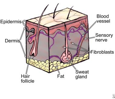
Tumor of the follicular infundibulum was first described in 1961 by Mehregan and Butler in a patient presenting with multiple papules. It is more frequent, however, as an isolated lesion affecting mainly the face, neck, and upper trunk. Clinical presentation is variable, requiring histology for the diagnosis, which reveals typically a plate-like proliferation of keratinocytes in continuity with the epidermis and hair follicles; some morphological features are reminiscent of the outer root sheath of the hair follicle. A well defined network of elastic fibers surrounding the tumor is usually present using the appropriate staining and this finding is specific because it is not found in other benign follicular tumors. Multiple infundibulomas are usually sporadic and there is no apparent association with internal malignancy. The authors report the case of a 30-year-old female patient with a 5-year history of multiple small discrete hypopigmented macules and papules, scattered over the submental and submaxillary regions and anterior neck. Histopathological findings were consistent with the diagnosis of tumor of the follicular infundibulum. Immunohistochemical study was performed to further characterize the proliferation. (+info)An acanthoma is a benign skin tumor characterized by the proliferation of epidermal cells, specifically the pickle cell layer (stratum spinosum). The term "acanthoma" comes from the Greek word "akantha," which means "thorn" or "spine."
There are several types of acanthomas, including:
1. Seborrheic keratosis: Also known as seborrheic warts, these are common benign growths that appear as rough, scaly patches on the skin. They can be tan, brown, or black and may have a waxy or greasy appearance.
2. Benign lichenoid keratosis: These are small, flat lesions with a scaly surface that typically occur on sun-exposed areas of the skin. They are usually asymptomatic but may occasionally itch.
3. Psoriasiform acanthoma: This is a rare type of acanthoma that resembles psoriasis, a chronic skin condition characterized by red, scaly patches.
4. Clear cell acanthoma: This is a distinctive type of acanthoma that appears as a solitary, dome-shaped nodule with a smooth surface and a central crust. It typically occurs on the lower legs of older adults.
Acanthomas are generally harmless and do not require treatment unless they become irritated or unsightly. In such cases, they can be removed through various methods, including cryosurgery (freezing), curettage (scraping), or excision (cutting).
A medical dictionary is a reference book that contains definitions and explanations of medical terms and jargon. It serves as a useful tool for healthcare professionals, students, patients, and anyone else who needs to understand medical terminology. Medical dictionaries can include definitions of diseases, conditions, treatments, procedures, drugs, equipment, anatomy, and more. They may also provide pronunciation guides, etymologies, and abbreviations.
Medical dictionaries can be found in print or digital form, and some are specialized to cover specific areas of medicine, such as oncology, psychiatry, or surgery. Some medical dictionaries are also bilingual, providing translations of medical terms between different languages. Overall, a medical dictionary is an essential resource for anyone who needs to communicate effectively in the field of medicine.
Malignant atrophic papulosis (MAP), also known as Kohlmeier-Degos disease, is a rare and progressive cutaneous vasculopathy of unknown etiology. It is characterized by the development of porcelain-white atrophic macules, which evolve into papules with a central necrotic depression or ulceration, surrounded by an erythematous halo. The lesions typically appear on the trunk and extremities, but may also affect mucous membranes, gastrointestinal tract, and other organs.
MAP is considered to be a chronic inflammatory disorder that affects small-sized blood vessels, leading to tissue ischemia and necrosis. The disease can have a variable clinical course, ranging from self-limited cutaneous involvement to systemic manifestations with potentially life-threatening complications.
The diagnosis of MAP is based on the clinical presentation, histopathological findings, and exclusion of other similar conditions. Treatment options for MAP are limited, and there is no cure for this disease. The management typically involves a multidisciplinary approach to address the various organ manifestations and prevent complications.
Keratoacanthoma is a rapidly growing, dome-shaped, skin tumor that typically arises on sun-exposed areas such as the face, arms, and legs. It is considered a low-grade squamous cell carcinoma (a type of skin cancer) because it shares some characteristics with both benign and malignant tumors.
Keratoacanthomas usually develop over a period of several weeks to months, growing rapidly in size before eventually stabilizing and then gradually regressing on their own within a few months to a year. However, the regression process can take years, and some lesions may not regress completely, leading to cosmetic concerns or even local invasion.
Histologically, keratoacanthomas are characterized by a central keratin-filled crater surrounded by a well-differentiated layer of squamous epithelial cells. The tumor's growth pattern and histological features can make it difficult to distinguish from other types of skin cancer, such as squamous cell carcinoma.
Treatment options for keratoacanthomas include surgical excision, cryosurgery, curettage and electrodesiccation, and topical therapies like imiquimod or 5-fluorouracil. The choice of treatment depends on various factors such as the size, location, and number of lesions, as well as patient preferences and overall health status.
An encyclopedia is a comprehensive reference work containing articles on various topics, usually arranged in alphabetical order. In the context of medicine, a medical encyclopedia is a collection of articles that provide information about a wide range of medical topics, including diseases and conditions, treatments, tests, procedures, and anatomy and physiology. Medical encyclopedias may be published in print or electronic formats and are often used as a starting point for researching medical topics. They can provide reliable and accurate information on medical subjects, making them useful resources for healthcare professionals, students, and patients alike. Some well-known examples of medical encyclopedias include the Merck Manual and the Stedman's Medical Dictionary.
Skin neoplasms refer to abnormal growths or tumors in the skin that can be benign (non-cancerous) or malignant (cancerous). They result from uncontrolled multiplication of skin cells, which can form various types of lesions. These growths may appear as lumps, bumps, sores, patches, or discolored areas on the skin.
Benign skin neoplasms include conditions such as moles, warts, and seborrheic keratoses, while malignant skin neoplasms are primarily classified into melanoma, squamous cell carcinoma, and basal cell carcinoma. These three types of cancerous skin growths are collectively known as non-melanoma skin cancers (NMSCs). Melanoma is the most aggressive and dangerous form of skin cancer, while NMSCs tend to be less invasive but more common.
It's essential to monitor any changes in existing skin lesions or the appearance of new growths and consult a healthcare professional for proper evaluation and treatment if needed.
 Acanthoma
Acanthoma fissuratum
Epidermolytic acanthoma
Clear cell acanthoma
Large-cell acanthoma
Pilar sheath acanthoma
Trichoadenoma
Dermatofibroma
Granuloma fissuratum
List of skin conditions
List of MeSH codes (C04)
List of Rhynchospora species
Acanthoma - Wikipedia
 Acanthoma fissuratum | DermNet
 Clear Cell Acanthoma | Treatment & Management | Point of Care
 Verruciform xanthoma in close association with isolated epidermolytic acanthoma: a case report and review of the Japanese...
Clear cell acanthoma - dermoscopedia academy
ACANTHOMA FISSURATUM CUTIS / ULCER
 Clear Cell Acanthoma - Derm In-Review
 Acanthoma etymology in English | Etymologeek.com
 Dermoscopy of clear cell acanthoma. - Dr. Aimilios Lallas
 Large cell acanthoma (Concept Id: C1334362)
- MedGen - NCBI
Large cell acanthoma (Concept Id: C1334362)
- MedGen - NCBI
Table 2 - Merkel Cell Polyomavirus DNA in Persons without Merkel Cell Carcinoma - Volume 15, Number 9-September 2009 - Emerging...
 Follicular Porokeratoma: Report of a New Variant of Porokeratoma (Porokeratotic
Acanthoma) and Literature Review. Kanitakis J,...
Follicular Porokeratoma: Report of a New Variant of Porokeratoma (Porokeratotic
Acanthoma) and Literature Review. Kanitakis J,...
 Merkel Cell Carcinoma and Rare Appendageal Tumors: Overview, Tumors of the Epidermis, Tumorlike Lesions of Fibrous or Elastic...
Dilated Pore of Winer Workup: Histologic Findings
 Indian Journal of Dermatology: Table of Contents
 Tumors of the Skin in Dogs - Dog Owners - Merck Veterinary Manual
Epulis Fissuratum: Practice Essentials, Pathophysiology, Epidemiology
 Sportability Misc Clinic Info
Tumors of the Skin in Dogs - Dog Owners - MSD Veterinary Manual
 Scaly signs in dermatology - Indian Journal of Dermatology, Venereology and Leprology
 Non-malignant skin disease - Libre Pathology
 KAKEN - Researchers | Adachi Takeya (30573258)
 DeCS 2020 - June 23, 2020 version
 Namespace
 Two-Step algorithm - dermoscopedia
 Woo Cheal Cho | MD Anderson Cancer Center
Carlos Antonio Torres-Cabala | MD Anderson Cancer Center
DeCS 2019 - June 12, 2019 version
DeCS 2018 - July 31, 2018 version
Sheath acanthoma2
- Lee JY, Hirsch E. Pilar sheath acanthoma. (medscape.com)
- Histopathological differential diagnosis includes trichoepithelioma, dilated pore of Winer, pilar sheath acanthoma, and basal cell carcinoma. (turkderm.org.tr)
Epidermolytic acanthoma2
Neoplasm3
- An acanthoma is a skin neoplasm composed of squamous or epidermal cells. (wikipedia.org)
- It is unclear whether clear cell acanthoma represents a benign neoplasm or a reactive inflammatory dermatosis. (statpearls.com)
- In fact, clear cell acanthoma possesses a similar staining pattern to inflammatory dermatoses such as psoriasis vulgaris, lichen planus, and discoid lupus erythematosus and might be a localized form of inflammatory eruption rather than a neoplasm. (medscape.com)
Large cell acanthoma5
- Dermoscopic findings and histopathological correlation in large cell acanthoma. (nih.gov)
- Clinicopathologic and immunohistochemical studies of conjunctival large cell acanthoma, epidermoid dysplasia, and squamous papilloma. (nih.gov)
- Large-cell acanthoma of the skin. (nih.gov)
- Large-cell acanthoma is a distinctive condition. (nih.gov)
- Large-cell acanthoma is a solar lentigo. (nih.gov)
Benign3
- Acanthoma fissuratum is a benign lesion that spontaneously resolves if the source of skin irritation is removed and the skin is allowed to heal. (dermnetnz.org)
- On the other hand, clear cell acanthoma is a rare, benign epithelial cutaneous tumor. (medscape.com)
- This presence of melanin provides pigmentation to the benign tumor, and this occurrence has been termed pigmented clear cell acanthoma. (medscape.com)
Dyskeratotic acanthoma1
- Hypergranulotic Dyskeratotic Acanthoma: Another Histologically Distinctive Acanthoma. (jefferson.edu)
Basal cell carc1
- Acanthoma fissuratum can be similar in appearance to basal cell carcinoma . (dermnetnz.org)
Acantholytic1
- I will call it acantholytic acanthoma. (dermpathpro.com)
Squamous2
- described a case of squamous cell carcinoma in situ occurring within a clear cell acanthoma. (statpearls.com)
- One side of the coin requires the observer to make a specific diagnosis by recognizing the classic patterns/structures associated with nevi, dermatofibromas (DF), intradermal nevi (IDN), basal cell carcinomas (BCC), squamous cell carcinomas (SCC), lentigines & seborrheic keratoses (SK), angiomas, angiokeratomas, sebaceous hyperplasias, and clear cell acanthoma (CCA). (dermoscopedia.org)
Fissuratum cutis1
- Acanthoma fissuratum cutis is a common cutaneous lesion induced by chronic mechanical trauma. (blogspot.com)
Tumor1
- Most experts maintain that clear cell acanthoma is an epidermal tumor, as originally described. (statpearls.com)
Degos2
- and Degos acanthoma, often confused with but unrelated to Degos disease. (wikipedia.org)
- Clear cell acanthoma (CCA), also known as "Degos acanthoma" and "acanthome cellules Claires of Degos and Civatte," was first described by Degos et al. (statpearls.com)
Lesions1
- Case reports in the literature report clear cell acanthomas with multiple lesions, (ie, polypoid clear cell acanthoma). (medscape.com)
Clear cell12
- The exact etiology of clear cell acanthoma is unknown. (statpearls.com)
- Clear cell acanthoma does not appear to occur in children. (statpearls.com)
- The pigmented clear cell acanthoma or melanoacanthoma histologically, as well as clinically varies from a typical clear cell acanthoma in that it shows a higher number of melanocytes and increased levels of melanin within the lesion. (statpearls.com)
- [2] To date, three case reports exist detailing "atypical clear cell acanthoma. (statpearls.com)
- A clear cell acanthoma presents as a solitary, red or red-brown, dome-shaped papule or nodule. (statpearls.com)
- This is the page on the glossary term Clear cell acanthoma which is relevant on dermoscopedia academy . (dermoscopedia.org)
- Dermoscopy of clear cell acanthoma. (aimilioslallas.com)
- Typically, the clinical presentation of clear cell acanthoma is a solitary nodule in the lower extremities. (medscape.com)
- The sharp demarcation of the borders of a clear cell acanthoma distinguishes clear cell acanthoma from psoriasis. (medscape.com)
- Clear cell acanthoma usually stains positively for epithelial membrane antigen and negatively for carcinoembryonic antigen. (medscape.com)
- Variants of the clear cell acanthoma have been noted and include polypoid, giant, multiple, and eruptive. (medscape.com)
- Wagner G, Back W, Sachse MM. Clear cell acanthoma. (lmshq.org)
Lesion1
- Acanthoma fissuratum presents as a firm flesh-coloured papule, nodule or plaque that has a central furrow dividing the lesion in half (a 'coffee bean' appearance). (dermnetnz.org)
Clinical1
- What are the clinical features of acanthoma fissuratum? (dermnetnz.org)
Differential diagnosis1
- What is the differential diagnosis of acanthoma fissuratum? (dermnetnz.org)
Histology1
- Acanthoma fissuratum has also been called granuloma fissuratum and spectacle frame granuloma, although no actual granulomas are found on histology . (dermnetnz.org)
Follicular1
- Follicular Porokeratoma: Report of a New Variant of Porokeratoma (Porokeratotic Acanthoma) and Literature Review. (amedeo.com)
Pore1
- Klovekorn G, Klovekorn W, Plewig G, Pinkus H. [Giant pore and hair-shaft acanthoma. (medscape.com)
Clinically1
- Acanthoma fissuratum can be recognised clinically if the possibility is considered. (dermnetnz.org)
Skin1
- Epulis fissuratum is analogous to acanthoma fissuratum of skin. (medscape.com)
Term3
- At that time, PubMed indexed only 206 articles with the term "acanthoma" (the term usually in the title or abstract). (wikipedia.org)
- When allowed to heal without irritation from spectacles, acanthoma fissuratum will resolve without long-term scarring. (dermnetnz.org)
- The very term "senile acanthoma" indicates that it is a disease of the elderly. (medic-journal.com)
Treatment1
- What is the treatment for acanthoma fissuratum? (dermnetnz.org)
Sheath6
- Pilar sheath acanthoma is an uncommon small benign tumour originating from the hair follicle . (dermnetnz.org)
- How is pilar sheath acanthoma diagnosed? (dermnetnz.org)
- A small biopsy (when a tiny piece of skin is removed under local anaesthetic ) is the only definitive diagnosis for pilar sheath acanthoma. (dermnetnz.org)
- The histology of pilar sheath acanthoma will differentiate it from other benign follicular tumours that have similar clinical presentations, these include dilated pore of Winer and trichofolliculoma . (dermnetnz.org)
- Pilar sheath acanthoma is a benign follicular tumour that requires no treatment. (dermnetnz.org)
- Lee JY, Hirsch E. Pilar sheath acanthoma. (medscape.com)
Acantholytic dyskeratotic acanthoma2
Fissuratum2
- Epulis fissuratum is analogous to acanthoma fissuratum of skin. (medscape.com)
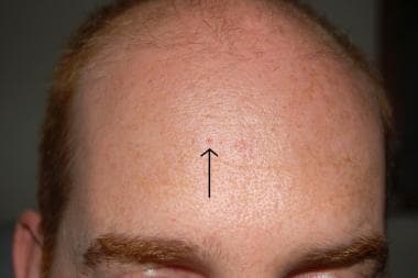
- Acanthoma fissuratum, commonly called 'bumps on nose,' is a medical term for the unusual growth that may develop in individuals who wear glasses for extended period. (rapplimited.com)
Papilloma1
- Clinicopathologic and immunohistochemical studies of conjunctival large cell acanthoma, epidermoid dysplasia, and squamous papilloma. (jamanetwork.com)
Abstract1
- At that time, PubMed indexed only 206 articles with the term "acanthoma" (the term usually in the title or abstract). (wikipedia.org)
Clear2
- Een clear cell acanthoma is een zeldzame, benigne, langzaam groeiende (2-10 mm per jaar) epidermale tumor, meestal op het onderbeen bij ouderen. (huidziekten.nl)
- Polypoid clear cell acanthoma of unusual size. (huidziekten.nl)
Large cell1
- 2. Large-cell acanthoma is a solar lentigo. (nih.gov)
Case1
- A case of acanthoma. (nih.gov)
Hair1
- Klovekorn G, Klovekorn W, Plewig G, Pinkus H. [Giant pore and hair-shaft acanthoma. (medscape.com)