Accessory Nerve Injuries
Accessory Nerve
Accessory Nerve Diseases
Nerve Transfer
Paralysis
Neck Dissection
Shoulder
Sciatic Nerve
Neck Muscles
Glossopharyngeal Nerve
Cranial Nerve Injuries
Scapula
Physical therapy for spinal accessory nerve injury complicated by adhesive capsulitis. (1/18)
BACKGROUND AND PURPOSE: The authors found no literature describing adhesive capsulitis as a consequence of spinal accessory nerve injury and no exercise program or protocol for patients with spinal accessory nerve injury. The purpose of this case report is to describe the management of a patient with adhesive capsulitis and spinal accessory nerve injury following a carotid endarterectomy. CASE DESCRIPTION: The patient was a 67-year-old woman referred for physical therapy following manipulation of the left shoulder and a diagnosis of adhesive capsulitis by her orthopedist. Spinal accessory nerve injury was identified during the initial physical therapy examination, and a program of neuromuscular electrical stimulation was initiated. OUTCOMES: The patient had almost full restoration of the involved muscle function after 5 months of physical therapy. DISCUSSION: This case report illustrates the importance of accurate diagnosis and suggests physical therapy intervention to manage adhesive capsulitis as a consequence of spinal accessory nerve injury. (+info)Levator scapulae and rhomboid transfer for paralysis of trapezius. The Eden-Lange procedure. (2/18)
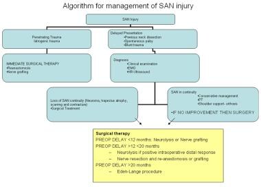
Spinal accessory nerve palsy leads to painful disability of the shoulder, carrying an uncertain prognosis. We reviewed the long-term outcome in 16 patients who were treated for pain, weakness of active elevation and asymmetry of the shoulder and the neck due to chronic paralysis of the trapezius muscle, as a result of nerve palsy. Of four patients who were treated conservatively, none regained satisfactory function, although two became pain-free. The other 12 patients were treated operatively with transfer of the levator scapulae to the acromion and the rhomboid muscles to the infraspinatus fossa (the Eden-Lange procedure). At a mean follow-up of 32 years, the clinical outcome of the operatively treated patients was excellent in nine, fair in two, and poor in one patient, as determined by the Constant score. Pain was adequately relieved in 11 and overhead function was restored in nine patients. Pre-operative electromyography had been carried out in four patients. In two, who eventually had a poor outcome, a concomitant long thoracic and dorsal scapular nerve lesion had been present. The Eden-Lange procedure gives very satisfactory long-term results for the treatment of isolated paralysis of trapezius. In the presence of an additional serratus anterior palsy or weak rhomboid muscles, the procedure is less successful in restoring shoulder function. (+info)Accessory nerve injury. (3/18)
This article discusses a Supreme Court judgment involving an injury to the spinal accessory nerve which occurred during the excision of a lymph node mass in the posterior triangle of the neck.1 In this case, the medical practitioner was found to have been negligent for failing to diagnose the nerve injury in the postoperative period, and not for the actual injury to the nerve during the procedure. (+info)An unusual presentation of whiplash injury: long thoracic and spinal accessory nerve injury. (4/18)
Whiplash injuries from motor vehicle accidents are very common. The usual presentation and course of this condition normally results in resolution of symptoms within a few weeks. Brachial plexus traction injuries without any bone or joint lesion of the cervical spine have been reported before. We report a case where a gentleman was involved in a rear end vehicle collision, sustained a whiplash injury and was later found to have a long thoracic nerve palsy and spinal accessory nerve palsy. Although isolated injuries of both nerves following a whiplash injury have been reported, combined injury of the two nerves following a whiplash injury is very uncommon and is being reported for the first time. (+info)Surgical treatment of winged scapula. (5/18)
(+info)Spinal accessory nerve palsy following gunshot injury: a case report. (6/18)
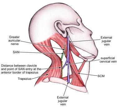
Injuries to the spinal accessory nerve are rare and mostly iatrogenic. Pain, impaired ability to raise the ipsilateral shoulder, and scapular winging on abduction of the arm are the most frequently noted clinical manifestations. As a seldom case, a 20 year-old male with spinal accessory nerve palsy after penetrating trauma by gunshot was reported. Three months after the injury, he was complaining about left arm pain in abduction to shoulder level and a decreased range of movement. On physical examination, wasting of the left trapezium with loss of nuchal ridge and drooping of the shoulder were found. On neurological examination of the left trapezius and sternomastoid muscles, motor function were 3/5 and wide dysesthesia on the neck, shoulder and arm was present. The bullet entered just above the clavicle and exited from trapezium. Radiological studies were normal, where electromyography (EMG) showed neuropathic changes. Surgical exploration showed the intact nerve lying on its natural course and we performed external neurolysis for decompression. The postoperative period was uneventful. Dysesthesia has diminished slowly. He was transferred to physical rehabilitation unit. In his clinical control after 3 months he had no dysesthesia and neurological examination of the left trapezius and sternomastoid muscles motor function were 4/5. EMG showed recovery in the left spinal accessory nerve. (+info)Vernet's syndrome caused by large mycotic aneurysm of the extracranial internal carotid artery after acute otitis media--case report. (7/18)
An 85-year-old man presented with a rare large aneurysm of the extracranial internal carotid artery (ICA) due to acute otitis media manifesting as Vernet's syndrome 2 weeks after the diagnosis of right acute otitis media. Angiography of the right extracranial ICA demonstrated an irregularly shaped large aneurysm with partial thrombosis. The aneurysm was treated by proximal ICA occlusion using endovascular coils. The ICA mycotic aneurysm was triggered by acute otitis media, and induced Vernet's syndrome as a result of direct compression to the jugular foramen. Extracranial ICA aneurysms due to focal infection should be considered in the differential diagnosis of lower cranial nerve palsy, although the incidence is thought to be very low. (+info)Accessory nerve palsy. (8/18)
After apparently uncomplicated excision of benign lesions in the posterior cervical triangle, two patients had shoulder pain. In one, neck pain and trapezius weakness were not prominent until one month after surgery. Inability to elevate the arm above the horizontal without externally rotating it, and prominent scapular displacement on arm abduction, but not on forward pushing movements, highlighted the trapezius dysfunction and differentiated it from serratus anterior weakness. Spinal accessory nerve lesions should be considered when minor surgical procedures, lymphadenitis, minor trauma, or tumours involved the posterior triangle of the neck. (+info)Accessory nerve injuries refer to damage or trauma to the eleventh cranial nerve, also known as the accessory nerve. This nerve has both a cranial and spinal root, and it primarily controls the movement of some muscles in the neck and shoulder.
Injuries to the accessory nerve can result in weakness or paralysis of the affected muscles, leading to difficulty turning the head or lifting the arm. The severity of the symptoms depends on the extent and location of the injury. Accessory nerve injuries can occur due to various reasons, such as trauma during surgery (particularly neck or shoulder surgeries), penetrating injuries, tumors, or neurological disorders.
Treatment for accessory nerve injuries typically involves a combination of physical therapy, pain management, and, in some cases, surgical intervention to repair the damaged nerve. The prognosis for recovery varies depending on the severity and cause of the injury.
The accessory nerve, also known as the eleventh cranial nerve (XI), has both a cranial and spinal component. It primarily controls the function of certain muscles in the back of the neck and shoulder.
The cranial part arises from nuclei in the brainstem and innervates some of the muscles that help with head rotation, including the sternocleidomastoid muscle. The spinal root originates from nerve roots in the upper spinal cord (C1-C5), exits the spine, and joins the cranial part to form a single trunk. This trunk then innervates the trapezius muscle, which helps with shoulder movement and stability.
Damage to the accessory nerve can result in weakness or paralysis of the affected muscles, causing symptoms such as difficulty turning the head, weak shoulder shrugging, or winged scapula (a condition where the shoulder blade protrudes from the back).
The accessory nerve, also known as the 11th cranial nerve (CN XI), has both a cranial and spinal root and innervates the sternocleidomastoid muscle and trapezius muscle. Accessory nerve diseases refer to conditions that affect the function of this nerve, leading to weakness or paralysis of the affected muscles.
Some examples of accessory nerve diseases include:
1. Traumatic injury: Direct trauma to the neck or posterior scalene region can damage the spinal root of the accessory nerve. This can result in weakness or paralysis of the trapezius muscle, leading to difficulty with shoulder movement and pain.
2. Neuralgia: Accessory nerve neuralgia is a condition characterized by painful spasms or shooting pains along the course of the accessory nerve. It can be caused by nerve compression, inflammation, or injury.
3. Tumors: Tumors in the neck region, such as schwannomas or neurofibromas, can compress or invade the accessory nerve, leading to weakness or paralysis of the affected muscles.
4. Infections: Viral infections, such as poliovirus or West Nile virus, can cause inflammation and damage to the accessory nerve, resulting in weakness or paralysis.
5. Neuropathy: Accessory nerve neuropathy is a condition characterized by degeneration of the accessory nerve fibers due to various causes such as diabetes, autoimmune disorders, or exposure to toxins. This can result in weakness or paralysis of the affected muscles.
6. Congenital defects: Some individuals may be born with congenital defects that affect the development and function of the accessory nerve, leading to weakness or paralysis of the affected muscles.
Treatment for accessory nerve diseases depends on the underlying cause and can include physical therapy, medications, surgery, or a combination of these approaches.
Peripheral nerve injuries refer to damage or trauma to the peripheral nerves, which are the nerves outside the brain and spinal cord. These nerves transmit information between the central nervous system (CNS) and the rest of the body, including sensory, motor, and autonomic functions. Peripheral nerve injuries can result in various symptoms, depending on the type and severity of the injury, such as numbness, tingling, weakness, or paralysis in the affected area.
Peripheral nerve injuries are classified into three main categories based on the degree of damage:
1. Neuropraxia: This is the mildest form of nerve injury, where the nerve remains intact but its function is disrupted due to a local conduction block. The nerve fiber is damaged, but the supporting structures remain intact. Recovery usually occurs within 6-12 weeks without any residual deficits.
2. Axonotmesis: In this type of injury, there is damage to both the axons and the supporting structures (endoneurium, perineurium). The nerve fibers are disrupted, but the connective tissue sheaths remain intact. Recovery can take several months or even up to a year, and it may be incomplete, with some residual deficits possible.
3. Neurotmesis: This is the most severe form of nerve injury, where there is complete disruption of the nerve fibers and supporting structures (endoneurium, perineurium, epineurium). Recovery is unlikely without surgical intervention, which may involve nerve grafting or repair.
Peripheral nerve injuries can be caused by various factors, including trauma, compression, stretching, lacerations, or chemical exposure. Treatment options depend on the type and severity of the injury and may include conservative management, such as physical therapy and pain management, or surgical intervention for more severe cases.
A nerve transfer is a surgical procedure where a functioning nerve is connected to an injured nerve to restore movement, sensation or function. The functioning nerve, called the donor nerve, usually comes from another less critical location in the body and has spare nerve fibers that can be used to reinnervate the injured nerve, called the recipient nerve.
During the procedure, a small section of the donor nerve is carefully dissected and prepared for transfer. The recipient nerve is also prepared by removing any damaged or non-functioning portions. The two ends are then connected using microsurgical techniques under a microscope. Over time, the nerve fibers from the donor nerve grow along the recipient nerve and reinnervate the muscles or sensory structures that were previously innervated by the injured nerve.
Nerve transfers can be used to treat various types of nerve injuries, including brachial plexus injuries, facial nerve palsy, and peripheral nerve injuries. The goal of the procedure is to restore function as quickly and efficiently as possible, allowing for a faster recovery and improved quality of life for the patient.
Paralysis is a loss of muscle function in part or all of your body. It can be localized, affecting only one specific area, or generalized, impacting multiple areas or even the entire body. Paralysis often occurs when something goes wrong with the way messages pass between your brain and muscles. In most cases, paralysis is caused by damage to the nervous system, especially the spinal cord. Other causes include stroke, trauma, infections, and various neurological disorders.
It's important to note that paralysis doesn't always mean a total loss of movement or feeling. Sometimes, it may just cause weakness or numbness in the affected area. The severity and extent of paralysis depend on the underlying cause and the location of the damage in the nervous system.
Neck dissection is a surgical procedure that involves the removal of lymph nodes and other tissues from the neck. It is typically performed as part of cancer treatment, particularly in cases of head and neck cancer, to help determine the stage of the cancer, prevent the spread of cancer, or treat existing metastases. There are several types of neck dissections, including radical, modified radical, and selective neck dissection, which vary based on the extent of tissue removal. The specific type of neck dissection performed depends on the location and extent of the cancer.
In anatomical terms, the shoulder refers to the complex joint of the human body that connects the upper limb to the trunk. It is formed by the union of three bones: the clavicle (collarbone), scapula (shoulder blade), and humerus (upper arm bone). The shoulder joint is a ball-and-socket type of synovial joint, allowing for a wide range of movements such as flexion, extension, abduction, adduction, internal rotation, and external rotation.
The shoulder complex includes not only the glenohumeral joint but also other structures that contribute to its movement and stability, including:
1. The acromioclavicular (AC) joint: where the clavicle meets the acromion process of the scapula.
2. The coracoclavicular (CC) ligament: connects the coracoid process of the scapula to the clavicle, providing additional stability to the AC joint.
3. The rotator cuff: a group of four muscles (supraspinatus, infraspinatus, teres minor, and subscapularis) that surround and reinforce the shoulder joint, contributing to its stability and range of motion.
4. The biceps tendon: originates from the supraglenoid tubercle of the scapula and passes through the shoulder joint, helping with flexion, supination, and stability.
5. Various ligaments and capsular structures that provide additional support and limit excessive movement in the shoulder joint.
The shoulder is a remarkable joint due to its wide range of motion, but this also makes it susceptible to injuries and disorders such as dislocations, subluxations, sprains, strains, tendinitis, bursitis, and degenerative conditions like osteoarthritis. Proper care, exercise, and maintenance are essential for maintaining shoulder health and function throughout one's life.
The sciatic nerve is the largest and longest nerve in the human body, running from the lower back through the buttocks and down the legs to the feet. It is formed by the union of the ventral rami (branches) of the L4 to S3 spinal nerves. The sciatic nerve provides motor and sensory innervation to various muscles and skin areas in the lower limbs, including the hamstrings, calf muscles, and the sole of the foot. Sciatic nerve disorders or injuries can result in symptoms such as pain, numbness, tingling, or weakness in the lower back, hips, legs, and feet, known as sciatica.
Neck muscles, also known as cervical muscles, are a group of muscles that provide movement, support, and stability to the neck region. They are responsible for various functions such as flexion, extension, rotation, and lateral bending of the head and neck. The main neck muscles include:
1. Sternocleidomastoid: This muscle is located on either side of the neck and is responsible for rotating and flexing the head. It also helps in tilting the head to the same side.
2. Trapezius: This large, flat muscle covers the back of the neck, shoulders, and upper back. It is involved in movements like shrugging the shoulders, rotating and extending the head, and stabilizing the scapula (shoulder blade).
3. Scalenes: These three pairs of muscles are located on the side of the neck and assist in flexing, rotating, and laterally bending the neck. They also help with breathing by elevating the first two ribs during inspiration.
4. Suboccipitals: These four small muscles are located at the base of the skull and are responsible for fine movements of the head, such as tilting and rotating.
5. Longus Colli and Longus Capitis: These muscles are deep neck flexors that help with flexing the head and neck forward.
6. Splenius Capitis and Splenius Cervicis: These muscles are located at the back of the neck and assist in extending, rotating, and laterally bending the head and neck.
7. Levator Scapulae: This muscle is located at the side and back of the neck, connecting the cervical vertebrae to the scapula. It helps with rotation, extension, and elevation of the head and scapula.
The glossopharyngeal nerve, also known as the ninth cranial nerve (IX), is a mixed nerve that carries both sensory and motor fibers. It originates from the medulla oblongata in the brainstem and has several functions:
1. Sensory function: The glossopharyngeal nerve provides general sensation to the posterior third of the tongue, the tonsils, the back of the throat (pharynx), and the middle ear. It also carries taste sensations from the back one-third of the tongue.
2. Special visceral afferent function: The nerve transmits information about the stretch of the carotid artery and blood pressure to the brainstem.
3. Motor function: The glossopharyngeal nerve innervates the stylopharyngeus muscle, which helps elevate the pharynx during swallowing. It also provides parasympathetic fibers to the parotid gland, stimulating saliva production.
4. Visceral afferent function: The glossopharyngeal nerve carries information about the condition of the internal organs in the thorax and abdomen to the brainstem.
Overall, the glossopharyngeal nerve plays a crucial role in swallowing, taste, saliva production, and monitoring blood pressure and heart rate.
Cranial nerve injuries refer to damages or trauma to one or more of the twelve cranial nerves (CN I through CN XII). These nerves originate from the brainstem and are responsible for transmitting sensory information (such as vision, hearing, smell, taste, and balance) and controlling various motor functions (like eye movement, facial expressions, swallowing, and speaking).
Cranial nerve injuries can result from various causes, including head trauma, tumors, infections, or neurological conditions. The severity of the injury may range from mild dysfunction to complete loss of function, depending on the extent of damage to the nerve. Treatment options vary based on the type and location of the injury but often involve a combination of medical management, physical therapy, surgical intervention, or rehabilitation.
The scapula, also known as the shoulder blade, is a flat, triangular bone located in the upper back region of the human body. It serves as the site of attachment for various muscles that are involved in movements of the shoulder joint and arm. The scapula has several important features:
1. Three borders (anterior, lateral, and medial)
2. Three angles (superior, inferior, and lateral)
3. Spine of the scapula - a long, horizontal ridge that divides the scapula into two parts: supraspinous fossa (above the spine) and infraspinous fossa (below the spine)
4. Glenoid cavity - a shallow, concave surface on the lateral border that articulates with the humerus to form the shoulder joint
5. Acromion process - a bony projection at the top of the scapula that forms part of the shoulder joint and serves as an attachment point for muscles and ligaments
6. Coracoid process - a hook-like bony projection extending from the anterior border, which provides attachment for muscles and ligaments
Understanding the anatomy and function of the scapula is essential in diagnosing and treating various shoulder and upper back conditions.
Iatrogenic disease refers to any condition or illness that is caused, directly or indirectly, by medical treatment or intervention. This can include adverse reactions to medications, infections acquired during hospitalization, complications from surgical procedures, or injuries caused by medical equipment. It's important to note that iatrogenic diseases are unintended and often preventable with proper care and precautions.
 Accessory nerve