Acute Chest Syndrome
Anemia, Sickle Cell
Embolism, Fat
Hemoglobin SC Disease
Antisickling Agents
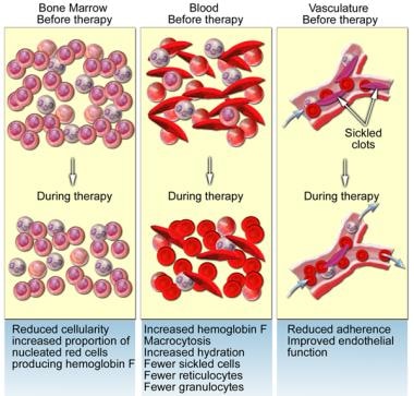
Hydroxyurea
Hemoglobin, Sickle
Pain
Blood Transfusion
Fetal Hemoglobin
Respiratory Insufficiency
Hemolysis
Retrospective Studies
Triage
Cohort Studies
Acute Coronary Syndrome
Emergency Service, Hospital
The acute chest syndrome of sickle cell disease following aortic valve replacement. (1/27)
(+info)Asthma and sickle cell disease: two distinct diseases or part of the same process? (2/27)
(+info)Novel therapies in sickle cell disease. (3/27)
(+info)Acute kidney injury in sickle patients with painful crisis or acute chest syndrome and its relation to pulmonary hypertension. (4/27)
(+info)The burden of emergency department use for sickle-cell disease: an analysis of the national emergency department sample database. (5/27)
(+info)Multi-modal intervention for the inpatient management of sickle cell pain significantly decreases the rate of acute chest syndrome. (6/27)
(+info)Improving care for children with sickle cell disease/acute chest syndrome. (7/27)
(+info)Elevation of IgE in children with sickle cell disease is associated with doctor diagnosis of asthma and increased morbidity. (8/27)
(+info)Acute chest syndrome (ACS) is a serious complication of sickle cell disease, characterized by the presence of new infiltrates on chest X-ray and at least one other clinical symptom such as fever, cough, chest pain, or difficulty breathing. It is often caused by infection, fat embolism, or lung tissue inflammation, leading to respiratory distress, hypoxemia, and potentially respiratory failure. Prompt diagnosis and treatment with antibiotics, analgesics, and sometimes blood transfusions or exchange transfusions are essential for managing ACS.
Sickle cell anemia is a genetic disorder that affects the hemoglobin in red blood cells. Hemoglobin is responsible for carrying oxygen throughout the body. In sickle cell anemia, the hemoglobin is abnormal and causes the red blood cells to take on a sickle shape, rather than the normal disc shape. These sickled cells are stiff and sticky, and they can block blood vessels, causing tissue damage and pain. They also die more quickly than normal red blood cells, leading to anemia.
People with sickle cell anemia often experience fatigue, chronic pain, and jaundice. They may also have a higher risk of infections and complications such as stroke, acute chest syndrome, and priapism. The disease is inherited from both parents, who must both be carriers of the sickle cell gene. It primarily affects people of African descent, but it can also affect people from other ethnic backgrounds.
There is no cure for sickle cell anemia, but treatments such as blood transfusions, medications to manage pain and prevent complications, and bone marrow transplantation can help improve quality of life for affected individuals. Regular medical care and monitoring are essential for managing the disease effectively.
Chest pain is a discomfort or pain that you feel in the chest area. The pain can be sharp, dull, burning, crushing, heaviness, or tightness. It may be accompanied by other symptoms such as shortness of breath, sweating, nausea, dizziness, or pain that radiates to the arm, neck, jaw, or back.
Chest pain can have many possible causes, including heart-related conditions such as angina or a heart attack, lung conditions such as pneumonia or pleurisy, gastrointestinal problems such as acid reflux or gastritis, musculoskeletal issues such as costochondritis or muscle strain, and anxiety or panic attacks.
It is important to seek immediate medical attention if you experience chest pain that is severe, persistent, or accompanied by other concerning symptoms, as it may be a sign of a serious medical condition. A healthcare professional can evaluate your symptoms, perform tests, and provide appropriate treatment.
Fat embolism is a medical condition that occurs when fat globules enter the bloodstream and block small blood vessels (arterioles and capillaries) in various tissues and organs. This can lead to inflammation, tissue damage, and potentially life-threatening complications.
Fat embolism typically occurs as a result of trauma, such as long bone fractures or orthopedic surgeries, where fat cells from the marrow of the broken bone enter the bloodstream. It can also occur in other conditions that cause fat to be released into the circulation, such as pancreatitis, decompression sickness, and certain medical procedures like liposuction.
Symptoms of fat embolism may include respiratory distress, fever, confusion, petechial rash (small purple or red spots on the skin), and a decrease in oxygen levels. In severe cases, it can lead to acute respiratory distress syndrome (ARDS) and even death. Treatment typically involves supportive care, such as oxygen therapy, mechanical ventilation, and medications to manage symptoms and prevent complications.
Hemoglobin SC disease, also known as sickle cell-C disease or SC disorder, is a genetic blood disorder that is a variant of sickle cell anemia. It is caused by the presence of both hemoglobin S (HbS) and hemoglobin C (HbC) in the red blood cells.
Hemoglobin is the protein in red blood cells that carries oxygen throughout the body. In Hemoglobin SC disease, the abnormal HbS and HbC proteins can cause the red blood cells to become rigid, sticky, and C-shaped (sickled), which can lead to blockages in small blood vessels.
Symptoms of Hemoglibin SC disease may include anemia, fatigue, jaundice, episodes of pain (known as sickle cell crises), and an increased risk of infection. The severity of the symptoms can vary widely from person to person. Treatment typically focuses on managing symptoms and preventing complications, and may include medications, blood transfusions, and sometimes a bone marrow transplant.
Antisickling agents are medications or substances that help prevent or reduce the sickling of red blood cells in individuals with sickle cell disease. Sickling is a pathological process where the normally disc-shaped red blood cells become crescent-shaped due to abnormal hemoglobin (HbS). This change in shape can lead to blockages in small blood vessels, causing tissue damage and various complications such as pain crises, acute chest syndrome, and stroke.
Antisickling agents work by either inhibiting the polymerization of HbS or improving the oxygen-carrying capacity of red blood cells. The most commonly used antisickling agent is hydroxyurea, which increases the production of fetal hemoglobin (HbF) in red blood cells. HbF has a higher affinity for oxygen than HbS and can prevent the polymerization of HbS, thereby reducing sickling. Other antisickling agents include:
1. L-glutamine: An amino acid that helps maintain the structural integrity of red blood cells and reduces oxidative stress.
2. Arginate: A salt of arginine, an amino acid that helps improve nitric oxide production and vasodilation, reducing sickling.
3. Senicapoc: A drug that inhibits the formation of HbS polymers by blocking the interaction between HbS molecules.
4. Voxelotor (Oxbryta): A medication that binds to HbS and stabilizes it in its oxygenated state, reducing sickling.
These antisickling agents can help alleviate symptoms, decrease the frequency of pain crises, and improve the quality of life for individuals with sickle cell disease. However, they should be used under the supervision of a healthcare professional, as each has its benefits, risks, and potential side effects.
A syndrome, in medical terms, is a set of symptoms that collectively indicate or characterize a disease, disorder, or underlying pathological process. It's essentially a collection of signs and/or symptoms that frequently occur together and can suggest a particular cause or condition, even though the exact physiological mechanisms might not be fully understood.
For example, Down syndrome is characterized by specific physical features, cognitive delays, and other developmental issues resulting from an extra copy of chromosome 21. Similarly, metabolic syndromes like diabetes mellitus type 2 involve a group of risk factors such as obesity, high blood pressure, high blood sugar, and abnormal cholesterol or triglyceride levels that collectively increase the risk of heart disease, stroke, and diabetes.
It's important to note that a syndrome is not a specific diagnosis; rather, it's a pattern of symptoms that can help guide further diagnostic evaluation and management.
Lung diseases refer to a broad category of disorders that affect the lungs and other structures within the respiratory system. These diseases can impair lung function, leading to symptoms such as coughing, shortness of breath, chest pain, and wheezing. They can be categorized into several types based on the underlying cause and nature of the disease process. Some common examples include:
1. Obstructive lung diseases: These are characterized by narrowing or blockage of the airways, making it difficult to breathe out. Examples include chronic obstructive pulmonary disease (COPD), asthma, bronchiectasis, and cystic fibrosis.
2. Restrictive lung diseases: These involve stiffening or scarring of the lungs, which reduces their ability to expand and take in air. Examples include idiopathic pulmonary fibrosis, sarcoidosis, and asbestosis.
3. Infectious lung diseases: These are caused by bacteria, viruses, fungi, or parasites that infect the lungs. Examples include pneumonia, tuberculosis, and influenza.
4. Vascular lung diseases: These affect the blood vessels in the lungs, impairing oxygen exchange. Examples include pulmonary embolism, pulmonary hypertension, and chronic thromboembolic pulmonary hypertension (CTEPH).
5. Neoplastic lung diseases: These involve abnormal growth of cells within the lungs, leading to cancer. Examples include small cell lung cancer, non-small cell lung cancer, and mesothelioma.
6. Other lung diseases: These include interstitial lung diseases, pleural effusions, and rare disorders such as pulmonary alveolar proteinosis and lymphangioleiomyomatosis (LAM).
It is important to note that this list is not exhaustive, and there are many other conditions that can affect the lungs. Proper diagnosis and treatment of lung diseases require consultation with a healthcare professional, such as a pulmonologist or respiratory therapist.
Sickle cell trait is a genetic condition where an individual inherits one abnormal gene for hemoglobin S (HbS) from one parent and one normal gene for hemoglobin A (HbA) from the other parent. Hemoglobin is a protein in red blood cells that carries oxygen throughout the body.
People with sickle cell trait do not have sickle cell disease, but they can pass the abnormal HbS gene on to their children. In certain situations, such as high altitude, low oxygen levels, or intense physical exertion, individuals with sickle cell trait may experience symptoms similar to those of sickle cell disease, such as fatigue, pain, and shortness of breath. However, these symptoms are typically milder and less frequent than in people with sickle cell disease.
It is important for individuals who know they have sickle cell trait to inform their healthcare providers, especially if they become pregnant or plan to engage in activities that may cause low oxygen levels, such as scuba diving or high-altitude climbing.
An acute disease is a medical condition that has a rapid onset, develops quickly, and tends to be short in duration. Acute diseases can range from minor illnesses such as a common cold or flu, to more severe conditions such as pneumonia, meningitis, or a heart attack. These types of diseases often have clear symptoms that are easy to identify, and they may require immediate medical attention or treatment.
Acute diseases are typically caused by an external agent or factor, such as a bacterial or viral infection, a toxin, or an injury. They can also be the result of a sudden worsening of an existing chronic condition. In general, acute diseases are distinct from chronic diseases, which are long-term medical conditions that develop slowly over time and may require ongoing management and treatment.
Examples of acute diseases include:
* Acute bronchitis: a sudden inflammation of the airways in the lungs, often caused by a viral infection.
* Appendicitis: an inflammation of the appendix that can cause severe pain and requires surgical removal.
* Gastroenteritis: an inflammation of the stomach and intestines, often caused by a viral or bacterial infection.
* Migraine headaches: intense headaches that can last for hours or days, and are often accompanied by nausea, vomiting, and sensitivity to light and sound.
* Myocardial infarction (heart attack): a sudden blockage of blood flow to the heart muscle, often caused by a buildup of plaque in the coronary arteries.
* Pneumonia: an infection of the lungs that can cause coughing, chest pain, and difficulty breathing.
* Sinusitis: an inflammation of the sinuses, often caused by a viral or bacterial infection.
It's important to note that while some acute diseases may resolve on their own with rest and supportive care, others may require medical intervention or treatment to prevent complications and promote recovery. If you are experiencing symptoms of an acute disease, it is always best to seek medical attention to ensure proper diagnosis and treatment.
Hydroxyurea is an antimetabolite drug that is primarily used in the treatment of myeloproliferative disorders such as chronic myelogenous leukemia (CML), essential thrombocythemia, and polycythemia vera. It works by interfering with the synthesis of DNA, which inhibits the growth of cancer cells.
In addition to its use in cancer therapy, hydroxyurea is also used off-label for the management of sickle cell disease. In this context, it helps to reduce the frequency and severity of painful vaso-occlusive crises by increasing the production of fetal hemoglobin (HbF), which decreases the formation of sickled red blood cells.
The medical definition of hydroxyurea is:
A hydantoin derivative and antimetabolite that inhibits ribonucleoside diphosphate reductase, thereby interfering with DNA synthesis. It has been used as an antineoplastic agent, particularly in the treatment of myeloproliferative disorders, and more recently for the management of sickle cell disease to reduce the frequency and severity of painful vaso-occlusive crises by increasing fetal hemoglobin production.
Hemoglobin S (HbS) is a genetic variant of hemoglobin, which is the oxygen-carrying protein in red blood cells. This abnormal form of hemogllobin results from a mutation in the beta-globin gene, leading to the substitution of valine for glutamic acid at position six of the beta-globin chain.
In individuals with sickle cell disease (a group of inherited red blood cell disorders), both copies of their beta-globin genes carry this mutation, causing the majority of their hemoglobin to be HbS. When deoxygenated, HbS molecules have a tendency to polymerize and form long, rigid rods within the red blood cells, distorting their shape into a characteristic sickle or crescent form.
These sickled red blood cells are less flexible and more prone to rupture (hemolysis), leading to chronic anemia, vaso-occlusive crises, and other disease complications. Sickle cell disease primarily affects people of African, Mediterranean, Middle Eastern, and Indian ancestry, but it can also be found in other populations worldwide.
Pain is an unpleasant sensory and emotional experience associated with actual or potential tissue damage, or described in terms of such damage. It is a complex phenomenon that can result from various stimuli, such as thermal, mechanical, or chemical irritation, and it can be acute or chronic. The perception of pain involves the activation of specialized nerve cells called nociceptors, which transmit signals to the brain via the spinal cord. These signals are then processed in different regions of the brain, leading to the conscious experience of pain. It's important to note that pain is a highly individual and subjective experience, and its perception can vary widely among individuals.
A blood transfusion is a medical procedure in which blood or its components are transferred from one individual (donor) to another (recipient) through a vein. The donated blood can be fresh whole blood, packed red blood cells, platelets, plasma, or cryoprecipitate, depending on the recipient's needs. Blood transfusions are performed to replace lost blood due to severe bleeding, treat anemia, support patients undergoing major surgeries, or manage various medical conditions such as hemophilia, thalassemia, and leukemia. The donated blood must be carefully cross-matched with the recipient's blood type to minimize the risk of transfusion reactions.
Fetal hemoglobin (HbF) is a type of hemoglobin that is produced in the fetus and newborn babies. It is composed of two alpha-like globin chains and two gamma-globin chains, designated as α2γ2. HbF is the primary form of hemoglobin during fetal development, replacing the embryonic hemoglobin (HbG) around the eighth week of gestation.
The unique property of HbF is its higher affinity for oxygen compared to adult hemoglobin (HbA), which helps ensure adequate oxygen supply from the mother to the developing fetus. After birth, as the newborn starts breathing on its own and begins to receive oxygen directly, the production of HbF gradually decreases and is usually replaced by HbA within the first year of life.
In some genetic disorders like sickle cell disease and beta-thalassemia, persistence of HbF into adulthood can be beneficial as it reduces the severity of symptoms due to its higher oxygen-carrying capacity and less polymerization tendency compared to HbS (in sickle cell disease) or unpaired alpha chains (in beta-thalassemia). Treatments like hydroxyurea are used to induce HbF production in these patients as a therapeutic approach.
Respiratory insufficiency is a condition characterized by the inability of the respiratory system to maintain adequate gas exchange, resulting in an inadequate supply of oxygen and/or removal of carbon dioxide from the body. This can occur due to various causes, such as lung diseases (e.g., chronic obstructive pulmonary disease, pneumonia), neuromuscular disorders (e.g., muscular dystrophy, spinal cord injury), or other medical conditions that affect breathing mechanics and/or gas exchange.
Respiratory insufficiency can manifest as hypoxemia (low oxygen levels in the blood) and/or hypercapnia (high carbon dioxide levels in the blood). Symptoms of respiratory insufficiency may include shortness of breath, rapid breathing, fatigue, confusion, and in severe cases, loss of consciousness or even death. Treatment depends on the underlying cause and severity of the condition and may include oxygen therapy, mechanical ventilation, medications, and/or other supportive measures.
A pulmonary embolism (PE) is a medical condition that occurs when a blood clot, often formed in the deep veins of the legs (deep vein thrombosis), breaks off and travels to the lungs, blocking one or more pulmonary arteries. This blockage can lead to various symptoms such as shortness of breath, chest pain, rapid heart rate, and coughing up blood. In severe cases, it can cause life-threatening complications like low oxygen levels, hypotension, and even death if not promptly diagnosed and treated with anticoagulant medications or thrombolytic therapy to dissolve the clot.
Hemolysis is the destruction or breakdown of red blood cells, resulting in the release of hemoglobin into the surrounding fluid (plasma). This process can occur due to various reasons such as chemical agents, infections, autoimmune disorders, mechanical trauma, or genetic abnormalities. Hemolysis may lead to anemia and jaundice, among other complications. It is essential to monitor hemolysis levels in patients undergoing medical treatments that might cause this condition.
Fever, also known as pyrexia or febrile response, is a common medical sign characterized by an elevation in core body temperature above the normal range of 36.5-37.5°C (97.7-99.5°F) due to a dysregulation of the body's thermoregulatory system. It is often a response to an infection, inflammation, or other underlying medical conditions, and it serves as a part of the immune system's effort to combat the invading pathogens or to repair damaged tissues.
Fevers can be classified based on their magnitude:
* Low-grade fever: 37.5-38°C (99.5-100.4°F)
* Moderate fever: 38-39°C (100.4-102.2°F)
* High-grade or severe fever: above 39°C (102.2°F)
It is important to note that a single elevated temperature reading does not necessarily indicate the presence of a fever, as body temperature can fluctuate throughout the day and can be influenced by various factors such as physical activity, environmental conditions, and the menstrual cycle in females. The diagnosis of fever typically requires the confirmation of an elevated core body temperature on at least two occasions or a consistently high temperature over a period of time.
While fevers are generally considered beneficial in fighting off infections and promoting recovery, extremely high temperatures or prolonged febrile states may necessitate medical intervention to prevent potential complications such as dehydration, seizures, or damage to vital organs.
Leukocytosis is a condition characterized by an increased number of leukocytes (white blood cells) in the peripheral blood. A normal white blood cell count ranges from 4,500 to 11,000 cells per microliter of blood in adults. Leukocytosis is typically considered present when the white blood cell count exceeds 11,000 cells/µL. However, the definition might vary slightly depending on the laboratory and clinical context.
Leukocytosis can be a response to various underlying conditions, including bacterial or viral infections, inflammation, tissue damage, leukemia, and other hematological disorders. It is essential to investigate the cause of leukocytosis through further diagnostic tests, such as blood smears, differential counts, and additional laboratory and imaging studies, to guide appropriate treatment.
Retrospective studies, also known as retrospective research or looking back studies, are a type of observational study that examines data from the past to draw conclusions about possible causal relationships between risk factors and outcomes. In these studies, researchers analyze existing records, medical charts, or previously collected data to test a hypothesis or answer a specific research question.
Retrospective studies can be useful for generating hypotheses and identifying trends, but they have limitations compared to prospective studies, which follow participants forward in time from exposure to outcome. Retrospective studies are subject to biases such as recall bias, selection bias, and information bias, which can affect the validity of the results. Therefore, retrospective studies should be interpreted with caution and used primarily to generate hypotheses for further testing in prospective studies.
Triage is a medical term that refers to the process of prioritizing patients based on the severity of their condition or illness, and the resources available. The goal of triage is to ensure that the most critical patients receive care first, which can help reduce morbidity and mortality in emergency situations. This process is typically used in settings where there are more patients than can be treated immediately, such as during mass casualty incidents or in busy emergency departments. Triage nurses or doctors quickly assess each patient's condition, often using a standardized system, to determine the urgency of their medical needs and allocate resources accordingly.
A cohort study is a type of observational study in which a group of individuals who share a common characteristic or exposure are followed up over time to determine the incidence of a specific outcome or outcomes. The cohort, or group, is defined based on the exposure status (e.g., exposed vs. unexposed) and then monitored prospectively to assess for the development of new health events or conditions.
Cohort studies can be either prospective or retrospective in design. In a prospective cohort study, participants are enrolled and followed forward in time from the beginning of the study. In contrast, in a retrospective cohort study, researchers identify a cohort that has already been assembled through medical records, insurance claims, or other sources and then look back in time to assess exposure status and health outcomes.
Cohort studies are useful for establishing causality between an exposure and an outcome because they allow researchers to observe the temporal relationship between the two. They can also provide information on the incidence of a disease or condition in different populations, which can be used to inform public health policy and interventions. However, cohort studies can be expensive and time-consuming to conduct, and they may be subject to bias if participants are not representative of the population or if there is loss to follow-up.
Acute Coronary Syndrome (ACS) is a term used to describe a range of conditions associated with sudden, reduced blood flow to the heart muscle. This reduction in blood flow, commonly caused by blood clots forming in coronary arteries, can lead to damage or death of the heart muscle and is often characterized by symptoms such as chest pain, shortness of breath, and fatigue.
There are three main types of ACS:
1. Unstable Angina: This occurs when there is reduced blood flow to the heart muscle, causing chest pain or discomfort, but the heart muscle is not damaged. It can be a warning sign for a possible future heart attack.
2. Non-ST Segment Elevation Myocardial Infarction (NSTEMI): This type of heart attack occurs when there is reduced blood flow to the heart muscle, causing damage or death of some of the muscle cells. However, the electrical activity of the heart remains relatively normal.
3. ST Segment Elevation Myocardial Infarction (STEMI): This is a serious and life-threatening type of heart attack that occurs when there is a complete blockage in one or more of the coronary arteries, causing extensive damage to the heart muscle. The electrical activity of the heart is significantly altered, which can lead to dangerous heart rhythms and even cardiac arrest.
Immediate medical attention is required for anyone experiencing symptoms of ACS, as prompt treatment can help prevent further damage to the heart muscle and reduce the risk of complications or death. Treatment options may include medications, lifestyle changes, and procedures such as angioplasty or bypass surgery.
An emergency service in a hospital is a department that provides immediate medical or surgical care for individuals who are experiencing an acute illness, injury, or severe symptoms that require immediate attention. The goal of an emergency service is to quickly assess, stabilize, and treat patients who require urgent medical intervention, with the aim of preventing further harm or death.
Emergency services in hospitals typically operate 24 hours a day, 7 days a week, and are staffed by teams of healthcare professionals including physicians, nurses, physician assistants, nurse practitioners, and other allied health professionals. These teams are trained to provide rapid evaluation and treatment for a wide range of medical conditions, from minor injuries to life-threatening emergencies such as heart attacks, strokes, and severe infections.
In addition to providing emergency care, hospital emergency services also serve as a key point of entry for patients who require further hospitalization or specialized care. They work closely with other departments within the hospital, such as radiology, laboratory, and critical care units, to ensure that patients receive timely and appropriate treatment. Overall, the emergency service in a hospital plays a crucial role in ensuring that patients receive prompt and effective medical care during times of crisis.
X-ray computed tomography (CT or CAT scan) is a medical imaging method that uses computer-processed combinations of many X-ray images taken from different angles to produce cross-sectional (tomographic) images (virtual "slices") of the body. These cross-sectional images can then be used to display detailed internal views of organs, bones, and soft tissues in the body.
The term "computed tomography" is used instead of "CT scan" or "CAT scan" because the machines take a series of X-ray measurements from different angles around the body and then use a computer to process these data to create detailed images of internal structures within the body.
CT scanning is a noninvasive, painless medical test that helps physicians diagnose and treat medical conditions. CT imaging provides detailed information about many types of tissue including lung, bone, soft tissue and blood vessels. CT examinations can be performed on every part of the body for a variety of reasons including diagnosis, surgical planning, and monitoring of therapeutic responses.
In computed tomography (CT), an X-ray source and detector rotate around the patient, measuring the X-ray attenuation at many different angles. A computer uses this data to construct a cross-sectional image by the process of reconstruction. This technique is called "tomography". The term "computed" refers to the use of a computer to reconstruct the images.
CT has become an important tool in medical imaging and diagnosis, allowing radiologists and other physicians to view detailed internal images of the body. It can help identify many different medical conditions including cancer, heart disease, lung nodules, liver tumors, and internal injuries from trauma. CT is also commonly used for guiding biopsies and other minimally invasive procedures.
In summary, X-ray computed tomography (CT or CAT scan) is a medical imaging technique that uses computer-processed combinations of many X-ray images taken from different angles to produce cross-sectional images of the body. It provides detailed internal views of organs, bones, and soft tissues in the body, allowing physicians to diagnose and treat medical conditions.
 Acute chest syndrome
Acute coronary syndrome
Mediastinum
Varespladib
Lorna Breen
Angor animi
Sickle cell disease
Vaso-occlusive crisis
Hemoglobin O-Arab
Troponin
COVID-19 pandemic in Bhutan
Costochondritis
Francis M. Fesmire
Exchange transfusion
Transfusion therapy (Sickle-cell disease)
Heart-type fatty acid binding protein
Cocaine intoxication
Glutamine
List of syndromes
Plastic bronchitis
Sickle cell trait
Hemolytic jaundice
Acute inhalation injury
N-Methyl-2-pyrrolidone
Acute aortic syndrome
Management of acute coronary syndrome
SARS
Kounis syndrome
Oxygen therapy
Acute respiratory distress syndrome
Acute chest syndrome - Wikipedia
JCI - Extracellular hemin crisis triggers acute chest syndrome in sickle mice
Chlorine Inhalation Induces Acute Chest Syndrome in Humanized Sickle Cell Mouse Model and Ameliorated by Postexposure Hemopexin
 Management of Acute Chest Syndrome in Sickle Cell Disease
 Comparison of chest computed tomography features in the acute phase of cardiogenic pulmonary edema and acute respiratory...
A new score for the diagnosis of acute coronary syndrome in acute chest pain with non-diagnostic ECG and normal troponin |...
 Acute chest syndrome - WikEM
 Angina - when you have chest pain: MedlinePlus Medical Encyclopedia
 Acute Chest Syndrome (Sickle Cell Disease) - First10EM
 Warning Signs Of Acute Chest Syndrome - HealthPrep.com
 The Risk that Chest Pain Represents Acute Coronary Syndrome
 The Invisible Illness: Sickle Cell Disease - AACN
 Sickle Cell Disease | Boston Children's Hospital
 Managing Acute Chest Syndrome: The Past, Present, and Future | Baylor College of Medicine
 New Protocol Dramatically Improves Outcomes for Children with Acute Chest Syndrome - Inside Pediatrics
 Severe Acute Respiratory Syndrome (SARS): Practice Essentials, Pathophysiology, Etiology
 An unusual cause of chest pain: Acute coronary syndrome following administration of ergotamine tartrate | AVESİS
 Sickle Cell Crisis/Acute Chest Syndrome | Case Files: Emergency Medicine, 5e | AccessEmergency Medicine | McGraw Hill Medical
 Staff View: Outcomes of acute chest syndrome in adult patients with sickle cell disease: predictors of mortality.
Coronavirus Disease among Persons with Sickle Cell Disease, United States, March 20-May 21, 2020 - Volume 26, Number 10-October...
 Sickle Cell Disease and Blood Transfusion: What to Know
Search Results: Chest pain, Dyspnea
Gene-centric association study of acute chest syndrome and painful crisis in sickle cell disease patients - Fingerprint -...
 Robust segmentation of lung in chest x-ray: applications in analysis of acute respiratory distress syndrome | BMC Medical...
 Inhaled nitric oxide for acute chest syndrome in people with sickle cell disease. | Hospital Medicine Virtual Journal Club |...
 Right on Prime - Clinical Value Of Diagnostic Instruments For Ruling Out Acute Coronary Syndrome In Patients With Chest Pain: A...
 Acute coronary syndrome - Symptoms and causes - Mayo Clinic
 Death rate rises among people with aortic dissection - UPI.com
NFL Player Tevin Coleman on Parenting a Child with Sickle Cell Disease
 Boerhaave's Syndrome | West Indian Medical Journal
Myocardial Infarction8
- The main outcome was whether the patients received objective evaluation for coronary artery disease after adjustment for cardiac risk, including race, age, total number of risk factors, Thrombolysis in Myocardial Infarction (TIMI) score, ECG, and whether the patient sustained an acute myocardial infarction on index hospitalization. (cdc.gov)
- Models adjusting for acute myocardial infarction or death, high-risk initial clinical impression, or emergency department disposition found similar results for increased likelihood of cardiac catheterization in men but no difference in stress testing between men and women. (cdc.gov)
- We did not perform acute coronary syndrome, coronary elevation myocardial infarction, whereas angioplasty because the obstruction embolism should be kept in mind in the rest present with non-ST elevation was in the distal portion of the vessel those with prosthetic valves even in the myocardial infarction [8]. (who.int)
- Participants 21 482 patients with acute myocardial infarction in England between January 2003 and March 2009, identified in four prospectively collected, linked electronic health record sources: Clinical Practice Research Datalink (primary care data), Hospital Episode Statistics (hospital admissions), the disease registry MINAP (Myocardial Ischaemia National Audit Project), and the Office for National Statistics mortality register (cause specific mortality data). (bmj.com)
- Main outcome measures Recording of acute myocardial infarction, incidence, all cause mortality within one year of acute myocardial infarction, and diagnostic validity of acute myocardial infarction compared with electrocardiographic and troponin findings in the disease registry (gold standard). (bmj.com)
- Immediate all cause mortality was highest among patients with acute myocardial infarction recorded in primary care, which (unlike hospital admission and disease registry sources) included patients who did not reach hospital, but at one year mortality rates in cohorts from each source were similar. (bmj.com)
- 5561 (31.0%) patients with non-fatal acute myocardial infarction were recorded in all three sources and 11 482 (63.9%) in at least two sources. (bmj.com)
- The crude incidence of acute myocardial infarction was underestimated by 25-50% using one source compared with using all three sources. (bmj.com)
Vaso-occlus4
- The acute chest syndrome is a vaso-occlusive crisis of the pulmonary vasculature commonly seen in people with sickle cell anemia. (wikipedia.org)
- Painful Vaso-occlusive Crisis as a Prodromal Phase of Acute Chest Syndrome. (cuni.cz)
- In contrast, we found that a simple PTS for HbF that includes only six variants explained a large fraction of the phenotypic variation (20.5-27.1%), associated with acute chest syndrome and stroke risk, and improved the statistical modeling of the vaso-occlusive crisis rate. (haematologica.org)
- 1 SCD patients present a wide range of complications such as vaso-occlusive crisis (VOC), acute chest syndrome (ACS), stroke, and end-organ dysfunction, and their life expectancy is reduced when compared to the general population. (haematologica.org)
Stroke5
- Current management strategies include prophylactic penicillin and immunizations to decrease the occurrence of pneumococcal infections, hydroxyurea (a disease-modifying agent), blood transfusions (for symptomatic acute anemia, stroke management, preoperative optimization), and bone marrow transplant. (ccjm.org)
- This can cause pain and other serious problems including infection, acute chest syndrome, and stroke. (cdc.gov)
- Encounters for complicated VOCs such as acute chest syndrome and stroke were excluded. (confex.com)
- Abstract: DESCRIPTION (provided by applicant): Sickle cell disease (SCD) is characterized by acute clinical manifestations, including painful crisis, acute chest syndrome, priapism, and stroke, as well as chronic irreversible damage to the heart, lungs, kidneys, eyes, spleen and femoral heads. (nih.gov)
- These cardiovascular complications following a stroke are known as Stroke-Heart Syndrome. (kvia.com)
Severe8
- Thus, persons with SCD could be at higher risk for development of severe disease if infected with severe acute respiratory syndrome coronavirus 2 (SARS-CoV-2), the causative agent of coronavirus disease (COVID-19). (cdc.gov)
- A patient in their 60s presented to the emergency department with approximately 20 minutes of acute, severe precordial chest pain radiating to their left arm at night, accompanied by dyspnea, dizziness, and sweating. (ama-assn.org)
- Additionally, during VOC, patients are often in severe pain, resulting in shallow breathing with minimal chest wall expansion, and together with high doses of opioids, it could lead to hypoventilation, with the formation of atelectasis, and increase risk for the development of ACS [ 6 ]. (hindawi.com)
- Sporadic cases of hantavirus pulmonary syndrome (HPS), a severe cardiopulmonary illness first identified in 1993, continue to be recognized in the United States (1,2). (cdc.gov)
- The ASH guideline panel suggests automated RCE or manual RCE over simple transfusions in patients with SCD and severe acute chest syndrome. (guidelinecentral.com)
- The medicine can help certain types of sickle cell diseases, like hemoglobin SS, pain crises, acute chest syndrome, severe anemia needing blood transfusions, and tiny blockages without pain. (consultantlive.com)
- Severe acute respiratory syndrome coronavirus 2 (SARS-CoV-2) has spread worldwide for more than a year. (frontiersin.org)
- Acute respiratory distress syndrome (ARDS) is caused by a severe inflammatory response in the body when there is a severe infection or after there has been trauma to the body. (nationaljewish.org)
Detected during1
- An increase in acute phase reactants and inflammatory markers can be detected during crises and predict severity [ 9 , 10 ]. (hindawi.com)
Pneumonia3
- citation needed] The diagnosis of acute chest syndrome is made difficult by its similarity in presentation with pneumonia. (wikipedia.org)
- Patients with SCD often have various complications, including acute chest syndrome (ACS), a life-threatening condition similar to pneumonia but unique to SCD. (cureswithinreach.org)
- Treated children needed fewer hospitalizations and blood transfusions and had fewer episodes of a pneumonia-like complication called acute chest syndrome. (nih.gov)
Complication2
- Acute chest syndrome (ACS) is a life-threatening complication in people living with SCD that can result in lung injury, breathing difficulty, and low oxygen to the rest of the body. (cdc.gov)
- A fever may be the first sign of an infection or other SCD-related complication, such as acute chest syndrome , that can be life-threatening. (cdc.gov)
Anemia1
- Cardiac chest pain can also be precipitated by anemia , bradycardias or tachycardias . (wikidoc.org)
Respiratory5
- The presence of fevers, low oxygen levels in the blood, increased respiratory rate, chest pain, and cough are also common in acute chest syndrome. (wikipedia.org)
- Conclusion This patient with prior treatment for coronary artery disease status post CABG x4 in 2003 presented with chest pain, acute respiratory distress requiring ventilator support was found to h. (aapc.com)
- For patients with acute respiratory distress syndrome lung functions improved faster during the first six months. (nature.com)
- What are the signs and symptoms of acute respiratory distress syndrome (ARDS)? (nationaljewish.org)
- Because acute respiratory distress syndrome involves internal fluid that fills the lungs' air sacs, doctors look for common signs (things that are observed) and symptoms (things that are experienced). (nationaljewish.org)
Symptoms4
- Dominant presenting symptoms in patients without chest pain (total exceeds 100% as patients may have presented with more than one dominant symptom). (medscape.com)
- Patients without chest pain were significantly older than those with typical symptoms. (medscape.com)
- Symptoms include coughing and chest pain. (peacehealth.org)
- It may also help you be more active without chest pain or other symptoms. (stlukes-stl.com)
Among patients with acute1
- To assess smoking cessation rates at 24 weeks among patients with acute coronary syndrome. (who.int)
Shortness of bre1
- You have been having chest pain or pressure, or shortness of breath. (stlukes-stl.com)
Diagnosis5
- Chest radiography for the diagnosis of acute aortic syndrome. (qxmd.com)
- Since a definitive diagnosis is required in any patient with clinically suspected acute aortic syndrome, routine chest radiography should be replaced by tomographic aortic imaging. (qxmd.com)
- The most common symptom prompting diagnosis of ACS is chest pain , often radiating to the left arm or angle of the jaw , pressure-like in character, and associated with nausea and sweating . (wikidoc.org)
- To view the detailed differential diagnosis of chest pain click here . (wikidoc.org)
- Control subjects ( n = 70) were identified among patients admitted for acute chest pain and in which a diagnosis of ATE was excluded. (thieme-connect.com)
Patients with sickle2
- De D. Acute nursing care and management of patients with sickle cell. (medscape.com)
- Close to half of all patients with sickle cell disease (SCD) will have at least one episode of acute chest syndrome (ACS) during their lifetime. (hindawi.com)
Radiography6
- We sought to assess the diagnostic accuracy of routine chest radiography for the acute aortic syndrome (dissection, intramural hematoma, penetrating ulcer, or nondissecting aneurysm). (qxmd.com)
- mean [+/- SD] age, 58 +/- 17 years) underwent chest radiography for suspected acute aortic syndrome. (qxmd.com)
- Chest radiography had a sensitivity of 64% (70/109) and a specificity of 86% (92/107) for aortic disease. (qxmd.com)
- Chest radiography is of limited value for diagnosing the acute aortic syndrome, particularly for conditions confined to the ascending aorta. (qxmd.com)
- A structured data instrument that included demographic information, chest pain description, history, physical examination, chest radiography, and electrocardiogram (ECG) data was completed. (cdc.gov)
- Chest radiography indicated ground-glass opacities and consolidation at the onset of disease, with the exception of 8.9% (5/56) that showed minor changes. (nature.com)
Infection1
- Acute chest syndrome is often precipitated by a lung infection, and the resulting inflammation and loss of oxygen saturation leads to further sickling of red cells, thus exacerbating pulmonary and systemic hypoxemia, sickling, and vaso-occlusion. (wikipedia.org)
Cardiovascular2
- Compared with HEART-score, HET-score is simpler and appears to have similar ability to discriminate between chest pain patients with and without cardiovascular event. (nih.gov)
- The aim of this study was to investigate the generation of NETs in different groups of patients with acute thrombotic events (ATEs) and to establish whether NETs markers can predict the risk of new cardiovascular events. (thieme-connect.com)
Transfusions3
- Hydroxycarbamide substantially reduces episodes of pain and acute chest syndrome, admissions to hospital, and transfusions in adults with sickle-cell anaemia. (nih.gov)
- The ASH guideline panel suggests automated RCE, manual RCE, or simple transfusions in patients with SCD and moderate acute chest syndrome. (guidelinecentral.com)
- The investigators wrote how other studies have noted the beneficial effects of hydroxyurea treatment, how the medication reduces pain, dactylitis, acute chest syndrome, hospitalizations, and transfusions. (consultantlive.com)
Sickle-cell an2
- Sickle-cell anaemia is associated with substantial morbidity from acute complications and organ dysfunction beginning in the first year of life. (nih.gov)
- According to the World Health Organization, the approximate estimates of affected individuals indicate that 240 million people are heterozygous for these disorders and at least 200000 lethally affected homozygotes are born annually, approximately equally divided between sickle-cell anaemia and thalassaemia syndromes. (ukessays.com)
Coronary heart d1
- Association of endothelial nitric oxide synthase promoter region (T-786C) gene polymorphism with acute coronary syndrome and coronary heart disease. (nih.gov)
Hydroxyurea2
- citation needed] Hydroxyurea is a medication that can help to prevent acute chest syndrome. (wikipedia.org)
- After detecting recurring vaso-occlusive crises or acute chest syndrome, a doctor may prescribe hydroxyurea. (consultantlive.com)
Cardiac4
- Other treatment for cardiac arrest, dysrhythmias, or acute hypertension may also be required. (medscape.com)
- Used to treat acute hypertension and cardiac chest pain. (medscape.com)
- More than two thirds of sudden cardiac death resulting from acute thrombus occurs in smokers (3). (who.int)
- in different circumstances, the cardiac apex could also be situated abnormally in the left side of the chest. (ehd.org)
Hemolytic1
- The ASH guideline panel suggests immunosuppressive therapy (intravenous immunoglobulin [IVIg], steroids, and/or rituximab) over no immunosuppressive therapy in patients with SCD (all genotypes) with an acute need for transfusion and at high risk for acute hemolytic transfusion reaction or with a history of multiple or life-threatening delayed hemolytic transfusion reactions. (guidelinecentral.com)
Painful2
- Painful events (sickle cell crises) in the hands or feet, belly, back, or chest are the most common symptom of sickle cell disease. (peacehealth.org)
- Acute painful vaso -occlusive crises (VOC) are the leading cause of emergency department (ED) encounters and hospital admissions for those with sickle cell disease (SCD). (confex.com)
Presenting with chest pain1
- Patients presenting with chest pain with duration of ≥10 min and an onset of last episode ≤12 h but without ST-segment elevation on ECG at 6 emergency departments were eligible for inclusion. (nih.gov)
Clinical2
- Medicine Central , im.unboundmedicine.com/medicine/view/5-Minute-Clinical-Consult/1688316/all/Chest_Pain_Acute_Coronary_Syndrome. (unboundmedicine.com)
- In 2017, the US Food and Drug Administration (FDA) approved L-glutamine oral powder for reducing acute complications of SCD, and many other drugs are in development and undergoing clinical testing. (ccjm.org)
Diagnostic1
- Acute- and convalescent-phase serum specimens from patient 2 were submitted to the South Dakota Public Health Laboratory and CDC for hantavirus diagnostic testing. (cdc.gov)
Organ1
- In conclusion, we report here the longest prospective neonatal cohort study to date addressing acute vaso-occlusive, hematological, and extracerebral major organ complications, and providing new data about transfusion requirements in [sickle cell disease] children," the investigators wrote. (consultantlive.com)
Disease5
- What is Hand-Foot Syndrome in Sickle Cell Disease? (cdc.gov)
- Chest films were re-evaluated blindly for aortic disease, based on an overall impression using standard criteria such as widening of the aortic contour and mediastinal shadow. (qxmd.com)
- LaDonna was hospitalized for three weeks with Acute Chest Syndrome (ACS), a common disorder for children with sickle cell disease. (lifesouth.org)
- I also see patients who have acute manifestations of sickle cell disease. (reachmd.com)
- Bronchial hyper-responsiveness may be a component of acute chest syndrome in people with sickle cell disease. (worldasthmafoundation.org)
Crisis1
- If patients with SCD crisis are being transported by emergency medical services (EMS), they should receive supplemental oxygen and intravenous hydration en route to the hospital. (medscape.com)
Discomfort2
- Angina is a type of chest discomfort due to poor blood flow through the blood vessels of the heart muscle. (medlineplus.gov)
- Chest discomfort or pain - This feeling can happen when inhaling. (nationaljewish.org)
Hospitalization1
- Few randomized controlled trials have examined the efficacy time of smoking cessation in hospitalized patients with acute coronary syndrome, either during hospitalization or after discharge. (who.int)
Lung2
- This condition commonly manifests with a new opacification of the lung(s) on a chest x-ray. (wikipedia.org)
- Both may present with a new opacification of the lung on chest x-ray. (wikipedia.org)
Thoracic1
- Doppler AdsonпїЅs test: predictor of consequence of surgery in non-specific thoracic outlet syndrome. (ehd.org)
Inhibitor1
- The use of angiotensin-converting enzyme (ACE) inhibitors was comparable between the two groups during hospital admission, and, at the time of discharge from hospital, patients who had presented without chest pain were more likely to be receiving an ACE inhibitor. (medscape.com)
Abdomen1
- Pain develops when sickle-shaped red blood cells block blood flow through tiny blood vessels to your chest, abdomen and joints. (healthanddietblog.com)
Bone marrow1
- First description of bone marrow failure syndrome in Spain caused by mutations in the ERCC6L2 gene. (amedeo.com)
Illness2
Arterial1
- In conclusion, we found that increase of markers of NETosis can be observed in acute thrombotic conditions, occurring both on the arterial and venous site. (thieme-connect.com)
Asthma1
- The main objectives of this paper were to test the hypothesis that polymorphisms in NOS1 and NOS3 genes associate with ACS in SCD patients and to characterize the association between physician-diagnosed asthma and acute chest syndrome (ACS). (nih.gov)
Angina1
- ACS should be distinguished from stable angina , which is chest pain that develops during exertion and resolves at rest. (wikidoc.org)
Interstitial1
- and alanine aminotransferase {ALT} 138 U/L {normal: 7-56 U/L}). Although he reported no abdominal pain and the abdominal examination on admission was normal, serum amylase and lipase levels were elevated (amylase 226 U/L {normal: 30-110 U/L} and lipase 771 U/L {normal: 23-300 U/L}). Chest radiographs at the time of admission demonstrated perihilar interstitial infiltrates. (cdc.gov)
Blood1
- Acute coronary syndrome ( ACS ) refers to a spectrum of conditions resulting from acute myocardial ischemia and/or infarction that is most often due to an abrupt reduction in coronary blood flow . (wikidoc.org)
METHODS1
- Methods: Patients with potential acute coronary syndrome (ACS) who presented to a university hospital were prospectively identified. (cdc.gov)