Adenoma, Pleomorphic
Adenoma, Villous
Pituitary Neoplasms
Adrenocortical Adenoma
Adenoma, Chromophobe
Growth Hormone-Secreting Pituitary Adenoma
Colorectal Neoplasms
Colonic Polyps
ACTH-Secreting Pituitary Adenoma
Adenoma, Acidophil
Prolactinoma
Adenoma, Basophil
Adenomatous Polyposis Coli
Acromegaly
Cushing Syndrome
Genes, APC
Hyperparathyroidism
Hyperplasia
Neoplasms, Multiple Primary
Pituitary ACTH Hypersecretion
Immunohistochemistry
Carcinoma
Hyperaldosteronism
Sphenoid Bone
Adrenocorticotropic Hormone
Pituitary Gland
Hyperparathyroidism, Primary
Precancerous Conditions
Sella Turcica
Pituitary Apoplexy
Human Growth Hormone
Colon
Pituitary Hormones
Adenomatous Polyposis Coli Protein
Urethane
Intestinal Mucosa
Prolactin
Villous adenoma of the bile ducts: a case report and a review of the reported cases in Korea. (1/60)
Villous adenomas are benign epithelial lesions with malignant potential which can occur at any site in the gastrointestinal tract. They are usually encountered in the rectum and colon, less frequently in the small bowel and very rarely in the biliary trees. Nine cases of bile duct villous adenomas have been reported in the literature. However, 4 cases of bile duct villous adenomas have been reported in the Korean literature. Recently, we experienced a case of villous adenoma in the common hepatic duct in a 77-year-old man presenting with obstructive jaundice in which preoperative histologic diagnosis of villous adenoma played a critical role in managing this patient. Herein, we present a case report of bile duct villous adenoma and a review of the reported cases in Korea to help define and manage this rare disease entity in the bile ducts. In addition, confusing nomenclature of bile duct adenomas is discussed. (+info)Treatment options for villous adenoma of the ampulla of Vater. (2/60)
INTRODUCTION: Duodenal villous adenoma arising from the ampulla of Vater has a high risk of malignant development. Excluding associated malignant disease prior to resection of an adenoma of the ampulla is not always possible. Therefore, the surgical procedure of choice to treat this rare tumour is still controversial. OBJECTIVE: To evaluate retrospectively results of treatment of villous adenoma arising from ampulla of Vater with dysplasia or associated carcinoma limited to the ampulla. PATIENTS AND METHODS: From 1985 to 1996, eight patients have been diagnosed with ampullary villous adenoma suitable for resection. We have reviewed treatment, morbidity, mortality, follow-up and final outcome. RESULTS: Pancreatoduodenectomy (PD) was performed in 4 patients. Transduodenal ampullectomy and endoscopic resection was performed in 2 patients each. There was no perioperative mortality. None of the patients had biliary, pancreatic or intestinal leakage but two patients who underwent PD had minor postoperative complications. The mean follow-up was 44 (range: 6-132) months. Villous adenoma was associated with adenocarcinoma in 50% of the cases (4/8 patients). During the follow-up both patients who underwent transduodenal ampullectomy developed recurrent disease. All patients initially treated by PD are alive without evidence of recurrent disease. CONCLUSIONS: Treatment of villous adenoma of the ampulla must be individualized within certain limits. In our series, PD achieve good results and it appears to be the procedure of choice in order to treat villous adenomas with proved presence of carcinoma, carcinoma in situ or severe dysplasia. Endoscopic or local resection may be appropriate for small benign tumours in high risk patients. (+info)Adenoma of the papillae of Vater. Report of eleven cases. (3/60)
Eleven patients with a preoperative diagnosis of adenoma of the papillae of Vater were followed up during the fifteen-year period from 1984 till 1998 in the Oulu University Hospital. Seven patients were treated primarily by transduodenal excision without any recurrences so far. One of these seven patients was found to have adenocarcinoma in a histological examination. Active surgery for adenoma of the papillae of Vater is recommended because of the precancerous nature of the lesion, and because malignancy cannot always be detected by endoscopic biopsies. Transduodenal excision could be recommend for patients at high operative risk, especially in cases with small adenomas and low-grade dysplasia, where histologically free resection margins can be achieved, but pancreaticoduodenectomy should still be performed on patients at low operative risk. (+info)Overexpression of cyclin D1 mRNA in colorectal carcinomas and relationship to clinicopathological features: an in situ hybridization analysis. (4/60)
Increased expression of a key cell cycle regulator, cyclin D1, may have relevance to carcinogenesis and clinicopathological characteristics of some cancers. This study represents the first application of in situ hybridization, ISH, to detect cyclin D1 mRNA in tissue sections from colorectal carcinomas. This approach was selected because of its unique potential to clarify whether increased expression of cyclin D1 mRNA correlates with clinical and pathological parameters. The ISH ofa non-radioactive oligonucleotide probe (Biogenex) was immunocytochemically detected in paraffin embedded sections from biopsy or resection specimens. Tumors ranged from well to poorly differentiated, and from stages A, B, C, and D. Ten year survival data were available on the majority of patients. Intensity of tumor and background (smooth muscle) signals were independently scored from 0 to 3. Overexpressed cyclin D1 mRNA was seen in 86% of cases compared to background. This frequency is similar to that reported for pancreatic carcinoma. The average signal intensity score in tumor foci was 1.9 with a background score of 0.05 (p<001). All cases showed specific staining judged by the cytoplasmic localization and a tumor signal:background ratio >1. Expression did not differentiate cancers based on grade, stage or survival (p>1), but did differentiate carcinoma and severe dysplasia from mild dysplasia. We conclude that ISH of cyclin D1 mRNA is an effective and relatively specific means of detecting activity of this gene in colonic neoplasms. The high frequency of overexpression implies that gene activity by itself is not likely to predict a tumor s biological or clinical behavior. On the other hand, these data suggest that increased cyclin D1 gene activity may be an early event in colorectal carcinogenesis. They also are consistent with findings showing cyclin D1 is inducible by a variety of oncogene products. (+info)Lack of mucin MUC5AC field change expression associated with tubulovillous and villous colorectal adenomas. (5/60)
BACKGROUND: MUC5AC is a secreted mucin aberrantly expressed by polypoid colorectal adenomas. It has been hypothesised that the "normal" surrounding colorectal mucosa expresses MUC5AC as a field change phenomenon that can be used to predict adenoma recurrence following resection. AIM: To determine if there is a field change of de novo MUC5AC expression in histologically normal rectal mucosa adjacent to villous and tubulovillous adenomas, and thus whether MUC5AC expression can be used as a marker of early tumour recurrence. METHODS: In a prospective cohort study paired mucosal biopsies of adenomatous and macroscopically "normal" mucosa were obtained from 11 patients with villous and 11 patients with tubulovillous adenomas who underwent primary resection for purpose of cure. The tissues were studied to determine MUC5AC gene expression by immunohistochemistry and in situ hybridisation. Patients were followed up by flexible sigmoidoscopy to detect the presence of early local recurrence. RESULTS: 10 villous adenomas showed mature MUC5AC glycoprotein and all 11 expressed MUC5AC mRNA. Five tubulovillous adenomas showed mature MUC5AC glycoprotein and 10 expressed MUC5AC mRNA. Neoexpression of the MUC5AC mucin gene was not detected in any of the mucosal biopsies taken adjacent to either villous or tubulovillous adenomas, even in three patients with early, locally recurrent disease. CONCLUSIONS: Aberrant MUC5AC gene expression is not a "field change" in the colorectal mucosa in patients with rectal adenomas and therefore cannot be used to predict local recurrence of villous and tubulovillous adenomas. (+info)Offline telepathology diagnosis of colorectal polyps: a study of interobserver agreement and comparison with glass slide diagnoses. (6/60)
BACKGROUND/AIMS: Technological advances have produced telepathology systems with high quality colour images and reasonable transmission times. Most applications of telepathology have centred on the remote diagnosis of frozen sections or remote real time expert opinions. This study investigates the reproducibility and accuracy of offline telepathology as a primary diagnostic medium for routine histopathology specimens. METHODS: One hundred colorectal polyps (50 hyperplastic, 50 adenomatous) were presented in a randomised order to five histopathologists as offline images on a telepathology workstation. Six images of each case were used: the slide label, a low power scan of all material on the slide, and four higher magnification views. The times taken to prepare the images, and to make the diagnoses, were recorded. Interobserver agreement was measured with kappa statistics and compared with the glass slide diagnoses. RESULTS: The kappa statistics for the interobserver agreement on the telepathology images lay in the range of 0.90-1.00, which is interpreted as excellent agreement, and were significantly higher than those for the glass slide diagnoses (range, 0.84-0.98; p = 0.001). The median time taken to capture the images for a case was 210 seconds. The median time taken to make a diagnosis from the telepathology images was five seconds, which was significantly shorter than for the glass slide diagnoses (median, 13 seconds; p < 0.0005). CONCLUSIONS: Offline telepathology has the potential to be a primary diagnostic medium for routine histopathology with a high degree of reproducibility and short diagnosis times. Further studies are required to validate offline telepathology for different types of specimens and different operators of the image capture system. (+info)Infectious endocarditis from Streptococcus bovis associated with colonic carcinoma: case report and literature review. (7/60)
BACKGROUND: Many studies in the literature have warned of the need for investigation of colonic lesions among patients, especially elderly ones, who have bacteremia and/or endocarditis from Streptococcus bovis. Bacteremia and infectious endocarditis from Streptococcus bovis may be related to the presence of neoplastic lesions in the large intestine and hepatic disease. AIM: This report describes a patient who presented infectious endocarditis from Streptococcus bovis associated with colonic carcinoma and tubular-villous adenomas. CONCLUSIONS: The finding of this bacterium among patients with septicemia and/or endocarditis is also related to the presence of villous or tubular-villous adenomas in the large intestine. For this reason, complete and detailed investigation of the large intestine must be performed in patients with infectious endocarditis, even in the absence of intestinal symptoms. An increased incidence of this condition or hepatic dysfunction has been reported among patients with infectious endocarditis from Streptococcus bovis. Patients with infectious endocarditis from Streptococcus bovis and normal colonoscopy may be included in the group at risk for developing colonic cancer. The knowledge that there is an association between endocarditis from Streptococcus bovis and carcinoma of the colon has important clinical implications. If the lesion can be discovered at an early stage, curative resection may become possible. (+info)Mucinous adenocarcinoma with superficial stromal invasion and villous adenoma of urachal remnants: a case report. (8/60)
This report describes a case of mucinous adenocarcinoma with superficial stromal invasion and villous adenoma originating in the dome of the urinary bladder. Although no urachal remnants were identified, the location suggested urachal derivation. Only two previous cases of urachal adenocarcinoma with features of early stromal invasion associated with a villous tumour have been described. (+info)An adenoma is a benign (noncancerous) tumor that develops from glandular epithelial cells. These types of cells are responsible for producing and releasing fluids, such as hormones or digestive enzymes, into the surrounding tissues. Adenomas can occur in various organs and glands throughout the body, including the thyroid, pituitary, adrenal, and digestive systems.
Depending on their location, adenomas may cause different symptoms or remain asymptomatic. Some common examples of adenomas include:
1. Colorectal adenoma (also known as a polyp): These growths occur in the lining of the colon or rectum and can develop into colorectal cancer if left untreated. Regular screenings, such as colonoscopies, are essential for early detection and removal of these polyps.
2. Thyroid adenoma: This type of adenoma affects the thyroid gland and may result in an overproduction or underproduction of hormones, leading to conditions like hyperthyroidism (overactive thyroid) or hypothyroidism (underactive thyroid).
3. Pituitary adenoma: These growths occur in the pituitary gland, which is located at the base of the brain and controls various hormonal functions. Depending on their size and location, pituitary adenomas can cause vision problems, headaches, or hormonal imbalances that affect growth, reproduction, and metabolism.
4. Liver adenoma: These rare benign tumors develop in the liver and may not cause any symptoms unless they become large enough to press on surrounding organs or structures. In some cases, liver adenomas can rupture and cause internal bleeding.
5. Adrenal adenoma: These growths occur in the adrenal glands, which are located above the kidneys and produce hormones that regulate stress responses, metabolism, and blood pressure. Most adrenal adenomas are nonfunctioning, meaning they do not secrete excess hormones. However, functioning adrenal adenomas can lead to conditions like Cushing's syndrome or Conn's syndrome, depending on the type of hormone being overproduced.
It is essential to monitor and manage benign tumors like adenomas to prevent potential complications, such as rupture, bleeding, or hormonal imbalances. Treatment options may include surveillance with imaging studies, medication to manage hormonal issues, or surgical removal of the tumor in certain cases.
A pleomorphic adenoma is a type of benign (non-cancerous) tumor that typically develops in the salivary glands, although they can also occur in other areas such as the nasopharynx and skin. "Pleomorphic" refers to the diverse appearance of the cells within the tumor, which can vary in size, shape, and arrangement.
Pleomorphic adenomas are composed of a mixture of epithelial and mesenchymal cells, which can form glandular structures, squamous (scale-like) cells, and areas that resemble cartilage or bone. These tumors tend to grow slowly and usually do not spread to other parts of the body.
While pleomorphic adenomas are generally not dangerous, they can cause problems if they become large enough to press on surrounding tissues or structures. In some cases, these tumors may also undergo malignant transformation, leading to a cancerous growth known as carcinoma ex pleomorphic adenoma. Surgical removal is the standard treatment for pleomorphic adenomas, and the prognosis is generally good with proper management.
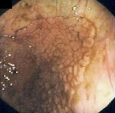
A villous adenoma is a type of polyp (a growth that protrudes from the lining of an organ) found in the colon or rectum. It is named for its appearance under a microscope, which reveals finger-like projections called "villi" on the surface of the polyp.
Villous adenomas are typically larger than other types of polyps and can be several centimeters in size. They are also more likely to be cancerous or precancerous, meaning that they have the potential to develop into colon or rectal cancer over time.
Because of this increased risk, it is important for villous adenomas to be removed surgically if they are found during a colonoscopy or other diagnostic procedure. Regular follow-up colonoscopies may also be recommended to monitor for the development of new polyps or recurrence of previous ones.
Pituitary neoplasms refer to abnormal growths or tumors in the pituitary gland, a small endocrine gland located at the base of the brain. These neoplasms can be benign (non-cancerous) or malignant (cancerous), with most being benign. They can vary in size and may cause various symptoms depending on their location, size, and hormonal activity.
Pituitary neoplasms can produce and secrete excess hormones, leading to a variety of endocrine disorders such as Cushing's disease (caused by excessive ACTH production), acromegaly (caused by excessive GH production), or prolactinoma (caused by excessive PRL production). They can also cause local compression symptoms due to their size, leading to headaches, vision problems, and cranial nerve palsies.
The exact causes of pituitary neoplasms are not fully understood, but genetic factors, radiation exposure, and certain inherited conditions may increase the risk of developing these tumors. Treatment options for pituitary neoplasms include surgical removal, radiation therapy, and medical management with drugs that can help control hormonal imbalances.
An adrenocortical adenoma is a benign tumor that arises from the cells of the adrenal cortex, which is the outer layer of the adrenal gland. These tumors can produce and release various hormones, such as cortisol, aldosterone, or androgens, depending on the type of cells they originate from.
Most adrenocortical adenomas are nonfunctioning, meaning that they do not secrete excess hormones and may not cause any symptoms. However, some functioning adenomas can produce excessive amounts of hormones, leading to a variety of clinical manifestations. For example:
* Cortisol-secreting adenomas can result in Cushing's syndrome, characterized by weight gain, muscle wasting, thin skin, easy bruising, and mood changes.
* Aldosterone-producing adenomas can cause Conn's syndrome, marked by hypertension (high blood pressure), hypokalemia (low potassium levels), and metabolic alkalosis.
* Androgen-secreting adenomas may lead to hirsutism (excessive hair growth) or virilization (development of male secondary sexual characteristics) in women.
The diagnosis of an adrenocortical adenoma typically involves imaging tests, such as CT or MRI scans, and hormonal evaluations to determine if the tumor is functioning or not. Treatment usually consists of surgical removal of the tumor, especially if it is causing hormonal imbalances or growing in size.
A liver cell adenoma is a benign tumor that develops in the liver and is composed of cells similar to those normally found in the liver (hepatocytes). These tumors are usually solitary, but multiple adenomas can occur, especially in women who have taken oral contraceptives for many years. Liver cell adenomas are typically asymptomatic and are often discovered incidentally during imaging studies performed for other reasons. In rare cases, they may cause symptoms such as abdominal pain or discomfort, or complications such as bleeding or rupture. Treatment options include monitoring with periodic imaging studies or surgical removal of the tumor.
A chromophobe adenoma is a type of benign (non-cancerous) tumor that typically arises in the pituitary gland, which is a small endocrine gland located at the base of the brain. The term "chromophobe" refers to the appearance of the cells under a microscope - they lack pigment and have a characteristic appearance with abundant clear or lightly stained cytoplasm.
Chromophobe adenomas are slow-growing tumors that can vary in size, and they may cause symptoms due to pressure on surrounding structures or by producing excess hormones. The most common hormone produced by chromophobe adenomas is prolactin, leading to symptoms such as menstrual irregularities, milk production (galactorrhea), and decreased sexual function in women, and decreased libido, erectile dysfunction, and infertility in men.
Treatment for chromophobe adenomas typically involves surgical removal of the tumor, often through a transsphenoidal approach (through the nose and sphenoid sinus). In some cases, radiation therapy or medical management with hormone-blocking drugs may also be necessary. Regular follow-up with an endocrinologist is important to monitor for any recurrence or hormonal imbalances.
A Growth Hormone-Secreting Pituitary Adenoma (GH-secreting pituitary adenoma, or GHoma) is a type of benign tumor that develops in the pituitary gland and results in excessive production of growth hormone (GH). This leads to a condition known as acromegaly if it occurs in adults, or gigantism if it occurs in children before the closure of the growth plates.
Symptoms of GH-secreting pituitary adenoma may include:
1. Coarsening of facial features
2. Enlargement of hands and feet
3. Deepened voice due to thickening of vocal cords
4. Increased sweating and body odor
5. Joint pain and stiffness
6. Sleep apnea
7. Fatigue, weakness, or muscle wasting
8. Headaches
9. Vision problems
10. Irregular menstrual periods in women
11. Erectile dysfunction in men
Diagnosis typically involves measuring the levels of GH and insulin-like growth factor 1 (IGF-1) in the blood, along with imaging tests like MRI or CT scans to locate and characterize the tumor. Treatment options include surgical removal of the tumor, radiation therapy, and medication to control GH production. Regular follow-ups are necessary to monitor for potential recurrence.
Colorectal neoplasms refer to abnormal growths in the colon or rectum, which can be benign or malignant. These growths can arise from the inner lining (mucosa) of the colon or rectum and can take various forms such as polyps, adenomas, or carcinomas.
Benign neoplasms, such as hyperplastic polyps and inflammatory polyps, are not cancerous but may need to be removed to prevent the development of malignant tumors. Adenomas, on the other hand, are precancerous lesions that can develop into colorectal cancer if left untreated.
Colorectal cancer is a malignant neoplasm that arises from the uncontrolled growth and division of cells in the colon or rectum. It is one of the most common types of cancer worldwide and can spread to other parts of the body through the bloodstream or lymphatic system.
Regular screening for colorectal neoplasms is recommended for individuals over the age of 50, as early detection and removal of precancerous lesions can significantly reduce the risk of developing colorectal cancer.
Colonic polyps are abnormal growths that protrude from the inner wall of the colon (large intestine). They can vary in size, shape, and number. Most colonic polyps are benign, meaning they are not cancerous. However, some types of polyps, such as adenomas, have a higher risk of becoming cancerous over time if left untreated.
Colonic polyps often do not cause any symptoms, especially if they are small. Larger polyps may lead to symptoms like rectal bleeding, changes in bowel habits, abdominal pain, or iron deficiency anemia. The exact cause of colonic polyps is not known, but factors such as age, family history, and certain medical conditions (like inflammatory bowel disease) can increase the risk of developing them.
Regular screening exams, such as colonoscopies, are recommended for individuals over the age of 50 to detect and remove polyps before they become cancerous. If you have a family history of colonic polyps or colorectal cancer, your doctor may recommend earlier or more frequent screenings.
An ACTH-secreting pituitary adenoma is a type of tumor that develops in the pituitary gland, a small gland located at the base of the brain. This type of tumor is also known as Cushing's disease.
ACTH stands for adrenocorticotropic hormone, which is a hormone produced and released by the pituitary gland. ACTH stimulates the adrenal glands (small glands located on top of the kidneys) to produce cortisol, a steroid hormone that helps regulate metabolism, helps the body respond to stress, and suppresses inflammation.
In an ACTH-secreting pituitary adenoma, the tumor cells produce and release excessive amounts of ACTH, leading to overproduction of cortisol by the adrenal glands. This can result in a constellation of symptoms known as Cushing's syndrome, which may include weight gain (especially around the trunk), fatigue, muscle weakness, mood changes, thinning of the skin, easy bruising, and increased susceptibility to infections.
Treatment for an ACTH-secreting pituitary adenoma typically involves surgical removal of the tumor, followed by medications to manage cortisol levels if necessary. Radiation therapy may also be used in some cases.
An adenoma is a benign tumor that forms in glandular tissue. When referring to "acidophil," it describes the appearance of the cells under a microscope. Acidophils are cells that take up acidic dyes, giving them a distinct appearance. In the context of an adenoma, an acidophil adenoma would be a benign tumor composed of acidophil cells.
Acidophil adenomas are most commonly found in the pituitary gland and are also known as lactotroph or mammosomatotroph adenomas. These tumors can produce and release prolactin, growth hormone, or both, leading to various endocrine disorders such as hyperprolactinemia, acromegaly, or gigantism. Treatment options typically include surgical removal of the tumor or medical management with dopamine agonists or somatostatin analogs.
A colonoscopy is a medical procedure used to examine the large intestine, also known as the colon and rectum. It is performed using a flexible tube with a tiny camera on the end, called a colonoscope, which is inserted into the rectum and gently guided through the entire length of the colon.
The procedure allows doctors to visually inspect the lining of the colon for any abnormalities such as polyps, ulcers, inflammation, or cancer. If any polyps are found during the procedure, they can be removed immediately using special tools passed through the colonoscope. Colonoscopy is an important tool in the prevention and early detection of colorectal cancer, which is one of the leading causes of cancer-related deaths worldwide.
Patients are usually given a sedative to help them relax during the procedure, which is typically performed on an outpatient basis in a hospital or clinic setting. The entire procedure usually takes about 30-60 minutes to complete, although patients should plan to spend several hours at the medical facility for preparation and recovery.
Adenomatous polyps, also known as adenomas, are benign (noncancerous) growths that develop in the lining of the glandular tissue of certain organs, most commonly occurring in the colon and rectum. These polyps are composed of abnormal glandular cells that can grow excessively and form a mass.
Adenomatous polyps can vary in size, ranging from a few millimeters to several centimeters in diameter. They may be flat or have a stalk (pedunculated). While adenomas are generally benign, they can potentially undergo malignant transformation and develop into colorectal cancer over time if left untreated. The risk of malignancy increases with the size of the polyp and the presence of certain histological features, such as dysplasia (abnormal cell growth).
Regular screening for adenomatous polyps is essential to detect and remove them early, reducing the risk of colorectal cancer. Screening methods include colonoscopy, sigmoidoscopy, and stool-based tests.
A prolactinoma is a type of pituitary tumor that produces an excess amount of the hormone prolactin, leading to various symptoms. The pituitary gland, located at the base of the brain, is responsible for producing and releasing several hormones that regulate different bodily functions. Prolactin is one such hormone, primarily known for its role in stimulating milk production in women during lactation (breastfeeding).
Prolactinoma tumors can be classified into two types: microprolactinomas and macroprolactinomas. Microprolactinomas are smaller tumors, typically less than 10 millimeters in size, while macroprolactinomas are larger tumors, generally greater than 10 millimeters in size.
The overproduction of prolactin caused by these tumors can lead to several clinical manifestations, including:
1. Galactorrhea: Unusual and often spontaneous milk production or leakage from the nipples, which can occur in both men and women who do not have a recent history of pregnancy or breastfeeding.
2. Menstrual irregularities: In women, high prolactin levels can interfere with the normal functioning of other hormones, leading to menstrual irregularities such as infrequent periods (oligomenorrhea) or absent periods (amenorrhea), and sometimes infertility.
3. Sexual dysfunction: In both men and women, high prolactin levels can cause decreased libido and sexual desire. Men may also experience erectile dysfunction and reduced sperm production.
4. Bone loss: Over time, high prolactin levels can lead to decreased bone density and an increased risk of osteoporosis due to the disruption of other hormones that regulate bone health.
5. Headaches and visual disturbances: As the tumor grows, it may put pressure on surrounding structures in the brain, leading to headaches and potential vision problems such as blurred vision or decreased peripheral vision.
Diagnosis typically involves measuring prolactin levels in the blood and performing imaging tests like an MRI (magnetic resonance imaging) scan to assess the size of the tumor. Treatment usually consists of medication to lower prolactin levels, such as dopamine agonists (e.g., bromocriptine or cabergoline), which can also help shrink the tumor. In some cases, surgery may be necessary if medication is ineffective or if the tumor is large and causing severe symptoms.
A basophilic adenoma is a rare type of benign tumor that arises from the glandular cells of an endocrine gland, specifically the cells that produce and store hormones. The term "basophilic" refers to the appearance of the tumor cells under a microscope, which have a high affinity for basic dyes due to their rich content of ribonucleic acid (RNA).
Basophilic adenomas are most commonly found in the pituitary gland, a small endocrine gland located at the base of the brain. These tumors can produce and secrete excessive amounts of hormones, leading to various clinical symptoms depending on the type of hormone involved. The most common types of basophilic adenomas are prolactinomas, which secrete high levels of the hormone prolactin, and growth hormone-secreting adenomas, which produce excessive amounts of growth hormone.
Treatment for basophilic adenomas typically involves surgical removal of the tumor, followed by radiation therapy or medical management with drugs that suppress hormone production. The prognosis for patients with basophilic adenomas is generally good, with most individuals experiencing a significant improvement in symptoms and quality of life following treatment. However, regular follow-up care is necessary to monitor for recurrence and manage any residual hormonal imbalances.
Adrenal cortex neoplasms refer to abnormal growths (tumors) in the adrenal gland's outer layer, known as the adrenal cortex. These neoplasms can be benign or malignant (cancerous). Benign tumors are called adrenal adenomas, while cancerous tumors are called adrenocortical carcinomas.
Adrenal cortex neoplasms can produce various hormones, leading to different clinical presentations. For instance, they may cause Cushing's syndrome (characterized by excessive cortisol production), Conn's syndrome (caused by aldosterone excess), or virilization (due to androgen excess). Some tumors may not produce any hormones and are discovered incidentally during imaging studies for unrelated conditions.
The diagnosis of adrenal cortex neoplasms typically involves a combination of imaging techniques, such as CT or MRI scans, and hormonal assessments to determine if the tumor is functional or non-functional. In some cases, a biopsy may be necessary to confirm the diagnosis and differentiate between benign and malignant tumors. Treatment options depend on the type, size, location, and hormonal activity of the neoplasm and may include surgical excision, radiation therapy, chemotherapy, or a combination of these approaches.
Adenomatous Polyposis Coli (APC) is a genetic disorder characterized by the development of numerous adenomatous polyps in the colon and rectum. APC is caused by mutations in the APC gene, which is a tumor suppressor gene that helps regulate cell growth and division. When the APC gene is mutated, it can lead to uncontrolled cell growth and the development of polyps, which can eventually become cancerous.
Individuals with APC typically develop hundreds to thousands of polyps in their colon and rectum, usually beginning in adolescence or early adulthood. If left untreated, APC can lead to colorectal cancer in nearly all affected individuals by the age of 40.
APC is an autosomal dominant disorder, which means that a person has a 50% chance of inheriting the mutated gene from an affected parent. However, some cases of APC may also occur spontaneously due to new mutations in the APC gene. Treatment for APC typically involves surgical removal of the colon and rectum (colectomy) to prevent the development of colorectal cancer. Regular surveillance with colonoscopy is also recommended to monitor for the development of new polyps.
Acromegaly is a rare hormonal disorder that typically occurs in middle-aged adults. It results from the pituitary gland producing too much growth hormone (GH) during adulthood. The excessive production of GH leads to abnormal growth of body tissues, particularly in the hands, feet, and face.
The term "acromegaly" is derived from two Greek words: "akros," meaning extremities, and "megaly," meaning enlargement. In most cases, acromegaly is caused by a benign tumor (adenoma) of the pituitary gland, which results in overproduction of GH.
Common symptoms include enlarged hands and feet, coarse facial features, deepened voice, joint pain, and sweating. If left untreated, acromegaly can lead to serious complications such as diabetes, hypertension, heart disease, and arthritis. Treatment usually involves surgical removal of the tumor, radiation therapy, or medication to control GH production.
Cushing syndrome is a hormonal disorder that occurs when your body is exposed to high levels of the hormone cortisol for a long time. This can happen due to various reasons such as taking high doses of corticosteroid medications or tumors that produce cortisol or adrenocorticotropic hormone (ACTH).
The symptoms of Cushing syndrome may include:
* Obesity, particularly around the trunk and upper body
* Thinning of the skin, easy bruising, and purple or red stretch marks on the abdomen, thighs, breasts, and arms
* Weakened bones, leading to fractures
* High blood pressure
* High blood sugar
* Mental changes such as depression, anxiety, and irritability
* Increased fatigue and weakness
* Menstrual irregularities in women
* Decreased fertility in men
Cushing syndrome can be diagnosed through various tests, including urine and blood tests to measure cortisol levels, saliva tests, and imaging tests to locate any tumors. Treatment depends on the cause of the condition but may include surgery, radiation therapy, chemotherapy, or adjusting medication dosages.
APC (Adenomatous Polyposis Coli) gene is a tumor suppressor gene that provides instructions for making a protein called adenomatous polyposis coli. This protein plays a crucial role in regulating the growth and division of cells in the colon and rectum. Specifically, it helps to maintain the stability of the cell's genetic material (DNA) by controlling the process of beta-catenin degradation.
When the APC gene is mutated or altered, it can lead to an accumulation of beta-catenin in the cell, which can result in uncontrolled cell growth and division. This can ultimately lead to the development of colon polyps, which are benign growths that can become cancerous over time if left untreated.
Mutations in the APC gene are associated with several inherited cancer syndromes, including familial adenomatous polyposis (FAP) and attenuated FAP (AFAP). These conditions are characterized by the development of numerous colon polyps at a young age, which can increase the risk of developing colorectal cancer.
Hyperparathyroidism is a condition in which the parathyroid glands produce excessive amounts of parathyroid hormone (PTH). There are four small parathyroid glands located in the neck, near or within the thyroid gland. They release PTH into the bloodstream to help regulate the levels of calcium and phosphorus in the body.
In hyperparathyroidism, overproduction of PTH can lead to an imbalance in these minerals, causing high blood calcium levels (hypercalcemia) and low phosphate levels (hypophosphatemia). This can result in various symptoms such as fatigue, weakness, bone pain, kidney stones, and cognitive issues.
There are two types of hyperparathyroidism: primary and secondary. Primary hyperparathyroidism occurs when there is a problem with one or more of the parathyroid glands, causing them to become overactive and produce too much PTH. Secondary hyperparathyroidism develops as a response to low calcium levels in the body due to conditions like vitamin D deficiency, chronic kidney disease, or malabsorption syndromes.
Treatment for hyperparathyroidism depends on the underlying cause and severity of symptoms. In primary hyperparathyroidism, surgery to remove the overactive parathyroid gland(s) is often recommended. For secondary hyperparathyroidism, treating the underlying condition and managing calcium levels with medications or dietary changes may be sufficient.
Salivary gland neoplasms refer to abnormal growths or tumors that develop in the salivary glands. These glands are responsible for producing saliva, which helps in digestion, lubrication of food and maintaining oral health. Salivary gland neoplasms can be benign (non-cancerous) or malignant (cancerous).
Benign neoplasms are slow-growing and typically do not spread to other parts of the body. They may cause symptoms such as swelling, painless lumps, or difficulty swallowing if they grow large enough to put pressure on surrounding tissues.
Malignant neoplasms, on the other hand, can be aggressive and have the potential to invade nearby structures and metastasize (spread) to distant organs. Symptoms of malignant salivary gland neoplasms may include rapid growth, pain, numbness, or paralysis of facial nerves.
Salivary gland neoplasms can occur in any of the major salivary glands (parotid, submandibular, and sublingual glands) or in the minor salivary glands located throughout the mouth and throat. The exact cause of these neoplasms is not fully understood, but risk factors may include exposure to radiation, certain viral infections, and genetic predisposition.
Colonic neoplasms refer to abnormal growths in the large intestine, also known as the colon. These growths can be benign (non-cancerous) or malignant (cancerous). The two most common types of colonic neoplasms are adenomas and carcinomas.
Adenomas are benign tumors that can develop into cancer over time if left untreated. They are often found during routine colonoscopies and can be removed during the procedure.
Carcinomas, on the other hand, are malignant tumors that invade surrounding tissues and can spread to other parts of the body. Colorectal cancer is the third leading cause of cancer-related deaths in the United States, and colonic neoplasms are a significant risk factor for developing this type of cancer.
Regular screenings for colonic neoplasms are recommended for individuals over the age of 50 or those with a family history of colorectal cancer or other risk factors. Early detection and removal of colonic neoplasms can significantly reduce the risk of developing colorectal cancer.
Intestinal neoplasms refer to abnormal growths in the tissues of the intestines, which can be benign or malignant. These growths are called neoplasms and they result from uncontrolled cell division. In the case of intestinal neoplasms, these growths occur in the small intestine, large intestine (colon), rectum, or appendix.
Benign intestinal neoplasms are not cancerous and often do not invade surrounding tissues or spread to other parts of the body. However, they can still cause problems if they grow large enough to obstruct the intestines or cause bleeding. Common types of benign intestinal neoplasms include polyps, leiomyomas, and lipomas.
Malignant intestinal neoplasms, on the other hand, are cancerous and can invade surrounding tissues and spread to other parts of the body. The most common type of malignant intestinal neoplasm is adenocarcinoma, which arises from the glandular cells lining the inside of the intestines. Other types of malignant intestinal neoplasms include lymphomas, sarcomas, and carcinoid tumors.
Symptoms of intestinal neoplasms can vary depending on their size, location, and type. Common symptoms include abdominal pain, bloating, changes in bowel habits, rectal bleeding, weight loss, and fatigue. If you experience any of these symptoms, it is important to seek medical attention promptly.
Hyperplasia is a medical term that refers to an abnormal increase in the number of cells in an organ or tissue, leading to an enlargement of the affected area. It's a response to various stimuli such as hormones, chronic irritation, or inflammation. Hyperplasia can be physiological, like the growth of breast tissue during pregnancy, or pathological, like in the case of benign or malignant tumors. The process is generally reversible if the stimulus is removed. It's important to note that hyperplasia itself is not cancerous, but some forms of hyperplasia can increase the risk of developing cancer over time.
Multiple primary neoplasms refer to the occurrence of more than one primary malignant tumor in an individual, where each tumor is unrelated to the other and originates from separate cells or organs. This differs from metastatic cancer, where a single malignancy spreads to multiple sites in the body. Multiple primary neoplasms can be synchronous (occurring at the same time) or metachronous (occurring at different times). The risk of developing multiple primary neoplasms increases with age and is associated with certain genetic predispositions, environmental factors, and lifestyle choices such as smoking and alcohol consumption.
Adrenal gland neoplasms refer to abnormal growths or tumors in the adrenal glands. These glands are located on top of each kidney and are responsible for producing hormones that regulate various bodily functions such as metabolism, blood pressure, and stress response. Adrenal gland neoplasms can be benign (non-cancerous) or malignant (cancerous).
Benign adrenal tumors are called adenomas and are usually small and asymptomatic. However, some adenomas may produce excessive amounts of hormones, leading to symptoms such as high blood pressure, weight gain, and mood changes.
Malignant adrenal tumors are called adrenocortical carcinomas and are rare but aggressive cancers that can spread to other parts of the body. Symptoms of adrenocortical carcinoma may include abdominal pain, weight loss, and hormonal imbalances.
It is important to diagnose and treat adrenal gland neoplasms early to prevent complications and improve outcomes. Diagnostic tests may include imaging studies such as CT scans or MRIs, as well as hormone level testing and biopsy. Treatment options may include surgery, radiation therapy, chemotherapy, or a combination of these approaches.
Pituitary ACTH hypersecretion, also known as Cushing's disease, is a condition characterized by the excessive production of adrenocorticotropic hormone (ACTH) from the pituitary gland. This results in an overproduction of cortisol, a steroid hormone produced by the adrenal glands, leading to a constellation of symptoms known as Cushing's syndrome.
In Cushing's disease, a benign tumor called an adenoma develops on the pituitary gland, causing it to release excess ACTH. This in turn stimulates the adrenal glands to produce more cortisol than necessary. The resulting high levels of cortisol can cause various symptoms such as weight gain, particularly around the trunk and face (central obesity), thinning of the skin, bruising, weakness, fatigue, mood changes, high blood pressure, and an increased risk of infections.
It is important to distinguish Cushing's disease from other causes of Cushing's syndrome, such as cortisol-producing adrenal tumors or exogenous sources of corticosteroid use, as the treatment approach may differ. Treatment for Cushing's disease typically involves surgical removal of the pituitary tumor, with additional medical management and/or radiation therapy in some cases.
Parotid neoplasms refer to abnormal growths or tumors in the parotid gland, which is the largest of the salivary glands and is located in front of the ear and extends down the neck. These neoplasms can be benign (non-cancerous) or malignant (cancerous).
Benign parotid neoplasms are typically slow-growing, painless masses that may cause facial asymmetry or difficulty in chewing or swallowing if they become large enough to compress surrounding structures. The most common type of benign parotid tumor is a pleomorphic adenoma.
Malignant parotid neoplasms, on the other hand, are more aggressive and can invade nearby tissues and spread to other parts of the body. They may present as rapidly growing masses that are firm or fixed to surrounding structures. Common types of malignant parotid tumors include mucoepidermoid carcinoma, adenoid cystic carcinoma, and squamous cell carcinoma.
The diagnosis of parotid neoplasms typically involves a thorough clinical evaluation, imaging studies such as CT or MRI scans, and fine-needle aspiration biopsy (FNAB) to determine the nature of the tumor. Treatment options depend on the type, size, and location of the neoplasm but may include surgical excision, radiation therapy, and chemotherapy.
Immunohistochemistry (IHC) is a technique used in pathology and laboratory medicine to identify specific proteins or antigens in tissue sections. It combines the principles of immunology and histology to detect the presence and location of these target molecules within cells and tissues. This technique utilizes antibodies that are specific to the protein or antigen of interest, which are then tagged with a detection system such as a chromogen or fluorophore. The stained tissue sections can be examined under a microscope, allowing for the visualization and analysis of the distribution and expression patterns of the target molecule in the context of the tissue architecture. Immunohistochemistry is widely used in diagnostic pathology to help identify various diseases, including cancer, infectious diseases, and immune-mediated disorders.
Carcinoma is a type of cancer that develops from epithelial cells, which are the cells that line the inner and outer surfaces of the body. These cells cover organs, glands, and other structures within the body. Carcinomas can occur in various parts of the body, including the skin, lungs, breasts, prostate, colon, and pancreas. They are often characterized by the uncontrolled growth and division of abnormal cells that can invade surrounding tissues and spread to other parts of the body through a process called metastasis. Carcinomas can be further classified based on their appearance under a microscope, such as adenocarcinoma, squamous cell carcinoma, and basal cell carcinoma.
Hyperaldosteronism is a medical condition characterized by the overproduction of aldosterone, a hormone produced by the adrenal glands. Aldosterone helps regulate sodium and potassium balance and blood pressure by promoting sodium retention and potassium excretion in the kidneys.
There are two types of hyperaldosteronism: primary and secondary. Primary hyperaldosteronism is caused by an overproduction of aldosterone from an abnormality within the adrenal gland, such as a tumor (Conn's syndrome) or hyperplasia. Secondary hyperaldosteronism occurs when there is an excess production of renin, a hormone produced by the kidneys, which then stimulates the adrenal glands to produce more aldosterone. This can be caused by various conditions that affect kidney function, such as renal artery stenosis or heart failure.
Symptoms of hyperaldosteronism may include high blood pressure, low potassium levels (hypokalemia), muscle weakness, and frequent urination. Diagnosis typically involves measuring aldosterone and renin levels in the blood, as well as other tests to determine the underlying cause. Treatment depends on the type and cause of hyperaldosteronism but may include medications, surgery, or lifestyle changes.
The sphenoid bone is a complex, irregularly shaped bone located in the middle cranial fossa and forms part of the base of the skull. It articulates with several other bones, including the frontal, parietal, temporal, ethmoid, palatine, and zygomatic bones. The sphenoid bone has two main parts: the body and the wings.
The body of the sphenoid bone is roughly cuboid in shape and contains several important structures, such as the sella turcica, which houses the pituitary gland, and the sphenoid sinuses, which are air-filled cavities within the bone. The greater wings of the sphenoid bone extend laterally from the body and form part of the skull's lateral walls. They contain the superior orbital fissure, through which important nerves and blood vessels pass between the cranial cavity and the orbit of the eye.
The lesser wings of the sphenoid bone are thin, blade-like structures that extend anteriorly from the body and form part of the floor of the anterior cranial fossa. They contain the optic canal, which transmits the optic nerve and ophthalmic artery between the brain and the orbit of the eye.
Overall, the sphenoid bone plays a crucial role in protecting several important structures within the skull, including the pituitary gland, optic nerves, and ophthalmic arteries.
Duodenal neoplasms refer to abnormal growths in the duodenum, which is the first part of the small intestine that receives digestive secretions from the pancreas and bile duct. These growths can be benign or malignant (cancerous).
Benign neoplasms include adenomas, leiomyomas, lipomas, and hamartomas. They are usually slow-growing and do not spread to other parts of the body. However, they may cause symptoms such as abdominal pain, bleeding, or obstruction of the intestine.
Malignant neoplasms include adenocarcinomas, neuroendocrine tumors (carcinoids), lymphomas, and sarcomas. They are more aggressive and can invade surrounding tissues and spread to other parts of the body. Symptoms may include abdominal pain, weight loss, jaundice, anemia, or bowel obstruction.
The diagnosis of duodenal neoplasms is usually made through imaging tests such as CT scans, MRI, or endoscopy with biopsy. Treatment depends on the type and stage of the tumor and may include surgery, chemotherapy, radiation therapy, or a combination of these modalities.
Adrenocorticotropic Hormone (ACTH) is a hormone produced and released by the anterior pituitary gland, a small endocrine gland located at the base of the brain. ACTH plays a crucial role in the regulation of the body's stress response and has significant effects on various physiological processes.
The primary function of ACTH is to stimulate the adrenal glands, which are triangular-shaped glands situated on top of the kidneys. The adrenal glands consist of two parts: the outer cortex and the inner medulla. ACTH specifically targets the adrenal cortex, where it binds to specific receptors and initiates a series of biochemical reactions leading to the production and release of steroid hormones, primarily cortisol (a glucocorticoid) and aldosterone (a mineralocorticoid).
Cortisol is involved in various metabolic processes, such as regulating blood sugar levels, modulating the immune response, and helping the body respond to stress. Aldosterone plays a vital role in maintaining electrolyte and fluid balance by promoting sodium reabsorption and potassium excretion in the kidneys.
ACTH release is controlled by the hypothalamus, another part of the brain, which produces corticotropin-releasing hormone (CRH). CRH stimulates the anterior pituitary gland to secrete ACTH, which in turn triggers cortisol production in the adrenal glands. This complex feedback system helps maintain homeostasis and ensures that appropriate amounts of cortisol are released in response to various physiological and psychological stressors.
Disorders related to ACTH can lead to hormonal imbalances, resulting in conditions such as Cushing's syndrome (excessive cortisol production) or Addison's disease (insufficient cortisol production). Proper diagnosis and management of these disorders typically involve assessing the function of the hypothalamic-pituitary-adrenal axis and addressing any underlying issues affecting ACTH secretion.
The pituitary gland is a small, endocrine gland located at the base of the brain, in the sella turcica of the sphenoid bone. It is often called the "master gland" because it controls other glands and makes the hormones that trigger many body functions. The pituitary gland measures about 0.5 cm in height and 1 cm in width, and it weighs approximately 0.5 grams.
The pituitary gland is divided into two main parts: the anterior lobe (adenohypophysis) and the posterior lobe (neurohypophysis). The anterior lobe is further divided into three zones: the pars distalis, pars intermedia, and pars tuberalis. Each part of the pituitary gland has distinct functions and produces different hormones.
The anterior pituitary gland produces and releases several important hormones, including:
* Growth hormone (GH), which regulates growth and development in children and helps maintain muscle mass and bone strength in adults.
* Thyroid-stimulating hormone (TSH), which controls the production of thyroid hormones by the thyroid gland.
* Adrenocorticotropic hormone (ACTH), which stimulates the adrenal glands to produce cortisol and other steroid hormones.
* Follicle-stimulating hormone (FSH) and luteinizing hormone (LH), which regulate reproductive function in both males and females.
* Prolactin, which stimulates milk production in pregnant and lactating women.
The posterior pituitary gland stores and releases two hormones that are produced by the hypothalamus:
* Antidiuretic hormone (ADH), which helps regulate water balance in the body by controlling urine production.
* Oxytocin, which stimulates uterine contractions during childbirth and milk release during breastfeeding.
Overall, the pituitary gland plays a critical role in maintaining homeostasis and regulating various bodily functions, including growth, development, metabolism, and reproductive function.
Adenocarcinoma is a type of cancer that arises from glandular epithelial cells. These cells line the inside of many internal organs, including the breasts, prostate, colon, and lungs. Adenocarcinomas can occur in any of these organs, as well as in other locations where glands are present.
The term "adenocarcinoma" is used to describe a cancer that has features of glandular tissue, such as mucus-secreting cells or cells that produce hormones. These cancers often form glandular structures within the tumor mass and may produce mucus or other substances.
Adenocarcinomas are typically slow-growing and tend to spread (metastasize) to other parts of the body through the lymphatic system or bloodstream. They can be treated with surgery, radiation therapy, chemotherapy, targeted therapy, or a combination of these treatments. The prognosis for adenocarcinoma depends on several factors, including the location and stage of the cancer, as well as the patient's overall health and age.
Primary hyperparathyroidism is a medical condition characterized by excessive secretion of parathyroid hormone (PTH) from one or more of the parathyroid glands in the neck. These glands are normally responsible for regulating calcium levels in the body by releasing PTH, which helps to maintain an appropriate balance of calcium and phosphate in the bloodstream.
In primary hyperparathyroidism, the parathyroid gland(s) become overactive and produce too much PTH, leading to elevated calcium levels (hypercalcemia) in the blood. This can result in a variety of symptoms, such as fatigue, weakness, bone pain, kidney stones, and cognitive impairment, although some individuals may not experience any symptoms at all.
The most common cause of primary hyperparathyroidism is a benign tumor called an adenoma that develops in one or more of the parathyroid glands. In rare cases, primary hyperparathyroidism can be caused by cancer of the parathyroid gland(s) or by enlargement of all four glands (four-gland hyperplasia). Treatment typically involves surgical removal of the affected parathyroid gland(s), which is usually curative.
Thyroid neoplasms refer to abnormal growths or tumors in the thyroid gland, which can be benign (non-cancerous) or malignant (cancerous). These growths can vary in size and may cause a noticeable lump or nodule in the neck. Thyroid neoplasms can also affect the function of the thyroid gland, leading to hormonal imbalances and related symptoms. The exact causes of thyroid neoplasms are not fully understood, but risk factors include radiation exposure, family history, and certain genetic conditions. It is important to note that most thyroid nodules are benign, but a proper medical evaluation is necessary to determine the nature of the growth and develop an appropriate treatment plan.
A precancerous condition, also known as a premalignant condition, is a state of abnormal cellular growth and development that has a higher-than-normal potential to progress into cancer. These conditions are characterized by the presence of certain anomalies in the cells, such as dysplasia (abnormal changes in cell shape or size), which can indicate an increased risk for malignant transformation.
It is important to note that not all precancerous conditions will eventually develop into cancer, and some may even regress on their own. However, individuals with precancerous conditions are often at a higher risk of developing cancer compared to the general population. Regular monitoring and appropriate medical interventions, if necessary, can help manage this risk and potentially prevent or detect cancer at an early stage when it is more treatable.
Examples of precancerous conditions include:
1. Dysplasia in the cervix (cervical intraepithelial neoplasia or CIN)
2. Atypical ductal hyperplasia or lobular hyperplasia in the breast
3. Actinic keratosis on the skin
4. Leukoplakia in the mouth
5. Barrett's esophagus in the digestive tract
Regular medical check-ups, screenings, and lifestyle modifications are crucial for individuals with precancerous conditions to monitor their health and reduce the risk of cancer development.
The Sella Turcica, also known as the Turkish saddle, is a depression or fossa in the sphenoid bone located at the base of the skull. It forms a housing for the pituitary gland, which is a small endocrine gland often referred to as the "master gland" because it controls other glands and makes several essential hormones. The Sella Turcica has a saddle-like shape, with its anterior and posterior clinoids forming the front and back of the saddle, respectively. This region is of significant interest in neuroimaging and clinical settings, as various conditions such as pituitary tumors or other abnormalities may affect the size, shape, and integrity of the Sella Turcica.
Sigmoidoscopy is a medical procedure that involves the insertion of a sigmoidoscope, a flexible tube with a light and camera at the end, into the rectum and lower colon (sigmoid colon) to examine these areas for any abnormalities such as inflammation, ulcers, polyps, or cancer. The procedure typically allows for the detection of issues in the sigmoid colon and rectum, and can help diagnose conditions such as inflammatory bowel disease, diverticulosis, or colorectal cancer.
There are two types of sigmoidoscopy: flexible sigmoidoscopy and rigid sigmoidoscopy. Flexible sigmoidoscopy is more commonly performed because it provides a better view of the lower colon and is less uncomfortable for the patient. Rigid sigmoidoscopy, on the other hand, uses a solid, inflexible tube and is typically used in specific situations such as the removal of foreign objects or certain types of polyps.
During the procedure, patients are usually positioned on their left side with their knees drawn up to their chest. The sigmoidoscope is gently inserted into the rectum and advanced through the lower colon while the doctor examines the lining for any abnormalities. Air may be introduced through the scope to help expand the colon and provide a better view. If polyps or other abnormal tissues are found, they can often be removed during the procedure for further examination and testing.
Sigmoidoscopy is generally considered a safe and well-tolerated procedure. Some patients may experience mild discomfort, bloating, or cramping during or after the exam, but these symptoms typically resolve on their own within a few hours.
A choristoma is a type of growth that occurs when normally functioning tissue is found in an abnormal location within the body. It is not cancerous or harmful, but it can cause problems if it presses on surrounding structures or causes symptoms. Choristomas are typically congenital, meaning they are present at birth, and are thought to occur due to developmental errors during embryonic growth. They can be found in various organs and tissues throughout the body, including the brain, eye, skin, and gastrointestinal tract.
Rectal neoplasms refer to abnormal growths in the tissues of the rectum, which can be benign or malignant. They are characterized by uncontrolled cell division and can invade nearby tissues or spread to other parts of the body (metastasis). The most common type of rectal neoplasm is rectal cancer, which often begins as a small polyp or growth in the lining of the rectum. Other types of rectal neoplasms include adenomas, carcinoids, and gastrointestinal stromal tumors (GISTs). Regular screenings are recommended for early detection and treatment of rectal neoplasms.
Pituitary apoplexy is a medical emergency that involves bleeding into the pituitary gland (a small gland at the base of the brain) and/or sudden swelling of the pituitary gland. This can lead to compression of nearby structures, such as the optic nerves and the hypothalamus, causing symptoms like severe headache, visual disturbances, hormonal imbalances, and altered mental status. It is often associated with a pre-existing pituitary tumor (such as a pituitary adenoma), but can also occur in individuals without any known pituitary abnormalities. Immediate medical attention is required to manage this condition, which may include surgical intervention, hormone replacement therapy, and supportive care.
Human Growth Hormone (HGH), also known as somatotropin, is a peptide hormone produced in the pituitary gland. It plays a crucial role in human development and growth by stimulating the production of another hormone called insulin-like growth factor 1 (IGF-1). IGF-1 promotes the growth and reproduction of cells throughout the body, particularly in bones and other tissues. HGH also helps regulate body composition, body fluids, muscle and bone growth, sugar and fat metabolism, and possibly heart function. It is essential for human development and continues to have important effects throughout life. The secretion of HGH decreases with age, which is thought to contribute to the aging process.
The colon, also known as the large intestine, is a part of the digestive system in humans and other vertebrates. It is an organ that eliminates waste from the body and is located between the small intestine and the rectum. The main function of the colon is to absorb water and electrolytes from digested food, forming and storing feces until they are eliminated through the anus.
The colon is divided into several regions, including the cecum, ascending colon, transverse colon, descending colon, sigmoid colon, rectum, and anus. The walls of the colon contain a layer of muscle that helps to move waste material through the organ by a process called peristalsis.
The inner surface of the colon is lined with mucous membrane, which secretes mucus to lubricate the passage of feces. The colon also contains a large population of bacteria, known as the gut microbiota, which play an important role in digestion and immunity.
Pituitary hormones are chemical messengers produced and released by the pituitary gland, a small endocrine gland located at the base of the brain. The pituitary gland is often referred to as the "master gland" because it controls several other endocrine glands and regulates various bodily functions.
There are two main types of pituitary hormones: anterior pituitary hormones and posterior pituitary hormones, which are produced in different parts of the pituitary gland and have distinct functions.
Anterior pituitary hormones include:
1. Growth hormone (GH): regulates growth and metabolism.
2. Thyroid-stimulating hormone (TSH): stimulates the thyroid gland to produce thyroid hormones.
3. Adrenocorticotropic hormone (ACTH): stimulates the adrenal glands to produce cortisol and other steroid hormones.
4. Follicle-stimulating hormone (FSH) and luteinizing hormone (LH): regulate reproductive function in both males and females.
5. Prolactin: stimulates milk production in lactating women.
6. Melanocyte-stimulating hormone (MSH): regulates skin pigmentation and appetite.
Posterior pituitary hormones include:
1. Oxytocin: stimulates uterine contractions during childbirth and milk ejection during lactation.
2. Vasopressin (antidiuretic hormone, ADH): regulates water balance in the body by controlling urine production in the kidneys.
Overall, pituitary hormones play crucial roles in regulating growth, development, metabolism, reproductive function, and various other bodily functions. Abnormalities in pituitary hormone levels can lead to a range of medical conditions, such as dwarfism, acromegaly, Cushing's disease, infertility, and diabetes insipidus.
Adenomatous polyposis coli (APC) protein is a tumor suppressor protein that plays a crucial role in regulating cell growth and division. It is encoded by the APC gene, which is located on chromosome 5. The APC protein helps to prevent excessive cell growth and division by inhibiting the activity of a protein called beta-catenin, which promotes cell growth and division when activated.
In individuals with certain genetic disorders, such as familial adenomatous polyposis (FAP), mutations in the APC gene can lead to the production of a defective APC protein or no APC protein at all. This can result in uncontrolled cell growth and division, leading to the development of numerous benign tumors called polyps in the colon and rectum. Over time, some of these polyps may become cancerous, leading to colorectal cancer if left untreated.
APC protein also has other functions in the body, including regulating cell migration and adhesion, and playing a role in maintaining the stability of the cytoskeleton. Mutations in the APC gene have been linked to other types of cancer besides colorectal cancer, including breast, lung, and ovarian cancers.
Parathyroidectomy is a surgical procedure for the removal of one or more of the parathyroid glands. These glands are located in the neck and are responsible for producing parathyroid hormone (PTH), which helps regulate the levels of calcium and phosphorus in the body.
Parathyroidectomy is typically performed to treat conditions such as hyperparathyroidism, where one or more of the parathyroid glands become overactive and produce too much PTH. This can lead to high levels of calcium in the blood, which can cause symptoms such as weakness, fatigue, bone pain, kidney stones, and mental confusion.
There are different types of parathyroidectomy procedures, including:
* Partial parathyroidectomy: removal of one or more, but not all, of the parathyroid glands.
* Total parathyroidectomy: removal of all four parathyroid glands.
* Subtotal parathyroidectomy: removal of three and a half of the four parathyroid glands, leaving a small portion of one gland to prevent hypoparathyroidism (a condition where the body produces too little PTH).
The choice of procedure depends on the underlying condition and its severity. After the surgery, patients may need to have their calcium levels monitored and may require calcium and vitamin D supplements to maintain normal calcium levels in the blood.
Urethane is not a term typically used in medical definitions. However, in the field of chemistry and pharmacology, urethane is an ethyl carbamate ester which has been used as a general anesthetic. It is rarely used today due to its potential carcinogenic properties and the availability of safer alternatives.
In the context of materials science, polyurethanes are a class of polymers that contain urethane linkages (-NH-CO-O-) in their main chain. They are widely used in various applications such as foam insulation, coatings, adhesives, and medical devices due to their versatile properties like flexibility, durability, and resistance to abrasion.
The intestinal mucosa is the innermost layer of the intestines, which comes into direct contact with digested food and microbes. It is a specialized epithelial tissue that plays crucial roles in nutrient absorption, barrier function, and immune defense. The intestinal mucosa is composed of several cell types, including absorptive enterocytes, mucus-secreting goblet cells, hormone-producing enteroendocrine cells, and immune cells such as lymphocytes and macrophages.
The surface of the intestinal mucosa is covered by a single layer of epithelial cells, which are joined together by tight junctions to form a protective barrier against harmful substances and microorganisms. This barrier also allows for the selective absorption of nutrients into the bloodstream. The intestinal mucosa also contains numerous lymphoid follicles, known as Peyer's patches, which are involved in immune surveillance and defense against pathogens.
In addition to its role in absorption and immunity, the intestinal mucosa is also capable of producing hormones that regulate digestion and metabolism. Dysfunction of the intestinal mucosa can lead to various gastrointestinal disorders, such as inflammatory bowel disease, celiac disease, and food allergies.
Adenoma of the bile duct is a benign (noncancerous) tumor that develops in the bile ducts, which are tiny tubes that carry bile from the liver to the gallbladder and small intestine. Bile is a digestive fluid produced by the liver.
Bile duct adenomas are rare and usually do not cause any symptoms. However, if they grow large enough, they may obstruct the flow of bile and cause jaundice (yellowing of the skin and whites of the eyes), abdominal pain, or itching. In some cases, bile duct adenomas may become cancerous and develop into bile duct carcinomas.
The exact cause of bile duct adenomas is not known, but they are more common in people with certain genetic disorders, such as Gardner's syndrome and von Hippel-Lindau disease. Treatment for bile duct adenomas typically involves surgical removal of the tumor.
Prolactin is a hormone produced by the pituitary gland, a small gland located at the base of the brain. Its primary function is to stimulate milk production in women after childbirth, a process known as lactation. However, prolactin also plays other roles in the body, including regulating immune responses, metabolism, and behavior. In men, prolactin helps maintain the sexual glands and contributes to paternal behaviors.
Prolactin levels are usually low in both men and non-pregnant women but increase significantly during pregnancy and after childbirth. Various factors can affect prolactin levels, including stress, sleep, exercise, and certain medications. High prolactin levels can lead to medical conditions such as amenorrhea (absence of menstruation), galactorrhea (spontaneous milk production not related to childbirth), infertility, and reduced sexual desire in both men and women.
The descending colon is a part of the large intestine in the human digestive system. It is called "descending" because it is located inferiorly and posteriorly to the transverse colon, and its direction goes downward as it continues toward the rectum. The descending colon receives digested food material from the transverse colon via the splenic flexure, also known as the left colic flexure.
The primary function of the descending colon is to absorb water, electrolytes, and any remaining nutrients from the undigested food materials that have passed through the small intestine. The descending colon also stores this waste material temporarily before it moves into the rectum for eventual elimination from the body.
The descending colon's wall contains a layer of smooth muscle, which helps propel the waste material along the gastrointestinal tract via peristalsis. Additionally, the inner mucosal lining of the descending colon contains numerous goblet cells that produce and secrete mucus to lubricate the passage of stool and protect the intestinal wall from irritation or damage caused by waste materials.
In summary, the medical definition of 'Colon, Descending' refers to a section of the large intestine responsible for absorbing water and electrolytes while storing and eliminating waste materials through peristaltic movements and mucus secretion.
 Colorectal polyp
Colorectal adenoma
Adenoma
Villoglandular adenocarcinoma of the cervix
McKittrick-Wheelock syndrome
Traditional serrated adenoma
Histopathology of colorectal adenocarcinoma
Sessile serrated lesion
Indigo carmine
Polyp (medicine)
Rectal discharge
International Classification of Diseases for Oncology
Index of oncology articles
Nuru Bayramov
List of MeSH codes (C04)
Colorectal cancer
Glossary of medicine
 Villous Adenoma: Background, Pathophysiology, Etiology
 Table of Contents 2005 | Analytical Cellular Pathology | Hindawi
Colorectal polyp - Wikipedia
 Neoplastic Polyps
 Colorectal polyps: MedlinePlus Medical Encyclopedia
 Rectal Cancer Treatment (PDQ®) - NCI
 Gastric villous adenoma: a case report and review of the literature | Journal of Medical Case Reports | Full Text
 Investigation of the First Seven Reported Cases of Candida auris, a Globally Emerging Invasive, Multidrug-Resistant Fungus -...
 Risk and cause of interval colorectal cancer after colonoscopic polypectomy
 3D Printed Pedunculated Adenoma of the Colon - Anatomy Warehouse
A case of concurrent cancer in a giant rectal villous adenoma that resulted in extensive lymph node metastases<...
 Efficacy of image-enhanced endoscopy for colorectal adenoma detection: A multicenter, randomized trial
 Tubular Adenoma: Definition, Treatment, Outlook, and More
 Hypokalemia | Diagnosaurus
All Phenotypes Apc
AI for Polyp Detection: Some Benefit, but Limitations Exist
 Adrenal uptake in PET/CT in a patient with pancreatic neoplasm: Not always metastasis | Gastroenterología y Hepatología ...
 Mr Marios Hadjipavlou | King Edward VII's Hospital
 The Sodium to Potassium Ratio Is One Reason Cheese is Not Paleo - The Paleo Diet®
Endoscopic Optical Coherence Tomography (OCT): Advances in Gastrointestinal Imaging
 Pathological Anatomy (LZ-H) 2023/2024 - University of Bologna
00226649 | PEIR Digital Library
 Insulin Therapy and Colorectal Adenomas in Patients with Diabetes Mellitus | Cancer Epidemiology, Biomarkers & Prevention |...
Internet Scientific Publications
 Are Non-dysplastic Crypts with Corrupted Shapes the Initial Recordable Histological Event in the Development of Sporadic...
 Flashcards - Pathology and Microbiology NBDE 1986
 Getting depressed about my ongoing anemia | The J-Pouch Group
 Report
Rhabdomyosarcoma in patients with constitutional mismatch-repair-deficiency syndrome | Journal of Medical Genetics
Colorectal Cancer Guidelines: Colorectal Cancer Screening, Postpolypectomy Surveillance, Familial Adenomatous Polyposis
High-grade dysplasia6
- 60 years (OR 2.97, 95% CI 2.31-3.82) was significantly associated with interval cancer on the surveillance colonoscopy as were advanced adenoma (OR 1.28, 95% CI 1.01-1.62), the presence of villous (HR 1.38, 95% CI 1.03-1.85) and high-grade dysplasia (OR 1.61, 95% CI 1.07-2.42). (nih.gov)
- If your adenoma looks more abnormal and more like cancer, it's described as having high-grade dysplasia. (healthline.com)
- We defined advanced neoplasia as cancer or adenoma that was at least 10 mm in diameter, had high-grade dysplasia, or had villous or tubulovillous histologic characteristics, or any combination thereof. (nih.gov)
- AN was defined as high-grade dysplasia, villous histology, or cancer. (bmj.com)
- Advanced colorectal neoplasia included advanced adenomas (high-grade dysplasia, ≥25% villous component, or ≥10-mm diameter), advanced serrated lesions (dysplasia or ≥10-mm diameter), and CRC. (eur.nl)
- 1 cm) tubular adenomas that do not show high-grade dysplasia, are classified into the low-risk group, and follow-up colonoscopy every 5-10 years is recommended for this group. (biomedcentral.com)
Polyp8
- Endoscopic view of a sessile polyp, which histology studies revealed to be a villous adenoma. (medscape.com)
- Endoscopic view of a sessile polyp histologically determined to be a villous adenoma. (medscape.com)
- Villous adenoma is the one subtype of adenomatous polyp that is very uncommon in the stomach. (biomedcentral.com)
- An adenoma is a type of polyp, or a small cluster of cells that forms on the lining of your colon. (healthline.com)
- Similarly, a systematic review and meta-analysis of trials on use of CAD for polyp detection during colonoscopy found increased detection of adenomas, but not advanced adenomas, and higher rates of unnecessary removal of non-neoplastic polyps. (medscape.com)
- Advanced colorectal neoplasia was defined as an advanced adenoma and/or advanced serrated polyp. (medscape.com)
- One of the types of benign tumor is a glandular-villous polyp and resembles in appearance to cauliflower. (vsebolezni.com)
- In addition, one case serrated adenoma and one hyperplastic polyp were removed. (journalcra.com)
Dysplasia6
- Villous adenomas are associated more often with larger adenomas and more severe degrees of dysplasia. (medscape.com)
- The progressive accumulation of multiple genetic mutations results in the transition from normal mucosa to adenoma to severe dysplasia and finally to carcinoma. (medscape.com)
- Neoplastic polyps show epithelial dysplasia by definition and include adenomas and carcinomas . (webpathology.com)
- The pathological report confirmed villous adenoma with low grade dysplasia. (biomedcentral.com)
- Colorectal adenomas are intraepithelial neoplasms that characteristically display epithelial dysplasia. (anatomywarehouse.com)
- In addition to adenoma-related factors (villous features, grade of dysplasia, location, size and number), 3 factors (age, BMI and FBG) were demonstrated to influence the recurrence rate of colorectal adenoma. (biomedcentral.com)
Tubulovillous adenomas3
- Sometimes doctors refer to polyps as being tubulovillous adenomas because they have features of both types. (healthline.com)
- Adenomas with a tubular component (either tubular adenomas or tubulovillous adenomas), but not villous adenomas, were significantly more frequent among carriers (37.2 percent vs. 23.6 percent, P = 0.005). (haifa.ac.il)
- These included tubular (n = 9), villous (n = 4), and tubulovillous adenomas (n = 4). (journalcra.com)
Tubular Adenoma5
- What is a tubular adenoma? (healthline.com)
- Doctors can take out a tubular adenoma with a retractable wire loop that's placed through the scope that's used during a colonoscopy. (healthline.com)
- a: NDC (arrows) underneath a tubular adenoma (H&E, ×4). (iiarjournals.org)
- Tubular adenoma is a benign neoplasm of the large intestine, which occurs in almost half of the population older than 50 years. (vsebolezni.com)
- Tubular adenoma is a kind of intestinal polyps. (vsebolezni.com)
Lesions7
- Molecular genetic studies also describe an adenoma-to-carcinoma sequence through accumulation of lesions in a variety of genes, with activation of oncogenes and inactivation of tumor suppressor genes. (medscape.com)
- The villous adenoma is the least common pathological finding among the adenomatous lesions [ 1 ]. (biomedcentral.com)
- The Spanish trial, led by Mangas-Sanjuan, aimed to assess the contribution of CAD (GI Genius, Medtronic) to colonoscopic detection of advanced colorectal neoplasias, adenomas, serrated polyps, and nonpolypoid and right-sided lesions. (medscape.com)
- adjusted rate ratio, 1.17), and lesions ≤ 5 mm (polyps in general and adenomas and serrated lesions in particular). (medscape.com)
- The colonic mucosa is constantly challenged by a series of carcinogenic factors able to generate mucosal foci of mutated dysplastic cells called adenomas, lesions that may eventually progress to invasive carcinoma. (iiarjournals.org)
- CRC screening efforts are directed toward removal of adenomas and sessile serrated lesions and detection of early-stage CRC. (lww.com)
- Most CRCs develop through the adenoma-carcinoma sequence, presenting opportunities to prevent cancer by removing its precursor lesions, in addition to identifying CRC in its earliest, curable stages ( 3 ). (lww.com)
Colon12
- These adenomas occur more frequently in the rectum and rectosigmoid, although they may occur anywhere in the colon. (medscape.com)
- Multiple sources have provided evidence for an adenoma-to-carcinoma progression: one third of operative specimens containing colon cancer contain one or more synchronous adenomas. (medscape.com)
- Patients who refuse polypectomy for adenomas develop colon cancer at a rate of about 4% after 5 years and 14% after 10 years. (medscape.com)
- The loss of the DCC (deleted in colon cancer) gene, on 18q, occurs in 50% of patients with adenomas and 70% of patients with carcinomas. (medscape.com)
- The colorectal adenoma is a benign glandular tumor of the colon and the rectum. (wikipedia.org)
- Tubular adenomas are small, benign polyps that form in the colon. (healthline.com)
- Less than 10 percent of all adenomas will turn into cancer, but more than 95 percent of colon cancers develop from adenomas. (healthline.com)
- Background: Colonic crypts with normal epithelial lining displaying corrupted shapes (called non-dysplastic crypts with corrupted shapes, NDCs) were earlier recorded underneath the adenomatous glands of conventional colon adenomas in rats. (iiarjournals.org)
- In light of these considerations, it is suggested that these putative mutated NDCs may represent the initial histological recordable event heralding the development of sporadic conventional adenomas in the human colon. (iiarjournals.org)
- la polipectomía endoscópica puede permitir la resección con intención curativa del adenocarcinoma invasivo sobre adenoma de colon. (isciii.es)
- Adenoma of the colon is a benign neoplasm of the large intestine, which creates the likelihood of developing cancer. (vsebolezni.com)
- Adenoma or polyps often strike the colon. (vsebolezni.com)
Endoscopic9
- 50% of ICCs were found in patients in whom adenomas had been incompletely removed by endoscopic therapy, 36% were missed cancers, and 14% were new cancers. (nih.gov)
- The majority of interval cancers originated from incomplete resection of advanced adenomas and missed cancers, which can be prevented by improving endoscopic techniques and selecting an appropriate follow-up time interval. (nih.gov)
- Endoscopic ultrasound (EUS): ampullary adenoma with growth in the bile duct (N0). (elsevier.es)
- endoscopic polypectomy may allow curative resection of invasive adenocarcinoma on colorectal adenoma. (isciii.es)
- endoscopic polypectomy allows complete resection of 91.4% of invasive adenocarcinomas on colorrectal adenoma in our series. (isciii.es)
- For this reason, endoscopic resection of adenoma is performed to prevent progression to CRC (1). (isciii.es)
- Endoscopic retrograde cholangiopancreatograpy revealed a 2 cm mass at the ampulla and histopathological review from surgical resection noted a villous adenoma with adenocarcinoma. (bmj.com)
- Metabolic factors have also been suggested as risk factors for recurrence after endoscopic removal of colorectal adenomas. (biomedcentral.com)
- To the best of our knowledge, however, no study as yet has discussed whether the presence of metabolic factors might accelerate the development of recurrence after endoscopic removal of colorectal adenomas. (biomedcentral.com)
Adenocarcinoma6
- Not all adenomas evolve into adenocarcinoma. (anatomywarehouse.com)
- The size of the adenoma is the biggest predictor of progression to adenocarcinoma. (anatomywarehouse.com)
- Although most portions were indicative of a villous adenoma, a reddish coarse region at the upper rectum (Ra) was diagnosed as a concurrent adenocarcinoma based on biopsies. (elsevierpure.com)
- Biopsy: signet ring cell adenocarcinoma inside villous adenoma. (elsevier.es)
- We included 151 patients with invasive adenocarcinoma on adenomas endoscopically resected between 1999 and 2009. (isciii.es)
- Se incluyeron 151 individuos con un adenocarcinoma invasivo sobre adenoma resecado endoscópicamente entre 1999 y 2009. (isciii.es)
Benign8
- Adenoma of the salivary gland is the most common benign tumor. (vsebolezni.com)
- Adenoma of the stomach is a benign neoplasm in the stomach, which is of several types. (vsebolezni.com)
- Adenoma of the adrenal gland is called benign tumor of the adrenal gland - an organ located above the kidneys. (vsebolezni.com)
- Adenoma of the breast is a benign tumor, which is formed of fibrous and glandular breast tissue. (vsebolezni.com)
- Follicular adenoma of the thyroid gland is one of the types of neoplasms of the thyroid gland of benign nature. (vsebolezni.com)
- Prostate adenoma - a benign urological disease that quite often affects men in older age. (vsebolezni.com)
- Prostate adenoma is a tumor that is benign in nature. (vsebolezni.com)
- Liver adenoma is a benign neoplasm in the liver. (vsebolezni.com)
Colonic adenomas1
- Group I: Fission distortions exhibited by nondysplastic corrupted colonic crypts (NDCs) underneath conventional (tubular/villous) colonic adenomas. (iiarjournals.org)
Rectum2
- Colonoscopy revealed a circumferential villous tumor with granular aggregates extending from the rectosigmoid (RS) to the lower rectum (Rb). (elsevierpure.com)
- This case features a 38 year old man with a carpeting tubulovillous adenoma of the rectum extending from the dentate line to 20 cm from the anal verge. (sages.org)
Polyps and adenomas2
- The results were 'surprising,' given previous studies showing benefit of AI devices in the detection of small polyps and adenomas, lead author Carolina Mangas-Sanjuan, MD, PhD, Hospital General Universitario Dr. Balmis, Alicante, Spain, told Medscape Medical News . (medscape.com)
- The prevalence of polyps and adenomas in specimens of colorectal cancer who are carriers and noncarriers of the APC I1307K polymorphism is compared. (haifa.ac.il)
Endoscopy1
- Materials and Methods: NDCs found underneath the adenomatous epithelium in 255 sporadic conventional adenomas removed at endoscopy were classified into four groups: i) With fission distortions, ii) with length distortions, iii) with outline distortions, and iv) with axial polarity distortions. (iiarjournals.org)
Adenocarcinomas5
- The gene is mutated in 70-80% of persons with sporadic adenomas and adenocarcinomas. (medscape.com)
- The loss of TP53 is frequent in patients with adenomas (50%) and occurs in more than 75% of patients with adenocarcinomas. (medscape.com)
- When adenomas become cancerous, they are known as adenocarcinomas. (medlineplus.gov)
- If adenomas become cancerous, they're referred to as adenocarcinomas. (healthline.com)
- la polipectomía endoscópica permite la resección completa del 91,4% de los adenocarcinomas invasivos sobre adenoma en nuestra serie. (isciii.es)
Small adenomas2
- The K-ras oncogene is described in 9% of small adenomas, 58% of adenomas larger than 1 cm, and 46% of colorectal carcinomas. (medscape.com)
- Most small adenomas are tubular, while larger ones are typically villous. (healthline.com)
Multiple adenomas2
- The patient's four eldest children underwent colonoscopy and two had multiple adenomas. (bmj.com)
- Usually, education occurs in a single number, but occasionally there are multiple adenomas. (vsebolezni.com)
Synchronous adenomas1
- Colonoscopy was incomplete in 84% of the patients and 60.3% had synchronous adenomas. (isciii.es)
Either tubular1
- Adenomas can be either tubular, tubulovillous, or villous. (surgpath4u.com)
Advanced serrated1
- In patients with one first-degree relative with colorectal cancer, advanced adenoma, or an advanced serrated lesion diagnosed at age 60 or older, screening should begin with a tier 1 test at age 40 and continue at the same intervals as in average-risk patients. (medscape.com)
Cancerous3
- citation needed] Tubulovillous adenoma, TVA are considered to have a higher risk of becoming malignant (cancerous) than tubular adenomas. (wikipedia.org)
- These adenomas may become malignant (cancerous). (wikipedia.org)
- Villous adenomas are more likely to turn cancerous. (healthline.com)
Lesion4
- Abdominal CT ( Fig. 1 A/B) and abdominal MRI ( Fig. 1 C /D/E) were performed, showing a 29-mm left adrenal nodule, well defined, hypointense and homogeneous in all sequences, compatible with adenoma and an ampullary lesion of 15 mm, solid hypointense in all sequences, compatible with ampullary tumour. (elsevier.es)
- Colorectal adenoma is the primary precursor lesion of colorectal cancer (CRC). (isciii.es)
- The corresponding resection from the cecum shows an exophytic lesion with heaped up edges , but proved to be a villous adenoma. (surgpath4u.com)
- At least 75% of the lesion should appear villous to qualify as as a villous adenoma. (surgpath4u.com)
Adrenal1
- This absence of signal indicates the presence of intracellular fat, considered diagnostic of adrenal adenoma. (elsevier.es)
Progression3
- Clinical, autopsical, and epidemiological studies provide evidence of adenoma-to-carcinoma progression. (medscape.com)
- The mean age of adenoma diagnosis is 10 years earlier than with carcinoma, and progression to carcinoma takes a minimum of 4 years. (medscape.com)
- Progression from conventional adenoma to carcinoma is triggered by the accumulation of molecular aberrations and by epigenetic modifications ( 8 , 9 ). (iiarjournals.org)
Tumor1
- Adenoma of the uterus is called the growth of tumor cells. (vsebolezni.com)
Patients10
- Patients with type 2 diabetes mellitus (DM) are at increased risk for colorectal adenomas and cancer because of endogenous hyperinsulinemia. (aacrjournals.org)
- Cases were patients with any adenoma on index colonoscopy. (aacrjournals.org)
- Patients without any adenoma composed the control group. (aacrjournals.org)
- Chronic insulin therapy is associated with increased risk of colorectal adenomas in patients with type 2 diabetes. (aacrjournals.org)
- For patients with a family history of colorectal cancer or advanced adenoma that was diagnosed before age 60 years in one first-degree relative or at any age in two first-degree relatives, testing should begin with colonoscopy at an age10 years younger than the youngest age at diagnosis of a first-degree relative, or age 40, to be repeated every 5 years. (medscape.com)
- These rectal tissues came from 325 individuals including 120 patients with rectal carcinoma, 25 with proliferative polyps, 155 with villous adenoma and 25 normal controls. (uwi.edu)
- The positive rate of heteroploid in patients with rectal carcinoma was significantly higher than that in individuals with proliferative polyps, villous adenoma and normal controls. (uwi.edu)
- The positive rate of heteroploid in patients of villous adenoma with severe atypical proliferation was much higher than that in proliferative polyps, normal controls, and villous adenoma without atypical proliferation, villous adenoma with mild or moderated atypical proliferation. (uwi.edu)
- Of these, the data of 1111 patients who had undergone removal of adenomas at the first screening colonoscopy, and then been followed up by colonoscopy 1 year and 2 years later were analyzed. (biomedcentral.com)
- Patients who have advanced adenomas or multiple (3 or more) adenomas are classified into the high-risk group. (biomedcentral.com)
Sporadic10
- Nearly all sporadic CRC's are thought to arise from adenomas ( 2 ). (aacrjournals.org)
- Are Non-dysplastic Crypts with Corrupted Shapes the Initial Recordable Histological Event in the Development of Sporadic Conventional Adenomas? (iiarjournals.org)
- Aim: To assess the frequency of NDCs in clinical sporadic conventional (tubular/villous) adenomas. (iiarjournals.org)
- Conclusion: NDCs were found underneath the adenomatous glands in all 255 sporadic conventional adenomas. (iiarjournals.org)
- Accordingly, NDCs emerge as a genuine phenomenon of crypt deformation in sporadic conventional adenomas. (iiarjournals.org)
- Considering that human colonic crypts typically divide at most once or twice during a lifetime, with an average crypt cycle length of 36 years, the accumulation of NDCs underneath sporadic conventional adenomas is remarkable. (iiarjournals.org)
- For many years, the general view was that the vast majority of colorectal carcinomas (CRC) in humans evolved from sporadic conventional (tubular or villous) adenomas via the adenoma-carcinoma pathway ( 1 ). (iiarjournals.org)
- In a study of all sporadic colorectal adenomas registered at the Karolinska University Hospital between 1993 and 2000 (n=3135), we found that 93% were conventional adenomas and the remaining 7% were traditional serrated adenomas (serrated and microtubular) ( 5 ). (iiarjournals.org)
- From the above, it might be deduced that conventional adenoma continues to be the most commonly reported histological phenotype of sporadic colorectal adenomas. (iiarjournals.org)
- The main risks for the growth of a sporadic conventional adenoma include family history, advanced age, male sex, lifestyle, natural exposure to dietary/environmental carcinogens, genetic differences, obesity, type 2 diabetes, the colonic microbiome ( 6 ) and the disintegration of the subepithelial macrophage barrier against the gut microbiota ( 7 ). (iiarjournals.org)
Multicenter1
- In this retrospective multicenter study, we clarified the risk factors for adenoma recurrence focusing on metabolic factors. (biomedcentral.com)
Stalk3
- Pedunculated adenomas have a slender fibromuscular stalk with blood vessels derived from the submucosa. (anatomywarehouse.com)
- Adenomas typically grow very slowly and look like a small mushroom with a stalk. (healthline.com)
- Occasionally, NDCs were present in the mucosa of the stalk of pediculated conventional adenomas. (iiarjournals.org)
Prevalence2
- While this may be due to a lower prevalence of risk factors for adenomas in this sample, our findings were confounded by a lower detection rate by consultant gastroenterologists at one institution. (scirp.org)
- Metabolic factors have been reported to increase the prevalence of colorectal adenomas, however, whether metabolic factors might also accelerate the recurrence after removal of adenomas has not yet been discussed. (biomedcentral.com)
Risk of malignant1
- Villous adenomas are of concern primarily because of the risk of malignant transformation. (medscape.com)
Sessile serrated2
- H&E stain Sessile serrated adenomas are characterized by (1) basal dilation of the crypts, (2) basal crypt serration, (3) crypts that run horizontal to the basement membrane (horizontal crypts), and (4) crypt branching. (wikipedia.org)
- More recently, serrated hyperplastic polyps, sessile serrated polyps and traditional serrated adenomas have been incorporated into the pathway of colorectal carcinogenesis ( 2 , 3 ). (iiarjournals.org)
Cancer4
- Herein, we report a rare case of cancer concurrent with a giant villous adenoma, which resulted in extensive LN metastases. (elsevierpure.com)
- But sometimes cancer can develop in an adenoma if it isn't removed. (healthline.com)
- Your doctor will probably remove any adenomas you may have because they can turn into cancer. (healthline.com)
- CONCLUSION: Together with former evidence of I1307K being a risk factor for colorectal cancer, these data suggest that colonoscopic surveillance for colorectal adenomas and cancer may be warranted in I1307K carriers, even in the absence of other identifiable risk factors. (haifa.ac.il)
Glandular1
- Adenomas can be tubular, tubulovillous, or villous based on the glandular architecture. (webpathology.com)
Carcinoma1
- In most cases, CRC develops through the adenoma-carcinoma sequence, which serves as the rationale for screening and prevention of CRC by colonoscopic examinations. (biomedcentral.com)
Removal1
- However, in the United States, the American Gastroenterological Association (AGA) guidelines are adopted as the basic protocols for colonoscopic surveillance after initial screening/removal of adenomas. (biomedcentral.com)
Malignant3
- Mutation on the TP53 gene, on 17p, results in malignant transformation of adenomas. (medscape.com)
- Villous adenomas have been demonstrated to contain malignant portions in about 15-25% of cases, approaching 40% in those over 4 cm in diameter. (wikipedia.org)
- Serrated adenomas , which are related to hyperplastic polyps, have malignant potential and are now considered neoplastic. (webpathology.com)
Ampulla1
- Although rare, villous adenomas of the duodenum and the small bowel, particularly at the ampulla, can occur. (medscape.com)
Proliferation2
- Adenomas are believed to have an abnormal process of cell proliferation and apoptosis. (medscape.com)
- In villous adenomas, mesenchymal proliferation results in longer projections and larger polyps. (medscape.com)
Secrete1
- Villous adenomas occasionally secrete large amounts of mucoid protein and/or potassium rich fluid, leading possibly to hypokalemia. (anatomywarehouse.com)
Growth1
- Disorder that often occurs due to the growth of a thyroid nodule is called toxic adenoma of the thyroid gland. (vsebolezni.com)
Epithelial cells1
- Being an adenoma, the mass is covered in columnar epithelial cells. (wikipedia.org)
Histologic1
- Adenomas are divided into three subtypes based on histologic criteria, as follows: (1) tubular, (2) tubulovillous, and (3) villous. (medscape.com)
Detection rate1
- Nor did the adenoma detection rate differ: 64.2% with CAD vs 62% without (aRR, 1.06). (medscape.com)
Crypts1
- [ 4 ] Mutations in the APC gene occur early in adenoma development and are often found in aberrant crypt foci, the earliest identifiable dysplastic crypts. (medscape.com)
Morphology1
- 75% have villous morphology). (anatomywarehouse.com)
Crypt1
- Delicate finger-like structures with some underlying crypt complexity are seen here in this villouw adenoma. (surgpath4u.com)
Clinical1
- Our report presents the case of a large gastric villous adenoma with long-term, clinical follow-up, including a review of the literature. (biomedcentral.com)
Diabetes mellitus1
- Neither race, ethnicity, BMI, diabetes mellitus, HIV nor insurance w as associated with adenoma detection. (scirp.org)
Frequent1
- Once you have an adenoma, you'll need to have frequent follow-up testing to make sure you don't develop any more polyps. (healthline.com)
Significantly1
- Adenoma location was not significantly affected by insulin therapy. (aacrjournals.org)