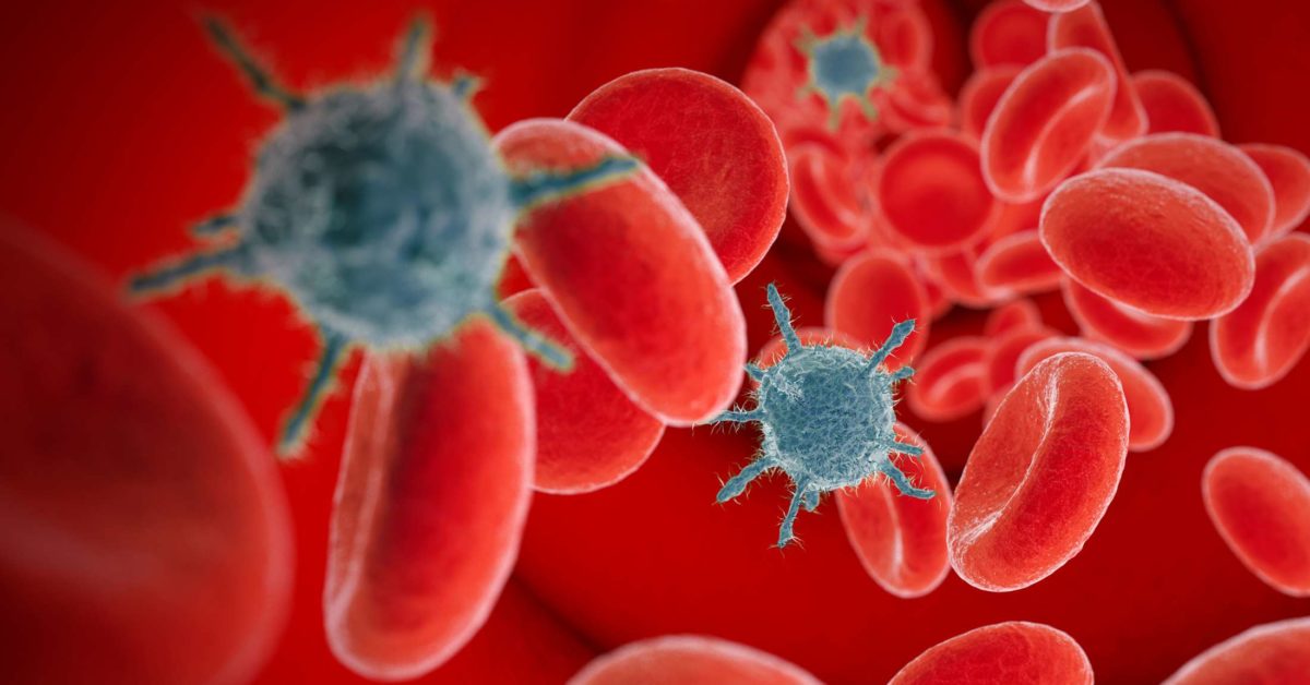
Adenovirus Infections, Human
Adenoviruses, Human
Adenoviridae
Adenovirus E1A Proteins
Adenovirus E1B Proteins
Adenovirus E4 Proteins
Adenovirus Early Proteins
Coxsackie and Adenovirus Receptor-Like Membrane Protein
Adenovirus E3 Proteins
Mastadenovirus
Genetic Vectors
HeLa Cells
Receptors, Virus
Adenovirus E1 Proteins
Conjunctivitis
Gene Transfer Techniques
Virus Replication
Inclusion Bodies, Viral
Genetic Therapy
Conjunctivitis, Viral
Adenoviruses, Canine
Gastroenteritis
Oncogene Proteins, Viral
Peliosis Hepatis
Respiratory Tract Infections
Adenovirus E2 Proteins
Enterovirus
Integrin alphaV
Gene Expression Regulation, Viral
Organophosphonates
Cytopathogenic Effect, Viral
Cell Transformation, Viral
Base Sequence
Molecular Sequence Data
RNA, Messenger
Transcription, Genetic
Depsipeptides
Haplorhini
Antiviral Agents
Fowl adenovirus A
Complement Fixation Tests
Feces
Polymerase Chain Reaction
Transfection
Cells, Cultured
Cell Nucleus
Immunocompromised Host
Dependovirus
DNA-Binding Proteins
Tumor Cells, Cultured
Nasopharynx
Serotyping
Nucleic Acid Hybridization
Promoter Regions, Genetic
Microscopy, Electron
Fatal Outcome
Gene Expression Regulation
Oncolytic Virotherapy
Oncolytic Viruses
Gene Expression
Transplantation, Homologous
Transcription Factors
Transduction, Genetic
Cricetinae
Nuclear Proteins
Mutation
Disease Outbreaks
Liver
Diarrhea
Lung
Immunoenzyme Techniques
Apoptosis
Protein Biosynthesis
Amino Acid Sequence
Fluorescent Antibody Technique
Mice, Inbred C57BL
Immunosuppression
Hematopoietic Stem Cell Transplantation
Cytoplasm
Protein Binding
Immunohistochemistry
Bone Marrow Transplantation
Viral Load
Helper Viruses
Defective Viruses
Incidence
Strain variation in adenovirus serotypes 4 and 7a causing acute respiratory disease. (1/452)
In order to determine the suitability of vaccine strains established in the 1960s for a new vaccine, a comprehensive study of strain variation of adenovirus serotype 4 (AV 4) and AV 7 was undertaken. A 1,500-bp region of the hexon gene containing the AV neutralization epitopes from prototype, vaccine, and community-acquired strains and from wild-type strains from military personnel that cause acute respiratory disease (ARD) was sequenced and analyzed. The whole hexon gene from prototype strains, vaccine strains, and selected isolates was sequenced. AV 7 and AV 7a were found to have distinct genotypes, and all vaccine and wild-type strains recovered from 1963 to 1997 had the AV 7a genotype. There was no significant strain variation in the neutralization epitopes of the AV 7a genotype over a 42-year period. The evolution of AV 4 was more complex, with continuous genetic drift punctuated by replacement with a new strain. The current strain of AV 4, which has been in circulation since 1995, is significantly different from the AV 4 prototype and the vaccine strains. Genetic differences were confirmed to be antigenic differences by neutralization tests, which define the new strain as an AV 4 variant. A type-specific PCR for AV 4, AV 7/7a, and AV 21 was developed, and this PCR facilitated the rapid identification of isolates from outbreaks of ARD. (+info)Serotyping of adenoviruses on conjunctival scrapings by PCR and sequence analysis. (2/452)
To detect and identify adenovirus (Ad), we investigated hypervariable regions (HVRs) of Ad by using a combination of PCR and direct sequencing (PCR-sequence) method. Primers for nested PCR to amplify the conserved region in the hexon protein containing HVRs were designed based on hexon gene sequences derived from GenBank. These two primer sets amplified a DNA fragment of 7 HVRs from 16 prototypes of Ad, which were divided into five subgenera, including seven serotypes that are the predominant causative agents of acute conjunctivitis in Japan, and from 31 recent conjunctival scraping specimens from patients with adenoviral conjunctivitis. HVR DNA sequences were determined by means of universal sequence primers. Analysis of the predicted amino acid homology of HVRs among Ad prototypes suggested three regions, HVR4, -5, and -7, to be candidates for the neutralization epitopes. The clinical serotype of specimens was determined by the PCR-sequence method with reference to these three HVRs. The serotype determined according to this method was identical to that obtained by culture isolation and the neutralization test (NT) in all scraping samples, whereas the results of this method did not match PCR and restriction fragment length polymorphism (PCR-RFLP) analysis in five samples. It took only three days to detect Ad and to identify the serotype, in contrast to culture isolation-NT, which took at least 2 weeks. These findings indicate that our newly developed PCR-sequence method is applicable for the detection and serotyping of human Ads. (+info)Molecular and serological characterization of adenovirus genome type 7h isolated in Japan. (3/452)
In 1996, three adenovirus type 7 (Ad7) strains were isolated from children with fever and upper respiratory diseases in Japan. Restriction endonucleases (REs) analysis and PCR amplification of the E3 7.7 kDa ORF revealed that these strains were genotype Ad7h and closely related to an Argentine Ad7h strain, which has been reported to be highly virulent and so far predominant only in South America. These strains showed weak cross-neutralizing activity and specific haemagglutination-inhibition activity to Ad3 antiserum. The present findings suggest that Ad7h in South America has spread to other parts of the world. Since the seroprevalence to Ad7 in the current Japanese population is very low due to the absence of Ad7 circulation in Japan for decades, Ad7 outbreak as a typical case of re-emerging infectious diseases is a cause for serious concern. (+info)Adenoviruses from human immunodeficiency virus-infected individuals, including two strains that represent new candidate serotypes Ad50 and Ad51 of species B1 and D, respectively. (4/452)
Adenovirus (Ad) isolates from a large number of human immunodeficiency virus (HIV)-infected individuals were compared serologically and genetically with Ad isolates from immunocompetent patients. Between 1982 and 1994, stool and urine samples from 137 subjects with AIDS hospitalized in The Netherlands yielded 143 Ad strains. Forty additional Ad strains were obtained from 35 HIV-positive patients in Manchester, United Kingdom, in 1992 and 1993. Of these 183 HIV-associated Ad strains, 84% belonged to species D and 3% belonged to species C. These strains were compared with 2,301 Ad strains collected during general diagnostic examinations in The Netherlands from 1973 to 1992. Of the latter strains, 5% belonged to species D and 49% belonged to species C. Two of the Ads isolated from fecal specimens of AIDS patients represent new serotypes: candidate Ad serotype 50 (prototype strain, Wan) of subspecies B1 and candidate Ad serotype 51 (prototype strain, Bom) of species D. The DNA restriction enzyme patterns of strains Wan and Bom differed from the patterns of all established prototypes. (+info)Adenovirus infections in hematopoietic stem cell transplant recipients. (5/452)
We report a 12% incidence of adenovirus infections among 532 recipients of hematopoietic stem cell transplant (HSCT) from January 1986 through March 1997. The median time from day of stem cell infusion to first positive culture was 41 days. Recipients of allogeneic stem cells, as opposed to autologous stem cell recipients, were more likely to have a culture positive for adenovirus (16% vs. 3%; P<.0001). Pediatric patients were also more likely than adults to have a positive culture (23% vs. 9%; P<.0001). Among stem cell recipients with partially matched related donors, pediatric recipients appear to be at significantly greater risk for infection than adult recipients (P<.001). Positive cultures were associated with evidence of invasion in 64% of cases (41 of 64). A multiple logistic regression analysis showed that isolating adenovirus from more than 1 site correlated with greater risk for invasive infections (P=.002). Invasive infections were associated with poorer chance of survival. (+info)Large, persistent epidemic of adenovirus type 4-associated acute respiratory disease in U.S. army trainees. (6/452)
In May 1997, a large, persistent epidemic of adenovirus type 4-associated acute respiratory disease began at Fort Jackson, South Carolina, the largest army basic training center. The epidemic lasted until December and declined when vaccine administration resumed. More than 1,000 male and female trainees were hospitalized; 66.1% of those hospitalized had an adenovirus type 4 isolate. (+info)Efficacy of topical cidofovir on multiple adenoviral serotypes in the New Zealand rabbit ocular model. (7/452)
PURPOSE: The goal of the present study was to determine the efficacy of topical 0.5% cidofovir twice daily for 7 days on the replication of multiple adenovirus (Ad) serotypes of subgroup C (Ad1, Ad5, Ad6) in the New Zealand rabbit ocular model. METHODS: In duplicate experiments for each serotype, a total of 20 rabbits (Ad5) or 16 rabbits each (Ad1 and Ad6) were inoculated topically in both eyes, with 1.5 X 10(6) pfu/eye of the appropriate virus. Twenty-four hours later, the rabbits in each serotype group were randomly divided into two topical treatment groups: I, 0.5% cidofovir; II, control vehicle. Treatment was twice daily for 7 days. All eyes were cultured for virus on days 0, 1, 3, 4, 5, 7, 9, 11, and 14. RESULTS: Compared to the control, treatment with 0.5% cidofovir reduced the following: mean Ad titer (days 1 to 7) for Ad1 (6.3 +/- 20 x 10(1) versus 2.5 +/- 3.9 X 10² pfu/ml; P < 0.0003), Ad5 (3.4 +/-5.8 x 102 versus 1.6 +/- 2.0 x 10(3) pfu/ml; P < 0.000001), and Ad6 (1.2 +/- 5.1 x 10(2) versus 5.5 +/-14 x 10(2) pfu/ml; P = 0.015); reduced Ad-positive eyes/total for Adl [45/128 (35%) versus 84/128 (66%); P = 0.000002], Ad5 [84/160 (53%) versus 131/152 (86%); P < 0.000001], and Ad6 [36/128 (28%) versus 82/128 (64%); P < 0.000001]: and reduced the duration of Ad shedding forAdl (4.9 +/-1.9 versus 9.3 +/- 3.3 days; P < 0.00007), Ad5 (6.4 +/- 2.8 versus 11.5 +/- 2.3 days; P < 0.0001), and Ad6 (4.4 +/- 2.1 versus 8.4 +/- 2.5 days; P < 0.00004). CONCLUSIONS: Topical 0.5% cidofovir twice daily for 7 days demonstrated significant antiviral activity against multiple adenoviral serotypes (Ad1, Ad5, and Ad6) in the New Zealand rabbit ocular model. These in vivo data expand in vitro studies indicating the efficacy of cidofovir against different adenovirus serotypes and support its use in clinical trials. (+info)Molecular epidemiology of ocular isolates of adenovirus 8 obtained over nine years. (8/452)
Twenty nine strains of adenovirus 8 have been isolated over nine years in Strasbourg, France, 22 of which were from one private ophthalmologist. To assess a possible relation between these strains, the DNA of adenovirus was analysed by restriction fragment length polymorphism using eight different enzymes. Among these, three proved discriminant (Xba I, Bgl II, Eco RI) and made it possible to define 13 genotypes differing from each other by one to three DNA bands. Seven genotypes were unique isolates, while three, representing 16 strains, were isolated over five to eight years. All the genotypes but one were closely related, with 87% homology. All 13 differed from an adenovirus 8 strain from Lyon (homology 68-76%). This study confirmed the stability of adenovirus 8 in a given population. (+info)Adenoviruses are a group of viruses that commonly cause respiratory infections such as bronchitis, pneumonia, and fevers in humans. They can also cause conjunctivitis (pink eye), croup, and stomach and intestinal inflammation (gastroenteritis). Adenovirus infections are most common in children, but people of any age can be infected. The viruses spread through the air when an infected person coughs or sneezes, or through contact with contaminated surfaces or objects. There is no specific treatment for adenovirus infections, and most people recover on their own within a week or two. However, some people may develop more severe illness, particularly those with weakened immune systems. Preventive measures include frequent hand washing and avoiding close contact with infected individuals. Some adenoviruses can also cause serious diseases in people with compromised immune systems, such as transplant recipients and people undergoing cancer treatment. There are vaccines available to prevent some types of adenovirus infections in military recruits, who are at higher risk due to close living quarters and stress on the immune system from basic training.
Adenoviridae infections refer to diseases caused by members of the Adenoviridae family of viruses, which are non-enveloped, double-stranded DNA viruses. These viruses can infect a wide range of hosts, including humans, animals, and birds. In humans, adenovirus infections can cause a variety of symptoms, depending on the specific type of virus and the age and immune status of the infected individual.
Common manifestations of adenovirus infections in humans include:
1. Respiratory illness: Adenoviruses are a common cause of respiratory tract infections, such as bronchitis, pneumonia, and croup. They can also cause conjunctivitis (pink eye) and pharyngoconjunctival fever.
2. Gastrointestinal illness: Some types of adenoviruses can cause diarrhea, vomiting, and abdominal pain, particularly in children and immunocompromised individuals.
3. Genitourinary illness: Adenoviruses have been associated with urinary tract infections, hemorrhagic cystitis, and nephritis.
4. Eye infections: Epidemic keratoconjunctivitis is a severe form of conjunctivitis caused by certain adenovirus types.
5. Central nervous system infections: Adenoviruses have been linked to meningitis, encephalitis, and other neurological disorders, although these are rare.
Transmission of adenoviruses typically occurs through respiratory droplets, contaminated surfaces, or contaminated water. Preventive measures include good hygiene practices, such as handwashing and avoiding close contact with infected individuals. There is no specific treatment for adenovirus infections, but supportive care can help alleviate symptoms. In severe cases or in immunocompromised patients, antiviral therapy may be considered.
Adenoviruses, Human: A group of viruses that commonly cause respiratory illnesses, such as bronchitis, pneumonia, and croup, in humans. They can also cause conjunctivitis (pink eye), cystitis (bladder infection), and gastroenteritis (stomach and intestinal infection).
Human adenoviruses are non-enveloped, double-stranded DNA viruses that belong to the family Adenoviridae. There are more than 50 different types of human adenoviruses, which can be classified into seven species (A-G). Different types of adenoviruses tend to cause specific illnesses, such as respiratory or gastrointestinal infections.
Human adenoviruses are highly contagious and can spread through close personal contact, respiratory droplets, or contaminated surfaces. They can also be transmitted through contaminated water sources. Some people may become carriers of the virus and experience no symptoms but still spread the virus to others.
Most human adenovirus infections are mild and resolve on their own within a few days to a week. However, some types of adenoviruses can cause severe illness, particularly in people with weakened immune systems, such as infants, young children, older adults, and individuals with HIV/AIDS or organ transplants.
There are no specific antiviral treatments for human adenovirus infections, but supportive care, such as hydration, rest, and fever reduction, can help manage symptoms. Preventive measures include practicing good hygiene, such as washing hands frequently, avoiding close contact with sick individuals, and not sharing personal items like towels or utensils.
Adenoviridae is a family of viruses that includes many species that can cause various types of illnesses in humans and animals. These viruses are non-enveloped, meaning they do not have a lipid membrane, and have an icosahedral symmetry with a diameter of approximately 70-90 nanometers.
The genome of Adenoviridae is composed of double-stranded DNA, which contains linear chromosomes ranging from 26 to 45 kilobases in length. The family is divided into five genera: Mastadenovirus, Aviadenovirus, Atadenovirus, Siadenovirus, and Ichtadenovirus.
Human adenoviruses are classified under the genus Mastadenovirus and can cause a wide range of illnesses, including respiratory infections, conjunctivitis, gastroenteritis, and upper respiratory tract infections. Some serotypes have also been associated with more severe diseases such as hemorrhagic cystitis, hepatitis, and meningoencephalitis.
Adenoviruses are highly contagious and can be transmitted through respiratory droplets, fecal-oral route, or by contact with contaminated surfaces. They can also be spread through contaminated water sources. Infections caused by adenoviruses are usually self-limiting, but severe cases may require hospitalization and supportive care.
Adenovirus E1A proteins are the early region 1A proteins encoded by adenoviruses, a group of viruses that commonly cause respiratory infections in humans. The E1A proteins play a crucial role in the regulation of the viral life cycle and host cell response. They function as transcriptional regulators, interacting with various cellular proteins to modulate gene expression and promote viral replication.
There are two major E1A protein isoforms, 289R and 243R, which differ in their amino-terminal regions due to alternative splicing of the E1A mRNA. The 289R isoform contains an additional 46 amino acids at its N-terminus compared to the 243R isoform. Both isoforms share conserved regions, including a strong transcriptional activation domain and a binding domain for cellular proteins involved in transcriptional regulation, such as retinoblastoma protein (pRb) and p300/CBP.
The interaction between E1A proteins and pRb is particularly important because it leads to the release of E2F transcription factors, which are essential for the initiation of viral DNA replication. By binding and inactivating pRb, E1A proteins promote the expression of cell cycle-regulated genes that facilitate viral replication in dividing cells.
In summary, adenovirus E1A proteins are multifunctional regulatory proteins involved in the control of viral gene expression and host cell response during adenovirus infection. They manipulate cellular transcription factors and pathways to create a favorable environment for viral replication.
Adenovirus E1B proteins are proteins encoded by the early region 1B (E1B) gene of adenoviruses. There are two main E1B proteins, E1B-55kD and E1B-19kD, which play crucial roles during the viral life cycle and in tumorigenesis.
1. E1B-55kD: This protein is a potent transcriptional repressor that inhibits the expression of host cell genes involved in DNA damage response, apoptosis, and antiviral defense mechanisms. By doing so, it creates a favorable environment for viral replication and evades the host's immune surveillance. E1B-55kD also interacts with p53, a tumor suppressor protein, leading to its degradation and further contributing to oncogenesis.
2. E1B-19kD: This protein is involved in blocking apoptosis or programmed cell death, which would otherwise be triggered by the host's defense mechanisms during viral infection. E1B-19kD forms a complex with another adenoviral protein, E4orf6, and together they inhibit the activity of several pro-apoptotic proteins, thus promoting viral replication and persistence in the host cell.
In summary, Adenovirus E1B proteins are essential for the viral life cycle by counteracting host defense mechanisms, particularly through the inhibition of apoptosis and transcriptional repression. Additionally, their interaction with crucial cellular regulatory proteins like p53 contributes to oncogenic transformation in certain contexts.
Adenoviruses are a group of viruses that commonly cause respiratory infections, conjunctivitis, and gastroenteritis. The E4 proteins of adenoviruses are non-structural proteins encoded by the early region 4 (E4) of the adenovirus genome. These proteins play important roles during the viral life cycle, including regulation of viral transcription, DNA replication, and host cell response.
There are several E4 proteins expressed by adenoviruses, depending on the serotype, but some of the well-characterized ones include E4 ORF6, E4 ORF3, and E4 ORF1/2. These proteins have been shown to interact with various host cell factors and viral proteins to modulate the intracellular environment for efficient viral replication.
For example, E4 ORF6 interacts with the host cell protein p53 to inhibit its transcriptional activity, which helps to prevent premature apoptosis of infected cells. E4 ORF3 is involved in the regulation of viral DNA replication and also interacts with cellular proteins to modulate the host cell cycle. E4 ORF1/2 forms a complex that functions as a helicase during viral DNA replication.
Overall, adenovirus E4 proteins are important regulators of the viral life cycle and play a significant role in the pathogenesis of adenovirus infections.
Adenovirus early proteins refer to the viral proteins that are expressed by adenoviruses during the early phase of their replication cycle. Adenoviruses are a group of viruses that can cause various symptoms, such as respiratory illness, conjunctivitis, and gastroenteritis.
The adenovirus replication cycle is divided into two phases: the early phase and the late phase. During the early phase, which occurs shortly after the virus infects a host cell, the viral genome is transcribed and translated into early proteins that help to prepare the host cell for viral replication. These early proteins play various roles in regulating the host cell's transcription, translation, and DNA replication machinery, as well as inhibiting the host cell's antiviral response.
There are several different adenovirus early proteins that have been identified, each with its own specific function. For example, E1A is an early protein that acts as a transcriptional activator and helps to activate the expression of other viral genes. E1B is another early protein that functions as a DNA-binding protein and inhibits the host cell's apoptosis (programmed cell death) response.
Overall, adenovirus early proteins are critical for the efficient replication of the virus within host cells, and understanding their functions can provide valuable insights into the mechanisms of viral infection and pathogenesis.
The Coxsackie and Adenovirus Receptor (CAR) is a transmembrane protein that serves as a receptor for several viruses, including Coxsackieviruses and certain types of Adenoviruses. The "Coxsackie and Adenovirus Receptor-Like Membrane Protein" likely refers to a membrane protein that shares structural or functional similarities with the CAR protein.
The CAR protein is a member of the immunoglobulin superfamily and is widely expressed in various tissues, including the heart, lungs, and nervous system. It plays important roles in cell adhesion, tissue development, and repair, as well as serving as an entry point for certain viruses to infect cells.
The CAR-like membrane protein may have similar functions or structures to the CAR protein, but its specific identity and role are not clearly defined in the medical literature. It is possible that it could be a target for viral infection or play a role in cellular processes, but further research is needed to confirm these possibilities.
Adenoviruses are a group of viruses that commonly cause respiratory infections, conjunctivitis, and gastroenteritis. The E3 region of the adenovirus genome encodes several proteins that play important roles in the virus's life cycle and its interactions with the host cell.
The E3 proteins include:
1. E3-10.4K: This protein helps to prevent the infected cell from undergoing programmed cell death (apoptosis), allowing the virus to continue replicating.
2. E3-14.7K: This protein inhibits the host cell's antiviral response by blocking the activation of certain immune signaling pathways.
3. E3-14.5K: This protein helps to prevent the infected cell from presenting viral antigens on its surface, which would otherwise alert the immune system to the infection.
4. E3-19K: This protein helps to stabilize the virion and protect it from being broken down by host cell enzymes.
5. E3-gp19K: This protein is involved in the transport of newly synthesized viral proteins to the endoplasmic reticulum, where they can be assembled into new virions.
6. E3-RID: This protein helps to protect the virus from being neutralized by antibodies produced by the host's immune system.
Overall, the E3 proteins play important roles in helping the adenovirus evade the host's immune response and establish a successful infection.
A mastadenovirus is a type of virus that belongs to the family Adenoviridae and the genus Mastadenovirus. These viruses are known to infect mammals, including humans, and can cause a variety of diseases such as respiratory infections, conjunctivitis, and gastroenteritis.
Human mastadenoviruses are typically associated with mild illnesses, although some strains can cause more severe disease, particularly in individuals with weakened immune systems. The virus is usually transmitted through respiratory droplets or contact with contaminated surfaces.
Mastadenoviruses are non-enveloped viruses, which means they do not have a lipid membrane surrounding their protein capsid. They contain a double-stranded DNA genome that encodes for several proteins involved in the virus's replication and assembly. The virus replicates in the nucleus of infected cells and can cause cell lysis or transformation, leading to various clinical manifestations.
Overall, mastadenoviruses are a significant cause of human and animal diseases, and understanding their biology and epidemiology is essential for developing effective prevention and treatment strategies.
A genetic vector is a vehicle, often a plasmid or a virus, that is used to introduce foreign DNA into a host cell as part of genetic engineering or gene therapy techniques. The vector contains the desired gene or genes, along with regulatory elements such as promoters and enhancers, which are needed for the expression of the gene in the target cells.
The choice of vector depends on several factors, including the size of the DNA to be inserted, the type of cell to be targeted, and the efficiency of uptake and expression required. Commonly used vectors include plasmids, adenoviruses, retroviruses, and lentiviruses.
Plasmids are small circular DNA molecules that can replicate independently in bacteria. They are often used as cloning vectors to amplify and manipulate DNA fragments. Adenoviruses are double-stranded DNA viruses that infect a wide range of host cells, including human cells. They are commonly used as gene therapy vectors because they can efficiently transfer genes into both dividing and non-dividing cells.
Retroviruses and lentiviruses are RNA viruses that integrate their genetic material into the host cell's genome. This allows for stable expression of the transgene over time. Lentiviruses, a subclass of retroviruses, have the advantage of being able to infect non-dividing cells, making them useful for gene therapy applications in post-mitotic tissues such as neurons and muscle cells.
Overall, genetic vectors play a crucial role in modern molecular biology and medicine, enabling researchers to study gene function, develop new therapies, and modify organisms for various purposes.
HeLa cells are a type of immortalized cell line used in scientific research. They are derived from a cancer that developed in the cervical tissue of Henrietta Lacks, an African-American woman, in 1951. After her death, cells taken from her tumor were found to be capable of continuous division and growth in a laboratory setting, making them an invaluable resource for medical research.
HeLa cells have been used in a wide range of scientific studies, including research on cancer, viruses, genetics, and drug development. They were the first human cell line to be successfully cloned and are able to grow rapidly in culture, doubling their population every 20-24 hours. This has made them an essential tool for many areas of biomedical research.
It is important to note that while HeLa cells have been instrumental in numerous scientific breakthroughs, the story of their origin raises ethical questions about informed consent and the use of human tissue in research.
An Aviadenovirus is a type of virus that belongs to the family *Adenoviridae* and the genus *Aviadenovirus*. These viruses primarily infect avian species, such as birds, and can cause a variety of diseases. The genome of an Aviadenovirus is double-stranded DNA. Some species of Aviadenoviruses have been known to cause respiratory and reproductive problems in poultry, leading to significant economic losses in the poultry industry. It's important to note that Aviadenoviruses are not known to infect or cause disease in humans.
Viral DNA refers to the genetic material present in viruses that consist of DNA as their core component. Deoxyribonucleic acid (DNA) is one of the two types of nucleic acids that are responsible for storing and transmitting genetic information in living organisms. Viruses are infectious agents much smaller than bacteria that can only replicate inside the cells of other organisms, called hosts.
Viral DNA can be double-stranded (dsDNA) or single-stranded (ssDNA), depending on the type of virus. Double-stranded DNA viruses have a genome made up of two complementary strands of DNA, while single-stranded DNA viruses contain only one strand of DNA.
Examples of dsDNA viruses include Adenoviruses, Herpesviruses, and Poxviruses, while ssDNA viruses include Parvoviruses and Circoviruses. Viral DNA plays a crucial role in the replication cycle of the virus, encoding for various proteins necessary for its multiplication and survival within the host cell.
Virus receptors are specific molecules (commonly proteins) on the surface of host cells that viruses bind to in order to enter and infect those cells. This interaction between the virus and its receptor is a critical step in the infection process. Different types of viruses have different receptor requirements, and identifying these receptors can provide important insights into the biology of the virus and potential targets for antiviral therapies.
Adenovirus E1 proteins are the earliest expressed and most critical proteins in the replication cycle of adenoviruses. The "E" stands for "early," indicating that these proteins are produced before the viral DNA begins to replicate. The E1 proteins play a crucial role in regulating the viral life cycle, altering cellular processes to support efficient viral replication, and inhibiting the host's antiviral responses.
The adenovirus E1 proteins are divided into two groups: E1A and E1B.
1. E1A proteins: These proteins are involved in transactivating various viral and cellular promoters, which leads to the expression of early and late viral genes. They also interact with several cellular proteins to alter the host cell cycle, promote cell growth, and inhibit apoptosis (programmed cell death). E1A proteins are essential for efficient viral replication and can transform cells in culture, contributing to adenovirus-induced tumorigenesis in certain animal models.
2. E1B proteins: These proteins have multiple functions during the viral life cycle. E1B 55kDa protein is a potent inhibitor of apoptosis and contributes to efficient viral replication by preventing premature cell death. It also interacts with several cellular proteins, including tumor suppressor p53, to modulate their functions. The E1B 19kDa protein, on the other hand, is a DNA-binding protein that plays a role in viral mRNA processing and export from the nucleus.
Together, adenovirus E1 proteins are essential for successful viral replication and manipulate host cellular processes to create a favorable environment for viral propagation. Understanding their functions has contributed significantly to our knowledge of viral pathogenesis and cancer biology.
Viral pneumonia is a type of pneumonia caused by viral infection. It primarily affects the upper and lower respiratory tract, leading to inflammation of the alveoli (air sacs) in the lungs. This results in symptoms such as cough, difficulty breathing, fever, fatigue, and chest pain. Common viruses that can cause pneumonia include influenza virus, respiratory syncytial virus (RSV), and adenovirus. Viral pneumonia is often milder than bacterial pneumonia but can still be serious, especially in young children, older adults, and people with weakened immune systems. Treatment typically involves supportive care, such as rest, hydration, and fever reduction, while the body fights off the virus. In some cases, antiviral medications may be used to help manage symptoms and prevent complications.
Capsid proteins are the structural proteins that make up the capsid, which is the protective shell of a virus. The capsid encloses the viral genome and helps to protect it from degradation and detection by the host's immune system. Capsid proteins are typically arranged in a symmetrical pattern and can self-assemble into the capsid structure when exposed to the viral genome.
The specific arrangement and composition of capsid proteins vary between different types of viruses, and they play important roles in the virus's life cycle, including recognition and binding to host cells, entry into the cell, and release of the viral genome into the host cytoplasm. Capsid proteins can also serve as targets for antiviral therapies and vaccines.
Conjunctivitis is an inflammation or infection of the conjunctiva, a thin, clear membrane that covers the inner surface of the eyelids and the outer surface of the eye. The condition can cause redness, itching, burning, tearing, discomfort, and a gritty feeling in the eyes. It can also result in a discharge that can be clear, yellow, or greenish.
Conjunctivitis can have various causes, including bacterial or viral infections, allergies, irritants (such as smoke, chlorine, or contact lens solutions), and underlying medical conditions (like dry eye or autoimmune disorders). Treatment depends on the cause of the condition but may include antibiotics, antihistamines, anti-inflammatory medications, or warm compresses.
It is essential to maintain good hygiene practices, like washing hands frequently and avoiding touching or rubbing the eyes, to prevent spreading conjunctivitis to others. If you suspect you have conjunctivitis, it's recommended that you consult an eye care professional for a proper diagnosis and treatment plan.
A cell line is a culture of cells that are grown in a laboratory for use in research. These cells are usually taken from a single cell or group of cells, and they are able to divide and grow continuously in the lab. Cell lines can come from many different sources, including animals, plants, and humans. They are often used in scientific research to study cellular processes, disease mechanisms, and to test new drugs or treatments. Some common types of human cell lines include HeLa cells (which come from a cancer patient named Henrietta Lacks), HEK293 cells (which come from embryonic kidney cells), and HUVEC cells (which come from umbilical vein endothelial cells). It is important to note that cell lines are not the same as primary cells, which are cells that are taken directly from a living organism and have not been grown in the lab.
Gene transfer techniques, also known as gene therapy, refer to medical procedures where genetic material is introduced into an individual's cells or tissues to treat or prevent diseases. This can be achieved through various methods:
1. **Viral Vectors**: The most common method uses modified viruses, such as adenoviruses, retroviruses, or lentiviruses, to carry the therapeutic gene into the target cells. The virus infects the cell and inserts the new gene into the cell's DNA.
2. **Non-Viral Vectors**: These include methods like electroporation (using electric fields to create pores in the cell membrane), gene guns (shooting gold particles coated with DNA into cells), or liposomes (tiny fatty bubbles that can enclose DNA).
3. **Direct Injection**: In some cases, the therapeutic gene can be directly injected into a specific tissue or organ.
The goal of gene transfer techniques is to supplement or replace a faulty gene with a healthy one, thereby correcting the genetic disorder. However, these techniques are still largely experimental and have their own set of challenges, including potential immune responses, issues with accurate targeting, and risks of mutations or cancer development.
Virus replication is the process by which a virus produces copies or reproduces itself inside a host cell. This involves several steps:
1. Attachment: The virus attaches to a specific receptor on the surface of the host cell.
2. Penetration: The viral genetic material enters the host cell, either by invagination of the cell membrane or endocytosis.
3. Uncoating: The viral genetic material is released from its protective coat (capsid) inside the host cell.
4. Replication: The viral genetic material uses the host cell's machinery to produce new viral components, such as proteins and nucleic acids.
5. Assembly: The newly synthesized viral components are assembled into new virus particles.
6. Release: The newly formed viruses are released from the host cell, often through lysis (breaking) of the cell membrane or by budding off the cell membrane.
The specific mechanisms and details of virus replication can vary depending on the type of virus. Some viruses, such as DNA viruses, use the host cell's DNA polymerase to replicate their genetic material, while others, such as RNA viruses, use their own RNA-dependent RNA polymerase or reverse transcriptase enzymes. Understanding the process of virus replication is important for developing antiviral therapies and vaccines.
Inclusion bodies, viral are typically described as intracellular inclusions that appear as a result of viral infections. These inclusion bodies consist of aggregates of virus-specific proteins, viral particles, or both, which accumulate inside the host cell's cytoplasm or nucleus during the replication cycle of certain viruses.
The presence of inclusion bodies can sometimes be observed through histological or cytological examination using various staining techniques. Different types of viruses may exhibit distinct morphologies and locations of these inclusion bodies, which can aid in the identification and diagnosis of specific viral infections. However, it is important to note that not all viral infections result in the formation of inclusion bodies, and their presence does not necessarily indicate active viral replication or infection.
Genetic therapy, also known as gene therapy, is a medical intervention that involves the use of genetic material, such as DNA or RNA, to treat or prevent diseases. It works by introducing functional genes into cells to replace missing or faulty ones caused by genetic disorders or mutations. The introduced gene is incorporated into the recipient's genome, allowing for the production of a therapeutic protein that can help manage the disease symptoms or even cure the condition.
There are several approaches to genetic therapy, including:
1. Replacing a faulty gene with a healthy one
2. Inactivating or "silencing" a dysfunctional gene causing a disease
3. Introducing a new gene into the body to help fight off a disease, such as cancer
Genetic therapy holds great promise for treating various genetic disorders, including cystic fibrosis, muscular dystrophy, hemophilia, and certain types of cancer. However, it is still an evolving field with many challenges, such as efficient gene delivery, potential immune responses, and ensuring the safety and long-term effectiveness of the therapy.
Viral conjunctivitis is an inflammation of the conjunctiva, the thin membrane that covers the white part of the eye (sclera) and the inner surface of the eyelids, caused by a viral infection. The condition is often characterized by redness, watering, gritty or burning sensation in the eyes, and a clear, watery discharge. In some cases, it may also cause swelling of the eyelids and light sensitivity.
The most common viruses that can cause conjunctivitis are adenoviruses, which are responsible for about 65-90% of all viral conjunctivitis cases. Other viruses that can cause the condition include herpes simplex virus, varicella-zoster virus (which causes chickenpox and shingles), and picornaviruses.
Viral conjunctivitis is highly contagious and can spread easily through direct contact with infected individuals or contaminated surfaces. It typically affects one eye first and then spreads to the other eye within a few days. The condition usually resolves on its own within 1-2 weeks, although in some cases it may take longer to clear up completely.
There is no specific treatment for viral conjunctivitis, and antibiotics are not effective against viral infections. However, cool compresses and artificial tears can help alleviate symptoms such as discomfort and dryness. It is important to practice good hygiene, such as washing hands frequently and avoiding touching the eyes, to prevent the spread of the virus to others.
Canine adenoviruses are a type of virus that can infect dogs and cause two distinct diseases: Infectious Canine Hepatitis (type 1) and Canine Respiratory Disease Complex (type 2).
Canine adenovirus type 1 primarily affects the liver, causing symptoms such as vomiting, diarrhea, loss of appetite, and abdominal pain. In severe cases, it can lead to liver failure and death.
Canine adenovirus type 2 mainly causes respiratory infections, including kennel cough, which is characterized by a harsh, hacking cough and nasal discharge. It can also cause pneumonia in some cases.
Both types of canine adenoviruses are highly contagious and can be spread through direct contact with infected dogs or their feces and urine. Vaccination is available to protect against both forms of the virus and is recommended for all dogs.
Gastroenteritis is not a medical condition itself, but rather a symptom-based description of inflammation in the gastrointestinal tract, primarily involving the stomach and intestines. It's often referred to as "stomach flu," although it's not caused by influenza virus.
Medically, gastroenteritis is defined as an inflammation of the mucous membrane of the stomach and intestines, usually resulting in symptoms such as diarrhea, abdominal cramps, nausea, vomiting, fever, and dehydration. This condition can be caused by various factors, including viral (like rotavirus or norovirus), bacterial (such as Salmonella, Shigella, or Escherichia coli), or parasitic infections, food poisoning, allergies, or the use of certain medications.
Gastroenteritis is generally self-limiting and resolves within a few days with proper hydration and rest. However, severe cases may require medical attention to prevent complications like dehydration, which can be particularly dangerous for young children, older adults, and individuals with weakened immune systems.
Oncogene proteins, viral, are cancer-causing proteins that are encoded by the genetic material (DNA or RNA) of certain viruses. These viral oncogenes can be acquired through infection with retroviruses, such as human immunodeficiency virus (HIV), human T-cell leukemia virus (HTLV), and certain types of papillomaviruses and polyomaviruses.
When these viruses infect host cells, they can integrate their genetic material into the host cell's genome, leading to the expression of viral oncogenes. These oncogenes may then cause uncontrolled cell growth and division, ultimately resulting in the formation of tumors or cancers. The process by which viruses contribute to cancer development is complex and involves multiple steps, including the alteration of signaling pathways that regulate cell proliferation, differentiation, and survival.
Examples of viral oncogenes include the v-src gene found in the Rous sarcoma virus (RSV), which causes chicken sarcoma, and the E6 and E7 genes found in human papillomaviruses (HPVs), which are associated with cervical cancer and other anogenital cancers. Understanding viral oncogenes and their mechanisms of action is crucial for developing effective strategies to prevent and treat virus-associated cancers.
Peliosis hepatis is a rare condition characterized by the presence of numerous blood-filled cavities or sinusoids in the liver. These cavities can vary in size and are usually distributed throughout the liver parenchyma. The term "peliosis" comes from the Greek word "pelios," which means "bluish-black."
In peliosis hepatis, these blood-filled spaces can rupture, leading to bleeding into the liver tissue or even into the abdominal cavity. This condition is often asymptomatic but may present with nonspecific symptoms such as fatigue, weakness, abdominal pain, or liver dysfunction.
Peliosis hepatis has been associated with various conditions, including blood disorders (such as leukemia and lymphoma), use of anabolic steroids, immunosuppressive therapy, chronic infections (such as HIV and tuberculosis), and certain malignancies. In many cases, the cause remains unknown, and the condition is referred to as idiopathic peliosis hepatis.
Diagnosis of peliosis hepatis typically requires imaging studies such as computed tomography (CT) or magnetic resonance imaging (MRI), which can demonstrate the presence of multiple, round, blood-filled spaces in the liver. In some cases, a liver biopsy may be necessary to confirm the diagnosis and evaluate the severity of the condition. Treatment depends on the underlying cause and may include discontinuation of any offending medications or treatment of any underlying conditions.
Respiratory tract infections (RTIs) are infections that affect the respiratory system, which includes the nose, throat (pharynx), voice box (larynx), windpipe (trachea), bronchi, and lungs. These infections can be caused by viruses, bacteria, or, less commonly, fungi.
RTIs are classified into two categories based on their location: upper respiratory tract infections (URTIs) and lower respiratory tract infections (LRTIs). URTIs include infections of the nose, sinuses, throat, and larynx, such as the common cold, flu, laryngitis, and sinusitis. LRTIs involve the lower airways, including the bronchi and lungs, and can be more severe. Examples of LRTIs are pneumonia, bronchitis, and bronchiolitis.
Symptoms of RTIs depend on the location and cause of the infection but may include cough, congestion, runny nose, sore throat, difficulty breathing, wheezing, fever, fatigue, and chest pain. Treatment for RTIs varies depending on the severity and underlying cause of the infection. For viral infections, treatment typically involves supportive care to manage symptoms, while antibiotics may be prescribed for bacterial infections.
A viral RNA (ribonucleic acid) is the genetic material found in certain types of viruses, as opposed to viruses that contain DNA (deoxyribonucleic acid). These viruses are known as RNA viruses. The RNA can be single-stranded or double-stranded and can exist as several different forms, such as positive-sense, negative-sense, or ambisense RNA. Upon infecting a host cell, the viral RNA uses the host's cellular machinery to translate the genetic information into proteins, leading to the production of new virus particles and the continuation of the viral life cycle. Examples of human diseases caused by RNA viruses include influenza, COVID-19 (SARS-CoV-2), hepatitis C, and polio.
Adenoviruses are a group of viruses that commonly cause respiratory infections, conjunctivitis, and gastroenteritis. The E2 proteins of adenoviruses are involved in the replication of the viral genome. Specifically, E2 consists of three proteins: E2a, E2b, and E2c.
E2a is a single-stranded DNA-binding protein that binds to the origin of replication on the viral genome and recruits other viral and cellular proteins necessary for replication. E2b is a DNA polymerase processivity factor that interacts with the viral DNA polymerase and increases its processivity, allowing for efficient synthesis of new viral DNA. E2c is a helicase that unwinds the double-stranded DNA at the replication fork, enabling the synthesis of new strands.
Together, these proteins play a critical role in the replication of adenoviruses and are important targets for the development of antiviral therapies.
An enterovirus is a type of virus that primarily infects the gastrointestinal tract. There are over 100 different types of enteroviruses, including polioviruses, coxsackieviruses, echoviruses, and newer enteroviruses such as EV-D68 and EV-A71. These viruses are typically spread through close contact with an infected person, or by consuming food or water contaminated with the virus.
While many people infected with enteroviruses may not experience any symptoms, some may develop mild to severe illnesses such as hand, foot and mouth disease, herpangina, meningitis, encephalitis, myocarditis, and paralysis (in case of poliovirus). Infection can occur in people of all ages, but young children are more susceptible to infection and severe illness.
Prevention measures include practicing good hygiene, such as washing hands frequently with soap and water, avoiding close contact with sick individuals, and not sharing food or drinks with someone who is ill. There are also vaccines available to prevent poliovirus infection.
Viral genes refer to the genetic material present in viruses that contains the information necessary for their replication and the production of viral proteins. In DNA viruses, the genetic material is composed of double-stranded or single-stranded DNA, while in RNA viruses, it is composed of single-stranded or double-stranded RNA.
Viral genes can be classified into three categories: early, late, and structural. Early genes encode proteins involved in the replication of the viral genome, modulation of host cell processes, and regulation of viral gene expression. Late genes encode structural proteins that make up the viral capsid or envelope. Some viruses also have structural genes that are expressed throughout their replication cycle.
Understanding the genetic makeup of viruses is crucial for developing antiviral therapies and vaccines. By targeting specific viral genes, researchers can develop drugs that inhibit viral replication and reduce the severity of viral infections. Additionally, knowledge of viral gene sequences can inform the development of vaccines that stimulate an immune response to specific viral proteins.
A capsid is the protein shell that encloses and protects the genetic material of a virus. It is composed of multiple copies of one or more proteins that are arranged in a specific structure, which can vary in shape and symmetry depending on the type of virus. The capsid plays a crucial role in the viral life cycle, including protecting the viral genome from host cell defenses, mediating attachment to and entry into host cells, and assisting with the assembly of new virus particles during replication.
Integrin αV (also known as ITGAV or CD51) is a subunit of a family of heterodimeric transmembrane receptors called integrins, which are involved in cell-cell and cell-extracellular matrix (ECM) interactions. Integrin αV combines with various β subunits (e.g., β1, β3, β5, β6, and β8) to form distinct integrin heterodimers, such as αVβ1, αVβ3, αVβ5, αVβ6, and αVβ8. These integrins recognize and bind to specific arginine-glycine-aspartic acid (RGD) sequences present in various ECM proteins, such as fibronectin, vitronectin, osteopontin, and collagens. Integrin αV plays crucial roles in cell adhesion, migration, proliferation, differentiation, survival, and angiogenesis, and has been implicated in several pathological processes, including cancer, fibrosis, and inflammation.
Gene expression regulation, viral, refers to the processes that control the production of viral gene products, such as proteins and nucleic acids, during the viral life cycle. This can involve both viral and host cell factors that regulate transcription, RNA processing, translation, and post-translational modifications of viral genes.
Viral gene expression regulation is critical for the virus to replicate and produce progeny virions. Different types of viruses have evolved diverse mechanisms to regulate their gene expression, including the use of promoters, enhancers, transcription factors, RNA silencing, and epigenetic modifications. Understanding these regulatory processes can provide insights into viral pathogenesis and help in the development of antiviral therapies.
Organophosphonates are a class of organic compounds characterized by the presence of a carbon-phosphorus bond. They contain a phosphonic acid group, which consists of a phosphorus atom bonded to four oxygen or nitrogen atoms, with one of those bonds being replaced by a carbon atom.
In a medical context, organophosphonates are commonly used as radiopharmaceuticals in diagnostic nuclear medicine procedures, such as bone scans. These compounds have the ability to bind to hydroxyapatite, the mineral component of bones, and can be labeled with radioactive isotopes for imaging purposes. They may also be used in therapeutic settings, including as treatments for conditions such as tumor-induced hypercalcemia and Paget's disease of bone.
It is important to note that organophosphonates are distinct from organophosphates, another class of compounds that contain a phosphorus atom bonded to three oxygen or sulfur atoms and one carbon atom. Organophosphates have been widely used as pesticides and chemical warfare agents, and can pose significant health risks due to their toxicity.
Viral proteins are the proteins that are encoded by the viral genome and are essential for the viral life cycle. These proteins can be structural or non-structural and play various roles in the virus's replication, infection, and assembly process. Structural proteins make up the physical structure of the virus, including the capsid (the protein shell that surrounds the viral genome) and any envelope proteins (that may be present on enveloped viruses). Non-structural proteins are involved in the replication of the viral genome and modulation of the host cell environment to favor viral replication. Overall, a thorough understanding of viral proteins is crucial for developing antiviral therapies and vaccines.
A Cytopathic Effect (CPE) is a visible change in the cell or group of cells due to infection by a pathogen, such as a virus. When the cytopathic effect is caused specifically by a viral infection, it is referred to as a "Viral Cytopathic Effect" (VCPE).
The VCPE can include various changes in the cell's morphology, size, and structure, such as rounding, shrinkage, multinucleation, inclusion bodies, and formation of syncytia (multinucleated giant cells). These changes are often used to identify and characterize viruses in laboratory settings.
The VCPE is typically observed under a microscope after the virus has infected cell cultures, and it can help researchers determine the type of virus, the degree of infection, and the effectiveness of antiviral treatments. The severity and timing of the VCPE can vary depending on the specific virus and the type of cells that are infected.
Cytosine is one of the four nucleobases in the nucleic acid molecules DNA and RNA, along with adenine, guanine, and thymine (in DNA) or uracil (in RNA). The single-letter abbreviation for cytosine is "C."
Cytosine base pairs specifically with guanine through hydrogen bonding, forming a base pair. In DNA, the double helix consists of two complementary strands of nucleotides held together by these base pairs, such that the sequence of one strand determines the sequence of the other. This property is critical for DNA replication and transcription, processes that are essential for life.
Cytosine residues in DNA can undergo spontaneous deamination to form uracil, which can lead to mutations if not corrected by repair mechanisms. In RNA, cytosine can be methylated at the 5-carbon position to form 5-methylcytosine, a modification that plays a role in regulating gene expression and other cellular processes.
Cell transformation, viral refers to the process by which a virus causes normal cells to become cancerous or tumorigenic. This occurs when the genetic material of the virus integrates into the DNA of the host cell and alters its regulation, leading to uncontrolled cell growth and division. Some viruses known to cause cell transformation include human papillomavirus (HPV), hepatitis B virus (HBV), and certain types of herpesviruses.
A base sequence in the context of molecular biology refers to the specific order of nucleotides in a DNA or RNA molecule. In DNA, these nucleotides are adenine (A), guanine (G), cytosine (C), and thymine (T). In RNA, uracil (U) takes the place of thymine. The base sequence contains genetic information that is transcribed into RNA and ultimately translated into proteins. It is the exact order of these bases that determines the genetic code and thus the function of the DNA or RNA molecule.
Molecular sequence data refers to the specific arrangement of molecules, most commonly nucleotides in DNA or RNA, or amino acids in proteins, that make up a biological macromolecule. This data is generated through laboratory techniques such as sequencing, and provides information about the exact order of the constituent molecules. This data is crucial in various fields of biology, including genetics, evolution, and molecular biology, allowing for comparisons between different organisms, identification of genetic variations, and studies of gene function and regulation.
Messenger RNA (mRNA) is a type of RNA (ribonucleic acid) that carries genetic information copied from DNA in the form of a series of three-base code "words," each of which specifies a particular amino acid. This information is used by the cell's machinery to construct proteins, a process known as translation. After being transcribed from DNA, mRNA travels out of the nucleus to the ribosomes in the cytoplasm where protein synthesis occurs. Once the protein has been synthesized, the mRNA may be degraded and recycled. Post-transcriptional modifications can also occur to mRNA, such as alternative splicing and addition of a 5' cap and a poly(A) tail, which can affect its stability, localization, and translation efficiency.
Adenoviruses, Porcine:
A group of viruses that primarily infect pigs and cause a variety of symptoms, such as respiratory illness, diarrhea, vomiting, and reproductive failure. They belong to the family Adenoviridae and are non-enveloped, double-stranded DNA viruses. Porcine adenoviruses can be classified into several serotypes, with different serotypes causing different disease manifestations. Some porcine adenoviruses have been associated with economic losses in the swine industry due to their impact on growth and mortality rates. They are primarily transmitted through the fecal-oral route and can also be found in contaminated environments. Currently, there are no specific treatments for porcine adenovirus infections, and control measures focus on preventing transmission through good biosecurity practices.
Genetic transcription is the process by which the information in a strand of DNA is used to create a complementary RNA molecule. This process is the first step in gene expression, where the genetic code in DNA is converted into a form that can be used to produce proteins or functional RNAs.
During transcription, an enzyme called RNA polymerase binds to the DNA template strand and reads the sequence of nucleotide bases. As it moves along the template, it adds complementary RNA nucleotides to the growing RNA chain, creating a single-stranded RNA molecule that is complementary to the DNA template strand. Once transcription is complete, the RNA molecule may undergo further processing before it can be translated into protein or perform its functional role in the cell.
Transcription can be either "constitutive" or "regulated." Constitutive transcription occurs at a relatively constant rate and produces essential proteins that are required for basic cellular functions. Regulated transcription, on the other hand, is subject to control by various intracellular and extracellular signals, allowing cells to respond to changing environmental conditions or developmental cues.
Depsipeptides are a type of naturally occurring or synthetic modified peptides that contain at least one amide bond replaced by an ester bond in their structure. These compounds exhibit diverse biological activities, including antimicrobial, antiviral, and antitumor properties. Some depsipeptides have been developed as pharmaceutical drugs for the treatment of various diseases.
Haplorhini is a term used in the field of primatology and physical anthropology to refer to a parvorder of simian primates, which includes humans, apes (both great and small), and Old World monkeys. The name "Haplorhini" comes from the Greek words "haploos," meaning single or simple, and "rhinos," meaning nose.
The defining characteristic of Haplorhini is the presence of a simple, dry nose, as opposed to the wet, fleshy noses found in other primates, such as New World monkeys and strepsirrhines (which include lemurs and lorises). The nostrils of haplorhines are located close together at the tip of the snout, and they lack the rhinarium or "wet nose" that is present in other primates.
Haplorhini is further divided into two infraorders: Simiiformes (which includes apes and Old World monkeys) and Tarsioidea (which includes tarsiers). These groups are distinguished by various anatomical and behavioral differences, such as the presence or absence of a tail, the structure of the hand and foot, and the degree of sociality.
Overall, Haplorhini is a group of primates that share a number of distinctive features related to their sensory systems, locomotion, and social behavior. Understanding the evolutionary history and diversity of this group is an important area of research in anthropology, biology, and psychology.
Antiviral agents are a class of medications that are designed to treat infections caused by viruses. Unlike antibiotics, which target bacteria, antiviral agents interfere with the replication and infection mechanisms of viruses, either by inhibiting their ability to replicate or by modulating the host's immune response to the virus.
Antiviral agents are used to treat a variety of viral infections, including influenza, herpes simplex virus (HSV) infections, human immunodeficiency virus (HIV) infection, hepatitis B and C, and respiratory syncytial virus (RSV) infections.
These medications can be administered orally, intravenously, or topically, depending on the type of viral infection being treated. Some antiviral agents are also used for prophylaxis, or prevention, of certain viral infections.
It is important to note that antiviral agents are not effective against all types of viruses and may have significant side effects. Therefore, it is essential to consult with a healthcare professional before starting any antiviral therapy.
Fowl adenovirus A, also known as Fowl aviadenovirus serotype 1 or Fowl adenovirus serotype 1 (FAdV-A), is a species of DNA virus that belongs to the family Adenoviridae and genus Aviadenovirus. It primarily infects birds, particularly chickens, causing various clinical manifestations such as inclusion body hepatitis (IBH) and hydropericardium syndrome (HPS). The virus is transmitted horizontally through the fecal-oral route and can be found in the environment for extended periods. FAdV-A infection can lead to significant economic losses in the poultry industry due to high mortality rates, especially in young chickens.
A transgene is a segment of DNA that has been artificially transferred from one organism to another, typically between different species, to introduce a new trait or characteristic. The term "transgene" specifically refers to the genetic material that has been transferred and has become integrated into the host organism's genome. This technology is often used in genetic engineering and biomedical research, including the development of genetically modified organisms (GMOs) for agricultural purposes or the creation of animal models for studying human diseases.
Transgenes can be created using various techniques, such as molecular cloning, where a desired gene is isolated, manipulated, and then inserted into a vector (a small DNA molecule, such as a plasmid) that can efficiently enter the host organism's cells. Once inside the cell, the transgene can integrate into the host genome, allowing for the expression of the new trait in the resulting transgenic organism.
It is important to note that while transgenes can provide valuable insights and benefits in research and agriculture, their use and release into the environment are subjects of ongoing debate due to concerns about potential ecological impacts and human health risks.
Complement fixation tests are a type of laboratory test used in immunology and serology to detect the presence of antibodies in a patient's serum. These tests are based on the principle of complement activation, which is a part of the immune response. The complement system consists of a group of proteins that work together to help eliminate pathogens from the body.
In a complement fixation test, the patient's serum is mixed with a known antigen and complement proteins. If the patient has antibodies against the antigen, they will bind to it and activate the complement system. This results in the consumption or "fixation" of the complement proteins, which are no longer available to participate in a secondary reaction.
A second step involves adding a fresh source of complement proteins and a dye-labeled antibody that recognizes a specific component of the complement system. If complement was fixed during the first step, it will not be available for this secondary reaction, and the dye-labeled antibody will remain unbound. Conversely, if no antibodies were present in the patient's serum, the complement proteins would still be available for the second reaction, leading to the binding of the dye-labeled antibody.
The mixture is then examined under a microscope or using a spectrophotometer to determine whether the dye-labeled antibody has bound. If it has not, this indicates that the patient's serum contains antibodies specific to the antigen used in the test, and a positive result is recorded.
Complement fixation tests have been widely used for the diagnosis of various infectious diseases, such as syphilis, measles, and influenza. However, they have largely been replaced by more modern serological techniques, like enzyme-linked immunosorbent assays (ELISAs) and nucleic acid amplification tests (NAATs), due to their increased sensitivity, specificity, and ease of use.
Feces are the solid or semisolid remains of food that could not be digested or absorbed in the small intestine, along with bacteria and other waste products. After being stored in the colon, feces are eliminated from the body through the rectum and anus during defecation. Feces can vary in color, consistency, and odor depending on a person's diet, health status, and other factors.
Polymerase Chain Reaction (PCR) is a laboratory technique used to amplify specific regions of DNA. It enables the production of thousands to millions of copies of a particular DNA sequence in a rapid and efficient manner, making it an essential tool in various fields such as molecular biology, medical diagnostics, forensic science, and research.
The PCR process involves repeated cycles of heating and cooling to separate the DNA strands, allow primers (short sequences of single-stranded DNA) to attach to the target regions, and extend these primers using an enzyme called Taq polymerase, resulting in the exponential amplification of the desired DNA segment.
In a medical context, PCR is often used for detecting and quantifying specific pathogens (viruses, bacteria, fungi, or parasites) in clinical samples, identifying genetic mutations or polymorphisms associated with diseases, monitoring disease progression, and evaluating treatment effectiveness.
An antigen is any substance that can stimulate an immune response, particularly the production of antibodies. Viral antigens are antigens that are found on or produced by viruses. They can be proteins, glycoproteins, or carbohydrates present on the surface or inside the viral particle.
Viral antigens play a crucial role in the immune system's recognition and response to viral infections. When a virus infects a host cell, it may display its antigens on the surface of the infected cell. This allows the immune system to recognize and target the infected cells for destruction, thereby limiting the spread of the virus.
Viral antigens are also important targets for vaccines. Vaccines typically work by introducing a harmless form of a viral antigen to the body, which then stimulates the production of antibodies and memory T-cells that can recognize and respond quickly and effectively to future infections with the actual virus.
It's worth noting that different types of viruses have different antigens, and these antigens can vary between strains of the same virus. This is why there are often different vaccines available for different viral diseases, and why flu vaccines need to be updated every year to account for changes in the circulating influenza virus strains.
Transfection is a term used in molecular biology that refers to the process of deliberately introducing foreign genetic material (DNA, RNA or artificial gene constructs) into cells. This is typically done using chemical or physical methods, such as lipofection or electroporation. Transfection is widely used in research and medical settings for various purposes, including studying gene function, producing proteins, developing gene therapies, and creating genetically modified organisms. It's important to note that transfection is different from transduction, which is the process of introducing genetic material into cells using viruses as vectors.
"Cells, cultured" is a medical term that refers to cells that have been removed from an organism and grown in controlled laboratory conditions outside of the body. This process is called cell culture and it allows scientists to study cells in a more controlled and accessible environment than they would have inside the body. Cultured cells can be derived from a variety of sources, including tissues, organs, or fluids from humans, animals, or cell lines that have been previously established in the laboratory.
Cell culture involves several steps, including isolation of the cells from the tissue, purification and characterization of the cells, and maintenance of the cells in appropriate growth conditions. The cells are typically grown in specialized media that contain nutrients, growth factors, and other components necessary for their survival and proliferation. Cultured cells can be used for a variety of purposes, including basic research, drug development and testing, and production of biological products such as vaccines and gene therapies.
It is important to note that cultured cells may behave differently than they do in the body, and results obtained from cell culture studies may not always translate directly to human physiology or disease. Therefore, it is essential to validate findings from cell culture experiments using additional models and ultimately in clinical trials involving human subjects.
The cell nucleus is a membrane-bound organelle found in the eukaryotic cells (cells with a true nucleus). It contains most of the cell's genetic material, organized as DNA molecules in complex with proteins, RNA molecules, and histones to form chromosomes.
The primary function of the cell nucleus is to regulate and control the activities of the cell, including growth, metabolism, protein synthesis, and reproduction. It also plays a crucial role in the process of mitosis (cell division) by separating and protecting the genetic material during this process. The nuclear membrane, or nuclear envelope, surrounding the nucleus is composed of two lipid bilayers with numerous pores that allow for the selective transport of molecules between the nucleoplasm (nucleus interior) and the cytoplasm (cell exterior).
The cell nucleus is a vital structure in eukaryotic cells, and its dysfunction can lead to various diseases, including cancer and genetic disorders.
An immunocompromised host refers to an individual who has a weakened or impaired immune system, making them more susceptible to infections and decreased ability to fight off pathogens. This condition can be congenital (present at birth) or acquired (developed during one's lifetime).
Acquired immunocompromised states may result from various factors such as medical treatments (e.g., chemotherapy, radiation therapy, immunosuppressive drugs), infections (e.g., HIV/AIDS), chronic diseases (e.g., diabetes, malnutrition, liver disease), or aging.
Immunocompromised hosts are at a higher risk for developing severe and life-threatening infections due to their reduced immune response. Therefore, they require special consideration when it comes to prevention, diagnosis, and treatment of infectious diseases.
A dependovirus, also known as a dependent adenovirus or satellite adenovirus, is a type of virus that requires the presence of another virus, specifically an adenovirus, to replicate. Dependoviruses are small, non-enveloped viruses with a double-stranded DNA genome. They cannot complete their replication cycle without the help of an adenovirus, which provides necessary functions for the dependovirus to replicate.
Dependoviruses are clinically significant because they can cause disease in humans, particularly in individuals with weakened immune systems. In some cases, dependoviruses may also affect the severity and outcome of adenovirus infections. However, it is important to note that not all adenovirus infections are associated with dependovirus co-infections.
DNA-binding proteins are a type of protein that have the ability to bind to DNA (deoxyribonucleic acid), the genetic material of organisms. These proteins play crucial roles in various biological processes, such as regulation of gene expression, DNA replication, repair and recombination.
The binding of DNA-binding proteins to specific DNA sequences is mediated by non-covalent interactions, including electrostatic, hydrogen bonding, and van der Waals forces. The specificity of binding is determined by the recognition of particular nucleotide sequences or structural features of the DNA molecule.
DNA-binding proteins can be classified into several categories based on their structure and function, such as transcription factors, histones, and restriction enzymes. Transcription factors are a major class of DNA-binding proteins that regulate gene expression by binding to specific DNA sequences in the promoter region of genes and recruiting other proteins to modulate transcription. Histones are DNA-binding proteins that package DNA into nucleosomes, the basic unit of chromatin structure. Restriction enzymes are DNA-binding proteins that recognize and cleave specific DNA sequences, and are widely used in molecular biology research and biotechnology applications.
'Tumor cells, cultured' refers to the process of removing cancerous cells from a tumor and growing them in controlled laboratory conditions. This is typically done by isolating the tumor cells from a patient's tissue sample, then placing them in a nutrient-rich environment that promotes their growth and multiplication.
The resulting cultured tumor cells can be used for various research purposes, including the study of cancer biology, drug development, and toxicity testing. They provide a valuable tool for researchers to better understand the behavior and characteristics of cancer cells outside of the human body, which can lead to the development of more effective cancer treatments.
It is important to note that cultured tumor cells may not always behave exactly the same way as they do in the human body, so findings from cell culture studies must be validated through further research, such as animal models or clinical trials.
The nasopharynx is the uppermost part of the pharynx (throat), which is located behind the nose. It is a muscular cavity that serves as a passageway for air and food. The nasopharynx extends from the base of the skull to the lower border of the soft palate, where it continues as the oropharynx. Its primary function is to allow air to flow into the respiratory system through the nostrils while also facilitating the drainage of mucus from the nose into the throat. The nasopharynx contains several important structures, including the adenoids and the opening of the Eustachian tubes, which connect the middle ear to the back of the nasopharynx.
Serotyping is a laboratory technique used to classify microorganisms, such as bacteria and viruses, based on the specific antigens or proteins present on their surface. It involves treating the microorganism with different types of antibodies and observing which ones bind to its surface. Each distinct set of antigens corresponds to a specific serotype, allowing for precise identification and characterization of the microorganism. This technique is particularly useful in epidemiology, vaccine development, and infection control.
Antibodies, viral are proteins produced by the immune system in response to an infection with a virus. These antibodies are capable of recognizing and binding to specific antigens on the surface of the virus, which helps to neutralize or destroy the virus and prevent its replication. Once produced, these antibodies can provide immunity against future infections with the same virus.
Viral antibodies are typically composed of four polypeptide chains - two heavy chains and two light chains - that are held together by disulfide bonds. The binding site for the antigen is located at the tip of the Y-shaped structure, formed by the variable regions of the heavy and light chains.
There are five classes of antibodies in humans: IgA, IgD, IgE, IgG, and IgM. Each class has a different function and is distributed differently throughout the body. For example, IgG is the most common type of antibody found in the bloodstream and provides long-term immunity against viruses, while IgA is found primarily in mucous membranes and helps to protect against respiratory and gastrointestinal infections.
In addition to their role in the immune response, viral antibodies can also be used as diagnostic tools to detect the presence of a specific virus in a patient's blood or other bodily fluids.
Nucleic acid hybridization is a process in molecular biology where two single-stranded nucleic acids (DNA, RNA) with complementary sequences pair together to form a double-stranded molecule through hydrogen bonding. The strands can be from the same type of nucleic acid or different types (i.e., DNA-RNA or DNA-cDNA). This process is commonly used in various laboratory techniques, such as Southern blotting, Northern blotting, polymerase chain reaction (PCR), and microarray analysis, to detect, isolate, and analyze specific nucleic acid sequences. The hybridization temperature and conditions are critical to ensure the specificity of the interaction between the two strands.
Promoter regions in genetics refer to specific DNA sequences located near the transcription start site of a gene. They serve as binding sites for RNA polymerase and various transcription factors that regulate the initiation of gene transcription. These regulatory elements help control the rate of transcription and, therefore, the level of gene expression. Promoter regions can be composed of different types of sequences, such as the TATA box and CAAT box, and their organization and composition can vary between different genes and species.
Electron microscopy (EM) is a type of microscopy that uses a beam of electrons to create an image of the sample being examined, resulting in much higher magnification and resolution than light microscopy. There are several types of electron microscopy, including transmission electron microscopy (TEM), scanning electron microscopy (SEM), and reflection electron microscopy (REM).
In TEM, a beam of electrons is transmitted through a thin slice of the sample, and the electrons that pass through the sample are focused to form an image. This technique can provide detailed information about the internal structure of cells, viruses, and other biological specimens, as well as the composition and structure of materials at the atomic level.
In SEM, a beam of electrons is scanned across the surface of the sample, and the electrons that are scattered back from the surface are detected to create an image. This technique can provide information about the topography and composition of surfaces, as well as the structure of materials at the microscopic level.
REM is a variation of SEM in which the beam of electrons is reflected off the surface of the sample, rather than scattered back from it. This technique can provide information about the surface chemistry and composition of materials.
Electron microscopy has a wide range of applications in biology, medicine, and materials science, including the study of cellular structure and function, disease diagnosis, and the development of new materials and technologies.
DNA replication is the biological process by which DNA makes an identical copy of itself during cell division. It is a fundamental mechanism that allows genetic information to be passed down from one generation of cells to the next. During DNA replication, each strand of the double helix serves as a template for the synthesis of a new complementary strand. This results in the creation of two identical DNA molecules. The enzymes responsible for DNA replication include helicase, which unwinds the double helix, and polymerase, which adds nucleotides to the growing strands.
A fatal outcome is a term used in medical context to describe a situation where a disease, injury, or illness results in the death of an individual. It is the most severe and unfortunate possible outcome of any medical condition, and is often used as a measure of the severity and prognosis of various diseases and injuries. In clinical trials and research, fatal outcome may be used as an endpoint to evaluate the effectiveness and safety of different treatments or interventions.
'Gene expression regulation' refers to the processes that control whether, when, and where a particular gene is expressed, meaning the production of a specific protein or functional RNA encoded by that gene. This complex mechanism can be influenced by various factors such as transcription factors, chromatin remodeling, DNA methylation, non-coding RNAs, and post-transcriptional modifications, among others. Proper regulation of gene expression is crucial for normal cellular function, development, and maintaining homeostasis in living organisms. Dysregulation of gene expression can lead to various diseases, including cancer and genetic disorders.
Oncolytic virotherapy is a type of cancer treatment that uses genetically modified viruses to selectively infect and destroy cancer cells, while leaving healthy cells unharmed. The virus used in oncolytic virotherapy can replicate inside cancer cells, causing them to rupture and release new viruses that can then infect nearby cancer cells.
The process continues in a cascading manner, leading to the destruction of many cancer cells in the treated area. Additionally, some oncolytic viruses can also stimulate an immune response against cancer cells, further enhancing their therapeutic effect. Oncolytic virotherapy is still an experimental treatment approach and is being studied in clinical trials for various types of cancer.
Oncolytic viruses are a type of viruses that preferentially infect and kill cancer cells, while leaving normal cells relatively unharmed. These viruses can replicate inside the cancer cells, causing them to rupture and ultimately leading to their death. The release of new virus particles from the dead cancer cells allows the infection to spread to nearby cancer cells, resulting in a potential therapeutic effect.
Oncolytic viruses can be genetically modified to enhance their ability to target specific types of cancer cells and to increase their safety and efficacy. They may also be used in combination with other cancer therapies, such as chemotherapy or radiation therapy, to improve treatment outcomes. Oncolytic virus therapy is a promising area of cancer research, with several clinical trials underway to evaluate its potential benefits for patients with various types of cancer.
Gene expression is the process by which the information encoded in a gene is used to synthesize a functional gene product, such as a protein or RNA molecule. This process involves several steps: transcription, RNA processing, and translation. During transcription, the genetic information in DNA is copied into a complementary RNA molecule, known as messenger RNA (mRNA). The mRNA then undergoes RNA processing, which includes adding a cap and tail to the mRNA and splicing out non-coding regions called introns. The resulting mature mRNA is then translated into a protein on ribosomes in the cytoplasm through the process of translation.
The regulation of gene expression is a complex and highly controlled process that allows cells to respond to changes in their environment, such as growth factors, hormones, and stress signals. This regulation can occur at various stages of gene expression, including transcriptional activation or repression, RNA processing, mRNA stability, and translation. Dysregulation of gene expression has been implicated in many diseases, including cancer, genetic disorders, and neurological conditions.
In the field of medicine, "time factors" refer to the duration of symptoms or time elapsed since the onset of a medical condition, which can have significant implications for diagnosis and treatment. Understanding time factors is crucial in determining the progression of a disease, evaluating the effectiveness of treatments, and making critical decisions regarding patient care.
For example, in stroke management, "time is brain," meaning that rapid intervention within a specific time frame (usually within 4.5 hours) is essential to administering tissue plasminogen activator (tPA), a clot-busting drug that can minimize brain damage and improve patient outcomes. Similarly, in trauma care, the "golden hour" concept emphasizes the importance of providing definitive care within the first 60 minutes after injury to increase survival rates and reduce morbidity.
Time factors also play a role in monitoring the progression of chronic conditions like diabetes or heart disease, where regular follow-ups and assessments help determine appropriate treatment adjustments and prevent complications. In infectious diseases, time factors are crucial for initiating antibiotic therapy and identifying potential outbreaks to control their spread.
Overall, "time factors" encompass the significance of recognizing and acting promptly in various medical scenarios to optimize patient outcomes and provide effective care.
Homologous transplantation is a type of transplant surgery where organs or tissues are transferred between two genetically non-identical individuals of the same species. The term "homologous" refers to the similarity in structure and function of the donated organ or tissue to the recipient's own organ or tissue.
For example, a heart transplant from one human to another is an example of homologous transplantation because both organs are hearts and perform the same function. Similarly, a liver transplant, kidney transplant, lung transplant, and other types of organ transplants between individuals of the same species are also considered homologous transplantations.
Homologous transplantation is in contrast to heterologous or xenogeneic transplantation, where organs or tissues are transferred from one species to another, such as a pig heart transplanted into a human. Homologous transplantation is more commonly performed than heterologous transplantation due to the increased risk of rejection and other complications associated with xenogeneic transplants.
Keratoconjunctivitis is a medical term that refers to the inflammation of both the cornea (the clear, outer layer at the front of the eye) and the conjunctiva (the mucous membrane that covers the inner surface of the eyelids and the white part of the eye).
The condition can cause symptoms such as redness, pain, sensitivity to light, watery eyes, and a gritty or burning sensation in the eyes. Keratoconjunctivitis can be caused by various factors, including viral or bacterial infections, allergies, or environmental irritants like dust, smoke, or chemical fumes.
Treatment for keratoconjunctivitis depends on the underlying cause of the condition and may include medications such as antibiotics, antivirals, or anti-inflammatory agents to reduce inflammation and relieve symptoms. In some cases, artificial tears or lubricants may also be recommended to help keep the eyes moist and comfortable.
Transcription factors are proteins that play a crucial role in regulating gene expression by controlling the transcription of DNA to messenger RNA (mRNA). They function by binding to specific DNA sequences, known as response elements, located in the promoter region or enhancer regions of target genes. This binding can either activate or repress the initiation of transcription, depending on the properties and interactions of the particular transcription factor. Transcription factors often act as part of a complex network of regulatory proteins that determine the precise spatiotemporal patterns of gene expression during development, differentiation, and homeostasis in an organism.
Genetic transduction is a process in molecular biology that describes the transfer of genetic material from one bacterium to another by a viral vector called a bacteriophage (or phage). In this process, the phage infects one bacterium and incorporates a portion of the bacterial DNA into its own genetic material. When the phage then infects a second bacterium, it can transfer the incorporated bacterial DNA to the new host. This can result in the horizontal gene transfer (HGT) of traits such as antibiotic resistance or virulence factors between bacteria.
There are two main types of transduction: generalized and specialized. In generalized transduction, any portion of the bacterial genome can be packaged into the phage particle, leading to a random assortment of genetic material being transferred. In specialized transduction, only specific genes near the site where the phage integrates into the bacterial chromosome are consistently transferred.
It's important to note that genetic transduction is not to be confused with transformation or conjugation, which are other mechanisms of HGT in bacteria.
Cricetinae is a subfamily of rodents that includes hamsters, gerbils, and relatives. These small mammals are characterized by having short limbs, compact bodies, and cheek pouches for storing food. They are native to various parts of the world, particularly in Europe, Asia, and Africa. Some species are popular pets due to their small size, easy care, and friendly nature. In a medical context, understanding the biology and behavior of Cricetinae species can be important for individuals who keep them as pets or for researchers studying their physiology.
Nuclear proteins are a category of proteins that are primarily found in the nucleus of a eukaryotic cell. They play crucial roles in various nuclear functions, such as DNA replication, transcription, repair, and RNA processing. This group includes structural proteins like lamins, which form the nuclear lamina, and regulatory proteins, such as histones and transcription factors, that are involved in gene expression. Nuclear localization signals (NLS) often help target these proteins to the nucleus by interacting with importin proteins during active transport across the nuclear membrane.
A mutation is a permanent change in the DNA sequence of an organism's genome. Mutations can occur spontaneously or be caused by environmental factors such as exposure to radiation, chemicals, or viruses. They may have various effects on the organism, ranging from benign to harmful, depending on where they occur and whether they alter the function of essential proteins. In some cases, mutations can increase an individual's susceptibility to certain diseases or disorders, while in others, they may confer a survival advantage. Mutations are the driving force behind evolution, as they introduce new genetic variability into populations, which can then be acted upon by natural selection.
A disease outbreak is defined as the occurrence of cases of a disease in excess of what would normally be expected in a given time and place. It may affect a small and localized group or a large number of people spread over a wide area, even internationally. An outbreak may be caused by a new agent, a change in the agent's virulence or host susceptibility, or an increase in the size or density of the host population.
Outbreaks can have significant public health and economic impacts, and require prompt investigation and control measures to prevent further spread of the disease. The investigation typically involves identifying the source of the outbreak, determining the mode of transmission, and implementing measures to interrupt the chain of infection. This may include vaccination, isolation or quarantine, and education of the public about the risks and prevention strategies.
Examples of disease outbreaks include foodborne illnesses linked to contaminated food or water, respiratory infections spread through coughing and sneezing, and mosquito-borne diseases such as Zika virus and West Nile virus. Outbreaks can also occur in healthcare settings, such as hospitals and nursing homes, where vulnerable populations may be at increased risk of infection.
The liver is a large, solid organ located in the upper right portion of the abdomen, beneath the diaphragm and above the stomach. It plays a vital role in several bodily functions, including:
1. Metabolism: The liver helps to metabolize carbohydrates, fats, and proteins from the food we eat into energy and nutrients that our bodies can use.
2. Detoxification: The liver detoxifies harmful substances in the body by breaking them down into less toxic forms or excreting them through bile.
3. Synthesis: The liver synthesizes important proteins, such as albumin and clotting factors, that are necessary for proper bodily function.
4. Storage: The liver stores glucose, vitamins, and minerals that can be released when the body needs them.
5. Bile production: The liver produces bile, a digestive juice that helps to break down fats in the small intestine.
6. Immune function: The liver plays a role in the immune system by filtering out bacteria and other harmful substances from the blood.
Overall, the liver is an essential organ that plays a critical role in maintaining overall health and well-being.
Diarrhea is a condition in which an individual experiences loose, watery stools frequently, often exceeding three times a day. It can be acute, lasting for several days, or chronic, persisting for weeks or even months. Diarrhea can result from various factors, including viral, bacterial, or parasitic infections, food intolerances, medications, and underlying medical conditions such as inflammatory bowel disease or irritable bowel syndrome. Dehydration is a potential complication of diarrhea, particularly in severe cases or in vulnerable populations like young children and the elderly.
A lung is a pair of spongy, elastic organs in the chest that work together to enable breathing. They are responsible for taking in oxygen and expelling carbon dioxide through the process of respiration. The left lung has two lobes, while the right lung has three lobes. The lungs are protected by the ribcage and are covered by a double-layered membrane called the pleura. The trachea divides into two bronchi, which further divide into smaller bronchioles, leading to millions of tiny air sacs called alveoli, where the exchange of gases occurs.
Immunoenzyme techniques are a group of laboratory methods used in immunology and clinical chemistry that combine the specificity of antibody-antigen reactions with the sensitivity and amplification capabilities of enzyme reactions. These techniques are primarily used for the detection, quantitation, or identification of various analytes (such as proteins, hormones, drugs, viruses, or bacteria) in biological samples.
In immunoenzyme techniques, an enzyme is linked to an antibody or antigen, creating a conjugate. This conjugate then interacts with the target analyte in the sample, forming an immune complex. The presence and amount of this immune complex can be visualized or measured by detecting the enzymatic activity associated with it.
There are several types of immunoenzyme techniques, including:
1. Enzyme-linked Immunosorbent Assay (ELISA): A widely used method for detecting and quantifying various analytes in a sample. In ELISA, an enzyme is attached to either the capture antibody or the detection antibody. After the immune complex formation, a substrate is added that reacts with the enzyme, producing a colored product that can be measured spectrophotometrically.
2. Immunoblotting (Western blot): A method used for detecting specific proteins in a complex mixture, such as a protein extract from cells or tissues. In this technique, proteins are separated by gel electrophoresis and transferred to a membrane, where they are probed with an enzyme-conjugated antibody directed against the target protein.
3. Immunohistochemistry (IHC): A method used for detecting specific antigens in tissue sections or cells. In IHC, an enzyme-conjugated primary or secondary antibody is applied to the sample, and the presence of the antigen is visualized using a chromogenic substrate that produces a colored product at the site of the antigen-antibody interaction.
4. Immunofluorescence (IF): A method used for detecting specific antigens in cells or tissues by employing fluorophore-conjugated antibodies. The presence of the antigen is visualized using a fluorescence microscope.
5. Enzyme-linked immunosorbent assay (ELISA): A method used for detecting and quantifying specific antigens or antibodies in liquid samples, such as serum or culture supernatants. In ELISA, an enzyme-conjugated detection antibody is added after the immune complex formation, and a substrate is added that reacts with the enzyme to produce a colored product that can be measured spectrophotometrically.
These techniques are widely used in research and diagnostic laboratories for various applications, including protein characterization, disease diagnosis, and monitoring treatment responses.
An acute disease is a medical condition that has a rapid onset, develops quickly, and tends to be short in duration. Acute diseases can range from minor illnesses such as a common cold or flu, to more severe conditions such as pneumonia, meningitis, or a heart attack. These types of diseases often have clear symptoms that are easy to identify, and they may require immediate medical attention or treatment.
Acute diseases are typically caused by an external agent or factor, such as a bacterial or viral infection, a toxin, or an injury. They can also be the result of a sudden worsening of an existing chronic condition. In general, acute diseases are distinct from chronic diseases, which are long-term medical conditions that develop slowly over time and may require ongoing management and treatment.
Examples of acute diseases include:
* Acute bronchitis: a sudden inflammation of the airways in the lungs, often caused by a viral infection.
* Appendicitis: an inflammation of the appendix that can cause severe pain and requires surgical removal.
* Gastroenteritis: an inflammation of the stomach and intestines, often caused by a viral or bacterial infection.
* Migraine headaches: intense headaches that can last for hours or days, and are often accompanied by nausea, vomiting, and sensitivity to light and sound.
* Myocardial infarction (heart attack): a sudden blockage of blood flow to the heart muscle, often caused by a buildup of plaque in the coronary arteries.
* Pneumonia: an infection of the lungs that can cause coughing, chest pain, and difficulty breathing.
* Sinusitis: an inflammation of the sinuses, often caused by a viral or bacterial infection.
It's important to note that while some acute diseases may resolve on their own with rest and supportive care, others may require medical intervention or treatment to prevent complications and promote recovery. If you are experiencing symptoms of an acute disease, it is always best to seek medical attention to ensure proper diagnosis and treatment.
Apoptosis is a programmed and controlled cell death process that occurs in multicellular organisms. It is a natural process that helps maintain tissue homeostasis by eliminating damaged, infected, or unwanted cells. During apoptosis, the cell undergoes a series of morphological changes, including cell shrinkage, chromatin condensation, and fragmentation into membrane-bound vesicles called apoptotic bodies. These bodies are then recognized and engulfed by neighboring cells or phagocytic cells, preventing an inflammatory response. Apoptosis is regulated by a complex network of intracellular signaling pathways that involve proteins such as caspases, Bcl-2 family members, and inhibitors of apoptosis (IAPs).
Protein biosynthesis is the process by which cells generate new proteins. It involves two major steps: transcription and translation. Transcription is the process of creating a complementary RNA copy of a sequence of DNA. This RNA copy, or messenger RNA (mRNA), carries the genetic information to the site of protein synthesis, the ribosome. During translation, the mRNA is read by transfer RNA (tRNA) molecules, which bring specific amino acids to the ribosome based on the sequence of nucleotides in the mRNA. The ribosome then links these amino acids together in the correct order to form a polypeptide chain, which may then fold into a functional protein. Protein biosynthesis is essential for the growth and maintenance of all living organisms.
An amino acid sequence is the specific order of amino acids in a protein or peptide molecule, formed by the linking of the amino group (-NH2) of one amino acid to the carboxyl group (-COOH) of another amino acid through a peptide bond. The sequence is determined by the genetic code and is unique to each type of protein or peptide. It plays a crucial role in determining the three-dimensional structure and function of proteins.
The Fluorescent Antibody Technique (FAT) is a type of immunofluorescence assay used in laboratory medicine and pathology for the detection and localization of specific antigens or antibodies in tissues, cells, or microorganisms. In this technique, a fluorescein-labeled antibody is used to selectively bind to the target antigen or antibody, forming an immune complex. When excited by light of a specific wavelength, the fluorescein label emits light at a longer wavelength, typically visualized as green fluorescence under a fluorescence microscope.
The FAT is widely used in diagnostic microbiology for the identification and characterization of various bacteria, viruses, fungi, and parasites. It has also been applied in the diagnosis of autoimmune diseases and certain cancers by detecting specific antibodies or antigens in patient samples. The main advantage of FAT is its high sensitivity and specificity, allowing for accurate detection and differentiation of various pathogens and disease markers. However, it requires specialized equipment and trained personnel to perform and interpret the results.
C57BL/6 (C57 Black 6) is an inbred strain of laboratory mouse that is widely used in biomedical research. The term "inbred" refers to a strain of animals where matings have been carried out between siblings or other closely related individuals for many generations, resulting in a population that is highly homozygous at most genetic loci.
The C57BL/6 strain was established in 1920 by crossing a female mouse from the dilute brown (DBA) strain with a male mouse from the black strain. The resulting offspring were then interbred for many generations to create the inbred C57BL/6 strain.
C57BL/6 mice are known for their robust health, longevity, and ease of handling, making them a popular choice for researchers. They have been used in a wide range of biomedical research areas, including studies of cancer, immunology, neuroscience, cardiovascular disease, and metabolism.
One of the most notable features of the C57BL/6 strain is its sensitivity to certain genetic modifications, such as the introduction of mutations that lead to obesity or impaired glucose tolerance. This has made it a valuable tool for studying the genetic basis of complex diseases and traits.
Overall, the C57BL/6 inbred mouse strain is an important model organism in biomedical research, providing a valuable resource for understanding the genetic and molecular mechanisms underlying human health and disease.
Immunosuppression is a state in which the immune system's ability to mount an immune response is reduced, compromised or inhibited. This can be caused by certain medications (such as those used to prevent rejection of transplanted organs), diseases (like HIV/AIDS), or genetic disorders. As a result, the body becomes more susceptible to infections and cancer development. It's important to note that immunosuppression should not be confused with immunity, which refers to the body's ability to resist and fight off infections and diseases.
Adenovirus vaccines are types of vaccines that are created using adenoviruses, which are a type of virus that commonly causes mild respiratory illnesses. These vaccines work by introducing a weakened or harmless form of the adenovirus to the body, which triggers an immune response and enables the body to recognize and fight off the virus if it encounters it in the future.
There are several adenovirus vaccines that have been developed and licensed for use, including those that protect against adenovirus types 4 and 7, which can cause acute respiratory disease in military recruits. Additionally, there are adenovirus vectors being studied as potential vaccine candidates for other diseases, such as COVID-19.
It's important to note that while adenovirus vaccines have been shown to be safe and effective in clinical trials, like any medical treatment or prevention strategy, they may carry some risks and side effects. It's always best to consult with a healthcare provider for personalized advice on vaccination and other preventive measures.
Hematopoietic Stem Cell Transplantation (HSCT) is a medical procedure where hematopoietic stem cells (immature cells that give rise to all blood cell types) are transplanted into a patient. This procedure is often used to treat various malignant and non-malignant disorders affecting the hematopoietic system, such as leukemias, lymphomas, multiple myeloma, aplastic anemia, inherited immune deficiency diseases, and certain genetic metabolic disorders.
The transplantation can be autologous (using the patient's own stem cells), allogeneic (using stem cells from a genetically matched donor, usually a sibling or unrelated volunteer), or syngeneic (using stem cells from an identical twin).
The process involves collecting hematopoietic stem cells, most commonly from the peripheral blood or bone marrow. The collected cells are then infused into the patient after the recipient's own hematopoietic system has been ablated (or destroyed) using high-dose chemotherapy and/or radiation therapy. This allows the donor's stem cells to engraft, reconstitute, and restore the patient's hematopoietic system.
HSCT is a complex and potentially risky procedure with various complications, including graft-versus-host disease, infections, and organ damage. However, it offers the potential for cure or long-term remission in many patients with otherwise fatal diseases.
Cytoplasm is the material within a eukaryotic cell (a cell with a true nucleus) that lies between the nuclear membrane and the cell membrane. It is composed of an aqueous solution called cytosol, in which various organelles such as mitochondria, ribosomes, endoplasmic reticulum, Golgi apparatus, lysosomes, and vacuoles are suspended. Cytoplasm also contains a variety of dissolved nutrients, metabolites, ions, and enzymes that are involved in various cellular processes such as metabolism, signaling, and transport. It is where most of the cell's metabolic activities take place, and it plays a crucial role in maintaining the structure and function of the cell.
Protein binding, in the context of medical and biological sciences, refers to the interaction between a protein and another molecule (known as the ligand) that results in a stable complex. This process is often reversible and can be influenced by various factors such as pH, temperature, and concentration of the involved molecules.
In clinical chemistry, protein binding is particularly important when it comes to drugs, as many of them bind to proteins (especially albumin) in the bloodstream. The degree of protein binding can affect a drug's distribution, metabolism, and excretion, which in turn influence its therapeutic effectiveness and potential side effects.
Protein-bound drugs may be less available for interaction with their target tissues, as only the unbound or "free" fraction of the drug is active. Therefore, understanding protein binding can help optimize dosing regimens and minimize adverse reactions.
Immunohistochemistry (IHC) is a technique used in pathology and laboratory medicine to identify specific proteins or antigens in tissue sections. It combines the principles of immunology and histology to detect the presence and location of these target molecules within cells and tissues. This technique utilizes antibodies that are specific to the protein or antigen of interest, which are then tagged with a detection system such as a chromogen or fluorophore. The stained tissue sections can be examined under a microscope, allowing for the visualization and analysis of the distribution and expression patterns of the target molecule in the context of the tissue architecture. Immunohistochemistry is widely used in diagnostic pathology to help identify various diseases, including cancer, infectious diseases, and immune-mediated disorders.
Bone marrow transplantation (BMT) is a medical procedure in which damaged or destroyed bone marrow is replaced with healthy bone marrow from a donor. Bone marrow is the spongy tissue inside bones that produces blood cells. The main types of BMT are autologous, allogeneic, and umbilical cord blood transplantation.
In autologous BMT, the patient's own bone marrow is used for the transplant. This type of BMT is often used in patients with lymphoma or multiple myeloma who have undergone high-dose chemotherapy or radiation therapy to destroy their cancerous bone marrow.
In allogeneic BMT, bone marrow from a genetically matched donor is used for the transplant. This type of BMT is often used in patients with leukemia, lymphoma, or other blood disorders who have failed other treatments.
Umbilical cord blood transplantation involves using stem cells from umbilical cord blood as a source of healthy bone marrow. This type of BMT is often used in children and adults who do not have a matched donor for allogeneic BMT.
The process of BMT typically involves several steps, including harvesting the bone marrow or stem cells from the donor, conditioning the patient's body to receive the new bone marrow or stem cells, transplanting the new bone marrow or stem cells into the patient's body, and monitoring the patient for signs of engraftment and complications.
BMT is a complex and potentially risky procedure that requires careful planning, preparation, and follow-up care. However, it can be a life-saving treatment for many patients with blood disorders or cancer.
Fever, also known as pyrexia or febrile response, is a common medical sign characterized by an elevation in core body temperature above the normal range of 36.5-37.5°C (97.7-99.5°F) due to a dysregulation of the body's thermoregulatory system. It is often a response to an infection, inflammation, or other underlying medical conditions, and it serves as a part of the immune system's effort to combat the invading pathogens or to repair damaged tissues.
Fevers can be classified based on their magnitude:
* Low-grade fever: 37.5-38°C (99.5-100.4°F)
* Moderate fever: 38-39°C (100.4-102.2°F)
* High-grade or severe fever: above 39°C (102.2°F)
It is important to note that a single elevated temperature reading does not necessarily indicate the presence of a fever, as body temperature can fluctuate throughout the day and can be influenced by various factors such as physical activity, environmental conditions, and the menstrual cycle in females. The diagnosis of fever typically requires the confirmation of an elevated core body temperature on at least two occasions or a consistently high temperature over a period of time.
While fevers are generally considered beneficial in fighting off infections and promoting recovery, extremely high temperatures or prolonged febrile states may necessitate medical intervention to prevent potential complications such as dehydration, seizures, or damage to vital organs.
Viral load refers to the amount or quantity of virus (like HIV, Hepatitis C, SARS-CoV-2) present in an individual's blood or bodily fluids. It is often expressed as the number of virus copies per milliliter of blood or fluid. Monitoring viral load is important in managing and treating certain viral infections, as a higher viral load may indicate increased infectivity, disease progression, or response to treatment.
In the context of medicine and pharmacology, "kinetics" refers to the study of how a drug moves throughout the body, including its absorption, distribution, metabolism, and excretion (often abbreviated as ADME). This field is called "pharmacokinetics."
1. Absorption: This is the process of a drug moving from its site of administration into the bloodstream. Factors such as the route of administration (e.g., oral, intravenous, etc.), formulation, and individual physiological differences can affect absorption.
2. Distribution: Once a drug is in the bloodstream, it gets distributed throughout the body to various tissues and organs. This process is influenced by factors like blood flow, protein binding, and lipid solubility of the drug.
3. Metabolism: Drugs are often chemically modified in the body, typically in the liver, through processes known as metabolism. These changes can lead to the formation of active or inactive metabolites, which may then be further distributed, excreted, or undergo additional metabolic transformations.
4. Excretion: This is the process by which drugs and their metabolites are eliminated from the body, primarily through the kidneys (urine) and the liver (bile).
Understanding the kinetics of a drug is crucial for determining its optimal dosing regimen, potential interactions with other medications or foods, and any necessary adjustments for special populations like pediatric or geriatric patients, or those with impaired renal or hepatic function.
Helper viruses, also known as "auxiliary" or "satellite" viruses, are defective viruses that depend on the assistance of a second virus, called a helper virus, to complete their replication cycle. They lack certain genes that are essential for replication, and therefore require the helper virus to provide these functions.
Helper viruses are often found in cases of dual infection, where both the helper virus and the dependent virus infect the same cell. The helper virus provides the necessary enzymes and proteins for the helper virus to replicate, package its genome into new virions, and bud off from the host cell.
One example of a helper virus is the hepatitis B virus (HBV), which can serve as a helper virus for hepatitis D virus (HDV) infection. HDV is a defective RNA virus that requires the HBV surface antigen to form an envelope around its nucleocapsid and be transmitted to other cells. In the absence of HBV, HDV cannot replicate or cause disease.
Understanding the role of helper viruses in viral infections is important for developing effective treatments and vaccines against viral diseases.
Defective viruses are viruses that have lost the ability to complete a full replication cycle and produce progeny virions independently. These viruses require the assistance of a helper virus, which provides the necessary functions for replication. Defective viruses can arise due to mutations, deletions, or other genetic changes that result in the loss of essential genes. They are often non-infectious and cannot cause disease on their own, but they may interfere with the replication of the helper virus and modulate the course of infection. Defective viruses can be found in various types of viruses, including retroviruses, bacteriophages, and DNA viruses.
In epidemiology, the incidence of a disease is defined as the number of new cases of that disease within a specific population over a certain period of time. It is typically expressed as a rate, with the number of new cases in the numerator and the size of the population at risk in the denominator. Incidence provides information about the risk of developing a disease during a given time period and can be used to compare disease rates between different populations or to monitor trends in disease occurrence over time.
Beta-galactosidase is an enzyme that catalyzes the hydrolysis of beta-galactosides into monosaccharides. It is found in various organisms, including bacteria, yeast, and mammals. In humans, it plays a role in the breakdown and absorption of certain complex carbohydrates, such as lactose, in the small intestine. Deficiency of this enzyme in humans can lead to a disorder called lactose intolerance. In scientific research, beta-galactosidase is often used as a marker for gene expression and protein localization studies.
 Adenovirus infection - Wikipedia
 Adenovirus Clinical Overview for Healthcare Professionals | CDC
 Adenovirus infection enhances in vitro adherence of Streptococcus pneumoniae | Lund University Publications
 Adenovirus: Background, Pathophysiology, Epidemiology
 Search Results
 Ketogenic HMGCS2 Is a c-Myc Target Gene Expressed in Differentiated Cells of Human Colonic Epithelium and Down-Regulated in...
 Triazole linker-based trivalent sialic acid inhibitors of adenovirus type 37 infection of human corneal epithelial cells
 Discovery of immunodominant T-cell epitopes reveals penton protein as a second immunodominant target in human adenovirus...
 Risk Factors for Severe Adenovirus Infection in Children during an Outbreak in Singapore - Annals Singapore
 Evidence of Recombination and Genetic Diversity in Human Rhinoviruses in Children with Acute Respiratory Infection | PLOS ONE
 Frontiers | Intramuscular Delivery of Gene Therapy for Targeting the Nervous System
 Islam HUSSEIN | Senior Scientist | PhD, Virology | Microbiotix Inc., Massachusetts | Research profile
 Viruses | Free Full-Text | Inhibition of SARS-CoV-2 Replication by a Small Interfering RNA Targeting the Leader Sequence
 Acute hepatitis of unknown origin in children | The BMJ
 Thieme E-Journals - Augenheilkunde up2date / Abstract
 Niklas Arnberg