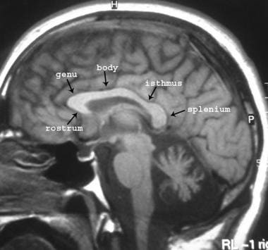
Corpus Callosum
Agenesis of Corpus Callosum
Acrocallosal Syndrome
Corpus Luteum
Anisotropy
Nerve Fibers, Myelinated
Diffusion Tensor Imaging
Magnetic Resonance Imaging
Diffusion Magnetic Resonance Imaging
Anodontia
Abnormalities, Multiple
Nervous System Malformations
Brain
Internal Capsule
Diffuse Axonal Injury
Image Processing, Computer-Assisted
Functional Laterality
Septum Pellucidum
Fornix, Brain
Demyelinating Diseases
Brain Diseases
Myelin Sheath
Aicardi Syndrome
Hydrocephalus
Cerebral Cortex
Oligodendroglia
Malformations of Cortical Development
Brain Mapping
Atrophy
Intellectual Disability
Leukoencephalopathies
Cerebral Ventricles
Hypertelorism
Pregnancy
Urogenital Abnormalities
Microcephaly
Brain Injuries
Corpora Allata
Dandy-Walker Syndrome
Marchiafava-Bignami Disease
Ultrasonography, Prenatal
Prenatal Diagnosis
Meningomyelocele
Lissencephaly
Neuroimaging
Fetal Diseases
Cerebrum
Spastic Paraplegia, Hereditary
Craniofacial Abnormalities
Tooth Exfoliation
MSX1 Transcription Factor
Cebus
Lipoma
Image Interpretation, Computer-Assisted
Gestational Age
Neuronal Migration Disorders
Hyperglycinemia, Nonketotic
Neuroglia
Neuropsychological Tests
Penis
Whale, Killer
Imaging, Three-Dimensional
Aging
Lateral Ventricles
Dichotic Listening Tests
Corpus Luteum Maintenance
Reference Values
Analysis of Variance
Echoencephalography
Sex Characteristics
Surgically-Created Structures
Apraxias
Callosal and cortical contribution to procedural learning. (1/203)
Acallosal and callosotomized subjects usually show impairments on tasks requiring bilateral interdependent motor control. However, few studies have assessed the ability of these subjects to learn a skill that requires the simultaneous contribution of each hemisphere in its acquisition. The present study examined whether acallosal and callosotomized subjects could learn a visuomotor skill that involved a motor control from either both or a single hemisphere. Eleven adult patients, six acallosal and five callosotomized, participated in this study. Seven of these patients had epileptic foci located in the frontal and/or temporal areas and one of the acallosal patients showed bilateral prefrontal atrophy following surgical removal of an orbitofrontal cyst. The performance of the experimental subjects was compared with that of 11 matched control subjects, on a modified version of a serial reaction time task developed by Nissen and Bullemer (Cogn Psychol 1987; 19: 1-32). This skill acquisition task involved bimanual or unimanual key-pressing responses to a sequence of 10 visual stimuli that was repeated 160 times. A declarative memory task was then performed to assess explicit knowledge of the sequence. None of the experimental subjects learned the task in the bimanual condition. Patients with frontal epileptic foci or orbitofrontal damage also failed to learn the task in the unimanual condition when they were using the hand contralateral to the damaged hemisphere. All other subjects, including the acallosal and callosotomized patients with temporal foci, learned the visuomotor skill as well as their controls in the unimanual condition. In spite of the absence of transfer and interhemispheric integration of procedural learning, some of the acallosal and callosotomized patients were able to learn the sequence explicitly. These findings indicate that the corpus callosum and the frontal cortical areas are important for procedural learning of a visuomotor skill. They also confirm the dissociation described by Squire (Science 1986; 232: 1612-9 and J Cogn Neurosci 1992; 4: 232-43) between the declarative and procedural memory systems and extend this dissociation to processes involving simultaneous bihemispheric co-operation. (+info)Abnormalities in neuronal process extension, hippocampal development, and the ventricular system of L1 knockout mice. (2/203)
In humans, mutations in the L1 cell adhesion molecule are associated with a neurological syndrome termed CRASH, which includes corpus callosum agenesis, mental retardation, adducted thumbs, spasticity, and hydrocephalus. A mouse model with a null mutation in the L1 gene (Cohen et al., 1997) was analyzed for brain abnormalities by Nissl and Golgi staining and immunocytochemistry. In the motor, somatosensory, and visual cortex, many pyramidal neurons in layer V exhibited undulating apical dendrites that did not reach layer I. The hippocampus of L1 mutant mice was smaller than normal, with fewer pyramidal and granule cells. The corpus callosum of L1-minus mice was reduced in size because of the failure of many callosal axons to cross the midline. Enlarged ventricles and septal abnormalities were also features of the mutant mouse brain. Immunoperoxidase staining showed that L1 was abundant in developing neurons at embryonic day 18 (E18) in wild-type cerebral cortex, hippocampus, and corpus callosum and then declined to low levels with maturation. In the E18 cortex, L1 colocalized with microtubule-associated protein 2, a marker of dendrites and somata. These new findings suggest new roles for L1 in the mechanism of cortical dendrite differentiation, as well as in guidance of callosal axons and regulation of hippocampal development. The phenotype of the L1 mutant mouse indicates that it is a potentially valuable model for the human CRASH syndrome. (+info)Reduction cranioplasty for macrocephaly. Two case reports. (3/203)
Multi-stage reduction cranioplasty was performed on two children with severe macrocephaly secondary to hydrocephalus. One patient underwent a four-stage operation, and the other underwent a two-stage operation. The postoperative course of both patients was uneventful. Reduction cranioplasty improved quality of life for both patients, and good cosmetic results were achieved. Reduction cranioplasty is effective for the treatment of macrocephaly, and multi-stage surgery can reduce the associated risks. (+info)Dissociation of the pathways mediating ipsilateral and contralateral motor-evoked potentials in human hand and arm muscles. (4/203)
1. Growing evidence points toward involvement of the human motor cortex in the control of the ipsilateral hand. We used focal transcranial magnetic stimulation (TMS) to examine the pathways of these ipsilateral motor effects. 2. Ipsilateral motor-evoked potentials (MEPs) were obtained in hand and arm muscles of all 10 healthy adult subjects tested. They occurred in the finger and wrist extensors and the biceps, but no response or inhibitory responses were observed in the opponens pollicis, finger and wrist flexors and the triceps. 3. The production of ipsilateral MEPs required contraction of the target muscle. The threshold TMS intensity for ipsilateral MEPs was on average 1.8 times higher, and the onset was 5.7 ms later (in the wrist extensor muscles) compared with size-matched contralateral MEPs. 4. The corticofugal pathways of ipsilateral and contralateral MEPs could be dissociated through differences in cortical map location and preferred stimulating current direction. 5. Both ipsi- and contralateral MEPs in the wrist extensors increased with lateral head rotation toward, and decreased with head rotation away from, the side of the TMS, suggesting a privileged input of the asymmetrical tonic neck reflex to the pathway of the ipsilateral MEP. 6. Large ipsilateral MEPs were obtained in a patient with complete agenesis of the corpus callosum. 7. The dissociation of the pathways for ipsilateral and contralateral MEPs indicates that corticofugal motor fibres other than the fast-conducting crossed corticomotoneuronal system can be activated by TMS. Our data suggest an ipsilateral oligosynaptic pathway, such as a corticoreticulospinal or a corticopropriospinal projection as the route for the ipsilateral MEP. Other pathways, such as branching of corticomotoneuronal axons, a transcallosal projection or a slow-conducting monosynaptic ipsilateral pathway are very unlikely or can be excluded. (+info)The homeodomain protein vax1 is required for axon guidance and major tract formation in the developing forebrain. (5/203)
The homeodomain protein Vax1 is expressed in a highly circumscribed set of cells at the ventral anterior midline of the embryonic CNS. These cells populate the choroid fissure of the optic disk, the body of the optic stalk and nerve, the optic chiasm and ventral diencephalon, and the anterior midline zones that abut developing commissural tracts. We have generated mutant mice that lack Vax1. In these mice (1) the optic disks fail to close, leading to coloboma and loss of the eye-nerve boundary; (2) optic nerve glia fail to associate with and appear to repulse ingrowing retinal axons, resulting in a fascicle of axons that are completely segregated from optic nerve astrocytes; (3) retinal axons fail to penetrate the brain in significant numbers and fail to form an optic chiasm; and (4) axons in multiple commissural tracts of the anterior CNS, including the corpus callosum and the hippocampal and anterior commissures, fail to cross the midline. These axon guidance defects do not result from the death of normally Vax1(+) midline cells but, instead, correlate with markedly diminished expression of attractive guidance cues in these cells. Vax1 therefore regulates the guidance properties of a set of anterior midline cells that orchestrate axon trajectories in the developing mammalian forebrain. (+info)Neuropathological abnormalities of the corpus callosum in schizophrenia: a diffusion tensor imaging study. (6/203)
OBJECTIVES: Diffusion tensor imaging (DTI), a technique capable of examining water diffusion in different tissues and the organisation of white matter tracts, was used to investigate the neuropathology of the corpus callosum in vivo in patients with schizophrenia. METHODS: Diffusion tensor imaging was performed in 20 schizophrenic patients and 25 healthy controls. Two complementary measures, mean diffusivity and fractional anisotropy, which are considered to be sensitive indices of axonal integrity, were obtained from regions of interest in the genu (anterior) and splenium (posterior) of the corpus callosum. RESULTS: Mean diffusivity was significantly increased and fractional anisotropy significantly reduced in the splenium but not the genu of the corpus callosum in the schizophrenic group compared with controls. There were no significant sex differences in the DTI measures for either the schizophrenic or control group. Clinical variables such as age, duration of illness, dose of antipsychotic medication, and schizophrenic symptoms did not predict the DTI changes in the schizophrenic patients. CONCLUSIONS: The presence of DTI changes in the splenium but not the genu of the corpus callosum suggests that there may be a focal disruption of commisural connectivity in schizophrenia. However, these findings do not exclude the possibility of abnormalities in other areas of the corpus callosum or other regions of white matter and further research using different methods of analysis may enable us to clarify this. Diffusion tensor imaging is a valuable tool in investigating the structure of white matter in schizophrenia. (+info)Parallel visuomotor processing in the split brain: cortico-subcortical interactions. (7/203)
We tested nine patients with callosal pathology in a simple reaction time task with and without redundant targets in the same or opposite visual hemifield. Four patients showed large facilitation (redundancy gain) in the presence of a redundant target, exceeding probability summation models (neural summation). Five patients showed redundancy gain not exceeding probability models. Violation of probability models was not associated with a specific type of callosal lesion. Neural summation, which probably occurs at collicular level, may be modulated by cortical activity. To test this hypothesis, we used functional MRI. During detection of redundant simultaneous targets, activations in the extrastriate cortex were observed in a patient with callosal agenesis and redundancy gain violating probability models, but not in a patient with callosal agenesis and redundancy gain not exceeding probability models. We conclude that cortical activity in the extrastriate cortex may be a modulating factor in the magnitude of the redundancy gain during parallel visuomotor transforms. (+info)Agenesis of corpus callosum - a rare case. (8/203)
A case of corpus callosum agenesis associated with a chromosomal structural defect is described. (+info)The corpus callosum is the largest collection of white matter in the brain, consisting of approximately 200 million nerve fibers. It is a broad, flat band of tissue that connects the two hemispheres of the brain, allowing them to communicate and coordinate information processing. The corpus callosum plays a crucial role in integrating sensory, motor, and cognitive functions between the two sides of the brain. Damage to the corpus callosum can result in various neurological symptoms, including difficulties with movement, speech, memory, and social behavior.
Agenesis of the corpus callosum is a birth defect in which the corpus callosum, the part of the brain that connects the two hemispheres and allows them to communicate, fails to develop normally during fetal development. In cases of agenesis of the corpus callosum, the corpus callosum is partially or completely absent.
This condition can vary in severity and may be associated with other brain abnormalities. Some individuals with agenesis of the corpus callosum may have normal intelligence and few symptoms, while others may have intellectual disability, developmental delays, seizures, vision problems, and difficulties with movement and coordination. The exact cause of agenesis of the corpus callosum is not always known, but it can be caused by genetic factors or exposure to certain medications or environmental toxins during pregnancy.
Acrocallosal syndrome is a rare genetic disorder characterized by the underdevelopment or absence of the corpus callosum (the part of the brain that connects the two hemispheres) and abnormalities of the fingers, toes, and face. The symptoms of this condition can vary widely in severity, but may include intellectual disability, developmental delays, seizures, weak muscle tone, abnormalities of the skull and facial bones, widely spaced eyes, a flat nasal bridge, a short nose with upturned nostrils, an open mouth with a highly arched roof, and a small jaw. In addition, individuals with Acrocallosal syndrome often have extra fingers or toes (polydactyly) and other skeletal abnormalities.
Acrocallosal syndrome is caused by mutations in the KIF7 gene, which provides instructions for making a protein that helps regulate the development and organization of cells in the body. Mutations in this gene are thought to disrupt the normal functioning of this protein, leading to the characteristic signs and symptoms of Acrocallosal syndrome. The disorder is inherited in an autosomal recessive manner, which means that an individual must inherit two copies of the mutated gene (one from each parent) in order to be affected.
The corpus luteum is a temporary endocrine structure that forms in the ovary after an oocyte (egg) has been released from a follicle during ovulation. It's formed by the remaining cells of the ruptured follicle, which transform into large, hormone-secreting cells.
The primary function of the corpus luteum is to produce progesterone and, to a lesser extent, estrogen during the menstrual cycle or pregnancy. Progesterone plays a crucial role in preparing the uterus for potential implantation of a fertilized egg and maintaining the early stages of pregnancy. If pregnancy does not occur, the corpus luteum will typically degenerate and stop producing hormones after approximately 10-14 days, leading to menstruation.
However, if pregnancy occurs, the developing embryo starts to produce human chorionic gonadotropin (hCG), which signals the corpus luteum to continue secreting progesterone and estrogen until the placenta takes over hormonal production, usually around the end of the first trimester.
Anisotropy is a medical term that refers to the property of being directionally dependent, meaning that its properties or characteristics vary depending on the direction in which they are measured. In the context of medicine and biology, anisotropy can refer to various biological structures, tissues, or materials that exhibit different physical or chemical properties along different axes.
For example, certain types of collagen fibers in tendons and ligaments exhibit anisotropic behavior because they are stronger and stiffer when loaded along their long axis compared to being loaded perpendicular to it. Similarly, some brain tissues may show anisotropy due to the presence of nerve fibers that are organized in specific directions, leading to differences in electrical conductivity or diffusion properties depending on the orientation of the measurement.
Anisotropy is an important concept in various medical fields, including radiology, neurology, and materials science, as it can provide valuable information about the structure and function of biological tissues and help guide diagnostic and therapeutic interventions.
Myelinated nerve fibers are neuronal processes that are surrounded by a myelin sheath, a fatty insulating substance that is produced by Schwann cells in the peripheral nervous system and oligodendrocytes in the central nervous system. This myelin sheath helps to increase the speed of electrical impulse transmission, also known as action potentials, along the nerve fiber. The myelin sheath has gaps called nodes of Ranvier where the electrical impulses can jump from one node to the next, which also contributes to the rapid conduction of signals. Myelinated nerve fibers are typically found in the peripheral nerves and the optic nerve, but not in the central nervous system (CNS) tracts that are located within the brain and spinal cord.
Diffusion Tensor Imaging (DTI) is a type of magnetic resonance imaging (MRI) technique that allows for the measurement and visualization of water diffusion in biological tissues, particularly in the brain. DTI provides information about the microstructural organization and integrity of nerve fibers within the brain by measuring the directionality of water diffusion in the brain's white matter tracts.
In DTI, a tensor is used to describe the three-dimensional diffusion properties of water molecules in each voxel (three-dimensional pixel) of an MRI image. The tensor provides information about the magnitude and direction of water diffusion, which can be used to calculate various diffusion metrics such as fractional anisotropy (FA), mean diffusivity (MD), axial diffusivity (AD), and radial diffusivity (RD). These metrics provide insights into the structural properties of nerve fibers, including their orientation, density, and integrity.
DTI has numerous clinical applications, such as in the diagnosis and monitoring of neurological disorders like multiple sclerosis, traumatic brain injury, and neurodegenerative diseases. It can also be used for presurgical planning to identify critical white matter tracts that need to be preserved during surgery.
Medical Definition:
Magnetic Resonance Imaging (MRI) is a non-invasive diagnostic imaging technique that uses a strong magnetic field and radio waves to create detailed cross-sectional or three-dimensional images of the internal structures of the body. The patient lies within a large, cylindrical magnet, and the scanner detects changes in the direction of the magnetic field caused by protons in the body. These changes are then converted into detailed images that help medical professionals to diagnose and monitor various medical conditions, such as tumors, injuries, or diseases affecting the brain, spinal cord, heart, blood vessels, joints, and other internal organs. MRI does not use radiation like computed tomography (CT) scans.
Cuprizone is not a medical condition or disease, but rather a chemical compound that is used in laboratory settings for research purposes. Cuprizone, also known as bis-cyclohexanone oxaldihydrazone, is a copper chelator, which means it can bind to and remove copper ions from various substances.
In research, cuprizone is often used to induce demyelination in animal models of multiple sclerosis (MS) and other neurological disorders. Demyelination refers to the loss or damage of the myelin sheath, which is a fatty substance that surrounds and protects nerve fibers in the brain and spinal cord. When cuprizone is added to the diet of laboratory animals such as mice, it can cause demyelination in specific areas of the brain, making it a useful tool for studying the mechanisms underlying MS and other demyelinating diseases.
It's important to note that while cuprizone is a valuable research tool, it is not used as a medical treatment or therapy for any human conditions.
Diffusion Magnetic Resonance Imaging (MRI) is a non-invasive medical imaging technique that uses magnetic fields and radio waves to produce detailed images of the body's internal structures, particularly the brain and nervous system. In diffusion MRI, the movement of water molecules in biological tissues is measured and analyzed to generate contrast in the images based on the microstructural properties of the tissue.
Diffusion MRI is unique because it allows for the measurement of water diffusion in various directions, which can reveal important information about the organization and integrity of nerve fibers in the brain. This technique has been widely used in research and clinical settings to study a variety of neurological conditions, including stroke, traumatic brain injury, multiple sclerosis, and neurodegenerative diseases such as Alzheimer's disease.
In summary, diffusion MRI is a specialized type of MRI that measures the movement of water molecules in biological tissues to generate detailed images of the body's internal structures, particularly the brain and nervous system. It provides valuable information about the microstructural properties of tissues and has important applications in both research and clinical settings.
Anodontia is a medical term that refers to the congenital absence or lack of development of all primary (deciduous) and/or permanent teeth. It is a rare dental condition that affects tooth development and can be isolated or associated with various syndromes and genetic disorders.
In anodontia, the dental tissues responsible for forming teeth, including the dental lamina, dental papilla, and dental follicle, fail to develop properly, resulting in missing teeth. The condition can affect all teeth or only some of them, leading to partial anodontia.
Anodontia is different from hypodontia, which refers to the congenital absence of one or more, but not all, teeth. It is also distinct from oligodontia, which is the absence of six or more permanent teeth, excluding third molars (wisdom teeth).
People with anodontia may experience difficulties in chewing, speaking, and maintaining oral hygiene, leading to various dental and social problems. Prosthodontic treatments, such as dentures or implants, are often necessary to restore oral function and aesthetics.
'Abnormalities, Multiple' is a broad term that refers to the presence of two or more structural or functional anomalies in an individual. These abnormalities can be present at birth (congenital) or can develop later in life (acquired). They can affect various organs and systems of the body and can vary greatly in severity and impact on a person's health and well-being.
Multiple abnormalities can occur due to genetic factors, environmental influences, or a combination of both. Chromosomal abnormalities, gene mutations, exposure to teratogens (substances that cause birth defects), and maternal infections during pregnancy are some of the common causes of multiple congenital abnormalities.
Examples of multiple congenital abnormalities include Down syndrome, Turner syndrome, and VATER/VACTERL association. Acquired multiple abnormalities can result from conditions such as trauma, infection, degenerative diseases, or cancer.
The medical evaluation and management of individuals with multiple abnormalities depend on the specific abnormalities present and their impact on the individual's health and functioning. A multidisciplinary team of healthcare professionals is often involved in the care of these individuals to address their complex needs.
Nervous system malformations, also known as nervous system dysplasias or developmental anomalies, refer to structural abnormalities or defects in the development of the nervous system. These malformations can occur during fetal development and can affect various parts of the nervous system, including the brain, spinal cord, and peripheral nerves.
Nervous system malformations can result from genetic mutations, environmental factors, or a combination of both. They can range from mild to severe and may cause a wide variety of symptoms, depending on the specific type and location of the malformation. Some common examples of nervous system malformations include:
* Spina bifida: a defect in the closure of the spinal cord and surrounding bones, which can lead to neurological problems such as paralysis, bladder and bowel dysfunction, and hydrocephalus.
* Anencephaly: a severe malformation where the brain and skull do not develop properly, resulting in stillbirth or death shortly after birth.
* Chiari malformation: a structural defect in the cerebellum, the part of the brain that controls balance and coordination, which can cause headaches, neck pain, and difficulty swallowing.
* Microcephaly: a condition where the head is smaller than normal due to abnormal development of the brain, which can lead to intellectual disability and developmental delays.
* Hydrocephalus: a buildup of fluid in the brain that can cause pressure on the brain and lead to cognitive impairment, vision problems, and other neurological symptoms.
Treatment for nervous system malformations depends on the specific type and severity of the condition and may include surgery, medication, physical therapy, or a combination of these approaches.
The brain is the central organ of the nervous system, responsible for receiving and processing sensory information, regulating vital functions, and controlling behavior, movement, and cognition. It is divided into several distinct regions, each with specific functions:
1. Cerebrum: The largest part of the brain, responsible for higher cognitive functions such as thinking, learning, memory, language, and perception. It is divided into two hemispheres, each controlling the opposite side of the body.
2. Cerebellum: Located at the back of the brain, it is responsible for coordinating muscle movements, maintaining balance, and fine-tuning motor skills.
3. Brainstem: Connects the cerebrum and cerebellum to the spinal cord, controlling vital functions such as breathing, heart rate, and blood pressure. It also serves as a relay center for sensory information and motor commands between the brain and the rest of the body.
4. Diencephalon: A region that includes the thalamus (a major sensory relay station) and hypothalamus (regulates hormones, temperature, hunger, thirst, and sleep).
5. Limbic system: A group of structures involved in emotional processing, memory formation, and motivation, including the hippocampus, amygdala, and cingulate gyrus.
The brain is composed of billions of interconnected neurons that communicate through electrical and chemical signals. It is protected by the skull and surrounded by three layers of membranes called meninges, as well as cerebrospinal fluid that provides cushioning and nutrients.
The internal capsule is a critical structure in the brain that consists of a bundle of white matter fibers (nerve tracts) located deep within the cerebral hemispheres. It serves as a major pathway for the transmission of motor, sensory, and cognitive information between different regions of the brain. The internal capsule is divided into several segments, including the anterior limb, genu, posterior limb, and retrolentiform and sublentiform parts.
The fibers within the internal capsule can be categorized into three groups: corticopontine fibers, corticospinal and corticobulbar fibers, and thalamocortical fibers. Corticopontine fibers originate from the cerebral cortex and terminate in the pons. Corticospinal and corticobulbar fibers are responsible for motor functions, with corticospinal fibers controlling movements of the trunk and limbs, while corticobulbar fibers control movements of the face and head. Thalamocortical fibers carry sensory information from the thalamus to the cerebral cortex.
Damage to the internal capsule can result in various neurological deficits, depending on the specific location and extent of the injury. These may include motor impairments, sensory loss, cognitive dysfunction, or a combination of these symptoms.
Diffuse axonal injury (DAI) is a type of traumatic brain injury that occurs when there is extensive damage to the nerve fibers (axons) in the brain. It is often caused by rapid acceleration or deceleration forces, such as those experienced during motor vehicle accidents or falls. In DAI, the axons are stretched and damaged, leading to disruption of communication between different parts of the brain. This can result in a wide range of symptoms, including cognitive impairment, loss of consciousness, and motor dysfunction. DAI is often difficult to diagnose and can have long-term consequences, making it an important area of study in traumatic brain injury research.
Computer-assisted image processing is a medical term that refers to the use of computer systems and specialized software to improve, analyze, and interpret medical images obtained through various imaging techniques such as X-ray, CT (computed tomography), MRI (magnetic resonance imaging), ultrasound, and others.
The process typically involves several steps, including image acquisition, enhancement, segmentation, restoration, and analysis. Image processing algorithms can be used to enhance the quality of medical images by adjusting contrast, brightness, and sharpness, as well as removing noise and artifacts that may interfere with accurate diagnosis. Segmentation techniques can be used to isolate specific regions or structures of interest within an image, allowing for more detailed analysis.
Computer-assisted image processing has numerous applications in medical imaging, including detection and characterization of lesions, tumors, and other abnormalities; assessment of organ function and morphology; and guidance of interventional procedures such as biopsies and surgeries. By automating and standardizing image analysis tasks, computer-assisted image processing can help to improve diagnostic accuracy, efficiency, and consistency, while reducing the potential for human error.
Functional laterality, in a medical context, refers to the preferential use or performance of one side of the body over the other for specific functions. This is often demonstrated in hand dominance, where an individual may be right-handed or left-handed, meaning they primarily use their right or left hand for tasks such as writing, eating, or throwing.
However, functional laterality can also apply to other bodily functions and structures, including the eyes (ocular dominance), ears (auditory dominance), or legs. It's important to note that functional laterality is not a strict binary concept; some individuals may exhibit mixed dominance or no strong preference for one side over the other.
In clinical settings, assessing functional laterality can be useful in diagnosing and treating various neurological conditions, such as stroke or traumatic brain injury, where understanding any resulting lateralized impairments can inform rehabilitation strategies.
The Septum Pellucidum is a thin, delicate, and almost transparent partition in the brain that separates the lateral ventricles, which are fluid-filled spaces within the brain. It consists of two laminae (plates) that fuse together during fetal development, forming a single structure. The Septum Pellucidum is an essential component of the brain's ventricular system and plays a role in maintaining the structural integrity of the brain. Any abnormalities or damage to the Septum Pellucidum can lead to neurological disorders or cognitive impairments.
The fornix, in the context of brain anatomy, is a bundle of nerve fibers that arises from the hippocampus, a major component of the limbic system associated with memory and spatial navigation. The fornix plays a crucial role in conveying information between different parts of the brain.
The fornix has two primary divisions: the precommissural fornix and the postcommissural fornix. The precommissural fornix contains fibers that originate from the hippocampus and the subiculum, while the postcommissural fornix consists of fibers that originate from the septal nuclei and other structures in the limbic system.
The two divisions of the fornix join together to form a structure called the body of the fornix, which then curves around the thalamus and continues as the crura (plural of crus) of the fornix. The crura split into two columns that pass through the interventricular foramen and terminate in the hypothalamus, specifically at the mammillary bodies.
The fornix is an essential structure for memory function, particularly episodic memory (memory of specific events or episodes). Damage to the fornix can result in various cognitive impairments, including memory loss and difficulties with spatial navigation.
PAX9 is a transcription factor that belongs to the PAX family of genes, which are characterized by a highly conserved DNA-binding domain known as the paired box. The PAX9 gene provides instructions for making a protein that plays important roles in the development of several parts of the body, including the face and the teeth.
As a transcription factor, PAX9 binds to specific regions of DNA and helps control the activity of other genes. In the developing face, PAX9 helps regulate the formation of facial structures by controlling the growth and development of cells that give rise to bones and cartilage. In the developing teeth, PAX9 plays a critical role in tooth development by controlling the formation and growth of dental tissues.
Mutations in the PAX9 gene have been associated with several genetic disorders, including tooth agenesis (the absence of one or more teeth) and oculo-auriculo-vertebral spectrum (a disorder that affects the development of the eyes, ears, and spine).
Demyelinating diseases are a group of disorders that are characterized by damage to the myelin sheath, which is the protective covering surrounding nerve fibers in the brain, optic nerves, and spinal cord. Myelin is essential for the rapid transmission of nerve impulses, and its damage results in disrupted communication between the brain and other parts of the body.
The most common demyelinating disease is multiple sclerosis (MS), where the immune system mistakenly attacks the myelin sheath. Other demyelinating diseases include:
1. Acute Disseminated Encephalomyelitis (ADEM): An autoimmune disorder that typically follows a viral infection or vaccination, causing widespread inflammation and demyelination in the brain and spinal cord.
2. Neuromyelitis Optica (NMO) or Devic's Disease: A rare autoimmune disorder that primarily affects the optic nerves and spinal cord, leading to severe vision loss and motor disability.
3. Transverse Myelitis: Inflammation of the spinal cord causing damage to both sides of one level (segment) of the spinal cord, resulting in various neurological symptoms such as muscle weakness, numbness, or pain, depending on which part of the spinal cord is affected.
4. Guillain-Barré Syndrome: An autoimmune disorder that causes rapid-onset muscle weakness, often beginning in the legs and spreading to the upper body, including the face and breathing muscles. It occurs when the immune system attacks the peripheral nerves' myelin sheath.
5. Central Pontine Myelinolysis (CPM): A rare neurological disorder caused by rapid shifts in sodium levels in the blood, leading to damage to the myelin sheath in a specific area of the brainstem called the pons.
These diseases can result in various symptoms, such as muscle weakness, numbness, vision loss, difficulty with balance and coordination, and cognitive impairment, depending on the location and extent of the demyelination. Treatment typically focuses on managing symptoms, modifying the immune system's response, and promoting nerve regeneration and remyelination when possible.
Brain diseases, also known as neurological disorders, refer to a wide range of conditions that affect the brain and nervous system. These diseases can be caused by various factors such as genetics, infections, injuries, degeneration, or structural abnormalities. They can affect different parts of the brain, leading to a variety of symptoms and complications.
Some examples of brain diseases include:
1. Alzheimer's disease - a progressive degenerative disorder that affects memory and cognitive function.
2. Parkinson's disease - a movement disorder characterized by tremors, stiffness, and difficulty with coordination and balance.
3. Multiple sclerosis - a chronic autoimmune disease that affects the nervous system and can cause a range of symptoms such as vision loss, muscle weakness, and cognitive impairment.
4. Epilepsy - a neurological disorder characterized by recurrent seizures.
5. Brain tumors - abnormal growths in the brain that can be benign or malignant.
6. Stroke - a sudden interruption of blood flow to the brain, which can cause paralysis, speech difficulties, and other neurological symptoms.
7. Meningitis - an infection of the membranes surrounding the brain and spinal cord.
8. Encephalitis - an inflammation of the brain that can be caused by viruses, bacteria, or autoimmune disorders.
9. Huntington's disease - a genetic disorder that affects muscle coordination, cognitive function, and mental health.
10. Migraine - a neurological condition characterized by severe headaches, often accompanied by nausea, vomiting, and sensitivity to light and sound.
Brain diseases can range from mild to severe and may be treatable or incurable. They can affect people of all ages and backgrounds, and early diagnosis and treatment are essential for improving outcomes and quality of life.
Neural pathways, also known as nerve tracts or fasciculi, refer to the highly organized and specialized routes through which nerve impulses travel within the nervous system. These pathways are formed by groups of neurons (nerve cells) that are connected in a series, creating a continuous communication network for electrical signals to transmit information between different regions of the brain, spinal cord, and peripheral nerves.
Neural pathways can be classified into two main types: sensory (afferent) and motor (efferent). Sensory neural pathways carry sensory information from various receptors in the body (such as those for touch, temperature, pain, and vision) to the brain for processing. Motor neural pathways, on the other hand, transmit signals from the brain to the muscles and glands, controlling movements and other effector functions.
The formation of these neural pathways is crucial for normal nervous system function, as it enables efficient communication between different parts of the body and allows for complex behaviors, cognitive processes, and adaptive responses to internal and external stimuli.
The myelin sheath is a multilayered, fatty substance that surrounds and insulates many nerve fibers in the nervous system. It is essential for the rapid transmission of electrical signals, or nerve impulses, along these nerve fibers, allowing for efficient communication between different parts of the body. The myelin sheath is produced by specialized cells called oligodendrocytes in the central nervous system (CNS) and Schwann cells in the peripheral nervous system (PNS). Damage to the myelin sheath, as seen in conditions like multiple sclerosis, can significantly impair nerve function and result in various neurological symptoms.
Aicardi syndrome is a rare genetic disorder that primarily affects girls and women. It is characterized by the absence or underdevelopment of a part of the brain called the corpus callosum, which connects the two hemispheres of the brain. This results in various neurological symptoms such as seizures, developmental delays, and intellectual disabilities.
Individuals with Aicardi syndrome may also have other distinctive features, including abnormalities of the eyes (such as retinal lacunae or colobomas), agenesis of the corpus callosum, and characteristic skin abnormalities called chorioretinal lacunae. The disorder is usually sporadic, meaning that it occurs randomly and is not inherited from parents.
The exact cause of Aicardi syndrome is unknown, but it is believed to be related to genetic mutations or deletions on the X chromosome. Because the disorder primarily affects girls and women, it is thought that the absence of a second X chromosome in males may lead to more severe symptoms or early lethality.
There is no cure for Aicardi syndrome, and treatment is focused on managing the symptoms and improving quality of life. This may include anti-seizure medications, physical therapy, occupational therapy, and special education services. The prognosis for individuals with Aicardi syndrome varies widely depending on the severity of their symptoms and the effectiveness of treatment.
Tooth abnormalities refer to any variations or irregularities in the size, shape, number, structure, or development of teeth that deviate from the typical or normal anatomy. These abnormalities can occur in primary (deciduous) or permanent teeth and can be caused by genetic factors, environmental influences, systemic diseases, or localized dental conditions during tooth formation.
Some examples of tooth abnormalities include:
1. Microdontia - teeth that are smaller than normal in size.
2. Macrodontia - teeth that are larger than normal in size.
3. Peg-shaped teeth - teeth with a narrow, conical shape.
4. Talon cusps - additional cusps or points on the biting surface of a tooth.
5. Dens invaginatus - an abnormal development where the tooth crown has an extra fold or pouch that can trap bacteria and cause dental problems.
6. Taurodontism - teeth with large pulp chambers and short roots.
7. Supernumerary teeth - having more teeth than the typical number (20 primary and 32 permanent teeth).
8. Hypodontia - missing one or more teeth due to a failure of development.
9. Germination - two adjacent teeth fused together, usually occurring in the front teeth.
10. Fusion - two separate teeth that have grown together during development.
Tooth abnormalities may not always require treatment unless they cause functional, aesthetic, or dental health issues. A dentist can diagnose and manage tooth abnormalities through various treatments, such as fillings, extractions, orthodontic care, or restorative procedures.
Hydrocephalus is a medical condition characterized by an abnormal accumulation of cerebrospinal fluid (CSF) within the brain, leading to an increase in intracranial pressure and potentially causing damage to the brain tissues. This excessive buildup of CSF can result from either overproduction or impaired absorption of the fluid, which typically causes the ventricles (fluid-filled spaces) inside the brain to expand and put pressure on surrounding brain structures.
The condition can be congenital, present at birth due to genetic factors or abnormalities during fetal development, or acquired later in life as a result of injuries, infections, tumors, or other disorders affecting the brain's ability to regulate CSF flow and absorption. Symptoms may vary depending on age, severity, and duration but often include headaches, vomiting, balance problems, vision issues, cognitive impairment, and changes in behavior or personality.
Treatment for hydrocephalus typically involves surgically implanting a shunt system that diverts the excess CSF from the brain to another part of the body where it can be absorbed, such as the abdominal cavity. In some cases, endoscopic third ventriculostomy (ETV) might be an alternative treatment option, creating a new pathway for CSF flow within the brain. Regular follow-ups with neurosurgeons and other healthcare professionals are essential to monitor the condition and make any necessary adjustments to the treatment plan.
The cerebral cortex is the outermost layer of the brain, characterized by its intricate folded structure and wrinkled appearance. It is a region of great importance as it plays a key role in higher cognitive functions such as perception, consciousness, thought, memory, language, and attention. The cerebral cortex is divided into two hemispheres, each containing four lobes: the frontal, parietal, temporal, and occipital lobes. These areas are responsible for different functions, with some regions specializing in sensory processing while others are involved in motor control or associative functions. The cerebral cortex is composed of gray matter, which contains neuronal cell bodies, and is covered by a layer of white matter that consists mainly of myelinated nerve fibers.
Oligodendroglia are a type of neuroglial cell found in the central nervous system (CNS) of vertebrates, including humans. These cells play a crucial role in providing support and insulation to nerve fibers (axons) in the CNS, which includes the brain and spinal cord.
More specifically, oligodendroglia produce a fatty substance called myelin that wraps around axons, forming myelin sheaths. This myelination process helps to increase the speed of electrical impulse transmission (nerve impulses) along the axons, allowing for efficient communication between different neurons.
In addition to their role in myelination, oligodendroglia also contribute to the overall health and maintenance of the CNS by providing essential nutrients and supporting factors to neurons. Dysfunction or damage to oligodendroglia has been implicated in various neurological disorders, such as multiple sclerosis (MS), where demyelination of axons leads to impaired nerve function and neurodegeneration.
Malformations of Cortical Development (MCDs) are a group of congenital brain abnormalities that occur during the development and organization of the cerebral cortex, which is the brain region responsible for higher cognitive functions. These malformations result from disruptions in neuronal migration, proliferation, or organization, leading to varying degrees of cortical thickness, folding, and structural integrity.
MCDs can be classified into several subtypes based on their distinct neuroimaging and histopathological features. Some common MCD subtypes include:
1. Lissencephaly (smooth brain): A severe malformation characterized by the absence of normal gyral and sulcal patterns, resulting in a smooth cortical surface. This is caused by defects in neuronal migration during early development.
2. Polymicrogyria (many small folds): A condition where the cortex has an excessive number of small, irregular gyri, leading to thickened and disorganized cortical layers. This can be focal or diffuse and is caused by abnormal neuronal migration or organization during mid to late development.
3. Schizencephaly (cleft brain): A malformation characterized by a linear cleft or gap in the cerebral cortex, extending from the pial surface to the ventricular system. This can be unilateral or bilateral and is caused by disruptions in neuronal migration and/or cortical organization during early development.
4. Heterotopias (misplaced cells): A condition where groups of neurons are abnormally located within the white matter or at the gray-white matter junction, instead of their normal position in the cerebral cortex. This can be focal or diffuse and is caused by defects in neuronal migration during early development.
5. Focal cortical dysplasia (abnormal localized tissue): A condition characterized by abnormal cortical architecture, including disorganized lamination, enlarged neurons, and heterotopic neurons. This can be focal or multifocal and is caused by defects in cortical organization during late development.
MCDs are often associated with neurological symptoms such as epilepsy, intellectual disability, motor deficits, and behavioral abnormalities. The severity of these symptoms depends on the type, location, and extent of the malformation.
Brain mapping is a broad term that refers to the techniques used to understand the structure and function of the brain. It involves creating maps of the various cognitive, emotional, and behavioral processes in the brain by correlating these processes with physical locations or activities within the nervous system. Brain mapping can be accomplished through a variety of methods, including functional magnetic resonance imaging (fMRI), positron emission tomography (PET) scans, electroencephalography (EEG), and others. These techniques allow researchers to observe which areas of the brain are active during different tasks or thoughts, helping to shed light on how the brain processes information and contributes to our experiences and behaviors. Brain mapping is an important area of research in neuroscience, with potential applications in the diagnosis and treatment of neurological and psychiatric disorders.
A syndrome, in medical terms, is a set of symptoms that collectively indicate or characterize a disease, disorder, or underlying pathological process. It's essentially a collection of signs and/or symptoms that frequently occur together and can suggest a particular cause or condition, even though the exact physiological mechanisms might not be fully understood.
For example, Down syndrome is characterized by specific physical features, cognitive delays, and other developmental issues resulting from an extra copy of chromosome 21. Similarly, metabolic syndromes like diabetes mellitus type 2 involve a group of risk factors such as obesity, high blood pressure, high blood sugar, and abnormal cholesterol or triglyceride levels that collectively increase the risk of heart disease, stroke, and diabetes.
It's important to note that a syndrome is not a specific diagnosis; rather, it's a pattern of symptoms that can help guide further diagnostic evaluation and management.
Atrophy is a medical term that refers to the decrease in size and wasting of an organ or tissue due to the disappearance of cells, shrinkage of cells, or decreased number of cells. This process can be caused by various factors such as disuse, aging, degeneration, injury, or disease.
For example, if a muscle is immobilized for an extended period, it may undergo atrophy due to lack of use. Similarly, certain medical conditions like diabetes, cancer, and heart failure can lead to the wasting away of various tissues and organs in the body.
Atrophy can also occur as a result of natural aging processes, leading to decreased muscle mass and strength in older adults. In general, atrophy is characterized by a decrease in the volume or weight of an organ or tissue, which can have significant impacts on its function and overall health.
Congenital abnormalities, also known as birth defects, are structural or functional anomalies that are present at birth. These abnormalities can develop at any point during fetal development, and they can affect any part of the body. They can be caused by genetic factors, environmental influences, or a combination of both.
Congenital abnormalities can range from mild to severe and may include structural defects such as heart defects, neural tube defects, and cleft lip and palate, as well as functional defects such as intellectual disabilities and sensory impairments. Some congenital abnormalities may be visible at birth, while others may not become apparent until later in life.
In some cases, congenital abnormalities may be detected through prenatal testing, such as ultrasound or amniocentesis. In other cases, they may not be diagnosed until after the baby is born. Treatment for congenital abnormalities varies depending on the type and severity of the defect, and may include surgery, therapy, medication, or a combination of these approaches.
Intellectual disability (ID) is a term used when there are significant limitations in both intellectual functioning and adaptive behavior, which covers many everyday social and practical skills. This disability originates before the age of 18.
Intellectual functioning, also known as intelligence, refers to general mental capacity, such as learning, reasoning, problem-solving, and other cognitive skills. Adaptive behavior includes skills needed for day-to-day life, such as communication, self-care, social skills, safety judgement, and basic academic skills.
Intellectual disability is characterized by below-average intelligence or mental ability and a lack of skills necessary for day-to-day living. It can be mild, moderate, severe, or profound, depending on the degree of limitation in intellectual functioning and adaptive behavior.
It's important to note that people with intellectual disabilities have unique strengths and limitations, just like everyone else. With appropriate support and education, they can lead fulfilling lives and contribute to their communities in many ways.
Leukoencephalopathies are a group of medical conditions that primarily affect the white matter of the brain, which consists mainly of nerve fibers covered by myelin sheaths. These conditions are characterized by abnormalities in the structure and function of the white matter, leading to various neurological symptoms such as cognitive decline, motor impairment, seizures, and behavioral changes.
The term "leukoencephalopathy" is derived from two Greek words: "leukos," meaning white, and "enkephalos," meaning brain. The suffix "-pathy" refers to a disease or suffering. Therefore, leukoencephalopathies refer specifically to diseases that affect the white matter of the brain.
There are various types of leukoencephalopathies, including genetic, metabolic, infectious, toxic, and immune-mediated forms. Some examples include multiple sclerosis, adrenoleukodystrophy, Alexander disease, Canavan disease, and Marchiafava-Bignami disease. The diagnosis of leukoencephalopathies typically involves a combination of clinical evaluation, imaging studies such as MRI, and sometimes genetic or laboratory testing to identify the underlying cause. Treatment depends on the specific type and severity of the condition and may include medications, dietary modifications, physical therapy, or supportive care.
An axon is a long, slender extension of a neuron (a type of nerve cell) that conducts electrical impulses (nerve impulses) away from the cell body to target cells, such as other neurons or muscle cells. Axons can vary in length from a few micrometers to over a meter long and are typically surrounded by a myelin sheath, which helps to insulate and protect the axon and allows for faster transmission of nerve impulses.
Axons play a critical role in the functioning of the nervous system, as they provide the means by which neurons communicate with one another and with other cells in the body. Damage to axons can result in serious neurological problems, such as those seen in spinal cord injuries or neurodegenerative diseases like multiple sclerosis.
The cerebral ventricles are a system of interconnected fluid-filled cavities within the brain. They are located in the center of the brain and are filled with cerebrospinal fluid (CSF), which provides protection to the brain by cushioning it from impacts and helping to maintain its stability within the skull.
There are four ventricles in total: two lateral ventricles, one third ventricle, and one fourth ventricle. The lateral ventricles are located in each cerebral hemisphere, while the third ventricle is located between the thalami of the two hemispheres. The fourth ventricle is located at the base of the brain, above the spinal cord.
CSF flows from the lateral ventricles into the third ventricle through narrow passageways called the interventricular foramen. From there, it flows into the fourth ventricle through another narrow passageway called the cerebral aqueduct. CSF then leaves the fourth ventricle and enters the subarachnoid space surrounding the brain and spinal cord, where it can be absorbed into the bloodstream.
Abnormalities in the size or shape of the cerebral ventricles can indicate underlying neurological conditions, such as hydrocephalus (excessive accumulation of CSF) or atrophy (shrinkage) of brain tissue. Imaging techniques, such as computed tomography (CT) or magnetic resonance imaging (MRI), are often used to assess the size and shape of the cerebral ventricles in clinical settings.
Hypertelorism is a medical term that refers to an ocular condition where the distance between two eyes (interpupillary distance) is abnormally increased. It's typically defined as an interpupillary distance that measures more than 2 standard deviations beyond the mean for a given age, gender, and race.
This condition can be associated with various genetic syndromes or conditions such as craniosynostosis (premature fusion of skull sutures), fetal alcohol syndrome, and certain chromosomal abnormalities like Down syndrome. Hypertelorism may also occur in isolation without any other associated anomalies.
It's important to note that hypertelorism can have cosmetic implications, particularly if the distance between the eyes is significantly increased, as it may affect the overall symmetry and appearance of the face. However, in most cases, this condition does not directly impact vision unless there are other related structural abnormalities of the eye or orbit.
Pregnancy is a physiological state or condition where a fertilized egg (zygote) successfully implants and grows in the uterus of a woman, leading to the development of an embryo and finally a fetus. This process typically spans approximately 40 weeks, divided into three trimesters, and culminates in childbirth. Throughout this period, numerous hormonal and physical changes occur to support the growing offspring, including uterine enlargement, breast development, and various maternal adaptations to ensure the fetus's optimal growth and well-being.
Urogenital abnormalities refer to structural or functional anomalies that affect the urinary and genital systems. These two systems are closely linked during embryonic development, and sometimes they may not develop properly, leading to various types of congenital defects. Urogenital abnormalities can range from minor issues like a bifid scrotum (a condition where the scrotum is split into two parts) to more severe problems such as bladder exstrophy (where the bladder develops outside the body).
These conditions may affect urination, reproduction, and sexual function. They can also increase the risk of infections and other complications. Urogenital abnormalities can be diagnosed through physical examination, imaging tests, or genetic testing. Treatment options depend on the specific condition but may include surgery, medication, or lifestyle changes.
Microcephaly is a medical condition where an individual has a smaller than average head size. The circumference of the head is significantly below the normal range for age and sex. This condition is typically caused by abnormal brain development, which can be due to genetic factors or environmental influences such as infections or exposure to harmful substances during pregnancy.
Microcephaly can be present at birth (congenital) or develop in the first few years of life. People with microcephaly often have intellectual disabilities, delayed development, and other neurological problems. However, the severity of these issues can vary widely, ranging from mild to severe. It is important to note that not all individuals with microcephaly will experience significant impairments or challenges.
A brain injury is defined as damage to the brain that occurs following an external force or trauma, such as a blow to the head, a fall, or a motor vehicle accident. Brain injuries can also result from internal conditions, such as lack of oxygen or a stroke. There are two main types of brain injuries: traumatic and acquired.
Traumatic brain injury (TBI) is caused by an external force that results in the brain moving within the skull or the skull being fractured. Mild TBIs may result in temporary symptoms such as headaches, confusion, and memory loss, while severe TBIs can cause long-term complications, including physical, cognitive, and emotional impairments.
Acquired brain injury (ABI) is any injury to the brain that occurs after birth and is not hereditary, congenital, or degenerative. ABIs are often caused by medical conditions such as strokes, tumors, anoxia (lack of oxygen), or infections.
Both TBIs and ABIs can range from mild to severe and may result in a variety of physical, cognitive, and emotional symptoms that can impact a person's ability to perform daily activities and function independently. Treatment for brain injuries typically involves a multidisciplinary approach, including medical management, rehabilitation, and supportive care.
Cerebral dominance is a concept in neuropsychology that refers to the specialization of one hemisphere of the brain over the other for certain cognitive functions. In most people, the left hemisphere is dominant for language functions such as speaking and understanding spoken or written language, while the right hemisphere is dominant for non-verbal functions such as spatial ability, face recognition, and artistic ability.
Cerebral dominance does not mean that the non-dominant hemisphere is incapable of performing the functions of the dominant hemisphere, but rather that it is less efficient or specialized in those areas. The concept of cerebral dominance has been used to explain individual differences in cognitive abilities and learning styles, as well as the laterality of brain damage and its effects on cognition and behavior.
It's important to note that cerebral dominance is a complex phenomenon that can vary between individuals and can be influenced by various factors such as genetics, environment, and experience. Additionally, recent research has challenged the strict lateralization of functions and suggested that there is more functional overlap and interaction between the two hemispheres than previously thought.
The corpora allata are small endocrine glands found in the head of insects, located near the brain. They are part of the insect endocrine system and produce important hormones that regulate various physiological processes, including growth, development, reproduction, and molting. The most well-known hormone produced by the corpora allata is juvenile hormone (JH), which plays a crucial role in maintaining the larval or nymphal stage of insects and preventing metamorphosis into the adult form. As the insect grows and develops, the production of JH decreases, allowing for the initiation of metamorphosis and the emergence of the adult form.
Dandy-Walker Syndrome is a congenital brain malformation characterized by the absence or underdevelopment of the cerebellar vermis (the part of the brain that helps coordinate movement) and an enlarged fluid-filled space (fourth ventricle) surrounding it. This condition can also be associated with an upward bulging of the back of the skull (occipital bone), and in some cases, hydrocephalus (excessive accumulation of cerebrospinal fluid in the brain). The syndrome can vary in severity, and symptoms may include problems with balance, coordination, developmental delays, and increased intracranial pressure. It is usually diagnosed through imaging tests such as ultrasound, CT scan, or MRI. Treatment typically involves managing symptoms and addressing complications, which may include surgical procedures to relieve hydrocephalus if present.
Marchiafava-Bignami Disease is a rare neurological disorder that primarily affects the central portion of the brain called the corpus callosum, which is responsible for connecting the two hemispheres of the brain. This disease is characterized by degeneration and necrosis (death of cells) of the corpus callosum, leading to various neurological symptoms.
The condition has been predominantly associated with chronic alcoholism, although it has also been reported in non-alcoholic individuals. It can present acutely or subacutely, causing a wide range of symptoms such as mental status changes, seizures, speech and gait disturbances, and various other motor and sensory abnormalities.
The exact cause of Marchiafava-Bignami Disease remains unclear; however, it is thought to be related to nutritional deficiencies, particularly thiamine (vitamin B1) deficiency, which is common in alcoholics. Diagnosis typically involves neuroimaging techniques like MRI and CT scans, along with clinical evaluation. Treatment usually includes supportive care, thiamine supplementation, and abstinence from alcohol. The prognosis for this condition varies but can be poor if not diagnosed and treated promptly.
Prenatal ultrasonography, also known as obstetric ultrasound, is a medical diagnostic procedure that uses high-frequency sound waves to create images of the developing fetus, placenta, and amniotic fluid inside the uterus. It is a non-invasive and painless test that is widely used during pregnancy to monitor the growth and development of the fetus, detect any potential abnormalities or complications, and determine the due date.
During the procedure, a transducer (a small handheld device) is placed on the mother's abdomen and moved around to capture images from different angles. The sound waves travel through the mother's body and bounce back off the fetus, producing echoes that are then converted into electrical signals and displayed as images on a screen.
Prenatal ultrasonography can be performed at various stages of pregnancy, including early pregnancy to confirm the pregnancy and detect the number of fetuses, mid-pregnancy to assess the growth and development of the fetus, and late pregnancy to evaluate the position of the fetus and determine if it is head down or breech. It can also be used to guide invasive procedures such as amniocentesis or chorionic villus sampling.
Overall, prenatal ultrasonography is a valuable tool in modern obstetrics that helps ensure the health and well-being of both the mother and the developing fetus.
Prenatal diagnosis is the medical testing of fetuses, embryos, or pregnant women to detect the presence or absence of certain genetic disorders or birth defects. These tests can be performed through various methods such as chorionic villus sampling (CVS), amniocentesis, or ultrasound. The goal of prenatal diagnosis is to provide early information about the health of the fetus so that parents and healthcare providers can make informed decisions about pregnancy management and newborn care. It allows for early intervention, treatment, or planning for the child's needs after birth.
Meningomyelocele is a type of neural tube defect that affects the development of the spinal cord and the surrounding membranes known as meninges. In this condition, a portion of the spinal cord and meninges protrude through an opening in the spine, creating a sac-like structure on the back. This sac is usually covered by skin, but it may be open in some cases.
Meningomyelocele can result in various neurological deficits, including muscle weakness, paralysis, and loss of sensation below the level of the lesion. It can also cause bladder and bowel dysfunction, as well as problems with sexual function. The severity of these symptoms depends on the location and extent of the spinal cord defect.
Early diagnosis and treatment are crucial for managing meningomyelocele and preventing further complications. Treatment typically involves surgical closure of the opening in the spine to protect the spinal cord and prevent infection. Physical therapy, occupational therapy, and other supportive care measures may also be necessary to help individuals with meningomyelocele achieve their full potential for mobility and independence.
Lissencephaly is a rare neurological disorder characterized by the absence or significant reduction of normal folds (gyri) and sulci (grooves) in the cerebral cortex of the brain. The cerebral cortex, which is responsible for higher brain functions such as thinking, learning, and language, usually has a smooth, flat appearance in individuals with lissencephaly. This condition results from abnormal neuronal migration during fetal development, where nerve cells fail to migrate to their proper positions in the brain.
There are several types of lissencephaly, each with distinct genetic causes and associated symptoms. The most common form is Type I (Classic) Lissencephaly, which affects both hemispheres of the brain and is characterized by a smooth brain surface with four bands of shallow grooves. Other forms include Type II (Cobblestone) Lissencephaly, Miller-Dieker Syndrome, and X-linked Lissencephaly with Ambiguous Genitalia (XLAG).
Symptoms of lissencephaly can vary but often include severe intellectual disability, developmental delays, muscle spasticity or hypotonia, seizures, difficulty swallowing, and problems with vision and hearing. The severity of the condition depends on the extent of the brain malformation. Lissencephaly is a lifelong condition, and individuals with this disorder usually require extensive care and support throughout their lives.
Organ size refers to the volume or physical measurement of an organ in the body of an individual. It can be described in terms of length, width, and height or by using specialized techniques such as imaging studies (like CT scans or MRIs) to determine the volume. The size of an organ can vary depending on factors such as age, sex, body size, and overall health status. Changes in organ size may indicate various medical conditions, including growths, inflammation, or atrophy.
Neuroimaging is a medical term that refers to the use of various techniques to either directly or indirectly image the structure, function, or pharmacology of the nervous system. It includes techniques such as computed tomography (CT), magnetic resonance imaging (MRI), functional MRI (fMRI), positron emission tomography (PET), single-photon emission computed tomography (SPECT), and diffusion tensor imaging (DTI). These techniques are used to diagnose and monitor various neurological and psychiatric conditions, as well as to understand the underlying mechanisms of brain function in health and disease.
A newborn infant is a baby who is within the first 28 days of life. This period is also referred to as the neonatal period. Newborns require specialized care and attention due to their immature bodily systems and increased vulnerability to various health issues. They are closely monitored for signs of well-being, growth, and development during this critical time.
Fetal diseases are medical conditions or abnormalities that affect a fetus during pregnancy. These diseases can be caused by genetic factors, environmental influences, or a combination of both. They can range from mild to severe and may impact various organ systems in the developing fetus. Examples of fetal diseases include congenital heart defects, neural tube defects, chromosomal abnormalities such as Down syndrome, and infectious diseases such as toxoplasmosis or rubella. Fetal diseases can be diagnosed through prenatal testing, including ultrasound, amniocentesis, and chorionic villus sampling. Treatment options may include medication, surgery, or delivery of the fetus, depending on the nature and severity of the disease.
The cerebrum is the largest part of the brain, located in the frontal part of the skull. It is divided into two hemispheres, right and left, which are connected by a band of nerve fibers called the corpus callosum. The cerebrum is responsible for higher cognitive functions such as thinking, learning, memory, language, perception, and consciousness.
The outer layer of the cerebrum is called the cerebral cortex, which is made up of gray matter containing billions of neurons. This region is responsible for processing sensory information, generating motor commands, and performing higher-level cognitive functions. The cerebrum also contains several subcortical structures such as the thalamus, hypothalamus, hippocampus, and amygdala, which play important roles in various brain functions.
Damage to different parts of the cerebrum can result in a range of neurological symptoms, depending on the location and severity of the injury. For example, damage to the left hemisphere may affect language function, while damage to the right hemisphere may affect spatial perception and visual-spatial skills.
Hereditary Spastic Paraplegia (HSP) is a group of genetic disorders that affect the long motor neurons in the spinal cord, leading to lower limb spasticity and weakness. It is characterized by progressive stiffness and contraction of the leg muscles, resulting in difficulty with walking and balance.
The symptoms of HSP typically begin in childhood or early adulthood and worsen over time. The severity of the condition can vary widely, even within the same family, depending on the specific genetic mutation involved. In addition to lower limb spasticity, some individuals with HSP may also experience bladder dysfunction, sensory loss, or other neurological symptoms.
HSP is inherited in an autosomal dominant or autosomal recessive pattern, depending on the specific genetic mutation involved. There are over 70 different genes that have been identified as causing HSP, and genetic testing can be used to confirm the diagnosis and identify the specific genetic mutation responsible.
Treatment for HSP is focused on managing symptoms and maintaining mobility. Physical therapy, orthotics, and medications such as baclofen or tizanidine may be used to help reduce muscle spasticity and improve mobility. In some cases, surgery may be necessary to relieve muscle contractures or other complications.
Craniofacial abnormalities refer to a group of birth defects that affect the development of the skull and face. These abnormalities can range from mild to severe and may involve differences in the shape and structure of the head, face, and jaws, as well as issues with the formation of facial features such as the eyes, nose, and mouth.
Craniofacial abnormalities can be caused by genetic factors, environmental influences, or a combination of both. Some common examples of craniofacial abnormalities include cleft lip and palate, craniosynostosis (premature fusion of the skull bones), and hemifacial microsomia (underdevelopment of one side of the face).
Treatment for craniofacial abnormalities may involve a team of healthcare professionals, including plastic surgeons, neurosurgeons, orthodontists, speech therapists, and other specialists. Treatment options may include surgery, bracing, therapy, and other interventions to help improve function and appearance.
Tooth exfoliation is not a term that is commonly used in dental or medical literature. However, I believe you may be referring to the natural process of tooth loss that occurs with the shedding of primary (baby) teeth to make way for permanent (adult) teeth. This process is also known as physical or physiological tooth exfoliation.
Exfoliation in this context refers to the separation and shedding of the primary tooth's root from the underlying permanent tooth, allowing the permanent tooth to erupt into its proper position. The primary tooth becomes loose due to the resorption of its roots by the developing permanent tooth beneath it. Eventually, the primary tooth falls out, making room for the adult tooth to emerge and take its place in the dental arch.
It is essential to maintain good oral hygiene during this process to prevent any potential complications such as infection or premature loss of primary teeth.
MSX1 (Homeobox protein MSX-1) is a transcription factor that belongs to the muscle segment homebox gene family, also known as the msh homeobox genes. These genes are involved in the development and differentiation of various tissues, including muscle, bone, and neural crest derivatives.
MSX1 plays crucial roles during embryonic development, such as regulating cell proliferation, differentiation, and apoptosis. It is widely expressed in the developing embryo, particularly in the oral ectoderm, neural crest, and mesenchyme. In the oral region, MSX1 helps control tooth development by interacting with other transcription factors and signaling molecules.
As a transcription factor, MSX1 binds to specific DNA sequences called homeobox response elements (HREs) in the promoter regions of its target genes. This binding either activates or represses gene expression, depending on the context and interacting partners. Dysregulation of MSX1 has been implicated in various developmental disorders and diseases, such as tooth agenesis, cleft lip/palate, and cancer.
"Cebus" is a genus of New World monkeys, also known as capuchin monkeys. They are small to medium-sized primates that are native to Central and South America. Capuchin monkeys are named after the Order of Friars Minor Capuchin, because of their similarity in color to the robes worn by the friars.
Capuchin monkeys are highly intelligent and social animals, living in groups of up to 30 individuals. They have a diverse diet that includes fruits, nuts, seeds, insects, and small vertebrates. Capuchin monkeys are known for their problem-solving abilities and have been observed using tools in the wild.
There are several species of capuchin monkeys, including the white-fronted capuchin (Cebus albifrons), the tufted capuchin (Cebus apella), and the weeper capuchin (Cebus olivaceus). They vary in size, coloration, and behavior, but all share the characteristic cap of hair on their heads that gives them their name.
A lipoma is a common, benign (non-cancerous) soft tissue growth. It is composed of adipose or fatty tissue and typically found just beneath the skin, but they can also occur deeper within the body. Lipomas are usually round, moveable, and painless, although they may cause discomfort if they grow large enough to put pressure on nearby nerves or if they're located in a sensitive area. They generally grow slowly over time. Surgical removal is an option if the lipoma becomes bothersome or grows significantly in size. It's important to note that while lipomas are typically harmless, any new lumps or bumps should be evaluated by a healthcare professional to confirm the diagnosis and rule out other more serious conditions.
Computer-assisted image interpretation is the use of computer algorithms and software to assist healthcare professionals in analyzing and interpreting medical images. These systems use various techniques such as pattern recognition, machine learning, and artificial intelligence to help identify and highlight abnormalities or patterns within imaging data, such as X-rays, CT scans, MRI, and ultrasound images. The goal is to increase the accuracy, consistency, and efficiency of image interpretation, while also reducing the potential for human error. It's important to note that these systems are intended to assist healthcare professionals in their decision making process and not to replace them.
Gestational age is the length of time that has passed since the first day of the last menstrual period (LMP) in pregnant women. It is the standard unit used to estimate the age of a pregnancy and is typically expressed in weeks. This measure is used because the exact date of conception is often not known, but the start of the last menstrual period is usually easier to recall.
It's important to note that since ovulation typically occurs around two weeks after the start of the LMP, gestational age is approximately two weeks longer than fetal age, which is the actual time elapsed since conception. Medical professionals use both gestational and fetal age to track the development and growth of the fetus during pregnancy.
Neuronal migration disorders (NMDs) are a group of genetic conditions that affect the development and migration of neurons (nerve cells) in the brain during embryonic development. These disorders result from abnormalities in the genetic code that control the movement and organization of neurons as they migrate to their proper positions in the brain.
NMDs can cause a wide range of neurological symptoms, depending on which areas of the brain are affected and the severity of the disorder. Symptoms may include intellectual disability, developmental delay, seizures, motor abnormalities, vision or hearing problems, and behavioral issues. Some NMDs may also be associated with structural brain abnormalities that can be seen on imaging studies.
Examples of neuronal migration disorders include lissencephaly, pachygyria, heterotopias, and agyria. These conditions are typically diagnosed through a combination of clinical evaluation, genetic testing, and neuroimaging studies. Treatment for NMDs is generally supportive and may involve medications, therapies, and surgical interventions to manage symptoms and improve quality of life.
Nonketotic hyperglycinemia (NKH) is a rare inherited metabolic disorder characterized by an elevated level of the amino acid glycine in the body due to a deficiency of the enzyme needed to break it down, specifically the glycine cleavage system. This condition leads to developmental delays, seizures, and hypotonia (low muscle tone). The nonketotic part of the name refers to the fact that there is no production of ketones during periods of fasting or illness, which is unusual in disorders associated with elevated glycine levels.
NKH is typically caused by mutations in the GLDC or AMT gene and can be further classified into two types: type I (also known as the classic or severe form) and type II (also known as the attenuated or mild form). Type I NKH usually presents in early infancy with severe symptoms, including seizures, lethargy, and apnea, while type II NKH may present later in childhood with less severe symptoms.
Treatment for NKH is primarily supportive and includes anticonvulsant medications to manage seizures, as well as dietary management to limit glycine intake. In some cases, sodium benzoate or dextromethorphan may be used to reduce glycine levels in the body. However, there is no cure for NKH and prognosis varies depending on the severity of the condition.
Neuroglia, also known as glial cells or simply glia, are non-neuronal cells that provide support and protection for neurons in the nervous system. They maintain homeostasis, form myelin sheaths around nerve fibers, and provide structural support. They also play a role in the immune response of the central nervous system. Some types of neuroglia include astrocytes, oligodendrocytes, microglia, and ependymal cells.
Neuropsychological tests are a type of psychological assessment that measures cognitive functions, such as attention, memory, language, problem-solving, and perception. These tests are used to help diagnose and understand the cognitive impact of neurological conditions, including dementia, traumatic brain injury, stroke, Parkinson's disease, and other disorders that affect the brain.
The tests are typically administered by a trained neuropsychologist and can take several hours to complete. They may involve paper-and-pencil tasks, computerized tasks, or interactive activities. The results of the tests are compared to normative data to help identify any areas of cognitive weakness or strength.
Neuropsychological testing can provide valuable information for treatment planning, rehabilitation, and assessing response to treatment. It can also be used in research to better understand the neural basis of cognition and the impact of neurological conditions on cognitive function.
The penis is a part of the male reproductive and urinary systems. It has three parts: the root, the body, and the glans. The root attaches to the pelvic bone and the body makes up the majority of the free-hanging portion. The glans is the cone-shaped end that protects the urethra, the tube inside the penis that carries urine from the bladder and semen from the testicles.
The penis has a dual function - it acts as a conduit for both urine and semen. During sexual arousal, the penis becomes erect when blood fills two chambers inside its shaft. This process is facilitated by the relaxation of the smooth muscles in the arterial walls and the trappping of blood in the corpora cavernosa. The stiffness of the penis enables sexual intercourse. After ejaculation, or when the sexual arousal passes, the muscles contract and the blood flows out of the penis back into the body, causing it to become flaccid again.
The foreskin, a layer of skin that covers the glans, is sometimes removed in a procedure called circumcision. Circumcision is often performed for religious or cultural reasons, or as a matter of family custom. In some countries, it's also done for medical reasons, such as to treat conditions like phimosis (an inability to retract the foreskin) or balanitis (inflammation of the glans).
It's important to note that any changes in appearance, size, or function of the penis should be evaluated by a healthcare professional, as they could indicate an underlying medical condition.
The term "Killer Whale" is used in medical literature to describe an unusual and very rare phenomenon where a live newborn calf becomes lodged in the birth canal of a female whale (usually a species of baleen whale), leading to potential serious complications such as infection, injury, or even death for the mother if not resolved. This condition is also known as "whale entrapment" or "cesarean delivery candidate." It is not to be confused with the common name of the species Orcinus orca, which are actually the largest species of dolphin and not whales, but are often called "killer whales" due to their size and predatory behavior.
Three-dimensional (3D) imaging in medicine refers to the use of technologies and techniques that generate a 3D representation of internal body structures, organs, or tissues. This is achieved by acquiring and processing data from various imaging modalities such as X-ray computed tomography (CT), magnetic resonance imaging (MRI), ultrasound, or confocal microscopy. The resulting 3D images offer a more detailed visualization of the anatomy and pathology compared to traditional 2D imaging techniques, allowing for improved diagnostic accuracy, surgical planning, and minimally invasive interventions.
In 3D imaging, specialized software is used to reconstruct the acquired data into a volumetric model, which can be manipulated and viewed from different angles and perspectives. This enables healthcare professionals to better understand complex anatomical relationships, detect abnormalities, assess disease progression, and monitor treatment response. Common applications of 3D imaging include neuroimaging, orthopedic surgery planning, cancer staging, dental and maxillofacial reconstruction, and interventional radiology procedures.
Aging is a complex, progressive and inevitable process of bodily changes over time, characterized by the accumulation of cellular damage and degenerative changes that eventually lead to increased vulnerability to disease and death. It involves various biological, genetic, environmental, and lifestyle factors that contribute to the decline in physical and mental functions. The medical field studies aging through the discipline of gerontology, which aims to understand the underlying mechanisms of aging and develop interventions to promote healthy aging and extend the human healthspan.
The lateral ventricles are a pair of fluid-filled cavities located within the brain. They are part of the ventricular system, which is a series of interconnected spaces filled with cerebrospinal fluid (CSF). The lateral ventricles are situated in the left and right hemispheres of the brain and are among the largest of the ventricles.
Each lateral ventricle has a complex structure and can be divided into several parts:
1. Anterior horn: This is the front part of the lateral ventricle, located in the frontal lobe of the brain.
2. Body: The central part of the lateral ventricle, which is continuous with the anterior horn and posterior horn.
3. Posterior horn: The back part of the lateral ventricle, located in the occipital lobe of the brain.
4. Temporal horn: An extension that projects into the temporal lobe of the brain.
The lateral ventricles are lined with ependymal cells, which produce cerebrospinal fluid. CSF circulates through the ventricular system, providing buoyancy and protection to the brain, and is eventually absorbed into the bloodstream. Abnormalities in the size or shape of the lateral ventricles can be associated with various neurological conditions, such as hydrocephalus, brain tumors, or neurodegenerative diseases.
Dichotic listening tests are a type of psychological and neurological assessment that measures the ability to process two different auditory stimuli presented simultaneously to each ear. In these tests, different speech sounds, tones, or other sounds are played at the same time, one to each ear, through headphones. The participant is then asked to repeat or identify the stimuli heard in each ear.
The test is designed to evaluate the functioning of the brain's hemispheres and their specialization for processing different types of information. Typically, the right ear is more efficient at sending information to the left hemisphere, which is specialized for language processing in most people. Therefore, speech sounds presented to the right ear are often identified more accurately than those presented to the left ear.
Dichotic listening tests can be used in various fields, including neuropsychology, audiology, and cognitive science, to assess brain function, laterality, attention, memory, and language processing abilities. These tests can also help identify any neurological impairments or deficits caused by injuries, diseases, or developmental disorders.
The Corpus Luteum is a temporary endocrine structure formed in the ovary after the release of a mature egg (ovulation) during the menstrual cycle. It produces progesterone and estrogen, which support the early stages of pregnancy by maintaining the lining of the uterus (endometrium). "Corpus Luteum Maintenance" refers to the physiological processes that sustain the function and survival of the Corpus Luteum.
The maintenance of the Corpus Luteum is primarily regulated by two hormones: luteinizing hormone (LH) and human chorionic gonadotropin (hCG). After ovulation, a surge in LH triggers the formation of the Corpus Luteum. In the absence of pregnancy, the Corpus Luteum begins to degenerate after approximately 10-14 days, leading to a decline in progesterone levels and the onset of menstruation.
However, if conception occurs, the developing embryo starts producing hCG, which shares structural similarities with LH. This hCG maintains the Corpus Luteum by binding to LH receptors and stimulating the continued production of progesterone. The high levels of progesterone help thicken the endometrium and prepare it for implantation of the fertilized egg, ensuring a suitable environment for fetal development during early pregnancy.
In summary, Corpus Luteum Maintenance refers to the hormonal regulation and physiological processes that sustain the function and survival of the Corpus Luteum, primarily through the actions of LH and hCG, leading to the production of progesterone and supporting the early stages of pregnancy.
Reference values, also known as reference ranges or reference intervals, are the set of values that are considered normal or typical for a particular population or group of people. These values are often used in laboratory tests to help interpret test results and determine whether a patient's value falls within the expected range.
The process of establishing reference values typically involves measuring a particular biomarker or parameter in a large, healthy population and then calculating the mean and standard deviation of the measurements. Based on these statistics, a range is established that includes a certain percentage of the population (often 95%) and excludes extreme outliers.
It's important to note that reference values can vary depending on factors such as age, sex, race, and other demographic characteristics. Therefore, it's essential to use reference values that are specific to the relevant population when interpreting laboratory test results. Additionally, reference values may change over time due to advances in measurement technology or changes in the population being studied.
Analysis of Variance (ANOVA) is a statistical technique used to compare the means of two or more groups and determine whether there are any significant differences between them. It is a way to analyze the variance in a dataset to determine whether the variability between groups is greater than the variability within groups, which can indicate that the groups are significantly different from one another.
ANOVA is based on the concept of partitioning the total variance in a dataset into two components: variance due to differences between group means (also known as "between-group variance") and variance due to differences within each group (also known as "within-group variance"). By comparing these two sources of variance, ANOVA can help researchers determine whether any observed differences between groups are statistically significant, or whether they could have occurred by chance.
ANOVA is a widely used technique in many areas of research, including biology, psychology, engineering, and business. It is often used to compare the means of two or more experimental groups, such as a treatment group and a control group, to determine whether the treatment had a significant effect. ANOVA can also be used to compare the means of different populations or subgroups within a population, to identify any differences that may exist between them.
Echoencephalography (EEG) is a type of neurosonology technique that uses ultrasound to assess the structures of the brain and detect any abnormalities. It is also known as brain ultrasound or transcranial Doppler ultrasound. This non-invasive procedure involves placing a small ultrasound probe on the skull, which emits sound waves that travel through the skull and bounce back (echo) when they reach the brain tissue. The resulting echoes are then analyzed to create images of the brain's structures, including the ventricles, cerebral arteries, and other blood vessels.
EEG is often used in infants and young children, as their skulls are still thin enough to allow for clear ultrasound imaging. It can help diagnose conditions such as hydrocephalus (fluid buildup in the brain), intracranial hemorrhage (bleeding in the brain), stroke, and other neurological disorders. EEG is a safe and painless procedure that does not require any radiation or contrast agents, making it an attractive alternative to other imaging techniques such as CT or MRI scans. However, its use is limited in older children and adults due to the thickening of the skull bones, which can make it difficult to obtain clear images.
"Sex characteristics" refer to the anatomical, chromosomal, and genetic features that define males and females. These include both primary sex characteristics (such as reproductive organs like ovaries or testes) and secondary sex characteristics (such as breasts or facial hair) that typically develop during puberty. Sex characteristics are primarily determined by the presence of either X or Y chromosomes, with XX individuals usually developing as females and XY individuals usually developing as males, although variations and exceptions to this rule do occur.
"Surgically-created structures" is not a standard medical term, but I can provide a general explanation of surgical procedures that create or modify anatomical structures.
Surgical procedures may involve creating new structures or modifying existing ones to achieve specific therapeutic goals. These modifications can be temporary or permanent and are often designed to improve organ function, restore physiological processes, or correct congenital abnormalities. Here are some examples of surgically-created structures:
1. Anastomosis: The connection of two hollow organs (e.g., intestines, blood vessels) or the reconnection of severed tubular structures after resection (removal) of damaged or diseased segments. Common types include end-to-end, side-to-side, and end-to-side anastomoses.
2. Stoma: An artificial opening created between a hollow organ (e.g., intestine, bladder) and the body surface to allow for waste elimination or drainage. Examples include colostomy, ileostomy, and urostomy.
3. Fistula: An abnormal connection or passageway between two organs, vessels, or the skin and an organ. Surgical creation of a fistula can be intentional (e.g., to divert intestinal contents in the management of complex wounds) or unintentional (e.g., as a complication).
4. Shunts: Artificial channels created to redirect fluid flow between body compartments, cavities, or vessels. Examples include peritoneal dialysis catheters, ventriculoperitoneal shunts for hydrocephalus management, and portosystemic shunts in the treatment of portal hypertension.
5. Flaps: A surgical technique used to relocate tissue from one part of the body to another while maintaining its blood supply. Flaps can be created using skin, muscle, fascia, or bone and are used for various purposes, such as wound closure, soft tissue reconstruction, or coverage of vital structures.
6. Grafts: Transplantation of tissue from one site to another or from a donor to a recipient. Common types include autografts (from the same individual), allografts (from another individual of the same species), and xenografts (from a different species). Examples include skin grafts, heart valve replacements, and corneal transplants.
7. Implants: Artificial devices or materials placed within the body to replace or augment function, support structures, or deliver medication. Examples include pacemakers, cochlear implants, orthopedic prostheses, and drug-eluting stents.
8. Stomas: Surgically created openings on the body surface that allow for the passage of bodily fluids or waste. Common examples include colostomies, ileostomies, and gastrostomies.
Apraxia is a motor disorder characterized by the inability to perform learned, purposeful movements despite having the physical ability and mental understanding to do so. It is not caused by weakness, paralysis, or sensory loss, and it is not due to poor comprehension or motivation.
There are several types of apraxias, including:
1. Limb-Kinematic Apraxia: This type affects the ability to make precise movements with the limbs, such as using tools or performing complex gestures.
2. Ideomotor Apraxia: In this form, individuals have difficulty executing learned motor actions in response to verbal commands or visual cues, but they can still perform the same action when given the actual object to use.
3. Ideational Apraxia: This type affects the ability to sequence and coordinate multiple steps of a complex action, such as dressing oneself or making coffee.
4. Oral Apraxia: Also known as verbal apraxia, this form affects the ability to plan and execute speech movements, leading to difficulties with articulation and speech production.
5. Constructional Apraxia: This type impairs the ability to draw, copy, or construct geometric forms and shapes, often due to visuospatial processing issues.
Apraxias can result from various neurological conditions, such as stroke, brain injury, dementia, or neurodegenerative diseases like Parkinson's disease and Alzheimer's disease. Treatment typically involves rehabilitation and therapy focused on retraining the affected movements and compensating for any residual deficits.
Luteolysis is the physiological process that leads to the breakdown and regression of the corpus luteum, a temporary endocrine structure in the ovary that forms after ovulation. The corpus luteum produces progesterone, which supports pregnancy in mammals. If pregnancy does not occur, luteolysis takes place approximately 10-14 days after ovulation in humans and is characterized by the degeneration of the corpus luteum, decreased production of progesterone, and the initiation of the menstrual cycle or the onset of a new reproductive cycle.
The primary event that triggers luteolysis is the release of prostaglandin F2α (PGF2α) from the uterus, which reaches the corpus luteum through the systemic circulation and causes vasoconstriction, reduced blood flow, and structural damage to the corpus luteum. This results in a decline in progesterone levels, which ultimately leads to menstruation or the onset of a new reproductive cycle.
In summary, luteolysis is a crucial process in the female reproductive system that regulates hormonal balance and prepares the body for a new reproductive cycle when pregnancy does not occur.
 Agenesis of the corpus callosum
MASA syndrome
List of OMIM disorder codes
X-linked complicated corpus callosum dysgenesis
Andermann syndrome
Joubert syndrome
Ganglionic eminence
Warren S. Brown
Lujan-Fryns syndrome
Corpus callosum
Frederick Andermann
Proud syndrome
Halperin-Birk syndrome
Developmental disorder
Empathising-systemising theory
Child psychopathology
Saal Bulas syndrome
Fetal warfarin syndrome
Spatial hearing loss
Electroneutral cation-Cl
Gómez-López-Hernández syndrome
Colpocephaly
Chudley-Mccullough syndrome
CDK13-related disorder
SLC12A6
Vici syndrome
Optic nerve hypoplasia
Anterior commissure
Aicardi syndrome
L1 (protein)
Agenesis of the corpus callosum - Wikipedia
 Agenesis of the Corpus Callosum Imaging: Practice Essentials, Computed Tomography, Magnetic Resonance Imaging
Agenesis of the Corpus Callosum: A Neurologic Pathology Case Study | E-Gallery | University of Nebraska Medical Center
 Agenesis of the Corpus Callosum | Conditions | UCSF Benioff Children's Hospitals
 Corpus callosum agenesis of blepharophimosis Robin type - About the Disease - Genetic and Rare Diseases Information Center
 Reactome | Defective SLC12A6 causes agenesis of the corpus callosum, with peripheral neuropathy (ACCPN)
 Agenesis of the corpus callosum | MedLink Neurology
 Corpus callosum agenesis-neuronopathy syndrome | RareDisease.net
 Agenesis of Corpus Callosum Archives - Sarah E. Brooks
 NGS Agenesis of the Corpus callosum | Pränatal-Medizin München
Natural cure for Corpus Callosum Agenesis and alternative treatments
 Anterior and posterior commissures in agenesis of the corpus callosum: alternative pathways for attention processes?<...
 agenesis of the corpus callosum-UKMLA PLAB AMC USMLE Prometric MCQ
 Self-referential and social cognition in a case of autism and agenesis of the corpus callosum | Molecular Autism | Full Text
A Novel Splice-Site Variant in SLC12A6 Causes Andermann Syndrome without Agenesis of the Corpus Callosum
WHAT IS THE AGENESIS OF THE CORPUS CALLOSUM AND HOW IS THE TREATMENT DONE - GENERAL PRACTITIONER
 Aicardi syndrome: MedlinePlus Genetics
 Spastic Paraplegia 11 - GeneReviews® - NCBI Bookshelf
 Ritscher-Schinzel (3C) syndrome: documentation of the phenotype
 General Data Protection Regulation Compliance - 23andMe Europe
 Pericallosal lipoma and agenesis of corpus callosum with associated lipoma of choroid plexus: A rare case report and review of...
Neural Tube Defects in the Neonatal Period: Background, Epidemiology, Etiology
Table 1 - Zika Virus-Associated Birth Defects, Costa Rica, 2016-2018 - Volume 27, Number 2-February 2021 - Emerging Infectious...
 Gale eBooks | The SAGE Encyclopedia of Intellectual and Developmental Disorders
Oral-Facial-Digital Syndrome Type I - GeneReviews® - NCBI Bookshelf
 PPA2-associated sudden cardiac death: extending the clinical and allelic spectrum in 20 new families | Genetics in Medicine
 The Fetal Care Center of Southern California - Fetal Care & Diagnostic Center
Autophagy and human diseases | Cell Research
 Infections
Rose Garden
Hypoplasia5
- In addition to agenesis, other degrees of callosal defects exist, including hypoplasia (underdevelopment or thinness), hypogenesis (partial agenesis) or dysgenesis (malformation). (wikipedia.org)
- With partial agenesis (hypoplasia), the anterior portion (posterior genu and anterior body) is formed, but the posterior portion (posterior body and splenium) is not. (medscape.com)
- Hypoplasia of the corpus callosum exists when all the components of the callosum are present, but the structure is thinner than normal. (medlink.com)
- With the advent of even newer MRI techniques such as diffusion tensor imaging and tractography ( 58 ), the recognition of various forms of partial agenesis and hypoplasia of the corpus callosum has also become possible. (medlink.com)
- The most common lesions found in our study were agenesis or hypoplasia of corpus callosum, ventriculomegaly, or Leigh-like lesions. (lu.se)
Absent Corpus Callosum1
- Anomalies of the sulci and gyri, periventricular heterotopia, associated cerebellar and brain stem anomalies with absent corpus callosum, can also be evaluated with magnetic resonance imaging (MRI) to aid in the diagnosis and to better determine the prognosis in Aicardi syndrome. (medscape.com)
Dysgenesis4
- Because the corpus callosum may be partially or completely absent, the term dysgenesis has also been used to describe the spectrum of callosal anomalies. (medscape.com)
- Corpus callosum agenesis-neuronopathy syndrome is a neurodegenerative disorder characterized by severe progressive sensorimotor neuropathy beginning in infancy with resulting hypotonia, areflexia, amyotrophy and variable degrees of dysgenesis of the corpus callosum. (raredisease.net)
- People with Aicardi syndrome have absent or underdeveloped tissue connecting the left and right halves of the brain (agenesis or dysgenesis of the corpus callosum ). (medlineplus.gov)
- These lipomas are generally measuring greater than 2 cm in diameter, have a high incidence of corpus callosum dysgenesis and fronto-facial anomalies and can extend into the choroid plexus/lateral ventricles[4,5]. (ghrnet.org)
Partial8
- Agenesis of the corpus callosum (ACC) is a rare birth defect in which there is a complete or partial absence of the corpus callosum. (wikipedia.org)
- Agenesis of the corpus callosum (ACC) is the complete or partial absence of corpus callosum and is one of the most common congenital cerebral malformations. (medscape.com)
- The normal appearance of the corpus callosum, along with the appearance on magnetic resonance imaging (MRI) of partial and complete ACC, are shown below. (medscape.com)
- With pseudo-corpus callosum, which involves conditions of complete or partial agenesis, the hippocampal commissure may become enlarged and appear like the posterior part of the corpus callosum. (medscape.com)
- Agenesis of the corpus callosum may be recognized antenatally or postnatally and may be partial or complete. (medlink.com)
- In 1965, a French neurologist, Dr Jean Dennis Aicardi, described 8 children with infantile spasm-in-flexion, total or partial agenesis of the corpus callosum, and variable ocular abnormalities. (medscape.com)
- Palliative epilepsy surgery in the form of corpus callosectomy of a partial corpus callosum and the use of vagal nerve stimulation have been reported to be of variable benefit. (medscape.com)
- Dandy-Walker malformation comprises progressive cystic enlargement of the 4th ventricle in fetal life, resulting in complete or partial agenesis of the cerebellar vermis and hydrocephalus. (msdmanuals.com)
Abnormalities1
- This entity may occur as the sole neuropathologic finding, referred to as primary (or isolated) agenesis of the corpus callosum, or in association with other congenital abnormalities as part of a syndromic spectrum. (medlink.com)
Syndrome6
- The causes of ACC aren't fully understood, but it has been linked to fetal alcohol syndrome, certain inherited genetic disorders, and infections or injuries that occur between the 12th and 22nd weeks of pregnancy, when the corpus callosum is developing. (ucsfbenioffchildrens.org)
- In cases where agenesis of the corpus callosum occurs as part of a syndrome or larger malformative cluster, the clinical scenario is usually dominated by the primary condition. (medlink.com)
- Andermann syndrome, otherwise known as agenesis of the corpus callosum with peripheral neuropathy (ACCPN), is an autosomal recessive motor and sensory neuropathy known to be associated with ACC and mild-to-moderate intellectual disability. (elsevierpure.com)
- Kjellin syndrome is characterized by retinal degeneration, autosomal recessive hereditary spastic paraplegia, and thin corpus callosum initially associated with spastic paraplegia 15 (SPG15) but more often occurring in individuals with SPG11. (nih.gov)
- An extremely rare and fatal association syndrome, characterized by absence of the mandible, cerebral malformations with facial anomalies related to a defect in cleavage in the embryonic brain (e.g. synophthalmia, malformed and low-set ears fused in midline (otocephaly), agenesis of the olfactory bulbs, microstomia, hypoglossia/aglossia) and situs inversus partialis or totalis. (nih.gov)
- In humans, mutations in the L1 cell adhesion molecule are associated with a neurological syndrome termed CRASH, which includes corpus callosum agenesis, mental retardation, adducted thumbs, spasticity, and hydrocephalus. (jneurosci.org)
Congenital2
- Developmental absence (agenesis) of the corpus callosum (AgCC) is a congenital brain malformation resulting from disruption of corpus callosum formation, a structure that is crucial for the transfer and integration of information, including attention processes, across the brain. (aston.ac.uk)
- Pericallosal lipomas are rare congenital malformative lesions occurring in the interhemispheric fissure closely related to the corpus callosum which is usually abnormal. (ghrnet.org)
Cysts1
- Agenesis of the corpus callosum is a disease caused by a birth defect that consists of a disruption of brain cell migration during fetal development, which may occur due to chromosomal defects, viral infections in the mother, exposure of the fetus to certain toxins and medications or due to the presence of cysts in the brain. (thelightlifeblog.com)
Malformations1
- Agenesis of the corpus callosum (ACC) is an anomaly that may occur in isolation or in association with other central nervous system (CNS) or systemic malformations. (medscape.com)
Occurs4
- It occurs when the development of the corpus callosum, the band of white matter connecting the two hemispheres in the brain, in the embryo is disrupted. (wikipedia.org)
- Secondary destruction of the corpus callosum occurs when the genu and anterior body are destroyed, leaving the posterior portion of the corpus callosum intact. (medscape.com)
- Agenesis of the corpus callosum is a disease that occurs when the nerve fibers that compose it do not form correctly. (thelightlifeblog.com)
- Agenesis of the corpus callosum is a disease that occurs when the nerve fibers that compose it do not form correctly, making it difficult to transmit information between the cerebral hemispheres. (thelightlifeblog.com)
Prenatal ultrasound1
- If a problem with the corpus callosum is detected during a routine prenatal ultrasound, you'll be referred for additional tests. (ucsfbenioffchildrens.org)
Absence3
- Although affected patients may exhibit few obvious neurologic functional deficits, the absence of the corpus callosum, a structure responsible for interhemispheric connections, carries consequences with respect to information processing and language functions. (medlink.com)
- Pericallosal lipomas may extend into either or both lateral ventricles and appearing as lipomas of choroid plexus in the absence of corpus callosum. (ghrnet.org)
- Most often, the absence of the corpus callosum on the fetal ultrasonogram prompts further imaging studies. (medscape.com)
Anomalies2
- Pericallosal lipomas constitute almost half of all intracranial lipomas and are commonly associated with corpus callosal anomalies[1]. (ghrnet.org)
- Associated anomalies of the central nervous system are common, including agenesis of the corpus callosum and heterotopias. (msdmanuals.com)
Hypoplastic1
- The curvilinear lipomas are generally associated with a normal or only mildly hypoplastic corpus callosum and otherwise have a low incidence of associated anomalie[4,5]. (ghrnet.org)
Seizures1
- This may occur secondary to porencephaly or schizencephaly , as a surgical complication in cases involving the transcallosal approach to the lateral and third ventricle, or with hemisection of the callosum for the treatment of seizures. (medscape.com)
AgCC3
- This study aimed to investigate previously proposed candidates for alternative inter-hemispheric pathways in AgCC by examining (1) white matter volume and microstructure of the anterior and posterior commissures in children with AgCC compared to typically developing controls (TDC), and (2) in children with AgCC, examine the associations of white matter volume and microstructure of the anterior and posterior commissures and any remaining corpus callosum with attention processes. (aston.ac.uk)
- 001). In children with AgCC, we found microstructural properties of the anterior commissure associated with attentional processes, specifically, mean FA of the anterior commissure was associated with better divided attention (p = .03), and the association between alerting attention and mean AD and RD was found to be moderated by age (p = .027, p = .008) and the degree of corpus callosum agenesis (p = .025, p = .016). (aston.ac.uk)
- Here we examine agenesis of the corpus callosum (AgCC) as a model at the level of neuroanatomy that may be relevant for understanding self-referential and social-cognitive difficulties in ASC. (biomedcentral.com)
Developmental2
- Agenesis of the corpus callosum may occur as an isolated event or as part of numerous developmental and dysmorphic syndromes. (medlink.com)
- Many genetic alterations have been etiologically linked to agenesis of the corpus callosum, both as isolated developmental events as well as in the context of syndromic clusters. (medlink.com)
Mental retardation1
- MIM:218000), a autosomal recessive disease characterised by severe progressive sensorimotor neuropathy, mental retardation, dysmorphic features and variable degree of agenesis of the corpus callosum (Howard et al. (reactome.org)
Cerebral3
- The corpus callosum, the largest fiber tract in the brain, connects to the 2 cerebral hemispheres in order to facilitate the integration of motor and sensory information from the 2 sides of the body. (medlink.com)
- The corpus callosum has the function of establishing a connection between the cerebral hemispheres, right and left, allowing the transmission of information between them. (thelightlifeblog.com)
- Immunoperoxidase staining showed that L1 was abundant in developing neurons at embryonic day 18 (E18) in wild-type cerebral cortex, hippocampus, and corpus callosum and then declined to low levels with maturation. (jneurosci.org)
Brain8
- The corpus callosum is the largest white matter structure of the brain. (medscape.com)
- Sagittal T1-weighted MRI of the brain shows the normal appearance of the corpus callosum. (medscape.com)
- Agenesis of the corpus callosum (ACC) is a birth defect of the brain. (ucsfbenioffchildrens.org)
- This represents a global defect of brain development, whereas agenesis of the corpus callosum may be an isolated abnormality. (medlink.com)
- An effective method for diagnosing Corpus callosum agenesis is through brain scan. (naturalcurefor.com)
- Corpus callosum agenesis results from development disruption of the 'fetal brain' during its fifth and sixth week of conception. (naturalcurefor.com)
- Brain magnetic resonance imaging showed intact corpus callosum. (elsevierpure.com)
- and parkinsonism with characteristic brain MRI features that include thinning of the corpus callosum. (nih.gov)
Genu1
- The genu and anterior body of the corpus callosum are visualized, whereas the posterior body, splenium, and rostrum are absent. (medscape.com)
Interhemispheric1
- Journal Article] Interhemispheric cortical connections and time perception: a case study with agenesis of the corpus callosum. (nii.ac.jp)
Ultrasonography1
- Newer neuroimaging techniques used to demonstrate agenesis of the corpus callosum include cranial ultrasonography, CT, and MRI. (medlink.com)
Diagnosis2
- The diagnosis of callosal agenesis depends on neuroimaging. (medscape.com)
- The diagnosis can be made during pregnancy and the agenesis of the corpus callosum can still be detected in prenatal care, through ultrasound. (thelightlifeblog.com)
Fibers3
- The result of this is that the fibers that would otherwise form the corpus callosum are instead longitudinally oriented along the ipsilateral ventricular wall and form structures called Probst bundles. (wikipedia.org)
- An element of great interest in functional connectivity is 'homotopic connectivity' (HC), namely the connectivity between two mirrored areas of the two hemispheres, mainly mediated by the fibers of the corpus callosum. (springer.com)
- HC refers to the (structural or functional) connectivity between two homologous areas of the two hemispheres, mainly mediated by the fibers of the corpus callosum (Goldstein et al. (springer.com)
Lipoma2
- Although pericallosal lipoma is often described as corpus callosal lipoma, it is most often pericallosal in location. (ghrnet.org)
- In this article, I present a rare case of tubulonodular midline lipoma extending into both lateral ventricles to appear as a lipoma of choroid plexus associated with corpus callosum agenesis. (ghrnet.org)
Posterior2
- Anterior and posterior commissures in agenesis of the corpus callosum: alternative pathways for attention processes? (aston.ac.uk)
- Dive into the research topics of 'Anterior and posterior commissures in agenesis of the corpus callosum: alternative pathways for attention processes? (aston.ac.uk)
Symptoms2
- When Do Symptoms of Corpus callosum agenesis of blepharophimosis Robin type Begin? (nih.gov)
- In addition, as a malformative lesion of cortical development, agenesis of the corpus callosum may give rise to subtle clinical signs and symptoms, or exhibit numerous neurologic manifestations. (medlink.com)
Fetal1
- Whole-Exome Sequencing Revealed Mutations of MED12 and EFNB1 in Fetal Agenesis of the Corpus Callosum. (bvsalud.org)
Findings1
- In this article, the author discusses attempts to classify and characterize agenesis of the corpus callosum based on morphologic and molecular findings. (medlink.com)
Disease1
- Agenesis of the corpus callosum is one such disease, part of an emerging class of diseases called ciliopathies. (wikipedia.org)
Connects1
- In ACC, the corpus callosum (a thick band of nerves that connects the brain's left and right sides) is either partly or fully missing. (ucsfbenioffchildrens.org)
Rare1
- Agenesis of Corpus Callosum (ACC) is a rare disorder that is present at birth. (kgab.com)