Anastomotic Leak
Anastomosis, Surgical
Surgical Stapling
Suture Techniques
Surgical Wound Dehiscence
Esophageal Atresia
Surgical Staplers
Esophagus
Postoperative Complications
Radiation
Colonic Diseases
Plastics
Colorectal Surgery
Sigmoid Diseases
Ileostomy
Stomach
Laparoscopy
Treatment Outcome
Reoperation
Pancreaticoduodenectomy
Gastric Bypass
Gastrectomy
Capillary Leak Syndrome
Retrospective Studies
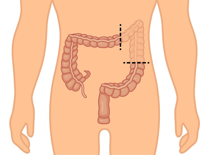
Colon
Stents
Survival Rate
Follow-Up Studies
Prospective Studies
Cerebrospinal Fluid Rhinorrhea
Risk Factors
Ureterostomy
Urinary Diversion
Encyclopedias as Topic
Spinal Cord Injuries
Surgical Stomas
A safe and reproducible anastomotic technique for minimally invasive Ivor Lewis oesophagectomy: the circular-stapled anastomosis with the trans-oral anvil. (1/105)
(+info)Effect of preoperative intraperitoneal injection of Sapylin in advanced gastric cancer. (2/105)
BACKGROUND AND OBJECTIVE: Sapylin is one of the biological response modifiers. It has been used in the comprehensive treatment for advanced cancer, and its clinical efficacy has been proved. This study was to evaluate the effect of preoperative intraperitoneal injection of Sapylin in treatment of advanced gastric cancer. METHODS: Seventy-nine patients eligible for radical gastrectomy were randomly divided into the treatment group (Sapylin + mitomycin C, 40 patients) and the control group (mitomycin C alone, 39 patients). In the treatment group, 5 KE Sapylin was injected intraperitoneally 48 h before operation and 4 mg of mitomycin C was injected into peritoneal cavity before the closure of the peritoneum. In the control group, only 4 mg mitomycin C was injected into peritoneal cavity before the closure of the peritonium. RESULTS: There was no operative mortality or duodenal stump leakage in the two groups. Postoperative complications were anastomotic leakage (2.5%, 1/40) and incision rupture (2.5%, 1/40) in the treatment group, and incision rupture (2.6%, 1/39) in the control group, with no significant difference between the two groups (P > 0.05). The 3-year survival rate was significantly higher in the treatment group than in the control group (76.5% vs. 49.4%, P < 0.05). CONCLUSIONS: Preoperative intraperitoneal injection of Sapylin can raise the 3-year survival rate after radical gastrectomy , without increasing the incidence rate of operative complications. Preoperative intraperitoneal injection of Sapylin is therefore a valuable therapy for advanced gastric cancer in clinic. (+info)Retrograde single stapling technique for laparoscopic ultralow anterior resection. (3/105)
(+info)Hybrid NOTES transgastric cholecystectomy with reliable gastric closure: an animal survival study. (4/105)
(+info)Recurrent abscess after primary successful endo-sponge treatment of anastomotic leakage following rectal surgery. (5/105)
AIM: To assess long-term efficacy of initially successful endo-sponge assisted therapy. METHODS: Between 2006 and 2009, consecutive patients who had undergone primary successful endo-sponge treatment of anastomotic leakage following rectal cancer surgery were enrolled in the study. Patients were recruited from 6 surgical departments in Vienna. Clinical and oncologic outcomes were assessed through routine endoscopic and radiologic follow-up examination. RESULTS: Twenty patients (7 female, 13 male) were included. The indications for endo-sponge treatment were anastomotic leakage (n = 17) and insufficiency of a rectal stump after Hartmann's procedure (n = 3). All patients were primarily operated for rectal cancer. The overall mortality rate was 25%. The median follow-up duration was 17 mo (range 1.5-29.8 mo). Five patients (25%) developed a recurrent abscess. Median time between last day of endo-sponge therapy and occurrence of recurrent abscess was 255 d (range 21-733 d). One of these patients was treated by computed tomography-guided drainage and in 3 patients Hartmann's procedure had to be performed. Two patients (10%) developed a local tumor recurrence and subsequently died. CONCLUSION: Despite successful primary outcome, patients who receive endo-sponge therapy should be closely monitored in the first 2 years, since recurrence might occur. (+info)Laparoscopic low anterior resection for rectal carcinoma: complications and management in 132 consecutive patients. (6/105)
AIM: To analyze the clinical manifestations and risk factors of complications in laparoscopic low anterior resection (LAR) for rectal cancer patients. METHODS: A series of 132 consecutive patients who received laparoscopic LAR for rectal cancer in our center were included. The etiology, diagnosis, treatment and prevention of rectal cancer were studied among the patients with surgery-related complications using both univariate and multivariate regression analysis. RESULTS: No conversion to open surgery was observed and 5 cases converted to hand-assisted laparoscopic operation. The overall morbidity rate was 20.5%. Complications occurred during the operation in 7 patients (5.3%), within 30 postoperative days in 24 patients (18.2%), and within 3 mo in 2 patients (1.5%). The most significant complications were anastomotic leakage (9.1%) and anastomotic hemorrhage (5.3%). Size and location of tumor, pathological staging and preoperative nutrition were significant factors associated with LAR complications, while gender, age and pathological type showed no relevance. Binary logistics regression showed that the size and location of tumor, and pathological staging were independent factors of laparoscopic LAR. All the complications were treated during their onset of clinical manifestations by interventional or conservative therapy. CONCLUSION: Anastomotic leakage is a major complication in laparoscopic LAR. The complications may be associated with tumor size and site, and pathological stage. Interventional therapies are of value in the management of laparoscopic LAR complications. (+info)The use of a compression device as an alternative to hand-sewn and stapled colorectal anastomoses: is three a crowd? (7/105)
(+info)The C-seal: a biofragmentable drain protecting the stapled colorectal anastomosis from leakage. (8/105)
(+info)An anastomotic leak is a medical condition that occurs after a surgical procedure where two hollow organs or vessels are connected (anastomosed). It refers to the failure of the connection, resulting in a communication between the inside of the connected structures and the outside, which can lead to the escape of fluids, such as digestive contents or blood, into the surrounding tissues.
Anastomotic leaks can occur in various parts of the body where anastomoses are performed, including the gastrointestinal tract, vasculature, and respiratory system. The leakage can cause localized or systemic infection, inflammation, sepsis, organ failure, or even death if not promptly diagnosed and treated.
The risk of anastomotic leaks depends on several factors, such as the patient's overall health, the type and location of the surgery, the quality of the surgical technique, and the presence of any underlying medical conditions that may affect wound healing. Treatment options for anastomotic leaks vary depending on the severity and location of the leak, ranging from conservative management with antibiotics and bowel rest to surgical intervention, such as drainage, revision of the anastomosis, or resection of the affected segment.
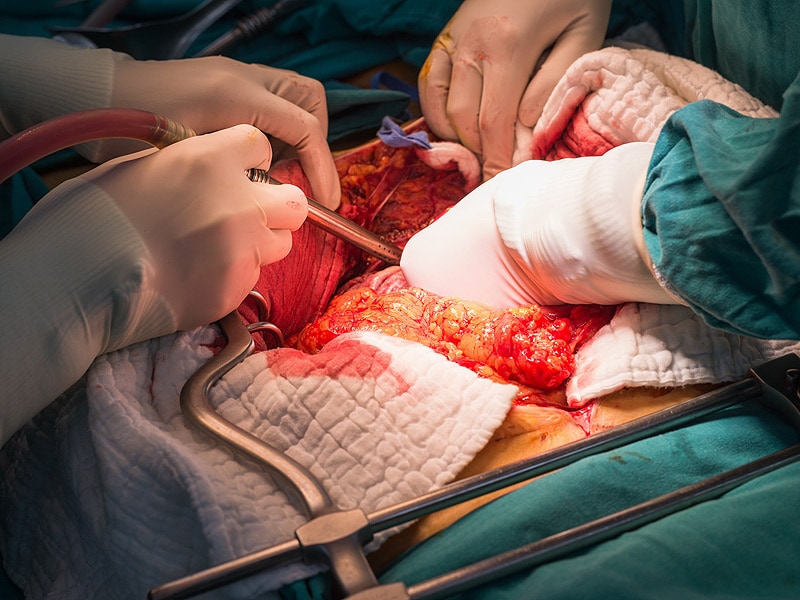
Surgical anastomosis is a medical procedure that involves the connection of two tubular structures, such as blood vessels or intestines, to create a continuous passage. This technique is commonly used in various types of surgeries, including vascular, gastrointestinal, and orthopedic procedures.
During a surgical anastomosis, the ends of the two tubular structures are carefully prepared by removing any damaged or diseased tissue. The ends are then aligned and joined together using sutures, staples, or other devices. The connection must be secure and leak-free to ensure proper function and healing.
The success of a surgical anastomosis depends on several factors, including the patient's overall health, the location and condition of the structures being joined, and the skill and experience of the surgeon. Complications such as infection, bleeding, or leakage can occur, which may require additional medical intervention or surgery.
Proper postoperative care is also essential to ensure the success of a surgical anastomosis. This may include monitoring for signs of complications, administering medications to prevent infection and promote healing, and providing adequate nutrition and hydration.
Esophagectomy is a surgical procedure in which part or all of the esophagus (the muscular tube that connects the throat to the stomach) is removed. This surgery is typically performed as a treatment for esophageal cancer, although it may also be used to treat other conditions such as severe damage to the esophagus from acid reflux or benign tumors.
During an esophagectomy, the surgeon will make incisions in the neck, chest, and/or abdomen to access the esophagus. The affected portion of the esophagus is then removed, and the remaining ends are reconnected, often using a section of the stomach or colon to create a new conduit for food to pass from the throat to the stomach.
Esophagectomy is a complex surgical procedure that requires significant expertise and experience on the part of the surgeon. It carries risks such as bleeding, infection, and complications related to anesthesia. Additionally, patients who undergo esophagectomy may experience difficulty swallowing, chronic pain, and other long-term complications. However, for some patients with esophageal cancer or other serious conditions affecting the esophagus, esophagectomy may be the best available treatment option.
Esophageal diseases refer to a range of medical conditions that affect the esophagus, which is the muscular tube that connects the throat to the stomach. Here are some common esophageal diseases with their brief definitions:
1. Gastroesophageal reflux disease (GERD): A chronic condition in which stomach acid or bile flows back into the esophagus, causing symptoms such as heartburn, chest pain, and difficulty swallowing.
2. Esophagitis: Inflammation of the esophageal lining, often caused by GERD, infection, or medication.
3. Esophageal stricture: Narrowing of the esophagus due to scarring or inflammation, which can make swallowing difficult.
4. Esophageal cancer: Cancer that forms in the tissues of the esophagus, often as a result of long-term GERD or smoking.
5. Esophageal motility disorders: Disorders that affect the normal movement and function of the esophagus, such as achalasia, diffuse spasm, and nutcracker esophagus.
6. Barrett's esophagus: A condition in which the lining of the lower esophagus changes, increasing the risk of esophageal cancer.
7. Esophageal diverticula: Small pouches that form in the esophageal wall, often causing difficulty swallowing or regurgitation.
8. Eosinophilic esophagitis (EoE): A chronic immune-mediated disorder characterized by inflammation of the esophagus due to an allergic reaction.
These are some of the common esophageal diseases, and their diagnosis and treatment may vary depending on the severity and underlying cause of the condition.
Surgical stapling is a medical technique that uses specialized staplers to place linear staple lines to close surgical incisions, connect or remove organs and tissues during surgical procedures. Surgical staples are made of titanium or stainless steel and can be absorbable or non-absorbable. They provide secure, fast, and accurate wound closure, reducing the risk of infection and promoting faster healing compared to traditional suturing methods.
The surgical stapler consists of a handle, an anvil, and a cartridge containing multiple staples. The device is loaded with staple cartridges and used to approximate tissue edges before deploying the staples. Once the staples are placed, the stapler is removed, leaving the staple line in place.
Surgical stapling has various applications, including gastrointestinal anastomosis, lung resection, vascular anastomosis, and skin closure. It is widely used in different types of surgeries, such as open, laparoscopic, and robotic-assisted procedures. The use of surgical stapling requires proper training and expertise to ensure optimal patient outcomes.
Suture techniques refer to the various methods used by surgeons to sew or stitch together tissues in the body after an injury, trauma, or surgical incision. The main goal of suturing is to approximate and hold the edges of the wound together, allowing for proper healing and minimizing scar formation.
There are several types of suture techniques, including:
1. Simple Interrupted Suture: This is one of the most basic suture techniques where the needle is passed through the tissue at a right angle, creating a loop that is then tightened to approximate the wound edges. Multiple stitches are placed along the length of the incision or wound.
2. Continuous Locking Suture: In this technique, the needle is passed continuously through the tissue in a zigzag pattern, with each stitch locking into the previous one. This creates a continuous line of sutures that provides strong tension and support to the wound edges.
3. Running Suture: Similar to the continuous locking suture, this technique involves passing the needle continuously through the tissue in a straight line. However, instead of locking each stitch, the needle is simply passed through the previous loop before being tightened. This creates a smooth and uninterrupted line of sutures that can be easily removed after healing.
4. Horizontal Mattress Suture: In this technique, two parallel stitches are placed horizontally across the wound edges, creating a "mattress" effect that provides additional support and tension to the wound. This is particularly useful in deep or irregularly shaped wounds.
5. Vertical Mattress Suture: Similar to the horizontal mattress suture, this technique involves placing two parallel stitches vertically across the wound edges. This creates a more pronounced "mattress" effect that can help reduce tension and minimize scarring.
6. Subcuticular Suture: In this technique, the needle is passed just below the surface of the skin, creating a smooth and barely visible line of sutures. This is particularly useful in cosmetic surgery or areas where minimizing scarring is important.
The choice of suture technique depends on various factors such as the location and size of the wound, the type of tissue involved, and the patient's individual needs and preferences. Proper suture placement and tension are crucial for optimal healing and aesthetic outcomes.
Surgical wound dehiscence is a medical condition that refers to the partial or complete separation of layers of a surgical incision after a surgical procedure, leading to the disruption of the wound closure. This can occur due to various factors such as infection, poor nutrition, increased tension on the sutures, hematoma or seroma formation, and patient's underlying health conditions like diabetes or immunodeficiency. Dehiscence may result in the exposure of internal tissues and organs, potentially causing severe complications such as infection, bleeding, or organ dysfunction. Immediate medical attention is required to manage this condition and prevent further complications.
The digestive system is a series of organs that work together to convert food into nutrients and energy. Digestive system surgical procedures involve operations on any part of the digestive system, including the esophagus, stomach, small intestine, large intestine, liver, pancreas, and gallbladder. These procedures can be performed for a variety of reasons, such as to treat diseases, repair damage, or remove cancerous growths.
Some common digestive system surgical procedures include:
1. Gastric bypass surgery: A procedure in which the stomach is divided into two parts and the smaller part is connected directly to the small intestine, bypassing a portion of the stomach and upper small intestine. This procedure is used to treat severe obesity.
2. Colonoscopy: A procedure in which a flexible tube with a camera on the end is inserted into the rectum and colon to examine the lining for polyps, cancer, or other abnormalities.
3. Colectomy: A procedure in which all or part of the colon is removed, often due to cancer, inflammatory bowel disease, or diverticulitis.
4. Gastrostomy: A procedure in which a hole is made through the abdominal wall and into the stomach to create an opening for feeding. This is often done for patients who have difficulty swallowing.
5. Esophagectomy: A procedure in which all or part of the esophagus is removed, often due to cancer. The remaining esophagus is then reconnected to the stomach or small intestine.
6. Liver resection: A procedure in which a portion of the liver is removed, often due to cancer or other diseases.
7. Pancreatectomy: A procedure in which all or part of the pancreas is removed, often due to cancer or chronic pancreatitis.
8. Cholecystectomy: A procedure in which the gallbladder is removed, often due to gallstones or inflammation.
These are just a few examples of digestive system surgical procedures. There are many other types of operations that can be performed on the digestive system depending on the specific needs and condition of each patient.
Esophageal atresia is a congenital condition in which the esophagus, the tube that connects the throat to the stomach, does not develop properly. In most cases, the upper esophagus ends in a pouch instead of connecting to the lower esophagus and stomach. This condition prevents food and liquids from reaching the stomach, leading to difficulty swallowing and feeding problems in newborn infants. Esophageal atresia often occurs together with a congenital defect called tracheoesophageal fistula, in which there is an abnormal connection between the esophagus and the windpipe (trachea).
The medical definition of 'Esophageal Atresia' is:
A congenital anomaly characterized by the absence of a normal connection between the upper esophagus and the stomach, resulting in the separation of the proximal and distal esophageal segments. The proximal segment usually ends in a blind pouch, while the distal segment may communicate with the trachea through a tracheoesophageal fistula. Esophageal atresia is often associated with other congenital anomalies and can cause serious complications if not diagnosed and treated promptly after birth.
A colectomy is a surgical procedure in which all or part of the large intestine (colon) is removed. This surgery may be performed to treat or prevent various medical conditions, including colon cancer, inflammatory bowel disease, diverticulitis, and severe obstructions or injuries of the colon.
There are several types of colectomies, depending on how much of the colon is removed:
* Total colectomy: Removal of the entire colon.
* Partial colectomy: Removal of a portion of the colon.
* Hemicolectomy: Removal of one half of the colon.
* Sigmoidectomy: Removal of the sigmoid colon, which is the part of the colon that is closest to the rectum.
After the affected portion of the colon is removed, the remaining ends of the intestine are reconnected, allowing stool to pass through the digestive system as usual. In some cases, a temporary or permanent colostomy may be necessary, in which a surgical opening (stoma) is created in the abdominal wall and the end of the colon is attached to it, allowing stool to be collected in a pouch outside the body.
Colectomies are major surgeries that require general anesthesia and hospitalization. The recovery time can vary depending on the type of colectomy performed and the individual's overall health, but typically ranges from several weeks to a few months. Complications of colectomy may include bleeding, infection, leakage from the surgical site, bowel obstruction, and changes in bowel habits or function.
Surgical staplers are medical devices used in various surgical procedures to create secure and precise connections between tissues, vessels, or organs. They function by placing sterile, disposable staple cartridges into the device that contain rows of stainless steel staples. The stapler then applies pressure to deform the staples, forming a B-shaped staple line that holds the tissue together.
These devices are often used in place of traditional suturing methods due to their speed, accuracy, and ability to reduce surgical trauma. They can be employed in various types of surgeries, including gastrointestinal, thoracic, gynecologic, and orthopedic procedures.
Surgical staplers come in different shapes and sizes, with some designed for specific applications such as linear or circular stapling. Linear staplers are used to create straight lines of staples, while circular staplers form a ring-shaped connection, often used in anastomosis procedures (the joining of two hollow organs or vessels).
It is essential to follow proper techniques and indications when using surgical staplers, as improper usage can lead to complications such as bleeding, infection, leakage, or even tissue necrosis.
The esophagus is the muscular tube that connects the throat (pharynx) to the stomach. It is located in the midline of the neck and chest, passing through the diaphragm to enter the abdomen and join the stomach. The main function of the esophagus is to transport food and liquids from the mouth to the stomach for digestion.
The esophagus has a few distinct parts: the upper esophageal sphincter (a ring of muscle that separates the esophagus from the throat), the middle esophagus, and the lower esophageal sphincter (another ring of muscle that separates the esophagus from the stomach). The lower esophageal sphincter relaxes to allow food and liquids to enter the stomach and then contracts to prevent stomach contents from flowing back into the esophagus.
The walls of the esophagus are made up of several layers, including mucosa (a moist tissue that lines the inside of the tube), submucosa (a layer of connective tissue), muscle (both voluntary and involuntary types), and adventitia (an outer layer of connective tissue).
Common conditions affecting the esophagus include gastroesophageal reflux disease (GERD), Barrett's esophagus, esophageal cancer, esophageal strictures, and eosinophilic esophagitis.
Postoperative complications refer to any unfavorable condition or event that occurs during the recovery period after a surgical procedure. These complications can vary in severity and may include, but are not limited to:
1. Infection: This can occur at the site of the incision or inside the body, such as pneumonia or urinary tract infection.
2. Bleeding: Excessive bleeding (hemorrhage) can lead to a drop in blood pressure and may require further surgical intervention.
3. Blood clots: These can form in the deep veins of the legs (deep vein thrombosis) and can potentially travel to the lungs (pulmonary embolism).
4. Wound dehiscence: This is when the surgical wound opens up, which can lead to infection and further complications.
5. Pulmonary issues: These include atelectasis (collapsed lung), pneumonia, or respiratory failure.
6. Cardiovascular problems: These include abnormal heart rhythms (arrhythmias), heart attack, or stroke.
7. Renal failure: This can occur due to various reasons such as dehydration, blood loss, or the use of certain medications.
8. Pain management issues: Inadequate pain control can lead to increased stress, anxiety, and decreased mobility.
9. Nausea and vomiting: These can be caused by anesthesia, opioid pain medication, or other factors.
10. Delirium: This is a state of confusion and disorientation that can occur in the elderly or those with certain medical conditions.
Prompt identification and management of these complications are crucial to ensure the best possible outcome for the patient.
Medical Definition:
Radiation is the emission of energy as electromagnetic waves or as moving subatomic particles, especially high-energy particles that cause ionization, which can occur naturally (e.g., sunlight) or be produced artificially (e.g., x-rays, radioisotopes). In medicine, radiation is used diagnostically and therapeutically in various forms, such as X-rays, gamma rays, and radiopharmaceuticals, to diagnose and treat diseases like cancer. However, excessive exposure to radiation can pose health risks, including radiation sickness and increased risk of cancer.
Esophagoplasty is a surgical procedure that involves reconstructing or reshaping the esophagus, which is the muscular tube that connects the throat to the stomach. This procedure may be performed to treat various conditions such as esophageal atresia (a birth defect in which the esophagus does not develop properly), esophageal stricture (narrowing of the esophagus), or esophageal cancer.
During an esophagoplasty, a surgeon may use tissue from another part of the body, such as the stomach or colon, to reconstruct the esophagus. The specific technique used will depend on the individual patient's needs and the nature of their condition.
It is important to note that esophagoplasty is a complex surgical procedure that carries risks such as bleeding, infection, and complications related to anesthesia. Patients who undergo this procedure may require extensive postoperative care and rehabilitation to recover fully.
A colostomy is a surgical procedure that involves creating an opening, or stoma, through the abdominal wall to divert the flow of feces from the colon (large intestine) through this opening and into a pouch or bag worn outside the body. This procedure is typically performed when a portion of the colon has been removed due to disease or injury, such as cancer, inflammatory bowel disease, or trauma.
There are several types of colostomies, including end colostomy, loop colostomy, and double-barrel colostomy, which differ in terms of the location and configuration of the stoma. The type of colostomy performed will depend on the individual's medical condition and the specific goals of the surgery.
After a colostomy, patients will need to learn how to care for their stoma and manage their bowel movements using specialized equipment and techniques. With proper care and management, most people are able to lead active and fulfilling lives after a colostomy.
Esophageal neoplasms refer to abnormal growths in the tissue of the esophagus, which is the muscular tube that connects the throat to the stomach. These growths can be benign (non-cancerous) or malignant (cancerous). Malignant esophageal neoplasms are typically classified as either squamous cell carcinomas or adenocarcinomas, depending on the type of cell from which they originate.
Esophageal cancer is a serious and often life-threatening condition that can cause symptoms such as difficulty swallowing, chest pain, weight loss, and coughing. Risk factors for esophageal neoplasms include smoking, heavy alcohol consumption, gastroesophageal reflux disease (GERD), and Barrett's esophagus. Treatment options may include surgery, radiation therapy, chemotherapy, or a combination of these approaches.
Colonic diseases refer to a group of medical conditions that affect the colon, also known as the large intestine or large bowel. The colon is the final segment of the digestive system, responsible for absorbing water and electrolytes, and storing and eliminating waste products.
Some common colonic diseases include:
1. Inflammatory bowel disease (IBD): This includes conditions such as Crohn's disease and ulcerative colitis, which cause inflammation and irritation in the lining of the digestive tract.
2. Diverticular disease: This occurs when small pouches called diverticula form in the walls of the colon, leading to symptoms such as abdominal pain, bloating, and changes in bowel movements.
3. Colorectal cancer: This is a type of cancer that develops in the colon or rectum, often starting as benign polyps that grow and become malignant over time.
4. Irritable bowel syndrome (IBS): This is a functional gastrointestinal disorder characterized by abdominal pain, bloating, and changes in bowel movements, but without any underlying structural or inflammatory causes.
5. Constipation: This is a common condition characterized by infrequent bowel movements, difficulty passing stools, or both.
6. Infectious colitis: This occurs when the colon becomes infected with bacteria, viruses, or parasites, leading to symptoms such as diarrhea, abdominal cramps, and fever.
Treatment for colonic diseases varies depending on the specific condition and its severity. Treatment options may include medications, lifestyle changes, surgery, or a combination of these approaches.
The rectum is the lower end of the digestive tract, located between the sigmoid colon and the anus. It serves as a storage area for feces before they are eliminated from the body. The rectum is about 12 cm long in adults and is surrounded by layers of muscle that help control defecation. The mucous membrane lining the rectum allows for the detection of stool, which triggers the reflex to have a bowel movement.
The sigmoid colon is a part of the large intestine that forms an "S"-shaped curve before it joins the rectum. It gets its name from its unique shape, which resembles the Greek letter sigma (σ). The main function of the sigmoid colon is to store stool temporarily and assist in the absorption of water and electrolytes from digestive waste before it is eliminated from the body.
"Plastics" is not a term that has a specific medical definition. However, in a broader context, plastics can refer to a wide range of synthetic or semi-synthetic materials that are used in various medical applications due to their durability, flexibility, and ability to be molded into different shapes. Some examples include:
1. Medical devices such as catheters, implants, and surgical instruments.
2. Packaging for medical supplies and pharmaceuticals.
3. Protective barriers like gloves and gowns used in medical settings.
4. Intraocular lenses and other ophthalmic applications.
It's important to note that the term "plastics" is not a medical term per se, but rather a general category of materials with diverse uses across different industries, including healthcare.
Esophageal stenosis is a medical condition characterized by the narrowing or constriction of the esophagus, which is the muscular tube that connects the throat to the stomach. This narrowing can make it difficult to swallow food and liquids, leading to symptoms such as dysphagia (difficulty swallowing), pain or discomfort while swallowing, regurgitation, and weight loss.
Esophageal stenosis can be caused by a variety of factors, including:
1. Scarring or fibrosis due to prolonged acid reflux or gastroesophageal reflux disease (GERD)
2. Radiation therapy for cancer treatment
3. Ingestion of corrosive substances
4. Eosinophilic esophagitis, an allergic condition that affects the esophagus
5. Esophageal tumors or cancers
6. Surgical complications
Depending on the underlying cause and severity of the stenosis, treatment options may include medications to manage symptoms, dilation procedures to widen the narrowed area, or surgery to remove the affected portion of the esophagus. It is important to seek medical attention if you experience any difficulty swallowing or other symptoms related to esophageal stenosis.
Colorectal surgery is a medical specialty that deals with the diagnosis and treatment of disorders affecting the colon, rectum, and anus. This can include conditions such as colorectal cancer, inflammatory bowel disease (such as Crohn's disease or ulcerative colitis), diverticulitis, and anal fistulas or fissures.
The surgical procedures performed by colorectal surgeons may involve minimally invasive techniques, such as laparoscopic or robotic-assisted surgery, or more traditional open surgery. These procedures can range from removing polyps during a colonoscopy to complex resections of the colon, rectum, or anus.
Colorectal surgeons also work closely with other medical specialists, such as gastroenterologists, oncologists, and radiologists, to provide comprehensive care for their patients.
Esophagoscopy is a medical procedure that involves the visual examination of the esophagus, which is the tube that connects the throat to the stomach. This procedure is typically carried out using an esophagogastroduodenoscope (EGD), a flexible tube with a camera and light on the end.
During the procedure, the EGD is inserted through the mouth and down the throat into the esophagus, allowing the medical professional to examine its lining for any abnormalities such as inflammation, ulcers, or tumors. The procedure may also involve taking tissue samples (biopsies) for further examination and testing.
Esophagoscopy is commonly used to diagnose and monitor conditions such as gastroesophageal reflux disease (GERD), Barrett's esophagus, esophageal cancer, and other disorders affecting the esophagus. It may also be used to treat certain conditions, such as removing polyps or foreign objects from the esophagus.
"Sigmoid diseases" is not a widely recognized medical term. However, the sigmoid colon is a part of the large intestine, and it can be affected by various conditions such as:
1. Sigmoid diverticulitis: Inflammation or infection of small pouches (diverticula) that form on the wall of the sigmoid colon.
2. Sigmoid volvulus: Twisting of the sigmoid colon on itself, which can lead to obstruction and ischemia.
3. Sigmoid cancer: Malignant tumor arising from the epithelial cells lining the sigmoid colon.
4. Inflammatory bowel disease (IBD): Chronic inflammation of the intestine, including the sigmoid colon, that can lead to symptoms such as diarrhea, abdominal pain, and weight loss.
5. Irritable bowel syndrome (IBS): Functional gastrointestinal disorder characterized by abdominal pain, bloating, and altered bowel habits, which can affect the sigmoid colon.
Therefore, "sigmoid diseases" could refer to any of these conditions or others that specifically affect the sigmoid colon.
An ileostomy is a surgical procedure in which the end of the small intestine, called the ileum, is brought through an opening in the abdominal wall (stoma) to create a path for waste material to leave the body. This procedure is typically performed when there is damage or removal of the colon, rectum, or anal canal due to conditions such as inflammatory bowel disease (Crohn's disease or ulcerative colitis), cancer, or trauma.
After an ileostomy, waste material from the small intestine exits the body through the stoma and collects in a pouch worn outside the body. The patient needs to empty the pouch regularly, typically every few hours, as the output is liquid or semi-liquid. Ileostomies can be temporary or permanent, depending on the underlying condition and the planned course of treatment. Proper care and management of the stoma and pouch are essential for maintaining good health and quality of life after an ileostomy.
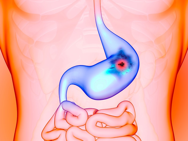
In anatomical terms, the stomach is a muscular, J-shaped organ located in the upper left portion of the abdomen. It is part of the gastrointestinal tract and plays a crucial role in digestion. The stomach's primary functions include storing food, mixing it with digestive enzymes and hydrochloric acid to break down proteins, and slowly emptying the partially digested food into the small intestine for further absorption of nutrients.
The stomach is divided into several regions, including the cardia (the area nearest the esophagus), the fundus (the upper portion on the left side), the body (the main central part), and the pylorus (the narrowed region leading to the small intestine). The inner lining of the stomach, called the mucosa, is protected by a layer of mucus that prevents the digestive juices from damaging the stomach tissue itself.
In medical contexts, various conditions can affect the stomach, such as gastritis (inflammation of the stomach lining), peptic ulcers (sores in the stomach or duodenum), gastroesophageal reflux disease (GERD), and stomach cancer. Symptoms related to the stomach may include abdominal pain, bloating, nausea, vomiting, heartburn, and difficulty swallowing.
Laparoscopy is a surgical procedure that involves the insertion of a laparoscope, which is a thin tube with a light and camera attached to it, through small incisions in the abdomen. This allows the surgeon to view the internal organs without making large incisions. It's commonly used to diagnose and treat various conditions such as endometriosis, ovarian cysts, infertility, and appendicitis. The advantages of laparoscopy over traditional open surgery include smaller incisions, less pain, shorter hospital stays, and quicker recovery times.
Treatment outcome is a term used to describe the result or effect of medical treatment on a patient's health status. It can be measured in various ways, such as through symptoms improvement, disease remission, reduced disability, improved quality of life, or survival rates. The treatment outcome helps healthcare providers evaluate the effectiveness of a particular treatment plan and make informed decisions about future care. It is also used in clinical research to compare the efficacy of different treatments and improve patient care.
A reoperation is a surgical procedure that is performed again on a patient who has already undergone a previous operation for the same or related condition. Reoperations may be required due to various reasons, such as inadequate initial treatment, disease recurrence, infection, or complications from the first surgery. The nature and complexity of a reoperation can vary widely depending on the specific circumstances, but it often carries higher risks and potential complications compared to the original operation.
A laparotomy is a surgical procedure that involves making an incision in the abdominal wall to gain access to the abdominal cavity. This procedure is typically performed to diagnose and treat various conditions such as abdominal trauma, tumors, infections, or inflammatory diseases. The size of the incision can vary depending on the reason for the surgery and the extent of the condition being treated. Once the procedure is complete, the incision is closed with sutures or staples.
The term "laparotomy" comes from the Greek words "lapara," which means "flank" or "side," and "tome," which means "to cut." Together, they describe the surgical procedure that involves cutting into the abdomen to examine its contents.
Pancreaticoduodenectomy, also known as the Whipple procedure, is a complex surgical operation that involves the removal of the head of the pancreas, the duodenum (the first part of the small intestine), the gallbladder, and the distal common bile duct. In some cases, a portion of the stomach may also be removed. The remaining parts of the pancreas, bile duct, and intestines are then reconnected to allow for the digestion of food and drainage of bile.
This procedure is typically performed as a treatment for various conditions affecting the pancreas, such as tumors (including pancreatic cancer), chronic pancreatitis, or traumatic injuries. It is a major surgical operation that requires significant expertise and experience to perform safely and effectively.
Gastric bypass is a surgical procedure that involves creating a small pouch in the stomach and rerouting the small intestine to connect to this pouch, thereby bypassing the majority of the stomach and the first part of the small intestine (duodenum). This procedure is typically performed as a treatment for morbid obesity and related health conditions such as type 2 diabetes, sleep apnea, and high blood pressure.
The smaller stomach pouch restricts food intake, while the rerouting of the small intestine reduces the amount of calories and nutrients that are absorbed, leading to weight loss. Gastric bypass can also result in hormonal changes that help regulate appetite and metabolism, further contributing to weight loss and improved health outcomes.
There are different types of gastric bypass procedures, including Roux-en-Y gastric bypass and laparoscopic gastric bypass. The choice of procedure depends on various factors such as the patient's overall health, medical history, and personal preferences. Gastric bypass is generally considered a safe and effective treatment for morbid obesity, but like any surgical procedure, it carries risks and requires careful consideration and preparation.
A Gastrectomy is a surgical procedure involving the removal of all or part of the stomach. This procedure can be total (complete resection of the stomach), partial (removal of a portion of the stomach), or sleeve (removal of a portion of the stomach to create a narrow sleeve-shaped pouch).
Gastrectomies are typically performed to treat conditions such as gastric cancer, benign tumors, severe peptic ulcers, and in some cases, for weight loss in individuals with morbid obesity. The type of gastrectomy performed depends on the patient's medical condition and the extent of the disease.
Following a gastrectomy, patients may require adjustments to their diet and lifestyle, as well as potential supplementation of vitamins and minerals that would normally be absorbed in the stomach. In some cases, further reconstructive surgery might be necessary to reestablish gastrointestinal continuity.
Rectal neoplasms refer to abnormal growths in the tissues of the rectum, which can be benign or malignant. They are characterized by uncontrolled cell division and can invade nearby tissues or spread to other parts of the body (metastasis). The most common type of rectal neoplasm is rectal cancer, which often begins as a small polyp or growth in the lining of the rectum. Other types of rectal neoplasms include adenomas, carcinoids, and gastrointestinal stromal tumors (GISTs). Regular screenings are recommended for early detection and treatment of rectal neoplasms.
Capillary leak syndrome (CLS) is a rare, but serious condition characterized by the abnormal leakage of plasma from the bloodstream into surrounding tissues. This occurs due to increased permeability of the capillary walls, which are the smallest blood vessels in the body that connect arterioles and venules, allowing for the exchange of nutrients, waste products, and gases between the blood and the tissues.
In CLS, the leakage of plasma leads to a rapid loss of intravascular volume, resulting in hypotension (low blood pressure), hemoconcentration (increased concentration of red blood cells due to reduced plasma volume), and edema (swelling) in various parts of the body. The fluid shift from the bloodstream to the tissues can also cause organ dysfunction and failure if not promptly treated.
The exact causes of capillary leak syndrome are not fully understood, but it can be associated with certain medical conditions, such as infections, autoimmune disorders, medications, or cancer. In some cases, CLS may occur without an identifiable underlying cause, known as idiopathic capillary leak syndrome.
Treatment for capillary leak syndrome typically involves supportive care to maintain blood pressure, replace lost fluids and electrolytes, and manage any organ dysfunction. Medications such as corticosteroids, immunoglobulins, or vasopressors may be used depending on the severity of the condition and the presence of underlying causes. In severe cases, extracorporeal membrane oxygenation (ECMO) or other intensive care interventions might be necessary to support organ function and ensure adequate blood flow.
Retrospective studies, also known as retrospective research or looking back studies, are a type of observational study that examines data from the past to draw conclusions about possible causal relationships between risk factors and outcomes. In these studies, researchers analyze existing records, medical charts, or previously collected data to test a hypothesis or answer a specific research question.
Retrospective studies can be useful for generating hypotheses and identifying trends, but they have limitations compared to prospective studies, which follow participants forward in time from exposure to outcome. Retrospective studies are subject to biases such as recall bias, selection bias, and information bias, which can affect the validity of the results. Therefore, retrospective studies should be interpreted with caution and used primarily to generate hypotheses for further testing in prospective studies.
"Length of Stay" (LOS) is a term commonly used in healthcare to refer to the amount of time a patient spends receiving care in a hospital, clinic, or other healthcare facility. It is typically measured in hours, days, or weeks and can be used as a metric for various purposes such as resource planning, quality assessment, and reimbursement. The length of stay can vary depending on the type of illness or injury, the severity of the condition, the patient's response to treatment, and other factors. It is an important consideration in healthcare management and can have significant implications for both patients and providers.
The colon, also known as the large intestine, is a part of the digestive system in humans and other vertebrates. It is an organ that eliminates waste from the body and is located between the small intestine and the rectum. The main function of the colon is to absorb water and electrolytes from digested food, forming and storing feces until they are eliminated through the anus.
The colon is divided into several regions, including the cecum, ascending colon, transverse colon, descending colon, sigmoid colon, rectum, and anus. The walls of the colon contain a layer of muscle that helps to move waste material through the organ by a process called peristalsis.
The inner surface of the colon is lined with mucous membrane, which secretes mucus to lubricate the passage of feces. The colon also contains a large population of bacteria, known as the gut microbiota, which play an important role in digestion and immunity.
A surgical wound infection, also known as a surgical site infection (SSI), is defined by the Centers for Disease Control and Prevention (CDC) as an infection that occurs within 30 days after surgery (or within one year if an implant is left in place) and involves either:
1. Purulent drainage from the incision;
2. Organisms isolated from an aseptically obtained culture of fluid or tissue from the incision;
3. At least one of the following signs or symptoms of infection: pain or tenderness, localized swelling, redness, or heat; and
4. Diagnosis of surgical site infection by the surgeon or attending physician.
SSIs can be classified as superficial incisional, deep incisional, or organ/space infections, depending on the depth and extent of tissue involvement. They are a common healthcare-associated infection and can lead to increased morbidity, mortality, and healthcare costs.
Adenocarcinoma is a type of cancer that arises from glandular epithelial cells. These cells line the inside of many internal organs, including the breasts, prostate, colon, and lungs. Adenocarcinomas can occur in any of these organs, as well as in other locations where glands are present.
The term "adenocarcinoma" is used to describe a cancer that has features of glandular tissue, such as mucus-secreting cells or cells that produce hormones. These cancers often form glandular structures within the tumor mass and may produce mucus or other substances.
Adenocarcinomas are typically slow-growing and tend to spread (metastasize) to other parts of the body through the lymphatic system or bloodstream. They can be treated with surgery, radiation therapy, chemotherapy, targeted therapy, or a combination of these treatments. The prognosis for adenocarcinoma depends on several factors, including the location and stage of the cancer, as well as the patient's overall health and age.
In the field of medicine, "time factors" refer to the duration of symptoms or time elapsed since the onset of a medical condition, which can have significant implications for diagnosis and treatment. Understanding time factors is crucial in determining the progression of a disease, evaluating the effectiveness of treatments, and making critical decisions regarding patient care.
For example, in stroke management, "time is brain," meaning that rapid intervention within a specific time frame (usually within 4.5 hours) is essential to administering tissue plasminogen activator (tPA), a clot-busting drug that can minimize brain damage and improve patient outcomes. Similarly, in trauma care, the "golden hour" concept emphasizes the importance of providing definitive care within the first 60 minutes after injury to increase survival rates and reduce morbidity.
Time factors also play a role in monitoring the progression of chronic conditions like diabetes or heart disease, where regular follow-ups and assessments help determine appropriate treatment adjustments and prevent complications. In infectious diseases, time factors are crucial for initiating antibiotic therapy and identifying potential outbreaks to control their spread.
Overall, "time factors" encompass the significance of recognizing and acting promptly in various medical scenarios to optimize patient outcomes and provide effective care.
A stent is a small mesh tube that's used to treat narrow or weak arteries. Arteries are blood vessels that carry blood away from your heart to other parts of your body. A stent is placed in an artery as part of a procedure called angioplasty. Angioplasty restores blood flow through narrowed or blocked arteries by inflating a tiny balloon inside the blocked artery to widen it.
The stent is then inserted into the widened artery to keep it open. The stent is usually made of metal, but some are coated with medication that is slowly and continuously released to help prevent the formation of scar tissue in the artery. This can reduce the chance of the artery narrowing again.
Stents are also used in other parts of the body, such as the neck (carotid artery) and kidneys (renal artery), to help maintain blood flow and prevent blockages. They can also be used in the urinary system to treat conditions like ureteropelvic junction obstruction or narrowing of the urethra.
Medical survival rate is a statistical measure used to determine the percentage of patients who are still alive for a specific period of time after their diagnosis or treatment for a certain condition or disease. It is often expressed as a five-year survival rate, which refers to the proportion of people who are alive five years after their diagnosis. Survival rates can be affected by many factors, including the stage of the disease at diagnosis, the patient's age and overall health, the effectiveness of treatment, and other health conditions that the patient may have. It is important to note that survival rates are statistical estimates and do not necessarily predict an individual patient's prognosis.
Follow-up studies are a type of longitudinal research that involve repeated observations or measurements of the same variables over a period of time, in order to understand their long-term effects or outcomes. In medical context, follow-up studies are often used to evaluate the safety and efficacy of medical treatments, interventions, or procedures.
In a typical follow-up study, a group of individuals (called a cohort) who have received a particular treatment or intervention are identified and then followed over time through periodic assessments or data collection. The data collected may include information on clinical outcomes, adverse events, changes in symptoms or functional status, and other relevant measures.
The results of follow-up studies can provide important insights into the long-term benefits and risks of medical interventions, as well as help to identify factors that may influence treatment effectiveness or patient outcomes. However, it is important to note that follow-up studies can be subject to various biases and limitations, such as loss to follow-up, recall bias, and changes in clinical practice over time, which must be carefully considered when interpreting the results.
Prospective studies, also known as longitudinal studies, are a type of cohort study in which data is collected forward in time, following a group of individuals who share a common characteristic or exposure over a period of time. The researchers clearly define the study population and exposure of interest at the beginning of the study and follow up with the participants to determine the outcomes that develop over time. This type of study design allows for the investigation of causal relationships between exposures and outcomes, as well as the identification of risk factors and the estimation of disease incidence rates. Prospective studies are particularly useful in epidemiology and medical research when studying diseases with long latency periods or rare outcomes.
Cerebrospinal fluid (CSF) rhinorrhea is a condition where the cerebrospinal fluid, which surrounds and protects the brain and spinal cord, leaks through the nasal cavity. This occurs due to a defect or opening in the skull base or the thin bone that separates the brain from the nasal cavity, known as the cribriform plate.
CSF rhinorrhea can result from trauma, surgery, or spontaneously due to increased pressure in the brain. It is important to diagnose and treat this condition promptly because it increases the risk of meningitis, an infection of the membranes covering the brain and spinal cord. Treatment options include bed rest, hydration, stool softeners, and sometimes surgical repair of the defect.
Medical Definition:
"Risk factors" are any attribute, characteristic or exposure of an individual that increases the likelihood of developing a disease or injury. They can be divided into modifiable and non-modifiable risk factors. Modifiable risk factors are those that can be changed through lifestyle choices or medical treatment, while non-modifiable risk factors are inherent traits such as age, gender, or genetic predisposition. Examples of modifiable risk factors include smoking, alcohol consumption, physical inactivity, and unhealthy diet, while non-modifiable risk factors include age, sex, and family history. It is important to note that having a risk factor does not guarantee that a person will develop the disease, but rather indicates an increased susceptibility.
Ureterostomy is a surgical procedure that creates an opening from one or both ureters, the tubes that carry urine from the kidneys to the bladder, to the abdominal wall. This allows urine to bypass the bladder and be expelled through the opening, called a stoma, into a collection device or onto the skin where it can be absorbed by a pad or diaper.
Ureterostomy is typically performed as a temporary measure in cases of severe bladder injury, infection, or obstruction that cannot be immediately corrected. It may also be used as a permanent solution for patients with congenital abnormalities or conditions that prevent the normal flow of urine through the bladder.
There are two main types of ureterostomy: cutaneous and uretero-cutanoeostomy. In a cutaneous ureterostomy, the ureter is brought directly to the abdominal wall and sutured in place. In a uretero-cutanoeostomy, a piece of intestine is used to create a conduit between the ureter and the abdominal wall.
Like any surgical procedure, ureterostomy carries risks such as bleeding, infection, and injury to surrounding organs. Patients who undergo this procedure will require close monitoring and follow-up care to ensure proper healing and function of the stoma.
Urinary diversion is a surgical procedure that involves the creation of a new way for urine to leave the body, bypassing the native urinary system. This is typically performed in individuals who have damaged or removed urinary systems due to conditions such as cancer, severe trauma, or congenital abnormalities.
There are several types of urinary diversions, including:
1. Ileal Conduit: A segment of the small intestine (ileum) is used to create a passageway for urine to flow from the ureters to an external collection bag or pouch worn on the abdomen.
2. Continent Urinary Reservoir: A pouch-like reservoir is created using a segment of the intestine, which is then connected to the ureters. The patient periodically empties the reservoir through a stoma (opening) in the abdominal wall using a catheter.
3. Orthotopic Neobladder: A pouch-like reservoir is created using a segment of the intestine, which is then connected to the urethra, allowing for normal urination through the native urethral opening.
These procedures can significantly improve the quality of life for patients with severe urinary system damage or disease, although they do come with potential complications such as infections, stone formation, and electrolyte imbalances.
An encyclopedia is a comprehensive reference work containing articles on various topics, usually arranged in alphabetical order. In the context of medicine, a medical encyclopedia is a collection of articles that provide information about a wide range of medical topics, including diseases and conditions, treatments, tests, procedures, and anatomy and physiology. Medical encyclopedias may be published in print or electronic formats and are often used as a starting point for researching medical topics. They can provide reliable and accurate information on medical subjects, making them useful resources for healthcare professionals, students, and patients alike. Some well-known examples of medical encyclopedias include the Merck Manual and the Stedman's Medical Dictionary.
Urination, also known as micturition, is the physiological process of excreting urine from the urinary bladder through the urethra. It is a complex process that involves several systems in the body, including the urinary system, nervous system, and muscular system.
In medical terms, urination is defined as the voluntary or involuntary discharge of urine from the urethra, which is the final pathway for the elimination of waste products from the body. The process is regulated by a complex interplay between the detrusor muscle of the bladder, the internal and external sphincters of the urethra, and the nervous system.
During urination, the detrusor muscle contracts, causing the bladder to empty, while the sphincters relax to allow the urine to flow through the urethra and out of the body. The nervous system plays a crucial role in coordinating these actions, with sensory receptors in the bladder sending signals to the brain when it is time to urinate.
Urination is essential for maintaining the balance of fluids and electrolytes in the body, as well as eliminating waste products such as urea, creatinine, and other metabolic byproducts. Abnormalities in urination can indicate underlying medical conditions, such as urinary tract infections, bladder dysfunction, or neurological disorders.
Spinal cord injuries (SCI) refer to damage to the spinal cord that results in a loss of function, such as mobility or feeling. This injury can be caused by direct trauma to the spine or by indirect damage resulting from disease or degeneration of surrounding bones, tissues, or blood vessels. The location and severity of the injury on the spinal cord will determine which parts of the body are affected and to what extent.
The effects of SCI can range from mild sensory changes to severe paralysis, including loss of motor function, autonomic dysfunction, and possible changes in sensation, strength, and reflexes below the level of injury. These injuries are typically classified as complete or incomplete, depending on whether there is any remaining function below the level of injury.
Immediate medical attention is crucial for spinal cord injuries to prevent further damage and improve the chances of recovery. Treatment usually involves immobilization of the spine, medications to reduce swelling and pressure, surgery to stabilize the spine, and rehabilitation to help regain lost function. Despite advances in treatment, SCI can have a significant impact on a person's quality of life and ability to perform daily activities.
A surgical stoma, also known simply as a stoma, is a surgically created opening on the surface of the body that allows for the passage of bodily waste. This procedure is typically performed when a person has a malfunctioning or diseased organ in the digestive or urinary system that cannot be effectively treated or repaired.
In a colostomy or ileostomy, which are common types of surgical stomas, a portion of the colon or small intestine is brought through an opening in the abdominal wall to create a new pathway for waste to exit the body. The stoma may be temporary or permanent, depending on the underlying condition and the success of any additional treatments.
After surgery, patients with a stoma will need to wear a pouching system to collect and contain the waste that is expelled through the stoma. This can take some getting used to, but with proper care and support, most people are able to adjust to life with a stoma and maintain a good quality of life.
Urinary Bladder Neoplasms are abnormal growths or tumors in the urinary bladder, which can be benign (non-cancerous) or malignant (cancerous). Malignant neoplasms can be further classified into various types of bladder cancer, such as urothelial carcinoma, squamous cell carcinoma, and adenocarcinoma. These malignant tumors often invade surrounding tissues and organs, potentially spreading to other parts of the body (metastasis), which can lead to serious health consequences if not detected and treated promptly and effectively.
 Management of Colorectal Anastomotic Leak
 How To Use Endoluminal Vacuum Therapy To Treat Upper Gastrointestinal Anastomotic Leaks and Perforations - SAGES Abstract...
 Defining a biomarker profile for anastomotic leak following colon surgery | Health Research Council of New Zealand
 Association of mechanical bowel preparation with oral antibiotics and anastomotic leak following left sided colorectal...
 2019 MIPS Measure #354: Anastomotic Leak Intervention | MDinteractive
 European Crohn´s and Colitis Organisation - ECCO - P639 Percutaneous drainage vs surgery as definitive treatment for...
 Exero Medical Completes First In-Human Implant of Wireless Anastomotic Leak Sensor at Rabin Medical Center
Risk factors for anastomotic leak after esophagectomy for cancer: A NSQIP procedure-targeted analysis<...
 Searching for "anastomotic leaking" - Nucleus Medical Art Library
Exero Medical's Multi-Center Study Demonstrates Early Detection of Anastomotic Leaks - MEDX Xelerator
 Outcomes following anastomotic leak from rectal resections, including bowel function and quality of life
Unsuccessful validation of the NUn score, for predicting anastomotic leak, morbidity and mortality after oesophagectomy -...
 NSAID administration post colorectal surgery increases anastomotic leak rate - systematic review/meta-analysis - Memorial...
Morino M
Standardized algorithms for management of anastomotic leaks and related abdominal and pelvic abscesses after colorectal surgery...
 Impact of anastomotic leak on long-term survival in patients undergoing gastrectomy for gastric cancer - ePrints - Newcastle...
 Ureterostomy - Wikipedia
 Evaluation of preoperative risk factors and postoperative indicators for anastomotic leak of minimally invasive McKeown...
 Anastomotic Leak Rate Is Doubled in Patients Diagnosed with Clostridium difficile Infection after Colectomy: A Retrospective...
 Treating the Obese Diabetic
 Minimally Invasive Esophagectomy for Esophageal Cancer: The First Experience from Pakistan
 Fast-track surgery could improve postoperative recovery in radical total gastrectomy patients
 Prevention and Management of Complications in Minimally Invasive Esophageal Surgery - Society of Laparoscopic & Robotic Surgeons
 Praachi Raje, M.D. | Harvard Catalyst Profiles | Harvard Catalyst
 Tal Hörer - School of Medical Sciences - Örebro University
Enterocutaneous Fistula: Practice Essentials, Etiology, Prognosis
EXPPECT Endometriosis Team | The University of Edinburgh
 PRIME PubMed | Laparoscopic Surgery for Diverticular Fistulas: Outcomes of 111 Consecutive Cases at a Single Institution
2013 ICD-9-CM Diagnosis Code 996.59 : Mechanical complication due to other implant and internal device, not elsewhere classified
 A New Risk Factor for Cervical Anastomotic Leakage-Role of The Relative Gastric Length in the Surgical Treatment of Esophageal...
Leakage13
- The company has created a patent-pending implantable biodegradable wireless sensor designed to continuously monitor the GI tract near the surgical site, alerting physicians to potential anastomotic leakage post-operation and also enabling early patient discharge by identifying proper tissue healing. (hhmglobal.com)
- Use of an NSAID post-operatively was associated with an overall increased risk of anastomotic leakage [OR 1.58 (1.23, 2.03), P = 0.0003]. (mun.ca)
- Making the anastomosis in this area would increase the risk of anastomotic leakage (AL). (springer.com)
- The multivariate logistic regression proved that RGL-G less than 1.3 was a risk factor for cervical anastomotic leakage ( p = 0.012). (springer.com)
- Aoyama T, Kazama K, Atsumi Y et al (2020) Clinical influence of anastomotic leakage on esophageal cancer survival and recurrence. (springer.com)
- Gooszen JAH, Goense L, Gisbertz SS et al (2018) Intrathoracic versus cervical anastomosis and predictors of anastomotic leakage after oesophagectomy for cancer. (springer.com)
- Verstegen MA-OX, Bouwense SAW, van Workum F et al (2019) Management of intrathoracic and cervical anastomotic leakage after esophagectomy for esophageal cancer: a systematic review. (springer.com)
- Li SJ, Wang ZQ, Li YJ et al (2017) Diabetes mellitus and risk of anastomotic leakage after esophagectomy: a systematic review and meta-analysis. (springer.com)
- One child suffered from prolonged bile leakage and re-explored to repair anastomotic leak. (scirp.org)
- Anastomotic leakage remains a concern in general surgical practice. (uwi.edu)
- Clinical studies are required in order to evaluate its efficacy in reducing the rate of gastrointestinal anastomotic leakage. (omicsonline.org)
- The circular stapling device as a risk factor for anastomotic leakage in rectal cancer surgery. (cancercentrum.se)
- Risk factors for anastomotic leakage after rectal cancer surgery: a case-control study. (cancercentrum.se)
Colorectal9
- Early diagnosis of an anastomotic leak following colorectal surgery is often delayed due its non-specific clinical picture. (hrc.govt.nz)
- CONCLUSION: This non-randomised study adds 'real-world', contemporaneous, and prospective evidence of the beneficial effects of combined mechanical bowel preparation and oral antibiotics in the prevention of anastomotic leak following left sided colorectal resection across diverse settings. (lu.se)
- These results are likely to reshape patient management as the system seamlessly integrated into our surgical workflow and detected leaks that the standard of care missed," said Professor Nir Wasserberg MD, Chair of Surgery Department at Rabin Medical Center and Chair of the Israel Colorectal Society. (medxelerator.com)
- Patients that have an anastomotic leak after colorectal surgery have a greater hospital length of stay, morbidity, mortality, permanent stoma rate, and even poorer oncological outcomes [ 8 , 9 ]. (coloproctol.org)
- Background: The risk factors and incidence of anastomotic leak following colorectal surgery are well reported in the literature. (elsevierpure.com)
- A six-round modified Delphi research method was utilized to find consensus among a group of expert colorectal surgeons and interventional radiologists regarding standardized management algorithms for anastomotic leaks. (elsevierpure.com)
- About ⅓ of deaths following colorectal surgery are associated with anastomotic leaks, according to Cohera. (massdevice.com)
- This analysis of colorectal anastomoses at a tertiary institution in Jamaica demonstrates acceptable leak and mortality rates. (uwi.edu)
- The significance lies in the resultant abdominal sepsis, related morbidity and mortality, risk of anastomotic loss, permanent stoma creation and the effect on local recurrence and overall patient survival in colorectal cancer cases. (uwi.edu)
Anastomosis6
- Intervention (via return to operating room, interventional radiology, or interventional gastroenterology) for presence of leak of endoluminal contents (such as air, fluid, GI contents, or contrast material) through an anastomosis. (mdinteractive.com)
- In the multivariate analysis (Table 2), PD was more likely to be performed in patients whose anastomotic leak was diagnosed later after surgery (OR 1.25, 95%CI 1.03-1.53, p=0.027), in those who underwent an ileo-colic anastomosis alone (OR 3.72, 95%CI 2.29-12.45, p=0.034) and in those who were treated after 2016 (OR 6.36, 95%CI 1.04-39.03, p=0.046). (ecco-ibd.eu)
- Methods A systematic review was performed for studies investigating anastomotic leak rate following NSAID use versus control after colonic or rectal anastomosis. (mun.ca)
- C. The fewer sutures in the anastomosis, the more chances for urine leak. (imop.gr)
- Rotura de la conexión y escape consiguiente (líquidos, secreciones, aire) en una ANASTOMOSIS QUIRÚRGICA en las estructuras del aparato digestivo, respiratorio, genitourinario y vascular. (bvsalud.org)
- Los escapes más frecuentes proceden de roturas de las líneas de sutura en el tubo digestivo y anastomosis intestinales. (bvsalud.org)
Incidence of anastomotic leak1
- only one study reported a higher incidence of anastomotic leak in obese patients, but only in the mid to lower rectum. (omicsonline.org)
Postoperative2
- The present study aims to assess whether postoperative NSAID use is associated with an increased anastomotic leak rate. (mun.ca)
- During hospitalization the patient presented temporary jaundice and low flow anastomotic leak that closed spontaneously and she went home on the 16th postoperative day. (bvsalud.org)
Complication6
- Retrospective, single-centre study including all consecutive patients who were diagnosed with an anastomotic leak as a complication of intestinal resection for CD between 2004 and 2022. (ecco-ibd.eu)
- Background: Anastomotic leak is the most common major complication after esophagectomy. (nebraska.edu)
- Anastomotic leak (AL) is an uncommon but potentially devastating complication after rectal resection. (coloproctol.org)
- Anastomotic leak is an uncommon but potentially devastating complication after total mesorectal excision (TME) of rectal cancer, occurring in 2% to 19% of patients [ 1 ]. (coloproctol.org)
- Anastomotic leak is the most frequently encountered complication. (wikipedia.org)
- Anastomotic leak can be a deadly complication that occurs when the staples in the stomach begin to leak. (noomii.com)
Rectal3
- In our study, we hope to provide an updated assessment of bowel function and quality of life after anastomotic leak from rectal resections. (coloproctol.org)
- Conclusions Great caution must be taken when prescribing NSAIDs following colonic or rectal anastomotic creation. (mun.ca)
- Tension on anastomotic lines following colonic resection, restoration of continuity without adequate mobilization, or a minimal leak or infection can lead to perianastomotic abscess formation, resulting in disruption, as seen in patients with anterior resection for rectal carcinoma. (medscape.com)
Esophagectomy2
- We investigated the 2016 American College of Surgeons National Surgical Quality Improvement Program esophagectomy targeted database to identify risk factors for anastomotic leak. (nebraska.edu)
- Manghelli JL, Ceppa DP, Greenberg JW et al (2019) Management of anastomotic leaks following esophagectomy: when to intervene. (springer.com)
Abscess2
- The present study confirms that PD is a safe and effective procedure to treat anastomotic leak and perianastomotic abscess in CD patients. (ecco-ibd.eu)
- Methods: The medical literature from 1973 to 2007 was reviewed using PubMed for papers relating to anastomotic leaks and abdominal abscess, with a specific emphasis on predisposing factors, prevention strategies, and treatment approaches. (elsevierpure.com)
Colonic1
- The proposed research aims to define a biomarker profile for anastomotic leak following colonic surgery. (hrc.govt.nz)
Surgery7
- P639 Percutaneous drainage vs surgery as definitive treatment for anastomotic leak after intestinal resection in patients with Crohn's disease. (ecco-ibd.eu)
- The aim of this study was therefore to compare the success rate of percutaneous drainage with that of surgery for the management of anastomotic leak in CD patients. (ecco-ibd.eu)
- Anastomotic leak was defined as a perianastomotic fluid collection confirmed by radiological findings within 30 days from surgery. (ecco-ibd.eu)
- Exero Medical, developer of a wireless sensor for early detection of anastomotic leaks following GI surgery, announced it has completed the first human implantation of its sensor. (hhmglobal.com)
- Or Yehuda, Israel, March 30, 2023 - Initial data from 50 patients participating in a multi-center, clinical study demonstrates that Exero Medical's xBar has the ability to reliably detect anastomotic leaks following gastrointestinal surgery at least three days earlier than the current standard of care - a potentially life-saving early alert. (medxelerator.com)
- Sylys is designed to prevent anastomotic leaks in patients undergoing gastrointestinal surgery. (massdevice.com)
- Patients who had a clinical diagnosis of a full-thickness bowel wall ischaemia at time of surgery without histological perforation, anastomotic leak following previous operation or pneumoperitoneum without a documented source were all excluded from this review. (ispub.com)
Complications4
- Anastomotic leak remains one of the most relevant complications after intestinal resection for Crohn's disease (CD). (ecco-ibd.eu)
- Zhuo ZG, Shen X, Song TN et al (2020) The efficacy of ischemic conditioning in the prevention of gastroesophageal anastomotic complications: a meta-analysis. (springer.com)
- Benefits of the new surgical technique are total absence of Roux-en-Y related intestinal complications. (scirp.org)
- This has been associated with some complications, including staple line leaks. (wjgnet.com)
Gastric1
- We report a 43-year old female who had undergone a laparoscopic sleeve gastrectomy that was complicated by a proximal gastric pouch leak at the gastroesophageal junction. (wjgnet.com)
Patients5
- By conducting a large multicentre, prospective observational study, this research will measure biomarkers in patients' blood in the peri-operative period, and correlating their trends in predicting an anastomotic leak. (hrc.govt.nz)
- Patients undergoing MBP+ABx had the lowest overall rate of anastomotic leak (6.1%, 9.2%, 8.7% respectively) in unadjusted analysis. (lu.se)
- Patients experiencing an anstomotic leak were identified, and univariate and multivariable logistic regression was performed to identify variables independently associated with anastomotic leak. (nebraska.edu)
- 001) and mortality (8% vs 2%, P =.001) were higher in patients with anastomotic leak. (nebraska.edu)
- The most frequent long-term morbidity was anastomotic narrowing observed in 5 (9.88%) patients. (hindawi.com)
Primary outcome1
- The primary outcome measure was anastomotic. (lu.se)
Endoluminal1
- This is a video describing the new method of treating anastomotic leaks and perforations using endoluminal vacuum therapy. (sages.org)
Retrospective1
- While biomarkers have been shown to assist in the early diagnosis of an anastomotic leak, data are limited to small or retrospective studies. (hrc.govt.nz)
Surgical2
- Despite improvements in surgical technique and perioperative care, anastomotic leaks still occur, and with them occur increased morbidity, mortality, length of stay, and costs. (nih.gov)
- A. Shimotakahara, A. Yamataka, T. Yanai, H. Kobayashi, T. Okazaki, G. J. Lane and T. Miyano, "Roux-en-Y Hepa tico-Jejunostomy for Biliary Reconstruction during the Surgical Treatment of Choledochal Cyst: Which Is Better? (scirp.org)
Abdominal1
- La tomografía abdominal ubicó a la masa en la tercera porción duodenal (confirmada por la duodenoscopía) con aspecto sugerente de tumor estromal. (bvsalud.org)
Management3
- We will also discuss the management strategies for anastomotic leak when they do inevitably occur. (nih.gov)
- Conclusions: This process was a useful first step toward establishing guidelines for the management of anastomotic leak. (elsevierpure.com)
- We used self-expandable stents (SEMS) in the management of the leak. (wjgnet.com)
Bowel2
- INTRODUCTION: The optimal bowel preparation strategy to minimise the risk of anastomotic leak is yet to be determined. (lu.se)
- This study aimed to determine whether oral antibiotics combined with mechanical bowel preparation (MBP+Abx) was associated with a reduced risk of anastomotic leak when compared to mechanical bowel preparation alone (MBP) or no bowel preparation (NBP). (lu.se)
Gastrectomy1
- Almadi MA, Bamihriz F, Aljebreen AM. Fatal aortoesophageal fistula bleeding after stenting for a leak post sleeve gastrectomy. (wjgnet.com)
Vacuum therapy1
- The primary goal of the study was to describe the endoscopic vacuum therapy as a treatment option for leaks in the upper gastrointestinal tract, to evaluate results and comparing the results with current studies in the literature. (uni-wuerzburg.de)
Surgeons1
- The fear of anastomotic leak is the one that keeps GI surgeons up at night and one that represents a true clinical need. (hhmglobal.com)
RESULTS1
- Study Design - Case Series Results: The defects were successfully closed using the U-Clip anastomotic device. (jefferson.edu)
Early3
- Founded in 2018 by the MEDX Xelerator, an Israeli Innovation Authority incubator and Clalit HMO, the largest HMO in Israel, Exero Medical goal is to save lives through early detection of anastomotic leaks following gastrointestinal (GI) surgeries, addressing a $2 billion market with its sensor technology. (hhmglobal.com)
- Though frequently non-modifiable, these findings could facilitate risk stratification and early detection of anastomotic leak to reduce associated morbidity. (nebraska.edu)
- The company's flagship product, xBar, which has an FDA Breakthrough Designation, is intended for the early detection of anastomotic leaks following gastrointestinal (GI) surgeries, a $2 billion market. (medxelerator.com)
Patient1
- Conclusion: Both patient and procedure-related factors are associated with anastomotic leak. (nebraska.edu)
Fluid1
- No cerebrospinal fluid leaks were experienced postoperatively. (jefferson.edu)
Impact1
- and from limited data, this may worsen after anastomotic leak [ 11 - 13 ] and negatively impact upon quality of life. (coloproctol.org)
Meta-analysis1
- Meta-analysis was performed to assess for overall risk of anastomotic leak with NSAID use, as well as sub-group analysis to compare selective vs non-selective NSAIDs and drug-specific NSAID safety profiles. (mun.ca)
Lower1
- After case-mix adjustment using a mixed-effects multivariable regression model, MBP+Abx was associated with a lower risk of anastomotic leak (OR 0.52, 0.30-0.92, P = 0.02) but MBP was not (OR 0.92, 0.63-1.36, P = 0.69) compared to NBP. (lu.se)
Reduce1
- We look forward to developing a reliable anastomotic leak detection system, that will save lives, reduce suffering and address a challenge currently costing billions of dollars for payers and providers. (hhmglobal.com)