Anemia, Megaloblastic
Anemia, Macrocytic
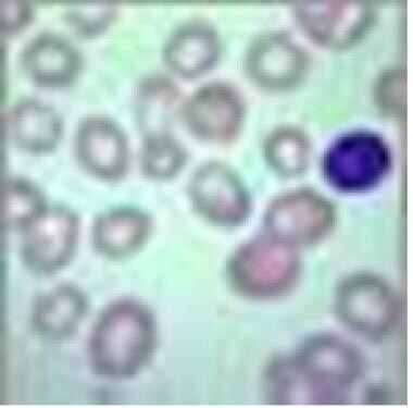
Megaloblasts
Vitamin B 12 Deficiency
Anemia, Pernicious
Folic Acid Deficiency
Vitamin B 12
FIGLU Test
Hydroxocobalamin
Intrinsic Factor
Anemia, Hypochromic
Folic Acid
Deoxyuridine
Anemia, Aplastic
Pregnancy Complications, Hematologic
Anemia, Hemolytic
Homocystinuria
Erythrocytes, Abnormal
Neurologic Manifestations
5-Methyltetrahydrofolate-Homocysteine S-Methyltransferase
Fanconi Anemia
Malabsorption Syndromes
Pancytopenia
Schilling Test
Erythropoiesis
Anemia, Hemolytic, Autoimmune
Euglena
Transcobalamins
Bone Marrow
Clinical Enzyme Tests
Anemia, Sickle Cell
Hearing Loss, Sensorineural
Methylmalonic Acid
Blood Group Antigens
Anemia, Sideroblastic
Erythrocytes
Erythroblasts
Infectious Anemia Virus, Equine
Thymine Nucleotides
Hemoglobins
Xylose
Anemia, Refractory
Folate Receptors, GPI-Anchored
Endemic tropical sprue in Rhodesia. (1/130)
The existence of tropical sprue in Africa is controversial. In this paper we present 31 cases seen in Rhodesia over a 15 month period. They have the clinical features, small intestinal morphology, malabsorption pattern, and treatment response of tropical sprue. Other causes of malabsorption, and primary malnutrition, have been excluded. The severity of the clinical state and intestinal malabsorption distinguish these patients from those we have described with tropical enteropathy. The previous work on tropical sprue in Africa is reviewed and it is apparent that, when it has been adequately looked for, it has been found. It is clear that the question of tropical sprue in Africa must be re-examined and that it existence may have hitherto been concealed by the assumption that primary malnutrition is responsible for the high prevalence of deficiency states. (+info)Defective high-affinity thiamine transporter leads to cell death in thiamine-responsive megaloblastic anemia syndrome fibroblasts. (2/130)
We have investigated the cellular pathology of the syndrome called thiamine-responsive megaloblastic anemia (TRMA) with diabetes and deafness. Cultured diploid fibroblasts were grown in thiamine-free medium and dialyzed serum. Normal fibroblasts survived indefinitely without supplemental thiamine, whereas patient cells died in 5-14 days (mean 9.5 days), and heterozygous cells survived for more than 30 days. TRMA fibroblasts were rescued from death with 10-30 nM thiamine (in the range of normal plasma thiamine concentrations). Positive terminal deoxynucleotide transferase-mediated dUTP nick end-labeling (TUNEL) staining suggested that cell death was due to apoptosis. We assessed cellular uptake of [3H]thiamine at submicromolar concentrations. Normal fibroblasts exhibited saturable, high-affinity thiamine uptake (Km 400-550 nM; Vmax 11 pmol/min/10(6) cells) in addition to a low-affinity unsaturable component. Mutant cells lacked detectable high-affinity uptake. At 30 nM thiamine, the rate of uptake of thiamine by TRMA fibroblasts was 10-fold less than that of wild-type, and cells from obligate heterozygotes had an intermediate phenotype. Transfection of TRMA fibroblasts with the yeast thiamine transporter gene THI10 prevented cell death when cells were grown in the absence of supplemental thiamine. We therefore propose that the primary abnormality in TRMA is absence of a high-affinity thiamine transporter and that low intracellular thiamine concentrations in the mutant cells cause biochemical abnormalities that lead to apoptotic cell death. (+info)Molecular basis for methionine synthase reductase deficiency in patients belonging to the cblE complementation group of disorders in folate/cobalamin metabolism. (3/130)
Methionine synthase reductase (MSR) deficiency is an autosomal recessive disorder of folate/cobalamin metabolism leading to hyperhomocysteinemia, hypo- methioninemia and megaloblastic anemia. Deficiency in MSR activity occurs as the result of a defect in the MSR enzyme, which is required for the reductive activation of methionine synthase (MS). MS itself is responsible for the folate/cobalamin-dependent conversion of homo- cysteine to methionine. We have recently cloned the cDNA corresponding to the MSR protein, a novel member of the ferredoxin-NADP(+)reductase (FNR) family of electron transferases. We have used RT-PCR, heteroduplex, single-strand conformation poly- morphism (SSCP) and DNA sequence analyses to reveal 11 mutations in eight patients from seven families belonging to the cblE complementation group of patients of cobalamin metabolism that is defective in the MSR protein. The mutations include splicing defects leading to large insertions or deletions, as well as a number of smaller deletions and point mutations. Apart from an intronic substitution found in two unrelated patients, the mutations appear singular among individuals. Of the eleven, three are nonsense mutations, allowing for the identification of two patients for whom little if any MSR protein should be produced. The remaining eight involve point mutations or in-frame disruptions of the coding sequence and are distributed throughout the coding region, including proposed FMN, FAD and NADPH binding sites. These data demonstrate a unique requirement for MSR in the reductive activation of MS. (+info)The pattern of severe protein-calorie malnutrition in Sudanese children attending a large hospital in The Sudan. (4/130)
One hundred fifty patients suffering from severe protein-calorie malnutrition, admitted in 1 month to the Pediatric wards of Wad Medani Hospital, Sudan, were classified according to the Wellcome classification. Marasmus was the prevailing type. It was common in the 2nd year of life, while kwashiorkor occurred mainly under the age of 12 months. Anthropometric measurements showed that kwashiorkor was an acute disease while marasmus and marasmic kwashiorkor were more chronic. The triceps skinfold was unexpectedly low in kwashiorkor. Of the simple measurements and ratios used for assessing the nutritional status, the head/chest ratio applied ot children over 1 year was not found to be reliable and the weight for head circumference correlated poorly with deficits in other variables. Non of the major clinical features was found to be pathognomonic of any type of severe protein-calorie malnutrition. Megaloblastic anemia was common. (+info)Cubilin P1297L mutation associated with hereditary megaloblastic anemia 1 causes impaired recognition of intrinsic factor-vitamin B(12) by cubilin. (5/130)
Megaloblastic anemia 1 (MGA1) is an autosomal recessive disorder caused by the selective intestinal malabsorption of intrinsic factor (IF) and vitamin B(12)/cobalamin (Cbl) in complex. Most Finnish patients with MGA1 carry the disease-specific P1297L mutation (FM1) in the IF-B(12) receptor, cubilin. By site-directed mutagenesis, mammalian expression, and functional comparison of the purified wild-type and FM1 mutant forms of the IF-Cbl-binding cubilin region (CUB domains 5-8, amino acid 928-1386), we have investigated the functional implications of the P1297L mutation. Surface plasmon resonance analysis revealed that the P1297L substitution specifically increases the K(d) for IF-Cbl binding several-fold, largely by decreasing the association rate constant. In agreement with the binding data, the wild-type protein, but not the FM1 mutant protein, potently inhibits 37 degrees C uptake of iodine 125-IF-Cbl in cubilin-expressing epithelial cells. In conclusion, the data presented show a substantial loss in affinity of the FM1 mutant form of the IF-Cbl binding region of cubilin. This now explains the malabsorption of Cbl and Cbl-dependent anemia in MGA1 patients with the FM1 mutation. (Blood. 2000;96:405-409) (+info)Oral contraceptive hormones, folate metabolism, and the cervical epithelium. (6/130)
The currently available evidence concerning disorders of folate metabolism in women taking oral contraceptives has been reviewed. A disturbance in folate balance serious enough to cause symptoms (i.e., megaloblastic anemia) occurs very rarely. In some series, but not in others, serum and/or red cell folate concentrations have been reduced in oral contraceptive users. It is doubtful whether sex steroids affect polyglutamate folate absorption. About 20 percent of women taking contraceptive hormones manifest mild megaloblastic changes on Papanicolaou smears of the cervicovaginal epithelium which disappear after folic acid therapy. The current evidence, however, would not indicate that any significant benefit would ensue from routine folate supplementation in women on oral contraceptives. (+info)A novel mutation in the thiamine responsive megaloblastic anaemia gene SLC19A2 in a patient with deficiency of respiratory chain complex I. (7/130)
The thiamine transporter gene SLC19A2 was recently found to be mutated in thiamine responsive megaloblastic anaemia with diabetes and deafness (TRMA, Rogers syndrome), an early onset autosomal recessive disorder. We now report a novel G1074A transition mutation in exon 4 of the SLC19A2 gene, predicting a Trp358 to ter change, in a girl with consanguineous parents. In addition to the typical triad of Rogers syndrome, the girl presented with short stature, hepatosplenomegaly, retinal degeneration, and a brain MRI lesion. Both muscle and skin biopsies were obtained before high dose thiamine supplementation. While no mitochondrial abnormalities were seen on morphological examination of muscle, biochemical analysis showed a severe deficiency of pyruvate dehydrogenase and complex I of the respiratory chain. In the patient's fibroblasts, the supplementation with high doses of thiamine resulted in restoration of complex I activity. In conclusion, we provide evidence that thiamine deficiency affects complex I activity. The clinical features of TRMA, resembling in part those found in typical mitochondrial disorders with complex I deficiency, may be caused by a secondary defect in mitochondrial energy production. (+info)Apoptosis in megaloblastic anemia occurs during DNA synthesis by a p53-independent, nucleoside-reversible mechanism. (8/130)
Deficiency of folate or vitamin B(12) (cobalamin) causes megaloblastic anemia, a disease characterized by pancytopenia due to the excessive apoptosis of hematopoietic progenitor cells. Clinical and experimental studies of megaloblastic anemia have demonstrated an impairment of DNA synthesis and repair in hematopoietic cells that is manifested by an increased percentage of cells in the DNA synthesis phase (S phase) of the cell cycle, compared with normal hematopoietic cells. Both folate and cobalamin are required for normal de novo synthesis of thymidylate and purines. However, previous studies of impaired DNA synthesis and repair in megaloblastic anemia have concerned mainly the decreased intracellular levels of thymidylate and its effects on nucleotide pools and misincorporation of uracil into DNA. An in vitro model of folate-deficient erythropoiesis was used to study the relationship between the S-phase accumulation and apoptosis in megaloblastic anemia. The results indicate that folate-deficient erythroblasts accumulate in and undergo apoptosis in the S phase when compared with control erythroblasts. Both the S-phase accumulation and the apoptosis were induced by folate deficiency in erythroblasts from p53 null mice. The complete reversal of the S-phase accumulation and apoptosis in folate-deficient erythroblasts required the exogenous provision of specific purines or purine nucleosides as well as thymidine. These results indicate that decreased de novo synthesis of purines plays as important a role as decreased de novo synthesis of thymidylate in the pathogenesis of megaloblastic anemia. (+info)Megaloblastic anemia is a type of macrocytic anemia, which is characterized by the presence of large, structurally abnormal, and immature red blood cells called megaloblasts in the bone marrow. This condition arises due to impaired DNA synthesis during erythropoiesis (the process of red blood cell production), often as a result of deficiencies in vitamin B12 or folate, or from the use of certain medications that interfere with DNA synthesis.
The hallmark feature of megaloblastic anemia is the presence of megaloblasts in the bone marrow, which exhibit an asynchrony between nuclear and cytoplasmic maturation. This means that although the cytoplasm of these cells may appear well-developed, their nuclei remain underdeveloped and fragmented. As a result, the peripheral blood shows an increase in mean corpuscular volume (MCV), reflecting the larger size of the red blood cells.
Additional hematological findings include decreased reticulocyte counts, neutrophil hypersegmentation, and occasionally thrombocytopenia or leukopenia. Neurological symptoms may also be present due to the involvement of the nervous system in vitamin B12 deficiency.
Megaloblastic anemia is typically treated with supplementation of the deficient vitamin (B12 or folate), which helps restore normal erythropoiesis and alleviate symptoms over time.
Macrocytic anemia is a type of anemia in which the red blood cells are larger than normal in size (macrocytic). This condition can be caused by various factors such as deficiency of vitamin B12 or folate, alcohol abuse, certain medications, bone marrow disorders, and some inherited genetic conditions.
The large red blood cells may not function properly, leading to symptoms such as fatigue, weakness, shortness of breath, pale skin, and a rapid heartbeat. Macrocytic anemia can be diagnosed through a complete blood count (CBC) test, which measures the size and number of red blood cells in the blood.
Treatment for macrocytic anemia depends on the underlying cause. In cases of vitamin B12 or folate deficiency, supplements or dietary changes may be recommended. If the anemia is caused by medication, a different medication may be prescribed. In severe cases, blood transfusions or injections of vitamin B12 may be necessary.
Megaloblasts are large, structurally abnormal immature red blood cells that appear in the bone marrow due to disorders in DNA synthesis, most commonly caused by deficiencies in folate or vitamin B12. They are characterized by an increased size, an oval or lobulated nucleus with condensed chromatin, and a cytoplasm filled with RNA and ribosomes. Megaloblasts can be found in megaloblastic anemias such as pernicious anemia and folate deficiency anemia. The presence of megaloblasts in the bone marrow is indicative of impaired maturation of red blood cells, which can lead to various hematological abnormalities.
Anemia is a medical condition characterized by a lower than normal number of red blood cells or lower than normal levels of hemoglobin in the blood. Hemoglobin is an important protein in red blood cells that carries oxygen from the lungs to the rest of the body. Anemia can cause fatigue, weakness, shortness of breath, and a pale complexion because the body's tissues are not getting enough oxygen.
Anemia can be caused by various factors, including nutritional deficiencies (such as iron, vitamin B12, or folate deficiency), blood loss, chronic diseases (such as kidney disease or rheumatoid arthritis), inherited genetic disorders (such as sickle cell anemia or thalassemia), and certain medications.
There are different types of anemia, classified based on the underlying cause, size and shape of red blood cells, and the level of hemoglobin in the blood. Treatment for anemia depends on the underlying cause and may include dietary changes, supplements, medication, or blood transfusions.
Vitamin B12 deficiency is a condition characterized by insufficient levels of vitamin B12 in the body, leading to impaired production of red blood cells, nerve function damage, and potential neurological complications. Vitamin B12 is an essential nutrient that plays a crucial role in DNA synthesis, fatty acid metabolism, and maintaining the health of the nervous system.
The medical definition of vitamin B12 deficiency includes:
1. Reduced serum or whole blood vitamin B12 concentrations (typically below 200 pg/mL or 145 pmol/L)
2. Presence of clinical symptoms and signs, such as:
* Fatigue, weakness, and lethargy
* Pale skin, shortness of breath, and heart palpitations due to anemia (megaloblastic or macrocytic anemia)
* Neurological symptoms like numbness, tingling, or burning sensations in the hands and feet (peripheral neuropathy), balance problems, confusion, memory loss, and depression
3. Laboratory findings consistent with deficiency, such as:
* Increased mean corpuscular volume (MCV) of red blood cells
* Reduced numbers of red and white blood cells and platelets in severe cases
* Elevated homocysteine and methylmalonic acid levels in the blood due to impaired metabolism
The most common causes of vitamin B12 deficiency include dietary insufficiency (common in vegetarians and vegans), pernicious anemia (an autoimmune condition affecting intrinsic factor production), gastrointestinal disorders (such as celiac disease, Crohn's disease, or gastric bypass surgery), and certain medications that interfere with vitamin B12 absorption.
Untreated vitamin B12 deficiency can lead to severe complications, including irreversible nerve damage, cognitive impairment, and increased risk of cardiovascular diseases. Therefore, prompt diagnosis and treatment are essential for preventing long-term health consequences.
Pernicious anemia is a specific type of vitamin B12 deficiency anemia that is caused by a lack of intrinsic factor, a protein made in the stomach that is needed to absorb vitamin B12. The absence of intrinsic factor leads to poor absorption of vitamin B12 from food and results in its deficiency.
Vitamin B12 is essential for the production of healthy red blood cells, which carry oxygen throughout the body. Without enough vitamin B12, the body cannot produce enough red blood cells, leading to anemia. Pernicious anemia typically develops slowly over several years and can cause symptoms such as fatigue, weakness, pale skin, shortness of breath, and a decreased appetite.
Pernicious anemia is an autoimmune disorder, which means that the body's immune system mistakenly attacks healthy cells in the stomach lining, leading to a loss of intrinsic factor production. It is more common in older adults, particularly those over 60 years old, and can also be associated with other autoimmune disorders such as type 1 diabetes, Hashimoto's thyroiditis, and Addison's disease.
Treatment for pernicious anemia typically involves vitamin B12 replacement therapy, either through oral supplements or injections of the vitamin. In some cases, dietary changes may also be recommended to ensure adequate intake of vitamin B12-rich foods such as meat, fish, poultry, and dairy products.
Folic Acid Deficiency is a condition characterized by insufficient levels of folic acid (Vitamin B9) in the body. Folic acid plays an essential role in the synthesis of DNA and RNA, the production of red blood cells, and the prevention of neural tube defects during fetal development.
A deficiency in folic acid can lead to a variety of health issues, including:
- Megaloblastic anemia: A type of anemia characterized by large, structurally abnormal, immature red blood cells (megaloblasts) that are unable to function properly. This results in fatigue, weakness, shortness of breath, and a pale appearance.
- Neural tube defects: In pregnant women, folic acid deficiency can increase the risk of neural tube defects, such as spina bifida and anencephaly, in the developing fetus.
- Developmental delays and neurological disorders: In infants and children, folic acid deficiency during pregnancy can lead to developmental delays, learning difficulties, and neurological disorders.
- Increased risk of cardiovascular disease: Folate plays a role in maintaining healthy homocysteine levels. Deficiency can result in elevated homocysteine levels, which is an independent risk factor for cardiovascular disease.
Folic acid deficiency can be caused by various factors, including poor dietary intake, malabsorption syndromes (such as celiac disease or Crohn's disease), pregnancy, alcoholism, certain medications (like methotrexate and phenytoin), and genetic disorders affecting folate metabolism. To prevent or treat folic acid deficiency, dietary supplementation with folic acid is often recommended, especially for pregnant women and individuals at risk of deficiency.
Vitamin B12, also known as cobalamin, is a water-soluble vitamin that plays a crucial role in the synthesis of DNA, formation of red blood cells, and maintenance of the nervous system. It is involved in the metabolism of every cell in the body, particularly affecting DNA regulation and neurological function.
Vitamin B12 is unique among vitamins because it contains a metal ion, cobalt, from which its name is derived. This vitamin can be synthesized only by certain types of bacteria and is not produced by plants or animals. The major sources of vitamin B12 in the human diet include animal-derived foods such as meat, fish, poultry, eggs, and dairy products, as well as fortified plant-based milk alternatives and breakfast cereals.
Deficiency in vitamin B12 can lead to various health issues, including megaloblastic anemia, fatigue, neurological symptoms such as numbness and tingling in the extremities, memory loss, and depression. Since vitamin B12 is not readily available from plant-based sources, vegetarians and vegans are at a higher risk of deficiency and may require supplementation or fortified foods to meet their daily requirements.
The FIGLU (Formiminoglutamic acid excretion) test is not a medical definition itself, but it is a test used to help diagnose Phenylketonuria (PKU), an inherited disorder of amino acid metabolism.
In PKU, the body cannot break down the amino acid phenylalanine properly due to a deficiency in the enzyme phenylalanine hydroxylase. As a result, phenylalanine and its toxic byproducts accumulate in the body, which can cause brain damage and intellectual disability if left untreated.
The FIGLU test measures the amount of formiminoglutamic acid (FIGLU) in the urine after a patient is given a load of histidine, another amino acid. In people with PKU, the accumulation of phenylalanine inhibits the conversion of histidine to glutamic acid, leading to an increase in FIGLU excretion in the urine. Therefore, a positive FIGLU test can indicate the presence of PKU. However, it is not a definitive diagnostic test and should be confirmed with other tests such as plasma amino acid analysis and/or genetic testing.
Thiamine, also known as vitamin B1, is a water-soluble vitamin that plays a crucial role in certain metabolic reactions, particularly in the conversion of carbohydrates into energy in the body. It is essential for the proper functioning of the heart, nerves, and digestive system. Thiamine acts as a cofactor for enzymes involved in the synthesis of neurotransmitters and the metabolism of carbohydrates, lipids, and proteins. Deficiency in thiamine can lead to serious health complications, such as beriberi (a disease characterized by peripheral neuropathy, muscle wasting, and heart failure) and Wernicke-Korsakoff syndrome (a neurological disorder often seen in alcoholics due to chronic thiamine deficiency). Thiamine is found in various foods, including whole grains, legumes, pork, beef, and fortified foods.
Hydroxocobalamin is a form of vitamin B12 that is used in medical treatments. It is a synthetic version of the naturally occurring compound, and it is often used to treat vitamin B12 deficiencies. Hydroxocobalamin is also used to treat poisoning from cyanide, as it can bind with the cyanide to form a non-toxic compound that can be excreted from the body.
In medical terms, hydroxocobalamin is defined as: "A bright red crystalline compound, C21H30CoN4O7·2H2O, used in the treatment of vitamin B12 deficiency and as an antidote for cyanide poisoning. It is converted in the body to active coenzyme forms."
It's important to note that hydroxocobalamin should only be used under the supervision of a medical professional, as improper use can lead to serious side effects or harm.
The Intrinsic Factor is a glycoprotein secreted by the parietal cells in the stomach lining. It plays an essential role in the absorption of vitamin B12 (cobalamin) in the small intestine. After binding with vitamin B12, the intrinsic factor-vitamin B12 complex moves through the digestive tract and gets absorbed in the ileum region of the small intestine. Deficiency in Intrinsic Factor can lead to Vitamin B12 deficiency disorders like pernicious anemia.
Hypochromic anemia is a type of anemia characterized by the presence of red blood cells that have lower than normal levels of hemoglobin and appear paler in color than normal. Hemoglobin is a protein in red blood cells that carries oxygen from the lungs to the rest of the body. In hypochromic anemia, there may be a decrease in the production or increased destruction of red blood cells, leading to a reduced number of red blood cells and insufficient oxygen supply to the tissues.
Hypochromic anemia can result from various underlying medical conditions, including iron deficiency, thalassemia, chronic inflammation, lead poisoning, and certain infections or chronic diseases. Treatment for hypochromic anemia depends on the underlying cause and may include iron supplements, dietary changes, medications, or blood transfusions.
Folic acid is the synthetic form of folate, a type of B vitamin (B9). It is widely used in dietary supplements and fortified foods because it is more stable and has a longer shelf life than folate. Folate is essential for normal cell growth and metabolism, and it plays a critical role in the formation of DNA and RNA, the body's genetic material. Folic acid is also crucial during early pregnancy to prevent birth defects of the brain and spine called neural tube defects.
Medical Definition: "Folic acid is the synthetic form of folate (vitamin B9), a water-soluble vitamin involved in DNA synthesis, repair, and methylation. It is used in dietary supplementation and food fortification due to its stability and longer shelf life compared to folate. Folic acid is critical for normal cell growth, development, and red blood cell production."
Deoxyuridine is a chemical compound that is a component of DNA. It is a nucleoside, which means it consists of a sugar (deoxyribose) linked to a nitrogenous base (uracil). In the case of deoxyuridine, the uracil is not methylated, which differentiates it from thymidine.
Deoxyuridine can be converted into deoxyuridine monophosphate (dUMP) by the enzyme thymidine kinase. The dUMP can then be converted into deoxythymidine triphosphate (dTTP), which is a building block of DNA, through a series of reactions involving other enzymes.
Deoxyuridine has been used in research and medicine as a marker for DNA synthesis and repair. It can also be used to inhibit the growth of certain types of cells, such as cancer cells, by disrupting their DNA synthesis.
Aplastic anemia is a medical condition characterized by pancytopenia (a decrease in all three types of blood cells: red blood cells, white blood cells, and platelets) due to the failure of bone marrow to produce new cells. It is called "aplastic" because the bone marrow becomes hypocellular or "aplastic," meaning it contains few or no blood-forming stem cells.
The condition can be acquired or inherited, with acquired aplastic anemia being more common. Acquired aplastic anemia can result from exposure to toxic chemicals, radiation, drugs, viral infections, or autoimmune disorders. Inherited forms of the disease include Fanconi anemia and dyskeratosis congenita.
Symptoms of aplastic anemia may include fatigue, weakness, shortness of breath, pale skin, easy bruising or bleeding, frequent infections, and fever. Treatment options for aplastic anemia depend on the severity of the condition and its underlying cause. They may include blood transfusions, immunosuppressive therapy, and stem cell transplantation.
Hematologic pregnancy complications refer to disorders related to the blood and blood-forming tissues that occur during pregnancy. These complications can have serious consequences for both the mother and the fetus if not properly managed. Some common hematologic pregnancy complications include:
1. Anemia: A condition characterized by a decrease in the number of red blood cells or hemoglobin in the blood, which can lead to fatigue, weakness, and shortness of breath. Iron-deficiency anemia is the most common type of anemia during pregnancy.
2. Thrombocytopenia: A condition characterized by a decrease in the number of platelets (cells that help blood clot) in the blood. Mild thrombocytopenia is relatively common during pregnancy, but severe thrombocytopenia can increase the risk of bleeding during delivery.
3. Gestational thrombotic thrombocytopenic purpura (GTTP): A rare but serious disorder that can cause blood clots to form in small blood vessels throughout the body, leading to a decrease in the number of platelets and red blood cells. GTTP can cause serious complications such as stroke, kidney failure, and even death if not promptly diagnosed and treated.
4. Disseminated intravascular coagulation (DIC): A condition characterized by abnormal clotting and bleeding throughout the body. DIC can be triggered by various conditions such as severe infections, pregnancy complications, or cancer.
5. Hemolysis, elevated liver enzymes, and low platelets (HELLP) syndrome: A serious complication of pregnancy that can cause damage to the liver and lead to bleeding. HELLP syndrome is often associated with preeclampsia, a condition characterized by high blood pressure and damage to organs such as the liver and kidneys.
It's important for pregnant women to receive regular prenatal care to monitor for these and other potential complications, and to seek prompt medical attention if any concerning symptoms arise.
A bone marrow examination is a medical procedure in which a sample of bone marrow, the spongy tissue inside bones where blood cells are produced, is removed and examined. This test is used to diagnose or monitor various conditions affecting blood cell production, such as infections, leukemia, anemia, and other disorders of the bone marrow.
The sample is typically taken from the hipbone (iliac crest) or breastbone (sternum) using a special needle. The procedure may be done under local anesthesia or with sedation to minimize discomfort. Once the sample is obtained, it is examined under a microscope for the presence of abnormal cells, changes in cell size and shape, and other characteristics that can help diagnose specific conditions. Various stains, cultures, and other tests may also be performed on the sample to provide additional information.
Bone marrow examination is an important diagnostic tool in hematology and oncology, as it allows for a detailed assessment of blood cell production and can help guide treatment decisions for patients with various blood disorders.
Hemolytic anemia is a type of anemia that occurs when red blood cells are destroyed (hemolysis) faster than they can be produced. Red blood cells are essential for carrying oxygen throughout the body. When they are destroyed, hemoglobin and other cellular components are released into the bloodstream, which can lead to complications such as kidney damage and gallstones.
Hemolytic anemia can be inherited or acquired. Inherited forms of the condition may result from genetic defects that affect the structure or function of red blood cells. Acquired forms of hemolytic anemia can be caused by various factors, including infections, medications, autoimmune disorders, and certain medical conditions such as cancer or blood disorders.
Symptoms of hemolytic anemia may include fatigue, weakness, shortness of breath, pale skin, jaundice (yellowing of the skin and eyes), dark urine, and a rapid heartbeat. Treatment for hemolytic anemia depends on the underlying cause and may include medications, blood transfusions, or surgery.
Homocystinuria is a genetic disorder characterized by the accumulation of homocysteine and its metabolites in the body due to a deficiency in the enzyme cystathionine beta-synthase (CBS). This enzyme is responsible for converting homocysteine to cystathionine, which is a critical step in the metabolic pathway that breaks down methionine.
As a result of this deficiency, homocysteine levels in the blood increase and can lead to various health problems, including neurological impairment, ocular abnormalities (such as ectopia lentis or dislocation of the lens), skeletal abnormalities (such as Marfan-like features), and vascular complications.
Homocystinuria can be diagnosed through newborn screening or by measuring homocysteine levels in the blood or urine. Treatment typically involves a low-methionine diet, supplementation with vitamin B6 (pyridoxine), betaine, and/or methylcobalamin (a form of vitamin B12) to help reduce homocysteine levels and prevent complications associated with the disorder.
Abnormal erythrocytes refer to red blood cells that have an abnormal shape, size, or other characteristics. This can include various types of abnormalities such as:
1. Anisocytosis: Variation in the size of erythrocytes.
2. Poikilocytosis: Variation in the shape of erythrocytes, including but not limited to teardrop-shaped cells (dacrocytes), crescent-shaped cells (sickle cells), and spherical cells (spherocytes).
3. Anemia: A decrease in the total number of erythrocytes or a reduction in hemoglobin concentration, which can result from various underlying conditions such as iron deficiency, chronic disease, or blood loss.
4. Hemoglobinopathies: Abnormalities in the structure or function of hemoglobin, the protein responsible for carrying oxygen in erythrocytes, such as sickle cell anemia and thalassemia.
5. Inclusion bodies: Abnormal structures within erythrocytes, such as Heinz bodies (denatured hemoglobin) or Howell-Jolly bodies (nuclear remnants).
These abnormalities can be detected through a complete blood count (CBC) and peripheral blood smear examination. The presence of abnormal erythrocytes may indicate an underlying medical condition, and further evaluation is often necessary to determine the cause and appropriate treatment.
Neurologic manifestations refer to the signs and symptoms that occur due to a disturbance or disease of the nervous system, which includes the brain, spinal cord, nerves, and muscles. These manifestations can vary widely depending on the specific location and nature of the underlying problem. They may include motor (movement-related) symptoms such as weakness, paralysis, tremors, or difficulty with coordination; sensory symptoms such as numbness, tingling, or pain; cognitive or behavioral changes; seizures; and autonomic symptoms such as changes in blood pressure, heart rate, or sweating. Neurologic manifestations can be caused by a wide range of conditions, including infections, injuries, degenerative diseases, strokes, tumors, and autoimmune disorders.
5-Methyltetrahydrofolate-Homocysteine S-Methyltransferase is also known as Methionine Synthase. It is a vital enzyme in the human body that plays a crucial role in methionine metabolism and homocysteine regulation.
The medical definition of 5-Methyltetrahydrofolate-Homocysteine S-Methyltransferase is as follows:
A enzyme (EC 2.1.1.13) that catalyzes the methylation of homocysteine to methionine, using 5-methyltetrahydrofolate as a methyl donor. This reaction also requires the cofactor vitamin B12 (cobalamin) as a coenzyme. The enzyme is located in the cytosol of cells and is essential for the synthesis of methionine, which is an important amino acid required for various biological processes such as protein synthesis, methylation reactions, and the formation of neurotransmitters.
Deficiency or dysfunction of this enzyme can lead to several health issues, including homocystinuria, a genetic disorder characterized by elevated levels of homocysteine in the blood, which can cause serious complications such as neurological damage, cardiovascular disease, and skeletal abnormalities.
Fanconi anemia is a rare, inherited disorder that affects the body's ability to produce healthy blood cells. It is characterized by bone marrow failure, congenital abnormalities, and an increased risk of developing certain types of cancer. The condition is caused by mutations in genes responsible for repairing damaged DNA, leading to chromosomal instability and cell death.
The classic form of Fanconi anemia (type A) is typically diagnosed in childhood and is associated with various physical abnormalities such as short stature, skin pigmentation changes, thumb and radial ray anomalies, kidney and genitourinary malformations, and developmental delays. Other types of Fanconi anemia (B-G) may have different clinical presentations but share the common feature of bone marrow failure and cancer predisposition.
Bone marrow failure in Fanconi anemia results in decreased production of all three types of blood cells: red blood cells, white blood cells, and platelets. This can lead to anemia (low red blood cell count), neutropenia (low white blood cell count), and thrombocytopenia (low platelet count). These conditions increase the risk of infections, fatigue, and bleeding.
Individuals with Fanconi anemia have a significantly higher risk of developing various types of cancer, particularly acute myeloid leukemia (AML) and solid tumors such as squamous cell carcinomas of the head, neck, esophagus, and anogenital region.
Treatment for Fanconi anemia typically involves managing symptoms related to bone marrow failure, such as transfusions, growth factors, and antibiotics. Hematopoietic stem cell transplantation (HSCT) is the only curative treatment option for bone marrow failure but carries risks of its own, including graft-versus-host disease and transplant-related mortality. Regular cancer surveillance is essential due to the increased risk of malignancies in these patients.
Malabsorption syndromes refer to a group of disorders in which the small intestine is unable to properly absorb nutrients from food, leading to various gastrointestinal and systemic symptoms. This can result from a variety of underlying conditions, including:
1. Mucosal damage: Conditions such as celiac disease, inflammatory bowel disease (IBD), or bacterial overgrowth that cause damage to the lining of the small intestine, impairing nutrient absorption.
2. Pancreatic insufficiency: A lack of digestive enzymes produced by the pancreas can lead to poor breakdown and absorption of fats, proteins, and carbohydrates. Examples include chronic pancreatitis or cystic fibrosis.
3. Bile acid deficiency: Insufficient bile acids, which are necessary for fat emulsification and absorption, can result in steatorrhea (fatty stools) and malabsorption. This may occur due to liver dysfunction, gallbladder removal, or ileal resection.
4. Motility disorders: Abnormalities in small intestine motility can affect nutrient absorption, as seen in conditions like gastroparesis, intestinal pseudo-obstruction, or scleroderma.
5. Structural abnormalities: Congenital or acquired structural defects of the small intestine, such as short bowel syndrome, may lead to malabsorption.
6. Infections: Certain bacterial, viral, or parasitic infections can cause transient malabsorption by damaging the intestinal mucosa or altering gut flora.
Symptoms of malabsorption syndromes may include diarrhea, steatorrhea, bloating, abdominal cramps, weight loss, and nutrient deficiencies. Diagnosis typically involves a combination of clinical evaluation, laboratory tests, radiologic imaging, and sometimes endoscopic procedures to identify the underlying cause. Treatment is focused on addressing the specific etiology and providing supportive care to manage symptoms and prevent complications.
Vitamin B Complex refers to a group of water-soluble vitamins that play essential roles in cell metabolism, cellular function, and formation of red blood cells. This complex includes 8 distinct vitamins, all of which were originally thought to be the same vitamin when first discovered. They are now known to have individual structures and specific functions.
1. Vitamin B1 (Thiamin): Necessary for energy production and nerve function.
2. Vitamin B2 (Riboflavin): Involved in energy production and growth.
3. Vitamin B3 (Niacin): Assists in energy production, DNA repair, and acts as a co-factor for various enzymes.
4. Vitamin B5 (Pantothenic Acid): Plays a role in the synthesis of Coenzyme A, which is vital for fatty acid metabolism.
5. Vitamin B6 (Pyridoxine): Needed for protein metabolism, neurotransmitter synthesis, hemoglobin formation, and immune function.
6. Vitamin B7 (Biotin): Involved in fatty acid synthesis, glucose metabolism, and nail and hair health.
7. Vitamin B9 (Folate or Folic Acid): Essential for DNA replication, cell division, and the production of red blood cells.
8. Vitamin B12 (Cobalamin): Necessary for nerve function, DNA synthesis, and the production of red blood cells.
These vitamins are often found together in various foods, and a balanced diet usually provides sufficient amounts of each. Deficiencies can lead to specific health issues related to the functions of each particular vitamin.
Pancytopenia is a medical condition characterized by a reduction in the number of all three types of blood cells in the peripheral blood: red blood cells (anemia), white blood cells (leukopenia), and platelets (thrombocytopenia). This condition can be caused by various underlying diseases, including bone marrow disorders, viral infections, exposure to toxic substances or radiation, vitamin deficiencies, and certain medications. Symptoms of pancytopenia may include fatigue, weakness, increased susceptibility to infections, and easy bruising or bleeding.
The Schilling test is a medical procedure that was used to diagnose pernicious anemia and malabsorption of vitamin B12. The test measures the body's ability to absorb vitamin B12 from food or supplements.
In the test, the patient is given a small amount of radioactive vitamin B12 to swallow. After a set period of time, a urine sample is collected and measured for the amount of radioactivity present. If the body has properly absorbed the vitamin B12, it will be excreted in the urine.
If the test shows that the patient is not absorbing enough vitamin B12, they may have pernicious anemia or another condition that affects vitamin B12 absorption. The Schilling test has largely been replaced by other diagnostic tests, such as blood tests for anti-intrinsic factor antibodies and parietal cell antibodies.
Diphyllobothriasis is a parasitic infection caused by the tapeworm of the genus Diphyllobothrium. The most common species to infect humans is Diphyllobothrium latum, which is found in freshwater fish. Humans can become infected with this tapeworm by consuming raw or undercooked fish that contain larval stages of the parasite.
The infection can cause a variety of symptoms, including abdominal discomfort, diarrhea, vomiting, and weight loss. In some cases, vitamin B12 deficiency may also occur, leading to neurological symptoms such as numbness, tingling, or weakness in the legs.
Treatment for diphyllobothriasis typically involves administration of a medication called niclosamide, which is an anthelmintic drug that kills the tapeworm. Prevention measures include cooking fish thoroughly before eating it and practicing good hygiene after handling raw fish.
Erythropoiesis is the process of forming and developing red blood cells (erythrocytes) in the body. It occurs in the bone marrow and is regulated by the hormone erythropoietin (EPO), which is produced by the kidneys. Erythropoiesis involves the differentiation and maturation of immature red blood cell precursors called erythroblasts into mature red blood cells, which are responsible for carrying oxygen to the body's tissues. Disorders that affect erythropoiesis can lead to anemia or other blood-related conditions.
Hemolytic anemia, autoimmune is a type of anemia characterized by the premature destruction of red blood cells (RBCs) in which the immune system mistakenly attacks and destroys its own RBCs. This occurs when the body produces autoantibodies that bind to the surface of RBCs, leading to their rupture (hemolysis). The symptoms may include fatigue, weakness, shortness of breath, and dark colored urine. The diagnosis is made through blood tests that measure the number and size of RBCs, reticulocyte count, and the presence of autoantibodies. Treatment typically involves suppressing the immune system with medications such as corticosteroids or immunosuppressive drugs, and sometimes removal of the spleen (splenectomy) may be necessary.
'Euglena' is a genus of unicellular flagellate protists that are typically characterized by their oval-shaped bodies, long whip-like tail (flagellum), and eyespot (stigma) which helps them to move towards light. They are commonly found in freshwater environments and can also be found in soil and brackish water. Some species of Euglena have the ability to photosynthesize, while others obtain their nutrition through heterotrophy (consuming other organisms or organic matter). The term 'Euglena' is derived from the Greek word 'euglenes', which means "well-shaped" or "true-eyed". Medical professionals and researchers may study Euglena as part of broader research into protists, microbiology, or ecology.
Transcobalamins are a group of proteins in the human body that are responsible for the transport of vitamin B12, also known as cobalamin. There are three main types of transcobalamins:
1. Transcobalamin I (also known as haptocorrin or R-binders): This is a protein produced in various tissues, including the salivary glands and gastric mucosa. It binds to vitamin B12 in the stomach and protects it from degradation by digestive enzymes. However, this form of vitamin B12 is not available for absorption and must be converted to other forms.
2. Transcobalamin II: This is a protein produced mainly in the kidneys and intestines. It binds to vitamin B12 that has been freed from its binding proteins in the stomach and facilitates its absorption in the intestine. Once absorbed, transcobalamin II transports vitamin B12 to tissues throughout the body.
3. Transcobalamin III (also known as intrinsic factor): This is a protein produced by the parietal cells of the stomach. It binds to vitamin B12 and protects it from degradation in the acidic environment of the stomach. Intrinsic factor is essential for the absorption of vitamin B12 in the intestine, as it facilitates its transport across the intestinal wall.
Deficiencies in transcobalamins can lead to vitamin B12 deficiency, which can result in a range of health problems, including anemia, fatigue, neurological symptoms, and developmental delays in children.
Bone marrow is the spongy tissue found inside certain bones in the body, such as the hips, thighs, and vertebrae. It is responsible for producing blood-forming cells, including red blood cells, white blood cells, and platelets. There are two types of bone marrow: red marrow, which is involved in blood cell production, and yellow marrow, which contains fatty tissue.
Red bone marrow contains hematopoietic stem cells, which can differentiate into various types of blood cells. These stem cells continuously divide and mature to produce new blood cells that are released into the circulation. Red blood cells carry oxygen throughout the body, white blood cells help fight infections, and platelets play a crucial role in blood clotting.
Bone marrow also serves as a site for immune cell development and maturation. It contains various types of immune cells, such as lymphocytes, macrophages, and dendritic cells, which help protect the body against infections and diseases.
Abnormalities in bone marrow function can lead to several medical conditions, including anemia, leukopenia, thrombocytopenia, and various types of cancer, such as leukemia and multiple myeloma. Bone marrow aspiration and biopsy are common diagnostic procedures used to evaluate bone marrow health and function.
Erythrocyte count, also known as red blood cell (RBC) count, is a laboratory test that measures the number of red blood cells in a sample of blood. Red blood cells are important because they carry oxygen from the lungs to the rest of the body. A low erythrocyte count may indicate anemia, while a high count may be a sign of certain medical conditions such as polycythemia. The normal range for erythrocyte count varies depending on a person's age, sex, and other factors.
Clinical enzyme tests are laboratory tests that measure the amount or activity of certain enzymes in biological samples, such as blood or bodily fluids. These tests are used to help diagnose and monitor various medical conditions, including organ damage, infection, inflammation, and genetic disorders.
Enzymes are proteins that catalyze chemical reactions in the body. Some enzymes are found primarily within specific organs or tissues, so elevated levels of these enzymes in the blood can indicate damage to those organs or tissues. For example, high levels of creatine kinase (CK) may suggest muscle damage, while increased levels of aspartate aminotransferase (AST) and alanine aminotransferase (ALT) can indicate liver damage.
There are several types of clinical enzyme tests, including:
1. Serum enzyme tests: These measure the level of enzymes in the blood serum, which is the liquid portion of the blood after clotting. Examples include CK, AST, ALT, alkaline phosphatase (ALP), and lactate dehydrogenase (LDH).
2. Urine enzyme tests: These measure the level of enzymes in the urine. An example is N-acetyl-β-D-glucosaminidase (NAG), which can indicate kidney damage.
3. Enzyme immunoassays (EIAs): These use antibodies to detect and quantify specific enzymes or proteins in a sample. They are often used for the diagnosis of infectious diseases, such as HIV or hepatitis.
4. Genetic enzyme tests: These can identify genetic mutations that cause deficiencies in specific enzymes, leading to inherited metabolic disorders like phenylketonuria (PKU) or Gaucher's disease.
It is important to note that the interpretation of clinical enzyme test results should be done by a healthcare professional, taking into account the patient's medical history, symptoms, and other diagnostic tests.
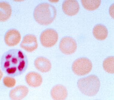
Sickle cell anemia is a genetic disorder that affects the hemoglobin in red blood cells. Hemoglobin is responsible for carrying oxygen throughout the body. In sickle cell anemia, the hemoglobin is abnormal and causes the red blood cells to take on a sickle shape, rather than the normal disc shape. These sickled cells are stiff and sticky, and they can block blood vessels, causing tissue damage and pain. They also die more quickly than normal red blood cells, leading to anemia.
People with sickle cell anemia often experience fatigue, chronic pain, and jaundice. They may also have a higher risk of infections and complications such as stroke, acute chest syndrome, and priapism. The disease is inherited from both parents, who must both be carriers of the sickle cell gene. It primarily affects people of African descent, but it can also affect people from other ethnic backgrounds.
There is no cure for sickle cell anemia, but treatments such as blood transfusions, medications to manage pain and prevent complications, and bone marrow transplantation can help improve quality of life for affected individuals. Regular medical care and monitoring are essential for managing the disease effectively.
Sensorineural hearing loss (SNHL) is a type of hearing impairment that occurs due to damage to the inner ear (cochlea) or to the nerve pathways from the inner ear to the brain. It can be caused by various factors such as aging, exposure to loud noises, genetics, certain medical conditions (like diabetes and heart disease), and ototoxic medications.
SNHL affects the ability of the hair cells in the cochlea to convert sound waves into electrical signals that are sent to the brain via the auditory nerve. As a result, sounds may be perceived as muffled, faint, or distorted, making it difficult to understand speech, especially in noisy environments.
SNHL is typically permanent and cannot be corrected with medication or surgery, but hearing aids or cochlear implants can help improve communication and quality of life for those affected.
Methylmalonic acid (MMA) is an organic compound that is produced in the human body during the metabolism of certain amino acids, including methionine and threonine. It is a type of fatty acid that is intermediate in the breakdown of these amino acids in the liver and other tissues.
Under normal circumstances, MMA is quickly converted to succinic acid, which is then used in the Krebs cycle to generate energy in the form of ATP. However, when there are deficiencies or mutations in enzymes involved in this metabolic pathway, such as methylmalonyl-CoA mutase, MMA can accumulate in the body and cause methylmalonic acidemia, a rare genetic disorder that affects approximately 1 in every 50,000 to 100,000 individuals worldwide.
Elevated levels of MMA in the blood or urine can be indicative of various metabolic disorders, including methylmalonic acidemia, vitamin B12 deficiency, and renal insufficiency. Therefore, measuring MMA levels is often used as a diagnostic tool to help identify and manage these conditions.
Blood group antigens are molecular markers found on the surface of red blood cells (RBCs) and sometimes other types of cells in the body. These antigens are proteins, carbohydrates, or glycoproteins that can stimulate an immune response when foreign antigens are introduced into the body.
There are several different blood group systems, but the most well-known is the ABO system, which includes A, B, AB, and O blood groups. The antigens in this system are called ABO antigens. Individuals with type A blood have A antigens on their RBCs, those with type B blood have B antigens, those with type AB blood have both A and B antigens, and those with type O blood have neither A nor B antigens.
Another important blood group system is the Rh system, which includes the D antigen. Individuals who have this antigen are considered Rh-positive, while those who do not have it are considered Rh-negative.
Blood group antigens can cause complications during blood transfusions and pregnancy if there is a mismatch between the donor's or fetus's antigens and the recipient's antibodies. For example, if a person with type A blood receives type B blood, their anti-B antibodies will attack the foreign B antigens on the donated RBCs, causing a potentially life-threatening transfusion reaction. Similarly, if an Rh-negative woman becomes pregnant with an Rh-positive fetus, her immune system may produce anti-D antibodies that can cross the placenta and attack the fetal RBCs, leading to hemolytic disease of the newborn.
It is important for medical professionals to determine a patient's blood group before performing a transfusion or pregnancy-related procedures to avoid these complications.
Sideroblastic anemia is a type of anemia characterized by the presence of ringed sideroblasts in the bone marrow. Ringed sideroblasts are red blood cell precursors that have an abnormal amount of iron accumulated in their mitochondria, which forms a ring around the nucleus. This results in the production of abnormal hemoglobin and impaired oxygen transport.
Sideroblastic anemia can be classified as congenital or acquired. Congenital sideroblastic anemias are caused by genetic defects that affect heme synthesis or mitochondrial function, while acquired sideroblastic anemias are associated with various conditions such as myelodysplastic syndromes, chronic alcoholism, lead toxicity, and certain medications.
Symptoms of sideroblastic anemia may include fatigue, weakness, shortness of breath, and pallor. Diagnosis is typically made through a bone marrow aspiration and biopsy, which can identify the presence of ringed sideroblasts. Treatment depends on the underlying cause but may include iron chelation therapy, vitamin B6 supplementation, or blood transfusions.
Erythrocytes, also known as red blood cells (RBCs), are the most common type of blood cell in circulating blood in mammals. They are responsible for transporting oxygen from the lungs to the body's tissues and carbon dioxide from the tissues to the lungs.
Erythrocytes are formed in the bone marrow and have a biconcave shape, which allows them to fold and bend easily as they pass through narrow blood vessels. They do not have a nucleus or mitochondria, which makes them more flexible but also limits their ability to reproduce or repair themselves.
In humans, erythrocytes are typically disc-shaped and measure about 7 micrometers in diameter. They contain the protein hemoglobin, which binds to oxygen and gives blood its red color. The lifespan of an erythrocyte is approximately 120 days, after which it is broken down in the liver and spleen.
Abnormalities in erythrocyte count or function can lead to various medical conditions, such as anemia, polycythemia, and sickle cell disease.
Erythroblasts are immature red blood cells that are produced in the bone marrow. They are also known as normoblasts and are a stage in the development of red blood cells, or erythrocytes. Erythroblasts are larger than mature red blood cells and have a nucleus, which is lost during the maturation process. These cells are responsible for producing hemoglobin, the protein that carries oxygen in the blood. Abnormal increases or decreases in the number of erythroblasts can be indicative of certain medical conditions, such as anemia or leukemia.
Equine Infectious Anemia (EIA) is a viral disease that affects horses and other equine animals. The causative agent of this disease is the Equine Infectious Anemia Virus (EIAV), which belongs to the family Retroviridae and genus Lentivirus. This virus is primarily transmitted through the transfer of infected blood, most commonly through biting insects such as horseflies and deerflies.
The EIAV attacks the immune system of the infected animal, causing a variety of symptoms including fever, weakness, weight loss, anemia, and edema. The virus has a unique ability to integrate its genetic material into the host's DNA, which can lead to a lifelong infection. Some animals may become chronic carriers of the virus, showing no signs of disease but remaining infectious to others.
There is currently no cure for EIA, and infected animals must be isolated to prevent the spread of the disease. Vaccines are available in some countries, but they do not provide complete protection against infection and may only help reduce the severity of the disease. Regular testing and monitoring of equine populations are essential to control the spread of this virus.
Consanguinity is a medical and genetic term that refers to the degree of genetic relationship between two individuals who share common ancestors. Consanguineous relationships exist when people are related by blood, through a common ancestor or siblings who have children together. The closer the relationship between the two individuals, the higher the degree of consanguinity.
The degree of consanguinity is typically expressed as a percentage or fraction, with higher values indicating a closer genetic relationship. For example, first-degree relatives, such as parents and children or full siblings, share approximately 50% of their genes and have a consanguinity coefficient of 0.25 (or 25%).
Consanguinity can increase the risk of certain genetic disorders and birth defects in offspring due to the increased likelihood of sharing harmful recessive genes. The risks depend on the degree of consanguinity, with closer relationships carrying higher risks. It is important for individuals who are planning to have children and have a history of consanguinity to consider genetic counseling and testing to assess their risk of passing on genetic disorders.
Thymine nucleotides are biochemical components that play a crucial role in the structure and function of DNA (deoxyribonucleic acid), which is the genetic material present in living organisms. A thymine nucleotide consists of three parts: a sugar molecule called deoxyribose, a phosphate group, and a nitrogenous base called thymine.
Thymine is one of the four nucleobases in DNA, along with adenine, guanine, and cytosine. It specifically pairs with adenine through hydrogen bonding, forming a base pair that is essential for maintaining the structure and stability of the double helix. Thymine nucleotides are linked together by phosphodiester bonds between the sugar molecules of adjacent nucleotides, creating a long, linear polymer known as a DNA strand.
In summary, thymine nucleotides are building blocks of DNA that consist of deoxyribose, a phosphate group, and the nitrogenous base thymine, which pairs with adenine in the double helix structure.
Hemoglobin (Hb or Hgb) is the main oxygen-carrying protein in the red blood cells, which are responsible for delivering oxygen throughout the body. It is a complex molecule made up of four globin proteins and four heme groups. Each heme group contains an iron atom that binds to one molecule of oxygen. Hemoglobin plays a crucial role in the transport of oxygen from the lungs to the body's tissues, and also helps to carry carbon dioxide back to the lungs for exhalation.
There are several types of hemoglobin present in the human body, including:
* Hemoglobin A (HbA): This is the most common type of hemoglobin, making up about 95-98% of total hemoglobin in adults. It consists of two alpha and two beta globin chains.
* Hemoglobin A2 (HbA2): This makes up about 1.5-3.5% of total hemoglobin in adults. It consists of two alpha and two delta globin chains.
* Hemoglobin F (HbF): This is the main type of hemoglobin present in fetal life, but it persists at low levels in adults. It consists of two alpha and two gamma globin chains.
* Hemoglobin S (HbS): This is an abnormal form of hemoglobin that can cause sickle cell disease when it occurs in the homozygous state (i.e., both copies of the gene are affected). It results from a single amino acid substitution in the beta globin chain.
* Hemoglobin C (HbC): This is another abnormal form of hemoglobin that can cause mild to moderate hemolytic anemia when it occurs in the homozygous state. It results from a different single amino acid substitution in the beta globin chain than HbS.
Abnormal forms of hemoglobin, such as HbS and HbC, can lead to various clinical disorders, including sickle cell disease, thalassemia, and other hemoglobinopathies.
Xylose is a type of sugar that is commonly found in plants and wood. In the context of medical definitions, xylose is often used in tests to assess the function of the small intestine. The most common test is called the "xylose absorption test," which measures the ability of the small intestine to absorb this sugar.
In this test, a patient is given a small amount of xylose to drink, and then several blood and/or urine samples are collected over the next few hours. The amount of xylose that appears in these samples is measured and used to determine how well the small intestine is absorbing nutrients.
Abnormal results on a xylose absorption test can indicate various gastrointestinal disorders, such as malabsorption syndromes, celiac disease, or bacterial overgrowth in the small intestine.
Refractory anemia is a type of anemia that does not respond to typical treatments, such as iron supplements or hormonal therapy. It is often associated with various bone marrow disorders, including myelodysplastic syndromes (MDS), a group of conditions characterized by abnormal blood cell production in the bone marrow.
In refractory anemia, the bone marrow fails to produce enough healthy red blood cells, leading to symptoms such as fatigue, weakness, shortness of breath, and pale skin. The condition can be difficult to treat, and treatment options may include more aggressive therapies such as immunosuppressive drugs, chemotherapy, or stem cell transplantation.
It is important to note that the term "refractory" in this context refers specifically to the lack of response to initial treatments, rather than a specific severity or type of anemia.
Folate receptors (FRs) are a group of cell surface proteins that bind and transport folate (vitamin B9) into cells. The subtype referred to as "GPI-anchored" refers to the type of anchoring that these receptors have in the cell membrane.
GPI stands for glycosylphosphatidylinositol, which is a molecule that acts as an anchor for certain proteins in the cell membrane. GPI-anchored folate receptors are attached to the outer layer of the cell membrane through this GPI anchor, rather than being embedded within the membrane like many other proteins.
GPI-anchored folate receptors are found on various types of cells, including some cancer cells, and they play a role in the uptake of folate into those cells. Folate is an essential nutrient that plays a critical role in DNA synthesis and methylation, among other processes. Abnormalities in folate metabolism have been linked to various diseases, including cancer and neurological disorders.
Homocysteine is an amino acid that is formed in the body during the metabolism of another amino acid called methionine. It's an important intermediate in various biochemical reactions, including the synthesis of proteins, neurotransmitters, and other molecules. However, elevated levels of homocysteine in the blood (a condition known as hyperhomocysteinemia) have been linked to several health issues, such as cardiovascular disease, stroke, and cognitive decline.
Homocysteine can be converted back to methionine with the help of vitamin B12 and a cofactor called betaine, or it can be converted to another amino acid called cystathionine with the help of vitamin B6 and folate (vitamin B9). Imbalances in these vitamins and other factors can lead to an increase in homocysteine levels.
It is crucial to maintain normal homocysteine levels for overall health, as high levels may contribute to the development of various diseases. Regular monitoring and maintaining a balanced diet rich in folate, vitamin B6, and vitamin B12 can help regulate homocysteine levels and reduce the risk of related health issues.