Angina Pectoris
Angina Pectoris, Variant
Angina, Stable
Microvascular Angina
Ludwig's Angina
Coronary Angiography
Myocardial Infarction
Ergonovine
Coronary Disease
Electrocardiography
Myocardial Ischemia
Exercise Test
Nitroglycerin
Coronary Artery Disease
Counterpulsation
Acetanilides
Isosorbide Dinitrate
Angioplasty, Balloon, Coronary
Coronary Artery Bypass
Myocardial Revascularization
Follow-Up Studies
Prospective Studies
Atherectomy, Coronary
Treatment Outcome
Electrocardiography, Ambulatory
Risk Factors
Acute Coronary Syndrome
Double-Blind Method
Cardiovascular Agents
Prognosis
Coronary Thrombosis
Adrenergic beta-Antagonists
Nicorandil
Platelet Aggregation Inhibitors
Biological Markers
C-Reactive Protein
Troponin T
Cardiac Catheterization
Predictive Value of Tests
Nifedipine
Calcium Channel Blockers
Stents
Aspirin
Physical Exertion
Oxyfedrine
Chronic Disease
Severity of Illness Index
Fibrinopeptide A
Hirudin Therapy
Exercise Tolerance
Ultrasonography, Interventional
Collateral Circulation
Creatine Kinase
Risk Assessment
Atenolol
Laser Therapy
Dipyridamole
Retrospective Studies
Iodobenzenes
Drug Therapy, Combination
Metoprolol
Diltiazem
Tomography, Emission-Computed, Single-Photon
Ischemic Preconditioning, Myocardial
Coronary Care Units
Platelet Glycoprotein GPIIb-IIIa Complex
Chi-Square Distribution
Multivariate Analysis
Nitrates
Heparin
Thallium Radioisotopes
Propranolol
Clinical Trials as Topic
Hemodynamics
Percutaneous Coronary Intervention
Spasm
Proportional Hazards Models
Enoxaparin
Incidence
Sex Factors
Regression Analysis
Stroke Volume
Case-Control Studies
Coronary Restenosis
Quality of Life
Echocardiography
Troponin
Nadroparin
Cohort Studies
Survival Analysis
Cardiovascular Diseases
Thrombolytic Therapy
Practolol
Spinal Cord Stimulation
Placebos
Age Factors
Logistic Models
Death, Sudden
Arrhythmias, Cardiac
Heart Diseases
Elevated levels of C-reactive protein at discharge in patients with unstable angina predict recurrent instability. (1/1246)
BACKGROUND: In a group of patients admitted for unstable angina, we investigated whether C-reactive protein (CRP) plasma levels remain elevated at discharge and whether persistent elevation is associated with recurrence of instability. METHODS AND RESULTS: We measured plasma levels of CRP, serum amyloid A protein (SAA), fibrinogen, total cholesterol, and Helicobacter pylori and Chlamydia pneumoniae antibody titers in 53 patients admitted to our coronary care unit for Braunwald class IIIB unstable angina. Blood samples were taken on admission, at discharge, and after 3 months. Patients were followed for 1 year. At discharge, CRP was elevated (>3 mg/L) in 49% of patients; of these, 42% had elevated levels on admission and at 3 months. Only 15% of patients with discharge levels of CRP <3 mg/L but 69% of those with elevated CRP (P<0.001) were readmitted because of recurrence of instability or new myocardial infarction. New phases of instability occurred in 13% of patients in the lower tertile of CRP (/=8.7 mg/L, P<0.001). The prognostic value of SAA was similar to that of CRP; that of fibrinogen was not significant. Chlamydia pneumoniae but not Helicobacter pylori antibody titers significantly correlated with CRP plasma levels. CONCLUSIONS: In unstable angina, CRP may remain elevated for at >/=3 months after the waning of symptoms and is associated with recurrent instability. Elevation of acute-phase reactants in unstable angina could represent a hallmark of subclinical persistent instability or of susceptibility to recurrent instability and, at least in some patients, could be related to chronic Chlamydia pneumoniae infection. (+info)A role for changes in platelet production in the cause of acute coronary syndromes. (2/1246)
Platelets are heterogeneous with respect to their size, density, and reactivity. Large platelets are more active hemostatically, and platelet volume has been found to be increased both in patients with unstable angina and with myocardial infarction. Furthermore, platelet volume is a predictor of a further ischemic event and death when measured after myocardial infarction. Platelets which are anucleate cells with no DNA are derived from their precursor, the megakaryocyte. Therefore, it is suggested that changes in platelet size are determined at thrombopoiesis in the megakaryocyte and that those changes might precede acute cardiac events. Understanding of the signaling system that controls platelet production may also further elucidate the cascade of events leading to acute vascular occlusion in some patients. (+info)Cardioprotection by opening of the K(ATP) channel in unstable angina. Is this a clinical manifestation of myocardial preconditioning? Results of a randomized study with nicorandil. CESAR 2 investigation. Clinical European studies in angina and revascularization. (3/1246)
AIMS: To assess the anti-ischaemic and anti-arrhythmic effects and overall safety of nicorandil, an ATP sensitive potassium (K+) channel opener, with 'cardioprotective' effects, in patients with unstable angina. METHODS: In a multicentre, randomized, double-blind, parallel-group, placebo-controlled study, oral nicorandil 20 mg twice daily or a matching placebo was administered for a minimum of 48 h to patients admitted with unstable angina. Treatment was standardized to include, where tolerated, oral aspirin, a beta-blocker and diltiazem. Continuous Holter ECG monitoring was performed for 48 h to assess the frequency and duration of transient myocardial ischaemia and any tachyarrhythmia, as the predefined end-points of the study. A pain chart recorded the incidence and severity of chest pain throughout the study period. Patients with myocardial infarction identified retrospectively from troponin-T analysis were excluded. RESULTS: Two hundred and forty-five patients were recruited into the study. Forty-three patients were excluded with an index diagnosis of myocardial infarction, two were not randomized and 12 had unsatisfactory tape data. In the remaining 188 patients, six out of 89 patients (6.7%) on nicorandil experienced an arrhythmia, compared with 17 out of 99 patients (17.2%) on placebo (P=0.04). Three nicorandil patients experienced three runs of non-sustained ventricular tachycardia compared to 31 runs in 10 patients on placebo (P=0.087 patients; P<0.0001 runs). Three nicorandil patients had four runs of supraventricular tachycardia, compared to 15 runs in nine patients on placebo (P=0.14 patients; P=0.017 runs). Eleven (12.4%) patients on nicorandil had 37 episodes of transient myocardial ischaemia (mostly silent) compared with 74 episodes in 21 (21.2%) patients on placebo (P=0.12 patients; P=0.0028 episodes). In the overall safety analysis, which included all patients who received at least one dose of study medication, there were no significant differences in the rates of myocardial infarction or death between the nicorandil or placebo-treated groups. CONCLUSIONS: Nicorandil, added to aggressive anti-anginal treatment for unstable angina, reduces transient myocardial ischaemia, non-sustained ventricular, and supraventricular arrhythmia compared to placebo. The anti-arrhythmic activity with nicorandil is probably a secondary effect resulting from its anti-ischaemic action and we suggest that this may be related to its effect on the ATP sensitive potassium channel causing pharmacological preconditioning. (+info)Platelet activation in patients after an acute coronary syndrome: results from the TIMI-12 trial. Thrombolysis in Myocardial Infarction. (4/1246)
This study was designed to determine the magnitude and time course of platelet activation during therapy of acute coronary syndromes with an oral platelet antagonist. BACKGROUND: Platelet activation and aggregation are central to the pathogenesis of the acute coronary syndromes (ACS). However, few data are available on levels of platelet activation over time in patients with ACS, especially in the setting of chronic glycoprotein (GP) IIb/IIIa inhibition. METHODS: The Thrombolysis in Myocardial Infarction (TIMI) 12 trial was a phase II, double-blind trial evaluating the effects of sibrafiban, an oral, selective antagonist of the platelet glycoprotein IIb/IIIa receptor in patients stabilized after an ACS. A subset of 90 of the 329 patients in the study had measurement of platelet activation as assessed by the expression of platelet associated P-Selectin on days 0, 7 and 28. Platelet activation was measured in blood samples that were fixed either immediately (spontaneous activation) or after 5 minute incubation with 0, 1 microM or 5 microM ADP in order to assess platelet responsiveness to very low or moderate stimulation. RESULTS: At baseline there was a significant elevation of spontaneous platelet activation as compared to samples obtained from normal donors or from patients who did not have acute coronary syndromes (ACS patients 27.6+/-18.7%, Normal controls 8.5+/-4.4%, Patient controls 10.9+/-7.1%, p < 0.005 for both). In addition, there was a significant decrease in the levels of platelet activation with time during the 28 days of treatment with sibrafiban. Nevertheless, even on day 28, the TIMI-12 patients continued to show elevated platelet activation in comparison to the control groups (p < 0.05 for both). CONCLUSIONS: These results suggest that platelets remain activated long after clinical stabilization post ACS. Although platelet activation decreased after one month of oral GPIIb/IIIa inhibition, levels remained higher than normal, suggesting the need for long-term antiplatelet therapy following ACS. (+info)Does coronary artery morphology predict favorable results of intracoronary thrombolysis in patients with unstable angina pectoris? (5/1246)
The efficacy of intracoronary thrombolysis (ICT) for unstable angina pectoris (UAP) has been limited, despite the similar pathogenesis between UAP and acute myocardial infarction. To ascertain the subset of UAP suitable for ICT, the clinical responses to ICT were assessed in patients with UAP. Eighty-2 patients with medically refractory angina were divided into 2 groups according to the coronary artery morphology of the culprit lesion before ICT: (1) lesions with acute cut off and/or filling defects (AC) and (2) lesions with a tapered shape (TA). The TIMI flow grade was determined from coronary angiograms before and immediately after ICT. The diameter stenosis (%DS) and minimal lumen diameter (MLD) of the culprit lesion were determined using quantitative coronary angiographic analysis before and immediately after ICT. In addition, inhospital cardiac event rates including urgent/emergency coronary angioplasty or bypass surgery, nonfatal myocardial infarction or cardiac death were compared between the 2 groups. Multivariate logistic regression analysis was performed using 13 clinical factors contributing to successful ICT. The results showed that all 3 coronary angiographic parameters (TIMI flow, %DS, and MLD) significantly improved in the AC group (p<0.01, p<0.01 and p<0.05, respectively), whereas none of these parameters improved in the TA group. The inhospital cardiac event rate after ICT was significantly higher in the TA group (76%) than in the AC group (48%; p=0.016). Odds ratio predicting successful ICT was 7.09 (p<0.01) for the AC lesion, and 2.54 (p<0.01) for new angina. In conclusion the AC lesions are more commonly associated with coronary thrombosis that responds to ICT than are the TA lesions. Thus, the coronary angiographic morphology may be an important predictor for a successful ICT in patients with medically refractory UAP. (+info)Coronary artery stenting in unstable angina pectoris: a comparison with stable angina pectoris. (6/1246)
OBJECTIVE: To compare early complication rates in unselected cases of coronary artery stenting in patients with stable v unstable angina. SETTING: Tertiary referral centre. PATIENTS: 390 patients with stable angina pectoris (SAP) and 306 with unstable angina (UAP). Patients treated for acute myocardial infarction (primary angioplasty) or cardiogenic shock were excluded. INTERVENTIONS: 268 coronary stents were attempted in 211 patients (30.3%). Stents used included AVE (63%), Freedom (14%), NIR (7%), Palmaz-Schatz (5%), JO (5%), and Multilink (4%). Intravascular ultrasound was not used in any of the cases. All stented patients were treated with ticlopidine and aspirin together with periprocedural unfractionated heparin. RESULTS: 123 stents were successfully deployed in 99 SAP patients v 132 stents in 103 UAP patients. Failed deployment occurred with nine stents in SAP patients, v four in UAP patients (NS). Stent thrombosis occurred in four SAP patients and 11 UAP patients. Multivariate analysis showed no relation between stent thrombosis and clinical presentation (SAP v UAP), age, sex, target vessel, stent length, or make of stent. Stent thrombosis was associated with small vessel size (p < 0.001) and bailout stenting (p = 0.01) compared with elective stenting and stenting for suboptimal PTCA, with strong trends toward smaller stent diameter (p = 0.052) and number of stents deployed (p = 0.06). Most stent thromboses occurred in vessels < 3 mm diameter. CONCLUSIONS: Coronary artery stenting in unstable angina is safe in vessels >/= 3 mm diameter, with comparable initial success and stent thrombosis rates to stenting in stable angina. (+info)Treatment with the antibiotic roxithromycin in patients with acute non-Q-wave coronary syndromes. The final report of the ROXIS Study. (7/1246)
AIMS: Mounting evidence suggests infection, specifically Chlamydia pneumoniae, plays a role in atherosclerosis. We tested whether antibiotic treatment with the macrolide roxithromycin improves clinical outcome in patients with acute non-Q-wave coronary syndromes. Preliminary reports revealed a reduction in events in the roxithromycin group at 30 days. We now report the long-term follow-up results. METHODS AND RESULTS: Sixty-four per cent of the initial 202 patients with unstable angina who were randomly assigned to receive either roxithromycin or placebo for 30 days completed the active treatment period. At day 30, the primary triple and double end-point rates were 9% and 4% in the placebo group compared to 2% and 0% in the roxithromycin group (unadjusted P = 0.032 and 0.058, respectively). The secondary triple and double end-point rates were again higher in the placebo group at day 90 (12.5% and 6.25% vs 4.37% and 0%, unadjusted P = 0.065 and 0.029, respectively), and at day 180 (14.6% and 7.29% vs 8.69% and 2.17%, unadjusted P = 0.259 and 0.17, respectively). Anti-C, pneumoniae IgG titres were unchanged in both groups while C-reactive protein levels decreased in both strategies, with a more significant decrease in the roxithromycin arm (P = 0.03). Elevated C-reactive protein levels predicted the need for revascularization. CONCLUSIONS: In this pilot trial, roxithromycin appears to extend the clinical benefit of preventing death and re-infarction for at least 6 months after initial treatment. (+info)Diagnostic marker cooperative study for the diagnosis of myocardial infarction. (8/1246)
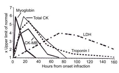
BACKGROUND: Millions of patients present annually with chest pain, but only 10% to 15% have myocardial infarction. Lack of diagnostic sensitivity and specificity of clinical and conventional markers prevents or delays treatment and leads to unnecessary costly admissions. Comparative data are lacking on the new markers, yet using all of them is inappropriate and expensive. METHODS AND RESULTS: The Diagnostic Marker Cooperative Study was a prospective, multicenter, double-blind study with consecutive enrollment of patients with chest pain presenting to the emergency department. Diagnostic sensitivity and specificity and frequency of increase in patients with unstable angina were determined for creatine kinase-MB (CK-MB) subforms, myoglobin, total CK-MB (activity and mass), and troponin T and I on the basis of frequent serial sampling for +info)Angina pectoris is a medical term that describes chest pain or discomfort caused by an inadequate supply of oxygen-rich blood to the heart muscle. This condition often occurs due to coronary artery disease, where the coronary arteries become narrowed or blocked by the buildup of cholesterol, fatty deposits, and other substances, known as plaques. These blockages can reduce blood flow to the heart, causing ischemia (lack of oxygen) and leading to angina symptoms.
There are two primary types of angina: stable and unstable. Stable angina is predictable and usually occurs during physical exertion or emotional stress when the heart needs more oxygen-rich blood. The pain typically subsides with rest or after taking prescribed nitroglycerin medication, which helps widen the blood vessels and improve blood flow to the heart.
Unstable angina, on the other hand, is more severe and unpredictable. It can occur at rest, during sleep, or with minimal physical activity and may not be relieved by rest or nitroglycerin. Unstable angina is considered a medical emergency, as it could indicate an imminent heart attack.
Symptoms of angina pectoris include chest pain, pressure, tightness, or heaviness that typically radiates to the left arm, neck, jaw, or back. Shortness of breath, nausea, sweating, and fatigue may also accompany angina symptoms. Immediate medical attention is necessary if you experience chest pain or discomfort, especially if it's new, severe, or persistent, as it could be a sign of a more serious condition like a heart attack.
Unstable angina is a term used in cardiology to describe chest pain or discomfort that occurs suddenly and unexpectedly, often at rest or with minimal physical exertion. It is caused by an insufficient supply of oxygen-rich blood to the heart muscle due to reduced blood flow, typically as a result of partial or complete blockage of the coronary arteries.
Unlike stable angina, which tends to occur predictably during physical activity and can be relieved with rest or nitroglycerin, unstable angina is more severe, unpredictable, and may not respond to traditional treatments. It is considered a medical emergency because it can be a sign of an impending heart attack or other serious cardiac event.
Unstable angina is often treated in the hospital with medications such as nitroglycerin, beta blockers, calcium channel blockers, and antiplatelet agents to improve blood flow to the heart and prevent further complications. In some cases, more invasive treatments such as coronary angioplasty or bypass surgery may be necessary to restore blood flow to the affected areas of the heart.
Angina pectoris, variant (also known as Prinzmetal's angina or vasospastic angina) is a type of chest pain that results from reduced blood flow to the heart muscle due to spasms in the coronary arteries. These spasms cause the arteries to narrow, temporarily reducing the supply of oxygen-rich blood to the heart. This can lead to symptoms such as chest pain, shortness of breath, and fatigue.
Variant angina is typically more severe than other forms of angina and can occur at rest or with minimal physical exertion. It is often treated with medications that help relax the coronary arteries and prevent spasms, such as calcium channel blockers and nitrates. In some cases, additional treatments such as angioplasty or bypass surgery may be necessary to improve blood flow to the heart.
It's important to note that chest pain can have many different causes, so it is essential to seek medical attention if you experience any symptoms of angina or other types of chest pain. A healthcare professional can help determine the cause of your symptoms and develop an appropriate treatment plan.
Stable angina is a type of chest pain or discomfort that typically occurs during physical exertion or emotional stress. It is caused by reduced blood flow to the heart muscle, which can occur when the coronary arteries become narrowed or blocked due to the buildup of cholesterol and other substances (a condition known as atherosclerosis).
The symptoms of stable angina are usually predictable and may include chest pain or discomfort that is:
* Described as a squeezing, pressure, heaviness, or tightness in the chest
* Typically located in the center of the chest, but may radiate to the shoulders, arms, neck, jaw, or back
* Lasts for a few minutes and is usually relieved by rest or nitroglycerin
Stable angina is considered "stable" because the symptoms tend to occur predictably and can be managed with medication, lifestyle changes, and sometimes medical procedures such as angioplasty or bypass surgery. However, it is still a serious condition that requires proper diagnosis and treatment to prevent complications such as heart attack or stroke.
Microvascular angina, also known as cardiac syndrome X or microvascular ischemia, is a type of angina (chest pain) that results from reduced blood flow to the heart muscle due to dysfunction in the small coronary arteries (microvasculature). These vessels are too small to be visualized during conventional diagnostic tests like coronary angiography.
The medical definition of microvascular angina is:
A clinical syndrome characterized by the presence of anginal chest pain, often accompanied by evidence of myocardial ischemia (insufficient blood flow to the heart muscle), in the absence of obstructive coronary artery disease on conventional diagnostic imaging. The underlying mechanism involves dysfunction and impaired regulation of the microvasculature, leading to reduced vasodilatory capacity, increased vasoconstriction, and ultimately, inadequate oxygen supply to meet the metabolic demands of the heart muscle.
Microvascular angina is more prevalent in women, especially those with risk factors such as hypertension, diabetes, hyperlipidemia, and smoking. Diagnosis often requires specialized testing like coronary flow reserve assessment using positron emission tomography (PET) or cardiac magnetic resonance imaging (MRI). Treatment typically involves a combination of lifestyle modifications, medications to improve blood vessel function and reduce chest pain, and sometimes, invasive treatments such as transmyocardial laser revascularization.
Ludwig's angina is a severe cellulitis (a bacterial infection of the connective tissues) of the floor of the mouth, below the tongue, and around the neck area. It's named after Wilhelm Friedrich von Ludwig, who first described it in 1836. The condition can lead to airway obstruction and significant swelling in the neck, making swallowing difficult or impossible. If not treated promptly with antibiotics and sometimes surgical drainage, it can be life-threatening due to the potential for spread of infection to the brain or other critical areas. It's typically caused by mixed oral flora, often including Streptococcus species, Staphylococcus aureus, and anaerobes.
Coronary vasospasm refers to a sudden constriction (narrowing) of the coronary arteries, which supply oxygenated blood to the heart muscle. This constriction can reduce or block blood flow, leading to symptoms such as chest pain (angina) or, in severe cases, a heart attack (myocardial infarction). Coronary vasospasm can occur spontaneously or be triggered by various factors, including stress, smoking, and certain medications. It is also associated with conditions such as coronary artery disease and variant angina. Prolonged or recurrent vasospasms can cause damage to the heart muscle and increase the risk of cardiovascular events.
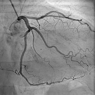
Coronary angiography is a medical procedure that uses X-ray imaging to visualize the coronary arteries, which supply blood to the heart muscle. During the procedure, a thin, flexible catheter is inserted into an artery in the arm or groin and threaded through the blood vessels to the heart. A contrast dye is then injected through the catheter, and X-ray images are taken as the dye flows through the coronary arteries. These images can help doctors diagnose and treat various heart conditions, such as blockages or narrowing of the arteries, that can lead to chest pain or heart attacks. It is also known as coronary arteriography or cardiac catheterization.
Myocardial infarction (MI), also known as a heart attack, is a medical condition characterized by the death of a segment of heart muscle (myocardium) due to the interruption of its blood supply. This interruption is most commonly caused by the blockage of a coronary artery by a blood clot formed on the top of an atherosclerotic plaque, which is a buildup of cholesterol and other substances in the inner lining of the artery.
The lack of oxygen and nutrients supply to the heart muscle tissue results in damage or death of the cardiac cells, causing the affected area to become necrotic. The extent and severity of the MI depend on the size of the affected area, the duration of the occlusion, and the presence of collateral circulation.
Symptoms of a myocardial infarction may include chest pain or discomfort, shortness of breath, nausea, lightheadedness, and sweating. Immediate medical attention is necessary to restore blood flow to the affected area and prevent further damage to the heart muscle. Treatment options for MI include medications, such as thrombolytics, antiplatelet agents, and pain relievers, as well as procedures such as percutaneous coronary intervention (PCI) or coronary artery bypass grafting (CABG).
Ergonovine is a medication that belongs to a class of drugs called ergot alkaloids. It is derived from the ergot fungus and is used in medical settings as a uterotonic agent, which means it causes the uterus to contract. Ergonovine is often used after childbirth to help the uterus return to its normal size and reduce bleeding.
Ergonovine works by binding to specific receptors in the smooth muscle of the uterus, causing it to contract. It has a potent effect on the uterus and can also cause vasoconstriction (narrowing of blood vessels) in other parts of the body. This is why ergonovine is sometimes used to treat severe bleeding caused by conditions such as uterine fibroids or ectopic pregnancy.
Like other ergot alkaloids, ergonovine can have serious side effects if not used carefully. It should be administered under the close supervision of a healthcare provider and should not be used in women with certain medical conditions, such as high blood pressure or heart disease. Ergonovine can also interact with other medications, so it's important to inform your healthcare provider of all medications you are taking before receiving this drug.
Coronary artery disease, often simply referred to as coronary disease, is a condition in which the blood vessels that supply oxygen-rich blood to the heart become narrowed or blocked due to the buildup of fatty deposits called plaques. This can lead to chest pain (angina), shortness of breath, or in severe cases, a heart attack.
The medical definition of coronary artery disease is:
A condition characterized by the accumulation of atheromatous plaques in the walls of the coronary arteries, leading to decreased blood flow and oxygen supply to the myocardium (heart muscle). This can result in symptoms such as angina pectoris, shortness of breath, or arrhythmias, and may ultimately lead to myocardial infarction (heart attack) or heart failure.
Risk factors for coronary artery disease include age, smoking, high blood pressure, high cholesterol, diabetes, obesity, physical inactivity, and a family history of the condition. Lifestyle changes such as quitting smoking, exercising regularly, eating a healthy diet, and managing stress can help reduce the risk of developing coronary artery disease. Medical treatments may include medications to control blood pressure, cholesterol levels, or irregular heart rhythms, as well as procedures such as angioplasty or bypass surgery to improve blood flow to the heart.
Electrocardiography (ECG or EKG) is a medical procedure that records the electrical activity of the heart. It provides a graphic representation of the electrical changes that occur during each heartbeat. The resulting tracing, called an electrocardiogram, can reveal information about the heart's rate and rhythm, as well as any damage to its cells or abnormalities in its conduction system.
During an ECG, small electrodes are placed on the skin of the chest, arms, and legs. These electrodes detect the electrical signals produced by the heart and transmit them to a machine that amplifies and records them. The procedure is non-invasive, painless, and quick, usually taking only a few minutes.
ECGs are commonly used to diagnose and monitor various heart conditions, including arrhythmias, coronary artery disease, heart attacks, and electrolyte imbalances. They can also be used to evaluate the effectiveness of certain medications or treatments.
Myocardial ischemia is a condition in which the blood supply to the heart muscle (myocardium) is reduced or blocked, leading to insufficient oxygen delivery and potential damage to the heart tissue. This reduction in blood flow typically results from the buildup of fatty deposits, called plaques, in the coronary arteries that supply the heart with oxygen-rich blood. The plaques can rupture or become unstable, causing the formation of blood clots that obstruct the artery and limit blood flow.
Myocardial ischemia may manifest as chest pain (angina pectoris), shortness of breath, fatigue, or irregular heartbeats (arrhythmias). In severe cases, it can lead to myocardial infarction (heart attack) if the oxygen supply is significantly reduced or cut off completely, causing permanent damage or death of the heart muscle. Early diagnosis and treatment of myocardial ischemia are crucial for preventing further complications and improving patient outcomes.
An exercise test, also known as a stress test or an exercise stress test, is a medical procedure used to evaluate the heart's function and response to physical exertion. It typically involves walking on a treadmill or pedaling a stationary bike while being monitored for changes in heart rate, blood pressure, electrocardiogram (ECG), and sometimes other variables such as oxygen consumption or gas exchange.
During the test, the patient's symptoms, such as chest pain or shortness of breath, are also closely monitored. The exercise test can help diagnose coronary artery disease, assess the severity of heart-related symptoms, and evaluate the effectiveness of treatments for heart conditions. It may also be used to determine a person's safe level of physical activity and fitness.
There are different types of exercise tests, including treadmill stress testing, stationary bike stress testing, nuclear stress testing, and stress echocardiography. The specific type of test used depends on the patient's medical history, symptoms, and overall health status.
Nitroglycerin, also known as glyceryl trinitrate, is a medication used primarily for the treatment of angina pectoris (chest pain due to coronary artery disease) and hypertensive emergencies (severe high blood pressure). It belongs to a class of drugs called nitrates or organic nitrites.
Nitroglycerin works by relaxing and dilating the smooth muscle in blood vessels, which leads to decreased workload on the heart and increased oxygen delivery to the myocardium (heart muscle). This results in reduced symptoms of angina and improved cardiac function during hypertensive emergencies.
The drug is available in various forms, including sublingual tablets, sprays, transdermal patches, ointments, and intravenous solutions. The choice of formulation depends on the specific clinical situation and patient needs. Common side effects of nitroglycerin include headache, dizziness, and hypotension (low blood pressure).
Coronary artery disease (CAD) is a medical condition in which the coronary arteries, which supply oxygen-rich blood to the heart muscle, become narrowed or blocked due to the buildup of cholesterol, fatty deposits, and other substances, known as plaque. Over time, this buildup can cause the arteries to harden and narrow (a process called atherosclerosis), reducing blood flow to the heart muscle.
The reduction in blood flow can lead to various symptoms and complications, including:
1. Angina (chest pain or discomfort) - This occurs when the heart muscle doesn't receive enough oxygen-rich blood, causing pain, pressure, or discomfort in the chest, arms, neck, jaw, or back.
2. Shortness of breath - When the heart isn't receiving adequate blood flow, it can't pump blood efficiently to meet the body's demands, leading to shortness of breath during physical activities or at rest.
3. Heart attack - If a piece of plaque ruptures or breaks off in a coronary artery, a blood clot can form and block the artery, causing a heart attack (myocardial infarction). This can damage or destroy part of the heart muscle.
4. Heart failure - Chronic reduced blood flow to the heart muscle can weaken it over time, leading to heart failure, a condition in which the heart can't pump blood efficiently to meet the body's needs.
5. Arrhythmias - Reduced blood flow and damage to the heart muscle can lead to abnormal heart rhythms (arrhythmias), which can be life-threatening if not treated promptly.
Coronary artery disease is typically diagnosed through a combination of medical history, physical examination, and diagnostic tests such as electrocardiograms (ECGs), stress testing, cardiac catheterization, and imaging studies like coronary computed tomography angiography (CCTA). Treatment options for CAD include lifestyle modifications, medications, medical procedures, and surgery.
Counterpulsation is a medical treatment used in critical care medicine, particularly in the management of cardiovascular conditions. It refers to a technique that involves delivering therapies that counter or oppose the patient's own cardiac cycle. The most common form of counterpulsation is through the use of an intra-aortic balloon pump (IABP).
During IABP, a catheter with a sausage-shaped balloon at its tip is inserted into the patient's aorta, usually through the femoral artery in the groin. The balloon is then connected to a console that controls its inflation and deflation. The console is programmed to detect the patient's cardiac cycle using either the ECG or arterial pressure waveform.
During diastole (when the heart muscle relaxes and fills with blood), the balloon inflates, increasing the volume of blood in the aorta and improving coronary artery perfusion. This helps to increase oxygen delivery to the myocardium (heart muscle) and reduce its workload.
During systole (when the heart muscle contracts and ejects blood), the balloon deflates, reducing afterload (the resistance against which the heart must pump). This reduces the workload of the left ventricle, allowing it to fill more easily during diastole and improving overall cardiac output.
In summary, counterpulsation is a medical intervention that uses therapies, such as intra-aortic balloon pumps, to counter or oppose the patient's own cardiac cycle. This technique aims to improve coronary artery perfusion, reduce afterload, and enhance overall cardiac function.
Acetanilides are a group of chemical compounds that consist of an acetic acid molecule (CH3COO-) linked to aniline (C6H5NH2) through an amide bond (-CONH-). The most well-known member of this class is acetanilide itself (N-phenylacetamide, C8H9NO), which has been used historically as a pain reliever and fever reducer. However, its use in medicine has largely been abandoned due to the discovery of serious side effects, including the potential for causing methemoglobinemia, a condition that can lead to tissue hypoxia and even death.
Acetanilides have also been used as intermediates in the synthesis of other chemical compounds, such as dyes and pharmaceuticals. Some derivatives of acetanilide continue to be used in medicine today, including certain antipyretic and analgesic agents. However, these drugs are carefully designed and tested to minimize the risk of adverse effects associated with acetanilide itself.
Isosorbide dinitrate is a medication that belongs to a class of drugs called nitrates. It is primarily used in the prevention and treatment of angina pectoris, which is chest pain caused by reduced blood flow to the heart muscle.
The medical definition of Isosorbide dinitrate is:
A soluble nitrate ester used in the prevention and treatment of anginal attacks. It acts by dilating coronary and peripheral arteries and veins, thereby reducing cardiac workload and increasing oxygen delivery to the heart muscle. Its therapeutic effects are attributed to its conversion to nitric oxide, a potent vasodilator, in the body. Isosorbide dinitrate is available in various forms, including tablets, capsules, and oral solutions, and is typically taken 2-3 times daily for optimal effect.
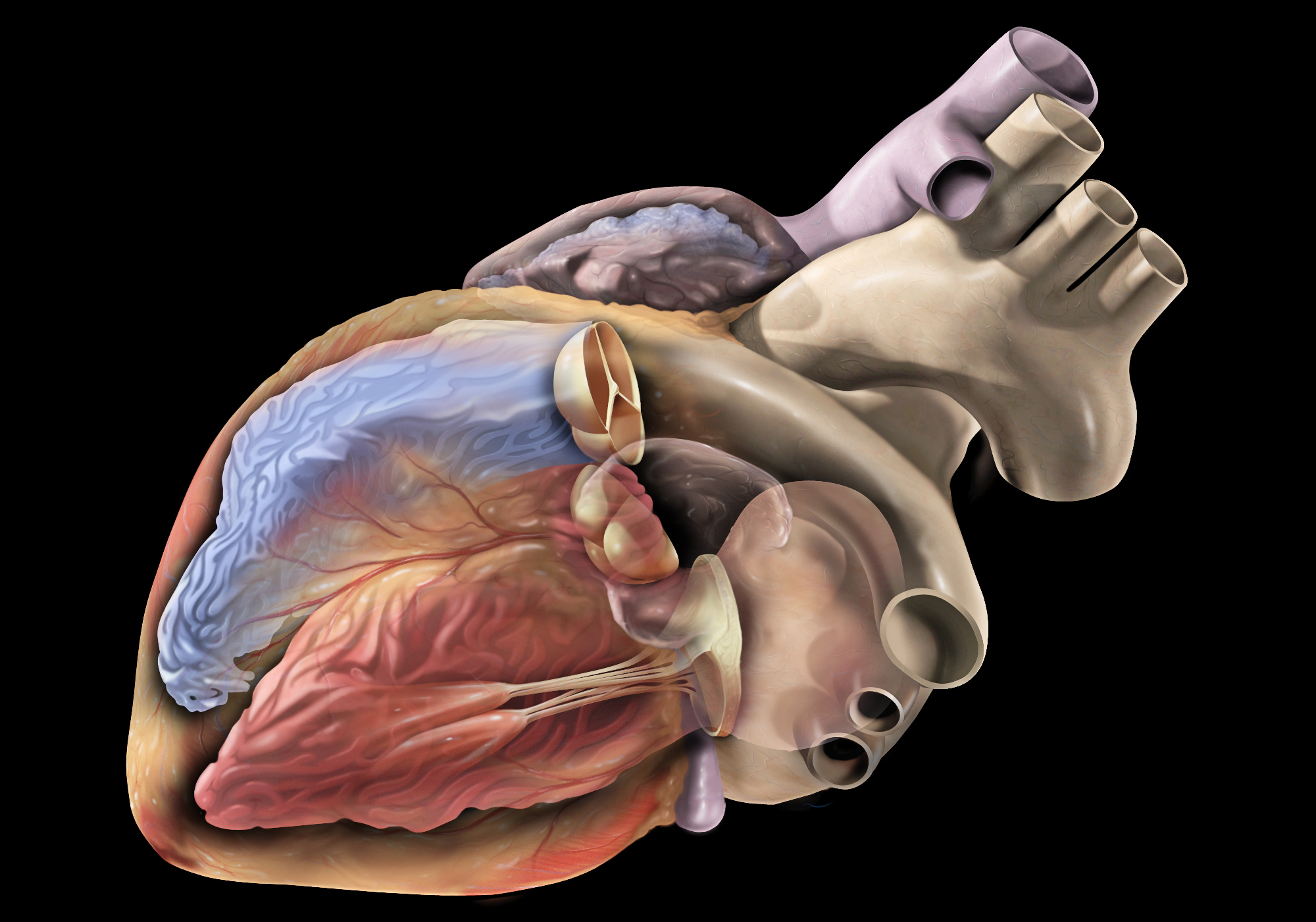
Coronary vessels refer to the network of blood vessels that supply oxygenated blood and nutrients to the heart muscle, also known as the myocardium. The two main coronary arteries are the left main coronary artery and the right coronary artery.
The left main coronary artery branches off into the left anterior descending artery (LAD) and the left circumflex artery (LCx). The LAD supplies blood to the front of the heart, while the LCx supplies blood to the side and back of the heart.
The right coronary artery supplies blood to the right lower part of the heart, including the right atrium and ventricle, as well as the back of the heart.
Coronary vessel disease (CVD) occurs when these vessels become narrowed or blocked due to the buildup of plaque, leading to reduced blood flow to the heart muscle. This can result in chest pain, shortness of breath, or a heart attack.
Chest pain is a discomfort or pain that you feel in the chest area. The pain can be sharp, dull, burning, crushing, heaviness, or tightness. It may be accompanied by other symptoms such as shortness of breath, sweating, nausea, dizziness, or pain that radiates to the arm, neck, jaw, or back.
Chest pain can have many possible causes, including heart-related conditions such as angina or a heart attack, lung conditions such as pneumonia or pleurisy, gastrointestinal problems such as acid reflux or gastritis, musculoskeletal issues such as costochondritis or muscle strain, and anxiety or panic attacks.
It is important to seek immediate medical attention if you experience chest pain that is severe, persistent, or accompanied by other concerning symptoms, as it may be a sign of a serious medical condition. A healthcare professional can evaluate your symptoms, perform tests, and provide appropriate treatment.
Coronary balloon angioplasty is a minimally invasive medical procedure used to widen narrowed or obstructed coronary arteries (the blood vessels that supply oxygen-rich blood to the heart muscle) and improve blood flow to the heart. This procedure is typically performed in conjunction with the insertion of a stent, a small mesh tube that helps keep the artery open.
During coronary balloon angioplasty, a thin, flexible catheter with a deflated balloon at its tip is inserted into a blood vessel, usually through a small incision in the groin or arm. The catheter is then guided to the narrowed or obstructed section of the coronary artery. Once in position, the balloon is inflated to compress the plaque against the artery wall and widen the lumen (the inner space) of the artery. This helps restore blood flow to the heart muscle.
The procedure is typically performed under local anesthesia and conscious sedation to minimize discomfort. Coronary balloon angioplasty is a relatively safe and effective treatment for many people with coronary artery disease, although complications such as bleeding, infection, or re-narrowing of the artery (restenosis) can occur in some cases.
Coronary artery bypass surgery, also known as coronary artery bypass grafting (CABG), is a surgical procedure used to improve blood flow to the heart in patients with severe coronary artery disease. This condition occurs when the coronary arteries, which supply oxygen-rich blood to the heart muscle, become narrowed or blocked due to the buildup of fatty deposits, called plaques.
During CABG surgery, a healthy blood vessel from another part of the body is grafted, or attached, to the coronary artery, creating a new pathway for oxygen-rich blood to flow around the blocked or narrowed portion of the artery and reach the heart muscle. This bypass helps to restore normal blood flow and reduce the risk of angina (chest pain), shortness of breath, and other symptoms associated with coronary artery disease.
There are different types of CABG surgery, including traditional on-pump CABG, off-pump CABG, and minimally invasive CABG. The choice of procedure depends on various factors, such as the patient's overall health, the number and location of blocked arteries, and the presence of other medical conditions.
It is important to note that while CABG surgery can significantly improve symptoms and quality of life in patients with severe coronary artery disease, it does not cure the underlying condition. Lifestyle modifications, such as regular exercise, a healthy diet, smoking cessation, and medication therapy, are essential for long-term management and prevention of further progression of the disease.
Myocardial revascularization is a medical term that refers to the restoration of blood flow to the heart muscle (myocardium), typically through a surgical or interventional procedure. This is often performed in patients with coronary artery disease, where the buildup of plaque in the coronary arteries restricts blood flow to the heart muscle, causing symptoms such as chest pain (angina) or shortness of breath, and increasing the risk of a heart attack (myocardial infarction).
There are two main types of myocardial revascularization:
1. Coronary artery bypass grafting (CABG): This is a surgical procedure in which a healthy blood vessel from another part of the body is used to create a detour around the blocked or narrowed coronary artery, allowing blood to flow more freely to the heart muscle.
2. Percutaneous coronary intervention (PCI), also known as angioplasty and stenting: This is a minimally invasive procedure in which a thin catheter is inserted into an artery in the groin or arm and threaded up to the blocked or narrowed coronary artery. A balloon is then inflated to widen the artery, and a stent may be placed to keep it open.
Both procedures aim to improve symptoms, reduce the risk of heart attack, and prolong survival in appropriately selected patients with coronary artery disease.
Follow-up studies are a type of longitudinal research that involve repeated observations or measurements of the same variables over a period of time, in order to understand their long-term effects or outcomes. In medical context, follow-up studies are often used to evaluate the safety and efficacy of medical treatments, interventions, or procedures.
In a typical follow-up study, a group of individuals (called a cohort) who have received a particular treatment or intervention are identified and then followed over time through periodic assessments or data collection. The data collected may include information on clinical outcomes, adverse events, changes in symptoms or functional status, and other relevant measures.
The results of follow-up studies can provide important insights into the long-term benefits and risks of medical interventions, as well as help to identify factors that may influence treatment effectiveness or patient outcomes. However, it is important to note that follow-up studies can be subject to various biases and limitations, such as loss to follow-up, recall bias, and changes in clinical practice over time, which must be carefully considered when interpreting the results.
Prospective studies, also known as longitudinal studies, are a type of cohort study in which data is collected forward in time, following a group of individuals who share a common characteristic or exposure over a period of time. The researchers clearly define the study population and exposure of interest at the beginning of the study and follow up with the participants to determine the outcomes that develop over time. This type of study design allows for the investigation of causal relationships between exposures and outcomes, as well as the identification of risk factors and the estimation of disease incidence rates. Prospective studies are particularly useful in epidemiology and medical research when studying diseases with long latency periods or rare outcomes.
Atherectomy, coronary, is a medical procedure used to treat narrowed or blocked coronary arteries due to the buildup of plaque (atherosclerosis). The goal of coronary atherectomy is to improve blood flow to the heart muscle by removing the obstructive material within the vessel.
During the procedure, a specialized catheter with a cutting device on its tip is inserted into a peripheral artery, usually in the groin or arm, and advanced to the affected coronary artery. The cutting device can be a rotating blade, a high-speed spinning burr, or a laser fiber that is used to shave, drill, or vaporize the plaque, respectively. The removed material is collected in a chamber within the catheter or washed away by blood flow.
There are different types of coronary atherectomy devices, including:
1. Directional atherectomy (DCA): A rotating blade cuts and removes the plaque in a targeted direction.
2. Rotational atherectomy (Rotablator): A high-speed spinning burr is used to abrade and pulverize the plaque into tiny particles that can be safely carried away by blood flow.
3. Laser atherectomy: A laser fiber is used to vaporize or break down the plaque into gaseous or small particle form.
Coronary atherectomy is typically performed in conjunction with angioplasty and stenting, as it helps prepare the narrowed artery for these procedures by creating a larger lumen and reducing the risk of complications like dissections or restenosis (re-narrowing). However, its use may be limited to specific cases due to the potential risks, such as vessel trauma, distal embolization, or perforation.
It is essential to consult with a medical professional for detailed information and personalized treatment recommendations regarding coronary atherectomy.
Treatment outcome is a term used to describe the result or effect of medical treatment on a patient's health status. It can be measured in various ways, such as through symptoms improvement, disease remission, reduced disability, improved quality of life, or survival rates. The treatment outcome helps healthcare providers evaluate the effectiveness of a particular treatment plan and make informed decisions about future care. It is also used in clinical research to compare the efficacy of different treatments and improve patient care.
Ambulatory electrocardiography, also known as ambulatory ECG or Holter monitoring, is a non-invasive method of recording the electrical activity of the heart over an extended period of time (typically 24 hours or more) while the patient goes about their daily activities. The device used to record the ECG is called a Holter monitor, which consists of a small, portable recorder that is attached to the patient's chest with electrodes.
The recorded data provides information on any abnormalities in the heart's rhythm or electrical activity during different stages of activity and rest, allowing healthcare providers to diagnose and evaluate various cardiac conditions such as arrhythmias, ischemia, and infarction. The ability to monitor the heart's activity over an extended period while the patient performs their normal activities provides valuable information that may not be captured during a standard ECG, which only records the heart's electrical activity for a few seconds.
In summary, ambulatory electrocardiography is a diagnostic tool used to evaluate the electrical activity of the heart over an extended period, allowing healthcare providers to diagnose and manage various cardiac conditions.
Medical Definition:
"Risk factors" are any attribute, characteristic or exposure of an individual that increases the likelihood of developing a disease or injury. They can be divided into modifiable and non-modifiable risk factors. Modifiable risk factors are those that can be changed through lifestyle choices or medical treatment, while non-modifiable risk factors are inherent traits such as age, gender, or genetic predisposition. Examples of modifiable risk factors include smoking, alcohol consumption, physical inactivity, and unhealthy diet, while non-modifiable risk factors include age, sex, and family history. It is important to note that having a risk factor does not guarantee that a person will develop the disease, but rather indicates an increased susceptibility.
Acute Coronary Syndrome (ACS) is a term used to describe a range of conditions associated with sudden, reduced blood flow to the heart muscle. This reduction in blood flow, commonly caused by blood clots forming in coronary arteries, can lead to damage or death of the heart muscle and is often characterized by symptoms such as chest pain, shortness of breath, and fatigue.
There are three main types of ACS:
1. Unstable Angina: This occurs when there is reduced blood flow to the heart muscle, causing chest pain or discomfort, but the heart muscle is not damaged. It can be a warning sign for a possible future heart attack.
2. Non-ST Segment Elevation Myocardial Infarction (NSTEMI): This type of heart attack occurs when there is reduced blood flow to the heart muscle, causing damage or death of some of the muscle cells. However, the electrical activity of the heart remains relatively normal.
3. ST Segment Elevation Myocardial Infarction (STEMI): This is a serious and life-threatening type of heart attack that occurs when there is a complete blockage in one or more of the coronary arteries, causing extensive damage to the heart muscle. The electrical activity of the heart is significantly altered, which can lead to dangerous heart rhythms and even cardiac arrest.
Immediate medical attention is required for anyone experiencing symptoms of ACS, as prompt treatment can help prevent further damage to the heart muscle and reduce the risk of complications or death. Treatment options may include medications, lifestyle changes, and procedures such as angioplasty or bypass surgery.
The double-blind method is a study design commonly used in research, including clinical trials, to minimize bias and ensure the objectivity of results. In this approach, both the participants and the researchers are unaware of which group the participants are assigned to, whether it be the experimental group or the control group. This means that neither the participants nor the researchers know who is receiving a particular treatment or placebo, thus reducing the potential for bias in the evaluation of outcomes. The assignment of participants to groups is typically done by a third party not involved in the study, and the codes are only revealed after all data have been collected and analyzed.
Recurrence, in a medical context, refers to the return of symptoms or signs of a disease after a period of improvement or remission. It indicates that the condition has not been fully eradicated and may require further treatment. Recurrence is often used to describe situations where a disease such as cancer comes back after initial treatment, but it can also apply to other medical conditions. The likelihood of recurrence varies depending on the type of disease and individual patient factors.
Coronary circulation refers to the circulation of blood in the coronary vessels, which supply oxygenated blood to the heart muscle (myocardium) and drain deoxygenated blood from it. The coronary circulation system includes two main coronary arteries - the left main coronary artery and the right coronary artery - that branch off from the aorta just above the aortic valve. These arteries further divide into smaller branches, which supply blood to different regions of the heart muscle.
The left main coronary artery divides into two branches: the left anterior descending (LAD) artery and the left circumflex (LCx) artery. The LAD supplies blood to the front and sides of the heart, while the LCx supplies blood to the back and sides of the heart. The right coronary artery supplies blood to the lower part of the heart, including the right ventricle and the bottom portion of the left ventricle.
The veins that drain the heart muscle include the great cardiac vein, the middle cardiac vein, and the small cardiac vein, which merge to form the coronary sinus. The coronary sinus empties into the right atrium, allowing deoxygenated blood to enter the right side of the heart and be pumped to the lungs for oxygenation.
Coronary circulation is essential for maintaining the health and function of the heart muscle, as it provides the necessary oxygen and nutrients required for proper contraction and relaxation of the myocardium. Any disruption or blockage in the coronary circulation system can lead to serious consequences, such as angina, heart attack, or even death.
Cardiovascular agents are a class of medications that are used to treat various conditions related to the cardiovascular system, which includes the heart and blood vessels. These agents can be further divided into several subcategories based on their specific mechanisms of action and therapeutic effects. Here are some examples:
1. Antiarrhythmics: These drugs are used to treat abnormal heart rhythms or arrhythmias. They work by stabilizing the electrical activity of the heart and preventing irregular impulses from spreading through the heart muscle.
2. Antihypertensives: These medications are used to lower high blood pressure, also known as hypertension. There are several classes of antihypertensive drugs, including diuretics, beta-blockers, calcium channel blockers, and angiotensin-converting enzyme (ACE) inhibitors.
3. Anticoagulants: These drugs are used to prevent blood clots from forming or growing larger. They work by interfering with the coagulation cascade, which is a series of chemical reactions that lead to the formation of a blood clot.
4. Antiplatelet agents: These medications are used to prevent platelets in the blood from sticking together and forming clots. They work by inhibiting the aggregation of platelets, which are small cells in the blood that help form clots.
5. Lipid-lowering agents: These drugs are used to lower cholesterol and other fats in the blood. They work by reducing the production or absorption of cholesterol in the body or increasing the removal of cholesterol from the bloodstream. Examples include statins, bile acid sequestrants, and PCSK9 inhibitors.
6. Vasodilators: These medications are used to widen blood vessels and improve blood flow. They work by relaxing the smooth muscle in the walls of blood vessels, causing them to dilate or widen. Examples include nitrates, calcium channel blockers, and ACE inhibitors.
7. Inotropes: These drugs are used to increase the force of heart contractions. They work by increasing the sensitivity of heart muscle cells to calcium ions, which are necessary for muscle contraction.
These are just a few examples of cardiovascular medications that are used to treat various conditions related to the heart and blood vessels. It is important to note that these medications can have side effects and should be taken under the guidance of a healthcare provider.
Prognosis is a medical term that refers to the prediction of the likely outcome or course of a disease, including the chances of recovery or recurrence, based on the patient's symptoms, medical history, physical examination, and diagnostic tests. It is an important aspect of clinical decision-making and patient communication, as it helps doctors and patients make informed decisions about treatment options, set realistic expectations, and plan for future care.
Prognosis can be expressed in various ways, such as percentages, categories (e.g., good, fair, poor), or survival rates, depending on the nature of the disease and the available evidence. However, it is important to note that prognosis is not an exact science and may vary depending on individual factors, such as age, overall health status, and response to treatment. Therefore, it should be used as a guide rather than a definitive forecast.
Vasodilator agents are pharmacological substances that cause the relaxation or widening of blood vessels by relaxing the smooth muscle in the vessel walls. This results in an increase in the diameter of the blood vessels, which decreases vascular resistance and ultimately reduces blood pressure. Vasodilators can be further classified based on their site of action:
1. Systemic vasodilators: These agents cause a generalized relaxation of the smooth muscle in the walls of both arteries and veins, resulting in a decrease in peripheral vascular resistance and preload (the volume of blood returning to the heart). Examples include nitroglycerin, hydralazine, and calcium channel blockers.
2. Arterial vasodilators: These agents primarily affect the smooth muscle in arterial vessel walls, leading to a reduction in afterload (the pressure against which the heart pumps blood). Examples include angiotensin-converting enzyme (ACE) inhibitors, angiotensin receptor blockers (ARBs), and direct vasodilators like sodium nitroprusside.
3. Venous vasodilators: These agents primarily affect the smooth muscle in venous vessel walls, increasing venous capacitance and reducing preload. Examples include nitroglycerin and other organic nitrates.
Vasodilator agents are used to treat various cardiovascular conditions such as hypertension, heart failure, angina, and pulmonary arterial hypertension. It is essential to monitor their use carefully, as excessive vasodilation can lead to orthostatic hypotension, reflex tachycardia, or fluid retention.
In the field of medicine, "time factors" refer to the duration of symptoms or time elapsed since the onset of a medical condition, which can have significant implications for diagnosis and treatment. Understanding time factors is crucial in determining the progression of a disease, evaluating the effectiveness of treatments, and making critical decisions regarding patient care.
For example, in stroke management, "time is brain," meaning that rapid intervention within a specific time frame (usually within 4.5 hours) is essential to administering tissue plasminogen activator (tPA), a clot-busting drug that can minimize brain damage and improve patient outcomes. Similarly, in trauma care, the "golden hour" concept emphasizes the importance of providing definitive care within the first 60 minutes after injury to increase survival rates and reduce morbidity.
Time factors also play a role in monitoring the progression of chronic conditions like diabetes or heart disease, where regular follow-ups and assessments help determine appropriate treatment adjustments and prevent complications. In infectious diseases, time factors are crucial for initiating antibiotic therapy and identifying potential outbreaks to control their spread.
Overall, "time factors" encompass the significance of recognizing and acting promptly in various medical scenarios to optimize patient outcomes and provide effective care.
Coronary thrombosis is a medical condition that refers to the formation of a blood clot (thrombus) inside a coronary artery, which supplies oxygenated blood to the heart muscle. The development of a thrombus can partially or completely obstruct blood flow, leading to insufficient oxygen supply to the heart muscle. This can cause chest pain (angina) or a heart attack (myocardial infarction), depending on the severity and duration of the blockage.
Coronary thrombosis often results from the rupture of an atherosclerotic plaque, a buildup of cholesterol, fat, calcium, and other substances in the inner lining (endothelium) of the coronary artery. The ruptured plaque exposes the underlying tissue to the bloodstream, triggering the coagulation cascade and resulting in the formation of a thrombus.
Immediate medical attention is crucial for managing coronary thrombosis, as timely treatment can help restore blood flow, prevent further damage to the heart muscle, and reduce the risk of complications such as heart failure or life-threatening arrhythmias. Treatment options may include medications, such as antiplatelet agents, anticoagulants, and thrombolytic drugs, or interventional procedures like angioplasty and stenting to open the blocked artery. In some cases, surgical intervention, such as coronary artery bypass grafting (CABG), may be necessary.
Adrenergic beta-antagonists, also known as beta blockers, are a class of medications that block the effects of adrenaline and noradrenaline (also known as epinephrine and norepinephrine) on beta-adrenergic receptors. These receptors are found in various tissues throughout the body, including the heart, lungs, and blood vessels.
Beta blockers work by binding to these receptors and preventing the activation of certain signaling pathways that lead to increased heart rate, force of heart contractions, and relaxation of blood vessels. As a result, beta blockers can lower blood pressure, reduce heart rate, and decrease the workload on the heart.
Beta blockers are used to treat a variety of medical conditions, including hypertension (high blood pressure), angina (chest pain), heart failure, irregular heart rhythms, migraines, and certain anxiety disorders. Some common examples of beta blockers include metoprolol, atenolol, propranolol, and bisoprolol.
It is important to note that while beta blockers can have many benefits, they can also cause side effects such as fatigue, dizziness, and shortness of breath. Additionally, sudden discontinuation of beta blocker therapy can lead to rebound hypertension or worsening chest pain. Therefore, it is important to follow the dosing instructions provided by a healthcare provider carefully when taking these medications.
Nicorandil is a medication that belongs to a class of drugs known as potassium channel activators. It works by relaxing and widening blood vessels, which improves blood flow and reduces the workload on the heart. Nicorandil is primarily used to treat chronic stable angina, a type of chest pain caused by reduced blood flow to the heart muscle.
The medical definition of Nicorandil can be described as:
A synthetic derivative of nicotinamide with vasodilatory properties, acting as an opener of ATP-sensitive potassium channels in vascular smooth muscle and cardiomyocytes. It is used in the management of chronic stable angina, providing both antianginal and antiischemic effects through a dual mechanism that includes coronary and peripheral vasodilation. By reducing afterload and preload, Nicorandil decreases myocardial oxygen demand while increasing supply, leading to improved exercise tolerance and reduced frequency of anginal episodes.
Platelet aggregation inhibitors are a class of medications that prevent platelets (small blood cells involved in clotting) from sticking together and forming a clot. These drugs work by interfering with the ability of platelets to adhere to each other and to the damaged vessel wall, thereby reducing the risk of thrombosis (blood clot formation).
Platelet aggregation inhibitors are often prescribed for people who have an increased risk of developing blood clots due to various medical conditions such as atrial fibrillation, coronary artery disease, peripheral artery disease, stroke, or a history of heart attack. They may also be used in patients undergoing certain medical procedures, such as angioplasty and stenting, to prevent blood clot formation in the stents.
Examples of platelet aggregation inhibitors include:
1. Aspirin: A nonsteroidal anti-inflammatory drug (NSAID) that irreversibly inhibits the enzyme cyclooxygenase, which is involved in platelet activation and aggregation.
2. Clopidogrel (Plavix): A P2Y12 receptor antagonist that selectively blocks ADP-induced platelet activation and aggregation.
3. Prasugrel (Effient): A third-generation thienopyridine P2Y12 receptor antagonist, similar to clopidogrel but with faster onset and greater potency.
4. Ticagrelor (Brilinta): A direct-acting P2Y12 receptor antagonist that does not require metabolic activation and has a reversible binding profile.
5. Dipyridamole (Persantine): An antiplatelet agent that inhibits platelet aggregation by increasing cyclic adenosine monophosphate (cAMP) levels in platelets, which leads to decreased platelet reactivity.
6. Iloprost (Ventavis): A prostacyclin analogue that inhibits platelet aggregation and causes vasodilation, often used in the treatment of pulmonary arterial hypertension.
7. Cilostazol (Pletal): A phosphodiesterase III inhibitor that increases cAMP levels in platelets, leading to decreased platelet activation and aggregation, as well as vasodilation.
8. Ticlopidine (Ticlid): An older P2Y12 receptor antagonist with a slower onset of action and more frequent side effects compared to clopidogrel or prasugrel.
A biological marker, often referred to as a biomarker, is a measurable indicator that reflects the presence or severity of a disease state, or a response to a therapeutic intervention. Biomarkers can be found in various materials such as blood, tissues, or bodily fluids, and they can take many forms, including molecular, histologic, radiographic, or physiological measurements.
In the context of medical research and clinical practice, biomarkers are used for a variety of purposes, such as:
1. Diagnosis: Biomarkers can help diagnose a disease by indicating the presence or absence of a particular condition. For example, prostate-specific antigen (PSA) is a biomarker used to detect prostate cancer.
2. Monitoring: Biomarkers can be used to monitor the progression or regression of a disease over time. For instance, hemoglobin A1c (HbA1c) levels are monitored in diabetes patients to assess long-term blood glucose control.
3. Predicting: Biomarkers can help predict the likelihood of developing a particular disease or the risk of a negative outcome. For example, the presence of certain genetic mutations can indicate an increased risk for breast cancer.
4. Response to treatment: Biomarkers can be used to evaluate the effectiveness of a specific treatment by measuring changes in the biomarker levels before and after the intervention. This is particularly useful in personalized medicine, where treatments are tailored to individual patients based on their unique biomarker profiles.
It's important to note that for a biomarker to be considered clinically valid and useful, it must undergo rigorous validation through well-designed studies, including demonstrating sensitivity, specificity, reproducibility, and clinical relevance.
Coronary stenosis is a medical condition that refers to the narrowing of the coronary arteries, which supply oxygen-rich blood to the heart muscle. This narrowing is typically caused by the buildup of plaque, made up of fat, cholesterol, and other substances, on the inner walls of the arteries. Over time, as the plaque hardens and calcifies, it can cause the artery to become narrowed or blocked, reducing blood flow to the heart muscle.
Coronary stenosis can lead to various symptoms and complications, including chest pain (angina), shortness of breath, irregular heart rhythms (arrhythmias), and heart attacks. Treatment options for coronary stenosis may include lifestyle changes, medications, medical procedures such as angioplasty or bypass surgery, or a combination of these approaches. Regular check-ups and diagnostic tests, such as stress testing or coronary angiography, can help detect and monitor coronary stenosis over time.
C-reactive protein (CRP) is a protein produced by the liver in response to inflammation or infection in the body. It is named after its ability to bind to the C-polysaccharide of pneumococcus, a type of bacteria. CRP levels can be measured with a simple blood test and are often used as a marker of inflammation or infection. Elevated CRP levels may indicate a variety of conditions, including infections, tissue damage, and chronic diseases such as rheumatoid arthritis and cancer. However, it is important to note that CRP is not specific to any particular condition, so additional tests are usually needed to make a definitive diagnosis.
Troponin T is a subunit of the troponin complex, which is a protein complex that plays a crucial role in muscle contraction. In particular, Troponin T is responsible for binding the troponin complex to tropomyosin, another protein that helps regulate muscle contraction.
In the context of medical diagnostics, Troponin T is often measured as a biomarker for heart damage. When heart muscle cells are damaged or die, such as in a myocardial infarction (heart attack), troponin T is released into the bloodstream. Therefore, measuring the levels of Troponin T in the blood can help diagnose and assess the severity of heart damage.
It's important to note that Troponin T is specific to cardiac muscle cells, which makes it a more reliable biomarker for heart damage than other markers that may also be found in skeletal muscle cells. However, it's worth noting that Troponin T levels can also be elevated in conditions other than heart attacks, such as heart failure, myocarditis, and pulmonary embolism, so clinical context is important when interpreting test results.
Cardiac catheterization is a medical procedure used to diagnose and treat cardiovascular conditions. In this procedure, a thin, flexible tube called a catheter is inserted into a blood vessel in the arm or leg and threaded up to the heart. The catheter can be used to perform various diagnostic tests, such as measuring the pressure inside the heart chambers and assessing the function of the heart valves.
Cardiac catheterization can also be used to treat certain cardiovascular conditions, such as narrowed or blocked arteries. In these cases, a balloon or stent may be inserted through the catheter to open up the blood vessel and improve blood flow. This procedure is known as angioplasty or percutaneous coronary intervention (PCI).
Cardiac catheterization is typically performed in a hospital cardiac catheterization laboratory by a team of healthcare professionals, including cardiologists, radiologists, and nurses. The procedure may be done under local anesthesia with sedation or general anesthesia, depending on the individual patient's needs and preferences.
Overall, cardiac catheterization is a valuable tool in the diagnosis and treatment of various heart conditions, and it can help improve symptoms, reduce complications, and prolong life for many patients.
The Predictive Value of Tests, specifically the Positive Predictive Value (PPV) and Negative Predictive Value (NPV), are measures used in diagnostic tests to determine the probability that a positive or negative test result is correct.
Positive Predictive Value (PPV) is the proportion of patients with a positive test result who actually have the disease. It is calculated as the number of true positives divided by the total number of positive results (true positives + false positives). A higher PPV indicates that a positive test result is more likely to be a true positive, and therefore the disease is more likely to be present.
Negative Predictive Value (NPV) is the proportion of patients with a negative test result who do not have the disease. It is calculated as the number of true negatives divided by the total number of negative results (true negatives + false negatives). A higher NPV indicates that a negative test result is more likely to be a true negative, and therefore the disease is less likely to be present.
The predictive value of tests depends on the prevalence of the disease in the population being tested, as well as the sensitivity and specificity of the test. A test with high sensitivity and specificity will generally have higher predictive values than a test with low sensitivity and specificity. However, even a highly sensitive and specific test can have low predictive values if the prevalence of the disease is low in the population being tested.
A syndrome, in medical terms, is a set of symptoms that collectively indicate or characterize a disease, disorder, or underlying pathological process. It's essentially a collection of signs and/or symptoms that frequently occur together and can suggest a particular cause or condition, even though the exact physiological mechanisms might not be fully understood.
For example, Down syndrome is characterized by specific physical features, cognitive delays, and other developmental issues resulting from an extra copy of chromosome 21. Similarly, metabolic syndromes like diabetes mellitus type 2 involve a group of risk factors such as obesity, high blood pressure, high blood sugar, and abnormal cholesterol or triglyceride levels that collectively increase the risk of heart disease, stroke, and diabetes.
It's important to note that a syndrome is not a specific diagnosis; rather, it's a pattern of symptoms that can help guide further diagnostic evaluation and management.
Nifedipine is an antihypertensive and calcium channel blocker medication. It works by relaxing the muscles of the blood vessels, which helps to lower blood pressure and improve the supply of oxygen and nutrients to the heart. Nifedipine is used to treat high blood pressure (hypertension), angina (chest pain), and certain types of heart rhythm disorders.
In medical terms, nifedipine can be defined as: "A dihydropyridine calcium channel blocker that is used in the treatment of hypertension, angina pectoris, and Raynaud's phenomenon. It works by inhibiting the influx of calcium ions into vascular smooth muscle and cardiac muscle, which results in relaxation of the vascular smooth muscle and decreased workload on the heart."
Calcium channel blockers (CCBs) are a class of medications that work by inhibiting the influx of calcium ions into cardiac and smooth muscle cells. This action leads to relaxation of the muscles, particularly in the blood vessels, resulting in decreased peripheral resistance and reduced blood pressure. Calcium channel blockers also have anti-arrhythmic effects and are used in the management of various cardiovascular conditions such as hypertension, angina, and certain types of arrhythmias.
Calcium channel blockers can be further classified into two main categories based on their chemical structure: dihydropyridines (e.g., nifedipine, amlodipine) and non-dihydropyridines (e.g., verapamil, diltiazem). Dihydropyridines are more selective for vascular smooth muscle and have a greater effect on blood pressure than heart rate or conduction. Non-dihydropyridines have a more significant impact on cardiac conduction and contractility, in addition to their vasodilatory effects.
It is important to note that calcium channel blockers may interact with other medications and should be used under the guidance of a healthcare professional. Potential side effects include dizziness, headache, constipation, and peripheral edema.
A stent is a small mesh tube that's used to treat narrow or weak arteries. Arteries are blood vessels that carry blood away from your heart to other parts of your body. A stent is placed in an artery as part of a procedure called angioplasty. Angioplasty restores blood flow through narrowed or blocked arteries by inflating a tiny balloon inside the blocked artery to widen it.
The stent is then inserted into the widened artery to keep it open. The stent is usually made of metal, but some are coated with medication that is slowly and continuously released to help prevent the formation of scar tissue in the artery. This can reduce the chance of the artery narrowing again.
Stents are also used in other parts of the body, such as the neck (carotid artery) and kidneys (renal artery), to help maintain blood flow and prevent blockages. They can also be used in the urinary system to treat conditions like ureteropelvic junction obstruction or narrowing of the urethra.
Aspirin is the common name for acetylsalicylic acid, which is a medication used to relieve pain, reduce inflammation, and lower fever. It works by inhibiting the activity of an enzyme called cyclooxygenase (COX), which is involved in the production of prostaglandins, hormone-like substances that cause inflammation and pain. Aspirin also has an antiplatelet effect, which means it can help prevent blood clots from forming. This makes it useful for preventing heart attacks and strokes.
Aspirin is available over-the-counter in various forms, including tablets, capsules, and chewable tablets. It is also available in prescription strengths for certain medical conditions. As with any medication, aspirin should be taken as directed by a healthcare provider, and its use should be avoided in children and teenagers with viral infections due to the risk of Reye's syndrome, a rare but serious condition that can affect the liver and brain.
Physical exertion is defined as the act of applying energy to physically demandable activities or tasks, which results in various body systems working together to produce movement and maintain homeostasis. It often leads to an increase in heart rate, respiratory rate, and body temperature, among other physiological responses. The level of physical exertion can vary based on the intensity, duration, and frequency of the activity.
It's important to note that engaging in regular physical exertion has numerous health benefits, such as improving cardiovascular fitness, strengthening muscles and bones, reducing stress, and preventing chronic diseases like obesity, diabetes, and heart disease. However, it is also crucial to balance physical exertion with adequate rest and recovery time to avoid overtraining or injury.
I'm sorry for any confusion, but "Oxyfedrine" does not appear to be a valid or recognized medication or substance in modern medicine. It's possible that there may be a spelling error or it could be an outdated or obsolete term. Please provide more context or check the spelling so I can give you a more accurate and helpful response.
Heart rate is the number of heartbeats per unit of time, often expressed as beats per minute (bpm). It can vary significantly depending on factors such as age, physical fitness, emotions, and overall health status. A resting heart rate between 60-100 bpm is generally considered normal for adults, but athletes and individuals with high levels of physical fitness may have a resting heart rate below 60 bpm due to their enhanced cardiovascular efficiency. Monitoring heart rate can provide valuable insights into an individual's health status, exercise intensity, and response to various treatments or interventions.
A chronic disease is a long-term medical condition that often progresses slowly over a period of years and requires ongoing management and care. These diseases are typically not fully curable, but symptoms can be managed to improve quality of life. Common chronic diseases include heart disease, stroke, cancer, diabetes, arthritis, and COPD (chronic obstructive pulmonary disease). They are often associated with advanced age, although they can also affect children and younger adults. Chronic diseases can have significant impacts on individuals' physical, emotional, and social well-being, as well as on healthcare systems and society at large.
A Severity of Illness Index is a measurement tool used in healthcare to assess the severity of a patient's condition and the risk of mortality or other adverse outcomes. These indices typically take into account various physiological and clinical variables, such as vital signs, laboratory values, and co-morbidities, to generate a score that reflects the patient's overall illness severity.
Examples of Severity of Illness Indices include the Acute Physiology and Chronic Health Evaluation (APACHE) system, the Simplified Acute Physiology Score (SAPS), and the Mortality Probability Model (MPM). These indices are often used in critical care settings to guide clinical decision-making, inform prognosis, and compare outcomes across different patient populations.
It is important to note that while these indices can provide valuable information about a patient's condition, they should not be used as the sole basis for clinical decision-making. Rather, they should be considered in conjunction with other factors, such as the patient's overall clinical presentation, treatment preferences, and goals of care.
Fibrinopeptide A is a small protein molecule that is cleaved and released from the larger fibrinogen protein during the blood clotting process. Specifically, it is removed by the enzyme thrombin as part of the conversion of fibrinogen to fibrin, which is the main structural component of a blood clot. The measurement of Fibrinopeptide A in the blood can be used as a marker for ongoing thrombin activation and fibrin formation, which are key events in coagulation and hemostasis. Increased levels of Fibrinopeptide A may indicate abnormal or excessive blood clotting, such as in disseminated intravascular coagulation (DIC) or deep vein thrombosis (DVT).
Hirudin therapy, also known as leech therapy, is a type of treatment that uses the saliva of medicinal leeches (Hirudo medicinalis) to alleviate symptoms and promote healing. The saliva of these leeches contains various bioactive compounds, including hirudin, which is a potent anticoagulant that prevents blood clotting.
In hirudin therapy, leeches are applied to specific areas of the body, usually on congested tissues or sites of stasis, where they feed on the patient's blood and release their saliva into the bite site. The hirudin in the saliva helps to dissolve blood clots, improve circulation, reduce swelling, and relieve pain.
Hirudin therapy is used in various medical conditions, such as arterial and venous insufficiency, skin ulcers, joint diseases, and post-surgical recovery, particularly after reconstructive surgery or organ transplantation. It can also be used to treat thrombophlebitis, varicose veins, and other circulatory disorders.
It is essential to note that hirudin therapy should only be performed by trained medical professionals in a controlled environment due to the potential risks associated with infection transmission and bleeding complications.
Exercise tolerance is a term used to describe the ability of an individual to perform physical activity or exercise without experiencing symptoms such as shortness of breath, chest pain, or undue fatigue. It is often used as a measure of cardiovascular fitness and can be assessed through various tests, such as a stress test or a six-minute walk test. Exercise intolerance may indicate the presence of underlying medical conditions, such as heart disease, lung disease, or deconditioning.
Angiocardiography is a medical procedure used to examine the heart and blood vessels, particularly the chambers of the heart and the valves between them. It involves injecting a contrast agent into the bloodstream and taking X-ray images as the agent flows through the heart. This allows doctors to visualize any abnormalities such as blockages, narrowing, or leakage in the heart valves or blood vessels.
There are different types of angiocardiography, including:
* Left heart catheterization (LHC): A thin tube called a catheter is inserted into a vein in the arm or groin and threaded through to the left side of the heart to measure pressure and oxygen levels.
* Right heart catheterization (RHC): Similar to LHC, but the catheter is threaded through to the right side of the heart to measure pressure and oxygen levels there.
* Selective angiocardiography: A catheter is used to inject the contrast agent into specific blood vessels or chambers of the heart to get a more detailed view.
Angiocardiography can help diagnose and evaluate various heart conditions, including congenital heart defects, coronary artery disease, cardiomyopathy, and valvular heart disease. It is an invasive procedure that carries some risks, such as bleeding, infection, and damage to blood vessels or heart tissue. However, it can provide valuable information for diagnosing and treating heart conditions.
An acute disease is a medical condition that has a rapid onset, develops quickly, and tends to be short in duration. Acute diseases can range from minor illnesses such as a common cold or flu, to more severe conditions such as pneumonia, meningitis, or a heart attack. These types of diseases often have clear symptoms that are easy to identify, and they may require immediate medical attention or treatment.
Acute diseases are typically caused by an external agent or factor, such as a bacterial or viral infection, a toxin, or an injury. They can also be the result of a sudden worsening of an existing chronic condition. In general, acute diseases are distinct from chronic diseases, which are long-term medical conditions that develop slowly over time and may require ongoing management and treatment.
Examples of acute diseases include:
* Acute bronchitis: a sudden inflammation of the airways in the lungs, often caused by a viral infection.
* Appendicitis: an inflammation of the appendix that can cause severe pain and requires surgical removal.
* Gastroenteritis: an inflammation of the stomach and intestines, often caused by a viral or bacterial infection.
* Migraine headaches: intense headaches that can last for hours or days, and are often accompanied by nausea, vomiting, and sensitivity to light and sound.
* Myocardial infarction (heart attack): a sudden blockage of blood flow to the heart muscle, often caused by a buildup of plaque in the coronary arteries.
* Pneumonia: an infection of the lungs that can cause coughing, chest pain, and difficulty breathing.
* Sinusitis: an inflammation of the sinuses, often caused by a viral or bacterial infection.
It's important to note that while some acute diseases may resolve on their own with rest and supportive care, others may require medical intervention or treatment to prevent complications and promote recovery. If you are experiencing symptoms of an acute disease, it is always best to seek medical attention to ensure proper diagnosis and treatment.
Interventional ultrasonography is a medical procedure that involves the use of real-time ultrasound imaging to guide minimally invasive diagnostic and therapeutic interventions. This technique combines the advantages of ultrasound, such as its non-ionizing nature (no radiation exposure), relatively low cost, and portability, with the ability to perform precise and targeted procedures.
In interventional ultrasonography, a specialized physician called an interventional radiologist or an interventional sonographer uses high-frequency sound waves to create detailed images of internal organs and tissues. These images help guide the placement of needles, catheters, or other instruments used during the procedure. Common interventions include biopsies (tissue sampling), fluid drainage, tumor ablation, and targeted drug delivery.
The real-time visualization provided by ultrasonography allows for increased accuracy and safety during these procedures, minimizing complications and reducing recovery time compared to traditional surgical approaches. Additionally, interventional ultrasonography can be performed on an outpatient basis, further contributing to its appeal as a less invasive alternative in many clinical scenarios.
Collateral circulation refers to the alternate blood supply routes that bypass an obstructed or narrowed vessel and reconnect with the main vascular system. These collateral vessels can develop over time as a result of the body's natural adaptation to chronic ischemia (reduced blood flow) caused by various conditions such as atherosclerosis, thromboembolism, or vasculitis.
The development of collateral circulation helps maintain adequate blood flow and oxygenation to affected tissues, minimizing the risk of tissue damage and necrosis. In some cases, well-developed collateral circulations can help compensate for significant blockages in major vessels, reducing symptoms and potentially preventing the need for invasive interventions like revascularization procedures. However, the extent and effectiveness of collateral circulation vary from person to person and depend on factors such as age, overall health status, and the presence of comorbidities.
Creatine kinase (CK) is a muscle enzyme that is normally present in small amounts in the blood. It is primarily found in tissues that require a lot of energy, such as the heart, brain, and skeletal muscles. When these tissues are damaged or injured, CK is released into the bloodstream, causing the levels to rise.
Creatine kinase exists in several forms, known as isoenzymes, which can be measured in the blood to help identify the location of tissue damage. The three main isoenzymes are:
1. CK-MM: Found primarily in skeletal muscle
2. CK-MB: Found primarily in heart muscle
3. CK-BB: Found primarily in the brain
Elevated levels of creatine kinase, particularly CK-MB, can indicate damage to the heart muscle, such as occurs with a heart attack. Similarly, elevated levels of CK-BB may suggest brain injury or disease. Overall, measuring creatine kinase levels is a useful diagnostic tool for assessing tissue damage and determining the severity of injuries or illnesses.
Risk assessment in the medical context refers to the process of identifying, evaluating, and prioritizing risks to patients, healthcare workers, or the community related to healthcare delivery. It involves determining the likelihood and potential impact of adverse events or hazards, such as infectious diseases, medication errors, or medical devices failures, and implementing measures to mitigate or manage those risks. The goal of risk assessment is to promote safe and high-quality care by identifying areas for improvement and taking action to minimize harm.
Atenolol is a beta-blocker medication that is primarily used to treat hypertension (high blood pressure), angina (chest pain), and certain types of heart rhythm disorders. It works by blocking the action of certain hormones in the body, such as adrenaline, on the heart and blood vessels. This helps to reduce the heart's workload, lower its rate and force of contractions, and improve blood flow.
Beta-blockers like atenolol are also sometimes used to prevent migraines or to treat symptoms of anxiety, such as rapid heartbeat or tremors. Atenolol is available in immediate-release and extended-release forms, and it is typically taken orally once or twice a day. As with any medication, atenolol can have side effects, including dizziness, fatigue, and gastrointestinal symptoms, and it may interact with other medications or medical conditions. It is important to use atenolol only under the supervision of a healthcare provider.
Laser therapy, also known as phototherapy or laser photobiomodulation, is a medical treatment that uses low-intensity lasers or light-emitting diodes (LEDs) to stimulate healing, reduce pain, and decrease inflammation. It works by promoting the increase of cellular metabolism, blood flow, and tissue regeneration through the process of photobiomodulation.
The therapy can be used on patients suffering from a variety of acute and chronic conditions, including musculoskeletal injuries, arthritis, neuropathic pain, and wound healing complications. The wavelength and intensity of the laser light are precisely controlled to ensure a safe and effective treatment.
During the procedure, the laser or LED device is placed directly on the skin over the area of injury or discomfort. The non-ionizing light penetrates the tissue without causing heat or damage, interacting with chromophores in the cells to initiate a series of photochemical reactions. This results in increased ATP production, modulation of reactive oxygen species, and activation of transcription factors that lead to improved cellular function and reduced pain.
In summary, laser therapy is a non-invasive, drug-free treatment option for various medical conditions, providing patients with an alternative or complementary approach to traditional therapies.
Trimetazidine is a medication that belongs to the class of drugs known as anti-ischemic agents. It is primarily used in the management of angina pectoris, a type of chest pain caused by reduced blood flow to the heart muscle.
Trimetazidine works by improving the efficiency of heart muscle cells in utilizing energy and reducing the production of harmful oxygen free radicals during periods of reduced oxygen supply, such as during angina attacks. This helps to preserve the function of the heart muscle and reduce the severity and frequency of angina episodes.
It is important to note that Trimetazidine should only be used under the supervision of a healthcare provider, and it is not recommended for individuals with severe liver or kidney impairment.
Dipyridamole is a medication that belongs to a class of drugs called antiplatelet agents. It works by preventing platelets in your blood from sticking together to form clots. Dipyridamole is often used in combination with aspirin to prevent stroke and other complications in people who have had a heart valve replacement or a type of irregular heartbeat called atrial fibrillation.
Dipyridamole can also be used as a stress agent in myocardial perfusion imaging studies, which are tests used to evaluate blood flow to the heart. When used for this purpose, dipyridamole is given intravenously and works by dilating the blood vessels in the heart, allowing more blood to flow through them and making it easier to detect areas of reduced blood flow.
The most common side effects of dipyridamole include headache, dizziness, and gastrointestinal symptoms such as diarrhea, nausea, and vomiting. In rare cases, dipyridamole can cause more serious side effects, such as allergic reactions, abnormal heart rhythms, or low blood pressure. It is important to take dipyridamole exactly as directed by your healthcare provider and to report any unusual symptoms or side effects promptly.
Retrospective studies, also known as retrospective research or looking back studies, are a type of observational study that examines data from the past to draw conclusions about possible causal relationships between risk factors and outcomes. In these studies, researchers analyze existing records, medical charts, or previously collected data to test a hypothesis or answer a specific research question.
Retrospective studies can be useful for generating hypotheses and identifying trends, but they have limitations compared to prospective studies, which follow participants forward in time from exposure to outcome. Retrospective studies are subject to biases such as recall bias, selection bias, and information bias, which can affect the validity of the results. Therefore, retrospective studies should be interpreted with caution and used primarily to generate hypotheses for further testing in prospective studies.
Angioscopy is a medical diagnostic procedure that uses a small fiber-optic scope, called an angioscope, to directly visualize the interior of blood vessels. The angioscope is inserted into the vessel through a small incision or catheter and allows physicians to examine the vessel walls for abnormalities such as plaque buildup, inflammation, or damage. This procedure can be used to diagnose and monitor conditions such as coronary artery disease, peripheral artery disease, and vasculitis. It can also be used during surgical procedures to assist with the placement of stents or other devices in the blood vessels.
Iodobenzenes are organic compounds that contain a iodine atom (I) attached to a benzene ring. The general formula for iodobenzenes is C6H5I. They can be considered as aryl halides and can undergo various chemical reactions such as nucleophilic substitution, electrophilic aromatic substitution, and reduction. Iodobenzenes are less reactive than other aryl halides due to the larger size and lower electronegativity of iodine compared to other halogens. They are used in organic synthesis as building blocks or reagents for various chemical transformations.
Combination drug therapy is a treatment approach that involves the use of multiple medications with different mechanisms of action to achieve better therapeutic outcomes. This approach is often used in the management of complex medical conditions such as cancer, HIV/AIDS, and cardiovascular diseases. The goal of combination drug therapy is to improve efficacy, reduce the risk of drug resistance, decrease the likelihood of adverse effects, and enhance the overall quality of life for patients.
In combining drugs, healthcare providers aim to target various pathways involved in the disease process, which may help to:
1. Increase the effectiveness of treatment by attacking the disease from multiple angles.
2. Decrease the dosage of individual medications, reducing the risk and severity of side effects.
3. Slow down or prevent the development of drug resistance, a common problem in chronic diseases like HIV/AIDS and cancer.
4. Improve patient compliance by simplifying dosing schedules and reducing pill burden.
Examples of combination drug therapy include:
1. Antiretroviral therapy (ART) for HIV treatment, which typically involves three or more drugs from different classes to suppress viral replication and prevent the development of drug resistance.
2. Chemotherapy regimens for cancer treatment, where multiple cytotoxic agents are used to target various stages of the cell cycle and reduce the likelihood of tumor cells developing resistance.
3. Cardiovascular disease management, which may involve combining medications such as angiotensin-converting enzyme (ACE) inhibitors, beta-blockers, diuretics, and statins to control blood pressure, heart rate, fluid balance, and cholesterol levels.
4. Treatment of tuberculosis, which often involves a combination of several antibiotics to target different aspects of the bacterial life cycle and prevent the development of drug-resistant strains.
When prescribing combination drug therapy, healthcare providers must carefully consider factors such as potential drug interactions, dosing schedules, adverse effects, and contraindications to ensure safe and effective treatment. Regular monitoring of patients is essential to assess treatment response, manage side effects, and adjust the treatment plan as needed.
Metoprolol is a type of medication known as a beta blocker. According to the US National Library of Medicine's MedlinePlus, metoprolol is used to treat high blood pressure, angina (chest pain), and heart conditions that may occur after a heart attack. It works by blocking the action of certain natural chemicals in your body, such as epinephrine, on the heart and blood vessels. This helps to reduce the heart's workload, lower its blood pressure, and regulate its rhythm.
Metoprolol is available under various brand names, including Lopressor and Toprol-XL. It can be taken orally as a tablet or an extended-release capsule. As with any medication, metoprolol should be used under the supervision of a healthcare provider, who can monitor its effectiveness and potential side effects.
It is important to note that this definition is intended to provide a general overview of the medical use of metoprolol and should not be considered a substitute for professional medical advice.
In medical terms, the heart is a muscular organ located in the thoracic cavity that functions as a pump to circulate blood throughout the body. It's responsible for delivering oxygen and nutrients to the tissues and removing carbon dioxide and other wastes. The human heart is divided into four chambers: two atria on the top and two ventricles on the bottom. The right side of the heart receives deoxygenated blood from the body and pumps it to the lungs, while the left side receives oxygenated blood from the lungs and pumps it out to the rest of the body. The heart's rhythmic contractions and relaxations are regulated by a complex electrical conduction system.
Cineangiography is a medical imaging technique used to visualize the blood flow in the heart and cardiovascular system. It involves the injection of a contrast agent into the bloodstream while X-ray images are taken in quick succession, creating a movie-like sequence that shows the movement of the contrast through the blood vessels and chambers of the heart. This technique is often used to diagnose and evaluate various heart conditions, such as coronary artery disease, valvular heart disease, and congenital heart defects.
The procedure typically involves threading a catheter through a blood vessel in the arm or leg and guiding it to the heart. Once in place, the contrast agent is injected, and X-ray images are taken using a specialized X-ray machine called a fluoroscope. The images captured during cineangiography can help doctors identify areas of narrowing or blockage in the coronary arteries, abnormalities in heart valves, and other cardiovascular problems.
Cineangiography is an invasive procedure that carries some risks, such as bleeding, infection, and reactions to the contrast agent. However, it can provide valuable information for diagnosing and treating heart conditions, and may be recommended when other diagnostic tests have been inconclusive.
Diltiazem is a calcium channel blocker medication that is used to treat hypertension (high blood pressure), angina (chest pain), and certain heart rhythm disorders. It works by relaxing the muscles of the blood vessels, which lowers blood pressure and improves blood flow to the heart. Diltiazem may also be used to reduce the risk of heart attack in patients with coronary artery disease.
The medication is available in various forms, including immediate-release tablets, extended-release tablets, and extended-release capsules. It is usually taken orally, one to three times a day, depending on the formulation and the individual patient's needs. Diltiazem may cause side effects such as dizziness, headache, nausea, and constipation.
It is important to follow the dosage instructions provided by your healthcare provider and to inform them of any other medications you are taking, as well as any medical conditions you have, before starting diltiazem.
Blood pressure is the force exerted by circulating blood on the walls of the blood vessels. It is measured in millimeters of mercury (mmHg) and is given as two figures:
1. Systolic pressure: This is the pressure when the heart pushes blood out into the arteries.
2. Diastolic pressure: This is the pressure when the heart rests between beats, allowing it to fill with blood.
Normal blood pressure for adults is typically around 120/80 mmHg, although this can vary slightly depending on age, sex, and other factors. High blood pressure (hypertension) is generally considered to be a reading of 130/80 mmHg or higher, while low blood pressure (hypotension) is usually defined as a reading below 90/60 mmHg. It's important to note that blood pressure can fluctuate throughout the day and may be affected by factors such as stress, physical activity, and medication use.
Emission-Computed Tomography, Single-Photon (SPECT) is a type of nuclear medicine imaging procedure that generates detailed, three-dimensional images of the distribution of radioactive pharmaceuticals within the body. It uses gamma rays emitted by a radiopharmaceutical that is introduced into the patient's body, and a specialized gamma camera to detect these gamma rays and create tomographic images. The data obtained from the SPECT imaging can be used to diagnose various medical conditions, evaluate organ function, and guide treatment decisions. It is commonly used to image the heart, brain, and bones, among other organs and systems.
Ischemic preconditioning, myocardial is a phenomenon in cardiac physiology where the heart muscle (myocardium) is made more resistant to the damaging effects of a prolonged period of reduced blood flow (ischemia) or oxygen deprivation (hypoxia), followed by reperfusion (restoration of blood flow). This resistance is developed through a series of brief, controlled episodes of ischemia and reperfusion, which act as "preconditioning" stimuli, protecting the myocardium from subsequent more severe ischemic events. The adaptive responses triggered during preconditioning include the activation of various protective signaling pathways, release of protective factors, and modulation of cellular metabolism, ultimately leading to reduced infarct size, improved contractile function, and attenuated reperfusion injury in the myocardium.
Coronary Care Units (CCUs) are specialized hospital wards that provide intensive care to patients with severe, life-threatening heart conditions. These units are equipped with advanced monitoring and treatment technologies to continuously monitor a patient's cardiac function and provide immediate medical interventions when necessary. Common conditions treated in CCUs include acute myocardial infarction (heart attack), unstable angina, cardiac arrhythmias, and heart failure. The primary goal of a CCU is to stabilize the patient's condition, prevent further complications, and facilitate recovery.
The platelet glycoprotein GPIIb-IIIa complex, also known as integrin αIIbβ3 or CD41/CD61, is a heterodimeric transmembrane receptor found on the surface of platelets and megakaryocytes. It plays a crucial role in platelet aggregation and thrombus formation during hemostasis and pathological conditions such as arterial thrombosis.
The GPIIb-IIIa complex is composed of two non-covalently associated subunits, GPIIb (αIIb or CD41) and IIIa (β3 or CD61). Upon platelet activation by various agonists like ADP, thrombin, or collagen, the GPIIb-IIIa complex undergoes a conformational change that allows it to bind fibrinogen, von Willebrand factor, and other adhesive proteins. This binding event leads to platelet aggregation and the formation of a hemostatic plug or pathological thrombus.
Inhibition of the GPIIb-IIIa complex has been a target for antiplatelet therapy in the prevention and treatment of arterial thrombosis, such as myocardial infarction and stroke. Several pharmacological agents, including monoclonal antibodies and small molecule antagonists, have been developed to block this complex and reduce platelet aggregation.
The Chi-square distribution is a continuous probability distribution that is often used in statistical hypothesis testing. It is the distribution of a sum of squares of k independent standard normal random variables. The resulting quantity follows a chi-square distribution with k degrees of freedom, denoted as χ²(k).
The probability density function (pdf) of the Chi-square distribution with k degrees of freedom is given by:
f(x; k) = (1/ (2^(k/2) * Γ(k/2))) \* x^((k/2)-1) \* e^(-x/2), for x > 0 and 0, otherwise.
Where Γ(k/2) is the gamma function evaluated at k/2. The mean and variance of a Chi-square distribution with k degrees of freedom are k and 2k, respectively.
The Chi-square distribution has various applications in statistical inference, including testing goodness-of-fit, homogeneity of variances, and independence in contingency tables.
Multivariate analysis is a statistical method used to examine the relationship between multiple independent variables and a dependent variable. It allows for the simultaneous examination of the effects of two or more independent variables on an outcome, while controlling for the effects of other variables in the model. This technique can be used to identify patterns, associations, and interactions among multiple variables, and is commonly used in medical research to understand complex health outcomes and disease processes. Examples of multivariate analysis methods include multiple regression, factor analysis, cluster analysis, and discriminant analysis.
Nitrates are chemical compounds that consist of a nitrogen atom bonded to three oxygen atoms (NO3-). In the context of medical science, nitrates are often discussed in relation to their use as medications or their presence in food and water.
As medications, nitrates are commonly used to treat angina (chest pain) caused by coronary artery disease. Nitrates work by relaxing and widening blood vessels, which improves blood flow and reduces the workload on the heart. Some examples of nitrate medications include nitroglycerin, isosorbide dinitrate, and isosorbide mononitrate.
In food and water, nitrates are naturally occurring compounds that can be found in a variety of vegetables, such as spinach, beets, and lettuce. They can also be present in fertilizers and industrial waste, which can contaminate groundwater and surface water sources. While nitrates themselves are not harmful, they can be converted into potentially harmful compounds called nitrites under certain conditions, particularly in the digestive system of young children or in the presence of bacteria such as those found in unpasteurized foods. Excessive levels of nitrites can react with hemoglobin in the blood to form methemoglobin, which cannot transport oxygen effectively and can lead to a condition called methemoglobinemia.
Heparin is defined as a highly sulfated glycosaminoglycan (a type of polysaccharide) that is widely present in many tissues, but is most commonly derived from the mucosal tissues of mammalian lungs or intestinal mucosa. It is an anticoagulant that acts as an inhibitor of several enzymes involved in the blood coagulation cascade, primarily by activating antithrombin III which then neutralizes thrombin and other clotting factors.
Heparin is used medically to prevent and treat thromboembolic disorders such as deep vein thrombosis, pulmonary embolism, and certain types of heart attacks. It can also be used during hemodialysis, cardiac bypass surgery, and other medical procedures to prevent the formation of blood clots.
It's important to note that while heparin is a powerful anticoagulant, it does not have any fibrinolytic activity, meaning it cannot dissolve existing blood clots. Instead, it prevents new clots from forming and stops existing clots from growing larger.
Thallium radioisotopes are radioactive isotopes or variants of the element thallium (Tl), which decays and emits radiation. Thallium has several radioisotopes, with the most commonly used being thallium-201 (^201Tl). This radioisotope is used in medical imaging, specifically in myocardial perfusion scintigraphy, to evaluate blood flow to the heart muscle. It decays by electron capture and emits gamma radiation with a half-life of 73 hours, making it suitable for diagnostic procedures.
It's important to note that handling and using radioisotopes require proper training and safety measures due to their ionizing radiation properties.
Propranolol is a medication that belongs to a class of drugs called beta blockers. Medically, it is defined as a non-selective beta blocker, which means it blocks the effects of both epinephrine (adrenaline) and norepinephrine (noradrenaline) on the heart and other organs. These effects include reducing heart rate, contractility, and conduction velocity, leading to decreased oxygen demand by the myocardium. Propranolol is used in the management of various conditions such as hypertension, angina pectoris, arrhythmias, essential tremor, anxiety disorders, and infants with congenital heart defects. It may also be used to prevent migraines and reduce the risk of future heart attacks. As with any medication, it should be taken under the supervision of a healthcare provider due to potential side effects and contraindications.
Clinical trials are research studies that involve human participants and are designed to evaluate the safety and efficacy of new medical treatments, drugs, devices, or behavioral interventions. The purpose of clinical trials is to determine whether a new intervention is safe, effective, and beneficial for patients, as well as to compare it with currently available treatments. Clinical trials follow a series of phases, each with specific goals and criteria, before a new intervention can be approved by regulatory authorities for widespread use.
Clinical trials are conducted according to a protocol, which is a detailed plan that outlines the study's objectives, design, methodology, statistical analysis, and ethical considerations. The protocol is developed and reviewed by a team of medical experts, statisticians, and ethicists, and it must be approved by an institutional review board (IRB) before the trial can begin.
Participation in clinical trials is voluntary, and participants must provide informed consent before enrolling in the study. Informed consent involves providing potential participants with detailed information about the study's purpose, procedures, risks, benefits, and alternatives, as well as their rights as research subjects. Participants can withdraw from the study at any time without penalty or loss of benefits to which they are entitled.
Clinical trials are essential for advancing medical knowledge and improving patient care. They help researchers identify new treatments, diagnostic tools, and prevention strategies that can benefit patients and improve public health. However, clinical trials also pose potential risks to participants, including adverse effects from experimental interventions, time commitment, and inconvenience. Therefore, it is important for researchers to carefully design and conduct clinical trials to minimize risks and ensure that the benefits outweigh the risks.
Hemodynamics is the study of how blood flows through the cardiovascular system, including the heart and the vascular network. It examines various factors that affect blood flow, such as blood volume, viscosity, vessel length and diameter, and pressure differences between different parts of the circulatory system. Hemodynamics also considers the impact of various physiological and pathological conditions on these variables, and how they in turn influence the function of vital organs and systems in the body. It is a critical area of study in fields such as cardiology, anesthesiology, and critical care medicine.
Hospitalization is the process of admitting a patient to a hospital for the purpose of receiving medical treatment, surgery, or other health care services. It involves staying in the hospital as an inpatient, typically under the care of doctors, nurses, and other healthcare professionals. The length of stay can vary depending on the individual's medical condition and the type of treatment required. Hospitalization may be necessary for a variety of reasons, such as to receive intensive care, to undergo diagnostic tests or procedures, to recover from surgery, or to manage chronic illnesses or injuries.
Atherosclerotic plaque is a deposit of fatty (cholesterol and fat) substances, calcium, and other substances in the inner lining of an artery. This plaque buildup causes the artery to narrow and harden, reducing blood flow through the artery, which can lead to serious cardiovascular conditions such as coronary artery disease, angina, heart attack, or stroke. The process of atherosclerosis develops gradually over decades and can start in childhood.
Percutaneous Coronary Intervention (PCI), also known as coronary angioplasty, is a non-surgical procedure that opens up clogged coronary arteries to improve blood flow to the heart. It involves inserting a thin, flexible catheter into an artery in the groin or wrist and guiding it to the blocked artery in the heart. A small balloon is then inflated to widen the narrowed or blocked artery, and sometimes a stent (a tiny mesh tube) is placed to keep the artery open. This procedure helps to restore and maintain blood flow to the heart muscle, reducing symptoms of angina and improving overall cardiac function.
Anticoagulants are a class of medications that work to prevent the formation of blood clots in the body. They do this by inhibiting the coagulation cascade, which is a series of chemical reactions that lead to the formation of a clot. Anticoagulants can be given orally, intravenously, or subcutaneously, depending on the specific drug and the individual patient's needs.
There are several different types of anticoagulants, including:
1. Heparin: This is a naturally occurring anticoagulant that is often used in hospitalized patients who require immediate anticoagulation. It works by activating an enzyme called antithrombin III, which inhibits the formation of clots.
2. Low molecular weight heparin (LMWH): LMWH is a form of heparin that has been broken down into smaller molecules. It has a longer half-life than standard heparin and can be given once or twice daily by subcutaneous injection.
3. Direct oral anticoagulants (DOACs): These are newer oral anticoagulants that work by directly inhibiting specific clotting factors in the coagulation cascade. Examples include apixaban, rivaroxaban, and dabigatran.
4. Vitamin K antagonists: These are older oral anticoagulants that work by inhibiting the action of vitamin K, which is necessary for the formation of clotting factors. Warfarin is an example of a vitamin K antagonist.
Anticoagulants are used to prevent and treat a variety of conditions, including deep vein thrombosis (DVT), pulmonary embolism (PE), atrial fibrillation, and prosthetic heart valve thrombosis. It is important to note that anticoagulants can increase the risk of bleeding, so they must be used with caution and regular monitoring of blood clotting times may be required.
Heart function tests are a group of diagnostic exams that are used to evaluate the structure and functioning of the heart. These tests help doctors assess the pumping efficiency of the heart, the flow of blood through the heart, the presence of any heart damage, and the overall effectiveness of the heart in delivering oxygenated blood to the rest of the body.
Some common heart function tests include:
1. Echocardiogram (Echo): This test uses sound waves to create detailed images of the heart's structure and functioning. It can help detect any damage to the heart muscle, valves, or sac surrounding the heart.
2. Nuclear Stress Test: This test involves injecting a small amount of radioactive substance into the patient's bloodstream and taking images of the heart while it is at rest and during exercise. The test helps evaluate blood flow to the heart and detect any areas of reduced blood flow, which could indicate coronary artery disease.
3. Cardiac Magnetic Resonance Imaging (MRI): This test uses magnetic fields and radio waves to create detailed images of the heart's structure and function. It can help detect any damage to the heart muscle, valves, or other structures of the heart.
4. Electrocardiogram (ECG): This test measures the electrical activity of the heart and helps detect any abnormalities in the heart's rhythm or conduction system.
5. Exercise Stress Test: This test involves walking on a treadmill or riding a stationary bike while being monitored for changes in heart rate, blood pressure, and ECG readings. It helps evaluate exercise capacity and detect any signs of coronary artery disease.
6. Cardiac Catheterization: This is an invasive procedure that involves inserting a catheter into the heart to measure pressures and take samples of blood from different parts of the heart. It can help diagnose various heart conditions, including heart valve problems, congenital heart defects, and coronary artery disease.
Overall, heart function tests play an essential role in diagnosing and managing various heart conditions, helping doctors provide appropriate treatment and improve patient outcomes.
A spasm is a sudden, involuntary contraction or tightening of a muscle, group of muscles, or a hollow organ such as the ureter or bronchi. Spasms can occur as a result of various factors including muscle fatigue, injury, irritation, or abnormal nerve activity. They can cause pain and discomfort, and in some cases, interfere with normal bodily functions. For example, a spasm in the bronchi can cause difficulty breathing, while a spasm in the ureter can cause severe pain and may lead to a kidney stone blockage. The treatment for spasms depends on the underlying cause and may include medication, physical therapy, or lifestyle changes.
Proportional hazards models are a type of statistical analysis used in medical research to investigate the relationship between covariates (predictor variables) and survival times. The most common application of proportional hazards models is in the Cox regression model, which is named after its developer, Sir David Cox.
In a proportional hazards model, the hazard rate or risk of an event occurring at a given time is assumed to be proportional to the hazard rate of a reference group, after adjusting for the covariates. This means that the ratio of the hazard rates between any two individuals remains constant over time, regardless of their survival times.
Mathematically, the hazard function h(t) at time t for an individual with a set of covariates X can be expressed as:
h(t|X) = h0(t) \* exp(β1X1 + β2X2 + ... + βpXp)
where h0(t) is the baseline hazard function, X1, X2, ..., Xp are the covariates, and β1, β2, ..., βp are the regression coefficients that represent the effect of each covariate on the hazard rate.
The assumption of proportionality is crucial in the interpretation of the results from a Cox regression model. If the assumption is violated, then the estimated regression coefficients may be biased and misleading. Therefore, it is important to test for the proportional hazards assumption before interpreting the results of a Cox regression analysis.
Enoxaparin is a low molecular weight heparin (LMWH) medication that is used as an anticoagulant to prevent and treat blood clots. It works by binding to and inhibiting the activity of factor Xa, a clotting factor in the blood. This helps to reduce the risk of clot formation and can help to prevent conditions such as deep vein thrombosis (DVT) and pulmonary embolism (PE). Enoxaparin is typically given by injection under the skin (subcutaneously) and is available under the brand names Lovenox and Clexane, among others. It is important to follow the instructions of a healthcare professional when using enoxaparin, as it can increase the risk of bleeding.
In epidemiology, the incidence of a disease is defined as the number of new cases of that disease within a specific population over a certain period of time. It is typically expressed as a rate, with the number of new cases in the numerator and the size of the population at risk in the denominator. Incidence provides information about the risk of developing a disease during a given time period and can be used to compare disease rates between different populations or to monitor trends in disease occurrence over time.
"Sex factors" is a term used in medicine and epidemiology to refer to the differences in disease incidence, prevalence, or response to treatment that are observed between males and females. These differences can be attributed to biological differences such as genetics, hormones, and anatomy, as well as social and cultural factors related to gender.
For example, some conditions such as autoimmune diseases, depression, and osteoporosis are more common in women, while others such as cardiovascular disease and certain types of cancer are more prevalent in men. Additionally, sex differences have been observed in the effectiveness and side effects of various medications and treatments.
It is important to consider sex factors in medical research and clinical practice to ensure that patients receive appropriate and effective care.
Regression analysis is a statistical technique used in medicine, as well as in other fields, to examine the relationship between one or more independent variables (predictors) and a dependent variable (outcome). It allows for the estimation of the average change in the outcome variable associated with a one-unit change in an independent variable, while controlling for the effects of other independent variables. This technique is often used to identify risk factors for diseases or to evaluate the effectiveness of medical interventions. In medical research, regression analysis can be used to adjust for potential confounding variables and to quantify the relationship between exposures and health outcomes. It can also be used in predictive modeling to estimate the probability of a particular outcome based on multiple predictors.
Stroke volume is a term used in cardiovascular physiology and medicine. It refers to the amount of blood that is pumped out of the left ventricle of the heart during each contraction (systole). Specifically, it is the difference between the volume of blood in the left ventricle at the end of diastole (when the ventricle is filled with blood) and the volume at the end of systole (when the ventricle has contracted and ejected its contents into the aorta).
Stroke volume is an important measure of heart function, as it reflects the ability of the heart to pump blood effectively to the rest of the body. A low stroke volume may indicate that the heart is not pumping efficiently, while a high stroke volume may suggest that the heart is working too hard. Stroke volume can be affected by various factors, including heart disease, high blood pressure, and physical fitness level.
The formula for calculating stroke volume is:
Stroke Volume = End-Diastolic Volume - End-Systolic Volume
Where end-diastolic volume (EDV) is the volume of blood in the left ventricle at the end of diastole, and end-systolic volume (ESV) is the volume of blood in the left ventricle at the end of systole.
A case-control study is an observational research design used to identify risk factors or causes of a disease or health outcome. In this type of study, individuals with the disease or condition (cases) are compared with similar individuals who do not have the disease or condition (controls). The exposure history or other characteristics of interest are then compared between the two groups to determine if there is an association between the exposure and the disease.
Case-control studies are often used when it is not feasible or ethical to conduct a randomized controlled trial, as they can provide valuable insights into potential causes of diseases or health outcomes in a relatively short period of time and at a lower cost than other study designs. However, because case-control studies rely on retrospective data collection, they are subject to biases such as recall bias and selection bias, which can affect the validity of the results. Therefore, it is important to carefully design and conduct case-control studies to minimize these potential sources of bias.
Coronary restenosis is the re-narrowing or re-occlusion of a coronary artery after a previous successful procedure to open or widen the artery, such as angioplasty or stenting. This narrowing is usually caused by the excessive growth of scar tissue or smooth muscle cells in the artery lining, which can occur spontaneously or as a response to the initial procedure. Restenosis can lead to recurrent symptoms of coronary artery disease, such as chest pain or shortness of breath, and may require additional medical intervention.
Quality of Life (QOL) is a broad, multidimensional concept that usually includes an individual's physical health, psychological state, level of independence, social relationships, personal beliefs, and their relationship to salient features of their environment. It reflects the impact of disease and treatment on a patient's overall well-being and ability to function in daily life.
The World Health Organization (WHO) defines QOL as "an individual's perception of their position in life in the context of the culture and value systems in which they live and in relation to their goals, expectations, standards and concerns." It is a subjective concept, meaning it can vary greatly from person to person.
In healthcare, QOL is often used as an outcome measure in clinical trials and other research studies to assess the impact of interventions or treatments on overall patient well-being.
Echocardiography is a medical procedure that uses sound waves to produce detailed images of the heart's structure, function, and motion. It is a non-invasive test that can help diagnose various heart conditions, such as valve problems, heart muscle damage, blood clots, and congenital heart defects.
During an echocardiogram, a transducer (a device that sends and receives sound waves) is placed on the chest or passed through the esophagus to obtain images of the heart. The sound waves produced by the transducer bounce off the heart structures and return to the transducer, which then converts them into electrical signals that are processed to create images of the heart.
There are several types of echocardiograms, including:
* Transthoracic echocardiography (TTE): This is the most common type of echocardiogram and involves placing the transducer on the chest.
* Transesophageal echocardiography (TEE): This type of echocardiogram involves passing a specialized transducer through the esophagus to obtain images of the heart from a closer proximity.
* Stress echocardiography: This type of echocardiogram is performed during exercise or medication-induced stress to assess how the heart functions under stress.
* Doppler echocardiography: This type of echocardiogram uses sound waves to measure blood flow and velocity in the heart and blood vessels.
Echocardiography is a valuable tool for diagnosing and managing various heart conditions, as it provides detailed information about the structure and function of the heart. It is generally safe, non-invasive, and painless, making it a popular choice for doctors and patients alike.
Troponin is a protein complex found in cardiac and skeletal muscle cells that plays a critical role in muscle contraction. It consists of three subunits: troponin C, which binds calcium ions; troponin I, which inhibits the interaction between actin and myosin in the absence of calcium; and troponin T, which binds to tropomyosin and helps anchor the complex to the muscle filament.
In clinical medicine, "troponin" usually refers to cardiac-specific isoforms of these proteins (cTnI and cTnT) that are released into the bloodstream following damage to the heart muscle, such as occurs in myocardial infarction (heart attack). Measurement of troponin levels in the blood is a sensitive and specific biomarker for the diagnosis of acute myocardial infarction.
Nadroparin is defined as a low molecular weight heparin (LMWH) drug, which is used as an anticoagulant. It is derived from unfractionated heparin and works by inhibiting the activity of coagulation factor Xa and to a lesser extent, thrombin. Nadroparin is commonly used for the prevention and treatment of deep vein thrombosis (DVT) and pulmonary embolism (PE), as well as for the management of unstable angina and non-Q wave myocardial infarction.
The drug is administered subcutaneously, and its anticoagulant effect is monitored by measuring the activated partial thromboplastin time (APTT) or anti-Xa activity. The half-life of nadroparin is approximately 4 hours, and it has a lower risk of heparin-induced thrombocytopenia (HIT) compared to unfractionated heparin.
It's important to note that the use of nadroparin or any other anticoagulant medication should be under the supervision of a healthcare professional, and patients should be closely monitored for bleeding risks and other potential adverse effects.
A cohort study is a type of observational study in which a group of individuals who share a common characteristic or exposure are followed up over time to determine the incidence of a specific outcome or outcomes. The cohort, or group, is defined based on the exposure status (e.g., exposed vs. unexposed) and then monitored prospectively to assess for the development of new health events or conditions.
Cohort studies can be either prospective or retrospective in design. In a prospective cohort study, participants are enrolled and followed forward in time from the beginning of the study. In contrast, in a retrospective cohort study, researchers identify a cohort that has already been assembled through medical records, insurance claims, or other sources and then look back in time to assess exposure status and health outcomes.
Cohort studies are useful for establishing causality between an exposure and an outcome because they allow researchers to observe the temporal relationship between the two. They can also provide information on the incidence of a disease or condition in different populations, which can be used to inform public health policy and interventions. However, cohort studies can be expensive and time-consuming to conduct, and they may be subject to bias if participants are not representative of the population or if there is loss to follow-up.
Fibrinolysis is the natural process in the body that leads to the dissolution of blood clots. It is a vital part of hemostasis, the process that regulates bleeding and wound healing. Fibrinolysis occurs when plasminogen activators convert plasminogen to plasmin, an enzyme that breaks down fibrin, the insoluble protein mesh that forms the structure of a blood clot. This process helps to prevent excessive clotting and maintains the fluidity of the blood. In medical settings, fibrinolysis can also refer to the therapeutic use of drugs that stimulate this process to dissolve unwanted or harmful blood clots, such as those that cause deep vein thrombosis or pulmonary embolism.
Survival analysis is a branch of statistics that deals with the analysis of time to event data. It is used to estimate the time it takes for a certain event of interest to occur, such as death, disease recurrence, or treatment failure. The event of interest is called the "failure" event, and survival analysis estimates the probability of not experiencing the failure event until a certain point in time, also known as the "survival" probability.
Survival analysis can provide important information about the effectiveness of treatments, the prognosis of patients, and the identification of risk factors associated with the event of interest. It can handle censored data, which is common in medical research where some participants may drop out or be lost to follow-up before the event of interest occurs.
Survival analysis typically involves estimating the survival function, which describes the probability of surviving beyond a certain time point, as well as hazard functions, which describe the instantaneous rate of failure at a given time point. Other important concepts in survival analysis include median survival times, restricted mean survival times, and various statistical tests to compare survival curves between groups.
Cardiovascular diseases (CVDs) are a class of diseases that affect the heart and blood vessels. They are the leading cause of death globally, according to the World Health Organization (WHO). The term "cardiovascular disease" refers to a group of conditions that include:
1. Coronary artery disease (CAD): This is the most common type of heart disease and occurs when the arteries that supply blood to the heart become narrowed or blocked due to the buildup of cholesterol, fat, and other substances in the walls of the arteries. This can lead to chest pain, shortness of breath, or a heart attack.
2. Heart failure: This occurs when the heart is unable to pump blood efficiently to meet the body's needs. It can be caused by various conditions, including coronary artery disease, high blood pressure, and cardiomyopathy.
3. Stroke: A stroke occurs when the blood supply to a part of the brain is interrupted or reduced, often due to a clot or a ruptured blood vessel. This can cause brain damage or death.
4. Peripheral artery disease (PAD): This occurs when the arteries that supply blood to the limbs become narrowed or blocked, leading to pain, numbness, or weakness in the legs or arms.
5. Rheumatic heart disease: This is a complication of untreated strep throat and can cause damage to the heart valves, leading to heart failure or other complications.
6. Congenital heart defects: These are structural problems with the heart that are present at birth. They can range from mild to severe and may require medical intervention.
7. Cardiomyopathy: This is a disease of the heart muscle that makes it harder for the heart to pump blood efficiently. It can be caused by various factors, including genetics, infections, and certain medications.
8. Heart arrhythmias: These are abnormal heart rhythms that can cause the heart to beat too fast, too slow, or irregularly. They can lead to symptoms such as palpitations, dizziness, or fainting.
9. Valvular heart disease: This occurs when one or more of the heart valves become damaged or diseased, leading to problems with blood flow through the heart.
10. Aortic aneurysm and dissection: These are conditions that affect the aorta, the largest artery in the body. An aneurysm is a bulge in the aorta, while a dissection is a tear in the inner layer of the aorta. Both can be life-threatening if not treated promptly.
It's important to note that many of these conditions can be managed or treated with medical interventions such as medications, surgery, or lifestyle changes. If you have any concerns about your heart health, it's important to speak with a healthcare provider.
Verapamil is a calcium channel blocker medication that is primarily used to treat hypertension (high blood pressure), angina (chest pain), and certain types of cardiac arrhythmias (irregular heart rhyats). It works by relaxing the smooth muscle cells in the walls of blood vessels, which causes them to dilate or widen, reducing the resistance to blood flow and thereby lowering blood pressure. Verapamil also slows down the conduction of electrical signals within the heart, which can help to regulate the heart rate and rhythm.
In addition to its cardiovascular effects, verapamil is sometimes used off-label for the treatment of other conditions such as migraine headaches, Raynaud's phenomenon, and certain types of tremors. It is available in various forms, including immediate-release tablets, extended-release capsules, and intravenous (IV) injection.
It is important to note that verapamil can interact with other medications, so it is essential to inform your healthcare provider about all the drugs you are taking before starting this medication. Additionally, verapamil should be used with caution in people with certain medical conditions, such as heart failure, liver disease, and low blood pressure.
Thrombolytic therapy, also known as thrombolysis, is a medical treatment that uses medications called thrombolytics or fibrinolytics to dissolve or break down blood clots (thrombi) in blood vessels. These clots can obstruct the flow of blood to vital organs such as the heart, lungs, or brain, leading to serious conditions like myocardial infarction (heart attack), pulmonary embolism, or ischemic stroke.
The goal of thrombolytic therapy is to restore blood flow as quickly and efficiently as possible to prevent further damage to the affected organ and potentially save lives. Commonly used thrombolytic drugs include alteplase (tPA), reteplase, and tenecteplase. It's essential to administer these medications as soon as possible after the onset of symptoms for optimal treatment outcomes. However, there are risks associated with thrombolytic therapy, such as an increased chance of bleeding complications, which must be carefully weighed against its benefits in each individual case.
Pracitolol is not a medical condition, it's a medication. Practolol is a beta blocker drug that is primarily used to treat various cardiovascular conditions such as hypertension (high blood pressure), angina (chest pain due to reduced blood flow to the heart), and certain types of arrhythmias (irregular heart rhythms).
Beta blockers like practolol work by blocking the effects of certain hormones, such as adrenaline, on the heart and blood vessels. This helps to reduce the heart rate, lower blood pressure, and decrease the force of heart contractions, which can improve overall cardiovascular function and reduce the risk of heart-related complications.
It's important to note that practolol is not commonly used in clinical practice due to its association with a rare but serious side effect known as the "practolol syndrome." This condition can cause various symptoms such as dry eyes, skin rashes, and abnormalities of the thyroid gland. As a result, other beta blockers are generally preferred over practolol for the treatment of cardiovascular conditions.
Amino alcohols are organic compounds containing both amine and hydroxyl (alcohol) functional groups. They have the general structure R-NH-OH, where R represents a carbon-containing group. Amino alcohols can be primary, secondary, or tertiary, depending on the number of alkyl or aryl groups attached to the nitrogen atom.
These compounds are important in many chemical and biological processes. For example, some amino alcohols serve as intermediates in the synthesis of pharmaceuticals, dyes, and polymers. In biochemistry, certain amino alcohols function as neurotransmitters or components of lipids.
Some common examples of amino alcohols include:
* Ethanolamine (monoethanolamine, MEA): a primary amino alcohol used in the production of detergents, emulsifiers, and pharmaceuticals
* Serinol: a primary amino alcohol that occurs naturally in some foods and is used as a flavoring agent
* Choline: a quaternary ammonium compound with a hydroxyl group, essential for human nutrition and found in various foods such as eggs, liver, and peanuts
* Trimethylamine (TMA): a tertiary amino alcohol that occurs naturally in some marine animals and is responsible for the "fishy" odor of their flesh.
Fibrinolytic agents are medications that dissolve or break down blood clots by activating plasminogen, which is converted into plasmin. Plasmin is a proteolytic enzyme that degrades fibrin, the structural protein in blood clots. Fibrinolytic agents are used medically to treat conditions such as acute ischemic stroke, deep vein thrombosis, pulmonary embolism, and myocardial infarction (heart attack) by restoring blood flow in occluded vessels. Examples of fibrinolytic agents include alteplase, reteplase, and tenecteplase. It is important to note that these medications carry a risk of bleeding complications and should be administered with caution.
Spinal cord stimulation (SCS) is a medical procedure that involves the use of an implanted device to deliver electrical pulses to the spinal cord. The pulses are intended to interrupt or mask the transmission of pain signals to the brain, thereby reducing the perception of pain. SCS is typically offered as a treatment option for patients with chronic pain who have not found relief from other therapies, such as medication or surgery.
During the procedure, electrodes are placed in the epidural space of the spinal cord, and connected to a pulse generator that is implanted under the skin, usually in the abdomen or buttocks. The patient can use a remote control to adjust the intensity and location of the stimulation, allowing them to customize the therapy to their individual pain patterns.
SCS is generally considered safe, although there are some risks associated with the procedure, such as infection, bleeding, and nerve damage. It is important for patients to discuss these risks with their healthcare provider before deciding whether to undergo SCS.
Creatine kinase (CK) is an enzyme found in various cells throughout the body, including heart muscle cells and skeletal muscle fibers. The CK enzyme exists in different forms, depending on the type of tissue where it is found. One such form is creatine kinase MB (CK-MB), which is primarily found in cardiac muscle cells.
An increase in the levels of CK-MB in the blood can indicate damage to the heart muscle, such as that caused by a heart attack or myocardial infarction. When heart muscle cells are damaged, they release their contents, including CK-MB, into the bloodstream. Therefore, measuring CK-MB levels is a useful diagnostic tool for detecting and monitoring heart muscle damage.
It's important to note that while an elevated CK-MB level can suggest heart muscle damage, it is not specific to the heart and can also be elevated in other conditions such as skeletal muscle damage or certain muscle disorders. Therefore, CK-MB levels should be interpreted in conjunction with other clinical findings and diagnostic tests.
A placebo is a substance or treatment that has no inherent therapeutic effect. It is often used in clinical trials as a control against which the effects of a new drug or therapy can be compared. Placebos are typically made to resemble the active treatment, such as a sugar pill for a medication trial, so that participants cannot tell the difference between what they are receiving and the actual treatment.
The placebo effect refers to the phenomenon where patients experience real improvements in their symptoms or conditions even when given a placebo. This may be due to psychological factors such as belief in the effectiveness of the treatment, suggestion, or conditioning. The placebo effect is often used as a comparison group in clinical trials to help determine if the active treatment has a greater effect than no treatment at all.
"Age factors" refer to the effects, changes, or differences that age can have on various aspects of health, disease, and medical care. These factors can encompass a wide range of issues, including:
1. Physiological changes: As people age, their bodies undergo numerous physical changes that can affect how they respond to medications, illnesses, and medical procedures. For example, older adults may be more sensitive to certain drugs or have weaker immune systems, making them more susceptible to infections.
2. Chronic conditions: Age is a significant risk factor for many chronic diseases, such as heart disease, diabetes, cancer, and arthritis. As a result, age-related medical issues are common and can impact treatment decisions and outcomes.
3. Cognitive decline: Aging can also lead to cognitive changes, including memory loss and decreased decision-making abilities. These changes can affect a person's ability to understand and comply with medical instructions, leading to potential complications in their care.
4. Functional limitations: Older adults may experience physical limitations that impact their mobility, strength, and balance, increasing the risk of falls and other injuries. These limitations can also make it more challenging for them to perform daily activities, such as bathing, dressing, or cooking.
5. Social determinants: Age-related factors, such as social isolation, poverty, and lack of access to transportation, can impact a person's ability to obtain necessary medical care and affect their overall health outcomes.
Understanding age factors is critical for healthcare providers to deliver high-quality, patient-centered care that addresses the unique needs and challenges of older adults. By taking these factors into account, healthcare providers can develop personalized treatment plans that consider a person's age, physical condition, cognitive abilities, and social circumstances.
Logistic models, specifically logistic regression models, are a type of statistical analysis used in medical and epidemiological research to identify the relationship between the risk of a certain health outcome or disease (dependent variable) and one or more independent variables, such as demographic factors, exposure variables, or other clinical measurements.
In contrast to linear regression models, logistic regression models are used when the dependent variable is binary or dichotomous in nature, meaning it can only take on two values, such as "disease present" or "disease absent." The model uses a logistic function to estimate the probability of the outcome based on the independent variables.
Logistic regression models are useful for identifying risk factors and estimating the strength of associations between exposures and health outcomes, adjusting for potential confounders, and predicting the probability of an outcome given certain values of the independent variables. They can also be used to develop clinical prediction rules or scores that can aid in decision-making and patient care.
Sudden death is a term used to describe a situation where a person dies abruptly and unexpectedly, often within minutes to hours of the onset of symptoms. It is typically caused by cardiac or respiratory arrest, which can be brought on by various medical conditions such as heart disease, stroke, severe infections, drug overdose, or trauma. In some cases, the exact cause of sudden death may remain unknown even after a thorough post-mortem examination.
It is important to note that sudden death should not be confused with "sudden cardiac death," which specifically refers to deaths caused by the abrupt loss of heart function (cardiac arrest). Sudden cardiac death is often related to underlying heart conditions such as coronary artery disease, cardiomyopathy, or electrical abnormalities in the heart.
Cardiac arrhythmias are abnormal heart rhythms that result from disturbances in the electrical conduction system of the heart. The heart's normal rhythm is controlled by an electrical signal that originates in the sinoatrial (SA) node, located in the right atrium. This signal travels through the atrioventricular (AV) node and into the ventricles, causing them to contract and pump blood throughout the body.
An arrhythmia occurs when there is a disruption in this electrical pathway or when the heart's natural pacemaker produces an abnormal rhythm. This can cause the heart to beat too fast (tachycardia), too slow (bradycardia), or irregularly.
There are several types of cardiac arrhythmias, including:
1. Atrial fibrillation: A rapid and irregular heartbeat that starts in the atria (the upper chambers of the heart).
2. Atrial flutter: A rapid but regular heartbeat that starts in the atria.
3. Supraventricular tachycardia (SVT): A rapid heartbeat that starts above the ventricles, usually in the atria or AV node.
4. Ventricular tachycardia: A rapid and potentially life-threatening heart rhythm that originates in the ventricles.
5. Ventricular fibrillation: A chaotic and disorganized electrical activity in the ventricles, which can be fatal if not treated immediately.
6. Heart block: A delay or interruption in the conduction of electrical signals from the atria to the ventricles.
Cardiac arrhythmias can cause various symptoms, such as palpitations, dizziness, shortness of breath, chest pain, and fatigue. In some cases, they may not cause any symptoms and go unnoticed. However, if left untreated, certain types of arrhythmias can lead to serious complications, including stroke, heart failure, or even sudden cardiac death.
Treatment for cardiac arrhythmias depends on the type, severity, and underlying causes. Options may include lifestyle changes, medications, cardioversion (electrical shock therapy), catheter ablation, implantable devices such as pacemakers or defibrillators, and surgery. It is essential to consult a healthcare professional for proper evaluation and management of cardiac arrhythmias.
Heart disease is a broad term for a class of diseases that involve the heart or blood vessels. It's often used to refer to conditions that include:
1. Coronary artery disease (CAD): This is the most common type of heart disease. It occurs when the arteries that supply blood to the heart become hardened and narrowed due to the buildup of cholesterol and other substances, which can lead to chest pain (angina), shortness of breath, or a heart attack.
2. Heart failure: This condition occurs when the heart is unable to pump blood efficiently to meet the body's needs. It can be caused by various conditions, including coronary artery disease, high blood pressure, and cardiomyopathy.
3. Arrhythmias: These are abnormal heart rhythms, which can be too fast, too slow, or irregular. They can lead to symptoms such as palpitations, dizziness, and fainting.
4. Valvular heart disease: This involves damage to one or more of the heart's four valves, which control blood flow through the heart. Damage can be caused by various conditions, including infection, rheumatic fever, and aging.
5. Cardiomyopathy: This is a disease of the heart muscle that makes it harder for the heart to pump blood efficiently. It can be caused by various factors, including genetics, viral infections, and drug abuse.
6. Pericardial disease: This involves inflammation or other problems with the sac surrounding the heart (pericardium). It can cause chest pain and other symptoms.
7. Congenital heart defects: These are heart conditions that are present at birth, such as a hole in the heart or abnormal blood vessels. They can range from mild to severe and may require medical intervention.
8. Heart infections: The heart can become infected by bacteria, viruses, or parasites, leading to various symptoms and complications.
It's important to note that many factors can contribute to the development of heart disease, including genetics, lifestyle choices, and certain medical conditions. Regular check-ups and a healthy lifestyle can help reduce the risk of developing heart disease.
Perhexiline is a prescription medication that belongs to a class of drugs called anti-anginal agents. It works by increasing the supply of oxygen to the heart muscle and decreasing its demand for oxygen, which helps prevent angina (chest pain) caused by coronary artery disease.
The medical definition of Perhexiline is:
Perhexiline is a cardiac anti-anginal agent with a mechanism of action that involves inhibition of the mitochondrial enzyme carnitine palmitoyltransferase, which results in increased myocardial oxygen delivery and decreased myocardial oxygen demand. It is used in the management of chronic stable angina pectoris that is refractory to other anti-anginal therapies. Perhexiline has a narrow therapeutic index and requires careful monitoring of plasma concentrations to avoid toxicity, which can manifest as neurological, hepatic, or cardiac adverse effects.
 Unstable angina
Acute coronary syndrome
Iloprost
Variant angina
Angina
Intra-aortic balloon pump
Management of acute coronary syndrome
John A. Ambrose
Myocardial infarction
James S. Forrester (cardiologist)
Coronary artery disease
TIMI
Wellens' syndrome
Mycoplasma hominis infection
Diltiazem
Prostacyclin receptor
Sauna
Heparin
Mikhail Stolyarov
Aspirin
Coronary care unit
N-terminal prohormone of brain natriuretic peptide
Glycogen phosphorylase isoenzyme BB
Cardiac stress test
AA amyloidosis
Hyperhomocysteinemia
Atorvastatin
Nisoldipine
Coronary arteries
Eptifibatide
Francis M. Fesmire
Unstable angina - Wikipedia
 Unstable angina: MedlinePlus Medical Encyclopedia
 Unstable angina | HealthLink BC
 Unstable Angina | American Heart Association
 Unstable angina and NSTEMI: early management | Guidance | NICE
 Assessment of microRNAs in patients with unstable angina pectoris
 Unstable angina | Lima Memorial Health System
Unstable Angina/Non-ST-Elevation Myocardial Infarction: Updated Guidelines Available
Failure to Diagnose and Treat Unstable Angina Prompts $1M Arbitration Award
 Assessment of the Treatment Effect of Enoxaparin for Unstable Angina/Non-Q-Wave Myocardial Infarction - American College of...
 Quiz: Acute Coronary Syndromes Heart Attack Myocardial Infarction Unstable Angina - MSD Manual Consumer Version
 Comparison of coronary angiographic narrowing in stable angina pectoris, unstable angina pectoris, and in acute myocardial...
A patient recently received a DES for unstable angina, then needed an emergent CABG now. How long after the CABG should she...
 Colocalisation of intraplaque C reactive protein, complement, oxidised low density lipoprotein, and macrophages in stable and...
Quyu Xiaoban in patients with unstable angina pectoris
 Understanding factors that influence the use of risk scoring instruments in the management of patients with unstable angina or...
 Brazilian Society of Cardiology Guidelines on Unstable Angina and Acute Myocardial Infarction without ST-Segment Elevation -...
Unstable Angina Differential Diagnoses
Quick Facts: Unstable Angina - MSD Manual Consumer Version
 Unstable Angina: Causes, symptoms, treatment, and prevention
Difference Between Stable and Unstable Angina - Difference Between
The early management of unstable angina and NSTEMI | resusme
SOLUBLE CELLULAR ADHESION MOLECULES, MYELOPEROXIDASE, AND NEOPTERIN IN METABOLIC SYNDROME PATIENTS WITH STABLE AND UNSTABLE...
 Unstable Angina (UA) and Non-ST-Elevation Myocardial Infarction (NSTEMI) | The Infographic Guide to Medicine | AccessMedicine |...
Role of low-molecular-weight heparins in the management of patients with unstable angina pectoris and non-Q-wave acute...
Patients aged 80 years or older with non-ST-elevation myocardial infarction or unstable angina pectoris randomised to an...
 Case 30: A 75-Year-Old Man with Unstable Angina - ECG Made Simple
 Cardicare E Learning May - ProProfs Quiz
 Praluent dosage: Form, strengths, how to use, and more
 What medical treatment should be given to a patient with stable and unstable angina?
Pectoris12
- Unstable angina is a type of angina pectoris that is irregular or more easily provoked. (wikipedia.org)
- Previously, unstable angina was assumed to be angina pectoris caused by disruption of an atherosclerotic plaque with partial thrombosis and possibly embolization or vasospasm leading to myocardial ischemia. (wikipedia.org)
- While cardiac troponin measurements have significantly improved the early diagnosis of myocardial infarction, the timely biomarker-based diagnosis of unstable angina pectoris (UAP) remains a major unmet clinical challenge. (nih.gov)
- Coronary angiographic findings were compared in patients who presented with acute myocardial infarction (AMI, n = 75), unstable angina pectoris (UAP, n = 36), or stable angina pectoris (SAP, n = 36) for gtoreq 2 years without evidence of any previous acute event and with an angiogram within 2 years of the initial symptoms. (eurekamag.com)
- Both groups showed improvement in the severity of angina pectoris and traditional Chinese medicine symptoms and signs. (thecamreport.com)
- The authors concluded that adding QYXB to aspirin therapy results in "significantly attenuated angina attacks and improved traditional Chinese medicine symptoms and signs in patients with unstable angina pectoris. (thecamreport.com)
- This entry was posted on Thursday, July 12th, 2007 at 8:07 PM and is filed under Angina Pectoris , Chinese Medicine . (thecamreport.com)
- Zhao YH, Xu Y, Gu YY, Li Y, Zhang JY, Su X. Functional effect of platelet membrane glycoprotein ia gene polymorphism in the pathogenesis of unstable angina pectoris. (medscape.com)
- The medical term for this pain is known as angina pectoris even though that only describes one type of angina. (canadapharmacy.com)
- Percutaneous transluminal coronary angioplasty has enjoyed a high success rate in the treatment of unstable angina pectoris. (houstonmethodist.org)
- History of previous illness indicates that the patient was diagnosed with angina pectoris 3 years ago. (cdc.gov)
- The patient's problem list includes recurrent headache and nausea, and unstable angina pectoris. (cdc.gov)
Chest pain15
- With stable angina, the chest pain or other symptoms only occur with a certain amount of activity or stress. (medlineplus.gov)
- Unstable angina is chest pain that is sudden and often gets worse over a short period of time. (medlineplus.gov)
- Unstable angina, one of several acute coronary syndromes , causes unexpected chest pain, and usually occurs while resting. (heart.org)
- In one study, the unintentional failure to recognize or hospitalize patients with myocardial infarction or unstable angina occurred in an average of 2.2 per 100 patients presenting to the emergency department with a chest pain syndrome. (medscape.com)
- The primary symptom of unstable angina is severe chest pain, but pain may also be experienced in the shoulders, neck, back, and arms . (belmarrahealth.com)
- Other differences in symptoms of unstable angina compared to stable angina is that the chest pain begins to feel different, more severe, more reoccurring, lasts longer than 15 to 20 minutes, occurs without a cause, does not respond to medications, and appears with a drop in blood pressure and shortness of breath. (belmarrahealth.com)
- The definition of stable angina is ischemic type chest pain occurring with exertion, not accompanied by electrocardiogram changes. (nftartranking.com)
- Unstable angina is an ischemic type chest pain occurring at rest, not accompanied by electrocardiogram changes of an infarction . (nftartranking.com)
- To reduce the risk of heart attack , stroke , and unstable angina (a type of chest pain ) that requires hospitalization in adults with heart disease . (medicalnewstoday.com)
- An angina is usually accompanied by chest pain that may feel like indigestion or something more severe like a heart attack. (canadapharmacy.com)
- This guideline covers treatments for people aged 18 and over with unstable angina (recurring chest pain) or a type of heart attack called non-ST-segment-elevation myocardial infarction (NSTEMI). (rcplondon.ac.uk)
- Patients/clients who have stable angina (i.e., chest pain that occurs in a predictable pattern) can generally safely receive routine outpatient dental/dental hygiene care. (cdho.org)
- Angina is chest pain or discomfort caused by reduced blood flow to your heart muscle. (mayoclinic.org)
- Stable angina is chest pain that usually occurs with activity and is relatively predictable. (mayoclinic.org)
- Unstable angina is chest pain that is sudden or new or changes from the typical pattern. (mayoclinic.org)
Atherosclerosis2
- Coronary artery disease due to atherosclerosis is the most common cause of unstable angina. (medlineplus.gov)
- In conclusion, patients who present with acute coronary syndromes have less extensive atherosclerosis than those who present with chronic stable angina. (eurekamag.com)
Stable and unstable angina1
- What medical treatment should be given to a patient with stable and unstable angina? (medtalks.in)
NSTEMI2
- Compared with NSTEMI patients, individuals with unstable angina do not experience acute cardiomyocyte injury/necrosis. (wikipedia.org)
- As soon as the diagnosis of unstable angina or NSTEMI is made, and aspirin and antithrombin therapy have been offered, formally assess individual risk of future adverse cardiovascular events using an established risk scoring system that predicts 6-month mortality (for example, Global Registry of Acute Cardiac Events [GRACE] ). (resus.me)
Shortness of bre1
- The pain that is accompanied by this angina lasts a lot longer than the other types of angina and also comes with fatigue, energy loss, shortness of breath, and problems sleeping. (canadapharmacy.com)
Symptoms16
- They differ primarily in whether the ischemia is severe enough to cause sufficient damage to the heart's muscular cells to release detectable quantities of a marker of injury, typically troponin T or troponin I. Unstable angina is considered to be present in patients with ischemic symptoms suggestive of an acute coronary syndrome and no change in troponin levels, with or without changes indicative of ischemia (e.g. (wikipedia.org)
- Symptoms of unstable angina are the same as those of stable angina, however the pattern of the symptoms changes. (wikipedia.org)
- In unstable angina, symptoms related to decreased blood flow to the heart may appear on rest or on minimal exertion. (wikipedia.org)
- The symptoms can last longer than those in stable angina, can be resistant to rest or medicine, and can get worse over time. (wikipedia.org)
- Your symptoms may be more frequent, severe, or longer-lasting than your usual pattern of stable angina. (healthlinkbc.ca)
- What are the symptoms of heart attack and unstable angina? (medscape.com)
- Available at http://www.emedicinehealth.com/heart_attack_and_unstable_angina-health/page5_em.htm#Symptoms. (medscape.com)
- Unlike stable angina, the symptoms of unstable angina appear randomly, and pinpointing the source of the pain may be difficult. (belmarrahealth.com)
- In unstable angina, pain and symptoms may appear even while resting. (belmarrahealth.com)
- In unstable angina, the plaque along the arteries may rupture or form a blood clot, further decreasing the blood flow and triggering symptoms. (belmarrahealth.com)
- You cannot differentiate between angina and heart attack with symptoms alone. (nftartranking.com)
- Its symptoms are similar to stable angina. (nftartranking.com)
- There are many signs and symptoms that happen when you have an angina. (canadapharmacy.com)
- Since the four types of angina occur differently, the symptoms of these are also varied. (canadapharmacy.com)
- The basic symptoms of an angina are similar, but there are signs that are unique to each type. (canadapharmacy.com)
- Angina in women may have different symptoms than in men. (canadapharmacy.com)
Elevated cardiac2
- Fifty percent of people with unstable angina will have evidence of necrosis of the heart's muscular cells based on elevated cardiac serum markers such as creatine kinase isoenzyme (CK)-MB and troponin T or troponin I, and thus have a diagnosis of non-ST elevation myocardial infarction. (wikipedia.org)
- Both trials required rest angina within 24 hours with either ST-segment deviation, elevated cardiac markers, or history of coronary artery disease (later dropped from TIMI 11B). (acc.org)
Myocardial ischemia3
- Since unstable angina is assumed to occur in the setting of acute myocardial ischemia without troponin release, the concept of unstable angina is being questioned with some calling for retiring the term altogether. (wikipedia.org)
- To determine whether ischemia-reperfusion injury causes this acute-phase response, we studied the temporal relation between plasma levels of CRP and ischemic episodes in 48 patients with unstable angina and 20 control patients with active variant angina, in which severe myocardial ischemia is caused by occlusive coronary artery spasm. (ox.ac.uk)
- CONCLUSIONS: The normal levels of CRP in variant angina, despite a significantly larger number of ischemic episodes and greater total ischemic burden, and the failure of CRP values to increase in unstable angina indicate that transient myocardial ischemia, within the range of duration observed, does not itself stimulate an appreciable acute-phase response. (ox.ac.uk)
Arteries5
- Angina is a type of chest discomfort due to poor blood flow through the blood vessels in the heart called the coronary arteries. (medlineplus.gov)
- Unstable angina happens when blood flow to the heart is suddenly slowed by narrowed vessels or small blood clots that form in the coronary arteries. (healthlinkbc.ca)
- Unstable angina is a sign that arteries are narrowing and that you are at a higher risk of a heart attack. (belmarrahealth.com)
- Stable angina is a sign of narrowed arteries supplying the heart muscle. (nftartranking.com)
- Unstable angina suggests a more serious block in arteries supplying the heart muscle. (nftartranking.com)
Prevent unstable angina2
- Blood thinners (antiplatelet drugs) are used to treat and prevent unstable angina. (medlineplus.gov)
- By controlling these risk factors, you can better prevent unstable angina or at least better manage the condition if already diagnosed. (belmarrahealth.com)
Variant Angina1
- Variant angina occurs when the artery has a spasm and either narrows or closes completely. (canadapharmacy.com)
Diagnosis1
- An initial consultation with the patient/client's physician (cardiologist or primary care physician) is indicated to establish the diagnosis and treatment regimen for angina, which will inform subsequent dental/dental hygiene management. (cdho.org)
Discomfort3
- Angina is a type of chest discomfort caused by poor blood flow through the blood vessels (coronary vessels) of the heart muscle (myocardium). (limamemorial.org)
- Angina Angina is pain, discomfort, or pressure in your chest that happens when your heart isn't getting enough oxygen. (msdmanuals.com)
- When you experience an angina, then you get feelings of pain or discomfort in and around the area of your heart. (canadapharmacy.com)
Elevation9
- It can be difficult to distinguish unstable angina from non-ST elevation (non-Q wave) myocardial infarction. (wikipedia.org)
- The American College of Cardiology Foundation and the American Heart Association Task Force on Practice Guidelines have published in Circulation a "2012 ACCF/AHA Focused Update of the Guideline for the Management of Patients With Unstable Angina/Non−ST-Elevation Myocardial Infarction (Updating the 2007 Guideline and Replacing the 2011 Focused Update). (clotcare.com)
- TIMI 11B and ESSENCE were Phase III trials designed to test the efficacy of enoxaparin versus unfractionated heparin in patients with unstable angina/non-ST elevation myocardial infarction (MI). (acc.org)
- In a pooled analysis of TIMI 11B and ESSENCE, treatment with enoxaparin was associated with an approximately 20% reduction in death/nonfatal MI and no difference in major hemorrhage compared with treatment with unfractionated heparin in patients with unstable angina/non-ST elevation MI, with a significant increase in minor hemorrhage. (acc.org)
- In both trials, rates of revascularization were low (approximately 30% in ESSENCE, and planned revascularization within 24 hours was an exclusion criterion in TIMI 11B), making it somewhat difficult to apply these results directly to patients treated with an early invasive strategy for unstable angina/non-ST elevation MI. (acc.org)
- They are developed to inform treatment decisions of patients diagnosed with unstable angina or non-ST-elevation myocardial infarction. (biomedcentral.com)
- The results of this study can be used to develop strategies that stimulate or sustain cardiac risk score use in practice, while taking into account barriers that affect cardiac risk score use, and possibly reduce practice variation in the management of unstable angina and non-ST-elevation myocardial infarction patients. (biomedcentral.com)
- Variant (Prinzmetal) angina is characterized by transient ST-segment elevation and can involve multiple coronary arterial territories. (medscape.com)
- ACC/AHA 2007 guidelines for the management of patients with unstable angina/non ST-elevation myocardial infarction: a report of the American College of Cardiology/American Heart Association Task Force on Practice Guidelines (Writing Committee to Revise the 2002 Guidelines for the Management of Patients With Unstable Angina/Non ST-Elevation Myocardial Infarction): developed in collaboration with the American College of Emergency Physicians, the Society for Cardiovascular Angiography and Interv. (medscape.com)
Form of angina3
- Learn about an unstable form of angina called Prinzmetal angina . (heart.org)
- Stable angina is the most common form of angina. (canadapharmacy.com)
- This is the rarest form of angina and is a symptom of the disease known as Coronary Microvascular Disease. (canadapharmacy.com)
Nitroglycerin3
- Unstable angina is characterized by at least one of the following: Occurs at rest or minimal exertion and usually lasts more than 20 minutes (if nitroglycerin is not administered) Being severe (at least Canadian Cardiovascular Society Classification 3) and of new onset (i.e. within 1 month) Occurs with a crescendo pattern (brought on by less activity, more severe, more prolonged or increased frequency than previously). (wikipedia.org)
- Similar to patients/clients with a previous myocardial infarction (MI), general management strategies for patients/clients with stable angina 7 include the following: short appointments, comfortable chair position, pretreatment vital signs, and availability of nitroglycerin and oxygen in case a medical emergency should arise. (cdho.org)
- Sublingual nitroglycerin relieved the pain of the Sunday morning angina attack within several minutes. (cdc.gov)
Occurs4
- Stable angina occurs with exertion while unstable angina comes on while the patient is at rest. (nftartranking.com)
- Stable angina occurs because the blood going to the heart muscle is not enough to cover for the extra workload in exercise. (nftartranking.com)
- Unstable angina occurs because a blood clot blocks an artery supplying the heart muscle. (nftartranking.com)
- This type of angina usually occurs when bigger vessels have been blocked or obstructed. (canadapharmacy.com)
Molecular weight heparin1
- Antiplatelet drugs such as aspirin and clopidogrel can reduce platelet aggregation at the unstable atherosclerotic plaque, as well as combining these with an anticoagulant such as a low molecular weight heparin, can reduce clot formation. (wikipedia.org)
Exertion2
- Unstable angina is defined as myocardial ischaemia at rest or on minimal exertion in the absence of acute cardiomyocyte injury/necrosis. (wikipedia.org)
- You usually get angina each time you do the same amount of exertion. (msdmanuals.com)
Ischemic2
- Electrocardiogram does not show any ischemic changes in both stable and unstable anginas, but there may be a rapid heart rate , non-specific ST segment changes. (nftartranking.com)
- Plasma protein acute-phase response in unstable angina is not induced by ischemic injury. (ox.ac.uk)
Heparin1
- Although differences in study design preclude direct comparison between the available low-molecular-weight heparins, in these trials low-molecular-weight heparins have been shown to be effective alternatives to conventional heparin for the management of patients with unstable angina and non-Q-wave infarction. (elsevierpure.com)
Heart's1
- With unstable angina, your heart's rhythm may also be affected, causing it to beat too fast or too slow. (msdmanuals.com)
Coronary syndromes1
- The development of effective approaches to the management of the unstable coronary syndromes has resulted from an improved understanding of interactions between the vascular wall, platelets, and coagulation factors, and of their response to pharmacologic and mechanical interventions. (elsevierpure.com)
Occur1
- Angina can occur each time a clot blocks blood flow in an artery. (heart.org)
Severity2
- Treatment for unstable angina depends on the severity of the condition. (belmarrahealth.com)
- To assess the relation between reduction of stenosis severity and improvement in diastolic function in this setting, 42 patients with either unstable angina (n = 22) or post-AMI ischemia (n = 20) were serially monitored by Doppler echocardiography 8 ± 5 hours before and 2 ± 1 days after PTCA. (psu.edu)
Plaque4
- The presence of CRP, complement, and oxLDL in a high proportion of plaque tissue from patients with unstable coronary artery disease implies that these surrogate markers have important proinflammatory effects inside atherosclerotic plaques. (bmj.com)
- This article will focus primarily on unstable angina, which is when plaque in the blood vessels either ruptures or forms a blood clot, suddenly reducing or blocking the blood flow. (belmarrahealth.com)
- The oxygen is carried through a variety of blood vessels, and if one of them gets clogged up by plaque 1 , then the heart manifests that deficit through an angina. (canadapharmacy.com)
- Unstable angina, on the other hand, is when the artery is blocked by a blood clot 2 instead of plaque. (canadapharmacy.com)
Patients treated1
- The effect of percutaneous transluminal coronary angioplasty (PTCA) on left ventricular (LV) diastolic function has not been systematically investigated in patients treated for unstable angina or ischemia after acute myocardial infarction (AMI). (psu.edu)
Randomly1
- 90 patients with unstable angina were randomly divided into 2 groups. (thecamreport.com)
Intermediate1
- The standard of care for unstable angina (the intermediate state between stable angina and having a heart attack) starts with prompt treatment using aspirin, beta-blockers, and statins. (thecamreport.com)
Diagnose2
- A plaintiff, the estate of a deceased patient, was awarded more than $1 million in an arbitration proceeding, settling allegations that healthcare providers failed to appreciate the patient's cardiac risks and failed to timely diagnose and treat his unstable angina that ultimately resulted in his death, according to a report from Zarin's Medical Liability Alert (subscription required). (ecri.org)
- Aside from a physical examination, your doctor will run other tests to better diagnose unstable angina. (belmarrahealth.com)
Management2
- Emergency management is similar to stable angina. (nftartranking.com)
- There is now extensive experience in the use of several of the low-molecular-weight heparins, particularly dalteparin and enoxaparin, in the management of patients with unstable angina. (elsevierpure.com)
Clinical3
- Stable angina and unstable angina are two clinical entities in cardiology caused by reduced blood supply to the heart muscle . (nftartranking.com)
- BACKGROUND: Elevated levels of C-reactive protein (CRP) are associated with an unfavorable clinical outcome in patients with unstable angina. (ox.ac.uk)
- The effect of semiemergent percutaneous transluminal coronary angioplasty on clinical and electrocardiographic (ECG) variables was assessed in 76 patients with unstable angina secondary to an isolated severe proximal left anterior descending coronary artery stenosis. (elsevierpure.com)
Heart failure2
- If left untreated, unstable angina can result in a heart attack, heart failure, or arrhythmia -which are all life-threatening conditions. (belmarrahealth.com)
- We modelled the cost-effectiveness of the Ottawa Model for Smoking Cessation (OMSC), an intervention that includes in-hospital counselling, pharmacotherapy and posthospital follow-up, compared to usual care among smokers hospitalised with acute myocardial infarction (AMI), unstable angina (UA), heart failure (HF), and chronic obstructive pulmonary disease (COPD). (bmj.com)
Pain1
- For example, pain from stable angina arises with vigorous activity or physical strain. (belmarrahealth.com)
Aspirin1
- A control group received 300 mg aspirin followed by 100 mg plus usual unstable agina therapy for 4 weeks. (thecamreport.com)
Type3
- This type of angina usually happens when you are doing some sort of physical exercise like running or climbing. (canadapharmacy.com)
- This type of angina usually happens when you are sleeping or resting. (canadapharmacy.com)
- This type of angina happens when you are going through your daily routine or when you are stressed. (canadapharmacy.com)
Warning sign2
- Unstable angina is a warning sign that a heart attack may happen soon and needs to be treated right away. (medlineplus.gov)
- However, unstable angina is a warning sign of a heart attack. (msdmanuals.com)
Severe1
- This study demonstrates that ECG changes may persist for up to 7 months in patients who undergo successful angioplasty for severe left anterior descending coronary artery disease and unstable angina. (elsevierpure.com)
Worse1
- If your angina gets worse or changes, seek emergency medical help immediately. (mayoclinic.org)
Mild1
- His visit was also prompted by a mild angina attack that he suffered this past weekend shortly after he awoke on Sunday morning. (cdc.gov)
Patient1
- A patient recently received a DES for unstable angina, then needed an emergent CABG now. (acc.org)
Emergency3
- Unstable angina is an emergency. (healthlinkbc.ca)
- Unstable angina should be treated as an emergency. (heart.org)
- Unstable angina is not relieved by your common medications-rather, it requires emergency treatment. (belmarrahealth.com)
Treatment2
- According to the Global Unstable Angina Registry and Treatment Evaluation (GUARANTEE) , the mean age of patients with unstable angina is 62 years, 44 percent of these patients being over the age of 65. (belmarrahealth.com)
- Kleiman, N , Goldberg, RK & Minor, ST 1988, ' Percutaneous transluminal coronary angioplasty in the treatment of patients with unstable angina and acute myocardial infarction ', Problems in Critical Care , vol. 2, no. 3, pp. 361-380. (houstonmethodist.org)
Blood flow2
- Unstable angina is a condition in which your heart doesn't get enough blood flow and oxygen. (medlineplus.gov)
- Unstable angina results from a restricted blood flow to the heart-for example, when a person has coronary artery disease (CAD). (belmarrahealth.com)
Having a heart1
- People with unstable angina are at higher risk of having a heart attack. (medlineplus.gov)