Pseudoxanthoma Elasticum
Fluorescein Angiography
Choroidal Neovascularization
Angioid streaks. I. Ophthalmoscopic variations and diagnostic problems. (1/26)
Fifty-six patients with angioid streaks were evaluated ophthalmologically. Most had repeated fundus photography and fluorescein angiography during a follow-up period of 6 months to 7 years. The ophthalmoscopic variations and diagnostic difficulties which occurred were noted. In most instances, the angioid streaks were not initially recognized and the patient was referred with another diagnosis. In several cases, the peripapillary, macular, and peripheral changes seen with angioid streaks were found to simulate other better known fundus conditions, resulting in the erroneous diagnosis and improper treatment. In some cases, the angioid streaks were so subtle that they were overlooked and in others they were observed, but initially interpreted as something else. Because of the medical significance of angioid streaks, ophthalmologists should be aware of their variable features. These are discussed, with emphasis upon those subtleties which differentiate angioid streaks from other conditions which they may simulate. On the basis of these observations, an ophthalmoscopic differential diagnosis of angioid streaks is proposed. (+info)Angioid streaks and traumatic ruptures of Bruch's membrane. (2/26)
Minor blunt trauma may cause typical haemorrhages and probably enlargement of breaks or new breaks in patients affected with the Groenblad-Strandberg syndrome. (+info)Elastic tissue abnormalities resembling pseudoxanthoma elasticum in beta thalassemia and the sickling syndromes. (3/26)
The development of clinical and histopathologic manifestations of a diffuse elastic tissue defect, resembling inherited pseudoxanthoma elasticum (PXE), has been encountered with a notable frequency in patients with beta thalassemia, sickle cell disease, and sickle thalassemia. The PXE-like clinical syndrome, consisting of skin, ocular, and vascular manifestations, has a variable severity in these hemoglobinopathies and it is age-dependent, with a generally late onset, after the second decade of life. The defect is believed to be acquired rather than inherited and related to the consequences of the primary disease. The high prevalence of the findings implicates the elastic tissue injury as one of the main comorbid abnormalities encountered in beta thalassemia and the sickling syndromes. In these patients a number of complications, sometimes serious, has been recognized to be related to ocular and vascular elastic tissue defects. Because several organ systems are involved, each medical specialty should be aware of the phenomenon. This coexistence, on the other hand, introduces a novel pathogenetic aspect of PXE and an important research challenge. (+info)Angioid streaks and sickle haemoglobinopathies. (4/26)
Five patients had angioid streaks associated with sickle cell haemoglobinopathy. Other diseases associated with angioid streaks were ruled out, as was elastic tissue degenegation in sickle cell patients. After studying over 350 patients, we believe the incidence of angioid streaks in sickle cell disease to be between 1 and 2 per cent. (+info)Intravascular ultrasound findings of coronary wall morphology in a patient with pseudoxanthoma elasticum. (5/26)
Pseudoxanthoma elasticum (PXE) is an inherited disorder characterised by progressive calcification of the elastic fibres in the skin, eye, and cardiovascular system. Recently, mutations in the ATP binding cassette transporter gene (ABCC6) were identified as cause of this disease. Although patients with PXE often have coronary artery disease, little is known about the process and the mechanism of coronary artery disease in PXE. In this report, intravascular ultrasound (IVUS) imaging was performed in a female patient with PXE seven years after the onset of skin lesion to assess the coronary wall morphology in detail. IVUS showed a unique five layer appearance without acoustic shadowing along the vessel wall observed in the angiographically normal portion. These findings may reflect the earlier stage of coronary artery disease caused by PXE before calcification of the internal elastic laminae. (+info)Photodynamic therapy with verteporfin for choroidal neovascularization in patients with angioid streaks. (6/26)
PURPOSE: To evaluate the functional and anatomic outcomes of photodynamic therapy (PDT) for choroidal neovascularization (CNV) in patients with angioid streaks. METHODS: The authors retrospectively evaluated 6 consecutive patients (6 eyes) with CNV secondary to angioid streaks. All patients were treated with standard PDT with verteporfin protocol. Standardized protocol refraction, visual acuity testing, ophthalmologic examination, color photographs, fluorescein angiograms and indocyanin angiograms were used to evaluate the results of PDT with verteporfin. Main outcome measures were visual acuity and CNV size. RESULTS: Their mean age was 61.3+/-5.50 years (range, 53-68 years). Follow-up time ranged from 12 to 38 months with mean of 20.5+/-10.91 months. The mean visual acuity at baseline was 20/100 (range 20/25-20/500), and the mean visual acuity at the last examination was 20/320(range 20/125-counting finger). The mean greatest linear dimension (GLD) at baseline was 2400+/-766.81 micrometer, and the mean GLD at the last examination was 3483+/-444.59 micrometer. CONCLUSIONS: PDT for CNV associated with angioid streaks seemed to slow down but not prevent the progression of the disease and associated visual loss. (+info)Intravitreal bevacizumab (Avastin) in choroidal neovascular membrane in angioid streaks. (7/26)
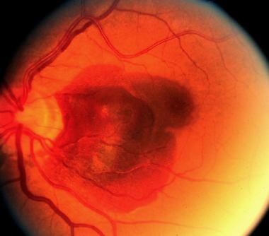
Angioid streaks are crack-like dehiscences in the Bruch's membrane, which predispose to the development of a choroidal neovascular membrane (CNVM) that carries a poor visual outcome. We report successful treatment in a 25-year-old woman with bilateral angioid streaks and subfoveal CNVM in the left eye who received two doses of intravitreal bevacizumab (1.25 mg) injections six weeks apart, resulting in rapid regression of the CNVM. (+info)Non-vascular vision loss in pseudoxanthoma elasticum. (8/26)
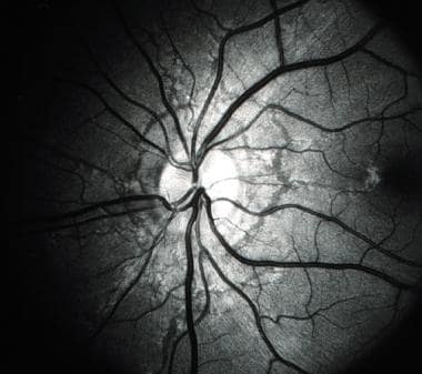
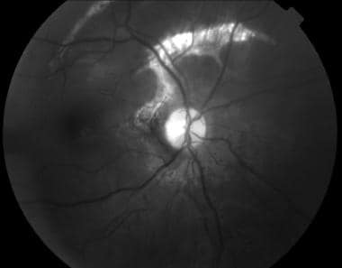
Pseudoxanthoma elasticum patients with angioid streaks are well-known to have acute vision loss due to choroidal bleeding. However, chronic vision loss due to macular atrophy is less well characterized. We describe a patient with sub-acute vision loss in one eye due to loss of macular retinal pigment epithelium function. Autofluorescence and pattern electroretinogram were useful adjuncts to help diagnose the source of her vision loss. (+info)Angioid streaks are abnormal, jagged lines or cracks in the delicate tissue at the back of the eye called the retina. These streaks typically occur near the optic nerve and radiate outward toward the edges of the retina. They are caused by degeneration of the underlying tissue, called Bruch's membrane, which separates the retina from the choroid, a layer of blood vessels that provides nutrients to the retina.
Angioid streaks are often associated with various medical conditions, including pseudoxanthoma elasticum, Paget's disease of bone, Ehlers-Danlos syndrome, and sickle cell anemia. They can also be a complication of cataract surgery or other eye trauma.
While angioid streaks themselves do not cause vision loss, they can lead to serious complications such as retinal hemorrhage, scarring, and detachment, which can result in significant vision loss if left untreated. Regular eye examinations are recommended for individuals with angioid streaks to monitor for any changes or complications that may require treatment.
Pseudoxanthoma Elasticum (PXE) is a rare genetic disorder characterized by the calcification and fragmentation of elastic fibers in the skin, eyes, and cardiovascular system. This causes changes in these tissues, leading to the clinical features of the disease. In the skin, this manifests as yellowish papules and plaques, often located on the neck, axillae, and flexural areas. In the eyes, it can cause angioid streaks, peau d'orange, and choroidal neovascularization, potentially leading to visual loss. In the cardiovascular system, calcification of the elastic fibers in the arterial walls can lead to premature atherosclerosis and increased risk of cardiovascular events. The disease is caused by mutations in the ABCC6 gene.
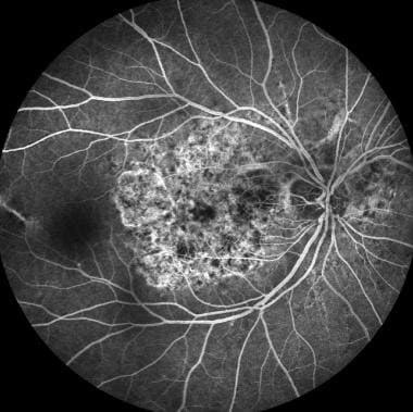
Fluorescein angiography is a medical diagnostic procedure used in ophthalmology to examine the blood flow in the retina and choroid, which are the inner layers of the eye. This test involves injecting a fluorescent dye, Fluorescein, into a patient's arm vein. As the dye reaches the blood vessels in the eye, a specialized camera takes rapid sequences of photographs to capture the dye's circulation through the retina and choroid.
The images produced by fluorescein angiography can help doctors identify any damage to the blood vessels, leakage, or abnormal growth of new blood vessels. This information is crucial in diagnosing and managing various eye conditions such as age-related macular degeneration, diabetic retinopathy, retinal vein occlusions, and inflammatory eye diseases.
It's important to note that while fluorescein angiography is a valuable diagnostic tool, it does carry some risks, including temporary side effects like nausea, vomiting, or allergic reactions to the dye. In rare cases, severe adverse reactions can occur, so patients should discuss these potential risks with their healthcare provider before undergoing the procedure.
Choroidal neovascularization (CNV) is a medical term that refers to the growth of new, abnormal blood vessels in the choroid layer of the eye, which is located between the retina and the sclera. This condition typically occurs as a complication of age-related macular degeneration (AMD), although it can also be caused by other eye diseases or injuries.
In CNV, the new blood vessels that grow into the choroid layer are fragile and can leak fluid or blood, which can cause distortion or damage to the retina, leading to vision loss. Symptoms of CNV may include blurred or distorted vision, a blind spot in the center of the visual field, or changes in color perception.
Treatment for CNV typically involves medications that are designed to stop the growth of new blood vessels, such as anti-VEGF drugs, which target a protein called vascular endothelial growth factor (VEGF) that is involved in the development of new blood vessels. Laser surgery or photodynamic therapy may also be used in some cases to destroy the abnormal blood vessels and prevent further vision loss.
"Fundus Oculi" is a medical term that refers to the back part of the interior of the eye, including the optic disc, macula, fovea, retinal vasculature, and peripheral retina. It is the area where light is focused and then transmitted to the brain via the optic nerve, forming visual images. Examinations of the fundus oculi are crucial for detecting various eye conditions such as diabetic retinopathy, macular degeneration, glaucoma, and other retinal diseases. The examination is typically performed using an ophthalmoscope or a specialized camera called a retinal camera.
 Angioid streaks
Bruch's membrane
Optic disc drusen
Sickle cell retinopathy
Indocyanine green angiography
Paget's disease of bone
Pseudoxanthoma elasticum
Jacob Hermann Knapp
Dystrophic calcification
Robert Walter Doyne
Eye disease
Peau d'orange
Ehlers-Danlos syndromes
List of MeSH codes (C11)
Angioid streaks - Wikipedia
 Angioid Streaks: Background, Pathophysiology, Epidemiology
 Angioid streaks | Annals Singapore
 Long-Term Effect of Anti-Vascular Endothelial Growth Factor (Anti-VEGF) Injections in Choroidal Neovascularization Secondary to...
Angioid Streaks: Background, Pathophysiology, Epidemiology
Weekend Bytes: Systemic Associations of Angioid Streaks
 Choroidal Neovascularization | SpringerLink
50. Choroidal Neovascularization Associated with Angioid Streaks in Pseudoxanthoma Elasticum | OCT Club
Choroidal Neovascularization (CNV) Clinical Presentation: History, Physical, Causes
Hemoglobinopathy Retinopathy: Background, Pathophysiology, Epidemiology
 Lucentis 10 mg/ml solution for injection - Summary of Product Characteristics (SmPC) - (emc)
 Opthamology Data (1971-75)
 click to copy a shareable link to this record
 Differences in Pulsatile Ocular Blood Flow among Three Classifications of Diabetic Retinopathy | IOVS | ARVO Journals
 Pseudoxanthoma Elasticum - Pediatrics - Merck Manuals Professional Edition
 Generalized arterial calcification of infancy: MedlinePlus Genetics
Survey: NHANES I
Orphanet: Congenital dyserythropoietic anemia
 Discover images - Retina Image Bank
 MODULE 14 OTHER CAUSES OF CHOROIDAL NEOVASCULAR DISEASE | Medical Retina Course
Ehlers-Danlos syndromes - Wikipedia
 Prognostic phenotypic and genotypic factors associated with photodynam | OPTH
 Namespace
Effectiveness and safety of intravitreal aflibercept in patients with wet age-related macular degeneration treated in routine...
What is the diagnosis?
VEGF165 Protein - ACROBiosystems
Bio2Vec
 CIL
CIL
Looking Through the Cracks
 EyeRounds.org: Pathologic Myopia
Pseudoxanthoma elasticum9
- Angioid streaks are often associated with pseudoxanthoma elasticum, but have been found to occur in conjunction with other disorders, including Paget's disease, sickle cell disease and Ehlers-Danlos syndrome. (wikipedia.org)
- Generally, the first signs of pseudoxanthoma elasticum are the asymptomatic cutaneous features and angioid streaks are the most frequent ocular findings. (octclub.org)
- Angioid streaks are gray-white lines with rupture of the thickened elastic fibers of Bruch's membrane extending radially from the optic disc and occur in 80% of patients with pseudoxanthoma elasticum. (octclub.org)
- Angioid streaks are not the pathognomonic sign of pseudoxanthoma elasticum and may occur due to Paget's disease, Marfan syndrome, Ehler-Danlos syndrom, sickle-cell anemia, and beta thalassemia. (octclub.org)
- Mirza E, Karanfil FC, Mirza GD, Choroidal neovascularization associated with angioid streaks in a patient with pseudoxanthoma elasticum. (octclub.org)
- roughly 50% of patients with angioid streaks have pseudoxanthoma elasticum, whereas 85% of those with pseudoxanthoma have angioid streaks). (syrianclinic.com)
- Diagnosis This patient has angioid streaks on fundoscopy and 'chicken-skin' appearance in the neck and axillae (lesions) due to pseudoxanthoma elasticum (aetiology). (syrianclinic.com)
- What is the triad of pseudoxanthoma elasticum, angioid streaks and vascular abnormalities known as? (syrianclinic.com)
- Angioid Streaks of the Retina Associated with Pseudoxanthoma Elasticum. (jkos.org)
Choroidal neovascularization7
- These streaks can have a negative impact on vision due to choroidal neovascularization or choroidal rupture. (wikipedia.org)
- Late complication of choroidal neovascularization in angioid streaks. (medscape.com)
- Choroidal neovascularization (CNV) is the major cause of vision loss and affects 70-86% of patients with angioid streaks. (medscape.com)
- This study aimed to evaluate the long-term effectiveness of intravitreal anti-vascular endothelial growth factor (VEGF) injections in the treatment of choroidal neovascularization (CNV) associated with angioid streaks. (hindawi.com)
- Patients with angioid streaks are generally asymptomatic, unless the lesions extend towards the fovea or develop complications, such as macular choroidal neovascularization (CNV) [ 2 , 4 ]. (hindawi.com)
- Ocular abnormalities or complications are angioid streaks, peau d'orange appearance, comet lesions, and choroidal neovascularization. (octclub.org)
- Based on these changes, we were certain our patient had developed choroidal neovascularization (CNV) in both eyes as a result of the angioid streaks. (reviewofoptometry.com)
Retina6
- Angioid streaks, also called Knapp streaks or Knapp striae, are small breaks in Bruch's membrane, an elastic tissue containing membrane of the retina that may become calcified and crack. (wikipedia.org)
- Dilated funduscopic examination revealed peau d'orange appearance in the temporal parts of the retina and angioid streaks extending radially from the optic disc and some involving the fovea in both eyes (Figure 1). (octclub.org)
- hereditary diseases: retinitis pigmentosa, angioid streaks of the retina, etc. (who.int)
- Angioid streaks may develop in the retina and compromise vision. (mhmedical.com)
- Patients may also have dental abnormalities, vascular calcifications, cortical hyperostosis and angioid streaks of the retina. (exeterlaboratory.com)
- Individuals most commonly present with angioid streaks of the retina found on routine eye examination or associated with retinal hemorrhage and/or characteristic papules in the skin. (nih.gov)
Ocular2
- Indocyanine green angiography can also be used for diagnosing angioid streaks and their associated ocular pathologies. (wikipedia.org)
- [ 1 ] Angioid streaks, also known as Knapp striae, are irregular jagged dehiscences in the mineralized, degenerated, brittle Bruch membrane that typically form alongside force lines exerted by intrinsic and extrinsic ocular muscles that radiate in a centrifugal pattern emanating from the optic disc. (medscape.com)
Myopia2
- They include conditions such as high myopia (nearsightedness), histoplasmosis, angioid streaks, and eye injury. (foxeye.com)
- Causes of included multifocal choroiditis and panuveitis (N = 12), angioid streaks (N = 11), pathologic myopia (N = 10), and idiopathic or other causes (N = 6). (medscape.com)
Sickle cell di1
- In 1959, Lieb and coworkers associated angioid streaks with sickle cell disease. (medscape.com)
Abnormalities1
- and abnormalities called angioid streaks affecting tissue at the back of the eye, which can be detected during an eye examination. (medlineplus.gov)
Fundus3
- The diagnosis is mainly clinical, however fundus fluorescein angiography shows that the streaks appear hyperfluorescent (window defect) in the early phase. (wikipedia.org)
- Angioid streaks on fundus autofluorescence appeared as radial hypoautofluorescent fissuresfissures (Figure 2). (octclub.org)
- In patients with a lighter fundus, the streaks will appear red, while those with a darker fundus will show medium to dark brown streaks. (reviewofoptometry.com)
Underlying systemic associations1
- Management of angioid streaks starts with complete medical checkup to rule out underlying systemic associations. (wikipedia.org)
Retinal hemorrhages1
- In 1889, Doyne first described angioid streaks in a patient with retinal hemorrhages secondary to trauma. (medscape.com)
Breaks in Bruch's membrane1
- The angioid streaks appear as crack-like breaks in Bruch's membrane and retinal pigment epithelium (RPE) atrophy. (reviewofoptometry.com)
Vascular1
- The management of CNV secondary to angioid streaks with intravitreal anti-vascular endothelial growth factor (VEGF) injections, irrespective of the molecule (i.e., bevacizumab, ranibizumab, or aflibercept), has shown superior functional and morphologic results compared with previous therapeutic options, such as photocoagulation laser and photodynamic therapy (PDT). (hindawi.com)
Glaucoma1
- Ahmed glaucoma valve, angioid streaks, & spontaneous OSSN regression. (cataractcoach.com)
Ophthalmology1
- Terry (1899-1946), Head of Ophthalmology at Harvard University, described angioid streaks in Paget's disease. (syrianclinic.com)
Secondary4
- Multicenter retrospective cohort study, including eyes with CNV secondary to angioid streaks treated with anti-VEGF injections, were performed. (hindawi.com)
- Recently, long-term retrospective and prospective studies reinforced the previous findings that anti-VEGF injections had remarkably improved the prognosis of CNV secondary to angioid streaks [ 12 - 14 ]. (hindawi.com)
- Moreover, these studies do not clarify whether the results may be generalized to all anti-VEGF and to all patients with CNV secondary to angioid streaks regardless of the associated systemic pathology. (hindawi.com)
- This is a multicenter retrospective cohort study, which included eyes with CNV secondary to angioid streaks treated with intravitreal anti-VEGF injections (with or without previous alternative treatments, such as laser photocoagulation or PDT). (hindawi.com)
Injections3
- Therefore, the primary endpoint of this study was to evaluate the effectiveness of intravitreal anti-VEGF injections at 48 months in the treatment of CNV associated with angioid streaks in a real-world study. (hindawi.com)
- There is no specific treatment, but intravitreal injections of angiogenesis-blocking antibodies may be given for angioid streaks. (merckmanuals.com)
- Intravitreal injections of angiogenesis-blocking antibodies (eg, bevacizumab ) show promise as an off-label treatment option for retinal angioid streaks. (merckmanuals.com)
Complication1
- In fact, CNV is the most serious complication of angioid streaks, with an incidence of 72% to 86% [ 1 ]. (hindawi.com)
Macula1
- The streaks extend into the macula in both eyes. (reviewofoptometry.com)
Diagnosis2
- The histological diagnosis is made by doing 4 mm punch biopsy of scars or flexural skin of the neck or axillae in patients who have angioid streaks on fundoscopy but no visible skin lesions. (syrianclinic.com)
- Improving diagnosis of Angioid Streaks. (ammaneyeclinic.com)
Idiopathic1
- Up to 50% of angioid streak cases are idiopathic. (wikipedia.org)
Irregular1
- Angioid streaks were first described as irregular red/brown lines that surround concentrically or radiate out of the optic nerve head [ 1 , 2 ]. (hindawi.com)
Onset1
- The onset of angioid streaks commonly lies between the second and fifth decades [ 3 ]. (hindawi.com)
Knapp4
- In 1892, ophthalmologist Hermann Jakob Knapp called them "angioid streaks" because of their resemblance to blood vessels. (wikipedia.org)
- Angioid Streaks (Knapp Streaks). (wikipedia.org)
- [ 2 ] Knapp named them angioid streaks because of their resemblance to blood vessels. (medscape.com)
- Knapp H. On the formation of dark angioid streaks as an unusual metamorphosis of retinal hemorrhage. (medscape.com)
Disease1
- Angioid streaks in patients with no systemic disease or with rare etiologies tend to present late in life with a mean age of 65.7 years. (medscape.com)
Optic1
- Interestingly, angioid streaks do have as high as 25% association with optic disc drusen. (reviewofoptometry.com)
Radial1
- Those lines of forces had the same configuration as the peripapillary interlacement and radial extensions of angioid streaks. (medscape.com)
Fovea1
- Also, vision can be impaired if the streaks progress to the fovea and damage the retinal pigment epithelium. (wikipedia.org)
Examination1
- On examination, she had well defined and confluent vesicles and bullae arranged in a linear streaks and bizarre pattern extending from the elbow joint to just above the wrist joint of right forearm [ Figure 1 ]. (cosmoderma.org)
Clinical1
- Angioid Streaks: A clinical and histopathologic study. (medscape.com)
Patients1
- Two studies showed similar results: of all patients with angioid streaks, 66.2% of patients were white, compared with 29% of Asian origin and 3.7% of black people. (medscape.com)
Conditions2
- Systemically, many of these conditions associated with angioid streaks can be life-threatening. (reviewofoptometry.com)
- ADVANCED-LEVEL QUESTIONS In which other conditions are angioid streaks seen? (syrianclinic.com)
Eyes1
- Ophthalmoscopy identified the presence of angioid streaks in both eyes of the two sisters, confirmed by retinograpy and angiography. (unimore.it)
Generally1
- This real-world study suggests that the functional and morphologic response to anti-VEGF therapy for CNV related to angioid streaks is generally satisfactory and maintained in the long term. (hindawi.com)
Result1
- [ 2 ] Angioid streaks result from pathological changes at the level of the Bruch membrane, which was confirmed histologically in the late 1930s. (medscape.com)
Treatment1
- Most literature on anti-VEGF treatment in CNV related to angioid streaks was based on single case reports, case series, or short-term cohorts [ 2 , 4 - 11 ]. (hindawi.com)
Article1
- eMedicine article on Angioid streaks Photos of Angioid streaks John F., Salmon (2020). (wikipedia.org)