Anterior Wall Myocardial Infarction
Inferior Wall Myocardial Infarction
Myocardial Infarction
Coronary Angiography
Ventricular Function, Left
Electrocardiography
Heart Rupture, Post-Infarction
Heart Ventricles
Echocardiography
Infarction
Myocardial Reperfusion
Angioplasty, Balloon, Coronary
Cell Wall
Cerebral Infarction
Ventricular Remodeling
Thrombolytic Therapy
Treatment Outcome
Prospective Studies
Myocardium
Risk Factors
Coronary Disease
Ventricular Dysfunction, Left
Myocardial Ischemia
Stroke Volume
Predictive Value of Tests
Streptokinase
Follow-Up Studies
Creatine Kinase
Prognosis
Cardiac Catheterization
Coronary Care Units
Cystocele
Retrospective Studies
Coronary Artery Bypass
Angina Pectoris
Tomography, Emission-Computed, Single-Photon
Platelet Aggregation Inhibitors
Hemodynamics
Stents
Myocardial Revascularization
Percutaneous Coronary Intervention
Coronary Thrombosis
Heart Septum
Coronary Artery Disease
Tomography, X-Ray Computed
Risk Assessment
Hospital Mortality
Uterine Prolapse
Myocardial Reperfusion Injury
Usefulness of the index of microcirculatory resistance for invasively assessing myocardial viability immediately after primary angioplasty for anterior myocardial infarction. (1/56)
(+info)Dual left anterior descending artery distribution. (2/56)
(+info)Low adiponectin blood concentration predicts left ventricular remodeling after ST-segment elevation myocardial infarction treated with primary percutaneous coronary intervention. (3/56)
BACKGROUND: Left ventricular remodeling (LVR), an increase in left ventricular end-diastolic volume index > or = 20%, is an adverse consequence of myocardial infarction. The aim of this study was to assess the association between LVR and adiponectin, which has been shown to protect against myocardial ischemia-reperfusion injury. METHODS: In 75 patients echocardiographic examination was performed one year after ST-segment elevation myocardial infarction, successfully treated with primary percutaneous coronary intervention (pPCI). Two groups of patients were analyzed: those with LVR (n = 15) and those without LVR (n = 60). RESULTS: The predictors of LVR were: anterior myocardial infarction, glucose at admission, baseline C-reactive protein, adiponectin, and echocardiographic parameters: left ventricular end-diastolic and end-systolic volume indices, ejection fraction < 40% and left ventricular wall motion score index (WMSI) at discharge. On multivariable regression analysis, lower adiponectin level (OR = 0.67, 95% CI 0.49-0.91, p < 0.05) and higher WMSI (OR = 20.14, 95% CI 2.62-154.82, p < 0.01) were the only independent negative predictors of LVR. The optimal cut-off for adiponectin for predicting LVR was < or = 4.7 mg/mL (sensitivity: 73%, specificity: 85%) and this level increased the risk of LVR 15-fold (95% CI 4.05-59.87, p = 0.0001). CONCLUSIONS: Baseline low blood adiponectin concentration, along with WMSI, can be considered as a predictor of the LVR in male patients one year after myocardial infarction and pPCI. (+info)Dor procedure for dyskinetic anteroapical myocardial infarction fails to improve contractility in the border zone. (4/56)
(+info)Right ventricular involvement in anterior myocardial infarction: a translational approach. (5/56)
(+info)Retrograde approach to a totally occluded right coronary artery via a septal perforator artery: the tale of a long and winding wire. (6/56)
Retrograde recannalization of chronic total occlusions has developed as a viable alternative to restore coronary patency. Techniques continue to evolve and complications described. We present a new complication related to equipment developed to improve outcomes via a retrograde approach. (+info)Complete atrioventricular block complicating acute anterior myocardial infarction can be reversed with acute coronary angioplasty. (7/56)
INTRODUCTION: A retrospective case series of acute anterior myocardial infarction (MI) patients complicated by complete atrioventricular block (AVB) treated with acute percutaneous transluminal coronary angioplasty (PTCA). CLINICAL PICTURE: Eight patients with anterior MI and complete AVB underwent acute PTCA between 2000 and 2005. Mean onset of complete AVB was 16.6 +/- 16.9 hours from chest pain onset. TREATMENT: All patients underwent successful PTCA to the left anterior descending artery. OUTCOME: Complete AVB resolved with PTCA in 88%; mean time of resolution was 89 +/- 144 minutes after revascularisation. One patient had permanent pacemaker implanted at Day 12 after developing an 8-second ventricular standstill during hospitalisation but not pacing-dependent on follow-up. The rhythm on discharge for the other surviving patients was normal sinus rhythm. CONCLUSION: This case series suggests that complete AVB complicating anterior MI is reversible with acute PTCA and survivors are not at increased risk of recurrent AVB. Nevertheless, this condition is associated with extensive myocardial damage and high mortality during the acute hospitalisation was not improved with correction of AVB with temporary pacing. (+info)Coronary flow velocity pattern and recovery of regional left ventricular function: the relationship observed in patients with reperfused acute myocardial infarction. (8/56)
Coronary flow velocity pattern (CFVP) recorded within 3 days of percutaneous coronary intervention (PCI) has been reported to be useful in predicting left ventricular (LV) function. The aim of this prospective study was to investigate, via transthoracic Doppler echocardiography, whether the relationship between CFVP and recovery of LV function persists. Our study group comprised 37 patients with 1st anterior-wall acute myocardial infarction who underwent successful PCI for lesions in the left anterior descending coronary artery (LAD). The CFVP in the LAD was recorded at 24-48 hours, 7 days, and 4 weeks after PCI. Myocardial contrast echocardiography was performed at 24-48 hours after PCI. The diastolic deceleration time (DDT) at each stage correlated significantly with the regional LV wall-motion score index at 6-month follow-up (r=-0.58 at 24-48 hr, -0.57 at day 7, and -0.50 at week 4; P <0.01 for all). The mean DDT increased over time. Optimal cutoff values for DDT to predict regional LV wall-motion score indices of <2.0 were 327 ms at 24-48 hours (sensitivity, 0.78; specificity, 0.64), 495 ms at day 7 (sensitivity, 0.75; specificity, 0.69), and 525 ms at week 4 (sensitivity, 0.83; specificity, 0.69). The DDT at 24-48 hours significantly correlated, better than the peak creatine kinase value, with reperfusion (r=0.68, P <0.01) as defined by myocardial contrast echocardiography. In conclusion, CFVP in the LAD can be used, within 4 weeks after PCI, to predict the recovery of regional LV function in patients with reperfused anterior-wall acute myocardial infarction. (+info)An anterior wall myocardial infarction (AMI) is a type of heart attack that occurs when there is a significant reduction or complete blockage of blood flow to the front wall of the heart muscle, also known as the anterior wall of the left ventricle. This reduction or blockage in blood flow is typically caused by a buildup of fatty deposits, called plaques, in the coronary arteries that supply oxygen-rich blood to the heart muscle.
When a plaque ruptures or breaks open, a blood clot forms around it, which can completely block the flow of blood to the heart muscle. This lack of blood flow causes the heart muscle to start to die, leading to a myocardial infarction or heart attack.
An anterior wall myocardial infarction is often associated with more severe symptoms and a higher risk of complications than other types of heart attacks because it affects a larger area of the heart muscle. Symptoms may include chest pain, shortness of breath, nausea, vomiting, sweating, and anxiety.
Immediate medical attention is necessary for an anterior wall myocardial infarction to restore blood flow to the heart muscle as quickly as possible and prevent further damage. Treatment options may include medications, such as clot-busting drugs or blood thinners, as well as procedures such as angioplasty or coronary artery bypass surgery.
An Inferior Wall Myocardial Infarction (MI) is a type of heart attack that occurs when there is a significant reduction or complete blockage of blood flow to the inferior (lower) region of the heart muscle, specifically the areas supplied by the right coronary artery or one of its branches. This reduction in blood flow, often caused by a blood clot forming around a ruptured plaque within the artery, can lead to ischemia and ultimately result in damage or death of the heart muscle cells (myocardial necrosis). Symptoms may include chest pain, shortness of breath, sweating, nausea, or vomiting. Diagnosis typically involves an electrocardiogram (ECG) and cardiac biomarker tests, such as troponin levels. Treatment includes medications, lifestyle changes, and possibly interventions like angioplasty or bypass surgery to restore blood flow.
Myocardial infarction (MI), also known as a heart attack, is a medical condition characterized by the death of a segment of heart muscle (myocardium) due to the interruption of its blood supply. This interruption is most commonly caused by the blockage of a coronary artery by a blood clot formed on the top of an atherosclerotic plaque, which is a buildup of cholesterol and other substances in the inner lining of the artery.
The lack of oxygen and nutrients supply to the heart muscle tissue results in damage or death of the cardiac cells, causing the affected area to become necrotic. The extent and severity of the MI depend on the size of the affected area, the duration of the occlusion, and the presence of collateral circulation.
Symptoms of a myocardial infarction may include chest pain or discomfort, shortness of breath, nausea, lightheadedness, and sweating. Immediate medical attention is necessary to restore blood flow to the affected area and prevent further damage to the heart muscle. Treatment options for MI include medications, such as thrombolytics, antiplatelet agents, and pain relievers, as well as procedures such as percutaneous coronary intervention (PCI) or coronary artery bypass grafting (CABG).
Coronary angiography is a medical procedure that uses X-ray imaging to visualize the coronary arteries, which supply blood to the heart muscle. During the procedure, a thin, flexible catheter is inserted into an artery in the arm or groin and threaded through the blood vessels to the heart. A contrast dye is then injected through the catheter, and X-ray images are taken as the dye flows through the coronary arteries. These images can help doctors diagnose and treat various heart conditions, such as blockages or narrowing of the arteries, that can lead to chest pain or heart attacks. It is also known as coronary arteriography or cardiac catheterization.
Left ventricular function refers to the ability of the left ventricle (the heart's lower-left chamber) to contract and relax, thereby filling with and ejecting blood. The left ventricle is responsible for pumping oxygenated blood to the rest of the body. Its function is evaluated by measuring several parameters, including:
1. Ejection fraction (EF): This is the percentage of blood that is pumped out of the left ventricle with each heartbeat. A normal ejection fraction ranges from 55% to 70%.
2. Stroke volume (SV): The amount of blood pumped by the left ventricle in one contraction. A typical SV is about 70 mL/beat.
3. Cardiac output (CO): The total volume of blood that the left ventricle pumps per minute, calculated as the product of stroke volume and heart rate. Normal CO ranges from 4 to 8 L/minute.
Assessment of left ventricular function is crucial in diagnosing and monitoring various cardiovascular conditions such as heart failure, coronary artery disease, valvular heart diseases, and cardiomyopathies.
Electrocardiography (ECG or EKG) is a medical procedure that records the electrical activity of the heart. It provides a graphic representation of the electrical changes that occur during each heartbeat. The resulting tracing, called an electrocardiogram, can reveal information about the heart's rate and rhythm, as well as any damage to its cells or abnormalities in its conduction system.
During an ECG, small electrodes are placed on the skin of the chest, arms, and legs. These electrodes detect the electrical signals produced by the heart and transmit them to a machine that amplifies and records them. The procedure is non-invasive, painless, and quick, usually taking only a few minutes.
ECGs are commonly used to diagnose and monitor various heart conditions, including arrhythmias, coronary artery disease, heart attacks, and electrolyte imbalances. They can also be used to evaluate the effectiveness of certain medications or treatments.
Post-infarction heart rupture is a serious and potentially fatal complication that can occur after a myocardial infarction (heart attack). It is defined as the disruption or tearing of the heart muscle (myocardium) in the area that was damaged by the heart attack. This condition typically occurs within 1 to 7 days following a heart attack, and it's more common in elderly patients and those with large infarctions.
There are three main types of post-infarction heart rupture:
1. Ventricular free wall rupture: This is the most common type, where there is a tear in the left ventricular wall, leading to rapid bleeding into the pericardial sac (the space surrounding the heart). This can cause cardiac tamponade, which is a life-threatening situation characterized by increased pressure in the pericardial sac, compromising cardiac filling and reducing cardiac output.
2. Ventricular septal rupture: In this case, there is a tear in the interventricular septum (the wall separating the left and right ventricles), leading to a communication between the two chambers. This results in a shunt of blood from the high-pressure left ventricle to the low-pressure right ventricle, causing a sudden increase in pulmonary congestion and reduced systemic output.
3. Papillary muscle rupture: The papillary muscles are finger-like projections that attach the heart valves (mitral and tricuspid) to the ventricular walls. Rupture of these muscles can lead to severe mitral or tricuspid regurgitation, causing acute pulmonary edema and reduced cardiac output.
Symptoms of post-infarction heart rupture may include chest pain, shortness of breath, palpitations, hypotension, tachycardia, and signs of cardiogenic shock (such as cold sweats, weak pulse, and altered mental status). Diagnosis is typically made using echocardiography, CT angiography, or MRI. Treatment usually involves emergency surgical intervention to repair the rupture and stabilize the patient's hemodynamic condition.
The heart ventricles are the two lower chambers of the heart that receive blood from the atria and pump it to the lungs or the rest of the body. The right ventricle pumps deoxygenated blood to the lungs, while the left ventricle pumps oxygenated blood to the rest of the body. Both ventricles have thick, muscular walls to generate the pressure necessary to pump blood through the circulatory system.
Echocardiography is a medical procedure that uses sound waves to produce detailed images of the heart's structure, function, and motion. It is a non-invasive test that can help diagnose various heart conditions, such as valve problems, heart muscle damage, blood clots, and congenital heart defects.
During an echocardiogram, a transducer (a device that sends and receives sound waves) is placed on the chest or passed through the esophagus to obtain images of the heart. The sound waves produced by the transducer bounce off the heart structures and return to the transducer, which then converts them into electrical signals that are processed to create images of the heart.
There are several types of echocardiograms, including:
* Transthoracic echocardiography (TTE): This is the most common type of echocardiogram and involves placing the transducer on the chest.
* Transesophageal echocardiography (TEE): This type of echocardiogram involves passing a specialized transducer through the esophagus to obtain images of the heart from a closer proximity.
* Stress echocardiography: This type of echocardiogram is performed during exercise or medication-induced stress to assess how the heart functions under stress.
* Doppler echocardiography: This type of echocardiogram uses sound waves to measure blood flow and velocity in the heart and blood vessels.
Echocardiography is a valuable tool for diagnosing and managing various heart conditions, as it provides detailed information about the structure and function of the heart. It is generally safe, non-invasive, and painless, making it a popular choice for doctors and patients alike.
Coronary vessels refer to the network of blood vessels that supply oxygenated blood and nutrients to the heart muscle, also known as the myocardium. The two main coronary arteries are the left main coronary artery and the right coronary artery.
The left main coronary artery branches off into the left anterior descending artery (LAD) and the left circumflex artery (LCx). The LAD supplies blood to the front of the heart, while the LCx supplies blood to the side and back of the heart.
The right coronary artery supplies blood to the right lower part of the heart, including the right atrium and ventricle, as well as the back of the heart.
Coronary vessel disease (CVD) occurs when these vessels become narrowed or blocked due to the buildup of plaque, leading to reduced blood flow to the heart muscle. This can result in chest pain, shortness of breath, or a heart attack.
Infarction is the term used in medicine to describe the death of tissue (also known as an "area of necrosis") due to the lack of blood supply. This can occur when a blood vessel that supplies oxygen and nutrients to a particular area of the body becomes blocked or obstructed, leading to the deprivation of oxygen and nutrients necessary for the survival of cells in that region.
The blockage in the blood vessel is usually caused by a clot (thrombus) or an embolus, which is a small particle that travels through the bloodstream and lodges in a smaller vessel. The severity and extent of infarction depend on several factors, including the size and location of the affected blood vessel, the duration of the obstruction, and the presence of collateral circulation (alternative blood vessels that can compensate for the blocked one).
Common examples of infarctions include myocardial infarction (heart attack), cerebral infarction (stroke), and pulmonary infarction (lung tissue death due to obstruction in the lung's blood vessels). Infarctions can lead to various symptoms, depending on the affected organ or tissue, and may require medical intervention to manage complications and prevent further damage.
Myocardial reperfusion is the restoration of blood flow to the heart muscle (myocardium), usually after a period of ischemia or reduced oxygen supply, such as during a myocardial infarction (heart attack). This can be achieved through various medical interventions, including thrombolytic therapy, percutaneous coronary intervention (PCI), or coronary artery bypass surgery (CABG). The goal of myocardial reperfusion is to salvage the jeopardized myocardium, preserve cardiac function, and reduce the risk of complications like heart failure or arrhythmias. However, it's important to note that while reperfusion is crucial for treating ischemic heart disease, it can also lead to additional injury to the heart muscle, known as reperfusion injury.
Coronary balloon angioplasty is a minimally invasive medical procedure used to widen narrowed or obstructed coronary arteries (the blood vessels that supply oxygen-rich blood to the heart muscle) and improve blood flow to the heart. This procedure is typically performed in conjunction with the insertion of a stent, a small mesh tube that helps keep the artery open.
During coronary balloon angioplasty, a thin, flexible catheter with a deflated balloon at its tip is inserted into a blood vessel, usually through a small incision in the groin or arm. The catheter is then guided to the narrowed or obstructed section of the coronary artery. Once in position, the balloon is inflated to compress the plaque against the artery wall and widen the lumen (the inner space) of the artery. This helps restore blood flow to the heart muscle.
The procedure is typically performed under local anesthesia and conscious sedation to minimize discomfort. Coronary balloon angioplasty is a relatively safe and effective treatment for many people with coronary artery disease, although complications such as bleeding, infection, or re-narrowing of the artery (restenosis) can occur in some cases.
A cell wall is a rigid layer found surrounding the plasma membrane of plant cells, fungi, and many types of bacteria. It provides structural support and protection to the cell, maintains cell shape, and acts as a barrier against external factors such as chemicals and mechanical stress. The composition of the cell wall varies among different species; for example, in plants, it is primarily made up of cellulose, hemicellulose, and pectin, while in bacteria, it is composed of peptidoglycan.
In the field of medicine, "time factors" refer to the duration of symptoms or time elapsed since the onset of a medical condition, which can have significant implications for diagnosis and treatment. Understanding time factors is crucial in determining the progression of a disease, evaluating the effectiveness of treatments, and making critical decisions regarding patient care.
For example, in stroke management, "time is brain," meaning that rapid intervention within a specific time frame (usually within 4.5 hours) is essential to administering tissue plasminogen activator (tPA), a clot-busting drug that can minimize brain damage and improve patient outcomes. Similarly, in trauma care, the "golden hour" concept emphasizes the importance of providing definitive care within the first 60 minutes after injury to increase survival rates and reduce morbidity.
Time factors also play a role in monitoring the progression of chronic conditions like diabetes or heart disease, where regular follow-ups and assessments help determine appropriate treatment adjustments and prevent complications. In infectious diseases, time factors are crucial for initiating antibiotic therapy and identifying potential outbreaks to control their spread.
Overall, "time factors" encompass the significance of recognizing and acting promptly in various medical scenarios to optimize patient outcomes and provide effective care.
Cerebral infarction, also known as a "stroke" or "brain attack," is the sudden death of brain cells caused by the interruption of their blood supply. It is most commonly caused by a blockage in one of the blood vessels supplying the brain (an ischemic stroke), but can also result from a hemorrhage in or around the brain (a hemorrhagic stroke).
Ischemic strokes occur when a blood clot or other particle blocks a cerebral artery, cutting off blood flow to a part of the brain. The lack of oxygen and nutrients causes nearby brain cells to die. Hemorrhagic strokes occur when a weakened blood vessel ruptures, causing bleeding within or around the brain. This bleeding can put pressure on surrounding brain tissues, leading to cell death.
Symptoms of cerebral infarction depend on the location and extent of the affected brain tissue but may include sudden weakness or numbness in the face, arm, or leg; difficulty speaking or understanding speech; vision problems; loss of balance or coordination; and severe headache with no known cause. Immediate medical attention is crucial for proper diagnosis and treatment to minimize potential long-term damage or disability.
Ventricular remodeling is a structural adaptation process of the heart in response to stress or injury, such as myocardial infarction (heart attack) or pressure overload. This process involves changes in size, shape, and function of the ventricles (the lower chambers of the heart).
In ventricular remodeling, the heart muscle may thicken, enlarge, or become more stiff, leading to alterations in the pumping ability of the heart. These changes can ultimately result in cardiac dysfunction, heart failure, and an increased risk of arrhythmias (irregular heart rhythms).
Ventricular remodeling is often classified into two types:
1. Concentric remodeling: This occurs when the ventricular wall thickens (hypertrophy) without a significant increase in chamber size, leading to a decrease in the cavity volume and an increase in the thickness of the ventricular wall.
2. Eccentric remodeling: This involves an increase in both the ventricular chamber size and wall thickness due to the addition of new muscle cells (hyperplasia) or enlargement of existing muscle cells (hypertrophy). As a result, the overall shape of the ventricle becomes more spherical and less elliptical.
Both types of remodeling can negatively impact heart function and contribute to the development of heart failure. Close monitoring and appropriate treatment are essential for managing ventricular remodeling and preventing further complications.
Thrombolytic therapy, also known as thrombolysis, is a medical treatment that uses medications called thrombolytics or fibrinolytics to dissolve or break down blood clots (thrombi) in blood vessels. These clots can obstruct the flow of blood to vital organs such as the heart, lungs, or brain, leading to serious conditions like myocardial infarction (heart attack), pulmonary embolism, or ischemic stroke.
The goal of thrombolytic therapy is to restore blood flow as quickly and efficiently as possible to prevent further damage to the affected organ and potentially save lives. Commonly used thrombolytic drugs include alteplase (tPA), reteplase, and tenecteplase. It's essential to administer these medications as soon as possible after the onset of symptoms for optimal treatment outcomes. However, there are risks associated with thrombolytic therapy, such as an increased chance of bleeding complications, which must be carefully weighed against its benefits in each individual case.
Coronary circulation refers to the circulation of blood in the coronary vessels, which supply oxygenated blood to the heart muscle (myocardium) and drain deoxygenated blood from it. The coronary circulation system includes two main coronary arteries - the left main coronary artery and the right coronary artery - that branch off from the aorta just above the aortic valve. These arteries further divide into smaller branches, which supply blood to different regions of the heart muscle.
The left main coronary artery divides into two branches: the left anterior descending (LAD) artery and the left circumflex (LCx) artery. The LAD supplies blood to the front and sides of the heart, while the LCx supplies blood to the back and sides of the heart. The right coronary artery supplies blood to the lower part of the heart, including the right ventricle and the bottom portion of the left ventricle.
The veins that drain the heart muscle include the great cardiac vein, the middle cardiac vein, and the small cardiac vein, which merge to form the coronary sinus. The coronary sinus empties into the right atrium, allowing deoxygenated blood to enter the right side of the heart and be pumped to the lungs for oxygenation.
Coronary circulation is essential for maintaining the health and function of the heart muscle, as it provides the necessary oxygen and nutrients required for proper contraction and relaxation of the myocardium. Any disruption or blockage in the coronary circulation system can lead to serious consequences, such as angina, heart attack, or even death.
Treatment outcome is a term used to describe the result or effect of medical treatment on a patient's health status. It can be measured in various ways, such as through symptoms improvement, disease remission, reduced disability, improved quality of life, or survival rates. The treatment outcome helps healthcare providers evaluate the effectiveness of a particular treatment plan and make informed decisions about future care. It is also used in clinical research to compare the efficacy of different treatments and improve patient care.
Prospective studies, also known as longitudinal studies, are a type of cohort study in which data is collected forward in time, following a group of individuals who share a common characteristic or exposure over a period of time. The researchers clearly define the study population and exposure of interest at the beginning of the study and follow up with the participants to determine the outcomes that develop over time. This type of study design allows for the investigation of causal relationships between exposures and outcomes, as well as the identification of risk factors and the estimation of disease incidence rates. Prospective studies are particularly useful in epidemiology and medical research when studying diseases with long latency periods or rare outcomes.
The myocardium is the middle layer of the heart wall, composed of specialized cardiac muscle cells that are responsible for pumping blood throughout the body. It forms the thickest part of the heart wall and is divided into two sections: the left ventricle, which pumps oxygenated blood to the rest of the body, and the right ventricle, which pumps deoxygenated blood to the lungs.
The myocardium contains several types of cells, including cardiac muscle fibers, connective tissue, nerves, and blood vessels. The muscle fibers are arranged in a highly organized pattern that allows them to contract in a coordinated manner, generating the force necessary to pump blood through the heart and circulatory system.
Damage to the myocardium can occur due to various factors such as ischemia (reduced blood flow), infection, inflammation, or genetic disorders. This damage can lead to several cardiac conditions, including heart failure, arrhythmias, and cardiomyopathy.
Medical Definition:
"Risk factors" are any attribute, characteristic or exposure of an individual that increases the likelihood of developing a disease or injury. They can be divided into modifiable and non-modifiable risk factors. Modifiable risk factors are those that can be changed through lifestyle choices or medical treatment, while non-modifiable risk factors are inherent traits such as age, gender, or genetic predisposition. Examples of modifiable risk factors include smoking, alcohol consumption, physical inactivity, and unhealthy diet, while non-modifiable risk factors include age, sex, and family history. It is important to note that having a risk factor does not guarantee that a person will develop the disease, but rather indicates an increased susceptibility.
Coronary artery disease, often simply referred to as coronary disease, is a condition in which the blood vessels that supply oxygen-rich blood to the heart become narrowed or blocked due to the buildup of fatty deposits called plaques. This can lead to chest pain (angina), shortness of breath, or in severe cases, a heart attack.
The medical definition of coronary artery disease is:
A condition characterized by the accumulation of atheromatous plaques in the walls of the coronary arteries, leading to decreased blood flow and oxygen supply to the myocardium (heart muscle). This can result in symptoms such as angina pectoris, shortness of breath, or arrhythmias, and may ultimately lead to myocardial infarction (heart attack) or heart failure.
Risk factors for coronary artery disease include age, smoking, high blood pressure, high cholesterol, diabetes, obesity, physical inactivity, and a family history of the condition. Lifestyle changes such as quitting smoking, exercising regularly, eating a healthy diet, and managing stress can help reduce the risk of developing coronary artery disease. Medical treatments may include medications to control blood pressure, cholesterol levels, or irregular heart rhythms, as well as procedures such as angioplasty or bypass surgery to improve blood flow to the heart.
Left ventricular dysfunction (LVD) is a condition characterized by the impaired ability of the left ventricle of the heart to pump blood efficiently during contraction. The left ventricle is one of the four chambers of the heart and is responsible for pumping oxygenated blood to the rest of the body.
LVD can be caused by various underlying conditions, such as coronary artery disease, cardiomyopathy, valvular heart disease, or hypertension. These conditions can lead to structural changes in the left ventricle, including remodeling, hypertrophy, and dilation, which ultimately impair its contractile function.
The severity of LVD is often assessed by measuring the ejection fraction (EF), which is the percentage of blood that is pumped out of the left ventricle during each contraction. A normal EF ranges from 55% to 70%, while an EF below 40% is indicative of LVD.
LVD can lead to various symptoms, such as shortness of breath, fatigue, fluid retention, and decreased exercise tolerance. It can also increase the risk of complications, such as heart failure, arrhythmias, and cardiac arrest. Treatment for LVD typically involves managing the underlying cause, along with medications to improve contractility, reduce fluid buildup, and control heart rate. In severe cases, devices such as implantable cardioverter-defibrillators (ICDs) or left ventricular assist devices (LVADs) may be required.
Myocardial ischemia is a condition in which the blood supply to the heart muscle (myocardium) is reduced or blocked, leading to insufficient oxygen delivery and potential damage to the heart tissue. This reduction in blood flow typically results from the buildup of fatty deposits, called plaques, in the coronary arteries that supply the heart with oxygen-rich blood. The plaques can rupture or become unstable, causing the formation of blood clots that obstruct the artery and limit blood flow.
Myocardial ischemia may manifest as chest pain (angina pectoris), shortness of breath, fatigue, or irregular heartbeats (arrhythmias). In severe cases, it can lead to myocardial infarction (heart attack) if the oxygen supply is significantly reduced or cut off completely, causing permanent damage or death of the heart muscle. Early diagnosis and treatment of myocardial ischemia are crucial for preventing further complications and improving patient outcomes.
In medical terms, the heart is a muscular organ located in the thoracic cavity that functions as a pump to circulate blood throughout the body. It's responsible for delivering oxygen and nutrients to the tissues and removing carbon dioxide and other wastes. The human heart is divided into four chambers: two atria on the top and two ventricles on the bottom. The right side of the heart receives deoxygenated blood from the body and pumps it to the lungs, while the left side receives oxygenated blood from the lungs and pumps it out to the rest of the body. The heart's rhythmic contractions and relaxations are regulated by a complex electrical conduction system.
Stroke volume is a term used in cardiovascular physiology and medicine. It refers to the amount of blood that is pumped out of the left ventricle of the heart during each contraction (systole). Specifically, it is the difference between the volume of blood in the left ventricle at the end of diastole (when the ventricle is filled with blood) and the volume at the end of systole (when the ventricle has contracted and ejected its contents into the aorta).
Stroke volume is an important measure of heart function, as it reflects the ability of the heart to pump blood effectively to the rest of the body. A low stroke volume may indicate that the heart is not pumping efficiently, while a high stroke volume may suggest that the heart is working too hard. Stroke volume can be affected by various factors, including heart disease, high blood pressure, and physical fitness level.
The formula for calculating stroke volume is:
Stroke Volume = End-Diastolic Volume - End-Systolic Volume
Where end-diastolic volume (EDV) is the volume of blood in the left ventricle at the end of diastole, and end-systolic volume (ESV) is the volume of blood in the left ventricle at the end of systole.
The Predictive Value of Tests, specifically the Positive Predictive Value (PPV) and Negative Predictive Value (NPV), are measures used in diagnostic tests to determine the probability that a positive or negative test result is correct.
Positive Predictive Value (PPV) is the proportion of patients with a positive test result who actually have the disease. It is calculated as the number of true positives divided by the total number of positive results (true positives + false positives). A higher PPV indicates that a positive test result is more likely to be a true positive, and therefore the disease is more likely to be present.
Negative Predictive Value (NPV) is the proportion of patients with a negative test result who do not have the disease. It is calculated as the number of true negatives divided by the total number of negative results (true negatives + false negatives). A higher NPV indicates that a negative test result is more likely to be a true negative, and therefore the disease is less likely to be present.
The predictive value of tests depends on the prevalence of the disease in the population being tested, as well as the sensitivity and specificity of the test. A test with high sensitivity and specificity will generally have higher predictive values than a test with low sensitivity and specificity. However, even a highly sensitive and specific test can have low predictive values if the prevalence of the disease is low in the population being tested.
Streptokinase is a thrombolytic or clot-busting enzyme produced by certain strains of streptococcus bacteria. It functions by converting plasminogen to plasmin, which then degrades fibrin, a protein that forms the structural framework of blood clots. This activity helps in dissolving blood clots and restoring blood flow in areas obstructed by them. In a medical context, streptokinase is often used as a medication to treat conditions associated with abnormal blood clotting, such as heart attacks, pulmonary embolisms, and deep vein thromboses. However, its use carries the risk of bleeding complications due to excessive fibrinolysis or clot dissolution.
Follow-up studies are a type of longitudinal research that involve repeated observations or measurements of the same variables over a period of time, in order to understand their long-term effects or outcomes. In medical context, follow-up studies are often used to evaluate the safety and efficacy of medical treatments, interventions, or procedures.
In a typical follow-up study, a group of individuals (called a cohort) who have received a particular treatment or intervention are identified and then followed over time through periodic assessments or data collection. The data collected may include information on clinical outcomes, adverse events, changes in symptoms or functional status, and other relevant measures.
The results of follow-up studies can provide important insights into the long-term benefits and risks of medical interventions, as well as help to identify factors that may influence treatment effectiveness or patient outcomes. However, it is important to note that follow-up studies can be subject to various biases and limitations, such as loss to follow-up, recall bias, and changes in clinical practice over time, which must be carefully considered when interpreting the results.
Creatine kinase (CK) is a muscle enzyme that is normally present in small amounts in the blood. It is primarily found in tissues that require a lot of energy, such as the heart, brain, and skeletal muscles. When these tissues are damaged or injured, CK is released into the bloodstream, causing the levels to rise.
Creatine kinase exists in several forms, known as isoenzymes, which can be measured in the blood to help identify the location of tissue damage. The three main isoenzymes are:
1. CK-MM: Found primarily in skeletal muscle
2. CK-MB: Found primarily in heart muscle
3. CK-BB: Found primarily in the brain
Elevated levels of creatine kinase, particularly CK-MB, can indicate damage to the heart muscle, such as occurs with a heart attack. Similarly, elevated levels of CK-BB may suggest brain injury or disease. Overall, measuring creatine kinase levels is a useful diagnostic tool for assessing tissue damage and determining the severity of injuries or illnesses.
Prognosis is a medical term that refers to the prediction of the likely outcome or course of a disease, including the chances of recovery or recurrence, based on the patient's symptoms, medical history, physical examination, and diagnostic tests. It is an important aspect of clinical decision-making and patient communication, as it helps doctors and patients make informed decisions about treatment options, set realistic expectations, and plan for future care.
Prognosis can be expressed in various ways, such as percentages, categories (e.g., good, fair, poor), or survival rates, depending on the nature of the disease and the available evidence. However, it is important to note that prognosis is not an exact science and may vary depending on individual factors, such as age, overall health status, and response to treatment. Therefore, it should be used as a guide rather than a definitive forecast.
Cardiac catheterization is a medical procedure used to diagnose and treat cardiovascular conditions. In this procedure, a thin, flexible tube called a catheter is inserted into a blood vessel in the arm or leg and threaded up to the heart. The catheter can be used to perform various diagnostic tests, such as measuring the pressure inside the heart chambers and assessing the function of the heart valves.
Cardiac catheterization can also be used to treat certain cardiovascular conditions, such as narrowed or blocked arteries. In these cases, a balloon or stent may be inserted through the catheter to open up the blood vessel and improve blood flow. This procedure is known as angioplasty or percutaneous coronary intervention (PCI).
Cardiac catheterization is typically performed in a hospital cardiac catheterization laboratory by a team of healthcare professionals, including cardiologists, radiologists, and nurses. The procedure may be done under local anesthesia with sedation or general anesthesia, depending on the individual patient's needs and preferences.
Overall, cardiac catheterization is a valuable tool in the diagnosis and treatment of various heart conditions, and it can help improve symptoms, reduce complications, and prolong life for many patients.
Myocardial contraction refers to the rhythmic and forceful shortening of heart muscle cells (myocytes) in the myocardium, which is the muscular wall of the heart. This process is initiated by electrical signals generated by the sinoatrial node, causing a wave of depolarization that spreads throughout the heart.
During myocardial contraction, calcium ions flow into the myocytes, triggering the interaction between actin and myosin filaments, which are the contractile proteins in the muscle cells. This interaction causes the myofilaments to slide past each other, resulting in the shortening of the sarcomeres (the functional units of muscle contraction) and ultimately leading to the contraction of the heart muscle.
Myocardial contraction is essential for pumping blood throughout the body and maintaining adequate circulation to vital organs. Any impairment in myocardial contractility can lead to various cardiac disorders, such as heart failure, cardiomyopathy, and arrhythmias.
Coronary Care Units (CCUs) are specialized hospital wards that provide intensive care to patients with severe, life-threatening heart conditions. These units are equipped with advanced monitoring and treatment technologies to continuously monitor a patient's cardiac function and provide immediate medical interventions when necessary. Common conditions treated in CCUs include acute myocardial infarction (heart attack), unstable angina, cardiac arrhythmias, and heart failure. The primary goal of a CCU is to stabilize the patient's condition, prevent further complications, and facilitate recovery.
A cystocele is a type of pelvic organ prolapse that occurs when the wall between the bladder and the vagina weakens and allows the bladder to bulge into the vagina. This condition is also sometimes referred to as a "prolapsed bladder." Cystoceles can cause various symptoms, including urinary incontinence, difficulty emptying the bladder completely, and discomfort or pain during sexual activity. The severity of a cystocele can vary, and treatment options may include lifestyle changes, pelvic floor exercises, or surgery.
Fibrinolytic agents are medications that dissolve or break down blood clots by activating plasminogen, which is converted into plasmin. Plasmin is a proteolytic enzyme that degrades fibrin, the structural protein in blood clots. Fibrinolytic agents are used medically to treat conditions such as acute ischemic stroke, deep vein thrombosis, pulmonary embolism, and myocardial infarction (heart attack) by restoring blood flow in occluded vessels. Examples of fibrinolytic agents include alteplase, reteplase, and tenecteplase. It is important to note that these medications carry a risk of bleeding complications and should be administered with caution.
Retrospective studies, also known as retrospective research or looking back studies, are a type of observational study that examines data from the past to draw conclusions about possible causal relationships between risk factors and outcomes. In these studies, researchers analyze existing records, medical charts, or previously collected data to test a hypothesis or answer a specific research question.
Retrospective studies can be useful for generating hypotheses and identifying trends, but they have limitations compared to prospective studies, which follow participants forward in time from exposure to outcome. Retrospective studies are subject to biases such as recall bias, selection bias, and information bias, which can affect the validity of the results. Therefore, retrospective studies should be interpreted with caution and used primarily to generate hypotheses for further testing in prospective studies.
Coronary artery bypass surgery, also known as coronary artery bypass grafting (CABG), is a surgical procedure used to improve blood flow to the heart in patients with severe coronary artery disease. This condition occurs when the coronary arteries, which supply oxygen-rich blood to the heart muscle, become narrowed or blocked due to the buildup of fatty deposits, called plaques.
During CABG surgery, a healthy blood vessel from another part of the body is grafted, or attached, to the coronary artery, creating a new pathway for oxygen-rich blood to flow around the blocked or narrowed portion of the artery and reach the heart muscle. This bypass helps to restore normal blood flow and reduce the risk of angina (chest pain), shortness of breath, and other symptoms associated with coronary artery disease.
There are different types of CABG surgery, including traditional on-pump CABG, off-pump CABG, and minimally invasive CABG. The choice of procedure depends on various factors, such as the patient's overall health, the number and location of blocked arteries, and the presence of other medical conditions.
It is important to note that while CABG surgery can significantly improve symptoms and quality of life in patients with severe coronary artery disease, it does not cure the underlying condition. Lifestyle modifications, such as regular exercise, a healthy diet, smoking cessation, and medication therapy, are essential for long-term management and prevention of further progression of the disease.
Angina pectoris is a medical term that describes chest pain or discomfort caused by an inadequate supply of oxygen-rich blood to the heart muscle. This condition often occurs due to coronary artery disease, where the coronary arteries become narrowed or blocked by the buildup of cholesterol, fatty deposits, and other substances, known as plaques. These blockages can reduce blood flow to the heart, causing ischemia (lack of oxygen) and leading to angina symptoms.
There are two primary types of angina: stable and unstable. Stable angina is predictable and usually occurs during physical exertion or emotional stress when the heart needs more oxygen-rich blood. The pain typically subsides with rest or after taking prescribed nitroglycerin medication, which helps widen the blood vessels and improve blood flow to the heart.
Unstable angina, on the other hand, is more severe and unpredictable. It can occur at rest, during sleep, or with minimal physical activity and may not be relieved by rest or nitroglycerin. Unstable angina is considered a medical emergency, as it could indicate an imminent heart attack.
Symptoms of angina pectoris include chest pain, pressure, tightness, or heaviness that typically radiates to the left arm, neck, jaw, or back. Shortness of breath, nausea, sweating, and fatigue may also accompany angina symptoms. Immediate medical attention is necessary if you experience chest pain or discomfort, especially if it's new, severe, or persistent, as it could be a sign of a more serious condition like a heart attack.
Emission-Computed Tomography, Single-Photon (SPECT) is a type of nuclear medicine imaging procedure that generates detailed, three-dimensional images of the distribution of radioactive pharmaceuticals within the body. It uses gamma rays emitted by a radiopharmaceutical that is introduced into the patient's body, and a specialized gamma camera to detect these gamma rays and create tomographic images. The data obtained from the SPECT imaging can be used to diagnose various medical conditions, evaluate organ function, and guide treatment decisions. It is commonly used to image the heart, brain, and bones, among other organs and systems.
Platelet aggregation inhibitors are a class of medications that prevent platelets (small blood cells involved in clotting) from sticking together and forming a clot. These drugs work by interfering with the ability of platelets to adhere to each other and to the damaged vessel wall, thereby reducing the risk of thrombosis (blood clot formation).
Platelet aggregation inhibitors are often prescribed for people who have an increased risk of developing blood clots due to various medical conditions such as atrial fibrillation, coronary artery disease, peripheral artery disease, stroke, or a history of heart attack. They may also be used in patients undergoing certain medical procedures, such as angioplasty and stenting, to prevent blood clot formation in the stents.
Examples of platelet aggregation inhibitors include:
1. Aspirin: A nonsteroidal anti-inflammatory drug (NSAID) that irreversibly inhibits the enzyme cyclooxygenase, which is involved in platelet activation and aggregation.
2. Clopidogrel (Plavix): A P2Y12 receptor antagonist that selectively blocks ADP-induced platelet activation and aggregation.
3. Prasugrel (Effient): A third-generation thienopyridine P2Y12 receptor antagonist, similar to clopidogrel but with faster onset and greater potency.
4. Ticagrelor (Brilinta): A direct-acting P2Y12 receptor antagonist that does not require metabolic activation and has a reversible binding profile.
5. Dipyridamole (Persantine): An antiplatelet agent that inhibits platelet aggregation by increasing cyclic adenosine monophosphate (cAMP) levels in platelets, which leads to decreased platelet reactivity.
6. Iloprost (Ventavis): A prostacyclin analogue that inhibits platelet aggregation and causes vasodilation, often used in the treatment of pulmonary arterial hypertension.
7. Cilostazol (Pletal): A phosphodiesterase III inhibitor that increases cAMP levels in platelets, leading to decreased platelet activation and aggregation, as well as vasodilation.
8. Ticlopidine (Ticlid): An older P2Y12 receptor antagonist with a slower onset of action and more frequent side effects compared to clopidogrel or prasugrel.
Cineangiography is a medical imaging technique used to visualize the blood flow in the heart and cardiovascular system. It involves the injection of a contrast agent into the bloodstream while X-ray images are taken in quick succession, creating a movie-like sequence that shows the movement of the contrast through the blood vessels and chambers of the heart. This technique is often used to diagnose and evaluate various heart conditions, such as coronary artery disease, valvular heart disease, and congenital heart defects.
The procedure typically involves threading a catheter through a blood vessel in the arm or leg and guiding it to the heart. Once in place, the contrast agent is injected, and X-ray images are taken using a specialized X-ray machine called a fluoroscope. The images captured during cineangiography can help doctors identify areas of narrowing or blockage in the coronary arteries, abnormalities in heart valves, and other cardiovascular problems.
Cineangiography is an invasive procedure that carries some risks, such as bleeding, infection, and reactions to the contrast agent. However, it can provide valuable information for diagnosing and treating heart conditions, and may be recommended when other diagnostic tests have been inconclusive.
Unstable angina is a term used in cardiology to describe chest pain or discomfort that occurs suddenly and unexpectedly, often at rest or with minimal physical exertion. It is caused by an insufficient supply of oxygen-rich blood to the heart muscle due to reduced blood flow, typically as a result of partial or complete blockage of the coronary arteries.
Unlike stable angina, which tends to occur predictably during physical activity and can be relieved with rest or nitroglycerin, unstable angina is more severe, unpredictable, and may not respond to traditional treatments. It is considered a medical emergency because it can be a sign of an impending heart attack or other serious cardiac event.
Unstable angina is often treated in the hospital with medications such as nitroglycerin, beta blockers, calcium channel blockers, and antiplatelet agents to improve blood flow to the heart and prevent further complications. In some cases, more invasive treatments such as coronary angioplasty or bypass surgery may be necessary to restore blood flow to the affected areas of the heart.
Hemodynamics is the study of how blood flows through the cardiovascular system, including the heart and the vascular network. It examines various factors that affect blood flow, such as blood volume, viscosity, vessel length and diameter, and pressure differences between different parts of the circulatory system. Hemodynamics also considers the impact of various physiological and pathological conditions on these variables, and how they in turn influence the function of vital organs and systems in the body. It is a critical area of study in fields such as cardiology, anesthesiology, and critical care medicine.
A stent is a small mesh tube that's used to treat narrow or weak arteries. Arteries are blood vessels that carry blood away from your heart to other parts of your body. A stent is placed in an artery as part of a procedure called angioplasty. Angioplasty restores blood flow through narrowed or blocked arteries by inflating a tiny balloon inside the blocked artery to widen it.
The stent is then inserted into the widened artery to keep it open. The stent is usually made of metal, but some are coated with medication that is slowly and continuously released to help prevent the formation of scar tissue in the artery. This can reduce the chance of the artery narrowing again.
Stents are also used in other parts of the body, such as the neck (carotid artery) and kidneys (renal artery), to help maintain blood flow and prevent blockages. They can also be used in the urinary system to treat conditions like ureteropelvic junction obstruction or narrowing of the urethra.
Myocardial revascularization is a medical term that refers to the restoration of blood flow to the heart muscle (myocardium), typically through a surgical or interventional procedure. This is often performed in patients with coronary artery disease, where the buildup of plaque in the coronary arteries restricts blood flow to the heart muscle, causing symptoms such as chest pain (angina) or shortness of breath, and increasing the risk of a heart attack (myocardial infarction).
There are two main types of myocardial revascularization:
1. Coronary artery bypass grafting (CABG): This is a surgical procedure in which a healthy blood vessel from another part of the body is used to create a detour around the blocked or narrowed coronary artery, allowing blood to flow more freely to the heart muscle.
2. Percutaneous coronary intervention (PCI), also known as angioplasty and stenting: This is a minimally invasive procedure in which a thin catheter is inserted into an artery in the groin or arm and threaded up to the blocked or narrowed coronary artery. A balloon is then inflated to widen the artery, and a stent may be placed to keep it open.
Both procedures aim to improve symptoms, reduce the risk of heart attack, and prolong survival in appropriately selected patients with coronary artery disease.
Percutaneous Coronary Intervention (PCI), also known as coronary angioplasty, is a non-surgical procedure that opens up clogged coronary arteries to improve blood flow to the heart. It involves inserting a thin, flexible catheter into an artery in the groin or wrist and guiding it to the blocked artery in the heart. A small balloon is then inflated to widen the narrowed or blocked artery, and sometimes a stent (a tiny mesh tube) is placed to keep the artery open. This procedure helps to restore and maintain blood flow to the heart muscle, reducing symptoms of angina and improving overall cardiac function.
Recurrence, in a medical context, refers to the return of symptoms or signs of a disease after a period of improvement or remission. It indicates that the condition has not been fully eradicated and may require further treatment. Recurrence is often used to describe situations where a disease such as cancer comes back after initial treatment, but it can also apply to other medical conditions. The likelihood of recurrence varies depending on the type of disease and individual patient factors.
Coronary thrombosis is a medical condition that refers to the formation of a blood clot (thrombus) inside a coronary artery, which supplies oxygenated blood to the heart muscle. The development of a thrombus can partially or completely obstruct blood flow, leading to insufficient oxygen supply to the heart muscle. This can cause chest pain (angina) or a heart attack (myocardial infarction), depending on the severity and duration of the blockage.
Coronary thrombosis often results from the rupture of an atherosclerotic plaque, a buildup of cholesterol, fat, calcium, and other substances in the inner lining (endothelium) of the coronary artery. The ruptured plaque exposes the underlying tissue to the bloodstream, triggering the coagulation cascade and resulting in the formation of a thrombus.
Immediate medical attention is crucial for managing coronary thrombosis, as timely treatment can help restore blood flow, prevent further damage to the heart muscle, and reduce the risk of complications such as heart failure or life-threatening arrhythmias. Treatment options may include medications, such as antiplatelet agents, anticoagulants, and thrombolytic drugs, or interventional procedures like angioplasty and stenting to open the blocked artery. In some cases, surgical intervention, such as coronary artery bypass grafting (CABG), may be necessary.
The heart septum is the thick, muscular wall that divides the right and left sides of the heart. It consists of two main parts: the atrial septum, which separates the right and left atria (the upper chambers of the heart), and the ventricular septum, which separates the right and left ventricles (the lower chambers of the heart). A normal heart septum ensures that oxygen-rich blood from the lungs does not mix with oxygen-poor blood from the body. Any defect or abnormality in the heart septum is called a septal defect, which can lead to various congenital heart diseases.
Coronary artery disease (CAD) is a medical condition in which the coronary arteries, which supply oxygen-rich blood to the heart muscle, become narrowed or blocked due to the buildup of cholesterol, fatty deposits, and other substances, known as plaque. Over time, this buildup can cause the arteries to harden and narrow (a process called atherosclerosis), reducing blood flow to the heart muscle.
The reduction in blood flow can lead to various symptoms and complications, including:
1. Angina (chest pain or discomfort) - This occurs when the heart muscle doesn't receive enough oxygen-rich blood, causing pain, pressure, or discomfort in the chest, arms, neck, jaw, or back.
2. Shortness of breath - When the heart isn't receiving adequate blood flow, it can't pump blood efficiently to meet the body's demands, leading to shortness of breath during physical activities or at rest.
3. Heart attack - If a piece of plaque ruptures or breaks off in a coronary artery, a blood clot can form and block the artery, causing a heart attack (myocardial infarction). This can damage or destroy part of the heart muscle.
4. Heart failure - Chronic reduced blood flow to the heart muscle can weaken it over time, leading to heart failure, a condition in which the heart can't pump blood efficiently to meet the body's needs.
5. Arrhythmias - Reduced blood flow and damage to the heart muscle can lead to abnormal heart rhythms (arrhythmias), which can be life-threatening if not treated promptly.
Coronary artery disease is typically diagnosed through a combination of medical history, physical examination, and diagnostic tests such as electrocardiograms (ECGs), stress testing, cardiac catheterization, and imaging studies like coronary computed tomography angiography (CCTA). Treatment options for CAD include lifestyle modifications, medications, medical procedures, and surgery.
X-ray computed tomography (CT or CAT scan) is a medical imaging method that uses computer-processed combinations of many X-ray images taken from different angles to produce cross-sectional (tomographic) images (virtual "slices") of the body. These cross-sectional images can then be used to display detailed internal views of organs, bones, and soft tissues in the body.
The term "computed tomography" is used instead of "CT scan" or "CAT scan" because the machines take a series of X-ray measurements from different angles around the body and then use a computer to process these data to create detailed images of internal structures within the body.
CT scanning is a noninvasive, painless medical test that helps physicians diagnose and treat medical conditions. CT imaging provides detailed information about many types of tissue including lung, bone, soft tissue and blood vessels. CT examinations can be performed on every part of the body for a variety of reasons including diagnosis, surgical planning, and monitoring of therapeutic responses.
In computed tomography (CT), an X-ray source and detector rotate around the patient, measuring the X-ray attenuation at many different angles. A computer uses this data to construct a cross-sectional image by the process of reconstruction. This technique is called "tomography". The term "computed" refers to the use of a computer to reconstruct the images.
CT has become an important tool in medical imaging and diagnosis, allowing radiologists and other physicians to view detailed internal images of the body. It can help identify many different medical conditions including cancer, heart disease, lung nodules, liver tumors, and internal injuries from trauma. CT is also commonly used for guiding biopsies and other minimally invasive procedures.
In summary, X-ray computed tomography (CT or CAT scan) is a medical imaging technique that uses computer-processed combinations of many X-ray images taken from different angles to produce cross-sectional images of the body. It provides detailed internal views of organs, bones, and soft tissues in the body, allowing physicians to diagnose and treat medical conditions.
Risk assessment in the medical context refers to the process of identifying, evaluating, and prioritizing risks to patients, healthcare workers, or the community related to healthcare delivery. It involves determining the likelihood and potential impact of adverse events or hazards, such as infectious diseases, medication errors, or medical devices failures, and implementing measures to mitigate or manage those risks. The goal of risk assessment is to promote safe and high-quality care by identifying areas for improvement and taking action to minimize harm.
Cardiogenic shock is a serious condition characterized by the inability of the heart to pump enough blood to meet the body's needs. It is a type of shock that originates from a primary cardiac dysfunction, such as severe heart muscle damage (myocardial infarction or heart attack), abnormal heart rhythms (arrhythmias), or acute valvular insufficiency.
In cardiogenic shock, the low cardiac output leads to inadequate tissue perfusion and oxygenation, resulting in multiple organ dysfunction and failure. Symptoms of cardiogenic shock include severe hypotension (low blood pressure), cool extremities, decreased urine output, altered mental status, and signs of congestive heart failure such as shortness of breath, cough, and peripheral edema.
Cardiogenic shock is a medical emergency that requires prompt diagnosis and immediate treatment, which may include medications to support blood pressure and heart function, mechanical assist devices, or even emergency heart transplantation in some cases.
Hospital mortality is a term used to describe the number or rate of deaths that occur in a hospital setting during a specific period. It is often used as a measure of the quality of healthcare provided by a hospital, as a higher hospital mortality rate may indicate poorer care or more complex cases being treated. However, it's important to note that hospital mortality rates can be influenced by many factors, including the severity of illness of the patients being treated, patient demographics, and the availability of resources and specialized care. Therefore, hospital mortality rates should be interpreted with caution and in the context of other quality metrics.
Uterine prolapse is a condition where the uterus descends or slips down from its normal position in the pelvic cavity into or through the cervix and sometimes even outside the vaginal opening. This occurs due to the weakening of the muscles and ligaments that support the uterus, often as a result of childbirth, aging, menopause, obesity, or prior hysterectomy. Uterine prolapse can lead to various symptoms such as a feeling of heaviness in the pelvis, difficulty in urinating or having bowel movements, and uncomfortable sexual intercourse. The severity of the condition may vary from mild to severe, and treatment options range from lifestyle changes and physical therapy to surgery.
Myocardial reperfusion injury is a pathological process that occurs when blood flow is restored to the heart muscle (myocardium) after a period of ischemia or reduced oxygen supply, such as during a myocardial infarction (heart attack). The restoration of blood flow, although necessary to salvage the dying tissue, can itself cause further damage to the heart muscle. This paradoxical phenomenon is known as myocardial reperfusion injury.
The mechanisms behind myocardial reperfusion injury are complex and involve several processes, including:
1. Oxidative stress: The sudden influx of oxygen into the previously ischemic tissue leads to an overproduction of reactive oxygen species (ROS), which can damage cellular structures, such as proteins, lipids, and DNA.
2. Calcium overload: During reperfusion, there is an increase in calcium influx into the cardiomyocytes (heart muscle cells). This elevated intracellular calcium level can disrupt normal cellular functions, leading to further damage.
3. Inflammation: Reperfusion triggers an immune response, with the recruitment of inflammatory cells, such as neutrophils and monocytes, to the site of injury. These cells release cytokines and other mediators that can exacerbate tissue damage.
4. Mitochondrial dysfunction: The restoration of blood flow can cause mitochondria, the powerhouses of the cell, to malfunction, leading to the release of pro-apoptotic factors and contributing to cell death.
5. Vasoconstriction and microvascular obstruction: During reperfusion, there may be vasoconstriction of the small blood vessels (microvasculature) in the heart, which can further limit blood flow and contribute to tissue damage.
Myocardial reperfusion injury is a significant concern because it can negate some of the benefits of early reperfusion therapy, such as thrombolysis or primary percutaneous coronary intervention (PCI), used to treat acute myocardial infarction. Strategies to minimize myocardial reperfusion injury are an area of active research and include pharmacological interventions, ischemic preconditioning, and remote ischemic conditioning.
An acute disease is a medical condition that has a rapid onset, develops quickly, and tends to be short in duration. Acute diseases can range from minor illnesses such as a common cold or flu, to more severe conditions such as pneumonia, meningitis, or a heart attack. These types of diseases often have clear symptoms that are easy to identify, and they may require immediate medical attention or treatment.
Acute diseases are typically caused by an external agent or factor, such as a bacterial or viral infection, a toxin, or an injury. They can also be the result of a sudden worsening of an existing chronic condition. In general, acute diseases are distinct from chronic diseases, which are long-term medical conditions that develop slowly over time and may require ongoing management and treatment.
Examples of acute diseases include:
* Acute bronchitis: a sudden inflammation of the airways in the lungs, often caused by a viral infection.
* Appendicitis: an inflammation of the appendix that can cause severe pain and requires surgical removal.
* Gastroenteritis: an inflammation of the stomach and intestines, often caused by a viral or bacterial infection.
* Migraine headaches: intense headaches that can last for hours or days, and are often accompanied by nausea, vomiting, and sensitivity to light and sound.
* Myocardial infarction (heart attack): a sudden blockage of blood flow to the heart muscle, often caused by a buildup of plaque in the coronary arteries.
* Pneumonia: an infection of the lungs that can cause coughing, chest pain, and difficulty breathing.
* Sinusitis: an inflammation of the sinuses, often caused by a viral or bacterial infection.
It's important to note that while some acute diseases may resolve on their own with rest and supportive care, others may require medical intervention or treatment to prevent complications and promote recovery. If you are experiencing symptoms of an acute disease, it is always best to seek medical attention to ensure proper diagnosis and treatment.
 Left anterior fascicular block
Left ventricular thrombus
Takotsubo cardiomyopathy
T wave
Third-degree atrioventricular block
Phakomatosis
Embolism
List of ICD-9 codes 390-459: diseases of the circulatory system
Left axis deviation
Myocardial rupture
Myocardial infarction
Coronary circulation
Pericardium
Tietze syndrome
Spleen
Electrocardiography
Feline arterial thromboembolism
Vectorcardiography
Referred pain
Coronary artery bypass surgery
ST depression
Ischemic cardiomyopathy
Outline of cardiology
Kawasaki disease
Sarcoidosis
Costochondritis
Hyperprolactinaemia
Dor procedure
Cardiac conduction system
Thorax
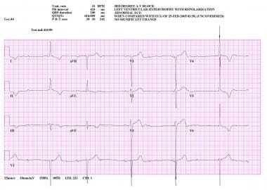
 Third-Degree Atrioventricular Block (Complete Heart Block): Background, Pathophysiology, Etiology
The actual prognostic worth of a leading Q trend throughout direct (-)aVR within intense anterior wall structure myocardial...
 Hw 8-2 Cardiovascular System - 478 Words | AntiEssays
Left anterior fascicular block - Wikipedia
Transfusion-Associated Babesiosis after Heart Transplant - Volume 9, Number 1-January 2003 - Emerging Infectious Diseases...
 Myocardial Infarction MCQs Quiz With Answers - ProProfs Quiz
Circadian variations of infarct size in acute myocardial infarction | Heart
 RePub, Erasmus University Repository:
A novel model of cryoinjury-induced myocardial infarction in the mouse: a comparison...
RePub, Erasmus University Repository:
A novel model of cryoinjury-induced myocardial infarction in the mouse: a comparison...
 Transient Mid-Ventricular Ballooning Due to Bad Dream in a Postmenopausal Woman
 Splenic Ly6Chi monocytes contribute to adverse late post-ischemic left ventricular remodeling in heme oxygenase-1 deficient...
 Cells | Free Full-Text | Preconditioning or Postconditioning with 8-Br-cAMP-AM Protects the Heart against Regional Ischemia and...
 Ventricular pseudoaneurysm secondary to myocardial infarction - an exuberant presentation | Eurorad
 Variants of arterial supply to the inferior (diaphragmatic) surface of the ventricles of the heart and the influence on age at...
 Telangana COVID-19 vaccine recipient dies; govt says no link to immunisation
 Aortic Deceleration Injury Treated by Endograft: A Case Report with 11-Year Followup
 Duration of DAPT in a Patient With PAD and ACS - American College of Cardiology
 Clinical, Angiographic Profile and Immediate Outcome of COVID-19 Patients Presenting as Acute Coronary Syndrome: An...
NIOSHTIC-2 Search Results - Full View
 Dr. Taiyeb Khumri, MD - Kansas City, MO | Cardiology
 Frequency and Criticality of Diagnoses in Family Medicine Practices: From the National Ambulatory Medical Care Survey (NAMCS) |...
His Bundle Electrogram in Patients with Acute Myocardial Infarction<...
Implantation of a three-dimensional fibroblast matrix improves left ventricular function and blood flow after acute myocardial...
 PakMediNet - Rawal Medical Journal
 Dealing with Demanding Dan
 Apex of the heart: What it is, function, and medical conditions
Dr. Wes: May 2007
![Ischemic heart diseases in Yemen [Archives:2007/1102/Health] - Yemen Times archives](data:image/png;base64,iVBORw0KGgoAAAANSUhEUgAAABAAAAAUCAYAAACEYr13AAAB8UlEQVQ4jaXRv2sTYRzH8ffnLjFRaYW00VwG7eYgSqFgc4iza6E4CdKCg4OKmxSkOIiTKHQRweIgTi7+A0KhQ0mhFARBcXHQ2uaSFFtj08Z7vg71Ss0PoXrTw+f5PK/vFw7+81MlCF8Jtkz2EwCTjzicX8lcFXPNpGicORQF/S+F7ZjYlpE2GPKOxO6GYe9lTAhNSuQy37m+/zGAeLeTd40JxDehSWArpdaVvUIUlJ5ExdCiIPxk4Hdb10CVYrgcBaUXSeYlh6xtT5tZA3EqKpTGuwH1wui4sJMZz93uAPpWlyPQzO/0roHapvvO8+5jNtX/ZbHWAQBkGjzEbEPobC0Ix/bfVYPwGsbm4Nfys/35H8CxjYU64hGAE9PJFtHghT7D7nlxfEvgegIAXtMeg9UFw1GhdBlA6fiO4M1AZXGhva/2AKAShFMSD4APKdOlltyS37ThgXr5c8fAbkDeNWbMrAqcbuHmBLPdHvcEtPa24cFTAElD7LjZbr2eAIDQPABmbrC6+PHAADGbACa1BHZwQC75XT0f/xWQLx9ARuqfABcruyuRMkbSBwbwbSQ5rhfT53tu2h5UiqWbQmNgF0HJ5HWweXP2+vhq+flet1Ca6QDsxLmjAKzltmEu3k1HUlG+mcmns04rSz+Sbi032v8LRDW76rOJY1IAAAAASUVORK5CYII=) Ischemic heart diseases in Yemen [Archives:2007/1102/Health] - Yemen Times archives
Ischemic heart diseases in Yemen [Archives:2007/1102/Health] - Yemen Times archives
 Within the last 50 years we've seen dramatic changes in cardiovascular - A high-throughput screen identifies miRNA inhibitors
Elevation10
- Objective To determine the impact of time-of-day onset of ST segment elevation myocardial infarction (STEMI) on infarct size. (bmj.com)
- The authors present a case of a 64-year-old man with a history of an inferior myocardial infarction with ST elevation 2 years ago. (eurorad.org)
- Five years back as an participating in cardiologist I accepted a 76-year-old girl using a non-ST portion elevation myocardial infarction. (sciencepop.org)
- Rheumatic cardiovascular disease and ST-elevation myocardial infarction are "offering method" to center failing degenerative valve disease and atrial fibrillation. (sciencepop.org)
- ICD-10-CM 2018: I21.0 ST elevation (STEMI) myocardial infarction of a. (medcode.ch)
- ST elevation is evident in Lead I, aVL, V5 and V6 lateral wall). (monitortech.org)
- ACS is defined as "any group of clinical symptoms compatible with acute myocardial ischemia", which includes unstable angina (UA) and myocardial infarction (MI), with or without ST-segment elevation according to American Heart Association [ 13 ]. (hindawi.com)
- AMI can be divided into 2 categories: non-ST-segment elevation myocardial infarction (NSTEMI) and ST-segment elevation myocardial infarction (STEMI). (statpearls.com)
- DIAGNOSIS: An electrocardiogram showed ST-segment elevation in the inferior wall leads, and blood tests suggested elevated troponin I levels. (bvsalud.org)
- The initial diagnosis was acute ST-segment elevation myocardial infarction. (bvsalud.org)
STEMI2
Complication of myocardial infarction2
- Cardiogenic shock is a serious complication of myocardial infarction with a high mortality rate. (proprofs.com)
- A false aneurysm is a rare complication of myocardial infarction and develops when myocardial rupture is contained by pericardial adhesions or scar tissue. (eurorad.org)
Inferior myocardial infarction1
- Arrhythmia and conduction disorders in acute inferior myocardial infarction with right ventricular involvement. (pakmedinet.com)
Ventricular9
- Left ventriculography was notable for a low normal ejection fraction of 35%, an akinetic anterior and hypokinetic mid-ventricular walls ( Figure 1 ). (scirp.org)
- Most frequently (53%), it extended into the apical two-fifths of the length of the inferior ventricular walls. (uni-koeln.de)
- An AIVA extending to the apical 40% of the length of the inferior ventricular walls may therefore be considered a common variant. (uni-koeln.de)
- This study was designed to determine if a viable biodegradable three-dimensional fibroblast construct (3DFC) patch implanted on the left ventricle after myocardial infarction (MI) improves left ventricular (LV) function and blood flow. (arizona.edu)
- Transthoracic echocardiography showed abnormal left ventricular contractility and a huge apical thrombus coating left ventricular lateral wall, with right ventricular dilatation. (researchbib.com)
- This is a large anterior lateral myocardial infarction with a very high risk for cardiac arrest from ventricular fibrillation. (monitortech.org)
- During ventricular systole, the mitral valve closes and prevents backflow to the LA. The normal function of the mitral valve depends on its 6 components, which are (1) the left atrial wall, (2) the annulus, (3) the leaflets, (4) the chordae tendineae, (5) the papillary muscles, and (6) the left ventricular wall (see the image below). (medscape.com)
- The mitral apparatus is composed of the left atrial wall, the annulus, the leaflets, the chordae tendineae, the papillary muscles, and the left ventricular wall. (medscape.com)
- Contrast-enhanced T1 mapping is mostly used for calculating extracellular volume fraction (ECV) - that is, the ratio of pre- and postcontrast myocardial T1 to that of the ventricular blood compartment T1. (medscape.com)
Ischemia3
- Acute myocardial infarction occurs due to decreased coronary blood flow, leading to insufficient oxygen supply to the heart and cardiac ischemia. (statpearls.com)
- Other etiologies of myocardial ischemia include coronary artery embolism, which accounts for 2.9% of patients, cocaine-induced ischemia, coronary dissection, and coronary vasospasm. (statpearls.com)
- After acute myocardial infarction, myocardial perfusion imaging can help estimate prognosis because it can show extent of the perfusion abnormality due to acute myocardial infarction, extent of scarring due to previous infarcts, and residual peri-infarct or other areas of reversible ischemia. (msdmanuals.com)
Rupture4
- Significantly lower incidence of left ventricle (LV) free wall rupture was noted between 3rd and 5th day after MI in Hmox1 −/− mice resulting in their better overall survival. (springer.com)
- Surgery is the treatment of choice since there is a higher risk of rupture (even several years after myocardial infarction), as well as malignant arrhythmias and embolic events. (eurorad.org)
- A rupture of interior wall was suspected (Figure 1(a) ). (hindawi.com)
- On his eighth hospital day he died of the autopsy-proven myocardial rupture abruptly. (sciencepop.org)
Infarct2
- Other infarct models include cryoinfarction, which produces reproducible infarcts of the anterior wall. (eur.nl)
- These results indicate that the cryoinfarction model represents the anterior myocardial infarct with modest adverse remodeling and may thus be representative for infarcts encountered in clinical practice. (eur.nl)
Occlusion4
- Myocardial Infarction: Occlusion of one or both of the coronary arteries will cause obliteration of the blood supply to the cardiac muscles in different walls anterior, lateral and inferior walls of the heart according to the segment affected. (yementimes.com)
- Anterior wall myocardial infarction is often caused by occlusion of the left anterior descending coronary artery. (bvsalud.org)
- Emergency coronary angiography revealed complete occlusion of the first diagonal branch, thrombolysis in myocardial infarction grade 0 flow, and smooth remaining vessels. (bvsalud.org)
- Complete occlusion of the left anterior descending artery unexpectedly occurred during interventional treatment. (bvsalud.org)
Right corona2
- He suffered an acute inferior wall myocardial infarction 11 months ago, requiring stenting of his mid-right coronary artery with an everolimus-eluting stent. (acc.org)
- As you can see below, the patient's right coronary artery that is responsible for circulation to the inferior wall of the heart is patent and continues to supply blood flow to that portion of the heart muscle. (monitortech.org)
Acute myocardial8
- Background The circadian clock influences a number of cardiovascular (patho)physiological processes including the incidence of acute myocardial infarction. (bmj.com)
- An electrocardiogram and cardiac enzymes suggested acute myocardial infarction. (scirp.org)
- However, recent studies have supported increased catecholamine levels during psychosomatic stress are believed to result in development of acute myocardial stunning and LV wall motion abnormalities. (scirp.org)
- Multiple studies have found that the incidence of hospitalization for acute Myocardial Infarction (MI) and admissions for most diagnoses have decreased by as much as 40-50% during the pandemic 6 - 8 . (scialert.net)
- Thirty‐seven patients with acute myocardial infarction complicated by atripventricular or bundle branch block, or a combingtioh of both, had His bundle electrogram studies performed during their stay in the coronary care unit. (elsevierpure.com)
- Effects of Diabetes Mellitus on outcome of patients with first acute Myocardial Infarction. (pakmedinet.com)
- Severe Epigastric pain can be one of many indicators of Acute Myocardial Infarction (AMI). (wikidoc.org)
- Acute myocardial infarction (AMI) is one of the leading causes of death in the developed world. (statpearls.com)
Septal wall1
- The posterior fascicle supplies the posterior and inferoposterior walls of the LV, the anterior fascicle supplies the upper and anterior parts of the LV and the septal fascicle supplies the septal wall with innervation. (wikipedia.org)
Prognosis1
- Medical complications often worsen the prognosis after myocardial infarction. (researchbib.com)
Echocardiography6
- A transthoracic echocardiography was performed and a posterior wall aneurysm was suspected. (eurorad.org)
- A defibrillation was successful, but the ST segment remained elevated on the lead V1-V5, with cardiac enzyme: CK3 974 U/L and CK-MB 469 U/L. Echocardiography showed no contraction of the anterior cardiac wall and ejection fraction (EF) 36%, indicating a myocardial infarction. (hindawi.com)
- Even though his echocardiography still showed no movement of the anterior cardiac wall, he was in good shape. (hindawi.com)
- Measurement of coronary sinus blood flow after first anterior myocardial infarction with transthoracic echocardiography and its correlation with wall motion scoring index. (pakmedinet.com)
- Myocardial strain imaging uses speckle-tracking echocardiography to measure tissue deformation through the cardiac cycle. (medscape.com)
- The principal utility of CMR and its advantage over echocardiography for cardiomyopathy is its use of late gadolinium enhancement (LGE) , which visualizes myocardial tissue and detects scar, fibrosis, or infiltration. (medscape.com)
Angina pectoris1
- During my work in Yemen since February 2004, I have seen 1,459 patients complaining from IHD 468 of them had myocardial infarction and the remaining had angina pectoris. (yementimes.com)
Incidence1
- During the past few decades we have seen dramatic declines in the incidence of cardiovascular death1 and myocardial infarction.2 Supported by authorities and market countless teams of fundamental translational clinical and populace scientists developed fresh paradigms for predicting avoiding and treating disease. (sciencepop.org)
Posterior wall1
- Troponin ~5 and echo showed infero-posterior wall hypokinenia. (ekgrhythm.com)
Thrombolysis1
- OUTCOMES: Ten days after admission, a repeat coronary angiography showed complete restoration of left anterior descending artery flow on its own, balloon dilation was again performed on the diagonal branch, and flow was restored to thrombolysis in myocardial infarction grade 1. (bvsalud.org)
Anteroseptal2
- citation needed] LAHB may be a cause of poor R wave progression across the precordium causing a pseudoinfarction pattern mimicking an anteroseptal infarction. (wikipedia.org)
- It can be categorized as anteroseptal or anterolateral wall myocardial infarction. (bvsalud.org)
Outflow tract1
- However, the peroperative ultrasonographic finding documented prolapse of the anterior cusp of the mitral valve into the outflow tract of left ventricle. (mefanet.cz)
Ejection fraction3
- One month follow up echocardiogram showed normal LV ejection fraction without regional wall motion abnormalities. (scirp.org)
- CMR showed a severely impaired left ventricle (LV) function (ejection fraction 27%) and it revealed the presence of an aneurismal dilation along the basal and medium segments of the lateral and inferolateral walls (Fig. 1a-c) with an inlet orifice of approximately 3 cm in diameter. (eurorad.org)
- 0.05) ejection fraction (37 ± 3% to 62 ± 5%), increasing regional systolic displacement of the infarcted wall (0.04 ± 0.02 to 0.11 ± 0.03 cm), and shifting the passive LV diastolic pressure volume relationship toward the pressure axis. (arizona.edu)
Scar1
- CMR identifies areas of increased interstitial space where gadolinium is retained (eg, areas of myocardial infarction, acute edema/inflammation due to capillary leak, myocardial fibrosis/scar, or amyloid protein infiltration). (medscape.com)
Anterolateral1
- Two of these indentations, the anterolateral and posteromedial commissures, divide the leaflets into anterior and posterior (see the first image below). (medscape.com)
Ligation2
- RePub, Erasmus University Repository: A novel model of cryoinjury-induced myocardial infarction in the mouse: a comparison with coronary artery ligation. (eur.nl)
- Hmox1 knockout (Hmox1 −/− ) and wild-type (WT, Hmox1 +/+ ) mice were subjected to a permanent ligation of the left anterior descending coronary artery. (springer.com)
Cardiac arrest2
- As emergency angioplasty and stent placement of his left anterior descending coronary artery was started, the FF had an arrhythmia and cardiac arrest. (cdc.gov)
- The death certificate (completed by the coroner) listed "cardiac arrest with electromechanical dissociation" due to "acute anterior wall myocardial infarction" due to "coronary artery disease" (CAD) as the cause of death. (cdc.gov)
Fibrosis3
- Delayed gadolinium enhancement is now accepted as a useful and important tool to detect myocardial fibrosis and to help make this differentiation [3]. (eurorad.org)
- Parametric T1 and T2 mapping sequences can be used for evaluation of diffuse, interstitial myocardial fibrosis that is not detected by LGE imaging. (medscape.com)
- T1 and T2 mapping better characterize myocardial tissue by identifying areas of edema (T2), inflammation, and more diffuse fibrosis (T1), which may aid in diagnosing myocarditis without LGE, amyloidosis , hemochromatosis , and Fabry disease . (medscape.com)
Occurs2
- LAFB - which is also known as left anterior hemiblock (LAHB) - occurs when a cardiac impulse spreads first through the left posterior fascicle, causing a delay in activation of the anterior and upper parts of the LV. (wikipedia.org)
- A myocardial infarction occurs when plaque builds up or a clot obstructs one of these arteries, starving the muscle of blood and oxygen, leading to very quick death of the tissues. (uaf.edu)
Troponin1
- He was admitted under the expert care of Dr. Tanmoy Majee (Consultant Cardiologist) and after a thorough evaluation and relevant investigations the ECG revealed Acute Anterior Wall Myocardial Infarction (AWMI) and Trop - T (Troponin) test had been positive. (rubyhospital.com)
Coronary arteries2
- The coronary arteries stem from the arch of the aorta by three dilations in the wall of the aorta superior to the aortic semilunar valve. (uaf.edu)
- The left anterior descending artery, which is one of the larger of the coronary arteries, also stems from the left coronary artery and follows down the anterior portion of the heart and around the pulmonary trunk. (uaf.edu)
Electrocardiogram2
- a patient history of severe, prolonged chest pain, unequivocal electrocardiogram (ECG) changes that include abnormal and persistent Q waves,changes in serial cardiac biomarker levels that indicate myocardial injury and infarction. (antiessays.com)
- After an exam, which was normal, and an electrocardiogram, which showed an old anterior wall MI, I ordered an echocardiogram and stress test. (physicianspractice.com)
Heart12
- It may also show any thickening of the walls of the pumping chambers of the heart. (antiessays.com)
- Left anterior fascicular block (LAFB) is an abnormal condition of the left ventricle of the heart, related to, but distinguished from, left bundle branch block (LBBB). (wikipedia.org)
- Otherwise and commonly known as a heart attack, Myocardial Infarction, abbreviated as MI, is when the blood decreases in the heart and stops. (proprofs.com)
- Early angiography studies have shown that the anterior interventricular artery (AIVA), a branch of the LCA, often continues on the inferior surface of the heart and may replace the inferior interventricular artery. (uni-koeln.de)
- A heart attack in medical parlance is called myocardial infarction. (org.in)
- He told me that five years earlier he'd had a myocardial infarction followed by open-heart surgery. (physicianspractice.com)
- MYOCARDIAL INFARCTION in which the anterior wall of the heart is involved. (bvsalud.org)
- A myocardial infarction is what you might know as a heart attack, because it is quite literally an "attack" on muscles of the heart, as they die from the lack of blood and oxygen. (uaf.edu)
- He had been scheduled for Coronary Angiography which revealed Single Vessel Coronary Artery Disease (A condition in which the walls of the small arteries in the heart do not work properly). (rubyhospital.com)
- The LAD is a very large blood vessel that is responsible for blood flow to most of the anterior wall of the heart. (monitortech.org)
- You can see from the above angiogram that blood flow has been restored to a large amount of heart muscle by opening the left anterior descending artery that was occluded when this gentleman came into the emergency department. (monitortech.org)
- Myocardial infarction (MI) causes permanent damage to the heart muscle due to inadequate oxygen supply. (statpearls.com)
Pulmonary1
- Myocardial Infarction Pulmonary Embolism. (researchbib.com)
Complications1
- True and false LV aneurysms are both complications of myocardial infarction. (eurorad.org)
Mortality1
- The acute mortality of the 16 patients with an inferior wall myocardial infarction was 6% The H‐V interval was normal in all but one of these patients. (elsevierpure.com)
Abnormalities1
- It is characterized by transient wall motion abnormalities of the mid-segment of the left ventricle with apical sparing. (scirp.org)
Symptoms3
- Tissue plasminogen activator (tPA) is considered for treatment of a patient who arrives in the emergency department following onset of symptoms of myocardial infarction. (proprofs.com)
- A patient arrives in the emergency department with symptoms of myocardial infarction, progressing to cardiogenic shock. (proprofs.com)
- These are clear symptoms of myocardial infarction (MI) and the death is not related to vaccination," he said. (org.in)
Diagnosis of anterior2
- The ®rst set was used to train an interpretation techniques have been used in interpre- arti®cial neural network for the diagnosis of anterior tation programs. (lu.se)
- This completes the diagnosis of anterior lateral wall myocardial infarction. (monitortech.org)
Bundle2
- It is caused by only the left anterior fascicle - one half of the left bundle branch being defective. (wikipedia.org)
- Normal activation of the left ventricle (LV) proceeds down the left bundle branch, which consist of three fascicles, the left anterior fascicle, the left posterior fascicle, and the septal fascicle. (wikipedia.org)
Infiltration1
- Consequently, a greater post-MI monocyte-derived myocardial macrophage infiltration was noted in Hmox1-deficient individuals. (springer.com)
Artery6
- Coronary artery systems of the inferior wall of the ventricles vary considerably. (uni-koeln.de)
- Coronary angiography revealed a disruption of the proximal left anterior descending artery with a 95% stenosis. (hindawi.com)
- He had mild nonobstructive coronary artery disease in the left circumflex and left anterior descending arteries. (acc.org)
- Along the left anterior descending artery, stems tiny branches, which also connect to the posterior descending artery, and form anastomosis. (uaf.edu)
- The most common location of these blockages are in the left anterior descending artery. (uaf.edu)
- It reveals a totally occluded left anterior descending artery (LAD) and left circumflex obtuse marginal branch. (monitortech.org)
Myocardium1
- True aneurysms represent areas of bulging, scarred and thinned myocardium, which are dyskinetic and usually located at the anterior wall and apex. (eurorad.org)
Vessels2
- 2. False Systolic pressure is the lowest pressure against the walls of the blood vessels. (antiessays.com)
- The posterior and anterior aortic sinuses stem the left and right coronary vessels. (uaf.edu)
Lateral and inferior1
- Usually, they have a narrow ostium connecting the aneurysmal sac and they are located mostly in the lateral and inferior wall. (eurorad.org)
Reproducible1
- Myocardial strain imaging, a reproducible measure of LV systolic function, can identify subclinical LV dysfunction and patterns that suggest specific cardiomyopathies (eg, ischemic cardiomyopathy, amyloidosis, hypertrophic cardiomyopathy, chemotherapy-induced cardiotoxicity). (medscape.com)
Blood1
- This biodegradable 3DFC patch improves LV function and myocardial blood flow 3 weeks after MI. (arizona.edu)
Patient2
- Following myocardial infarction, a hospitalized patient is encouraged to practice frequent leg exercises and ambulate in the hallway as directed by his physician. (proprofs.com)
- 2 subsequent episode of care Use fifth-digit 2 to designate an episode of care following the initial episode when the patient is admitted for further observation, evaluation or treatment for a myocardial infarction that has received initial treatment, but is still less than 8 weeks old. (theodora.com)