Aortic Arch Syndromes
Vocal Cord Paralysis
Recurrent Laryngeal Nerve
Subclavian Artery
Recurrent Laryngeal Nerve Injuries
Lynch Syndrome II
Laryngeal Nerves
Surgical treatment of nonaneurysmal aortic arch lesions in patients with systemic embolization. (1/58)
PURPOSE: Atherosclerotic lesions of the aortic arch are potential sources of arterial embolism. Here we investigate whether surgery, with the necessary circulatory supports, can be proposed as a good option for treatment of this problem. Study of these lesions on a national scale in France has made possible the assessment for future indications of techniques and results of the surgical management of aortic arch lesions, which retrospectively proved to be embolic. METHODS: Thirty-eight patients, (19 men and 19 women) underwent surgery between 1976 and 1996 in 17 French cardiovascular surgical centers. The average age at the time of surgery was 49 +/- 12 years (range, 31 to 82 years). Atherosclerotic lesions were detected with transesophagial echocardiography (n = 19), angiography of the aortic arch (n = 16), computed tomography (n = 9), and magnetic resonance imaging (n = 10). Surgery consisted of thrombectomy and endarterectomy (n = 22), aortic resection and graft replacement (n = 10), and patch aortoplasty (n = 5; one thrombus disappeared spontaneously before surgery was performed). RESULTS: The average postoperative period was 30 months (range, 3 to 82 months). Contact was lost with four patients after a follow-up period of 12 months. On pathologic specimens obtained at surgery, an atherosclerotic plaque was found in 73% of the cases (n = 28). In 15% of the cases, the aorta appeared normal (n = 6) and four other types of lesion were identified: angiosarcoma (n = 1), ectasia at the insertion of the remains of the ductus arteriosus (n = 1), rupture of tunica intima (n = 1), and a fibroblastic plaque (n = 1). A thrombus was identified in 26 cases, attached to the arterial wall in 18 cases. When transesophagial echocardiographic results showed mobile lesions (n = 22), histopathologic examination of specimens allowed the detection of a thrombus in 18 cases and an atherosclerotic plaque with a mobile projection in four cases. The postoperative mortality rate was 2.6%. The morbidity rate (28.9%; n = 11) was related to neurologic complications (n = 6), vascular complications (n = 4), and infection (n = 1). Four cases (12%) were reoperated. CONCLUSION: Nonaneurysmal aortic arch lesions are a frequent and still underestimated source of stroke and peripheral embolization. Surgery with circulatory support can be recommended in good operative candidates with recurrent critical events despite medical management and with high embolic potential (young patients with no calcified plaques). (+info)PARADOXES OF TAKAYASU'S DISEASE. (2/58)
Takayasu's disease (or arteritis) has been defined as an "idiopathic aortitis usually affecting young women." It can come to light from very spectacular and often quite puzzling clinical manifestations. Six cases of Takayasu's disease were investigated at the UCLA Hospital in the years 1961-1962, and signs and symptoms of central nervous system involvement were found in five of the patients. This relatively high incidence of neurological deficit prompted a review of case reports in the literature and this in turn led to a series of "unexpected" findings in the historical evolution of the illness as well as in its anatomopathological aspects. The study indicated that Takayasu's disease is frequently associated with neurological manifestations, at times very severe. In addition, the disease appears to be far more extensive than its classical description suggests. New criteria for the diagnosis of Takayasu's disease must include, among other things, special emphasis on the disseminated nature of the disease. (+info)Common variable immunodeficiency syndrome with right aortic arch: a case report. (3/58)
BACKGROUND: Common variable immunodeficiency syndrome predominantly affects adults. It is characterized by low production of all the major classes of immunoglobulins. We report a case of common variable immunodeficiency syndrome with right aortic arch. An association of right-sided arch and common variable immunodeficiency syndrome has not been previously reported. CASE PRESENTATION: A 41-year-old female patient presented with a history of recurrent pneumonia, sinusitis, otitis media, diarrhoea, cystitis since childhood. Biochemical and immunocytochemical analysis revealed common variable immunodeficiency syndrome and radiological evaluation confirmed right aortic arch and aberrant left subclavian artery. CONCLUSION: Common variable immunodeficiency syndrome syndrome is a clinical entity that should be kept in mind in patients with recurrent infections of different sites. (+info)Subclavian steal in Takayasu's arteritis. A hemodynamic study by means of ultrasonic Doppler flowmetry. (4/58)
Blood flow in the vertebral artery and the upper extremity was studied in five cases of Takayasu's arteritis with subclavian steal by use of ultrasonic Doppler flowmetry and finger plethysmography. The diagnosis of subclavian steal was made by observation of flow reversal in the vertebral artery on the subclavian steal side during grip exercise and, in addition, the vertebral flow change with brachial artery occlusion. The blood flow increase of both internal cartotid and non-affected (non-subclavian steal side) vertebral arteries during a common carotid compression was almost normal in patients with Takayasu's arteritis in this study. During carotid compression on the side of the subclavian steal, ipsilateral vertebral blood flow greatly decreased, and the amplitude the ipsilateral finger plethysmogram decreased slightly or moderately. It is suggested that there are significantly important factors in suppressing sumptoms of vertebrobasilar ischemia in these patients with Takayasu's arteritis with subclavian steal. These factors are believed to be (1) good function of the circle of Willis, (2) good blood supply to the brain stem, and (3) collateral circulation to the distal subclavian artery not via the vertebral artery. (+info)Major vascular anomalies in Turner syndrome: prevalence and magnetic resonance angiographic features. (5/58)
BACKGROUND: Turner syndrome (TS) is associated with aortic coarctation and dissection; hence, echocardiographic evaluation of all patients is currently recommended. X-ray angiography in clinically symptomatic patients has suggested a range of other vascular anomalies, but the true prevalence of such lesions in TS is unknown. To better understand the prevalence and pathogenesis of cardiovascular defects in TS, we prospectively evaluated a group of asymptomatic adult volunteers with TS using magnetic resonance (MR) angiography. METHODS AND RESULTS: A total of 85 adults with TS and 27 normal female adult volunteers underwent gadolinium-enhanced 3D MR angiography. A high prevalence of aortic anomalies was seen in women with TS, including elongation of the transverse arch (49%), aortic coarctation (12%), and aberrant right subclavian artery (8%). Venous anomalies were also prominent, including persistent left superior vena cava (13%) and partial anomalous pulmonary venous return (13%). None of these anomalies were found in healthy female controls. The constellation of elongation of the transverse arch, aortic coarctation, and persistent left superior vena cava was significantly associated with women with TS. Neck webbing and increased thoracic anterior-to-posterior dimension diameters were strong predictors for arterial and venous anomalies. CONCLUSIONS: Thoracic vascular anomalies are common in TS, occurring in approximately 50% of a group not preselected for cardiovascular disease. The highly significant association between neck webbing, increased chest diameter, and these vascular anomalies suggests that in utero, centrally localized lymphatic obstruction may contribute to these cardiovascular deformities in TS. Improved recognition of these often-undetected vascular lesions may be important for identification of patients in need of closer cardiovascular monitoring. (+info)Aortic root replacement with a freestyle stentless valve for aortitis syndrome with ascending aortic aneurysm and aortic regurgitation. (6/58)
A 47-year-old woman who had been diagnosed as having aortitis syndrome underwent aortic root replacement for an ascending aortic aneurysm and aortic regurgitation. Because the patient has been treated with steroids for more than 20 years, a Freestyle stentless valve was used to avoid the risk of valve detachment. There were no complications observed during the postoperative course. Although long-term follow-up will be necessary to observe the valve durability, the Freestyle stentless valve seems to be useful for aortic root replacement in patients at high risk of valve detachment due to aortitis syndrome. (+info)Protruding aortic arch thrombus: treatment with minimally invasive surgical approach. (7/58)
BACKGROUND: Protruding aortic arch thrombus is associated clinically with life-threatening emboli. Definitive treatment for aortic arch thrombus removal has demanded complicated vascular surgical procedures, with high morbidity and mortality. METHODS AND RESULTS: Transesophageal echocardiography (TEE) enabled diagnosis of a protruding thrombus at the aortic arch in 5 patients, and a simultaneous lesion in the descending aorta in 1 patient. Four patients had visceral emboli, coinciding with peripheral emboli in 2 patients, and the fifth patient had peripheral and cerebral emboli. One patient had had ischemic stroke and femoral emboli a few months previously. Mean patient age was 51 years. None had clinical evidence of coronary or peripheral atherosclerotic occlusive disease. Risk factors included hypertension (n = 2), smoking (n = 4), and preexisting thrombophilia (n = 4). Five patients underwent TEE-guided aortic balloon thrombectomy from the arch with a 34-mm occluding balloon catheter. One patient also underwent balloon thrombectomy from the descending aorta with a 14F Foley catheter. Access into the aorta was obtained through the iliac artery (n = 4) during laparotomy because of visceral ischemia or through the transfemoral approach (n = 2). Previous procedures included superior mesenteric embolectomy (n = 3), segmental bowel resection (n = 1), splenectomy (n = 1), and peripheral arterial embolectomy n = 3). Real-time intraoperative TEE enabled visualization of the protruding thrombus and assisted with maneuvering of the balloon catheter. At completion peripheral thrombectomy thrombus material was retrieved in 4 patients. Postoperatively there were no clinically proved new procedure-related visceral emboli, and all patients received anticoagulant therapy thereafter. Follow-up TEE within 2 weeks and up to 7 years revealed no recurrent aortic arch thrombus. CONCLUSIONS: TEE-guided aortic balloon thrombectomy used in 6 procedures was effectively completed without visceral or peripheral ischemic complications. It enabled removal of the life-threatening source of emboli from the proximal aorta, thereby averting the need of major aortic surgery. (+info)Unusual vascular ring anomaly in a foal. (8/58)
A 2.5-month-old filly was presented with signs of esophageal obstruction. The filly was euthanized and postmortem examination revealed a vascular ring anomaly. The vascular ring anomaly was not caused by a persistent right aortic arch, which is the only vascular ring anomaly reported to occur in horses. (+info)Aortic arch syndromes are a group of conditions that affect the aortic arch, which is the curved portion of the aorta that arises from the left ventricle of the heart and gives rise to the major branches of the arterial system. These syndromes are typically caused by congenital abnormalities or degenerative changes in the aorta and can result in various complications, such as obstruction of blood flow, aneurysm formation, and dissection.
There are several types of aortic arch syndromes, including:
1. Coarctation of the Aorta: This is a narrowing of the aorta at the point where it leaves the heart, just distal to the origin of the left subclavian artery. It can cause hypertension in the upper extremities and reduced blood flow to the lower extremities.
2. Aortic Arch Aneurysm: This is a localized dilation or bulging of the aorta in the region of the aortic arch. It can lead to dissection, rupture, or embolism.
3. Aortic Arch Dissection: This is a separation of the layers of the aortic wall, which can result from hypertension, trauma, or genetic disorders such as Marfan syndrome. It can cause severe chest pain, shortness of breath, and shock.
4. Kommerell's Diverticulum: This is an outpouching or bulge in the aorta at the origin of the ligamentum arteriosum, which is a remnant of the ductus arteriosus. It can cause compression of the airways or esophagus and increase the risk of dissection or rupture.
5. Abernethy Malformation: This is a rare congenital anomaly in which there is an abnormal connection between the portal vein and systemic venous circulation, leading to the bypass of the liver. It can cause various complications such as hepatic encephalopathy, pulmonary hypertension, and liver tumors.
The diagnosis and management of aortic arch syndromes require a multidisciplinary approach involving cardiologists, radiologists, surgeons, and other specialists. Treatment options may include medications, endovascular procedures, or surgical interventions depending on the severity and location of the lesion.
Hoarseness is a condition characterized by an abnormal change in the quality of voice, making it sound rough, breathy, strained, or weak. Medically, it's described as a disorder of phonation, which is the process of producing sound by vibrating the vocal cords in the larynx (voice box). Hoarseness can be caused by various factors, such as inflammation, irritation, or injury to the vocal cords, and may result in symptoms like altered voice pitch, volume, and clarity. It's essential to consult a healthcare professional if hoarseness persists for more than two weeks, especially if it's accompanied by other concerning symptoms like difficulty swallowing or breathing.
Vocal cord paralysis is a medical condition characterized by the inability of one or both vocal cords to move or function properly due to nerve damage or disruption. The vocal cords are two bands of muscle located in the larynx (voice box) that vibrate to produce sound during speech, singing, and breathing. When the nerves that control the vocal cord movements are damaged or not functioning correctly, the vocal cords may become paralyzed or weakened, leading to voice changes, breathing difficulties, and other symptoms.
The causes of vocal cord paralysis can vary, including neurological disorders, trauma, tumors, surgery, or infections. The diagnosis typically involves a physical examination, including a laryngoscopy, to assess the movement and function of the vocal cords. Treatment options may include voice therapy, surgical procedures, or other interventions to improve voice quality and breathing functions.
The Recurrent Laryngeal Nerve (RLN) is a branch of the vagus nerve (cranial nerve X), which is a mixed sensory, motor, and autonomic nerve. The RLN has important functions in providing motor innervation to the intrinsic muscles of the larynx, except for the cricothyroid muscle, which is supplied by the external branch of the superior laryngeal nerve.
The recurrent laryngeal nerve supplies all the muscles that are responsible for adduction (bringing together) of the vocal cords, including the vocalis muscle, lateral cricoarytenoid, thyroarytenoid, and interarytenoid muscles. These muscles play a crucial role in voice production, coughing, and swallowing.
The right recurrent laryngeal nerve has a longer course than the left one. It loops around the subclavian artery in the chest before ascending to the larynx, while the left RLN hooks around the arch of the aorta. This anatomical course makes them vulnerable to injury during various surgical procedures, such as thyroidectomy and neck dissection, leading to potential voice impairment or vocal cord paralysis.
The subclavian artery is a major blood vessel that supplies the upper limb and important structures in the neck and head. It arises from the brachiocephalic trunk (in the case of the right subclavian artery) or directly from the aortic arch (in the case of the left subclavian artery).
The subclavian artery has several branches, including:
1. The vertebral artery, which supplies blood to the brainstem and cerebellum.
2. The internal thoracic artery (also known as the mammary artery), which supplies blood to the chest wall, breast, and anterior mediastinum.
3. The thyrocervical trunk, which gives rise to several branches that supply the neck, including the inferior thyroid artery, the suprascapular artery, and the transverse cervical artery.
4. The costocervical trunk, which supplies blood to the neck and upper back, including the posterior chest wall and the lower neck muscles.
The subclavian artery is a critical vessel in maintaining adequate blood flow to the upper limb, and any blockage or damage to this vessel can lead to significant morbidity, including arm pain, numbness, weakness, or even loss of function.
Recurrent laryngeal nerve injuries refer to damages or trauma inflicted on the recurrent laryngeal nerve, which is a branch of the vagus nerve that supplies motor function to the intrinsic muscles of the larynx, except for the cricothyroid muscle. This nerve plays a crucial role in controlling vocal fold movement and swallowing.
Injuries to this nerve can result in voice changes, hoarseness, or even complete loss of voice, depending on the severity and location of the injury. Additionally, it may also lead to breathing difficulties, coughing, and choking while swallowing due to impaired laryngeal function.
Recurrent laryngeal nerve injuries can occur due to various reasons, such as surgical complications (particularly during thyroid or neck surgeries), tumors, infections, inflammation, or direct trauma to the neck region. In some cases, these injuries may be temporary and resolve on their own or through appropriate treatment; however, severe or prolonged injuries might require medical intervention, including possible surgical repair.
Lynch Syndrome II is a genetic disorder also known as Hereditary Non-Polyposis Colorectal Cancer (HNPCC) type II. It is characterized by an increased risk of developing certain types of cancer, including colorectal, endometrial, stomach, small intestine, pancreas, kidney, urinary tract, brain, and skin cancers.
Unlike Lynch Syndrome I (HNPCC type I), which primarily involves mutations in the MLH1 or PMS2 genes, Lynch Syndrome II is caused by mutations in the MSH2 or MSH6 genes. These genes are responsible for DNA mismatch repair, and their malfunction leads to an accumulation of errors during DNA replication, increasing the risk of cancer development.
Individuals with Lynch Syndrome II have a higher lifetime risk of colorectal cancer (up to 80%) and endometrial cancer (up to 60%). The onset of these cancers tends to occur at an earlier age compared to sporadic cases. It is essential for individuals with Lynch Syndrome II to undergo regular cancer screening, including colonoscopies and gynecological examinations, to facilitate early detection and treatment of potential malignancies.
In addition to increased cancer risks, individuals with Lynch Syndrome II may also experience other clinical features such as café-au-lait spots or sebaceous gland tumors. Genetic counseling and testing are recommended for individuals with a family history suggestive of Lynch Syndrome II to assess their risk and develop appropriate surveillance strategies.
The laryngeal nerves are a pair of nerves that originate from the vagus nerve (cranial nerve X) and provide motor and sensory innervation to the larynx. There are two branches of the laryngeal nerves: the superior laryngeal nerve and the recurrent laryngeal nerve.
The superior laryngeal nerve has two branches: the external branch, which provides motor innervation to the cricothyroid muscle and sensation to the mucous membrane of the laryngeal vestibule; and the internal branch, which provides sensory innervation to the mucous membrane of the laryngeal vestibule.
The recurrent laryngeal nerve provides motor innervation to all the intrinsic muscles of the larynx, except for the cricothyroid muscle, and sensation to the mucous membrane below the vocal folds. The right recurrent laryngeal nerve has a longer course than the left one, as it hooks around the subclavian artery before ascending to the larynx.
Damage to the laryngeal nerves can result in voice changes, difficulty swallowing, and respiratory distress.
 Ortner's syndrome
Interrupted aortic arch
Aortic arch anomaly - peculiar facies - intellectual disability
Takayasu's arteritis
DiGeorge syndrome
TBX1
Fertilysin
PHACE syndrome
Adams-Oliver syndrome
Right-sided aortic arch
Yasui procedure
Double aortic arch
Hypoplastic right heart syndrome
Branchial
Circle of Willis
Intra-aortic balloon pump
Inflammatory aortic aneurysm
Median arcuate ligament syndrome
Aberrant subclavian artery
Cardiac neural crest
List of syndromes
Fryns syndrome
Vocal cord paresis
List of ICD-9 codes 740-759: congenital anomalies
Cyanotic heart defect
Pulse
Tetralogy of Fallot
LeCompte maneuver
Prostaglandin E1
Fetal aortic stenosis
 Aortic arch syndrome: MedlinePlus Medical Encyclopedia
 Right Aortic Arch in Vascular Ring Defects: Background, Epidemiology, Etiology
 Takayasu Arteritis - Bone, Joint, and Muscle Disorders - Merck Manuals Consumer Version
Ortner's syndrome - Wikipedia
Telescopic aortic arch: A new entity in marfan syndrome Teleskopik arkus aorta: Marfan sendromunda yeni bir antite | AVESİS
 Rare Case of a Newborn with Heterotaxy Syndrome, Right Aortic Arch and an Isolated Left Brachiocephalic Artery Arising from a...
Genetics of Ehlers-Danlos Syndrome Treatment & Management: Medical Care, Surgical Care, Consultations
Critical congenital heart disease: MedlinePlus Genetics
 A Helping Hand for Children with Congenital Heart Disease | Maxim Healthcare Services
 Michael Joseph Luceri, DO - Pediatric Cardiology
Nak Hyun Choi - Pediatric Cardiology
 urofacial syndrome - Ontology Browser - Rat Genome Database
 Abnormalities of the heart and great arteries in first trimester chromosomally abnormal fetuses
Austrian Syndrome Associated with Pandemic (H1N1) 2009 in Child - Volume 16, Number 9-September 2010 - Emerging Infectious...
Survival and morbidity after diagnosis of occlusive thromboaortopathy (Takayasu's disease)
Takayasu arteritis
 Heersink School of Medicine News | UAB
Heersink School of Medicine News | UAB
 Arteritis: Causes, Types & Diagnosis
Pediatric Holt-Oram Syndrome Treatment & Management: Medical Care, Surgical Care, Long-Term Monitoring
 Inpatient Hospitalization Costs Associated with Birth Defects Among Persons Aged 65 Years - United States, 2019 | MMWR
 Dr. Heather Sun - Fetal Cardiology Director | Rady Children's Hospital
 Fontan Ten Commandments Revisited and Revised - HGExperts.com
 Changes in the diagnosis and management of acute aortic syndrome and associated mortality in the last 20 years | Revista...
 Cord
Cord
 The Aortic and Renovascular Center for Hypertension (ARCH) | St. Louis Children's Hospital
neonatal cardiology
Articoli scritti da gerbonis
Hypoplastic left heart syndrome-outcome and management | Archives of Disease in Childhood
Hypoplastic11
- The aortic valve was mildly hypoplastic and the main pulmonary artery moderately dilated along with suspicion for a single coronary artery. (heraldopenaccess.us)
- The previously noted right aortic arch was mildly hypoplastic and gave rise to the right common carotid and right subclavian arteries. (heraldopenaccess.us)
- Mild degrees of aortic arch obstruction are common following Norwood palliation for hypoplastic heart syndrome. (hgexperts.com)
- Coarctation of the aorta, interrupted aortic arch, and hypoplastic left heart syndrome in three generations. (gerboni.net)
- In this issue of Archives of Disease in Childhood , the Guy's group present their experience with staged reconstructive surgery for hypoplastic left heart syndrome (HLHS). (bmj.com)
- Preoperative and postoperative Norwood stage I circulation in hypoplastic left heart syndrome: the balance between the systemic and pulmonary circulations is crucial. (bmj.com)
- Our journey begins at the 20 week scan, when our Little Beatrice was diagnosed with Hypoplastic Left Heart Syndrome. (tinytickers.org)
- At our next scan they told us her heart had in fact grown and was repairing itself .It was definitely not Hypoplastic Left Heart Syndrome, but she would need a Bi Ventricular repair as the left side of her heart was still slender. (tinytickers.org)
- Photograph showing hypoplastic right thumb of the right hand of a 6-month-old infant with Holt-Oram syndrome. (medscape.com)
- In a baby with a coarctation, the aortic arch also might be smaller than usual ( hypoplastic ). (kidshealth.org)
- Outcomes for the superior cavopulmonary connection in children with hypoplastic left heart syndrome: a 30-year experience. (chop.edu)
Coarctation4
- This right sided arch showed no evidence of coarctation. (heraldopenaccess.us)
- Fetal Diagnosis of Dextroposition, Left Pulmonary Artery Sling, Partial Anomalous Left Pulmonary Artery, and Aortic Coarctation. (rchsd.org)
- Double Choker: Double Aortic Arch with Bilateral Aortic Coarctation Associated with Heterotaxy-Asplenia Syndrome and Complex Atrioventricular Canal Defect. (rchsd.org)
- A cervical arch on either side, variable laterality of the descending thoracic aorta, coarctation of the major arch, and/or discontinuity of the central pulmonary arteries may be present. (medscape.com)
Aneurysm3
- These patients were classified according to the presence and severity of four major complications (Takayasu's retinopathy, secondary hypertension, aortic regurgitation and aortic or arterial aneurysm) attributed to Takayasu's disease at the time when the diagnosis was established: no complications (group I) or mild single complication (group IIa) and severe single complication (group IIb) or multiple complications (group III). (nih.gov)
- Abdominal Aortic Aneurysm (AAA), a focal enlargement of the abdominal aorta is an ongoing process that can be affected by many parameters. (asme.org)
- Aortic media necrosis and aortic dissection without aneurysm formation]. (bvsalud.org)
Aorta12
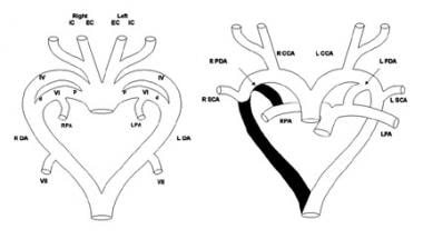
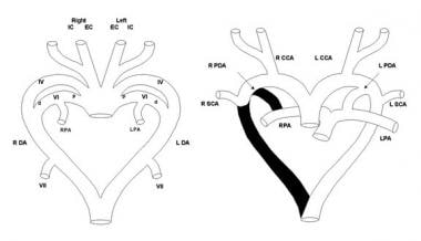
- As normal cardiovascular morphogenesis proceeds, a patterned regression and persistence of the various arches and right-sided dorsal aorta occur, ultimately resulting in the mature configuration of the thoracic aorta and its branches. (medscape.com)
- The third, fourth, and sixth arches, along with the seventh intersegmental arteries and the left dorsal aorta, are the primary contributors to the normal aortic arch and its major thoracic branches (see image below). (medscape.com)
- The segments of the bilateral aortic arch system that normally regress include the distal portion of the sixth arch and the right-sided dorsal aorta. (medscape.com)
- Normally, the left fourth arch becomes the aortic arch, the right fourth arch contributes to the innominate artery, the distal left sixth arch becomes the ductus arteriosus, the proximal sixth arches bilaterally contribute to the proximal branch pulmonary arteries, the left dorsal aorta becomes the descending thoracic aorta, and the dorsal intersegmental arteries bilaterally become the subclavian arteries. (medscape.com)
- A right aortic arch is formed when the right dorsal aorta remains patent and either the left fourth arch or the left dorsal aorta regress abnormally (see image below). (medscape.com)
- Examination of aorta showed a second/inner aortic arch just as a tube lying inside the aortic arch. (bezmialem.edu.tr)
- The inner aortic arch was arrised 2.5 cm above aortic valves and lasted at the beginning of the descending aorta. (bezmialem.edu.tr)
- The reported incidence is 1-1.5 in 10,000 live births.Right-sided aortic arch with isolation of the innominate artery is an extremely rare congenital anomaly, in which the innominate artery loses its connection with the ascending aorta and is supplied by either a patent ductus or mediastinal collaterals [1]. (heraldopenaccess.us)
- Perform a baseline echocardiogram, including views of the aortic arch and aorta. (medscape.com)
- and proximal dilatation of the aorta can cause aortic regurgitation , dilated cardiomyopathy , and congestive heart failure . (logicalimages.com)
- When a child is diagnosed with midaortic syndrome - a rare condition where part of the aorta (the largest blood vessel coming from the heart) and its major branches narrow - this can lead to an impaired blood flow to vital organs. (stlouischildrens.org)
- Middle aortic syndrome, also known as midaortic syndrome - narrowing of part of the aorta (the main artery of the heart) and its major branches, leading to impaired blood flow to vital organs in the chest, abdomen and the lower limbs. (stlouischildrens.org)
Left pulmon1
- Right: Mature anatomy of a vascular ring formed by a right aortic arch with an aberrant left subclavian artery arising from a retroesophageal diverticulum with a left-sided ligamentum arteriosum to the left pulmonary artery. (medscape.com)
Truncus arteriosus1
- 4 7 Conotruncal heart defects most commonly found in DGS/VCFS patients with 22q11.2 deletions are interrupted aortic arch (IAA) type B, truncus arteriosus (TA), and tetralogy of Fallot (TOF). (bmj.com)
Aneurysms2
- When considering cardiovocal syndrome, the most common historical cause is a dilated left atrium due to mitral stenosis, but other causes, including pulmonary hypertension, thoracic aortic aneurysms, an enlarged pulmonary artery and aberrant subclavian artery syndrome have been reported compressing the nerve. (wikipedia.org)
- Progressive weakness of your artery wall results in aortic aneurysms, essentially blisters of the walls of the blood vessels, to form. (healthline.com)
Double Aortic Arch8
- Double aortic arch is one of the 2 most common forms of vascular ring, a class of congenital anomalies of the aortic arch system in which the trachea and esophagus are completely encircled by connected segments of the aortic arch and its branches. (medscape.com)
- Although the double aortic arch has various forms, the common defining feature is that both the left and right aortic arches are present. (medscape.com)
- The easiest way to understand the anatomy and development of double aortic arch and other forms of vascular ring is to begin by considering the bilateral system of pharyngeal arch vessels in the early embryo. (medscape.com)
- A double aortic arch is formed when both fourth arches and both dorsal aortas remain present. (medscape.com)
- Double aortic arch has various forms. (medscape.com)
- In more than 75% of patients with double aortic arch, the right arch is dominant. (medscape.com)
- Most people with double aortic arch will require surgery. (umms.org)
- Babies who have breathing problems due to double aortic arch usually continue to have breathing problems for a few weeks to months after surgery because the cartilage in the trachea is still soft. (umms.org)
Bicuspid aorti2
- Fetal echocardiographic features associated with bicuspid aortic valve. (uchicago.edu)
- Aortopathy Including Hereditary Disease (Marfan Syndrome, Bicuspid Aortic Valve, etc.) -- 14. (nshealth.ca)
Stenosis4
- An inflammatory disease called Takayasu syndrome may result in narrowing (stenosis) of the vessels of the aortic arch. (medlineplus.gov)
- Common examples include mild aortic arch obstruction, branch pulmonary stenosis, mild to moderate degrees of atrioventricular and semilunar valve insufficiency, and aortopulmonary collateral flow. (hgexperts.com)
- [11] Other upper airway abnormalities that can be seen in CHARGE syndrome include: laryngomalacia, tracheomalacia, tracheoesophageal fistula, and subglottic stenosis. (aao.org)
- Transluminal dilatation of innominate stenosis in aortic arch syndrome]. (bvsalud.org)
Tetralogy1
- [9] Cardiac defects can include Tetralogy of Fallot, aortic arch interruption, double outlet right ventricle with arch vessel abnormalities, and atrioventricular septal defects (AVSD). (aao.org)
Defects4
- However, the heart defects associated with CCHD can also occur as part of genetic syndromes that have additional features. (medlineplus.gov)
- Case Report: Airway and Concurrent Hemodynamic Management in a Neonate with Oculo-Auriculo-Vertebral (Goldenhar) Syndrome, Severe Cervical Scoliosis, Interrupted Aortic Arch, Multiple Ventricular Septal Defects, and an Unstable Cervical Spine. (stanford.edu)
- [ 3 ] In addition, mutations of the gene encoding chromodomain-helicase DNA-binding protein 7 ( CHD7 ) have been found in some patients with Kallmann syndrome or idiopathic hypogonadotropic hypogonadism, some of whom have features of the CHARGE syndrome (characterized by delayed growth and development, congenital cardiac defects, dysmorphic ears, hearing loss, coloboma of the eyes). (medscape.com)
- OBJECTIVE: To evaluate the hypothesis that childhood survival for individuals with Down syndrome (DS) and congenital heart defects (CHDs) has improved in recent years, approaching survival of those with DS without CHDs. (cdc.gov)
Pulseless Disease1
- Takayasu arteritis, also referred to as pulseless disease and aortic arch syndrome, is a rare chronic inflammatory vasculitis that primarily affects large- and medium-sized vessels. (logicalimages.com)
Atresia2
- When the minor arch is atretic, the atretic segment almost always is distal to the left subclavian artery, although atresia may also occur between the left common carotid and subclavian arteries. (medscape.com)
- Approximately 65% of patients with CHARGE syndrome may have obstructed breathing due to choanal atresia at birth. (aao.org)
Dissection3
- Aortic dissection More commonly affects the right recurrent laryngeal nerve as the most common type of aortic dissection is type A (Figure 2). (wikipedia.org)
- Telescopic aortic arch is a consequence of aortic dissection in Marfan's syndrome, which has not been reported previously. (bezmialem.edu.tr)
- This paper presents the first case of telescopic aortic arch secondary to chronic aortic dissection, as an incidental finding of a forensic autopsy. (bezmialem.edu.tr)
Trachea and esophagus2
- Although the specific anatomic details of the various forms differ, they share the defining feature of all vascular rings, namely, encirclement of the trachea and esophagus by connected segments of the aortic arch and its branches. (medscape.com)
- The two arches encircle the trachea and esophagus and often cause compression on these structures. (umms.org)
Great arteries1
- Schematic diagram (right) shows the segments of the pharyngeal arch system that regress (shown in black) in the normal formation of the thoracic great arteries. (medscape.com)
Acute4
- Unstable Angina) Acute coronary syndromes result from a sudden blockage in a coronary artery. (merckmanuals.com)
- Mortality is high in acute aortic syndrome (AAS), which therefore requires early treatment. (revespcardiol.org)
- Introduction: A cluster of pneumonia cases of unknown origin was first reported in Wuhan China then the causa- tive pathogen was identified and named severe acute respiratory syndrome coronavirus 2 (SARS-Cov2) and the associated disease was named coronavirus disease 2019 (COVID-19). (who.int)
- The two beta coronaviruses, severe acute respiratory symptoms associated with COVID-19 infection are syndrome (SARS-CoV) and Middle East respiratory fever (accounting for 98% of the symptoms), myalgia syndrome coronavirus (MERS-CoV) have caused or fatigue, and shortness of breath. (who.int)
Disease3
- Ortner's syndrome is a rare cardiovocal syndrome and refers to recurrent laryngeal nerve palsy from cardiovascular disease. (wikipedia.org)
- Cardiofaciocutaneous syndrome or CFC syndrome is a rare disease that affects about 1 in 800,000 people, possibly more. (maximhealthcare.com)
- A region of homozygosity within 22q11.2 associated with congenital heart disease: recessive DiGeorge/velocardiofacial syndrome? (bmj.com)
Ductus arteriosus4
- [ 1 ] The presence or absence of a vascular ring in the setting of a right aortic arch depends on the branching of the brachiocephalic vessels and the location of the ductus arteriosus, as discussed below. (medscape.com)
- Some examples of reported cardiovascular causes include: Congenital abnormalities: Atrial septal defect Aortopulmonary window Ebstein's Anomaly Patent Ductus Arteriosus (PDA) Surgical intervention: Transcatheter closure of a PDA Incidence: due to the close proximity of the LRLN to the aortic arch, transient paralysis can occur in 10% of cases while permanent effects can occur in 1% of cases. (wikipedia.org)
- We present a case of left isomeric heterotaxy, a right aortic arch, and a left brachiocephalic artery arising from a left ductus arteriosus. (heraldopenaccess.us)
- The aortic arch was noted to be right sided with bilateral ductus arteriosus. (heraldopenaccess.us)
Cardiac9
- The definition of Ortner's syndrome has since then expanded to encompass all possible causes of left recurrent laryngeal nerve palsy with cardiac etiologies. (wikipedia.org)
- Patients with Holt-Oram syndrome may require dietary modification because of their specific cardiac abnormality. (medscape.com)
- Malignant hyperthermia-like manifestations in a two-month-old child with Holt-Oram syndrome undergoing cardiac surgery. (medscape.com)
- Cardiac malformations are found in 75-85% of patients with CHARGE syndrome. (aao.org)
- The cardiac apex, aortic arch, and stomach are all normally on the left. (uab.edu)
- In situs inversus totalis, the cardiac apex, stomach and aortic arch are all on the right. (uab.edu)
- Complex cardiac anomalies are usually found in this syndrome. (uab.edu)
- The cardiac anomalies associated with polysplenia syndrome are less complex giving this syndrome a better prognosis than asplenia syndrome. (uab.edu)
- The majority of patients with DiGeorge syndrome are recognized to have immunodeficiency in the first few months of life when they are being evaluated for cardiac malformations that are highly associated DiGeorge syndrome and/or deletions of chromosome 22q11.2. (lu.se)
Proximal1
- The proximal (p) sixth arches develop into the proximal pulmonary arteries, and the distal (d) sixth arches become the arterial ducts. (medscape.com)
Bilateral3
- The easiest way to understand the anatomy and development of vascular rings with a right aortic arch is to begin by considering the bilateral system of pharyngeal arch vessels in the early embryo. (medscape.com)
- Situs ambiguous with bilateral 'right-sidedness' is known as Asplenia syndrome. (uab.edu)
- Situs ambiguous with bilateral 'left-sidedness' is called Polysplenia syndrome. (uab.edu)
Septal1
- A 2-dimensional echocardiographic picture taken from subxiphoid window showing a large secundum atrial septal defect (arrow) in a 7-year-old boy with Holt-Oram syndrome. (medscape.com)
Abnormal2
- In all four groups of chromosomally abnormal fetuses, the aortic isthmus was significantly narrower than in normal fetuses and the degree of narrowing was significantly greater in fetuses with high nuchal translucency thickness. (nih.gov)
- Deficient hypothalamic GnRH secretion underlies the markedly abnormal gonadotropin secretion patterns in most patients with Kallmann syndrome or idiopathic hypogonadotropic hypogonadism. (medscape.com)
Vena cava1
- Vena caval syndrome occurs when heartworms reside in the vena cava (the big vein returning blood from the liver and lower body to the heart). (irishdogs.ie)
Patients18
- The correct diagnosis for patients with Ehlers-Danlos syndrome (EDS) is critical and must be determined, if possible. (medscape.com)
- Recent studies indicate a risk for thoracic aortic enlargement in patients with classical-type EDS (types I and II). (medscape.com)
- Pregnancy represents a special issue in patients with certain types of Ehlers-Danlos syndrome (EDS). (medscape.com)
- Among patients with a right-dominant double arch, those with a patent minor arch outnumber those with an atretic minor arch. (medscape.com)
- In approximately 20% of patients, the left arch is dominant. (medscape.com)
- In these patients, the minor right arch is typically patent. (medscape.com)
- A report identified this syndrome in 4% of patients with radial longitudinal deficiency. (medscape.com)
- [4] It was found that 90-95% of patients fulfilling the formal diagnostic criteria for CHARGE syndrome are heterozygous for a CDH7 mutation or deletion. (aao.org)
- In addition to visual impairment, colobomas predispose CHARGE syndrome patients to retinal detachment . (aao.org)
- [7] Other ophthalmic features that can occur in patients with CHARGE syndrome include microphthalmia, microcornea, cataracts, strabismus, cranial nerve VII palsy, and ptosis. (aao.org)
- Genital hypoplasia is a common feature in patients with CHARGE Syndrome. (aao.org)
- One study showed that 70% of CHARGE syndrome patients have an IQ less than 70. (aao.org)
- [21] Patients with CHARGE syndrome are also at risk for hypothyroidism [22] and recurrent suppurative ear and chest infections. (aao.org)
- By definition, either anosmia (lack of sense of smell) or severe hyposmia is present in patients with Kallmann syndrome, in contrast to patients with idiopathic hypogonadotropic hypogonadism, whose sense of smell is normal. (medscape.com)
- MRI of the brain in patients with Kallmann syndrome (KS) and idiopathic hypogonadotropic hypogonadism (IHH). (medscape.com)
- Mutations of the KAL1 gene, which encodes a putative neural cell adhesion molecule (anosmin), have been described in several patients with X-linked Kallmann syndrome. (medscape.com)
- Loss-of-function mutations of the gene encoding fibroblast growth factor receptor 1 (FGFR1) have been described in patients with autosomal dominant Kallmann syndrome. (medscape.com)
- Mutations of the gene encoding fibroblast growth factor 8 have been found in a small minority of patients with autosomal dominant Kallmann syndrome. (medscape.com)
DiGeorge3
- E ditor -DiGeorge syndrome (DGS, MIM 18840) and velocardiofacial syndrome (VCFS, MIM 192430) are associated with interstitial deletions of chromosome 22q11.2 and are considered to be phenotypic variations of the same underlying genetic defect. (bmj.com)
- There is no known cause of vascular rings, thought it can be associated with DiGeorge syndrome. (umms.org)
- DiGeorge Syndrome by The Jeffrey Modell Foundation (JMF). (lu.se)
Right subclavian1
- In the most frequent form of vascular ring with a right aortic arch, an aberrant origin of the left subclavian artery from a retroesophageal diverticulum (diverticulum of Kommerell) is present, which originates as the last branch of the aortic arch (distal to the right subclavian artery). (medscape.com)
Artery3
- The aortic arch is the top part of the main artery carrying blood away from the heart. (medlineplus.gov)
- Left: Schematic diagram depicting the segments of the pharyngeal arch system that regress (shown in black) in order for the development of a right aortic arch with aberrant left subclavian artery. (medscape.com)
- Likewise pulmonary artery narrowing or distortion can be approached in the same manner as aortic arch obstructions equalizing pulmonary blood flow and decreasing already elevated caval and lymphatic pressures in addition to reducing the total resistance the single ventricle faces. (hgexperts.com)
Anomalies1
- Holt-Oram syndrome associated with anomalies of the feet. (medscape.com)
Valve3
- In 1957, an American internist reported the preference of Streptococcus pneumoniae for the aortic valve and its frequent association with meningitis and pneumonia ( 1 ), an association now known as Austrian syndrome. (cdc.gov)
- One case of Austrian syndrome has been reported in the pediatric age group, in a 7-year-old girl in whom aortic valve endocarditis developed after pneumococcal meningitis infection ( 2 ). (cdc.gov)
- In pneumococcal endocarditis, the native aortic valve is the most frequent location of vegetation. (cdc.gov)
Surgery6
- Surgery is often needed to treat the underlying cause of aortic arch syndrome. (medlineplus.gov)
- ARCH offers world-class care from Washington University specialists in pediatric nephrology, pediatric interventional cardiology and vascular surgery, along with their fellow St. Louis Children's Hospital clinicians to provide unified care of the whole patient in a single center. (stlouischildrens.org)
- She had surgery to repair a narrow aortic arch, close a VSD and shut her PDA duct. (tinytickers.org)
- After numerous daily scans, they decided Beatrice needed open heart surgery to repair the narrow Aortic Arch, close the VSD and to shut the PDA duct. (tinytickers.org)
- Only people with symptoms due to the vascular rings from a right-sided aortic arch will require surgery. (umms.org)
- Most people with a right aortic arch never need surgery. (umms.org)
Vascular rings1
- Several types of vascular rings have aortic arches that are right sided. (medscape.com)
Severe2
- Ullrich-Turner syndrome was associated with severe narrowing of the whole aortic arch. (nih.gov)
- Many children with midaortic syndrome experience severe hypertension, which is addressed using medical, endovascular, and surgical approaches depending on each patient's need. (stlouischildrens.org)
Pseudoaneurysm1
- Pseudoaneurysm Notable case: A male with long-standing uncontrolled hypertension and hoarseness of voice attributed to life-long smoking was found to have a pseudoaneurysm of the aortic arch which was compressing the LRLN. (wikipedia.org)
Occur3
- A right aortic arch may occur without forming a vascular ring. (medscape.com)
- Other forms of aortic arch anomaly occur in which a vascular ring is not present. (medscape.com)
- [8] High refractive errors and amblyopia also occur in CHARGE syndrome. (aao.org)
Arterial1
- Left: Schematic diagram of the primitive pharyngeal arch system showing the left (L) and right (R) external carotid (EC) and internal carotid (IC) arteries, the fourth (IV) and sixth (VI) pharyngeal arches, distal pulmonary arterial segments (PA), dorsal aortas (DA), and seventh intersegmental arteries (VII). (medscape.com)
22q11.2 deletion2
- Some of these genetic conditions, such as Down syndrome , Turner syndrome , and 22q11.2 deletion syndrome , result from changes in the number or structure of particular chromosomes. (medlineplus.gov)
- Individuals with 22q11.2 deletion syndrome (22q11.2DS) can present with a wide range of features that are highly variable, even within families. (nih.gov)
Situs2
Genetic7
- When the genetic test results came back, Nolan was found to have Cardiofaciocutaneous Syndrome or CFC syndrome. (maximhealthcare.com)
- KLF13 is a genetic modifier of the Holt-Oram syndrome gene TBX5. (medscape.com)
- Li B, Chen S, Sun K, Xu R, Wu Y. Genetic analyses identified a SALL4 gene mutation associated with Holt-Oram syndrome. (medscape.com)
- A cardiomelic developmental field has also been postulated to relate the genetic heterogeneity of HOS (and other similar syndromes) to a cascade of molecules, including the brachyury, sonic hedgehog, bone morphogenetic protein, retinoic acid receptor, and transforming growth factor beta families. (medscape.com)
- CHARGE Syndrome is a rare genetic syndrome that produces a constellation of clinical features. (aao.org)
- Classic Kallmann syndrome (KS) and idiopathic hypogonadotropic hypogonadism (IHH) are rare genetic conditions that encompass the spectrum of isolated hypogonadotropic hypogonadism. (medscape.com)
- It's also common in girls born with Turner syndrome , a genetic disorder in which one of a girl's two X chromosomes is incomplete or missing. (kidshealth.org)
Branch1
- Aortic arch syndrome refers to a group of signs and symptoms associated with structural problems in the arteries that branch off the aortic arch. (medlineplus.gov)
Abnormalities2
- Heterotaxy syndrome is a complex set of abnormalities related to arrangements of the internal thoracic-abdominal structures across the left-right axis. (heraldopenaccess.us)
- It is postulated that narrowing of the aortic isthmus may be the basis of increased nuchal translucency thickness in all four chromosomal abnormalities. (nih.gov)
Affects3
- CFC syndrome generally affects an individual's heart (cardio), face (facio), and skin (cutaneous). (maximhealthcare.com)
- Takeyasu's arteritis, also known as aortic arch syndrome or nonspecific aortoarteritis, predominately affects young to middle-aged females of Asian descent. (healthline.com)
- Williams-Beuren Syndrome: Computed Tomography Imaging Review Karuna M. Das,Tarek S. Momenah,Sven G. Larsson,Shehla Jadoon, Abdullah S. Aldosary,Edward Y. Lee Abstract Williams-Beuren syndrome (WBS) affects young infants and children. (gerboni.net)