Aortic Aneurysm, Thoracic
Aneurysm, Dissecting
Aorta, Thoracic
Marfan Syndrome
Blood Vessel Prosthesis Implantation
Aorta, Abdominal
Blood Vessel Prosthesis
Aortography
Endovascular Procedures
Diagnostic Techniques, Cardiovascular
Aortic Rupture
Stents
Aortic Aneurysm, Abdominal
Heart Valve Diseases
Elastin
Postoperative Complications
Angioplasty
Magnetic Resonance Angiography
Treatment Outcome
Tomography, X-Ray Computed
Aortic Valve
Retrospective Studies
Reoperation
Follow-Up Studies
Risk Assessment
Risk Factors
Endovascular stent graft repair of aortopulmonary fistula. (1/1622)
Two patients who had aortopulmonary fistula of postoperative origin with hemoptysis underwent successful repair by means of an endovascular stent graft procedure. One patient had undergone repeated thoracotomies two times, and the other one time to repair anastomotic aneurysms of the descending aorta after surgery for Takayasu's arteritis. A self-expanding stainless steel stent covered with a Dacron graft was inserted into the lesion through the external iliac or femoral artery. The patients recovered well, with no signs of infection or recurrent hemoptysis 8 months after the procedure. Endovascular stent grafting may be a therapeutic option for treating patients with aortopulmonary fistula. (+info)Analysis of macrophage scavenger receptor (SR-A) expression in human aortic atherosclerotic lesions. (2/1622)
The class A scavenger receptors (SR-As) are trimeric, integral membrane glycoproteins that exhibit unusually broad ligand-binding properties. A number of studies have suggested that these receptors may play an important role in host defense and in many macrophage-associated pathological processes, including atherosclerosis and Alzheimer's disease. The study of the expression and function of these receptors in human disease has been hampered by the lack of suitable antibodies recognizing human SR-A. This has generated questions regarding the nature of receptors responsible for scavenger receptor activity detected in a variety of cell types, including monocytes, macrophages, smooth muscle cells, and endothelial cells. To address these questions, we have produced high-titer antisera recognizing human SR-A by using mice deficient for SR-A (SR-A -/-). We show that SR-A -/- mice produce a significantly higher-titer immune response than do wild-type (SR-A +/+) littermates, with antisera of the former having a broad species reactivity and recognizing SR-A from humans, mice, and rabbits. The antisera recognize both type I and II SR-A in a wide range of immunological techniques. Using these antisera we show that the expression of SR-A protein is induced during monocyte to macrophage differentiation and that SR-A mediates 80% of the uptake of acetylated low density lipoprotein by human monocyte-derived macrophages. We also establish that human SR-A is expressed by tissue macrophages in liver and lung and by macrophage-derived foam cells within aortic atherosclerotic lesions, with little detectable expression by smooth muscle cells or aortic endothelium. (+info)Enhanced fatty streak formation in C57BL/6J mice by immunization with heat shock protein-65. (3/1622)
Recent data suggest that the immune system is involved in atherogenesis. Thus, interest has been raised as to the possible antigens that could serve as the initiators of the immune reaction. In the current work, we studied the effects of immunization with recombinant heat shock protein-65 (HSP-65) and HSP-65-rich Mycobacterium tuberculosis (MT) on early atherogenesis in C57BL/6J mice fed either a normal chow diet or a high-cholesterol diet (HCD). A rapid, cellular immune response to HSP-65 was evident in mice immunized with HSP-65 or with MT but not in the animals immunized with phosphate-buffered saline (PBS) alone. Early atherosclerosis was significantly enhanced in HCD-fed mice immunized with HSP-65 (n=10; mean aortic lesion size, 45 417+/-9258 microm2) or MT (n=15; 66 350+/-6850 microm2) compared with PBS-injected (n=10; 10 028+/-3599 microm2) or nonimmunized (n=10; 9500+/-2120 microm2) mice. No fatty streak lesions were observed in mice fed a chow diet regardless of the immunization protocol applied. Immunohistochemical analysis of atherosclerotic lesions from the HSP-65- and MT-immunized mice revealed infiltration of CD4 lymphocytes compared with the relatively lymphocyte-poor lesions in the PBS-treated or nonimmunized mice. Direct immunofluorescence analysis of lesions from HSP-65- and MT-immunized mice fed an HCD exhibited extensive deposits of immunoglobulins compared with the fatty streaks in the other study groups, consistent with the larger and more advanced lesions found in the former 2 groups. This model, which supports the involvement of HSP-65 in atherogenesis, furnishes a valuable tool to study the role of the immune system in atherogenesis. (+info)Atherosclerotic aortic gangliosides enhance integrin-mediated platelet adhesion to collagen. (4/1622)
Gangliosides, sialic acid-containing glycosphingolipids, accumulate in atherosclerotic vessels. Their role in the pathogenesis of atherosclerosis is unknown. Gangliosides isolated from tumor cells promote collagen-stimulated platelet aggregation and ATP secretion and enhance platelet adhesion to immobilized collagen. These activities are all mediated by ganglioside effects on the platelet integrin collagen receptor alpha2beta1. Therefore, we hypothesized that gangliosides isolated from atherosclerotic plaques would enhance platelet adhesion to immobilized collagen, a major component of the subendothelial matrix of blood vessels. Furthermore, we questioned whether this effect of atherosclerotic gangliosides might play a role in the pathogenesis of atherosclerosis. To test this hypothesis, we isolated the gangliosides from postmortem aortas of patients with extensive atherosclerotic disease and examined their effects on platelet adhesion. Samples of aortic tissue taken from areas involved with atherosclerotic plaque demonstrated accumulation of gangliosides (64.9+/-6.5 nmol/g wet weight) compared with gangliosides isolated from control normal aortic tissue taken from children who died of noncardiac causes (NAGs; 21.1+/-6.4 nmol/g wet weight). Interestingly, samples of tissue taken from diseased aortas but from areas not involved with gross plaque formation also demonstrated ganglioside accumulation (47.6+/-12.8 nmol/g wet weight). Next, the activity of each of these gangliosides on platelet adhesion to immobilized type I collagen was studied. Atherosclerotic aortic gangliosides (AAGs) as well as those isolated from grossly unaffected areas of the same aorta (UAGs) both increased platelet adhesion compared with control NAGs (OD570, 0. 37+/-0.11 and 0.29+/-0.14 versus 0.16+/-0.07, respectively; P<0.01 and P<0.05, respectively). These OD570 values corresponded to 9x10(5), 8x10(4), and 6x10(3) platelets per well after preincubation with 5 micromol/L AAG, UAG, and NAG, respectively. Increased adhesion was observed after preincubation with as little as 0.5 micromol/L AAG, and maximal adhesion was seen at 2.5 micromol/L, with a plateau extending to the highest concentration tested, 10 micromol/L. The effect of AAGs on platelet adhesion to collagen was abrogated by incubation of treated platelets with F-17 anti-alpha2 monoclonal antibody (OD570, 0.13+/-0.02). Finally, the effects of the major individual gangliosides isolated from atherosclerotic tissues, GM3 and GD3, were tested. GM3 increased adhesion to collagen (OD570, 0.415+/-0.06) as did GD3 (0.31+/-0.08). Similar to that of AAGs, the effect of both molecules was blocked by F-17 (0. 09+/-0.04 and 0.13+/-0.06, respectively). These experiments demonstrate that accumulated atherosclerotic gangliosides promote platelet adhesion to collagen, the major component of the subendothelial matrix. Furthermore, this activity is mediated by an effect of the gangliosides on the collagen-binding integrin alpha2beta1. This activity may provide a mechanism for the development of platelet thrombi at sites where atherosclerotic gangliosides accumulate and help to explain the role of platelets in the process of atherosclerotic disease progression. (+info)ApoA1 reduces free cholesterol accumulation in atherosclerotic lesions of ApoE-deficient mice transplanted with ApoE-expressing macrophages. (5/1622)
Along with apolipoprotein (apo) E, which promotes cholesterol efflux from foam cells, apoA1-containing high density lipoprotein (HDL) is thought to facilitate the transport of cholesterol from lesions. This role for apoA1 was tested in vivo by lethally irradiating apoE-deficient and apoE- plus apoA1-deficient mice and reconstituting them with bone marrow cells isolated from wild-type (WT) mice. ApoE, but not apoA1, was synthesized by the transplanted bone marrow-derived cells. Therefore, this transplantation procedure generated apoE-deficient animals with atherosclerotic lesions that contained both apoE and apoA1 (E/A1 mice) and apoE-deficient animals with lesions that contained apoE but no apoA1 (E/A1o mice). As shown previously, the transplanted WT macrophage-derived apoE dramatically lowered the plasma hypercholesterolemia in both groups. On feeding with an atherogenic diet after transplantation, plasma cholesterol levels were raised in both groups of mice, but the levels in the E/A1 mice at 20 weeks were 2- to 3-fold higher than in E/A1o mice. Immunohistochemical staining verified that apoE was abundant in lesions of both groups, whereas apoA1 was detected in the lesions of E/A1 mice only. Despite a 2- to 3-fold lower total plasma cholesterol in the E/A1o mice, the free cholesterol recovered from isolated aortas was approximately 60% higher and the mean lesion area in serial sections of the aortic valves 45% larger. Therefore, apoA1 reduces free cholesterol accumulation in vivo in atherosclerotic lesions. (+info)Generation and characterization of human smooth muscle cell lines derived from atherosclerotic plaque. (6/1622)
The study of atherogenesis in humans has been restricted by the limited availability and brief in vitro life span of plaque smooth muscle cells (SMCs). We describe plaque SMC lines with extended life spans generated by the expression of the human papillomavirus (HPV)-16 E6 and E7 genes, which has been shown to extend the life span of normal adult human aortic SMCs. Resulting cell lines (pdSMC1A and 2) demonstrated at least 10-fold increases in life span; pdSMC1A became immortal. The SMC identity of both pdSMC lines was confirmed by SM22 mRNA expression. pdSMC2 were generally diploid but with various structural and numerical alterations; pdSMC1A demonstrated several chromosomal abnormalities, most commonly -Y, +7, -13, anomalies previously reported in both primary pdSMCs and atherosclerotic tissue. Confluent pdSMC2 appeared grossly similar to HPV-16 E6/E7-expressing normal adult aortic SMCs (AASMCs), exhibiting typical SMC morphology/growth patterns; pdSMC1A displayed irregular cell shape/organization with numerous mitotic figures. Dedifferentiation to a synthetic/proliferative phenotype has been hypothesized as a critical step in atherogenesis, because rat neonatal SMCs and adult intimal SMCs exhibit similar gene expression patterns. To confirm that our pdSMC lines likewise express this apparent plaque phenotype, osteopontin, platelet-derived growth factor B, and elastin mRNA levels were determined in pdSMC1A, pdSMC2, and AASMCs. However, no significant increases in osteopontin or platelet-derived growth factor B expression levels were observed in either pdSMC compared with AASMCs. pdSMC2 alone expressed high levels of elastin mRNA. Lower levels of SM22 mRNA in pdSMC1A suggested greater dedifferentiation and/or additional population doublings in pdSMC1A relative to pdSMC2. Both pdSMC lines (particularly 1A) demonstrated high message levels for matrix Gla protein, previously reported to be highly expressed by human neointimal SMCs in vitro. These results describe 2 novel plaque cell lines exhibiting various features of plaque SMC biology; pdSMC2 may represent an earlier plaque SMC phenotype, whereas pdSMC1A may be representative of cells comprising an advanced atherosclerotic lesion. (+info)Expression of interleukin-10 in advanced human atherosclerotic plaques: relation to inducible nitric oxide synthase expression and cell death. (7/1622)
Inflammation is a major feature of human atherosclerosis and is central to development and progression of the disease. A variety of proinflammatory cytokines are expressed in the atherosclerotic plaque and may modulate extracellular matrix remodeling, cell proliferation, and cell death. Little is known, however, about the expression and potential role of anti-inflammatory cytokines in human atherosclerosis. Interleukin-10 (IL-10) is a major anti-inflammatory cytokine whose expression and potential effects in advanced human atherosclerotic plaques have not been evaluated. We studied 21 advanced human atherosclerotic plaques. IL-10 expression was analyzed by use of reverse transcription-polymerase chain reaction and immunohistochemical techniques. Inducible nitric oxide synthase expression was assessed by using immunohistochemistry, and cell death was determined by use of the TUNEL method. Reverse transcription-polymerase chain reaction identified IL-10 mRNA in 12 of 17 atherosclerotic plaques. Immunohistochemical staining of serial sections and double staining identified immunoreactive IL-10 mainly in macrophages, as well as in smooth muscle cells. Consistent with its anti-inflammatory properties, high levels of IL-10 expression were associated with significant decrease in inducible nitric oxide synthase expression (P<0.0001) and cell death (P<0. 0001). Hence, IL-10, a potent anti-inflammatory cytokine, is expressed in a substantial number of advanced human atherosclerotic plaques and might contribute to the modulation of the local inflammatory response and protect from excessive cell death in the plaque. (+info)Reduction of serum cholesterol and hypercholesterolemic atherosclerosis in rabbits by secoisolariciresinol diglucoside isolated from flaxseed. (8/1622)
BACKGROUND: Secoisolariciresinol diglucoside (SDG) is a plant lignan isolated from flaxseed. Lignans are platelet-activating factor-receptor antagonists that would inhibit the production of oxygen radicals by polymorphonuclear leukocytes. SDG is an antioxidant. Antioxidants studied thus far are known to reduce hypercholesterolemic atherosclerosis. The objective of this study was to determine the effect of SDG on various blood lipid and aortic tissue oxidative stress parameters and on the development of atherosclerosis in rabbits fed a high-cholesterol diet. METHODS AND RESULTS: Rabbits were assigned to 4 groups: group 1, control; group 2, SDG control (15 mg. kg body wt-1. d-1 PO); group 3, 1% cholesterol diet; and group 4, same as group 3 but with added SDG (15 mg. kg body wt-1. d-1 PO). Blood samples were collected before (time 0) and after 4 and 8 weeks of experimental diets for measurement of serum triglycerides, total cholesterol (TC), and LDL, HDL, and VLDL cholesterol (LDL-C, HDL-C, and VLDL-C). The aorta was removed at the end of the protocol for assessment of atherosclerotic plaques; malondialdehyde, an aortic tissue lipid peroxidation product; and aortic tissue chemiluminescence, a marker for antioxidant reserve. Serum TC, LDL-C, and the ratios LDL-C/HDL-C and TC/HDL-C increased in groups 3 and 4 compared with time 0, the increase being smaller in group 4 than in group 3. Serum HDL-C decreased in group 3 and increased in group 4 compared with time 0, but changes were lower in group 3 than in group 4. SDG reduced TC and LDL-C by 33% and 35%, respectively, at week 8 but increased HDL-C significantly, by>140%, as early as week 4. It also decreased TC/LDL-C and LDL-C/HDL-C ratios by approximately 64%. There was an increase in aortic malondialdehyde and chemiluminescence in group 3, and they were lower in group 4 than in group 3. SDG reduced hypercholesterolemic atherosclerosis by 73%. CONCLUSIONS: These results suggest that SDG reduced hypercholesterolemic atherosclerosis and that this effect was associated with a decrease in serum cholesterol, LDL-C, and lipid peroxidation product and an increase in HDL-C and antioxidant reserve. (+info)Aortic diseases refer to conditions that affect the aorta, which is the largest and main artery in the body. The aorta carries oxygenated blood from the heart to the rest of the body. Aortic diseases can weaken or damage the aorta, leading to various complications. Here are some common aortic diseases with their medical definitions:
1. Aortic aneurysm: A localized dilation or bulging of the aortic wall, which can occur in any part of the aorta but is most commonly found in the abdominal aorta (abdominal aortic aneurysm) or the thoracic aorta (thoracic aortic aneurysm). Aneurysms can increase the risk of rupture, leading to life-threatening bleeding.
2. Aortic dissection: A separation of the layers of the aortic wall due to a tear in the inner lining, allowing blood to flow between the layers and potentially cause the aorta to rupture. This is a medical emergency that requires immediate treatment.
3. Aortic stenosis: A narrowing of the aortic valve opening, which restricts blood flow from the heart to the aorta. This can lead to shortness of breath, chest pain, and other symptoms. Severe aortic stenosis may require surgical or transcatheter intervention to replace or repair the aortic valve.
4. Aortic regurgitation: Also known as aortic insufficiency, this condition occurs when the aortic valve does not close properly, allowing blood to leak back into the heart. This can lead to symptoms such as fatigue, shortness of breath, and palpitations. Treatment may include medication or surgical repair or replacement of the aortic valve.
5. Aortitis: Inflammation of the aorta, which can be caused by various conditions such as infections, autoimmune diseases, or vasculitides. Aortitis can lead to aneurysms, dissections, or stenosis and may require medical treatment with immunosuppressive drugs or surgical intervention.
6. Marfan syndrome: A genetic disorder that affects the connective tissue, including the aorta. People with Marfan syndrome are at risk of developing aortic aneurysms and dissections, and may require close monitoring and prophylactic surgery to prevent complications.
A thoracic aortic aneurysm is a localized dilatation or bulging of the thoracic aorta, which is the part of the aorta that runs through the chest cavity. The aorta is the largest artery in the body, and it carries oxygenated blood from the heart to the rest of the body.
Thoracic aortic aneurysms can occur anywhere along the thoracic aorta, but they are most commonly found in the aortic arch or the descending thoracic aorta. These aneurysms can vary in size, and they are considered significant when they are 50% larger than the expected normal diameter of the aorta.
The exact cause of thoracic aortic aneurysms is not fully understood, but several factors can contribute to their development, including:
* Atherosclerosis (hardening and narrowing of the arteries)
* High blood pressure
* Genetic disorders such as Marfan syndrome or Ehlers-Danlos syndrome
* Infections or inflammation of the aorta
* Trauma to the chest
Thoracic aortic aneurysms can be asymptomatic and found incidentally on imaging studies, or they may present with symptoms such as chest pain, cough, difficulty swallowing, or hoarseness. If left untreated, thoracic aortic aneurysms can lead to serious complications, including aortic dissection (tearing of the inner layer of the aorta) or rupture, which can be life-threatening.
Treatment options for thoracic aortic aneurysms include medical management with blood pressure control and cholesterol-lowering medications, as well as surgical repair or endovascular stenting, depending on the size, location, and growth rate of the aneurysm. Regular follow-up imaging is necessary to monitor the size and progression of the aneurysm over time.
A dissecting aneurysm is a serious and potentially life-threatening condition that occurs when there is a tear in the inner layer of the artery wall, allowing blood to flow between the layers of the artery wall. This can cause the artery to bulge or balloon out, leading to a dissection aneurysm.
Dissecting aneurysms can occur in any artery, but they are most commonly found in the aorta, which is the largest artery in the body. When a dissecting aneurysm occurs in the aorta, it is often referred to as a "dissecting aortic aneurysm."
Dissecting aneurysms can be caused by various factors, including high blood pressure, atherosclerosis (hardening and narrowing of the arteries), genetic disorders that affect the connective tissue, trauma, or illegal drug use (such as cocaine).
Symptoms of a dissecting aneurysm may include sudden severe chest or back pain, which can feel like ripping or tearing, shortness of breath, sweating, lightheadedness, or loss of consciousness. If left untreated, a dissecting aneurysm can lead to serious complications, such as rupture of the artery, stroke, or even death.
Treatment for a dissecting aneurysm typically involves surgery or endovascular repair to prevent further damage and reduce the risk of rupture. The specific treatment approach will depend on various factors, including the location and size of the aneurysm, the patient's overall health, and their medical history.
An aortic aneurysm is a medical condition characterized by the abnormal widening or bulging of the wall of the aorta, which is the largest artery in the body. The aorta carries oxygenated blood from the heart to the rest of the body. When the aortic wall weakens, it can stretch and balloon out, forming an aneurysm.
Aortic aneurysms can occur anywhere along the aorta but are most commonly found in the abdominal section (abdominal aortic aneurysm) or the chest area (thoracic aortic aneurysm). The size and location of the aneurysm, as well as the patient's overall health, determine the risk of rupture and associated complications.
Aneurysms often do not cause symptoms until they become large or rupture. Symptoms may include:
* Pain in the chest, back, or abdomen
* Pulsating sensation in the abdomen
* Difficulty breathing
* Hoarseness
* Coughing or vomiting
Risk factors for aortic aneurysms include age, smoking, high blood pressure, family history, and certain genetic conditions. Treatment options depend on the size and location of the aneurysm and may include monitoring, medication, or surgical repair.
The thoracic aorta is the segment of the largest artery in the human body (the aorta) that runs through the chest region (thorax). The thoracic aorta begins at the aortic arch, where it branches off from the ascending aorta, and extends down to the diaphragm, where it becomes the abdominal aorta.
The thoracic aorta is divided into three parts: the ascending aorta, the aortic arch, and the descending aorta. The ascending aorta rises from the left ventricle of the heart and is about 2 inches (5 centimeters) long. The aortic arch curves backward and to the left, giving rise to the brachiocephalic trunk, the left common carotid artery, and the left subclavian artery. The descending thoracic aorta runs downward through the chest, passing through the diaphragm to become the abdominal aorta.
The thoracic aorta supplies oxygenated blood to the upper body, including the head, neck, arms, and chest. It plays a critical role in maintaining blood flow and pressure throughout the body.
Marfan syndrome is a genetic disorder that affects the body's connective tissue. Connective tissue helps to strengthen and support various structures in the body, including the skin, ligaments, blood vessels, and heart. In Marfan syndrome, the body produces an abnormal amount of a protein called fibrillin-1, which is a key component of connective tissue. This leads to problems with the formation and function of connective tissue throughout the body.
The most serious complications of Marfan syndrome typically involve the heart and blood vessels. The aorta, which is the large artery that carries blood away from the heart, can become weakened and stretched, leading to an increased risk of aortic dissection or rupture. Other common features of Marfan syndrome include long, thin fingers and toes; tall stature; a curved spine; and eye problems such as nearsightedness and lens dislocation.
Marfan syndrome is usually inherited in an autosomal dominant pattern, which means that a child has a 50% chance of inheriting the gene mutation from a parent who has the condition. However, about 25% of cases are the result of a new mutation and occur in people with no family history of the disorder. There is no cure for Marfan syndrome, but treatment can help to manage the symptoms and reduce the risk of complications.
Blood vessel prosthesis implantation is a surgical procedure in which an artificial blood vessel, also known as a vascular graft or prosthetic graft, is inserted into the body to replace a damaged or diseased native blood vessel. The prosthetic graft can be made from various materials such as Dacron (polyester), PTFE (polytetrafluoroethylene), or bovine/human tissue.
The implantation of a blood vessel prosthesis is typically performed to treat conditions that cause narrowing or blockage of the blood vessels, such as atherosclerosis, aneurysms, or traumatic injuries. The procedure may be used to bypass blocked arteries in the legs (peripheral artery disease), heart (coronary artery bypass surgery), or neck (carotid endarterectomy). It can also be used to replace damaged veins for hemodialysis access in patients with kidney failure.
The success of blood vessel prosthesis implantation depends on various factors, including the patient's overall health, the location and extent of the vascular disease, and the type of graft material used. Possible complications include infection, bleeding, graft thrombosis (clotting), and graft failure, which may require further surgical intervention or endovascular treatments.
The abdominal aorta is the portion of the aorta, which is the largest artery in the body, that runs through the abdomen. It originates from the thoracic aorta at the level of the diaphragm and descends through the abdomen, where it branches off into several smaller arteries that supply blood to the pelvis, legs, and various abdominal organs. The abdominal aorta is typically divided into four segments: the suprarenal, infrarenal, visceral, and parietal portions. Disorders of the abdominal aorta can include aneurysms, atherosclerosis, and dissections, which can have serious consequences if left untreated.
A blood vessel prosthesis is a medical device that is used as a substitute for a damaged or diseased natural blood vessel. It is typically made of synthetic materials such as polyester, Dacron, or ePTFE (expanded polytetrafluoroethylene) and is designed to mimic the function of a native blood vessel by allowing the flow of blood through it.
Blood vessel prostheses are used in various surgical procedures, including coronary artery bypass grafting, peripheral arterial reconstruction, and the creation of arteriovenous fistulas for dialysis access. The choice of material and size of the prosthesis depends on several factors, such as the location and diameter of the vessel being replaced, the patient's age and overall health status, and the surgeon's preference.
It is important to note that while blood vessel prostheses can be effective in restoring blood flow, they may also carry risks such as infection, thrombosis (blood clot formation), and graft failure over time. Therefore, careful patient selection, surgical technique, and postoperative management are crucial for the success of these procedures.
Aortography is a medical procedure that involves taking X-ray images of the aorta, which is the largest blood vessel in the body. The procedure is usually performed to diagnose or assess various conditions related to the aorta, such as aneurysms, dissections, or blockages.
To perform an aortography, a contrast dye is injected into the aorta through a catheter that is inserted into an artery, typically in the leg or arm. The contrast dye makes the aorta visible on X-ray images, allowing doctors to see its structure and any abnormalities that may be present.
The procedure is usually performed in a hospital or outpatient setting and may require sedation or anesthesia. While aortography can provide valuable diagnostic information, it also carries some risks, such as allergic reactions to the contrast dye, damage to blood vessels, or infection. Therefore, it is typically reserved for situations where other diagnostic tests have been inconclusive or where more invasive treatment may be required.
Endovascular procedures are minimally invasive medical treatments that involve accessing and repairing blood vessels or other interior parts of the body through small incisions or punctures. These procedures typically use specialized catheters, wires, and other tools that are inserted into the body through an artery or vein, usually in the leg or arm.
Endovascular procedures can be used to treat a wide range of conditions, including aneurysms, atherosclerosis, peripheral artery disease, carotid artery stenosis, and other vascular disorders. Some common endovascular procedures include angioplasty, stenting, embolization, and thrombectomy.
The benefits of endovascular procedures over traditional open surgery include smaller incisions, reduced trauma to surrounding tissues, faster recovery times, and lower risks of complications such as infection and bleeding. However, endovascular procedures may not be appropriate for all patients or conditions, and careful evaluation and consideration are necessary to determine the best treatment approach.
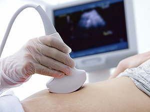
Diagnostic techniques in cardiovascular medicine refer to the various tests and methods used to diagnose and evaluate conditions related to the heart and blood vessels. These techniques can be non-invasive or invasive and are designed to provide critical information about a patient's cardiovascular health, such as heart function, blood flow, and the presence of any abnormalities or diseases. Here are some common diagnostic techniques used in cardiovascular medicine:
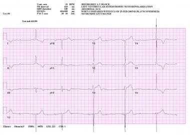
1. Electrocardiogram (ECG): An ECG is a non-invasive test that records the electrical activity of the heart. It can help detect heart conditions such as arrhythmias, heart attacks, and structural abnormalities.
2. Echocardiogram: This is a non-invasive ultrasound test that produces images of the heart's structures, including the chambers, valves, and major blood vessels. It can help assess heart function, identify damage from heart attacks, and detect various cardiovascular conditions.
3. Stress testing: A stress test involves exercising on a treadmill or stationary bike while being monitored by an ECG to evaluate the heart's response to physical exertion. It can help diagnose coronary artery disease, assess exercise capacity, and determine the need for further testing or treatment.
4. Cardiac catheterization: This is an invasive procedure where a thin, flexible tube (catheter) is inserted into a blood vessel in the arm or leg and guided to the heart. It can help diagnose and treat various cardiovascular conditions, such as blocked arteries, heart valve problems, and congenital heart defects.
5. Coronary angiography: During a cardiac catheterization, a special dye is injected into the coronary arteries to visualize blood flow using X-ray imaging. This can help identify blockages or narrowing in the coronary arteries and guide treatment decisions.
6. Nuclear stress testing: This test combines the use of a radioactive tracer with exercise or pharmacological stress to evaluate heart function and blood flow. It can help diagnose coronary artery disease, assess the effectiveness of treatments, and determine the need for further interventions.
7. Cardiac magnetic resonance imaging (MRI): This non-invasive imaging technique uses a strong magnetic field and radio waves to create detailed images of the heart's structure and function. It can help diagnose various cardiovascular conditions, such as heart muscle disorders, valve problems, and congenital heart defects.
8. Transesophageal echocardiography (TEE): This is a specialized ultrasound technique where a probe is inserted through the esophagus to obtain detailed images of the heart's structure and function. It can help diagnose conditions such as blood clots, valve problems, and infective endocarditis.
9. Positron emission tomography (PET) scanning: This imaging technique uses a small amount of radioactive tracer to evaluate the metabolic activity of the heart. It can help diagnose coronary artery disease, assess the effectiveness of treatments, and determine the need for further interventions.
10. Electrophysiology studies (EPS): These are invasive procedures where catheters are inserted into the heart to study its electrical system. They can help diagnose and treat various arrhythmias, such as atrial fibrillation, ventricular tachycardia, and Wolff-Parkinson-White syndrome.
Aortic rupture is a medical emergency that refers to the tearing or splitting of the aorta, which is the largest and main artery in the body. The aorta carries oxygenated blood from the heart to the rest of the body. An aortic rupture can lead to life-threatening internal bleeding and requires immediate medical attention.
There are two types of aortic ruptures:
1. Aortic dissection: This occurs when there is a tear in the inner lining of the aorta, allowing blood to flow between the layers of the aortic wall. This can cause the aorta to bulge or split, leading to a rupture.
2. Thoracic aortic aneurysm rupture: An aneurysm is a weakened and bulging area in the aortic wall. When an aneurysm in the thoracic aorta (the part of the aorta that runs through the chest) ruptures, it can cause severe bleeding and other complications.
Risk factors for aortic rupture include high blood pressure, smoking, aging, family history of aortic disease, and certain genetic conditions such as Marfan syndrome or Ehlers-Danlos syndrome. Symptoms of an aortic rupture may include sudden severe chest or back pain, difficulty breathing, weakness, sweating, and loss of consciousness. Treatment typically involves emergency surgery to repair the aorta and control bleeding.
A stent is a small mesh tube that's used to treat narrow or weak arteries. Arteries are blood vessels that carry blood away from your heart to other parts of your body. A stent is placed in an artery as part of a procedure called angioplasty. Angioplasty restores blood flow through narrowed or blocked arteries by inflating a tiny balloon inside the blocked artery to widen it.
The stent is then inserted into the widened artery to keep it open. The stent is usually made of metal, but some are coated with medication that is slowly and continuously released to help prevent the formation of scar tissue in the artery. This can reduce the chance of the artery narrowing again.
Stents are also used in other parts of the body, such as the neck (carotid artery) and kidneys (renal artery), to help maintain blood flow and prevent blockages. They can also be used in the urinary system to treat conditions like ureteropelvic junction obstruction or narrowing of the urethra.
An abdominal aortic aneurysm (AAA) is a localized dilatation or bulging of the abdominal aorta, which is the largest artery in the body that supplies oxygenated blood to the trunk and lower extremities. Normally, the diameter of the abdominal aorta measures about 2 centimeters (cm) in adults. However, when the diameter of the aorta exceeds 3 cm, it is considered an aneurysm.
AAA can occur anywhere along the length of the abdominal aorta, but it most commonly occurs below the renal arteries and above the iliac bifurcation. The exact cause of AAA remains unclear, but several risk factors have been identified, including smoking, hypertension, advanced age, male gender, family history, and certain genetic disorders such as Marfan syndrome and Ehlers-Danlos syndrome.
The main concern with AAA is the risk of rupture, which can lead to life-threatening internal bleeding. The larger the aneurysm, the greater the risk of rupture. Symptoms of AAA may include abdominal or back pain, a pulsating mass in the abdomen, or symptoms related to compression of surrounding structures such as the kidneys, ureters, or nerves. However, many AAAs are asymptomatic and are discovered incidentally during imaging studies performed for other reasons.
Diagnosis of AAA typically involves imaging tests such as ultrasound, computed tomography (CT) scan, or magnetic resonance imaging (MRI). Treatment options depend on the size and location of the aneurysm, as well as the patient's overall health status. Small AAAs that are not causing symptoms may be monitored with regular imaging studies to assess for growth. Larger AAAs or those that are growing rapidly may require surgical repair, either through open surgery or endovascular repair using a stent graft.
Heart valve diseases are a group of conditions that affect the function of one or more of the heart's four valves (tricuspid, pulmonic, mitral, and aortic). These valves are responsible for controlling the direction and flow of blood through the heart. Heart valve diseases can cause the valves to become narrowed (stenosis), leaky (regurgitation or insufficiency), or improperly closed (prolapse), leading to disrupted blood flow within the heart and potentially causing symptoms such as shortness of breath, fatigue, chest pain, and irregular heart rhythms. The causes of heart valve diseases can include congenital defects, age-related degenerative changes, infections, rheumatic heart disease, and high blood pressure. Treatment options may include medications, surgical repair or replacement of the affected valve(s), or transcatheter procedures.
The aorta is the largest artery in the human body, which originates from the left ventricle of the heart and carries oxygenated blood to the rest of the body. It can be divided into several parts, including the ascending aorta, aortic arch, and descending aorta. The ascending aorta gives rise to the coronary arteries that supply blood to the heart muscle. The aortic arch gives rise to the brachiocephalic, left common carotid, and left subclavian arteries, which supply blood to the head, neck, and upper extremities. The descending aorta travels through the thorax and abdomen, giving rise to various intercostal, visceral, and renal arteries that supply blood to the chest wall, organs, and kidneys.
Vascular surgical procedures are operations that are performed to treat conditions and diseases related to the vascular system, which includes the arteries, veins, and capillaries. These procedures can be invasive or minimally invasive and are often used to treat conditions such as peripheral artery disease, carotid artery stenosis, aortic aneurysms, and venous insufficiency.
Some examples of vascular surgical procedures include:
* Endarterectomy: a procedure to remove plaque buildup from the inside of an artery
* Bypass surgery: creating a new path for blood to flow around a blocked or narrowed artery
* Angioplasty and stenting: using a balloon to open a narrowed artery and placing a stent to keep it open
* Aneurysm repair: surgically repairing an aneurysm, a weakened area in the wall of an artery that has bulged out and filled with blood
* Embolectomy: removing a blood clot from a blood vessel
* Thrombectomy: removing a blood clot from a vein
These procedures are typically performed by vascular surgeons, who are trained in the diagnosis and treatment of vascular diseases.
Elastin is a protein that provides elasticity to tissues and organs, allowing them to resume their shape after stretching or contracting. It is a major component of the extracellular matrix in many tissues, including the skin, lungs, blood vessels, and ligaments. Elastin fibers can stretch up to 1.5 times their original length and then return to their original shape due to the unique properties of this protein. The elastin molecule is made up of cross-linked chains of the protein tropoelastin, which are produced by cells called fibroblasts and then assembled into larger elastin fibers by enzymes called lysyl oxidases. Elastin has a very long half-life, with some estimates suggesting that it can remain in the body for up to 70 years or more.
Postoperative complications refer to any unfavorable condition or event that occurs during the recovery period after a surgical procedure. These complications can vary in severity and may include, but are not limited to:
1. Infection: This can occur at the site of the incision or inside the body, such as pneumonia or urinary tract infection.
2. Bleeding: Excessive bleeding (hemorrhage) can lead to a drop in blood pressure and may require further surgical intervention.
3. Blood clots: These can form in the deep veins of the legs (deep vein thrombosis) and can potentially travel to the lungs (pulmonary embolism).
4. Wound dehiscence: This is when the surgical wound opens up, which can lead to infection and further complications.
5. Pulmonary issues: These include atelectasis (collapsed lung), pneumonia, or respiratory failure.
6. Cardiovascular problems: These include abnormal heart rhythms (arrhythmias), heart attack, or stroke.
7. Renal failure: This can occur due to various reasons such as dehydration, blood loss, or the use of certain medications.
8. Pain management issues: Inadequate pain control can lead to increased stress, anxiety, and decreased mobility.
9. Nausea and vomiting: These can be caused by anesthesia, opioid pain medication, or other factors.
10. Delirium: This is a state of confusion and disorientation that can occur in the elderly or those with certain medical conditions.
Prompt identification and management of these complications are crucial to ensure the best possible outcome for the patient.
Angioplasty is a medical procedure used to open narrowed or blocked blood vessels, often referred to as coronary angioplasty when it involves the heart's blood vessels (coronary arteries). The term "angio" refers to an angiogram, which is a type of X-ray image that reveals the inside of blood vessels.
The procedure typically involves the following steps:
1. A thin, flexible catheter (tube) is inserted into a blood vessel, usually through a small incision in the groin or arm.
2. The catheter is guided to the narrowed or blocked area using real-time X-ray imaging.
3. Once in place, a tiny balloon attached to the tip of the catheter is inflated to widen the blood vessel and compress any plaque buildup against the artery walls.
4. A stent (a small mesh tube) may be inserted to help keep the blood vessel open and prevent it from narrowing again.
5. The balloon is deflated, and the catheter is removed.
Angioplasty helps improve blood flow, reduce symptoms such as chest pain or shortness of breath, and lower the risk of heart attack in patients with blocked arteries. It's important to note that angioplasty is not a permanent solution for coronary artery disease, and lifestyle changes, medications, and follow-up care are necessary to maintain long-term cardiovascular health.
Magnetic Resonance Angiography (MRA) is a non-invasive medical imaging technique that uses magnetic fields and radio waves to create detailed images of the blood vessels or arteries within the body. It is a type of Magnetic Resonance Imaging (MRI) that focuses specifically on the circulatory system.
MRA can be used to diagnose and evaluate various conditions related to the blood vessels, such as aneurysms, stenosis (narrowing of the vessel), or the presence of plaques or tumors. It can also be used to plan for surgeries or other treatments related to the vascular system. The procedure does not use radiation and is generally considered safe, although people with certain implants like pacemakers may not be able to have an MRA due to safety concerns.
Treatment outcome is a term used to describe the result or effect of medical treatment on a patient's health status. It can be measured in various ways, such as through symptoms improvement, disease remission, reduced disability, improved quality of life, or survival rates. The treatment outcome helps healthcare providers evaluate the effectiveness of a particular treatment plan and make informed decisions about future care. It is also used in clinical research to compare the efficacy of different treatments and improve patient care.
X-ray computed tomography (CT or CAT scan) is a medical imaging method that uses computer-processed combinations of many X-ray images taken from different angles to produce cross-sectional (tomographic) images (virtual "slices") of the body. These cross-sectional images can then be used to display detailed internal views of organs, bones, and soft tissues in the body.
The term "computed tomography" is used instead of "CT scan" or "CAT scan" because the machines take a series of X-ray measurements from different angles around the body and then use a computer to process these data to create detailed images of internal structures within the body.
CT scanning is a noninvasive, painless medical test that helps physicians diagnose and treat medical conditions. CT imaging provides detailed information about many types of tissue including lung, bone, soft tissue and blood vessels. CT examinations can be performed on every part of the body for a variety of reasons including diagnosis, surgical planning, and monitoring of therapeutic responses.
In computed tomography (CT), an X-ray source and detector rotate around the patient, measuring the X-ray attenuation at many different angles. A computer uses this data to construct a cross-sectional image by the process of reconstruction. This technique is called "tomography". The term "computed" refers to the use of a computer to reconstruct the images.
CT has become an important tool in medical imaging and diagnosis, allowing radiologists and other physicians to view detailed internal images of the body. It can help identify many different medical conditions including cancer, heart disease, lung nodules, liver tumors, and internal injuries from trauma. CT is also commonly used for guiding biopsies and other minimally invasive procedures.
In summary, X-ray computed tomography (CT or CAT scan) is a medical imaging technique that uses computer-processed combinations of many X-ray images taken from different angles to produce cross-sectional images of the body. It provides detailed internal views of organs, bones, and soft tissues in the body, allowing physicians to diagnose and treat medical conditions.
The aortic valve is the valve located between the left ventricle (the lower left chamber of the heart) and the aorta (the largest artery in the body, which carries oxygenated blood from the heart to the rest of the body). It is made up of three thin flaps or leaflets that open and close to regulate blood flow. During a heartbeat, the aortic valve opens to allow blood to be pumped out of the left ventricle into the aorta, and then closes to prevent blood from flowing back into the ventricle when it relaxes. Any abnormality or damage to this valve can lead to various cardiovascular conditions such as aortic stenosis, aortic regurgitation, or infective endocarditis.
Retrospective studies, also known as retrospective research or looking back studies, are a type of observational study that examines data from the past to draw conclusions about possible causal relationships between risk factors and outcomes. In these studies, researchers analyze existing records, medical charts, or previously collected data to test a hypothesis or answer a specific research question.
Retrospective studies can be useful for generating hypotheses and identifying trends, but they have limitations compared to prospective studies, which follow participants forward in time from exposure to outcome. Retrospective studies are subject to biases such as recall bias, selection bias, and information bias, which can affect the validity of the results. Therefore, retrospective studies should be interpreted with caution and used primarily to generate hypotheses for further testing in prospective studies.
Prosthesis design is a specialized field in medical device technology that involves creating and developing artificial substitutes to replace a missing body part, such as a limb, tooth, eye, or internal organ. The design process typically includes several stages: assessment of the patient's needs, selection of appropriate materials, creation of a prototype, testing and refinement, and final fabrication and fitting of the prosthesis.
The goal of prosthesis design is to create a device that functions as closely as possible to the natural body part it replaces, while also being comfortable, durable, and aesthetically pleasing for the patient. The design process may involve collaboration between medical professionals, engineers, and designers, and may take into account factors such as the patient's age, lifestyle, occupation, and overall health.
Prosthesis design can be highly complex, particularly for advanced devices such as robotic limbs or implantable organs. These devices often require sophisticated sensors, actuators, and control systems to mimic the natural functions of the body part they replace. As a result, prosthesis design is an active area of research and development in the medical field, with ongoing efforts to improve the functionality, comfort, and affordability of these devices for patients.
A reoperation is a surgical procedure that is performed again on a patient who has already undergone a previous operation for the same or related condition. Reoperations may be required due to various reasons, such as inadequate initial treatment, disease recurrence, infection, or complications from the first surgery. The nature and complexity of a reoperation can vary widely depending on the specific circumstances, but it often carries higher risks and potential complications compared to the original operation.
Contrast media are substances that are administered to a patient in order to improve the visibility of internal body structures or processes in medical imaging techniques such as X-rays, CT scans, MRI scans, and ultrasounds. These media can be introduced into the body through various routes, including oral, rectal, or intravenous administration.
Contrast media work by altering the appearance of bodily structures in imaging studies. For example, when a patient undergoes an X-ray examination, contrast media can be used to highlight specific organs, tissues, or blood vessels, making them more visible on the resulting images. In CT and MRI scans, contrast media can help to enhance the differences between normal and abnormal tissues, allowing for more accurate diagnosis and treatment planning.
There are several types of contrast media available, each with its own specific properties and uses. Some common examples include barium sulfate, which is used as a contrast medium in X-ray studies of the gastrointestinal tract, and iodinated contrast media, which are commonly used in CT scans to highlight blood vessels and other structures.
While contrast media are generally considered safe, they can sometimes cause adverse reactions, ranging from mild symptoms such as nausea or hives to more serious complications such as anaphylaxis or kidney damage. As a result, it is important for healthcare providers to carefully evaluate each patient's medical history and individual risk factors before administering contrast media.
Follow-up studies are a type of longitudinal research that involve repeated observations or measurements of the same variables over a period of time, in order to understand their long-term effects or outcomes. In medical context, follow-up studies are often used to evaluate the safety and efficacy of medical treatments, interventions, or procedures.
In a typical follow-up study, a group of individuals (called a cohort) who have received a particular treatment or intervention are identified and then followed over time through periodic assessments or data collection. The data collected may include information on clinical outcomes, adverse events, changes in symptoms or functional status, and other relevant measures.
The results of follow-up studies can provide important insights into the long-term benefits and risks of medical interventions, as well as help to identify factors that may influence treatment effectiveness or patient outcomes. However, it is important to note that follow-up studies can be subject to various biases and limitations, such as loss to follow-up, recall bias, and changes in clinical practice over time, which must be carefully considered when interpreting the results.
Risk assessment in the medical context refers to the process of identifying, evaluating, and prioritizing risks to patients, healthcare workers, or the community related to healthcare delivery. It involves determining the likelihood and potential impact of adverse events or hazards, such as infectious diseases, medication errors, or medical devices failures, and implementing measures to mitigate or manage those risks. The goal of risk assessment is to promote safe and high-quality care by identifying areas for improvement and taking action to minimize harm.
In the field of medicine, "time factors" refer to the duration of symptoms or time elapsed since the onset of a medical condition, which can have significant implications for diagnosis and treatment. Understanding time factors is crucial in determining the progression of a disease, evaluating the effectiveness of treatments, and making critical decisions regarding patient care.
For example, in stroke management, "time is brain," meaning that rapid intervention within a specific time frame (usually within 4.5 hours) is essential to administering tissue plasminogen activator (tPA), a clot-busting drug that can minimize brain damage and improve patient outcomes. Similarly, in trauma care, the "golden hour" concept emphasizes the importance of providing definitive care within the first 60 minutes after injury to increase survival rates and reduce morbidity.
Time factors also play a role in monitoring the progression of chronic conditions like diabetes or heart disease, where regular follow-ups and assessments help determine appropriate treatment adjustments and prevent complications. In infectious diseases, time factors are crucial for initiating antibiotic therapy and identifying potential outbreaks to control their spread.
Overall, "time factors" encompass the significance of recognizing and acting promptly in various medical scenarios to optimize patient outcomes and provide effective care.
Medical Definition:
"Risk factors" are any attribute, characteristic or exposure of an individual that increases the likelihood of developing a disease or injury. They can be divided into modifiable and non-modifiable risk factors. Modifiable risk factors are those that can be changed through lifestyle choices or medical treatment, while non-modifiable risk factors are inherent traits such as age, gender, or genetic predisposition. Examples of modifiable risk factors include smoking, alcohol consumption, physical inactivity, and unhealthy diet, while non-modifiable risk factors include age, sex, and family history. It is important to note that having a risk factor does not guarantee that a person will develop the disease, but rather indicates an increased susceptibility.
A mutation is a permanent change in the DNA sequence of an organism's genome. Mutations can occur spontaneously or be caused by environmental factors such as exposure to radiation, chemicals, or viruses. They may have various effects on the organism, ranging from benign to harmful, depending on where they occur and whether they alter the function of essential proteins. In some cases, mutations can increase an individual's susceptibility to certain diseases or disorders, while in others, they may confer a survival advantage. Mutations are the driving force behind evolution, as they introduce new genetic variability into populations, which can then be acted upon by natural selection.
 Aortic dissection