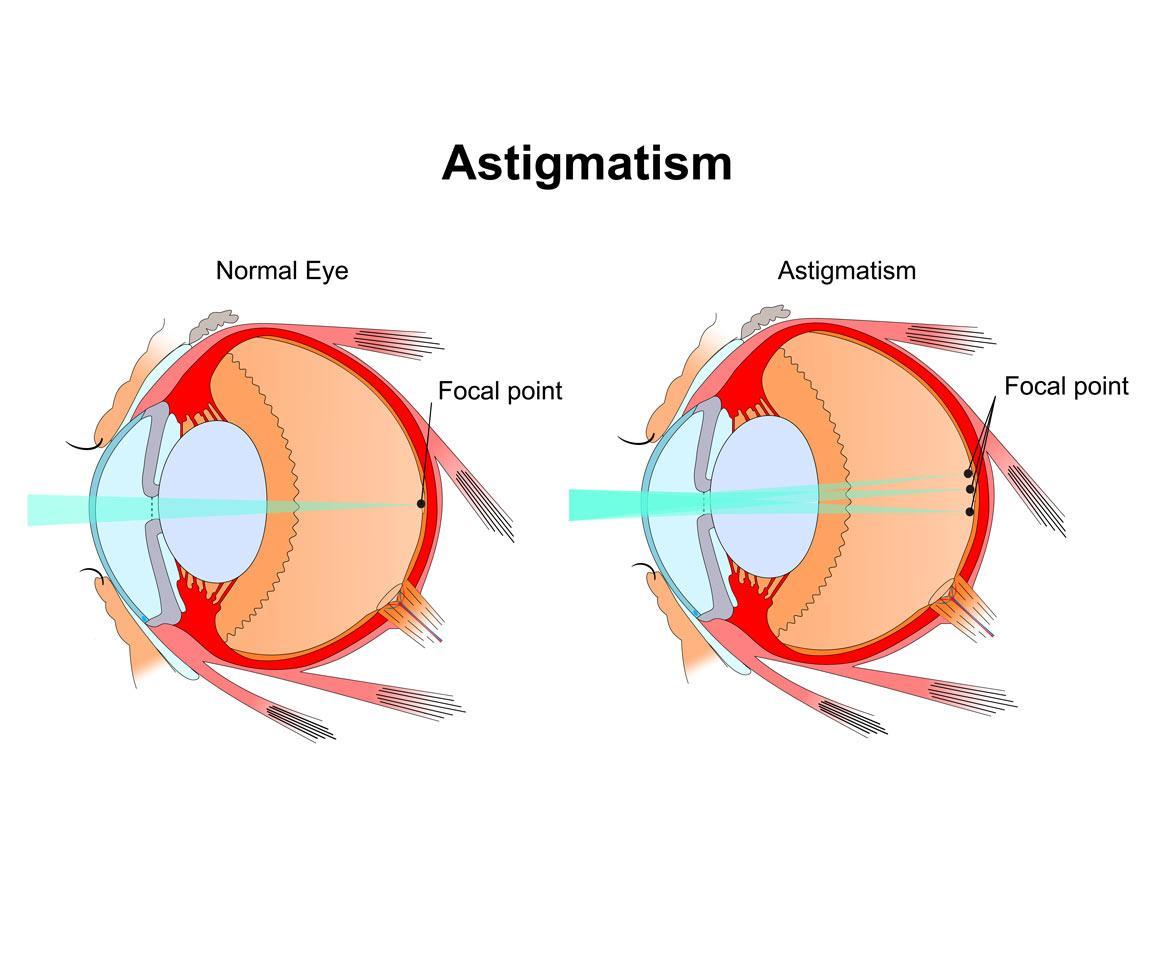
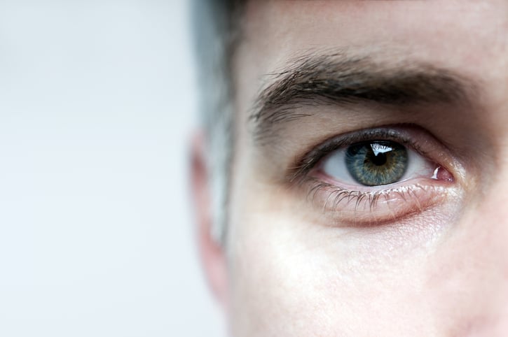
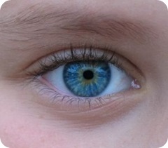
Astigmatism
Corneal Topography
Refractive Errors
Retinoscopy
Myopia
Hyperopia
Visual Acuity
Keratoplasty, Penetrating
Suture Techniques
Cornea
Keratoconus
Lens Implantation, Intraocular
Eyeglasses
Lasers, Excimer
Keratomileusis, Laser In Situ
Corneal Wavefront Aberration
Aberrometry
Keratectomy, Subepithelial, Laser-Assisted
Vision Screening
Refractive Surgical Procedures
Keratotomy, Radial
Corneal Surgery, Laser
Photorefractive Keratectomy
Glare
Phacoemulsification
Biometry
Amblyopia
Corneal Transplantation
Nylons
Albinism, Ocular
Pterygium
Phakic Intraocular Lenses
Accommodation, Ocular
Ophthalmology
Retinopathy of Prematurity
Results of small incision extracapsular cataract surgery using the anterior chamber maintainer without viscoelastic. (1/358)
AIMS: To assess the efficacy of extracapsular cataract surgery using the anterior chamber maintainer (ACM) without the use of viscoelastic. To compare the effects of this surgical technique on non-diabetic and diabetic patients. METHODS: A prospective single armed clinical trial of 46 eyes in 46 patients undergoing cataract surgery using the ACM without viscoelastic. Patients were assessed preoperatively and at 3 weeks, 3 months, and 12 months postoperatively. The main outcome variables included visual acuity, surgically induced astigmatic change (SIAC), changes in endothelial cell density (ECD), and morphology affecting the central and superior regions of the cornea. RESULTS: Postoperatively, 56% and 70% of patients had unaided visual acuities of 6/12 or better at 3 weeks and 3 months respectively. Even after excluding those patients with pre-existing maculopathy (including diabetic maculopathy), there remains a significant difference between the non-diabetic and diabetic groups in terms of the proportion of patients attaining an unaided visual acuity of 6/12 or better at both 3 weeks (p = 0.003) and 3 months (p = 0.001). Three months postoperatively, the SIAC based upon the keratometric and refractive data was 1.1 dioptres (D) and 1.3 D respectively. There was no statistically significant difference in the SIAC when the non-diabetic and diabetic groups were compared. The mean central and superior endothelial cell losses at 3 months postoperatively were 16% and 22% respectively and at 12 months postoperatively were 20% and 25% respectively. The diabetic group demonstrated greater endothelial cell losses and a more marked and protracted deviation of endothelial cell morphology from normality when compared with the non-diabetic group; however, the differences did not reach statistical significance. CONCLUSIONS: The efficacy of small incision cataract surgery using the ACM in terms of visual outcome and induced astigmatism is comparable with the results obtained using other techniques that utilise a similar size of incision. However, in view of the magnitude and range of the endothelial cell losses associated with this technique the concurrent use of viscoelastic is suggested. There does not appear to be a statistically or clinically significant difference between non-diabetic and diabetic patients in terms of the magnitude of the endothelial cell losses or in the wound healing response in the 12 months after cataract surgery using the ACM. (+info)Off-axis monochromatic aberrations estimated from double pass measurements in the human eye. (2/358)
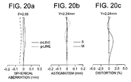
Off-axis monochromatic aberrations in the human eye impose limits on peripheral vision. However, the magnitude of the aberrations off-axis, and in particular coma, has not been yet completely determined. We have developed a procedure to estimate third order aberrations in the periphery of the human eye. The technique is based on recording series of double pass retinal images with unequal entrance and exit pupil diameters (Artal, Iglesias, Lopez-Gil & Green (1995b). J. Opt. Soc. Am. A, 12, 2358-2366.) which allows the odd asymmetries in the retinal image be assessed. The procedure that is described provides accurate estimates of the main off-axis aberrations: astigmatism, defocus and coma. We have measured these aberrations in four normal subjects. For a given eccentricity, the measured amount of coma and astigmatism are relatively similar among subjects, because the angular distance from the axis is the dominant factor in determining the magnitude of these aberrations. However, we found considerable variability in the values of peripheral defocus, probably due to a complicate combination of off-axis aberrations and fundus shape. The final off-axis optical performance of the eye for a given object location is determined by a particular mixture of defocus, astigmatism, coma and higher order aberrations. (+info)Image quality in polypseudophakia for extremely short eyes. (3/358)
AIM: To evaluate the image quality produced by polypseudophakia used for strongly hypermetropic and nanophthalmic eyes. METHODS: Primary aberration theory and ray tracing analysis were used to calculate the optimum lens shapes and power distribution between the two intraocular lenses for two example eyes: one a strongly hypermetropic eye, the other a nanophthalmic eye. Spherical aberration and oblique astigmatism were considered. Modulation transfer function (MTF) curves were computed using commercial optical design software (Sigma 2100, Kidger Optics Ltd) to assess axial image quality, and the sagittal and tangential image surfaces were computed to study image quality across the field. RESULTS: A significant improvement in the axial MTF was found for the eyes with double implants. However, results indicate that this may be realised as a better contrast sensitivity in the low to mid spatial frequency range rather than as a better Snellen acuity. The optimum lens shapes for minimum spherical aberration (best axial image quality) were approximately convex-plano for both lenses with the convex surface facing the cornea. Conversely, the optimum lens shapes for zero oblique astigmatism were strongly meniscus with the anterior surface concave. Correction of oblique astigmatism was only achieved with a loss in axial performance. CONCLUSIONS: Optimum estimated visual acuity exceeds 6/5 in both the hypermetropic and the nanophthalmic eyes studied (pupil size of 4 mm) with polypseudophakic correction. These results can be attained using convex-plano or biconvex lenses with the most convex surface facing the cornea. If the posterior surface of the posterior intraocular lens is convex, as is commonly used to help prevent migration of lens epithelial cells causing posterior capsular opacification (PCO), then it is still possible to achieve 6/4.5 in the hypermetropic eye and 6/5.3 in the nanophthalmic eye provided the anterior intraocular lens has an approximately convex-plano shape with the convex surface anterior. It was therefore concluded that consideration of optical image quality does not demand that additional intraocular lens shapes need to be manufactured for polypseudophakic correction of extremely short eyes and that implanting the posterior intraocular lens in the conventional orientation to help prevent PCO does not necessarily limit estimated visual acuity. (+info)Cataract extraction and lens implantation with and without trabeculectomy: an intrapatient comparison. (4/358)
OBJECTIVE: To determine whether cataract extraction and lens implantation combined with trabeculectomy provides better long-term results than cataract extraction and lens implantation alone in a group of patients with primary open-angle glaucoma and cataract randomly selected to receive surgery with trabeculectomy in one eye and without in the other. METHODS: A prospective, randomized clinical trial involving 35 patients with bilateral symmetric primary open-angle glaucoma and visually disabling cataracts with procedures performed by a single surgeon in a private practice setting with follow-up for more than 5 years in all cases. RESULTS: After an average of 87 months of follow-up, cataract extraction and lens implantation reduced intraocular pressure 4.4 mm Hg, reduced number of medications by 1.28, increased diopter vector of astigmatism by 1.49, and was associated with visual field loss in 6 of 35 eyes. After an average of 80 months of follow-up, cataract extraction, lens implantation, and trabeculectomy reduced intraocular pressure 8.2 mm Hg, reduced number of medications by 1.76, increased diopter vector of astigmatism by 1.14, and was associated with visual field loss in 1 eye. Both groups had similar improvement in visual acuity and perioperative complications. CONCLUSIONS: Extracapsular cataract extraction, lens implantation, and trabeculectomy is a complex procedure that was beneficial in the long-term control of intraocular pressure and in prevention of visual field loss. This procedure should be considered in patients who may not be able to comply with a complex medical regimen, in whom pressure elevation in the immediate postoperative period would be undesirable, or in whom long-term pressure control at a lower level would be beneficial in preventing further optic nerve damage. (+info)Topographic and keratometric astigmatism up to 1 year following small flap trabeculectomy (microtrabeculectomy). (5/358)
AIM: To determine the induced corneal astigmatism by measuring the changes in manual keratometry and computerised corneal videokeratoscopy up to 1 year following small flap trabeculectomy (microtrabeculectomy). METHOD: A prospective study of a case series of small flap trabeculectomy procedures performed at the 90 degree meridian on 16 eyes of 16 patients, all followed to 1 year postoperatively. Changes in manual keratometry and computerised videokeratoscopy (Eyesys) readings were analysed by vector analysis and vector decomposition techniques. RESULTS: By vector analysis, the mean surgically induced refractive change (SIRC) cylinder power vectors induced at 1, 3, 6, and 12 months as measured by manual keratometry were 0.68, 0.38, 0.52, and 0.55 dioptres, and by keratography 0.75, 0.66, 0.59, and 0.64 dioptres. Vector decomposition on the induced vector cylinders on manual keratometry resulted in a "with the rule" mean vector of 0.52 and 0.22 dioptres at 1 and 3 months and an "against the rule" mean vector of 0.16 and 0.16 dioptres at the same time points (p=0.03 and 0.28 respectively). Vector decomposition at 6 and 12 months revealed no significant with the rule changes induced. Similar analysis on the videokeratoscopy results revealed significant induced with the rule astigmatism until 3 months, but not at 6 and 12 months postoperatively. CONCLUSION: Small flap trabeculectomy (microtrabeculectomy) produces smaller changes in corneal curvature that resolve sooner than previous reports of larger flap techniques. (+info)Screening for refractive errors in children: accuracy of the hand held refractor Retinomax to screen for astigmatism. (6/358)
AIMS: To assess the reliability of the hand held automated refractor Retinomax in measuring astigmatism in non-cycloplegic conditions. To assess the accuracy of Retinomax in diagnosing abnormal astigmatism in non-cycloplegic refractive screening of children between 9 and 36 months. METHODS: Among 1205 children undergoing a non-cycloplegic refractive screening with Retinomax, 299 (25%) had repeated non-cycloplegic measurements, 302 (25%) were refracted under cycloplegia using the same refractor, and 88 (7%) using retinoscopy or an automated on table refractor. The reproducibility of non-cycloplegic cylinder measurement was assessed by comparing the cylindrical power and axis values in the 299 repeated measurements without cycloplegia. The influence of the quick mode on cylinder measurement was analysed by comparing the cylinder and axis value in 93 repeated measurements without cycloplegia where normal mode was used in one measurement and quick mode in the other. Predictive values of the refractive screening were calculated for three different thresholds of manifest astigmatism (> or = 1.5, > or = 1.75, and > or = 2 D) considering as a true positive case an astigmatism > or = 2 D under cycloplegic condition (measured by retinoscopy, on table, or hand held refractor). RESULTS: The 95% limits of agreement between two repeated manifest cylinder measurements with Retinomax attained levels slightly less than plus or minus 1 D. The 95% limits of agreement for the axis were plus or minus 46 degrees. The comparison of non-cycloplegic measurements in the quick and normal mode showed no significant difference and 95% limits of agreement plus or minus 0.75 D. The mean difference between non-cycloplegic and cycloplegic cylinder values measured by Retinomax reached 0.17 D and was statistically significant. Manifest thresholds of > or = 1.5 D, > or = 1.75 D, > or = 2 D cylinder value diagnosed 2 D of astigmatism under cyclplegia respectively with 71-84%, 59-80%, 51-54% of sensitivity (right eye-left eye) and 90-92%, 95%, 98% of specificity. CONCLUSION: Without cycloplegia, Retinomax is able to measure cylinder power with the same reproducibility as cycloplegic retinoscopy. No significant difference was found in the cylinder values obtained with the quick and the normal modes. Therefore, the quick mode of measurement is recommended as it is more feasible in children. No difference, which is significant from a screening point of view, exists between the non-cycloplegic and the cycloplegic cylinder value (< 0.25 D). Retinomax diagnoses abnormal astigmatism (> or = 2 D) in a non-cycloplegic refractive screening at preschool ages with 51-84% sensitivity rates and 98-90% specificity rates, depending on the chosen threshold of manifest astigmatism. If 2 D of manifest astigmatism is chosen as a positive test, the positive predictive value of the screening reaches 81-84% and the negative predictive value 91-90% (right eye-left eye). (+info)Proposed classification for topographic patterns seen after penetrating keratoplasty. (7/358)
AIMS: To create a clinically useful classification for post-keratoplasty corneas based on corneal topography. METHODS: A total of 360 topographic maps obtained with the TMS-1, from 95 eyes that had undergone penetrating keratoplasty (PKP), were reviewed independently by two examiners in a masked fashion, and were categorised according to a proposed classification scheme. RESULTS: A high interobserver agreement (88% in the first categorisation) was achieved. At 12 months post-PKP, a regular astigmatic pattern was observed in 20/85 cases (24%). This was subclassified as oval in three cases (4%), oblate symmetric bow tie in six cases (7%), prolate asymmetric bow tie in six cases (7%), and oblate asymmetric bow tie in five cases (6%). An irregular astigmatic pattern was observed in 61/85 cases (72%), subclassified as prolate irregular in five cases (6%), oblate irregular in four cases (5%), mixed in seven cases (8%), steep/flat in 11 cases (13%), localised steepness in 16 cases (19%), and triple pattern in three cases (4%). Regular astigmatic patterns were associated with significantly higher astigmatism measurements. The surface asymmetry index was significantly lower in the regular astigmatic patterns. CONCLUSIONS: In post-PKP corneas, the prevalence of irregular astigmatism is about double that of regular astigmatism, with a trend for increase of the irregular patterns over time. (+info)LASIK for post penetrating keratoplasty astigmatism and myopia. (8/358)
AIMS: To report the results of a series of patients who were treated with LASIK to correct post penetrating keratoplasty ametropia. METHODS: 26 eyes of 24 patients underwent LASIK to correct astigmatism and myopia after corneal transplantation; 14 eyes also received arcuate cuts in the stromal bed at the time of surgery. The mean preoperative spherical equivalent was -5.20D and the mean preoperative astigmatism was 8.67D. RESULTS: The results of 25 eyes are reported. The mean 1 month values for spherical equivalent and astigmatism were -0.24D and 2.48D respectively. 18 eyes have been followed up for 6 months or more. The final follow up results for these eyes are -1.91D and 2.92D for spherical equivalent and astigmatism. The patients undergoing arcuate cuts were less myopic but had greater astigmatism than those not. The patients receiving arcuate cuts had a greater target induced astigmatism, surgically induced astigmatism, and astigmatism correction index than those eyes that did not. One eye suffered a surgical complication. No eyes lost more than one line of BSCVA and all eyes gained between 0 and 6 lines UCVA. CONCLUSIONS: LASIK after penetrating keratoplasty is a relatively safe and effective procedure. It reduces both the spherical error and the cylindrical component of the ametropia. Correction of high astigmatism may be augmented by performing arcuate cuts in the stromal bed. (+info)Astigmatism is a common eye condition that occurs when the cornea or lens has an irregular shape, causing blurred or distorted vision. The cornea and lens are typically smooth and curved uniformly in all directions, allowing light to focus clearly on the retina. However, if the cornea or lens is not smoothly curved and has a steeper curve in one direction than the other, it causes light to focus unevenly on the retina, leading to astigmatism.
Astigmatism can cause blurred vision at all distances, as well as eye strain, headaches, and fatigue. It is often present from birth and can be hereditary, but it can also develop later in life due to eye injuries or surgery. Astigmatism can be corrected with glasses, contact lenses, or refractive surgery such as LASIK.
Ocular refraction is a medical term that refers to the bending of light as it passes through the optical media of the eye, including the cornea and lens. This process allows the eye to focus light onto the retina, creating a clear image. The refractive power of the eye is determined by the curvature and transparency of these structures.
In a normal eye, light rays are bent or refracted in such a way that they converge at a single point on the retina, producing a sharp and focused image. However, if the curvature of the cornea or lens is too steep or too flat, the light rays may not converge properly, resulting in a refractive error such as myopia (nearsightedness), hyperopia (farsightedness), or astigmatism.
Ocular refraction can be measured using a variety of techniques, including retinoscopy, automated refraction, and subjective refraction. These measurements are used to determine the appropriate prescription for corrective lenses such as eyeglasses or contact lenses. In some cases, ocular refractive errors may be corrected surgically through procedures such as LASIK or PRK.
Corneal topography is a non-invasive medical imaging technique used to create a detailed map of the surface curvature of the cornea, which is the clear, dome-shaped surface at the front of the eye. This procedure provides valuable information about the shape and condition of the cornea, helping eye care professionals assess various eye conditions such as astigmatism, keratoconus, and other corneal abnormalities. It can also be used in contact lens fitting, refractive surgery planning, and post-surgical evaluation.
Refractive errors are a group of vision conditions that include nearsightedness (myopia), farsightedness (hyperopia), astigmatism, and presbyopia. These conditions occur when the shape of the eye prevents light from focusing directly on the retina, causing blurred or distorted vision.
Myopia is a condition where distant objects appear blurry while close-up objects are clear. This occurs when the eye is too long or the cornea is too curved, causing light to focus in front of the retina instead of directly on it.
Hyperopia, on the other hand, is a condition where close-up objects appear blurry while distant objects are clear. This happens when the eye is too short or the cornea is not curved enough, causing light to focus behind the retina.
Astigmatism is a condition that causes blurred vision at all distances due to an irregularly shaped cornea or lens.
Presbyopia is a natural aging process that affects everyone as they get older, usually around the age of 40. It causes difficulty focusing on close-up objects and can be corrected with reading glasses, bifocals, or progressive lenses.
Refractive errors can be diagnosed through a comprehensive eye exam and are typically corrected with eyeglasses, contact lenses, or refractive surgery such as LASIK.
Retinoscopy is a diagnostic technique used in optometry and ophthalmology to estimate the refractive error of the eye, or in other words, to determine the prescription for eyeglasses or contact lenses. This procedure involves shining a light into the patient's pupil and observing the reflection off the retina while introducing different lenses in front of the patient's eye. The examiner then uses specific movements and observations to determine the amount and type of refractive error, such as myopia (nearsightedness), hyperopia (farsightedness), astigmatism, or presbyopia. Retinoscopy is a fundamental skill for eye care professionals and helps ensure that patients receive accurate prescriptions for corrective lenses.
Myopia, also known as nearsightedness, is a common refractive error of the eye. It occurs when the eye is either too long or the cornea (the clear front part of the eye) is too curved. As a result, light rays focus in front of the retina instead of directly on it, causing distant objects to appear blurry while close objects remain clear.
Myopia typically develops during childhood and can progress gradually or rapidly until early adulthood. It can be corrected with glasses, contact lenses, or refractive surgery such as LASIK. Regular eye examinations are essential for people with myopia to monitor any changes in their prescription and ensure proper correction.
While myopia is generally not a serious condition, high levels of nearsightedness can increase the risk of certain eye diseases, including cataracts, glaucoma, retinal detachment, and myopic degeneration. Therefore, it's crucial to manage myopia effectively and maintain regular follow-ups with an eye care professional.
Hyperopia, also known as farsightedness, is a refractive error in which the eye does not focus light directly on the retina when looking at a distant object. Instead, light is focused behind the retina, causing close-up objects to appear blurry. This condition usually results from the eyeball being too short or the cornea having too little curvature. It can be corrected with eyeglasses, contact lenses, or refractive surgery.
Visual acuity is a measure of the sharpness or clarity of vision. It is usually tested by reading an eye chart from a specific distance, such as 20 feet (6 meters). The standard eye chart used for this purpose is called the Snellen chart, which contains rows of letters that decrease in size as you read down the chart.
Visual acuity is typically expressed as a fraction, with the numerator representing the testing distance and the denominator indicating the smallest line of type that can be read clearly. For example, if a person can read the line on the eye chart that corresponds to a visual acuity of 20/20, it means they have normal vision at 20 feet. If their visual acuity is 20/40, it means they must be as close as 20 feet to see what someone with normal vision can see at 40 feet.
It's important to note that visual acuity is just one aspect of overall vision and does not necessarily reflect other important factors such as peripheral vision, depth perception, color vision, or contrast sensitivity.
Penetrating keratoplasty (PK) is a type of corneal transplant surgery where the entire thickness of the host's damaged or diseased cornea is removed and replaced with a similar full-thickness portion of a healthy donor's cornea. The procedure aims to restore visual function, alleviate pain, and improve the structural integrity of the eye. It is typically performed for conditions such as severe keratoconus, corneal scarring, or corneal ulcers that cannot be treated with other, less invasive methods. Following the surgery, patients may require extended recovery time and rigorous postoperative care to minimize the risk of complications and ensure optimal visual outcomes.
Suture techniques refer to the various methods used by surgeons to sew or stitch together tissues in the body after an injury, trauma, or surgical incision. The main goal of suturing is to approximate and hold the edges of the wound together, allowing for proper healing and minimizing scar formation.
There are several types of suture techniques, including:
1. Simple Interrupted Suture: This is one of the most basic suture techniques where the needle is passed through the tissue at a right angle, creating a loop that is then tightened to approximate the wound edges. Multiple stitches are placed along the length of the incision or wound.
2. Continuous Locking Suture: In this technique, the needle is passed continuously through the tissue in a zigzag pattern, with each stitch locking into the previous one. This creates a continuous line of sutures that provides strong tension and support to the wound edges.
3. Running Suture: Similar to the continuous locking suture, this technique involves passing the needle continuously through the tissue in a straight line. However, instead of locking each stitch, the needle is simply passed through the previous loop before being tightened. This creates a smooth and uninterrupted line of sutures that can be easily removed after healing.
4. Horizontal Mattress Suture: In this technique, two parallel stitches are placed horizontally across the wound edges, creating a "mattress" effect that provides additional support and tension to the wound. This is particularly useful in deep or irregularly shaped wounds.
5. Vertical Mattress Suture: Similar to the horizontal mattress suture, this technique involves placing two parallel stitches vertically across the wound edges. This creates a more pronounced "mattress" effect that can help reduce tension and minimize scarring.
6. Subcuticular Suture: In this technique, the needle is passed just below the surface of the skin, creating a smooth and barely visible line of sutures. This is particularly useful in cosmetic surgery or areas where minimizing scarring is important.
The choice of suture technique depends on various factors such as the location and size of the wound, the type of tissue involved, and the patient's individual needs and preferences. Proper suture placement and tension are crucial for optimal healing and aesthetic outcomes.
Anisometropia is a medical term that refers to a condition where there is a significant difference in the refractive power between the two eyes. In other words, one eye has a significantly different optical prescription compared to the other eye. This condition can cause issues with binocular vision and depth perception, and can sometimes lead to amblyopia (lazy eye) if not corrected early in life. It is typically diagnosed through a comprehensive eye examination and can be corrected with glasses or contact lenses.
The cornea is the clear, dome-shaped surface at the front of the eye. It plays a crucial role in focusing vision. The cornea protects the eye from harmful particles and microorganisms, and it also serves as a barrier against UV light. Its transparency allows light to pass through and get focused onto the retina. The cornea does not contain blood vessels, so it relies on tears and the fluid inside the eye (aqueous humor) for nutrition and oxygen. Any damage or disease that affects its clarity and shape can significantly impact vision and potentially lead to blindness if left untreated.
Cataract extraction is a surgical procedure that involves removing the cloudy lens (cataract) from the eye. This procedure is typically performed to restore vision impairment caused by cataracts and improve overall quality of life. There are two primary methods for cataract extraction:
1. Phacoemulsification: This is the most common method used today. It involves making a small incision in the front part of the eye (cornea), inserting an ultrasonic probe to break up the cloudy lens into tiny pieces, and then removing those pieces with suction. After removing the cataract, an artificial intraocular lens (IOL) is inserted to replace the natural lens and help focus light onto the retina.
2. Extracapsular Cataract Extraction: In this method, a larger incision is made on the side of the cornea, allowing the surgeon to remove the cloudy lens in one piece without breaking it up. The back part of the lens capsule is left intact to support the IOL. This technique is less common and typically reserved for more advanced cataracts or when phacoemulsification cannot be performed.
Recovery from cataract extraction usually involves using eye drops to prevent infection and inflammation, as well as protecting the eye with a shield or glasses during sleep for a few weeks after surgery. Most people experience improved vision within a few days to a week following the procedure.
Corneal diseases are a group of disorders that affect the cornea, which is the clear, dome-shaped surface at the front of the eye. The cornea plays an important role in focusing vision, and any damage or disease can cause significant visual impairment or loss. Some common types of corneal diseases include:
1. Keratoconus: A progressive disorder in which the cornea thins and bulges outward into a cone shape, causing distorted vision.
2. Fuchs' dystrophy: A genetic disorder that affects the inner layer of the cornea called the endothelium, leading to swelling, cloudiness, and decreased vision.
3. Dry eye syndrome: A condition in which the eyes do not produce enough tears or the tears evaporate too quickly, causing discomfort, redness, and blurred vision.
4. Corneal ulcers: Open sores on the cornea that can be caused by infection, trauma, or other factors.
5. Herpes simplex keratitis: A viral infection of the cornea that can cause recurrent episodes of inflammation, scarring, and vision loss.
6. Corneal dystrophies: Inherited disorders that affect the structure and clarity of the cornea, leading to visual impairment or blindness.
7. Bullous keratopathy: A condition in which the endothelium fails to pump fluid out of the cornea, causing it to swell and form blisters.
8. Corneal trauma: Injury to the cornea caused by foreign objects, chemicals, or other factors that can lead to scarring, infection, and vision loss.
Treatment for corneal diseases varies depending on the specific condition and severity of the disease. Options may include eyedrops, medications, laser surgery, corneal transplantation, or other treatments.
Keratoconus is a degenerative non-inflammatory disorder of the eye, primarily affecting the cornea. It is characterized by a progressive thinning and steepening of the central or paracentral cornea, causing it to assume a conical shape. This results in irregular astigmatism, myopia, and scattering of light leading to blurred vision, visual distortions, and sensitivity to glare. The exact cause of keratoconus is unknown, but it may be associated with genetics, eye rubbing, and certain medical conditions. It typically starts in the teenage years and progresses into the third or fourth decade of life. Treatment options include glasses, contact lenses, cross-linking, and corneal transplantation in advanced cases.
Intraocular lens (IOL) implantation is a surgical procedure that involves placing a small artificial lens inside the eye to replace the natural lens that has been removed. This procedure is typically performed during cataract surgery, where the cloudy natural lens is removed and replaced with an IOL to restore clear vision.
During the procedure, a small incision is made in the eye, and the cloudy lens is broken up and removed using ultrasound waves or laser energy. Then, the folded IOL is inserted through the same incision and positioned in the correct place inside the eye. Once in place, the IOL unfolds and is secured into position.
There are several types of IOLs available, including monofocal, multifocal, toric, and accommodating lenses. Monofocal lenses provide clear vision at one distance, while multifocal lenses offer clear vision at multiple distances. Toric lenses correct astigmatism, and accommodating lenses can change shape to focus on objects at different distances.
Overall, intraocular lens implantation is a safe and effective procedure that can help restore clear vision in patients with cataracts or other eye conditions that require the removal of the natural lens.
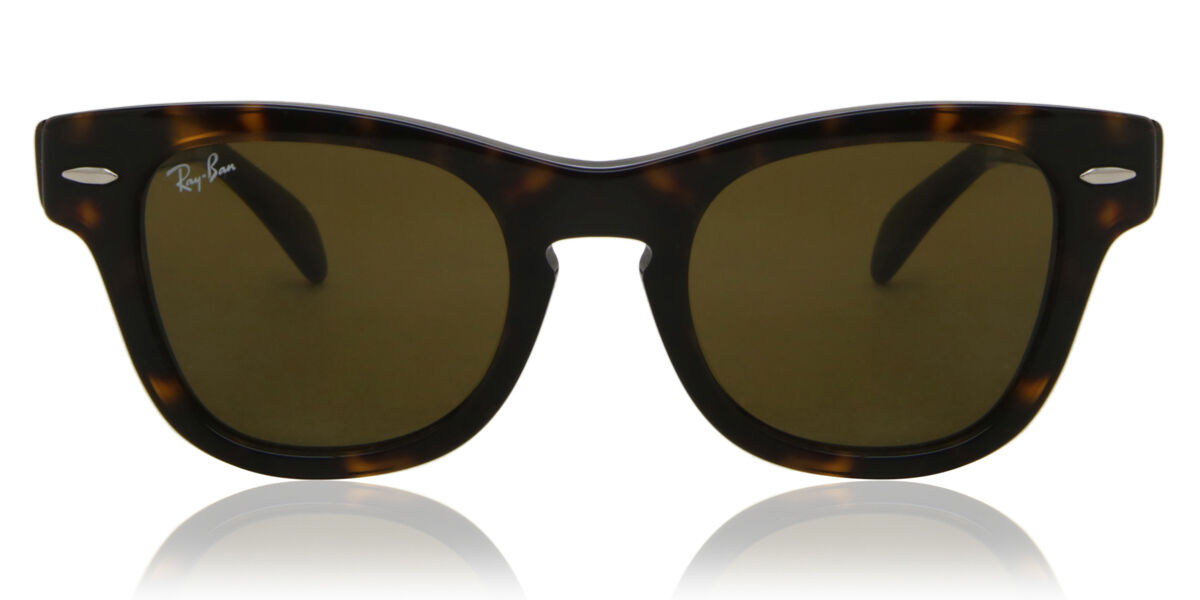
Eyeglasses are a medical device used to correct vision problems. Also known as spectacles, they consist of frames that hold one or more lenses through which a person looks to see clearly. The lenses may be made of glass or plastic and are designed to compensate for various visual impairments such as nearsightedness, farsightedness, astigmatism, or presbyopia. Eyeglasses can be custom-made to fit an individual's face and prescription, and they come in a variety of styles, colors, and materials. Some people wear eyeglasses all the time, while others may only need to wear them for certain activities such as reading or driving.
Intraocular lenses (IOLs) are artificial lens implants that are placed inside the eye during ophthalmic surgery, such as cataract removal. These lenses are designed to replace the natural lens of the eye that has become clouded or damaged, thereby restoring vision impairment caused by cataracts or other conditions.
There are several types of intraocular lenses available, including monofocal, multifocal, toric, and accommodative lenses. Monofocal IOLs provide clear vision at a single fixed distance, while multifocal IOLs offer clear vision at multiple distances. Toric IOLs are designed to correct astigmatism, and accommodative IOLs can change shape and position within the eye to allow for a range of vision.
The selection of the appropriate type of intraocular lens depends on various factors, including the patient's individual visual needs, lifestyle, and ocular health. The implantation procedure is typically performed on an outpatient basis and involves minimal discomfort or recovery time. Overall, intraocular lenses have become a safe and effective treatment option for patients with vision impairment due to cataracts or other eye conditions.
An excimer laser is a type of laser that is used in various medical procedures, particularly in ophthalmology and dermatology. The term "excimer" is derived from "excited dimer," which refers to a short-lived molecule formed when two atoms combine in an excited state.
Excimer lasers emit light at a specific wavelength that is determined by the type of gas used in the laser. In medical applications, excimer lasers typically use noble gases such as argon, krypton, or xenon, combined with halogens such as fluorine or chlorine. The most commonly used excimer laser in medical procedures is the excimer laser that uses a mixture of argon and fluoride gas to produce light at a wavelength of 193 nanometers (nm).
In ophthalmology, excimer lasers are primarily used for refractive surgery, such as LASIK and PRK, to correct vision problems like myopia, hyperopia, and astigmatism. The laser works by vaporizing tiny amounts of tissue from the cornea, reshaping its curvature to improve the way light is focused onto the retina.
In dermatology, excimer lasers are used for various skin conditions, including psoriasis, vitiligo, and atopic dermatitis. The laser works by emitting high-energy ultraviolet (UV) light that selectively targets and destroys the abnormal cells responsible for these conditions while leaving surrounding healthy tissue intact.
Excimer lasers are known for their precision, accuracy, and minimal side effects, making them a popular choice in medical procedures where fine detail and tissue preservation are critical.
Laser In Situ Keratomileusis (LASIK) is a type of refractive surgery used to correct vision issues such as myopia (nearsightedness), hyperopia (farsightedness), and astigmatism. The procedure involves reshaping the cornea, which is the clear, dome-shaped surface at the front of the eye, using an excimer laser.
In LASIK, a thin flap is created on the surface of the cornea using a femtosecond or microkeratome laser. The flap is then lifted, and the excimer laser is used to reshape the underlying tissue. After the reshaping is complete, the flap is replaced, allowing for quicker healing and visual recovery compared to other refractive surgery procedures.
LASIK is an outpatient procedure that typically takes about 30 minutes or less per eye. Most people can expect to see improved vision within a few days of the procedure, although it may take several weeks for vision to fully stabilize. LASIK has a high success rate and is generally considered safe when performed by a qualified surgeon. However, as with any surgical procedure, there are risks involved, including dry eye, infection, and visual complications such as glare or halos around lights.
Corneal wavefront aberration is a measurement of the irregularities in the shape and curvature of the cornea, which can affect the way light enters the eye and is focused on the retina. A wavefront aberration test uses a device to measure the refraction of light as it passes through the cornea and calculates the degree of any distortions or irregularities in the wavefront of the light. This information can be used to guide treatment decisions, such as the prescription for eyeglasses or contact lenses, or the planning of a surgical procedure to correct the aberration.
Corneal wavefront aberrations can be classified into two types: low-order and high-order aberrations. Low-order aberrations include myopia (nearsightedness), hyperopia (farsightedness), and astigmatism, which are common refractive errors that can be easily corrected with glasses or contact lenses. High-order aberrations are more complex irregularities in the wavefront of light that cannot be fully corrected with traditional eyeglasses or contact lenses. These may include coma, trefoil, and spherical aberration, among others.
High-order corneal wavefront aberrations can affect visual quality, causing symptoms such as glare, halos around lights, and decreased contrast sensitivity. They are often associated with conditions that cause changes in the shape of the cornea, such as keratoconus or corneal surgery. In some cases, high-order aberrations can be corrected with specialized contact lenses or refractive surgery procedures such as wavefront-guided LASIK or PRK.
Aberrometry is a medical diagnostic technique used to measure the amount and type of aberration or distortion in the optical system of the eye. It is often used to evaluate the quality of vision, particularly in cases where traditional methods of measuring visual acuity are not sufficient.
During an aberrometry test, the patient looks into a specialized instrument called a wavefront sensor while a series of light patterns are projected onto the retina. The sensor then measures how the light is distorted as it passes through the eye's optical system, including the cornea and lens. This information is used to create a detailed map of the eye's aberrations, which can help doctors identify any irregularities that may be contributing to visual symptoms such as blurred vision, glare, or halos around lights.
Aberrometry is often used in conjunction with other diagnostic tests to evaluate patients who are considering refractive surgery, such as LASIK or PRK. By identifying any abnormalities in the eye's optical system, doctors can determine whether a patient is a good candidate for surgery and make more informed decisions about how to proceed with treatment.
Subepithelial laser-assisted keratectomy (SELAK) is a type of refractive surgery used to correct vision problems such as myopia (nearsightedness), hyperopia (farsightedness), and astigmatism. In this procedure, a precise and controlled laser beam is used to remove a thin layer of tissue from the cornea, specifically from the subepithelial region, which lies just beneath the surface epithelium.
The goal of SELAK is to reshape the cornea and improve its focusing power, thereby reducing or eliminating the need for corrective lenses such as glasses or contact lenses. The laser-assisted technique allows for a high degree of precision and customization, enabling the surgeon to tailor the procedure to each patient's individual needs.
It is important to note that while SELAK can be an effective treatment option for many people, it may not be suitable for everyone. A thorough eye examination and consultation with an eye care professional are necessary to determine whether this procedure is appropriate for a particular individual.
Pseudophakia is a medical term that refers to the condition where a person's natural lens in the eye has been replaced with an artificial one. This procedure is typically performed during cataract surgery, where the cloudy, natural lens is removed and replaced with a clear, artificial lens to improve vision. The prefix "pseudo" means false or fake, and "phakia" refers to the natural lens of the eye, hence the term "Pseudophakia" implies a false or artificial lens.
Vision screening is a quick and cost-effective method used to identify individuals who are at risk of vision problems or eye diseases. It is not a comprehensive eye examination, but rather an initial evaluation that helps to determine if a further, more in-depth examination by an eye care professional is needed. Vision screenings typically involve tests for visual acuity, distance and near vision, color perception, depth perception, and alignment of the eyes. The goal of vision screening is to detect potential vision issues early on, so that they can be treated promptly and effectively, thereby preventing or minimizing any negative impact on a person's overall vision and quality of life.
In medical terms, sutures are specialized surgical threads made from various materials such as absorbable synthetic or natural fibers, or non-absorbable materials like nylon or silk. They are used to approximate and hold together the edges of a wound or incision in the skin or other tissues during the healing process. Sutures come in different sizes, types, and shapes, each designed for specific uses and techniques depending on the location and type of tissue being sutured. Properly placed sutures help to promote optimal healing, minimize scarring, and reduce the risk of infection or other complications.
Refractive surgical procedures are a type of ophthalmic surgery aimed at improving the refractive state of the eye and reducing or eliminating the need for corrective eyewear. These procedures reshape the cornea or alter the lens of the eye to correct nearsightedness (myopia), farsightedness (hyperopia), presbyopia, or astigmatism.
Examples of refractive surgical procedures include:
1. Laser-assisted in situ keratomileusis (LASIK): A laser is used to create a thin flap in the cornea, which is then lifted to allow reshaping of the underlying tissue with another laser. The flap is replaced, and the procedure is completed.
2. Photorefractive keratectomy (PRK): This procedure involves removing the outer layer of the cornea (epithelium) and using a laser to reshape the underlying tissue. A bandage contact lens is placed over the eye to protect it during healing.
3. LASEK (laser-assisted subepithelial keratomileusis): Similar to LASIK, but instead of creating a flap, the epithelium is loosened with an alcohol solution and moved aside. The laser treatment is applied, and the epithelium is replaced.
4. Small Incision Lenticule Extraction (SMILE): A femtosecond laser creates a small lenticule within the cornea, which is then removed through a tiny incision. This procedure reshapes the cornea to correct refractive errors.
5. Refractive lens exchange (RLE): The eye's natural lens is removed and replaced with an artificial intraocular lens (IOL) to correct refractive errors, similar to cataract surgery.
6. Implantable contact lenses: A thin, foldable lens is placed between the iris and the natural lens or behind the iris to improve the eye's focusing power.
These procedures are typically performed on an outpatient basis and may require topical anesthesia (eye drops) or local anesthesia. Potential risks and complications include infection, dry eye, visual disturbances, and changes in night vision. It is essential to discuss these potential risks with your ophthalmologist before deciding on a refractive surgery procedure.
Radial Keratotomy (RK) is a type of refractive surgery used to correct vision problems such as nearsightedness and astigmatism. The procedure involves making small, precise incisions in the cornea in a radial pattern, like the spokes of a wheel. These incisions cause the cornea to change shape, which can help to improve the way that light is focused onto the retina and reduce the need for corrective lenses.
During the procedure, the surgeon uses a specialized blade or laser to make the incisions in the cornea. The incisions are typically made at the periphery of the cornea, leaving the central portion of the cornea untouched. This helps to preserve the strength and stability of the cornea while still allowing it to change shape enough to improve vision.
Radial keratotomy was first developed in the 1970s and was widely used in the 1980s and 1990s. However, it has largely been replaced by newer procedures such as LASIK and PRK, which are considered to be safer and more effective. RK is still occasionally performed in cases where other procedures are not an option or when a patient prefers this type of surgery.
It's important to note that any surgical procedure carries risks, including infection, scarring, and changes in vision. Patients considering radial keratotomy should discuss the potential benefits and risks with their eye care provider before making a decision.
Corneal surgery, laser refers to a type of surgical procedure performed on the cornea (the clear, dome-shaped surface at the front of the eye) using a laser. The most common type of laser used in corneal surgery is an excimer laser, which can be used to reshape the cornea and correct refractive errors such as nearsightedness, farsightedness, and astigmatism. This procedure is commonly known as LASIK (Laser-Assisted In Situ Keratomileusis).
Another type of laser corneal surgery is PRK (Photorefractive Keratectomy) which uses a laser to reshape the surface of the cornea. This procedure is typically used for patients who have thin corneas or other conditions that make them ineligible for LASIK.
Additionally, there are other types of laser corneal surgeries such as LASEK (Laser Epithelial Keratomileusis), Epi-LASIK (Epithelial Laser-Assisted Keratomileusis) and SBK (Sub Bowman's Keratomileusis) which are variations of the above procedures.
It is important to note that, as with any surgical procedure, laser corneal surgery has risks and potential complications, including dry eye, infection, and visual symptoms such as glare or halos around lights. It is essential for patients to have a thorough examination and consultation with an ophthalmologist before deciding if laser corneal surgery is the right choice for them.
Photorefractive Keratectomy (PRK) is a type of refractive surgery used to correct vision issues such as nearsightedness, farsightedness, and astigmatism. It works by reshaping the cornea using a laser, which alters how light enters the eye and focuses on the retina.
In PRK, the surgeon removes the thin outer layer of the cornea (epithelium) with an alcohol solution or a blunt surgical instrument before using the laser to reshape the underlying stromal layer. The epithelium then grows back during the healing process, which can take several days.
Compared to LASIK (another type of refractive surgery), PRK has a longer recovery time and may cause more discomfort in the first few days after surgery. However, it is an option for people who are not good candidates for LASIK due to thin corneas or other eye conditions.
It's important to note that while refractive surgeries like PRK can significantly improve vision and reduce dependence on glasses or contact lenses, they may not completely eliminate the need for corrective eyewear in all cases. Additionally, as with any surgical procedure, there are potential risks and complications associated with PRK, including infection, dry eye, and visual disturbances such as glare or halos around lights.
In the context of ophthalmology and optometry, glare refers to a visual sensation caused by excessive brightness or contrast that interferes with the ability to see comfortably or clearly. It can be caused by direct or reflected light sources that enter the eye and scatter within the eye or on the surface of the eye, reducing contrast and visibility. Glare can lead to discomfort, disability, or both, and it can significantly impact visual performance in various activities such as driving, reading, and using digital devices. There are different types of glare, including direct glare, reflected glare, and veiling glare, each with its own characteristics and effects on vision.
Phacoemulsification is a surgical procedure used in cataract removal. It involves using an ultrasonic device to emulsify (break up) the cloudy lens (cataract) into small pieces, which are then aspirated or sucked out through a small incision. This procedure allows for smaller incisions and faster recovery times compared to traditional cataract surgery methods. After the cataract is removed, an artificial intraocular lens (IOL) is typically implanted to replace the natural lens and restore vision.
Biometry, also known as biometrics, is the scientific study of measurements and statistical analysis of living organisms. In a medical context, biometry is often used to refer to the measurement and analysis of physical characteristics or features of the human body, such as height, weight, blood pressure, heart rate, and other physiological variables. These measurements can be used for a variety of purposes, including diagnosis, treatment planning, monitoring disease progression, and research.
In addition to physical measurements, biometry may also refer to the use of statistical methods to analyze biological data, such as genetic information or medical images. This type of analysis can help researchers and clinicians identify patterns and trends in large datasets, and make predictions about health outcomes or treatment responses.
Overall, biometry is an important tool in modern medicine, as it allows healthcare professionals to make more informed decisions based on data and evidence.
Amblyopia is a medical condition that affects the visual system, specifically the way the brain and eyes work together. It is often referred to as "lazy eye" and is characterized by reduced vision in one or both eyes that is not correctable with glasses or contact lenses alone. This occurs because the brain favors one eye over the other, causing the weaker eye to become neglected and underdeveloped.
Amblyopia can result from various conditions such as strabismus (eye misalignment), anisometropia (significant difference in prescription between the two eyes), or deprivation (such as a cataract that blocks light from entering the eye). Treatment for amblyopia typically involves correcting any underlying refractive errors, patching or blurring the stronger eye to force the weaker eye to work, and/or vision therapy. Early intervention is crucial to achieve optimal visual outcomes.
Corneal transplantation, also known as keratoplasty, is a surgical procedure in which all or part of a damaged or diseased cornea is replaced with healthy corneal tissue from a deceased donor. The cornea is the clear, dome-shaped surface at the front of the eye that plays an important role in focusing vision. When it becomes cloudy or misshapen due to injury, infection, or inherited conditions, vision can become significantly impaired.
During the procedure, the surgeon carefully removes a circular section of the damaged cornea and replaces it with a similarly sized piece of donor tissue. The new cornea is then stitched into place using very fine sutures that are typically removed several months after surgery.
Corneal transplantation has a high success rate, with more than 90% of procedures resulting in improved vision. However, as with any surgical procedure, there are risks involved, including infection, rejection of the donor tissue, and bleeding. Regular follow-up care is essential to monitor for any signs of complications and ensure proper healing.
I believe there may be some confusion in your question. "Nylons" is a common term for a type of synthetic fiber often used in clothing, hosiery, and other textile applications. It is not a medical term or concept. If you have any questions related to medical terminology or concepts, I would be happy to try and help clarify!
Ocular albinism is a type of albinism that primarily affects the eyes. It is a genetic disorder characterized by the reduction or absence of melanin, the pigment responsible for coloring the skin, hair, and eyes. In ocular albinism, melanin production is deficient in the eyes, leading to various eye abnormalities.
The main features of ocular albinism include:
1. Nystagmus: Rapid, involuntary back-and-forth movement of the eyes.
2. Iris transillumination: The iris appears translucent due to the lack of pigment, allowing light to pass through easily. This can be observed using a light source shone into the eye.
3. Foveal hypoplasia: Underdevelopment or absence of the fovea, a small pit in the retina responsible for sharp, central vision.
4. Photophobia: Increased sensitivity to light due to the lack of pigment in the eyes.
5. Strabismus: Misalignment of the eyes, which can result in double vision or lazy eye.
6. Reduced visual acuity: Decreased ability to see clearly, even with corrective lenses.
Ocular albinism is typically inherited as an X-linked recessive trait, meaning it primarily affects males, while females can be carriers of the condition. However, there are also autosomal recessive forms of ocular albinism that can affect both males and females equally. Treatment for ocular albinism usually involves managing symptoms with corrective lenses, low-vision aids, and vision therapy to improve visual skills.
Eyelids are the thin folds of skin that cover and protect the front surface (cornea) of the eye when closed. They are composed of several layers, including the skin, muscle, connective tissue, and a mucous membrane called the conjunctiva. The upper and lower eyelids meet at the outer corner of the eye (lateral canthus) and the inner corner of the eye (medial canthus).
The main function of the eyelids is to protect the eye from foreign particles, light, and trauma. They also help to distribute tears evenly over the surface of the eye through blinking, which helps to keep the eye moist and healthy. Additionally, the eyelids play a role in facial expressions and non-verbal communication.
A pterygium is a benign, triangular-shaped growth of the conjunctiva (the clear, thin tissue that covers the white part of the eye) that extends onto the cornea (the clear front "window" of the eye). It typically forms on the side of the eye closest to the nose and can sometimes grow large enough to interfere with vision.
Pterygium is believed to be caused by a combination of environmental factors, such as prolonged exposure to sunlight, wind, and dust, and genetic predisposition. Chronic inflammation and dry eye syndrome may also contribute to its development.
While pterygium is not cancerous, it can cause discomfort, redness, and irritation. In some cases, surgery may be recommended to remove the growth, especially if it affects vision or becomes cosmetically bothersome. However, recurrence of pterygium after surgery is relatively common.
Phakic Intraocular Lenses (PIOLs) are a type of surgical implant used in refractive eye surgery to correct vision problems such as myopia (nearsightedness), hyperopia (farsightedness), and astigmatism. These lenses are placed inside the eye, specifically between the cornea and the natural lens (crystalline lens) of the eye, without removing the natural lens. This is why they are called "phakic," which means the natural lens remains in place.
PIOLs can provide an alternative to other refractive surgeries like LASIK or PRK, particularly for individuals with high levels of refractive error who may not be suitable candidates for those procedures. The procedure to implant a phakic intraocular lens is typically performed on an outpatient basis and takes only a few minutes.
There are two main types of PIOLs: anterior chamber phakic lenses, which are placed in front of the iris, and posterior chamber phakic lenses, which are placed behind the iris but in front of the natural lens. Both types of lenses have their own advantages and disadvantages, and the choice between them depends on various factors such as the patient's eye anatomy and the specific type and degree of refractive error.
It is important to note that, like any surgical procedure, there are potential risks associated with PIOL implantation, including infection, increased intraocular pressure, cataract formation, and changes in vision. Therefore, a thorough evaluation by an eye care professional is necessary before deciding if this type of surgery is appropriate for an individual patient.
Vision tests are a series of procedures used to assess various aspects of the visual system, including visual acuity, accommodation, convergence, divergence, stereopsis, color vision, and peripheral vision. These tests help healthcare professionals diagnose and manage vision disorders, such as nearsightedness, farsightedness, astigmatism, amblyopia, strabismus, and eye diseases like glaucoma, cataracts, and macular degeneration. Common vision tests include:
1. Visual acuity test (Snellen chart or letter chart): Measures the sharpness of a person's vision at different distances.
2. Refraction test: Determines the correct lens prescription for glasses or contact lenses by assessing how light is bent as it passes through the eye.
3. Color vision test: Evaluates the ability to distinguish between different colors and color combinations, often using pseudoisochromatic plates or Ishihara tests.
4. Stereopsis test: Assesses depth perception and binocular vision by presenting separate images to each eye that, when combined, create a three-dimensional effect.
5. Cover test: Examines eye alignment and the presence of strabismus (crossed eyes or turned eyes) by covering and uncovering each eye while observing eye movements.
6. Ocular motility test: Assesses the ability to move the eyes in various directions and coordinate both eyes during tracking and convergence/divergence movements.
7. Accommodation test: Evaluates the ability to focus on objects at different distances by using lenses, prisms, or dynamic retinoscopy.
8. Pupillary response test: Examines the size and reaction of the pupils to light and near objects.
9. Visual field test: Measures the peripheral (side) vision using automated perimetry or manual confrontation techniques.
10. Slit-lamp examination: Inspects the structures of the front part of the eye, such as the cornea, iris, lens, and anterior chamber, using a specialized microscope.
These tests are typically performed by optometrists, ophthalmologists, or other vision care professionals during routine eye examinations or when visual symptoms are present.
Ocular accommodation is the process by which the eye changes optical power to maintain a clear image or focus on an object as its distance varies. This is primarily achieved by the lens of the eye changing shape through the action of the ciliary muscles inside the eye. When you look at something far away, the lens becomes flatter, and when you look at something close up, the lens thickens. This ability to adjust focus allows for clear vision at different distances.
Ophthalmology is a branch of medicine that deals with the diagnosis, treatment, and prevention of diseases and disorders of the eye and visual system. It is a surgical specialty, and ophthalmologists are medical doctors who complete additional years of training to become experts in eye care. They are qualified to perform eye exams, diagnose and treat eye diseases, prescribe glasses and contact lenses, and perform eye surgery. Some subspecialties within ophthalmology include cornea and external disease, glaucoma, neuro-ophthalmology, pediatric ophthalmology, retina and vitreous, and oculoplastics.
Retinopathy of Prematurity (ROP) is a potentially sight-threatening proliferative retinal vascular disorder that primarily affects prematurely born infants, particularly those with low birth weight and/or young gestational age. It is characterized by the abnormal growth and development of retinal blood vessels due to disturbances in the oxygen supply and metabolic demands during critical phases of fetal development.
The condition can be classified into various stages (1-5) based on its severity, with stages 4 and 5 being more severe forms that may lead to retinal detachment and blindness if left untreated. The pathogenesis of ROP involves an initial phase of vessel loss and regression in the central retina, followed by a secondary phase of abnormal neovascularization, which can cause fibrosis, traction, and ultimately, retinal detachment.
ROP is typically managed with a multidisciplinary approach involving ophthalmologists, neonatologists, and pediatricians. Treatment options include laser photocoagulation, cryotherapy, intravitreal anti-VEGF injections, or even surgical interventions to prevent retinal detachment and preserve vision. Regular screening examinations are crucial for early detection and timely management of ROP in at-risk infants.
Mydriatics are medications that cause mydriasis, which is the dilation of the pupil. These drugs work by blocking the action of the muscarinic receptors in the iris, leading to relaxation of the circular muscle and constriction of the radial muscle, resulting in pupil dilation. Mydriatics are often used in eye examinations to facilitate examination of the interior structures of the eye. Commonly used mydriatic agents include tropicamide, phenylephrine, and cyclopentolate. It is important to note that mydriatics can have side effects such as blurred vision, photophobia, and accommodation difficulties, so patients should be advised accordingly.
Corneal pachymetry is a medical measurement of the thickness of the cornea, which is the clear, dome-shaped surface at the front of the eye. This measurement is typically taken using a specialized instrument called a pachymeter. The procedure is quick, painless, and non-invasive.
Corneal pachymetry is an essential test in optometry and ophthalmology for various reasons. For instance, it helps assess the overall health of the cornea, identify potential abnormalities or diseases, and determine the correct intraocular lens power during cataract surgery. Additionally, corneal thickness is a crucial factor in determining a person's risk for developing glaucoma and monitoring the progression of the disease.
In some cases, such as with contact lens fitting, corneal pachymetry can help ensure proper fit and minimize potential complications. Overall, corneal pachymetry is an essential diagnostic tool in eye care that provides valuable information for maintaining eye health and ensuring appropriate treatment.
In the context of medical terminology, "lenses" generally refers to optical lenses used in various medical devices and instruments. These lenses are typically made of glass or plastic and are designed to refract (bend) light in specific ways to help magnify, focus, or redirect images. Here are some examples:
1. In ophthalmology and optometry, lenses are used in eyeglasses, contact lenses, and ophthalmic instruments to correct vision problems like myopia (nearsightedness), hypermetropia (farsightedness), astigmatism, or presbyopia.
2. In surgical microscopes, lenses are used to provide a magnified and clear view of the operating field during microsurgical procedures like ophthalmic, neurosurgical, or ENT (Ear, Nose, Throat) surgeries.
3. In endoscopes and laparoscopes, lenses are used to transmit light and images from inside the body during minimally invasive surgical procedures.
4. In ophthalmic diagnostic instruments like slit lamps, lenses are used to examine various structures of the eye in detail.
In summary, "lenses" in medical terminology refer to optical components that help manipulate light to aid in diagnosis, treatment, or visual correction.
 Astigmatism
Astigmatism (optical systems)
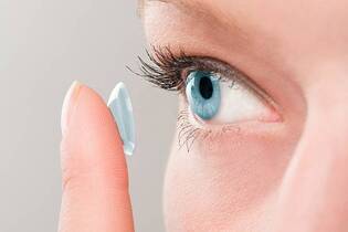
Contact lens
Optical aberration
Ryan Searle (darts player)
Intraocular lens
Paul Waner
Cathode-ray tube
Noel Alpins
Chalazion
Complex beam parameter
Sri Siddhartha Medical College
Toric lens
Stokes lens
Christ Carrying the Cross (El Greco, New York)
Corneal topography
Refractive surgery
Megalocornea
Subjective refraction
List of bespectacled baseball players
Ernest Maddox
Ritchey-Chrétien telescope
Near-sightedness
Staphyloma
Craniosynostosis
Refractive error
Reversing type
Kaan Güneşberk
Stigmator
Electrostatic deflection
Astigmatism - Wikipedia
 Astigmatism: MedlinePlus Medical Encyclopedia
 Astigmatism | AOA
 astigmatism - Wiktionary, the free dictionary
 Minimizing Astigmatism in Cataract Surgery
 Astigmatism vs. nearsightedness: What to know
 El Greco had style not astigmatism | New Scientist
 Astigmatism in infancy and childhood
 Guide to Glasses for Astigmatism
Lasik for Astigmatism: What to Expect
 Blog: Astigmatism | CooperVision
 Bungee cord-induced corneal lacerations correcting for myopic astigmatism
 Astigmatism Testing - All About Vision
Cook Children's Health Library - Astigmatism - Conditions - 96479
 Astigmatism - Causes, Symptoms, Treatment, Diagnosis - MedBroadcast.com
 Astigmatism: Types, Diagnosis and Treatment Options - Nova Science Publishers
 The contribution of ocular residual astigmatism to anterior corneal astigmatism in refractive astigmatism eyes | Scientific...
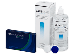
Contact Lenses for Astigmatism: Toric, GP, Hybrid
 Will Dark Glasses Help with Astigmatism and Light Sensitivity? - American Academy of Ophthalmology
 Astigmatism Correction Prescription Glasses | Blog | EyeBuyDirect
 The 10 Best Selling Contacts for Astigmatism - 2023 (Updated) | Eyestyle Blog
 Astigmatism | The SuperHeroHype Forums
 What is astigmatism? | Hoya Vision Care
 Astigmatism Archive - SynergEyes
 Bausch + Lomb ULTRA Multifocal for Astigmatism (6 pack) Contacts | Warby Parker
What is astigmatism? | Hoya Vision Care
 Buy Purevision 2 for Astigmatism - ContactLens.com
 LASIK for Astigmatism and How to Correct Them?
 Pathology of vision: Astigmatism and glaucoma | Article | Onestopenglish
Thinning and st1
- In keratoconus, progressive thinning and steepening of the cornea cause irregular astigmatism. (wikipedia.org)
Farsightedness11
- Astigmatism often occurs together with nearsightedness or farsightedness. (medlineplus.gov)
- Laser surgery can help change the shape of the cornea surface to eliminate astigmatism, along with nearsightedness or farsightedness. (medlineplus.gov)
- Astigmatism frequently occurs with other vision conditions like myopia (nearsightedness) and hyperopia (farsightedness). (aoa.org)
- Often, astigmatism goes along with other refractive eye conditions like nearsightedness and farsightedness. (healthline.com)
- Getting LASIK for astigmatism is similar to getting it for nearsightedness or farsightedness. (healthline.com)
- While most people may have heard of the terms nearsightedness and farsightedness, some eye care patients may ask, "What is astigmatism? (coopervision.com)
- Toric lenses focus on different parts of the lens to correct the nearsightedness or farsightedness that goes along with astigmatism. (allaboutvision.com)
- The most common vision related issues people experience are nearsightedness, farsightedness, and astigmatisms. (eyebuydirect.com)
- Astigmatism, commonly mispronounced as "astigmatism", is different from farsightedness and nearsightedness in that it is not typically caused by eye disease. (lasikplus.com)
- As cataract surgery progressed, intraocular lenses were invented which corrected the majority of a patient's nearsightedness and farsightedness, however did not correct for astigmatism. (ocli.net)
- LASEK is specifically used to correct astigmatism, hyperopia (farsightedness), and myopia (nearsightedness). (medscape.com)
Irregular14
- The underlying mechanism involves an irregular curvature of the cornea and protective reaction changes in the lens of the eye, called lens astigmatism, that has the same mechanism as spasm of accommodation. (wikipedia.org)
- Because rigid gas-permeable contact lenses maintain their regular shape while on the cornea, they can compensate for the cornea's irregular shape and improve vision for people with astigmatism. (aoa.org)
- Astigmatism is a common vision problem caused by an irregular cornea or an irregularly shaped part of your eye (its lens). (healthline.com)
- To correct severe astigmatism, an eye surgeon might use special knives or a laser beam to correct the abnormal or irregular curve of the cornea. (epnet.com)
- larger lenses that provide excellent astigmatism correction - even for irregular eye surfaces. (allaboutvision.com)
- These are specially designed and shaped to compensate for the irregular curve of the cornea that causes astigmatism. (hoyavision.com)
- That is regular astigmatism, irregular astigmatism, and oblique astigmatism. (lasikplus.com)
- What is irregular astigmatism? (lasikplus.com)
- Irregular astigmatism, which is typically caused by a physical injury that caused scarring on the cornea, occurs when the principal meridians are not perpendicular to one another. (lasikplus.com)
- If the astigmatism were irregular, I would not address it initially. (crstoday.com)
- The cylindrical power corrects the astigmatism, which means it counteracts the irregular curvature of the cornea or lens to provide clear vision. (lens.com)
- Another possible complication of these infections is uneven healing of the stroma, resulting in irregular astigmatism (that may require a gas-permeable contact lens or PTK to improve vision). (medscape.com)
- These initial attempts were complex and unpredictable, often leading to keratoconus and other irregular astigmatisms. (medscape.com)
- Since its introduction, LASIK has been associated with various complications, specifically when performed on eyes with decreased corneal thickness, irregular astigmatism, dryness, preexisting ocular surface disease, or glaucoma, to the point where several of these entities have become relative contraindications to performing LASIK. (medscape.com)
Myopia9
- Another recent follow-up study again had identified four novel loci for corneal astigmatism, with two also being novel loci for astigmatism: ZC3H11B (associated with axial length), NPLOC4 (associated with myopia), LINC00340 (associated with spherical equivalent refractive error) and HERC2 (associated with eye color). (wikipedia.org)
- What are the differences between astigmatism and myopia? (medicalnewstoday.com)
- Both astigmatism and myopia are issues that can affect a person's vision. (medicalnewstoday.com)
- Astigmatism and myopia are common eye conditions that can result in blurry vision. (medicalnewstoday.com)
- This article will discuss the differences between astigmatism and myopia. (medicalnewstoday.com)
- Both astigmatism and myopia are known as refractive errors . (medicalnewstoday.com)
- Often, people with astigmatism have myopia (nearsightedness) as well. (medbroadcast.com)
- 13, 2018-- STAAR Surgical Company (NASDAQ: STAA), a leading developer, manufacturer and marketer of implantable lenses and companion delivery systems for the eye, is today announcing that the FDA has granted approval of the PMA Supplement for the Visian Toric ICL for the correction of myopia with astigmatism. (staar.com)
- This approval represents a meaningful expansion of the Implantable Collamer® Lens (ICL) product line for the correction of refractive error in patients with both myopia and astigmatism which are common conditions in the United States (US). (staar.com)
Prescribed to correct astigmatism1
- If your glasses are prescribed to correct astigmatism only, single-vision lenses are a good choice. (eyebuydirect.com)
Cornea or lens7
- Astigmatism is an irregularly shaped cornea or lens that prevents light from focusing properly on the retina, the light-sensitive surface at the back of the eye. (aoa.org)
- Astigmatism occurs due to the cornea or lens having a different shape. (medicalnewstoday.com)
- Astigmatism occurs when your cornea or lens is oddly shaped. (healthline.com)
- The unique shape of a toric lens allows it to correct for astigmatism issues that arise from a different curvature of the eye's cornea or lens. (lens.com)
- Acuvue Moist contacts are available for correcting astigmatism, a refractive error that causes hazy, blurry, or distorted vision due to the unequal shape of the cornea or lens. (webeyecare.com)
- Toric contact lenses are a type of specialized contact lens designed to correct astigmatism , a common vision condition that results from an irregularly shaped cornea or lens inside the eye. (lens.com)
- Astigmatism is an irregularity in the curvature of the cornea or lens. (msdmanuals.com)
Lens28
- Diagnosis is by an eye examination called autorefractor keratometry (objective, allows to see lens and cornea components of astigmatism) and subjective refraction. (wikipedia.org)
- But subjective methods are almost always inaccurate if lens astigmatism is not fully removed first with a week of eye drops. (wikipedia.org)
- In addition, the curvature of the lens inside the eye can change, resulting in an increase or decrease in astigmatism. (aoa.org)
- The eyeglasses contain a special cylindrical lens prescription that compensates for astigmatism. (aoa.org)
- An increasingly popular way to manage astigmatism is with the use of a toric (astigmatism correcting) intraocular lens (IOL). (medscape.com)
- Astigmatism happens when the cornea (the clear front layer of the eye) or the lens (an inner part of the eye that helps with focus) has a different shape than usual. (medicalnewstoday.com)
- Glasses and contact lenses for people with astigmatism have an extra component called a toric lens . (medbroadcast.com)
- In today's modern contact lens market, people with astigmatism have more options than ever. (allaboutvision.com)
- Deciding which contacts are best for your astigmatism depends on how well you tolerate each lens, the characteristics of your eyes and your current level of astigmatism. (allaboutvision.com)
- Every eye with astigmatism is unique, so it can take some trial and error to find the right lens with the best fit, comfort and visual sharpness. (allaboutvision.com)
- Gas permeable contact lenses are another popular type of contact lens for astigmatism. (allaboutvision.com)
- A growing number of studies have found that lower efficiency of astigmatism correction by laser systems is mainly due to the eye's internal optics, crystalline lens is the largest component of it. (nature.com)
- Astigmatism can also result from irregularities in the shape of the lens, but the cornea is the more common cause. (eyebuydirect.com)
- ACUVUE OASYS for ASTIGMATISM is a 1-2 week lens (take out daily, clean, and replace after 14 days), while Biofinity toric is a monthly lens (take out daily, clean, and replace after 30 days). (lens.com)
- There are several different contact lens designs for people that have astigmatism. (synergeyes.com)
- Similarly, to corneal astigmatism, lenticular astigmatism is caused by an imperfect curvature of the lens which focuses light behind or in front of the retina. (lasikplus.com)
- Acuvue Moist for Astigmatism contacts provides clear vision and comfort for people with astigmatism using a BLINK STABILIZED design that holds the lens in place. (webeyecare.com)
- 1-Day Acuvue Moist for Astigmatism 90 pack is a daily disposable contact lens, meaning you don't have to worry about cleaning solutions and contact storage cases. (webeyecare.com)
- Acuvue Moist daily contacts have a torus shape that creates multiple refractive powers across the lens to correct astigmatism. (webeyecare.com)
- Placement of the extra power in a toric lens for astigmatism. (visiondirect.co.uk)
- AIR OPTIX® plus HydraGlyde® for Astigmatism contact lenses combine two unique technologies, SmartShield Technology® and HydraGlyde® Moisture Matrix, so that you can experience consistent comfort and long-lasting lens surface moisture. (yourlens.com)
- Toric lenses are uniquely shaped to provide different powers in different meridians of the lens to address both the spherical and cylindrical prescriptions needed for individuals with astigmatism. (lens.com)
- Cylinder and Axis: Astigmatism is characterized by two components in an eyeglass or contact lens prescription: the spherical power (measured in diopters) and the cylindrical power (also measured in diopters) along with an axis (a degree measurement). (lens.com)
- As the name suggests, Bausch and Lomb ULTRA for Astigmatism contacts are made by Bausch and Lomb, one of the world's largest suppliers of contact lenses and lens care products. (lens.com)
- For patients that have moderate to severe astigmatism, your surgeon might elect to use a toric intraocular lens. (ocli.net)
- ACUVUE OASYS for ASTIGMATISM features a unique BLINK STABILIZED lens design that actively prevents toric lenses from rotating in the eye. (eyeglassworld.com)
- For this reason most RCs employ a correcting lens to eliminate the residual astigmatism. (starizona.com)
- The author concludes that the tolerances for sphere power, axial astigmatism, and prism in a single lens given in Z87.1 were adequate. (cdc.gov)
Cataract surgery5
- Astigmatism may also occur following a cataract surgery or a corneal injury. (wikipedia.org)
- Cite this: Minimizing Astigmatism in Cataract Surgery - Medscape - Oct 19, 2017. (medscape.com)
- Can I have my astigmatism corrected when I undergo cataract surgery? (ocli.net)
- The most recent inventions in cataract surgery have brought a wide range of options that can correct moderate to severe astigmatism. (ocli.net)
- Toric lenses are specifically designed to correct astigmatism in patients undergoing cataract surgery, which can reduce or even eliminate your need for glasses in order to see well in the distance, however reading glasses are still required to see up close. (ocli.net)
Blurry5
- However, astigmatism may also result in blurry vision with nearby objects. (medicalnewstoday.com)
- You'll need glasses for astigmatism if your vision is blurry or distorted. (healthline.com)
- You'll need glasses for your astigmatism if your vision is blurry or you have eyestrain. (healthline.com)
- Objects in your sight line may be blurry because your eyes cannot focus light when you have astigmatism. (healthline.com)
- Astigmatism is a vision condition that causes you to have blurry or distorted vision when looking at things up close, far away, or both. (eyebuydirect.com)
Correction6
- Laser vision correction can most often eliminate, or greatly reduce astigmatism. (medlineplus.gov)
- Especially for young school-age children, they won't know that they have astigmatism, even if it requires correction," he explains. (aoa.org)
- If you think you may have astigmatism based on this screening, be sure to follow up with an eye doctor, who can confirm the assessment and help you find adequate correction. (allaboutvision.com)
- Patients with astigmatism still had to wear some form of correction, whether it be glasses or toric contact lenses. (ocli.net)
- Princess Pinky Eurasia Toric Series is the world's first cosplay toric lenses that features a super bright effect that complete any cosplay look, while keeping your vision clear with astigmatism correction. (pinkyparadise.com)
- for the correction of myopic astigmatism with spherical equivalent ranging from -3.0 D to ≤ -15.0 D (in the spectacle plane) with cylinder (spectacle plane) of 1.0 D to 4.0 D in the spectacle plane. (staar.com)
Types of astigmatism4
- Special toric soft contact lenses can correct for many types of astigmatism. (aoa.org)
- What are the three main most common types of Astigmatism? (lasikplus.com)
- The three commonly known types of astigmatism are myopic astigmatism, hyperopic astigmatism, and mixed astigmatism. (lasikplus.com)
- Both LASIK and PRK are types of "refractive surgery" and can safely and permanently correct most types of astigmatism. (lasikplus.com)
Contacts for Astigmatism1
- A lot of buyers pick eyeglasses with unique tube colored contacts for astigmatism(乱視用カラコン) lenses medicines that make up for astigmatism. (vslondon.org)
Specifically designed to correct astigmatism1
- If so, then you need contact lenses that are specifically designed to correct astigmatism called toric contact lenses . (lens.com)
Symptoms10
- With cases of mild astigmatism, a person may not notice any symptoms. (medicalnewstoday.com)
- If you experience symptoms such as blurred or distorted vision, headaches and/or eye strain/discomfort, there's a chance you could have some form of astigmatism . (allaboutvision.com)
- This strategy can certainly help identity symptoms of astigmatism, as an initial approach. (allaboutvision.com)
- Some people with astigmatism may have no symptoms. (epnet.com)
- Symptoms vary depending on the extent of the astigmatism. (epnet.com)
- Astigmatism is a common refractive anomaly that results in reduced visual acuity and causes various symptoms like glare, monocular diplopia, asthenopia, and distortion 8 . (nature.com)
- Having these symptoms doesn't necessarily mean you have astigmatism. (eyebuydirect.com)
- What are the symptoms of astigmatism? (lens.com)
- The symptoms of lenticular astigmatism are the same as corneal astigmatism. (lasikplus.com)
- The symptoms of astigmatism occur when light entering the eyes scatters at multiple points on the retina, causing distorted vision. (webeyecare.com)
Regarding the prevalence of astigmatism1
- Factors such as race, nutrition, and environmental condition did not vary from the literature regarding the prevalence of astigmatism in infants and children. (nih.gov)
Principal meridians are perpendicular2
- Regular astigmatism - principal meridians are perpendicular. (wikipedia.org)
- Oblique astigmatism is identical to regular astigmatism, where the principal meridians are perpendicular to one another, with the difference being that they are not at 90° or 180°, respectively. (lasikplus.com)
Shape of the cornea1
- Astigmatism is commonly caused by imperfections in the shape of the cornea. (eyebuydirect.com)
Refractive error that causes1
- Astigmatism is a refractive error that causes blurred or distorted vision at all distances. (allaboutvision.com)
Keratoconus3
- If astigmatism gets worse, it may be a sign of keratoconus . (medlineplus.gov)
- Early postoperative astigmatism following penetrating keratoplasty (PK) may occur more frequently in keratoconus than other conditions, although all corneal grafts are beset by this problem. (bmj.com)
- The work presented in this month's issue of the BJO (p 371 ) by Lim et al on late onset post-keratoplasty astigmatism in patients with keratoconus identifies a subgroup of patients who developed excessive astigmatism years after successful surgery. (bmj.com)
Forms of astigmatism2
- There are three forms of astigmatism. (lasikplus.com)
- Everything you need to know about astigmatism colorcon The majority of forms of astigmatism are dealt with effectively. (vslondon.org)
Signs of astigmatism1
- If results from the at-home screening show signs of astigmatism, you should follow up with an eye care professional as soon as possible. (allaboutvision.com)
Lasik for Astigmatism1
- LASIK for Astigmatism and How to Correct Them? (lasikplus.com)
Effects of astigmatism2
- This bends light to counterbalance the effects of astigmatism. (medbroadcast.com)
- Contact lenses for astigmatism are specially designed to correct the visual effects of astigmatism - a common refractive error caused by a misshapen cornea . (allaboutvision.com)
Glasses for astigmatism5
- Do you need glasses for astigmatism? (healthline.com)
- Where can you get glasses for astigmatism? (healthline.com)
- If you've been told you need prescription eyeglasses to correct astigmatism, you may be wondering what makes glasses for astigmatism different from other types of eyewear. (eyebuydirect.com)
- Do I Need Glasses for Astigmatism? (eyebuydirect.com)
- What Are the Best Glasses for Astigmatism? (eyebuydirect.com)
Suffer from astigmatism2
- Do you suffer from astigmatism? (lens.com)
- Parabolic mirrors such as those used in Newtonians do not suffer from astigmatism. (starizona.com)
Person with astigmatism2
- A person with astigmatism may also have poor night vision and see halos around lights at night. (webeyecare.com)
- A person with astigmatism (each eye should be tested separately) tends to see certain lines more boldly (that is, in better focus) than the others. (msdmanuals.com)
Correct Your Astigmatism3
- Cylinder (CYL) - The lenses in your glasses need to have the right power to correct your astigmatism. (eyebuydirect.com)
- The cylinder value is the power needed to correct your astigmatism. (eyebuydirect.com)
- If your eye doctor recommends that you wear glasses to correct your astigmatism, how do you know what kind to get? (eyebuydirect.com)
Management of astigmatism1
- As such, the management of astigmatism is crucial in delivering optimal outcomes to patients. (medscape.com)
Children with astigmatism3
- A total of 478 children with astigmatism from Valencia Province, Spain were examined. (nih.gov)
- Glasses were prescribed to correct for blurred vision in the two groups of children with astigmatism. (aoa.org)
- They assessed children with astigmatism before and after the children received glasses. (aoa.org)
Ocular9
- Additionally, it is also possible for people to develop an astigmatism due to an eye condition, eye injury, or after ocular surgery. (medicalnewstoday.com)
- To determine the distribution of ocular residual astigmatism (ORA) in astigmatic eyes and the influence on the anterior corneal (ACA) and refractive astigmatism (RA). (nature.com)
- The magnitude of ocular residual astigmatism was relatively small in children's astigmatic eyes. (nature.com)
- Twenty three years ago, based on the term residual astigmatism, Alpins 2 proposed the parameter ocular residual astigmatism (ORA) to express the discrepancy between corneal and refractive astigmatism (RA). (nature.com)
- RA is the total ocular astigmatism obtained by phoropter or retinoscopy. (nature.com)
- My daughter has astigmatism (imperfectly shaped cornea) and ocular albinism (lack of color in the eye) . (aao.org)
- In astigmatism, the corneal surface is typically toric, causing each of the ocular meridians to refract light uniquely. (lasikplus.com)
- Myopic Astigmatism occurs when one or two principal ocular meridians focus light in front of the retina (are nearsighted). (lasikplus.com)
- Hyperopic astigmatism occurs when one or two principal ocular meridians focus light behind the retina (are farsighted). (lasikplus.com)
Myopic2
- What is Myopic Astigmatism? (lasikplus.com)
- FDA's approval of STAAR's Visian Toric ICL in the U.S. provides an exciting treatment option for myopic patients with astigmatism in search of visual freedom. (staar.com)
Mild6
- Mild astigmatism may not need to be corrected. (medlineplus.gov)
- You can be a candidate for laser-assisted in situ keratomileusis (LASIK) if you have mild to moderate astigmatism . (healthline.com)
- This procedure may be an option for those who have mild astigmatism. (medbroadcast.com)
- People with mild astigmatism usually tolerate toric lenses (soft contacts) well, but rigid gas permeable or hybrid contacts may be able to offer even sharper vision. (allaboutvision.com)
- You might not need eyeglasses if you have mild astigmatism and can see clearly without any problems. (eyebuydirect.com)
- For patients that have mild corneal astigmatism, your surgeon might elect to do an LRI, which stands for limbal relaxing incision. (ocli.net)
Retina1
- Astigmatism, as described above, occurs when the light is not focused to a single point on the retina. (lasikplus.com)
High-astigmatism3
- Based on vision testing, the students were categorized into three groups: those with high astigmatism in both eyes, moderate astigmatism in both eyes, and no or low astigmatism (defined as being less than or equal to one diopter), in both eyes. (aoa.org)
- After receiving glasses, high-astigmatism students boosted their accurate reading speed by almost seven words per minute-the approximate gain expected from one-half school year. (aoa.org)
- A clear corneal graft may be an optical failure if high astigmatism limits visual acuity. (bmj.com)
Oblique astigmatism3
- Oblique astigmatism - the steepest curve lies in between 120 and 150 degrees and 30 and 60 degrees. (wikipedia.org)
- The prevalence of against-the-rule astigmatism decreased from 61.5% (at 2 years) to 33.4% (at 12 years), with-the-rule astigmatism increased in prevalence from 23.1% (at 2 years) to 66.6% (at 12 years), and oblique astigmatism decreased with age from 15.4% (at 2 years) and 0% (at 12 years). (nih.gov)
- The median magnitude of ORA in astigmatic eyes was 0.57 D, with interquartile range was 0.42 D. And they were main against-the-rule (57.6-75.8%) and oblique astigmatism (13.9-34.5%) ORA. (nature.com)
Moderate astigmatism5
- People with moderate astigmatism may be able to temporarily obtain clear vision without lenses for most of their daily activities. (aoa.org)
- With uncorrected vision, students in the high group showed significantly decreased oral reading fluency compared with the low/no and moderate astigmatism groups. (aoa.org)
- Children with moderate astigmatism had reading fluency similar to those with no/low astigmatism. (aoa.org)
- Glasses had minimal or no effect on reading fluency for those with moderate astigmatism. (aoa.org)
- The children with moderate astigmatism may have shown greater improvement if some testing factors had been different, adds Dr. Chung. (aoa.org)
Eliminate astigmatism1
- Most two-mirror Cassegrain telescopes are designed specifically to eliminate astigmatism (again the RC being the exception). (starizona.com)
Significant astigmatism2
- If significant astigmatism remains, which cannot be corrected by optical means, then further surgical procedures may be utilised to reduce the astigmatism. (bmj.com)
- Very few telescope designs have significant astigmatism. (starizona.com)
Prevalence3
- Some research has pointed to the link between astigmatism and higher prevalence of migraine headaches. (wikipedia.org)
- To evaluate the prevalence of astigmatism in infancy and childhood in a Spanish population. (nih.gov)
- The study, published in the February 2016 Optometry and Vision Science , included 273 students in third through eighth grade in schools on the Tohono O'odham Nation reservation, a group with a high prevalence of astigmatism. (aoa.org)
Blurred vision2
Occurs2
- Astigmatism often occurs at birth and can change or develop later in life. (wikipedia.org)
- Regular astigmatism occurs when the principal meridians are separated by 90 degrees on the 90° and 180° lines. (lasikplus.com)
Eyestrain2
- People with astigmatism may experience headache or eyestrain. (medbroadcast.com)
- The Easily Erase Astigmatism Program can help with distorted vision, headaches, eyestrain, neck and shoulder tension, tiredness and eye fatigue due to astigmatism. (bettervision.com)
Biofinity5
- The two biggest sellers historically are ACUVUE OASYS for ASTIGMATISM and Biofinity toric . (lens.com)
- Bausch and Lomb ULTRA® for Astigmatism and Biofinity® toric are two of the most popular toric contact lenses on the market today. (lens.com)
- This guide will compare Bausch and Lomb ULTRA for Astigmatism and Biofinity toric and discuss their features, including their wearing and replacement schedules, price, and more. (lens.com)
- Bausch and Lomb ULTRA for Astigmatism and Biofinity toric are monthly disposable contacts, which means they can be worn daily for up to 30 days. (lens.com)
- As toric lenses, both Bausch and Lomb ULTRA for Astigmatism and Biofinity toric do a great job staying put on the eye, but they don't work the same way. (lens.com)
Bausch4
- If you don't have an up-to-date Bausch + Lomb ULTRA Multifocal for Astigmatism prescription, book an eye exam near you or renew it in just 10 minutes with Virtual Vision Test. (warbyparker.com)
- Bausch & Lomb PureVision®2 HD for Astigmatism contact lenses with HD Optics are designed to reduce halos and glare and deliver the clear, crisp vision everyone wants. (contactlens.com)
- Biotrue One Day for Astigmatism are daily disposable toric lenses manufactured by Bausch & Lomb and made from a unique material called hypergel. (visiondirect.co.uk)
- Bausch and Lomb ULTRA for Astigmatism contacts use OpticAlign™ Design. (lens.com)
Diagnosis1
- This book discusses the types, diagnosis and treatment options of astigmatism. (novapublishers.com)
Common refractive1
- A 2018 systematic review suggests that astigmatism is the most common refractive error. (medicalnewstoday.com)
Posterior corneal1
- One notable limitation was the lack of inclusion of posterior corneal astigmatism in toric calculations. (medscape.com)
Curvature of the eye's cornea1
- Astigmatisms are caused by imperfections in the curvature of the eye's cornea. (eyebuydirect.com)
Axis4
- Astigmatism error and axis orientation were determined using noncyclopegic retinoscopy over each eye. (nih.gov)
- 5 In the absence of published studies, our clinical experience suggests that there continues to be a slow long term drift in the axis and magnitude of astigmatism if patients are followed for 10-15 years following PK. (bmj.com)
- Fitting: Fitting toric contact lenses requires additional expertise compared to spherical lenses, as the optometrist or ophthalmologist must determine the correct power, axis, and fit for the specific individual's astigmatism. (lens.com)
- Like coma , astigmatism is an off-axis aberration. (starizona.com)
People13
- People of all ages can be affected by astigmatism. (wikipedia.org)
- People with astigmatism primarily choose eyeglasses to improve their vision. (aoa.org)
- While the exact cause is currently unknown, evidence suggests that people can inherit the risk of developing an astigmatism. (medicalnewstoday.com)
- His experiments show that people with astigmatism adapt to the condition, and are as capable as anyone else of drawing objects in their proper proportions. (newscientist.com)
- Anstis then persuaded one of his volunteers to wear the monocular for four days to see if people could adapt to the artificial astigmatism. (newscientist.com)
- Lots of eyeglass websites offer lenses that work for people with astigmatism. (healthline.com)
- Some people are born with astigmatism. (medbroadcast.com)
- For people who are also nearsighted, the surgery can correct both the nearsightedness and astigmatism at the same time. (medbroadcast.com)
- Many people with astigmatism find that their vision is noticeably sharper with gas permeable lenses than it is with torics. (allaboutvision.com)
- Rather than seeing complete blurriness at certain distances, people with astigmatisms see hazy or blotched spots in an otherwise clear image. (eyebuydirect.com)
- The vast majority of people with astigmatism were born with it, but experts still have no explanation as to what causes astigmatism. (lens.com)
- In rare cases, people can get astigmatism through an eye injury or a disease. (lens.com)
- More than 8 million people had vision impairment caused by an uncorrected refractive error, such as nearsightedness or astigmatism. (cdc.gov)
Toric lenses2
- Toric lenses are soft contact lenses that correct astigmatism. (allaboutvision.com)
- Usually, astigmatism can be corrected effectively with toric lenses. (hoyavision.com)