Autolysis
Bacteriolysis
N-Acetylmuramoyl-L-alanine Amidase
Calpain
Cell Wall
Peptidoglycan
Trypsinogen
Teichoic Acids
Cefotiam
Enteropeptidase
Staphylococcus aureus
Octoxynol
Endopeptidases
Muramidase
Enterococcus faecalis
Chymosin
Caseins
Phosphotungstic Acid
Lysostaphin
Penicillins
Glucosamine
Streptolysins
Mutation
Molecular Sequence Data
Chloramphenicol
Autolysosomal membrane-associated betaine homocysteine methyltransferase. Limited degradation fragment of a sequestered cytosolic enzyme monitoring autophagy. (1/238)
We compared the membrane proteins of autolysosomes isolated from leupeptin-administered rat liver with those of lysosomes. In addition to many polypeptides common to the two membranes, the autolysosomal membranes were found to be more enriched in endoplasmic reticulum lumenal proteins (protein-disulfide isomerase, calreticulin, ER60, BiP) and endosome/Golgi markers (cation-independent mannose 6-phosphate receptor, transferrin receptor, Golgi 58-kDa protein) than lysosomal membranes. The autolysosomal membrane proteins include three polypeptides (44, 35, and 32 kDa) whose amino-terminal sequences have not yet been reported. Combining immunoblotting and reverse transcriptase-polymerase chain reaction analyses, we identified the 44-kDa peptide as the intact subunit of betaine homocysteine methyltransferase and the 35- and 32-kDa peptides as two proteolytic fragments. Pronase digestion of autolysosomes revealed that the 44-kDa and 32-kDa peptides are present in the lumen, whereas the 35-kDa peptide is not. In primary hepatocyte cultures, the starvation-induced accumulation of the 32-kDa peptide occurs in the presence of E64d, showing that the 32-kDa peptide is formed from the sequestered 44-kDa peptide during autophagy. The accumulation is induced by rapamycin but completely inhibited by wortmannin, 3-methyladenine, and bafilomycin. Thus, detection of the 32-kDa peptide by immunoblotting can be used as a streamlined assay for monitoring autophagy. (+info)Developmental aspects of secondary palate formation. (2/238)
Research on development of the secondary palate has, in the past, dealt primarily with morphological aspects of shelf elevation and fusion. The many factors thought to be involved in palatal elevation, such as fetal neuromuscular activity and growth of the cranial base and mandible, as well as production of extracellular matrix and contractile elements in the palate, are mostly based on gross, light microscopic, morphometric or histochemical observations. Recently, more biochemical procedures have been utilized to described palatal shelf elevation. Although these studies strongly suggest that palatal extracellular matrix plays a major role in shelf movement, interpretation of these data remains difficult owing to the complexity of tissue interactions involved in craniofacial development. Shelf elevation does not appear to involve a single motive factor, but rather a coordinated interaction of all of the abovementioned developmental events. Further analysis of mechanisms of shelf elevation requires development of new, and refinement of existing, in vitro procedures. A system that enables one to examine shelf elevation in vitro would allow more meaningful analysis of the relative importance of the various components in shelf movement. Much more is known about fusion of the palatal shelves, owing in large part to in vitro studies. Fusion of the apposing shelves, both in vivo and in vitro, is dependent upon adhesion and cell dealth of the midline epithelial cells. Adhesion betweeen apposing epithelial surfaces appears to involve epithelial cell surface macromolecules. Further analysis of palatal epithelial adhesion should be directed towards characterization of those cell surface components responsible for this adhesive interaction. Midline epithelial cells cease DNA synthesis 24-36 h before shelf elevation and contact, become active in the synthesis of cell surface glycoproteins, and subsequently manifest morphological signs of necrosis. Death of the midline epithelial cells is thought to involve a programmed, lysosomal-mediated autolysis... (+info)Suppression of the lytic and bactericidal effects of cell wallinhibitory antibiotics. (3/238)
The bacteriolytic effect of beta-lactam antibiotics on Bacillus subtilis and on Streptococcus pneumoniae was found to be a function of the pH; lysis was suppressed if the pH of the pneumococcal culture was below 6.0 during penicillin treatment. In the case of B. subtilis, growth at pH 6.6 prevented penicillin-induced lysis. In pneumococci, the addition of trypsin to the growth medium also protected against lysis. The pH-dependent protection phenomenon resembled in several respects the antibiotic "tolerance" of pneumococci with a defective autolytic system. (i) At the pH nonpermissive for lysis, the bacteria retained their normal sensitivity to beta-lactam and to other cell wall inhibitors; however, instead of lysis, the drug-treated bacteria simply stopped growing. Loss of viability of the cells was also greatly reduced. (ii) Protection against lysis was independent of the dose and chemical nature of the cell wall inhibitors. (iii) The protection effect was reversible; lysis and loss of viability could be triggered by a postincubation of the drug-treated bacteria at the pH permissive for lysis. (+info)The autolysis loop of activated protein C interacts with factor Va and differentiates between the Arg506 and Arg306 cleavage sites. (4/238)
The anticoagulant human plasma serine protease, activated protein C (APC), inactivates blood coagulation factors Va (FVa) and VIIIa. The so-called autolysis loop of APC (residues 301-316, equivalent to chymotrypsin [CHT] residues 142-153) has been hypothesized to bind FVa. In this study, site-directed mutagenesis was used to probe the role of the charged residues in this loop in interactions between APC and FVa. Residues Arg306 (147 CHT), Glu307, Lys308, Glu309, Lys311, Arg312, and Arg314 were each individually, or in selected combinations, mutated to Ala. The purified recombinant protein C mutants were characterized using activated partial thromboplastin time (APTT) clotting assays and FVa inactivation assays. Mutants 306A, 308A, 311A, 312A, and 314A had mildly reduced anticoagulant activity. Based on FVa inactivation assays and APTT assays using purified Gln506-FVa and plasma containing Gln506-FV, it appeared that these mutants were primarily impaired for cleavage of FVa at Arg506. Studies of the quadruple APC mutant (306A, 311A, 312A, and 314A) suggested that the autolysis loop provides for up to 15-fold discrimination of the Arg506 cleavage site relative to the Arg306 cleavage site. This study shows that the loop on APC of residues 306 to 314 defines an FVa binding site and accounts for much of the difference in cleavage rates at the 2 major cleavage sites in FVa. (Blood. 2000;96:585-593) (+info)The D-alanine residues of Staphylococcus aureus teichoic acids alter the susceptibility to vancomycin and the activity of autolytic enzymes. (5/238)
Recently, Staphylococcus aureus strains with intermediate resistance to vancomycin, the antibiotic of last resort, have been described. Multiple changes in peptidoglycan turnover and structure contribute to the resistance phenotype. Here, we describe that structural changes of teichoic acids in the cell envelope have a considerable influence on the susceptibility to vancomycin and other glycopeptides. S. aureus cells lacking D-alanine esters in teichoic acids exhibited an at least threefold-increased sensitivity to glycopeptide antibiotics. Furthermore, the autolytic activity of the D-alanine mutant was reduced compared to the wild-type, and the mutant was more susceptible to the staphylolytic enzyme lysostaphin. Vancomycin inhibited autolysis at very high concentrations but neither in the wild-type nor in the mutant was the autolytic activity influenced in the range of the MIC. Mutant cells had a considerably higher capacity to bind vancomycin. (+info)Description of staphylococcus serine protease (ssp) operon in Staphylococcus aureus and nonpolar inactivation of sspA-encoded serine protease. (6/238)
Signature tagged mutagenesis has recently revealed that the Ssp serine protease (V8 protease) contributes to in vivo growth and survival of Staphylococcus aureus in different infection models, and our previous work indicated that Ssp could play a role in controlling microbial adhesion. In this study, we describe an operon structure within the ssp locus of S. aureus RN6390. The ssp gene encoding V8 protease is designated as sspA, and is followed by sspB, which encodes a 40.6-kDa cysteine protease, and sspC, which encodes a 12.9-kDa protein of unknown function. S. aureus SP6391 is an isogenic derivative of RN6390, in which specific loss of SspA function was achieved through a nonpolar allelic replacement mutation. In addition to losing SspA, the culture supernatant of SP6391 showed a loss of 22- to 23-kDa proteins and the appearance of a 40-kDa protein corresponding to SspB. Although the 40-kDa SspB protein could degrade denatured collagen, our data establish that this is a precursor form which is normally processed by SspA to form a mature cysteine protease. Culture supernatant of SP6391 also showed a new 42-kDa glucosaminidase and enhanced glucosaminidase activity in the 29 to 32 kDa range. Although nonpolar inactivation of sspA exerted a pleiotropic effect, S. aureus SP6391 exhibited enhanced virulence in a tissue abscess infection model relative to RN6390. Therefore, we conclude that SspA is required for maturation of SspB and plays a role in controlling autolytic activity but does not by itself exert a significant contribution to the development of tissue abscess infections. (+info)Increasing the thermal stability of euphauserase. A cold-active and multifunctional serine protease from Antarctic krill. (7/238)
A molecular model of Antarctic krill euphauserase based on the known crystal structure of its fiddler crab analog, collagenase I, indicates that the core structure of these enzymes is almost identical. Euphauserase is a cold-active and thermally sensitive enzyme with a high affinity for Lys, Arg and large hydrophobic amino acids. Residue Phe137 in euphauserase, localized in loop D (autolysis loop), is highly exposed on the surface of the molecule. Therefore, it appeared to be an easy target for autolysis. The broadly specific euphauserase has a low affinity for negatively charged residues. In order to increase the stability of the enzyme, two mutants were created in which residue Phe137 was replaced by a Glu and an Asp residue. Both mutations resulted in increased stability of the recombinant euphauserase towards thermal inactivation. (+info)The AbcA transporter of Staphylococcus aureus affects cell autolysis. (8/238)
Increased production of penicillin-binding protein PBP 4 is known to increase peptidoglycan cross-linking and contributes to methicillin resistance in Staphylococcus aureus. The pbp4 gene shares a 400-nucleotide intercistronic region with the divergently transcribed abcA gene, encoding an ATP-binding cassette transporter of unknown function. Our study revealed that methicillin stimulated abcA transcription but had no effects on pbp4 transcription. Analysis of abcA expression in mutants defective for global regulators showed that abcA is under the control of agr. Insertional inactivation of abcA by an erythromycin resistance determinant did not influence pbp4 transcription, nor did it alter resistance to methicillin and other cell wall-directed antibiotics. However, abcA mutants showed spontaneous partial lysis on plates containing subinhibitory concentrations of methicillin due to increased spontaneous autolysis. Since the autolytic zymograms of cell extracts were identical in mutants and parental strains, we postulate an indirect role of AbcA in control of autolytic activities and in protection of the cells against methicillin. (+info)Autolysis is the process of self-digestion that occurs when living cells are broken down and destroyed through the action of their own enzymes. This term is often used in the context of biological or medical research, particularly in studies involving cell death and tissue breakdown. Autolysis can occur as a result of injury, disease, or programmed cell death (apoptosis). It's important to note that autolysis is different from necrosis, which is the premature death of cells due to external factors such as infection, toxins, or trauma.
Bacteriolysis is the breaking down or destruction of bacterial cells. This process can occur naturally or as a result of medical treatment, such as when antibiotics target and destroy bacteria by disrupting their cell walls. The term "bacteriolysis" specifically refers to the breakdown of the bacterial cell membrane, which can lead to the release of the contents of the bacterial cell and ultimately result in the death of the organism.
N-Acetylmuramoyl-L-alanine Amidase (also known as NAM Amidase or MurNAc-LAA Amidase) is an enzyme that plays a crucial role in the bacterial cell wall metabolism. It is responsible for cleaving the amide bond between N-acetylmuramic acid (NAM) and L-alanine (L-Ala) in the peptidoglycan, which is a major component of the bacterial cell wall.
The enzyme's systematic name is N-acetylmuramoyl-L-alanine amidase, but it can also be referred to as:
* N-acetylmuramic acid lyase
* Peptidoglycan N-acetylmuramoylhydrolase
* N-acetylmuramoyl-L-alanine glycohydrolase
* N-acetylmuramoyl-L-alanine amidohydrolase
N-Acetylmuramoyl-L-alanine Amidase is an essential enzyme for bacterial cell division and morphogenesis, as it facilitates the separation of daughter cells by cleaving peptidoglycan crosslinks. This enzyme has been studied extensively due to its potential as a target for developing new antibiotics that can selectively inhibit bacterial cell wall biosynthesis without affecting human cells.
Calpains are a family of calcium-dependent cysteine proteases that play important roles in various cellular processes, including signal transduction, cell death, and remodeling of the cytoskeleton. They are present in most tissues and can be activated by an increase in intracellular calcium levels. There are at least 15 different calpain isoforms identified in humans, which are categorized into two groups based on their calcium requirements for activation: classical calpains (calpain-1 and calpain-2) and non-classical calpains (calpain-3 to calpain-15). Dysregulation of calpain activity has been implicated in several pathological conditions, such as neurodegenerative diseases, muscular dystrophies, and cancer.
A cell wall is a rigid layer found surrounding the plasma membrane of plant cells, fungi, and many types of bacteria. It provides structural support and protection to the cell, maintains cell shape, and acts as a barrier against external factors such as chemicals and mechanical stress. The composition of the cell wall varies among different species; for example, in plants, it is primarily made up of cellulose, hemicellulose, and pectin, while in bacteria, it is composed of peptidoglycan.
Peptidoglycan is a complex biological polymer made up of sugars and amino acids that forms a crucial component of the cell walls of bacteria. It provides structural support and protection to bacterial cells, contributing to their shape and rigidity. Peptidoglycan is unique to bacterial cell walls and is not found in the cells of other organisms, such as plants, animals, or fungi.
The polymer is composed of linear chains of alternating units of N-acetylglucosamine (NAG) and N-acetylmuramic acid (NAM), which are linked together by glycosidic bonds. The NAM residues contain short peptide side chains, typically consisting of four amino acids, that cross-link adjacent polysaccharide chains, forming a rigid layer around the bacterial cell.
The composition and structure of peptidoglycan can vary between different species of bacteria, which is one factor contributing to their diversity. The enzymes responsible for synthesizing and degrading peptidoglycan are important targets for antibiotics, as inhibiting these processes can weaken or kill the bacterial cells without affecting host organisms.
Trypsinogen is a precursor protein that is converted into the enzyme trypsin in the small intestine. It is produced by the pancreas and released into the duodenum, where it is activated by enterokinase, an enzyme produced by the intestinal mucosa. Trypsinogen plays a crucial role in digestion by helping to break down proteins into smaller peptides and individual amino acids.
In medical terms, an elevated level of trypsinogen in the blood may indicate pancreatic disease or injury, such as pancreatitis or pancreatic cancer. Therefore, measuring trypsinogen levels in the blood is sometimes used as a diagnostic tool to help identify these conditions.
Bacterial proteins are a type of protein that are produced by bacteria as part of their structural or functional components. These proteins can be involved in various cellular processes, such as metabolism, DNA replication, transcription, and translation. They can also play a role in bacterial pathogenesis, helping the bacteria to evade the host's immune system, acquire nutrients, and multiply within the host.
Bacterial proteins can be classified into different categories based on their function, such as:
1. Enzymes: Proteins that catalyze chemical reactions in the bacterial cell.
2. Structural proteins: Proteins that provide structural support and maintain the shape of the bacterial cell.
3. Signaling proteins: Proteins that help bacteria to communicate with each other and coordinate their behavior.
4. Transport proteins: Proteins that facilitate the movement of molecules across the bacterial cell membrane.
5. Toxins: Proteins that are produced by pathogenic bacteria to damage host cells and promote infection.
6. Surface proteins: Proteins that are located on the surface of the bacterial cell and interact with the environment or host cells.
Understanding the structure and function of bacterial proteins is important for developing new antibiotics, vaccines, and other therapeutic strategies to combat bacterial infections.
Teichoic acids are complex polymers of glycerol or ribitol linked by phosphate groups, found in the cell wall of gram-positive bacteria. They play a crucial role in the bacterial cell's defense against hostile environments and can also contribute to virulence by helping the bacteria evade the host's immune system. Teichoic acids can be either linked to peptidoglycan (wall teichoic acids) or to membrane lipids (lipoteichoic acids). They can vary in structure and composition among different bacterial species, which can have implications for the design of antibiotics and other therapeutics.
"Postmortem changes," also known as "autolysis" or "decomposition," refer to the natural biological processes that occur in a deceased body after death. These changes include various chemical, physical, and biological alterations such as livor mortis (pooling of blood), algor mortis (drop in body temperature), rigor mortis (stiffening of muscles), putrefaction (breakdown by microorganisms), and decomposition by insects and other animals. These changes help forensic experts estimate the time since death, known as the postmortem interval.
Cefotiam is a type of antibiotic known as a cephalosporin, which is used to treat various bacterial infections. It works by interfering with the bacteria's ability to form a cell wall, leading to bacterial cell death. Cefotiam has a broad spectrum of activity and is effective against many gram-positive and gram-negative bacteria.
Here is the medical definition of 'Cefotiam':
Cefotiam is a semisynthetic, broad-spectrum, beta-lactam antibiotic belonging to the cephalosporin class. It has activity against both gram-positive and gram-negative bacteria, including many strains that are resistant to other antibiotics. Cefotiam inhibits bacterial cell wall synthesis by binding to penicillin-binding proteins (PBPs), leading to bacterial cell death.
Cefotiam is available in various formulations, including intravenous (IV) and intramuscular (IM) injections, for the treatment of a wide range of infections, such as:
* Lower respiratory tract infections (e.g., pneumonia, bronchitis)
* Urinary tract infections (e.g., pyelonephritis, cystitis)
* Skin and soft tissue infections (e.g., cellulitis, wound infections)
* Bone and joint infections (e.g., osteomyelitis, septic arthritis)
* Intra-abdominal infections (e.g., peritonitis, appendicitis)
* Septicemia (bloodstream infections)
Cefotiam is generally well tolerated, but like other antibiotics, it can cause side effects, including gastrointestinal symptoms (e.g., nausea, vomiting, diarrhea), skin rashes, and allergic reactions. In rare cases, cefotiam may cause serious adverse effects, such as seizures, interstitial nephritis, or hemorrhagicystitis. It should be used with caution in patients with a history of allergy to beta-lactam antibiotics, impaired renal function, or a history of seizure disorders.
It is essential to complete the full course of treatment as prescribed by a healthcare professional, even if symptoms improve, to ensure that the infection is entirely eradicated and to reduce the risk of developing antibiotic resistance.
Enteropeptidase, also known as enterokinase, is an enzyme that is produced by the intestinal brush border cells. Its primary function is to activate other digestive enzymes, most notably trypsinogen, which is a precursor to the digestive enzyme trypsin.
Trypsinogen is inactive until it is cleaved by enteropeptidase, which removes a small peptide from the N-terminus of the molecule, activating it and allowing it to participate in protein digestion. Enteropeptidase also plays a role in activating other zymogens, such as chymotrypsinogen and procarboxypeptidases, which are involved in the breakdown of proteins and peptides in the small intestine.
Deficiency or absence of enteropeptidase can lead to malabsorption and impaired digestion, as the activation of other digestive enzymes is hindered.
Staphylococcus aureus is a type of gram-positive, round (coccal) bacterium that is commonly found on the skin and mucous membranes of warm-blooded animals and humans. It is a facultative anaerobe, which means it can grow in the presence or absence of oxygen.
Staphylococcus aureus is known to cause a wide range of infections, from mild skin infections such as pimples, impetigo, and furuncles (boils) to more severe and potentially life-threatening infections such as pneumonia, endocarditis, osteomyelitis, and sepsis. It can also cause food poisoning and toxic shock syndrome.
The bacterium is often resistant to multiple antibiotics, including methicillin, which has led to the emergence of methicillin-resistant Staphylococcus aureus (MRSA) strains that are difficult to treat. Proper hand hygiene and infection control practices are critical in preventing the spread of Staphylococcus aureus and MRSA.
Octoxynol is a type of surfactant, which is a compound that lowers the surface tension between two substances, such as oil and water. It is a synthetic chemical that is composed of repeating units of octylphenoxy polyethoxy ethanol.
Octoxynol is commonly used in medical applications as a spermicide, as it is able to disrupt the membrane of sperm cells and prevent them from fertilizing an egg. It is found in some contraceptive creams, gels, and films, and is also used as an ingredient in some personal care products such as shampoos and toothpastes.
In addition to its use as a spermicide, octoxynol has been studied for its potential antimicrobial properties, and has been shown to have activity against certain viruses, bacteria, and fungi. However, its use as an antimicrobial agent is not widely established.
It's important to note that octoxynol can cause irritation and allergic reactions in some people, and should be used with caution. Additionally, there is some concern about the potential for octoxynol to have harmful effects on the environment, as it has been shown to be toxic to aquatic organisms at high concentrations.
Endopeptidases are a type of enzyme that breaks down proteins by cleaving peptide bonds inside the polypeptide chain. They are also known as proteinases or endoproteinases. These enzymes work within the interior of the protein molecule, cutting it at specific points along its length, as opposed to exopeptidases, which remove individual amino acids from the ends of the protein chain.
Endopeptidases play a crucial role in various biological processes, such as digestion, blood coagulation, and programmed cell death (apoptosis). They are classified based on their catalytic mechanism and the structure of their active site. Some examples of endopeptidase families include serine proteases, cysteine proteases, aspartic proteases, and metalloproteases.
It is important to note that while endopeptidases are essential for normal physiological functions, they can also contribute to disease processes when their activity is unregulated or misdirected. For instance, excessive endopeptidase activity has been implicated in the pathogenesis of neurodegenerative disorders, cancer, and inflammatory conditions.
Muramidase, also known as lysozyme, is an enzyme that hydrolyzes the glycosidic bond between N-acetylmuramic acid and N-acetylglucosamine in peptidoglycan, a polymer found in bacterial cell walls. This enzymatic activity plays a crucial role in the innate immune system by contributing to the destruction of invading bacteria. Muramidase is widely distributed in various tissues and bodily fluids, such as tears, saliva, and milk, and is also found in several types of white blood cells, including neutrophils and monocytes.
Enterococcus faecalis is a species of gram-positive, facultatively anaerobic bacteria that are part of the normal gut microbiota in humans and animals. It is a type of enterococci that can cause a variety of infections, including urinary tract infections, bacteremia, endocarditis, and meningitis, particularly in hospitalized patients or those with compromised immune systems.
E. faecalis is known for its ability to survive in a wide range of environments and resist various antibiotics, making it difficult to treat infections caused by this organism. It can also form biofilms, which further increase its resistance to antimicrobial agents and host immune responses. Accurate identification and appropriate treatment of E. faecalis infections are essential to prevent complications and ensure positive patient outcomes.
Chymosin, also known as rennin or rennet, is a proteolytic enzyme that is naturally present in the stomachs of ruminant animals such as cows, goats, and sheep. It plays an essential role in the digestion of milk in these animals by curdling or coagulating the milk protein casein, which helps in the separation of solid curds from liquid whey during the process of stomach digestion.
In the context of food production, chymosin is often used as a coagulant in the manufacturing of cheese and other dairy products. Traditionally, rennet was obtained by extracting it from the fourth stomach chamber (abomasum) of young calves, but nowadays, most commercial chymosin is produced through microbial fermentation using genetically modified bacteria or yeast that have been engineered to produce this enzyme. This method of production allows for a more consistent and animal-friendly source of chymosin for industrial applications.
The primary function of chymosin in cheese making is to catalyze the coagulation of casein, leading to the formation of a curd that can be further processed into various types of cheese. The enzyme specifically cleaves a bond in the casein protein called Phe105-Met106, resulting in the formation of para-κ-casein and paracaseinompholine, which then interact to form the curd. This reaction is crucial for initiating the cheese making process, as it allows for the separation of solid curds from liquid whey, which can then be pressed, aged, and transformed into a wide variety of cheese styles.
Caseins are a group of phosphoproteins found in the milk of mammals, including cows and humans. They are the major proteins in milk, making up about 80% of the total protein content. Caseins are characterized by their ability to form micelles, or tiny particles, in milk when it is mixed with calcium. This property allows caseins to help transport calcium and other minerals throughout the body.
Caseins are also known for their nutritional value, as they provide essential amino acids and are easily digestible. They are often used as ingredients in infant formula and other food products. Additionally, caseins have been studied for their potential health benefits, such as reducing the risk of cardiovascular disease and improving bone health. However, more research is needed to confirm these potential benefits.
Phosphotungstic acid is not typically defined in a medical context as it is a chemical compound with the formula H3PW12O40. It is a complex polyoxometalate anion consisting of 12 tungsten atoms and one phosphorus atom, all in the +5 or +6 oxidation state, surrounded by 40 oxygen atoms.
In medicine, phosphotungstic acid is sometimes used as a negative stain for electron microscopy to enhance contrast and visualization of biological specimens. However, it is not a medication or a therapeutic agent, so it does not have a medical definition per se.
Lysostaphin is not a disease or condition, but rather a bacteriolytic enzyme produced by certain strains of Staphylococcus species. It is an endopeptidase that specifically targets and cleaves the pentaglycine cross-bridge in the cell wall peptidoglycan of Staphylococcus aureus, leading to bacterial lysis and death. Lysostaphin has been studied for its potential therapeutic use in treating Staphylococcus aureus infections, including those caused by methicillin-resistant Staphylococcus aureus (MRSA) strains.
Cephaloridine is a type of antibiotic that belongs to the class of cephalosporins. It is used for treating various bacterial infections, including respiratory tract infections, urinary tract infections, skin and soft tissue infections, bone and joint infections, and septicemia.
Cephaloridine works by inhibiting the synthesis of the bacterial cell wall, leading to bacterial death. It is administered intramuscularly or intravenously and is known for its broad-spectrum activity against both Gram-positive and Gram-negative bacteria. However, due to its potential nephrotoxicity (kidney toxicity), it has largely been replaced by other antibiotics with similar spectra of activity but better safety profiles.
It's important to note that the use of cephaloridine should be reserved for infections caused by bacteria that are resistant to other antibiotics, and its administration should be closely monitored by a healthcare professional to minimize the risk of adverse effects.
Penicillins are a group of antibiotics derived from the Penicillium fungus. They are widely used to treat various bacterial infections due to their bactericidal activity, which means they kill bacteria by interfering with the synthesis of their cell walls. The first penicillin, benzylpenicillin (also known as penicillin G), was discovered in 1928 by Sir Alexander Fleming. Since then, numerous semi-synthetic penicillins have been developed to expand the spectrum of activity and stability against bacterial enzymes that can inactivate these drugs.
Penicillins are classified into several groups based on their chemical structure and spectrum of activity:
1. Natural Penicillins (e.g., benzylpenicillin, phenoxymethylpenicillin): These have a narrow spectrum of activity, mainly targeting Gram-positive bacteria such as streptococci and staphylococci. However, they are susceptible to degradation by beta-lactamase enzymes produced by some bacteria.
2. Penicillinase-resistant Penicillins (e.g., methicillin, oxacillin, nafcillin): These penicillins resist degradation by certain bacterial beta-lactamases and are primarily used to treat infections caused by staphylococci, including methicillin-susceptible Staphylococcus aureus (MSSA).
3. Aminopenicillins (e.g., ampicillin, amoxicillin): These penicillins have an extended spectrum of activity compared to natural penicillins, including some Gram-negative bacteria such as Escherichia coli and Haemophilus influenzae. However, they are still susceptible to degradation by many beta-lactamases.
4. Antipseudomonal Penicillins (e.g., carbenicillin, ticarcillin): These penicillins have activity against Pseudomonas aeruginosa and other Gram-negative bacteria with increased resistance to other antibiotics. They are often combined with beta-lactamase inhibitors such as clavulanate or tazobactam to protect them from degradation.
5. Extended-spectrum Penicillins (e.g., piperacillin): These penicillins have a broad spectrum of activity, including many Gram-positive and Gram-negative bacteria. They are often combined with beta-lactamase inhibitors to protect them from degradation.
Penicillins are generally well-tolerated antibiotics; however, they can cause allergic reactions in some individuals, ranging from mild skin rashes to life-threatening anaphylaxis. Cross-reactivity between different penicillin classes and other beta-lactam antibiotics (e.g., cephalosporins) is possible but varies depending on the specific drugs involved.
Glucosamine is a natural compound found in the body, primarily in the fluid around joints. It is a building block of cartilage, which is the tissue that cushions bones and allows for smooth joint movement. Glucosamine can also be produced in a laboratory and is commonly sold as a dietary supplement.
Medical definitions of glucosamine describe it as a type of amino sugar that plays a crucial role in the formation and maintenance of cartilage, ligaments, tendons, and other connective tissues. It is often used as a supplement to help manage osteoarthritis symptoms, such as pain, stiffness, and swelling in the joints, by potentially reducing inflammation and promoting cartilage repair.
There are different forms of glucosamine available, including glucosamine sulfate, glucosamine hydrochloride, and N-acetyl glucosamine. Glucosamine sulfate is the most commonly used form in supplements and has been studied more extensively than other forms. While some research suggests that glucosamine may provide modest benefits for osteoarthritis symptoms, its effectiveness remains a topic of ongoing debate among medical professionals.
Streptolysins are exotoxins produced by certain strains of Streptococcus bacteria, primarily Group A Streptococcus (GAS). These toxins are classified into two types: streptolysin O (SLO) and streptolysin S (SLS).
1. Streptolysin O (SLO): It is a protein exotoxin that exhibits oxygen-labile hemolytic activity, meaning it can lyse or destroy red blood cells in the presence of oxygen. SLO is capable of entering host cells and causing various cellular damages, including inhibition of phagocytosis, modulation of immune responses, and induction of apoptosis (programmed cell death).
2. Streptolysin S (SLS): It is a non-protein, oxygen-stable hemolysin that can also lyse red blood cells but does so independently of oxygen presence. SLS is more heat-resistant than SLO and has a stronger ability to penetrate host cell membranes.
Both streptolysins contribute to the virulence of Streptococcus pyogenes, which can cause various clinical infections such as pharyngitis (strep throat), impetigo, scarlet fever, and invasive diseases like necrotizing fasciitis and toxic shock syndrome.
The detection of streptolysin O antibodies (ASO titer) is often used as a diagnostic marker for past or recent GAS infections, particularly in cases of rheumatic fever, where elevated ASO titers indicate ongoing or previous streptococcal infection.
A mutation is a permanent change in the DNA sequence of an organism's genome. Mutations can occur spontaneously or be caused by environmental factors such as exposure to radiation, chemicals, or viruses. They may have various effects on the organism, ranging from benign to harmful, depending on where they occur and whether they alter the function of essential proteins. In some cases, mutations can increase an individual's susceptibility to certain diseases or disorders, while in others, they may confer a survival advantage. Mutations are the driving force behind evolution, as they introduce new genetic variability into populations, which can then be acted upon by natural selection.
Molecular sequence data refers to the specific arrangement of molecules, most commonly nucleotides in DNA or RNA, or amino acids in proteins, that make up a biological macromolecule. This data is generated through laboratory techniques such as sequencing, and provides information about the exact order of the constituent molecules. This data is crucial in various fields of biology, including genetics, evolution, and molecular biology, allowing for comparisons between different organisms, identification of genetic variations, and studies of gene function and regulation.
Chloramphenicol is an antibiotic medication that is used to treat a variety of bacterial infections. It works by inhibiting the ability of bacteria to synthesize proteins, which essential for their growth and survival. This helps to stop the spread of the infection and allows the body's immune system to clear the bacteria from the body.
Chloramphenicol is a broad-spectrum antibiotic, which means that it is effective against many different types of bacteria. It is often used to treat serious infections that have not responded to other antibiotics. However, because of its potential for serious side effects, including bone marrow suppression and gray baby syndrome, chloramphenicol is usually reserved for use in cases where other antibiotics are not effective or are contraindicated.
Chloramphenicol can be given by mouth, injection, or applied directly to the skin in the form of an ointment or cream. It is important to take or use chloramphenicol exactly as directed by a healthcare provider, and to complete the full course of treatment even if symptoms improve before all of the medication has been taken. This helps to ensure that the infection is fully treated and reduces the risk of antibiotic resistance.
Cycloserine is an antibiotic medication used to treat tuberculosis (TB) that is resistant to other antibiotics. It works by killing or inhibiting the growth of the bacteria that cause TB. Cycloserine is a second-line drug, which means it is used when first-line treatments have failed or are not effective.
The medical definition of Cycloserine is:
A bacteriostatic antibiotic derived from Streptomyces orchidaceus that inhibits gram-positive and gram-negative bacteria by interfering with peptidoglycan synthesis in the bacterial cell wall. It has been used to treat tuberculosis, but its use is limited due to its adverse effects, including neurotoxicity, which can manifest as seizures, dizziness, and confusion. Cycloserine is also used in the treatment of urinary tract infections and other bacterial infections that are resistant to other antibiotics. It is available in oral form and is typically taken two to four times a day.
 Autolysis
 Innovative methods of extracting mannoproteins from lees and accelerating autolysis
 Mix & Go! XJa Autolysis Competent Cells
 Ischemic Necrosis "Autolysis" - Lord Of The Sick Recordings
 Optimal conditions for autolysis of jack fish muscles Trachurus trachurus (L.)
 Autolysis of thermolysin. Isolation and characterization of a folded three-fragment complex.
 Site-selective chemical protein glycosylation protects from autolysis and proteolytic degradation. - Department of Pharmacology
 Wound Care: A Guide to Practice for Healthcare Professionals
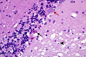
 Correlation of cerebellar granular layer autolysis with ante-mortem systemic acid-base status<...
 Autolytic and Proteolytic Properties of Strains of Lactococcus lactis Isolated from Different Vegetables, Raw-Milk Cheeses and...
 What Happens To Your Body After You've Been Dead For An Hour?
 Postmortem Changes: Overview, Definitions, Scene Findings
 Dorcas gazelle (Gazella dorcas) | IVIS
 Supporting COVID-19 Research | NEB
 Toxins | Free Full-Text | Investigation of the Efficacy of a Postbiotic Yeast Cell Wall-Based Blend on Newly-Weaned Pigs under...
 Distinct 'chemical cocktail' released by dead bodies smells like berries and apples | Daily Mail Online
 Registration Dossier - ECHA
 Amyloid beta-Protein Precursor
- Amyloid Protein Precursor
Summary Report | CureHunter
Amyloid beta-Protein Precursor
- Amyloid Protein Precursor
Summary Report | CureHunter Control of adipose tissue cellularity by the terminal complement cascade | Nature Reviews Endocrinology