Basal Cell Nevus Syndrome
Carcinoma, Basal Cell
Nevus, Spindle Cell
Nevus
Nevus, Blue
Dysplastic Nevus Syndrome
Nevus, Sebaceous of Jadassohn
Nevus of Ota
Nevus, Epithelioid and Spindle Cell
Involvement of patched (PTCH) gene in Gorlin syndrome and related disorders: three family cases. (1/106)
AIM: To find genetic alterations in PTC or other genes of the Shh/PTCH pathway in tumorous and non- tumorous samples from three families and to correlate them with the varying expression of disorders in presented nevoid basal cell carcinoma syndrome (NBCCS) phenotypes. METHOD: DNA was extracted from archival paraffin-embedded tissues, tumor tissue or peripheral blood leukocytes, and the loss of heterozygosity (LOH) and single strand conformational polymorphism analysis was performed using PCR with primers for polymorphic 9q22.3 markers (D9S196, D9S287, D9S180, D9S127); PTCH exons 3, 6, 8, 13, 15, 16; and smo (smoothened) exon 1. G-banding tecnique was used for cytogenetic analysis of the peripheral blood lymphocytes. RESULTS: We found a LOH for PTCH in several cases and variability in smo in one case. In one case NBCCS could reasonably be ascribed to hemizygous PTCH inactivation, while in other two families this typical correlation between the syndrome phenotype and the observed genetic alterations could not been established. CONCLUSIONS: Further analysis of relatively sparse cases of NBCCS is needed before the symptoms of the syndrome could be convincingly explained by genetic alterations in the Shh/PTCH signalling pathway. (+info)The hedgehog signalling pathway in tumorigenesis and development. (2/106)
The hedgehog signalling pathway is responsible for the embryonic patterning of a range of tissues, and it is now known that dysregulation of this pathway can result in the formation of several tumour types. This cascade is regulated at the cell surface by the opposing actions of the patched and smoothened molecules which together form a receptor complex for hedgehog. The discovery that inactivation of the human patched gene is responsible for familial and sporadic forms of basal cell carcinoma firmly established a role for dysregulation of hedgehog signalling in tumorigenesis. Other key members of this pathway have also been shown to be involved in tumour formation, as have more distal downstream targets of hedgehog signalling. Since it appears that tumorigenesis results from constitutive activation of hedgehog responsive genes, the identification of novel downstream targets of hedgehog signalling in given cell types is likely to increase our understanding of the molecular processes underlying tumour formation. (+info)Intracranial calcifications in childhood medulloblastoma: relation to nevoid basal cell carcinoma syndrome. (3/106)
BACKGROUND AND PURPOSE: Medulloblastoma is one of the most common posterior fossa tumors to occur in children. Our purpose was to document the frequency, location, and time of occurrence of intracranial calcifications in cranial CT studies of children with medulloblastoma. METHODS: We retrospectively reviewed cranial CT studies of 56 patients diagnosed with medulloblastoma from 1983 through 1997 for the presence of intracranial calcifications. The findings were compared with 159 cranial CT studies of patients who were evaluated in the emergency department (control group). Thirty-two patients with medulloblastoma without shunts were compared with 118 patients from the control group without shunts. Similarly, 24 patients with medulloblastoma with shunts were compared with 41 patients from the control group with shunts. RESULTS: Overall, three (9%) patients with medulloblastoma without shunts, four (16%) patients with medulloblastoma with shunts, and four (10%) patients from the control group with shunts had falx calcification. Only the two children carrying the diagnoses of medulloblastoma and nevoid basal cell carcinoma syndrome, however, had calcification of the falx cerebri shown on the cranial CT scans obtained during the peridiagnostic period. Both were diagnosed with medulloblastoma before the age of 3 years and later developed jaw cysts and multiple basal cell carcinomas in the radiation field. CONCLUSION: Previous studies have shown that falx calcification is a major component of nevoid basal cell carcinoma syndrome. Our two cases illustrate the importance of considering the diagnosis of nevoid basal cell carcinoma syndrome when falx calcification is present in young patients with medulloblastoma. If the concomitant diagnosis of nevoid basal cell carcinoma syndrome is made, alternative types of therapy should be sought to minimize radiation therapy sequelae. (+info)The hedgehog pathway and basal cell carcinomas. (4/106)
Developmental pathways first elucidated by genetic studies in the fruit fly, Drosophila melanogaster, are conserved in vertebrates, and disruption of these pathways has been associated with various human congenital anomalies. Many developmental genes continue to play an important role in regulation of cell growth and differentiation after embryogenesis, and mutations in some of these genes can result in cancer. Basal cell carcinoma (BCC) of the skin is the most common type of cancer in humans. Although most BCCs are sporadic, in rare cases, individuals have a hereditary disease, Gorlin syndrome, that predisposes to multiple skin tumors as well as a variety of birth defects. Mutations in the human homolog of a Drosophila gene, patched, underlie Gorlin syndrome. Genetic studies in Drosophila show that patched is part of the hedgehog signaling pathway, important in determining embryonic patterning and cell fate in multiple structures of the developing embryo. Human patched is mutated in sporadic as well as hereditary BCCs, and inactivation of this gene is probably a necessary if not sufficient step for tumor formation. Delineation of the biochemical pathway in which patched functions may lead to rational medical therapy for skin cancer and possibly other tumors. (+info)Bilateral hyperplasia of the mandibular coronoid processes associated with the nevoid basal cell carcinoma syndrome in an Italian boy. (5/106)
In this report we present a subject affected by nevoid basal cell carcinoma syndrome (NBCCS), showing also bilateral mandibular coronoid processes hyperplasia, a hitherto unreported association. Our observation of bilateral hyperplasia of the mandibular coronoid processes in a boy with NBCCS may prompt a retrospective and prospective review of other patients affected by this syndrome in order to establish if this anomaly is part of it. (+info)Conservative treatment of recurrent ovarian fibromas in a young patient affected by Gorlin syndrome. (6/106)
The case of recurrent bilateral ovarian fibromas occurring in a 22 year old Italian girl affected by Gorlin syndrome is reported. Ovarian fibromas occur in 75% of female patients with Gorlin syndrome and their recurrence has rarely been reported in the literature. Management is guided by the benign nature of the lesion and consists of surgical removal of the fibroma. Preservation of the normal ovarian tissue is recommended even though there is risk of recurrence of the fibroma. (+info)Genetic mutations in certain head and neck conditions of interest to the dentist. (7/106)
This article identifies certain syndromes of the head and neck, which a dentist may see in clinical practice, and relates these syndromes to their sites of mutation on involved genes. This paper is timely with the near completion of the Human Genome Project, the mapping of the entire human genetic material. Knowing the site of the genetic lesion is important in helping clinicians understand the genetic basis for these conditions, and may help in our future understanding of remedies and treatments. (+info)PATCHED and p53 gene alterations in sporadic and hereditary basal cell cancer. (8/106)
It is widely accepted that disruption of the hedgehog-patched pathway is a key event in development of basal cell cancer. In addition to patched gene alterations, p53 gene mutations are also frequent in basal cell cancer. We determined loss of heterozygosity in the patched and p53 loci as well as sequencing the p53 gene in tumors both from sporadic and hereditary cases. A total of 70 microdissected samples from tumor and adjacent skin were subjected to PCR followed by fragment analysis and DNA sequencing. We found allelic loss in the patched locus in 6/8 sporadic basal cell cancer and 17/19 hereditary tumors. All sporadic and 7/20 hereditary tumors showed p53 gene mutations. Loss of heterozygosity in the p53 locus was rare in both groups. The p53 mutations detected in hereditary tumors included rare single nucleotide deletions and unusual double-base substitutions compared to the typical ultraviolet light induced missense mutations found in sporadic tumors. Careful microdissection of individual tumors revealed genetically linked subclones with different p53 and/or patched genotype providing an insight on time sequence of genetic events. The high frequency and co-existence of genetic alterations in the patched and p53 genes suggest that both these genes are important in the development of basal cell cancer. (+info)Basal Cell Nevus Syndrome (BCNS), also known as Gorlin-Goltz Syndrome, is a rare genetic disorder that is characterized by the development of multiple basal cell carcinomas (BCCs), which are skin cancer tumors that arise from the basal cells in the outermost layer of the skin.
The syndrome is caused by mutations in the PTCH1 gene, which regulates the hedgehog signaling pathway involved in embryonic development and tissue growth regulation. The condition is inherited in an autosomal dominant manner, meaning that a child has a 50% chance of inheriting the mutated gene from an affected parent.
Individuals with BCNS typically develop hundreds to thousands of BCCs over their lifetime, often beginning in childhood or adolescence. They may also have other benign and malignant tumors, such as medulloblastomas (brain tumors), fibromas, and rhabdomyosarcomas.
Additional features of BCNS can include:
1. Facial abnormalities, such as a broad nasal bridge, widely spaced eyes, and pits or depressions on the palms and soles.
2. Skeletal abnormalities, such as spine deformities, rib anomalies, and jaw cysts.
3. Developmental delays and intellectual disabilities in some cases.
4. Increased risk of other cancers, including breast, ovarian, and lung cancer.
Early detection and management of BCCs and other tumors are crucial for individuals with BCNS to prevent complications and improve their quality of life. Regular dermatological examinations, sun protection measures, and surgical removal of tumors are common treatment approaches.
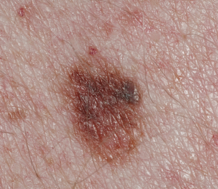
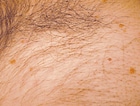
Carcinoma, basal cell is a type of skin cancer that arises from the basal cells, which are located in the lower part of the epidermis (the outermost layer of the skin). It is also known as basal cell carcinoma (BCC) and is the most common form of skin cancer.
BCC typically appears as a small, shiny, pearly bump or nodule on the skin, often in sun-exposed areas such as the face, ears, neck, hands, and arms. It may also appear as a scar-like area that is white, yellow, or waxy. BCCs are usually slow growing and rarely spread (metastasize) to other parts of the body. However, they can be locally invasive and destroy surrounding tissue if left untreated.
The exact cause of BCC is not known, but it is thought to be related to a combination of genetic and environmental factors, including exposure to ultraviolet (UV) radiation from the sun or tanning beds. People with fair skin, light hair, and blue or green eyes are at increased risk of developing BCC.
Treatment for BCC typically involves surgical removal of the tumor, along with a margin of healthy tissue. Other treatment options may include radiation therapy, topical chemotherapy, or photodynamic therapy. Prevention measures include protecting your skin from UV radiation by wearing protective clothing, using sunscreen, and avoiding tanning beds.
Skin neoplasms refer to abnormal growths or tumors in the skin that can be benign (non-cancerous) or malignant (cancerous). They result from uncontrolled multiplication of skin cells, which can form various types of lesions. These growths may appear as lumps, bumps, sores, patches, or discolored areas on the skin.
Benign skin neoplasms include conditions such as moles, warts, and seborrheic keratoses, while malignant skin neoplasms are primarily classified into melanoma, squamous cell carcinoma, and basal cell carcinoma. These three types of cancerous skin growths are collectively known as non-melanoma skin cancers (NMSCs). Melanoma is the most aggressive and dangerous form of skin cancer, while NMSCs tend to be less invasive but more common.
It's essential to monitor any changes in existing skin lesions or the appearance of new growths and consult a healthcare professional for proper evaluation and treatment if needed.
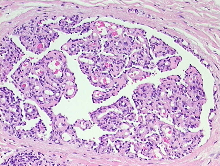
A "Spindle Cell Nevus" is a type of melanocytic nevus (mole), which is a benign growth that occurs from the uncontrolled multiplication of melanocytes (pigment-producing cells). In a spindle cell nevus, the melanocytes are elongated and take on a spindle shape. This type of nevus is not common and typically appears as a solitary, brown or skin-colored papule or nodule. Spindle cell nevi can be found anywhere on the body but are most commonly located on the scalp and face. They usually occur in adults and are generally considered to have a low malignant potential, although there is a small risk of transformation into a malignant melanoma. It's important to monitor any changes in size, color, or shape of a spindle cell nevus and to have it evaluated by a healthcare professional if there are any concerns.
A nevus, also known as a mole, is a benign growth or mark on the skin that is usually brown or black. It can be raised or flat and can appear anywhere on the body. Nevi are made up of cells called melanocytes, which produce the pigment melanin. Most nevi develop in childhood or adolescence, but they can also appear later in life. Some people have many nevi, while others have few or none.
There are several types of nevi, including:
* Common nevi: These are the most common type of mole and are usually small, round, and brown or black. They can be flat or raised and can appear anywhere on the body.
* Atypical nevi: These moles are larger than common nevi and have irregular borders and color. They may be flat or raised and can appear anywhere on the body, but are most commonly found on the trunk and extremities. Atypical nevi are more likely to develop into melanoma, a type of skin cancer, than common nevi.
* Congenital nevi: These moles are present at birth and can vary in size from small to large. They are more likely to develop into melanoma than moles that develop later in life.
* Spitz nevi: These are rare, benign growths that typically appear in children and adolescents. They are usually pink or red and dome-shaped.
It is important to monitor nevi for changes in size, shape, color, and texture, as these can be signs of melanoma. If you notice any changes in a mole, or if you have a new mole that is unusual or bleeding, it is important to see a healthcare provider for further evaluation.
A blue nevus, also known as a "naevus" or "mole," is a type of melanocytic nevus, which means it contains the pigment-producing cells called melanocytes. The term "blue" refers to its characteristic color, which results from the way light penetrates and scatters in the deep layers of the skin where the nevus is located.
Blue nevi are typically benign, meaning they are not cancerous and do not usually pose a threat to health. They can appear as solitary lesions or multiple lesions and may be present at birth (congenital) or develop during childhood or adulthood.
While blue nevi are generally harmless, it is important to monitor them for any changes in size, shape, color, or texture, as well as the development of new symptoms such as pain, itching, or bleeding. In rare cases, a blue nevus may undergo malignant transformation and develop into a type of skin cancer called melanoma.
If you have a blue nevus that is changing or causing concern, it is recommended to consult with a healthcare professional for further evaluation and management.
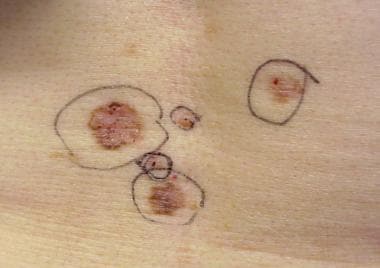
Dysplastic Nevus Syndrome, also known as atypical mole syndrome, is a condition characterized by the presence of numerous dysplastic nevi (abnormal moles) that may appear irregular in shape, color, and size. These moles are typically larger than normal moles (greater than 5 mm in diameter) and have an asymmetrical shape, uneven borders, and varied colors.
Individuals with Dysplastic Nevus Syndrome have a higher risk of developing melanoma, a type of skin cancer that can be life-threatening if not detected and treated early. The syndrome is usually inherited in an autosomal dominant manner, meaning that a child has a 50% chance of inheriting the gene from an affected parent.
It's important to note that having dysplastic nevi does not necessarily mean that a person will develop melanoma, but it does increase their risk. Regular skin examinations by a dermatologist and self-examinations are recommended for early detection of any changes in moles or the development of new suspicious lesions.
A nevus sebaceous of Jadassohn is a type of congenital benign skin tumor or birthmark that is composed of epidermal, hair follicle, and sebaceous gland components. It typically appears as a yellowish, greasy, or warty plaque on the scalp or face during infancy or early childhood. The lesion tends to enlarge slowly and may undergo various changes in appearance over time.
In adolescence or adulthood, there is a risk of secondary tumor development within the nevus sebaceous, such as basal cell carcinoma, squamous cell carcinoma, or sebaceous carcinoma. Therefore, regular monitoring and possible surgical removal of the lesion may be recommended, especially in cases where the nevus is large, symptomatic, or shows signs of malignant transformation.
A syndrome, in medical terms, is a set of symptoms that collectively indicate or characterize a disease, disorder, or underlying pathological process. It's essentially a collection of signs and/or symptoms that frequently occur together and can suggest a particular cause or condition, even though the exact physiological mechanisms might not be fully understood.
For example, Down syndrome is characterized by specific physical features, cognitive delays, and other developmental issues resulting from an extra copy of chromosome 21. Similarly, metabolic syndromes like diabetes mellitus type 2 involve a group of risk factors such as obesity, high blood pressure, high blood sugar, and abnormal cholesterol or triglyceride levels that collectively increase the risk of heart disease, stroke, and diabetes.
It's important to note that a syndrome is not a specific diagnosis; rather, it's a pattern of symptoms that can help guide further diagnostic evaluation and management.
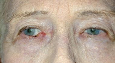
A Nevus of Ota, also known as an oculodermal melanocytosis, is a benign birthmark characterized by the presence of darkly pigmented (melanin-containing) cells called melanocytes in the skin and mucous membranes around the eye. These pigmented cells can also extend to the sclera (the white part of the eye), dura mater (the outer covering of the brain), and leptomeninges (the middle layer of the meninges, which cover the brain and spinal cord).
The Nevus of Ota typically presents as a unilateral (occurring on one side) bluish-gray or brown patch that follows the distribution of the ophthalmic and maxillary divisions of the trigeminal nerve. It usually affects the eye, forehead, temple, and cheek, but it can also involve other areas of the face, scalp, and neck.
While Nevi of Ota are generally harmless, they may be associated with an increased risk of developing melanoma (a type of skin cancer) in the affected area. Therefore, regular monitoring and evaluation by a healthcare professional is recommended.
A nevus is a general term for a benign growth or mole on the skin. There are many different types of nevi, including epithelioid and spindle cell nevi.
Epithelioid cell: A type of cell that is typically found in certain types of nevi, as well as in some malignant tumors such as melanoma. Epithelioid cells are large, round cells with a pale, clear cytoplasm and centrally located nuclei.
Spindle cell: A type of cell that is often found in certain types of nevi, including Spitz nevi and deep penetrating nevi. Spindle cells are elongated, thin cells with cigar-shaped nuclei. They can also be found in some malignant tumors such as melanoma.
Epithelioid and spindle cell nevus: A type of nevus that contains both epithelioid and spindle cells. These nevi are typically benign, but they can sometimes be difficult to distinguish from melanoma, especially if they have atypical features. Therefore, it is important for these types of nevi to be evaluated by a dermatopathologist or a specialist in skin pathology.
 PTCH1
PTCH1 Basal Cell Nevus Syndrome (Gorlin Syndrome) | Johns Hopkins Medicine
 Nevoid Basal Cell Carcinoma Syndrome (Basal Cell Nevus Syndrome): Background, Pathophysiology, Etiology
 BASAL CELL NEVUS SYNDROME 1; BCNS1
 Can Rashes Be a Sign of Cancer? Itchiness and Symptoms
 Broad nasal bridge: MedlinePlus Medical Encyclopedia
 Basal Cell Carcinoma - Pipeline Insight, 2021
 Ultraviolet and ionizing radiation enhance the growth of BCCs and trichoblastomas in patched heterozygous knockout mice |...
![PTCH1 patched 1 [Homo sapiens (human)] - Gene - NCBI](data:image/png;base64,iVBORw0KGgoAAAANSUhEUgAAABAAAAAQCAYAAAAf8/9hAAAB1ElEQVQ4jaWSPWgTcRjGf/ehuWhobO2JxGJRY3taTTRV2yoqSpW6iIWO4iAoUsRBioNDKUWKLU7i4KA4OfhVREQnETRia03k7IdiS0LaQYKJQg3mLtfc30GySNUDn/V5nx/vy/vAf0pqad3db2xquiBJku93s2Tb2eEHdw1rTcsxol23sObTjN7oIp9KVmaU9kMdTxcLAyiqGtA0bfms+XKQULSdQG2EmnUx0q9ughAA8p/CFW0IN3Sv0vUI5p2zIMpUrd5JeP/Jii//80ZJUlrb9lyV8qn3zI5dB8A4MoBWtcITAKBmZe3eRmPzccYf9uIUsyzx6zQd7fMMAIjFdgxpkuPy4clFANbu6qa6fouybXtznxeAoqoBn0/zz5kvBqVQ5DBasJ5gXaPnDQAWFpwCkiwLZekyAMp2wTPAsqy5d8nEZcIHThPQo7jlIua9854BibdvekqKX8PouARAOn6F+c8pT4Bc7svz6U8f77O1cwDVV439PcPU4yHw8AUhhDPyOn4OfWOMuuZfBZp41INTLACorhC2/Jc2zsxMX8vl8lMcPBUHFL5mnpEZGa748sS42esKYS8WLtl2NjE22s/6fScIhtr48W2S5O0zIFwvp3vST6Z+myCvkaonAAAAAElFTkSuQmCC) PTCH1 patched 1 [Homo sapiens (human)] - Gene - NCBI
PTCH1 patched 1 [Homo sapiens (human)] - Gene - NCBI High-Risk Skin Cancer Clinic | UCSF Health
 Skin Cancer | St. Louis Children's Hospital
 Search - NeL.edu
 Exploring Dysregulated Signaling Pathways in Cancer
| Bentham Science
Exploring Dysregulated Signaling Pathways in Cancer
| Bentham Science Repurposing Drugs in Oncology (ReDO)-itraconazole as an anti-cancer agent - ecancer
 ERIC - ED458748 - Manifestations, Treatment Implications and Speech-Language Consideration in Gorlin Syndrome: A Case Study.,...
 Basal and Squamous Cell Skin Cancer Causes | What Causes Skin Cancer? | American Cancer Society
 Gorlin Syndrome - AboutFace
 Albert Sean Chiou, MD, MBA's Profile | Stanford Profiles
 12.1 Systemic therapies for advanced and metastatic basal cell carcinoma - Cancer Guidelines Wiki
 An Update on Nonmelanoma Skin Cancer | JCAD | The Journal of Clinical and Aesthetic Dermatology
 Baby’s Pregnancy Calendar
 COM June 2005 Diagnosis - UW School of Dentistry