Bone Cysts
Bone Cysts, Aneurysmal
Cysts
Curettage
Bone and Bones
Jaw Cysts
Mandibular Diseases
Bone Remodeling
Chondroblastoma
Bone Density
Epidermal Cyst
Spinal Diseases
Maxillary Diseases
Bone Matrix
Fibrous Dysplasia of Bone
Bone Marrow
Mediastinal Cyst
Giant Cell Tumor of Bone
Fractures, Spontaneous
Sclerosing Solutions
Bone Development
Synovial Cyst
Bone Marrow Cells
Bone Substitutes
Frontal Bone
Bone Demineralization Technique
Sphenoid Bone
Bronchogenic Cyst
Dermoid Cyst
Nonodontogenic Cysts
Bone Marrow Transplantation
Diatrizoate
Ischium
Bone Regeneration
Epiphyses
Cementoma
Tomography, X-Ray Computed
Tibia
Calcaneus
Odontogenic Cysts
Ribs
Radicular Cyst
Bone Nails
Oral Surgical Procedures
Giant Cell Tumors
Dentigerous Cyst
Bone Diseases, Metabolic
Magnetic Resonance Imaging
Decompression, Surgical
Treatment Outcome
Mesenteric Cyst
Granuloma, Giant Cell
Ubiquitin Thiolesterase
Bone Morphogenetic Proteins
Fracture Healing
Cervical Vertebrae
Sclerotherapy
Chondrodiatasis in a patient with spondyloepimetaphyseal dysplasia using the Ilizarov technique: successful correction of an angular deformity with ensuing ossification of a large metaphyseal lesion. A case report. (1/147)
Distraction through the physis (chondrodiatasis) is a controversial technique with unpredictable results. However, it has been used in the past for the lengthening and correction of angular deformities of long bones. We report the case of an 11-year-old patient with spondyloepimetaphyseal dysplasia (SEMD) who presented with a severe recurvatum deformity of the left proximal tibia secondary to collapse of the tibial plateau into a large metaphyseal cystic lesion. Using the chondrodiatasis technique with a percutaneously applied Ilizarov circular frame, we were able to correct this deformity. Surprisingly, healing and ossification of the metaphyseal lesion was simultaneously observed at the end of the treatment, a finding which, to the best of our knowledge, has not been previously reported. (+info)The relationship between synovitis and bone changes in early untreated rheumatoid arthritis: a controlled magnetic resonance imaging study. (2/147)
OBJECTIVE: The interrelationship between synovitis and bone damage in rheumatoid arthritis (RA) is a subject of controversy. Using magnetic resonance imaging (MRI), this study followed the bone changes in early RA and determined their relationship to synovitis. METHODS: Thirty-one patients with early RA who had swelling of the metacarpophalangeal (MCP) joints and 31 healthy control subjects with no clinical evidence of arthritis underwent MRI of the second through fifth MCP joints of the dominant hand by use of a 1.5T scanner. Coronal T1-weighted and T2-fat suppressed (FS) sequences were performed to evaluate bone edema, and gadolinium-diethylenetriaminepentaacetic acid (Gd-DTPA) pulse sequences were obtained to evaluate synovitis. Bony abnormalities were described as bone edema (low signal on T1-weighted sequences and intermediate/high signal on T2 FS sequences adjacent to the bone cortex) or as bone cysts (circular juxtacortical abnormalities with low signal on T1-weighted images and with very high signal on T2 FS sequences). Contrast and noncontrast MRI films were scored in a blinded manner, and Fisher's exact probability test was used to determine differences between groups. RESULTS: Twenty-one of the 31 RA patients (68%) had bone edema, which was seen in 43 of 124 joints (35% of joints) and 3 of the 31 control subjects had bone edema seen in 3 of 124 joints (2% of joints) (P < 0.0001). Thirty RA patients (97%) had Gd-DTPA-confirmed MCP joint synovitis, and bone edema was seen in 40 of the 75 joints with Gd-DTPA-proven synovitis (53%), but in only 3 of 49 without (6%) (P < 0.0001). CONCLUSION: MCP joint bone edema is present in the majority of patients with RA at presentation, but is seen only occasionally in normal control subjects. The fact that bone edema occurred rarely in the absence of synovitis in patients with RA suggests that bony changes in RA are secondary to synovitis. (+info)Subperiosteal ganglion cyst of the tibia. A communication with the knee demonstrated by delayed arthrography. (3/147)
We report a patient with a subperiosteal ganglion cyst of the tibia which was imaged by radiography, arthrography, CT and MRI. The images were correlated with the arthroscopic surgical and histological findings. Spiculated formation of periosteal new bone on plain radiographs led to the initial suspicion of a malignant tumour. Demonstration of the cystic nature of the tumour using cross-sectional imaging was important for the precise diagnosis. Communication between the ganglion cyst and the knee was shown by a delayed arthrographic technique, and the presence of this communication was confirmed at arthroscopy and surgically. (+info)Clinical manifestations of AB-amyloidosis: effects of biocompatibility and flux. (4/147)
BACKGROUND: Highly permeable biocompatible dialysis membranes may postpone the development of AB-amyloidosis, but the relative contribution of enhanced flux or reduced inflammation by highly biocompatible membranes and sterile dialysis fluid remains unknown. METHODS: In this retrospective investigation, 89 patients with end-stage renal disease maintained on regular haemodialysis for at least 10 years and treated with one type of dialysis membrane exclusively were selected for analysis. They were divided into three groups: low-flux, bioincompatible cellulose (I), low-flux, intermediately biocompatible polysulphone or PMMA (II), or high-flux, highly biocompatible polysulphone or AN69 (III). In addition, the patients were analysed according to the microbiological quality of the dialysis fluid, which had been tested regularly and was classified either as standard or as intermittently contaminated. The clinical manifestations indicative of AB-amyloidosis, namely, carpal tunnel syndrome, arthropathy and bone cysts, were diagnosed after recruitment. RESULTS: Clinical symptoms were most pronounced in group I, intermediate in group II, and lowest in group III. Patients treated with intermittently contaminated dialysis fluid showed a higher prevalence of AB-amyloidosis than patients with less contaminated dialysis fluid. Logistic regression analysis demonstrated that the flux characteristics of the dialyser and the microbiological quality of the dialysis fluid as well as the biocompatibility of the dialyser were independent determinants of AB-amyloidosis. CONCLUSION: It would be prudent clinical practice to employ high-flux biocompatible membranes in conjunction with ultrapure dialysis fluid for the treatment of end-stage renal disease patients who need to remain on long-term haemodialysis. (+info)Hemophilic pseudotumor of the ulna treated with low dose radiation therapy: a case report. (5/147)
We report a case of hemophilic pseudotumor in the ulna of a 6-year-old boy treated with radiation therapy. A total dose of 900 cGy in 6 fractions was given in 6 consecutive days. Progression of cystic changes was halted within a month. New bone formation and trabeculation were found on the 4th month. Complete healing of the lesion and bony replacement were found on the 12th month. The patient was followed up to 72 months and there was no evidence of recurrence and no bone growth disturbance. Radiation therapy can be an effective alternative modality in treating hemophilic pseudotumor. (+info)Cannulation of simple bone cysts. (6/147)
We describe a consecutive series of 26 patients with simple bone cysts who were treated by curettage, multiple drilling and continuous decompression by the insertion of either a cannulated screw or a pin. In the first 15 patients we used titanium cannulated screws (group 1) and in the next 11 a cannulated hydroxyapatite pin (group 2). Satisfactory healing was achieved in 12 patients in group 1 (80%) and in all in group 2. This technique seems to be a promising option for the treatment of simple bone cysts. The cannulated hydroxyapatite pin is recommended because of its higher success rate and the fact that it does not need to be removed. (+info)Intertrochanteric osteotomy for osteoarthritis of the hip. A radiological assessment of non-compressive and compressive methods. (7/147)
A radiological review of two groups of intertrochanteric osteotomies of the femur for primary osteoarthritis of the hip has been made. Each group oroginally consisted of forty-one hips. In one group a Wainwright straight V-spline without compression had been used for fixation, and in the other group an AO angled plate with compression. The time for bony union was equal in the two groups but the incidence of non-union was lower in the AO group. Regression of cysts and of bone sclerosis was more frequent in the Wainwright group, possible as a consequence of the greater medial displacement and varus angulation. (+info)Unicameral bone cysts treated by injection of bone marrow or methylprednisolone. (8/147)
In 79 consecutive patients with unicameral bone cysts we compared the results of aspiration and injection of bone marrow with those of aspiration and injection of steroid. All were treated by the same protocol. The only difference was the substance injected into the cysts. The mean radiological follow-up to detect activity in the cyst was 44 months (12 to 108). Of the 79 patients, 14 received a total of 27 injections of bone marrow and 65 a total of 99 injections of steroid. Repeated injections were required in 57% of patients after bone marrow had been used and in 49% after steroid. No complications were noted in either group. In this series no advantage could be shown for the use of autogenous injection of bone marrow compared with injection of steroid in the management of unicameral bone cysts. (+info)A bone cyst is a fluid-filled sac that develops within a bone. It can be classified as either simple (unicameral) or aneurysmal. Simple bone cysts are more common in children and adolescents, and they typically affect the long bones of the arms or legs. These cysts are usually asymptomatic unless they become large enough to weaken the bone and cause a fracture. Aneurysmal bone cysts, on the other hand, can occur at any age and can affect any bone, but they are most common in the leg bones and spine. They are characterized by rapidly growing blood-filled sacs that can cause pain, swelling, and fractures.
Both types of bone cysts may be treated with observation, medication, or surgery depending on their size, location, and symptoms. It is important to note that while these cysts can be benign, they should still be evaluated and monitored by a healthcare professional to ensure proper treatment and prevention of complications.
Aneurysmal bone cyst (ABC) is a benign but locally aggressive tumor that typically involves the metaphysis of long bones in children and adolescents. It is characterized by blood-filled spaces or cysts separated by fibrous septa containing osteoclast-type giant cells, spindle cells, and capillary vessels.
ABCs can also arise in other locations such as the vertebral column, pelvis, and skull. They may cause bone pain, swelling, or pathologic fractures. The exact cause of ABC is unknown, but it is thought to be related to a reactive process to a primary bone lesion or trauma.
Treatment options for ABC include curettage and bone grafting, intralesional injection of corticosteroids or bone marrow aspirate, and adjuvant therapy with phenol or liquid nitrogen. In some cases, radiation therapy may be used, but it is generally avoided due to the risk of secondary malignancies. Recurrence rates after treatment range from 10-30%.
A cyst is a closed sac, having a distinct membrane and division between the sac and its surrounding tissue, that contains fluid, air, or semisolid material. Cysts can occur in various parts of the body, including the skin, internal organs, and bones. They can be caused by various factors, such as infection, genetic predisposition, or blockage of a duct or gland. Some cysts may cause symptoms, such as pain or discomfort, while others may not cause any symptoms at all. Treatment for cysts depends on the type and location of the cyst, as well as whether it is causing any problems. Some cysts may go away on their own, while others may need to be drained or removed through a surgical procedure.
Curettage is a medical procedure that involves scraping or removing tissue from the lining of an organ or body cavity, typically performed using a curette, which is a long, thin surgical instrument with a looped or sharp end. In gynecology, curettage is often used to remove tissue from the uterus during a procedure called dilation and curettage (D&C) to diagnose or treat abnormal uterine bleeding, or to remove residual placental or fetal tissue following a miscarriage or abortion. Curettage may also be used in other medical specialties to remove damaged or diseased tissue from areas such as the nose, throat, or skin.
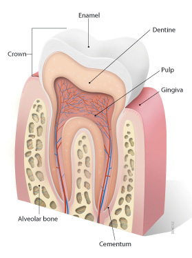
"Bone" is the hard, dense connective tissue that makes up the skeleton of vertebrate animals. It provides support and protection for the body's internal organs, and serves as a attachment site for muscles, tendons, and ligaments. Bone is composed of cells called osteoblasts and osteoclasts, which are responsible for bone formation and resorption, respectively, and an extracellular matrix made up of collagen fibers and mineral crystals.
Bones can be classified into two main types: compact bone and spongy bone. Compact bone is dense and hard, and makes up the outer layer of all bones and the shafts of long bones. Spongy bone is less dense and contains large spaces, and makes up the ends of long bones and the interior of flat and irregular bones.
The human body has 206 bones in total. They can be further classified into five categories based on their shape: long bones, short bones, flat bones, irregular bones, and sesamoid bones.
A jaw cyst is a pathological cavity filled with fluid or semi-fluid material, which forms within the jaw bones. They are typically classified as odontogenic (developing from tooth-forming tissues) or non-odontogenic (developing from other tissues). The most common types of odontogenic jaw cysts include dentigerous cysts (formed around the crown of an unerupted tooth) and follicular cysts (formed from the inflammation of a developing tooth's tissue). Non-odontogenic cysts, such as nasopalatine duct cysts and keratocystic odontogenic tumors, can also occur in the jaw bones. Jaw cysts may cause symptoms like swelling, pain, or displacement of teeth, but some may not present any symptoms until they grow large enough to be detected on a radiographic examination. Treatment typically involves surgical removal of the cyst and, if necessary, reconstruction of the affected bone.
Cyst fluid refers to the fluid accumulated within a cyst, which is a closed sac-like or capsular structure, typically filled with liquid or semi-solid material. Cysts can develop in various parts of the body for different reasons, and the composition of cyst fluid may vary depending on the type of cyst and its location.
In some cases, cyst fluid might contain proteins, sugars, hormones, or even cells from the surrounding tissue. Infected cysts may have pus-like fluid, while cancerous or precancerous cysts might contain abnormal cells or tumor markers. The analysis of cyst fluid can help medical professionals diagnose and manage various medical conditions, including infections, inflammatory diseases, genetic disorders, and cancers.
It is important to note that the term 'cyst fluid' generally refers to the liquid content within a cyst, but the specific composition and appearance of this fluid may vary significantly depending on the underlying cause and type of cyst.
Mandibular diseases refer to conditions that affect the mandible, or lower jawbone. These diseases can be classified as congenital (present at birth) or acquired (developing after birth). They can also be categorized based on the tissues involved, such as bone, muscle, or cartilage. Some examples of mandibular diseases include:
1. Mandibular fractures: These are breaks in the lower jawbone that can result from trauma or injury.
2. Osteomyelitis: This is an infection of the bone and surrounding tissues, which can affect the mandible.
3. Temporomandibular joint (TMJ) disorders: These are conditions that affect the joint that connects the jawbone to the skull, causing pain and limited movement.
4. Mandibular tumors: These are abnormal growths that can be benign or malignant, and can develop in any of the tissues of the mandible.
5. Osteonecrosis: This is a condition where the bone tissue dies due to lack of blood supply, which can affect the mandible.
6. Cleft lip and palate: This is a congenital deformity that affects the development of the face and mouth, including the lower jawbone.
7. Mandibular hypoplasia: This is a condition where the lower jawbone does not develop properly, leading to a small or recessed chin.
8. Developmental disorders: These are conditions that affect the growth and development of the mandible, such as condylar hyperplasia or hemifacial microsomia.
Bone remodeling is the normal and continuous process by which bone tissue is removed from the skeleton (a process called resorption) and new bone tissue is formed (a process called formation). This ongoing cycle allows bones to repair microdamage, adjust their size and shape in response to mechanical stress, and maintain mineral homeostasis. The cells responsible for bone resorption are osteoclasts, while the cells responsible for bone formation are osteoblasts. These two cell types work together to maintain the structural integrity and health of bones throughout an individual's life.
During bone remodeling, the process can be divided into several stages:
1. Activation: The initiation of bone remodeling is triggered by various factors such as microdamage, hormonal changes, or mechanical stress. This leads to the recruitment and activation of osteoclast precursor cells.
2. Resorption: Osteoclasts attach to the bone surface and create a sealed compartment called a resorption lacuna. They then secrete acid and enzymes that dissolve and digest the mineralized matrix, creating pits or cavities on the bone surface. This process helps remove old or damaged bone tissue and releases calcium and phosphate ions into the bloodstream.
3. Reversal: After resorption is complete, the osteoclasts undergo apoptosis (programmed cell death), and mononuclear cells called reversal cells appear on the resorbed surface. These cells prepare the bone surface for the next stage by cleaning up debris and releasing signals that attract osteoblast precursors.
4. Formation: Osteoblasts, derived from mesenchymal stem cells, migrate to the resorbed surface and begin producing a new organic matrix called osteoid. As the osteoid mineralizes, it forms a hard, calcified structure that gradually replaces the resorbed bone tissue. The osteoblasts may become embedded within this newly formed bone as they differentiate into osteocytes, which are mature bone cells responsible for maintaining bone homeostasis and responding to mechanical stress.
5. Mineralization: Over time, the newly formed bone continues to mineralize, becoming stronger and more dense. This process helps maintain the structural integrity of the skeleton and ensures adequate calcium storage.
Throughout this continuous cycle of bone remodeling, hormones, growth factors, and mechanical stress play crucial roles in regulating the balance between resorption and formation. Disruptions to this delicate equilibrium can lead to various bone diseases, such as osteoporosis, where excessive resorption results in weakened bones and increased fracture risk.
An ovarian cyst is a sac or pouch filled with fluid that forms on the ovary. Ovarian cysts are quite common in women during their childbearing years, and they often cause no symptoms. In most cases, ovarian cysts disappear without treatment over a few months. However, larger or persistent cysts may require medical intervention, including surgical removal.
There are various types of ovarian cysts, such as functional cysts (follicular and corpus luteum cysts), which develop during the menstrual cycle due to hormonal changes, and non-functional cysts (dermoid cysts, endometriomas, and cystadenomas), which can form due to different causes.
While many ovarian cysts are benign, some may have malignant potential or indicate an underlying medical condition like polycystic ovary syndrome (PCOS). Regular gynecological check-ups, including pelvic examinations and ultrasounds, can help detect and monitor ovarian cysts.
Chondroblastoma is a rare, benign (non-cancerous) bone tumor that typically develops in the epiphysis, which is the rounded end of a long bone near a joint. It primarily affects children and adolescents, with around 90% of cases occurring before the age of 20.
The tumor arises from chondroblasts, cells responsible for producing cartilage during bone growth. Chondroblastoma is usually slow-growing and typically causes localized pain, swelling, or tenderness in the affected area. In some cases, it may weaken the bone and lead to fractures.
Treatment generally involves surgical removal of the tumor, followed by curettage (scraping) of the surrounding bone tissue and replacement with bone grafts or substitutes. Recurrence is possible but rare, and long-term prognosis is usually favorable.
Bone transplantation, also known as bone grafting, is a surgical procedure in which bone or bone-like material is transferred from one part of the body to another or from one person to another. The graft may be composed of cortical (hard outer portion) bone, cancellous (spongy inner portion) bone, or a combination of both. It can be taken from different sites in the same individual (autograft), from another individual of the same species (allograft), or from an animal source (xenograft). The purpose of bone transplantation is to replace missing bone, provide structural support, and stimulate new bone growth. This procedure is commonly used in orthopedic, dental, and maxillofacial surgeries to repair bone defects caused by trauma, tumors, or congenital conditions.
Bone density refers to the amount of bone mineral content (usually measured in grams) in a given volume of bone (usually measured in cubic centimeters). It is often used as an indicator of bone strength and fracture risk. Bone density is typically measured using dual-energy X-ray absorptiometry (DXA) scans, which provide a T-score that compares the patient's bone density to that of a young adult reference population. A T-score of -1 or above is considered normal, while a T-score between -1 and -2.5 indicates osteopenia (low bone mass), and a T-score below -2.5 indicates osteoporosis (porous bones). Regular exercise, adequate calcium and vitamin D intake, and medication (if necessary) can help maintain or improve bone density and prevent fractures.
The humerus is the long bone in the upper arm that extends from the shoulder joint (glenohumeral joint) to the elbow joint. It articulates with the glenoid cavity of the scapula to form the shoulder joint and with the radius and ulna bones at the elbow joint. The proximal end of the humerus has a rounded head that provides for movement in multiple planes, making it one of the most mobile joints in the body. The greater and lesser tubercles are bony prominences on the humeral head that serve as attachment sites for muscles that move the shoulder and arm. The narrow shaft of the humerus provides stability and strength for weight-bearing activities, while the distal end forms two articulations: one with the ulna (trochlea) and one with the radius (capitulum). Together, these structures allow for a wide range of motion in the shoulder and elbow joints.
An epidermal cyst is a common benign skin condition characterized by the growth of a sac-like structure filled with keratin, a protein found in the outermost layer of the skin (epidermis). These cysts typically appear as round, firm bumps just under the surface of the skin, often on the face, neck, trunk, or scalp. They can vary in size from a few millimeters to several centimeters in diameter.
Epidermal cysts usually develop as a result of the accumulation of dead skin cells that become trapped within a hair follicle or a pilosebaceous unit (a structure that contains a hair follicle and an oil gland). The keratin produced by the skin cells then collects inside the sac, causing it to expand gradually.
These cysts are generally slow-growing, painless, and rarely cause any symptoms. However, they may become infected or inflamed, leading to redness, tenderness, pain, or pus formation. In such cases, medical attention might be necessary to drain the cyst or administer antibiotics to treat the infection.
Epidermal cysts can be removed surgically if they cause cosmetic concerns or become frequently infected. The procedure typically involves making an incision in the skin and removing the entire sac along with its contents to prevent recurrence.
Bone neoplasms are abnormal growths or tumors that develop in the bone. They can be benign (non-cancerous) or malignant (cancerous). Benign bone neoplasms do not spread to other parts of the body and are rarely a threat to life, although they may cause problems if they grow large enough to press on surrounding tissues or cause fractures. Malignant bone neoplasms, on the other hand, can invade and destroy nearby tissue and may spread (metastasize) to other parts of the body.
There are many different types of bone neoplasms, including:
1. Osteochondroma - a benign tumor that develops from cartilage and bone
2. Enchondroma - a benign tumor that forms in the cartilage that lines the inside of the bones
3. Chondrosarcoma - a malignant tumor that develops from cartilage
4. Osteosarcoma - a malignant tumor that develops from bone cells
5. Ewing sarcoma - a malignant tumor that develops in the bones or soft tissues around the bones
6. Giant cell tumor of bone - a benign or occasionally malignant tumor that develops from bone tissue
7. Fibrosarcoma - a malignant tumor that develops from fibrous tissue in the bone
The symptoms of bone neoplasms vary depending on the type, size, and location of the tumor. They may include pain, swelling, stiffness, fractures, or limited mobility. Treatment options depend on the type and stage of the tumor but may include surgery, radiation therapy, chemotherapy, or a combination of these treatments.
Spinal diseases refer to a range of medical conditions that affect the spinal column, which is made up of vertebrae (bones), intervertebral discs, facet joints, nerves, ligaments, and muscles. These diseases can cause pain, discomfort, stiffness, numbness, weakness, or even paralysis, depending on the severity and location of the condition. Here are some examples of spinal diseases:
1. Degenerative disc disease: This is a condition where the intervertebral discs lose their elasticity and height, leading to stiffness, pain, and decreased mobility.
2. Herniated disc: This occurs when the inner material of the intervertebral disc bulges or herniates out through a tear in the outer layer, causing pressure on the spinal nerves and resulting in pain, numbness, tingling, or weakness in the affected area.
3. Spinal stenosis: This is a narrowing of the spinal canal or the neural foramen (the openings where the spinal nerves exit the spinal column), which can cause pressure on the spinal cord or nerves and result in pain, numbness, tingling, or weakness.
4. Scoliosis: This is a curvature of the spine that can occur in children or adults, leading to an abnormal posture, back pain, and decreased lung function.
5. Osteoarthritis: This is a degenerative joint disease that affects the facet joints in the spine, causing pain, stiffness, and decreased mobility.
6. Ankylosing spondylitis: This is a chronic inflammatory disease that affects the spine and sacroiliac joints, leading to pain, stiffness, and fusion of the vertebrae.
7. Spinal tumors: These are abnormal growths that can occur in the spinal column, which can be benign or malignant, causing pain, neurological symptoms, or even paralysis.
8. Infections: Bacterial or viral infections can affect the spine, leading to pain, fever, and other systemic symptoms.
9. Trauma: Fractures, dislocations, or sprains of the spine can occur due to accidents, falls, or sports injuries, causing pain, neurological deficits, or even paralysis.
Maxillary diseases refer to conditions that affect the maxilla, which is the upper bone of the jaw. This bone plays an essential role in functions such as biting, chewing, and speaking, and also forms the upper part of the oral cavity, houses the upper teeth, and supports the nose and the eyes.
Maxillary diseases can be caused by various factors, including infections, trauma, tumors, congenital abnormalities, or systemic conditions. Some common maxillary diseases include:
1. Maxillary sinusitis: Inflammation of the maxillary sinuses, which are air-filled cavities located within the maxilla, can cause symptoms such as nasal congestion, facial pain, and headaches.
2. Periodontal disease: Infection and inflammation of the tissues surrounding the teeth, including the gums and the alveolar bone (which is part of the maxilla), can lead to tooth loss and other complications.
3. Maxillary fractures: Trauma to the face can result in fractures of the maxilla, which can cause pain, swelling, and difficulty breathing or speaking.
4. Maxillary cysts and tumors: Abnormal growths in the maxilla can be benign or malignant and may require surgical intervention.
5. Oral cancer: Cancerous lesions in the oral cavity, including the maxilla, can cause pain, swelling, and difficulty swallowing or speaking.
Treatment for maxillary diseases depends on the specific condition and its severity. Treatment options may include antibiotics, surgery, radiation therapy, or chemotherapy. Regular dental check-ups and good oral hygiene practices can help prevent many maxillary diseases.
Bone matrix refers to the non-cellular component of bone that provides structural support and functions as a reservoir for minerals, such as calcium and phosphate. It is made up of organic and inorganic components. The organic component consists mainly of type I collagen fibers, which provide flexibility and tensile strength to the bone. The inorganic component is primarily composed of hydroxyapatite crystals, which give bone its hardness and compressive strength. Bone matrix also contains other proteins, growth factors, and signaling molecules that regulate bone formation, remodeling, and repair.
Bone resorption is the process by which bone tissue is broken down and absorbed into the body. It is a normal part of bone remodeling, in which old or damaged bone tissue is removed and new tissue is formed. However, excessive bone resorption can lead to conditions such as osteoporosis, in which bones become weak and fragile due to a loss of density. This process is carried out by cells called osteoclasts, which break down the bone tissue and release minerals such as calcium into the bloodstream.
Bone diseases is a broad term that refers to various medical conditions that affect the bones. These conditions can be categorized into several groups, including:
1. Developmental and congenital bone diseases: These are conditions that affect bone growth and development before or at birth. Examples include osteogenesis imperfecta (brittle bone disease), achondroplasia (dwarfism), and cleidocranial dysostosis.
2. Metabolic bone diseases: These are conditions that affect the body's ability to maintain healthy bones. They are often caused by hormonal imbalances, vitamin deficiencies, or problems with mineral metabolism. Examples include osteoporosis, osteomalacia, and Paget's disease of bone.
3. Inflammatory bone diseases: These are conditions that cause inflammation in the bones. They can be caused by infections, autoimmune disorders, or other medical conditions. Examples include osteomyelitis, rheumatoid arthritis, and ankylosing spondylitis.
4. Degenerative bone diseases: These are conditions that cause the bones to break down over time. They can be caused by aging, injury, or disease. Examples include osteoarthritis, avascular necrosis, and diffuse idiopathic skeletal hyperostosis (DISH).
5. Tumors and cancers of the bone: These are conditions that involve abnormal growths in the bones. They can be benign or malignant. Examples include osteosarcoma, chondrosarcoma, and Ewing sarcoma.
6. Fractures and injuries: While not strictly a "disease," fractures and injuries are common conditions that affect the bones. They can result from trauma, overuse, or weakened bones. Examples include stress fractures, compound fractures, and dislocations.
Overall, bone diseases can cause a wide range of symptoms, including pain, stiffness, deformity, and decreased mobility. Treatment for these conditions varies depending on the specific diagnosis but may include medication, surgery, physical therapy, or lifestyle changes.
Fibrous Dysplasia of Bone is a rare, benign bone disorder that is characterized by the replacement of normal bone tissue with fibrous (scar-like) and immature bone tissue. This results in weakened bones that are prone to fractures, deformities, and pain. The condition can affect any bone in the body but most commonly involves the long bones of the legs, arms, and skull. It can occur as an isolated finding or as part of a genetic disorder called McCune-Albright syndrome. The exact cause of fibrous dysplasia is not fully understood, but it is believed to result from a genetic mutation that occurs during early bone development. There is no cure for fibrous dysplasia, and treatment typically focuses on managing symptoms and preventing complications.
"Intralesional injection" is a medical term that refers to the administration of a medication directly into a lesion or skin abnormality, such as a tumor, cyst, or blister. This technique is used to deliver the medication directly to the site of action, allowing for higher local concentrations and potentially reducing systemic side effects. Common examples include the injection of corticosteroids into inflamed tissues to reduce swelling and pain, or the injection of chemotherapeutic agents directly into tumors to shrink them.
Bone marrow is the spongy tissue found inside certain bones in the body, such as the hips, thighs, and vertebrae. It is responsible for producing blood-forming cells, including red blood cells, white blood cells, and platelets. There are two types of bone marrow: red marrow, which is involved in blood cell production, and yellow marrow, which contains fatty tissue.
Red bone marrow contains hematopoietic stem cells, which can differentiate into various types of blood cells. These stem cells continuously divide and mature to produce new blood cells that are released into the circulation. Red blood cells carry oxygen throughout the body, white blood cells help fight infections, and platelets play a crucial role in blood clotting.
Bone marrow also serves as a site for immune cell development and maturation. It contains various types of immune cells, such as lymphocytes, macrophages, and dendritic cells, which help protect the body against infections and diseases.
Abnormalities in bone marrow function can lead to several medical conditions, including anemia, leukopenia, thrombocytopenia, and various types of cancer, such as leukemia and multiple myeloma. Bone marrow aspiration and biopsy are common diagnostic procedures used to evaluate bone marrow health and function.
A mediastinal cyst is a rare, abnormal fluid-filled sac located in the mediastinum, which is the central part of the chest cavity that separates the lungs and contains various organs such as the heart, esophagus, trachea, thymus gland, and lymph nodes. Mediastinal cysts can be congenital (present at birth) or acquired (develop later in life). They are usually asymptomatic but can cause symptoms depending on their size and location. Symptoms may include chest pain, cough, difficulty breathing, or swallowing. Treatment typically involves surgical removal of the cyst to prevent complications such as infection, bleeding, or pressure on surrounding structures.
A Giant Cell Tumor (GCT) of bone is a relatively uncommon, locally aggressive tumor that can sometimes become malignant. It is characterized by the presence of multinucleated giant cells which are distributed throughout the tumor tissue. These giant cells are thought to be derived from osteoclasts, which are specialized cells responsible for bone resorption.
GCTs typically affect adults in their 20s and 30s, with a slight female predominance. The most common sites of involvement include the long bones near the knee (distal femur and proximal tibia), as well as the distal radius, sacrum, and spine.
The tumor usually presents as pain and swelling in the affected area, sometimes accompanied by restricted mobility or pathological fractures due to bone weakening. The diagnosis is typically made based on imaging studies (such as X-rays, CT scans, or MRI) and confirmed through a biopsy.
Treatment options for GCTs of bone may include intralesional curettage with or without the use of adjuvant therapies (like phenol, liquid nitrogen, or cement), radiation therapy, or surgical resection. In some cases, systemic treatments like denosumab, a monoclonal antibody targeting RANKL, may be used to control the growth and spread of the tumor. Regular follow-ups are essential to monitor for potential recurrence, which can occur in up to 50% of cases within five years after treatment.
The pubic bone, also known as the pubis or pubic symphysis, is a part of the pelvis - the complex ring-like structure that forms the lower part of the trunk and supports the weight of the upper body. The pubic bone is the anterior (front) portion of the pelvic girdle, located at the bottom of the abdomen, and it connects to the other side at the pubic symphysis, a cartilaginous joint.
The pubic bone plays an essential role in supporting the lower limbs and providing attachment for various muscles involved in movements like walking, running, and jumping. It also protects some abdominal organs and contributes to the structure of the pelvic outlet, which is crucial during childbirth.
Spontaneous fractures are bone breaks that occur without any identifiable trauma or injury. They are typically caused by underlying medical conditions that weaken the bones, making them more susceptible to breaking under normal stress or weight. The most common cause of spontaneous fractures is osteoporosis, a condition characterized by weak and brittle bones. Other potential causes include various bone diseases, certain cancers, long-term use of corticosteroids, and genetic disorders affecting bone strength.
It's important to note that while the term "spontaneous" implies that the fracture occurred without any apparent cause, it is usually the result of an underlying medical condition. Therefore, if you experience a spontaneous fracture, seeking medical attention is crucial to diagnose and manage the underlying cause to prevent future fractures and related complications.
Sclerosing solutions are medications or substances that are used to intentionally cause the scarring and hardening (sclerosis) of tissue, usually in the context of treating various medical conditions. These solutions work by irritating the interior lining of blood vessels or other targeted tissues, leading to the formation of a fibrous scar and the eventual closure of the affected area.
One common use of sclerosing solutions is in the treatment of abnormal veins, such as varicose veins or spider veins. A solution like sodium tetradecyl sulfate or polidocanol is injected directly into the problematic vein, causing inflammation and eventual closure of the vein. The body then gradually absorbs the closed vein, reducing its appearance and associated symptoms.
Other medical applications for sclerosing solutions include the treatment of lymphatic malformations, hydroceles, and certain types of tumors or cysts. It is essential to administer these substances under the supervision of a qualified healthcare professional, as improper use can lead to complications such as infection, tissue damage, or embolism.
Bone development, also known as ossification, is the process by which bone tissue is formed and grows. This complex process involves several different types of cells, including osteoblasts, which produce new bone matrix, and osteoclasts, which break down and resorb existing bone tissue.
There are two main types of bone development: intramembranous and endochondral ossification. Intramembranous ossification occurs when bone tissue forms directly from connective tissue, while endochondral ossification involves the formation of a cartilage model that is later replaced by bone.
During fetal development, most bones develop through endochondral ossification, starting as a cartilage template that is gradually replaced by bone tissue. However, some bones, such as those in the skull and clavicles, develop through intramembranous ossification.
Bone development continues after birth, with new bone tissue being laid down and existing tissue being remodeled throughout life. This ongoing process helps to maintain the strength and integrity of the skeleton, allowing it to adapt to changing mechanical forces and repair any damage that may occur.
A Synovial Cyst is a type of benign cyst that typically develops in the synovium, which is the membrane that lines and lubricates joint capsules. These cysts are filled with synovial fluid, which is the same lubricating fluid found inside joints. They usually form as a result of degenerative changes, trauma, or underlying joint diseases such as osteoarthritis.
Synovial cysts commonly occur in the spine (particularly in the facet joints), but they can also develop in other areas of the body, including the knees, hips, and hands. While synovial cysts are generally not harmful, they may cause discomfort or pain if they press on nearby nerves or restrict movement in the affected joint. Treatment options for synovial cysts range from conservative measures like physical therapy and pain management to surgical intervention in severe cases.
Bone marrow cells are the types of cells found within the bone marrow, which is the spongy tissue inside certain bones in the body. The main function of bone marrow is to produce blood cells. There are two types of bone marrow: red and yellow. Red bone marrow is where most blood cell production takes place, while yellow bone marrow serves as a fat storage site.
The three main types of bone marrow cells are:
1. Hematopoietic stem cells (HSCs): These are immature cells that can differentiate into any type of blood cell, including red blood cells, white blood cells, and platelets. They have the ability to self-renew, meaning they can divide and create more hematopoietic stem cells.
2. Red blood cell progenitors: These are immature cells that will develop into mature red blood cells, also known as erythrocytes. Red blood cells carry oxygen from the lungs to the body's tissues and carbon dioxide back to the lungs.
3. Myeloid and lymphoid white blood cell progenitors: These are immature cells that will develop into various types of white blood cells, which play a crucial role in the body's immune system by fighting infections and diseases. Myeloid progenitors give rise to granulocytes (neutrophils, eosinophils, and basophils), monocytes, and megakaryocytes (which eventually become platelets). Lymphoid progenitors differentiate into B cells, T cells, and natural killer (NK) cells.
Bone marrow cells are essential for maintaining a healthy blood cell count and immune system function. Abnormalities in bone marrow cells can lead to various medical conditions, such as anemia, leukopenia, leukocytosis, thrombocytopenia, or thrombocytosis, depending on the specific type of blood cell affected. Additionally, bone marrow cells are often used in transplantation procedures to treat patients with certain types of cancer, such as leukemia and lymphoma, or other hematologic disorders.
Bone substitutes are materials that are used to replace missing or damaged bone in the body. They can be made from a variety of materials, including natural bone from other parts of the body or from animals, synthetic materials, or a combination of both. The goal of using bone substitutes is to provide structural support and promote the growth of new bone tissue.
Bone substitutes are often used in dental, orthopedic, and craniofacial surgery to help repair defects caused by trauma, tumors, or congenital abnormalities. They can also be used to augment bone volume in procedures such as spinal fusion or joint replacement.
There are several types of bone substitutes available, including:
1. Autografts: Bone taken from another part of the patient's body, such as the hip or pelvis.
2. Allografts: Bone taken from a deceased donor and processed to remove any cells and infectious materials.
3. Xenografts: Bone from an animal source, typically bovine or porcine, that has been processed to remove any cells and infectious materials.
4. Synthetic bone substitutes: Materials such as calcium phosphate ceramics, bioactive glass, and polymer-based materials that are designed to mimic the properties of natural bone.
The choice of bone substitute material depends on several factors, including the size and location of the defect, the patient's medical history, and the surgeon's preference. It is important to note that while bone substitutes can provide structural support and promote new bone growth, they may not have the same strength or durability as natural bone. Therefore, they may not be suitable for all applications, particularly those that require high load-bearing capacity.
The frontal bone is the bone that forms the forehead and the upper part of the eye sockets (orbits) in the skull. It is a single, flat bone that has a prominent ridge in the middle called the superior sagittal sinus, which contains venous blood. The frontal bone articulates with several other bones, including the parietal bones at the sides and back, the nasal bones in the center of the face, and the zygomatic (cheek) bones at the lower sides of the orbits.
I'm not able to directly access real-time medical databases or search functions, but I can provide you with some information on the topic.
Bone demineralization is a process often associated with various medical and scientific techniques. It generally refers to the reduction of mineral content in bones, which can occur due to certain medical conditions, aging, or as part of laboratory procedures.
A bone demineralization technique in a laboratory setting typically involves the use of chemical solutions to remove minerals, mainly calcium and phosphate, from bone samples. This process is often used in research and scientific studies to isolate the organic matrix of bones, allowing for the study of its properties and components, such as collagen.
The demineralization process usually involves soaking bone specimens in a weak acid solution, like ethylenediaminetetraacetic acid (EDTA) or acetic acid, for several days to weeks, depending on the size and density of the bones. The procedure must be carefully controlled to avoid damaging the organic matrix while ensuring complete demineralization.
Keep in mind that this is a simplified explanation, and specific techniques and protocols may vary based on the research question and bone type being studied.
The sphenoid bone is a complex, irregularly shaped bone located in the middle cranial fossa and forms part of the base of the skull. It articulates with several other bones, including the frontal, parietal, temporal, ethmoid, palatine, and zygomatic bones. The sphenoid bone has two main parts: the body and the wings.
The body of the sphenoid bone is roughly cuboid in shape and contains several important structures, such as the sella turcica, which houses the pituitary gland, and the sphenoid sinuses, which are air-filled cavities within the bone. The greater wings of the sphenoid bone extend laterally from the body and form part of the skull's lateral walls. They contain the superior orbital fissure, through which important nerves and blood vessels pass between the cranial cavity and the orbit of the eye.
The lesser wings of the sphenoid bone are thin, blade-like structures that extend anteriorly from the body and form part of the floor of the anterior cranial fossa. They contain the optic canal, which transmits the optic nerve and ophthalmic artery between the brain and the orbit of the eye.
Overall, the sphenoid bone plays a crucial role in protecting several important structures within the skull, including the pituitary gland, optic nerves, and ophthalmic arteries.
A bronchogenic cyst is a type of congenital cyst that develops from abnormal budding or development of the bronchial tree during fetal growth. These cysts are typically filled with mucus or fluid and can be found in the mediastinum (the area between the lungs) or within the lung tissue itself.
Bronchogenic cysts are usually asymptomatic, but they can cause symptoms if they become infected, rupture, or compress nearby structures such as airways or blood vessels. Symptoms may include cough, chest pain, difficulty breathing, and recurrent respiratory infections.
Diagnosis of bronchogenic cysts is typically made through imaging tests such as chest X-rays, CT scans, or MRI scans. Treatment usually involves surgical removal of the cyst to prevent complications.
Mandibular injuries refer to damages or traumas that affect the mandible, which is the lower part of the jawbone. These injuries can result from various causes, such as road accidents, physical assaults, sports-related impacts, or falls. Mandibular injuries may include fractures, dislocations, soft tissue damage, or dental injuries.
Symptoms of mandibular injuries might include pain, swelling, bruising, difficulty speaking, chewing, or opening the mouth wide, and in some cases, visible deformity or misalignment of the jaw. Depending on the severity and type of injury, treatment options may range from conservative management with pain control and soft diet to surgical intervention for fracture reduction and fixation. Immediate medical attention is crucial to ensure proper diagnosis, appropriate treatment, and prevention of potential complications.
The ilium is the largest and broadest of the three parts that make up the hip bone or coxal bone. It is the uppermost portion of the pelvis and forms the side of the waist. The ilium has a curved, fan-like shape and articulates with the sacrum at the back to form the sacroiliac joint. The large, concave surface on the top of the ilium is called the iliac crest, which can be felt as a prominent ridge extending from the front of the hip to the lower back. This region is significant in orthopedics and physical examinations for its use in assessing various medical conditions and performing certain maneuvers during the physical examination.
A dermoid cyst is a type of benign (non-cancerous) growth that typically develops during embryonic development. It is a congenital condition, which means it is present at birth, although it may not become apparent until later in life. Dermoid cysts are most commonly found in the skin or the ovaries of women, but they can also occur in other areas of the body, such as the spine or the brain.
Dermoid cysts form when cells that are destined to develop into skin and its associated structures, such as hair follicles and sweat glands, become trapped during fetal development. These cells continue to grow and multiply, forming a sac-like structure that contains various types of tissue, including skin, fat, hair, and sometimes even teeth or bone.
Dermoid cysts are usually slow-growing and may not cause any symptoms unless they become infected or rupture. In some cases, they may cause pain or discomfort if they press on nearby structures. Treatment typically involves surgical removal of the cyst to prevent complications and alleviate symptoms.
Nonodontogenic cysts are a type of cyst that occur in the oral and maxillofacial region, but they do not originate from tooth-forming tissues. These cysts can develop in various locations within the jaws, including the bone or soft tissues. They are typically classified into several categories based on their origin, such as developmental, inflammatory, or neoplastic.
Examples of nonodontogenic cysts include:
1. Nasopalatine duct cyst - This is the most common type of nonodontogenic cyst and arises from remnants of the nasopalatine duct, which is a structure present during embryonic development. It typically appears in the anterior region of the maxilla (upper jaw).
2. Nasolabial cyst - This rare cyst develops near the nasolabial fold, between the nose and the upper lip. Its origin is unclear but may be related to embryonic remnants or developmental abnormalities.
3. Median mandibular cyst - Also known as a median mental cyst, this rare cyst forms in the midline of the mandible (lower jaw) and may originate from remnants of the dental lamina or other developmental structures.
4. Lateral periodontal cyst - This inflammatory cyst arises from the periodontal ligament, which supports the teeth within their sockets. It is usually found near the roots of lower molars and premolars.
5. Glandular odontogenic cyst - This developmental cyst originates from remnants of minor salivary glands or epithelial rests in the jawbone. It can appear as a unilocular (single-chambered) or multilocular (multi-chambered) cyst and may have a more aggressive behavior than other nonodontogenic cysts.
6. Dentigerous cyst - Although technically classified as an odontogenic cyst, the dentigerous cyst is sometimes considered a borderline case because it arises from the crowns of unerupted teeth rather than their roots. It can grow quite large and may cause significant bone resorption.
Nonodontogenic cysts are less common than odontogenic cysts, but they still require proper diagnosis and treatment to prevent complications such as tooth displacement, jaw deformation, or infection. Treatment options for nonodontogenic cysts depend on their size, location, and histological features and may include enucleation (complete removal), marsupialization (creating a communication between the cyst and oral cavity to allow for gradual reduction), or more extensive surgical procedures. Regular follow-up appointments with your dentist or oral surgeon are essential to monitor healing and ensure that the cyst does not recur.
Bone marrow transplantation (BMT) is a medical procedure in which damaged or destroyed bone marrow is replaced with healthy bone marrow from a donor. Bone marrow is the spongy tissue inside bones that produces blood cells. The main types of BMT are autologous, allogeneic, and umbilical cord blood transplantation.
In autologous BMT, the patient's own bone marrow is used for the transplant. This type of BMT is often used in patients with lymphoma or multiple myeloma who have undergone high-dose chemotherapy or radiation therapy to destroy their cancerous bone marrow.
In allogeneic BMT, bone marrow from a genetically matched donor is used for the transplant. This type of BMT is often used in patients with leukemia, lymphoma, or other blood disorders who have failed other treatments.
Umbilical cord blood transplantation involves using stem cells from umbilical cord blood as a source of healthy bone marrow. This type of BMT is often used in children and adults who do not have a matched donor for allogeneic BMT.
The process of BMT typically involves several steps, including harvesting the bone marrow or stem cells from the donor, conditioning the patient's body to receive the new bone marrow or stem cells, transplanting the new bone marrow or stem cells into the patient's body, and monitoring the patient for signs of engraftment and complications.
BMT is a complex and potentially risky procedure that requires careful planning, preparation, and follow-up care. However, it can be a life-saving treatment for many patients with blood disorders or cancer.
Diatrizoate is a type of contrast medium that is used during X-ray examinations, such as CT scans and urography, to help improve the visibility of internal body structures. It is a type of iodinated compound, which means it contains iodine atoms. Diatrizoate works by blocking the absorption of X-rays, causing the areas where it is injected or introduced to appear white on X-ray images. This can help doctors to diagnose a variety of medical conditions, including problems with the urinary system and digestive tract.
Like all medications and contrast agents, diatrizoate can have side effects, including allergic reactions, kidney damage, and thyroid problems. It is important for patients to discuss any potential risks and benefits of using this agent with their healthcare provider before undergoing an X-ray examination.
The ischium is a part of the pelvic bone, specifically the lower and posterior portion. It is one of the three bones that fuse together to form each half of the pelvis, along with the ilium (the upper and largest portion) and the pubis (anteriorly).
The ischium has a thick, robust structure because it supports our body weight when we sit. Its main parts include:
1. The ischial tuberosity (sitting bone): This is the roughened, weight-bearing portion where you typically feel discomfort after sitting for long periods.
2. The ischial spine: A thin bony projection that serves as an attachment point for various muscles and ligaments.
3. The ramus of the ischium: The slender, curved part that extends downwards and joins with the pubis to form the inferior (lower) portion of the pelvic ring called the obturator foramen.
Together with the other components of the pelvis, the ischium plays a crucial role in providing stability, supporting the lower limbs, and protecting internal organs.
A bone fracture is a medical condition in which there is a partial or complete break in the continuity of a bone due to external or internal forces. Fractures can occur in any bone in the body and can vary in severity from a small crack to a shattered bone. The symptoms of a bone fracture typically include pain, swelling, bruising, deformity, and difficulty moving the affected limb. Treatment for a bone fracture may involve immobilization with a cast or splint, surgery to realign and stabilize the bone, or medication to manage pain and prevent infection. The specific treatment approach will depend on the location, type, and severity of the fracture.
Bone regeneration is the biological process of new bone formation that occurs after an injury or removal of a portion of bone. This complex process involves several stages, including inflammation, migration and proliferation of cells, matrix deposition, and mineralization, leading to the restoration of the bone's structure and function.
The main cells involved in bone regeneration are osteoblasts, which produce new bone matrix, and osteoclasts, which resorb damaged or old bone tissue. The process is tightly regulated by various growth factors, hormones, and signaling molecules that promote the recruitment, differentiation, and activity of these cells.
Bone regeneration can occur naturally in response to injury or surgical intervention, such as fracture repair or dental implant placement. However, in some cases, bone regeneration may be impaired due to factors such as age, disease, or trauma, leading to delayed healing or non-union of the bone. In these situations, various strategies and techniques, including the use of bone grafts, scaffolds, and growth factors, can be employed to enhance and support the bone regeneration process.
The metacarpus is the medical term for the part of the hand located between the carpus (wrist) and the digits (fingers). It consists of five bones, known as the metacarpal bones, which are numbered 1 to 5 from the thumb side to the little finger side. Each metacarpal bone has a base, a shaft, and a head. The bases of the metacarpal bones articulate with the carpal bones to form the wrist joint, while the heads of the metacarpal bones form the knuckles at the back of the hand.
The metacarpus plays an essential role in hand function as it provides stability and support for the movement of the fingers and thumb. Injuries or conditions affecting the metacarpus can significantly impact hand function, causing pain, stiffness, weakness, or deformity.
The epiphyses are the rounded ends of long bones in the body, which articulate with other bones to form joints. They are separated from the main shaft of the bone (diaphysis) by a growth plate called the physis or epiphyseal plate. The epiphyses are made up of spongy bone and covered with articular cartilage, which allows for smooth movement between bones. During growth, the epiphyseal plates produce new bone cells that cause the bone to lengthen until they eventually fuse during adulthood, at which point growth stops.
The femur is the medical term for the thigh bone, which is the longest and strongest bone in the human body. It connects the hip bone to the knee joint and plays a crucial role in supporting the weight of the body and allowing movement during activities such as walking, running, and jumping. The femur is composed of a rounded head, a long shaft, and two condyles at the lower end that articulate with the tibia and patella to form the knee joint.
Cementoma is a benign (non-cancerous) tumor that primarily affects the jaw bones, particularly the lower jaw (mandible). It is characterized by the growth of abnormal cementum-like tissue within the bone. Cementum is a hard tissue that covers the roots of teeth and helps anchor them to the jawbone.
There are different types of cementomas, including:
1. Periapical cemental dysplasia (PCD): This type of cementoma usually affects the anterior region of the lower jaw and is often associated with non-vital teeth. It typically presents as a small, radiopaque (dark) area on an X-ray.
2. Florid cemento-osseous dysplasia (FCOD): FCOD is a more widespread form of cementoma that affects multiple areas of the jawbones. It primarily affects middle-aged women and can cause significant bone remodeling, leading to radiopaque lesions on X-rays.
3. Gigantiform cementoma: This rare, aggressive type of cementoma typically affects children and adolescents. It can cause rapid bone growth and expansion, resulting in facial deformities and functional impairments.
4. Ossifying fibroma: Although not strictly a cementoma, ossifying fibroma shares some similarities with these tumors. It is characterized by the formation of both bone and cementum-like tissue within the lesion.
Treatment for cementomas depends on their size, location, and growth rate. Small, asymptomatic lesions may not require treatment, while larger or symptomatic ones might need surgical removal to prevent complications such as tooth displacement, infection, or pathological fractures. Regular follow-ups with dental X-rays are essential to monitor the progression of these lesions.
X-ray computed tomography (CT or CAT scan) is a medical imaging method that uses computer-processed combinations of many X-ray images taken from different angles to produce cross-sectional (tomographic) images (virtual "slices") of the body. These cross-sectional images can then be used to display detailed internal views of organs, bones, and soft tissues in the body.
The term "computed tomography" is used instead of "CT scan" or "CAT scan" because the machines take a series of X-ray measurements from different angles around the body and then use a computer to process these data to create detailed images of internal structures within the body.
CT scanning is a noninvasive, painless medical test that helps physicians diagnose and treat medical conditions. CT imaging provides detailed information about many types of tissue including lung, bone, soft tissue and blood vessels. CT examinations can be performed on every part of the body for a variety of reasons including diagnosis, surgical planning, and monitoring of therapeutic responses.
In computed tomography (CT), an X-ray source and detector rotate around the patient, measuring the X-ray attenuation at many different angles. A computer uses this data to construct a cross-sectional image by the process of reconstruction. This technique is called "tomography". The term "computed" refers to the use of a computer to reconstruct the images.
CT has become an important tool in medical imaging and diagnosis, allowing radiologists and other physicians to view detailed internal images of the body. It can help identify many different medical conditions including cancer, heart disease, lung nodules, liver tumors, and internal injuries from trauma. CT is also commonly used for guiding biopsies and other minimally invasive procedures.
In summary, X-ray computed tomography (CT or CAT scan) is a medical imaging technique that uses computer-processed combinations of many X-ray images taken from different angles to produce cross-sectional images of the body. It provides detailed internal views of organs, bones, and soft tissues in the body, allowing physicians to diagnose and treat medical conditions.
Orbital diseases refer to a group of medical conditions that affect the orbit, which is the bony cavity in the skull that contains the eye, muscles, nerves, fat, and blood vessels. These diseases can cause various symptoms such as eyelid swelling, protrusion or displacement of the eyeball, double vision, pain, and limited extraocular muscle movement.
Orbital diseases can be broadly classified into inflammatory, infectious, neoplastic (benign or malignant), vascular, traumatic, and congenital categories. Some examples of orbital diseases include:
* Orbital cellulitis: a bacterial or fungal infection that causes swelling and inflammation in the orbit
* Graves' disease: an autoimmune disorder that affects the thyroid gland and can cause protrusion of the eyeballs (exophthalmos)
* Orbital tumors: benign or malignant growths that develop in the orbit, such as optic nerve gliomas, lacrimal gland tumors, and lymphomas
* Carotid-cavernous fistulas: abnormal connections between the carotid artery and cavernous sinus, leading to pulsatile proptosis and other symptoms
* Orbital fractures: breaks in the bones surrounding the orbit, often caused by trauma
* Congenital anomalies: structural abnormalities present at birth, such as craniofacial syndromes or dermoid cysts.
Proper diagnosis and management of orbital diseases require a multidisciplinary approach involving ophthalmologists, neurologists, radiologists, and other specialists.
The tibia, also known as the shin bone, is the larger of the two bones in the lower leg and part of the knee joint. It supports most of the body's weight and is a major insertion point for muscles that flex the foot and bend the leg. The tibia articulates with the femur at the knee joint and with the fibula and talus bone at the ankle joint. Injuries to the tibia, such as fractures, are common in sports and other activities that put stress on the lower leg.
The calcaneus is the largest tarsal bone in the human foot, and it is commonly known as the heel bone. It articulates with the cuboid bone anteriorly, the talus bone superiorly, and several tendons and ligaments that help to form the posterior portion of the foot's skeletal structure. The calcaneus plays a crucial role in weight-bearing and movement, as it forms the lower part of the leg's ankle joint and helps to absorb shock during walking or running.
Odontogenic cysts are a type of cyst that originates from the dental tissues or odontogenic apparatus. They are typically found in the jawbones, and can be classified as developmental or inflammatory in origin. Developmental odontogenic cysts arise from remnants of the tooth-forming structures, while inflammatory odontogenic cysts result from an infection or injury to a tooth.
The most common types of odontogenic cysts include:
1. Periapical cyst - an inflammatory cyst that forms at the tip of the root of a dead or non-vital tooth.
2. Dentigerous cyst - a developmental cyst that surrounds the crown of an unerupted or impacted tooth.
3. Follicular cyst - a type of dentigerous cyst that forms around the crown of an unerupted wisdom tooth.
4. Odontogenic keratocyst - a developmental cyst that arises from the dental lamina and has a high recurrence rate.
5. Lateral periodontal cyst - a rare, developmental cyst that forms in the periodontal ligament of a vital tooth.
Odontogenic cysts can cause various symptoms such as swelling, pain, or numbness in the affected area. They may also displace or resorb adjacent teeth. Diagnosis is typically made through radiographic imaging and histopathological examination of tissue samples obtained through biopsy. Treatment options include surgical excision, marsupialization (a procedure that creates an opening between the cyst and oral cavity), or enucleation (removal of the cyst lining).
Methylprednisolone is a synthetic glucocorticoid drug, which is a class of hormones that naturally occur in the body and are produced by the adrenal gland. It is often used to treat various medical conditions such as inflammation, allergies, and autoimmune disorders. Methylprednisolone works by reducing the activity of the immune system, which helps to reduce symptoms such as swelling, pain, and redness.
Methylprednisolone is available in several forms, including tablets, oral suspension, and injectable solutions. It may be used for short-term or long-term treatment, depending on the condition being treated. Common side effects of methylprednisolone include increased appetite, weight gain, insomnia, mood changes, and increased susceptibility to infections. Long-term use of methylprednisolone can lead to more serious side effects such as osteoporosis, cataracts, and adrenal suppression.
It is important to note that methylprednisolone should be used under the close supervision of a healthcare provider, as it can cause serious side effects if not used properly. The dosage and duration of treatment will depend on various factors such as the patient's age, weight, medical history, and the condition being treated.
In medical terms, ribs are the long, curved bones that make up the ribcage in the human body. They articulate with the thoracic vertebrae posteriorly and connect to the sternum anteriorly via costal cartilages. There are 12 pairs of ribs in total, and they play a crucial role in protecting the lungs and heart, allowing room for expansion and contraction during breathing. Ribs also provide attachment points for various muscles involved in respiration and posture.
A radicular cyst is a type of dental cyst that forms around the root of a tooth, usually as a result of chronic infection or inflammation. It is also known as a periapical cyst. The cyst develops from the accumulation of fluid and cells in the periodontal ligament, which is the tissue that connects the tooth to the jawbone.
Radicular cysts are often caused by untreated dental caries or trauma to the tooth that allows bacteria to enter the pulp chamber of the tooth and cause an infection. Over time, the infection can spread to the surrounding tissues, leading to the formation of a cyst. Symptoms of a radicular cyst may include pain, swelling, and tenderness in the affected area. Treatment typically involves removing the affected tooth and the cyst through a surgical procedure.
I believe you are referring to "bone pins" or "bone nails" rather than "bone nails." These terms are used in the medical field to describe surgical implants made of metal or biocompatible materials that are used to stabilize and hold together fractured bones during the healing process. They can also be used in spinal fusion surgery to provide stability and promote bone growth between vertebrae.
Bone pins or nails typically have a threaded or smooth shaft, with a small diameter that allows them to be inserted into the medullary canal of long bones such as the femur or tibia. They may also have a head or eyelet on one end that allows for attachment to external fixation devices or other surgical instruments.
The use of bone pins and nails has revolutionized orthopedic surgery, allowing for faster healing times, improved stability, and better functional outcomes for patients with fractures or spinal deformities.
Oral surgical procedures refer to various types of surgeries performed in the oral cavity and maxillofacial region, which includes the mouth, jaws, face, and skull. These procedures are typically performed by oral and maxillofacial surgeons, who are dental specialists with extensive training in surgical procedures involving the mouth, jaws, and face.
Some common examples of oral surgical procedures include:
1. Tooth extractions: This involves removing a tooth that is damaged beyond repair or causing problems for the surrounding teeth. Wisdom tooth removal is a common type of tooth extraction.
2. Dental implant placement: This procedure involves placing a small titanium post in the jawbone to serve as a replacement root for a missing tooth. A dental crown is then attached to the implant, creating a natural-looking and functional replacement tooth.
3. Jaw surgery: Also known as orthognathic surgery, this procedure involves repositioning the jaws to correct bite problems or facial asymmetry.
4. Biopsy: This procedure involves removing a small sample of tissue from the oral cavity for laboratory analysis, often to diagnose suspicious lesions or growths.
5. Lesion removal: This procedure involves removing benign or malignant growths from the oral cavity, such as tumors or cysts.
6. Temporomandibular joint (TMJ) surgery: This procedure involves treating disorders of the TMJ, which connects the jawbone to the skull and allows for movement when eating, speaking, and yawning.
7. Facial reconstruction: This procedure involves rebuilding or reshaping the facial bones after trauma, cancer surgery, or other conditions that affect the face.
Overall, oral surgical procedures are an important part of dental and medical care, helping to diagnose and treat a wide range of conditions affecting the mouth, jaws, and face.
Giant cell tumors (GCTs) are a type of benign or rarely malignant bone tumor that is characterized by the presence of multinucleated giant cells. These tumors typically affect adults between the ages of 20 and 40, and they can occur in any bone, but they most commonly involve the long bones near the knee joint.
GCTs are composed of three types of cells: mononuclear stromal cells, which produce the matrix of the tumor; multinucleated osteoclast-like giant cells, which resemble the bone-resorbing cells found in normal bone; and macrophages, which are part of the body's immune system.
The mononuclear stromal cells produce a variety of growth factors that stimulate the formation and activity of the osteoclast-like giant cells, leading to localized bone destruction. The tumor may cause pain, swelling, and limited mobility in the affected area.
While GCTs are typically benign, they can be aggressive and locally destructive, with a tendency to recur after surgical removal. In some cases, GCTs may undergo malignant transformation, leading to the development of sarcomas. Treatment options for GCTs include curettage (scraping out) of the tumor, followed by bone grafting or the use of a cement spacer to fill the defect, and/or adjuvant therapy with radiation or chemotherapy.
A dentigerous cyst is a type of odontogenic cyst that forms around the crown of an unerupted tooth. It is typically slow-growing and often asymptomatic, but it can cause displacement or resorption of adjacent teeth if it becomes large enough. Dentigerous cysts are more common in permanent teeth than primary teeth, and they are more likely to occur in the mandible (lower jaw) than the maxilla (upper jaw). They are usually diagnosed through radiographic examination and can be treated by surgical removal of the cyst along with the affected tooth. If left untreated, dentigerous cysts can continue to grow and may eventually develop into a tumor or cancer.
The patella, also known as the kneecap, is a sesamoid bone located at the front of the knee joint. It is embedded in the tendon of the quadriceps muscle and serves to protect the knee joint and increase the leverage of the extensor mechanism, allowing for greater extension force of the lower leg. The patella moves within a groove on the femur called the trochlea during flexion and extension of the knee.
Metabolic bone diseases are a group of conditions that affect the bones and are caused by disorders in the body's metabolism. These disorders can result in changes to the bone structure, density, and strength, leading to an increased risk of fractures and other complications. Some common examples of metabolic bone diseases include:
1. Osteoporosis: a condition characterized by weak and brittle bones that are more likely to break, often as a result of age-related bone loss or hormonal changes.
2. Paget's disease of bone: a chronic disorder that causes abnormal bone growth and deformities, leading to fragile and enlarged bones.
3. Osteomalacia: a condition caused by a lack of vitamin D or problems with the body's ability to absorb it, resulting in weak and soft bones.
4. Hyperparathyroidism: a hormonal disorder that causes too much parathyroid hormone to be produced, leading to bone loss and other complications.
5. Hypoparathyroidism: a hormonal disorder that results in low levels of parathyroid hormone, causing weak and brittle bones.
6. Renal osteodystrophy: a group of bone disorders that occur as a result of chronic kidney disease, including osteomalacia, osteoporosis, and high turnover bone disease.
Treatment for metabolic bone diseases may include medications to improve bone density and strength, dietary changes, exercise, and lifestyle modifications. In some cases, surgery may be necessary to correct bone deformities or fractures.
Medical Definition:
Magnetic Resonance Imaging (MRI) is a non-invasive diagnostic imaging technique that uses a strong magnetic field and radio waves to create detailed cross-sectional or three-dimensional images of the internal structures of the body. The patient lies within a large, cylindrical magnet, and the scanner detects changes in the direction of the magnetic field caused by protons in the body. These changes are then converted into detailed images that help medical professionals to diagnose and monitor various medical conditions, such as tumors, injuries, or diseases affecting the brain, spinal cord, heart, blood vessels, joints, and other internal organs. MRI does not use radiation like computed tomography (CT) scans.
Surgical decompression is a medical procedure that involves relieving pressure on a nerve or tissue by creating additional space. This is typically accomplished through the removal of a portion of bone or other tissue that is causing the compression. The goal of surgical decompression is to alleviate symptoms such as pain, numbness, tingling, or weakness caused by the compression.
In the context of spinal disorders, surgical decompression is often used to treat conditions such as herniated discs, spinal stenosis, or bone spurs that are compressing nerves in the spine. The specific procedure used may vary depending on the location and severity of the compression, but common techniques include laminectomy, discectomy, and foraminotomy.
It's important to note that surgical decompression is a significant medical intervention that carries risks such as infection, bleeding, and injury to surrounding tissues. As with any surgery, it should be considered as a last resort after other conservative treatments have been tried and found to be ineffective. A thorough evaluation by a qualified medical professional is necessary to determine whether surgical decompression is appropriate in a given case.
Treatment outcome is a term used to describe the result or effect of medical treatment on a patient's health status. It can be measured in various ways, such as through symptoms improvement, disease remission, reduced disability, improved quality of life, or survival rates. The treatment outcome helps healthcare providers evaluate the effectiveness of a particular treatment plan and make informed decisions about future care. It is also used in clinical research to compare the efficacy of different treatments and improve patient care.
An injection is a medical procedure in which a medication, vaccine, or other substance is introduced into the body using a needle and syringe. The substance can be delivered into various parts of the body, including into a vein (intravenous), muscle (intramuscular), under the skin (subcutaneous), or into the spinal canal (intrathecal or spinal).
Injections are commonly used to administer medications that cannot be taken orally, have poor oral bioavailability, need to reach the site of action quickly, or require direct delivery to a specific organ or tissue. They can also be used for diagnostic purposes, such as drawing blood samples (venipuncture) or injecting contrast agents for imaging studies.
Proper technique and sterile conditions are essential when administering injections to prevent infection, pain, and other complications. The choice of injection site depends on the type and volume of the substance being administered, as well as the patient's age, health status, and personal preferences.
A Mesenteric Cyst is a rare, benign abdominal mass that forms within the mesentery, which is the fold of membrane that attaches the intestine to the abdominal wall and contains blood vessels, lymphatic vessels, and nerves. These cysts can vary in size from a few centimeters to several inches in diameter. They are typically asymptomatic but can cause symptoms such as abdominal pain, bloating, or a palpable mass, depending on their size and location. The exact cause of mesenteric cysts is not well understood, but they may be congenital or acquired due to trauma, inflammation, or surgery. Treatment usually involves surgical removal of the cyst.
A giant cell granuloma is a type of non-cancerous (benign) lesion characterized by the presence of large collections of immune cells called macrophages, which have fused together to form multinucleated giant cells. These lesions can occur in various tissues throughout the body but are most commonly found in the oral cavity and jawbone.
Giant cell granulomas can be further classified into two types: central (or bone) giant cell granuloma and peripheral giant cell granuloma. Central giant cell granulomas arise from the bone, while peripheral giant cell granulomas occur in the soft tissues of the gingiva or mouth lining.
Central giant cell granulomas are more aggressive than peripheral ones and can cause significant bone destruction if left untreated. They typically affect younger individuals, with a higher prevalence in females than males. The exact cause of central giant cell granulomas is not well understood but may be associated with local trauma, hormonal imbalances, or genetic factors.
Peripheral giant cell granulomas are less aggressive and usually present as painless, slow-growing nodules on the gums. They typically affect adults, with a higher prevalence in females than males. Peripheral giant cell granulomas may be associated with local irritants such as plaque, calculus, or dental restorations.
Treatment for giant cell granulomas depends on their size, location, and aggressiveness. Surgical excision is the most common treatment approach, but other options such as curettage, corticosteroid injections, or medication therapy may also be considered. Regular follow-up appointments with a healthcare provider are essential to monitor for recurrence.
I'm sorry for any confusion, but "Ubiquitin Thiolesterase" is not a widely recognized medical term or a well-defined concept in the field of medicine. Ubiquitination, however, is a post-translational modification that plays a crucial role in various cellular processes, including protein degradation and regulation of signaling pathways.
Ubiquitin Thiolesterase could potentially refer to an enzyme that catalyzes the hydrolysis of a thioester bond between ubiquitin and a target protein. This process would be part of the ubiquitination cascade, where ubiquitin is transferred from one protein to another through various intermediates, including thioester bonds. However, I would recommend consulting primary literature or speaking with an expert in the field for more precise information on this topic.
Bone Morphogenetic Proteins (BMPs) are a group of growth factors that play crucial roles in the development, growth, and repair of bones and other tissues. They belong to the Transforming Growth Factor-β (TGF-β) superfamily and were first discovered when researchers found that certain proteins extracted from demineralized bone matrix had the ability to induce new bone formation.
BMPs stimulate the differentiation of mesenchymal stem cells into osteoblasts, which are the cells responsible for bone formation. They also promote the recruitment and proliferation of these cells, enhancing the overall process of bone regeneration. In addition to their role in bone biology, BMPs have been implicated in various other biological processes, including embryonic development, wound healing, and the regulation of fat metabolism.
There are several types of BMPs (BMP-2, BMP-4, BMP-7, etc.) that exhibit distinct functions and expression patterns. Due to their ability to stimulate bone formation, recombinant human BMPs have been used in clinical applications, such as spinal fusion surgery and non-healing fracture treatment. However, the use of BMPs in medicine has been associated with certain risks and complications, including uncontrolled bone growth, inflammation, and cancer development, which necessitates further research to optimize their therapeutic potential.
Fracture healing is the natural process by which a broken bone repairs itself. When a fracture occurs, the body responds by initiating a series of biological and cellular events aimed at restoring the structural integrity of the bone. This process involves the formation of a hematoma (a collection of blood) around the fracture site, followed by the activation of inflammatory cells that help to clean up debris and prepare the area for repair.
Over time, specialized cells called osteoblasts begin to lay down new bone matrix, or osteoid, along the edges of the broken bone ends. This osteoid eventually hardens into new bone tissue, forming a bridge between the fracture fragments. As this process continues, the callus (a mass of newly formed bone and connective tissue) gradually becomes stronger and more compact, eventually remodeling itself into a solid, unbroken bone.
The entire process of fracture healing can take several weeks to several months, depending on factors such as the severity of the injury, the patient's age and overall health, and the location of the fracture. In some cases, medical intervention may be necessary to help promote healing or ensure proper alignment of the bone fragments. This may include the use of casts, braces, or surgical implants such as plates, screws, or rods.
The cervical vertebrae are the seven vertebrae that make up the upper part of the spine, also known as the neck region. They are labeled C1 to C7, with C1 being closest to the skull and C7 connecting to the thoracic vertebrae in the chest region. The cervical vertebrae have unique structures to allow for a wide range of motion in the neck while also protecting the spinal cord and providing attachment points for muscles and ligaments.
Sclerotherapy is a medical procedure used to treat varicose veins and spider veins. It involves the injection of a solution (called a sclerosant) directly into the affected vein, which causes the vein to collapse and eventually fade away. The sclerosant works by irritating the lining of the vein, causing it to swell and stick together, which then leads to clotting and the eventual reabsorption of the vein by the body.
The procedure is typically performed in a doctor's office or outpatient setting and may require multiple sessions depending on the severity and number of veins being treated. Common side effects include bruising, swelling, and discomfort at the injection site, as well as the possibility of developing brownish pigmentation or small ulcers near the treatment area. However, these side effects are usually temporary and resolve on their own within a few weeks.
Sclerotherapy is considered a safe and effective treatment for varicose veins and spider veins, with high success rates and low complication rates. It is important to note that while sclerotherapy can improve the appearance of affected veins, it does not prevent new veins from developing in the future.
Tarlov cysts, also known as perineural cysts or sacral nerve root sheath cysts, are fluid-filled sacs that develop on the outside of the spinal nerve roots, most commonly found in the lower spine (sacrum). These cysts typically form at the point where the nerves exit the spinal canal and enter the surrounding tissue. They are usually benign but can cause various symptoms depending on their size and location.
Tarlov cysts contain cerebrospinal fluid (CSF), which is the same fluid that surrounds and protects the brain and spinal cord. The exact cause of Tarlov cysts remains unclear, but they may result from trauma, degenerative changes, or congenital factors. Some individuals with Tarlov cysts may not experience any symptoms, while others might have pain, tingling, numbness, or weakness in the lower back, legs, or feet. In rare cases, Tarlov cysts can lead to more severe complications such as nerve compression or spinal cord injury. Treatment options for Tarlov cysts include observation, pain management, and surgical intervention in select cases.
 Bone cyst
Unicameral bone cyst
Traumatic bone cyst
Aneurysmal bone cyst
Odontogenic cyst
Dermoid cyst
Periapical cyst
Caldwell-Luc surgery
Calcifying odontogenic cyst
Dentigerous cyst
Stafne defect
Index of oral health and dental articles
Osteitis fibrosa cystica
Cerebroretinal microangiopathy with calcifications and cysts
Glandular odontogenic cyst
Lateral periodontal cyst
Cyst
Mandibular setback surgery
Thyroglossal cyst
Tarlov cyst
Hereditary diffuse leukoencephalopathy with spheroids
Muthana Mithqal Sartawi
Jeff Banister
Cherubism
John Chalmers (surgeon)
Tooth resorption
Karl Kreibich (dermatologist)
Wayman Tisdale
USP6
Odontoma
Bone cyst - Wikipedia
 Aneurysmal Bone Cyst: Practice Essentials, Anatomy, Pathophysiology
![Orbital aneurysmal bone cyst in a 2-year-old child]](data:image/png;base64,iVBORw0KGgoAAAANSUhEUgAAABAAAAAQCAMAAAAoLQ9TAAAARVBMVEVHcEwoU45gYmYAUpQAUpRPYGVgYmZLXnJgYmYAUZUAUpRJXnIAUpQAUpRgYmYAUpRgYmZgYmZhYmYAUpQAUpQAUpRgYmaDiPJuAAAAFXRSTlMADOJ+6QewGO8/uTRqtH7GdFJ11p1bCL3TAAAAZUlEQVQYlV2PVw7AIAxDTeney7n/UcsoldX3E+VJOAboEi7MBpHWMs1ADlG8u7UYWauwyZFeRQVPOhG2o+aiwhByJxUx91Jxhje3iJSqGfHuLKI0+0TpXvY1twCOPlFh5pa/++MB0vIOBm+1zaoAAAAASUVORK5CYII=) Orbital aneurysmal bone cyst in a 2-year-old child]
Orbital aneurysmal bone cyst in a 2-year-old child]
Aneurysmal Bone Cyst Imaging and Diagnosis: Practice Essentials, Radiography, Computed Tomography
 Bone cyst - Ontology Browser - Rat Genome Database
 ICD-10 Code for Aneurysmal bone cyst, right upper arm- M85.521- Codify by AAPC
Postoperative Lumbar Fusion Bone Morphogenic Protein-Related Epidural Cyst Formation | American Journal of Neuroradiology
 What are the treatment options for Unicameral Bone Cyst?
 Oncology Treatment | Bone Cyst | Orlando Orthopaedic Center
 Simple Bone Cyst Archives - Hamilton Health Care System
 Traumatic Bone Cyst | Registered Dental Hygienists
 Simple bone cyst with pathologic fracture | Radiology Case | Radiopaedia.org
 Traumatic bone cyst - report of a case diagnosed after orthodontic treatment
| Revista Odonto Ciência
Traumatic bone cyst - report of a case diagnosed after orthodontic treatment
| Revista Odonto Ciência
 Patient Story: Bone Cyst
 Unicameral Bone Cyst - Pathology - Orthobullets
 Enlarging Bilateral Femoral Condylar Bone Cysts Without Scintigraphic Uptake In A Yearling Foal | Advanced Veterinary Medical...
baker's cyst - Louisville Bones
 Ganglion cysts (Synovial cysts)
 Simple Bone Cyst - Case Report
 Course: Aneurysmal Bone Cyst
 Aneursymal bone cyst - Radiology Cases
 Bone Cyst (Bone Cysts): Symptoms, Diagnosis and Treatment - Symptoma
 UNICAMERAL BONE CYST (UBC) - Lenin Ortho
 Bone w cyst | Animal Wellness Guide
 Baker Cysts - Bone, Joint, and Muscle Disorders - MSD Manual Consumer Version
 Hip Bone Cysts Hinsdale, Elmhurst | Subchondral Bone Cysts Naperville IL
 Orthopedic Surgeon | Bone Cysts | Hip Preservation | North Hollywood | Los Angeles
 Percutaneous radionuclide ablation of axial aneurysmal bone cysts. | Read by QxMD
 A Novel Minimally Invasive Technique for Treatment of Unicameral Bone Cysts
 Periosteal aneurysmal bone cyst - PULSE MD - Musculoskeletal Cases
Unicameral bone cysts8
- Some unicameral bone cysts may spontaneously resolve without medical intervention. (wikipedia.org)
- Watchful waiting and activity modifications are the most common nonsurgical treatments that will help resolve and help prevent unicameral bone cysts from occurring and reoccurring. (wikipedia.org)
- Unicameral Bone Cysts are non-neoplastic, serous fluid-filled bone lesions most commonly found in the proximal humerus. (orthobullets.com)
- Management of unicameral bone cysts (UBC) remain controversial. (openorthopaedicsjournal.com)
- Over a seven year period, six patients with unicameral bone cysts were treated by inserting a modified drain into the wall of the cyst. (openorthopaedicsjournal.com)
- This study evaluated the effectiveness of demineralized bone matrix as a graft material after intramedullary decompression for the treatment of unicameral bone cysts. (korea.ac.kr)
- Conclusions: A minimal invasive technique including the injection of DBM could serve as an excellent treatment method for unicameral bone cysts. (korea.ac.kr)
- The most common benign bone tumours that cause pathological fractures in children are unicameral bone cysts, aneurysmal bone cysts, non-ossifying fibromas and fibrous dysplasia. (lu.se)
Lesion34
- The simple bone cyst is a common, benign, fluid-containing lesion, most commonly found in the metaphysis of long bones, typically the proximal humerus or femur. (wikipedia.org)
- An aneurysmal bone cyst is a rare tumor-like lesion which can affect any part of the skeleton. (nih.gov)
- An aneurysmal bone cyst is an expansile osteolytic lesion with a thin wall, containing blood-filled cystic cavities. (medscape.com)
- Imaging characteristics of secondary aneurysmal bone cysts were found to be similar to that of the primary lesion but to have a higher presence of fluid-fluid levels. (medscape.com)
- The trabeculae in the cyst may create a soap-bubble appearance in the lesion. (medscape.com)
- Demonstration of a solitary lesion on bone scintigraphy helps distinguish an aneurysmal bone cyst from a brown tumor or a hemophilic pseudotumor, for example. (medscape.com)
- In your nephew's case, in a lesion of the lower limb, I would consider injection of steroid / bone marrow with protected weight bearing as an option. (ndtv.com)
- The lesion may also have the characteristic "soap bubble" appearance of other cysts such as the ameloblastoma. (rdhmag.com)
- When the cyst is opened, the lesion is found to be blood-filled in a liquid form. (rdhmag.com)
- With the drainage and release of blood, the lesion can begin to heal with bone formation. (rdhmag.com)
- However, if undetected, the lesion may have expanded, covering a large area of bone and tissue. (rdhmag.com)
- A long-bone study of the right humerus is nececary to asses the distal extent of the lesion. (radiopaedia.org)
- There is moderate medial angulation and lateral offset of the primary oblique fracture, with multiple smaller comminuted fragments of thin cortical bone, making the lesion appear more cystic in nature. (radiopaedia.org)
- Traumatic bone cyst is an uncommon lesion that may be incidentally diagnosed in routine dental treatment. (pucrs.br)
- Clinical features may comprehend asymptomatic lesion, with no bone expansion, most commonly located on the posterior mandible area. (pucrs.br)
- Simple exploration of the cyst may be the curative procedure for this lesion. (pucrs.br)
- He then explained why they needed to move forward with surgery - if the lesion was not removed, it would continue grow and further weaken her bone. (umms.org)
- That lesion intraosseus is also known by your variety of synonyms, such as: traumatic bony cyst, cyst bony hemorragic, cavity bony idiopatic, progressive bony cyst, solitary bony cyst, besides other definitions. (bvsalud.org)
- if pain persists, it may be caused by the bone lesion. (symptoma.com)
- of major long bone Narrow zone of transition with well defined sclerotic margins Cortex is not disrupted unless there is a pathological fracture & the lesion doesn't extend into the soft tissues. (symptoma.com)
- On surgical exploration of her right mandibular lesion an empty cavity was found suggestive of traumatic bone cyst. (symptoma.com)
- This lesion is found in other skeletal bones as well and there is a suggestion of trauma commonly associated with these lesions. (symptoma.com)
- CT-guided injection of axial aneurysmal bone cysts with 32P chromic phosphate leads to excellent local lesion control. (qxmd.com)
- The lesion does not usually extend beyond the original borders of the involved bone. (bonecancer.in)
- Curettage and augmentation with plate or screw and bone graft in cases of large lesion, weakened bone and proximal femur location. (bonecancer.in)
- Aneurysmal Bone Cyst (ABC) is a benign expansile cystic blood filled reactive lesion of the bone, most common in the first 2 decades of life. (jocr.co.in)
- Aneurysmal Bone Cyst (ABC) is a common benign locally aggressive lesion. (jocr.co.in)
- The clinical diagnosis for the recurrent lesion was recurrent ABC, and curettage and bone graft with internal fixation were performed. (elsevierpure.com)
- We report a case of a huge cystic bony lesion on the mandible of a 10-years girl from the Volta Region of Ghana.Histopathological and radiological investigations reported the lesion as a primary aneurysmal bone cyst. (jscimedcentral.com)
- The lesion tends to affect the metaphyses of long bones and the dorsal elements of the vertebrae [4-9]. (jscimedcentral.com)
- Fibrous dysplasia is a slowly growing lesion that usually appears during periods of bone growth and is thus seen in those in early teen and adolescent years. (medscape.com)
- Pregnancy can cause increased growth of the lesion as well as secondary changes of aneurysmal bone cyst formation. (medscape.com)
- [ 4 , 5 ] The lesion may involve only a small segment of bone or it may occupy its entire length. (medscape.com)
- Primary multiple temporal cystic lesion, minimally en- human infection are E. granulosus and cerebral hydatid cysts are quite unu- hanced after contrast. (who.int)
Lesions21
- The lesions were expansile and showed evidence of erosion of the surrounding bone and encroachment of the surrounding tissues. (medscape.com)
- The pelvis accounts for approximately 50% of lesions occurring in the flat bones. (medscape.com)
- The vascular lesions then cause increased pressure, expansion, erosion, and resorption of the surrounding bone. (medscape.com)
- Aneurysmal bone cysts are benign but locally aggressive lesions. (medscape.com)
- They are tumor-like, vascular lesions composed of blood-filled channels frequently accompanied by multiple cystic lesions and aggressive bone destruction. (medscape.com)
- Aneurysmal bone cysts are relatively rare lesions, representing only 1% of bone tumors, and are most common in the femur, tibia, and vertebrae. (medscape.com)
- Aneurysmal bone cysts may be primary or secondary bone lesions. (medscape.com)
- A study of 49 cases of histopathologically confirmed secondary aneurysmal bone cysts found giant cell tumor and chondroblastoma to be the most frequent primary lesions. (medscape.com)
- On angiograms, aneurysmal bone cysts are hypovascular lesions with a hypervascular localized region. (medscape.com)
- Hypervascular regions in aneurysmal bone cysts may affect the prognosis, because the number and size of the lesions are positively correlated with the likelihood of lesional recurrence after treatment. (medscape.com)
- Under the direction of fellowship-trained orthopaedic surgeon, Craig P. Jones, M.D., patients may be treated for disorders such as benign tumors, malignant tumors, bone tumors, soft tissue tumors, bone cysts and metastic lesions. (orlandoortho.com)
- In these situations, the fracture itself acts as a stimulant to resolve and treat lesions in 8-20 % of situations with adequate immobilization depending on the bone involved. (bonecancer.in)
- Curettage and filling of defect with bone graft or cement in large lesions or recurrent cases. (bonecancer.in)
- Aneurysmal bone cysts are benign lesions that may develop in distinct parts of the skeleton. (symptoma.com)
- The book details the pathologic and radiologic characteristics of all bone and joint diseases, including arthritis, metastatic bone disease, osteoporosis, trauma, osteomyelitis, developmental bone disorders , and tumor-like lesions. (symptoma.com)
- Recently, a technique for insufflating bone substitute into the bone marrow lesions of the knee, Subchondroplasty (Zimmer Knee Creations, Exton, PA), has led to promising results. (manhattansportsdoc.com)
- The differential diagnosis of posterior knee lesions is broad and includes cystic lesions (other bursae, meniscal cysts, ganglion cysts and popliteal cysts). (scirp.org)
- In this article, we review the chief US and MR characteristics of popliteal cysts and some biopsy proven cases of mimics of popliteal cysts in each of the broad categories of cystic tumors, vascular lesions and synovial based cystic lesions. (scirp.org)
- ABCs represent approximately 1% -2% of all primary bone lesions that are sampled for biopsy [1]. (jscimedcentral.com)
- Discal cysts are rare intraspinal lesions. (bvsalud.org)
- Thyroglossal duct cysts (TGDCs) and branchial cleft cysts are the 2 most common congenital lesions. (medscape.com)
Aneurysmal bone33
- Types include: Unicameral bone cyst (simple or solidary bone cyst) Aneurysmal bone cyst Traumatic bone cyst On CT scans, bone cysts that have a radiodensity of 20 Hounsfield units (HU) or less, and are osteolytic, tend to be aneurysmal bone cysts. (wikipedia.org)
- Aneurysmal bone cysts can be treated with a variety of different methods. (wikipedia.org)
- Aneurysmal bone cysts are benign neoformations which can affect any bone. (wikipedia.org)
- The aneurysmal bone cyst (ABC) is a benign expansile osteoclastic giant cell-rich bony neoplasm composed of numerous blood-filled channels and cystic spaces. (medscape.com)
- Aneurysmal bone cyst of the upper arm. (medscape.com)
- Definitive histological diagnosis after removal showed an aneurysmal bone cyst. (nih.gov)
- If there is evidence for aggressive, expansive growth, an aneurysmal bone cyst should be included into the ENT-differential diagnosis of orbital tumors. (nih.gov)
- Solid variants have a variable radiologic appearance, which ranges from that of a completely cystic aneurysmal bone cyst to a moth-eaten appearance with cortical destruction and soft-tissue extension. (medscape.com)
- See the image below of an aneurysmal bone cyst. (medscape.com)
- The classic description of an aneurysmal bone cyst includes an eccentric radiolucency and a purely lytic or, occasionally, trabecular process, with its epicenter in the metaphysis of an unfused long bone. (medscape.com)
- Bush et al, in a retrospective study of 5 patients with large aneurysmal bone cysts of the axial skeleton, found that the morbidity of CT-guided intralesional injection of 32P chromic phosphate was lower than that of associated morbidities with surgical or other nonsurgical treatments. (medscape.com)
- When the results came back, she learned that she had a subtle aneurysmal bone cyst that was eroding her thigh bone. (umms.org)
- Incisional biopsy through a superior orbitotomy via a sub-brow approach revealed a tissue diagnosis of aneurysmal bone cyst. (bascompalmerlearn.org)
- With the assistance of the Neurosurgery service, a supraorbital craniotomy was performed, and the aneurysmal bone cyst was excised successfully. (bascompalmerlearn.org)
- Aneurysmal bone cysts do not generally go away on their own. (symptoma.com)
- Percutaneous radionuclide ablation of axial aneurysmal bone cysts. (qxmd.com)
- The purpose of our study was to retrospectively examine the efficacy of intralesional injection of 32P chromic phosphate, a beta-emitting colloidal radiopharmaceutical, in the treatment of aneurysmal bone cysts of the axial skeleton. (qxmd.com)
- Five patients with large aneurysmal bone cysts were managed with injection of 32P chromic phosphate into their tumors under CT guidance. (qxmd.com)
- MRI may be performed when the clinical and radiological findings allow the surgeon to suspect other causes such as Aneurysmal bone cyst, Non-ossifying fibroma or Fibrous dysplasia. (bonecancer.in)
- In contrast, treatment of aneurysmal bone cyst by external beam radiotherapy is associated with occasional cases of myelopathy (4) and radiation-induced sarcomas (4, 6, 7). (symptoma.com)
- Chondroblastoma of the cuboid with an associated aneurysmal bone cyst:" by Yasir J Sepah, masood umer et al. (aku.edu)
- Association of chondroblastoma with aneurysmal bone cyst is well documented however this association has only once been reported in the cuboid.Imaging techniques should be supplemented with an open biopsy for the final diagnosis. (aku.edu)
- Aneurysmal Bone Cysts (ABCs) are benign destructive tumors. (ac.ir)
- Aneurysmal bone cyst of thoracic vertebrae in a young asymptomatic boy with spinal cord compression. (univ-lorraine.fr)
- Rare presentation of Epiphyseal Aneurysmal bone cyst. (jocr.co.in)
- Pathologically, aneurysmal bone cyst has been likened to a "blood-filled sponge", composed of blood-filled, anastomosing, cavernomatous spaces, separated by a cyst like wall composed of fibroblasts, myofibroblasts, osteoclast like giant cells, osteoid and woven bone. (jocr.co.in)
- This supports the concept that aneurysmal bone cyst may be primarily a reparative process, probably the result of "trauma or tumor induced anomalous vascular process"[ 6 ]. (jocr.co.in)
- We present the clinical, radiographic, and pathologic features of a telangiectatic osteosarcoma (TOS) of the right femoral neck in a 20-year-old man which was initially diagnosed and managed as an aneurysmal bone cyst (ABC). (elsevierpure.com)
- Aneurysmal bone cyst treated with percutaneous doxycycline: is a single treatment sufficient? (bvsalud.org)
- The purpose of this case series is to report on the effectiveness of a single percutaneous injection of doxycycline as a primary treatment for aneurysmal bone cyst (ABC). (bvsalud.org)
- An aneurysmal bone cyst (ABC) in the right mandibular condyle and ramus of a 37-year-old woman was surgically resected and immediately reconstructed with a costochondral graft. (huji.ac.il)
- Aneurysmal bone cyst (ABC) of the jaw is a rare condition in the literature, especially in Ghana [1-3]. (jscimedcentral.com)
- We report a case of a primary mandibular aneurysmal bone cyst occurring in the mandible of a 10-years girl from the Volta Region of Ghana. (jscimedcentral.com)
Tumors9
- They may be associated with bone tumors. (wikipedia.org)
- These cysts typically resolve after skeletal maturity and are not typically associated with bone tumors. (wikipedia.org)
- Jaffe and Lichtenstein suggested that ABCs may have been mistaken for other benign and malignant bone tumors in the past. (medscape.com)
- The Oncology Center at Orlando Orthopaedic Center offers treatments for adults and children with musculoskeletal tumors and bone cysts. (orlandoortho.com)
- Pathologic Fractures of the Proximal Femur secondary to Benign Bone tumors. (symptoma.com)
- Epidemiology and Demographics ABC accounts for 1% to 2% of all primary bone tumors. (symptoma.com)
- We report the case of a young adult who presented with a painful foot due to chondroblastoma associated with an aneurismal bonecyst.Chondroblastoma is a rare benign cartilaginous neoplasm that accounts for approximately 1% of all bone tumors and characteristically arises in the epiphysis of a long bone, particularly the humerus, tibia, and femur. (aku.edu)
- Pelvis is one of the most challenging sites for the diagnosis and treatment of bone tumors. (ac.ir)
- They can trigger severe neurological symptoms, and should be differentiated from other forms of epidural cysts and tumors. (bvsalud.org)
Wall of the cyst2
- A true confirmation is usually made by submitting a small tissue sample of bone and blood from the wall of the cyst for a definitive diagnosis. (rdhmag.com)
- RECENT FINDINGS: Recent studies have focused on disrupting the wall of the cyst in combination with injectable bone substitutes. (ox.ac.uk)
Recurrence2
- The remaining 35% to 45% of patients have had recurrence of the cyst, requiring additional open surgical procedures . (symptoma.com)
- Two of 25 patients required a second intervention because of cyst recurrence. (korea.ac.kr)
Cavity3
- A fluid filled cavity that develops with a bone. (mcw.edu)
- Over time, the bone will fill in the cavity and return to a normal state. (rdhmag.com)
- These cysts lack ger- cavity. (who.int)
Cystic7
- The Radiological features include: Cystic bone expansion. (symptoma.com)
- The diagnosis of E. granulosus infection (cystic hydatid disease) is suggested by identification of a cyst-like mass in a person with a history of exposure to sheepdogs in areas where the parasite is endemic. (cdc.gov)
- The incubation period of cystic echinococcosis is often prolonged for several years and most cases of cystic echinococcosis remain asymptomatic until the cysts reach a large enough size to cause dysfunction. (cdc.gov)
- In the past, surgery was the only treatment for cystic echinococcal cysts. (cdc.gov)
- Chemotherapy, cyst puncture, and PAIR (percutaneous aspiration, injection of chemicals and reaspiration) have been used to replace surgery as effective treatments for cystic echinococcosis and, for some cases, no treatment but a conservative "watch and wait" approach is best. (cdc.gov)
- The differential diagnosis for cysts in the neck includes congenital neck masses, metastatic squamous cell carcinoma , acquired laryngoceles, and cystic schwannomas. (medscape.com)
- Congenital masses in the neck include branchial cleft cysts, thyroglossal duct cysts (TGDCs), ectopic thymus cysts, dermoid and teratoid cysts, cystic vascular abnormalities, and lymphatic malformations such as the cystic lymphangioma. (medscape.com)
Fibrous8
- Fibrous dysplasia is most likely, with simple bone cyst less likely. (radiopaedia.org)
- The fibrous septa are composed of a moderately dense, cellular proliferation of bland fibroblasts with scattered multinucleated, osteoclast-type giant cells and reactive woven bone rimmed by osteoblasts. (radiologycases.my)
- Giant-cell reparative granuloma, traumatic bone cyst, and fibrous (fibro-osseous) dysplasia of the jawbones. (thieme-connect.de)
- Histopathological examination of the cyst revealed thick fibrous connective tissue interspersed with areas of chronic inflammation. (bvsalud.org)
- Fibrous dysplasia is a noninherited developmental anomaly of bone in which normal bone marrow is replaced by fibro-osseous tissue. (medscape.com)
- The disease process may be localized to a single bone (monostotic fibrous dysplasia) or multiple bones (polyostotic fibrous dysplasia). (medscape.com)
- The most common sites of skeletal involvement in monostotic fibrous dysplasia are the ribs, proximal femur, and craniofacial bones, typically the posterior maxilla. (medscape.com)
- In polyostotic fibrous dysplasia, the spectrum of involvement varies from 2 bones to more than 75% of the skeleton. (medscape.com)
Mandible5
- The traumatic bone cyst, also referred to as a simple bone cyst or hemorrhagic cyst, is a pseudocyst that most commonly affects the mandible of young individuals. (wikipedia.org)
- Most commonly, the mandible is the site of the cyst. (rdhmag.com)
- Solitary bone cyst of the mandible and platelet rich fibrin (PRF). (thieme-connect.de)
- 13 Patrikiou A, Sepheriadou-Mavropoulou T, Zambelis G. Bilateral traumatic bone cyst of the mandible. (thieme-connect.de)
- Treatment of traumatic bone cyst of mandible by injection of autogenic blood. (thieme-connect.de)
Traumatic13
- The traumatic bone cyst treatment consists of surgical exploration, curettage of the osseous socket and bony walls, subsequent filling with blood, and intralesional steroid injections. (wikipedia.org)
- Young athletes can reduce their risk of traumatic bone cyst by wearing protective mouth wear or protective headgear. (wikipedia.org)
- The scalloping in between the roots follows the nature of the traumatic bone cyst. (rdhmag.com)
- The traumatic bone cyst may also be called the simple bone cyst. (rdhmag.com)
- The traumatic bone cyst is usually found in the 10 to 20 year age range, but they may occur in any age group. (rdhmag.com)
- Pain is usually not reported by the patient and the traumatic bone cyst may be routinely found during a Panorex or intraoral radiograph examination. (rdhmag.com)
- Blood and bone are the composition of the traumatic bone cyst. (rdhmag.com)
- Radiographically traumatic bone cyst it is manifested as a well-defined radiolucent area with a festooned pattern around the apexes of the adjacent teeth. (pucrs.br)
- The present study reports a clinic case in which traumatic bone cyst had been diagnosed during final documentation at the end of the orthodontic treatment. (pucrs.br)
- The rare solitary bone cyst which is also referred to as simple, traumatic or juvenile bone cyst, is a pseudocyst with an expansive growth and non-neoplastic osteolysis. (thieme-connect.de)
- Traumatic bone cyst: a retrospective study of 21 cases. (thieme-connect.de)
- The traumatic bone cyst: a new dimension. (thieme-connect.de)
- Traumatic bone cyst resembling apical periodontitis. (thieme-connect.de)
Surgical10
- Because ABCs may affect any bone in the body, the relevant surgical anatomy necessarily varies with location. (medscape.com)
- Surgical curettage and bone grafting is indicated for pathologic fractures that have a high rate of refracture and malunion. (orthobullets.com)
- Surgical debridement was performed, and over the next 18 months the appearance of the cysts was evaluated with radiography and bone scintigraphy. (avmi.net)
- The surgical procedure consisted of curettage with bone replacement and plugging. (symptoma.com)
- Surgical intervention is necessary when it is situated adjacent to joints showing signs of growth (expansion), deformity, fracture, nonunion of fracture leading to weakening of the bone. (bonecancer.in)
- Results of surgical treatment are good especially when performed after the cyst has stopped growing and has become inactive. (bonecancer.in)
- Ganglion cyst removal is surgical procedure to remove a fluid-filled lump or sac on the skin of the wrist, finger, or foot. (cooperhealth.org)
- Surgical removal of the cyst may be recommended when it is painful, interferes with function (especially when the hand is involved), or causes numbness or tingling. (cooperhealth.org)
- 6 Horton JE, Tarpley Jr TM, Jacoway JR. Clinical applications of ultrasonic instrumentation in the surgical removal of bone. (thieme-connect.de)
- Surgical excision of the cyst has long been considered the only effective treatment and is still the treatment of choice [5]. (who.int)
Asymptomatic3
- Whilst no treatment isneeded for small asymptomatic cysts, larger cysts with thinner cortices, especially in weight bearing bones, usually need intervention to prevent fractures. (openorthopaedicsjournal.com)
- Background: Popliteal cysts are common and present as asymptomatic lumps in the medial popliteal fossa. (scirp.org)
- Musculoskeletal echinococcosis are typically asymptomatic and the cyst abscess and fat suppressed T2 images is very rare because few embryos can presents as a slow growing soft tissue showed 3 small cysts suggestive of hy- escape the capillary filtrating systems of tumour [2]. (who.int)
Abstract1
- abstract = "Background: Various treatments for unicameral bone cyst have been proposed. (korea.ac.kr)
Simple bone cysts3
- Jaffe and Lichtenstein provided a detailed discussion of simple bone cysts in 1942. (wikipedia.org)
- Recent developments in treatment for simple bone cysts. (ox.ac.uk)
- PURPOSE OF REVIEW: The purpose of this paper is to review treatment strategies for simple bone cysts (SBCs). (ox.ac.uk)
Size of the cyst2
- Specific treatments are determined based on the size of the cyst, the strength of the bone, medical history, extent of the disease, activity level, symptoms an individual is experiencing, and tolerance for specific medications, procedures, or therapies. (wikipedia.org)
- and inject a long-acting corticosteroid (such as triamcinolone acetonide) to reduce the size of the cyst or prevent the formation of a Baker cyst. (msdmanuals.com)
Fracture4
- If there is a fracture the fracture heals but the bone may remain weak. (ndtv.com)
- If there is an impending fracture, you may need to operate cure it out the area and pack it with bone graft. (ndtv.com)
- These cysts seldom heal spontaneously or even after pathological fracture. (openorthopaedicsjournal.com)
- Percutaneous placement of nails to drain the cyst and stabilize fracture. (bonecancer.in)
Lower extremity1
- When considering cysts of the lower extremity, structural support may be required in addition to treatment of the cyst. (ox.ac.uk)
Solitary5
- Unicameral Bone Cyst is a condition seen in children as a solitary fluid filled cyst at the end of the long bone of a limb. (ndtv.com)
- It is useful for the radiological evaluation of unspecified osseous findings (in this case a solitary bone cyst) and also for the exact localization of supernumerary teeth. (thieme-connect.de)
- Solitary bone cyst of the jaws: radiographic review of 44 cases. (thieme-connect.de)
- Solitary bone cyst of the jaws: a review of the etiopathogenic hypotheses. (thieme-connect.de)
- Primary cysts of the brain showed a left parietal- common Echinococcus species causing are usually solitary. (who.int)
Type of cyst3
- The types of methods used to treat this type of cyst are curettage and bone grafting, aspiration, steroid injections, and bone marrow injections. (wikipedia.org)
- En bloc resection and reconstruction with strut grafting are the most common treatments and procedures that prevent recurrences of this type of cyst. (wikipedia.org)
- Trauma is the usual cause of this type of cyst and as stated in the case above, a blow to the region in question is usually reported by the patient. (rdhmag.com)
Proximal femur1
- Most commonly affected area is metaphysis and bone is proximal aspect of the humerus adjacent to the growth plate (nearly 60 %), followed by proximal femur, around the knee joint and rarely in the heel bone (calcaneus). (bonecancer.in)
Drain the cyst1
- If this occurs, your physician may recommend a procedure called a fine needle aspiration in which a thin needle is inserted into the cyst under ultrasound or fluoroscopic guidance to drain the cyst. (justinlareaumd.com)
Cortical2
- Time to healing was defined as the period required achieving cortical thickening on the anteroposterior and lateral plain radiographs, as well as consolidation of the cyst. (korea.ac.kr)
- they occur during childhood case of primary multiple hydatid cysts without loss of cortical sensation. (who.int)
Symptoms6
- Baker cysts often cause discomfort at the back of the knee but often do not cause symptoms. (msdmanuals.com)
- The doctor can usually make the diagnosis of a Baker cyst by asking the person specific questions about symptoms and feeling a swelling behind the knee or in the calf. (msdmanuals.com)
- The cysts by themselves do not seem to cause any major symptoms. (justinlareaumd.com)
- In order to diagnose hip bone cysts, your physician will review your symptoms and conduct a thorough physical examination. (justinlareaumd.com)
- Hip bone cysts may not cause any symptoms. (justinlareaumd.com)
- In more than half of all ABC patients, the above mentioned symptoms affect the long bones, frequently their metaphyses, in close proximity to growth plates, and only rarely epiphyses or apophyses [10]. (symptoma.com)
Femur3
- More than half occur in the metaphysis of long bones (especially femur and tibia) and between 12 and 30% in the spine. (wikipedia.org)
- Though it can involve any bone in the body but tibia, humerus, femur and posterior elements of spine are most commonly affected. (jocr.co.in)
- It can involve any bone of the body4, but most common locations are metaphysis of proximal humerus, distal femur, proximal tibia and posterior elements of spine. (jocr.co.in)
Percutaneous2
- Minimally invasive percutaneous breakage of cyst wall and 1-3 doses of steroid injections (depomedrol) under radiological guidance. (bonecancer.in)
- Percutaneous curettage and infiltration of Stem cells with bone matrix. (bonecancer.in)
Active cysts1
- There were 17 active cysts and 8 latent cysts. (korea.ac.kr)
Hydatid cyst3
- The most tracranial hydatid cyst. (who.int)
- A craniotomy was done, and six mary intracranial hydatid cyst results parents gave a history of unexplained cysts were evacuated. (who.int)
- Intracranial hydatid cyst is rare infection is endemic, females and chil- months after his last CT, 24 months even in countries where the cestode dren are affected more, probably due after his second craniotomy. (who.int)
Ganglion Cyst1
- In this stitched TikTok from emergency medicine physician Fayez Ajib, DO, aka @lifeofadoctor , Ajib reacts to an original video of someone popping a ganglion cyst with a textbook. (medscape.com)
Popliteal Cysts3
Subchondral bone2
- Bilateral subchondral bone cysts of the femoral condyles were diagnosed by conventional radiography in a 14 month old Appaloosa colt. (avmi.net)
- Hip bone cysts, also referred to as subchondral bone cysts, are fluid-filled sacs or spaces that form in one or both of the bones that make up a hip joint. (justinlareaumd.com)
Fractures4
- Both types can cause pain and swelling and, in rare cases, fractures in the involved bone. (symptoma.com)
- Methylprednisolone injections have been used to promote healing Complications Pathologic fractures in 50-65% Growth arrest in affected limb Prognosis Usually undergo spontaneous regression Unicameral Bone Cyst. (symptoma.com)
- Fractures through benign and malignant bone tumours should be recognised and managed appropriately by the treating orthopaedic surgeon. (lu.se)
- Although pathological fractures through a primary bone malignancy are rare, these should be recognised quickly in order to achieve better outcomes. (lu.se)
Fluid8
- Baker cysts are small sacs filled with joint (synovial) fluid that form in an extension of the joint capsule behind the knee. (msdmanuals.com)
- A Baker cyst results from an accumulation of trapped joint fluid, which bulges from the joint capsule behind the knee as a protruding sac. (msdmanuals.com)
- A rapid increase in the amount and pressure of fluid within the cyst can cause it to rupture. (msdmanuals.com)
- Sometimes cysts rupture and the cyst fluid is reabsorbed by the body. (msdmanuals.com)
- The sacs typically consist of hyaluronic acid, a constituent found in synovial fluid, which is a thick substance that lubricates joints, enabling the bones to slide past one another without friction. (justinlareaumd.com)
- These are cavities filled with serous fluid and covered by a thin membrane in the bone. (bonecancer.in)
- The cyst fluid contains prostaglandins which stimulate osteoclasts to eat away the surrounding bone, leading to the accumulation of clear yellow/straw colored fluid. (bonecancer.in)
- Noninvasive imaging techniques such as CT scans, MRI, and ultrasound imaging are all used for detecting and defining the extent and condition of avascular fluid-filled cysts in most organs. (cdc.gov)
Pseudocyst1
- This cyst does not have a true epithelial lining and is not considered a true cyst, but rather a pseudocyst. (rdhmag.com)
Recurrent2
- Persistent /Recurrent cysts (Grade 3 and 4) were noted as unsatisfactory. (openorthopaedicsjournal.com)
- Cerebral hydatid cysts are clas- tory of epilepsy, no history of jaundice, recurrent hydatid cysts. (who.int)
Odontogenic2
- Odontogenic cysts. (bvsalud.org)
- Glandular odontogenic cyst is a rare developmental odontogenic cysts of the jaws having an aggressive behavior . (symptoma.com)
Treatment9
- What are the treatment options for Unicameral Bone Cyst? (ndtv.com)
- Home » Frequently asked Questions on Health » What are the treatment options for Unicameral Bone Cyst? (ndtv.com)
- Treatment is OCCASIONALLY curettage and bone grafting. (orthobullets.com)
- See a GP if you get a lump, pain or swelling in the bone after treatment This could mean the cyst has come back or you've developed an infection from surgery. (symptoma.com)
- This video demonstrates treatment of painful bone cysts in the hip, which often occur in the setting of hip impingement , hip instability , or osteoarthritis . (socalhip.com)
- Multimodal treatment strategies with opening of the medullary canal and disruption of the cyst wall, filling defect with a bone substitute, and possible biologic treatment of the cyst membrane may be the best strategy. (ox.ac.uk)
- Except for their unsightly appearance, these non-cancerous cysts may be completely harmless, causing no pain and requiring no treatment. (cooperhealth.org)
- Ultrasonography has been widely used for screening, clinical diagnosis, and monitoring of treatment of liver and intra-abdominal cysts. (cdc.gov)
- In secondary echinococcosis, larval tissue spreads from the primary site and new cysts develop after spontaneous or trauma-induced cyst rupture or after release of viable parasite material during invasive treatment procedures. (cdc.gov)
Tumor3
- [ 9 ] Although ABC is a separate entity, in some situations, distinguishing an ABC from a giant cell tumor of bone or a telangiectatic osteosarcoma is difficult. (medscape.com)
- An echography allows to check that the cyst is a regular one and not a tumor. (bone-joint-surgery.com)
- The differential diagnosis includes simple bone cyst, ABC (primary or secondary), chondroblastoma, giant cell tumor (GCT), osteosarcoma, ossifying hematoma or pseudotumor of hemophilia . (symptoma.com)
Secondary1
- Physeal injury may occur secondary to direct cyst expansion, leading to growth disturbance and skeletal deformity. (openorthopaedicsjournal.com)
Injection2
- Some have even tried injection of bone marrow. (ndtv.com)
- In those cysts that respond to injection therapy, radiological signs of healing can be noticed anytime between 6 weeks to 6 months. (bonecancer.in)
Etiology1
- Though exact etiology is not clear, it usually occur due to the altered hemodynamics in bone [ 5 ]. (jocr.co.in)
ABCs2
- The long tubular bones are the most common sites for ABCs, followed by the spine and the flat bones. (medscape.com)
- When present in long tubular bones, ABCs tend to be eccentrically located in the metaphysis. (medscape.com)
Rupture1
- Brain cysts caused by E. granulosus rupture [6,7,10]. (who.int)
Pathology1
- discal cysts are rare pathology. (bvsalud.org)
Tumour2
- #UNICAMERAL BONE CYST (UBC)" A rare bone tumour seen in children affecting the long bones. (leninortho.com)
- Seronegativity does not ex- clude the diagnosis hydatidosis, but se- ropositivity may be helpful if diagnosis is difficult, especially when the cyst mim- ics a tumour or abscess and imaging appearance is not specific [1,7]. (who.int)
Fluoroscopic guidance1
- We present the technique of acetabular Subchondroplasty, in which a bone substitute material is injected into subchondral acetabular cysts under fluoroscopic guidance. (manhattansportsdoc.com)
Necrosis2
- Host tissue is directly invaded by extension of the budding and proliferating cyst wall, causing a pressure necrosis of surrounding host tissue. (cdc.gov)
- Figure 3 Mass incision showing many daughter vesicles, pus and flexor muscle necrosis cysts, especially in fat suppressed T2 weighted images [4]. (who.int)
Commonly4
- Hip bone cysts are commonly caused by osteoarthritis (OA), a degenerative joint condition and the most common form of arthritis, in which the cartilage between the joint wears away due to wear and tear over time or because of an injury to the joint. (justinlareaumd.com)
- They most commonly involve metaphysis or metaphysio-diaphyseal part of the bone. (jocr.co.in)
- The recognition of a cyst in the neck may not occur until decades later, commonly in association with a minor upper respiratory infection. (medscape.com)
- Other sites less commonly affected include the ribs, skull, and bones of the upper extremity. (medscape.com)
Benign Bone1
- The study was conducted among 200 patients aged 18 to 60 years who underwent surgery to remove cysts, benign bone formations of the jaw bones and extraction of impacted teeth (after the operation, bone defects were formed to be filled with a diameter of no more than 1.5 cm). (who.int)
Curettage and bone1
- These methods include open curettage and bone grafting with or without adjuvant therapy, cryotherapy, sclerotherapy, ethibloc injections, radionuclide ablation, and selective arterial embolization. (wikipedia.org)
Axial1
- Axial CT scan of a patient with a relatively small thyroglossal duct cyst. (medscape.com)


.jpg)