Infarction
Brain Stem Infarctions
Brain Stem
Myocardial Infarction
Stem Cells
Cerebral Infarction
Brain Chemistry
Brain Injuries
Brain Neoplasms
Brain Stem Neoplasms
Brain
Brain Mapping
Evoked Potentials, Auditory, Brain Stem
Magnetic Resonance Imaging
Stem Cell Transplantation
Plant Stems
Brain Ischemia
Electrocardiography
Neoplastic Stem Cells
Neurons
Brain Edema
Angioplasty, Balloon, Coronary
Pons
Treatment Outcome
Thrombolytic Therapy
Medulla Oblongata
Splenic Infarction
Coronary Angiography
Anterior Wall Myocardial Infarction
Risk Factors
Prospective Studies
Ventricular Remodeling
Myocardial Reperfusion
Disease Models, Animal
Hematopoietic Stem Cell Transplantation
Diffusion-weighted imaging identifies a subset of lacunar infarction associated with embolic source. (1/65)
BACKGROUND AND PURPOSE: Small infarcts in the territory of penetrator arteries were described as causing a number of distinct clinical syndromes. The vascular pathophysiology underlying such infarcts is difficult to ascertain without careful pathological study. However, the occurrence of multiple, small infarcts, linked closely in time but dispersed widely in the brain, raises the possibility of an embolic mechanism. The current study determines the frequency and clinical characteristics of patients with well-defined lacunar syndromes and the diffusion-weighted imaging (DWI) evidence of multiple acute lesions. METHODS: Sixty-two consecutive patients who presented to the emergency room with a clinically well-defined lacunar syndrome were studied by DWI within the first 3 days of admission. RESULTS: DWI showed multiple regions of increased signal intensity in 10 patients (16%). A hemispheric or brain stem lesion in a penetrator territory that accounted for the clinical syndrome ("index lesion") was found in all. DWI-hyperintense lesions other than the index lesion ("subsidiary infarctions") were punctate and lay within leptomeningeal artery territories in the majority. As opposed to patients with a single lacunar infarction, patients with a subsidiary infarction more frequently (P<0.05) harbored an identifiable cause of stroke. CONCLUSIONS: Almost 1 of every 6 patients presenting with a classic lacunar syndrome has multiple infarctions demonstrated on DWI. This DWI finding usually indicates an identifiable cause of stroke and therefore may influence clinical decisions regarding the extent of etiologic investigations and treatment for secondary prevention. (+info)Sensory sequelae of medullary infarction: differences between lateral and medial medullary syndrome. (2/65)
BACKGROUND AND PURPOSE: A comparison between long-term sensory sequelae of lateral medullary infarction (LMI) and medial medullary infarction (MMI) has never been made. METHODS: We studied 55 patients with medullary infarction (41 with LMI and 14 with MMI) who were followed up for >6 months. We examined and interviewed the patients with the use of a structured format regarding the most important complaints, functional disabilities, and the presence of sensory symptoms. The nature and the intensity of sensory symptoms were assessed with the modified McGill-Melzack Pain Questionnaire and the visual analog scale, respectively. RESULTS: There were 43 men and 12 women, with an average age of 59 years. Mean follow-up period was 21 months. The sensory symptoms were the most important residual sequelae in LMI patients and the second most important in MMI patients. In LMI patients, the severity of residual sensory symptoms was significantly related to the initial severity of objective sensory deficits (P<0.05). Sensory symptoms were most often described by LMI patients as numbness (39%), burning (35%), and cold (22%) in the face, and cold (38%), numbness (29%), and burning (27%) in the body/limbs, whereas they were described as numbness (60%), squeezing (30%) and cold (10%), but never as burning, in their body/limbs by MMI patients. LMI patients significantly (P<0.05) more often cited a cold environment as an aggravating factor for the sensory symptoms than did the MMI patients without spinothalamic sensory impairment. The subjective sensory symptoms were frequently of a delayed onset (up to 6 months) in LMI patients, whereas they usually started immediately after the onset in MMI patients. CONCLUSIONS: Our study shows that sensory symptoms are major sequelae in both LMI and MMI patients. However, the nature, the mode of onset, and aggravating factors are different between the 2 groups, which probably is related to a selective involvement of the spinothalamic tract by the former and the medial lemniscus by the latter. We suggest that the mechanisms for the central poststroke pain or paresthesia may differ according to the site of damages on the sensory tracts (spinothalamic tract versus medial lemniscal tract). (+info)Xenon contrast-enhanced CT imaging of supratentorial hypoperfusion in patients with brain stem infarction. (3/65)
BACKGROUND AND PURPOSE: The characteristics of hypoperfusion in the supratentorial region of patients with brain stem infarction are unclear. We investigated the relationships between the presence of hypoperfusion and the location, number, and size of the infarcts with xenon contrast-enhanced CT. METHODS: One hundred five patients with brain stem infarction detected by MR imaging underwent xenon contrast-enhanced CT to measure the regional CBF (rCBF) in the frontal, temporal, parietal, and occipital regions and in the putamen and thalamus. A decrease of more than 10% from the mean rCBF value for normal individuals was considered to indicate hypoperfusion. RESULTS: Thirty-six patients had supratentorial hypoperfusion. The mean rCBF values (measured in mL/100 g/minute) were as follows: frontal region, 36.2 +/- 5.1 (-14.8%, n = 28); parietal region, 42.3 +/- 4.7 (-19.1%, n = 29); temporal region, 41.5 +/- 2.8 (-12.6%, n = 12); and thalamus, 50.1 +/- 3.2 (-19.6%, n = 7). Supratentorial hypoperfusion was associated with pontine infarction in 33 patients (upper pons in 15, middle pons in 18, and lower pons in seven), midbrain infarction in two, and medulla infarction in one. Twenty-three patients had infarcts that were larger than 5 mm, and 11 had infarcts that were 2 to 5 mm. Only two had infarcts that were smaller than 2 mm. Seven patients each had one infarct, 13 each had two, and 16 each had three. CONCLUSION: Supratentorial hypoperfusion was associated with larger infarcts, with more infarcts, and with pontine infarction. (+info)Course and distribution of facial corticobulbar tract fibres in the lower brain stem. (4/65)
The course and distribution of the facial corticobulbar tract (CBT) was examined by correlating MRI of brain stem lesions with neurological symptoms and signs including central (C-FP) or peripheral facial paresis (P-FP) in 70 patients with localised infarction of the lower brain stem. C-FP occurred more often in patients with lesions of the lower pons or upper medulla of the ventromedial brain stem. Some patients with dorsolateral infarcts of the upper medulla to the lower pons showed C-FP, mostly on the lesion side. P-FP on the side of the lesion was also seen in patients with dorsolateral involvement of the lower pons. Patients with ventromedial infarction of the brain stem showed paresis of extremities contralateral to the lesion. Specific neurological symptoms and signs such as dysphagia, vertigo, nystagmus, Horner's syndrome, ipsilateral cerebellar ataxia, and contralateral superficial sensory impairment were seen in patients with dorsolateral infarcts of the brain stem. It is hypothesised that the facial CBT descends at the ventromedial lower pons, near the corticospinal tract, mainly to the level of the upper medulla, where the fibres then decussate and ascend in the dorsolateral medulla to synapse in the contralateral facial nucleus. (+info)Neuroimaging in deteriorating patients with cerebellar infarcts and mass effect. (5/65)
BACKGROUND AND PURPOSE: The decision to proceed with surgery in cerebellar infarct with mass effect (CIMASS) in deteriorating patients is based on clinical features. The potential role of neuroimaging in predicting deterioration has not been systematically studied. In this study we determine the role of neuroimaging in predicting deterioration in CIMASS. METHODS: -We retrospectively reviewed the clinical and neuroimaging features in 90 patients with cerebellar infarcts. We selected for detailed analysis CIMASS in 35 patients. RESULTS: Eighteen patients remained stable and 17 deteriorated. Of these 17 patients, 8 were treated conservatively and 9 had surgery. Radiological features indicative of progression were more common in deteriorating patients compared with stable patients: fourth ventricular shift (82.3% versus 50%, P:=0.075, OR=4. 67), hydrocephalus (76.5% versus 11.1%, P:=0.0001, OR=26), brain stem deformity (47% versus 5.6%, P:=0.0065, OR=15.1), and basal cistern compression (35.3% versus 0%, P:=0.0076, OR=20.91). Differences in upward displacement of the aqueduct and pontomesencephalic junction from Twining's line, tonsillar descent on sagittal MRI, and infarct volumes between stable and deteriorating patients were not statistically significant. CONCLUSIONS: Hydrocephalus, brain stem deformity, and basal cistern compression may herald deterioration in CIMASS. Admission to a neurological-neurosurgical intensive care unit and consideration of preemptive surgery are warranted in these patients. Vertical displacement of tonsils or aqueduct, demonstrated by MR imaging, did not predict deterioration. (+info)Dissecting aneurysm of the vertebral artery causing subarachnoid hemorrhage after non-hemorrhagic infarction--case report. (6/65)
A 45-year-old male presented with lateral medullary infarction. Cerebral angiography showed dissecting aneurysm as pearl and string sign in the right vertebral artery (VA). Conservative treatment was administered with antiplatelet agent. However, subarachnoid hemorrhage occurred 2 days after admission, inducing coma. Intraaneurysmal embolization and proximal occlusion of the right VA by intravascular surgery resulted in only mild neurological deficits. Conservative treatment including strict control of blood pressure is the first choice of treatment. Antiplatelet therapy and anticoagulant therapy should not be administered. Patients must be followed up by serial angiography and surgery considered if signs of aneurysmal progression are seen. (+info)Neurological complications of cervical spine manipulation. (7/65)
To obtain preliminary data on neurological complications of spinal manipulation in the UK all members of the Association of British Neurologists were asked to report cases referred to them of neurological complications occurring within 24 hours of cervical spine manipulation over a 12-month period. The response rate was 74%. 24 respondents reported at least one case each, contributing to a total of about 35 cases. These included 7 cases of stroke in brainstem territory (4 with confirmation of vertebral artery dissection), 2 cases of stroke in carotid territory and 1 case of acute subdural haematoma. There were 3 cases of myelopathy and 3 of cervical radiculopathy. Concern about neurological complications following cervical spine manipulation appears to be justified. A large long-term prospective study is required to determine the scale of the hazard. (+info)Massive pontine hemorrhagic infarction associated with embolic basilar artery occlusion. (8/65)
We report here an autopsy case of massive pontine hemorrhagic infarction secondary to embolic basilar artery occlusion. A large embolus appeared to have traversed the vertebral artery into the basilar artery as a result of basilarization of the vertebral artery due to severe stenosis of the contralateral vertebral artery. Extensive ischemia due to embolic occlusion of the entire length of the basilar artery, and migration of the embolus are assumed to have resulted in a massive pontine hemorrhagic infarction. (+info)Infarction is the term used in medicine to describe the death of tissue (also known as an "area of necrosis") due to the lack of blood supply. This can occur when a blood vessel that supplies oxygen and nutrients to a particular area of the body becomes blocked or obstructed, leading to the deprivation of oxygen and nutrients necessary for the survival of cells in that region.
The blockage in the blood vessel is usually caused by a clot (thrombus) or an embolus, which is a small particle that travels through the bloodstream and lodges in a smaller vessel. The severity and extent of infarction depend on several factors, including the size and location of the affected blood vessel, the duration of the obstruction, and the presence of collateral circulation (alternative blood vessels that can compensate for the blocked one).
Common examples of infarctions include myocardial infarction (heart attack), cerebral infarction (stroke), and pulmonary infarction (lung tissue death due to obstruction in the lung's blood vessels). Infarctions can lead to various symptoms, depending on the affected organ or tissue, and may require medical intervention to manage complications and prevent further damage.
Brainstem infarctions refer to the damage or death of brain tissue in the brainstem due to lack of blood supply, resulting in a localized injury known as an infarction. The brainstem is a critical region that controls essential functions such as breathing, heart rate, and consciousness. Infarctions in this area can result in various symptoms depending on the location and extent of damage, which may include:
1. Hemiparesis or paralysis on one side of the body
2. Cranial nerve dysfunction, leading to double vision, slurred speech, or facial weakness
3. Difficulty swallowing or speaking
4. Unstable blood pressure and heart rate
5. Altered level of consciousness, ranging from confusion to coma
6. Abnormal muscle tone and reflexes
7. Respiratory disturbances, such as irregular breathing patterns or apnea (cessation of breathing)
Brainstem infarctions can be caused by various conditions, including atherosclerosis, embolism, vasospasm, or small vessel disease. Prompt diagnosis and treatment are crucial to minimize the risk of long-term disability or death.
The brainstem is the lower part of the brain that connects to the spinal cord. It consists of the midbrain, pons, and medulla oblongata. The brainstem controls many vital functions such as heart rate, breathing, and blood pressure. It also serves as a relay center for sensory and motor information between the cerebral cortex and the rest of the body. Additionally, several cranial nerves originate from the brainstem, including those that control eye movements, facial movements, and hearing.
Myocardial infarction (MI), also known as a heart attack, is a medical condition characterized by the death of a segment of heart muscle (myocardium) due to the interruption of its blood supply. This interruption is most commonly caused by the blockage of a coronary artery by a blood clot formed on the top of an atherosclerotic plaque, which is a buildup of cholesterol and other substances in the inner lining of the artery.
The lack of oxygen and nutrients supply to the heart muscle tissue results in damage or death of the cardiac cells, causing the affected area to become necrotic. The extent and severity of the MI depend on the size of the affected area, the duration of the occlusion, and the presence of collateral circulation.
Symptoms of a myocardial infarction may include chest pain or discomfort, shortness of breath, nausea, lightheadedness, and sweating. Immediate medical attention is necessary to restore blood flow to the affected area and prevent further damage to the heart muscle. Treatment options for MI include medications, such as thrombolytics, antiplatelet agents, and pain relievers, as well as procedures such as percutaneous coronary intervention (PCI) or coronary artery bypass grafting (CABG).
According to the National Institutes of Health (NIH), stem cells are "initial cells" or "precursor cells" that have the ability to differentiate into many different cell types in the body. They can also divide without limit to replenish other cells for as long as the person or animal is still alive.
There are two main types of stem cells: embryonic stem cells, which come from human embryos, and adult stem cells, which are found in various tissues throughout the body. Embryonic stem cells have the ability to differentiate into all cell types in the body, while adult stem cells have more limited differentiation potential.
Stem cells play an essential role in the development and repair of various tissues and organs in the body. They are currently being studied for their potential use in the treatment of a wide range of diseases and conditions, including cancer, diabetes, heart disease, and neurological disorders. However, more research is needed to fully understand the properties and capabilities of these cells before they can be used safely and effectively in clinical settings.
Cerebral infarction, also known as a "stroke" or "brain attack," is the sudden death of brain cells caused by the interruption of their blood supply. It is most commonly caused by a blockage in one of the blood vessels supplying the brain (an ischemic stroke), but can also result from a hemorrhage in or around the brain (a hemorrhagic stroke).
Ischemic strokes occur when a blood clot or other particle blocks a cerebral artery, cutting off blood flow to a part of the brain. The lack of oxygen and nutrients causes nearby brain cells to die. Hemorrhagic strokes occur when a weakened blood vessel ruptures, causing bleeding within or around the brain. This bleeding can put pressure on surrounding brain tissues, leading to cell death.
Symptoms of cerebral infarction depend on the location and extent of the affected brain tissue but may include sudden weakness or numbness in the face, arm, or leg; difficulty speaking or understanding speech; vision problems; loss of balance or coordination; and severe headache with no known cause. Immediate medical attention is crucial for proper diagnosis and treatment to minimize potential long-term damage or disability.
Brain chemistry refers to the chemical processes that occur within the brain, particularly those involving neurotransmitters, neuromodulators, and neuropeptides. These chemicals are responsible for transmitting signals between neurons (nerve cells) in the brain, allowing for various cognitive, emotional, and physical functions.
Neurotransmitters are chemical messengers that transmit signals across the synapse (the tiny gap between two neurons). Examples of neurotransmitters include dopamine, serotonin, norepinephrine, GABA (gamma-aminobutyric acid), and glutamate. Each neurotransmitter has a specific role in brain function, such as regulating mood, motivation, attention, memory, and movement.
Neuromodulators are chemicals that modify the effects of neurotransmitters on neurons. They can enhance or inhibit the transmission of signals between neurons, thereby modulating brain activity. Examples of neuromodulators include acetylcholine, histamine, and substance P.
Neuropeptides are small protein-like molecules that act as neurotransmitters or neuromodulators. They play a role in various physiological functions, such as pain perception, stress response, and reward processing. Examples of neuropeptides include endorphins, enkephalins, and oxytocin.
Abnormalities in brain chemistry can lead to various neurological and psychiatric conditions, such as depression, anxiety disorders, schizophrenia, Parkinson's disease, and Alzheimer's disease. Understanding brain chemistry is crucial for developing effective treatments for these conditions.
A brain injury is defined as damage to the brain that occurs following an external force or trauma, such as a blow to the head, a fall, or a motor vehicle accident. Brain injuries can also result from internal conditions, such as lack of oxygen or a stroke. There are two main types of brain injuries: traumatic and acquired.
Traumatic brain injury (TBI) is caused by an external force that results in the brain moving within the skull or the skull being fractured. Mild TBIs may result in temporary symptoms such as headaches, confusion, and memory loss, while severe TBIs can cause long-term complications, including physical, cognitive, and emotional impairments.
Acquired brain injury (ABI) is any injury to the brain that occurs after birth and is not hereditary, congenital, or degenerative. ABIs are often caused by medical conditions such as strokes, tumors, anoxia (lack of oxygen), or infections.
Both TBIs and ABIs can range from mild to severe and may result in a variety of physical, cognitive, and emotional symptoms that can impact a person's ability to perform daily activities and function independently. Treatment for brain injuries typically involves a multidisciplinary approach, including medical management, rehabilitation, and supportive care.
Brain neoplasms, also known as brain tumors, are abnormal growths of cells within the brain. These growths can be benign (non-cancerous) or malignant (cancerous). Benign brain tumors typically grow slowly and do not spread to other parts of the body. However, they can still cause serious problems if they press on sensitive areas of the brain. Malignant brain tumors, on the other hand, are cancerous and can grow quickly, invading surrounding brain tissue and spreading to other parts of the brain or spinal cord.
Brain neoplasms can arise from various types of cells within the brain, including glial cells (which provide support and insulation for nerve cells), neurons (nerve cells that transmit signals in the brain), and meninges (the membranes that cover the brain and spinal cord). They can also result from the spread of cancer cells from other parts of the body, known as metastatic brain tumors.
Symptoms of brain neoplasms may vary depending on their size, location, and growth rate. Common symptoms include headaches, seizures, weakness or paralysis in the limbs, difficulty with balance and coordination, changes in speech or vision, confusion, memory loss, and changes in behavior or personality.
Treatment for brain neoplasms depends on several factors, including the type, size, location, and grade of the tumor, as well as the patient's age and overall health. Treatment options may include surgery, radiation therapy, chemotherapy, targeted therapy, or a combination of these approaches. Regular follow-up care is essential to monitor for recurrence and manage any long-term effects of treatment.
Brain stem neoplasms refer to tumors that originate in the brainstem, which is the lower part of the brain that connects to the spinal cord. These tumors can be benign or malignant and can arise from various types of cells within the brainstem, such as nerve cells, glial cells (which support and protect nerve cells), or cells that make up blood vessels.
Brain stem neoplasms are relatively rare, accounting for about 2% of all primary brain tumors. They can cause a variety of symptoms depending on their size and location, including headache, vomiting, double vision, difficulty swallowing, facial weakness, and problems with balance and coordination. Treatment options may include surgery, radiation therapy, and chemotherapy, depending on the type, location, and extent of the tumor.
The brain is the central organ of the nervous system, responsible for receiving and processing sensory information, regulating vital functions, and controlling behavior, movement, and cognition. It is divided into several distinct regions, each with specific functions:
1. Cerebrum: The largest part of the brain, responsible for higher cognitive functions such as thinking, learning, memory, language, and perception. It is divided into two hemispheres, each controlling the opposite side of the body.
2. Cerebellum: Located at the back of the brain, it is responsible for coordinating muscle movements, maintaining balance, and fine-tuning motor skills.
3. Brainstem: Connects the cerebrum and cerebellum to the spinal cord, controlling vital functions such as breathing, heart rate, and blood pressure. It also serves as a relay center for sensory information and motor commands between the brain and the rest of the body.
4. Diencephalon: A region that includes the thalamus (a major sensory relay station) and hypothalamus (regulates hormones, temperature, hunger, thirst, and sleep).
5. Limbic system: A group of structures involved in emotional processing, memory formation, and motivation, including the hippocampus, amygdala, and cingulate gyrus.
The brain is composed of billions of interconnected neurons that communicate through electrical and chemical signals. It is protected by the skull and surrounded by three layers of membranes called meninges, as well as cerebrospinal fluid that provides cushioning and nutrients.
In the field of medicine, "time factors" refer to the duration of symptoms or time elapsed since the onset of a medical condition, which can have significant implications for diagnosis and treatment. Understanding time factors is crucial in determining the progression of a disease, evaluating the effectiveness of treatments, and making critical decisions regarding patient care.
For example, in stroke management, "time is brain," meaning that rapid intervention within a specific time frame (usually within 4.5 hours) is essential to administering tissue plasminogen activator (tPA), a clot-busting drug that can minimize brain damage and improve patient outcomes. Similarly, in trauma care, the "golden hour" concept emphasizes the importance of providing definitive care within the first 60 minutes after injury to increase survival rates and reduce morbidity.
Time factors also play a role in monitoring the progression of chronic conditions like diabetes or heart disease, where regular follow-ups and assessments help determine appropriate treatment adjustments and prevent complications. In infectious diseases, time factors are crucial for initiating antibiotic therapy and identifying potential outbreaks to control their spread.
Overall, "time factors" encompass the significance of recognizing and acting promptly in various medical scenarios to optimize patient outcomes and provide effective care.
Brain mapping is a broad term that refers to the techniques used to understand the structure and function of the brain. It involves creating maps of the various cognitive, emotional, and behavioral processes in the brain by correlating these processes with physical locations or activities within the nervous system. Brain mapping can be accomplished through a variety of methods, including functional magnetic resonance imaging (fMRI), positron emission tomography (PET) scans, electroencephalography (EEG), and others. These techniques allow researchers to observe which areas of the brain are active during different tasks or thoughts, helping to shed light on how the brain processes information and contributes to our experiences and behaviors. Brain mapping is an important area of research in neuroscience, with potential applications in the diagnosis and treatment of neurological and psychiatric disorders.
Auditory brainstem evoked potentials (ABEPs or BAEPs) are medical tests that measure the electrical activity in the auditory pathway of the brain in response to sound stimulation. The test involves placing electrodes on the scalp and recording the tiny electrical signals generated by the nerve cells in the brainstem as they respond to clicks or tone bursts presented through earphones.
The resulting waveform is analyzed for latency (the time it takes for the signal to travel from the ear to the brain) and amplitude (the strength of the signal). Abnormalities in the waveform can indicate damage to the auditory nerve or brainstem, and are often used in the diagnosis of various neurological conditions such as multiple sclerosis, acoustic neuroma, and brainstem tumors.
The test is non-invasive, painless, and takes only a few minutes to perform. It provides valuable information about the functioning of the auditory pathway and can help guide treatment decisions for patients with hearing or balance disorders.
Medical Definition:
Magnetic Resonance Imaging (MRI) is a non-invasive diagnostic imaging technique that uses a strong magnetic field and radio waves to create detailed cross-sectional or three-dimensional images of the internal structures of the body. The patient lies within a large, cylindrical magnet, and the scanner detects changes in the direction of the magnetic field caused by protons in the body. These changes are then converted into detailed images that help medical professionals to diagnose and monitor various medical conditions, such as tumors, injuries, or diseases affecting the brain, spinal cord, heart, blood vessels, joints, and other internal organs. MRI does not use radiation like computed tomography (CT) scans.
Stem cell transplantation is a medical procedure where stem cells, which are immature and unspecialized cells with the ability to differentiate into various specialized cell types, are introduced into a patient. The main purpose of this procedure is to restore the function of damaged or destroyed tissues or organs, particularly in conditions that affect the blood and immune systems, such as leukemia, lymphoma, aplastic anemia, and inherited metabolic disorders.
There are two primary types of stem cell transplantation: autologous and allogeneic. In autologous transplantation, the patient's own stem cells are collected, stored, and then reinfused back into their body after high-dose chemotherapy or radiation therapy to destroy the diseased cells. In allogeneic transplantation, stem cells are obtained from a donor (related or unrelated) whose human leukocyte antigen (HLA) type closely matches that of the recipient.
The process involves several steps: first, the patient undergoes conditioning therapy to suppress their immune system and make space for the new stem cells. Then, the harvested stem cells are infused into the patient's bloodstream, where they migrate to the bone marrow and begin to differentiate and produce new blood cells. This procedure requires close monitoring and supportive care to manage potential complications such as infections, graft-versus-host disease, and organ damage.
A plant stem is not typically defined in a medical context, but rather in the field of botany. However, I can certainly provide a general biological definition for you.
In plants, stems are organs that serve primarily as support structures, holding leaves, flowers, and fruits aloft where they can receive sunlight and exchange gases. They also act as conduits, transporting water, nutrients, and sugars made during photosynthesis between the roots and shoots of a plant.
The stem is usually composed of three main tissue systems: dermal, vascular, and ground. The dermal tissue system forms the outermost layer(s) of the stem, providing protection and sometimes participating in gas exchange. The vascular tissue system contains the xylem (which transports water and nutrients upward) and phloem (which transports sugars and other organic compounds downward). The ground tissue system, located between the dermal and vascular tissues, is responsible for food storage and support.
While not a direct medical definition, understanding the structure and function of plant stems can be relevant in fields such as nutrition, agriculture, and environmental science, which have implications for human health.
Hematopoietic stem cells (HSCs) are immature, self-renewing cells that give rise to all the mature blood and immune cells in the body. They are capable of both producing more hematopoietic stem cells (self-renewal) and differentiating into early progenitor cells that eventually develop into red blood cells, white blood cells, and platelets. HSCs are found in the bone marrow, umbilical cord blood, and peripheral blood. They have the ability to repair damaged tissues and offer significant therapeutic potential for treating various diseases, including hematological disorders, genetic diseases, and cancer.
Brain ischemia is the medical term used to describe a reduction or interruption of blood flow to the brain, leading to a lack of oxygen and glucose delivery to brain tissue. This can result in brain damage or death of brain cells, known as infarction. Brain ischemia can be caused by various conditions such as thrombosis (blood clot formation), embolism (obstruction of a blood vessel by a foreign material), or hypoperfusion (reduced blood flow). The severity and duration of the ischemia determine the extent of brain damage. Symptoms can range from mild, such as transient ischemic attacks (TIAs or "mini-strokes"), to severe, including paralysis, speech difficulties, loss of consciousness, and even death. Immediate medical attention is required for proper diagnosis and treatment to prevent further damage and potential long-term complications.
Adult stem cells, also known as somatic stem cells, are undifferentiated cells found in specialized tissues or organs throughout the body of a developed organism. Unlike embryonic stem cells, which are derived from blastocysts and have the ability to differentiate into any cell type in the body (pluripotency), adult stem cells are typically more limited in their differentiation potential, meaning they can only give rise to specific types of cells within the tissue or organ where they reside.
Adult stem cells serve to maintain and repair tissues by replenishing dying or damaged cells. They can divide and self-renew over time, producing one daughter cell that remains a stem cell and another that differentiates into a mature, functional cell type. The most well-known adult stem cells are hematopoietic stem cells, which give rise to all types of blood cells, and mesenchymal stem cells, which can differentiate into various connective tissue cells such as bone, cartilage, fat, and muscle.
The potential therapeutic use of adult stem cells has been explored in various medical fields, including regenerative medicine and cancer therapy. However, their limited differentiation capacity and the challenges associated with isolating and expanding them in culture have hindered their widespread application. Recent advances in stem cell research, such as the development of techniques to reprogram adult cells into induced pluripotent stem cells (iPSCs), have opened new avenues for studying and harnessing the therapeutic potential of these cells.
Electrocardiography (ECG or EKG) is a medical procedure that records the electrical activity of the heart. It provides a graphic representation of the electrical changes that occur during each heartbeat. The resulting tracing, called an electrocardiogram, can reveal information about the heart's rate and rhythm, as well as any damage to its cells or abnormalities in its conduction system.
During an ECG, small electrodes are placed on the skin of the chest, arms, and legs. These electrodes detect the electrical signals produced by the heart and transmit them to a machine that amplifies and records them. The procedure is non-invasive, painless, and quick, usually taking only a few minutes.
ECGs are commonly used to diagnose and monitor various heart conditions, including arrhythmias, coronary artery disease, heart attacks, and electrolyte imbalances. They can also be used to evaluate the effectiveness of certain medications or treatments.
Neoplastic stem cells, also known as cancer stem cells (CSCs), are a subpopulation of cells within a tumor that are capable of self-renewal and generating the heterogeneous lineages of cells that comprise the tumor. These cells are believed to be responsible for the initiation, maintenance, and progression of cancer, as well as its recurrence and resistance to therapy.
CSCs share some similarities with normal stem cells, such as their ability to divide asymmetrically and give rise to differentiated progeny. However, they also have distinct characteristics that distinguish them from their normal counterparts, including aberrant gene expression, altered signaling pathways, and increased resistance to apoptosis (programmed cell death).
The existence of CSCs has important implications for cancer diagnosis, treatment, and prevention. Targeting these cells specifically may be necessary to achieve durable remissions and prevent relapse, as they are thought to survive conventional therapies that target the bulk of the tumor. Further research is needed to better understand the biology of CSCs and develop effective strategies for their elimination.
Neurons, also known as nerve cells or neurocytes, are specialized cells that constitute the basic unit of the nervous system. They are responsible for receiving, processing, and transmitting information and signals within the body. Neurons have three main parts: the dendrites, the cell body (soma), and the axon. The dendrites receive signals from other neurons or sensory receptors, while the axon transmits these signals to other neurons, muscles, or glands. The junction between two neurons is called a synapse, where neurotransmitters are released to transmit the signal across the gap (synaptic cleft) to the next neuron. Neurons vary in size, shape, and structure depending on their function and location within the nervous system.
Brain edema is a medical condition characterized by the abnormal accumulation of fluid in the brain, leading to an increase in intracranial pressure. This can result from various causes, such as traumatic brain injury, stroke, infection, brain tumors, or inflammation. The swelling of the brain can compress vital structures, impair blood flow, and cause neurological symptoms, which may range from mild headaches to severe cognitive impairment, seizures, coma, or even death if not treated promptly and effectively.
Coronary balloon angioplasty is a minimally invasive medical procedure used to widen narrowed or obstructed coronary arteries (the blood vessels that supply oxygen-rich blood to the heart muscle) and improve blood flow to the heart. This procedure is typically performed in conjunction with the insertion of a stent, a small mesh tube that helps keep the artery open.
During coronary balloon angioplasty, a thin, flexible catheter with a deflated balloon at its tip is inserted into a blood vessel, usually through a small incision in the groin or arm. The catheter is then guided to the narrowed or obstructed section of the coronary artery. Once in position, the balloon is inflated to compress the plaque against the artery wall and widen the lumen (the inner space) of the artery. This helps restore blood flow to the heart muscle.
The procedure is typically performed under local anesthesia and conscious sedation to minimize discomfort. Coronary balloon angioplasty is a relatively safe and effective treatment for many people with coronary artery disease, although complications such as bleeding, infection, or re-narrowing of the artery (restenosis) can occur in some cases.
The pons is a part of the brainstem that lies between the medulla oblongata and the midbrain. Its name comes from the Latin word "ponte" which means "bridge," as it serves to connect these two regions of the brainstem. The pons contains several important structures, including nerve fibers that carry signals between the cerebellum (the part of the brain responsible for coordinating muscle movements) and the rest of the nervous system. It also contains nuclei (clusters of neurons) that help regulate various functions such as respiration, sleep, and facial movements.
Treatment outcome is a term used to describe the result or effect of medical treatment on a patient's health status. It can be measured in various ways, such as through symptoms improvement, disease remission, reduced disability, improved quality of life, or survival rates. The treatment outcome helps healthcare providers evaluate the effectiveness of a particular treatment plan and make informed decisions about future care. It is also used in clinical research to compare the efficacy of different treatments and improve patient care.
Thrombolytic therapy, also known as thrombolysis, is a medical treatment that uses medications called thrombolytics or fibrinolytics to dissolve or break down blood clots (thrombi) in blood vessels. These clots can obstruct the flow of blood to vital organs such as the heart, lungs, or brain, leading to serious conditions like myocardial infarction (heart attack), pulmonary embolism, or ischemic stroke.
The goal of thrombolytic therapy is to restore blood flow as quickly and efficiently as possible to prevent further damage to the affected organ and potentially save lives. Commonly used thrombolytic drugs include alteplase (tPA), reteplase, and tenecteplase. It's essential to administer these medications as soon as possible after the onset of symptoms for optimal treatment outcomes. However, there are risks associated with thrombolytic therapy, such as an increased chance of bleeding complications, which must be carefully weighed against its benefits in each individual case.
Pluripotent stem cells are a type of undifferentiated stem cell that have the ability to differentiate into any cell type of the three germ layers (endoderm, mesoderm, and ectoderm) of a developing embryo. These cells can give rise to all the cell types that make up the human body, with the exception of those that form the extra-embryonic tissues such as the placenta.
Pluripotent stem cells are characterized by their ability to self-renew, which means they can divide and produce more pluripotent stem cells, and differentiate, which means they can give rise to specialized cell types with specific functions. Pluripotent stem cells can be derived from embryos at the blastocyst stage of development or generated in the lab through a process called induced pluripotency, where adult cells are reprogrammed to have the properties of embryonic stem cells.
Pluripotent stem cells hold great promise for regenerative medicine and tissue engineering because they can be used to generate large numbers of specific cell types that can potentially replace or repair damaged or diseased tissues in the body. However, their use is still a subject of ethical debate due to concerns about the source of embryonic stem cells and the potential risks associated with their use in clinical applications.
The medulla oblongata is a part of the brainstem that is located in the posterior portion of the brainstem and continues with the spinal cord. It plays a vital role in controlling several critical bodily functions, such as breathing, heart rate, and blood pressure. The medulla oblongata also contains nerve pathways that transmit sensory information from the body to the brain and motor commands from the brain to the muscles. Additionally, it is responsible for reflexes such as vomiting, swallowing, coughing, and sneezing.
Splenic infarction is the death of splenic tissue due to blockage of its arterial supply or, less commonly, its venous drainage. This results in ischemia and necrosis of the affected portion of the spleen. The most common cause is embolism from a distant source such as atrial fibrillation, infective endocarditis, or malignancy. Other causes include splenic artery thrombosis, sickle cell disease, hematologic disorders, and trauma. Clinical presentation can vary widely, ranging from being asymptomatic to acute abdominal pain, nausea, vomiting, and fever. Diagnosis is often made with imaging studies such as ultrasound or CT scan. Treatment depends on the underlying cause and severity of symptoms, but may include anticoagulation, antibiotics, or surgical intervention in severe cases.
Coronary angiography is a medical procedure that uses X-ray imaging to visualize the coronary arteries, which supply blood to the heart muscle. During the procedure, a thin, flexible catheter is inserted into an artery in the arm or groin and threaded through the blood vessels to the heart. A contrast dye is then injected through the catheter, and X-ray images are taken as the dye flows through the coronary arteries. These images can help doctors diagnose and treat various heart conditions, such as blockages or narrowing of the arteries, that can lead to chest pain or heart attacks. It is also known as coronary arteriography or cardiac catheterization.
An anterior wall myocardial infarction (AMI) is a type of heart attack that occurs when there is a significant reduction or complete blockage of blood flow to the front wall of the heart muscle, also known as the anterior wall of the left ventricle. This reduction or blockage in blood flow is typically caused by a buildup of fatty deposits, called plaques, in the coronary arteries that supply oxygen-rich blood to the heart muscle.
When a plaque ruptures or breaks open, a blood clot forms around it, which can completely block the flow of blood to the heart muscle. This lack of blood flow causes the heart muscle to start to die, leading to a myocardial infarction or heart attack.
An anterior wall myocardial infarction is often associated with more severe symptoms and a higher risk of complications than other types of heart attacks because it affects a larger area of the heart muscle. Symptoms may include chest pain, shortness of breath, nausea, vomiting, sweating, and anxiety.
Immediate medical attention is necessary for an anterior wall myocardial infarction to restore blood flow to the heart muscle as quickly as possible and prevent further damage. Treatment options may include medications, such as clot-busting drugs or blood thinners, as well as procedures such as angioplasty or coronary artery bypass surgery.
Medical Definition:
"Risk factors" are any attribute, characteristic or exposure of an individual that increases the likelihood of developing a disease or injury. They can be divided into modifiable and non-modifiable risk factors. Modifiable risk factors are those that can be changed through lifestyle choices or medical treatment, while non-modifiable risk factors are inherent traits such as age, gender, or genetic predisposition. Examples of modifiable risk factors include smoking, alcohol consumption, physical inactivity, and unhealthy diet, while non-modifiable risk factors include age, sex, and family history. It is important to note that having a risk factor does not guarantee that a person will develop the disease, but rather indicates an increased susceptibility.
Prospective studies, also known as longitudinal studies, are a type of cohort study in which data is collected forward in time, following a group of individuals who share a common characteristic or exposure over a period of time. The researchers clearly define the study population and exposure of interest at the beginning of the study and follow up with the participants to determine the outcomes that develop over time. This type of study design allows for the investigation of causal relationships between exposures and outcomes, as well as the identification of risk factors and the estimation of disease incidence rates. Prospective studies are particularly useful in epidemiology and medical research when studying diseases with long latency periods or rare outcomes.
Ventricular remodeling is a structural adaptation process of the heart in response to stress or injury, such as myocardial infarction (heart attack) or pressure overload. This process involves changes in size, shape, and function of the ventricles (the lower chambers of the heart).
In ventricular remodeling, the heart muscle may thicken, enlarge, or become more stiff, leading to alterations in the pumping ability of the heart. These changes can ultimately result in cardiac dysfunction, heart failure, and an increased risk of arrhythmias (irregular heart rhythms).
Ventricular remodeling is often classified into two types:
1. Concentric remodeling: This occurs when the ventricular wall thickens (hypertrophy) without a significant increase in chamber size, leading to a decrease in the cavity volume and an increase in the thickness of the ventricular wall.
2. Eccentric remodeling: This involves an increase in both the ventricular chamber size and wall thickness due to the addition of new muscle cells (hyperplasia) or enlargement of existing muscle cells (hypertrophy). As a result, the overall shape of the ventricle becomes more spherical and less elliptical.
Both types of remodeling can negatively impact heart function and contribute to the development of heart failure. Close monitoring and appropriate treatment are essential for managing ventricular remodeling and preventing further complications.
Myocardial reperfusion is the restoration of blood flow to the heart muscle (myocardium), usually after a period of ischemia or reduced oxygen supply, such as during a myocardial infarction (heart attack). This can be achieved through various medical interventions, including thrombolytic therapy, percutaneous coronary intervention (PCI), or coronary artery bypass surgery (CABG). The goal of myocardial reperfusion is to salvage the jeopardized myocardium, preserve cardiac function, and reduce the risk of complications like heart failure or arrhythmias. However, it's important to note that while reperfusion is crucial for treating ischemic heart disease, it can also lead to additional injury to the heart muscle, known as reperfusion injury.
Animal disease models are specialized animals, typically rodents such as mice or rats, that have been genetically engineered or exposed to certain conditions to develop symptoms and physiological changes similar to those seen in human diseases. These models are used in medical research to study the pathophysiology of diseases, identify potential therapeutic targets, test drug efficacy and safety, and understand disease mechanisms.
The genetic modifications can include knockout or knock-in mutations, transgenic expression of specific genes, or RNA interference techniques. The animals may also be exposed to environmental factors such as chemicals, radiation, or infectious agents to induce the disease state.
Examples of animal disease models include:
1. Mouse models of cancer: Genetically engineered mice that develop various types of tumors, allowing researchers to study cancer initiation, progression, and metastasis.
2. Alzheimer's disease models: Transgenic mice expressing mutant human genes associated with Alzheimer's disease, which exhibit amyloid plaque formation and cognitive decline.
3. Diabetes models: Obese and diabetic mouse strains like the NOD (non-obese diabetic) or db/db mice, used to study the development of type 1 and type 2 diabetes, respectively.
4. Cardiovascular disease models: Atherosclerosis-prone mice, such as ApoE-deficient or LDLR-deficient mice, that develop plaque buildup in their arteries when fed a high-fat diet.
5. Inflammatory bowel disease models: Mice with genetic mutations affecting intestinal barrier function and immune response, such as IL-10 knockout or SAMP1/YitFc mice, which develop colitis.
Animal disease models are essential tools in preclinical research, but it is important to recognize their limitations. Differences between species can affect the translatability of results from animal studies to human patients. Therefore, researchers must carefully consider the choice of model and interpret findings cautiously when applying them to human diseases.
Hematopoietic Stem Cell Transplantation (HSCT) is a medical procedure where hematopoietic stem cells (immature cells that give rise to all blood cell types) are transplanted into a patient. This procedure is often used to treat various malignant and non-malignant disorders affecting the hematopoietic system, such as leukemias, lymphomas, multiple myeloma, aplastic anemia, inherited immune deficiency diseases, and certain genetic metabolic disorders.
The transplantation can be autologous (using the patient's own stem cells), allogeneic (using stem cells from a genetically matched donor, usually a sibling or unrelated volunteer), or syngeneic (using stem cells from an identical twin).
The process involves collecting hematopoietic stem cells, most commonly from the peripheral blood or bone marrow. The collected cells are then infused into the patient after the recipient's own hematopoietic system has been ablated (or destroyed) using high-dose chemotherapy and/or radiation therapy. This allows the donor's stem cells to engraft, reconstitute, and restore the patient's hematopoietic system.
HSCT is a complex and potentially risky procedure with various complications, including graft-versus-host disease, infections, and organ damage. However, it offers the potential for cure or long-term remission in many patients with otherwise fatal diseases.
 Alternating hemiplegia
Daijiro Kato
List of Suzuka Circuit fatalities
Collicular artery
Intracranial pressure
Middle cerebellar peduncle
Clinical uses of mesenchymal stem cells
David Phiri
Cushing reflex
Grinker myelinopathy
Induced stem cells
Thoracic outlet syndrome
Raymond-Céstan syndrome
Hemifacial spasm
List of MeSH codes (C14)
Pseudobulbar palsy
Stem cell
List of MeSH codes (C10)
Auricular branch of vagus nerve
Paul Bach-y-Rita
Abducens nerve
List of medical mnemonics
Lateral medullary syndrome
Stem-cell therapy
Stroke in China
Psammoma body
Thalamus
Musical hallucinations
Immune system contribution to regeneration
Reperfusion injury
![Infantile brainstem infarction due to vertebral artery fenestration: a case report]](data:image/png;base64,iVBORw0KGgoAAAANSUhEUgAAABAAAAAQCAMAAAAoLQ9TAAAARVBMVEVHcEwoU45gYmYAUpQAUpRPYGVgYmZLXnJgYmYAUZUAUpRJXnIAUpQAUpRgYmYAUpRgYmZgYmZhYmYAUpQAUpQAUpRgYmaDiPJuAAAAFXRSTlMADOJ+6QewGO8/uTRqtH7GdFJ11p1bCL3TAAAAZUlEQVQYlV2PVw7AIAxDTeney7n/UcsoldX3E+VJOAboEi7MBpHWMs1ADlG8u7UYWauwyZFeRQVPOhG2o+aiwhByJxUx91Jxhje3iJSqGfHuLKI0+0TpXvY1twCOPlFh5pa/++MB0vIOBm+1zaoAAAAASUVORK5CYII=) Infantile brainstem infarction due to vertebral artery fenestration: a case report]
Infantile brainstem infarction due to vertebral artery fenestration: a case report]
Wallerian degeneration after cerebral infarction: evaluation with sequential MR imaging
2012 ICD-9-CM Diagnosis Code 434.11 : Cerebral embolism with cerebral infarction
 Fatal outcome after brain stem infarction related to bilateral vertebral artery occlusion - case report of a detrimental...
 Infarction Patterns in Posterior Cerebral Circulation: Etiology and Prognosis
 Advanced Search Results - Public Health Image Library(PHIL)
Safety of the Pipeline Embolization Device in Treatment of Posterior Circulation Aneurysms | American Journal of Neuroradiology
 Thieme E-Books & E-Journals - Radiologie up2date / Neuroradiologie
 Petrea Frid - Forskningsoutput
- Lunds universitet
Petrea Frid - Forskningsoutput
- Lunds universitet
Glasgow Coma Scale Does Not Predict Outcome Post-Intra-Arterial Treatment for Basilar Artery Thrombosis | American Journal of...
 LCD - Epidural Steroid Injections for Pain Management (L38994)
 Ischemic Brain Stroke and Mesenchymal Stem Cells: An Overview of Molecular Mechanisms and Therapeutic Potential
 The Radiology Assistant : Trigeminal neuralgia
 Frontiers | Mesenchymal Stem Cells Shift Mitochondrial Dynamics and Enhance Oxidative Phosphorylation in Recipient Cells
 Cancer and aging: a mini-review - vechnayamolodost.ru
Stereotactic Radiofrequency Ablation of Trigeminal Ganglion with Intraoperative CT Scans and under General Anesthesia
 Glossary of Brain Injury Terms | BrainLine
 Transient Ischaemic Attack - Brain Foundation
 Early ophthalmological manifestations of AML | OPTH
Why did the Alberta Society of Radiologists SAY NO to chiropractors who treat children?
Emad N. Eskandar - Publications - Albert Einstein College of Medicine
 Cerebellar infarction and cerebellar hemorrhage | MedLink Neurology
 Hyponatremia - Endocrine and Metabolic Disorders - MSD Manual Professional Edition
 PRIME PubMed | Teaching NeuroImages: Acute infarction of the left medial lemniscus masquerading as a peripheral neuropathy
Deena Nasr - Research output - Mayo Clinic
 Strong Contrast Stagnation of Unilateral Vertebral Artery on Three-Dimensional Black Blood-Enhanced MRI Predicts Acute Medulla...
DeCS - Términos Nuevos
DeCS - Términos Nuevos
DeCS - Términos Nuevos
DeCS - New terms
Myocardial infarction4
- This opens up great hopes in terms of the treatment of neurodegenerative diseases and myocardial infarction [2-4]. (vechnayamolodost.ru)
- Belgian cell therapeutics specialist TiGenix NV announced positive top-line one-year results from its CAREMI stem cell study in acute myocardial infarction. (european-biotechnology.com)
- The Phase I/II trial with AlloCSC-01, a allogenic stem cell therapy licensed from Spanish Coretherapix in 201 5, clearly demonstrated that there is a therapeutic window to apply donor-derived and in vitro expanded cardiac stem cells (AlloCSCs) in patients suffering from acute myocardial infarction. (european-biotechnology.com)
- This is the first study in which we have used a state of the art comprehensive MRI analysis to include patients with a large myocardial infarction in an innovative cell therapy protocol," said Professor Janssens, Head of the Department of Cardiovascular Diseases, University Hospital, Leuven, and principal investigator on the trial in Belgium. (european-biotechnology.com)
Cerebellar4
- According to these findings, the patient was diagnosed with brainstem and cerebellar infarction. (nih.gov)
- 1 - 4 Consequent thalamic, brain stem, and cerebellar infarction can result in herniation, hydrocephalus, and death. (ajnr.org)
- Cerebellar hemorrhage is a life-threatening condition with significant risk for neurologic decline due to brainstem compression and hydrocephalus. (medlink.com)
- Patients with large cerebellar ischemic strokes can have a delayed neurologic decline due to the development of cerebellar edema leading to brainstem compression and obstructive hydrocephalus. (medlink.com)
Vertebral4
- Barbara Hofforth, 33, claims she suffered a bilateral vertebral artery dissection, which led to a warning stroke and permanent brain damage at the base of her brain stem within moments of having her neck adjusted by chiropractor George Herman. (chirowatch.com)
- If the patient suffers a basilar thrombosis, one of the two (or both) vertebral arteries of the brain stem is affected by the calcification. (bestitude.com)
- The doctor speaks of a basilar thrombosis when one of the two (or both) vertebral arteries is blocked, a blood clot forms and the brainstem infarction is triggered. (bestitude.com)
- Blood supply areas of vertebral-basal artery include occipital lobe, thalamus, corpus callosum, cerebellum and brainstem. (journalmc.org)
Acute infarction2
- Aamodt WW, Siegler JE, Elman L. Teaching NeuroImages: Acute infarction of the left medial lemniscus masquerading as a peripheral neuropathy. (unboundmedicine.com)
- Diffusion-weighted imaging (DWI) is a useful diagnostic tool for acute infarction, with a sensitivity of 88% and specificity of 95% [ 4 ]. (neurointervention.org)
Vertebrobasilar arterial system2
- Posterior circulation ischemic stroke is a clinical syndrome that is classically defined by infarction occurring within the vascular territory supplied by the vertebrobasilar arterial system. (scirp.org)
- Posterior circulation infarction, which consists of about 20%-25% of all ischemic strokes, includes any infarction within the vertebrobasilar arterial system [ 1 ]. (neurointervention.org)
Stroke14
- Vertebrobasilar stroke is particularly prone to devastating consequences especially brain stem infarctions due to damage of the regional brain tissues that contain vital centers, and is associated with high rates of death and disability. (scirp.org)
- Diagnosis of ischemic stroke and stroke subtypes were defined using the Trial of ORG 10,172 in Acute Stroke Treatment (TOAST) criteria as well as clinical and brain imaging features. (scirp.org)
- Conclusions: Different vascular risk factors such as hypertension, diabetes, dyslipidemia, and smoking are present in all infarction patterns of posterior circulation ischemic stroke either single or multiple infarctions. (scirp.org)
- The objective of this study was to provide a generalized critique for the role of mesenchymal stem cell therapy in ischemic stroke injury, its underlying mechanisms, and constraints on its preclinical and clinical applications. (hindawi.com)
- Intrinsically, ischemic stroke indicates the cascade of congesting events, i.e., thrombus formation and embolism, that ultimately decreases the local blood flow and cause oxygen deprivation in affected brain tissue. (hindawi.com)
- Herein, we presented an overview of a previously published work regarding the role of stem cell therapy in ischemic stroke and its underlying molecular mechanisms. (hindawi.com)
- A stroke may be caused by (a) blockage of a blood vessel within the brain (cerebral infarction), (b) rupture of a blood vessel within the brain (cerebral haemorrhage), or (c) rupture of a blood vessel into the space surrounding the brain (subarachnoid haemorrhage). (brainfoundation.org.au)
- The symptoms and effects vary according to the type of stroke, the part of the brain affected and the size of the damaged area. (brainfoundation.org.au)
- Usually a stroke in one side of the brain causes the opposite side of the body to be affected. (brainfoundation.org.au)
- She had a warning stroke from the chiropractic neck manipulation and then a small area of brain stem infarction, (or brain damage)' said Zochodne last night. (chirowatch.com)
- Acute vestibular syndrome can be due to stroke, and bedside neurologic testing has been shown to be highly sensitive for detection of a central etiology, even in the setting of a negative brain MRI. (medlink.com)
- From January 2020 to August 2021, we retrospectively analyzed stroke 3D BB contrast-enhanced magnetic resonance imaging (MRI) and magnetic resonance angiography (MRA) findings of patients visiting the emergency room for symptom evaluation of acute medulla infarction. (neurointervention.org)
- Among the 5 subtypes developed in the Trial of Org 10172 in Acute Stroke Treatment, large-artery atherosclerosis is known to be the most common mechanism (34.5%) for medulla infarction [ 3 ]. (neurointervention.org)
- This special form of stroke primarily affects the brain. (bestitude.com)
Vascular3
- In this case, the fenestration may have played a role as an embolic source because there was no probable cause of the cerebral infarction, and the vascular occlusion and recanalization occurred near the distal site of the fenestration. (nih.gov)
- Other types of pathology in the brain stem segment are neoplasms (mostly glioma and metastases), vascular lesions (infarction, cavernoma) and infections (rhombencephalitis). (radiologyassistant.nl)
- The etiology of this pain includes vascular compression of the nerve as it exits the brain stem, mass effect from a tumor, trauma to the nerve, infection, amyloidosis, infarction in the brainstem and demyelination of the nerve root due to multiple sclerosis (MS). While familial cases have been described, the vast majority are idiopathic. (scirp.org)
Occlusion4
- CT angiogram also revealed complete occlusion of bilateral VA. The following day, a repeat CT of the head revealed brain stem infarction due to bilateral VA occlusion. (biomedcentral.com)
- Brain stem infarction secondary to bilateral VA occlusion following cervical spine trauma resulted in fatal outcome. (biomedcentral.com)
- The published results of treating internal carotid artery aneurysms with the PED do not necessarily apply to its use in the posterior circulation because disabling brain stem infarcts can be caused by occlusion of a single perforator. (ajnr.org)
- Unilateral CE on 3D BB contrast-enhanced MRI and no visualization of the VA on MRA are related to the recent occlusion of the distal VA. These findings suggest that the recent occlusion of the distal VA is related to acute medulla infarction, including delayed visualization on DWI. (neurointervention.org)
Magnetic Resonan2
- There was significant difference between the three groups as regard volume of infarction in Brain magnetic resonance imaging (MRI). (scirp.org)
- To diagnose a brain stem injury, doctors will conduct a number of neurological tests as well as use medical imaging such as magnetic resonance imaging (MRI) to determine the location and severity of the injuries and the areas of the body they affect. (brainandspinalcord.org)
Strokes1
- In those that survive, injuries, strokes, or hemorrhages may cause compression in the brainstem and a wide range of effects. (brainandspinalcord.org)
Occurs4
- Over several years, accompanying ipsilateral brain stem shrinkage occurs. (nih.gov)
- Tissue regeneration occurs due to the proliferation of stem cells, which can not only divide, but also differentiate into cells of the tissue whose regeneration is taking place. (vechnayamolodost.ru)
- The so-called brainstem infarction often occurs directly in the center of the brainstem, which regulates the state of consciousness of the person as well as his breathing control. (bestitude.com)
- When a cerebral infarction occurs, especially in the brain stem where the nerve fiber bundles in the respiratory and circulatory centers are concentrated, if the treatment is not treated in time, the mortality rate is as high as 80-90%, which seriously threatens the life of the patient [ 4 , 5 ]. (journalmc.org)
Basilar2
- Perforator territory infarctions occurred in 3 (14%) of the 21 patients with basilar artery aneurysms, and in all 3, a single PED was used. (ajnr.org)
- The course of the basilar thrombosis depends primarily on how severely the patient is affected by the infarction. (bestitude.com)
Patient was diagnosed1
- Shortly following, the patient was diagnosed with brain death and care was withdrawn. (biomedcentral.com)
Ischemia1
- Acute cerebral infarction leads to cerebral tissue ischemia, hypoxia and necrosis, mainly manifested as a series of neurological deficits such as disturbance of consciousness, aphasia and hemiplegia. (journalmc.org)
Bilateral1
- This fracture-subluxation also caused bilateral VA injury that progressed to brain stem infarction and, ultimately, death. (biomedcentral.com)
Medulla6
- Arousal is a primitive state of alertness managed by the reticular activating system (extending from medulla to the thalamus in the core of the brain stem) activating the cortex. (brainline.org)
- This study aimed to evaluate angiographic and contrast enhancement (CE) patterns on three-dimensional (3D) black blood (BB) contrast-enhanced MRI in patients with acute medulla infarction. (neurointervention.org)
- In total, 28 patients with acute medulla infarction were enrolled in this study. (neurointervention.org)
- Of the 28 patients with acute medulla infarction, 7 (25.0%) showed delayed positive findings after 24 hours on diffusion-weighted imaging (DWI). (neurointervention.org)
- Brainstem infarction accounts for 48% of all posterior circulation infarctions, and 7% of brainstem infarctions are medulla infarctions [ 1 , 2 ]. (neurointervention.org)
- Since medulla infarction is a life-threatening disease, early diagnosis and initial aggressive treatment are important. (neurointervention.org)
Diagnosis1
- Early diagnosis and control of potentially modifiable risk factors and comorbid conditions are an important aspect in the early management of patients with infarction in the posterior circulation. (scirp.org)
Metastases1
- Gliomas (astrocytomas) of the brain-stem with spinal intra- and extradural metastases: report of three cases. (bmj.com)
Clinical5
- Focal infarction without distal axonal degeneration is demonstrated for the 1st month following onset of clinical symptoms. (nih.gov)
- However, despite having the promising outcome of preclinical studies, the clinical application of stem cell therapy remained elusive due to little or no progress in clinical trials. (hindawi.com)
- Thus, we attempted to present an overview of previously published reports to evaluate the progress and provide molecular basis of mesenchymal stem cells (MSCs) therapy and its application in preclinical and clinical settings, which could aid in designing an effective regenerative therapeutic strategy in the future. (hindawi.com)
- However, effective dose and appropriate time of MSCs delivery are the main challenges in the clinical translation of stem cell therapy. (hindawi.com)
- We aim to provide the basis for establishing a future study to promote the clinical translation of stem cell therapy in ischemic brain diseases. (hindawi.com)
Pathology1
- Pathology in the brain stem segment is most often due to demyelination by multiple sclerosis. (radiologyassistant.nl)
Neurodegenerative1
- Recently, a growing number of studies are focusing on mesenchymal stem cell-based therapies for neurodegenerative disorders. (hindawi.com)
Symptoms2
- Similar symptoms to an infarction may be produced by the presence of a cerebral haemorrhage in either the left or right hemisphere or brain stem. (brainfoundation.org.au)
- A 52-year old man, who had undergone allogenic bone marrow transplantation, suffered from rapid-growing brain masses in addition to pneumonia and died within 1 month from the onset of the symptoms including fever, headache and disorientation. (biomedcentral.com)
Tumor3
- OBJECTIVE: Glioblastoma (GBM) is the most common and aggressive malignant primary brain tumor, and resection is a key part of the standard of care. (bvsalud.org)
- In fluorescence-guided surgery (FGS), fluorophores differentiate tumor tissue from surrounding normal brain. (bvsalud.org)
- Acute slice cultures from mouse glioma models showed that 5-ALA preferentially labeled GBM tumor tissue over nonneoplastic brain tissue with significant labeling in the tumor margins, and that this contrast was not due to blood-brain barrier disruption. (bvsalud.org)
Secondary1
- AML affects the ocular system through direct infiltration of tissues, secondary to hematological abnormalities, or in the form of chloroma or myeloid sarcoma in the brain or orbit consequently leading to a variety of manifestations depending on the ocular tissue involved. (dovepress.com)
Tissues2
- Stem cells that exist in almost all organs and tissues are able to divide indefinitely. (vechnayamolodost.ru)
- Stem cells are present in the myocardium, in the brain (in the hypocampus and in the olfactory bulbs) and in other tissues. (vechnayamolodost.ru)
Mortality2
- Ischemic brain injury is associated with a high rate of mortality and disability with no effective therapeutic strategy. (hindawi.com)
- The damage caused is usually greater than that of cerebral infarction and the mortality rate is higher. (brainfoundation.org.au)
Diagnostic1
- This diagnostic method is intended to provide information about whether a brainstem infarction is present. (bestitude.com)
Anterior1
- Compared to patients with anterior circulation infarction (2%), this is a high number [ 5 ]. (neurointervention.org)
Mesenchymal2
- The recent decade has seen encouraging outcomes of mesenchymal stem cell therapy that holds promise to alleviate the burden of neurological disorders Moreover, initial study data of preclinical trials have also indicated the effectiveness, tolerance, and safety of MSC-based therapy [ 10 ]. (hindawi.com)
- Mesenchymal stem cells (MSCs) are the most commonly used cells in tissue engineering and regenerative medicine. (frontiersin.org)
Hematopoietic2
- Acute myeloid leukemia (AML) is a malignant disorder of the hematopoietic stem cells characterized by abnormal proliferation of myeloid blast cells in the bone marrow and blood, preventing them from further differentiating into the specialized cells of the bone marrow and thus causing pancytopenia. (dovepress.com)
- Allogenic hematopoietic stem cell transplantation recipients often develop several opportunistic infections associated with fatal outcomes. (biomedcentral.com)
Cerebral artery1
- Acute confusional states with right middle cerebral artery infarctions. (bmj.com)
Fatal1
- Because of the vital role it plays in many autonomic functions, injuries to the brainstem are often fatal. (brainandspinalcord.org)
Cardiac1
- This is the first trial in which it has been demonstrated that allogeneic cardiac stem cells can be transplanted safely through the coronary tree, and in the worst possible setting represented by patients with an acute heart attack with left ventricular dysfunction," commented Fernández-Avilés, Head of the Department of Cardiology at the Hospital General Universitario Gregorio Marañón in Madrid (Spain), principal investigator on the trial in Spain. (european-biotechnology.com)
Patients6
- Forty-three patients with wallerian degeneration seen on MR images after cerebral infarction were studied. (nih.gov)
- Patients were classified according to infarction patterns into a single small lacunar lesion (group I), a single large lesion (group II), and multiple scattered lesions (group III) 20 patients in each group. (scirp.org)
- Group II and group III patients had larger volumes of infarction when compared to group I patients. (scirp.org)
- Thus, MSCs were suggested as a promising candidate for ischemic brain injury patients[ 11 ]. (hindawi.com)
- However, in DWI taken within 24 hours from symptom onset, high rates of false-negative results (31%) have been reported in posterior circulation infarction patients [ 5 ]. (neurointervention.org)
- Large-area cerebral infarction has a high disability rate, and is a serious threat to patients' lives. (journalmc.org)
Injury9
- This glossary provides information and definitions of medical terms associated with brain injury and rehabilitation to help you or your family. (brainline.org)
- acquired brain injury - the implication of this term is that the individual experienced normal growth and development from conception through birth, until sustaining an insult to the brain at some later time which resulted in impairment of brain function. (brainline.org)
- When a victim survives a brainstem injury, they often have serious impairments or even develop locked in syndrome . (brainandspinalcord.org)
- Any injury to the neck or head, or a whipping or twisting injury involving significant forces to the head and neck, can cause a brainstem injury. (brainandspinalcord.org)
- If the brainstem injury limits motor functioning, the injured person may require intensive therapy and rehabilitation. (brainandspinalcord.org)
- With the right care and tools , brainstem injury victims and their families can learn to live with the mental, physical, and emotional challenges that come along with this type of injury. (brainandspinalcord.org)
- While many people cannot return to work after suffering this type of injury or an illness that causes a brainstem disorder, this does not mean they cannot learn to adapt to their impairments. (brainandspinalcord.org)
- If you or a loved one suffered a brainstem injury, you might be eligible to pursue compensation to cover your expenses, losses, and ongoing care costs. (brainandspinalcord.org)
- review your case today to learn if you may be eligible to take legal action and pursue compensation to help you pay for treatment and ongoing care following a brainstem injury. (brainandspinalcord.org)
Subsequently1
- This calcification results in a restriction in the blood supply, which subsequently leads to a brainstem infarction. (bestitude.com)
Spinal2
- The brainstem serves as the link between the brain and the spinal cord. (brainandspinalcord.org)
- When there is an issue with the brainstem, transmission of signals from the brain to the spinal cord may get interrupted or cease. (brainandspinalcord.org)
Damage6
- Caused by damage to brain cells rather than deficits in speech or hearing organs. (brainline.org)
- 30% of people have damage evident on sensitive brain imaging techniques such as MRI after a TIA. (brainfoundation.org.au)
- Barbara Hofforth says her sore neck led to brain damage after treatment by a chiropractor. (chirowatch.com)
- An Olds woman is considering suing an Innisfail chiropractor, alleging she suffered brain damage following a neck adjustment she received in his office. (chirowatch.com)
- The breathing difficulties can also permanently damage the internal organs or the brain. (bestitude.com)
- If the patient suffers a severe brainstem infarction, this can sometimes lead to long-term restrictions and permanent damage. (bestitude.com)
Shift1
- This article reports that a patient with a large-area cerebral infarction with a midline shift had recovered well after conservative treatment, and analyzes the reasons. (journalmc.org)
Syndrome2
- The locked-in syndrome is considered to be most severe form of the infarction. (bestitude.com)
- Even those with locked in syndrome may learn to communicate using an eye gaze or brain-computer interface and get around thanks to a customized powered wheelchair. (brainandspinalcord.org)
Blood1
- When blood flow to the brain is reduced or when oxygen in the blood is too low, brain cells are damaged. (brainline.org)


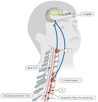


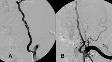
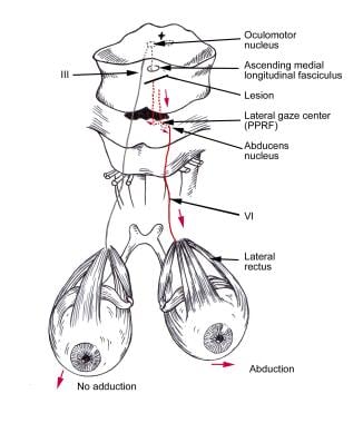


![17. [Blood] | Anatomy & Physiology | Educator.com](https://www.educator.com/media/lesson/poster/anatomy-physiology-cardella/blood.jpg)