Carcinoma, Transitional Cell
Carcinoma
Ureteral Neoplasms
Carcinoma, Squamous Cell
Carcinoma, Hepatocellular
Urethral Neoplasms
Carcinoma in Situ
Urinary Bladder
Carcinoma, Papillary
Administration, Intravesical
Immunohistochemistry
Neoplasm Staging
Uroplakin II
Ureter
Tumor Markers, Biological
Prognosis
Neoplasm Recurrence, Local
Carcinoma, Ductal, Breast
Carcinoma, Basal Cell
Tumor Cells, Cultured
Uroplakin III
Brenner Tumor
Neoplasms, Multiple Primary
Gene Expression Regulation, Neoplastic
Muscle Neoplasms
Uroplakin Ib
Uroplakin Ia
Immunoenzyme Techniques
Disease Progression
Fatal Outcome
Carcinoma, Bronchogenic
Carcinoma, Intraductal, Noninfiltrating
Survival Analysis
Carcinoma, Adenoid Cystic
Mice, Nude
Keratins
Spermatocele
Neoplasm Proteins
RNA, Messenger
Carcinoma, Small Cell
Lymphatic Metastasis
Carcinoma, Medullary
Neoplasm Transplantation
Papilloma
Retrospective Studies
Tumor Suppressor Protein p53
Carcinoma, Lobular
Cisplatin
Treatment Outcome
Neoplasm Metastasis
Antigens, Neoplasm
Survival Rate
Carcinoma, Neuroendocrine
Papilloma, Inverted
Cell Division
Keratin-7
Chromosomes, Human, Pair 9
Ki-67 Antigen
Deoxycytidine
Follow-Up Studies
Carcinoma, Mucoepidermoid
Disease-Free Survival
Hyperplasia
Semicircular Ducts
FANFT
Keratin-20
Dog Diseases
Carcinoma, Endometrioid
Reverse Transcriptase Polymerase Chain Reaction
Antineoplastic Combined Chemotherapy Protocols
Precancerous Conditions
Head and Neck Neoplasms
Genes, p53
Combined Modality Therapy
Carcinoma, Embryonal
Carcinoma, Merkel Cell
Carcinoma, Ductal
Metaplasia
Loss of Heterozygosity
Occlusive Dressings
Cystitis
Biopsy
Neoplasm Grading
Epithelium
Ovarian Neoplasms
Adrenocortical Carcinoma
Urinary Diversion
Apoptosis
Urologic Surgical Procedures
Carcinoma, Verrucous
Kidney Calices
Carcinoma, Signet Ring Cell
Neoplasms, Experimental
Cytodiagnosis
Urinary Tract
Genes, Tumor Suppressor
Paraffin Embedding
Cell Transformation, Neoplastic
Polymorphism, Single-Stranded Conformational
Rats, Inbred F344
Neovascularization, Pathologic
Paclitaxel
Blotting, Western
Carcinoma, Large Cell
Tomography, X-Ray Computed
Proliferating Cell Nuclear Antigen
Mutation
Papillomaviridae
Tumor Suppressor Proteins
Laryngeal Neoplasms
Chromosome Aberrations
Base Sequence
Schistosomiasis
In Situ Hybridization, Fluorescence
Microsatellite Repeats
Sensitivity and Specificity
Adenocarcinoma, Follicular
Receptor, erbB-2
BCG Vaccine
Lymph Node Excision
Molecular Sequence Data
Risk Factors
Kaplan-Meier Estimate
Adenocarcinoma, Mucinous
Gene Expression
Carcinogens
Embryonal Carcinoma Stem Cells
In Situ Hybridization
Cell Differentiation
Cyclooxygenase 2
Mucous Membrane
Carcinoma, Papillary, Follicular
Multivariate Analysis
Mitomycin
Receptor, Epidermal Growth Factor
Mice, Inbred BALB C
Urine
Carcinoma, Non-Small-Cell Lung
Dogs
Endometrial Neoplasms
Predictive Value of Tests
Adenocarcinoma, Clear Cell
Colorectal Neoplasms
Down-Regulation
Dose-Response Relationship, Drug
Flow Cytometry
Chemotherapy, Adjuvant
Papillomavirus Infections
alpha-Fetoproteins
Transfection
Blotting, Northern
Phenotype
Vascular Endothelial Growth Factor A
Case-Control Studies
Chromosome Disorders
Pancreatic Neoplasms
Cystadenocarcinoma, Serous
Long-term transplantability and morphological stability of three experimentally induced urinary bladder carcinomas in rats. (1/1469)
Three transitional cell carcinomas induced in Fischer 344 rats by a methylcholanthrene pellet or a foreign body inserted locally into the bladder have been serially transplanted in the syngeneic strain for up to 6.5 years. There have been no changes in the individual morphological characteristics of the tumors during this time. Cells cultured in vitro for varying numbers of passages reproduce regularly the morphology of each tumor when they are injected back into the animals and results from a microcytotoxicity assay for cellular immunity indicate that they retain a common, bladder tumor-specific antigen. These tumors are useful for research in turmo biology and are offered to other scientists seeking transplantable carcinomas for experimentation. (+info)Natural history of papillary lesions of the urinary bladder in schistosomiasis. (2/1469)
Variable epithelial hyperplasia was observed in urinary bladder of nine capuchin monkeys (Cebus apella) when examined at cystotomy 94 to 164 weeks after infection with Schistosoma haematobium. These hosts were followed for 24 to 136 weeks postcystotomy to determine the status of bladder lesions in relation to duration of infection and to ascertain whether lesion samples removed at cystotomy reestablished themselves in autologous and heterologous transfers. There was involution of urothelial hyperplasia in eight of nine animals and no evidence for establishment of transplanted bladder lesions. (+info)Differential regulation of p21waf-1/cip-1 and Mdm2 by etoposide: etoposide inhibits the p53-Mdm2 autoregulatory feedback loop. (3/1469)
The Mdm2 protein is frequently overexpressed in human non-seminomatous germ cell tumours and transitional carcinoma of the bladder where it may contribute to tolerance of wtp53. Mdm2 forms an autoregulatory feedback loop with p53; the Mdm2 gene is responsive to transactivation by p53 and once synthesized the Mdm2 protein terminates the p53 response. We show here that the topoisomerase poison etoposide, like ultra violet irradiation, inhibits Mdm2 synthesis. Cytotoxic concentrations of etoposide (IC90 for > 3 h) result in inhibition of Mdm2 induction at both the RNA and protein level. Rapid apoptosis ensues. Global transcription is not inhibited: p21waf-1/cip1 and GADD45 expression increase in a dose dependent manner. Inhibition of Mdm2 synthesis depends on the continuous presence of etoposide, suggesting the DNA damage may prevent transcription. Downregulation of Mdm2 transcript occurs in cells expressing HPV16-E6 suggesting that inhibition of Mdm2 transcription is p53-independent. When cells are -treated with a pulse (1 h) of etoposide and reincubated in drug free medium, Mdm2 synthesis commences immediately after damage is repaired (3 h) and the p53 response is attenuated. Induction of apoptosis and loss of clonogenicity are 3-5-fold lower under pulse treatment conditions. This is the first observation of inhibition of Mdm2 transcription following treatment with topoisomerase (topo II) poisons, a feature that may be useful in tumour types where p53 is tolerated by overexpression of Mdm2. (+info)Tumor-induced interleukin-10 inhibits type 1 immune responses directed at a tumor antigen as well as a non-tumor antigen present at the tumor site. (4/1469)
Interleukin (IL)-10 is a potent immunosuppressive cytokine that has been found to be present at the tumor site in a wide variety of human cancers, including transitional cell carcinoma of the bladder. Using a murine bladder tumor (MB49), which we show to express the male transplantation antigen (HY), we tested the hypothesis that IL-10 at the tumor site can block the generation of a tumor-specific type 1 immune response. We show that, despite its expression of HY, MB49 fails to prime for an HY-specific type 1 (IFN-gamma) response in normal female mice. Although MB49 does not constitutively produce IL-10, our data support a model whereby MB49 induces infiltrating cells to produce IL-10. This feature rendered the IL-10 knockout (KO) mouse, whose infiltrating cells are incapable of IL-10 production, a suitable model in which to study MB49 in the absence of IL-10. When injected into IL-10 KO mice, MB49 does prime for an HY-specific, type 1 immune response. Furthermore, IL-10 KO mice show prolonged survival and an increased capacity to reject tumors as compared with normal mice. We also tested the ability of tumor-induced IL-10 to inhibit immunization to a non-tumor antigen present at the tumor site. When vaccinia virus encoding beta-galactosidase (beta-gal) is injected into the tumors of normal mice, no beta-gal-specific IFN-gamma response is mounted. However, when this same viral construct is injected into the tumors of IL-10 KO mice, it produces a strong beta-gal-specific, IFN-gamma response. These studies demonstrate that tumor-induced IL-10 can block the generation of a tumor-specific type 1 immune response as well as subvert attempts to elicit a type 1 immune response to a non-tumor antigen at the tumor site. (+info)Anti-epidermal growth factor receptor antibody C225 inhibits angiogenesis in human transitional cell carcinoma growing orthotopically in nude mice. (5/1469)
Epidermal growth factor receptor (EGFR) regulates the growth and progression of human transitional cell carcinoma (TCC) of the bladder. We have shown that therapy targeting EGFR inhibited the growth of human TCC established orthotopically in nude mice. The purpose of this study was to evaluate whether EGFR-directed therapy affects angiogenesis associated with the growth and metastasis of human TCC. We determined the cytostatic effect and the effect on production of angiogenic factors after in vitro treatment of the human TCC cell line 253J B-V with MAb C225, a chimerized monoclonal anti-EGFR antibody. The 253J B-V cells were implanted orthotopically into athymic nude mice, and established tumors (4 weeks) were treated with i.p. MAb C225. Expression of the angiogenic factors vascular endothelial growth factor (VEGF), interleukin-8 (IL-8), and basic fibroblast growth factor (bFGF) was evaluated by immunohistochemistry and in situ mRNA hybridization analyses and correlated with microvessel density evaluated after immunohistochemical staining with anti-CD31. In vitro treatment with MAb C225 inhibited mRNA and protein production of VEGF, IL-8, and bFGF by 253J B-V cells in a dose-dependent manner. MAb C225 therapy of nude mice with established TCCs growing orthotopically resulted in inhibition of growth and metastasis compared with controls (P <0.0005). VEGF, IL-8, and bFGF expression was significantly lower in treated tumors than in controls. The down-regulation of these angiogenic factors preceded the involution of blood vessels. These studies indicate that therapy with anti-EGFR MAb C225 has a significant antitumor effect mediated, in part, by inhibition of angiogenesis. (+info)Interleukin-2 gene transfer into human transitional cell carcinoma of the urinary bladder. (6/1469)
Transitional cell carcinoma of the bladder is one of the human cancers most responsive to immunotherapy, and local interleukin-2 (IL-2) production appears to be an important requirement for immunotherapy to be effective. In this study, we engineered two human bladder cancer cell lines (RT112 and EJ) to constitutively release human IL-2 by retroviral vector-mediated gene transfer. Following infection and selection, stable and consistent production of biologically active IL-2 was demonstrated at both the mRNA and the protein level. Morphology, in vitro growth rate and proliferation, as well as other cytokine gene mRNA or membrane adhesion receptor expression, were not altered in IL-2 transduced cells as compared to their parental or control vector-infected counterparts. Moreover, IL-2 engineered cells lost their tumorigenicity into nu/nu mice and the mechanism of rejection appeared to involve multiple host effector cell populations, among which a prominent role was played by neutrophils and radiosensitive cells. These findings may offer support to the development of an IL-2-based gene therapy approach to human bladder cancer. (+info)Urinary bladder transitional cell carcinogenesis is associated with down-regulation of NF1 tumor suppressor gene in vivo and in vitro. (7/1469)
The NF1 gene product (neurofibromin) is known to act as a tumor suppressor protein by inactivating ras. The best documented factors involved in urinary bladder transitional cell carcinoma (TCC) are ras proto-oncogene activation and p53 suppressor gene mutations. This is the first study reporting alterations in NF1 gene expression in TCC. We examined NF1 gene expression in a total of 29 surgical urinary bladder TCC specimens representing grades 1 to 3 and in three cell lines, RT4, 5637, and T24 (representing grades 1 to 3, respectively). Decreased NF1 gene expression was observed in 23 of 29 (83%) TCC specimens as estimated by immunohistochemistry, the decrease being more pronounced in high-grade tumors. NF1 mRNA levels were markedly lower in TCC tissue compared with adjacent non-neoplastic urothelium, as studied by in situ hybridization for grade 3 TCC. Immunohistochemistry and Western blotting demonstrated that TCC cell lines expressed NF1 protein at different levels, expression being almost undetectable in T24 (grade 3) cells. Northern blotting for cell lines demonstrated reduced NF1 mRNA levels in grade 3 TCC cells. Reverse transcription polymerase chain reaction for cell lines and selected grade 2 and grade 3 tissue samples demonstrated NF1 type II mRNA isoform predominance in all samples studied. Our results show that both NF1 mRNA and protein levels are decreased in high-grade TCC, suggesting that alterations of NF1 gene expression may be involved in bladder TCC carcinogenesis. (+info)Overexpression of the wild type p73 gene in human bladder cancer. (8/1469)
p73, a first p53 relative, was recently identified and shown to be monoallelically expressed in a number of different human tissues. To determine the potential role of this gene in human bladder cancer, we investigated p73 expression levels, allelic expression patterns, and analysed p73 mutations in 23 unselected primary invasive bladder cancers with matched normal tissues and in seven bladder cancer cell lines. In a comparison between normal and tumor tissues using quantitative RT-PCR analysis, we found that p73 was overexpressed in 22/23 bladder cancers, sometimes as great as 20-fold. Allelic expression analysis using a C/T polymorphism in exon 2 and a newly identified T/C polymorphism in exon 5 revealed that p73 was biallelically expressed in both normal bladder and cancer tissues, suggesting that p73 is not imprinted in bladder tissue. Mutation screening of the p73 gene in bladder cancer DNAs using denaturing high-performance liquid chromatography analysis and DNA sequencing revealed no tumor-specific mutations in any coding exons of the p73 gene. These data suggest that the p73 is unlikely to be a tumor suppressor gene, but that overexpression of p73 may contribute to tumorigenesis in bladder cancer. (+info)Transitional cell carcinoma (TCC) is a type of cancer that develops in the transitional epithelium, which is the tissue that lines the inner surface of the urinary tract. This includes the renal pelvis, ureters, bladder, and urethra. Transitional cell carcinoma is the most common type of bladder cancer and can also occur in other parts of the urinary system.
Transitional cells are specialized epithelial cells that can stretch and change shape as the organs they line expand or contract. These cells normally have a flat, squamous appearance when at rest but become more cuboidal and columnar when the organ is full. Transitional cell carcinomas typically start in the urothelium, which is the innermost lining of the urinary tract.
Transitional cell carcinoma can be classified as non-invasive (also called papillary or superficial), invasive, or both. Non-invasive TCCs are confined to the urothelium and have not grown into the underlying connective tissue. Invasive TCCs have grown through the urothelium and invaded the lamina propria (a layer of connective tissue beneath the urothelium) or the muscle wall of the bladder.
Transitional cell carcinoma can also be categorized as low-grade or high-grade, depending on how abnormal the cancer cells look under a microscope and how likely they are to grow and spread. Low-grade TCCs tend to have a better prognosis than high-grade TCCs.
Treatment for transitional cell carcinoma depends on the stage and grade of the cancer, as well as other factors such as the patient's overall health. Treatment options may include surgery, radiation therapy, chemotherapy, or immunotherapy.
Urinary Bladder Neoplasms are abnormal growths or tumors in the urinary bladder, which can be benign (non-cancerous) or malignant (cancerous). Malignant neoplasms can be further classified into various types of bladder cancer, such as urothelial carcinoma, squamous cell carcinoma, and adenocarcinoma. These malignant tumors often invade surrounding tissues and organs, potentially spreading to other parts of the body (metastasis), which can lead to serious health consequences if not detected and treated promptly and effectively.
Carcinoma is a type of cancer that develops from epithelial cells, which are the cells that line the inner and outer surfaces of the body. These cells cover organs, glands, and other structures within the body. Carcinomas can occur in various parts of the body, including the skin, lungs, breasts, prostate, colon, and pancreas. They are often characterized by the uncontrolled growth and division of abnormal cells that can invade surrounding tissues and spread to other parts of the body through a process called metastasis. Carcinomas can be further classified based on their appearance under a microscope, such as adenocarcinoma, squamous cell carcinoma, and basal cell carcinoma.
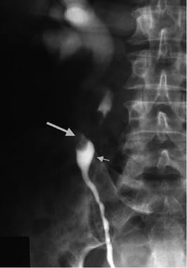
Ureteral neoplasms refer to abnormal growths or tumors in the ureters, which are the tubes that carry urine from the kidneys to the bladder. These neoplasms can be benign (non-cancerous) or malignant (cancerous). Benign ureteral neoplasms are rare and usually do not pose a significant health risk, although they may need to be removed if they cause obstructions or other complications.
Malignant ureteral neoplasms, on the other hand, are more serious and can spread to other parts of the body. The most common type of malignant ureteral neoplasm is transitional cell carcinoma (TCC), which arises from the cells that line the inside of the ureters. Other types of malignant ureteral neoplasms include squamous cell carcinoma, adenocarcinoma, and sarcoma.
Symptoms of ureteral neoplasms may include hematuria (blood in the urine), flank pain, weight loss, and fatigue. Diagnosis typically involves imaging tests such as CT scans or MRIs, as well as urine cytology and biopsy to confirm the presence of cancer cells. Treatment options may include surgery, radiation therapy, chemotherapy, or a combination of these approaches.
Urologic neoplasms refer to abnormal growths or tumors in the urinary system, which includes the kidneys, ureters, bladder, prostate, and urethra. These growths can be benign (non-cancerous) or malignant (cancerous). Common types of urologic neoplasms include renal cell carcinoma, transitional cell carcinoma, bladder cancer, prostate cancer, and testicular cancer. It is important to note that early detection and treatment can significantly improve outcomes for patients with urologic neoplasms.
Squamous cell carcinoma is a type of skin cancer that begins in the squamous cells, which are flat, thin cells that form the outer layer of the skin (epidermis). It commonly occurs on sun-exposed areas such as the face, ears, lips, and backs of the hands. Squamous cell carcinoma can also develop in other areas of the body including the mouth, lungs, and cervix.
This type of cancer usually develops slowly and may appear as a rough or scaly patch of skin, a red, firm nodule, or a sore or ulcer that doesn't heal. While squamous cell carcinoma is not as aggressive as some other types of cancer, it can metastasize (spread) to other parts of the body if left untreated, making early detection and treatment important.
Risk factors for developing squamous cell carcinoma include prolonged exposure to ultraviolet (UV) radiation from the sun or tanning beds, fair skin, a history of sunburns, a weakened immune system, and older age. Prevention measures include protecting your skin from the sun by wearing protective clothing, using a broad-spectrum sunscreen with an SPF of at least 30, avoiding tanning beds, and getting regular skin examinations.
The kidney pelvis, also known as the renal pelvis, is the funnel-shaped part of the upper end of the ureter in the kidney. It receives urine from the minor and major calyces, which are extensions of the renal collecting tubules, and then drains it into the ureter, which carries it to the bladder for storage and eventual elimination from the body. The kidney pelvis is lined with transitional epithelium, which is designed to stretch and accommodate changes in urine volume.
Hepatocellular carcinoma (HCC) is the most common type of primary liver cancer in adults. It originates from the hepatocytes, which are the main functional cells of the liver. This type of cancer is often associated with chronic liver diseases such as cirrhosis caused by hepatitis B or C virus infection, alcohol abuse, non-alcoholic fatty liver disease (NAFLD), and aflatoxin exposure.
The symptoms of HCC can vary but may include unexplained weight loss, lack of appetite, abdominal pain or swelling, jaundice, and fatigue. The diagnosis of HCC typically involves imaging tests such as ultrasound, CT scan, or MRI, as well as blood tests to measure alpha-fetoprotein (AFP) levels. Treatment options for Hepatocellular carcinoma depend on the stage and extent of the cancer, as well as the patient's overall health and liver function. Treatment options may include surgery, radiation therapy, chemotherapy, targeted therapy, or liver transplantation.
Urethral neoplasms refer to abnormal growths or tumors in the urethra, which is the tube that carries urine from the bladder out of the body. These growths can be benign (non-cancerous) or malignant (cancerous).
Benign urethral neoplasms may include conditions such as urethral polyps or papillomas, which are usually not life-threatening and can often be removed with surgery.
Malignant urethral neoplasms, on the other hand, are cancerous tumors that can invade surrounding tissues and spread to other parts of the body. These include urethral carcinomas, which can be further classified into different types such as squamous cell carcinoma, transitional cell carcinoma, and adenocarcinoma, depending on the type of cells involved.
Urethral neoplasms are relatively rare, but when they do occur, they can cause a variety of symptoms such as difficulty urinating, blood in the urine, pain during urination or sexual intercourse, and discharge from the urethra. Treatment options depend on the type, location, and stage of the neoplasm, and may include surgery, radiation therapy, chemotherapy, or a combination of these approaches.
Urothelium is the specialized type of epithelial tissue that lines the urinary tract, including the renal pelvis, ureters, bladder, and urethra. It is a type of transitional epithelium that can change its shape and size depending on the degree of distension or stretching of the organs it lines.
The main function of urothelium is to provide a barrier against urine, which contains various waste products and potential irritants, while also allowing the exchange of ions and water. The urothelial cells are joined together by tight junctions that prevent the passage of substances through the paracellular space, and they also have the ability to transport ions and water through their cell membranes.
In addition to its barrier function, urothelium is also involved in sensory and immune functions. It contains specialized nerve endings that can detect mechanical and chemical stimuli, such as stretch or irritation, and it expresses various antimicrobial peptides and other defense mechanisms that help protect the urinary tract from infection.
Overall, urothelium plays a critical role in maintaining the health and function of the urinary tract, and its dysfunction has been implicated in various urinary tract disorders, such as interstitial cystitis/bladder pain syndrome and bladder cancer.
Cystectomy is a surgical procedure in which all or part of the urinary bladder is removed. This procedure is often used to treat bladder cancer, but it may also be necessary in cases of severe bladder damage, infection, or inflammation that do not respond to other treatments.
There are several types of cystectomy, including:
1. Radical cystectomy: This is the most common type of cystectomy performed for bladder cancer. It involves removing the entire bladder, as well as nearby lymph nodes, the prostate gland in men, and the uterus, ovaries, fallopian tubes, and a portion of the vagina in women.
2. Partial cystectomy: In this procedure, only a part of the bladder is removed. This may be an option for patients with early-stage bladder cancer that has not spread deeply into the bladder muscle or to other parts of the body.
3. Urinary diversion: After a cystectomy, the surgeon must create a new way for urine to leave the body. This may involve creating a urostomy, in which a piece of intestine is used to form a stoma (an opening) on the abdominal wall, through which urine can be collected in a bag. Alternatively, the surgeon may create an internal pouch using a segment of intestine, which can then be connected to the ureters and allowed to drain into the rectum or vagina.
As with any surgical procedure, cystectomy carries risks such as bleeding, infection, and reactions to anesthesia. Patients may also experience long-term complications such as urinary incontinence, sexual dysfunction, and changes in bowel habits. However, for many patients with bladder cancer or other severe bladder conditions, cystectomy can be a life-saving procedure.
Carcinoma in situ is a medical term used to describe the earliest stage of cancer, specifically a type of cancer that begins in the epithelial tissue, which is the tissue that lines the outer surfaces of organs and body structures. In this stage, the cancer cells are confined to the layer of cells where they first developed and have not spread beyond that layer into the surrounding tissues or organs.
Carcinoma in situ can occur in various parts of the body, including the skin, cervix, breast, lung, prostate, bladder, and other areas. It is often detected through routine screening tests, such as Pap smears for cervical cancer or mammograms for breast cancer.
While carcinoma in situ is not invasive, it can still be a serious condition because it has the potential to develop into an invasive cancer if left untreated. Treatment options for carcinoma in situ may include surgery, radiation therapy, or other forms of treatment, depending on the location and type of cancer. It is important to consult with a healthcare provider to determine the best course of action for each individual case.
The urinary bladder is a muscular, hollow organ in the pelvis that stores urine before it is released from the body. It expands as it fills with urine and contracts when emptying. The typical adult bladder can hold between 400 to 600 milliliters of urine for about 2-5 hours before the urge to urinate occurs. The wall of the bladder contains several layers, including a mucous membrane, a layer of smooth muscle (detrusor muscle), and an outer fibrous adventitia. The muscles of the bladder neck and urethra remain contracted to prevent leakage of urine during filling, and they relax during voiding to allow the urine to flow out through the urethra.
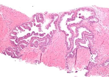
Carcinoma, papillary is a type of cancer that begins in the cells that line the glandular structures or the lining of organs. In a papillary carcinoma, the cancerous cells grow and form small finger-like projections, called papillae, within the tumor. This type of cancer most commonly occurs in the thyroid gland, but can also be found in other organs such as the lung, breast, and kidney. Papillary carcinoma of the thyroid gland is usually slow-growing and has a good prognosis, especially when it is diagnosed at an early stage.
Intravesical administration refers to the instillation of medication directly into the bladder through a catheter or other medical device. This method is often used to deliver treatments for various bladder conditions, such as interstitial cystitis, bladder cancer, and chronic bladder infections. The medication is held in the bladder for a specified period, usually ranging from a few minutes to several hours, before being urinated out. This allows the medication to come into close contact with the bladder lining, potentially enhancing its effectiveness while minimizing systemic side effects.
Immunohistochemistry (IHC) is a technique used in pathology and laboratory medicine to identify specific proteins or antigens in tissue sections. It combines the principles of immunology and histology to detect the presence and location of these target molecules within cells and tissues. This technique utilizes antibodies that are specific to the protein or antigen of interest, which are then tagged with a detection system such as a chromogen or fluorophore. The stained tissue sections can be examined under a microscope, allowing for the visualization and analysis of the distribution and expression patterns of the target molecule in the context of the tissue architecture. Immunohistochemistry is widely used in diagnostic pathology to help identify various diseases, including cancer, infectious diseases, and immune-mediated disorders.
Neoplasm staging is a systematic process used in medicine to describe the extent of spread of a cancer, including the size and location of the original (primary) tumor and whether it has metastasized (spread) to other parts of the body. The most widely accepted system for this purpose is the TNM classification system developed by the American Joint Committee on Cancer (AJCC) and the Union for International Cancer Control (UICC).
In this system, T stands for tumor, and it describes the size and extent of the primary tumor. N stands for nodes, and it indicates whether the cancer has spread to nearby lymph nodes. M stands for metastasis, and it shows whether the cancer has spread to distant parts of the body.
Each letter is followed by a number that provides more details about the extent of the disease. For example, a T1N0M0 cancer means that the primary tumor is small and has not spread to nearby lymph nodes or distant sites. The higher the numbers, the more advanced the cancer.
Staging helps doctors determine the most appropriate treatment for each patient and estimate the patient's prognosis. It is an essential tool for communication among members of the healthcare team and for comparing outcomes of treatments in clinical trials.
Neoplasm invasiveness is a term used in pathology and oncology to describe the aggressive behavior of cancer cells as they invade surrounding tissues and organs. This process involves the loss of cell-to-cell adhesion, increased motility and migration, and the ability of cancer cells to degrade the extracellular matrix (ECM) through the production of enzymes such as matrix metalloproteinases (MMPs).
Invasive neoplasms are cancers that have spread beyond the original site where they first developed and have infiltrated adjacent tissues or structures. This is in contrast to non-invasive or in situ neoplasms, which are confined to the epithelial layer where they originated and have not yet invaded the underlying basement membrane.
The invasiveness of a neoplasm is an important prognostic factor in cancer diagnosis and treatment, as it can indicate the likelihood of metastasis and the potential effectiveness of various therapies. In general, more invasive cancers are associated with worse outcomes and require more aggressive treatment approaches.
Urogenital neoplasms refer to abnormal growths or tumors that occur in the urinary and genital organs. These can include various types of cancer, such as bladder cancer, kidney cancer, prostate cancer, testicular cancer, cervical cancer, ovarian cancer, and others. Some urogenital neoplasms may be benign (non-cancerous), while others are malignant (cancerous) and can spread to other parts of the body.
The term "urogenital" refers to the combined urinary and genital systems in the human body. The urinary system includes the kidneys, ureters, bladder, and urethra, which are responsible for filtering waste from the blood and eliminating it as urine. The genital system includes the reproductive organs such as the ovaries, fallopian tubes, uterus, vagina, prostate gland, testicles, and penis.
Urogenital neoplasms can cause various symptoms depending on their location and size. Common symptoms include blood in urine, pain during urination, difficulty urinating, abnormal discharge, lumps or swelling in the genital area, and unexplained weight loss. If you experience any of these symptoms, it is important to consult a healthcare professional for further evaluation and treatment.
Uroplakin II is a type of protein that is a component of the urothelium, which is the tissue that lines the urinary tract. Specifically, uroplakins are part of the asymmetric unit membrane (AUM) of the urothelial plaques, which are specialized structures on the apical surface of the urothelium. These plaques help to provide a barrier function and protect the underlying tissues from various harmful substances in the urine. Uroplakin II is a transmembrane protein that forms heterodimers with other uroplakins, such as uroplakin Ib, to create the building blocks of the urothelial plaques.
Cystoscopy is a medical procedure that involves the insertion of a thin, flexible tube with a camera and light on the end (cystoscope) into the bladder through the urethra. This procedure allows healthcare professionals to examine the lining of the bladder and urethra for any abnormalities such as inflammation, tumors, or stones. Cystoscopy can be used for diagnostic purposes, as well as for therapeutic interventions like removing small bladder tumors or performing biopsies. It is typically performed under local or general anesthesia to minimize discomfort and pain.
A ureter is a thin, muscular tube that transports urine from the kidney to the bladder. In humans, there are two ureters, one for each kidney, and they are typically about 10-12 inches long. The ureters are lined with a special type of cells called transitional epithelium that can stretch and expand as urine passes through them. They are located in the retroperitoneal space, which is the area behind the peritoneum, the membrane that lines the abdominal cavity. The ureters play a critical role in the urinary system by ensuring that urine flows from the kidneys to the bladder for storage and eventual elimination from the body.
Tumor markers are substances that can be found in the body and their presence can indicate the presence of certain types of cancer or other conditions. Biological tumor markers refer to those substances that are produced by cancer cells or by other cells in response to cancer or certain benign (non-cancerous) conditions. These markers can be found in various bodily fluids such as blood, urine, or tissue samples.
Examples of biological tumor markers include:
1. Proteins: Some tumor markers are proteins that are produced by cancer cells or by other cells in response to the presence of cancer. For example, prostate-specific antigen (PSA) is a protein produced by normal prostate cells and in higher amounts by prostate cancer cells.
2. Genetic material: Tumor markers can also include genetic material such as DNA, RNA, or microRNA that are shed by cancer cells into bodily fluids. For example, circulating tumor DNA (ctDNA) is genetic material from cancer cells that can be found in the bloodstream.
3. Metabolites: Tumor markers can also include metabolic products produced by cancer cells or by other cells in response to cancer. For example, lactate dehydrogenase (LDH) is an enzyme that is released into the bloodstream when cancer cells break down glucose for energy.
It's important to note that tumor markers are not specific to cancer and can be elevated in non-cancerous conditions as well. Therefore, they should not be used alone to diagnose cancer but rather as a tool in conjunction with other diagnostic tests and clinical evaluations.
Prognosis is a medical term that refers to the prediction of the likely outcome or course of a disease, including the chances of recovery or recurrence, based on the patient's symptoms, medical history, physical examination, and diagnostic tests. It is an important aspect of clinical decision-making and patient communication, as it helps doctors and patients make informed decisions about treatment options, set realistic expectations, and plan for future care.
Prognosis can be expressed in various ways, such as percentages, categories (e.g., good, fair, poor), or survival rates, depending on the nature of the disease and the available evidence. However, it is important to note that prognosis is not an exact science and may vary depending on individual factors, such as age, overall health status, and response to treatment. Therefore, it should be used as a guide rather than a definitive forecast.
Liver neoplasms refer to abnormal growths in the liver that can be benign or malignant. Benign liver neoplasms are non-cancerous tumors that do not spread to other parts of the body, while malignant liver neoplasms are cancerous tumors that can invade and destroy surrounding tissue and spread to other organs.
Liver neoplasms can be primary, meaning they originate in the liver, or secondary, meaning they have metastasized (spread) to the liver from another part of the body. Primary liver neoplasms can be further classified into different types based on their cell of origin and behavior, including hepatocellular carcinoma, cholangiocarcinoma, and hepatic hemangioma.
The diagnosis of liver neoplasms typically involves a combination of imaging studies, such as ultrasound, CT scan, or MRI, and biopsy to confirm the type and stage of the tumor. Treatment options depend on the type and extent of the neoplasm and may include surgery, radiation therapy, chemotherapy, or liver transplantation.
Local neoplasm recurrence is the return or regrowth of a tumor in the same location where it was originally removed or treated. This means that cancer cells have survived the initial treatment and started to grow again in the same area. It's essential to monitor and detect any local recurrence as early as possible, as it can affect the prognosis and may require additional treatment.
Carcinoma, ductal, breast is a type of breast cancer that begins in the milk ducts (the tubes that carry milk from the lobules of the breast to the nipple). It is called "ductal" because it starts in the cells that line the milk ducts. Ductal carcinoma can be further classified as either non-invasive or invasive, based on whether the cancer cells are confined to the ducts or have spread beyond them into the surrounding breast tissue.
Non-invasive ductal carcinoma (also known as intraductal carcinoma or ductal carcinoma in situ) is a condition where abnormal cells have been found in the lining of the milk ducts, but they have not spread outside of the ducts. These cells have the potential to become invasive and spread to other parts of the breast or body if left untreated.
Invasive ductal carcinoma (IDC) is a type of breast cancer that starts in a milk duct and then grows into the surrounding breast tissue. From there, it can spread to other parts of the body through the bloodstream and lymphatic system. IDC is the most common form of breast cancer, accounting for about 80% of all cases.
Symptoms of ductal carcinoma may include a lump or thickening in the breast, changes in the size or shape of the breast, dimpling or puckering of the skin on the breast, nipple discharge (especially if it is clear or bloody), and/or redness or scaling of the nipple or breast skin. However, many cases of ductal carcinoma are detected through mammography before any symptoms develop.
Treatment for ductal carcinoma depends on several factors, including the stage and grade of the cancer, as well as the patient's overall health and personal preferences. Treatment options may include surgery (such as a lumpectomy or mastectomy), radiation therapy, chemotherapy, hormone therapy, and/or targeted therapies.
Kidney neoplasms refer to abnormal growths or tumors in the kidney tissues that can be benign (non-cancerous) or malignant (cancerous). These growths can originate from various types of kidney cells, including the renal tubules, glomeruli, and the renal pelvis.
Malignant kidney neoplasms are also known as kidney cancers, with renal cell carcinoma being the most common type. Benign kidney neoplasms include renal adenomas, oncocytomas, and angiomyolipomas. While benign neoplasms are generally not life-threatening, they can still cause problems if they grow large enough to compromise kidney function or if they undergo malignant transformation.
Early detection and appropriate management of kidney neoplasms are crucial for improving patient outcomes and overall prognosis. Regular medical check-ups, imaging studies, and urinalysis can help in the early identification of these growths, allowing for timely intervention and treatment.
Carcinoma, basal cell is a type of skin cancer that arises from the basal cells, which are located in the lower part of the epidermis (the outermost layer of the skin). It is also known as basal cell carcinoma (BCC) and is the most common form of skin cancer.
BCC typically appears as a small, shiny, pearly bump or nodule on the skin, often in sun-exposed areas such as the face, ears, neck, hands, and arms. It may also appear as a scar-like area that is white, yellow, or waxy. BCCs are usually slow growing and rarely spread (metastasize) to other parts of the body. However, they can be locally invasive and destroy surrounding tissue if left untreated.
The exact cause of BCC is not known, but it is thought to be related to a combination of genetic and environmental factors, including exposure to ultraviolet (UV) radiation from the sun or tanning beds. People with fair skin, light hair, and blue or green eyes are at increased risk of developing BCC.
Treatment for BCC typically involves surgical removal of the tumor, along with a margin of healthy tissue. Other treatment options may include radiation therapy, topical chemotherapy, or photodynamic therapy. Prevention measures include protecting your skin from UV radiation by wearing protective clothing, using sunscreen, and avoiding tanning beds.
'Tumor cells, cultured' refers to the process of removing cancerous cells from a tumor and growing them in controlled laboratory conditions. This is typically done by isolating the tumor cells from a patient's tissue sample, then placing them in a nutrient-rich environment that promotes their growth and multiplication.
The resulting cultured tumor cells can be used for various research purposes, including the study of cancer biology, drug development, and toxicity testing. They provide a valuable tool for researchers to better understand the behavior and characteristics of cancer cells outside of the human body, which can lead to the development of more effective cancer treatments.
It is important to note that cultured tumor cells may not always behave exactly the same way as they do in the human body, so findings from cell culture studies must be validated through further research, such as animal models or clinical trials.
Uroplakin III is a protein that is a component of urothelial plaques, which are specialized structures found on the surface of urothelial cells in the urinary bladder. Urothelial plaques play an important role in maintaining the barrier function and permeability properties of the urothelium.
Uroplakin III is a member of the uroplakin family of proteins, which includes UPIa, UPII, UPIII, and UPIIIA. These proteins are synthesized in the endoplasmic reticulum and transported to the Golgi apparatus, where they form heterodimers that are then transported to the plasma membrane. At the plasma membrane, the heterodimers assemble into larger complexes called urothelial plaques.
Uroplakin III is a transmembrane protein with a molecular weight of approximately 27 kDa. It has been shown to play a role in the formation and stability of urothelial plaques, as well as in the regulation of ion transport across the urothelium. Mutations in the gene encoding Uroplakin III have been associated with certain bladder diseases, including interstitial cystitis/bladder pain syndrome and bladder cancer.
A Brenner tumor is a rare type of benign (non-cancerous) ovarian tumor that originates from the tissue that lines the ovary (the epithelium). These tumors are typically small, slow-growing, and asymptomatic, although in some cases they may cause abdominal discomfort or bloating.
Brenner tumors are composed of transitional cells, which are similar to the cells found in the urinary bladder. They are usually solid and contain areas of calcification (calcium deposits). While most Brenner tumors are benign, a small percentage may become malignant (cancerous) and spread to other parts of the body.
The exact cause of Brenner tumors is not known, but they are more common in older women and are often found incidentally during routine pelvic exams or imaging studies. Treatment typically involves surgical removal of the tumor, and the prognosis is generally excellent, especially for benign tumors.
Multiple primary neoplasms refer to the occurrence of more than one primary malignant tumor in an individual, where each tumor is unrelated to the other and originates from separate cells or organs. This differs from metastatic cancer, where a single malignancy spreads to multiple sites in the body. Multiple primary neoplasms can be synchronous (occurring at the same time) or metachronous (occurring at different times). The risk of developing multiple primary neoplasms increases with age and is associated with certain genetic predispositions, environmental factors, and lifestyle choices such as smoking and alcohol consumption.
Neoplastic gene expression regulation refers to the processes that control the production of proteins and other molecules from genes in neoplastic cells, or cells that are part of a tumor or cancer. In a normal cell, gene expression is tightly regulated to ensure that the right genes are turned on or off at the right time. However, in cancer cells, this regulation can be disrupted, leading to the overexpression or underexpression of certain genes.
Neoplastic gene expression regulation can be affected by a variety of factors, including genetic mutations, epigenetic changes, and signals from the tumor microenvironment. These changes can lead to the activation of oncogenes (genes that promote cancer growth and development) or the inactivation of tumor suppressor genes (genes that prevent cancer).
Understanding neoplastic gene expression regulation is important for developing new therapies for cancer, as targeting specific genes or pathways involved in this process can help to inhibit cancer growth and progression.
Muscle neoplasms are abnormal growths or tumors that develop in the muscle tissue. They can be benign (non-cancerous) or malignant (cancerous). Benign muscle neoplasms are typically slow-growing and do not spread to other parts of the body, while malignant muscle neoplasms, also known as soft tissue sarcomas, can grow quickly, invade nearby tissues, and metastasize (spread) to distant parts of the body.
Soft tissue sarcomas can arise from any of the muscles in the body, including the skeletal muscles (voluntary muscles that attach to bones and help with movement), smooth muscles (involuntary muscles found in the walls of blood vessels, digestive tract, and other organs), or cardiac muscle (the specialized muscle found in the heart).
There are many different types of soft tissue sarcomas, each with its own set of characteristics and prognosis. Treatment for muscle neoplasms typically involves a combination of surgery, radiation therapy, and chemotherapy, depending on the type, size, location, and stage of the tumor.
Adenocarcinoma is a type of cancer that arises from glandular epithelial cells. These cells line the inside of many internal organs, including the breasts, prostate, colon, and lungs. Adenocarcinomas can occur in any of these organs, as well as in other locations where glands are present.
The term "adenocarcinoma" is used to describe a cancer that has features of glandular tissue, such as mucus-secreting cells or cells that produce hormones. These cancers often form glandular structures within the tumor mass and may produce mucus or other substances.
Adenocarcinomas are typically slow-growing and tend to spread (metastasize) to other parts of the body through the lymphatic system or bloodstream. They can be treated with surgery, radiation therapy, chemotherapy, targeted therapy, or a combination of these treatments. The prognosis for adenocarcinoma depends on several factors, including the location and stage of the cancer, as well as the patient's overall health and age.
Uroplakin Ib is not a recognized medical term or concept in and of itself. However, Uroplakins are a group of proteins found on the surface of urothelial cells, which make up the lining of the urinary tract. These proteins play an important role in maintaining the barrier function and integrity of the urothelium.
Uroplakin Ib is one of four major uroplakins (Ia, Ib, II, and III) that form complexes called uroplakins plaques on the apical surface of superficial urothelial cells. These plaques are thought to provide a protective barrier against urinary constituents, as well as contribute to the low permeability of the urothelium.
Therefore, while "Uroplakin Ib" may not have its own medical definition, it is an important component of the larger structure and function of uroplakins in the urinary tract.
A cell line that is derived from tumor cells and has been adapted to grow in culture. These cell lines are often used in research to study the characteristics of cancer cells, including their growth patterns, genetic changes, and responses to various treatments. They can be established from many different types of tumors, such as carcinomas, sarcomas, and leukemias. Once established, these cell lines can be grown and maintained indefinitely in the laboratory, allowing researchers to conduct experiments and studies that would not be feasible using primary tumor cells. It is important to note that tumor cell lines may not always accurately represent the behavior of the original tumor, as they can undergo genetic changes during their time in culture.
Uroplakin Ia is not a medical term itself, but it is a component of uroplakins which are a group of proteins found in the urothelium, the tissue that lines the urinary tract. Uroplakins are involved in the formation of the asymmetric unit membrane (AUM) of the urothelial plaques, which are specialized structures on the apical surface of the superficial urothelial cells. These plaques provide a barrier function and protect the underlying tissues from various harmful substances in urine.
Uroplakin Ia is one of the four major uroplakins (UPIa, UPIb, UPII, and UPIII) that form heterodimers and then assemble into larger complexes to form the urothelial plaques. Specifically, Uroplakin Ia combines with Uroplakin Ib to form a heterodimer, which then associates with UPII and UPIII heterodimers to form a tetraspanin complex. These complexes are then incorporated into the AUM of the urothelial plaques.
Abnormalities in uroplakins have been associated with various urological disorders, including bladder cancer, interstitial cystitis, and chronic pelvic pain syndrome.
Hematuria is a medical term that refers to the presence of blood in urine. It can be visible to the naked eye, which is called gross hematuria, or detected only under a microscope, known as microscopic hematuria. The blood in urine may come from any site along the urinary tract, including the kidneys, ureters, bladder, or urethra. Hematuria can be a symptom of various medical conditions, such as urinary tract infections, kidney stones, kidney disease, or cancer of the urinary tract. It is essential to consult a healthcare professional if you notice blood in your urine to determine the underlying cause and receive appropriate treatment.
Immunoenzyme techniques are a group of laboratory methods used in immunology and clinical chemistry that combine the specificity of antibody-antigen reactions with the sensitivity and amplification capabilities of enzyme reactions. These techniques are primarily used for the detection, quantitation, or identification of various analytes (such as proteins, hormones, drugs, viruses, or bacteria) in biological samples.
In immunoenzyme techniques, an enzyme is linked to an antibody or antigen, creating a conjugate. This conjugate then interacts with the target analyte in the sample, forming an immune complex. The presence and amount of this immune complex can be visualized or measured by detecting the enzymatic activity associated with it.
There are several types of immunoenzyme techniques, including:
1. Enzyme-linked Immunosorbent Assay (ELISA): A widely used method for detecting and quantifying various analytes in a sample. In ELISA, an enzyme is attached to either the capture antibody or the detection antibody. After the immune complex formation, a substrate is added that reacts with the enzyme, producing a colored product that can be measured spectrophotometrically.
2. Immunoblotting (Western blot): A method used for detecting specific proteins in a complex mixture, such as a protein extract from cells or tissues. In this technique, proteins are separated by gel electrophoresis and transferred to a membrane, where they are probed with an enzyme-conjugated antibody directed against the target protein.
3. Immunohistochemistry (IHC): A method used for detecting specific antigens in tissue sections or cells. In IHC, an enzyme-conjugated primary or secondary antibody is applied to the sample, and the presence of the antigen is visualized using a chromogenic substrate that produces a colored product at the site of the antigen-antibody interaction.
4. Immunofluorescence (IF): A method used for detecting specific antigens in cells or tissues by employing fluorophore-conjugated antibodies. The presence of the antigen is visualized using a fluorescence microscope.
5. Enzyme-linked immunosorbent assay (ELISA): A method used for detecting and quantifying specific antigens or antibodies in liquid samples, such as serum or culture supernatants. In ELISA, an enzyme-conjugated detection antibody is added after the immune complex formation, and a substrate is added that reacts with the enzyme to produce a colored product that can be measured spectrophotometrically.
These techniques are widely used in research and diagnostic laboratories for various applications, including protein characterization, disease diagnosis, and monitoring treatment responses.
Disease progression is the worsening or advancement of a medical condition over time. It refers to the natural course of a disease, including its development, the severity of symptoms and complications, and the impact on the patient's overall health and quality of life. Understanding disease progression is important for developing appropriate treatment plans, monitoring response to therapy, and predicting outcomes.
The rate of disease progression can vary widely depending on the type of medical condition, individual patient factors, and the effectiveness of treatment. Some diseases may progress rapidly over a short period of time, while others may progress more slowly over many years. In some cases, disease progression may be slowed or even halted with appropriate medical interventions, while in other cases, the progression may be inevitable and irreversible.
In clinical practice, healthcare providers closely monitor disease progression through regular assessments, imaging studies, and laboratory tests. This information is used to guide treatment decisions and adjust care plans as needed to optimize patient outcomes and improve quality of life.
A fatal outcome is a term used in medical context to describe a situation where a disease, injury, or illness results in the death of an individual. It is the most severe and unfortunate possible outcome of any medical condition, and is often used as a measure of the severity and prognosis of various diseases and injuries. In clinical trials and research, fatal outcome may be used as an endpoint to evaluate the effectiveness and safety of different treatments or interventions.
Carcinoma, bronchogenic is a medical term that refers to a type of lung cancer that originates in the bronchi, which are the branching tubes that carry air into the lungs. It is the most common form of lung cancer and can be further classified into different types based on the specific cell type involved, such as squamous cell carcinoma, adenocarcinoma, or large cell carcinoma.
Bronchogenic carcinomas are often associated with smoking and exposure to environmental pollutants, although they can also occur in non-smokers. Symptoms may include coughing, chest pain, shortness of breath, wheezing, hoarseness, or unexplained weight loss. Treatment options depend on the stage and location of the cancer, as well as the patient's overall health and may include surgery, radiation therapy, chemotherapy, targeted therapy, or a combination of these approaches.
Butylhydroxybutylnitrosamine (OH-BBN or BBN) is a chemical compound that is primarily used in laboratory research as a potent carcinogenic agent. It is known to induce tumors in various organs, particularly in the urinary bladder and liver, when administered to experimental animals.
The IUPAC name for Butylhydroxybutylnitrosamine is N-butyl-N-(4-hydroxybutyl)nitrosamine. Its molecular formula is C8H19NO3. It is a white to off-white crystalline powder, soluble in water and alcohol.
It is important to note that Butylhydroxybutylnitrosamine is not used in human medicine or therapy due to its carcinogenic properties. Its use is restricted to research purposes only, under controlled conditions and with appropriate safety measures in place.
Intraductal carcinoma, noninfiltrating is a medical term used to describe a type of breast cancer that is confined to the milk ducts of the breast. It is also sometimes referred to as ductal carcinoma in situ (DCIS). Noninfiltrating means that the cancer cells have not spread beyond the ducts into the surrounding breast tissue or elsewhere in the body.
In this type of cancer, abnormal cells line the milk ducts and fill the inside of the ducts. These abnormal cells may look like cancer cells under a microscope, but they have not grown through the walls of the ducts into the surrounding breast tissue. However, if left untreated, noninfiltrating intraductal carcinoma can progress to an invasive form of breast cancer where the cancer cells spread beyond the milk ducts and invade the surrounding breast tissue.
It is important to note that while noninfiltrating intraductal carcinoma is considered a precancerous condition, it still requires medical treatment to prevent the development of invasive breast cancer. Treatment options may include surgery, radiation therapy, or hormone therapy, depending on the size and location of the tumor and other individual factors.
The term "DNA, neoplasm" is not a standard medical term or concept. DNA refers to deoxyribonucleic acid, which is the genetic material present in the cells of living organisms. A neoplasm, on the other hand, is a tumor or growth of abnormal tissue that can be benign (non-cancerous) or malignant (cancerous).
In some contexts, "DNA, neoplasm" may refer to genetic alterations found in cancer cells. These genetic changes can include mutations, amplifications, deletions, or rearrangements of DNA sequences that contribute to the development and progression of cancer. Identifying these genetic abnormalities can help doctors diagnose and treat certain types of cancer more effectively.
However, it's important to note that "DNA, neoplasm" is not a term that would typically be used in medical reports or research papers without further clarification. If you have any specific questions about DNA changes in cancer cells or neoplasms, I would recommend consulting with a healthcare professional or conducting further research on the topic.
Survival analysis is a branch of statistics that deals with the analysis of time to event data. It is used to estimate the time it takes for a certain event of interest to occur, such as death, disease recurrence, or treatment failure. The event of interest is called the "failure" event, and survival analysis estimates the probability of not experiencing the failure event until a certain point in time, also known as the "survival" probability.
Survival analysis can provide important information about the effectiveness of treatments, the prognosis of patients, and the identification of risk factors associated with the event of interest. It can handle censored data, which is common in medical research where some participants may drop out or be lost to follow-up before the event of interest occurs.
Survival analysis typically involves estimating the survival function, which describes the probability of surviving beyond a certain time point, as well as hazard functions, which describe the instantaneous rate of failure at a given time point. Other important concepts in survival analysis include median survival times, restricted mean survival times, and various statistical tests to compare survival curves between groups.
Adenoid cystic carcinoma (AdCC) is a rare type of cancer that can occur in various glands and tissues of the body, most commonly in the salivary glands. AdCC is characterized by its slow growth and tendency to spread along nerves. It typically forms solid, cystic, or mixed tumors with distinct histological features, including epithelial cells arranged in tubular, cribriform, or solid patterns.
The term "carcinoma" refers to a malignant tumor originating from the epithelial cells lining various organs and glands. In this case, adenoid cystic carcinoma is a specific type of carcinoma that arises in the salivary glands or other glandular tissues.
The primary treatment options for AdCC include surgical resection, radiation therapy, and sometimes chemotherapy. Despite its slow growth, adenoid cystic carcinoma has a propensity to recur locally and metastasize to distant sites such as the lungs, bones, and liver. Long-term follow-up is essential due to the risk of late recurrences.
"Nude mice" is a term used in the field of laboratory research to describe a strain of mice that have been genetically engineered to lack a functional immune system. Specifically, nude mice lack a thymus gland and have a mutation in the FOXN1 gene, which results in a failure to develop a mature T-cell population. This means that they are unable to mount an effective immune response against foreign substances or organisms.
The name "nude" refers to the fact that these mice also have a lack of functional hair follicles, resulting in a hairless or partially hairless phenotype. This feature is actually a secondary consequence of the same genetic mutation that causes their immune deficiency.
Nude mice are commonly used in research because their weakened immune system makes them an ideal host for transplanted tumors, tissues, and cells from other species, including humans. This allows researchers to study the behavior of these foreign substances in a living organism without the complication of an immune response. However, it's important to note that because nude mice lack a functional immune system, they must be kept in sterile conditions and are more susceptible to infection than normal mice.
Keratins are a type of fibrous structural proteins that constitute the main component of the integumentary system, which includes the hair, nails, and skin of vertebrates. They are also found in other tissues such as horns, hooves, feathers, and reptilian scales. Keratins are insoluble proteins that provide strength, rigidity, and protection to these structures.
Keratins are classified into two types: soft keratins (Type I) and hard keratins (Type II). Soft keratins are found in the skin and simple epithelial tissues, while hard keratins are present in structures like hair, nails, horns, and hooves.
Keratin proteins have a complex structure consisting of several domains, including an alpha-helical domain, beta-pleated sheet domain, and a non-repetitive domain. These domains provide keratin with its unique properties, such as resistance to heat, chemicals, and mechanical stress.
In summary, keratins are fibrous structural proteins that play a crucial role in providing strength, rigidity, and protection to various tissues in the body.
A spermatocele is a type of cyst that develops in the epididymis, which is a small, coiled tube located on the back surface of the testicle. This cyst typically contains sperm and fluid from the epididymis, and it is usually benign and harmless.
Spermatoceles are often asymptomatic and may be discovered during a routine physical examination or self-examination. In some cases, however, they may cause discomfort or pain, particularly if they become large enough to press on the testicle or surrounding structures.
While spermatoceles do not typically require treatment unless they are causing symptoms, it is important to have them evaluated by a healthcare provider to rule out other potential causes of any symptoms and to ensure that appropriate treatment is provided if necessary.
Nephrectomy is a surgical procedure in which all or part of a kidney is removed. It may be performed due to various reasons such as severe kidney damage, kidney cancer, or living donor transplantation. The type of nephrectomy depends on the reason for the surgery - a simple nephrectomy involves removing only the affected portion of the kidney, while a radical nephrectomy includes removal of the whole kidney along with its surrounding tissues like the adrenal gland and lymph nodes.
A neoplasm is a tumor or growth that is formed by an abnormal and excessive proliferation of cells, which can be benign or malignant. Neoplasm proteins are therefore any proteins that are expressed or produced in these neoplastic cells. These proteins can play various roles in the development, progression, and maintenance of neoplasms.
Some neoplasm proteins may contribute to the uncontrolled cell growth and division seen in cancer, such as oncogenic proteins that promote cell cycle progression or inhibit apoptosis (programmed cell death). Others may help the neoplastic cells evade the immune system, allowing them to proliferate undetected. Still others may be involved in angiogenesis, the formation of new blood vessels that supply the tumor with nutrients and oxygen.
Neoplasm proteins can also serve as biomarkers for cancer diagnosis, prognosis, or treatment response. For example, the presence or level of certain neoplasm proteins in biological samples such as blood or tissue may indicate the presence of a specific type of cancer, help predict the likelihood of cancer recurrence, or suggest whether a particular therapy will be effective.
Overall, understanding the roles and behaviors of neoplasm proteins can provide valuable insights into the biology of cancer and inform the development of new diagnostic and therapeutic strategies.
Messenger RNA (mRNA) is a type of RNA (ribonucleic acid) that carries genetic information copied from DNA in the form of a series of three-base code "words," each of which specifies a particular amino acid. This information is used by the cell's machinery to construct proteins, a process known as translation. After being transcribed from DNA, mRNA travels out of the nucleus to the ribosomes in the cytoplasm where protein synthesis occurs. Once the protein has been synthesized, the mRNA may be degraded and recycled. Post-transcriptional modifications can also occur to mRNA, such as alternative splicing and addition of a 5' cap and a poly(A) tail, which can affect its stability, localization, and translation efficiency.
Carcinoma, small cell is a type of lung cancer that typically starts in the bronchi (the airways that lead to the lungs). It is called "small cell" because the cancer cells are small and appear round or oval in shape. This type of lung cancer is also sometimes referred to as "oat cell carcinoma" due to the distinctive appearance of the cells, which can resemble oats when viewed under a microscope.
Small cell carcinoma is a particularly aggressive form of lung cancer that tends to spread quickly to other parts of the body. It is strongly associated with smoking and is less common than non-small cell lung cancer (NSCLC), which accounts for about 85% of all lung cancers.
Like other types of lung cancer, small cell carcinoma may not cause any symptoms in its early stages. However, as the tumor grows and spreads, it can cause a variety of symptoms, including coughing, chest pain, shortness of breath, hoarseness, and weight loss. Treatment for small cell carcinoma typically involves a combination of chemotherapy, radiation therapy, and sometimes surgery.
Lymphatic metastasis is the spread of cancer cells from a primary tumor to distant lymph nodes through the lymphatic system. It occurs when malignant cells break away from the original tumor, enter the lymphatic vessels, and travel to nearby or remote lymph nodes. Once there, these cancer cells can multiply and form new tumors, leading to further progression of the disease. Lymphatic metastasis is a common way for many types of cancer to spread and can have significant implications for prognosis and treatment strategies.
Medullary carcinoma is a type of cancer that develops in the neuroendocrine cells of the thyroid gland. These cells produce hormones that help regulate various bodily functions. Medullary carcinoma is a relatively rare form of thyroid cancer, accounting for about 5-10% of all cases.
Medullary carcinoma is characterized by the presence of certain genetic mutations that cause the overproduction of calcitonin, a hormone produced by the neuroendocrine cells. This overproduction can lead to the formation of tumors in the thyroid gland.
Medullary carcinoma can be hereditary or sporadic. Hereditary forms of the disease are caused by mutations in the RET gene and are often associated with multiple endocrine neoplasia type 2 (MEN 2), a genetic disorder that affects the thyroid gland, adrenal glands, and parathyroid glands. Sporadic forms of medullary carcinoma, on the other hand, are not inherited and occur randomly in people with no family history of the disease.
Medullary carcinoma is typically more aggressive than other types of thyroid cancer and tends to spread (metastasize) to other parts of the body, such as the lymph nodes, lungs, and liver. Symptoms may include a lump or nodule in the neck, difficulty swallowing, hoarseness, and coughing. Treatment options may include surgery, radiation therapy, and chemotherapy. Regular monitoring of calcitonin levels is also recommended to monitor the effectiveness of treatment and detect any recurrence of the disease.
Neoplasm transplantation is not a recognized or established medical procedure in the field of oncology. The term "neoplasm" refers to an abnormal growth of cells, which can be benign or malignant (cancerous). "Transplantation" typically refers to the surgical transfer of living cells, tissues, or organs from one part of the body to another or between individuals.
The concept of neoplasm transplantation may imply the transfer of cancerous cells or tissues from a donor to a recipient, which is not a standard practice due to ethical considerations and the potential harm it could cause to the recipient. In some rare instances, researchers might use laboratory animals to study the transmission and growth of human cancer cells, but this is done for scientific research purposes only and under strict regulatory guidelines.
In summary, there is no medical definition for 'Neoplasm Transplantation' as it does not represent a standard or ethical medical practice.
A papilloma is a benign (noncancerous) tumor that grows on a stalk, often appearing as a small cauliflower-like growth. It can develop in various parts of the body, but when it occurs in the mucous membranes lining the respiratory, digestive, or genitourinary tracts, they are called squamous papillomas. The most common type is the skin papilloma, which includes warts. They are usually caused by human papillomavirus (HPV) infection and can be removed through various medical procedures if they become problematic or unsightly.
Retrospective studies, also known as retrospective research or looking back studies, are a type of observational study that examines data from the past to draw conclusions about possible causal relationships between risk factors and outcomes. In these studies, researchers analyze existing records, medical charts, or previously collected data to test a hypothesis or answer a specific research question.
Retrospective studies can be useful for generating hypotheses and identifying trends, but they have limitations compared to prospective studies, which follow participants forward in time from exposure to outcome. Retrospective studies are subject to biases such as recall bias, selection bias, and information bias, which can affect the validity of the results. Therefore, retrospective studies should be interpreted with caution and used primarily to generate hypotheses for further testing in prospective studies.
Tumor suppressor protein p53, also known as p53 or tumor protein p53, is a nuclear phosphoprotein that plays a crucial role in preventing cancer development and maintaining genomic stability. It does so by regulating the cell cycle and acting as a transcription factor for various genes involved in apoptosis (programmed cell death), DNA repair, and cell senescence (permanent cell growth arrest).
In response to cellular stress, such as DNA damage or oncogene activation, p53 becomes activated and accumulates in the nucleus. Activated p53 can then bind to specific DNA sequences and promote the transcription of target genes that help prevent the proliferation of potentially cancerous cells. These targets include genes involved in cell cycle arrest (e.g., CDKN1A/p21), apoptosis (e.g., BAX, PUMA), and DNA repair (e.g., GADD45).
Mutations in the TP53 gene, which encodes p53, are among the most common genetic alterations found in human cancers. These mutations often lead to a loss or reduction of p53's tumor suppressive functions, allowing cancer cells to proliferate uncontrollably and evade apoptosis. As a result, p53 has been referred to as "the guardian of the genome" due to its essential role in preventing tumorigenesis.
Carcinoma, lobular is a type of breast cancer that begins in the milk-producing glands (lobules) of the breast. It can be either invasive or non-invasive (in situ). Invasive lobular carcinoma (ILC) occurs when the cancer cells break through the wall of the lobule and invade the surrounding breast tissue, and can potentially spread to other parts of the body. Non-invasive lobular carcinoma (LCIS), on the other hand, refers to the presence of abnormal cells within the lobule that have not invaded nearby breast tissue.
ILC is usually detected as a mass or thickening in the breast, and it may not cause any symptoms or show up on mammograms until it has grown quite large. It tends to grow more slowly than some other types of breast cancer, but it can still be serious and require extensive treatment. LCIS does not typically cause any symptoms and is usually found during a biopsy performed for another reason.
Treatment options for carcinoma, lobular depend on several factors, including the stage of the cancer, the patient's overall health, and their personal preferences. Treatment may include surgery, radiation therapy, chemotherapy, hormone therapy, or targeted therapy. Regular follow-up care is essential to monitor for recurrence or the development of new cancers.
Cisplatin is a chemotherapeutic agent used to treat various types of cancers, including testicular, ovarian, bladder, head and neck, lung, and cervical cancers. It is an inorganic platinum compound that contains a central platinum atom surrounded by two chloride atoms and two ammonia molecules in a cis configuration.
Cisplatin works by forming crosslinks between DNA strands, which disrupts the structure of DNA and prevents cancer cells from replicating. This ultimately leads to cell death and slows down or stops the growth of tumors. However, cisplatin can also cause damage to normal cells, leading to side effects such as nausea, vomiting, hearing loss, and kidney damage. Therefore, it is essential to monitor patients closely during treatment and manage any adverse effects promptly.
Treatment outcome is a term used to describe the result or effect of medical treatment on a patient's health status. It can be measured in various ways, such as through symptoms improvement, disease remission, reduced disability, improved quality of life, or survival rates. The treatment outcome helps healthcare providers evaluate the effectiveness of a particular treatment plan and make informed decisions about future care. It is also used in clinical research to compare the efficacy of different treatments and improve patient care.
Lung neoplasms refer to abnormal growths or tumors in the lung tissue. These tumors can be benign (non-cancerous) or malignant (cancerous). Malignant lung neoplasms are further classified into two main types: small cell lung carcinoma and non-small cell lung carcinoma. Lung neoplasms can cause symptoms such as cough, chest pain, shortness of breath, and weight loss. They are often caused by smoking or exposure to secondhand smoke, but can also occur due to genetic factors, radiation exposure, and other environmental carcinogens. Early detection and treatment of lung neoplasms is crucial for improving outcomes and survival rates.
Neoplasm metastasis is the spread of cancer cells from the primary site (where the original or primary tumor formed) to other places in the body. This happens when cancer cells break away from the original (primary) tumor and enter the bloodstream or lymphatic system. The cancer cells can then travel to other parts of the body and form new tumors, called secondary tumors or metastases.
Metastasis is a key feature of malignant neoplasms (cancers), and it is one of the main ways that cancer can cause harm in the body. The metastatic tumors may continue to grow and may cause damage to the organs and tissues where they are located. They can also release additional cancer cells into the bloodstream or lymphatic system, leading to further spread of the cancer.
The metastatic tumors are named based on the location where they are found, as well as the type of primary cancer. For example, if a patient has a primary lung cancer that has metastasized to the liver, the metastatic tumor would be called a liver metastasis from lung cancer.
It is important to note that the presence of metastases can significantly affect a person's prognosis and treatment options. In general, metastatic cancer is more difficult to treat than cancer that has not spread beyond its original site. However, there are many factors that can influence a person's prognosis and response to treatment, so it is important for each individual to discuss their specific situation with their healthcare team.
Neoplasm antigens, also known as tumor antigens, are substances that are produced by cancer cells (neoplasms) and can stimulate an immune response. These antigens can be proteins, carbohydrates, or other molecules that are either unique to the cancer cells or are overexpressed or mutated versions of normal cellular proteins.
Neoplasm antigens can be classified into two main categories: tumor-specific antigens (TSAs) and tumor-associated antigens (TAAs). TSAs are unique to cancer cells and are not expressed by normal cells, while TAAs are present at low levels in normal cells but are overexpressed or altered in cancer cells.
TSAs can be further divided into viral antigens and mutated antigens. Viral antigens are produced when cancer is caused by a virus, such as human papillomavirus (HPV) in cervical cancer. Mutated antigens are the result of genetic mutations that occur during cancer development and are unique to each patient's tumor.
Neoplasm antigens play an important role in the immune response against cancer. They can be recognized by the immune system, leading to the activation of immune cells such as T cells and natural killer (NK) cells, which can then attack and destroy cancer cells. However, cancer cells often develop mechanisms to evade the immune response, allowing them to continue growing and spreading.
Understanding neoplasm antigens is important for the development of cancer immunotherapies, which aim to enhance the body's natural immune response against cancer. These therapies include checkpoint inhibitors, which block proteins that inhibit T cell activation, and therapeutic vaccines, which stimulate an immune response against specific tumor antigens.
Medical survival rate is a statistical measure used to determine the percentage of patients who are still alive for a specific period of time after their diagnosis or treatment for a certain condition or disease. It is often expressed as a five-year survival rate, which refers to the proportion of people who are alive five years after their diagnosis. Survival rates can be affected by many factors, including the stage of the disease at diagnosis, the patient's age and overall health, the effectiveness of treatment, and other health conditions that the patient may have. It is important to note that survival rates are statistical estimates and do not necessarily predict an individual patient's prognosis.
Carcinoma, neuroendocrine is a type of cancer that arises from the neuroendocrine cells, which are specialized cells that have both nerve and hormone-producing functions. These cells are found throughout the body, but neuroendocrine tumors (NETs) most commonly occur in the lungs, gastrointestinal tract, pancreas, and thyroid gland.
Neuroendocrine carcinomas can be classified as well-differentiated or poorly differentiated based on how closely they resemble normal neuroendocrine cells under a microscope. Well-differentiated tumors tend to grow more slowly and are less aggressive than poorly differentiated tumors.
Neuroendocrine carcinomas can produce and release hormones and other substances that can cause a variety of symptoms, such as flushing, diarrhea, wheezing, and heart palpitations. Treatment for neuroendocrine carcinoma depends on the location and extent of the tumor, as well as the patient's overall health. Treatment options may include surgery, radiation therapy, chemotherapy, targeted therapy, or a combination of these approaches.
Nasopharyngeal neoplasms refer to abnormal growths or tumors in the nasopharynx, which is the upper part of the pharynx (throat) behind the nose. These growths can be benign (non-cancerous) or malignant (cancerous).
Malignant nasopharyngeal neoplasms are often referred to as nasopharyngeal carcinoma or cancer. There are different types of nasopharyngeal carcinomas, including keratinizing squamous cell carcinoma, non-keratinizing carcinoma, and basaloid squamous cell carcinoma.
The risk factors for developing nasopharyngeal neoplasms include exposure to the Epstein-Barr virus (EBV), consumption of certain foods, smoking, and genetic factors. Symptoms may include a lump in the neck, nosebleeds, hearing loss, ringing in the ears, and difficulty swallowing or speaking. Treatment options depend on the type, size, and stage of the neoplasm and may include surgery, radiation therapy, chemotherapy, or a combination of these treatments.
Inverted papilloma is a specific type of benign (non-cancerous) growth that occurs in the mucosal lining of the nasal cavity or paranasal sinuses. It is also known as schneiderian papilloma or cylindrical cell papilloma.
This condition is characterized by the growth of finger-like projections (papillae) that invert or grow inward into the underlying tissue, hence the name "inverted." The lesions are usually composed of an outer layer of stratified squamous epithelium and an inner core of connective tissue.
Inverted papillomas can cause symptoms such as nasal congestion, nosebleeds, sinus pressure, and difficulty breathing through the nose. In some cases, they may also lead to more serious complications, including recurrence after removal and a small risk of malignant transformation into squamous cell carcinoma.
It is important to note that while inverted papillomas are benign, they can still cause significant problems due to their location and tendency to recur. Therefore, they typically require surgical removal and close follow-up with an otolaryngologist (ear, nose, and throat specialist).
Antineoplastic agents are a class of drugs used to treat malignant neoplasms or cancer. These agents work by inhibiting the growth and proliferation of cancer cells, either by killing them or preventing their division and replication. Antineoplastic agents can be classified based on their mechanism of action, such as alkylating agents, antimetabolites, topoisomerase inhibitors, mitotic inhibitors, and targeted therapy agents.
Alkylating agents work by adding alkyl groups to DNA, which can cause cross-linking of DNA strands and ultimately lead to cell death. Antimetabolites interfere with the metabolic processes necessary for DNA synthesis and replication, while topoisomerase inhibitors prevent the relaxation of supercoiled DNA during replication. Mitotic inhibitors disrupt the normal functioning of the mitotic spindle, which is essential for cell division. Targeted therapy agents are designed to target specific molecular abnormalities in cancer cells, such as mutated oncogenes or dysregulated signaling pathways.
It's important to note that antineoplastic agents can also affect normal cells and tissues, leading to various side effects such as nausea, vomiting, hair loss, and myelosuppression (suppression of bone marrow function). Therefore, the use of these drugs requires careful monitoring and management of their potential adverse effects.
Thyroid neoplasms refer to abnormal growths or tumors in the thyroid gland, which can be benign (non-cancerous) or malignant (cancerous). These growths can vary in size and may cause a noticeable lump or nodule in the neck. Thyroid neoplasms can also affect the function of the thyroid gland, leading to hormonal imbalances and related symptoms. The exact causes of thyroid neoplasms are not fully understood, but risk factors include radiation exposure, family history, and certain genetic conditions. It is important to note that most thyroid nodules are benign, but a proper medical evaluation is necessary to determine the nature of the growth and develop an appropriate treatment plan.
Breast neoplasms refer to abnormal growths in the breast tissue that can be benign or malignant. Benign breast neoplasms are non-cancerous tumors or growths, while malignant breast neoplasms are cancerous tumors that can invade surrounding tissues and spread to other parts of the body.
Breast neoplasms can arise from different types of cells in the breast, including milk ducts, milk sacs (lobules), or connective tissue. The most common type of breast cancer is ductal carcinoma, which starts in the milk ducts and can spread to other parts of the breast and nearby structures.
Breast neoplasms are usually detected through screening methods such as mammography, ultrasound, or MRI, or through self-examination or clinical examination. Treatment options for breast neoplasms depend on several factors, including the type and stage of the tumor, the patient's age and overall health, and personal preferences. Treatment may include surgery, radiation therapy, chemotherapy, hormone therapy, or targeted therapy.
Cell division is the process by which a single eukaryotic cell (a cell with a true nucleus) divides into two identical daughter cells. This complex process involves several stages, including replication of DNA, separation of chromosomes, and division of the cytoplasm. There are two main types of cell division: mitosis and meiosis.
Mitosis is the type of cell division that results in two genetically identical daughter cells. It is a fundamental process for growth, development, and tissue repair in multicellular organisms. The stages of mitosis include prophase, prometaphase, metaphase, anaphase, and telophase, followed by cytokinesis, which divides the cytoplasm.
Meiosis, on the other hand, is a type of cell division that occurs in the gonads (ovaries and testes) during the production of gametes (sex cells). Meiosis results in four genetically unique daughter cells, each with half the number of chromosomes as the parent cell. This process is essential for sexual reproduction and genetic diversity. The stages of meiosis include meiosis I and meiosis II, which are further divided into prophase, prometaphase, metaphase, anaphase, and telophase.
In summary, cell division is the process by which a single cell divides into two daughter cells, either through mitosis or meiosis. This process is critical for growth, development, tissue repair, and sexual reproduction in multicellular organisms.
Keratin-7 is not a medical term itself, but it is a specific type of keratin protein that is often used in pathology as a marker for certain types of carcinomas. Keratins are a family of fibrous proteins that make up the structural framework of epithelial cells, which line the surfaces and glands of the body.
Keratin-7 is typically expressed in simple epithelia, such as those found in the gastrointestinal tract, pancreas, bile ducts, and respiratory and genitourinary tracts. It can be used as a marker to help identify carcinomas that arise from these tissues, such as adenocarcinomas of the pancreas or biliary system.
In medical terminology, keratin-7 positivity is often reported in the pathology report of a biopsy or surgical specimen to indicate the presence of this protein in cancer cells. This information can be helpful in determining the origin and behavior of the tumor, as well as guiding treatment decisions.
Human chromosome pair 9 consists of two rod-shaped structures present in the nucleus of each cell of the human body. Each member of the pair contains thousands of genes and other genetic material, encoded in the form of DNA molecules. The two chromosomes in a pair are identical or very similar to each other in terms of their size, shape, and genetic makeup.
Chromosome 9 is one of the autosomal chromosomes, meaning that it is not a sex chromosome (X or Y) and is present in two copies in all cells of the body, regardless of sex. Chromosome 9 is a medium-sized chromosome, and it is estimated to contain around 135 million base pairs of DNA and approximately 1200 genes.
Chromosome 9 contains several important genes that are associated with various human traits and diseases. For example, mutations in the gene that encodes the protein APOE on chromosome 9 have been linked to an increased risk of developing Alzheimer's disease. Additionally, variations in the gene that encodes the protein EGFR on chromosome 9 have been associated with an increased risk of developing certain types of cancer.
Overall, human chromosome pair 9 plays a critical role in the development and function of the human body, and variations in its genetic makeup can contribute to a wide range of traits and diseases.
The Ki-67 antigen is a cellular protein that is expressed in all active phases of the cell cycle (G1, S, G2, and M), but not in the resting phase (G0). It is often used as a marker for cell proliferation and can be found in high concentrations in rapidly dividing cells. Immunohistochemical staining for Ki-67 can help to determine the growth fraction of a group of cells, which can be useful in the diagnosis and prognosis of various malignancies, including cancer. The level of Ki-67 expression is often associated with the aggressiveness of the tumor and its response to treatment.
Deoxycytidine is a chemical compound that is a component of DNA, one of the nucleic acids in living organisms. It is a nucleoside, consisting of the sugar deoxyribose and the base cytosine. Deoxycytidine pairs with guanine via hydrogen bonds to form base pairs in the double helix structure of DNA.
In biochemistry, deoxycytidine can also exist as a free nucleoside, not bound to other molecules. It is involved in various cellular processes related to DNA metabolism and replication. Deoxycytidine can be phosphorylated to form deoxycytidine monophosphate (dCMP), which is an important intermediate in the synthesis of DNA.
It's worth noting that while deoxycytidine is a component of DNA, its counterpart in RNA is cytidine, which contains ribose instead of deoxyribose as the sugar component.
Follow-up studies are a type of longitudinal research that involve repeated observations or measurements of the same variables over a period of time, in order to understand their long-term effects or outcomes. In medical context, follow-up studies are often used to evaluate the safety and efficacy of medical treatments, interventions, or procedures.
In a typical follow-up study, a group of individuals (called a cohort) who have received a particular treatment or intervention are identified and then followed over time through periodic assessments or data collection. The data collected may include information on clinical outcomes, adverse events, changes in symptoms or functional status, and other relevant measures.
The results of follow-up studies can provide important insights into the long-term benefits and risks of medical interventions, as well as help to identify factors that may influence treatment effectiveness or patient outcomes. However, it is important to note that follow-up studies can be subject to various biases and limitations, such as loss to follow-up, recall bias, and changes in clinical practice over time, which must be carefully considered when interpreting the results.
Mucoepidermoid carcinoma is a type of cancer that develops in the salivary glands or, less commonly, in other areas such as the lungs or skin. It is called "mucoepidermoid" because it contains two types of cells: mucus-secreting cells and squamous (or epidermoid) cells.
Mucoepidermoid carcinomas can vary in their behavior, ranging from low-grade tumors that grow slowly and rarely spread to other parts of the body, to high-grade tumors that are aggressive and can metastasize. The treatment and prognosis for mucoepidermoid carcinoma depend on several factors, including the grade and stage of the tumor, as well as the patient's overall health.
It is important to note that while I strive to provide accurate and up-to-date information, this definition may not capture all the nuances of this medical condition. Therefore, it is always best to consult with a healthcare professional for medical advice.
Adenosquamous carcinoma is a rare type of cancer that contains two types of cells: glandular (adeno) and squamous. This mixed composition leads to a unique microscopic appearance and more aggressive behavior compared to other types of carcinomas. Adenosquamous carcinoma can occur in various organs, such as the lung, pancreas, cervix, and skin.
The glandular (adeno) component is made up of columnar epithelial cells that form glands or tubular structures. These cells produce mucus or other secretions. The squamous component consists of flat, scale-like cells that resemble the cells found in the outer layer of the skin.
The presence of both adeno and squamous components in a single tumor can lead to more rapid growth, increased likelihood of metastasis (spreading to other parts of the body), and poorer prognosis compared to carcinomas with only one cell type. Treatment typically involves surgical resection, radiation therapy, chemotherapy, or a combination of these approaches, depending on the location and stage of the cancer.
Disease-free survival (DFS) is a term used in medical research and clinical practice, particularly in the field of oncology. It refers to the length of time after primary treatment for a cancer during which no evidence of the disease can be found. This means that the patient shows no signs or symptoms of the cancer, and any imaging studies or other tests do not reveal any tumors or other indications of the disease.
DFS is often used as an important endpoint in clinical trials to evaluate the effectiveness of different treatments for cancer. By measuring the length of time until the cancer recurs or a new cancer develops, researchers can get a better sense of how well a particular treatment is working and whether it is improving patient outcomes.
It's important to note that DFS is not the same as overall survival (OS), which refers to the length of time from primary treatment until death from any cause. While DFS can provide valuable information about the effectiveness of cancer treatments, it does not necessarily reflect the impact of those treatments on patients' overall survival.
Hyperplasia is a medical term that refers to an abnormal increase in the number of cells in an organ or tissue, leading to an enlargement of the affected area. It's a response to various stimuli such as hormones, chronic irritation, or inflammation. Hyperplasia can be physiological, like the growth of breast tissue during pregnancy, or pathological, like in the case of benign or malignant tumors. The process is generally reversible if the stimulus is removed. It's important to note that hyperplasia itself is not cancerous, but some forms of hyperplasia can increase the risk of developing cancer over time.
Pelvic neoplasms refer to abnormal growths or tumors located in the pelvic region. These growths can be benign (non-cancerous) or malignant (cancerous). They can originate from various tissues within the pelvis, including the reproductive organs (such as ovaries, uterus, cervix, vagina, and vulva in women; and prostate, testicles, and penis in men), the urinary system (kidneys, ureters, bladder, and urethra), the gastrointestinal tract (colon, rectum, and anus), as well as the muscles, nerves, blood vessels, and other connective tissues.
Malignant pelvic neoplasms can invade surrounding tissues and spread to distant parts of the body (metastasize). The symptoms of pelvic neoplasms may vary depending on their location, size, and type but often include abdominal or pelvic pain, bloating, changes in bowel or bladder habits, unusual vaginal bleeding or discharge, and unintentional weight loss. Early detection and prompt treatment are crucial for improving the prognosis of malignant pelvic neoplasms.
The semicircular ducts are part of the vestibular system in the inner ear that contributes to maintaining balance and spatial orientation. They are fluid-filled tubes, shaped like a candy cane, and arranged roughly at right angles to each other. There is one horizontal duct (the lateral semicircular duct) and two vertical ducts (anterior and posterior semicircular ducts).
The semicircular ducts detect rotational movements of the head by sensing the movement of fluid within them, which in turn stimulates hair cells that transmit signals to the brain. These structures play a crucial role in our ability to maintain equilibrium and coordinate head movements with body posture during various activities like walking, running, or changing direction quickly.
I'm not able to find a medical definition for the acronym "FANFT." It is possible that it could be a specialized or obscure term, or perhaps it is used in a specific context within medical research or literature. In general, when searching for medical definitions, it is best to use established and well-known terminology. If "FANFT" is a term you have encountered in your studies or research, I would recommend checking the source material for additional context or reaching out to the author for clarification.
Keratin 20 is a type of keratin protein that is specifically expressed in the differentiated cells of the upper layer of the epidermis, particularly in the small intestine and colon. It is often used as a marker for the identification and study of these cell types. Mutations in the gene that encodes keratin 20 have been associated with certain diseases, such as benign and malignant tumors of the gastrointestinal tract.
There is no medical definition for "dog diseases" as it is too broad a term. However, dogs can suffer from various health conditions and illnesses that are specific to their species or similar to those found in humans. Some common categories of dog diseases include:
1. Infectious Diseases: These are caused by viruses, bacteria, fungi, or parasites. Examples include distemper, parvovirus, kennel cough, Lyme disease, and heartworms.
2. Hereditary/Genetic Disorders: Some dogs may inherit certain genetic disorders from their parents. Examples include hip dysplasia, elbow dysplasia, progressive retinal atrophy (PRA), and degenerative myelopathy.
3. Age-Related Diseases: As dogs age, they become more susceptible to various health issues. Common age-related diseases in dogs include arthritis, dental disease, cancer, and cognitive dysfunction syndrome (CDS).
4. Nutritional Disorders: Malnutrition or improper feeding can lead to various health problems in dogs. Examples include obesity, malnutrition, and vitamin deficiencies.
5. Environmental Diseases: These are caused by exposure to environmental factors such as toxins, allergens, or extreme temperatures. Examples include heatstroke, frostbite, and toxicities from ingesting harmful substances.
6. Neurological Disorders: Dogs can suffer from various neurological conditions that affect their nervous system. Examples include epilepsy, intervertebral disc disease (IVDD), and vestibular disease.
7. Behavioral Disorders: Some dogs may develop behavioral issues due to various factors such as anxiety, fear, or aggression. Examples include separation anxiety, noise phobias, and resource guarding.
It's important to note that regular veterinary care, proper nutrition, exercise, and preventative measures can help reduce the risk of many dog diseases.
Carcinoma, endometrioid is a type of cancer that arises from the glandular cells of the endometrium, which is the lining of the uterus. This type of cancer is named for its similarity in appearance to the normal endometrial cells, and it is the second most common type of endometrial cancer after serous carcinoma.
Endometrioid carcinomas are typically divided into different grades based on how abnormal the cells look under a microscope. Low-grade tumors tend to grow more slowly and are less likely to spread beyond the uterus than high-grade tumors.
Risk factors for endometrioid carcinoma include obesity, older age, early menstruation, late menopause, never having been pregnant, and a history of taking estrogen hormone replacement therapy without progesterone. Treatment typically involves surgery to remove the uterus, fallopian tubes, ovaries, and nearby lymph nodes, followed by radiation therapy, chemotherapy, or hormonal therapy in some cases.
Reverse Transcriptase Polymerase Chain Reaction (RT-PCR) is a laboratory technique used in molecular biology to amplify and detect specific DNA sequences. This technique is particularly useful for the detection and quantification of RNA viruses, as well as for the analysis of gene expression.
The process involves two main steps: reverse transcription and polymerase chain reaction (PCR). In the first step, reverse transcriptase enzyme is used to convert RNA into complementary DNA (cDNA) by reading the template provided by the RNA molecule. This cDNA then serves as a template for the PCR amplification step.
In the second step, the PCR reaction uses two primers that flank the target DNA sequence and a thermostable polymerase enzyme to repeatedly copy the targeted cDNA sequence. The reaction mixture is heated and cooled in cycles, allowing the primers to anneal to the template, and the polymerase to extend the new strand. This results in exponential amplification of the target DNA sequence, making it possible to detect even small amounts of RNA or cDNA.
RT-PCR is a sensitive and specific technique that has many applications in medical research and diagnostics, including the detection of viruses such as HIV, hepatitis C virus, and SARS-CoV-2 (the virus that causes COVID-19). It can also be used to study gene expression, identify genetic mutations, and diagnose genetic disorders.
Antineoplastic combined chemotherapy protocols refer to a treatment plan for cancer that involves the use of more than one antineoplastic (chemotherapy) drug given in a specific sequence and schedule. The combination of drugs is used because they may work better together to destroy cancer cells compared to using a single agent alone. This approach can also help to reduce the likelihood of cancer cells becoming resistant to the treatment.
The choice of drugs, dose, duration, and frequency are determined by various factors such as the type and stage of cancer, patient's overall health, and potential side effects. Combination chemotherapy protocols can be used in various settings, including as a primary treatment, adjuvant therapy (given after surgery or radiation to kill any remaining cancer cells), neoadjuvant therapy (given before surgery or radiation to shrink the tumor), or palliative care (to alleviate symptoms and prolong survival).
It is important to note that while combined chemotherapy protocols can be effective in treating certain types of cancer, they can also cause significant side effects, including nausea, vomiting, hair loss, fatigue, and an increased risk of infection. Therefore, patients undergoing such treatment should be closely monitored and managed by a healthcare team experienced in administering chemotherapy.
A precancerous condition, also known as a premalignant condition, is a state of abnormal cellular growth and development that has a higher-than-normal potential to progress into cancer. These conditions are characterized by the presence of certain anomalies in the cells, such as dysplasia (abnormal changes in cell shape or size), which can indicate an increased risk for malignant transformation.
It is important to note that not all precancerous conditions will eventually develop into cancer, and some may even regress on their own. However, individuals with precancerous conditions are often at a higher risk of developing cancer compared to the general population. Regular monitoring and appropriate medical interventions, if necessary, can help manage this risk and potentially prevent or detect cancer at an early stage when it is more treatable.
Examples of precancerous conditions include:
1. Dysplasia in the cervix (cervical intraepithelial neoplasia or CIN)
2. Atypical ductal hyperplasia or lobular hyperplasia in the breast
3. Actinic keratosis on the skin
4. Leukoplakia in the mouth
5. Barrett's esophagus in the digestive tract
Regular medical check-ups, screenings, and lifestyle modifications are crucial for individuals with precancerous conditions to monitor their health and reduce the risk of cancer development.
Head and neck neoplasms refer to abnormal growths or tumors in the head and neck region, which can be benign (non-cancerous) or malignant (cancerous). These tumors can develop in various sites, including the oral cavity, nasopharynx, oropharynx, larynx, hypopharynx, paranasal sinuses, salivary glands, and thyroid gland.
Benign neoplasms are slow-growing and generally do not spread to other parts of the body. However, they can still cause problems if they grow large enough to press on surrounding tissues or structures. Malignant neoplasms, on the other hand, can invade nearby tissues and organs and may also metastasize (spread) to other parts of the body.
Head and neck neoplasms can have various symptoms depending on their location and size. Common symptoms include difficulty swallowing, speaking, or breathing; pain in the mouth, throat, or ears; persistent coughing or hoarseness; and swelling or lumps in the neck or face. Early detection and treatment of head and neck neoplasms are crucial for improving outcomes and reducing the risk of complications.
p53 is a tumor suppressor gene that encodes a protein responsible for controlling cell growth and division. The p53 protein plays a crucial role in preventing the development of cancer by regulating the cell cycle and activating DNA repair processes when genetic damage is detected. If the damage is too severe to be repaired, p53 can trigger apoptosis, or programmed cell death, to prevent the propagation of potentially cancerous cells. Mutations in the TP53 gene, which encodes the p53 protein, are among the most common genetic alterations found in human cancers and are often associated with a poor prognosis.
Combined modality therapy (CMT) is a medical treatment approach that utilizes more than one method or type of therapy simultaneously or in close succession, with the goal of enhancing the overall effectiveness of the treatment. In the context of cancer care, CMT often refers to the combination of two or more primary treatment modalities, such as surgery, radiation therapy, and systemic therapies (chemotherapy, immunotherapy, targeted therapy, etc.).
The rationale behind using combined modality therapy is that each treatment method can target cancer cells in different ways, potentially increasing the likelihood of eliminating all cancer cells and reducing the risk of recurrence. The specific combination and sequence of treatments will depend on various factors, including the type and stage of cancer, patient's overall health, and individual preferences.
For example, a common CMT approach for locally advanced rectal cancer may involve preoperative (neoadjuvant) chemoradiation therapy, followed by surgery to remove the tumor, and then postoperative (adjuvant) chemotherapy. This combined approach allows for the reduction of the tumor size before surgery, increases the likelihood of complete tumor removal, and targets any remaining microscopic cancer cells with systemic chemotherapy.
It is essential to consult with a multidisciplinary team of healthcare professionals to determine the most appropriate CMT plan for each individual patient, considering both the potential benefits and risks associated with each treatment method.
RNA (Ribonucleic acid) is a single-stranded molecule similar in structure to DNA, involved in the process of protein synthesis in the cell. It acts as a messenger carrying genetic information from DNA to the ribosomes, where proteins are produced.
A neoplasm, on the other hand, is an abnormal growth of cells, which can be benign or malignant. Benign neoplasms are not cancerous and do not invade nearby tissues or spread to other parts of the body. Malignant neoplasms, however, are cancerous and have the potential to invade surrounding tissues and spread to distant sites in the body through a process called metastasis.
Therefore, an 'RNA neoplasm' is not a recognized medical term as RNA is not a type of growth or tumor. However, there are certain types of cancer-causing viruses known as oncoviruses that contain RNA as their genetic material and can cause neoplasms. For example, human T-cell leukemia virus (HTLV-1) and hepatitis C virus (HCV) are RNA viruses that can cause certain types of cancer in humans.
Embryonal carcinoma is a rare and aggressive type of cancer that arises from primitive germ cells. It typically occurs in the gonads (ovaries or testicles), but can also occur in other areas of the body such as the mediastinum, retroperitoneum, or sacrococcygeal region.
Embryonal carcinoma is called "embryonal" because the cancerous cells resemble those found in an embryo during early stages of development. These cells are capable of differentiating into various cell types, which can lead to a mix of cell types within the tumor.
Embryonal carcinoma is a highly malignant tumor that tends to grow and spread quickly. It can metastasize to other parts of the body, including the lungs, liver, brain, and bones. Treatment typically involves surgical removal of the tumor, followed by chemotherapy and/or radiation therapy to kill any remaining cancer cells.
Prognosis for embryonal carcinoma depends on several factors, including the stage of the disease at diagnosis, the location of the tumor, and the patient's overall health. In general, this type of cancer has a poor prognosis, with a high risk of recurrence even after treatment.
In the field of medicine, "time factors" refer to the duration of symptoms or time elapsed since the onset of a medical condition, which can have significant implications for diagnosis and treatment. Understanding time factors is crucial in determining the progression of a disease, evaluating the effectiveness of treatments, and making critical decisions regarding patient care.
For example, in stroke management, "time is brain," meaning that rapid intervention within a specific time frame (usually within 4.5 hours) is essential to administering tissue plasminogen activator (tPA), a clot-busting drug that can minimize brain damage and improve patient outcomes. Similarly, in trauma care, the "golden hour" concept emphasizes the importance of providing definitive care within the first 60 minutes after injury to increase survival rates and reduce morbidity.
Time factors also play a role in monitoring the progression of chronic conditions like diabetes or heart disease, where regular follow-ups and assessments help determine appropriate treatment adjustments and prevent complications. In infectious diseases, time factors are crucial for initiating antibiotic therapy and identifying potential outbreaks to control their spread.
Overall, "time factors" encompass the significance of recognizing and acting promptly in various medical scenarios to optimize patient outcomes and provide effective care.
Esophageal neoplasms refer to abnormal growths in the tissue of the esophagus, which is the muscular tube that connects the throat to the stomach. These growths can be benign (non-cancerous) or malignant (cancerous). Malignant esophageal neoplasms are typically classified as either squamous cell carcinomas or adenocarcinomas, depending on the type of cell from which they originate.
Esophageal cancer is a serious and often life-threatening condition that can cause symptoms such as difficulty swallowing, chest pain, weight loss, and coughing. Risk factors for esophageal neoplasms include smoking, heavy alcohol consumption, gastroesophageal reflux disease (GERD), and Barrett's esophagus. Treatment options may include surgery, radiation therapy, chemotherapy, or a combination of these approaches.
Monoclonal antibodies are a type of antibody that are identical because they are produced by a single clone of cells. They are laboratory-produced molecules that act like human antibodies in the immune system. They can be designed to attach to specific proteins found on the surface of cancer cells, making them useful for targeting and treating cancer. Monoclonal antibodies can also be used as a therapy for other diseases, such as autoimmune disorders and inflammatory conditions.
Monoclonal antibodies are produced by fusing a single type of immune cell, called a B cell, with a tumor cell to create a hybrid cell, or hybridoma. This hybrid cell is then able to replicate indefinitely, producing a large number of identical copies of the original antibody. These antibodies can be further modified and engineered to enhance their ability to bind to specific targets, increase their stability, and improve their effectiveness as therapeutic agents.
Monoclonal antibodies have several mechanisms of action in cancer therapy. They can directly kill cancer cells by binding to them and triggering an immune response. They can also block the signals that promote cancer growth and survival. Additionally, monoclonal antibodies can be used to deliver drugs or radiation directly to cancer cells, increasing the effectiveness of these treatments while minimizing their side effects on healthy tissues.
Monoclonal antibodies have become an important tool in modern medicine, with several approved for use in cancer therapy and other diseases. They are continuing to be studied and developed as a promising approach to treating a wide range of medical conditions.
A mouth neoplasm refers to an abnormal growth or tumor in the oral cavity, which can be benign (non-cancerous) or malignant (cancerous). Malignant mouth neoplasms are also known as oral cancer. They can develop on the lips, gums, tongue, roof and floor of the mouth, inside the cheeks, and in the oropharynx (the middle part of the throat at the back of the mouth).
Mouth neoplasms can have various causes, including genetic factors, tobacco use, alcohol consumption, and infection with human papillomavirus (HPV). Symptoms may include a lump or thickening in the oral soft tissues, white or red patches, persistent mouth sores, difficulty swallowing or speaking, and numbness in the mouth. Early detection and treatment of mouth neoplasms are crucial for improving outcomes and preventing complications.
Merkel cell carcinoma (MCC) is a rare and aggressive type of skin cancer that originates from the uncontrolled growth of Merkel cells, which are specialized nerve cells found in the top layer of the skin (epidermis). These cells are responsible for touch sensation. MCC typically presents as a painless, firm, rapidly growing nodule or mass, often on sun-exposed areas such as the head, neck, and arms of older adults.
The primary risk factors for Merkel cell carcinoma include:
1. Exposure to ultraviolet (UV) radiation from sunlight or tanning beds
2. Advanced age (most commonly occurs in people over 50)
3. A weakened immune system due to conditions like HIV/AIDS, organ transplantation, or long-term use of immunosuppressive medications
4. History of other types of skin cancer, such as melanoma or basal cell carcinoma
5. Fair skin and light eye color
MCC is considered an aggressive cancer because it can spread quickly to nearby lymph nodes and other parts of the body (metastasize). The major prognostic factor for MCC is the presence or absence of lymph node involvement at the time of diagnosis. Early detection and treatment are crucial for improving outcomes.
Standard treatments for Merkel cell carcinoma include surgical excision, radiation therapy, and chemotherapy. Immunotherapy with drugs like avelumab has also shown promising results in treating advanced stages of MCC. Regular follow-up care is essential to monitor for recurrence or metastasis.
Carcinoma, ductal refers to a type of cancer that begins in the milk ducts (tubes that carry milk from the breast to the nipple). It is most commonly found in the breast and is often referred to as "invasive ductal carcinoma" when it has spread beyond the ducts into the surrounding breast tissue. Ductal carcinoma can also occur in other organs, such as the pancreas, where it is called "pancreatic ductal adenocarcinoma." This type of cancer is usually aggressive and can metastasize (spread) to other parts of the body.
Metaplasia is a term used in pathology to describe the replacement of one differentiated cell type with another differentiated cell type within a tissue or organ. It is an adaptive response of epithelial cells to chronic irritation, inflammation, or injury and can be reversible if the damaging stimulus is removed. Metaplastic changes are often associated with an increased risk of cancer development in the affected area.
For example, in the case of gastroesophageal reflux disease (GERD), chronic exposure to stomach acid can lead to metaplasia of the esophageal squamous epithelium into columnar epithelium, a condition known as Barrett's esophagus. This metaplastic change is associated with an increased risk of developing esophageal adenocarcinoma.
Loss of Heterozygosity (LOH) is a term used in genetics to describe the loss of one copy of a gene or a segment of a chromosome, where there was previously a pair of different genes or chromosomal segments (heterozygous). This can occur due to various genetic events such as mutation, deletion, or mitotic recombination.
LOH is often associated with the development of cancer, as it can lead to the loss of tumor suppressor genes, which normally help to regulate cell growth and division. When both copies of a tumor suppressor gene are lost or inactivated, it can result in uncontrolled cell growth and the formation of a tumor.
In medical terms, LOH is used as a biomarker for cancer susceptibility, progression, and prognosis. It can also be used to identify individuals who may be at increased risk for certain types of cancer, or to monitor patients for signs of cancer recurrence.
Ploidy is a term used in genetics to describe the number of sets of chromosomes in a cell or an organism. The ploidy level can have important implications for genetic inheritance and expression, as well as for evolutionary processes such as speciation and hybridization.
In most animals, including humans, the normal ploidy level is diploid, meaning that each cell contains two sets of chromosomes - one set inherited from each parent. However, there are also many examples of polyploidy, in which an organism has more than two sets of chromosomes.
Polyploidy can arise through various mechanisms, such as genome duplication or hybridization between different species. In some cases, polyploidy may confer evolutionary advantages, such as increased genetic diversity and adaptability to new environments. However, it can also lead to reproductive isolation and the formation of new species.
In plants, polyploidy is relatively common and has played a significant role in their evolution and diversification. Many crop plants are polyploids, including wheat, cotton, and tobacco. In some cases, artificial induction of polyploidy has been used to create new varieties with desirable traits for agriculture and horticulture.
Overall, ploidy is an important concept in genetics and evolution, with implications for a wide range of biological processes and phenomena.
Occlusive dressings are specialized bandages or coverings that form a barrier over the skin, preventing air and moisture from passing through. They are designed to create a moist environment that promotes healing by increasing local blood flow, reducing wound desiccation, and encouraging the growth of new tissue. Occlusive dressings can also help to minimize pain, scarring, and the risk of infection in wounds. These dressings are often used for dry, necrotic, or hard-to-heal wounds, such as pressure ulcers, diabetic foot ulcers, and burns. It is important to monitor the wound closely while using occlusive dressings, as they can sometimes lead to skin irritation or maceration if left in place for too long.
Cystitis is a medical term that refers to inflammation of the bladder, usually caused by a bacterial infection. The infection can occur when bacteria from the digestive tract or skin enter the urinary tract through the urethra and travel up to the bladder. This condition is more common in women than men due to their shorter urethras, which makes it easier for bacteria to reach the bladder.
Symptoms of cystitis may include a strong, frequent, or urgent need to urinate, pain or burning during urination, cloudy or strong-smelling urine, and discomfort in the lower abdomen or back. In some cases, there may be blood in the urine, fever, chills, or nausea and vomiting.
Cystitis can usually be treated with antibiotics to kill the bacteria causing the infection. Drinking plenty of water to flush out the bacteria and alleviating symptoms with over-the-counter pain medications may also help. Preventive measures include practicing good hygiene, wiping from front to back after using the toilet, urinating after sexual activity, and avoiding using douches or perfumes in the genital area.
A biopsy is a medical procedure in which a small sample of tissue is taken from the body to be examined under a microscope for the presence of disease. This can help doctors diagnose and monitor various medical conditions, such as cancer, infections, or autoimmune disorders. The type of biopsy performed will depend on the location and nature of the suspected condition. Some common types of biopsies include:
1. Incisional biopsy: In this procedure, a surgeon removes a piece of tissue from an abnormal area using a scalpel or other surgical instrument. This type of biopsy is often used when the lesion is too large to be removed entirely during the initial biopsy.
2. Excisional biopsy: An excisional biopsy involves removing the entire abnormal area, along with a margin of healthy tissue surrounding it. This technique is typically employed for smaller lesions or when cancer is suspected.
3. Needle biopsy: A needle biopsy uses a thin, hollow needle to extract cells or fluid from the body. There are two main types of needle biopsies: fine-needle aspiration (FNA) and core needle biopsy. FNA extracts loose cells, while a core needle biopsy removes a small piece of tissue.
4. Punch biopsy: In a punch biopsy, a round, sharp tool is used to remove a small cylindrical sample of skin tissue. This type of biopsy is often used for evaluating rashes or other skin abnormalities.
5. Shave biopsy: During a shave biopsy, a thin slice of tissue is removed from the surface of the skin using a sharp razor-like instrument. This technique is typically used for superficial lesions or growths on the skin.
After the biopsy sample has been collected, it is sent to a laboratory where a pathologist will examine the tissue under a microscope and provide a diagnosis based on their findings. The results of the biopsy can help guide further treatment decisions and determine the best course of action for managing the patient's condition.
Neoplasm grading is a system used by pathologists to classify the degree of abnormality in cells that make up a tumor (neoplasm). It provides an assessment of how quickly the tumor is likely to grow and spread. The grade helps doctors predict the prognosis and determine the best treatment options.
Neoplasm grading typically involves evaluating certain cellular features under a microscope, such as:
1. Differentiation or degree of maturity: This refers to how closely the tumor cells resemble their normal counterparts in terms of size, shape, and organization. Well-differentiated tumors have cells that look more like normal cells and are usually slower growing. Poorly differentiated tumors have cells that appear very abnormal and tend to grow and spread more aggressively.
2. Mitotic count: This is the number of times the tumor cells divide (mitosis) within a given area. A higher mitotic count indicates a faster-growing tumor.
3. Necrosis: This refers to areas of dead tissue within the tumor. A significant amount of necrosis may suggest a more aggressive tumor.
Based on these and other factors, pathologists assign a grade to the tumor using a standardized system, such as the Bloom-Richardson or Scarff-Bloom-Richardson grading systems for breast cancer or the Fuhrman grading system for kidney cancer. The grade usually consists of a number or a range (e.g., G1, G2, G3, or G4) or a combination of grades (e.g., low grade, intermediate grade, and high grade).
In general, higher-grade tumors have a worse prognosis than lower-grade tumors because they are more likely to grow quickly, invade surrounding tissues, and metastasize (spread) to other parts of the body. However, neoplasm grading is just one aspect of cancer diagnosis and treatment planning. Other factors, such as the stage of the disease, location of the tumor, patient's overall health, and specific molecular markers, are also considered when making treatment decisions.
Epithelium is the tissue that covers the outer surface of the body, lines the internal cavities and organs, and forms various glands. It is composed of one or more layers of tightly packed cells that have a uniform shape and size, and rest on a basement membrane. Epithelial tissues are avascular, meaning they do not contain blood vessels, and are supplied with nutrients by diffusion from the underlying connective tissue.
Epithelial cells perform a variety of functions, including protection, secretion, absorption, excretion, and sensation. They can be classified based on their shape and the number of cell layers they contain. The main types of epithelium are:
1. Squamous epithelium: composed of flat, scalelike cells that fit together like tiles on a roof. It forms the lining of blood vessels, air sacs in the lungs, and the outermost layer of the skin.
2. Cuboidal epithelium: composed of cube-shaped cells with equal height and width. It is found in glands, tubules, and ducts.
3. Columnar epithelium: composed of tall, rectangular cells that are taller than they are wide. It lines the respiratory, digestive, and reproductive tracts.
4. Pseudostratified epithelium: appears stratified or layered but is actually made up of a single layer of cells that vary in height. The nuclei of these cells appear at different levels, giving the tissue a stratified appearance. It lines the respiratory and reproductive tracts.
5. Transitional epithelium: composed of several layers of cells that can stretch and change shape to accommodate changes in volume. It is found in the urinary bladder and ureters.
Epithelial tissue provides a barrier between the internal and external environments, protecting the body from physical, chemical, and biological damage. It also plays a crucial role in maintaining homeostasis by regulating the exchange of substances between the body and its environment.
Vinblastine is an alkaloid derived from the Madagascar periwinkle plant (Catharanthus roseus) and is primarily used in cancer chemotherapy. It is classified as a vinca alkaloid, along with vincristine, vinorelbine, and others.
Medically, vinblastine is an antimicrotubule agent that binds to tubulin, a protein involved in the formation of microtubules during cell division. By binding to tubulin, vinblastine prevents the assembly of microtubules, which are essential for mitosis (cell division). This leads to the inhibition of cell division and ultimately results in the death of rapidly dividing cells, such as cancer cells.
Vinblastine is used to treat various types of cancers, including Hodgkin's lymphoma, non-Hodgkin's lymphoma, testicular cancer, breast cancer, and others. It is often administered intravenously in a healthcare setting and may be given as part of a combination chemotherapy regimen with other anticancer drugs.
As with any medication, vinblastine can have side effects, including bone marrow suppression (leading to an increased risk of infection, anemia, and bleeding), neurotoxicity (resulting in peripheral neuropathy, constipation, and jaw pain), nausea, vomiting, hair loss, and mouth sores. Regular monitoring by a healthcare professional is necessary during vinblastine treatment to manage side effects and ensure the safe and effective use of this medication.
Ovarian neoplasms refer to abnormal growths or tumors in the ovary, which can be benign (non-cancerous) or malignant (cancerous). These growths can originate from various cell types within the ovary, including epithelial cells, germ cells, and stromal cells. Ovarian neoplasms are often classified based on their cell type of origin, histological features, and potential for invasive or metastatic behavior.
Epithelial ovarian neoplasms are the most common type and can be further categorized into several subtypes, such as serous, mucinous, endometrioid, clear cell, and Brenner tumors. Some of these epithelial tumors have a higher risk of becoming malignant and spreading to other parts of the body.
Germ cell ovarian neoplasms arise from the cells that give rise to eggs (oocytes) and can include teratomas, dysgerminomas, yolk sac tumors, and embryonal carcinomas. Stromal ovarian neoplasms develop from the connective tissue cells supporting the ovary and can include granulosa cell tumors, thecomas, and fibromas.
It is essential to diagnose and treat ovarian neoplasms promptly, as some malignant forms can be aggressive and potentially life-threatening if not managed appropriately. Regular gynecological exams, imaging studies, and tumor marker tests are often used for early detection and monitoring of ovarian neoplasms. Treatment options may include surgery, chemotherapy, or radiation therapy, depending on the type, stage, and patient's overall health condition.
Adrenocortical carcinoma (ACC) is a rare cancer that develops in the outer layer of the adrenal gland, known as the adrenal cortex. The adrenal glands are small hormone-producing glands located on top of each kidney. They produce important hormones such as cortisol, aldosterone, and sex steroids.
ACC is a malignant tumor that can invade surrounding tissues and organs and may metastasize (spread) to distant parts of the body. Symptoms of ACC depend on the size and location of the tumor and whether it produces excess hormones. Common symptoms include abdominal pain, a mass in the abdomen, weight loss, and weakness. Excessive production of hormones can lead to additional symptoms such as high blood pressure, Cushing's syndrome, virilization (excessive masculinization), or feminization.
The exact cause of ACC is not known, but genetic factors, exposure to certain chemicals, and radiation therapy may increase the risk of developing this cancer. Treatment options for ACC include surgery, chemotherapy, radiation therapy, and targeted therapy. The prognosis for ACC varies depending on the stage and extent of the disease at diagnosis, as well as the patient's overall health.
Urinary diversion is a surgical procedure that involves the creation of a new way for urine to leave the body, bypassing the native urinary system. This is typically performed in individuals who have damaged or removed urinary systems due to conditions such as cancer, severe trauma, or congenital abnormalities.
There are several types of urinary diversions, including:
1. Ileal Conduit: A segment of the small intestine (ileum) is used to create a passageway for urine to flow from the ureters to an external collection bag or pouch worn on the abdomen.
2. Continent Urinary Reservoir: A pouch-like reservoir is created using a segment of the intestine, which is then connected to the ureters. The patient periodically empties the reservoir through a stoma (opening) in the abdominal wall using a catheter.
3. Orthotopic Neobladder: A pouch-like reservoir is created using a segment of the intestine, which is then connected to the urethra, allowing for normal urination through the native urethral opening.
These procedures can significantly improve the quality of life for patients with severe urinary system damage or disease, although they do come with potential complications such as infections, stone formation, and electrolyte imbalances.
Apoptosis is a programmed and controlled cell death process that occurs in multicellular organisms. It is a natural process that helps maintain tissue homeostasis by eliminating damaged, infected, or unwanted cells. During apoptosis, the cell undergoes a series of morphological changes, including cell shrinkage, chromatin condensation, and fragmentation into membrane-bound vesicles called apoptotic bodies. These bodies are then recognized and engulfed by neighboring cells or phagocytic cells, preventing an inflammatory response. Apoptosis is regulated by a complex network of intracellular signaling pathways that involve proteins such as caspases, Bcl-2 family members, and inhibitors of apoptosis (IAPs).
Urologic surgical procedures refer to various types of surgeries that are performed on the urinary system and male reproductive system. These surgeries can be invasive (requiring an incision) or minimally invasive (using small incisions or scopes). They may be performed to treat a range of conditions, including but not limited to:
1. Kidney stones: Procedures such as shock wave lithotripsy, ureteroscopy, and percutaneous nephrolithotomy are used to remove or break up kidney stones.
2. Urinary tract obstructions: Surgeries like pyeloplasty and urethral dilation can be done to correct blockages in the urinary tract.
3. Prostate gland issues: Transurethral resection of the prostate (TURP), simple prostatectomy, and robotic-assisted laparoscopic radical prostatectomy are some procedures used for benign prostatic hyperplasia (BPH) or prostate cancer.
4. Bladder problems: Procedures such as cystectomy (removal of the bladder), bladder augmentation, and implantation of an artificial urinary sphincter can be done for conditions like bladder cancer or incontinence.
5. Kidney diseases: Nephrectomy (removal of a kidney) may be necessary for severe kidney damage or cancer.
6. Testicular issues: Orchiectomy (removal of one or both testicles) can be performed for testicular cancer.
7. Pelvic organ prolapse: Surgeries like sacrocolpopexy and vaginal vault suspension can help correct this condition in women.
These are just a few examples; there are many other urologic surgical procedures available to treat various conditions affecting the urinary and reproductive systems.
Colonic neoplasms refer to abnormal growths in the large intestine, also known as the colon. These growths can be benign (non-cancerous) or malignant (cancerous). The two most common types of colonic neoplasms are adenomas and carcinomas.
Adenomas are benign tumors that can develop into cancer over time if left untreated. They are often found during routine colonoscopies and can be removed during the procedure.
Carcinomas, on the other hand, are malignant tumors that invade surrounding tissues and can spread to other parts of the body. Colorectal cancer is the third leading cause of cancer-related deaths in the United States, and colonic neoplasms are a significant risk factor for developing this type of cancer.
Regular screenings for colonic neoplasms are recommended for individuals over the age of 50 or those with a family history of colorectal cancer or other risk factors. Early detection and removal of colonic neoplasms can significantly reduce the risk of developing colorectal cancer.
Heterologous transplantation is a type of transplantation where an organ or tissue is transferred from one species to another. This is in contrast to allogeneic transplantation, where the donor and recipient are of the same species, or autologous transplantation, where the donor and recipient are the same individual.
In heterologous transplantation, the immune systems of the donor and recipient are significantly different, which can lead to a strong immune response against the transplanted organ or tissue. This is known as a graft-versus-host disease (GVHD), where the immune cells in the transplanted tissue attack the recipient's body.
Heterologous transplantation is not commonly performed in clinical medicine due to the high risk of rejection and GVHD. However, it may be used in research settings to study the biology of transplantation and to develop new therapies for transplant rejection.
Carcinoma, verrucous is a type of slow-growing, well-differentiated squamous cell carcinoma that has a exophytic, papillary, and warty appearance. It typically occurs in the oral cavity, larynx, and genital regions. The tumor often has a long clinical course and is locally invasive but has low potential for metastasis.
It's also known as Ackerman's tumor or Buschke-Lowenstein tumor when it occurs in the genital region. It can be caused by long-standing irritation, chronic inflammation, or infection with certain types of human papillomavirus (HPV). The diagnosis is usually made through a biopsy and imaging studies may be used to determine the extent of the tumor. Treatment typically involves surgical excision, but radiation therapy and chemotherapy may also be used in some cases.
A kidney calculus, also known as a kidney stone or nephrolith, is a solid concretion or crystal aggregation that forms in the kidney from minerals in urine. These calculi can vary in size and location within the urinary tract. They can cause pain, bleeding, infection, or blockage of the urinary system if they become too large to pass through the urinary tract.
Calcium oxalate and calcium phosphate are the most common types of kidney calculi. Other less common types include uric acid stones, struvite stones, and cystine stones. The formation of kidney calculi can be influenced by various factors such as diet, dehydration, family history, medical conditions (e.g., gout, hyperparathyroidism), and certain medications.
The Mitotic Index (MI) is a measure of cell proliferation that reflects the percentage of cells in a population or sample that are undergoing mitosis, which is the process of cell division. It is often expressed as the number of mitotic figures (dividing cells) per 100 or 1,000 cells counted in a microscopic field. The Mitotic Index is used in various fields, including pathology and research, to assess the growth fraction of cells in tissues or cultures, and to monitor the effects of treatments that affect cell division, such as chemotherapy or radiation therapy.
Carcinoma, signet ring cell is a type of adenocarcinoma, which is a cancer that begins in glandular cells. In signet ring cell carcinoma, the cancer cells have a characteristic appearance when viewed under a microscope. They contain large amounts of mucin, a substance that causes the nucleus of the cell to be pushed to one side, giving the cell a crescent or "signet ring" shape.
Signet ring cell carcinoma can occur in various organs, including the stomach, colon, rectum, and breast. It is often aggressive and has a poor prognosis, as it tends to grow and spread quickly. Treatment options may include surgery, chemotherapy, and radiation therapy, depending on the location and extent of the cancer.
Cell proliferation is the process by which cells increase in number, typically through the process of cell division. In the context of biology and medicine, it refers to the reproduction of cells that makes up living tissue, allowing growth, maintenance, and repair. It involves several stages including the transition from a phase of quiescence (G0 phase) to an active phase (G1 phase), DNA replication in the S phase, and mitosis or M phase, where the cell divides into two daughter cells.
Abnormal or uncontrolled cell proliferation is a characteristic feature of many diseases, including cancer, where deregulated cell cycle control leads to excessive and unregulated growth of cells, forming tumors that can invade surrounding tissues and metastasize to distant sites in the body.
Experimental neoplasms refer to abnormal growths or tumors that are induced and studied in a controlled laboratory setting, typically in animals or cell cultures. These studies are conducted to understand the fundamental mechanisms of cancer development, progression, and potential treatment strategies. By manipulating various factors such as genetic mutations, environmental exposures, and pharmacological interventions, researchers can gain valuable insights into the complex processes underlying neoplasm formation and identify novel targets for cancer therapy. It is important to note that experimental neoplasms may not always accurately represent human cancers, and further research is needed to translate these findings into clinically relevant applications.
Cytodiagnosis is the rapid, initial evaluation and diagnosis of a disease based on the examination of individual cells obtained from a body fluid or tissue sample. This technique is often used in cytopathology to investigate abnormalities such as lumps, bumps, or growths that may be caused by cancerous or benign conditions.
The process involves collecting cells through various methods like fine-needle aspiration (FNA), body fluids such as urine, sputum, or washings from the respiratory, gastrointestinal, or genitourinary tracts. The collected sample is then spread onto a microscope slide, stained, and examined under a microscope for abnormalities in cell size, shape, structure, and organization.
Cytodiagnosis can provide crucial information to guide further diagnostic procedures and treatment plans. It is often used as an initial screening tool due to its speed, simplicity, and cost-effectiveness compared to traditional histopathological methods that require tissue biopsy and more extensive processing. However, cytodiagnosis may not always be able to distinguish between benign and malignant conditions definitively; therefore, additional tests or follow-up evaluations might be necessary for a conclusive diagnosis.
The urinary tract is a system in the body responsible for producing, storing, and eliminating urine. It includes two kidneys, two ureters, the bladder, and the urethra. The kidneys filter waste and excess fluids from the blood to produce urine, which then travels down the ureters into the bladder. When the bladder is full, urine is released through the urethra during urination. Any part of this system can become infected or inflamed, leading to conditions such as urinary tract infections (UTIs) or kidney stones.
Stomach neoplasms refer to abnormal growths in the stomach that can be benign or malignant. They include a wide range of conditions such as:
1. Gastric adenomas: These are benign tumors that develop from glandular cells in the stomach lining.
2. Gastrointestinal stromal tumors (GISTs): These are rare tumors that can be found in the stomach and other parts of the digestive tract. They originate from the stem cells in the wall of the digestive tract.
3. Leiomyomas: These are benign tumors that develop from smooth muscle cells in the stomach wall.
4. Lipomas: These are benign tumors that develop from fat cells in the stomach wall.
5. Neuroendocrine tumors (NETs): These are tumors that develop from the neuroendocrine cells in the stomach lining. They can be benign or malignant.
6. Gastric carcinomas: These are malignant tumors that develop from the glandular cells in the stomach lining. They are the most common type of stomach neoplasm and include adenocarcinomas, signet ring cell carcinomas, and others.
7. Lymphomas: These are malignant tumors that develop from the immune cells in the stomach wall.
Stomach neoplasms can cause various symptoms such as abdominal pain, nausea, vomiting, weight loss, and difficulty swallowing. The diagnosis of stomach neoplasms usually involves a combination of imaging tests, endoscopy, and biopsy. Treatment options depend on the type and stage of the neoplasm and may include surgery, chemotherapy, radiation therapy, or targeted therapy.
Skin neoplasms refer to abnormal growths or tumors in the skin that can be benign (non-cancerous) or malignant (cancerous). They result from uncontrolled multiplication of skin cells, which can form various types of lesions. These growths may appear as lumps, bumps, sores, patches, or discolored areas on the skin.
Benign skin neoplasms include conditions such as moles, warts, and seborrheic keratoses, while malignant skin neoplasms are primarily classified into melanoma, squamous cell carcinoma, and basal cell carcinoma. These three types of cancerous skin growths are collectively known as non-melanoma skin cancers (NMSCs). Melanoma is the most aggressive and dangerous form of skin cancer, while NMSCs tend to be less invasive but more common.
It's essential to monitor any changes in existing skin lesions or the appearance of new growths and consult a healthcare professional for proper evaluation and treatment if needed.
Tumor suppressor genes are a type of gene that helps to regulate and prevent cells from growing and dividing too rapidly or in an uncontrolled manner. They play a critical role in preventing the formation of tumors and cancer. When functioning properly, tumor suppressor genes help to repair damaged DNA, control the cell cycle, and trigger programmed cell death (apoptosis) when necessary. However, when these genes are mutated or altered, they can lose their ability to function correctly, leading to uncontrolled cell growth and the development of tumors. Examples of tumor suppressor genes include TP53, BRCA1, and BRCA2.
Paraffin embedding is a process in histology (the study of the microscopic structure of tissues) where tissue samples are impregnated with paraffin wax to create a solid, stable block. This allows for thin, uniform sections of the tissue to be cut and mounted on slides for further examination under a microscope.
The process involves fixing the tissue sample with a chemical fixative to preserve its structure, dehydrating it through a series of increasing concentrations of alcohol, clearing it in a solvent such as xylene to remove the alcohol, and then impregnating it with melted paraffin wax. The tissue is then cooled and hardened into a block, which can be stored, transported, and sectioned as needed.
Paraffin embedding is a commonly used technique in histology due to its relative simplicity, low cost, and ability to produce high-quality sections for microscopic examination.
Neoplastic cell transformation is a process in which a normal cell undergoes genetic alterations that cause it to become cancerous or malignant. This process involves changes in the cell's DNA that result in uncontrolled cell growth and division, loss of contact inhibition, and the ability to invade surrounding tissues and metastasize (spread) to other parts of the body.
Neoplastic transformation can occur as a result of various factors, including genetic mutations, exposure to carcinogens, viral infections, chronic inflammation, and aging. These changes can lead to the activation of oncogenes or the inactivation of tumor suppressor genes, which regulate cell growth and division.
The transformation of normal cells into cancerous cells is a complex and multi-step process that involves multiple genetic and epigenetic alterations. It is characterized by several hallmarks, including sustained proliferative signaling, evasion of growth suppressors, resistance to cell death, enabling replicative immortality, induction of angiogenesis, activation of invasion and metastasis, reprogramming of energy metabolism, and evading immune destruction.
Neoplastic cell transformation is a fundamental concept in cancer biology and is critical for understanding the molecular mechanisms underlying cancer development and progression. It also has important implications for cancer diagnosis, prognosis, and treatment, as identifying the specific genetic alterations that underlie neoplastic transformation can help guide targeted therapies and personalized medicine approaches.
Penile neoplasms refer to abnormal growths or tumors in the penis. These can be benign (non-cancerous) or malignant (cancerous). The most common type of penile cancer is squamous cell carcinoma, which begins in the flat cells that line the surface of the penis. Other types of penile cancer include melanoma, basal cell carcinoma, and adenocarcinoma.
Benign penile neoplasms include conditions such as papillomas, condylomas, and peyronie's disease. These growths are usually not life-threatening, but they can cause discomfort, pain, or other symptoms that may require medical treatment.
It is important to note that any unusual changes in the penis, such as lumps, bumps, or sores, should be evaluated by a healthcare professional to determine the underlying cause and appropriate treatment.
Single-Stranded Conformational Polymorphism (SSCP) is not a medical condition but rather a laboratory technique used in molecular biology and genetics. It refers to the phenomenon where a single-stranded DNA or RNA molecule can adopt different conformations or shapes based on its nucleotide sequence, even if the difference in the sequence is as small as a single base pair change. This property is used in SSCP analysis to detect mutations or variations in DNA or RNA sequences.
In SSCP analysis, the denatured single-stranded DNA or RNA sample is subjected to electrophoresis on a non-denaturing polyacrylamide gel. The different conformations of the single-stranded molecules migrate at different rates in the gel, creating multiple bands that can be visualized by staining or other detection methods. The presence of additional bands or shifts in band patterns can indicate the presence of a sequence variant or mutation.
SSCP analysis is often used as a screening tool for genetic diseases, cancer, and infectious diseases to identify genetic variations associated with these conditions. However, it has largely been replaced by more sensitive and accurate methods such as next-generation sequencing.
F344 is a strain code used to designate an outbred stock of rats that has been inbreeded for over 100 generations. The F344 rats, also known as Fischer 344 rats, were originally developed at the National Institutes of Health (NIH) and are now widely used in biomedical research due to their consistent and reliable genetic background.
Inbred strains, like the F344, are created by mating genetically identical individuals (siblings or parents and offspring) for many generations until a state of complete homozygosity is reached, meaning that all members of the strain have identical genomes. This genetic uniformity makes inbred strains ideal for use in studies where consistent and reproducible results are important.
F344 rats are known for their longevity, with a median lifespan of around 27-31 months, making them useful for aging research. They also have a relatively low incidence of spontaneous tumors compared to other rat strains. However, they may be more susceptible to certain types of cancer and other diseases due to their inbred status.
It's important to note that while F344 rats are often used as a standard laboratory rat strain, there can still be some genetic variation between individual animals within the same strain, particularly if they come from different suppliers or breeding colonies. Therefore, it's always important to consider the source and history of any animal model when designing experiments and interpreting results.
Pathologic neovascularization is the abnormal growth of new blood vessels in previously avascular tissue or excessive growth within existing vasculature, which occurs as a result of hypoxia, inflammation, or angiogenic stimuli. These newly formed vessels are often disorganized, fragile, and lack proper vessel hierarchy, leading to impaired blood flow and increased vascular permeability. Pathologic neovascularization can be observed in various diseases such as cancer, diabetic retinopathy, age-related macular degeneration, and chronic inflammation. This process contributes to disease progression by promoting tumor growth, metastasis, and edema formation, ultimately leading to tissue damage and organ dysfunction.
Paclitaxel is a chemotherapeutic agent derived from the bark of the Pacific yew tree (Taxus brevifolia). It is an antimicrotubule agent that promotes the assembly and stabilization of microtubules, thereby interfering with the normal dynamic reorganization of the microtubule network that is essential for cell division.
Paclitaxel is used in the treatment of various types of cancer including ovarian, breast, lung, and pancreatic cancers. It works by inhibiting the disassembly of microtubules, which prevents the separation of chromosomes during mitosis, leading to cell cycle arrest and apoptosis (programmed cell death).
Common side effects of paclitaxel include neutropenia (low white blood cell count), anemia (low red blood cell count), alopecia (hair loss), peripheral neuropathy (nerve damage causing numbness or tingling in the hands and feet), myalgias (muscle pain), arthralgias (joint pain), and hypersensitivity reactions.
Western blotting is a laboratory technique used in molecular biology to detect and quantify specific proteins in a mixture of many different proteins. This technique is commonly used to confirm the expression of a protein of interest, determine its size, and investigate its post-translational modifications. The name "Western" blotting distinguishes this technique from Southern blotting (for DNA) and Northern blotting (for RNA).
The Western blotting procedure involves several steps:
1. Protein extraction: The sample containing the proteins of interest is first extracted, often by breaking open cells or tissues and using a buffer to extract the proteins.
2. Separation of proteins by electrophoresis: The extracted proteins are then separated based on their size by loading them onto a polyacrylamide gel and running an electric current through the gel (a process called sodium dodecyl sulfate-polyacrylamide gel electrophoresis or SDS-PAGE). This separates the proteins according to their molecular weight, with smaller proteins migrating faster than larger ones.
3. Transfer of proteins to a membrane: After separation, the proteins are transferred from the gel onto a nitrocellulose or polyvinylidene fluoride (PVDF) membrane using an electric current in a process called blotting. This creates a replica of the protein pattern on the gel but now immobilized on the membrane for further analysis.
4. Blocking: The membrane is then blocked with a blocking agent, such as non-fat dry milk or bovine serum albumin (BSA), to prevent non-specific binding of antibodies in subsequent steps.
5. Primary antibody incubation: A primary antibody that specifically recognizes the protein of interest is added and allowed to bind to its target protein on the membrane. This step may be performed at room temperature or 4°C overnight, depending on the antibody's properties.
6. Washing: The membrane is washed with a buffer to remove unbound primary antibodies.
7. Secondary antibody incubation: A secondary antibody that recognizes the primary antibody (often coupled to an enzyme or fluorophore) is added and allowed to bind to the primary antibody. This step may involve using a horseradish peroxidase (HRP)-conjugated or alkaline phosphatase (AP)-conjugated secondary antibody, depending on the detection method used later.
8. Washing: The membrane is washed again to remove unbound secondary antibodies.
9. Detection: A detection reagent is added to visualize the protein of interest by detecting the signal generated from the enzyme-conjugated or fluorophore-conjugated secondary antibody. This can be done using chemiluminescent, colorimetric, or fluorescent methods.
10. Analysis: The resulting image is analyzed to determine the presence and quantity of the protein of interest in the sample.
Western blotting is a powerful technique for identifying and quantifying specific proteins within complex mixtures. It can be used to study protein expression, post-translational modifications, protein-protein interactions, and more. However, it requires careful optimization and validation to ensure accurate and reproducible results.
Carcinoma, large cell is a type of lung cancer that is characterized by the presence of large, abnormal-looking cells when viewed under a microscope. These cells have a large nucleus and a significant amount of cytoplasm. This type of lung cancer can be further divided into subtypes based on the appearance of the cells and the presence or absence of specific genetic mutations.
Large cell carcinoma is often aggressive and tends to grow and spread quickly. It is typically treated with a combination of surgery, chemotherapy, and/or radiation therapy. The prognosis for large cell carcinoma varies depending on the stage at diagnosis and the individual's overall health.
X-ray computed tomography (CT or CAT scan) is a medical imaging method that uses computer-processed combinations of many X-ray images taken from different angles to produce cross-sectional (tomographic) images (virtual "slices") of the body. These cross-sectional images can then be used to display detailed internal views of organs, bones, and soft tissues in the body.
The term "computed tomography" is used instead of "CT scan" or "CAT scan" because the machines take a series of X-ray measurements from different angles around the body and then use a computer to process these data to create detailed images of internal structures within the body.
CT scanning is a noninvasive, painless medical test that helps physicians diagnose and treat medical conditions. CT imaging provides detailed information about many types of tissue including lung, bone, soft tissue and blood vessels. CT examinations can be performed on every part of the body for a variety of reasons including diagnosis, surgical planning, and monitoring of therapeutic responses.
In computed tomography (CT), an X-ray source and detector rotate around the patient, measuring the X-ray attenuation at many different angles. A computer uses this data to construct a cross-sectional image by the process of reconstruction. This technique is called "tomography". The term "computed" refers to the use of a computer to reconstruct the images.
CT has become an important tool in medical imaging and diagnosis, allowing radiologists and other physicians to view detailed internal images of the body. It can help identify many different medical conditions including cancer, heart disease, lung nodules, liver tumors, and internal injuries from trauma. CT is also commonly used for guiding biopsies and other minimally invasive procedures.
In summary, X-ray computed tomography (CT or CAT scan) is a medical imaging technique that uses computer-processed combinations of many X-ray images taken from different angles to produce cross-sectional images of the body. It provides detailed internal views of organs, bones, and soft tissues in the body, allowing physicians to diagnose and treat medical conditions.
Proliferating Cell Nuclear Antigen (PCNA) is a protein that plays an essential role in the process of DNA replication and repair in eukaryotic cells. It functions as a cofactor for DNA polymerase delta, enhancing its activity during DNA synthesis. PCNA forms a sliding clamp around DNA, allowing it to move along the template and coordinate the actions of various enzymes involved in DNA metabolism.
PCNA is often used as a marker for cell proliferation because its levels increase in cells that are actively dividing or have been stimulated to enter the cell cycle. Immunostaining techniques can be used to detect PCNA and determine the proliferative status of tissues or cultures. In this context, 'proliferating' refers to the rapid multiplication of cells through cell division.
A mutation is a permanent change in the DNA sequence of an organism's genome. Mutations can occur spontaneously or be caused by environmental factors such as exposure to radiation, chemicals, or viruses. They may have various effects on the organism, ranging from benign to harmful, depending on where they occur and whether they alter the function of essential proteins. In some cases, mutations can increase an individual's susceptibility to certain diseases or disorders, while in others, they may confer a survival advantage. Mutations are the driving force behind evolution, as they introduce new genetic variability into populations, which can then be acted upon by natural selection.
Papillomaviridae is a family of small, non-enveloped DNA viruses that primarily infect the epithelial cells of mammals, birds, and reptiles. The name "papillomavirus" comes from the Latin word "papilla," which means nipple or small projection, reflecting the characteristic wart-like growths (papillomas) that these viruses can cause in infected host tissues.
The family Papillomaviridae includes more than 200 distinct papillomavirus types, with each type being defined by its specific DNA sequence. Human papillomaviruses (HPVs), which are the most well-studied members of this family, are associated with a range of diseases, from benign warts and lesions to malignant cancers such as cervical, anal, penile, vulvar, and oropharyngeal cancers.
Papillomaviruses have a circular, double-stranded DNA genome that is approximately 8 kbp in size. The viral genome encodes several early (E) proteins involved in viral replication and oncogenesis, as well as late (L) proteins that form the viral capsid. The life cycle of papillomaviruses is tightly linked to the differentiation program of their host epithelial cells, with productive infection occurring primarily in the differentiated layers of the epithelium.
In summary, Papillomaviridae is a family of DNA viruses that infect epithelial cells and can cause a variety of benign and malignant diseases. Human papillomaviruses are a significant public health concern due to their association with several cancer types.
Tumor suppressor proteins are a type of regulatory protein that helps control the cell cycle and prevent cells from dividing and growing in an uncontrolled manner. They work to inhibit tumor growth by preventing the formation of tumors or slowing down their progression. These proteins can repair damaged DNA, regulate gene expression, and initiate programmed cell death (apoptosis) if the damage is too severe for repair.
Mutations in tumor suppressor genes, which provide the code for these proteins, can lead to a decrease or loss of function in the resulting protein. This can result in uncontrolled cell growth and division, leading to the formation of tumors and cancer. Examples of tumor suppressor proteins include p53, Rb (retinoblastoma), and BRCA1/2.
Laryngeal neoplasms refer to abnormal growths or tumors in the larynx, also known as the voice box. These growths can be benign (non-cancerous) or malignant (cancerous). Laryngeal neoplasms can affect any part of the larynx, including the vocal cords, epiglottis, and the area around the vocal cords called the ventricle.
Benign laryngeal neoplasms may include papillomas, hemangiomas, or polyps. Malignant laryngeal neoplasms are typically squamous cell carcinomas, which account for more than 95% of all malignant laryngeal tumors. Other types of malignant laryngeal neoplasms include adenocarcinoma, sarcoma, and lymphoma.
Risk factors for developing laryngeal neoplasms include smoking, alcohol consumption, exposure to industrial chemicals, and a history of acid reflux. Symptoms may include hoarseness, difficulty swallowing, sore throat, ear pain, or a lump in the neck. Treatment options depend on the type, size, location, and stage of the neoplasm but may include surgery, radiation therapy, chemotherapy, or a combination of these treatments.
Chromosome aberrations refer to structural and numerical changes in the chromosomes that can occur spontaneously or as a result of exposure to mutagenic agents. These changes can affect the genetic material encoded in the chromosomes, leading to various consequences such as developmental abnormalities, cancer, or infertility.
Structural aberrations include deletions, duplications, inversions, translocations, and rings, which result from breaks and rearrangements of chromosome segments. Numerical aberrations involve changes in the number of chromosomes, such as aneuploidy (extra or missing chromosomes) or polyploidy (multiples of a complete set of chromosomes).
Chromosome aberrations can be detected and analyzed using various cytogenetic techniques, including karyotyping, fluorescence in situ hybridization (FISH), and comparative genomic hybridization (CGH). These methods allow for the identification and characterization of chromosomal changes at the molecular level, providing valuable information for genetic counseling, diagnosis, and research.
A base sequence in the context of molecular biology refers to the specific order of nucleotides in a DNA or RNA molecule. In DNA, these nucleotides are adenine (A), guanine (G), cytosine (C), and thymine (T). In RNA, uracil (U) takes the place of thymine. The base sequence contains genetic information that is transcribed into RNA and ultimately translated into proteins. It is the exact order of these bases that determines the genetic code and thus the function of the DNA or RNA molecule.
Uterine cervical neoplasms, also known as cervical cancer or cervical dysplasia, refer to abnormal growths or lesions on the lining of the cervix that have the potential to become cancerous. These growths are usually caused by human papillomavirus (HPV) infection and can be detected through routine Pap smears.
Cervical neoplasms are classified into different grades based on their level of severity, ranging from mild dysplasia (CIN I) to severe dysplasia or carcinoma in situ (CIN III). In some cases, cervical neoplasms may progress to invasive cancer if left untreated.
Risk factors for developing cervical neoplasms include early sexual activity, multiple sexual partners, smoking, and a weakened immune system. Regular Pap smears and HPV testing are recommended for early detection and prevention of cervical cancer.
Schistosomiasis, also known as bilharzia or snail fever, is a parasitic infection caused by several species of the trematode flatworm Schistosoma. The infection occurs when people come into contact with freshwater contaminated with the parasite's larvae, which are released by infected freshwater snails.
The larvae penetrate the skin, enter the bloodstream, and mature into adult worms in the blood vessels of the urinary tract or intestines. The female worms lay eggs, which can cause inflammation and scarring in various organs, including the liver, lungs, and brain.
Symptoms of schistosomiasis may include fever, chills, cough, muscle aches, and diarrhea. In chronic cases, the infection can lead to serious complications such as kidney damage, bladder cancer, and seizures. Schistosomiasis is prevalent in tropical and subtropical regions with poor sanitation and lack of access to safe drinking water. It is preventable through improved water supply, sanitation, and snail control measures. Treatment typically involves the use of a medication called praziquantel, which kills the adult worms.
Antimetabolites are a class of antineoplastic (chemotherapy) drugs that interfere with the metabolism of cancer cells and inhibit their growth and proliferation. These agents are structurally similar to naturally occurring metabolites, such as amino acids, nucleotides, and folic acid, which are essential for cellular replication and growth. Antimetabolites act as false analogs and get incorporated into the growing cells' DNA or RNA, causing disruption of the normal synthesis process, leading to cell cycle arrest and apoptosis (programmed cell death).
Examples of antimetabolite drugs include:
1. Folate antagonists: Methotrexate, Pemetrexed
2. Purine analogs: Mercaptopurine, Thioguanine, Fludarabine, Cladribine
3. Pyrimidine analogs: 5-Fluorouracil (5-FU), Capecitabine, Cytarabine, Gemcitabine
These drugs are used to treat various types of cancers, such as leukemias, lymphomas, breast, ovarian, and gastrointestinal cancers. Due to their mechanism of action, antimetabolites can also affect normal, rapidly dividing cells in the body, leading to side effects like myelosuppression (decreased production of blood cells), mucositis (inflammation and ulceration of the gastrointestinal tract), and alopecia (hair loss).
In situ hybridization, fluorescence (FISH) is a type of molecular cytogenetic technique used to detect and localize the presence or absence of specific DNA sequences on chromosomes through the use of fluorescent probes. This technique allows for the direct visualization of genetic material at a cellular level, making it possible to identify chromosomal abnormalities such as deletions, duplications, translocations, and other rearrangements.
The process involves denaturing the DNA in the sample to separate the double-stranded molecules into single strands, then adding fluorescently labeled probes that are complementary to the target DNA sequence. The probe hybridizes to the complementary sequence in the sample, and the location of the probe is detected by fluorescence microscopy.
FISH has a wide range of applications in both clinical and research settings, including prenatal diagnosis, cancer diagnosis and monitoring, and the study of gene expression and regulation. It is a powerful tool for identifying genetic abnormalities and understanding their role in human disease.
Microsatellite repeats, also known as short tandem repeats (STRs), are repetitive DNA sequences made up of units of 1-6 base pairs that are repeated in a head-to-tail manner. These repeats are spread throughout the human genome and are highly polymorphic, meaning they can have different numbers of repeat units in different individuals.
Microsatellites are useful as genetic markers because of their high degree of variability. They are commonly used in forensic science to identify individuals, in genealogy to trace ancestry, and in medical research to study genetic diseases and disorders. Mutations in microsatellite repeats have been associated with various neurological conditions, including Huntington's disease and fragile X syndrome.
Sensitivity and specificity are statistical measures used to describe the performance of a diagnostic test or screening tool in identifying true positive and true negative results.
* Sensitivity refers to the proportion of people who have a particular condition (true positives) who are correctly identified by the test. It is also known as the "true positive rate" or "recall." A highly sensitive test will identify most or all of the people with the condition, but may also produce more false positives.
* Specificity refers to the proportion of people who do not have a particular condition (true negatives) who are correctly identified by the test. It is also known as the "true negative rate." A highly specific test will identify most or all of the people without the condition, but may also produce more false negatives.
In medical testing, both sensitivity and specificity are important considerations when evaluating a diagnostic test. High sensitivity is desirable for screening tests that aim to identify as many cases of a condition as possible, while high specificity is desirable for confirmatory tests that aim to rule out the condition in people who do not have it.
It's worth noting that sensitivity and specificity are often influenced by factors such as the prevalence of the condition in the population being tested, the threshold used to define a positive result, and the reliability and validity of the test itself. Therefore, it's important to consider these factors when interpreting the results of a diagnostic test.
Adenocarcinoma, follicular is a type of cancer that develops in the follicular cells of the thyroid gland. The thyroid gland is a butterfly-shaped endocrine gland located in the neck that produces hormones responsible for regulating various bodily functions such as metabolism and growth.
Follicular adenocarcinoma arises from the follicular cells, which are responsible for producing thyroid hormones. This type of cancer is typically slow-growing and may not cause any symptoms in its early stages. However, as it progresses, it can lead to a variety of symptoms such as a lump or nodule in the neck, difficulty swallowing, hoarseness, or pain in the neck or throat.
Follicular adenocarcinoma is usually treated with surgical removal of the thyroid gland (thyroidectomy), followed by radioactive iodine therapy to destroy any remaining cancer cells. In some cases, additional treatments such as radiation therapy or chemotherapy may be necessary. The prognosis for follicular adenocarcinoma is generally good, with a five-year survival rate of around 90%. However, this can vary depending on the stage and aggressiveness of the cancer at the time of diagnosis.
Karyotyping is a medical laboratory test used to study the chromosomes in a cell. It involves obtaining a sample of cells from a patient, usually from blood or bone marrow, and then staining the chromosomes so they can be easily seen under a microscope. The chromosomes are then arranged in pairs based on their size, shape, and other features to create a karyotype. This visual representation allows for the identification and analysis of any chromosomal abnormalities, such as extra or missing chromosomes, or structural changes like translocations or inversions. These abnormalities can provide important information about genetic disorders, diseases, and developmental problems.
"ErbB-2" is also known as "HER2" or "human epidermal growth factor receptor 2." It is a type of receptor tyrosine kinase (RTK) found on the surface of some cells. ErbB-2 does not bind to any known ligands, but it can form heterodimers with other ErbB family members, such as ErbB-3 and ErbB-4, which do have identified ligands. When a ligand binds to one of these receptors, it causes a conformational change that allows the ErbB-2 receptor to become activated through transphosphorylation. This activation triggers a signaling cascade that regulates cell growth, differentiation, and survival.
Overexpression or amplification of the ERBB2 gene, which encodes the ErbB-2 protein, is observed in approximately 20-30% of breast cancers and is associated with a more aggressive disease phenotype and poorer prognosis. Therefore, ErbB-2 has become an important target for cancer therapy, and several drugs that target this receptor have been developed, including trastuzumab (Herceptin), lapatinib (Tykerb), and pertuzumab (Perjeta).
BCG (Bacillus Calmette-Guérin) vaccine is a type of immunization used primarily to prevent tuberculosis (TB). It contains a live but weakened strain of Mycobacterium bovis, which is related to the bacterium that causes TB in humans (Mycobacterium tuberculosis).
The BCG vaccine works by stimulating an immune response in the body, enabling it to better resist infection with TB bacteria if exposed in the future. It is often given to infants and children in countries where TB is common, and its use varies depending on the national immunization policies. The protection offered by the BCG vaccine is moderate and may not last for a very long time.
In addition to its use against TB, the BCG vaccine has also been investigated for its potential therapeutic role in treating bladder cancer and some other types of cancer. The mechanism of action in these cases is thought to be related to the vaccine's ability to stimulate an immune response against abnormal cells.
Lymph node excision is a surgical procedure in which one or more lymph nodes are removed from the body for the purpose of examination. This procedure is often conducted to help diagnose or stage various types of cancer, as malignant cells may spread to the lymphatic system and eventually accumulate within nearby lymph nodes.
During a lymph node excision, an incision is made in the skin overlying the affected lymph node(s). The surgeon carefully dissects the tissue surrounding the lymph node(s) to isolate them from adjacent structures before removing them. In some cases, a sentinel lymph node biopsy may be performed instead, where only the sentinel lymph node (the first lymph node to which cancer cells are likely to spread) is removed and examined.
The excised lymph nodes are then sent to a laboratory for histopathological examination, which involves staining and microscopic evaluation of the tissue to determine whether it contains any malignant cells. The results of this examination can help guide further treatment decisions and provide valuable prognostic information.
Molecular sequence data refers to the specific arrangement of molecules, most commonly nucleotides in DNA or RNA, or amino acids in proteins, that make up a biological macromolecule. This data is generated through laboratory techniques such as sequencing, and provides information about the exact order of the constituent molecules. This data is crucial in various fields of biology, including genetics, evolution, and molecular biology, allowing for comparisons between different organisms, identification of genetic variations, and studies of gene function and regulation.
Medical Definition:
"Risk factors" are any attribute, characteristic or exposure of an individual that increases the likelihood of developing a disease or injury. They can be divided into modifiable and non-modifiable risk factors. Modifiable risk factors are those that can be changed through lifestyle choices or medical treatment, while non-modifiable risk factors are inherent traits such as age, gender, or genetic predisposition. Examples of modifiable risk factors include smoking, alcohol consumption, physical inactivity, and unhealthy diet, while non-modifiable risk factors include age, sex, and family history. It is important to note that having a risk factor does not guarantee that a person will develop the disease, but rather indicates an increased susceptibility.
The Kaplan-Meier estimate is a statistical method used to calculate the survival probability over time in a population. It is commonly used in medical research to analyze time-to-event data, such as the time until a patient experiences a specific event like disease progression or death. The Kaplan-Meier estimate takes into account censored data, which occurs when some individuals are lost to follow-up before experiencing the event of interest.
The method involves constructing a survival curve that shows the proportion of subjects still surviving at different time points. At each time point, the survival probability is calculated as the product of the conditional probabilities of surviving from one time point to the next. The Kaplan-Meier estimate provides an unbiased and consistent estimator of the survival function, even when censoring is present.
In summary, the Kaplan-Meier estimate is a crucial tool in medical research for analyzing time-to-event data and estimating survival probabilities over time while accounting for censored observations.
Adenocarcinoma, mucinous is a type of cancer that begins in the glandular cells that line certain organs and produce mucin, a substance that lubricates and protects tissues. This type of cancer is characterized by the presence of abundant pools of mucin within the tumor. It typically develops in organs such as the colon, rectum, lungs, pancreas, and ovaries.
Mucinous adenocarcinomas tend to have a distinct appearance under the microscope, with large pools of mucin pushing aside the cancer cells. They may also have a different clinical behavior compared to other types of adenocarcinomas, such as being more aggressive or having a worse prognosis in some cases.
It is important to note that while a diagnosis of adenocarcinoma, mucinous can be serious, the prognosis and treatment options may vary depending on several factors, including the location of the cancer, the stage at which it was diagnosed, and the individual's overall health.
Gene expression is the process by which the information encoded in a gene is used to synthesize a functional gene product, such as a protein or RNA molecule. This process involves several steps: transcription, RNA processing, and translation. During transcription, the genetic information in DNA is copied into a complementary RNA molecule, known as messenger RNA (mRNA). The mRNA then undergoes RNA processing, which includes adding a cap and tail to the mRNA and splicing out non-coding regions called introns. The resulting mature mRNA is then translated into a protein on ribosomes in the cytoplasm through the process of translation.
The regulation of gene expression is a complex and highly controlled process that allows cells to respond to changes in their environment, such as growth factors, hormones, and stress signals. This regulation can occur at various stages of gene expression, including transcriptional activation or repression, RNA processing, mRNA stability, and translation. Dysregulation of gene expression has been implicated in many diseases, including cancer, genetic disorders, and neurological conditions.
Carcinogens are agents (substances or mixtures of substances) that can cause cancer. They may be naturally occurring or man-made. Carcinogens can increase the risk of cancer by altering cellular DNA, disrupting cellular function, or promoting cell growth. Examples of carcinogens include certain chemicals found in tobacco smoke, asbestos, UV radiation from the sun, and some viruses.
It's important to note that not all exposures to carcinogens will result in cancer, and the risk typically depends on factors such as the level and duration of exposure, individual genetic susceptibility, and lifestyle choices. The International Agency for Research on Cancer (IARC) classifies carcinogens into different groups based on the strength of evidence linking them to cancer:
Group 1: Carcinogenic to humans
Group 2A: Probably carcinogenic to humans
Group 2B: Possibly carcinogenic to humans
Group 3: Not classifiable as to its carcinogenicity to humans
Group 4: Probably not carcinogenic to humans
This information is based on medical research and may be subject to change as new studies become available. Always consult a healthcare professional for medical advice.
Embryonal carcinoma stem cells (ECSCs) are a type of cancer stem cell found in embryonal carcinomas, which are a rare form of germ cell tumor that primarily affect the testicles and ovaries. These stem cells are characterized by their ability to differentiate into various cell types, similar to embryonic stem cells. They are believed to play a key role in the development and progression of embryonal carcinomas, as they can self-renew and generate the heterogeneous population of cancer cells that make up the tumor.
Embryonal carcinoma stem cells have been studied extensively as a model system for understanding the biology of cancer stem cells and developing new therapies for germ cell tumors. They are known to express specific markers, such as Oct-4, Nanog, and Sox2, which are also expressed in embryonic stem cells and are involved in maintaining their pluripotency.
It is important to note that while embryonal carcinoma stem cells share some similarities with embryonic stem cells, they are distinct from them and have undergone malignant transformation, making them a target for cancer therapy.
In situ hybridization (ISH) is a molecular biology technique used to detect and localize specific nucleic acid sequences, such as DNA or RNA, within cells or tissues. This technique involves the use of a labeled probe that is complementary to the target nucleic acid sequence. The probe can be labeled with various types of markers, including radioisotopes, fluorescent dyes, or enzymes.
During the ISH procedure, the labeled probe is hybridized to the target nucleic acid sequence in situ, meaning that the hybridization occurs within the intact cells or tissues. After washing away unbound probe, the location of the labeled probe can be visualized using various methods depending on the type of label used.
In situ hybridization has a wide range of applications in both research and diagnostic settings, including the detection of gene expression patterns, identification of viral infections, and diagnosis of genetic disorders.
Cell differentiation is the process by which a less specialized cell, or stem cell, becomes a more specialized cell type with specific functions and structures. This process involves changes in gene expression, which are regulated by various intracellular signaling pathways and transcription factors. Differentiation results in the development of distinct cell types that make up tissues and organs in multicellular organisms. It is a crucial aspect of embryonic development, tissue repair, and maintenance of homeostasis in the body.
Cyclooxygenase-2 (COX-2) is an enzyme involved in the synthesis of prostaglandins, which are hormone-like substances that play a role in inflammation, pain, and fever. COX-2 is primarily expressed in response to stimuli such as cytokines and growth factors, and its expression is associated with the development of inflammation.
COX-2 inhibitors are a class of nonsteroidal anti-inflammatory drugs (NSAIDs) that selectively block the activity of COX-2, reducing the production of prostaglandins and providing analgesic, anti-inflammatory, and antipyretic effects. These medications are often used to treat pain and inflammation associated with conditions such as arthritis, menstrual cramps, and headaches.
It's important to note that while COX-2 inhibitors can be effective in managing pain and inflammation, they may also increase the risk of cardiovascular events such as heart attack and stroke, particularly when used at high doses or for extended periods. Therefore, it's essential to use these medications under the guidance of a healthcare provider and to follow their instructions carefully.
A mucous membrane is a type of moist, protective lining that covers various body surfaces inside the body, including the respiratory, gastrointestinal, and urogenital tracts, as well as the inner surface of the eyelids and the nasal cavity. These membranes are composed of epithelial cells that produce mucus, a slippery secretion that helps trap particles, microorganisms, and other foreign substances, preventing them from entering the body or causing damage to tissues. The mucous membrane functions as a barrier against infection and irritation while also facilitating the exchange of gases, nutrients, and waste products between the body and its environment.
Papillary and follicular carcinomas are both types of differentiated thyroid cancer. They are called "differentiated" because the cells still have some features of normal thyroid cells. These cancers tend to grow slowly and usually have a good prognosis, especially if they are treated early.
Papillary carcinoma is the most common type of thyroid cancer, accounting for about 80% of all cases. It tends to grow in finger-like projections called papillae, which give the tumor its name. Papillary carcinoma often spreads to nearby lymph nodes, but it is usually still treatable and curable.
Follicular carcinoma is less common than papillary carcinoma, accounting for about 10-15% of all thyroid cancers. It tends to grow in round clusters called follicles, which give the tumor its name. Follicular carcinoma is more likely to spread to distant parts of the body, such as the lungs or bones, than papillary carcinoma. However, it is still usually treatable and curable if it is caught early.
It's important to note that while these cancers are called "papillary" and "follicular," they are not the same as benign (non-cancerous) tumors called papillomas or follicular adenomas, which do not have the potential to spread or become life-threatening.
Multivariate analysis is a statistical method used to examine the relationship between multiple independent variables and a dependent variable. It allows for the simultaneous examination of the effects of two or more independent variables on an outcome, while controlling for the effects of other variables in the model. This technique can be used to identify patterns, associations, and interactions among multiple variables, and is commonly used in medical research to understand complex health outcomes and disease processes. Examples of multivariate analysis methods include multiple regression, factor analysis, cluster analysis, and discriminant analysis.
Mitomycin is an antineoplastic antibiotic derived from Streptomyces caespitosus. It is primarily used in cancer chemotherapy, particularly in the treatment of various carcinomas including gastrointestinal tract malignancies and breast cancer. Mitomycin works by forming cross-links in DNA, thereby inhibiting its replication and transcription, which ultimately leads to cell death.
In addition to its systemic use, mitomycin is also used topically in ophthalmology for the treatment of certain eye conditions such as glaucoma and various ocular surface disorders. The topical application of mitomycin can help reduce scarring and fibrosis by inhibiting the proliferation of fibroblasts.
It's important to note that mitomycin has a narrow therapeutic index, meaning there is only a small range between an effective dose and a toxic one. Therefore, its use should be closely monitored to minimize side effects, which can include myelosuppression, mucositis, alopecia, and potential secondary malignancies.
The Epidermal Growth Factor Receptor (EGFR) is a type of receptor found on the surface of many cells in the body, including those of the epidermis or outer layer of the skin. It is a transmembrane protein that has an extracellular ligand-binding domain and an intracellular tyrosine kinase domain.
EGFR plays a crucial role in various cellular processes such as proliferation, differentiation, migration, and survival. When EGF (Epidermal Growth Factor) or other ligands bind to the extracellular domain of EGFR, it causes the receptor to dimerize and activate its intrinsic tyrosine kinase activity. This leads to the autophosphorylation of specific tyrosine residues on the receptor, which in turn recruits and activates various downstream signaling molecules, resulting in a cascade of intracellular signaling events that ultimately regulate gene expression and cell behavior.
Abnormal activation of EGFR has been implicated in several human diseases, including cancer. Overexpression or mutation of EGFR can lead to uncontrolled cell growth and division, angiogenesis, and metastasis, making it an important target for cancer therapy.
BALB/c is an inbred strain of laboratory mouse that is widely used in biomedical research. The strain was developed at the Institute of Cancer Research in London by Henry Baldwin and his colleagues in the 1920s, and it has since become one of the most commonly used inbred strains in the world.
BALB/c mice are characterized by their black coat color, which is determined by a recessive allele at the tyrosinase locus. They are also known for their docile and friendly temperament, making them easy to handle and work with in the laboratory.
One of the key features of BALB/c mice that makes them useful for research is their susceptibility to certain types of tumors and immune responses. For example, they are highly susceptible to developing mammary tumors, which can be induced by chemical carcinogens or viral infection. They also have a strong Th2-biased immune response, which makes them useful models for studying allergic diseases and asthma.
BALB/c mice are also commonly used in studies of genetics, neuroscience, behavior, and infectious diseases. Because they are an inbred strain, they have a uniform genetic background, which makes it easier to control for genetic factors in experiments. Additionally, because they have been bred in the laboratory for many generations, they are highly standardized and reproducible, making them ideal subjects for scientific research.
Gallbladder neoplasms refer to abnormal growths in the tissue of the gallbladder, which can be benign or malignant. Benign neoplasms are non-cancerous and typically do not spread to other parts of the body. Malignant neoplasms, also known as gallbladder cancer, can invade nearby tissues and organs and may metastasize (spread) to distant parts of the body. Gallbladder neoplasms can cause symptoms such as abdominal pain, jaundice, and nausea, but they are often asymptomatic until they have advanced to an advanced stage. The exact causes of gallbladder neoplasms are not fully understood, but risk factors include gallstones, chronic inflammation of the gallbladder, and certain inherited genetic conditions.
Urine is a physiological excretory product that is primarily composed of water, urea, and various ions (such as sodium, potassium, chloride, and others) that are the byproducts of protein metabolism. It also contains small amounts of other substances like uric acid, creatinine, ammonia, and various organic compounds. Urine is produced by the kidneys through a process called urination or micturition, where it is filtered from the blood and then stored in the bladder until it is excreted from the body through the urethra. The color, volume, and composition of urine can provide important diagnostic information about various medical conditions.
Carcinoma, non-small-cell lung (NSCLC) is a type of lung cancer that includes several subtypes of malignant tumors arising from the epithelial cells of the lung. These subtypes are classified based on the appearance of the cancer cells under a microscope and include adenocarcinoma, squamous cell carcinoma, and large cell carcinoma. NSCLC accounts for about 85% of all lung cancers and tends to grow and spread more slowly than small-cell lung cancer (SCLC).
NSCLC is often asymptomatic in its early stages, but as the tumor grows, symptoms such as coughing, chest pain, shortness of breath, hoarseness, and weight loss may develop. Treatment options for NSCLC depend on the stage and location of the cancer, as well as the patient's overall health and lung function. Common treatments include surgery, radiation therapy, chemotherapy, targeted therapy, or a combination of these approaches.
I believe there might be a misunderstanding in your question. "Dogs" is not a medical term or condition. It is the common name for a domesticated carnivore of the family Canidae, specifically the genus Canis, which includes wolves, foxes, and other extant and extinct species of mammals. Dogs are often kept as pets and companions, and they have been bred in a wide variety of forms and sizes for different purposes, such as hunting, herding, guarding, assisting police and military forces, and providing companionship and emotional support.
If you meant to ask about a specific medical condition or term related to dogs, please provide more context so I can give you an accurate answer.
Endometrial neoplasms refer to abnormal growths or tumors in the endometrium, which is the innermost lining of the uterus. These neoplasms can be benign (non-cancerous) or malignant (cancerous). The two main types of endometrial cancer are type I, also known as endometrioid adenocarcinoma, and type II, which includes serous carcinoma, clear cell carcinoma, and carcinosarcoma.
Type I endometrial cancers are usually estrogen-dependent and associated with risk factors such as obesity, diabetes, and prolonged exposure to estrogen without progesterone. They tend to grow more slowly and have a better prognosis than type II cancers.
Type II endometrial cancers are less common but more aggressive, often presenting at an advanced stage and having a worse prognosis. They are not typically associated with hormonal factors and may occur in women who have gone through menopause.
Endometrial neoplasms can also include benign growths such as polyps, hyperplasia, and endometriosis. While these conditions are not cancerous, they can increase the risk of developing endometrial cancer and should be monitored closely by a healthcare provider.
The Predictive Value of Tests, specifically the Positive Predictive Value (PPV) and Negative Predictive Value (NPV), are measures used in diagnostic tests to determine the probability that a positive or negative test result is correct.
Positive Predictive Value (PPV) is the proportion of patients with a positive test result who actually have the disease. It is calculated as the number of true positives divided by the total number of positive results (true positives + false positives). A higher PPV indicates that a positive test result is more likely to be a true positive, and therefore the disease is more likely to be present.
Negative Predictive Value (NPV) is the proportion of patients with a negative test result who do not have the disease. It is calculated as the number of true negatives divided by the total number of negative results (true negatives + false negatives). A higher NPV indicates that a negative test result is more likely to be a true negative, and therefore the disease is less likely to be present.
The predictive value of tests depends on the prevalence of the disease in the population being tested, as well as the sensitivity and specificity of the test. A test with high sensitivity and specificity will generally have higher predictive values than a test with low sensitivity and specificity. However, even a highly sensitive and specific test can have low predictive values if the prevalence of the disease is low in the population being tested.
Adenocarcinoma, clear cell is a type of cancer that begins in the glandular cells lining various organs and appears "clear" under the microscope due to its characteristic appearance. These cells produce and release mucus or other fluids. This type of cancer can occur in several parts of the body including the lungs, breasts, ovaries, prostate, and kidneys. Clear cell adenocarcinoma is most commonly found in the ovary and accounts for around 5-10% of all ovarian cancers. It is also associated with endometriosis, a condition where tissue similar to the lining of the uterus grows outside the uterine cavity.
Clear cell adenocarcinoma has unique features that distinguish it from other types of cancer. The cells are often large and have distinct borders, giving them a "clear" appearance under the microscope due to their high lipid or glycogen content. This type of cancer tends to be more aggressive than some other forms of adenocarcinoma and may have a poorer prognosis, particularly if it has spread beyond its original site.
Treatment for clear cell adenocarcinoma typically involves surgery to remove the tumor, followed by chemotherapy or radiation therapy to kill any remaining cancer cells. The specific treatment plan will depend on several factors, including the location and stage of the cancer, as well as the patient's overall health and medical history.
Colorectal neoplasms refer to abnormal growths in the colon or rectum, which can be benign or malignant. These growths can arise from the inner lining (mucosa) of the colon or rectum and can take various forms such as polyps, adenomas, or carcinomas.
Benign neoplasms, such as hyperplastic polyps and inflammatory polyps, are not cancerous but may need to be removed to prevent the development of malignant tumors. Adenomas, on the other hand, are precancerous lesions that can develop into colorectal cancer if left untreated.
Colorectal cancer is a malignant neoplasm that arises from the uncontrolled growth and division of cells in the colon or rectum. It is one of the most common types of cancer worldwide and can spread to other parts of the body through the bloodstream or lymphatic system.
Regular screening for colorectal neoplasms is recommended for individuals over the age of 50, as early detection and removal of precancerous lesions can significantly reduce the risk of developing colorectal cancer.
Down-regulation is a process that occurs in response to various stimuli, where the number or sensitivity of cell surface receptors or the expression of specific genes is decreased. This process helps maintain homeostasis within cells and tissues by reducing the ability of cells to respond to certain signals or molecules.
In the context of cell surface receptors, down-regulation can occur through several mechanisms:
1. Receptor internalization: After binding to their ligands, receptors can be internalized into the cell through endocytosis. Once inside the cell, these receptors may be degraded or recycled back to the cell surface in smaller numbers.
2. Reduced receptor synthesis: Down-regulation can also occur at the transcriptional level, where the expression of genes encoding for specific receptors is decreased, leading to fewer receptors being produced.
3. Receptor desensitization: Prolonged exposure to a ligand can lead to a decrease in receptor sensitivity or affinity, making it more difficult for the cell to respond to the signal.
In the context of gene expression, down-regulation refers to the decreased transcription and/or stability of specific mRNAs, leading to reduced protein levels. This process can be induced by various factors, including microRNA (miRNA)-mediated regulation, histone modification, or DNA methylation.
Down-regulation is an essential mechanism in many physiological processes and can also contribute to the development of several diseases, such as cancer and neurodegenerative disorders.
A dose-response relationship in the context of drugs refers to the changes in the effects or symptoms that occur as the dose of a drug is increased or decreased. Generally, as the dose of a drug is increased, the severity or intensity of its effects also increases. Conversely, as the dose is decreased, the effects of the drug become less severe or may disappear altogether.
The dose-response relationship is an important concept in pharmacology and toxicology because it helps to establish the safe and effective dosage range for a drug. By understanding how changes in the dose of a drug affect its therapeutic and adverse effects, healthcare providers can optimize treatment plans for their patients while minimizing the risk of harm.
The dose-response relationship is typically depicted as a curve that shows the relationship between the dose of a drug and its effect. The shape of the curve may vary depending on the drug and the specific effect being measured. Some drugs may have a steep dose-response curve, meaning that small changes in the dose can result in large differences in the effect. Other drugs may have a more gradual dose-response curve, where larger changes in the dose are needed to produce significant effects.
In addition to helping establish safe and effective dosages, the dose-response relationship is also used to evaluate the potential therapeutic benefits and risks of new drugs during clinical trials. By systematically testing different doses of a drug in controlled studies, researchers can identify the optimal dosage range for the drug and assess its safety and efficacy.
Flow cytometry is a medical and research technique used to measure physical and chemical characteristics of cells or particles, one cell at a time, as they flow in a fluid stream through a beam of light. The properties measured include:
* Cell size (light scatter)
* Cell internal complexity (granularity, also light scatter)
* Presence or absence of specific proteins or other molecules on the cell surface or inside the cell (using fluorescent antibodies or other fluorescent probes)
The technique is widely used in cell counting, cell sorting, protein engineering, biomarker discovery and monitoring disease progression, particularly in hematology, immunology, and cancer research.
A cell line is a culture of cells that are grown in a laboratory for use in research. These cells are usually taken from a single cell or group of cells, and they are able to divide and grow continuously in the lab. Cell lines can come from many different sources, including animals, plants, and humans. They are often used in scientific research to study cellular processes, disease mechanisms, and to test new drugs or treatments. Some common types of human cell lines include HeLa cells (which come from a cancer patient named Henrietta Lacks), HEK293 cells (which come from embryonic kidney cells), and HUVEC cells (which come from umbilical vein endothelial cells). It is important to note that cell lines are not the same as primary cells, which are cells that are taken directly from a living organism and have not been grown in the lab.
Adjuvant chemotherapy is a medical treatment that is given in addition to the primary therapy, such as surgery or radiation, to increase the chances of a cure or to reduce the risk of recurrence in patients with cancer. It involves the use of chemicals (chemotherapeutic agents) to destroy any remaining cancer cells that may not have been removed by the primary treatment. This type of chemotherapy is typically given after the main treatment has been completed, and its goal is to kill any residual cancer cells that may be present in the body and reduce the risk of the cancer coming back. The specific drugs used and the duration of treatment will depend on the type and stage of cancer being treated.
Papillomavirus infections are a group of diseases caused by various types of human papillomaviruses (HPVs). These viruses infect the skin and mucous membranes, and can cause benign growths such as warts or papillomas, as well as malignant growths like cervical cancer.
There are more than 100 different types of HPVs, and they can be classified into low-risk and high-risk types based on their potential to cause cancer. Low-risk HPV types, such as HPV-6 and HPV-11, commonly cause benign genital warts and respiratory papillomas. High-risk HPV types, such as HPV-16 and HPV-18, are associated with an increased risk of developing cancer, including cervical, anal, penile, vulvar, and oropharyngeal cancers.
HPV infections are typically transmitted through sexual contact, and most sexually active individuals will acquire at least one HPV infection during their lifetime. In many cases, the immune system is able to clear the virus without any symptoms or long-term consequences. However, persistent high-risk HPV infections can lead to the development of cancer over time.
Prevention measures for HPV infections include vaccination against high-risk HPV types, safe sex practices, and regular screening for cervical cancer in women. The HPV vaccine is recommended for both boys and girls aged 11-12 years old, and can also be given to older individuals up to age 45 who have not previously been vaccinated or who have not completed the full series of shots.
Alpha-fetoprotein (AFP) is a protein produced by the yolk sac and the liver during fetal development. In adults, AFP is normally present in very low levels in the blood. However, abnormal production of AFP can occur in certain medical conditions, such as:
* Liver cancer or hepatocellular carcinoma (HCC)
* Germ cell tumors, including non-seminomatous testicular cancer and ovarian cancer
* Hepatitis or liver inflammation
* Certain types of benign liver disease, such as cirrhosis or hepatic adenomas
Elevated levels of AFP in the blood can be detected through a simple blood test. This test is often used as a tumor marker to help diagnose and monitor certain types of cancer, particularly HCC. However, it's important to note that an elevated AFP level alone is not enough to diagnose cancer, and further testing is usually needed to confirm the diagnosis. Additionally, some non-cancerous conditions can also cause elevated AFP levels, so it's important to interpret the test results in the context of the individual's medical history and other diagnostic tests.
Tongue neoplasms refer to abnormal growths or tumors that develop in the tongue tissue. These growths can be benign (non-cancerous) or malignant (cancerous).
Benign tongue neoplasms may include entities such as papillomas, fibromas, or granular cell tumors. They are typically slow growing and less likely to spread to other parts of the body.
Malignant tongue neoplasms, on the other hand, are cancers that can invade surrounding tissues and spread to other parts of the body. The most common type of malignant tongue neoplasm is squamous cell carcinoma, which arises from the thin, flat cells (squamous cells) that line the surface of the tongue.
Tongue neoplasms can cause various symptoms such as a lump or thickening on the tongue, pain or burning sensation in the mouth, difficulty swallowing or speaking, and unexplained bleeding from the mouth. Early detection and treatment are crucial for improving outcomes and preventing complications.
Transfection is a term used in molecular biology that refers to the process of deliberately introducing foreign genetic material (DNA, RNA or artificial gene constructs) into cells. This is typically done using chemical or physical methods, such as lipofection or electroporation. Transfection is widely used in research and medical settings for various purposes, including studying gene function, producing proteins, developing gene therapies, and creating genetically modified organisms. It's important to note that transfection is different from transduction, which is the process of introducing genetic material into cells using viruses as vectors.
Northern blotting is a laboratory technique used in molecular biology to detect and analyze specific RNA molecules (such as mRNA) in a mixture of total RNA extracted from cells or tissues. This technique is called "Northern" blotting because it is analogous to the Southern blotting method, which is used for DNA detection.
The Northern blotting procedure involves several steps:
1. Electrophoresis: The total RNA mixture is first separated based on size by running it through an agarose gel using electrical current. This separates the RNA molecules according to their length, with smaller RNA fragments migrating faster than larger ones.
2. Transfer: After electrophoresis, the RNA bands are denatured (made single-stranded) and transferred from the gel onto a nitrocellulose or nylon membrane using a technique called capillary transfer or vacuum blotting. This step ensures that the order and relative positions of the RNA fragments are preserved on the membrane, similar to how they appear in the gel.
3. Cross-linking: The RNA is then chemically cross-linked to the membrane using UV light or heat treatment, which helps to immobilize the RNA onto the membrane and prevent it from washing off during subsequent steps.
4. Prehybridization: Before adding the labeled probe, the membrane is prehybridized in a solution containing blocking agents (such as salmon sperm DNA or yeast tRNA) to minimize non-specific binding of the probe to the membrane.
5. Hybridization: A labeled nucleic acid probe, specific to the RNA of interest, is added to the prehybridization solution and allowed to hybridize (form base pairs) with its complementary RNA sequence on the membrane. The probe can be either a DNA or an RNA molecule, and it is typically labeled with a radioactive isotope (such as ³²P) or a non-radioactive label (such as digoxigenin).
6. Washing: After hybridization, the membrane is washed to remove unbound probe and reduce background noise. The washing conditions (temperature, salt concentration, and detergent concentration) are optimized based on the stringency required for specific hybridization.
7. Detection: The presence of the labeled probe is then detected using an appropriate method, depending on the type of label used. For radioactive probes, this typically involves exposing the membrane to X-ray film or a phosphorimager screen and analyzing the resulting image. For non-radioactive probes, detection can be performed using colorimetric, chemiluminescent, or fluorescent methods.
8. Data analysis: The intensity of the signal is quantified and compared to controls (such as housekeeping genes) to determine the relative expression level of the RNA of interest. This information can be used for various purposes, such as identifying differentially expressed genes in response to a specific treatment or comparing gene expression levels across different samples or conditions.
DNA Mutational Analysis is a laboratory test used to identify genetic variations or changes (mutations) in the DNA sequence of a gene. This type of analysis can be used to diagnose genetic disorders, predict the risk of developing certain diseases, determine the most effective treatment for cancer, or assess the likelihood of passing on an inherited condition to offspring.
The test involves extracting DNA from a patient's sample (such as blood, saliva, or tissue), amplifying specific regions of interest using polymerase chain reaction (PCR), and then sequencing those regions to determine the precise order of nucleotide bases in the DNA molecule. The resulting sequence is then compared to reference sequences to identify any variations or mutations that may be present.
DNA Mutational Analysis can detect a wide range of genetic changes, including single-nucleotide polymorphisms (SNPs), insertions, deletions, duplications, and rearrangements. The test is often used in conjunction with other diagnostic tests and clinical evaluations to provide a comprehensive assessment of a patient's genetic profile.
It is important to note that not all mutations are pathogenic or associated with disease, and the interpretation of DNA Mutational Analysis results requires careful consideration of the patient's medical history, family history, and other relevant factors.
A phenotype is the physical or biochemical expression of an organism's genes, or the observable traits and characteristics resulting from the interaction of its genetic constitution (genotype) with environmental factors. These characteristics can include appearance, development, behavior, and resistance to disease, among others. Phenotypes can vary widely, even among individuals with identical genotypes, due to differences in environmental influences, gene expression, and genetic interactions.
Vascular Endothelial Growth Factor A (VEGFA) is a specific isoform of the vascular endothelial growth factor (VEGF) family. It is a well-characterized signaling protein that plays a crucial role in angiogenesis, the process of new blood vessel formation from pre-existing vessels. VEGFA stimulates the proliferation and migration of endothelial cells, which line the interior surface of blood vessels, thereby contributing to the growth and development of new vasculature. This protein is essential for physiological processes such as embryonic development and wound healing, but it has also been implicated in various pathological conditions, including cancer, age-related macular degeneration, and diabetic retinopathy. The regulation of VEGFA expression and activity is critical to maintaining proper vascular function and homeostasis.
A case-control study is an observational research design used to identify risk factors or causes of a disease or health outcome. In this type of study, individuals with the disease or condition (cases) are compared with similar individuals who do not have the disease or condition (controls). The exposure history or other characteristics of interest are then compared between the two groups to determine if there is an association between the exposure and the disease.
Case-control studies are often used when it is not feasible or ethical to conduct a randomized controlled trial, as they can provide valuable insights into potential causes of diseases or health outcomes in a relatively short period of time and at a lower cost than other study designs. However, because case-control studies rely on retrospective data collection, they are subject to biases such as recall bias and selection bias, which can affect the validity of the results. Therefore, it is important to carefully design and conduct case-control studies to minimize these potential sources of bias.
Chromosome disorders are a group of genetic conditions caused by abnormalities in the number or structure of chromosomes. Chromosomes are thread-like structures located in the nucleus of cells that contain most of the body's genetic material, which is composed of DNA and proteins. Normally, humans have 23 pairs of chromosomes, for a total of 46 chromosomes.
Chromosome disorders can result from changes in the number of chromosomes (aneuploidy) or structural abnormalities in one or more chromosomes. Some common examples of chromosome disorders include:
1. Down syndrome: a condition caused by an extra copy of chromosome 21, resulting in intellectual disability, developmental delays, and distinctive physical features.
2. Turner syndrome: a condition that affects only females and is caused by the absence of all or part of one X chromosome, resulting in short stature, lack of sexual development, and other symptoms.
3. Klinefelter syndrome: a condition that affects only males and is caused by an extra copy of the X chromosome, resulting in tall stature, infertility, and other symptoms.
4. Cri-du-chat syndrome: a condition caused by a deletion of part of the short arm of chromosome 5, resulting in intellectual disability, developmental delays, and a distinctive cat-like cry.
5. Fragile X syndrome: a condition caused by a mutation in the FMR1 gene on the X chromosome, resulting in intellectual disability, behavioral problems, and physical symptoms.
Chromosome disorders can be diagnosed through various genetic tests, such as karyotyping, chromosomal microarray analysis (CMA), or fluorescence in situ hybridization (FISH). Treatment for these conditions depends on the specific disorder and its associated symptoms and may include medical interventions, therapies, and educational support.
Pancreatic neoplasms refer to abnormal growths in the pancreas that can be benign or malignant. The pancreas is a gland located behind the stomach that produces hormones and digestive enzymes. Pancreatic neoplasms can interfere with the normal functioning of the pancreas, leading to various health complications.
Benign pancreatic neoplasms are non-cancerous growths that do not spread to other parts of the body. They are usually removed through surgery to prevent any potential complications, such as blocking the bile duct or causing pain.
Malignant pancreatic neoplasms, also known as pancreatic cancer, are cancerous growths that can invade and destroy surrounding tissues and organs. They can also spread (metastasize) to other parts of the body, such as the liver, lungs, or bones. Pancreatic cancer is often aggressive and difficult to treat, with a poor prognosis.
There are several types of pancreatic neoplasms, including adenocarcinomas, neuroendocrine tumors, solid pseudopapillary neoplasms, and cystic neoplasms. The specific type of neoplasm is determined through various diagnostic tests, such as imaging studies, biopsies, and blood tests. Treatment options depend on the type, stage, and location of the neoplasm, as well as the patient's overall health and preferences.
Cystadenocarcinoma, serous is a type of cystic tumor that arises from the lining of the abdominal or pelvic cavity (the peritoneum). It is called "serous" because the tumor cells produce a thin, watery fluid similar to serum.
Cystadenocarcinoma is a malignant (cancerous) tumor that can invade surrounding tissues and spread (metastasize) to other parts of the body. It typically affects women over the age of 50 and can cause symptoms such as abdominal pain, bloating, and changes in bowel or bladder habits.
Serous cystadenocarcinoma is a subtype of ovarian cancer that arises from the surface of the ovary. It can also occur in other organs, including the fallopian tubes, peritoneum, and endometrium. This type of tumor tends to grow slowly but can spread widely throughout the abdominal cavity, making it difficult to treat.
Treatment for serous cystadenocarcinoma typically involves surgery to remove the tumor and any affected tissues, followed by chemotherapy to kill any remaining cancer cells. The prognosis for this type of cancer depends on several factors, including the stage of the disease at diagnosis, the patient's age and overall health, and the response to treatment.
Methotrexate is a medication used in the treatment of certain types of cancer and autoimmune diseases. It is an antimetabolite that inhibits the enzyme dihydrofolate reductase, which is necessary for the synthesis of purines and pyrimidines, essential components of DNA and RNA. By blocking this enzyme, methotrexate interferes with cell division and growth, making it effective in treating rapidly dividing cells such as cancer cells.
In addition to its use in cancer treatment, methotrexate is also used to manage autoimmune diseases such as rheumatoid arthritis, psoriasis, and inflammatory bowel disease. In these conditions, methotrexate modulates the immune system and reduces inflammation.
It's important to note that methotrexate can have significant side effects and should be used under the close supervision of a healthcare provider. Regular monitoring of blood counts, liver function, and kidney function is necessary during treatment with methotrexate.
 Transitional cell carcinoma
Transitional cell carcinoma of the ovary
Histopathologic diagnosis of prostate cancer
Ureteral cancer
Bladder cancer
1-Naphthylamine
Phenazopyridine
Comet assay
Uroplakin-1a
Michael Palese
PSCA (gene)
Fallopian tube cancer
Smoothened
Squamous-cell carcinoma
Endometrial cancer
Sperm-associated antigen 9
Cora Sternberg
Gallium nitrate
Anjani Kumar Sharma
ERBB4
Chromosome 15q partial deletion
PCDH17
Zbtb7
Glandular metaplasia
Tropomodulin 3
Tropomodulin 1
Tropomodulin 2
Uroplakin-2
Transitional epithelium
Immunocontraception
Invasive urothelial carcinoma
Douglas Scherr
Tropomyosin
Transitional cell carcinoma - Wikipedia
 transitional cell carcinoma Clinical Research Trials | CenterWatch
 Renal Transitional Cell Carcinoma: Practice Essentials, Pathophysiology, Etiology
 Stereotactic Body Radiotherapy for Localized Ureter Transitional Cell Carcinoma: Three Case Reports
Papillary transitional cell carcinoma, grade 2-3 o - Bladder Cancer WebCafé
Evaluation of a bladder tumor antigen test as a screening test for transitional cell carcinoma of the lower urinary tract in...
Sarcomatoid Transitional Cell Carcinoma - My Cancer Genome
DogCancer.TV: Transitional Cell Carcinoma- What You Need to Know About Your Dog's Cancer - Dog Cancer Blog
 MedPix Case - Transitional Cell Carcinoma, Bladder
 Transitional Cell Carcinoma in Dogs | Treatment Overview
Mammographic and sonographic findings in metastatic transitional cell carcinoma of the breast<...
transitional cell carcinoma pathology
 GFP-Human Bladder Transitional Carcinoma Cells (SW780) - UBPONE
Pathology of Transitional Cell Prostate Carcinoma - Naqlafshk.com
UGELAB for Transitional Cell Carcinoma in the Dog | SonoPath
 Twiggy's Legacy: The Battle Against Transitional Cell Carcinoma (TCC)
Transitional Cell Carcinoma: Impact of Life Style on Grading of Tumor
Pretreatment staging of invasive transitional cell carcinoma of the bladder. American College of Radiology. ACR Appropriateness...
Genetic instability and transitional cell carcinoma of the bladder. - Nuffield Department of Surgical Sciences
 UCSF Transitional Cell Carcinoma Trial → Vactosertib With Durvalumab in Urothelial Carcinoma Failing Checkpoint Inhibition
 Transitional cell carcinoma and regional lymphadenopathy in a male neutered dog - Small Animal Ultrasonography
 TRANSITIONAL CELL CARCINOMA OF THE BLADDER(TCC)-incidence of transitional cell carcinoma of the bladder
 Kidney Cancer - Renal & Transitional Carcinoma | Massachusetts General Hospital
 Mitotic clustering of pulverized chromosomes from micronuclei | Nature
Phase I trial of intravesical Suramin in recurrent superficial transitional cell bladder carcinoma. - Department of Oncology
 August 2002 - Volume 100 - Issue 2 : Obstetrics & Gynecology
Single-molecule genome-wide mutation profiles of cell-free DNA for non-invasive detection of cancer | Nature Genetics
 Kidney cancer: Symptoms, treatment, and stages
Pattern of recurrence and the prognostic factors of upper urinary tract transitional cell carcinoma<...
Time-related recurrence rates in patients with upper tract transitional cell carcinoma - Fingerprint - Oregon Health &...
Situ11
- The pattern of growth of transitional cell carcinomas can be papillary, sessile, or carcinoma in situ. (wikipedia.org)
- The frequency of upper urinary tract UC may reach 21% in patients with bladder carcinoma in situ (CIS) and in those with certain occupational exposures. (medscape.com)
- it results from direct pagetoid spread of urothelial cancer in situ (CIS) or direct pathologic invasion of bladder urothelial carcinoma. (naqlafshk.com)
- Carcinoma in situ tumors are flat. (medlineplus.gov)
- Based on cancer type, the market is segmented into transitional cell bladder cancer/ urothelial carcinoma, squamous cell bladder cancer, adenocarcinoma, and other rare types (sarcomas, carcinoma in situ). (researchandmarkets.com)
- Biopsies indicated invasive carcinoma or in-situ nonpapillary carcinoma in five positive subjects. (cdc.gov)
- Of these, three had normal urothelium or hyperplasia, 11 atypia, one had carcinoma in-situ, and one had a noninfiltrating papillary carcinoma. (cdc.gov)
- The treatment of non-muscle-invasive bladder cancer (Ta, T1, carcinoma in situ [CIS]) begins with transurethral resection of bladder tumor (TURBT). (medscape.com)
- Carcinoma in situ (CIS) is frequently found in association with high-grade or extensive TCC. (medscape.com)
- The classic appearance of carcinoma in situ as a flat, velvety patch. (medscape.com)
- However, using special staining techniques such as 5-aminolevulinic acid, it has been shown that significant areas of carcinoma in situ are easily overlooked by conventional cystoscopy. (medscape.com)
Metastasis2
- Analysis of the changes of migration and invasion, showed significant reduced LAPTM5 suppressed cell metastasis. (spandidos-publications.com)
- The patient will require systemic chemotherapy since transitional cell carcinoma always presents with metastasis in those under age 20 years. (medscape.com)
Squamous7
- other cancer types seen include squamous cell carcinoma (SCC) and adenocarcinoma. (medscape.com)
- Ninety percent of all occurrences of bladder cancer are due to Transitional cell carcinoma (TCC), whereas only 5% are due to squamous cell carcinoma, and the remaining 5% include rare subtypes such as adenocarcinoma, sarcoma, and metastatic urothelial cancer [5]. (fortuneonline.org)
- According to the study of the American Society of Clinical Oncology (ASCO), urothelial carcinoma accounts for around 90%, squamous cell carcinoma accounts for 4% and adenocarcinoma accounts for 2% of all bladder cancer. (medgadget.com)
- Other types include squamous cell carcinoma (see the image below) and adenocarcinomas. (medscape.com)
- Cross-section through the bladder, uterus, and vagina with squamous cell carcinoma of the bladder infiltrating through the bladder wall into the vaginal wall. (medscape.com)
- Squamous cell carcinoma (SCC) is the second most common cell type associated with bladder cancer in industrialized countries. (medscape.com)
- Squamous cell carcinoma ac- rate for 31 266 patients (74% men, 26% women) with counts for about 4% of cases of bladder cancer, adenocar- urinary bladder cancer diagnosed from 1997 to 2011 was cinoma for about 2% and all other types for about 4% of 72% (10) . (who.int)
Ureter3
- The gold standard management for ureter transitional cell carcinoma (UTCC) is radical nephroureterectomy with excision of the bladder cuff. (hindawi.com)
- Ureter transitional cell carcinoma (UTCC) is rare, accounting for only 1-3% of urinary transitional cell carcinoma cases [ 1 ]. (hindawi.com)
- Has a histologically or cytologically confirmed diagnosis of advanced/unresectable (inoperable) or metastatic urothelial carcinoma (UC) of the renal pelvis, ureter (upper urinary tract), bladder, or urethra. (uci.edu)
Epithelial cells that line the urinary2
- Renal transitional cell carcinoma (TCC), or renal urothelial carcinoma (UC), is a malignant tumor arising from the transitional (urothelial) epithelial cells that line the urinary tract from the renal calyces to the ureteral orifice (see the image below). (medscape.com)
- Transitional cell carcinoma is a malignant tumor arising from transitional epithelial cells that line the urinary bladder [1]. (fortuneonline.org)
Tumors3
- As a result, the cancerous cells create masses called tumors . (medicalnewstoday.com)
- Sarcomatoid renal cell carcinoma (SRCC) is currently defined in the 2004 World Health Organization (WHO) classification of renal tumors as any histologic type of renal cell carcinoma (RCC) containing foci of high-grade malignant spindle cells. (medscape.com)
- however, occasional tumors composed entirely of rhabdoid cells have been described in which no RCC component is detected despite extensive sampling. (medscape.com)
Dogs with transitional cell carc3
- It's one of those that guardians look back and wonder if it's should have been caught sooner because and actually was interesting as a lot of dogs with transitional cell carcinoma have a secondary bacterial infection, or secondary urinary tract infection, and that's why they temporarily respond to the antibiotics and then they complete the course of antibiotics and the symptoms recur. (dogcancerblog.com)
- Approximately 30% of dogs with transitional cell carcinoma release tumor cells in the urine so the illness can sometimes be detected through urinalysis alone. (petinsurancequotes.com)
- Roughly 85% of dogs with transitional cell carcinoma exhibit the BRAF mutation. (petinsurancequotes.com)
Renal cell10
- See Renal Cell Carcinoma: Recognition and Follow-up , a Critical Images slideshow, to help evaluate renal masses and determine when and what type of follow-up is necessary. (medscape.com)
- According to the ACS , renal cell carcinoma (also known as renal cell cancer) is "by far the most common type of kidney cancer. (massgeneral.org)
- About 9 out of 10 kidney cancers are renal cell carcinomas. (massgeneral.org)
- Please note: The information on this page refers to renal cell carcinoma. (massgeneral.org)
- The primary risk factor for renal cell carcinoma is smoking cigarettes, pipes, and cigars. (medicalnewstoday.com)
- Go to Renal Cell Carcinoma and Clear Cell Renal Cell Carcinoma for more complete information on these topics. (medscape.com)
- Renal cell carcinoma growing into the renal pelvis and mimicking transitional cell carcinoma: A case report and literature review. (qxmd.com)
- Renal cell carcinoma (RCC) originated from parenchyma and the majority of malignancies originating in the renal pelvis are transitional cell carcinoma (TCC). (qxmd.com)
- Renal cell carcinoma starts in the lining of small tubes in the kidneys. (uclahealth.org)
- Ninety percent of all kidney cancer is renal cell carcinoma. (uclahealth.org)
Invasive3
- Transitional cell bladder carcinomas associated with this sSMS are more aggressive and invasive than those not associated with it. (wikipedia.org)
- Pretreatment staging of invasive transitional cell carcinoma of the bladder. (elsevierpure.com)
- The expression of ED-A positive fibronectin RNA then suggest the presence of a neoplastic lesion, and identification of ED-B and/or IIICS recommend a closer, follow up protocol not to overlook a high grade and invasive bladder carcinoma to increase the life expectancy of the patient. (ac.ir)
Recurrent2
- sSMC i(5)(p10)" is the single most common recurrent structural chromosomal abnormality in transitional cell carcinoma, being present in its malignant cells in most cases of the disease. (wikipedia.org)
- Phase I trial of intravesical Suramin in recurrent superficial transitional cell bladder carcinoma. (ox.ac.uk)
Cancers3
- One of the cancers that you both talk about in the Dog Cancer Survival Guide is transitional cell carcinoma. (dogcancerblog.com)
- Transitional cell carcinoma (TCC) accounts for approximately 95% of bladder cancers and is the 4th most common cancer among men and the 10th most common in women, in the United States. (thesecretsofmedicine.com)
- Almost all bladder cancers originate in the urothelium, which is a 3- to 7-cell mucosal layer within the muscular bladder. (medscape.com)
Papillary carcinoma1
- The 1973 WHO grading system for transitional cell carcinomas (papilloma, G1, G2 or G3) is most commonly used despite being superseded by the 2004 WHO grading for papillary types (papillary neoplasm of low malignant potential [PNLMP], low grade, and high grade papillary carcinoma). (wikipedia.org)
Lamina propria2
- Biopsy revealed a papillary transitional cell carcinoma invading the lamina propria. (cdc.gov)
- The final pathology (Figs. 3A,3B) revealed a superficial grade 2 transitional cell carcinoma without invasion into the lamina propria (stage Ta). (medscape.com)
Biopsy1
- Video 1 - Ultrasound-guided endoscopic biopsy of a cystourethral junction transitional cell carcinoma in a 13-year-old FS Pit Bull Terrier. (sonopath.com)
Histological4
- Transitional refers to the histological subtype of the cancerous cells as seen under a microscope. (wikipedia.org)
- A histological examination confirmed transitional cell carcinoma grade 2. (hindawi.com)
- WHO classifies urothelial carcinoma as low-grade urothelial carcinoma (LGUC) and high-grade urothelial carcinoma (HGUC) based on its cell morphology and histological structure [3, 11]. (fortuneonline.org)
- Urothelial (transitional cell) carcinoma is the most patients younger than 50 years to 81.3% in patients older common histological type of bladder cancer, accounting than 50 years (9) . (who.int)
Upper urinary tract2
- Some particular examples are listed below: Certain drugs, such as cyclophosphamide, via the metabolites acrolein and phenacetin, may predispose to the development of transitional cell carcinomas (the latter especially with respect to the upper urinary tract). (wikipedia.org)
- Purpose: We evaluated the pattern of recurrence and the prognostic factors for patients who suffered with upper urinary tract transitional cell carcinoma (TCCa) and were surgically treated. (elsevierpure.com)
Metastatic urothelial2
- Histologically or cytologically documented locally advanced/inoperable or metastatic urothelial bladder carcinoma (UBC), including renal pelvis, ureters, urinary bladder, and urethra. (ucsf.edu)
- The purpose of this study is to evaluate the efficacy and safety of lenvatinib (MK-7902/E7080) in combination with pembrolizumab (MK-3475) in the treatment of cisplatin-ineligible participants with a Programmed Cell Death-Ligand 1 (PD-L1) Combined Positive Score (CPS) ≥10, or in participants ineligible for any platinum-containing chemotherapy regardless of CPS, with advanced/unresectable or metastatic urothelial carcinoma (UC). (uci.edu)
Chemotherapy2
- There are a lot of different options and some new chemotherapy options out there for transitional cell carcinoma. (dogcancerblog.com)
- The chemotherapy is highly adopted as it kills cancer cells that are spread to other parts of the body, far from its primary tumor. (researchandmarkets.com)
Patients6
- Thus, urothelial carcinomas are more common in the context of chronic urinary stone disease, chronic catheterization (as in patients with paraplegia or multiple sclerosis), and chronic infections. (wikipedia.org)
- Transitional cell carcinomas are often multifocal, with 30-40% of patients having more than one tumor at diagnosis. (wikipedia.org)
- We analyzed whole-genome sequencing data from 2,511 individuals in the Pan-Cancer Analysis of Whole Genomes (PCAWG) study as well as 489 individuals from four prospective cohorts and found distinct regional mutation type-specific frequencies in tissue and cell-free DNA from patients with cancer that were associated with replication timing and other chromatin features. (nature.com)
- In this study we were going to analyze the mRNA of fibronectin in transitional carcinoma cells of bladder for the presence of the oncofetal domains.Patients and methods: A total of 20 patients with known bladder cancer recruited. (ac.ir)
- The current study employed Bayesian networks to a longitudinal proteomic dataset generated from Caco-2 cells transfected with SARS-CoV-2 (isolated from patients returning from Wuhan to Frankfurt). (mdpi.com)
- Transitional cell carcinoma presenting in the first 2 decades of life tends to be no more aggressive than in older patients. (medscape.com)
Spindle1
- A poorly differentiated transitional cell carcinoma characterized by the presence of malignant cells with spindle cell morphologic features. (mycancergenome.org)
Urinary tract in dogs1
- Transitional cell carcinoma (TCC) is a cancer of the transitional epithelial cells of the urinary tract in dogs. (petinsurancequotes.com)
Hematuria1
- Symptoms of urothelial carcinoma in the bladder include hematuria (blood in the urine). (wikipedia.org)
Tumor cells2
- citation needed] Presence of an abnormal extra chromosome, classified as a small supernumerary marker chromosome (sSMC), in this malignancy's tumor cells. (wikipedia.org)
- Focal spindling due to noncohesion of tumor cells is not considered to represent sarcomatoid differentiation. (medscape.com)
Diagnosis2
- Dr. Dressler and Dr. Ettinger discuss the detection, diagnosis, and Full Spectrum Care Approach to treatment of transitional cell carcinoma in dogs. (dogcancerblog.com)
- Once there's a diagnosis of transitional cell carcinoma, treatment hugely depends on the stage of the disease. (petinsurancequotes.com)
Pathology1
- Transitional cell carcinoma (TCC) of the prostate is carcinoma of urothelial origin with a pathology that involves prostatic tissue. (naqlafshk.com)
Alterations3
- TP53 c.217-c.1178 Missense, TP53 Mutation, TP53 Missense, TP53 Exon 7 Mutation, and TP53 Codon 245 Missense are the most common alterations in sarcomatoid transitional cell carcinoma [ 2 ]. (mycancergenome.org)
- Sequence alterations are abundant in cancer genomes but the proportion of fragments in cell-free DNA (cfDNA) that harbor tumor-specific (somatic) mutations is often low 8 , 9 , making it difficult to detect bona fide variants amidst background noise from sequence changes introduced in library construction and sequencing. (nature.com)
- RR CC was named for its morphologic resemblance to pediatric malignant rhabdoid tumor (MRT) of the kidney, which is a highly aggressive tumor characterized by cells that resemble rhabdomyoblasts and by genetic alterations involving chromosome 22, particularly the hSNF5/INI1 gene on 22q11.2. (medscape.com)
Lining the bladder2
- Bladder cancer often starts from the cells lining the bladder. (medlineplus.gov)
- Transitional cell carcinoma is also called urothelial carcinoma and it begins in the urothelial cells of the bladder which are present in the lining the bladder. (medgadget.com)
Malignant cells1
- In consequence, the malignant cells bearing it have four copies of this p arm's genetic material, two from each of the normal chromosome 5's and two from the sSMC. (wikipedia.org)
Aggressive2
- transitional cell carcinoma (TCC) , an aggressive and deadly cancer with a very poor prognosis for dogs and humans alike. (blogspot.com)
- During EMT, cells will undergo transformation from epithelial phenotype to mesenchymal phenotype ( 14 ) and many characteristics of cells will change including loss of cell-cell adhesion and acquisition of aggressive and metastatic ability ( 15 ). (spandidos-publications.com)
Urothelium1
- When the term "urothelial" is used, it specifically refers to a carcinoma of the urothelium, meaning a transitional cell carcinomas of the urinary system. (wikipedia.org)
Cancerous cells2
- Cancer is caused by malignant (cancerous) cells that grow and multiply without control. (massgeneral.org)
- In this condition, cancerous cells are formed in the urinary bladder. (medgadget.com)
Types of kidney1
- Other types of kidney cancer include transitional cell carcinoma of the renal pelvis and Wilms tumor. (massgeneral.org)
Sarcomatoid4
- There is 1 clinical trial for sarcomatoid transitional cell carcinoma, of which 1 is open and 0 are completed or closed. (mycancergenome.org)
- Of the trial that contains sarcomatoid transitional cell carcinoma as an inclusion criterion, 1 is phase 2 (1 open). (mycancergenome.org)
- Nilotinib and paclitaxel are the most common interventions in sarcomatoid transitional cell carcinoma clinical trials. (mycancergenome.org)
- Sarcomatoid differentiation also occurs in around 29% of carcinomas of the collecting duct of Bellini. (medscape.com)
Sarcoma1
- sarcoma and small cell anaplastic cancer. (medgadget.com)
Mutation2
- However, the mutation rate of somatic cells is too low to account for the number of mutations required for a cell to undergo carcinogenesis. (ox.ac.uk)
- In HNPCC, a mutation that inactivates an MMR gene leads to the accumulation of cell mutations and greatly increases the likelihood of malignant transformation and cancer. (medscape.com)
Superficial1
- Tumor is superficial grade 2, stage Ta transitional cell carcinoma. (medscape.com)
Kidney1
- In the case of renal cancer, cells in the kidney undergo certain changes. (medicalnewstoday.com)
Genes2
- The changes may turn on oncogenes, which are genes that help cells grow, divide, and stay alive. (medicalnewstoday.com)
- They may also turn off tumor suppressor genes, which keep cell division under control and help cells die at an appropriate time. (medicalnewstoday.com)
Biopsies1
- These biopsies were suggestive of papillary transitional cell carcinoma grade 2/4 and focal moderate-severe dysplasia. (blcwebcafe.org)
Bladder tumor1
- Objective -To evaluate the veterinary version of the bladder tumor antigen (V-BTA) test as a screening test for transitional cell carcinoma (TCC) of the lower urinary tract of dogs. (avma.org)
Urine2
- The result of urine cytology was class V, transitional cell carcinoma. (hindawi.com)
- A urine sample is taken from the dog in a sterile manner and sent to the lab for an analysis of the chemical composition as well as cells present in the urine. (petinsurancequotes.com)
Symptoms4
- Signs and symptoms of transitional cell carcinomas depend on the location and extent of the cancer. (wikipedia.org)
- Are you aware of the signs and symptoms of transitional cell carcinoma in your dog? (dogcancerblog.com)
- I wanna first of all throw this to you Dr. Dressler, if you're looking at transitional cell carcinoma, what likely are the signs and symptoms that a dog guardian is facing? (dogcancerblog.com)
- The symptoms of transitional cell carcinoma in dogs are typical of other more common conditions like a urinary bladder infection and bladder stone. (petinsurancequotes.com)
Surgical1
- Surgical management of transitional cell carcinoma of the lacrimal sac: is it time for a new treatment algorithm? (edu.au)
Stain1
- H&E stain Papillary transitional cell carcinoma, low grade Histopathology of urothelial carcinoma of the urinary bladder, showing a nested pattern of invasion. (wikipedia.org)
Microscope1
- or monitor the cells under a microscope until most of cells become rounded up, and then gently tap the flask against the bench surface, and the cells will move on the surface of the flask when monitoring under microscope. (ubpone.com)
Cancer15
- Transitional cell carcinoma, also called urothelial carcinoma, is a type of cancer that typically occurs in the urinary system. (wikipedia.org)
- There's a lot of information on transitional cell carcinoma in The Dog Cancer Survival Guide, and I want to thank you both for joining us today. (dogcancerblog.com)
- Unlike other cancer types, surgery is often not an option with transitional cell carcinoma because it's almost impossible to completely excise the tumor. (petinsurancequotes.com)
- Transitional cell carcinoma (TCC) is a common type of bladder cancer, predominantly affecting males exposed to certain chemical products and is strongly associated with tobacco consumption. (fortuneonline.org)
- However, in most cases, these patterns of chromothripsis differed from those in cancer genomes as the rearrangements were largely restricted to a single daughter cell and lacked the canonical oscillations in DNA copy-number states 2 . (nature.com)
- Mitotic clustering drives the unequal inheritance of acentric fragments by a single daughter cell, providing an explanation for the origins of distinct patterns of chromothripsis found across diverse cancer types and congenital disorders. (nature.com)
- Cancer begins when cells grow and divide uncontrollably instead of in an organized manner. (medicalnewstoday.com)
- Cancer starts when there is a change in the structure of DNA in cells, which can trigger them to grow uncontrollably. (medicalnewstoday.com)
- There are three major types of bladder cancer and are named based on the type of cells become cancerous (malignant). (medgadget.com)
- This type of cancer begins in the secretory cells (glandular) that are located in the lining of the bladder and it is a very rare type of bladder cancer. (medgadget.com)
- Therapies that stimulate the body's immune system to attack cancer cells. (uclahealth.org)
- Our transcriptome analysis revealed in bladder cancer (BCa) tissues a significant induction of lysosomal-associated multispanning membrane protein 5 (LAPTM5), a lysosomal membrane protein preferentially expressing in immune cells and hematopoietic cells. (spandidos-publications.com)
- Furthermore, previous studies suggested that knockdown of LAPTM4B , another important subtype of the LAPTM family inhibited proliferation of hepatocellular carcinoma ( 11 ), prostate ( 12 ) and breast cancer cells ( 13 ). (spandidos-publications.com)
- Bladder cancer is usually transitional cell (urothelial) carcinoma. (msdmanuals.com)
- Survival three main types of bladder cancer: transitional cell car- rates varies significantly among age groups. (who.int)
Arise1
- Transitional cell carcinomas arise from the transitional epithelium, a tissue lining the inner surface of these hollow organs. (wikipedia.org)