Cavernous Sinus
Carotid-Cavernous Sinus Fistula
Arteriovenous Fistula
Cavernous Sinus Thrombosis
Fistula
Respiratory Tract Fistula
Thyroiditis, Suppurative
Carotid Artery Diseases
Carotid Arteries
Carotid Artery, Internal
Dura Mater
Embolization, Therapeutic
Carotid Stenosis
Sphenoid Sinus
Intestinal Fistula
Coronary Sinus
Cranial Sinuses
Cutaneous Fistula
Cerebral Angiography
Central Nervous System Vascular Malformations
Carotid Sinus
Bronchial Fistula
Vascular Fistula
Barium Sulfate
Rectal Fistula
Abducens Nerve Diseases
Urinary Fistula
Esophageal Fistula
Endarterectomy, Carotid
Ophthalmoplegia
Cranial Nerves
Biliary Fistula
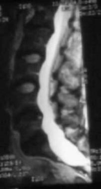
Sinus Thrombosis, Intracranial
Cranial Fossa, Middle
Sphenoid Bone
Tomography, X-Ray Computed
Carotid Artery, Common
Carotid Body
Abscess
Maxillary Sinus
Sella Turcica
Oculomotor Nerve Diseases
Thyroiditis
Carotid Artery, External
Endovascular arterial occlusion accomplished using microcoils deployed with and without proximal flow arrest: results in 19 patients. (1/104)
BACKGROUND AND PURPOSE: Prior to their relatively recent FDA approval, detachable balloons for endovascular arterial occlusion had been available on only a limited basis. We evaluated the feasibility of permanent endovascular carotid and vertebral artery occlusion using microcoils deployed with and without proximal flow arrest in 19 patients. METHODS: Permanent endovascular occlusion was performed in 19 arteries of 19 patients. The treated lesions included nine aneurysms, one carotid-cavernous fistula/pseudoaneurysm, seven neoplasms, and two dissections. Nondetachable balloons were used to arrest proximal blood flow during occlusion of only six arteries. Anticoagulation (heparin, 5000 U IV) was used during occlusion of 18 arteries. Three to 88 coils were used per lesion. Complex fibered platinum microcoils were used for all cases, and GDCs were also used in two patients. RESULTS: Sixteen patients had no new neurologic deficits after arterial occlusion. No patient had an acute event that suggested an embolic complication. Coils provided rapid and durable arterial occlusion in 17 patients. In both patients with acute carotid artery rupture, large numbers of coils placed during flow arrest failed to produce complete occlusion, which was accomplished subsequently with detachable balloons. One of these patients incurred a fatal hemispheric infarct after occlusion. One patient treated for a ruptured posterior inferior cerebellar artery aneurysm by vertebral artery occlusion continued to have progressive neurologic deficits. One patient with a cavernous aneurysm had upper extremity weakness and mild dysphasia 24 hours after internal carotid artery occlusion. CONCLUSION: In our small series, microcoils were found to be safe and effective for neurovascular occlusion. When both intravenous heparin (5000 U IV bolus) and heparinized catheter flush solutions (5000 U/L) are used, flow arrest during coil placement is unnecessary to prevent clinically apparent embolic complications. (+info)Management of a rare complication of endovascular treatment of direct carotid cavernous fistula. (2/104)
A 30-year-old woman with direct carotid cavernous fistula underwent endovascular treatment with detachable balloons via a transarterial route. The patient returned with diplopia 1 year after therapy. On cranial MR imaging, one of the balloons was detected in the proximal portion of the superior ophthalmic vein and was deflated percutaneously with a 22-gauge Chiba needle under CT guidance. The patient's symptoms resolved after balloon deflation. This case report presents a unique complication of endovascular treatment of direct carotid cavernous fistula and its management. (+info)Cavernous sinus and inferior petrosal sinus flow signal on three-dimensional time-of-flight MR angiography. (3/104)
BACKGROUND AND PURPOSE: Venous flow signal in the cavernous sinus and inferior petrosal sinus has been shown on MR angiograms in patients with carotid cavernous fistula (CCF). We, however, identified flow signal in some patients without symptoms and signs of CCF. This review was performed to determine the frequency of such normal venous flow depiction at MR angiography. METHODS: Twenty-five 3D time-of-flight (TOF) MR angiograms obtained on two different imaging units (scanners A and B) were reviewed with attention to presence of venous flow signal in the cavernous sinus or inferior petrosal sinus or both. Twenty-five additional MR angiograms were reviewed in patients who had also had cerebral arteriography to document absence of CCF where venous MR angiographic signal was detected, as well as to gain insight into venous flow patterns that might contribute to MR angiographic venous flow signal. Differences in scanning technique parameters were reviewed. RESULTS: Nine (36%) of the 25 MR angiograms obtained on scanner A but only one (4%) of the 25 obtained on scanner B showed flow signal in the cavernous or inferior petrosal sinus or both in the absence of signs of CCF. On review of 25 patients who had both MR angiography and arteriography, three patients with venous signal at MR angiography failed to exhibit CCF at arteriography. CONCLUSION: Identification of normal cavernous sinus or inferior petrosal sinus venous signal on 3D TOF MR angiograms may occur frequently, and is probably dependent on technical factors that vary among scanners. The exact factors most responsible, however, were not elucidated by this preliminary review. (+info)Direct carotid-cavernous sinus fistula due to ruptured intracavernous aneurysm treated with electrodetachable coils--case report. (4/104)
A 66-year-old female developed exophthalmos, impaired visual acuity (perception of light), and diplopia one day after sudden onset of headache. Neurological examination revealed proptosis, chemosis, impaired vision, and ophthalmoplegia. Carotid angiography showed direct carotid-cavernous sinus fistula concomitant with an intracavernous aneurysm on the right side. Intraaneurysmal embolization using the Guglielmi detachable coils (GDCs) via the transarterial route was performed and complete occlusion of the fistula successfully achieved. The neurological deficits resolved completely by 6 months after embolization. Intraaneurysmal GDC embolization via the transarterial route may be an alternative for the treatment of direct carotid-cavernous sinus fistula due to rupture of intracavernous aneurysm. (+info)Carotid and transcranial color-coded duplex sonography in different types of carotid-cavernous fistula. (5/104)
BACKGROUND AND PURPOSE: Patients with carotid-cavernous fistula (CCF) may undergo direct or indirect shunting. Ultrasonography has value that is complementary to angiography in the assessment and follow-up of these patients. The aim of this study was to characterize findings provided by carotid duplex sonography (CDS) and transcranial color-coded duplex sonography (TCCD) in patients with different types of CCF. METHODS: CDS and TCCD were independently performed by technologists and neurologists. Digital subtraction or MR angiography was interpreted by a neuroradiologist. Ultrasonographic studies were categorized into 4 types: I, direct shunting only; II, direct shunting with a carotid aneurysm; III, indirect shunting only; and IV, mixed (direct and indirect) shunting. In addition to carotid and intracranial flow velocities, volume, and pulsatility, other direct and indirect ultrasound signs of shunting were evaluated. The direct sign of CCF was a mosaic flash detected by TCCD. Alteration of hemodynamic parameters on CDS and demonstration of draining veins with the use of TCCD were considered indirect signs. RESULTS: Fifteen patients (8 men, 7 women) were included in the study. According to angiographic results, patients in ultrasonographic classification types I (n=7) and II (n=3) corresponded to type A of Barrow's classification. Patients with type III (n=8) were Barrow's type C. Type IV (n=1) had a combination of Barrow's types A and C. On ultrasound, both direct and indirect signs were seen in types I, II, and IV CCF. The presence of a 2-colored oval mass divided by a zone of separation without turbulence differentiated type I from type II CCF. All patients with type III CCF had indirect signs, and only 1 patient had direct signs on TCCD. Abnormal TCCD findings were most commonly seen through the transorbital window (100%), followed by the transtemporal window (63%) and transforaminal window (40%). CONCLUSIONS: If only indirect ultrasonographic signs of CCF are present, TCCD can be used to predict an indirect CCF type on the basis of the origin of the fistula. With direct communication between carotid artery and cavernous sinus, both direct and indirect ultrasonographic signs can be found. The combination of CDS/TCCD may provide a noninvasive and reliable way to classify patients with CCF. (+info)Transvenous embolization of carotid-cavernous sinus fistula associated with a primitive trigeminal artery--case report. (6/104)
A 58-year-old female presented with right conjunctival chemosis and right abducens nerve paresis. Cerebral angiography demonstrated a right carotid-cavernous sinus fistula associated with persistent primitive trigeminal artery. The fistula was treated by introducing detachable coils through the transvenous approach, as the detachable balloon was not available. Follow-up angiography performed 14 days after the embolization revealed complete disappearance of the carotid-cavernous sinus fistula due to thrombosis, which was presumably accelerated by the coils. Transvenous coil embolization should be considered as an alternative treatment for high-flow carotid-cavernous sinus fistula, but only if transarterial balloon embolization is not successful or unavailable. (+info)Extracranial carotid artery aneurysms: Texas Heart Institute experience. (7/104)
BACKGROUND AND PURPOSE: Aneurysms of the extracranial carotid artery (ECA) are rare. Large single-institution series are seldom reported and usually are not aneurysm type-specific. Thus, information about immediate and long-term results of surgical therapy is sparse. This review was conducted to elucidate etiology, presentation, and treatment for ECA aneurysms. METHODS: We retrospectively reviewed the case records of the Texas Heart Institute/St Luke's Episcopal Hospital, Houston, and found 67 cases of ECA aneurysms treated surgically (the largest series to date) between 1960 and 1995: 38 pseudoaneurysms after previous carotid surgery and 29 atherosclerotic or traumatic aneurysms. All aneurysms were surgically explored, and all were repaired except two: a traumatic distal internal carotid artery aneurysm and an infected pseudoaneurysm in which the carotid artery was ligated. RESULTS: Four deaths (three fatal strokes and one myocardial infarction) and two nonfatal strokes were directly attributed to a repaired ECA aneurysm (overall mortality/major stroke incidence, 9%); there was one minor stroke (incidence, 1.5%). The incidence of cranial nerve injury was 6% (four cases). During long-term follow-up (1.5 months-30 years; mean, 5.9 years), 19 patients died, mainly of cardiac causes (11 myocardial infarctions). CONCLUSION: The potential risks of cerebral ischemia and rupture as well as the satisfactory long-term results achieved with surgery strongly argue in favor of surgical treatment of ECA aneurysms. (+info)Carotid-cavernous fistulas: diagnosis with spiral CT angiography. (8/104)
Four cases in which the diagnosis of carotid-cavernous fistula was made by using CT angiography are illustrated. The diagnosis was confirmed by digital subtraction angiography in all four instances. To our knowledge, this is the first report of the CT angiographic appearance of carotid-cavernous fistulas. (+info)The cavernous sinus is a venous structure located in the middle cranial fossa, which is a depression in the skull that houses several important nerves and blood vessels. The cavernous sinus is situated on either side of the sphenoid bone, near the base of the skull, and it contains several important structures:
* The internal carotid artery, which supplies oxygenated blood to the brain
* The abducens nerve (cranial nerve VI), which controls lateral movement of the eye
* The oculomotor nerve (cranial nerve III), which controls most of the muscles that move the eye
* The trochlear nerve (cranial nerve IV), which controls one of the muscles that moves the eye
* The ophthalmic and maxillary divisions of the trigeminal nerve (cranial nerve V), which transmit sensory information from the face and head
The cavernous sinus is an important structure because it serves as a conduit for several critical nerves and blood vessels. However, it is also vulnerable to various pathological conditions such as thrombosis (blood clots), infection, tumors, or aneurysms, which can lead to serious neurological deficits or even death.
A Carotid-Cavernous Sinus Fistula (CCSF) is an abnormal connection between the carotid artery and the cavernous sinus, a venous structure in the skull. This connection can be either direct or indirect. Direct CCSFs are caused by trauma or rupture of an aneurysm, while indirect CCSFs are usually spontaneous and associated with conditions such as hypertension, atherosclerosis, or connective tissue disorders.
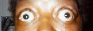
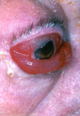
Symptoms of a CCSF may include headache, eye redness, protrusion of the eyeball, double vision, hearing disturbances, and pulsatile tinnitus (a rhythmic sound in the ear). The severity of symptoms can vary depending on the size of the fistula and the pressure within the cavernous sinus.
Treatment options for CCSF include endovascular repair with stenting or coiling, surgical closure, or observation, depending on the type and size of the fistula and the presence of symptoms.
An arteriovenous fistula is an abnormal connection or passageway between an artery and a vein. This connection causes blood to flow directly from the artery into the vein, bypassing the capillary network that would normally distribute the oxygen-rich blood to the surrounding tissues.
Arteriovenous fistulas can occur as a result of trauma, disease, or as a planned surgical procedure for patients who require hemodialysis, a treatment for advanced kidney failure. In hemodialysis, the arteriovenous fistula serves as a site for repeated access to the bloodstream, allowing for efficient removal of waste products and excess fluids.
The medical definition of an arteriovenous fistula is:
"An abnormal communication between an artery and a vein, usually created by surgical means for hemodialysis access or occurring as a result of trauma, congenital defects, or disease processes such as vasculitis or neoplasm."
Cavernous sinus thrombosis is a medical condition that refers to the formation of a blood clot (thrombus) in the cavernous sinuses, which are located near the base of the brain and are important for draining blood from the face and brain. This condition can occur as a complication of an infection in the facial area or sinuses, or it can be associated with other medical conditions such as cancer or trauma.
Symptoms of cavernous sinus thrombosis may include headache, fever, eye pain, swelling or bulging of the eyes, double vision, and decreased vision. If left untreated, this condition can lead to serious complications such as meningitis, brain abscess, or even death. Treatment typically involves administering antibiotics to treat any underlying infection and anticoagulants to prevent further clot formation. In some cases, surgery may be necessary to remove the clot.
A fistula is an abnormal connection or passage between two organs, vessels, or body parts that usually do not connect. It can form as a result of injury, infection, surgery, or disease. A fistula can occur anywhere in the body but commonly forms in the digestive system, genital area, or urinary system. The symptoms and treatment options for a fistula depend on its location and underlying cause.
A respiratory tract fistula is an abnormal connection or passage between the respiratory tract (which includes the nose, throat, windpipe, and lungs) and another organ or structure, such as the skin, digestive tract, or blood vessels. This condition can lead to complications such as air leakage, infection, and difficulty breathing. The causes of respiratory tract fistulas vary and can include trauma, surgery, infection, or cancer. Treatment depends on the location and severity of the fistula and may involve surgical repair, antibiotics, or other therapies.
The pyriform (or piriform) sinus refers to a pair of narrow, funnel-shaped spaces located at the base of the tongue, near the epiglottis, in the upper part of the larynx. These sinuses are lined with respiratory epithelium and are part of the digestive tract, as they connect to the esophagus through the upper esophageal sphincter. The pyriform sinuses play a role in the initial stages of swallowing, directing food and liquids into the esophagus. They can also serve as a potential site for the entrapment and growth of foreign bodies or abnormal tissue, such as in the case of a pyriform sinus fistula or diverticulum.
Suppurative thyroiditis is a rare type of thyroid gland inflammation that is caused by a bacterial infection. It is characterized by the formation of pus (suppuration) within the thyroid tissue. The infection can result from a direct spread of bacteria from adjacent structures, such as the upper respiratory tract or neck, or through the bloodstream due to an underlying infection elsewhere in the body.
Suppurative thyroiditis primarily affects people with pre-existing conditions that weaken the immune system, making them more susceptible to bacterial infections. These conditions may include diabetes, HIV/AIDS, or alcoholism. Additionally, it can occur in individuals who have recently undergone surgical procedures on the thyroid gland or after a traumatic injury to the area.
Symptoms of suppurative thyroiditis include fever, chills, painful swallowing, neck pain and swelling, difficulty breathing, hoarseness, and symptoms related to bacteremia (bacterial infection in the blood) if the infection spreads. Diagnosis typically involves a combination of clinical evaluation, imaging studies like ultrasound or CT scan, and laboratory tests to identify the causative organism. Treatment usually consists of antibiotics to eliminate the bacterial infection and possible surgical drainage of the infected thyroid tissue in severe cases.
Carotid artery diseases refer to conditions that affect the carotid arteries, which are the major blood vessels that supply oxygen-rich blood to the head and neck. The most common type of carotid artery disease is atherosclerosis, which occurs when fatty deposits called plaques build up in the inner lining of the arteries.
These plaques can cause the arteries to narrow or become blocked, reducing blood flow to the brain and increasing the risk of stroke. Other carotid artery diseases include carotid artery dissection, which occurs when there is a tear in the inner lining of the artery, and fibromuscular dysplasia, which is a condition that affects the muscle and tissue in the walls of the artery.
Symptoms of carotid artery disease may include neck pain or pulsations, transient ischemic attacks (TIAs) or "mini-strokes," and strokes. Treatment options for carotid artery disease depend on the severity and type of the condition but may include lifestyle changes, medications, endarterectomy (a surgical procedure to remove plaque from the artery), or angioplasty and stenting (procedures to open blocked arteries using a balloon and stent).
The carotid arteries are a pair of vital blood vessels in the human body that supply oxygenated blood to the head and neck. Each person has two common carotid arteries, one on each side of the neck, which branch off from the aorta, the largest artery in the body.
The right common carotid artery originates from the brachiocephalic trunk, while the left common carotid artery arises directly from the aortic arch. As they ascend through the neck, they split into two main branches: the internal and external carotid arteries.
The internal carotid artery supplies oxygenated blood to the brain, eyes, and other structures within the skull, while the external carotid artery provides blood to the face, scalp, and various regions of the neck.
Maintaining healthy carotid arteries is crucial for overall cardiovascular health and preventing serious conditions like stroke, which can occur when the arteries become narrowed or blocked due to the buildup of plaque or fatty deposits (atherosclerosis). Regular check-ups with healthcare professionals may include monitoring carotid artery health through ultrasound or other imaging techniques.
The internal carotid artery is a major blood vessel that supplies oxygenated blood to the brain. It originates from the common carotid artery and passes through the neck, entering the skull via the carotid canal in the temporal bone. Once inside the skull, it branches into several smaller vessels that supply different parts of the brain with blood.
The internal carotid artery is divided into several segments: cervical, petrous, cavernous, clinoid, and supraclinoid. Each segment has distinct clinical significance in terms of potential injury or disease. The most common conditions affecting the internal carotid artery include atherosclerosis, which can lead to stroke or transient ischemic attack (TIA), and dissection, which can cause severe headache, neck pain, and neurological symptoms.
It's important to note that any blockage or damage to the internal carotid artery can have serious consequences, as it can significantly reduce blood flow to the brain and lead to permanent neurological damage or even death. Therefore, regular check-ups and screening tests are recommended for individuals at high risk of developing vascular diseases.
Dura Mater is the thickest and outermost of the three membranes (meninges) that cover the brain and spinal cord. It provides protection and support to these delicate structures. The other two layers are called the Arachnoid Mater and the Pia Mater, which are thinner and more delicate than the Dura Mater. Together, these three layers form a protective barrier around the central nervous system.
Therapeutic embolization is a medical procedure that involves intentionally blocking or obstructing blood vessels to stop excessive bleeding or block the flow of blood to a tumor or abnormal tissue. This is typically accomplished by injecting small particles, such as microspheres or coils, into the targeted blood vessel through a catheter, which is inserted into a larger blood vessel and guided to the desired location using imaging techniques like X-ray or CT scanning. The goal of therapeutic embolization is to reduce the size of a tumor, control bleeding, or block off abnormal blood vessels that are causing problems.
Carotid stenosis is a medical condition that refers to the narrowing or constriction of the lumen (inner space) of the carotid artery. The carotid arteries are major blood vessels that supply oxygenated blood to the head and neck. Carotid stenosis usually results from the buildup of plaque, made up of fat, cholesterol, calcium, and other substances, on the inner walls of the artery. This process is called atherosclerosis.
As the plaque accumulates, it causes the artery to narrow, reducing blood flow to the brain. Severe carotid stenosis can increase the risk of stroke, as a clot or debris from the plaque can break off and travel to the brain, blocking a smaller blood vessel and causing tissue damage or death.
Carotid stenosis is typically diagnosed through imaging tests such as ultrasound, CT angiography, or MRI angiography. Treatment options may include lifestyle modifications (such as quitting smoking, controlling blood pressure, and managing cholesterol levels), medications to reduce the risk of clots, or surgical procedures like endarterectomy or stenting to remove or bypass the blockage.
The sphenoid sinuses are air-filled spaces located within the sphenoid bone, which is one of the bones that make up the skull base. These sinuses are located deep inside the skull, behind the eyes and nasal cavity. They are paired and separated by a thin bony septum, and each one opens into the corresponding nasal cavity through a small opening called the sphenoethmoidal recess. The sphenoid sinuses vary greatly in size and shape between individuals. They develop during childhood and continue to grow until early adulthood. The function of the sphenoid sinuses, like other paranasal sinuses, is not entirely clear, but they may contribute to reducing the weight of the skull, resonating voice during speech, and insulating the brain from trauma.
An intestinal fistula is an abnormal communication or connection between the intestines (or a portion of the intestine) and another organ or the skin surface. This connection forms a tract or passage, allowing the contents of the intestines, such as digestive enzymes, bacteria, and waste materials, to leak into other body areas or outside the body. Intestinal fistulas can develop due to various reasons, including inflammatory bowel diseases (like Crohn's disease), infections, complications from surgery, radiation therapy, or trauma. They can cause symptoms such as abdominal pain, diarrhea, skin irritation, and infection. Treatment of intestinal fistulas often involves a combination of medical management, nutritional support, and surgical intervention.
The coronary sinus is a large vein that receives blood from the heart's muscle tissue. It is located on the posterior side of the heart and is a part of the cardiovascular system. The coronary sinus collects oxygen-depleted blood from the myocardium (the heart muscle) and drains it into the right atrium, where it will then be pumped to the lungs for oxygenation.
The coronary sinus is an essential structure in medical procedures such as cardiac catheterization and electrophysiological studies. It is also a common site for the implantation of pacemakers and other cardiac devices.
Cranial sinuses are a part of the venous system in the human head. They are air-filled spaces located within the skull and are named according to their location. The cranial sinuses include:
1. Superior sagittal sinus: It runs along the top of the brain, inside the skull, and drains blood from the scalp and the veins of the brain.
2. Inferior sagittal sinus: It runs along the bottom of the brain and drains into the straight sinus.
3. Straight sinus: It is located at the back of the brain and receives blood from the inferior sagittal sinus and great cerebral vein.
4. Occipital sinuses: They are located at the back of the head and drain blood from the scalp and skull.
5. Cavernous sinuses: They are located on each side of the brain, near the temple, and receive blood from the eye and surrounding areas.
6. Sphenoparietal sinus: It is a small sinus that drains blood from the front part of the brain into the cavernous sinus.
7. Petrosquamosal sinuses: They are located near the ear and drain blood from the scalp and skull.
The cranial sinuses play an essential role in draining blood from the brain and protecting it from injury.
A cutaneous fistula is a type of fistula that occurs when a tract or tunnel forms between the skin (cutaneous) and another organ or structure, such as the gastrointestinal tract, vagina, or urinary system. Cutaneous fistulas can result from various medical conditions, including infections, inflammatory diseases, surgical complications, trauma, or malignancies.
Cutaneous fistulas may present with symptoms such as drainage of fluid or pus from the skin, pain, redness, swelling, or irritation around the affected area. The treatment for cutaneous fistulas depends on their underlying cause and can range from conservative management with antibiotics and wound care to surgical intervention.
It is essential to seek medical attention if you suspect a cutaneous fistula, as untreated fistulas can lead to complications such as infection, sepsis, or tissue damage. A healthcare professional can provide an accurate diagnosis and develop an appropriate treatment plan based on the individual's needs.
Cerebral angiography is a medical procedure that involves taking X-ray images of the blood vessels in the brain after injecting a contrast dye into them. This procedure helps doctors to diagnose and treat various conditions affecting the blood vessels in the brain, such as aneurysms, arteriovenous malformations, and stenosis (narrowing of the blood vessels).
During the procedure, a catheter is inserted into an artery in the leg and threaded through the body to the blood vessels in the neck or brain. The contrast dye is then injected through the catheter, and X-ray images are taken to visualize the blood flow through the brain's blood vessels.
Cerebral angiography provides detailed images of the blood vessels in the brain, allowing doctors to identify any abnormalities or blockages that may be causing symptoms or increasing the risk of stroke. Based on the results of the cerebral angiography, doctors can develop a treatment plan to address these issues and prevent further complications.
Central nervous system (CNS) vascular malformations are abnormal tangles or masses of blood vessels in the brain or spinal cord. These malformations can be congenital (present at birth) or acquired (develop later in life). They can vary in size, location, and symptoms, which may include headaches, seizures, weakness, numbness, difficulty speaking or understanding speech, and vision problems.
There are several types of CNS vascular malformations, including:
1. Arteriovenous malformations (AVMs): These are tangles of arteries and veins with a direct connection between them, bypassing the capillary network. AVMs can cause bleeding in the brain or spinal cord, leading to stroke or neurological deficits.
2. Cavernous malformations: These are clusters of dilated, thin-walled blood vessels that form a sac-like structure. They can rupture and bleed, causing symptoms such as seizures, headaches, or neurological deficits.
3. Developmental venous anomalies (DVAs): These are benign vascular malformations characterized by an abnormal pattern of veins that drain blood from the brain. DVAs are usually asymptomatic but can be associated with other vascular malformations.
4. Capillary telangiectasias: These are small clusters of dilated capillaries in the brain or spinal cord. They are usually asymptomatic and found incidentally during imaging studies.
5. Moyamoya disease: This is a rare, progressive cerebrovascular disorder characterized by the narrowing or blockage of the internal carotid arteries and their branches. This can lead to decreased blood flow to the brain, causing symptoms such as headaches, seizures, and strokes.
The diagnosis of CNS vascular malformations typically involves imaging studies such as MRI or CT scans, and sometimes angiography. Treatment options may include observation, medication, surgery, or endovascular procedures, depending on the type, location, and severity of the malformation.
The carotid sinus is a small, dilated area located at the bifurcation (or fork) of the common carotid artery into the internal and external carotid arteries. It is a baroreceptor region, which means it contains specialized sensory nerve endings that can detect changes in blood pressure. When the blood pressure increases, the walls of the carotid sinus stretch, activating these nerve endings and sending signals to the brain. The brain then responds by reducing the heart rate and relaxing the blood vessels, which helps to lower the blood pressure back to normal.
The carotid sinus is an important part of the body's autonomic nervous system, which regulates various involuntary functions such as heart rate, blood pressure, and digestion. It plays a crucial role in maintaining cardiovascular homeostasis and preventing excessive increases in blood pressure that could potentially damage vital organs.
A bronchial fistula is an abnormal connection or passage between the bronchial tree (the airways in the lungs) and the surrounding tissues, such as the pleural space (the space between the lungs and the chest wall), blood vessels, or other organs. This condition can result from various causes, including lung injury, infection, surgery, or certain diseases such as cancer or tuberculosis.
Bronchial fistulas can lead to symptoms like coughing, wheezing, shortness of breath, and chest pain. They may also cause air leaks, pneumothorax (collapsed lung), or chronic infections. Treatment for bronchial fistulas depends on the underlying cause and severity of the condition but often involves surgical repair or closure of the abnormal connection.
A vascular fistula is an abnormal connection or passage between the artery and vein, which usually results from a surgical procedure to create access for hemodialysis in patients with chronic kidney disease. This communication allows blood to flow directly from the artery into the vein, bypassing the capillary network and causing high-flow conditions in the affected area. Over time, the increased pressure and flow can lead to various complications such as venous hypertension, stenosis, aneurysm formation, or even heart failure if left untreated. Vascular fistulas may also occur spontaneously due to certain medical conditions like vasculitis, trauma, or infection, although this is less common.
Barium sulfate is a medication that is commonly used as a contrast material in medical imaging procedures, such as X-rays and CT scans. It works by coating the inside of the digestive tract, making it visible on an X-ray or CT scan and allowing doctors to see detailed images of the stomach, intestines, and other parts of the digestive system.
Barium sulfate is a white, chalky powder that is mixed with water to create a thick, milky liquid. It is generally safe and does not cause significant side effects when used in medical imaging procedures. However, it should not be taken by individuals who have a known allergy to barium or who have certain digestive conditions, such as obstructions or perforations of the bowel.
It's important to note that while barium sulfate is an important tool for medical diagnosis, it is not a treatment for any medical condition and should only be used under the direction of a healthcare professional.
A rectal fistula is an abnormal connection or tunnel that develops between the rectum, which is the lower end of the colon, and another organ or the skin surface surrounding the anus. This condition often results from inflammation, infection, trauma, or surgery in the anal area. The fistula can cause symptoms such as pain, discharge, irritation, and swelling around the anus. In some cases, it may also lead to complications like abscesses or recurrent infections if not treated promptly and effectively. Treatment options typically include surgical intervention to close the fistula and promote healing of the affected tissues.
The abducens nerve, also known as the sixth cranial nerve, is responsible for controlling the lateral rectus muscle of the eye, which enables the eye to move outward. Abducens nerve diseases refer to conditions that affect this nerve and can result in various symptoms, primarily affecting eye movement.
Here are some medical definitions related to abducens nerve diseases:
1. Abducens Nerve Palsy: A condition characterized by weakness or paralysis of the abducens nerve, causing difficulty in moving the affected eye outward. This results in double vision (diplopia), especially when gazing towards the side of the weakened nerve. Abducens nerve palsy can be congenital, acquired, or caused by various factors such as trauma, tumors, aneurysms, infections, or diseases like diabetes and multiple sclerosis.
2. Sixth Nerve Palsy: Another term for abducens nerve palsy, referring to the weakness or paralysis of the sixth cranial nerve.
3. Internuclear Ophthalmoplegia (INO): A neurological condition affecting eye movement, often caused by a lesion in the medial longitudinal fasciculus (MLF), a bundle of nerve fibers that connects the abducens nucleus with the oculomotor nucleus. INO results in impaired adduction (inward movement) of the eye on the side of the lesion and nystagmus (involuntary eye movements) of the abducting eye on the opposite side when attempting to look towards the side of the lesion.
4. One-and-a-Half Syndrome: A rare neurological condition characterized by a combination of INO and internuclear ophthalmoplegia with horizontal gaze palsy on the same side, caused by damage to both the abducens nerve and the paramedian pontine reticular formation (PPRF). This results in limited or no ability to move the eyes towards the side of the lesion and impaired adduction of the eye on the opposite side.
5. Brainstem Encephalitis: Inflammation of the brainstem, which can affect the abducens nerve and other cranial nerves, leading to various neurological symptoms such as diplopia (double vision), ataxia (loss of balance and coordination), and facial weakness. Brainstem encephalitis can be caused by infectious agents, autoimmune disorders, or paraneoplastic syndromes.
6. Multiple Sclerosis (MS): An autoimmune disorder characterized by inflammation and demyelination of the central nervous system, including the brainstem and optic nerves. MS can cause various neurological symptoms, such as diplopia, nystagmus, and INO, due to damage to the abducens nerve and other cranial nerves.
7. Wernicke's Encephalopathy: A neurological disorder caused by thiamine (vitamin B1) deficiency, often seen in alcoholics or individuals with malnutrition. Wernicke's encephalopathy can affect the brainstem and cause various symptoms such as diplopia, ataxia, confusion, and oculomotor abnormalities.
8. Pontine Glioma: A rare type of brain tumor that arises from the glial cells in the pons (a part of the brainstem). Pontine gliomas can cause various neurological symptoms such as diplopia, facial weakness, and difficulty swallowing due to their location in the brainstem.
9. Brainstem Cavernous Malformation: A benign vascular lesion that arises from the small blood vessels in the brainstem. Brainstem cavernous malformations can cause various neurological symptoms such as diplopia, ataxia, and facial weakness due to their location in the brainstem.
10. Pituitary Adenoma: A benign tumor that arises from the pituitary gland, located at the base of the brain. Large pituitary adenomas can compress the optic nerves and cause various visual symptoms such as diplopia, visual field defects, and decreased vision.
11. Craniopharyngioma: A benign tumor that arises from the remnants of the Rathke's pouch, a structure that gives rise to the anterior pituitary gland. Craniopharyngiomas can cause various neurological and endocrine symptoms such as diplopia, visual field defects, headaches, and hormonal imbalances due to their location near the optic nerves and pituitary gland.
12. Meningioma: A benign tumor that arises from the meninges, the protective covering of the brain and spinal cord. Meningiomas can cause various neurological symptoms such as diplopia, headaches, and seizures depending on their location in the brain or spinal cord.
13. Chordoma: A rare type of malignant tumor that arises from the remnants of the notochord, a structure that gives rise to the spine during embryonic development. Chordomas can cause various neurological and endocrine symptoms such as diplopia, visual field defects, headaches, and hormonal imbalances due to their location near the brainstem and spinal cord.
14. Metastatic Brain Tumors: Malignant tumors that spread from other parts of the body to the brain. Metastatic brain tumors can cause various neurological symptoms such as diplopia, headaches, seizures, and cognitive impairment depending on their location in the brain.
15. Other Rare Brain Tumors: There are many other rare types of brain tumors that can cause diplopia or other neurological symptoms, including gliomas, ependymomas, pineal region tumors, and others. These tumors require specialized diagnosis and treatment by neuro-oncologists and neurosurgeons with expertise in these rare conditions.
In summary, diplopia can be caused by various brain tumors, including pituitary adenomas, meningiomas, chordomas, metastatic brain tumors, and other rare types of tumors. It is important to seek medical attention promptly if you experience diplopia or other neurological symptoms, as early diagnosis and treatment can improve outcomes and quality of life.
A gastric fistula is an abnormal connection or passage between the stomach and another organ or the skin surface. This condition can occur as a result of complications from surgery, injury, infection, or certain diseases such as cancer. Symptoms may include persistent drainage from the site of the fistula, pain, malnutrition, and infection. Treatment typically involves surgical repair of the fistula and management of any underlying conditions.
A urinary fistula is an abnormal connection or passage between the urinary tract and another organ or tissue, such as the bladder, ureter, or kidney, and the skin, vagina, or intestine. This condition can lead to urine leakage through the abnormal opening, causing discomfort, infection, and other complications if not treated promptly and effectively. Urinary fistulas can be caused by various factors, including surgery, injury, radiation therapy, inflammation, or cancer. The type and location of the fistula will determine the specific symptoms and treatment options.
An esophageal fistula is an abnormal connection or passage between the esophagus (the tube that carries food and liquids from the throat to the stomach) and another organ, such as the trachea (windpipe) or the skin. This condition can result from complications of certain medical conditions, including cancer, prolonged infection, or injury to the esophagus.
Esophageal fistulas can cause a variety of symptoms, including difficulty swallowing, coughing, chest pain, and fever. They can also lead to serious complications, such as pneumonia or sepsis, if left untreated. Treatment for an esophageal fistula typically involves surgical repair of the abnormal connection, along with management of any underlying conditions that may have contributed to its development.
Carotid endarterectomy is a surgical procedure to remove plaque buildup (atherosclerosis) from the carotid arteries, which are the major blood vessels that supply oxygen-rich blood to the brain. The surgery involves making an incision in the neck, opening the carotid artery, and removing the plaque from the inside of the artery wall. The goal of the procedure is to restore normal blood flow to the brain and reduce the risk of stroke caused by the narrowing or blockage of the carotid arteries.
Pharyngeal diseases refer to conditions that affect the pharynx, which is the part of the throat that lies behind the nasal cavity and mouth, and above the esophagus and larynx. The pharynx plays a crucial role in swallowing, speaking, and breathing. Pharyngeal diseases can cause symptoms such as sore throat, difficulty swallowing, pain during swallowing, swollen lymph nodes, and earaches.
Some common pharyngeal diseases include:
1. Pharyngitis: Inflammation of the pharynx, often caused by a viral or bacterial infection.
2. Tonsillitis: Inflammation of the tonsils, which are two masses of lymphoid tissue located on either side of the back of the throat.
3. Epiglottitis: Inflammation of the epiglottis, a flap of cartilage that covers the windpipe during swallowing to prevent food and liquids from entering the lungs.
4. Abscesses: A collection of pus in the pharynx caused by a bacterial infection.
5. Cancer: Malignant tumors that can develop in the pharynx, often caused by smoking or heavy alcohol use.
6. Dysphagia: Difficulty swallowing due to nerve damage, muscle weakness, or structural abnormalities in the pharynx.
7. Stridor: Noisy breathing caused by a narrowed or obstructed airway in the pharynx.
Treatment for pharyngeal diseases depends on the underlying cause and may include antibiotics, pain relievers, surgery, or radiation therapy.
Ophthalmoplegia is a medical term that refers to the paralysis or weakness of the eye muscles, which can result in double vision (diplopia) or difficulty moving the eyes. It can be caused by various conditions, including nerve damage, muscle disorders, or neurological diseases such as myasthenia gravis or multiple sclerosis. Ophthalmoplegia can affect one or more eye muscles and can be partial or complete. Depending on the underlying cause, ophthalmoplegia may be treatable with medications, surgery, or other interventions.
Cranial nerves are a set of twelve pairs of nerves that originate from the brainstem and skull, rather than the spinal cord. These nerves are responsible for transmitting sensory information (such as sight, smell, hearing, and taste) to the brain, as well as controlling various muscles in the head and neck (including those involved in chewing, swallowing, and eye movement). Each cranial nerve has a specific function and is named accordingly. For example, the optic nerve (cranial nerve II) transmits visual information from the eyes to the brain, while the vagus nerve (cranial nerve X) controls parasympathetic functions in the body such as heart rate and digestion.
A biliary fistula is an abnormal connection or passage between the biliary system (which includes the gallbladder, bile ducts, and liver) and another organ or structure, usually in the abdominal cavity. This connection allows bile, which is a digestive fluid produced by the liver, to leak out of its normal pathway and into other areas of the body.
Biliary fistulas can occur as a result of trauma, surgery, infection, or inflammation in the biliary system. Symptoms may include abdominal pain, fever, jaundice (yellowing of the skin and eyes), nausea, vomiting, and clay-colored stools. Treatment typically involves addressing the underlying cause of the fistula, such as draining an infection or repairing damaged tissue, and diverting bile flow away from the site of the leak. In some cases, surgery may be necessary to repair the fistula.
Intracranial sinus thrombosis is a medical condition characterized by the formation of a blood clot (thrombus) within the intracranial venous sinuses, which are responsible for draining blood from the brain. The condition can lead to various neurological symptoms and complications, such as increased intracranial pressure, headaches, seizures, visual disturbances, and altered consciousness. Intracranial sinus thrombosis may result from various factors, including hypercoagulable states, infections, trauma, and malignancies. Immediate medical attention is necessary for proper diagnosis and treatment to prevent potential long-term neurological damage or even death.
The middle cranial fossa is a depression or hollow in the skull that forms the upper and central portion of the cranial cavity. It is located between the anterior cranial fossa (which lies anteriorly) and the posterior cranial fossa (which lies posteriorly). The middle cranial fossa contains several important structures, including the temporal lobes of the brain, the pituitary gland, the optic chiasm, and the cavernous sinuses. It is also where many of the cranial nerves pass through on their way to the brain.
The middle cranial fossa can be further divided into two parts: the anterior and posterior fossae. The anterior fossa contains the optic chiasm and the pituitary gland, while the posterior fossa contains the temporal lobes of the brain and the cavernous sinuses.
The middle cranial fossa is formed by several bones of the skull, including the sphenoid bone, the temporal bone, and the parietal bone. The shape and size of the middle cranial fossa can vary from person to person, and abnormalities in its structure can be associated with various medical conditions, such as pituitary tumors or aneurysms.
Cerebral veins are the blood vessels that carry deoxygenated blood from the brain to the dural venous sinuses, which are located between the layers of tissue covering the brain. The largest cerebral vein is the superior sagittal sinus, which runs along the top of the brain. Other major cerebral veins include the straight sinus, transverse sinus, sigmoid sinus, and cavernous sinus. These veins receive blood from smaller veins called venules that drain the surface and deep structures of the brain. The cerebral veins play an important role in maintaining normal circulation and pressure within the brain.
The sphenoid bone is a complex, irregularly shaped bone located in the middle cranial fossa and forms part of the base of the skull. It articulates with several other bones, including the frontal, parietal, temporal, ethmoid, palatine, and zygomatic bones. The sphenoid bone has two main parts: the body and the wings.
The body of the sphenoid bone is roughly cuboid in shape and contains several important structures, such as the sella turcica, which houses the pituitary gland, and the sphenoid sinuses, which are air-filled cavities within the bone. The greater wings of the sphenoid bone extend laterally from the body and form part of the skull's lateral walls. They contain the superior orbital fissure, through which important nerves and blood vessels pass between the cranial cavity and the orbit of the eye.
The lesser wings of the sphenoid bone are thin, blade-like structures that extend anteriorly from the body and form part of the floor of the anterior cranial fossa. They contain the optic canal, which transmits the optic nerve and ophthalmic artery between the brain and the orbit of the eye.
Overall, the sphenoid bone plays a crucial role in protecting several important structures within the skull, including the pituitary gland, optic nerves, and ophthalmic arteries.
X-ray computed tomography (CT or CAT scan) is a medical imaging method that uses computer-processed combinations of many X-ray images taken from different angles to produce cross-sectional (tomographic) images (virtual "slices") of the body. These cross-sectional images can then be used to display detailed internal views of organs, bones, and soft tissues in the body.
The term "computed tomography" is used instead of "CT scan" or "CAT scan" because the machines take a series of X-ray measurements from different angles around the body and then use a computer to process these data to create detailed images of internal structures within the body.
CT scanning is a noninvasive, painless medical test that helps physicians diagnose and treat medical conditions. CT imaging provides detailed information about many types of tissue including lung, bone, soft tissue and blood vessels. CT examinations can be performed on every part of the body for a variety of reasons including diagnosis, surgical planning, and monitoring of therapeutic responses.
In computed tomography (CT), an X-ray source and detector rotate around the patient, measuring the X-ray attenuation at many different angles. A computer uses this data to construct a cross-sectional image by the process of reconstruction. This technique is called "tomography". The term "computed" refers to the use of a computer to reconstruct the images.
CT has become an important tool in medical imaging and diagnosis, allowing radiologists and other physicians to view detailed internal images of the body. It can help identify many different medical conditions including cancer, heart disease, lung nodules, liver tumors, and internal injuries from trauma. CT is also commonly used for guiding biopsies and other minimally invasive procedures.
In summary, X-ray computed tomography (CT or CAT scan) is a medical imaging technique that uses computer-processed combinations of many X-ray images taken from different angles to produce cross-sectional images of the body. It provides detailed internal views of organs, bones, and soft tissues in the body, allowing physicians to diagnose and treat medical conditions.
A pancreatic fistula is an abnormal connection or passage between the pancreas and another organ, often the digestive system. It usually occurs as a complication following trauma, surgery, or inflammation of the pancreas (such as pancreatitis). The pancreas secretes digestive enzymes, and when these enzymes escape the pancreas through a damaged or disrupted duct, they can cause irritation and inflammation in nearby tissues, leading to the formation of a fistula.
Pancreatic fistulas are typically characterized by the drainage of pancreatic fluid, which contains high levels of digestive enzymes, into other parts of the body. This can lead to various symptoms, including abdominal pain, swelling, fever, and malnutrition. Treatment may involve surgical repair of the fistula, as well as supportive care such as antibiotics, nutritional support, and drainage of any fluid collections.
The common carotid artery is a major blood vessel in the neck that supplies oxygenated blood to the head and neck. It originates from the brachiocephalic trunk or the aortic arch and divides into the internal and external carotid arteries at the level of the upper border of the thyroid cartilage. The common carotid artery is an important structure in the circulatory system, and any damage or blockage to it can have serious consequences, including stroke.
The carotid body is a small chemoreceptor organ located near the bifurcation of the common carotid artery into the internal and external carotid arteries. It plays a crucial role in the regulation of respiration, blood pressure, and pH balance by detecting changes in the chemical composition of the blood, particularly oxygen levels, carbon dioxide levels, and hydrogen ion concentration (pH).
The carotid body contains specialized nerve endings called glomus cells that are sensitive to changes in these chemical parameters. When there is a decrease in oxygen or an increase in carbon dioxide or hydrogen ions, the glomus cells release neurotransmitters such as acetylcholine and dopamine, which activate afferent nerve fibers leading to the brainstem's nucleus tractus solitarius. This information is then integrated with other physiological signals in the brainstem, resulting in appropriate adjustments in breathing rate, depth, and pattern, as well as changes in heart rate and blood vessel diameter to maintain homeostasis.
Dysfunction of the carotid body can lead to various disorders, such as hypertension, sleep apnea, and chronic lung disease. In some cases, overactivity of the carotid body may result in conditions like primary breathing pattern disorders or pseudohypoxia, where the body responds as if it is experiencing hypoxia despite normal oxygen levels.
A rectovaginal fistula is an abnormal connection or passage between the rectum (the lower end of the colon, leading to the anus) and the vagina. This type of fistula can result from various causes, such as childbirth injuries, surgery complications, Crohn's disease, radiation therapy, or infections. The condition may lead to symptoms like fecal matter passing through the vagina, recurrent vaginal infections, discomfort during sexual intercourse, and skin irritation around the vaginal area. Treatment options typically involve surgical repair of the fistula, depending on its size, location, and underlying cause.
An abscess is a localized collection of pus caused by an infection. It is typically characterized by inflammation, redness, warmth, pain, and swelling in the affected area. Abscesses can form in various parts of the body, including the skin, teeth, lungs, brain, and abdominal organs. They are usually treated with antibiotics to eliminate the infection and may require drainage if they are large or located in a critical area. If left untreated, an abscess can lead to serious complications such as sepsis or organ failure.
The maxillary sinuses, also known as the antrums of Highmore, are the largest of the four pairs of paranasal sinuses located in the maxilla bones. They are air-filled cavities that surround the nasolacrimal duct and are situated superior to the upper teeth and lateral to the nasal cavity. Each maxillary sinus is lined with a mucous membrane, which helps to warm, humidify, and filter the air we breathe. Inflammation or infection of the maxillary sinuses can result in conditions such as sinusitis, leading to symptoms like facial pain, headaches, and nasal congestion.
The Sella Turcica, also known as the Turkish saddle, is a depression or fossa in the sphenoid bone located at the base of the skull. It forms a housing for the pituitary gland, which is a small endocrine gland often referred to as the "master gland" because it controls other glands and makes several essential hormones. The Sella Turcica has a saddle-like shape, with its anterior and posterior clinoids forming the front and back of the saddle, respectively. This region is of significant interest in neuroimaging and clinical settings, as various conditions such as pituitary tumors or other abnormalities may affect the size, shape, and integrity of the Sella Turcica.
The oculomotor nerve, also known as the third cranial nerve (CN III), is responsible for controlling several important eye movements and functions. Oculomotor nerve diseases refer to conditions that affect this nerve and can lead to various symptoms related to eye movement and function. Here's a medical definition of oculomotor nerve diseases:
Oculomotor nerve diseases are a group of medical disorders characterized by the dysfunction or damage to the oculomotor nerve (CN III), resulting in impaired eye movements, abnormalities in pupillary response, and potential effects on eyelid position. These conditions can be congenital, acquired, or traumatic in nature and may lead to partial or complete paralysis of the nerve. Common oculomotor nerve diseases include oculomotor nerve palsy, third nerve ganglionopathies, and compressive oculomotor neuropathies caused by various pathologies such as aneurysms, tumors, or infections.
A vesicovaginal fistula is an abnormal opening or connection between the bladder and the vagina, resulting in the continuous involuntary discharge of urine into the vaginal vault. This condition most commonly occurs as a result of complications during childbirth, particularly in developing countries with limited access to medical care. It can also be caused by surgery, radiation therapy, infection, or injury.
The symptoms of vesicovaginal fistula include constant urinary leakage from the vagina, frequent urinary tract infections, and a foul odor. The condition can lead to social isolation, depression, and other psychological issues due to its impact on a woman's quality of life. Treatment typically involves surgical repair of the fistula, which can be complex and may require specialized medical care.
Thyroiditis is a general term that refers to inflammation of the thyroid gland. It can be caused by various factors such as infections, autoimmune disorders, or medications. Depending on the cause and severity, thyroiditis may lead to overproduction (hyperthyroidism) or underproduction (hypothyroidism) of thyroid hormones, or it can result in a temporary or permanent loss of thyroid function.
There are several types of thyroiditis, including:
1. Hashimoto's thyroiditis - an autoimmune disorder where the body attacks and damages the thyroid gland, leading to hypothyroidism.
2. Subacute granulomatous thyroiditis (De Quervain's thyroiditis) - often follows a viral infection and results in painful inflammation of the thyroid gland, causing hyperthyroidism followed by hypothyroidism.
3. Silent thyroiditis - an autoimmune disorder similar to Hashimoto's thyroiditis but without symptoms like pain or tenderness; it can cause temporary hyperthyroidism and later hypothyroidism.
4. Postpartum thyroiditis - occurs in women after childbirth, causing inflammation of the thyroid gland leading to hyperthyroidism followed by hypothyroidism.
5. Acute suppurative thyroiditis - a rare bacterial infection that causes painful swelling and redness of the thyroid gland, usually requiring antibiotics for treatment.
Symptoms of thyroiditis depend on whether it leads to hyperthyroidism or hypothyroidism. Hyperthyroidism symptoms include rapid heartbeat, weight loss, heat intolerance, anxiety, and tremors. Hypothyroidism symptoms include fatigue, weight gain, cold intolerance, constipation, dry skin, and depression. Treatment varies depending on the type of thyroiditis and its severity.
The external carotid artery is a major blood vessel in the neck that supplies oxygenated blood to the structures of the head and neck, excluding the brain. It originates from the common carotid artery at the level of the upper border of the thyroid cartilage, then divides into several branches that supply various regions of the head and neck, including the face, scalp, ears, and neck muscles.
The external carotid artery has eight branches:
1. Superior thyroid artery: Supplies blood to the thyroid gland, larynx, and surrounding muscles.
2. Ascending pharyngeal artery: Supplies blood to the pharynx, palate, and meninges of the brain.
3. Lingual artery: Supplies blood to the tongue and floor of the mouth.
4. Facial artery: Supplies blood to the face, nose, lips, and palate.
5. Occipital artery: Supplies blood to the scalp and muscles of the neck.
6. Posterior auricular artery: Supplies blood to the ear and surrounding muscles.
7. Maxillary artery: Supplies blood to the lower face, nasal cavity, palate, and meninges of the brain.
8. Superficial temporal artery: Supplies blood to the scalp, face, and temporomandibular joint.
The external carotid artery is an essential structure for maintaining adequate blood flow to the head and neck, and any damage or blockage can lead to serious medical conditions such as stroke or tissue necrosis.
A vaginal fistula is an abnormal opening or connection between the vagina and another organ, such as the bladder (resulting in a vesicovaginal fistula), the rectum (resulting in a rectovaginal fistula), or the colon (resulting in a colovaginal fistula). This condition can lead to various complications, including chronic urinary or fecal incontinence, infection, and difficulty with sexual intercourse.
Vaginal fistulas are often caused by obstetric trauma, such as prolonged labor, or may be the result of surgery, radiation therapy, injury, or infection. Symptoms can vary depending on the size and location of the fistula but typically include abnormal discharge, pain, and foul-smelling odor. Treatment usually involves surgical repair of the fistula, although smaller fistulas may sometimes heal on their own with proper care and management.
 Coma
Coma
Ehlers-Danlos syndromes
List of MeSH codes (C21)
Dural arteriovenous fistula
List of MeSH codes (C10)
Cavernous sinus
List of MeSH codes (C14)
Fistula
Chemosis
Carotid-cavernous fistula
Sinus (anatomy)
Cerebral angiography
Idiopathic orbital inflammatory disease
Orbital cellulitis
Sphenoidal emissary foramen
International Classification of Headache Disorders
Index of anatomy articles
Endoscopic endonasal surgery
List of diseases (C)
July 1927
![Hasuo K[au] - Search Results - PubMed](data:image/png;base64,iVBORw0KGgoAAAANSUhEUgAAABAAAAAQCAMAAAAoLQ9TAAAARVBMVEVHcEwoU45gYmYAUpQAUpRPYGVgYmZLXnJgYmYAUZUAUpRJXnIAUpQAUpRgYmYAUpRgYmZgYmZhYmYAUpQAUpQAUpRgYmaDiPJuAAAAFXRSTlMADOJ+6QewGO8/uTRqtH7GdFJ11p1bCL3TAAAAZUlEQVQYlV2PVw7AIAxDTeney7n/UcsoldX3E+VJOAboEi7MBpHWMs1ADlG8u7UYWauwyZFeRQVPOhG2o+aiwhByJxUx91Jxhje3iJSqGfHuLKI0+0TpXvY1twCOPlFh5pa/++MB0vIOBm+1zaoAAAAASUVORK5CYII=) Hasuo K[au] - Search Results - PubMed
Hasuo K[au] - Search Results - PubMed
Coma - Wikipedia
 Carotid-Cavernous Fistula: Practice Essentials, Prognosis
GMS | GMS Current Topics in Otorhinolaryngology - Head and Neck Surgery | Risks and complications in rhinoplasty
Table of Contents - March 01, 1980, 1 (2) | American Journal of Neuroradiology
Index by author - October 01, 1998, 19 (9) | American Journal of Neuroradiology
 Pathophysiology of carotid-cavernous fistulas in vascular Ehlers-Danlos syndrome: a retrospective cohort and comprehensive...
 Angioarchitectural Evolution of Clival Dural Arteriovenous Fistulas in Two Patients
 Indian Journal of Ophthalmology
 Surgical Neurology International
DeCS - Términos Nuevos
DeCS - Términos Nuevos
DeCS - Términos Nuevos
DeCS - New terms
DeCS - New terms
DeCS - Termos Novos
DeCS - Términos Nuevos
DeCS - New terms
DeCS - Termos Novos
DeCS - New terms
DeCS - New terms
 DeCS - Termos Novos
 Search | VHL CLAP/WR-PAHO/WHO
Arteriovenous Fistula | Profiles RNS
 Caroticocavernous fistula | Radiology Reference Article | Radiopaedia.org
 Utilization of the Neuron 6 French 0.053 inch inner luminal diameter guide catheter for treatment of cerebral vascular...
 Journal of the Korean Ophthalmological Society
 Third Cranial (Oculomotor) Nerve Disorders - Neurologic Disorders - MSD Manual Professional Edition
Characteristics and Surgical Repair Outcomes of Obstetric Fistula Patients Managedata Teaching Hospital in Zambia: A...
Artery37
- Direct CCFs are high-flow fistulas with a direct connection between the internal carotid artery (ICA) and the cavernous sinus. (medscape.com)
- The cavernous sinus is a network of venous channels traversed by the intracranial portion of the internal carotid artery. (medscape.com)
- The internal carotid artery gives rise to several intracavernous branches. (medscape.com)
- The external carotid artery provides several branches to the dura of the cavernous sinus and forms anastomoses with the branches of the internal carotid artery. (medscape.com)
- Type A fistulas consist of a direct connection between the intracavernous internal carotid artery and the cavernous sinus. (medscape.com)
- Type B fistulas consist of a dural shunt between intracavernous branches of the internal carotid artery and the cavernous sinus. (medscape.com)
- Type D fistulas are a combination of types B and C, with dural shunts between internal and external carotid artery branches and the cavernous sinus. (medscape.com)
- Indirect CCFs result from a dural branch rupture of the carotid artery caused by a genetic condition or a comorbidity such as hypertension. (medscape.com)
- There are reports on various other risks like rhinoliquorrhea, brain damage, fistulas between sinus-cavernosus and carotid artery, aneurysms and thrombosis of the cavernous sinus. (egms.de)
- In vEDS, anatomical and pathophysiological features of the intra-cavernous internal carotid artery make it prone to shunting in the cavernous sinus, due either to a spontaneous rupture or to a spontaneous dissection with pseudoaneurysm formation. (biomedcentral.com)
- In direct CCF, internal carotid artery (ICA) wall disruption allows high-pressure blood to move into the CS, resulting in short-circuiting of the ICA arterial blood into the venous system of the CS. (biomedcentral.com)
- However, it carries some risk of intraoperative arterial injuries, which is mainly attributed to direct iatrogenic rupture of the internal carotid artery (ICA). (surgicalneurologyint.com)
- A direct fistula is due to direct communication between the intracavernous internal carotid artery and the surrounding cavernous sinus. (radiopaedia.org)
- Results The Neuron 6 F 0.053 inch inner luminal diameter delivery catheter (Penumbra) was placed in a very distal location within the internal carotid artery, external carotid artery and venous system enabling successful endovascular treatment of the intracranial pathology with no related neurological complications. (bmj.com)
- The internal carotid artery (ICA) is the most medial structure and cranial nerves III, IV, and first and second branches of cranial nerve V are located in the lateral wall of the dura. (stanford.edu)
- 38 In otitis media, infection spreads via the sigmoid sinus and along the internal carotid artery plexus. (stanford.edu)
- Transorbital intracranial penetrating trauma with carotid artery injury: a multidisciplinary approach to management. (riaco.com)
- Selection of carotid artery stenting or endarterectomy based on magnetic resonance plaque imaging reduced periprocedural adverse events. (hyo-med.ac.jp)
- Silent coronary artery disease in Japanese patients undergoing carotid artery stenting. (hyo-med.ac.jp)
- Asymptomatic moderate carotid artery stenosis with intraplaque hemorrhage: onset of new ischemic stroke. (hyo-med.ac.jp)
- Carotid-cavernous fistulas (CCFs) are abnormal vascular shunts between the carotid artery and the cavernous sinus. (neurosurgery-blog.com)
- Carotid artery revascularization using the Walrus balloon guide catheter: safety and feasibility from a US multicenter experience. (umassmed.edu)
- Addition of common carotid intervention increases the risk of stroke and death after carotid artery stenting for asymptomatic patients. (umassmed.edu)
- A more technical page on this site, Internal Carotid Artery and Its Aneurysms , shows no shortage of various aneurysm forms and shapes,and this is just a small fraction of the overall aneurysm mix. (neuroangio.org)
- The blood does not collect within a preexisting space, but rather creates a space at the Fractures of Cranial Base In fractures of the cranial base, the internal carotid artery may be torn, producing an arteriovenous fistula within the cavernous sinus. (gov.gy)
- Kinking of the carotid artery is an inflection of a limited area of the internal carotid vessel caused by its stretching. (medic-journal.com)
- Carotid cavernous fistula is a pathological communication between the cavernous part of the internal carotid artery and the cavity of the cavernous sinus. (medic-journal.com)
- Irritation of one particular nerve - the oculomotor nerve- generates particular concern that an aneurysm of the internal carotid artery is enlarging and at-risk of rupture. (surgical-neurology.com)
- Aneurysms may develp on the internal carotid artery within the cavernous sinus. (surgical-neurology.com)
- Traumatic Carotid Cavernous Fistula (TCCF) refers to the rupture of the arterial wall or branches of the cavernous sinus segment of the internal carotid artery caused by trauma, resulting in abnormal arteriovenous communication between the internal carotid artery and the cavernous sinus. (heraldopenaccess.us)
- Therefore, it is often necessary to use arterial catheterization for selective angiography of the whole brain, in addition to contralateral internal and external carotid artery angiography, contralateral internal carotid artery and vertebral artery are also photographed when the ipsilateral carotid artery is compressed and the blood flow is temporarily blocked. (heraldopenaccess.us)
- Usually on the imaging of the ipsilateral internal carotid artery, there is only a mass of contrast medium in the cavernous sinus, and the filling of the distal cerebral vessels is poor, and the exact location of the fistula is difficult to determine. (heraldopenaccess.us)
- Vertebral arteriography is used to compress the ipsilateral carotid artery at the same time, so that the contrast medium can be seen retrograde from the posterior communicating branch through the cavernous fistula of the internal carotid artery. (heraldopenaccess.us)
- At the same time, the contralateral internal carotid artery angiography can also understand the integrity of the Willis ring and estimate the compensation of the cerebral artery, which is helpful to judge whether the blood flow of the ipsilateral internal carotid artery can be interrupted. (heraldopenaccess.us)
- Selective external carotid artery angiography can show that the branches of the internal carotid artery are anastomosed with the middle meningeal artery, the accessory meningeal artery and the ascending pharyngeal artery at the bottom of the cavernous sinus to form the external carotid artery. (heraldopenaccess.us)
- Surgical treatment is a traditional treatment, which can be removed through the neck or scalp incision and cut off the abnormal communication between the cavernous sinus and the internal carotid artery to prevent the occurrence of complications such as hemorrhage and cerebral embolism. (heraldopenaccess.us)
- Interventional therapy mainly includes occlusion of fistula with detachable balloon, coil, Onyx glue, Willis covered stent or internal carotid artery. (heraldopenaccess.us)
Aneurysm3
- We present a case of a patient who was found to have a growth hormone (GH)-secreting pituitary adenoma and a coexisting cavernous ICA aneurysm which was embedded within the tumor. (surgicalneurologyint.com)
- An A-V fistula usually leads to the formation of a dilated sac-like connection, arteriovenous aneurysm. (sdsu.edu)
- If the oculomotor nerve is affected in concert with the other nerves controlling the globe of the eye- the trochlear and abducens nerves- then the aneurysm is most likely to be located in the skull base (see below under Cavernous Sinus Syndromes. (surgical-neurology.com)
Thrombosis of the cavernous sinus1
- Septic thrombosis of the cavernous sinus often results in the development of acute meningitis. (gov.gy)
Aneurysms1
- He focuses on treating stroke, carotid stenosis, intracranial aneurysms, arteriovenous malformation, pituitary tumors and other skull base tumors. (uky.edu)
Stenosis4
- Carotid Stenosis" is a descriptor in the National Library of Medicine's controlled vocabulary thesaurus, MeSH (Medical Subject Headings) . (umassmed.edu)
- This graph shows the total number of publications written about "Carotid Stenosis" by people in this website by year, and whether "Carotid Stenosis" was a major or minor topic of these publications. (umassmed.edu)
- Below are the most recent publications written about "Carotid Stenosis" by people in Profiles. (umassmed.edu)
- Arous EJ, Judelson DR, Agrawal A, Dundamadappa SK, Crawford AS, Malka KT, Simons JP, Schanzer A. Computed tomography angiography-derived area stenosis calculations overestimate degree of carotid stenosis compared with North American Symptomatic Carotid Endarterectomy Trial-derived diameter stenosis calculations. (umassmed.edu)
Cranial3
- The cavernous sinuses are bony enclosures in the base of the skull that transmit the internal carotid arteries as they enter the cranial cavity to supplu blood to he brain. (surgical-neurology.com)
- Purpose To evaluate the efficacy of n-butyl-2-cyanoacrylate (Trufill n-BCA) versus ethylene vinyl alcohol copolymer (ONYX) for the embolization of cranial dural arteriovenous fistulas (DAVF). (bmj.com)
- Cranial dural arteriovenous fistulas (DAVF) are a protean group of lesions involving the meninges. (bmj.com)
Internal carotid2
- 1 29 ] One of the preoperative considerations for TSS is an exclusion of "kissing internal carotid arteries: (ICA), a rare anatomical variant and absolute contraindication for TSS. (surgicalneurologyint.com)
- The diagnosis of internal carotid cavernous fistula is mainly determined by neuroimaging examination, such as head CT scan, neck MRI and cerebral angiography. (heraldopenaccess.us)
Arterial4
- Dural CCFs are low-flow fistulas resulting from communications of cavernous arterial branches and the cavernous sinus. (medscape.com)
- These vessels branch to provide arterial blood to the nerves and dura of the cavernous sinus and the pituitary gland. (medscape.com)
- Carotid-cavernous fistula (CCF) is an abnormal communication between the high-pressure carotid arterial system and the low-pressure cavernous venous system (CS). (biomedcentral.com)
- Arterial blood rushes into the cavernous sinus, enlarging it and forcing retrograde blood flow into its venous tributaries, especially the ophthalmic veins. (gov.gy)
Caroticocavernous fistula2
- Type-D caroticocavernous fistula: the eye demonstrates proptosis, chemosis, and scleral edema. (medscape.com)
- CT angiography is the noninvasive imaging modality of choice for evaluation of suspected caroticocavernous fistula 9 . (radiopaedia.org)
Embolization1
- One patient developed a major stroke from venous sinus thrombosis after embolization. (bmj.com)
Cerebral2
- Rhino-orbito-cerebral mucormycosis is the commonest form of the disease which starts in the sinus and slowly spreads to nearby structures including the orbital apex and the brain. (rstmh.org)
- Transcranial Doppler ultrasonography in the assessment of cerebral circulation arrest: improving sensitivity by transcervical and transorbital carotid insonation and serial examinations. (scienceopen.com)
Spinal1
- Venous shunting occurs into the dural venous sinuses or directly into cortical or spinal veins. (bmj.com)
Pituitary gland1
- The CS is a paired structure on either side of the sella, pituitary gland, and sphenoid sinus. (stanford.edu)
Sphenoid sinus1
- A 37-year-old male presented with a traumatic CCF and basal skull fracture extending through the medial wall of the cavernous sinus and sphenoid sinus. (neurosurgery-blog.com)
Transverse sinus1
- T, transverse sinus. (gov.gy)
Endarterectomy2
Abnormal arteriovenous1
- The carotid-cavernous fistula (CCF) is a specific type of dural arteriovenous fistula characterized by abnormal arteriovenous shunting within the cavernous sinus. (medscape.com)
Sigmoid sinus1
- A posterior condylar emissary vein may also be present, passing through the condylar canal, connecting the sigmoid sinus with the suboccipital venous plexus. (gov.gy)
Superior opht2
- Correct identification of the dilated superior ophthalmic vein is key for the diagnosis of carotid-cavernous sinus fistulas. (missionforvisionusa.org)
- Jacobs SM, Arias EJ , Derdeyn CP, Couch SM, Custer PL. Carotid Cavernous Sinus Fistulas Without Superior Ophthalmic Vein Enlargement. (riaco.com)
Medial1
- 1. Maxillary sinus with the nasolacrimal duct just medial. (missionforvisionusa.org)
Trauma3
- Carotid-cavernous sinus fistula accompanying facial trauma. (nih.gov)
- [ 3 ] Karaman et al reported on a carotid-cavernous fistula secondary to blunt trauma after functional endoscopic sinus surgery. (medscape.com)
- Direct caroticocavernous fistulas are often secondary to trauma, and as such the demographics reflect the distribution of head trauma, most commonly seen in young male patients. (radiopaedia.org)
Revascularization1
- They are postulated to occur secondary to cavernous sinus thrombosis with revascularization and thus are similar to dural arteriovenous fistulas elsewhere. (radiopaedia.org)
Arteries3
- Blunt head injury can lead to shearing of intracavernous arteries, causing the development of a fistula. (medscape.com)
- Indirect fistulas are due to communication by multiple branches between the internal and/or external carotid arteries and the cavernous sinus. (radiopaedia.org)
- Narrowing or stricture of any part of the CAROTID ARTERIES, most often due to atherosclerotic plaque formation. (umassmed.edu)
Orbital2
- Infectious CST is typically a complication of a facial, orbital, odontogenic, or paranasal sinus infection. (stanford.edu)
- Denervation within V2 will confirm the need for emergent non-contrast orbital CT to evaluate for a break in the orbital floor and to discount a hemorrhage within the sinus cavities. (reviewofoptometry.com)
Spontaneous2
- Direct spontaneous carotid-cavernous fistula (sCCF) is reportedly pathognomonic of vEDS. (biomedcentral.com)
- It is therefore unsurprising that vEDS has been associated with the development of spontaneous direct carotid-cavernous fistula (sCCF). (biomedcentral.com)
Syndrome1
- In addition, there may be symptoms such as cavernous sinus and supraorbital fissure syndrome. (heraldopenaccess.us)
Patients4
- Direct sCCF is rare in the general population as most direct fistulas are posttraumatic but seems to be more common in vEDS patients and especially in females. (biomedcentral.com)
- Patients with sinus fractures in the periorbital region should not blow their nose in order to avoid additional emphysema due to acute pressure rise. (aofoundation.org)
- This operation has the advantages of simple operation, low cost and no space occupying effect, but it should be noted that for patients with small fistula and less blood theft, it is difficult for balloon to enter the fistula. (heraldopenaccess.us)
- Therefore, for patients whose clinical symptoms are relieved and aggravated again after operation, it is necessary to guard against the possibility of fistula recurrence. (heraldopenaccess.us)
Cavity1
- H returned to the practice and she was advised that the extraction has caused an ora-antral fistula which is a cavity between her mouth and sinus. (sotamsarl.com)
Branches1
- The most frequent are type C, with meningeal branches of the external carotid forming the fistula 3 . (radiopaedia.org)
Formation2
- Penetrating head injury can lead to fistula formation by direct laceration of intracavernous vessels. (medscape.com)
- Formation of carotid cavernous sinus fistula is seen with no traumatic cause. (healthcaretip.com)
Symptoms2
- Types B, C, and D tend to be lower-flow and lower-pressure fistulas with a slower progression of signs and symptoms. (medscape.com)
- In contrast, indirect caroticocavernous fistulas have a predilection for the postmenopausal female patient and the onset of symptoms is often insidious. (radiopaedia.org)
Indirect1
- In cases of indirect carotid-cavernous fistulas, there is moderate ocular congestion, mild proptosis, and ocular pulsation on aplanotonometry. (medscape.com)
Infection2
Subarachnoid1
- They are usually at low risk of causing subarachnoid haemorrhage when confined within the sinus. (surgical-neurology.com)
Vertebral1
- Because these venous channels are valveless, compression of the thorax, abdomen, or pelvis, as occurs during heavy coughing and straining, may force venous blood from these regions into the internal vertebral venous system and from it into the dural venous sinuses. (gov.gy)
Occur1
- cavernous sinus tuberculoma may occur in the absence of pulmonary findings. (stanford.edu)