Cerebral Arterial Diseases
Peripheral Arterial Disease
Peripheral Vascular Diseases
Intermittent Claudication
Ankle Brachial Index
Arterial Occlusive Diseases
Cerebral Infarction
Lower Extremity
Cerebral Palsy
Middle Cerebral Artery
Brachial Artery
Cerebral Angiography
Ischemia
Tibial Arteries
Infarction, Middle Cerebral Artery
Risk Factors
Malaria, Cerebral
Brain Ischemia
Brain
Walking
Vascular Diseases
Intracranial Arterial Diseases
Cerebral Hemorrhage
Prospective Studies
Thromboangiitis Obliterans
Popliteal Artery
Blood Flow Velocity
Follow-Up Studies
Cerebrovascular Disorders
Treatment Outcome
Severity of Illness Index
Arteriosclerosis Obliterans
Prevalence
Atherosclerosis
Ischemic Attack, Transient
Stroke
Magnetic Resonance Angiography
Arteriosclerosis
Risk Assessment
Exercise Test
Magnetic Resonance Imaging
Cardiovascular Diseases
Photoplethysmography
Predictive Value of Tests
Carotid Artery Diseases
Biological Markers
Constriction, Pathologic
Angioplasty, Balloon
Cross-Sectional Studies
Platelet Aggregation Inhibitors
Blood Gas Monitoring, Transcutaneous
Anterior Cerebral Artery
Ultrasonography, Doppler, Duplex
Angiography, Digital Subtraction
Cerebral Amyloid Angiopathy
Posterior Cerebral Artery
Ultrasonography, Doppler
Cerebral Ventricles
Case-Control Studies
Brain Edema
Tomography, X-Ray Computed
Aspirin
Subarachnoid Hemorrhage
Leg Ulcer
Spectroscopy, Near-Infrared
Carotid Arteries
Nebraska
Limb Salvage
Exercise Tolerance
Carotid Stenosis
Diabetic Foot
Odds Ratio
Retrospective Studies
Intracranial Embolism and Thrombosis
Perfusion Imaging
Cardiovascular Agents
Hydroxymethylglutaryl-CoA Reductase Inhibitors
Disease Models, Animal
Laser-Doppler Flowmetry
Hypertension
Brain Diseases
Iliac Artery
Reproducibility of Results
Foot
Intracranial Pressure
Ultrasonography
Prognosis
Intracranial Aneurysm
Cohort Studies
Foot Ulcer
Cerebral Revascularization
Cerebral arterial lesions resulting from inflammatory emboli. (1/277)
In order to study the effects of septic embolism on the brain, silicone rubber emboli of various types were injected into the carotid arteries of 35 dogs. Pathologic and angiographic studies were performed to assess the resultant arterial and parenchymal lesions. Pure silicone rubber emboli (14 dogs) produced occasional intra-arterial thrombosis but no arteritis. Sterile and bacterially contaminated emboli containing a lead-chromate pigment (similar to those used in previous studies of septic embolism) (11 dogs) and pure silicone rubber emboli with transversely oriented canals (10 dogs), after brief placement in a bacterial suspension, were associated with intense inflammatory arteritis. This was accompanied by focal meningitis, subarachnoid hemorrhage, thrombosis, and cerebritis of the underlying cortex. The findings resembled those found in mycotic aneurysm. Aneurysmal dilatation was observed in one postmortem angiogram. In previous models of mycotic aneurysm, the inflammation attributed to bacterial contamination was probably due to the lead-chromate pigment used. (+info)Epilepsy after two different neurosurgical approaches to the treatment of ruptured intracranial aneurysm. (2/277)
One-hundred-and-fifty-two patients who underwent surgery for intracranial aneurysm were studied to determine the incidence of postoperative epilepsy in relation to the site of the aneurysm and the type of surgical approach. The overall incidence of epilepsy was 22%. Of the 116 patients treated by the intracranial approach 27.5% developed epilepsy, in contrast with only 5% of the 36 patients who had carotid artery ligation in the neck. Epilepsy occurred most frequently (35%) with middle cerebral artery aneurysms, especially if moderate or severe operative trauma was sustained and there was postoperative dysphasia. (+info)Arterial spasm and recovery from subarachnoid haemorrhage. (3/277)
In a series of 120 cases of subarachnoid haemorrhage due to ruptured intracranial aneurysm the occurrence of preoperative arterial spasm was found to have no effect upon the clinical outcome. After surgery, generalised arterial spasm was found to lead to an increased probability of fatality, and to an increased probability of psychological impariment among the survivors. The occurrence of spasm only in the vessels immediately adjacent to the haemorrhage did not constitute a risk to survival. However, the presence of generalised or localised spasm led to an increased risk of neurological impairment. It is suggested that the mechanisms by which postoperative arterial spasm is responsible for fatalities and for neurological impairment are distinct. (+info)Upregulation of MAP1B and MAP2 in the rat brain after middle cerebral artery occlusion: effect of age. (4/277)
Although stroke in humans usually afflicts the elderly, most experimental studies on the nature of cerebral ischemia have used young animals. This is especially important when studying restorative processes that are age dependent. To explore the potential of older animals to initiate regenerative processes after cerebral ischemia, the authors studied the expression of the juvenile-specific cytoskeletal protein, microtubule-associated protein (MAP) 1B, and the adult-specific protein, MAP2, in male Sprague-Dawley rats at 3 months and 20 months of age. The levels of MAP1B and MAP2 transcripts and the corresponding proteins declined with increasing age in the hippocampus. In the cortex, the levels of the transcripts did not change significantly with age, but the morphologic features of immunostained fibers were clearly affected by age; that is, cortical MAP1B fibers became thicker, and MAP2 fibers, more diffuse, in aged rats. Focal cerebral ischemia, produced by reversible occlusion of the right middle cerebral artery, resulted in a large decrease in the expression of both MAP1B and MAP2 in the infarct core at the messenger ribonucleic acid and protein levels. However, at 1 week after the stroke, there was vigorous expression of MAP1B and its messenger ribonucleic acid, as well as MAP2 protein, in the border zone adjacent to the infarct of 3-month-old and 20 month-old male Sprague-Dawley rats. The upregulation of these key cytologic elements generally was diminished in aged rats compared with young animals, although the morphologic features of fibers in the infarct border zone were similar in both age groups. These results suggest that the regenerative potential of the aged rat brain appears to be competent, although attenuated, at least with respect to MAP1B and MAP2 expression up to 20 months of age. (+info)Transluminal angioplasty for middle cerebral artery stenosis in patients with acute ischemic stroke. (5/277)
BACKGROUND AND PURPOSE: Precutaneous transluminal angioplasty (PTA) is currently performed to treat supraaortic atherosclerotic lesions. Our purpose was to evaluate the safety and efficacy of PTA for middle cerebral artery (MCA) stenosis in patients with acute ischemic stroke. METHODS: We performed PTA with the use of a microballoon (2-2.5 mm in diameter and 10-13 mm in length) in 10 consecutive patients (mean age, 48 years) who met the following criteria: high-grade M1 stenosis (> 70%) and mild neurologic deficits (NIH stroke scale < 4) and/or recurrent transient ischemic attacks (TIAs) resistant to anticoagulation, or a large area of hypoperfusion in the MCA territory on brain perfusion SPECT scans. During follow-up, we administered antiplatelet agents and evaluated the status of restenosis by angiography (n = 2), brain perfusion SPECT (n = 4), and/or transcranial Doppler sonography (TCD) (n = 7). RESULTS: Stenotic arteries were successfully dilated in nine of 10 patients. Angioplasty failed in one patient because the balloon could not pass through the tortuous cavernous internal carotid artery. None of the patients experienced either peri- or postangioplasty complications. Residual stenosis was less than 50%, and clinical improvement, including elimination of TIAs in four patients who had suffered resistant TIAs, was observed in all patients; improvement of the cerebral perfusion was also noted in two patients with a large hypoperfusion area in the MCA territory. The average follow-up period was 11 months (range, 2 to 36 months). None experienced recurrent stroke during the follow-up period. TCD revealed decreased flow velocity of the MCA after angioplasty in seven patients. CONCLUSION: PTA of the proximal portion of the MCA seems to be a safe and effective therapeutic technique for the prevention of secondary ischemic stroke. (+info)Various patterns of perfusion-weighted MR imaging and MR angiographic findings in hyperacute ischemic stroke. (6/277)
BACKGROUND AND PURPOSE: Various clinical subtypes of patients presenting with sudden-onset ischemic stroke have been recognized, but classification of those types is not simple. We identified various patterns of perfusion-weighted MR imaging and MR angiographic findings in hyperacute ischemic stroke with relation to clinical outcomes. METHODS: Twelve patients with symptoms of acute ischemic stroke due to middle cerebral artery occlusion underwent perfusion-weighted MR imaging and MR angiography within 6 hours after the onset of symptoms. Perfusion-weighted imaging was performed with a conventional dynamic contrast-enhanced T2*-weighted sequence, and cerebral blood volume (CBV) maps were then created. CBV maps and MR angiographic findings were compared with 99mTc-HMPAO brain SPECT scans, short-term outcomes, and follow-up imaging findings. RESULTS: The combined CBV and MR angiographic findings were classified into three patterns: arterial occlusion and decreased CBV (n = 8), arterial occlusion and increased CBV (n = 2), and no arterial occlusion and normal CBV (n = 2). These three patterns were strongly related to SPECT findings, short-term outcomes, and follow-up imaging findings. Perfusion on SPECT decreased markedly in the affected regions in all patients with the first pattern, decreased slightly in the second pattern, and was normal in the third pattern. Symptoms were not significantly changed at 24 hours after onset in any of the patients with the first pattern, but resolved completely in all patients with the latter two patterns. Follow-up imaging showed large infarctions in all patients with the first pattern. Initially, no infarction was seen in the second pattern, but watershed infarction developed later in one of these patients. CONCLUSION: Hyperacute ischemic stroke may be differentiated into three imaging patterns with different clinical outcomes. The combined use of perfusion-weighted MR imaging and MR angiography may play a substantial role in guiding the choice of treatment of this disease. (+info)Carotid artery tandem lesions: frequency of angiographic detection and consequences for endarterectomy. (7/277)
BACKGROUND AND PURPOSE: Several prospective trials have shown that ischemic stroke can be prevented by performing an endarterectomy in patients with high-grade carotid stenosis. Our purpose was to ascertain the frequency of carotid artery tandem lesions and to determine whether their presence alters the surgeon's decision to perform an endarterectomy. METHODS: We retrospectively reviewed the cerebral angiograms obtained between January 1994 and June 1996 in 853 patients with carotid occlusive disease. Studies were analyzed for the presence of internal carotid artery (ICA) stenosis as well as for tandem lesions (defined as > or = 50% diameter stenosis) within the common carotid artery, carotid siphon, or proximal intracranial arteries. The frequency of intracranial saccular aneurysms was determined. RESULTS: Six hundred seventy-two of the 853 patients had a carotid bifurcation stenosis of 70% or greater or underwent an endarterectomy. Of these, a carotid siphon stenosis of 50% or greater was noted in 65 patients (9.7%) and was ipsilateral to an ICA stenosis in 37 patients (5.5%). A common carotid stenosis was present in 29 patients (4.3%), ipsilateral to an ICA stenosis in 14 patients (2.1%). A stenosis of 50% or greater within the proximal intracranial circulation was present in 28 patients (4.2%), ipsilateral to an ICA stenosis in 15 patients (2.2 %). Four patients had tandem stenoses at more than one site. Tandem stenoses in the siphon or intracranial segments were noted in 13.5% with a bifurcation stenosis and in 8.8% of those with no bifurcation stenosis. Endarterectomy was performed in 48 of the 66 patients with tandem stenotic lesions. CONCLUSION: The presence of a tandem lesion infrequently alters the surgeon's decision to perform an endarterectomy. However, the importance of detecting tandem stenoses cannot be underestimated, since they may have important implications for long-term medical management in symptomatic patients. (+info)Cerebral hemodynamics in relation to patterns of collateral flow. (8/277)
BACKGROUND AND PURPOSE: We sought to investigate the relation between collateral flow via different pathways and hemodynamic parameters measured by dynamic susceptibility contrast-enhanced MRI in patients with severe carotid artery disease. METHODS: Dynamic susceptibility contrast-enhanced MRI was performed in 66 patients and 33 control subjects. Patients had severe stenosis (>70%, n=12), unilateral occlusion (n=38), or bilateral occlusion (n=16) of the internal carotid artery (ICA). Cerebripetal flow and collateral flow via the circle of Willis were investigated with MR angiography. Collateral flow via the ophthalmic artery was investigated with transcranial Doppler sonography. RESULTS: Patients with ICA stenosis had well-preserved cerebral perfusion and were in general not dependent on collateral supply. Patients with unilateral ICA occlusion had impaired cerebral perfusion. However, appearance time, peak time, and mean transit time in white matter were less increased in patients with than in patients without collateral flow via the circle of Willis (P<0.05). Furthermore, patients with collateral flow via both anterior and posterior communicating arteries had less increased regional cerebral blood volume than patients with collateral flow via the posterior communicating artery only (P<0.05). Patients with bilateral ICA occlusion had severely compromised hemodynamic status despite recruitment of collateral supply. CONCLUSIONS: In patients with unilateral ICA occlusion, the pattern of collateral supply has significant influence on hemodynamic status. Collateral flow via the anterior communicating artery is a sign of well-preserved hemodynamic status, whereas no collateral flow via the circle of Willis or flow via only the posterior communicating artery is a sign of deteriorated cerebral perfusion. (+info)Cerebral arterial diseases refer to conditions that affect the blood vessels supplying the brain. These diseases can result in reduced blood flow, blockages, or bleeding in the brain. The most common cerebral arterial diseases include:
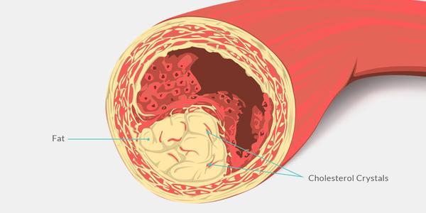
1. Atherosclerosis: A buildup of plaque made up of fat, cholesterol, and other substances in the inner lining of an artery, which can lead to narrowing or blockage of the artery.
2. Embolism: A blood clot or other particle that forms elsewhere in the body and travels to the brain, where it blocks a cerebral artery.
3. Thrombosis: The formation of a blood clot within a cerebral artery.
4. Aneurysm: A weakened area in the wall of an artery that bulges out and can rupture, causing bleeding in the brain.
5. Arteriovenous malformation (AVM): An abnormal tangle of blood vessels in the brain that can cause bleeding or reduced blood flow to surrounding tissue.
6. Vasculitis: Inflammation of the blood vessels in the brain, which can lead to narrowing, blockage, or weakening of the vessel walls.
These conditions can lead to serious complications such as stroke, transient ischemic attack (TIA), or vascular dementia. Treatment options include medications, surgery, and lifestyle changes to manage risk factors.
Peripheral Arterial Disease (PAD) is a medical condition characterized by the narrowing or blockage of arteries that supply blood to the extremities, most commonly the legs. This results in reduced blood flow, leading to symptoms such as leg pain, cramping, numbness, or weakness during physical activity, and in severe cases, tissue damage or gangrene. PAD is often indicative of widespread atherosclerosis, which is the hardening and narrowing of arteries due to the buildup of fatty deposits called plaques. It's important to note that early detection and management can help prevent serious complications.
Peripheral Vascular Diseases (PVD) refer to a group of medical conditions that affect the blood vessels outside of the heart and brain. These diseases are characterized by a narrowing or blockage of the peripheral arteries, which can lead to reduced blood flow to the limbs, particularly the legs.
The primary cause of PVD is atherosclerosis, a buildup of fats, cholesterol, and other substances in and on the walls of the arteries, forming plaques that restrict blood flow. Other risk factors include smoking, diabetes, hypertension, high cholesterol levels, and a family history of vascular disease.
Symptoms of PVD can vary depending on the severity of the condition but may include leg pain or cramping during exercise (claudication), numbness or tingling in the legs, coldness or discoloration of the feet, sores or wounds that heal slowly or not at all, and in severe cases, gangrene.
PVD can increase the risk of heart attack and stroke, so it is essential to diagnose and treat the condition as early as possible. Treatment options include lifestyle changes such as quitting smoking, exercising regularly, and maintaining a healthy diet, medications to control symptoms and reduce the risk of complications, and surgical procedures such as angioplasty or bypass surgery to restore blood flow.
Intermittent claudication is a medical condition characterized by pain or cramping in the legs, usually in the calf muscles, that occurs during exercise or walking and is relieved by rest. This symptom is caused by insufficient blood flow to the working muscles due to peripheral artery disease (PAD), a narrowing or blockage of the arteries in the limbs. As the individual walks, the muscle demands for oxygen and nutrients increase, but the restricted blood supply cannot meet these demands, leading to ischemia (lack of oxygen) and pain. The pain typically subsides after a few minutes of rest, as the muscle's demand for oxygen decreases, allowing the limited blood flow to compensate. Regular exercise and medications may help improve symptoms and reduce the risk of complications associated with PAD.
The Ankle-Brachial Index (ABI) is a medical test used to diagnose and evaluate peripheral artery disease (PAD), a condition characterized by narrowing or blockage of the blood vessels outside of the heart. The ABI measures the ratio of blood pressure in the ankles to the blood pressure in the arms, which can indicate whether there is reduced blood flow to the legs due to PAD.
To perform the test, healthcare professionals measure the blood pressure in both arms and ankles using a blood pressure cuff and a Doppler ultrasound device. The systolic blood pressure (the higher number) is used for the calculation. The ABI value is obtained by dividing the highest ankle pressure by the highest arm pressure.
In healthy individuals, the ABI values typically range from 0.9 to 1.3. Values below 0.9 suggest that there may be narrowed or blocked blood vessels in the legs, indicating PAD. The lower the ABI value, the more severe the blockage is likely to be. Additionally, an ABI of 1.4 or higher may indicate calcification of the arteries, which can also affect blood flow.
In summary, the Ankle-Brachial Index (ABI) is a medical test that measures the ratio of blood pressure in the ankles to the blood pressure in the arms, providing valuable information about peripheral artery disease and overall circulatory health.
Cerebral arteries refer to the blood vessels that supply oxygenated blood to the brain. These arteries branch off from the internal carotid arteries and the vertebral arteries, which combine to form the basilar artery. The major cerebral arteries include:
1. Anterior cerebral artery (ACA): This artery supplies blood to the frontal lobes of the brain, including the motor and sensory cortices responsible for movement and sensation in the lower limbs.
2. Middle cerebral artery (MCA): The MCA is the largest of the cerebral arteries and supplies blood to the lateral surface of the brain, including the temporal, parietal, and frontal lobes. It is responsible for providing blood to areas involved in motor function, sensory perception, speech, memory, and vision.
3. Posterior cerebral artery (PCA): The PCA supplies blood to the occipital lobe, which is responsible for visual processing, as well as parts of the temporal and parietal lobes.
4. Anterior communicating artery (ACoA) and posterior communicating arteries (PComAs): These are small arteries that connect the major cerebral arteries, forming an important circulatory network called the Circle of Willis. The ACoA connects the two ACAs, while the PComAs connect the ICA with the PCA and the basilar artery.
These cerebral arteries play a crucial role in maintaining proper brain function by delivering oxygenated blood to various regions of the brain. Any damage or obstruction to these arteries can lead to serious neurological conditions, such as strokes or transient ischemic attacks (TIAs).
The ankle, also known as the talocrural region, is the joint between the leg and the foot. It is a synovial hinge joint that allows for dorsiflexion and plantarflexion movements. The ankle is composed of three bones: the tibia and fibula of the lower leg, and the talus of the foot. The bottom portion of the tibia and fibula, called the malleoli, form a mortise that surrounds and articulates with the talus.
The ankle joint is strengthened by several ligaments, including the medial (deltoid) ligament and lateral ligament complex. The ankle also contains important nerves and blood vessels that provide sensation and circulation to the foot.
Damage to the ankle joint, such as sprains or fractures, can result in pain, swelling, and difficulty walking. Proper care and rehabilitation are essential for maintaining the health and function of the ankle joint.
Arterial occlusive diseases are medical conditions characterized by the blockage or narrowing of the arteries, which can lead to a reduction in blood flow to various parts of the body. This reduction in blood flow can cause tissue damage and may result in serious complications such as tissue death (gangrene), organ dysfunction, or even death.
The most common cause of arterial occlusive diseases is atherosclerosis, which is the buildup of plaque made up of fat, cholesterol, calcium, and other substances in the inner lining of the artery walls. Over time, this plaque can harden and narrow the arteries, restricting blood flow. Other causes of arterial occlusive diseases include blood clots, emboli (tiny particles that travel through the bloodstream and lodge in smaller vessels), inflammation, trauma, and certain inherited conditions.
Symptoms of arterial occlusive diseases depend on the location and severity of the blockage. Common symptoms include:
* Pain, cramping, or fatigue in the affected limb, often triggered by exercise and relieved by rest (claudication)
* Numbness, tingling, or weakness in the affected limb
* Coldness or discoloration of the skin in the affected area
* Slow-healing sores or wounds on the toes, feet, or legs
* Erectile dysfunction in men
Treatment for arterial occlusive diseases may include lifestyle changes such as quitting smoking, exercising regularly, and eating a healthy diet. Medications to lower cholesterol, control blood pressure, prevent blood clots, or manage pain may also be prescribed. In severe cases, surgical procedures such as angioplasty, stenting, or bypass surgery may be necessary to restore blood flow.
Cerebral infarction, also known as a "stroke" or "brain attack," is the sudden death of brain cells caused by the interruption of their blood supply. It is most commonly caused by a blockage in one of the blood vessels supplying the brain (an ischemic stroke), but can also result from a hemorrhage in or around the brain (a hemorrhagic stroke).
Ischemic strokes occur when a blood clot or other particle blocks a cerebral artery, cutting off blood flow to a part of the brain. The lack of oxygen and nutrients causes nearby brain cells to die. Hemorrhagic strokes occur when a weakened blood vessel ruptures, causing bleeding within or around the brain. This bleeding can put pressure on surrounding brain tissues, leading to cell death.
Symptoms of cerebral infarction depend on the location and extent of the affected brain tissue but may include sudden weakness or numbness in the face, arm, or leg; difficulty speaking or understanding speech; vision problems; loss of balance or coordination; and severe headache with no known cause. Immediate medical attention is crucial for proper diagnosis and treatment to minimize potential long-term damage or disability.
The term "lower extremity" is used in the medical field to refer to the portion of the human body that includes the structures below the hip joint. This includes the thigh, lower leg, ankle, and foot. The lower extremities are responsible for weight-bearing and locomotion, allowing individuals to stand, walk, run, and jump. They contain many important structures such as bones, muscles, tendons, ligaments, nerves, and blood vessels.
Cerebral palsy (CP) is a group of disorders that affect a person's ability to move and maintain balance and posture. According to the Mayo Clinic, CP is caused by abnormal brain development or damage to the developing brain that affects a child's ability to control movement.
The symptoms of cerebral palsy can vary in severity and may include:
* Spasticity (stiff or tight muscles)
* Rigidity (resistance to passive movement)
* Poor coordination and balance
* Weakness or paralysis
* Tremors or involuntary movements
* Abnormal gait or difficulty walking
* Difficulty with fine motor skills, such as writing or using utensils
* Speech and language difficulties
* Vision, hearing, or swallowing problems
It's important to note that cerebral palsy is not a progressive condition, meaning that it does not worsen over time. However, the symptoms may change over time, and some individuals with CP may experience additional medical conditions as they age.
Cerebral palsy is usually caused by brain damage that occurs before or during birth, but it can also be caused by brain injuries that occur in the first few years of life. Some possible causes of cerebral palsy include:
* Infections during pregnancy
* Lack of oxygen to the brain during delivery
* Traumatic head injury during birth
* Brain bleeding or stroke in the newborn period
* Genetic disorders
* Maternal illness or infection during pregnancy
There is no cure for cerebral palsy, but early intervention and treatment can help improve outcomes and quality of life. Treatment may include physical therapy, occupational therapy, speech therapy, medications to manage symptoms, surgery, and assistive devices such as braces or wheelchairs.
In medical terms, the leg refers to the lower portion of the human body that extends from the knee down to the foot. It includes the thigh (femur), lower leg (tibia and fibula), foot, and ankle. The leg is primarily responsible for supporting the body's weight and enabling movements such as standing, walking, running, and jumping.
The leg contains several important structures, including bones, muscles, tendons, ligaments, blood vessels, nerves, and joints. These structures work together to provide stability, support, and mobility to the lower extremity. Common medical conditions that can affect the leg include fractures, sprains, strains, infections, peripheral artery disease, and neurological disorders.
Cerebrovascular circulation refers to the network of blood vessels that supply oxygenated blood and nutrients to the brain tissue, and remove waste products. It includes the internal carotid arteries, vertebral arteries, circle of Willis, and the intracranial arteries that branch off from them.
The internal carotid arteries and vertebral arteries merge to form the circle of Willis, a polygonal network of vessels located at the base of the brain. The anterior cerebral artery, middle cerebral artery, posterior cerebral artery, and communicating arteries are the major vessels that branch off from the circle of Willis and supply blood to different regions of the brain.
Interruptions or abnormalities in the cerebrovascular circulation can lead to various neurological conditions such as stroke, transient ischemic attack (TIA), and vascular dementia.
The Middle Cerebral Artery (MCA) is one of the main blood vessels that supplies oxygenated blood to the brain. It arises from the internal carotid artery and divides into several branches, which supply the lateral surface of the cerebral hemisphere, including the frontal, parietal, and temporal lobes.
The MCA is responsible for providing blood flow to critical areas of the brain, such as the primary motor and sensory cortices, Broca's area (associated with speech production), Wernicke's area (associated with language comprehension), and the visual association cortex.
Damage to the MCA or its branches can result in a variety of neurological deficits, depending on the specific location and extent of the injury. These may include weakness or paralysis on one side of the body, sensory loss, language impairment, and visual field cuts.
The brachial artery is a major blood vessel in the upper arm. It supplies oxygenated blood to the muscles and tissues of the arm, forearm, and hand. The brachial artery originates from the axillary artery at the level of the shoulder joint and runs down the medial (inner) aspect of the arm, passing through the cubital fossa (the depression on the anterior side of the elbow) where it can be palpated during a routine blood pressure measurement. At the lower end of the forearm, the brachial artery bifurcates into the radial and ulnar arteries, which further divide into smaller vessels to supply the hand and fingers.
Cerebral angiography is a medical procedure that involves taking X-ray images of the blood vessels in the brain after injecting a contrast dye into them. This procedure helps doctors to diagnose and treat various conditions affecting the blood vessels in the brain, such as aneurysms, arteriovenous malformations, and stenosis (narrowing of the blood vessels).
During the procedure, a catheter is inserted into an artery in the leg and threaded through the body to the blood vessels in the neck or brain. The contrast dye is then injected through the catheter, and X-ray images are taken to visualize the blood flow through the brain's blood vessels.
Cerebral angiography provides detailed images of the blood vessels in the brain, allowing doctors to identify any abnormalities or blockages that may be causing symptoms or increasing the risk of stroke. Based on the results of the cerebral angiography, doctors can develop a treatment plan to address these issues and prevent further complications.
Ischemia is the medical term used to describe a lack of blood flow to a part of the body, often due to blocked or narrowed blood vessels. This can lead to a shortage of oxygen and nutrients in the tissues, which can cause them to become damaged or die. Ischemia can affect many different parts of the body, including the heart, brain, legs, and intestines. Symptoms of ischemia depend on the location and severity of the blockage, but they may include pain, cramping, numbness, weakness, or coldness in the affected area. In severe cases, ischemia can lead to tissue death (gangrene) or organ failure. Treatment for ischemia typically involves addressing the underlying cause of the blocked blood flow, such as through medication, surgery, or lifestyle changes.
The tibial arteries are three major arteries that supply blood to the lower leg and foot. They are branches of the popliteal artery, which is a continuation of the femoral artery. The three tibial arteries are:
1. Anterior tibial artery: This artery runs down the front of the leg and supplies blood to the muscles in the anterior compartment of the leg, as well as to the foot. It becomes the dorsalis pedis artery as it approaches the ankle.
2. Posterior tibial artery: This artery runs down the back of the leg and supplies blood to the muscles in the posterior compartment of the leg. It then branches into the fibular (peroneal) artery and the medial and lateral plantar arteries, which supply blood to the foot.
3. Fibular (peroneal) artery: This artery runs down the outside of the leg and supplies blood to the muscles in the lateral compartment of the leg. It also provides branches that anastomose with the anterior and posterior tibial arteries, forming a network of vessels that helps ensure adequate blood flow to the foot.
Together, these arteries play a critical role in providing oxygenated blood and nutrients to the lower leg and foot, helping to maintain their health and function.
Middle Cerebral Artery (MCA) infarction is a type of ischemic stroke that occurs when there is an obstruction in the blood supply to the middle cerebral artery, which is one of the major blood vessels that supplies oxygenated blood to the brain. The MCA supplies blood to a large portion of the brain, including the motor and sensory cortex, parts of the temporal and parietal lobes, and the basal ganglia.
An infarction is the death of tissue due to the lack of blood supply, which can lead to damage or loss of function in the affected areas of the brain. Symptoms of MCA infarction may include weakness or numbness on one side of the body, difficulty speaking or understanding speech, vision problems, and altered levels of consciousness.
MCA infarctions can be caused by various factors, including embolism (a blood clot that travels to the brain from another part of the body), thrombosis (a blood clot that forms in the MCA itself), or stenosis (narrowing of the artery due to atherosclerosis or other conditions). Treatment for MCA infarction may include medications to dissolve blood clots, surgery to remove the obstruction, or rehabilitation to help regain lost function.
Medical Definition:
"Risk factors" are any attribute, characteristic or exposure of an individual that increases the likelihood of developing a disease or injury. They can be divided into modifiable and non-modifiable risk factors. Modifiable risk factors are those that can be changed through lifestyle choices or medical treatment, while non-modifiable risk factors are inherent traits such as age, gender, or genetic predisposition. Examples of modifiable risk factors include smoking, alcohol consumption, physical inactivity, and unhealthy diet, while non-modifiable risk factors include age, sex, and family history. It is important to note that having a risk factor does not guarantee that a person will develop the disease, but rather indicates an increased susceptibility.
Cerebral malaria is a severe form of malaria that affects the brain. It is caused by Plasmodium falciparum parasites, which are transmitted to humans through the bites of infected Anopheles mosquitoes. In cerebral malaria, the parasites infect and destroy red blood cells, leading to their accumulation in small blood vessels in the brain. This can cause swelling of the brain, impaired consciousness, seizures, coma, and even death if left untreated.
The medical definition of cerebral malaria is:
A severe form of malaria caused by Plasmodium falciparum parasites that affects the brain and results in altered mental status, seizures, coma, or other neurological symptoms. It is characterized by the sequestration of infected red blood cells in the cerebral microvasculature, leading to inflammation, endothelial activation, and disruption of the blood-brain barrier. Cerebral malaria can cause long-term neurological deficits or death if not promptly diagnosed and treated with appropriate antimalarial therapy.
Brain ischemia is the medical term used to describe a reduction or interruption of blood flow to the brain, leading to a lack of oxygen and glucose delivery to brain tissue. This can result in brain damage or death of brain cells, known as infarction. Brain ischemia can be caused by various conditions such as thrombosis (blood clot formation), embolism (obstruction of a blood vessel by a foreign material), or hypoperfusion (reduced blood flow). The severity and duration of the ischemia determine the extent of brain damage. Symptoms can range from mild, such as transient ischemic attacks (TIAs or "mini-strokes"), to severe, including paralysis, speech difficulties, loss of consciousness, and even death. Immediate medical attention is required for proper diagnosis and treatment to prevent further damage and potential long-term complications.
The brain is the central organ of the nervous system, responsible for receiving and processing sensory information, regulating vital functions, and controlling behavior, movement, and cognition. It is divided into several distinct regions, each with specific functions:
1. Cerebrum: The largest part of the brain, responsible for higher cognitive functions such as thinking, learning, memory, language, and perception. It is divided into two hemispheres, each controlling the opposite side of the body.
2. Cerebellum: Located at the back of the brain, it is responsible for coordinating muscle movements, maintaining balance, and fine-tuning motor skills.
3. Brainstem: Connects the cerebrum and cerebellum to the spinal cord, controlling vital functions such as breathing, heart rate, and blood pressure. It also serves as a relay center for sensory information and motor commands between the brain and the rest of the body.
4. Diencephalon: A region that includes the thalamus (a major sensory relay station) and hypothalamus (regulates hormones, temperature, hunger, thirst, and sleep).
5. Limbic system: A group of structures involved in emotional processing, memory formation, and motivation, including the hippocampus, amygdala, and cingulate gyrus.
The brain is composed of billions of interconnected neurons that communicate through electrical and chemical signals. It is protected by the skull and surrounded by three layers of membranes called meninges, as well as cerebrospinal fluid that provides cushioning and nutrients.
Medical science often defines and describes "walking" as a form of locomotion or mobility where an individual repeatedly lifts and sets down each foot to move forward, usually bearing weight on both legs. It is a complex motor activity that requires the integration and coordination of various systems in the human body, including the musculoskeletal, neurological, and cardiovascular systems.
Walking involves several components such as balance, coordination, strength, and endurance. The ability to walk independently is often used as a measure of functional mobility and overall health status. However, it's important to note that the specific definition of walking may vary depending on the context and the medical or scientific field in question.
Cerebral veins are the blood vessels that carry deoxygenated blood from the brain to the dural venous sinuses, which are located between the layers of tissue covering the brain. The largest cerebral vein is the superior sagittal sinus, which runs along the top of the brain. Other major cerebral veins include the straight sinus, transverse sinus, sigmoid sinus, and cavernous sinus. These veins receive blood from smaller veins called venules that drain the surface and deep structures of the brain. The cerebral veins play an important role in maintaining normal circulation and pressure within the brain.
Amputation is defined as the surgical removal of all or part of a limb or extremity such as an arm, leg, foot, hand, toe, or finger. This procedure is typically performed to remove damaged or dead tissue due to various reasons like severe injury, infection, tumors, or chronic conditions that impair circulation, such as diabetes or peripheral arterial disease. The goal of amputation is to alleviate pain, prevent further complications, and improve the patient's quality of life. Following the surgery, patients may require rehabilitation and prosthetic devices to help them adapt to their new physical condition.
Vascular diseases are medical conditions that affect the circulatory system, specifically the blood vessels (arteries, veins, and capillaries). These diseases can include conditions such as:
1. Atherosclerosis: The buildup of fats, cholesterol, and other substances in and on the walls of the arteries, which can restrict blood flow.
2. Peripheral Artery Disease (PAD): A condition caused by atherosclerosis where there is narrowing or blockage of the peripheral arteries, most commonly in the legs. This can lead to pain, numbness, and cramping.
3. Coronary Artery Disease (CAD): Atherosclerosis of the coronary arteries that supply blood to the heart muscle. This can lead to chest pain, shortness of breath, or a heart attack.
4. Carotid Artery Disease: Atherosclerosis of the carotid arteries in the neck that supply blood to the brain. This can increase the risk of stroke.
5. Cerebrovascular Disease: Conditions that affect blood flow to the brain, including stroke and transient ischemic attack (TIA or "mini-stroke").
6. Aneurysm: A weakened area in the wall of a blood vessel that causes it to bulge outward and potentially rupture.
7. Deep Vein Thrombosis (DVT): A blood clot that forms in the deep veins, usually in the legs, which can cause pain, swelling, and increased risk of pulmonary embolism if the clot travels to the lungs.
8. Varicose Veins: Swollen, twisted, and often painful veins that have filled with an abnormal collection of blood, usually appearing in the legs.
9. Vasculitis: Inflammation of the blood vessels, which can cause damage and narrowing, leading to reduced blood flow.
10. Raynaud's Phenomenon: A condition where the small arteries that supply blood to the skin become narrowed, causing decreased blood flow, typically in response to cold temperatures or stress.
These are just a few examples of vascular conditions that fall under the umbrella term "cerebrovascular disease." Early diagnosis and treatment can significantly improve outcomes for many of these conditions.
Intracranial arterial diseases refer to conditions that affect the blood vessels within the brain. These diseases can include stenosis (narrowing) or occlusion (blockage) of the intracranial arteries, aneurysms (bulging or weakened areas in the artery wall), and vasculitis (inflammation of the blood vessel walls).
These conditions can lead to serious complications such as stroke, transient ischemic attack (TIA or "mini-stroke"), bleeding in the brain, and cognitive decline. Risk factors for intracranial arterial diseases include age, hypertension, diabetes, smoking, high cholesterol, and a history of heart disease.
Diagnosis of intracranial arterial diseases may involve imaging tests such as magnetic resonance angiography (MRA), computed tomographic angiography (CTA), or digital subtraction angiography (DSA). Treatment options may include medications to manage risk factors, endovascular procedures such as angioplasty and stenting, or surgical intervention in some cases.
The femoral artery is the major blood vessel that supplies oxygenated blood to the lower extremity of the human body. It is a continuation of the external iliac artery and becomes the popliteal artery as it passes through the adductor hiatus in the adductor magnus muscle of the thigh.
The femoral artery is located in the femoral triangle, which is bound by the sartorius muscle anteriorly, the adductor longus muscle medially, and the biceps femoris muscle posteriorly. It can be easily palpated in the groin region, making it a common site for taking blood samples, measuring blood pressure, and performing surgical procedures such as femoral artery catheterization and bypass grafting.
The femoral artery gives off several branches that supply blood to the lower limb, including the deep femoral artery, the superficial femoral artery, and the profunda femoris artery. These branches provide blood to the muscles, bones, skin, and other tissues of the leg, ankle, and foot.
Blood pressure is the force exerted by circulating blood on the walls of the blood vessels. It is measured in millimeters of mercury (mmHg) and is given as two figures:
1. Systolic pressure: This is the pressure when the heart pushes blood out into the arteries.
2. Diastolic pressure: This is the pressure when the heart rests between beats, allowing it to fill with blood.
Normal blood pressure for adults is typically around 120/80 mmHg, although this can vary slightly depending on age, sex, and other factors. High blood pressure (hypertension) is generally considered to be a reading of 130/80 mmHg or higher, while low blood pressure (hypotension) is usually defined as a reading below 90/60 mmHg. It's important to note that blood pressure can fluctuate throughout the day and may be affected by factors such as stress, physical activity, and medication use.
A cerebral hemorrhage, also known as an intracranial hemorrhage or intracerebral hemorrhage, is a type of stroke that results from bleeding within the brain tissue. It occurs when a weakened blood vessel bursts and causes localized bleeding in the brain. This bleeding can increase pressure in the skull, damage nearby brain cells, and release toxic substances that further harm brain tissues.
Cerebral hemorrhages are often caused by chronic conditions like hypertension (high blood pressure) or cerebral amyloid angiopathy, which weakens the walls of blood vessels over time. Other potential causes include trauma, aneurysms, arteriovenous malformations, illicit drug use, and brain tumors. Symptoms may include sudden headache, weakness, numbness, difficulty speaking or understanding speech, vision problems, loss of balance, and altered level of consciousness. Immediate medical attention is required to diagnose and manage cerebral hemorrhage through imaging techniques, supportive care, and possible surgical interventions.
Prospective studies, also known as longitudinal studies, are a type of cohort study in which data is collected forward in time, following a group of individuals who share a common characteristic or exposure over a period of time. The researchers clearly define the study population and exposure of interest at the beginning of the study and follow up with the participants to determine the outcomes that develop over time. This type of study design allows for the investigation of causal relationships between exposures and outcomes, as well as the identification of risk factors and the estimation of disease incidence rates. Prospective studies are particularly useful in epidemiology and medical research when studying diseases with long latency periods or rare outcomes.
In medical terms, toes are the digits located at the end of the foot. Humans typically have five toes on each foot, consisting of the big toe (hallux), second toe, third toe, fourth toe, and little toe (fifth toe). The bones of the toes are called phalanges, with the exception of the big toe, which has a different bone structure and is composed of a proximal phalanx, distal phalanx, and sometimes a sesamoid bone.
Toes play an essential role in maintaining balance and assisting in locomotion by helping to push off the ground during walking or running. They also contribute to the overall stability and posture of the body. Various medical conditions can affect toes, such as ingrown toenails, bunions, hammertoes, and neuromas, which may require specific treatments or interventions to alleviate pain, restore function, or improve appearance.
Thromboangiitis obliterans, also known as Buerger's disease, is a rare inflammatory disease that affects the small and medium-sized arteries and veins, most commonly in the legs and feet but sometimes in the arms and hands. The condition is characterized by the formation of blood clots (thrombi) and inflammation in the affected blood vessels, leading to their obstruction and damage.
The exact cause of thromboangiitis obliterans is not known, but it is strongly associated with tobacco use, particularly smoking. The condition primarily affects young men, although women can also develop the disease. The symptoms include pain and cramping in the affected limbs, especially during exercise, skin discoloration, ulcers, and in severe cases, gangrene.
The diagnosis of thromboangiitis obliterans is based on a combination of clinical presentation, medical history, laboratory tests, and imaging studies. There is no cure for the disease, but quitting smoking and other tobacco products can help slow its progression and reduce the risk of complications. Treatment typically involves medications to manage symptoms, improve blood flow, and prevent further clotting. In severe cases, surgery may be necessary to remove damaged tissue or bypass blocked blood vessels.
The popliteal artery is the continuation of the femoral artery that passes through the popliteal fossa, which is the area behind the knee. It is the major blood vessel that supplies oxygenated blood to the lower leg and foot. The popliteal artery divides into the anterior tibial artery and the tibioperoneal trunk at the lower border of the popliteus muscle. Any damage or blockage to this artery can result in serious health complications, including reduced blood flow to the leg and foot, which may lead to pain, cramping, numbness, or even tissue death (gangrene) if left untreated.
Arteries are blood vessels that carry oxygenated blood away from the heart to the rest of the body. They have thick, muscular walls that can withstand the high pressure of blood being pumped out of the heart. Arteries branch off into smaller vessels called arterioles, which further divide into a vast network of tiny capillaries where the exchange of oxygen, nutrients, and waste occurs between the blood and the body's cells. After passing through the capillary network, deoxygenated blood collects in venules, then merges into veins, which return the blood back to the heart.
In the field of medicine, "time factors" refer to the duration of symptoms or time elapsed since the onset of a medical condition, which can have significant implications for diagnosis and treatment. Understanding time factors is crucial in determining the progression of a disease, evaluating the effectiveness of treatments, and making critical decisions regarding patient care.
For example, in stroke management, "time is brain," meaning that rapid intervention within a specific time frame (usually within 4.5 hours) is essential to administering tissue plasminogen activator (tPA), a clot-busting drug that can minimize brain damage and improve patient outcomes. Similarly, in trauma care, the "golden hour" concept emphasizes the importance of providing definitive care within the first 60 minutes after injury to increase survival rates and reduce morbidity.
Time factors also play a role in monitoring the progression of chronic conditions like diabetes or heart disease, where regular follow-ups and assessments help determine appropriate treatment adjustments and prevent complications. In infectious diseases, time factors are crucial for initiating antibiotic therapy and identifying potential outbreaks to control their spread.
Overall, "time factors" encompass the significance of recognizing and acting promptly in various medical scenarios to optimize patient outcomes and provide effective care.
Blood flow velocity is the speed at which blood travels through a specific part of the vascular system. It is typically measured in units of distance per time, such as centimeters per second (cm/s) or meters per second (m/s). Blood flow velocity can be affected by various factors, including cardiac output, vessel diameter, and viscosity of the blood. Measuring blood flow velocity is important in diagnosing and monitoring various medical conditions, such as heart disease, stroke, and peripheral vascular disease.
Follow-up studies are a type of longitudinal research that involve repeated observations or measurements of the same variables over a period of time, in order to understand their long-term effects or outcomes. In medical context, follow-up studies are often used to evaluate the safety and efficacy of medical treatments, interventions, or procedures.
In a typical follow-up study, a group of individuals (called a cohort) who have received a particular treatment or intervention are identified and then followed over time through periodic assessments or data collection. The data collected may include information on clinical outcomes, adverse events, changes in symptoms or functional status, and other relevant measures.
The results of follow-up studies can provide important insights into the long-term benefits and risks of medical interventions, as well as help to identify factors that may influence treatment effectiveness or patient outcomes. However, it is important to note that follow-up studies can be subject to various biases and limitations, such as loss to follow-up, recall bias, and changes in clinical practice over time, which must be carefully considered when interpreting the results.
Cerebrovascular disorders are a group of medical conditions that affect the blood vessels of the brain. These disorders can be caused by narrowing, blockage, or rupture of the blood vessels, leading to decreased blood flow and oxygen supply to the brain. The most common types of cerebrovascular disorders include:
1. Stroke: A stroke occurs when a blood vessel in the brain becomes blocked or bursts, causing a lack of oxygen and nutrients to reach brain cells. This can lead to permanent damage or death of brain tissue.
2. Transient ischemic attack (TIA): Also known as a "mini-stroke," a TIA occurs when blood flow to the brain is temporarily blocked, often by a blood clot. Symptoms may last only a few minutes to a few hours and typically resolve on their own. However, a TIA is a serious warning sign that a full-blown stroke may occur in the future.
3. Aneurysm: An aneurysm is a weakened or bulging area in the wall of a blood vessel. If left untreated, an aneurysm can rupture and cause bleeding in the brain.
4. Arteriovenous malformation (AVM): An AVM is a tangled mass of abnormal blood vessels that connect arteries and veins. This can lead to bleeding in the brain or stroke.
5. Carotid stenosis: Carotid stenosis occurs when the carotid arteries, which supply blood to the brain, become narrowed or blocked due to plaque buildup. This can increase the risk of stroke.
6. Vertebrobasilar insufficiency: This condition occurs when the vertebral and basilar arteries, which supply blood to the back of the brain, become narrowed or blocked. This can lead to symptoms such as dizziness, vertigo, and difficulty swallowing.
Cerebrovascular disorders are a leading cause of disability and death worldwide. Risk factors for these conditions include age, high blood pressure, smoking, diabetes, high cholesterol, and family history. Treatment may involve medications, surgery, or lifestyle changes to reduce the risk of further complications.
Treatment outcome is a term used to describe the result or effect of medical treatment on a patient's health status. It can be measured in various ways, such as through symptoms improvement, disease remission, reduced disability, improved quality of life, or survival rates. The treatment outcome helps healthcare providers evaluate the effectiveness of a particular treatment plan and make informed decisions about future care. It is also used in clinical research to compare the efficacy of different treatments and improve patient care.
A Severity of Illness Index is a measurement tool used in healthcare to assess the severity of a patient's condition and the risk of mortality or other adverse outcomes. These indices typically take into account various physiological and clinical variables, such as vital signs, laboratory values, and co-morbidities, to generate a score that reflects the patient's overall illness severity.
Examples of Severity of Illness Indices include the Acute Physiology and Chronic Health Evaluation (APACHE) system, the Simplified Acute Physiology Score (SAPS), and the Mortality Probability Model (MPM). These indices are often used in critical care settings to guide clinical decision-making, inform prognosis, and compare outcomes across different patient populations.
It is important to note that while these indices can provide valuable information about a patient's condition, they should not be used as the sole basis for clinical decision-making. Rather, they should be considered in conjunction with other factors, such as the patient's overall clinical presentation, treatment preferences, and goals of care.
Vascular surgical procedures are operations that are performed to treat conditions and diseases related to the vascular system, which includes the arteries, veins, and capillaries. These procedures can be invasive or minimally invasive and are often used to treat conditions such as peripheral artery disease, carotid artery stenosis, aortic aneurysms, and venous insufficiency.
Some examples of vascular surgical procedures include:
* Endarterectomy: a procedure to remove plaque buildup from the inside of an artery
* Bypass surgery: creating a new path for blood to flow around a blocked or narrowed artery
* Angioplasty and stenting: using a balloon to open a narrowed artery and placing a stent to keep it open
* Aneurysm repair: surgically repairing an aneurysm, a weakened area in the wall of an artery that has bulged out and filled with blood
* Embolectomy: removing a blood clot from a blood vessel
* Thrombectomy: removing a blood clot from a vein
These procedures are typically performed by vascular surgeons, who are trained in the diagnosis and treatment of vascular diseases.
Diabetic angiopathies refer to a group of vascular complications that occur due to diabetes mellitus. Prolonged exposure to high blood sugar levels can damage the blood vessels, leading to various types of angiopathies such as:
1. Diabetic retinopathy: This is a condition where the small blood vessels in the retina get damaged due to diabetes, leading to vision loss or blindness if left untreated.
2. Diabetic nephropathy: In this condition, the kidneys' glomeruli (the filtering units) become damaged due to diabetes, leading to protein leakage and eventually kidney failure if not managed properly.
3. Diabetic neuropathy: This is a type of nerve damage caused by diabetes that can affect various parts of the body, including the legs, feet, and hands, causing numbness, tingling, or pain.
4. Diabetic cardiomyopathy: This is a condition where the heart muscle becomes damaged due to diabetes, leading to heart failure.
5. Diabetic peripheral arterial disease (PAD): In this condition, the blood vessels that supply the legs and feet become narrowed or blocked due to diabetes, leading to pain, cramping, or even gangrene in severe cases.
Overall, diabetic angiopathies are serious complications of diabetes that can significantly impact a person's quality of life and overall health. Therefore, it is crucial for individuals with diabetes to manage their blood sugar levels effectively and undergo regular check-ups to detect any early signs of these complications.
Regional blood flow (RBF) refers to the rate at which blood flows through a specific region or organ in the body, typically expressed in milliliters per minute per 100 grams of tissue (ml/min/100g). It is an essential physiological parameter that reflects the delivery of oxygen and nutrients to tissues while removing waste products. RBF can be affected by various factors such as metabolic demands, neural regulation, hormonal influences, and changes in blood pressure or vascular resistance. Measuring RBF is crucial for understanding organ function, diagnosing diseases, and evaluating the effectiveness of treatments.
Arteriosclerosis obliterans (ASO) is a specific type of arteriosclerosis, which is a hardening and narrowing of the arteries. ASO is also known as peripheral artery disease (PAD). It mainly affects the arteries that supply blood to the legs, but it can also affect the arms, head, and stomach.
In ASO, fatty deposits called plaques build up in the inner lining of the arterial walls, causing them to become thickened and less flexible. This leads to a decrease in blood flow, which can cause symptoms such as leg pain or cramping when walking (claudication), numbness, weakness, and coldness in the legs or feet. In severe cases, ASO can lead to tissue damage, gangrene, and even amputation if left untreated.
ASO is typically caused by risk factors such as smoking, high blood pressure, diabetes, high cholesterol, and a family history of the disease. Treatment may include lifestyle changes, medication, or surgery to improve blood flow.
Prevalence, in medical terms, refers to the total number of people in a given population who have a particular disease or condition at a specific point in time, or over a specified period. It is typically expressed as a percentage or a ratio of the number of cases to the size of the population. Prevalence differs from incidence, which measures the number of new cases that develop during a certain period.
Atherosclerosis is a medical condition characterized by the buildup of plaques, made up of fat, cholesterol, calcium, and other substances found in the blood, on the inner walls of the arteries. This process gradually narrows and hardens the arteries, reducing the flow of oxygen-rich blood to various parts of the body. Atherosclerosis can affect any artery in the body, including those that supply blood to the heart (coronary arteries), brain, limbs, and other organs. The progressive narrowing and hardening of the arteries can lead to serious complications such as coronary artery disease, carotid artery disease, peripheral artery disease, and aneurysms, which can result in heart attacks, strokes, or even death if left untreated.
The exact cause of atherosclerosis is not fully understood, but it is believed to be associated with several risk factors, including high blood pressure, high cholesterol levels, smoking, diabetes, obesity, physical inactivity, and a family history of the condition. Atherosclerosis can often progress without any symptoms for many years, but as the disease advances, it can lead to various signs and symptoms depending on which arteries are affected. Treatment typically involves lifestyle changes, medications, and, in some cases, surgical procedures to restore blood flow.
A Transient Ischemic Attack (TIA), also known as a "mini-stroke," is a temporary period of symptoms similar to those you'd get if you were having a stroke. A TIA doesn't cause permanent damage and is often caused by a temporary decrease in blood supply to part of your brain, which may last as little as five minutes.
Like an ischemic stroke, a TIA occurs when a clot or debris blocks blood flow to part of your nervous system. However, unlike a stroke, a TIA doesn't leave lasting damage because the blockage is temporary.
Symptoms of a TIA can include sudden onset of weakness, numbness or paralysis in your face, arm or leg, typically on one side of your body. You could also experience slurred or garbled speech, or difficulty understanding others. Other symptoms can include blindness in one or both eyes, dizziness, or a severe headache with no known cause.
Even though TIAs usually last only a few minutes, they are a serious condition and should not be ignored. If you suspect you or someone else is experiencing a TIA, seek immediate medical attention. TIAs can be a warning sign that a full-blown stroke is imminent.
Perinephritis is a medical term that refers to the inflammation of the tissues surrounding the kidney. It is a relatively rare condition that can result from various causes, including bacterial infections, fungal infections, or chemical irritants. In some cases, perinephritis may also occur as a complication of kidney surgery or trauma to the kidney.
The symptoms of perinephritis can vary depending on the severity and cause of the inflammation. They may include fever, abdominal or back pain, nausea, vomiting, and difficulty urinating. In severe cases, perinephritis can lead to serious complications such as sepsis, kidney failure, or even death if left untreated.
Diagnosis of perinephritis typically involves a combination of physical examination, medical history, laboratory tests, and imaging studies such as ultrasound, CT scan, or MRI. Treatment usually involves antibiotics to treat any underlying infection, as well as supportive care to manage symptoms and prevent complications. In some cases, surgery may be necessary to drain any accumulated pus or fluid in the perinephric area.
A stroke, also known as cerebrovascular accident (CVA), is a serious medical condition that occurs when the blood supply to part of the brain is interrupted or reduced, leading to deprivation of oxygen and nutrients to brain cells. This can result in the death of brain tissue and cause permanent damage or temporary impairment to cognitive functions, speech, memory, movement, and other body functions controlled by the affected area of the brain.
Strokes can be caused by either a blockage in an artery that supplies blood to the brain (ischemic stroke) or the rupture of a blood vessel in the brain (hemorrhagic stroke). A transient ischemic attack (TIA), also known as a "mini-stroke," is a temporary disruption of blood flow to the brain that lasts only a few minutes and does not cause permanent damage.
Symptoms of a stroke may include sudden weakness or numbness in the face, arm, or leg; difficulty speaking or understanding speech; vision problems; loss of balance or coordination; severe headache with no known cause; and confusion or disorientation. Immediate medical attention is crucial for stroke patients to receive appropriate treatment and prevent long-term complications.
Magnetic Resonance Angiography (MRA) is a non-invasive medical imaging technique that uses magnetic fields and radio waves to create detailed images of the blood vessels or arteries within the body. It is a type of Magnetic Resonance Imaging (MRI) that focuses specifically on the circulatory system.
MRA can be used to diagnose and evaluate various conditions related to the blood vessels, such as aneurysms, stenosis (narrowing of the vessel), or the presence of plaques or tumors. It can also be used to plan for surgeries or other treatments related to the vascular system. The procedure does not use radiation and is generally considered safe, although people with certain implants like pacemakers may not be able to have an MRA due to safety concerns.
Arteriosclerosis is a general term that describes the hardening and stiffening of the artery walls. It's a progressive condition that can occur as a result of aging, or it may be associated with certain risk factors such as high blood pressure, high cholesterol, diabetes, smoking, and a sedentary lifestyle.
The process of arteriosclerosis involves the buildup of plaque, made up of fat, cholesterol, calcium, and other substances, in the inner lining of the artery walls. Over time, this buildup can cause the artery walls to thicken and harden, reducing the flow of oxygen-rich blood to the body's organs and tissues.
Arteriosclerosis can affect any of the body's arteries, but it is most commonly found in the coronary arteries that supply blood to the heart, the cerebral arteries that supply blood to the brain, and the peripheral arteries that supply blood to the limbs. When arteriosclerosis affects the coronary arteries, it can lead to heart disease, angina, or heart attack. When it affects the cerebral arteries, it can lead to stroke or transient ischemic attack (TIA). When it affects the peripheral arteries, it can cause pain, numbness, or weakness in the limbs, and in severe cases, gangrene and amputation.
Risk assessment in the medical context refers to the process of identifying, evaluating, and prioritizing risks to patients, healthcare workers, or the community related to healthcare delivery. It involves determining the likelihood and potential impact of adverse events or hazards, such as infectious diseases, medication errors, or medical devices failures, and implementing measures to mitigate or manage those risks. The goal of risk assessment is to promote safe and high-quality care by identifying areas for improvement and taking action to minimize harm.
An exercise test, also known as a stress test or an exercise stress test, is a medical procedure used to evaluate the heart's function and response to physical exertion. It typically involves walking on a treadmill or pedaling a stationary bike while being monitored for changes in heart rate, blood pressure, electrocardiogram (ECG), and sometimes other variables such as oxygen consumption or gas exchange.
During the test, the patient's symptoms, such as chest pain or shortness of breath, are also closely monitored. The exercise test can help diagnose coronary artery disease, assess the severity of heart-related symptoms, and evaluate the effectiveness of treatments for heart conditions. It may also be used to determine a person's safe level of physical activity and fitness.
There are different types of exercise tests, including treadmill stress testing, stationary bike stress testing, nuclear stress testing, and stress echocardiography. The specific type of test used depends on the patient's medical history, symptoms, and overall health status.
Medical Definition:
Magnetic Resonance Imaging (MRI) is a non-invasive diagnostic imaging technique that uses a strong magnetic field and radio waves to create detailed cross-sectional or three-dimensional images of the internal structures of the body. The patient lies within a large, cylindrical magnet, and the scanner detects changes in the direction of the magnetic field caused by protons in the body. These changes are then converted into detailed images that help medical professionals to diagnose and monitor various medical conditions, such as tumors, injuries, or diseases affecting the brain, spinal cord, heart, blood vessels, joints, and other internal organs. MRI does not use radiation like computed tomography (CT) scans.
Cardiovascular diseases (CVDs) are a class of diseases that affect the heart and blood vessels. They are the leading cause of death globally, according to the World Health Organization (WHO). The term "cardiovascular disease" refers to a group of conditions that include:
1. Coronary artery disease (CAD): This is the most common type of heart disease and occurs when the arteries that supply blood to the heart become narrowed or blocked due to the buildup of cholesterol, fat, and other substances in the walls of the arteries. This can lead to chest pain, shortness of breath, or a heart attack.
2. Heart failure: This occurs when the heart is unable to pump blood efficiently to meet the body's needs. It can be caused by various conditions, including coronary artery disease, high blood pressure, and cardiomyopathy.
3. Stroke: A stroke occurs when the blood supply to a part of the brain is interrupted or reduced, often due to a clot or a ruptured blood vessel. This can cause brain damage or death.
4. Peripheral artery disease (PAD): This occurs when the arteries that supply blood to the limbs become narrowed or blocked, leading to pain, numbness, or weakness in the legs or arms.
5. Rheumatic heart disease: This is a complication of untreated strep throat and can cause damage to the heart valves, leading to heart failure or other complications.
6. Congenital heart defects: These are structural problems with the heart that are present at birth. They can range from mild to severe and may require medical intervention.
7. Cardiomyopathy: This is a disease of the heart muscle that makes it harder for the heart to pump blood efficiently. It can be caused by various factors, including genetics, infections, and certain medications.
8. Heart arrhythmias: These are abnormal heart rhythms that can cause the heart to beat too fast, too slow, or irregularly. They can lead to symptoms such as palpitations, dizziness, or fainting.
9. Valvular heart disease: This occurs when one or more of the heart valves become damaged or diseased, leading to problems with blood flow through the heart.
10. Aortic aneurysm and dissection: These are conditions that affect the aorta, the largest artery in the body. An aneurysm is a bulge in the aorta, while a dissection is a tear in the inner layer of the aorta. Both can be life-threatening if not treated promptly.
It's important to note that many of these conditions can be managed or treated with medical interventions such as medications, surgery, or lifestyle changes. If you have any concerns about your heart health, it's important to speak with a healthcare provider.
Photoplethysmography (PPG) is a non-invasive method used to measure changes in blood volume in the microvascular bed of tissue, typically the skin. It is based on the principle that light absorption and reflection by the skin change as the amount of blood in the capillaries changes due to the cardiac cycle.
A PPG sensor consists of a light-emitting diode (LED) that emits light at a specific wavelength, typically red or infrared, and a photodiode detector that measures the intensity of the transmitted or reflected light. The LED is placed in contact with the skin, and as the blood volume in the capillaries changes during the cardiac cycle, the amount of light absorbed or reflected by the skin also changes.
The PPG signal provides information about the cardiovascular system, including heart rate, blood pressure, and peripheral vascular tone. It is widely used in medical devices such as pulse oximeters, which measure oxygen saturation in the blood, and wearable devices for monitoring vital signs.
The Predictive Value of Tests, specifically the Positive Predictive Value (PPV) and Negative Predictive Value (NPV), are measures used in diagnostic tests to determine the probability that a positive or negative test result is correct.
Positive Predictive Value (PPV) is the proportion of patients with a positive test result who actually have the disease. It is calculated as the number of true positives divided by the total number of positive results (true positives + false positives). A higher PPV indicates that a positive test result is more likely to be a true positive, and therefore the disease is more likely to be present.
Negative Predictive Value (NPV) is the proportion of patients with a negative test result who do not have the disease. It is calculated as the number of true negatives divided by the total number of negative results (true negatives + false negatives). A higher NPV indicates that a negative test result is more likely to be a true negative, and therefore the disease is less likely to be present.
The predictive value of tests depends on the prevalence of the disease in the population being tested, as well as the sensitivity and specificity of the test. A test with high sensitivity and specificity will generally have higher predictive values than a test with low sensitivity and specificity. However, even a highly sensitive and specific test can have low predictive values if the prevalence of the disease is low in the population being tested.
Carotid artery diseases refer to conditions that affect the carotid arteries, which are the major blood vessels that supply oxygen-rich blood to the head and neck. The most common type of carotid artery disease is atherosclerosis, which occurs when fatty deposits called plaques build up in the inner lining of the arteries.
These plaques can cause the arteries to narrow or become blocked, reducing blood flow to the brain and increasing the risk of stroke. Other carotid artery diseases include carotid artery dissection, which occurs when there is a tear in the inner lining of the artery, and fibromuscular dysplasia, which is a condition that affects the muscle and tissue in the walls of the artery.
Symptoms of carotid artery disease may include neck pain or pulsations, transient ischemic attacks (TIAs) or "mini-strokes," and strokes. Treatment options for carotid artery disease depend on the severity and type of the condition but may include lifestyle changes, medications, endarterectomy (a surgical procedure to remove plaque from the artery), or angioplasty and stenting (procedures to open blocked arteries using a balloon and stent).
Angiography is a medical procedure in which an x-ray image is taken to visualize the internal structure of blood vessels, arteries, or veins. This is done by injecting a radiopaque contrast agent (dye) into the blood vessel using a thin, flexible catheter. The dye makes the blood vessels visible on an x-ray image, allowing doctors to diagnose and treat various medical conditions such as blockages, narrowing, or malformations of the blood vessels.
There are several types of angiography, including:
* Cardiac angiography (also called coronary angiography) - used to examine the blood vessels of the heart
* Cerebral angiography - used to examine the blood vessels of the brain
* Peripheral angiography - used to examine the blood vessels in the limbs or other parts of the body.
Angiography is typically performed by a radiologist, cardiologist, or vascular surgeon in a hospital setting. It can help diagnose conditions such as coronary artery disease, aneurysms, and peripheral arterial disease, among others.
A biological marker, often referred to as a biomarker, is a measurable indicator that reflects the presence or severity of a disease state, or a response to a therapeutic intervention. Biomarkers can be found in various materials such as blood, tissues, or bodily fluids, and they can take many forms, including molecular, histologic, radiographic, or physiological measurements.
In the context of medical research and clinical practice, biomarkers are used for a variety of purposes, such as:
1. Diagnosis: Biomarkers can help diagnose a disease by indicating the presence or absence of a particular condition. For example, prostate-specific antigen (PSA) is a biomarker used to detect prostate cancer.
2. Monitoring: Biomarkers can be used to monitor the progression or regression of a disease over time. For instance, hemoglobin A1c (HbA1c) levels are monitored in diabetes patients to assess long-term blood glucose control.
3. Predicting: Biomarkers can help predict the likelihood of developing a particular disease or the risk of a negative outcome. For example, the presence of certain genetic mutations can indicate an increased risk for breast cancer.
4. Response to treatment: Biomarkers can be used to evaluate the effectiveness of a specific treatment by measuring changes in the biomarker levels before and after the intervention. This is particularly useful in personalized medicine, where treatments are tailored to individual patients based on their unique biomarker profiles.
It's important to note that for a biomarker to be considered clinically valid and useful, it must undergo rigorous validation through well-designed studies, including demonstrating sensitivity, specificity, reproducibility, and clinical relevance.
Pathological constriction refers to an abnormal narrowing or tightening of a body passage or organ, which can interfere with the normal flow of blood, air, or other substances through the area. This constriction can occur due to various reasons such as inflammation, scarring, or abnormal growths, and can affect different parts of the body, including blood vessels, airways, intestines, and ureters. Pathological constriction can lead to a range of symptoms and complications depending on its location and severity, and may require medical intervention to correct.
Angioplasty, balloon refers to a medical procedure used to widen narrowed or obstructed blood vessels, particularly the coronary arteries that supply blood to the heart muscle. This procedure is typically performed using a catheter-based technique, where a thin, flexible tube called a catheter is inserted into an artery, usually through the groin or wrist, and guided to the site of the narrowing or obstruction in the coronary artery.
Once the catheter reaches the affected area, a small balloon attached to the tip of the catheter is inflated, which compresses the plaque against the artery wall and stretches the artery, thereby restoring blood flow. The balloon is then deflated and removed, along with the catheter.
Balloon angioplasty is often combined with the placement of a stent, a small metal mesh tube that helps to keep the artery open and prevent it from narrowing again. This procedure is known as percutaneous coronary intervention (PCI) or coronary angioplasty and stenting.
Overall, balloon angioplasty is a relatively safe and effective treatment for coronary artery disease, although complications such as bleeding, infection, or re-narrowing of the artery can occur in some cases.
A cross-sectional study is a type of observational research design that examines the relationship between variables at one point in time. It provides a snapshot or a "cross-section" of the population at a particular moment, allowing researchers to estimate the prevalence of a disease or condition and identify potential risk factors or associations.
In a cross-sectional study, data is collected from a sample of participants at a single time point, and the variables of interest are measured simultaneously. This design can be used to investigate the association between exposure and outcome, but it cannot establish causality because it does not follow changes over time.
Cross-sectional studies can be conducted using various data collection methods, such as surveys, interviews, or medical examinations. They are often used in epidemiology to estimate the prevalence of a disease or condition in a population and to identify potential risk factors that may contribute to its development. However, because cross-sectional studies only provide a snapshot of the population at one point in time, they cannot account for changes over time or determine whether exposure preceded the outcome.
Therefore, while cross-sectional studies can be useful for generating hypotheses and identifying potential associations between variables, further research using other study designs, such as cohort or case-control studies, is necessary to establish causality and confirm any findings.
Platelet aggregation inhibitors are a class of medications that prevent platelets (small blood cells involved in clotting) from sticking together and forming a clot. These drugs work by interfering with the ability of platelets to adhere to each other and to the damaged vessel wall, thereby reducing the risk of thrombosis (blood clot formation).
Platelet aggregation inhibitors are often prescribed for people who have an increased risk of developing blood clots due to various medical conditions such as atrial fibrillation, coronary artery disease, peripheral artery disease, stroke, or a history of heart attack. They may also be used in patients undergoing certain medical procedures, such as angioplasty and stenting, to prevent blood clot formation in the stents.
Examples of platelet aggregation inhibitors include:
1. Aspirin: A nonsteroidal anti-inflammatory drug (NSAID) that irreversibly inhibits the enzyme cyclooxygenase, which is involved in platelet activation and aggregation.
2. Clopidogrel (Plavix): A P2Y12 receptor antagonist that selectively blocks ADP-induced platelet activation and aggregation.
3. Prasugrel (Effient): A third-generation thienopyridine P2Y12 receptor antagonist, similar to clopidogrel but with faster onset and greater potency.
4. Ticagrelor (Brilinta): A direct-acting P2Y12 receptor antagonist that does not require metabolic activation and has a reversible binding profile.
5. Dipyridamole (Persantine): An antiplatelet agent that inhibits platelet aggregation by increasing cyclic adenosine monophosphate (cAMP) levels in platelets, which leads to decreased platelet reactivity.
6. Iloprost (Ventavis): A prostacyclin analogue that inhibits platelet aggregation and causes vasodilation, often used in the treatment of pulmonary arterial hypertension.
7. Cilostazol (Pletal): A phosphodiesterase III inhibitor that increases cAMP levels in platelets, leading to decreased platelet activation and aggregation, as well as vasodilation.
8. Ticlopidine (Ticlid): An older P2Y12 receptor antagonist with a slower onset of action and more frequent side effects compared to clopidogrel or prasugrel.
Transcutaneous blood gas monitoring (TcBGM) is a non-invasive method to measure the partial pressure of oxygen (pO2) and carbon dioxide (pCO2) in the blood. This technique uses heated sensors placed on the skin, typically on the ear lobe or the soles of the feet, to estimate the gas tensions in the capillary blood.
The sensors contain a electrochemical or optical sensor that measures the pO2 and pCO2 levels in the tiny amount of gas that diffuses through the skin from the underlying capillaries. The measurements are then adjusted to reflect the actual blood gas values based on calibration curves and other factors, such as the patient's age, temperature, and skin perfusion.
TcBGM is commonly used in neonatal intensive care units (NICUs) to monitor oxygenation and ventilation in premature infants, who may have immature lungs or other respiratory problems that make invasive blood gas sampling difficult or risky. It can also be used in adults with conditions such as chronic obstructive pulmonary disease (COPD), sleep apnea, or neuromuscular disorders, where frequent blood gas measurements are needed to guide therapy and monitor response to treatment.
Overall, TcBGM provides a safe, painless, and convenient way to monitor blood gases in real-time, without the need for repeated arterial punctures or other invasive procedures. However, it is important to note that TcBGM may not always provide accurate measurements in certain situations, such as when the skin perfusion is poor or when there are significant differences between the capillary and arterial blood gases. Therefore, clinical judgment and other diagnostic tests should be used in conjunction with TcBGM to ensure appropriate patient management.
The Anterior Cerebral Artery (ACA) is a paired set of arteries that originate from the internal carotid artery or its branch, the posterior communicating artery. They supply oxygenated blood to the frontal lobes and parts of the parietal lobes of the brain.
The ACA runs along the medial side of each hemisphere, anterior to the corpus callosum, which is the largest bundle of nerve fibers connecting the two hemispheres of the brain. It gives off branches that supply the motor and sensory areas of the lower extremities, as well as the areas responsible for higher cognitive functions such as language, memory, and emotion.
The ACA is divided into several segments: A1, A2, A3, and A4. The A1 segment runs from its origin at the internal carotid artery to the anterior communicating artery, which connects the two ACAs. The A2 segment extends from the anterior communicating artery to the bifurcation of the ACA into its terminal branches. The A3 and A4 segments are the distal branches that supply the frontal and parietal lobes.
Interruptions or blockages in the flow of blood through the ACA can lead to various neurological deficits, including weakness or paralysis of the lower extremities, language impairment, and changes in cognitive function.
Ultrasonography, Doppler, and Duplex are diagnostic medical techniques that use sound waves to create images of internal body structures and assess their function. Here are the definitions for each:
1. Ultrasonography: Also known as ultrasound, this is a non-invasive imaging technique that uses high-frequency sound waves to produce images of internal organs and tissues. A small handheld device called a transducer is placed on the skin surface, which emits and receives sound waves. The returning echoes are then processed to create real-time visual images of the internal structures.
2. Doppler: This is a type of ultrasound that measures the velocity and direction of blood flow in the body by analyzing the frequency shift of the reflected sound waves. It can be used to assess blood flow in various parts of the body, such as the heart, arteries, and veins.
3. Duplex: Duplex ultrasonography is a combination of both gray-scale ultrasound and Doppler ultrasound. It provides detailed images of internal structures, as well as information about blood flow velocity and direction. This technique is often used to evaluate conditions such as deep vein thrombosis, carotid artery stenosis, and peripheral arterial disease.
In summary, ultrasonography is a diagnostic imaging technique that uses sound waves to create images of internal structures, Doppler is a type of ultrasound that measures blood flow velocity and direction, and duplex is a combination of both techniques that provides detailed images and information about blood flow.
Digital subtraction angiography (DSA) is a medical imaging technique used to visualize the blood vessels and blood flow within the body. It combines the use of X-ray technology with digital image processing to produce detailed images of the vascular system.
In DSA, a contrast agent is injected into the patient's bloodstream through a catheter, which is typically inserted into an artery in the leg and guided to the area of interest using fluoroscopy. As the contrast agent flows through the blood vessels, X-ray images are taken at multiple time points.
The digital subtraction process involves taking a baseline image without contrast and then subtracting it from subsequent images taken with contrast. This allows for the removal of background structures and noise, resulting in clearer images of the blood vessels. DSA can be used to diagnose and evaluate various vascular conditions, such as aneurysms, stenosis, and tumors, and can also guide interventional procedures such as angioplasty and stenting.
Cerebral amyloid angiopathy (CAA) is a medical condition characterized by the accumulation of beta-amyloid protein in the walls of small to medium-sized blood vessels in the brain. This protein buildup can cause damage to the vessel walls, leading to bleeding (cerebral hemorrhage), cognitive decline, and other neurological symptoms.
CAA is often associated with aging and is a common finding in older adults. It can also be seen in people with Alzheimer's disease and other forms of dementia. The exact cause of CAA is not fully understood, but it is believed to result from the abnormal processing and clearance of beta-amyloid protein in the brain.
The diagnosis of CAA typically involves a combination of clinical evaluation, imaging studies such as MRI or CT scans, and sometimes cerebrospinal fluid analysis. Treatment for CAA is generally supportive and focused on managing symptoms and preventing complications. There are currently no approved disease-modifying treatments for CAA.
The Posterior Cerebral Artery (PCA) is one of the major arteries that supplies blood to the brain. It is a branch of the basilar artery, which is formed by the union of the two vertebral arteries. The PCA supplies oxygenated blood to the occipital lobe (responsible for visual processing), the temporal lobe (involved in auditory and memory functions), and the thalamus and midbrain (relay station for sensory and motor signals).
The PCA has two segments: the precommunicating segment (P1) and the postcommunicating segment (P2). The P1 segment runs posteriorly along the cerebral peduncle, while the P2 segment courses around the midbrain to reach the occipital lobe.
Atherosclerosis, embolism, or other vascular conditions can affect the PCA and lead to a variety of neurological symptoms, including visual loss, memory impairment, and difficulty with language processing.
Ultrasonography, Doppler refers to a non-invasive diagnostic medical procedure that uses high-frequency sound waves to create real-time images of the movement of blood flow through vessels, tissues, or heart valves. The Doppler effect is used to measure the frequency shift of the ultrasound waves as they bounce off moving red blood cells, which allows for the calculation of the speed and direction of blood flow. This technique is commonly used to diagnose and monitor various conditions such as deep vein thrombosis, carotid artery stenosis, heart valve abnormalities, and fetal heart development during pregnancy. It does not use radiation or contrast agents and is considered safe with minimal risks.
The cerebral ventricles are a system of interconnected fluid-filled cavities within the brain. They are located in the center of the brain and are filled with cerebrospinal fluid (CSF), which provides protection to the brain by cushioning it from impacts and helping to maintain its stability within the skull.
There are four ventricles in total: two lateral ventricles, one third ventricle, and one fourth ventricle. The lateral ventricles are located in each cerebral hemisphere, while the third ventricle is located between the thalami of the two hemispheres. The fourth ventricle is located at the base of the brain, above the spinal cord.
CSF flows from the lateral ventricles into the third ventricle through narrow passageways called the interventricular foramen. From there, it flows into the fourth ventricle through another narrow passageway called the cerebral aqueduct. CSF then leaves the fourth ventricle and enters the subarachnoid space surrounding the brain and spinal cord, where it can be absorbed into the bloodstream.
Abnormalities in the size or shape of the cerebral ventricles can indicate underlying neurological conditions, such as hydrocephalus (excessive accumulation of CSF) or atrophy (shrinkage) of brain tissue. Imaging techniques, such as computed tomography (CT) or magnetic resonance imaging (MRI), are often used to assess the size and shape of the cerebral ventricles in clinical settings.
A case-control study is an observational research design used to identify risk factors or causes of a disease or health outcome. In this type of study, individuals with the disease or condition (cases) are compared with similar individuals who do not have the disease or condition (controls). The exposure history or other characteristics of interest are then compared between the two groups to determine if there is an association between the exposure and the disease.
Case-control studies are often used when it is not feasible or ethical to conduct a randomized controlled trial, as they can provide valuable insights into potential causes of diseases or health outcomes in a relatively short period of time and at a lower cost than other study designs. However, because case-control studies rely on retrospective data collection, they are subject to biases such as recall bias and selection bias, which can affect the validity of the results. Therefore, it is important to carefully design and conduct case-control studies to minimize these potential sources of bias.
Brain edema is a medical condition characterized by the abnormal accumulation of fluid in the brain, leading to an increase in intracranial pressure. This can result from various causes, such as traumatic brain injury, stroke, infection, brain tumors, or inflammation. The swelling of the brain can compress vital structures, impair blood flow, and cause neurological symptoms, which may range from mild headaches to severe cognitive impairment, seizures, coma, or even death if not treated promptly and effectively.
X-ray computed tomography (CT or CAT scan) is a medical imaging method that uses computer-processed combinations of many X-ray images taken from different angles to produce cross-sectional (tomographic) images (virtual "slices") of the body. These cross-sectional images can then be used to display detailed internal views of organs, bones, and soft tissues in the body.
The term "computed tomography" is used instead of "CT scan" or "CAT scan" because the machines take a series of X-ray measurements from different angles around the body and then use a computer to process these data to create detailed images of internal structures within the body.
CT scanning is a noninvasive, painless medical test that helps physicians diagnose and treat medical conditions. CT imaging provides detailed information about many types of tissue including lung, bone, soft tissue and blood vessels. CT examinations can be performed on every part of the body for a variety of reasons including diagnosis, surgical planning, and monitoring of therapeutic responses.
In computed tomography (CT), an X-ray source and detector rotate around the patient, measuring the X-ray attenuation at many different angles. A computer uses this data to construct a cross-sectional image by the process of reconstruction. This technique is called "tomography". The term "computed" refers to the use of a computer to reconstruct the images.
CT has become an important tool in medical imaging and diagnosis, allowing radiologists and other physicians to view detailed internal images of the body. It can help identify many different medical conditions including cancer, heart disease, lung nodules, liver tumors, and internal injuries from trauma. CT is also commonly used for guiding biopsies and other minimally invasive procedures.
In summary, X-ray computed tomography (CT or CAT scan) is a medical imaging technique that uses computer-processed combinations of many X-ray images taken from different angles to produce cross-sectional images of the body. It provides detailed internal views of organs, bones, and soft tissues in the body, allowing physicians to diagnose and treat medical conditions.
Aspirin is the common name for acetylsalicylic acid, which is a medication used to relieve pain, reduce inflammation, and lower fever. It works by inhibiting the activity of an enzyme called cyclooxygenase (COX), which is involved in the production of prostaglandins, hormone-like substances that cause inflammation and pain. Aspirin also has an antiplatelet effect, which means it can help prevent blood clots from forming. This makes it useful for preventing heart attacks and strokes.
Aspirin is available over-the-counter in various forms, including tablets, capsules, and chewable tablets. It is also available in prescription strengths for certain medical conditions. As with any medication, aspirin should be taken as directed by a healthcare provider, and its use should be avoided in children and teenagers with viral infections due to the risk of Reye's syndrome, a rare but serious condition that can affect the liver and brain.
A subarachnoid hemorrhage is a type of stroke that results from bleeding into the space surrounding the brain, specifically within the subarachnoid space which contains cerebrospinal fluid (CSF). This space is located between the arachnoid membrane and the pia mater, two of the three layers that make up the meninges, the protective covering of the brain and spinal cord.
The bleeding typically originates from a ruptured aneurysm, a weakened area in the wall of a cerebral artery, or less commonly from arteriovenous malformations (AVMs) or head trauma. The sudden influx of blood into the CSF-filled space can cause increased intracranial pressure, irritation to the brain, and vasospasms, leading to further ischemia and potential additional neurological damage.
Symptoms of a subarachnoid hemorrhage may include sudden onset of severe headache (often described as "the worst headache of my life"), neck stiffness, altered mental status, nausea, vomiting, photophobia, and focal neurological deficits. Rapid diagnosis and treatment are crucial to prevent further complications and improve the chances of recovery.
A leg ulcer is a chronic wound that occurs on the lower extremities, typically on the inner or outer ankle. It's often caused by poor circulation, venous insufficiency, or diabetes. Leg ulcers can also result from injury, infection, or inflammatory diseases such as rheumatoid arthritis or lupus. These ulcers can be painful, and they may take a long time to heal, making them prone to infection. Proper diagnosis, treatment, and wound care are essential for healing leg ulcers and preventing complications.
Near-infrared spectroscopy (NIRS) is a non-invasive optical technique that uses the near-infrared region of the electromagnetic spectrum (approximately 700-2500 nanometers) to analyze various chemical and physical properties of materials, primarily in the fields of biomedical research and industry. In medicine, NIRS is often used to measure tissue oxygenation, hemodynamics, and metabolism, providing valuable information about organ function and physiology. This technique is based on the principle that different molecules absorb and scatter near-infrared light differently, allowing for the identification and quantification of specific chromophores, such as oxyhemoglobin, deoxyhemoglobin, and cytochrome c oxidase. NIRS can be employed in a variety of clinical settings, including monitoring cerebral or muscle oxygenation during surgery, assessing tissue viability in wound healing, and studying brain function in neuroscience research.
The carotid arteries are a pair of vital blood vessels in the human body that supply oxygenated blood to the head and neck. Each person has two common carotid arteries, one on each side of the neck, which branch off from the aorta, the largest artery in the body.
The right common carotid artery originates from the brachiocephalic trunk, while the left common carotid artery arises directly from the aortic arch. As they ascend through the neck, they split into two main branches: the internal and external carotid arteries.
The internal carotid artery supplies oxygenated blood to the brain, eyes, and other structures within the skull, while the external carotid artery provides blood to the face, scalp, and various regions of the neck.
Maintaining healthy carotid arteries is crucial for overall cardiovascular health and preventing serious conditions like stroke, which can occur when the arteries become narrowed or blocked due to the buildup of plaque or fatty deposits (atherosclerosis). Regular check-ups with healthcare professionals may include monitoring carotid artery health through ultrasound or other imaging techniques.
I'm sorry for any confusion, but "Nebraska" is a state in the central United States and not a medical term. If you have any medical questions or terms you would like defined, I'd be happy to help with those!
Limb salvage is a medical term used to describe the surgical procedures and treatments aimed at preserving and restoring the functionality of a severely injured or diseased limb, rather than amputating it. The goal of limb salvage is to improve the patient's quality of life by maintaining their mobility, independence, and overall well-being.
Limb salvage may involve various surgical techniques such as vascular reconstruction, bone realignment, muscle flap coverage, and external fixation. These procedures aim to restore blood flow, stabilize bones, cover exposed tissues, and prevent infection. Additionally, adjuvant therapies like hyperbaric oxygen treatment, physical therapy, and pain management may be employed to support the healing process and improve functional outcomes.
Limb salvage is typically considered when a limb is threatened by conditions such as severe trauma, tumors, infections, or peripheral arterial disease. The decision to pursue limb salvage over amputation depends on factors like the patient's overall health, age, and personal preferences, as well as the extent of the injury or disease, potential for recovery, and likelihood of successful rehabilitation.
Exercise tolerance is a term used to describe the ability of an individual to perform physical activity or exercise without experiencing symptoms such as shortness of breath, chest pain, or undue fatigue. It is often used as a measure of cardiovascular fitness and can be assessed through various tests, such as a stress test or a six-minute walk test. Exercise intolerance may indicate the presence of underlying medical conditions, such as heart disease, lung disease, or deconditioning.
Carotid stenosis is a medical condition that refers to the narrowing or constriction of the lumen (inner space) of the carotid artery. The carotid arteries are major blood vessels that supply oxygenated blood to the head and neck. Carotid stenosis usually results from the buildup of plaque, made up of fat, cholesterol, calcium, and other substances, on the inner walls of the artery. This process is called atherosclerosis.
As the plaque accumulates, it causes the artery to narrow, reducing blood flow to the brain. Severe carotid stenosis can increase the risk of stroke, as a clot or debris from the plaque can break off and travel to the brain, blocking a smaller blood vessel and causing tissue damage or death.
Carotid stenosis is typically diagnosed through imaging tests such as ultrasound, CT angiography, or MRI angiography. Treatment options may include lifestyle modifications (such as quitting smoking, controlling blood pressure, and managing cholesterol levels), medications to reduce the risk of clots, or surgical procedures like endarterectomy or stenting to remove or bypass the blockage.
The term "diabetic foot" refers to a condition that affects the feet of people with diabetes, particularly when the disease is not well-controlled. It is characterized by a combination of nerve damage (neuropathy) and poor circulation (peripheral artery disease) in the feet and lower legs.
Neuropathy can cause numbness, tingling, or pain in the feet, making it difficult for people with diabetes to feel injuries, cuts, blisters, or other foot problems. Poor circulation makes it harder for wounds to heal and increases the risk of infection.
Diabetic foot ulcers are a common complication of diabetic neuropathy and can lead to serious infections, hospitalization, and even amputation if not treated promptly and effectively. Preventive care, including regular foot exams, proper footwear, and good blood glucose control, is essential for people with diabetes to prevent or manage diabetic foot problems.
The odds ratio (OR) is a statistical measure used in epidemiology and research to estimate the association between an exposure and an outcome. It represents the odds that an event will occur in one group versus the odds that it will occur in another group, assuming that all other factors are held constant.
In medical research, the odds ratio is often used to quantify the strength of the relationship between a risk factor (exposure) and a disease outcome. An OR of 1 indicates no association between the exposure and the outcome, while an OR greater than 1 suggests that there is a positive association between the two. Conversely, an OR less than 1 implies a negative association.
It's important to note that the odds ratio is not the same as the relative risk (RR), which compares the incidence rates of an outcome in two groups. While the OR can approximate the RR when the outcome is rare, they are not interchangeable and can lead to different conclusions about the association between an exposure and an outcome.
Retrospective studies, also known as retrospective research or looking back studies, are a type of observational study that examines data from the past to draw conclusions about possible causal relationships between risk factors and outcomes. In these studies, researchers analyze existing records, medical charts, or previously collected data to test a hypothesis or answer a specific research question.
Retrospective studies can be useful for generating hypotheses and identifying trends, but they have limitations compared to prospective studies, which follow participants forward in time from exposure to outcome. Retrospective studies are subject to biases such as recall bias, selection bias, and information bias, which can affect the validity of the results. Therefore, retrospective studies should be interpreted with caution and used primarily to generate hypotheses for further testing in prospective studies.
1. Intracranial Embolism: This is a medical condition that occurs when a blood clot or other particle (embolus) formed elsewhere in the body, travels through the bloodstream and lodges itself in the intracranial blood vessels, blocking the flow of blood to a part of the brain. This can lead to various neurological symptoms such as weakness, numbness, speech difficulties, or even loss of consciousness, depending on the severity and location of the blockage.
2. Intracranial Thrombosis: This is a medical condition that occurs when a blood clot (thrombus) forms within the intracranial blood vessels. The clot can partially or completely obstruct the flow of blood, leading to various symptoms such as headache, confusion, seizures, or neurological deficits, depending on the severity and location of the thrombosis. Intracranial thrombosis can occur due to various factors including atherosclerosis, hypertension, diabetes, and other medical conditions that increase the risk of blood clot formation.
Auscultation is a medical procedure in which a healthcare professional uses a stethoscope to listen to the internal sounds of the body, such as heart, lung, or abdominal sounds. These sounds can provide important clues about a person's health and help diagnose various medical conditions, such as heart valve problems, lung infections, or digestive issues.
During auscultation, the healthcare professional places the stethoscope on different parts of the body and listens for any abnormal sounds, such as murmurs, rubs, or wheezes. They may also ask the person to perform certain movements, such as breathing deeply or coughing, to help identify any changes in the sounds.
Auscultation is a simple, non-invasive procedure that can provide valuable information about a person's health. It is an essential part of a physical examination and is routinely performed by healthcare professionals during regular checkups and hospital visits.
Perfusion imaging is a medical imaging technique used to evaluate the blood flow or perfusion in various organs and tissues of the body. It is often utilized in conjunction with computed tomography (CT), magnetic resonance imaging (MRI), or single-photon emission computed tomography (SPECT) scans.
During a perfusion imaging procedure, a contrast agent is introduced into the patient's bloodstream, and a series of images are captured to track the flow and distribution of the contrast agent over time. This information helps medical professionals assess tissue viability, identify areas of reduced or blocked blood flow, and detect various pathological conditions such as stroke, heart attack, pulmonary embolism, and tumors.
In summary, perfusion imaging is a valuable diagnostic tool for evaluating the circulatory function of different organs and tissues in the body.
Cardiovascular agents are a class of medications that are used to treat various conditions related to the cardiovascular system, which includes the heart and blood vessels. These agents can be further divided into several subcategories based on their specific mechanisms of action and therapeutic effects. Here are some examples:
1. Antiarrhythmics: These drugs are used to treat abnormal heart rhythms or arrhythmias. They work by stabilizing the electrical activity of the heart and preventing irregular impulses from spreading through the heart muscle.
2. Antihypertensives: These medications are used to lower high blood pressure, also known as hypertension. There are several classes of antihypertensive drugs, including diuretics, beta-blockers, calcium channel blockers, and angiotensin-converting enzyme (ACE) inhibitors.
3. Anticoagulants: These drugs are used to prevent blood clots from forming or growing larger. They work by interfering with the coagulation cascade, which is a series of chemical reactions that lead to the formation of a blood clot.
4. Antiplatelet agents: These medications are used to prevent platelets in the blood from sticking together and forming clots. They work by inhibiting the aggregation of platelets, which are small cells in the blood that help form clots.
5. Lipid-lowering agents: These drugs are used to lower cholesterol and other fats in the blood. They work by reducing the production or absorption of cholesterol in the body or increasing the removal of cholesterol from the bloodstream. Examples include statins, bile acid sequestrants, and PCSK9 inhibitors.
6. Vasodilators: These medications are used to widen blood vessels and improve blood flow. They work by relaxing the smooth muscle in the walls of blood vessels, causing them to dilate or widen. Examples include nitrates, calcium channel blockers, and ACE inhibitors.
7. Inotropes: These drugs are used to increase the force of heart contractions. They work by increasing the sensitivity of heart muscle cells to calcium ions, which are necessary for muscle contraction.
These are just a few examples of cardiovascular medications that are used to treat various conditions related to the heart and blood vessels. It is important to note that these medications can have side effects and should be taken under the guidance of a healthcare provider.
Hydroxymethylglutaryl-CoA (HMG-CoA) reductase inhibitors, also known as statins, are a class of cholesterol-lowering medications. They work by inhibiting the enzyme HMG-CoA reductase, which plays a central role in the production of cholesterol in the liver. By blocking this enzyme, the liver is stimulated to take up more low-density lipoprotein (LDL) cholesterol from the bloodstream, leading to a decrease in LDL cholesterol levels and a reduced risk of cardiovascular disease.
Examples of HMG-CoA reductase inhibitors include atorvastatin, simvastatin, pravastatin, rosuvastatin, and fluvastatin. These medications are commonly prescribed to individuals with high cholesterol levels, particularly those who are at risk for or have established cardiovascular disease.
It's important to note that while HMG-CoA reductase inhibitors can be effective in reducing LDL cholesterol levels and the risk of cardiovascular events, they should be used as part of a comprehensive approach to managing high cholesterol, which may also include lifestyle modifications such as dietary changes, exercise, and weight management.
Animal disease models are specialized animals, typically rodents such as mice or rats, that have been genetically engineered or exposed to certain conditions to develop symptoms and physiological changes similar to those seen in human diseases. These models are used in medical research to study the pathophysiology of diseases, identify potential therapeutic targets, test drug efficacy and safety, and understand disease mechanisms.
The genetic modifications can include knockout or knock-in mutations, transgenic expression of specific genes, or RNA interference techniques. The animals may also be exposed to environmental factors such as chemicals, radiation, or infectious agents to induce the disease state.
Examples of animal disease models include:
1. Mouse models of cancer: Genetically engineered mice that develop various types of tumors, allowing researchers to study cancer initiation, progression, and metastasis.
2. Alzheimer's disease models: Transgenic mice expressing mutant human genes associated with Alzheimer's disease, which exhibit amyloid plaque formation and cognitive decline.
3. Diabetes models: Obese and diabetic mouse strains like the NOD (non-obese diabetic) or db/db mice, used to study the development of type 1 and type 2 diabetes, respectively.
4. Cardiovascular disease models: Atherosclerosis-prone mice, such as ApoE-deficient or LDLR-deficient mice, that develop plaque buildup in their arteries when fed a high-fat diet.
5. Inflammatory bowel disease models: Mice with genetic mutations affecting intestinal barrier function and immune response, such as IL-10 knockout or SAMP1/YitFc mice, which develop colitis.
Animal disease models are essential tools in preclinical research, but it is important to recognize their limitations. Differences between species can affect the translatability of results from animal studies to human patients. Therefore, researchers must carefully consider the choice of model and interpret findings cautiously when applying them to human diseases.
Laser-Doppler flowmetry (LDF) is a non-invasive, investigative technique used to measure microcirculatory blood flow in real time. It is based on the principle of the Doppler effect, which describes the change in frequency or wavelength of light or sound waves as they encounter a moving object or reflect off a moving surface.
In LDF, a low-power laser beam is directed at the skin or other transparent tissue. The light penetrates the tissue and scatters off the moving red blood cells within the microvasculature. As the light scatters, it undergoes a slight frequency shift due to the movement of the red blood cells. This frequency shift is then detected by a photodetector, which converts it into an electrical signal. The magnitude of this signal is directly proportional to the speed and concentration of the moving red blood cells, providing a measure of microcirculatory blood flow.
LDF has various clinical applications, including the assessment of skin perfusion in patients with peripheral arterial disease, burn injuries, and flaps used in reconstructive surgery. It can also be used to study the effects of drugs or other interventions on microcirculation in research settings.
Hypertension is a medical term used to describe abnormally high blood pressure in the arteries, often defined as consistently having systolic blood pressure (the top number in a blood pressure reading) over 130 mmHg and/or diastolic blood pressure (the bottom number) over 80 mmHg. It is also commonly referred to as high blood pressure.
Hypertension can be classified into two types: primary or essential hypertension, which has no identifiable cause and accounts for about 95% of cases, and secondary hypertension, which is caused by underlying medical conditions such as kidney disease, hormonal disorders, or use of certain medications.
If left untreated, hypertension can lead to serious health complications such as heart attack, stroke, heart failure, and chronic kidney disease. Therefore, it is important for individuals with hypertension to manage their condition through lifestyle modifications (such as healthy diet, regular exercise, stress management) and medication if necessary, under the guidance of a healthcare professional.
Blood pressure determination is the medical procedure to measure and assess the force or pressure exerted by the blood on the walls of the arteries during a heartbeat cycle. It is typically measured in millimeters of mercury (mmHg) and is expressed as two numbers: systolic pressure (the higher number, representing the pressure when the heart beats and pushes blood out into the arteries) and diastolic pressure (the lower number, representing the pressure when the heart rests between beats). A normal blood pressure reading is typically around 120/80 mmHg. High blood pressure (hypertension) is defined as a consistently elevated blood pressure of 130/80 mmHg or higher, while low blood pressure (hypotension) is defined as a consistently low blood pressure below 90/60 mmHg. Blood pressure determination is an important vital sign and helps to evaluate overall cardiovascular health and identify potential health risks.
Vascular patency is a term used in medicine to describe the state of a blood vessel (such as an artery or vein) being open, unobstructed, and allowing for the normal flow of blood. It is an important concept in the treatment and management of various cardiovascular conditions, such as peripheral artery disease, coronary artery disease, and deep vein thrombosis.
Maintaining vascular patency can help prevent serious complications like tissue damage, organ dysfunction, or even death. This may involve medical interventions such as administering blood-thinning medications to prevent clots, performing procedures to remove blockages, or using devices like stents to keep vessels open. Regular monitoring of vascular patency is also crucial for evaluating the effectiveness of treatments and adjusting care plans accordingly.
Brain diseases, also known as neurological disorders, refer to a wide range of conditions that affect the brain and nervous system. These diseases can be caused by various factors such as genetics, infections, injuries, degeneration, or structural abnormalities. They can affect different parts of the brain, leading to a variety of symptoms and complications.
Some examples of brain diseases include:
1. Alzheimer's disease - a progressive degenerative disorder that affects memory and cognitive function.
2. Parkinson's disease - a movement disorder characterized by tremors, stiffness, and difficulty with coordination and balance.
3. Multiple sclerosis - a chronic autoimmune disease that affects the nervous system and can cause a range of symptoms such as vision loss, muscle weakness, and cognitive impairment.
4. Epilepsy - a neurological disorder characterized by recurrent seizures.
5. Brain tumors - abnormal growths in the brain that can be benign or malignant.
6. Stroke - a sudden interruption of blood flow to the brain, which can cause paralysis, speech difficulties, and other neurological symptoms.
7. Meningitis - an infection of the membranes surrounding the brain and spinal cord.
8. Encephalitis - an inflammation of the brain that can be caused by viruses, bacteria, or autoimmune disorders.
9. Huntington's disease - a genetic disorder that affects muscle coordination, cognitive function, and mental health.
10. Migraine - a neurological condition characterized by severe headaches, often accompanied by nausea, vomiting, and sensitivity to light and sound.
Brain diseases can range from mild to severe and may be treatable or incurable. They can affect people of all ages and backgrounds, and early diagnosis and treatment are essential for improving outcomes and quality of life.
Cerebral dominance is a concept in neuropsychology that refers to the specialization of one hemisphere of the brain over the other for certain cognitive functions. In most people, the left hemisphere is dominant for language functions such as speaking and understanding spoken or written language, while the right hemisphere is dominant for non-verbal functions such as spatial ability, face recognition, and artistic ability.
Cerebral dominance does not mean that the non-dominant hemisphere is incapable of performing the functions of the dominant hemisphere, but rather that it is less efficient or specialized in those areas. The concept of cerebral dominance has been used to explain individual differences in cognitive abilities and learning styles, as well as the laterality of brain damage and its effects on cognition and behavior.
It's important to note that cerebral dominance is a complex phenomenon that can vary between individuals and can be influenced by various factors such as genetics, environment, and experience. Additionally, recent research has challenged the strict lateralization of functions and suggested that there is more functional overlap and interaction between the two hemispheres than previously thought.
Smoking is not a medical condition, but it's a significant health risk behavior. Here is the definition from a public health perspective:
Smoking is the act of inhaling and exhaling the smoke of burning tobacco that is commonly consumed through cigarettes, pipes, and cigars. The smoke contains over 7,000 chemicals, including nicotine, tar, carbon monoxide, and numerous toxic and carcinogenic substances. These toxins contribute to a wide range of diseases and health conditions, such as lung cancer, heart disease, stroke, chronic obstructive pulmonary disease (COPD), and various other cancers, as well as adverse reproductive outcomes and negative impacts on the developing fetus during pregnancy. Smoking is highly addictive due to the nicotine content, which makes quitting smoking a significant challenge for many individuals.
The iliac arteries are major branches of the abdominal aorta, the large artery that carries oxygen-rich blood from the heart to the rest of the body. The iliac arteries divide into two branches, the common iliac arteries, which further bifurcate into the internal and external iliac arteries.
The internal iliac artery supplies blood to the lower abdomen, pelvis, and the reproductive organs, while the external iliac artery provides blood to the lower extremities, including the legs and feet. Together, the iliac arteries play a crucial role in circulating blood throughout the body, ensuring that all tissues and organs receive the oxygen and nutrients they need to function properly.
Reproducibility of results in a medical context refers to the ability to obtain consistent and comparable findings when a particular experiment or study is repeated, either by the same researcher or by different researchers, following the same experimental protocol. It is an essential principle in scientific research that helps to ensure the validity and reliability of research findings.
In medical research, reproducibility of results is crucial for establishing the effectiveness and safety of new treatments, interventions, or diagnostic tools. It involves conducting well-designed studies with adequate sample sizes, appropriate statistical analyses, and transparent reporting of methods and findings to allow other researchers to replicate the study and confirm or refute the results.
The lack of reproducibility in medical research has become a significant concern in recent years, as several high-profile studies have failed to produce consistent findings when replicated by other researchers. This has led to increased scrutiny of research practices and a call for greater transparency, rigor, and standardization in the conduct and reporting of medical research.
In medical terms, the foot is the part of the lower limb that is distal to the leg and below the ankle, extending from the tarsus to the toes. It is primarily responsible for supporting body weight and facilitating movement through push-off during walking or running. The foot is a complex structure made up of 26 bones, 33 joints, and numerous muscles, tendons, ligaments, and nerves that work together to provide stability, balance, and flexibility. It can be divided into three main parts: the hindfoot, which contains the talus and calcaneus (heel) bones; the midfoot, which includes the navicular, cuboid, and cuneiform bones; and the forefoot, which consists of the metatarsals and phalanges that form the toes.
Intracranial pressure (ICP) is the pressure inside the skull and is typically measured in millimeters of mercury (mmHg). It's the measurement of the pressure exerted by the cerebrospinal fluid (CSF), blood, and brain tissue within the confined space of the skull.
Normal ICP ranges from 5 to 15 mmHg in adults when lying down. Intracranial pressure may increase due to various reasons such as bleeding in the brain, swelling of the brain, increased production or decreased absorption of CSF, and brain tumors. Elevated ICP is a serious medical emergency that can lead to brain damage or even death if not promptly treated. Symptoms of high ICP may include severe headache, vomiting, altered consciousness, and visual changes.
Ultrasonography, also known as sonography, is a diagnostic medical procedure that uses high-frequency sound waves (ultrasound) to produce dynamic images of organs, tissues, or blood flow inside the body. These images are captured in real-time and can be used to assess the size, shape, and structure of various internal structures, as well as detect any abnormalities such as tumors, cysts, or inflammation.
During an ultrasonography procedure, a small handheld device called a transducer is placed on the patient's skin, which emits and receives sound waves. The transducer sends high-frequency sound waves into the body, and these waves bounce back off internal structures and are recorded by the transducer. The recorded data is then processed and transformed into visual images that can be interpreted by a medical professional.
Ultrasonography is a non-invasive, painless, and safe procedure that does not use radiation like other imaging techniques such as CT scans or X-rays. It is commonly used to diagnose and monitor conditions in various parts of the body, including the abdomen, pelvis, heart, blood vessels, and musculoskeletal system.
Prognosis is a medical term that refers to the prediction of the likely outcome or course of a disease, including the chances of recovery or recurrence, based on the patient's symptoms, medical history, physical examination, and diagnostic tests. It is an important aspect of clinical decision-making and patient communication, as it helps doctors and patients make informed decisions about treatment options, set realistic expectations, and plan for future care.
Prognosis can be expressed in various ways, such as percentages, categories (e.g., good, fair, poor), or survival rates, depending on the nature of the disease and the available evidence. However, it is important to note that prognosis is not an exact science and may vary depending on individual factors, such as age, overall health status, and response to treatment. Therefore, it should be used as a guide rather than a definitive forecast.
An intracranial aneurysm is a localized, blood-filled dilation or bulging in the wall of a cerebral artery within the skull (intracranial). These aneurysms typically occur at weak points in the arterial walls, often at branching points where the vessel divides into smaller branches. Over time, the repeated pressure from blood flow can cause the vessel wall to weaken and balloon out, forming a sac-like structure. Intracranial aneurysms can vary in size, ranging from a few millimeters to several centimeters in diameter.
There are three main types of intracranial aneurysms:
1. Saccular (berry) aneurysm: This is the most common type, characterized by a round or oval shape with a narrow neck and a bulging sac. They usually develop at branching points in the arteries due to congenital weaknesses in the vessel wall.
2. Fusiform aneurysm: These aneurysms have a dilated segment along the length of the artery, forming a cigar-shaped or spindle-like structure. They are often caused by atherosclerosis and can affect any part of the cerebral arteries.
3. Dissecting aneurysm: This type occurs when there is a tear in the inner lining (intima) of the artery, allowing blood to flow between the layers of the vessel wall. It can lead to narrowing or complete blockage of the affected artery and may cause subarachnoid hemorrhage if it ruptures.
Intracranial aneurysms can be asymptomatic and discovered incidentally during imaging studies for other conditions. However, when they grow larger or rupture, they can lead to severe complications such as subarachnoid hemorrhage, stroke, or even death. Treatment options include surgical clipping, endovascular coiling, or flow diversion techniques to prevent further growth and potential rupture of the aneurysm.
A cohort study is a type of observational study in which a group of individuals who share a common characteristic or exposure are followed up over time to determine the incidence of a specific outcome or outcomes. The cohort, or group, is defined based on the exposure status (e.g., exposed vs. unexposed) and then monitored prospectively to assess for the development of new health events or conditions.
Cohort studies can be either prospective or retrospective in design. In a prospective cohort study, participants are enrolled and followed forward in time from the beginning of the study. In contrast, in a retrospective cohort study, researchers identify a cohort that has already been assembled through medical records, insurance claims, or other sources and then look back in time to assess exposure status and health outcomes.
Cohort studies are useful for establishing causality between an exposure and an outcome because they allow researchers to observe the temporal relationship between the two. They can also provide information on the incidence of a disease or condition in different populations, which can be used to inform public health policy and interventions. However, cohort studies can be expensive and time-consuming to conduct, and they may be subject to bias if participants are not representative of the population or if there is loss to follow-up.
A foot ulcer is a wound or sore on the foot that occurs most commonly in people with diabetes, but can also affect other individuals with poor circulation or nerve damage. These ulcers can be challenging to heal and are prone to infection, making it essential for individuals with foot ulcers to seek medical attention promptly.
Foot ulcers typically develop due to prolonged pressure on bony prominences of the foot, leading to breakdown of the skin and underlying tissues. The development of foot ulcers can be attributed to several factors, including:
1. Neuropathy (nerve damage): This condition causes a loss of sensation in the feet, making it difficult for individuals to feel pain or discomfort associated with pressure points, leading to the formation of ulcers.
2. Peripheral artery disease (PAD): Reduced blood flow to the lower extremities can impair wound healing and make the body more susceptible to infection.
3. Deformities: Structural foot abnormalities, such as bunions or hammertoes, can cause increased pressure on specific areas of the foot, increasing the risk of ulcer formation.
4. Poorly fitting shoes: Shoes that are too tight, narrow, or ill-fitting can create friction and pressure points, contributing to the development of foot ulcers.
5. Trauma: Injuries or trauma to the feet can lead to the formation of ulcers, particularly in individuals with neuropathy who may not feel the initial pain associated with the injury.
6. Foot care neglect: Failure to inspect and care for the feet regularly can result in undetected wounds or sores that progress into ulcers.
Foot ulcers are classified based on their depth, severity, and extent of tissue involvement. Proper assessment, treatment, and prevention strategies are crucial in managing foot ulcers and minimizing the risk of complications such as infection, gangrene, and amputation.
Cerebral revascularization is a surgical procedure aimed at restoring blood flow to the brain. This is often performed in cases where there is narrowing or blockage of the cerebral arteries, a condition known as cerebrovascular disease. The most common type of cerebral revascularization is called carotid endarterectomy, which involves removing plaque buildup from the carotid artery in the neck to improve blood flow to the brain. Another type is extracranial-intracranial bypass, where a new connection is created between an external carotid artery and an intracranial artery to bypass a blockage.
 Cerebral circulation
Binswanger's disease
Systemic vasculitis
Perinatal stroke
Thrombotic storm
Feline arterial thromboembolism
Nicergoline
Wouter Schievink
Medical ultrasound
Lipohyalinosis
Vascular malformation
Cerebral blood volume
Pavlos Vlachos
Lane Sisters
Meldonium
Sneddon's syndrome
Generalized arterial calcification of infancy
Endothelin
List of MeSH codes (C14)
Catastrophic antiphospholipid syndrome
Intracranial aneurysm
Carlos Andrés García (politician)
Interventional radiology
Venous thrombosis
Thoracic outlet syndrome
Moyamoya disease
Vasa vasorum
Naftidrofuryl
Libman-Sacks endocarditis
Almitrine
Metabolic syndrome, intracranial arterial stenosis and cerebral small vessel disease in community-dwelling populations | Stroke...
 Antithrombin Deficiency: Practice Essentials, Background, Pathophysiology
 Treatment issues in spontaneous cervicocephalic artery dissections
 Advanced Search Results - Public Health Image Library(PHIL)
Cerebral circulation - Wikipedia
Fatal Naegleria fowleri Meningoencephalitis, Italy - Volume 10, Number 10-October 2004 - Emerging Infectious Diseases journal -...
 Ralph E. Vatner, MD,PhD
 Conditions, Procedures & Services // Lowell General Hospital
 Michael Henein
 Frontiers | ATP1A2 Mutations in Migraine: Seeing through the Facets of an Ion Pump onto the Neurobiology of Disease
 Nutrients | Free Full-Text | The Different Relationship between Homocysteine and Uric Acid Levels with Respect to the MTHFR...
 Naoki SAJI | Vice Director | MD, Ph.D. | National Center for Geriatrics and Gerontology, Ōbu | NCGG | Center for Comprehensive...
Thrombolytic Therapy: Background, Thrombolytic Agents, Thrombolytic Therapy for Acute Myocardial Infarction
Diprivan (propofol) dosing, indications, interactions, adverse effects, and more
 Levonorgestrel (IUD) (Professional Patient Advice) - Drugs.com
 PRoFESS results announced at XVII European Stroke Conference | WebWire
Cerebral glucose metabolism and cognition in newly diagnosed Parkinson's disease: ICICLE-PD study | Journal of Neurology,...
 Demulen (Ethinyl Estradiol and Ethynodiol Diacetate): Uses, Dosage, Side Effects, Interactions, Warning
 Final Recommendation Statement: Abdominal Aortic Aneurysm: Screening | United States Preventive Services Taskforce
 Cerebral Endothelial Function Determined by Cerebrovascular Reactivity to L-Arginine
Frontiers | Aberrant Cerebral Blood Flow in Response to Hunger and Satiety in Women Remitted from Anorexia Nervosa
 Ginkgo Biloba | RxMed: Diseases and Preparations' Description
Paroxysmal Nocturnal Hemoglobinuria: Practice Essentials, Pathophysiology, Etiology
 A Workout for the Brain to Fight Parkinson's Disease? | Drug Discovery News
JCM | Free Full-Text | Therapeutic Alternatives in Diabetic Foot Patients without an Option for Revascularization: A Narrative...
 MedlinePlus: Genetic Conditions: D
 Volume 77, Number 1, 2020 | Journal of Alzheimer's Disease
 Health History Supplement, Ages 12-74 (1976-1980)
Cortical Cerebral Blood Flow, Oxygen Extraction Fraction, and Metabolic Rate in Patients with Middle Cerebral Artery Stenosis...
Vascular8
- 13-16 Other risk factors include a history of other vascular aneurysms, coronary artery disease, cerebrovascular disease, atherosclerosis, hypercholesterolemia, and hypertension. (uspreventiveservicestaskforce.org)
- Much of our work has focused on cardiovascular function and specifically how blood pressure, arterial stiffness and vascular elasticity contribute to cognitive function and the risk of cognitive decline. (edu.au)
- Do not prescribe drospirenone, ethinyl estradiol and levomefolate calcium tablets and levomefolate calcium tablets for women with uncontrolled hypertension or hypertension with vascular disease. (nih.gov)
- Proteomic markers for vascular disease. (mayo.edu)
- Dr. Kullo's team studies the connections between novel protein markers and measurable traits of vascular disease, such as coronary artery calcium, cerebral leukoaraiosis, albuminuria and ankle-brachial index, with the goal of identifying new proteomic markers for vascular disease. (mayo.edu)
- Vascular diseases biorepository. (mayo.edu)
- Dr. Kullo's lab is creating a vascular diseases biorepository containing DNA, serum, plasma and cell line samples from volunteers with common vascular diseases - including carotid artery stenosis, aortic aneurysm and peripheral arterial disease - and rare vascular traits, including fibromuscular dysplasia. (mayo.edu)
- Atherosclerotic vascular disease is the leading cause of mortality and morbidity in the U.S. Often, the first manifestation is myocardial infarction, ischemic stroke or sudden death. (mayo.edu)
Peripheral arterial8
- INTRODUCTION: Peripheral arterial disease is a severe manifestation of atherosclerosis that can lead to critical ischemia of the lower limbs and is also associated with high cardiovascular risk. (scielo.br)
- Diagonal lobular and anterior tragal ear creases have been associated with coronary artery disease, but they have not yet been investigated in patients with peripheral arterial disease. (scielo.br)
- OBJECTIVES: To evaluate the prevalence of ear creases among patients with peripheral arterial disease of the lower limbs, compared with patients without documented atherosclerotic disease. (scielo.br)
- METHODS: Cross-sectional study including 60 male patients with peripheral arterial disease of the lower limbs and 60 dermatologic outpatients matched for age and gender. (scielo.br)
- CONCLUSIONS: Ear creases are independently associated with peripheral arterial disease and may be an external marker for risk identification. (scielo.br)
- Also, pleiotropism of health effects has been reported, including hypertension, microcirculation damage, coronary and peripheral arterial disease, among others 7 . (scielo.org.pe)
- In man, ginkgo leaf is used in cerebral insufficiency, peripheral arterial occlusive disease and disturbance of vestibular origin, namely dizziness, equilibrium related problems and tinnitus. (vin.com)
- Peripheral arterial disease (PAD). (mayo.edu)
Ischemia2
- In arterial ischemic stroke, children present with a focal neurologic deficit that corresponds to a region of ischemia in the brain. (logicalimages.com)
- Ischemic Stroke Ischemic stroke is sudden neurologic deficits that result from focal cerebral ischemia associated with permanent brain infarction (eg, positive results on diffusion-weighted MRI). (msdmanuals.com)
Middle cerebr5
- the internal carotid artery branches into the anterior cerebral artery and continues to form the middle cerebral artery. (wikipedia.org)
- Middle cerebral artery (MCA) The posterior cerebral circulation is the blood supply to the posterior portion of the brain, including the occipital lobes, cerebellum and brainstem. (wikipedia.org)
- Our aim was to measure the CBF, oxygen extraction fraction, and cerebral metabolic rate of oxygen use in patients with different severities of middle cerebral artery stenosis or acute stroke by using the arterial spin-labeling and susceptibility-weighted imaging techniques. (ajnr.org)
- few studies have investigated the stenotic or occluded middle cerebral artery. (ajnr.org)
- Magnetic resonance (MR) imaging of the brain [Figure 1] showed loss of normal flow void signal in the right middle cerebral artery (MCA) with multiple flow voids in the region of lenticulostriate branches. (jpgmonline.com)
Stenosis2
- Noninvasive imaging of asymptomatic brain aneurysms, arteriovenous malformations, and intracranial arterial stenosis became available. (hindawi.com)
- Moyamoya disease is an idiopathic, nonatherosclerotic, noninflammatory, chronic progressive cerebrovascular disease characterized by bilateral stenosis or occlusion of the arteries around the circle of Willis, typically the supraclinoid internal carotid arteries, followed by extensive collateralization, which are prone to thrombosis, aneurysm, and hemorrhage. (jpgmonline.com)
Autoregulation4
- Because the brain would quickly suffer damage from any stoppage in blood supply, the cerebral circulatory system has safeguards including autoregulation of the blood vessels. (wikipedia.org)
- Cerebral autoregulation maintains constant blood flow (CBF) through the brain in spite of changing mean arterial pressure [ 8 ]. (hindawi.com)
- Autoregulation of cerebral blood flow consists of mechano- and chemoregulation. (hindawi.com)
- Cerebral autoregulation allows these components to adjust to each other to preserve cerebral blood flow. (nursingcenter.com)
Stroke12
- The management of cervicocephalic arterial dissections raises many unsolved issues such as: how to best acutely treat patients who present with ischemic stroke or occasionally with sub-arachnoid hemorrhage? (nih.gov)
- Stroke is the third leading cause of death in developed countries after coronary heart disease and cancer.4 Patients who survive a first stroke are at a high risk of a first recurrent stroke.5 PRoFESS is the first trial to directly compare the efficacy and safety of the two antiplatelet agents, ER-DP plus aspirin and clopidogrel, in the prevention of recurrent stroke after non-cardioembolic ischaemic stroke. (webwire.com)
- So far CVR-L-Arg has been used to study cerebral endothelial function in many pathological conditions such as stroke, migraine, etc. (hindawi.com)
- Hemispheres with occluded MCA (group 3) or acute stroke (group 4) had a significantly lower CBF and cerebral metabolic rate of oxygen and a significantly higher oxygen extraction fraction than the contralateral hemisphere. (ajnr.org)
- Moreover, the oxygen extraction fraction and cerebral metabolic rate of oxygen significantly increased and decreased, respectively, in the occluded MCA region during acute stroke. (ajnr.org)
- It is possible to use both parameters and the arterial oxygen content to derive cerebral metabolic rate of oxygen (CMRO 2 ) use, which is of critical importance in the occurrence of stroke. (ajnr.org)
- Recommendations are also provided for the prevention of recurrent stroke in a variety of specific circumstances, including aortic arch atherosclerosis, arterial dissection, patent foramen ovale, hyperhomocysteinemia, hypercoagulable states, antiphospholipid antibody syndrome, sickle cell disease, cerebral venous sinus thrombosis, and pregnancy. (nih.gov)
- Atherosclerotic plaques may rupture without warning and cause subsequential acute syndromes such as myocardial infarction and cerebral stroke. (asme.org)
- Common risk factors for childhood stroke include arteriopathy (eg, Moyamoya disease), cardiac disease (eg, congenital cardiac defects), and prothrombotic disorders. (logicalimages.com)
- Infections, sickle cell disease , and genetic or metabolic disorders can also predispose to stroke. (logicalimages.com)
- Since MMS is a progressive disease, it is important to diagnose and initiate treatment to prevent worsening of the disease and recurrence of stroke. (jpgmonline.com)
- Although moyamoya disease (MMD) is a common cause of transient ischemic stroke in Asian children and young adults, there have been very few cases of MMS in thalassemia published in the literature. (jpgmonline.com)
Atherosclerosis1
- Genetics of arterial plaques (atherosclerosis). (mayo.edu)
Coronary artery1
- 4 ( Figure 1 ), were identified as factors independently associated with coronary artery disease, 5-7 but they have not yet been studied in patients with PAD. (scielo.br)
Metabolic4
- With the advances of magnetic resonance technology, the CBF, oxygen extraction fraction, and cerebral metabolic rate of oxygen can be measured in MRI. (ajnr.org)
- Arterial spin-labeling and SWI sequences were used to acquire CBF, oxygen extraction fraction, and cerebral metabolic rate of oxygen. (ajnr.org)
- When this offset a decrease in CBF, the cerebral metabolic rate of oxygen remained at a normal level. (ajnr.org)
- An occluded MCA led to reduction in both the CBF and cerebral metabolic rate of oxygen. (ajnr.org)
Thrombosis1
- see cerebral venous thrombosis . (logicalimages.com)
Stiffness4
- Arterial stiffness and ventricular arterial interactions. (mayo.edu)
- The laboratory is investigating the clinical utility of measures of arterial stiffness such as aortic pulse wave velocity and characteristic impedance. (mayo.edu)
- The research team is also studying the association of arterial stiffness on measures of left ventricular function including longitudinal strain and diastolic function. (mayo.edu)
- One-Year Aerobic Exercise Reduced Carotid Arterial Stiffness and Increased Cerebral Blood Flow in Amnestic Mild Cognitive Impairment. (sharecare.com)
Hypertension2
- The next day, arterial hypertension and tachycardia developed in the patient. (cdc.gov)
- Cardiovascular risk factors (CRF) are conditions present in an individual that increase the probability of suffering a CVD, including arterial hypertension (HT), smoking, hypercholesterolemia, etc. (scielo.org.pe)
Acute3
- Severe acute respiratory syndrome coronavirus 2 (SARS-CoV-2) infection and its corresponding coronavirus disease (COVID-19) was first reported as a cluster of pneumonia cases in. (annals.edu.sg)
- We provide medical care for patients suffering from cardiovascular diseases, who have developed chronic disorders or undergone surgery due to an acute illness, for example a cardiac operation or angioplasty. (fachklinik-st-georg.de)
- Acute respiratory distress syndrome (ARDS) is a life-threatening disease, characterized by acute onset of hypoxia and pulmonary infiltrates, and incited by conditions such as sepsis, pneumonia, trauma, burns, pancreatitis and blood transfusion. (nursingcenter.com)
Reduced cerebral bloo1
- [1] This term was used to describe the smoky angiographic appearance of tangled, tiny, collateral vasculature which develops as a compensation to reduced cerebral blood flow. (jpgmonline.com)
Alzheimer's disease6
- Alzheimer's disease (AD) is the most common progressive neurodegenerative disease worldwide, but its cause remains unclear. (j-alz.com)
- The ε4 allele of the Apolipoprotein E (APOE) gene in individuals infected by Herpes simplex virus type 1 (HSV-1) has been demonstrated to be a risk factor in Alzheimer's disease (AD). (j-alz.com)
- One of the major challenges of medical sciences has been finding a reliable compound for the pharmacological treatment of Alzheimer's disease (AD). (j-alz.com)
- I performed gene therapy against amyloid-beta pathology, the hallmark of Alzheimer's disease (AD). (sunnybrook.ca)
- One group of researchers from Rush University Medical Center in Chicago found that older adults who read regularly, visited libraries, and kept up letter writing were less likely to experience brain changes related to Alzheimer's disease . (sharecare.com)
- Social Determinants of Health and Alzheimer's Disease and Related Dementias. (sharecare.com)
Cerebrovascular disease1
- In an elderly population with cognitive impairment, we investigated the association between serum uric acid (sUA) and serum homocysteine (sHcy), known risk factors for cerebrovascular disease. (mdpi.com)
Patients12
- How long and how should patients be treated after cervicocephalic arterial dissections? (nih.gov)
- The consensus is that, given the well-established initial thromboembolic risk, an urgent antithrombotic treatment is required in patients with a recent nonhemorrhagic cervicocephalic arterial dissection, but the type of antithrombotic treatment - anticoagulants or aspirin - as well as the indication for a local arterial treatment such as angioplasty/stenting remain debated. (nih.gov)
- Meanwhile, cervicocephalic arterial dissection remains a situation when a bedside clinician should use, on a case-by-case basis, best clinical judgment and adopt a stepped care approach in the minority of patients who deteriorate despite initial treatment. (nih.gov)
- Methods FDG-PET was performed on a cohort of 79 patients with newly diagnosed PD (mean disease duration 8 months) and 20 unrelated controls. (bmj.com)
- In addition, patients with arterial disease of the lower limbs who receive Ginkgo extract have shown a clinically and statistically significant improvement in pain-free walking distance, maximum walking distance and in circulation measurements. (rxmed.com)
- While researchers search for a cure for Parkinson's disease in the lab, patients are taking on the disease in a different setting: the gym. (drugdiscoverynews.com)
- He serves on the editorial board of numerous national and international journals, and is a frequent lecturer and presenter at national and international pediatric cardiac conferences on complex cardiovascular disease in both fetal and pediatric patients. (chop.edu)
- Detection of external signs associated with PAD can assist in risk stratification and identification of patients who would benefit from early intervention or modification of risk factors related to disease progression. (scielo.br)
- We studied the prevalence of ear creases in patients with PAD of the lower limbs, in comparison with patients without documented atherosclerotic disease. (scielo.br)
- Patients who presented any of the following conditions were excluded from the study: immunosuppression, ear deformity, earring use or arterial obstruction due to other etiologies that could not be attributed to PAD. (scielo.br)
- The laboratory is engaged in clinical investigation related to PAD, including epidemiology of symptomatic PAD, the genetic bases of PAD and alterations in arterial function in patients with PAD. (mayo.edu)
- Before using gadolinium in patients with renal disease, clinicians should consult with a radiologist and a nephrologist. (msdmanuals.com)
Dementia1
- 3.Reddan JM, Macpherson H, White DJ, Scholey A, Pipingas A. Examining the relationship between nutrition and cerebral structural integrity in older adults without dementia. (edu.au)
Infarction1
- Dr. Kullo's laboratory conducts clinical trials in genomic medicine, such as the recently concluded Myocardioal Infarction Genes (MI-GENES) study, which assessed the effect on LDL cholesterol of disclosing a genetic risk score for coronary heart disease based on 28 susceptibility variants. (mayo.edu)
Centers for Diseas2
Palsy2
- This may result in consent was taken from the pregnant fetal mortality or later problems of cerebral women and they were enrolled in the study palsy and mental retardation [ 4,5 ]. (who.int)
- The moth- are also good criteria for assessing the like- er's age, gestational age, gravidity and any lihood of neonatal asphyxia and cerebral history of high-risk pregnancy with its rea- palsy [ 6 ]. (who.int)
Arteries5
- Cerebral circulation is the movement of blood through a network of cerebral arteries and veins supplying the brain. (wikipedia.org)
- The anterior and posterior cerebral circulations are interconnected via bilateral posterior communicating arteries. (wikipedia.org)
- In case one of the supply arteries is occluded, the Circle of Willis provides interconnections between the anterior and the posterior cerebral circulation along the floor of the cerebral vault, providing blood to tissues that would otherwise become ischemic. (wikipedia.org)
- Anterior cerebral artery (ACA) Anterior communicating artery: Connects both anterior cerebral arteries, within and along the floor of the cerebral vault. (wikipedia.org)
- TCD enables continuous monitoring of mean blood flow velocity through the cerebral arteries and therefore the evaluation of cerebral blood flow [ 2 ]. (hindawi.com)
Cardiovascular disease3
- Effect of high polyphenol extra virgin olive oil on markers of cardiovascular disease risk in healthy Australian adults (OLIVAUS): A protocol for a double-blind randomised, controlled, cross-over study. (edu.au)
- According to the World Health Organization (WHO), cardiovascular disease (CVD) is one of the major public health concerns worldwide and is the leading cause of mortality 1 . (scielo.org.pe)
- Arsenic (As) and other metals are considered to be among the ten chemicals of major public health concern by the WHO, and some authors have linked them to cardiovascular disease. (scielo.org.pe)
Pericardial diseases1
- In addition, the academic environment and scientific collaboration between departments within Umea University assisted him in publishing over 350 papers in peer reviewed journals, writing and editing 3 textbooks and many chapters, 2 of them in Oxford Textbook of Medicine, pericardial diseases and valvular heart diseases. (umu.se)
Noninvasive2
- In conclusion CVR-L-Arg is a promising noninvasive research method that could provide means for evaluation of cerebral endothelial function in physiological and pathological conditions. (hindawi.com)
- These include genetic markers, circulating biomarkers, and noninvasive tests of arterial function and structure. (mayo.edu)
Frontal1
- A repeat CT scan showed a lesion in the right frontal lobe and diffuse cerebral edema ( Figure 1A ). (cdc.gov)
Functional4
- Strikingly diverse functional abnormalities have been identified for disease-linked ATP1A2 mutations which frequently lead to changes in the enzyme's voltage-dependent properties, kinetics, or apparent cation affinities, but some mutations are truly deleterious for enzyme function and thus cause full haploinsufficiency. (frontiersin.org)
- In addition, imaging of the cerebral function became possible by functional magnetic resonance imaging (fMRI) and positron emission tomography (PET) scan. (hindawi.com)
- Ultrasound remains the ultimate method for real time functional cerebral blood flow imaging. (hindawi.com)
- Federal health authorities in Germany have declared GBE (Gingko biloba extract ) to be an effective treatment for cerebral circulatory disturbances resulting in reduced functional capacity and vigilance. (rxmed.com)
Thrombotic1
- In 1965, Olav Egeberg described the first family with thrombotic disease due to inherited antithrombin deficiency, providing convincing evidence of the clinical importance of antithrombin. (medscape.com)
Coronavirus disease2
- During the coronavirus disease 2019 (COVID-19) pandemic, children have been relatively spared from the severe symptomatic infection affecting adults, particularly the elderly and those. (annals.edu.sg)
- Yesterday (March 11, 2020), the World Health Organization declared the coronavirus disease, also known as COVID-19, a pandemic. (nursingcenter.com)
Asthma1
- Asthma is the most prevalent chronic respiratory disease, estimated to affect more than 300 million people worldwide.1 First recognised in December 2019, the coronavirus. (annals.edu.sg)
Magnetic resonance1
- Nicholas Luciw (Physical Sciences, supervised by MacIntosh): "A deep learning approach for simultaneous estimation of quantitative cerebral blood flow and arterial transit time from multiple-delay arterial spin-labelled magnetic resonance images. (sunnybrook.ca)
Valvular2
- He is a PhD supervisor and researcher in the field of coronary calcification, and Doppler echocardiography in heart failure and valvular heart disease. (umu.se)
- Currently the most active projects Michael Henein's Team is working on are coronary calcification, heart failure and valvular heart disease. (umu.se)
Blood vessels1
- In the past few decades the immense development of neuroradiological methods enabled better imaging of cerebral blood vessels. (hindawi.com)
Strokes1
- Cerebral arteriopathy is the cause of approximately one-half of arterial ischemic strokes in children. (logicalimages.com)
Neurovascular1
- The neurovascular unit regulates cerebral blood flow so that activated neurons can be supplied with energy in the right amount and at the right time. (wikipedia.org)
Pathophysiology1
- Cerebral endothelial dysfunction is mentioned in the pathophysiology of several neurological diseases. (hindawi.com)
Associated with coronary heart d1
- 5) Behavior Questionnaire elicited data on behavior which may be associated with coronary heart disease for examined persons ages 25-74. (cdc.gov)
Genes1
- when they created mutations that disrupted mitochondrial genes in mice, it gave them symptoms of Parkinson's disease (2). (drugdiscoverynews.com)
Neurodegenerative2
- Parkinson's disease (PD) is a progressive neurodegenerative disorder affecting over four million people above the age of 50, with the prevalence expected to double to 9.3 million by 2030. (bmj.com)
- Among stem cells, mesenchymal stem cells (MSCs) have been used to treat the related pathologies in animal models of AD, and other neurodegenerative disease. (j-alz.com)
Cardiac1
- The rate of cerebral blood flow in an adult human is typically 750 milliliters per minute, or about 15% of cardiac output. (wikipedia.org)
Abnormalities1
- As the Director of the Fetal Heart Program and the Fontan Rehabilitation, Wellness, Activity and Resilience Development (FORWARD) Program at Children's Hospital of Philadelphia (CHOP), Dr. Rychik specializes in the early detection and long-term treatment of complex forms of heart disease and developmental abnormalities. (chop.edu)
Veins1
- The deep venous system The deep venous system is primarily composed of traditional veins inside the deep structures of the brain, which join behind the midbrain to form the great cerebral vein (vein of Galen). (wikipedia.org)
Parkinson's14
- Objective To assess reductions of cerebral glucose metabolism in Parkinson's disease (PD) with 18F-fluorodeoxyglucose (FDG) positron emission tomography (PET), and their associations with cognitive decline. (bmj.com)
- A Workout for the Brain to Fight Parkinson's Disease? (drugdiscoverynews.com)
- Exercise may improve cellular power plants gone haywire to ward off Parkinson's disease. (drugdiscoverynews.com)
- Exercise throughout adulthood is thought to help slow Parkinson's disease onset and progression. (drugdiscoverynews.com)
- Studies have shown that exercise has tangible benefits for people with Parkinson's disease, a disorder where nerve degeneration erodes movement. (drugdiscoverynews.com)
- In one report from the Parkinson's Foundation's Parkinson's Outcomes Project , people who exercised at least 2.5 hours per week had slower disease progression. (drugdiscoverynews.com)
- The exercise connection is pretty clear," said James Surmeier, a neuroscientist who studies Parkinson's disease at Northwestern's Feinberg School of Medicine. (drugdiscoverynews.com)
- Now, some researchers are trying to understand how the benefit of lifestyle interventions connects to the cellular mishaps that characterize Parkinson's disease. (drugdiscoverynews.com)
- Parkinson's disease is marked by the death of neurons that make dopamine. (drugdiscoverynews.com)
- This is where things may go wrong in Parkinson's disease. (drugdiscoverynews.com)
- Studies have shown that in Parkinson's disease, both of these processes are hampered, leading to the accumulation of old, toxic mitochondria that continue battering the cell with ROS without generating much-needed energy. (drugdiscoverynews.com)
- Surmeier is especially interested in the role of mitochondria in large dopamine-producing neurons, which are most often affected by Parkinson's disease. (drugdiscoverynews.com)
- He thinks that this mitochondrial strain might explain these cells' rapid deterioration in Parkinson's disease. (drugdiscoverynews.com)
- In people with Parkinson's disease, studies have shown that regular exercise (3 days per week of high-intensity workouts for 16-weeks) improves muscle function and markers of mitochondrial function (4). (drugdiscoverynews.com)
Brain tissue2
- Bilobalide acts in concert with the ginkgolides to enhance cerebral circulation and increase oxygen flow to the brain tissue and hence the cognitive benefits. (rxmed.com)
- A reduction in cerebral blood flow in brain tissue is typically accompanied by a compensatory increase in the oxygen extraction fraction (OEF) to maintain normal neuronal function. (ajnr.org)
Hypoxia2
- Cerebral hypoxia leads to prolonged inspiration (i.e., deep sighs), which result in low CO2 concentration and elevated arterial pH (respiratory alkalosis). (dentalcare.com)
- Hypoxia, associated with cardiopulmonary disease, may also cause hyperventilation. (dentalcare.com)