Cervical Intraepithelial Neoplasia
Colposcopy
Papillomaviridae
Papillomavirus Infections
Vaginal Smears
Cervix Uteri
Prostatic Intraepithelial Neoplasia
Uterine Cervical Dysplasia
Electrosurgery
Carcinoma in Situ
Tumor Virus Infections
Papanicolaou Test
Human papillomavirus 16
Human papillomavirus 18
Alphapapillomavirus
Precancerous Conditions
Condylomata Acuminata
Neoplasms, Squamous Cell
Neoplasm Regression, Spontaneous
Oncogene Proteins, Viral
Biopsy
Carcinoma, Squamous Cell
Papillomavirus Vaccines
Mass Screening
Neoplasm Grading
Sensitivity and Specificity
Costa Rica
Papillomavirus E7 Proteins
Polymerase Chain Reaction
Pathology, Clinical
Cyclin-Dependent Kinase Inhibitor p16
Risk Factors
Cytodiagnosis
Tumor Markers, Biological
Immunohistochemistry
Early Detection of Cancer
Triage
Human Papillomavirus DNA Tests
Keratin-17
Human papillomavirus 31
Obstetric Surgical Procedures
Disease Progression
Virology
Epithelium
Follow-Up Studies
Prostate
Cryotherapy
Frozen Sections
Case-Control Studies
Neoplasm Staging
Carcinoma, Pancreatic Ductal
Predictive Value of Tests
Acetic Acid
Cohort Studies
Retrospective Studies
Genotype
Pregnancy Complications, Neoplastic
Colombia
Nucleic Acid Hybridization
Prognosis
Ki-67 Antigen
Viral Load
Prevalence
Prospective Studies
Incidence
Age Factors
Human papillomavirus 6
Electrocoagulation
Immunoenzyme Techniques
Coinfection
Biopsy, Needle
In Situ Hybridization
DNA Methylation
Cell Transformation, Neoplastic
Keratins
Pancreatic Neoplasms
Carcinoma
False Negative Reactions
Thailand
Virus Integration
Brazil
Anus Diseases
Risk
Prostatic Hyperplasia
beta Carotene
Paraffin Embedding
HIV Infections
ROC Curve
Neoplasm Recurrence, Local
Specimen Handling
Image Cytometry
Reproducibility of Results
Neoplasm Proteins
Reagent Kits, Diagnostic
Repressor Proteins
Metaplasia
Observer Variation
Statistics as Topic
Telomerase
Intestinal Mucosa
Human papillomavirus 11
Logistic Models
Multiple Endocrine Neoplasia Type 1
Odds Ratio
Risk Assessment
Hybrid capture II, a new sensitive test for human papillomavirus detection. Comparison with hybrid capture I and PCR results in cervical lesions. (1/1426)
AIM: To test a new assay for the detection of human papillomavirus (HPV) DNA, hybrid capture II (HC II), compared with the previous commercialized hybrid capture I (HC I) and polymerase chain reaction (PCR) results on cervical scrapes from fresh cone excision biopsy samples. METHODS: The three methods were used on cervical scrapes from 42 fresh cone excision biopsy samples. There were nine metaplastic and inflammatory lesions, five low grade lesions, and 28 high grade lesions. PCR was performed using the general primers GP5+/GP6+. The viral load of high risk HPV DNA was estimated by the ratio of relative light units to positive control values in the samples. RESULTS: The sensitivity of HC I for the detection of high grade lesions was 71.4%, while it was 92.8% for HC II and 96.4% for the PCR. Considering only the absence of detectable cervical in situ neoplasia, the specificity was 88.9% for HC I, 66.7% for HC II, and 66.7% for PCR. With HC II, for a ratio of cervical sample to normal control of > 200, the sensitivity for the detection of high grade lesion was only 34.6% with a specificity of 66.7%. CONCLUSIONS: HPV detection with the HC II assay is more sensitive than the previous HC I and represents a more convenient and easier test than PCR for routine use. Nevertheless the viral load estimated with this test cannot be a reliable predictive indicator of high grade lesions. (+info)Screening for cervical cancer: a review of women's attitudes, knowledge, and behaviour. (2/1426)
The United Kingdom (UK) cervical screening programme has been successful in securing participation of a high proportion of targeted women, and has seen a fall in mortality rates of those suffering from cervical cancer. There remains, however, a significant proportion of unscreened women and, of women in whom an abnormality is detected, many will not attend for colposcopy. The present work reviews the psychological consequences of receiving an abnormal cervical smear result and of secondary screening and treatment, and examines reasons for women's non-participation in the screening programme. Psychological theories of screening behavior are used to elucidate women's reactions and to suggest methods of increasing participation, of improving the quality of the service, and of reducing women's anxiety. A literature search identified studies that examine factors influencing women's participation in the screening programme, their psychological reaction to the receipt of an abnormal cervical smear result, and experiences of colposcopy. Reasons for non-participation include administrative failures, unavailability of a female screener, inconvenient clinic times, lack of awareness of the test's indications and benefits, considering oneself not to be at risk of developing cervical cancer, and fear of embarrassment, pain, or the detection of cancer. The receipt of an abnormal result and referral for colposcopy cause high levels of distress owing to limited understanding of the meaning of the smear test; many women believe the test aims to detect existing cervical cancer. The quality of the cervical screening service can be enhanced by the provision of additional information, by improved quality of communication, and by consideration of women's health beliefs. This may result in increased participation in, and satisfaction with, the service. (+info)Cervicovaginal human papillomavirus infection in human immunodeficiency virus-1 (HIV)-positive and high-risk HIV-negative women. (3/1426)
BACKGROUND: Human papillomavirus (HPV) infection is associated with precancerous cervical squamous intraepithelial lesions commonly seen among women infected with human immunodeficiency virus-1 (HIV). We characterized HPV infection in a large cohort of HIV-positive and HIV-negative women participating in the Women's Interagency HIV Study to determine the prevalence of and risk factors for cervicovaginal HPV infection in HIV-positive women. METHODS: HIV-positive (n = 1778) and HIV-negative (n = 500) women were tested at enrollment for the presence of HPV DNA in a cervicovaginal lavage specimen. Blood samples were tested for HIV antibody status, level of CD4-positive T cells, and HIV RNA load (copies/mL). An interview detailing risk factors was conducted. Univariate and multivariate analyses were performed. RESULTS: Compared with HIV-negative women, HIV-positive women with a CD4+ cell count of less than 200/mm3 were at the highest risk of HPV infection, regardless of HIV RNA load (odds ratio [OR] = 10.13; 95% confidence interval [CI] = 7.32-14.04), followed by women with a CD4+ count greater than 200/mm3 and an HIV RNA load greater than 20,000 copies/mL (OR = 5.78; 95% CI = 4.17-8.08) and women with a CD4+ count greater than 200/mm3 and an HIV RNA load less than 20,000 copies/mL (OR = 3.12; 95% CI = 2.36-4.12), after adjustment for other factors. Other risk factors among HIV-positive women included racial/ethnic background (African-American versus Caucasian, OR = 1.64; 95% CI = 1.19-2.28), current smoking (yes versus no; OR = 1.55; 95% CI = 1.20-1.99), and younger age (age < 30 years versus > or = 40 years; OR = 1.75; 95% CI = 1.23-2.49). CONCLUSIONS: Although the strongest risk factors of HPV infection among HIV-positive women were indicators of more advanced HIV-related disease, other factors commonly found in studies of HIV-negative women, including racial/ethnic background, current smoking, and age, were important in HIV-positive women as well. (+info)Risk factors for abnormal anal cytology in young heterosexual women. (4/1426)
Although anal cancers are up to four times more common in women than men, little is known about the natural history of anal human papillomavirus (HPV) infections and HPV-related anal lesions in women. This study reports on the prevalence of and risks for anal cytological abnormalities over a 1-year period in a cohort of young women participating in a study of the natural history of cervical HPV infection. In addition to their regularly scheduled sexual behavior interviews and cervical testing, consenting women received anal HPV DNA and cytological testing. Anal cytology smears were obtained from 410 women whose mean age was 22.5 +/- 2.5 years at the onset of the study. Sixteen women (3.9%) were found to have abnormal anal cytology: 4 women had low-grade squamous intraepithelial lesions (SILs) or condyloma; and 12 women had atypical cells of undetermined significance. Factors found to be significantly associated with abnormal anal cytology were a history of anal sex [odds ratio (OR), 6.90; 95% confidence interval (CI), 1.7-47.2], a history of cervical SILs (OR, 4.13; 95% CI, 1.3-14.9), and a current anal HPV infection (OR, 12.28; 95% CI, 3.9-43.5). The strong association between anal intercourse and the development of HPV-induced SILs supports the role of sexual transmission of HPV in anal SILs. Young women who had engaged in anal intercourse or had a history of cervical SILs were found to be at highest risk. (+info)Sexual behaviour and papillomavirus exposure in cervical intraepithelial neoplasia: a population-based case-control study. (5/1426)
Sexual history is an established risk determinant for cervical neoplasia. It is not clear if human papillomavirus (HPV) exposure entirely explains the sexual behaviour-related risk or if other sexually transmitted agents may act as cofactors for HPV in carcinogenesis. The aim of this study was to elucidate whether HPV exposure or HPV persistence explains the sexual history-related risk of high-grade cervical intraepithelial neoplasia (CIN) using a population-based case-control study of most of the 254 women referred to colposcopy in the Vasterbotten county in Sweden because of an abnormal cervical smear during October 1993 to December 1995 and 320 age-matched women from the general population. The women were interviewed for sexual history and tested for presence of serum antibodies to HPV-16, -18 and -33 as well as for presence of HPV DNA in cervical brush samples. HPV-16, -18 and -33 seropositivity was specific for the corresponding type of HPV DNA, dependent on the lifetime sexual history and associated with a two- to threefold increased risk of CIN 3. There was no sexual history-related risk of CIN among HPV-seropositive women and adjustment for HPV DNA presence explained the sexual history-related risk of CIN. In conclusion, HPV exposure appeared to explain the sexual history-related risk of high-grade CIN. (+info)Immune responses against human papillomavirus (HPV) type 16 virus-like particles in a cohort study of women with cervical intraepithelial neoplasia. I. Differential T-helper and IgG responses in relation to HPV infection and disease outcome. (6/1426)
T-helper (Th) cell-dependent IL-2 production and plasma IgG responses to virus-like particles consisting of the human papillomavirus type 16 (HPV-16) major capsid protein L1 (L1-VLP) were determined in patients with cytological evidence of cervical intraepithelial neoplasia (CIN) participating in a non-intervention prospective cohort study. IgG responses were associated with HPV-16 persistence and high-grade CIN lesions, while high frequencies of Th responses were observed in patients with both virus clearance and virus persistence, irrespective of CIN grade. The IgG response was found in conjunction with an IL-2 response to L1-VLP in 87% of the patients. Recognition of the HPV-16 L1 Th epitope (amino acids 311-335) was found to be more closely associated than recognition of L1-VLP as a whole to HPV exposure and CIN development. Among the HPV-16+ patients included in this study, those showing a Th response to amino acids 311-335 were more likely to carry the HLA DRB1*11/DQB1*0301 haplotype, while those showing an IgG response to L1-VLP were more likely to carry DRB1*0101/DQB1*0501. However, neither cell-mediated nor humoral immune responses against HPV-16 L1 appear to be sufficient for the natural control of HPV infection and CIN development. (+info)Immune responses against human papillomavirus (HPV) type 16 virus-like particles in a cohort study of women with cervical intraepithelial neoplasia. II. Systemic but not local IgA responses correlate with clearance of HPV-16. (7/1426)
To investigate whether there is an association between local or systemic IgG and IgA responses against human papillomavirus (HPV) type 16 virus-like particles (VLP) containing L1 and L2 and the possible influence of these responses on clearance of HPV-16 and its associated lesions, cervical mucus samples from 125 patients and plasma samples from 100 patients, all participating in a non-intervention cohort study of women with abnormal cytology, were analysed. The results show that local IgG and IgA HPV-16 VLP-specific antibodies do not correlate with virus clearance. However, systemic IgG responses were more frequently detected in patients with a persistent infection (11/24) compared with patients with cleared HPV-16 infections (3/28, P = 0.006). Furthermore, the ultimate development of high-grade lesions was associated with systemic VLP-specific IgG reactivity (P = 0.026). By contrast, systemic IgA responses were correlated with virus clearance (7/28 clearance compared with 1/24 persistence patients, P = 0.06). This correlation was statistically significant when only those clearance patients who tested HPV-16 DNA-positive at more than one visit were included in the analysis (5/11 compared with 1/24, P = 0.007). As these systemic IgA responses were not accompanied by local IgA responses, the systemic IgA responses in HPV-16 clearance patients are suggested to be a by-product of a successful cellular immune response induced at the local lymph nodes, mediated by cytokines. (+info)Human papillomavirus (HPV) DNA copy number is dependent on grade of cervical disease and HPV type. (8/1426)
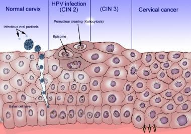
The association between human papillomavirus (HPV) DNA copy number and cervical disease was investigated. Viral DNA copy number for the most common high-risk HPV types in cervical cancer (types 16, 18, 31, and 45) was determined in cervical cytobrush specimens from 149 women with high-grade cervical intraepithelial neoplasias (CIN II-CIN III), 176 with low-grade CIN (CIN I), and 270 with normal cytology. Quantitative, PCR-based fluorescent assays for each of the HPV genotypes and for the beta-globin gene were used. The amount of cellular DNA increased significantly with increasing disease; thus, HPV was expressed as copies per microgram of cellular DNA. The assay had a dynamic range of >10(7), allowing documentation for the first time of the wide range of HPV copy numbers seen in clinical specimens. Median HPV DNA copy number varied by more than 10(4) among the viral types. HPV16 was present in the highest copy number; over 55% of HPV16-positive samples contained more than 10(8) copies/microgram. Median copy number for HPV16 showed dramatic increases with increasing epithelial abnormality, an effect not seen with the other HPV types. HPV16 increased from a median of 2.2 x 10(7) in patients with normal cytology, to 4.1 x 10(7) in CIN I patients, to 1.3 x 10(9) copies/microgram in CIN II-III patients. Even when stratified by cervical disease and viral type, the range of viral DNA copies per microgram of cellular DNA was quite large, precluding setting a clinically significant cutoff value for "high" copy numbers predictive of disease. This study suggests that the clinical usefulness of HPV quantitation requires reassessment and is assay dependent. (+info)Cervical intraepithelial neoplasia (CIN) is a term used to describe the abnormal growth and development of cells on the surface of the cervix. These changes are usually caused by human papillomavirus (HPV) infection, which is a common sexually transmitted infection. CIN is not cancer, but it can develop into cancer if left untreated.
The term "intraepithelial" refers to the fact that the abnormal cells are found in the epithelium, or the lining of the cervix. The term "neoplasia" means abnormal growth or development of cells. CIN is further classified into three grades based on the severity of the cell changes:
* CIN 1: Mild dysplasia (abnormal cell growth) affecting the lower third of the epithelium.
* CIN 2: Moderate dysplasia affecting the lower two-thirds of the epithelium.
* CIN 3: Severe dysplasia or carcinoma in situ, which means that the abnormal cells are found in the full thickness of the epithelium and have a high risk of progressing to invasive cancer if not treated.
It's important to note that CIN can regress on its own without treatment, especially in younger women. However, some cases may progress to invasive cervical cancer if left untreated. Regular Pap testing is recommended to detect and monitor any abnormal cell changes in the cervix. If CIN is detected, further diagnostic procedures such as a colposcopy or biopsy may be performed to determine the extent of the abnormality and guide treatment decisions.
Uterine cervical neoplasms, also known as cervical cancer or cervical dysplasia, refer to abnormal growths or lesions on the lining of the cervix that have the potential to become cancerous. These growths are usually caused by human papillomavirus (HPV) infection and can be detected through routine Pap smears.
Cervical neoplasms are classified into different grades based on their level of severity, ranging from mild dysplasia (CIN I) to severe dysplasia or carcinoma in situ (CIN III). In some cases, cervical neoplasms may progress to invasive cancer if left untreated.
Risk factors for developing cervical neoplasms include early sexual activity, multiple sexual partners, smoking, and a weakened immune system. Regular Pap smears and HPV testing are recommended for early detection and prevention of cervical cancer.
Colposcopy is a medical procedure in which a colposcope, which is a type of microscope, is used to examine the cervix, vagina, and vulva for signs of disease or abnormalities. The colposcope allows the healthcare provider to see these areas in greater detail than is possible with the naked eye. During the procedure, the provider may take a small sample of tissue (biopsy) for further examination under a microscope.
Colposcopy is often used to investigate abnormal Pap test results or to follow up on women who have been diagnosed with certain types of cervical dysplasia (abnormal cell growth). It can also be used to diagnose and monitor other conditions, such as genital warts, inflammation, or cancer.
It is important to note that colposcopy is a diagnostic procedure and not a treatment. If abnormalities are found during the exam, additional procedures may be necessary to remove or treat them.
Papillomaviridae is a family of small, non-enveloped DNA viruses that primarily infect the epithelial cells of mammals, birds, and reptiles. The name "papillomavirus" comes from the Latin word "papilla," which means nipple or small projection, reflecting the characteristic wart-like growths (papillomas) that these viruses can cause in infected host tissues.
The family Papillomaviridae includes more than 200 distinct papillomavirus types, with each type being defined by its specific DNA sequence. Human papillomaviruses (HPVs), which are the most well-studied members of this family, are associated with a range of diseases, from benign warts and lesions to malignant cancers such as cervical, anal, penile, vulvar, and oropharyngeal cancers.
Papillomaviruses have a circular, double-stranded DNA genome that is approximately 8 kbp in size. The viral genome encodes several early (E) proteins involved in viral replication and oncogenesis, as well as late (L) proteins that form the viral capsid. The life cycle of papillomaviruses is tightly linked to the differentiation program of their host epithelial cells, with productive infection occurring primarily in the differentiated layers of the epithelium.
In summary, Papillomaviridae is a family of DNA viruses that infect epithelial cells and can cause a variety of benign and malignant diseases. Human papillomaviruses are a significant public health concern due to their association with several cancer types.
Papillomavirus infections are a group of diseases caused by various types of human papillomaviruses (HPVs). These viruses infect the skin and mucous membranes, and can cause benign growths such as warts or papillomas, as well as malignant growths like cervical cancer.
There are more than 100 different types of HPVs, and they can be classified into low-risk and high-risk types based on their potential to cause cancer. Low-risk HPV types, such as HPV-6 and HPV-11, commonly cause benign genital warts and respiratory papillomas. High-risk HPV types, such as HPV-16 and HPV-18, are associated with an increased risk of developing cancer, including cervical, anal, penile, vulvar, and oropharyngeal cancers.
HPV infections are typically transmitted through sexual contact, and most sexually active individuals will acquire at least one HPV infection during their lifetime. In many cases, the immune system is able to clear the virus without any symptoms or long-term consequences. However, persistent high-risk HPV infections can lead to the development of cancer over time.
Prevention measures for HPV infections include vaccination against high-risk HPV types, safe sex practices, and regular screening for cervical cancer in women. The HPV vaccine is recommended for both boys and girls aged 11-12 years old, and can also be given to older individuals up to age 45 who have not previously been vaccinated or who have not completed the full series of shots.
A vaginal smear, also known as a Pap test or Pap smear, is a medical procedure in which a sample of cells is collected from the cervix (the lower part of the uterus that opens into the vagina) and examined under a microscope. The purpose of this test is to detect abnormal cells, including precancerous changes, that may indicate the presence of cervical cancer or other conditions such as infections or inflammation.
During the procedure, a speculum is inserted into the vagina to allow the healthcare provider to visualize the cervix. A spatula or brush is then used to gently scrape cells from the surface of the cervix. The sample is spread onto a microscope slide and sent to a laboratory for analysis.
Regular Pap smears are recommended for women as part of their routine healthcare, as they can help detect abnormalities at an early stage when they are more easily treated. The frequency of Pap smears may vary depending on age, medical history, and other factors. It is important to follow the recommendations of a healthcare provider regarding the timing and frequency of Pap smears.
The cervix uteri, often simply referred to as the cervix, is the lower part of the uterus (womb) that connects to the vagina. It has an opening called the external os through which menstrual blood exits the uterus and sperm enters during sexual intercourse. During childbirth, the cervix dilates or opens to allow for the passage of the baby through the birth canal.
Prostatic Intraepithelial Neoplasia (PIN) is a term used in pathology to describe the abnormal growth of cells within the lining of the prostate gland's ducts and acini (small sacs that produce and store fluids). PIN is not considered a cancer, but it can be a precursor to prostate cancer.
There are two types of PIN: low-grade and high-grade. Low-grade PIN shows mild to moderate atypia (abnormalities in the cells), while high-grade PIN displays more significant atypia, which resembles prostate cancer. High-grade PIN is often found close to or within areas of prostate cancer, making it a potential indicator of malignancy.
However, not all cases of high-grade PIN progress to cancer, and some men with high-grade PIN may never develop prostate cancer. Nonetheless, the presence of high-grade PIN might prompt further investigation or monitoring to ensure early detection and treatment of any potential cancer development.
Uterine cervical dysplasia is a condition characterized by abnormal cell growth on the lining of the cervix, which is the lower part of the uterus that connects to the vagina. It is also known as cervical intraepithelial neoplasia (CIN).
Cervical dysplasia can be caused by certain strains of human papillomavirus (HPV), a common sexually transmitted infection. The abnormal cells may develop into cancerous cells over time, although not all cases of cervical dysplasia will progress to cancer.
Cervical dysplasia is typically detected through a Pap test or HPV test, which are screening tests used to detect precancerous changes in the cervix. Depending on the severity and extent of the abnormal cells, treatment options may include close monitoring, surgical removal of the affected tissue, or more extensive surgery.
It is important for women to receive regular Pap tests and HPV tests as recommended by their healthcare provider to detect and treat cervical dysplasia early, before it has a chance to progress to cancer.
Electrosurgery is a surgical procedure that uses high-frequency electrical currents to cut, coagulate, or fulgurate tissue. It is often used in surgical procedures as an alternative to traditional scalpels and electrocautery. The electrical currents are delivered through a specialized instrument called an electrosurgical unit (ESU) that can be set to produce different forms of energy, including cutting, coagulation, or blended currents.
During the procedure, the ESU is used to apply electrical energy to the target tissue, which responds by heating up and vaporizing, allowing for precise cuts to be made. The heat generated during the procedure also helps to seal off blood vessels and nerve endings, reducing bleeding and minimizing post-operative pain.
Electrosurgery is commonly used in a variety of surgical procedures, including dermatology, gynecology, urology, orthopedics, and general surgery. It offers several advantages over traditional surgical techniques, such as reduced blood loss, shorter operating times, and faster recovery times for patients. However, it also requires specialized training and equipment to ensure safe and effective use.
Carcinoma in situ is a medical term used to describe the earliest stage of cancer, specifically a type of cancer that begins in the epithelial tissue, which is the tissue that lines the outer surfaces of organs and body structures. In this stage, the cancer cells are confined to the layer of cells where they first developed and have not spread beyond that layer into the surrounding tissues or organs.
Carcinoma in situ can occur in various parts of the body, including the skin, cervix, breast, lung, prostate, bladder, and other areas. It is often detected through routine screening tests, such as Pap smears for cervical cancer or mammograms for breast cancer.
While carcinoma in situ is not invasive, it can still be a serious condition because it has the potential to develop into an invasive cancer if left untreated. Treatment options for carcinoma in situ may include surgery, radiation therapy, or other forms of treatment, depending on the location and type of cancer. It is important to consult with a healthcare provider to determine the best course of action for each individual case.
A tumor virus infection is a condition in which a person's cells become cancerous or transformed due to the integration and disruption of normal cellular functions by a viral pathogen. These viruses are also known as oncoviruses, and they can cause tumors or cancer by altering the host cell's genetic material, promoting uncontrolled cell growth and division, evading immune surveillance, and inhibiting apoptosis (programmed cell death).
Examples of tumor viruses include:
1. DNA tumor viruses: These are double-stranded DNA viruses that can cause cancer in humans. Examples include human papillomavirus (HPV), hepatitis B virus (HBV), and Merkel cell polyomavirus (MCV).
2. RNA tumor viruses: Also known as retroviruses, these single-stranded RNA viruses can cause cancer in humans. Examples include human T-cell leukemia virus type 1 (HTLV-1) and human immunodeficiency virus (HIV).
Tumor virus infections are responsible for approximately 15-20% of all cancer cases worldwide, making them a significant public health concern. Prevention strategies, such as vaccination against HPV and HBV, have been shown to reduce the incidence of associated cancers.
Conization is a surgical procedure that involves the removal of a cone-shaped piece of tissue from the cervix. It is typically performed to diagnose or treat abnormal or precancerous cells in the cervix, and is also known as a cone biopsy or a cervical conization.
The procedure is usually done under general anesthesia, and involves using a surgical instrument such as a scalpel or a laser to remove a cone-shaped piece of tissue from the cervix. The tissue is then sent to a laboratory for examination under a microscope to determine whether there are any abnormal or precancerous cells present.
Conization may be recommended in cases where Pap tests or other screening methods have detected abnormal cells in the cervix, or if there are suspicious-looking areas that cannot be fully evaluated with a colposcopy (a procedure that uses a special magnifying device to examine the cervix). It may also be used as a treatment for certain types of cervical dysplasia (abnormal cell growth) or early-stage cervical cancer.
It's important to note that conization is a surgical procedure and, like any surgery, carries some risks such as bleeding, infection, and damage to surrounding tissues. However, these complications are generally rare and can be effectively managed with appropriate medical care.
The Papanicolaou (Pap) test, also known as the Pap smear, is a screening procedure for detecting precancerous and cancerous cells in the cervix. It involves collecting cells from the cervix and examining them under a microscope to look for any abnormalities. The test is typically recommended for women aged 21-65 as part of routine pelvic exams, with the frequency depending on age and risk factors.
The Pap test was developed by Georgios Papanikolaou in the early 20th century and has since become a widely used and important tool in preventing cervical cancer. The test is usually performed in a healthcare provider's office and takes only a few minutes to complete. It is a relatively simple, safe, and painless procedure that can help detect cervical abnormalities at an early stage, when they are most treatable.
Human papillomavirus 16 (HPV16) is a specific type of human papillomavirus (HPV). HPV is a DNA virus that infects the skin and mucous membranes, and there are over 200 types of HPV. Some types of HPV can cause warts, while others are associated with an increased risk of certain cancers.
HPV16 is one of the high-risk types of HPV and is strongly associated with several types of cancer, including cervical, anal, penile, vulvar, and oropharyngeal (throat) cancers. HPV16 is responsible for about 50% of all cervical cancers and is the most common high-risk type of HPV found in these cancers.
HPV16 is typically transmitted through sexual contact, and most people who are sexually active will acquire at least one type of HPV at some point in their lives. While HPV infections are often harmless and clear up on their own without causing any symptoms or health problems, high-risk types like HPV16 can lead to cancer if left untreated.
Fortunately, there are vaccines available that protect against HPV16 and other high-risk types of HPV. These vaccines have been shown to be highly effective in preventing HPV-related cancers and precancerous lesions. The Centers for Disease Control and Prevention (CDC) recommends routine HPV vaccination for both boys and girls starting at age 11 or 12, although the vaccine can be given as early as age 9. Catch-up vaccinations are also recommended for older individuals who have not yet been vaccinated.
Human papillomavirus 18 (HPV-18) is a specific type of human papillomavirus (HPV), which is a group of more than 200 related viruses. HPV is named for the warts (papillomas) some types can cause.
HPV-18 is one of the high-risk types of HPV that are linked to several types of cancer, including cervical, anal, vaginal, vulvar, and oropharyngeal (throat) cancers. HPV-18 along with HPV-16 are responsible for about 70% of all cervical cancers.
HPV is passed from one person to another during skin-to-skin contact, usually during sexual activity. Most sexually active people will have an HPV infection at some point in their lives, but most will never know it because the virus often causes no symptoms and goes away on its own. However, when HPV doesn't go away, it can cause serious health problems, including cancer.
There are vaccines available to protect against HPV-18 and other high-risk types of HPV. The Centers for Disease Control and Prevention (CDC) recommends that all boys and girls get the HPV vaccine at age 11 or 12, but it can be given as early as age 9 and until age 26 for those who have not yet received it. The vaccine is most effective when given before becoming sexually active.
Alphapapillomavirus is a genus of Papillomaviridae, a family of small, non-enveloped DNA viruses that infect the skin and mucous membranes of humans and other animals. Members of this genus are known to cause various types of benign and malignant tumors in humans, including skin warts, genital warts, and cancers of the cervix, anus, penis, vulva, and oropharynx.
The Alphapapillomavirus genus is further divided into several species, each containing multiple types or strains of the virus. Some of the most well-known and studied types of Alphapapillomavirus include:
* Human papillomavirus (HPV) type 16 and 18, which are associated with a high risk of cervical cancer and other anogenital cancers
* HPV type 6 and 11, which are commonly found in genital warts and recurrent respiratory papillomatosis
* HPV types 31, 33, 45, 52, and 58, which are also associated with an increased risk of cervical cancer and other malignancies.
Preventive measures such as vaccination against high-risk HPV types have been shown to significantly reduce the incidence of cervical cancer and other HPV-related diseases. Regular screening for cervical cancer and other precancerous lesions is also an important part of prevention and early detection.
Viral DNA refers to the genetic material present in viruses that consist of DNA as their core component. Deoxyribonucleic acid (DNA) is one of the two types of nucleic acids that are responsible for storing and transmitting genetic information in living organisms. Viruses are infectious agents much smaller than bacteria that can only replicate inside the cells of other organisms, called hosts.
Viral DNA can be double-stranded (dsDNA) or single-stranded (ssDNA), depending on the type of virus. Double-stranded DNA viruses have a genome made up of two complementary strands of DNA, while single-stranded DNA viruses contain only one strand of DNA.
Examples of dsDNA viruses include Adenoviruses, Herpesviruses, and Poxviruses, while ssDNA viruses include Parvoviruses and Circoviruses. Viral DNA plays a crucial role in the replication cycle of the virus, encoding for various proteins necessary for its multiplication and survival within the host cell.
A precancerous condition, also known as a premalignant condition, is a state of abnormal cellular growth and development that has a higher-than-normal potential to progress into cancer. These conditions are characterized by the presence of certain anomalies in the cells, such as dysplasia (abnormal changes in cell shape or size), which can indicate an increased risk for malignant transformation.
It is important to note that not all precancerous conditions will eventually develop into cancer, and some may even regress on their own. However, individuals with precancerous conditions are often at a higher risk of developing cancer compared to the general population. Regular monitoring and appropriate medical interventions, if necessary, can help manage this risk and potentially prevent or detect cancer at an early stage when it is more treatable.
Examples of precancerous conditions include:
1. Dysplasia in the cervix (cervical intraepithelial neoplasia or CIN)
2. Atypical ductal hyperplasia or lobular hyperplasia in the breast
3. Actinic keratosis on the skin
4. Leukoplakia in the mouth
5. Barrett's esophagus in the digestive tract
Regular medical check-ups, screenings, and lifestyle modifications are crucial for individuals with precancerous conditions to monitor their health and reduce the risk of cancer development.
Vaginal neoplasms refer to abnormal growths or tumors in the vagina. These growths can be benign (non-cancerous) or malignant (cancerous). The two main types of vaginal neoplasms are:
1. Vaginal intraepithelial neoplasia (VAIN): This is a condition where the cells on the inner lining of the vagina become abnormal but have not invaded deeper tissues. VAIN can be low-grade or high-grade, depending on the severity of the cell changes.
2. Vaginal cancer: This is a malignant tumor that arises from the cells in the vagina. The two main types of vaginal cancer are squamous cell carcinoma and adenocarcinoma. Squamous cell carcinoma is the most common type, accounting for about 85% of all cases.
Risk factors for vaginal neoplasms include human papillomavirus (HPV) infection, smoking, older age, history of cervical cancer or precancerous changes, and exposure to diethylstilbestrol (DES) in utero. Treatment options depend on the type, stage, and location of the neoplasm but may include surgery, radiation therapy, chemotherapy, or a combination of these approaches.
Vulvar neoplasms refer to abnormal growths or tumors in the vulvar region, which is the exterior female genital area including the mons pubis, labia majora, labia minora, clitoris, and the vaginal vestibule. These neoplasms can be benign (non-cancerous) or malignant (cancerous).
Benign vulvar neoplasms may include conditions such as vulvar cysts, fibromas, lipomas, or condylomas (genital warts). They are typically slow-growing and less likely to spread or invade surrounding tissues.
Malignant vulvar neoplasms, on the other hand, are cancers that can invade nearby tissues and potentially metastasize (spread) to distant parts of the body. The most common types of malignant vulvar neoplasms are squamous cell carcinoma, vulvar melanoma, and adenocarcinoma.
Early detection and treatment of vulvar neoplasms are essential for improving prognosis and reducing the risk of complications or recurrence. Regular gynecological examinations, self-examinations, and prompt attention to any unusual symptoms or changes in the vulvar area can help ensure timely diagnosis and management.
'Condylomata Acuminata' is the medical term for genital warts, which are growths or bumps that appear on the genital area. They are caused by certain types of the human papillomavirus (HPV). Genital warts can vary in appearance, and they may be small, flat, and difficult to see or large, cauliflower-like, and easily visible.
The warts can appear on the vulva, vagina, cervix, rectum, anus, penis, or scrotum. They are usually painless but can cause discomfort during sexual intercourse. In some cases, genital warts can lead to serious health problems, such as cervical cancer in women.
It is important to note that not all people with HPV will develop genital warts, and many people with HPV are asymptomatic and unaware they have the virus. The Centers for Disease Control and Prevention (CDC) recommends routine HPV vaccination for both boys and girls aged 11-12 years to prevent HPV infection and related diseases, including genital warts.
DNA probes for HPV (Human Papillomavirus) are specific DNA sequences that are used in diagnostic tests to detect and identify the presence of HPV DNA in a sample. HPV is a viral infection that can cause various types of cancer, including cervical, anal, and oropharyngeal cancers.
DNA probes for HPV work by binding to complementary sequences of HPV DNA in the sample. This binding can be detected and measured using various methods, such as hybridization, amplification, or labeling techniques. The use of DNA probes for HPV can help identify the specific type of HPV that is present in a sample, which can inform clinical management and treatment decisions.
It's important to note that not all HPV infections lead to cancer, and most HPV infections resolve on their own without causing any harm. However, certain high-risk types of HPV are more strongly associated with an increased risk of developing cancer, so identifying the presence and type of HPV infection can be useful for monitoring and managing patients who may be at higher risk.
Squamous cell neoplasms are abnormal growths or tumors that originate from squamous cells, which are flat, scale-like cells that make up the outer layer of the skin and the lining of mucous membranes. These neoplasms can be benign (noncancerous) or malignant (cancerous). When malignant, they are called squamous cell carcinomas.
Squamous cell carcinomas often develop in areas exposed to excessive sunlight or ultraviolet radiation, such as the skin, lips, and mouth. They can also occur in other areas of the body, including the cervix, anus, and lungs. Risk factors for developing squamous cell carcinoma include fair skin, a history of sunburns, exposure to certain chemicals or radiation, and a weakened immune system.
Symptoms of squamous cell carcinomas may include rough or scaly patches on the skin, a sore that doesn't heal, a wart-like growth, or a raised bump with a central depression. Treatment for squamous cell carcinomas typically involves surgical removal of the tumor, along with radiation therapy or chemotherapy in some cases. Early detection and treatment can help prevent the spread of the cancer to other parts of the body.
Spontaneous neoplasm regression is a rare and somewhat controversial phenomenon in which a tumor or malignancy appears to decrease in size or disappear without any treatment or with treatment that is typically not expected to produce such an effect. This can occur through various mechanisms, including immune-mediated processes, apoptosis (programmed cell death), differentiation of cancer cells into normal cells, and angiogenesis inhibition (preventing the growth of new blood vessels that feed the tumor).
Spontaneous regression of neoplasms is not well understood and is considered unpredictable. It has been reported in various types of cancers, including neuroblastoma, melanoma, renal cell carcinoma, and others. However, it should be noted that spontaneous regression does not imply a cure, as the tumor may still recur or metastasize later on.
In summary, spontaneous neoplasm regression refers to the partial or complete disappearance of a malignancy without any specific treatment or with treatment that is not typically associated with such an effect.
Oncogene proteins, viral, are cancer-causing proteins that are encoded by the genetic material (DNA or RNA) of certain viruses. These viral oncogenes can be acquired through infection with retroviruses, such as human immunodeficiency virus (HIV), human T-cell leukemia virus (HTLV), and certain types of papillomaviruses and polyomaviruses.
When these viruses infect host cells, they can integrate their genetic material into the host cell's genome, leading to the expression of viral oncogenes. These oncogenes may then cause uncontrolled cell growth and division, ultimately resulting in the formation of tumors or cancers. The process by which viruses contribute to cancer development is complex and involves multiple steps, including the alteration of signaling pathways that regulate cell proliferation, differentiation, and survival.
Examples of viral oncogenes include the v-src gene found in the Rous sarcoma virus (RSV), which causes chicken sarcoma, and the E6 and E7 genes found in human papillomaviruses (HPVs), which are associated with cervical cancer and other anogenital cancers. Understanding viral oncogenes and their mechanisms of action is crucial for developing effective strategies to prevent and treat virus-associated cancers.
A biopsy is a medical procedure in which a small sample of tissue is taken from the body to be examined under a microscope for the presence of disease. This can help doctors diagnose and monitor various medical conditions, such as cancer, infections, or autoimmune disorders. The type of biopsy performed will depend on the location and nature of the suspected condition. Some common types of biopsies include:
1. Incisional biopsy: In this procedure, a surgeon removes a piece of tissue from an abnormal area using a scalpel or other surgical instrument. This type of biopsy is often used when the lesion is too large to be removed entirely during the initial biopsy.
2. Excisional biopsy: An excisional biopsy involves removing the entire abnormal area, along with a margin of healthy tissue surrounding it. This technique is typically employed for smaller lesions or when cancer is suspected.
3. Needle biopsy: A needle biopsy uses a thin, hollow needle to extract cells or fluid from the body. There are two main types of needle biopsies: fine-needle aspiration (FNA) and core needle biopsy. FNA extracts loose cells, while a core needle biopsy removes a small piece of tissue.
4. Punch biopsy: In a punch biopsy, a round, sharp tool is used to remove a small cylindrical sample of skin tissue. This type of biopsy is often used for evaluating rashes or other skin abnormalities.
5. Shave biopsy: During a shave biopsy, a thin slice of tissue is removed from the surface of the skin using a sharp razor-like instrument. This technique is typically used for superficial lesions or growths on the skin.
After the biopsy sample has been collected, it is sent to a laboratory where a pathologist will examine the tissue under a microscope and provide a diagnosis based on their findings. The results of the biopsy can help guide further treatment decisions and determine the best course of action for managing the patient's condition.
Uterine cervical diseases refer to conditions that affect the cervix, which is the lower part of the uterus that opens into the vagina. These diseases can range from minor abnormalities to more serious conditions, such as:
1. Cervical dysplasia: This is a precancerous condition characterized by the presence of abnormal cells on the cervix. It is usually caused by the human papillomavirus (HPV) and can be detected through a Pap test.
2. Cervical cancer: This is a malignant tumor that develops in the cervical tissue. The most common type of cervical cancer is squamous cell carcinoma, which arises from the cells lining the surface of the cervix.
3. Cervicitis: This is an inflammation of the cervix, which can be caused by infections, irritants, or allergies. Symptoms may include vaginal discharge, pain, and bleeding.
4. Cervical polyps: These are benign growths that develop on the cervix. They are usually small and asymptomatic but can cause abnormal vaginal bleeding or discharge.
5. Cervical incompetence: This is a condition where the cervix begins to open prematurely during pregnancy, leading to a risk of miscarriage or preterm labor.
It's important to note that regular screening and early detection can help prevent or manage many cervical diseases, including cervical cancer.
Squamous cell carcinoma is a type of skin cancer that begins in the squamous cells, which are flat, thin cells that form the outer layer of the skin (epidermis). It commonly occurs on sun-exposed areas such as the face, ears, lips, and backs of the hands. Squamous cell carcinoma can also develop in other areas of the body including the mouth, lungs, and cervix.
This type of cancer usually develops slowly and may appear as a rough or scaly patch of skin, a red, firm nodule, or a sore or ulcer that doesn't heal. While squamous cell carcinoma is not as aggressive as some other types of cancer, it can metastasize (spread) to other parts of the body if left untreated, making early detection and treatment important.
Risk factors for developing squamous cell carcinoma include prolonged exposure to ultraviolet (UV) radiation from the sun or tanning beds, fair skin, a history of sunburns, a weakened immune system, and older age. Prevention measures include protecting your skin from the sun by wearing protective clothing, using a broad-spectrum sunscreen with an SPF of at least 30, avoiding tanning beds, and getting regular skin examinations.
Papillomavirus vaccines are vaccines that have been developed to prevent infection by human papillomaviruses (HPV). HPV is a DNA virus that is capable of infecting the skin and mucous membranes. Certain types of HPV are known to cause cervical cancer, as well as other types of cancer such as anal, penile, vulvar, and oropharyngeal cancers. Other types of HPV can cause genital warts.
There are currently two papillomavirus vaccines that have been approved for use in the United States: Gardasil and Cervarix. Both vaccines protect against the two most common cancer-causing types of HPV (types 16 and 18), which together cause about 70% of cervical cancers. Gardasil also protects against the two most common types of HPV that cause genital warts (types 6 and 11).
Papillomavirus vaccines are given as a series of three shots over a period of six months. They are most effective when given to people before they become sexually active, as this reduces the risk of exposure to HPV. The Centers for Disease Control and Prevention (CDC) recommends that all boys and girls get vaccinated against HPV at age 11 or 12, but the vaccine can be given to people as young as age 9 and as old as age 26.
It is important to note that papillomavirus vaccines do not protect against all types of HPV, and they do not treat existing HPV infections or cervical cancer. They are intended to prevent new HPV infections and the cancers and other diseases that can be caused by HPV.
Medical mass screening, also known as population screening, is a public health service that aims to identify and detect asymptomatic individuals in a given population who have or are at risk of a specific disease. The goal is to provide early treatment, reduce morbidity and mortality, and prevent the spread of diseases within the community.
A mass screening program typically involves offering a simple, quick, and non-invasive test to a large number of people in a defined population, regardless of their risk factors or symptoms. Those who test positive are then referred for further diagnostic tests and appropriate medical interventions. Examples of mass screening programs include mammography for breast cancer detection, PSA (prostate-specific antigen) testing for prostate cancer, and fecal occult blood testing for colorectal cancer.
It is important to note that mass screening programs should be evidence-based, cost-effective, and ethically sound, with clear benefits outweighing potential harms. They should also consider factors such as the prevalence of the disease in the population, the accuracy and reliability of the screening test, and the availability and effectiveness of treatment options.
Neoplasm grading is a system used by pathologists to classify the degree of abnormality in cells that make up a tumor (neoplasm). It provides an assessment of how quickly the tumor is likely to grow and spread. The grade helps doctors predict the prognosis and determine the best treatment options.
Neoplasm grading typically involves evaluating certain cellular features under a microscope, such as:
1. Differentiation or degree of maturity: This refers to how closely the tumor cells resemble their normal counterparts in terms of size, shape, and organization. Well-differentiated tumors have cells that look more like normal cells and are usually slower growing. Poorly differentiated tumors have cells that appear very abnormal and tend to grow and spread more aggressively.
2. Mitotic count: This is the number of times the tumor cells divide (mitosis) within a given area. A higher mitotic count indicates a faster-growing tumor.
3. Necrosis: This refers to areas of dead tissue within the tumor. A significant amount of necrosis may suggest a more aggressive tumor.
Based on these and other factors, pathologists assign a grade to the tumor using a standardized system, such as the Bloom-Richardson or Scarff-Bloom-Richardson grading systems for breast cancer or the Fuhrman grading system for kidney cancer. The grade usually consists of a number or a range (e.g., G1, G2, G3, or G4) or a combination of grades (e.g., low grade, intermediate grade, and high grade).
In general, higher-grade tumors have a worse prognosis than lower-grade tumors because they are more likely to grow quickly, invade surrounding tissues, and metastasize (spread) to other parts of the body. However, neoplasm grading is just one aspect of cancer diagnosis and treatment planning. Other factors, such as the stage of the disease, location of the tumor, patient's overall health, and specific molecular markers, are also considered when making treatment decisions.
Sensitivity and specificity are statistical measures used to describe the performance of a diagnostic test or screening tool in identifying true positive and true negative results.
* Sensitivity refers to the proportion of people who have a particular condition (true positives) who are correctly identified by the test. It is also known as the "true positive rate" or "recall." A highly sensitive test will identify most or all of the people with the condition, but may also produce more false positives.
* Specificity refers to the proportion of people who do not have a particular condition (true negatives) who are correctly identified by the test. It is also known as the "true negative rate." A highly specific test will identify most or all of the people without the condition, but may also produce more false negatives.
In medical testing, both sensitivity and specificity are important considerations when evaluating a diagnostic test. High sensitivity is desirable for screening tests that aim to identify as many cases of a condition as possible, while high specificity is desirable for confirmatory tests that aim to rule out the condition in people who do not have it.
It's worth noting that sensitivity and specificity are often influenced by factors such as the prevalence of the condition in the population being tested, the threshold used to define a positive result, and the reliability and validity of the test itself. Therefore, it's important to consider these factors when interpreting the results of a diagnostic test.
I apologize, but "Costa Rica" is not a medical term or concept. Costa Rica is a country located in Central America, known for its rich biodiversity and progressive environmental policies. If you have any questions related to medicine or health, I would be happy to try and help answer those for you.
Papillomavirus E7 proteins are small, viral regulatory proteins encoded by the E7 gene in papillomaviruses (HPVs). These proteins play a crucial role in the life cycle of HPVs and are associated with the development of various types of cancer, most notably cervical cancer.
The E7 protein functions as a transcriptional activator and can bind to and degrade the retinoblastoma protein (pRb), which is a tumor suppressor. By binding to and inactivating pRb, E7 promotes the expression of genes required for cell cycle progression, leading to uncontrolled cell growth and proliferation.
E7 proteins are also capable of inducing genetic alterations, such as chromosomal instability and DNA damage, which can contribute to the development of cancer. Additionally, E7 has been shown to inhibit apoptosis (programmed cell death) and promote angiogenesis (the formation of new blood vessels), further contributing to tumor growth and progression.
Overall, Papillomavirus E7 proteins are important oncogenic factors that play a central role in the development of HPV-associated cancers.
Polymerase Chain Reaction (PCR) is a laboratory technique used to amplify specific regions of DNA. It enables the production of thousands to millions of copies of a particular DNA sequence in a rapid and efficient manner, making it an essential tool in various fields such as molecular biology, medical diagnostics, forensic science, and research.
The PCR process involves repeated cycles of heating and cooling to separate the DNA strands, allow primers (short sequences of single-stranded DNA) to attach to the target regions, and extend these primers using an enzyme called Taq polymerase, resulting in the exponential amplification of the desired DNA segment.
In a medical context, PCR is often used for detecting and quantifying specific pathogens (viruses, bacteria, fungi, or parasites) in clinical samples, identifying genetic mutations or polymorphisms associated with diseases, monitoring disease progression, and evaluating treatment effectiveness.
Anus neoplasms refer to abnormal growths or tumors in the anus, which is the opening at the end of the digestive tract where solid waste leaves the body. These growths can be benign (non-cancerous) or malignant (cancerous). Common types of anus neoplasms include squamous cell carcinoma, adenocarcinoma, and melanoma.
Squamous cell carcinoma is the most common type of anus cancer, accounting for about 80% of all cases. It begins in the squamous cells that line the anal canal and can spread to other parts of the body if left untreated.
Adenocarcinoma is a less common type of anus cancer that arises from glandular cells in the anus. This type of cancer is often associated with long-standing inflammatory conditions, such as anal fistulas or ulcerative colitis.
Melanoma is a rare form of skin cancer that can also occur in the anus. It develops from pigment-producing cells called melanocytes and tends to be aggressive with a high risk of spreading to other parts of the body.
Other less common types of anus neoplasms include basal cell carcinoma, sarcoma, and lymphoma. Treatment options for anus neoplasms depend on the type, stage, and location of the tumor, as well as the patient's overall health.
Clinical pathology is a medical specialty that focuses on the diagnosis of diseases through the examination of organs, tissues, and bodily fluids, such as blood and urine. It involves the use of laboratory tests to identify abnormalities in the body's cells, chemicals, and functions that may indicate the presence of a specific disease or condition. Clinical pathologists work closely with other healthcare professionals to help manage patient care, provide treatment recommendations, and monitor the effectiveness of treatments. They are responsible for supervising the laboratory testing process, ensuring accurate results, and interpreting the findings in the context of each patient's medical history and symptoms. Overall, clinical pathology plays a critical role in the diagnosis, treatment, and prevention of many different types of diseases and conditions.
Cytological techniques refer to the methods and procedures used to study individual cells, known as cytopathology. These techniques are used in the diagnosis and screening of various medical conditions, including cancer. The most common cytological technique is the Pap test, which involves collecting cells from the cervix and examining them for abnormalities. Other cytological techniques include fine-needle aspiration (FNA), which involves using a thin needle to withdraw cells from a tumor or lump, and body fluids analysis, which involves examining cells present in various bodily fluids such as urine, sputum, and pleural effusions. These techniques allow for the examination of cellular structure, morphology, and other characteristics to help diagnose and monitor diseases.
Cyclin-Dependent Kinase Inhibitor p16, also known as CDKN2A or INK4a, is a protein that regulates the cell cycle. It functions as an inhibitor of cyclin-dependent kinases (CDKs) 4 and 6, which are enzymes that play a crucial role in regulating the progression of the cell cycle.
The p16 protein is produced in response to various signals, including DNA damage and oncogene activation, and its main function is to prevent the phosphorylation and activation of the retinoblastoma protein (pRb) by CDK4/6. When pRb is not phosphorylated, it binds to and inhibits the E2F transcription factor, which results in the suppression of genes required for cell cycle progression.
Therefore, p16 acts as a tumor suppressor protein by preventing the uncontrolled proliferation of cells that can lead to cancer. Mutations or deletions in the CDKN2A gene, which encodes the p16 protein, have been found in many types of human cancers, including lung, breast, and head and neck cancers.
Medical Definition:
"Risk factors" are any attribute, characteristic or exposure of an individual that increases the likelihood of developing a disease or injury. They can be divided into modifiable and non-modifiable risk factors. Modifiable risk factors are those that can be changed through lifestyle choices or medical treatment, while non-modifiable risk factors are inherent traits such as age, gender, or genetic predisposition. Examples of modifiable risk factors include smoking, alcohol consumption, physical inactivity, and unhealthy diet, while non-modifiable risk factors include age, sex, and family history. It is important to note that having a risk factor does not guarantee that a person will develop the disease, but rather indicates an increased susceptibility.
Cytodiagnosis is the rapid, initial evaluation and diagnosis of a disease based on the examination of individual cells obtained from a body fluid or tissue sample. This technique is often used in cytopathology to investigate abnormalities such as lumps, bumps, or growths that may be caused by cancerous or benign conditions.
The process involves collecting cells through various methods like fine-needle aspiration (FNA), body fluids such as urine, sputum, or washings from the respiratory, gastrointestinal, or genitourinary tracts. The collected sample is then spread onto a microscope slide, stained, and examined under a microscope for abnormalities in cell size, shape, structure, and organization.
Cytodiagnosis can provide crucial information to guide further diagnostic procedures and treatment plans. It is often used as an initial screening tool due to its speed, simplicity, and cost-effectiveness compared to traditional histopathological methods that require tissue biopsy and more extensive processing. However, cytodiagnosis may not always be able to distinguish between benign and malignant conditions definitively; therefore, additional tests or follow-up evaluations might be necessary for a conclusive diagnosis.
Tumor markers are substances that can be found in the body and their presence can indicate the presence of certain types of cancer or other conditions. Biological tumor markers refer to those substances that are produced by cancer cells or by other cells in response to cancer or certain benign (non-cancerous) conditions. These markers can be found in various bodily fluids such as blood, urine, or tissue samples.
Examples of biological tumor markers include:
1. Proteins: Some tumor markers are proteins that are produced by cancer cells or by other cells in response to the presence of cancer. For example, prostate-specific antigen (PSA) is a protein produced by normal prostate cells and in higher amounts by prostate cancer cells.
2. Genetic material: Tumor markers can also include genetic material such as DNA, RNA, or microRNA that are shed by cancer cells into bodily fluids. For example, circulating tumor DNA (ctDNA) is genetic material from cancer cells that can be found in the bloodstream.
3. Metabolites: Tumor markers can also include metabolic products produced by cancer cells or by other cells in response to cancer. For example, lactate dehydrogenase (LDH) is an enzyme that is released into the bloodstream when cancer cells break down glucose for energy.
It's important to note that tumor markers are not specific to cancer and can be elevated in non-cancerous conditions as well. Therefore, they should not be used alone to diagnose cancer but rather as a tool in conjunction with other diagnostic tests and clinical evaluations.
Immunohistochemistry (IHC) is a technique used in pathology and laboratory medicine to identify specific proteins or antigens in tissue sections. It combines the principles of immunology and histology to detect the presence and location of these target molecules within cells and tissues. This technique utilizes antibodies that are specific to the protein or antigen of interest, which are then tagged with a detection system such as a chromogen or fluorophore. The stained tissue sections can be examined under a microscope, allowing for the visualization and analysis of the distribution and expression patterns of the target molecule in the context of the tissue architecture. Immunohistochemistry is widely used in diagnostic pathology to help identify various diseases, including cancer, infectious diseases, and immune-mediated disorders.
Early detection of cancer refers to the identification of malignant cells or tumors in their initial stages, before they have had a chance to grow and spread. This is typically achieved through various screening methods and tests that are designed to detect specific types of cancers. The goal of early detection is to increase the chances of successful treatment and improve the overall prognosis for patients.
Some common methods used for early cancer detection include:
1. Regular screenings such as mammograms, colonoscopies, and Pap tests, which can help identify precancerous or cancerous cells in their earliest stages.
2. Imaging tests like CT scans, MRIs, and PET scans, which can help detect tumors that may not be visible through other screening methods.
3. Blood tests that look for specific biomarkers or tumor markers, which can indicate the presence of cancer in the body.
4. Genetic testing to identify individuals who may be at higher risk of developing certain types of cancer due to inherited genetic mutations.
It's important to note that while early detection is an important tool in the fight against cancer, it is not a guarantee of successful treatment or cure. However, it can significantly improve the odds of successful treatment and increase the chances of survival for many patients.
Triage is a medical term that refers to the process of prioritizing patients based on the severity of their condition or illness, and the resources available. The goal of triage is to ensure that the most critical patients receive care first, which can help reduce morbidity and mortality in emergency situations. This process is typically used in settings where there are more patients than can be treated immediately, such as during mass casualty incidents or in busy emergency departments. Triage nurses or doctors quickly assess each patient's condition, often using a standardized system, to determine the urgency of their medical needs and allocate resources accordingly.
A Human Papillomavirus (HPV) DNA test is a molecular diagnostic assay used to detect the presence or absence of DNA from high-risk types of HPV in cervical or anal samples. High-risk HPV types are those most strongly associated with an increased risk for developing cervical cancer and other anogenital cancers.
HPV DNA tests typically use polymerase chain reaction (PCR) or other nucleic acid amplification techniques to detect and quantify the viral DNA in clinical samples. These tests can help identify women at higher risk for cervical precancer or cancer, particularly when combined with cytology results from a Pap test.
HPV DNA testing is recommended as a primary screening method for cervical cancer in certain populations or as a follow-up test to abnormal Pap test results. It's important to note that HPV DNA tests do not diagnose cervical precancer or cancer but rather identify the presence of high-risk HPV types, which may increase the risk of developing these conditions over time.
A hysterectomy is a surgical procedure that involves the removal of the uterus (womb). Depending on the specific medical condition and necessity, a hysterectomy may also include the removal of the ovaries, fallopian tubes, and surrounding tissues. There are different types of hysterectomies, including:
1. Total hysterectomy: The uterus and cervix are removed.
2. Supracervical (or subtotal) hysterectomy: Only the upper part of the uterus is removed, leaving the cervix intact.
3. Radical hysterectomy: This procedure involves removing the uterus, cervix, surrounding tissues, and the upper part of the vagina. It is typically performed in cases of cervical cancer.
4. Oophorectomy: The removal of one or both ovaries can be performed along with a hysterectomy depending on the patient's medical condition and age.
5. Salpingectomy: The removal of one or both fallopian tubes can also be performed along with a hysterectomy if needed.
The reasons for performing a hysterectomy may include but are not limited to: uterine fibroids, heavy menstrual bleeding, endometriosis, adenomyosis, pelvic prolapse, cervical or uterine cancer, and chronic pelvic pain. The choice of the type of hysterectomy depends on the patient's medical condition, age, and personal preferences.
Keratin-1
Human papillomavirus 31 (HPV31) is a specific type of human papillomavirus (HPV), which is a DNA virus that infects the skin and mucous membranes. HPV31 is one of the high-risk types of HPV, meaning it has a higher association with the development of certain cancers, particularly cervical cancer.
HPV31, like other HPV types, can cause various clinical manifestations, such as anogenital warts and precancerous lesions in the cervix, anus, vulva, vagina, and penis. Infection with HPV31 may lead to abnormal Pap test results and potentially increase the risk of developing cervical cancer if left untreated.
Prevention strategies include HPV vaccination, which protects against several high-risk types of HPV, including HPV31. Regular screening for cervical cancer through Pap tests and, when necessary, HPV testing is also crucial in early detection and treatment of precancerous lesions caused by HPV infections.
Obstetric surgical procedures are operations that are performed on the female reproductive system during pregnancy, labor, delivery, or after childbirth to address various medical conditions and complications. Some common obstetric surgical procedures include:
1. Cesarean section (C-section): A surgical delivery of a baby through incisions in the abdomen and uterus.
2. Induction of labor: The use of medication or other methods to stimulate labor.
3. Dilation and curettage (D&C): A procedure to remove tissue from the uterus using a thin, sharp instrument called a curette.
4. Hysterectomy: The surgical removal of the uterus.
5. Myomectomy: The surgical removal of fibroids, which are noncancerous growths in the muscular wall of the uterus.
6. Ovarian cystectomy: The surgical removal of a cyst from the ovary.
7. Tubal ligation: A permanent form of birth control in which the fallopian tubes are tied, cut, or sealed to prevent pregnancy.
8. Ectopic pregnancy surgery: Removal of an ectopic pregnancy, which is a pregnancy that develops outside of the uterus, usually in the fallopian tube.
These procedures may be necessary to save the life of the mother or baby, to treat medical conditions, or to prevent future complications. They should only be performed by trained medical professionals in a hospital setting.
Adenocarcinoma is a type of cancer that arises from glandular epithelial cells. These cells line the inside of many internal organs, including the breasts, prostate, colon, and lungs. Adenocarcinomas can occur in any of these organs, as well as in other locations where glands are present.
The term "adenocarcinoma" is used to describe a cancer that has features of glandular tissue, such as mucus-secreting cells or cells that produce hormones. These cancers often form glandular structures within the tumor mass and may produce mucus or other substances.
Adenocarcinomas are typically slow-growing and tend to spread (metastasize) to other parts of the body through the lymphatic system or bloodstream. They can be treated with surgery, radiation therapy, chemotherapy, targeted therapy, or a combination of these treatments. The prognosis for adenocarcinoma depends on several factors, including the location and stage of the cancer, as well as the patient's overall health and age.
Disease progression is the worsening or advancement of a medical condition over time. It refers to the natural course of a disease, including its development, the severity of symptoms and complications, and the impact on the patient's overall health and quality of life. Understanding disease progression is important for developing appropriate treatment plans, monitoring response to therapy, and predicting outcomes.
The rate of disease progression can vary widely depending on the type of medical condition, individual patient factors, and the effectiveness of treatment. Some diseases may progress rapidly over a short period of time, while others may progress more slowly over many years. In some cases, disease progression may be slowed or even halted with appropriate medical interventions, while in other cases, the progression may be inevitable and irreversible.
In clinical practice, healthcare providers closely monitor disease progression through regular assessments, imaging studies, and laboratory tests. This information is used to guide treatment decisions and adjust care plans as needed to optimize patient outcomes and improve quality of life.
Neoplasm invasiveness is a term used in pathology and oncology to describe the aggressive behavior of cancer cells as they invade surrounding tissues and organs. This process involves the loss of cell-to-cell adhesion, increased motility and migration, and the ability of cancer cells to degrade the extracellular matrix (ECM) through the production of enzymes such as matrix metalloproteinases (MMPs).
Invasive neoplasms are cancers that have spread beyond the original site where they first developed and have infiltrated adjacent tissues or structures. This is in contrast to non-invasive or in situ neoplasms, which are confined to the epithelial layer where they originated and have not yet invaded the underlying basement membrane.
The invasiveness of a neoplasm is an important prognostic factor in cancer diagnosis and treatment, as it can indicate the likelihood of metastasis and the potential effectiveness of various therapies. In general, more invasive cancers are associated with worse outcomes and require more aggressive treatment approaches.
Virology is the study of viruses, their classification, and their effects on living organisms. It involves the examination of viral genetic material, viral replication, how viruses cause disease, and the development of antiviral drugs and vaccines to treat or prevent virus infections. Virologists study various types of viruses that can infect animals, plants, and microorganisms, as well as understand their evolution and transmission patterns.
Prostatic neoplasms refer to abnormal growths in the prostate gland, which can be benign or malignant. The term "neoplasm" simply means new or abnormal tissue growth. When it comes to the prostate, neoplasms are often referred to as tumors.
Benign prostatic neoplasms, such as prostate adenomas, are non-cancerous overgrowths of prostate tissue. They usually grow slowly and do not spread to other parts of the body. While they can cause uncomfortable symptoms like difficulty urinating, they are generally not life-threatening.
Malignant prostatic neoplasms, on the other hand, are cancerous growths. The most common type of prostate cancer is adenocarcinoma, which arises from the glandular cells in the prostate. Prostate cancer often grows slowly and may not cause any symptoms for many years. However, some types of prostate cancer can be aggressive and spread quickly to other parts of the body, such as the bones or lymph nodes.
It's important to note that while prostate neoplasms can be concerning, early detection and treatment can significantly improve outcomes for many men. Regular check-ups with a healthcare provider are key to monitoring prostate health and catching any potential issues early on.
Epithelium is the tissue that covers the outer surface of the body, lines the internal cavities and organs, and forms various glands. It is composed of one or more layers of tightly packed cells that have a uniform shape and size, and rest on a basement membrane. Epithelial tissues are avascular, meaning they do not contain blood vessels, and are supplied with nutrients by diffusion from the underlying connective tissue.
Epithelial cells perform a variety of functions, including protection, secretion, absorption, excretion, and sensation. They can be classified based on their shape and the number of cell layers they contain. The main types of epithelium are:
1. Squamous epithelium: composed of flat, scalelike cells that fit together like tiles on a roof. It forms the lining of blood vessels, air sacs in the lungs, and the outermost layer of the skin.
2. Cuboidal epithelium: composed of cube-shaped cells with equal height and width. It is found in glands, tubules, and ducts.
3. Columnar epithelium: composed of tall, rectangular cells that are taller than they are wide. It lines the respiratory, digestive, and reproductive tracts.
4. Pseudostratified epithelium: appears stratified or layered but is actually made up of a single layer of cells that vary in height. The nuclei of these cells appear at different levels, giving the tissue a stratified appearance. It lines the respiratory and reproductive tracts.
5. Transitional epithelium: composed of several layers of cells that can stretch and change shape to accommodate changes in volume. It is found in the urinary bladder and ureters.
Epithelial tissue provides a barrier between the internal and external environments, protecting the body from physical, chemical, and biological damage. It also plays a crucial role in maintaining homeostasis by regulating the exchange of substances between the body and its environment.
Uterine cervicitis is a medical condition that refers to the inflammation of the uterine cervix, which is the lower part of the uterus that opens into the vagina. It can be caused by various factors, including bacterial or viral infections, allergies, or irritants. The symptoms of cervicitis may include abnormal vaginal discharge, pain during sexual intercourse, bleeding after sex, and irregular menstrual bleeding. In some cases, cervicitis may not cause any noticeable symptoms. If left untreated, cervicitis can increase the risk of developing more severe complications, such as pelvic inflammatory disease or infertility. Treatment for cervicitis typically involves antibiotics to eliminate any underlying infections and management of symptoms. Regular gynecological exams and Pap tests are essential for early detection and prevention of cervical diseases.
Follow-up studies are a type of longitudinal research that involve repeated observations or measurements of the same variables over a period of time, in order to understand their long-term effects or outcomes. In medical context, follow-up studies are often used to evaluate the safety and efficacy of medical treatments, interventions, or procedures.
In a typical follow-up study, a group of individuals (called a cohort) who have received a particular treatment or intervention are identified and then followed over time through periodic assessments or data collection. The data collected may include information on clinical outcomes, adverse events, changes in symptoms or functional status, and other relevant measures.
The results of follow-up studies can provide important insights into the long-term benefits and risks of medical interventions, as well as help to identify factors that may influence treatment effectiveness or patient outcomes. However, it is important to note that follow-up studies can be subject to various biases and limitations, such as loss to follow-up, recall bias, and changes in clinical practice over time, which must be carefully considered when interpreting the results.
The prostate is a small gland that is part of the male reproductive system. Its main function is to produce a fluid that, together with sperm cells from the testicles and fluids from other glands, makes up semen. This fluid nourishes and protects the sperm, helping it to survive and facilitating its movement.
The prostate is located below the bladder and in front of the rectum. It surrounds part of the urethra, the tube that carries urine and semen out of the body. This means that prostate problems can affect urination and sexual function. The prostate gland is about the size of a walnut in adult men.
Prostate health is an important aspect of male health, particularly as men age. Common prostate issues include benign prostatic hyperplasia (BPH), which is an enlarged prostate not caused by cancer, and prostate cancer, which is one of the most common types of cancer in men. Regular check-ups with a healthcare provider can help to detect any potential problems early and improve outcomes.
Cryotherapy is a medical treatment that uses low temperatures to destroy abnormal or diseased tissue. It can be applied locally to a small area, or more widely to larger areas of the body. In local cryotherapy, a substance such as liquid nitrogen or argon gas is applied directly to the skin to freeze and destroy unwanted cells, such as in the treatment of warts, skin tags, or certain types of cancer. More widespread cryotherapy can be achieved through the use of cold chambers that lower the temperature of the air around the body, which has been used to treat conditions such as inflammation, pain, and muscle spasms.
The medical definition of cryotherapy is:
"The therapeutic application of cold temperatures to damaged tissues to reduce inflammation, promote healing, and provide pain relief."
"Frozen sections" is a medical term that refers to the process of quickly preparing and examining a small piece of tissue during surgery. This procedure is typically performed by a pathologist in order to provide immediate diagnostic information to the surgeon, who can then make informed decisions about the course of the operation.
To create a frozen section, the surgical team first removes a small sample of tissue from the patient's body. This sample is then quickly frozen, typically using a special machine that can freeze the tissue in just a few seconds. Once the tissue is frozen, it can be cut into thin slices and stained with dyes to help highlight its cellular structures.
The stained slides are then examined under a microscope by a pathologist, who looks for any abnormalities or signs of disease. The results of this examination are typically available within 10-30 minutes, allowing the surgeon to make real-time decisions about whether to remove more tissue, change the surgical approach, or take other actions based on the findings.
Frozen sections are often used in cancer surgery to help ensure that all of the cancerous tissue has been removed, and to guide decisions about whether additional treatments such as radiation therapy or chemotherapy are necessary. They can also be used in other types of surgeries to help diagnose conditions and make treatment decisions during the procedure.
A case-control study is an observational research design used to identify risk factors or causes of a disease or health outcome. In this type of study, individuals with the disease or condition (cases) are compared with similar individuals who do not have the disease or condition (controls). The exposure history or other characteristics of interest are then compared between the two groups to determine if there is an association between the exposure and the disease.
Case-control studies are often used when it is not feasible or ethical to conduct a randomized controlled trial, as they can provide valuable insights into potential causes of diseases or health outcomes in a relatively short period of time and at a lower cost than other study designs. However, because case-control studies rely on retrospective data collection, they are subject to biases such as recall bias and selection bias, which can affect the validity of the results. Therefore, it is important to carefully design and conduct case-control studies to minimize these potential sources of bias.
Penile diseases refer to a range of medical conditions that affect the penis, including infections, inflammatory conditions, and structural abnormalities. Some common penile diseases include:
1. Balanitis: an infection or inflammation of the foreskin and/or head of the penis.
2. Balanoposthitis: an infection or inflammation of both the foreskin and the head of the penis.
3. Phimosis: a condition in which the foreskin is too tight to be pulled back over the head of the penis.
4. Paraphimosis: a medical emergency in which the foreskin becomes trapped behind the head of the penis and cannot be returned to its normal position.
5. Peyronie's disease: a condition characterized by the development of scar tissue inside the penis, leading to curvature during erections.
6. Erectile dysfunction: the inability to achieve or maintain an erection sufficient for sexual intercourse.
7. Penile cancer: a rare form of cancer that affects the skin and tissues of the penis.
These conditions can have various causes, including bacterial or fungal infections, sexually transmitted infections (STIs), skin conditions, trauma, or underlying medical conditions. Treatment for penile diseases varies depending on the specific condition and its severity, but may include medications, surgery, or lifestyle changes.
Neoplasm staging is a systematic process used in medicine to describe the extent of spread of a cancer, including the size and location of the original (primary) tumor and whether it has metastasized (spread) to other parts of the body. The most widely accepted system for this purpose is the TNM classification system developed by the American Joint Committee on Cancer (AJCC) and the Union for International Cancer Control (UICC).
In this system, T stands for tumor, and it describes the size and extent of the primary tumor. N stands for nodes, and it indicates whether the cancer has spread to nearby lymph nodes. M stands for metastasis, and it shows whether the cancer has spread to distant parts of the body.
Each letter is followed by a number that provides more details about the extent of the disease. For example, a T1N0M0 cancer means that the primary tumor is small and has not spread to nearby lymph nodes or distant sites. The higher the numbers, the more advanced the cancer.
Staging helps doctors determine the most appropriate treatment for each patient and estimate the patient's prognosis. It is an essential tool for communication among members of the healthcare team and for comparing outcomes of treatments in clinical trials.
Molecular diagnostic techniques are a group of laboratory methods used to analyze biological markers in DNA, RNA, and proteins to identify specific health conditions or diseases at the molecular level. These techniques include various methods such as polymerase chain reaction (PCR), DNA sequencing, gene expression analysis, fluorescence in situ hybridization (FISH), and mass spectrometry.
Molecular diagnostic techniques are used to detect genetic mutations, chromosomal abnormalities, viral and bacterial infections, and other molecular changes associated with various diseases, including cancer, genetic disorders, infectious diseases, and neurological disorders. These techniques provide valuable information for disease diagnosis, prognosis, treatment planning, and monitoring of treatment response.
Compared to traditional diagnostic methods, molecular diagnostic techniques offer several advantages, such as higher sensitivity, specificity, and speed. They can detect small amounts of genetic material or proteins, even in early stages of the disease, and provide accurate results with a lower risk of false positives or negatives. Additionally, molecular diagnostic techniques can be automated, standardized, and performed in high-throughput formats, making them suitable for large-scale screening and research applications.
Pancreatic ductal carcinoma (PDC) is a specific type of cancer that forms in the ducts that carry digestive enzymes out of the pancreas. It's the most common form of exocrine pancreatic cancer, making up about 90% of all cases.
The symptoms of PDC are often vague and can include abdominal pain, jaundice (yellowing of the skin and eyes), unexplained weight loss, and changes in bowel movements. These symptoms can be similar to those caused by other less serious conditions, which can make diagnosis difficult.
Pancreatic ductal carcinoma is often aggressive and difficult to treat. The prognosis for PDC is generally poor, with a five-year survival rate of only about 9%. Treatment options may include surgery, chemotherapy, radiation therapy, or a combination of these approaches. However, because PDC is often not detected until it has advanced, treatment is frequently focused on palliative care to relieve symptoms and improve quality of life.
The Predictive Value of Tests, specifically the Positive Predictive Value (PPV) and Negative Predictive Value (NPV), are measures used in diagnostic tests to determine the probability that a positive or negative test result is correct.
Positive Predictive Value (PPV) is the proportion of patients with a positive test result who actually have the disease. It is calculated as the number of true positives divided by the total number of positive results (true positives + false positives). A higher PPV indicates that a positive test result is more likely to be a true positive, and therefore the disease is more likely to be present.
Negative Predictive Value (NPV) is the proportion of patients with a negative test result who do not have the disease. It is calculated as the number of true negatives divided by the total number of negative results (true negatives + false negatives). A higher NPV indicates that a negative test result is more likely to be a true negative, and therefore the disease is less likely to be present.
The predictive value of tests depends on the prevalence of the disease in the population being tested, as well as the sensitivity and specificity of the test. A test with high sensitivity and specificity will generally have higher predictive values than a test with low sensitivity and specificity. However, even a highly sensitive and specific test can have low predictive values if the prevalence of the disease is low in the population being tested.
Acetic acid is an organic compound with the chemical formula CH3COOH. It is a colorless liquid with a pungent, vinegar-like smell and is the main component of vinegar. In medical terms, acetic acid is used as a topical antiseptic and antibacterial agent, particularly for the treatment of ear infections, external genital warts, and nail fungus. It can also be used as a preservative and solvent in some pharmaceutical preparations.
A cohort study is a type of observational study in which a group of individuals who share a common characteristic or exposure are followed up over time to determine the incidence of a specific outcome or outcomes. The cohort, or group, is defined based on the exposure status (e.g., exposed vs. unexposed) and then monitored prospectively to assess for the development of new health events or conditions.
Cohort studies can be either prospective or retrospective in design. In a prospective cohort study, participants are enrolled and followed forward in time from the beginning of the study. In contrast, in a retrospective cohort study, researchers identify a cohort that has already been assembled through medical records, insurance claims, or other sources and then look back in time to assess exposure status and health outcomes.
Cohort studies are useful for establishing causality between an exposure and an outcome because they allow researchers to observe the temporal relationship between the two. They can also provide information on the incidence of a disease or condition in different populations, which can be used to inform public health policy and interventions. However, cohort studies can be expensive and time-consuming to conduct, and they may be subject to bias if participants are not representative of the population or if there is loss to follow-up.
Retrospective studies, also known as retrospective research or looking back studies, are a type of observational study that examines data from the past to draw conclusions about possible causal relationships between risk factors and outcomes. In these studies, researchers analyze existing records, medical charts, or previously collected data to test a hypothesis or answer a specific research question.
Retrospective studies can be useful for generating hypotheses and identifying trends, but they have limitations compared to prospective studies, which follow participants forward in time from exposure to outcome. Retrospective studies are subject to biases such as recall bias, selection bias, and information bias, which can affect the validity of the results. Therefore, retrospective studies should be interpreted with caution and used primarily to generate hypotheses for further testing in prospective studies.
Genotype, in genetics, refers to the complete heritable genetic makeup of an individual organism, including all of its genes. It is the set of instructions contained in an organism's DNA for the development and function of that organism. The genotype is the basis for an individual's inherited traits, and it can be contrasted with an individual's phenotype, which refers to the observable physical or biochemical characteristics of an organism that result from the expression of its genes in combination with environmental influences.
It is important to note that an individual's genotype is not necessarily identical to their genetic sequence. Some genes have multiple forms called alleles, and an individual may inherit different alleles for a given gene from each parent. The combination of alleles that an individual inherits for a particular gene is known as their genotype for that gene.
Understanding an individual's genotype can provide important information about their susceptibility to certain diseases, their response to drugs and other treatments, and their risk of passing on inherited genetic disorders to their offspring.
Neoplastic pregnancy complications refer to the abnormal growth of cells (neoplasia) that can occur during pregnancy. These growths can be benign or malignant and can arise from any type of tissue in the body. However, when they occur in pregnant women, they can pose unique challenges due to the potential effects on the developing fetus and the changes in the mother's body.
Some common neoplastic pregnancy complications include:
1. Gestational trophoblastic disease (GTD): This is a group of rare tumors that occur in the uterus during pregnancy. GTD can range from benign conditions like hydatidiform mole to malignant forms like choriocarcinoma.
2. Breast cancer: Pregnancy-associated breast cancer (PABC) is a type of breast cancer that occurs during pregnancy or within one year after delivery. It can be aggressive and challenging to diagnose due to the changes in the breast tissue during pregnancy.
3. Cervical cancer: Cervical cancer can occur during pregnancy, and its management depends on the stage of the disease and the gestational age. In some cases, treatment may need to be delayed until after delivery.
4. Lung cancer: Pregnancy does not increase the risk of lung cancer, but it can make diagnosis and treatment more challenging.
5. Melanoma: Melanoma is the most common malignant skin cancer during pregnancy. It can spread quickly and requires prompt treatment.
The management of neoplastic pregnancy complications depends on several factors, including the type and stage of the tumor, gestational age, and the patient's wishes. In some cases, surgery, chemotherapy, or radiation therapy may be necessary. However, these treatments can have potential risks to the developing fetus, so a multidisciplinary team of healthcare providers is often involved in the care of pregnant women with neoplastic complications.
I apologize for any confusion, but "Colombia" is not a medical term. It is the name of a country located in South America, known officially as the Republic of Colombia. If you are looking for medical information related to Colombia or its healthcare system, I would be happy to try to help you with that.
Nucleic acid hybridization is a process in molecular biology where two single-stranded nucleic acids (DNA, RNA) with complementary sequences pair together to form a double-stranded molecule through hydrogen bonding. The strands can be from the same type of nucleic acid or different types (i.e., DNA-RNA or DNA-cDNA). This process is commonly used in various laboratory techniques, such as Southern blotting, Northern blotting, polymerase chain reaction (PCR), and microarray analysis, to detect, isolate, and analyze specific nucleic acid sequences. The hybridization temperature and conditions are critical to ensure the specificity of the interaction between the two strands.
Vulvar diseases refer to a range of medical conditions that affect the vulva, which is the external female genital area including the mons pubis, labia majora and minora, clitoris, and the vaginal opening. These conditions can cause various symptoms such as itching, burning, pain, soreness, irritation, or abnormal growths or lesions. Some common vulvar diseases include:
1. Vulvitis: inflammation of the vulva that can be caused by infection, allergies, or irritants.
2. Lichen sclerosus: a chronic skin condition that causes thin, white patches on the vulva.
3. Lichen planus: an inflammatory condition that affects the skin and mucous membranes, including the vulva.
4. Vulvar cancer: a rare type of cancer that develops in the tissues of the vulva.
5. Genital warts: caused by human papillomavirus (HPV) infection, these are small growths or bumps on the vulva.
6. Pudendal neuralgia: a nerve condition that causes pain in the vulvar area.
7. Vestibulodynia: pain or discomfort in the vestibule, the area surrounding the vaginal opening.
It is important to consult a healthcare professional if experiencing any symptoms related to vulvar diseases for proper diagnosis and treatment.
Prognosis is a medical term that refers to the prediction of the likely outcome or course of a disease, including the chances of recovery or recurrence, based on the patient's symptoms, medical history, physical examination, and diagnostic tests. It is an important aspect of clinical decision-making and patient communication, as it helps doctors and patients make informed decisions about treatment options, set realistic expectations, and plan for future care.
Prognosis can be expressed in various ways, such as percentages, categories (e.g., good, fair, poor), or survival rates, depending on the nature of the disease and the available evidence. However, it is important to note that prognosis is not an exact science and may vary depending on individual factors, such as age, overall health status, and response to treatment. Therefore, it should be used as a guide rather than a definitive forecast.
The Ki-67 antigen is a cellular protein that is expressed in all active phases of the cell cycle (G1, S, G2, and M), but not in the resting phase (G0). It is often used as a marker for cell proliferation and can be found in high concentrations in rapidly dividing cells. Immunohistochemical staining for Ki-67 can help to determine the growth fraction of a group of cells, which can be useful in the diagnosis and prognosis of various malignancies, including cancer. The level of Ki-67 expression is often associated with the aggressiveness of the tumor and its response to treatment.
Viral load refers to the amount or quantity of virus (like HIV, Hepatitis C, SARS-CoV-2) present in an individual's blood or bodily fluids. It is often expressed as the number of virus copies per milliliter of blood or fluid. Monitoring viral load is important in managing and treating certain viral infections, as a higher viral load may indicate increased infectivity, disease progression, or response to treatment.
Prevalence, in medical terms, refers to the total number of people in a given population who have a particular disease or condition at a specific point in time, or over a specified period. It is typically expressed as a percentage or a ratio of the number of cases to the size of the population. Prevalence differs from incidence, which measures the number of new cases that develop during a certain period.
Prospective studies, also known as longitudinal studies, are a type of cohort study in which data is collected forward in time, following a group of individuals who share a common characteristic or exposure over a period of time. The researchers clearly define the study population and exposure of interest at the beginning of the study and follow up with the participants to determine the outcomes that develop over time. This type of study design allows for the investigation of causal relationships between exposures and outcomes, as well as the identification of risk factors and the estimation of disease incidence rates. Prospective studies are particularly useful in epidemiology and medical research when studying diseases with long latency periods or rare outcomes.
In epidemiology, the incidence of a disease is defined as the number of new cases of that disease within a specific population over a certain period of time. It is typically expressed as a rate, with the number of new cases in the numerator and the size of the population at risk in the denominator. Incidence provides information about the risk of developing a disease during a given time period and can be used to compare disease rates between different populations or to monitor trends in disease occurrence over time.
"Age factors" refer to the effects, changes, or differences that age can have on various aspects of health, disease, and medical care. These factors can encompass a wide range of issues, including:
1. Physiological changes: As people age, their bodies undergo numerous physical changes that can affect how they respond to medications, illnesses, and medical procedures. For example, older adults may be more sensitive to certain drugs or have weaker immune systems, making them more susceptible to infections.
2. Chronic conditions: Age is a significant risk factor for many chronic diseases, such as heart disease, diabetes, cancer, and arthritis. As a result, age-related medical issues are common and can impact treatment decisions and outcomes.
3. Cognitive decline: Aging can also lead to cognitive changes, including memory loss and decreased decision-making abilities. These changes can affect a person's ability to understand and comply with medical instructions, leading to potential complications in their care.
4. Functional limitations: Older adults may experience physical limitations that impact their mobility, strength, and balance, increasing the risk of falls and other injuries. These limitations can also make it more challenging for them to perform daily activities, such as bathing, dressing, or cooking.
5. Social determinants: Age-related factors, such as social isolation, poverty, and lack of access to transportation, can impact a person's ability to obtain necessary medical care and affect their overall health outcomes.
Understanding age factors is critical for healthcare providers to deliver high-quality, patient-centered care that addresses the unique needs and challenges of older adults. By taking these factors into account, healthcare providers can develop personalized treatment plans that consider a person's age, physical condition, cognitive abilities, and social circumstances.
Human papillomavirus 6 (HPV-6) is a type of human papillomavirus (HPV), which is a double-stranded DNA virus belonging to the Papillomaviridae family. HPV-6 is one of the low-risk types of HPV that primarily causes benign, self-limiting epithelial lesions, such as genital warts (condyloma acuminata) and respiratory papillomas.
HPV-6 is sexually transmitted and can infect both males and females. Infection with HPV-6 may not always cause symptoms or noticeable lesions, but when it does, the most common manifestation is genital warts. These warts can appear as small, flesh-colored bumps or growths on the genitals, anus, or surrounding skin. They can be flat or raised, single or multiple, and sometimes cluster together in a cauliflower-like shape.
Although HPV-6 is generally considered low risk, it has been associated with rare cases of recurrent respiratory papillomatosis (RRP), a condition characterized by the growth of benign tumors in the respiratory tract. RRP can cause hoarseness, noisy breathing, and difficulty swallowing, and may require surgical intervention to manage.
Preventive measures against HPV-6 include vaccination with approved HPV vaccines (Gardasil and Gardasil 9) that protect against HPV-6, as well as other low-risk and high-risk types of HPV. Safe sexual practices, such as using condoms, can also reduce the risk of transmission but do not provide complete protection since HPV can infect areas not covered by condoms.
Electrocoagulation is a medical procedure that uses heat generated from an electrical current to cause coagulation (clotting) of tissue. This procedure is often used to treat a variety of medical conditions, such as:
* Gastrointestinal bleeding: Electrocoagulation can be used to control bleeding in the stomach or intestines by applying an electrical current to the affected blood vessels, causing them to shrink and clot.
* Skin lesions: Electrocoagulation can be used to remove benign or malignant skin lesions, such as warts, moles, or skin tags, by applying an electrical current to the growth, which causes it to dehydrate and eventually fall off.
* Vascular malformations: Electrocoagulation can be used to treat vascular malformations (abnormal blood vessels) by applying an electrical current to the affected area, causing the abnormal vessels to shrink and clot.
The procedure is typically performed using a specialized device that delivers an electrical current through a needle or probe. The intensity and duration of the electrical current can be adjusted to achieve the desired effect. Electrocoagulation may be used alone or in combination with other treatments, such as surgery or medication.
It's important to note that electrocoagulation is not without risks, including burns, infection, and scarring. It should only be performed by a qualified medical professional who has experience with the procedure.
Immunoenzyme techniques are a group of laboratory methods used in immunology and clinical chemistry that combine the specificity of antibody-antigen reactions with the sensitivity and amplification capabilities of enzyme reactions. These techniques are primarily used for the detection, quantitation, or identification of various analytes (such as proteins, hormones, drugs, viruses, or bacteria) in biological samples.
In immunoenzyme techniques, an enzyme is linked to an antibody or antigen, creating a conjugate. This conjugate then interacts with the target analyte in the sample, forming an immune complex. The presence and amount of this immune complex can be visualized or measured by detecting the enzymatic activity associated with it.
There are several types of immunoenzyme techniques, including:
1. Enzyme-linked Immunosorbent Assay (ELISA): A widely used method for detecting and quantifying various analytes in a sample. In ELISA, an enzyme is attached to either the capture antibody or the detection antibody. After the immune complex formation, a substrate is added that reacts with the enzyme, producing a colored product that can be measured spectrophotometrically.
2. Immunoblotting (Western blot): A method used for detecting specific proteins in a complex mixture, such as a protein extract from cells or tissues. In this technique, proteins are separated by gel electrophoresis and transferred to a membrane, where they are probed with an enzyme-conjugated antibody directed against the target protein.
3. Immunohistochemistry (IHC): A method used for detecting specific antigens in tissue sections or cells. In IHC, an enzyme-conjugated primary or secondary antibody is applied to the sample, and the presence of the antigen is visualized using a chromogenic substrate that produces a colored product at the site of the antigen-antibody interaction.
4. Immunofluorescence (IF): A method used for detecting specific antigens in cells or tissues by employing fluorophore-conjugated antibodies. The presence of the antigen is visualized using a fluorescence microscope.
5. Enzyme-linked immunosorbent assay (ELISA): A method used for detecting and quantifying specific antigens or antibodies in liquid samples, such as serum or culture supernatants. In ELISA, an enzyme-conjugated detection antibody is added after the immune complex formation, and a substrate is added that reacts with the enzyme to produce a colored product that can be measured spectrophotometrically.
These techniques are widely used in research and diagnostic laboratories for various applications, including protein characterization, disease diagnosis, and monitoring treatment responses.
Coinfection is a term used in medicine to describe a situation where a person is infected with more than one pathogen (infectious agent) at the same time. This can occur when a person is infected with two or more viruses, bacteria, parasites, or fungi. Coinfections can complicate the diagnosis and treatment of infectious diseases, as the symptoms of each infection can overlap and interact with each other.
Coinfections are common in certain populations, such as people who are immunocompromised, have chronic illnesses, or live in areas with high levels of infectious agents. For example, a person with HIV/AIDS may be more susceptible to coinfections with tuberculosis, hepatitis, or pneumocystis pneumonia. Similarly, a person who has recently undergone an organ transplant may be at risk for coinfections with cytomegalovirus, Epstein-Barr virus, or other opportunistic pathogens.
Coinfections can also occur in people who are otherwise healthy but are exposed to multiple infectious agents at once, such as through travel to areas with high levels of infectious diseases or through close contact with animals that carry infectious agents. For example, a person who travels to a tropical area may be at risk for coinfections with malaria and dengue fever, while a person who works on a farm may be at risk for coinfections with influenza and Q fever.
Effective treatment of coinfections requires accurate diagnosis and appropriate antimicrobial therapy for each pathogen involved. In some cases, treating one infection may help to resolve the other, but in other cases, both infections may need to be treated simultaneously to achieve a cure. Preventing coinfections is an important part of infectious disease control, and can be achieved through measures such as vaccination, use of personal protective equipment, and avoidance of high-risk behaviors.
A needle biopsy is a medical procedure in which a thin, hollow needle is used to remove a small sample of tissue from a suspicious or abnormal area of the body. The tissue sample is then examined under a microscope to check for cancer cells or other abnormalities. Needle biopsies are often used to diagnose lumps or masses that can be felt through the skin, but they can also be guided by imaging techniques such as ultrasound, CT scan, or MRI to reach areas that cannot be felt. There are several types of needle biopsy procedures, including fine-needle aspiration (FNA) and core needle biopsy. FNA uses a thin needle and gentle suction to remove fluid and cells from the area, while core needle biopsy uses a larger needle to remove a small piece of tissue. The type of needle biopsy used depends on the location and size of the abnormal area, as well as the reason for the procedure.
In situ hybridization (ISH) is a molecular biology technique used to detect and localize specific nucleic acid sequences, such as DNA or RNA, within cells or tissues. This technique involves the use of a labeled probe that is complementary to the target nucleic acid sequence. The probe can be labeled with various types of markers, including radioisotopes, fluorescent dyes, or enzymes.
During the ISH procedure, the labeled probe is hybridized to the target nucleic acid sequence in situ, meaning that the hybridization occurs within the intact cells or tissues. After washing away unbound probe, the location of the labeled probe can be visualized using various methods depending on the type of label used.
In situ hybridization has a wide range of applications in both research and diagnostic settings, including the detection of gene expression patterns, identification of viral infections, and diagnosis of genetic disorders.
DNA methylation is a process by which methyl groups (-CH3) are added to the cytosine ring of DNA molecules, often at the 5' position of cytospine phosphate-deoxyguanosine (CpG) dinucleotides. This modification is catalyzed by DNA methyltransferase enzymes and results in the formation of 5-methylcytosine.
DNA methylation plays a crucial role in the regulation of gene expression, genomic imprinting, X chromosome inactivation, and suppression of transposable elements. Abnormal DNA methylation patterns have been associated with various diseases, including cancer, where tumor suppressor genes are often silenced by promoter methylation.
In summary, DNA methylation is a fundamental epigenetic modification that influences gene expression and genome stability, and its dysregulation has important implications for human health and disease.
Neoplastic cell transformation is a process in which a normal cell undergoes genetic alterations that cause it to become cancerous or malignant. This process involves changes in the cell's DNA that result in uncontrolled cell growth and division, loss of contact inhibition, and the ability to invade surrounding tissues and metastasize (spread) to other parts of the body.
Neoplastic transformation can occur as a result of various factors, including genetic mutations, exposure to carcinogens, viral infections, chronic inflammation, and aging. These changes can lead to the activation of oncogenes or the inactivation of tumor suppressor genes, which regulate cell growth and division.
The transformation of normal cells into cancerous cells is a complex and multi-step process that involves multiple genetic and epigenetic alterations. It is characterized by several hallmarks, including sustained proliferative signaling, evasion of growth suppressors, resistance to cell death, enabling replicative immortality, induction of angiogenesis, activation of invasion and metastasis, reprogramming of energy metabolism, and evading immune destruction.
Neoplastic cell transformation is a fundamental concept in cancer biology and is critical for understanding the molecular mechanisms underlying cancer development and progression. It also has important implications for cancer diagnosis, prognosis, and treatment, as identifying the specific genetic alterations that underlie neoplastic transformation can help guide targeted therapies and personalized medicine approaches.
Keratins are a type of fibrous structural proteins that constitute the main component of the integumentary system, which includes the hair, nails, and skin of vertebrates. They are also found in other tissues such as horns, hooves, feathers, and reptilian scales. Keratins are insoluble proteins that provide strength, rigidity, and protection to these structures.
Keratins are classified into two types: soft keratins (Type I) and hard keratins (Type II). Soft keratins are found in the skin and simple epithelial tissues, while hard keratins are present in structures like hair, nails, horns, and hooves.
Keratin proteins have a complex structure consisting of several domains, including an alpha-helical domain, beta-pleated sheet domain, and a non-repetitive domain. These domains provide keratin with its unique properties, such as resistance to heat, chemicals, and mechanical stress.
In summary, keratins are fibrous structural proteins that play a crucial role in providing strength, rigidity, and protection to various tissues in the body.
Pancreatic neoplasms refer to abnormal growths in the pancreas that can be benign or malignant. The pancreas is a gland located behind the stomach that produces hormones and digestive enzymes. Pancreatic neoplasms can interfere with the normal functioning of the pancreas, leading to various health complications.
Benign pancreatic neoplasms are non-cancerous growths that do not spread to other parts of the body. They are usually removed through surgery to prevent any potential complications, such as blocking the bile duct or causing pain.
Malignant pancreatic neoplasms, also known as pancreatic cancer, are cancerous growths that can invade and destroy surrounding tissues and organs. They can also spread (metastasize) to other parts of the body, such as the liver, lungs, or bones. Pancreatic cancer is often aggressive and difficult to treat, with a poor prognosis.
There are several types of pancreatic neoplasms, including adenocarcinomas, neuroendocrine tumors, solid pseudopapillary neoplasms, and cystic neoplasms. The specific type of neoplasm is determined through various diagnostic tests, such as imaging studies, biopsies, and blood tests. Treatment options depend on the type, stage, and location of the neoplasm, as well as the patient's overall health and preferences.
Carcinoma is a type of cancer that develops from epithelial cells, which are the cells that line the inner and outer surfaces of the body. These cells cover organs, glands, and other structures within the body. Carcinomas can occur in various parts of the body, including the skin, lungs, breasts, prostate, colon, and pancreas. They are often characterized by the uncontrolled growth and division of abnormal cells that can invade surrounding tissues and spread to other parts of the body through a process called metastasis. Carcinomas can be further classified based on their appearance under a microscope, such as adenocarcinoma, squamous cell carcinoma, and basal cell carcinoma.
A "false negative" reaction in medical testing refers to a situation where a diagnostic test incorrectly indicates the absence of a specific condition or disease, when in fact it is present. This can occur due to various reasons such as issues with the sensitivity of the test, improper sample collection, or specimen handling and storage.
False negative results can have serious consequences, as they may lead to delayed treatment, misdiagnosis, or a false sense of security for the patient. Therefore, it is essential to interpret medical test results in conjunction with other clinical findings, patient history, and physical examination. In some cases, repeating the test or using a different diagnostic method may be necessary to confirm the initial result.
The term "DNA, neoplasm" is not a standard medical term or concept. DNA refers to deoxyribonucleic acid, which is the genetic material present in the cells of living organisms. A neoplasm, on the other hand, is a tumor or growth of abnormal tissue that can be benign (non-cancerous) or malignant (cancerous).
In some contexts, "DNA, neoplasm" may refer to genetic alterations found in cancer cells. These genetic changes can include mutations, amplifications, deletions, or rearrangements of DNA sequences that contribute to the development and progression of cancer. Identifying these genetic abnormalities can help doctors diagnose and treat certain types of cancer more effectively.
However, it's important to note that "DNA, neoplasm" is not a term that would typically be used in medical reports or research papers without further clarification. If you have any specific questions about DNA changes in cancer cells or neoplasms, I would recommend consulting with a healthcare professional or conducting further research on the topic.
I'm sorry for any confusion, but "Thailand" is not a medical term. It is a country located in Southeast Asia. If you have any questions about medical terms or concepts, I would be happy to help answer those for you!
Virus integration, in the context of molecular biology and virology, refers to the insertion of viral genetic material into the host cell's genome. This process is most commonly associated with retroviruses, such as HIV (Human Immunodeficiency Virus), which have an enzyme called reverse transcriptase that converts their RNA genome into DNA. This DNA can then integrate into the host's chromosomal DNA, becoming a permanent part of the host's genetic material.
This integration is a crucial step in the retroviral life cycle, allowing the virus to persist within the host cell and evade detection by the immune system. It also means that the viral genome can be passed on to daughter cells when the host cell divides.
However, it's important to note that not all viruses integrate their genetic material into the host's genome. Some viruses, like influenza, exist as separate entities within the host cell and do not become part of the host's DNA.
Sexually Transmitted Diseases (STDs), also known as Sexually Transmitted Infections (STIs), are a group of diseases or infections that spread primarily through sexual contact, including vaginal, oral, and anal sex. They can also be transmitted through non-sexual means such as mother-to-child transmission during childbirth or breastfeeding, or via shared needles.
STDs can cause a range of symptoms, from mild to severe, and some may not show any symptoms at all. Common STDs include chlamydia, gonorrhea, syphilis, HIV/AIDS, human papillomavirus (HPV), herpes simplex virus (HSV), hepatitis B, and pubic lice.
If left untreated, some STDs can lead to serious health complications, such as infertility, organ damage, blindness, or even death. It is important to practice safe sex and get regular screenings for STDs if you are sexually active, especially if you have multiple partners or engage in high-risk behaviors.
Preventive measures include using barrier methods of protection, such as condoms, dental dams, and female condoms, getting vaccinated against HPV and hepatitis B, and limiting the number of sexual partners. If you suspect that you may have an STD, it is important to seek medical attention promptly for diagnosis and treatment.
I apologize, but "Brazil" is not a medical term or condition. It is the largest country in both South America and Latin America by land area and population. If you have any questions related to medical terminology or health concerns, please provide more information and I will do my best to help.
The anus is the opening at the end of the digestive tract where feces are eliminated from the body. There are several diseases and conditions that can affect the anus, including:
1. Anal fissure: A small tear in the lining of the anus, which can cause pain and bleeding during bowel movements.
2. Hemorrhoids: Swollen veins in the rectum or anus that can cause discomfort, itching, and bleeding.
3. Perianal abscess: A collection of pus in the tissue surrounding the anus, which can cause pain, swelling, and redness.
4. Anal fistula: An abnormal connection between the anal canal and the skin around the anus, often resulting from a perianal abscess that did not heal properly.
5. Anal cancer: A rare form of cancer that develops in the cells lining the anus, usually affecting people over the age of 50.
6. Inflammatory bowel disease (IBD): A group of chronic inflammatory conditions of the intestine, including Crohn's disease and ulcerative colitis, which can affect the anus and cause symptoms such as pain, bleeding, and diarrhea.
7. Sexually transmitted infections (STIs): Certain STIs, such as herpes simplex virus, chlamydia, gonorrhea, and syphilis, can affect the anus and cause symptoms such as pain, discharge, and sores.
8. Fecal incontinence: The involuntary loss of bowel control, which can be caused by nerve damage, muscle weakness, or other medical conditions affecting the anus.
In the context of medicine, risk is the probability or likelihood of an adverse health effect or the occurrence of a negative event related to treatment or exposure to certain hazards. It is usually expressed as a ratio or percentage and can be influenced by various factors such as age, gender, lifestyle, genetics, and environmental conditions. Risk assessment involves identifying, quantifying, and prioritizing risks to make informed decisions about prevention, mitigation, or treatment strategies.
Prostatic hyperplasia, also known as benign prostatic hyperplasia (BPH), is a noncancerous enlargement of the prostate gland. The prostate gland surrounds the urethra, the tube that carries urine and semen out of the body. When the prostate gland enlarges, it can squeeze or partially block the urethra, causing problems with urination, such as a weak stream, difficulty starting or stopping the flow, and more frequent urination, especially at night. Prostatic hyperplasia is a common condition as men age and does not necessarily lead to cancer. However, it can cause significant discomfort and decreased quality of life if left untreated. Treatment options include medications, minimally invasive procedures, and surgery.
Beta-carotene is a type of carotenoid, which is a pigment found in plants that gives them their vibrant colors. It is commonly found in fruits and vegetables, such as carrots, sweet potatoes, and spinach.
Beta-carotene is converted into vitamin A in the body, which is an essential nutrient for maintaining healthy vision, immune function, and cell growth. It acts as an antioxidant, helping to protect cells from damage caused by free radicals.
According to the medical definition, beta-carotene is a provitamin A carotenoid that is converted into vitamin A in the body. It has a variety of health benefits, including supporting eye health, boosting the immune system, and reducing the risk of certain types of cancer. However, it is important to note that excessive consumption of beta-carotene supplements can lead to a condition called carotenemia, which causes the skin to turn yellow or orange.
Paraffin embedding is a process in histology (the study of the microscopic structure of tissues) where tissue samples are impregnated with paraffin wax to create a solid, stable block. This allows for thin, uniform sections of the tissue to be cut and mounted on slides for further examination under a microscope.
The process involves fixing the tissue sample with a chemical fixative to preserve its structure, dehydrating it through a series of increasing concentrations of alcohol, clearing it in a solvent such as xylene to remove the alcohol, and then impregnating it with melted paraffin wax. The tissue is then cooled and hardened into a block, which can be stored, transported, and sectioned as needed.
Paraffin embedding is a commonly used technique in histology due to its relative simplicity, low cost, and ability to produce high-quality sections for microscopic examination.
HIV (Human Immunodeficiency Virus) infection is a viral illness that progressively attacks and weakens the immune system, making individuals more susceptible to other infections and diseases. The virus primarily infects CD4+ T cells, a type of white blood cell essential for fighting off infections. Over time, as the number of these immune cells declines, the body becomes increasingly vulnerable to opportunistic infections and cancers.
HIV infection has three stages:
1. Acute HIV infection: This is the initial stage that occurs within 2-4 weeks after exposure to the virus. During this period, individuals may experience flu-like symptoms such as fever, fatigue, rash, swollen glands, and muscle aches. The virus replicates rapidly, and the viral load in the body is very high.
2. Chronic HIV infection (Clinical latency): This stage follows the acute infection and can last several years if left untreated. Although individuals may not show any symptoms during this phase, the virus continues to replicate at low levels, and the immune system gradually weakens. The viral load remains relatively stable, but the number of CD4+ T cells declines over time.
3. AIDS (Acquired Immunodeficiency Syndrome): This is the most advanced stage of HIV infection, characterized by a severely damaged immune system and numerous opportunistic infections or cancers. At this stage, the CD4+ T cell count drops below 200 cells/mm3 of blood.
It's important to note that with proper antiretroviral therapy (ART), individuals with HIV infection can effectively manage the virus, maintain a healthy immune system, and significantly reduce the risk of transmission to others. Early diagnosis and treatment are crucial for improving long-term health outcomes and reducing the spread of HIV.
A Receiver Operating Characteristic (ROC) curve is a graphical representation used in medical decision-making and statistical analysis to illustrate the performance of a binary classifier system, such as a diagnostic test or a machine learning algorithm. It's a plot that shows the tradeoff between the true positive rate (sensitivity) and the false positive rate (1 - specificity) for different threshold settings.
The x-axis of an ROC curve represents the false positive rate (the proportion of negative cases incorrectly classified as positive), while the y-axis represents the true positive rate (the proportion of positive cases correctly classified as positive). Each point on the curve corresponds to a specific decision threshold, with higher points indicating better performance.
The area under the ROC curve (AUC) is a commonly used summary measure that reflects the overall performance of the classifier. An AUC value of 1 indicates perfect discrimination between positive and negative cases, while an AUC value of 0.5 suggests that the classifier performs no better than chance.
ROC curves are widely used in healthcare to evaluate diagnostic tests, predictive models, and screening tools for various medical conditions, helping clinicians make informed decisions about patient care based on the balance between sensitivity and specificity.
Local neoplasm recurrence is the return or regrowth of a tumor in the same location where it was originally removed or treated. This means that cancer cells have survived the initial treatment and started to grow again in the same area. It's essential to monitor and detect any local recurrence as early as possible, as it can affect the prognosis and may require additional treatment.
Specimen handling is a set of procedures and practices followed in the collection, storage, transportation, and processing of medical samples or specimens (e.g., blood, tissue, urine, etc.) for laboratory analysis. Proper specimen handling ensures accurate test results, patient safety, and data integrity. It includes:
1. Correct labeling of the specimen container with required patient information.
2. Using appropriate containers and materials to collect, store, and transport the specimen.
3. Following proper collection techniques to avoid contamination or damage to the specimen.
4. Adhering to specific storage conditions (temperature, time, etc.) before testing.
5. Ensuring secure and timely transportation of the specimen to the laboratory.
6. Properly documenting all steps in the handling process for traceability and quality assurance.
Sexual behavior refers to any physical or emotional interaction that has the potential to lead to sexual arousal and/or satisfaction. This can include a wide range of activities, such as kissing, touching, fondling, oral sex, vaginal sex, anal sex, and masturbation. It can also involve the use of sexual aids, such as vibrators or pornography.
Sexual behavior is influenced by a variety of factors, including biological, psychological, social, and cultural influences. It is an important aspect of human development and relationships, and it is essential to healthy sexual functioning and satisfaction. However, sexual behavior can also be associated with risks, such as sexually transmitted infections (STIs) and unintended pregnancies, and it is important for individuals to engage in safe and responsible sexual practices.
It's important to note that sexual behavior can vary widely among individuals and cultures, and what may be considered normal or acceptable in one culture or context may not be in another. It's also important to recognize that all individuals have the right to make informed decisions about their own sexual behavior and to have their sexual rights and autonomy respected.
Image cytometry is a technique that combines imaging and cytometry to analyze individual cells within a population. It involves capturing digital images of cells, followed by the extraction and analysis of quantitative data from those images. This can include measurements of cell size, shape, and fluorescence intensity, which can be used to identify and characterize specific cell types or functional states. Image cytometry has applications in basic research, diagnostics, and drug development, particularly in the fields of oncology and immunology.
The term "image cytometry" is often used interchangeably with "cellular imaging," although some sources distinguish between the two based on the level of automation and quantitative analysis involved. In general, image cytometry involves more automated and standardized methods for acquiring and analyzing large numbers of cell images, while cellular imaging may involve more manual or qualitative assessment of individual cells.
Reproducibility of results in a medical context refers to the ability to obtain consistent and comparable findings when a particular experiment or study is repeated, either by the same researcher or by different researchers, following the same experimental protocol. It is an essential principle in scientific research that helps to ensure the validity and reliability of research findings.
In medical research, reproducibility of results is crucial for establishing the effectiveness and safety of new treatments, interventions, or diagnostic tools. It involves conducting well-designed studies with adequate sample sizes, appropriate statistical analyses, and transparent reporting of methods and findings to allow other researchers to replicate the study and confirm or refute the results.
The lack of reproducibility in medical research has become a significant concern in recent years, as several high-profile studies have failed to produce consistent findings when replicated by other researchers. This has led to increased scrutiny of research practices and a call for greater transparency, rigor, and standardization in the conduct and reporting of medical research.
In the field of medicine, "time factors" refer to the duration of symptoms or time elapsed since the onset of a medical condition, which can have significant implications for diagnosis and treatment. Understanding time factors is crucial in determining the progression of a disease, evaluating the effectiveness of treatments, and making critical decisions regarding patient care.
For example, in stroke management, "time is brain," meaning that rapid intervention within a specific time frame (usually within 4.5 hours) is essential to administering tissue plasminogen activator (tPA), a clot-busting drug that can minimize brain damage and improve patient outcomes. Similarly, in trauma care, the "golden hour" concept emphasizes the importance of providing definitive care within the first 60 minutes after injury to increase survival rates and reduce morbidity.
Time factors also play a role in monitoring the progression of chronic conditions like diabetes or heart disease, where regular follow-ups and assessments help determine appropriate treatment adjustments and prevent complications. In infectious diseases, time factors are crucial for initiating antibiotic therapy and identifying potential outbreaks to control their spread.
Overall, "time factors" encompass the significance of recognizing and acting promptly in various medical scenarios to optimize patient outcomes and provide effective care.
A neoplasm is a tumor or growth that is formed by an abnormal and excessive proliferation of cells, which can be benign or malignant. Neoplasm proteins are therefore any proteins that are expressed or produced in these neoplastic cells. These proteins can play various roles in the development, progression, and maintenance of neoplasms.
Some neoplasm proteins may contribute to the uncontrolled cell growth and division seen in cancer, such as oncogenic proteins that promote cell cycle progression or inhibit apoptosis (programmed cell death). Others may help the neoplastic cells evade the immune system, allowing them to proliferate undetected. Still others may be involved in angiogenesis, the formation of new blood vessels that supply the tumor with nutrients and oxygen.
Neoplasm proteins can also serve as biomarkers for cancer diagnosis, prognosis, or treatment response. For example, the presence or level of certain neoplasm proteins in biological samples such as blood or tissue may indicate the presence of a specific type of cancer, help predict the likelihood of cancer recurrence, or suggest whether a particular therapy will be effective.
Overall, understanding the roles and behaviors of neoplasm proteins can provide valuable insights into the biology of cancer and inform the development of new diagnostic and therapeutic strategies.
Reagent kits, diagnostic are prepackaged sets of chemical reagents and other components designed for performing specific diagnostic tests or assays. These kits are often used in clinical laboratories to detect and measure the presence or absence of various biomarkers, such as proteins, antibodies, antigens, nucleic acids, or small molecules, in biological samples like blood, urine, or tissues.
Diagnostic reagent kits typically contain detailed instructions for their use, along with the necessary reagents, controls, and sometimes specialized equipment or supplies. They are designed to simplify the testing process, reduce human error, and increase standardization, ensuring accurate and reliable results. Examples of diagnostic reagent kits include those used for pregnancy tests, infectious disease screening, drug testing, genetic testing, and cancer biomarker detection.
In medical terminology, "sexual partners" refers to individuals who engage in sexual activity with each other. This can include various forms of sexual contact, such as vaginal, anal, or oral sex. The term is often used in the context of discussing sexual health and the transmission of sexually transmitted infections (STIs). It's important to note that full disclosure of sexual partners to healthcare providers can help in diagnosing and treating STIs, as well as in understanding an individual's sexual health history.
Repressor proteins are a type of regulatory protein in molecular biology that suppress the transcription of specific genes into messenger RNA (mRNA) by binding to DNA. They function as part of gene regulation processes, often working in conjunction with an operator region and a promoter region within the DNA molecule. Repressor proteins can be activated or deactivated by various signals, allowing for precise control over gene expression in response to changing cellular conditions.
There are two main types of repressor proteins:
1. DNA-binding repressors: These directly bind to specific DNA sequences (operator regions) near the target gene and prevent RNA polymerase from transcribing the gene into mRNA.
2. Allosteric repressors: These bind to effector molecules, which then cause a conformational change in the repressor protein, enabling it to bind to DNA and inhibit transcription.
Repressor proteins play crucial roles in various biological processes, such as development, metabolism, and stress response, by controlling gene expression patterns in cells.
Metaplasia is a term used in pathology to describe the replacement of one differentiated cell type with another differentiated cell type within a tissue or organ. It is an adaptive response of epithelial cells to chronic irritation, inflammation, or injury and can be reversible if the damaging stimulus is removed. Metaplastic changes are often associated with an increased risk of cancer development in the affected area.
For example, in the case of gastroesophageal reflux disease (GERD), chronic exposure to stomach acid can lead to metaplasia of the esophageal squamous epithelium into columnar epithelium, a condition known as Barrett's esophagus. This metaplastic change is associated with an increased risk of developing esophageal adenocarcinoma.
Adenosquamous carcinoma is a rare type of cancer that contains two types of cells: glandular (adeno) and squamous. This mixed composition leads to a unique microscopic appearance and more aggressive behavior compared to other types of carcinomas. Adenosquamous carcinoma can occur in various organs, such as the lung, pancreas, cervix, and skin.
The glandular (adeno) component is made up of columnar epithelial cells that form glands or tubular structures. These cells produce mucus or other secretions. The squamous component consists of flat, scale-like cells that resemble the cells found in the outer layer of the skin.
The presence of both adeno and squamous components in a single tumor can lead to more rapid growth, increased likelihood of metastasis (spreading to other parts of the body), and poorer prognosis compared to carcinomas with only one cell type. Treatment typically involves surgical resection, radiation therapy, chemotherapy, or a combination of these approaches, depending on the location and stage of the cancer.
Observer variation, also known as inter-observer variability or measurement agreement, refers to the difference in observations or measurements made by different observers or raters when evaluating the same subject or phenomenon. It is a common issue in various fields such as medicine, research, and quality control, where subjective assessments are involved.
In medical terms, observer variation can occur in various contexts, including:
1. Diagnostic tests: Different radiologists may interpret the same X-ray or MRI scan differently, leading to variations in diagnosis.
2. Clinical trials: Different researchers may have different interpretations of clinical outcomes or adverse events, affecting the consistency and reliability of trial results.
3. Medical records: Different healthcare providers may document medical histories, physical examinations, or treatment plans differently, leading to inconsistencies in patient care.
4. Pathology: Different pathologists may have varying interpretations of tissue samples or laboratory tests, affecting diagnostic accuracy.
Observer variation can be minimized through various methods, such as standardized assessment tools, training and calibration of observers, and statistical analysis of inter-rater reliability.
Statistics, as a topic in the context of medicine and healthcare, refers to the scientific discipline that involves the collection, analysis, interpretation, and presentation of numerical data or quantifiable data in a meaningful and organized manner. It employs mathematical theories and models to draw conclusions, make predictions, and support evidence-based decision-making in various areas of medical research and practice.
Some key concepts and methods in medical statistics include:
1. Descriptive Statistics: Summarizing and visualizing data through measures of central tendency (mean, median, mode) and dispersion (range, variance, standard deviation).
2. Inferential Statistics: Drawing conclusions about a population based on a sample using hypothesis testing, confidence intervals, and statistical modeling.
3. Probability Theory: Quantifying the likelihood of events or outcomes in medical scenarios, such as diagnostic tests' sensitivity and specificity.
4. Study Designs: Planning and implementing various research study designs, including randomized controlled trials (RCTs), cohort studies, case-control studies, and cross-sectional surveys.
5. Sampling Methods: Selecting a representative sample from a population to ensure the validity and generalizability of research findings.
6. Multivariate Analysis: Examining the relationships between multiple variables simultaneously using techniques like regression analysis, factor analysis, or cluster analysis.
7. Survival Analysis: Analyzing time-to-event data, such as survival rates in clinical trials or disease progression.
8. Meta-Analysis: Systematically synthesizing and summarizing the results of multiple studies to provide a comprehensive understanding of a research question.
9. Biostatistics: A subfield of statistics that focuses on applying statistical methods to biological data, including medical research.
10. Epidemiology: The study of disease patterns in populations, which often relies on statistical methods for data analysis and interpretation.
Medical statistics is essential for evidence-based medicine, clinical decision-making, public health policy, and healthcare management. It helps researchers and practitioners evaluate the effectiveness and safety of medical interventions, assess risk factors and outcomes associated with diseases or treatments, and monitor trends in population health.
Telomerase is an enzyme that adds repetitive DNA sequences (telomeres) to the ends of chromosomes, which are lost during each cell division due to the incomplete replication of the ends of linear chromosomes. Telomerase is not actively present in most somatic cells, but it is highly expressed in germ cells and stem cells, allowing them to divide indefinitely. However, in many types of cancer cells, telomerase is abnormally activated, which leads to the maintenance or lengthening of telomeres, contributing to their unlimited replicative potential and tumorigenesis.
The intestinal mucosa is the innermost layer of the intestines, which comes into direct contact with digested food and microbes. It is a specialized epithelial tissue that plays crucial roles in nutrient absorption, barrier function, and immune defense. The intestinal mucosa is composed of several cell types, including absorptive enterocytes, mucus-secreting goblet cells, hormone-producing enteroendocrine cells, and immune cells such as lymphocytes and macrophages.
The surface of the intestinal mucosa is covered by a single layer of epithelial cells, which are joined together by tight junctions to form a protective barrier against harmful substances and microorganisms. This barrier also allows for the selective absorption of nutrients into the bloodstream. The intestinal mucosa also contains numerous lymphoid follicles, known as Peyer's patches, which are involved in immune surveillance and defense against pathogens.
In addition to its role in absorption and immunity, the intestinal mucosa is also capable of producing hormones that regulate digestion and metabolism. Dysfunction of the intestinal mucosa can lead to various gastrointestinal disorders, such as inflammatory bowel disease, celiac disease, and food allergies.
Genital neoplasms in females refer to abnormal growths or tumors that occur in the female reproductive organs. These can be benign (non-cancerous) or malignant (cancerous). The most common types of female genital neoplasms are:
1. Cervical cancer: This is a malignancy that arises from the cells lining the cervix, usually caused by human papillomavirus (HPV) infection.
2. Uterine cancer: Also known as endometrial cancer, this type of female genital neoplasm originates in the lining of the uterus (endometrium).
3. Ovarian cancer: This is a malignancy that develops from the cells in the ovaries, which can be difficult to detect at an early stage due to its location and lack of symptoms.
4. Vulvar cancer: A rare type of female genital neoplasm that affects the external female genital area (vulva).
5. Vaginal cancer: This is a malignancy that occurs in the vagina, often caused by HPV infection.
6. Gestational trophoblastic neoplasia: A rare group of tumors that develop from placental tissue and can occur during or after pregnancy.
Regular screening and early detection are crucial for successful treatment and management of female genital neoplasms.
Human papillomavirus type 11 (HPV-11) is a specific type of human papillomavirus that is known to cause benign, or noncancerous, growths called papillomas or warts on the skin and mucous membranes. HPV-11 is one of several types of HPV that are classified as low-risk because they are rarely associated with cancer.
HPV-11 is primarily transmitted through sexual contact and can infect the genital area, leading to the development of genital warts. In some cases, HPV-11 infection may also cause respiratory papillomatosis, a rare condition in which benign growths develop in the airways, including the throat and lungs.
HPV-11 is preventable through vaccination with the human papillomavirus vaccine, which protects against several low-risk and high-risk types of HPV. It is important to note that while HPV-11 is not associated with cancer, other high-risk types of HPV can cause cervical, anal, and oral cancers, so vaccination is still recommended for individuals who are sexually active or plan to become sexually active.
Logistic models, specifically logistic regression models, are a type of statistical analysis used in medical and epidemiological research to identify the relationship between the risk of a certain health outcome or disease (dependent variable) and one or more independent variables, such as demographic factors, exposure variables, or other clinical measurements.
In contrast to linear regression models, logistic regression models are used when the dependent variable is binary or dichotomous in nature, meaning it can only take on two values, such as "disease present" or "disease absent." The model uses a logistic function to estimate the probability of the outcome based on the independent variables.
Logistic regression models are useful for identifying risk factors and estimating the strength of associations between exposures and health outcomes, adjusting for potential confounders, and predicting the probability of an outcome given certain values of the independent variables. They can also be used to develop clinical prediction rules or scores that can aid in decision-making and patient care.
Capsid proteins are the structural proteins that make up the capsid, which is the protective shell of a virus. The capsid encloses the viral genome and helps to protect it from degradation and detection by the host's immune system. Capsid proteins are typically arranged in a symmetrical pattern and can self-assemble into the capsid structure when exposed to the viral genome.
The specific arrangement and composition of capsid proteins vary between different types of viruses, and they play important roles in the virus's life cycle, including recognition and binding to host cells, entry into the cell, and release of the viral genome into the host cytoplasm. Capsid proteins can also serve as targets for antiviral therapies and vaccines.
Multiple Endocrine Neoplasia Type 1 (MEN1) is a rare inherited disorder characterized by the development of tumors in various endocrine glands. These tumors can be benign or malignant and may lead to overproduction of hormones, causing a variety of symptoms. The three main endocrine glands affected in MEN1 are:
1. Parathyroid glands: Over 90% of individuals with MEN1 develop multiple parathyroid tumors (parathyroid hyperplasia), leading to primary hyperparathyroidism, which results in high levels of calcium in the blood.
2. Pancreas: Up to 80% of individuals with MEN1 develop pancreatic neuroendocrine tumors (PNETs). These tumors can produce and release various hormones, such as gastrin, insulin, glucagon, and vasoactive intestinal peptide (VIP), leading to specific clinical syndromes like Zollinger-Ellison syndrome, hypoglycemia, or watery diarrhea.
3. Pituitary gland: Approximately 30-40% of individuals with MEN1 develop pituitary tumors, most commonly prolactinomas, which can cause menstrual irregularities, galactorrhea (milk production), and visual field defects.
MEN1 is caused by mutations in the MEN1 gene, located on chromosome 11, and it is inherited in an autosomal dominant manner. This means that a person has a 50% chance of inheriting the disease-causing mutation from an affected parent. The diagnosis of MEN1 typically requires meeting specific clinical criteria or having a positive genetic test for a pathogenic MEN1 gene variant. Regular monitoring and early intervention are crucial in managing this condition to prevent complications and improve outcomes.
Penile neoplasms refer to abnormal growths or tumors in the penis. These can be benign (non-cancerous) or malignant (cancerous). The most common type of penile cancer is squamous cell carcinoma, which begins in the flat cells that line the surface of the penis. Other types of penile cancer include melanoma, basal cell carcinoma, and adenocarcinoma.
Benign penile neoplasms include conditions such as papillomas, condylomas, and peyronie's disease. These growths are usually not life-threatening, but they can cause discomfort, pain, or other symptoms that may require medical treatment.
It is important to note that any unusual changes in the penis, such as lumps, bumps, or sores, should be evaluated by a healthcare professional to determine the underlying cause and appropriate treatment.
Antibodies, viral are proteins produced by the immune system in response to an infection with a virus. These antibodies are capable of recognizing and binding to specific antigens on the surface of the virus, which helps to neutralize or destroy the virus and prevent its replication. Once produced, these antibodies can provide immunity against future infections with the same virus.
Viral antibodies are typically composed of four polypeptide chains - two heavy chains and two light chains - that are held together by disulfide bonds. The binding site for the antigen is located at the tip of the Y-shaped structure, formed by the variable regions of the heavy and light chains.
There are five classes of antibodies in humans: IgA, IgD, IgE, IgG, and IgM. Each class has a different function and is distributed differently throughout the body. For example, IgG is the most common type of antibody found in the bloodstream and provides long-term immunity against viruses, while IgA is found primarily in mucous membranes and helps to protect against respiratory and gastrointestinal infections.
In addition to their role in the immune response, viral antibodies can also be used as diagnostic tools to detect the presence of a specific virus in a patient's blood or other bodily fluids.
The odds ratio (OR) is a statistical measure used in epidemiology and research to estimate the association between an exposure and an outcome. It represents the odds that an event will occur in one group versus the odds that it will occur in another group, assuming that all other factors are held constant.
In medical research, the odds ratio is often used to quantify the strength of the relationship between a risk factor (exposure) and a disease outcome. An OR of 1 indicates no association between the exposure and the outcome, while an OR greater than 1 suggests that there is a positive association between the two. Conversely, an OR less than 1 implies a negative association.
It's important to note that the odds ratio is not the same as the relative risk (RR), which compares the incidence rates of an outcome in two groups. While the OR can approximate the RR when the outcome is rare, they are not interchangeable and can lead to different conclusions about the association between an exposure and an outcome.
Risk assessment in the medical context refers to the process of identifying, evaluating, and prioritizing risks to patients, healthcare workers, or the community related to healthcare delivery. It involves determining the likelihood and potential impact of adverse events or hazards, such as infectious diseases, medication errors, or medical devices failures, and implementing measures to mitigate or manage those risks. The goal of risk assessment is to promote safe and high-quality care by identifying areas for improvement and taking action to minimize harm.
Conjunctival neoplasms refer to abnormal growths or tumors that develop on the conjunctiva, which is the thin, clear mucous membrane that covers the inner surface of the eyelids and the outer surface of the eye. These neoplasms can be benign (non-cancerous) or malignant (cancerous).
Benign conjunctival neoplasms are typically slow-growing and do not spread to other parts of the body. They may include lesions such as conjunctival cysts, papillomas, or naevi (moles). These growths can usually be removed through simple surgical procedures with a good prognosis.
Malignant conjunctival neoplasms, on the other hand, are cancerous and have the potential to invade surrounding tissues and spread to other parts of the body. The most common type of malignant conjunctival neoplasm is squamous cell carcinoma, which arises from the epithelial cells that line the surface of the conjunctiva. Other less common types include melanoma, lymphoma, and adenocarcinoma.
Malignant conjunctival neoplasms typically require more extensive treatment, such as surgical excision, radiation therapy, or chemotherapy. The prognosis for malignant conjunctival neoplasms depends on the type and stage of the cancer at the time of diagnosis, as well as the patient's overall health and age. Early detection and prompt treatment are key to improving outcomes in patients with these conditions.
 Cervical intraepithelial neoplasia
Cervical cancer
Staining
Lesley McCowan
Birth defects of diethylstilbestrol
Akinyinka Omigbodun
Keratin 8
Magnus von Knebel Doeberitz
HLA-DQ6
Mycoplasma
Cancer staging
Cervical screening
Vaginal intraepithelial neoplasia
KIR3DL3
Lactobacillus vaccine
Cervicography
Pap test
Gynecologic oncology
Condom
Comparison of birth control methods
Cartwright Inquiry
Anal cancer
Cervical conization
Loop electrical excision procedure
P16
Bethesda system
Pacer (protein)
Carcinoma in situ
Gardasil
Jana Pittman
Cervical intraepithelial neoplasia - Wikipedia
 Cytological status and lesion size: a further dimension in cervical intraepithelial neoplasia
 Cervical Intraepithelial Neoplasia - Clinical and Etiological Aspects | IntechOpen
 An Intelligent Clinical Decision Support System for Patient-Specific Predictions to Improve Cervical Intraepithelial Neoplasia...
 Clinical course of untreated cervical intraepithelial neoplasia grade 2 under active surveillance: systematic review and meta...
Different HLA-DR-DQ haplotypes are associated with cervical intraepithelial neoplasia among human papillomavirus type-16...
Screening for cervical intraepithelial neoplasia and cancer in the Sheffield STD clinic. | Sexually Transmitted Infections
Uncertainty remains about early pregnancy outcomes after treatment for cervical intraepithelial neoplasia | BMJ Evidence-Based...
Comprehensive genomic variation profiling of cervical intraepithelial neoplasia and cervical cancer identifies potential...
 Cervical Intraepithelial Neoplasia Managed With Classical Homeopathy: A Case Report - Authorea
Performance of HPV E4 and p16INK4a biomarkers in predicting regression of cervical intraepithelial neoplasia grade 2 (CIN2):...
 β-carotene treatment of cervical intraepithelial neoplasia: A phase II study
 HPV-induced host epigenetic reprogramming is lost upon progression to high-grade cervical intraepithelial neoplasia
...
HPV-induced host epigenetic reprogramming is lost upon progression to high-grade cervical intraepithelial neoplasia
...
A Trial to Evaluate the Efficacy and Safety of BLS-ILB-E710c in Patients With Cervical Intraepithelial Neoplasia 2/3 (CIN2/3) -...
Aberrant Expression of Long Noncoding RNAs in Cervical Intraepithelial Neoplasia | International Journal of Gynecologic Cancer
 Cervical Screening: Overview, Human Papillomavirus, Papanicolaou Test
 Polymorphisms in the CCR5 promoter associated with cervical intraepithelial neoplasia in a Chinese Han population | BMC Cancer ...
Human papillomavirus and cervical intraepithelial neoplasia<...
 Predictive Value of Micronucleus Count in Cervical Smears of Normal, Infective Inflammatory & Intraepithelial Neoplasia...
Cervical cancer - Wikipedia
 Summary of Classifications for Hormonal Contraceptive Methods and Intrauterine Devices | CDC
The efficacy of laser therapy in the treatment of cervical intraepithelial neoplasia<...
 View of Vaginal stenosis with cervical intraepithelial neoplasia in a postmenopausal patient with pyometra: a diagnostic dilemma
 Diagnosis by colposcopy: Cervical Intraepithelial Neoplasia I (CIN I) / Human Papilloma Virus (HPV) | Hong Kong Health Practice
 Cone biopsy: MedlinePlus Medical Encyclopedia
 IJMS | Free Full-Text | Cigarette Smoking Promotes Infection of Cervical Cells by High-Risk Human Papillomaviruses, but not...
Table - Monitoring Effect of Human Papillomavirus Vaccines in US Population, Emerging Infections Program, 2008-2012 - Volume 21...
 Role of DNA ploidy in diagnosis and prognosis of high-grade cervical intraepithelial neoplasia: A prospective cohort study. |...
Lesions17
- While infection with HPV is needed for development of CIN, most women with HPV infection do not develop high-grade intraepithelial lesions or cancer. (wikipedia.org)
- citation needed] CIN is classified in grades: The College of American Pathology and the American Society of Colposcopy and Cervical Pathology came together in 2012 to publish changes in terminology to describe HPV-associated squamous lesions of the anogenital tract as LSIL or HSIL as follows below: CIN 1 is referred to as LSIL. (wikipedia.org)
- Objective To estimate the regression, persistence, and progression of untreated cervical intraepithelial neoplasia grade 2 (CIN2) lesions managed conservatively as well as compliance with follow-up protocols. (bmj.com)
- Organised cervical cancer screening has led to a noticeable reduction in the incidence of and mortality from invasive cervical cancer, as pre-invasive lesions (cervical intraepithelial neoplasia, CIN) can be detected and treated appropriately. (bmj.com)
- To analyze whether HLA may be a determinant of the risk of developing cervical cancer precursor lesions, the association between HLA and cervical neoplasia among HPV16-seropositive and -negative subjects was determined in a population-based cohort in the Västerbotten county of Northern Sweden. (nih.gov)
- INTRODUCTION: Cervical intraepithelial neoplasia grade 2 (CIN2) represents a spectrum of lesions with variable progression and regression. (cam.ac.uk)
- [ 3 ] Although the test, known as the Papanicolaou smear (or Pap smear or Pap test), continues to be the mainstay of identification of cervical cancer and precancerous lesions of the cervix, it is still not being used to full advantage. (medscape.com)
- To compare the sensitivity and specificity of DNA ploidy with cytology , human papillomavirus (HPV) testing and colposcopy in diagnosis of high-grade cervical intraepithelial neoplasia (CIN) and to assess the role of aneuploidy in cervical lesions with the worst prognosis . (bvsalud.org)
- Whereas not all high grade CIN lesions progress to cervical cancer, the natural history and risk of progression of individual lesions remain unpredictable. (maastrichtuniversity.nl)
- To investigate the clinical role of nm23 expression in identifying both high‐risk human papillomavirus (HR‐HPV) and high‐grade cervical lesions or carcinomas [cervical intraepithelial neoplasia 2 + (CIN2 + )], and to compare it with p16 overexpression, as this latter biomarker has already been reported widely in HR‐HPV infected cervical lesions. (mcmaster.ca)
- Immunohistochemical evaluation of nm23 and p16 in 143 cervical biopsy specimens including negative, low‐ and high‐grade lesions and squamous carcinomas (SC). (mcmaster.ca)
- Baloglu A, Uysal D, Bezircioglu I, Bicer M, Inci A. Residual and recurrent disease rates following LEEP treatment in high-grade cervical intraepithelial lesions. (tci-thaijo.org)
- It is estimated that the 9vHPV vaccine can increase prevention of cervical high-grade squamous intraepithelial lesions in up to 90% of cases compared with the quadrivalent HPV vaccine. (medscape.com)
- Both cryotherapy and thermal ablation are treatment methods for cervical precancerous lesions in screening programs in resource constrained settings. (tudelft.nl)
- Cer- risk of transformation to high-grade lesions vical neoplasia typically develops into inva- or carcinomas [ 9-11 ]. (who.int)
- 677 this study was to evaluate the prevalence of normal, while 53 (4.5%) smears were clas- cervical lesions in cervical smears analysed sified as inadequate (Table 1). (who.int)
- Sadly, despite widespread screening and treatment of pre-invasive lesions, there are still approximately 12,000 new cases of cervical cancer in the United States each year and roughly 4,000 annual deaths from the disease. (medscape.com)
Squamous intraepithelial5
- The grade of squamous intraepithelial lesion can vary. (wikipedia.org)
- Dysphasic changes were rare: 9 cases (0.8%) were classified as atypical squamous cells of undetermined significance (ASCUS) and 2 cases (0.2%) were low-grade squamous intraepithelial lesion (LSIL). (who.int)
- The lower anogenital squamous terminology (LAST), an acronym for LAST, incorporates the low- and high-grade squamous intraepithelial lesion (HSIL) terminology. (cytojournal.com)
- No benefit of addition of other biomarkers like p63 or ki67 is found in problem-solving in differentiation of HSIL from mimics or low-grade squamous intraepithelial lesion. (cytojournal.com)
- HSIL, high-grade squamous intraepithelial lesion. (who.int)
Developing cervical cancer1
- Human Papilloma Virus (HPV) infection of cervical cells can result cells damage which can increase the risk of developing cervical cancer. (hongkonghealthpractice.com)
Lesion3
- The introduction of a cervical screening programme has reduced the overall mortality rate from cervical cancer by 60% in the UK, mainly through pre-emptive treatment of the precursor lesion, cervical intraepithelial neoplasia (CIN). (bmj.com)
- Cervical smears were taken by gynae- intraepithelial lesion (LSIL). (who.int)
- Cervical intraepithelial neoplasia (CIN) is a premalignant lesion that is diagnosed by histology as CIN1, CIN2, or CIN3. (bvsalud.org)
Biopsy8
- The diagnosis of CIN or cervical carcinoma requires a biopsy for histological analysis. (wikipedia.org)
- Diagnosis is typically by cervical screening followed by a biopsy. (wikipedia.org)
- Parameters examined included severity of neoplasia, presence of koilocytosis on biopsy, depth of laser ablation, and the number of cervical quadrants involved by CIN. (uky.edu)
- If a woman who has undergone colposcopy +/- biopsy has been diagnosed Cervical Intraepithelial Neoplasia (CIN), it means that the cells of the cervix are abnormal and have changed in appearance. (hongkonghealthpractice.com)
- The cervical canal above the cone biopsy may also be scraped to remove cells for evaluation. (medlineplus.gov)
- Cone biopsy is done to detect cervical cancer or early changes that lead to cancer. (medlineplus.gov)
- Colposcopy , biopsy , DNA -ICM and HPV examinations were applied to cervical cytological and histological samples. (bvsalud.org)
- Five out of these 30 women (16.67%) had Cervical intraepithelial neoplasia (CIN) on biopsy. (lww.com)
Detecting cervical intraepithelial1
- The CDSS demonstrated high sensitivity (89.4%), high specificity (97.1%), high positive predictive value (89.4%), and high negative predictive value (97.1%), for detecting cervical intraepithelial neoplasia grade 2 or worse (CIN2+). (hindawi.com)
Invasive7
- Looking at HPV-associated changes across disease development, HPV-infected women with minor cytological alterations (cervical intraepithelial neoplasia grade 1/2, CIN1/2), but surprisingly not those with precancerous changes or invasive cervical cancer (CIN3+), show an increased WID-HPV index, indicating the WID-HPV may reflect a successful viral clearance response absent in progression to cancer. (ucl.ac.uk)
- It is estimated that 50% of women diagnosed with invasive cervical cancer have never had a Papanicolaou test, and 10% have not had a Papanicolaou test in the 5 years prior to diagnosis. (medscape.com)
- This study found that factors associated with cervical cone margin after LEEP were statistically significant with cervical cytology of SCCA, grade of disease at invasive SCC and glandular involvement on pathology and size of disease more than one quadrant in cervical of patient. (tci-thaijo.org)
- Human papillomavirus is a necessary cause of invasive cervical cancer worldwide. (tci-thaijo.org)
- Invasive cervical cancer after conservative therapy for cervical intraepithelial neoplasia. (tci-thaijo.org)
- The objective of a cervical screening programme is to prevent invasive cancer of the cervix by detecting and treating pre-invasive disease of the cervix. (annals.edu.sg)
- Annual screening reduces the In the absence of a national cervical probability of developing invasive carcino- screening programme in Jordan, the aim of ma by over 95% [ 2 ]. (who.int)
Dysplasia5
- Cervical intraepithelial neoplasia (CIN), also known as cervical dysplasia, is the abnormal growth of cells on the surface of the cervix that could potentially lead to cervical cancer. (wikipedia.org)
- citation needed] Historically, abnormal changes of cervical epithelial cells were described as mild, moderate, or severe epithelial dysplasia. (wikipedia.org)
- It is the women who fail to clear the HPV infection who are at risk for cervical dysplasia and subsequent cervical cancer. (medscape.com)
- Abnormal changes in the cells on the surface of the cervix is called cervical dysplasia . (medlineplus.gov)
- Order of the MOH of Ukraine of 02.04.2014 №236 "On approval and introduction of medical and technological documents on standardization of medical care in dysplasia and cervical cancer. (reproduct-endo.com)
Cervix6
- Generally, signs and symptoms of cervical cancer include: abnormal or post-menopausal bleeding abnormal discharge changes in bladder or bowel function pelvic pain on examination abnormal appearance or palpation of cervix. (wikipedia.org)
- The widespread utilization of routine cervical cancer screening has greatly enhanced physicians' ability to detect cancer ous and precancerous changes of the cervix. (medscape.com)
- The most common cervical screening test used worldwide was developed by Dr. George Papanicolaou in 1943 when he described how vaginal cells could be collected and stained as a means of detecting cytologic abnormalities of the uterine cervix. (medscape.com)
- Cervical cancer is a cancer arising from the cervix. (wikipedia.org)
- Cervical Cancer Cervical cancer develops in the cervix (the lower part of the uterus). (msdmanuals.com)
- Doctors collect the sample by inserting a speculum (a metal or plastic instrument) into the vagina to spread the walls of the vagina apart and using a plastic brush to remove some cells from the surface of the cervix and from the passageway through the cervix (cervical canal). (msdmanuals.com)
Diagnosis3
- Role of DNA ploidy in diagnosis and prognosis of high-grade cervical intraepithelial neoplasia: A prospective cohort study. (bvsalud.org)
- The overall aim of SYSTEMCERV is to develop and to validate, a system biology based proto-array for biomarker screening in cervical cancer and pre-cancer to improve clinical diagnosis, and management of disease. (europa.eu)
- Observer variation in histopathological diagnosis and grading of cervical intraepithelial neoplasia. (bmj.com)
Incidence3
- In the United States, the incidence of cervical cancer fell approximately 70% from 1950 to 1970, followed by an additional 50% drop from 1970 to 2000. (medscape.com)
- this increases the and incidence of cervical carcinoma. (who.int)
- In the United States, the incidence of cervical cancer fell approximately 70% from 1950-1970, followed by an additional 50% drop from 1970-2000. (medscape.com)
Carcinomas3
- Worldwide, HPV has been detected in as many as 99.7% of cervical carcinomas. (medscape.com)
- About 90% of cervical cancer cases are squamous cell carcinomas, 10% are adenocarcinoma, and a small number are other types. (wikipedia.org)
- Worldwide, the human papillomavirus (HPV) has been detected in more than 90% of cervical carcinomas and in as many as 99.7% of cervical neoplasias. (medscape.com)
Progress to cervical cancer2
- However, a small percentage of cases progress to cervical cancer, typically cervical squamous cell carcinoma (SCC), if left untreated. (wikipedia.org)
- If left untreated, CIN2 or CIN3 (collectively referred to as CIN2+) can progress to cervical cancer. (bvsalud.org)
Symptoms of cervical cancer1
- Also, moderate pain during sexual intercourse and vaginal discharge are symptoms of cervical cancer. (wikipedia.org)
Pathology4
- Through the Danish Pathology Databank, we will collect results on all subsequent cervical biopsies. (cam.ac.uk)
- The objective of this study was to get the predictive value of micronucleus count in cervical smears of normal, infective inflammatory and intraepithelial neoplasia pathology in perimenopausal women and to prove Micronucleus evaluation in routine Pap smears is a very useful biomarker in cervical cancer screening. (ijsr.net)
- For further recommendations concerning cervical cancer evaluation and management of abnormal Pap test results, and treatment of cervical intraepithelial neoplasia (CIN), see the American Society for Colposcopy and Cervical Pathology (ASCCP) guidelines. (medscape.com)
- 120 (50.6%) had significant pathology (cervical intraepithelial neoplasia (CIN) 2 or worse). (bvsalud.org)
Neoplasms1
- Most of the cervical epithelial neoplasms and their precursors occur at the squamocolumnar junction or within the transformation zone. (cytojournal.com)
Risk of cervical2
- In conclusion, DQA1*0102-DQB1*0602 (DQ6) is associated with an increased risk of cervical neoplasia among HPV16-seropositive subjects and DQA1*0501-DQB1*0301 (DQ7) with an increased risk among HPV16-seronegative subjects. (nih.gov)
- Cigarette smoking, both active and passive, increases the risk of cervical cancer. (wikipedia.org)
CIN22
- Performance of HPV E4 and p16INK4a biomarkers in predicting regression of cervical intraepithelial neoplasia grade 2 (CIN2): protocol for a historical cohort study. (cam.ac.uk)
- Women with high-grade cervical neoplasia (CIN2-3) or HPV-related genital cancer are at increased risk of developing AIN. (bmj.com)
Biomarkers2
- Within the project using next generation sequencing technology and signal transduction pathway activity profiling technology, we have identified novel pathways and biomarkers implicated in cervical carcinogenesis. (europa.eu)
- Combining these new datasets with existing published gene expression and protein expression data have identified selected panels of novel biomarkers which have been evaluated on over 100 cervical cancer and pre-cancer cases using standard immunohistochemistry approaches. (europa.eu)
Smear8
- According to the 'Guidelines on the Management of Abnormal Cervical Cytology' published by the Hong Kong College of Obstetricians & Gynaecologists, after colposcopy you will need repeat smear tests every 6 months until you have 3 consecutive normal cervical smears. (hongkonghealthpractice.com)
- Cervical cacer Screeing: Pap-Smear (Pap Smear and Pap test) [Internet]. (tci-thaijo.org)
- The cervical smear (Papanicolaou, Pap ficial in detecting infections that are risk smear) is a routine screening test used for factors associated with cervical cancer, the detection of early cervical abnormali- such as human papillomavirus (HPV) [ 7,8 ]. (who.int)
- A cervical smear sive cancer over a 10-year period [ 3-6 ] and also detects vaginal infections such as Can- apparent cases of rapidly progressive cer- dida albicans , where patients present with vical cancer are likely to be among women physical discomfort, excess vaginal dis- who have escaped screening and proper charge, itching and other complaints. (who.int)
- from all military hospitals in Jordan to the Low-grade cervical abnormalities were gynaecology clinic at the Centre with com- seen in 11 cases: 9 cases (0.8%) were clas- plaints of vaginal itching or discharge, and sified as atypical squamous cells of unde- those who came for a first-time or follow- termined significance (ASCUS) and 2 up cervical smear. (who.int)
- Nearly 16.67% women with persistent inflammation on Pap smear had cervical intraepithelial neoplasia. (lww.com)
- World over, Pap smear has been the standard test for screening of cervical cancer and its precursors. (lww.com)
- However, due to the low sensitivity and high false negative rate of Pap smear there is a possibility that an inflammatory Pap smear may miss cervical premalignant changes. (lww.com)
Clinical6
- Lyhyrda, N.F., Vorobiova, L.I. "Clinical and laboratory study of complex treatment of cervical intraepithelial neoplasia. (reproduct-endo.com)
- Cervical Intraepithelial Neoplasia Pipeline Insight, 2023 " report by DelveInsight outlines a comprehensive assessment of the present clinical/non-clinical development activities and growth prospects across the Cervical Intraepithelial Neoplasia Market. (californianewsreporter.com)
- The Cervical Intraepithelial Neoplasia Pipeline report embraces in-depth commercial, regulatory, and Cervical Intraepithelial Neoplasia clinical trial assessment of the pipeline products from the pre-clinical developmental phase to the marketed phase. (californianewsreporter.com)
- It accesses the different Cervical Intraepithelial Neoplasia therapies segmented into early-stage, mid-stage, and late-stage of clinical development. (californianewsreporter.com)
- Currently, Inovio Pharmaceuticals is leading the therapeutics market with its Cervical Intraepithelial Neoplasia drug candidates in the most advanced stage of clinical development. (californianewsreporter.com)
- This impacted on several aspects of the research programme, in that we were not able to produce the SYSTEMCERV proto-array for validation of cervical pre-cancer clinical specimens on the SYSTEMCERV demonstrator device. (europa.eu)
Epithelium2
- The majority of these changes occur at the squamocolumnar junction, or transformation zone, an area of unstable cervical epithelium that is prone to abnormal changes. (wikipedia.org)
- The patient is positioned on a gynecological chair, and under the guidance of a colposcope, fluorescent diagnostics is performed to identify all the pathological areas of the cervical epithelium accurately. (who.int)
Outcomes1
- Melnikow J, McGahan C, Sawaya GF, Ehlen T, Coldman A. Cervical intraepithelial neoplasia outcomes after treatment: long-term follow-up from the British Columbia Cohort Study. (tci-thaijo.org)
Premalignant1
- Background: Cervical intraepithelial neoplasia (CIN) is the premalignant condition of cervical cancer. (maastrichtuniversity.nl)
Lead to cervical cancer3
- If not treated, these abnormal cells could lead to cervical cancer . (womenshealth.gov)
- 1 If it does not go away, HPV can cause abnormal cervical cells that can lead to cervical cancer. (womenshealth.gov)
- HPV can lead to cervical cancer. (msdmanuals.com)
Related to cervical cancer2
- Nowadays, there are molecular biology techniques providing information related to cervical cancer and its cause: the human Papillomavirus (HPV), including DNA microarrays identifying HPV subtypes, mRNA techniques such as nucleic acid based amplification or flow cytometry identifying E6/E7 oncogenes, and immunocytochemistry techniques such as overexpression of p16. (hindawi.com)
- However, these serotypes are usually not related to cervical cancer. (wikipedia.org)
Patients with cervical cancer1
- The most common finding in patients with cervical cancer is an abnormal Papanicolaou (Pap) test result. (medscape.com)
Abstract1
- ABSTRACT The objective of this study was to determine the association between vaginal Chlamydia infection and cervical intraepithelial neoplasia (CIN). (who.int)
Women11
- Many women with HPV infection never develop CIN or cervical cancer. (wikipedia.org)
- Using cervical sample methylation array data from disease-free women with or without an oncogenic HPV infection, we develop the WID (Women's cancer risk identification)-HPV, a signature reflective of changes in the healthy host epigenome related to high-risk HPV strains (AUC = 0.78, 95% CI: 0.72-0.85, in nondiseased women). (ucl.ac.uk)
- Material and Method: In this study 90 cases, consisting of 30 normal, 30 trichomonas infective, 30 intraepithelial neoplasia cervical smears of perimenopausal women of age group of (40-45) 2 years in Mahatma Gandhi Medical College and Research Institute, Puducherry from January 2014 - June 2014 were studied for micronucleus count. (ijsr.net)
- Worldwide, cervical cancer is both the fourth-most common type of cancer and the fourth-most common cause of death from cancer in women. (wikipedia.org)
- The at-home collection of samples helps address the urgent public health challenge of reaching out to women who do not attend routine cervical cancer screening. (business-era.com)
- This study retrospective cohort study was to investigate the factors associated with cervical cone margin after Loop Electrosurgical Excision Procedure (LEEP) in women with cervical intraepithelial neoplasia. (tci-thaijo.org)
- Cervical cancer (see the image below) is the third most common malignancy in women worldwide, and it remains a leading cause of cancer-related death for women in developing countries. (medscape.com)
- Recognition of the etiologic role of human papillomavirus ( HPV ) infection in cervical cancer has led to the recommendation of adding HPV testing to the screening regimen in women 30-65 years of age (see Workup). (medscape.com)
- Cervical cancer is a preventable disease that affects nearly half a million women worldwide. (annals.edu.sg)
- SUMMARY Cervical smears taken from women referred for a check-up or with vaginal itching/discharge over a period of 3.5 years were reviewed at the King Hussein Medical Centre, Jordan. (who.int)
- Women who were pregnant or postmenopausal, with proven CIN or obvious cervical growth, IUCD users, with diabetes mellitus, and with multiple sexual partners were excluded from the study. (lww.com)
High-grade2
- HPV 16 and 18 strains are responsible for nearly 50% of high grade cervical pre-cancers. (wikipedia.org)
- Introduction/Background Anal high-grade intraepithelial neoplasia (AIN2-3) is the precursor of HPV-related anal cancer. (bmj.com)
Conization2
- Watson LA. Cervical conization. (medlineplus.gov)
- Kietpeerakool C, Khunamornpong S, Srisomboon J, Siriaunkgul S, Suprasert P. Cervical intraepithelial neoplasia II-III with endocervical cone margin involvement after cervical loop conization: is there any predictor for residual disease? (tci-thaijo.org)
Cause cervical cancer2
- Having multiple strains at the same time is common, including those that can cause cervical cancer along with those that cause warts. (wikipedia.org)
- Certain types of HPV are more likely to cause cervical cancer. (womenshealth.gov)
Develop cervical cancer2
- Human papillomavirus (HPV) infection is necessary for the development of CIN, but not all with this infection develop cervical cancer. (wikipedia.org)
- most who have had HPV infections, however, do not develop cervical cancer. (wikipedia.org)
Smears3
- Only routine papanicolaou-stained cervical smears were used. (ijsr.net)
- In the meantime, you need to have regular and more frequent follow-up cervical smears to monitor your condition. (hongkonghealthpractice.com)
- Table 1 Classification of 1176 cervical smears fixed in 96% ethyl alcohol. (who.int)
Cytology1
- Results showed that the factors associated with cervical cone margin after LEEP were cervical cytology of SCCA (Adj. (tci-thaijo.org)
Treatment5
- The efficacy of carbon dioxide laser therapy in the treatment of cervical intraepithelial neoplasia (CIN) was evaluated in 253 patients treated at the University of Kentucky Medical Center from 1984 to 1987. (uky.edu)
- Is the increased risk of preterm birth following excision for cervical intraepithelial neoplasia restricted to the first birth post treatment? (qmul.ac.uk)
- The report provides detailed insights into the emerging therapies for Cervical Intraepithelial Neoplasia treatment and the aggregate therapies developed by major pharma companies. (californianewsreporter.com)
- Fan Q, Tay SK, Shen K. [Loop electrosurgical excision procedure: a valuable method for the treatment of cervical intraepithelial neoplasia]. (tci-thaijo.org)
- The chance of successful treatment of cervical cancer is very high if the disease is caught early. (womenshealth.gov)
Routine1
- Afterwards, you can return to routine cervical screening every 3 years. (hongkonghealthpractice.com)
Abnormal4
- Abnormal results may also be due to cervical cancer. (medlineplus.gov)
- Pap tests can also find abnormal cervical cells before they become cancer (precancers). (womenshealth.gov)
- For example, if the Pap test shows abnormal cervical cells, the HPV test can show whether you have a type of HPV that causes cervical cancer. (womenshealth.gov)
- The samples are sent to a laboratory, where they are examined under a microscope to look for abnormal cells, which may indicate precancerous changes or, rarely, cervical cancer. (msdmanuals.com)
Disease1
- Based on disease type, the market is subsegmented into cervical intraepithelial neoplasia 1 (CIN 1), cervical intraepithelial neoplasia 2 (CIN 2), and cervical intraepithelial neoplasia 3 (CIN 3). (business-era.com)
20231
- The cervical intraepithelial neoplasia 3 (CIN 3) segment registered the largest market share in 2023. (business-era.com)
Vagina1
- In the study participated 62 patients with сervical intraepithelial neoplasia (CIN) with extension to the fornix and walls of vagina. (reproduct-endo.com)
Carcinoma2
- Cervical carcinoma with adnexa. (medscape.com)
- No cases of human papillomavirus infection (HPV) or cervical carcinoma were found. (who.int)
Cells6
- In medical research, the most famous immortalized cell line, known as HeLa, was developed from cervical cancer cells of a woman named Henrietta Lacks. (wikipedia.org)
- If your colposcopy report shows CIN I/HPV, this means that you have mild cervical cells changes/you have been infected with HPV. (hongkonghealthpractice.com)
- An HPV test looks for HPV in cervical cells. (womenshealth.gov)
- It can find cervical cancer cells early. (womenshealth.gov)
- We developed techniques to detect weak T cell responses against HPV, define CD8 and CD4 epitopes and to isolate T cell clones capable of killing cervical cancer cells. (cardiff.ac.uk)
- They can also detect precancerous changes in cervical cells. (msdmanuals.com)
Cancers6
- [ 11 ] Because HPV is implicated in such a high percentage of cervical cancers, a great deal of research has been devoted to characterizing the virus and its role in cervical cancer. (medscape.com)
- they are found in 99% of cervical cancers, with types 16 and 18 being found in 70% of them. (medscape.com)
- HPV vaccines protect against two to seven high-risk strains of this family of viruses and may prevent up to 90% of cervical cancers. (wikipedia.org)
- About 70% of cervical cancers and 90% of deaths occur in developing countries. (wikipedia.org)
- Most cervical cancers are caused by human papillomavirus (HPV) infection. (msdmanuals.com)
- Pap tests identify most cervical cancers, even very early-stage cancer. (msdmanuals.com)
Mortality1
- Expected scenarios for the reduction of mortality due to cervical cancer worldwide (and specially in low-income countries) have been reviewed, given assumptions with respect to the achievement of recommended prevention targets using triple-intervention strategies defined by WHO. (wikipedia.org)
Human4
- Cervical cancer is known to be caused almost always by human papillomavirus (HPV) infection which is the commonest sexually transmitted infection worldwide. (hindawi.com)
- Persistent oncogenic human papilloma virus (HPV) infection is the most common cause for cervical cancer. (ucl.ac.uk)
- Luthi, TE & Burk, RD 1993, ' Human papillomavirus and cervical intraepithelial neoplasia ', Journal of the National Cancer Institute , vol. 85, no. 22, pp. 1868. (elsevierpure.com)
- Human papillomavirus ( HPV ) infection must be present for cervical cancer to occur. (medscape.com)
Excision2
- Cai H, Ma T, Che Y, Wang Y, Wang C, Yin G. Loop electrosurgical excision procedure followed by 5-aminolevulinic acid photodynamic therapy for cervical intraepithelial neoplasia, a report of six cases. (tci-thaijo.org)
- Kietpeerakool C, Srisomboon J, Khobjai A, Chandacham A, Tucksinsook U. Complications of loop electrosurgical excision procedure for cervical neoplasia: a prospective study. (tci-thaijo.org)