Chromosome Breakage
Chromosomes
Chromosome Mapping
Chromosome Fragility
Chromosome Aberrations
Chromosome Fragile Sites
Chromosome Banding
Chromosome Disorders
X Chromosome
In Situ Hybridization, Fluorescence
Sex Chromosomes
Chromosomes, Human, Pair 1
Fanconi Anemia
Chromosomes, Human
Micronucleus Tests
Aneuploidy
Telomere
Chromosomes, Bacterial
Mitosis
Chromosomes, Human, Pair 11
Centromere
Chromosomes, Human, Pair 7
Molecular Sequence Data
DNA Damage
Base Sequence
Micronuclei, Chromosome-Defective
Chromosomes, Human, Pair 17
Translocation, Genetic
Sister Chromatid Exchange
Chromosomes, Human, Pair 6
Chromosomes, Human, Pair 9
Recombination, Genetic
Aphidicolin
Chromosomes, Fungal
Chromosomes, Plant
Chromosomes, Human, Pair 21
Chromosomal Instability
X-Rays
Chromosomes, Human, 6-12 and X
Chromosomes, Human, Pair 16
Chromosomes, Human, Pair 2
Mutation
Chromosomes, Human, Pair 4
Chromosomes, Human, Pair 22
Chromosomes, Human, Pair 8
Chromosomes, Mammalian
Chromosomes, Human, Pair 13
Chromosomes, Human, Pair 10
DNA
Chromosomes, Human, Y
Chromosomes, Human, Pair 19
Lymphocytes
Chromosomes, Artificial, Bacterial
Chromosomes, Human, X
Zea mays
Chromosomes, Human, 1-3
Chromosomes, Human, Pair 12
Chromosome Painting
Chromosomes, Human, Pair 5
Fanconi Anemia Complementation Group D2 Protein
Chromosomes, Human, Pair 14
DNA Transposable Elements
Chromosomes, Human, Pair 15
Chromosomes, Artificial, Yeast
Chromosomes, Human, Pair 18
Chromosomes, Human, 16-18
DNA Repair
Chromosomes, Human, Pair 20
Genetic Linkage
Chromosomes, Human, 13-15
Chromosome Inversion
Cloning, Molecular
Chromosomes, Human, 21-22 and Y
Genetic Markers
DNA-Binding Proteins
Polymerase Chain Reaction
Chromosome Positioning
Saccharomyces cerevisiae
Chromosomes, Human, 4-5
X Chromosome Inactivation
Meiosis
Hybrid Cells
Metaphase
Chromosomes, Human, 19-20
Cells, Cultured
Crosses, Genetic
Phenotype
Pedigree
Lod Score
Microsatellite Repeats
Alleles
Cell Cycle Proteins
Models, Genetic
Chromatids
Abnormalities, Multiple
Nucleic Acid Hybridization
Blotting, Southern
Chromosomal Proteins, Non-Histone
Nuclear Proteins
Amino Acid Sequence
Nondisjunction, Genetic
Chromosomes, Artificial, Human
Kinetochores
Spindle Apparatus
Sequence Analysis, DNA
Equipment Failure
Genotype
Chromosome Walking
Repetitive Sequences, Nucleic Acid
Diploidy
DNA Probes
Evolution, Molecular
Gene Rearrangement
Drosophila melanogaster
Interphase
Mosaicism
Haplotypes
Heterozygote
Genes
DNA, Satellite
Plasmids
Polyploidy
Gene Deletion
Chromatin
Cytogenetic Analysis
Chromosome Breakpoints
Genome, Human
Cell Nucleus
DNA Topoisomerases, Type II
Haploidy
Escherichia coli
Polytene Chromosomes
Multigene Family
Polymorphism, Genetic
Gene Amplification
Prophase
Gene Dosage
Loss of Heterozygosity
Genome
Species Specificity
Cytogenetics
Karyotype
Cosmids
DNA Breaks, Double-Stranded
Genetic Predisposition to Disease
Genes, Lethal
Histones
Sex Chromosome Disorders
Monosomy
Spermatocytes
Sequence Tagged Sites
Genomic Instability
Cricetinae
Polymorphism, Restriction Fragment Length
Sequence Homology, Nucleic Acid
DNA Primers
Gene Duplication
Genes, Dominant
Triticum
Polymorphism, Single Nucleotide
Sequence Homology, Amino Acid
Intellectual Disability
Cell Cycle
Comet Assay
Philadelphia Chromosome
Mutagens
Azure Stains
Chromosomes, Archaeal
Saccharomyces cerevisiae Proteins
Contig Mapping
Drosophila
Microtubules
Ataxia Telangiectasia
Transcription, Genetic
Sequence Alignment
Telomere loss in somatic cells of Drosophila causes cell cycle arrest and apoptosis. (1/785)
Checkpoint mechanisms that respond to DNA damage in the mitotic cell cycle are necessary to maintain the fidelity of chromosome transmission. These mechanisms must be able to distinguish the normal telomeres of linear chromosomes from double-strand break damage. However, on several occasions, Drosophila chromosomes that lack their normal telomeric DNA have been recovered, raising the issue of whether Drosophila is able to distinguish telomeric termini from nontelomeric breaks. We used site-specific recombination on a dispensable chromosome to induce the formation of a dicentric chromosome and an acentric, telomere-bearing, chromosome fragment in somatic cells of Drosophila melanogaster. The acentric fragment is lost when cells divide and the dicentric breaks, transmitting a chromosome that has lost a telomere to each daughter cell. In the eye imaginal disc, cells with a newly broken chromosome initially experience mitotic arrest and then undergo apoptosis when cells are induced to divide as the eye differentiates. Therefore, Drosophila cells can detect and respond to a single broken chromosome. It follows that transmissible chromosomes lacking normal telomeric DNA nonetheless must possess functional telomeres. We conclude that Drosophila telomeres can be established and maintained by a mechanism that does not rely on the terminal DNA sequence. (+info)Der(22) syndrome and velo-cardio-facial syndrome/DiGeorge syndrome share a 1.5-Mb region of overlap on chromosome 22q11. (2/785)
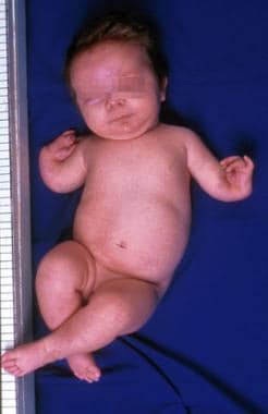
Derivative 22 (der[22]) syndrome is a rare disorder associated with multiple congenital anomalies, including profound mental retardation, preauricular skin tags or pits, and conotruncal heart defects. It can occur in offspring of carriers of the constitutional t(11;22)(q23;q11) translocation, owing to a 3:1 meiotic malsegregation event resulting in partial trisomy of chromosomes 11 and 22. The trisomic region on chromosome 22 overlaps the region hemizygously deleted in another congenital anomaly disorder, velo-cardio-facial syndrome/DiGeorge syndrome (VCFS/DGS). Most patients with VCFS/DGS have a similar 3-Mb deletion, whereas some have a nested distal deletion endpoint resulting in a 1.5-Mb deletion, and a few rare patients have unique deletions. To define the interval on 22q11 containing the t(11;22) breakpoint, haplotype analysis and FISH mapping were performed for five patients with der(22) syndrome. Analysis of all the patients was consistent with 3:1 meiotic malsegregation in the t(11;22) carrier parent. FISH-mapping studies showed that the t(11;22) breakpoint occurred in the same interval as the 1.5-Mb distal deletion breakpoint for VCFS. The deletion breakpoint of one VCFS patient with an unbalanced t(18;22) translocation also occurred in the same region. Hamster-human somatic hybrid cell lines from a patient with der(22) syndrome and a patient with VCFS showed that the breakpoints occurred in an interval containing low-copy repeats, distal to RANBP1 and proximal to ZNF74. The presence of low-copy repetitive sequences may confer susceptibility to chromosome rearrangements. A 1.5-Mb region of overlap on 22q11 in both syndromes suggests the presence of dosage-dependent genes in this interval. (+info)Low-copy repeats mediate the common 3-Mb deletion in patients with velo-cardio-facial syndrome. (3/785)
Velo-cardio-facial syndrome (VCFS) is the most common microdeletion syndrome in humans. It occurs with an estimated frequency of 1 in 4, 000 live births. Most cases occur sporadically, indicating that the deletion is recurrent in the population. More than 90% of patients with VCFS and a 22q11 deletion have a similar 3-Mb hemizygous deletion, suggesting that sequences at the breakpoints confer susceptibility to rearrangements. To define the region containing the chromosome breakpoints, we constructed an 8-kb-resolution physical map. We identified a low-copy repeat in the vicinity of both breakpoints. A set of genetic markers were integrated into the physical map to determine whether the deletions occur within the repeat. Haplotype analysis with genetic markers that flank the repeats showed that most patients with VCFS had deletion breakpoints in the repeat. Within the repeat is a 200-kb duplication of sequences, including a tandem repeat of genes/pseudogenes, surrounding the breakpoints. The genes in the repeat are GGT, BCRL, V7-rel, POM121-like, and GGT-rel. Physical mapping and genomic fingerprint analysis showed that the repeats are virtually identical in the 200-kb region, suggesting that the deletion is mediated by homologous recombination. Examination of two three-generation families showed that meiotic intrachromosomal recombination mediated the deletion. (+info)Delineation of the critical deletion region for congenital heart defects, on chromosome 8p23.1. (4/785)
Deletions in the distal region of chromosome 8p (del8p) are associated with congenital heart malformations. Other major manifestations include microcephaly, intrauterine growth retardation, mental retardation, and a characteristic hyperactive, impulsive behavior. We studied genotype-phenotype correlations in nine unrelated patients with a de novo del8p, by using the combination of classic cytogenetics, FISH, and the analysis of polymorphic DNA markers. With the exception of one large terminal deletion, all deletions were interstitial. In five patients, a commonly deleted region of approximately 6 Mb was present, with breakpoints clustering in the same regions. One patient without a heart defect or microcephaly but with mild mental retardation and characteristic behavior had a smaller deletion within this commonly deleted region. Two patients without a heart defect had a more proximal interstitial deletion that did not overlap with the commonly deleted region. Taken together, these data allowed us to define the critical deletion regions for the major features of a del8p. (+info)Development and validation of a quantitative polymerase chain reaction assay to evaluate minimal residual disease for T-cell acute lymphoblastic leukemia and follicular lymphoma. (5/785)
The presence of occult disease in cancer patients after therapy is one of the major problems faced by oncologists. For example, although 95% of pediatric T-cell acute lymphoblastic leukemia (T-ALL) patients have a complete therapeutic response to multiagent chemotherapy, half will relapse, indicating that they must have harbored low levels of residual cancer cells at the end of therapy. Sensitive detection assays promise to help identify those patients that carry this minimal residual disease (MRD) and are at risk of relapse. We have developed and validated a quantitative polymerase chain reaction (PCR) assay targeting tumor-specific chromosomal rearrangements, including del(1) involving the tal-1 locus in pediatric T-ALL and t(14;18) involving the bcl-2 locus in follicular lymphoma. This quantitative PCR assay utilizes a synthetic internal calibration standard (ICS) that contains priming sequences identical to those found flanking the chromosomal rearrangement breakpoints. Using this ICS-PCR method, the limits of detection were 5 tumor cells at ratios of 1 tumor cell in 10(5) normal cells and a linear range up to 100% tumor cells. This ICS-PCR method has also performed well in terms of precision and accuracy as indicated by low coefficients of variation, minimal random, proportional, and constant errors, and good clinical sensitivity and specificity characteristics. This technique will allow for the evaluation of parameters such as the rate of therapeutic response and the levels of MRD as predictors of patient outcome. (+info)Nonrandom cytogenetic alterations in hepatocellular carcinoma from transgenic mice overexpressing c-Myc and transforming growth factor-alpha in the liver. (6/785)
Identification of specific and primary chromosomal alterations during the course of neoplastic development is an essential part of defining the genetic basis of cancer. We have developed a transgenic mouse model for liver neoplasia in which chromosomal lesions associated with both the initial stages of the neoplastic process and the acquisition of malignancy can be analyzed. Here we analyze chromosomal alterations in 11 hepatocellular carcinomas from the c-myc/TGF-alpha double-transgenic mice by fluorescent in situ hybridization with whole chromosome probes, single-copy genes, and 4'-6-diamidino-2-phenylindole (DAPI-) and G-banded chromosomes and report nonrandom cytogenetic alterations associated with the tumor development. All tumors were aneuploid and exhibited nonrandom structural and numerical alterations. A balanced translocation t(5:6)(G1;F2) was identified by two-color fluorescent in situ hybridization in all tumors, and, using a genomic probe, the c-myc transgene was localized near the breakpoint on derivative chromosome der 6. Partial or complete loss of chromosome 4 was observed in all tumors with nonrandom breakage in band C2. Deletions of chromosome 1 were observed in 80% of the tumors, with the most frequent deletion at the border of bands C4 and C5. An entire copy of chromosome 7 was lost in 80% of the tumors cells. Eighty-five percent of the tumor cells had lost one copy of chromosome 12, and the most common breakpoint on chromosome 12 occurred at band D3 (28%). A copy of chromosome 14 was lost in 72%, and band 14E1 was deleted in 32% of the tumor cells. The X chromosome was lost in the majority of the tumor cells. The most frequent deletion on the X chromosome involved band F1. We have previously shown that breakages of chromosomes 1, 6, 7, and 12 were observed before the appearance of morphologically distinct neoplastic liver lesions in this transgenic mouse model. Thus breakpoints on chromosome 4, 9, 14, and X appear to be later events in this model of liver neoplasia. This is the first study to demonstrate that specific sites of chromosomal breakage observed during a period of chromosomal instability in early stages of carcinogenesis are later involved in stable rearrangements in solid tumors. The identification of the 5;6 translocation in all of the tumors has a special significance, being the first balanced translocation reported in human and mouse hepatocellular carcinoma and having the breakpoint near a tumor susceptibility gene and myc transgene site of integration. Moreover, its early occurrence indicates that this is a primary and relevant alteration to the initiation of the neoplastic process. In addition, the concordance between the breakpoints observed during the early dysplastic stage of hepatocarcinogenesis and the stable deletions of chromosomes 1, 4, 6, 7, 9, and 12 in the tumors provides evidence for preferential site of genetic changes in hepatocarcinogenesis. (+info)Increased chromosomal instability in peripheral lymphocytes and risk of human gliomas. (7/785)
Brain tumors exhibit considerable chromosome instability (CIN), suggesting that genetic susceptibility may contribute to brain tumorigenesis. To test this hypothesis, in this pilot study, we examined for CIN in short-term lymphocyte cultures from 25 adult glioma patients and 28 age-, sex- and ethnicity-matched healthy controls (all Caucasian). We evaluated CIN by a multicolor fluorescence in situ hybridization assay using two probes: a classic satellite probe for a large heterochromatin breakage-prone region of chromosome 1 and an alpha satellite probe for a smaller region adjacent to the heterochromatin probe. Our results showed a significant increase in the mean number of spontaneous breaks per 1000 cells in glioma patients (mean +/- SD, 2.4+/-0.8) compared with controls (1.4+/-0.9; P < 0.001). By using the median number of breaks per 1000 cells in the controls as the cutoff value, we observed a crude odds ratio (OR) of 8.5 [95% confidence interval (CI) = 2.05-34.9, P < 0.001] for spontaneous breaks and brain tumor risk. After adjustment for age, sex and smoking status, the adjusted OR was 15.3 (95% CI, 2.71-87.8). A significant increase in cells with chromosome 1 aneuploidy (in the form of hyperdiploidy) (P < 0.001) was also observed in the glioma cases, with an adjusted OR of 6.6 (95% CI = 1.5-30, P < 0.05). These findings suggest that CIN can be detected in the peripheral blood lymphocytes of brain tumor patients and may be a marker for identifying individuals at risk. (+info)Rearrangements of chromosome band 1p36 in non-Hodgkin's lymphoma. (8/785)
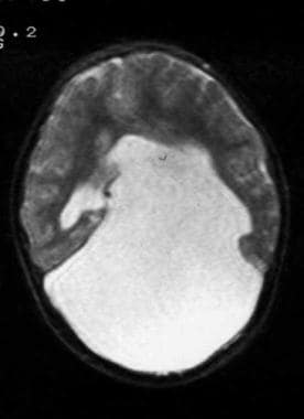
We studied 850 consecutive cases of histologically ascertained pretreatment non-Hodgkin's lymphoma with cytogenetically abnormal clones. The diagnostic karyotypes revealed that 12% of these cases exhibited structural rearrangements involving chromosome band 1p36. Here, we describe the karyotypes of 53 cases containing a 1p36 rearrangement [often involving translocations of unknown material and presented as add(1)(p36)]. We used fluorescence in situ hybridization to determine the origin of the translocation partners. We report three different recurrent translocations involving 1p36. These include der(1)t(1;1)(p36;q21) (three cases), der(1)t(1;1)(p36;q25) (three cases), and der(1)t(1;9)(p36;q13) (four cases). Using cytogenetic and fluorescence in situ hybridization analyses, we have resolved the translocation partners in 31 cases. Rearrangements of band 1p36 were found among different histopathological subtypes. Alterations of 1p36 never occurred as a sole abnormality, and in 42 of 53 cases, alterations of the band 14q32 were observed. The t(14;18)(q32;q21) translocation was present in 35 cases. The significantly high occurrence of 1p36 breakpoint in structural rearrangements and its involvement in recurrent translocations suggest that the region is bearing gene(s) that are important in lymphomagenesis. Our study also showed that cytogenetically evident deletions were frequent in chromosome 1p, almost always involving the p36 region, whereas duplications were rare and never encompassed the p36 region. Chromosome band 1p36 harbors many candidate tumor suppressor genes, and we propose that one or more of these genes might be deleted or functionally disrupted as a molecular consequence of the rearrangements, thus contributing to lymphomagenesis. (+info)Chromosome breakage is a medical term that refers to the breaking or fragmentation of chromosomes, which are thread-like structures located in the nucleus of cells that carry genetic information. Normally, chromosomes are tightly coiled and consist of two strands called chromatids, joined together at a central point called the centromere.
Chromosome breakage can occur spontaneously or be caused by environmental factors such as radiation or chemicals, or inherited genetic disorders. When a chromosome breaks, it can result in various genetic abnormalities, depending on the location and severity of the break.
For instance, if the break occurs in a region containing important genes, it can lead to the loss or alteration of those genes, causing genetic diseases or birth defects. In some cases, the broken ends of the chromosome may rejoin incorrectly, leading to chromosomal rearrangements such as translocations, deletions, or inversions. These rearrangements can also result in genetic disorders or cancer.
Chromosome breakage is commonly observed in individuals with certain inherited genetic conditions, such as Bloom syndrome, Fanconi anemia, and ataxia-telangiectasia, which are characterized by an increased susceptibility to chromosome breakage due to defects in DNA repair mechanisms.
Chromosomes are thread-like structures that exist in the nucleus of cells, carrying genetic information in the form of genes. They are composed of DNA and proteins, and are typically present in pairs in the nucleus, with one set inherited from each parent. In humans, there are 23 pairs of chromosomes for a total of 46 chromosomes. Chromosomes come in different shapes and forms, including sex chromosomes (X and Y) that determine the biological sex of an individual. Changes or abnormalities in the number or structure of chromosomes can lead to genetic disorders and diseases.
Chromosome mapping, also known as physical mapping, is the process of determining the location and order of specific genes or genetic markers on a chromosome. This is typically done by using various laboratory techniques to identify landmarks along the chromosome, such as restriction enzyme cutting sites or patterns of DNA sequence repeats. The resulting map provides important information about the organization and structure of the genome, and can be used for a variety of purposes, including identifying the location of genes associated with genetic diseases, studying evolutionary relationships between organisms, and developing genetic markers for use in breeding or forensic applications.
Chromosome fragility refers to the susceptibility of specific regions on chromosomes to break or become unstable during cell division. These fragile sites are prone to forming gaps or breaks in the chromosome structure, which can lead to genetic rearrangements, including deletions, duplications, or translocations.
Chromosome fragility is often associated with certain genetic disorders and syndromes. For example, the most common fragile site in human chromosomes is FRAXA, located on the X chromosome, which is linked to Fragile X Syndrome, a leading cause of inherited intellectual disability and autism.
Environmental factors such as exposure to chemicals or radiation can also increase chromosome fragility, leading to an increased risk of genetic mutations and diseases.
Chromosome aberrations refer to structural and numerical changes in the chromosomes that can occur spontaneously or as a result of exposure to mutagenic agents. These changes can affect the genetic material encoded in the chromosomes, leading to various consequences such as developmental abnormalities, cancer, or infertility.
Structural aberrations include deletions, duplications, inversions, translocations, and rings, which result from breaks and rearrangements of chromosome segments. Numerical aberrations involve changes in the number of chromosomes, such as aneuploidy (extra or missing chromosomes) or polyploidy (multiples of a complete set of chromosomes).
Chromosome aberrations can be detected and analyzed using various cytogenetic techniques, including karyotyping, fluorescence in situ hybridization (FISH), and comparative genomic hybridization (CGH). These methods allow for the identification and characterization of chromosomal changes at the molecular level, providing valuable information for genetic counseling, diagnosis, and research.
Chromosome fragile sites are specific locations along the length of a chromosome that are prone to breakage or rearrangement when exposed to certain chemicals or conditions, such as replication stress during cell division. These sites are often characterized by the presence of repetitive DNA sequences and proteins that help maintain the stability of the chromosome.
Fragile sites can be classified into two categories: common and rare. Common fragile sites are present in most individuals and are typically not associated with genetic disorders, while rare fragile sites are less common and may be linked to specific genetic conditions or increased risk for cancer.
When a chromosome breaks at a fragile site, it can lead to various genetic abnormalities such as deletions, duplications, inversions, or translocations of genetic material. These changes can have significant consequences on gene expression and function, potentially leading to developmental disorders, intellectual disability, cancer, or other health issues.
It is important to note that not all fragile sites will result in genetic abnormalities, as some may remain stable under normal conditions. However, certain factors such as environmental exposures, aging, or inherited genetic predispositions can increase the likelihood of chromosomal instability at fragile sites.
Tetrahymena thermophila is not a medical term, but rather it refers to a species of ciliated protozoan that is commonly used in scientific research, including biomedical research. Here's a brief biological definition:
Tetrahymena thermophila is a free-living, freshwater ciliate protozoan found in various aquatic environments. It has a complex cell structure with two types of nuclei (a macronucleus and a micronucleus) and numerous cilia for movement. This organism is known for its ability to reproduce both sexually and asexually, making it a valuable model for studying genetic processes. Its genome has been fully sequenced, and it is widely used in research fields such as molecular biology, cell biology, and genetics due to its ease of cultivation and manipulation.
While not directly related to medical terminology, Tetrahymena thermophila has contributed significantly to our understanding of various biological processes with potential implications for medical research, including gene regulation, protein function, and DNA repair mechanisms.
Chromosome banding is a technique used in cytogenetics to identify and describe the physical structure and organization of chromosomes. This method involves staining the chromosomes with specific dyes that bind differently to the DNA and proteins in various regions of the chromosome, resulting in a distinct pattern of light and dark bands when viewed under a microscope.
The most commonly used banding techniques are G-banding (Giemsa banding) and R-banding (reverse banding). In G-banding, the chromosomes are stained with Giemsa dye, which preferentially binds to the AT-rich regions, creating a characteristic banding pattern. The bands are numbered from the centromere (the constriction point where the chromatids join) outwards, with the darker bands (rich in A-T base pairs and histone proteins) labeled as "q" arms and the lighter bands (rich in G-C base pairs and arginine-rich proteins) labeled as "p" arms.
R-banding, on the other hand, uses a different staining procedure that results in a reversed banding pattern compared to G-banding. The darker R-bands correspond to the lighter G-bands, and vice versa. This technique is particularly useful for identifying and analyzing specific regions of chromosomes that may be difficult to visualize with G-banding alone.
Chromosome banding plays a crucial role in diagnosing genetic disorders, identifying chromosomal abnormalities, and studying the structure and function of chromosomes in both clinical and research settings.
Chromosome disorders are a group of genetic conditions caused by abnormalities in the number or structure of chromosomes. Chromosomes are thread-like structures located in the nucleus of cells that contain most of the body's genetic material, which is composed of DNA and proteins. Normally, humans have 23 pairs of chromosomes, for a total of 46 chromosomes.
Chromosome disorders can result from changes in the number of chromosomes (aneuploidy) or structural abnormalities in one or more chromosomes. Some common examples of chromosome disorders include:
1. Down syndrome: a condition caused by an extra copy of chromosome 21, resulting in intellectual disability, developmental delays, and distinctive physical features.
2. Turner syndrome: a condition that affects only females and is caused by the absence of all or part of one X chromosome, resulting in short stature, lack of sexual development, and other symptoms.
3. Klinefelter syndrome: a condition that affects only males and is caused by an extra copy of the X chromosome, resulting in tall stature, infertility, and other symptoms.
4. Cri-du-chat syndrome: a condition caused by a deletion of part of the short arm of chromosome 5, resulting in intellectual disability, developmental delays, and a distinctive cat-like cry.
5. Fragile X syndrome: a condition caused by a mutation in the FMR1 gene on the X chromosome, resulting in intellectual disability, behavioral problems, and physical symptoms.
Chromosome disorders can be diagnosed through various genetic tests, such as karyotyping, chromosomal microarray analysis (CMA), or fluorescence in situ hybridization (FISH). Treatment for these conditions depends on the specific disorder and its associated symptoms and may include medical interventions, therapies, and educational support.
The X chromosome is one of the two types of sex-determining chromosomes in humans (the other being the Y chromosome). It's one of the 23 pairs of chromosomes that make up a person's genetic material. Females typically have two copies of the X chromosome (XX), while males usually have one X and one Y chromosome (XY).
The X chromosome contains hundreds of genes that are responsible for the production of various proteins, many of which are essential for normal bodily functions. Some of the critical roles of the X chromosome include:
1. Sex Determination: The presence or absence of the Y chromosome determines whether an individual is male or female. If there is no Y chromosome, the individual will typically develop as a female.
2. Genetic Disorders: Since females have two copies of the X chromosome, they are less likely to be affected by X-linked genetic disorders than males. Males, having only one X chromosome, will express any recessive X-linked traits they inherit.
3. Dosage Compensation: To compensate for the difference in gene dosage between males and females, a process called X-inactivation occurs during female embryonic development. One of the two X chromosomes is randomly inactivated in each cell, resulting in a single functional copy per cell.
The X chromosome plays a crucial role in human genetics and development, contributing to various traits and characteristics, including sex determination and dosage compensation.
A chromosome deletion is a type of genetic abnormality that occurs when a portion of a chromosome is missing or deleted. Chromosomes are thread-like structures located in the nucleus of cells that contain our genetic material, which is organized into genes.
Chromosome deletions can occur spontaneously during the formation of reproductive cells (eggs or sperm) or can be inherited from a parent. They can affect any chromosome and can vary in size, from a small segment to a large portion of the chromosome.
The severity of the symptoms associated with a chromosome deletion depends on the size and location of the deleted segment. In some cases, the deletion may be so small that it does not cause any noticeable symptoms. However, larger deletions can lead to developmental delays, intellectual disabilities, physical abnormalities, and various medical conditions.
Chromosome deletions are typically detected through a genetic test called karyotyping, which involves analyzing the number and structure of an individual's chromosomes. Other more precise tests, such as fluorescence in situ hybridization (FISH) or chromosomal microarray analysis (CMA), may also be used to confirm the diagnosis and identify the specific location and size of the deletion.
In situ hybridization, fluorescence (FISH) is a type of molecular cytogenetic technique used to detect and localize the presence or absence of specific DNA sequences on chromosomes through the use of fluorescent probes. This technique allows for the direct visualization of genetic material at a cellular level, making it possible to identify chromosomal abnormalities such as deletions, duplications, translocations, and other rearrangements.
The process involves denaturing the DNA in the sample to separate the double-stranded molecules into single strands, then adding fluorescently labeled probes that are complementary to the target DNA sequence. The probe hybridizes to the complementary sequence in the sample, and the location of the probe is detected by fluorescence microscopy.
FISH has a wide range of applications in both clinical and research settings, including prenatal diagnosis, cancer diagnosis and monitoring, and the study of gene expression and regulation. It is a powerful tool for identifying genetic abnormalities and understanding their role in human disease.
Sex chromosomes, often denoted as X and Y, are one of the 23 pairs of human chromosomes found in each cell of the body. Normally, females have two X chromosomes (46,XX), and males have one X and one Y chromosome (46,XY). The sex chromosomes play a significant role in determining the sex of an individual. They contain genes that contribute to physical differences between men and women. Any variations or abnormalities in the number or structure of these chromosomes can lead to various genetic disorders and conditions related to sexual development and reproduction.
Human chromosome pair 1 refers to the first pair of chromosomes in a set of 23 pairs found in the cells of the human body, excluding sex cells (sperm and eggs). Each cell in the human body, except for the gametes, contains 46 chromosomes arranged in 23 pairs. These chromosomes are rod-shaped structures that contain genetic information in the form of DNA.
Chromosome pair 1 is the largest pair, making up about 8% of the total DNA in a cell. Each chromosome in the pair consists of two arms - a shorter p arm and a longer q arm - connected at a centromere. Chromosome 1 carries an estimated 2,000-2,500 genes, which are segments of DNA that contain instructions for making proteins or regulating gene expression.
Defects or mutations in the genes located on chromosome 1 can lead to various genetic disorders and diseases, such as Charcot-Marie-Tooth disease type 1A, Huntington's disease, and certain types of cancer.
Fanconi anemia is a rare, inherited disorder that affects the body's ability to produce healthy blood cells. It is characterized by bone marrow failure, congenital abnormalities, and an increased risk of developing certain types of cancer. The condition is caused by mutations in genes responsible for repairing damaged DNA, leading to chromosomal instability and cell death.
The classic form of Fanconi anemia (type A) is typically diagnosed in childhood and is associated with various physical abnormalities such as short stature, skin pigmentation changes, thumb and radial ray anomalies, kidney and genitourinary malformations, and developmental delays. Other types of Fanconi anemia (B-G) may have different clinical presentations but share the common feature of bone marrow failure and cancer predisposition.
Bone marrow failure in Fanconi anemia results in decreased production of all three types of blood cells: red blood cells, white blood cells, and platelets. This can lead to anemia (low red blood cell count), neutropenia (low white blood cell count), and thrombocytopenia (low platelet count). These conditions increase the risk of infections, fatigue, and bleeding.
Individuals with Fanconi anemia have a significantly higher risk of developing various types of cancer, particularly acute myeloid leukemia (AML) and solid tumors such as squamous cell carcinomas of the head, neck, esophagus, and anogenital region.
Treatment for Fanconi anemia typically involves managing symptoms related to bone marrow failure, such as transfusions, growth factors, and antibiotics. Hematopoietic stem cell transplantation (HSCT) is the only curative treatment option for bone marrow failure but carries risks of its own, including graft-versus-host disease and transplant-related mortality. Regular cancer surveillance is essential due to the increased risk of malignancies in these patients.
Chromosomes are thread-like structures that contain genetic material, i.e., DNA and proteins, present in the nucleus of human cells. In humans, there are 23 pairs of chromosomes, for a total of 46 chromosomes, in each diploid cell. Twenty-two of these pairs are called autosomal chromosomes, which come in identical pairs and contain genes that determine various traits unrelated to sex.
The last pair is referred to as the sex chromosomes (X and Y), which determines a person's biological sex. Females have two X chromosomes (46, XX), while males possess one X and one Y chromosome (46, XY). Chromosomes vary in size, with the largest being chromosome 1 and the smallest being the Y chromosome.
Human chromosomes are typically visualized during mitosis or meiosis using staining techniques that highlight their banding patterns, allowing for identification of specific regions and genes. Chromosomal abnormalities can lead to various genetic disorders, including Down syndrome (trisomy 21), Turner syndrome (monosomy X), and Klinefelter syndrome (XXY).
A micronucleus test is a type of genetic toxicology assay used to detect the presence of micronuclei in cells, which are small chromosomal fragments or whole chromosomes that have been missegregated during cell division. The test measures the frequency of micronuclei in cells exposed to a potential genotoxic agent, such as a chemical or radiation, and compares it to the frequency in untreated control cells.
The assay is typically performed on cultured mammalian cells, such as human lymphocytes or Chinese hamster ovary (CHO) cells, and involves exposing the cells to the test agent for a specific period of time, followed by staining and examination of the cells under a microscope. The micronuclei are identified based on their size, shape, and staining characteristics, and the frequency of micronucleated cells is calculated as a measure of genotoxic potential.
Micronucleus tests are widely used in regulatory toxicology to assess the genetic safety of chemicals, drugs, and other substances, and can provide valuable information on potential risks to human health. The test is also used in basic research to study the mechanisms of genotoxicity and chromosomal instability.
Aneuploidy is a medical term that refers to an abnormal number of chromosomes in a cell. Chromosomes are thread-like structures located inside the nucleus of cells that contain genetic information in the form of genes.
In humans, the normal number of chromosomes in a cell is 46, arranged in 23 pairs. Aneuploidy occurs when there is an extra or missing chromosome in one or more of these pairs. For example, Down syndrome is a condition that results from an extra copy of chromosome 21, also known as trisomy 21.
Aneuploidy can arise during the formation of gametes (sperm or egg cells) due to errors in the process of cell division called meiosis. These errors can result in eggs or sperm with an abnormal number of chromosomes, which can then lead to aneuploidy in the resulting embryo.
Aneuploidy is a significant cause of birth defects and miscarriages. The severity of the condition depends on which chromosomes are affected and the extent of the abnormality. In some cases, aneuploidy may have no noticeable effects, while in others it can lead to serious health problems or developmental delays.
Karyotyping is a medical laboratory test used to study the chromosomes in a cell. It involves obtaining a sample of cells from a patient, usually from blood or bone marrow, and then staining the chromosomes so they can be easily seen under a microscope. The chromosomes are then arranged in pairs based on their size, shape, and other features to create a karyotype. This visual representation allows for the identification and analysis of any chromosomal abnormalities, such as extra or missing chromosomes, or structural changes like translocations or inversions. These abnormalities can provide important information about genetic disorders, diseases, and developmental problems.
A telomere is a region of repetitive DNA sequences found at the end of chromosomes, which protects the genetic data from damage and degradation during cell division. Telomeres naturally shorten as cells divide, and when they become too short, the cell can no longer divide and becomes senescent or dies. This natural process is associated with aging and various age-related diseases. The length of telomeres can also be influenced by various genetic and environmental factors, including stress, diet, and lifestyle.
Chromosome segregation is the process that occurs during cell division (mitosis or meiosis) where replicated chromosomes are separated and distributed equally into two daughter cells. Each chromosome consists of two sister chromatids, which are identical copies of genetic material. During chromosome segregation, these sister chromatids are pulled apart by a structure called the mitotic spindle and moved to opposite poles of the cell. This ensures that each new cell receives one copy of each chromosome, preserving the correct number and composition of chromosomes in the organism.
Bacterial chromosomes are typically circular, double-stranded DNA molecules that contain the genetic material of bacteria. Unlike eukaryotic cells, which have their DNA housed within a nucleus, bacterial chromosomes are located in the cytoplasm of the cell, often associated with the bacterial nucleoid.
Bacterial chromosomes can vary in size and structure among different species, but they typically contain all of the genetic information necessary for the survival and reproduction of the organism. They may also contain plasmids, which are smaller circular DNA molecules that can carry additional genes and can be transferred between bacteria through a process called conjugation.
One important feature of bacterial chromosomes is their ability to replicate rapidly, allowing bacteria to divide quickly and reproduce in large numbers. The replication of the bacterial chromosome begins at a specific origin point and proceeds in opposite directions until the entire chromosome has been copied. This process is tightly regulated and coordinated with cell division to ensure that each daughter cell receives a complete copy of the genetic material.
Overall, the study of bacterial chromosomes is an important area of research in microbiology, as understanding their structure and function can provide insights into bacterial genetics, evolution, and pathogenesis.
Mitosis is a type of cell division in which the genetic material of a single cell, called the mother cell, is equally distributed into two identical daughter cells. It's a fundamental process that occurs in multicellular organisms for growth, maintenance, and repair, as well as in unicellular organisms for reproduction.
The process of mitosis can be broken down into several stages: prophase, prometaphase, metaphase, anaphase, and telophase. During prophase, the chromosomes condense and become visible, and the nuclear envelope breaks down. In prometaphase, the nuclear membrane is completely disassembled, and the mitotic spindle fibers attach to the chromosomes at their centromeres.
During metaphase, the chromosomes align at the metaphase plate, an imaginary line equidistant from the two spindle poles. In anaphase, sister chromatids are pulled apart by the spindle fibers and move toward opposite poles of the cell. Finally, in telophase, new nuclear envelopes form around each set of chromosomes, and the chromosomes decondense and become less visible.
Mitosis is followed by cytokinesis, a process that divides the cytoplasm of the mother cell into two separate daughter cells. The result of mitosis and cytokinesis is two genetically identical cells, each with the same number and kind of chromosomes as the original parent cell.
Human chromosome pair 11 consists of two rod-shaped structures present in the nucleus of each cell in the human body. Each member of the pair is a single chromosome, and together they contain the genetic material that is inherited from both parents. They are located on the eleventh position in the standard karyotype, which is a visual representation of the 23 pairs of human chromosomes.
Chromosome 11 is one of the largest human chromosomes and contains an estimated 135 million base pairs. It contains approximately 1,400 genes that provide instructions for making proteins, as well as many non-coding RNA molecules that play a role in regulating gene expression.
Chromosome 11 is known to contain several important genes and genetic regions associated with various human diseases and conditions. For example, it contains the Wilms' tumor 1 (WT1) gene, which is associated with kidney cancer in children, and the neurofibromatosis type 1 (NF1) gene, which is associated with a genetic disorder that causes benign tumors to grow on nerves throughout the body. Additionally, chromosome 11 contains the region where the ABO blood group genes are located, which determine a person's blood type.
It's worth noting that human chromosomes come in pairs because they contain two copies of each gene, one inherited from the mother and one from the father. This redundancy allows for genetic diversity and provides a backup copy of essential genes, ensuring their proper function and maintaining the stability of the genome.
A centromere is a specialized region found on chromosomes that plays a crucial role in the separation of replicated chromosomes during cell division. It is the point where the sister chromatids (the two copies of a chromosome formed during DNA replication) are joined together. The centromere contains highly repeated DNA sequences and proteins that form a complex structure known as the kinetochore, which serves as an attachment site for microtubules of the mitotic spindle during cell division.
During mitosis or meiosis, the kinetochore facilitates the movement of chromosomes by interacting with the microtubules, allowing for the accurate distribution of genetic material to the daughter cells. Centromeres can vary in their position and structure among different species, ranging from being located near the middle of the chromosome (metacentric) to being positioned closer to one end (acrocentric). The precise location and characteristics of centromeres are essential for proper chromosome segregation and maintenance of genomic stability.
Human chromosome pair 7 consists of two rod-shaped structures present in the nucleus of each cell in the human body. Each member of the pair is a single chromosome, and together they contain the genetic material that is inherited from both parents. They are identical in size, shape, and banding pattern and are therefore referred to as homologous chromosomes.
Chromosome 7 is one of the autosomal chromosomes, meaning it is not a sex chromosome (X or Y). It is composed of double-stranded DNA that contains approximately 159 million base pairs and around 1,200 genes. Chromosome 7 contains several important genes associated with human health and disease, including those involved in the development of certain types of cancer, such as colon cancer and lung cancer, as well as genetic disorders such as Williams-Beuren syndrome and Charcot-Marie-Tooth disease.
Abnormalities in chromosome 7 have been linked to various genetic conditions, including deletions, duplications, translocations, and other structural changes. These abnormalities can lead to developmental delays, intellectual disabilities, physical abnormalities, and increased risk of certain types of cancer.
Molecular sequence data refers to the specific arrangement of molecules, most commonly nucleotides in DNA or RNA, or amino acids in proteins, that make up a biological macromolecule. This data is generated through laboratory techniques such as sequencing, and provides information about the exact order of the constituent molecules. This data is crucial in various fields of biology, including genetics, evolution, and molecular biology, allowing for comparisons between different organisms, identification of genetic variations, and studies of gene function and regulation.
DNA damage refers to any alteration in the structure or composition of deoxyribonucleic acid (DNA), which is the genetic material present in cells. DNA damage can result from various internal and external factors, including environmental exposures such as ultraviolet radiation, tobacco smoke, and certain chemicals, as well as normal cellular processes such as replication and oxidative metabolism.
Examples of DNA damage include base modifications, base deletions or insertions, single-strand breaks, double-strand breaks, and crosslinks between the two strands of the DNA helix. These types of damage can lead to mutations, genomic instability, and chromosomal aberrations, which can contribute to the development of diseases such as cancer, neurodegenerative disorders, and aging-related conditions.
The body has several mechanisms for repairing DNA damage, including base excision repair, nucleotide excision repair, mismatch repair, and double-strand break repair. However, if the damage is too extensive or the repair mechanisms are impaired, the cell may undergo apoptosis (programmed cell death) to prevent the propagation of potentially harmful mutations.
A base sequence in the context of molecular biology refers to the specific order of nucleotides in a DNA or RNA molecule. In DNA, these nucleotides are adenine (A), guanine (G), cytosine (C), and thymine (T). In RNA, uracil (U) takes the place of thymine. The base sequence contains genetic information that is transcribed into RNA and ultimately translated into proteins. It is the exact order of these bases that determines the genetic code and thus the function of the DNA or RNA molecule.
Micronuclei, chromosome-defective, refer to small additional nuclei that form during cell division when the genetic material is not properly divided between the two resulting daughter cells. These micronuclei can contain whole chromosomes or fragments of chromosomes that were not incorporated into either of the main nuclei during cell division. Chromosome-defective micronuclei are often associated with genomic instability, DNA damage, and chromosomal aberrations, which can lead to various health issues, including cancer and developmental defects. They can be used as a biomarker for genetic damage in cells and are commonly observed in response to exposure to mutagenic agents such as radiation or chemicals.
Human chromosome pair 17 consists of two rod-shaped structures present in the nucleus of each human cell. Each chromosome is made up of DNA tightly coiled around histone proteins, forming a complex called chromatin. Chromosomes carry genetic information in the form of genes, which are segments of DNA that contain instructions for the development and function of an organism.
Human cells typically have 23 pairs of chromosomes, for a total of 46 chromosomes. Pair 17 is one of the autosomal pairs, meaning it is not a sex chromosome (X or Y). Chromosome 17 is a medium-sized chromosome and contains an estimated 800 million base pairs of DNA. It contains approximately 1,500 genes that provide instructions for making proteins and regulating various cellular processes.
Chromosome 17 is associated with several genetic disorders, including inherited cancer syndromes such as Li-Fraumeni syndrome and hereditary nonpolyposis colorectal cancer (HNPCC). Mutations in genes located on chromosome 17 can increase the risk of developing various types of cancer, including breast, ovarian, colon, and pancreatic cancer.
Translocation, genetic, refers to a type of chromosomal abnormality in which a segment of a chromosome is transferred from one chromosome to another, resulting in an altered genome. This can occur between two non-homologous chromosomes (non-reciprocal translocation) or between two homologous chromosomes (reciprocal translocation). Genetic translocations can lead to various clinical consequences, depending on the genes involved and the location of the translocation. Some translocations may result in no apparent effects, while others can cause developmental abnormalities, cancer, or other genetic disorders. In some cases, translocations can also increase the risk of having offspring with genetic conditions.
Sister chromatid exchange (SCE) is a type of genetic recombination that takes place between two identical sister chromatids during the DNA repair process in meiosis or mitosis. It results in an exchange of genetic material between the two chromatids, creating a new combination of genes on each chromatid. This event is a normal part of cell division and helps to increase genetic variability within a population. However, an increased rate of SCEs can also be indicative of exposure to certain genotoxic agents or conditions that cause DNA damage.
Human chromosome pair 6 consists of two rod-shaped structures present in the nucleus of each human cell. They are identical in size and shape and contain genetic material, made up of DNA and proteins, that is essential for the development and function of the human body.
Chromosome pair 6 is one of the 23 pairs of chromosomes found in humans, with one chromosome inherited from each parent. Each chromosome contains thousands of genes that provide instructions for the production of proteins and regulate various cellular processes.
Chromosome pair 6 contains several important genes, including those involved in the development and function of the immune system, such as the major histocompatibility complex (MHC) genes. It also contains genes associated with certain genetic disorders, such as hereditary neuropathy with liability to pressure palsies (HNPP), a condition that affects the nerves, and Waardenburg syndrome, a disorder that affects pigmentation and hearing.
Abnormalities in chromosome pair 6 can lead to various genetic disorders, including numerical abnormalities such as trisomy 6 (three copies of chromosome 6) or monosomy 6 (only one copy of chromosome 6), as well as structural abnormalities such as deletions, duplications, or translocations of parts of the chromosome.
Human chromosome pair 9 consists of two rod-shaped structures present in the nucleus of each cell of the human body. Each member of the pair contains thousands of genes and other genetic material, encoded in the form of DNA molecules. The two chromosomes in a pair are identical or very similar to each other in terms of their size, shape, and genetic makeup.
Chromosome 9 is one of the autosomal chromosomes, meaning that it is not a sex chromosome (X or Y) and is present in two copies in all cells of the body, regardless of sex. Chromosome 9 is a medium-sized chromosome, and it is estimated to contain around 135 million base pairs of DNA and approximately 1200 genes.
Chromosome 9 contains several important genes that are associated with various human traits and diseases. For example, mutations in the gene that encodes the protein APOE on chromosome 9 have been linked to an increased risk of developing Alzheimer's disease. Additionally, variations in the gene that encodes the protein EGFR on chromosome 9 have been associated with an increased risk of developing certain types of cancer.
Overall, human chromosome pair 9 plays a critical role in the development and function of the human body, and variations in its genetic makeup can contribute to a wide range of traits and diseases.
Genetic recombination is the process by which genetic material is exchanged between two similar or identical molecules of DNA during meiosis, resulting in new combinations of genes on each chromosome. This exchange occurs during crossover, where segments of DNA are swapped between non-sister homologous chromatids, creating genetic diversity among the offspring. It is a crucial mechanism for generating genetic variability and facilitating evolutionary change within populations. Additionally, recombination also plays an essential role in DNA repair processes through mechanisms such as homologous recombinational repair (HRR) and non-homologous end joining (NHEJ).
Aphidicolin is an antimicrotubule agent that is specifically a inhibitor of DNA polymerase alpha. It is an antibiotic that is produced by the fungus Cephalosporium aphidicola and is used in research to study the cell cycle and DNA replication. In clinical medicine, it has been explored as a potential anticancer agent, although its use is not currently approved for this indication.
Chromosomes in fungi are thread-like structures that contain genetic material, composed of DNA and proteins, present in the nucleus of a cell. Unlike humans and other eukaryotes that have a diploid number of chromosomes in their somatic cells, fungal chromosome numbers can vary widely between and within species.
Fungal chromosomes are typically smaller and fewer in number compared to those found in plants and animals. The chromosomal organization in fungi is also different from other eukaryotes. In many fungi, the chromosomes are condensed throughout the cell cycle, whereas in other eukaryotes, chromosomes are only condensed during cell division.
Fungi can have linear or circular chromosomes, depending on the species. For example, the model organism Saccharomyces cerevisiae (budding yeast) has a set of 16 small circular chromosomes, while other fungi like Neurospora crassa (red bread mold) and Aspergillus nidulans (a filamentous fungus) have linear chromosomes.
Fungal chromosomes play an essential role in the growth, development, reproduction, and survival of fungi. They carry genetic information that determines various traits such as morphology, metabolism, pathogenicity, and resistance to environmental stresses. Advances in genomic technologies have facilitated the study of fungal chromosomes, leading to a better understanding of their structure, function, and evolution.
Chromosomes in plants are thread-like structures that contain genetic material, DNA, and proteins. They are present in the nucleus of every cell and are inherited from the parent plants during sexual reproduction. Chromosomes come in pairs, with each pair consisting of one chromosome from each parent.
In plants, like in other organisms, chromosomes play a crucial role in inheritance, development, and reproduction. They carry genetic information that determines various traits and characteristics of the plant, such as its physical appearance, growth patterns, and resistance to diseases.
Plant chromosomes are typically much larger than those found in animals, making them easier to study under a microscope. The number of chromosomes varies among different plant species, ranging from as few as 2 in some ferns to over 1000 in certain varieties of wheat.
During cell division, the chromosomes replicate and then separate into two identical sets, ensuring that each new cell receives a complete set of genetic information. This process is critical for the growth and development of the plant, as well as for the production of viable seeds and offspring.
Human chromosome pair 21 consists of two rod-shaped structures present in the nucleus of each cell in the human body. Each member of the pair is a single chromosome, and they are identical to each other. Chromosomes are made up of DNA, which contains genetic information that determines many of an individual's traits and characteristics.
Chromosome pair 21 is one of the 23 pairs of human autosomal chromosomes, meaning they are not sex chromosomes (X or Y). Chromosome pair 21 is the smallest of the human chromosomes, and it contains approximately 48 million base pairs of DNA. It contains around 200-300 genes that provide instructions for making proteins and regulating various cellular processes.
Down syndrome, a genetic disorder characterized by intellectual disability, developmental delays, distinct facial features, and sometimes heart defects, is caused by an extra copy of chromosome pair 21 or a part of it. This additional genetic material can lead to abnormalities in brain development and function, resulting in the characteristic symptoms of Down syndrome.
A protozoan genome refers to the complete set of genetic material or DNA present in a protozoan organism. Protozoa are single-celled eukaryotic microorganisms that lack cell walls and have diverse morphology and nutrition modes. The genome of a protozoan includes all the genes that code for proteins, as well as non-coding DNA sequences that regulate gene expression and other cellular processes.
The size and complexity of protozoan genomes can vary widely depending on the species. Some protozoa have small genomes with only a few thousand genes, while others have larger genomes with tens of thousands of genes or more. The genome sequencing of various protozoan species has provided valuable insights into their evolutionary history, biology, and potential as model organisms for studying eukaryotic cellular processes.
It is worth noting that the study of protozoan genomics is still an active area of research, and new discoveries are continually being made about the genetic diversity and complexity of these fascinating microorganisms.
Chromosomal instability is a term used in genetics to describe a type of genetic alteration where there are abnormalities in the number or structure of chromosomes within cells. Chromosomes are thread-like structures that contain our genetic material, and they usually exist in pairs in the nucleus of a cell.
Chromosomal instability can arise due to various factors, including errors in DNA replication or repair, problems during cell division, or exposure to environmental mutagens. This instability can lead to an increased frequency of chromosomal abnormalities, such as deletions, duplications, translocations, or changes in the number of chromosomes.
Chromosomal instability is associated with several human diseases, including cancer. In cancer cells, chromosomal instability can contribute to tumor heterogeneity, drug resistance, and disease progression. It is also observed in certain genetic disorders, such as Down syndrome, where an extra copy of chromosome 21 is present, and in some rare inherited syndromes, such as Bloom syndrome and Fanconi anemia, which are characterized by a high risk of cancer and other health problems.
X-rays, also known as radiographs, are a type of electromagnetic radiation with higher energy and shorter wavelength than visible light. In medical imaging, X-rays are used to produce images of the body's internal structures, such as bones and organs, by passing the X-rays through the body and capturing the resulting shadows or patterns on a specialized film or digital detector.
The amount of X-ray radiation used is carefully controlled to minimize exposure and ensure patient safety. Different parts of the body absorb X-rays at different rates, allowing for contrast between soft tissues and denser structures like bone. This property makes X-rays an essential tool in diagnosing and monitoring a wide range of medical conditions, including fractures, tumors, infections, and foreign objects within the body.
Chromosomes are thread-like structures that contain genetic material, made up of DNA and proteins, in the nucleus of cells. In humans, there are typically 46 chromosomes arranged in 23 pairs, with one member of each pair coming from each parent. The six pairs of chromosomes numbered 6 through 12, along with the X chromosome, are part of these 23 pairs and are referred to as autosomal chromosomes and a sex chromosome.
Human chromosome 6 is one of the autosomal chromosomes and contains an estimated 170 million base pairs and around 1,500 genes. It plays a role in several important functions, including immune response, cell signaling, and nervous system function.
Human chromosome 7 is another autosomal chromosome that contains approximately 159 million base pairs and around 1,200 genes. Chromosome 7 is best known for containing the gene for the cystic fibrosis transmembrane conductance regulator (CFTR) protein, whose mutations can lead to cystic fibrosis.
Human chromosome 8 is an autosomal chromosome that contains around 146 million base pairs and approximately 900 genes. Chromosome 8 has been associated with several genetic disorders, including Smith-Magenis syndrome and 8p deletion syndrome.
Human chromosome 9 is an autosomal chromosome that contains around 139 million base pairs and approximately 950 genes. Chromosome 9 has been linked to several genetic disorders, including Hereditary Spherocytosis and CHARGE syndrome.
Human chromosome 10 is an autosomal chromosome that contains around 135 million base pairs and approximately 800 genes. Chromosome 10 has been associated with several genetic disorders, including Dyschondrosteosis and Melanoma.
Human chromosome 11 is an autosomal chromosome that contains around 135 million base pairs and approximately 800 genes. Chromosome 11 has been linked to several genetic disorders, including Wilms tumor and Beckwith-Wiedemann syndrome.
Human chromosome 12 is an autosomal chromosome that contains around 133 million base pairs and approximately 750 genes. Chromosome 12 has been associated with several genetic disorders, including Charcot-Marie-Tooth disease type 1A and Hereditary Neuropathy with Liability to Pressure Palsies (HNPP).
The X chromosome is one of the two sex chromosomes in humans. Females have two X chromosomes, while males have one X and one Y chromosome. The X chromosome contains around 155 million base pairs and approximately 1,000 genes. It has been linked to several genetic disorders, including Duchenne muscular dystrophy and Fragile X syndrome.
The Y chromosome is the other sex chromosome in humans. Males have one X and one Y chromosome, while females have two X chromosomes. The Y chromosome contains around 59 million base pairs and approximately 70 genes. It is primarily responsible for male sexual development and fertility.
In summary, the human genome consists of 23 pairs of chromosomes, including 22 autosomal pairs and one sex chromosome pair (XX in females and XY in males). The total length of the human genome is approximately 3 billion base pairs, and it contains around 20,000-25,000 protein-coding genes. Chromosomes are made up of DNA and proteins called histones, which help to package the DNA into a compact structure. The chromosomes contain genetic information that is passed down from parents to their offspring through reproduction.
Human chromosome pair 16 consists of two rod-shaped structures present in the nucleus of each cell in the human body. Each chromosome is made up of DNA tightly coiled around histone proteins, forming a complex structure called a chromatin.
Chromosomes come in pairs, with one chromosome inherited from each parent. Chromosome pair 16 contains two homologous chromosomes, which are similar in size, shape, and genetic content but may have slight variations due to differences in the DNA sequences inherited from each parent.
Chromosome pair 16 is one of the 22 autosomal pairs, meaning it contains non-sex chromosomes that are present in both males and females. Chromosome 16 is a medium-sized chromosome, and it contains around 2,800 genes that provide instructions for making proteins and regulating various cellular processes.
Abnormalities in chromosome pair 16 can lead to genetic disorders such as chronic myeloid leukemia, some forms of mental retardation, and other developmental abnormalities.
Human chromosome pair 2 consists of two rod-shaped structures present in the nucleus of each cell of the human body. Each member of the pair contains thousands of genes and other genetic material, encoded in the form of DNA molecules. Chromosomes are the physical carriers of inheritance, and human cells typically contain 23 pairs of chromosomes for a total of 46 chromosomes.
Chromosome pair 2 is one of the autosomal pairs, meaning that it is not a sex chromosome (X or Y). Each member of chromosome pair 2 is approximately 247 million base pairs in length and contains an estimated 1,000-1,300 genes. These genes play crucial roles in various biological processes, including development, metabolism, and response to environmental stimuli.
Abnormalities in chromosome pair 2 can lead to genetic disorders, such as cat-eye syndrome (CES), which is characterized by iris abnormalities, anal atresia, hearing loss, and intellectual disability. This disorder arises from the presence of an extra copy of a small region on chromosome 2, resulting in partial trisomy of this region. Other genetic conditions associated with chromosome pair 2 include proximal 2q13.3 microdeletion syndrome and Potocki-Lupski syndrome (PTLS).
A mutation is a permanent change in the DNA sequence of an organism's genome. Mutations can occur spontaneously or be caused by environmental factors such as exposure to radiation, chemicals, or viruses. They may have various effects on the organism, ranging from benign to harmful, depending on where they occur and whether they alter the function of essential proteins. In some cases, mutations can increase an individual's susceptibility to certain diseases or disorders, while in others, they may confer a survival advantage. Mutations are the driving force behind evolution, as they introduce new genetic variability into populations, which can then be acted upon by natural selection.
Human chromosome pair 4 consists of two rod-shaped structures present in the nucleus of each cell in the human body. Each member of the pair is a single chromosome, and they are identical or very similar in length and gene content. Chromosomes are made up of DNA, which contains genetic information, and proteins that package and organize the DNA.
Human chromosomes are numbered from 1 to 22, with chromosome pair 4 being one of the autosomal pairs, meaning it is not a sex chromosome (X or Y). Chromosome pair 4 is a medium-sized pair and contains an estimated 1,800-2,000 genes. These genes provide instructions for making proteins that are essential for various functions in the body, such as development, growth, and metabolism.
Abnormalities in chromosome pair 4 can lead to genetic disorders, including Wolf-Hirschhorn syndrome, which is caused by a deletion of part of the short arm of chromosome 4, and 4p16.3 microdeletion syndrome, which is caused by a deletion of a specific region on the short arm of chromosome 4. These conditions can result in developmental delays, intellectual disability, physical abnormalities, and other health problems.
Human chromosome pair 22 consists of two rod-shaped structures present in the nucleus of each cell in the human body. Each chromosome is made up of DNA tightly coiled around histone proteins, forming a complex structure called a chromatin.
Chromosome pair 22 is one of the 22 autosomal pairs of human chromosomes, meaning they are not sex chromosomes (X or Y). Chromosome 22 is the second smallest human chromosome, with each arm of the chromosome designated as p and q. The short arm is labeled "p," and the long arm is labeled "q."
Chromosome 22 contains several genes that are associated with various genetic disorders, including DiGeorge syndrome, velocardiofacial syndrome, and cat-eye syndrome, which result from deletions or duplications of specific regions on the chromosome. Additionally, chromosome 22 is the location of the NRXN1 gene, which has been associated with an increased risk for autism spectrum disorder (ASD) and schizophrenia when deleted or disrupted.
Understanding the genetic makeup of human chromosome pair 22 can provide valuable insights into human genetics, evolution, and disease susceptibility, as well as inform medical diagnoses, treatments, and research.
Chromosome pairing, also known as chromosome synapsis, is a process that occurs during meiosis, which is the type of cell division that results in the formation of sex cells or gametes (sperm and eggs).
In humans, each cell contains 23 pairs of chromosomes, for a total of 46 chromosomes. Of these, 22 pairs are called autosomal chromosomes, and they are similar in size and shape between the two copies in a pair. The last pair is called the sex chromosomes (X and Y), which determine the individual's biological sex.
During meiosis, homologous chromosomes (one from each parent) come together and pair up along their lengths in a process called synapsis. This pairing allows for the precise alignment of corresponding genes and genetic regions between the two homologous chromosomes. Once paired, the chromosomes exchange genetic material through a process called crossing over, which increases genetic diversity in the resulting gametes.
After crossing over, the homologous chromosomes separate during meiosis I, followed by the separation of sister chromatids (the two copies of each chromosome) during meiosis II. The end result is four haploid cells, each containing 23 chromosomes, which then develop into sperm or eggs.
Chromosome pairing is a crucial step in the process of sexual reproduction, ensuring that genetic information is accurately passed from one generation to the next while also promoting genetic diversity through recombination and independent assortment of chromosomes.
Human chromosome pair 8 consists of two rod-shaped structures present in the nucleus of each cell of the human body. Each chromosome is made up of DNA tightly coiled around histone proteins, forming a complex structure known as a chromatin.
Human cells have 23 pairs of chromosomes, for a total of 46 chromosomes. Pair 8 is one of the autosomal pairs, meaning that it is not a sex chromosome (X or Y). Each member of chromosome pair 8 has a similar size, shape, and banding pattern, and they are identical in males and females.
Chromosome pair 8 contains several genes that are essential for various cellular functions and human development. Some of the genes located on chromosome pair 8 include those involved in the regulation of metabolism, nerve function, immune response, and cell growth and division.
Abnormalities in chromosome pair 8 can lead to genetic disorders such as Wolf-Hirschhorn syndrome, which is caused by a partial deletion of the short arm of chromosome 4, or partial trisomy 8, which results from an extra copy of all or part of chromosome 8. Both of these conditions are associated with developmental delays, intellectual disability, and various physical abnormalities.
Mammalian chromosomes are thread-like structures that exist in the nucleus of mammalian cells, consisting of DNA, hist proteins, and RNA. They carry genetic information that is essential for the development and function of all living organisms. In mammals, each cell contains 23 pairs of chromosomes, for a total of 46 chromosomes, with one set inherited from the mother and the other from the father.
The chromosomes are typically visualized during cell division, where they condense and become visible under a microscope. Each chromosome is composed of two identical arms, separated by a constriction called the centromere. The short arm of the chromosome is labeled as "p," while the long arm is labeled as "q."
Mammalian chromosomes play a critical role in the transmission of genetic information from one generation to the next and are essential for maintaining the stability and integrity of the genome. Abnormalities in the number or structure of mammalian chromosomes can lead to various genetic disorders, including Down syndrome, Turner syndrome, and Klinefelter syndrome.
Human chromosome pair 13 consists of two rod-shaped structures present in the nucleus of each cell in the human body. Each chromosome is made up of DNA tightly coiled around histone proteins, forming a complex structure called a chromatin.
Chromosomes carry genetic information in the form of genes, which are sequences of DNA that code for specific traits and functions. Human cells typically have 23 pairs of chromosomes, for a total of 46 chromosomes. Chromosome pair 13 is one of the autosomal pairs, meaning it is not a sex chromosome (X or Y).
Chromosome pair 13 contains several important genes that are associated with various genetic disorders, such as cri-du-chat syndrome and Phelan-McDermid syndrome. Cri-du-chat syndrome is caused by a deletion of the short arm of chromosome 13 (13p), resulting in distinctive cat-like crying sounds in infants, developmental delays, and intellectual disabilities. Phelan-McDermid syndrome is caused by a deletion or mutation of the terminal end of the long arm of chromosome 13 (13q), leading to developmental delays, intellectual disability, absent or delayed speech, and autistic behaviors.
It's important to note that while some genetic disorders are associated with specific chromosomal abnormalities, many factors can contribute to the development and expression of these conditions, including environmental influences and interactions between multiple genes.
Human chromosome pair 10 refers to a group of genetic materials that are present in every cell of the human body. Chromosomes are thread-like structures that carry our genes and are located in the nucleus of most cells. They come in pairs, with one set inherited from each parent.
Chromosome pair 10 is one of the 22 autosomal chromosome pairs, meaning they contain genes that are not related to sex determination. Each member of chromosome pair 10 is a single, long DNA molecule that contains thousands of genes and other genetic material.
Chromosome pair 10 is responsible for carrying genetic information that influences various traits and functions in the human body. Some of the genes located on chromosome pair 10 are associated with certain medical conditions, such as hereditary breast and ovarian cancer syndrome, neurofibromatosis type 1, and Waardenburg syndrome type 2A.
It's important to note that while chromosomes carry genetic information, not all variations in the DNA sequence will result in a change in phenotype or function. Some variations may have no effect at all, while others may lead to changes in how proteins are made and function, potentially leading to disease or other health issues.
Deoxyribonucleic acid (DNA) is the genetic material present in the cells of organisms where it is responsible for the storage and transmission of hereditary information. DNA is a long molecule that consists of two strands coiled together to form a double helix. Each strand is made up of a series of four nucleotide bases - adenine (A), guanine (G), cytosine (C), and thymine (T) - that are linked together by phosphate and sugar groups. The sequence of these bases along the length of the molecule encodes genetic information, with A always pairing with T and C always pairing with G. This base-pairing allows for the replication and transcription of DNA, which are essential processes in the functioning and reproduction of all living organisms.
Human Y chromosomes are one of the two sex-determining chromosomes in humans (the other being the X chromosome). They are found in the 23rd pair of human chromosomes and are significantly smaller than the X chromosome.
The Y chromosome is passed down from father to son through the paternal line, and it plays a crucial role in male sex determination. The SRY gene (sex-determining region Y) on the Y chromosome initiates the development of male sexual characteristics during embryonic development.
In addition to the SRY gene, the human Y chromosome contains several other genes that are essential for sperm production and male fertility. However, the Y chromosome has a much lower gene density compared to other chromosomes, with only about 80 protein-coding genes, making it one of the most gene-poor chromosomes in the human genome.
Because of its small size and low gene density, the Y chromosome is particularly susceptible to genetic mutations and deletions, which can lead to various genetic disorders and male infertility. Nonetheless, the Y chromosome remains a critical component of human genetics and evolution, providing valuable insights into sex determination, inheritance patterns, and human diversity.
Human chromosome pair 19 refers to a group of 19 identical chromosomes that are present in every cell of the human body, except for the sperm and egg cells which contain only 23 chromosomes. Chromosomes are thread-like structures that carry genetic information in the form of DNA (deoxyribonucleic acid) molecules.
Each chromosome is made up of two arms, a shorter p arm and a longer q arm, separated by a centromere. Human chromosome pair 19 is an acrocentric chromosome, which means that the centromere is located very close to the end of the short arm (p arm).
Chromosome pair 19 contains approximately 58 million base pairs of DNA and encodes for around 1,400 genes. It is one of the most gene-dense chromosomes in the human genome, with many genes involved in important biological processes such as metabolism, immunity, and neurological function.
Abnormalities in chromosome pair 19 have been associated with various genetic disorders, including Sotos syndrome, which is characterized by overgrowth, developmental delay, and distinctive facial features, and Smith-Magenis syndrome, which is marked by intellectual disability, behavioral problems, and distinct physical features.
Lymphocytes are a type of white blood cell that is an essential part of the immune system. They are responsible for recognizing and responding to potentially harmful substances such as viruses, bacteria, and other foreign invaders. There are two main types of lymphocytes: B-lymphocytes (B-cells) and T-lymphocytes (T-cells).
B-lymphocytes produce antibodies, which are proteins that help to neutralize or destroy foreign substances. When a B-cell encounters a foreign substance, it becomes activated and begins to divide and differentiate into plasma cells, which produce and secrete large amounts of antibodies. These antibodies bind to the foreign substance, marking it for destruction by other immune cells.
T-lymphocytes, on the other hand, are involved in cell-mediated immunity. They directly attack and destroy infected cells or cancerous cells. T-cells can also help to regulate the immune response by producing chemical signals that activate or inhibit other immune cells.
Lymphocytes are produced in the bone marrow and mature in either the bone marrow (B-cells) or the thymus gland (T-cells). They circulate throughout the body in the blood and lymphatic system, where they can be found in high concentrations in lymph nodes, the spleen, and other lymphoid organs.
Abnormalities in the number or function of lymphocytes can lead to a variety of immune-related disorders, including immunodeficiency diseases, autoimmune disorders, and cancer.
Artificial bacterial chromosomes (ABCs) are synthetic replicons that are designed to function like natural bacterial chromosomes. They are created through the use of molecular biology techniques, such as recombination and cloning, to construct large DNA molecules that can stably replicate and segregate within a host bacterium.
ABCs are typically much larger than traditional plasmids, which are smaller circular DNA molecules that can also replicate in bacteria but have a limited capacity for carrying genetic information. ABCs can accommodate large DNA inserts, making them useful tools for cloning and studying large genes, gene clusters, or even entire genomes of other organisms.
There are several types of ABCs, including bacterial artificial chromosomes (BACs), P1-derived artificial chromosomes (PACs), and yeast artificial chromosomes (YACs). BACs are the most commonly used type of ABC and can accommodate inserts up to 300 kilobases (kb) in size. They have been widely used in genome sequencing projects, functional genomics studies, and protein production.
Overall, artificial bacterial chromosomes provide a powerful tool for manipulating and studying large DNA molecules in a controlled and stable manner within bacterial hosts.
There doesn't seem to be a specific medical definition for "DNA, protozoan" as it is simply a reference to the DNA found in protozoa. Protozoa are single-celled eukaryotic organisms that can be found in various environments such as soil, water, and the digestive tracts of animals.
Protozoan DNA refers to the genetic material present in these organisms. It is composed of nucleic acids, including deoxyribonucleic acid (DNA) and ribonucleic acid (RNA), which contain the instructions for the development, growth, and reproduction of the protozoan.
The DNA in protozoa, like in other organisms, is made up of two strands of nucleotides that coil together to form a double helix. The four nucleotide bases that make up protozoan DNA are adenine (A), thymine (T), guanine (G), and cytosine (C). These bases pair with each other to form the rungs of the DNA ladder, with A always pairing with T and G always pairing with C.
The genetic information stored in protozoan DNA is encoded in the sequence of these nucleotide bases. This information is used to synthesize proteins, which are essential for the structure and function of the organism's cells. Protozoan DNA also contains other types of genetic material, such as regulatory sequences that control gene expression and repetitive elements with no known function.
Understanding the DNA of protozoa is important for studying their biology, evolution, and pathogenicity. It can help researchers develop new treatments for protozoan diseases and gain insights into the fundamental principles of genetics and cellular function.
A chromosome is a thread-like structure that contains genetic material, made up of DNA and proteins, in the nucleus of a cell. In humans, there are 23 pairs of chromosomes, for a total of 46 chromosomes, in each cell of the body, with the exception of the sperm and egg cells which contain only 23 chromosomes.
The X chromosome is one of the two sex-determining chromosomes in humans. Females typically have two X chromosomes (XX), while males have one X and one Y chromosome (XY). The X chromosome contains hundreds of genes that are responsible for various functions in the body, including some related to sexual development and reproduction.
Humans inherit one X chromosome from their mother and either an X or a Y chromosome from their father. In females, one of the two X chromosomes is randomly inactivated during embryonic development, resulting in each cell having only one active X chromosome. This process, known as X-inactivation, helps to ensure that females have roughly equal levels of gene expression from the X chromosome, despite having two copies.
Abnormalities in the number or structure of the X chromosome can lead to various genetic disorders, such as Turner syndrome (X0), Klinefelter syndrome (XXY), and fragile X syndrome (an X-linked disorder caused by a mutation in the FMR1 gene).
'Zea mays' is the biological name for corn or maize, which is not typically considered a medical term. However, corn or maize can have medical relevance in certain contexts. For example, cornstarch is sometimes used as a diluent for medications and is also a component of some skin products. Corn oil may be found in topical ointments and creams. In addition, some people may have allergic reactions to corn or corn-derived products. But generally speaking, 'Zea mays' itself does not have a specific medical definition.
Human chromosomes are the thread-like structures located in the nucleus of human cells, which carry genetic information in the form of DNA. Humans have a total of 46 chromosomes arranged in 23 pairs. The first 22 pairs are called autosomes, and the last pair are the sex chromosomes, X and Y.
Chromosomes 1-3 are the largest human chromosomes, and they contain a significant portion of the human genome. Here is a brief overview of each:
1. Chromosome 1: This is the largest human chromosome, spanning about 8% of the human genome. It contains approximately 2,800 genes that are responsible for various functions such as cell growth and division, nerve function, and response to stimuli.
2. Chromosome 2: The second largest human chromosome, spanning about 7% of the human genome. It contains approximately 2,300 genes that are involved in various functions such as metabolism, development, and immune response.
3. Chromosome 3: This is the third largest human chromosome, spanning about 6% of the human genome. It contains approximately 1,900 genes that are responsible for various functions such as DNA repair, cell signaling, and response to stress.
It's worth noting that while these chromosomes contain a large number of genes, they also have significant amounts of non-coding DNA, which means that not all of the genetic material on these chromosomes is responsible for encoding proteins or other functional elements.
Human chromosome pair 12 consists of two rod-shaped structures present in the nucleus of each cell in the human body. Each chromosome is made up of DNA tightly coiled around histone proteins, forming a complex structure called a chromatin.
Chromosomes come in pairs, with one chromosome inherited from each parent. In humans, there are 23 pairs of chromosomes, for a total of 46 chromosomes in each cell. Chromosome pair 12 is the 12th pair of autosomal chromosomes, meaning they are not sex chromosomes (X or Y).
Chromosome 12 is a medium-sized chromosome and contains an estimated 130 million base pairs of DNA. It contains around 1,200 genes that provide instructions for making proteins and regulating various cellular processes. Some of the genes located on chromosome 12 include those involved in metabolism, development, and response to environmental stimuli.
Abnormalities in chromosome 12 can lead to genetic disorders, such as partial trisomy 12q, which is characterized by an extra copy of the long arm of chromosome 12, and Jacobsen syndrome, which is caused by a deletion of the distal end of the long arm of chromosome 12.
Chromosome painting is a molecular cytogenetic technique used to identify and visualize the specific chromosomes or chromosomal regions that are present in an abnormal location or number in a cell. This technique uses fluorescent probes that bind specifically to different chromosomes or chromosomal regions, allowing for their identification under a fluorescence microscope.
The process of chromosome painting involves labeling different chromosomes or chromosomal regions with fluorescent dyes of distinct colors. The labeled probes are then hybridized to the metaphase chromosomes of a cell, and any excess probe is washed away. The resulting fluorescent pattern allows for the identification of specific chromosomes or chromosomal regions that have been gained, lost, or rearranged in the genome.
Chromosome painting has numerous applications in medical genetics, including prenatal diagnosis, cancer cytogenetics, and constitutional genetic disorders. It can help to identify chromosomal abnormalities such as translocations, deletions, and duplications that may contribute to disease or cancer development.
Human chromosome pair 5 consists of two rod-shaped structures present in the nucleus of human cells, which contain genetic material in the form of DNA and proteins. Each member of chromosome pair 5 is a single chromosome, and humans typically have 23 pairs of chromosomes for a total of 46 chromosomes in every cell of their body (except gametes or sex cells, which contain 23 chromosomes).
Chromosome pair 5 is one of the autosomal pairs, meaning it is not a sex chromosome. Each member of chromosome pair 5 is approximately 197 million base pairs in length and contains around 800-900 genes that provide instructions for making proteins and regulating various cellular processes.
Chromosome pair 5 is associated with several genetic disorders, including cri du chat syndrome (resulting from a deletion on the short arm of chromosome 5), Prader-Willi syndrome and Angelman syndrome (both resulting from abnormalities in gene expression on the long arm of chromosome 5).
Fanconi Anemia Complementation Group D2 Protein, also known as FANCD2 protein, is a key player in the Fanconi anemia (FA) pathway, which is a DNA repair pathway that helps to maintain genomic stability. The FA pathway is responsible for the repair of DNA interstrand cross-links (ICLs), which are harmful lesions that can lead to genomic instability and cancer.
FANCD2 protein is part of the E3 ubiquitin ligase complex that monoubiquitinates FANCI protein, forming a heterodimeric complex known as ID2. The monoubiquitination of FANCD2/FANCI is a critical step in the FA pathway and is required for the recruitment of downstream repair factors to the site of DNA damage.
Mutations in the gene that encodes FANCD2 protein can lead to Fanconi anemia, a rare genetic disorder characterized by bone marrow failure, congenital abnormalities, and an increased risk of cancer. The disease is typically inherited in an autosomal recessive manner, meaning that an individual must inherit two copies of the mutated gene (one from each parent) to develop the condition.
Human chromosome pair 14 consists of two rod-shaped structures present in the nucleus of human cells, which contain genetic material in the form of DNA and proteins. Each member of the pair contains a single very long DNA molecule that carries an identical set of genes and other genetic elements, totaling approximately 105 million base pairs. These chromosomes play a crucial role in the development, functioning, and reproduction of human beings.
Chromosome 14 is one of the autosomal chromosomes, meaning it is not involved in determining the sex of an individual. It contains around 800-1,000 genes that provide instructions for producing various proteins responsible for numerous cellular functions and processes. Some notable genes located on chromosome 14 include those associated with neurodevelopmental disorders, cancer susceptibility, and immune system regulation.
Human cells typically have 23 pairs of chromosomes, including 22 autosomal pairs (numbered 1-22) and one pair of sex chromosomes (XX for females or XY for males). Chromosome pair 14 is the eighth largest autosomal pair in terms of its total length.
It's important to note that genetic information on chromosome 14, like all human chromosomes, can vary between individuals due to genetic variations and mutations. These differences contribute to the unique characteristics and traits found among humans.
DNA transposable elements, also known as transposons or jumping genes, are mobile genetic elements that can change their position within a genome. They are composed of DNA sequences that include genes encoding the enzymes required for their own movement (transposase) and regulatory elements. When activated, the transposase recognizes specific sequences at the ends of the element and catalyzes the excision and reintegration of the transposable element into a new location in the genome. This process can lead to genetic variation, as the insertion of a transposable element can disrupt the function of nearby genes or create new combinations of gene regulatory elements. Transposable elements are widespread in both prokaryotic and eukaryotic genomes and are thought to play a significant role in genome evolution.
Human chromosome pair 15 consists of two rod-shaped structures present in the nucleus of each cell in the human body. Each chromosome is made up of DNA tightly coiled around histone proteins, forming a complex structure called a chromatin.
Chromosomes come in pairs, with one chromosome inherited from each parent. Chromosome pair 15 includes two homologous chromosomes, meaning they have the same size, shape, and gene content but may contain slight variations in their DNA sequences.
These chromosomes play a crucial role in inheritance and the development and function of the human body. Chromosome pair 15 contains around 100 million base pairs of DNA and approximately 700 protein-coding genes, which are involved in various biological processes such as growth, development, metabolism, and regulation of gene expression.
Abnormalities in chromosome pair 15 can lead to genetic disorders, including Prader-Willi syndrome and Angelman syndrome, which are caused by the loss or alteration of specific regions on chromosome 15.
Artificial chromosomes, yeast are synthetic chromosomes that have been created in the laboratory and can function in yeast cells. They are made up of DNA sequences that have been chemically synthesized or engineered from existing yeast chromosomes. These artificial chromosomes can be used to introduce new genes or modify existing ones in yeast, allowing for the study of gene function and genetic interactions in a controlled manner.
The creation of artificial chromosomes in yeast has been an important tool in biotechnology and synthetic biology, enabling the development of novel industrial processes and the engineering of yeast strains with enhanced properties for various applications, such as biofuel production or the manufacture of pharmaceuticals. Additionally, the study of artificial chromosomes in yeast has provided valuable insights into the fundamental principles of genome organization, replication, and inheritance.
Human chromosome pair 18 consists of two rod-shaped structures present in the nucleus of each cell of the human body. Chromosomes are made up of DNA, protein, and RNA, and they carry genetic information that determines an individual's physical characteristics, biochemical processes, and susceptibility to disease.
Chromosome pair 18 is one of the 23 pairs of chromosomes that make up the human genome. Each member of chromosome pair 18 has a length of about 75 million base pairs and contains around 600 genes. Chromosome pair 18 is also known as the "smart chromosome" because it contains many genes involved in brain development, function, and cognition.
Abnormalities in chromosome pair 18 can lead to genetic disorders such as Edwards syndrome (trisomy 18), in which there is an extra copy of chromosome 18, or deletion of a portion of the chromosome, leading to various developmental and cognitive impairments.
Chromosomes are thread-like structures located in the nucleus of cells that contain most of the DNA present in cells. They come in pairs, with one set inherited from each parent. In humans, there are typically 23 pairs of chromosomes, for a total of 46 chromosomes.
Chromosomes 16-18 refer to the specific chromosomes that make up the 16th and 17th pairs in human cells. Chromosome 16 is an acrocentric chromosome, meaning it has a short arm (p arm) and a long arm (q arm), with the centromere located near the middle of the chromosome. It contains around 115 million base pairs of DNA and encodes approximately 1,100 genes.
Chromosome 17 is a metacentric chromosome, meaning it has a centromere located in the middle, dividing the chromosome into two arms of equal length. It contains around 81 million base pairs of DNA and encodes approximately 1,300 genes.
Chromosome 18 is a small acrocentric chromosome with a short arm (p arm) and a long arm (q arm), with the centromere located near the end of the short arm. It contains around 76 million base pairs of DNA and encodes approximately 1,200 genes.
Abnormalities in these chromosomes can lead to various genetic disorders, such as Edwards syndrome (trisomy 18), Patau syndrome (trisomy 13), and some forms of Down syndrome (translocation between chromosomes 14 and 21).
DNA repair is the process by which cells identify and correct damage to the DNA molecules that encode their genome. DNA can be damaged by a variety of internal and external factors, such as radiation, chemicals, and metabolic byproducts. If left unrepaired, this damage can lead to mutations, which may in turn lead to cancer and other diseases.
There are several different mechanisms for repairing DNA damage, including:
1. Base excision repair (BER): This process repairs damage to a single base in the DNA molecule. An enzyme called a glycosylase removes the damaged base, leaving a gap that is then filled in by other enzymes.
2. Nucleotide excision repair (NER): This process repairs more severe damage, such as bulky adducts or crosslinks between the two strands of the DNA molecule. An enzyme cuts out a section of the damaged DNA, and the gap is then filled in by other enzymes.
3. Mismatch repair (MMR): This process repairs errors that occur during DNA replication, such as mismatched bases or small insertions or deletions. Specialized enzymes recognize the error and remove a section of the newly synthesized strand, which is then replaced by new nucleotides.
4. Double-strand break repair (DSBR): This process repairs breaks in both strands of the DNA molecule. There are two main pathways for DSBR: non-homologous end joining (NHEJ) and homologous recombination (HR). NHEJ directly rejoins the broken ends, while HR uses a template from a sister chromatid to repair the break.
Overall, DNA repair is a crucial process that helps maintain genome stability and prevent the development of diseases caused by genetic mutations.
Human chromosome pair 20 is one of the 23 pairs of human chromosomes present in every cell of the body, except for the sperm and egg cells which contain only 23 individual chromosomes. Chromosomes are thread-like structures that carry genetic information in the form of genes.
Human chromosome pair 20 is an acrocentric chromosome, meaning it has a short arm (p arm) and a long arm (q arm), with the centromere located near the junction of the two arms. The short arm of chromosome 20 is very small and contains few genes, while the long arm contains several hundred genes that play important roles in various biological processes.
Chromosome pair 20 is associated with several genetic disorders, including DiGeorge syndrome, which is caused by a deletion of a portion of the long arm of chromosome 20. This syndrome is characterized by birth defects affecting the heart, face, and immune system. Other conditions associated with abnormalities of chromosome pair 20 include some forms of intellectual disability, autism spectrum disorder, and cancer.
Genetic linkage is the phenomenon where two or more genetic loci (locations on a chromosome) tend to be inherited together because they are close to each other on the same chromosome. This occurs during the process of sexual reproduction, where homologous chromosomes pair up and exchange genetic material through a process called crossing over.
The closer two loci are to each other on a chromosome, the lower the probability that they will be separated by a crossover event. As a result, they are more likely to be inherited together and are said to be linked. The degree of linkage between two loci can be measured by their recombination frequency, which is the percentage of meiotic events in which a crossover occurs between them.
Linkage analysis is an important tool in genetic research, as it allows researchers to identify and map genes that are associated with specific traits or diseases. By analyzing patterns of linkage between markers (identifiable DNA sequences) and phenotypes (observable traits), researchers can infer the location of genes that contribute to those traits or diseases on chromosomes.
Human chromosomes 13-15 are part of a set of 23 pairs of chromosomes found in the cells of the human body. Chromosomes are thread-like structures that contain genetic material, or DNA, that is inherited from each parent. They are responsible for the development and function of all the body's organs and systems.
Chromosome 13 is a medium-sized chromosome and contains an estimated 114 million base pairs of DNA. It is associated with several genetic disorders, including cri du chat syndrome, which is caused by a deletion on the short arm of the chromosome. Chromosome 13 also contains several important genes, such as those involved in the production of enzymes and proteins that help regulate growth and development.
Chromosome 14 is a medium-sized chromosome and contains an estimated 107 million base pairs of DNA. It is known to contain many genes that are important for the normal functioning of the brain and nervous system, as well as genes involved in the production of immune system proteins. Chromosome 14 is also associated with a number of genetic disorders, including Wolf-Hirschhorn syndrome, which is caused by a deletion on the short arm of the chromosome.
Chromosome 15 is a medium-sized chromosome and contains an estimated 102 million base pairs of DNA. It is associated with several genetic disorders, including Prader-Willi syndrome and Angelman syndrome, which are caused by abnormalities in the expression of genes on the chromosome. Chromosome 15 also contains important genes involved in the regulation of growth and development, as well as genes that play a role in the production of neurotransmitters, the chemical messengers of the brain.
It is worth noting that while chromosomes 13-15 are important for normal human development and function, abnormalities in these chromosomes can lead to a variety of genetic disorders and developmental issues.
DNA replication is the biological process by which DNA makes an identical copy of itself during cell division. It is a fundamental mechanism that allows genetic information to be passed down from one generation of cells to the next. During DNA replication, each strand of the double helix serves as a template for the synthesis of a new complementary strand. This results in the creation of two identical DNA molecules. The enzymes responsible for DNA replication include helicase, which unwinds the double helix, and polymerase, which adds nucleotides to the growing strands.
A chromosome inversion is a genetic rearrangement where a segment of a chromosome has been reversed end to end, so that its order of genes is opposite to the original. This means that the gene sequence on the segment of the chromosome has been inverted.
In an inversion, the chromosome breaks in two places, and the segment between the breaks rotates 180 degrees before reattaching. This results in a portion of the chromosome being inverted, or turned upside down, relative to the rest of the chromosome.
Chromosome inversions can be either paracentric or pericentric. Paracentric inversions involve a segment that does not include the centromere (the central constriction point of the chromosome), while pericentric inversions involve a segment that includes the centromere.
Inversions can have various effects on an individual's phenotype, depending on whether the inversion involves genes and if so, how those genes are affected by the inversion. In some cases, inversions may have no noticeable effect, while in others they may cause genetic disorders or predispose an individual to certain health conditions.
Molecular cloning is a laboratory technique used to create multiple copies of a specific DNA sequence. This process involves several steps:
1. Isolation: The first step in molecular cloning is to isolate the DNA sequence of interest from the rest of the genomic DNA. This can be done using various methods such as PCR (polymerase chain reaction), restriction enzymes, or hybridization.
2. Vector construction: Once the DNA sequence of interest has been isolated, it must be inserted into a vector, which is a small circular DNA molecule that can replicate independently in a host cell. Common vectors used in molecular cloning include plasmids and phages.
3. Transformation: The constructed vector is then introduced into a host cell, usually a bacterial or yeast cell, through a process called transformation. This can be done using various methods such as electroporation or chemical transformation.
4. Selection: After transformation, the host cells are grown in selective media that allow only those cells containing the vector to grow. This ensures that the DNA sequence of interest has been successfully cloned into the vector.
5. Amplification: Once the host cells have been selected, they can be grown in large quantities to amplify the number of copies of the cloned DNA sequence.
Molecular cloning is a powerful tool in molecular biology and has numerous applications, including the production of recombinant proteins, gene therapy, functional analysis of genes, and genetic engineering.
Human chromosomes are thread-like structures that contain genetic material, composed of DNA and proteins, present in the nucleus of human cells. Each chromosome is a single, long DNA molecule that carries hundreds to thousands of genes.
Chromosomes 21, 22, and Y are three of the 23 pairs of human chromosomes. Here's what you need to know about each:
* Chromosome 21 is the smallest human autosomal chromosome, with a total length of about 47 million base pairs. It contains an estimated 200-300 genes and is associated with several genetic disorders, most notably Down syndrome, which occurs when there is an extra copy of this chromosome (trisomy 21).
* Chromosome 22 is the second smallest human autosomal chromosome, with a total length of about 50 million base pairs. It contains an estimated 500-600 genes and is associated with several genetic disorders, including DiGeorge syndrome and cat-eye syndrome.
* The Y chromosome is one of the two sex chromosomes (the other being the X chromosome) and is found only in males. It is much smaller than the X chromosome, with a total length of about 59 million base pairs and an estimated 70-200 genes. The Y chromosome determines maleness by carrying the gene for the testis-determining factor (TDF), which triggers male development in the embryo.
It's worth noting that while we have a standard set of 23 pairs of chromosomes, there can be variations and abnormalities in the number or structure of these chromosomes that can lead to genetic disorders.
Genetic markers are specific segments of DNA that are used in genetic mapping and genotyping to identify specific genetic locations, diseases, or traits. They can be composed of short tandem repeats (STRs), single nucleotide polymorphisms (SNPs), restriction fragment length polymorphisms (RFLPs), or variable number tandem repeats (VNTRs). These markers are useful in various fields such as genetic research, medical diagnostics, forensic science, and breeding programs. They can help to track inheritance patterns, identify genetic predispositions to diseases, and solve crimes by linking biological evidence to suspects or victims.
A ring chromosome is a structurally abnormal chromosome that has formed a circle or ring shape. This occurs when both ends of the chromosome break off and the resulting fragments join together to form a circular structure. Ring chromosomes can vary in size, and the loss of genetic material during the formation of the ring can lead to genetic disorders and developmental delays. The effects of a ring chromosome depend on the location of the breakpoints and the amount of genetic material lost. Some individuals with ring chromosomes may have mild symptoms, while others may have severe disabilities or health problems.
DNA-binding proteins are a type of protein that have the ability to bind to DNA (deoxyribonucleic acid), the genetic material of organisms. These proteins play crucial roles in various biological processes, such as regulation of gene expression, DNA replication, repair and recombination.
The binding of DNA-binding proteins to specific DNA sequences is mediated by non-covalent interactions, including electrostatic, hydrogen bonding, and van der Waals forces. The specificity of binding is determined by the recognition of particular nucleotide sequences or structural features of the DNA molecule.
DNA-binding proteins can be classified into several categories based on their structure and function, such as transcription factors, histones, and restriction enzymes. Transcription factors are a major class of DNA-binding proteins that regulate gene expression by binding to specific DNA sequences in the promoter region of genes and recruiting other proteins to modulate transcription. Histones are DNA-binding proteins that package DNA into nucleosomes, the basic unit of chromatin structure. Restriction enzymes are DNA-binding proteins that recognize and cleave specific DNA sequences, and are widely used in molecular biology research and biotechnology applications.
Polymerase Chain Reaction (PCR) is a laboratory technique used to amplify specific regions of DNA. It enables the production of thousands to millions of copies of a particular DNA sequence in a rapid and efficient manner, making it an essential tool in various fields such as molecular biology, medical diagnostics, forensic science, and research.
The PCR process involves repeated cycles of heating and cooling to separate the DNA strands, allow primers (short sequences of single-stranded DNA) to attach to the target regions, and extend these primers using an enzyme called Taq polymerase, resulting in the exponential amplification of the desired DNA segment.
In a medical context, PCR is often used for detecting and quantifying specific pathogens (viruses, bacteria, fungi, or parasites) in clinical samples, identifying genetic mutations or polymorphisms associated with diseases, monitoring disease progression, and evaluating treatment effectiveness.
Chromosome positioning, also known as chromosome organization or chromosome architecture, refers to the specific location and spatial arrangement of chromosomes within the nucleus of a eukaryotic cell. This complex process is critical for proper regulation of gene expression, DNA replication, and chromosomal stability during the cell cycle.
Chromosomes are not randomly positioned in the nucleus; instead, they occupy distinct territories that are non-randomly organized with respect to each other. Chromosome positioning is influenced by several factors, including the presence of nuclear bodies, such as the nucleolus and nuclear speckles, as well as by the interactions between chromatin regions and the nuclear lamina.
The spatial organization of chromosomes can have significant consequences for gene regulation, as genes that are located in close proximity to each other may be more likely to interact and influence each other's expression. Chromosome positioning has also been implicated in various diseases, including cancer, where abnormalities in chromosome organization have been associated with changes in gene expression and genomic instability.
Overall, the medical definition of 'chromosome positioning' refers to the complex and dynamic process by which chromosomes are organized within the nucleus of a cell, and how this organization influences various cellular processes and functions.
"Saccharomyces cerevisiae" is not typically considered a medical term, but it is a scientific name used in the field of microbiology. It refers to a species of yeast that is commonly used in various industrial processes, such as baking and brewing. It's also widely used in scientific research due to its genetic tractability and eukaryotic cellular organization.
However, it does have some relevance to medical fields like medicine and nutrition. For example, certain strains of S. cerevisiae are used as probiotics, which can provide health benefits when consumed. They may help support gut health, enhance the immune system, and even assist in the digestion of certain nutrients.
In summary, "Saccharomyces cerevisiae" is a species of yeast with various industrial and potential medical applications.
Chromosomes are thread-like structures located in the nucleus of cells that carry genetic information in the form of genes. In humans, there are 23 pairs of chromosomes for a total of 46 chromosomes in every cell of the body, except for the sperm and egg cells which contain only 23 chromosomes.
Human chromosomes are numbered from 1 to 22, based on their size, with chromosome 1 being the largest and chromosome 22 being the smallest. The last two pairs of human chromosomes are known as the sex chromosomes because they determine a person's biological sex. These are labeled X and Y, with females having two X chromosomes (44+XX) and males having one X and one Y chromosome (44+XY).
Therefore, "Chromosomes, Human, 4-5" refers to the fourth and fifth pairs of human chromosomes. Chromosome 4 is an acrocentric chromosome, meaning its centromere is located near one end, resulting in a short arm (p) and a long arm (q). It contains about 190 million base pairs and encodes approximately 700 genes.
Chromosome 5 is a submetacentric chromosome, with the centromere located closer to the middle, creating two arms of roughly equal length: the short arm (p) and the long arm (q). It contains about 182 million base pairs and encodes approximately 900 genes.
Both chromosomes 4 and 5 are involved in various genetic disorders when abnormalities occur, such as deletions, duplications, or translocations. Some of the well-known genetic conditions associated with these chromosomes include:
* Chromosome 4: Wolf-Hirschhorn syndrome (deletion), Charcot-Marie-Tooth disease type 1A (duplication)
* Chromosome 5: Cri du Chat syndrome (deletion), Duchenne muscular dystrophy (deletion or mutation in a gene located on chromosome 5)
X chromosome inactivation (XCI) is a process that occurs in females of mammalian species, including humans, to compensate for the difference in gene dosage between the sexes. Females have two X chromosomes, while males have one X and one Y chromosome. To prevent females from having twice as many X-linked genes expressed as males, one of the two X chromosomes in each female cell is randomly inactivated during early embryonic development.
XCI results in the formation of a condensed and transcriptionally inactive structure called a Barr body, which can be observed in the nucleus of female cells. This process ensures that females express similar levels of X-linked genes as males, maintaining a balanced gene dosage. The choice of which X chromosome is inactivated (maternal or paternal) is random and occurs independently in each cell, leading to a mosaic expression pattern of X-linked genes in different cells and tissues of the female body.
Meiosis is a type of cell division that results in the formation of four daughter cells, each with half the number of chromosomes as the parent cell. It is a key process in sexual reproduction, where it generates gametes or sex cells (sperm and eggs).
The process of meiosis involves one round of DNA replication followed by two successive nuclear divisions, meiosis I and meiosis II. In meiosis I, homologous chromosomes pair, form chiasma and exchange genetic material through crossing over, then separate from each other. In meiosis II, sister chromatids separate, leading to the formation of four haploid cells. This process ensures genetic diversity in offspring by shuffling and recombining genetic information during the formation of gametes.
I'm sorry for any confusion, but "hybrid cells" is not a standard medical term with a widely accepted or specific definition in the field of medicine. The term "hybrid" is used in various scientific and medical contexts to describe combinations or mixtures of different elements, such as hybridoma cells (a type of fusion cell used in research, created by combining a B cell and a tumor cell) or hybridization (in genetics, the process of combining DNA from two different sources).
Without more specific context, it's difficult to provide an accurate medical definition for "hybrid cells." If you could provide more information about the context in which this term was used, I would be happy to help you further!
Metaphase is a phase in the cell division process (mitosis or meiosis) where the chromosomes align in the middle of the cell, also known as the metaphase plate or equatorial plane. During this stage, each chromosome consists of two sister chromatids attached to each other by a protein complex called the centromere. The spindle fibers from opposite poles of the cell attach to the centromeres of each chromosome, and through a process called congression, they align the chromosomes in the middle of the cell. This alignment allows for accurate segregation of genetic material during the subsequent anaphase stage.
Chromosomes in insects are thread-like structures that contain genetic material, made up of DNA and proteins, found in the nucleus of a cell. In insects, like other eukaryotes, chromosomes come in pairs, with one set inherited from each parent. They are crucial for the inheritance, storage, and transmission of genetic information from one generation to the next.
Insects typically have a diploid number of chromosomes (2n), which varies among species. The chromosomes are present in the cell's nucleus during interphase as loosely coiled structures called chromatin. During cell division, they condense and become visible under the microscope as distinct, X-shaped structures called metaphase chromosomes.
The insect chromosome set includes autosomal chromosomes, which are identical in appearance and function between males and females, and sex chromosomes, which differ between males and females. In many insects, the males have an XY sex chromosome constitution, while the females have an XX sex chromosome constitution. The sex chromosomes carry genes that determine the sex of the individual.
Insect chromosomes play a vital role in various biological processes, including development, reproduction, and evolution. They are also essential for genetic research and breeding programs in agriculture and medicine.
A cell line is a culture of cells that are grown in a laboratory for use in research. These cells are usually taken from a single cell or group of cells, and they are able to divide and grow continuously in the lab. Cell lines can come from many different sources, including animals, plants, and humans. They are often used in scientific research to study cellular processes, disease mechanisms, and to test new drugs or treatments. Some common types of human cell lines include HeLa cells (which come from a cancer patient named Henrietta Lacks), HEK293 cells (which come from embryonic kidney cells), and HUVEC cells (which come from umbilical vein endothelial cells). It is important to note that cell lines are not the same as primary cells, which are cells that are taken directly from a living organism and have not been grown in the lab.
Chromosomes are thread-like structures located in the nucleus of cells that carry genetic information in the form of genes. A chromosome is made up of one long DNA molecule coiled tightly with proteins called histones to form a compact structure. In humans, there are 23 pairs of chromosomes, for a total of 46 chromosomes in every cell of the body, except for the sperm and egg cells which contain only 23 chromosomes each.
Chromosome structures can be described by their number, shape, size, and banding pattern. The number of chromosomes in a cell is usually constant for a species, but can vary between species. Chromosomes come in different shapes, including rod-shaped, V-shaped, or J-shaped, depending on the position of the centromere, which is the constricted region where the chromatids (the two copies of chromosome) are joined together.
The size of chromosomes also varies, with some being much larger than others. Chromosomes can be classified into several groups based on their size and banding pattern, which is a series of light and dark bands that appear when chromosomes are stained with certain dyes. The banding pattern is unique to each chromosome and can be used to identify specific regions or genes on the chromosome.
Chromosome structures can also be affected by genetic changes, such as mutations, deletions, duplications, inversions, and translocations, which can lead to genetic disorders and diseases. Understanding the structure and function of chromosomes is essential for diagnosing and treating genetic conditions, as well as for advancing our knowledge of genetics and human health.
Human chromosomes are thread-like structures that contain genetic information in the form of DNA and proteins. Each human cell typically contains 46 chromosomes arranged in 23 pairs, except for the sperm and egg cells which contain only 23 chromosomes (one half of the full set).
Chromosome 19 is one of the autosomal chromosomes, meaning it is not a sex chromosome. It is the fifth smallest human chromosome, spanning about 58 million base pairs and representing approximately 1.9% of the total DNA in cells. Chromosome 19 contains more than 1,200 genes that provide instructions for making proteins and RNA molecules involved in various cellular processes.
Chromosome 20 is also an autosomal chromosome, slightly smaller than chromosome 19. It spans about 54 million base pairs and contains around 800 genes that code for proteins and RNA molecules. Chromosome 20 is known to contain several important genes involved in cancer development, such as the tumor suppressor gene TP53.
Together, chromosomes 19 and 20 carry crucial genetic information necessary for normal human growth, development, and health. Abnormalities in these chromosomes can lead to various genetic disorders and diseases.
"Cells, cultured" is a medical term that refers to cells that have been removed from an organism and grown in controlled laboratory conditions outside of the body. This process is called cell culture and it allows scientists to study cells in a more controlled and accessible environment than they would have inside the body. Cultured cells can be derived from a variety of sources, including tissues, organs, or fluids from humans, animals, or cell lines that have been previously established in the laboratory.
Cell culture involves several steps, including isolation of the cells from the tissue, purification and characterization of the cells, and maintenance of the cells in appropriate growth conditions. The cells are typically grown in specialized media that contain nutrients, growth factors, and other components necessary for their survival and proliferation. Cultured cells can be used for a variety of purposes, including basic research, drug development and testing, and production of biological products such as vaccines and gene therapies.
It is important to note that cultured cells may behave differently than they do in the body, and results obtained from cell culture studies may not always translate directly to human physiology or disease. Therefore, it is essential to validate findings from cell culture experiments using additional models and ultimately in clinical trials involving human subjects.
"Genetic crosses" refer to the breeding of individuals with different genetic characteristics to produce offspring with specific combinations of traits. This process is commonly used in genetics research to study the inheritance patterns and function of specific genes.
There are several types of genetic crosses, including:
1. Monohybrid cross: A cross between two individuals that differ in the expression of a single gene or trait.
2. Dihybrid cross: A cross between two individuals that differ in the expression of two genes or traits.
3. Backcross: A cross between an individual from a hybrid population and one of its parental lines.
4. Testcross: A cross between an individual with unknown genotype and a homozygous recessive individual.
5. Reciprocal cross: A cross in which the male and female parents are reversed to determine if there is any effect of sex on the expression of the trait.
These genetic crosses help researchers to understand the mode of inheritance, linkage, recombination, and other genetic phenomena.
A phenotype is the physical or biochemical expression of an organism's genes, or the observable traits and characteristics resulting from the interaction of its genetic constitution (genotype) with environmental factors. These characteristics can include appearance, development, behavior, and resistance to disease, among others. Phenotypes can vary widely, even among individuals with identical genotypes, due to differences in environmental influences, gene expression, and genetic interactions.
I must clarify that the term "pedigree" is not typically used in medical definitions. Instead, it is often employed in genetics and breeding, where it refers to the recorded ancestry of an individual or a family, tracing the inheritance of specific traits or diseases. In human genetics, a pedigree can help illustrate the pattern of genetic inheritance in families over multiple generations. However, it is not a medical term with a specific clinical definition.
A LOD (Logarithm of Odds) score is not a medical term per se, but rather a statistical concept that is used in genetic research and linkage analysis to determine the likelihood of a gene or genetic marker being linked to a particular disease or trait. The LOD score compares the odds of observing the pattern of inheritance of a genetic marker in a family if the marker is linked to the disease, versus the odds if the marker is not linked. A LOD score of 3 or higher is generally considered evidence for linkage, while a score of -2 or lower is considered evidence against linkage.
Microsatellite repeats, also known as short tandem repeats (STRs), are repetitive DNA sequences made up of units of 1-6 base pairs that are repeated in a head-to-tail manner. These repeats are spread throughout the human genome and are highly polymorphic, meaning they can have different numbers of repeat units in different individuals.
Microsatellites are useful as genetic markers because of their high degree of variability. They are commonly used in forensic science to identify individuals, in genealogy to trace ancestry, and in medical research to study genetic diseases and disorders. Mutations in microsatellite repeats have been associated with various neurological conditions, including Huntington's disease and fragile X syndrome.
An allele is a variant form of a gene that is located at a specific position on a specific chromosome. Alleles are alternative forms of the same gene that arise by mutation and are found at the same locus or position on homologous chromosomes.
Each person typically inherits two copies of each gene, one from each parent. If the two alleles are identical, a person is said to be homozygous for that trait. If the alleles are different, the person is heterozygous.
For example, the ABO blood group system has three alleles, A, B, and O, which determine a person's blood type. If a person inherits two A alleles, they will have type A blood; if they inherit one A and one B allele, they will have type AB blood; if they inherit two B alleles, they will have type B blood; and if they inherit two O alleles, they will have type O blood.
Alleles can also influence traits such as eye color, hair color, height, and other physical characteristics. Some alleles are dominant, meaning that only one copy of the allele is needed to express the trait, while others are recessive, meaning that two copies of the allele are needed to express the trait.
Cell cycle proteins are a group of regulatory proteins that control the progression of the cell cycle, which is the series of events that take place in a eukaryotic cell leading to its division and duplication. These proteins can be classified into several categories based on their functions during different stages of the cell cycle.
The major groups of cell cycle proteins include:
1. Cyclin-dependent kinases (CDKs): CDKs are serine/threonine protein kinases that regulate key transitions in the cell cycle. They require binding to a regulatory subunit called cyclin to become active. Different CDK-cyclin complexes are activated at different stages of the cell cycle.
2. Cyclins: Cyclins are a family of regulatory proteins that bind and activate CDKs. Their levels fluctuate throughout the cell cycle, with specific cyclins expressed during particular phases. For example, cyclin D is important for the G1 to S phase transition, while cyclin B is required for the G2 to M phase transition.
3. CDK inhibitors (CKIs): CKIs are regulatory proteins that bind to and inhibit CDKs, thereby preventing their activation. CKIs can be divided into two main families: the INK4 family and the Cip/Kip family. INK4 family members specifically inhibit CDK4 and CDK6, while Cip/Kip family members inhibit a broader range of CDKs.
4. Anaphase-promoting complex/cyclosome (APC/C): APC/C is an E3 ubiquitin ligase that targets specific proteins for degradation by the 26S proteasome. During the cell cycle, APC/C regulates the metaphase to anaphase transition and the exit from mitosis by targeting securin and cyclin B for degradation.
5. Other regulatory proteins: Several other proteins play crucial roles in regulating the cell cycle, such as p53, a transcription factor that responds to DNA damage and arrests the cell cycle, and the polo-like kinases (PLKs), which are involved in various aspects of mitosis.
Overall, cell cycle proteins work together to ensure the proper progression of the cell cycle, maintain genomic stability, and prevent uncontrolled cell growth, which can lead to cancer.
Genetic models are theoretical frameworks used in genetics to describe and explain the inheritance patterns and genetic architecture of traits, diseases, or phenomena. These models are based on mathematical equations and statistical methods that incorporate information about gene frequencies, modes of inheritance, and the effects of environmental factors. They can be used to predict the probability of certain genetic outcomes, to understand the genetic basis of complex traits, and to inform medical management and treatment decisions.
There are several types of genetic models, including:
1. Mendelian models: These models describe the inheritance patterns of simple genetic traits that follow Mendel's laws of segregation and independent assortment. Examples include autosomal dominant, autosomal recessive, and X-linked inheritance.
2. Complex trait models: These models describe the inheritance patterns of complex traits that are influenced by multiple genes and environmental factors. Examples include heart disease, diabetes, and cancer.
3. Population genetics models: These models describe the distribution and frequency of genetic variants within populations over time. They can be used to study evolutionary processes, such as natural selection and genetic drift.
4. Quantitative genetics models: These models describe the relationship between genetic variation and phenotypic variation in continuous traits, such as height or IQ. They can be used to estimate heritability and to identify quantitative trait loci (QTLs) that contribute to trait variation.
5. Statistical genetics models: These models use statistical methods to analyze genetic data and infer the presence of genetic associations or linkage. They can be used to identify genetic risk factors for diseases or traits.
Overall, genetic models are essential tools in genetics research and medical genetics, as they allow researchers to make predictions about genetic outcomes, test hypotheses about the genetic basis of traits and diseases, and develop strategies for prevention, diagnosis, and treatment.
DNA breaks refer to any damage or disruption in the DNA molecule that results in a separation of the double helix strands. There are two types of DNA breaks: single-strand breaks (SSBs) and double-strand breaks (DSBs).
Single-strand breaks occur when one of the two strands in the DNA duplex is cleaved, leaving the other strand intact. These breaks are usually repaired quickly and efficiently by enzymes that can recognize and repair the damage.
Double-strand breaks, on the other hand, are more serious forms of DNA damage because they result in a complete separation of both strands of the DNA duplex. DSBs can lead to genomic instability, chromosomal aberrations, and cell death if not repaired promptly and accurately.
DSBs can be caused by various factors, including ionizing radiation, chemotherapeutic agents, oxidative stress, and errors during DNA replication or repair. The body has several mechanisms to repair DSBs, including non-homologous end joining (NHEJ) and homologous recombination (HR). However, if these repair pathways are impaired or overwhelmed, DSBs can lead to mutations, cancer, and other diseases.
Chromatids are defined as the individual strands that make up a duplicated chromosome. They are formed during the S phase of the cell cycle, when replication occurs and each chromosome is copied, resulting in two identical sister chromatids. These chromatids are connected at a region called the centromere and are held together by cohesin protein complexes until they are separated during mitosis or meiosis.
During mitosis, the sister chromatids are pulled apart by the mitotic spindle apparatus and distributed equally to each daughter cell. In meiosis, which is a type of cell division that occurs in the production of gametes (sex cells), homologous chromosomes pair up and exchange genetic material through a process called crossing over. After crossing over, each homologous chromosome consists of two recombinant chromatids that are separated during meiosis I, and then sister chromatids are separated during meiosis II.
Chromatids play an essential role in the faithful transmission of genetic information from one generation to the next, ensuring that each daughter cell or gamete receives a complete set of chromosomes with intact and functional genes.
'Abnormalities, Multiple' is a broad term that refers to the presence of two or more structural or functional anomalies in an individual. These abnormalities can be present at birth (congenital) or can develop later in life (acquired). They can affect various organs and systems of the body and can vary greatly in severity and impact on a person's health and well-being.
Multiple abnormalities can occur due to genetic factors, environmental influences, or a combination of both. Chromosomal abnormalities, gene mutations, exposure to teratogens (substances that cause birth defects), and maternal infections during pregnancy are some of the common causes of multiple congenital abnormalities.
Examples of multiple congenital abnormalities include Down syndrome, Turner syndrome, and VATER/VACTERL association. Acquired multiple abnormalities can result from conditions such as trauma, infection, degenerative diseases, or cancer.
The medical evaluation and management of individuals with multiple abnormalities depend on the specific abnormalities present and their impact on the individual's health and functioning. A multidisciplinary team of healthcare professionals is often involved in the care of these individuals to address their complex needs.
Nucleic acid hybridization is a process in molecular biology where two single-stranded nucleic acids (DNA, RNA) with complementary sequences pair together to form a double-stranded molecule through hydrogen bonding. The strands can be from the same type of nucleic acid or different types (i.e., DNA-RNA or DNA-cDNA). This process is commonly used in various laboratory techniques, such as Southern blotting, Northern blotting, polymerase chain reaction (PCR), and microarray analysis, to detect, isolate, and analyze specific nucleic acid sequences. The hybridization temperature and conditions are critical to ensure the specificity of the interaction between the two strands.
Southern blotting is a type of membrane-based blotting technique that is used in molecular biology to detect and locate specific DNA sequences within a DNA sample. This technique is named after its inventor, Edward M. Southern.
In Southern blotting, the DNA sample is first digested with one or more restriction enzymes, which cut the DNA at specific recognition sites. The resulting DNA fragments are then separated based on their size by gel electrophoresis. After separation, the DNA fragments are denatured to convert them into single-stranded DNA and transferred onto a nitrocellulose or nylon membrane.
Once the DNA has been transferred to the membrane, it is hybridized with a labeled probe that is complementary to the sequence of interest. The probe can be labeled with radioactive isotopes, fluorescent dyes, or chemiluminescent compounds. After hybridization, the membrane is washed to remove any unbound probe and then exposed to X-ray film (in the case of radioactive probes) or scanned (in the case of non-radioactive probes) to detect the location of the labeled probe on the membrane.
The position of the labeled probe on the membrane corresponds to the location of the specific DNA sequence within the original DNA sample. Southern blotting is a powerful tool for identifying and characterizing specific DNA sequences, such as those associated with genetic diseases or gene regulation.
Trisomy is a genetic condition where there is an extra copy of a particular chromosome, resulting in 47 chromosomes instead of the typical 46 in a cell. This usually occurs due to an error in cell division during the development of the egg, sperm, or embryo.
Instead of the normal pair, there are three copies (trisomy) of that chromosome. The most common form of trisomy is Trisomy 21, also known as Down syndrome, where there is an extra copy of chromosome 21. Other forms include Trisomy 13 (Patau syndrome) and Trisomy 18 (Edwards syndrome), which are associated with more severe developmental issues and shorter lifespans.
Trisomy can also occur in a mosaic form, where some cells have the extra chromosome while others do not, leading to varying degrees of symptoms depending on the proportion of affected cells.
Chromosomal proteins, non-histone, are a diverse group of proteins that are associated with chromatin, the complex of DNA and histone proteins, but do not have the characteristic structure of histones. These proteins play important roles in various nuclear processes such as DNA replication, transcription, repair, recombination, and chromosome condensation and segregation during cell division. They can be broadly classified into several categories based on their functions, including architectural proteins, enzymes, transcription factors, and structural proteins. Examples of non-histone chromosomal proteins include high mobility group (HMG) proteins, poly(ADP-ribose) polymerases (PARPs), and condensins.
Nuclear proteins are a category of proteins that are primarily found in the nucleus of a eukaryotic cell. They play crucial roles in various nuclear functions, such as DNA replication, transcription, repair, and RNA processing. This group includes structural proteins like lamins, which form the nuclear lamina, and regulatory proteins, such as histones and transcription factors, that are involved in gene expression. Nuclear localization signals (NLS) often help target these proteins to the nucleus by interacting with importin proteins during active transport across the nuclear membrane.
An amino acid sequence is the specific order of amino acids in a protein or peptide molecule, formed by the linking of the amino group (-NH2) of one amino acid to the carboxyl group (-COOH) of another amino acid through a peptide bond. The sequence is determined by the genetic code and is unique to each type of protein or peptide. It plays a crucial role in determining the three-dimensional structure and function of proteins.
Nondisjunction is a genetic term that refers to the failure of homologous chromosomes or sister chromatids to properly separate during cell division, resulting in an abnormal number of chromosomes in the daughter cells. This can occur during either mitosis (resulting in somatic mutations) or meiosis (leading to gametes with an incorrect number of chromosomes).
In humans, nondisjunction of chromosome 21 during meiosis is the most common cause of Down syndrome, resulting in three copies of chromosome 21 (trisomy 21) in the affected individual. Nondisjunction can also result in other aneuploidies, such as Turner syndrome (X monosomy), Klinefelter syndrome (XXY), and Edwards syndrome (trisomy 18).
Nondisjunction is typically a random event, although maternal age has been identified as a risk factor for nondisjunction during meiosis. In some cases, structural chromosomal abnormalities or genetic factors may predispose an individual to nondisjunction events.
Artificial human chromosomes are artificially constructed chromosomes that contain human genetic material. They are created in a laboratory setting and can be used for various research purposes, such as studying the function of specific genes or creating cell lines with modified genetic characteristics. Artificial human chromosomes are typically created by combining pieces of human DNA with a scaffold made of non-human DNA, which provides structural support and allows the artificial chromosome to behave like a natural human chromosome. These chromosomes can then be introduced into human cells through various methods, such as microcell-mediated chromosome transfer or direct injection into the cell nucleus. It is important to note that artificial human chromosomes are not present in nature and are solely created for research purposes.
Kinetochores are specialized protein structures that form on the centromere region of a chromosome. They play a crucial role in the process of cell division, specifically during mitosis and meiosis. The primary function of kinetochores is to connect the chromosomes to the microtubules of the spindle apparatus, which is responsible for separating the sister chromatids during cell division. Through this connection, kinetochores facilitate the movement of chromosomes towards opposite poles of the cell during anaphase, ensuring equal distribution of genetic material to each resulting daughter cell.
The spindle apparatus is a microtubule-based structure that plays a crucial role in the process of cell division, specifically during mitosis and meiosis. It consists of three main components:
1. The spindle poles: These are organized structures composed of microtubules and associated proteins that serve as the anchoring points for the spindle fibers. In animal cells, these poles are typically formed by centrosomes, while in plant cells, they form around nucleation sites called microtubule-organizing centers (MTOCs).
2. The spindle fibers: These are dynamic arrays of microtubules that extend between the two spindle poles. They can be categorized into three types: kinetochore fibers, which connect to the kinetochores on chromosomes; astral fibers, which radiate from the spindle poles and help position the spindle within the cell; and interpolar fibers, which lie between the two spindle poles and contribute to their separation during anaphase.
3. Regulatory proteins: Various motor proteins, such as dynein and kinesin, as well as non-motor proteins like tubulin and septins, are involved in the assembly, maintenance, and dynamics of the spindle apparatus. These proteins help to generate forces that move chromosomes, position the spindle, and ultimately segregate genetic material between two daughter cells during cell division.
The spindle apparatus is essential for ensuring accurate chromosome separation and maintaining genomic stability during cell division. Dysfunction of the spindle apparatus can lead to various abnormalities, including aneuploidy (abnormal number of chromosomes) and chromosomal instability, which have been implicated in several diseases, such as cancer and developmental disorders.
DNA Sequence Analysis is the systematic determination of the order of nucleotides in a DNA molecule. It is a critical component of modern molecular biology, genetics, and genetic engineering. The process involves determining the exact order of the four nucleotide bases - adenine (A), guanine (G), cytosine (C), and thymine (T) - in a DNA molecule or fragment. This information is used in various applications such as identifying gene mutations, studying evolutionary relationships, developing molecular markers for breeding, and diagnosing genetic diseases.
The process of DNA Sequence Analysis typically involves several steps, including DNA extraction, PCR amplification (if necessary), purification, sequencing reaction, and electrophoresis. The resulting data is then analyzed using specialized software to determine the exact sequence of nucleotides.
In recent years, high-throughput DNA sequencing technologies have revolutionized the field of genomics, enabling the rapid and cost-effective sequencing of entire genomes. This has led to an explosion of genomic data and new insights into the genetic basis of many diseases and traits.
Equipment failure is a term used in the medical field to describe the malfunction or breakdown of medical equipment, devices, or systems that are essential for patient care. This can include simple devices like syringes and thermometers, as well as complex machines such as ventilators, infusion pumps, and imaging equipment.
Equipment failure can have serious consequences for patients, including delayed or inappropriate treatment, injury, or even death. It is therefore essential that medical equipment is properly maintained, tested, and repaired to ensure its safe and effective operation.
There are many potential causes of equipment failure, including:
* Wear and tear from frequent use
* Inadequate cleaning or disinfection
* Improper handling or storage
* Power supply issues
* Software glitches or bugs
* Mechanical failures or defects
* Human error or misuse
To prevent equipment failure, healthcare facilities should have established policies and procedures for the acquisition, maintenance, and disposal of medical equipment. Staff should be trained in the proper use and handling of equipment, and regular inspections and testing should be performed to identify and address any potential issues before they lead to failure.
Genotype, in genetics, refers to the complete heritable genetic makeup of an individual organism, including all of its genes. It is the set of instructions contained in an organism's DNA for the development and function of that organism. The genotype is the basis for an individual's inherited traits, and it can be contrasted with an individual's phenotype, which refers to the observable physical or biochemical characteristics of an organism that result from the expression of its genes in combination with environmental influences.
It is important to note that an individual's genotype is not necessarily identical to their genetic sequence. Some genes have multiple forms called alleles, and an individual may inherit different alleles for a given gene from each parent. The combination of alleles that an individual inherits for a particular gene is known as their genotype for that gene.
Understanding an individual's genotype can provide important information about their susceptibility to certain diseases, their response to drugs and other treatments, and their risk of passing on inherited genetic disorders to their offspring.
Chromosome walking is a historical term used in genetics to describe the process of mapping and sequencing DNA along a chromosome. It involves the identification and characterization of a specific starting point, or "landmark," on a chromosome, followed by the systematic analysis of adjacent DNA segments, one after another, in a step-by-step manner.
The technique typically employs the use of molecular biology tools such as restriction enzymes, cloning vectors, and genetic markers to physically isolate and characterize overlapping DNA fragments that cover the region of interest. By identifying shared sequences or markers between adjacent fragments, researchers can "walk" along the chromosome, gradually building up a more detailed map of the genetic sequence.
Chromosome walking was an important technique in the early days of genetics and genomics research, as it allowed scientists to systematically analyze large stretches of DNA before the advent of high-throughput sequencing technologies. Today, while whole-genome sequencing has largely replaced chromosome walking for many applications, the technique is still used in some specialized contexts where a targeted approach is required.
Repetitive sequences in nucleic acid refer to repeated stretches of DNA or RNA nucleotide bases that are present in a genome. These sequences can vary in length and can be arranged in different patterns such as direct repeats, inverted repeats, or tandem repeats. In some cases, these repetitive sequences do not code for proteins and are often found in non-coding regions of the genome. They can play a role in genetic instability, regulation of gene expression, and evolutionary processes. However, certain types of repeat expansions have been associated with various neurodegenerative disorders and other human diseases.
Diploidy is a term used in genetics to describe the state of having two sets of chromosomes in each cell. In diploid organisms, one set of chromosomes is inherited from each parent, resulting in a total of 2 sets of chromosomes.
In humans, for example, most cells are diploid and contain 46 chromosomes arranged in 23 pairs. This includes 22 pairs of autosomal chromosomes and one pair of sex chromosomes (XX in females or XY in males). Diploidy is a characteristic feature of many complex organisms, including animals, plants, and fungi.
Diploid cells can undergo a process called meiosis, which results in the formation of haploid cells that contain only one set of chromosomes. These haploid cells can then combine with other haploid cells during fertilization to form a new diploid organism.
Abnormalities in diploidy can lead to genetic disorders, such as Down syndrome, which occurs when an individual has three copies of chromosome 21 instead of the typical two. This extra copy of the chromosome can result in developmental delays and intellectual disabilities.
A DNA probe is a single-stranded DNA molecule that contains a specific sequence of nucleotides, and is labeled with a detectable marker such as a radioisotope or a fluorescent dye. It is used in molecular biology to identify and locate a complementary sequence within a sample of DNA. The probe hybridizes (forms a stable double-stranded structure) with its complementary sequence through base pairing, allowing for the detection and analysis of the target DNA. This technique is widely used in various applications such as genetic testing, diagnosis of infectious diseases, and forensic science.
Molecular evolution is the process of change in the DNA sequence or protein structure over time, driven by mechanisms such as mutation, genetic drift, gene flow, and natural selection. It refers to the evolutionary study of changes in DNA, RNA, and proteins, and how these changes accumulate and lead to new species and diversity of life. Molecular evolution can be used to understand the history and relationships among different organisms, as well as the functional consequences of genetic changes.
"Gene rearrangement" is a process that involves the alteration of the order, orientation, or copy number of genes or gene segments within an organism's genome. This natural mechanism plays a crucial role in generating diversity and specificity in the immune system, particularly in vertebrates.
In the context of the immune system, gene rearrangement occurs during the development of B-cells and T-cells, which are responsible for adaptive immunity. The process involves breaking and rejoining DNA segments that encode antigen recognition sites, resulting in a unique combination of gene segments and creating a vast array of possible antigen receptors.
There are two main types of gene rearrangement:
1. V(D)J recombination: This process occurs in both B-cells and T-cells. It involves the recombination of variable (V), diversity (D), and joining (J) gene segments to form a functional antigen receptor gene. In humans, there are multiple copies of V, D, and J segments for each antigen receptor gene, allowing for a vast number of possible combinations.
2. Class switch recombination: This process occurs only in mature B-cells after antigen exposure. It involves the replacement of the constant (C) region of the immunoglobulin heavy chain gene with another C region, resulting in the production of different isotypes of antibodies (IgG, IgA, or IgE) that have distinct effector functions while maintaining the same antigen specificity.
These processes contribute to the generation of a diverse repertoire of antigen receptors, allowing the immune system to recognize and respond effectively to a wide range of pathogens.
'Drosophila melanogaster' is the scientific name for a species of fruit fly that is commonly used as a model organism in various fields of biological research, including genetics, developmental biology, and evolutionary biology. Its small size, short generation time, large number of offspring, and ease of cultivation make it an ideal subject for laboratory studies. The fruit fly's genome has been fully sequenced, and many of its genes have counterparts in the human genome, which facilitates the understanding of genetic mechanisms and their role in human health and disease.
Here is a brief medical definition:
Drosophila melanogaster (droh-suh-fih-luh meh-lon-guh-ster): A species of fruit fly used extensively as a model organism in genetic, developmental, and evolutionary research. Its genome has been sequenced, revealing many genes with human counterparts, making it valuable for understanding genetic mechanisms and their role in human health and disease.
Quantitative Trait Loci (QTL) are regions of the genome that are associated with variation in quantitative traits, which are traits that vary continuously in a population and are influenced by multiple genes and environmental factors. QTLs can help to explain how genetic variations contribute to differences in complex traits such as height, blood pressure, or disease susceptibility.
Quantitative trait loci are identified through statistical analysis of genetic markers and trait values in experimental crosses between genetically distinct individuals, such as strains of mice or plants. The location of a QTL is inferred based on the pattern of linkage disequilibrium between genetic markers and the trait of interest. Once a QTL has been identified, further analysis can be conducted to identify the specific gene or genes responsible for the variation in the trait.
It's important to note that QTLs are not themselves genes, but rather genomic regions that contain one or more genes that contribute to the variation in a quantitative trait. Additionally, because QTLs are identified through statistical analysis, they represent probabilistic estimates of the location of genetic factors influencing a trait and may encompass large genomic regions containing multiple genes. Therefore, additional research is often required to fine-map and identify the specific genes responsible for the variation in the trait.
Interphase is a phase in the cell cycle during which the cell primarily performs its functions of growth and DNA replication. It is the longest phase of the cell cycle, consisting of G1 phase (during which the cell grows and prepares for DNA replication), S phase (during which DNA replication occurs), and G2 phase (during which the cell grows further and prepares for mitosis). During interphase, the chromosomes are in their relaxed, extended form and are not visible under the microscope. Interphase is followed by mitosis, during which the chromosomes condense and separate to form two genetically identical daughter cells.
Mosaicism, in the context of genetics and medicine, refers to the presence of two or more cell lines with different genetic compositions in an individual who has developed from a single fertilized egg. This means that some cells have one genetic makeup, while others have a different genetic makeup. This condition can occur due to various reasons such as errors during cell division after fertilization.
Mosaicism can involve chromosomes (where whole or parts of chromosomes are present in some cells but not in others) or it can involve single genes (where a particular gene is present in one form in some cells and a different form in others). The symptoms and severity of mosaicism can vary widely, depending on the type and location of the genetic difference and the proportion of cells that are affected. Some individuals with mosaicism may not experience any noticeable effects, while others may have significant health problems.
A haplotype is a group of genes or DNA sequences that are inherited together from a single parent. It refers to a combination of alleles (variant forms of a gene) that are located on the same chromosome and are usually transmitted as a unit. Haplotypes can be useful in tracing genetic ancestry, understanding the genetic basis of diseases, and developing personalized medical treatments.
In population genetics, haplotypes are often used to study patterns of genetic variation within and between populations. By comparing haplotype frequencies across populations, researchers can infer historical events such as migrations, population expansions, and bottlenecks. Additionally, haplotypes can provide information about the evolutionary history of genes and genomic regions.
In clinical genetics, haplotypes can be used to identify genetic risk factors for diseases or to predict an individual's response to certain medications. For example, specific haplotypes in the HLA gene region have been associated with increased susceptibility to certain autoimmune diseases, while other haplotypes in the CYP450 gene family can affect how individuals metabolize drugs.
Overall, haplotypes provide a powerful tool for understanding the genetic basis of complex traits and diseases, as well as for developing personalized medical treatments based on an individual's genetic makeup.
A heterozygote is an individual who has inherited two different alleles (versions) of a particular gene, one from each parent. This means that the individual's genotype for that gene contains both a dominant and a recessive allele. The dominant allele will be expressed phenotypically (outwardly visible), while the recessive allele may or may not have any effect on the individual's observable traits, depending on the specific gene and its function. Heterozygotes are often represented as 'Aa', where 'A' is the dominant allele and 'a' is the recessive allele.
Chromosome duplication is a genetic alteration where a segment of a chromosome or the entire chromosome is present in an extra copy. This results in an additional portion of genetic material, leading to an abnormal number of genes. In humans, chromosomes typically occur in pairs (23 pairs for a total of 46 chromosomes), and any deviation from this normal number can cause genetic disorders or developmental abnormalities.
Duplication can occur in various ways:
1. Duplication of a chromosome segment: A specific region of a chromosome is repeated, leading to an extra copy of the genes present in that area. This type of duplication may not always cause noticeable effects, depending on the size and location of the duplicated segment. However, if the duplicated region contains important genes or growth regulatory elements, it can lead to genetic disorders or developmental abnormalities.
2. Duplication of a whole chromosome: An entire chromosome is present in an extra copy, leading to 3 copies instead of the typical 2 copies (one from each parent). This condition is called trisomy and can result in various genetic disorders, depending on which chromosome is duplicated. For example, Trisomy 21 or Down syndrome occurs when there are three copies of chromosome 21.
3. Mosaicism: When an individual has some cells with a normal number of chromosomes and others with the extra copy, it is called mosaicism. The severity of symptoms depends on the proportion of cells carrying the duplication and the specific genes involved in the duplicated region.
Chromosome duplications can occur spontaneously during cell division or may be inherited from a parent. They are often detected through prenatal testing, such as amniocentesis or chorionic villus sampling (CVS), or through genetic testing for individuals with developmental delays, intellectual disabilities, or birth defects.
A gene is a specific sequence of nucleotides in DNA that carries genetic information. Genes are the fundamental units of heredity and are responsible for the development and function of all living organisms. They code for proteins or RNA molecules, which carry out various functions within cells and are essential for the structure, function, and regulation of the body's tissues and organs.
Each gene has a specific location on a chromosome, and each person inherits two copies of every gene, one from each parent. Variations in the sequence of nucleotides in a gene can lead to differences in traits between individuals, including physical characteristics, susceptibility to disease, and responses to environmental factors.
Medical genetics is the study of genes and their role in health and disease. It involves understanding how genes contribute to the development and progression of various medical conditions, as well as identifying genetic risk factors and developing strategies for prevention, diagnosis, and treatment.
A syndrome, in medical terms, is a set of symptoms that collectively indicate or characterize a disease, disorder, or underlying pathological process. It's essentially a collection of signs and/or symptoms that frequently occur together and can suggest a particular cause or condition, even though the exact physiological mechanisms might not be fully understood.
For example, Down syndrome is characterized by specific physical features, cognitive delays, and other developmental issues resulting from an extra copy of chromosome 21. Similarly, metabolic syndromes like diabetes mellitus type 2 involve a group of risk factors such as obesity, high blood pressure, high blood sugar, and abnormal cholesterol or triglyceride levels that collectively increase the risk of heart disease, stroke, and diabetes.
It's important to note that a syndrome is not a specific diagnosis; rather, it's a pattern of symptoms that can help guide further diagnostic evaluation and management.
Satellite DNA is a type of DNA sequence that is repeated in a tandem arrangement in the genome. These repeats are usually relatively short, ranging from 2 to 10 base pairs, and are often present in thousands to millions of copies arranged in head-to-tail fashion. Satellite DNA can be found in centromeric and pericentromeric regions of chromosomes, as well as at telomeres and other heterochromatic regions of the genome.
Due to their repetitive nature, satellite DNAs are often excluded from the main part of the genome during DNA sequencing projects, and therefore have been referred to as "satellite" DNA. However, recent studies suggest that satellite DNA may play important roles in chromosome structure, function, and evolution.
It's worth noting that not all repetitive DNA sequences are considered satellite DNA. For example, microsatellites and minisatellites are also repetitive DNA sequences, but they have different repeat lengths and arrangements than satellite DNA.
A plasmid is a small, circular, double-stranded DNA molecule that is separate from the chromosomal DNA of a bacterium or other organism. Plasmids are typically not essential for the survival of the organism, but they can confer beneficial traits such as antibiotic resistance or the ability to degrade certain types of pollutants.
Plasmids are capable of replicating independently of the chromosomal DNA and can be transferred between bacteria through a process called conjugation. They often contain genes that provide resistance to antibiotics, heavy metals, and other environmental stressors. Plasmids have also been engineered for use in molecular biology as cloning vectors, allowing scientists to replicate and manipulate specific DNA sequences.
Plasmids are important tools in genetic engineering and biotechnology because they can be easily manipulated and transferred between organisms. They have been used to produce vaccines, diagnostic tests, and genetically modified organisms (GMOs) for various applications, including agriculture, medicine, and industry.
Polyploidy is a condition in which a cell or an organism has more than two sets of chromosomes, unlike the typical diploid state where there are only two sets (one from each parent). Polyploidy can occur through various mechanisms such as errors during cell division, fusion of egg and sperm cells that have an abnormal number of chromosomes, or through the reproduction process in plants.
Polyploidy is common in the plant kingdom, where it often leads to larger size, increased biomass, and sometimes hybrid vigor. However, in animals, polyploidy is less common and usually occurs in only certain types of cells or tissues, as most animals require a specific number of chromosomes for normal development and reproduction. In humans, polyploidy is typically not compatible with life and can lead to developmental abnormalities and miscarriage.
Gene deletion is a type of mutation where a segment of DNA, containing one or more genes, is permanently lost or removed from a chromosome. This can occur due to various genetic mechanisms such as homologous recombination, non-homologous end joining, or other types of genomic rearrangements.
The deletion of a gene can have varying effects on the organism, depending on the function of the deleted gene and its importance for normal physiological processes. If the deleted gene is essential for survival, the deletion may result in embryonic lethality or developmental abnormalities. However, if the gene is non-essential or has redundant functions, the deletion may not have any noticeable effects on the organism's phenotype.
Gene deletions can also be used as a tool in genetic research to study the function of specific genes and their role in various biological processes. For example, researchers may use gene deletion techniques to create genetically modified animal models to investigate the impact of gene deletion on disease progression or development.
Chromatin is the complex of DNA, RNA, and proteins that make up the chromosomes in the nucleus of a cell. It is responsible for packaging the long DNA molecules into a more compact form that fits within the nucleus. Chromatin is made up of repeating units called nucleosomes, which consist of a histone protein octamer wrapped tightly by DNA. The structure of chromatin can be altered through chemical modifications to the histone proteins and DNA, which can influence gene expression and other cellular processes.
Bacterial DNA refers to the genetic material found in bacteria. It is composed of a double-stranded helix containing four nucleotide bases - adenine (A), thymine (T), guanine (G), and cytosine (C) - that are linked together by phosphodiester bonds. The sequence of these bases in the DNA molecule carries the genetic information necessary for the growth, development, and reproduction of bacteria.
Bacterial DNA is circular in most bacterial species, although some have linear chromosomes. In addition to the main chromosome, many bacteria also contain small circular pieces of DNA called plasmids that can carry additional genes and provide resistance to antibiotics or other environmental stressors.
Unlike eukaryotic cells, which have their DNA enclosed within a nucleus, bacterial DNA is present in the cytoplasm of the cell, where it is in direct contact with the cell's metabolic machinery. This allows for rapid gene expression and regulation in response to changing environmental conditions.
Cytogenetic analysis is a laboratory technique used to identify and study the structure and function of chromosomes, which are the structures in the cell that contain genetic material. This type of analysis involves examining the number, size, shape, and banding pattern of chromosomes in cells, typically during metaphase when they are at their most condensed state.
There are several methods used for cytogenetic analysis, including karyotyping, fluorescence in situ hybridization (FISH), and comparative genomic hybridization (CGH). Karyotyping involves staining the chromosomes with a dye to visualize their banding patterns and then arranging them in pairs based on their size and shape. FISH uses fluorescent probes to label specific DNA sequences, allowing for the detection of genetic abnormalities such as deletions, duplications, or translocations. CGH compares the DNA content of two samples to identify differences in copy number, which can be used to detect chromosomal imbalances.
Cytogenetic analysis is an important tool in medical genetics and is used for a variety of purposes, including prenatal diagnosis, cancer diagnosis and monitoring, and the identification of genetic disorders.
A chromosome breakpoint is a specific location on a chromosome where a chromosomal rearrangement, such as a translocation or inversion, has occurred. A breakpoint is the point at which the chromosome has broken and then rejoined, often with another chromosome, resulting in a changed genetic sequence. These changes can have various consequences, including altered gene expression, loss of genetic material, or gain of new genetic material, which can lead to genetic disorders or predisposition to certain diseases. The identification and characterization of breakpoints are important for understanding the molecular basis of genomic rearrangements and their associated phenotypes.
Recessive genes refer to the alleles (versions of a gene) that will only be expressed when an individual has two copies of that particular allele, one inherited from each parent. If an individual inherits one recessive allele and one dominant allele for a particular gene, the dominant allele will be expressed and the recessive allele will have no effect on the individual's phenotype (observable traits).
Recessive genes can still play a role in determining an individual's genetic makeup and can be passed down through generations even if they are not expressed. If two carriers of a recessive gene have children, there is a 25% chance that their offspring will inherit two copies of the recessive allele and exhibit the associated recessive trait.
Examples of genetic disorders caused by recessive genes include cystic fibrosis, sickle cell anemia, and albinism.
A human genome is the complete set of genetic information contained within the 23 pairs of chromosomes found in the nucleus of most human cells. It includes all of the genes, which are segments of DNA that contain the instructions for making proteins, as well as non-coding regions of DNA that regulate gene expression and provide structural support to the chromosomes.
The human genome contains approximately 3 billion base pairs of DNA and is estimated to contain around 20,000-25,000 protein-coding genes. The sequencing of the human genome was completed in 2003 as part of the Human Genome Project, which has had a profound impact on our understanding of human biology, disease, and evolution.
The cell nucleus is a membrane-bound organelle found in the eukaryotic cells (cells with a true nucleus). It contains most of the cell's genetic material, organized as DNA molecules in complex with proteins, RNA molecules, and histones to form chromosomes.
The primary function of the cell nucleus is to regulate and control the activities of the cell, including growth, metabolism, protein synthesis, and reproduction. It also plays a crucial role in the process of mitosis (cell division) by separating and protecting the genetic material during this process. The nuclear membrane, or nuclear envelope, surrounding the nucleus is composed of two lipid bilayers with numerous pores that allow for the selective transport of molecules between the nucleoplasm (nucleus interior) and the cytoplasm (cell exterior).
The cell nucleus is a vital structure in eukaryotic cells, and its dysfunction can lead to various diseases, including cancer and genetic disorders.
DNA topoisomerases are enzymes that regulate the topological state of DNA during various cellular processes such as replication, transcription, and repair. They do this by introducing temporary breaks in the DNA strands and allowing the strands to rotate around each other, thereby relieving torsional stress and supercoiling. Topoisomerases are classified into two types: type I and type II.
Type II topoisomerases are further divided into two subtypes: type IIA and type IIB. These enzymes function by forming a covalent bond with the DNA strands, cleaving them, and then passing another segment of DNA through the break before resealing the original strands. This process allows for the removal of both positive and negative supercoils from DNA as well as the separation of interlinked circular DNA molecules (catenanes) or knotted DNA structures.
Type II topoisomerases are essential for cell viability, and their dysfunction has been linked to various human diseases, including cancer and neurodegenerative disorders. They have also emerged as important targets for the development of anticancer drugs that inhibit their activity and induce DNA damage leading to cell death. Examples of type II topoisomerase inhibitors include etoposide, doxorubicin, and mitoxantrone.
Haploidy is a term used in genetics to describe the condition of having half the normal number of chromosomes in a cell or an organism. In humans, for example, a haploid cell contains 23 chromosomes, whereas a diploid cell has 46 chromosomes.
Haploid cells are typically produced through a process called meiosis, which is a type of cell division that occurs in the reproductive organs of sexually reproducing organisms. During meiosis, a diploid cell undergoes two rounds of division to produce four haploid cells, each containing only one set of chromosomes.
In humans, haploid cells are found in the sperm and egg cells, which fuse together during fertilization to create a diploid zygote with 46 chromosomes. Haploidy is important for maintaining the correct number of chromosomes in future generations and preventing genetic abnormalities that can result from having too many or too few chromosomes.
'Escherichia coli' (E. coli) is a type of gram-negative, facultatively anaerobic, rod-shaped bacterium that commonly inhabits the intestinal tract of humans and warm-blooded animals. It is a member of the family Enterobacteriaceae and one of the most well-studied prokaryotic model organisms in molecular biology.
While most E. coli strains are harmless and even beneficial to their hosts, some serotypes can cause various forms of gastrointestinal and extraintestinal illnesses in humans and animals. These pathogenic strains possess virulence factors that enable them to colonize and damage host tissues, leading to diseases such as diarrhea, urinary tract infections, pneumonia, and sepsis.
E. coli is a versatile organism with remarkable genetic diversity, which allows it to adapt to various environmental niches. It can be found in water, soil, food, and various man-made environments, making it an essential indicator of fecal contamination and a common cause of foodborne illnesses. The study of E. coli has contributed significantly to our understanding of fundamental biological processes, including DNA replication, gene regulation, and protein synthesis.
Polytene chromosomes are highly specialized and significantly enlarged chromosomes that are formed by the endoreduplication process, where multiple rounds of DNA replication occur without cell division. This results in the formation of several identical sister chromatids that remain tightly associated with each other, forming a single, visually thick and banded structure. These chromosomes are typically found in the cells of certain insects, such as dipteran flies, and are particularly prominent during the larval stages of development. Polytene chromosomes play crucial roles in various biological processes, including growth, development, and gene regulation. The distinctive banding pattern observed in polytene chromosomes is often used in genetic studies to map the locations of specific genes within the genome.
Genetic variation refers to the differences in DNA sequences among individuals and populations. These variations can result from mutations, genetic recombination, or gene flow between populations. Genetic variation is essential for evolution by providing the raw material upon which natural selection acts. It can occur within a single gene, between different genes, or at larger scales, such as differences in the number of chromosomes or entire sets of chromosomes. The study of genetic variation is crucial in understanding the genetic basis of diseases and traits, as well as the evolutionary history and relationships among species.
A multigene family is a group of genetically related genes that share a common ancestry and have similar sequences or structures. These genes are arranged in clusters on a chromosome and often encode proteins with similar functions. They can arise through various mechanisms, including gene duplication, recombination, and transposition. Multigene families play crucial roles in many biological processes, such as development, immunity, and metabolism. Examples of multigene families include the globin genes involved in oxygen transport, the immune system's major histocompatibility complex (MHC) genes, and the cytochrome P450 genes associated with drug metabolism.
Genetic polymorphism refers to the occurrence of multiple forms (called alleles) of a particular gene within a population. These variations in the DNA sequence do not generally affect the function or survival of the organism, but they can contribute to differences in traits among individuals. Genetic polymorphisms can be caused by single nucleotide changes (SNPs), insertions or deletions of DNA segments, or other types of genetic rearrangements. They are important for understanding genetic diversity and evolution, as well as for identifying genetic factors that may contribute to disease susceptibility in humans.
The term "DNA, neoplasm" is not a standard medical term or concept. DNA refers to deoxyribonucleic acid, which is the genetic material present in the cells of living organisms. A neoplasm, on the other hand, is a tumor or growth of abnormal tissue that can be benign (non-cancerous) or malignant (cancerous).
In some contexts, "DNA, neoplasm" may refer to genetic alterations found in cancer cells. These genetic changes can include mutations, amplifications, deletions, or rearrangements of DNA sequences that contribute to the development and progression of cancer. Identifying these genetic abnormalities can help doctors diagnose and treat certain types of cancer more effectively.
However, it's important to note that "DNA, neoplasm" is not a term that would typically be used in medical reports or research papers without further clarification. If you have any specific questions about DNA changes in cancer cells or neoplasms, I would recommend consulting with a healthcare professional or conducting further research on the topic.
Gene amplification is a process in molecular biology where a specific gene or set of genes are copied multiple times, leading to an increased number of copies of that gene within the genome. This can occur naturally in cells as a response to various stimuli, such as stress or exposure to certain chemicals, but it can also be induced artificially through laboratory techniques for research purposes.
In cancer biology, gene amplification is often associated with tumor development and progression, where the amplified genes can contribute to increased cell growth, survival, and drug resistance. For example, the overamplification of the HER2/neu gene in breast cancer has been linked to more aggressive tumors and poorer patient outcomes.
In diagnostic and research settings, gene amplification techniques like polymerase chain reaction (PCR) are commonly used to detect and analyze specific genes or genetic sequences of interest. These methods allow researchers to quickly and efficiently generate many copies of a particular DNA sequence, facilitating downstream analysis and detection of low-abundance targets.
Prophase is the first phase of mitosis, the process by which eukaryotic cells divide and reproduce. During prophase, the chromosomes condense and become visible. The nuclear envelope breaks down, allowing the spindle fibers to attach to the centromeres of each chromatid in the chromosome. This is a critical step in preparing for the separation of genetic material during cell division. Prophase is also marked by the movement of the centrosomes to opposite poles of the cell, forming the mitotic spindle.
Gene dosage, in genetic terms, refers to the number of copies of a particular gene present in an organism's genome. Each gene usually has two copies (alleles) in diploid organisms, one inherited from each parent. An increase or decrease in the number of copies of a specific gene can lead to changes in the amount of protein it encodes, which can subsequently affect various biological processes and phenotypic traits.
For example, gene dosage imbalances have been associated with several genetic disorders, such as Down syndrome (trisomy 21), where an individual has three copies of chromosome 21 instead of the typical two copies, leading to developmental delays and intellectual disabilities. Similarly, in certain cases of cancer, gene amplification (an increase in the number of copies of a particular gene) can result in overexpression of oncogenes, contributing to tumor growth and progression.
Loss of Heterozygosity (LOH) is a term used in genetics to describe the loss of one copy of a gene or a segment of a chromosome, where there was previously a pair of different genes or chromosomal segments (heterozygous). This can occur due to various genetic events such as mutation, deletion, or mitotic recombination.
LOH is often associated with the development of cancer, as it can lead to the loss of tumor suppressor genes, which normally help to regulate cell growth and division. When both copies of a tumor suppressor gene are lost or inactivated, it can result in uncontrolled cell growth and the formation of a tumor.
In medical terms, LOH is used as a biomarker for cancer susceptibility, progression, and prognosis. It can also be used to identify individuals who may be at increased risk for certain types of cancer, or to monitor patients for signs of cancer recurrence.
A genome is the complete set of genetic material (DNA, or in some viruses, RNA) present in a single cell of an organism. It includes all of the genes, both coding and noncoding, as well as other regulatory elements that together determine the unique characteristics of that organism. The human genome, for example, contains approximately 3 billion base pairs and about 20,000-25,000 protein-coding genes.
The term "genome" was first coined by Hans Winkler in 1920, derived from the word "gene" and the suffix "-ome," which refers to a complete set of something. The study of genomes is known as genomics.
Understanding the genome can provide valuable insights into the genetic basis of diseases, evolution, and other biological processes. With advancements in sequencing technologies, it has become possible to determine the entire genomic sequence of many organisms, including humans, and use this information for various applications such as personalized medicine, gene therapy, and biotechnology.
Species specificity is a term used in the field of biology, including medicine, to refer to the characteristic of a biological entity (such as a virus, bacterium, or other microorganism) that allows it to interact exclusively or preferentially with a particular species. This means that the biological entity has a strong affinity for, or is only able to infect, a specific host species.
For example, HIV is specifically adapted to infect human cells and does not typically infect other animal species. Similarly, some bacterial toxins are species-specific and can only affect certain types of animals or humans. This concept is important in understanding the transmission dynamics and host range of various pathogens, as well as in developing targeted therapies and vaccines.
Fungal DNA refers to the genetic material present in fungi, which are a group of eukaryotic organisms that include microorganisms such as yeasts and molds, as well as larger organisms like mushrooms. The DNA of fungi, like that of all living organisms, is made up of nucleotides that are arranged in a double helix structure.
Fungal DNA contains the genetic information necessary for the growth, development, and reproduction of fungi. This includes the instructions for making proteins, which are essential for the structure and function of cells, as well as other important molecules such as enzymes and nucleic acids.
Studying fungal DNA can provide valuable insights into the biology and evolution of fungi, as well as their potential uses in medicine, agriculture, and industry. For example, researchers have used genetic engineering techniques to modify the DNA of fungi to produce drugs, biofuels, and other useful products. Additionally, understanding the genetic makeup of pathogenic fungi can help scientists develop new strategies for preventing and treating fungal infections.
Cytogenetics is a branch of genetics that deals with the study of chromosomes and their structure, function, and abnormalities. It involves the examination of chromosome number and structure in the cells of an organism, usually through microscopic analysis of chromosomes prepared from cell cultures or tissue samples. Cytogenetic techniques can be used to identify chromosomal abnormalities associated with genetic disorders, cancer, and other diseases.
The process of cytogenetics typically involves staining the chromosomes to make them visible under a microscope, and then analyzing their number, size, shape, and banding pattern. Chromosomal abnormalities such as deletions, duplications, inversions, translocations, and aneuploidy (abnormal number of chromosomes) can be detected through cytogenetic analysis.
Cytogenetics is an important tool in medical genetics and has many clinical applications, including prenatal diagnosis, cancer diagnosis and monitoring, and identification of genetic disorders. Advances in molecular cytogenetic techniques, such as fluorescence in situ hybridization (FISH) and comparative genomic hybridization (CGH), have improved the resolution and accuracy of chromosome analysis and expanded its clinical applications.
A karyotype is a method used in genetics to describe the number and visual appearance of chromosomes in the nucleus of a cell. It includes the arrangement of the chromosomes by length, position of the centromeres, and banding pattern. A karyotype is often represented as a photograph or image of an individual's chromosomes, arranged in pairs from largest to smallest, that has been stained to show the bands of DNA. This information can be used to identify genetic abnormalities, such as extra or missing chromosomes, or structural changes, such as deletions, duplications, or translocations. A karyotype is typically obtained by culturing cells from a sample of blood or tissue, then arresting the cell division at metaphase and staining the chromosomes to make them visible for analysis.
Cosmids are a type of cloning vector, which are self-replicating DNA molecules that can be used to introduce foreign DNA fragments into a host organism. Cosmids are plasmids that contain the cos site from bacteriophage λ, allowing them to be packaged into bacteriophage heads during an in vitro packaging reaction. This enables the transfer of large DNA fragments (up to 45 kb) into a host cell through transduction. Cosmids are widely used in molecular biology for the construction and analysis of genomic libraries, physical mapping, and DNA sequencing.
Double-stranded DNA breaks (DSBs) refer to a type of damage that occurs in the DNA molecule when both strands of the double helix are severed or broken at the same location. This kind of damage is particularly harmful to cells because it can disrupt the integrity and continuity of the genetic material, potentially leading to genomic instability, mutations, and cell death if not properly repaired.
DSBs can arise from various sources, including exposure to ionizing radiation, chemical agents, free radicals, reactive oxygen species (ROS), and errors during DNA replication or repair processes. Unrepaired or incorrectly repaired DSBs have been implicated in numerous human diseases, such as cancer, neurodegenerative disorders, and premature aging.
Cells possess several mechanisms to repair double-stranded DNA breaks, including homologous recombination (HR) and non-homologous end joining (NHEJ). HR is a more accurate repair pathway that uses a homologous template, typically the sister chromatid, to restore the original DNA sequence. NHEJ, on the other hand, directly ligates the broken ends together, often resulting in small deletions or insertions at the break site and increased risk of errors. The choice between these two pathways depends on various factors, such as the cell cycle stage, the presence of nearby breaks, and the availability of repair proteins.
In summary, double-stranded DNA breaks are severe forms of DNA damage that can have detrimental consequences for cells if not properly repaired. Cells employ multiple mechanisms to address DSBs, with homologous recombination and non-homologous end joining being the primary repair pathways.
A homozygote is an individual who has inherited the same allele (version of a gene) from both parents and therefore possesses two identical copies of that allele at a specific genetic locus. This can result in either having two dominant alleles (homozygous dominant) or two recessive alleles (homozygous recessive). In contrast, a heterozygote has inherited different alleles from each parent for a particular gene.
The term "homozygote" is used in genetics to describe the genetic makeup of an individual at a specific locus on their chromosomes. Homozygosity can play a significant role in determining an individual's phenotype (observable traits), as having two identical alleles can strengthen the expression of certain characteristics compared to having just one dominant and one recessive allele.
Genetic predisposition to disease refers to an increased susceptibility or vulnerability to develop a particular illness or condition due to inheriting specific genetic variations or mutations from one's parents. These genetic factors can make it more likely for an individual to develop a certain disease, but it does not guarantee that the person will definitely get the disease. Environmental factors, lifestyle choices, and interactions between genes also play crucial roles in determining if a genetically predisposed person will actually develop the disease. It is essential to understand that having a genetic predisposition only implies a higher risk, not an inevitable outcome.
A lethal gene is a type of gene that causes the death of an organism or prevents it from surviving to maturity. This can occur when the gene contains a mutation that disrupts the function of a protein essential for the organism's survival. In some cases, the presence of two copies of a lethal gene (one inherited from each parent) can result in a condition that is incompatible with life, and the organism will not survive beyond embryonic development or shortly after birth.
Lethal genes can also contribute to genetic disorders, where the disruption of protein function caused by the mutation leads to progressive degeneration and ultimately death. In some cases, lethal genes may only cause harm when expressed in certain tissues or at specific stages of development, leading to a range of phenotypes from embryonic lethality to adult-onset disorders.
It's important to note that the term "lethal" is relative and can depend on various factors such as genetic background, environmental conditions, and the presence of modifier genes. Additionally, some lethal genes may be targeted for gene editing or other therapeutic interventions to prevent their harmful effects.
Histones are highly alkaline proteins found in the chromatin of eukaryotic cells. They are rich in basic amino acid residues, such as arginine and lysine, which give them their positive charge. Histones play a crucial role in packaging DNA into a more compact structure within the nucleus by forming a complex with it called a nucleosome. Each nucleosome contains about 146 base pairs of DNA wrapped around an octamer of eight histone proteins (two each of H2A, H2B, H3, and H4). The N-terminal tails of these histones are subject to various post-translational modifications, such as methylation, acetylation, and phosphorylation, which can influence chromatin structure and gene expression. Histone variants also exist, which can contribute to the regulation of specific genes and other nuclear processes.
Sex chromosome disorders are genetic conditions that occur due to an atypical number or structure of the sex chromosomes, which are X and Y. Normally, females have two X chromosomes (XX), and males have one X and one Y chromosome (XY). However, in sex chromosome disorders, there is a variation in the number or composition of these chromosomes.
The most common sex chromosome disorders include:
1. Turner syndrome (Monosomy X): Occurs when a female has only one X chromosome (45,X). This condition affects about 1 in every 2,500 female births and can lead to short stature, infertility, heart defects, and learning disabilities.
2. Klinefelter syndrome (XXY): Occurs when a male has an extra X chromosome (47,XXY). This condition affects about 1 in every 500-1,000 male births and can lead to tall stature, infertility, breast development, and learning disabilities.
3. Jacobs syndrome (XYY): Occurs when a male has an extra Y chromosome (47,XYY). This condition affects about 1 in every 1,000 male births and can lead to tall stature, learning disabilities, and behavioral issues.
4. Triple X syndrome (XXX): Occurs when a female has an extra X chromosome (47,XXX). This condition affects about 1 in every 1,000 female births and can lead to mild developmental delays and learning disabilities.
5. Other rare sex chromosome disorders: These include conditions like 48,XXXX, 49,XXXXY, and mosaicism (a mixture of cells with different chromosome compositions).
Sex chromosome disorders can have varying degrees of impact on an individual's physical and cognitive development. While some individuals may experience significant challenges, others may have only mild or no symptoms at all. Early diagnosis and appropriate interventions can help improve outcomes for those affected by sex chromosome disorders.
Monosomy is a type of chromosomal abnormality in which there is only one copy of a particular chromosome instead of the usual pair in a diploid cell. In monosomy, an individual has one less chromosome than the normal diploid number (46 chromosomes) due to the absence of one member of a chromosome pair. This condition arises from the loss of one chromosome in an egg or sperm during gamete formation or at conception.
Examples of monosomy include Turner syndrome, which is characterized by the presence of only one X chromosome (45,X), and Cri du Chat syndrome, which results from a deletion of a portion of the short arm of chromosome 5 (46,del(5)(p15.2)). Monosomy can lead to developmental abnormalities, physical defects, intellectual disabilities, and various health issues depending on the chromosome involved.
Spermatocytes are a type of cell that is involved in the process of spermatogenesis, which is the formation of sperm in the testes. Specifically, spermatocytes are the cells that undergo meiosis, a special type of cell division that results in the production of four haploid daughter cells, each containing half the number of chromosomes as the parent cell.
There are two types of spermatocytes: primary and secondary. Primary spermatocytes are diploid cells that contain 46 chromosomes (23 pairs). During meiosis I, these cells undergo a process called crossing over, in which genetic material is exchanged between homologous chromosomes. After crossing over, the primary spermatocytes divide into two secondary spermatocytes, each containing 23 chromosomes (but still with 23 pairs).
Secondary spermatocytes then undergo meiosis II, which results in the formation of four haploid spermatids. Each spermatid contains 23 single chromosomes and will eventually develop into a mature sperm cell through a process called spermiogenesis.
It's worth noting that spermatocytes are only found in males, as they are specific to the male reproductive system.
X-linked genes are those genes that are located on the X chromosome. In humans, females have two copies of the X chromosome (XX), while males have one X and one Y chromosome (XY). This means that males have only one copy of each X-linked gene, whereas females have two copies.
X-linked genes are important in medical genetics because they can cause different patterns of inheritance and disease expression between males and females. For example, if a mutation occurs in an X-linked gene, it is more likely to affect males than females because males only have one copy of the gene. This means that even a single mutated copy of the gene can cause the disease in males, while females may be carriers of the mutation and not show any symptoms due to their second normal copy of the gene.
X-linked recessive disorders are more common in males than females because they only have one X chromosome. Examples of X-linked recessive disorders include Duchenne muscular dystrophy, hemophilia, and color blindness. In contrast, X-linked dominant disorders can affect both males and females, but females may have milder symptoms due to their second normal copy of the gene. Examples of X-linked dominant disorders include Rett syndrome and incontinentia pigmenti.
Sequence Tagged Sites (STSs) are specific, defined DNA sequences that are mapped to a unique location in the human genome. They were developed as part of a physical mapping strategy for the Human Genome Project and serve as landmarks for identifying and locating genetic markers, genes, and other features within the genome. STSs are typically short (around 200-500 base pairs) and contain unique sequences that can be amplified by PCR, allowing for their detection and identification in DNA samples. The use of STSs enables researchers to construct physical maps of large genomes with high resolution and accuracy, facilitating the study of genome organization, variation, and function.
Genomic instability is a term used in genetics and molecular biology to describe a state of increased susceptibility to genetic changes or mutations in the genome. It can be defined as a condition where the integrity and stability of the genome are compromised, leading to an increased rate of DNA alterations such as point mutations, insertions, deletions, and chromosomal rearrangements.
Genomic instability is a hallmark of cancer cells and can also be observed in various other diseases, including genetic disorders and aging. It can arise due to defects in the DNA repair mechanisms, telomere maintenance, epigenetic regulation, or chromosome segregation during cell division. These defects can result from inherited genetic mutations, acquired somatic mutations, exposure to environmental mutagens, or age-related degenerative changes.
Genomic instability is a significant factor in the development and progression of cancer as it promotes the accumulation of oncogenic mutations that contribute to tumor initiation, growth, and metastasis. Therefore, understanding the mechanisms underlying genomic instability is crucial for developing effective strategies for cancer prevention, diagnosis, and treatment.
Cricetinae is a subfamily of rodents that includes hamsters, gerbils, and relatives. These small mammals are characterized by having short limbs, compact bodies, and cheek pouches for storing food. They are native to various parts of the world, particularly in Europe, Asia, and Africa. Some species are popular pets due to their small size, easy care, and friendly nature. In a medical context, understanding the biology and behavior of Cricetinae species can be important for individuals who keep them as pets or for researchers studying their physiology.
Restriction Fragment Length Polymorphism (RFLP) is a term used in molecular biology and genetics. It refers to the presence of variations in DNA sequences among individuals, which can be detected by restriction enzymes. These enzymes cut DNA at specific sites, creating fragments of different lengths.
In RFLP analysis, DNA is isolated from an individual and treated with a specific restriction enzyme that cuts the DNA at particular recognition sites. The resulting fragments are then separated by size using gel electrophoresis, creating a pattern unique to that individual's DNA. If there are variations in the DNA sequence between individuals, the restriction enzyme may cut the DNA at different sites, leading to differences in the length of the fragments and thus, a different pattern on the gel.
These variations can be used for various purposes, such as identifying individuals, diagnosing genetic diseases, or studying evolutionary relationships between species. However, RFLP analysis has largely been replaced by more modern techniques like polymerase chain reaction (PCR)-based methods and DNA sequencing, which offer higher resolution and throughput.
Sequence homology in nucleic acids refers to the similarity or identity between the nucleotide sequences of two or more DNA or RNA molecules. It is often used as a measure of biological relationship between genes, organisms, or populations. High sequence homology suggests a recent common ancestry or functional constraint, while low sequence homology may indicate a more distant relationship or different functions.
Nucleic acid sequence homology can be determined by various methods such as pairwise alignment, multiple sequence alignment, and statistical analysis. The degree of homology is typically expressed as a percentage of identical or similar nucleotides in a given window of comparison.
It's important to note that the interpretation of sequence homology depends on the biological context and the evolutionary distance between the sequences compared. Therefore, functional and experimental validation is often necessary to confirm the significance of sequence homology.
DNA primers are short single-stranded DNA molecules that serve as a starting point for DNA synthesis. They are typically used in laboratory techniques such as the polymerase chain reaction (PCR) and DNA sequencing. The primer binds to a complementary sequence on the DNA template through base pairing, providing a free 3'-hydroxyl group for the DNA polymerase enzyme to add nucleotides and synthesize a new strand of DNA. This allows for specific and targeted amplification or analysis of a particular region of interest within a larger DNA molecule.
Gene duplication, in the context of genetics and genomics, refers to an event where a segment of DNA that contains a gene is copied, resulting in two identical copies of that gene. This can occur through various mechanisms such as unequal crossing over during meiosis, retrotransposition, or whole genome duplication. The duplicate genes are then passed on to the next generation.
Gene duplications can have several consequences. Often, one copy may continue to function normally while the other is free to mutate without affecting the organism's survival, potentially leading to new functions (neofunctionalization) or subfunctionalization where each copy takes on some of the original gene's roles.
Gene duplication plays a significant role in evolution by providing raw material for the creation of novel genes and genetic diversity. However, it can also lead to various genetic disorders if multiple copies of a gene become dysfunctional or if there are too many copies, leading to an overdose effect.
Dominant genes refer to the alleles (versions of a gene) that are fully expressed in an individual's phenotype, even if only one copy of the gene is present. In dominant inheritance patterns, an individual needs only to receive one dominant allele from either parent to express the associated trait. This is in contrast to recessive genes, where both copies of the gene must be the recessive allele for the trait to be expressed. Dominant genes are represented by uppercase letters (e.g., 'A') and recessive genes by lowercase letters (e.g., 'a'). If an individual inherits one dominant allele (A) from either parent, they will express the dominant trait (A).
"Triticum" is the genus name for a group of cereal grains that includes common wheat (T. aestivum), durum wheat (T. durum), and spelt (T. spelta). These grains are important sources of food for humans, providing carbohydrates, proteins, and various nutrients. They are used to make a variety of foods such as bread, pasta, and breakfast cereals. Triticum species are also known as "wheat" in layman's terms.
Single Nucleotide Polymorphism (SNP) is a type of genetic variation that occurs when a single nucleotide (A, T, C, or G) in the DNA sequence is altered. This alteration must occur in at least 1% of the population to be considered a SNP. These variations can help explain why some people are more susceptible to certain diseases than others and can also influence how an individual responds to certain medications. SNPs can serve as biological markers, helping scientists locate genes that are associated with disease. They can also provide information about an individual's ancestry and ethnic background.
Sequence homology, amino acid, refers to the similarity in the order of amino acids in a protein or a portion of a protein between two or more species. This similarity can be used to infer evolutionary relationships and functional similarities between proteins. The higher the degree of sequence homology, the more likely it is that the proteins are related and have similar functions. Sequence homology can be determined through various methods such as pairwise alignment or multiple sequence alignment, which compare the sequences and calculate a score based on the number and type of matching amino acids.
Intellectual disability (ID) is a term used when there are significant limitations in both intellectual functioning and adaptive behavior, which covers many everyday social and practical skills. This disability originates before the age of 18.
Intellectual functioning, also known as intelligence, refers to general mental capacity, such as learning, reasoning, problem-solving, and other cognitive skills. Adaptive behavior includes skills needed for day-to-day life, such as communication, self-care, social skills, safety judgement, and basic academic skills.
Intellectual disability is characterized by below-average intelligence or mental ability and a lack of skills necessary for day-to-day living. It can be mild, moderate, severe, or profound, depending on the degree of limitation in intellectual functioning and adaptive behavior.
It's important to note that people with intellectual disabilities have unique strengths and limitations, just like everyone else. With appropriate support and education, they can lead fulfilling lives and contribute to their communities in many ways.
The cell cycle is a series of events that take place in a cell leading to its division and duplication. It consists of four main phases: G1 phase, S phase, G2 phase, and M phase.
During the G1 phase, the cell grows in size and synthesizes mRNA and proteins in preparation for DNA replication. In the S phase, the cell's DNA is copied, resulting in two complete sets of chromosomes. During the G2 phase, the cell continues to grow and produces more proteins and organelles necessary for cell division.
The M phase is the final stage of the cell cycle and consists of mitosis (nuclear division) and cytokinesis (cytoplasmic division). Mitosis results in two genetically identical daughter nuclei, while cytokinesis divides the cytoplasm and creates two separate daughter cells.
The cell cycle is regulated by various checkpoints that ensure the proper completion of each phase before progressing to the next. These checkpoints help prevent errors in DNA replication and division, which can lead to mutations and cancer.
The Comet Assay, also known as single-cell gel electrophoresis (SCGE), is a sensitive method used to detect and measure DNA damage at the level of individual cells. The assay gets its name from the comet-like shape that formed DNA fragments migrate towards the anode during electrophoresis, creating a "tail" that represents the damaged DNA.
In this assay, cells are embedded in low melting point agarose on a microscope slide and then lysed to remove the cell membranes and histones, leaving the DNA intact. The slides are then subjected to electrophoresis under neutral or alkaline conditions, which causes the negatively charged DNA fragments to migrate out of the nucleus towards the anode. After staining with a DNA-binding dye, the slides are visualized under a fluorescence microscope and the degree of DNA damage is quantified by measuring the length and intensity of the comet "tail."
The Comet Assay is widely used in genetic toxicology to assess the genotoxic potential of chemicals, drugs, and environmental pollutants. It can also be used to measure DNA repair capacity and oxidative DNA damage.
The Philadelphia chromosome is a specific genetic alteration in certain types of leukemia and lymphoma, including chronic myelogenous leukemia (CML) and acute lymphoblastic leukemia (ALL). It is the result of a translocation between chromosomes 9 and 22, which forms an abnormal fusion gene called BCR-ABL. This gene produces an abnormal protein that leads to unregulated cell growth and division, causing cancer. The Philadelphia chromosome was first discovered in Philadelphia, USA, hence the name.
Mutagens are physical or chemical agents that can cause permanent changes in the structure of genetic material, including DNA and chromosomes, leading to mutations. These mutations can be passed down to future generations and may increase the risk of cancer and other diseases. Examples of mutagens include ultraviolet (UV) radiation, tobacco smoke, and certain chemicals found in industrial settings. It is important to note that not all mutations are harmful, but some can have negative effects on health and development.
'Azure stains' is a term used in pathology to describe a histological staining technique that uses a type of dye called methyl blue, which turns the stained structures a blue-purple color. This technique is often used to stain acid mucins, which are found in various types of tissues and can be indicative of certain medical conditions.
In particular, azure stains are sometimes used to help diagnose certain types of cancer, such as mucoepidermoid carcinoma, a type of salivary gland tumor that produces acid mucins. The staining technique can help pathologists identify the presence and distribution of these mucins within the tumor cells, which can aid in making an accurate diagnosis and determining the best course of treatment.
It's worth noting that there are several different types of histological stains that use various dyes to highlight different structures or features within tissues. Azure stains are just one example of these techniques, and they are typically used in conjunction with other staining methods to provide a comprehensive picture of the tissue being examined.
Archaeal chromosomes refer to the genetic material present in Archaea, a domain of single-celled microorganisms. Like bacteria and eukaryotes, Archaea have their genetic material organized into a single circular chromosome, which is typically smaller than bacterial chromosomes. The archaeal chromosome contains all the genetic information necessary for the organism's survival, including genes coding for proteins, RNA molecules, and regulatory elements that control gene expression.
Archaeal chromosomes are structurally similar to bacterial chromosomes, with a histone-like protein called histone-like protein A (HLP) that helps compact the DNA into a more condensed form. However, archaeal chromosomes also share some features with eukaryotic chromosomes, such as the presence of nucleosome-like structures and the use of similar mechanisms for DNA replication and repair.
Overall, archaeal chromosomes are an important area of study in molecular biology, as they provide insights into the evolution and diversity of life on Earth.
Saccharomyces cerevisiae proteins are the proteins that are produced by the budding yeast, Saccharomyces cerevisiae. This organism is a single-celled eukaryote that has been widely used as a model organism in scientific research for many years due to its relatively simple genetic makeup and its similarity to higher eukaryotic cells.
The genome of Saccharomyces cerevisiae has been fully sequenced, and it is estimated to contain approximately 6,000 genes that encode proteins. These proteins play a wide variety of roles in the cell, including catalyzing metabolic reactions, regulating gene expression, maintaining the structure of the cell, and responding to environmental stimuli.
Many Saccharomyces cerevisiae proteins have human homologs and are involved in similar biological processes, making this organism a valuable tool for studying human disease. For example, many of the proteins involved in DNA replication, repair, and recombination in yeast have human counterparts that are associated with cancer and other diseases. By studying these proteins in yeast, researchers can gain insights into their function and regulation in humans, which may lead to new treatments for disease.
Contig mapping, short for contiguous mapping, is a process used in genetics and genomics to construct a detailed map of a particular region or regions of a genome. It involves the use of molecular biology techniques to physically join together, or "clone," overlapping DNA fragments from a specific region of interest in a genome. These joined fragments are called "contigs" because they are continuous and contiguous stretches of DNA that represent a contiguous map of the region.
Contig mapping is often used to study large-scale genetic variations, such as deletions, duplications, or rearrangements, in specific genomic regions associated with diseases or other traits. It can also be used to identify and characterize genes within those regions, which can help researchers understand their function and potential role in disease processes.
The process of contig mapping typically involves several steps, including:
1. DNA fragmentation: The genomic region of interest is broken down into smaller fragments using physical or enzymatic methods.
2. Cloning: The fragments are inserted into a vector, such as a plasmid or bacteriophage, which can be replicated in bacteria to produce multiple copies of each fragment.
3. Library construction: The cloned fragments are pooled together to create a genomic library, which contains all the DNA fragments from the region of interest.
4. Screening and selection: The library is screened using various methods, such as hybridization or PCR, to identify clones that contain overlapping fragments from the region of interest.
5. Contig assembly: The selected clones are ordered based on their overlapping regions to create a contiguous map of the genomic region.
6. Sequencing and analysis: The DNA sequence of the contigs is determined and analyzed to identify genes, regulatory elements, and other features of the genomic region.
Overall, contig mapping is an important tool for studying the structure and function of genomes, and has contributed significantly to our understanding of genetic variation and disease mechanisms.
"Drosophila" is a genus of small flies, also known as fruit flies. The most common species used in scientific research is "Drosophila melanogaster," which has been a valuable model organism for many areas of biological and medical research, including genetics, developmental biology, neurobiology, and aging.
The use of Drosophila as a model organism has led to numerous important discoveries in genetics and molecular biology, such as the identification of genes that are associated with human diseases like cancer, Parkinson's disease, and obesity. The short reproductive cycle, large number of offspring, and ease of genetic manipulation make Drosophila a powerful tool for studying complex biological processes.
Microtubules are hollow, cylindrical structures composed of tubulin proteins in the cytoskeleton of eukaryotic cells. They play crucial roles in various cellular processes such as maintaining cell shape, intracellular transport, and cell division (mitosis and meiosis). Microtubules are dynamic, undergoing continuous assembly and disassembly, which allows them to rapidly reorganize in response to cellular needs. They also form part of important cellular structures like centrioles, basal bodies, and cilia/flagella.
Ploidy is a term used in genetics to describe the number of sets of chromosomes in a cell or an organism. The ploidy level can have important implications for genetic inheritance and expression, as well as for evolutionary processes such as speciation and hybridization.
In most animals, including humans, the normal ploidy level is diploid, meaning that each cell contains two sets of chromosomes - one set inherited from each parent. However, there are also many examples of polyploidy, in which an organism has more than two sets of chromosomes.
Polyploidy can arise through various mechanisms, such as genome duplication or hybridization between different species. In some cases, polyploidy may confer evolutionary advantages, such as increased genetic diversity and adaptability to new environments. However, it can also lead to reproductive isolation and the formation of new species.
In plants, polyploidy is relatively common and has played a significant role in their evolution and diversification. Many crop plants are polyploids, including wheat, cotton, and tobacco. In some cases, artificial induction of polyploidy has been used to create new varieties with desirable traits for agriculture and horticulture.
Overall, ploidy is an important concept in genetics and evolution, with implications for a wide range of biological processes and phenomena.
Ataxia telangiectasia is a rare, inherited genetic disorder that affects the nervous system, immune system, and overall development. The condition is characterized by progressive difficulty with coordination and balance (ataxia), as well as the development of small, dilated blood vessels (telangiectasias) on the skin and eyes.
The underlying cause of ataxia telangiectasia is a mutation in the ATM gene, which provides instructions for making a protein that plays a critical role in DNA repair and maintaining genetic stability. When this gene is mutated, cells are unable to properly repair damaged DNA, leading to an increased risk of cancer and other health problems.
Individuals with ataxia telangiectasia typically begin to show symptoms during early childhood, with progressive difficulties in coordination and balance, slurred speech, and recurrent respiratory infections due to weakened immune function. Over time, these symptoms can worsen, leading to significant disability and reduced life expectancy.
There is currently no cure for ataxia telangiectasia, and treatment is focused on managing the symptoms and complications of the condition. This may include physical therapy, speech therapy, and medications to help control infections and other health problems.
Genetic transcription is the process by which the information in a strand of DNA is used to create a complementary RNA molecule. This process is the first step in gene expression, where the genetic code in DNA is converted into a form that can be used to produce proteins or functional RNAs.
During transcription, an enzyme called RNA polymerase binds to the DNA template strand and reads the sequence of nucleotide bases. As it moves along the template, it adds complementary RNA nucleotides to the growing RNA chain, creating a single-stranded RNA molecule that is complementary to the DNA template strand. Once transcription is complete, the RNA molecule may undergo further processing before it can be translated into protein or perform its functional role in the cell.
Transcription can be either "constitutive" or "regulated." Constitutive transcription occurs at a relatively constant rate and produces essential proteins that are required for basic cellular functions. Regulated transcription, on the other hand, is subject to control by various intracellular and extracellular signals, allowing cells to respond to changing environmental conditions or developmental cues.
In genetics, sequence alignment is the process of arranging two or more DNA, RNA, or protein sequences to identify regions of similarity or homology between them. This is often done using computational methods to compare the nucleotide or amino acid sequences and identify matching patterns, which can provide insight into evolutionary relationships, functional domains, or potential genetic disorders. The alignment process typically involves adjusting gaps and mismatches in the sequences to maximize the similarity between them, resulting in an aligned sequence that can be visually represented and analyzed.
Phylogeny is the evolutionary history and relationship among biological entities, such as species or genes, based on their shared characteristics. In other words, it refers to the branching pattern of evolution that shows how various organisms have descended from a common ancestor over time. Phylogenetic analysis involves constructing a tree-like diagram called a phylogenetic tree, which depicts the inferred evolutionary relationships among organisms or genes based on molecular sequence data or other types of characters. This information is crucial for understanding the diversity and distribution of life on Earth, as well as for studying the emergence and spread of diseases.
Bleomycin is a type of chemotherapeutic agent used to treat various types of cancer, including squamous cell carcinoma, testicular cancer, and lymphomas. It works by causing DNA damage in rapidly dividing cells, which can inhibit the growth and proliferation of cancer cells.
Bleomycin is an antibiotic derived from Streptomyces verticillus and is often administered intravenously or intramuscularly. While it can be effective in treating certain types of cancer, it can also have serious side effects, including lung toxicity, which can lead to pulmonary fibrosis and respiratory failure. Therefore, bleomycin should only be used under the close supervision of a healthcare professional who is experienced in administering chemotherapy drugs.
Protein-Serine-Threonine Kinases (PSTKs) are a type of protein kinase that catalyzes the transfer of a phosphate group from ATP to the hydroxyl side chains of serine or threonine residues on target proteins. This phosphorylation process plays a crucial role in various cellular signaling pathways, including regulation of metabolism, gene expression, cell cycle progression, and apoptosis. PSTKs are involved in many physiological and pathological processes, and their dysregulation has been implicated in several diseases, such as cancer, diabetes, and neurodegenerative disorders.
A genetic locus (plural: loci) is a specific location on a chromosome where a particular gene or DNA sequence is found. It is the precise position where a specific genetic element, such as a gene or marker, is located on a chromsomere. This location is defined in terms of its relationship to other genetic markers and features on the same chromosome. Genetic loci can be used in linkage and association studies to identify the inheritance patterns and potential relationships between genes and various traits or diseases.
DNA, or deoxyribonucleic acid, is the genetic material present in the cells of all living organisms, including plants. In plants, DNA is located in the nucleus of a cell, as well as in chloroplasts and mitochondria. Plant DNA contains the instructions for the development, growth, and function of the plant, and is passed down from one generation to the next through the process of reproduction.
The structure of DNA is a double helix, formed by two strands of nucleotides that are linked together by hydrogen bonds. Each nucleotide contains a sugar molecule (deoxyribose), a phosphate group, and a nitrogenous base. There are four types of nitrogenous bases in DNA: adenine (A), guanine (G), cytosine (C), and thymine (T). Adenine pairs with thymine, and guanine pairs with cytosine, forming the rungs of the ladder that make up the double helix.
The genetic information in DNA is encoded in the sequence of these nitrogenous bases. Large sequences of bases form genes, which provide the instructions for the production of proteins. The process of gene expression involves transcribing the DNA sequence into a complementary RNA molecule, which is then translated into a protein.
Plant DNA is similar to animal DNA in many ways, but there are also some differences. For example, plant DNA contains a higher proportion of repetitive sequences and transposable elements, which are mobile genetic elements that can move around the genome and cause mutations. Additionally, plant cells have cell walls and chloroplasts, which are not present in animal cells, and these structures contain their own DNA.
Sex chromatin, also known as the Barr body, is an inactive X chromosome found in the nucleus of female cells. In females, one of the two X chromosomes is randomly inactivated during embryonic development to ensure that the dosage of X-linked genes is equivalent between males (who have one X chromosome) and females (who have two X chromosomes). The inactive X chromosome condenses and forms a compact structure called a sex chromatin body or Barr body, which can be observed during microscopic examination of cell nuclei. This phenomenon is known as X-inactivation and helps to prevent an overexpression of X-linked genes that could lead to developmental abnormalities.
Genomic imprinting is a epigenetic process that leads to the differential expression of genes depending on their parental origin. It involves the methylation of certain CpG sites in the DNA, which results in the silencing of one of the two copies of a gene, either the maternal or paternal allele. This means that only one copy of the gene is active and expressed, while the other is silent.
This phenomenon is critical for normal development and growth, and it plays a role in the regulation of genes involved in growth and behavior. Genomic imprinting is also associated with certain genetic disorders, such as Prader-Willi and Angelman syndromes, which occur when there are errors in the imprinting process that lead to the absence or abnormal expression of certain genes.
It's important to note that genomic imprinting is a complex and highly regulated process that is not yet fully understood. Research in this area continues to provide new insights into the mechanisms underlying gene regulation and their impact on human health and disease.
Genetic hybridization is a biological process that involves the crossing of two individuals from different populations or species, which can lead to the creation of offspring with new combinations of genetic material. This occurs when the gametes (sex cells) from each parent combine during fertilization, resulting in a zygote with a unique genetic makeup.
In genetics, hybridization can also refer to the process of introducing new genetic material into an organism through various means, such as genetic engineering or selective breeding. This type of hybridization is often used in agriculture and biotechnology to create crops or animals with desirable traits, such as increased disease resistance or higher yields.
It's important to note that the term "hybrid" can refer to both crosses between different populations within a single species (intraspecific hybrids) and crosses between different species (interspecific hybrids). The latter is often more challenging, as significant genetic differences between the two parental species can lead to various reproductive barriers, making it difficult for the hybrid offspring to produce viable offspring of their own.
 MRN complex
Type I topoisomerase
Warsaw breakage syndrome
Artemis (protein)
Chromosome instability
Matthew Meselson
Rad50
Chromosomal fragile site
Valery Soyfer
Nijmegen breakage syndrome
DNA repair and recombination protein RAD54-like
Barbara McClintock
IFI6
SMC6
GeneMatcher
Diploidization
SMC5
Chromosome abnormality
Robin Allshire
G0 phase
Chromosome instability syndrome
Alma Howard
Caren Norden
Nondisjunction
Acute myelomonocytic leukemia
Mutagenesis
EcoEI R protein C-terminal domain
DNA damage (naturally occurring)
Marc Tischkowitz
Bloom syndrome
Chromosomal breakage7
- The tests consist of chromosomal breakage and cell cycle studied after sensitization by the introduction of nitrogen mustard into phytohemagglutinin-stimulated blood-cell cultures. (nih.gov)
- The increased frequency of induced chromosomal breakage in lymphocytes and fibroblasts clearly differentiates Nijmegen breakage syndrome cells from healthy cells. (medscape.com)
- Findings on chromosomal breakage studies with clastogenic agents are normal. (medscape.com)
- And so what that means is that there's just a ton of mutations and even chromosome elaboration meaning that there's chromosomal breakage, chromosomal inversions, um sort of transitions these all these different things that we've talked about in different chapters. (pearson.com)
- Chromosomal breakage syndromes are a group of genetic disorders that are typically transmitted in an autosomal recessive mode of inheritance. (medscape.com)
- In culture, cells from affected individuals exhibit elevated rates of chromosomal breakage or instability, leading to chromosomal rearrangements. (medscape.com)
- most practicing physicians may never see a patient with a chromosomal breakage syndrome. (medscape.com)
Segregation7
- Accurate chromosome segregation during mitosis requires the assembly of centromeric DNA and proteins to form the kinetochore, which couples chromosome movement to dynamic spindle microtubules. (rupress.org)
- mutations in which disrupt the structure and decrease the fidelity of chromosome segregation. (rupress.org)
- That higher-order chromatin structure contributes to function is evidenced by the decreased segregation fidelity of deletion derivative chromosomes (10-100 kb in size) ( Newlon, 1988 ). (rupress.org)
- The accurate segregation of chromosomes during meiosis-which is critical for genome stability across sexual cycles-relies on homologous recombination initiated by DNA double-strand breaks (DSBs) made by the Spo11 protein 1 , 2 . (nature.com)
- Sister chromatid cohesion essential for mitotic chromosome segregation is thought to involve the co-entrapment of sister DNAs within cohesin rings. (elifesciences.org)
- The chromosomes undergo segregation and independent assortment. (easynotecards.com)
- The segregation of each centromere into daughter cells leads to chromosome breakage and uneven distribution of genetic material. (antiviralbiologic.com)
Aberrations1
- However, in a high proportion of phytohemagglutinin-stimulated lymphocytes (10-60%), spontaneous structural chromosomal rearrangements are observed (QFQ or GTG banding), as well as other aberrations such as chromatid and/or chromosome breaks and acentric fragments (after Giemsa-staining). (medscape.com)
Dicentric chromosome4
- Suppression of dicentric chromosome breakage reflects loss of kinetochore function at the kinetochore-microtubule or the kinetochore-DNA interface. (rupress.org)
- Using a conditionally functional dicentric chromosome in vivo, we demonstrate that kinetochore mutants exhibit quantitative differences in their degree of chromosome breakage. (rupress.org)
- But at Cold Spring Harbor, she began the studies of the consequence of dicentric chromosome formation and breakage that led her to the discovery of genetic elements capable of moving within the genome and controlling expression of other genes. (cshlpress.com)
- The broken PGC1A ends of the chromatids then undergo fusion, resulting in the production of a dicentric chromosome. (antiviralbiologic.com)
Metaphase4
- Mitotic entry culminates at metaphase when all the chromosomes are properly attached to the spindle. (nature.com)
- Copy number variants (CNVs) resulting from genomic deletions and duplications and common fragile sites (CFSs) seen as breaks on metaphase chromosomes are distinct forms of structural chromosome instability precipitated by replication inhibition. (nih.gov)
- During mitosis the centromeres can be identified in particular during the metaphase stage as a constriction at the chromosome. (wikidoc.org)
- At this centromeric constriction the two mostly identical halves of the chromosome, the sister chromatids , are held together until late metaphase. (wikidoc.org)
Centromeres7
- In most animal cells, chromosomes are metacentric, where their centromeres are located on one small region of the sister chromatids. (nature.com)
- Dicentric chromosomes undergo a breakage-fusion-bridge cycle as a consequence of having two centromeres on the same chromatid attach to opposite spindle poles in mitosis. (rupress.org)
- Mutations in chl4 / mcm17 / ctf17 segregate dicentric chromosomes through successive cell divisions without breakage, indicating that only one of the two centromeres is functional. (rupress.org)
- Centromeric structures on the NC appear to use native centromeres, and once these are corroded by the breakage-fusion-bridge process outlined below, substitution by neocentromeres which assemble on non-alphoid regions of Chr1 and Chr6 which have become incorporated into the NC. (sarcomahelp.org)
- The portion of chromosomes that links pairs of sister chromatids, centromeres play a key role in cell division, and centromere malformations are linked to a variety of diseases, including various cancers and genetic disorders like Down syndrome. (genomeweb.com)
- The centromere is also where kinetochore formation takes place: proteins bind on the centromeres that form an anchor point for the spindle formation required for the pull of chromosomes toward the centrioles during the anaphase and telophase of mitosis. (wikidoc.org)
- The daughter chromosomes will assemble centromeres in the same place as the parent chromosome, independent of sequence. (wikidoc.org)
Mitosis2
- [2] Chromosome anomalies usually occur when there is an error in cell division following meiosis or mitosis . (wikipedia.org)
- But when errors occur in the distribution of chromosomes during mitosis, it leads to the daughter cells having defects. (cancer.org)
Mutations7
- Prenatal Fanconi anemia diagnosis can be accomplished by demonstration of chromosome breaks in cells obtained in utero from chorionic villus biopsy, amniocentesis, or cord blood (by cordocentesis) or by identification of Fanconi anemia gene mutations in DNA extracted from fetal cells. (medscape.com)
- Mutations in RAD50 have been linked to a Nijmegen Breakage Syndrome-like disorder (NBSLD). (wikipedia.org)
- Mutations in the NBN gene, encoding the human Nbs1 subunit of the MRN complex, are causal for Nijmegen Breakage Syndrome. (wikipedia.org)
- In mice models, mutations in the Nbs1 subunit of MRN alone (producing the phenotypic analog of Nijmegen Breakage Syndrome in humans) have failed to produce tumorigenesis. (wikipedia.org)
- Exchange of DNA between chromosomes derived from the individual's mother and father are also increased in people with BLM gene mutations. (medlineplus.gov)
- The three major single-chromosome mutations: deletion (1), duplication (2) and inversion (3). (wikipedia.org)
- The two major two-chromosome mutations: insertion (1) and translocation (2). (wikipedia.org)
Centromere8
- An entire chromosome has attached to another at the centromere - in humans, these only occur with chromosomes 13, 14, 15, 21, and 22. (wikipedia.org)
- Formed by the mirror image copy of a chromosome segment including the centromere. (wikipedia.org)
- For the proteomic analysis, the researchers used human centromere segments in bacterial artificial chromosomes, putting these segments into Xenopus laevis egg extracts, which provided the proteins required for replication. (genomeweb.com)
- The centromere is a region, often found in the middle of the chromosome , involved in cell division and the control of gene expression . (wikidoc.org)
- The centromere is together with telomeres and origin of replications one of the essential parts of any eukaryotic chromosomes. (wikidoc.org)
- A centromere functions in sister chromatid adhesion, kinetochore formation, and pairing of homologous chromosomes . (wikidoc.org)
- When the centromere doesn't function properly, the chromosomes don't align and separate properly, resulting in the wrong number of chromosomes in the daughter cells ( aneuploidy ), and conditions such as Down syndrome , if the cells survive at all. (wikidoc.org)
- A telocentric chromosome's centromere is located at the terminal end of the chromosome. (wikidoc.org)
Nijmegen Breakage6
- Laboratory studies helpful in diagnosing Nijmegen breakage syndrome include cytogenetic analysis, an evaluation of humoral and cellular immunity, and radiation-sensitivity testing. (medscape.com)
- In general, the constitutional karyotypes of patients with Nijmegen breakage syndrome are normal (46,XX or 46,XY). (medscape.com)
- T-cell immunity is impaired in most patients with Nijmegen breakage syndrome. (medscape.com)
- Serum alpha-fetoprotein levels are within the reference range in patients with Nijmegen breakage syndrome, in contrast to elevated concentrations in approximately 90% of patients with A-T. (medscape.com)
- Some laboratories use the increased sensitivity of different types of cells (eg, lymphoblastoid cell lines [LCLs], cultured skin fibroblasts) to IR or to the radiomimetic agent bleomycin to confirm the diagnosis of Nijmegen breakage syndrome. (medscape.com)
- In a colony survival assay, Nijmegen breakage syndrome cells are 3-5 times more radiosensitive than control cells. (medscape.com)
Telomeres3
- Telomeres maintain the integrity of the ends of linear chromosomes during replication and protect them from being recognized as double-strand breaks by the DNA repair machinery. (wikipedia.org)
- The telomeres are acquired by capture from native chromosomes, illustrated here for the GOT3 cell line by regions from chromosomes X and 1 (Figure 1A). (sarcomahelp.org)
- Telomeres may extend from both ends of the chromosome. (wikidoc.org)
Chromatid Type1
- this sensitivity correlates with the accumulation of chromatid-type chromosome breakage and radial structure formation. (europa.eu)
Translocation2
- A translocation X;Y system for detecting meiotic nondisjunction and chromosome breakage in males of Drosophila melanogaster. (nih.gov)
- Gene rearrangements take place near the translocation site around the chromosome. (antiviralbiologic.com)
Gene9
- correction of chromosome breakage or of impaired growth by a specific vector indicates that the cells have a mutation in that gene. (medscape.com)
- Chromosome mutation was formerly used in a strict sense to mean a change in a chromosomal segment, involving more than one gene . (wikipedia.org)
- Known human disorders include Charcot-Marie-Tooth disease type 1A , which may be caused by duplication of the gene encoding peripheral myelin protein 22 (PMP22) on chromosome 17. (wikipedia.org)
- The gene for complementation group C (FACC) has been cloned and mapped to chromosome 9q22.3, but neither its genetic location nor the proportion of patients belonging to group C is known. (bmj.com)
- We have used a polymorphism within the FACC gene to localise it within a 5 cM interval on chromosome 9q, bounded by D9S196/D9S197 and D9S176. (bmj.com)
- Table 1 provides a summary outline of the gene symbols, chromosomal locations, radiation sensitivity characteristics, immunodeficiencies, chromosome breakage characteristics, and major cancer risk for each of these disorders. (medscape.com)
- Provided the first solid evidence of a specific gene associating with a specific chromosome. (easynotecards.com)
- Gene on the Y chromosome required for the development of testes. (easynotecards.com)
- The Anopheles gambiae study combines topics in areas such as Evolutionary biology, Chromosomal inversion, Polytene chromosome, Sympatric speciation and Gene flow. (research.com)
Instability1
- Chromosome instability syndromes are a group of disorders characterized by chromosomal instability and breakage. (wikipedia.org)
Strand breakage2
- Topoisomerases are key nuclear enzymes that control the topological states of DNA by generating transient strand breakage. (springer.com)
- DINP and 4-NP were mutagenic in the Ames assay and also induced significant levels of unscheduled DNA synthesis and DNA strand breakage. (scirp.org)
Broken chromosomes1
- MRE11 alone is known to contribute to repair pathway selection, while MRE11 and Rad50 work together to spatially align DNA molecules: Rad50 tethers two linear DNA molecules together while MRE11 fine-tunes the alignment by binding to the ends of the broken chromosomes. (wikipedia.org)
Replication2
- Topoisomerase IIα (topo2α) is an essential nuclear enzyme involved in DNA replication, transcription, recombination, chromosome condensation, and highly expressed in many tumors. (springer.com)
- They are, therefore, involved in key processes such as DNA replication and transcription, as well as chromosome formation, enrichment, and separation (Fig. 1 ). (springer.com)
Assay2
Genes2
- These five chromosomes are the site of genes encoding rRNAs. (wikidoc.org)
- Genes Chromosomes and Cancer , 53 (2), 117-128. (northwestern.edu)
Fanconi1
- Fanconi anaemia is an autosomal recessive disorder associated with increased chromosome breakage and progressive bone marrow failure. (bmj.com)
Humans1
- humans do not possess any telocentric chromosomes. (wikidoc.org)
Acentric1
- The shattering of the chromosomes can produce multiple acentric DNA segments. (antiviralbiologic.com)
Deletion2
- Prader-Willi syndrome (PWS) and Angelman syndrome (AS) are distinct neurobehavioral disorders that most often arise from a 4-Mb deletion of chromosome 15q11-q13 during paternal or maternal gametogenesis, respectively. (nih.gov)
- Specifically, one daughter cell gets a chromosome with inverted repetitive DNA sequences on its terminal, while the other gets a chromosome with a terminal deletion. (antiviralbiologic.com)
Ataxia1
- Most of these rearrangements specifically involve chromosomes 7 and 14, with breakpoints at bands 7p13, 7q35, 14q11, and 14q32, which are identical to those found in persons with ataxia-telangiectasia (A-T) . (medscape.com)
Clastogenic1
- In addition, we utilized fluorescence in situ hybridization to detect the chromatin pan-centromeric signals within the MN indicating their origin by aneugenic (chromosomal malsegregation) or clastogenic (chromosome breakage) mechanisms. (cdc.gov)
Individual's1
- Chromosome abnormalities may be detected or confirmed by comparing an individual's karyotype , or full set of chromosomes, to a typical karyotype for the species via genetic testing . (wikipedia.org)
Abnormalities3
- [1] These can occur in the form of numerical abnormalities, where there is an atypical number of chromosomes, or as structural abnormalities, where one or more individual chromosomes are altered. (wikipedia.org)
- Most chromosome abnormalities occur as an accident in the egg cell or sperm, and therefore the anomaly is present in every cell of the body. (wikipedia.org)
- Inversions are structural chromosome abnormalities that may be associated with infertility , multiple miscarriage and chromosomally unbalanced offspring. (lookfordiagnosis.com)
Mitotic2
- During mitotic entry in animal cells, microtubules rearrange into a bipolar spindle and chromatin condenses into distinct chromosomes concomitantly with the breakdown of the nuclear envelope. (nature.com)
- This has been said, but not, you to changes on an inferior level are present which resulted in theory that basic framework of your chromosome quickly before mitotic prophase is not that of a couple of chromatids but of five 50 % of-chromatids, and you may numbers was basically bar- lished proving three ones annihilated by the X-radiation and simply you to leftover. (thebhopalnews.com)
Lymphocytes2
- Chromosome breakage is usually examined in short-term cultures of peripheral blood T-cell mitogen-stimulated lymphocytes in the presence of DNA cross-linkers, such as DEB or MMC. (medscape.com)
- Chromosome breakage Cytostatics Karyotype Lymphocytes Acta haemat. (karger.com)
Mutation2
- it must be considered and tested for using chromosome breakage in blood or fibroblasts, or germline mutation analysis. (medscape.com)
- Spread of Mutation Change Along Chromosome. (nih.gov)
Segregate1
- Centrosomes form and organize the spindle apparatus that assures chromosomes properly segregate after they've been copied, before the cells divide. (cancer.org)
Cells4
- When a cell prepares to divide to form two cells, the DNA that makes up the chromosomes is copied so that each new cell will have two copies of each chromosome, one from each parent. (medlineplus.gov)
- When a cell divides, or splits, to form two new "daughter" cells, it must first make an exact copy of its chromosomes so the daughter cells can function in the same way as their parent cell. (cancer.org)
- While there are similarities in the way bacteria and eukaryotes replicate their genomes, the establishment of sister chromatid cohesion during S phase and its destruction during anaphase are aspects of chromosome biology unique to eukaryotic cells. (elifesciences.org)
- black patches have cells in which the X chromosome with black allele is active. (easynotecards.com)
Alleles1
- Only females can have both alleles, because only they have two X chromosomes. (easynotecards.com)
Homologous1
- if the crack happens immediately following busting with the chromatids, pseudo-chiasmata may appear within four chromatids out of non-homologous chromosomes being lying close one another (top line). (thebhopalnews.com)
Acrocentric1
- If the p (short) arm is so short that is hard to observe, but still present, then the chromosome is acrocentric (The "acro-" in acrocentric refers to the Greek word for "peak. (wikidoc.org)
Human genome1
- At a de novo frequency of approximately.67-1/10,000 births, these deletions represent a common structural chromosome change in the human genome. (nih.gov)
Segments2
- Segments from two different chromosomes have been exchanged. (wikipedia.org)
- however, gain of chromosome 7 in A-RMS and gain of chromosome 9 segments in E-RMS observed here have seldom been reported in the literature . (lookfordiagnosis.com)
Structural1
- LINX, an interpretation tool, leverages the integrated structural variant and copy number calling to cluster individual structural variants into higher order events and chains them together to predict local derivative chromosome structure. (biorxiv.org)
Metacentric2
- If both arms are equal in length, the chromosome is said to be metacentric . (wikidoc.org)
- Robertsonian -- fusion of 2 p arms by centric fusion to form metacentric chromosome. (wikidoc.org)
Fibroblasts1
- In these cases, skin fibroblasts may be needed for the chromosome breakage test. (medscape.com)
VACTERL4
- Should chromosome breakage studies be performed in patients with VACTERL association? (nih.gov)
- Although VACTERL with hydrocephaly has clearly been associated with FA, the indication for chromosome breakage studies is not clear in VACTERL without hydrocephaly. (nih.gov)
- The rate of spontaneous and mitomycin C induced chromosome breakage and sister chromatid exchange (SCE) was studied in three related cases diagnosed with VACTERL-H syndrome. (nih.gov)
- There have been recent reports of sporadic patients with VACTERL-H in whom high rates of chromosome breakage were observed. (nih.gov)
Aneuploidy2
- An abnormal number of chromosomes is known as aneuploidy , and occurs when an individual is either missing a chromosome from a pair (resulting in monosomy ) or has more than two chromosomes of a pair ( trisomy , tetrasomy , etc.). [3] [4] Aneuploidy can be full, involving a whole chromosome missing or added, or partial, where only part of a chromosome is missing or added. (wikipedia.org)
- [3] Aneuploidy can occur with sex chromosomes or autosomes . (wikipedia.org)
Prophase2
- The duplication of the chromosome during prophase results in the formation of two chromatids. (antiviralbiologic.com)
- The brand new Auto mechanics Of your CHROMOSOMES 117 is different in almost any organisms but you to splitting happens sometime following telophase and up until the prophase, elizabeth. (thebhopalnews.com)
Recombination1
- Altogether, these results suggest that eccDNAs could be produced from the chromosomes mediated by recombination-dependent and -impartial mechanisms. (antiviralbiologic.com)
Cycle2
- Although cohesin can load onto chromosomes throughout the cell cycle, it only builds cohesion during S phase. (elifesciences.org)
- The BFB cycle is initiated when a chromosome loses a telomere. (antiviralbiologic.com)
Pairs1
- Chiasmata Chiasmata are available first in diplotene as "nodes" amongst the loops towards that the quadripartite chromosome pairs was opening out. (thebhopalnews.com)
Sequences1
- Analyses of rodent-human somatic-cell hybrids, YAC contigs, and FISH of normal or rearranged chromosomes 15 identified duplicated sequences (the END repeats) at or near the breakpoints. (nih.gov)
Damage1
- That it protection is because of the damage out-of interstitial chiasmata, however the research as an alternative signifies that it is as a result of the moving from the earliest chiasma to get to the end regarding a good chromosome because of the 2nd chiasma which gets terminalized to you to definitely prevent later on (find p. (thebhopalnews.com)
Mechanisms1
- Andrew Holland, PhD, and his lab team study the mechanisms that control the accurate distribution of chromosomes during cell division as well as how any errors lead to the development of cancer. (cancer.org)
Cell1
- Chromosomes within a cell contain DNA and other proteins that provide instructions for the cell and keep the cell's genome stable. (cancer.org)
Cancer1
- A distinct form of cancer-associated chromosomal variation is the formation of ring or giant marker chromosomes (neochromosomes: NCs). (sarcomahelp.org)
Organisms1
- It is weakest into the organisms on the longest chromosomes. (thebhopalnews.com)
Deficiency1
- Deficiency increases chromosome breakage and elevates serum homocysteine. (syrianclinic.com)
Studies1
- The Chromosome Aberration test is considered sufficient to address this endpoint, and over-rules the results of both other indicator studies. (europa.eu)
Occurs1
- In addition, chromosome breakage occurs more frequently in affected individuals. (medlineplus.gov)
Genetic material2
- A portion of the chromosome has been duplicated, resulting in extra genetic material. (wikipedia.org)
- A portion of the chromosome has broken off, turned upside down, and reattached, therefore the genetic material is inverted. (wikipedia.org)