Cochlear Microphonic Potentials
Cochlea
Supporting cells contribute to control of hearing sensitivity. (1/77)
The mammalian hearing organ, the organ of Corti, was studied in an in vitro preparation of the guinea pig temporal bone. As in vivo, the hearing organ responded with an electrical potential, the cochlear microphonic potential, when stimulated with a test tone. After exposure to intense sound, the response to the test tone was reduced. The electrical response either recovered within 10-20 min or remained permanently reduced, thus corresponding to a temporary or sustained loss of sensitivity. Using laser scanning confocal microscopy, stimulus-induced changes of the cellular structure of the hearing organ were simultaneously studied. The cells in the organ were labeled with two fluorescent probes, a membrane dye and a cytoplasm dye, showing enzymatic activity in living cells. Confocal microscopy images were collected and compared before and after intense sound exposure. The results were as follows. (1) The organ of Corti could be divided into two different structural entities in terms of their susceptibility to damage: an inner, structurally stable region comprised of the inner hair cell with its supporting cells and the inner and outer pillar cells; and an outer region that exhibited dynamic structural changes and consisted of the outer hair cells and the third Deiters' cell with its attached Hensen's cells. (2) Exposure to intense sound caused the Deiters' cells and Hensen's cells to move in toward the center of the cochlear turn. (3) This event coincided with a reduced sensitivity to the test tone (i.e., reduced cochlear microphonic potential). (4) The displacement and sensitivity loss could be reversible. It is concluded that these observations have relevance for understanding the mechanisms behind hearing loss after noise exposure and that the supporting cells take an active part in protection against trauma during high-intensity sound exposure. (+info)The effects of tone exposure on the inner ear functions in the guinea pig: impact tone vs. steady state tone. (2/77)
The damage-risk criterion (DRC) for hearing supposes that sound exposure with equal energy implies equal risk for noise-induced hearing loss (NIHL). We measured cochlear microphonics (CM), compound action potential (CAP), endocochlear potential (EP) and K+ ion concentration in the scala media, to see if the same level of Leq24h (impact tone and steady state tone) induced the same physiological changes in the inner ear function or not. Regarding the equal energy principle (EEP), we also examined if the EEP is appropriate or not at exposure of moderate level tone. We also checked how the time interval between impact tones affects or not the inner ear functions at the same Leq24h tone exposure. Therefore we used exposure at 1 pulse/second or 1 pulse/3 seconds and steady state tone exposure at Leq24h=90, 85 and 80 dB. The results are the following. Both steady state and impact tone exposure causes change of the electrophysiological data. First, CM maximum output voltage after exposure to impact tone of 115 dB (Leq24h=90 dB) was lower than after exposure to a 8 kHz steady state tone of 90 dB. CAP threshold (below 10 microV) obtained after the 115 and 110 dB exposure of impact tone were 5-10 dB higher than that of steady state tone of 90 dB. The negative EP induced by impact tone exposures showed the same tendency as the CM experiments. Having more frequent pulses (1 pulse/second vs. to 1 pulse/3 seconds) showed more inhibition. The K+ concentration time course remained similar to the control when the Leq24h was low (80 dB). Impact tone exposure induced stronger effects to the inner ear at exposure of moderate level tone than that of steady state tone of Leq24h. (+info)Comparative study of effects of impact tone and steady state tone exposure: EP and concentration of K+ ion and Na+ ion. (3/77)
To test the adequacy of equal energy principle (EEP), guinea pigs were exposed to impact tone. The changes in electrophysiological data, namely endocochlear potential (EP) and the change in K+ ion and Na+ ion concentrations in the endolymph were investigated. The frequency of impact tone was 1 pulse/second or 1 pulse/3 seconds. The steady state tone had Leq24h = 100, 95, 90 or 85 dB, and impact tone had Leq24h = 95, 90 or 85 dB. The results are the following. Both steady state and impact tone exposure cause changes of electrophysiological data. The effects on the absolute value of negative EP induced by impact tone exposures were smaller than that of steady state tone of the same Leq. The rate of pulses was also an important factor for impact tone exposure. Impact tone exposure of 1 pulse/second caused smaller absolute value of negative EP than that of 1 pulse/3 seconds. The K+ ion concentration time course in the endolymph remained similar to the control (Exp. 1) only in Exp. 8 (85 dB; the lowest steady state noise exposure in our experiments), but no decrease in the K+ ion concentration was detected in the other experiments, suggesting an alteration in the K+ ion flow. The Na+ ion concentration time course was also influenced showing no increase in Na+ ion concentration compared to the control (Exp. 1c) and the lowest steady-state exposure experiment (Exp. 8c). Our experimental results suggest that both the K+ ion and Na+ ion movement are altered by tone exposure. We found also that the different types of noise exposure with the same Leq value does not exhibit the same changes. Leq24h is not an accurate damage risk criteria. (+info)A targeted deletion in alpha-tectorin reveals that the tectorial membrane is required for the gain and timing of cochlear feedback. (4/77)
alpha-tectorin is an extracellular matrix molecule of the inner ear. Mice homozygous for a targeted deletion in a-tectorin have tectorial membranes that are detached from the cochlear epithelium and lack all noncollagenous matrix, but the architecture of the organ of Corti is otherwise normal. The basilar membranes of wild-type and alpha-tectorin mutant mice are tuned, but the alpha-tectorin mutants are 35 dB less sensitive. Basilar membrane responses of wild-type mice exhibit a second resonance, indicating that the tectorial membrane provides an inertial mass against which outer hair cells can exert forces. Cochlear microphonics recorded in alpha-tectorin mutants differ in both phase and symmetry relative to those of wild-type mice. Thus, the tectorial membrane ensures that outer hair cells can effectively respond to basilar membrane motion and that feedback is delivered with the appropriate gain and timing required for amplification. (+info)KCNJ10 (Kir4.1) potassium channel knockout abolishes endocochlear potential. (5/77)
Stria vascularis of the cochlea generates the endocochlear potential and secretes K(+). K(+) is the main charge carrier and the endocochlear potential the main driving force for the sensory transduction that leads to hearing. Stria vascularis consists of two barriers, marginal cells that secrete potassium and basal cells that are coupled via gap junctions to intermediate cells. Mice lacking the KCNJ10 (Kir4.1) K(+) channel in strial intermediate cells did not generate an endocochlear potential. Endolymph volume and K(+) concentration ([K(+)]) were reduced. These studies establish that the KCNJ10 K(+) channel provides the molecular mechanism for generation of the endocochlear potential in concert with other transport pathways that establish the [K(+)] difference across the channel. KCNJ10 is also a limiting pathway for K(+) secretion. (+info)Loud sound-induced changes in cochlear mechanics. (6/77)
To investigate the inner ear response to intense sound and the mechanisms behind temporary threshold shifts, anesthetized guinea pigs were exposed to tones at 100-112 dB SPL. Basilar membrane vibration was measured using laser velocimetry, and the cochlear microphonic potential, compound action potential of the auditory nerve, and local electric AC potentials in the organ of Corti were used as additional indicators of cochlear function. After exposure to a 12-kHz intense tone, basilar membrane vibrations in response to probe tones at the characteristic frequency of the recording location (17 kHz) were transiently reduced. This reduction recovered over the course of 50 ms in most cases. Organ of Corti AC potentials were also reduced and recovered with a time course similar to the basilar membrane. When using a probe tone at either 1 or 4 kHz, organ of Corti AC potentials were unaffected by loud sound, indicating that transducer channels remained intact. In most experiments, both the basilar membrane and the cochlear microphonic response to the 12-kHz overstimulation was constant throughout the duration of the intense stimulus, despite a large loss of cochlear sensitivity. It is concluded that the reduction of basilar membrane velocity that followed loud sound was caused by changes in cochlear amplification and that the cochlear response to intense stimulation is determined by the passive mechanical properties of the inner ear structures. (+info)NompC TRP channel required for vertebrate sensory hair cell mechanotransduction. (7/77)
The senses of hearing and balance in vertebrates rely on the sensory hair cells (HCs) of the inner ear. The central element of the HC's transduction apparatus is a mechanically gated ion channel of unknown identity. Here we report that the zebrafish ortholog of Drosophila no mechanoreceptor potential C (nompC), which encodes a transient receptor potential (TRP) channel, is critical for HC mechanotransduction. In zebrafish larvae, nompC is selectively expressed in sensory HCs. Morpholino-mediated removal of nompC function eliminated transduction-dependent endocytosis and electrical responses in HCs, resulting in larval deafness and imbalance. These observations indicate that nompC encodes a vertebrate HC mechanotransduction channel. (+info)Stiffness of the gerbil basilar membrane: radial and longitudinal variations. (8/77)
Experimental data on the mechanical properties of the tissues of the mammalian cochlea are essential for understanding the frequency- and location-dependent motion patterns that result in response to incoming sound waves. Within the cochlea, sound-induced vibrations are transduced into neural activity by the organ of Corti, the gross motion of which is dependent on the motion of the underlying basilar membrane. In this study we present data on stiffness of the gerbil basilar membrane measured at multiple positions within a cochlear cross section and at multiple locations along the length of the cochlea. A basic analysis of these data using relatively simple models of cochlear mechanics reveals our most important result: the experimentally measured longitudinal stiffness gradient at the middle of the pectinate zone of the basilar membrane (4.43 dB/mm) can account for changes of best frequency along the length of the cochlea. Furthermore, our results indicate qualitative changes of stiffness-deflection curves as a function of radial position; in particular, there are differences in the rate of stiffness growth with increasing tissue deflection. Longitudinal coupling within the basilar membrane/organ of Corti complex is determined to have a space constant of 21 microm in the middle turn of the cochlea. The bulk of our data was obtained in the hemicochlea preparation, and we include a comparison of this set of data to data obtained in vivo. (+info)Cochlear microphonic potentials (CMs) are electrical responses that originate from the hair cells in the cochlea, which is a part of the inner ear responsible for hearing. These potentials can be recorded using an electrode placed near the cochlea in response to sound stimulation.
The CMs are considered to be a passive response of the hair cells to the mechanical deflection caused by sound waves. They represent the receptor potential of the outer hair cells and are directly proportional to the sound pressure level. Unlike other electrical responses in the cochlea, such as the action potentials generated by the auditory nerve fibers, CMs do not require the presence of neurotransmitters or synaptic transmission.
Cochlear microphonic potentials have been used in research to study the biophysical properties of hair cells and their response to different types of sound stimuli. However, they are not typically used in clinical audiology due to their small amplitude and susceptibility to interference from other electrical signals in the body.
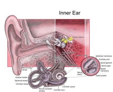
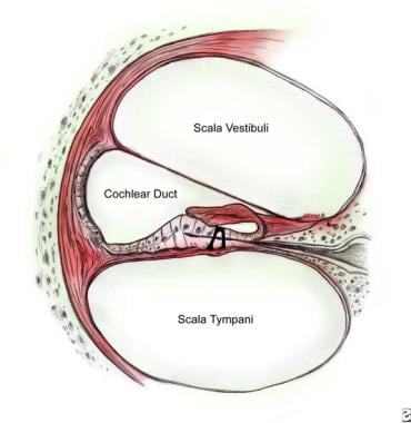
The cochlea is a part of the inner ear that is responsible for hearing. It is a spiral-shaped structure that looks like a snail shell and is filled with fluid. The cochlea contains hair cells, which are specialized sensory cells that convert sound vibrations into electrical signals that are sent to the brain.
The cochlea has three main parts: the vestibular canal, the tympanic canal, and the cochlear duct. Sound waves enter the inner ear and cause the fluid in the cochlea to move, which in turn causes the hair cells to bend. This bending motion stimulates the hair cells to generate electrical signals that are sent to the brain via the auditory nerve.
The brain then interprets these signals as sound, allowing us to hear and understand speech, music, and other sounds in our environment. Damage to the hair cells or other structures in the cochlea can lead to hearing loss or deafness.
Acoustic stimulation refers to the use of sound waves or vibrations to elicit a response in an individual, typically for the purpose of assessing or treating hearing, balance, or neurological disorders. In a medical context, acoustic stimulation may involve presenting pure tones, speech sounds, or other types of auditory signals through headphones, speakers, or specialized devices such as bone conduction transducers.
The response to acoustic stimulation can be measured using various techniques, including electrophysiological tests like auditory brainstem responses (ABRs) or otoacoustic emissions (OAEs), behavioral observations, or functional imaging methods like fMRI. Acoustic stimulation is also used in therapeutic settings, such as auditory training programs for hearing impairment or vestibular rehabilitation for balance disorders.
It's important to note that acoustic stimulation should be administered under the guidance of a qualified healthcare professional to ensure safety and effectiveness.
 E. Glen Wever
Frequency following response
Electrocochleography
List of MeSH codes (G11)
List of MeSH codes (G07)
Comparison of cochlear microphonic potentials from albino and pigmented Guinea pigs - Fingerprint - Oregon Health &...
 Is cochlear microphonic present in auditory neuropathy? - Kyoto2.org
E. Glen Wever - Wikipedia
 Clinical Electrocochleography: Overview of Theories, Techniques and Applications
Internet Scientific Publications
NIOSHTIC-2 Search Results - Full View
 Cochlear Function: Overview, Microenvironment of the Inner Ear, Traveling Wave and Signal Transduction
The actions of dopamine receptors on sound-evoked and spontaneous activity in the inner ear</em>...
NIOSHTIC-2 Search Results - Full View
 ELIOS | Aair Medicals | Aair Medicals | Pakistan Largest Medical Online Store
Vestibulocochlear Physiological Phenomena - Evoked Potentials, Auditory | CU Experts | CU Boulder
 A homozygous SLITRK6 nonsense mutation is associated with progressive auditory neuropathy in humans | Psychiatry Neuroimaging...
 Electrophysiological Assessments | ASU Speech and Hearing Clinic
 Pesquisa | Portal Regional da BVS
 Correlation between schroeder-phase effect and electrocochleography findings - IIUM Repository (IRep)
MH DELETED MN ADDED MN
MH DELETED MN ADDED MN
MH DELETED MN ADDED MN
MH DELETED MN ADDED MN
 MH DELETED MN ADDED MN
 "Human Medial Olivocochlear Reflex: Contralateral Activation Effect on " by Abdullah M. Jamos, Wafaa A. Kaf et al.
Columns | Canadian Audiologist
 Tone-Burst Auditory Brainstem Response and Cortical Potentials in Diagnosis of Syndromic Auditory Neuropathy Spectrum Disorder
Plus it
Pai, I.<...
 Course: Diagnosis and Management of ANSD | Interacoustics
 Cortical Neuroplasticity in Hearing Loss: Why It Matters in Clinical Decision-Making for Children and Adults | The Hearing...
 Doctor Ben Lineton | University of Southampton
 US/UK state brainwashing/torture/murder
ECochG6
- As the term implies, 'Electrocochleography' (ECochG) is a method for recording the electrical potentials of the cochlea. (audiologyonline.com)
- ECochG generally involves measurement of the stimulus-related cochlear potentials (as opposed to the resting potentials), and often includes measurement of the whole nerve or compound action potential (AP) of the auditory nerve. (audiologyonline.com)
- The technical capability of recording cochlear and auditory nerve potentials in humansled to a variety of clinical applications for ECochG. (audiologyonline.com)
- As indicated in the preceding section, the potentials most often recorded via ECochG include the CM, SP and AP. (audiologyonline.com)
- Electrocochleography (ECochG) is a method of recording the stimulus related potentials of the cochlea and auditory nerve. (ispub.com)
- The response that is measured in ECochG occurs within the first two or three milliseconds after an abrupt stimulus, and it includes the following components: the cochlear microphonic (CM), the summating potential (SP), and the whole nerve or compound action potential (AP). (ispub.com)
Compound action potential4
- The cochlear microphonic and the summating potential (SP) are generated by the hair cells of the organ of Corti, whereas the compound action potential (AP) of the auditory nerve represents the summed synchronized response of many individual nerve fibers. (kyoto2.org)
- Interactions with noise on cochlear potentials, namely compound action potential (CAP) and cochlear microphonic (CM) were studied. (cdc.gov)
- Sound-evoked (compound action potential, summating potential, cochlear microphonic) and spontaneous cochlear responses were recorded before and after perfusion. (edu.au)
- End points measured were electrophysiological, namely, compound action potential (CAP) an cochlear microphonic (CM). CAP is a measure of cochlear output generated at the inner hair cell-type I spiral ganglion synapse, and , CM is generated largely by the outer hair cell. (cdc.gov)
Microphonics3
- You can measure cochlear microphonics with a standard ABR electrode montage. (kyoto2.org)
- A 2 stage aABR protocol was used and ANSD was classified when click evoked ABR were absent or grossly abnormal but otoacoustic emissions and or cochlear microphonics were present. (bvsalud.org)
- Audiological evaluation revealed mixed hearing loss and signs of auditory neuropathy spectrum disorder (ANSD) despite absence of otoacoustic emissions and an absent click-evoked auditory brainstem response (ABR) without recording of cochlear microphonics (CM). ANSD was characterized by abnormal speech discrimination, bilateral robust CM to 2,000 Hz tone-burst (TB) ABR, and abnormal left thalamocortical and cortical pathways diagnosed based on auditory middle latency and cortical N1-P2 responses. (ejao.org)
Cortical4
- The findings in this study support the use of TB ABR and auditory cortical potentials in the ANSD test protocol and in patients with craniofacial anomalies. (ejao.org)
- With a better understanding of cortical brain changes associated with hearing loss, the potential to develop objective brain-based tools (ie, biomarkers) increases. (hearingreview.com)
- That is, we can examine the function of higher auditory centers of the brain (eg, auditory cortex) using cortical auditory evoked potentials (CAEPs). (hearingreview.com)
- The cortical auditory evoked potential (CAEP) response is comprised of three parts: the P1, N1, and P2. (hearingreview.com)
Amplitude3
- Action potential latency, duration, and amplitude have been studied considerably in the analysis of electrocochleographic waveforms, and variations from normal have been associated with endolymphatic hydrops. (ispub.com)
- Since the absolute amplitude of the summating potential and the action potential, when studied individually, displayed sizable variation across and within normal subjects, the amplitude ratio between the SP and the AP seems to be a more consistent within-subject feature of the overall response. (ispub.com)
- Distortion product otoacoustic emissions (DPOAEs) were absent in all ears tested and the cochlear microphonic (CM) was increased in amplitude and duration in young patients and absent in the two oldest subjects. (harvard.edu)
Cochlea6
- What is Summating potential in cochlea? (kyoto2.org)
- The cochlear microphonic is an alternating current electrical potential generated at the hair cell level in the cochlea. (ispub.com)
- For many years, cochlear fluids were thought to be generated by filtration of blood or cerebrospinal fluid, which then flowed longitudinally down the length of the cochlea to be absorbed through the endolymphatic sac. (medscape.com)
- While the function of the lateral efferent system in the cochlea is still unknown, previous studies have identified both excitatory and inhibitory changes in sound-evoked and spontaneous cochlear responses attributable to the lateral efferent system. (edu.au)
- The first experiments in this thesis were designed to determine if activation or blockade of different dopamine receptor subtypes in the cochlea could lead to both excitatory and inhibitory changes in sound-evoked and spontaneous cochlear responses. (edu.au)
- For example, how do the 3000 rows of active outer hair cells interact with each other and with other cochlear structures to amplify the waves in the cochlea that allow us to hear? (southampton.ac.uk)
Implant2
- cochlear implant - a surgically implanted device that stimulates the nerves of the inner ear. (kyoto2.org)
- As hearing care professionals-whether our jobs involve activating a cochlear implant, fitting a hearing aid, or providing rehabilitation to adults or children to help them reach their optimum performance after intervention-neuroplasticity is at the heart of what we do. (hearingreview.com)
Myogenic potentials2
- Vestibular evoked myogenic potentials were normal in three ears and absent in one. (harvard.edu)
- Vestibular evoked myogenic potentials are important in the evaluation of a patient who is dizzy and can help to determine the origin of the problem. (asuspeechandhearingclinic.org)
Responses5
- Thus comparison of response latency at various intensities can be used to distinguish cochlear from neural responses. (kyoto2.org)
- Adding separate R and C responses (middle tracing) enhances the cochlear Summating Potential (SP) and auditory nerve Action Potential (AP). (audiologyonline.com)
- Subtracting R and C responses (bottom tracing), enhances the Cochlear Microphonic (CM) (from ASHA, 1988, pg. (audiologyonline.com)
- These responses are comprised of the cochlear microphonic, the cochlear summating potential and the auditory nerve action potential. (asuspeechandhearingclinic.org)
- The role of the medial olivocochlear (MOC) reflex has been investigated by assessing changes of cochlear responses (CR) in humans. (missouristate.edu)
Stimulus2
- In contrast, the cochlear microphonic does NOT increase in latency as the stimulus intensity decreases. (kyoto2.org)
- 2 The summating potential is a direct current response to an alternating current stimulus which arises from the Organ of Corti hair cells in response to acoustic stimuli. (ispub.com)
Otoacoustic3
- Appropriately titled "From the Labs to the Clinics", Bob is involved in laboratory and applied/clinical research, including evoked potential and otoacoustic emission studies and behavioural studies of speech and language development in children with cochlear implants. (canadianaudiologist.ca)
- How are the motions of these cochlear structures related to the otoacoustic emissions that we can measure in the ear canal? (southampton.ac.uk)
- The ways in which these models may be useful clinically are: to aid the development of treatments, or prostheses for hearing impairment, to improve our ability to interpret clinical results (such as measurements of otoacoustic emissions or electrophysiology), to aid the development of new clinical tests of cochlear function. (southampton.ac.uk)
Neural1
- The CR consists of pre-neural and neural potentials originating from the inner ear, and at high signal levels is dominated by cochlear microphonic (CM). The CM originates from the outer hair cells, where the MOC fibers synapse, and there is little research about using it to investigate the MOC reflex in humans. (missouristate.edu)
Differential diagnosis2
- Conclusion: The presence of the Cochlear Microphonic is a determining finding in the differential diagnosis of Auditory Neuropathy/Dyssynchrony. (kyoto2.org)
- Electrophysiological measures, such as those procedures listed below, play an important role in the assessment of hearing in difficult to test populations, such as very young children, as well as in the differential diagnosis of cochlear versus retrocochlear disorders. (asuspeechandhearingclinic.org)
Suppression1
- The suppression of the cochlear microphonic suggests that dopamine receptor influence is not confined to the primary afferent dendrite may also include the active process of the outer hair cells. (edu.au)
Hydrops2
- In the case of endolymphatic hydrops, in which there is increased pressure in the scala media, the normal asymmetric vibration of the basilar membrane is altered, thus resulting in the large summating potential. (ispub.com)
- This may be used to establish the presence of cochlear hydrops. (asuspeechandhearingclinic.org)
Electrode1
- Aran and LeBert, 1968) performed their measurements on patients undergoing middle ear surgery and/or used a non-surgical approach that involved passing a needle electrode through the tympanic membrane (TM) to rest on the cochlear promontory. (audiologyonline.com)
Waves3
- How do we differentiate ABR waves from a cochlear microphonic wave? (kyoto2.org)
- These seven waves, and the cochlear nucleus and between these the top five most interest and, among them, the pathways exists a series of parallel pathways7. (bvsalud.org)
- Waves I and II are addition, in the computer some characteristics that generated in the cochlear nerve and subsequent define certain acquisition parameters record the 1 Speech-Language Therapist. (bvsalud.org)
Outer hair1
- Homozygous SLITRK6 c.1240C>T (p.Gln414Ter) nonsense mutations are associated with high myopia, cochlear dysfunction attributed to outer hair cell disease, and progressive auditory neuropathy. (harvard.edu)
ANSD1
- The most common etiology for ANSD was hypoplasia or absence of the cochlear nerve with 37 cases (46.3%), and it was significantly more likely with unilateral than bilateral ANSD. (bvsalud.org)
Nerve3
- Although Wever and Bray misinterpreted the signal as the action potential of the auditory nerve, the effect was real. (wikipedia.org)
- The action potential is an alternating current response which is generated by the cochlear end of the VIIIth Cranial Nerve, and it represents the summed response of the synchronous firing of thousands of auditory nerve fibers. (ispub.com)
- Cochlear nerve deficiency was the most common etiology. (bvsalud.org)
Action4
- What is the difference between cochlear microphonic and action potential? (kyoto2.org)
- Wever and Bray subsequently recorded true action potentials, resulting in, among other discoveries, the now-accepted volley theory of frequency coding. (wikipedia.org)
- 1 , 2 The SP generally appears on the electrocochleograhpic waveform as a "ledge or hump on the beginning slope of the action potential wave", but it can also appear as a separate peak preceding the action potential. (ispub.com)
- 1 , 2 The synchronous activity essential for producing the action potential is most often seen at the onset of a tone burst or in response to clicks. (ispub.com)
Pathways1
- In this review, we will discuss how CAEPs can be used to assess development of the auditory cortex and monitor the maturation of the auditory cortex and central auditory pathways before and after intervention with hearing aids and cochlear implants. (hearingreview.com)
Clinical2
- Although available to the hearing scientist/clinician for over 50 years, ECochG's emergence as a clinical tool (as well as all other auditory evoked potentials) was rekindled in part by the discovery, application and popularity of the auditory brainstem response (ABR). (audiologyonline.com)
- The long term research goal is to understand human cochlear physiology in both normal and pathological conditions with a view to aiding the development of improved clinical diagnostic techniques and treatments. (southampton.ac.uk)
Outcomes1
- 4,7-10 Based on this research, we know that early cochlear implantation within this sensitive period (best by age 1) leads to more normal brain development and yields more optimal listening and spoken language outcomes compared to late cochlear implantation. (hearingreview.com)
Electrical2
- Auditory evoked potentials are very small electrical voltage potentials originating from the brain. (asuspeechandhearingclinic.org)
- These should capture the essential hydrodynamics, structural dynamics, and electrical processes involved in cochlear physiology. (southampton.ac.uk)
Stimuli1
- We investigated 20 students from a Center for Diagnosis and Rehabilitation, with no hearing impairment, all female aged 15-30 years old, who were screened for brainstem auditory evoked potential, which presented stimuli in different polarities ranges, including condensation and rarefaction in different presentation rates of 21.7, 27.7 and 47.7 stimuli per second. (bvsalud.org)
Frequency1
- It is often considered the most enigmatic of cochlear potentials because its magnitude and polarity vary across frequency and level and its origins are uncertain. (kyoto2.org)
Hearing aids1
- But, depending on the severity of the damage, sensorineural hearing loss has been successfully treated with hearing aids or cochlear implants. (kyoto2.org)
Function2
- This so-called cochlear microphonic is still used today as a measure of cochlear function. (wikipedia.org)
- The reader is urged to review this literature to gain a better understanding and appreciation of the specific features of these potentials and their relevance to hearing function. (audiologyonline.com)
Normal1
- gem homozygous receptor mutant HCs display normal cell viability, afferent synaptogenesis, and peripheral innervation, yet exhibit strongly reduced extracellular potentials (∼50% of wild-type potentials). (jneurosci.org)
Duration1
- The cochlear summating potential (SP) to a tone is a baseline shift that persists for the duration of the burst. (kyoto2.org)
Humans2
- The reader is referred to Ferraro (2000) for a more thorough review of the history of these potentials as recorded in humans. (audiologyonline.com)
- Moreover, the CR could be used as a potential tool to study the MOC reflex in humans. (missouristate.edu)
Hair1
- The cochlear microphonic is a receptor potential believed to be generated primarily by outer hair cells. (kyoto2.org)
Present1
- Is cochlear microphonic present in auditory neuropathy? (kyoto2.org)
High1
- This potential is a change in the surface-recorded electromyogram (EMG) that can be evoked over neck and spinal muscles following a high-intensity acoustic input. (asuspeechandhearingclinic.org)
Development1
- The endocochlear potential is established through the development of tight cellular junctions between local networks of epithelial cells, connective tissue and supporting cells that completely partition the endolymph from the surrounding perilymph. (kyoto2.org)