Colonic Polyps
Polyps
Intestinal Polyps
Colonography, Computed Tomographic
Adenomatous Polyposis Coli
Colon
Diverticulosis, Colonic
Nasal Polyps
Rectal Diseases
Sigmoid Diseases
Genes, APC
Radiographic Image Interpretation, Computer-Assisted
Intestinal Mucosa
Colonic Diseases
Colorectal Neoplasms
Sensitivity and Specificity
Retrospective Studies
Human colon adenocarcinomas express a MUC1-associated novel carbohydrate epitope on core mucin glycans defined by a monoclonal antibody (A10) raised against murine Ehrlich tumor cells. (1/764)
A monoclonal antibody (mAb; A10) raised against murine Ehrlich tumor cell surface carbohydrates was tested for reactivity with human normal and malignant tissues. A10 reacted strongly, with a high proportion of adenocarcinomas arising from colon and other tissues but not with breast carcinomas or other malignant tumors. Normal tissues were virtually A10 unreactive, except for the duct cells from breast and pancreas and some bronchial mucosae. Ultrastructural studies showed mAb A10 immunolabeling of both microvilli and mucin droplets in colon cancer cells but not in normal absorptive or globet cells. A10 reacted strongly with mucin-enriched fractions from colon cancer tissues and HT-29 xenografts but not from normal colon tissues. A10 epitope was carried on MUC1 derived from colon adenocarcinomas and probably on other mucin species, although not on MUC2 molecules. A10 epitope was resistant to exoglycosidases and periodate oxidation but sensitive to the Smith's degradation and beta-elimination, suggesting the involvement of O-linked carbohydrates in nonterminal reducing positions. A mucin-type glycosidic linkage was supported because of the lack of A10 reactivity with HT-29 cells grown with phenyl-N-acetyl-alpha-D-galactosaminide. Deglycosylation studies with trifluoromethanesulfonic acid pointed to the involvement of core mucin glycans in the A10 epitope. This epitope was resistant to protease, O- and N-glycanase treatments carried out on trifluoromethanesulfonic acid-deglycosylated mucins. Inhibition studies with core 1, core 2, core 3, and core 6 suggested the latter [GlcNAcbeta(1-6)GalNAc] as being involved in A10 epitope. Taken together, the present results point to A10 defining a core 6-related epitope on core mucin glycans expressed by colon cancer MUC1 not previously associated with human cancer. (+info)Dietary determinants of colorectal proliferation in the normal mucosa of subjects with previous colon adenomas. (2/764)
Dietary determinants of colorectal mucosa proliferation were studied in 69 subjects previously operated for at least two sporadic colon adenomas. Information on recent dietary habits was collected by a validated food frequency questionnaire, and proliferation was measured by [3H]thymidine incorporation in colorectal biopsies by determining the labeling index (LI) and the percentage of LI in the upper part of the crypt, two parameters that are increased in subjects at high risk of colon cancer. The LI was significantly higher in women as compared with men (P = 0.01). Diet showed several associations with colorectal mucosa proliferation: (a) subjects in the highest tertile of fish consumption had a significantly lower LI (P = 0.0013) compared with those in the lower tertiles [5.20 +/- 1.87 versus 6.80 +/- 2.18 (mean +/- SD)]; (b) subjects with a low red meat consumption had lower proliferation in the upper part of the crypt [2.38 +/- 2.10, 5.30 +/- 4.62, and 5.89 +/- 4.82 in the low, middle, and high tertile of consumption, respectively (mean +/- SD); P = 0.0093]; (c) according to estimated nutrient intakes, the LI was lower in subjects reporting a high intake of starch (P = 0.006) and higher in subjects with a low intake of beta-carotene (P = 0.002). The results show that subjects reporting a diet rich in fish, starch, and beta-carotene and low in red meat had lower colorectal mucosa proliferation and a normal pattern of proliferation along the crypt. Given the correlation between colorectal proliferative activity and colon cancer risk, such a dietary pattern might be beneficial for subjects at high risk of colon cancer. (+info)Differential expression of a new tumor-associated antigen, TLP, during human colorectal cancer tumorigenesis. (3/764)
Tumour liberated particles (TLP) have been proposed as a potential new serum tumor marker. In particular, a high percentage of patients with early stages of lung cancer scored positive for serum TLP, suggesting its possible role as a marker for early diagnosis of disease. The aim of the present study was to analyze the expression of TLP in the colorectal adenoma-carcinoma sequence in order to determine whether its expression correlates with the various stages of cancer transformation. TLP distribution was assessed by immunohistochemistry in normal, premalignant, and malignant colorectal lesions. Normal colonic mucosa and hyperplastic polyps showed no positive staining, whereas adenomas and adenocarcinomas reacted to anti-TLP serum. The percentage of positive tumor cells increased from adenomas with mild dysplasia to adenomas with severe dysplasia. Moreover, a supranuclear staining pattern was observed mainly in adenomas with mild dysplasia, whereas adenomas with severe dysplasia as well as adenocarcinomas showed a characteristic diffuse staining pattern and a strong staining intensity. Only a few cases of adenocarcinoma were found to be TLP-negative and all were poorly differentiated. Our results suggest that TLP antigen expression may be considered as a marker of epithelial atypia in the colorectal tract and as a potential target for new diagnostic and/or therapeutic approaches to human colorectal cancer. (+info)A bile acid-induced apoptosis assay for colon cancer risk and associated quality control studies. (4/764)
Bile acids are important in the etiology of colorectal cancer. Bile acids induce apoptosis in colonic goblet cells at concentrations comparable to those found in fecal water after high-fat meals. Preliminary evidence indicated that cells of the normal-appearing (nontumorous) portion of the colon epithelium of colon cancer patients are more resistant to bile salt-induced apoptosis than are cells from normal individuals. In the present study, 68 patients were examined, and biopsies were taken at 20 cm from the anal verge, cecum, and descending colon. The patients included 17 individuals with a history of colorectal cancer, 37 individuals with adenomas, and 14 individuals who were neoplasia free. The mean bile salt-induced apoptotic index among normal individuals was 57.6 +/- 3.47 (SE), which differed significantly (P < 0.05) from the mean value of 36.41 +/- 3.12 in individuals with a history of colon cancer. The correlation between independent observers was 0.89 (P < 0.001), indicating good interobserver reliability. Components of variance comparing interindividual versus intraindividual sources of variation suggested that site-to-site variability, both between regions of the colon and for adjacent biopsies, was larger than the interpatient variability for individuals with a history of neoplasia. Therefore, there was "patchiness" of the susceptibility of regions of the colon to bile acid-induced apoptosis in individuals with a history of neoplasia (a patchy field effect). There was no obvious correlation of low-apoptotic index regions with regions in which previous neoplasias had been found and removed. On the other hand, for normal, i.e., neoplasia-free, individuals, there was relatively less intraindividual variation compared to interindividual variation. Our assay shows an association between resistance to bile acid-induced apoptosis, measured at 20 cm from the anal verge, and colon cancer risk. Thus, this assay may prove useful as a biomarker of colon cancer risk. (+info)Frequent mutation of beta-catenin and APC genes in primary colorectal tumors from patients with hereditary nonpolyposis colorectal cancer. (5/764)
Hereditary nonpolyposis colorectal cancer (HNPCC) is characterized by defective DNA mismatch repair, which results in genetic instability of tumors; however, only a few target genes have been recognized. Our previous study detected a low frequency of APC gene mutation (21%) in colorectal tumors from HNPCC patients, in contrast to a high frequency of APC gene alteration (>70%) in non-HNPCC tumors. Because both beta-catenin and ACP gene mutations have recently been shown to activate the same signaling pathway, we analyzed beta-catenin mutation in HNPCC tumors. A notable frequency of beta-catenin gene mutation (43%, 12 of 28) was found to occur in HNPCC colorectal tumors. Beta-catenin mutations were not detected in tumors with APC mutations. All beta-catenin mutations detected in HNPCC tumors existed within the regulatory domain of beta-catenin. Immunohistochemical staining of tumors with this mutation showed accumulation of beta-catenin protein in nuclei. These and previous data from our laboratory suggest that activation of the beta-catenin-Tcf signaling pathway, through either beta-catenin or APC mutation, contributes to HNPCC colorectal carcinogenesis in approximately 65% of cases. (+info)Up-regulation of macrophage wnt gene expression in adenoma-carcinoma progression of human colorectal cancer. (6/764)
Defects in the APC-beta-catenin pathway are common in colon cancer. We investigated whether aberrant regulation of upstream ligands stimulating this pathway occur in colon cancer. Using RNAase protection analysis, six out of eight wnt genes were expressed in 14 matched cases of normal, adenomatous and malignant colorectal tissues. Wnt 2 and wnt 5a were significantly up-regulated in the progression from normal through adenoma to carcinoma. Transcripts for wnts 4, 7b, 10b and 13, but not wnt 2 and wnt 5a were detected in several colorectal cell lines. In situ hybridization demonstrated that wnt 2 and wnt 5a transcripts were mainly in the lamina propria/stroma region with labelling predominantly in macrophages. Immunostaining with CD68 confirmed the wnt-expressing cells as macrophages. These results show a major difference in wnt expression in colon cancer compared to colon adenomas and suggest stromal wnt expression may play a role in tumour progression. (+info)A comparison of virtual and conventional colonoscopy for the detection of colorectal polyps. (7/764)
BACKGROUND: Virtual colonoscopy is a new method of imaging the colon in which thin-section, helical computed tomography (CT) is used to generate high-resolution, two-dimensional axial images. Three-dimensional images of the colon simulating those obtained with conventional colonoscopy are then reconstructed off-line. We compared the performance of virtual and conventional colonoscopy for the detection of colorectal polyps. METHODS: We prospectively studied 100 patients at high risk for colorectal neoplasia (60 men and 40 women; mean age, 62 years). We performed virtual colonoscopy immediately before conventional colonoscopy. We inserted a rectal tube and insufflated the colon with air to the maximal level that the patient could tolerate. We administered 1 mg of glucagon intravenously immediately before CT scanning to minimize the degree of smooth-muscle spasm and peristalsis and to reduce the patient's discomfort. RESULTS: The entire colon was clearly seen by virtual colonoscopy in 87 patients and by conventional colonoscopy in 89. Fifty-one patients had normal findings on conventional colonoscopy. In the other 49, we identified a total of 115 polyps and 3 carcinomas. Virtual colonoscopy identified all 3 cancers, 20 of 22 polyps that were 10 mm or more in diameter (91 percent), 33 of 40 that were 6 to 9 mm (82 percent), and 29 of 53 that were 5 mm or smaller (55 percent). There were 19 false positive findings of polyps and no false positive findings of cancer. Of the 69 adenomatous polyps, 46 of the 51 that were 6 mm or more in diameter (90 percent) and 12 of the 18 that were 5 mm or smaller (67 percent) were correctly identified by virtual colonoscopy. Although discomfort was not specifically recorded, none of the patients requested that virtual colonoscopy be stopped because of discomfort or pain. CONCLUSIONS: In a group of patients at high risk for colorectal neoplasia, virtual and conventional colonoscopy had similar efficacy for the detection of polyps that were 6 mm or more in diameter. (+info)Patterns of proliferative changes in crypts bordering colonic tumors: zonal histology and cell cycle marker expression. (8/764)
Proliferative crypt changes have been noted in mucosa bordering colonic carcinomas, but their biological significance is disputed. We anticipated that zonal patterning of histological changes and cell cycle marker expression would provide clues to the pathogenesis of these border changes. 81 specimens were examined including carcinomas, adenomatours polyps, adenomas with early carcinoma, flat adenomas and aberrant crypt foci. The spatial distribution and frequency of micro-architectural features, and mucosal thickness were determined in a border domain of 150 300 sequential crypts/specimen. Immunocytochemical expression of Ki67 and p53 antigens in crypts also was semi-quantitatively examined. We found that in 100% of carcinomas two histologically abnormal zones (Proximate and Middle) separated tumor from normal mucosa. Differences in the feature frequency between zones were statistically significant (p<0.05). Both zones showed mild increases in crypt cell expression of Ki67, with a statistically significant relationship to zonal patterning (p<0.005). Weak expression of p53 only appeared in rare cells. Crypt elongation with mucosal thickening (1.9x normal, p<0.001) in the Proximate and Middle zones distinguished carcinomas from border changes in all benign lesions, except flat adenomas. Since this change occurs in all cases of carcinoma, there is no correlation with tumor stage or grade. Also in carcinomas, elaborate complexes of attached crypts (connected crypt structures) were characteristic of the Middle zone, so that proximate zone was always architecturally simpler. We conclude, that despite continuous carcinoma growth, the invaded border mucosa maintains a prototypical zonal organization of molecular and histological crypt changes This spatially organized reaction pattern is likely to reflect an interplay between regulated growth and destructive processes in response to advancing carcinoma. Compared to the edges of benign colonic tumors, the edges of carcinomas are distinctive and consistent enough to be diagnostically useful. (+info)Colonic polyps are abnormal growths that protrude from the inner wall of the colon (large intestine). They can vary in size, shape, and number. Most colonic polyps are benign, meaning they are not cancerous. However, some types of polyps, such as adenomas, have a higher risk of becoming cancerous over time if left untreated.
Colonic polyps often do not cause any symptoms, especially if they are small. Larger polyps may lead to symptoms like rectal bleeding, changes in bowel habits, abdominal pain, or iron deficiency anemia. The exact cause of colonic polyps is not known, but factors such as age, family history, and certain medical conditions (like inflammatory bowel disease) can increase the risk of developing them.
Regular screening exams, such as colonoscopies, are recommended for individuals over the age of 50 to detect and remove polyps before they become cancerous. If you have a family history of colonic polyps or colorectal cancer, your doctor may recommend earlier or more frequent screenings.
A polyp is a general term for a small growth that protrudes from a mucous membrane, such as the lining of the nose or the digestive tract. Polyps can vary in size and shape, but they are usually cherry-sized or smaller and have a stalk or a broad base. They are often benign (noncancerous), but some types of polyps, especially those in the colon, can become cancerous over time.
In the digestive tract, polyps can form in the colon, rectum, stomach, or small intestine. Colorectal polyps are the most common type and are usually found during routine colonoscopies. There are several types of colorectal polyps, including:
* Adenomatous polyps (adenomas): These polyps can become cancerous over time and are the most likely to turn into cancer.
* Hyperplastic polyps: These polyps are usually small and benign, but some types may have a higher risk of becoming cancerous.
* Inflammatory polyps: These polyps are caused by chronic inflammation in the digestive tract, such as from inflammatory bowel disease (IBD).
Polyps can also form in other parts of the body, including the nose, sinuses, ears, and uterus. In most cases, polyps are benign and do not cause any symptoms. However, if they become large enough, they may cause problems such as bleeding, obstruction, or discomfort. Treatment typically involves removing the polyp through a surgical procedure.
Intestinal polyps are abnormal growths that protrude from the lining of the intestines. They can occur in any part of the digestive tract, including the colon and rectum (colorectal polyps), small intestine, or stomach. These growths vary in size, shape, and number. Most intestinal polyps are benign, meaning they are not cancerous. However, some types of polyps, such as adenomatous polyps, can become cancerous over time if left untreated.
Intestinal polyps can be asymptomatic or cause symptoms like rectal bleeding, abdominal pain, changes in bowel habits, or anemia (in cases where there is chronic, slow bleeding). The exact cause of intestinal polyps is not fully understood, but factors such as age, family history, and certain genetic conditions can increase the risk of developing them. Regular screening exams, like colonoscopies, are essential for early detection and removal of polyps to prevent potential complications, including colorectal cancer.
Computed tomographic colonography (CTC), also known as virtual colonoscopy, is a medical imaging technique that uses computed tomography (CT) scans to produce detailed images of the large intestine (colon) and rectum. In CTC, specialized software creates two- and three-dimensional images of the colon's inner surface, allowing healthcare providers to examine the colon for polyps, tumors, and other abnormalities.
During a CTC procedure, patients are usually given a mild laxative and asked to follow a clear liquid diet beforehand to clean out the colon. A small tube is inserted into the rectum to inflate the colon with air or carbon dioxide, making it easier to visualize any abnormalities. The patient lies on their back and then their stomach while the CT scanner takes multiple images of the abdomen and pelvis from different angles.
CTC has several advantages over traditional colonoscopy, including less invasiveness, lower risk of complications, faster recovery time, and the ability to examine the entire colon without missing any areas. However, if polyps or other abnormalities are detected during a CTC, a follow-up diagnostic colonoscopy may be necessary for removal or further evaluation.
It is important to note that CTC does not replace traditional colonoscopy as a screening tool for colorectal cancer. While it has similar accuracy in detecting large polyps and cancers, its ability to detect smaller polyps is less reliable compared to optical colonoscopy. Therefore, guidelines recommend using CTC as an alternative option for individuals who cannot or do not wish to undergo traditional colonoscopy, or as a supplemental screening tool for those at higher risk of colorectal cancer.
Adenomatous polyps, also known as adenomas, are benign (noncancerous) growths that develop in the lining of the glandular tissue of certain organs, most commonly occurring in the colon and rectum. These polyps are composed of abnormal glandular cells that can grow excessively and form a mass.
Adenomatous polyps can vary in size, ranging from a few millimeters to several centimeters in diameter. They may be flat or have a stalk (pedunculated). While adenomas are generally benign, they can potentially undergo malignant transformation and develop into colorectal cancer over time if left untreated. The risk of malignancy increases with the size of the polyp and the presence of certain histological features, such as dysplasia (abnormal cell growth).
Regular screening for adenomatous polyps is essential to detect and remove them early, reducing the risk of colorectal cancer. Screening methods include colonoscopy, sigmoidoscopy, and stool-based tests.
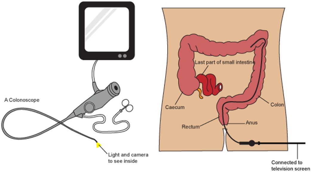
A colonoscopy is a medical procedure used to examine the large intestine, also known as the colon and rectum. It is performed using a flexible tube with a tiny camera on the end, called a colonoscope, which is inserted into the rectum and gently guided through the entire length of the colon.
The procedure allows doctors to visually inspect the lining of the colon for any abnormalities such as polyps, ulcers, inflammation, or cancer. If any polyps are found during the procedure, they can be removed immediately using special tools passed through the colonoscope. Colonoscopy is an important tool in the prevention and early detection of colorectal cancer, which is one of the leading causes of cancer-related deaths worldwide.
Patients are usually given a sedative to help them relax during the procedure, which is typically performed on an outpatient basis in a hospital or clinic setting. The entire procedure usually takes about 30-60 minutes to complete, although patients should plan to spend several hours at the medical facility for preparation and recovery.
Colonic neoplasms refer to abnormal growths in the large intestine, also known as the colon. These growths can be benign (non-cancerous) or malignant (cancerous). The two most common types of colonic neoplasms are adenomas and carcinomas.
Adenomas are benign tumors that can develop into cancer over time if left untreated. They are often found during routine colonoscopies and can be removed during the procedure.
Carcinomas, on the other hand, are malignant tumors that invade surrounding tissues and can spread to other parts of the body. Colorectal cancer is the third leading cause of cancer-related deaths in the United States, and colonic neoplasms are a significant risk factor for developing this type of cancer.
Regular screenings for colonic neoplasms are recommended for individuals over the age of 50 or those with a family history of colorectal cancer or other risk factors. Early detection and removal of colonic neoplasms can significantly reduce the risk of developing colorectal cancer.
Adenomatous Polyposis Coli (APC) is a genetic disorder characterized by the development of numerous adenomatous polyps in the colon and rectum. APC is caused by mutations in the APC gene, which is a tumor suppressor gene that helps regulate cell growth and division. When the APC gene is mutated, it can lead to uncontrolled cell growth and the development of polyps, which can eventually become cancerous.
Individuals with APC typically develop hundreds to thousands of polyps in their colon and rectum, usually beginning in adolescence or early adulthood. If left untreated, APC can lead to colorectal cancer in nearly all affected individuals by the age of 40.
APC is an autosomal dominant disorder, which means that a person has a 50% chance of inheriting the mutated gene from an affected parent. However, some cases of APC may also occur spontaneously due to new mutations in the APC gene. Treatment for APC typically involves surgical removal of the colon and rectum (colectomy) to prevent the development of colorectal cancer. Regular surveillance with colonoscopy is also recommended to monitor for the development of new polyps.
The colon, also known as the large intestine, is a part of the digestive system in humans and other vertebrates. It is an organ that eliminates waste from the body and is located between the small intestine and the rectum. The main function of the colon is to absorb water and electrolytes from digested food, forming and storing feces until they are eliminated through the anus.
The colon is divided into several regions, including the cecum, ascending colon, transverse colon, descending colon, sigmoid colon, rectum, and anus. The walls of the colon contain a layer of muscle that helps to move waste material through the organ by a process called peristalsis.
The inner surface of the colon is lined with mucous membrane, which secretes mucus to lubricate the passage of feces. The colon also contains a large population of bacteria, known as the gut microbiota, which play an important role in digestion and immunity.
Diverticulosis, colonic is a medical condition characterized by the presence of small sacs or pouches (diverticula) that form on the outer wall of the large intestine (colon). These sacs are usually found in the sigmoid colon, which is the part of the colon that is closest to the rectum.
Diverticulosis occurs when the inner layer of the colon's muscle pushes through weak spots in the outer layer of the colon wall, creating small pockets or sacs. The exact cause of diverticulosis is not known, but it may be associated with a low-fiber diet, aging, and increased pressure in the colon.
Most people with diverticulosis do not experience any symptoms, and the condition is often discovered during routine screening exams or when complications arise. However, some people may experience cramping, bloating, and changes in bowel habits.
Diverticulosis can lead to complications such as inflammation (diverticulitis), bleeding, and infection. It is important to seek medical attention if you experience symptoms such as severe abdominal pain, fever, or rectal bleeding, as these may be signs of a more serious condition.
Treatment for diverticulosis typically involves making dietary changes, increasing fiber intake, and taking medications to manage symptoms. In some cases, surgery may be necessary to remove affected portions of the colon.
An adenoma is a benign (noncancerous) tumor that develops from glandular epithelial cells. These types of cells are responsible for producing and releasing fluids, such as hormones or digestive enzymes, into the surrounding tissues. Adenomas can occur in various organs and glands throughout the body, including the thyroid, pituitary, adrenal, and digestive systems.
Depending on their location, adenomas may cause different symptoms or remain asymptomatic. Some common examples of adenomas include:
1. Colorectal adenoma (also known as a polyp): These growths occur in the lining of the colon or rectum and can develop into colorectal cancer if left untreated. Regular screenings, such as colonoscopies, are essential for early detection and removal of these polyps.
2. Thyroid adenoma: This type of adenoma affects the thyroid gland and may result in an overproduction or underproduction of hormones, leading to conditions like hyperthyroidism (overactive thyroid) or hypothyroidism (underactive thyroid).
3. Pituitary adenoma: These growths occur in the pituitary gland, which is located at the base of the brain and controls various hormonal functions. Depending on their size and location, pituitary adenomas can cause vision problems, headaches, or hormonal imbalances that affect growth, reproduction, and metabolism.
4. Liver adenoma: These rare benign tumors develop in the liver and may not cause any symptoms unless they become large enough to press on surrounding organs or structures. In some cases, liver adenomas can rupture and cause internal bleeding.
5. Adrenal adenoma: These growths occur in the adrenal glands, which are located above the kidneys and produce hormones that regulate stress responses, metabolism, and blood pressure. Most adrenal adenomas are nonfunctioning, meaning they do not secrete excess hormones. However, functioning adrenal adenomas can lead to conditions like Cushing's syndrome or Conn's syndrome, depending on the type of hormone being overproduced.
It is essential to monitor and manage benign tumors like adenomas to prevent potential complications, such as rupture, bleeding, or hormonal imbalances. Treatment options may include surveillance with imaging studies, medication to manage hormonal issues, or surgical removal of the tumor in certain cases.
Nasal polyps are benign (noncancerous) growths that originate from the lining of your nasal passages or sinuses. They most often occur in the area where the sinuses open into the nasal cavity. Small nasal polyps may not cause any problems. But if they grow large enough, they can block your nasal passages and lead to breathing issues, frequent infections and loss of smell.
Nasal polyps are associated with chronic inflammation due to conditions such as asthma, allergic rhinitis or chronic sinusitis. Treatment typically includes medication to reduce the size of the polyps or surgery to remove them. Even after successful treatment, nasal polyps often return.
Rectal diseases refer to conditions that affect the structure or function of the rectum, which is the lower end of the large intestine, just above the anus. The rectum serves as a storage area for stool before it is eliminated from the body. Some common rectal diseases include:
1. Hemorrhoids: Swollen veins in the rectum or anus that can cause pain, itching, bleeding, and discomfort.
2. Rectal cancer: Abnormal growth of cells in the rectum that can invade and destroy nearby tissue and spread to other parts of the body.
3. Anal fissures: Small tears in the lining of the anus that can cause pain, bleeding, and itching.
4. Rectal prolapse: A condition where the rectum slips outside the anus, causing discomfort, fecal incontinence, and other symptoms.
5. Inflammatory bowel disease (IBD): A group of chronic inflammatory conditions that affect the digestive tract, including the rectum, such as Crohn's disease and ulcerative colitis.
6. Rectal abscess: A collection of pus in the rectum caused by an infection, which can cause pain, swelling, and fever.
7. Fistula-in-ano: An abnormal connection between the rectum and the skin around the anus, which can cause drainage of pus or stool.
8. Rectal foreign bodies: Objects that are accidentally or intentionally inserted into the rectum and can cause injury, infection, or obstruction.
These are just a few examples of rectal diseases, and there are many other conditions that can affect the rectum. If you experience any symptoms related to the rectum, it is important to seek medical attention from a healthcare professional for proper diagnosis and treatment.
"Sigmoid diseases" is not a widely recognized medical term. However, the sigmoid colon is a part of the large intestine, and it can be affected by various conditions such as:
1. Sigmoid diverticulitis: Inflammation or infection of small pouches (diverticula) that form on the wall of the sigmoid colon.
2. Sigmoid volvulus: Twisting of the sigmoid colon on itself, which can lead to obstruction and ischemia.
3. Sigmoid cancer: Malignant tumor arising from the epithelial cells lining the sigmoid colon.
4. Inflammatory bowel disease (IBD): Chronic inflammation of the intestine, including the sigmoid colon, that can lead to symptoms such as diarrhea, abdominal pain, and weight loss.
5. Irritable bowel syndrome (IBS): Functional gastrointestinal disorder characterized by abdominal pain, bloating, and altered bowel habits, which can affect the sigmoid colon.
Therefore, "sigmoid diseases" could refer to any of these conditions or others that specifically affect the sigmoid colon.
APC (Adenomatous Polyposis Coli) gene is a tumor suppressor gene that provides instructions for making a protein called adenomatous polyposis coli. This protein plays a crucial role in regulating the growth and division of cells in the colon and rectum. Specifically, it helps to maintain the stability of the cell's genetic material (DNA) by controlling the process of beta-catenin degradation.
When the APC gene is mutated or altered, it can lead to an accumulation of beta-catenin in the cell, which can result in uncontrolled cell growth and division. This can ultimately lead to the development of colon polyps, which are benign growths that can become cancerous over time if left untreated.
Mutations in the APC gene are associated with several inherited cancer syndromes, including familial adenomatous polyposis (FAP) and attenuated FAP (AFAP). These conditions are characterized by the development of numerous colon polyps at a young age, which can increase the risk of developing colorectal cancer.
Computer-assisted radiographic image interpretation is the use of computer algorithms and software to assist and enhance the interpretation and analysis of medical images produced by radiography, such as X-rays, CT scans, and MRI scans. The computer-assisted system can help identify and highlight certain features or anomalies in the image, such as tumors, fractures, or other abnormalities, which may be difficult for the human eye to detect. This technology can improve the accuracy and speed of diagnosis, and may also reduce the risk of human error. It's important to note that the final interpretation and diagnosis is always made by a qualified healthcare professional, such as a radiologist, who takes into account the computer-assisted analysis in conjunction with their clinical expertise and knowledge.
The intestinal mucosa is the innermost layer of the intestines, which comes into direct contact with digested food and microbes. It is a specialized epithelial tissue that plays crucial roles in nutrient absorption, barrier function, and immune defense. The intestinal mucosa is composed of several cell types, including absorptive enterocytes, mucus-secreting goblet cells, hormone-producing enteroendocrine cells, and immune cells such as lymphocytes and macrophages.
The surface of the intestinal mucosa is covered by a single layer of epithelial cells, which are joined together by tight junctions to form a protective barrier against harmful substances and microorganisms. This barrier also allows for the selective absorption of nutrients into the bloodstream. The intestinal mucosa also contains numerous lymphoid follicles, known as Peyer's patches, which are involved in immune surveillance and defense against pathogens.
In addition to its role in absorption and immunity, the intestinal mucosa is also capable of producing hormones that regulate digestion and metabolism. Dysfunction of the intestinal mucosa can lead to various gastrointestinal disorders, such as inflammatory bowel disease, celiac disease, and food allergies.
Colonic diseases refer to a group of medical conditions that affect the colon, also known as the large intestine or large bowel. The colon is the final segment of the digestive system, responsible for absorbing water and electrolytes, and storing and eliminating waste products.
Some common colonic diseases include:
1. Inflammatory bowel disease (IBD): This includes conditions such as Crohn's disease and ulcerative colitis, which cause inflammation and irritation in the lining of the digestive tract.
2. Diverticular disease: This occurs when small pouches called diverticula form in the walls of the colon, leading to symptoms such as abdominal pain, bloating, and changes in bowel movements.
3. Colorectal cancer: This is a type of cancer that develops in the colon or rectum, often starting as benign polyps that grow and become malignant over time.
4. Irritable bowel syndrome (IBS): This is a functional gastrointestinal disorder characterized by abdominal pain, bloating, and changes in bowel movements, but without any underlying structural or inflammatory causes.
5. Constipation: This is a common condition characterized by infrequent bowel movements, difficulty passing stools, or both.
6. Infectious colitis: This occurs when the colon becomes infected with bacteria, viruses, or parasites, leading to symptoms such as diarrhea, abdominal cramps, and fever.
Treatment for colonic diseases varies depending on the specific condition and its severity. Treatment options may include medications, lifestyle changes, surgery, or a combination of these approaches.
Colorectal neoplasms refer to abnormal growths in the colon or rectum, which can be benign or malignant. These growths can arise from the inner lining (mucosa) of the colon or rectum and can take various forms such as polyps, adenomas, or carcinomas.
Benign neoplasms, such as hyperplastic polyps and inflammatory polyps, are not cancerous but may need to be removed to prevent the development of malignant tumors. Adenomas, on the other hand, are precancerous lesions that can develop into colorectal cancer if left untreated.
Colorectal cancer is a malignant neoplasm that arises from the uncontrolled growth and division of cells in the colon or rectum. It is one of the most common types of cancer worldwide and can spread to other parts of the body through the bloodstream or lymphatic system.
Regular screening for colorectal neoplasms is recommended for individuals over the age of 50, as early detection and removal of precancerous lesions can significantly reduce the risk of developing colorectal cancer.
Sensitivity and specificity are statistical measures used to describe the performance of a diagnostic test or screening tool in identifying true positive and true negative results.
* Sensitivity refers to the proportion of people who have a particular condition (true positives) who are correctly identified by the test. It is also known as the "true positive rate" or "recall." A highly sensitive test will identify most or all of the people with the condition, but may also produce more false positives.
* Specificity refers to the proportion of people who do not have a particular condition (true negatives) who are correctly identified by the test. It is also known as the "true negative rate." A highly specific test will identify most or all of the people without the condition, but may also produce more false negatives.
In medical testing, both sensitivity and specificity are important considerations when evaluating a diagnostic test. High sensitivity is desirable for screening tests that aim to identify as many cases of a condition as possible, while high specificity is desirable for confirmatory tests that aim to rule out the condition in people who do not have it.
It's worth noting that sensitivity and specificity are often influenced by factors such as the prevalence of the condition in the population being tested, the threshold used to define a positive result, and the reliability and validity of the test itself. Therefore, it's important to consider these factors when interpreting the results of a diagnostic test.
Retrospective studies, also known as retrospective research or looking back studies, are a type of observational study that examines data from the past to draw conclusions about possible causal relationships between risk factors and outcomes. In these studies, researchers analyze existing records, medical charts, or previously collected data to test a hypothesis or answer a specific research question.
Retrospective studies can be useful for generating hypotheses and identifying trends, but they have limitations compared to prospective studies, which follow participants forward in time from exposure to outcome. Retrospective studies are subject to biases such as recall bias, selection bias, and information bias, which can affect the validity of the results. Therefore, retrospective studies should be interpreted with caution and used primarily to generate hypotheses for further testing in prospective studies.
 Polyp (medicine)
Juvenile polyp
Colectomy
Mismatch repair cancer syndrome
Colonic polypectomy
Colorectal polyp
HPG80
Endoscope
Sessile serrated lesion
Familial adenomatous polyposis
Shomron Ben-Horin
Hepatoblastoma
40S ribosomal protein S8
Stool guaiac test
40S ribosomal protein S3
Gardner's syndrome
Chromosome 5
Radiation enteropathy
Ulcerative colitis
Histopathology of colorectal adenocarcinoma
Kenneth Binmoeller
Argon plasma coagulation
List of MeSH codes (C23)
Chromoendoscopy
Exisulind
Lower gastrointestinal bleeding
Pruritus ani
ELVIS Procedure
Fundic gland polyposis
Dietary fiber
Adenomas16
- Colon polyps can be categorized into adenomatous polyps, hyperplastic polyps or sessile serrated adenomas. (nfdd.sg)
- Adenomatous polyps may be classified into tubular, villous or tubulovillous adenomas. (nfdd.sg)
- Types of polyps include common polypoid adenomas, villous adenomas, hereditary polyposis, focal polypoid hyperplasia, and juvenile polyps (hamartomas). (medicscientist.com)
- Familial adenomatous polyposis Peutz-Jeghers syndrome Turcot syndrome Juvenile polyposis syndrome Cowden disease Bannayan-Riley-Ruvalcaba syndrome (Bannayan-Zonana syndrome) Gardner's syndrome Serrated polyposis syndrome Cronkhite-Canada syndrome Malignant Hamartomatous Hyperplastic Inflammatory: Inflammatory fibroid polyp Adenomatous polyps, or adenomas, are polyps that grow on the lining of the colon and which carry a high risk of cancer. (wikipedia.org)
- Adenomas constitute approximately 10% of digestive polyps. (wikipedia.org)
- they may occur everywhere in the colon and they are the least likely colon polyps to develop into colon cancer Tubulovillous Villous adenomas are commonly found in the rectal area and they are normally larger in size than the other two types of adenomas. (wikipedia.org)
- Colonoscopy has a known miss rate for polyps and adenomas. (nih.gov)
- High definition (HD) colonoscopes may allow detection of subtle mucosal change, potentially aiding detection of adenomas and hyperplastic polyps. (nih.gov)
- In a prospective intervention study of colorectal adenomas, and intermediary stage in colorectal carcinogenesis, 116 polyp-bearing patients received a placebo-controlled daily mixture of beta-carotene 15 mg, vitamin C 150 mg, vitamin E 75 mg, selenium 101 microg, and calcium (1.6 g daily) as carbonate for a period of 3 years with annual colonoscopic follow-up to test if the mixture was able to reduce polyp growth or recurrence. (nih.gov)
- Adenomas have the highest potential or clinical value from among colonic polyps of developing into adenocarcinoma. (nel.edu)
- Current classification systems based on narrow band imaging (NBI), however, do not include neoplastic sessile serrated adenomas/polyps (SSA/Ps). (bmj.com)
- Conclusions We developed and validated the first integrative classification method for endoscopic differentiation of small and diminutive adenomas, hyperplastic polyps and SSA/Ps. (bmj.com)
- Sessile serrated adenomas/polyps (SSA/Ps) with cytological dysplasia (SSA/P-D) are a high-risk serrated CRC precursor with little existing data. (bmj.com)
- In vivo, NTSR1 mRNA expression was undetectable in superficial differentiated epithelial cells in histological specimens of normal human colonic epithelium, but there was moderate and strong expression in adenomas and adenocarcinomas respectively. (springer.com)
- El 84% de las colonoscopias fueron completas y en el 60,3% se detectaron adenomas sincrónicos. (isciii.es)
- In 1966, our team described 2 large families from the mid-western United States with an apparent excess number of members with colorectal cancer that lacked multiple colonic adenomas. (cmaj.ca)
Colonoscopy20
- Colonoscopy is the preferred test to detect colonic polyps, obtain biopsies, and/or perform endoscopic resection. (medscape.com)
- Polyps can be removed when a doctor examines the inside of the large intestine during a colonoscopy . (medlineplus.gov)
- Colonic polyps can be identified through a variety of tests, namely colonoscopy or CT colonography. (nfdd.sg)
- Often, it is impossible to ascertain whether a polyp is adenomatous, sessile serrated or hyperplastic based on its appearance during colonoscopy. (nfdd.sg)
- These polyps are often identified during CT colonography or colonoscopy. (nfdd.sg)
- Proctosigmoidoscopy or colonoscopy and rectal biopsy confirm the presence of the polyps. (medicscientist.com)
- The polyps are routinely removed at the time of colonoscopy, either with a wire loop known as a polypectomy snare (first description by P. Deyhle, Germany, 1970), or with biopsy forceps. (wikipedia.org)
- For certainty, all polyps which are found by any diagnostic modality, are removed by a colonoscopy. (wikipedia.org)
- When adenomatous polyps are removed, a repeat colonoscopy is usually performed three to five years later. (wikipedia.org)
- Stool guaiac may be helpful in detecting colonic carcinomas but colonoscopy will be performed in possible MTS. (medscape.com)
- CT and magnetic resonance (MR) colonography (virtual colonoscopy) techniques are being developed for the imaging of colorectal polyps and cancer. (medscape.com)
- The virtual scale endoscope (VSE) is a newly introduced endoscope that helps endoscopists in measuring colorectal polyp size (CPS) during colonoscopy by displaying a virtual scale. (karger.com)
- Then when I moved to the States, a routine screening colonoscopy clinic of the age of patients of subjects that we looked at in the study, namely between the ages of 50 and 65, we find polyps or cancer in about 20 to 30 percent, and the rates are even higher in African-Americans. (voanews.com)
- It is believed to be driven by changes in risk factors, early detection of cancer through CRC screening, and removal of precancerous polyps with colonoscopy, in addition to advances in surgical and treatment approaches. (lww.com)
- A diminutive rectal polyp amidst internal hemorrhoids, detected by rectal retroflexion during colonoscopy, was shown to harbor invasive rectal adenocarcinoma by colonoscopic biopsy. (medscape.com)
- Initially this lesion had appeared to be a relatively innocuous prominent anorectal mucosal fold and was recognized as a diminutive polyp only after careful rectal retroflexion during colonoscopy. (medscape.com)
- This report emphasizes that lesions just above the anorectal junction with atypical endoscopic features for internal hemorrhoids should be carefully examined at rectal retroflexion and that polyps or suspicious lesions amidst internal hemorrhoids identified during colonoscopy should be snared or at least biopsied, even if small. (medscape.com)
- Prompt diagnosis and treatment of colorectal cancer or precancerous polyps detected at colonoscopy is important to improve disease prognosis and provides the rationale for colonoscopy to screen for colon cancer or precancerous polyps when patients are asymptomatic and the lesions are correspondingly less advanced. (medscape.com)
- [ 1 , 2 ] The rectum just above the dentate line is a particularly difficult area to identify potentially precancerous or cancerous polyps at colonoscopy because this location is visualized only with the colonoscope in an awkward, retroflexed position and small polyps at this location can be mistaken for the much more common lesion of internal hemorrhoids. (medscape.com)
- My doctor was doing a standard colonoscopy, and that's when they discovered polyps in my colon. (medlineplus.gov)
Resection13
- In the case of multiple intestinal polyps associated with familial adenomatous polyposis (FAP), colon resection remains the only feasible option (see the image below). (medscape.com)
- Surgical resection may be advocated for large, sessile polyps that are difficult to remove endoscopically or for advanced colonic polyps that recur despite adequate initial endoscopic treatment. (medscape.com)
- Colonic resection is also advocated for patients with long-standing ulcerative colitis who have developed high-grade dysplasia or a dysplasia-associated lesion or mass (DALM). (medscape.com)
- Background: Sessile polyps are generally considered one of the most difficult polyps to remove endoscopically many polyps that might be considered endoscopically resectable are sent for surgical resection. (journalcra.com)
- Many endoscopists appear to refer large sessile polyps for surgical resection. (journalcra.com)
- Aim of the work: The aim of the work is to view the role of endoscopy in resection of sessile colonic polyps and review different techniques of endoscopic resection identifing their safety and efficacy. (journalcra.com)
- The majority of polyps (83.3%) removed (including both successful and incomplete resection) were benign. (journalcra.com)
- Conclusion: Endoscopic resection of sessile colonic polyps presents a number of unique challenges. (journalcra.com)
- submucosal saline injection is important in lifting the mucosa of the flat or sessile polyps and makes it easier for complete resection now surgical transferring is only for complicated cases or failed endoscopy not the first choise. (journalcra.com)
- Whether the polyp resection margins are clear of abnormal tissue. (nfdd.sg)
- The resection of colon polyps can reduce an individual's risk of colon cancer. (nfdd.sg)
- Endoscopic surveillance after colonic polyps and colorectal cancer resection. (ueg.eu)
- Resection of colonic lesions by ER is associated with a small but definite incidence of significant complications, most commonly bleeding and perforation. (eurekamag.com)
Malignant9
- Colonic polyps can occur as part of inherited polyposis syndromes in which their number is greater and the risk for malignant progression is much higher compared to the risk associated with isolated colonic polyps. (medscape.com)
- Like adenomatous polyps, a sessile serrated adenoma also has a risk of malignant transformation, and it needs to be resected. (nfdd.sg)
- however, villous and hereditary polyps tend to become malignant. (medicscientist.com)
- They are a concern because of the potential for colon cancer being present microscopically, and the risk of benign colon polyps becoming malignant over time. (wikipedia.org)
- The adenomatous polyp is considered pre-malignant, i.e., likely to develop into colon cancer. (wikipedia.org)
- The usefulness of determination of electrophoresis of serum proteins has been specially analysed to detect early development of malignant growths in patients with colonic polyps regarding alfa-1/alfa-2 and alfa/beta. (nel.edu)
- In 1-2% of endoscopic polypectomies, the adenocarcinoma is observed to be invading the submucosa indicating a malignant polyp (2). (isciii.es)
- Gardner syndrome is set apart as a subtype because, in addition to colonic polyps, there are also extra-colonic growths (both malignant and benign). (wikipedia.org)
- Early detection of GS is very important because of the strong predilection of the intestinal polyps to undergo malignant conversion 2,3 . (bvsalud.org)
High-grade dysplasia4
- Other high-risk features are a polyp size of more than 1cm, or a polyp with high-grade dysplasia. (nfdd.sg)
- The term 'high-grade dysplasia' suggests that the cells are abnormal, and this is detected when the polyp is examined under a microscope. (nfdd.sg)
- Polyps with high-grade dysplasia are pre-cancerous and can transform into cancer anytime. (nfdd.sg)
- Before fecal sampling, 219 participants had colonic lesions, including low-grade dysplasia, high-grade dysplasia, and serrated polyps, and 26 cases of CRC. (medscape.com)
Adenomatous10
- Studies indicate that the majority of large adenomatous polyps in women will be missed by using flexible sigmoidoscopy alone. (medscape.com)
- Adenomatous polyps containing a villous pattern have a higher risk of transforming into a colon cancer. (nfdd.sg)
- If an adenomatous polyp is found, it must be removed, since such a polyp is pre-cancerous and has a propensity to become cancerous. (wikipedia.org)
- About 5% of people aged 60 will have at least one adenomatous polyp of 1 cm diameter or greater. (wikipedia.org)
- Multiple adenomatous polyps often result from familial polyposis coli or familial adenomatous polyposis, a condition that carries a very high risk of colon cancer. (wikipedia.org)
- Adenomatous polyps are considered a common hallmark of colorectal cancer, the second leading cause of carcinogenesis-mediated death worldwide. (tmu.edu.tw)
- The gut microbiota was assessed in 132 clinical subjects, including 53 healthy participants, 36 patients with occult blood in the gut, and 43 cases with adenomatous polyps. (tmu.edu.tw)
- An elevation in the relative abundance of Klebsiella pneumonia, Fusobacterium varium, and Fusobacterium mortiferum was identified in patients with adenomatous polyps compared with the other groups using long-read sequencing workflow. (tmu.edu.tw)
- These results indicated that alterations in gut microbiota were characteristic of participants with adenomatous polyps, which might be relevant to the further development of CRC. (tmu.edu.tw)
- Gardner syndrome can be identified based on oral findings, including multiple impacted and supernumerary teeth , multiple jaw osteomas that give a "cotton-wool" appearance to the jaws, as well as multiple odontomas , congenital hypertrophy of the retinal pigment epithelium (CHRPE), in addition to multiple adenomatous polyps of the colon. (wikipedia.org)
Biopsy2
- Your doctor can help determine if the growth is a polyp by performing a biopsy . (healthline.com)
- Tissue samples ( biopsy ) or polyps may be removed using tiny tools inserted through the scope. (limamemorial.org)
Lesions9
- In patients with a solitary or a few pedunculated or sessile polyps, colonoscopic removal can be performed concurrently with the search for other lesions. (medscape.com)
- Objective Accurate endoscopic differentiation would enable to resect and discard small and diminutive colonic lesions, thereby increasing cost-efficiency. (bmj.com)
- After 6 months, the same endoscopists predicted polyp histology of a new set of 50 polyps, with a ratio of lesions comparable to daily practice. (bmj.com)
- Large laterally spreading lesions (LSLs) and sessile polyps ≥20 mm are primarily removed by EMR. (eurekamag.com)
- COPENHAGEN - People with precancerous colonic lesions show significant differences in their gut microbiome compared to the general population up to 5 years before the lesions develop, according to a large, 22-year analysis from the Dutch Microbiome Project cohort study. (medscape.com)
- In the current study, Gacesa and colleagues looked at the potential for the gut microbiome in humans to play a role in the detection of precancerous colonic lesions. (medscape.com)
- A total of 315 participants developed assorted colonic lesions after fecal sampling, with a total of 29 cases of CRC. (medscape.com)
- When the researchers looked at microbiome diversity in people who had experienced precancerous colonic lesions 1 to 5 years before fecal sampling, they found that diversity was lower compared to controls. (medscape.com)
- Microbiome diversity was also decreased in participants who developed colonic lesions after sampling. (medscape.com)
Colon and Rectum1
- Clinical manifestations include multiple osteomas, enostosis, epidermoid cysts, subcutaneous desmoid tumors, and intestinal polyps - mainly colon and rectum. (bvsalud.org)
Growths1
- Polyps are tissue growths that most often look like small, flat bumps or tiny mushroom-like stalks. (healthline.com)
Precancerous1
- It appears to be more accurate than existing blood markers and is unique in being able to detect precancerous polyps. (springer.com)
Endoscopy4
- Endoscopy is the conventional technique for detecting colon polyps, and considerable research has proved that automated diagnosis of image regions that might have polyps within the colon might be used to help experts for decreasing the polyp miss rate. (techscience.com)
- Longcroft-wheaton, G & Bhandari, P 2014, ' A review of image-enhanced endoscopy in the evaluation of colonic polyps ', Expert Review of Gastroenterology & Hepatology , vol. 8, no. 3, pp. 267-281. (port.ac.uk)
- 97 polyps were resected with endoscopy in 67 patients. (nel.edu)
- GI) endoscopy in the 1970s and the subse- data Indications for the procedure and en- quent development of smaller instruments doscopic findings were recorded. (who.int)
Asymptomatic4
- Most patients with colonic polyps are asymptomatic. (medscape.com)
- Patients with isolated colonic polyps are usually asymptomatic but can experience overt or occult colonic bleeding. (medscape.com)
- discovered incidentally during a digital examination or rectosigmoidoscopy Rectal bleeding (high rectal polyps leave a streak of blood on the stool, whereas low rectal polyps bleed freely) Painful defecation Diarrhea Clinical Tip Although most are asymptomatic, polyps may cause symptoms by virtue of their protrusion into the bowel lumen. (medicscientist.com)
- Since most polyps are asymptomatic, they are usually discovered at the time of colon cancer screening. (wikipedia.org)
Type of polyp2
- This type of polyp can be easily missed on CT colonography and faecal immunochemical tests. (nfdd.sg)
- Each type of polyp can cause unique symptoms based on location. (healthline.com)
Rectal4
- Distal rectal polyps can be detected by digital rectal examination. (medscape.com)
- Age Alert Juvenile polyps, usually occurring among children under age 10, are characterized by rectal bleeding. (medicscientist.com)
- Relative incidences by location: Incidences and malignancy risks of various types of colorectal polyps Relative incidences of gastric polyps While colon polyps are not commonly associated with symptoms, occasionally they may cause rectal bleeding, and on rare occasions pain, diarrhea or constipation. (wikipedia.org)
- [ 5 ] The current report of invasive adenocarcinoma in a diminutive polyp identified by rectal retroflexion and located just above the dentate line amidst hemorrhoids, illustrates and emphasizes the importance of biopsying suspicious polyps identified by rectal retroflexion despite the small polyp size, the presence of adjacent hemorrhoids, and the difficulty of biopsying in rectal retroflexion. (medscape.com)
Adenocarcinoma1
- RESULTS: Immunohistochemistry was performed in 16 patients with colorectal cancer and 11 patients with colonic polyps and compared to normal colon tissues and prostate adenocarcinoma (positive control) tissues. (ucl.ac.uk)
Sigmoidoscopy1
- Flexible sigmoidoscopy is a good screening test for colonic polyps and is the only procedure or imaging modality to be validated by studies that document a decrease in colorectal cancer mortality. (medscape.com)
Colorectal cancer and polyps2
- The aim of this paper is to investigate the expression of IGF-1Ec in colorectal cancer and polyps compared to normal colon tissues and its association with recurrent disease using semi-quantitative immunohistochemistry. (ucl.ac.uk)
- CONCLUSION: IGF-1Ec is significantly overexpressed in colorectal cancer and polyps compared to normal colon tissues offering a potential target to improve colonoscopic identification of colorectal polyps and cancer and intraoperative identification of colorectal tumours. (ucl.ac.uk)
Diagnosis5
- The automated diagnosis of polyps in a computer-aided diagnosis (CAD) method is implemented using statistical analysis. (techscience.com)
- The role of the radiologist in the diagnosis and evaluation of intestinal polyposis syndromes cannot be overemphasized, as missed polyps are potentially missed cancers. (medscape.com)
- 1) alfa-1/alfa-2 and alfa/beta is a helpful test in identifying the high risk group among patients with colonic polyps and it can be used as a screening test, 2) the determination of beta-2-macroglobuline is not useful in the diagnosis of this group of patients, 3) the electrophoresis of proteins should be the first test to perform on patients with colonic polyps. (nel.edu)
- Plasma NTS had an optimal sensitivity of 60.4% and specificity of 71.6% for the diagnosis of colorectal polyps and cancers. (springer.com)
- The reason for the delayed diagnosis in this case was likely due to that initial blood cultures were negative due to preceding antibiotic treatment, discrepancies between echocardiographic investigations, and a thymoma and colonic polyps which were thought to explain the symptoms. (lu.se)
Diminutive colonic2
- This case report also illustrates how easily an early cancer in a diminutive colonic polyp can be missed when in difficult areas of colonoscopic inspection, such as behind a colonic fold or immediately above the anus. (medscape.com)
- This case illustrates how easily an early cancer in a diminutive colonic polyp can be missed when in difficult areas of colonoscopic inspection. (medscape.com)
Sessile serrated adenoma2
- A sessile serrated adenoma is another type of colon polyp, which tends to be flat and spreads outwards as it grows. (nfdd.sg)
- Pathologic diagnoses of the polyps included tubular adenoma (n=4), tubulovillous adenoma (n=4), and sessile serrated adenoma/polyp (n=1). (lab.equipment)
Rectum2
Neoplastic3
- Some polyps are tumors (neoplasms) and others are non-neoplastic, for example hyperplastic or dysplastic, which are benign. (wikipedia.org)
- Polyps can be neoplastic, nonneoplastic, or submucosal. (medscape.com)
- This shows that you can change your environment very acutely with a dietary change,' O'Keefe said, 'and it has dramatic effects on the interior milieu of the colon, namely the microbiota, and the way that it metabolizes in the digestive system to produce things that either preserve colonic health or actually produce inflammation and increase risk of neoplastic change. (voanews.com)
Types of polyps2
- The other types of polyps that can occur in the colon are hyperplastic and inflammatory polyps, which are unlikely to develop into colorectal cancer. (wikipedia.org)
- Research suggests you may have a higher chance of developing certain types of polyps if you have some genetic changes or a family history of syndromes caused by genetic features. (healthline.com)
Adenoma3
- In addition, one case serrated adenoma and one hyperplastic polyp were removed. (journalcra.com)
- High definition did not lead to a significant increase in adenoma or hyperplastic polyp detection, but may help where comprehensive lesion detection is paramount. (nih.gov)
- la polipectomía endoscópica permite la resección completa del 91,4% de los adenocarcinomas invasivos sobre adenoma en nuestra serie. (isciii.es)
Flat polyps2
- While a CT colonography is capable of detecting polyps that are 6mm or larger with a high degree of accuracy, the caveat is that small or flat polyps can be easily missed. (nfdd.sg)
- Polyps that are pedunculated (with a stalk) are usually less dangerous than sessile polyps (flat polyps). (wikipedia.org)
Gastric polyps1
- But like gastric polyps, they can develop into cancer. (healthline.com)
Intestinal3
- The major mortality factor relevant to the intestinal tract is the growth of tumorous cells (polyps) in various parts. (techscience.com)
- Polyps may be described by their appearance: pedunculated (attached by a stalk to the intestinal wall) or sessile (attached to the wall with a broad base and no stalk). (medicscientist.com)
- A significant feature of GS is the progression to malignancy of the intestinal polyps in almost 100% of patients. (bvsalud.org)
Gastrointestinal tract1
- In the gastrointestinal tract, polyps can be found in the large intestine, which is also known as the colon. (nfdd.sg)
Symptoms9
- Other symptoms of polyps include diarrhea or constipation, often with decreased stool caliber. (medscape.com)
- Most colon polyps do not cause symptoms. (medlineplus.gov)
- Colon polyps that are less than 1cm will not cause any symptoms. (nfdd.sg)
- What Are the Symptoms, Types, and Treatments for Polyps? (healthline.com)
- What are the symptoms of polyps? (healthline.com)
- Below are some common polyp types, their locations, and symptoms. (healthline.com)
- Most colon polyps are noncancerous and do not often cause symptoms until they are in their later stages. (healthline.com)
- Given the variety of clinical manifestations, the triad of symptoms that better characterizes the GS is composed by polyps of the colon, multiple osteomas and tumors of soft tissue. (bvsalud.org)
- Given this limitation in the diagnostic process, the triad of symptoms that better characterizes the GS is composed by polyps of the colon, multiple osteomas and tumors of soft tissue. (bvsalud.org)
Mucosa2
- The colonic mucosa is studded with innumerable sessile and small pedunculated polyps, which involve the entire length of the specimen. (medscape.com)
- A polyp is a medical term used to describe a rounded fleshy growth, which arises from the mucosa (internal lining of an organ. (nfdd.sg)
Mucosal2
- Pathophysiology Colonic polyps are masses of tissue resulting from unrestrained cell growth in the upper epithelium that rise above the mucosal membrane and protrude into the GI tract. (medicscientist.com)
- A GI polyp is defined as a mass of the mucosal surface protruding into the lumen of the bowel (see the images below). (medscape.com)
Patients6
- Occult blood in stools (detected by guaiac and antibody-based tests) may be found in a minority of patients with colonic polyps. (medscape.com)
- When combined with a sodium chloride enema technique, ultrasonography can be used to detect colonic polyps as small as 7 mm in 91% of patients. (medscape.com)
- Is serum protein electrophoresis useful in separating the "high risk group" in patients with colonic polyps? (nel.edu)
- The aims of this paper are: to establish criteria to identify the high risk group of patients in a group of patients with colonic polyps, to work out a simple scheme for follow-up care after endoscopic polypectomy, and to establish indications for surgery. (nel.edu)
- Kokocinska D, Grzyb M, Dyaczynski M, Partyka R, Sikora J, Starzewski J, Kozera J. Is serum protein electrophoresis useful in separating the "high risk group" in patients with colonic polyps? (nel.edu)
- This study included 42 colorectal polyps in 26 patients treated at Hiroshima University Hospital. (karger.com)
Tissue2
- A polyp is an extra piece of tissue that grows inside your body. (medlineplus.gov)
- In anatomy, a polyp is an abnormal growth of tissue projecting from a mucous membrane. (wikipedia.org)
Colonoscopic1
- This study adds to the prior literature by documenting with endoscopic photographs how deceptively innocuous a diminutive cancer can appear amidst hemorrhoids at colonoscopic retroflexion and the need for careful retroflexion to differentiate a small polyp from adjacent hemorrhoids. (medscape.com)
Tumors2
- Gardner syndrome is an autosomal dominant form of polyposis characterized by the presence of multiple polyps in the colon together with tumors outside the colon. (wikipedia.org)
- Gardner's syndrome (GS) is a hereditary disorder characterized by multiple osteomas, enostosis, epidermoid cysts, subcutaneous desmoid tumors and multiple gastrointestinal polyps. (bvsalud.org)
Stool2
- A stool occult blood test can detect a proportion (20%-40%) of colonic polyps that are larger than 10 mm in diameter, but this test also suggests the presence of other causes of gastrointestinal blood loss. (medscape.com)
- The stool faecal occult, faecal immunochemical test (FIT) is used as part of screening at-risk individuals for colonic polyps and colorectal cancer. (nfdd.sg)
Biopsies1
- They recorded cases of colonic biopsies from the extensive Dutch national database of medical biopsies (PALGA). (medscape.com)
Large intestine1
- Colonic polyps grow in the large intestine, or colon. (medlineplus.gov)
Invasive1
- Sessile polyps have a shorter pathway for migration of invasive cells from the tumor into submucosal and more distant structures, and they are also more difficult to remove and ascertain. (wikipedia.org)
Detection7
- This manuscript devises a new Northern Goshawk Optimization with Transfer Learning Model for Colonic Polyp Detection and Classification (NGOTL-CPDC) model. (techscience.com)
- The NGOTL-CPDC technique aims to investigate endoscopic images for automated colonic polyp detection. (techscience.com)
- Finally, the fuzzy Hopfield neural network (FHNN) method can be employed for colonic poly detection and classification. (techscience.com)
- M. J. M. Jasim, B. K. Hussan, S. R. M. Zeebaree and Z. S. Ageed, "Automated colonic polyp detection and classification enabled northern goshawk optimization with deep learning," Computers, Materials & Continua , vol. 75, no.2, pp. 3677-3693, 2023. (techscience.com)
- DCBE is more sensitive in the detection of polyps smaller than 1 cm. (medscape.com)
- however, the detection rate decreases precipitously for smaller polyps. (medscape.com)
- It [also] suggests that gut bacteria might enhance currently used noninvasive fecal tests for the detection of colorectal polyps, and even that microbiome-modulating therapies might play a role in prevention of colorectal cancer ," said Gacesa, who won the award for best abstract in the meeting session. (medscape.com)
Endoscopists1
- In study 2, 14 endoscopists (5 beginners, 5 intermediates, and 4 experts) took a pre-test to determine the size of 42 polyps. (karger.com)
Histology2
- Ten consultant gastroenterologists predicted polyp histology, including levels of confidence, based on the endoscopic aspect of 45 polyps, before and after participation in training in the WASP classification. (bmj.com)
- Artificial Intelligence-Based Assessment of Colorectal Polyp Histology by Elastic-Scattering Spectroscopy. (bu.edu)
Villous component1
- The risks of progression to colorectal cancer increase if the polyp is larger than 1 cm and contains a higher percentage of villous component. (wikipedia.org)
Commonly found in the colon1
- Hyperplastic polyps, which are commonly found in the colon, are non-cancerous. (nfdd.sg)
Cancer16
- However, because colonic polyps are highly prevalent in the general population (especially with increasing age), they confer an important predisposition to colon cancer and are therefore removed when detected. (medscape.com)
- More specifically, colonic polyps have a high rate and are recognized as a precursor of colon cancer growth. (techscience.com)
- METHODS: Immunohistochemistry for IGF-1Ec expression was performed for colorectal cancer, colorectal polyps and normal colonic tissues. (ucl.ac.uk)
- However, some polyps may turn into cancer or already be cancer. (medlineplus.gov)
- Can Colorectal Polyps and Cancer Be Found Early? (medlineplus.gov)
- Colonic polyps are important because, over time, some of them may transform into a colon cancer. (nfdd.sg)
- Older tests such as the barium enema and guaiac faecal occult blood tests are no longer recommended, as their accuracy for detecting colon polyps and colon cancer is much lower. (nfdd.sg)
- Scientific research has shown that colon polyps may take as long as ten years to transform into cancer. (nfdd.sg)
- Of note, the primary aim of colorectal cancer screening is to identify individuals with colon cancer, and not polyps. (nfdd.sg)
- Nonetheless, if polyps are detected during cancer screening, it should be removed, because the eradication of these polyps will mitigate one's risk of colon cancer. (nfdd.sg)
- In summary, colon polyps are a precursor to colon cancer. (nfdd.sg)
- Although colon cancer is usually not found in polyps smaller than 2.5 cm, all polyps found are removed since their removal reduces the likelihood of future colon cancer. (wikipedia.org)
- Read more about colonic polyps and colon cancer. (healthline.com)
- Polyps grow through rapidly dividing cells, which is similar to how cancer cells grow. (healthline.com)
- We found a lot of parasitic infections, but we never found colonic polyps or colon cancer, or extremely rarely. (voanews.com)
- My doctor tested the polyps and said that I did have colon cancer. (medlineplus.gov)
Inflammation1
- Nasal polyps are often triggered by inflammation, possibly due to a fungal or bacterial infection or an allergic reaction. (healthline.com)
Chronic1
- chronic bleeding from colonic polyps may cause iron deficiency anemia. (medscape.com)
Cancers1
- This study evaluated the diagnostic potential of plasma NTS for colorectal polyps and cancers. (springer.com)