Common Bile Duct
Common Bile Duct Neoplasms
Common Bile Duct Diseases
Bile Ducts
Gallstones
Cholangiopancreatography, Endoscopic Retrograde
Cholangiography
Bile
Bile Duct Diseases
Bile Ducts, Extrahepatic
Cholestasis
Cholestasis, Extrahepatic
Cholecystectomy, Laparoscopic
Sphincterotomy, Endoscopic
Bile Acids and Salts
Bile Ducts, Intrahepatic
Jaundice, Obstructive
Cholangiopancreatography, Magnetic Resonance
Sphincter of Oddi
Cholangitis
Cystic Duct
Cholelithiasis
Ampulla of Vater
Pancreatic Ducts
Biliary Tract Diseases
Hepatic Duct, Common
Gallbladder
Pancreatitis
Sphincterotomy, Transhepatic
Choledochal Cyst
Biliary Fistula
Choledochostomy
Endoscopes
Cholecystitis
Imino Acids
Gallbladder Diseases
Jaundice
Lithotripsy
Adenomyoma
Technetium Tc 99m Disofenin
Endoscopy
Dilatation, Pathologic
Pancreaticoduodenectomy
Cholestasis, Intrahepatic
Cholecystitis, Acute
Sphincter of Oddi Dysfunction
Jejunostomy
Duodenum
Pancreatic Neoplasms
Endosonography
Liver
Postoperative Complications
Cholecystostomy
Bile Reflux
Liver Cirrhosis, Experimental
Constriction, Pathologic
Intraoperative Care
Catheterization
Liver Function Tests
Duodenal Diseases
Thoracic Duct
Calculi
Liver Cirrhosis, Biliary
Treatment Outcome
Ascaridida
Adenocarcinoma, Papillary
Retrospective Studies
Lithiasis
Tomography, X-Ray Computed
Surgical Instruments
Hepatopulmonary Syndrome
Pancreas
Colic
Intraoperative Complications
Laparoscopy
Neoplasms, Multiple Primary
Pancreatitis, Chronic
Ursodeoxycholic Acid
Diverticulum
Anastomosis, Roux-en-Y
Adenoma, Villous
Bile Canaliculi
Anastomosis, Surgical
Cholagogues and Choleretics
Postcholecystectomy Syndrome
Fascioliasis
Preoperative Care
Salivary Ducts
Carcinoid Tumor
Cholangitis, Sclerosing
Technetium Tc 99m Lidofenin
Iatrogenic Disease
Bile Pigments
Stents
Immunohistochemistry
Cysts
Prospective Studies
Ultrasonography
Video-Assisted Surgery
Duodenal Obstruction
Incidental Findings
Follow-Up Studies
Hemobilia
Mixed Tumor, Malignant
Wolffian Ducts
Taurocholic Acid
Pancreatic Pseudocyst
Amylases
Foreign-Body Migration
Hepatic Artery
Digestive System Fistula
Neoplasms
Liver Cirrhosis
Reoperation
Mullerian Ducts
Rats, Sprague-Dawley
Bacterial Translocation
Biliary Atresia
Carcinoids of the common bile duct: a case report and literature review. (1/323)
Carcinoids of the extrahepatic bile ducts and particularly the common bile duct are extremely rare. A 65-year-old woman presented with obstructive jaundice. Laboratory and imaging studies gave results that were consistent with an obstructing lesion in the common bile duct. In this case, a stent was inserted initially to decompress the bile ducts. Subsequently a laparotomy and pancreaticoduodenectomy were performed and a tissue diagnosis of carcinoid of the common bile duct was made. The patient was well with no evidence of recurrence 17 months postoperatively. The authors believe this is the 19th reported case of an extrahepatic bile duct carcinoid. (+info)Reexploration for periampullary carcinoma: resectability, perioperative results, pathology, and long-term outcome. (2/323)
OBJECTIVE: This single-institution experience retrospectively reviews the outcomes of patients undergoing reexploration for periampullary carcinoma at a high-volume center. SUMMARY BACKGROUND DATA: Many patients are referred to tertiary centers with periampullary carcinoma after their tumors were deemed unresectable at previous laparotomy. In carefully selected patients, tumor resection is often possible; however, the perioperative results and long-term outcome have not been well defined. METHODS: From November 1991 through December 1997, 78 patients who underwent previous exploratory laparotomy and/or palliative surgery for suspected periampullary carcinoma underwent reexploration. The operative outcome, resectability rate, pathology, and long-term survival rate were compared with 690 concurrent patients who had not undergone previous exploratory surgery. RESULTS: Fifty-two of the 78 patients (67%) undergoing reexploration underwent successful resection by pancreaticoduodenectomy; the remaining 26 patients (34%) were deemed to have unresectable disease. Compared with the 690 patients who had not undergone recent related surgery, the patients in the reoperative group were similar with respect to gender, race, and resectability rate but were significantly younger. The distribution of periampullary cancers by site in the reoperative group undergoing pancreaticoduodenectomy (n = 52) was 60%, 19%, 15%, and 6% for pancreatic, ampullary, distal bile duct, and duodenal tumors, respectively. These figures were similar to the 65%, 14%, 16% and 5% for resectable periampullary cancers found in the primary surgery group (n = 460). Intraoperative blood loss and transfusion requirements did not differ between the two groups. However, the mean operative time was 7.4 hours in the reoperative group, significantly longer than in the control group. On pathologic examination, reoperative patients had smaller tumors, and the percentage of patients with positive lymph nodes in the resection specimen was significantly less. The incidence of positive margins was similar between the two groups. Postoperative lengths of stay, complication rates, and perioperative mortality rates were not higher in reoperative patients. The long-term survival rate was similar between the two resected groups, with a median survival of 24 months in the reoperative group and 20 months in those without previous exploration. CONCLUSIONS: These data demonstrate that patients undergoing reoperation for periampullary carcinoma have similar resectability, perioperative morbidity and mortality, and long-term survival rates as patients undergoing initial exploration. The results suggest that selected patients considered to have unresectable disease at previous surgery should undergo restaging and reexploration at specialized high-volume centers. (+info)Prognostic value of MIB-1 index and DNA ploidy in resectable ampulla of Vater carcinoma. (3/323)
OBJECTIVE: To evaluate the prognostic value of the proliferative factors, MIB-1 index, DNA ploidy, and S-phase fraction, and further to determine the independent prognostic factors in ampulla of Vater carcinoma after pancreaticoduodenectomy. SUMMARY BACKGROUND DATA: Cell kinetics are important indicators of the biologic behavior of various human tumors, but only a few authors have reported the application of cell proliferative factors in ampulla of Vater carcinoma. METHODS: Patients undergoing pancreaticoduodenectomy for ampulla of Vater carcinoma were included. Proliferative factors, MIB-1 index, and DNA contents, measured by flow cytometry, were evaluated and compared with the conventional clinicopathologic factors. RESULTS: Ninety resectable ampulla of Vater carcinomas were included. By univariate analysis, MIB-1 index, DNA ploidy, S-phase fraction, stage, and lymph node status were significant prognostic factors. The 5-year survival rate was 40.7% for tumors with MIB-1 index < or =15% and 0% for those with MIB-1 index >15%. Diploid tumors had a significantly better prognosis than aneuploid. Outcomes of stage I and II tumors were more favorable than those of stage III and IV. After multivariate analysis, MIB-1 index, DNA ploidy, and stage remained as the independent prognostic factors. Among the three independent prognostic factors, MIB-1 index was the most powerful. CONCLUSIONS: Both MIB-1 index and DNA ploidy provide important prognostic value and potentially complement the conventional prognostic factors in resectable ampulla of Vater carcinoma. MIB-1 index is the most powerful independent prognostic factor. (+info)Pancreaticoduodenectomy with or without extended retroperitoneal lymphadenectomy for periampullary adenocarcinoma: comparison of morbidity and mortality and short-term outcome. (4/323)
OBJECTIVE: This prospective, randomized, single-institution trial was designed to evaluate the end points of mortality, morbidity, and survival in patients undergoing standard versus radical (extended) pancreaticoduodenectomy (including distal gastrectomy and retroperitoneal lymphadenectomy). SUMMARY BACKGROUND DATA: Numerous retrospective reports and one prospective randomized trial have suggested that the performance of an extended lymphadenectomy in association with a pancreaticoduodenal resection may improve long-term survival for some patients with pancreatic and other periampullary adenocarcinomas. Many of these previously published studies can be criticized for their retrospective and nonrandomized designs, for the inclusion of nonconcurrent control groups, and for their small numbers. METHODS: Between April 1996 and December 1997, 114 patients with periampullary adenocarcinoma were enrolled in an ongoing, prospective, randomized trial at The Johns Hopkins Hospital. After intraoperative verification of completely resected periampullary adenocarcinoma, the patients were randomized to receive either a standard pancreaticoduodenectomy (removing only the peripancreatic lymph nodes en bloc with the specimen) or a radical pancreaticoduodenectomy (standard resection plus distal gastrectomy and retroperitoneal lymphadenectomy). All pathology specimens were reviewed and categorized. The postoperative morbidity, mortality, and short-term outcomes were examined. RESULTS: Of the 114 patients randomized, 56 underwent a standard pancreaticoduodenectomy and 58 a radical pancreaticoduodenectomy. The two groups were statistically similar with regard to age and gender, but there was a higher percentage of white patients in the radical group. All the patients in the radical group underwent distal gastric resection, whereas 86% of the patients in the standard group underwent pylorus preservation. The mean operative time in the radical group was 6.8 hours, compared with 6.2 hours in the standard group. There were no significant differences between the two groups with respect to the intraoperative blood loss, transfusion requirements, location of primary tumor, mean tumor size, positive lymph node status, or positive margin status. There were three deaths in the standard group and two in the radical group. The complication rates were 34% for the standard group and 40% for the radical group. Patients undergoing radical resection had a higher incidence of early delayed gastric emptying but had similar rates of other complications, such as pancreatic fistula, wound infection, intraabdominal abscess, and need for reoperation. The mean total number of lymph nodes resected was higher in the radical group. Of the 58 patients in the radical group, only 10% had metastatic carcinoma in the resected retroperitoneal lymph nodes, and none of those patients had the retroperitoneal nodes as the only site of lymph node involvement. The 1-year actuarial survival rate for patients surviving the immediate postoperative periods was 77% for the standard resection group and 83% for the radical resection group. CONCLUSIONS: These data demonstrate that radical pancreaticoduodenectomy (with the addition of a distal gastrectomy and extended retroperitoneal lymphadenectomy to a standard pancreaticoduodenectomy) can be performed with similar morbidity and mortality to standard pancreaticoduodenectomy. However, the survival data are not sufficiently mature and the numbers of patients enrolled are not adequate to allow firm conclusions to be drawn regarding survival benefit. (+info)Bilateral ovarian carcinoma metastatic from the ampulla of Vater: a rare Krukenberg tumor. (5/323)
Carcinoma of the ampulla of Vater is a relatively rare neoplasm and its longterm survival rate is considerably high. However, because of differences in tumor pathologic features and local invasiveness, a 5-year survival rate differ widely. We present a case of metastatic carcinoma of the ampulla of Vater presenting as a Krukenberg tumor in a 59-year-old woman. Eight months earlier, she had been diagnosed as well-differentiated adenocarcinoma of the ampulla of Vater. Abdominal examination revealed a hard mass with mild tenderness in the RLQ area. The laboratory findings were unremarkable except for mild anemia. CT scan of the abdomen revealed enlargement of both ovaries. An exploratory laparotomy disclosed bilateral ovarian masses, 18 x 12 x 8 cm and 8 x 5.5 x 4 cm in size, respectively. Histologic findings of the both ovarian masses were consistent with metastatic adenocarcinoma from the ampulla of Vater. (+info)Germline and somatic mutations of the STK11/LKB1 Peutz-Jeghers gene in pancreatic and biliary cancers. (6/323)
Peutz-Jeghers syndrome (PJS) is an autosomal-dominant disorder characterized by hamartomatous polyps in the gastrointestinal tract and by pigmented macules of the lips, buccal mucosa, and digits. Less appreciated is the fact that PJS also predisposes patients to an increased risk of gastrointestinal cancer, and pancreatic cancer has been reported in many PJS patients. It was recently shown that germline mutations of the STK11/LKB1 gene are responsible for PJS. We investigated the role of STK11/LKB1 in the development of pancreatic and biliary cancer in patients with and without the PJS. In a PJS patient having a germline splice site mutation in the STK11/LKB1 gene, sequencing analysis of an intestinal polyp and pancreatic cancer from this patient revealed loss of the wild-type allele of the STK11/LKB1 gene in the cancer. Inactivation of STK11/LKB1, by homozygous deletions or somatic sequence mutations coupled with loss of heterozygosity, was also demonstrated in 4-6% of 127 sporadic pancreatic and biliary adenocarcinomas. Our results demonstrate that germline and somatic genetic alterations of the STK11/LKB1 gene may play a causal role in carcinogenesis and that the same gene contributes to the development of both sporadic and familial forms of cancer. (+info)Adenoma of the ampulla of Vater: a genetic condition? (7/323)
The etiology of adenoma of the ampulla of Vater is not well understood. Previous authors reported the association of this neoplasm with polycystic kidney disease of two fraternal sisters. They concluded that these two conditions were somehow related. We describe a case of ampullary adenoma associated with polycystic kidney disease. This presentation raises again the question of a possible link between these two diseases. (+info)Brain metastases from adenoendocrine carcinoma of the common bile duct: a case report. (8/323)
A 68-year-old man with metastatic brain tumors from adenoendocrine carcinoma of the common bile duct is reported. A common bile duct tumor and a metastatic liver tumor had been resected 6 years and 3 years prior to admission, respectively. Microscopically they showed two components; moderately differentiated tubular adenocarcinoma and neuroendocrine carcinoma. He presented with headache and vomiting and MRI revealed two metastatic brain tumors. They were successfully resected and radiotherapy was carried out. Histological diagnosis of the metastatic brain tumors was neuroendocrine carcinoma, but carbohydrate antigen (CA)-19-9 and carcinoembryonic antigen (CEA)-immunoreactive cells were observed without glandular pattern. Immunohistochemically serotonin and pancreatic polypeptide were detected, but somatostatin was not. As the endocrine cells demonstrated in the normal extrahepatic bile ducts are only somatostatin-containing D cells, these cells are considered to originate as part of a metaplastic process. To our knowledge, this represents the second case of adenoendocrine carcinoma of the common bile duct. (+info)The common bile duct is a duct that results from the union of the cystic duct (which drains bile from the gallbladder) and the common hepatic duct (which drains bile from the liver). The common bile duct transports bile, a digestive enzyme, from the liver and gallbladder to the duodenum, which is the first part of the small intestine.
The common bile duct runs through the head of the pancreas before emptying into the second part of the duodenum, either alone or in conjunction with the pancreatic duct, via a small opening called the ampulla of Vater. The common bile duct plays a crucial role in the digestion of fats by helping to break them down into smaller molecules that can be absorbed by the body.
Common bile duct neoplasms refer to abnormal growths that can occur in the common bile duct, which is a tube that carries bile from the liver and gallbladder into the small intestine. These growths can be benign or malignant (cancerous).
Benign neoplasms of the common bile duct include papillomas, adenomas, and leiomyomas. Malignant neoplasms are typically adenocarcinomas, which arise from the glandular cells lining the duct. Other types of malignancies that can affect the common bile duct include cholangiocarcinoma, gallbladder carcinoma, and metastatic cancer from other sites.
Symptoms of common bile duct neoplasms may include jaundice (yellowing of the skin and eyes), abdominal pain, dark urine, and light-colored stools. Diagnosis may involve imaging tests such as CT scans or MRCP (magnetic resonance cholangiopancreatography) and biopsy to confirm the type of neoplasm. Treatment options depend on the type and stage of the neoplasm and may include surgery, radiation therapy, chemotherapy, or a combination of these approaches.
Common bile duct diseases refer to conditions that affect the common bile duct, a tube that carries bile from the liver and gallbladder into the small intestine. Some common examples of common bile duct diseases include:
1. Choledocholithiasis: This is the presence of stones (calculi) in the common bile duct, which can cause blockage, inflammation, and infection.
2. Cholangitis: This is an infection or inflammation of the common bile duct, often caused by obstruction due to stones, tumors, or strictures.
3. Common bile duct cancer (cholangiocarcinoma): This is a rare but aggressive cancer that arises from the cells lining the common bile duct.
4. Biliary strictures: These are narrowing or scarring of the common bile duct, which can be caused by injury, inflammation, or surgery.
5. Benign tumors: Non-cancerous growths in the common bile duct can also cause blockage and other symptoms.
Symptoms of common bile duct diseases may include abdominal pain, jaundice (yellowing of the skin and eyes), fever, chills, nausea, vomiting, and dark urine or light-colored stools. Treatment depends on the specific condition and severity but may include medications, endoscopic procedures, surgery, or a combination of these approaches.
Bile ducts are tubular structures that carry bile from the liver to the gallbladder for storage or directly to the small intestine to aid in digestion. There are two types of bile ducts: intrahepatic and extrahepatic. Intrahepatic bile ducts are located within the liver and drain bile from liver cells, while extrahepatic bile ducts are outside the liver and include the common hepatic duct, cystic duct, and common bile duct. These ducts can become obstructed or inflamed, leading to various medical conditions such as cholestasis, cholecystitis, and gallstones.
Bile duct neoplasms, also known as cholangiocarcinomas, refer to a group of malignancies that arise from the bile ducts. These are the tubes that carry bile from the liver to the gallbladder and small intestine. Bile duct neoplasms can be further classified based on their location as intrahepatic (within the liver), perihilar (at the junction of the left and right hepatic ducts), or distal (in the common bile duct).
These tumors are relatively rare, but their incidence has been increasing in recent years. They can cause a variety of symptoms, including jaundice, abdominal pain, weight loss, and fever. The diagnosis of bile duct neoplasms typically involves imaging studies such as CT or MRI scans, as well as blood tests to assess liver function. In some cases, a biopsy may be necessary to confirm the diagnosis.
Treatment options for bile duct neoplasms depend on several factors, including the location and stage of the tumor, as well as the patient's overall health. Surgical resection is the preferred treatment for early-stage tumors, while chemotherapy and radiation therapy may be used in more advanced cases. For patients who are not candidates for surgery, palliative treatments such as stenting or bypass procedures may be recommended to relieve symptoms and improve quality of life.
Gallstones are small, hard deposits that form in the gallbladder, a small organ located under the liver. They can range in size from as small as a grain of sand to as large as a golf ball. Gallstones can be made of cholesterol, bile pigments, or calcium salts, or a combination of these substances.
There are two main types of gallstones: cholesterol stones and pigment stones. Cholesterol stones are the most common type and are usually yellow-green in color. They form when there is too much cholesterol in the bile, which causes it to become saturated and form crystals that eventually grow into stones. Pigment stones are smaller and darker in color, ranging from brown to black. They form when there is an excess of bilirubin, a waste product produced by the breakdown of red blood cells, in the bile.
Gallstones can cause symptoms such as abdominal pain, nausea, vomiting, and bloating, especially after eating fatty foods. In some cases, gallstones can lead to serious complications, such as inflammation of the gallbladder (cholecystitis), infection, or blockage of the bile ducts, which can cause jaundice, a yellowing of the skin and eyes.
The exact cause of gallstones is not fully understood, but risk factors include being female, older age, obesity, a family history of gallstones, rapid weight loss, diabetes, and certain medical conditions such as cirrhosis or sickle cell anemia. Treatment for gallstones may involve medication to dissolve the stones, shock wave therapy to break them up, or surgery to remove the gallbladder.
Endoscopic retrograde cholangiopancreatography (ERCP) is a medical procedure that combines upper gastrointestinal (GI) endoscopy and fluoroscopy to diagnose and treat certain problems of the bile ducts and pancreas.
During ERCP, a flexible endoscope (a long, thin, lighted tube with a camera on the end) is passed through the patient's mouth and throat, then through the stomach and into the first part of the small intestine (duodenum). A narrow plastic tube (catheter) is then inserted through the endoscope and into the bile ducts and/or pancreatic duct. Contrast dye is injected through the catheter, and X-rays are taken to visualize the ducts.
ERCP can be used to diagnose a variety of conditions affecting the bile ducts and pancreas, including gallstones, tumors, strictures (narrowing of the ducts), and chronic pancreatitis. It can also be used to treat certain conditions, such as removing gallstones from the bile duct or placing stents to keep the ducts open in cases of stricture.
ERCP is an invasive procedure that carries a risk of complications, including pancreatitis, infection, bleeding, and perforation (a tear in the lining of the GI tract). It should only be performed by experienced medical professionals in a hospital setting.
Cholangiography is a medical procedure that involves taking X-ray images of the bile ducts (the tubes that carry bile from the liver to the small intestine). This is typically done by injecting a contrast dye into the bile ducts through an endoscope or a catheter that has been inserted into the body.
There are several types of cholangiography, including:
* Endoscopic retrograde cholangiopancreatography (ERCP): This procedure involves inserting an endoscope through the mouth and down the throat into the small intestine. A dye is then injected into the bile ducts through a small tube that is passed through the endoscope.
* Percutaneous transhepatic cholangiography (PTC): This procedure involves inserting a needle through the skin and into the liver to inject the contrast dye directly into the bile ducts.
* Operative cholangiography: This procedure is performed during surgery to examine the bile ducts for any abnormalities or blockages.
Cholangiography can help diagnose a variety of conditions that affect the bile ducts, such as gallstones, tumors, or inflammation. It can also be used to guide treatment decisions, such as whether surgery is necessary to remove a blockage.
Bile is a digestive fluid that is produced by the liver and stored in the gallbladder. It plays an essential role in the digestion and absorption of fats and fat-soluble vitamins in the small intestine. Bile consists of bile salts, bilirubin, cholesterol, phospholipids, electrolytes, and water.
Bile salts are amphipathic molecules that help to emulsify fats into smaller droplets, increasing their surface area and allowing for more efficient digestion by enzymes such as lipase. Bilirubin is a breakdown product of hemoglobin from red blood cells and gives bile its characteristic greenish-brown color.
Bile is released into the small intestine in response to food, particularly fats, entering the digestive tract. It helps to break down large fat molecules into smaller ones that can be absorbed through the walls of the intestines and transported to other parts of the body for energy or storage.
Bile duct diseases refer to a group of medical conditions that affect the bile ducts, which are tiny tubes that carry bile from the liver to the gallbladder and small intestine. Bile is a digestive juice produced by the liver that helps break down fats in food.
There are several types of bile duct diseases, including:
1. Choledocholithiasis: This occurs when stones form in the common bile duct, causing blockage and leading to symptoms such as abdominal pain, jaundice, and fever.
2. Cholangitis: This is an infection of the bile ducts that can cause inflammation, pain, and fever. It can occur due to obstruction of the bile ducts or as a complication of other medical procedures.
3. Primary Biliary Cirrhosis (PBC): This is a chronic autoimmune disease that affects the bile ducts in the liver, causing inflammation and scarring that can lead to cirrhosis and liver failure.
4. Primary Sclerosing Cholangitis (PSC): This is another autoimmune disease that causes inflammation and scarring of the bile ducts, leading to liver damage and potential liver failure.
5. Bile Duct Cancer: Also known as cholangiocarcinoma, this is a rare form of cancer that affects the bile ducts and can cause jaundice, abdominal pain, and weight loss.
6. Benign Strictures: These are narrowing of the bile ducts that can occur due to injury, inflammation, or surgery, leading to blockage and potential infection.
Symptoms of bile duct diseases may include jaundice, abdominal pain, fever, itching, dark urine, and light-colored stools. Treatment depends on the specific condition and may involve medication, surgery, or other medical interventions.
Choledocholithiasis is a medical condition characterized by the presence of one or more gallstones in the common bile duct, which is the tube that carries bile from the liver and gallbladder to the small intestine. Bile is a digestive fluid produced by the liver that helps break down fats in the small intestine. Gallstones are hardened deposits of digestive fluids that can form in the gallbladder or, less commonly, in the bile ducts.
Choledocholithiasis can cause a variety of symptoms, including abdominal pain, jaundice (yellowing of the skin and eyes), nausea, vomiting, and fever. If left untreated, it can lead to serious complications such as infection or inflammation of the bile ducts or pancreas, which can be life-threatening.
The condition is typically diagnosed through imaging tests such as ultrasound, CT scan, or MRI, and may require endoscopic or surgical intervention to remove the gallstones from the common bile duct.
Extrahepatic bile ducts refer to the portion of the biliary system that lies outside the liver. The biliary system is responsible for producing, storing, and transporting bile, a digestive fluid produced by the liver.
The extrahepatic bile ducts include:
1. The common hepatic duct: This duct is formed by the union of the right and left hepatic ducts, which drain bile from the corresponding lobes of the liver.
2. The cystic duct: This short duct connects the gallbladder to the common hepatic duct, allowing bile to flow into the gallbladder for storage and concentration.
3. The common bile duct: This is the result of the fusion of the common hepatic duct and the cystic duct. It transports bile from the liver and gallbladder to the duodenum, the first part of the small intestine, where it aids in fat digestion.
4. The ampulla of Vater (or hepatopancreatic ampulla): This is a dilated area where the common bile duct and the pancreatic duct join and empty their contents into the duodenum through a shared opening called the major duodenal papilla.
Extrahepatic bile ducts can be affected by various conditions, such as gallstones, inflammation (cholangitis), strictures, or tumors, which may require medical or surgical intervention.
Cholestasis is a medical condition characterized by the interruption or reduction of bile flow from the liver to the small intestine. Bile is a digestive fluid produced by the liver that helps in the breakdown and absorption of fats. When the flow of bile is blocked or reduced, it can lead to an accumulation of bile components, such as bilirubin, in the blood, which can cause jaundice, itching, and other symptoms.
Cholestasis can be caused by various factors, including liver diseases (such as hepatitis, cirrhosis, or cancer), gallstones, alcohol abuse, certain medications, pregnancy, and genetic disorders. Depending on the underlying cause, cholestasis may be acute or chronic, and it can range from mild to severe in its symptoms and consequences. Treatment for cholestasis typically involves addressing the underlying cause and managing the symptoms with supportive care.
Extrahepatic cholestasis is a medical condition characterized by the impaired flow of bile outside of the liver. Bile is a digestive fluid produced by the liver that helps in the absorption and digestion of fats. When the flow of bile is obstructed or blocked, it can lead to an accumulation of bile components, such as bilirubin, in the bloodstream, resulting in jaundice, dark urine, light-colored stools, and itching.
Extrahepatic cholestasis can be caused by various factors, including gallstones, tumors, strictures, or inflammation of the bile ducts. It is essential to diagnose and treat extrahepatic cholestasis promptly to prevent further complications, such as liver damage or infection. Treatment options may include medications, endoscopic procedures, or surgery, depending on the underlying cause of the condition.
Laparoscopic cholecystectomy is a surgical procedure to remove the gallbladder using a laparoscope, a thin tube with a camera, which allows the surgeon to view the internal structures on a video monitor. The surgery is performed through several small incisions in the abdomen, rather than a single large incision used in open cholecystectomy. This approach results in less postoperative pain, fewer complications, and shorter recovery time compared to open cholecystectomy.
The procedure is typically indicated for symptomatic gallstones or chronic inflammation of the gallbladder (cholecystitis), which can cause severe abdominal pain, nausea, vomiting, and fever. Laparoscopic cholecystectomy has become the standard of care for gallbladder removal due to its minimally invasive nature and excellent outcomes.
Cholecystectomy is a medical procedure to remove the gallbladder, a small pear-shaped organ located on the right side of the abdomen, just beneath the liver. The primary function of the gallbladder is to store and concentrate bile, a digestive fluid produced by the liver. During a cholecystectomy, the surgeon removes the gallbladder, usually due to the presence of gallstones or inflammation that can cause pain, infection, or other complications.
There are two primary methods for performing a cholecystectomy:
1. Open Cholecystectomy: In this traditional surgical approach, the surgeon makes an incision in the abdomen to access and remove the gallbladder. This method is typically used when there are complications or unique circumstances that make laparoscopic surgery difficult or risky.
2. Laparoscopic Cholecystectomy: This is a minimally invasive surgical procedure where the surgeon makes several small incisions in the abdomen, through which a thin tube with a camera (laparoscope) and specialized surgical instruments are inserted. The surgeon then guides these tools to remove the gallbladder while viewing the internal structures on a video monitor.
After the gallbladder is removed, bile flows directly from the liver into the small intestine through the common bile duct, and the body continues to function normally without any significant issues.
Endoscopic sphincterotomy is a medical procedure that involves the use of an endoscope (a flexible tube with a light and camera) to cut the papilla of Vater, which contains the sphincter of Oddi muscle. This procedure is typically performed to treat gallstones or to manage other conditions related to the bile ducts or pancreatic ducts.
The sphincterotomy helps to widen the opening of the papilla, allowing stones or other obstructions to pass through more easily. It may also be used to relieve pressure and pain caused by spasms of the sphincter of Oddi muscle. The procedure is usually done under sedation or anesthesia and carries a risk of complications such as bleeding, infection, perforation, and pancreatitis.
Bile acids and salts are naturally occurring steroidal compounds that play a crucial role in the digestion and absorption of lipids (fats) in the body. They are produced in the liver from cholesterol and then conjugated with glycine or taurine to form bile acids, which are subsequently converted into bile salts by the addition of a sodium or potassium ion.
Bile acids and salts are stored in the gallbladder and released into the small intestine during digestion, where they help emulsify fats, allowing them to be broken down into smaller molecules that can be absorbed by the body. They also aid in the elimination of waste products from the liver and help regulate cholesterol metabolism.
Abnormalities in bile acid synthesis or transport can lead to various medical conditions, such as cholestatic liver diseases, gallstones, and diarrhea. Therefore, understanding the role of bile acids and salts in the body is essential for diagnosing and treating these disorders.
Intrahepatic bile ducts are the small tubular structures inside the liver that collect bile from the liver cells (hepatocytes). Bile is a digestive fluid produced by the liver that helps in the absorption of fats and fat-soluble vitamins from food. The intrahepatic bile ducts merge to form larger ducts, which eventually exit the liver and join with the cystic duct from the gallbladder to form the common bile duct. The common bile duct then empties into the duodenum, the first part of the small intestine, where bile aids in digestion. Intrahepatic bile ducts can become obstructed or damaged due to various conditions such as gallstones, tumors, or inflammation, leading to complications like jaundice, liver damage, and infection.
Obstructive Jaundice is a medical condition characterized by the yellowing of the skin, sclera (whites of the eyes), and mucous membranes due to the accumulation of bilirubin in the bloodstream. This occurs when there is an obstruction or blockage in the bile ducts that transport bile from the liver to the small intestine.
Bile, which contains bilirubin, aids in digestion and is usually released from the liver into the small intestine. When the flow of bile is obstructed, bilirubin builds up in the blood, causing jaundice. The obstruction can be caused by various factors, such as gallstones, tumors, or strictures in the bile ducts.
Obstructive jaundice may present with additional symptoms like dark urine, light-colored stools, itching, abdominal pain, and weight loss, depending on the cause and severity of the obstruction. It is essential to seek medical attention if jaundice is observed, as timely diagnosis and management can prevent potential complications, such as liver damage or infection.
Magnetic resonance cholangiopancreatography (MRCP) is a non-invasive medical imaging technique that uses magnetic resonance imaging (MRI) to visualize the bile ducts and pancreatic duct. This diagnostic test does not use radiation like other imaging techniques such as computed tomography (CT) scans or endoscopic retrograde cholangiopancreatography (ERCP).
During an MRCP, the patient lies on a table that slides into the MRI machine. Contrast agents may be used to enhance the visibility of the ducts. The MRI machine uses a strong magnetic field and radio waves to produce detailed images of the internal structures, allowing radiologists to assess any abnormalities or blockages in the bile and pancreatic ducts.
MRCP is often used to diagnose conditions such as gallstones, tumors, inflammation, or strictures in the bile or pancreatic ducts. It can also be used to monitor the effectiveness of treatments for these conditions. However, it does not allow for therapeutic interventions like ERCP, which can remove stones or place stents.
The Sphincter of Oddi is a muscular valve that controls the flow of bile and pancreatic juice from the pancreatic and bile ducts into the duodenum, which is the first part of the small intestine. It is named after Ruggero Oddi, an Italian physiologist who discovered it in 1887. The Sphincter of Oddi has two parts: the sphincter papillae, which surrounds the common opening of the pancreatic and bile ducts into the duodenum, and the sphincter choledochus, which is located more proximally in the bile duct. The contraction and relaxation of these muscles help regulate the release of digestive enzymes from the pancreas and the flow of bile from the liver to aid in digestion.
Cholangitis is a medical condition characterized by inflammation of the bile ducts, which are the tubes that carry bile from the liver to the small intestine. Bile is a digestive juice produced by the liver that helps break down fats in food.
There are two types of cholangitis: acute and chronic. Acute cholangitis is a sudden and severe infection that can cause symptoms such as abdominal pain, fever, jaundice (yellowing of the skin and eyes), and dark urine. It is usually caused by a bacterial infection that enters the bile ducts through a blockage or obstruction.
Chronic cholangitis, on the other hand, is a long-term inflammation of the bile ducts that can lead to scarring and narrowing of the ducts. This can cause symptoms such as abdominal pain, itching, and jaundice. Chronic cholangitis can be caused by various factors, including primary sclerosing cholangitis (an autoimmune disease), bile duct stones, or tumors in the bile ducts.
Treatment for cholangitis depends on the underlying cause of the condition. Antibiotics may be used to treat bacterial infections, and surgery may be necessary to remove blockages or obstructions in the bile ducts. In some cases, medications may be prescribed to manage symptoms and prevent further complications.
The cystic duct is a short tube that connects the gallbladder to the common bile duct, which carries bile from the liver and gallbladder into the small intestine. The cystic duct allows bile to flow from the gallbladder into the common bile duct when it is needed for digestion. It is a part of the biliary system and plays an important role in the digestive process.
Cholelithiasis is a medical term that refers to the presence of gallstones in the gallbladder. The gallbladder is a small pear-shaped organ located beneath the liver that stores bile, a digestive fluid produced by the liver. Gallstones are hardened deposits that can form in the gallbladder when substances in the bile, such as cholesterol or bilirubin, crystallize.
Gallstones can vary in size and may be as small as a grain of sand or as large as a golf ball. Some people with gallstones may not experience any symptoms, while others may have severe abdominal pain, nausea, vomiting, fever, and jaundice (yellowing of the skin and eyes) if the gallstones block the bile ducts.
Cholelithiasis is a common condition that affects millions of people worldwide, particularly women over the age of 40 and those with certain medical conditions such as obesity, diabetes, and rapid weight loss. If left untreated, gallstones can lead to serious complications such as inflammation of the gallbladder (cholecystitis), infection, or pancreatitis (inflammation of the pancreas). Treatment options for cholelithiasis include medication, shock wave lithotripsy (breaking up the gallstones with sound waves), and surgery to remove the gallbladder (cholecystectomy).
The ampulla of Vater, also known as hepatopancreatic ampulla, is a dilated portion of the common bile duct where it joins the main pancreatic duct and empties into the second part of the duodenum. It serves as a conduit for both bile from the liver and digestive enzymes from the pancreas to reach the small intestine, facilitating the digestion and absorption of nutrients. The ampulla of Vater is surrounded by a muscular sphincter, the sphincter of Oddi, which controls the flow of these secretions into the duodenum.
The pancreatic ducts are a set of tubular structures within the pancreas that play a crucial role in the digestive system. The main pancreatic duct, also known as the duct of Wirsung, is responsible for transporting pancreatic enzymes and bicarbonate-rich fluid from the pancreas to the duodenum, which is the first part of the small intestine.
The exocrine portion of the pancreas contains numerous smaller ducts called interlobular ducts and intralobular ducts that merge and ultimately join the main pancreatic duct. This system ensures that the digestive enzymes and fluids produced by the pancreas are effectively delivered to the small intestine, where they aid in the breakdown and absorption of nutrients from food.
In addition to the main pancreatic duct, there is an accessory pancreatic duct, also known as Santorini's duct, which can sometimes join the common bile duct before emptying into the duodenum through a shared opening called the ampulla of Vater. However, in most individuals, the accessory pancreatic duct usually drains into the main pancreatic duct before entering the duodenum.
Biliary tract diseases refer to a group of medical conditions that affect the biliary system, which includes the gallbladder, bile ducts, and liver. Bile is a digestive juice produced by the liver, stored in the gallbladder, and released into the small intestine through the bile ducts to help digest fats.
Biliary tract diseases can cause various symptoms such as abdominal pain, jaundice, fever, nausea, vomiting, and changes in stool color. Some of the common biliary tract diseases include:
1. Gallstones: Small, hard deposits that form in the gallbladder or bile ducts made up of cholesterol or bilirubin.
2. Cholecystitis: Inflammation of the gallbladder, often caused by gallstones.
3. Cholangitis: Infection or inflammation of the bile ducts.
4. Biliary dyskinesia: A motility disorder that affects the contraction and relaxation of the muscles in the biliary system.
5. Primary sclerosing cholangitis: A chronic autoimmune disease that causes scarring and narrowing of the bile ducts.
6. Biliary tract cancer: Rare cancers that affect the gallbladder, bile ducts, or liver.
Treatment for biliary tract diseases varies depending on the specific condition and severity but may include medications, surgery, or a combination of both.
The common hepatic duct is a medical term that refers to the duct in the liver responsible for carrying bile from the liver. More specifically, it is the duct that results from the convergence of the right and left hepatic ducts, which themselves carry bile from the right and left lobes of the liver, respectively. The common hepatic duct then joins with the cystic duct from the gallbladder to form the common bile duct, which ultimately drains into the duodenum, a part of the small intestine.
The primary function of the common hepatic duct is to transport bile, a digestive juice produced by the liver, to the small intestine. Bile helps break down fats during the digestion process, making it possible for the body to absorb them properly. Any issues or abnormalities in the common hepatic duct can lead to problems with bile flow and potentially cause health complications such as jaundice, gallstones, or liver damage.
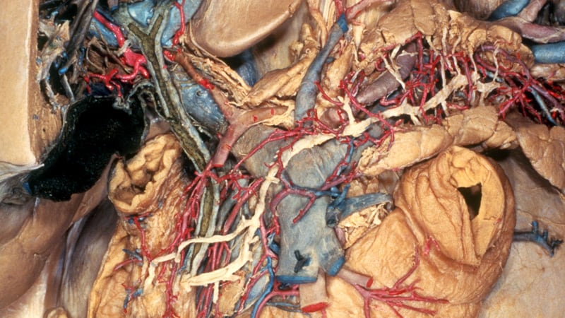
The biliary tract is a system of ducts that transport bile from the liver to the gallbladder and then to the small intestine. Bile is a digestive fluid produced by the liver that helps in the breakdown and absorption of fats in the small intestine. The main components of the biliary tract are:
1. Intrahepatic bile ducts: These are the smaller branches of bile ducts located within the liver that collect bile from the liver cells or hepatocytes.
2. Gallbladder: A small pear-shaped organ located beneath the liver, which stores and concentrates bile received from the intrahepatic bile ducts. The gallbladder releases bile into the small intestine when food is ingested, particularly fats, to aid digestion.
3. Common hepatic duct: This is a duct that forms by the union of the right and left hepatic ducts, which carry bile from the right and left lobes of the liver, respectively.
4. Cystic duct: A short duct that connects the gallbladder to the common hepatic duct, forming the beginning of the common bile duct.
5. Common bile duct: This is a larger duct formed by the union of the common hepatic duct and the cystic duct. It carries bile from the liver and gallbladder into the small intestine.
6. Pancreatic duct: A separate duct that originates from the pancreas, a gland located near the liver and stomach. The pancreatic duct joins the common bile duct just before they both enter the duodenum, the first part of the small intestine.
7. Ampulla of Vater: This is the dilated portion where the common bile duct and the pancreatic duct join together and empty their contents into the duodenum through a shared opening called the papilla of Vater.
Disorders related to the biliary tract include gallstones, cholecystitis (inflammation of the gallbladder), bile duct stones, bile duct strictures or obstructions, and primary sclerosing cholangitis, among others.
Ligation, in the context of medical terminology, refers to the process of tying off a part of the body, usually blood vessels or tissue, with a surgical suture or another device. The goal is to stop the flow of fluids such as blood or other substances within the body. It is commonly used during surgeries to control bleeding or to block the passage of fluids, gases, or solids in various parts of the body.
The gallbladder is a small, pear-shaped organ located just under the liver in the right upper quadrant of the abdomen. Its primary function is to store and concentrate bile, a digestive enzyme produced by the liver, which helps in the breakdown of fats during the digestion process. When food, particularly fatty foods, enter the stomach and small intestine, the gallbladder contracts and releases bile through the common bile duct into the duodenum, the first part of the small intestine, to aid in fat digestion.
The gallbladder is made up of three main parts: the fundus, body, and neck. It has a muscular wall that allows it to contract and release bile. Gallstones, an inflammation of the gallbladder (cholecystitis), or other gallbladder diseases can cause pain, discomfort, and potentially serious health complications if left untreated.
Pancreatitis is a medical condition characterized by inflammation of the pancreas, a gland located in the abdomen that plays a crucial role in digestion and regulating blood sugar levels. The inflammation can be acute (sudden and severe) or chronic (persistent and recurring), and it can lead to various complications if left untreated.
Acute pancreatitis often results from gallstones or excessive alcohol consumption, while chronic pancreatitis may be caused by long-term alcohol abuse, genetic factors, autoimmune conditions, or metabolic disorders like high triglyceride levels. Symptoms of acute pancreatitis include severe abdominal pain, nausea, vomiting, fever, and increased heart rate, while chronic pancreatitis may present with ongoing abdominal pain, weight loss, diarrhea, and malabsorption issues due to impaired digestive enzyme production. Treatment typically involves supportive care, such as intravenous fluids, pain management, and addressing the underlying cause. In severe cases, hospitalization and surgery may be necessary.
Transhepatic sphincterotomy is a medical procedure that involves the incision or cutting of the papilla of Vater, which is a small muscular structure located at the junction of the common bile duct and the main pancreatic duct, with the ampulla of Vater, within the second part of the duodenum. This procedure is performed using a special type of endoscope that is passed through the liver (transhepatically) to access the bile ducts.
The goal of transhepatic sphincterotomy is to relieve obstructions or blockages in the bile ducts, such as gallstones or tumors, that cannot be removed using other endoscopic techniques. This procedure is typically performed by an interventional radiologist or a gastroenterologist with specialized training in endoscopic retrograde cholangiopancreatography (ERCP).
Transhepatic sphincterotomy is considered a higher-risk procedure than traditional ERCP sphincterotomy due to the need for liver puncture and the potential complications associated with this approach, including bleeding, infection, and injury to surrounding organs. However, it may be necessary in certain situations where traditional ERCP is not feasible or has failed.
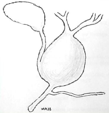
A Choledochal cyst is a congenital dilatation or abnormal enlargement of the bile ducts, which are the tubes that carry bile from the liver to the small intestine. Bile is a digestive juice produced by the liver that helps in the digestion of fats.
Choledochal cysts can be classified into several types based on their location and the anatomy of the biliary tree. The most common type, called Type I, involves dilatation of the common bile duct. Other types include dilatation of the intrahepatic bile ducts (Type II), dilatation of both the intrahepatic and extrahepatic bile ducts (Type III), and multiple cystic dilatations of the bile ducts (Type IV).
Choledochal cysts are more common in females than males, and they can present at any age. Symptoms may include abdominal pain, jaundice, vomiting, and fever. Complications of choledochal cysts can include bile duct stones, infection, and cancer. Treatment typically involves surgical removal of the cyst, followed by reconstruction of the biliary tree.
Biliary tract surgical procedures refer to a range of operations that involve the biliary system, which includes the liver, gallbladder, and bile ducts. These procedures can be performed for various reasons, including the treatment of gallstones, bile duct injuries, tumors, or other conditions affecting the biliary tract. Here are some examples of biliary tract surgical procedures:
1. Cholecystectomy: This is the surgical removal of the gallbladder, which is often performed to treat symptomatic gallstones or chronic cholecystitis (inflammation of the gallbladder). It can be done as an open procedure or laparoscopically.
2. Bile duct exploration: This procedure involves opening the common bile duct to remove stones, strictures, or tumors. It is often performed during a cholecystectomy if there is suspicion of common bile duct involvement.
3. Hepaticojejunostomy: This operation connects the liver's bile ducts directly to a portion of the small intestine called the jejunum, bypassing a damaged or obstructed segment of the biliary tract. It is often performed for benign or malignant conditions affecting the bile ducts.
4. Roux-en-Y hepaticojejunostomy: This procedure involves creating a Y-shaped limb of jejunum and connecting it to the liver's bile ducts, bypassing the common bile duct and duodenum. It is often performed for complex biliary tract injuries or malignancies.
5. Whipple procedure (pancreaticoduodenectomy): This extensive operation involves removing the head of the pancreas, the duodenum, a portion of the jejunum, the gallbladder, and the common bile duct. It is performed for malignancies involving the pancreas, bile duct, or duodenum.
6. Liver resection: This procedure involves removing a portion of the liver to treat primary liver tumors (hepatocellular carcinoma or cholangiocarcinoma) or metastatic cancer from other organs.
7. Biliary stenting or bypass: These minimally invasive procedures involve placing a stent or creating a bypass to relieve bile duct obstructions caused by tumors, strictures, or stones. They can be performed endoscopically (ERCP) or percutaneously (PTC).
8. Cholecystectomy: This procedure involves removing the gallbladder, often for symptomatic cholelithiasis (gallstones) or cholecystitis (inflammation of the gallbladder). It can be performed laparoscopically or open.
9. Biliary drainage: This procedure involves placing a catheter to drain bile from the liver or bile ducts, often for acute or chronic obstructions caused by tumors, strictures, or stones. It can be performed endoscopically (ERCP) or percutaneously (PTC).
10. Bilioenteric anastomosis: This procedure involves connecting the biliary tract to a portion of the small intestine, often for benign or malignant conditions affecting the bile ducts or pancreas. It can be performed open or laparoscopically.
A biliary fistula is an abnormal connection or passage between the biliary system (which includes the gallbladder, bile ducts, and liver) and another organ or structure, usually in the abdominal cavity. This connection allows bile, which is a digestive fluid produced by the liver, to leak out of its normal pathway and into other areas of the body.
Biliary fistulas can occur as a result of trauma, surgery, infection, or inflammation in the biliary system. Symptoms may include abdominal pain, fever, jaundice (yellowing of the skin and eyes), nausea, vomiting, and clay-colored stools. Treatment typically involves addressing the underlying cause of the fistula, such as draining an infection or repairing damaged tissue, and diverting bile flow away from the site of the leak. In some cases, surgery may be necessary to repair the fistula.
Choledochostomy is a surgical procedure that involves creating an opening (stoma) into the common bile duct, which carries bile from the liver and gallbladder to the small intestine. This procedure is typically performed to relieve obstructions or blockages in the bile duct, such as those caused by gallstones, tumors, or scar tissue.
During the choledochostomy procedure, a surgeon makes an incision in the abdomen and exposes the common bile duct. The duct is then cut open, and a small tube (catheter) is inserted into the duct to allow bile to drain out of the body. The catheter may be left in place temporarily or permanently, depending on the underlying condition causing the obstruction.
Choledochostomy is typically performed as an open surgical procedure, but it can also be done using minimally invasive techniques such as laparoscopy or robotic-assisted surgery. As with any surgical procedure, choledochostomy carries risks such as bleeding, infection, and damage to surrounding tissues. However, these risks are generally low in the hands of an experienced surgeon.
Adenoma of the bile duct is a benign (noncancerous) tumor that develops in the bile ducts, which are tiny tubes that carry bile from the liver to the gallbladder and small intestine. Bile is a digestive fluid produced by the liver.
Bile duct adenomas are rare and usually do not cause any symptoms. However, if they grow large enough, they may obstruct the flow of bile and cause jaundice (yellowing of the skin and whites of the eyes), abdominal pain, or itching. In some cases, bile duct adenomas may become cancerous and develop into bile duct carcinomas.
The exact cause of bile duct adenomas is not known, but they are more common in people with certain genetic disorders, such as Gardner's syndrome and von Hippel-Lindau disease. Treatment for bile duct adenomas typically involves surgical removal of the tumor.
Drainage, in medical terms, refers to the removal of excess fluid or accumulated collections of fluids from various body parts or spaces. This is typically accomplished through the use of medical devices such as catheters, tubes, or drains. The purpose of drainage can be to prevent the buildup of fluids that may cause discomfort, infection, or other complications, or to treat existing collections of fluid such as abscesses, hematomas, or pleural effusions. Drainage may also be used as a diagnostic tool to analyze the type and composition of the fluid being removed.
An endoscope is a medical device used for examining the interior of a body cavity or organ. It consists of a long, thin, flexible (or rigid) tube with a light and a camera at one end. The other end is connected to a video monitor that displays the images captured by the camera. Endoscopes can be inserted through natural openings in the body, such as the mouth or anus, or through small incisions. They are used for diagnostic purposes, as well as for performing various medical procedures, including biopsies and surgeries. Different types of endoscopes include gastroscopes, colonoscopes, bronchoscopes, and arthroscopes, among others.
Duodenoscopy is a medical procedure that involves the insertion of a duodenoscope, which is a flexible, lighted tube with a camera and tiny tools on the end, through the mouth and down the throat to examine the upper part of the small intestine (duodenum) and the opening of the bile and pancreatic ducts.
During the procedure, the doctor can take tissue samples for biopsy, remove polyps or other abnormal growths, or perform other interventions as needed. Duodenoscopy is commonly used to diagnose and treat conditions such as gastrointestinal bleeding, inflammation, infection, and cancer.
It's important to note that duodenoscopes have been associated with the spread of antibiotic-resistant bacteria in some cases, so healthcare providers must follow strict cleaning and disinfection protocols to minimize this risk.
Cholecystitis is a medical condition characterized by inflammation of the gallbladder, a small pear-shaped organ located under the liver that stores and concentrates bile produced by the liver. Bile is a digestive fluid that helps break down fats in the small intestine during digestion.
Acute cholecystitis is a sudden inflammation of the gallbladder, often caused by the presence of gallstones that block the cystic duct, the tube that carries bile from the gallbladder to the common bile duct. This blockage can cause bile to build up in the gallbladder, leading to inflammation, swelling, and pain.
Chronic cholecystitis is a long-term inflammation of the gallbladder, often caused by repeated attacks of acute cholecystitis or the presence of gallstones that cause ongoing irritation and damage to the gallbladder wall. Over time, chronic cholecystitis can lead to thickening and scarring of the gallbladder wall, which can reduce its ability to function properly.
Symptoms of cholecystitis may include sudden and severe abdominal pain, often in the upper right or center of the abdomen, that may worsen after eating fatty foods; fever; nausea and vomiting; bloating and gas; and clay-colored stools. Treatment for cholecystitis typically involves antibiotics to treat any infection present, pain relief, and surgery to remove the gallbladder (cholecystectomy). In some cases, a nonsurgical procedure called endoscopic retrograde cholangiopancreatography (ERCP) may be used to remove gallstones from the bile duct.
Imino acids are organic compounds that contain a nitrogen atom as part of an amide-like structure. They are structurally similar to amino acids, which contain a carboxyl group and an amino group, but instead of the amino group, imino acids have a structural unit known as an imine or Schiff base, which is a carbon-nitrogen double bond with a hydrogen atom attached to the nitrogen atom.
One example of an imino acid is proline, which is a cyclic imino acid that plays important roles in protein structure and function. Proline is unique among the 20 standard amino acids because its side chain is linked to the nitrogen atom of the backbone, forming a ring-like structure. This structural feature gives proline unique properties, such as restricted rotation around the bond between the nitrogen and alpha carbon atoms, which can affect protein folding and stability.
Other imino acids may be formed through chemical reactions or enzymatic processes, and they can play important roles in various biological pathways, including the biosynthesis of amino acids, nucleotides, and other biomolecules. However, imino acids are not typically considered to be part of the standard set of 20 amino acids that make up proteins.
Gallbladder diseases refer to a range of conditions that affect the function and structure of the gallbladder, a small pear-shaped organ located beneath the liver. The primary role of the gallbladder is to store, concentrate, and release bile into the small intestine to aid in digesting fats. Gallbladder diseases can be chronic or acute and may cause various symptoms, discomfort, or complications if left untreated. Here are some common gallbladder diseases with brief definitions:
1. Cholelithiasis: The presence of gallstones within the gallbladder. Gallstones are small, hard deposits made of cholesterol, bilirubin, or a combination of both, which can vary in size from tiny grains to several centimeters.
2. Cholecystitis: Inflammation of the gallbladder, often caused by obstruction of the cystic duct (the tube connecting the gallbladder and the common bile duct) due to a gallstone. This condition can be acute or chronic and may cause abdominal pain, fever, and tenderness in the right upper quadrant of the abdomen.
3. Choledocholithiasis: The presence of gallstones within the common bile duct, which can lead to obstruction, jaundice, and potential infection of the biliary system (cholangitis).
4. Acalculous gallbladder disease: Gallbladder dysfunction or inflammation without the presence of gallstones. This condition is often seen in critically ill patients and can lead to similar symptoms as cholecystitis.
5. Gallbladder polyps: Small growths attached to the inner wall of the gallbladder. While most polyps are benign, some may have malignant potential, especially if they are larger than 1 cm in size or associated with certain risk factors.
6. Gallbladder cancer: A rare form of cancer that originates in the gallbladder tissue. It is often asymptomatic in its early stages and can be challenging to diagnose. Symptoms may include abdominal pain, jaundice, or a palpable mass in the right upper quadrant of the abdomen.
It is essential to consult with a healthcare professional if experiencing symptoms related to gallbladder disease for proper diagnosis and treatment.
Jaundice is a medical condition characterized by the yellowing of the skin, sclera (whites of the eyes), and mucous membranes due to an excess of bilirubin in the bloodstream. Bilirubin is a yellow-orange pigment produced when hemoglobin from red blood cells is broken down. Normally, bilirubin is processed by the liver and excreted through bile into the digestive system. However, if there's an issue with bilirubin metabolism or elimination, it can accumulate in the body, leading to jaundice.
Jaundice can be a symptom of various underlying conditions, such as liver diseases (hepatitis, cirrhosis), gallbladder issues (gallstones, tumors), or blood disorders (hemolysis). It is essential to consult a healthcare professional if jaundice is observed, as it may indicate a severe health problem requiring prompt medical attention.
Lithotripsy is a medical procedure that uses shock waves or other high-energy sound waves to break down and remove calculi (stones) in the body, particularly in the kidneys, ureters, or gallbladder. The procedure is typically performed on an outpatient basis and does not require any incisions.
During lithotripsy, the patient lies on a cushioned table while a lithotripter, a device that generates shock waves, is positioned around the area of the stone. As the shock waves pass through the body, they break the stone into tiny fragments that can then be easily passed out of the body in urine.
Lithotripsy is generally a safe and effective procedure, but it may not be suitable for everyone. Patients with certain medical conditions, such as bleeding disorders or pregnancy, may not be able to undergo lithotripsy. Additionally, some stones may be too large or too dense to be effectively treated with lithotripsy. In these cases, other treatment options, such as surgery, may be necessary.
Adenomyoma is a benign (non-cancerous) growth that occurs when the glands and muscle tissue from the lining of the uterus (endometrium) become embedded in the muscular wall of the uterus (myometrium). This condition most commonly affects women in their 40s and 50s, and it can cause symptoms such as heavy menstrual bleeding, painful periods, and pelvic pain or discomfort.
The term "adenomyoma" is derived from two words: "adeno," which means gland, and "myoma," which refers to a benign muscle tumor. Therefore, an adenomyoma can be thought of as a benign growth that contains both glandular tissue and muscle tissue.
Adenomyomas are typically found in the lower part of the uterus, near the cervix, and they can vary in size from small nodules to larger masses. In some cases, adenomyomas may cause no symptoms at all, while in other cases, they can lead to significant discomfort and pain.
The exact cause of adenomyoma is not fully understood, but it is thought to be related to hormonal factors, as well as trauma or injury to the uterus. Treatment options for adenomyoma may include medication to manage symptoms, such as pain relievers or hormone therapy, or surgical intervention, such as a hysterectomy (removal of the uterus).
Gallbladder neoplasms refer to abnormal growths in the tissue of the gallbladder, which can be benign or malignant. Benign neoplasms are non-cancerous and typically do not spread to other parts of the body. Malignant neoplasms, also known as gallbladder cancer, can invade nearby tissues and organs and may metastasize (spread) to distant parts of the body. Gallbladder neoplasms can cause symptoms such as abdominal pain, jaundice, and nausea, but they are often asymptomatic until they have advanced to an advanced stage. The exact causes of gallbladder neoplasms are not fully understood, but risk factors include gallstones, chronic inflammation of the gallbladder, and certain inherited genetic conditions.
Technetium Tc 99m Disofenin is not a medical condition, but rather a radiopharmaceutical used in diagnostic imaging. It is a radioactive tracer used in nuclear medicine scans, specifically for liver and biliary system imaging. The compound consists of the radioisotope Technetium-99m (Tc-99m) bonded to the pharmaceutical Disofenin.
The Tc-99m is a gamma emitter with a half-life of 6 hours, making it ideal for diagnostic imaging. When administered to the patient, the compound is taken up by the liver and excreted into the bile ducts and gallbladder, allowing medical professionals to visualize these structures using a gamma camera. This can help detect various conditions such as tumors, gallstones, or obstructions in the biliary system.
It's important to note that Technetium Tc 99m Disofenin is used diagnostically and not for therapeutic purposes. The radiation exposure from this compound is generally low and considered safe for diagnostic use. However, as with any medical procedure involving radiation, the benefits and risks should be carefully weighed and discussed with a healthcare professional.
Endoscopy of the digestive system, also known as gastrointestinal (GI) endoscopy, is a medical procedure that allows healthcare professionals to visually examine the inside lining of the digestive tract using a flexible tube with a light and camera attached to it, called an endoscope. This procedure can help diagnose and treat various conditions affecting the digestive system, including gastroesophageal reflux disease (GERD), ulcers, inflammatory bowel disease (IBD), and cancer.
There are several types of endoscopy procedures that focus on different parts of the digestive tract:
1. Esophagogastroduodenoscopy (EGD): This procedure examines the esophagus, stomach, and duodenum (the first part of the small intestine). It is often used to investigate symptoms such as difficulty swallowing, abdominal pain, or bleeding in the upper GI tract.
2. Colonoscopy: This procedure explores the large intestine (colon) and rectum. It is commonly performed to screen for colon cancer, as well as to diagnose and treat conditions like inflammatory bowel disease, diverticulosis, or polyps.
3. Sigmoidoscopy: Similar to a colonoscopy, this procedure examines the lower part of the colon (sigmoid colon) and rectum. It is often used as a screening tool for colon cancer and to investigate symptoms like rectal bleeding or changes in bowel habits.
4. Upper GI endoscopy: This procedure focuses on the esophagus, stomach, and duodenum, using a thin, flexible tube with a light and camera attached to it. It is used to diagnose and treat conditions such as GERD, ulcers, and difficulty swallowing.
5. Capsule endoscopy: This procedure involves swallowing a small capsule containing a camera that captures images of the digestive tract as it passes through. It can help diagnose conditions in the small intestine that may be difficult to reach with traditional endoscopes.
Endoscopy is typically performed under sedation or anesthesia to ensure patient comfort during the procedure. The images captured by the endoscope are displayed on a monitor, allowing the healthcare provider to assess the condition of the digestive tract and make informed treatment decisions.
Endoscopy is a medical procedure that involves the use of an endoscope, which is a flexible tube with a light and camera at the end, to examine the interior of a body cavity or organ. The endoscope is inserted through a natural opening in the body, such as the mouth or anus, or through a small incision. The images captured by the camera are transmitted to a monitor, allowing the physician to visualize the internal structures and detect any abnormalities, such as inflammation, ulcers, or tumors. Endoscopy can also be used for diagnostic purposes, such as taking tissue samples for biopsy, or for therapeutic purposes, such as removing polyps or performing minimally invasive surgeries.
Pathologic dilatation refers to an abnormal and excessive widening or enlargement of a body cavity or organ, which can result from various medical conditions. This abnormal dilation can occur in different parts of the body, including the blood vessels, digestive tract, airways, or heart chambers.
In the context of the cardiovascular system, pathologic dilatation may indicate a weakening or thinning of the heart muscle, leading to an enlarged chamber that can no longer pump blood efficiently. This condition is often associated with various heart diseases, such as cardiomyopathy, valvular heart disease, or long-standing high blood pressure.
In the gastrointestinal tract, pathologic dilatation may occur due to mechanical obstruction, neuromuscular disorders, or inflammatory conditions that affect the normal motility of the intestines. Examples include megacolon in Hirschsprung's disease, toxic megacolon in ulcerative colitis, or volvulus (twisting) of the bowel.
Pathologic dilatation can lead to various complications, such as reduced organ function, impaired circulation, and increased risk of infection or perforation. Treatment depends on the underlying cause and may involve medications, surgery, or other interventions to address the root problem and prevent further enlargement.
Pancreaticoduodenectomy, also known as the Whipple procedure, is a complex surgical operation that involves the removal of the head of the pancreas, the duodenum (the first part of the small intestine), the gallbladder, and the distal common bile duct. In some cases, a portion of the stomach may also be removed. The remaining parts of the pancreas, bile duct, and intestines are then reconnected to allow for the digestion of food and drainage of bile.
This procedure is typically performed as a treatment for various conditions affecting the pancreas, such as tumors (including pancreatic cancer), chronic pancreatitis, or traumatic injuries. It is a major surgical operation that requires significant expertise and experience to perform safely and effectively.
Intrahepatic cholestasis is a medical condition characterized by the interruption or reduction of bile flow within the liver. Bile is a digestive fluid produced by the liver that helps in the absorption of fats and fat-soluble vitamins. Intrahepatic cholestasis occurs when there is a problem with the transport of bile components inside the liver cells (hepatocytes). This can lead to an accumulation of bile acids, bilirubin, and other substances in the liver, which can cause damage to liver cells and result in symptoms such as jaundice, itching, and dark urine.
Intrahepatic cholestasis can be caused by various factors, including medications, alcohol abuse, hepatitis viruses, autoimmune disorders, genetic defects, and cancer. Depending on the underlying cause, intrahepatic cholestasis can be acute or chronic, and it can range from mild to severe. Treatment typically involves addressing the underlying cause of the condition, as well as providing supportive care to manage symptoms and prevent complications.
Acute cholecystitis is a medical condition characterized by inflammation of the gallbladder (cholecystitis) that develops suddenly (acute). The gallbladder is a small pear-shaped organ located in the upper right part of the abdomen, beneath the liver. It stores bile, a digestive juice produced by the liver, which helps break down fats in the food we eat.
Acute cholecystitis occurs when the gallbladder becomes inflamed and irritated, often due to the presence of gallstones that block the cystic duct, the tube that carries bile from the gallbladder into the small intestine. When the cystic duct is obstructed, bile builds up in the gallbladder, causing it to become swollen, inflamed, and infected.
Symptoms of acute cholecystitis may include sudden and severe abdominal pain, often located in the upper right or middle part of the abdomen, that may radiate to the back or shoulder blade area. Other symptoms may include fever, nausea, vomiting, loss of appetite, and abdominal tenderness or swelling.
Acute cholecystitis is typically diagnosed through a combination of medical history, physical examination, laboratory tests, and imaging studies such as ultrasound or CT scan. Treatment may involve hospitalization, antibiotics to treat infection, pain relief medications, and surgery to remove the gallbladder (cholecystectomy). In some cases, nonsurgical treatments such as endoscopic sphincterotomy or percutaneous cholecystostomy may be used to relieve obstruction and inflammation.
Sphincter of Oddi dysfunction (SOD) is a condition characterized by abnormalities in the functioning of the Sphincter of Oddi, which is a muscular valve that controls the flow of bile and pancreatic juice from the pancreas and gallbladder into the duodenum (the first part of the small intestine).
In SOD, the sphincter may either fail to relax properly or become overactive, leading to a variety of symptoms such as abdominal pain, nausea, vomiting, bloating, and elevated liver enzymes. The condition can be classified into two types: Type I, which is associated with elevated liver enzymes and/or pancreatic enzymes, and Type II, which is characterized by abdominal pain without biochemical abnormalities.
The diagnosis of SOD typically involves a series of tests such as manometry (measuring the pressure inside the sphincter), endoscopic ultrasound, or magnetic resonance cholangiopancreatography (MRCP) to visualize the anatomy and function of the sphincter. Treatment options may include medications to relax the sphincter, endoscopic therapy to cut or stretch the muscle, or surgery in severe cases.
A jejunostomy is a surgical procedure where an opening (stoma) is created in the lower part of the small intestine, called the jejunum. This stoma allows for the passage of nutrients and digestive enzymes from the small intestine into a tube or external pouch, bypassing the mouth, esophagus, stomach, and upper small intestine (duodenum).
Jejunostomy is typically performed to provide enteral nutrition support in patients who are unable to consume food or liquids by mouth due to various medical conditions such as dysphagia, gastroparesis, bowel obstruction, or after certain surgical procedures. The jejunostomy tube can be used for short-term or long-term nutritional support, depending on the patient's needs and underlying medical condition.
Cholecystolithiasis is the medical term for the presence of gallstones in the gallbladder. The gallbladder is a small pear-shaped organ located under the liver that stores and concentrates bile, a digestive fluid produced by the liver. Gallstones are hardened deposits that can form in the gallbladder when substances in the bile, such as cholesterol or bilirubin, become concentrated and crystallize.
Gallstones can vary in size, from tiny grains of sand to large stones several centimeters in diameter. Some people may have a single gallstone, while others may have many. Gallstones may cause no symptoms at all, but if they block the flow of bile out of the gallbladder, they can cause pain, inflammation, and infection.
Symptoms of cholecystolithiasis may include abdominal pain, often in the upper right or center of the abdomen, that may be sharp or crampy and may occur after eating fatty foods. Other symptoms may include nausea, vomiting, fever, and chills. If gallstones are left untreated, they can lead to serious complications such as cholecystitis (inflammation of the gallbladder), pancreatitis (inflammation of the pancreas), or cholangitis (infection of the bile ducts). Treatment for cholecystolithiasis may include medication to dissolve the gallstones, shock wave lithotripsy to break up the stones, or surgery to remove the gallbladder.
The duodenum is the first part of the small intestine, immediately following the stomach. It is a C-shaped structure that is about 10-12 inches long and is responsible for continuing the digestion process that begins in the stomach. The duodenum receives partially digested food from the stomach through the pyloric valve and mixes it with digestive enzymes and bile produced by the pancreas and liver, respectively. These enzymes help break down proteins, fats, and carbohydrates into smaller molecules, allowing for efficient absorption in the remaining sections of the small intestine.
Bilirubin is a yellowish pigment that is produced by the liver when it breaks down old red blood cells. It is a normal byproduct of hemoglobin metabolism and is usually conjugated (made water-soluble) in the liver before being excreted through the bile into the digestive system. Elevated levels of bilirubin can cause jaundice, a yellowing of the skin and eyes. Increased bilirubin levels may indicate liver disease or other medical conditions such as gallstones or hemolysis. It is also measured to assess liver function and to help diagnose various liver disorders.
Pancreatic neoplasms refer to abnormal growths in the pancreas that can be benign or malignant. The pancreas is a gland located behind the stomach that produces hormones and digestive enzymes. Pancreatic neoplasms can interfere with the normal functioning of the pancreas, leading to various health complications.
Benign pancreatic neoplasms are non-cancerous growths that do not spread to other parts of the body. They are usually removed through surgery to prevent any potential complications, such as blocking the bile duct or causing pain.
Malignant pancreatic neoplasms, also known as pancreatic cancer, are cancerous growths that can invade and destroy surrounding tissues and organs. They can also spread (metastasize) to other parts of the body, such as the liver, lungs, or bones. Pancreatic cancer is often aggressive and difficult to treat, with a poor prognosis.
There are several types of pancreatic neoplasms, including adenocarcinomas, neuroendocrine tumors, solid pseudopapillary neoplasms, and cystic neoplasms. The specific type of neoplasm is determined through various diagnostic tests, such as imaging studies, biopsies, and blood tests. Treatment options depend on the type, stage, and location of the neoplasm, as well as the patient's overall health and preferences.
Pancreatic diseases refer to a group of medical conditions that affect the structure and function of the pancreas, a vital organ located in the abdomen. The pancreas has two main functions: an exocrine function, which involves the production of digestive enzymes that help break down food in the small intestine, and an endocrine function, which involves the production of hormones such as insulin and glucagon that regulate blood sugar levels.
Pancreatic diseases can be broadly classified into two categories: inflammatory and non-inflammatory. Inflammatory pancreatic diseases include conditions such as acute pancreatitis, which is characterized by sudden inflammation of the pancreas, and chronic pancreatitis, which is a long-term inflammation that can lead to scarring and loss of function.
Non-inflammatory pancreatic diseases include conditions such as pancreatic cancer, which is a malignant tumor that can arise from the cells of the pancreas, and benign tumors such as cysts or adenomas. Other non-inflammatory conditions include pancreatic insufficiency, which can occur when the pancreas does not produce enough digestive enzymes, and diabetes mellitus, which can result from impaired insulin production or action.
Overall, pancreatic diseases can have serious consequences on a person's health and quality of life, and early diagnosis and treatment are essential for optimal outcomes.
The intraoperative period is the phase of surgical treatment that refers to the time during which the surgery is being performed. It begins when the anesthesia is administered and the patient is prepared for the operation, and it ends when the surgery is completed, the anesthesia is discontinued, and the patient is transferred to the recovery room or intensive care unit (ICU).
During the intraoperative period, the surgical team, including surgeons, anesthesiologists, nurses, and other healthcare professionals, work together to carry out the surgical procedure safely and effectively. The anesthesiologist monitors the patient's vital signs, such as heart rate, blood pressure, oxygen saturation, and body temperature, throughout the surgery to ensure that the patient remains stable and does not experience any complications.
The surgeon performs the operation, using various surgical techniques and instruments to achieve the desired outcome. The surgical team also takes measures to prevent infection, control bleeding, and manage pain during and after the surgery.
Overall, the intraoperative period is a critical phase of surgical treatment that requires close collaboration and communication among members of the healthcare team to ensure the best possible outcomes for the patient.
Endosonography, also known as endoscopic ultrasound (EUS), is a medical procedure that combines endoscopy and ultrasound to obtain detailed images and information about the digestive tract and surrounding organs. An endoscope, which is a flexible tube with a light and camera at its tip, is inserted through the mouth or rectum to reach the area of interest. A high-frequency ultrasound transducer at the tip of the endoscope generates sound waves that bounce off body tissues and create echoes, which are then translated into detailed images by a computer.
Endosonography allows doctors to visualize structures such as the esophageal, stomach, and intestinal walls, lymph nodes, blood vessels, and organs like the pancreas, liver, and gallbladder. It can help diagnose conditions such as tumors, inflammation, and infections, and it can also be used to guide biopsies or fine-needle aspirations of suspicious lesions.
Overall, endosonography is a valuable tool for the diagnosis and management of various gastrointestinal and related disorders.
The liver is a large, solid organ located in the upper right portion of the abdomen, beneath the diaphragm and above the stomach. It plays a vital role in several bodily functions, including:
1. Metabolism: The liver helps to metabolize carbohydrates, fats, and proteins from the food we eat into energy and nutrients that our bodies can use.
2. Detoxification: The liver detoxifies harmful substances in the body by breaking them down into less toxic forms or excreting them through bile.
3. Synthesis: The liver synthesizes important proteins, such as albumin and clotting factors, that are necessary for proper bodily function.
4. Storage: The liver stores glucose, vitamins, and minerals that can be released when the body needs them.
5. Bile production: The liver produces bile, a digestive juice that helps to break down fats in the small intestine.
6. Immune function: The liver plays a role in the immune system by filtering out bacteria and other harmful substances from the blood.
Overall, the liver is an essential organ that plays a critical role in maintaining overall health and well-being.
Duodenal neoplasms refer to abnormal growths in the duodenum, which is the first part of the small intestine that receives digestive secretions from the pancreas and bile duct. These growths can be benign or malignant (cancerous).
Benign neoplasms include adenomas, leiomyomas, lipomas, and hamartomas. They are usually slow-growing and do not spread to other parts of the body. However, they may cause symptoms such as abdominal pain, bleeding, or obstruction of the intestine.
Malignant neoplasms include adenocarcinomas, neuroendocrine tumors (carcinoids), lymphomas, and sarcomas. They are more aggressive and can invade surrounding tissues and spread to other parts of the body. Symptoms may include abdominal pain, weight loss, jaundice, anemia, or bowel obstruction.
The diagnosis of duodenal neoplasms is usually made through imaging tests such as CT scans, MRI, or endoscopy with biopsy. Treatment depends on the type and stage of the tumor and may include surgery, chemotherapy, radiation therapy, or a combination of these modalities.
Postoperative complications refer to any unfavorable condition or event that occurs during the recovery period after a surgical procedure. These complications can vary in severity and may include, but are not limited to:
1. Infection: This can occur at the site of the incision or inside the body, such as pneumonia or urinary tract infection.
2. Bleeding: Excessive bleeding (hemorrhage) can lead to a drop in blood pressure and may require further surgical intervention.
3. Blood clots: These can form in the deep veins of the legs (deep vein thrombosis) and can potentially travel to the lungs (pulmonary embolism).
4. Wound dehiscence: This is when the surgical wound opens up, which can lead to infection and further complications.
5. Pulmonary issues: These include atelectasis (collapsed lung), pneumonia, or respiratory failure.
6. Cardiovascular problems: These include abnormal heart rhythms (arrhythmias), heart attack, or stroke.
7. Renal failure: This can occur due to various reasons such as dehydration, blood loss, or the use of certain medications.
8. Pain management issues: Inadequate pain control can lead to increased stress, anxiety, and decreased mobility.
9. Nausea and vomiting: These can be caused by anesthesia, opioid pain medication, or other factors.
10. Delirium: This is a state of confusion and disorientation that can occur in the elderly or those with certain medical conditions.
Prompt identification and management of these complications are crucial to ensure the best possible outcome for the patient.
Cholecystostomy is a medical procedure that involves the creation of an opening or fistula between the gallbladder and the stomach or the skin surface to drain bile from the gallbladder. This procedure is typically performed when there is an obstruction in the cystic duct or common bile duct, leading to inflammation or infection of the gallbladder (cholecystitis).
There are two types of cholecystostomy: percutaneous and open. Percutaneous cholecystostomy is a minimally invasive procedure that involves inserting a small tube through the skin and into the gallbladder under the guidance of imaging techniques such as ultrasound or CT scan. Open cholecystostomy, on the other hand, requires an open surgical incision to access the gallbladder and create the fistula.
Cholecystostomy is usually a temporary measure to manage acute cholecystitis in high-risk patients who are not suitable candidates for surgery or until they become stable enough to undergo a more definitive treatment, such as cholecystectomy (surgical removal of the gallbladder).
Bile reflux is a condition in which bile flows backward from the small intestine into the stomach and sometimes into the esophagus, causing symptoms such as heartburn, nausea, vomiting a greenish-yellow fluid (bile), and abdominal pain. Bile is a digestive fluid produced by the liver that helps to break down fats in the small intestine. Normally, a muscle called the sphincter of Oddi prevents bile from flowing backward into the stomach. However, if this muscle becomes weak or damaged, bile reflux can occur.
Bile reflux is different from gastroesophageal reflux disease (GERD), which occurs when stomach acid flows backward into the esophagus. Although both conditions can cause similar symptoms, such as heartburn and regurgitation, they require different treatments. Bile reflux can increase the risk of complications such as inflammation of the stomach lining (gastritis), ulcers, and cancer of the esophagus. If left untreated, bile reflux can lead to serious health problems, so it is important to seek medical attention if you experience symptoms.
Experimental liver cirrhosis refers to a controlled research setting where various factors and substances are intentionally introduced to induce liver cirrhosis in animals or cell cultures. The purpose is to study the mechanisms, progression, potential treatments, and prevention strategies for liver cirrhosis. This could involve administering chemicals, drugs, alcohol, viruses, or manipulating genes associated with liver damage and fibrosis. It's important to note that results from experimental models may not directly translate to human conditions, but they can provide valuable insights into disease pathophysiology and therapeutic development.
Lithotripsy, laser refers to a medical procedure that uses laser energy to break down and fragment stones located in the urinary tract, such as kidney or ureteral stones. The laser energy is delivered through a flexible fiberoptic endoscope, which is inserted into the urinary tract. Once the stone is targeted, the laser energy is focused on it, causing the stone to fragment into tiny pieces that can then be passed naturally through the urine. This procedure is typically performed under anesthesia and may require hospitalization depending on the size and location of the stone. It is a minimally invasive alternative to traditional surgical methods for treating urinary tract stones.
Pathological constriction refers to an abnormal narrowing or tightening of a body passage or organ, which can interfere with the normal flow of blood, air, or other substances through the area. This constriction can occur due to various reasons such as inflammation, scarring, or abnormal growths, and can affect different parts of the body, including blood vessels, airways, intestines, and ureters. Pathological constriction can lead to a range of symptoms and complications depending on its location and severity, and may require medical intervention to correct.
Intraoperative care refers to the medical care and interventions provided to a patient during a surgical procedure. This care is typically administered by a team of healthcare professionals, including anesthesiologists, surgeons, nurses, and other specialists as needed. The goal of intraoperative care is to maintain the patient's physiological stability throughout the surgery, minimize complications, and ensure the best possible outcome.
Intraoperative care may include:
1. Anesthesia management: Administering and monitoring anesthetic drugs to keep the patient unconscious and free from pain during the surgery.
2. Monitoring vital signs: Continuously tracking the patient's heart rate, blood pressure, oxygen saturation, body temperature, and other key physiological parameters to ensure they remain within normal ranges.
3. Fluid and blood product administration: Maintaining adequate intravascular volume and oxygen-carrying capacity through the infusion of fluids and blood products as needed.
4. Intraoperative imaging: Utilizing real-time imaging techniques, such as X-ray, ultrasound, or CT scans, to guide the surgical procedure and ensure accurate placement of implants or other devices.
5. Neuromonitoring: Using electrophysiological methods to monitor the functional integrity of nerves and neural structures during surgery, particularly in procedures involving the brain, spine, or peripheral nerves.
6. Intraoperative medication management: Administering various medications as needed for pain control, infection prophylaxis, or the treatment of medical conditions that may arise during the surgery.
7. Temperature management: Regulating the patient's body temperature to prevent hypothermia or hyperthermia, which can have adverse effects on surgical outcomes and overall patient health.
8. Communication and coordination: Ensuring effective communication among the members of the surgical team to optimize patient care and safety.
Cholangiocarcinoma is a type of cancer that arises from the cells that line the bile ducts, which are small tubes that carry digestive enzymes from the liver to the small intestine. It can occur in different parts of the bile duct system, including the bile ducts inside the liver (intrahepatic), the bile ducts outside the liver (extrahepatic), and the area where the bile ducts join the pancreas and small intestine (ampulla of Vater).
Cholangiocarcinoma is a relatively rare cancer, but its incidence has been increasing in recent years. It can be difficult to diagnose because its symptoms are often nonspecific and similar to those of other conditions, such as gallstones or pancreatitis. Treatment options depend on the location and stage of the cancer, and may include surgery, radiation therapy, chemotherapy, or a combination of these approaches.
Catheterization is a medical procedure in which a catheter (a flexible tube) is inserted into the body to treat various medical conditions or for diagnostic purposes. The specific definition can vary depending on the area of medicine and the particular procedure being discussed. Here are some common types of catheterization:
1. Urinary catheterization: This involves inserting a catheter through the urethra into the bladder to drain urine. It is often performed to manage urinary retention, monitor urine output in critically ill patients, or assist with surgical procedures.
2. Cardiac catheterization: A procedure where a catheter is inserted into a blood vessel, usually in the groin or arm, and guided to the heart. This allows for various diagnostic tests and treatments, such as measuring pressures within the heart chambers, assessing blood flow, or performing angioplasty and stenting of narrowed coronary arteries.
3. Central venous catheterization: A catheter is inserted into a large vein, typically in the neck, chest, or groin, to administer medications, fluids, or nutrition, or to monitor central venous pressure.
4. Peritoneal dialysis catheterization: A catheter is placed into the abdominal cavity for individuals undergoing peritoneal dialysis, a type of kidney replacement therapy.
5. Neurological catheterization: In some cases, a catheter may be inserted into the cerebrospinal fluid space (lumbar puncture) or the brain's ventricular system (ventriculostomy) to diagnose or treat various neurological conditions.
These are just a few examples of catheterization procedures in medicine. The specific definition and purpose will depend on the medical context and the particular organ or body system involved.
Liver function tests (LFTs) are a group of blood tests that are used to assess the functioning and health of the liver. These tests measure the levels of various enzymes, proteins, and waste products that are produced or metabolized by the liver. Some common LFTs include:
1. Alanine aminotransferase (ALT): An enzyme found primarily in the liver, ALT is released into the bloodstream in response to liver cell damage. Elevated levels of ALT may indicate liver injury or disease.
2. Aspartate aminotransferase (AST): Another enzyme found in various tissues, including the liver, heart, and muscles. Like ALT, AST is released into the bloodstream following tissue damage. High AST levels can be a sign of liver damage or other medical conditions.
3. Alkaline phosphatase (ALP): An enzyme found in several organs, including the liver, bile ducts, and bones. Elevated ALP levels may indicate a blockage in the bile ducts, liver disease, or bone disorders.
4. Gamma-glutamyl transferase (GGT): An enzyme found mainly in the liver, pancreas, and biliary system. Increased GGT levels can suggest liver disease, alcohol consumption, or the use of certain medications.
5. Bilirubin: A yellowish pigment produced when hemoglobin from red blood cells is broken down. Bilirubin is processed by the liver and excreted through bile. High bilirubin levels can indicate liver dysfunction, bile duct obstruction, or certain types of anemia.
6. Albumin: A protein produced by the liver that helps maintain fluid balance in the body and transports various substances in the blood. Low albumin levels may suggest liver damage, malnutrition, or kidney disease.
7. Total protein: A measure of all proteins present in the blood, including albumin and other types of proteins produced by the liver. Decreased total protein levels can indicate liver dysfunction or other medical conditions.
These tests are often ordered together as part of a routine health checkup or when evaluating symptoms related to liver function or disease. The results should be interpreted in conjunction with clinical findings, medical history, and other diagnostic tests.
Duodenal diseases refer to a range of medical conditions that affect the duodenum, which is the first part of the small intestine. Here are some examples of duodenal diseases:
1. Duodenitis: This is inflammation of the duodenum, which can cause symptoms such as abdominal pain, nausea, vomiting, and bloating. Duodenitis can be caused by bacterial or viral infections, excessive use of nonsteroidal anti-inflammatory drugs (NSAIDs), or chronic inflammation due to conditions like Crohn's disease.
2. Peptic ulcers: These are sores that develop in the lining of the duodenum, usually as a result of infection with Helicobacter pylori bacteria or long-term use of NSAIDs. Symptoms can include abdominal pain, bloating, and heartburn.
3. Duodenal cancer: This is a rare type of cancer that affects the duodenum. Symptoms can include abdominal pain, weight loss, and blood in the stool.
4. Celiac disease: This is an autoimmune disorder that causes the immune system to attack the lining of the small intestine in response to gluten, a protein found in wheat, barley, and rye. This can lead to inflammation and damage to the duodenum.
5. Duodenal diverticulosis: This is a condition in which small pouches form in the lining of the duodenum. While many people with duodenal diverticulosis do not experience symptoms, some may develop complications such as inflammation or infection.
6. Duodenal atresia: This is a congenital condition in which the duodenum does not form properly, leading to blockage of the intestine. This can cause symptoms such as vomiting and difficulty feeding in newborns.
The thoracic duct is the largest lymphatic vessel in the human body. It is a part of the lymphatic system, which helps to regulate fluid balance and immune function. The thoracic duct originates from the cisterna chyli, a dilated sac located in the abdomen near the aorta.
The thoracic duct collects lymph from the lower extremities, abdomen, pelvis, and left side of the thorax (chest). It ascends through the diaphragm and enters the chest, where it passes through the mediastinum (the central part of the chest between the lungs) and eventually drains into the left subclavian vein.
The thoracic duct plays a crucial role in transporting lymphatic fluid, which contains white blood cells, fats, proteins, and other substances, back into the circulatory system. Any obstruction or damage to the thoracic duct can lead to lymph accumulation in the surrounding tissues, causing swelling and other symptoms.
"Calculi" is a medical term that refers to abnormal concretions or hard masses formed within the body, usually in hollow organs or cavities. These masses are typically composed of minerals such as calcium oxalate, calcium phosphate, or magnesium ammonium phosphate, and can vary in size from tiny granules to large stones. The plural form of the Latin word "calculus" (meaning "pebble"), calculi are commonly known as "stones." They can occur in various locations within the body, including the kidneys, gallbladder, urinary bladder, and prostate gland. The presence of calculi can cause a range of symptoms, such as pain, obstruction, infection, or inflammation, depending on their size, location, and composition.
Biliary cirrhosis is a specific type of liver cirrhosis that results from chronic inflammation and scarring of the bile ducts, leading to impaired bile flow, liver damage, and fibrosis. It can be further classified into primary biliary cholangitis (PBC) and secondary biliary cirrhosis. PBC is an autoimmune disease, while secondary biliary cirrhosis is often associated with chronic gallstones, biliary tract obstruction, or recurrent pyogenic cholangitis. Symptoms may include fatigue, itching, jaundice, and abdominal discomfort. Diagnosis typically involves blood tests, imaging studies, and sometimes liver biopsy. Treatment focuses on managing symptoms, slowing disease progression, and preventing complications.
Treatment outcome is a term used to describe the result or effect of medical treatment on a patient's health status. It can be measured in various ways, such as through symptoms improvement, disease remission, reduced disability, improved quality of life, or survival rates. The treatment outcome helps healthcare providers evaluate the effectiveness of a particular treatment plan and make informed decisions about future care. It is also used in clinical research to compare the efficacy of different treatments and improve patient care.
Ascaridida is an order of large, parasitic roundworms that belong to the phylum Nematoda. These worms are known to infect a wide range of hosts, including humans, and can cause significant damage to their host's health. The most well-known species in this order is Ascaris lumbricoides, which is the causative agent of ascariasis in humans.
Ascariasis is a common parasitic infection that affects millions of people worldwide, particularly in areas with poor sanitation and hygiene. The life cycle of Ascaris lumbricoides involves the ingestion of eggs present in contaminated food or water. Once inside the human body, the larvae hatch from the eggs and migrate through the lungs, where they mature into adult worms that can grow up to 15 inches long. The adult worms then lay thousands of eggs that are passed through the feces, perpetuating the life cycle.
Symptoms of ascariasis can range from mild to severe and may include abdominal pain, nausea, vomiting, diarrhea, and coughing. In severe cases, the worms can cause intestinal obstruction or perforation, which can be life-threatening if left untreated.
Prevention of ascariasis involves practicing good hygiene and sanitation, including washing hands thoroughly with soap and water before eating or preparing food, avoiding contact with contaminated soil or water, and cooking food thoroughly. Treatment typically involves the use of anti-parasitic drugs that kill the worms and their eggs.
Adenocarcinoma, papillary is a type of cancer that begins in the glandular cells and grows in a finger-like projection (called a papilla). This type of cancer can occur in various organs, including the lungs, pancreas, thyroid, and female reproductive system. The prognosis and treatment options for papillary adenocarcinoma depend on several factors, such as the location and stage of the tumor, as well as the patient's overall health. It is important to consult with a healthcare professional for an accurate diagnosis and personalized treatment plan.
Retrospective studies, also known as retrospective research or looking back studies, are a type of observational study that examines data from the past to draw conclusions about possible causal relationships between risk factors and outcomes. In these studies, researchers analyze existing records, medical charts, or previously collected data to test a hypothesis or answer a specific research question.
Retrospective studies can be useful for generating hypotheses and identifying trends, but they have limitations compared to prospective studies, which follow participants forward in time from exposure to outcome. Retrospective studies are subject to biases such as recall bias, selection bias, and information bias, which can affect the validity of the results. Therefore, retrospective studies should be interpreted with caution and used primarily to generate hypotheses for further testing in prospective studies.
Lithiasis is a medical term that refers to the formation of stones or calculi in various organs of the body. These stones can develop in the kidneys (nephrolithiasis), gallbladder (cholelithiasis), urinary bladder (cystolithiasis), or salivary glands (sialolithiasis). The stones are usually composed of minerals and organic substances, and their formation can be influenced by various factors such as diet, dehydration, genetic predisposition, and chronic inflammation. Lithiasis can cause a range of symptoms depending on the location and size of the stone, including pain, obstruction, infection, and damage to surrounding tissues. Treatment may involve medication, shock wave lithotripsy, or surgical removal of the stones.
X-ray computed tomography (CT or CAT scan) is a medical imaging method that uses computer-processed combinations of many X-ray images taken from different angles to produce cross-sectional (tomographic) images (virtual "slices") of the body. These cross-sectional images can then be used to display detailed internal views of organs, bones, and soft tissues in the body.
The term "computed tomography" is used instead of "CT scan" or "CAT scan" because the machines take a series of X-ray measurements from different angles around the body and then use a computer to process these data to create detailed images of internal structures within the body.
CT scanning is a noninvasive, painless medical test that helps physicians diagnose and treat medical conditions. CT imaging provides detailed information about many types of tissue including lung, bone, soft tissue and blood vessels. CT examinations can be performed on every part of the body for a variety of reasons including diagnosis, surgical planning, and monitoring of therapeutic responses.
In computed tomography (CT), an X-ray source and detector rotate around the patient, measuring the X-ray attenuation at many different angles. A computer uses this data to construct a cross-sectional image by the process of reconstruction. This technique is called "tomography". The term "computed" refers to the use of a computer to reconstruct the images.
CT has become an important tool in medical imaging and diagnosis, allowing radiologists and other physicians to view detailed internal images of the body. It can help identify many different medical conditions including cancer, heart disease, lung nodules, liver tumors, and internal injuries from trauma. CT is also commonly used for guiding biopsies and other minimally invasive procedures.
In summary, X-ray computed tomography (CT or CAT scan) is a medical imaging technique that uses computer-processed combinations of many X-ray images taken from different angles to produce cross-sectional images of the body. It provides detailed internal views of organs, bones, and soft tissues in the body, allowing physicians to diagnose and treat medical conditions.
Surgical instruments are specialized tools or devices that are used by medical professionals during surgical procedures to assist in various tasks such as cutting, dissecting, grasping, holding, retracting, clamping, and suturing body tissues. These instruments are designed to be safe, precise, and effective, with a variety of shapes, sizes, and materials used depending on the specific surgical application. Some common examples of surgical instruments include scalpels, forceps, scissors, hemostats, retractors, and needle holders. Proper sterilization and maintenance of these instruments are crucial to ensure patient safety and prevent infection.
Hepatopulmonary syndrome (HPS) is a pulmonary vascular disorder characterized by the abnormal dilatation of the blood vessels in the lungs and intrapulmonary shunting, leading to hypoxemia (low levels of oxygen in the blood). This condition primarily affects individuals with liver diseases, particularly those with cirrhosis.
HPS is defined by the following triad of symptoms:
1. Liver dysfunction or portal hypertension
2. Intrapulmonary vascular dilatations
3. Hypoxemia (PaO2 ≤ 80 mmHg or alveolar-arterial oxygen gradient ≥ 15 mmHg in room air)
The pathophysiology of HPS involves the production and release of vasoactive substances from the liver, which cause dilation of the pulmonary vessels. This results in ventilation-perfusion mismatch and right-to-left shunting, leading to hypoxemia. Clinical manifestations include shortness of breath, platypnea (worsening dyspnea while in the upright position), orthodeoxia (decrease in oxygen saturation when changing from supine to upright position), digital clubbing, and cyanosis.
Diagnosis is confirmed through contrast-enhanced echocardiography or macroaggregated albumin lung scan, which demonstrates intrapulmonary shunting. Treatment of HPS primarily focuses on managing the underlying liver disease and improving hypoxemia with supplemental oxygen or other supportive measures. In some cases, liver transplantation may be considered as a definitive treatment option for both the liver disease and HPS.
The pancreas is a glandular organ located in the abdomen, posterior to the stomach. It has both exocrine and endocrine functions. The exocrine portion of the pancreas consists of acinar cells that produce and secrete digestive enzymes into the duodenum via the pancreatic duct. These enzymes help in the breakdown of proteins, carbohydrates, and fats in food.
The endocrine portion of the pancreas consists of clusters of cells called islets of Langerhans, which include alpha, beta, delta, and F cells. These cells produce and secrete hormones directly into the bloodstream, including insulin, glucagon, somatostatin, and pancreatic polypeptide. Insulin and glucagon are critical regulators of blood sugar levels, with insulin promoting glucose uptake and storage in tissues and glucagon stimulating glycogenolysis and gluconeogenesis to raise blood glucose when it is low.
Colic is a term used to describe excessive, frequent crying or fussiness in a healthy infant, often lasting several hours a day and occurring several days a week. Although the exact cause of colic is unknown, it may be related to digestive issues, such as gas or indigestion. The medical community defines colic by the "Rule of Three": crying for more than three hours per day, for more than three days per week, and for longer than three weeks in an infant who is well-fed and otherwise healthy. It typically begins within the first few weeks of life and improves on its own, usually by age 3-4 months. While colic can be distressing for parents and caregivers, it does not cause any long-term harm to the child.
Intraoperative complications refer to any unforeseen problems or events that occur during the course of a surgical procedure, once it has begun and before it is completed. These complications can range from minor issues, such as bleeding or an adverse reaction to anesthesia, to major complications that can significantly impact the patient's health and prognosis.
Examples of intraoperative complications include:
1. Bleeding (hemorrhage) - This can occur due to various reasons such as injury to blood vessels or organs during surgery.
2. Infection - Surgical site infections can develop if the surgical area becomes contaminated during the procedure.
3. Anesthesia-related complications - These include adverse reactions to anesthesia, difficulty maintaining the patient's airway, or cardiovascular instability.
4. Organ injury - Accidental damage to surrounding organs can occur during surgery, leading to potential long-term consequences.
5. Equipment failure - Malfunctioning surgical equipment can lead to complications and compromise the safety of the procedure.
6. Allergic reactions - Patients may have allergies to certain medications or materials used during surgery, causing an adverse reaction.
7. Prolonged operative time - Complications may arise if a surgical procedure takes longer than expected, leading to increased risk of infection and other issues.
Intraoperative complications require prompt identification and management by the surgical team to minimize their impact on the patient's health and recovery.
Laparoscopy is a surgical procedure that involves the insertion of a laparoscope, which is a thin tube with a light and camera attached to it, through small incisions in the abdomen. This allows the surgeon to view the internal organs without making large incisions. It's commonly used to diagnose and treat various conditions such as endometriosis, ovarian cysts, infertility, and appendicitis. The advantages of laparoscopy over traditional open surgery include smaller incisions, less pain, shorter hospital stays, and quicker recovery times.
Multiple primary neoplasms refer to the occurrence of more than one primary malignant tumor in an individual, where each tumor is unrelated to the other and originates from separate cells or organs. This differs from metastatic cancer, where a single malignancy spreads to multiple sites in the body. Multiple primary neoplasms can be synchronous (occurring at the same time) or metachronous (occurring at different times). The risk of developing multiple primary neoplasms increases with age and is associated with certain genetic predispositions, environmental factors, and lifestyle choices such as smoking and alcohol consumption.
Chronic pancreatitis is a long-standing inflammation of the pancreas that leads to irreversible structural changes and impaired function of the pancreas. It is characterized by recurrent or persistent abdominal pain, often radiating to the back, and maldigestion with steatorrhea (fatty stools) due to exocrine insufficiency. The pancreatic damage results from repeated episodes of acute pancreatitis, alcohol abuse, genetic predisposition, or autoimmune processes. Over time, the pancreas may lose its ability to produce enough digestive enzymes and hormones like insulin, which can result in diabetes mellitus. Chronic pancreatitis also increases the risk of developing pancreatic cancer.
Ursodeoxycholic acid (UDCA) is a naturally occurring bile acid that is used medically as a therapeutic agent. It is commonly used to treat gallstones, particularly cholesterol gallstones, and other conditions associated with abnormal liver function, such as primary biliary cholangitis (PBC). UDCA works by decreasing the amount of cholesterol in bile and protecting liver cells from damage. It is also known as ursodiol or Ursotan.
A diverticulum is a small sac or pouch that forms as a result of a weakness in the wall of a hollow organ, such as the intestine. These sacs can become inflamed or infected, leading to conditions like diverticulitis. Diverticula are common in the large intestine, particularly in the colon, and are more likely to develop with age. They are usually asymptomatic but can cause symptoms such as abdominal pain, bloating, constipation, or diarrhea if they become inflamed or infected.
Roux-en-Y anastomosis is a type of surgical connection between two parts of the gastrointestinal tract, typically performed during gastric bypass surgery for weight loss. In this procedure, a small pouch is created from the upper stomach, and the remaining portion of the stomach is bypassed. The Roux limb, a segment of the small intestine, is then connected to both the pouch and the bypassed stomach, creating two separate channels for food and digestive juices to mix. This surgical technique helps to reduce the amount of food that can be consumed and absorbed, leading to weight loss.
A villous adenoma is a type of polyp (a growth that protrudes from the lining of an organ) found in the colon or rectum. It is named for its appearance under a microscope, which reveals finger-like projections called "villi" on the surface of the polyp.
Villous adenomas are typically larger than other types of polyps and can be several centimeters in size. They are also more likely to be cancerous or precancerous, meaning that they have the potential to develop into colon or rectal cancer over time.
Because of this increased risk, it is important for villous adenomas to be removed surgically if they are found during a colonoscopy or other diagnostic procedure. Regular follow-up colonoscopies may also be recommended to monitor for the development of new polyps or recurrence of previous ones.
Cholecystography is a medical procedure that involves the use of X-rays to examine the gallbladder and bile ducts. It is also known as an oral cholecystogram (OCG).
The procedure involves administering a contrast agent, typically a iodine-based dye, which is absorbed by the liver and excreted into the bile ducts and gallbladder. The dye makes the bile ducts and gallbladder visible on X-ray images, allowing doctors to diagnose conditions such as gallstones, tumors, or inflammation of the gallbladder.
Cholecystography is not commonly used today due to the development of more advanced imaging techniques, such as ultrasound and computed tomography (CT) scans, which are non-invasive and do not require the use of contrast agents. However, it may still be used in certain cases where other imaging tests are inconclusive or unavailable.
Bile canaliculi are the smallest bile-transporting structures in the liver. They are formed by the close apposition of hepatocyte (liver cell) plasma membranes, and they are responsible for the majority of bile production. The bile canaliculi merge to form bile ductules, which then merge to form larger bile ducts that transport bile to the gallbladder and small intestine. Bile is a fluid that contains water, electrolytes, bile salts, cholesterol, phospholipids, and bilirubin, which are produced by the liver and play important roles in digestion and elimination of waste products.
Liver diseases refer to a wide range of conditions that affect the normal functioning of the liver. The liver is a vital organ responsible for various critical functions such as detoxification, protein synthesis, and production of biochemicals necessary for digestion.
Liver diseases can be categorized into acute and chronic forms. Acute liver disease comes on rapidly and can be caused by factors like viral infections (hepatitis A, B, C, D, E), drug-induced liver injury, or exposure to toxic substances. Chronic liver disease develops slowly over time, often due to long-term exposure to harmful agents or inherent disorders of the liver.
Common examples of liver diseases include hepatitis, cirrhosis (scarring of the liver tissue), fatty liver disease, alcoholic liver disease, autoimmune liver diseases, genetic/hereditary liver disorders (like Wilson's disease and hemochromatosis), and liver cancers. Symptoms may vary widely depending on the type and stage of the disease but could include jaundice, abdominal pain, fatigue, loss of appetite, nausea, and weight loss.
Early diagnosis and treatment are essential to prevent progression and potential complications associated with liver diseases.
Surgical anastomosis is a medical procedure that involves the connection of two tubular structures, such as blood vessels or intestines, to create a continuous passage. This technique is commonly used in various types of surgeries, including vascular, gastrointestinal, and orthopedic procedures.
During a surgical anastomosis, the ends of the two tubular structures are carefully prepared by removing any damaged or diseased tissue. The ends are then aligned and joined together using sutures, staples, or other devices. The connection must be secure and leak-free to ensure proper function and healing.
The success of a surgical anastomosis depends on several factors, including the patient's overall health, the location and condition of the structures being joined, and the skill and experience of the surgeon. Complications such as infection, bleeding, or leakage can occur, which may require additional medical intervention or surgery.
Proper postoperative care is also essential to ensure the success of a surgical anastomosis. This may include monitoring for signs of complications, administering medications to prevent infection and promote healing, and providing adequate nutrition and hydration.
Cholagogues and choleretics are terms used to describe medications or substances that affect bile secretion and flow in the body. Here is a medical definition for each:
1. Cholagogue: A substance that promotes the discharge of bile from the gallbladder into the duodenum, often by stimulating the contraction of the gallbladder muscle. This helps in the digestion and absorption of fats. Examples include chenodeoxycholic acid, ursodeoxycholic acid, and some herbal remedies like dandelion root and milk thistle.
2. Choleretic: A substance that increases the production of bile by the liver or its flow through the biliary system. This can help with the digestion of fats and the elimination of waste products from the body. Examples include certain medications like ursodeoxycholic acid, as well as natural substances such as lemon juice, artichoke extract, and turmeric.
It is important to note that while cholagogues and choleretics can aid in digestion, they should be used under the guidance of a healthcare professional, as improper use or overuse may lead to complications like diarrhea or gallstone formation.
Postcholecystectomy Syndrome is a condition that occurs in some patients following the surgical removal of the gallbladder (cholecystectomy). The syndrome encompasses a variety of symptoms such as abdominal pain, bloating, gas, indigestion, and diarrhea, which can be caused by several factors including:
1. Abnormal functioning or motility of the sphincter of Oddi (a muscle that controls the flow of bile and pancreatic juice into the small intestine)
2. Formation of gallstones in the bile ducts (choledocholithiasis)
3. Biliary dyskinesia (impaired functioning of the biliary tract muscles)
4. Persistent or recurrent infection or inflammation of the bile ducts (biliopathy)
5. Formation of abnormal bile-filled pouches (biliolethiasis or bile duct cysts)
6. Changes in bowel habits due to altered enterohepatic circulation of bile acids
The symptoms of Postcholecystectomy Syndrome can vary in severity and frequency, and they may appear soon after the surgery or develop months or even years later. The diagnosis of this condition typically involves a comprehensive medical evaluation, including a detailed history, physical examination, laboratory tests, and imaging studies such as ultrasound, CT scan, MRI, or endoscopic retrograde cholangiopancreatography (ERCP).
Treatment options for Postcholecystectomy Syndrome depend on the underlying cause of the symptoms and may include medications, dietary modifications, endoscopic procedures, or surgery. In some cases, the syndrome may resolve on its own without any specific treatment.
Fascioliasis is a parasitic infection caused by two species of flatworms (trematodes) called Fasciola hepatica and Fasciola gigantica. These worms are commonly known as liver flukes. The infection occurs when people consume raw or undercooked watercress, watercress salad, or other contaminated vegetables.
The life cycle of these parasites involves a complex series of stages involving snails and aquatic vegetation. When humans ingest the larval stage of the parasite, it migrates through the intestinal wall, enters the abdominal cavity, and eventually reaches the liver. Here, it causes damage to the bile ducts and liver parenchyma, leading to symptoms such as fever, abdominal pain, diarrhea, and jaundice.
Fascioliasis is more common in areas where livestock farming is prevalent, particularly in parts of South America, Africa, and Asia. However, it can also occur in travelers who have consumed contaminated food or water while visiting endemic areas. Treatment typically involves the use of anti-parasitic medications such as triclabendazole or praziquantel.
Preoperative care refers to the series of procedures, interventions, and preparations that are conducted before a surgical operation. The primary goal of preoperative care is to ensure the patient's well-being, optimize their physical condition, reduce potential risks, and prepare them mentally and emotionally for the upcoming surgery.
Preoperative care typically includes:
1. Preoperative assessment: A thorough evaluation of the patient's overall health status, including medical history, physical examination, laboratory tests, and diagnostic imaging, to identify any potential risk factors or comorbidities that may impact the surgical procedure and postoperative recovery.
2. Informed consent: The process of ensuring the patient understands the nature of the surgery, its purpose, associated risks, benefits, and alternative treatment options. The patient signs a consent form indicating they have been informed and voluntarily agree to undergo the surgery.
3. Preoperative instructions: Guidelines provided to the patient regarding their diet, medication use, and other activities in the days leading up to the surgery. These instructions may include fasting guidelines, discontinuing certain medications, or arranging for transportation after the procedure.
4. Anesthesia consultation: A meeting with the anesthesiologist to discuss the type of anesthesia that will be used during the surgery and address any concerns related to anesthesia risks, side effects, or postoperative pain management.
5. Preparation of the surgical site: Cleaning and shaving the area where the incision will be made, as well as administering appropriate antimicrobial agents to minimize the risk of infection.
6. Medical optimization: Addressing any underlying medical conditions or correcting abnormalities that may negatively impact the surgical outcome. This may involve adjusting medications, treating infections, or managing chronic diseases such as diabetes.
7. Emotional and psychological support: Providing counseling, reassurance, and education to help alleviate anxiety, fear, or emotional distress related to the surgery.
8. Preoperative holding area: The patient is transferred to a designated area near the operating room where they are prepared for surgery by changing into a gown, having intravenous (IV) lines inserted, and receiving monitoring equipment.
By following these preoperative care guidelines, healthcare professionals aim to ensure that patients undergo safe and successful surgical procedures with optimal outcomes.
Salivary ducts are the excretory tubules that transport saliva from the major and minor salivary glands to the oral cavity. The main function of these ducts is to convey the salivary secretions, which contain enzymes and lubricants, into the mouth to aid in digestion, speech, and swallowing.
There are two pairs of major salivary glands: the parotid glands and the submandibular glands. Each pair has its own set of ducts. The parotid gland's saliva is drained through the parotid duct, also known as Stensen's duct, which opens into the oral cavity opposite the upper second molar tooth. The submandibular gland's saliva is transported through the submandibular duct, or Wharton's duct, which empties into the floor of the mouth near the base of the tongue.
Minor salivary glands are scattered throughout the oral cavity and pharynx, and their secretions are drained via small ducts directly into the oral mucosa.
A carcinoid tumor is a type of slow-growing neuroendocrine tumor that usually originates in the digestive tract, particularly in the small intestine. These tumors can also arise in other areas such as the lungs, appendix, and rarely in other organs. Carcinoid tumors develop from cells of the diffuse endocrine system (also known as the neuroendocrine system) that are capable of producing hormones or biologically active amines.
Carcinoid tumors can produce and release various hormones and bioactive substances, such as serotonin, histamine, bradykinins, prostaglandins, and tachykinins, which can lead to a variety of symptoms. The most common syndrome associated with carcinoid tumors is the carcinoid syndrome, characterized by flushing, diarrhea, abdominal cramping, and wheezing or difficulty breathing.
Carcinoid tumors are typically classified as functional or nonfunctional based on whether they produce and secrete hormones that cause symptoms. Functional carcinoid tumors account for approximately 30% of cases and can lead to the development of carcinoid syndrome, while nonfunctional tumors do not produce significant amounts of hormones and are often asymptomatic until they grow large enough to cause local or distant complications.
Treatment options for carcinoid tumors depend on the location, size, and extent of the tumor, as well as whether it is functional or nonfunctional. Treatment may include surgery, medications (such as somatostatin analogs, chemotherapy, or targeted therapies), and radiation therapy. Regular follow-up with imaging studies and biochemical tests is essential to monitor for recurrence and assess treatment response.
Sclerosing cholangitis is a chronic progressive disease characterized by inflammation and scarring (fibrosis) of the bile ducts, leading to their narrowing or obstruction. This results in impaired bile flow from the liver to the small intestine, which can cause damage to the liver cells and eventually result in cirrhosis and liver failure.
The condition often affects both the intrahepatic (within the liver) and extrahepatic (outside the liver) bile ducts. The exact cause of sclerosing cholangitis is not known, but it is believed to involve an autoimmune response, genetic predisposition, and environmental factors.
Symptoms of sclerosing cholangitis may include jaundice (yellowing of the skin and eyes), itching, abdominal pain, fatigue, weight loss, dark urine, and light-colored stools. The diagnosis is typically made through imaging tests such as magnetic resonance cholangiopancreatography (MRCP) or endoscopic retrograde cholangiopancreatography (ERCP), which can visualize the bile ducts and detect any abnormalities.
Treatment for sclerosing cholangitis is aimed at managing symptoms, preventing complications, and slowing down the progression of the disease. This may include medications to relieve itching, antibiotics to treat infections, and drugs to reduce inflammation and improve bile flow. In severe cases, a liver transplant may be necessary.
The portal vein is the large venous trunk that carries blood from the gastrointestinal tract, spleen, pancreas, and gallbladder to the liver. It is formed by the union of the superior mesenteric vein (draining the small intestine and a portion of the large intestine) and the splenic vein (draining the spleen and pancreas). The portal vein then divides into right and left branches within the liver, where the blood flows through the sinusoids and gets enriched with oxygen and nutrients before being drained by the hepatic veins into the inferior vena cava. This unique arrangement allows the liver to process and detoxify the absorbed nutrients, remove waste products, and regulate metabolic homeostasis.
Technetium Tc 99m Lidofenin is a radiopharmaceutical used in nuclear medicine imaging procedures, specifically for hepatobiliary scintigraphy. It is a technetium-labeled compound, where the radioisotope technetium-99m (^99m^Tc) is bound to lidofenin, a liver-imaging agent.
The compound is used to assess the function and anatomy of the liver, gallbladder, and biliary system. After intravenous administration, Technetium Tc 99m Lidofenin is taken up by hepatocytes (liver cells) and excreted into the bile ducts and ultimately into the small intestine. The distribution and excretion of this radiopharmaceutical can be monitored using a gamma camera, providing functional information about the liver and biliary system.
It is essential to note that the use of Technetium Tc 99m Lidofenin should be under the guidance and supervision of healthcare professionals trained in nuclear medicine, as its administration and handling require specific expertise and safety measures due to the radioactive nature of the compound.
Iatrogenic disease refers to any condition or illness that is caused, directly or indirectly, by medical treatment or intervention. This can include adverse reactions to medications, infections acquired during hospitalization, complications from surgical procedures, or injuries caused by medical equipment. It's important to note that iatrogenic diseases are unintended and often preventable with proper care and precautions.
Bile pigments are the yellow-brown colored end products of hemoglobin breakdown in the liver. Hemoglobin is a protein found in red blood cells that carries oxygen throughout the body. When these cells are broken down, heme (the non-protein part of hemoglobin) is converted into biliverdin, which is then converted into bilirubin. Bilirubin is further metabolized and excreted by the liver as a component of bile, a digestive fluid that helps break down fats in the small intestine.
Under normal conditions, the liver effectively removes and excretes bilirubin from the body through the bile ducts into the small intestine. However, when there is an overproduction of bilirubin or a problem with its elimination, it can accumulate in the blood, leading to jaundice (yellowing of the skin and eyes) and other symptoms associated with liver dysfunction.
In summary, bile pigments are the waste products formed during the breakdown of hemoglobin, primarily consisting of bilirubin, which is eliminated from the body via the liver and bile ducts.
A stent is a small mesh tube that's used to treat narrow or weak arteries. Arteries are blood vessels that carry blood away from your heart to other parts of your body. A stent is placed in an artery as part of a procedure called angioplasty. Angioplasty restores blood flow through narrowed or blocked arteries by inflating a tiny balloon inside the blocked artery to widen it.
The stent is then inserted into the widened artery to keep it open. The stent is usually made of metal, but some are coated with medication that is slowly and continuously released to help prevent the formation of scar tissue in the artery. This can reduce the chance of the artery narrowing again.
Stents are also used in other parts of the body, such as the neck (carotid artery) and kidneys (renal artery), to help maintain blood flow and prevent blockages. They can also be used in the urinary system to treat conditions like ureteropelvic junction obstruction or narrowing of the urethra.
Immunohistochemistry (IHC) is a technique used in pathology and laboratory medicine to identify specific proteins or antigens in tissue sections. It combines the principles of immunology and histology to detect the presence and location of these target molecules within cells and tissues. This technique utilizes antibodies that are specific to the protein or antigen of interest, which are then tagged with a detection system such as a chromogen or fluorophore. The stained tissue sections can be examined under a microscope, allowing for the visualization and analysis of the distribution and expression patterns of the target molecule in the context of the tissue architecture. Immunohistochemistry is widely used in diagnostic pathology to help identify various diseases, including cancer, infectious diseases, and immune-mediated disorders.
A cyst is a closed sac, having a distinct membrane and division between the sac and its surrounding tissue, that contains fluid, air, or semisolid material. Cysts can occur in various parts of the body, including the skin, internal organs, and bones. They can be caused by various factors, such as infection, genetic predisposition, or blockage of a duct or gland. Some cysts may cause symptoms, such as pain or discomfort, while others may not cause any symptoms at all. Treatment for cysts depends on the type and location of the cyst, as well as whether it is causing any problems. Some cysts may go away on their own, while others may need to be drained or removed through a surgical procedure.
Prospective studies, also known as longitudinal studies, are a type of cohort study in which data is collected forward in time, following a group of individuals who share a common characteristic or exposure over a period of time. The researchers clearly define the study population and exposure of interest at the beginning of the study and follow up with the participants to determine the outcomes that develop over time. This type of study design allows for the investigation of causal relationships between exposures and outcomes, as well as the identification of risk factors and the estimation of disease incidence rates. Prospective studies are particularly useful in epidemiology and medical research when studying diseases with long latency periods or rare outcomes.
Ultrasonography, also known as sonography, is a diagnostic medical procedure that uses high-frequency sound waves (ultrasound) to produce dynamic images of organs, tissues, or blood flow inside the body. These images are captured in real-time and can be used to assess the size, shape, and structure of various internal structures, as well as detect any abnormalities such as tumors, cysts, or inflammation.
During an ultrasonography procedure, a small handheld device called a transducer is placed on the patient's skin, which emits and receives sound waves. The transducer sends high-frequency sound waves into the body, and these waves bounce back off internal structures and are recorded by the transducer. The recorded data is then processed and transformed into visual images that can be interpreted by a medical professional.
Ultrasonography is a non-invasive, painless, and safe procedure that does not use radiation like other imaging techniques such as CT scans or X-rays. It is commonly used to diagnose and monitor conditions in various parts of the body, including the abdomen, pelvis, heart, blood vessels, and musculoskeletal system.
Dilation, also known as dilatation, refers to the process of expanding or enlarging a body passage or cavity. In medical terms, it typically refers to the widening of a bodily opening or hollow organ, allowing for increased flow or access. This can occur naturally, such as during childbirth when the cervix dilates to allow for the passage of a baby, or it can be induced through medical procedures or interventions.
For example, dilation of the pupils is a natural response to darkness or certain medications, while dilation of blood vessels is a common side effect of some drugs and can also occur in response to changes in temperature or emotional state. Dilation of the stomach or intestines may be necessary for medical procedures such as endoscopies or surgeries.
It's important to note that dilation can also refer to the abnormal enlargement of a body part, such as dilated cardiomyopathy, which refers to an enlarged and weakened heart muscle.
Video-assisted surgery, also known as video-assisted thoracic surgery (VATS), is a type of minimally invasive surgical procedure that uses a video camera and specialized instruments to perform the operation. A small incision is made in the body, and the surgeon inserts a thin tube with a camera on the end, known as a thoracoscope, into the chest cavity. The camera transmits images of the internal organs onto a video monitor, allowing the surgeon to visualize and perform the surgery. This type of surgery often results in smaller incisions, less pain, and faster recovery times compared to traditional open surgery. It is commonly used for procedures such as lung biopsies, lobectomies, and esophageal surgeries.
Duodenal obstruction is a medical condition characterized by the blockage or impediment of the normal flow of contents through the duodenum, which is the first part of the small intestine. This blockage can be partial or complete and can be caused by various factors such as:
1. Congenital abnormalities: Duodenal atresia or stenosis, where there is a congenital absence or narrowing of a portion of the duodenum.
2. Inflammatory conditions: Duodenitis, Crohn's disease, or tumors that cause swelling and inflammation in the duodenum.
3. Mechanical obstructions: Gallstones, tumors, strictures, or adhesions (scar tissue) from previous surgeries can physically block the duodenum.
4. Neuromuscular disorders: Conditions like progressive systemic sclerosis or amyloidosis that affect the neuromuscular function of the intestines can lead to duodenal obstruction.
Symptoms of duodenal obstruction may include nausea, vomiting (often with bilious or fecal matter), abdominal pain, distention, and decreased bowel movements. Diagnosis typically involves imaging studies such as X-rays, CT scans, or upper gastrointestinal series to visualize the blockage. Treatment depends on the underlying cause but may involve surgery, endoscopic procedures, or medications to manage symptoms and address the obstruction.
Incidental findings are diagnoses or conditions that are discovered unintentionally while evaluating a patient for a different condition or symptom. These findings are not related to the primary reason for the medical examination, investigation, or procedure. They can occur in various contexts such as radiology studies, laboratory tests, or physical examinations.
Incidental findings can sometimes lead to further evaluation and management, depending on their nature and potential clinical significance. However, they also pose challenges related to communication, informed consent, and potential patient anxiety or harm. Therefore, it is essential to have clear guidelines for managing incidental findings in clinical practice.
Follow-up studies are a type of longitudinal research that involve repeated observations or measurements of the same variables over a period of time, in order to understand their long-term effects or outcomes. In medical context, follow-up studies are often used to evaluate the safety and efficacy of medical treatments, interventions, or procedures.
In a typical follow-up study, a group of individuals (called a cohort) who have received a particular treatment or intervention are identified and then followed over time through periodic assessments or data collection. The data collected may include information on clinical outcomes, adverse events, changes in symptoms or functional status, and other relevant measures.
The results of follow-up studies can provide important insights into the long-term benefits and risks of medical interventions, as well as help to identify factors that may influence treatment effectiveness or patient outcomes. However, it is important to note that follow-up studies can be subject to various biases and limitations, such as loss to follow-up, recall bias, and changes in clinical practice over time, which must be carefully considered when interpreting the results.
Hemobilia is a medical condition that refers to the presence of blood in the bile ducts, which can lead to the passage of blood in the stool or vomiting of blood (hematemesis). This condition usually results from a traumatic injury, rupture of a blood vessel, or a complication from a medical procedure involving the liver, gallbladder, or bile ducts. In some cases, hemobilia may also be caused by tumors or abnormal blood vessels in the liver. Symptoms of hemobilia can include abdominal pain, jaundice, and gastrointestinal bleeding. Diagnosis typically involves imaging tests such as CT scans or endoscopic retrograde cholangiopancreatography (ERCP) to visualize the bile ducts and identify the source of bleeding. Treatment may involve endovascular procedures, surgery, or other interventions to stop the bleeding and manage any underlying conditions.
A "mixed tumor, malignant" is a rare and aggressive type of cancer that contains two or more different types of malignant tissue. It is also known as a "malignant mixed Mullerian tumor" (MMMT) or "carcinosarcoma." This type of tumor most commonly arises in the female reproductive organs, such as the uterus or ovaries, but can also occur in other parts of the body.
The malignant mixed Mullerian tumor is composed of both epithelial and mesenchymal components, which are two different types of tissue. The epithelial component is made up of cancerous glandular or squamous cells, while the mesenchymal component consists of cancerous connective tissue, such as muscle, fat, or bone.
Mixed tumors, malignant can be aggressive and have a high risk of recurrence and metastasis. Treatment typically involves surgical removal of the tumor, followed by radiation therapy and/or chemotherapy to kill any remaining cancer cells. The prognosis for mixed tumors, malignant varies depending on several factors, including the size and location of the tumor, the stage of the disease at diagnosis, and the patient's overall health.
'Digestive System Neoplasms' refer to new and abnormal growths of tissue in the digestive system that can be benign or malignant. These growths are also known as tumors, and they can occur in any part of the digestive system, including the esophagus, stomach, small intestine, large intestine (colon and rectum), liver, bile ducts, pancreas, and gallbladder. Neoplasms in the digestive system can interfere with normal digestion and absorption of nutrients, cause bleeding, obstruct the digestive tract, and spread to other parts of the body (metastasis) if they are malignant.
Benign neoplasms are not cancerous and do not usually spread to other parts of the body. They can often be removed surgically and may not require further treatment. Malignant neoplasms, on the other hand, are cancerous and can invade nearby tissues and organs and spread to other parts of the body. Treatment for malignant neoplasms in the digestive system typically involves a combination of surgery, radiation therapy, and chemotherapy.
The causes of digestive system neoplasms are varied and include genetic factors, environmental exposures, lifestyle factors (such as diet and smoking), and infectious agents. Prevention strategies may include maintaining a healthy diet, avoiding tobacco and excessive alcohol consumption, practicing safe sex, getting vaccinated against certain viral infections, and undergoing regular screenings for certain types of neoplasms (such as colonoscopies for colorectal cancer).
The Wolffian ducts, also known as the mesonephric ducts, are a pair of embryological structures present in the developing urinary system of male fetuses. They originate from the intermediate mesoderm and descend towards the posterior end of the developing kidney, or the metanephros.
The Wolffian ducts play a crucial role in the formation of the male reproductive system. In males, these ducts give rise to the vas deferens, seminal vesicles, and ejaculatory ducts. They also contribute to the development of the kidneys, specifically the pronephros and mesonephros, which are transient structures that eventually give way to the permanent kidney, or metanephros.
In females, the Wolffian ducts regress due to the absence of testicular hormones, as they do not contribute to the formation of female reproductive organs. Instead, the paramesonephric ducts, also known as the Mullerian ducts, develop into the female reproductive structures such as the fallopian tubes, uterus, and vagina.
Liver neoplasms refer to abnormal growths in the liver that can be benign or malignant. Benign liver neoplasms are non-cancerous tumors that do not spread to other parts of the body, while malignant liver neoplasms are cancerous tumors that can invade and destroy surrounding tissue and spread to other organs.
Liver neoplasms can be primary, meaning they originate in the liver, or secondary, meaning they have metastasized (spread) to the liver from another part of the body. Primary liver neoplasms can be further classified into different types based on their cell of origin and behavior, including hepatocellular carcinoma, cholangiocarcinoma, and hepatic hemangioma.
The diagnosis of liver neoplasms typically involves a combination of imaging studies, such as ultrasound, CT scan, or MRI, and biopsy to confirm the type and stage of the tumor. Treatment options depend on the type and extent of the neoplasm and may include surgery, radiation therapy, chemotherapy, or liver transplantation.
Taurocholic acid is a bile salt, which is a type of organic compound that plays a crucial role in the digestion and absorption of fats and fat-soluble vitamins in the small intestine. It is formed in the liver by conjugation of cholic acid with taurine, an amino sulfonic acid.
Taurocholic acid has a detergent-like effect on the lipids in our food, helping to break them down into smaller molecules that can be absorbed through the intestinal wall and transported to other parts of the body for energy production or storage. It also helps to maintain the flow of bile from the liver to the gallbladder and small intestine, where it is stored until needed for digestion.
Abnormal levels of taurocholic acid in the body have been linked to various health conditions, including gallstones, liver disease, and gastrointestinal disorders. Therefore, it is important to maintain a healthy balance of bile salts, including taurocholic acid, for optimal digestive function.
A pancreatic pseudocyst is a fluid-filled sac that forms in the abdomen, usually as a result of pancreatitis or trauma to the pancreas. It is composed of cells and tissues from the pancreas, along with enzymes, debris, and fluids. Unlike true cysts, pseudocysts do not have an epithelial lining. They can vary in size and may cause symptoms such as abdominal pain, nausea, vomiting, or fever. In some cases, they may resolve on their own, but larger or symptomatic pseudocysts may require medical intervention, such as drainage or surgery.
Amylases are enzymes that break down complex carbohydrates, such as starch and glycogen, into simpler sugars like maltose, glucose, and maltotriose. There are several types of amylases found in various organisms, including humans.
In humans, amylases are produced by the pancreas and salivary glands. Pancreatic amylase is released into the small intestine where it helps to digest dietary carbohydrates. Salivary amylase, also known as alpha-amylase, is secreted into the mouth and begins breaking down starches in food during chewing.
Deficiency or absence of amylases can lead to difficulties in digesting carbohydrates and may cause symptoms such as bloating, diarrhea, and abdominal pain. Elevated levels of amylase in the blood may indicate conditions such as pancreatitis, pancreatic cancer, or other disorders affecting the pancreas.
Adenocarcinoma is a type of cancer that arises from glandular epithelial cells. These cells line the inside of many internal organs, including the breasts, prostate, colon, and lungs. Adenocarcinomas can occur in any of these organs, as well as in other locations where glands are present.
The term "adenocarcinoma" is used to describe a cancer that has features of glandular tissue, such as mucus-secreting cells or cells that produce hormones. These cancers often form glandular structures within the tumor mass and may produce mucus or other substances.
Adenocarcinomas are typically slow-growing and tend to spread (metastasize) to other parts of the body through the lymphatic system or bloodstream. They can be treated with surgery, radiation therapy, chemotherapy, targeted therapy, or a combination of these treatments. The prognosis for adenocarcinoma depends on several factors, including the location and stage of the cancer, as well as the patient's overall health and age.
Foreign-body migration is a medical condition that occurs when a foreign object, such as a surgical implant, tissue graft, or trauma-induced fragment, moves from its original position within the body to a different location. This displacement can cause various complications and symptoms depending on the type of foreign body, the location it migrated to, and the individual's specific physiological response.
Foreign-body migration may result from insufficient fixation or anchoring of the object during implantation, inadequate wound healing, infection, or an inflammatory reaction. Symptoms can include pain, swelling, redness, or infection at the new location, as well as potential damage to surrounding tissues and organs. Diagnosis typically involves imaging techniques like X-rays, CT scans, or MRIs to locate the foreign body, followed by a surgical procedure to remove it and address any resulting complications.
The hepatic artery is a branch of the celiac trunk or abdominal aorta that supplies oxygenated blood to the liver. It typically divides into two main branches, the right and left hepatic arteries, which further divide into smaller vessels to supply different regions of the liver. The hepatic artery also gives off branches to supply other organs such as the gallbladder, pancreas, and duodenum.
It's worth noting that there is significant variability in the anatomy of the hepatic artery, with some individuals having additional branches or variations in the origin of the vessel. This variability can have implications for surgical procedures involving the liver and surrounding organs.
Manometry is a medical test that measures pressure inside various parts of the gastrointestinal tract. It is often used to help diagnose digestive disorders such as achalasia, gastroparesis, and irritable bowel syndrome. During the test, a thin, flexible tube called a manometer is inserted through the mouth or rectum and into the area being tested. The tube is connected to a machine that measures and records pressure readings. These readings can help doctors identify any abnormalities in muscle function or nerve reflexes within the digestive tract.
In the field of medicine, "time factors" refer to the duration of symptoms or time elapsed since the onset of a medical condition, which can have significant implications for diagnosis and treatment. Understanding time factors is crucial in determining the progression of a disease, evaluating the effectiveness of treatments, and making critical decisions regarding patient care.
For example, in stroke management, "time is brain," meaning that rapid intervention within a specific time frame (usually within 4.5 hours) is essential to administering tissue plasminogen activator (tPA), a clot-busting drug that can minimize brain damage and improve patient outcomes. Similarly, in trauma care, the "golden hour" concept emphasizes the importance of providing definitive care within the first 60 minutes after injury to increase survival rates and reduce morbidity.
Time factors also play a role in monitoring the progression of chronic conditions like diabetes or heart disease, where regular follow-ups and assessments help determine appropriate treatment adjustments and prevent complications. In infectious diseases, time factors are crucial for initiating antibiotic therapy and identifying potential outbreaks to control their spread.
Overall, "time factors" encompass the significance of recognizing and acting promptly in various medical scenarios to optimize patient outcomes and provide effective care.
An acute disease is a medical condition that has a rapid onset, develops quickly, and tends to be short in duration. Acute diseases can range from minor illnesses such as a common cold or flu, to more severe conditions such as pneumonia, meningitis, or a heart attack. These types of diseases often have clear symptoms that are easy to identify, and they may require immediate medical attention or treatment.
Acute diseases are typically caused by an external agent or factor, such as a bacterial or viral infection, a toxin, or an injury. They can also be the result of a sudden worsening of an existing chronic condition. In general, acute diseases are distinct from chronic diseases, which are long-term medical conditions that develop slowly over time and may require ongoing management and treatment.
Examples of acute diseases include:
* Acute bronchitis: a sudden inflammation of the airways in the lungs, often caused by a viral infection.
* Appendicitis: an inflammation of the appendix that can cause severe pain and requires surgical removal.
* Gastroenteritis: an inflammation of the stomach and intestines, often caused by a viral or bacterial infection.
* Migraine headaches: intense headaches that can last for hours or days, and are often accompanied by nausea, vomiting, and sensitivity to light and sound.
* Myocardial infarction (heart attack): a sudden blockage of blood flow to the heart muscle, often caused by a buildup of plaque in the coronary arteries.
* Pneumonia: an infection of the lungs that can cause coughing, chest pain, and difficulty breathing.
* Sinusitis: an inflammation of the sinuses, often caused by a viral or bacterial infection.
It's important to note that while some acute diseases may resolve on their own with rest and supportive care, others may require medical intervention or treatment to prevent complications and promote recovery. If you are experiencing symptoms of an acute disease, it is always best to seek medical attention to ensure proper diagnosis and treatment.
A digestive system fistula is an abnormal connection or passageway that forms between the organs of the gastrointestinal tract, such as the stomach, small intestine, colon, or rectum, and another organ, tissue, or the skin. Fistulas can develop as a result of injury, surgery, infection, inflammation, or cancer.
In the digestive system, fistulas can cause symptoms such as abdominal pain, diarrhea, fever, nausea, vomiting, and malnutrition. The severity of these symptoms depends on the location and size of the fistula, as well as the underlying cause. Treatment for a digestive system fistula may involve antibiotics to treat infection, nutritional support, and surgical repair of the fistula.
Neoplasms are abnormal growths of cells or tissues in the body that serve no physiological function. They can be benign (non-cancerous) or malignant (cancerous). Benign neoplasms are typically slow growing and do not spread to other parts of the body, while malignant neoplasms are aggressive, invasive, and can metastasize to distant sites.
Neoplasms occur when there is a dysregulation in the normal process of cell division and differentiation, leading to uncontrolled growth and accumulation of cells. This can result from genetic mutations or other factors such as viral infections, environmental exposures, or hormonal imbalances.
Neoplasms can develop in any organ or tissue of the body and can cause various symptoms depending on their size, location, and type. Treatment options for neoplasms include surgery, radiation therapy, chemotherapy, immunotherapy, and targeted therapy, among others.
Liver cirrhosis is a chronic, progressive disease characterized by the replacement of normal liver tissue with scarred (fibrotic) tissue, leading to loss of function. The scarring is caused by long-term damage from various sources such as hepatitis, alcohol abuse, nonalcoholic fatty liver disease, and other causes. As the disease advances, it can lead to complications like portal hypertension, fluid accumulation in the abdomen (ascites), impaired brain function (hepatic encephalopathy), and increased risk of liver cancer. It is generally irreversible, but early detection and treatment of underlying causes may help slow down its progression.
A reoperation is a surgical procedure that is performed again on a patient who has already undergone a previous operation for the same or related condition. Reoperations may be required due to various reasons, such as inadequate initial treatment, disease recurrence, infection, or complications from the first surgery. The nature and complexity of a reoperation can vary widely depending on the specific circumstances, but it often carries higher risks and potential complications compared to the original operation.
Müllerian ducts are a pair of embryonic structures found in female mammals, including humans. They give rise to the female reproductive system during fetal development. In females, the Müllerian ducts develop into the fallopian tubes, uterus, cervix, and upper part of the vagina.
In males, the regression of Müllerian ducts is induced by a hormone called anti-Müllerian hormone (AMH), produced by the developing testes. In the absence of AMH or if it fails to function properly, the Müllerian ducts may persist and lead to conditions known as persistent Müllerian duct syndrome (PMDS) or Müllerian remnants in males.
In summary, Müllerian ducts are essential structures for female reproductive system development, and their regression is crucial for male reproductive organ formation.
A pancreatic fistula is an abnormal connection or passage between the pancreas and another organ, often the digestive system. It usually occurs as a complication following trauma, surgery, or inflammation of the pancreas (such as pancreatitis). The pancreas secretes digestive enzymes, and when these enzymes escape the pancreas through a damaged or disrupted duct, they can cause irritation and inflammation in nearby tissues, leading to the formation of a fistula.
Pancreatic fistulas are typically characterized by the drainage of pancreatic fluid, which contains high levels of digestive enzymes, into other parts of the body. This can lead to various symptoms, including abdominal pain, swelling, fever, and malnutrition. Treatment may involve surgical repair of the fistula, as well as supportive care such as antibiotics, nutritional support, and drainage of any fluid collections.
Sprague-Dawley rats are a strain of albino laboratory rats that are widely used in scientific research. They were first developed by researchers H.H. Sprague and R.C. Dawley in the early 20th century, and have since become one of the most commonly used rat strains in biomedical research due to their relatively large size, ease of handling, and consistent genetic background.
Sprague-Dawley rats are outbred, which means that they are genetically diverse and do not suffer from the same limitations as inbred strains, which can have reduced fertility and increased susceptibility to certain diseases. They are also characterized by their docile nature and low levels of aggression, making them easier to handle and study than some other rat strains.
These rats are used in a wide variety of research areas, including toxicology, pharmacology, nutrition, cancer, and behavioral studies. Because they are genetically diverse, Sprague-Dawley rats can be used to model a range of human diseases and conditions, making them an important tool in the development of new drugs and therapies.
Bacterial translocation is a medical condition that refers to the migration and establishment of bacteria from the gastrointestinal tract to normally sterile sites inside the body, such as the mesenteric lymph nodes, bloodstream, or other organs. This phenomenon is most commonly associated with impaired intestinal barrier function, which can occur in various clinical settings, including severe trauma, burns, sepsis, major surgery, and certain gastrointestinal diseases like inflammatory bowel disease (IBD) and liver cirrhosis.
The translocation of bacteria from the gut to other sites can lead to systemic inflammation, sepsis, and multiple organ dysfunction syndrome (MODS), which can be life-threatening in severe cases. The underlying mechanisms of bacterial translocation are complex and involve several factors, such as changes in gut microbiota, increased intestinal permeability, impaired immune function, and altered intestinal motility.
Preventing bacterial translocation is an important goal in the management of patients at risk for this condition, and strategies may include optimizing nutritional support, maintaining adequate fluid and electrolyte balance, using probiotics or antibiotics to modulate gut microbiota, and promoting intestinal barrier function through various pharmacological interventions.
A pancreatectomy is a surgical procedure in which all or part of the pancreas is removed. There are several types of pancreatectomies, including:
* **Total pancreatectomy:** Removal of the entire pancreas, as well as the spleen and nearby lymph nodes. This type of pancreatectomy is usually done for patients with cancer that has spread throughout the pancreas or for those who have had multiple surgeries to remove pancreatic tumors.
* **Distal pancreatectomy:** Removal of the body and tail of the pancreas, as well as nearby lymph nodes. This type of pancreatectomy is often done for patients with tumors in the body or tail of the pancreas.
* **Partial (or segmental) pancreatectomy:** Removal of a portion of the head or body of the pancreas, as well as nearby lymph nodes. This type of pancreatectomy is often done for patients with tumors in the head or body of the pancreas that can be removed without removing the entire organ.
* **Pylorus-preserving pancreaticoduodenectomy (PPPD):** A type of surgery used to treat tumors in the head of the pancreas, as well as other conditions such as chronic pancreatitis. In this procedure, the head of the pancreas, duodenum, gallbladder, and bile duct are removed, but the stomach and lower portion of the esophagus (pylorus) are left in place.
After a pancreatectomy, patients may experience problems with digestion and blood sugar regulation, as the pancreas plays an important role in these functions. Patients may need to take enzyme supplements to help with digestion and may require insulin therapy to manage their blood sugar levels.
Biliary atresia is a rare, progressive liver disease in infants and children, characterized by the inflammation, fibrosis, and obstruction of the bile ducts. This results in the impaired flow of bile from the liver to the intestine, leading to cholestasis (accumulation of bile in the liver), jaundice (yellowing of the skin and eyes), and eventually liver cirrhosis and failure if left untreated.
The exact cause of biliary atresia is not known, but it is believed to be a combination of genetic and environmental factors. It can occur as an isolated condition or in association with other congenital anomalies. The diagnosis of biliary atresia is typically made through imaging studies, such as ultrasound and cholangiography, and confirmed by liver biopsy.
The standard treatment for biliary atresia is a surgical procedure called the Kasai portoenterostomy, which aims to restore bile flow from the liver to the intestine. In this procedure, the damaged bile ducts are removed and replaced with a loop of intestine that is connected directly to the liver. The success of the Kasai procedure depends on several factors, including the age at diagnosis and surgery, the extent of liver damage, and the skill and experience of the surgeon.
Despite successful Kasai surgery, many children with biliary atresia will eventually develop cirrhosis and require liver transplantation. The prognosis for children with biliary atresia has improved significantly over the past few decades due to earlier diagnosis, advances in surgical techniques, and better postoperative care. However, it remains a challenging condition that requires close monitoring and multidisciplinary management by pediatric hepatologists, surgeons, and other healthcare professionals.
In the context of medical research, "methods" refers to the specific procedures or techniques used in conducting a study or experiment. This includes details on how data was collected, what measurements were taken, and what statistical analyses were performed. The methods section of a medical paper allows other researchers to replicate the study if they choose to do so. It is considered one of the key components of a well-written research article, as it provides transparency and helps establish the validity of the findings.
Portal hypertension is a medical condition characterized by an increased pressure in the portal vein, which is the large blood vessel that carries blood from the intestines, spleen, and pancreas to the liver. Normal portal venous pressure is approximately 5-10 mmHg. Portal hypertension is defined as a portal venous pressure greater than 10 mmHg.
The most common cause of portal hypertension is cirrhosis of the liver, which leads to scarring and narrowing of the small blood vessels in the liver, resulting in increased resistance to blood flow. Other causes include blood clots in the portal vein, inflammation of the liver or bile ducts, and invasive tumors that block the flow of blood through the liver.
Portal hypertension can lead to a number of complications, including the development of abnormal blood vessels (varices) in the esophagus, stomach, and intestines, which are prone to bleeding. Ascites, or the accumulation of fluid in the abdominal cavity, is another common complication of portal hypertension. Other potential complications include encephalopathy, which is a condition characterized by confusion, disorientation, and other neurological symptoms, and an increased risk of bacterial infections.
Treatment of portal hypertension depends on the underlying cause and the severity of the condition. Medications to reduce pressure in the portal vein, such as beta blockers or nitrates, may be used. Endoscopic procedures to band or inject varices can help prevent bleeding. In severe cases, surgery or liver transplantation may be necessary.
 List of MeSH codes (C06)
Mucinous cystadenoma
Pemigatinib
Nucleoporin
Choledochoduodenostomy
David B. Adams
Colorectal polyp
List of cancer types
Sarcoma botryoides
Pancreatic cancer
Hepatotoxicity
Gallbladder disease
Neoplasm
Somatic evolution in cancer
Small intestine cancer
Francis Peyton Rous
Interventional oncology
Infectious causes of cancer
Hyperbilirubinemia in adults
Small intestine
Harrison's Principles of Internal Medicine
Pancreas
Marginal zone B-cell lymphoma
Interventional radiology
RBM10
Embryonal rhabdomyosarcoma
International Classification of Diseases for Oncology
Glossary of medicine
List of MeSH codes (C04)
Recurrent pyogenic cholangitis
Index of oncology articles
Brachytherapy
List of MeSH codes (C04) - Wikipedia
 RX List database - use generic or medication brand name - GlobalRPH
 Choledochal Cyst Imaging: Practice Essentials, Radiography, Computed Tomography
 Neoplasm staging. Medical search. Definitions
 Incidence of malignant Obstructive Jaundice - a study of hundred patients at Nishtar Hospital Multan
| Annals of King...
Incidence of malignant Obstructive Jaundice - a study of hundred patients at Nishtar Hospital Multan
| Annals of King...
HuGE Navigator|Genopedia|PHGKB
 Epidemiology and outcome of individuals with intraductal papillary neoplasms of the bile duct
 Pesquisa | Portal Regional da BVS
HuGE Navigator|Genopedia|PHGKB
Heterogeneity of management practices surrounding operable gallbladder cancer - results of the OMEGA-S international HPB...
Bio2Vec
 Mark Redston, M.D. | Harvard Catalyst Profiles | Harvard Catalyst
HuGE Navigator|Genopedia|PHGKB
 SA Journal of Radiology
Pathology in Practice in: Journal of the American Veterinary Medical Association Volume 241 Issue 12 ()
 Medical Science Monitor | Laparoscopic pancreaticoduodenectomy for ductal adenocarcinoma of common bileduct: A case report and...
 Researchers interested in Intestinal Neoplasms | Internal Medicine
![がん情報サイト | がん情報各論:[医療専門家向け]Bile Duct Cancer (Cholangiocarcinoma) Treatment](data:image/png;base64,iVBORw0KGgoAAAANSUhEUgAAABAAAAAQCAYAAAAf8/9hAAAC0klEQVQ4jcWTzWscZQDGf+/7zuzMZPY72W6baEIlmGD8KkSKVK1FUfxCEKkHMZeCUBAvgr0pFUFPimChqAcpFIuKIHjxJkEvCmporajZbtpkTbr5aJLdnZ2dmfd9PRRs7x58/oAfv4eHB/7vCAC7MQ+N96H+FMQtKEwD+ERXXmZo/+dEjTV+OgGP/yiwqRUjh/4FOPZToPkReHtA+XfgFGeQwTy9xVkGa69hTR2neI77T99HvPQNjte2+g+EmrpuYK29brJ85hmyzpNEzZcQqonuj2D6dXLlayjRxyldoHrwBOVHF9AXrHDuAkByVEDryzKD9QNEjRfRnQCzM0PO1CkWoKgqVMMqo8MrFNrHMT/UEN0bFfjgq8Ok6w8jdo/hk8ctgOehXUWiLD2hGfJCHbjyXiF4AzG+ib16E0D4P5Mftih/HLF9EDfndLk21ehusBWlQJ7by5Nnfa/4ZhwX1wL/d6B6EyDsdAj3r2B7p1ATpwY6mWtuL41cjcOyNsJ6Koy2k8rfteyhrcBZzZFNJCjvxoxrSYOaWEXSA+dueWm3caux3pFuuv5qX+8ERXfP2Xxu9Ot99q8DCl1M4osf9itHcUUesEhho0A6h+SfAwukxtjB5cnS7JkhN/9K4OTPV/zRb8fy078txpeeWx6sDBaRuITerr5C32wiLsffP5aY6Alp3Y83k6WLY94scRbRzdYLFvN23qm9lZro6Yzuu54sPe/mTGCtOVJ0xt8zNi05EvlLZuOTme48W1B7Twaq/JkQIhHSxL1sc0tK7W9HG3dmxGaiMLY30qvvONJ1d/XytECeV3OvH44CVU5SE88Zmz2obTbf6v+6vBovmErulgcMSSvL5EKgStHAdqa6WfsRhA6VVEoK9xNZz81QdW/7IifDc8amtZ2kdTqUtWP3hHM5KUXbke6+kj+cIvR4O2oe7+ktT5M1AznyQtWZ/O4/v/EfNE86A/msCDkAAAAASUVORK5CYII=) がん情報サイト | がん情報各論:[医療専門家向け]Bile Duct Cancer (Cholangiocarcinoma) Treatment
がん情報サイト | がん情報各論:[医療専門家向け]Bile Duct Cancer (Cholangiocarcinoma) Treatment
 Management of postoperative complications following cholecystectomy - Health & Disability Commissioner
 Liver and Other Neoplasms - Treatment Approaches - Medical Clinical Policy Bulletins | Aetna
 Periampullary Carcinoma Treatment in Bangalore | Top Centre for Periampullary Cancer Treatment(in Bangalore)
 The Isolated Rectal Neurofibroma: A Rare Case Report | OMICS International
 DOCTOR PROFILE - SAMSUNG MEDICAL CENTER - Samsung Medical Center
Researchers interested in Emphysematous Cholecystitis | Directory of Faculty Research Interests
 Klatskinov tumor kao moguća indikacija za transplantaciju jetre | Dr Med - University of Zagreb School of Medicine Digital...
ENDOU | Cancer Genetics Web
 Online ICD9/ICD9CM codes
CLASSIFICATION OF DISEASES AND INJURIES
 Mutations in isocitrate dehydrogenase 1 and 2 occur frequently in intrahepatic cholangiocarcinomas and share hypermethylation...
Cholangiocarcinoma9
- Cancer of the bile duct (also called cholangiocarcinoma) is extremely rare. (tri-kobe.org)
- It includes types of cholangiocarcinoma involving the intra-pancreatic distal bile duct. (can-c.in)
- Klatskin's tumor (or hilar cholangiocarcinoma) is an extra-hepatic cholangiocarcinoma arising in the junction of the main right and left hepatic bile ducts. (unizg.hr)
- Cholangiocarcinoma is a rare malignant neoplasm and accounts for only 3% of gastrointestinal tumours. (unizg.hr)
- Bile duct cancer ,* also known as cholangiocarcinoma, in adults. (medicalnewstoday.com)
- Cholangiocarcinoma (155.1) begins in small bile ducts in the liver. (fortherecordmag.com)
- There are exciting changes in survival rates for patients with cholangiocarcinoma of the liver, also known as bile duct or biliary tract cancer. (miamitransplantinnovation.org)
- Cancer of bile duct called cholangiocarcinoma Bile duct is a slendrical tube that connects liver, gallbladder and duodenum. (247homeopathy.com)
- When cell of bile duct get cancerous and grow abnormally it called cholangiocarcinoma It's a very rear but aggressive kind of tumor It mostly affects older adults. (247homeopathy.com)
Malignant neoplasm3
- C25.9 is a billable ICD code used to specify a diagnosis of malignant neoplasm of pancreas, unspecified. (icd.codes)
- pancreatic cancer is a malignant neoplasm originating from transformed cells arising in tissues forming the pancreas. (icd.codes)
- Cancer deaths were defined as those with were examined by week, age, sex, race and ethnicity, and cancer malignant neoplasm (ICD-10 codes C00-C97) listed as either type. (cdc.gov)
Papillary neoplasms of the bile1
- Wu RS, Liao WJ, Ma JS, Wang JK, Wu LQ, Hou P. Epidemiology and outcome of individuals with intraductal papillary neoplasms of the bile duct. (wjgnet.com)
Pancreas7
- Periampullary carcinoma is a widely used term to define a heterogeneous group of neoplasms arising from the head of the pancreas, the distal common bile duct and the duodenum. (can-c.in)
- This includes the head of the pancreas, duodenum, gallbladder , part of the common bile duct, nearby lymph nodes, and sometimes part of the stomach . (can-c.in)
- The common bile duct then courses downward about 7 cm, passes behind the superior part of the duodenum, and enters the pancreas. (clinicalgate.com)
- However, in a minority of individuals, the common bile duct and pancreatic duct remain separate when they pass through the pancreas and ampulla. (clinicalgate.com)
- However, cancer of the pancreas is significantly more common, being responsible for approximately 12% of digestive organ cancers. (medscape.com)
- the most common type of pancreatic cancer, accounting for 95% of these tumors, is adenocarcinoma (tumors exhibiting glandular architecture on light microscopy) arising within the exocrine component of the pancreas. (icd.codes)
- These make up the vast majority of all pancreatic cancers (around 90%) and come from the cells that line the ducts in the pancreas which carry digestive juices into the intestine. (apollohospitals.com)
Gallbladder and extrahepatic bile1
- Carcinoid tumors and small-cell carcinomas of the gallbladder and extrahepatic bile ducts: a comparative study based on 221 cases from the Surveillance, Epidemiology, and End Results Program. (cancercentrum.se)
Extrahepatic biliary4
- [ 1 ] In contrast, bile duct tumors located outside of the liver had been classified with gallbladder cancer as extrahepatic biliary tract tumors. (tri-kobe.org)
- an ultrasound, which showed an extrahepatic biliary dilatation, and common bile duct measuring 12.6mm. (hdc.org.nz)
- Biliary atresia is a condition in which the intra‐ and extrahepatic biliary ducts fibrose, resulting in liver cirrhosis and death in the first year of life [ 3 , 4 ]. (radiologykey.com)
- After ingestion of food that stimulates cholecystokinin and neural activation, the gallbladder contracts and propels the concentrated bile into the tubal gut through the extrahepatic biliary system. (clinicalgate.com)
Right and left hep3
- Tumors of the intrahepatic bile ducts originate in small intrahepatic ductules or large intrahepatic ducts that are proximal to the bifurcation of the right and left hepatic ducts. (tri-kobe.org)
- They include part of the right and left hepatic ducts that are outside the liver, the common hepatic duct, and the common bile duct. (tri-kobe.org)
- The hilum is the region where the right and left hepatic ducts exit the liver and join to form the common hepatic duct that is proximal to the origin of the cystic duct. (tri-kobe.org)
Hepatic duct2
- The cystic duct is approximately 3 to 4 cm long and joins the common hepatic duct to form the common bile duct. (clinicalgate.com)
- Bile is secreted by the liver into small ducts that join to form the common hepatic duct. (apollohospitals.com)
Carcinoma3
- Traditionally, bile duct tumors located within the liver had been classified with hepatocellular carcinoma as primary liver tumors. (tri-kobe.org)
- Hepatocellular carcinoma (155.0), the most common form, starts in the hepatocytes. (fortherecordmag.com)
- Should the extrahepatic bile duct be resected or preserved in R0 radical surgery for advanced gallbladder carcinoma? (cancercentrum.se)
Tumors7
- [ 1 ] The classification of bile duct tumors has changed to include intrahepatic tumors of the bile ducts and extrahepatic tumors (perihilar and distal) of the bile ducts. (tri-kobe.org)
- Half of the hypermethylated genes overlapped with DNA hypermethylation in IDH1-mutant gliobastomas, suggesting the existence of a common set of genes whose expression may be affected by mutations in IDH1 or IDH2 in different types of tumors. (nih.gov)
- Papillary tumors are unusual entities, making up less than 5% of all new digestive tract neoplasms in the United States. (medscape.com)
- Periampullary cancers can be broadly considered as tumors arising within 1 cm of the ampulla of Vater and include ampullary, distal bile duct, pancreatic, and duodenal cancers. (medscape.com)
- Although malignant tumors of the main papilla are more common, a number of benign neoplasms also may arise in the periampullary area, including benign adenomas (tubular and villous), lipomas, hamartomas, fibromas, and neurogenic tumors. (medscape.com)
- Cardiac tumors are neoplasm grow in inner lining, muscle layer or pericardium surrounding the heart. (247homeopathy.com)
- Pancreatic islet cell tumors - These are known as neuroendocrine tumours, and are much less common. (apollohospitals.com)
Obstruction5
- and Roux-en-Y reconstruction complications In the intestines, such as an obstruction at the site of anastomosis or retrograde intussusception. (medscape.com)
- Obstructed biliary system due to an obstruction in the distal CBD [common bile duct]. (hdc.org.nz)
- The cause of obstruction is not seen and the differential includes a small neoplasm in the region of the pancreatic head or a gallstone. (hdc.org.nz)
- 1] The ERCP was performed on 21 June 1999 by Dr J. The report concluded that Mrs K had a mildly dilated extra and intrahepatic biliary system, the cause of which was uncertain, and queried whether she had an obstructed cystic duct from a cystic duct stone obstruction or stone in the neck of the gall bladder. (hdc.org.nz)
- Progressive destruction or the absence of all or part of the extrahepatic BILE DUCTS, resulting in the complete obstruction of BILE flow. (bvsalud.org)
Ampulla of Va1
- In 80% to 90% of individuals, the common bile duct and main pancreatic duct unite and emerge as a common channel through the ampulla of Vater. (clinicalgate.com)
Cancers3
- This PDQ cancer information summary for health professionals provides comprehensive, peer-reviewed, evidence-based information about the treatment of bile duct cancers. (tri-kobe.org)
- Many bile duct cancers are multifocal. (tri-kobe.org)
- Combined liver and bile duct resection for advanced bile duct cancers are being performed regularly. (apollohospitals.com)
Metastasis1
- Malignant neoplasms show a greater degree of anaplasia and have the properties of invasion and metastasis, compared to benign neoplasms . (lookformedical.com)
Adenocarcinoma3
- Laparoscopic pancreaticoduodenectomy for ductal adenocarcinoma of common bileduct: A case report and literature review. (medscimonit.com)
- Intrahepatic bile duct cancer may be relatively indolent and difficult to clinically differentiate from metastatic adenocarcinoma deposits in the liver. (tri-kobe.org)
- Aneurysmal dilatation of the lumen may be seen and excludes other more common causes of duodenal wall neoplastic infiltration, namely adenocarcinoma. (eurorad.org)
Benign neoplasm1
- Benign neoplasm of the liver is assigned to code 211.5. (fortherecordmag.com)
Dilatation3
- This is probably proximal to the level of the pancreatic duct/ampulla since there is no pancreatic duct dilatation. (hdc.org.nz)
- Blood workup was consistent with obstructive jaundice and ultrasound examination revealed dilatation of the intrahepatic bile ducts and the common bile duct. (eurorad.org)
- This duodenal thickening caused biliary and pancreatic ducts dilatation. (eurorad.org)
Cystic duct1
- At the most proximal end of the cystic duct, there is a discrete area of narrowing that helps to regulate the flow of bile into and out of the gallbladder. (clinicalgate.com)
Cancer7
- The true incidence of bile duct cancer is unknown, however, because establishing an accurate diagnosis is difficult. (tri-kobe.org)
- Bile duct cancer may occur more frequently in patients with a history of primary sclerosing cholangitis, chronic ulcerative colitis, choledochal cysts, or infections with the liver fluke Clonorchis sinensis . (tri-kobe.org)
- A blood cancer called myeloid/lymphoid neoplasms that's relapsed (returned after past treatment) or hasn't responded to past treatment. (medicalnewstoday.com)
- Pemazyre is prescribed to treat bile duct cancer in certain adults. (medicalnewstoday.com)
- Common signs and symptoms that may appear in a patient with liver cancer are a loss of appetite, weight loss, right upper quadrant abdominal pain (which may extend into the back and right shoulder blade), bleeding or bruising that occurs easily, nausea and vomiting, general weakness and fatigue, an enlarged liver, ascites, or jaundice. (fortherecordmag.com)
- When the cell of cervix lining become malignant and grown abnormally is called cervical cancer Most common in females over age 30 CERVICAL CANCER CAUSE: Mostly cervical cancer caused by virus i.e. (247homeopathy.com)
- During 1999-2014, brain cancer replaced leukemia as the most common cancer causing death among children and adolescents aged 1-19 years, accounting for 3 out of 10 cancer deaths in 2014. (cdc.gov)
Pancreatic ducts1
- When it was introduced, it was a revolutionary technology that provided successful visualization of bile ducts and pancreatic ducts. (medscape.com)
Duodenum2
- During a meal, the smooth muscles in the gallbladder wall contract, leading to the bile being secreted into the duodenum. (apollohospitals.com)
- The distal ileum is the most common site due to its greater amount of lymphoid tissue and just 0.8-2% of all primary GI lymphomas occur in the duodenum [1, 2]. (eurorad.org)
Jaundice2
- Obstructive jaundice is the most common presenting symptom, which is associated with pruritus, recurrent attacks of cholangitis, and liver dysfunction that dramatically affect the life quality of patients. (scirp.org)
- the signs and symptoms that eventually lead to the diagnosis depend on the location, the size, and the tissue type of the tumor, and may include abdominal pain, lower back pain, and jaundice (if the tumor compresses the bile duct), unexplained weight loss, and digestive problems. (icd.codes)
Adenoma1
- The most common benign tumor of the ampulla is the villous adenoma. (medscape.com)
Duodenal3
- Only 12% of all malignant duodenal neoplasms are duodenal lymphomas [3]. (eurorad.org)
- One of the most common presentations is a diffuse circumferential bulky mass in the duodenal wall, involving a relatively long segment with gradual tapering to a normal mucosa, often associated with regional adenopathies. (eurorad.org)
- Endoscopic and surgical management of nonampullary duodenal neoplasms. (msdmanuals.com)
Lymph nodes2
- Transfer of a neoplasm from its primary site to lymph nodes or to distant parts of the body by way of the lymphatic system. (lookformedical.com)
- The liver is the second most common site for metastatic disease, with lymph nodes being the first. (fortherecordmag.com)
Biliary drainage1
- Hepatobiliary scintigraphy assesses liver uptake and biliary drainage through the use of bile‐like radiotracers technetium‐99m ( 99m Tc)‐bromo‐2,4,6‐trimethylacetanilido iminodiacetic acid ( 99m Tc‐mebrofenin) or 99m Tc‐2,6‐diisopropylacetanilido iminodiacetic acid ( 99m Tc‐disofenin). (radiologykey.com)