Pancreatic Neoplasms
Neoplasms
Neoplasms, Cystic, Mucinous, and Serous
Neoplasms, Multiple Primary
Neoplasms, Second Primary
Adenocarcinoma, Mucinous
Myeloproliferative Disorders
Dermoid Cyst
Adenocarcinoma, Sebaceous
Sebaceous Gland Neoplasms
Encyclopedias as Topic
Immunoglobulin VH gene expression among extranodal marginal zone B-cell lymphomas of the ocular adnexa. (1/169)
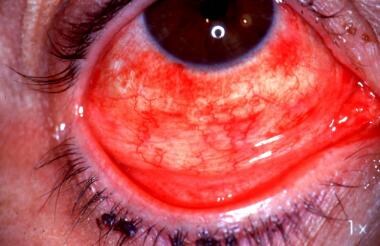
PURPOSE: Most lymphomas of the ocular adnexa are primary extranodal non-Hodgkin's lymphomas of the B-cell type, with the most common lymphoma subtype being the extranodal marginal-zone B-cell lymphoma (EMZL). Analysis of somatic mutations in the variable (V) region of the Ig heavy (H)-chain gene segment suggests that EMZL development in other locations is dependent on antigen stimulation. The purpose of this study was to analyze the presence of somatic hypermutations in clonally rearranged Ig H-chain V genes of this lymphoma entity in the ocular adnexa and to estimate whether the mutation pattern is compatible with antigen selection. METHODS: Twenty-six cases of EMZL of the ocular adnexa were diagnosed on the basis of morphology, histology, and immunohistology. A nested polymerase chain reaction (PCR) was performed on DNA extracted from paraffin sections. The isolated PCR products were sequenced and compared with published VH germline segments to determine the number of somatic mutations in the complementarity-determining region (CDR) 2 and framework (FW) region 3. RESULTS: The number of somatic mutations in the cases of EMZL varied between 0 and 24: Five cases involved 0 to 3 somatic mutations, and the remaining 21 cases involved 4 to 24 mutations. Based on the ratio of replacement (R) to silent (S) mutations in the CDR2 or FW3 regions, antigen selection seems to have occurred in 60% of ocular adnexal EMZL. The VH3 family was the most commonly expressed germline VH family (54%), followed by VH4 (23%), with biased usage of the latter. Some germline VH1 genes used included DP-8, DP-10, DP-53, DP-63 (VH4.21), and DP-49, which are frequently used by autoantibodies (e.g., rheumatoid factors) and natural autoantibodies. CONCLUSIONS: EMZLs of the ocular adnexa have an Ig H-chain mutation pattern that supports the concept that they represent a clonal expansion of post-germinal-center memory B-cells in most instances. In two thirds of cases, antigen selection may have occurred, and autoantibodies may have a role in their development. (+info)Intraepithelial and invasive squamous cell carcinoma of the conjunctiva: analysis of 60 cases. (2/169)
AIM: To evaluate the clinical features, treatment results, and recurrence rates in patients with either intraepithelial or invasive squamous cell carcinoma of the conjunctiva. METHODS: Retrospective analysis of 60 cases (22 conjunctival intraepithelial and 38 invasive squamous cell carcinomas) to determine patterns of clinical presentation, aetiological factors, and treatment results. The mean patient age was 64 years old. 70% of the patients were male. Patients were treated with a variety of therapies, depending on the degree of tumour involvement; most cases were treated with frozen section controlled excision and adjunctive cryotherapy. Modified eye wall resection or enucleation was done for intraocular invasion and exenteration was done for orbital involvement. RESULTS: Red eye (68%) and ocular irritation (57%) were the most common presenting symptoms. 44% of the patients had other eye findings consistent with extensive solar exposure. 20% of the patients had a history of malignant skin tumours. Visceral malignancies developed in 8%. Scleral involvement was present in 14 (37%), intraocular involvement in five (13%), and orbital invasion in four (11%) cases with invasive squamous cell carcinoma. After a mean follow up of 56 months (18-226 months) the rate of new or recurrent tumours was 4.5% for intraepithelial squamous carcinoma and 5.3% for invasive squamous cell carcinoma. No patient developed metastases or tumour related deaths. CONCLUSION: Excision with intraoperative control of the surgical margins and adjunctive cryotherapy results in good tumour control rates. (+info)Conjunctival squamous cell carcinoma in Tanzania. (3/169)
AIMS: To assess changes in incidence of conjunctival squamous cell carcinoma over a 22 year period in Tanzania and to analyse possible reasons for change. METHODS: Retrospective analysis of records from a Tanzanian pathology department serving north and central Tanzania from 1976 to 1997; medical record analysis of cases of conjunctival squamous cell carcinoma presenting in the last 2 years of the study. RESULTS: There was a sharp rise in the incidence of conjunctival squamous cell carcinoma in the last 3 years of the study (1995-7). The mean age of patients presenting with the condition over the full period was 44.7 years (95% confidence interval 42.4-46.9 years). In the final 2 years of the study the mean length of history on presentation was 3.1 months (2.1-4.0 months). Several patients had a previous history of chronic conjunctival disease such as allergic conjunctivitis and trachoma; one had had a conjunctival papilloma excised previously. Only five patients had been tested for HIV status, but of these four were positive. CONCLUSION: Tanzania is experiencing an epidemic of conjunctival squamous cell carcinoma similar to that seen in other African countries. Often the tumours are aggressive and occur in patients of relatively young age. The epidemic appears to be related to HIV infection, on a background of ultraviolet light exposure. Previous chronic conjunctival disease and exposure to human papillomavirus may also have a role. (+info)Conjunctival MALT lymphoma: an usual cause of red eye. (4/169)
We describe a patient presenting with a red eye who was found to have conjunctival non-Hodgkin's lymphoma of the mucosa-associated lymphoid tissue (MALT) type. (+info)Treatment of conjunctival squamous cell carcinoma with topical 5-fluorouracil. (5/169)
AIM: To evaluate the efficacy of topical 5-fluorouracil (5-FU) alone, without concurrent surgery or radiotherapy, for the treatment of conjunctival squamous cell carcinoma. METHODS: Eight patients affected by conjunctival squamous cell carcinoma (three recurrent cases, three incompletely excised, and two untreated cases) were treated with 1% 5-FU eye drops. Topical 1% 5-FU was administered four times daily for 4 weeks (one course). Clinical examination (biomicroscopy and photography) and morphological evaluation of conjunctival cytological specimens were used to monitor the efficacy of local chemotherapy, side effects, and recurrences. RESULTS: All patients showed clinical regression of conjunctival carcinoma after topical 1% 5-FU treatment. Neoplastic conjunctiva was completely replaced by normal epithelium within 3 months. Mean follow up was 27 months. One patient needed two courses of local chemotherapy for recurrent disease. An acute transient toxic keratoconjunctivitis was observed in all treated cases; it was easily controlled with topical therapy. No long term side effects were found. CONCLUSIONS: Topical 1% 5-FU is effective in the treatment of recurrent, incompletely excised, and selected untreated conjunctival squamous cell carcinomas. Topical 1% 5-FU has no major complications. This study suggests that topical conjunctival chemotherapy with 1% 5-FU may be useful, at least as adjunctive therapy, in the treatment of conjunctival squamous cell carcinoma. (+info)Indeterminate melanocytic proliferations of the conjunctiva. (6/169)
PURPOSE: The purpose of this study is to test the hypothesis that a subset of conjunctival melanocytic proliferations exists that cannot be reproducibly classified as benign, malignant, or indeterminate. METHODS: Three groups of excisional biopsy specimens of conjunctival melanocytic proliferations were evaluated by 5 ophthalmic pathologists. These groups included lesions that were considered by the authors to represent benign (Group 1, n = 5), malignant (Group 2, n = 5) and indeterminate melanocytic proliferations (Group 3, n = 5). The panel classified the same sections in all 3 groups in a randomized, masked fashion, first without and then with a clinical history of patient age, sex and race. The kappa statistic (k) was used to quantify the degree of agreement among observers. RESULTS: There was strong concordance among the panel for both Group 1 (benign, k = 0.76) and Group 2 (malignant, k = 0.70) melanocytic proliferations. There was no concordance of the panel for Group 3 (indeterminate) lesions (k = -0.045). The concordance for Groups 1 and 2 and lack of concordance for Group 3 lesions were independent of knowledge of clinical history of age, sex, and race. CONCLUSIONS: A subset of melanocytic proliferations of the conjunctiva exists that cannot be reproducibly classified by pathologists as benign, malignant, or indeterminate. (+info)Combined nevi of the conjunctiva. (7/169)
PURPOSE: To report the clinical and histologic features of combined nevi of the conjunctiva, a type of nevus that is not uncommon in the skin but has rarely been reported in the conjunctiva. METHODS: Conjunctival nevi and melanomas from the files of the University of California, San Francisco, eye pathology laboratory were reviewed from 1984 to 1999 for the presence of features of both standard nevocytic nevi and blue nevi. Clinical histories and, when available, clinical photographs were obtained. RESULTS: Thirty-one combined nevi were discovered during the 15-year period between 1984 and 1999. One case before 1984 had been incorrectly diagnosed as a junctional nevus. The dendritic and spindle-shaped blue nevus cells had been overlooked because they were not recognized as distinct from the standard nevocytic nevus cells. The recognition of a blue as well as a brown color, a deep as well as a superficial component in the lesion, or a history of pigmentation since birth may help to establish the correct clinical diagnosis and prevent an unnecessarily deep surgical resection. Although growth of the lesion or "satellites" in some patients may favor a clinical diagnosis of melanoma, none of the lesions in this series were malignant. CONCLUSION: Despite a paucity of reports of combined nevi of the conjunctiva in the medical literature, this type of nevus--a combination of a nevocytic and a blue nevus--is common and has been overlooked in the past. (+info)Evaluation of ocular tumors with technetium-99m-MIBI: planar pinhole technique or SPECT? (8/169)
OBJECTIVE: This study compares 2 imaging protocols, planar pinhole technique (PPHT) and SPECT, for evaluating ocular masses with 99mTc-MIBI. METHODS: Sixteen patients with ocular lesions were studied. Planar images were acquired 10 min after the injection of 740 MBq 99mTc-MIBI with an LFOV camera fitted with a pinhole collimator (5.0 mm). A SPECT study was performed immediately after the planar study, using a 360 degrees orbit, 64 steps, 20 s/stop, a 128 x 128 matrix, and a low-energy high-resolution (LEHR) collimator. Twelve lesions (9.5-18.0 mm) proved to be malignant: 8 primary tumors (ocular melanoma); 3 local relapses of different tumors of the conjunctiva; and 1 ocular metastasis from breast cancer. The remaining 4 lesions (10.0-16.0 mm) were benign: 1 inflammatory lesion; 1 benign intraocular calcification; and 2 naevi. RESULTS: SPECT images showed 11 of 12 malignant lesions (91.6%), whereas the planar technique demonstrated only 4 of the 12 lesions (33.3%). One false-positive result, the inflammatory lesion, was visualized by both techniques. The remaining benign lesions were not detected with either method. CONCLUSION: Technetium-99m-MIBI SPECT is a sensitive technique for detecting malignant ocular tumors. SPECT imaging is a better alternative to planar imaging for ocular tumors. (+info)Conjunctival neoplasms refer to abnormal growths or tumors that develop on the conjunctiva, which is the thin, clear mucous membrane that covers the inner surface of the eyelids and the outer surface of the eye. These neoplasms can be benign (non-cancerous) or malignant (cancerous).
Benign conjunctival neoplasms are typically slow-growing and do not spread to other parts of the body. They may include lesions such as conjunctival cysts, papillomas, or naevi (moles). These growths can usually be removed through simple surgical procedures with a good prognosis.
Malignant conjunctival neoplasms, on the other hand, are cancerous and have the potential to invade surrounding tissues and spread to other parts of the body. The most common type of malignant conjunctival neoplasm is squamous cell carcinoma, which arises from the epithelial cells that line the surface of the conjunctiva. Other less common types include melanoma, lymphoma, and adenocarcinoma.
Malignant conjunctival neoplasms typically require more extensive treatment, such as surgical excision, radiation therapy, or chemotherapy. The prognosis for malignant conjunctival neoplasms depends on the type and stage of the cancer at the time of diagnosis, as well as the patient's overall health and age. Early detection and prompt treatment are key to improving outcomes in patients with these conditions.
Pancreatic neoplasms refer to abnormal growths in the pancreas that can be benign or malignant. The pancreas is a gland located behind the stomach that produces hormones and digestive enzymes. Pancreatic neoplasms can interfere with the normal functioning of the pancreas, leading to various health complications.
Benign pancreatic neoplasms are non-cancerous growths that do not spread to other parts of the body. They are usually removed through surgery to prevent any potential complications, such as blocking the bile duct or causing pain.
Malignant pancreatic neoplasms, also known as pancreatic cancer, are cancerous growths that can invade and destroy surrounding tissues and organs. They can also spread (metastasize) to other parts of the body, such as the liver, lungs, or bones. Pancreatic cancer is often aggressive and difficult to treat, with a poor prognosis.
There are several types of pancreatic neoplasms, including adenocarcinomas, neuroendocrine tumors, solid pseudopapillary neoplasms, and cystic neoplasms. The specific type of neoplasm is determined through various diagnostic tests, such as imaging studies, biopsies, and blood tests. Treatment options depend on the type, stage, and location of the neoplasm, as well as the patient's overall health and preferences.
Neoplasms are abnormal growths of cells or tissues in the body that serve no physiological function. They can be benign (non-cancerous) or malignant (cancerous). Benign neoplasms are typically slow growing and do not spread to other parts of the body, while malignant neoplasms are aggressive, invasive, and can metastasize to distant sites.
Neoplasms occur when there is a dysregulation in the normal process of cell division and differentiation, leading to uncontrolled growth and accumulation of cells. This can result from genetic mutations or other factors such as viral infections, environmental exposures, or hormonal imbalances.
Neoplasms can develop in any organ or tissue of the body and can cause various symptoms depending on their size, location, and type. Treatment options for neoplasms include surgery, radiation therapy, chemotherapy, immunotherapy, and targeted therapy, among others.
Neoplasms: Neoplasms refer to abnormal growths of tissue that can be benign (non-cancerous) or malignant (cancerous). They occur when the normal control mechanisms that regulate cell growth and division are disrupted, leading to uncontrolled cell proliferation.
Cystic Neoplasms: Cystic neoplasms are tumors that contain fluid-filled sacs or cysts. These tumors can be benign or malignant and can occur in various organs of the body, including the pancreas, ovary, and liver.
Mucinous Neoplasms: Mucinous neoplasms are a type of cystic neoplasm that is characterized by the production of mucin, a gel-like substance produced by certain types of cells. These tumors can occur in various organs, including the ovary, pancreas, and colon. Mucinous neoplasms can be benign or malignant, and malignant forms are often aggressive and have a poor prognosis.
Serous Neoplasms: Serous neoplasms are another type of cystic neoplasm that is characterized by the production of serous fluid, which is a thin, watery fluid. These tumors commonly occur in the ovary and can be benign or malignant. Malignant serous neoplasms are often aggressive and have a poor prognosis.
In summary, neoplasms refer to abnormal tissue growths that can be benign or malignant. Cystic neoplasms contain fluid-filled sacs and can occur in various organs of the body. Mucinous neoplasms produce a gel-like substance called mucin and can also occur in various organs, while serous neoplasms produce thin, watery fluid and commonly occur in the ovary. Both mucinous and serous neoplasms can be benign or malignant, with malignant forms often being aggressive and having a poor prognosis.
Skin neoplasms refer to abnormal growths or tumors in the skin that can be benign (non-cancerous) or malignant (cancerous). They result from uncontrolled multiplication of skin cells, which can form various types of lesions. These growths may appear as lumps, bumps, sores, patches, or discolored areas on the skin.
Benign skin neoplasms include conditions such as moles, warts, and seborrheic keratoses, while malignant skin neoplasms are primarily classified into melanoma, squamous cell carcinoma, and basal cell carcinoma. These three types of cancerous skin growths are collectively known as non-melanoma skin cancers (NMSCs). Melanoma is the most aggressive and dangerous form of skin cancer, while NMSCs tend to be less invasive but more common.
It's essential to monitor any changes in existing skin lesions or the appearance of new growths and consult a healthcare professional for proper evaluation and treatment if needed.
Multiple primary neoplasms refer to the occurrence of more than one primary malignant tumor in an individual, where each tumor is unrelated to the other and originates from separate cells or organs. This differs from metastatic cancer, where a single malignancy spreads to multiple sites in the body. Multiple primary neoplasms can be synchronous (occurring at the same time) or metachronous (occurring at different times). The risk of developing multiple primary neoplasms increases with age and is associated with certain genetic predispositions, environmental factors, and lifestyle choices such as smoking and alcohol consumption.
Kidney neoplasms refer to abnormal growths or tumors in the kidney tissues that can be benign (non-cancerous) or malignant (cancerous). These growths can originate from various types of kidney cells, including the renal tubules, glomeruli, and the renal pelvis.
Malignant kidney neoplasms are also known as kidney cancers, with renal cell carcinoma being the most common type. Benign kidney neoplasms include renal adenomas, oncocytomas, and angiomyolipomas. While benign neoplasms are generally not life-threatening, they can still cause problems if they grow large enough to compromise kidney function or if they undergo malignant transformation.
Early detection and appropriate management of kidney neoplasms are crucial for improving patient outcomes and overall prognosis. Regular medical check-ups, imaging studies, and urinalysis can help in the early identification of these growths, allowing for timely intervention and treatment.
A "second primary neoplasm" is a distinct, new cancer or malignancy that develops in a person who has already had a previous cancer. It is not a recurrence or metastasis of the original tumor, but rather an independent cancer that arises in a different location or organ system. The development of second primary neoplasms can be influenced by various factors such as genetic predisposition, environmental exposures, and previous treatments like chemotherapy or radiation therapy.
It is important to note that the definition of "second primary neoplasm" may vary slightly depending on the specific source or context. In general medical usage, it refers to a new, separate cancer; however, in some research or clinical settings, there might be more precise criteria for defining and diagnosing second primary neoplasms.
Adenocarcinoma, mucinous is a type of cancer that begins in the glandular cells that line certain organs and produce mucin, a substance that lubricates and protects tissues. This type of cancer is characterized by the presence of abundant pools of mucin within the tumor. It typically develops in organs such as the colon, rectum, lungs, pancreas, and ovaries.
Mucinous adenocarcinomas tend to have a distinct appearance under the microscope, with large pools of mucin pushing aside the cancer cells. They may also have a different clinical behavior compared to other types of adenocarcinomas, such as being more aggressive or having a worse prognosis in some cases.
It is important to note that while a diagnosis of adenocarcinoma, mucinous can be serious, the prognosis and treatment options may vary depending on several factors, including the location of the cancer, the stage at which it was diagnosed, and the individual's overall health.
Thyroid neoplasms refer to abnormal growths or tumors in the thyroid gland, which can be benign (non-cancerous) or malignant (cancerous). These growths can vary in size and may cause a noticeable lump or nodule in the neck. Thyroid neoplasms can also affect the function of the thyroid gland, leading to hormonal imbalances and related symptoms. The exact causes of thyroid neoplasms are not fully understood, but risk factors include radiation exposure, family history, and certain genetic conditions. It is important to note that most thyroid nodules are benign, but a proper medical evaluation is necessary to determine the nature of the growth and develop an appropriate treatment plan.
Myeloproliferative disorders (MPDs) are a group of rare, chronic blood cancers that originate from the abnormal proliferation or growth of one or more types of blood-forming cells in the bone marrow. These disorders result in an overproduction of mature but dysfunctional blood cells, which can lead to serious complications such as blood clots, bleeding, and organ damage.
There are several subtypes of MPDs, including:
1. Chronic Myeloid Leukemia (CML): A disorder characterized by the overproduction of mature granulocytes (a type of white blood cell) in the bone marrow, leading to an increased number of these cells in the blood. CML is caused by a genetic mutation that results in the formation of the BCR-ABL fusion protein, which drives uncontrolled cell growth and division.
2. Polycythemia Vera (PV): A disorder characterized by the overproduction of all three types of blood cells - red blood cells, white blood cells, and platelets - in the bone marrow. This can lead to an increased risk of blood clots, bleeding, and enlargement of the spleen.
3. Essential Thrombocythemia (ET): A disorder characterized by the overproduction of platelets in the bone marrow, leading to an increased risk of blood clots and bleeding.
4. Primary Myelofibrosis (PMF): A disorder characterized by the replacement of normal bone marrow tissue with scar tissue, leading to impaired blood cell production and anemia, enlargement of the spleen, and increased risk of infections and bleeding.
5. Chronic Neutrophilic Leukemia (CNL): A rare disorder characterized by the overproduction of neutrophils (a type of white blood cell) in the bone marrow, leading to an increased number of these cells in the blood. CNL can lead to an increased risk of infections and organ damage.
MPDs are typically treated with a combination of therapies, including chemotherapy, targeted therapy, immunotherapy, and stem cell transplantation. The choice of treatment depends on several factors, including the subtype of MPD, the patient's age and overall health, and the presence of any comorbidities.
A dermoid cyst is a type of benign (non-cancerous) growth that typically develops during embryonic development. It is a congenital condition, which means it is present at birth, although it may not become apparent until later in life. Dermoid cysts are most commonly found in the skin or the ovaries of women, but they can also occur in other areas of the body, such as the spine or the brain.
Dermoid cysts form when cells that are destined to develop into skin and its associated structures, such as hair follicles and sweat glands, become trapped during fetal development. These cells continue to grow and multiply, forming a sac-like structure that contains various types of tissue, including skin, fat, hair, and sometimes even teeth or bone.
Dermoid cysts are usually slow-growing and may not cause any symptoms unless they become infected or rupture. In some cases, they may cause pain or discomfort if they press on nearby structures. Treatment typically involves surgical removal of the cyst to prevent complications and alleviate symptoms.
Adenocarcinoma, sebaceous is a type of cancer that develops from the sebaceous glands, which are glands in the skin that produce an oily substance called sebum. This type of cancer is a malignant tumor that forms in the glandular cells and can spread to other parts of the body. It most commonly occurs in the glands found in the eyelids (known as meibomian glands), but it can also occur in other areas of the body such as the genitals, breasts, and skin.
Sebaceous adenocarcinoma is a rare type of cancer, accounting for less than 1% of all skin cancers. It typically affects older adults and has been linked to exposure to radiation and certain genetic mutations. Treatment usually involves surgical removal of the tumor, along with radiation therapy or chemotherapy in some cases.
It is important to note that while I strive to provide accurate and up-to-date information, this definition may not be complete or fully comprehensive. If you have any concerns about your health or a medical condition, it is always best to consult with a qualified healthcare professional for personalized advice and treatment.
Orbital neoplasms refer to abnormal growths or tumors that develop in the orbit, which is the bony cavity that contains the eyeball, muscles, nerves, fat, and blood vessels. These neoplasms can be benign (non-cancerous) or malignant (cancerous), and they can arise from various types of cells within the orbit.
Orbital neoplasms can cause a variety of symptoms depending on their size, location, and rate of growth. Common symptoms include protrusion or displacement of the eyeball, double vision, limited eye movement, pain, swelling, and numbness in the face. In some cases, orbital neoplasms may not cause any noticeable symptoms, especially if they are small and slow-growing.
There are many different types of orbital neoplasms, including:
1. Optic nerve glioma: a rare tumor that arises from the optic nerve's supportive tissue.
2. Orbital meningioma: a tumor that originates from the membranes covering the brain and extends into the orbit.
3. Lacrimal gland tumors: benign or malignant growths that develop in the lacrimal gland, which produces tears.
4. Orbital lymphangioma: a non-cancerous tumor that arises from the lymphatic vessels in the orbit.
5. Rhabdomyosarcoma: a malignant tumor that develops from the skeletal muscle cells in the orbit.
6. Metastatic tumors: cancerous growths that spread to the orbit from other parts of the body, such as the breast, lung, or prostate.
The diagnosis and treatment of orbital neoplasms depend on several factors, including the type, size, location, and extent of the tumor. Imaging tests, such as CT scans and MRI, are often used to visualize the tumor and determine its extent. A biopsy may also be performed to confirm the diagnosis and determine the tumor's type and grade. Treatment options include surgery, radiation therapy, chemotherapy, or a combination of these approaches.
Sebaceous gland neoplasms are abnormal growths or tumors that develop in the sebaceous glands, which are small oil-producing glands found in the skin. These glands are responsible for producing sebum, a natural oil that helps keep the skin and hair moisturized. Sebaceous gland neoplasms can be benign (non-cancerous) or malignant (cancerous).
Benign sebaceous gland neoplasms include:
* Seborrheic keratosis: These are common, harmless growths that appear as rough, scaly patches on the skin. They can be tan, brown, or black in color and vary in size from small to large.
* Sebaceous adenoma: This is a benign tumor that arises from the sebaceous glands. It typically appears as a small, yellowish bump on the skin.
Malignant sebaceous gland neoplasms include:
* Sebaceous carcinoma: This is a rare but aggressive form of skin cancer that arises from the sebaceous glands. It often appears as a hard, painless nodule on the eyelid or other areas of the face and can spread to other parts of the body if left untreated.
* Basal cell carcinoma: While not exclusively a sebaceous gland neoplasm, basal cell carcinomas can sometimes arise from the sebaceous glands. These are slow-growing but invasive skin cancers that typically appear as pearly or flesh-colored bumps on the skin.
It is important to have any new or changing growths on the skin evaluated by a healthcare professional to determine whether they are benign or malignant and to develop an appropriate treatment plan if necessary.
An encyclopedia is a comprehensive reference work containing articles on various topics, usually arranged in alphabetical order. In the context of medicine, a medical encyclopedia is a collection of articles that provide information about a wide range of medical topics, including diseases and conditions, treatments, tests, procedures, and anatomy and physiology. Medical encyclopedias may be published in print or electronic formats and are often used as a starting point for researching medical topics. They can provide reliable and accurate information on medical subjects, making them useful resources for healthcare professionals, students, and patients alike. Some well-known examples of medical encyclopedias include the Merck Manual and the Stedman's Medical Dictionary.
Eyelid neoplasms refer to abnormal growths or tumors that develop in the tissues of the eyelids. These growths can be benign (non-cancerous) or malignant (cancerous). Common types of benign eyelid neoplasms include papillomas, hemangiomas, and nevi. Malignant eyelid neoplasms are typically classified as basal cell carcinomas, squamous cell carcinomas, or melanomas. These malignant tumors can be aggressive and may spread to other parts of the body if left untreated. Treatment options for eyelid neoplasms depend on the type, size, and location of the growth, as well as the patient's overall health. Surgical excision is often the preferred treatment approach, although radiation therapy and chemotherapy may also be used in some cases. Regular follow-up care is important to monitor for recurrence or new growths.
Eye neoplasms, also known as ocular tumors or eye cancer, refer to abnormal growths of tissue in the eye. These growths can be benign (non-cancerous) or malignant (cancerous). Eye neoplasms can develop in various parts of the eye, including the eyelid, conjunctiva, cornea, iris, ciliary body, choroid, retina, and optic nerve.
Benign eye neoplasms are typically slow-growing and do not spread to other parts of the body. They may cause symptoms such as vision changes, eye pain, or a noticeable mass in the eye. Treatment options for benign eye neoplasms include monitoring, surgical removal, or radiation therapy.
Malignant eye neoplasms, on the other hand, can grow and spread rapidly to other parts of the body. They may cause symptoms such as vision changes, eye pain, floaters, or flashes of light. Treatment options for malignant eye neoplasms depend on the type and stage of cancer but may include surgery, radiation therapy, chemotherapy, or a combination of these treatments.
It is important to note that early detection and treatment of eye neoplasms can improve outcomes and prevent complications. Regular eye exams with an ophthalmologist are recommended for early detection and prevention of eye diseases, including eye neoplasms.
 Eye neoplasm
Conjunctival squamous cell carcinoma
Corneal limbus
List of MeSH codes (C11)
Carol Shields (ophthalmologist)
Squamous cell papilloma
List of MeSH codes (C04)
Nasolacrimal duct obstruction
International Classification of Headache Disorders
Pituitary adenoma
Oncovirus
Goblet cell
Marginal zone B-cell lymphoma
BRAF (gene)
 Conjunctival Papilloma: Background, Pathophysiology, Epidemiology
Eye neoplasm - Wikipedia
 Surgical management of conjunctival tumors. The 1994 Lynn B. McMahan Lecture
Management of conjunctival and corneal melanoma with surgical excision, amniotic membrane allograft, and topical chemotherapy
Conjunctival Papilloma Workup: Laboratory Studies, Imaging Studies, Procedures
 Advanced Search Results - Public Health Image Library(PHIL)
 Advances in the management of conjunctival melanoma.
K. Tomo Wiggans<...
 Clinical Techniques in Amphibians - Exotic and Laboratory Animals - MSD Veterinary Manual
Fluorescence in situ hybridisation (FISH) in histologically challenging conjunctival melanocytic lesions | British Journal of...
 The Ophthalmic Examination - WSAVA 2001 - VIN
 Mitomycin
- Mitomycin-C
Summary Report | CureHunter
Mitomycin
- Mitomycin-C
Summary Report | CureHunter
 Portal Regional da BVS
Child Physical Abuse - Eli Newberger
VEGF165 Protein - ACROBiosystems
 Conjunctival Hemangioma (Concept Id: C4545024)
- MedGen - NCBI
Conjunctival Hemangioma (Concept Id: C4545024)
- MedGen - NCBI
 Common Corneal Disorders | Eye Care Center of Northern Colorado
Corneal Disorders We Treat | Eye Care Center of Northern Colorado
 Diagnosis and staging of ocular surface squamous neoplasia | Höllhumer | African Vision and Eye Health
 Dr. Robert Dundervill III, MD, Ophthalmology Specialist - Charleston, WV | Sharecare
 IARC Publications Website - WHO Classification of Tumours of the Eye
BVS Brasil
 Namespace
 Клиника Движение на Рионской
 Pterygium/Pinguecula | The Atlas of Emergency Medicine, 5e | AccessEmergency Medicine | McGraw Hill Medical
BiCNU, Gliadel (carmustine) dosing, indications, interactions, adverse effects, and more
NIOSHTIC-2 Search Results - Full View
 Quizwise 80 - Conjunctival naevus: Clinical insights - Suraj Eye Institute
 Pesquisa | Biblioteca Virtual em Saúde - BRASIL
 Dr. Joshua Powell, MD - Ophthalmology Specialist in Norman, OK | Healthgrades
Melanoma15
- We have employed this technique on 109 patients with conjunctival squamous neoplasms and 137 patients with conjunctival melanoma, about 80 of which neoplasms were associated with primary acquired melanosis. (nih.gov)
- Our observations suggest that well-planned initial surgical management using this technique decreases the chance of tumor recurrence for conjunctival melanoma and squamous cell carcinoma. (nih.gov)
- To illustrate a novel method of management for extensive conjunctival and corneal melanoma. (nih.gov)
- A 40-year-old Caucasian woman presented with a large, diffuse conjunctival melanoma involving 6 clock hours of the limbus. (nih.gov)
- The conjunctival melanoma was completely resected microsurgically in one piece without disrupting the tumor. (nih.gov)
- Preliminary evidence suggests that combined therapeutic approaches, consisting of extensive tumor removal, cryotherapy, amniotic membrane allograft, and topical mitomyin C, can be effective in the management of diffuse conjunctival and corneal melanoma arising from primary acquired melanosis. (nih.gov)
- Scholars@Duke publication: Advances in the management of conjunctival melanoma. (duke.edu)
- Long term follow-up of patients with conjunctival melanoma with systemic surveillance is necessary to detect recurrences and metastases. (duke.edu)
- There have been particularly substantial changes to the classification of conjunctival neoplasia and melanoma, based on the latest information from genetic and molecular studies. (iarc.fr)
- 4. What is the most common location for conjunctival melanoma? (surajeyeinstitute.org)
- 5. Which architectural pattern is a clue to the diagnosis of conjunctival melanoma? (surajeyeinstitute.org)
- Explanation: The most common location for conjunctival melanoma is the bulbar conjunctiva close to the limbus. (surajeyeinstitute.org)
- Management of conjunctival malignant melanoma: a review and update. (surajeyeinstitute.org)
- Explanation: Presence of intraepithelial nests, cyst formation, and maturation with depth are not specific architectural clues for conjunctival melanoma, while the presence of pagetoid growth is a pattern suggestive of conjunctival melanoma. (surajeyeinstitute.org)
- The most useful architectural patterns suggestive of conjunctival melanoma include (1) intraepithelial component showing pagetoid growth (2) radial extension of the intraepithelial component beyond the lateral edge of subepithelial component, (3) patchy or bandlike inflammation at the base of the lesion, (4) mitotic activity, (5) lack of maturation toward the base of the lesion, (6) invasion of the sclera or cornea. (surajeyeinstitute.org)
Conjunctiva7
- The extensive conjunctival defect, involving one-half of the bulbar conjunctiva, was reconstructed with an amniotic membrane allograft. (nih.gov)
- Conclusions FISH is very useful in classifying equivocal conjunctival melanocytic lesions, especially those with atypical junctional activity and naevoid melanocytic proliferations of the conjunctiva. (bmj.com)
- Sections are included on all recognized neoplasms (and their variants) of the eye, lacrimal apparatus, and conjunctiva. (iarc.fr)
- Explanation: melanocytic naevi are the most common tumors of the conjunctiva, accounting for up to 28% of all conjunctival neoplasms. (surajeyeinstitute.org)
- and 4) the thin and flexible palpebral conjunctiva, which continues to the conjunctival fornix or conjunctival cul-de-sac. (merckvetmanual.com)
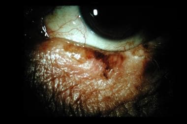
- [2] Conjunctival lesions present as a localized yellow nodule on the bulbar conjunctiva (Figure 3). (eyewiki.org)
- Additionally it is regarded the most frequent neoplasm from the conjunctiva, normally affecting elderly men of around 70?years [3,4]. (antiviralbiologic.com)
Tumors3
- Specifically, conjunctival papillomas are benign squamous epithelial tumors with a minimal propensity toward malignancy. (medscape.com)
- Surgical management of conjunctival tumors. (nih.gov)
- To our knowledge, there are no articles that describe the specific step-by-step details of the surgical removal of premalignant and malignant conjunctival tumors. (nih.gov)
Carcinoma3
- Several cases of locally advanced conjunctival, eyelid, and lacrimal sac/duct squamous cell carcinoma that were successfully treated with immune checkpoint inhibitors (PD-1 directed) are shared. (bvsalud.org)
- Parallel studies by Waddell and colleagues also suggested that HIV contamination is strongly associated with an increase in the incidence of conjunctival carcinoma in Africa [10]. (antiviralbiologic.com)
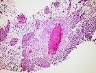
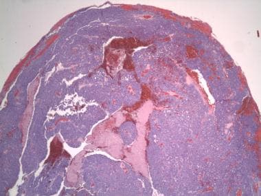
- Sebaceous carcinoma is a malignant neoplasm arising in sebaceous glands that is characterized by extensive lipid production. (missionforvisionusa.org)
Lesions4
- Although rare, inverted conjunctival papillomas sometimes are referred to as mucoepidermoid papillomas because these lesions possess both a mucous component and an epidermoid component. (medscape.com)
- It is useful as a guide for where to obtain a biopsy specimen or resect ill-defined conjunctival lesions. (medscape.com)
- Follow-up Examinations for Occular Neoplasms and Pigmented Ocular Lesions, at the Dupont, West Virginia Plant. (cdc.gov)
- Active and retired workers at the duPont Belle chemicals facility in West Virginia were ophthalmologically surveyed first in 1978 and again in 1979 in a follow-up study to identify cases of ocular neoplasms and determine the prevalence of ocular pigmented lesions. (cdc.gov)
Tumor3
- Eye neoplasms can affect all parts of the eye, and can be a benign tumor or a malignant tumor (cancer). (wikipedia.org)
- 1. What is the most common type of tumor among conjunctival neoplasms? (surajeyeinstitute.org)
- It ranges from a well-differentiated tumor with EPITHELIAL CELLS indistinguishable from normal HEPATOCYTES to a poorly differentiated neoplasm. (lookformedical.com)
Lymphoma6
- Results -Animals with intraocular (4 dogs and 1 cat) or conjunctival (3 dogs and 1 cat) lymphoma represented 0.1% and 0.08% of patients with lymphoma evaluated at the hospital during the study period, respectively. (avma.org)
- animals with conjunctival lymphoma represented 0.16% of all patients with conjunctivitis. (avma.org)
- Lymph node metastasis was detected in 2 patients with conjunctival lymphoma. (avma.org)
- Median PFST and OST for animals with conjunctival lymphoma were 221 and 549 days, respectively. (avma.org)
- To date, the clinical features of the various subtypes of conjunctival lymphoma (CL) have not been previously evaluated in a large cohort. (liverpool.ac.uk)
- Conjunctival lymphoma consists of mainly 4 subtypes of B-cell non-Hodgkin lymphoma: EMZL, FL, MCL, and DLBCL. (liverpool.ac.uk)
Management of conjuncti1
- We describe a detailed stepwise approach to the surgical management of conjunctival neoplasms. (nih.gov)
Humans1
- Substances that increase the risk of NEOPLASMS in humans or animals. (lookformedical.com)
Benign1
- Malignant neoplasms show a greater degree of anaplasia and have the properties of invasion and metastasis, compared to benign neoplasms. (lookformedical.com)
Treatment of conjunctival2
- Topical timolol for the treatment of conjunctival pyogenic granulomas: Outcomes and effect on intraocular pressure. (nih.gov)
- METHODOLOGY: Patients who underwent excision for the diagnosis and treatment of conjunctival surface masses with clinical suspicion of malignancy were evaluated retrospectively. (bvsalud.org)
Cytology1
- Irrelevant of its cytology, a neoplasm of epithelial origin with this form of growth is also called papilloma. (medscape.com)
Conjunctivitis1
- Pterygium mimics include localized conjunctival neoplasia, conjunctivitis, and episcleritis. (mhmedical.com)
Ocular neoplasms1
- Most of the ocular neoplasms are squamous cell carcinomas. (thieme-connect.de)
Epithelial2
- Epithelial cells do not demonstrate atypia, and dysplastic changes are uncommon for conjunctival inverted papillomas. (medscape.com)
- A primary malignant neoplasm of epithelial liver cells. (lookformedical.com)
Neoplasia1
- Overview of Neoplasia of the Eye and Associated Structures in Animals The various tissues of the eye and associated structures can be the site of primary or metastatic neoplasms. (merckvetmanual.com)
Diagnosis1
- In skin pathology, the recent application of fluorescence in situ hybridisation (FISH) has been demonstrated to be of use for the analysis and diagnosis of ambiguous melanocytic neoplasms of the skin. (bmj.com)
Histologically2
- This study set out to evaluate this method on seven prospective conjunctival cases that were histologically equivocal. (bmj.com)
- The neoplasms may be histologically the same or different, and may be found in the same or different sites. (lookformedical.com)
Papilloma7
- Conjunctival papilloma also can be classified based on gross clinical appearance, as either pedunculated or sessile. (medscape.com)
- The pedunculated type is synonymous with infectious conjunctival papilloma and squamous cell papilloma. (medscape.com)
- The limbal conjunctival papilloma often is referred to as noninfectious conjunctival papilloma because it is believed that limbal papillomas arise from UV radiation exposure. (medscape.com)
- HPV type 11 was the most common and frequently found in conjunctival papilloma as analyzed by polymerase chain reaction (PCR). (medscape.com)
- HPV types 6 and 11 are the most frequently found in conjunctival papilloma. (medscape.com)
- HPV type 33 is another source in the pathogenesis of conjunctival papilloma. (medscape.com)
- Squamous cell papillomas (eg, infectious papilloma, viral conjunctival papilloma) are composed of multiple branching fronds emanating from a narrow pedunculated base. (medscape.com)
Confocal1
- Finally, confocal microscopy provides en-face images of the ocular surface, with OSSN showing a classic 'starry night' appearance. (avehjournal.org)
Disease1
- RESULTS Of the 112 leopard geckos, 52 (46%) had ophthalmic disease (mainly corneal or conjunctival disease). (avma.org)
Ophthalmic1
- Ophthalmic neoplasms vary in histologic type, frequency, and importance in different. (merckvetmanual.com)
Carcinomas1
- Moreover, the HPV genome is identifiable in most conjunctival papillomas and in 85% of conjunctival dysplasias and carcinomas. (medscape.com)
Nevi1
- The prevalence of participants with conjunctival nevi, iris nevi, and chordal nevi amounted to 0.9 and and nonindustrial workers, respectively. (cdc.gov)
Solitary1
- Solitary conjunctival hemangioma presenting as a chocolate cyst. (nih.gov)
Tissue3
- It appears as a light brown or yellow-white amorphous, slightly raised conjunctival tissue adjacent to the limbus. (mhmedical.com)
- A small area of yellowish "heaped up" conjunctival tissue is seen adjacent to the nasal limbus. (mhmedical.com)
- Abnormal growths of tissue that follow a previous neoplasm but are not metastases of the latter. (lookformedical.com)
Prevalence1
- The literature reviewed yielded no published study outlining the prevalence of conjunctival papillomas in a cross-section of a population. (medscape.com)
Infectious1
- Infectious conjunctival papillomas also are known as squamous cell papillomas. (medscape.com)
Examination1
- OBJECTIVE: To evaluate the histopathological results of conjunctival masses suspected to be malignant based on biomicroscopic examination. (bvsalud.org)
Cytoplasm1
- In the higher magnification image, the malignant neoplasm features large cells with vesicular bubbly cytoplasm (arrow 6). (missionforvisionusa.org)