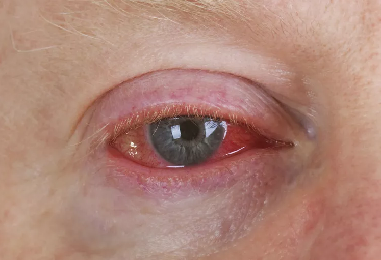
Corneal Ulcer
Eye Infections, Bacterial
Eye Infections, Fungal
Stomach Ulcer
Corneal Perforation
Natamycin
Peptic Ulcer
Administration, Topical
Cornea
Ophthalmic Solutions
Leg Ulcer
Pressure Ulcer
Contact Lenses, Hydrophilic
Skin Ulcer
Fusariosis
Aza Compounds
Iodophors
Acanthamoeba Keratitis
Burns, Chemical
Rosacea
Peptic Ulcer Hemorrhage
Burkholderia gladioli
Fibrin Tissue Adhesive
Visual Acuity
Peptic Ulcer Perforation
Keratoplasty, Penetrating
Quinolines
Buruli Ulcer
Anti-Infective Agents
Prednisolone
Conjunctiva
Fusarium
Acanthamoeba
Herpetic keratitis. Proctor Lecture. (1/403)
Although much needs to be learned about the serious clinical problem of herpes infection of the cornea, we have come a long way. We now have effective topical antiviral drugs. We have animal models which, with a high degree of reliability, clearly predict the effect to be expected clinically in man, as well as the toxicity. We have systemically active drugs and the potential of getting highly active, potent, completely selective drugs, with the possibility that perhaps the source of viral reinfection can be eradicated. The biology of recurrent herpes and stromal disease is gradually being understood, and this understanding may result in new and better therapy of this devastating clinical disease. (+info)Infectious keratitis in leprosy. (2/403)
AIM: To describe leprosy characteristics, ocular features, and type of organisms that produce infective corneal ulcers in leprosy patients. METHOD: The records of all leprosy patients admitted for treatment of corneal ulcers between 1992 and 1997 were reviewed. RESULTS: 63 leprosy patients, 53 males and 10 females, are described. 16 were tuberculoid and 47 lepromatous. 25 patients had completed multidrug therapy. 10 patients had face patches, eight had type I reaction, and 10 had type II reaction. 43 (68%) patients had hand deformities. In 54% of patients pain was absent as a presenting symptom. 19 patients gave a history of trauma. In 15 patients ulcers had also occurred on the other eye, five of them having occurred during the study period and the rest before 1992. Of the 68 eyes with corneal ulcers, 28 had madarosis, 34 had lagophthalmos, nine had ectropion, three had trichiasis, six had blocked nasolacrimal ducts, and 39 decreased corneal sensation. In 14 eyes, a previous lagophthalmos surgery had been done. 16 patients were blind at presentation. 32% of ulcers were located centrally. After treatment only 18% of the eyes showed visual improvement. Five types of fungus were cultured, two of them rare ocular pathogens. CONCLUSIONS: Corneal ulcers occur more in males and in the lepromatous group of patients. Decreased corneal sensation, lagophthalmos and hand deformity are closely associated. Indigenous treatment and late presentations were notable in many patients. Visual outcome is not good. There is increased risk of developing an ulcer in the other eye. Fungal corneal ulcers are not uncommon. (+info)Incidence of corneal melting in association with systemic disease in the Yorkshire Region, 1995-7. (3/403)
AIMS: To estimate the incidence of corneal melting or necrotising keratitis in association with systemic disease in the Yorkshire Region and to determine the type and duration of the systemic association. METHODS: In a prospective study, vigorous attempts were made to identify all patients presenting with newly diagnosed corneal melting over a 3 year period. RESULTS: 27 patients were identified during the study period. Rheumatoid arthritis and Wegener's granulomatosis were the most common disease associations. Corneal melting was a late complication of rheumatoid arthritis, but usually occurred during early and overt systemic disease in patients with Wegener's granulomatosis. CONCLUSION: The annual incidence of corneal melting in the Yorkshire Region is 3.01/million/year (95% CI = 0.7-9.6). (+info)Corneal epithelial-specific cytokeratin 3 is an autoantigen in Wegener's granulomatosis-associated peripheral ulcerative keratitis. (4/403)
PURPOSE: In a previous investigation it was demonstrated that circulating antibodies to a 66-kDa corneal epithelial antigen (BCEA-A) are associated with peripheral ulcerative keratitis (PUK) in patients with Wegener's granulomatosis (WG). The aim of this study was to identify BCEA-A. METHODS: The 66-kDa antigen was purified from a bovine corneal epithelial protein extract, using DE52 ion exchange chromatography. Purified protein was used to raise rabbit polyclonal antibodies. These antibodies were used to screen a bovine corneal epithelial cDNA expression library. Positive clones were purified and sequenced. Clones were identified by DNA sequence homology searches of the GenBank DNA database. RESULTS: A cDNA clone that demonstrated strong binding to both the rabbit polyclonal antibody and patient sera, showed 85% homology to rabbit cytokeratin 3 (K3). K3 is a basic cytokeratin specific to corneal epithelium. No bovine DNA sequence for K3 is available. However, bovine K3 is larger than rabbit K3, with a molecular weight of 66 kDa. Immunofluorescence using both patient sera and the rabbit antibody demonstrated a cytoplasmic binding pattern on human corneal epithelium. CONCLUSIONS: This evidence suggests that the 66-kDa autoantigen (BCEA-A) associated with PUK in WG is cytokeratin 3, and this may form the basis of a diagnostic/prognostic test. (+info)Fungal corneal ulcers of onion harvesters in southern Taiwan. (5/403)
Fungal corneal ulcers related to agriculture has been reported throughout the world, especially in tropical areas. Most of them were sporadic and had histories of ocular trauma or use of topical corticosteroids and topical antibiotics. Five onion harvesters had fungal corneal ulcers during the same harvest period in Southern Taiwan. The authors think that this is the first report of a group occurrence relating to agricultural workers. Although all of the patients improved after medical and surgical management, their vision was greatly decreased. It is suggested that the tropical climate, the harvest procedure, the characteristic monsoon, and lack of eye protection were involved. Therefore, the importance of the eye protection, hygiene education, and improving medical care to reduce the occurrence of fungal corneal ulcer in agriculture workers must be emphasised. (+info)Role of ocular matrix metalloproteinases in peripheral ulcerative keratitis. (6/403)
AIM: Peripheral ulcerative keratitis (PUK) is an ocular manifestation of rheumatoid arthritis and other similar systemic diseases. The purpose of this inquiry was to investigate the involvement of matrix metalloproteinases (MMPs) in the induction and/or maintenance of PUK. METHODS: Substrate gel electrophoresis was used to characterise the MMP activities secreted by primary cultures of keratocytes derived from normal and perforated pathological corneal specimens, and those present in tears of normal subjects and patients with PUK. Substrate specificity and the in vivo activity status of the secreted MMPs was assessed by SDS-polyacrylamide gel electrophoresis of standard collagens incubated in the presence or absence of the various enzyme preparations. RESULTS: In addition to MMP-2 of M(r) 66,000, cultured keratocytes derived from perforated corneas of patients with PUK abnormally produce the MMP-2 of apparent M(r) 62,000. Other MMPs and in particular MMP-9 of M(r) 92,000, also occur in the tears of these patients. Their visualisation on substrate polyacrylamide gels correlated with clinical manifestations of disease activity; during periods of disease quiescence they were barely detectable. The steroid prednisolone, frequently used in systemic therapy, had no effect on the in vitro activity of MMP-2, or on its production by cultured corneal keratocytes. Although the in vitro activity of MMP-2 was inhibited by both Cu(2+) and Zn(2+), Cu(2+) apparently induced the keratocytes to produce activated enzyme and Zn(2+) irreversibly inhibited their production of MMP-2. CONCLUSION: Overexpression of corneal MMP-2 and tear film MMP-9 are characteristic features of patients with PUK and their activation may be a crucial facet of disease initiation or progression. Although effective in systemic therapy for PUK, prednisolone had no direct control over corneal MMP-2 production or activity. Zn(2+) on the other hand inhibited both MMP-2 production and MMP-2 activity and may, therefore, be of therapeutic value if suitably formulated and used in conjunction with systemic steroid treatment. (+info)Evidence for TIMP-1 protection against P. aeruginosa-induced corneal ulceration and perforation. (7/403)
PURPOSE: To determine the biological significance of individual endogenous tissue inhibitors of metalloproteinases (TIMPs) in protection against tissue destruction using a Pseudomonas aeruginosa-induced model of corneal ulceration. METHODS: Corneal TIMP-1, -2, and -3 mRNA levels were compared between young adult (resistant) and aged (susceptible) mice challenged with P. aeruginosa. Resistant mice that demonstrated greater amounts of an individual TIMP were treated with polyclonal antibody (pAb) to that TIMP. To determine whether TIMP neutralization exacerbated P. aeruginosa-induced corneal disease, TIMP pAb- and normal rabbit serum (NRS)- (control) treated mice were examined macroscopically and histopathologically after infection. Corneal neutrophil (PMN) myeloperoxidase (MPO) levels also were examined in these mice. RESULTS: Greater amounts of TIMP-1 mRNA only were found in corneas of resistant versus suscep tible mice after P. aeruginosa challenge. Systemic treatment of resistant mice with TIMP-1 pAb resulted in corneal perforation by 5 to 7 days after infection (PI). Histopathologic evaluation of corneal tissues from TIMP-1 pAb- versus NRS-treated mice confirmed that TIMP-1 pAb treatment resulted in extensive stromal dissolution. This treatment also was associated with loss of epithelium within the central cornea. Both the histopathology and PMN MPO enzyme assays also showed an increase in corneal PMN number following TIMP-1 pAb treatment. CONCLUSIONS: These studies provide evidence that, after P. aeruginosa infection, adequate endogenous expression of TIMP-1 in cornea protects against extensive corneal tissue destruction. The protective effects of TIMP-1 may be multifactorial. In addition to directly protecting extracellular matrix components from active matrix metalloproteinases, TIMP-1 may either directly or indirectly influence recruitment of PMNs into infected cornea. Finally, TIMP-1 also may affect wound healing and resurfacing of the corneal epithelium. (+info)Peripheral ulcerative keratitis 'corneal melt' and rheumatoid arthritis: a case series. (8/403)
OBJECTIVES: (1) To review the visual and systemic outcomes of patients who developed rheumatoid arthritis (RA)-associated peripheral ulcerative keratitis (PUK). (2) To describe the clinical and serological characteristics of the patients' arthropathy at the time of presentation of this rare condition. (3) To review the aetiology and management of RA-associated PUK. Patients and methods. A case series is given of all nine patients within our unit who have developed RA-associated PUK since 1996. Details of the patients' arthropathy and the serological characteristics of the RA at presentation of PUK were noted. The patients' visual outcomes and the development of any significant systemic complications were recorded. RESULTS: All patients had long-standing seropositive, erosive RA. PUK was associated with a poor visual outcome in most patients, five requiring emergency corneal surgery to prevent perforation of the globe. Two patients developed systemic vasculitis within 1 month of PUK onset, one of whom died. CONCLUSION: RA-associated PUK often has a poor visual outcome and its appearance may herald the transformation of a patient's RA into the systemic vasculitic phase. RA-associated PUK should be managed with aggressive immunosuppression if the associated morbidity and mortality are to be avoided. Cell-mediated mechanisms appear to be important in the aetiopathogenesis of PUK and a combination of corticosteroids and cyclosporin is therefore probably the regimen of choice. (+info)A corneal ulcer is a medical condition that affects the eye, specifically the cornea. It is characterized by an open sore or lesion on the surface of the cornea, which can be caused by various factors such as bacterial or fungal infections, viruses, or injury to the eye.
The cornea is a transparent tissue that covers the front part of the eye and protects it from harmful particles, bacteria, and other foreign substances. When the cornea becomes damaged or infected, it can lead to the development of an ulcer. Symptoms of a corneal ulcer may include pain, redness, tearing, sensitivity to light, blurred vision, and a white spot on the surface of the eye.
Corneal ulcers require prompt medical attention to prevent further damage to the eye and potential loss of vision. Treatment typically involves antibiotics or antifungal medications to eliminate the infection, as well as pain management and measures to protect the eye while it heals. In severe cases, surgery may be necessary to repair the damage to the cornea.
Bacterial eye infections, also known as bacterial conjunctivitis or bacterial keratitis, are caused by the invasion of bacteria into the eye. The most common types of bacteria that cause these infections include Staphylococcus aureus, Streptococcus pneumoniae, and Haemophilus influenzae.
Bacterial conjunctivitis is an inflammation of the conjunctiva, the thin membrane that covers the white part of the eye and the inner surface of the eyelids. Symptoms include redness, swelling, pain, discharge, and a gritty feeling in the eye. Bacterial keratitis is an infection of the cornea, the clear front part of the eye. Symptoms include severe pain, sensitivity to light, tearing, and decreased vision.
Bacterial eye infections are typically treated with antibiotic eye drops or ointments. It is important to seek medical attention promptly if you suspect a bacterial eye infection, as untreated infections can lead to serious complications such as corneal ulcers and vision loss. Preventive measures include good hygiene practices, such as washing your hands frequently and avoiding touching or rubbing your eyes.
Fungal eye infections, also known as fungal keratitis or ocular fungal infections, are caused by the invasion of fungi into the eye. The most common types of fungi that cause these infections include Fusarium, Aspergillus, and Candida. These infections can affect any part of the eye, including the cornea, conjunctiva, sclera, and vitreous humor.
Fungal eye infections often present with symptoms such as redness, pain, sensitivity to light, tearing, blurred vision, and discharge. In severe cases, they can lead to corneal ulcers, perforation of the eye, and even blindness if left untreated. Risk factors for fungal eye infections include trauma to the eye, contact lens wear, immunosuppression, and pre-existing eye conditions such as dry eye or previous eye surgery.
Diagnosis of fungal eye infections typically involves a thorough eye examination, including visual acuity testing, slit lamp examination, and sometimes corneal scrapings for microbiological culture and sensitivity testing. Treatment usually involves topical antifungal medications, such as natamycin or amphotericin B, and in some cases may require oral or intravenous antifungal therapy. In severe cases, surgical intervention may be necessary to remove infected tissue or repair any damage caused by the infection.
Keratitis is a medical condition that refers to inflammation of the cornea, which is the clear, dome-shaped surface at the front of the eye. The cornea plays an essential role in focusing vision, and any damage or infection can cause significant visual impairment. Keratitis can result from various causes, including bacterial, viral, fungal, or parasitic infections, as well as trauma, allergies, or underlying medical conditions such as dry eye syndrome. Symptoms of keratitis may include redness, pain, tearing, sensitivity to light, blurred vision, and a feeling of something foreign in the eye. Treatment for keratitis depends on the underlying cause but typically includes antibiotics, antivirals, or anti-fungal medications, as well as measures to alleviate symptoms and promote healing.
A stomach ulcer, also known as a gastric ulcer, is a sore that forms in the lining of the stomach. It's caused by a breakdown in the mucous layer that protects the stomach from digestive juices, allowing acid to come into contact with the stomach lining and cause an ulcer. The most common causes are bacterial infection (usually by Helicobacter pylori) and long-term use of nonsteroidal anti-inflammatory drugs (NSAIDs). Stomach ulcers may cause symptoms such as abdominal pain, bloating, heartburn, and nausea. If left untreated, they can lead to more serious complications like internal bleeding, perforation, or obstruction.
A duodenal ulcer is a type of peptic ulcer that develops in the lining of the first part of the small intestine, called the duodenum. It is characterized by a break in the mucosal layer of the duodinal wall, leading to tissue damage and inflammation. Duodenal ulcers are often caused by an imbalance between digestive acid and mucus production, which can be exacerbated by factors such as bacterial infection (commonly with Helicobacter pylori), nonsteroidal anti-inflammatory drug use, smoking, and stress. Symptoms may include gnawing or burning abdominal pain, often occurring a few hours after meals or during the night, bloating, nausea, vomiting, loss of appetite, and weight loss. Complications can be severe, including bleeding, perforation, and obstruction of the duodenum. Diagnosis typically involves endoscopy, and treatment may include antibiotics (if H. pylori infection is present), acid-suppressing medications, lifestyle modifications, and potentially surgery in severe cases.
Corneal perforation is a serious eye condition that refers to a hole or rupture in the cornea, which is the clear, dome-shaped surface at the front of the eye. The cornea plays an important role in protecting the eye and focusing light onto the retina. A perforation can result from trauma, infection, degenerative conditions, or surgical complications. It can lead to severe vision loss or blindness if not treated promptly and properly. Treatment typically involves surgery to repair or replace the damaged cornea.
Natamycin is an antifungal medication used to treat and prevent fungal infections. It is a polyene macrolide antibiotic produced by the bacterium Streptomyces natalensis. In medical contexts, it is often used as a topical treatment for eye, skin, and mucous membrane infections caused by susceptible fungi. Natamycin works by binding to ergosterol, a component of fungal cell membranes, which disrupts the membrane's structure and function, ultimately leading to fungal cell death.
In addition to its medical uses, natamycin is also used as a food preservative to prevent mold growth in certain dairy products, such as cheese, and in some countries, it is approved for use in the production of certain types of sausages and fermented meat products.
A peptic ulcer is a sore or erosion in the lining of your stomach and the first part of your small intestine (duodenum). The most common causes of peptic ulcers are bacterial infection and long-term use of nonsteroidal anti-inflammatory drugs (NSAIDs) such as aspirin, ibuprofen, or naproxen.
The symptoms of a peptic ulcer include abdominal pain, often in the upper middle part of your abdomen, which can be dull, sharp, or burning and may come and go for several days or weeks. Other symptoms can include bloating, burping, heartburn, nausea, vomiting, loss of appetite, and weight loss. Severe ulcers can cause bleeding in the digestive tract, which can lead to anemia, black stools, or vomit that looks like coffee grounds.
If left untreated, peptic ulcers can result in serious complications such as perforation (a hole through the wall of the stomach or duodenum), obstruction (blockage of the digestive tract), and bleeding. Treatment for peptic ulcers typically involves medications to reduce acid production, neutralize stomach acid, and kill the bacteria causing the infection. In severe cases, surgery may be required.
Topical administration refers to a route of administering a medication or treatment directly to a specific area of the body, such as the skin, mucous membranes, or eyes. This method allows the drug to be applied directly to the site where it is needed, which can increase its effectiveness and reduce potential side effects compared to systemic administration (taking the medication by mouth or injecting it into a vein or muscle).
Topical medications come in various forms, including creams, ointments, gels, lotions, solutions, sprays, and patches. They may be used to treat localized conditions such as skin infections, rashes, inflammation, or pain, or to deliver medication to the eyes or mucous membranes for local or systemic effects.
When applying topical medications, it is important to follow the instructions carefully to ensure proper absorption and avoid irritation or other adverse reactions. This may include cleaning the area before application, covering the treated area with a dressing, or avoiding exposure to sunlight or water after application, depending on the specific medication and its intended use.
The cornea is the clear, dome-shaped surface at the front of the eye. It plays a crucial role in focusing vision. The cornea protects the eye from harmful particles and microorganisms, and it also serves as a barrier against UV light. Its transparency allows light to pass through and get focused onto the retina. The cornea does not contain blood vessels, so it relies on tears and the fluid inside the eye (aqueous humor) for nutrition and oxygen. Any damage or disease that affects its clarity and shape can significantly impact vision and potentially lead to blindness if left untreated.
Ophthalmic solutions are sterile, single-use or multi-dose preparations in a liquid form that are intended for topical administration to the eye. These solutions can contain various types of medications, such as antibiotics, anti-inflammatory agents, antihistamines, or lubricants, which are used to treat or prevent ocular diseases and conditions.
The pH and osmolarity of ophthalmic solutions are carefully controlled to match the physiological environment of the eye and minimize any potential discomfort or irritation. The solutions may be packaged in various forms, including drops, sprays, or irrigations, depending on the intended use and administration route.
It is important to follow the instructions for use provided by a healthcare professional when administering ophthalmic solutions, as improper use can lead to eye injury or reduced effectiveness of the medication.
A leg ulcer is a chronic wound that occurs on the lower extremities, typically on the inner or outer ankle. It's often caused by poor circulation, venous insufficiency, or diabetes. Leg ulcers can also result from injury, infection, or inflammatory diseases such as rheumatoid arthritis or lupus. These ulcers can be painful, and they may take a long time to heal, making them prone to infection. Proper diagnosis, treatment, and wound care are essential for healing leg ulcers and preventing complications.
A pressure ulcer, also known as a pressure injury or bedsore, is defined by the National Pressure Injury Advisory Panel (NPIAP) as "localized damage to the skin and/or underlying soft tissue usually over a bony prominence or related to a medical or other device." The damage can be caused by intense and/or prolonged pressure or shear forces, or a combination of both. Pressure ulcers are staged based on their severity, ranging from an initial reddening of the skin (Stage 1) to full-thickness tissue loss that extends down to muscle and bone (Stage 4). Unstageable pressure ulcers are those in which the base of the wound is covered by yellow, tan, green or brown tissue and the extent of tissue damage is not visible. Suspected deep tissue injury (Suspected DTI) describes intact skin or non-blanchable redness of a localized area usually over a bony prominence due to pressure and/or shear. The area may be preceded by tissue that is painful, firm, mushy, boggy, warmer or cooler as compared to adjacent tissue.
Hydrophilic contact lenses are a type of contact lens that is designed to absorb and retain water. These lenses are made from materials that have an affinity for water, which helps them to remain moist and comfortable on the eye. The water content of hydrophilic contact lenses can vary, but typically ranges from 30-80% by weight.
Hydrophilic contact lenses are often used to correct refractive errors such as myopia (nearsightedness), hyperopia (farsightedness), and astigmatism. They can be made in a variety of materials, including soft hydrogel and silicone hydrogel.
One advantage of hydrophilic contact lenses is that they tend to be more comfortable to wear than other types of contacts, as they retain moisture and conform closely to the shape of the eye. However, they may also be more prone to deposits and buildup, which can lead to protein accumulation and discomfort over time. Proper care and cleaning are essential to maintain the health of the eyes when wearing hydrophilic contact lenses.
A skin ulcer is a defined as a loss of continuity or disruption of the skin surface, often accompanied by inflammation and/or infection. These lesions can result from various causes including pressure, venous or arterial insufficiency, diabetes, and chronic dermatological conditions. Skin ulcers are typically characterized by their appearance, depth, location, and underlying cause. Common types of skin ulcers include pressure ulcers (also known as bedsores), venous leg ulcers, arterial ulcers, and diabetic foot ulcers. Proper evaluation, wound care, management of underlying conditions, and prevention strategies are crucial in the treatment of skin ulcers to promote healing and prevent complications.
Fusariosis is a rare but serious invasive fungal infection caused by the Fusarium species, a type of filamentous fungi that are commonly found in the environment, particularly in soil and plants. The infection can affect various organs and tissues, including the lungs, sinuses, skin, nails, and internal organs such as the brain, heart, and kidneys.
Fusariosis is often difficult to diagnose due to its nonspecific symptoms and the challenges of detecting the fungus in clinical samples. The infection can occur in people with weakened immune systems, such as those undergoing chemotherapy, organ transplantation, or treatment with immunosuppressive drugs.
The severity of fusariosis varies depending on the site of infection and the patient's underlying health status. In some cases, it can cause severe illness and even death, especially in patients with prolonged neutropenia (low white blood cell count) or other serious medical conditions. Treatment typically involves antifungal medications, such as voriconazole or amphotericin B, and sometimes surgical debridement of infected tissues.
'Aza compounds' is a general term used in chemistry to describe organic compounds containing a nitrogen atom (denoted by the symbol 'N' or 'aza') that has replaced a carbon atom in a hydrocarbon structure. The term 'aza' comes from the Greek word for nitrogen, 'azote.'
In medicinal chemistry and pharmacology, aza compounds are of particular interest because the presence of the nitrogen atom can significantly affect the chemical and biological properties of the compound. For example, aza compounds may exhibit enhanced bioavailability, metabolic stability, or receptor binding affinity compared to their non-aza counterparts.
Some common examples of aza compounds in medicine include:
1. Aza-aromatic compounds: These are aromatic compounds that contain one or more nitrogen atoms in the ring structure. Examples include pyridine, quinoline, and isoquinoline derivatives, which have been used as anti-malarial, anti-inflammatory, and anti-cancer agents.
2. Aza-heterocyclic compounds: These are non-aromatic compounds that contain one or more nitrogen atoms in a cyclic structure. Examples include azepine, diazepine, and triazole derivatives, which have been used as anxiolytic, anti-viral, and anti-fungal agents.
3. Aza-peptides: These are peptide compounds that contain one or more nitrogen atoms in the backbone structure. Examples include azapeptides and azabicyclopeptides, which have been used as enzyme inhibitors and neuroprotective agents.
4. Aza-sugars: These are sugar derivatives that contain one or more nitrogen atoms in the ring structure. Examples include azasugars and iminosugars, which have been used as glycosidase inhibitors and anti-viral agents.
Overall, aza compounds represent an important class of medicinal agents with diverse chemical structures and biological activities.
Iodophors are antiseptic solutions or preparations that contain iodine complexed with a solubilizing agent, usually a nonionic surfactant. The most common example is povidone-iodine (polyvinylpyrrolidone-iodine). Iodophors are widely used for skin disinfection before surgical procedures and injections, as well as for the treatment of wounds and burns.
The advantage of iodophors over traditional tincture of iodine is that they provide a more sustained release of iodine, which results in a longer-lasting antimicrobial effect while being less irritating to the skin. The complexation with the solubilizing agent also helps to reduce staining of the skin and clothing compared to traditional iodine solutions.
Eye burns typically refer to injuries or damage to the eyes caused by exposure to harmful substances, extreme temperatures, or radiation. This can result in a variety of symptoms, including redness, pain, tearing, swelling, and blurred vision.
Chemical eye burns can occur when the eyes come into contact with strong acids, alkalis, or other irritants. These substances can cause damage to the cornea, conjunctiva, and other structures of the eye. The severity of the burn will depend on the type and concentration of the chemical, as well as the length of time it was in contact with the eye.
Thermal eye burns can result from exposure to hot or cold temperatures, such as steam, flames, or extreme cold. These types of burns can cause damage to the surface of the eye and may require medical attention to prevent further complications.
Radiation eye burns can occur after exposure to high levels of ultraviolet (UV) light, such as from welding torches, sun lamps, or tanning beds. Prolonged exposure to these sources can cause damage to the cornea and other structures of the eye, leading to symptoms like pain, redness, and sensitivity to light.
If you experience symptoms of an eye burn, it is important to seek medical attention as soon as possible. Treatment may include flushing the eyes with water or saline solution, administering medication to relieve pain and inflammation, or in severe cases, surgery to repair damaged tissue.
Acanthamoeba keratitis is a rare but serious infection of the cornea, which is the clear outer layer at the front of the eye. It's caused by a microscopic organism called Acanthamoeba, which is commonly found in water and soil.
The infection typically occurs in people who wear contact lenses, particularly those who do not clean and disinfect their lenses properly or who swim or shower while wearing their contacts. It can cause pain, redness, blurry vision, sensitivity to light, and a feeling like there's something in your eye.
If left untreated, Acanthamoeba keratitis can lead to serious complications, including corneal scarring, loss of vision, or even blindness. Treatment typically involves the use of specialized antimicrobial drops and sometimes requires a corneal transplant in severe cases. Prevention measures include proper contact lens hygiene, avoiding swimming or showering while wearing contacts, and regularly replacing contact lens storage cases.
Chemical burns are a type of tissue injury that results from exposure to strong acids, bases, or other corrosive chemicals. These substances can cause damage by reacting chemically with the skin or other tissues, leading to destruction of cells and potentially serious harm. The severity of a chemical burn depends on several factors, including the type and concentration of the chemical, the duration of exposure, and the amount of body surface area affected.
Chemical burns can occur through direct contact with the skin or eyes, inhalation of toxic fumes, or ingestion of harmful substances. Symptoms may include redness, pain, blistering, swelling, and irritation at the site of contact. In severe cases, chemical burns can lead to scarring, disability, or even death.
Immediate medical attention is required for chemical burns, as they can continue to cause damage until the source of the injury is removed, and appropriate first aid measures are taken. Treatment typically involves thorough cleaning and irrigation of the affected area, followed by administration of pain medication and other supportive care as needed. In some cases, skin grafting or other surgical interventions may be required to promote healing and minimize scarring.
Rosacea is a chronic skin condition primarily characterized by persistent redness, inflammation, and visible blood vessels on the face, particularly the nose, cheeks, forehead, and chin. It can also cause small, red, pus-filled bumps. Rosacea typically affects adults between 30 and 50 years old, with fair skin types being more susceptible. The exact cause of rosacea is unknown, but it's believed to be a combination of genetic and environmental factors, including abnormal facial blood vessels, immune system issues, and certain triggers (such as sun exposure, emotional stress, hot or cold weather, heavy exercise, alcohol consumption, spicy foods, and certain skin care products). There is no cure for rosacea, but various treatments can help control its symptoms and improve the appearance of the skin. These may include topical medications, oral antibiotics, laser therapy, and lifestyle modifications to avoid triggers.
Peptic ulcer hemorrhage is a medical condition characterized by bleeding in the gastrointestinal tract due to a peptic ulcer. Peptic ulcers are open sores that develop on the lining of the stomach, lower esophagus, or small intestine. They are usually caused by infection with the bacterium Helicobacter pylori or long-term use of nonsteroidal anti-inflammatory drugs (NSAIDs).
When a peptic ulcer bleeds, it can cause symptoms such as vomiting blood or passing black, tarry stools. In severe cases, the bleeding can lead to shock, which is a life-threatening condition characterized by a rapid heartbeat, low blood pressure, and confusion. Peptic ulcer hemorrhage is a serious medical emergency that requires immediate treatment. Treatment may include medications to reduce stomach acid, antibiotics to eliminate H. pylori infection, and endoscopic procedures to stop the bleeding. In some cases, surgery may be necessary to repair the ulcer or remove damaged tissue.
Corneal diseases are a group of disorders that affect the cornea, which is the clear, dome-shaped surface at the front of the eye. The cornea plays an important role in focusing vision, and any damage or disease can cause significant visual impairment or loss. Some common types of corneal diseases include:
1. Keratoconus: A progressive disorder in which the cornea thins and bulges outward into a cone shape, causing distorted vision.
2. Fuchs' dystrophy: A genetic disorder that affects the inner layer of the cornea called the endothelium, leading to swelling, cloudiness, and decreased vision.
3. Dry eye syndrome: A condition in which the eyes do not produce enough tears or the tears evaporate too quickly, causing discomfort, redness, and blurred vision.
4. Corneal ulcers: Open sores on the cornea that can be caused by infection, trauma, or other factors.
5. Herpes simplex keratitis: A viral infection of the cornea that can cause recurrent episodes of inflammation, scarring, and vision loss.
6. Corneal dystrophies: Inherited disorders that affect the structure and clarity of the cornea, leading to visual impairment or blindness.
7. Bullous keratopathy: A condition in which the endothelium fails to pump fluid out of the cornea, causing it to swell and form blisters.
8. Corneal trauma: Injury to the cornea caused by foreign objects, chemicals, or other factors that can lead to scarring, infection, and vision loss.
Treatment for corneal diseases varies depending on the specific condition and severity of the disease. Options may include eyedrops, medications, laser surgery, corneal transplantation, or other treatments.
'Burkholderia gladioli' is a gram-negative, rod-shaped bacterium that belongs to the Burkholderia cepacia complex (Bcc). This complex includes several closely related species that can cause respiratory infections, particularly in people with weakened immune systems or chronic lung diseases such as cystic fibrosis.
'Burkholderia gladioli' is commonly found in the environment, including soil and water. It has been isolated from a variety of plants, including onions, gladiolus, and other flowers. While it can cause serious infections in humans, it is also being studied for its potential use in bioremediation and as a source of novel antibiotics.
Infections caused by 'Burkholderia gladioli' can be difficult to treat due to the bacterium's resistance to many commonly used antibiotics. Treatment typically involves the use of multiple antibiotics and close monitoring of the patient's response to therapy.
A fibrin tissue adhesive is a type of surgical glue that is used to approximate and secure together cut or wounded tissues in the body during surgical procedures. It is made from fibrin, a protein involved in blood clotting, and is often combined with other substances like thrombin and calcium chloride to promote clot formation and enhance adhesion.
Fibrin tissue adhesives work by mimicking the body's natural clotting process. When applied to the wound site, the fibrinogen component of the adhesive is converted into fibrin by the thrombin component, creating a stable fibrin clot that holds the edges of the wound together. This helps to promote healing and reduce the risk of complications such as bleeding or infection.
Fibrin tissue adhesives are commonly used in various surgical procedures, including dermatologic, ophthalmic, orthopedic, and neurologic surgeries. They offer several advantages over traditional suturing methods, such as reduced operation time, less trauma to the tissues, and improved cosmetic outcomes. However, they may not be suitable for all types of wounds or surgical sites, and their use should be determined by a qualified healthcare professional based on individual patient needs and circumstances.
Extended-wear contact lenses are a type of contact lens that is designed to be worn continuously, including during sleep, for an extended period of time. These lenses are typically made from materials that allow more oxygen to reach the eye, reducing the risk of eye irritation and infection compared to traditional overnight wear of non-extended wear lenses.
Extended-wear contact lenses can be worn for up to 30 days or longer, depending on the specific lens material and the individual's tolerance. However, it is important to note that even extended-wear contacts come with some risks, including a higher risk of eye infections and corneal ulcers compared to daily wear lenses. Therefore, it is essential to follow the recommended wearing schedule and replacement schedule provided by an eye care professional, as well as to have regular eye exams to monitor the health of the eyes.
Visual acuity is a measure of the sharpness or clarity of vision. It is usually tested by reading an eye chart from a specific distance, such as 20 feet (6 meters). The standard eye chart used for this purpose is called the Snellen chart, which contains rows of letters that decrease in size as you read down the chart.
Visual acuity is typically expressed as a fraction, with the numerator representing the testing distance and the denominator indicating the smallest line of type that can be read clearly. For example, if a person can read the line on the eye chart that corresponds to a visual acuity of 20/20, it means they have normal vision at 20 feet. If their visual acuity is 20/40, it means they must be as close as 20 feet to see what someone with normal vision can see at 40 feet.
It's important to note that visual acuity is just one aspect of overall vision and does not necessarily reflect other important factors such as peripheral vision, depth perception, color vision, or contrast sensitivity.
Peptic ulcer perforation is a serious and sightful gastrointestinal complication characterized by the penetration or erosion of an acid-peptic ulcer through the full thickness of the stomach or duodenal wall, resulting in spillage of gastric or duodenal contents into the peritoneal cavity. This leads to chemical irritation and/or bacterial infection of the abdominal cavity, causing symptoms such as sudden severe abdominal pain, tenderness, rigidity, and potentially life-threatening sepsis if not promptly diagnosed and treated with surgical intervention, antibiotics, and supportive care.
Penetrating keratoplasty (PK) is a type of corneal transplant surgery where the entire thickness of the host's damaged or diseased cornea is removed and replaced with a similar full-thickness portion of a healthy donor's cornea. The procedure aims to restore visual function, alleviate pain, and improve the structural integrity of the eye. It is typically performed for conditions such as severe keratoconus, corneal scarring, or corneal ulcers that cannot be treated with other, less invasive methods. Following the surgery, patients may require extended recovery time and rigorous postoperative care to minimize the risk of complications and ensure optimal visual outcomes.
Quinolines are a class of organic compounds that consist of a bicyclic structure made up of a benzene ring fused to a piperidine ring. They have a wide range of applications, but they are perhaps best known for their use in the synthesis of various medications, including antibiotics and antimalarial drugs.
Quinolone antibiotics, such as ciprofloxacin and levofloxacin, work by inhibiting the bacterial enzymes involved in DNA replication and repair. They are commonly used to treat a variety of bacterial infections, including urinary tract infections, pneumonia, and skin infections.
Quinoline-based antimalarial drugs, such as chloroquine and hydroxychloroquine, work by inhibiting the parasite's ability to digest hemoglobin in the red blood cells. They are commonly used to prevent and treat malaria.
It is important to note that quinolines have been associated with serious side effects, including tendinitis and tendon rupture, nerve damage, and abnormal heart rhythms. As with any medication, it is important to use quinolines only under the supervision of a healthcare provider, and to follow their instructions carefully.
Ophthalmologic surgical procedures refer to various types of surgeries performed on the eye and its surrounding structures by trained medical professionals called ophthalmologists. These procedures aim to correct or improve vision, diagnose and treat eye diseases or injuries, and enhance the overall health and functionality of the eye. Some common examples of ophthalmologic surgical procedures include:
1. Cataract Surgery: This procedure involves removing a cloudy lens (cataract) from the eye and replacing it with an artificial intraocular lens (IOL).
2. LASIK (Laser-Assisted In Situ Keratomileusis): A type of refractive surgery that uses a laser to reshape the cornea, correcting nearsightedness, farsightedness, and astigmatism.
3. Glaucoma Surgery: Several surgical options are available for treating glaucoma, including laser trabeculoplasty, traditional trabeculectomy, and various drainage device implantations. These procedures aim to reduce intraocular pressure (IOP) and prevent further optic nerve damage.
4. Corneal Transplant: This procedure involves replacing a damaged or diseased cornea with a healthy donor cornea to restore vision and improve the eye's appearance.
5. Vitreoretinal Surgery: These procedures focus on treating issues within the vitreous humor (gel-like substance filling the eye) and the retina, such as retinal detachment, macular holes, or diabetic retinopathy.
6. Strabismus Surgery: This procedure aims to correct misalignment of the eyes (strabismus) by adjusting the muscles responsible for eye movement.
7. Oculoplastic Surgery: These procedures involve reconstructive, cosmetic, and functional surgeries around the eye, such as eyelid repair, removal of tumors, or orbital fracture repairs.
8. Pediatric Ophthalmologic Procedures: Various surgical interventions are performed on children to treat conditions like congenital cataracts, amblyopia (lazy eye), or blocked tear ducts.
These are just a few examples of ophthalmic surgical procedures. The specific treatment plan will depend on the individual's condition and overall health.
Pseudomonas infections are infections caused by the bacterium Pseudomonas aeruginosa or other species of the Pseudomonas genus. These bacteria are gram-negative, opportunistic pathogens that can cause various types of infections, including respiratory, urinary tract, gastrointestinal, dermatological, and bloodstream infections.
Pseudomonas aeruginosa is a common cause of healthcare-associated infections, particularly in patients with weakened immune systems, chronic lung diseases, or those who are hospitalized for extended periods. The bacteria can also infect wounds, burns, and medical devices such as catheters and ventilators.
Pseudomonas infections can be difficult to treat due to the bacteria's resistance to many antibiotics. Treatment typically involves the use of multiple antibiotics that are effective against Pseudomonas aeruginosa. In severe cases, intravenous antibiotics or even hospitalization may be necessary.
Prevention measures include good hand hygiene, contact precautions for patients with known Pseudomonas infections, and proper cleaning and maintenance of medical equipment.
Buruli ulcer is a neglected tropical disease caused by the bacterium Mycobacterium ulcerans. It mainly affects the skin and occasionally the bones and joints. The infection typically begins with a painless nodule or papule that may progress to a large, painful ulcer with undermined edges if left untreated. In severe cases, it can lead to permanent disfigurement and disability. Buruli ulcer is primarily found in rural areas of West and Central Africa, but also occurs in other parts of the world including Australia, Asia, and South America. It is transmitted through contact with contaminated water or soil, although the exact mode of transmission is not fully understood. Early diagnosis and treatment with antibiotics can cure the disease and prevent complications.
Anti-infective agents are a class of medications that are used to treat infections caused by various microorganisms such as bacteria, viruses, fungi, and parasites. These agents work by either killing the microorganism or inhibiting its growth, thereby helping to control the infection and alleviate symptoms.
There are several types of anti-infective agents, including:
1. Antibiotics: These are medications that are used to treat bacterial infections. They work by either killing bacteria (bactericidal) or inhibiting their growth (bacteriostatic).
2. Antivirals: These are medications that are used to treat viral infections. They work by interfering with the replication of the virus, preventing it from spreading and causing further damage.
3. Antifungals: These are medications that are used to treat fungal infections. They work by disrupting the cell membrane of the fungus, killing it or inhibiting its growth.
4. Antiparasitics: These are medications that are used to treat parasitic infections. They work by either killing the parasite or inhibiting its growth and reproduction.
It is important to note that anti-infective agents are not effective against all types of infections, and it is essential to use them appropriately to avoid the development of drug-resistant strains of microorganisms.
Prednisolone is a synthetic glucocorticoid drug, which is a class of steroid hormones. It is commonly used in the treatment of various inflammatory and autoimmune conditions due to its potent anti-inflammatory and immunosuppressive effects. Prednisolone works by binding to specific receptors in cells, leading to changes in gene expression that reduce the production of substances involved in inflammation, such as cytokines and prostaglandins.
Prednisolone is available in various forms, including tablets, syrups, and injectable solutions. It can be used to treat a wide range of medical conditions, including asthma, rheumatoid arthritis, inflammatory bowel disease, allergies, skin conditions, and certain types of cancer.
Like other steroid medications, prednisolone can have significant side effects if used in high doses or for long periods of time. These may include weight gain, mood changes, increased risk of infections, osteoporosis, diabetes, and adrenal suppression. As a result, the use of prednisolone should be closely monitored by a healthcare professional to ensure that its benefits outweigh its risks.
The conjunctiva is the mucous membrane that lines the inner surface of the eyelids and covers the front part of the eye, also known as the sclera. It helps to keep the eye moist and protected from irritants. The conjunctiva can become inflamed or infected, leading to conditions such as conjunctivitis (pink eye).
"Fusarium" is a genus of fungi that are widely distributed in the environment, particularly in soil, water, and on plants. They are known to cause a variety of diseases in animals, including humans, as well as in plants. In humans, Fusarium species can cause localized and systemic infections, particularly in immunocompromised individuals. These infections often manifest as keratitis (eye infection), onychomycosis (nail infection), and invasive fusariosis, which can affect various organs such as the lungs, brain, and bloodstream. Fusarium species produce a variety of toxins that can contaminate crops and pose a threat to food safety and human health.
Acanthamoeba is a genus of free-living, ubiquitous amoebae found in various environments such as soil, water, and air. These microorganisms have a characteristic morphology with thin, flexible pseudopods and large, rounded cells that contain endospores. They are known to cause two major types of infections in humans: Acanthamoeba keratitis, an often painful and potentially sight-threatening eye infection affecting the cornea; and granulomatous amoebic encephalitis (GAE), a rare but severe central nervous system infection primarily impacting individuals with weakened immune systems.
Acanthamoeba keratitis typically occurs through contact lens wearers accidentally introducing the organism into their eyes, often via contaminated water sources or inadequately disinfected contact lenses and solutions. Symptoms include eye pain, redness, sensitivity to light, tearing, and blurred vision. Early diagnosis and treatment are crucial for preventing severe complications and potential blindness.
Granulomatous amoebic encephalitis is an opportunistic infection that affects people with compromised immune systems, such as those with HIV/AIDS, cancer, or organ transplant recipients. The infection spreads hematogenously (through the bloodstream) to the central nervous system, where it causes inflammation and damage to brain tissue. Symptoms include headache, fever, stiff neck, seizures, altered mental status, and focal neurological deficits. GAE is associated with high mortality rates due to its severity and the challenges in diagnosing and treating the infection effectively.
Prevention strategies for Acanthamoeba infections include maintaining good hygiene practices, regularly replacing contact lenses and storage cases, using sterile saline solution or disposable contact lenses, and avoiding swimming or showering while wearing contact lenses. Early detection and appropriate medical intervention are essential for managing these infections and improving patient outcomes.
Mycoses are a group of diseases caused by fungal infections. These infections can affect various parts of the body, including the skin, nails, hair, lungs, and internal organs. The severity of mycoses can range from superficial, mild infections to systemic, life-threatening conditions, depending on the type of fungus and the immune status of the infected individual. Some common types of mycoses include candidiasis, dermatophytosis, histoplasmosis, coccidioidomycosis, and aspergillosis. Treatment typically involves antifungal medications, which can be topical or systemic, depending on the location and severity of the infection.
 Corneal ulcer - Wikipedia
 Corneal Ulcer: Background, Pathophysiology, Epidemiology
 Medical Management of Corneal Ulcers | IVIS
 Corneal ulcer - wikidoc
 Image: Corneal ulcer, Pekingese - Merck Veterinary Manual
 Canine Corneal Ulcer Case and Flat Faced Dog Susceptibility to Eye Damage
 Corneal Ulcer: Care information for your Dog
 Dog Corneal Ulcer | Dog Eye Ulcer Symptoms | Vets4Pets
 Corneal Ulcer Treatment in Horses with Cushing's Disease - Veterinary Partner - VIN
 Corneal Ulcer | Eyes on Rosemont
 Corneal Ulcer - Dr Davies Optometrist
 Corneal Ulcer - Buena Vista Eye Care
 CORNEAL ULCER IN A CAT - Just Sweet Pets
 Red Eyes: 20 Causes, Symptoms, Complications, and More
 What is a Corneal Ulcer? Symptoms, Diagnosis and Treatment
 Corneal Ulcers - Vet Professional - Eye Vet
 Crusty Eyes: Waking up With Too Much Rheum May Be a Warning Sign for 3 Major Diseases | The Epoch Times
 The Basics on Corneal Ulcers | South Mountain Equine
 CDC - NIOSH Pocket Guide to Chemical Hazards -
Hydrogen peroxide
CDC - NIOSH Pocket Guide to Chemical Hazards -
Hydrogen peroxide Matrix therapy offers alternative in treatment of chronic corneal ulcers - LinkOPH
 Microbial profile of corneal ulcer in a tertiary eye care hospital at Tamil Nadu