Craniopharyngioma
Pituitary Neoplasms
Hypothalamic Neoplasms
Sella Turcica
Hypopituitarism
Pilomatrixoma
Diabetes Insipidus
Cerebral Ventricle Neoplasms
Central Nervous System Cysts
Third Ventricle
Odontogenic Cyst, Calcifying
Skull Base Neoplasms
Diabetes Insipidus, Neurogenic
Sphenoid Bone
Endocrine System Diseases
Myelinolysis, Central Pontine
Jaw Neoplasms
Brain Neoplasms
Optic Chiasm
Cyclic compression of the intracranial optic nerve: patterns of visual failure and recovery. (1/203)
A patient with a cystic craniopharyngioma below the right optic nerve had several recurrences requiring surgery. Finally the cyst was connected with a subcutaneous reservoir by means of a fine catheter. Symptoms of optic nerve compression recurred more than 50 times during the following year, and were relieved within seconds upon drainage of the reservoir. In each cycle, a drop in visual acuity preceded a measurable change in the visual field. The pattern of field changes was an increasingly severe, uniform depression. Optic nerve ischaemia induced by compression was probably the most important factor causing visual failure in this case. (+info)Recovery from anterograde and retrograde amnesia after percutaneous drainage of a cystic craniopharyngioma. (2/203)
A case is reported of a cystic craniopharyngioma involving the floor and walls of the third ventricle. Pronounced anterograde and retrograde amnesia were documented preoperatively by formal testing. Rapid improvement in both new learning capacity and remote memory occurred after percutaneous twist drill drainage of the cystic portion of the tumour. The relevance of these observations to the amnesic syndrome and its neuropathological basis is discussed. (+info)CT-guided stereotactic biopsy of deep brain lesions: report of 310 cases. (3/203)
OBJECTIVE: To evaluate the accuracy of CT-guided stereotactic biopsy in making correct pathological diagnosis and choosing corresponding management of brain tumors. METHODS: From 1991 to 1995, CT-guided stereotactic biopsy was performed in 310 patients with intracerebral lesions which were deep-seated or located in certain main functional areas. The patients were 198 men and 112 women. Their ages ranged from 4.5 to 70 years (average: 39.3 years). The lesions were located in the deep cerebrum (74 patients), the sellar area (62), the basal ganglion (51), the posterior part of the third ventricle (38), other intraventricleular area (21), the cerebellum (17) and the brain stem (9), and intracranial multiple lesions were found in 38 patients. RESULTS: Brain tumors were diagnosed pathologically in 266 patients (85.8%); inflammatory process in 25 (8.1%), other lesions in 8 (2.6%) and uncertain cases were 11 (3.6%). The overall positive rate of biopsy was 96.4% and the positive rate for brain tumor was 85.8%. Intracranial hematomas after biopsy were found in 5 patients (1.6%). There were no deaths induced by the biopsy or other serious complications. CONCLUSIONS: The results suggest that CT-guided stereotactic biopsy is a reliable method for histopathological diagnosis of brain tumors and it is also of great help in selecting appropriate management. (+info)Suprasellar arachnoid cyst presenting with precocious puberty : report of two cases. (4/203)
Suprasellar arachnoid cysts (SSAC) are uncommon intracranial lesions. Two patients of SSAC presenting with precocious puberty are described. In both the cases partial excision of the cyst wall, through a pterional craniotomy, establishing communication with the basal subarachnoid spaces was carried out. The endocrinological symptoms regressed after surgery. The clinical presentations of SSAC and the treatment options available are reviewed. (+info)Metastatic craniopharyngioma. (5/203)
We report a unique case of metastatic craniopharyngioma. Initially, the patient had a right frontal craniotomy for resection of a suprasellar mass, which was determined to be an adamantinomatous craniopharyngioma. Seven years later, an MR study of the brain showed two peripheral enhancing lesions adjacent to the dura and contralateral to the craniotomy site. Pathologic examination again showed adamantinomatous craniopharyngioma. Although recurrence, both local and along surgical tracts due to implantation of craniopharyngioma tissue, has been reported, this case raises the possibility of meningeal seeding to remote sites. (+info)Atypical Rathke's cleft cyst associated with ossification. (6/203)
We report a case of symptomatic Rathke's cleft cyst with ossification. CT scans showed curvilinear calcification on the wall of the cyst. MR images revealed a cystic sellar lesion with a nodular solid mass extending to the floor of the third ventricle. This case shows that calcification of the suprasellar cyst does not always suggest craniopharyngioma. Rathke's cysts should be histologically differentiated from craniopharyngiomas because their treatments are different. (+info)Long-term magnetic resonance imaging follow-up of asymptomatic sellar tumors. -- their natural history and surgical indications. (7/203)
Serial magnetic resonance (MR) images and clinical symptoms were analyzed in 23 patients with sellar lesions, who were followed up without initial therapy for mass reduction to evaluate their natural history and surgical indication for these lesions. The patients were aged 17 to 78 years (mean 47.3 years) and the follow-up period was 1.5 to 11.6 years (mean 5.1 years). Lesions were divided into two types based on the MR imaging findings, regardless of their histological types. Type C was cystic with or without enhancement of the smooth and thin wall. Type S had enhanced solid components. Ten patients had Type C tumors. Three patients presented with sudden onset of headache. The tumor size spontaneously decreased with intensity change, indicating pituitary apoplexy as the trigger of the onset and intensity change. Four patients presented with the visual disturbance which improved with the reduction of tumor size, but three patients deteriorated and required surgery. The operation revealed Rathke's cleft cyst. The remaining three patients were found incidentally and have been asymptomatic without MR imaging changes. Thirteen patients had Type S tumors. Six patients of nine with 14 mm or larger tumors developed symptomatic tumor enlargement over the follow-up period of 1.2 to 8.6 years (mean 4.9 years) and required treatment. The remainder showed no change. Type C tumors frequently shrink or even disappear spontaneously. We can justify conservative follow-up of Type C tumors in patients with no or only transient symptoms. Type S tumors, larger than 14 mm in size, need closer observation or treatment because they often enlarge and become symptomatic. (+info)Tension pneumocephalus after neurosurgery in the supine position. (8/203)
Tension pneumocephalus has been reported most frequently after posterior fossa surgery performed in the sitting position. We present a paediatric patient who developed tension pneumocephalus in the postoperative period after decompression of a craniopharyngioma performed with the patient in the supine position. (+info)A craniopharyngioma is a type of brain tumor that develops near the pituitary gland, which is a small gland located at the base of the brain. These tumors arise from remnants of Rathke's pouch, an embryonic structure involved in the development of the pituitary gland.
Craniopharyngiomas are typically slow-growing and benign (non-cancerous), but they can still cause significant health problems due to their location. They can compress nearby structures such as the optic nerves, hypothalamus, and pituitary gland, leading to symptoms like vision loss, hormonal imbalances, and cognitive impairment.
Treatment for craniopharyngiomas usually involves surgical removal of the tumor, followed by radiation therapy in some cases. Regular follow-up with a healthcare team is essential to monitor for recurrence and manage any long-term effects of treatment.
Pituitary neoplasms refer to abnormal growths or tumors in the pituitary gland, a small endocrine gland located at the base of the brain. These neoplasms can be benign (non-cancerous) or malignant (cancerous), with most being benign. They can vary in size and may cause various symptoms depending on their location, size, and hormonal activity.
Pituitary neoplasms can produce and secrete excess hormones, leading to a variety of endocrine disorders such as Cushing's disease (caused by excessive ACTH production), acromegaly (caused by excessive GH production), or prolactinoma (caused by excessive PRL production). They can also cause local compression symptoms due to their size, leading to headaches, vision problems, and cranial nerve palsies.
The exact causes of pituitary neoplasms are not fully understood, but genetic factors, radiation exposure, and certain inherited conditions may increase the risk of developing these tumors. Treatment options for pituitary neoplasms include surgical removal, radiation therapy, and medical management with drugs that can help control hormonal imbalances.
Hypothalamic neoplasms refer to tumors that originate in the hypothalamus, a small region of the brain that is located at the base of the brain and forms part of the limbic system. The hypothalamus plays a critical role in regulating many bodily functions, including hormone release, temperature regulation, hunger, thirst, sleep, and emotional behavior.
Hypothalamic neoplasms can be benign or malignant and can arise from various cell types within the hypothalamus, such as neurons, glial cells, or supportive tissue. These tumors can cause a variety of symptoms depending on their size, location, and rate of growth. Common symptoms include endocrine disorders (such as diabetes insipidus or precocious puberty), visual disturbances, headaches, behavioral changes, and cognitive impairment.
The diagnosis of hypothalamic neoplasms typically involves a combination of clinical evaluation, imaging studies (such as MRI or CT scans), and sometimes biopsy or surgical removal of the tumor. Treatment options depend on the type, size, and location of the tumor but may include surgery, radiation therapy, chemotherapy, or a combination of these approaches. Regular follow-up care is essential to monitor for recurrence or progression of the tumor.
The Sella Turcica, also known as the Turkish saddle, is a depression or fossa in the sphenoid bone located at the base of the skull. It forms a housing for the pituitary gland, which is a small endocrine gland often referred to as the "master gland" because it controls other glands and makes several essential hormones. The Sella Turcica has a saddle-like shape, with its anterior and posterior clinoids forming the front and back of the saddle, respectively. This region is of significant interest in neuroimaging and clinical settings, as various conditions such as pituitary tumors or other abnormalities may affect the size, shape, and integrity of the Sella Turcica.
Hypopituitarism is a medical condition characterized by deficient secretion of one or more hormones produced by the pituitary gland, a small endocrine gland located at the base of the brain. The pituitary gland controls several other endocrine glands in the body, including the thyroid, adrenals, and sex glands (ovaries and testes).
Hypopituitarism can result from damage to the pituitary gland due to various causes such as tumors, surgery, radiation therapy, trauma, or inflammation. In some cases, hypopituitarism may also be caused by a dysfunction of the hypothalamus, a region in the brain that regulates the pituitary gland's function.
The symptoms and signs of hypopituitarism depend on which hormones are deficient and can include fatigue, weakness, decreased appetite, weight loss, low blood pressure, decreased sex drive, infertility, irregular menstrual periods, intolerance to cold, constipation, thinning hair, dry skin, and depression.
Treatment of hypopituitarism typically involves hormone replacement therapy to restore the deficient hormones' normal levels. The type and dosage of hormones used will depend on which hormones are deficient and may require regular monitoring and adjustments over time.
Pilomatrixoma is a benign skin tumor that originates from the hair follicle's matrix. It is also known as calcifying epithelioma of Malherbe. This slow-growing tumor typically appears as a hard, mobile, small nodule, often on the head or neck region. Pilomatrixomas are usually painless but can become inflamed or infected. They are more common in children and young adults and are slightly more prevalent in females than males. Histologically, pilomatrixoma is characterized by the presence of shadow cells, basaloid cells, and calcifications. Surgical excision is the standard treatment for this condition.
Eye manifestations refer to any changes or abnormalities in the eye that can be observed or detected. These manifestations can be related to various medical conditions, diseases, or disorders affecting the eye or other parts of the body. They can include structural changes, such as swelling or bulging of the eye, as well as functional changes, such as impaired vision or sensitivity to light. Examples of eye manifestations include cataracts, glaucoma, diabetic retinopathy, macular degeneration, and uveitis.
Diabetes Insipidus is a medical condition characterized by the excretion of large amounts of dilute urine (polyuria) and increased thirst (polydipsia). It is caused by a deficiency in the hormone vasopressin (also known as antidiuretic hormone or ADH), which regulates the body's water balance.
In normal physiology, vasopressin is released from the posterior pituitary gland in response to an increase in osmolality of the blood or a decrease in blood volume. This causes the kidneys to retain water and concentrate the urine. In Diabetes Insipidus, there is either a lack of vasopressin production (central diabetes insipidus) or a decreased response to vasopressin by the kidneys (nephrogenic diabetes insipidus).
Central Diabetes Insipidus can be caused by damage to the hypothalamus or pituitary gland, such as from tumors, trauma, or surgery. Nephrogenic Diabetes Insipidus can be caused by genetic factors, kidney disease, or certain medications that interfere with the action of vasopressin on the kidneys.
Treatment for Diabetes Insipidus depends on the underlying cause. In central diabetes insipidus, desmopressin, a synthetic analogue of vasopressin, can be administered to replace the missing hormone. In nephrogenic diabetes insipidus, treatment may involve addressing the underlying kidney disease or adjusting medications that interfere with vasopressin action. It is important for individuals with Diabetes Insipidus to maintain adequate hydration and monitor their fluid intake and urine output.
Cerebral ventricle neoplasms refer to tumors that develop within the cerebral ventricles, which are fluid-filled spaces in the brain. These tumors can arise from various types of cells within the ventricular system, including the ependymal cells that line the ventricles, choroid plexus cells that produce cerebrospinal fluid, or other surrounding tissues.
Cerebral ventricle neoplasms can cause a variety of symptoms depending on their size and location, such as headaches, nausea, vomiting, vision changes, imbalance, weakness, or difficulty with mental tasks. The treatment options for these tumors may include surgical resection, radiation therapy, and chemotherapy, depending on the type and extent of the tumor. Regular follow-up care is essential to monitor for recurrence and manage any long-term effects of treatment.
Central nervous system (CNS) cysts are abnormal fluid-filled sacs that develop in the brain or spinal cord. These cysts can be congenital, meaning they are present at birth and develop as a result of abnormal embryonic development, or they can be acquired later in life due to injury, infection, or disease.
CNS cysts can vary in size and may cause symptoms depending on their location and the amount of pressure they place on surrounding brain or spinal cord tissue. Symptoms may include headaches, seizures, weakness, numbness, or difficulty with coordination and balance. In some cases, CNS cysts may not cause any symptoms and may be discovered incidentally during imaging studies performed for other reasons.
There are several types of CNS cysts, including:
1. Arachnoid cysts: These are the most common type of CNS cyst and occur between the layers of the arachnoid membrane that covers the brain and spinal cord.
2. Colloid cysts: These cysts typically develop at the junction of the third and fourth ventricles in the brain and can obstruct the flow of cerebrospinal fluid (CSF), leading to increased intracranial pressure.
3. Ependymal cysts: These cysts arise from the ependymal cells that line the ventricular system of the brain and can cause symptoms by compressing surrounding brain tissue.
4. Neuroglial cysts: These cysts are composed of glial cells, which support and protect nerve cells in the CNS.
5. Pineal cysts: These cysts develop in the pineal gland, a small endocrine gland located near the center of the brain.
Treatment for CNS cysts depends on their size, location, and symptoms. In some cases, observation and monitoring may be all that is necessary. However, if the cyst is causing significant symptoms or is at risk of rupturing or obstructing CSF flow, surgical intervention may be required to remove or reduce the size of the cyst.
The third ventricle is a narrow, fluid-filled cavity in the brain that is located between the thalamus and hypothalamus. It is one of the four ventricles in the ventricular system of the brain, which produces and circulates cerebrospinal fluid (CSF) around the brain and spinal cord.
The third ventricle is shaped like a slit and communicates with the lateral ventricles through the interventricular foramen (also known as the foramen of Monro), and with the fourth ventricle through the cerebral aqueduct (also known as the aqueduct of Sylvius).
The third ventricle contains choroid plexus tissue, which produces CSF. The fluid flows from the lateral ventricles into the third ventricle, then through the cerebral aqueduct and into the fourth ventricle, where it can circulate around the brainstem and spinal cord before being absorbed back into the bloodstream.
Abnormalities in the third ventricle, such as enlargement or obstruction of the cerebral aqueduct, can lead to hydrocephalus, a condition characterized by an accumulation of CSF in the brain.
An Odontogenic Cyst, Calcifying is a specific type of cyst that originates from the dental tissues. It's also known as a calcifying odontogenic cyst or Gorlin cyst. This cyst is characterized by the presence of calcified structures within its lining.
The calcifications can appear as flecks or more complex structures, such as teeth-like formations. The lining of this cyst often contains ghost cells, which are the remains of epithelial cells that have undergone calcification.
These cysts are typically slow-growing and asymptomatic, although they can sometimes cause swelling or pain if they become large enough to compress adjacent tissues. They are most commonly found in the jaw bones, particularly the mandible.
While the exact cause of calcifying odontogenic cysts is not fully understood, they are thought to arise from developmental abnormalities in the tissues that form teeth. Treatment typically involves surgical removal of the cyst.
Neurosurgical procedures are operations that are performed on the brain, spinal cord, and peripheral nerves. These procedures are typically carried out by neurosurgeons, who are medical doctors with specialized training in the diagnosis and treatment of disorders of the nervous system. Neurosurgical procedures can be used to treat a wide range of conditions, including traumatic injuries, tumors, aneurysms, vascular malformations, infections, degenerative diseases, and congenital abnormalities.
Some common types of neurosurgical procedures include:
* Craniotomy: A procedure in which a bone flap is temporarily removed from the skull to gain access to the brain. This type of procedure may be performed to remove a tumor, repair a blood vessel, or relieve pressure on the brain.
* Spinal fusion: A procedure in which two or more vertebrae in the spine are fused together using bone grafts and metal hardware. This is often done to stabilize the spine and alleviate pain caused by degenerative conditions or spinal deformities.
* Microvascular decompression: A procedure in which a blood vessel that is causing pressure on a nerve is repositioned or removed. This type of procedure is often used to treat trigeminal neuralgia, a condition that causes severe facial pain.
* Deep brain stimulation: A procedure in which electrodes are implanted in specific areas of the brain and connected to a battery-operated device called a neurostimulator. The neurostimulator sends electrical impulses to the brain to help alleviate symptoms of movement disorders such as Parkinson's disease or dystonia.
* Stereotactic radiosurgery: A non-invasive procedure that uses focused beams of radiation to treat tumors, vascular malformations, and other abnormalities in the brain or spine. This type of procedure is often used for patients who are not good candidates for traditional surgery due to age, health status, or location of the lesion.
Neurosurgical procedures can be complex and require a high degree of skill and expertise. Patients considering neurosurgical treatment should consult with a qualified neurosurgeon to discuss their options and determine the best course of action for their individual situation.
Skull base neoplasms refer to abnormal growths or tumors located in the skull base, which is the region where the skull meets the spine and where the brain connects with the blood vessels and nerves that supply the head and neck. These neoplasms can be benign (non-cancerous) or malignant (cancerous), and they can arise from various types of cells in this area, including bone, nerve, glandular, and vascular tissue.
Skull base neoplasms can cause a range of symptoms depending on their size, location, and growth rate. Some common symptoms include headaches, vision changes, hearing loss, facial numbness or weakness, difficulty swallowing, and balance problems. Treatment options for skull base neoplasms may include surgery, radiation therapy, chemotherapy, or a combination of these approaches. The specific treatment plan will depend on the type, size, location, and stage of the tumor, as well as the patient's overall health and medical history.
Neurogenic diabetes insipidus is a condition characterized by the production of large amounts of dilute urine (polyuria) and increased thirst (polydipsia) due to deficiency of antidiuretic hormone (ADH), also known as vasopressin, which is produced by the hypothalamus and stored in the posterior pituitary gland.
Neurogenic diabetes insipidus can occur when there is damage to the hypothalamus or pituitary gland, leading to a decrease in ADH production or release. Causes of neurogenic diabetes insipidus include brain tumors, head trauma, surgery, meningitis, encephalitis, and autoimmune disorders.
In this condition, the kidneys are unable to reabsorb water from the urine due to the lack of ADH, resulting in the production of large volumes of dilute urine. This can lead to dehydration, electrolyte imbalances, and other complications if not properly managed. Treatment typically involves replacing the missing ADH with a synthetic hormone called desmopressin, which can be administered as a nasal spray, oral tablet, or injection.
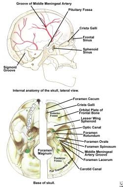
The sphenoid bone is a complex, irregularly shaped bone located in the middle cranial fossa and forms part of the base of the skull. It articulates with several other bones, including the frontal, parietal, temporal, ethmoid, palatine, and zygomatic bones. The sphenoid bone has two main parts: the body and the wings.
The body of the sphenoid bone is roughly cuboid in shape and contains several important structures, such as the sella turcica, which houses the pituitary gland, and the sphenoid sinuses, which are air-filled cavities within the bone. The greater wings of the sphenoid bone extend laterally from the body and form part of the skull's lateral walls. They contain the superior orbital fissure, through which important nerves and blood vessels pass between the cranial cavity and the orbit of the eye.
The lesser wings of the sphenoid bone are thin, blade-like structures that extend anteriorly from the body and form part of the floor of the anterior cranial fossa. They contain the optic canal, which transmits the optic nerve and ophthalmic artery between the brain and the orbit of the eye.
Overall, the sphenoid bone plays a crucial role in protecting several important structures within the skull, including the pituitary gland, optic nerves, and ophthalmic arteries.
The endocrine system is a complex network of glands and organs that produce, store, and secrete hormones. It plays a crucial role in regulating various functions in the body, including metabolism, growth and development, tissue function, sexual function, reproduction, sleep, and mood.
Endocrine system diseases or disorders occur when there is a problem with the production or regulation of hormones. This can result from:
1. Overproduction or underproduction of hormones by the endocrine glands.
2. Impaired response of target cells to hormones.
3. Disruption in the feedback mechanisms that regulate hormone production.
Examples of endocrine system diseases include:
1. Diabetes Mellitus - a group of metabolic disorders characterized by high blood sugar levels due to insulin deficiency or resistance.
2. Hypothyroidism - underactive thyroid gland leading to slow metabolism, weight gain, fatigue, and depression.
3. Hyperthyroidism - overactive thyroid gland causing rapid heartbeat, anxiety, weight loss, and heat intolerance.
4. Cushing's Syndrome - excess cortisol production resulting in obesity, high blood pressure, and weak muscles.
5. Addison's Disease - insufficient adrenal hormone production leading to weakness, fatigue, and low blood pressure.
6. Acromegaly - overproduction of growth hormone after puberty causing enlargement of bones, organs, and soft tissues.
7. Gigantism - similar to acromegaly but occurs before puberty resulting in excessive height and body size.
8. Hypopituitarism - underactive pituitary gland leading to deficiencies in various hormones.
9. Hyperparathyroidism - overactivity of the parathyroid glands causing calcium imbalances and kidney stones.
10. Precocious Puberty - early onset of puberty due to premature activation of the pituitary gland.
Treatment for endocrine system diseases varies depending on the specific disorder and may involve medication, surgery, lifestyle changes, or a combination of these approaches.
Central pontine myelinolysis (CPM) is a neurological disorder that results from the damage to the myelin sheath in the central pons region of the brainstem. Myelin is the fatty substance that insulates and protects nerve fibers, allowing for the efficient transmission of electrical signals.
In CPM, the myelin sheath in the center of the pons area becomes damaged or destroyed due to various factors, most commonly rapid correction of hyponatremia (low sodium levels in the blood). This rapid correction can lead to an osmotic shift of water from inside the cells to outside, causing swelling and damage to the myelin sheath.
CPM is characterized by the development of symmetrical lesions in the central pons region, which can result in a range of neurological symptoms, including weakness or paralysis of muscles, difficulty swallowing, speech impairment, and altered levels of consciousness. In severe cases, CPM can lead to coma, respiratory failure, and even death.
It's important to note that the management of CPM involves preventing further damage to the myelin sheath by avoiding rapid correction of hyponatremia and providing supportive care for the neurological symptoms. Currently, there is no specific treatment for CPM, and recovery can be slow and incomplete.
Jaw neoplasms refer to abnormal growths or tumors in the jawbone (mandible) or maxilla (upper jaw). These growths can be benign (non-cancerous) or malignant (cancerous). Benign neoplasms are not considered life-threatening, but they can still cause problems by invading nearby tissues and causing damage. Malignant neoplasms, on the other hand, can spread to other parts of the body and can be life-threatening if not treated promptly and effectively.
Jaw neoplasms can present with various symptoms such as swelling, pain, loose teeth, numbness or tingling in the lips or tongue, difficulty chewing or swallowing, and jaw stiffness or limited movement. The diagnosis of jaw neoplasms typically involves a thorough clinical examination, imaging studies such as X-rays, CT scans, or MRI, and sometimes a biopsy to determine the type and extent of the tumor.
Treatment options for jaw neoplasms depend on several factors, including the type, size, location, and stage of the tumor, as well as the patient's overall health and medical history. Treatment may involve surgery, radiation therapy, chemotherapy, or a combination of these modalities. Regular follow-up care is essential to monitor for recurrence or metastasis (spread) of the neoplasm.
Brain neoplasms, also known as brain tumors, are abnormal growths of cells within the brain. These growths can be benign (non-cancerous) or malignant (cancerous). Benign brain tumors typically grow slowly and do not spread to other parts of the body. However, they can still cause serious problems if they press on sensitive areas of the brain. Malignant brain tumors, on the other hand, are cancerous and can grow quickly, invading surrounding brain tissue and spreading to other parts of the brain or spinal cord.
Brain neoplasms can arise from various types of cells within the brain, including glial cells (which provide support and insulation for nerve cells), neurons (nerve cells that transmit signals in the brain), and meninges (the membranes that cover the brain and spinal cord). They can also result from the spread of cancer cells from other parts of the body, known as metastatic brain tumors.
Symptoms of brain neoplasms may vary depending on their size, location, and growth rate. Common symptoms include headaches, seizures, weakness or paralysis in the limbs, difficulty with balance and coordination, changes in speech or vision, confusion, memory loss, and changes in behavior or personality.
Treatment for brain neoplasms depends on several factors, including the type, size, location, and grade of the tumor, as well as the patient's age and overall health. Treatment options may include surgery, radiation therapy, chemotherapy, targeted therapy, or a combination of these approaches. Regular follow-up care is essential to monitor for recurrence and manage any long-term effects of treatment.
The optic chiasm is a structure in the brain where the optic nerves from each eye meet and cross. This allows for the integration of visual information from both eyes into the brain's visual cortex, creating a single, combined image of the visual world. The optic chiasm plays an important role in the processing of visual information and helps to facilitate depth perception and other complex visual tasks. Damage to the optic chiasm can result in various visual field deficits, such as bitemporal hemianopsia, where there is a loss of vision in the outer halves (temporal fields) of both eyes' visual fields.
 Craniopharyngioma
Visual pathway lesions
Catenin beta-1
Jakob Erdheim
Ghost cell
Augmented reality-assisted surgery
Vagotomy
Octreotide
Rosenthal fiber
Tolosa-Hunt syndrome
Scott Pomeroy
Scott Hamilton (figure skater)
Eating disorder
Hypophysectomy
Weight gain
Augmented reality
Rathke's pouch
Growth hormone
Chiasmal syndrome
Lou Gramm
Robert Schumann
Pilomatricoma
Pituicyte
Adrenal insufficiency
Delayed puberty
BRAF (gene)
History of neuroimaging
Hypopituitarism
Lars Leksell
Endoscope
Craniopharyngioma - Wikipedia
 Craniopharyngioma: MedlinePlus Medical Encyclopedia
 Craniopharyngioma Pathology: Definition, Epidemiology, Etiology
Pediatric Craniopharyngioma Clinical Presentation: History, Physical, Causes
 "Craniopharyngioma: An 18-Year Experience" by Sheri...
 Proton Therapy for Craniopharyngioma | Children's Hospital of Philadelphia
 Our Providers | Skull Base Tumors (Craniopharyngioma) | Main Line Health
 Pituitary tumors, adenoma, craniopharyngioma | Mayfield Brain & Spine, Cincinnati, OH
 What is Craniopharyngioma? | The Brain Tumour Charity
The Wnt Signalling Cascade and the Adherens Junction Complex in Craniopharyngioma Tumorigenesis - Nuffield Department of...
Diagnosing Craniopharyngioma | Expert Surgeon | Aaron Cohen-Gadol, MD
 Identification of targets for rational pharmacological therapy in childhood craniopharyngioma | Acta Neuropathologica...
 Early signs and symptoms of a brain tumor
 Craniopharyngioma - Libre Pathology
 2018 NPA Minisymposium | College of American Pathologists
Pediatric Craniopharyngioma: Background, Pathophysiology, Epidemiology
 Combined strategy of maximal endoscopic endonasal resection and early radiation therapy for complex cystic and solid...
 Craniopharyngioma in Children | Kettering Health
 Survivorship of Childhood Craniopharyngioma - ZenOnco.io
![Top 10 Craniopharyngioma Clinical Trials [2023 Studies] | Power](data:image/png;base64,iVBORw0KGgoAAAANSUhEUgAAABAAAAAQCAMAAAAoLQ9TAAAANlBMVEVHcEx3R/93Rv93R/93R/93R/93R/92Rv93R/93R/93R/93R/93R/93R/93R/93Rv93R/93R/+D+yDOAAAAEXRSTlMAYyy7rPbXEO2jhlF04j8fj3YMbBAAAABwSURBVBiVXY5ZEoAwCEOpXegyWrn/ZQ1Qp475CfOaQki2SidIvqr8A3IuUBuUdBpEh7p+tinCAzwYKcjCGKAZSCubcdHA6QmK/nBpICm4MUwLihwKhm+tMOtqXaomsh/zJTmNSa/Y6jJtXX9A3MsCD287CIN/VQGjAAAAAElFTkSuQmCC) Top 10 Craniopharyngioma Clinical Trials [2023 Studies] | Power
Top 10 Craniopharyngioma Clinical Trials [2023 Studies] | Power
 Childhood Cancer Awareness Month | September - St. Jude Children's Research Hospital
Pituitary disease: presentation, diagnosis, and management | Journal of Neurology, Neurosurgery & Psychiatry
 Brain and Spinal Cord Tumors Treatment Overview | Vanderbilt-Ingram Cancer Center
 Live Action Mafia • View topic - Finasteride hospital craniopharyngioma, ship, haemothorax, s
 "Multimodality, Multidirectional Resection of Craniopharyngioma: Versat" by Walter C. Jean MD
Intrachiasmatic craniopharyngioma: A rare cause of chiasmal thickening. Case report<...
Detailed assessment of hypothalamic damage in craniopharyngioma patients with obesity | Huntington Research - Translational...
 Find a Doctor in Your Area: Search Results - Sharecare
Adamantinomatous craniopharyngioma13
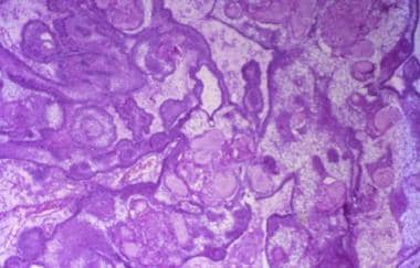
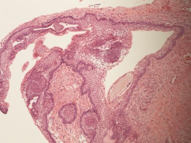
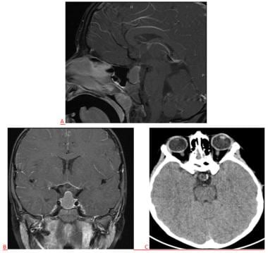
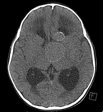
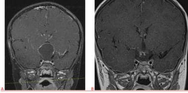
- CT scan showing a craniopharyngioma Enhanced T1 weighted MRIs of craniopharyngiomas Micrograph showing the characteristic features of an adamantinomatous craniopharyngioma - cystic spaces, calcifications, and "wet" keratin, HPS stain Micrograph showing a papillary craniopharyngioma, HPS stain Craniopharyngiomas are usually successfully managed with a combination of adjuvant chemotherapy and neurosurgery. (wikipedia.org)
- [ 10 ] Papillary craniopharyngioma occurs almost exclusively in adults, whereas adamantinomatous craniopharyngioma occurs in both adults and children. (medscape.com)
- Adamantinomatous craniopharyngioma presumably arises from remnants of the Rathke pouch and the craniopharyngeal duct. (medscape.com)
- The close histopathologic and immunohistochemical resemblance among adamantinomatous craniopharyngioma, adamantinoma of the jaw, and calcifying odontogenic cyst suggests an odontogenic epithelial differentiation for these tumors. (medscape.com)
- The current hypothesis is that pituitary adenoma, adamantinomatous craniopharyngioma, and Rathke cyst share a common ancestry from involuted remnants of the Rathke pouch and the craniopharyngeal duct. (medscape.com)
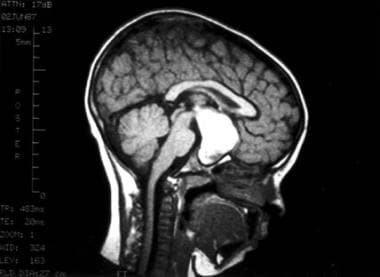
- Magnetic resonance imaging (MRI) typically shows an adamantinomatous craniopharyngioma as a complex solid/cystic lesion with heterogeneous signal intensity. (medscape.com)
- Adamantinomatous craniopharyngioma is a partly cystic mass filled with dark greenish-brown fluid that has traditionally been compared in terms of color and consistency to "machinery oil. (medscape.com)
- Pediatric adamantinomatous craniopharyngioma (ACP) is a histologically benign but clinically aggressive brain tumor that arises from the sellar/suprasellar region. (biomedcentral.com)
- It is subdivided into papillary craniopharyngioma and adamantinomatous craniopharyngioma . (librepathology.org)
- Calcifications in adamantinomatous craniopharyngioma. (librepathology.org)
- Adamantinomatous craniopharyngioma - very low mag. (librepathology.org)
- Transcriptional analyses of adult and pediatric adamantinomatous craniopharyngioma reveals similar expression signatures regarding potential therapeutic targets. (cornell.edu)
- Adamantinomatous craniopharyngioma (ACP) is a biologically benign but clinically aggressive lesion that has a significant impact on quality of life. (cornell.edu)
Tumors2
- Craniopharyngioma comprises 5%-10% of all childhood brain tumors and 1.2%-4.6% of brain tumors in adults (0.5-2.5 new cases per million population per year). (medscape.com)
- CT, MRI) pinpoint the exact location of the tumor and can help to differentiate craniopharyngioma from other tumors based on their characteristic appearance. (aaroncohen-gadol.com)
Cystic5
- Symptoms include: Fatigue Low blood pressure Electrolyte abnormalities Craniopharyngioma is a rare, usually suprasellar neoplasm, which may be cystic, that develops from nests of epithelium derived from Rathke's pouch. (wikipedia.org)
- MRI of a papillary craniopharyngioma characteristically depicts an enhancing, predominantly solid, circumscribed mass without the calcification or complex cystic architecture of the adamantinomatous variant. (medscape.com)
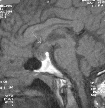
- Figure 1: Please see the appearance of a cystic craniopharyngioma on MRI and evidence of calcification on a CT scan (right lower corner, bright white nodule. (aaroncohen-gadol.com)
- In this operative video, we demonstrate the case of a 56-year-old female who had a complex craniopharyngioma with solid and cystic components extending superolaterally into the right frontal lobe. (researchwithrutgers.com)
- This case illustrates the feasibility of a combined strategy of maximal safe endoscopic endonasal resection followed by early radiation therapy for a complex, invasive cystic and solid craniopharyngioma. (researchwithrutgers.com)
Papillary variant of craniopharyngioma2
- In elderly persons, squamous metaplasia of adenohypophyseal cells of the pituitary stalk or gland has been postulated as a possible origin for the papillary variant of craniopharyngioma. (medscape.com)
- May not be seen in the papillary variant of craniopharyngioma. (librepathology.org)
Tumor12
- A craniopharyngioma is a rare type of brain tumor derived from pituitary gland embryonic tissue that occurs most commonly in children, but also affects adults. (wikipedia.org)
- A craniopharyngioma is a noncancerous (benign) tumor that develops at the base of the brain near the pituitary gland. (medlineplus.gov)
- She was diagnosed in 2014 with craniopharyngioma , a rare type of benign brain tumor that begins near the brain's pituitary gland. (chop.edu)
- A craniopharyngioma is a benign tumor that develops close to the pituitary gland. (medicalnewstoday.com)
- Craniopharyngioma is a rare and benign intracranial tumor of the sellar and suprasellar region. (researchwithrutgers.com)
- Craniopharyngioma is a noncancerous brain tumor found near the pituitary gland, which may press on parts of the brain and nearby tissue, affecting hormones, vision, and other normal functions. (ketteringhealth.org)
- Craniopharyngioma is a brain tumor that's not cancer (benign). (ketteringhealth.org)
- A craniopharyngioma is a benign tumor that is found near the pituitary gland. (ketteringhealth.org)
- Survivorship of Craniopharyngioma is one of the most complex parts of having craniopharyngioma, a rare brain tumor. (zenonco.io)
- In the case of childhood craniopharyngioma, some children whose tumor condition is likely to become chronic continue to receive treatment and care for an extended time. (zenonco.io)
- When it comes to advancing research and treatment options for the rare brain tumor known as craniopharyngioma, several top hospitals across the United States are at the forefront of innovation. (withpower.com)
- Hypothalamic obesity (HO) occurs in 50% of patients with the pituitary tumor craniopharyngioma (CP). (lu.se)
Diagnosis of craniopharyngioma3
- One series reported that growth failure preceded diagnosis of craniopharyngioma by a mean of 4 years. (medscape.com)
- The diagnosis of craniopharyngioma requires obtaining a comprehensive history and physical exam. (aaroncohen-gadol.com)
- Although a combination of laboratory and imaging tests are often all that is needed to be confident about a diagnosis of craniopharyngioma, a biopsy during surgery may be obtained for definitive confirmation. (aaroncohen-gadol.com)
Pediatric Craniopharyngioma2
- Using our compiled transcriptome dataset of 27 pediatric and 9 adult ACPs, obtained through the Advancing Treatment for Pediatric Craniopharyngioma Consortium, we interrogated potential age-related transcriptional differences using several rigorous mathematical analyses. (cornell.edu)
- Predictive Factors for Pediatric Craniopharyngioma Recurrence: An Extensive Narrative Review. (bvsalud.org)
Cyst2
- [ 11 ] Collision lesions of the sellar region, including pituitary adenoma associated with Rathke cyst and pituitary adenoma associated with craniopharyngioma, have been described. (medscape.com)
- Approximately 85% of masses from this region are pituitary adenomas, followed in incidence by craniopharyngioma, Rathke cleft cyst, meningioma, and metastasis. (cap.org)
20231
- Craniopharyngioma research studies recruiting patients in 2023 need your help. (withpower.com)
Patients7
- nevertheless, this symptom reportedly occurs in 20-60% of pediatric patients with craniopharyngioma at presentation. (medscape.com)
- Formal testing is generally required to identify visual field deficits in children, which likely explains the wide reported range (10-95%) of patients with craniopharyngioma. (medscape.com)
- Once a craniopharyngioma is diagnosed, patients can then be directed to appropriate treatment options. (aaroncohen-gadol.com)
- Lifelong care is necessary for most childhood craniopharyngioma patients and ACP and is considered by many to be a chronic disease [ 14 ]. (biomedcentral.com)
- We aimed to describe and predict the risk of severe hypernatremia after surgical resection of craniopharyngioma and to identify the association of water intake, urine output, and sodium level change in the patients. (bioscientifica.com)
- The overall incidence of severe hypernatremia after surgical resection of craniopharyngioma was significant, especially in patients with gross total resection, hypothalamus distortion, preoperative adrenal insufficiency, and preoperative severe hypernatremia. (bioscientifica.com)
- Craniopharyngioma (CP) patients suffer from increased cerebrovascular mortality. (lu.se)
Symptoms6
- Symptoms of craniopharyngioma can be present for years before diagnosis. (medscape.com)
- Other symptoms that may present due to a craniopharyngioma are hydrocephalus, diabetes and personality changes. (thebraintumourcharity.org)
- Diagnosing craniopharyngioma is a step-by-step process that typically begins when a patient reports their symptoms to a physician. (aaroncohen-gadol.com)
- Because a craniopharyngioma is located near structures critical to produce hormones (pituitary gland, hypothalamus), symptoms are often related to hormonal abnormalities. (aaroncohen-gadol.com)
- What are the symptoms of a craniopharyngioma in a child? (ketteringhealth.org)
- The symptoms of a craniopharyngioma can be like other health conditions. (ketteringhealth.org)
Calcifications1
- Figure 2: MRI findings of a typical craniopharyngioma with strong enhancement (bright white) due to high protein content and calcifications. (aaroncohen-gadol.com)
Surgery4
- Usually, surgery has been the main treatment for craniopharyngioma. (medlineplus.gov)
- The primary element of standard treatment for a craniopharyngioma is neurosurgery surgery to remove as much of the tumour as possible. (thebraintumourcharity.org)
- A craniopharyngioma is usually removed with surgery. (ketteringhealth.org)
- Craniopharyngioma or its surgery induces diabetes mellitus. (nel.edu)
Clinically1
- Craniopharyngioma comprises two clinically, histologically, and biologically distinct subtypes: adamantinomatous (most common) and papillary. (medscape.com)
Ventricle1
- Autopsy case with papillary craniopharyngioma of the 3rd ventricle. (librepathology.org)
Incidence1
- [ 1 ] The reported incidence of craniopharyngioma is particularly high in Nigeria and Japan. (medscape.com)
Predominantly1
- A craniopharyngioma is a low grade brain tumour which affects people of all ages but predominantly children and young adults. (thebraintumourcharity.org)
20191
- Similarly, Children's Hospital Colorado in Aurora has also made significant contributions with three ongoing craniopharyngioma trials and a total of four conducted trials since their initial study in 2019. (withpower.com)
Tomography1
- however, computer tomography (CT) remains the gold standard imaging choice for craniopharyngioma diagnosis as it can detect the severity of the calcification within the tumour. (wikipedia.org)
Acute1
- Advanced neuroimaging in acute lymphoblastic leukemia and craniopharyngioma. (lu.se)
Hormone1
- There may be long-term hormone, vision, and nervous system problems after craniopharyngioma is treated. (medlineplus.gov)
Surgical1
- Multimodality, Multidirectional Resection of Craniopharyngioma: Versatility in Alternating the Principal and Auxiliary Surgical Corridors and Visualization Modalities. (lvhn.org)
Children2
- Headache: Headaches occur in 60-80% of children with craniopharyngioma at presentation and are usually a symptom of increased intracranial pressure or hydrocephalus. (medscape.com)
- 1. Modified Orbitozygomatic Craniotomy for Craniopharyngioma Resection in Children. (ohsu.edu)
Radiation1
- The introduction of rational therapy to treat craniopharyngioma could drastically reduce the morbidity associated with both the primary disease and current treatments by reducing the extent of resection and/or reducing or eliminating the need for subsequent radiation. (biomedcentral.com)
Mass1
- This fluid-filled mass is consistent with a typical craniopharyngioma. (medscape.com)
Scans1
- A physician can conduct a few scans and tests to diagnose a person with craniopharyngioma. (wikipedia.org)
Care3
- Craniopharyngioma survivors and their caretakers can feel stressed once the frequent visits to the hospital and meetings with the health care team end. (zenonco.io)
- Craniopharyngioma survivors and their caretakers can feel stressed once the frequent visits to the hospital and meeting the health care team ends. (zenonco.io)
- Receive premium care & cutting edge treatments by enrolling in craniopharyngioma clinical trials today. (withpower.com)
High1
- Papillary craniopharyngioma - high mag. (librepathology.org)
Occur1
- In younger people, panhypopituitarism may occur secondary to a craniopharyngioma. (msdmanuals.com)


![10 Best Clinics for Esophageal Cancer Treatment in Thailand [2023 Prices]](https://www.mymeditravel.com/cdn-cgi/image/f=auto,fit=contain,quality=75/uploads/property/gallery/5af2758efa6b7e04401f8c27/5af51dadfa6b7e4212052361/preview.jpg)