Cysts
Epidermal Cyst
Mediastinal Cyst
Synovial Cyst
Bone Cysts
Bronchogenic Cyst
Dermoid Cyst
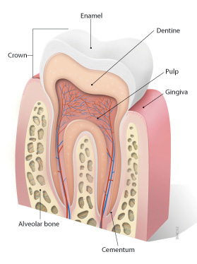
Odontogenic Cysts
Radicular Cyst
Dentigerous Cyst
Mesenteric Cyst
Bone Cysts, Aneurysmal
Tarlov Cysts
Popliteal Cyst
Esophageal Cyst
Echinococcosis
Urachal Cyst
Breast Cyst
Jaw Cysts
Giardia
Echinococcosis, Hepatic
Periodontal Cyst
Kidney Diseases, Cystic
Thyroglossal Cyst
Nonodontogenic Cysts
Echinococcus granulosus
Maxillary Diseases
Parovarian Cyst
Echinococcosis, Pulmonary
Odontogenic Cyst, Calcifying
Mandibular Diseases
Polycystic Kidney, Autosomal Dominant
Albendazole
Echinococcus
Tomography, X-Ray Computed
Giardia lamblia
Splenic Diseases
Giardiasis
Spores, Protozoan
TRPP Cation Channels
Brain Diseases
Rupture, Spontaneous
Arachnoid
Mebendazole
Curettage
Fibrocystic Breast Disease
Suction
Spinal Diseases
Third Ventricle
Cholangiopancreatography, Magnetic Resonance
Oocysts
Nematoda
Craniopharyngioma
Disruption of the Toxoplasma gondii bradyzoite-specific gene BAG1 decreases in vivo cyst formation. (1/1471)
The bradyzoite stage of the Apicomplexan protozoan parasite Toxoplasma gondii plays a critical role in maintenance of latent infection. We reported previously the cloning of a bradyzoite-specific gene BAG1/hsp30 (previously referred to as BAG5) encoding a cytoplasmic antigen related to small heat shock proteins. We have now disrupted BAG1 in the T. gondii PLK strain by homologous recombination. H7, a cloned null mutant, and Y8, a control positive for both cat and BAG1, were chosen for further characterization. Immunofluorescence and Western blot analysis of bradyzoites with BAG1 antisera demonstrated expression of BAG1 in the Y8 and the PLK strain but no expression in H7. All three strains expressed a 116 kDa bradyzoite cyst wall antigen, a 29 kDa matrix antigen and the 65 kDa matrix reactive antigen MAG1. Mice inoculated with H7 parasites formed significantly fewer cysts than those inoculated with the Y8 and the PLK strains. H7 parasites were complemented with BAG1 using phleomycin selection. Cyst formation in vivo for the BAG1-complemented H7 parasites was similar to wild-type parasites. We therefore conclude that BAG1 is not essential for cyst formation, but facilitates formation of cysts in vivo. (+info)Cauda equina syndrome in ankylosing spondylitis: a report of six cases. (2/1471)
Six patients with ankylosing spondylitis and features of a cauda equina syndrome are described. The myelographic findings are discussed in relation to the pathogenesis of the disorder and its natural history. Present experience suggests that the cauda equina syndrome is a more common complication of ankylosing spondylitis than is usually thought. (+info)Cystic lymph node metastases of squamous cell carcinoma of Waldeyer's ring origin. (3/1471)
We analysed in a retrospective study the frequency of cystic lymph node (LN) metastases in neck dissection specimens of 123 patients with primary squamous cell carcinoma (SCC) arising in the palatine tonsils (62 M/14 F), the base of the tongue (38 M/5 F) and the nasopharynx (2 M/2 F). Eighty-two per cent of patients had metastases (64 tonsillar SCC, 33 base of tongue SCC and all four nasopharynx SCC) in 368 LN of a total 2298 sampled LN. Thirty-nine per cent of patients had exclusively solid metastases and 37% of patients had exclusively cystic metastases. A total of 62 patients had some signs of cyst formation in one or more metastatically affected LN (27 with only histological evidence of cyst formation with luminal diameters < 5 mm, 35 with clinically detectable cyst with luminal diameter > 5 mm). Cystic metastases were more common in patients with SCC of the base of the tongue (P = 0.005), while solitary clinically evident cystic metastasis with lumina > 5 mm were found exclusively in tonsillar carcinoma (P = 0.024). In comparison with solid metastases, cyst formation was associated with N-categories (N2b and N3, P = 0.005) in SCC of the base of the tongue origin. No such association was observed for tonsillar SCC (P = 0.65). The primary mechanism of cyst formation was cystic degeneration. (+info)Laparoscopic management of benign solid and cystic lesions of the liver. (4/1471)
OBJECTIVE: The authors present their experience in the laparoscopic management of benign liver disease. The aim of the study is to analyze technical feasibility and evaluate immediate and long-term outcome. SUMMARY BACKGROUND DATA: Indications for the laparoscopic management of varied abdominal conditions have evolved. Although the minimally invasive treatment of liver cysts has been reported, the laparoscopic approach to other liver lesions remains undefined. METHODS: Between September 1990 and October 1997, 43 patients underwent laparoscopic liver surgery. There were two groups of benign lesions: cysts (n = 31) and solid tumors (n = 12). Indications were solitary giant liver cysts (n = 16), polycystic liver disease (n = 9), hydatid cyst (n = 6), focal nodular hyperplasia (n = 3), and adenoma (n = 9). Only solid tumors, hydatid cysts, and patients with polycystic disease and large dominant cysts located in anterior liver segments were included. All giant solitary liver cysts were considered for laparoscopy. Patients with cholangitis, cirrhosis, and significant cardiac disease were excluded. Data were collected prospectively. RESULTS: The procedures were completed laparoscopically in 40 patients. Median size was 4 cm for solid nodules and 14 cm for solitary liver cysts. Conversion occurred in three patients (7%), for bleeding (n = 2) and impingement of a solid tumor on the inferior vena cava (n = 1). The median operative time was 179 minutes. All solitary liver cysts were fenestrated in less than 1 hour. There were no deaths. Complications occurred in 6 cases (14.1%). Two hemorrhagic and two infectious complications were noted after management of hydatid cysts. There were no complications after resection of solid tumors. Three patients received transfusions (7%). The median length of stay was 4.7 days. Median follow-up was 30 months. There was no recurrence of solitary liver or hydatid cysts. One patient with polycystic disease had symptomatic recurrent cysts at 6 months requiring laparotomy. CONCLUSION: Laparoscopic liver surgery can be accomplished safely in selected patients with small benign solid tumors located in the anterior liver segments and giant solitary cysts. The laparoscopic management of polycystic liver disease should be reserved for patients with a limited number of large, anteriorly located cysts. Hydatid disease is best treated through an open approach. (+info)Late consequences of acute ischemic injury to a solitary kidney. (5/1471)
The sequelae of acute ischemic injury to a solitary kidney were assessed in rats subjected to right nephrectomy and transient occlusion of the left renal artery; control rats underwent right nephrectomy alone. Incomplete recovery from ischemic injury at 2 wk (serum creatinine levels of 1.1 +/- 0.2 versus 0.5 +/- 0.1 mg/dl, P < 0.05 for ischemia versus control) was followed by deterioration of renal function at 20 wk (serum creatinine levels of 1.7 +/- 0.4 versus 0.7 +/- 0.1 mg/dl, P < 0.05 for ischemia versus control). Morphologic studies showed that impairment of function after ischemic injury was associated with widespread tubulointerstitial disease. Some tubule segments were atrophic and others exhibited cystic dilation, so that the tubular cell volume fraction was reduced (37 +/- 4 versus 53 +/- 2%, P < 0.05), while the tubular lumen and interstitial volume fractions were increased (31 +/- 4 versus 23 +/- 2% and 29 +/- 2 versus 20 +/- 1%, respectively, both P < 0.05). Many glomeruli retained open capillary loops but were no longer connected to normal tubule segments (63 +/- 8 versus 15 +/- 7% of glomeruli, P < 0.05). There was a strong inverse correlation between the prevalence of such glomeruli and the GFR at 20 wk after ischemia (r2 = 0.79, P < 0.001). Tubulointerstitial disease at that time was accompanied by proteinuria and widespread segmental glomerular tuft injury. The occurrence of similar processes in human patients could contribute to the loss of graft kidneys that suffer ischemic injury during transplantation. (+info)Histological characteristics of sternoclavicular beta 2-microglobulin amyloidosis and clues for its histogenesis. (6/1471)
BACKGROUND: The pathogenesis of beta 2-microglobulin amyloidosis (A beta 2m) has yet to be fully elucidated. METHODS: We describe the distribution and extent of A beta 2m deposition and macrophagic infiltration in cartilage, capsule, and synovium of sternoclavicular joints obtained postmortem from 54 patients after 3 to 244 (median 46) months of dialysis. Twenty-four nonuremic patients served as a control group. The diagnosis of amyloidosis (A) rested on a positive Congo Red staining (typical birefringence) and that of A beta 2m on positive immunostaining of the A deposits with a monoclonal anti-beta 2m antibody. The size of A deposits was measured. RESULTS: A beta 2m was detected in 32 (59%), and non-beta 2m amyloid (Anon beta 2m) was detected in an additional 8 (15%) of the 54 dialyzed patients. A beta 2m deposits were present in the cartilage of all A beta 2m (+) patients (100%). They were localized solely in the cartilage in 27% of the cases, either as a thin patchy layer or as a continuous thicker layer (identified as stage I). A beta 2m was additionally present in the capsule and/or synovium without macrophages in 27% of the cases (identified as stage II). The correlation between the size of cartilaginous deposits and dialysis duration (P = 0.02) as well as with the prevalence (P = 0.03) and size of capsular deposits (P = 0.02) suggests that stage II is a later stage of A deposition. Clusters of macrophages were detected around capsular and synovial amyloid deposits in 46% of the cases (identified as stage III). The longer duration of dialysis in those with stage III as well as the relationship between the size of the A beta 2m deposits and the prevalence of macrophagic infiltration suggests that stage III is the last stage of A beta 2m deposition. Marginal bone erosions were observed in 9 out of 12 patients with stage III deposits. Their size was correlated with that of cartilaginous deposits (P = 0.01). Among the 24 control patients, Anon beta 2m was detected in 12 patients (cartilage 100%, capsule 8%, synovium 30%). CONCLUSIONS: The earliest stage of A beta 2m deposition occurs in the cartilage. A beta 2m subsequently extends to capsule and synovium. These two first stages do not require macrophage infiltration. Macrophages are eventually recruited around larger synovial or capsular deposits in the final stage. Marginal bone erosions develop in this late stage. (+info)Multifocal meningioangiomatosis: a report of two cases. (7/1471)
We report the CT and MR findings in two patients with multifocal meningioangiomatosis, neither of whom had a family history or stigmata of neurofibromatosis. All lesions were located in the cortical and subcortical areas and had round dense calcifications with eccentric cysts. The masses were associated with surrounding edema and gliosis. (+info)Posterior fossa epithelial cyst: case report and review of the literature. (8/1471)
A 49-year old woman with progressive cranial nerve signs and hemiparesis was found at MR imaging and at surgery to have a cyst at the foramen magnum. Immunohistochemistry and electron microscopy showed an epithelial cyst of endodermal origin. MR findings were of an extraaxial mass, with short T1 and T2 times. Unless immunohistochemistry and electron microscopy are used in the final diagnosis of such cysts, all posterior fossa cysts lined by a single layer of epithelium should be described simply as epithelial cysts. (+info)A cyst is a closed sac, having a distinct membrane and division between the sac and its surrounding tissue, that contains fluid, air, or semisolid material. Cysts can occur in various parts of the body, including the skin, internal organs, and bones. They can be caused by various factors, such as infection, genetic predisposition, or blockage of a duct or gland. Some cysts may cause symptoms, such as pain or discomfort, while others may not cause any symptoms at all. Treatment for cysts depends on the type and location of the cyst, as well as whether it is causing any problems. Some cysts may go away on their own, while others may need to be drained or removed through a surgical procedure.
Cyst fluid refers to the fluid accumulated within a cyst, which is a closed sac-like or capsular structure, typically filled with liquid or semi-solid material. Cysts can develop in various parts of the body for different reasons, and the composition of cyst fluid may vary depending on the type of cyst and its location.
In some cases, cyst fluid might contain proteins, sugars, hormones, or even cells from the surrounding tissue. Infected cysts may have pus-like fluid, while cancerous or precancerous cysts might contain abnormal cells or tumor markers. The analysis of cyst fluid can help medical professionals diagnose and manage various medical conditions, including infections, inflammatory diseases, genetic disorders, and cancers.
It is important to note that the term 'cyst fluid' generally refers to the liquid content within a cyst, but the specific composition and appearance of this fluid may vary significantly depending on the underlying cause and type of cyst.
An ovarian cyst is a sac or pouch filled with fluid that forms on the ovary. Ovarian cysts are quite common in women during their childbearing years, and they often cause no symptoms. In most cases, ovarian cysts disappear without treatment over a few months. However, larger or persistent cysts may require medical intervention, including surgical removal.
There are various types of ovarian cysts, such as functional cysts (follicular and corpus luteum cysts), which develop during the menstrual cycle due to hormonal changes, and non-functional cysts (dermoid cysts, endometriomas, and cystadenomas), which can form due to different causes.
While many ovarian cysts are benign, some may have malignant potential or indicate an underlying medical condition like polycystic ovary syndrome (PCOS). Regular gynecological check-ups, including pelvic examinations and ultrasounds, can help detect and monitor ovarian cysts.
An epidermal cyst is a common benign skin condition characterized by the growth of a sac-like structure filled with keratin, a protein found in the outermost layer of the skin (epidermis). These cysts typically appear as round, firm bumps just under the surface of the skin, often on the face, neck, trunk, or scalp. They can vary in size from a few millimeters to several centimeters in diameter.
Epidermal cysts usually develop as a result of the accumulation of dead skin cells that become trapped within a hair follicle or a pilosebaceous unit (a structure that contains a hair follicle and an oil gland). The keratin produced by the skin cells then collects inside the sac, causing it to expand gradually.
These cysts are generally slow-growing, painless, and rarely cause any symptoms. However, they may become infected or inflamed, leading to redness, tenderness, pain, or pus formation. In such cases, medical attention might be necessary to drain the cyst or administer antibiotics to treat the infection.
Epidermal cysts can be removed surgically if they cause cosmetic concerns or become frequently infected. The procedure typically involves making an incision in the skin and removing the entire sac along with its contents to prevent recurrence.
A mediastinal cyst is a rare, abnormal fluid-filled sac located in the mediastinum, which is the central part of the chest cavity that separates the lungs and contains various organs such as the heart, esophagus, trachea, thymus gland, and lymph nodes. Mediastinal cysts can be congenital (present at birth) or acquired (develop later in life). They are usually asymptomatic but can cause symptoms depending on their size and location. Symptoms may include chest pain, cough, difficulty breathing, or swallowing. Treatment typically involves surgical removal of the cyst to prevent complications such as infection, bleeding, or pressure on surrounding structures.
A Synovial Cyst is a type of benign cyst that typically develops in the synovium, which is the membrane that lines and lubricates joint capsules. These cysts are filled with synovial fluid, which is the same lubricating fluid found inside joints. They usually form as a result of degenerative changes, trauma, or underlying joint diseases such as osteoarthritis.
Synovial cysts commonly occur in the spine (particularly in the facet joints), but they can also develop in other areas of the body, including the knees, hips, and hands. While synovial cysts are generally not harmful, they may cause discomfort or pain if they press on nearby nerves or restrict movement in the affected joint. Treatment options for synovial cysts range from conservative measures like physical therapy and pain management to surgical intervention in severe cases.
A bone cyst is a fluid-filled sac that develops within a bone. It can be classified as either simple (unicameral) or aneurysmal. Simple bone cysts are more common in children and adolescents, and they typically affect the long bones of the arms or legs. These cysts are usually asymptomatic unless they become large enough to weaken the bone and cause a fracture. Aneurysmal bone cysts, on the other hand, can occur at any age and can affect any bone, but they are most common in the leg bones and spine. They are characterized by rapidly growing blood-filled sacs that can cause pain, swelling, and fractures.
Both types of bone cysts may be treated with observation, medication, or surgery depending on their size, location, and symptoms. It is important to note that while these cysts can be benign, they should still be evaluated and monitored by a healthcare professional to ensure proper treatment and prevention of complications.
A bronchogenic cyst is a type of congenital cyst that develops from abnormal budding or development of the bronchial tree during fetal growth. These cysts are typically filled with mucus or fluid and can be found in the mediastinum (the area between the lungs) or within the lung tissue itself.
Bronchogenic cysts are usually asymptomatic, but they can cause symptoms if they become infected, rupture, or compress nearby structures such as airways or blood vessels. Symptoms may include cough, chest pain, difficulty breathing, and recurrent respiratory infections.
Diagnosis of bronchogenic cysts is typically made through imaging tests such as chest X-rays, CT scans, or MRI scans. Treatment usually involves surgical removal of the cyst to prevent complications.
A dermoid cyst is a type of benign (non-cancerous) growth that typically develops during embryonic development. It is a congenital condition, which means it is present at birth, although it may not become apparent until later in life. Dermoid cysts are most commonly found in the skin or the ovaries of women, but they can also occur in other areas of the body, such as the spine or the brain.
Dermoid cysts form when cells that are destined to develop into skin and its associated structures, such as hair follicles and sweat glands, become trapped during fetal development. These cells continue to grow and multiply, forming a sac-like structure that contains various types of tissue, including skin, fat, hair, and sometimes even teeth or bone.
Dermoid cysts are usually slow-growing and may not cause any symptoms unless they become infected or rupture. In some cases, they may cause pain or discomfort if they press on nearby structures. Treatment typically involves surgical removal of the cyst to prevent complications and alleviate symptoms.
Odontogenic cysts are a type of cyst that originates from the dental tissues or odontogenic apparatus. They are typically found in the jawbones, and can be classified as developmental or inflammatory in origin. Developmental odontogenic cysts arise from remnants of the tooth-forming structures, while inflammatory odontogenic cysts result from an infection or injury to a tooth.
The most common types of odontogenic cysts include:
1. Periapical cyst - an inflammatory cyst that forms at the tip of the root of a dead or non-vital tooth.
2. Dentigerous cyst - a developmental cyst that surrounds the crown of an unerupted or impacted tooth.
3. Follicular cyst - a type of dentigerous cyst that forms around the crown of an unerupted wisdom tooth.
4. Odontogenic keratocyst - a developmental cyst that arises from the dental lamina and has a high recurrence rate.
5. Lateral periodontal cyst - a rare, developmental cyst that forms in the periodontal ligament of a vital tooth.
Odontogenic cysts can cause various symptoms such as swelling, pain, or numbness in the affected area. They may also displace or resorb adjacent teeth. Diagnosis is typically made through radiographic imaging and histopathological examination of tissue samples obtained through biopsy. Treatment options include surgical excision, marsupialization (a procedure that creates an opening between the cyst and oral cavity), or enucleation (removal of the cyst lining).
A radicular cyst is a type of dental cyst that forms around the root of a tooth, usually as a result of chronic infection or inflammation. It is also known as a periapical cyst. The cyst develops from the accumulation of fluid and cells in the periodontal ligament, which is the tissue that connects the tooth to the jawbone.
Radicular cysts are often caused by untreated dental caries or trauma to the tooth that allows bacteria to enter the pulp chamber of the tooth and cause an infection. Over time, the infection can spread to the surrounding tissues, leading to the formation of a cyst. Symptoms of a radicular cyst may include pain, swelling, and tenderness in the affected area. Treatment typically involves removing the affected tooth and the cyst through a surgical procedure.
A dentigerous cyst is a type of odontogenic cyst that forms around the crown of an unerupted tooth. It is typically slow-growing and often asymptomatic, but it can cause displacement or resorption of adjacent teeth if it becomes large enough. Dentigerous cysts are more common in permanent teeth than primary teeth, and they are more likely to occur in the mandible (lower jaw) than the maxilla (upper jaw). They are usually diagnosed through radiographic examination and can be treated by surgical removal of the cyst along with the affected tooth. If left untreated, dentigerous cysts can continue to grow and may eventually develop into a tumor or cancer.
A Mesenteric Cyst is a rare, benign abdominal mass that forms within the mesentery, which is the fold of membrane that attaches the intestine to the abdominal wall and contains blood vessels, lymphatic vessels, and nerves. These cysts can vary in size from a few centimeters to several inches in diameter. They are typically asymptomatic but can cause symptoms such as abdominal pain, bloating, or a palpable mass, depending on their size and location. The exact cause of mesenteric cysts is not well understood, but they may be congenital or acquired due to trauma, inflammation, or surgery. Treatment usually involves surgical removal of the cyst.
Aneurysmal bone cyst (ABC) is a benign but locally aggressive tumor that typically involves the metaphysis of long bones in children and adolescents. It is characterized by blood-filled spaces or cysts separated by fibrous septa containing osteoclast-type giant cells, spindle cells, and capillary vessels.
ABCs can also arise in other locations such as the vertebral column, pelvis, and skull. They may cause bone pain, swelling, or pathologic fractures. The exact cause of ABC is unknown, but it is thought to be related to a reactive process to a primary bone lesion or trauma.
Treatment options for ABC include curettage and bone grafting, intralesional injection of corticosteroids or bone marrow aspirate, and adjuvant therapy with phenol or liquid nitrogen. In some cases, radiation therapy may be used, but it is generally avoided due to the risk of secondary malignancies. Recurrence rates after treatment range from 10-30%.
Tarlov cysts, also known as perineural cysts or sacral nerve root sheath cysts, are fluid-filled sacs that develop on the outside of the spinal nerve roots, most commonly found in the lower spine (sacrum). These cysts typically form at the point where the nerves exit the spinal canal and enter the surrounding tissue. They are usually benign but can cause various symptoms depending on their size and location.
Tarlov cysts contain cerebrospinal fluid (CSF), which is the same fluid that surrounds and protects the brain and spinal cord. The exact cause of Tarlov cysts remains unclear, but they may result from trauma, degenerative changes, or congenital factors. Some individuals with Tarlov cysts may not experience any symptoms, while others might have pain, tingling, numbness, or weakness in the lower back, legs, or feet. In rare cases, Tarlov cysts can lead to more severe complications such as nerve compression or spinal cord injury. Treatment options for Tarlov cysts include observation, pain management, and surgical intervention in select cases.
A Popliteal cyst, also known as Baker's cyst, is a fluid-filled sac that develops behind the knee, in the popliteal fossa. It forms when synovial fluid from the knee joint extends through a tear in the joint capsule, creating a visible bulge. The cyst may cause discomfort, swelling, or pain, especially when fully extended or flexed. In some cases, it can rupture and cause further complications, such as increased pain and inflammation in the calf region. Treatment options for Popliteal cysts include physical therapy, corticosteroid injections, and, in severe cases, surgical intervention to repair the underlying joint issue and remove the cyst.
An esophageal cyst is a rare, abnormal growth that forms in the wall of the esophagus, which is the muscular tube that connects the throat to the stomach. These cysts are typically filled with fluid and can vary in size. They are usually congenital, meaning they are present at birth and develop as a result of abnormal embryonic development.
Esophageal cysts are typically asymptomatic and may not cause any problems until they become large enough to compress nearby structures, such as the trachea or other parts of the digestive system. In some cases, esophageal cysts may cause difficulty swallowing, coughing, or breathing.
Diagnosis of an esophageal cyst is typically made through imaging tests, such as a CT scan or MRI, which can help to visualize the cyst and determine its size and location. Treatment usually involves surgical removal of the cyst, which is typically performed using minimally invasive techniques such as endoscopy or thoracoscopy.
It's important to note that while I strive to provide accurate information, my responses should not be used as a substitute for professional medical advice, diagnosis or treatment. If you have any concerns about your health, it is always best to consult with a healthcare provider.
Echinococcosis is a parasitic infection caused by the larval stage of tapeworms belonging to the genus Echinococcus. There are several species of Echinococcus that can cause disease in humans, but the most common ones are Echinococcus granulosus (causing cystic echinococcosis) and Echinococcus multilocularis (causing alveolar echinococcosis).
Humans typically become infected with echinococcosis by accidentally ingesting eggs of the tapeworm, which are shed in the feces of infected animals such as dogs, foxes, and wolves. The eggs hatch in the small intestine and release larvae that migrate to various organs in the body, where they form cysts or hydatids.
The symptoms of echinococcosis depend on the location and size of the cysts. Cystic echinococcosis often affects the liver and lungs, causing symptoms such as abdominal pain, cough, and shortness of breath. Alveolar echinococcosis typically involves the liver and can cause chronic liver disease, abdominal pain, and jaundice.
Treatment of echinococcosis may involve surgery to remove the cysts, medication to kill the parasites, or both. Preventive measures include avoiding contact with dogs and other animals that may be infected with Echinococcus, practicing good hygiene, and cooking meat thoroughly before eating it.
A urachal cyst is a rare type of abdominal wall defect that results from the persistent embryonic remnant of the urachus, which is a canal-like structure that connects the bladder to the umbilicus (belly button) during fetal development. This canal normally obliterates and becomes a fibrous cord known as the median umbilical ligament after birth. However, if it fails to do so, it can result in the formation of various urachal anomalies, including a urachal cyst.
A urachal cyst is a fluid-filled sac that forms along any part of the urachus, usually located between the bladder and the umbilicus. These cysts are typically asymptomatic but can become infected or inflamed, leading to symptoms such as abdominal pain, tenderness, fever, and a palpable mass in the lower abdomen. In some cases, urachal cysts may also cause urinary tract infections or bladder irritation. Diagnosis is usually made through imaging studies such as ultrasound, CT scan, or MRI, and treatment typically involves surgical excision of the cyst to prevent complications.
A breast cyst is a fluid-filled sac that forms within the breast tissue. It is a common, benign (non-cancerous) condition and can affect people of any age, but it is more commonly found in women between the ages of 35 and 50. Breast cysts can vary in size and may be asymptomatic or cause discomfort or pain, especially just before menstruation.
Breast cysts are usually diagnosed through a physical examination, breast ultrasound, or mammography. In some cases, a fine-needle aspiration (FNA) may be performed to drain the fluid from the cyst and confirm the diagnosis. If the cyst is small, causes no symptoms, and appears benign on imaging studies, then further treatment may not be necessary. However, if the cyst is large, painful, or has concerning features on imaging studies, then additional diagnostic tests or drainage procedures may be recommended.
It's important to note that while breast cysts are generally harmless, they can sometimes mimic the symptoms of breast cancer. Therefore, any new or unusual changes in the breast should be evaluated by a healthcare professional.
A jaw cyst is a pathological cavity filled with fluid or semi-fluid material, which forms within the jaw bones. They are typically classified as odontogenic (developing from tooth-forming tissues) or non-odontogenic (developing from other tissues). The most common types of odontogenic jaw cysts include dentigerous cysts (formed around the crown of an unerupted tooth) and follicular cysts (formed from the inflammation of a developing tooth's tissue). Non-odontogenic cysts, such as nasopalatine duct cysts and keratocystic odontogenic tumors, can also occur in the jaw bones. Jaw cysts may cause symptoms like swelling, pain, or displacement of teeth, but some may not present any symptoms until they grow large enough to be detected on a radiographic examination. Treatment typically involves surgical removal of the cyst and, if necessary, reconstruction of the affected bone.
Giardia is a genus of microscopic parasitic flagellates that cause giardiasis, a type of diarrheal disease. The most common species to infect humans is Giardia intestinalis (also known as Giardia lamblia or Giardia duodenalis). These microscopic parasites are found worldwide, particularly in areas with poor sanitation and unsafe water.
Giardia exists in two forms: the trophozoite, which is the actively feeding form that multiplies in the small intestine, and the cyst, which is the infective stage that is passed in feces and can survive outside the body for long periods under appropriate conditions. Infection occurs when a person ingests contaminated water or food, or comes into direct contact with an infected person's feces.
Once inside the body, the cysts transform into trophozoites, which attach to the lining of the small intestine and disrupt the normal function of the digestive system, leading to symptoms such as diarrhea, stomach cramps, nausea, dehydration, and weight loss. In some cases, giardiasis can cause long-term health problems, particularly in children, including malnutrition and developmental delays.
Preventing the spread of Giardia involves maintaining good hygiene practices, such as washing hands thoroughly after using the toilet or changing diapers, avoiding contaminated water sources, and practicing safe food handling and preparation. In cases where infection occurs, medication is usually effective in treating the illness.
Echinococcosis, hepatic is a type of parasitic infection caused by the larval stage of the tapeworm Echinococcus granulosus. The infection typically occurs when a person accidentally ingests microscopic eggs of the tapeworm, which can be present in contaminated food, water, or soil.
Once inside the body, the eggs hatch and release larvae that can migrate to various organs, including the liver. In the liver, the larvae form hydatid cysts, which are fluid-filled sacs that can grow slowly over several years, causing symptoms such as abdominal pain, nausea, vomiting, and jaundice.
Hepatic echinococcosis is a serious condition that can lead to complications such as cyst rupture, infection, or organ damage if left untreated. Treatment options include surgery to remove the cysts, medication to kill the parasites, or a combination of both. Prevention measures include good hygiene practices, avoiding contact with contaminated soil or water, and cooking meat thoroughly before eating it.
A periodontal cyst, also known as a radicular cyst or dental cyst, is a type of odontogenic cyst that forms from the tissue of the periodontium, which surrounds and supports the teeth. It typically develops at the apex (tip) of a dead or non-vital tooth root and is filled with fluid. The cyst can grow slowly and painlessly, often going unnoticed until it becomes quite large or causes symptoms such as swelling, tenderness, or tooth mobility.
Periodontal cysts are usually asymptomatic and are often discovered during routine dental x-rays. If left untreated, they can eventually lead to the destruction of surrounding bone and tissue, potentially causing teeth to become loose or even fall out. Treatment typically involves surgical removal of the cyst along with the affected tooth, followed by careful monitoring to ensure that the cyst does not recur.
Cystic kidney diseases are a group of genetic disorders that cause fluid-filled sacs called cysts to form in the kidneys. These cysts can vary in size and can grow over time, which can lead to damage in the kidneys and affect their function. There are two main types of cystic kidney diseases: autosomal dominant polycystic kidney disease (ADPKD) and autosomal recessive polycystic kidney disease (ARPKD).
ADPKD is the most common type and is characterized by the presence of numerous cysts in both kidneys. It is usually diagnosed in adulthood, but it can also occur in children. The cysts can cause high blood pressure, kidney stones, urinary tract infections, and eventually kidney failure.
ARPKD is a rare, inherited disorder that affects both the kidneys and liver. It is characterized by the presence of numerous cysts in the kidneys and abnormalities in the bile ducts of the liver. ARPKD is usually diagnosed in infancy or early childhood and can cause serious complications such as respiratory distress, kidney failure, and liver fibrosis.
Other types of cystic kidney diseases include nephronophthisis, medullary cystic kidney disease, and glomerulocystic kidney disease. These conditions are also inherited and can cause kidney damage and kidney failure.
Treatment for cystic kidney diseases typically involves managing symptoms such as high blood pressure, pain, and infections. In some cases, surgery may be necessary to remove large cysts or to treat complications such as kidney stones. For individuals with advanced kidney disease, dialysis or a kidney transplant may be necessary.
A Thyroglossal cyst is defined as a congenital abnormality, specifically a developmental anomaly of the thyroid gland. It is a cystic mass that forms along the path of the thyroglossal duct, which is a tube-like structure that extends from the tongue to the developing thyroid gland in the neck during embryonic development.
The thyroglossal duct typically disappears before birth, but if it persists, it can result in the formation of a cyst. Thyroglossal cysts are usually midline and located either at or above the level of the hyoid bone in the neck. They may become symptomatic if they become infected or inflamed, leading to pain, swelling, and difficulty swallowing.
Treatment for thyroglossal cyst typically involves surgical removal through a procedure called a Sistrunk operation, which involves removing the cyst as well as a portion of the hyoid bone and the central part of the thyroglossal duct to reduce the risk of recurrence.
Nonodontogenic cysts are a type of cyst that occur in the oral and maxillofacial region, but they do not originate from tooth-forming tissues. These cysts can develop in various locations within the jaws, including the bone or soft tissues. They are typically classified into several categories based on their origin, such as developmental, inflammatory, or neoplastic.
Examples of nonodontogenic cysts include:
1. Nasopalatine duct cyst - This is the most common type of nonodontogenic cyst and arises from remnants of the nasopalatine duct, which is a structure present during embryonic development. It typically appears in the anterior region of the maxilla (upper jaw).
2. Nasolabial cyst - This rare cyst develops near the nasolabial fold, between the nose and the upper lip. Its origin is unclear but may be related to embryonic remnants or developmental abnormalities.
3. Median mandibular cyst - Also known as a median mental cyst, this rare cyst forms in the midline of the mandible (lower jaw) and may originate from remnants of the dental lamina or other developmental structures.
4. Lateral periodontal cyst - This inflammatory cyst arises from the periodontal ligament, which supports the teeth within their sockets. It is usually found near the roots of lower molars and premolars.
5. Glandular odontogenic cyst - This developmental cyst originates from remnants of minor salivary glands or epithelial rests in the jawbone. It can appear as a unilocular (single-chambered) or multilocular (multi-chambered) cyst and may have a more aggressive behavior than other nonodontogenic cysts.
6. Dentigerous cyst - Although technically classified as an odontogenic cyst, the dentigerous cyst is sometimes considered a borderline case because it arises from the crowns of unerupted teeth rather than their roots. It can grow quite large and may cause significant bone resorption.
Nonodontogenic cysts are less common than odontogenic cysts, but they still require proper diagnosis and treatment to prevent complications such as tooth displacement, jaw deformation, or infection. Treatment options for nonodontogenic cysts depend on their size, location, and histological features and may include enucleation (complete removal), marsupialization (creating a communication between the cyst and oral cavity to allow for gradual reduction), or more extensive surgical procedures. Regular follow-up appointments with your dentist or oral surgeon are essential to monitor healing and ensure that the cyst does not recur.
'Echinococcus granulosus' is a species of tapeworm that causes hydatid disease or echinococcosis in humans and other animals. The adult worms are small, typically less than 1 cm in length, and live in the intestines of their definitive hosts, which are usually dogs or other canids.
The life cycle of 'Echinococcus granulosus' involves the shedding of eggs in the feces of the definitive host, which are then ingested by an intermediate host, such as a sheep or a human. Once inside the intermediate host, the eggs hatch and release larvae that migrate to various organs, where they form hydatid cysts. These cysts can grow slowly over several years and may cause significant damage to the affected organ.
Humans can become accidentally infected with 'Echinococcus granulosus' by ingesting contaminated food or water, or through direct contact with infected dogs. The treatment of hydatid disease typically involves surgical removal of the cysts, followed by anti-parasitic medication to kill any remaining parasites. Prevention measures include proper hygiene and sanitation practices, as well as regular deworming of dogs and other definitive hosts.
Maxillary diseases refer to conditions that affect the maxilla, which is the upper bone of the jaw. This bone plays an essential role in functions such as biting, chewing, and speaking, and also forms the upper part of the oral cavity, houses the upper teeth, and supports the nose and the eyes.
Maxillary diseases can be caused by various factors, including infections, trauma, tumors, congenital abnormalities, or systemic conditions. Some common maxillary diseases include:
1. Maxillary sinusitis: Inflammation of the maxillary sinuses, which are air-filled cavities located within the maxilla, can cause symptoms such as nasal congestion, facial pain, and headaches.
2. Periodontal disease: Infection and inflammation of the tissues surrounding the teeth, including the gums and the alveolar bone (which is part of the maxilla), can lead to tooth loss and other complications.
3. Maxillary fractures: Trauma to the face can result in fractures of the maxilla, which can cause pain, swelling, and difficulty breathing or speaking.
4. Maxillary cysts and tumors: Abnormal growths in the maxilla can be benign or malignant and may require surgical intervention.
5. Oral cancer: Cancerous lesions in the oral cavity, including the maxilla, can cause pain, swelling, and difficulty swallowing or speaking.
Treatment for maxillary diseases depends on the specific condition and its severity. Treatment options may include antibiotics, surgery, radiation therapy, or chemotherapy. Regular dental check-ups and good oral hygiene practices can help prevent many maxillary diseases.
A parovarian cyst is a type of fluid-filled sac that develops in the vicinity of the ovary, often found attached to or adjoined with the fallopian tube or the ovary itself. These cysts are typically benign (noncancerous) and can vary in size, from being quite small to becoming large enough to cause discomfort or other symptoms.
Parovarian cysts are thought to arise from remnants of embryonic tissues known as Wolffian or Müllerian ducts, which contribute to the development of the reproductive system during fetal growth. These cysts can be found in individuals with ovaries, including both cisgender women and transgender men who have not had their ovaries removed.
While parovarian cysts are often asymptomatic and discovered incidentally during routine pelvic examinations or imaging studies, they may cause symptoms if they grow significantly in size. These symptoms can include:
1. Pelvic pain or discomfort
2. Bloating or a feeling of fullness in the abdomen
3. Pain during sexual intercourse (dyspareunia)
4. Abnormal menstrual bleeding or irregular periods
5. Difficulty with bowel movements or urination, depending on the cyst's size and location
In cases where parovarian cysts become large, cause persistent symptoms, or demonstrate concerning features (such as rapid growth or signs of malignancy), surgical intervention may be required to remove the cyst. This can often be accomplished through minimally invasive techniques like laparoscopy. However, in some instances, a more extensive open surgery might be necessary.
It is essential to consult with a healthcare professional if you suspect or have been diagnosed with a parovarian cyst, as they will provide guidance and determine the most appropriate course of action based on your individual circumstances.
Pulmonary echinococcosis is a rare infection caused by the larval stage of the tapeworm Echinococcus granulosus or Echinococcus multilocularis. The infection occurs when the eggs of the tapeworm, which are passed in the feces of an infected animal (usually a dog or fox), are ingested by another host (usually a human). Once inside the body, the eggs hatch and release larvae that can migrate to various organs, including the lungs. In the lungs, the larvae form hydatid cysts, which can grow slowly over several years and cause symptoms such as cough, chest pain, shortness of breath, and fever. Treatment typically involves surgical removal of the cysts, followed by medication to prevent recurrence.
Anticestodal agents are a type of medication used to treat infections caused by tapeworms (cestodes) and other related parasites. These agents work by either stunting the growth or killing the parasites, which allows the body to expel them naturally. Common anticestodal agents include niclosamide, praziquantel, and albendazole. It is important to note that proper diagnosis of the specific type of tapeworm infection is necessary for effective treatment, as different medications may be more or less effective against certain species.
An Odontogenic Cyst, Calcifying is a specific type of cyst that originates from the dental tissues. It's also known as a calcifying odontogenic cyst or Gorlin cyst. This cyst is characterized by the presence of calcified structures within its lining.
The calcifications can appear as flecks or more complex structures, such as teeth-like formations. The lining of this cyst often contains ghost cells, which are the remains of epithelial cells that have undergone calcification.
These cysts are typically slow-growing and asymptomatic, although they can sometimes cause swelling or pain if they become large enough to compress adjacent tissues. They are most commonly found in the jaw bones, particularly the mandible.
While the exact cause of calcifying odontogenic cysts is not fully understood, they are thought to arise from developmental abnormalities in the tissues that form teeth. Treatment typically involves surgical removal of the cyst.
Mandibular diseases refer to conditions that affect the mandible, or lower jawbone. These diseases can be classified as congenital (present at birth) or acquired (developing after birth). They can also be categorized based on the tissues involved, such as bone, muscle, or cartilage. Some examples of mandibular diseases include:
1. Mandibular fractures: These are breaks in the lower jawbone that can result from trauma or injury.
2. Osteomyelitis: This is an infection of the bone and surrounding tissues, which can affect the mandible.
3. Temporomandibular joint (TMJ) disorders: These are conditions that affect the joint that connects the jawbone to the skull, causing pain and limited movement.
4. Mandibular tumors: These are abnormal growths that can be benign or malignant, and can develop in any of the tissues of the mandible.
5. Osteonecrosis: This is a condition where the bone tissue dies due to lack of blood supply, which can affect the mandible.
6. Cleft lip and palate: This is a congenital deformity that affects the development of the face and mouth, including the lower jawbone.
7. Mandibular hypoplasia: This is a condition where the lower jawbone does not develop properly, leading to a small or recessed chin.
8. Developmental disorders: These are conditions that affect the growth and development of the mandible, such as condylar hyperplasia or hemifacial microsomia.
Autosomal Dominant Polycystic Kidney Disease (ADPKD) is a genetic disorder characterized by the growth of multiple cysts in the kidneys. These cysts are fluid-filled sacs that can vary in size and can multiply, leading to enlarged kidneys. The increased size and number of cysts can eventually result in reduced kidney function, high blood pressure, and potentially kidney failure.
ADPKD is an autosomal dominant disorder, meaning it only requires one copy of the altered gene (from either the mother or father) to have the disease. Each child of an affected individual has a 50% chance of inheriting the mutated gene. The two genes most commonly associated with ADPKD are PKD1 and PKD2, located on chromosomes 16 and 4, respectively.
Symptoms can vary widely among individuals with ADPKD, but they often include high blood pressure, back or side pain, headaches, increased abdominal size due to enlarged kidneys, blood in the urine, and kidney failure. Other complications may include cysts in the liver, pancreas, and/or brain (berries aneurysms).
Early diagnosis and management of ADPKD can help slow down disease progression and improve quality of life. Treatment typically includes controlling high blood pressure, managing pain, monitoring kidney function, and addressing complications as they arise. In some cases, dialysis or a kidney transplant may be necessary if kidney failure occurs.
Albendazole is an antiparasitic medication used to treat a variety of parasitic infections, including neurocysticercosis (a tapeworm infection that affects the brain), hydatid disease (a parasitic infection that can affect various organs), and other types of worm infestations such as pinworm, roundworm, hookworm, and whipworm infections.
Albendazole works by inhibiting the polymerization of beta-tubulin, a protein found in the microtubules of parasitic cells, which disrupts the parasite's ability to maintain its shape and move. This leads to the death of the parasite and elimination of the infection.
Albendazole is available in oral form and is typically taken two to three times a day with meals for several days or weeks, depending on the type and severity of the infection being treated. Common side effects of albendazole include nausea, vomiting, diarrhea, abdominal pain, and headache. Rare but serious side effects may include liver damage, bone marrow suppression, and neurological problems.
It is important to note that albendazole should only be used under the supervision of a healthcare provider, as it can have serious side effects and interactions with other medications. Additionally, it is not effective against all types of parasitic infections, so proper diagnosis is essential before starting treatment.
'Echinococcus' is a genus of tapeworms that can cause serious infections known as echinococcosis in humans and other animals. The most common species that infect humans are Echinococcus granulosus and Echinococcus multilocularis.
Echinococcus granulosus typically causes cystic echinococcosis, also known as hydatid disease, which affects the liver, lungs, or other organs. The tapeworm's eggs are passed in the feces of infected animals, such as dogs or sheep, and can be ingested by humans, leading to the development of cysts in various organs.
Echinococcus multilocularis typically causes alveolar echinococcosis, a more severe and invasive form of the disease that affects the liver and can spread to other organs. This species has a complex life cycle involving small mammals as intermediate hosts and canids (such as foxes or dogs) as definitive hosts.
Human infections with Echinococcus are rare but can lead to severe health complications if left untreated. Preventive measures include proper hygiene, avoiding contact with infected animals, and cooking meat thoroughly before consumption.
X-ray computed tomography (CT or CAT scan) is a medical imaging method that uses computer-processed combinations of many X-ray images taken from different angles to produce cross-sectional (tomographic) images (virtual "slices") of the body. These cross-sectional images can then be used to display detailed internal views of organs, bones, and soft tissues in the body.
The term "computed tomography" is used instead of "CT scan" or "CAT scan" because the machines take a series of X-ray measurements from different angles around the body and then use a computer to process these data to create detailed images of internal structures within the body.
CT scanning is a noninvasive, painless medical test that helps physicians diagnose and treat medical conditions. CT imaging provides detailed information about many types of tissue including lung, bone, soft tissue and blood vessels. CT examinations can be performed on every part of the body for a variety of reasons including diagnosis, surgical planning, and monitoring of therapeutic responses.
In computed tomography (CT), an X-ray source and detector rotate around the patient, measuring the X-ray attenuation at many different angles. A computer uses this data to construct a cross-sectional image by the process of reconstruction. This technique is called "tomography". The term "computed" refers to the use of a computer to reconstruct the images.
CT has become an important tool in medical imaging and diagnosis, allowing radiologists and other physicians to view detailed internal images of the body. It can help identify many different medical conditions including cancer, heart disease, lung nodules, liver tumors, and internal injuries from trauma. CT is also commonly used for guiding biopsies and other minimally invasive procedures.
In summary, X-ray computed tomography (CT or CAT scan) is a medical imaging technique that uses computer-processed combinations of many X-ray images taken from different angles to produce cross-sectional images of the body. It provides detailed internal views of organs, bones, and soft tissues in the body, allowing physicians to diagnose and treat medical conditions.
"Giardia lamblia," also known as "Giardia duodenalis" or "Giardia intestinalis," is a species of microscopic parasitic protozoan that colonizes and reproduces in the small intestine of various vertebrates, including humans. It is the most common cause of human giardiasis, a diarrheal disease. The trophozoite (feeding form) of Giardia lamblia has a distinctive tear-drop shape and possesses flagella for locomotion. It attaches to the intestinal epithelium, disrupting the normal function of the small intestine and leading to various gastrointestinal symptoms such as diarrhea, stomach cramps, nausea, and dehydration. Giardia lamblia is typically transmitted through the fecal-oral route, often via contaminated food or water.
A branchioma is a benign (non-cancerous) tumor that develops from remnants of the branchial apparatus, which are structures that form in the embryo during fetal development and give rise to parts of the head and neck. Branchiomas are also known as branchial cleft cysts or branchial apparatus remnants.
Branchioma typically occurs in the neck region and can cause a variety of symptoms, such as a painless lump, difficulty swallowing, or breathing problems if the tumor is large enough to compress the airway. Treatment usually involves surgical removal of the tumor.
Splenic diseases refer to a range of medical conditions that affect the structure, function, or health of the spleen. The spleen is an organ located in the upper left quadrant of the abdomen, which plays a vital role in filtering the blood and fighting infections. Some common splenic diseases include:
1. Splenomegaly: Enlargement of the spleen due to various causes such as infections, liver disease, blood disorders, or cancer.
2. Hypersplenism: Overactivity of the spleen leading to excessive removal of blood cells from circulation, causing anemia, leukopenia, or thrombocytopenia.
3. Splenic infarction: Partial or complete blockage of the splenic artery or its branches, resulting in tissue death and potential organ dysfunction.
4. Splenic rupture: Traumatic or spontaneous tearing of the spleen capsule, causing internal bleeding and potentially life-threatening conditions.
5. Infections: Bacterial (e.g., sepsis, tuberculosis), viral (e.g., mononucleosis, cytomegalovirus), fungal (e.g., histoplasmosis), or parasitic (e.g., malaria) infections can affect the spleen and cause various symptoms.
6. Hematologic disorders: Conditions such as sickle cell disease, thalassemia, hemolytic anemias, lymphomas, leukemias, or myeloproliferative neoplasms can involve the spleen and lead to its enlargement or dysfunction.
7. Autoimmune diseases: Conditions like rheumatoid arthritis, systemic lupus erythematosus, or vasculitis can affect the spleen and cause various symptoms.
8. Cancers: Primary (e.g., splenic tumors) or secondary (e.g., metastatic cancer from other organs) malignancies can involve the spleen and lead to its enlargement, dysfunction, or rupture.
9. Vascular abnormalities: Conditions such as portal hypertension, Budd-Chiari syndrome, or splenic vein thrombosis can affect the spleen and cause various symptoms.
10. Trauma: Accidental or intentional injuries to the spleen can lead to bleeding, infection, or organ dysfunction.
Giardiasis is a digestive infection caused by the microscopic parasite Giardia intestinalis, also known as Giardia lamblia or Giardia duodenalis. The parasite is found worldwide, especially in areas with poor sanitation and unsafe water.
The infection typically occurs after ingesting contaminated water, food, or surfaces that have been exposed to fecal matter containing the cyst form of the parasite. Once inside the body, the cysts transform into trophozoites, which attach to the lining of the small intestine and cause symptoms such as diarrhea, stomach cramps, nausea, dehydration, and greasy stools that may float due to excess fat.
In some cases, giardiasis can lead to lactose intolerance and malabsorption of nutrients, resulting in weight loss and vitamin deficiencies. The infection is usually diagnosed through a stool sample test and treated with antibiotics such as metronidazole or tinidazole. Preventive measures include practicing good hygiene, avoiding contaminated water and food, and washing hands regularly.
Medical definitions for "spores" and "protozoan" are as follows:
1. Spores: These are typically single-celled reproductive units that are resistant to heat, drying, and chemicals. They are produced by certain bacteria, fungi, algae, and plants. In the context of infectious diseases, spores are particularly relevant in relation to certain types of bacteria such as Clostridium tetani (causes tetanus) and Bacillus anthracis (causes anthrax). These bacterial spores can survive for long periods in harsh environments and can cause illness if they germinate and multiply in a host.
2. Protozoan: This term refers to a diverse group of single-celled eukaryotic organisms, which are typically classified as animals rather than plants or fungi. Some protozoa can exist as free-living organisms, while others are parasites that require a host to complete their life cycle. Protozoa can cause various diseases in humans, such as malaria (caused by Plasmodium spp.), giardiasis (caused by Giardia lamblia), and amoebic dysentery (caused by Entamoeba histolytica).
Therefore, there isn't a specific medical definition for "spores, protozoan" as spores are produced by various organisms, including bacteria and fungi, while protozoa are single-celled organisms that can be free-living or parasitic. However, some protozoa do produce spores as part of their life cycle in certain species.
Iris diseases refer to a variety of conditions that affect the iris, which is the colored part of the eye that regulates the amount of light reaching the retina by adjusting the size of the pupil. Some common iris diseases include:
1. Iritis: This is an inflammation of the iris and the adjacent tissues in the eye. It can cause pain, redness, photophobia (sensitivity to light), and blurred vision.
2. Aniridia: A congenital condition characterized by the absence or underdevelopment of the iris. This can lead to decreased visual acuity, sensitivity to light, and an increased risk of glaucoma.
3. Iris cysts: These are fluid-filled sacs that form on the iris. They are usually benign but can cause vision problems if they grow too large or interfere with the function of the eye.
4. Iris melanoma: A rare type of eye cancer that develops in the pigmented cells of the iris. It can cause symptoms such as blurred vision, floaters, and changes in the appearance of the iris.
5. Iridocorneal endothelial syndrome (ICE): A group of rare eye conditions that affect the cornea and the iris. They are characterized by the growth of abnormal tissue on the back surface of the cornea and can lead to vision loss.
It is important to seek medical attention if you experience any symptoms of iris diseases, as early diagnosis and treatment can help prevent complications and preserve your vision.
Transient Receptor Potential (TRP) channels are a type of ion channel that play a crucial role in various physiological processes, including sensory perception, cellular signaling, and regulation of intracellular calcium levels. TRPP cation channels, also known as TRPP subfamily or polycystin channels, are a specific subgroup within the TRP channel family.
TRPP channels consist of two members: TRPP1 (also known as PKD1 or polycystin-1) and TRPP2 (also known as PKD2 or polycystin-2). These channels form heterodimers, meaning they are composed of two different subunits that come together to create a functional channel.
TRPP channels are primarily located in the primary cilium, a hair-like structure protruding from the cell surface, and in the endoplasmic reticulum (ER), an intracellular organelle involved in protein folding and calcium storage. They function as mechano- and chemosensors, responding to various stimuli such as mechanical forces, changes in temperature, pH, or chemical ligands.
TRPP channels are particularly important in the context of renal physiology and pathophysiology. Mutations in TRPP1 and TRPP2 have been linked to autosomal dominant polycystic kidney disease (ADPKD), a genetic disorder characterized by the formation of fluid-filled cysts in the kidneys, leading to progressive loss of renal function.
In summary, TRPP cation channels are a subfamily of TRP channels formed by the heterodimerization of TRPP1 and TRPP2 subunits. They play essential roles in sensory perception, cellular signaling, and calcium homeostasis, with particular significance in renal physiology and pathophysiology.
Brain diseases, also known as neurological disorders, refer to a wide range of conditions that affect the brain and nervous system. These diseases can be caused by various factors such as genetics, infections, injuries, degeneration, or structural abnormalities. They can affect different parts of the brain, leading to a variety of symptoms and complications.
Some examples of brain diseases include:
1. Alzheimer's disease - a progressive degenerative disorder that affects memory and cognitive function.
2. Parkinson's disease - a movement disorder characterized by tremors, stiffness, and difficulty with coordination and balance.
3. Multiple sclerosis - a chronic autoimmune disease that affects the nervous system and can cause a range of symptoms such as vision loss, muscle weakness, and cognitive impairment.
4. Epilepsy - a neurological disorder characterized by recurrent seizures.
5. Brain tumors - abnormal growths in the brain that can be benign or malignant.
6. Stroke - a sudden interruption of blood flow to the brain, which can cause paralysis, speech difficulties, and other neurological symptoms.
7. Meningitis - an infection of the membranes surrounding the brain and spinal cord.
8. Encephalitis - an inflammation of the brain that can be caused by viruses, bacteria, or autoimmune disorders.
9. Huntington's disease - a genetic disorder that affects muscle coordination, cognitive function, and mental health.
10. Migraine - a neurological condition characterized by severe headaches, often accompanied by nausea, vomiting, and sensitivity to light and sound.
Brain diseases can range from mild to severe and may be treatable or incurable. They can affect people of all ages and backgrounds, and early diagnosis and treatment are essential for improving outcomes and quality of life.
Spontaneous rupture in medical terms refers to the sudden breaking or tearing of an organ, tissue, or structure within the body without any identifiable trauma or injury. This event can occur due to various reasons such as weakening of the tissue over time because of disease or degeneration, or excessive pressure on the tissue.
For instance, a spontaneous rupture of the appendix is called an "appendiceal rupture," which can lead to peritonitis, a serious inflammation of the abdominal cavity. Similarly, a spontaneous rupture of a blood vessel, like an aortic aneurysm, can result in life-threatening internal bleeding.
Spontaneous ruptures are often medical emergencies and require immediate medical attention for proper diagnosis and treatment.
Liver diseases refer to a wide range of conditions that affect the normal functioning of the liver. The liver is a vital organ responsible for various critical functions such as detoxification, protein synthesis, and production of biochemicals necessary for digestion.
Liver diseases can be categorized into acute and chronic forms. Acute liver disease comes on rapidly and can be caused by factors like viral infections (hepatitis A, B, C, D, E), drug-induced liver injury, or exposure to toxic substances. Chronic liver disease develops slowly over time, often due to long-term exposure to harmful agents or inherent disorders of the liver.
Common examples of liver diseases include hepatitis, cirrhosis (scarring of the liver tissue), fatty liver disease, alcoholic liver disease, autoimmune liver diseases, genetic/hereditary liver disorders (like Wilson's disease and hemochromatosis), and liver cancers. Symptoms may vary widely depending on the type and stage of the disease but could include jaundice, abdominal pain, fatigue, loss of appetite, nausea, and weight loss.
Early diagnosis and treatment are essential to prevent progression and potential complications associated with liver diseases.
The arachnoid is one of the three membranes that cover the brain and the spinal cord, known as the meninges. It is located between the dura mater (the outermost layer) and the pia mater (the innermost layer). The arachnoid is a thin, delicate membrane that is filled with cerebrospinal fluid, which provides protection and nutrition to the central nervous system.
The arachnoid has a spider-web like appearance, hence its name, and it is composed of several layers of collagen fibers and elastic tissue. It is highly vascularized, meaning that it contains many blood vessels, and it plays an important role in regulating the flow of cerebrospinal fluid around the brain and spinal cord.
In some cases, the arachnoid can become inflamed or irritated, leading to a condition called arachnoiditis. This can cause a range of symptoms, including pain, muscle weakness, and sensory changes, and it may require medical treatment to manage.
Mebendazole is a medication used to treat various types of worm infections, such as roundworm, whipworm, hookworm, and threadworm. It belongs to a class of drugs called anthelmintics, which work by preventing the worms from absorbing nutrients, leading to their eventual death and elimination from the body.
Mebendazole is available in various forms, including tablets, chewable tablets, and suspensions. It is usually taken as a single dose or for several days, depending on the type and severity of the infection being treated.
It's important to note that mebendazole is not effective against all types of worm infections, so it should only be used under the guidance and supervision of a healthcare professional. Additionally, while taking mebendazole, it's recommended to maintain good hygiene practices, such as washing hands frequently and avoiding contaminated food or water, to prevent reinfection.
Curettage is a medical procedure that involves scraping or removing tissue from the lining of an organ or body cavity, typically performed using a curette, which is a long, thin surgical instrument with a looped or sharp end. In gynecology, curettage is often used to remove tissue from the uterus during a procedure called dilation and curettage (D&C) to diagnose or treat abnormal uterine bleeding, or to remove residual placental or fetal tissue following a miscarriage or abortion. Curettage may also be used in other medical specialties to remove damaged or diseased tissue from areas such as the nose, throat, or skin.
Fibrocystic breast disease, also known as fibrocystic change or chronic cystic mastitis, is not actually a disease but a condition that affects many women at some point in their lives. It is characterized by the formation of benign (non-cancerous) lumps or cysts in the breasts, often accompanied by breast pain, tenderness, and swelling.
The condition is caused by hormonal fluctuations that affect the breast tissue, making it more prone to developing fibrous tissue and fluid-filled sacs called cysts. Fibrocystic breast changes are usually harmless and do not increase the risk of breast cancer. However, in some cases, they can make it harder to detect early signs of breast cancer through mammography or self-examination.
The symptoms of fibrocystic breast change may vary from woman to woman and can range from mild to severe. They tend to be more noticeable just before a woman's menstrual period and may improve after menopause. Treatment options for fibrocystic breast changes include pain relievers, hormonal medications, and lifestyle modifications such as reducing caffeine intake and wearing a well-supportive bra. In some cases, draining or removing the cysts may be necessary to alleviate symptoms.
In medical terms, suction refers to the process of creating and maintaining a partial vacuum in order to remove fluids or gases from a body cavity or wound. This is typically accomplished using specialized medical equipment such as a suction machine, which uses a pump to create the vacuum, and a variety of different suction tips or catheters that can be inserted into the area being treated.
Suction is used in a wide range of medical procedures and treatments, including wound care, surgical procedures, respiratory therapy, and diagnostic tests. It can help to remove excess fluids such as blood or pus from a wound, clear secretions from the airways during mechanical ventilation, or provide a means of visualizing internal structures during endoscopic procedures.
It is important to use proper technique when performing suctioning, as excessive or improperly applied suction can cause tissue damage or bleeding. Medical professionals are trained in the safe and effective use of suction equipment and techniques to minimize risks and ensure optimal patient outcomes.
Spinal diseases refer to a range of medical conditions that affect the spinal column, which is made up of vertebrae (bones), intervertebral discs, facet joints, nerves, ligaments, and muscles. These diseases can cause pain, discomfort, stiffness, numbness, weakness, or even paralysis, depending on the severity and location of the condition. Here are some examples of spinal diseases:
1. Degenerative disc disease: This is a condition where the intervertebral discs lose their elasticity and height, leading to stiffness, pain, and decreased mobility.
2. Herniated disc: This occurs when the inner material of the intervertebral disc bulges or herniates out through a tear in the outer layer, causing pressure on the spinal nerves and resulting in pain, numbness, tingling, or weakness in the affected area.
3. Spinal stenosis: This is a narrowing of the spinal canal or the neural foramen (the openings where the spinal nerves exit the spinal column), which can cause pressure on the spinal cord or nerves and result in pain, numbness, tingling, or weakness.
4. Scoliosis: This is a curvature of the spine that can occur in children or adults, leading to an abnormal posture, back pain, and decreased lung function.
5. Osteoarthritis: This is a degenerative joint disease that affects the facet joints in the spine, causing pain, stiffness, and decreased mobility.
6. Ankylosing spondylitis: This is a chronic inflammatory disease that affects the spine and sacroiliac joints, leading to pain, stiffness, and fusion of the vertebrae.
7. Spinal tumors: These are abnormal growths that can occur in the spinal column, which can be benign or malignant, causing pain, neurological symptoms, or even paralysis.
8. Infections: Bacterial or viral infections can affect the spine, leading to pain, fever, and other systemic symptoms.
9. Trauma: Fractures, dislocations, or sprains of the spine can occur due to accidents, falls, or sports injuries, causing pain, neurological deficits, or even paralysis.
The third ventricle is a narrow, fluid-filled cavity in the brain that is located between the thalamus and hypothalamus. It is one of the four ventricles in the ventricular system of the brain, which produces and circulates cerebrospinal fluid (CSF) around the brain and spinal cord.
The third ventricle is shaped like a slit and communicates with the lateral ventricles through the interventricular foramen (also known as the foramen of Monro), and with the fourth ventricle through the cerebral aqueduct (also known as the aqueduct of Sylvius).
The third ventricle contains choroid plexus tissue, which produces CSF. The fluid flows from the lateral ventricles into the third ventricle, then through the cerebral aqueduct and into the fourth ventricle, where it can circulate around the brainstem and spinal cord before being absorbed back into the bloodstream.
Abnormalities in the third ventricle, such as enlargement or obstruction of the cerebral aqueduct, can lead to hydrocephalus, a condition characterized by an accumulation of CSF in the brain.
Magnetic resonance cholangiopancreatography (MRCP) is a non-invasive medical imaging technique that uses magnetic resonance imaging (MRI) to visualize the bile ducts and pancreatic duct. This diagnostic test does not use radiation like other imaging techniques such as computed tomography (CT) scans or endoscopic retrograde cholangiopancreatography (ERCP).
During an MRCP, the patient lies on a table that slides into the MRI machine. Contrast agents may be used to enhance the visibility of the ducts. The MRI machine uses a strong magnetic field and radio waves to produce detailed images of the internal structures, allowing radiologists to assess any abnormalities or blockages in the bile and pancreatic ducts.
MRCP is often used to diagnose conditions such as gallstones, tumors, inflammation, or strictures in the bile or pancreatic ducts. It can also be used to monitor the effectiveness of treatments for these conditions. However, it does not allow for therapeutic interventions like ERCP, which can remove stones or place stents.
An oocyst is a thick-walled, environmentally resistant spore-like structure produced by some protozoan parasites, such as Cryptosporidium and Cyclospora, during their life cycle. These oocysts can survive for long periods in the environment and can infect a host when ingested, leading to infection and disease. The term "oocyst" is specific to certain groups of protozoan parasites and should not be confused with other types of spores produced by fungi or bacteria.
Nematoda is a phylum of pseudocoelomate, unsegmented worms with a round or filiform body shape. They are commonly known as roundworms or threadworms. Nematodes are among the most diverse and numerous animals on earth, with estimates of over 1 million species, of which only about 25,000 have been described.
Nematodes are found in a wide range of habitats, including marine, freshwater, and terrestrial environments. Some nematode species are free-living, while others are parasitic, infecting a variety of hosts, including plants, animals, and humans. Parasitic nematodes can cause significant disease and economic losses in agriculture, livestock production, and human health.
The medical importance of nematodes lies primarily in their role as parasites that infect humans and animals. Some common examples of medically important nematodes include:
* Ascaris lumbricoides (human roundworm)
* Trichuris trichiura (whipworm)
* Ancylostoma duodenale and Necator americanus (hookworms)
* Enterobius vermicularis (pinworm or threadworm)
* Wuchereria bancrofti, Brugia malayi, and Loa loa (filarial nematodes that cause lymphatic filariasis, onchocerciasis, and loiasis, respectively)
Nematode infections can cause a range of clinical symptoms, depending on the species and the location of the parasite in the body. Common symptoms include gastrointestinal disturbances, anemia, skin rashes, and lymphatic swelling. In some cases, nematode infections can lead to serious complications or even death if left untreated.
Medical management of nematode infections typically involves the use of anthelmintic drugs, which are medications that kill or expel parasitic worms from the body. The choice of drug depends on the species of nematode and the severity of the infection. In some cases, preventive measures such as improved sanitation and hygiene can help reduce the risk of nematode infections.
A craniopharyngioma is a type of brain tumor that develops near the pituitary gland, which is a small gland located at the base of the brain. These tumors arise from remnants of Rathke's pouch, an embryonic structure involved in the development of the pituitary gland.
Craniopharyngiomas are typically slow-growing and benign (non-cancerous), but they can still cause significant health problems due to their location. They can compress nearby structures such as the optic nerves, hypothalamus, and pituitary gland, leading to symptoms like vision loss, hormonal imbalances, and cognitive impairment.
Treatment for craniopharyngiomas usually involves surgical removal of the tumor, followed by radiation therapy in some cases. Regular follow-up with a healthcare team is essential to monitor for recurrence and manage any long-term effects of treatment.
Acanthamoeba is a genus of free-living, ubiquitous amoebae found in various environments such as soil, water, and air. These microorganisms have a characteristic morphology with thin, flexible pseudopods and large, rounded cells that contain endospores. They are known to cause two major types of infections in humans: Acanthamoeba keratitis, an often painful and potentially sight-threatening eye infection affecting the cornea; and granulomatous amoebic encephalitis (GAE), a rare but severe central nervous system infection primarily impacting individuals with weakened immune systems.
Acanthamoeba keratitis typically occurs through contact lens wearers accidentally introducing the organism into their eyes, often via contaminated water sources or inadequately disinfected contact lenses and solutions. Symptoms include eye pain, redness, sensitivity to light, tearing, and blurred vision. Early diagnosis and treatment are crucial for preventing severe complications and potential blindness.
Granulomatous amoebic encephalitis is an opportunistic infection that affects people with compromised immune systems, such as those with HIV/AIDS, cancer, or organ transplant recipients. The infection spreads hematogenously (through the bloodstream) to the central nervous system, where it causes inflammation and damage to brain tissue. Symptoms include headache, fever, stiff neck, seizures, altered mental status, and focal neurological deficits. GAE is associated with high mortality rates due to its severity and the challenges in diagnosing and treating the infection effectively.
Prevention strategies for Acanthamoeba infections include maintaining good hygiene practices, regularly replacing contact lenses and storage cases, using sterile saline solution or disposable contact lenses, and avoiding swimming or showering while wearing contact lenses. Early detection and appropriate medical intervention are essential for managing these infections and improving patient outcomes.
 Vaginal cysts
Choledochal cysts
Calcareous dinoflagellate cysts
Siliceous dinoflagellate cysts
Equine nasal cysts
Cysts of the jaws
Cyst
Megalencephalic leukoencephalopathy with subcortical cysts
Renal cysts and diabetes syndrome
Thyroglossal cyst
Paradental cyst
Nabothian cyst
Cyst nematode
Verrucous cyst
Renal cyst
Follicular cyst
Tarlov cyst
Ovarian cyst
Tornwaldt cyst
Ganglion cyst
Breast cyst
Trichilemmal cyst
Urachal cyst
Periapical cyst
Iris cyst
Nasopharyngeal cyst
Sebaceous cyst
Laryngeal cyst
Eruption cyst
Colloid cyst
Vaginal cysts - Wikipedia
 Vaginal cysts: MedlinePlus Medical Encyclopedia
 Hydatid Cysts: Background, Pathophysiology, Etiology
 Endometrial Cysts: Symptoms, Diagnosis, Treatment
 cysts - Healthy.net
 Treatment of Ganglion Cysts
Ovarian Cysts: Causes, Symptoms, and Treatment
 Breast Cysts - Breast Cancer
 Endometriosis and ruptured cysts: What to know
 Arachnoid Cysts | Johns Hopkins Medicine
 Spinal arachnoid cyst
In Diabetes, Fast-Growing Pancreatic Cysts May Be a Red Flag
 Epidermoid cysts - Doctors and departments - Mayo Clinic
Metastatic Malignant Melanoma Mimicking Benign Breast Cysts
Echinococcosis Hydatid Cyst Medication: Anthelmintics
Trichilemmal Cyst (Pilar Cyst) Medication
 Tarlov Cysts | Sutter Health
Brain cysts - Symptoms, Treatments and Resources for Brain cysts
Breast Cysts
 Vaginal Cyst: Types, Symptoms, and Diagnosis
 Bartholin's cysts: What women need to know
 Ovarian Cyst And Pregnancy
 Ovarian Cysts | Boston Children's Hospital
 Hydatid cysts - British Liver Trust
 What Is a Branchial Cyst in Neck? Symptoms & Treatment
Fibroid vs Cyst: Symptoms, Causes, Diagnosis & Treatment
 Breast cysts
Functional Ovarian Cysts
 Ganglion Cysts Information - Emory Orthopaedics and Spine Center
 Ganglion Cysts: Causes, Symptoms, Diagnosis & Treatment
Type of cyst7
- This type of cyst comprises 23% of all vaginal cysts and is the second most common. (wikipedia.org)
- Histological assessment is needed to determine the specific type of cyst. (wikipedia.org)
- A physical exam is essential to determine what type of cyst or mass you may have. (medlineplus.gov)
- The cells that cover your ovary can form this type of cyst. (webmd.com)
- This type of cyst can often be detected by a mammogram, in which case your doctor may send you for further tests, such as an ultrasound, to get more information. (bellaonline.com)
- We may also recommend ongoing surveillance for people who've had surgery to remove a type of cyst that can return over time called an intraductal papillary mucinous neoplasm, or for people who had surgery in which not all of the pancreas was removed. (mskcc.org)
- instead, it comes from Dr. William Morrant Baker, an English surgeon who first described this type of cyst way back in 1877 ( ahem , that was over 140 years ago ). (dane101.com)
Benign20
- Vaginal cysts are uncommon benign cysts that develop in the vaginal wall. (wikipedia.org)
- Benign cysts are one of the most common mass-occupying lesions of the breast and are often investigated with triple diagnostic trial (clinical examination, radiology, and cytology). (hindawi.com)
- This case demonstrates that not all cyst-like breast lesions are benign, and morphologic examination of cyst content by experienced cytopathologist may contribute to final diagnosis or change of further diagnostic approach. (hindawi.com)
- Benign tumors in the vagina may resemble cysts. (healthline.com)
- Cysts are usually benign and often remain small. (healthline.com)
- These cysts are usually benign, but could also be malignant. (home-remedies-for-you.com)
- Ultrasound can show up a cyst and also determine its size and whether it is malignant or benign. (home-remedies-for-you.com)
- Most often, cysts form in the breast as part of mastopathy - a very common benign breast disease that affects almost every second woman. (usz.ch)
- Cysts in the breast are among the most common benign breast lesions. (usz.ch)
- Such cysts are always benign. (usz.ch)
- Benign ovarian cysts, that do not regress spontaneously, may be cured by surgically removing or draining them, but new cysts may form in the future. (childrenshospital.org)
- Cysts are almost always benign but can develop into cancer. (childrenshospital.org)
- In the case of the latter, it's an ovarian cyst, which is almost always benign. (healthline.com)
- The aim of the study was to investigate the existence of neuroendocrine cells and to compare the density of those in normal ovarian tissue, endometriotic and non-endometriotic benign ovarian cysts. (nih.gov)
- Normal ovarian tissue was significantly more stained with PGP9.5, NSE and SYN when compared to endometrioma and non-endometriotic benign ovarian cyst. (nih.gov)
- During endometrioma transformation, expression intensity of neuroendocrine markers decreases compared to normal ovarian tissue and other benign ovarian cysts. (nih.gov)
- Epididymal cysts are benign swellings in the epididymis (the sperm-collecting tubes around the top of men's testicles), which can be surgically removed if they are causing pain. (spirehealthcare.com)
- Benign ovarian cysts and breast cancer risk. (medscape.com)
- Thyroglossal duct cysts (TDC) are usually considered to be a benign embryonic malformation where the thyroglossal duct fails to obliterate after descent of the thyroid gland [3]. (bvsalud.org)
- Cite this: Benign Enterogenous Cyst of the Pancreas - Medscape - Mar 01, 2000. (medscape.com)
Ultrasound13
- You may not know you have a cyst until your doctor feels it during a pelvic exam or sees it on ultrasound . (webmd.com)
- A 42-year-old woman presented with bilateral lesions of the breasts which on ultrasound examination were considered hypoechoic cyst-like lesions with sharply demarcated margins (Figure 1 ). (hindawi.com)
- Specific ultrasound description of metastatic malignant melanoma in the breast is sparse, but the "cyst-like" hypoechogenic character with sharply demarcated margins has previously been reported [ 4 , 5 ]. (hindawi.com)
- Hydatid cysts in breast: mammography and ultrasound findings. (medscape.com)
- In a minority of cases, the cyst may be cancerous, especially if there are sinister factors seen on ultrasound. (kkh.com.sg)
- Breast cysts can be detected very well on ultrasound. (usz.ch)
- You may have a pelvic ultrasound to see if the cyst is filled with fluid. (healthwise.net)
- Your doctor may recommend further testing in some cases, such as when the first ultrasound doesn't clearly show what kind of cyst or growth you have. (healthwise.net)
- What do ovarian cysts look like on ultrasound? (healthwise.net)
- Treatment with medicines or surgery may be used for a cyst that doesn't go away, looks odd on an ultrasound , or causes symptoms. (healthwise.net)
- If the ultrasound detects a cyst, your child's doctor will likely repeat the ultrasound in two to eight weeks to make sure it is shrinking. (childrenshospital.org)
- Your provider may use ultrasound to tell the difference between a solid mass and a cyst. (clevelandclinic.org)
- We suspected hepatic cyst infection and placed an ultrasound-guided drainage tube, by which we obtained ≈100 mL of purulent liquid. (cdc.gov)
Diagnosis12
- Radiology, "Endometrial cysts: diagnosis with MR Imaging. (webmd.com)
- Current Obstetrics and Gynecology Reports: "Clinical Management of Ovarian Endometriotic Cyst (Chocolate Cyst): Diagnosis, Medical Treatment, and Minimally Invasive Surgery. (webmd.com)
- Korean Journal of Radiology: "CT Imaging Findings of Ruptured Ovarian Endometriotic Cysts: Emphasis on the Differential Diagnosis with Ruptured Ovarian Functional Cysts. (webmd.com)
- LAS VEGAS - New results from a single center, retrospective analysis suggest that individuals with diabetes and pancreatic cysts have larger cyst sizes at diagnosis, and a faster subsequent cyst growth rate. (medscape.com)
- Ibrahim AE, Barikian A, Janom H, Kaddoura I. Numerous recurrent trichilemmal cysts of the scalp: differential diagnosis and surgical management. (medscape.com)
- If the cyst is easily felt, your doctor may insert a fine needle into the lump to draw out (aspirate) the fluid to confirm the diagnosis. (kkh.com.sg)
- The diagnosis of breast cysts always begins with a discussion of the patient's medical history, the anamnesis. (usz.ch)
- Mammography is not a routine examination in the diagnosis of breast cysts. (usz.ch)
- We have been caring for infants, children, and adolescents for more than three decades, and we are experts in the diagnosis and treatment of ovarian cysts. (childrenshospital.org)
- Thus we made a preoperative diagnosis of MTCs with thymoma within the cyst wall. (scirp.org)
- Bottomley C, Bourne T. Diagnosis and management of ovarian cyst accidents. (medscape.com)
- Differential diagnosis involves branchial cleft cyst, lymphoepithelial cyst, thyroid gland lesions, ranula and lymphadenopathy (of various etiologies). (bvsalud.org)
Small cysts4
- Endometriosis may appear as small cysts in the vagina. (medlineplus.gov)
- You can also get a condition called polycystic ovary syndrome (PCOS), in which your ovaries make many small cysts with immature eggs in them. (webmd.com)
- As a result, many, usually small cysts form in the glandular lobules. (usz.ch)
- Spermatoceles, small cysts possibly formed from blocked sperm ducts, are treated in the same way. (spirehealthcare.com)
Tumors6
- Vaginal cysts are one type of vaginal mass, others include cancers and tumors. (wikipedia.org)
- Other structures that resemble vaginal cysts are connective tissue tumors, hematocolpos, and hymenal atresia. (wikipedia.org)
- These tumors are most often composed of cysts. (medlineplus.gov)
- The cysts are fluid-filled sacs, not tumors. (hopkinsmedicine.org)
- Kirkham N. Tumors and Cysts of the Epidermis. (medscape.com)
- They can trigger severe neurological symptoms, and should be differentiated from other forms of epidural cysts and tumors. (bvsalud.org)
Ganglion cysts18
- Ganglion cysts are soft tissue swellings occurring most commonly in the hand or wrist. (hindawi.com)
- This review concluded that nonsurgical treatment is largely ineffective in treating ganglion cysts. (hindawi.com)
- Most of the ganglion cysts are asymptomatic besides swelling. (hindawi.com)
- The exact cause of ganglion cysts is unknown but may be caused by arthritis, joint inflammation or trauma. (emoryhealthcare.org)
- Ganglion cysts most often develop at the center of the back of the wrist and on the palm side of the hand near the base of the fingers. (emoryhealthcare.org)
- Treatment for ganglion cysts varies because not all cysts require treatment. (emoryhealthcare.org)
- Most ganglion cysts are on the wrist, finger or foot. (clevelandclinic.org)
- How common are ganglion cysts? (clevelandclinic.org)
- Lots of people get ganglion cysts. (clevelandclinic.org)
- Currently, most researchers agree that ganglion cysts develop from mesenchymal cells at the synovial capsular junction as a result of the continuous micro-injury. (clevelandclinic.org)
- Who gets ganglion cysts? (clevelandclinic.org)
- From what the medical community understands about ganglion cysts, anyone can get them. (clevelandclinic.org)
- What are the symptoms of ganglion cysts? (clevelandclinic.org)
- Where do ganglion cysts appear? (clevelandclinic.org)
- Ganglion cysts usually appear at specific joints. (clevelandclinic.org)
- Are ganglion cysts hard or soft? (clevelandclinic.org)
- People experience ganglion cysts differently. (clevelandclinic.org)
- Some ganglion cysts are so small that they don't cause obvious physical swelling, but they still cause pain. (clevelandclinic.org)
Ovary12
- They sometimes adhere to the ovary, leading to the growth of an ovarian cyst. (home-remedies-for-you.com)
- Your doctor will also decide whether you need to remove the ovary, or if it is possible to remove only the cyst. (home-remedies-for-you.com)
- Ovarian cyst and Pregnancy: Cysts on the ovary usually do not hinder pregnancy. (home-remedies-for-you.com)
- A functional ovarian cyst is a fluid-filled sac that forms on an ovary . (healthwise.net)
- A functional ovarian cyst is caused by slight changes in the way the ovary makes or releases an egg. (healthwise.net)
- A cyst may form when a sac on the ovary doesn't release an egg, and the sac swells with fluid. (healthwise.net)
- Figure 2 shows large cysts that affect the ovary and may cause pain. (healthwise.net)
- Complications of cysts can include disruption of the blood flow to an ovary (torsion) or rupture. (childrenshospital.org)
- Sometimes, ovarian cysts cause the ovary to twist and block blood flow from the ovary. (childrenshospital.org)
- In this case, a surgeon would remove the cyst while leaving the rest of the ovary in place. (childrenshospital.org)
- As with any balanced diet, overweight should be avoided, but more especially in the case of women with ovarian cysts , because a high incidence of obesity has been observed in women with polycystic ovary syndrome (PCOS). (botanical-online.com)
- If there's a mass on an ovary, it's most likely a cyst. (healthline.com)
Arachnoid cysts11
- Arachnoid cysts are the most common type of brain cyst. (hopkinsmedicine.org)
- They are often congenital, or present at birth (primary arachnoid cysts). (hopkinsmedicine.org)
- Arachnoid cysts occur in one of the three layers of tissue that surround the brain and spinal cord. (hopkinsmedicine.org)
- Most arachnoid cysts are stable and do not require treatment. (hopkinsmedicine.org)
- Arachnoid cysts are diagnosed with a CT or MRI scan. (hopkinsmedicine.org)
- Some arachnoid cysts never present a problem, but others can cause symptoms by putting pressure on the brain. (hopkinsmedicine.org)
- Arachnoid cysts - even large ones - that do not cause symptoms or put pressure on the brain or spinal cord do not require treatment. (hopkinsmedicine.org)
- All patients were managed surgically: extradural arachnoid cysts were managed by excision and intradural arachnoid cysts were managed by marsupialization. (nih.gov)
- Spinal extradural arachnoid cysts have been observed in some families but their true frequency is unknown. (cun.es)
- The aim of this study is to determine the frequency of spinal extradural arachnoid cysts in lymphedema distichiasis syndrome. (cun.es)
- Of these, 58% (seven individuals) had extradural arachnoid cysts. (cun.es)
Endometrial cysts3
- Less common vaginal cysts are endometrial cysts and vaginitis emphysematosa. (wikipedia.org)
- Learn more about diagnosing endometriosis and endometrial cysts. (medicalnewstoday.com)
- People with endometrial cysts that are causing painful symptoms may require surgery to remove the cysts. (medicalnewstoday.com)
Follicular4
- A follicular cyst. (healthwise.net)
- We herein describe a rare case of multilocular thymic cysts (MTCs) with follicular hyperplasia. (scirp.org)
- Fine needle aspiration cytology of an ovarian luteinized follicular cyst mimicking a granulosa cell tumor. (lu.se)
- article{64d88d5f-8005-40b8-a78d-138d8661f21c, author = {{Dejmek, Annika}}, issn = {{0001-5547}}, language = {{eng}}, number = {{6}}, pages = {{1059--1062}}, publisher = {{Karger}}, series = {{Acta Cytologica}}, title = {{Fine needle aspiration cytology of an ovarian luteinized follicular cyst mimicking a granulosa cell tumor. (lu.se)
Complications6
- When cysts are treated complications are few. (wikipedia.org)
- In most cases, there are no complications from the cysts themselves. (medlineplus.gov)
- Complications due to vaginal cysts are rare. (healthline.com)
- Surgery to remove a cyst may carry a risk of infection or other complications at the excision site. (healthline.com)
- Complications: If you have been detected with ovarian cysts before you have become pregnant, it is advisable to have it treated before you go in for pregnancy. (home-remedies-for-you.com)
- Infection and abscess formation are frequent complications due to a communication between the cyst and the mouth with subsequent contamination by oral flora, which is the more common presentation in adults [7, 9]. (bvsalud.org)
Ovaries14
- If the tissue gets to your ovaries , a cyst (lump) forms. (webmd.com)
- It is one kind of cyst that can form on the ovaries. (webmd.com)
- You could have only one, or you could have a cyst on each of your ovaries. (webmd.com)
- Ovarian cysts are fluid-filled or semi-solid pockets in or on one of your ovaries. (webmd.com)
- Anyone who has ovaries can get an ovarian cyst. (webmd.com)
- Endometriosis can cause fluid-filled cysts, usually in or around the ovaries. (medicalnewstoday.com)
- When endometriosis occurs in the ovaries, ovarian cysts filled with blood can form. (medicalnewstoday.com)
- Doctors may refer to endometriosis cysts on the ovaries as chocolate cysts due to the color of the blood in them. (medicalnewstoday.com)
- Endometriosis cysts may occur if endometrial tissue forms on the ovaries. (medicalnewstoday.com)
- Every month the ovaries produce a tiny cyst called a follicle with an egg inside. (home-remedies-for-you.com)
- If both ovaries are intact after removal of the cyst, you can become pregnant. (home-remedies-for-you.com)
- These cysts can appear on one or both ovaries, individually, or in clusters. (childrenshospital.org)
- In contrast, ovarian cysts are fluid-filled pouches that develop in or on the ovaries. (healthline.com)
- I used to be riddled with cysts on my ovaries and breasts, only after using progesterone cream did they all disappear. (progesteronetherapy.com)
Breast cysts usually do not2
- Most people who discover that they have breast cysts usually do not require any treatment or follow-up. (kkh.com.sg)
- Breast cysts usually do not cause any symptoms. (usz.ch)
Painful7
- However, Bartholin cysts can become infected, swollen and painful. (medlineplus.gov)
- Treatment for cysts: While medication may sometimes be sufficient to treat the cyst, surgery is recommended if the condition is too painful, or if the cyst is too large and there is a danger of it bursting, or if malignancy is suspected. (home-remedies-for-you.com)
- In some cases, the cysts can feel uncomfortable and even painful for some women. (kkh.com.sg)
- Mia developed a painful Bartholin's cyst and shared her story on TikTok to raise awareness. (yahoo.com)
- The cysts that are not painful and do not impede motion are simply monitored. (emoryhealthcare.org)
- Painful cysts can be drained with a needle and injected with a steroid medication to decrease inflammation. (emoryhealthcare.org)
- Honestly, while a Baker's cyst may make moving around uncomfortable or even painful in some conditions, they rarely pose any real threat to your overall health . (dane101.com)
Pelvic exam3
- Vaginal cysts are often discovered during a routine pelvic exam or pap smear. (wikipedia.org)
- Your doctor may find an ovarian cyst during a pelvic exam . (healthwise.net)
- Your doctor may suspect you have fibroids or cysts based on your symptoms and a pelvic exam. (healthline.com)
Vaginal cyst6
- A vaginal cyst can develop on the surface of the vaginal epithelium or in deeper layers. (wikipedia.org)
- The prevalence of vaginal cysts is uncertain since many go unreported but it is estimated that 1 out of 200 women have a vaginal cyst. (wikipedia.org)
- Vaginal cysts can also be congenital and associated with urinary system anomalies The most common type of vaginal cyst are Müllerian cysts. (wikipedia.org)
- An epidermoid cyst is one type of vaginal cyst. (wikipedia.org)
- Occasionally, a parasitic infection causes a vaginal cyst. (wikipedia.org)
- A vaginal cyst occurs on or under the lining of the vagina. (medlineplus.gov)
Inclusion cysts5
- Vaginal inclusion cysts can appear as small bumps and can develop during childbirth, or after surgery. (wikipedia.org)
- Other cysts can be Bartholin's cysts, Gartner's duct cysts, mucous inclusions, epithelial inclusion cysts, embryonic cysts and urothelial cysts. (wikipedia.org)
- Inclusion cysts are small and located on the posterior, lower end of the vagina. (wikipedia.org)
- Vaginal inclusion cysts are the most common. (medlineplus.gov)
- The most common types include vaginal inclusion cysts, Gartner's duct cysts, and Bartholin's cysts . (healthline.com)
Pancreatic cysts5
- Faster growth rates of pancreatic cysts in the presence of diabetes are important because they represent a potential mark for cyst aggressiveness. (medscape.com)
- I think this is important information to guide us to look more closely at patients with diabetes who do have pancreatic cysts," she said in an interview. (medscape.com)
- The study included 177 adults with pancreatic cysts or abnormal imaging results between 2013 and 2020. (medscape.com)
- Memorial Sloan Kettering experts have found that surgery isn't necessary for most people with pancreatic cysts, as long as the cysts are relatively small and aren't causing symptoms. (mskcc.org)
- Transformative' Tool for Pancreatic Cysts, Cancer? (medscape.com)
Types of vaginal cysts2
- There are several types of vaginal cysts. (medlineplus.gov)
- What are the types of vaginal cysts? (healthline.com)
Performed to remove the cyst1
- Surgery and/or drainage is performed to remove the cyst. (wikipedia.org)
Rupture7
- A cyst can break open, or rupture. (webmd.com)
- Sometimes, when ovarian cysts rupture, you'll have discharge that looks like vaginal spotting or bleeding. (webmd.com)
- In some people, these cysts may rupture. (medicalnewstoday.com)
- It is not clear why some cysts rupture and some do not. (medicalnewstoday.com)
- Sexual activity and strenuous exercise may make it more likely for an ovarian cyst to rupture. (medicalnewstoday.com)
- This is because the cyst may rupture during the course of your pregnancy. (home-remedies-for-you.com)
- A mucous cyst often does not require treatment - it will usually rupture on its own, according to NIH. (colgate.com)
Occur9
- Ruptured endometriomas occur in less than 3% of known endometriomas, and usually in cysts larger than 6 centimeters (cm). (medicalnewstoday.com)
- Tarlov cysts (also known as meningeal or perineurial cysts) occur on weakened areas of spinal nerve roots. (sutterhealth.org)
- Though Tarlov cysts most often occur in the sacral area - the group of bones above the tailbone - they can occur anywhere on the spine. (sutterhealth.org)
- Cysts that are surgically removed usually won't occur again. (healthline.com)
- Breast cysts occur when fluid-filled sacs develop in the breast. (kkh.com.sg)
- Fibroids and cysts can occur at any time. (healthline.com)
- These cysts commonly occur on the inside of the lips, but can also form on your tongue, palate, the inside of the cheeks, floor of the mouth, or around piercings on the tongue or lips. (colgate.com)
- Other protoperidinioid cysts may also occur in high relative abundances in coastal regions outside active upwelling systems, if the availability of nutrients, co-responsible for the presence/absence of their main food sources such as diatoms and other protists, is sufficient. (gbif.org)
- CTNNB1 mutations also occur in other ghost- cell -containing tumours, including calcifying odontogenic cysts , dentinogenic ghost cell tumours and odontogenic carcinoma with dentinoid, but the link between CTNNB1 mutations and ghost cell formation in these lesions remains unclear. (bvsalud.org)
Abscess5
- Bartholin cyst or abscess forms when fluid or pus builds up and forms a lump in one of the Bartholin glands. (medlineplus.gov)
- If the cyst becomes infected, it can become an abscess . (healthline.com)
- I went in to see him and I was like, 'I think it's something called a Bartholin's cyst or abscess,' and he basically laughed at me and said no,' she recalls. (yahoo.com)
- The terms cyst, sinus, and abscess refer to different stages of the disease process. (epnet.com)
- When a pilonidal cyst gets infected, it forms an abscess, eventually draining pus through a sinus. (epnet.com)
Dermoid2
- Dermoid cyst. (webmd.com)
- Dermoid cysts have human substances such as pieces of hair, skin or even teeth in them. (home-remedies-for-you.com)
Menstrual cycle3
- During the normal monthly menstrual cycle, one of two types of functional cysts may form. (healthwise.net)
- A functional ovarian cyst sometimes develops near the end of the menstrual cycle, when an egg follicle fills up with fluid. (healthwise.net)
- In adolescence, ovarian cysts can develop in response to fluctuating levels of female sex hormones during the menstrual cycle. (childrenshospital.org)
Fluid-filled cysts1
- Some people report that the fluid-filled cysts as soft. (clevelandclinic.org)
Functional ovarian cyst4
- A history of a previous functional ovarian cyst. (healthwise.net)
- There are many other conditions that cause symptoms like those of a functional ovarian cyst. (healthwise.net)
- Watchful waiting is usually okay if you are diagnosed with a functional ovarian cyst. (healthwise.net)
- Holt VL, Cushing-Haugen KL, Daling JR. Risk of functional ovarian cyst: effects of smoking and marijuana use according to body mass index. (medscape.com)
Lesions1
- Discal cysts are rare intraspinal lesions. (bvsalud.org)
Vagina5
- Vaginal cysts can mimic other structures that protrude from the vagina such as a rectocele and cystocele. (wikipedia.org)
- Growths that originate from the urethra and other tissue can present as cysts of the vagina. (wikipedia.org)
- Gartner duct cysts develop on the side walls of the vagina. (medlineplus.gov)
- These are not really cysts, but can look and feel like cystic masses in the vagina. (medlineplus.gov)
- It can sometimes accumulate fluid and later develop into a cyst on the walls of the vagina. (healthline.com)
Surgically removed1
- When surgically removed, the cysts most often do not return. (medlineplus.gov)
Treatment19
- Treatment continues after the surgical removal by scheduled followups with the medical provider who looks for changes in the cyst. (wikipedia.org)
- Follow-up exams to check the size of the cyst and look for any changes may be the only treatment needed. (medlineplus.gov)
- Cysts often remain small and do not need treatment. (medlineplus.gov)
- Bartholin cysts can sometimes recur and need ongoing treatment. (medlineplus.gov)
- UW Medicine: "Treatment for Ovarian Cysts. (webmd.com)
- CT or MRI scans will help the surgeon see the location and characteristics of an arachnoid cyst, then determine the most appropriate treatment for your child. (hopkinsmedicine.org)
- The main goal of arachnoid cyst treatment is to drain fluid from the cyst and relieve pressure. (hopkinsmedicine.org)
- A ruptured cyst may not cause any problems, but it can sometimes require immediate medical treatment. (medicalnewstoday.com)
- Treatment for endometriosis and its related cysts usually involves hormonal medication. (medicalnewstoday.com)
- Treatment for a ruptured cyst may depend on whether the cyst is regular or complex. (medicalnewstoday.com)
- Most functional ovarian cysts go away without treatment. (healthwise.net)
- Most functional ovarian cysts are harmless, don't cause symptoms, and go away without treatment. (healthwise.net)
- What are the treatment options for an ovarian cyst? (childrenshospital.org)
- Well treatment is typically focused on taking care of any underlying conditions that might be causing it in the first place ( cough cough Osteoarthritis ) plus reducing/relieving swelling associated with the specific cyst itself. (dane101.com)
- Usually, you won't need treatment for epididymal cysts because they are harmless. (spirehealthcare.com)
- A similar but rarely diagnosed condition is an odontoid synovial cyst , which has similar symptoms and treatment. (upmc.com)
- At UPMC, the preferred surgical treatment for spinal cord or brainstem compression caused by odontoid pannus, rheumatoid pannus, or synovial cyst is the Endoscopic Endonasal Approach (EEA) to decompress the brainstem and spinal cord. (upmc.com)
- Farmers can reduce the risk of soybean cyst nematode damage by planting resistant varieties, rotating between PI 88788 and Peking resistance sources, and using a nematode protectant seed treatment such as ILEVO ® HL. (pioneer.com)
- If the cyst does not open on its own and you do not receive treatment, a permanent bump may form. (colgate.com)
Surgery to r1
- In Rare cases where other treatments are not effective or cyst is causing pain or mobility issues , Doctors opt for surgery to remove the cyst. (dane101.com)
Harmless5
- Cysts are one of the most common kinds of lumps we might find and are usually harmless. (bellaonline.com)
- If cysts tend to be harmless, why is it so important to have them checked by your doctor? (bellaonline.com)
- Functional ovarian cysts usually are harmless and don't cause symptoms. (healthwise.net)
- In pregnancy, harmless cysts often form when the ruptured follicle releases the egg, reseals itself and fills with fluid. (childrenshospital.org)
- Also called a mucous cyst or mucocele, the sac is harmless and painless, says the National Institutes of Health (NIH). (colgate.com)
Remove a cyst1
- If EFT can remove a cyst what else is it capable of doing. (emofree.com)
Congenital4
- Sometimes the predisposition to the formation of breast cysts is also congenital . (usz.ch)
- A branchial cyst is a cavity that is a congenital remnant from embryologic development. (medicinenet.com)
- A pilonidal cyst may be congenital or acquired. (epnet.com)
- Enterogenous cysts, enterocystomas, and duplications of the alimentary canal are a group of congenital abnormalities characterized by cystic or tubular formations connected with or adherent to any segment of the alimentary canal. (medscape.com)
Bartholin's10
- Bartholin's cysts are fluid-filled lumps near the vaginal opening. (yahoo.com)
- Bartholin's cysts form when glands along the vulva become obstructed and cause fluid to back up into the gland. (yahoo.com)
- As a young woman, she struggled to find help and answers but she was finally diagnosed with Bartholin's cysts, a fluid-filled lump near the vaginal opening. (yahoo.com)
- After she developed symptoms, Mia says she spoke to her doctor on the phone and was told that it sounded like a Bartholin's cyst. (yahoo.com)
- The head of gynecology was called in to see her and immediately diagnosed Mia with a Bartholin's cyst. (yahoo.com)
- Mia learned that when Bartholin's cysts go untreated for too long, it raises the odds of getting them again. (yahoo.com)
- After reading a story like Mia's, it's understandable to have questions about Bartholin's cysts. (yahoo.com)
- What are Bartholin's cysts? (yahoo.com)
- A Bartholin's cyst forms when the glands become obstructed and cause fluid to back up into the gland, the Mayo Clinic says. (yahoo.com)
- About 2% of women will have a Bartholin's cyst at some point in their lifetime, according to the Cleveland Clinic . (yahoo.com)
Hepatic6
- Albendazole in combination with percutaneous aspiration or PAIR therapy can lead to a reduction in cyst size, and, in one study, it improved efficacy over albendazole alone against hepatic hydatid cysts. (medscape.com)
- None had cysts elsewhere, except one patient with hepatic cysts. (unboundmedicine.com)
- We report a novel case of hepatic cyst infection with bacteremia caused by H. cinaedi in an immunocompetent woman in Japan. (cdc.gov)
- Although various manifestations of H. cinaedi infection have been described, to our knowledge, no cases of hepatic cyst infection have been reported. (cdc.gov)
- Enhanced CT on day 5 revealed enlargement of the hepatic cyst from 63 mm to 69 mm in diameter. (cdc.gov)
- A nonenhanced CT scan indicated that the hepatic cyst had collapsed from drainage on day 17. (cdc.gov)
Medication1
- People may be able to take over-the-counter (OTC) pain medication to treat symptoms of a regular cyst. (medicalnewstoday.com)
Treatments1
- Sadick NS, Yee-Levin J. Laser and light treatments for pilonidal cysts. (epnet.com)
Brain cyst1
- i ve been diagnosed with a brain cyst about 5 weeks ago. (medhelp.org)
Hydatid Cysts1
- Albendazole is the drug of choice against this disease because its degree of systemic absorption and penetration into hydatid cysts is superior to that of mebendazole. (medscape.com)
Pilonidal-cyst3
- A pilonidal cyst is a fluid-filled developmental defect at the base of the spine. (epnet.com)
- Available at: https://familydoctor.org/condition/pilonidal-cyst. (epnet.com)
- Hello, I was hoping to get some advice on my pilonidal cyst that has reared it's ugly head. (abchomeopathy.com)
Branchial cyst4
- What Is a Branchial Cyst in Neck? (medicinenet.com)
- A branchial cyst, also called branchial cleft cyst, is a cavity that is present at birth on one side of the neck. (medicinenet.com)
- What are symptoms of a branchial cyst? (medicinenet.com)
- What are structures in the neck that can be similar to a branchial cyst? (medicinenet.com)
Chocolate cysts1
- Sometimes called chocolate cysts, these complex ovarian cysts are filled with dark brown fluid. (webmd.com)
Lump6
- The lump you might feel when this happens is harder because what you're actually feeling is breast tissue, not the cyst itself. (bellaonline.com)
- You may be wondering what happens if a cyst is forming deep inside your breast and no lump appears on the surface. (bellaonline.com)
- Another reason to have a cyst aspirated is because as the cyst fills with fluid and gets larger, it could obscure a malignant lump that might otherwise be seen (on a mammogram, for instance). (bellaonline.com)
- A ganglion cyst is a fluid-filled lump below the surface of the skin that appear on near joints and tendons. (clevelandclinic.org)
- A ganglion cyst (plural: ganglia) is a small, fluid-filled lump just below the skin. (clevelandclinic.org)
- A ganglion cyst usually looks like a lump or bump on your wrist, finger or foot. (clevelandclinic.org)
Parasitic2
Sebaceous2
- Several readers had questions about ovarian and sebaceous cysts last time. (healthy.net)
- Hanau D, Grosshans E. Trichilemmal cyst with intrinsic parietal sebaceous and apocrine structures. (medscape.com)
Tissue17
- The type of epithelial tissue lining a cyst is used to classify these growths. (wikipedia.org)
- This cyst originates from epithelium tissue that has been 'trapped' from surgery, episiotomy, or other trauma. (wikipedia.org)
- A Gartner's duct cyst develops from a small tissue remnant of the mesonephric duct. (wikipedia.org)
- This cyst has a lining of epithelial tissue that varies from squamous to mucin-secreting transitional epithelium. (wikipedia.org)
- A cyst is a closed pocket or pouch of tissue. (medlineplus.gov)
- Cysts can also form deeper inside the breast tissue. (bellaonline.com)
- In this case, as the cyst fills with fluid it may push the breast tissue out. (bellaonline.com)
- Ganglion cyst is the most common soft tissue swelling in hand and wrist. (hindawi.com)
- These cysts form because, in endometriosis, tissue similar to the endometrium, the lining of the uterus, develops outside of the uterus. (medicalnewstoday.com)
- The endometrial tissue may swell and bleed, forming a cyst. (medicalnewstoday.com)
- Breast cysts are cavities in the breast tissue that contain fluid and are surrounded by a capsule. (usz.ch)
- Large breast cysts press on the surrounding tissue and can thus cause discomfort. (usz.ch)
- Some theories indicate that a cyst may develop after something injures a joint, allowing tissue to leak or bulge out. (clevelandclinic.org)
- Macrophage deposition was higher in cyst samples with endometrioma and in normal ovarian tissue when compared to serous cystadenoma and mucinous cystadenoma. (nih.gov)
- 001). Endometrioma samples were significantly more stained with p53 when compared to non-endometriotic cysts and normal ovarian tissue. (nih.gov)
- Histopathological examination of the cyst revealed thick fibrous connective tissue interspersed with areas of chronic inflammation. (bvsalud.org)
- Since the functional effect of pathogenic mutations is context and tissue -dependent, a clear role for the reported mutations in odontogenic cysts and tumours in their pathogenesis remains to be elucidated. (bvsalud.org)
Discomfort6
- Some women with vaginal cysts may have discomfort during sex or trouble inserting a tampon. (medlineplus.gov)
- You may notice some discomfort a few days after your cyst bursts, too. (webmd.com)
- The cysts typically don't cause any symptoms, but they may cause discomfort with sexual activity or the insertion of tampons. (healthline.com)
- However, cysts may grow over time, which can lead to increased pain and discomfort, and can also increase the risk of infection. (healthline.com)
- However, if the cyst is large and causing discomfort, or does not go away on its own, your doctor may draw out the fluids using a fine needle. (kkh.com.sg)
- Using the joint near the cyst may increase swelling and worsen any discomfort you feel. (clevelandclinic.org)
Endometriomas1
- The term for these cysts is endometriomas. (medicalnewstoday.com)
Spinal3
- Preoperative, operative and postoperative radiological examinations and operative notes of 31 patients with spinal arachnoid cyst operated on during 2002-2009 at the Institute of Medical Sciences, Banaras Hindu University, were evaluated. (nih.gov)
- The pressure of a growing cyst on the spinal nerves can cause pain and a number of debilitating symptoms. (sutterhealth.org)
- We suggest that a follow-up protocol for lymphedema-distichiasis syndrome families should include spinal magnetic resonance imaging for all affected members so that the timing of surgery for removal of these cysts can be optimized. (cun.es)
Mucous3
- An ovarian cyst is a sac filled with mucous, fluid or semi solid materials. (home-remedies-for-you.com)
- Your dentist can usually diagnose a mucous cyst just by looking at it. (colgate.com)
- Tornwaldt cyst is a remnant of the embryonal notochord superficial to the superior constrictor muscle of the pharynx and is covered by the mucous membrane of the nasopharynx. (msdmanuals.com)
Pathogenesis1
- Multilocular Thymic Cyst Associated with Thymoma: A Clinicopathologic Study of 20 Cases with an Emphasis on the Pathogenesis of Cyst Formation," The American Journal of Surgical Pathology, Vol. 36, No. 12, 2012, pp. 1857-1867. (scirp.org)
Cause symptoms5
- Most vaginal cysts usually do not cause symptoms. (medlineplus.gov)
- A ruptured cyst may not always cause symptoms. (medicalnewstoday.com)
- Most functional ovarian cysts don't cause symptoms. (healthwise.net)
- The larger the cyst is, the more likely it is to cause symptoms. (healthwise.net)
- Because most ovarian cysts don't cause symptoms and go away on their own, they may go completely unnoticed. (childrenshospital.org)
Cystic neoplasm2
- Some guidelines recommend that individuals with new-onset or worsening diabetes and intraductal papillary mucinous neoplasm or mucinous cystic neoplasm cysts, or cysts alone that are growing faster than 3 mm per year, may be at significantly increased risk of pancreatic cancer. (medscape.com)
- [ 2 ] We report a case of enterogenous cyst of probable foregut origin that appeared clinically as a pancreatic cystic neoplasm. (medscape.com)
Hormonal3
- Ruptured cysts are more common during pregnancy, due to hormonal changes, in people with larger cysts. (medicalnewstoday.com)
- Risk factors for ruptured cysts may include cysts that are larger than 6 cm and hormonal changes during pregnancy. (medicalnewstoday.com)
- The exact cause of breast cysts is unknown but it is likely related to the hormonal fluctuations. (kkh.com.sg)
Uncomfortable2
- The cyst is quite uncomfortable and looking like it will burst shortly. (abchomeopathy.com)
- If a mouth cyst becomes uncomfortable or irritated, interferes with chewing, or does not go away in a couple of weeks, see a dentist. (colgate.com)
Bartholin1
- Bartholin gland cysts often need to be drained. (wikipedia.org)
20171
- 2017). Gartner's duct cyst. (healthline.com)