Morphometry on lancet flukes found in Japanese sika deer (Cervus nippon centralis) captured in Iwate Prefecture, Japan. (1/6)
Thirty-six flukes were collected from the livers of wild deer (Cervus nippon centralis) captured in Iwate Prefecture, Japan, and were served for morphometry. The length and/or the width of the body, suckers, testes, ovary, vitelline glands, cirrus pouch and eggs in the uterus of the flukes were measured. The distance between anterior end of the body and position of the maximal body-width or upper end of the testes were also determined. A remarked morphological characteristic was that the right and left testes did not lie tandem but lined bilaterally. Also the position of the maximal body-width did not always locate in the posterior part of the body of the fluke. The property was in accordance with those for Dicrocoelium chinensis. (+info)Lipid peroxidation and antioxidant potential of sheep liver infected naturally with distomatosis. (2/6)
The aim of this study was to assess the effects of natural distomatosis infections on sheep liver malondialdehyde (MDA) concentration, activities of enzymatic antioxidants (glutathione peroxidase (GPx), superoxide dismutase (Cu, Zn-SOD), catalase (CAT)) and concentrations of non-enzymatic antioxidants (reduced glutathione (GSH), vitamin C, and beta-carotene). Eighteen Akkaraman sheep naturally infected with Fasciola sp and Dicrocoelium dentriticum (D. dentriticum) and ten healthy Akkaraman sheep were included in the study Liver samples for the analysis of MDA, GPx, Cu, Zn-SOD, CAT, GSH, vitamin C, and beta-carotene and blood samples for the measurement of alanine aminotransferase and aspartate aminotransferase were collected immediately after sheep in the two groups were slaughtered. The concentration of MDA and activity of GPx in the group with distomatosis were higher than in the control group (P < 0.001). However, the Cu, Zn-SOD, CAT activities and the GSH, vitamin C concentrations in the infected group were significantly lower than in the control group (P < 0.001). The serum beta-carotene was not found to be statistically different in the two groups (P > 0.05). ALT and AST serum activities of the group with distomatosis were significantly higher in comparison to the control group (P < 0.001). In this study it was demonstrated that lipid peroxidation increased and activities or/and concentrations of antioxidant compounds were significantly changed in the liver of sheep with distomatosis. (+info)Dicrocoelium dendriticum infection in a patient with Crohn's disease. (3/6)
Infection with Dicrocoelium dendriticum in humans is rarely reported in the medical literature. This liver fluke, which commonly infects ruminants, has a complex life cycle with two intermediate hosts--the land snail and the ant. True human infection occurs by ingestion of the second intermediate host, but spurious infections have occurred after consumption of undercooked animal liver. The present report describes a patient with active Crohn's disease whose stool contained D dendriticum eggs. A brief discussion of the medical literature is presented. (+info)Dicrocoelium dendriticum: a true infection? (4/6)
Dicrocoelium dendriticum is a liver parasite of ruminants. Humans are occasionally infected by ingestion of intermediate hosts. We report a rare case of dicrocoeliasis in a 55-year-old woman who presented with eosinophilia and elevated bilirubin. Therapy with albendazole eradicated the parasite and normalized blood parameters. (+info)Human infection with Dicrocoelium dendriticum in Turkey. (5/6)
(+info)Dicrocoeliiasis with signs of chronic diarrhea. (6/6)
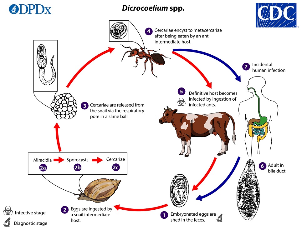
A case who was suffering from abdominal pain and chronic diarrhea refered to Isfahan Health Training and Research Center, Institute of Public Health. Current treatments for chronic diarrhea incloding a traial of gluten fre diet were performed but these were not effective because he was infected by Dicrocoelium dentriticum. Then he was cured with parasitic treatment. (+info)Dicrocoeliasis is a parasitic infection caused by the fluke Dicrocoelium dendriticum, also known as the lancet liver fluke. This small flatworm infects the bile ducts of the liver in its definitive host, which are usually herbivorous animals such as sheep and cattle. Humans can become accidental hosts by ingesting contaminated vegetation or water that contains the encysted larval stage of the fluke.
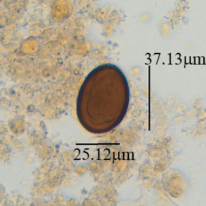
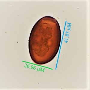
The infection is typically asymptomatic or causes only mild symptoms, such as abdominal discomfort, diarrhea, and fatigue. However, in severe cases, it can lead to liver damage and other complications. The diagnosis of dicrocoeliasis is usually made by detecting the presence of the parasite's eggs in the stool or through imaging techniques such as ultrasound or CT scan. Treatment typically involves the use of anthelmintic medications that are effective against flukes, such as praziquantel or triclabendazole.
 Dicrocoelium dendriticum
Dicrocoelium hospes
Trematoda
Gastropod-borne parasitic disease
List of MeSH codes (C03)
Dicrocoelium dendriticum - Wikipedia
 Trematode Infection: Background, Pathophysiology, Epidemiology
 CDC - DPDx - Parasites A-Z Index
Pinworm infection - Wikipedia
 Albendazole 500mg tablets for SALE | Zentel analog | Pet Meds Online
 Disease - di Index | CureHunter
 LXR-like Receptors
 Generika Levothyroxine Levothyroxin Ohne Rezept Kaufen, Wirkstoff levothyroxine levothyroxin synthroid euthyrox thyrex tirosint...
 Dicrotic wave: Definition with Dicrotic wave Pictures and Photos
 Cutaneous larva migrans - WikiProjectMed
Metagonimus takahashii - WikiProjectMed
Nematode - Wikipedia
 MeSH Browser
Diseases1
- Dicrocoeliasis: Global Status is one in a series of GIDEON ebooks which explore all individual infectious diseases, drugs, vaccines, outbreaks, surveys and pathogens in every country of the world. (gideononline.com)