Duodenal Obstruction
Gastric Outlet Obstruction
Duodenal Diseases
Intestinal Atresia
Intestinal Obstruction
Polyhydramnios
Brunner Glands
Retroperitoneal Fibrosis
Duodenum
Ampulla of Vater
Ureteral Obstruction
Food entrapped in papilla of Vater: uncommon cause of vomiting. (1/119)
CASE REPORT: A 20 month old girl was admitted for intractable vomiting over several days, with no other symptoms. Family and personal history were not contributive. Clinical and neurological examination, and routine blood tests and investigations (plain abdominal x ray, upper gastrointestinal tract contrast study, abdominal ultrasonography) were normal. The upper gastrointestinal endoscopy showed a mild antral gastritis and the second portion of duodenum was occupied by a tough, fibrous mass partially embedded into the papilla of Vater. The foreign body was removed and proved to be vegetable fibre (pineapple). Symptoms subsided immediately and the child was discharged with gastroprotective therapy. After two months, on endoscopic examination, the signs of gastropathy had cleared; the papilla of Vater was undamaged, but unchomped food debris was again found in the duodenum. DISCUSSION: There are sporadic reports of foreign bodies retained into the papilla of Vater, all of them in adults. This child, though her papilla was tiny, had no jaundice or pancreatitis, unlike most of the reported cases. This is the first report of this finding in a child. The cause of the vomiting was not shown on abdominal ultrasonography or contrast study. It should be added to the list of unusual causes of vomiting. (+info)Congenital duodenal diaphragms in adults: a delayed cause of intestinal obstruction. (2/119)
Congenital duodenal diaphragms in the adult are uncommon, unsuspected lesions that infrequently cause intestinal obstruction. The diaphragms may be single or multiple and are usually located near the ampulla of Vater. Three cases are summarized and the recent literature reviewed. At least 35 cases have been reported. Treatment most often consisted of duodenotomy,excision of the web and duodenal closure. (+info)Comparison of upper gastrointestinal toxicity of rofecoxib and naproxen in patients with rheumatoid arthritis. VIGOR Study Group. (3/119)
BACKGROUND: Each year, clinical upper gastrointestinal events occur in 2 to 4 percent of patients who are taking nonselective nonsteroidal antiinflammatory drugs (NSAIDs). We assessed whether rofecoxib, a selective inhibitor of cyclooxygenase-2, would be associated with a lower incidence of clinically important upper gastrointestinal events than is the nonselective NSAID naproxen among patients with rheumatoid arthritis. METHODS: We randomly assigned 8076 patients who were at least 50 years of age (or at least 40 years of age and receiving long-term glucocorticoid therapy) and who had rheumatoid arthritis to receive either 50 mg of rofecoxib daily or 500 mg of naproxen twice daily. The primary end point was confirmed clinical upper gastrointestinal events (gastroduodenal perforation or obstruction, upper gastrointestinal bleeding, and symptomatic gastroduodenal ulcers). RESULTS: Rofecoxib and naproxen had similar efficacy against rheumatoid arthritis. During a median follow-up of 9.0 months, 2.1 confirmed gastrointestinal events per 100 patient-years occurred with rofecoxib, as compared with 4.5 per 100 patient-years with naproxen (relative risk, 0.5; 95 percent confidence interval, 0.3 to 0.6; P<0.001). The respective rates of complicated confirmed events (perforation, obstruction, and severe upper gastrointestinal bleeding) were 0.6 per 100 patient-years and 1.4 per 100 patient-years (relative risk, 0.4; 95 percent confidence interval, 0.2 to 0.8; P=0.005). The incidence of myocardial infarction was lower among patients in the naproxen group than among those in the rofecoxib group (0.1 percent vs. 0.4 percent; relative risk, 0.2; 95 percent confidence interval, 0.1 to 0.7); the overall mortality rate and the rate of death from cardiovascular causes were similar in the two groups. CONCLUSIONS: In patients with rheumatoid arthritis, treatment with rofecoxib, a selective inhibitor of cyclooxygenase-2, is associated with significantly fewer clinically important upper gastrointestinal events than treatment with naproxen, a nonselective inhibitor. (+info)Retroperitoneal fibrosis with duodenal stenosis. (4/119)
Retroperitoneal fibrosis is a rare disease characterized by the formation of dense plaque of fibrous tissue covering the retroperitoneal structures. This disease is commonly presented as ureteral obstruction, but the involvement of duodenum is rare. We report a case of retroperitoneal fibrosis which was complicated with duodenal stenosis and was successfully treated with corticosteroids. A 58-yr-old man, who had history of aorto-iliac bypass graft due to arteriosclerosis obliterans with infrarenal aortic occlusion was admitted to the hospital with abdominal pain and a mass. Abdominal CT scan revealed the periaortic soft tissue mass encircling grafted aorta and stenosis of duodenal third portion. Retroperitoneal fibrosis with duodenal stenosis was diagnosed and prednisolone therapy was initiated. Follow-up CT scan showed that the patient responded to prednisolone therapy with eased pain, shrinking periaortic mass, and reduced duodenal stenosis. (+info)Unique cause of duodenal obstruction by an abdominal aortic aneurysm. (5/119)
We report herein a unique cause of duodenal obstruction secondary to expansion of an abdominal aortic aneurysm in a 75-year-old man with congenital malrotation of the intestines. The duodenum was found to be compressed between the abdominal aortic aneurysm inferiorly and the peritoneal band superiorly. The patient underwent uncomplicated lysis of peritoneal bands relieving the duodenal obstruction, followed by repair of the abdominal aortic aneurysm. (+info)Bouveret's syndrome complicated by distal gallstone ileus after laser lithotropsy using Holmium: YAG laser. (6/119)
BACKGROUND: Bouveret's syndrome is an unusual presentation of duodenal obstruction caused by the passage of a large gallstone through a cholecystoduodenal fistula. Endoscopic therapy has been used as first-line treatment, especially in patients with high surgical risk. CASE PRESENTATION: We report a 67-year-old woman who underwent an endoscopic attempt to fragment and retrieve a duodenal stone using a Holmium: Yttrium-Aluminum-Garnet Laser (Ho:YAG) which resulted in small bowel obstruction. The patient successfully underwent enterolithotomy without cholecystectomy or closure of the fistula. CONCLUSION: We conclude that, distal gallstone obstruction, due to migration of partially fragmented stones, can occur as a possible complication of laser lithotripsy treatment of Bouveret's syndrome and might require urgent enterolithotomy. (+info)Groove pancreatitis: report of a case and review of the clinical and radiologic features of groove pancreatitis reported in Japan. (7/119)
We report a case of groove pancreatitis in which a hypoechoic mass between the duodenum and pancreas head was clearly imaged, and narrowing of the supra-ampullary area of the duodenum and bile duct stenosis were also found. The diagnosis was confirmed by surgery. Microscopic examination showed extensive scarring between the duodenum and pancreas head. Protein plugs were found in Santorini's duct. We consider that the disturbance of the pancreatic juice outflow in Santorini's duct is one of the important pathogenic factors in the development of groove pancreatitis. Therefore, we emphasize the finding of Santorini's duct in the differential diagnosis of groove pancreatitis. (+info)Duodenal stents for malignant duodenal strictures. (8/119)
Duodenal obstruction may be caused by inoperable malignant disease. Symptoms of nausea and vomiting have been traditionally palliated by surgery. The aim of the study was to determine the efficacy of the endoscopic placement of metal self expanding duodenal stents for the palliation of malignant duodenal obstruction. Four patients with malignant gastric outlet obstruction are described. One patient had a history of oesophagectomy for oesophageal adenocarcinoma and presented with further dysphagia. At endoscopy the recurrent oesophageal tumour and an adenocarcinoma involving the pylorus were both stented. In the other three patients there was a previous history of colonic carcinoma, cholangiocarcinoma and oesophageal adenocarcinoma respectively. All four patients were successfully stented with good palliation of their symptoms. Duodenal Wallstents are a useful alternative to surgery in patients with inoperable malignant duodenal obstruction or those who are unfit for surgery. (+info)Duodenal obstruction is a medical condition characterized by the blockage or impediment of the normal flow of contents through the duodenum, which is the first part of the small intestine. This blockage can be partial or complete and can be caused by various factors such as:
1. Congenital abnormalities: Duodenal atresia or stenosis, where there is a congenital absence or narrowing of a portion of the duodenum.
2. Inflammatory conditions: Duodenitis, Crohn's disease, or tumors that cause swelling and inflammation in the duodenum.
3. Mechanical obstructions: Gallstones, tumors, strictures, or adhesions (scar tissue) from previous surgeries can physically block the duodenum.
4. Neuromuscular disorders: Conditions like progressive systemic sclerosis or amyloidosis that affect the neuromuscular function of the intestines can lead to duodenal obstruction.
Symptoms of duodenal obstruction may include nausea, vomiting (often with bilious or fecal matter), abdominal pain, distention, and decreased bowel movements. Diagnosis typically involves imaging studies such as X-rays, CT scans, or upper gastrointestinal series to visualize the blockage. Treatment depends on the underlying cause but may involve surgery, endoscopic procedures, or medications to manage symptoms and address the obstruction.
Gastric outlet obstruction (GOO) is a medical condition that refers to the blockage of the passage from the stomach to the small intestine, also known as the pylorus. This blockage can be caused by various factors, including tumors, scar tissue, or gallstones. As a result, food and digestive enzymes cannot pass through the pylorus into the small intestine, leading to symptoms such as vomiting, abdominal pain, bloating, and weight loss. In severe cases, GOO can lead to malnutrition, dehydration, and other complications if left untreated. Treatment options for GOO depend on the underlying cause of the obstruction and may include medication, endoscopic procedures, or surgery.
Duodenal diseases refer to a range of medical conditions that affect the duodenum, which is the first part of the small intestine. Here are some examples of duodenal diseases:
1. Duodenitis: This is inflammation of the duodenum, which can cause symptoms such as abdominal pain, nausea, vomiting, and bloating. Duodenitis can be caused by bacterial or viral infections, excessive use of nonsteroidal anti-inflammatory drugs (NSAIDs), or chronic inflammation due to conditions like Crohn's disease.
2. Peptic ulcers: These are sores that develop in the lining of the duodenum, usually as a result of infection with Helicobacter pylori bacteria or long-term use of NSAIDs. Symptoms can include abdominal pain, bloating, and heartburn.
3. Duodenal cancer: This is a rare type of cancer that affects the duodenum. Symptoms can include abdominal pain, weight loss, and blood in the stool.
4. Celiac disease: This is an autoimmune disorder that causes the immune system to attack the lining of the small intestine in response to gluten, a protein found in wheat, barley, and rye. This can lead to inflammation and damage to the duodenum.
5. Duodenal diverticulosis: This is a condition in which small pouches form in the lining of the duodenum. While many people with duodenal diverticulosis do not experience symptoms, some may develop complications such as inflammation or infection.
6. Duodenal atresia: This is a congenital condition in which the duodenum does not form properly, leading to blockage of the intestine. This can cause symptoms such as vomiting and difficulty feeding in newborns.
Intestinal atresia is a congenital condition characterized by the absence or complete closure of a portion of the intestine, preventing the passage of digested food from the stomach to the remaining part of the intestines. This results in a blockage in the digestive system, which can be life-threatening if not treated promptly after birth. The condition can occur anywhere along the small or large intestine and may affect either a single segment or multiple segments of the intestine.
There are several types of intestinal atresia, including:
1. Jejunal atresia: A closure or absence in the jejunum, a part of the small intestine located between the duodenum and ileum.
2. Ileal atresia: A closure or absence in the ileum, the lower portion of the small intestine that connects to the large intestine (cecum).
3. Colonic atresia: A closure or absence in the colon, a part of the large intestine responsible for storing and eliminating waste.
4. Duodenal atresia: A closure or absence in the duodenum, the uppermost portion of the small intestine that receives chyme (partially digested food) from the stomach.
5. Multiple atresias: When more than one segment of the intestines is affected by atresia.
The exact cause of intestinal atresia remains unclear, but it is believed to be related to disruptions in fetal development during pregnancy. Treatment typically involves surgical correction to reconnect the affected segments of the intestine and restore normal digestive function. The prognosis for infants with intestinal atresia depends on the severity and location of the atresia, as well as any associated conditions or complications.
Duodenostomy is a surgical procedure that creates an opening (stoma) into the duodenum, which is the first part of the small intestine. This procedure is typically performed to divert the flow of digestive secretions and contents away from a diseased or obstructed area of the gastrointestinal tract.
A duodenostomy may be created as a temporary measure to allow a portion of the intestine to heal or as a permanent solution for conditions such as chronic inflammatory bowel disease, cancer, or congenital abnormalities. The stoma can be located on the abdominal wall, allowing for the external drainage of digestive secretions and contents into a collection bag.
It is important to note that the specific medical definition and indications for duodenostomy may vary based on individual clinical context and patient needs. Therefore, it is always best to consult with a healthcare professional or medical expert for accurate information.
Intestinal obstruction, also known as bowel obstruction, is a medical condition characterized by a blockage that prevents the normal flow of contents through the small intestine or large intestine (colon). This blockage can be caused by various factors such as tumors, adhesions (scar tissue), hernias, inflammation, or impacted feces.
The obstruction can be mechanical, where something physically blocks the intestinal lumen, or functional, where the normal muscular contractions of the bowel are impaired. Mechanical obstructions are more common than functional ones.
Symptoms of intestinal obstruction may include abdominal pain and cramping, nausea and vomiting, bloating, inability to pass gas or have a bowel movement, and abdominal distention. If left untreated, intestinal obstruction can lead to serious complications such as tissue death (necrosis), perforation of the intestine, and sepsis. Treatment typically involves hospitalization, intravenous fluids, nasogastric decompression, and possibly surgery to remove the obstruction.
Polyhydramnios is a medical condition characterized by an excessive accumulation of amniotic fluid in the sac surrounding the fetus during pregnancy, typically defined as an amniotic fluid index (AFI) greater than 24 cm or a single deepest pocket (SDP) measurement of more than 8 cm. It occurs in approximately 1-2% of pregnancies and can be associated with various maternal, fetal, and genetic conditions. If left untreated, polyhydramnios may increase the risk of premature labor, premature rupture of membranes, and other pregnancy complications. Proper diagnosis and management are essential to ensure a healthy pregnancy outcome.
Brunner glands, also known as submucosal glands of Brunner, are tubulo-acinar exocrine glands located in the submucosa of the duodenum, which is the first part of the small intestine. These glands secrete alkaline mucus that helps neutralize the acidic chyme (partially digested food) entering from the stomach, providing a more favorable environment for the enzymes involved in nutrient absorption and protecting the duodenal mucosa from acid-induced damage.
Retroperitoneal fibrosis (RPF) is a rare and progressive condition characterized by the abnormal growth of fibrous tissue in the retroperitoneal space, which is the area behind the peritoneum (the lining that covers the abdominal cavity). This fibrous tissue can encase and compress vital structures such as the ureters, blood vessels, and nerves, leading to various symptoms.
RPF can be idiopathic (without a known cause) or secondary to other conditions like infections, malignancies, autoimmune diseases, or medications. The exact pathogenesis of RPF is not fully understood, but it's believed that an abnormal immune response and inflammation play significant roles in its development.
Symptoms of RPF may include:
1. Flank pain or back pain
2. Renal insufficiency or kidney failure due to ureteral compression
3. Hydronephrosis (dilatation of the renal pelvis and calyces)
4. Deep vein thrombosis (DVT) or pulmonary embolism (PE) due to vascular compression
5. Neurological symptoms due to nerve compression
6. Weight loss, fatigue, and fever (in some cases)
Diagnosis of RPF typically involves imaging studies such as computed tomography (CT) scans or magnetic resonance imaging (MRI), along with laboratory tests and sometimes biopsy for confirmation. Treatment options depend on the underlying cause but generally involve immunosuppressive medications, corticosteroids, and surgical intervention in severe cases.
Duodenal neoplasms refer to abnormal growths in the duodenum, which is the first part of the small intestine that receives digestive secretions from the pancreas and bile duct. These growths can be benign or malignant (cancerous).
Benign neoplasms include adenomas, leiomyomas, lipomas, and hamartomas. They are usually slow-growing and do not spread to other parts of the body. However, they may cause symptoms such as abdominal pain, bleeding, or obstruction of the intestine.
Malignant neoplasms include adenocarcinomas, neuroendocrine tumors (carcinoids), lymphomas, and sarcomas. They are more aggressive and can invade surrounding tissues and spread to other parts of the body. Symptoms may include abdominal pain, weight loss, jaundice, anemia, or bowel obstruction.
The diagnosis of duodenal neoplasms is usually made through imaging tests such as CT scans, MRI, or endoscopy with biopsy. Treatment depends on the type and stage of the tumor and may include surgery, chemotherapy, radiation therapy, or a combination of these modalities.
The duodenum is the first part of the small intestine, immediately following the stomach. It is a C-shaped structure that is about 10-12 inches long and is responsible for continuing the digestion process that begins in the stomach. The duodenum receives partially digested food from the stomach through the pyloric valve and mixes it with digestive enzymes and bile produced by the pancreas and liver, respectively. These enzymes help break down proteins, fats, and carbohydrates into smaller molecules, allowing for efficient absorption in the remaining sections of the small intestine.
The ampulla of Vater, also known as hepatopancreatic ampulla, is a dilated portion of the common bile duct where it joins the main pancreatic duct and empties into the second part of the duodenum. It serves as a conduit for both bile from the liver and digestive enzymes from the pancreas to reach the small intestine, facilitating the digestion and absorption of nutrients. The ampulla of Vater is surrounded by a muscular sphincter, the sphincter of Oddi, which controls the flow of these secretions into the duodenum.
Airway obstruction is a medical condition that occurs when the normal flow of air into and out of the lungs is partially or completely blocked. This blockage can be caused by a variety of factors, including swelling of the tissues in the airway, the presence of foreign objects or substances, or abnormal growths such as tumors.
When the airway becomes obstructed, it can make it difficult for a person to breathe normally. They may experience symptoms such as shortness of breath, wheezing, coughing, and chest tightness. In severe cases, airway obstruction can lead to respiratory failure and other life-threatening complications.
There are several types of airway obstruction, including:
1. Upper airway obstruction: This occurs when the blockage is located in the upper part of the airway, such as the nose, throat, or voice box.
2. Lower airway obstruction: This occurs when the blockage is located in the lower part of the airway, such as the trachea or bronchi.
3. Partial airway obstruction: This occurs when the airway is partially blocked, allowing some air to flow in and out of the lungs.
4. Complete airway obstruction: This occurs when the airway is completely blocked, preventing any air from flowing into or out of the lungs.
Treatment for airway obstruction depends on the underlying cause of the condition. In some cases, removing the obstruction may be as simple as clearing the airway of foreign objects or mucus. In other cases, more invasive treatments such as surgery may be necessary.
Ureteral obstruction is a medical condition characterized by the partial or complete blockage of the ureter, which is the tube that carries urine from the kidney to the bladder. This blockage can be caused by various factors such as kidney stones, tumors, blood clots, or scar tissue, leading to a backup of urine in the kidney (hydronephrosis). Ureteral obstruction can cause pain, infection, and potential kidney damage if not treated promptly.
 Annular pancreas
Megaduodenum
Duodenal atresia
Intestinal atresia
Pancreatic duct
Self-expandable metallic stent
Intestinal malrotation
Pythiosis
Phycomycosis
Duodenal switch
Vagotomy
Sylvia Rexach
Stump blow-out
Equine gastric ulcer syndrome
Pyloric stenosis
SADI-S surgery
Hereditary pancreatitis
Common bile duct stone
Bile
Vomiting
Lester Dragstedt
Enterolith
Pneumoperitoneum
Eosinophilic gastroenteritis
Acute pancreatitis
Differential diagnoses of anorexia nervosa
Periampullary cancer
List of MeSH codes (C06)
Double bubble (radiology)
Abdominal pain
Neonatal bowel obstruction
Polyhydramnios
Gastric outlet obstruction
 Neonatal Duodenal Obstruction: Postoperative Feeding
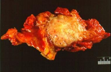
 Brunner's gland adenoma: unusual cause of duodenal haemorrhage and obstruction | HKMJ
 Pediatric Small Bowel Obstruction: Background, Pathophysiology, Etiology
 Duodenal Obstruction - Pediatrics - MSD Manual Professional Edition
 "Malignant Duodenal Obstruction Caused by Urothelial Carcinoma" by Dylan Johnson, Ankita Mishra et al.
 D
D
Annular pancreas - Wikipedia
 Endoscopic ultrasound-guided gastroenterostomy versus duodenal stenting for malignant gastric outlet obstruction: an...
 Cryptogenic multifocal ulcerous stenosing enteritis (CMUSE) in a man with a diagnosis of X-linked reticulate pigmentary...
 Is a Nonspecific Bowel Gas Pattern Normal? Causes & Meaning
 Related Articles - Annals Singapore
KoreaMed
 Fully covered metal biliary stents: A review of the literature
 Ouadii Mouaqit - Articles - Scientific Research Publishing
 2014 DPDx Case Studies
Pancreatic Cancer: Practice Essentials, Pathophysiology, Etiology
 IBD Treatment
 Table of Contents - December 03, 1966, 95 (23) | CMAJ
 SPED - Edição 2022
SPED - Edição 2022
 Pancreatic disorders | PPT
 Duodenal Switch Forum (DS) - Page 415
Pathology of Sudden Natural Death: Overview, Terminology, Medical Examiner Role and Autopsy Indications
 Bile Reflux: Symptoms, Treatment, Causes & What It Is
Table of contents | Archives of Disease in Childhood
Duodenoduodenostomia laparoscópica para el tratamiento de la obstrucción duodenal congénita
Surgical Bypass versus Endoscopic Stenting for Unresectable Head Pancreatic Cancer, Which Palliative Treatment Is Better in...
Gastrointestinal foreign body obstruction is not associated with abnormal point-of-care pancreas-specific lipase test results...
 Fundoplication and postoperative management. - Health & Disability Commissioner
 Pancreatic Cancer | Durham, Raleigh, NC | Duke Health
Duodenum8
- Duodenal atresia is due to the failure of canalization of the embryonic duodenum. (msdmanuals.com)
- He was found to have an obstruction of the duodenum and hepatobiliary system that was highly suggestive of malignancy. (hcahealthcare.com)
- Upper GI series may be suggestive of annular pancreas, especially if they show a duodenal narrowing of the second portion of the duodenum and the concomitant dilatation of the proximal duodenum. (wikipedia.org)
- An abdominal CT scan or an MRI allows to highlight the narrowing of the descending duodenal tract and the ring of pancreatic tissue surrounding the duodenum: this ring can be complete or, in patients with an incomplete annular pancreas, extended in a postero-lateral or anterolateral direction with respect to the second part of the duodenum. (wikipedia.org)
- In neonates, treatment for relief of obstruction usually is bypassing the obstructed segment of duodenum by duodeno-jejunostomy. (wikipedia.org)
- Computed tomography (CT) scan showed in the third duodenal segment the presence of an area with the characteristics of inflammatory tissue, including air bubbles between the duodenum and aortic-bi-femoral prosthesis adherent to the third duodenal portion. (sciencedaily.com)
- WallFlex Colonic and Duodenal Soft Stent Systems are designed to be a more flexible option to relieve malignant obstructions of the colon and duodenum. (bostonscientific.com)
- The terminal part of the CBD is joined by the terminal part of the pancreatic duct in the pancreatic head to form a common channel (called the biliopancreatic ampulla when dilated), which runs through the medial duodenal wall and opens on the dome of the major duodenal papilla, a nipplelike projection on the medial wall of the middle segment of the second part (C loop) of the duodenum. (medscape.com)
Bowel20
- Pediatric small bowel obstructions have a variable etiology, with processes that can be divided into acute intestinal obstructions and chronic, partial intestinal obstructions. (medscape.com)
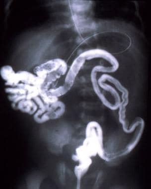
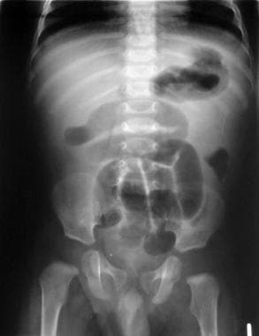
- Small bowel obstruction is visible on a plain radiograph caused by intussusception in a 5-month-old patient. (medscape.com)
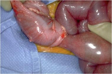
- The surgical photograph depicts a transition zone in a patient with small bowel obstruction. (medscape.com)
- Intrinsic or extrinsic small bowel obstruction leads to accumulating secretions that dilate the intestine proximal to the obstruction. (medscape.com)
- This is followed by venous obstruction, which accelerates edema formation because blood can enter the affected bowel segment but does not have a drainage route. (medscape.com)
- This sequence may occur more rapidly in a closed-loop obstruction because there is no proximal escape for pressurized bowel contents. (medscape.com)
- A sampling of the many etiologies that can cause small bowel obstruction in children are discussed in greater detail below. (medscape.com)
- however, it doesn't meet the criteria for a more precise diagnosis, such as a small bowel obstruction. (medicinenet.com)
- According to a study, there are some patients whose abdominal radiographs do not exhibit 'normal,' 'possible small intestinal obstruction ,' or 'definite small bowel obstruction' gas patterns. (medicinenet.com)
- The definition of a " possible " small bowel obstruction (SBO) pattern is unequivocally the presence of dilated numerous gaseous and/or fluid-filled loops of the small intestine with a mild quantity of colonic gas, but the degree of distention of the small intestine relative to the colon is insufficient to render a conclusive diagnosis. (medicinenet.com)
- She was referred and admitted to Tauranga Hospital with a small bowel obstruction. (hdc.org.nz)
- Ms A's bowel obstruction settled and resolved during the admission, and she was discharged on 8 October. (hdc.org.nz)
- More rarely, the clinical picture of ADF is subtle, presenting as an obstructive syndrome, and in these cases the principal goal is to effectively relieve the mechanical bowel obstruction. (sciencedaily.com)
- SEMS vs cSEMS in duodenal and small bowel obstruction: High risk of migration in the covered stent group. (wjgnet.com)
- There were 18 postoperative bowel obstructions, 8 anastamotic leaks, and 4 occurrences of postoperative bleeding. (sages.org)
- The aim of this report is to present a case of bowel obstruction caused by A. lumbricoides as a cause of acute abdomen in one governorate in Egypt. (who.int)
- On the radiograph an obstruction can only be diagnosed if the bowel has had sufficient time to become air-filled after birth. (radiologyassistant.nl)
- however, there was also a distal small bowel obstruction unrelated to this mesh. (hindawi.com)
- His small bowel obstruction was not strangulated and resolved several days later. (hindawi.com)
- 1.2 billion infections to present a case of bowel obstruction raised (13 × 103/µL), and eosinophils globally [1]. (who.int)
Gastric outlet obst4
- Endoscopic ultrasound-guided gastroenterostomy versus duodenal stenting for malignant gastric outlet obstruction: an international, multicenter, propensity score-matched comparison. (bvsalud.org)
- Endoscopic duodenal stenting is the current standard treatment for malignant gastric outlet obstruction (GOO) in patients with limited life expectancy . (bvsalud.org)
- Evidence of gastric outlet obstruction may also be seen. (medscape.com)
- This article summarizes surgical, endoscopic, and other palliative techniques for relief of obstructive jaundice, relief of duodenal or gastric outlet obstruction, and relief of pain due to invasion of the celiac plexus. (ahrq.gov)
Neonatal Duodenal Obstruction1
- Intrahepatic gas shadows in neonatal duodenal obstruction. (bmj.com)
Ulcer3
- Unhealed wound, active gastric or duodenal ulcer, or bone fracture. (uclahealth.org)
- The most common causes of acute abdomen are acute appendicitis, acute peptic ulcer, acute cholecystitis, acute pancreatitis, intestinal obstruction, acute peritonitis and acute pyelonephritis [8]. (who.int)
- Clinical Features Duodenal ulcer: пїЅ Epigastric pain, sometimes at evening and when hungry пїЅ May current for the first time with problems (see later on this part) пїЅ Wide particular person variation in symptoms and meals that give pain пїЅ 95% of duodenal ulcers are brought on by Helicobacter pylori (H insomnia audien [url=http://www.firenzesalem.com/medicine/order-sominex-online/]sominex 25 mg cheap amex[/url]. (ehd.org)
Biliary obstruction2
- Purpose To evaluate the factors that predict symptomatic dislodgement of a percutaneous transhepatic biliary drainage (PTBD) catheter in patients with malignant biliary obstruction. (koreamed.org)
- Duodenal malignancies presenting with jaundice due to extrahepatic biliary obstruction occur in 43% of cases, but the exact prevalence of obstructive jaundice in duodenal lymphoma is unknown, although there are a few cases reported [5]. (eurorad.org)
Malignant4
- Malignant Duodenal Obstruction Caused by Urothelial Carcinoma" by Dylan Johnson, Ankita Mishra et al. (hcahealthcare.com)
- We compared clinical success, safety , and stent dysfunction of EUS-GE and duodenal stenting in patients with malignant GOO using propensity score matching. (bvsalud.org)
- EUS-GE had higher clinical success and lower stent dysfunction, with similar safety , compared with duodenal stenting, suggesting that EUS-GE may be preferred over duodenal stenting in patients with malignant GOO. (bvsalud.org)
- Only 12% of all malignant duodenal neoplasms are duodenal lymphomas [3]. (eurorad.org)
Stent2
- However, duodenal stenting is prone to stent dysfunction. (bvsalud.org)
- When cancer blocks the outlet of the stomach, one option is to place a stent across the obstruction. (dukehealth.org)
Stenosis3
- Single-center electronic medical records for all newborns with duodenal atresia and stenosis admitted to a university surgical centre between January 1997 and September 2021 were reviewed. (physiciansweekly.com)
- Escobar MA, Ladd AP, Grosfeld JL, et al: Duodenal atresia and stenosis: long-term follow-up over 30 years. (medigraphic.com)
- McCollum MO, Jamieson DH, Webber EM: Annular pancreas and duodenal stenosis. (medigraphic.com)
Proximal2
- In some cases it is possible to have signs of inverse peristalsis of the duodenal tract which is proximal to the narrowing caused by the annular pancreas, and the dilatation of the duodenal portion distal to the anomaly. (wikipedia.org)
- High obstructions are defined as proximal to the ileum. (radiologyassistant.nl)
Laparoscopic5
- In adults, due to the minor duodenal mobility, the approach is laparoscopic gastrojejunostomy or duodenojejunostomy. (wikipedia.org)
- Rothenberg SS: Laparoscopic duodenoduodenostomy for duodenal obstruction in infants and children. (medigraphic.com)
- Steyaert H, Valla JS, Van Hoorde E: Diaphragmatic duodenal atresia: Laparoscopic repair. (medigraphic.com)
- The aim of this study is to describe perioperative and follow-up outcomes of laparoscopic pancreaticoduodenectomy (LPD) for Duodenal GIST. (sages.org)
- Laparoscopic pancreaticoduodenectomy is a safe, feasible and effective surgical procedure for duodenal GIST. (sages.org)
Pancreatic3
- This duodenal thickening caused biliary and pancreatic ducts dilatation. (eurorad.org)
- Most gastrinomas present as the gastrinoma triangle, which includes the porta hepatis, duodenal sweep, and pancreatic head. (medscape.com)
- A smooth muscle sphincter (of Oddi) is present around the common channel of the CBD and the pancreatic duct and prevents reflux of duodenal juice into the 2 ducts. (medscape.com)
Diagnosis3
- Diagnosis of duodenal atresia is suspected prenatally if there is polyhydramnios and/or a dilated stomach. (msdmanuals.com)
- The indication for surgery was intestinal obstruction (244), abdominal pain (53), unclear diagnosis (14), duodenal obstruction (9), abscess (11), and peritonitis (2). (sages.org)
- Early diagnosis of intestinal obstruction caused by A. lumbricoides using ultrasonography is very useful to avoid its serious and lethal complications [10]. (who.int)
Papilla4
- Early signs of abnormality include polyhydramnios (an excess of amniotic fluid), low birth weight, and feeding intolerance immediately after birth, in particular a tendency to develop epigastric distention associated with non-biliary vomiting (the obstruction is generally above the papilla of Vater, therefore superior to the junction with the bile ducts). (wikipedia.org)
- An ulcerative tumor was found around the duodenal papilla region by upper esophagogastroduodenoscopy (Figure 1(c) ). (hindawi.com)
- The site of the greater duodenal papilla marks the junction of the embryologic foregut and midgut. (medscape.com)
- The greater duodenal papilla is covered by a semicircular hoodlike mucosal fold superiorly. (medscape.com)
Ulcers1
- Upper GI series findings may reveal esophageal stricture, duodenal ulcers, duodenal strictures, and hypertrophic gastric and duodenal folds. (medscape.com)
Gastrointestinal7
- Overview of Congenital Gastrointestinal Anomalies Most congenital gastrointestinal (GI) anomalies present as intestinal obstruction and frequently manifest as feeding difficulties, distention, emesis, and an inability to pass gas and stool. (msdmanuals.com)
- Duodenal atresia is the 2nd most common atresia of the gastrointestinal (GI) tract. (msdmanuals.com)
- Maxwell EA , Dugat DR , Waltenburg M , Outcomes of dogs undergoing immediate or delayed surgical treatment for gastrointestinal foreign body obstruction: a retrospective study by the Society of Veterinary Soft Tissue Surgery . (avma.org)
- Aorto-duodenal fistulae constitute 80 percent of aorto-enteric fistulae, presenting with upper gastrointestinal bleeding. (sciencedaily.com)
- Aorto-duodenal fistulae (ADF) are the most frequent aorto-enteric fistulae (80%) and the most frequent presenting sign of ADF is upper gastrointestinal bleeding (UGI). (sciencedaily.com)
- When the symptoms are present immediately after birth, the most common cause is a gastrointestinal obstruction. (radiologyassistant.nl)
- In this article we will discuss the congenital gastrointestinal obstructions and also some acquired diseases that present as an acute abdomen in the neonate. (radiologyassistant.nl)
Annular1
- Unfortunately, this double-bubble sign is not pathognomonic for annular pancreas, as it can also be observed in other conditions, such as duodenal atresia and intestinal malrotation. (wikipedia.org)
Bile duct1
- if you have a bile duct obstruction. (stfrancisherbfarm.com)
Lumen4
- Esophago-gastro-duodenoscopy showed the aortic prosthesis crossing the third segment of duodenal wall occluding the intestinal lumen. (sciencedaily.com)
- Aneurysmal dilatation of the lumen may be seen and excludes other more common causes of duodenal wall neoplastic infiltration, namely adenocarcinoma. (eurorad.org)
- Emergency surgical treatment may be necessary in acute intestinal obstruction in which the mass of the parasite obstructs the intestinal lumen or intestinal obstruction develops due to volvulus [11]. (who.int)
- An esophagogastroduodenoscopy (EGD) revealed the Teflon felt within the lumen of the duodenal bulb with cleanly ulcerated mucosa. (hindawi.com)
Abdominal pain1
- Intestinal obstruction should be suspected in any child with persistent vomiting, abdominal distention, and abdominal pain. (medscape.com)
Mucosal1
- 3. Patients should be able to have an oral intake and not be dependant on tube feeding, or have significant oropharyngeal obstruction, gastro-duodenal obstruction, or oral mucosal inflammation interfering with oral intake. (knowcancer.com)
Anomaly2
- This anomaly occurs less commonly than duodenal atresia but manifests in a similar fashion and requires surgery. (msdmanuals.com)
- Preduodenal portal vein, which was first reported by knight in 1921, is extremely rare congenital anomaly and may cause duodenal obstruction. (koreamed.org)
Complete obstruction2
- Massive dilatation is seen in complete obstruction and is accompanied by fluid levels on the dorsal decubitus radiograph. (radiologyassistant.nl)
- However, the endoscope was able to move distally past the mesh indicating the absence of complete obstruction by the mesh. (hindawi.com)
Small intestine3
- It creates a connection between the stomach and a healthy portion of the small intestine, bypassing the obstruction. (dukehealth.org)
- Acute intestinal obstruction or subobstruction, history of inflammatory intestinal disease or extended resection of the small intestine. (uclahealth.org)
- We evaluated the myoelectrical and motor function of the esophagus, small intestine, colon and anal sphincter in four patients with chronic idiopathic intestinal pseudo-obstruction. (nih.gov)
Infants2
- Intussusception is the most common cause of intestinal obstruction in infants and children aged 3 months to 6 years. (medscape.com)
- Postnatally, infants with duodenal atresia present with feeding difficulties and emesis that may be bilious. (msdmanuals.com)
Postoperative2
- Oral or incremental gavage-fed rates of at least 2.5 ml/kg in stepwise increments and numerous steps per day are totally viable in the postoperative feeding plans of neonates with congenital duodenal abnormalities. (physiciansweekly.com)
- Gavopoulos S, Limas C, Avizoglou, et al: Operative and postoperative management of congenital duodenal obstruction: a 10 year experience. (medigraphic.com)
Surgery4
- For a study, researchers sought to determine whether starting small volume bolus feeds was more practical than starting oral or large volume(s) gavage feeds following surgery for congenital duodenal abnormalities. (physiciansweekly.com)
- Duplication of the portal vein or a preduodenal portal vein are hazards at the time of biliary or duodenal surgery, or liver transplantation. (koreamed.org)
- Bypass surgery is done in some cases to relieve the biliary and duodenal obstruction [4]. (eurorad.org)
- The two most common issues following Weight loss Surgery or Duodenal Switch may be albumin level and Vitamin D level. (dssurgery.com)
Intrinsic1
- Spigland N, Yazbeck S: Complications associated with surgical treatment of congenital intrinsic duodenal obstruction. (medigraphic.com)
Lymphoma1
- The CT appearance of duodenal lymphoma is variable: aneurysmal, constrictive, nodular and ulcerative infiltration. (eurorad.org)
Perforation2
- Undiagnosed or improperly managed obstructions can progress to intestinal ischemia, which in turn can progress to necrosis, perforation, and sepsis when left untreated. (medscape.com)
- Subsequently, a study by Sharma et al documented the use of ERAS in emergency settings in patients with perforation and intestinal obstruction and found it to be safe and feasible. (medscape.com)
Anastomosis1
- Diamond shaped anastomosis for duodenal atresia: an experience with 44 patients over 15 years. (medigraphic.com)
Differentiation1
- Although in high obstruction vomiting will be the most striking symptom, whereas in low obstruction this will be constipation, both symptoms are often present concurrently, and the clinical differentiation between a high and a low obstruction is difficult. (radiologyassistant.nl)
Complication2
- The most commonly known serious and lethal complication of A. lumbricoides infection is intestinal obstruction, caused by an aggregated mass of A. lumbricoides worms, which may develop acutely or subacutely [9]. (who.int)
- Malnutrition is one of the most feared complication of the duodenal switch operation. (dssurgery.com)
Retrospective study1
- This international, multicenter, retrospective study analyzed consecutive patients undergoing EUS-GE or duodenal stenting for GOO between 2015 and 2021 in three European centers. (bvsalud.org)
Segment2
- The third or fourth duodenal segment is the most frequently involved site. (sciencedaily.com)
- One of the most common presentations is a diffuse circumferential bulky mass in the duodenal wall, involving a relatively long segment with gradual tapering to a normal mucosa, often associated with regional adenopathies. (eurorad.org)
Portal vein1
- Recently, we experienced a case of preduodenal portal vein associated with dextrocardia, situs inversus, and duodenal obstruction in a 3 days old male newborn and report with review of the references. (koreamed.org)
Carcinoma2
- The patient's duodenal obstruction had been caused by metastatic urothelial carcinoma within the pancreas. (hcahealthcare.com)
- This is the first description of a patient presenting with duodenal obstruction caused by urothelial carcinoma metastasizing to the pancreas. (hcahealthcare.com)
Patients7
- 214 patients underwent EUS-GE (nâ =â 107) or duodenal stenting (nâ =â 107). (bvsalud.org)
- Although pancreatitis, duodenal obstruction, and so on are known as symptoms of AP, approximately two-thirds of the patients are asymptomatic [ 3 ]. (hindawi.com)
- From January 2013 to September 2015, there were 13 patients with duodenal GIST received LPD in a single institution. (sages.org)
- In the study, 13 patients underwent LPD for duodenal GIST , 5 male and 8 female, with a median age of 59 years old. (sages.org)
- The patients did not have a normal increase in duodenal spike or motor activity after intestinal distension, but duodenal activity increased after stimulation with intravenous secretin. (nih.gov)
- Could this be the cause of why I see some patients coming to us for revision of their duodenal switch for malnutrition? (dssurgery.com)
- Computed tomographic enterography should be performed in patients with suspected obstruction before VCE or after negative VCE examinations. (medscape.com)
Ducts1
- Knechtle SJ, Filston HC: Anomalous biliary ducts associated with duodenal atresia. (medigraphic.com)
Radiograph1
- In suspected neonatal obstruction the first step is an abdominal radiograph. (radiologyassistant.nl)
Prosthesis1
- At laparotomy, after viscerolisis, the prosthesis was detached from duodenal wall and the intestine failed to close transversely. (sciencedaily.com)
Incidence1
- The incidence of duodenal location of stromal tumors is rare. (sages.org)