Dysplastic Nevus Syndrome
Nevus
Nevus, Pigmented
Basal Cell Nevus Syndrome
Nevus, Blue
Nevus, Sebaceous of Jadassohn
Lentigo
Melanoma
A locus linked to p16 modifies melanoma risk in Dutch familial atypical multiple mole melanoma (FAMMM) syndrome families. (1/95)
The CDKN2A gene that encodes the cell cycle inhibitor p16 shows mutations in many but not all 9p21-linked melanoma families. Most Dutch melanoma families segregate for a unique founder mutation (p16-Leiden), encoding a truncated nonfunctional p16 protein. The highly variable risk for p16-Leiden carriers to develop melanoma suggests a role for other genetic and/or environmental factors. We hypothesized that a 9p21 gene other than CDKN2A may be relevant in the remaining 9p21-linked melanoma families without p16 mutations but may also act as a risk modifier in p16-Leiden carriers. Haplotype analysis for 9p21 was performed using microsatellite markers in six p16-Leiden families originating from a founder population. p16-Leiden carriers in two families shared an unexpectedly large founder haplotype ( approximately 20-cM) around CDKN2A, mostly in proximal direction. Melanoma-positive p16-Leiden carriers from these families showed this extensive proximal haplotype compared with melanoma-negative p16-Leiden carriers from the same families. Additional p16-Leiden families less heavily affected with melanoma showed shorter haplotypes sharing, excluding the region proximally of CDKN2A. The presence of a gene involved in melanoma susceptibility proximal of CDKN2A is corroborated by somatic deletions of 9p in tumors, which frequently do not include CDKN2A but a more proximal chromosomal area instead. Our results provide a candidate region for further gene mapping in p16-negative 9p21-linked melanoma families and guide the search for risk modifiers in melanoma development. (+info)Alterations in cadherin and catenin expression during the biological progression of melanocytic tumours. (2/95)
AIMS: Compelling evidence from cell culture studies implicates cadherins in the neoplastic progression of melanocytic tumours but few reports describe the expression of cadherins and the related transmembrane proteins, catenins, in a full range of benign and malignant excised melanocytic tumours. METHODS: Using immunohistochemistry and western blotting after tissue fractionation, the pattern of expression of cadherins/catenins was studied in a range of surgically excised melanocytic tumours, from dysplastic naevi to stage III cutaneous metastatic malignant melanoma. RESULTS: Appropriate membranous expression of E-cadherins and P-cadherins is seen in dysplastic naevocytes with an epithelioid phenotype and is largely maintained with malignant transformation to radial growth phase melanoma and primary vertical growth phase malignant melanoma. Loss of membranous E-cadherin is seen in a small number of vertical growth phase melanomas only when metastasis has occurred. However, there is a concomitant dramatic loss of membranous P-cadherin expression in all melanomas at the same stage. A minority of metastatic melanomas show de novo membranous N-cadherin expression in comparison with dysplastic naevi and primary melanoma. Membranous expression of the desmosomal cadherin, desmoglein, was not seen in any tumour studied. Frequently, beta catenin is aberrantly produced in the cytoplasm of cells in dysplastic naevi and metastatic malignant melanoma, with an implied compromise to adhesive function. Furthermore, membranous gamma catenin expression was not seen in any of the 70 melanocytic tumours studied, implying obligatory transmembrane binding of cadherins to beta catenin for maintenance of adhesive function. CONCLUSIONS: The most important alterations in membranous cadherin and catenin expression are seen late in the biological progression of melanocytic tumours at the stage of "in transit" or regional lymph node metastasis, with implications for tumour growth, invasion, and dissemination. (+info)Telomerase activity in melanocytic lesions: A potential marker of tumor biology. (3/95)
Telomerase activation, being a cardinal requirement for immortalization, is a crucial step in the development of malignancy. With a view toward diagnostic and biological aspects in melanocytic neoplasia, we investigated the relative levels of telomerase activity in 72 nevi and 16 malignant melanomas by means of a modified telomeric repeat amplification protocol (TRAP) assay, including an internal amplification standard. We further compared telomerase activity with the expression of two different proliferation-specific proteins, Ki-67 and repp86, a protein expressed exclusively in the cell cycle phases S, G2, and M. Telomerase activity was associated with the overall growth fraction (Ki-67) but showed a closer correlation with the expression of repp86. Both telomerase activity and proliferation indices discriminated clearly between malignant melanomas and nevi, but not between common and dysplastic nevi. Nonetheless, a portion of nevi exhibited markedly elevated telomerase activity levels without proportionally increased proliferation. This was independent of discernible morphological changes. Clinicopathological correlations showed an association between high telomerase activity and early metastatic spread in melanomas, linking telomerase to tumor biology. Our results provide arguments in favor of an occasional progression from nevi to melanomas and imply that proliferation measurements in combination with telomerase assays may help to elicit early malignant transformation that is undetectable by conventional morphology. (+info)CDKN2A mutations in Spanish cutaneous malignant melanoma families and patients with multiple melanomas and other neoplasia. (4/95)
The CDKN2A gene has been implicated in cutaneous malignant melanoma (CMM) in about 40% of families with linkage to chromosome 9p21, while a small proportion of families have mutations in the CDK4 gene. In order to estimate the importance of these genes in the predisposition to CMM in Spanish families and patients we have analysed, by SSCA, a total of 56 subjects belonging to 34 CMM families, and nine patients with multiple CMM and other neoplasia. We have detected germline CDKN2A mutations in six out of the 34 families (17%). A frameshift mutation (358delG) and four missense mutations (G59V, G101W (two cases), D84Y, and R87W) were identified. Five CMM patients from different families (14%) carried the A148T variant, which is known not to affect p16 activity. No mutations were detected in the patients with multiple CMM or other neoplasms. We have not found mutations either in exon 1 beta of the CDKN2A gene or in exon 2A of CDK4. Linkage analysis of the 9p21 region showed exclusion for one of the families for CMM and for four families for CMM/dysplastic naevi. This study indicates a small role for CDKN2A in Spanish CMM families and suggests that other genes are also responsible for CMM predisposition. (+info)Gene-covariate interaction between dysplastic nevi and the CDKN2A gene in American melanoma-prone families. (5/95)
The CDKN2A gene has been implicated in cutaneous malignant melanoma pathogenesis. Although CDKN2A mutations confer substantial risk for melanoma, clinicoepidemiological covariates including dysplastic nevi (DN), total nevi, and solar injury also enhance melanoma risk. To examine the relationship between CDKN2A and these three risk factors, we conducted combined segregation/linkage analysis using the class D regressive logistic model, as implemented in the computer program REGRESS. Genetic and covariate data were collected on 20 American melanoma-prone families, 13 of which had cosegregating CDKN2A mutations. Two types of analyses were conducted. The missing-indicator method used a missing-value indicator, set to 1 for unknown and 0 for known covariate status, and a second variable set to 1 for exposed and 0 for unexposed or unknown. The second method, complete-cases method, coded subjects with missing covariates as unknown for the affection status. The results for both analyses were very similar. Overall, there was a significant improvement in the likelihood when DN, total nevi or both covariates were added to the base model, which included dominant transmission of the CDKN2A gene and a linear increase of risk with the logarithm of age on the logit scale. In contrast, inclusion of solar injury did not significantly improve the likelihood for the base model. Significant evidence for a gene-covariate interaction was detected between DN and CDKN2A when DN was the only covariate in the model (missing-indicator method or complete-cases method) or when both DN and total nevi were in the model (complete-cases method only). Interestingly, in both methods, the odds ratio (OR) for DN was greater in subjects without mutations (OR, 20.1; 95% confidence interval, 4.8-92.8) versus those with CDKN2A mutations (OR, 3.3; 95% confidence interval, 1.1-10.0; complete-cases method). The CDKN2A-DN interaction illustrates the complex etiology of melanoma and needs to be confirmed in a larger sample of families. (+info)Towards non-invasive screening of skin lesions by near-infrared spectroscopy. (6/95)
A noninvasive tool for skin tumor diagnosis would be a useful clinical adjunct. The purpose of this study was to determine whether near-infrared spectroscopy can be used to noninvasively characterize skin lesions. In vivo visible- and near-infrared spectra (400--2500 nm) of skin neoplasms (actinic keratoses, basal cell carcinomas, banal common acquired melanocytic nevi, dysplastic melanocytic nevi, actinic lentigines, and seborrheic keratoses) were collected by placing a fiberoptic probe on the skin. Paired t tests, repeated measures analysis of variance and linear discriminant analysis were used to determine whether significant spectral differences existed and whether spectra could be classified according to lesion type. Paired t tests showed significant differences (p < 0.05) between normal skin and skin lesions in several areas of the near-infrared spectrum. In addition, significant differences were found between the lesion groups by analysis of variance. Linear discriminant analysis classified spectra from benign lesions compared with premalignant or malignant lesions with high accuracy. Near-infrared spectroscopy is a promising noninvasive technique for the screening of skin lesions. (+info)Critical analysis of histologic criteria for grading atypical (dysplastic) melanocytic nevi. (7/95)
Low concordance in grading atypical (dysplastic) melanocytic nevi (AMN) has been reported, and no systematic evaluation is available. We studied 123 AMN with architectural and cytologic atypia (40 associated with atypical-mole syndrome), classified according to standard criteria by 3 independent observers. Histologic variables included junctional and dermal symmetry, lateral extension, cohesion and migration of epidermal melanocytes, maturation, regression, nuclear features, nuclear grade, melanin, inflammatory infiltrate location, and fibroplasia. AMN (43 junctional and 80 compound) were graded mild (31), moderate (61), and severe (31). AMN-severe correlated with 3 or more nuclear abnormalities (especially pleomorphism, heterogeneous chromatin, and prominent nucleolus) and absence of regression, mixed junctional pattern, and suprabasilar melanocytes on top of lentiginous hyperplasia. AMN-severe diagnostic accuracy was 99.5% using these criteria, but only the absence of nuclear pleomorphism differentiated AMN-mild from AMN-moderate. No architectural features distinguishing AMN-mild from AMN-moderate were selected as significant by the discriminant analysis. AMN from atypical-mole syndrome revealed subtle architectural differences, but none were statistically significant in the discriminant analysis. Histologic criteria can reliably distinguish AMN-severe but fail to differentiate AMN-mild from AMN-moderate. AMN from atypical-mole syndrome cannot be diagnosed using pathologic criteria alone. (+info)Dimerization co-factor of hepatocyte nuclear factor 1/pterin-4alpha-carbinolamine dehydratase is necessary for pigmentation in Xenopus and overexpressed in primary human melanoma lesions. (8/95)
Dimerization co-factor of hepatocyte nuclear factor 1 (HNF1)/pterin-4alpha-carbinolamine dehydratase (DCoH/PCD) is both a positive co-factor of the HNF1 homeobox transcription factors and thus involved in gene regulation as well as an enzyme catalyzing the regeneration of tetrahydrobiopterin. Dysfunction of DCoH/PCD is associated with the human disorders hyperphenylalaninemia and vitiligo. In Xenopus, overexpression of the protein during development induces ectopic pigmentation. In this study loss of function experiments using DCoH/PCD-specific antibodies demonstrated that the protein is also absolutely necessary for pigment cell formation in Xenopus. In normal human skin DCoH/PCD protein is weakly expressed in the basal layer of the epidermis that consists of keratinocytes and melanocytes. Whereas only 4 of 25 benign nevi reacted with DCoH/PCD-specific antibodies, high protein levels were detectable in melanoma cell lines and 13 of 15 primary malignant melanoma lesions. The comparison with the commonly used melanoma markers S100 and HMB45 demonstrated that DCoH/PCD has an overlapping but distinct expression pattern in melanoma lesions. In addition to human colon cancer, this is the second report about the overexpression of DCoH/PCD in human tumor cells indicating that the protein might be involved in cancerogenesis. (+info)Dysplastic Nevus Syndrome, also known as atypical mole syndrome, is a condition characterized by the presence of numerous dysplastic nevi (abnormal moles) that may appear irregular in shape, color, and size. These moles are typically larger than normal moles (greater than 5 mm in diameter) and have an asymmetrical shape, uneven borders, and varied colors.
Individuals with Dysplastic Nevus Syndrome have a higher risk of developing melanoma, a type of skin cancer that can be life-threatening if not detected and treated early. The syndrome is usually inherited in an autosomal dominant manner, meaning that a child has a 50% chance of inheriting the gene from an affected parent.
It's important to note that having dysplastic nevi does not necessarily mean that a person will develop melanoma, but it does increase their risk. Regular skin examinations by a dermatologist and self-examinations are recommended for early detection of any changes in moles or the development of new suspicious lesions.
A nevus, also known as a mole, is a benign growth or mark on the skin that is usually brown or black. It can be raised or flat and can appear anywhere on the body. Nevi are made up of cells called melanocytes, which produce the pigment melanin. Most nevi develop in childhood or adolescence, but they can also appear later in life. Some people have many nevi, while others have few or none.
There are several types of nevi, including:
* Common nevi: These are the most common type of mole and are usually small, round, and brown or black. They can be flat or raised and can appear anywhere on the body.
* Atypical nevi: These moles are larger than common nevi and have irregular borders and color. They may be flat or raised and can appear anywhere on the body, but are most commonly found on the trunk and extremities. Atypical nevi are more likely to develop into melanoma, a type of skin cancer, than common nevi.
* Congenital nevi: These moles are present at birth and can vary in size from small to large. They are more likely to develop into melanoma than moles that develop later in life.
* Spitz nevi: These are rare, benign growths that typically appear in children and adolescents. They are usually pink or red and dome-shaped.
It is important to monitor nevi for changes in size, shape, color, and texture, as these can be signs of melanoma. If you notice any changes in a mole, or if you have a new mole that is unusual or bleeding, it is important to see a healthcare provider for further evaluation.
A nevus pigmentosus, also known as a pigmented mole or melanocytic nevus, is a benign proliferation of melanocytes, the pigment-producing cells in the skin. These lesions typically appear as well-circumscribed, brown to black macules or papules. They can vary in size and shape and may be flat or raised. Most nevi are harmless and do not require treatment; however, some may undergo malignant transformation into melanoma, a potentially life-threatening skin cancer. Regular self-skin examinations and professional skin checks are recommended to monitor for changes in nevi that may indicate malignancy.
Basal Cell Nevus Syndrome (BCNS), also known as Gorlin-Goltz Syndrome, is a rare genetic disorder that is characterized by the development of multiple basal cell carcinomas (BCCs), which are skin cancer tumors that arise from the basal cells in the outermost layer of the skin.
The syndrome is caused by mutations in the PTCH1 gene, which regulates the hedgehog signaling pathway involved in embryonic development and tissue growth regulation. The condition is inherited in an autosomal dominant manner, meaning that a child has a 50% chance of inheriting the mutated gene from an affected parent.
Individuals with BCNS typically develop hundreds to thousands of BCCs over their lifetime, often beginning in childhood or adolescence. They may also have other benign and malignant tumors, such as medulloblastomas (brain tumors), fibromas, and rhabdomyosarcomas.
Additional features of BCNS can include:
1. Facial abnormalities, such as a broad nasal bridge, widely spaced eyes, and pits or depressions on the palms and soles.
2. Skeletal abnormalities, such as spine deformities, rib anomalies, and jaw cysts.
3. Developmental delays and intellectual disabilities in some cases.
4. Increased risk of other cancers, including breast, ovarian, and lung cancer.
Early detection and management of BCCs and other tumors are crucial for individuals with BCNS to prevent complications and improve their quality of life. Regular dermatological examinations, sun protection measures, and surgical removal of tumors are common treatment approaches.
A blue nevus, also known as a "naevus" or "mole," is a type of melanocytic nevus, which means it contains the pigment-producing cells called melanocytes. The term "blue" refers to its characteristic color, which results from the way light penetrates and scatters in the deep layers of the skin where the nevus is located.
Blue nevi are typically benign, meaning they are not cancerous and do not usually pose a threat to health. They can appear as solitary lesions or multiple lesions and may be present at birth (congenital) or develop during childhood or adulthood.
While blue nevi are generally harmless, it is important to monitor them for any changes in size, shape, color, or texture, as well as the development of new symptoms such as pain, itching, or bleeding. In rare cases, a blue nevus may undergo malignant transformation and develop into a type of skin cancer called melanoma.
If you have a blue nevus that is changing or causing concern, it is recommended to consult with a healthcare professional for further evaluation and management.
Skin neoplasms refer to abnormal growths or tumors in the skin that can be benign (non-cancerous) or malignant (cancerous). They result from uncontrolled multiplication of skin cells, which can form various types of lesions. These growths may appear as lumps, bumps, sores, patches, or discolored areas on the skin.
Benign skin neoplasms include conditions such as moles, warts, and seborrheic keratoses, while malignant skin neoplasms are primarily classified into melanoma, squamous cell carcinoma, and basal cell carcinoma. These three types of cancerous skin growths are collectively known as non-melanoma skin cancers (NMSCs). Melanoma is the most aggressive and dangerous form of skin cancer, while NMSCs tend to be less invasive but more common.
It's essential to monitor any changes in existing skin lesions or the appearance of new growths and consult a healthcare professional for proper evaluation and treatment if needed.
A nevus sebaceous of Jadassohn is a type of congenital benign skin tumor or birthmark that is composed of epidermal, hair follicle, and sebaceous gland components. It typically appears as a yellowish, greasy, or warty plaque on the scalp or face during infancy or early childhood. The lesion tends to enlarge slowly and may undergo various changes in appearance over time.
In adolescence or adulthood, there is a risk of secondary tumor development within the nevus sebaceous, such as basal cell carcinoma, squamous cell carcinoma, or sebaceous carcinoma. Therefore, regular monitoring and possible surgical removal of the lesion may be recommended, especially in cases where the nevus is large, symptomatic, or shows signs of malignant transformation.
A lentigo is a small, sharply defined, pigmented macule (flat spot) on the skin. It's usually tan, brown, or black and can appear on various parts of the body, particularly where the skin has been exposed to the sun. Lentigos are typically harmless and don't require treatment unless they're uncomfortable or for cosmetic reasons. However, some types of lentigines, such as lentigo maligna, can progress into melanoma, a type of skin cancer, so regular self-examinations and professional skin checks are important.
It is essential to differentiate between simple lentigos and lentigo maligna, which is a precancerous lesion. Lentigo maligna tends to occur in older individuals, often on the face, and can appear as a large, irregularly shaped, and darkly pigmented patch. A dermatologist should evaluate any suspicious or changing skin spots for proper diagnosis and treatment.
Melanoma is defined as a type of cancer that develops from the pigment-containing cells known as melanocytes. It typically occurs in the skin but can rarely occur in other parts of the body, including the eyes and internal organs. Melanoma is characterized by the uncontrolled growth and multiplication of melanocytes, which can form malignant tumors that invade and destroy surrounding tissue.
Melanoma is often caused by exposure to ultraviolet (UV) radiation from the sun or tanning beds, but it can also occur in areas of the body not exposed to the sun. It is more likely to develop in people with fair skin, light hair, and blue or green eyes, but it can affect anyone, regardless of their skin type.
Melanoma can be treated effectively if detected early, but if left untreated, it can spread to other parts of the body and become life-threatening. Treatment options for melanoma include surgery, radiation therapy, chemotherapy, immunotherapy, and targeted therapy, depending on the stage and location of the cancer. Regular skin examinations and self-checks are recommended to detect any changes or abnormalities in moles or other pigmented lesions that may indicate melanoma.
A syndrome, in medical terms, is a set of symptoms that collectively indicate or characterize a disease, disorder, or underlying pathological process. It's essentially a collection of signs and/or symptoms that frequently occur together and can suggest a particular cause or condition, even though the exact physiological mechanisms might not be fully understood.
For example, Down syndrome is characterized by specific physical features, cognitive delays, and other developmental issues resulting from an extra copy of chromosome 21. Similarly, metabolic syndromes like diabetes mellitus type 2 involve a group of risk factors such as obesity, high blood pressure, high blood sugar, and abnormal cholesterol or triglyceride levels that collectively increase the risk of heart disease, stroke, and diabetes.
It's important to note that a syndrome is not a specific diagnosis; rather, it's a pattern of symptoms that can help guide further diagnostic evaluation and management.
A Nevus of Ota, also known as an oculodermal melanocytosis, is a benign birthmark characterized by the presence of darkly pigmented (melanin-containing) cells called melanocytes in the skin and mucous membranes around the eye. These pigmented cells can also extend to the sclera (the white part of the eye), dura mater (the outer covering of the brain), and leptomeninges (the middle layer of the meninges, which cover the brain and spinal cord).
The Nevus of Ota typically presents as a unilateral (occurring on one side) bluish-gray or brown patch that follows the distribution of the ophthalmic and maxillary divisions of the trigeminal nerve. It usually affects the eye, forehead, temple, and cheek, but it can also involve other areas of the face, scalp, and neck.
While Nevi of Ota are generally harmless, they may be associated with an increased risk of developing melanoma (a type of skin cancer) in the affected area. Therefore, regular monitoring and evaluation by a healthcare professional is recommended.
 Dysplastic nevus syndrome
Dysplastic nevus
Wallace H. Clark Jr.
Melanocytic nevus
Xiaohong Rose Yang
Pancreatic cancer
Melanoma
Hereditary cancer syndrome
Pancreas
Index of oncology articles
List of syndromes
Choroidal nevus
Nevus
List of MeSH codes (C16)
List of diseases (D)
List of MeSH codes (C04)
Acro-dermato-ungual-lacrimal-tooth syndrome
List of skin conditions
Focal facial dermal dysplasia
International Classification of Diseases for Oncology
Dysplastic nevus syndrome - Wikipedia
 Atypical Mole (Clark Nevus or Dysplastic Nevus): Practice Essentials, Pathophysiology, Epidemiology
 Dysplastic Nevus Syndrome | Profiles RNS
Ciliary Body Melanoma: Overview, Pathophysiology, Etiology
Ciliary Body Melanoma: Overview, Pathophysiology, Etiology
 Risk Factors
 Advanced Search Results - Public Health Image Library(PHIL)
 SciELO - Brazil - Melanoma do aparelho ungueal Melanoma do aparelho ungueal
 Carlos Antonio Torres-Cabala | MD Anderson Cancer Center
 urofacial syndrome - Ontology Browser - Rat Genome Database
 Can aspirin reduce your risk of getting skin cancer?
 Can Melanoma Be Found Early? | Finding Melanoma Early | American Cancer Society
Choroidal Melanoma: Practice Essentials, Overview, Pathophysiology
 COOKIE Issue 08: The ebook version (The Ophthalmology-Optometry Crossover Issue) by Media MICE - Issuu
 Is Ocular Melanoma A Serious Condition & Can It Be Reversed?
Bio2Vec
Melanoma Eyelid: Causes, Symptoms, Diagnosis, and Treatments - Doab Express Times
 "OncoLog, Volume 45, Number 05, May 2000" by Kerry L. Wright, Stephanie Deming et al.
Medicine - Scholarly Works - University of Arizona
 Melanoma, cutaneous malignant, susceptibility to, 1 (Concept Id: C1835047)
- MedGen - NCBI
Melanoma, cutaneous malignant, susceptibility to, 1 (Concept Id: C1835047)
- MedGen - NCBI
 Melanoma - Dermatologic Disorders - MSD Manual Professional Edition
 Anal carcinoma - Altmeyers Encyclopedia - Department Dermatology
Rigel, D.<...
 glaucoma Archives - Disha Eye Care
OncoLog MD Anderson's Report to Physicians (All issues) | OncoLog MD Anderson's Report to Physicians | University of Texas MD...
Melanoma: Practice Essentials, Overview, Indications and Guidelines
 Conditions related to Large
 Melanoma: Symptoms, Causes, Treatment, Stages & Survival Rate
Familial atypical multiple mole-melanoma syndrome
Melanoma35
- Dysplastic nevus syndrome, also known as familial atypical multiple mole-melanoma (FAMMM) syndrome, is an inherited cutaneous condition described in certain families, and characterized by unusual nevi and multiple inherited melanomas. (wikipedia.org)
- The histopathologic characteristics of melanoma in FAMMM kindreds are not different from those seen in sporadic cases of melanoma and, thus, are not useful in diagnosing the syndrome. (wikipedia.org)
- The National Institutes of Health (NIH) Consensus 1992 definition, which is still controversial, requires a family history of melanoma, in addition to a large number of melanocytic nevi (often greater than 50) and melanocytic nevi that present certain histological features. (wikipedia.org)
- Similarly, biopsy of multiple pigmented dysplastic nevi is not recommended and biopsy should be limited to specific nevi with appearance concerning for melanoma. (wikipedia.org)
- Clark nevus (also known as dysplastic nevus) was initially described in melanoma-prone families, implying that it was a premalignant condition. (medscape.com)
- In 1820, Norris proposed an association between nevi and melanoma. (medscape.com)
- [ 5 ] Similarly during this time, Lynch et al described a similar nevus phenotype in a melanoma-prone family, coining the phrase, "familial atypical multiple-mole melanoma (FAMMM) syndrome. (medscape.com)
- [ 8 ] During the 1980s, Ackerman extensively debated the concept of dysplastic nevi and challenged the hypothesis of this being a precursor to melanoma, emphasizing architectural changes over nuclear atypia for the assessment of melanocytic lesions. (medscape.com)
- The US National Institutes of Health Consensus Conference on the diagnosis and treatment of early melanoma recommended "dysplastic nevus" be replaced with "atypical nevus" and that histologically, lesions be diagnosed as "nevi with architectural disorder with a statement as to the presence and degree of melanocytic atypia. (medscape.com)
- Clinically atypical nevi (usually exceeding 5 mm in diameter and having variable pigmentation and ill defined borders) with an increased risk for development of non-familial cutaneous malignant melanoma. (wakehealth.edu)
- Nevi are clinically and histologically identical to the precursor lesions for melanoma in the B-K mole syndrome. (wakehealth.edu)
- In addition, up to 26% of conjunctival melanoma is thought to arise from a nevus. (cancer.net)
- Dysplastic nevus syndrome also increases the risk of melanoma of the skin . (cancer.net)
- Nail apparatus melanoma is a rare presentation of melanoma and may be misdiagnosed as junctional nevus, subungual hematoma or onychomycosis. (scielo.br)
- You may need extra protection if you have 100 or more moles on your skin, dysplastic nevus syndrome, or several family members who have had melanoma. (aad.org)
- Atypical moles or nevi often develop into a malignant form of ocular melanoma as compared to regular moles. (epainassist.com)
- Melanoma is also a common feature of genetic syndromes affecting the skin such as xeroderma pigmentosum. (nih.gov)
- Cutaneous Malignant Melanoma, hereditary, is also named dysplastic nevus syndrome. (brainandnervecenter.com)
- Familial atypical multiple mole-melanoma (FAMMM) syndrome is an autosomal dominantly inherited melanoma predisposition syndrome in which germline CDKN2A mutations (and much less commonly CDK4 mutations) lead to a predisposition to melanoma and atypical moles , as well as pancreatic cancer in some kindreds. (logicalimages.com)
- Nomenclature has evolved, with this entity formerly or also known as B-K mole syndrome, familial atypical multiple mole melanoma-pancreatic carcinoma (FAMM-PC) syndrome, familial Clark nevus syndrome, familial atypical mole syndrome, familial dysplastic nevus syndrome, melanoma-pancreatic cancer syndrome, and familial atypical mole melanoma (FAMM) syndrome. (logicalimages.com)
- Features associated with FAMMM syndrome include multiple cases of melanoma within a family, young age at diagnosis of melanoma, family members with pancreatic cancer, and family members with multiple primary melanomas. (logicalimages.com)
- Melanoma-astrocytoma syndrome is a rare syndrome thought to be a variant of FAMMM syndrome secondary to loss of p14ARF function. (logicalimages.com)
- Twenty nevi on the arms: A simple rule to identify patients younger than 50 years of age at higher risk for melanoma. (dynamicpr.co)
- If you have dysplastic nevus syndrome, which involves having high numbers of dysplastic nevi, youre at higher risk of developing primary intraocular melanoma. (dynamicpr.co)
- A condition called dysplastic nevus syndrome, which causes abnormal moles, may increase your risk of developing melanoma on your skin and in your eye. (ahdubai.com)
- Diameter of dysplastic nevi is a more robust biomarker of increased melanoma risk than degree of histologic dysplasia: a case-control study. (uchicago.edu)
- The other side of the coin requires the observer to acknowledge the nevus patterns that require context for their interpretation and the patterns and structures associated with melanoma. (dermoscopedia.org)
- While black and blue globules can on occasion be seen in congenital nevi, their presence should raise suspicion for melanoma. (dermoscopedia.org)
- Can a dysplastic nevus turn into melanoma? (cancer.gov)
- Only rarely does a dysplastic nevus turn into melanoma ( 1 , 3 ). (cancer.gov)
- However, dysplastic nevi are a risk factor for developing melanoma, and the more dysplastic nevi a person has, the greater their risk of developing melanoma ( 1 , 3 ). (cancer.gov)
- Researchers estimate that the risk of melanoma is about 10 times greater for someone with more than five dysplastic nevi than for someone who has none. (cancer.gov)
- Oral pigmented lesions result from cellular hyperplasia that can range from benign nevi to fatal oral melanoma . (medscape.com)
- Four different types of oral pigmentation are described in detail to illustrate the 4 major mechanisms leading to increased oral pigmentation: oral pigmentation due to intrinsic processes (eg, Peutz-Jeghers syndrome), oral pigmentation due to extrinsic processes (eg, amalgam tattoo), oral pigmentation due to hyperplastic or neoplastic processes (eg, melanoma), and iatrogenic oral pigmentation (eg, smoker melanosis). (medscape.com)
- How would one distinguish between a dysplastic nevus and in situ melanoma and did it really matter provided the lesion was fully excised? (thebsd.org.uk)
Moles10
- These nevi had the clinical and microscopic appearance of what are now called atypical moles. (medscape.com)
- Numerous definitions and criteria have been proposed, including the use of the term atypical moles for clinically suspicious nevi and Clark nevi for those that histologically present with the architectural changes described by Clark. (medscape.com)
- Atypical moles differ from common acquired melanocytic nevi in several respects, including diameter and lack of pigment uniformity. (medscape.com)
- According to this syndrome, a person may develop atypical moles referred to as dysplastic nevi, which are somewhat different from the ordinary moles. (epainassist.com)
- Sometimes, people with this condition grow strange-looking moles called 'dysplastic nevi. (doabexpress.com)
- Since dysplastic nevi often run in families, there is a genetic component to these moles. (dynamicpr.co)
- Characteristics of atypical moles are listed in Table 1.8,10,12,13, Identifying atypical moles can be difficult because a mole exhibiting few or no findings may have dysplastic changes on microscopic examination, whereas a lesion with a worrisome appearance may be histologically benign. (dynamicpr.co)
- A Nevi , a congenital malformation of the skin, produced by excess pigmentation or dysplastic nevus syndrome, moles of particular shape and colors. (eyes-road.eu)
- People who have dysplastic nevi usually also have an increased number of common moles. (cancer.gov)
- Having atypical moles (dysplastic nevi). (stlouischildrens.org)
Hereditary4
- Hereditary Breast and Ovarian Cancer Syndrome" is a descriptor in the National Library of Medicine's controlled vocabulary thesaurus, MeSH (Medical Subject Headings) . (umassmed.edu)
- Autosomal dominant HEREDITARY CANCER SYNDROME in which a mutation most often in either BRCA1 or BRCA2 is associated with a significantly increased risk for breast and ovarian cancers. (umassmed.edu)
- This graph shows the total number of publications written about "Hereditary Breast and Ovarian Cancer Syndrome" by people in this website by year, and whether "Hereditary Breast and Ovarian Cancer Syndrome" was a major or minor topic of these publications. (umassmed.edu)
- Below are the most recent publications written about "Hereditary Breast and Ovarian Cancer Syndrome" by people in Profiles. (umassmed.edu)
Benign2
- Melanomas most often arise on normal skin, but they may also occasionally occur in conjunction with a benign nevus (beauty mark or birthmark). (barconnyc.com)
- In younger patients, most pigmented lesions are melanocytic nevi composed of benign melanocytes growing in nests or clumps within the skin. (barconnyc.com)
Diameter2
- The Classical (1990) definition uses the following criteria: 1) 100 or more melanocytic nevi, 2) one or more melanocytic nevi greater than or equal to 8mm in its largest diameter, and 3) one or more clinically atypical melanocytic nevi. (wikipedia.org)
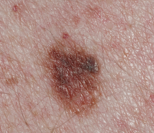
- This dysplastic nevus is more than 5 millimeters in diameter. (cancer.gov)
Spitz2
- The Spitz nevus: review and update. (jamanetwork.com)
- Since the description of the dysplastic nevus, so many "new" entities have been described sometimes including the term atypical- atypical genital nevi, milk line nevi, atypical nevi of the ear and scalp, atypical common blue nevus, atypical cellular blue nevus and last but certainly not least, the atypical Spitz nevus- more about that later. (thebsd.org.uk)
Melanocytic nevus1
- Although uveal melanomas may grow de novo, most develop from a preexisting melanocytic nevus. (medscape.com)
Basal1
- One side of the coin requires the observer to make a specific diagnosis by recognizing the classic patterns/structures associated with nevi, dermatofibromas (DF), intradermal nevi (IDN), basal cell carcinomas (BCC), squamous cell carcinomas (SCC), lentigines & seborrheic keratoses (SK), angiomas, angiokeratomas, sebaceous hyperplasias, and clear cell acanthoma (CCA). (dermoscopedia.org)
Histologically1
- Unfortunately, when clinically abnormal nevi are evaluated histologically, some studies have shown a lack of concordance, with some clinically abnormal nevi having no dysplastic features and some normal-appearing nevi having features of Clark nevi. (medscape.com)
Lesions3
- The features include: 1) two or more clinically atypical nevi, 2) more than 100 nevi in patients between 20 and 50 years of age, 3) more than 50 nevi in patients under 20 years of age or more than 50 years of age, 4) more than one nevus in buttocks or instep, 5) nevi on the anterior scalp, 6) one or more pigmented lesions in the iris. (wikipedia.org)
- [ 2 , 3 ] described these lesions as dysplastic nevi when they were observed in a series of family members of whom 36% had melanomas. (medscape.com)
- It became an almost personal matter- they didn't exist, grading was worthless, they could be divided into low- and high-grade lesions (without giving very clear instructions on how to do this) or if one followed Ray Barnhill and his colleagues, they should be classified in 3 categories based on a comparison of the nevus nuclear size with that of a nucleus of a keratinocyte in the mid prickle cell layer. (thebsd.org.uk)
Tumor3
- Tumor-induced glaucoma may be produced by obstruction of outflow pathways by pigment cells (pigment dispersion syndrome), melanin-laden macrophages (melanomalytic glaucoma), or tumor cells. (medscape.com)
- Gripp reviewed the occurrence of cancer in Costello syndrome: the most common tumor in CS is rhabdomyosarcoma (RMS), followed by neuroblastoma and bladder carcinoma[12]. (familialcancerdatabase.nl)
- Tumor predisposition in Costello syndrome. (familialcancerdatabase.nl)
Dysplasia1
- He divided dysplastic nevi into those showing epithelioid and lentiginous cell dysplasia. (medscape.com)
Melanomas1
- Dysplastic nevus syndrome is characterized by unusual nevi and multiple inherited melanomas. (wikipedia.org)
Diagnosis3
- The diagnosis of dysplastic nevus syndrome is based on clinical presentation and family history. (wikipedia.org)
- Onychomycosis, subungual hematoma, striated melanonychia and junctional nevus might simulate NAM and must be included in the differential diagnosis. (scielo.br)
- FAMMM syndrome is unique because the numbers of nevi in concert with a family history are needed for diagnosis. (logicalimages.com)
Term atypical1
- Some doctors use the term "atypical mole" to refer to a dysplastic nevus. (cancer.gov)
Cytological1
- Nothing lasts forever, in the most recent WHO blue book, dysplastic nevi with mild cytological atypia became demoted and subsumed within the category of a banal nevus. (thebsd.org.uk)
Clinically1
- The dysplastic nevi were characterized clinically by showing variability in size, border, and colors. (medscape.com)
Skin5
- Dermal nevi can eventually drop off the skin. (dynamicpr.co)
- These are people who have experienced an Ota nevus , hyperpigmentation of the eye or the skin around it. (eyes-road.eu)
- This syndrome is characterized by postnatal growth deficiency, cardiomyopathy, (mild to moderate) mental deficiency, redundant skin on neck, palms and soles, skin hyperpigmentation, acanthosis nigricans, papillomata, resembling verruca vulgaris and typically located on the nose, periorally and on trunk and limbs. (familialcancerdatabase.nl)
- Everyone should protect their skin from the sun and stay away from sunlamps and tanning booths, but for people who have dysplastic nevi, it is even more important to protect the skin and avoid getting a suntan or sunburn. (cancer.gov)
- In addition, many doctors recommend that people with dysplastic nevi check their skin once a month ( 2 , 4 ). (cancer.gov)
FAMMM3
- The risk of pancreatic cancer is estimated to be 13-22 times higher for FAMMM syndrome patients than the general population. (logicalimages.com)
- This risk increases to 38-fold in CDKN2A -mutant FAMMM syndrome patients. (logicalimages.com)
- Nevi in patients with FAMMM syndrome are phenotypically diverse. (logicalimages.com)
Genetic2
- Beckwith-Wiedemann syndrome (BWS) is a rare genetic overgrowth disorder. (brainandnervecenter.com)
- Adie syndrome, also known as Holmes-Adie syndrome, is a rare genetic condition that affects the eye's pupil. (brainandnervecenter.com)
MeSH1
- Dysplastic Nevus Syndrome" is a descriptor in the National Library of Medicine's controlled vocabulary thesaurus, MeSH (Medical Subject Headings) . (wakehealth.edu)
Histological1
- In 1984, Clark introduced five histological criteria for the characterization of a dysplastic nevus. (medscape.com)
Mutations1
- HRAS protooncogene mutations cause this syndrome[13,14]. (familialcancerdatabase.nl)
Disorders1
- Multiple causes are known, and they may range from simple iatrogenic mechanisms, such as implantation of dental amalgam, to complex medical disorders, such as Peutz-Jeghers syndrome . (medscape.com)
Colors2
- Dysplastic nevi possess irregular borders and may even have different colors while appearing in the form of clusters. (epainassist.com)
- A dysplastic nevus can have a mixture of several colors, from pink to dark brown. (cancer.gov)
Disorder characterized2
- Budd-Chiari syndrome is a rare disorder characterized by narrowing and obstruction (occlusion) of the veins of the liver (hepatic veins). (brainandnervecenter.com)
- Peutz-Jeghers syndrome is an autosomal dominant disorder characterized by intestinal hamartomatous polyps in association with mucocutaneous melanocytic macules. (medscape.com)
Clark2
- Clark described these families as having a "B-K mole syndrome. (medscape.com)
- The terms atypical mole and Clark nevus continue to be used interchangeably, regardless of clinical or histologic appearance. (medscape.com)
Lesion1
- Needless to say, the individual dermoscopic structures present in a lesion, within each diagnostic category (nevus, DF, BCC, etc), need to be placed within the context of the other features within the lesion. (dermoscopedia.org)
Common3
- A dysplastic nevus is a type of mole that looks different from a common mole. (dynamicpr.co)
- Level 1: The most common patterns found in nevi (excluding IDN). (dermoscopedia.org)
- A dysplastic nevus may be bigger than a common mole, and its color, surface, and border may be different. (cancer.gov)
Occur1
- A dysplastic nevus may occur anywhere on the body, but it is usually seen in areas exposed to the sun, such as on the back. (cancer.gov)